US9462802B2 - Systems and methods for ex vivo lung care - Google Patents
Systems and methods for ex vivo lung careDownload PDFInfo
- Publication number
- US9462802B2 US9462802B2US12/099,725US9972508AUS9462802B2US 9462802 B2US9462802 B2US 9462802B2US 9972508 AUS9972508 AUS 9972508AUS 9462802 B2US9462802 B2US 9462802B2
- Authority
- US
- United States
- Prior art keywords
- lung
- gas
- lungs
- perfusion
- perfusion fluid
- Prior art date
- Legal status (The legal status is an assumption and is not a legal conclusion. Google has not performed a legal analysis and makes no representation as to the accuracy of the status listed.)
- Active, expires
Links
Images


Classifications
- A01N1/02—
- A01N1/0247—
- A—HUMAN NECESSITIES
- A01—AGRICULTURE; FORESTRY; ANIMAL HUSBANDRY; HUNTING; TRAPPING; FISHING
- A01N—PRESERVATION OF BODIES OF HUMANS OR ANIMALS OR PLANTS OR PARTS THEREOF; BIOCIDES, e.g. AS DISINFECTANTS, AS PESTICIDES OR AS HERBICIDES; PEST REPELLANTS OR ATTRACTANTS; PLANT GROWTH REGULATORS
- A01N1/00—Preservation of bodies of humans or animals, or parts thereof
- A01N1/10—Preservation of living parts
- A01N1/12—Chemical aspects of preservation
- A01N1/122—Preservation or perfusion media
- A—HUMAN NECESSITIES
- A01—AGRICULTURE; FORESTRY; ANIMAL HUSBANDRY; HUNTING; TRAPPING; FISHING
- A01N—PRESERVATION OF BODIES OF HUMANS OR ANIMALS OR PLANTS OR PARTS THEREOF; BIOCIDES, e.g. AS DISINFECTANTS, AS PESTICIDES OR AS HERBICIDES; PEST REPELLANTS OR ATTRACTANTS; PLANT GROWTH REGULATORS
- A01N1/00—Preservation of bodies of humans or animals, or parts thereof
- A01N1/10—Preservation of living parts
- A01N1/14—Mechanical aspects of preservation; Apparatus or containers therefor
- A01N1/142—Apparatus
- A01N1/143—Apparatus for organ perfusion
Definitions
- the inventiongenerally relates to systems, methods, and devices for ex vivo organ care. More particularly, in various embodiments, the invention relates to a portable device for caring, assessing, and applying therapeutic measures to a lung or a pair of lungs ex vivo at physiologic or near-physiologic conditions.
- physiologic ex vivo preservationwould provide important benefits compared to conventional approaches. For instance, physiologic ex vivo preservation would permit more careful monitoring, functional testing, assessment, and therapy of the harvested organ. This would in turn allow earlier detection and potential repair of defects in the harvested organ, further reducing the likelihood of post-transplant organ failure. The ability to perform and assess simple repairs on the organ would also allow many organs with minor defects to be saved, whereas current transplantation techniques require them to be discarded. This is of crucial importance when harvesting lungs because lungs are easily compromised even before harvesting within the donor's body.
- HLAHuman Leukocyte Antigen
- injuries caused by ischemiaincrease as a function of the length of time an organ is maintained ex vivo.
- a lungmay typically be preserved ex vivo for only about 6 to about 8 hours before it becomes unusable for transplantation.
- a hearttypically may be preserved ex vivo for only about 4 to about 6 hours before it becomes unusable for transplantation.
- These relatively brief time periodslimit the number of recipients who can be reached from a given donor site, thereby restricting the recipient pool for a harvested organ. Even within the time limits, the organs may nevertheless be significantly damaged. A significant issue is that there may not be any observable indication of the damage. Because of this, less-than-optimal organs may be transplanted, resulting in post-transplant organ dysfunction or other injuries.
- Prolonged and reliable ex vivo organ carewould also provide benefits outside the context of organ transplantation.
- a patient's bodyas a whole, can typically tolerate much lower levels of chemo-, bio- and radiation therapy than many particular organs.
- An ex vivo organ care systemwould permit an organ to be removed from the body and treated in isolation, reducing the risk of damage to other parts of the body.
- the inventionaddresses the deficiencies in the state of the art by, in various embodiments, providing improved systems, methods, solutions and devices relating to portable ex vivo organ care.
- the inventionfeatures a lung care system that includes: a portable multiple use module including a portable chassis, a single use disposable module including: an interface adapted to couple the single use disposable module with the multiple use module for electro-mechanical interoperation with the multiple use module; and a lung chamber assembly having a first interface for allowing a flow of a perfusion fluid into the lung, a second interface for allowing ventilation of the lung with a ventilation gas, and a third interface for allowing a flow of the perfusion fluid away from the lung, the lung chamber assembly including a dual drain system for carrying the flow of the perfusion fluid away from the lung, the dual drain system comprising a measurement drain for directing a part of the perfusion fluid flow to a sensor of a perfusion fluid gas content and a main drain for receiving a remaining part of perfusion fluid flow.
- the lung care systemincludes a drainage system for draining the perfusion fluid from the lung chamber assembly, the drain system including a measurement conduit and a main drain conduit, the measurement conduit further directing a flow of perfusion fluid to a sensor that is adapted to measure a perfusion fluid gas content.
- the dual drainincludes a vessel for receiving the perfusion fluid flow, and overflow from the vessel flows to the main drain.
- the systemincludes a pump for the circulating the perfusion fluid, and a ventilation system for ventilating the lung with a gas having a predetermined composition.
- the gasincludes oxygen, carbon dioxide.
- the portable multiple use moduleincludes a lung console for providing at least one of electrical, pneumatic, and mechanical control of the disposable module; the lung console includes a ventilation controller for controlling ventilation of the lung, and includes a mechanical actuator for actuating a bellows to cause flow of gas into the lung.
- the lung console pneumatic control systemcontrols one or valves in a ventilation gas circuit connected to the lung in the disposable module.
- the pneumatic control systemcontrols at least one of a bellows valve for cutting off flow between the lung and the bellows, a relief valve for venting ventilation gas, and a trickle valve for introducing gas into the ventilation gas circuit.
- the ventilation controllerselects the gas that is used to ventilate the lung from one of an oxygenation gas, a deoxygenation gas, and a maintenance gas.
- the oxygenation gasis air, or a gas containing between 25% and 100% oxygen.
- the deoxygenation gasis composed of carbon dioxide and nitrogen
- the maintenance gasis composed of oxygen, carbon dioxide, and nitrogen. In one embodiment, the deoxygenation gas is about 6% carbon dioxide and about 94% nitrogen, and the maintenance gas is about 12% oxygen, about 5.5% carbon dioxide, and about 82.5% nitrogen.
- the multiple use moduleincludes a perfusion fluid controller that can control a level of gas content, such as oxygen, in the perfusion fluid.
- the perfusion fluid controllercontrols a perfusion fluid gas component, for example by controlling the flow of gas into a gas exchanger that exchanges gas between the flow of gas and the perfusion fluid.
- the gas flowing into the gas exchangeris a deoxygenation gas that removes oxygen from the perfusion fluid.
- the multiple use monitorincludes a monitor for displaying the status of the lung case system; the status includes information about the oxygen content of the perfusion fluid entering the lung and exiting the lung. It also displays real time traces of the ventilation gas pressure and the pulmonary arterial pressure.
- the inventionfeatures a lung care module comprising: a single use disposable module including an interface adapted for attachment to the multiple use module, and a lung chamber assembly having a first interface for allowing a flow of a perfusion fluid into the lung and a second interface for allowing ventilation of the lung with a ventilation gas; and a drain system for draining a flow of perfusion fluid from the lung chamber assembly, the drain system including a measurement conduit and a main drain conduit, the measurement conduit further directing a flow of perfusion fluid to a sensor that is adapted to measure a perfusion fluid gas content.
- the moduleincludes a system for ventilating the lungs with one of a maintenance gas, an assessment gas, and an oxygenation gas, such as air.
- the systemcan be configured to cause the lung to rebreath a volume of gas.
- the ventilation systemventilates the lung with a maintenance gas having a composition of about 12% oxygen, about 5.5% carbon dioxide, and about 82.5% nitrogen.
- the lungis ventilated by using a mechanically actuated bellows.
- the ventilation systemfurther includes a trickle valve for introducing a flow of maintenance gas, and a relief valve for venting excess gas.
- the second interface to the lungscomprises a tracheal cannula, which has an insertion portion for inserting into the trachea, and a connector portion for connecting to the ventilation gas circuit.
- the first interface to the lungsincludes a pulmonary artery cannula, which includes an insertion portion for inserting into the pulmonary artery and a connector portion for connecting to the perfusion fluid circuit. It also includes a pressure transducer connector defining an opening into a lumen of the connector portion near the insertion tube for positioning a pressure transducer near a point of entry of the perfusion fluid into the lung.
- the pressure transducer connectorfurther provides a channel for the pressure transducer to be remotely vented.
- the inventionfeatures a lung chamber assembly comprising: a housing having a bottom including at least one housing drain, and walls; a support surface for supporting a lung, the support surface defining a drain and drainage channels leading to the drain for draining a perfusion fluid exiting the lung; an openable lid that provides a sealable connection to the walls of the housing; a first interface for allowing a flow of the perfusion fluid into the lung; a second interface for allowing ventilation of the lung; and a third interface for allowing a flow of the perfusion fluid away from the lung.
- the housingincludes a drain system for carrying the flow of the perfusion fluid away from the lung, the drain system comprising a measurement drain for directing a part of the perfusion fluid flow to a sensor of a perfusion fluid gas content and a main drain for receiving a remaining part of perfusion fluid flow.
- the drain systemhas a region for collecting the flow of perfusion fluid away from the lung into a pool that feeds the measurement drain, the measurement drain having a drainage capacity less than a flow rate of the perfusion fluid away from the lung. Flow of perfusion fluid overflowing the region flows to the main drain.
- the drain systemfurther includes a wall partially surrounding the measurement drain, the wall partially blocking a flow of perfusion fluid from the measurement drain to the main drain, the wall promoting formation of a pool of perfusion fluid above the measurement drain.
- the housing of the lung chamberdefines openings that provide sealed passage through the housing of a pulmonary artery cannula, a pulmonary artery pressure transducer conduit, and a tracheal cannula.
- the perfusion fluidexits the lung through an exposed left atrial cuff, and flows into a drainage system.
- the flow of perfusion fluid exiting the lungpasses through a sealed connection to a left atrial cannula, which is connected to a conduit that carries the perfusion fluid away from the lung. A part of the perfusion fluid flow passes an oxygen content sensor, and the remainder flows to a reservoir.
- the inventionfeatures a method of evaluating a lung including: positioning the lung in an ex vivo perfusion circuit; circulating a perfusion fluid through the lung, the fluid entering the lung through a pulmonary artery interface and leaving the lung through a left atrial interface; ventilating the lung by flowing a ventilation gas through a tracheal interface; deoxygenating the perfusion fluid until a predetermined first value of oxygen content in the perfusion fluid is reached; reoxygenating the perfusion fluid by ventilating the lung with an oxygenation gas until a predetermined second value of oxygen content in the perfusion fluid is reached; and determining a condition of the lung based on a time taken for the lung to cause the oxygen content level in the perfusion fluid to change from the first value of oxygen content to the second value of oxygen content.
- the perfusion fluidis deoxygenated by ventilating the lung with a ventilation gas comprising carbon dioxide and nitrogen, for example about 5.5% carbon dioxide and about 94.5% nitrogen.
- the perfusion fluidis deoxygenated by circulating the perfusion fluid through a gas exchange device, the gas exchange device being in fluid communication with a ventilation gas comprising carbon dioxide and nitrogen, the gas exchange device altering a composition of oxygen in the perfusion fluid by gas exchange between the ventilation gas and the perfusion fluid.
- the predetermined first value of oxygen contentcorresponds to a red blood cell saturation of about 73%.
- the oxygenation gasis air, or a gas comprising between about 25% and about 100% oxygen.
- the predetermined second value of oxygen contentcorresponds to a red blood cell saturation of about 93%.
- the perfusion fluidflows at a rate of about 1.5 liters per minute, and is warmed by a heater to a near-physiologic temperature level.
- the perfusion fluidis composed of whole blood, or of a blood product, such as blood partially depleted of leukocytes, or partially depleted of platelets.
- Various therapeuticsare delivered to the ling during perfusion via the perfusion fluid, or through the tracheal interface using a nebulizer or a bronchoscope. Oxygen levels in the perfusion fluid are measured using a pulse oxymeter that determines the red blood cell saturation in the fluid.
- the inventionfeatures a method of preserving a lung ex vivo comprising: circulating a perfusion fluid through the lung, the fluid entering the lung through a pulmonary artery interface and leaving the lung through a left atrial interface; ventilating the lung through a tracheal interface by flowing a captive volume of a ventilation gas back and forth between the lung and a variable volume chamber; and introducing into the captive volume an additional volume of the ventilation gas and venting excess ventilation gas from the captive volume to maintain a predetermined composition of the ventilation gas and to maintain a minimum gas pressure of the captive volume.
- the ventilation gasincludes a composition of oxygen, carbon dioxide and an inert gas, such as nitrogen.
- the perfusion fluidreaches an equilibrium level corresponding to a predetermined composition of the ventilation gas.
- the predetermined composition of the ventilation gasincludes about 5-20% oxygen and about 2-10% carbon dioxide.
- a gas content of the perfusion fluidreaches an equilibrium level, the equilibrium level having a hemoglobin saturation level of about 88%-98%.
- the predetermined composition of the ventilation gasincludes about 12% oxygen and about 5.5% carbon dioxide.
- the hemoglobin saturation level of the perfusion fluid entering the lungreaches an equilibrium level of about 90-95% and a hemoglobin saturation level of the perfusion fluid leaving the lung reaches an equilibrium level of about 90-95%.
- the oxygen content of the perfusion fluid entering the lungis lower than physiologic levels, and the oxygen content of perfusion fluid leaving the lung is higher than physiologic levels.
- the following parametersare used in certain embodiments: the additional flow of ventilation gas is about 400-600 mL per minute; the captive volume is about 400-1200 mL; the minimum gas pressure of the captive volume is about 4-8 cm of H 2 O; and the maximum pressure of the ventilation gas is about 12-22 cm of H 2 O.
- Excess ventilation gasis vented through a relief valve in communication with the captive volume.
- the variable volume chamberis a bellows; compressing the bellows causes the flow of ventilation gas into the lung.
- the pulmonary artery interfaceincludes a pulmonary artery cannula, a portion of the pulmonary artery cannula being inserted into a pulmonary artery of the lung.
- the perfusion fluidto flows away from the lung through an exposed left atrial cuff of the lung, or through a sealed or semi-sealed connection between the left atrial cuff and a left atrial cannula.
- the tracheal interfaceincludes a tracheal cannula, a portion of the tracheal cannula being inserted into a trachea of the lung.
- the methodincludes measuring a first level of oxygen content in the perfusion fluid flowing into the lung and a second level of oxygen content in the perfusion fluid flowing out of the lung.
- the oxygen measurementinvolves measuring at least one of a level of oxygen saturation of hemoglobin in the perfusion fluid and a partial pressure of oxygen in the perfusion fluid flowing into the lung and flowing out of the lung.
- the perfusion fluidincludes a blood product, and can deliver therapeutics to the lung.
- the gas exchange in the lung between the ventilation gas and the perfusion fluidcauses the level of one or more gases, such as oxygen and carbon dioxide, in the perfusion fluid to reach equilibrium values.
- the lungmay be preserved for a period of about 3-24 hours when maintained with the equilibrium levels of gas.
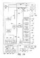
- FIGS. 1A-1Bare a schematic diagram of the described embodiment of a portable organ care system.
- FIG. 1Bshows the gas-related components of the lung perfusion module.
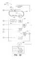
- FIG. 2is a schematic diagram of the lung perfusion circuit of the described embodiment.
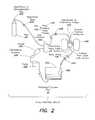
- FIG. 3is a schematic diagram of the gas loop of the organ care system in maintenance mode, according to the described embodiment.
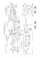
- FIG. 4is a schematic diagram of the gas loop of the organ care system in assessment mode, according to the described embodiment.
- FIGS. 5A-5Bare a schematic diagram of the lung ventilator pneumatic circuit, according to the described embodiment.
- FIG. 6is a diagram showing a typical pressure waveform in the lung over a breathing cycle, according to the described embodiment.
- FIGS. 7A-7Eshow examples of tracheal cannulae, according to the described embodiment.
- FIGS. 8A-8Fshow examples of pulmonary artery cannulae, according to the described embodiment.
- FIGS. 9A-9Fshow lateral views of the pulmonary artery cannulae illustrated in FIGS. 8A-8F .
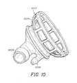
- FIG. 10is an illustration of a left atrium cannula.
- FIG. 11is a screenshot of the monitor of the organ care system in maintenance mode, according to the described embodiment.
- FIG. 12is a screenshot of the monitor of the organ care system in maintenance mode showing the configuration menu maintenance tab, according to the described embodiment.
- FIG. 13is a screenshot of the monitor of the organ care system in continuous assessment mode, according to the described embodiment.
- FIG. 14is a screenshot of the monitor of the organ care system in sequential assessment mode, deoxygenation submode, according to the described embodiment.
- FIG. 15is a screenshot of the monitor of the organ care system showing the configuration menu for the sequential assessment submode setting, according to the described embodiment.
- FIG. 16is a screenshot of the monitor of the organ care system in sequential assessment mode, hold submode, according to the described embodiment.
- FIG. 17is a screenshot of the monitor of the organ care system in sequential assessment mode, oxygenation submode, according to the described embodiment.
- FIG. 18is a screenshot of the monitor of the organ care system showing the configuration menu for the assessment tab, according to the described embodiment.
- FIG. 19is a screenshot of the monitor of the organ care system showing the configuration menu for the ventilator settings, according to the described embodiment.
- FIG. 20is a screenshot of the monitor of the organ care system showing the configuration menu for the lung tab, according to the described embodiment.
- FIG. 21is a screenshot of the monitor of the organ care system showing the configuration menu for the system tab, according to the described embodiment.
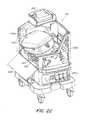
- FIG. 22is an illustration of the organ care system drawn from a 45-degree from the front view, according to the described embodiment.
- FIG. 23is a side view illustration of the organ care system, according to the described embodiment.
- FIG. 24is a front view illustration of the organ care system, according to the described embodiment.
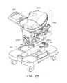
- FIG. 25is an illustration of the organ care system with the side panels removed, according to the described embodiment.
- FIG. 26is an illustration of the organ care system with the lung perfusion module removed, according to the described embodiment.
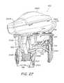
- FIG. 27is an illustration of the lung perfusion module, according to the described embodiment.
- FIG. 28is an exploded illustration of the lung chamber, according to the described embodiment.
- FIG. 29is an illustration of the lung support surface, housing, and front piece of the lung chamber, according to the described embodiment.
- FIG. 30is an illustration of the lung support surface, housing, and front piece of the lung chamber, showing the tracheal cannula and the PA cannula, according to the described embodiment.
- FIG. 31is a flow diagram showing steps performed at the lung donor site prior to place the lungs into the organ care system, according to the described embodiment.
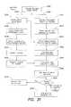
- FIG. 32is a flow diagram showing steps performed during transport of the lungs from the donor site to the recipient site, according to the described embodiment.
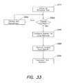
- FIG. 33is a flow diagram showing steps performed at the lung recipient site to remove the lungs from the organ care system and transplant them into the recipient, according to the described embodiment.
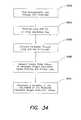
- FIG. 34is a flow diagram showing steps performed during continuous assessment of lungs ex vivo.
- FIG. 35is a flow diagram showing steps performed during sequential assessment of lungs ex vivo.
- the described embodimentgenerally provides improved approaches to ex vivo lung care, particularly in an ex vivo portable environment.
- the organ care systemmaintains a lung in an equilibrium state by circulating a perfusion fluid through the lung's vascular system, while causing the lung to rebreath a specially formulated gas having about half the oxygen of air.
- the perfusion fluidcirculates by entering the pulmonary artery (PA) via a cannula inserted into the PA. After passing through the lung, the perfusion fluid exits the lung from an open, uncannulated left atrium (LA) where it drains into a reservoir.
- a pumpdraws the fluid out of the reservoir, passes it through a heater and a gas exchanger, and back into the cannulated PA.
- the perfusion fluidis derived from donor blood.
- the perfusion fluidis blood-product based, synthetic blood substitute based, a mixture of blood product and blood substitute, or derived from blood from a blood bank.
- the described embodimentsenable a lung to be maintained ex vivo for extended periods of time, such as, for example, 3-24 or more hours.
- extended ex vivo maintenance timesexpand the pool of potential recipients for donor lungs, making geographic distance between donors and recipients less important.
- Extended ex vivo maintenance timesalso provide the time needed for better genetic and HLA matching between donor organs and organ recipients, increasing the likelihood of a favorable outcome.
- the ability to maintain the organ in a near physiologic functioning conditionalso enables a clinician to evaluate the organ's function ex vivo, and identify organs that are damaged. This is especially valuable in the case of the lung, since lungs are often compromised as a direct or indirect result of the cause of the death of the donor. Thus even a newly harvested lung may be damaged.
- the ability to make a prompt assessment of a harvested organenables a surgeon to determine the quality of a lung and, if there is damage, to make a determination of the nature of the problem. The surgeon then makes a decision as to whether to discard the lung, or to apply therapy to the lung.
- Therapiescan include recruitment processes, removing or stapling off damaged areas of lung, suctioning secretions, cauterizing bleeding blood vessels, and giving radiation treatment.
- the ability to assess and, if necessary provide therapy to lungs at several stages from harvesting to implantationgreatly improves the overall likelihood of lung transplant success. In some instances, the improved assessment capability and extended maintenance time enables medical operators to perform physical repairs on donor organs with minor defects.
- Increased ex vivo organ maintenance timescan also enable an organ to be removed from a patient, treated in isolation ex vivo, and then put back into the body of a patient.
- Such treatmentmay include, without limitation, pharmaceutical treatments, gas therapies, surgical treatments, chemo-, bio-, gene and/or radiation therapies.
- the lung care systemis described below in the following order. First, an overview of the components of an illustrative organ care system is given. Second, illustrative operation of the system is discussed, starting with preparing a lung and mounting it in the system. Third the use of the system for maintaining a lung is described. Two methods of assessing a lung are then described in the fourth and fifth sections—continuous assessment mode, and sequential assessment mode. Sixth, the functioning of the lung ventilator pneumatic circuit is described. Seventh, exemplary organ care system user interfaces and system displays are shown during lung maintenance and assessment. Eighth, illustrative implementations of the organ care system and selected components are described. In the ninth section, illustrative models for using the organ care system are described.
- FIG. 1is a block diagram that shows the main components of an organ care system (OCS) 1000 adapted to the preservation and treatment of a lung.
- the organ care systemincludes a permanent, multiple use, non-disposable section, OCS lung console 101 , and a single use disposable section, lung perfusion module 400 , which is in direct contact with the physical lungs, and the gases and fluids that pass through it.
- Multiple use OCS lung console 101includes four components: OCS console 100 , lung console module 200 , OCS monitor 300 , and probes for measuring flow ( 114 ), and perfusion fluid oxygen and hematocrit levels ( 116 , 118 ).
- OCS 1000is a self contained, mobile and portable unit, and can readily be handled by one person for transport on a flat surface using wheels, or lifted by two people, such as when being loaded into a vehicle.
- OCS 1000weighs about 75-100 pounds, and preferably about 80 pounds.
- OCS console 100provides processing, temperature, and power control services to the system. During the manufacturing process, OCS console 100 is adapted for use with OCS lung console module 200 . Alternatively, OCS console 100 can be adapted for use with modules that are adapted to preserve organs other than the lung, such as the heart, liver, or kidney.
- OCS console 100includes main processor 102 , which is a Freescale MX1 in the described embodiment, to provide system control and process data.
- Main processor 102distributes software to other processors in the system, including lung console module controller 202 , heater controller 104 , OCS monitor processor 302 , and pump controller (not shown). It also manages data, such as that received from flow sensor 114 , pressure sensor 115 , and oxygen sensors 116 , 118 .
- Heater controller 104which is a PIC microcontroller in the described embodiment, controls the heating of the perfusion fluid.
- Pressure transducer 223measures the pressure of internal maintenance gas in tank 221 , so that the amount of gas remaining can be determined.
- Regulator 222converts the gas tank pressure to 25 mm Hg for use in the system.
- Internal maintenance gas tank 221contains a mixture that is designed to provide enough oxygen to maintain the lung tissue during maintenance mode, described below. In the described embodiment, the maintenance gas is composed of 12% oxygen, 5.5% carbon dioxide, and 82.5% nitrogen.
- OCS console 100also includes an internal deoxygenation gas tank, regulator, and pressure transducer (not shown), which is used during assessment of the lungs. Assessment modes are described in a later section.
- lung console module 200The functions specific to the preservation of a lung (as opposed to other organs) are controlled by lung console module 200 .
- Lung console module 200is connected to OCS console 100 with data, power, and gas connections.
- the data connectionlinks main processor 102 on OCS console 100 with lung console module controller 202 , which is implemented on a PIC microcontroller in the described embodiment.
- the power connectionlinks the OCS console's power control module 106 with power converter 218 , which in turn supplies power at the appropriate voltage to the powered components within lung console module 200 .
- the gas connectionruns from maintenance gas regulator 222 to gas selector switch 216 , which selects whether maintenance gas or deoxygenation gas flows into the lungs.
- deoxygenation gas tank 501is external to OCS 100 and maintenance gas tank 221 is located internal to OCS console 100 .
- OCS console 100also includes an internal deoxygenation gas tank.
- an additional external maintenance gas tank 221supplements the maintenance gas tank internal to the OCS console.
- External gas tankscan be supplied at the donor site, recipient site, or can be stowed in a vehicle transporting the lungs. Since external tanks do not need to be accommodated within the confined volume of the OCS lung console 101 , they can be larger, and can supplement the limited gas supply of the smaller internal gas tanks of OCS 1000 .
- Controller 202manages the release of maintenance and assessment gases by controlling the valves, gas selector switch 216 , and ventilator 214 , thus implementing the preservation of the lungs in maintenance mode, or the assessment of the lungs in one of the assessment modes.
- Blood gas solenoid valve 204controls the amount of gas flowing into blood gas exchanger 402 .
- Airway pressure sensor 206samples pressure in the airway of lungs 404 , as sensed through isolation membrane 408 .
- Relief valve actuator 207is pneumatically controlled, and controls relief valve 412 .
- the pneumatic controlis carried out by inflating or deflating orifice restrictors that block or unblock the air pathway being controlled. This method of control allows complete isolation between the control systems in lung console module 200 and the ventilation gas loop in lung perfusion module 400 .
- Pneumatic control 208controls relief valve 207 and bellows valve actuator 210 .
- the pneumatic control circuits of lung console module 200are described in detail below.
- Trickle valve 212controls delivery of gas to the airway of lungs 404 .
- Ventilator 214is a mechanical device with an actuator arm that causes bellows 418 to contract and expand, which causes inhalation and exhalation of gas into and out of lungs 404 .
- OCS monitor 300provides user control of OCS 1000 via buttons, and displays data from the system's sensors that indicate the state of the lungs and of the various subsystems within OCS 1000 .
- Monitor 300is universal, i.e., it can be used for any organ. It includes monitor processor 302 that runs the software controlling monitor 300 and displays data on LCD 304 . In the described embodiment, monitor processor 302 is a Freescale MX1. Examples of various screen displays are described below in connection with the usage modes of OCS 1000 .
- OCS monitor 300includes four control buttons for the user: menu button 306 brings up the configuration menu; alarm button 308 silences the speaker; pump button 310 controls the circulatory pump; and action button 312 provides access to certain organ-specific actions, such as ventilator control, or to system actions, such as saving a session file to an external memory card. Other controls can also be included, such as a knob for controlling a value or selecting an item.
- OCS lung console 101includes probes that measure properties of circulating perfusion medium 250 , also referred to herein as perfusion fluid and perfusate.
- Flow probe 114measures the rate of flow of perfusion fluid 250 through the system. In the described embodiment, flow probe 114 is placed on the perfusate line as it leads towards the pulmonary artery.
- Pressure sensor 115measures pulmonary arterial pressure at the point of entry of perfusion fluid 250 into the lungs.
- Two oxygen saturation sensors 116 and 118sense the amount of oxygen in perfusion fluid 250 in the arterial, i.e., oxygenated, side of the circuit and in the venous, i.e., de-oxygenated, side of the circuit.
- Lung perfusion module 400is in direct contact with the gas and fluid circuits flowing through lungs 404 . It is therefore necessary to isolate it from the rest of OCS 1000 so that no tissue or fluids that come into contact with the organ ever come into contact with OCS lung console 101 . This is achieved by connecting it to the OCS lung console 101 only via one-way gas lines, or via isolated control gas for pneumatic control, or by means of a mechanical actuator (for the bellows).
- the entire lung perfusion module 400which contains all of the tissue and blood-contacting surfaces for the whole system, is disposable and is replaced for each new lung that is placed in OCS 1000 .
- All tissue and blood-contacting surfacesare part of disposable lung perfusion module 400 , which is manufactured from injection-molded components using inexpensive biocompatible materials that can easily be sterilized.
- Lung perfusion module 400is shaped and sized for coupling with OCS console 100 .
- the coupling between lung perfusion module and the OCS consolecan involve an interlocking mechanism, or other mechanism that secures the perfusion module to the OCS console or otherwise maintains the perfusion module in a desired position relative to the OCS console.
- lung perfusion moduleis easily attached to and detached from OCS console 100 with a mechanical hinge and clasp mechanism, described below in connection with FIG. 22 . It is also connected by plug-in electrical and optical connections.
- Lung perfusion module 400includes bellows 418 , which is actuated by ventilator 214 .
- Ventilator 214uses a mechanical actuator arm to compress and release bellows 418 . Compressing the bellows causes gas to be inspired by lungs 404 ; releasing the bellows causes it to expand and allow gas to be expired by the lungs.
- the distance traveled by the mechanical actuator in compressing bellows 418determines the tidal volume, i.e., the volume of gas inhaled by lungs 404 . Gas flowing in and out of the lungs passes through gas filter 410 , which prevents any fluids produced by the lungs from entering the gas loop.
- all lung gas connections between lung perfusion module 400 and OCS lung console 101include membranes that prevent gas from flowing back into OCS lung module 101 .
- Isolation membranesare not needed for pneumatic control gas connections, such as from relief valve actuator 207 and bellows valve actuator, because this gas has no contact with the organ.
- One-way gas flow valves that only permit flow into the lung perfusion moduleare automatically isolated from gas in the ventilation loop; such valves include trickle valve 212 and blood gas solenoid valve 204 .
- Airway pressure sensor 206samples the gas line pressure via isolation membrane 408 that prevents any exchange of gas backwards towards OCS lung console 101 .
- Perfusion module 400includes blood gas exchanger 402 , which includes a perfusate/gas exchange membrane that enables the infusion of a gas into the perfusate stream.
- the perfusatecirculates through circuits 406 and 407 between lungs 404 and gas exchanger 402 .
- the organ chambersupports lungs 404 and channels the perfusate coming out of the lungs from the left atrium in a manner that facilitates accurate measurement of arterial oxygen content levels. A detailed description of the perfusion circuit and the organ chamber is provided below.
- Perfusion module 400also includes relief valve 412 , which provides for controlled release of gas to be expired to the outside, serving to reduce gas pressure within the ventilator gas loop.
- Bellows valve 414controls the gas flow to or from the lungs.
- Check valve 416is a one-way valve which allows external air to be drawn into the ventilation system.
- Bellows 418expands and contracts; when the ventilator system is used in rebreathing mode, the bellows exchanges a substantially fixed volume of gas with the lungs as it expands and contracts.
- FIG. 2illustrates the lung perfusion circuit.
- the circuitis housed entirely within the lung perfusion module, and all its components are disposable.
- Perfusion fluid 250circulates within the perfusion circuit, passing through various components of lung perfusion module before passing through the vascular system of lungs 404 .
- Pump 226causes perfusion fluid 250 to flow around the lung perfusion circuit. It receives perfusion fluid 250 from reservoir 224 , and pumps the solution through compliance chamber 228 to heater 230 .
- Compliance chamber 228is a flexible portion of tubing that serves to mitigate the pulsatile nature of pump 226 .
- Heater 230replaces heat lost by perfusion fluid 250 to the environment during circulation of the fluid.
- the heatermaintains perfusion fluid 250 at or near the physiologic temperature of 30-37 degrees C., and preferably at about 34 degrees C.
- perfusion fluid 250flows into gas exchanger 402 .
- gas exchanger 402enables gases to be exchanged between gas and perfusion fluid 250 via a gas-permeable, hollow fiber membrane.
- the gas exchangerhas an effective gas exchange surface area of about 1 square meter, which is only a fraction of the 50-100 square meter effective exchange area of the lungs.
- gas exchanger 402has only a limited gas exchange capability compared to the lungs.
- Blood gas solenoid valve 204regulates the supply of gas into gas exchanger 402 .
- the composition of gas supplied to gas exchangeris determined by which mode the OCS is in, described in detail below.
- deoxygenation gas 500is supplied to the gas exchanger during the deoxygenation phase of the sequential assessment cycle.
- perfusion fluid 250passes through flow rate probe 114 , pressure probe 115 , and a perfusate oxygen probe 116 .
- SvO 2the readings from oxygen probe 116 as SvO 2 since it measures oxygen in perfusion fluid 250 just before it enters the lungs, which is analogous to venous blood oxygen.
- Sampling/injection port 236facilitates the removal of a sample or the injection of a chemical just before perfusion fluid 250 reaches the lungs.
- Perfusion solutionthen enters lungs 404 through cannulated pulmonary artery 232 .
- the pulmonary artery (PA) cannulaconnects the perfusion circuit with the vascular system of lungs 404 .
- a pulmonary artery (PA) cannulaSeveral exemplary embodiments of a pulmonary artery (PA) cannula are shown in FIGS. 8A-8F .
- single PA cannula 802has single insertion tube 804 for insertion into a single PA, and is used to cannulate the PA at a point before it branches to the two lungs.
- insertion tube 804is inserted into the PA, and the PA is secured onto the tube with sutures.
- Insertion tube 804 of cannula 802connects to connector portion 805 , which serves to position insertion tube 804 at an angle and location suitable for strain-free connection to the pulmonary artery of lungs 404 .
- Connection portion 805connects to main tube portion 808 , which is attached to the perfusion fluid circuit.
- FIGS. 9Ais a lateral view of PA cannulae 802 showing the angle between insertion tube 804 and connecting portion 805 ; in the described embodiment, the angle is between about 15 degrees and 30 degrees, and preferably about 22.5 degrees.
- double PA cannulae 810 , 820 , 830 , 840 , and 850each have two insertion tubes 812 , 814 , 822 , 824 , 832 , 834 , 842 , 844 , and 852 , 854 , each pair of tubes being angled apart from the main axis of the cannula by 30, 45, 60, 75, and 90 degrees in cannulae 810 , 820 , 830 , 840 , and 850 respectively.
- Each tubehas a diameter of about 0.5 to 0.72 inches at the rib, and about 0.4 to 0.62 inches on the body of the insertion tube.
- pair of insertion tubes 812 and 814are joined to connecting portion 815 in a Y-shaped configuration.
- connecting portion 815is angled with respect to main tube 818 ; the angle is chosen to facilitate the insertion of insertion tubes 812 and 814 into the pulmonary arteries of lungs 404 .
- the angleis between 15 and 30 degrees, and preferably about 22.5 degrees.
- insertion portion 804has a polycarbonate tip, with connector portion 805 and main tube portion 808 being made of urethane tubing.
- insertion tube 804 , connector portion 805 , and main tube portion 808are all made of a single piece of silicone of between 50 Shore A to 90 Shore A hardness silicone, preferably of a 80 Shore A hardness silicone.
- main tubes 818 , 828 , 838 , 848 , 858 and connector portions 815 , 825 , 835 , 845 , 855 of double PA cannulae 810 , 820 , 830 , 840 , and 850 respectivelymay be made of urethane, and the insertion tubes 812 , 814 , 822 , 824 , 832 , 834 , 842 , 844 , 852 , and 854 may be made of polycarbonate.
- the entire dual tube PA cannulai.e., the dual insertion tubes, connector portion, and main tube, are all made of a single piece of 80 Shore A silicone.
- silicone constructionis soft enough to provide a good purchase and grip for lung vessels tied on to the cannula connector with sutures.
- siliconecan readily be cut to the required length at the time of attachment to the lung PA.
- siliconeallows fabrication of the entire cannula in a single piece because it can be molded into a complex shape. Integral construction of the cannula eliminates transitions between separate cannula parts, which can produce unwanted turbulence in perfusion fluid 250 , introduce impurities, or cause leaks at the joints between separate parts.
- integral constructionrequires the molding of a single piece only, which reduces cost and increases the reliability of the cannula.
- each PA cannulaalso includes a connector for connecting perfusate pressure transducer 115 .
- PA cannulae 802 , 810 , 820 , 830 , 840 , and 850include pressure transducer connectors 806 , 816 , 826 , 836 , 846 , and 856 respectively.
- the connectorserves to allow placement of the perfusate pressure sensor at the correct location, right at the point of entry to the lungs where the perfusate flow slows, and pressure readings are not distorted by Bernoulli flow pressure.
- the pressure transducer connectorsalso provides a channel for pressure sensor 115 to be remotely vented, helping to ensure the accuracy of the pressure reading.
- the perfusateAfter passing through the lungs, the perfusate exits the lungs from the left atrium, a portion of which is removed along with the lung during explantation of the lungs from the donor. Since the left atrial tissue serves as an attachment zone during transplantation of the lungs into the recipient, it is important to leave it as undisturbed and healthy as possible. Therefore, in the described embodiment, the left atrial cuff is not cannulated, allowing the circulating perfusate to drain from the open left atrium and the left atrial cuff.
- the left atrial cuffis cannulated with cage-like cannula 1002 , illustrated in FIG. 10 .
- all the LA vesselsare placed inside the cannula; the excess LA tissue is then wrapped around the cannula.
- the cage-like structure 1004 of LA cannula 1002is designed to hold the left atrium open without occluding any pulmonary veins, thus helping to reduce the risk of compromising the health of the tissue.
- Connector 1008provides a connection point for a pressure transducer, which can be placed inside cannula 1002 and measure perfusate pressure.
- the perfusate exiting the lungsis collected in a dual drain system, using an “over flowing cup” technique to allow the sampling of newly drained fluid before it becomes mixed with other perfusate in the reservoir. All the flow from the lungs is directed to a small cup which feeds a measurement drain. The capacity of this drain is restricted by the use of small diameter tubing. Perfusate from the lungs exits at a flow rate that exceeds the capacity of the measurement drain. Excess blood overflows this small cup and is directed to the main drain and thus to the reservoir pool. The measurement drain directs a bubble free stream of newly drained perfusate toward the second oxygen probe 118 to obtain an accurate reading of arterial oxygen level, referred to as SaO2.
- the perfusion solutionAfter passing through second sampling/injection port 234 , the perfusion solution completes its cycle and returns to reservoir 224 .
- the dual drain systemis necessary only in the configuration in which the left atrial cuff is uncannulated. But if the left atrial cuff is cannulated, such as with a cage cannula as described below, there is no need for the dual drain system since a solid column of newly drained, bubble-free perfusate exits the cannulated left atrial cuff.
- perfusion fluid 250is composed of donor blood with the addition of heparin, insulin, vitamins, and antibiotics. Dextran serves to adjust oncotic pressure, Hematocrit levels, and pH.
- the following sectionsdescribe how OCS 1000 is used to preserve and assess a lung.
- the preinstrumentation sectiondescribes the initial steps in preparing OCS 1000 and the lung prior to connecting the lung to the OCS.
- the maintenance mode sectiondescribes how the OCS is used to preserve the lung.
- the assessment mode sectionsdescribe two ways of assessing the condition of the lungs—continuous mode and sequential mode.
- FIGS. 7A-7Eillustrate a set of exemplary tracheal cannulae.
- cannula 700includes tracheal insertion portion 704 to which the trachea is secured with a cable tie, or by other means.
- insertion portion 704is about 0.8 inches long.
- the base of cannula 700is preferably composed of polycarbonate, or another hard injection-moldable, biocompatible plastic, such as acrylic, polyester, K-resin, nylon, polyethylene, or polypropylene.
- the over-layer over insertion portion 704is preferably composed of a soft silicone rubber; alternative materials for the over-layer are other soft, biocompatible extruded or moldable materials such as polyurethane, thermoplastic elastomers, and other rubber materials.
- Adjacent to tracheal attachment portion 704is flexible section 706 , which is preferably composed of polyurethane, or one of the other biocompatible materials listed above as being suitable for the insertion portion over-layer.
- Insertion portion 704 and its over-layer, and flexible portion 706are injection moldable, with the silicone over-layer being overmolded onto the base part.
- the silicone over-layeris separately molded, or extruded and stretched over the base.
- rib 703At the end of insertion portion 704 that is inserted into the trachea is rib 703 ; the rib helps secure insertion portion 704 at the inserted location within the trachea, and is secured with a cable tie placed around the trachea.
- second rib 705At the opposite end of insertion portion 704 , second rib 705 , having a diameter about 0.2 inches greater than the base part diameter of insertion portion 704 , acts as a stop for the silicone over-layer and as a stop for the trachea.
- Past rib 705is a tubing barb fitting that is about 0.5 inches long, and has an angled barb to hold a 0.5 inch diameter tube.
- On the base piece that goes to lung OCS lung chamber connector 710there is a second tubing barb fitting that is about 0.5 inches long, having an angled barb to hold a 0.5 inch diameter tube.
- Flexible portion 706can be clamped to seal off air flow in and out of lungs 404 .
- clamping of section 706is used to maintain a static inflation of lungs 404 after explantation and before connections to the gas circuit of the OCS. Static inflation serves to prevent collapse of the lungs, and the consequent damage to the alveoli.
- Static inflationserves to prevent collapse of the lungs, and the consequent damage to the alveoli.
- static inflationthe lungs are inflated to a pressure of about 20 centimeters of water. The tracheal cannula is then clamped off at flexible section 706 .
- cannula 700includes locknut 708 for securing the cannula to the lung chamber.
- Locknut 708is mounted on a stepped portion of the cannula tube. Adjacent to locknut 708 , 0.7 inch-long 15 mm. connector 710 , serves to connect the cannula to a standard ventilator connector, which connects the lung to the gas circuit of the OCS.
- Tracheal cannulaeare designed to accommodate donor lungs having varying tracheal diameters according to the size of the donor.
- FIG. 7Aillustrates tracheal cannula 700 having insertion portion tip diameter 702 of 0.9 inches.
- cannulae having insertion portion tip diameters 722 , 742 , 762 , 782 of 0.85, 0.80, 0.75, and 0.70 inches of insertion portions 724 , 744 , 764 , and 784 respectivelyare shown.
- Cannulae having insertion portion diameters smaller than 0.7 inches, or larger than 0.9 inchesmay be needed to accommodate lungs from certain donors.
- the OCS perfusion circuitBefore receiving the lungs, the OCS perfusion circuit is primed with donor blood, priming solution, and drugs. This perfusate is then circulated and warmed.
- gas exchanger 402establishes blood gases that correspond to maintenance mode. This is achieved by setting gas selector switch 216 to allow maintenance gas to flow into the gas exchanger, and by duty cycle modulating gas exchanger valve 204 to provide a low average flow of maintenance gas through the gas exchanger.
- the exchange of gases in the gas exchangercauses the circulating perfusate to reach equilibrium with the maintenance gas, establishing the desired maintenance perfusate gas levels of O 2 and CO 2 .
- the perfusate pHis controlled by the CO 2 level.
- Maintenance modeplaces the lungs in a safe, stable condition so as to allow them to be preserved for an extended period of time.
- the maintenance gassatisfies the lung's cellular requirements.
- Oxygen consumption in the lungis so low that each breath can be substantially recycled, dramatically reducing the volume of fresh gas consumption. Since it is normally necessary to transport donated organs to a different site where the recipient is located, reducing the amount of gas needed to support the lungs, and thereby increasing the portability of the system, is a significant benefit.
- the tracheal cannulaWhen the lungs are placed within the organ chamber, the tracheal cannula is connected to the system gas line, which is placed in pause mode. In pause mode, bellows 418 are in a fully expanded state, i.e., prepared to perform the first lung inhalation. The clamp on the tracheal cannula is removed, and the pressures in the lung and in the gas line equalize. Inhalation then commences.
- FIG. 3is an illustration of the functioning of the OCS in maintenance mode.
- the ventilator systemmoves a captive volume of gas back and forth between the lungs and the bellows, causing the lungs to rebreath the gas.
- a small amount of maintenance gas 220is trickled into the ventilation circuit during each breath through valve 212 . Excess gas is exhausted from the circuit through relief valve 412 in order to prevent pressure buildup and maintain the desired minimum gas pressure in the system.
- maintenance gas 220is composed of about 9-15% oxygen, and preferably about 12% oxygen, about 4-7% carbon dioxide, and preferably about 5.5% carbon dioxide, with the balance being nitrogen.
- the composition of maintenance gas 220includes an amount of oxygen that is about one half that of air, and an amount of carbon dioxide that maintains a near-physiologic pH level in perfusion fluid 250 .
- an equilibriumis achieved between maintenance gas 220 and perfusate gas levels. In this equilibrium, there is only a small difference between the oxygen level in perfusion fluid 250 entering lungs 404 , i.e., the venous level PvO 2 , and the level exiting lungs 404 , i.e., the arterial level PaO 2 .
- the composition of maintenance gas 220is chosen to achieve perfusate oxygen levels that depart as little as possible from physiologic blood gas levels.
- the preferred maintenance gas compositionis a compromise between these levels, achieving equilibrium arterial and venous oxygen levels in perfusion fluid 250 that are approximately mid-way between physiologic venous and arterial levels.
- the preferred oxygen component of about 12%also provides more than sufficient oxygen to serve the lungs' metabolic needs.
- a 12% oxygen levelis close to the oxygen level in the alveoli of a healthy lung breathing air, because there is a gradient between the oxygen level in the trachea and the level in the alveoli caused by gas exchange along the airway path into the lungs. This gradient is absent in the case of lungs 404 in maintenance mode, when maintenance gas is being rebreathed, and the oxygen level is about 12% throughout the lung.
- the gas loopis filled with air, not with maintenance gas.
- ventilation of the lungsis initially with air.
- the maintenance gasis trickled in, and excess gas is released, the composition of gas in the gas loop soon changes to that of the maintenance gas.
- gas selector valve 216( FIG. 1 ) is set to select maintenance gas tank 221 .
- Gas exchanger valve 204is always closed in maintenance mode because gas exchanger 402 is not used.
- Bellows valve 414is always open to maintain the exchange of gas between the bellows and the lungs.
- passive check valve 416allows air into the circuit under suction conditions, but remains closed during maintenance mode because the ventilation circuit always has positive pressure.
- bellows 418are at the fully open position and the lungs are at their minimum volume. During the cycle, bellows 418 compresses, driving gas into the lungs. The lungs expand to accommodate this gas volume, causing a rise in pressure. When the specified volume of gas has been delivered, bellows 418 pauses for a specified plateau time before starting the exhalation portion of the cycle. During exhalation, bellows 418 returns to its original fully expanded state, and the lungs relax. The next ventilation cycle begins after an interval set by the specified respiration rate. The extent to which bellows 418 compress during the inhalation phase of each cycle is determined by the user-specified tidal volume, typically between 400 and 1200 mL.
- FIG. 6shows typical respiration pressure waveform 650 for each ventilation cycle.
- the pressureis set to positive end expiratory pressure (PEEP) value 652 , which is approximately 5 cm of H 2 O.
- PEEPpositive end expiratory pressure
- the pressureincreases to peak pressure 656 , and remains at the peak pressure for plateau portion 658 of the cycle.
- the peak pressureis about 20 cm H 2 O.
- the pressuredecreases until it reaches the desired PEEP level at the end of the cycle.
- Duration 662 of a complete ventilation cycleis set by the user-selected respiration rate, and is typically about 6 seconds.
- trickle valve 212opens briefly allowing a specific volume of calibrated maintenance gas into the circuit. Later, at the end of exhalation phase 660 , relief valve 412 opens briefly to exhaust excess gas to the outside air until the desired PEEP is reached.
- the opening of trickle valve 212 and relief valve 412are illustrated in FIG. 6 by traces 664 and 666 respectively.
- the average flow of maintenance gas into the ventilation loopis specified by the user, and is typically 500 ml/min. At a ventilation rate of 10 breaths per minute, trickle valve 212 allows 50 ml of maintenance gas into the circuit on each cycle.
- the flow rate of maintenance gasis usually set at the minimum level required to keep the gas composition in the gas loop close to the maintenance gas levels despite the tendency of the lungs' metabolism to decrease the oxygen level and increase the CO 2 level. Injection of maintenance gas is also used to maintain the desired PEEP level in the system. The amount of gas leakage from the lungs and from respiration fittings also affects the amount of maintenance gas injected.
- the lung's own metabolismhas only a small effect on the composition of the ventilation gas and perfusate gases. Since maintenance gas is injected into the gas line during each ventilation cycle, the composition of ventilation gas and of the perfusate gases rapidly reach the same composition, namely that of the maintenance gas. Once this situation occurs, the lungs are in a state of equilibrium with the maintenance gas. In the equilibrium state, the perfusate oxygen levels achieve steady state values.
- the SaO 2 steady state levelis in the range of about 93-95%, a little lower than the physiologic levels.
- the corresponding steady state SvO 2 levelis in the range of about 90-91%, which is higher than physiologic levels.
- the difference between saturation levels in perfusion fluid 250 across the lungsis lower than the physiologic difference.
- the higher SvO 2results, in part, from the absence of the deoxygenating effect of the body tissue, which is present in the physiologic case.
- the lower SaO 2 levelis caused in part by ventilation of the lungs with maintenance gas, which has only about half the oxygen content of air.
- the systemshortens the bellows compression stroke to account for the volume of gas contributed by trickle valve 212 , so as to maintain an accurate and constant tidal volume delivery to the lungs.
- FIG. 4is a schematic diagram showing the various components involved in performing lung assessments.
- the systemmimics body processes by inhaling air into the lungs, and then removing the perfusate oxygen before the perfusion fluid returns to the lungs.
- the removal of the oxygenis accomplished by tissues; in the OCS it is accomplished by deoxygenation gas flowing through the gas exchanger.
- Continuous mode assessmenttests the gas exchange capability of the lungs by measuring how well the lungs can reoxygenate the blood. This measurement is performed by measuring venous and arterial blood oxygen levels. The scoring of lung performance in continuous assessment mode is discussed further below.
- FIG. 34is a flow diagram showing the principal steps involved in performing continuous assessment of the lungs.
- deoxygenation gasis flowed through gas exchanger 402 . This is accomplished using gas selector switch 216 , which is set to select deoxygenation gas 500 , and by opening gas exchanger valve 204 to connect gas exchanger 402 to the deoxygenation gas supply.
- deoxygenation gasis composed of 4-7% CO 2 and preferably 6% CO 2 , with the balance being nitrogen.
- Trickle valve 212is kept closed in this mode.
- the lungsare ventilated with air or another ventilation gas using bellows 418 , which deliver a fresh breath of air or other ventilation gas to the lungs during the inhalation phase of each cycle.
- FIG. 6shows the gas pressure profile and valve settings in a continuous mode ventilation cycle.
- bellows 418are at the fully open position, the lungs are at their minimum volume, and the pressure is at PEEP level 652 .
- Bellows valve 414is opened 668 and the bellows compress, driving gas into the lungs in inhalation phase 654 .
- the lungsexpand to accommodate the gas, and there is an accompanying rise in pressure.
- bellows 418has delivered the specified volume of gas, the system pauses for a user-specified plateau time 658 (also referred to as dwell time), before starting exhalation phase 660 of the cycle.
- plateau time 658also referred to as dwell time
- bellows valve 414is closed at the end of inhalation phase 654 , before plateau 658 . This allows bellows expansion to begin immediately after the inhalation phase.
- a gas other than aircan be supplied to the inlet of check valve 416 .
- gas of any desired compositioncan be provided.
- the gascan be provided from common gas entrainment devices that provide oxygen enrichment in a hospital. Such devices can supply ventilation gas at standard 50% or 100% oxygen levels.
- gas exchanger 402While deoxygenation gas is flowing through gas exchanger 402 and the lung is being ventilated with air, perfusate is circulated through the lung and gas exchanger, as shown in FIG. 34 , step 3406 .
- perfusateis circulated through the lung and gas exchanger, as shown in FIG. 34 , step 3406 .
- gas exchanger 402has a limited gas exchange capability, and at the physiologic blood flow rate of 3-4 l/min., it is not able to remove enough oxygen from the blood to reduce the saturation levels to levels corresponding to the body while the blood is being circulated through the lungs where is continually being reoxygenated.
- the flow rateis reduced to about 1.5 l/min.
- a flow rate intermediate between 1.5 l/min. and physiologic flow rates of 3-4 l/min.are used, with correspondingly higher oxygen levels for the venous blood entering the lungs.
- the trade-offmay be reduced or eliminated by increasing the gas exchange capability of the system.
- multiple gas exchangersare used in series or in parallel in the lung perfusion circuit.
- the gas exchanger's gas exchange capabilityis increased by equipping it with a larger gas exchange surface.
- Continuous mode assessmentis typically performed directly after the lungs have been kept in maintenance mode.
- the following alternate embodimentexpedites the switchover from maintenance to continuous mode assessment.
- bellows 418contain a full volume of maintenance gas, which would normally be flushed out during several air ventilation cycles. Instead, a purge maneuver is performed to replace the entire contents of the bellows 418 with air.
- bellows valve 414is open, and bellows 418 are fully compressed at a slow rate.
- relief valve 412is actively controlled to maintain the pressure near the PEEP level.
- bellows valve 414is closed, and bellows 418 is fully expanded, filling its entire volume with fresh air from check valve 416 .
- One or more purge cyclesmay be performed to thoroughly establish the new gas composition.
- the values of the perfusate oxygen levels entering the lung and exiting the lungare measured, as indicated in FIG. 34 , step 3408 .
- Perfusate samplescan also be taken to confirm levels of oxygen and determine other components of the perfusion fluid.
- the userassesses the gas exchange capability of a lung by determining how much oxygen the lung can transfer to the perfusate in each breath. This assessment is based on the measured values of the oxygen levels in the perfusate entering the lung, and leaving the lung ( 3410 ). The assessment is calibrated using various parameters, such as the fraction of oxygen in the gas that is ventilating the lung.
- the standard measure of gas exchange capabilityis the ratio between the partial pressure of oxygen in the blood in mm.
- Another measure of the gas exchange capacity of the lungsis the difference between oxygen levels of blood entering the lungs, PvO 2 , and that of the blood leaving the lungs, PaO 2 .
- the PvO 2 levelis about 40 mm Hg and PaO 2 is about 100 mm Hg, with a difference between outgoing and incoming oxygen levels of 60 mm Hg.
- the PvO 2 levelmay be 60 mm Hg, and a healthy lung may achieve a PaO 2 of 115 mm Hg, with a PaO 2 -PvO 2 value of 55 mm Hg, close to the corresponding value in vivo.
- Sequential assessment modeis a second method of evaluating the lungs' gas exchange capability. In this mode, the lungs receive deeply venous perfusate oxygen levels that subject them to a different capability test than that of continuous assessment mode.
- Sequential assessmentincludes three phases: deoxygenation, hold, and reoxygenation.
- the deoxygenation phaseremoves oxygen from all the perfusate in the system.
- the hold phasethe lungs then reoxygenate the perfusate pool.
- the speed at which they achieve reoxygenationis an indication of their gas exchange capability.
- FIG. 35shows the principal steps involved in performing a sequential assessment of the lungs.
- Deoxygenation phase 3502 , 3504is used to lower the oxygen content of perfusion fluid 250 . This is achieved by using both gas exchanger 402 and lungs 404 .
- deoxygenation gas 500is fed into it by setting gas selector valve 216 to select deoxygenation gas, and opening gas exchanger valve 204 .
- the gas exchangercan deoxygenate the blood on its own, the process is expedited by using the lungs and the ventilator.
- the ventilatoris configured to run as a rebreather, as in maintenance mode (see above), and trickle valve 212 injects deoxygenation gas 500 into the gas circuit.
- the rebreathed gas in the gas circuitconforms to the deoxygenation gas composition, i.e., about 6% CO 2 and 94% N 2 , and the lungs act to deoxygenate the perfusion fluid circulating through them. In effect, the lungs are being used as a very effective gas exchanger to help deoxygenate the perfusate pool.
- the deoxygenation phasecontinues until the perfusate oxygen falls to a user-defined threshold value, which is usually approximately 50-70% oxygen, and preferably about 60% oxygen.
- the deoxygenation processis halted by closing gas exchanger valve 204 and trickle valve 212 while perfusate continues to flow through the perfusion circuit.
- the perfusate poolis allowed to stabilize to a uniform level of deoxygenation.
- the time required to achieve uniformitymay depend on the perfusate flow rate.
- arterial and venous oxygen content levelsare monitored, and the hold phase is maintained until the levels become equal and constant over time.
- ventilationis halted, or, alternatively, the system performs one or more purge cycles (described above in the continuous assessment section) to prepare for the reoxygenation phase.
- the purge cycleserves a useful role here because the gas in the gas circuit is being switched from deoxygenation gas to air, its polar opposite, and in order to start oxygenating the perfusion fluid immediately, the gas circuit needs to be filled with air at the outset.
- the oxygen-depleted perfusate poolis reoxygenated by ventilating the lungs with air or another ventilation gas (step 3508 ).
- the ventilationis performed using the same method as described above for continuous assessment, with the difference that gas exchanger valve 204 is kept closed.
- the lungsare the only source of gas exchange in the perfusion circuit (step 3510 ).
- the time taken for the lungs to reoxygenate the perfusate poolis the key indicator of the lung gas exchange capability.
- the measured reoxygenation timeis the time for perfusion fluid 250 to go from a de-oxygenated state to a predetermined oxygenated level as measured by one or both of pulse oximeter probes 116 and 118 (step 3512 ).
- blood samplesare taken from one or more of sampling ports 234 , 236 and the saturation levels are measured by a lab blood gas analyzer.
- the saturation at the oxygenation threshold levelis set in the range of 90% to 100% and is preferably set at 93%.
- the gas exchange capability of the lungsas measured by the time taken for the air-ventilated lungs to reoxygenate the blood from the deoxygenation threshold level to the oxygenation threshold level provides a measure of the condition of the lungs (step 3514 ).
- a healthy lungwill be able to reoxygenate the perfusate pool in 4-5 breaths, which corresponds to a sequential assessment mode reoxygenation time in the range of 45 to 90 seconds, and typically approximately one minute.
- Validation of the reoxygenation time as an assessment toolmay require normalization based on ventilation parameters, hematocrit, blood flow rate, lung volume, and altitude.
- a gas other than airis supplied to the inlet of check valve 416 during the oxygenation phase.
- gas from devices that provide gas at 50% or 100% oxygen in a hospital settingcan supply the ventilation gas.
- reoxygenation timesare reduced, and to determine the lungs' gas exchange capability, the reoxygenation time measurements need to be appropriately calibrated.
- Another method of assessing lung gas exchange capability during sequential assessment modeis to measure the speed at which the lungs deoxygenate perfusion fluid 250 during the deoxygenation phase.
- the effectiveness of the lungs in deoxygenating perfusion fluid 250 while being ventilated with deoxygenation gas 500provides an indication of the lungs' gas exchange capability.
- An advantage of sequential assessment modeis that physiologic blood flow rates of 3-4 l/minute can be used because, during reoxygenation, gas exchange is being performed only by the lung. Since the gas exchanger is not involved, there is no need to limit blood flow.
- the lung ventilator pneumatic circuitprovides a means of controlling bellows valve 414 and relief valve 412 for controlling various modes of ventilation. It also controls gas flow to blood gas exchanger 402 and the lungs. Pneumatic control offers several advantages, including the ability to open and close valves at different rates, the availability of inexpensive, disposable pilot valves, the ability to isolate lung console module 200 from the valves carrying gases exposed to the lung, and providing a convenient and modular interface for connecting and disconnecting disposable lung perfusion module 400 to console module 200 .
- FIG. 5 ashows the components of the pneumatic circuit in lung console module 200 , and how the circuit connects to lung perfusion module 400 .
- the components corresponding to pneumatic control module 208 as indicated on FIG. 1are identified by the dotted line in FIG. 5 a .
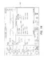
- Table 1is a list of the pneumatic circuit parts according to the described embodiment.
- V1Gas Selector Valve, 3 way 15 SLPM, 25 PSI MAX Line Pressure, ASCO AL2312, 0.65 W, 0.055′′ (4.3 PSI drop @ 15 SLPM) 204
- V2Blood Gas Valve, 2 way NC, 10 SLPM, 25 PSI MAX Line Pressure, ASCO AL2112, 0.65 W, 0.055′′ (2 PSI drop @ 10 SLPM) 212
- V3Re-breather Gas Valve, 2 way NC, 5 SLPM, 25 PSI MAX Line Pressure, ASCO AL2112, 0.65 W, 0.55′′ (0.6 PSI drop @ 5 SLPM)
- V4Bellows Pilot Valve, 3 way, 1.5 SLPM, 3 PSI MAX Line Pressure, ASCO AL2312, 0.65 W, 0.55′′ (2.5 cm H2O drop @ 1.5 SLPM) 207
- V5Relief Pilot Linear Pressure Control, Variable Orifice (0.020′′ to 0.170′′ Using Linear Step
- FIG. 5 bshows a front view of connector 624 , showing a six-lumen connector, with gas lines 630 , 632 , 634 , 636 , and 638 providing connections to gas exchanger 402 , the rebreathing gas circuit, bellows valve 414 , relief valve 412 , and airway pressure respectively.
- the connectorpermits rapid removal and hookup of disposable lung perfusion module 400 to lung console module 200 .
- Gas selector switch 216selects which gas to pass through gas exchanger valve 204 and trickle valve 212 .
- the control of trickle valve 212is synchronized with the ventilation cycle; the valve is opened during the inhalation phase, as described above for FIG. 6 , and is kept open for long enough to obtain the desired average gas flow rate.
- the rate of flow to gas exchanger 402is controlled by pulse width modulation of the control valve 204 from valve 216 .
- Valves 204 and 212effect control of the gas flow rate using orifice restrictors 205 and 213 respectively.
- Bellows valve 414 and relief valve 412are both capable of high flow rates, such as 1 liter/second.
- the high flow rate capabilityallows non-restrictive, free gas flow between the lungs and the bellows during inhalation and exhalation.
- relief valve 412the high flow rate capability allows the lungs to exhale rapidly to the PEEP value.
- bellows valve 414 and relief valve 412are commercially available high flow rate pilot valves. Applying positive pressure to the pilot valve diaphragm closes the valve; negative pressure fully opens the valve.
- FIG. 5 ashows how pilot valve control is achieved for bellows valve 414 and relief valve 412 .
- Air pump 612runs constantly, providing an approximately constant flow of air through the pump.
- the pumpdraws in ambient air through inlet filter 606 , and check valve 608 .
- This flowcreates a pressure difference across check valve 608 of about 1 PSI, or 70 cm of H 2 O, which results in a pressure in inlet reservoir 610 pressure of ⁇ 70 cm of H 2 O relative to ambient pressure.
- Inlet reservoir 610 and outlet reservoir 614serve to filter the uneven pressure ripple from reciprocating pump 612 .
- the outlet of air pump 612flows through second 1 PSI check valve 616 .
- the pressure in outlet reservoir 614is 70 cm of H 2 O above ambient, provided relief valve actuator 207 is open to ambient pressure.
- Bellows valve 414is controlled as follows.
- Bellows valve actuator 210can be connected to either inlet reservoir 610 or outlet reservoir 614 .
- actuator 210is connected to inlet reservoir 610 , which is at ⁇ 70 cm of H 2 O.
- Actuator 210causes this negative pressure to be transferred via pneumatic line 634 to the diaphragm of bellows valve 414 .
- the negative pressure on the diaphragmcauses valve 414 to open.
- actuator 210is connected to outlet reservoir 614 at +70 cm of H 2 O, causing positive pressure to be applied to the valve diaphragm, which shuts off the valve.
- Relief valve 412is controlled by applying a positive pressure to the valve's diaphragm, but in this case a controllable pilot gas pressure of the valve is used to set the PEEP in the perfusion module gas circuit.
- Relief valve 412remains open, and gas in the ventilation loop is vented to the outside, as long as the pressure in the ventilation loop is greater than the pilot pressure on the valve's diaphragm. When the pressure in the ventilation loop falls below that of the pilot pressure, relief valve 412 closes.
- the pilot pressureto the desired PEEP value
- the relief valveallows gas to vent from the gas loop until the pressure falls to the desired PEEP level, and then it shuts off.
- the PEEP valveis actuated with higher or lower pilot pressure to effect the exhalation rate through the valve.
- Variable control of pilot pressure in relief valve 412is achieved by using linear stepper motor 618 in conjunction with a variable orifice valve in relief valve actuator 207 .
- Stepper motor 618controls the size of the opening of the variable orifice valve. The smaller the opening of the orifice, the more resistance to airflow, the less airflow from air pump 612 escapes to the ambient air, and the higher the pressure between check valve 616 and relief valve actuator 207 .
- This pressureis transmitted to relief valve 412 via pneumatic line 636 . This enables the processor to obtain an empirically calibrated relationship between relief valve pilot pressure and PEEP.
- the actual pilot pressureis measured by relief pilot valve pressure sensor 620 ; this is monitored by lung console module processor 202 , which also receives measurements of airway pressure from airway pressure sensor 206 .
- the pilot pressure measurementis used to control the pilot pressure by comparing the actual pilot pressure to the desired pilot pressure and changing the stepper motor position to equalize them.
- OCS monitor 300is the main input and output interface for the system operator.
- LCD 304displays real time measurements and derived values of interest for the perfusion solution and for the gas loop. It also displays the status of other OCS subsystems, such as battery levels and gas tank levels. The nature of the information displayed on OCS LCD display 402 is explained next. Following this, screen shots corresponding to maintenance mode, continuous assessment mode, and sequential assessment mode are described.
- FIG. 11is an exemplary screen shot of LCD 304 ; the screen shot corresponds to maintenance mode.
- LCD 304includes a display area 1102 showing real time trace 1104 of the ventilation pressure at the entrance to the lungs, as measured by airway pressure sensor 206 .
- the displayalso includes numerical values 1106 , 1108 of the ventilation pressure readings; numerator 1106 is the peak pressure value, which is the maximum pressure sampled over the entire ventilation cycle.
- Denominator 1108is the PEEP value for the last respiration cycle, which is derived by sampling the airway pressure at the end of the expiratory time, i.e., just before inhalation for the next cycle begins. Since PEEP is defined as the pressure right at the end of the respiration cycle, it does not necessarily correspond to the minimum pressure in the cycle.
- Lower pressuresmay occur in the system if, for example, the system overshoots or undershoots as it attempts to reach the set PEEP value.
- Additional numerical values 1110 , 1112 , and 1114show the configured set point (sp) values, i.e., the values selected by the user. The display of these values helps the user compare the displayed actual values of respiratory pressure with the configured desired values.
- Value 1110shows the set point value for PAWP, which is an absolute upper pressure limit, or clamp, on the respiratory pressure. Generally, the ventilation pressure waveform is below the PAWP limit at all times.
- the PEEP set point 1112corresponds to the desired respiratory pressure at the end of a respiration cycle, after exhalation is complete and just before the inhalation pressure ramp of the next cycle starts.
- Value 1114shows I:E, which is the ratio of the respiration cycle time associated with inspiration and exhalation.
- the inspiration periodincludes both the inhalation time corresponding to flowing gas into the lungs, i.e., inhalation ramp 654 ( FIG. 6 ), as well as the plateau time 658 .
- I:E(inspiratory time+plateau time):expiratory time.
- the systemderives the I:E value from the configured inspiratory time, plateau time, and respiration rate.
- Display area 1116 of LCD 304shows a real time trace 1118 of pulmonary arterial pressure (PAP) as measured by pressure sensor 115 . Also displayed are PAP numerical values showing a snapshot of key values: peak or systolic pressure 1120 , valley or diastolic pressure 1122 , and mean perfusate pressure 1124 at the pulmonary artery feed at the lung.
- PAPpulmonary arterial pressure
- FIG. 12shows configuration menu 1202 , with maintenance tab 1204 selected. In this mode, the operator can select what information to display in each of middle graphic area 1116 and bottom graphic area 1126 .
- Upper graphic frame 1102is also configurable (not shown).
- the configuration menu maintenance tabalso provides the ability to set the average flow rate of maintenance gas 220 through trickle valve 212 , as well as control the perfusate temperature. Other parameters of the lung ventilator can also be controlled via the maintenance tab menu.
- Displayed value 1160shows pulmonary flow (PF) of perfusate into lungs 404 as measured by flow rate sensor 114 .
- Displayed value 1162shows pulmonary vascular resistance (PVR), which is a measure of the resistance exerted by lungs 404 to the flow of perfusate.
- PVRpulmonary vascular resistance
- a lower PVR valueis preferable because it indicates a less restrictive flow of the perfusate through the vasculature of lungs 404 .
- favorable values of PVRare in the range of 200 to 400 dynes.
- Displayed value 1164shows venous saturation hemoglobin content, SvO 2 of perfusion fluid 250 , as measured by oxygen sensor 116 .
- displayed value 1166shows arterial saturated hemoglobin content, SaO 2 of perfusion fluid 250 , as measured by oxygen sensor 118 .
- icons indicating SVO 2 and SaO 2 alarmsare displayed adjacent to displayed values 1164 and 1166 respectively, for signaling the operator if either saturated hemoglobin value falls below an operator preset threshold. Such alarms may be implemented for any parameter measured, calculated or displayed.
- Displayed value 1168shows the hematocrit (HCT) level of perfusion fluid 250 and, optionally, an HCT alarm indicator for signaling the operator if the HCT level 1168 falls below an operator preset threshold.
- HCThematocrit
- Displayed value 1170indicates the temperature (Temp) 1170 of perfusion fluid 250 as it flows away from heater assembly 230 .
- Displayed value 1170may also include a Temp alarm indicator which signals in response to Temp 1170 being outside of an operator preset range.
- Temperature set point 1171 selected by the operatoris also shown.
- Display area 1172shows a numerical reading of the ventilation rate measured in breaths per minute (BPM) of a gas delivered to lungs 404 via the tracheal interface 1024 .
- the BPM valueis derived from one or more inputs, including readings from airway pressure sensor 206 .
- BPM set point 1173is displayed.
- Displayed value 1174shows the tidal volume (TV), the volume of gas flowing into lungs 404 during each inhalation.
- TVtidal volume
- LCD 304further includes circulatory pump indicator 1138 showing a status of the system's circulatory pump.
- Display area 1176shows an organ type indicator 1140 that indicates which organ is being perfused and an organ mode indicator 1142 that indicates what mode of operation is being used. For example, an “M” is used to indicate maintenance mode.
- SD card indicator 1144shows whether an SD card is used to store data collected during organ perfusion.
- Display area 1146includes gas tank diagram 1178 that graphically indicates remaining maintenance gas volume. Display area 1146 also includes one or more numerical displayed values 1180 indicating a flow rate of the gas in the gas supply along with the time remaining for which the gas is delivered to lungs 404 during perfusion. This remaining time may be calculated based on the remaining gas volume and the gas flow rate.
- Display area 1148shows graphical representation 1182 of the degree to which each of the batteries of OCS console 100 are charged.
- Battery status symbol 1184indicates that the batteries whose status are represented by graphical representation 1182 , are used to power OCS console 100 .
- Display area 1150shows graphical representation 1186 of the degree to which the battery that powers the user interface is charged.
- Display area 1188identifies whether the OCS monitor 300 is operating in a wireless fashion.
- display screen 304also shows FiO 2 and FiCO 2 concentrations, which are fractional concentrations of oxygen and carbon dioxide, respectively, measured at the entrance to the trachea.
- Display screen 406can additionally show readings of weight and elasticity of lungs 404 , PH of perfusion fluid 250 circulating through the lungs 1004 , partial pressures of gas components in perfusion fluid 250 , and PEEP levels.
- FIG. 11shows a lung in maintenance mode; the values displayed in the figure are to be taken as exemplary.
- the perfusate flow rateis 1.46 l/min, a value lower than physiologic levels, but sufficient to nourish the lung.
- SvO 2 value 1164is 92.4% and SaO 2 value 1166 is 92.2%. These levels correspond to equilibrium between maintenance ventilation gas 220 and the perfusate gases.
- the difference between arterial and venous oxygen levelsis caused by oxygenation from air entering the organ chamber (tending to increase SaO 2 ), and from the small consumption of oxygen by the lungs (tending to decrease SaO 2 ).
- the balance between these factorscan cause SaO 2 to be higher or lower than SvO 2 .
- the oxygen saturation values of the perfusate as it enters and exits the lungsare stable and equal to each other within about +/ ⁇ 5%.
- Graph 1104shows the ventilation pressure over time; the pressure rises when the bellows pushes air into the lungs, and diminishes to the desired PEEP value at the end of exhalation.
- the graphshows the pressure profile over the most recent ventilation cycles; display area 1172 shows that the lungs are being ventilated at a rate of 10 breaths per minute.
- Graph 1118shows real time PAP corresponding to the most recent ventilation cycles. The curve shows periodic peaks that correspond to the pulse of the circulatory pump 226 .
- Graph 1128shows the PAP trend.
- Numerical value 1170shows that the perfusate temperature is measured to be 35.0 degrees centigrade, and is equal to the set point value shown in numerical displayed value 1171 .
- Such a sub-physiologic temperature levelis selected to reduce the metabolic rate of lungs 404 during preservation.
- One advantage of a lower metabolic rateis the ability to lower the maintenance gas requirement of lungs 404 , thereby permitting them to be preserved for a longer time with a finite volume of maintenance gas 220 .
- FIG. 13is an exemplary screen shot of OCS monitor LCD 304 when the system is in continuous assessment mode.
- Respiration graph 1302 and numerical values 1304are similar to those shown in FIG. 11 for maintenance mode.
- PAP graph 1306 and numerical values 1308show an average pressure of 13 mm Hg, which is considerably higher than the corresponding 10 mm Hg pressure during maintenance mode. The higher pressure is required to achieve a higher flow rate of perfusion fluid through the lung, so as to allow testing of the lung's gas exchange capability.
- the screenshows flow rate 1310 at 2.93 liters/minute. In this mode, gas exchanger 402 deoxygenates perfusion fluid 250 to SvO 2 level 1312 of 82.3%.
- the lungsreoxygenate the blood using air ventilation, achieving SaO 2 level 1314 of 96.1%.
- Hematocrit level 1316is 30%, and perfusate temperature 1318 is maintained at about 37.1 degrees C., the physiologic value.
- the respiration rate displayed value 1320shows a rate of 12 breaths per minute, corresponding to that of a resting person.
- Tidal volume displayed value 1322shows a value of 700 ml, well within the physiologic range.
- OCS status display 1324shows a graphic of the lungs, indicating that OCS 1000 is preserving a lung, and the letters A and C, indicating that the system is in continuous assessment mode.
- FIG. 14is an exemplary screen shot of LCD 304 when the system is in deoxygenation phase.
- deoxygenation gas 500is passed through gas exchanger 402 and into the ventilation loop into lungs 404 .
- Oxygen levels in perfusion fluid 250drop rapidly, since oxygen is being removed by gas exchange both in lungs 404 and gas exchanger 402 .
- Graphs 1406 and 1408show the values of SaO 2 and SvO 2 respectively over a period of about one minute after the start of the deoxygenation phase.
- System status display 1414indicates lung assessment—deoxygenation phase, with the letters A, D. Also displayed are the user-determined values for the deoxygenation termination threshold 1416 , oxygenation phase lower threshold 1418 , and oxygenation phase upper threshold 1420 .
- FIG. 15shows an exemplary user interface for setting sequential assessment parameters.
- Configuration mode 1502is selected by pressing menu button 306 on OCS monitor 300 .
- the userenters and applies settings in sequential submode settings menu 1504 .
- Listed are user-settable values for hold time 1506which is the time between the end of the deoxygenation phase and the start of the oxygenation phase, and deoxygenation termination threshold 1508 , which is the target lowest level of oxygen content in perfusion fluid 250 , i.e., the system stops deoxygenation if/when this level is reached.
- the useralso sets values for oxygenation lower threshold 1510 , the target value for perfusate SvO 2 in the oxygenation phase, and oxygenation upper threshold 1512 , the target value for perfusate SaO 2 in the oxygenation phase.
- FIG. 16is an exemplary screen shot corresponding to hold phase.
- the purpose of hold phaseis to allow the oxygen levels in perfusion fluid 250 to become uniform. The extent to which this is achieved can be seen in graphs 1602 and 1604 , showing the time-changing values of SaO 2 and SvO 2 in perfusion fluid 250 .
- the flat parts of both curvesindicate the saturation levels are constant, and the closeness of the graphs for SaO 2 and SvO 2 indicate uniformity of the saturation levels on each side of lungs 404 .
- Numerical displayed values 1608 and 1606indicate values of SaO 2 and SvO 2 respectively. As shown in FIG. 16 , the measured values of SaO 2 and SvO 2 about one minute into the hold phase are 58.9% and 58.0% respectively, i.e., very close to each other.
- FIG. 17is an exemplary screen shot of the system in the reoxygenation mode.
- Graphs 1702 and 1704show the time-changing values of SaO 2 and SvO 2 in perfusion fluid 250 .
- the graphsshow the initial decline of the oxygen levels during the deoxygenation phase described above.
- the flat portions of the curves in the middle of the graphscorrespond to the hold phase, which lasts for about one minute.
- oxygenation modebegins.
- the graphsShortly after switching to oxygenation mode, the graphs start rising, which indicates oxygen gas exchange via the lungs into perfusion fluid 250 .
- Graphs 1702 and 1704 and numerical displayed values 1708 and 1706show that about 80 seconds into the oxygenation phase, SaO 2 and SvO 2 levels have climbed to 94.6% and 85.2% respectively.
- the time taken to reach a user-selected threshold oxygenation level in perfusion fluid 250is shown in numerical displayed value 1710 .
- FIG. 18shows the assessment tab 1802 of configuration menu 1202 .
- This screenenables the user to determine what information is to be shown in middle graphic frame 1116 , in the bottom graphic frame 1126 , to set temperature set point 1171 , and to choose which assessment mode to perform—sequential or continuous.
- Tab 1802also allows the user to select the ventilator setting menu, as well as the sequential assessment submode settings.
- FIG. 19shows ventilator settings menu 1902 .
- Respiration rate 1904selects the number of ventilation cycles per minute.
- Tidal volume 1906determines the volume of gas inhaled by the lung in each breath.
- Inspiratory time 1908is the duration of the inhalation phase.
- Peak airway pressure (PAWP) 1912is the maximum allowed gas pressure during the breathing cycle; it occurs while gas is being pushed into lungs 404 by bellows 418 .
- PEEP 1914controls the pressure in the lung when exhalation is complete.
- FIG. 20shows lung tab 2002 , which allows the user to set lung mode 2004 to maintenance or assessment, allows ventilator control 2006 to be switched on or off, and provides a link 2008 to lung setting submenu.
- FIG. 21shows system tab 2102 , which allows the user to set time and date, language, and perform other system actions. Other configuration tabs and associated menus can be added based on the needs of users.
- FIG. 22is an overall view of OCS console 100 showing the single use, disposable lung perfusion module in a semi-installed position.
- single use disposable lung perfusion moduleis sized and shaped to fit into OCS console 100 , and to couple with it.
- the unithas a similar form to the organ care system described in U.S. patent application Ser. No. 11/788,865.
- Removable lung perfusion module 400is insertable into OCS console 100 by means of a pivoting mechanism that allows module 400 to slide into the organ console module from the front, as shown in FIG. 22 , and then pivot towards the rear of the unit. Clasp mechanism 2202 secures lung perfusion module 400 in place.
- lung perfusion module 400is used to couple the module with OCS 100 .
- electrical and optical connections(not shown) provide power and communication between OCS console 100 and lung perfusion module 400 . Details of the electrical and optical connections are described in U.S. patent application Ser. No. 11/246,013, filed on Oct. 7, 2005, the specification of which is incorporated by reference herein in its entirety.
- a key component of lung perfusion module 400is organ chamber 2204 , which is described in detail below.
- Battery compartments 2206 and maintenance gas cylinder 220(not shown) are located in the base of the OCS console 100 .
- OCS console 100is protected by removable panels, such as front panels 2208 .
- lung perfusion moduleare perfusate sampling ports 234 and 236 .
- Mounted on top of OCS console 100is OCS monitor 300 .
- FIG. 23is a side view of OCS console 100 .
- LA sampling port 234 and PA sampling port 236provide means for removing perfusate samples, or for injecting chemicals into perfusion fluid 250 .
- Maintenance gas tank regulator 222 and gauge 2304are visible in OCS console 100 base. Also visible is one way inflow valve 2306 , which is attached to the reservoir and connected to the dome of the perfusate pump.
- FIG. 24is a front view.
- Bellows 418is located just above the OCS console module base, and is driven by mechanical actuator arm 2402 connected to ventilator unit 214 in lung console module 200 .
- Mechanical motion of actuator arm 2402causes bellows 418 to compress and expand, which drives the gas flow into and out of lungs 404 during a breathing cycle.
- Gas exchanger 402is located above bellows 418 .
- gas exchanger 402is a Novalung oxygenator.
- Perfusate fluid line 2404connects fluid pump 226 (not shown) and heater 230 (not shown).
- reservoir 224collects perfusion fluid, and connects via drain 2408 to pump 226 for recirculation through the system.
- FIG. 25the walls of OCS console 100 have been omitted so as to reveal additional internal components of the system.
- Maintenance gas 220is stored in a horizontally disposed cylinder, feeding maintenance gas 220 to the system when needed via regulator 222 .
- Lung perfusion module 400is shown in the installed vertical position.
- Adjacent to bellows 418is bellows drive plate 2502 , which mates with a flat disk at the end of linear actuator 2402 (not shown).
- FIG. 26is a view of OCS console 100 without disposable lung perfusion module 400 . Visible are ventilator module 214 and mechanical actuator arm 2402 . Other components (not shown) of lung console module 200 are housed within the module mounted along the left side wall of the OCS console 100 . These components are shown in FIG. 1 within lung console module 200 , and include console module controller 202 , gas exchanger valve 204 , airway pressure sensor 206 , relief valve actuator 207 , pneumatic control module 208 , bellows valve actuator 210 , trickle valve 212 , ventilator 214 , gas selector switch 216 , and power converter 218 .
- Pneumatic connector 624provides rapid hook-up to matching lung perfusion module connector 626 .
- Connectors 624 and 626also provide pneumatic control connections between lung console module 200 and lung perfusion module 400 to control bellows valve 414 , relief valve 412 , and receive pressure data for air sensor 206 .
- FIG. 27is a front view of lung perfusion module 400 .
- Organ chamber 2204includes a removable lid 2820 and housing 2802 .
- Sampling ports, including LA sampling port 234 and PA sampling port 236are visible below organ chamber 2802 .
- Gas exchanger 402 , bellows 418 , and bellows plate 2502are also visible in the figure.
- perfusate reservoir 224which stores perfusate 250 .
- the perfusateexits through one-way inflow valve 2306 , line 2702 , and pump dome 2704 to pump 226 (not shown).
- the perfusateis pumped through perfusate fluid line 2404 through compliance chamber 228 , and then to perfusate heater 230 .
- the perfusatepasses through connecting line 2706 to gas exchanger 402 .
- the perfusateexits gas exchanger 402 through connecting line 2708 to the interface with the pulmonary artery.
- the perfusatedrains through from the base of organ chamber 2204 , as described below. These drains feed the perfusate to reservoir 224 , where the cycle begins again.
- FIG. 28shows an exploded view of the components of organ chamber 2204 .
- Base 2802 of chamber 2204is shaped and positioned within lung perfusion module 400 to facilitate the drainage of the perfusion medium.
- Organ chamber 2204has two drains, measurement drain 2804 , and main drain 2806 , which receives overflow from the measurement drain.
- Measurement drain 2804drains perfusate at a rate of about 0.5 l/min, considerably less than perfusion fluid 250 flow rate through lungs 404 of between 1.5 l/min and 4 l/min.
- Measurement drainleads to oxygen probe 118 , which measures SaO 2 values, and then leads on to reservoir 224 .
- Main drain 2806leads directly to reservoir 224 without oxygen measurement.
- Oxygen probe 118which is a pulse oxymeter in the described embodiment, cannot obtain an accurate measurement of perfusate oxygen levels unless perfusion fluid 250 is substantially free of air bubbles.
- base 2802is shaped to collect perfusion fluid 250 draining from lungs 404 into a pool that collects above drain 2804 . The perfusate pool allows air bubbles to dissipate before the perfusate enters drain 2804 .
- a pool above drain 2804is promoted by wall 2808 , which partially blocks the flow of perfusate from measurement drain 2804 to main drain 2806 until the perfusate pool is large enough to ensure the dissipation of bubbles from the flow.
- Main drain 2806is lower than measurement drain 2804 , so once perfusate overflows the depression surrounding drain 2804 , it flows around wall 2808 , to drain from main drain 2806 .
- other systemsare used to collect perfusion fluid into a pool that feeds the measurement drain.
- the flow from the lungsis directed to a vessel, such as a small cup, which feeds the measurement drain.
- the cupfills with perfusion fluid, and excess blood overflows the cup and is directed to the main drain and thus to the reservoir pool.
- the cupperforms a function similar to that of wall 2808 in the embodiment described above by forming a small pool of perfusion fluid from which bubbles can dissipate before the perfusate flows into the measurement drain on its way to the oxygen sensor.
- Lungs 404are supported by support surface 2810 .
- the surfaceis designed to support lungs 404 without applying undue pressure, while angling lungs 404 slightly downwards towards the lower lobes to promote easy drainage of the perfusate.
- Support surfaceincludes drainage channels 2812 to collect and channel perfusate issuing from lungs 404 , and to guide the perfusate towards drain 2814 , which feeds perfusate directly to the blood pool for measurement drain 2804 .
- lungs 404are wrapped with a polyurethane wrap (not shown) when placed on support surface 2810 .
- the polyurethane wrapanchors lungs 404 , helps keep the lungs in a physiologic configuration, and prevents the bronchi from being kinked and limiting the total volume of inflation.
- the wrapprovides a smooth surface for the exterior of the lung to interface with organ chamber 2204 , reducing the risk of the chamber applying excessive pressure on any part of lungs 404 , which might cause undesirable hemorrhaging.
- the polyurethane wrapis marked with a series of lines indicating how much volume is being wrapped.
- the desired volume of wrapped lungcan be determined by an empirical relationship between lung size and the weight of the donor.
- the polyurethane wraphas a series of small holes for draining perfusate that collects around lungs 404 .
- the perfusateis collected by drainage channels 2812 in support surface 2810 , which channel the perfusate to drain 2814 .
- the top of organ chamber 2204is covered with a sealable lid that includes front piece 2816 , top piece 2820 , inner lid with sterile drape (not shown), and sealing piece 2818 that seals front piece 2816 to top piece 2820 .
- the organ chamberincludes a double lid system similar to that disclosed in connection with the heart preservation chamber described in U.S. patent application Ser. No. 11/245,957, which is incorporated herein in its entirety.
- the double lid systemincludes an outer lid, an intermediate lid, a flexible membrane and sealing frames between the lids and the organ chamber walls.
- the membraneis preferably transparent, and permits a medical operator to touch/examine the lungs indirectly through the membrane, or apply an ultrasound probe to the lungs through the membrane, while maintaining the sterility of the chamber.
- the outer lidopens and closes over the intermediate lid independently of the intermediate lid.
- the outer lidis rigid enough to protect lungs 404 from physical contact, indirect or direct.
- the outer lid and the chambermay be made from any suitable polymer plastic, for example
- Covering the organ chamberserves to minimize the exchange of gases between perfusion fluid 250 and ambient air, and helps ensure that the oxygen probes measure the desired oxygen values, i.e., values corresponding to perfusate exiting the lungs via the LA (SaO 2 ), and entering the lung via the PA (SvO 2 ).
- the closing of organ chamber 2204also serves to reduce heat loss from lungs 404 . Heat loss can be considerable because of the large surface area of the lungs. Heat loss can be an important issue during transport of the lungs when OCS 1000 may be placed into relatively low temperature environments, such as a vehicle, or the outdoors when moving OCS 1000 into and out of a vehicle.
- OCS 1000may be temporarily placed in a hospital holding area or in an operating theater, both of which typically have temperatures in the range of 15-22 degrees C. At such ambient temperatures, it is important to reduce heat loss from organ chamber 2204 in order to allow heater 230 to maintain the desired perfusate (and lung) temperature of 35-37 degrees C. Sealing the lungs in the organ chamber 2204 also helps to maintain uniformity of the temperature through lungs 404 .
- FIG. 29is a right side view of organ chamber 2204 with the cover removed so as to show support surface 2810 .
- Perfusate drainage channels 2812 and drain 2814carry perfusate to housing 2802 .
- tracheal cannula 700 and tracheal cannula connector 710for connection to OCS 1000 gas loop.
- Above tracheal cannula 700is PA cannula 850 with double connection tubes 852 and 854 at 90 degrees, as illustrated in FIG. 8 .
- Remotely vented pressure sensor 115(not shown) is connected to the perfusate flow at the point of entry from the PA cannula into lungs 404 by means of connector 806 , pressure transducer conduit 2902 , and pressure transducer cable 2904 .
- FIG. 29is a right side view of organ chamber 2204 with the cover removed so as to show support surface 2810 .
- Perfusate drainage channels 2812 and drain 2814carry perfusate to housing 2802 .
- tracheal cannula 700is clearly displayed.
- Tracheal cannula 700is secured to the wall of housing 2802 by means of locknut 708 .
- flexible urethane tubing 706projects into housing 2802 of organ chamber 2204 , leading to silicone-covered connector 704 , which connects to the trachea.
- FIGS. 31 and 32An exemplary model for using the organ care system described above for lung transplantation is described next with reference to FIGS. 31 and 32 .
- the process of obtaining and preparing the lungs 404 for cannulation and transportbegins by providing a suitable organ donor at step 3100 .
- the organ donoris brought to a donor location, whereupon the process of receiving and preparing the donor lungs 404 for cannulation and transport proceeds down two intersecting pathways.
- the pathwaysprincipally involve preparing OCS 1000 to receive donor lungs 404 and then transporting lungs 404 via OCS 1000 to a recipient site.
- pathway 3102includes exsanguinating the donor, arresting the donor's heart, and preparing lungs 404 for cannulation into OCS 1000 .
- the donor's bloodis removed and set aside so it can be used to perfuse lungs 404 during their maintenance on the OCS 1000 .
- the donor heartis injected in step 3106 with a cardioplegic solution to temporarily halt its beating in preparation for harvesting lungs 404 .
- a pneumoplegia solutionis administered to the lungs at step 3108 before lungs 404 are explanted from the donor at step 3110 and prepared for loading onto OCS 1000 at step 3112 .
- the lungs 404are explanted from the donor's body, they are instrumented onto OCS 1000 at step 3124 by insertion into the lung chamber 2204 and cannulation at the appropriate perfusion fluid and gas loop interfaces as described above.
- the lungs 404can be transferred directly from the donor to OCS 1000 without the use of cardioplegia.
- the donor's lungs 404are removed without the donor's heart being arrested and are subsequently instrumented into OCS 1000 for maintenance.
- OCS 1000is prepared through the steps of path 3114 so it is primed and waiting to receive lungs 404 for cannulation and transport as soon as the lungs 404 are prepared.
- OCS 1000is prepared in pathway 3114 through a series of steps including providing single use lung perfusion module 400 (step 3116 ), priming OCS 1000 with a maintenance solution (step 3118 ), filtering the blood from the donor and adding it to reservoir 224 (step 3120 ), and circulating and warming the perfusate within OCS 1000 (step 3122 ).
- perfusion fluid 250includes whole blood. In certain embodiments, perfusion fluid 250 is partially or completely depleted of leukocytes.
- perfusion fluid 250is partially or completely depleted of platelets, or includes a blood plasma substitute and is packed with red blood cells.
- perfusion fluid additivesinclude prostaglandin E, Prostacycline, dextran, isuprel, flolan and nitric oxide donors are added while epinephrine is removed.
- the additivesmay be generally selected from antimicrobials, vasodilators, and anti-inflammatory drugs.
- the additivesmay be delivered to the system 1000 via ports 234 , 236 coupled to the reservoir 224 , or via an interface in tracheal cannula 700 through a nebulizer or a bronchoscope.
- OCS 1000is selected to operate in maintenance mode. Maintenance mode is described in detail above. After reaching equilibrium in maintenance mode in step 3126 , and before being accepted for transport to the donor site, instrumented lungs 404 are assessed in step 3128 . The OCS user may select continuous assessment and/or sequential assessment, both of which have been described above.
- lungs 404may be re-inflated using sigh breathing, i.e., by causing lungs 404 to take breaths of varying tidal volume.
- lungs 404are caused to inhale a first breath having a tidal volume of up to about 1000 ml., followed by two or more smaller breaths having tidal volumes as low as about 100 ml.
- a second methodinvolves adjusting PEEP levels between values ranging from about 2 cm. H 2 O to 15 cm. H 2 O.
- over-inflated regions of lungs 404are restrained with the polyurethane wrap that is used to provide support for lungs 404 when placed on support surface 2810 . Such restraint allows the judicious application of gas loop pressure to re-inflate collapsed regions of the lungs.
- the I:E ratiois manipulated, which allows the amount of time spent at pressure plateau 658 ( FIG. 6 ) to be increased, helping lung reinflation, without exceeding peak pressure 656 and PEEP levels 652 .
- Fifth, simple manipulation of lungs 404 on support surface 2810 to change lung positioncan be an effective recruitment method.
- Sixth, lung secretions, and alveoli debris in the tracheaare removed by suction using a bronchoscope.
- the bronchoscopeis inserted into lungs 404 via a port in a connector between tracheal cannula 700 and gas circuit tubing of lung perfusion module 400 .
- surfactant inhalation therapyis performed by injecting surfactants, preferable in aerosol form, into the gas line during the inhalation phase of a breathing cycle.
- EdemaAnother pathology that is often found in donor lungs is localized edema, which can occur in a single or in multiple lobes. Edema can be remedied on OCS 1000 by manipulating PEEP levels, increasing oncotic pressure by ultrafiltration, and manipulation of perfusion fluid pressure by means of vasodilators and/or the flow rate of pump 226 .
- Pneumoniais also another common pathology of donor lungs, and can be addressed by direct injection of anti-microbial agents into perfusion fluid 250 , and/or by inhalation of the agents through the ventilator system of lung perfusion module 400 .
- Another pneumonia recruitment techniqueis broncho-alveolar lavage.
- Bronchospasmwhich occurs less frequently than the pathologies discussed above, is managed on OCS 1000 with inhaled broncholdilators.
- a bronchoscopeis optionally used to help inject the bronchodilators into the lungs' airways.
- Another pathologyis high PAP; this is managed by adding vasolidators to perfusion fluid 250 .
- an operatormay perform surgery on lungs 404 or provide therapeutic or other treatment, such as immunosuppressive treatments, chemotherapy, genetic testing or irradiation therapy.
- lungs 404are placed in maintenance mode while recruitment is being performed.
- Assessment step 3128 and recruitment step 3130may be repeated several times, and may last for a period of up to several hours if needed.
- the goalis to obtain an assessment of lungs 404 that indicates that the lungs are sufficiently healthy in order to be accepted for transport to the recipient site. Once this condition is satisfied, OCS 1000 , with its instrumented lung 404 , is loaded into a vehicle for transport to the recipient site.
- FIG. 32illustrates an exemplary usage mode of OCS 1000 during transport from the donor site to the recipient site.
- OCS 1000Before being placed in the transport vehicle, OCS 1000 is placed into maintenance mode (step 3202 ). OCS 1000 is then placed in the vehicle and the journey is commenced (step 3204 ). After a time interval, the lungs are assessed (step 3206 ). The time interval before the first assessment depends on the condition of lungs 404 as determined at the donor site, on monitored parameters of lungs 404 , and on the anticipated duration of the trip. In general, the poorer the condition of lungs 404 , the sooner an assessment will be conducted. If assessment 3206 finds that lungs 404 are in poor condition, therapy and recruitment are performed (step 3210 ).
- step 3206Another assessment is performed.
- the cycle of assessment and recruitmentcontinues until assessment step 3206 indicates that lungs 404 are above a certain health threshold, and then lungs 404 are returned to maintenance mode 3208 .
- no further assessment or recruitmenttakes place during transport.
- additional assessment and, if necessary, recruitment stepsare performed at intervals during transport.
- the decision as to whether to conduct further assessmentsis governed by the operator's overall assessment of the health of lungs 404 , as well as by the availability of assessment gas in OCS 1000 . Arrival at the recipient site (step 3212 ) completes the journey.
- OCS 1000functions in a self-contained manner. In particular, it relies on its own internal supplies of maintenance gas and deoxygenation gas. In an illustrative configuration, OCS 1000 has a 200 liter supply of deoxygenation gas 500 .
- the deoxygenation tank capacitylimits the number of sequential assessments in a trip to a maximum of five, and more generally, four or fewer, depending on the condition of lungs 404 .
- performing continuous assessmentdoes not require the achievement of any target deoxygenation level in perfusion fluid 250 .
- the assessmentis run for a fixed time interval, during which deoxygenation gas 500 is flowed through gas exchanger 402 at an average rate of about 10 liters/minute.
- continuous assessmentis run for 2 minutes, consuming a total of about 20 liters of deoxygenation gas 500 , i.e., about half that consumed in a sequential assessment.
- continuous assessmentmay be preferable to sequential assessment.
- the perfusate column measured by pulse oxymeters 116 and 118should be free of gas bubbles.
- the dual drain system 2804 and 2806 and the perfusate pool above drain 2804helps ensure that bubbles do not enter the perfusate line.
- motion of the vehicle transporting OCS 1000may cause enough agitation to cause some bubbles to drain into the perfusate column. Therefore, in the described embodiment, the vehicle is parked in a level area while assessment is being performed.
- lung chamber 2204 , lung housing 2802 and the dual drain systemare modified to make the system more resistant to motion, such as by confining the blood pool more securely, or by draining perfusate directly into tubes. Such modifications may permit accurate lungs assessments to be performed even while the transporting vehicle is moving.
- FIG. 33provides an exemplary process for conducting additional tests on the lungs 404 while OCS 1000 is at the recipient site.
- OCS 1000performs another assessment (step 3302 ) of lungs 404 .
- An additional supply of deoxygenation gasmay be available at the recipient site, which can supplement the OCS's supply of deoxygenation gas 500 that may have been depleted during transit from the donor site.
- therapy and recruitmentstep 3304 . If, after a final assessment step, lungs 404 are assessed to be in a condition suitable for transplant, lungs 404 are prepared for implantation into the recipient.
- lungs 404are de-cannulated and removed from the lung chamber assembly 2204 .
- lungs 404are transplanted into the recipient patient by inserting them into the recipient's chest cavity and suturing the various pulmonary connections to their appropriate mating connections within the recipient.
- a portion of the recipient's left atriummay be excised and replaced with one or more of the donor's left atrial cuff to which the donor's pulmonary veins are attached.
- only one of two lungsis removed while the remaining lung continues to be perfused and ventilated on the OCS.
Landscapes
- Life Sciences & Earth Sciences (AREA)
- Zoology (AREA)
- Engineering & Computer Science (AREA)
- Dentistry (AREA)
- General Health & Medical Sciences (AREA)
- Wood Science & Technology (AREA)
- Health & Medical Sciences (AREA)
- Environmental Sciences (AREA)
- Agricultural Chemicals And Associated Chemicals (AREA)
- Measurement Of The Respiration, Hearing Ability, Form, And Blood Characteristics Of Living Organisms (AREA)
- Medicines Containing Material From Animals Or Micro-Organisms (AREA)
- Medicines Containing Plant Substances (AREA)
- Investigating Or Analysing Biological Materials (AREA)
Abstract
Description
| TABLE 1 | |
| Ref. No. in | |
| FIG. 5a | Part Description |
| 216 | V1: Gas Selector Valve, 3 way 15 SLPM, 25 PSI MAX Line Pressure, |
| ASCO AL2312, 0.65 W, 0.055″ (4.3 PSI drop @ 15 SLPM) | |
| 204 | V2: Blood Gas Valve, 2 way NC, 10 SLPM, 25 PSI MAX Line |
| Pressure, ASCO AL2112, 0.65 W, 0.055″ (2 PSI drop @ 10 SLPM) | |
| 212 | V3: Re-breather Gas Valve, 2 way NC, 5 SLPM, 25 PSI MAX Line |
| Pressure, ASCO AL2112, 0.65 W, 0.55″ (0.6 PSI drop @ 5 SLPM) | |
| 210 | V4: Bellows Pilot Valve, 3 way, 1.5 SLPM, 3 PSI MAX Line Pressure, |
| ASCO AL2312, 0.65 W, 0.55″ (2.5 cm H2O drop @ 1.5 SLPM) | |
| 207 | V5: Relief Pilot Linear Pressure Control, Variable Orifice (0.020″ to |
| 0.170″ Using Linear Stepper motor, Haydon 20544-05-018, 2.5 W (0.1 to | |
| 70 cm H2O drop at 1.8 to 2.5 SLPM) | |
| 414 | V6: Bellows Valve, Instrument Industries BE 30-115-BL |
| 412 | V7: Relief Valve, Instrument Industries BE 30-115-BL |
| 205 | R1: Blood Gas Restrictor, Bird Precision RB 82304 BR (SA087)- |
| 24054, 10.8 SLPM @ 25 PSI, 0.0290″ | |
| 213 | R2: Ventilator Gas Restrictor, Bird Precision RB 82304 BR (SA087)- |
| 24060, 5.7 SLPM @ 25 PSI, 0.0210″ | |
| 608, 616 | C1, C2: Check Valve, 1 PSI, McMaster Carr 6079T54 |
| 606 | F1: Filter, McMaster Carr 8991T312 |
| 602 | P1: Assessment Gas Connector Colder PMC1602 |
| 624 | P2: Perfusion Module Gas Connector, Colder SM1702 (six lumen) |
| 604 | P3: Maintenance Gas Connector, Colder PMC1702 |
| 206 | X1: Airway Pressure (PEEP, PAWP sensing) |
| 620 | X2: Relief Valve Pilot Pressure (for controlling relief valve) |
| 612 | A1: Air Pump, 1.5 SLPM @ 3 PSI, Hargraves H103- |
| 11_B.1F28E1.A12VDC, 3 W | |
Claims (23)
Priority Applications (50)
| Application Number | Priority Date | Filing Date | Title |
|---|---|---|---|
| US12/099,725US9462802B2 (en) | 2008-01-31 | 2008-04-08 | Systems and methods for ex vivo lung care |
| IL299248AIL299248B2 (en) | 2008-01-31 | 2009-01-30 | Systems and methods for extracorporeal lung preservation |
| CN200980110231.5ACN101977649B (en) | 2008-01-31 | 2009-01-30 | Systems and methods for ex vivo lung care |
| NZ709775ANZ709775A (en) | 2008-01-31 | 2009-01-30 | Systems and methods for ex vivo lung care |
| CA3176645ACA3176645A1 (en) | 2008-01-31 | 2009-01-30 | Systems and methods for ex vivo lung care |
| CN201310148246.9ACN103430939B (en) | 2008-01-31 | 2009-01-30 | For the system and method for ex vivo lung care |
| NZ586901ANZ586901A (en) | 2008-01-31 | 2009-01-30 | Systems and methods for ex vivo lung care |
| PCT/US2009/032619WO2009099939A2 (en) | 2008-01-31 | 2009-01-30 | Systems and methods for ex vivo lung care |
| EP23209551.3AEP4331362A3 (en) | 2008-01-31 | 2009-01-30 | Systems and methods for ex vivo lung care |
| EP09707471.0AEP2244773B1 (en) | 2008-01-31 | 2009-01-30 | Systems and methods for ex vivo lung care |
| CA2713443ACA2713443C (en) | 2008-01-31 | 2009-01-30 | Systems and methods for ex vivo lung care |
| IL304036AIL304036B2 (en) | 2008-01-31 | 2009-01-30 | Systems and methods for extracorporeal lung preservation |
| CN201510886161.XACN105494312B (en) | 2008-01-31 | 2009-01-30 | Systems and methods for isolated lung care |
| JP2010545201AJP5599322B2 (en) | 2008-01-31 | 2009-01-30 | Systems and methods for ex vivo lung care |
| CA2899880ACA2899880C (en) | 2008-01-31 | 2009-01-30 | Systems and methods for ex vivo lung care |
| CN201810088427.XACN108402030B (en) | 2008-01-31 | 2009-01-30 | Systems and methods for isolated lung care |
| DK09707471.0TDK2244773T3 (en) | 2008-01-31 | 2009-01-30 | SYSTEMS AND METHODS FOR EX VIVO LUNG CARE |
| ES09707471TES2966520T3 (en) | 2008-01-31 | 2009-01-30 | Systems and methods for ex vivo lung care |
| CN202310186780.2ACN116267894A (en) | 2008-01-31 | 2009-01-30 | Systems and methods for ex vivo lung care |
| CA2948767ACA2948767C (en) | 2008-01-31 | 2009-01-30 | Systems and methods for ex vivo lung care |
| NZ739110ANZ739110A (en) | 2008-01-31 | 2009-01-30 | Systems and methods for ex vivo lung care |
| NZ724681ANZ724681A (en) | 2008-01-31 | 2009-01-30 | Systems and methods for ex vivo lung care |
| AU2009212725AAU2009212725B2 (en) | 2008-01-31 | 2009-01-30 | Systems and methods for ex vivo lung care |
| CA3015816ACA3015816C (en) | 2008-01-31 | 2009-01-30 | Systems and methods for ex vivo lung care |
| CN202011251518.4ACN112438251B (en) | 2008-01-31 | 2009-01-30 | Systems and methods for ex vivo lung care |
| IL315867AIL315867A (en) | 2008-01-31 | 2009-01-30 | Systems and methods for ex vivo lung care |
| NZ753880ANZ753880B2 (en) | 2008-01-31 | 2009-01-30 | Systems and methods for ex vivo lung care |
| IL207289AIL207289A (en) | 2008-01-31 | 2010-07-29 | Systems for ex vivo lung care |
| NZ623220ANZ623220A (en) | 2008-01-31 | 2012-10-31 | Systems and methods for ex vivo lung care |
| NZ603329ANZ603329A (en) | 2008-01-31 | 2012-10-31 | Systems and methods for ex vivo lung care |
| AU2014202736AAU2014202736C1 (en) | 2008-01-31 | 2014-05-20 | Systems and methods for ex vivo lung care |
| HK14105510.4AHK1192115B (en) | 2008-01-31 | 2014-06-11 | Systems and methods for ex vivo lung care |
| JP2014164178AJP6144238B2 (en) | 2008-01-31 | 2014-08-12 | Systems and methods for ex vivo lung care |
| AU2015202735AAU2015202735B2 (en) | 2008-01-31 | 2015-05-20 | Systems and methods for ex vivo lung care |
| JP2015231545AJP6483594B2 (en) | 2008-01-31 | 2015-11-27 | Systems and methods for ex vivo lung care |
| HK16105788.7AHK1217607B (en) | 2008-01-31 | 2016-05-20 | Systems and methods for ex vivo lung care |
| IL247534AIL247534B (en) | 2008-01-31 | 2016-08-29 | Systems and methods for ex vivo lung care |
| JP2017047023AJP6343696B2 (en) | 2008-01-31 | 2017-03-13 | Systems and methods for ex vivo lung care |
| AU2017204594AAU2017204594C1 (en) | 2008-01-31 | 2017-07-05 | Systems and methods for ex vivo lung care |
| JP2018097232AJP6756775B2 (en) | 2008-01-31 | 2018-05-21 | Systems and methods for Exvivo lung care |
| IL264199AIL264199B (en) | 2008-01-31 | 2019-01-10 | Systems and methods for extracorporeal lung preservation |
| AU2019213360AAU2019213360B2 (en) | 2008-01-31 | 2019-08-07 | Systems and methods for ex vivo lung care |
| JP2020091133AJP6887045B2 (en) | 2008-01-31 | 2020-05-26 | Systems and methods for Exvivo lung care |
| IL282647AIL282647B (en) | 2008-01-31 | 2021-04-26 | Systems and methods for extracorporeal lung preservation |
| JP2021083165AJP7186825B2 (en) | 2008-01-31 | 2021-05-17 | Systems and methods for ex vivo lung care |
| AU2021282426AAU2021282426B2 (en) | 2008-01-31 | 2021-12-07 | Systems and methods for ex vivo lung care |
| IL290501AIL290501B2 (en) | 2008-01-31 | 2022-02-09 | Systems and methods for ex vivo lung care |
| JP2022190532AJP7518142B2 (en) | 2008-01-31 | 2022-11-29 | Systems and methods for ex vivo lung care |
| AU2023203083AAU2023203083B2 (en) | 2008-01-31 | 2023-05-17 | Systems and methods for ex vivo lung care |
| JP2024108227AJP2024127996A (en) | 2008-01-31 | 2024-07-04 | Systems and methods for ex vivo lung care |
Applications Claiming Priority (2)
| Application Number | Priority Date | Filing Date | Title |
|---|---|---|---|
| US2497608P | 2008-01-31 | 2008-01-31 | |
| US12/099,725US9462802B2 (en) | 2008-01-31 | 2008-04-08 | Systems and methods for ex vivo lung care |
Publications (2)
| Publication Number | Publication Date |
|---|---|
| US20090197324A1 US20090197324A1 (en) | 2009-08-06 |
| US9462802B2true US9462802B2 (en) | 2016-10-11 |
Family
ID=40932055
Family Applications (7)
| Application Number | Title | Priority Date | Filing Date |
|---|---|---|---|
| US12/099,687Active2030-05-06US10750738B2 (en) | 2008-01-31 | 2008-04-08 | Systems and methods for ex vivo lung care |
| US12/099,715Active2031-09-22US9247728B2 (en) | 2008-01-31 | 2008-04-08 | Systems and methods for ex vivo lung care |
| US12/099,725Active2031-12-18US9462802B2 (en) | 2008-01-31 | 2008-04-08 | Systems and methods for ex vivo lung care |
| US12/099,728Active2031-01-17US9814230B2 (en) | 2008-01-31 | 2008-04-08 | Systems and methods for ex vivo lung care |
| US12/099,717Active2031-04-20US8420380B2 (en) | 2008-01-31 | 2008-04-08 | Systems and methods for ex vivo lung care |
| US13/765,009ActiveUS9516875B2 (en) | 2008-01-31 | 2013-02-12 | Systems and methods for ex vivo lung care |
| US16/943,808Active2030-04-01US12317888B2 (en) | 2008-01-31 | 2020-07-30 | Systems and methods for ex vivo lung care |
Family Applications Before (2)
| Application Number | Title | Priority Date | Filing Date |
|---|---|---|---|
| US12/099,687Active2030-05-06US10750738B2 (en) | 2008-01-31 | 2008-04-08 | Systems and methods for ex vivo lung care |
| US12/099,715Active2031-09-22US9247728B2 (en) | 2008-01-31 | 2008-04-08 | Systems and methods for ex vivo lung care |
Family Applications After (4)
| Application Number | Title | Priority Date | Filing Date |
|---|---|---|---|
| US12/099,728Active2031-01-17US9814230B2 (en) | 2008-01-31 | 2008-04-08 | Systems and methods for ex vivo lung care |
| US12/099,717Active2031-04-20US8420380B2 (en) | 2008-01-31 | 2008-04-08 | Systems and methods for ex vivo lung care |
| US13/765,009ActiveUS9516875B2 (en) | 2008-01-31 | 2013-02-12 | Systems and methods for ex vivo lung care |
| US16/943,808Active2030-04-01US12317888B2 (en) | 2008-01-31 | 2020-07-30 | Systems and methods for ex vivo lung care |
Country Status (10)
| Country | Link |
|---|---|
| US (7) | US10750738B2 (en) |
| EP (2) | EP4331362A3 (en) |
| JP (9) | JP5599322B2 (en) |
| CN (6) | CN112438251B (en) |
| AU (6) | AU2009212725B2 (en) |
| CA (5) | CA2713443C (en) |
| ES (1) | ES2966520T3 (en) |
| IL (7) | IL315867A (en) |
| NZ (4) | NZ739110A (en) |
| WO (1) | WO2009099939A2 (en) |
Cited By (16)
| Publication number | Priority date | Publication date | Assignee | Title |
|---|---|---|---|---|
| US9756850B2 (en) | 1997-09-23 | 2017-09-12 | The Department Of Veteran Affairs | Compositions, methods and devices for maintaining an organ |
| US9814230B2 (en) | 2008-01-31 | 2017-11-14 | Transmedics, Inc. | Systems and methods for ex vivo lung care |
| US9894894B2 (en) | 2004-10-07 | 2018-02-20 | Transmedics, Inc. | Systems and methods for ex-vivo organ care and for using lactate as an indication of donor organ status |
| US10039276B2 (en) | 2005-06-28 | 2018-08-07 | Transmedics, Inc. | Systems, methods, compositions and solutions for perfusing an organ |
| US10076112B2 (en) | 2014-06-02 | 2018-09-18 | Transmedic, Inc. | Ex vivo organ care system |
| US10194655B2 (en) | 2015-09-09 | 2019-02-05 | Transmedics, Inc. | Aortic cannula for ex vivo organ care system |
| US10314303B2 (en) | 2004-10-07 | 2019-06-11 | Transmedics, Inc. | Systems and methods for ex-vivo organ care |
| US10327443B2 (en) | 2007-03-20 | 2019-06-25 | Transmedics, Inc. | Systems for monitoring and applying electrical currents in an organ perfusion system |
| US20220152607A1 (en)* | 2019-01-10 | 2022-05-19 | Organ Assist B.V. | An apparatus and a method for ex-vivo measurement of performance of a donor heart |
| US11571585B2 (en) | 2020-06-19 | 2023-02-07 | The Methodist Hospital | Method and apparatus for oncomagnetic treatment |
| US11856944B2 (en) | 2011-04-14 | 2024-01-02 | Transmedics, Inc. | Organ care solution for ex-vivo machine perfusion of donor lungs |
| US11963526B2 (en) | 2014-12-12 | 2024-04-23 | Transmedics, Inc. | Apparatus and method for organ perfusion |
| US12010987B2 (en) | 2004-10-07 | 2024-06-18 | Transmedics, Inc. | Systems and methods for ex-vivo organ care and for using lactate as an indication of donor organ status |
| US12127554B2 (en) | 2016-05-30 | 2024-10-29 | Transmedics, Inc. | Apparatus and method for ex vivo lung ventilation with a varying exterior pressure |
| US12186575B2 (en) | 2018-11-13 | 2025-01-07 | The Methodist Hospital | Method and apparatus for oncomagnetic treatment |
| US12440843B2 (en)* | 2019-01-10 | 2025-10-14 | Xvivo Holding B.V. | Apparatus and a method for ex-vivo measurement of performance of a donor heart |
Families Citing this family (103)
| Publication number | Priority date | Publication date | Assignee | Title |
|---|---|---|---|---|
| US9301519B2 (en)* | 2004-10-07 | 2016-04-05 | Transmedics, Inc. | Systems and methods for ex-vivo organ care |
| DK1942726T3 (en)* | 2006-04-19 | 2017-04-10 | Transmedics Inc | METHODS FOR EX VIVO ORGANIC CARE |
| EP2123320A1 (en)* | 2008-05-20 | 2009-11-25 | General Electric Company | Arrangement and method for supervising medical monitor |
| US10091985B2 (en)* | 2009-06-18 | 2018-10-09 | Giner, Inc. | Perfusing an organ with an in situ generated gas |
| US9357764B2 (en) | 2009-06-18 | 2016-06-07 | Giner, Inc. | System for fluid perfusion of biological matter comprising tissue |
| WO2011002926A2 (en) | 2009-07-01 | 2011-01-06 | The General Hospital Corporation | Isolated adult cells, artificial organs,rehabilitated organs, research rools, organ encasements, organ perfusion systems, and methods for preparing and utilizing the same |
| US20120201906A1 (en)* | 2009-09-08 | 2012-08-09 | Reynolds James D | Novel medical uses for no and no donor compounds |
| EP2482646A4 (en) | 2009-09-25 | 2017-07-26 | Vivoline Medical AB | Container and supporting structure for housing an organ |
| US10575515B2 (en) | 2010-05-04 | 2020-03-03 | The General Hospital Corporation | Methods and compositions for preserving tissues and organs |
| US9253976B2 (en) | 2011-03-15 | 2016-02-09 | Paragonix Technologies, Inc. | Methods and devices for preserving tissues |
| US9867368B2 (en) | 2011-03-15 | 2018-01-16 | Paragonix Technologies, Inc. | System for hypothermic transport of samples |
| US8828710B2 (en) | 2011-03-15 | 2014-09-09 | Paragonix Technologies, Inc. | System for hypothermic transport of samples |
| US11178866B2 (en) | 2011-03-15 | 2021-11-23 | Paragonix Technologies, Inc. | System for hypothermic transport of samples |
| US9426979B2 (en) | 2011-03-15 | 2016-08-30 | Paragonix Technologies, Inc. | Apparatus for oxygenation and perfusion of tissue for organ preservation |
| WO2012125782A2 (en) | 2011-03-15 | 2012-09-20 | Paragonix Technologies, Inc. | Apparatus for oxygenation and perfusion of tissue for organ preservation |
| US12279610B2 (en) | 2011-03-15 | 2025-04-22 | Paragonix Technonogies, Inc. | System for hypothermic transport of samples |
| US12096765B1 (en) | 2011-03-15 | 2024-09-24 | Paragonix Technologies, Inc. | System for hypothermic transport of samples |
| EP2688541A4 (en)* | 2011-03-23 | 2014-08-20 | Vivoline Medical Ab | APPARATUS FOR PRESERVING A VIABLE AND TRANSPORTABLE PRE-EVASED ORGAN |
| CN104039137B (en) | 2011-11-10 | 2017-03-08 | 奥加诺克斯有限责任公司 | Organ perfusion system |
| GB201119420D0 (en)* | 2011-11-10 | 2011-12-21 | Organox Ltd | Oxygen supply for organ perfusion systems |
| US20140308654A1 (en) | 2011-11-10 | 2014-10-16 | Organox Limited | Organ perfusion systems |
| DE102012112709B4 (en)* | 2011-12-30 | 2022-09-08 | Giner Life Sciences, Inc. | System for fluid perfusion or persufflation of biological material comprising tissue |
| EP4091446A1 (en) | 2012-01-17 | 2022-11-23 | Tevosol, Inc. | Apparatus for testing, assessment, and maintenance of harvested hearts for transplanting |
| WO2013168036A1 (en) | 2012-05-11 | 2013-11-14 | Koninklijke Philips N.V. | Systems and methods to determine the fraction of inhaled oxygen during ventilation. |
| US20140017665A1 (en)* | 2012-07-10 | 2014-01-16 | Lifeline Scientific, Inc. | Organ transporter with oxygen generation |
| US8785116B2 (en) | 2012-08-10 | 2014-07-22 | Paragonix Technologies, Inc. | Methods for evaluating the suitability of an organ for transplant |
| US9560846B2 (en) | 2012-08-10 | 2017-02-07 | Paragonix Technologies, Inc. | System for hypothermic transport of biological samples |
| CN102813559B (en)* | 2012-08-27 | 2014-11-05 | 山东大学 | Full-automatic hemoperfusion device |
| SMT201700286T1 (en) | 2012-12-04 | 2017-07-18 | Ino Therapeutics Llc | Cannula for minimizing dilution of dosing during nitric oxide delivery |
| US9795756B2 (en) | 2012-12-04 | 2017-10-24 | Mallinckrodt Hospital Products IP Limited | Cannula for minimizing dilution of dosing during nitric oxide delivery |
| US10688236B2 (en)* | 2013-01-21 | 2020-06-23 | Lxs, Llc | Systems and methods for performing medical procedures involving accessing the lymphatic system |
| US9629358B2 (en)* | 2013-03-15 | 2017-04-25 | Mallinckrodt Hospital Products IP Limited | Administration and monitoring of nitric oxide in ex vivo fluids |
| ES2981406T3 (en) | 2013-03-15 | 2024-10-08 | Mallinckrodt Pharmaceuticals Ireland Ltd | Administration and monitoring of nitric oxide in ex vivo fluids |
| US10111418B2 (en) | 2013-06-07 | 2018-10-30 | Perfusion Solutions Pty Ltd | Organ perfusion system and device |
| JP6749239B2 (en) | 2013-09-24 | 2020-09-02 | ガイナー ライフ サイエンシズ,インク. | Cell implant gas treatment system |
| CN103719075B (en)* | 2014-01-14 | 2015-01-28 | 吕凌 | Isolated organ preserving device and preserving method thereof |
| WO2015126853A1 (en)* | 2014-02-18 | 2015-08-27 | Baylor College Of Medicine | Negative pressure ex vivo lung ventilation and perfusion system |
| US10918102B2 (en) | 2014-03-13 | 2021-02-16 | The General Hospital Corporation | Devices and methods to improve and assess viability of human livers |
| CN117694335A (en)* | 2014-03-14 | 2024-03-15 | 通用医疗公司 | Lung bioreactor |
| WO2015143552A1 (en) | 2014-03-26 | 2015-10-01 | University Of Manitoba | Apparatus for maintenance of harvested hearts for transplanting |
| DE112015000252T5 (en)* | 2014-06-23 | 2016-10-20 | The Armor All/Stp Products Company | Cleaning composition with improved vertical adhesion |
| WO2016044681A1 (en) | 2014-09-18 | 2016-03-24 | North Carolina State University | Mammalian lung spheroids and lung spheroid cells and uses thereof |
| USD765874S1 (en) | 2014-10-10 | 2016-09-06 | Paragonix Technologies, Inc. | Transporter for a tissue transport system |
| CN105679165A (en)* | 2014-11-18 | 2016-06-15 | 复旦大学附属妇产科医院 | Human placenta extracorporeal circulation irrigating box |
| GB201511220D0 (en)* | 2015-06-25 | 2015-08-12 | Xvivo Perfusion Ab | Isolated organ evaluation |
| WO2017134192A1 (en)* | 2016-02-04 | 2017-08-10 | Xvivo Perfusion Ab | Ex vivo lung simulator |
| US10091986B2 (en) | 2016-05-09 | 2018-10-09 | Xor-Labs Toronto Inc. | Organ perfusion device and method |
| CN107183005A (en)* | 2016-05-09 | 2017-09-22 | 多伦多Xor-Labs股份有限公司 | The apparatus and method for of organ perfusion |
| CN109715551A (en) | 2016-06-07 | 2019-05-03 | 纳米药业有限责任公司 | With 3 integrin throstatics of α v β conjugation can not polymer pyrolysis |
| JP2019525776A (en) | 2016-07-22 | 2019-09-12 | エー・テー・ハー・チューリッヒEth Zuerich | Perfusion loop assembly and liver chamber assembly for perfusing the liver outside the body |
| WO2018023085A1 (en)* | 2016-07-29 | 2018-02-01 | Juno Therapeutics, Inc. | Bioreactor console |
| CN110022885A (en)* | 2016-08-19 | 2019-07-16 | 免疫疗法有限责任公司 | Gas treatment transportation system and method |
| CN106241011B (en)* | 2016-10-18 | 2018-07-06 | 宁波高智科技咨询服务有限公司 | One kind shifts apparatus for placing for sufferer bodily tissue |
| JP7044268B2 (en) | 2016-11-15 | 2022-03-30 | ガイナー ライフ サイエンシズ,インク. | Self-regulating electrolytic gas generator and implant system with it |
| US11033666B2 (en) | 2016-11-15 | 2021-06-15 | Giner Life Sciences, Inc. | Percutaneous gas diffusion device suitable for use with a subcutaneous implant |
| US10602741B2 (en)* | 2016-11-28 | 2020-03-31 | Universidad Del Valle | Organ-chimerization maintenance apparatus and method of use |
| DE102016225167B4 (en) | 2016-12-15 | 2018-10-11 | Otto-Von-Guericke-Universität Magdeburg | Ex vivo pulmonary circulation and ventilation device and method for training surgical and minimally invasive surgical procedures |
| US20210400952A1 (en)* | 2017-06-07 | 2021-12-30 | Paragonix Technologies, Inc. | Apparatus for tissue transport and preservation |
| EP3634127B1 (en)* | 2017-06-07 | 2024-12-18 | Paragonix Technologies, Inc. | Apparatus for tissue transport and preservation |
| EP3638019A4 (en) | 2017-06-14 | 2020-08-26 | The General Hospital Corporation | Cryopreservation with temperatures well below zero |
| US11881119B2 (en)* | 2017-12-06 | 2024-01-23 | Cilag Gmbh International | Modeling air leaks in lungs |
| CN111867528A (en)* | 2018-01-19 | 2020-10-30 | 马里兰大学巴尔的摩分校 | Technology for handling human organs during transport |
| US12225900B2 (en) | 2018-01-19 | 2025-02-18 | Eth Zurich | Perfusion loop assembly for an ex-vivo liver perfusion and a method for ex-vivo liver perfusion |
| US20210001062A1 (en)* | 2018-03-08 | 2021-01-07 | University Of Washington | Devices, systems, and methods for delivery of a reperfusion-injury-modifying drug to a patient |
| US10328043B1 (en) | 2018-04-11 | 2019-06-25 | Nanopharmaceuticals, Llc. | Composition and method for dual targeting in treatment of neuroendocrine tumors |
| US11351137B2 (en) | 2018-04-11 | 2022-06-07 | Nanopharmaceuticals Llc | Composition and method for dual targeting in treatment of neuroendocrine tumors |
| CA3100192C (en) | 2018-05-17 | 2023-03-14 | Giner Life Sciences, Inc. | Electrolytic gas generator with combined lead and gas port terminals |
| DE102019111186A1 (en) | 2018-05-18 | 2019-11-21 | Spiration, Inc. D/B/A Olympus Respiratory America | LUNG VENTILATION DEVICE |
| US11357939B2 (en)* | 2018-05-18 | 2022-06-14 | Gyrus Acmi, Inc. | Breathing lung device |
| WO2019232268A1 (en) | 2018-05-30 | 2019-12-05 | Mustafa Korkut Uygun | Cryopreservation of tissues and organs |
| DE102018118379A1 (en)* | 2018-07-30 | 2020-01-30 | Westfälische Wilhelms-Universität Münster | Cardiac phantom suitable for MRI |
| KR102354882B1 (en) | 2018-12-17 | 2022-01-25 | 주식회사 지아이셀 | Block copolymer comprising a first hydrophilic block, a second hydrophobic block, and a functional group capable of specifically binding to thiol |
| EP3921007B1 (en) | 2019-02-06 | 2023-10-25 | Belluscura LLC | Extracorporeal oxygenation and co2 removal system |
| CN113439310B (en)* | 2019-02-26 | 2024-07-30 | Obi股份有限公司 | Method for providing decision support related to patients receiving oxygen therapy |
| WO2020252148A1 (en) | 2019-06-11 | 2020-12-17 | Paragonix Technologies, Inc. | Organ transport container with antiviral therapy |
| US11324954B2 (en) | 2019-06-28 | 2022-05-10 | Covidien Lp | Achieving smooth breathing by modified bilateral phrenic nerve pacing |
| CN110786322B (en)* | 2019-12-09 | 2025-05-13 | 无锡市人民医院 | Isolated lung storage and perfusion device |
| US11632951B2 (en) | 2020-01-31 | 2023-04-25 | Paragonix Technologies, Inc. | Apparatus for tissue transport and preservation |
| CA3229732A1 (en)* | 2020-02-20 | 2021-08-26 | Deka Products Limited Partnership | System and method for organ maintenance and transport |
| CN111406737B (en)* | 2020-04-24 | 2021-11-19 | 润方(北京)生物医药研究院有限公司 | Compositions, devices and methods for continuous organ maintenance |
| US10961204B1 (en) | 2020-04-29 | 2021-03-30 | Nanopharmaceuticals Llc | Composition of scalable thyrointegrin antagonists with improved blood brain barrier penetration and retention into brain tumors |
| EP4146313A4 (en)* | 2020-05-05 | 2024-06-05 | Hospitech Respiration Ltd. | FLUID DISTRIBUTION DEVICE |
| CN111505666A (en)* | 2020-05-20 | 2020-08-07 | 北京市伟业供热设备有限责任公司 | System for positioning heating equipment |
| CN116648278A (en) | 2020-07-16 | 2023-08-25 | 英瓦卡尔公司 | System and method for concentrating gas |
| CA3189540A1 (en) | 2020-07-16 | 2022-01-20 | Invacare Corporation | System and method for concentrating gas |
| JP7645570B2 (en) | 2020-07-16 | 2025-03-14 | ベンテック ライフ システムズ, インコーポレイテッド | Systems and methods for concentrating gases - Patents.com |
| WO2022015908A1 (en) | 2020-07-16 | 2022-01-20 | Invacare Corporation | System and method for concentrating gas |
| US20220152326A1 (en)* | 2020-11-13 | 2022-05-19 | Koninklijke Philips N.V. | Lung volume recruitment (lvr) automatic maneuver |
| CN112106766B (en)* | 2020-11-18 | 2021-03-12 | 中山大学附属第一医院 | An isolated heart-lung combined perfusion system and perfusion method |
| CN112687144B (en)* | 2020-12-17 | 2022-09-27 | 杭州堃博生物科技有限公司 | Lung supporting device for helping lung contraction and expansion |
| CN112687143B (en)* | 2020-12-17 | 2022-09-27 | 杭州堃博生物科技有限公司 | Lung clamping and supporting equipment with heat preservation effect |
| CN112687171B (en)* | 2020-12-17 | 2022-09-27 | 杭州堃博生物科技有限公司 | Multi-functional lung clamping and supporting equipment |
| CN112687170B (en)* | 2020-12-17 | 2022-09-27 | 杭州堃博生物科技有限公司 | Multifunctional lung supporting equipment |
| CN112687169B (en)* | 2020-12-17 | 2022-09-27 | 杭州堃博生物科技有限公司 | Lung fixing and supporting equipment with heating function |
| JP7579757B2 (en)* | 2021-06-22 | 2024-11-08 | 株式会社Screenホールディングス | Leak valve and storage container |
| US12347555B2 (en) | 2021-07-15 | 2025-07-01 | Ventec Life Systems, Inc. | System and method for medical device communication |
| FR3126283B1 (en)* | 2021-08-30 | 2024-06-21 | Stella Surgical | DEVICE AND METHOD FOR GRAFT MONITORING |
| US11723888B2 (en) | 2021-12-09 | 2023-08-15 | Nanopharmaceuticals Llc | Polymer conjugated thyrointegrin antagonists |
| USD1031028S1 (en) | 2022-09-08 | 2024-06-11 | Paragonix Technologies, Inc. | Tissue suspension adaptor |
| WO2024073540A2 (en)* | 2022-09-28 | 2024-04-04 | The General Hospital Corporation | System and method for determining perfused tissue viability |
| WO2025049335A1 (en)* | 2023-08-25 | 2025-03-06 | Paragonix Technologies, Inc. | Systems and methods for maintaining organ pressure |
| US20250248390A1 (en) | 2024-02-02 | 2025-08-07 | Paragonix Technologies, Inc. | System for perfusion of an organ |
| USD1087382S1 (en) | 2025-01-30 | 2025-08-05 | Paragonix Technologies, Inc. | Device for transporting a biological sample |
Citations (196)
| Publication number | Priority date | Publication date | Assignee | Title |
|---|---|---|---|---|
| US3253595A (en) | 1963-08-07 | 1966-05-31 | Cordis Corp | Cardiac pacer electrode system |
| US3388803A (en) | 1965-04-16 | 1968-06-18 | Applied Biolog Sciences Lab In | Wearable dialysis apparatus |
| US3406531A (en) | 1964-08-25 | 1968-10-22 | Emil S. Swenson | Apparatus for maintaining organs in a completely viable state |
| US3468136A (en) | 1964-08-25 | 1969-09-23 | Emil S Swenson | Method for maintaining organs in a completely viable state |
| US3537956A (en) | 1967-06-26 | 1970-11-03 | Joseph R Falcone | Petrie dish |
| US3545605A (en) | 1969-01-24 | 1970-12-08 | Ruth Robins | Paint roller package |
| US3545221A (en) | 1967-05-22 | 1970-12-08 | Swenko Research & Dev Inc | Apparatus for maintaining organs in vitro in a completely viable state |
| US3587567A (en) | 1968-12-20 | 1971-06-28 | Peter Paul Schiff | Mechanical ventricular assistance assembly |
| US3607646A (en) | 1968-04-04 | 1971-09-21 | Air Liquide | Method for the preservation of animal or human organs in living condition |
| US3632473A (en) | 1969-04-21 | 1972-01-04 | Univ California | Method and apparatus for preserving human organs extracorporeally |
| US3639084A (en) | 1970-04-06 | 1972-02-01 | Baxter Laboratories Inc | Mechanism for control pulsatile fluid flow |
| US3654085A (en) | 1968-11-26 | 1972-04-04 | Aga Ab | Storage device for organ transplants |
| US3738914A (en) | 1969-10-06 | 1973-06-12 | Baxter Laboratories Inc | Apparatus for preservation of organs |
| US3772153A (en) | 1969-04-02 | 1973-11-13 | Air Liquide | Apparatus for the preservation of animal or human organs in living condition |
| US3777507A (en) | 1971-11-24 | 1973-12-11 | Waters Instr Inc | Renal preservation system |
| US3843455A (en) | 1972-09-13 | 1974-10-22 | M Bier | Apparatus and technique for preservation of isolated organs through perfusion |
| US3851646A (en)* | 1973-04-13 | 1974-12-03 | Sarns Inc | Connector for open heart surgery |
| US3881990A (en) | 1971-11-24 | 1975-05-06 | Waters Instr Inc | Method of transporting and storing organs while retaining the organs in a viable condition |
| US3995444A (en) | 1974-11-08 | 1976-12-07 | American Hospital Supply Corporation | Organ perfusion system |
| US4186565A (en) | 1978-05-19 | 1980-02-05 | Henry Ford Hospital | Perfusion system for organ preservation |
| US4231354A (en) | 1978-07-14 | 1980-11-04 | Howmedica, Incorporated | Pulsatile blood pumping apparatus and method |
| US4415556A (en) | 1980-12-23 | 1983-11-15 | Dr. Franz Kohler Chemie Gmbh | Protective solution for heart and kidney and process for its preparation |
| US4598697A (en) | 1983-12-29 | 1986-07-08 | Senko Medical Instrument Mfg. Co., Ltd. | Blood pump apparatus |
| US4605644A (en) | 1985-02-07 | 1986-08-12 | Regents Of The University Of Minnesota | Method for stimulating recovery from ischemia employing ribose and adenine |
| US4666425A (en) | 1985-12-17 | 1987-05-19 | The Dis Corporation | Device for perfusing an animal head |
| US4719201A (en) | 1985-02-07 | 1988-01-12 | Regents Of The University Of Minnesota | Method for stimulating recovery from ischemia |
| US4723939A (en) | 1986-07-31 | 1988-02-09 | The Research Foundation Of State Univ. Of New York | Apparatus and method for multiple organ procurement |
| US4745759A (en) | 1986-12-23 | 1988-05-24 | Bauer Dan O | Kidney preservation machine |
| US4759371A (en) | 1986-05-02 | 1988-07-26 | Siemens Aktiengesellschaft | Implantable, calibrateable measuring instrument for a body substance and a calibrating method |
| WO1988005261A1 (en) | 1987-01-16 | 1988-07-28 | Tops Systems, Inc. | Total organ perfusion system |
| US4847470A (en) | 1987-12-14 | 1989-07-11 | Bakke Allan P | Electric blood warmer utilizing metallic ribbon flow cartridge and low thermal mass heating units |
| EP0347923A1 (en) | 1988-06-23 | 1989-12-27 | Nicholas J. Perrotta | Portable pulsatile organ perfusion device and method |
| US4920044A (en) | 1988-11-08 | 1990-04-24 | The Cleveland Clinic Foundation | Intracellular flush solution for preserving organs |
| EP0376763A2 (en) | 1988-10-26 | 1990-07-04 | McKelvey, Karen | A device for transportation of human organs used for transplantation |
| JPH02306901A (en) | 1989-05-19 | 1990-12-20 | Olympus Optical Co Ltd | Organ-storing device |
| US5051352A (en) | 1987-10-07 | 1991-09-24 | The Regents Of The University Of California | Apparatus and method of preserving the viability of animal organs |
| US5066578A (en) | 1989-12-21 | 1991-11-19 | The Regents Of The University Of California | Long-term preservation of organs for transplantation |
| JPH0499701A (en) | 1988-10-26 | 1992-03-31 | Mckelvey Karen | Transporting device for human internal organs for use in transplant |
| US5141847A (en) | 1989-08-18 | 1992-08-25 | Kureha Chemical Industry Co., Ltd. | Method and apparatus for producing pulsation |
| US5145771A (en) | 1990-04-12 | 1992-09-08 | The University Of North Carolina At Chapel Hill | Rinse solution for organs and tissues |
| US5157930A (en) | 1991-04-22 | 1992-10-27 | Mcghee Samuel C | Organ preservation apparatus |
| US5200398A (en) | 1991-09-12 | 1993-04-06 | Mount Sinai Hospital Corporation | Composition for the preservation of organs comprising glucuronic acid or a physiologically tolerated salt or ester thereof |
| US5217860A (en) | 1991-07-08 | 1993-06-08 | The American National Red Cross | Method for preserving organs for transplantation by vitrification |
| DE4201259A1 (en) | 1992-01-18 | 1993-07-22 | Sachs Elektronik Kg Hugo | DEVICE FOR PERFUSING AN INSULATED HEART |
| US5285657A (en) | 1990-09-28 | 1994-02-15 | Electrolux S.A.R.L. | Controlled-environment medical container |
| US5306711A (en) | 1992-06-24 | 1994-04-26 | Georgetown University | Organ preservative solution |
| US5326706A (en) | 1989-07-17 | 1994-07-05 | Research Foundation Of State University Of New York | Homeostatic organ preservation system |
| US5338662A (en) | 1992-09-21 | 1994-08-16 | Bio-Preserve Medical Corporation | Organ perfusion device |
| US5356593A (en) | 1991-02-15 | 1994-10-18 | Cobe Laboratories, Inc. | Apparatus for measurement of blood saturation and hematocrit |
| US5356771A (en) | 1993-03-11 | 1994-10-18 | Board Of Regents, The University Of Texas System | Combined perfusion and oxygenation organ preservation apparatus |
| US5362622A (en) | 1993-03-11 | 1994-11-08 | Board Of Regents, The University Of Texas System | Combined perfusion and oxygenation apparatus |
| US5370989A (en) | 1992-04-03 | 1994-12-06 | The Trustees Of Columbia University In The City Of New York | Solution for prolonged organ preservation |
| US5381510A (en) | 1991-03-15 | 1995-01-10 | In-Touch Products Co. | In-line fluid heating apparatus with gradation of heat energy from inlet to outlet |
| US5395314A (en) | 1990-10-10 | 1995-03-07 | Life Resuscitation Technologies, Inc. | Brain resuscitation and organ preservation device and method for performing the same |
| US5405742A (en) | 1993-07-16 | 1995-04-11 | Cyromedical Sciences, Inc. | Solutions for tissue preservation and bloodless surgery and methods using same |
| US5407793A (en) | 1991-10-18 | 1995-04-18 | University Of Pittsburgh Of The Commonwealth System Of Higher Education | An aqueous heart preservation and cardioplegia solution |
| US5407669A (en) | 1990-03-05 | 1995-04-18 | Lindstrom; Richard L. | Method and apparatus of a defined serumfree medical solution |
| WO1995031897A1 (en) | 1994-05-20 | 1995-11-30 | Vec Tec, Inc. | Method and apparatus monitoring viability of transplantable organs |
| US5473791A (en) | 1994-09-22 | 1995-12-12 | Holcomb; Tim C. | Paint roller and tray apparatus |
| US5498427A (en) | 1990-11-20 | 1996-03-12 | Pasteur Merieux Serums Et Vaccines | Solutions for the perfusion, preservation and reperfusion of organs |
| US5505709A (en) | 1994-09-15 | 1996-04-09 | Minimed, Inc., A Delaware Corporation | Mated infusion pump and syringe |
| WO1996018293A1 (en) | 1994-12-12 | 1996-06-20 | Charlotte-Mecklenburg Hospital Authority | Organ transplant solutions and method for transplanting an organ |
| US5552267A (en) | 1992-04-03 | 1996-09-03 | The Trustees Of Columbia University In The City Of New York | Solution for prolonged organ preservation |
| US5554123A (en) | 1994-10-31 | 1996-09-10 | Glenn Herskowitz | Portable infusion pump |
| US5554497A (en) | 1994-12-12 | 1996-09-10 | Charlotte-Mecklenburg Hospital Authority | Cardioplegic solution for arresting an organ |
| WO1996029865A1 (en) | 1995-03-27 | 1996-10-03 | Organ, Inc. | Organ evaluation and resuscitation device and method |
| US5571801A (en) | 1993-06-04 | 1996-11-05 | Biotime, Inc. | Plasma expander and blood substitute |
| US5584804A (en) | 1990-10-10 | 1996-12-17 | Life Resuscitation Technologies, Inc. | Brain resuscitation and organ preservation device and method for performing the same |
| US5586438A (en) | 1995-03-27 | 1996-12-24 | Organ, Inc. | Portable device for preserving organs by static storage or perfusion |
| US5588816A (en) | 1993-05-26 | 1996-12-31 | Quest Medical, Inc. | Disposable cassette for cardioplegia delivery system |
| US5599173A (en) | 1994-02-10 | 1997-02-04 | Baxter International, Inc. | Blood pump system |
| US5599659A (en) | 1993-03-11 | 1997-02-04 | Breonics, Inc. | Preservation solution for ex vivo, warm preservation of tissues, explants,organs and vascular endothelial cells comprising retinal-derived fibroblast growth factor, cyclodextrin and chondroitin sulfate |
| US5643712A (en) | 1994-05-20 | 1997-07-01 | Brasile; Lauren | Method for treating and rendering grafts nonthrombogenic and substantially nonimmunogenic using an extracellular matrix coating |
| US5656420A (en) | 1995-02-24 | 1997-08-12 | University Of Kentucky Research Foundation | Method for employing the delta opioid dadle to extend tissue survival time during ischemia |
| US5679565A (en) | 1995-04-10 | 1997-10-21 | The Regents Of The University Of California | Method of preserving pancreatic islets |
| WO1997046091A1 (en) | 1996-06-06 | 1997-12-11 | Medtronic, Inc. | Heart preservation and transportation apparatus and method |
| US5702881A (en) | 1993-03-16 | 1997-12-30 | Alliance Pharmaceutical Corp. | Method and solution for organ preservation comprising retinal-derived growth factor, cyclodextrin, mucopolysaccharide and fluorocarbon |
| US5770149A (en) | 1995-10-31 | 1998-06-23 | Baxter International | Extracorporeal blood oxygenation system having integrated blood pump, heat exchanger and membrane oxygenator |
| US5776063A (en) | 1996-09-30 | 1998-07-07 | Molecular Biosystems, Inc. | Analysis of ultrasound images in the presence of contrast agent |
| US5786136A (en) | 1993-06-07 | 1998-07-28 | Mayer; Berndt | Method and device for conserving organs, body extremities and pieces of body tissue |
| US5787544A (en) | 1996-12-20 | 1998-08-04 | Ppg Industries, Inc. | Combined paint package and roller tray |
| US5807737A (en) | 1996-07-26 | 1998-09-15 | Schill Enterprises, Inc. | Heart and lung support assembly |
| US5823799A (en) | 1996-06-25 | 1998-10-20 | Thomas & Betts Corporation | Single-sided, straddle mount printed circuit board connector |
| US5843024A (en) | 1996-05-17 | 1998-12-01 | Breonics, Inc. | Solution and process for resuscitation and preparation of ischemically damaged tissue |
| US5856081A (en) | 1991-07-08 | 1999-01-05 | The American National Red Cross | Computer controlled cryoprotectant perfusion apparatus |
| US5882328A (en) | 1995-01-13 | 1999-03-16 | Qlt Phototherapeutics, Inc. | Method to prevent transplant rejection |
| WO1999015011A1 (en) | 1997-09-23 | 1999-04-01 | Hassanein Waleed H | Compositions, methods and devices for maintaining an organ |
| US5965433A (en) | 1996-05-29 | 1999-10-12 | Trans D.A.T.A. Service, Inc. | Portable perfusion/oxygenation module having mechanically linked dual pumps and mechanically actuated flow control for pulsatile cycling of oxygenated perfusate |
| CN1232723A (en) | 1998-04-21 | 1999-10-27 | 瓜拉喷注公开有限公司 | Hand-operated pump with trigger, for dispensing liquids |
| US5998240A (en) | 1996-07-22 | 1999-12-07 | Northrop Grumman Corporation | Method of extracting heat from a semiconductor body and forming microchannels therein |
| US6034109A (en) | 1994-12-22 | 2000-03-07 | The Regents Of The University Of California | Method for protection of heart by limiting metabolic and ionic abnormalities developed during ischemia following ischemia or resulting from ischemia |
| US6042550A (en) | 1998-09-09 | 2000-03-28 | Ntc Technology, Inc. | Methods of non-invasively estimating intrapulmonary shunt fraction and measuring cardiac output |
| US6046046A (en) | 1997-09-23 | 2000-04-04 | Hassanein; Waleed H. | Compositions, methods and devices for maintaining an organ |
| US6050987A (en)* | 1998-09-21 | 2000-04-18 | The United States Of America As Represented By The Administrator Of The National Aeronautics And Space Administration | Tubular coupling |
| US6110139A (en) | 1997-10-21 | 2000-08-29 | Loubser; Paul Gerhard | Retrograde perfusion monitoring and control system |
| WO2000060936A1 (en) | 1999-04-12 | 2000-10-19 | Cambridge University Technical Services Limited | Methods and means for extracorporeal organ perfusion |
| US6144444A (en) | 1998-11-06 | 2000-11-07 | Medtronic Avecor Cardiovascular, Inc. | Apparatus and method to determine blood parameters |
| US6168877B1 (en) | 1998-12-18 | 2001-01-02 | Aer Energy Resources, Inc. | Air-managing system for metal-air battery using resealable septum |
| JP2001061956A (en) | 1999-06-24 | 2001-03-13 | Aisin Seiki Co Ltd | Autologous blood perfusion device for coronary artery bypass surgery under heartbeat |
| US20010003652A1 (en) | 1995-03-28 | 2001-06-14 | Thomas Charles Freeman | Relating to sample processing |
| US20020012988A1 (en) | 1999-04-14 | 2002-01-31 | Lauren Brasile | Organ chamber for exsanguinous metabolic support system |
| US6365338B1 (en) | 1999-04-27 | 2002-04-02 | David A. Bull | Organ preservative solution containing trehalose, anti-oxidant, cations and an energy source |
| US6375611B1 (en) | 2000-01-07 | 2002-04-23 | Origin Medsystems, Inc. | Organ stabilizer |
| US6375613B1 (en) | 1994-05-20 | 2002-04-23 | Breonics, Inc. | Prognostic testing of organs intended for transplantation |
| US6389308B1 (en) | 2000-05-30 | 2002-05-14 | Vladimir Shusterman | System and device for multi-scale analysis and representation of electrocardiographic data |
| US20020102720A1 (en) | 1995-12-15 | 2002-08-01 | Stig Steen | Preservation solution |
| US20020132220A1 (en) | 2000-12-27 | 2002-09-19 | Berens Kurt L. | Use of selectin antagonists in organ preservation solutions |
| US20020151950A1 (en) | 1998-11-04 | 2002-10-17 | Acorn Cardiovascular, Inc. | Cardio therapeutic heart sack |
| US6475716B1 (en) | 2001-03-06 | 2002-11-05 | Biobank Co., Ltd. | Method for preserving mammalian organs |
| US20020164795A1 (en) | 1999-06-02 | 2002-11-07 | Shokyu Gen | Storage agent for preservation of an animal cell, tissue or organ, and preserved process of the same |
| DE10121159A1 (en) | 2001-04-30 | 2002-11-21 | Juergen Biederer | Artificial thorax for housing an animal heart and lungs preparation for experimental development of X-ray and magnetic resonance diagnostic imaging methods has a vacuum pressure applied so that the lungs are not collapsed |
| US20020177117A1 (en) | 2000-03-13 | 2002-11-28 | Wolf E. George | Hyperbaric oxygen organ preservation system (hoops) |
| US6492103B1 (en) | 2000-01-31 | 2002-12-10 | Organ Recovery Systems, Inc. | System for organ and tissue preservation and hypothermic blood substitution |
| US6492745B1 (en) | 2000-07-17 | 2002-12-10 | Marconi Communications, Inc. | Battery disconnect system |
| US6490880B1 (en) | 2000-10-26 | 2002-12-10 | Islet Technology Inc. | Regulated organ containment shipping system using dual-layer preservation liquid |
| US20030011604A1 (en) | 2001-06-20 | 2003-01-16 | Capers Walter E. | Method and apparatus for capturing and viewing a sequence of 3-D images |
| US6524785B1 (en) | 1998-11-10 | 2003-02-25 | Centre National De La Recherche Scientifique | Perfusion and/or preservation and/or re-perfusion solution during organ transplant |
| US20030040665A1 (en) | 1999-05-28 | 2003-02-27 | E-Monitors, Inc. | Systems and methods of pH tissue monitoring |
| US20030050689A1 (en) | 1999-03-05 | 2003-03-13 | Surgica Corporation | Surface-modified bioactive suppressant surgical implants |
| US20030053998A1 (en) | 2001-09-18 | 2003-03-20 | Daemen Marc A.R.C. | Methods and active substances for protecting organs |
| US20030073227A1 (en) | 2001-09-28 | 2003-04-17 | Hull Harry F. | Simple, fully transportable device for maintaining an excised heart in optimum condition for transplantation |
| US20030074760A1 (en) | 2001-10-19 | 2003-04-24 | Rdk Company | Paint applicator system |
| US20030086830A1 (en) | 2001-10-12 | 2003-05-08 | Becton Dickinson And Company | Method and apparatus for transporting biological samples |
| US6569615B1 (en) | 2000-04-10 | 2003-05-27 | The United States Of America As Represented By The Department Of Veteran's Affairs | Composition and methods for tissue preservation |
| US20030111604A1 (en) | 2001-12-14 | 2003-06-19 | Irene Quek | Photo-receiver arrangement |
| US20030135152A1 (en) | 2000-09-27 | 2003-07-17 | Kollar Kevin J. | Disposable cartridge for a blood perfusion system |
| US6600941B1 (en) | 1999-05-28 | 2003-07-29 | E-Monitors, Inc. | Systems and methods of pH tissue monitoring |
| US20030147466A1 (en) | 2002-02-01 | 2003-08-07 | Qilian Liang | Method, system, device and computer program product for MPEG variable bit rate (VBR) video traffic classification using a nearest neighbor classifier |
| US6609987B1 (en) | 2001-10-01 | 2003-08-26 | General Motors Corporation | Hydraulic timing chain tensioner assembly |
| US6631830B2 (en) | 2001-08-20 | 2003-10-14 | Larami Limited | Snap action ball valve assembly and liquid dispenser using same |
| US6642045B1 (en) | 1997-04-14 | 2003-11-04 | Breonics, Inc. | System for exsanguinous metabolic support of an organ or tissue |
| US6673594B1 (en) | 1998-09-29 | 2004-01-06 | Organ Recovery Systems | Apparatus and method for maintaining and/or restoring viability of organs |
| US20040015042A1 (en) | 2002-02-21 | 2004-01-22 | Douglas Vincent | Fluid pump |
| US20040018966A1 (en) | 1999-02-03 | 2004-01-29 | Segall Paul E. | Methods and compositions for use in perfusion applications |
| US20040017658A1 (en) | 2002-07-23 | 2004-01-29 | Chih-Ching Lo | Heat dissipating apparatus for interface cards |
| US20040029096A1 (en) | 2000-11-03 | 2004-02-12 | Stig Steen | Evaluation and preservation solution |
| US6696238B2 (en) | 2000-07-28 | 2004-02-24 | Christopher J. Murphy | Transplant media |
| US20040038192A1 (en) | 1999-04-14 | 2004-02-26 | Breonics, Inc. | System for exsanguinous metabolic support of an organ or tissue |
| US20040058432A1 (en) | 1998-09-29 | 2004-03-25 | Owen Donald R. | Apparatus and method for maintaining and/or restoring viability of organs |
| WO2004026031A2 (en) | 2002-09-18 | 2004-04-01 | Lifeblood Medical Inc. | Organ preservation and transportation apparatus and method |
| US20040082057A1 (en) | 2001-09-14 | 2004-04-29 | Alford Martin L. | Organ preservation apparatus and methods |
| US20040086578A1 (en) | 1993-06-04 | 2004-05-06 | Segall Paul E. | Physiologically acceptable aqueous solutions and methods for their use |
| US20040102415A1 (en) | 2000-04-10 | 2004-05-27 | Hemant Thatte | Composition and methods for tissue preservation |
| US20040102678A1 (en) | 1997-06-21 | 2004-05-27 | Acorn Cardiovascular Inc. | Bag for at least partially enveloping a heart |
| US20040106958A1 (en) | 2001-02-13 | 2004-06-03 | Quetzal Biomedical Inc. | Multi-electrode apparatus and method for treatment of congestive heart failure |
| US20040110800A1 (en) | 1998-07-21 | 2004-06-10 | Smithkline Beecham P.L.C. & Smithkline Beecham Laboratoires Pharmaceutiques | Use of glucose uptake enhancer for reducing post-ischemic injury of the heart |
| US20040115689A1 (en) | 2000-11-08 | 2004-06-17 | Augello Frank A. | Method and device for collecting and stabilizing a biological sample |
| JP2004525290A (en) | 2000-12-21 | 2004-08-19 | ドレッサー ランド カンパニー | Two-layer acoustic liner, fluid compression device and method of using same |
| US6783328B2 (en) | 1996-09-30 | 2004-08-31 | Terumo Cardiovascular Systems Corporation | Method and apparatus for controlling fluid pumps |
| US20040168341A1 (en) | 2001-03-21 | 2004-09-02 | Petersen Peter Mosborg | Methods for performing operations, a housing for such methods, and furnishings for such housing |
| US20040171138A1 (en) | 2002-08-14 | 2004-09-02 | Transmedics, Inc. | Heart preservation chamber |
| US20040170950A1 (en) | 2002-09-12 | 2004-09-02 | Prien Samuel D. | Organ preservation apparatus and methods |
| US6792309B1 (en) | 1999-03-23 | 2004-09-14 | St. Jude Medical Ab | Multi-sensor system for controlling an implantable heart stimulator |
| JP2004529938A (en) | 2001-05-04 | 2004-09-30 | ブレオニクス,インコーポレーティッド | Organ chamber for blood-free metabolic support system |
| US20040202993A1 (en) | 2003-04-10 | 2004-10-14 | Poo Ramon E. | Apparatus and method for organ preservation and transportation |
| US6811965B2 (en) | 2000-03-08 | 2004-11-02 | Yoram Vodovotz | Kidney perfusion solution containing nitric oxide donor, inhibitor of NOS2, glutathione, gluconate and methods of use |
| US20040224298A1 (en) | 1998-09-29 | 2004-11-11 | John Brassil | Apparatus and method for determining effects of a substance of an organ |
| US20040236170A1 (en) | 2000-11-15 | 2004-11-25 | Ducksoo Kim | Method for surgically joining a ventricular assist device to the cardiovascular system of a living subject using a piercing introducer assembly |
| US20040235142A1 (en) | 2003-04-04 | 2004-11-25 | Organ Recovery Systems | Method and apparatus for holding a plurality of tubes connectible to an organ or tissue container |
| US20050010118A1 (en) | 2003-07-10 | 2005-01-13 | Nikkiso Co. Ltd. | Method and device for measuring pulse rate, blood pressure, and monitoring blood vessel access |
| US20050019917A1 (en) | 2003-04-23 | 2005-01-27 | Human Biosystems | Methods and solutions for storing donor organs |
| US6878339B2 (en) | 2002-02-19 | 2005-04-12 | Horiba, Ltd. | NOx-concentration measuring apparatus |
| US20050147958A1 (en) | 1997-09-23 | 2005-07-07 | Waleed Hassanein | Compositions, method and devices for maintaining an organ |
| US20050153271A1 (en) | 2004-01-13 | 2005-07-14 | Wenrich Marshall S. | Organ preservation apparatus and methods |
| US20050170019A1 (en) | 2003-10-22 | 2005-08-04 | Fred Hutchinson Cancer Research Center | Methods, compositions and devices for inducing stasis in cells |
| US20050182349A1 (en) | 2004-02-02 | 2005-08-18 | Linde Peter G. | Modified organ support devices |
| US20050187469A1 (en) | 2002-05-06 | 2005-08-25 | Uscom Pty Ltd. | Blood flow oxygen measurement system and method |
| US6953655B1 (en) | 1997-09-23 | 2005-10-11 | The United States Of America As Represented By The Department Of Veterans Affairs | Compositions, methods and devices for maintaining an organ |
| US6974436B1 (en)* | 1997-09-19 | 2005-12-13 | A-Med Systems, Inc. | Integrated pump and cannula system and related methods |
| US20060039870A1 (en) | 2004-08-20 | 2006-02-23 | Turner Jeffrey D | Pulmonary delivery of enzymatic medical countermeasures |
| US7008380B1 (en) | 1999-02-03 | 2006-03-07 | Stephen Edward Rees | Automatic lung parameter estimator |
| US20060074470A1 (en) | 2004-10-04 | 2006-04-06 | Biotronik Crm Patent Ag | Electrode lead |
| WO2006042138A2 (en) | 2004-10-07 | 2006-04-20 | Transmedics, Inc. | Systems and methods for ex-vivo organ care |
| US20060124130A1 (en) | 2003-12-26 | 2006-06-15 | Jorge Bonassa | Ventilation system for respiratory devices |
| US20060134073A1 (en) | 2004-11-24 | 2006-06-22 | Yoshifumi Naka | Compositions and methods for ex vivo preservation of blood vessels for vascular grafts using analogues of cAMP and cGMP |
| US20060154357A1 (en) | 2004-10-07 | 2006-07-13 | Transmedics, Inc. | Systems and methods for ex-vivo organ care |
| WO2006076590A2 (en) | 2005-01-14 | 2006-07-20 | Philadelphia College Of Osteopathic Medicine | Perfusion and/or preservation solution for organs |
| US20060292544A1 (en) | 2005-06-28 | 2006-12-28 | Transmedics, Inc. | Systems, methods, compositions and solutions for perfusing an organ |
| WO2006124820A3 (en) | 2005-05-12 | 2007-01-18 | Univ Florida | Materials and methods for regulating blood flow |
| WO2007079185A2 (en) | 2005-12-29 | 2007-07-12 | Anthrogenesis Corporation | Improved composition for collecting and preserving placental stem cells and methods of using the composition |
| US20070196461A1 (en) | 2005-12-08 | 2007-08-23 | Jeff Weers | Lipid-based compositions of antiinfectives for treating pulmonary infections and methods of use thereof |
| WO2007124044A2 (en) | 2006-04-19 | 2007-11-01 | Transmedics, Inc. | Systems and methods for ex vivo organ care |
| US7316666B1 (en) | 2004-04-12 | 2008-01-08 | Arizant Healthcare Inc. | Fluid warming cassette with rails and a stiffening member |
| WO2008106724A1 (en) | 2007-03-02 | 2008-09-12 | Hibernation Therapeutics Limited | Transplants |
| US20080234768A1 (en) | 2007-03-20 | 2008-09-25 | Transmedics, Inc | Systems for monitoring and applying electrical currents in an organ perfusion system |
| US7452711B2 (en) | 2005-05-09 | 2008-11-18 | Victor Daykin | Contact plate |
| US20080286746A1 (en)* | 2007-05-17 | 2008-11-20 | Biorep Technologies, Inc. | Perfusion apparatus and method |
| US20090142830A1 (en) | 2005-11-17 | 2009-06-04 | Nippon Zenyaku Kogyo Co., Ltd. | Aqueous Solution for Cell Preservation |
| US20090143417A1 (en) | 2004-12-24 | 2009-06-04 | The University Of Queensland | Methods of treating pain |
| US20090197292A1 (en) | 2008-01-31 | 2009-08-06 | Transmedics, Inc | Systems and methods for ex vivo lung care |
| US20090215022A1 (en) | 2006-05-22 | 2009-08-27 | Biovest International, Inc. | Extra-capillary fluid cycling system and method for a cell culture device |
| US20090312724A1 (en) | 2007-06-28 | 2009-12-17 | Cydex Pharmaceuticals, Inc. | Nasal and Ophthalmic Delivery of Aqueous Corticosteroid Solutions |
| US20110190572A1 (en) | 2009-12-08 | 2011-08-04 | Vanderbilt University | Methods and Compositions for Vein Harvest and Autografting |
| US20110212431A1 (en) | 2009-09-25 | 2011-09-01 | Leonid Bunegin | Fluidics based pulsatile perfusion preservation device and method |
| US8304181B2 (en) | 2004-10-07 | 2012-11-06 | Transmedics, Inc. | Method for ex-vivo organ care and for using lactate as an indication of donor organ status |
| US20130011823A1 (en) | 2011-04-14 | 2013-01-10 | Hassanein Waleed H | Organ care solution for ex-vivo machine perfusion of donor lungs |
Family Cites Families (167)
| Publication number | Priority date | Publication date | Assignee | Title |
|---|---|---|---|---|
| US3660241A (en) | 1970-01-12 | 1972-05-02 | Baxter Laboratories Inc | Container for organ perfusion or the like |
| US4004298A (en) | 1975-03-31 | 1977-01-25 | Sinai Hospital Of Detroit | Magnetically aligned releasable connector |
| US4069826A (en) | 1976-09-03 | 1978-01-24 | Barlow Mfg. Corporation | Surgical tube adapter clamp |
| US4186253A (en) | 1978-10-10 | 1980-01-29 | The Green Cross Corporation | Perfusate for preserving organ to be transplanted and preserving method |
| JPS5710695U (en) | 1980-06-17 | 1982-01-20 | ||
| US4801299A (en) | 1983-06-10 | 1989-01-31 | University Patents, Inc. | Body implants of extracellular matrix and means and methods of making and using such implants |
| JPS63270601A (en) | 1987-04-30 | 1988-11-08 | Olympus Optical Co Ltd | Apparatus for storing internal organs |
| JPH02282301A (en) | 1989-04-24 | 1990-11-19 | Olympus Optical Co Ltd | Device for storing organ |
| JPH0374302A (en) | 1989-08-15 | 1991-03-28 | Olympus Optical Co Ltd | Organ storage system |
| US5358931A (en) | 1990-01-17 | 1994-10-25 | The Regents Of The University Of California | Interaction of thermal hysteresis proteins with cells and cell membranes and associated applications |
| JP2860301B2 (en) | 1990-09-19 | 1999-02-24 | ヘキスト・マリオン・ルセル株式会社 | Organ preservation solution |
| US6090776A (en) | 1991-03-11 | 2000-07-18 | Creative Bio Molecules, Inc. | Morphogen treatment of organ implants |
| JPH0656601B2 (en) | 1991-11-28 | 1994-07-27 | インターナショナル・ビジネス・マシーンズ・コーポレイション | Interface circuit for data transfer control |
| US5654266A (en) | 1992-02-10 | 1997-08-05 | Chen; Chung-Ho | Composition for tissues to sustain viability and biological functions in surgery and storage |
| JPH0656601A (en) | 1992-08-07 | 1994-03-01 | Morishita Roussel Kk | Electrolyte solution for organ for transplantation |
| US5354268A (en) | 1992-11-04 | 1994-10-11 | Medical Instrument Development Laboratories, Inc. | Methods and apparatus for control of vacuum and pressure for surgical procedures |
| US6610329B2 (en)* | 1993-04-22 | 2003-08-26 | Emisphere Technologies Inc. | Compositions for the delivery of antigens |
| JPH06305901A (en) | 1993-04-26 | 1994-11-01 | Akio Kawamura | Perfusion liquid for room temperature preservation and preservation method using the same liquid |
| ATE218802T1 (en) | 1993-08-12 | 2002-06-15 | Univ North Carolina | FLUSHING SOLUTION FOR ORGANS AND TISSUES |
| EP0927538B1 (en) | 1993-11-05 | 2004-04-07 | Resmed Limited | Determination of airway patency |
| DE4342728A1 (en) | 1993-12-15 | 1995-06-22 | Thomae Gmbh Dr K | Aq. isotonic soln. for perfusion and preservation of explanted liver |
| CN2216835Y (en)* | 1994-09-23 | 1996-01-10 | 北京卡迪欧医疗器械有限公司 | Oxygenating device |
| JPH0889518A (en) | 1994-09-25 | 1996-04-09 | Terumo Corp | Device for connecting hollow organs |
| EP0862474A4 (en) | 1995-09-18 | 2000-05-03 | Resmed Ltd | Pressure control in cpap treatment or assisted respiration |
| DE19535781C2 (en) | 1995-09-26 | 1999-11-11 | Fraunhofer Ges Forschung | Device for active flow support of body fluids |
| US5759148A (en) | 1995-10-18 | 1998-06-02 | Sipin; Anatole J. | Controlled pneumatic driving system |
| SE505499C2 (en) | 1995-12-15 | 1997-09-08 | Stiftelsen Facthor | Storage solution for organs and tissues or parts thereof of humans and animals containing calcium and nitroglycerin, their use and method of storage therewith |
| US5885328A (en)* | 1996-05-31 | 1999-03-23 | Ltv Steel Company, Inc. | Method of agglomerating oil-containing steel mill waste |
| US6217546B1 (en) | 1997-05-19 | 2001-04-17 | United States Surgical Corporation | Catheter system |
| US20050027237A1 (en) | 1997-09-03 | 2005-02-03 | Dan Weiner | Apparatus and methods for control of intravenous fluids |
| US6352543B1 (en) | 2000-04-29 | 2002-03-05 | Ventrica, Inc. | Methods for forming anastomoses using magnetic force |
| GR1003087B (en) | 1998-06-30 | 1999-03-08 | Self-cleaning hydraulic toilet for small pets that utilises only water. | |
| DE69941362D1 (en) | 1998-09-29 | 2009-10-15 | Organ Recovery Systems Inc | DEVICE AND METHOD FOR MAINTAINING AND / OR RESTORING THE VITABILITY OF ORGANS |
| WO2000022927A1 (en) | 1998-10-22 | 2000-04-27 | Medtronic, Inc. | Isolated perfused heart preparation and method of use |
| US7045279B1 (en) | 1998-10-22 | 2006-05-16 | Medtronic, Inc. | Isolated perfused heart preparation and method of use |
| US6128519A (en) | 1998-12-16 | 2000-10-03 | Pepex Biomedical, Llc | System and method for measuring a bioanalyte such as lactate |
| GB9907372D0 (en) | 1999-03-30 | 1999-05-26 | Concentric Pumps Ltd | Improvements in pumps |
| US6194137B1 (en) | 1999-04-13 | 2001-02-27 | Organ Recovery Systems, Inc. | Method of cryopreservation of blood vessels by vitrification |
| US20040038193A1 (en) | 1999-04-14 | 2004-02-26 | Breonics, Inc. | System for exsanguinous metabolic support of an organ or tissue |
| US20070166292A1 (en)* | 1999-04-14 | 2007-07-19 | Breonics, Inc. | System for exsanguinous metabolic support of an organ or tissue |
| AU5750600A (en) | 1999-06-17 | 2001-01-22 | Regents Of The University Of California, The | Continuous cardiac perfusion preservation with peg-hb for improved hypothermic storage |
| DE19928485C1 (en) | 1999-06-22 | 2000-10-19 | Jostra Medizintechnik Ag | Perfusion system useful for storing human or animal donor organ or body part, e.g. liver, before transplantation has inner bag with blood supply for organ and outer bag containing liquid, preferably suspended in frame |
| CA2286929A1 (en) | 1999-10-18 | 2001-04-18 | Anthony Paolitto | Valve surgery apparatus |
| CN2418882Y (en)* | 1999-10-20 | 2001-02-14 | 张国报 | In vitro pulmonary circulation and perfusion device |
| US6764462B2 (en) | 2000-11-29 | 2004-07-20 | Hill-Rom Services Inc. | Wound treatment apparatus |
| US6736836B2 (en) | 2000-02-17 | 2004-05-18 | The Johns Hopkins University | Transplant organ external cooling system |
| JP2002119586A (en) | 2000-10-16 | 2002-04-23 | Univ Nihon | Blood temperature cooling or body temperature adjustment method and device |
| US20020187132A1 (en) | 2001-04-30 | 2002-12-12 | Mcgregor Christopher G.A. | Cardiac gene transfer |
| FR2830183B1 (en) | 2001-10-02 | 2004-06-11 | Daniel Roux | AORTIC PROSTHESIS WITH QUICK DEVICE FOR CONNECTION TO EXTRA-BODY CIRCULATION |
| US6582375B2 (en) | 2001-10-11 | 2003-06-24 | University Of Cincinnati | System for study of global uni-or bi-ventricular function of an explanted, reperfused heart |
| US6910483B2 (en) | 2001-12-10 | 2005-06-28 | Resmed Limited | Double-ended blower and volutes therefor |
| EP1321159A1 (en) | 2001-12-21 | 2003-06-25 | CHIESI FARMACEUTICI S.p.A. | Pressurized metered dose inhaler (pMDI) actuators with laser drilled orifices |
| US20030124503A1 (en) | 2001-12-28 | 2003-07-03 | Olivencia-Yurvati Albert H. | Pyruvate cardioplegia solutions for administration to the heart during cardiopulmonary surgery and methods of use thereof |
| JP2003206201A (en) | 2001-12-28 | 2003-07-22 | Katsutoshi Naruse | Organ preservation apparatus for transplantation comprising organ storage apparatus and artificial organ system comprising the storage apparatus |
| JP2003315220A (en) | 2002-02-19 | 2003-11-06 | Horiba Ltd | Nitrogen oxides measuring apparatus |
| AUPS312602A0 (en) | 2002-06-21 | 2002-07-18 | James Cook University | Organ arrest, protection, preservation and recovery |
| US6875605B1 (en) | 2002-08-21 | 2005-04-05 | Florida State University Research Foundation, Inc. | Modular cell culture bioreactor and associated methods |
| WO2004017838A2 (en) | 2002-08-23 | 2004-03-04 | Organ Recovery Systems | Connectors, particularity for transplant organs |
| US20060166360A1 (en) | 2002-10-18 | 2006-07-27 | The General Hospital Corporation | Compositions, solutions, and methods used for transplantation |
| CN1256016C (en)* | 2002-10-29 | 2006-05-17 | 陈静瑜 | Lavement preservation solution for organ transplantation |
| CA2551169A1 (en) | 2002-12-23 | 2004-07-08 | Global Cardiac Solutions Pty Ltd | Organ preconditioning, arrest, protection, preservation and recovery |
| US7198751B2 (en) | 2003-01-14 | 2007-04-03 | Medtronic, Inc. | Disposable, integrated, extracorporeal blood circuit |
| US20040193096A1 (en) | 2003-03-25 | 2004-09-30 | Cooper William Isaac | Conceptus chamber |
| US20050085762A1 (en) | 2003-03-31 | 2005-04-21 | Venkataramana Vijay | Perfusion circuit for cardiopulmonary bypass with air removal system |
| WO2004089085A2 (en) | 2003-04-04 | 2004-10-21 | Organ Recovery Systems, Inc. | Device for separating gas from a liquid path |
| JP2004307404A (en) | 2003-04-08 | 2004-11-04 | Nipro Corp | Pharmaceutical composition containing artificial oxygen carrier |
| EP1475434A1 (en) | 2003-05-09 | 2004-11-10 | Oncoscience AG | Method for storing tumor cells |
| CN1233231C (en)* | 2003-05-12 | 2005-12-28 | 四川大学华西医院 | Continuous perfusion heart preservation device |
| CN2616058Y (en) | 2003-05-12 | 2004-05-19 | 四川大学华西医院 | Continuous perfusion heart preservation device |
| BRPI0412362A (en) | 2003-07-08 | 2006-09-05 | Ventor Technologies Ltd | prosthetic implant devices particularly for transarterial transport in the treatment of aortic stenoses and implantation methods for such devices |
| NL1024022C2 (en) | 2003-07-30 | 2005-02-01 | Technologiestichting Stw | Portable preservation device for a donor organ. |
| DE10340487B4 (en) | 2003-09-03 | 2007-07-12 | Technische Universität Dresden | perfusion |
| JP3742894B2 (en) | 2004-01-14 | 2006-02-08 | ダイキン工業株式会社 | Temperature control device |
| US7390028B2 (en) | 2004-05-12 | 2008-06-24 | Blazek Larry M | Medical tubing quick disconnect apparatus |
| US8741555B2 (en) | 2004-05-14 | 2014-06-03 | Organ Recovery Systems, Inc. | Apparatus and method for perfusion and determining the viability of an organ |
| ITBO20040434A1 (en) | 2004-07-12 | 2004-10-12 | Gd Spa | METHOD FOR THE FORMATION OF A HINGED LID PORTFOLIO PACKAGE |
| JPWO2006011364A1 (en) | 2004-07-28 | 2008-05-01 | 松下電器産業株式会社 | Oscillator |
| JP2008511012A (en) | 2004-08-23 | 2008-04-10 | 富士フイルム株式会社 | Method and apparatus for coating plastic optical fiber using resin |
| AU2016222388B2 (en) | 2004-10-07 | 2017-12-07 | Transmedics, Inc. | Systems and methods for ex-vivo organ care |
| US12010987B2 (en) | 2004-10-07 | 2024-06-18 | Transmedics, Inc. | Systems and methods for ex-vivo organ care and for using lactate as an indication of donor organ status |
| WO2006060309A2 (en) | 2004-11-24 | 2006-06-08 | The Trustees Of Columbia University In The City Of New York | Compositions and methods for ex vivo preservations of organs |
| US20090182302A1 (en) | 2004-11-30 | 2009-07-16 | Anthony Garabet | Method and apparatus for pressurizing a body cavity for diagnostic and rehabilitative purposes |
| US20060151950A1 (en)* | 2005-01-10 | 2006-07-13 | Puttential Unlimited | Two-sided novelty product simulating sport object |
| US20060182722A1 (en) | 2005-02-11 | 2006-08-17 | Hering Bernhard J | Methods and materials for isolating isogenic islet cells |
| JP2006305901A (en) | 2005-04-28 | 2006-11-09 | Brother Ind Ltd | Inkjet recording device |
| FR2888238B1 (en) | 2005-07-07 | 2007-09-07 | Servier Lab | NOVEL 1H-INDOLE-PYRIDINECARBOXAMIDE AND 1H-INDOLE-PIPERIDINECARBOXAMIDE DERIVATIVES, PROCESS FOR THEIR PREPARATION AND PHARCEUMATIC COMPOSITIONS CONTAINING THEM |
| DE102005048625A1 (en) | 2005-10-11 | 2007-04-12 | Universität Leipzig | System for non-cardioplegic preservation of donor hearts for transplantation, comprises an equipment for the preservation of donor hearts, coaxial aortic cannula, perfusion management, organ holding bag and perfusion solution |
| US7575856B2 (en) | 2005-10-28 | 2009-08-18 | The United States Of America As Represented By The Department Of Veterans Affairs | Compositions and methods for the evaluation and resuscitation of cadaveric hearts for transplant |
| US20070135760A1 (en) | 2005-12-14 | 2007-06-14 | Williams David L | Occlusion clearance in microsurgical system |
| US7559914B2 (en) | 2005-12-14 | 2009-07-14 | Alcon, Inc. | Priming a microsurgical system |
| CN101404968B (en) | 2006-01-13 | 2012-04-18 | 康沃特克科技公司 | Devices, systems and methods for compression treatment of body parts |
| US20080009815A1 (en) | 2006-07-10 | 2008-01-10 | Grabenkort Richard W | Vacuum Control System For A Breast Pump |
| CN2936268Y (en)* | 2006-08-01 | 2007-08-22 | 云琛 | Blood-oxygen mixing device |
| US7321811B1 (en)* | 2006-09-14 | 2008-01-22 | Rawls-Meehan Martin B | Methods and systems of adjustable bed position control |
| EP2543249B1 (en) | 2007-02-17 | 2018-05-23 | President and Fellows of Harvard College | Compositions and method for tissue preservation |
| WO2008108996A1 (en) | 2007-03-01 | 2008-09-12 | Lifeline Scientific, Inc. | Perfusion regulation |
| US20080295839A1 (en) | 2007-06-01 | 2008-12-04 | Habashi Nader M | Ventilator Apparatus and System of Ventilation |
| BRPI0821252A2 (en) | 2007-12-17 | 2015-06-16 | Medical Res Council Technology | Hepatitis C virus antibodies |
| EP2285211B1 (en) | 2008-05-06 | 2019-07-24 | XVIVO Perfusion AB | Apparatus for housing an organ during evaluation and preservation |
| US8329450B2 (en) | 2008-08-01 | 2012-12-11 | Biomedinnovations, Llc | Methods and apparatus for organ support |
| WO2010036726A1 (en) | 2008-09-23 | 2010-04-01 | Cedars-Sinai Medical Center | Methods and apparatus for perfusion of an explanted donor heart |
| US9113624B2 (en) | 2008-10-15 | 2015-08-25 | Covidien Lp | System and method for perfusing biological organs |
| US20120064050A1 (en) | 2009-02-04 | 2012-03-15 | Yale University | Tissue Engineering of Lung |
| US8167869B2 (en) | 2009-02-10 | 2012-05-01 | Tyco Healthcare Group Lp | Wound therapy system with proportional valve mechanism |
| GB0903299D0 (en) | 2009-02-26 | 2009-04-08 | Guys And St Thomas Nhs Foundat | Composition and methods |
| JP5453918B2 (en) | 2009-05-18 | 2014-03-26 | トヨタ自動車株式会社 | Flow control valve |
| JP5246425B2 (en) | 2009-05-19 | 2013-07-24 | 株式会社Ihi | Aircraft jet engine noise reduction apparatus and method |
| JP5906184B2 (en) | 2009-06-22 | 2016-04-20 | バーナム インスティテュート フォー メディカル リサーチ | Methods and compositions using peptides and proteins having C-terminal elements |
| US8337783B2 (en) | 2009-06-23 | 2012-12-25 | The United States of America as represented by the Secretary of Commerce, the National Institute of Standards and Technology | Magnetic connectors for microfluidic applications |
| WO2011002926A2 (en) | 2009-07-01 | 2011-01-06 | The General Hospital Corporation | Isolated adult cells, artificial organs,rehabilitated organs, research rools, organ encasements, organ perfusion systems, and methods for preparing and utilizing the same |
| SE534527C2 (en) | 2009-09-24 | 2011-09-20 | Vivoline Medical Ab | Procedure, device and fluid for treating a heart after withdrawal |
| US9320269B2 (en) | 2009-09-25 | 2016-04-26 | John Brassil | Organ preservation system |
| US20110294108A1 (en) | 2010-05-28 | 2011-12-01 | Tyco Healthcare Group Lp | System and Method for an Ex Vivo Body Organ Electrosurgical Research Device |
| US20130102917A1 (en) | 2010-07-02 | 2013-04-25 | Michael Edward Colbaugh | System and method for performing respiratory diagnostics |
| US8715305B2 (en) | 2010-09-03 | 2014-05-06 | The Board Of Regents Of The University Of Texas Systems | Magnetic ventricular connector |
| DK2621569T3 (en) | 2010-09-28 | 2017-07-31 | B&D Electromedical Ltd | treatment Device |
| EP2688541A4 (en) | 2011-03-23 | 2014-08-20 | Vivoline Medical Ab | APPARATUS FOR PRESERVING A VIABLE AND TRANSPORTABLE PRE-EVASED ORGAN |
| US9642625B2 (en) | 2011-04-29 | 2017-05-09 | Lifeline Scientific, Inc. | Cannula for a donor organ with or without an aortic cuff or patch |
| FR2975869B1 (en) | 2011-05-31 | 2017-03-03 | Hemarina | ORGAN PRESERVATION COMPOSITION AND USES |
| US20120330438A1 (en) | 2011-06-22 | 2012-12-27 | Shaf Keshavjee | Repaired organ and method for making the same |
| WO2013032319A1 (en) | 2011-09-02 | 2013-03-07 | Organ Assist B.V. | Apparatus, system and method for conditioning and preserving an organ from a donor |
| US9127694B2 (en) | 2011-09-09 | 2015-09-08 | Woodward, Inc. | High-flow electro-hydraulic actuator |
| GB201119420D0 (en) | 2011-11-10 | 2011-12-21 | Organox Ltd | Oxygen supply for organ perfusion systems |
| US20140308654A1 (en) | 2011-11-10 | 2014-10-16 | Organox Limited | Organ perfusion systems |
| CN104039137B (en) | 2011-11-10 | 2017-03-08 | 奥加诺克斯有限责任公司 | Organ perfusion system |
| AU2012340789B2 (en) | 2011-11-23 | 2017-05-04 | Solventum Intellectual Properties Company | Reduced-pressure systems, methods, and devices for simultaneously treating a plurality of tissue sites |
| EP4091446A1 (en) | 2012-01-17 | 2022-11-23 | Tevosol, Inc. | Apparatus for testing, assessment, and maintenance of harvested hearts for transplanting |
| US20140315175A1 (en) | 2012-01-24 | 2014-10-23 | Pulmonx Corporation | System for simulating lung function |
| US9845909B2 (en) | 2012-06-21 | 2017-12-19 | Fiskars Oyj Abp | Quick connect and quick disconnect system and method of manipulating a quick connect and quick disconnect system |
| US9259562B2 (en) | 2012-07-10 | 2016-02-16 | Lifeline Scientific, Inc. | Cannula |
| US8814889B2 (en) | 2012-07-10 | 2014-08-26 | Lifeline Scientific, Inc. | Cannula with floating clamp member |
| US10602740B2 (en) | 2012-07-10 | 2020-03-31 | Lifeline Scientific, Inc. | Organ perfusion apparatus with downstream flow control |
| US9402389B2 (en) | 2012-07-10 | 2016-08-02 | Lifeline Scientific, Inc. | Organ transport apparatus with sample compartments |
| US9565853B2 (en) | 2012-07-10 | 2017-02-14 | Lifeline Scientific, Inc. | Perfusion apparatus with reduced pressure fluctuations, and bubble trap |
| GB201216928D0 (en) | 2012-09-21 | 2012-11-07 | I2R Medical Ltd | Portable medical device system |
| US10433538B2 (en) | 2012-10-12 | 2019-10-08 | University of Pittsburgh—of the Commonwealth System of Higher Education | Compositions and methods for organ preservation |
| US9629977B2 (en) | 2012-11-09 | 2017-04-25 | Carefusion 303, Inc. | Capping device for a medical access connector |
| US9629358B2 (en) | 2013-03-15 | 2017-04-25 | Mallinckrodt Hospital Products IP Limited | Administration and monitoring of nitric oxide in ex vivo fluids |
| CA2905994C (en) | 2013-03-15 | 2022-01-04 | Ino Therapeutics Llc | Therapeutic gas delivery device with pulsed and continuous flow control |
| US10111418B2 (en) | 2013-06-07 | 2018-10-30 | Perfusion Solutions Pty Ltd | Organ perfusion system and device |
| SE538477C2 (en) | 2013-10-24 | 2016-07-19 | Vivoline Medical Ab | Device for examining an isolated beating heart |
| CN203724222U (en) | 2014-01-20 | 2014-07-23 | 辽宁中医药大学 | Large animal in-vitro working heart double-channel perfusion experimental equipment |
| WO2015126853A1 (en) | 2014-02-18 | 2015-08-27 | Baylor College Of Medicine | Negative pressure ex vivo lung ventilation and perfusion system |
| EP3110428A4 (en) | 2014-02-28 | 2017-11-22 | Bdbc Sciences Corp. | A system for tissue manipulation |
| US10918102B2 (en) | 2014-03-13 | 2021-02-16 | The General Hospital Corporation | Devices and methods to improve and assess viability of human livers |
| CN117694335A (en) | 2014-03-14 | 2024-03-15 | 通用医疗公司 | Lung bioreactor |
| WO2015143552A1 (en) | 2014-03-26 | 2015-10-01 | University Of Manitoba | Apparatus for maintenance of harvested hearts for transplanting |
| JP6580329B2 (en) | 2014-03-31 | 2019-09-25 | シスメックス株式会社 | Method for obtaining differentiated cells and / or products of differentiated cells from undifferentiated cells |
| US10433539B2 (en) | 2014-04-10 | 2019-10-08 | Tevosol, Inc. | Composition and solution with controlled calcium ion level, and related method and use for reperfusion |
| CN106457569B (en) | 2014-04-10 | 2019-03-12 | 宽射咨询公司 | Robot system and method of operating a robot system |
| US11045633B2 (en) | 2014-04-10 | 2021-06-29 | University Health Network | Cannula for connecting medical devices to biological systems |
| ES2834388T3 (en) | 2014-04-10 | 2021-06-17 | Tevosol Inc | Modulation of calcium ion homeostasis in harvested transplantable hearts |
| CN103893205B (en) | 2014-04-15 | 2016-03-09 | 青岛大学附属医院 | A kind ofly comprise cardioplegic solution of lignocaine and adenosine and preparation method thereof |
| CA3185937A1 (en) | 2014-06-02 | 2015-12-10 | Transmedics, Inc. | Ex vivo organ care system |
| CA2965400C (en) | 2014-10-24 | 2023-03-14 | Darren FREED | Novel composition and solution with controlled calcium ion level, and related method and use for reperfusion |
| EP3229588B1 (en) | 2014-12-12 | 2025-04-23 | TransMedics, Inc. | Apparatus and method for organ perfusion |
| US10124093B1 (en) | 2015-01-16 | 2018-11-13 | Kci Licensing, Inc. | System and method for hybrid control of reduced pressures delivered to a tissue site |
| CN104770361B (en) | 2015-03-11 | 2017-05-17 | 浙江大学 | Supplied-liver quickly-fixed perfusion device for liver transplant |
| DK3347084T3 (en) | 2015-09-09 | 2021-02-15 | Transmedics Inc | AORTIC NEEDLE FOR EX VIVO ORGAN CARE SYSTEM |
| SG11201802777XA (en) | 2015-10-14 | 2018-05-30 | X Therma Inc | Compositions and methods for reducing ice crystal formation |
| WO2017205967A1 (en) | 2016-05-30 | 2017-12-07 | Freed Darren | Apparatus and method for ex vivo lung ventilation with a varying exterior pressure |
| SE542143C2 (en) | 2016-10-14 | 2020-03-03 | Nordhydraulic Ab | Actuator linkage assembly |
| CA3080411A1 (en) | 2017-11-20 | 2019-05-23 | Tevosol, Inc. | Apparatus for perfusion of an excised organ |
| US11632951B2 (en) | 2020-01-31 | 2023-04-25 | Paragonix Technologies, Inc. | Apparatus for tissue transport and preservation |
| CA3229732A1 (en) | 2020-02-20 | 2021-08-26 | Deka Products Limited Partnership | System and method for organ maintenance and transport |
| WO2022082285A1 (en) | 2020-10-19 | 2022-04-28 | Oxiteno S.A. Indústria E Comércio | Composition, agrochemical formulation, methods for increasing water and nutrient availability and for improving pest control in plants and seeds, and uses of the composition and the agrochemical formulation |
| AU2023356911A1 (en) | 2022-10-03 | 2025-05-15 | Transmedics, Inc. | Valve assembly and ventilator with spools for proportional fluid flow control |
- 2008
- 2008-04-08USUS12/099,687patent/US10750738B2/enactiveActive
- 2008-04-08USUS12/099,715patent/US9247728B2/enactiveActive
- 2008-04-08USUS12/099,725patent/US9462802B2/enactiveActive
- 2008-04-08USUS12/099,728patent/US9814230B2/enactiveActive
- 2008-04-08USUS12/099,717patent/US8420380B2/enactiveActive
- 2009
- 2009-01-30CNCN202011251518.4Apatent/CN112438251B/enactiveActive
- 2009-01-30AUAU2009212725Apatent/AU2009212725B2/enactiveActive
- 2009-01-30JPJP2010545201Apatent/JP5599322B2/enactiveActive
- 2009-01-30CACA2713443Apatent/CA2713443C/enactiveActive
- 2009-01-30CACA3015816Apatent/CA3015816C/enactiveActive
- 2009-01-30CNCN201810088427.XApatent/CN108402030B/enactiveActive
- 2009-01-30NZNZ739110Apatent/NZ739110A/enunknown
- 2009-01-30CACA2899880Apatent/CA2899880C/enactiveActive
- 2009-01-30NZNZ724681Apatent/NZ724681A/enunknown
- 2009-01-30EPEP23209551.3Apatent/EP4331362A3/enactivePending
- 2009-01-30NZNZ586901Apatent/NZ586901A/enunknown
- 2009-01-30CNCN202310186780.2Apatent/CN116267894A/enactivePending
- 2009-01-30CACA2948767Apatent/CA2948767C/enactiveActive
- 2009-01-30CNCN200980110231.5Apatent/CN101977649B/enactiveActive
- 2009-01-30CNCN201510886161.XApatent/CN105494312B/enactiveActive
- 2009-01-30EPEP09707471.0Apatent/EP2244773B1/enactiveActive
- 2009-01-30ILIL315867Apatent/IL315867A/enunknown
- 2009-01-30ESES09707471Tpatent/ES2966520T3/enactiveActive
- 2009-01-30ILIL299248Apatent/IL299248B2/enunknown
- 2009-01-30CNCN201310148246.9Apatent/CN103430939B/enactiveActive
- 2009-01-30ILIL304036Apatent/IL304036B2/enunknown
- 2009-01-30WOPCT/US2009/032619patent/WO2009099939A2/enactiveApplication Filing
- 2009-01-30CACA3176645Apatent/CA3176645A1/enactivePending
- 2012
- 2012-10-31NZNZ623220Apatent/NZ623220A/enunknown
- 2013
- 2013-02-12USUS13/765,009patent/US9516875B2/enactiveActive
- 2014
- 2014-05-20AUAU2014202736Apatent/AU2014202736C1/enactiveActive
- 2014-08-12JPJP2014164178Apatent/JP6144238B2/enactiveActive
- 2015
- 2015-11-27JPJP2015231545Apatent/JP6483594B2/enactiveActive
- 2016
- 2016-08-29ILIL247534Apatent/IL247534B/enactiveIP Right Grant
- 2017
- 2017-03-13JPJP2017047023Apatent/JP6343696B2/enactiveActive
- 2017-07-05AUAU2017204594Apatent/AU2017204594C1/enactiveActive
- 2018
- 2018-05-21JPJP2018097232Apatent/JP6756775B2/enactiveActive
- 2019
- 2019-01-10ILIL264199Apatent/IL264199B/enactiveIP Right Grant
- 2019-08-07AUAU2019213360Apatent/AU2019213360B2/enactiveActive
- 2020
- 2020-05-26JPJP2020091133Apatent/JP6887045B2/enactiveActive
- 2020-07-30USUS16/943,808patent/US12317888B2/enactiveActive
- 2021
- 2021-04-26ILIL282647Apatent/IL282647B/enunknown
- 2021-05-17JPJP2021083165Apatent/JP7186825B2/enactiveActive
- 2021-12-07AUAU2021282426Apatent/AU2021282426B2/enactiveActive
- 2022
- 2022-02-09ILIL290501Apatent/IL290501B2/enunknown
- 2022-11-29JPJP2022190532Apatent/JP7518142B2/enactiveActive
- 2023
- 2023-05-17AUAU2023203083Apatent/AU2023203083B2/enactiveActive
- 2024
- 2024-07-04JPJP2024108227Apatent/JP2024127996A/enactivePending
Patent Citations (242)
| Publication number | Priority date | Publication date | Assignee | Title |
|---|---|---|---|---|
| US3253595A (en) | 1963-08-07 | 1966-05-31 | Cordis Corp | Cardiac pacer electrode system |
| US3406531A (en) | 1964-08-25 | 1968-10-22 | Emil S. Swenson | Apparatus for maintaining organs in a completely viable state |
| US3468136A (en) | 1964-08-25 | 1969-09-23 | Emil S Swenson | Method for maintaining organs in a completely viable state |
| US3388803A (en) | 1965-04-16 | 1968-06-18 | Applied Biolog Sciences Lab In | Wearable dialysis apparatus |
| US3545221A (en) | 1967-05-22 | 1970-12-08 | Swenko Research & Dev Inc | Apparatus for maintaining organs in vitro in a completely viable state |
| US3537956A (en) | 1967-06-26 | 1970-11-03 | Joseph R Falcone | Petrie dish |
| US3607646A (en) | 1968-04-04 | 1971-09-21 | Air Liquide | Method for the preservation of animal or human organs in living condition |
| US3654085A (en) | 1968-11-26 | 1972-04-04 | Aga Ab | Storage device for organ transplants |
| US3587567A (en) | 1968-12-20 | 1971-06-28 | Peter Paul Schiff | Mechanical ventricular assistance assembly |
| US3545605A (en) | 1969-01-24 | 1970-12-08 | Ruth Robins | Paint roller package |
| US3772153A (en) | 1969-04-02 | 1973-11-13 | Air Liquide | Apparatus for the preservation of animal or human organs in living condition |
| US3632473A (en) | 1969-04-21 | 1972-01-04 | Univ California | Method and apparatus for preserving human organs extracorporeally |
| US3738914A (en) | 1969-10-06 | 1973-06-12 | Baxter Laboratories Inc | Apparatus for preservation of organs |
| US3639084A (en) | 1970-04-06 | 1972-02-01 | Baxter Laboratories Inc | Mechanism for control pulsatile fluid flow |
| US3777507A (en) | 1971-11-24 | 1973-12-11 | Waters Instr Inc | Renal preservation system |
| US3881990A (en) | 1971-11-24 | 1975-05-06 | Waters Instr Inc | Method of transporting and storing organs while retaining the organs in a viable condition |
| US3843455A (en) | 1972-09-13 | 1974-10-22 | M Bier | Apparatus and technique for preservation of isolated organs through perfusion |
| US3851646A (en)* | 1973-04-13 | 1974-12-03 | Sarns Inc | Connector for open heart surgery |
| US3995444A (en) | 1974-11-08 | 1976-12-07 | American Hospital Supply Corporation | Organ perfusion system |
| US4186565A (en) | 1978-05-19 | 1980-02-05 | Henry Ford Hospital | Perfusion system for organ preservation |
| US4231354A (en) | 1978-07-14 | 1980-11-04 | Howmedica, Incorporated | Pulsatile blood pumping apparatus and method |
| US4415556A (en) | 1980-12-23 | 1983-11-15 | Dr. Franz Kohler Chemie Gmbh | Protective solution for heart and kidney and process for its preparation |
| US4598697A (en) | 1983-12-29 | 1986-07-08 | Senko Medical Instrument Mfg. Co., Ltd. | Blood pump apparatus |
| US4719201A (en) | 1985-02-07 | 1988-01-12 | Regents Of The University Of Minnesota | Method for stimulating recovery from ischemia |
| US4605644A (en) | 1985-02-07 | 1986-08-12 | Regents Of The University Of Minnesota | Method for stimulating recovery from ischemia employing ribose and adenine |
| US4666425B1 (en) | 1985-12-17 | 1992-02-25 | Dis Corp | |
| US4666425A (en) | 1985-12-17 | 1987-05-19 | The Dis Corporation | Device for perfusing an animal head |
| US4759371A (en) | 1986-05-02 | 1988-07-26 | Siemens Aktiengesellschaft | Implantable, calibrateable measuring instrument for a body substance and a calibrating method |
| US4723939A (en) | 1986-07-31 | 1988-02-09 | The Research Foundation Of State Univ. Of New York | Apparatus and method for multiple organ procurement |
| US4745759A (en) | 1986-12-23 | 1988-05-24 | Bauer Dan O | Kidney preservation machine |
| WO1988005261A1 (en) | 1987-01-16 | 1988-07-28 | Tops Systems, Inc. | Total organ perfusion system |
| US5051352A (en) | 1987-10-07 | 1991-09-24 | The Regents Of The University Of California | Apparatus and method of preserving the viability of animal organs |
| US4847470A (en) | 1987-12-14 | 1989-07-11 | Bakke Allan P | Electric blood warmer utilizing metallic ribbon flow cartridge and low thermal mass heating units |
| EP0347923A1 (en) | 1988-06-23 | 1989-12-27 | Nicholas J. Perrotta | Portable pulsatile organ perfusion device and method |
| EP0376763A2 (en) | 1988-10-26 | 1990-07-04 | McKelvey, Karen | A device for transportation of human organs used for transplantation |
| JPH0499701A (en) | 1988-10-26 | 1992-03-31 | Mckelvey Karen | Transporting device for human internal organs for use in transplant |
| US4920044A (en) | 1988-11-08 | 1990-04-24 | The Cleveland Clinic Foundation | Intracellular flush solution for preserving organs |
| JPH02306901A (en) | 1989-05-19 | 1990-12-20 | Olympus Optical Co Ltd | Organ-storing device |
| US5326706A (en) | 1989-07-17 | 1994-07-05 | Research Foundation Of State University Of New York | Homeostatic organ preservation system |
| US5141847A (en) | 1989-08-18 | 1992-08-25 | Kureha Chemical Industry Co., Ltd. | Method and apparatus for producing pulsation |
| US5066578A (en) | 1989-12-21 | 1991-11-19 | The Regents Of The University Of California | Long-term preservation of organs for transplantation |
| US5407669A (en) | 1990-03-05 | 1995-04-18 | Lindstrom; Richard L. | Method and apparatus of a defined serumfree medical solution |
| US5145771A (en) | 1990-04-12 | 1992-09-08 | The University Of North Carolina At Chapel Hill | Rinse solution for organs and tissues |
| US5285657A (en) | 1990-09-28 | 1994-02-15 | Electrolux S.A.R.L. | Controlled-environment medical container |
| US5752929A (en) | 1990-10-10 | 1998-05-19 | Life Resuscitation Technologies, Inc. | Method of preserving organs other than the brain |
| US5584804A (en) | 1990-10-10 | 1996-12-17 | Life Resuscitation Technologies, Inc. | Brain resuscitation and organ preservation device and method for performing the same |
| US5395314A (en) | 1990-10-10 | 1995-03-07 | Life Resuscitation Technologies, Inc. | Brain resuscitation and organ preservation device and method for performing the same |
| US5498427A (en) | 1990-11-20 | 1996-03-12 | Pasteur Merieux Serums Et Vaccines | Solutions for the perfusion, preservation and reperfusion of organs |
| US5356593A (en) | 1991-02-15 | 1994-10-18 | Cobe Laboratories, Inc. | Apparatus for measurement of blood saturation and hematocrit |
| US5381510A (en) | 1991-03-15 | 1995-01-10 | In-Touch Products Co. | In-line fluid heating apparatus with gradation of heat energy from inlet to outlet |
| US5157930A (en) | 1991-04-22 | 1992-10-27 | Mcghee Samuel C | Organ preservation apparatus |
| US5217860A (en) | 1991-07-08 | 1993-06-08 | The American National Red Cross | Method for preserving organs for transplantation by vitrification |
| US5856081A (en) | 1991-07-08 | 1999-01-05 | The American National Red Cross | Computer controlled cryoprotectant perfusion apparatus |
| US5472876A (en) | 1991-07-08 | 1995-12-05 | The American National Red Cross | Computer controlled cryoprotectant perfusion apparatus |
| US5200398A (en) | 1991-09-12 | 1993-04-06 | Mount Sinai Hospital Corporation | Composition for the preservation of organs comprising glucuronic acid or a physiologically tolerated salt or ester thereof |
| US5407793A (en) | 1991-10-18 | 1995-04-18 | University Of Pittsburgh Of The Commonwealth System Of Higher Education | An aqueous heart preservation and cardioplegia solution |
| DE4201259A1 (en) | 1992-01-18 | 1993-07-22 | Sachs Elektronik Kg Hugo | DEVICE FOR PERFUSING AN INSULATED HEART |
| US5370989A (en) | 1992-04-03 | 1994-12-06 | The Trustees Of Columbia University In The City Of New York | Solution for prolonged organ preservation |
| US5552267A (en) | 1992-04-03 | 1996-09-03 | The Trustees Of Columbia University In The City Of New York | Solution for prolonged organ preservation |
| US5306711A (en) | 1992-06-24 | 1994-04-26 | Georgetown University | Organ preservative solution |
| US5338662A (en) | 1992-09-21 | 1994-08-16 | Bio-Preserve Medical Corporation | Organ perfusion device |
| US5494822A (en) | 1992-09-21 | 1996-02-27 | Bio-Preserve Medical Corporation | Organ perfusion device |
| US5599659A (en) | 1993-03-11 | 1997-02-04 | Breonics, Inc. | Preservation solution for ex vivo, warm preservation of tissues, explants,organs and vascular endothelial cells comprising retinal-derived fibroblast growth factor, cyclodextrin and chondroitin sulfate |
| US5356771A (en) | 1993-03-11 | 1994-10-18 | Board Of Regents, The University Of Texas System | Combined perfusion and oxygenation organ preservation apparatus |
| US5385821A (en) | 1993-03-11 | 1995-01-31 | Board Of Regents, The University Of Texas System | Method for hypothermic preservation of living tissue |
| US5362622A (en) | 1993-03-11 | 1994-11-08 | Board Of Regents, The University Of Texas System | Combined perfusion and oxygenation apparatus |
| US5702881A (en) | 1993-03-16 | 1997-12-30 | Alliance Pharmaceutical Corp. | Method and solution for organ preservation comprising retinal-derived growth factor, cyclodextrin, mucopolysaccharide and fluorocarbon |
| US5588816A (en) | 1993-05-26 | 1996-12-31 | Quest Medical, Inc. | Disposable cassette for cardioplegia delivery system |
| US20040086578A1 (en) | 1993-06-04 | 2004-05-06 | Segall Paul E. | Physiologically acceptable aqueous solutions and methods for their use |
| US5698536A (en) | 1993-06-04 | 1997-12-16 | Biotime, Inc | Plasma-like substance |
| US5571801A (en) | 1993-06-04 | 1996-11-05 | Biotime, Inc. | Plasma expander and blood substitute |
| US5733894A (en) | 1993-06-04 | 1998-03-31 | Biotime, Inc. | Plasma-like substance |
| US6110504A (en) | 1993-06-04 | 2000-08-29 | Biotime, Inc. | Plasma-like substance |
| US5747071A (en) | 1993-06-04 | 1998-05-05 | Biotime, Inc. | Plasma-like substance |
| US5723281A (en) | 1993-06-04 | 1998-03-03 | Biotime, Inc. | Plasma-like substance |
| US5613944A (en) | 1993-06-04 | 1997-03-25 | Biotime, Inc. | Plasma-like substance |
| US5786136A (en) | 1993-06-07 | 1998-07-28 | Mayer; Berndt | Method and device for conserving organs, body extremities and pieces of body tissue |
| US5514536A (en) | 1993-07-16 | 1996-05-07 | Cryomedical Sciences, Inc. | Solutions for tissue preservation and bloodless surgery and methods using same |
| US5405742A (en) | 1993-07-16 | 1995-04-11 | Cyromedical Sciences, Inc. | Solutions for tissue preservation and bloodless surgery and methods using same |
| US5599173A (en) | 1994-02-10 | 1997-02-04 | Baxter International, Inc. | Blood pump system |
| US6024698A (en) | 1994-05-20 | 2000-02-15 | Breonics, Inc. | Apparatus for monitoring functional characteristics of an organ intended for transplantations |
| US5643712A (en) | 1994-05-20 | 1997-07-01 | Brasile; Lauren | Method for treating and rendering grafts nonthrombogenic and substantially nonimmunogenic using an extracellular matrix coating |
| US6375613B1 (en) | 1994-05-20 | 2002-04-23 | Breonics, Inc. | Prognostic testing of organs intended for transplantation |
| US5699793A (en) | 1994-05-20 | 1997-12-23 | Breonics Inc. | Diagnostic methods for monitoring functional charcteristics of an organ intended for transplantation |
| WO1995031897A1 (en) | 1994-05-20 | 1995-11-30 | Vec Tec, Inc. | Method and apparatus monitoring viability of transplantable organs |
| US5505709A (en) | 1994-09-15 | 1996-04-09 | Minimed, Inc., A Delaware Corporation | Mated infusion pump and syringe |
| US5473791A (en) | 1994-09-22 | 1995-12-12 | Holcomb; Tim C. | Paint roller and tray apparatus |
| US5554123A (en) | 1994-10-31 | 1996-09-10 | Glenn Herskowitz | Portable infusion pump |
| US5554497A (en) | 1994-12-12 | 1996-09-10 | Charlotte-Mecklenburg Hospital Authority | Cardioplegic solution for arresting an organ |
| WO1996018293A1 (en) | 1994-12-12 | 1996-06-20 | Charlotte-Mecklenburg Hospital Authority | Organ transplant solutions and method for transplanting an organ |
| US5693462A (en) | 1994-12-12 | 1997-12-02 | Charlotte-Mecklenburg Hospital Authority | Organ transplant solutions and method for transplanting an organ |
| US6034109A (en) | 1994-12-22 | 2000-03-07 | The Regents Of The University Of California | Method for protection of heart by limiting metabolic and ionic abnormalities developed during ischemia following ischemia or resulting from ischemia |
| US5882328A (en) | 1995-01-13 | 1999-03-16 | Qlt Phototherapeutics, Inc. | Method to prevent transplant rejection |
| US5656420A (en) | 1995-02-24 | 1997-08-12 | University Of Kentucky Research Foundation | Method for employing the delta opioid dadle to extend tissue survival time during ischemia |
| US5586438A (en) | 1995-03-27 | 1996-12-24 | Organ, Inc. | Portable device for preserving organs by static storage or perfusion |
| WO1996029865A1 (en) | 1995-03-27 | 1996-10-03 | Organ, Inc. | Organ evaluation and resuscitation device and method |
| US20010003652A1 (en) | 1995-03-28 | 2001-06-14 | Thomas Charles Freeman | Relating to sample processing |
| US5679565A (en) | 1995-04-10 | 1997-10-21 | The Regents Of The University Of California | Method of preserving pancreatic islets |
| US5770149A (en) | 1995-10-31 | 1998-06-23 | Baxter International | Extracorporeal blood oxygenation system having integrated blood pump, heat exchanger and membrane oxygenator |
| US6794124B2 (en) | 1995-12-15 | 2004-09-21 | Stiftelsen Facthor | Preservation solution |
| US20020102720A1 (en) | 1995-12-15 | 2002-08-01 | Stig Steen | Preservation solution |
| US5843024A (en) | 1996-05-17 | 1998-12-01 | Breonics, Inc. | Solution and process for resuscitation and preparation of ischemically damaged tissue |
| US5965433A (en) | 1996-05-29 | 1999-10-12 | Trans D.A.T.A. Service, Inc. | Portable perfusion/oxygenation module having mechanically linked dual pumps and mechanically actuated flow control for pulsatile cycling of oxygenated perfusate |
| US5716378A (en) | 1996-06-06 | 1998-02-10 | Medtronic, Inc. | Heart preservation and transportation apparatus and method employing low rate cardiac pacing for improved removal of catabolites from the myocardium |
| WO1997046091A1 (en) | 1996-06-06 | 1997-12-11 | Medtronic, Inc. | Heart preservation and transportation apparatus and method |
| US5823799A (en) | 1996-06-25 | 1998-10-20 | Thomas & Betts Corporation | Single-sided, straddle mount printed circuit board connector |
| US5998240A (en) | 1996-07-22 | 1999-12-07 | Northrop Grumman Corporation | Method of extracting heat from a semiconductor body and forming microchannels therein |
| US5807737A (en) | 1996-07-26 | 1998-09-15 | Schill Enterprises, Inc. | Heart and lung support assembly |
| US6783328B2 (en) | 1996-09-30 | 2004-08-31 | Terumo Cardiovascular Systems Corporation | Method and apparatus for controlling fluid pumps |
| US5776063A (en) | 1996-09-30 | 1998-07-07 | Molecular Biosystems, Inc. | Analysis of ultrasound images in the presence of contrast agent |
| US5787544A (en) | 1996-12-20 | 1998-08-04 | Ppg Industries, Inc. | Combined paint package and roller tray |
| US6642045B1 (en) | 1997-04-14 | 2003-11-04 | Breonics, Inc. | System for exsanguinous metabolic support of an organ or tissue |
| US20040102678A1 (en) | 1997-06-21 | 2004-05-27 | Acorn Cardiovascular Inc. | Bag for at least partially enveloping a heart |
| US6974436B1 (en)* | 1997-09-19 | 2005-12-13 | A-Med Systems, Inc. | Integrated pump and cannula system and related methods |
| US20130295552A1 (en) | 1997-09-23 | 2013-11-07 | The Department Of Veterans Affairs | Compositions, methods and devices for maintaining an organ |
| US6953655B1 (en) | 1997-09-23 | 2005-10-11 | The United States Of America As Represented By The Department Of Veterans Affairs | Compositions, methods and devices for maintaining an organ |
| WO1999015011A1 (en) | 1997-09-23 | 1999-04-01 | Hassanein Waleed H | Compositions, methods and devices for maintaining an organ |
| JP2001516768A (en) | 1997-09-23 | 2001-10-02 | ハッサネイン,ワリード,エイチ. | Compositions, methods and devices for maintaining organs |
| US8409846B2 (en) | 1997-09-23 | 2013-04-02 | The United States Of America As Represented By The Department Of Veteran Affairs | Compositions, methods and devices for maintaining an organ |
| US6046046A (en) | 1997-09-23 | 2000-04-04 | Hassanein; Waleed H. | Compositions, methods and devices for maintaining an organ |
| US20050147958A1 (en) | 1997-09-23 | 2005-07-07 | Waleed Hassanein | Compositions, method and devices for maintaining an organ |
| US6100082A (en) | 1997-09-23 | 2000-08-08 | Hassanein; Waleed H. | Perfusion apparatus and method including chemical compositions for maintaining an organ |
| US6110139A (en) | 1997-10-21 | 2000-08-29 | Loubser; Paul Gerhard | Retrograde perfusion monitoring and control system |
| CN1232723A (en) | 1998-04-21 | 1999-10-27 | 瓜拉喷注公开有限公司 | Hand-operated pump with trigger, for dispensing liquids |
| US20040110800A1 (en) | 1998-07-21 | 2004-06-10 | Smithkline Beecham P.L.C. & Smithkline Beecham Laboratoires Pharmaceutiques | Use of glucose uptake enhancer for reducing post-ischemic injury of the heart |
| US6042550A (en) | 1998-09-09 | 2000-03-28 | Ntc Technology, Inc. | Methods of non-invasively estimating intrapulmonary shunt fraction and measuring cardiac output |
| US6050987A (en)* | 1998-09-21 | 2000-04-18 | The United States Of America As Represented By The Administrator Of The National Aeronautics And Space Administration | Tubular coupling |
| US20040058432A1 (en) | 1998-09-29 | 2004-03-25 | Owen Donald R. | Apparatus and method for maintaining and/or restoring viability of organs |
| US20040224298A1 (en) | 1998-09-29 | 2004-11-11 | John Brassil | Apparatus and method for determining effects of a substance of an organ |
| US6673594B1 (en) | 1998-09-29 | 2004-01-06 | Organ Recovery Systems | Apparatus and method for maintaining and/or restoring viability of organs |
| US20020151950A1 (en) | 1998-11-04 | 2002-10-17 | Acorn Cardiovascular, Inc. | Cardio therapeutic heart sack |
| US6144444A (en) | 1998-11-06 | 2000-11-07 | Medtronic Avecor Cardiovascular, Inc. | Apparatus and method to determine blood parameters |
| US6524785B1 (en) | 1998-11-10 | 2003-02-25 | Centre National De La Recherche Scientifique | Perfusion and/or preservation and/or re-perfusion solution during organ transplant |
| US6168877B1 (en) | 1998-12-18 | 2001-01-02 | Aer Energy Resources, Inc. | Air-managing system for metal-air battery using resealable septum |
| US7008380B1 (en) | 1999-02-03 | 2006-03-07 | Stephen Edward Rees | Automatic lung parameter estimator |
| US20040018966A1 (en) | 1999-02-03 | 2004-01-29 | Segall Paul E. | Methods and compositions for use in perfusion applications |
| US20030050689A1 (en) | 1999-03-05 | 2003-03-13 | Surgica Corporation | Surface-modified bioactive suppressant surgical implants |
| US6792309B1 (en) | 1999-03-23 | 2004-09-14 | St. Jude Medical Ab | Multi-sensor system for controlling an implantable heart stimulator |
| WO2000060936A1 (en) | 1999-04-12 | 2000-10-19 | Cambridge University Technical Services Limited | Methods and means for extracorporeal organ perfusion |
| US20040038192A1 (en) | 1999-04-14 | 2004-02-26 | Breonics, Inc. | System for exsanguinous metabolic support of an organ or tissue |
| US20020012988A1 (en) | 1999-04-14 | 2002-01-31 | Lauren Brasile | Organ chamber for exsanguinous metabolic support system |
| US6582953B2 (en)* | 1999-04-14 | 2003-06-24 | Breonics, Inc. | Organ chamber for exsanguinous metabolic support system |
| US6365338B1 (en) | 1999-04-27 | 2002-04-02 | David A. Bull | Organ preservative solution containing trehalose, anti-oxidant, cations and an energy source |
| US6600941B1 (en) | 1999-05-28 | 2003-07-29 | E-Monitors, Inc. | Systems and methods of pH tissue monitoring |
| US20040138542A1 (en) | 1999-05-28 | 2004-07-15 | E-Monitors, Inc. | Systems and methods of pH tissue monitoring |
| US20030040665A1 (en) | 1999-05-28 | 2003-02-27 | E-Monitors, Inc. | Systems and methods of pH tissue monitoring |
| US20020164795A1 (en) | 1999-06-02 | 2002-11-07 | Shokyu Gen | Storage agent for preservation of an animal cell, tissue or organ, and preserved process of the same |
| JP2001061956A (en) | 1999-06-24 | 2001-03-13 | Aisin Seiki Co Ltd | Autologous blood perfusion device for coronary artery bypass surgery under heartbeat |
| US7001354B2 (en) | 1999-06-24 | 2006-02-21 | Nemoto Kyorindo Co., Ltd. | Perfusion system for off-pump coronary artery bypass |
| US6375611B1 (en) | 2000-01-07 | 2002-04-23 | Origin Medsystems, Inc. | Organ stabilizer |
| US6492103B1 (en) | 2000-01-31 | 2002-12-10 | Organ Recovery Systems, Inc. | System for organ and tissue preservation and hypothermic blood substitution |
| US6811965B2 (en) | 2000-03-08 | 2004-11-02 | Yoram Vodovotz | Kidney perfusion solution containing nitric oxide donor, inhibitor of NOS2, glutathione, gluconate and methods of use |
| US20020177117A1 (en) | 2000-03-13 | 2002-11-28 | Wolf E. George | Hyperbaric oxygen organ preservation system (hoops) |
| US6569615B1 (en) | 2000-04-10 | 2003-05-27 | The United States Of America As Represented By The Department Of Veteran's Affairs | Composition and methods for tissue preservation |
| US20040102415A1 (en) | 2000-04-10 | 2004-05-27 | Hemant Thatte | Composition and methods for tissue preservation |
| US6925324B2 (en) | 2000-05-30 | 2005-08-02 | Vladimir Shusterman | System and device for multi-scale analysis and representation of physiological data |
| US6389308B1 (en) | 2000-05-30 | 2002-05-14 | Vladimir Shusterman | System and device for multi-scale analysis and representation of electrocardiographic data |
| US6492745B1 (en) | 2000-07-17 | 2002-12-10 | Marconi Communications, Inc. | Battery disconnect system |
| US6696238B2 (en) | 2000-07-28 | 2004-02-24 | Christopher J. Murphy | Transplant media |
| JP2004513889A (en) | 2000-08-25 | 2004-05-13 | オルガン リカヴァリー システムス インコーポレイテッド | Apparatus and method for maintaining and / or restoring organ viability |
| US20030135152A1 (en) | 2000-09-27 | 2003-07-17 | Kollar Kevin J. | Disposable cartridge for a blood perfusion system |
| US6490880B1 (en) | 2000-10-26 | 2002-12-10 | Islet Technology Inc. | Regulated organ containment shipping system using dual-layer preservation liquid |
| US20040029096A1 (en) | 2000-11-03 | 2004-02-12 | Stig Steen | Evaluation and preservation solution |
| US20040115689A1 (en) | 2000-11-08 | 2004-06-17 | Augello Frank A. | Method and device for collecting and stabilizing a biological sample |
| US20040236170A1 (en) | 2000-11-15 | 2004-11-25 | Ducksoo Kim | Method for surgically joining a ventricular assist device to the cardiovascular system of a living subject using a piercing introducer assembly |
| JP2004525290A (en) | 2000-12-21 | 2004-08-19 | ドレッサー ランド カンパニー | Two-layer acoustic liner, fluid compression device and method of using same |
| US20020132220A1 (en) | 2000-12-27 | 2002-09-19 | Berens Kurt L. | Use of selectin antagonists in organ preservation solutions |
| US20040106958A1 (en) | 2001-02-13 | 2004-06-03 | Quetzal Biomedical Inc. | Multi-electrode apparatus and method for treatment of congestive heart failure |
| US6475716B1 (en) | 2001-03-06 | 2002-11-05 | Biobank Co., Ltd. | Method for preserving mammalian organs |
| US20040168341A1 (en) | 2001-03-21 | 2004-09-02 | Petersen Peter Mosborg | Methods for performing operations, a housing for such methods, and furnishings for such housing |
| DE10121159A1 (en) | 2001-04-30 | 2002-11-21 | Juergen Biederer | Artificial thorax for housing an animal heart and lungs preparation for experimental development of X-ray and magnetic resonance diagnostic imaging methods has a vacuum pressure applied so that the lungs are not collapsed |
| JP2004529938A (en) | 2001-05-04 | 2004-09-30 | ブレオニクス,インコーポレーティッド | Organ chamber for blood-free metabolic support system |
| US20030011604A1 (en) | 2001-06-20 | 2003-01-16 | Capers Walter E. | Method and apparatus for capturing and viewing a sequence of 3-D images |
| US6631830B2 (en) | 2001-08-20 | 2003-10-14 | Larami Limited | Snap action ball valve assembly and liquid dispenser using same |
| US20040082057A1 (en) | 2001-09-14 | 2004-04-29 | Alford Martin L. | Organ preservation apparatus and methods |
| US20030053998A1 (en) | 2001-09-18 | 2003-03-20 | Daemen Marc A.R.C. | Methods and active substances for protecting organs |
| US20030073227A1 (en) | 2001-09-28 | 2003-04-17 | Hull Harry F. | Simple, fully transportable device for maintaining an excised heart in optimum condition for transplantation |
| US6609987B1 (en) | 2001-10-01 | 2003-08-26 | General Motors Corporation | Hydraulic timing chain tensioner assembly |
| US20030086830A1 (en) | 2001-10-12 | 2003-05-08 | Becton Dickinson And Company | Method and apparatus for transporting biological samples |
| US20030074760A1 (en) | 2001-10-19 | 2003-04-24 | Rdk Company | Paint applicator system |
| US20030111604A1 (en) | 2001-12-14 | 2003-06-19 | Irene Quek | Photo-receiver arrangement |
| US20030147466A1 (en) | 2002-02-01 | 2003-08-07 | Qilian Liang | Method, system, device and computer program product for MPEG variable bit rate (VBR) video traffic classification using a nearest neighbor classifier |
| US6878339B2 (en) | 2002-02-19 | 2005-04-12 | Horiba, Ltd. | NOx-concentration measuring apparatus |
| US7238165B2 (en) | 2002-02-21 | 2007-07-03 | Design Mentor, Inc. | Fluid pump |
| US20040015042A1 (en) | 2002-02-21 | 2004-01-22 | Douglas Vincent | Fluid pump |
| US20050187469A1 (en) | 2002-05-06 | 2005-08-25 | Uscom Pty Ltd. | Blood flow oxygen measurement system and method |
| US20040017658A1 (en) | 2002-07-23 | 2004-01-29 | Chih-Ching Lo | Heat dissipating apparatus for interface cards |
| US7572622B2 (en) | 2002-08-14 | 2009-08-11 | Transmedic, Inc. | Heart preservation chamber |
| US20040171138A1 (en) | 2002-08-14 | 2004-09-02 | Transmedics, Inc. | Heart preservation chamber |
| US20040170950A1 (en) | 2002-09-12 | 2004-09-02 | Prien Samuel D. | Organ preservation apparatus and methods |
| WO2004026031A2 (en) | 2002-09-18 | 2004-04-01 | Lifeblood Medical Inc. | Organ preservation and transportation apparatus and method |
| US20040235142A1 (en) | 2003-04-04 | 2004-11-25 | Organ Recovery Systems | Method and apparatus for holding a plurality of tubes connectible to an organ or tissue container |
| US20040248281A1 (en) | 2003-04-04 | 2004-12-09 | Organ Recovery Systems, Inc. | Method and apparatus for controlling air pressure in an organ or tissue container |
| US20040202993A1 (en) | 2003-04-10 | 2004-10-14 | Poo Ramon E. | Apparatus and method for organ preservation and transportation |
| US20050142532A1 (en) | 2003-04-10 | 2005-06-30 | Biorep Technologies, Inc. | Apparatus and method for organ preservation and transportation |
| US20050019917A1 (en) | 2003-04-23 | 2005-01-27 | Human Biosystems | Methods and solutions for storing donor organs |
| US20060121438A1 (en) | 2003-04-23 | 2006-06-08 | Human Biosystems | Methods and solutions for storing donor organs |
| US20050010118A1 (en) | 2003-07-10 | 2005-01-13 | Nikkiso Co. Ltd. | Method and device for measuring pulse rate, blood pressure, and monitoring blood vessel access |
| US20050170019A1 (en) | 2003-10-22 | 2005-08-04 | Fred Hutchinson Cancer Research Center | Methods, compositions and devices for inducing stasis in cells |
| US20060124130A1 (en) | 2003-12-26 | 2006-06-15 | Jorge Bonassa | Ventilation system for respiratory devices |
| US20050153271A1 (en) | 2004-01-13 | 2005-07-14 | Wenrich Marshall S. | Organ preservation apparatus and methods |
| US20050182349A1 (en) | 2004-02-02 | 2005-08-18 | Linde Peter G. | Modified organ support devices |
| US7316666B1 (en) | 2004-04-12 | 2008-01-08 | Arizant Healthcare Inc. | Fluid warming cassette with rails and a stiffening member |
| US20060039870A1 (en) | 2004-08-20 | 2006-02-23 | Turner Jeffrey D | Pulmonary delivery of enzymatic medical countermeasures |
| US20060074470A1 (en) | 2004-10-04 | 2006-04-06 | Biotronik Crm Patent Ag | Electrode lead |
| WO2006042138A2 (en) | 2004-10-07 | 2006-04-20 | Transmedics, Inc. | Systems and methods for ex-vivo organ care |
| US7651835B2 (en) | 2004-10-07 | 2010-01-26 | Transmedics, Inc. | Method of timing pulsatile flow of normothermic perfusate to the heart |
| US20060160204A1 (en) | 2004-10-07 | 2006-07-20 | Transmedics, Inc. | Systems and methods for ex-vivo organ care |
| US8304181B2 (en) | 2004-10-07 | 2012-11-06 | Transmedics, Inc. | Method for ex-vivo organ care and for using lactate as an indication of donor organ status |
| US20130078710A1 (en) | 2004-10-07 | 2013-03-28 | Transmedics, Inc | Systems and methods for ex-vivo organ care and for using lactate as an indication of donor organ status |
| US20060154359A1 (en) | 2004-10-07 | 2006-07-13 | Transmedics, Inc. | Systems and methods for ex-vivo organ care |
| US8465970B2 (en) | 2004-10-07 | 2013-06-18 | Transmedics, Inc. | Systems and methods for ex-vivo organ care |
| US9215867B2 (en) | 2004-10-07 | 2015-12-22 | Transmedics, Inc. | Systems and methods for ex-vivo organ care |
| US20060148062A1 (en) | 2004-10-07 | 2006-07-06 | Transmedics, Inc. | Systems and methods for ex-vivo organ care |
| US20060154357A1 (en) | 2004-10-07 | 2006-07-13 | Transmedics, Inc. | Systems and methods for ex-vivo organ care |
| US8585380B2 (en) | 2004-10-07 | 2013-11-19 | Transmedics, Inc. | Systems and methods for ex-vivo organ care |
| US20060134073A1 (en) | 2004-11-24 | 2006-06-22 | Yoshifumi Naka | Compositions and methods for ex vivo preservation of blood vessels for vascular grafts using analogues of cAMP and cGMP |
| US20090143417A1 (en) | 2004-12-24 | 2009-06-04 | The University Of Queensland | Methods of treating pain |
| WO2006076590A2 (en) | 2005-01-14 | 2006-07-20 | Philadelphia College Of Osteopathic Medicine | Perfusion and/or preservation solution for organs |
| US7452711B2 (en) | 2005-05-09 | 2008-11-18 | Victor Daykin | Contact plate |
| WO2006124820A3 (en) | 2005-05-12 | 2007-01-18 | Univ Florida | Materials and methods for regulating blood flow |
| US20060292544A1 (en) | 2005-06-28 | 2006-12-28 | Transmedics, Inc. | Systems, methods, compositions and solutions for perfusing an organ |
| US20090142830A1 (en) | 2005-11-17 | 2009-06-04 | Nippon Zenyaku Kogyo Co., Ltd. | Aqueous Solution for Cell Preservation |
| US20070196461A1 (en) | 2005-12-08 | 2007-08-23 | Jeff Weers | Lipid-based compositions of antiinfectives for treating pulmonary infections and methods of use thereof |
| WO2007079185A2 (en) | 2005-12-29 | 2007-07-12 | Anthrogenesis Corporation | Improved composition for collecting and preserving placental stem cells and methods of using the composition |
| US20080017194A1 (en) | 2006-04-19 | 2008-01-24 | Transmedics, Inc | Systems and methods for ex vivo organ care |
| US20110136096A1 (en) | 2006-04-19 | 2011-06-09 | Transmedics, Inc. | Systems and Methods for Ex Vivo Organ Care |
| WO2007124044A2 (en) | 2006-04-19 | 2007-11-01 | Transmedics, Inc. | Systems and methods for ex vivo organ care |
| US8535934B2 (en) | 2006-04-19 | 2013-09-17 | Transmedics, Inc. | Systems and methods for ex vivo organ care |
| US20090215022A1 (en) | 2006-05-22 | 2009-08-27 | Biovest International, Inc. | Extra-capillary fluid cycling system and method for a cell culture device |
| WO2008106724A1 (en) | 2007-03-02 | 2008-09-12 | Hibernation Therapeutics Limited | Transplants |
| US20080234768A1 (en) | 2007-03-20 | 2008-09-25 | Transmedics, Inc | Systems for monitoring and applying electrical currents in an organ perfusion system |
| US20080286746A1 (en)* | 2007-05-17 | 2008-11-20 | Biorep Technologies, Inc. | Perfusion apparatus and method |
| US20090312724A1 (en) | 2007-06-28 | 2009-12-17 | Cydex Pharmaceuticals, Inc. | Nasal and Ophthalmic Delivery of Aqueous Corticosteroid Solutions |
| US20090197292A1 (en) | 2008-01-31 | 2009-08-06 | Transmedics, Inc | Systems and methods for ex vivo lung care |
| US8420380B2 (en) | 2008-01-31 | 2013-04-16 | Transmedics, Inc. | Systems and methods for ex vivo lung care |
| US20090197325A1 (en) | 2008-01-31 | 2009-08-06 | Transmedics, Inc | SYSTEMS AND METHODS FOR Ex vivo LUNG CARE |
| US20130157248A1 (en) | 2008-01-31 | 2013-06-20 | Transmedics, Inc | Systems and methods for ex vivo lung care |
| US20090197240A1 (en) | 2008-01-31 | 2009-08-06 | Transmedics, Inc | Systems and methods for ex vivo lung care |
| US20110212431A1 (en) | 2009-09-25 | 2011-09-01 | Leonid Bunegin | Fluidics based pulsatile perfusion preservation device and method |
| US20110190572A1 (en) | 2009-12-08 | 2011-08-04 | Vanderbilt University | Methods and Compositions for Vein Harvest and Autografting |
| US20130011823A1 (en) | 2011-04-14 | 2013-01-10 | Hassanein Waleed H | Organ care solution for ex-vivo machine perfusion of donor lungs |
Non-Patent Citations (294)
Cited By (35)
| Publication number | Priority date | Publication date | Assignee | Title |
|---|---|---|---|---|
| US9756850B2 (en) | 1997-09-23 | 2017-09-12 | The Department Of Veteran Affairs | Compositions, methods and devices for maintaining an organ |
| US9756849B2 (en) | 1997-09-23 | 2017-09-12 | The Department Of Veteran Affairs | Compositions, methods and devices for maintaining an organ |
| US9756851B2 (en) | 1997-09-23 | 2017-09-12 | The Department Of Veteran Affairs | Compositions, methods and devices for maintaining an organ |
| US12396454B2 (en) | 2004-10-07 | 2025-08-26 | Transmedics, Inc. | Systems and methods for ex-vivo organ care and for using lactate as an indication of donor organ status |
| US9894894B2 (en) | 2004-10-07 | 2018-02-20 | Transmedics, Inc. | Systems and methods for ex-vivo organ care and for using lactate as an indication of donor organ status |
| US11570985B2 (en) | 2004-10-07 | 2023-02-07 | Transmedics, Inc. | Systems and methods for ex-vivo organ care and for using lactate as an indication of donor organ status |
| US12137683B2 (en) | 2004-10-07 | 2024-11-12 | Transmedics, Inc. | Systems and methods for ex-vivo organ care |
| US11723357B2 (en) | 2004-10-07 | 2023-08-15 | Transmedics, Inc. | Systems and methods for ex-vivo organ care |
| US10314303B2 (en) | 2004-10-07 | 2019-06-11 | Transmedics, Inc. | Systems and methods for ex-vivo organ care |
| US10321676B2 (en) | 2004-10-07 | 2019-06-18 | Transmedics, Inc. | System and methods for ex-vivo organ care and for using lactate as an indication of donor organ status |
| US12010987B2 (en) | 2004-10-07 | 2024-06-18 | Transmedics, Inc. | Systems and methods for ex-vivo organ care and for using lactate as an indication of donor organ status |
| US10736314B2 (en) | 2004-10-07 | 2020-08-11 | Transmedics, Inc. | Systems and methods for ex-vivo organ care and for using lactate as an indication of donor organ status |
| US11191263B2 (en) | 2004-10-07 | 2021-12-07 | Transmedics, Inc. | Systems and methods for ex-vivo organ care |
| US10039276B2 (en) | 2005-06-28 | 2018-08-07 | Transmedics, Inc. | Systems, methods, compositions and solutions for perfusing an organ |
| US11844345B2 (en) | 2005-06-28 | 2023-12-19 | Transmedics, Inc. | Systems, methods, compositions and solutions for perfusing an organ |
| US12207649B2 (en) | 2007-03-20 | 2025-01-28 | Transmedics, Inc. | Systems for monitoring and applying electrical currents in an organ perfusion system |
| US10327443B2 (en) | 2007-03-20 | 2019-06-25 | Transmedics, Inc. | Systems for monitoring and applying electrical currents in an organ perfusion system |
| US11917991B2 (en) | 2007-03-20 | 2024-03-05 | Transmedics, Inc. | Systems for monitoring and applying electrical currents in an organ perfusion system |
| US10750738B2 (en) | 2008-01-31 | 2020-08-25 | Transmedics, Inc. | Systems and methods for ex vivo lung care |
| US12317888B2 (en) | 2008-01-31 | 2025-06-03 | Transmedics, Inc | Systems and methods for ex vivo lung care |
| US9814230B2 (en) | 2008-01-31 | 2017-11-14 | Transmedics, Inc. | Systems and methods for ex vivo lung care |
| US11856944B2 (en) | 2011-04-14 | 2024-01-02 | Transmedics, Inc. | Organ care solution for ex-vivo machine perfusion of donor lungs |
| US11944088B2 (en) | 2014-06-02 | 2024-04-02 | Transmedics, Inc. | Ex vivo organ care system |
| US11903381B2 (en) | 2014-06-02 | 2024-02-20 | Transmedics, Inc. | Ex vivo organ care system |
| US11154050B2 (en) | 2014-06-02 | 2021-10-26 | Transmedics, Inc. | Ex vivo organ care system |
| US10076112B2 (en) | 2014-06-02 | 2018-09-18 | Transmedic, Inc. | Ex vivo organ care system |
| US11963526B2 (en) | 2014-12-12 | 2024-04-23 | Transmedics, Inc. | Apparatus and method for organ perfusion |
| US12185718B2 (en) | 2015-09-09 | 2025-01-07 | Transmedics, Inc. | Aortic cannula for ex vivo organ care system |
| US11122795B2 (en) | 2015-09-09 | 2021-09-21 | Transmedics, Inc. | Aortic cannula for ex vivo organ care system |
| US10194655B2 (en) | 2015-09-09 | 2019-02-05 | Transmedics, Inc. | Aortic cannula for ex vivo organ care system |
| US12127554B2 (en) | 2016-05-30 | 2024-10-29 | Transmedics, Inc. | Apparatus and method for ex vivo lung ventilation with a varying exterior pressure |
| US12186575B2 (en) | 2018-11-13 | 2025-01-07 | The Methodist Hospital | Method and apparatus for oncomagnetic treatment |
| US20220152607A1 (en)* | 2019-01-10 | 2022-05-19 | Organ Assist B.V. | An apparatus and a method for ex-vivo measurement of performance of a donor heart |
| US12440843B2 (en)* | 2019-01-10 | 2025-10-14 | Xvivo Holding B.V. | Apparatus and a method for ex-vivo measurement of performance of a donor heart |
| US11571585B2 (en) | 2020-06-19 | 2023-02-07 | The Methodist Hospital | Method and apparatus for oncomagnetic treatment |
Also Published As
Similar Documents
| Publication | Publication Date | Title |
|---|---|---|
| US12317888B2 (en) | Systems and methods for ex vivo lung care | |
| AU2015202735A1 (en) | Systems and methods for ex vivo lung care | |
| NZ772073A (en) | Systems and methods for ex vivo lung care | |
| HK1150235B (en) | Systems and methods for ex vivo lung care | |
| HK1150235A (en) | Systems and methods for ex vivo lung care | |
| NZ753880B2 (en) | Systems and methods for ex vivo lung care |
Legal Events
| Date | Code | Title | Description |
|---|---|---|---|
| AS | Assignment | Owner name:TRANSMEDICS, INC., MASSACHUSETTS Free format text:ASSIGNMENT OF ASSIGNORS INTEREST;ASSIGNORS:FISHMAN, ROBERT;HAVENER, ROBERT;FATTAH, IHAB ABDEL;AND OTHERS;REEL/FRAME:022253/0438;SIGNING DATES FROM 20080501 TO 20081124 Owner name:TRANSMEDICS, INC., MASSACHUSETTS Free format text:ASSIGNMENT OF ASSIGNORS INTEREST;ASSIGNORS:FISHMAN, ROBERT;HAVENER, ROBERT;FATTAH, IHAB ABDEL;AND OTHERS;SIGNING DATES FROM 20080501 TO 20081124;REEL/FRAME:022253/0438 | |
| AS | Assignment | Owner name:HERCULES TECHNOLOGY GROWTH CAPITAL, INC., CALIFORN Free format text:SECURITY AGREEMENT;ASSIGNOR:TRANSMEDICS, INC.;REEL/FRAME:022367/0662 Effective date:20090225 Owner name:HERCULES TECHNOLOGY GROWTH CAPITAL, INC., CALIFORNIA Free format text:SECURITY AGREEMENT;ASSIGNOR:TRANSMEDICS, INC.;REEL/FRAME:022367/0662 Effective date:20090225 | |
| AS | Assignment | Owner name:HERCULES TECHNOLOGY GROWTH CAPITAL, INC., CALIFORNIA Free format text:PATENT GRANT OF SECURITY INTEREST;ASSIGNOR:TRANSMEDICS, INC.;REEL/FRAME:026101/0020 Effective date:20110316 Owner name:HERCULES TECHNOLOGY GROWTH CAPITAL, INC., CALIFORN Free format text:PATENT GRANT OF SECURITY INTEREST;ASSIGNOR:TRANSMEDICS, INC.;REEL/FRAME:026101/0020 Effective date:20110316 | |
| STCF | Information on status: patent grant | Free format text:PATENTED CASE | |
| AS | Assignment | Owner name:TRANSMEDICS, INC., MASSACHUSETTS Free format text:RELEASE BY SECURED PARTY;ASSIGNOR:HERCULES CAPITAL, INC. (F/K/A HERCULES TECHNOLOGY GROWTH CAPITAL, INC.);REEL/FRAME:046176/0714 Effective date:20180622 Owner name:ORBIMED ROYALTY OPPORTUNITIES II, LP, NEW YORK Free format text:SECURITY INTEREST;ASSIGNOR:TRANSMEDICS, INC.;REEL/FRAME:046179/0223 Effective date:20180622 | |
| MAFP | Maintenance fee payment | Free format text:PAYMENT OF MAINTENANCE FEE, 4TH YR, SMALL ENTITY (ORIGINAL EVENT CODE: M2551); ENTITY STATUS OF PATENT OWNER: SMALL ENTITY Year of fee payment:4 | |
| AS | Assignment | Owner name:TRANSMEDICS, INC., MASSACHUSETTS Free format text:RELEASE BY SECURED PARTY;ASSIGNOR:ORBIMED ROYALTY OPPORTUNITIES II, LP;REEL/FRAME:060610/0023 Effective date:20220725 | |
| AS | Assignment | Owner name:CANADIAN IMPERIAL BANK OF COMMERCE, CANADA Free format text:SECURITY INTEREST;ASSIGNOR:TRANSMEDICS, INC.;REEL/FRAME:060677/0520 Effective date:20220725 | |
| MAFP | Maintenance fee payment | Free format text:PAYMENT OF MAINTENANCE FEE, 8TH YR, SMALL ENTITY (ORIGINAL EVENT CODE: M2552); ENTITY STATUS OF PATENT OWNER: SMALL ENTITY Year of fee payment:8 |