US9460508B2 - Image handling and display in X-ray mammography and tomosynthesis - Google Patents
Image handling and display in X-ray mammography and tomosynthesisDownload PDFInfo
- Publication number
- US9460508B2 US9460508B2US14/742,040US201514742040AUS9460508B2US 9460508 B2US9460508 B2US 9460508B2US 201514742040 AUS201514742040 AUS 201514742040AUS 9460508 B2US9460508 B2US 9460508B2
- Authority
- US
- United States
- Prior art keywords
- images
- image
- tomosynthesis
- display
- slice
- Prior art date
- Legal status (The legal status is an assumption and is not a legal conclusion. Google has not performed a legal analysis and makes no representation as to the accuracy of the status listed.)
- Expired - Fee Related
Links
Images


Classifications
- G—PHYSICS
- G06—COMPUTING OR CALCULATING; COUNTING
- G06F—ELECTRIC DIGITAL DATA PROCESSING
- G06F3/00—Input arrangements for transferring data to be processed into a form capable of being handled by the computer; Output arrangements for transferring data from processing unit to output unit, e.g. interface arrangements
- G06F3/01—Input arrangements or combined input and output arrangements for interaction between user and computer
- G06F3/048—Interaction techniques based on graphical user interfaces [GUI]
- G06F3/0484—Interaction techniques based on graphical user interfaces [GUI] for the control of specific functions or operations, e.g. selecting or manipulating an object, an image or a displayed text element, setting a parameter value or selecting a range
- G06F3/04847—Interaction techniques to control parameter settings, e.g. interaction with sliders or dials
- A—HUMAN NECESSITIES
- A61—MEDICAL OR VETERINARY SCIENCE; HYGIENE
- A61B—DIAGNOSIS; SURGERY; IDENTIFICATION
- A61B5/00—Measuring for diagnostic purposes; Identification of persons
- A61B5/0048—Detecting, measuring or recording by applying mechanical forces or stimuli
- A—HUMAN NECESSITIES
- A61—MEDICAL OR VETERINARY SCIENCE; HYGIENE
- A61B—DIAGNOSIS; SURGERY; IDENTIFICATION
- A61B6/00—Apparatus or devices for radiation diagnosis; Apparatus or devices for radiation diagnosis combined with radiation therapy equipment
- A61B6/02—Arrangements for diagnosis sequentially in different planes; Stereoscopic radiation diagnosis
- A61B6/025—Tomosynthesis
- A—HUMAN NECESSITIES
- A61—MEDICAL OR VETERINARY SCIENCE; HYGIENE
- A61B—DIAGNOSIS; SURGERY; IDENTIFICATION
- A61B6/00—Apparatus or devices for radiation diagnosis; Apparatus or devices for radiation diagnosis combined with radiation therapy equipment
- A61B6/46—Arrangements for interfacing with the operator or the patient
- A61B6/461—Displaying means of special interest
- A61B6/463—Displaying means of special interest characterised by displaying multiple images or images and diagnostic data on one display
- A—HUMAN NECESSITIES
- A61—MEDICAL OR VETERINARY SCIENCE; HYGIENE
- A61B—DIAGNOSIS; SURGERY; IDENTIFICATION
- A61B6/00—Apparatus or devices for radiation diagnosis; Apparatus or devices for radiation diagnosis combined with radiation therapy equipment
- A61B6/46—Arrangements for interfacing with the operator or the patient
- A61B6/461—Displaying means of special interest
- A61B6/464—Displaying means of special interest involving a plurality of displays
- A—HUMAN NECESSITIES
- A61—MEDICAL OR VETERINARY SCIENCE; HYGIENE
- A61B—DIAGNOSIS; SURGERY; IDENTIFICATION
- A61B6/00—Apparatus or devices for radiation diagnosis; Apparatus or devices for radiation diagnosis combined with radiation therapy equipment
- A61B6/46—Arrangements for interfacing with the operator or the patient
- A61B6/461—Displaying means of special interest
- A61B6/465—Displaying means of special interest adapted to display user selection data, e.g. graphical user interface, icons or menus
- A—HUMAN NECESSITIES
- A61—MEDICAL OR VETERINARY SCIENCE; HYGIENE
- A61B—DIAGNOSIS; SURGERY; IDENTIFICATION
- A61B6/00—Apparatus or devices for radiation diagnosis; Apparatus or devices for radiation diagnosis combined with radiation therapy equipment
- A61B6/46—Arrangements for interfacing with the operator or the patient
- A61B6/461—Displaying means of special interest
- A61B6/466—Displaying means of special interest adapted to display 3D data
- A—HUMAN NECESSITIES
- A61—MEDICAL OR VETERINARY SCIENCE; HYGIENE
- A61B—DIAGNOSIS; SURGERY; IDENTIFICATION
- A61B6/00—Apparatus or devices for radiation diagnosis; Apparatus or devices for radiation diagnosis combined with radiation therapy equipment
- A61B6/50—Apparatus or devices for radiation diagnosis; Apparatus or devices for radiation diagnosis combined with radiation therapy equipment specially adapted for specific body parts; specially adapted for specific clinical applications
- A61B6/502—Apparatus or devices for radiation diagnosis; Apparatus or devices for radiation diagnosis combined with radiation therapy equipment specially adapted for specific body parts; specially adapted for specific clinical applications for diagnosis of breast, i.e. mammography
- A—HUMAN NECESSITIES
- A61—MEDICAL OR VETERINARY SCIENCE; HYGIENE
- A61B—DIAGNOSIS; SURGERY; IDENTIFICATION
- A61B6/00—Apparatus or devices for radiation diagnosis; Apparatus or devices for radiation diagnosis combined with radiation therapy equipment
- A61B6/52—Devices using data or image processing specially adapted for radiation diagnosis
- A61B6/5211—Devices using data or image processing specially adapted for radiation diagnosis involving processing of medical diagnostic data
- A61B6/5229—Devices using data or image processing specially adapted for radiation diagnosis involving processing of medical diagnostic data combining image data of a patient, e.g. combining a functional image with an anatomical image
- A61B6/5235—Devices using data or image processing specially adapted for radiation diagnosis involving processing of medical diagnostic data combining image data of a patient, e.g. combining a functional image with an anatomical image combining images from the same or different ionising radiation imaging techniques, e.g. PET and CT
- A—HUMAN NECESSITIES
- A61—MEDICAL OR VETERINARY SCIENCE; HYGIENE
- A61B—DIAGNOSIS; SURGERY; IDENTIFICATION
- A61B6/00—Apparatus or devices for radiation diagnosis; Apparatus or devices for radiation diagnosis combined with radiation therapy equipment
- A61B6/56—Details of data transmission or power supply, e.g. use of slip rings
- A61B6/563—Details of data transmission or power supply, e.g. use of slip rings involving image data transmission via a network
- G—PHYSICS
- G06—COMPUTING OR CALCULATING; COUNTING
- G06T—IMAGE DATA PROCESSING OR GENERATION, IN GENERAL
- G06T11/00—2D [Two Dimensional] image generation
- G06T11/003—Reconstruction from projections, e.g. tomography
- G—PHYSICS
- G06—COMPUTING OR CALCULATING; COUNTING
- G06T—IMAGE DATA PROCESSING OR GENERATION, IN GENERAL
- G06T11/00—2D [Two Dimensional] image generation
- G06T11/003—Reconstruction from projections, e.g. tomography
- G06T11/006—Inverse problem, transformation from projection-space into object-space, e.g. transform methods, back-projection, algebraic methods
- G—PHYSICS
- G06—COMPUTING OR CALCULATING; COUNTING
- G06T—IMAGE DATA PROCESSING OR GENERATION, IN GENERAL
- G06T7/00—Image analysis
- G06T7/0002—Inspection of images, e.g. flaw detection
- G06T7/0012—Biomedical image inspection
- A—HUMAN NECESSITIES
- A61—MEDICAL OR VETERINARY SCIENCE; HYGIENE
- A61B—DIAGNOSIS; SURGERY; IDENTIFICATION
- A61B5/00—Measuring for diagnostic purposes; Identification of persons
- A61B5/72—Signal processing specially adapted for physiological signals or for diagnostic purposes
- A61B5/7232—Signal processing specially adapted for physiological signals or for diagnostic purposes involving compression of the physiological signal, e.g. to extend the signal recording period
- G—PHYSICS
- G06—COMPUTING OR CALCULATING; COUNTING
- G06T—IMAGE DATA PROCESSING OR GENERATION, IN GENERAL
- G06T2207/00—Indexing scheme for image analysis or image enhancement
- G06T2207/10—Image acquisition modality
- G06T2207/10016—Video; Image sequence
- G—PHYSICS
- G06—COMPUTING OR CALCULATING; COUNTING
- G06T—IMAGE DATA PROCESSING OR GENERATION, IN GENERAL
- G06T2207/00—Indexing scheme for image analysis or image enhancement
- G06T2207/10—Image acquisition modality
- G06T2207/10072—Tomographic images
- G06T2207/10112—Digital tomosynthesis [DTS]
- G—PHYSICS
- G06—COMPUTING OR CALCULATING; COUNTING
- G06T—IMAGE DATA PROCESSING OR GENERATION, IN GENERAL
- G06T2207/00—Indexing scheme for image analysis or image enhancement
- G06T2207/10—Image acquisition modality
- G06T2207/10116—X-ray image
- G—PHYSICS
- G06—COMPUTING OR CALCULATING; COUNTING
- G06T—IMAGE DATA PROCESSING OR GENERATION, IN GENERAL
- G06T2207/00—Indexing scheme for image analysis or image enhancement
- G06T2207/20—Special algorithmic details
- G06T2207/20092—Interactive image processing based on input by user
- G06T2207/20104—Interactive definition of region of interest [ROI]
- G—PHYSICS
- G06—COMPUTING OR CALCULATING; COUNTING
- G06T—IMAGE DATA PROCESSING OR GENERATION, IN GENERAL
- G06T2207/00—Indexing scheme for image analysis or image enhancement
- G06T2207/20—Special algorithmic details
- G06T2207/20092—Interactive image processing based on input by user
- G06T2207/20108—Interactive selection of 2D slice in a 3D data set
- G—PHYSICS
- G06—COMPUTING OR CALCULATING; COUNTING
- G06T—IMAGE DATA PROCESSING OR GENERATION, IN GENERAL
- G06T2207/00—Indexing scheme for image analysis or image enhancement
- G06T2207/20—Special algorithmic details
- G06T2207/20212—Image combination
- G—PHYSICS
- G06—COMPUTING OR CALCULATING; COUNTING
- G06T—IMAGE DATA PROCESSING OR GENERATION, IN GENERAL
- G06T2207/00—Indexing scheme for image analysis or image enhancement
- G06T2207/30—Subject of image; Context of image processing
- G06T2207/30004—Biomedical image processing
- G06T2207/30068—Mammography; Breast
Definitions
- This patent specificationpertains to x-ray mammography and tomosynthesis, and more specifically to techniques and equipment for acquiring, processing, storing and displaying mammograms, tomosynthesis projection images, and tomosynthesis reconstructed images.
- Mammographyhas long been used to screen for breast cancer and other abnormalities.
- mammogramswere formed on X-ray film, but more recently flat panel digital imagers have been introduced that acquire a mammogram in digital form and thereby facilitate analysis and storage.
- X-ray tomosynthesis of the breasthas been proposed recently, as discussed in the earlier-filed applications identified above, and clinical testing has been carried out.
- Hologic, Inc.has demonstrated at trade shows in this country a fused, multimode mammography/tomosynthesis system that takes either or both types of images, and either while the breast remains immobilized or in different compressions of the breast.
- Tomosynthesis as used in the systems and methods disclosed in this patent specificationtypically involves acquiring a plurality of tomosynthesis projection images Tp at respective angles relative to the breast, and reconstructing therefrom a plurality of tomosynthesis reconstructed images Tr representative of breast slices.
- Proper display techniquesare desirable to make the presentation of Tp and/or Tr images more effective and efficient for review by health professionals.
- tomosynthesis projection images Tpare acquired along with conventional 2D mammograms Mp
- improved display methodsare desirable that facilitate the display of both types of images.
- Effective display approachesalso are desirable when tomosynthesis images Tp or Tr that are acquired at one time need to be compared to mammograms Mp or to tomosynthesis images Tp or Tr acquired at a different time.
- CADComputer Aided Detection
- Tprefers to an image that is similarly two-dimensional but is taken at a respective tomosynthesis angle between the breast and the origin of the imaging X-rays (typically the focal spot of an X-ray tube), and also encompasses the image as acquired as well as the image after being processed for display or for some other use.
- Trrefers to an image that is reconstructed from images Tp, for example in the manner described in said earlier-filed patent applications, and represents a slice of the breast as it would appear in a projection X-ray image of that slice, and also encompasses information sufficient to describe such a slice image.
- the images Mp, Tp and Trtypically are in digital form before being displayed, and are defined by information identifying properties of each pixel in a two-dimensional array of pixels.
- the pixel valuestypically relate to respective measured or estimated or computed responses to X-rays of corresponding volumes in the breast.
- Tp and/or TrYet another issue concerns the large storage requirements of tomosynthesis images Tp and/or Tr. Because the reconstructed datasets for Tr images are large, it may be better in some circumstances to store unreconstructed projections Tp, which require less storage. Transmission times to the storage device, and from the storage device to the display workstation, can thus be reduced. The Tp images in this case can be reconstructed to Tr images just prior to viewing. Further, it may be desirable that images viewed on a workstation are the same or at least comparable to images viewed on a different workstation, or the same or at least comparable to images previously viewed of the same dataset, even if the software and/or hardware of the acquisition or workstation, or acquisition system, has changed.
- acquisition and display of x-ray imagesstarts with acquiring x-ray mammography image data representative of projection mammography images Mp of patients' breasts and x-ray tomosynthesis image data representative of projection images Tp taken at different angles of at least a source of imaging x-rays relative to the patients' breasts (e.g., different angles of the focal spot in an X-ray tube relative an immobilized breast).
- This acquisitioncan be performed by a single unit, using a single X-ray tube and a single flat panel digital imager or some other imaging device, configured to selectively acquire one or both of the mammography and tomosynthesis image data, in the same compression of a patient's breast or in different compressions.
- the disclosed system and methoduse at least a subset of the acquired Tp images to form reconstructed tomosynthesis images Tr representative of slices of the breasts that have selected orientations and thicknesses.
- the system and methoddisplay at least a selected subcombination of the Mp, Tr and Tp images, preferably for concurrent viewing and preferably while showing, at or near the displayed images, respective labeling symbols identifying them as Mp, Tr or Tp images.
- the method and systemcan further generate or otherwise obtain computer aided detection (CAD) marks for suspected abnormalities in said Mp images, and can display said marks at corresponding locations on Tr images associated, e.g. by orientation, with respective Mp images.
- CAD markscan provide information regarding, for example, the type of suspected abnormality and/or a confidence level that the marks points to an actual abnormality.
- CAD marks that are initially generated from or are otherwise related to some of the Tr, Tp or Mp imagescan be displayed at images from which they were not generated or with which they were not initially associated, at corresponding or at least related locations.
- Tp imagescan be stored together with version information indicative of at least one of an acquisition configuration used to acquire them and a reconstruction configuration used to reconstruct Tr images from said Tp images, to thereby enable later reconstruction of Tr images that match those reconstructed originally.
- Tp imagescan be stored together with version information related to when they were acquired and can be later reconstructed into Tr images using a reconstruction configuration that matches the version information.
- a reconstruction configurationcan be provided that has at least two different versions of reconstruction software, so that Tr images can be reconstructed using a version of the reconstruction software that matches the version information of the Tp images or earlier Tr images.
- Tr imagescan be reconstructed from only a subset of the acquired Tp images, which in an extreme case means reconstruction from a single Tp image to yield an equivalent of the Tp image.
- Tr images representative of at least two breast slices that differ in thicknesscan be formed, for example using MW (Maximum Intensity Projection) methods or a summing method that may or may not use different weighting of the summed pixel data.
- the displaycan be toggled between Tr images representative of breast slices having different thicknesses, wherein the slices may or may not overlap in space.
- the volume of a lesioncan be computed and displayed from information contained in the Mp, Tr and/or Tp images.
- the displaycan show concurrently Tr images reconstructed from a current acquisition of Tp images and at least one Mp image obtained from a previous acquisition involving a different breast compression.
- the concurrent displaycan be on the same or different monitors, and can include at least Mp and Tr images, or at least Mp and Tp, images, or at least Tr and Tp images, or all three types of images, and can instead or additionally include 3D images formed from some or all of the acquired X-ray data, image data and/or from Mp, Tr and/or Tp images.
- Information indicative of status of loading Tr images for displaycan be shown as a part of the display.
- Different imagescan be displayed at different pixel sizes or fields of view or, alternatively, they can be selectively equalized by pixel size or field of view by selected types of interpolation or extrapolation, for example by up-converting to a smaller pixel size and thus a higher converted pixel count or by down-converting to a larger pixel size and thus a lower pixel count.
- the concurrent display of Mp and Tr imagescan include displaying non-numeric indications of respective levels of displayed Tr images relative to Mp images, for example in the form of cross-lines on a bar related to displayed Mp images, wherein the height of the bar may relate to the thickness of the compressed breast, and/or non-numeric indications of respective thicknesses of breast slices represented by displayed Tr images, for example in the form of cross-bars of respective thickness on a bar related to Mp images.
- numerical indicationscan be provided and displayed of the position of a slice image Tr relative to a breast imaged in an image Mp, and/or the thickness of the slice.
- Mp and Tr imagescan be shown overlaid on each other, and toggling can be allowed to switch between the image that is visible at the time.
- other image display effectscan be provided, such as, without limitation, fade-in/fade-out and blending two or more images at respective weighting, as commonly used in post-production of television images and in known image processing software such as Photoshop from Adobe.
- Tr imagescan be displayed in cine mode, with selective control over the speed of changing from one image to another and/or the order of images for display relative to an order in which they were reconstructed.
- At least two sets of Tr images, e.g. Tr images reconstructed from different acquisitions of Tp imagescan be shown concurrently and scrolled through in synchronism.
- Tr imagescan be printed in an N ⁇ M format (where N and M are positive integers), and printing of images displayed concurrently on one or more monitors in WISIWIG format can be allowed. Compression of Mp, Tp and/or Tr images and/or of image data can be selectively carried out prior to storage.
- the compressioncan be lossless, or it can be lossy to a selected degree. Reconstruction of Tr images can be selectively carried out from compressed Tp images.
- Window/level controlscan be provided for at least selected ones of the displayed images, and the controls can be set by the user, or automatically, to control the window width and/or the window level of only one, or only selected ones, or all of the displayed images.
- Image regionscan be magnified for display, and the window/level controls automatically applied to the magnified regions.
- the Tr, or the Tp, or both the Tr and Tp imagescan be stored in PACS storage.
- the Tp imagescan be acquired by using coarser binning in a direction of relative motion between the source of imaging x-rays and a breast during image acquisition. Alternatively, such binning can be done after the Tp images are acquired, to thereby reduce storage and further processing requirements.
- the Mp and Tp images that are concurrently displayedcan be acquired from the same breast of a patient while the breast remains immobilized under compression that can remain the same or change between the acquisition of Mp images and Tp images.
- the Mp and Tp imagescan come from different acquisitions at different times or different breast compressions.
- Image data for Tp images acquired at two or more acquisition unitscan be supplied to and reconstructed into Tr images at a single reconstruction unit, from which one or more data display units can acquire Tr images for display, or image data for Tp images can be stored as such and only reconstructed into Tr images immediately prior to display thereof.
- An additional or alternative display approachuses the Tp and/or Tr images in stereoscopic display. For example, when any two Tp images taken at different angles to the breast are displayed concurrently and viewed such that each is seen by a different eye of the observer, depth information is visualized. Similarly, when any two Tr images are reconstructed such that their image planes are at an angle to each other, depth information can also be perceived.
- FIG. 1is a block diagram illustrating flow of data through a system where reconstruction of tomosynthesis slice images Tr occurs after (or, alternatively, before) storage of acquired tomosynthesis projection images Tp.
- FIG. 2is a block diagram illustrating flow of data where the reconstruction of images Tr occurs before storage.
- FIG. 3illustrates an example where four units acquiring Tp images feed a single unit that reconstructs Tr images.
- FIG. 4illustrates an example where each of four units acquiring Tp images has its own unit for reconstructing Tr images.
- FIG. 5illustrates an example of displaying Tr (or Tp) images and mammogram images Mp at separate areas of a single screen or on different screens.
- FIG. 6illustrates an example where an Mp image and a Tr image may be shown at the same or substantially same area on a screen, with an example of a non-numeric indication of a thickness and position in the breast of a breast slice represented by a Tr image.
- FIG. 7illustrates a concurrent display of Tr and Mp images, at separate areas on a screen or as combined images.
- FIG. 8illustrates a display of Mp/Tr images with CAD marks and a non-numeric indication of Tr images in which CAD marks exist.
- FIG. 9illustrates stereoscopic display of Tp images.
- FIG. 10illustrates stereoscopic display of Tr images.
- FIG. 11is a block diagram illustrating major elements of a mammography/tomosynthesis system.
- An image data acquisition system 1acquires tomosynthesis and/or mammography image data for Tp and/or Mp images of patients' breasts, and can take the form of and use the acquisition methods of any of the systems disclosed in said earlier-filed patent applications.
- the data describing projection images Tpare sent to storage device 2 , which can include a Picture Archiving and Communication System (PACS) storage, for example of the type commonly used in hospitals and other healthcare facilities, preferably a DICOM-compliant PACS.
- PACSPicture Archiving and Communication System
- the data for Mp and/or Tp imagesare sent, from either acquisition system 1 or from storage device 2 , to a computer system 3 configured as a reconstruction engine that can perform tomosynthesis reconstruction into images Tr representing breast slices of selected thickness and at selected orientations, as disclosed in said earlier-filed patent applications.
- the reconstructed slice images Trare then sent to a display system 4 so that they can be viewed. If the reconstruction engine 3 is connected to display 4 via a fast link, then large datasets can be transmitted quickly.
- One way to accomplish this in accordance with the disclosure in this patent specificationis to put a version number or some other information in the data for Tp images, which identifies the software and/or hardware versions of the image data acquisition and/or Tr image reconstruction system at the time of acquisition. During reconstruction at a later time, the reconstruction engine reads this version number or other similar information and reconstructs using the appropriate algorithm.
- system upgradescan maintain a library of older algorithms and/or hardware so as to be able to reconstruct using the proper technique.
- FIG. 2An alternative design is illustrated in FIG. 2 .
- the reconstructions at unit 3occur near or at the acquisition station 1 , and it is the reconstructions Tr that are sent to storage system 2 and display devices 4 .
- One advantage of the configuration of FIG. 2is in the way it handles acquisition upgrades—if a new hardware/software version has a modified reconstruction algorithm, then all Tr images reconstructed from Tp image data taken after the upgrade will automatically reflect this new algorithm, and Tr images reconstructed from Tp image data taken prior to the upgrade will have been reconstructed with the older version and properly stored as such.
- the images stored on a PACSwill be the same as they were viewed by the radiologist or other health professional during the diagnosis or other initial review.
- 2is the reduced system reconstruction burden compared to the system in, where the reconstruction engine is just prior to the display. If there are multiple acquisition systems, for example four systems, that are all pushing images to the display, then the reconstruction engine in will need to reconstruct images at 4 times the rate of a reconstruction engine in a system having only one acquisition system, for the same total patient throughput.
- An example of such a four-acquisition station system using the design ofis illustrated in.
- An example of a four-acquisition station system using the design ofis illustrated in, and this system can reconstruct more images in a given amount of time due to the increased number of reconstruction engines.
- One way to reduce the size of an original dataset for a Tp imageis to bin the projection Tp data to as large a pixel size as practical without reducing clinical efficacy of the final Tp or Tr images. It can be particularly useful to bin the pixel data asymmetrically, with a coarser bin in the direction of motion of a source of the imaging x-rays relative to the breast being imaged and a finer bin in the orthogonal direction, as described in at least one of said earlier-filed patent applications.
- the binningcan be done as a part of the X-ray data acquisition process, in the course of reading out measurement data from a flat panel digital imager. Alternatively, it can be done after initial data acquisition.
- Compression of the projections using lossless or lossy compression algorithmscan also serve to reduce the image size.
- Data compressionis one way to reduce dataset size.
- Anotheris to make the reconstructed pixel sizes as large as practical consistent with the clinical imaging task. It is believed that, as one non-limiting example, a pixel size of 140 microns ⁇ 140 microns for the reconstructed slices is reasonable for many if not most viewing purposes.
- the display systemcan interpolate or extrapolate to a finer pixel size for display, and this can be useful when it is desired to confirm the pixel size of another image, such as a digital mammogram taken at a finer resolution than 140 microns. It is also faster to reconstruct into a coarser pixel size and then perform display interpolation or extrapolation to a finer pixel size, and doing so may not affect clinical efficacy as long as the reconstructed pixel size is adequately fine.
- the same tomosynthesis acquisition systemcan be capable of acquiring either mammograms Mp or tomosynthesis images Tp (reconstructed into tomosynthesis images Tr), or both, as described in said earlier-filed applications.
- a display systempreferably should be able to display both Mp and Tr (and/or Tp) images concurrently or sequentially.
- the display systempreferably should be able to display the current images as well as additional images taken at other times.
- the tomosynthesis acquisitioncan acquire mammograms and tomosynthesis images Tp in a single compression, as described in said earlier-filed applications. In such a case, because the breast geometry is essentially unchanged between the two image types, a location in an Mp or Tr image can be related to the same breast location in the other image.
- the two image typescan be overlaid on top of each other, and the user can toggle back and forth between which image type is visible at a given time.
- the displaycan simultaneously or sequentially display mammograms and tomosynthesis images Tr (and/or Tp) from the current and previous studies.
- FIG. 5An icon is used to identify an image type.
- the symbol M on the left imageindicates that it is a mammogram.
- the symbol T on the right imageindicates that it is a tomosynthesis slice image Tr.
- a symbol Tp(not shown) can be used to indicate that the displayed image is a tomosynthesis projection image Tp
- the symbol 3Dalso not shown
- Other symbols serving a similar purposecan be used instead of, or in addition, to those identified above.
- the system described as a non-limiting example in this patent specificationis capable of receiving and displaying selectively the tomosynthesis projection images Tp, the tomosynthesis reconstruction images Tr, and/or the mammogram images Mp. It can receive images stored uncompressed, losslessly compressed, and lossyly compressed. It can also include algorithms to decompress images sent in compressed format.
- the systemhas software to perform reconstruction of tomosynthesis image data for images Tp into images Tr. Further, it can include software to generate 3D display images from the tomosynthesis reconstructed images Tr using standard known methods such as MIP (Maximum Intensity Projection), summing, and/or weighted summing algorithms.
- a slider barindicates the height of the displayed slice, in this example above the breast platform, although the height could be related to other references instead. In this case the height is approximately 5 cm.
- the height of a Tr slice that is displayedcan be changed using a standard computer interface, such as a keyboard or mouse or mouse wheel or trackball. When the height changes, the slider bar updates by moving up or down to accurately reflect the displayed slice.
- Another method of displayis an overlay method, where the mammogram Mp and the tomographic slice image(s) Tr are stacked one on top of another. This is illustrated in.
- the symbol TM in this non-limiting examplemeans that the display is an overlay of at least one tomosynthesis image Tr plus a mammogram image Mp.
- the visible imagethat is the image type on top, can be changed from Tr to Mp and vice versa easily, such as toggling back and forth using a keyboard or another interface device.
- the image type that is visiblecan be identified by changing the symbols such as bolding or underlining the top one. For example, if the image Tr was on top, the symbol could be TM, while if the image Mp was on top the symbol could be TM.
- the top imagecan be made partly transparent, and other techniques such as fading one image into the other can be used.
- FIG. 6further illustrates another display method.
- the slice thicknesscan be adjusted and displayed, preferably non-numerically.
- the displayed slice height and/or thicknesscan be displayed in a numeric format.
- the breast slices represented by Tr imagesare thin, on the order of 0.5-3 mm, and will not show objects that are far from the given slice. If it is desired to view objects seen from a thicker slice, one can perform reconstructions to generate Tr images of synthesized thicker slices, such as 5, 10, 15 or 20 mm or more, or two or more Tr images can be blended into a single Tr image representing a thicker slice. The blending can be with the same or different weighting of the original Tr images.
- the control of the Tr slice image that should be displayedcan be handled in a number of different ways.
- the usercan click or drag the slider bar to the desired slice height, and the display would follow.
- the heightcould be selected using keyboard commands, mouse wheels or trackballs, or other such computer selection tools.
- the Tr slice imagecan be played in cine mode, with the speed and direction controllable by the user.
- Tomosynthesis projection images Tpcan also be displayed in cine mode.
- these two sets of imagescan be simultaneously displayed in cine mode.
- the cine displayscan be synchronized, so that if these two datasets represent the same breast, the cine display of both will traverse through each breast dataset at the same rate.
- the display of the slice image Trhas, in addition to the display of the slice height, a graphical method of displaying the corresponding slice thickness.
- the width of the cross-bar shown inillustrates the slice thickness.
- Tr images for thicker slicescan be derived in several ways.
- One wayis to sum together a number of the adjacent thinner Tr slice images.
- Anotheris to calculate a maximum intensity projection through the adjacent slices.
- Yet another way to change the slice thicknessis to reconstruct the dataset using a subset of the projections Tp. If one uses fewer projections, this is equivalent to an acquisition over a shallower angle and consequently the reconstructed images Tr have a greater depth of field and thus represent thicker slices. For example, if only one projection is used to reconstruct, this represents a tomosynthesis acquisition over a 0° angular scan and the depth of field is infinite, i.e. the reconstructions are 2D, as in an Mp image of the same breast.
- the display screenswill contain a mixture of mammogram Mp, tomosynthesis Tr and/or Tp, and combination (Mp+Tp/Tr) images.
- Mp+Tp/Trmammogram
- FIG. 7shows a 4-view examination being compared to a prior 4-view exam, where different views of different breasts are either Mp, Tr/Tp, or combination displays.
- the softwareallows the selection of one or more image planes, for use in image processing, or to change window/level or to change slice height, etc.
- the selected image planesare indicated in some way; in this non-limiting example the selected plane is outlined with a dotted line.
- the imagesWhen more than one image is displayed, it is convenient to have the images all be displayed at the same pixel spacing, using known interpolation or extrapolation method applied to digital images. This facilitates image comparison. As an example, if the prior mammogram was acquired on a system using 100 micron pixel spacing, but the current mammogram was acquired on a system using 70 micron pixel spacing, the display will map the images so the pixel spacings are identical. This pixel spacing is also true for Mp and Tr/Tp images. In a preferred embodiment, the Mp and Tr/Tp images are displayed at the same pixel size. This is especially useful in performing overlaid image display, with the Mp and Tr/Tp images on top of each other. Thus, an object in a Tr image will appear at the same place as in the corresponding Mp image.
- Zoomingcan be done on any of the images on the display.
- the zoomed areawill zoom both the Mp and the Tr slice images as they are toggled.
- Window/levelcan be independently, or jointly, applied to any combination of images on the display.
- the window/levelcan be applied to just the single displayed Tr slice image, or all the Tr slice images. If there is a magnified region of an image, window/level can be selectively applied just to the magnified region or to the entire image.
- a compressed breastis frequently about 50 mm thick, and if the reconstructed slice separation is 1 mm then the examination will consist of 50 slices.
- the time it takes to load this study into displaymight be significant. Because of this, it can be useful for the display to indicate the status of the display if the image is currently being loaded. This can take the form of a message such as “image loading” or an icon indicating the same, or information providing more detail regarding loading status such as, without limitation, remaining time for completed display.
- the sequence of displaying Tr imagescan be controlled to select either the first, last, or middle, or some other Tr slice image, as the initial slice to display. This control also defines the starting slice thickness to display.
- CAD algorithmsare commonly used to analyze mammograms. CAD can also be applied to Tr and/or Tr images. It can be useful to display CAD marks that are derived from or are otherwise associated with the Tr/Tp images, at the appropriate locations on the Mp images. For example, when a Tr slice image is displayed that contains one or more CAD marks, the x,y location of the CAD mark on the Tr slice image is used to compute the corresponding x,y location on the Mp image that represents the same breast location. The mark can then be placed on one or both of the Mp and Tr images at the same locations. Similarly, it can be useful to display CAD marks that are derived from or are otherwise associated with the Mp images on the appropriate locations on the Tr slice images. For example, the x,y location from the Mp CAD mark is used to compute the corresponding x,y location on the Tr slice image that represents the same breast location, and the mark is placed on the Tr slice.
- CAD informationOne method of displaying CAD information is illustrated in.
- Slice locations where there are CAD marksare indicated. In this example, they are indicated though the use of arrows positioned at the slice heights where the marks are. In this non-limiting example, there were CAD marks at heights 1 and 3 cm, and the currently displayed slice is at 5 cm.
- Tr images that have Tr CAD dataAnother display method for use with Tr images that have Tr CAD data is to restrict the display of Tr slice images that do not have CAD marks on them. For example, if only Tr slice images 10 and 20 had CAD marks, then only those two slice images would be displayed. This allows the speedup of image review, because there can be 50 or more Tr slices that need to be displayed. The image display could jump from one CAD-marked slice image Tr to another quickly. There can also be an override method so that all the slice images Tr could be reviewed if desired.
- the unitcan display patient demographic and acquisition information relevant to the acquisition and reconstruction of Tp/Tr images.
- Tr/Tp imagesthere also are different methods of printing Tr/Tp images. Because there are many slice images Tr, it may not be desirable to print out each individual slice image. In this case, the system can support printing of the Tr images in an N ⁇ M film layout format. In addition, printing can be allowed in a screen capture WYSIWYG (What You See Is What You Get) format.
- a common method of reviewing digital mammography and tomosynthesis images Mp, Tp, and Tris by using one or more monitors, and looking at the images in an essentially monoscopic mode—the same image is viewed by both of the viewer's eyes.
- researchershave proposed using stereo viewing systems, whereby different images are presented to the left and right eyes. This method of viewing is known as stereoscopic, and can offer distance or depth cues similarly to what is normally seen by human eyes in regular vision tasks.
- Stereoscopic viewingoffers potential benefits in viewing radiological images, because relative spatial relationships between objects in the body might be more apparent.
- One such stereoscopic system, for use in medical displaysis proposed in U.S. Pat. No. 6,031,565 issued on Feb. 29, 2000 and involves taking two radiographic images of a body from different angles. The and the display of these two images provides depth information.
- Tomosynthesis imagesoffer new opportunities for improved stereoscopic viewing, at least in part because it provides a richer dataset than just a stereo pair to be displayed, it provides many possible combination of image pairs, and provides for scrolling through different displayed sets of images.
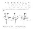
- FIG. 9One method of display using pairs of images from the tomographic projection dataset Tp is illustrates in FIG. 9 .
- Any two pairs of projections Tpmay provide stereo visualization, and by displaying dynamically sets of these pairs of projections, one will get both a stereoscopic view and one which dynamically moves around the body that was imaged.
- 8 projections Tpwere taken as a part of a tomosynthesis acquisition: Tp[ 1 ], Tp[ 2 ], . . . Tp[ 21 ].
- the first pair to be stereo viewedcould be Tp[ 1 ] and Tp[ 3 ], the second pair Tp[ 2 ] and Tp[ 4 ], the third pair Tp[ 3 ] and Tp[ 5 ] and so on to Tp[ 6 ] and Tp[ 8 ].
- pairscould be adjacent pairs such as Tp[ 1 ] and Tp[ 2 ] or separated by three projections Tp[ 1 ] and Tp[ 4 ], etc.
- the optimal spacing between the two projections in the displayed stereo pairis dependent upon the imaging geometry and angular separation between successive projections Tp[i] and Tp[i+1]. It is known that only certain angular differences between stereo pairs give good stereo visualization to humans, and the selection of appropriate pairs of images Tp for a particular acquisition setting can be determined through convenient experimentation.
- FIG. 10Another method of display uses a variant of the reconstructed dataset Tr and is illustrated in FIG. 10 .
- onereconstructs two different datasets Tr′ and Tr′′, both reconstructed from some or all of the original projections Tp.
- the geometry of reconstructionWhen performing reconstruction, one chooses the geometry of reconstruction and it is possible to reconstruct these two datasets into images Tr that differ in their view of the body by a selected angular separation, e.g. a few degrees, thus mimicking the apparent viewing of the body that the human eye would see, if it had x-ray vision.
- Each dataset Tr′ and Tr′′consist of sets of cross sectional slices.
- FIG. 9illustrates a stereo view of a thin cross-sectional slice through the body, and a scroll through such sections, while the patent proposes displaying stereo pairs of projection radiographs through the entire body.
- FIG. 11illustrates an overall mammography/tomography system in which the preferred but non-limiting examples discussed above can be implemented.
- the Figureillustrates in block diagram form an x-ray data acquisition unit 100 that includes an x-ray source 110 imaging a breast 112 supported on a breast platform 114 .
- An x-ray imager 116such as a flat panel x-ray imager commercially available from the assignee of this patent specification generates projection image data that can be a mammogram Mp or a tomosynthesis projection image Tp.
- X-ray source 110is mounted for movement around breast platform 114 so that images Tp can be taken at different angles.
- X-ray imager 116can be stationary or it can also move relative to breast platform 114 , preferable in synchronism with movement of x-ray source 110 . Elements 110 and 116 communicate with x-ray data acquisition control 118 that controls operations in a manner known from said earlier-filed patent specifications. X-ray image data from imager 116 is delivered to processing and image reconstruction unit 120 , where the data is processed as known from said earlier-filed patent application into Tp and Tr image data, possibly stored, and prepared for display at image display unit 122 as disclosed in the various embodiments described above.
Landscapes
- Engineering & Computer Science (AREA)
- Health & Medical Sciences (AREA)
- Life Sciences & Earth Sciences (AREA)
- Medical Informatics (AREA)
- Physics & Mathematics (AREA)
- General Health & Medical Sciences (AREA)
- Radiology & Medical Imaging (AREA)
- Nuclear Medicine, Radiotherapy & Molecular Imaging (AREA)
- Veterinary Medicine (AREA)
- Animal Behavior & Ethology (AREA)
- Pathology (AREA)
- Biomedical Technology (AREA)
- Heart & Thoracic Surgery (AREA)
- Molecular Biology (AREA)
- Surgery (AREA)
- Biophysics (AREA)
- Public Health (AREA)
- High Energy & Nuclear Physics (AREA)
- Optics & Photonics (AREA)
- Theoretical Computer Science (AREA)
- Human Computer Interaction (AREA)
- General Physics & Mathematics (AREA)
- Computer Vision & Pattern Recognition (AREA)
- General Engineering & Computer Science (AREA)
- Quality & Reliability (AREA)
- Dentistry (AREA)
- Oral & Maxillofacial Surgery (AREA)
- Computer Networks & Wireless Communication (AREA)
- Mathematical Optimization (AREA)
- Mathematical Physics (AREA)
- Mathematical Analysis (AREA)
- Algebra (AREA)
- Pure & Applied Mathematics (AREA)
- Apparatus For Radiation Diagnosis (AREA)
- Ultra Sonic Daignosis Equipment (AREA)
- Investigating Materials By The Use Of Optical Means Adapted For Particular Applications (AREA)
Abstract
Description
Claims (17)
Priority Applications (3)
| Application Number | Priority Date | Filing Date | Title |
|---|---|---|---|
| US14/742,040US9460508B2 (en) | 2002-11-27 | 2015-06-17 | Image handling and display in X-ray mammography and tomosynthesis |
| US15/282,596US10108329B2 (en) | 2002-11-27 | 2016-09-30 | Image handling and display in x-ray mammography and tomosynthesis |
| US16/127,570US10452252B2 (en) | 2002-11-27 | 2018-09-11 | Image handling and display in X-ray mammography and tomosynthesis |
Applications Claiming Priority (7)
| Application Number | Priority Date | Filing Date | Title |
|---|---|---|---|
| US10/305,480US7123684B2 (en) | 2002-11-27 | 2002-11-27 | Full field mammography with tissue exposure control, tomosynthesis, and dynamic field of view processing |
| US10/723,486US7831296B2 (en) | 2002-11-27 | 2003-11-26 | X-ray mammography with tomosynthesis |
| US62851604P | 2004-11-15 | 2004-11-15 | |
| US63129604P | 2004-11-26 | 2004-11-26 | |
| US11/271,050US7577282B2 (en) | 2002-11-27 | 2005-11-10 | Image handling and display in X-ray mammography and tomosynthesis |
| US12/344,121US9095306B2 (en) | 2002-11-27 | 2008-12-24 | Image handling and display in X-ray mammography and tomosynthesis |
| US14/742,040US9460508B2 (en) | 2002-11-27 | 2015-06-17 | Image handling and display in X-ray mammography and tomosynthesis |
Related Parent Applications (1)
| Application Number | Title | Priority Date | Filing Date |
|---|---|---|---|
| US12/344,121ContinuationUS9095306B2 (en) | 2002-11-27 | 2008-12-24 | Image handling and display in X-ray mammography and tomosynthesis |
Related Child Applications (1)
| Application Number | Title | Priority Date | Filing Date |
|---|---|---|---|
| US15/282,596ContinuationUS10108329B2 (en) | 2002-11-27 | 2016-09-30 | Image handling and display in x-ray mammography and tomosynthesis |
Publications (2)
| Publication Number | Publication Date |
|---|---|
| US20150310611A1 US20150310611A1 (en) | 2015-10-29 |
| US9460508B2true US9460508B2 (en) | 2016-10-04 |
Family
ID=37876814
Family Applications (5)
| Application Number | Title | Priority Date | Filing Date |
|---|---|---|---|
| US11/271,050Expired - LifetimeUS7577282B2 (en) | 2002-11-27 | 2005-11-10 | Image handling and display in X-ray mammography and tomosynthesis |
| US12/344,121Expired - LifetimeUS9095306B2 (en) | 2002-11-27 | 2008-12-24 | Image handling and display in X-ray mammography and tomosynthesis |
| US14/742,040Expired - Fee RelatedUS9460508B2 (en) | 2002-11-27 | 2015-06-17 | Image handling and display in X-ray mammography and tomosynthesis |
| US15/282,596Expired - LifetimeUS10108329B2 (en) | 2002-11-27 | 2016-09-30 | Image handling and display in x-ray mammography and tomosynthesis |
| US16/127,570Expired - Fee RelatedUS10452252B2 (en) | 2002-11-27 | 2018-09-11 | Image handling and display in X-ray mammography and tomosynthesis |
Family Applications Before (2)
| Application Number | Title | Priority Date | Filing Date |
|---|---|---|---|
| US11/271,050Expired - LifetimeUS7577282B2 (en) | 2002-11-27 | 2005-11-10 | Image handling and display in X-ray mammography and tomosynthesis |
| US12/344,121Expired - LifetimeUS9095306B2 (en) | 2002-11-27 | 2008-12-24 | Image handling and display in X-ray mammography and tomosynthesis |
Family Applications After (2)
| Application Number | Title | Priority Date | Filing Date |
|---|---|---|---|
| US15/282,596Expired - LifetimeUS10108329B2 (en) | 2002-11-27 | 2016-09-30 | Image handling and display in x-ray mammography and tomosynthesis |
| US16/127,570Expired - Fee RelatedUS10452252B2 (en) | 2002-11-27 | 2018-09-11 | Image handling and display in X-ray mammography and tomosynthesis |
Country Status (4)
| Country | Link |
|---|---|
| US (5) | US7577282B2 (en) |
| EP (3) | EP2974663B1 (en) |
| JP (1) | JP5702041B2 (en) |
| ES (2) | ES2744203T3 (en) |
Cited By (19)
| Publication number | Priority date | Publication date | Assignee | Title |
|---|---|---|---|---|
| US10108329B2 (en) | 2002-11-27 | 2018-10-23 | Hologic, Inc. | Image handling and display in x-ray mammography and tomosynthesis |
| US10194875B2 (en) | 2004-11-26 | 2019-02-05 | Hologic, Inc. | Integrated multi-mode mammography/tomosynthesis X-ray system and method |
| US10296199B2 (en) | 2002-11-27 | 2019-05-21 | Hologic, Inc. | Image handling and display in X-Ray mammography and tomosynthesis |
| US10413255B2 (en) | 2003-11-26 | 2019-09-17 | Hologic, Inc. | System and method for low dose tomosynthesis |
| US10881359B2 (en) | 2017-08-22 | 2021-01-05 | Hologic, Inc. | Computed tomography system for imaging multiple anatomical targets |
| US10959694B2 (en) | 2002-11-27 | 2021-03-30 | Hologic, Inc. | Full field mammography with tissue exposure control, tomosynthesis, and dynamic field of view processing |
| US11054534B1 (en) | 2020-04-24 | 2021-07-06 | Ronald Nutt | Time-resolved positron emission tomography encoder system for producing real-time, high resolution, three dimensional positron emission tomographic image without the necessity of performing image reconstruction |
| US11076820B2 (en) | 2016-04-22 | 2021-08-03 | Hologic, Inc. | Tomosynthesis with shifting focal spot x-ray system using an addressable array |
| US11090017B2 (en) | 2018-09-13 | 2021-08-17 | Hologic, Inc. | Generating synthesized projection images for 3D breast tomosynthesis or multi-mode x-ray breast imaging |
| US20220101573A1 (en)* | 2020-09-30 | 2022-03-31 | Siemens Healthcare Gmbh | Method for generating result slice images with at least partially different slice thickness |
| US11300695B2 (en) | 2020-04-24 | 2022-04-12 | Ronald Nutt | Time-resolved positron emission tomography encoder system for producing event-by-event, real-time, high resolution, three-dimensional positron emission tomographic image without the necessity of performing image reconstruction |
| US11419569B2 (en) | 2017-08-16 | 2022-08-23 | Hologic, Inc. | Image quality compliance tool |
| US11471118B2 (en) | 2020-03-27 | 2022-10-18 | Hologic, Inc. | System and method for tracking x-ray tube focal spot position |
| US11510306B2 (en) | 2019-12-05 | 2022-11-22 | Hologic, Inc. | Systems and methods for improved x-ray tube life |
| US11631490B2 (en) | 2018-09-25 | 2023-04-18 | Fujifilm Corporation | Medical image display device, method, and program for managing the display of abnormality detection results |
| US11783476B2 (en) | 2019-10-25 | 2023-10-10 | DeepHealth, Inc. | System and method for analyzing three-dimensional image data |
| US11786191B2 (en) | 2021-05-17 | 2023-10-17 | Hologic, Inc. | Contrast-enhanced tomosynthesis with a copper filter |
| US12367574B2 (en) | 2019-12-23 | 2025-07-22 | DeepHealth, Inc. | Systems and methods for analyzing two-dimensional and three-dimensional image data |
| US12414217B2 (en) | 2022-02-07 | 2025-09-09 | Hologic, Inc. | Systems and methods for adaptively controlling filament current in an X-ray tube |
Families Citing this family (125)
| Publication number | Priority date | Publication date | Assignee | Title |
|---|---|---|---|---|
| US8571289B2 (en) | 2002-11-27 | 2013-10-29 | Hologic, Inc. | System and method for generating a 2D image from a tomosynthesis data set |
| US7760924B2 (en)* | 2002-11-27 | 2010-07-20 | Hologic, Inc. | System and method for generating a 2D image from a tomosynthesis data set |
| US10638994B2 (en) | 2002-11-27 | 2020-05-05 | Hologic, Inc. | X-ray mammography with tomosynthesis |
| US8768026B2 (en)* | 2003-11-26 | 2014-07-01 | Hologic, Inc. | X-ray imaging with x-ray markers that provide adjunct information but preserve image quality |
| US7662082B2 (en) | 2004-11-05 | 2010-02-16 | Theragenics Corporation | Expandable brachytherapy device |
| US7702142B2 (en) | 2004-11-15 | 2010-04-20 | Hologic, Inc. | Matching geometry generation and display of mammograms and tomosynthesis images |
| US10008184B2 (en) | 2005-11-10 | 2018-06-26 | Hologic, Inc. | System and method for generating a 2D image using mammography and/or tomosynthesis image data |
| US7465268B2 (en) | 2005-11-18 | 2008-12-16 | Senorx, Inc. | Methods for asymmetrical irradiation of a body cavity |
| US7515682B2 (en)* | 2006-02-02 | 2009-04-07 | General Electric Company | Method and system to generate object image slices |
| WO2007095330A2 (en) | 2006-02-15 | 2007-08-23 | Hologic Inc | Breast biopsy and needle localization using tomosynthesis systems |
| US8340241B2 (en)* | 2006-02-27 | 2012-12-25 | Kabushiki Kaisha Toshiba | Image display apparatus and X-ray computed tomography apparatus |
| US7711087B2 (en)* | 2006-04-07 | 2010-05-04 | Varian Medical Systems, Inc. | Patient setup using tomosynthesis techniques |
| DE102006040791B3 (en)* | 2006-08-31 | 2008-03-27 | Fraunhofer-Gesellschaft zur Förderung der angewandten Forschung e.V. | Apparatus and method for combining a plurality of partial images for any imaging surfaces |
| US8051386B2 (en)* | 2006-12-21 | 2011-11-01 | Sectra Ab | CAD-based navigation of views of medical image data stacks or volumes |
| US7992100B2 (en)* | 2006-12-21 | 2011-08-02 | Sectra Ab | Dynamic slabbing to render views of medical image data |
| US8044972B2 (en)* | 2006-12-21 | 2011-10-25 | Sectra Mamea Ab | Synchronized viewing of tomosynthesis and/or mammograms |
| US20080219567A1 (en)* | 2007-03-07 | 2008-09-11 | General Electric Company | Tomosynthesis imaging data compression system and method |
| DE102007018324B3 (en)* | 2007-04-18 | 2008-06-05 | Siemens Ag | Image data acquisition system for use in imaging system, has data compression module whose functionality is implemented in context of customer-specific integrated circuit in front end area of imaging system |
| US8139710B2 (en)* | 2007-05-18 | 2012-03-20 | General Electric Company | Systems, methods and apparatus to image objects |
| US8553967B2 (en)* | 2007-06-29 | 2013-10-08 | General Electric Company | System and method for a digital X-ray radiographic tomosynthesis user interface |
| SE0702061L (en)* | 2007-09-17 | 2009-03-18 | Xcounter Ab | Method for creating, displaying and analyzing X-rays and device for implementing the method |
| US7630533B2 (en)* | 2007-09-20 | 2009-12-08 | Hologic, Inc. | Breast tomosynthesis with display of highlighted suspected calcifications |
| US20090085897A1 (en)* | 2007-09-28 | 2009-04-02 | Olympus Medical Systems Corp. | Image display apparatus |
| US7490988B1 (en)* | 2008-03-19 | 2009-02-17 | General Electric Company | Systems and methods for patient specific pixel spacing calibration for mammography X-ray |
| DE102008020670B4 (en)* | 2008-04-24 | 2016-05-04 | Siemens Aktiengesellschaft | Method for generating an image with a mammography device |
| US8634610B2 (en)* | 2008-06-20 | 2014-01-21 | The Trustees Of The University Of Pennsylvania | System and method for assessing cancer risk |
| US7792245B2 (en)* | 2008-06-24 | 2010-09-07 | Hologic, Inc. | Breast tomosynthesis system with shifting face shield |
| US7991106B2 (en) | 2008-08-29 | 2011-08-02 | Hologic, Inc. | Multi-mode tomosynthesis/mammography gain calibration and image correction using gain map information from selected projection angles |
| JP2010104771A (en)* | 2008-09-30 | 2010-05-13 | Fujifilm Corp | Radiation image diagnosing system |
| JP5512113B2 (en)* | 2008-10-20 | 2014-06-04 | 株式会社東芝 | Medical image display device and mammography device |
| JP5242349B2 (en)* | 2008-11-13 | 2013-07-24 | 株式会社東芝 | Image display device |
| JP5642698B2 (en)* | 2008-11-26 | 2014-12-17 | アナロジック コーポレイション | Method and apparatus for continuous wave tomosynthesis using photon counting |
| US9248311B2 (en) | 2009-02-11 | 2016-02-02 | Hologic, Inc. | System and method for modifying a flexibility of a brachythereapy catheter |
| US9579524B2 (en) | 2009-02-11 | 2017-02-28 | Hologic, Inc. | Flexible multi-lumen brachytherapy device |
| JP2010187916A (en)* | 2009-02-18 | 2010-09-02 | Fujifilm Corp | Image processing device, image processing system, and program |
| US10207126B2 (en) | 2009-05-11 | 2019-02-19 | Cytyc Corporation | Lumen visualization and identification system for multi-lumen balloon catheter |
| FR2945929B1 (en)* | 2009-06-02 | 2012-12-07 | Gen Electric | METHOD, SYSTEM AND COMPUTER PROGRAM PRODUCT FOR PROCESSING A CUT SERIES AND TOMOSYNTHESIS. |
| WO2010150147A1 (en)* | 2009-06-24 | 2010-12-29 | Koninklijke Philips Electronics N. V. | Spatial and shape characterization of an implanted device within an object |
| US8798353B2 (en)* | 2009-09-08 | 2014-08-05 | General Electric Company | Apparatus and method for two-view tomosynthesis imaging |
| JP5551960B2 (en)* | 2009-09-30 | 2014-07-16 | 富士フイルム株式会社 | Diagnosis support system, diagnosis support program, and diagnosis support method |
| ES2862525T3 (en) | 2009-10-08 | 2021-10-07 | Hologic Inc | Needle Breast Biopsy System and Method of Use |
| FR2954556B1 (en)* | 2009-12-22 | 2017-07-28 | Gen Electric | METHOD OF PROCESSING TOMOSYNTHESIS ACQUISITIONS TO OBTAIN REPRESENTATION OF THE CONTENT OF AN ORGAN |
| US9687200B2 (en) | 2010-06-08 | 2017-06-27 | Accuray Incorporated | Radiation treatment delivery system with translatable ring gantry |
| US8934605B2 (en) | 2010-02-24 | 2015-01-13 | Accuray Incorporated | Gantry image guided radiotherapy system and related treatment delivery methods |
| JP5587640B2 (en)* | 2010-03-10 | 2014-09-10 | 富士フイルム株式会社 | Radiographic imaging system and radiographic image display method |
| JP5436301B2 (en)* | 2010-03-29 | 2014-03-05 | 富士フイルム株式会社 | Radiography apparatus and radiation imaging system |
| JP5340213B2 (en)* | 2010-03-30 | 2013-11-13 | 富士フイルム株式会社 | Image display system |
| WO2011156526A2 (en) | 2010-06-08 | 2011-12-15 | Accuray, Inc. | Imaging methods and target tracking for image-guided radiation treatment |
| JP2012050605A (en)* | 2010-08-31 | 2012-03-15 | Fujifilm Corp | X-ray image photographing apparatus, the x-ray image photographing method, and program |
| US9352172B2 (en) | 2010-09-30 | 2016-05-31 | Hologic, Inc. | Using a guide member to facilitate brachytherapy device swap |
| CA2813591C (en) | 2010-10-05 | 2020-09-22 | Hologic, Inc. | Upright x-ray breast imaging with a ct mode, multiple tomosynthesis modes, and a mammography mode |
| FR2967520B1 (en)* | 2010-11-16 | 2012-12-21 | Gen Electric | METHOD FOR PROCESSING RADIOLOGICAL IMAGES OF A PATIENT |
| US20120133600A1 (en) | 2010-11-26 | 2012-05-31 | Hologic, Inc. | User interface for medical image review workstation |
| JP2012115381A (en)* | 2010-11-30 | 2012-06-21 | Fujifilm Corp | Phantom for radiation irradiation angle measurement, and radiation irradiation angle measurement method and stereoscopic image acquisition method using the phantom |
| US10342992B2 (en) | 2011-01-06 | 2019-07-09 | Hologic, Inc. | Orienting a brachytherapy applicator |
| US8536547B2 (en) | 2011-01-20 | 2013-09-17 | Accuray Incorporated | Ring gantry radiation treatment delivery system with dynamically controllable inward extension of treatment head |
| JP5605246B2 (en)* | 2011-02-01 | 2014-10-15 | コニカミノルタ株式会社 | Abnormal shadow candidate detection system, server device, and program |
| JP6057922B2 (en)* | 2011-03-08 | 2017-01-11 | ホロジック, インコーポレイテッドHologic, Inc. | System and method for dual energy and / or contrast enhanced breast imaging for screening, diagnosis and biopsy |
| JP5613094B2 (en)* | 2011-03-29 | 2014-10-22 | 富士フイルム株式会社 | Radiation image display apparatus and method |
| EP2782505B1 (en)* | 2011-11-27 | 2020-04-22 | Hologic, Inc. | System and method for generating a 2d image using mammography and/or tomosynthesis image data |
| JP6240097B2 (en) | 2012-02-13 | 2017-11-29 | ホロジック インコーポレイティッド | How to navigate a tomosynthesis stack using composite image data |
| JP6081093B2 (en)* | 2012-07-09 | 2017-02-15 | 東芝メディカルシステムズ株式会社 | Image display device |
| JP5528518B2 (en)* | 2012-09-28 | 2014-06-25 | 富士フイルム株式会社 | Radiation image generating apparatus and method |
| JP5519753B2 (en)* | 2012-09-28 | 2014-06-11 | 富士フイルム株式会社 | Tomographic image generating apparatus and method |
| JP5960015B2 (en)* | 2012-09-28 | 2016-08-02 | 富士フイルム株式会社 | Image display system, radiation image capturing system, image display control program, and image display control method |
| JP2014068874A (en)* | 2012-09-28 | 2014-04-21 | Fujifilm Corp | Image display system, radiation imaging system, image display control program and image display control method |
| US9940738B2 (en) | 2013-01-10 | 2018-04-10 | Hologic, Inc. | System and method for reducing data transmission volume in tomosynthesis |
| WO2014109197A1 (en) | 2013-01-11 | 2014-07-17 | 富士フイルム株式会社 | Image display device, image display method, medical image diagnosatic device, medical image diagnosatic method, medical image diagnosatic system, data preparation device, data preparation method, program, and recording medium |
| US10070828B2 (en) | 2013-03-05 | 2018-09-11 | Nview Medical Inc. | Imaging systems and related apparatus and methods |
| US10846860B2 (en) | 2013-03-05 | 2020-11-24 | Nview Medical Inc. | Systems and methods for x-ray tomosynthesis image reconstruction |
| US10092358B2 (en) | 2013-03-15 | 2018-10-09 | Hologic, Inc. | Tomosynthesis-guided biopsy apparatus and method |
| CN105451657A (en) | 2013-03-15 | 2016-03-30 | 霍罗吉克公司 | System and method for navigating tomosynthesis stack including automatic focusing |
| CA2925907C (en) | 2013-10-09 | 2022-03-15 | Hologic, Inc. | X-ray breast tomosynthesis enhancing spatial resolution including in the thickness direction of a flattened breast |
| EP3060132B1 (en) | 2013-10-24 | 2019-12-04 | Hologic, Inc. | System and method for navigating x-ray guided breast biopsy |
| WO2015126504A2 (en)* | 2013-12-04 | 2015-08-27 | The Trustees Of The University Of Pennsylvania | Dynamic four-dimensional contrast enhanced tomosynthesis |
| US9474497B2 (en) | 2014-01-15 | 2016-10-25 | Agfa Healthcare | Method and system for generating pre-scaled images for a series of mammography images |
| JP6506769B2 (en) | 2014-02-28 | 2019-04-24 | ホロジック, インコーポレイテッドHologic, Inc. | System and method for generating and displaying tomosynthesis image slabs |
| JP6482934B2 (en) | 2014-06-03 | 2019-03-13 | キヤノンメディカルシステムズ株式会社 | Image processing apparatus, radiation detection apparatus, and image processing method |
| JP6126058B2 (en)* | 2014-09-30 | 2017-05-10 | 富士フイルム株式会社 | Image display apparatus, image processing apparatus, radiographic imaging system, tomographic image display method, and tomographic image display program. |
| JP6275030B2 (en) | 2014-12-24 | 2018-02-07 | 富士フイルム株式会社 | Biopsy device and method of operating the same |
| US9652871B2 (en) | 2015-01-28 | 2017-05-16 | Impac Medical Systems, Inc. | Three dimensional localization of a moving target for adaptive radiation therapy |
| US9878177B2 (en)* | 2015-01-28 | 2018-01-30 | Elekta Ab (Publ) | Three dimensional localization and tracking for adaptive radiation therapy |
| KR20160139810A (en)* | 2015-05-28 | 2016-12-07 | 삼성전자주식회사 | Method and apparatus for displaying a medical image |
| JP6584231B2 (en)* | 2015-08-27 | 2019-10-02 | キヤノン株式会社 | Image processing apparatus, image processing system, image processing method, and program |
| CN115414059A (en)* | 2016-03-24 | 2022-12-02 | 尼维医疗公司 | System and method for image reconstruction |
| JP6821943B2 (en)* | 2016-04-22 | 2021-01-27 | コニカミノルタ株式会社 | Image generation system |
| JP6765871B2 (en)* | 2016-06-22 | 2020-10-07 | キヤノン株式会社 | Radiation imaging equipment, radiography systems, radiography methods, and programs |
| JP6853004B2 (en)* | 2016-09-16 | 2021-03-31 | キヤノンメディカルシステムズ株式会社 | Medical image processing equipment and mammography equipment |
| US10157460B2 (en) | 2016-10-25 | 2018-12-18 | General Electric Company | Interpolated tomosynthesis projection images |
| US11147525B2 (en) | 2016-11-04 | 2021-10-19 | Hologic, Inc. | Medical imaging device and method of operating a medical imaging device |
| US10096106B2 (en) | 2016-11-10 | 2018-10-09 | General Electric Company | Combined medical imaging |
| US10463333B2 (en) | 2016-12-13 | 2019-11-05 | General Electric Company | Synthetic images for biopsy control |
| US10372876B2 (en) | 2017-01-20 | 2019-08-06 | Agfa Healthcare Inc. | System and method for providing breast image data |
| EP3600047A1 (en) | 2017-03-30 | 2020-02-05 | Hologic, Inc. | System and method for hierarchical multi-level feature image synthesis and representation |
| EP3600052A1 (en) | 2017-03-30 | 2020-02-05 | Hologic, Inc. | System and method for targeted object enhancement to generate synthetic breast tissue images |
| CN110621233B (en) | 2017-03-30 | 2023-12-12 | 豪洛捷公司 | Method for processing breast tissue image data |
| EP3273374B1 (en)* | 2017-05-23 | 2023-05-17 | Siemens Healthcare GmbH | Method for analysing image data of a patient, system for performing a method for analysing image data of a patient and computer readable medium |
| WO2018236565A1 (en) | 2017-06-20 | 2018-12-27 | Hologic, Inc. | METHOD AND SYSTEM FOR MEDICAL IMAGING WITH DYNAMIC SELF-LEARNING |
| EP3644854B1 (en)* | 2017-06-30 | 2022-06-01 | Trophy | X-ray ct imaging method, interface system and apparatus |
| WO2019060843A1 (en) | 2017-09-22 | 2019-03-28 | Nview Medical Inc. | Image reconstruction using machine learning regularizers |
| JP7032157B2 (en)* | 2018-02-02 | 2022-03-08 | キヤノンメディカルシステムズ株式会社 | Medical image diagnostic device and X-ray irradiation control device |
| US10893842B2 (en) | 2018-02-08 | 2021-01-19 | Covidien Lp | System and method for pose estimation of an imaging device and for determining the location of a medical device with respect to a target |
| US12121304B2 (en) | 2018-05-04 | 2024-10-22 | Hologic, Inc. | Introducer and localization wire visualization |
| EP3787520B1 (en) | 2018-05-04 | 2024-09-25 | Hologic, Inc. | Biopsy needle visualization |
| WO2020068851A1 (en) | 2018-09-24 | 2020-04-02 | Hologic, Inc. | Breast mapping and abnormality localization |
| WO2020068767A1 (en) | 2018-09-28 | 2020-04-02 | Hologic, Inc. | System and method for synthetic breast tissue image generation by high density element suppression |
| WO2020107019A1 (en) | 2018-11-25 | 2020-05-28 | Hologic, Inc. | Multimodality hanging protocols |
| DE202020006044U1 (en) | 2019-03-29 | 2024-07-02 | Hologic Inc. | Report generation for cropped digital images |
| JP6694536B2 (en)* | 2019-04-09 | 2020-05-13 | キヤノンメディカルシステムズ株式会社 | Medical image display device and mammography device |
| US11883206B2 (en) | 2019-07-29 | 2024-01-30 | Hologic, Inc. | Personalized breast imaging system |
| JP7326070B2 (en) | 2019-08-27 | 2023-08-15 | 富士フイルム株式会社 | Image display device, method and program, image management device, method and program |
| EP4439580A3 (en) | 2019-09-27 | 2024-12-25 | Hologic, Inc. | Ai system for predicting reading time and reading complexity for reviewing 2d/3d breast images |
| EP4101386A4 (en) | 2020-02-04 | 2023-07-12 | FUJIFILM Corporation | IMAGE ADJUSTMENT DEVICE, METHOD AND PROGRAM |
| EP4119055B1 (en) | 2020-03-13 | 2024-10-30 | FUJIFILM Corporation | Image generation device and program, learning device and program, and image processing device and program |
| JP7446410B2 (en) | 2020-03-18 | 2024-03-08 | 富士フイルム株式会社 | Image processing device, method and program |
| CN115297778B (en) | 2020-03-18 | 2025-08-08 | 富士胶片株式会社 | Image processing device, method, and recording medium |
| JP7483434B2 (en)* | 2020-03-23 | 2024-05-15 | キヤノンメディカルシステムズ株式会社 | X-ray diagnostic device, stereoscopic image display device, and stereoscopic image display method |
| US11481038B2 (en) | 2020-03-27 | 2022-10-25 | Hologic, Inc. | Gesture recognition in controlling medical hardware or software |
| CN112767395B (en)* | 2021-04-07 | 2021-06-25 | 上海市东方医院(同济大学附属东方医院) | Image performance detection method and device |
| US12288275B2 (en)* | 2021-07-28 | 2025-04-29 | GE Precision Healthcare LLC | Methods and systems for breast tomosynthesis |
| US12186119B2 (en) | 2021-10-05 | 2025-01-07 | Hologic, Inc. | Interactive model interface for image selection in medical imaging systems |
| US12254586B2 (en) | 2021-10-25 | 2025-03-18 | Hologic, Inc. | Auto-focus tool for multimodality image review |
| WO2023097279A1 (en) | 2021-11-29 | 2023-06-01 | Hologic, Inc. | Systems and methods for correlating objects of interest |
| KR102841311B1 (en)* | 2022-01-21 | 2025-08-01 | 주식회사 루닛 | Apparatus and method for interlocking lesion locations between a guide image and a 3d tomosynthesis images composed of a plurality of 3d image slices |
| EP4467077A4 (en)* | 2022-01-21 | 2025-04-30 | Lunit Inc. | APPARATUS AND METHOD FOR LINKING AND PROVIDING LESION LOCATION BETWEEN A GUIDE IMAGE AND A 3D TOMOSYNTHESIS IMAGE COMPRISING A PLURALITY OF 3D IMAGE SLICES |
Citations (184)
| Publication number | Priority date | Publication date | Assignee | Title |
|---|---|---|---|---|
| US3502878A (en) | 1967-09-22 | 1970-03-24 | Us Health Education & Welfare | Automatic x-ray apparatus for limiting the field size of a projected x-ray beam in response to film size and to source-to-film distance |
| US3863073A (en) | 1973-04-26 | 1975-01-28 | Machlett Lab Inc | Automatic system for precise collimation of radiation |
| US3971950A (en) | 1975-04-14 | 1976-07-27 | Xerox Corporation | Independent compression and positioning device for use in mammography |
| US4160906A (en) | 1977-06-23 | 1979-07-10 | General Electric Company | Anatomically coordinated user dominated programmer for diagnostic x-ray apparatus |
| US4310766A (en) | 1978-09-06 | 1982-01-12 | Siemens Aktiengesellschaft | Motor driven x-ray grid and film-holder assembly |
| US4496557A (en) | 1981-08-27 | 1985-01-29 | Adir | Tricyclic ethers, their preparation and the pharmaceutical compositions containing them |
| US4513433A (en) | 1980-10-04 | 1985-04-23 | U.S. Philips Corporation | Fluoroscopy apparatus for forming layer images of a three-dimensional object |
| US4559641A (en) | 1983-06-24 | 1985-12-17 | Thomson-Cgr | Retractable cassette holder for a radiological and radiographic examination apparatus |
| US4662379A (en) | 1984-12-20 | 1987-05-05 | Stanford University | Coronary artery imaging system using gated tomosynthesis |
| US4706269A (en) | 1985-03-11 | 1987-11-10 | Reina Leo J | Anti-scatter grid structure |
| US4744099A (en) | 1983-11-03 | 1988-05-10 | Siemens Aktiengesellschaft | X-ray diagnostic apparatus comprising radiation filters |
| US4773086A (en) | 1983-12-16 | 1988-09-20 | Yokogawa Medical Systems, Limited | Operator console for X-ray tomographs |
| US4773087A (en) | 1986-04-14 | 1988-09-20 | University Of Rochester | Quality of shadowgraphic x-ray images |
| US4819258A (en) | 1986-11-28 | 1989-04-04 | Bennett X-Ray Corp. | Auto-setting of KV in an x-ray machine after selection of technic factors |
| US4821727A (en) | 1986-10-30 | 1989-04-18 | Elscint Ltd. | Mammographic biopsy needle holder system |
| WO1990005485A1 (en) | 1988-11-23 | 1990-05-31 | Nrt-Nordisk Roentgen Teknik A/S | X-ray apparatus |
| WO1990005495A1 (en) | 1988-11-15 | 1990-05-31 | Baxter International Inc. | Apparatus and method for collecting and freezing blood plasma |
| US4969174A (en) | 1989-09-06 | 1990-11-06 | General Electric Company | Scanning mammography system with reduced scatter radiation |
| US4989227A (en) | 1989-04-28 | 1991-01-29 | General Electric Cgr S.A. | Cassette carrier adaptable in size and position for mammography |
| US5018176A (en) | 1989-03-29 | 1991-05-21 | General Electric Cgr S.A. | Mammograph equipped with an integrated device for taking stereotaxic photographs and a method of utilization of said mammograph |
| US5029193A (en) | 1989-07-03 | 1991-07-02 | Siemens Aktiengesellschaft | X-ray diagnostic installation for mammography exposures |
| USRE33634E (en) | 1986-09-23 | 1991-07-09 | Method and structure for optimizing radiographic quality by controlling X-ray tube voltage, current focal spot size and exposure time | |
| US5051904A (en) | 1988-03-24 | 1991-09-24 | Olganix Corporation | Computerized dynamic tomography system |
| US5078142A (en) | 1989-11-21 | 1992-01-07 | Fischer Imaging Corporation | Precision mammographic needle biopsy system |
| US5163075A (en) | 1991-08-08 | 1992-11-10 | Eastman Kodak Company | Contrast enhancement of electrographic imaging |
| US5164976A (en) | 1989-09-06 | 1992-11-17 | General Electric Company | Scanning mammography system with improved skin line viewing |
| US5199056A (en) | 1989-11-28 | 1993-03-30 | Darrah Carol J | Mammography compression paddle |
| US5212637A (en) | 1989-11-22 | 1993-05-18 | Stereometrix Corporation | Method of investigating mammograms for masses and calcifications, and apparatus for practicing such method |
| US5240011A (en) | 1991-11-27 | 1993-08-31 | Fischer Imaging Corporation | Motorized biopsy needle positioner |
| US5289520A (en) | 1991-11-27 | 1994-02-22 | Lorad Corporation | Stereotactic mammography imaging system with prone position examination table and CCD camera |
| US5291539A (en) | 1992-10-19 | 1994-03-01 | General Electric Company | Variable focussed X-ray grid |
| US5359637A (en) | 1992-04-28 | 1994-10-25 | Wake Forest University | Self-calibrated tomosynthetic, radiographic-imaging system, method, and device |
| US5365562A (en) | 1993-09-20 | 1994-11-15 | Fischer Imaging Corporation | Digital imaging apparatus |
| US5415169A (en) | 1989-11-21 | 1995-05-16 | Fischer Imaging Corporation | Motorized mammographic biopsy apparatus |
| US5452367A (en) | 1993-11-29 | 1995-09-19 | Arch Development Corporation | Automated method and system for the segmentation of medical images |
| US5506877A (en) | 1994-11-23 | 1996-04-09 | The General Hospital Corporation | Mammography breast compression device and method |
| US5526394A (en) | 1993-11-26 | 1996-06-11 | Fischer Imaging Corporation | Digital scan mammography apparatus |
| US5539797A (en) | 1993-03-29 | 1996-07-23 | Ge Medical Systems Sa | Method and apparatus for digital stereotaxic mammography |
| US5553111A (en) | 1994-10-26 | 1996-09-03 | The General Hospital Corporation | Apparatus and method for improved tissue imaging |
| US5592562A (en) | 1994-01-19 | 1997-01-07 | International Business Machines Corporation | Inspection system for cross-sectional imaging |
| US5594769A (en) | 1991-11-27 | 1997-01-14 | Thermotrex Corporation | Method and apparatus for obtaining stereotactic mammographic guided needle breast biopsies |
| US5596200A (en) | 1992-10-14 | 1997-01-21 | Primex | Low dose mammography system |
| US5598454A (en) | 1994-04-26 | 1997-01-28 | Siemens Aktiengesellschaft | X-ray diagnostics installation |
| US5627869A (en) | 1995-11-22 | 1997-05-06 | Thermotrex Corporation | Mammography apparatus with proportional collimation |
| EP0775467A1 (en) | 1995-11-23 | 1997-05-28 | Planmed Oy | Method and system for controlling the functions of a mammography apparatus |
| US5657362A (en) | 1995-02-24 | 1997-08-12 | Arch Development Corporation | Automated method and system for computerized detection of masses and parenchymal distortions in medical images |
| US5668889A (en) | 1990-04-19 | 1997-09-16 | Fuji Photo Film Co., Ltd. | Apparatus for determining an image position, and method for adjusting read-out conditions and/or image processing conditions for a radiation image |
| US5706327A (en) | 1996-02-09 | 1998-01-06 | Trex Medical Corporation | Method and apparatus for mammographic compression |
| WO1998016903A1 (en) | 1996-10-16 | 1998-04-23 | Vital Images, Inc. | Advanced diagnostic viewer |
| US5769086A (en) | 1995-12-06 | 1998-06-23 | Biopsys Medical, Inc. | Control system and method for automated biopsy device |
| US5818898A (en) | 1995-11-07 | 1998-10-06 | Kabushiki Kaisha Toshiba | X-ray imaging apparatus using X-ray planar detector |
| US5828722A (en) | 1996-05-17 | 1998-10-27 | Sirona Dental Systems Gmbh & Co., Kg | X-ray diagnostic apparatus for tomosynthesis having a detector that detects positional relationships |
| US5844965A (en) | 1989-11-24 | 1998-12-01 | Thomas Jefferson University | Method and apparatus for using film density measurements of a radiograph to monitor the reproducibility of X-ray exposure parameters of a mammography unit |
| US5872828A (en) | 1996-07-23 | 1999-02-16 | The General Hospital Corporation | Tomosynthesis system for breast imaging |
| US5878104A (en) | 1996-05-17 | 1999-03-02 | Sirona Dental Systems Gmbh & Co. Kg | Method for producing tomosynthesis exposures employing a reference object formed by a region of the examination subject |
| US5896437A (en) | 1996-05-17 | 1999-04-20 | Sirona Dental Systems Gmbh & Co. Kg | X-ray diagnostics apparatus for tomosynthesis having a reference object in fixed relationship to a radiation emitter |
| US5941832A (en) | 1991-09-27 | 1999-08-24 | Tumey; David M. | Method and apparatus for detection of cancerous and precancerous conditions in a breast |
| US5970118A (en) | 1993-01-27 | 1999-10-19 | Sokolov; Oleg | Cellular X-ray grid |
| US5999836A (en) | 1995-06-06 | 1999-12-07 | Nelson; Robert S. | Enhanced high resolution breast imaging device and method utilizing non-ionizing radiation of narrow spectral bandwidth |
| US6005907A (en) | 1996-05-17 | 1999-12-21 | Sirona Dental Systems Gmbh & Co. Kg | Method and apparatus for producing tomosynthesis exposures employing a reference object composed of a number of sub-objects |
| EP0982001A1 (en) | 1998-08-25 | 2000-03-01 | General Electric Company | Protocol driven image reconstruction, display, and processing in a multislice imaging system |
| US6075879A (en) | 1993-09-29 | 2000-06-13 | R2 Technology, Inc. | Method and system for computer-aided lesion detection using information from multiple images |
| US6091841A (en) | 1997-09-04 | 2000-07-18 | Qualia Computing, Inc. | Method and system for segmenting desired regions in digital mammograms |
| US6137527A (en) | 1996-12-23 | 2000-10-24 | General Electric Company | System and method for prompt-radiology image screening service via satellite |
| US6149301A (en) | 1998-12-30 | 2000-11-21 | General Electric Company | X-ray target centering apparatus for radiographic imaging system |
| US6175117B1 (en) | 1998-01-23 | 2001-01-16 | Quanta Vision, Inc. | Tissue analysis apparatus |
| US6196715B1 (en) | 1959-04-28 | 2001-03-06 | Kabushiki Kaisha Toshiba | X-ray diagnostic system preferable to two dimensional x-ray detection |
| US6216540B1 (en) | 1995-06-06 | 2001-04-17 | Robert S. Nelson | High resolution device and method for imaging concealed objects within an obscuring medium |
| US6233473B1 (en) | 1999-02-16 | 2001-05-15 | Hologic, Inc. | Determining body composition using fan beam dual-energy x-ray absorptiometry |
| US6243441B1 (en) | 1999-07-13 | 2001-06-05 | Edge Medical Devices | Active matrix detector for X-ray imaging |
| US6256370B1 (en) | 2000-01-24 | 2001-07-03 | General Electric Company | Method and apparatus for performing tomosynthesis |
| US6272207B1 (en) | 1999-02-18 | 2001-08-07 | Creatv Microtech, Inc. | Method and apparatus for obtaining high-resolution digital X-ray and gamma ray images |
| US6289235B1 (en) | 1998-03-05 | 2001-09-11 | Wake Forest University | Method and system for creating three-dimensional images using tomosynthetic computed tomography |
| US6292530B1 (en) | 1999-04-29 | 2001-09-18 | General Electric Company | Method and apparatus for reconstructing image data acquired by a tomosynthesis x-ray imaging system |
| US20010038681A1 (en) | 2000-02-11 | 2001-11-08 | Brandeis University | Method and system for low-dose three-dimensional imaging of a scene |
| US6327336B1 (en) | 2000-06-05 | 2001-12-04 | Direct Radiography Corp. | Radiogram showing location of automatic exposure control sensor |
| JP2001346786A (en) | 2000-06-08 | 2001-12-18 | Fuji Photo Film Co Ltd | Questionable image detecting method, reproducing method, and detecting system |
| US6341156B1 (en) | 1999-05-14 | 2002-01-22 | Siemens Aktiengesellschaft | X-ray diagnostic apparatus with relatively moved x-ray source and detector |
| US20020012450A1 (en) | 1998-01-09 | 2002-01-31 | Osamu Tsujii | Image processing apparatus and method |
| US6375352B1 (en) | 1999-10-01 | 2002-04-23 | General Electric Company | Apparatus and method for obtaining x-ray tomosynthesis data for mammography |
| US20020048343A1 (en) | 2000-09-08 | 2002-04-25 | Laurent Launay | Three-dimensional image generating method and device and associated radiology apparatus |
| US20020050986A1 (en) | 2000-08-11 | 2002-05-02 | Hitoshi Inoue | Image display apparatus and method, and storage medium |
| US20020070970A1 (en) | 2000-11-22 | 2002-06-13 | Wood Susan A. | Graphical user interface for display of anatomical information |
| US20020075997A1 (en) | 2000-12-18 | 2002-06-20 | Unger Christopher David | Medical diagnostic method and apparatus to control dual energy exposure techniques based on image information |
| US6411836B1 (en) | 1999-12-30 | 2002-06-25 | General Electric Company | Method and apparatus for user preferences configuring in an image handling system |
| US6415015B2 (en) | 1999-12-28 | 2002-07-02 | Ge Medical Systems Sa | Method and system of compensation of thickness of an organ |
| US6442288B1 (en) | 1997-12-17 | 2002-08-27 | Siemens Aktiengesellschaft | Method for reconstructing a three-dimensional image of an object scanned in the context of a tomosynthesis, and apparatus for tomosynthesis |
| US6459925B1 (en) | 1998-11-25 | 2002-10-01 | Fischer Imaging Corporation | User interface system for mammographic imager |
| US6480565B1 (en) | 1999-11-18 | 2002-11-12 | University Of Rochester | Apparatus and method for cone beam volume computed tomography breast imaging |
| US6490476B1 (en) | 1999-10-14 | 2002-12-03 | Cti Pet Systems, Inc. | Combined PET and X-ray CT tomograph and method for using same |
| US20030007598A1 (en) | 2000-11-24 | 2003-01-09 | U-Systems, Inc. | Breast cancer screening with adjunctive ultrasound mammography |
| US20030018272A1 (en) | 2001-06-28 | 2003-01-23 | Treado Patrick J. | Method for Raman chemical imaging and characterization of calcification in tissue |
| US20030026386A1 (en) | 2001-02-01 | 2003-02-06 | Cha-Mei Tang | Anti-scatter grids and collimator designs, and their motion, fabrication and assembly |
| US20030072417A1 (en) | 2001-10-12 | 2003-04-17 | Kaufhold John Patrick | Method and apparatus for calibrating an imaging system |
| US6556655B1 (en) | 1998-11-27 | 2003-04-29 | Ge Medical Systems Sa | Method for automatic detection of glandular tissue |
| US20030097055A1 (en) | 2001-11-21 | 2003-05-22 | Philips Medical Systems(Cleveland), Inc. | Method of reviewing tomographic scans with a large number of images |
| US20030095624A1 (en) | 2001-11-21 | 2003-05-22 | Eberhard Jeffrey Wayne | Dose management system for mammographic tomosynthesis |
| US6574629B1 (en) | 1998-12-23 | 2003-06-03 | Agfa Corporation | Picture archiving and communication system |
| US6574304B1 (en) | 2002-09-13 | 2003-06-03 | Ge Medical Systems Global Technology Company, Llc | Computer aided acquisition of medical images |
| US6597762B1 (en) | 2002-11-27 | 2003-07-22 | Ge Medical Systems Global Technology Co., Llc | Method and apparatus of lesion detection and validation based on multiple reviews of a CT image |
| US6611575B1 (en) | 2001-07-27 | 2003-08-26 | General Electric Company | Method and system for high resolution 3D visualization of mammography images |
| US20030169847A1 (en) | 2001-11-21 | 2003-09-11 | University Of Massachusetts Medical Center | System and method for x-ray fluoroscopic imaging |
| US6620111B2 (en) | 2001-04-20 | 2003-09-16 | Ethicon Endo-Surgery, Inc. | Surgical biopsy device having automatic rotation of the probe for taking multiple samples |
| US6626849B2 (en) | 2001-11-01 | 2003-09-30 | Ethicon Endo-Surgery, Inc. | MRI compatible surgical biopsy device |
| US6633674B1 (en) | 1999-11-24 | 2003-10-14 | General Electric Company | Picture archiving and communication system employing improved data compression |
| US20030194051A1 (en) | 2002-04-15 | 2003-10-16 | General Electric | Tomosynthesis X-ray mammogram system and method with automatic drive system |
| US20030194121A1 (en) | 2002-04-15 | 2003-10-16 | General Electric Company | Computer aided detection (CAD) for 3D digital mammography |
| US20030194050A1 (en)* | 2002-04-15 | 2003-10-16 | General Electric Company | Multi modality X-ray and nuclear medicine mammography imaging system and method |
| US6638235B2 (en) | 2000-11-06 | 2003-10-28 | Suros Surgical Systems, Inc. | Biopsy apparatus |
| WO2003020114A3 (en) | 2001-08-31 | 2003-11-06 | Analogic Corp | Image positioning method and system for tomosynthesis in a digital x-ray radiography system |
| US6647092B2 (en) | 2002-01-18 | 2003-11-11 | General Electric Company | Radiation imaging system and method of collimation |
| US20030210254A1 (en) | 2002-05-13 | 2003-11-13 | Doan William D. | Method, system and computer product for displaying axial images |
| US20030212327A1 (en) | 2000-11-24 | 2003-11-13 | U-Systems Inc. | Adjunctive ultrasound processing and display for breast cancer screening |
| US20030215120A1 (en) | 2002-05-15 | 2003-11-20 | Renuka Uppaluri | Computer aided diagnosis of an image set |
| US20040008809A1 (en) | 1998-07-24 | 2004-01-15 | Webber Richard L. | Method and system for creating task-dependent three-dimensional images |
| US20040066884A1 (en) | 2002-10-07 | 2004-04-08 | Hermann Claus Bernhard Erich | Continuous scan tomosynthesis system and method |
| US20040066882A1 (en) | 2002-10-07 | 2004-04-08 | Eberhard Jeffrey Wayne | Continuous scan RAD tomosynthesis system and method |
| US20040070582A1 (en) | 2002-10-11 | 2004-04-15 | Matthew Warren Smith To Sonocine, Inc. | 3D modeling system |
| US20040094167A1 (en) | 2000-03-17 | 2004-05-20 | Brady John Michael | Three-dimensional reconstructions of a breast from two x-ray mammographics |
| US20040101095A1 (en) | 2002-11-27 | 2004-05-27 | Hologic Inc. | Full field mammography with tissue exposure control, tomosynthesis, and dynamic field of view processing |
| US6748044B2 (en) | 2002-09-13 | 2004-06-08 | Ge Medical Systems Global Technology Company, Llc | Computer assisted analysis of tomographic mammography data |
| US20040109529A1 (en) | 2002-12-10 | 2004-06-10 | General Electric Company | Full field digital tomosynthesis method and apparatus |
| US20040146221A1 (en) | 2003-01-23 | 2004-07-29 | Siegel Scott H. | Radiography Image Management System |
| WO2004043535A3 (en) | 2002-11-12 | 2004-07-29 | Univ Rochester | Apparatus and method for cone beam volume computed tomography breast imaging |
| WO2003057564A8 (en) | 2001-12-21 | 2004-08-26 | Nektar Therapeutics | Apparatus and method for sealing cavities |
| US20040171986A1 (en) | 1999-04-26 | 2004-09-02 | Scimed Life System, Inc. | Apparatus and methods for guiding a needle |
| US6813334B2 (en) | 2000-10-20 | 2004-11-02 | Koninklijke Philips Electronics N.V. | Tomosynthesis in a limited angular range |
| US20050063509A1 (en) | 2001-10-19 | 2005-03-24 | Defreitas Kenneth F | Mammography system and method employing offset compression paddles automatic collimation and retractable anti-scatter grid |
| US20050078797A1 (en) | 2002-03-01 | 2005-04-14 | Mats Danielsson | X-ray protection device |
| US6885724B2 (en) | 2003-08-22 | 2005-04-26 | Ge Medical Systems Global Technology Company, Llc | Radiographic tomosynthesis image acquisition utilizing asymmetric geometry |
| US20050105679A1 (en) | 2003-02-12 | 2005-05-19 | Tao Wu | Tomosynthesis imaging system and method |
| US20050113715A1 (en) | 2000-11-06 | 2005-05-26 | Jeffrey Schwindt | Biopsy apparatus |
| US20050113681A1 (en) | 2002-11-27 | 2005-05-26 | Defreitas Kenneth F. | X-ray mammography with tomosynthesis |
| US20050129172A1 (en) | 2003-11-17 | 2005-06-16 | Thomas Mertelmeier | X-ray diagnostic apparatus for mammography examinations |
| US6909792B1 (en) | 2000-06-23 | 2005-06-21 | Litton Systems, Inc. | Historical comparison of breast tissue by image processing |
| US6909790B2 (en) | 2002-02-15 | 2005-06-21 | Inventec Corporation | System and method of monitoring moving objects |
| US20050135555A1 (en) | 2003-12-23 | 2005-06-23 | Claus Bernhard Erich H. | Method and system for simultaneously viewing rendered volumes |
| US20050135664A1 (en) | 2003-12-23 | 2005-06-23 | Kaufhold John P. | Methods and apparatus for reconstruction of volume data from projection data |
| US20050226375A1 (en) | 2004-03-31 | 2005-10-13 | Eberhard Jeffrey W | Enhanced X-ray imaging system and method |
| US6957099B1 (en) | 1999-02-23 | 2005-10-18 | Teraview Limited | Method and apparatus for terahertz imaging |
| WO2005110230A1 (en) | 2004-05-14 | 2005-11-24 | Philips Intellectual Property & Standards Gmbh | System and method for diagnosing breast cancer |
| WO2005112767A1 (en) | 2004-05-21 | 2005-12-01 | Tissuomics Limited | Apparatus and method for penetrating radiation measurements |
| US6978040B2 (en) | 2001-12-19 | 2005-12-20 | Canon Kabushiki Kaisha | Optical recovery of radiographic geometry |
| US20060074288A1 (en) | 2004-10-04 | 2006-04-06 | Thomas Kelly | Estimating visceral fat by dual-energy x-ray absorptiometry |
| US20060098855A1 (en) | 2002-11-27 | 2006-05-11 | Gkanatsios Nikolaos A | Image handling and display in X-ray mammography and tomosynthesis |
| DE102004051401A1 (en) | 2004-10-21 | 2006-05-24 | Siemens Ag | Method for diagnosis in three-dimensional imaging, in particular in mammography |
| WO2006055830A2 (en) | 2004-11-15 | 2006-05-26 | Hologic, Inc. | Matching geometry generation and display of mammograms and tomosynthesis images |
| JP2006231054A (en) | 2005-02-21 | 2006-09-07 | General Electric Co <Ge> | Imaging apparatus having protection screen |
| US7110502B2 (en) | 2003-05-12 | 2006-09-19 | Canon Kabushiki Kaisha | Radiographic apparatus and method for switching a grid |
| WO2006058160A3 (en) | 2004-11-26 | 2006-10-12 | Hologic Inc | Integrated multi-mode mammography/tomosynthesis x-ray system and method |
| US7127091B2 (en) | 2000-12-22 | 2006-10-24 | Koninklijke Philips Electronics, N.V. | Method and apparatus for visualizing a limited part of a 3D medical image-point-related data set, through basing a rendered image on an intermediate region between first and second clipping planes, and including spectroscopic viewing of such region |
| US20060269041A1 (en) | 2005-05-18 | 2006-11-30 | Thomas Mertelmeier | Mammography method and apparatus for generating digital tomosynthetic 3D image |
| US20070036265A1 (en) | 2005-08-15 | 2007-02-15 | Zhenxue Jing | X-ray mammography/tomosynthesis of patient's breast |
| WO2005051197A3 (en) | 2003-11-26 | 2007-03-01 | Koninkl Philips Electronics Nv | Workflow optimization for high throughput imaging environment |
| US20070078335A1 (en)* | 2005-09-30 | 2007-04-05 | Eli Horn | System and method for in-vivo feature detection |
| US20070140419A1 (en) | 2005-09-13 | 2007-06-21 | Henri Souchay | Method and apparatus for combining images |
| JP2007521911A (en) | 2004-02-13 | 2007-08-09 | セクトラ マメア エービー | Method and configuration for X-ray imaging |
| US20070223651A1 (en) | 2006-03-21 | 2007-09-27 | Wagenaar Douglas J | Dual modality mammography device |
| US7302031B2 (en) | 2005-10-27 | 2007-11-27 | Sectra Mamea Ab | Method and arrangement relating to X-ray imaging |
| US7315607B2 (en) | 2005-09-02 | 2008-01-01 | Siemens Aktiengesellschaft | Mammograph system with a face shield |
| US20080019581A1 (en) | 2002-11-27 | 2008-01-24 | Gkanatsios Nikolaos A | Image Handling and display in X-ray mammography and tomosynthesis |
| US7323692B2 (en) | 2004-08-10 | 2008-01-29 | Research Foundation Of State University Of New York | Flat-panel detector with avalanche gain |
| US20080045833A1 (en) | 2006-02-15 | 2008-02-21 | Defreitas Kenneth F | Breast biopsy and needle localization using tomosynthesis systems |
| JP2008067933A (en) | 2006-09-14 | 2008-03-27 | Toshiba Corp | Digital mammography equipment |
| US20080101537A1 (en) | 2006-10-26 | 2008-05-01 | Fujifilm Corporation | Tomographic image obtainment apparatus and method |
| US20080112534A1 (en) | 2004-10-18 | 2008-05-15 | Defreitas Kenneth F | Mammography system and method employing offset compression paddles, automatic collimations, and retractable anti-scatter grid |
| US20080212861A1 (en) | 2005-07-26 | 2008-09-04 | Koninklijke Philips Electronics N. V. | Revolutionary Series Control for Medical Imaging Archive Manager |
| US20090080594A1 (en) | 2006-08-03 | 2009-03-26 | Kenneth Brooks | Dedicated breast radiation imaging/therapy system |
| US20090135997A1 (en) | 2006-03-27 | 2009-05-28 | Hologic, Inc. | Breast Compression For Digital Mammography, Tomosynthesis And Other Modalities |
| US20090213987A1 (en) | 2003-11-26 | 2009-08-27 | Jay Stein | System and Method for Low Dose Tomosynthesis |
| US20090238424A1 (en) | 2008-03-19 | 2009-09-24 | Kazumasa Arakita | Image processing apparatus and image processing method |
| US20090268865A1 (en) | 2003-11-26 | 2009-10-29 | Baorui Ren | X-ray imaging with X-ray markers that provide adjunct information but preserve image quality |
| US7630531B2 (en) | 2006-01-31 | 2009-12-08 | Mevis Medical Solutions, Inc. | Enhanced navigational tools for comparing medical images |
| US7630533B2 (en) | 2007-09-20 | 2009-12-08 | Hologic, Inc. | Breast tomosynthesis with display of highlighted suspected calcifications |
| US20100034450A1 (en) | 2008-01-15 | 2010-02-11 | Thomas Mertelmeier | Method and device for producing a tomosynthetic 3d x-ray image |
| US20100054400A1 (en) | 2008-08-29 | 2010-03-04 | Hologic, Inc. | Multi-mode tomosynthesis/mammography gain calibration and image correction using gain map information from selected projection angles |
| US7760924B2 (en) | 2002-11-27 | 2010-07-20 | Hologic, Inc. | System and method for generating a 2D image from a tomosynthesis data set |
| US7792245B2 (en) | 2008-06-24 | 2010-09-07 | Hologic, Inc. | Breast tomosynthesis system with shifting face shield |
| US20100226475A1 (en) | 2009-03-03 | 2010-09-09 | Hologic Inc. | Mammography/tomosynthesis systems and methods automatically deriving breast characteristics from breast x-ray images and automatically adjusting image processing parameters accordingly |
| US20100290585A1 (en) | 2009-05-13 | 2010-11-18 | Eva Eliasson | Mammography method and mammography apparatus |
| US7894646B2 (en) | 2003-08-01 | 2011-02-22 | Hitachi Medical Corporation | Medical image diagnosis support device and method for calculating degree of deformation from normal shapes of organ regions |
| US20110178389A1 (en) | 2008-05-02 | 2011-07-21 | Eigen, Inc. | Fused image moldalities guidance |
| US20110234630A1 (en) | 2008-11-28 | 2011-09-29 | Fujifilm Medical Systems USA, Inc | Active Overlay System and Method for Accessing and Manipulating Imaging Displays |
| US8571289B2 (en) | 2002-11-27 | 2013-10-29 | Hologic, Inc. | System and method for generating a 2D image from a tomosynthesis data set |
Family Cites Families (34)
| Publication number | Priority date | Publication date | Assignee | Title |
|---|---|---|---|---|
| US3365575A (en) | 1964-12-10 | 1968-01-23 | Charles & Stella Guttman Breas | Breast x-ray apparatus with means to accurately position the body of a patient |
| JPS5753531Y2 (en) | 1977-05-04 | 1982-11-19 | ||
| US4380086A (en) | 1980-11-24 | 1983-04-12 | Picker Corporation | Radiation imaging system with cyclically shiftable grid assembly |
| DE3236081A1 (en) | 1982-09-29 | 1984-03-29 | Siemens AG, 1000 Berlin und 8000 München | RECORDING DEVICE |
| JPH074354B2 (en) | 1984-10-29 | 1995-01-25 | 富士写真フイルム株式会社 | Radiation image information recording / reading device |
| US4760589A (en) | 1986-04-21 | 1988-07-26 | Siczek Aldona A | Grid cabinet and cassette tray for an X-ray examination apparatus |
| US4752948A (en) | 1986-12-01 | 1988-06-21 | University Of Chicago | Mobile radiography alignment device |
| FR2668359B1 (en) | 1990-10-24 | 1998-02-20 | Gen Electric Cgr | MAMMOGRAPH PROVIDED WITH A PERFECTED NEEDLE HOLDER. |
| US5256370B1 (en) | 1992-05-04 | 1996-09-03 | Indium Corp America | Lead-free alloy containing tin silver and indium |
| JPH06133964A (en)* | 1992-10-29 | 1994-05-17 | Hitachi Medical Corp | Spiral scanning radioscopic system |
| US5651047A (en)* | 1993-01-25 | 1997-07-22 | Cardiac Mariners, Incorporated | Maneuverable and locateable catheters |
| US5590271A (en)* | 1993-05-21 | 1996-12-31 | Digital Equipment Corporation | Interactive visualization environment with improved visual programming interface |
| US6031565A (en) | 1993-06-18 | 2000-02-29 | Gte Internetworking Incorporated | Stereo radiography |
| US5983123A (en) | 1993-10-29 | 1999-11-09 | United States Surgical Corporation | Methods and apparatus for performing ultrasound and enhanced X-ray imaging |
| US6345194B1 (en) | 1995-06-06 | 2002-02-05 | Robert S. Nelson | Enhanced high resolution breast imaging device and method utilizing non-ionizing radiation of narrow spectral bandwidth |
| EP0990254A1 (en) | 1996-02-12 | 2000-04-05 | The University of Akron | Multimedia detectors for medical imaging |
| US6415051B1 (en)* | 1999-06-24 | 2002-07-02 | Geometrix, Inc. | Generating 3-D models using a manually operated structured light source |
| US6244507B1 (en) | 1999-06-25 | 2001-06-12 | Canon Kabushiki Kaisha | Automatic grid parameter logging for digital radiography |
| US6266453B1 (en)* | 1999-07-26 | 2001-07-24 | Computerized Medical Systems, Inc. | Automated image fusion/alignment system and method |
| US7072501B2 (en)* | 2000-11-22 | 2006-07-04 | R2 Technology, Inc. | Graphical user interface for display of anatomical information |
| SE0004298D0 (en) | 2000-11-23 | 2000-11-23 | Siemens Elema Ab | Radiology Unit |
| US20020090055A1 (en) | 2000-11-27 | 2002-07-11 | Edge Medical Devices Ltd. | Digital X-ray bucky including grid storage |
| FR2818116B1 (en) | 2000-12-19 | 2004-08-27 | Ge Med Sys Global Tech Co Llc | MAMMOGRAPHY APPARATUS |
| US6674835B2 (en) | 2001-10-12 | 2004-01-06 | General Electric Co. | Methods and apparatus for estimating a material composition of an imaged object |
| US6581585B2 (en)* | 2001-11-16 | 2003-06-24 | Alfred F. Nibecker, Jr. | Air gun |
| US20040091520A1 (en) | 2002-11-12 | 2004-05-13 | Samuel Amdur | Disposable gloves with at least one alpha hydroxy acid |
| US10638994B2 (en) | 2002-11-27 | 2020-05-05 | Hologic, Inc. | X-ray mammography with tomosynthesis |
| US6909795B2 (en)* | 2003-06-16 | 2005-06-21 | R2 Technology, Inc. | Communicating computer-aided detection results in a standards-based medical imaging environment |
| US20050089205A1 (en)* | 2003-10-23 | 2005-04-28 | Ajay Kapur | Systems and methods for viewing an abnormality in different kinds of images |
| KR100907248B1 (en) | 2005-04-21 | 2009-07-10 | (주)안트로젠 | Volume replacement method of body by transplantation of differentiated young fat cells and biodegradable polymer |
| JP4769097B2 (en) | 2006-03-01 | 2011-09-07 | 富士フイルム株式会社 | Mammography device and breast compression plate used in the mammography device |
| JP4874755B2 (en) | 2006-09-29 | 2012-02-15 | 富士フイルム株式会社 | Radiation imaging equipment |
| JP2012050519A (en) | 2010-08-31 | 2012-03-15 | Fujifilm Corp | Mammographic apparatus |
| JP6057922B2 (en) | 2011-03-08 | 2017-01-11 | ホロジック, インコーポレイテッドHologic, Inc. | System and method for dual energy and / or contrast enhanced breast imaging for screening, diagnosis and biopsy |
- 2005
- 2005-11-10USUS11/271,050patent/US7577282B2/ennot_activeExpired - Lifetime
- 2006
- 2006-11-10ESES15181798Tpatent/ES2744203T3/enactiveActive
- 2006-11-10EPEP15181798.8Apatent/EP2974663B1/enactiveActive
- 2006-11-10EPEP06255790.5Apatent/EP1792569B1/enactiveActive
- 2006-11-10ESES19182295Tpatent/ES2947391T3/enactiveActive
- 2006-11-10EPEP19182295.6Apatent/EP3569152B1/enactiveActive
- 2006-11-10JPJP2006332575Apatent/JP5702041B2/enactiveActive
- 2008
- 2008-12-24USUS12/344,121patent/US9095306B2/ennot_activeExpired - Lifetime
- 2015
- 2015-06-17USUS14/742,040patent/US9460508B2/ennot_activeExpired - Fee Related
- 2016
- 2016-09-30USUS15/282,596patent/US10108329B2/ennot_activeExpired - Lifetime
- 2018
- 2018-09-11USUS16/127,570patent/US10452252B2/ennot_activeExpired - Fee Related
Patent Citations (277)
| Publication number | Priority date | Publication date | Assignee | Title |
|---|---|---|---|---|
| US6196715B1 (en) | 1959-04-28 | 2001-03-06 | Kabushiki Kaisha Toshiba | X-ray diagnostic system preferable to two dimensional x-ray detection |
| US3502878A (en) | 1967-09-22 | 1970-03-24 | Us Health Education & Welfare | Automatic x-ray apparatus for limiting the field size of a projected x-ray beam in response to film size and to source-to-film distance |
| US3863073A (en) | 1973-04-26 | 1975-01-28 | Machlett Lab Inc | Automatic system for precise collimation of radiation |
| US3971950A (en) | 1975-04-14 | 1976-07-27 | Xerox Corporation | Independent compression and positioning device for use in mammography |
| US4160906A (en) | 1977-06-23 | 1979-07-10 | General Electric Company | Anatomically coordinated user dominated programmer for diagnostic x-ray apparatus |
| US4310766A (en) | 1978-09-06 | 1982-01-12 | Siemens Aktiengesellschaft | Motor driven x-ray grid and film-holder assembly |
| US4513433A (en) | 1980-10-04 | 1985-04-23 | U.S. Philips Corporation | Fluoroscopy apparatus for forming layer images of a three-dimensional object |
| US4496557A (en) | 1981-08-27 | 1985-01-29 | Adir | Tricyclic ethers, their preparation and the pharmaceutical compositions containing them |
| US4559641A (en) | 1983-06-24 | 1985-12-17 | Thomson-Cgr | Retractable cassette holder for a radiological and radiographic examination apparatus |
| US4744099A (en) | 1983-11-03 | 1988-05-10 | Siemens Aktiengesellschaft | X-ray diagnostic apparatus comprising radiation filters |
| US4773086A (en) | 1983-12-16 | 1988-09-20 | Yokogawa Medical Systems, Limited | Operator console for X-ray tomographs |
| US4662379A (en) | 1984-12-20 | 1987-05-05 | Stanford University | Coronary artery imaging system using gated tomosynthesis |
| US4706269A (en) | 1985-03-11 | 1987-11-10 | Reina Leo J | Anti-scatter grid structure |
| US4773087A (en) | 1986-04-14 | 1988-09-20 | University Of Rochester | Quality of shadowgraphic x-ray images |
| USRE33634E (en) | 1986-09-23 | 1991-07-09 | Method and structure for optimizing radiographic quality by controlling X-ray tube voltage, current focal spot size and exposure time | |
| US4821727A (en) | 1986-10-30 | 1989-04-18 | Elscint Ltd. | Mammographic biopsy needle holder system |
| US4819258A (en) | 1986-11-28 | 1989-04-04 | Bennett X-Ray Corp. | Auto-setting of KV in an x-ray machine after selection of technic factors |
| US5051904A (en) | 1988-03-24 | 1991-09-24 | Olganix Corporation | Computerized dynamic tomography system |
| WO1990005495A1 (en) | 1988-11-15 | 1990-05-31 | Baxter International Inc. | Apparatus and method for collecting and freezing blood plasma |
| WO1990005485A1 (en) | 1988-11-23 | 1990-05-31 | Nrt-Nordisk Roentgen Teknik A/S | X-ray apparatus |
| US5018176A (en) | 1989-03-29 | 1991-05-21 | General Electric Cgr S.A. | Mammograph equipped with an integrated device for taking stereotaxic photographs and a method of utilization of said mammograph |
| US4989227A (en) | 1989-04-28 | 1991-01-29 | General Electric Cgr S.A. | Cassette carrier adaptable in size and position for mammography |
| US5029193A (en) | 1989-07-03 | 1991-07-02 | Siemens Aktiengesellschaft | X-ray diagnostic installation for mammography exposures |
| US4969174A (en) | 1989-09-06 | 1990-11-06 | General Electric Company | Scanning mammography system with reduced scatter radiation |
| US5164976A (en) | 1989-09-06 | 1992-11-17 | General Electric Company | Scanning mammography system with improved skin line viewing |
| US5078142A (en) | 1989-11-21 | 1992-01-07 | Fischer Imaging Corporation | Precision mammographic needle biopsy system |
| US5803912A (en) | 1989-11-21 | 1998-09-08 | Fischer Imaging Corporation | Positioning function mammographic biopsy function system with offset |
| US6022325A (en) | 1989-11-21 | 2000-02-08 | Fischer Imaging Corporation | Mammographic biopsy apparatus |
| US5735264A (en) | 1989-11-21 | 1998-04-07 | Fischer Imaging Corporation | Motorized mammographic biopsy apparatus |
| US5415169A (en) | 1989-11-21 | 1995-05-16 | Fischer Imaging Corporation | Motorized mammographic biopsy apparatus |
| US5212637A (en) | 1989-11-22 | 1993-05-18 | Stereometrix Corporation | Method of investigating mammograms for masses and calcifications, and apparatus for practicing such method |
| US5844965A (en) | 1989-11-24 | 1998-12-01 | Thomas Jefferson University | Method and apparatus for using film density measurements of a radiograph to monitor the reproducibility of X-ray exposure parameters of a mammography unit |
| US5199056A (en) | 1989-11-28 | 1993-03-30 | Darrah Carol J | Mammography compression paddle |
| US5668889A (en) | 1990-04-19 | 1997-09-16 | Fuji Photo Film Co., Ltd. | Apparatus for determining an image position, and method for adjusting read-out conditions and/or image processing conditions for a radiation image |
| US5163075A (en) | 1991-08-08 | 1992-11-10 | Eastman Kodak Company | Contrast enhancement of electrographic imaging |
| US5941832A (en) | 1991-09-27 | 1999-08-24 | Tumey; David M. | Method and apparatus for detection of cancerous and precancerous conditions in a breast |
| US5594769A (en) | 1991-11-27 | 1997-01-14 | Thermotrex Corporation | Method and apparatus for obtaining stereotactic mammographic guided needle breast biopsies |
| US5609152A (en) | 1991-11-27 | 1997-03-11 | Thermotrex Corporation | Prone position stereotactic mammography needle biopsy apparatus and method for using the same |
| US5426685A (en) | 1991-11-27 | 1995-06-20 | Thermotrex Corporation | Stereotactic mammography system imaging |
| US5289520A (en) | 1991-11-27 | 1994-02-22 | Lorad Corporation | Stereotactic mammography imaging system with prone position examination table and CCD camera |
| US5240011A (en) | 1991-11-27 | 1993-08-31 | Fischer Imaging Corporation | Motorized biopsy needle positioner |
| US5359637A (en) | 1992-04-28 | 1994-10-25 | Wake Forest University | Self-calibrated tomosynthetic, radiographic-imaging system, method, and device |
| US5668844A (en) | 1992-04-28 | 1997-09-16 | Webber; Richard L. | Self-calibrated tomosynthetic, radiographic-imaging system, method, and device |
| US5596200A (en) | 1992-10-14 | 1997-01-21 | Primex | Low dose mammography system |
| US5291539A (en) | 1992-10-19 | 1994-03-01 | General Electric Company | Variable focussed X-ray grid |
| US5970118A (en) | 1993-01-27 | 1999-10-19 | Sokolov; Oleg | Cellular X-ray grid |
| US5539797A (en) | 1993-03-29 | 1996-07-23 | Ge Medical Systems Sa | Method and apparatus for digital stereotaxic mammography |
| US5365562A (en) | 1993-09-20 | 1994-11-15 | Fischer Imaging Corporation | Digital imaging apparatus |
| US6075879A (en) | 1993-09-29 | 2000-06-13 | R2 Technology, Inc. | Method and system for computer-aided lesion detection using information from multiple images |
| US5526394A (en) | 1993-11-26 | 1996-06-11 | Fischer Imaging Corporation | Digital scan mammography apparatus |
| US5452367A (en) | 1993-11-29 | 1995-09-19 | Arch Development Corporation | Automated method and system for the segmentation of medical images |
| US5719952A (en) | 1994-01-19 | 1998-02-17 | International Business Machines Corporation | Inspection system for cross-sectional imaging |
| US5592562A (en) | 1994-01-19 | 1997-01-07 | International Business Machines Corporation | Inspection system for cross-sectional imaging |
| US5598454A (en) | 1994-04-26 | 1997-01-28 | Siemens Aktiengesellschaft | X-ray diagnostics installation |
| US5553111A (en) | 1994-10-26 | 1996-09-03 | The General Hospital Corporation | Apparatus and method for improved tissue imaging |
| US5506877A (en) | 1994-11-23 | 1996-04-09 | The General Hospital Corporation | Mammography breast compression device and method |
| US5657362A (en) | 1995-02-24 | 1997-08-12 | Arch Development Corporation | Automated method and system for computerized detection of masses and parenchymal distortions in medical images |
| US5999836A (en) | 1995-06-06 | 1999-12-07 | Nelson; Robert S. | Enhanced high resolution breast imaging device and method utilizing non-ionizing radiation of narrow spectral bandwidth |
| US6216540B1 (en) | 1995-06-06 | 2001-04-17 | Robert S. Nelson | High resolution device and method for imaging concealed objects within an obscuring medium |
| US5818898A (en) | 1995-11-07 | 1998-10-06 | Kabushiki Kaisha Toshiba | X-ray imaging apparatus using X-ray planar detector |
| US5627869A (en) | 1995-11-22 | 1997-05-06 | Thermotrex Corporation | Mammography apparatus with proportional collimation |
| EP0775467A1 (en) | 1995-11-23 | 1997-05-28 | Planmed Oy | Method and system for controlling the functions of a mammography apparatus |
| US5769086A (en) | 1995-12-06 | 1998-06-23 | Biopsys Medical, Inc. | Control system and method for automated biopsy device |
| US5706327A (en) | 1996-02-09 | 1998-01-06 | Trex Medical Corporation | Method and apparatus for mammographic compression |
| US5896437A (en) | 1996-05-17 | 1999-04-20 | Sirona Dental Systems Gmbh & Co. Kg | X-ray diagnostics apparatus for tomosynthesis having a reference object in fixed relationship to a radiation emitter |
| US6005907A (en) | 1996-05-17 | 1999-12-21 | Sirona Dental Systems Gmbh & Co. Kg | Method and apparatus for producing tomosynthesis exposures employing a reference object composed of a number of sub-objects |
| US5878104A (en) | 1996-05-17 | 1999-03-02 | Sirona Dental Systems Gmbh & Co. Kg | Method for producing tomosynthesis exposures employing a reference object formed by a region of the examination subject |
| US5828722A (en) | 1996-05-17 | 1998-10-27 | Sirona Dental Systems Gmbh & Co., Kg | X-ray diagnostic apparatus for tomosynthesis having a detector that detects positional relationships |
| US5872828A (en) | 1996-07-23 | 1999-02-16 | The General Hospital Corporation | Tomosynthesis system for breast imaging |
| US20030073895A1 (en) | 1996-10-15 | 2003-04-17 | Nields Morgan W. | User interface system for mammographic imager |
| US5986662A (en) | 1996-10-16 | 1999-11-16 | Vital Images, Inc. | Advanced diagnostic viewer employing automated protocol selection for volume-rendered imaging |
| WO1998016903A1 (en) | 1996-10-16 | 1998-04-23 | Vital Images, Inc. | Advanced diagnostic viewer |
| US6219059B1 (en) | 1996-10-16 | 2001-04-17 | Vital Images, Inc. | Interactive control of voxel attributes using selectable characteristics |
| US6137527A (en) | 1996-12-23 | 2000-10-24 | General Electric Company | System and method for prompt-radiology image screening service via satellite |
| US6091841A (en) | 1997-09-04 | 2000-07-18 | Qualia Computing, Inc. | Method and system for segmenting desired regions in digital mammograms |
| US6442288B1 (en) | 1997-12-17 | 2002-08-27 | Siemens Aktiengesellschaft | Method for reconstructing a three-dimensional image of an object scanned in the context of a tomosynthesis, and apparatus for tomosynthesis |
| US20020012450A1 (en) | 1998-01-09 | 2002-01-31 | Osamu Tsujii | Image processing apparatus and method |
| US6175117B1 (en) | 1998-01-23 | 2001-01-16 | Quanta Vision, Inc. | Tissue analysis apparatus |
| US6289235B1 (en) | 1998-03-05 | 2001-09-11 | Wake Forest University | Method and system for creating three-dimensional images using tomosynthetic computed tomography |
| US20040008809A1 (en) | 1998-07-24 | 2004-01-15 | Webber Richard L. | Method and system for creating task-dependent three-dimensional images |
| EP0982001A1 (en) | 1998-08-25 | 2000-03-01 | General Electric Company | Protocol driven image reconstruction, display, and processing in a multislice imaging system |
| US6141398A (en) | 1998-08-25 | 2000-10-31 | General Electric Company | Protocol driven image reconstruction, display, and processing in a multislice imaging system |
| US6459925B1 (en) | 1998-11-25 | 2002-10-01 | Fischer Imaging Corporation | User interface system for mammographic imager |
| WO2000051484A3 (en) | 1998-11-25 | 2004-02-12 | Fischer Imaging Corp | User interface system for mammographic imager |
| US6556655B1 (en) | 1998-11-27 | 2003-04-29 | Ge Medical Systems Sa | Method for automatic detection of glandular tissue |
| US6574629B1 (en) | 1998-12-23 | 2003-06-03 | Agfa Corporation | Picture archiving and communication system |
| US6149301A (en) | 1998-12-30 | 2000-11-21 | General Electric Company | X-ray target centering apparatus for radiographic imaging system |
| US6233473B1 (en) | 1999-02-16 | 2001-05-15 | Hologic, Inc. | Determining body composition using fan beam dual-energy x-ray absorptiometry |
| US6272207B1 (en) | 1999-02-18 | 2001-08-07 | Creatv Microtech, Inc. | Method and apparatus for obtaining high-resolution digital X-ray and gamma ray images |
| US6957099B1 (en) | 1999-02-23 | 2005-10-18 | Teraview Limited | Method and apparatus for terahertz imaging |
| US20040171986A1 (en) | 1999-04-26 | 2004-09-02 | Scimed Life System, Inc. | Apparatus and methods for guiding a needle |
| US6292530B1 (en) | 1999-04-29 | 2001-09-18 | General Electric Company | Method and apparatus for reconstructing image data acquired by a tomosynthesis x-ray imaging system |
| US6341156B1 (en) | 1999-05-14 | 2002-01-22 | Siemens Aktiengesellschaft | X-ray diagnostic apparatus with relatively moved x-ray source and detector |
| US6243441B1 (en) | 1999-07-13 | 2001-06-05 | Edge Medical Devices | Active matrix detector for X-ray imaging |
| US6375352B1 (en) | 1999-10-01 | 2002-04-23 | General Electric Company | Apparatus and method for obtaining x-ray tomosynthesis data for mammography |
| US6490476B1 (en) | 1999-10-14 | 2002-12-03 | Cti Pet Systems, Inc. | Combined PET and X-ray CT tomograph and method for using same |
| US6480565B1 (en) | 1999-11-18 | 2002-11-12 | University Of Rochester | Apparatus and method for cone beam volume computed tomography breast imaging |
| US6987831B2 (en) | 1999-11-18 | 2006-01-17 | University Of Rochester | Apparatus and method for cone beam volume computed tomography breast imaging |
| US7697660B2 (en) | 1999-11-18 | 2010-04-13 | University Of Rochester | Apparatus and method for cone beam computed tomography breast imaging |
| US6912319B1 (en) | 1999-11-24 | 2005-06-28 | Ge Medical Systems Information Technologies, Inc. | Method and system for lossless wavelet decomposition, compression and decompression of data |
| US6633674B1 (en) | 1999-11-24 | 2003-10-14 | General Electric Company | Picture archiving and communication system employing improved data compression |
| US6415015B2 (en) | 1999-12-28 | 2002-07-02 | Ge Medical Systems Sa | Method and system of compensation of thickness of an organ |
| US6411836B1 (en) | 1999-12-30 | 2002-06-25 | General Electric Company | Method and apparatus for user preferences configuring in an image handling system |
| US6256370B1 (en) | 2000-01-24 | 2001-07-03 | General Electric Company | Method and apparatus for performing tomosynthesis |
| US6744848B2 (en) | 2000-02-11 | 2004-06-01 | Brandeis University | Method and system for low-dose three-dimensional imaging of a scene |
| US20010038681A1 (en) | 2000-02-11 | 2001-11-08 | Brandeis University | Method and system for low-dose three-dimensional imaging of a scene |
| US20040094167A1 (en) | 2000-03-17 | 2004-05-20 | Brady John Michael | Three-dimensional reconstructions of a breast from two x-ray mammographics |
| US6327336B1 (en) | 2000-06-05 | 2001-12-04 | Direct Radiography Corp. | Radiogram showing location of automatic exposure control sensor |
| JP2001346786A (en) | 2000-06-08 | 2001-12-18 | Fuji Photo Film Co Ltd | Questionable image detecting method, reproducing method, and detecting system |
| US6909792B1 (en) | 2000-06-23 | 2005-06-21 | Litton Systems, Inc. | Historical comparison of breast tissue by image processing |
| US20020050986A1 (en) | 2000-08-11 | 2002-05-02 | Hitoshi Inoue | Image display apparatus and method, and storage medium |
| US20020048343A1 (en) | 2000-09-08 | 2002-04-25 | Laurent Launay | Three-dimensional image generating method and device and associated radiology apparatus |
| US6813334B2 (en) | 2000-10-20 | 2004-11-02 | Koninklijke Philips Electronics N.V. | Tomosynthesis in a limited angular range |
| US20060155209A1 (en) | 2000-11-06 | 2006-07-13 | Miller Michael E | Selectively detachable outer cannula hub |
| US20040267157A1 (en) | 2000-11-06 | 2004-12-30 | Miller Michael E | Biopsy apparatus |
| US20050049521A1 (en) | 2000-11-06 | 2005-03-03 | Suros Surgical Systems, Inc. | Collection filter for biopsy apparatus |
| US6758824B1 (en) | 2000-11-06 | 2004-07-06 | Suros Surgical Systems, Inc. | Biopsy apparatus |
| US20050113715A1 (en) | 2000-11-06 | 2005-05-26 | Jeffrey Schwindt | Biopsy apparatus |
| US6638235B2 (en) | 2000-11-06 | 2003-10-28 | Suros Surgical Systems, Inc. | Biopsy apparatus |
| US20060030784A1 (en) | 2000-11-06 | 2006-02-09 | Miller Michael E | Collection filter |
| US20060129062A1 (en) | 2000-11-06 | 2006-06-15 | Nicoson Zachary R | Fluid control element for biopsy apparatus |
| US20020070970A1 (en) | 2000-11-22 | 2002-06-13 | Wood Susan A. | Graphical user interface for display of anatomical information |
| US20030212327A1 (en) | 2000-11-24 | 2003-11-13 | U-Systems Inc. | Adjunctive ultrasound processing and display for breast cancer screening |
| US20030007598A1 (en) | 2000-11-24 | 2003-01-09 | U-Systems, Inc. | Breast cancer screening with adjunctive ultrasound mammography |
| US6501819B2 (en) | 2000-12-18 | 2002-12-31 | Ge Medical Systems Global Technology Company, Llc | Medical diagnostic method and apparatus to control dual energy exposure techniques based on image information |
| US20020075997A1 (en) | 2000-12-18 | 2002-06-20 | Unger Christopher David | Medical diagnostic method and apparatus to control dual energy exposure techniques based on image information |
| US7127091B2 (en) | 2000-12-22 | 2006-10-24 | Koninklijke Philips Electronics, N.V. | Method and apparatus for visualizing a limited part of a 3D medical image-point-related data set, through basing a rendered image on an intermediate region between first and second clipping planes, and including spectroscopic viewing of such region |
| US20030026386A1 (en) | 2001-02-01 | 2003-02-06 | Cha-Mei Tang | Anti-scatter grids and collimator designs, and their motion, fabrication and assembly |
| US6620111B2 (en) | 2001-04-20 | 2003-09-16 | Ethicon Endo-Surgery, Inc. | Surgical biopsy device having automatic rotation of the probe for taking multiple samples |
| US20030018272A1 (en) | 2001-06-28 | 2003-01-23 | Treado Patrick J. | Method for Raman chemical imaging and characterization of calcification in tissue |
| US6611575B1 (en) | 2001-07-27 | 2003-08-26 | General Electric Company | Method and system for high resolution 3D visualization of mammography images |
| WO2003020114A3 (en) | 2001-08-31 | 2003-11-06 | Analogic Corp | Image positioning method and system for tomosynthesis in a digital x-ray radiography system |
| US20030072417A1 (en) | 2001-10-12 | 2003-04-17 | Kaufhold John Patrick | Method and apparatus for calibrating an imaging system |
| US7319735B2 (en) | 2001-10-19 | 2008-01-15 | Hologic, Inc. | Mammography system and method employing offset compression paddles, automatic collimation, and retractable anti-scatter grid |
| US20100150306A1 (en) | 2001-10-19 | 2010-06-17 | Hologic, Inc. | Mammography System And Method Employing Offset Compression Paddles, Automatic Collimation, And Retractable Anti-scatter Grid |
| US8559595B2 (en) | 2001-10-19 | 2013-10-15 | Hologic, Inc. | Mammography system and method employing offset compression paddles automatic collimation and retractable anti-scatter grid |
| US20140098935A1 (en) | 2001-10-19 | 2014-04-10 | Hologic, Inc. | Mammography System and Method Employing Offset Compression Paddles, Automatic Collimation, and Retractable Anti-scatter Grid |
| US7986765B2 (en) | 2001-10-19 | 2011-07-26 | Hologic, Inc. | Mammography system and method employing offset compression paddles, automatic collimation, and retractable anti-scatter grid |
| US7443949B2 (en) | 2001-10-19 | 2008-10-28 | Hologic, Inc. | Mammography system and method employing offset compression paddles, automatic collimation, and retractable anti-scatter grid |
| US20050063509A1 (en) | 2001-10-19 | 2005-03-24 | Defreitas Kenneth F | Mammography system and method employing offset compression paddles automatic collimation and retractable anti-scatter grid |
| US7688940B2 (en) | 2001-10-19 | 2010-03-30 | Hologic, Inc. | Mammography system and method employing offset compression paddles, automatic collimation, and retractable anti-scatter grid |
| US20070076844A1 (en) | 2001-10-19 | 2007-04-05 | Defreitas Kenneth F | Mammography system and method employing offset compression paddles, automatic collimation, and retractable anti-scatter grid |
| US6626849B2 (en) | 2001-11-01 | 2003-09-30 | Ethicon Endo-Surgery, Inc. | MRI compatible surgical biopsy device |
| US6751285B2 (en) | 2001-11-21 | 2004-06-15 | General Electric Company | Dose management system for mammographic tomosynthesis |
| US20030097055A1 (en) | 2001-11-21 | 2003-05-22 | Philips Medical Systems(Cleveland), Inc. | Method of reviewing tomographic scans with a large number of images |
| US20030169847A1 (en) | 2001-11-21 | 2003-09-11 | University Of Massachusetts Medical Center | System and method for x-ray fluoroscopic imaging |
| US20030095624A1 (en) | 2001-11-21 | 2003-05-22 | Eberhard Jeffrey Wayne | Dose management system for mammographic tomosynthesis |
| US6978040B2 (en) | 2001-12-19 | 2005-12-20 | Canon Kabushiki Kaisha | Optical recovery of radiographic geometry |
| WO2003057564A8 (en) | 2001-12-21 | 2004-08-26 | Nektar Therapeutics | Apparatus and method for sealing cavities |
| US20040066904A1 (en) | 2002-01-18 | 2004-04-08 | Eberhard Jeffrey Wayne | Radiation imaging system and method of collimation |
| US6647092B2 (en) | 2002-01-18 | 2003-11-11 | General Electric Company | Radiation imaging system and method of collimation |
| US6909790B2 (en) | 2002-02-15 | 2005-06-21 | Inventec Corporation | System and method of monitoring moving objects |
| US20050078797A1 (en) | 2002-03-01 | 2005-04-14 | Mats Danielsson | X-ray protection device |
| US20030194121A1 (en) | 2002-04-15 | 2003-10-16 | General Electric Company | Computer aided detection (CAD) for 3D digital mammography |
| US20030194051A1 (en) | 2002-04-15 | 2003-10-16 | General Electric | Tomosynthesis X-ray mammogram system and method with automatic drive system |
| US20030194050A1 (en)* | 2002-04-15 | 2003-10-16 | General Electric Company | Multi modality X-ray and nuclear medicine mammography imaging system and method |
| US6882700B2 (en) | 2002-04-15 | 2005-04-19 | General Electric Company | Tomosynthesis X-ray mammogram system and method with automatic drive system |
| US20030210254A1 (en) | 2002-05-13 | 2003-11-13 | Doan William D. | Method, system and computer product for displaying axial images |
| US20030215120A1 (en) | 2002-05-15 | 2003-11-20 | Renuka Uppaluri | Computer aided diagnosis of an image set |
| US6748044B2 (en) | 2002-09-13 | 2004-06-08 | Ge Medical Systems Global Technology Company, Llc | Computer assisted analysis of tomographic mammography data |
| US6574304B1 (en) | 2002-09-13 | 2003-06-03 | Ge Medical Systems Global Technology Company, Llc | Computer aided acquisition of medical images |
| US6940943B2 (en) | 2002-10-07 | 2005-09-06 | General Electric Company | Continuous scan tomosynthesis system and method |
| US20040066884A1 (en) | 2002-10-07 | 2004-04-08 | Hermann Claus Bernhard Erich | Continuous scan tomosynthesis system and method |
| US6970531B2 (en) | 2002-10-07 | 2005-11-29 | General Electric Company | Continuous scan RAD tomosynthesis system and method |
| US20040066882A1 (en) | 2002-10-07 | 2004-04-08 | Eberhard Jeffrey Wayne | Continuous scan RAD tomosynthesis system and method |
| US20040070582A1 (en) | 2002-10-11 | 2004-04-15 | Matthew Warren Smith To Sonocine, Inc. | 3D modeling system |
| WO2004043535A3 (en) | 2002-11-12 | 2004-07-29 | Univ Rochester | Apparatus and method for cone beam volume computed tomography breast imaging |
| US8416915B2 (en) | 2002-11-27 | 2013-04-09 | Hologic, Inc. | Full field mammography with tissue exposure control, tomosynthesis, and dynamic field of view processing |
| US20050113681A1 (en) | 2002-11-27 | 2005-05-26 | Defreitas Kenneth F. | X-ray mammography with tomosynthesis |
| US20140044230A1 (en) | 2002-11-27 | 2014-02-13 | Hologic, Inc. | System and Method for Low Dose Tomosynthesis |
| US20060098855A1 (en) | 2002-11-27 | 2006-05-11 | Gkanatsios Nikolaos A | Image handling and display in X-ray mammography and tomosynthesis |
| US8452379B2 (en) | 2002-11-27 | 2013-05-28 | Hologic, Inc. | X-ray mammography with tomosynthesis |
| US20140086471A1 (en) | 2002-11-27 | 2014-03-27 | Hologic, Inc. | System and Method for Generating A 2D Image from a Tomosynthesis Data Set |
| US20040101095A1 (en) | 2002-11-27 | 2004-05-27 | Hologic Inc. | Full field mammography with tissue exposure control, tomosynthesis, and dynamic field of view processing |
| US8831171B2 (en) | 2002-11-27 | 2014-09-09 | Hologic, Inc. | Full field mammography with tissue exposure control, tomosynthesis, and dynamic field of view processing |
| US20130028374A1 (en) | 2002-11-27 | 2013-01-31 | Hologic, Inc. | Image handling and display in x-ray mammography and tomosynthesis |
| US8571289B2 (en) | 2002-11-27 | 2013-10-29 | Hologic, Inc. | System and method for generating a 2D image from a tomosynthesis data set |
| US20140376690A1 (en) | 2002-11-27 | 2014-12-25 | Hologic, Inc. | Full field mammography with tissue exposure control, tomosynthesis, and dynamic field of view processing |
| US8285020B2 (en) | 2002-11-27 | 2012-10-09 | Hologic, Inc. | Image handling and display in x-ray mammography and tomosynthesis |
| US7123684B2 (en) | 2002-11-27 | 2006-10-17 | Hologic, Inc. | Full field mammography with tissue exposure control, tomosynthesis, and dynamic field of view processing |
| US9042612B2 (en) | 2002-11-27 | 2015-05-26 | Hologic, Inc. | Image handling and display in X-ray mammography and tomosynthesis |
| EP1569556B1 (en) | 2002-11-27 | 2012-04-25 | Hologic, Inc. | Full field mammography with tissue exposure control, tomosynthesis, and dynamic field of view processing |
| US20090141859A1 (en) | 2002-11-27 | 2009-06-04 | Hologic, Inc. | Image Handling and Display in X-Ray Mammography and Tomosynthesis |
| US7430272B2 (en) | 2002-11-27 | 2008-09-30 | Hologic, Inc. | Full field mammography with tissue exposure control, tomosynthesis, and dynamic field of view processing |
| US20070030949A1 (en) | 2002-11-27 | 2007-02-08 | Zhenxue Jing | Full field mammography with tissue exposure control, tomosynthesis, and dynamic field of view processing |
| US7949091B2 (en) | 2002-11-27 | 2011-05-24 | Hologic, Inc. | Full field mammography with tissue exposure control, tomosynthesis, and dynamic field of view processing |
| US7916915B2 (en) | 2002-11-27 | 2011-03-29 | Hologic, Inc | Image handling and display in x-ray mammography and tomosynthesis |
| US7831296B2 (en) | 2002-11-27 | 2010-11-09 | Hologic, Inc. | X-ray mammography with tomosynthesis |
| US7760853B2 (en) | 2002-11-27 | 2010-07-20 | Hologic, Inc. | Full field mammography with tissue exposure control, tomosynthesis, and dynamic field of view processing |
| US7760924B2 (en) | 2002-11-27 | 2010-07-20 | Hologic, Inc. | System and method for generating a 2D image from a tomosynthesis data set |
| US20080019581A1 (en) | 2002-11-27 | 2008-01-24 | Gkanatsios Nikolaos A | Image Handling and display in X-ray mammography and tomosynthesis |
| US6597762B1 (en) | 2002-11-27 | 2003-07-22 | Ge Medical Systems Global Technology Co., Llc | Method and apparatus of lesion detection and validation based on multiple reviews of a CT image |
| US20150160848A1 (en) | 2002-11-27 | 2015-06-11 | Hologic, Inc. | Image handling and display in x-ray mammography and tomosynthesis |
| US20090010384A1 (en) | 2002-11-27 | 2009-01-08 | Hologic, Inc. | Full field mammography with tissue exposure control, tomosynthesis, and dynamic field of view processing |
| US20090296882A1 (en) | 2002-11-27 | 2009-12-03 | Hologic, Inc. | Image Handling And Display In X-Ray Mammography And Tomosynthess |
| US7616801B2 (en) | 2002-11-27 | 2009-11-10 | Hologic, Inc. | Image handling and display in x-ray mammography and tomosynthesis |
| US7577282B2 (en) | 2002-11-27 | 2009-08-18 | Hologic, Inc. | Image handling and display in X-ray mammography and tomosynthesis |
| US7110490B2 (en) | 2002-12-10 | 2006-09-19 | General Electric Company | Full field digital tomosynthesis method and apparatus |
| EP1428473A3 (en) | 2002-12-10 | 2004-07-21 | General Electric Company | Full field digital tomosynthesis method and apparatus |
| US20040109529A1 (en) | 2002-12-10 | 2004-06-10 | General Electric Company | Full field digital tomosynthesis method and apparatus |
| US20060291618A1 (en) | 2002-12-10 | 2006-12-28 | General Electric Company | Tomographic mammography method |
| US20040146221A1 (en) | 2003-01-23 | 2004-07-29 | Siegel Scott H. | Radiography Image Management System |
| US20050105679A1 (en) | 2003-02-12 | 2005-05-19 | Tao Wu | Tomosynthesis imaging system and method |
| US7110502B2 (en) | 2003-05-12 | 2006-09-19 | Canon Kabushiki Kaisha | Radiographic apparatus and method for switching a grid |
| US7894646B2 (en) | 2003-08-01 | 2011-02-22 | Hitachi Medical Corporation | Medical image diagnosis support device and method for calculating degree of deformation from normal shapes of organ regions |
| US6885724B2 (en) | 2003-08-22 | 2005-04-26 | Ge Medical Systems Global Technology Company, Llc | Radiographic tomosynthesis image acquisition utilizing asymmetric geometry |
| US20050129172A1 (en) | 2003-11-17 | 2005-06-16 | Thomas Mertelmeier | X-ray diagnostic apparatus for mammography examinations |
| US6999554B2 (en) | 2003-11-17 | 2006-02-14 | Siemens Aktiengesellschaft | X-ray diagnostic apparatus for mammography examinations |
| WO2005051197A3 (en) | 2003-11-26 | 2007-03-01 | Koninkl Philips Electronics Nv | Workflow optimization for high throughput imaging environment |
| US20090268865A1 (en) | 2003-11-26 | 2009-10-29 | Baorui Ren | X-ray imaging with X-ray markers that provide adjunct information but preserve image quality |
| US8565372B2 (en) | 2003-11-26 | 2013-10-22 | Hologic, Inc | System and method for low dose tomosynthesis |
| US20090213987A1 (en) | 2003-11-26 | 2009-08-27 | Jay Stein | System and Method for Low Dose Tomosynthesis |
| US20050135555A1 (en) | 2003-12-23 | 2005-06-23 | Claus Bernhard Erich H. | Method and system for simultaneously viewing rendered volumes |
| US20050135664A1 (en) | 2003-12-23 | 2005-06-23 | Kaufhold John P. | Methods and apparatus for reconstruction of volume data from projection data |
| JP2007521911A (en) | 2004-02-13 | 2007-08-09 | セクトラ マメア エービー | Method and configuration for X-ray imaging |
| US7142633B2 (en) | 2004-03-31 | 2006-11-28 | General Electric Company | Enhanced X-ray imaging system and method |
| US20050226375A1 (en) | 2004-03-31 | 2005-10-13 | Eberhard Jeffrey W | Enhanced X-ray imaging system and method |
| WO2005110230A1 (en) | 2004-05-14 | 2005-11-24 | Philips Intellectual Property & Standards Gmbh | System and method for diagnosing breast cancer |
| US20070225600A1 (en) | 2004-05-14 | 2007-09-27 | Koninklijke Philips Electronics, N.V. | System And Method For Diagnosing Breast Cancer |
| WO2005112767A1 (en) | 2004-05-21 | 2005-12-01 | Tissuomics Limited | Apparatus and method for penetrating radiation measurements |
| US7323692B2 (en) | 2004-08-10 | 2008-01-29 | Research Foundation Of State University Of New York | Flat-panel detector with avalanche gain |
| US20060074288A1 (en) | 2004-10-04 | 2006-04-06 | Thomas Kelly | Estimating visceral fat by dual-energy x-ray absorptiometry |
| US7609806B2 (en) | 2004-10-18 | 2009-10-27 | Hologic Inc. | Mammography system and method employing offset compression paddles, automatic collimations, and retractable anti-scatter grid |
| US20080112534A1 (en) | 2004-10-18 | 2008-05-15 | Defreitas Kenneth F | Mammography system and method employing offset compression paddles, automatic collimations, and retractable anti-scatter grid |
| DE102004051401A1 (en) | 2004-10-21 | 2006-05-24 | Siemens Ag | Method for diagnosis in three-dimensional imaging, in particular in mammography |
| US7702142B2 (en) | 2004-11-15 | 2010-04-20 | Hologic, Inc. | Matching geometry generation and display of mammograms and tomosynthesis images |
| WO2006055830A2 (en) | 2004-11-15 | 2006-05-26 | Hologic, Inc. | Matching geometry generation and display of mammograms and tomosynthesis images |
| US8712127B2 (en) | 2004-11-15 | 2014-04-29 | Hologic, Inc. | Matching geometry generation and display of mammograms and tomosynthesis images |
| US8155421B2 (en) | 2004-11-15 | 2012-04-10 | Hologic, Inc. | Matching geometry generation and display of mammograms and tomosynthesis images |
| US20140232752A1 (en) | 2004-11-15 | 2014-08-21 | Hologic, Inc. | Matching geometry generation and display of mammograms and tomosynthesis |
| EP2602743B1 (en) | 2004-11-15 | 2014-11-05 | Hologic, Inc. | Matching geometry generation and display of mammograms and tomosynthesis images |
| US20080130979A1 (en) | 2004-11-15 | 2008-06-05 | Baorui Ren | Matching Geometry Generation and Display of Mammograms and Tomosynthesis Images |
| US20100195882A1 (en) | 2004-11-15 | 2010-08-05 | Hologic, Inc. | Matching Geometry Generation And Display Of Mammograms And Tomosynthesis Images |
| WO2006058160A3 (en) | 2004-11-26 | 2006-10-12 | Hologic Inc | Integrated multi-mode mammography/tomosynthesis x-ray system and method |
| US20110069809A1 (en) | 2004-11-26 | 2011-03-24 | Hologic, Inc. | Integrated multi-mode mammography/tomosynthesis x-ray system and method |
| US9066706B2 (en) | 2004-11-26 | 2015-06-30 | Hologic, Inc. | Integrated multi-mode mammography/tomosynthesis x-ray system and method |
| US20150049859A1 (en) | 2004-11-26 | 2015-02-19 | Hologic, Inc. | Integrated multi-mode mammography/tomosynthesis x-ray system and method |
| US20090003519A1 (en) | 2004-11-26 | 2009-01-01 | Kenneth Defreitas | Integrated Multi-Mode Mammography/Tomosynthesis X-Ray System And Method |
| US8565374B2 (en) | 2004-11-26 | 2013-10-22 | Hologic, Inc. | Integrated multi-mode mammography/tomosynthesis x-ray system and method |
| US8175219B2 (en) | 2004-11-26 | 2012-05-08 | Hologic, Inc. | Integrated multi-mode mammography/tomosynthesis X-ray system and method |
| US7869563B2 (en) | 2004-11-26 | 2011-01-11 | Hologic, Inc. | Integrated multi-mode mammography/tomosynthesis x-ray system and method |
| JP2006231054A (en) | 2005-02-21 | 2006-09-07 | General Electric Co <Ge> | Imaging apparatus having protection screen |
| US20060269041A1 (en) | 2005-05-18 | 2006-11-30 | Thomas Mertelmeier | Mammography method and apparatus for generating digital tomosynthetic 3D image |
| US20080212861A1 (en) | 2005-07-26 | 2008-09-04 | Koninklijke Philips Electronics N. V. | Revolutionary Series Control for Medical Imaging Archive Manager |
| US7583786B2 (en) | 2005-08-15 | 2009-09-01 | Hologic, Inc. | X-ray mammography/tomosynthesis of patient's breast |
| US20090304147A1 (en) | 2005-08-15 | 2009-12-10 | Hologic, Inc. | X-ray mammography/tomosynthesis of patient's breast |
| JP2007050264A (en) | 2005-08-15 | 2007-03-01 | Hologic Inc | X-ray mammography / tomosynthesis of the patient's chest |
| US20070036265A1 (en) | 2005-08-15 | 2007-02-15 | Zhenxue Jing | X-ray mammography/tomosynthesis of patient's breast |
| EP1759637A2 (en) | 2005-08-15 | 2007-03-07 | Hologic, Inc. | X-ray mammography/tomosynthesis of patient's breast |
| US7881428B2 (en) | 2005-08-15 | 2011-02-01 | Hologic, Inc. | X-ray mammography/tomosynthesis of patient's breast |
| US7245694B2 (en) | 2005-08-15 | 2007-07-17 | Hologic, Inc. | X-ray mammography/tomosynthesis of patient's breast |
| US20070242800A1 (en) | 2005-08-15 | 2007-10-18 | Zhenxue Jing | X-ray mammography/tomosynthesis of patient's breast |
| US7315607B2 (en) | 2005-09-02 | 2008-01-01 | Siemens Aktiengesellschaft | Mammograph system with a face shield |
| US20070140419A1 (en) | 2005-09-13 | 2007-06-21 | Henri Souchay | Method and apparatus for combining images |
| US20070078335A1 (en)* | 2005-09-30 | 2007-04-05 | Eli Horn | System and method for in-vivo feature detection |
| US7302031B2 (en) | 2005-10-27 | 2007-11-27 | Sectra Mamea Ab | Method and arrangement relating to X-ray imaging |
| US7630531B2 (en) | 2006-01-31 | 2009-12-08 | Mevis Medical Solutions, Inc. | Enhanced navigational tools for comparing medical images |
| US20080045833A1 (en) | 2006-02-15 | 2008-02-21 | Defreitas Kenneth F | Breast biopsy and needle localization using tomosynthesis systems |
| US20070223651A1 (en) | 2006-03-21 | 2007-09-27 | Wagenaar Douglas J | Dual modality mammography device |
| US20090135997A1 (en) | 2006-03-27 | 2009-05-28 | Hologic, Inc. | Breast Compression For Digital Mammography, Tomosynthesis And Other Modalities |
| US20090080594A1 (en) | 2006-08-03 | 2009-03-26 | Kenneth Brooks | Dedicated breast radiation imaging/therapy system |
| US20090080602A1 (en) | 2006-08-03 | 2009-03-26 | Kenneth Brooks | Dedicated breast radiation imaging/therapy system |
| JP2008067933A (en) | 2006-09-14 | 2008-03-27 | Toshiba Corp | Digital mammography equipment |
| US20080101537A1 (en) | 2006-10-26 | 2008-05-01 | Fujifilm Corporation | Tomographic image obtainment apparatus and method |
| US7630533B2 (en) | 2007-09-20 | 2009-12-08 | Hologic, Inc. | Breast tomosynthesis with display of highlighted suspected calcifications |
| US20100086188A1 (en) | 2007-09-20 | 2010-04-08 | Hologic, Inc. | Breast Tomosynthesis With Display Of Highlighted Suspected Calcifications |
| US8131049B2 (en) | 2007-09-20 | 2012-03-06 | Hologic, Inc. | Breast tomosynthesis with display of highlighted suspected calcifications |
| US20100034450A1 (en) | 2008-01-15 | 2010-02-11 | Thomas Mertelmeier | Method and device for producing a tomosynthetic 3d x-ray image |
| US20090238424A1 (en) | 2008-03-19 | 2009-09-24 | Kazumasa Arakita | Image processing apparatus and image processing method |
| US20110178389A1 (en) | 2008-05-02 | 2011-07-21 | Eigen, Inc. | Fused image moldalities guidance |
| US7792245B2 (en) | 2008-06-24 | 2010-09-07 | Hologic, Inc. | Breast tomosynthesis system with shifting face shield |
| US7991106B2 (en) | 2008-08-29 | 2011-08-02 | Hologic, Inc. | Multi-mode tomosynthesis/mammography gain calibration and image correction using gain map information from selected projection angles |
| US20100054400A1 (en) | 2008-08-29 | 2010-03-04 | Hologic, Inc. | Multi-mode tomosynthesis/mammography gain calibration and image correction using gain map information from selected projection angles |
| US20110234630A1 (en) | 2008-11-28 | 2011-09-29 | Fujifilm Medical Systems USA, Inc | Active Overlay System and Method for Accessing and Manipulating Imaging Displays |
| US8170320B2 (en) | 2009-03-03 | 2012-05-01 | Hologic, Inc. | Mammography/tomosynthesis systems and methods automatically deriving breast characteristics from breast x-ray images and automatically adjusting image processing parameters accordingly |
| US20100226475A1 (en) | 2009-03-03 | 2010-09-09 | Hologic Inc. | Mammography/tomosynthesis systems and methods automatically deriving breast characteristics from breast x-ray images and automatically adjusting image processing parameters accordingly |
| US20100290585A1 (en) | 2009-05-13 | 2010-11-18 | Eva Eliasson | Mammography method and mammography apparatus |
Non-Patent Citations (22)
| Title |
|---|
| "Essentials for life: Senographe Essential Full-Field Digital Mammography system", GE Health-care Brochure, MM-0132-05.06-EN-US, 2006, 12 pgs. |
| "Filtered Back Projection," (NYGREN) published May 8, 2007; URL:http://web.archive.org/web/19991010131715/http://www.owlnet.rice.edu/-.about.e1ec539/Projects97/cult/node2.html., 2 pgs. |
| "Lorad Selenia" Document B-BI-SEO US/Intl (May 2006) copyright Hologic 2006, 12 pgs. |
| Acrin website, located at https://www.acrin.org/patients/aboutimagingexamsandagents/aboutmammographyandtomosynthesis.aspx, "About Mammography and Tomosnythesis", obtained online on Dec. 8, 2015, 5 pgs. |
| American College of Radiology website, located at http://www.acr.org/FAQs/DBT-FAQ, "Digital Breast Tomosynthesis FAQ for Insurers", obtained online on Dec. 8, 2015, 2 pages. |
| Aslund, Magnus, "Digital Mammography with a Photon Counting Detector in a Scanned Multislit Geometry", Doctoral Thesis, Dept of Physics, Royal Institute of Technology, Stockholm, Sweden, Apr. 2007, 51 pages. |
| Chan, Heang-Ping et al., "ROC study of the effect of stereoscopic imaging on assessment of breast lesions", Medical Physics, vol. 32, No. 4, Apr. 2005, 7 pgs. |
| Cole, Elodia, et al., "The Effects of Gray Scale Image Processing on Digital Mammography Interpretation Performance", Academic Radiology, vol. 12, No. 5, pp. 585-595, May 2005. |
| Digital Clinical Reports, Tomosynthesis, GE Brochure 98-5493, Nov. 1998, 8 pgs. |
| Dobbins, James T., "Digital x-ray tomosynthesis: current state of the art and clinical potential," Physics in Medicine and Biology, Taylor and Francis LTD, London GB, vol. 48, No. 19, Oct. 7, 2003, 42 pages. |
| European Extended Search Report in Application 15181798.8, mailed Nov. 26, 2015, 11 pgs. |
| Federica Pediconi et al., "Color-coded automated signal intensity-curve for detection and characterization of breast lesions: Preliminary evaluation of a new software for MR-based breast imaging", International Congress Series 1281 (2005) 1081-1086. |
| Grant, David G., "Tomosynthesis: a three-dimensional imaging technique", IEEE Trans. Biomed. Engineering, vol. BME-19, #1, Jan. 1972, pp. 20-28. |
| Kita et al., "Correspondence between different view breast X-rays using simulation of breast deformation", Proceedings 1998 IEE Computer Society Conference on Computer Vision and Pattern Recognition, Santa Barbara, CA, Jun. 23-25, 1998, pp. 700-707. |
| Mammographic Accreditation Phantom, http://www.cirsinc.com/pdfs/015cp.pdf. (2006), 2 pgs. |
| Niklason, Loren T. et al., "Digital Tomosynthesis in Breast Imaging", Radiology, Nov. 1997, vol. 205, No. 2, pp. 399-406. |
| Pisano, Etta D., "Digital Mammography", Radiology, vol. 234, No. 2, Feb. 2005, pp. 353-362. |
| Senographe 700 & 800T (GE); 2-page download on Jun. 22, 2006 from www.gehealthcare.com/inen/rad/whe/products/mswh800t.html.; Figures 1-7 on 4 sheets re lateral shift compression paddle, 2 pgs. |
| Smith, A., "Fundamentals of Breast Tomosynthesis", White Paper, Hologic Inc., WP-00007, Jun. 2008, 8 pgs. |
| Smith, Andrew, PhD, "Full Field Breast Tomosynthesis", Hologic White Paper, Oct. 2004, 6 pgs. |
| Wheeler F. W., et al. "Micro-Calcification Detection in Digital Tomosynthesis Mammography", Proceedings of SPIE, Conf-Physics of Semiconductor Devices, Dec. 11, 2001 to Dec. 15, 2001, Delhi, SPIE, US, vol. 6144, Feb. 13, 2006, 12 pgs. |
| Wu, Tao, et al. "Tomographic Mammography Using a Limited Number of Low-Dose Cone-Beam Projection Images" Medical Physics, AIP, Melville, NY, vol. 30, No. 3, Mar. 1, 2003, p. 365-380. |
Cited By (28)
| Publication number | Priority date | Publication date | Assignee | Title |
|---|---|---|---|---|
| US10108329B2 (en) | 2002-11-27 | 2018-10-23 | Hologic, Inc. | Image handling and display in x-ray mammography and tomosynthesis |
| US11372534B2 (en) | 2002-11-27 | 2022-06-28 | Hologic, Inc. | Image handling and display in x-ray mammography and tomosynthesis |
| US10296199B2 (en) | 2002-11-27 | 2019-05-21 | Hologic, Inc. | Image handling and display in X-Ray mammography and tomosynthesis |
| US10452252B2 (en) | 2002-11-27 | 2019-10-22 | Hologic, Inc. | Image handling and display in X-ray mammography and tomosynthesis |
| US10719223B2 (en) | 2002-11-27 | 2020-07-21 | Hologic, Inc. | Image handling and display in X-ray mammography and tomosynthesis |
| US10959694B2 (en) | 2002-11-27 | 2021-03-30 | Hologic, Inc. | Full field mammography with tissue exposure control, tomosynthesis, and dynamic field of view processing |
| US10413255B2 (en) | 2003-11-26 | 2019-09-17 | Hologic, Inc. | System and method for low dose tomosynthesis |
| US10905385B2 (en) | 2004-11-26 | 2021-02-02 | Hologic, Inc. | Integrated multi-mode mammography/tomosynthesis x-ray system and method |
| US11617548B2 (en) | 2004-11-26 | 2023-04-04 | Hologic, Inc. | Integrated multi-mode mammography/tomosynthesis x-ray system and method |
| US10194875B2 (en) | 2004-11-26 | 2019-02-05 | Hologic, Inc. | Integrated multi-mode mammography/tomosynthesis X-ray system and method |
| US11076820B2 (en) | 2016-04-22 | 2021-08-03 | Hologic, Inc. | Tomosynthesis with shifting focal spot x-ray system using an addressable array |
| US11672500B2 (en) | 2017-08-16 | 2023-06-13 | Hologic, Inc. | Image quality compliance tool |
| US12011310B2 (en) | 2017-08-16 | 2024-06-18 | Hologic, Inc. | Image quality compliance tool |
| US12053319B2 (en) | 2017-08-16 | 2024-08-06 | Hologic, Inc. | Image quality compliance tool |
| US11419569B2 (en) | 2017-08-16 | 2022-08-23 | Hologic, Inc. | Image quality compliance tool |
| US10881359B2 (en) | 2017-08-22 | 2021-01-05 | Hologic, Inc. | Computed tomography system for imaging multiple anatomical targets |
| US11090017B2 (en) | 2018-09-13 | 2021-08-17 | Hologic, Inc. | Generating synthesized projection images for 3D breast tomosynthesis or multi-mode x-ray breast imaging |
| US11631490B2 (en) | 2018-09-25 | 2023-04-18 | Fujifilm Corporation | Medical image display device, method, and program for managing the display of abnormality detection results |
| US11783476B2 (en) | 2019-10-25 | 2023-10-10 | DeepHealth, Inc. | System and method for analyzing three-dimensional image data |
| US11510306B2 (en) | 2019-12-05 | 2022-11-22 | Hologic, Inc. | Systems and methods for improved x-ray tube life |
| US12367574B2 (en) | 2019-12-23 | 2025-07-22 | DeepHealth, Inc. | Systems and methods for analyzing two-dimensional and three-dimensional image data |
| US11471118B2 (en) | 2020-03-27 | 2022-10-18 | Hologic, Inc. | System and method for tracking x-ray tube focal spot position |
| US11300695B2 (en) | 2020-04-24 | 2022-04-12 | Ronald Nutt | Time-resolved positron emission tomography encoder system for producing event-by-event, real-time, high resolution, three-dimensional positron emission tomographic image without the necessity of performing image reconstruction |
| US11054534B1 (en) | 2020-04-24 | 2021-07-06 | Ronald Nutt | Time-resolved positron emission tomography encoder system for producing real-time, high resolution, three dimensional positron emission tomographic image without the necessity of performing image reconstruction |
| US11935161B2 (en)* | 2020-09-30 | 2024-03-19 | Siemens Healthineers Ag | Method for generating result slice images with at least partially different slice thickness |
| US20220101573A1 (en)* | 2020-09-30 | 2022-03-31 | Siemens Healthcare Gmbh | Method for generating result slice images with at least partially different slice thickness |
| US11786191B2 (en) | 2021-05-17 | 2023-10-17 | Hologic, Inc. | Contrast-enhanced tomosynthesis with a copper filter |
| US12414217B2 (en) | 2022-02-07 | 2025-09-09 | Hologic, Inc. | Systems and methods for adaptively controlling filament current in an X-ray tube |
Also Published As
| Publication number | Publication date |
|---|---|
| US7577282B2 (en) | 2009-08-18 |
| EP1792569A3 (en) | 2007-09-19 |
| US20060098855A1 (en) | 2006-05-11 |
| EP1792569B1 (en) | 2015-08-26 |
| EP2974663A1 (en) | 2016-01-20 |
| US20170024113A1 (en) | 2017-01-26 |
| JP5702041B2 (en) | 2015-04-15 |
| US20190095087A1 (en) | 2019-03-28 |
| US20090141859A1 (en) | 2009-06-04 |
| ES2744203T3 (en) | 2020-02-24 |
| US10108329B2 (en) | 2018-10-23 |
| ES2947391T3 (en) | 2023-08-08 |
| EP3569152B1 (en) | 2023-05-03 |
| EP2974663B1 (en) | 2019-06-26 |
| US9095306B2 (en) | 2015-08-04 |
| US20150310611A1 (en) | 2015-10-29 |
| EP3569152A1 (en) | 2019-11-20 |
| JP2007130487A (en) | 2007-05-31 |
| US10452252B2 (en) | 2019-10-22 |
| EP1792569A2 (en) | 2007-06-06 |
Similar Documents
| Publication | Publication Date | Title |
|---|---|---|
| US10452252B2 (en) | Image handling and display in X-ray mammography and tomosynthesis | |
| US11372534B2 (en) | Image handling and display in x-ray mammography and tomosynthesis | |
| JP6208731B2 (en) | System and method for generating 2D images from tomosynthesis data sets | |
| US8897535B2 (en) | System and method for generating a 2D image from a tomosynthesis data set |
Legal Events
| Date | Code | Title | Description |
|---|---|---|---|
| AS | Assignment | Owner name:BANK OF AMERICA, N.A., AS COLLATERAL AGENT, NORTH CAROLINA Free format text:SECURITY AGREEMENT;ASSIGNORS:HOLOGIC, INC.;BIOLUCENT, LLC;CYTYC CORPORATION;AND OTHERS;REEL/FRAME:036855/0468 Effective date:20151013 Owner name:BANK OF AMERICA, N.A., AS COLLATERAL AGENT, NORTH Free format text:SECURITY AGREEMENT;ASSIGNORS:HOLOGIC, INC.;BIOLUCENT, LLC;CYTYC CORPORATION;AND OTHERS;REEL/FRAME:036855/0468 Effective date:20151013 | |
| STCF | Information on status: patent grant | Free format text:PATENTED CASE | |
| MAFP | Maintenance fee payment | Free format text:PAYMENT OF MAINTENANCE FEE, 4TH YEAR, LARGE ENTITY (ORIGINAL EVENT CODE: M1551); ENTITY STATUS OF PATENT OWNER: LARGE ENTITY Year of fee payment:4 | |
| FEPP | Fee payment procedure | Free format text:MAINTENANCE FEE REMINDER MAILED (ORIGINAL EVENT CODE: REM.); ENTITY STATUS OF PATENT OWNER: LARGE ENTITY | |
| LAPS | Lapse for failure to pay maintenance fees | Free format text:PATENT EXPIRED FOR FAILURE TO PAY MAINTENANCE FEES (ORIGINAL EVENT CODE: EXP.); ENTITY STATUS OF PATENT OWNER: LARGE ENTITY | |
| STCH | Information on status: patent discontinuation | Free format text:PATENT EXPIRED DUE TO NONPAYMENT OF MAINTENANCE FEES UNDER 37 CFR 1.362 | |
| FP | Lapsed due to failure to pay maintenance fee | Effective date:20241004 |