US9434937B2 - Rapid cell purification systems - Google Patents
Rapid cell purification systemsDownload PDFInfo
- Publication number
- US9434937B2 US9434937B2US14/004,145US201214004145AUS9434937B2US 9434937 B2US9434937 B2US 9434937B2US 201214004145 AUS201214004145 AUS 201214004145AUS 9434937 B2US9434937 B2US 9434937B2
- Authority
- US
- United States
- Prior art keywords
- cells
- medium
- viruses
- well
- sample
- Prior art date
- Legal status (The legal status is an assumption and is not a legal conclusion. Google has not performed a legal analysis and makes no representation as to the accuracy of the status listed.)
- Active
Links
Images


Classifications
- C—CHEMISTRY; METALLURGY
- C12—BIOCHEMISTRY; BEER; SPIRITS; WINE; VINEGAR; MICROBIOLOGY; ENZYMOLOGY; MUTATION OR GENETIC ENGINEERING
- C12N—MICROORGANISMS OR ENZYMES; COMPOSITIONS THEREOF; PROPAGATING, PRESERVING, OR MAINTAINING MICROORGANISMS; MUTATION OR GENETIC ENGINEERING; CULTURE MEDIA
- C12N13/00—Treatment of microorganisms or enzymes with electrical or wave energy, e.g. magnetism, sonic waves
- B—PERFORMING OPERATIONS; TRANSPORTING
- B03—SEPARATION OF SOLID MATERIALS USING LIQUIDS OR USING PNEUMATIC TABLES OR JIGS; MAGNETIC OR ELECTROSTATIC SEPARATION OF SOLID MATERIALS FROM SOLID MATERIALS OR FLUIDS; SEPARATION BY HIGH-VOLTAGE ELECTRIC FIELDS
- B03C—MAGNETIC OR ELECTROSTATIC SEPARATION OF SOLID MATERIALS FROM SOLID MATERIALS OR FLUIDS; SEPARATION BY HIGH-VOLTAGE ELECTRIC FIELDS
- B03C5/00—Separating dispersed particles from liquids by electrostatic effect
- B03C5/005—Dielectrophoresis, i.e. dielectric particles migrating towards the region of highest field strength
- C—CHEMISTRY; METALLURGY
- C12—BIOCHEMISTRY; BEER; SPIRITS; WINE; VINEGAR; MICROBIOLOGY; ENZYMOLOGY; MUTATION OR GENETIC ENGINEERING
- C12M—APPARATUS FOR ENZYMOLOGY OR MICROBIOLOGY; APPARATUS FOR CULTURING MICROORGANISMS FOR PRODUCING BIOMASS, FOR GROWING CELLS OR FOR OBTAINING FERMENTATION OR METABOLIC PRODUCTS, i.e. BIOREACTORS OR FERMENTERS
- C12M47/00—Means for after-treatment of the produced biomass or of the fermentation or metabolic products, e.g. storage of biomass
- C12M47/12—Purification
- C—CHEMISTRY; METALLURGY
- C12—BIOCHEMISTRY; BEER; SPIRITS; WINE; VINEGAR; MICROBIOLOGY; ENZYMOLOGY; MUTATION OR GENETIC ENGINEERING
- C12Q—MEASURING OR TESTING PROCESSES INVOLVING ENZYMES, NUCLEIC ACIDS OR MICROORGANISMS; COMPOSITIONS OR TEST PAPERS THEREFOR; PROCESSES OF PREPARING SUCH COMPOSITIONS; CONDITION-RESPONSIVE CONTROL IN MICROBIOLOGICAL OR ENZYMOLOGICAL PROCESSES
- C12Q1/00—Measuring or testing processes involving enzymes, nucleic acids or microorganisms; Compositions therefor; Processes of preparing such compositions
- C12Q1/02—Measuring or testing processes involving enzymes, nucleic acids or microorganisms; Compositions therefor; Processes of preparing such compositions involving viable microorganisms
- C12Q1/24—Methods of sampling, or inoculating or spreading a sample; Methods of physically isolating an intact microorganisms
- G—PHYSICS
- G01—MEASURING; TESTING
- G01N—INVESTIGATING OR ANALYSING MATERIALS BY DETERMINING THEIR CHEMICAL OR PHYSICAL PROPERTIES
- G01N1/00—Sampling; Preparing specimens for investigation
- G01N1/28—Preparing specimens for investigation including physical details of (bio-)chemical methods covered elsewhere, e.g. G01N33/50, C12Q
- G01N1/34—Purifying; Cleaning
- G—PHYSICS
- G01—MEASURING; TESTING
- G01N—INVESTIGATING OR ANALYSING MATERIALS BY DETERMINING THEIR CHEMICAL OR PHYSICAL PROPERTIES
- G01N33/00—Investigating or analysing materials by specific methods not covered by groups G01N1/00 - G01N31/00
- G01N33/48—Biological material, e.g. blood, urine; Haemocytometers
- G01N33/483—Physical analysis of biological material
- G01N33/487—Physical analysis of biological material of liquid biological material
- G01N33/49—Blood
- G01N33/491—Blood by separating the blood components
- G—PHYSICS
- G02—OPTICS
- G02B—OPTICAL ELEMENTS, SYSTEMS OR APPARATUS
- G02B21/00—Microscopes
- G02B21/36—Microscopes arranged for photographic purposes or projection purposes or digital imaging or video purposes including associated control and data processing arrangements
- G02B21/361—Optical details, e.g. image relay to the camera or image sensor
- B—PERFORMING OPERATIONS; TRANSPORTING
- B03—SEPARATION OF SOLID MATERIALS USING LIQUIDS OR USING PNEUMATIC TABLES OR JIGS; MAGNETIC OR ELECTROSTATIC SEPARATION OF SOLID MATERIALS FROM SOLID MATERIALS OR FLUIDS; SEPARATION BY HIGH-VOLTAGE ELECTRIC FIELDS
- B03C—MAGNETIC OR ELECTROSTATIC SEPARATION OF SOLID MATERIALS FROM SOLID MATERIALS OR FLUIDS; SEPARATION BY HIGH-VOLTAGE ELECTRIC FIELDS
- B03C2201/00—Details of magnetic or electrostatic separation
- B03C2201/18—Magnetic separation whereby the particles are suspended in a liquid
- B—PERFORMING OPERATIONS; TRANSPORTING
- B03—SEPARATION OF SOLID MATERIALS USING LIQUIDS OR USING PNEUMATIC TABLES OR JIGS; MAGNETIC OR ELECTROSTATIC SEPARATION OF SOLID MATERIALS FROM SOLID MATERIALS OR FLUIDS; SEPARATION BY HIGH-VOLTAGE ELECTRIC FIELDS
- B03C—MAGNETIC OR ELECTROSTATIC SEPARATION OF SOLID MATERIALS FROM SOLID MATERIALS OR FLUIDS; SEPARATION BY HIGH-VOLTAGE ELECTRIC FIELDS
- B03C2201/00—Details of magnetic or electrostatic separation
- B03C2201/26—Details of magnetic or electrostatic separation for use in medical or biological applications
- C—CHEMISTRY; METALLURGY
- C12—BIOCHEMISTRY; BEER; SPIRITS; WINE; VINEGAR; MICROBIOLOGY; ENZYMOLOGY; MUTATION OR GENETIC ENGINEERING
- C12N—MICROORGANISMS OR ENZYMES; COMPOSITIONS THEREOF; PROPAGATING, PRESERVING, OR MAINTAINING MICROORGANISMS; MUTATION OR GENETIC ENGINEERING; CULTURE MEDIA
- C12N2710/00—MICROORGANISMS OR ENZYMES; COMPOSITIONS THEREOF; PROPAGATING, PRESERVING, OR MAINTAINING MICROORGANISMS; MUTATION OR GENETIC ENGINEERING; CULTURE MEDIA dsDNA viruses
- C12N2710/00011—Details
- C12N2710/00051—Methods of production or purification of viral material
- C—CHEMISTRY; METALLURGY
- C12—BIOCHEMISTRY; BEER; SPIRITS; WINE; VINEGAR; MICROBIOLOGY; ENZYMOLOGY; MUTATION OR GENETIC ENGINEERING
- C12N—MICROORGANISMS OR ENZYMES; COMPOSITIONS THEREOF; PROPAGATING, PRESERVING, OR MAINTAINING MICROORGANISMS; MUTATION OR GENETIC ENGINEERING; CULTURE MEDIA
- C12N7/00—Viruses; Bacteriophages; Compositions thereof; Preparation or purification thereof
- C12N7/02—Recovery or purification
Definitions
- This disclosurerelates to methods and systems for purifying cells and/or viruses, particularly microorganisms in a sample, particularly in preparation for diagnostics systems.
- Diagnostic systemsthat detect cells and/or viruses are of clinical and diagnostic interest. Detection of cells and/or viruses is often prevented or complicated by the presence of contaminants that interfere with collection or detection of the cells and/or viruses. This may be particularly true for cells or viruses that are adhered or fixed to a solid surface prior to detection.
- Specimen qualityis dependent on patient factors including but not limited to differences between patients, and the presence or absence of various interfering substances.
- the specimenis split and analyzed using various diagnostic tests. Therefore, purifying samples reliably and cost-effectively to remove inhomogeneities helps to improve the likelihood of relevant statistical sampling of cells and/or viruses therein.
- Methods and systems for purifying a microorganismare provided.
- the sampleis added to a well disposed in a medium.
- a potentialis applied across the medium to cause the contaminants to enter one or more walls of the well, while the cells and/or viruses are retained in the well.
- the cells and/or virusescan be removed from the well, and optionally adhered or fixed to a surface, or detected.
- the cells and/or virusesmay be retained in the well by embedding in the medium.
- the medium including the embedded cells and/or virusesmay be excised or otherwise removed and transferred to a glass slide or other solid surface.
- the mediummay then be cut or sectioned to correspond to the respective wells.
- the mediumis then dried, Gram stained, and the cells/viruses detected.
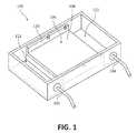
- FIG. 1is a perspective view of an embodiment of a system used to purify a microorganism.
- FIG. 2Ais a perspective view of an embodiment of a medium in a cassette which may be used with the system of FIG. 1 .
- FIG. 2Bis a partial perspective view of another embodiment of a medium in a cassette which may be used with the system of FIG. 1 .
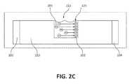
- FIG. 2Cdepicts a side view of the medium and system of FIG. 2B .
- FIGS. 2D and 2Eillustrate excision of a portion of the medium of FIG. 2B .
- FIGS. 2F, 2G and 2Hdepict the excised portion of the medium of FIGS. 2D and 2E on a solid surface.
- FIG. 2Idepicts Gram stained microorganisms that may be detected in accordance with the systems and methods described herein.
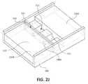
- FIG. 2Jis a top view of a medium in a cassette which may be used with the system of FIG. 1 , wherein a localization device is used to localize the cells and/or viruses.
- FIG. 2Kdepicts Gram stained microorganisms that may be detected in accordance with the systems and methods described herein.
- FIG. 3is a side view of an embodiment of a custom well-forming comb used to make wells in a medium.
- FIGS. 4A and 4Bare a perspective view of multiple flow cell laminate assembly and a single flow cell cutaway view with corresponding electrode and circuit details used to create a potential across a medium.
- FIG. 5is a graph showing results of accumulated objects (solid materials immobilized on a surface) over time for treated compared to non-treated samples.
- FIGS. 6A, 6B and 6Cdepict microscopic images of non-treated samples over time.
- FIGS. 7A, 7B and 7Cdepict microscopic images of treated samples over time.
- FIG. 8is a side view of an embodiment of a circular well formed in a medium with corresponding electrodes
- FIG. 9is a side view of an embodiment of a chamber formed in the medium with an inlet and outlet port enabling sample recirculation.
- a sample containing cells and/or virusesis added to a well disposed in a medium.
- a potentialis applied across the medium to cause contaminants to enter the medium through one or more walls of said well which retain the cells and/or viruses in the well.
- the cells and/or virusesare then removed from the well.
- the cells and/or virusesmay also remain in or on the wall of the well, and/or the wall/well may be excised for further analysis.
- the systems and methods described hereinmay concentrate cells and/or viruses from a low content specimen or sample in the wells, thereby removing or reducing potentially interfering debris and resulting in more readable specimens.
- the disclosed methods and systemsmay be used in testing of CSF (cerebro-spinal fluid) specimens or other hypocellular specimens.
- CSFcerebro-spinal fluid
- bacterial organismscan be localized in 5 ⁇ 5 field of view capture areas (100 ⁇ objective magnification) to minimize time-consuming searching during microscopic examination.
- a system having multiple wellsmay also be used to support parallel processing of sample aliquots for concurrent analyses by multiple downstream methods.
- FIG. 1depicts a system 100 used to purify cells and/or viruses with a cassette 106 configured to receive a medium.
- the cassette 106includes a bottom plate 108 and sides 110 .
- Negative electrode 102 and positive electrode 104are operably connected to the medium through a buffer (not shown) placed in a reservoir 112 .
- the sampleis added to a well disposed in a medium, preferably formed in the medium.
- a plastic wellmay also be disposed in the medium, in addition to well(s) formed in the medium.
- An electrical potentialis applied to the well causing contaminant material to enter the medium while the cells and/or viruses accumulate on the wall of the well.
- the cells and/or virusesmay be localized on the wall of the well. Cells and/or viruses remain in the well, thereby purifying the sample.
- the samplemay be mixed during or after a time period of the applied electric field. In some embodiments, the process can be repeated until separation of contaminants that interfere with adhesion to a detection surface has been achieved.
- the wellcan then be rinsed, and cells and/or viruses recovered.
- the wall of the well where the cells and/or viruses have accumulatedmay be excised or otherwise removed from the rest of the gel medium.
- the electrical potentialcan be briefly reversed in polarity to displace the cells and/or viruses from the wall prior to rinsing and recovery. Mixing, applying a potential, and/or reversing polarity of applied field can be performed iteratively to further purify the sample.
- the sample volume recoveredis less than, and sometimes substantially less than, the initial sample volume in the wells.
- a barriersuch as an impermeable plastic sheet, is inserted into the wells and used to reduce the volume in the wells, thereby further concentrating the cells and/or viruses in the well and providing a reduced sample volume for recovery.
- FIG. 2Adepicts an embodiment of a medium in a cassette 106 comprising a bottom plate 108 and sides 110 , which may be used with the system 100 .
- a medium 220is disposed in the cassette 106 with a plurality of wells 222 in the medium 220 .
- a sample 224is added in some wells 222 with a pipette 226 comprising a pipette tip 228 .
- the mediumis shown as a top load gel slab, media in other forms, including but not limited to vertical gel slabs, can be used.
- FIGS. 2B-2Hillustrate an embodiment where a medium 220 is disposed in a cassette 106 and has a plurality of wells 222 in the medium 220 .
- a sampleis added and a potential is applied between negative electrode 102 and positive electrode 104 as described elsewhere herein to cause contaminant material to enter the medium while the cells 201 and/or viruses accumulate or become embedded on the wall 223 of the well 222 , depicted in FIG. 2C as concentrated cells 202 .
- a portion 230 of the medium 220including at least a portion of the wells 222 , is excised, such as by cutting the medium along planes of excision defined by lines A-A′ and B-B′, or otherwise removed from the medium.
- the excisioncan be robotically automated.
- the excised portion of the mediummay be fixed, for example, for staining or extracting molecular samples for analysis.
- the excised portion 230may be placed on a solid surface 232 , such as a glass slide, and the excised portion 230 may be sectioned at each well 222 into sections 231 .
- the sections 231 of excised portion 230may be dried, Gram stained, and detected as indicated in FIGS. 2H-2I .
- a prepared slidee.g., wherein the sectioned, excised portion has been dried
- the cells and/or virusesmay be localized on the wall of the well by, or with the help of, a localization device.
- FIG. 2Jis a top view of a gel medium that utilizes non-conductive materials as a localization device to distort the electric field resulting in localized concentration of cells and/or viruses, such as microorganisms.
- FIG. 2Jillustrates an embodiment where the medium 220 is disposed in the cassette 106 showing a plurality of wells 222 , including a plastic well 224 , in the medium 220 .
- the medium 220includes a proximal end 220 a and a distal end 220 b .
- a localization device 240Disposed between the wells 222 and the medium 220 is a localization device 240 comprising a non-conductive material including at least one hole or aperture 240 a .
- a sampleis added and a potential is applied as described elsewhere herein to cause contaminant material to enter the medium while the cells and/or viruses accumulate or become embedded on the wall 223 of the well 222 .
- the sampleflows through the aperture 240 a in the non-conductive localization device 240 , thereby localizing the cells and/or viruses that accumulate or become embedded on the wall 223 of the well 222 , as described with reference to FIG. 2C .
- the non-conductive localization device 240prevents or inhibits the sample from flowing anywhere but through the aperture(s) 240 a .
- the non-conductive materialmay be a plastic film. As described above with reference to FIGS. 2D-2I , a portion of the medium 220 , including at least a portion of the wells 222 , is excised or otherwise removed.
- the excised portion 230may be placed on a solid surface 232 , such as a glass slide, and may be dried, Gram stained, and detected.
- FIG. 2Kdepicts Gram stained localized microorganisms 241 that have been localized in accordance with the methods and systems described herein.
- the localization deviceis a conductive material, such as a metal or metal alloy wire, that is embedded in or placed near a distal end 220 b of the medium 220 (i.e., downstream relative to the direction of migration).
- a potentialis applied, the cells and/or viruses localize on the wall of the well in a location corresponding to the position of the conductive material. That is, where the conductive material is a straight metal wire, the cells and/or viruses localize on the wall of the well in a straight line corresponding to the line of the metal wire.
- the localization deviceincludes large and small (or discrete) electrodes are used.
- a large electrodesuch as a sheet electrode, may be placed at a proximal location relative to the proximal end 220 a of the medium 220 .
- a small electrodemay be placed at a distal location relative to the distal end 220 b of the medium 220 .
- the localization deviceis a discontinuous buffer system. In such a system, the conductivity inside the well is different from the conductivity outside the well. For example, where a well is made of the medium, the conductivity of the sample and the conductivity of the well are different. When a potential is applied, the cells and/or viruses localize on the wall of the well due, at least in part, to this conductivity difference.
- Dyescan be used in samples to pre-label or added to provide a tracking dye for purposes of a quantitative reference or sample transfer quality control indicator.
- dyesinclude colorants, bio-active adjuncts such as labeled antibodies, vital stains, mortal stains (such as propidium iodide and the like). Zwitterionic or neutrally charged dye molecules can be used to monitor electro-osmotic flow.
- the potential applied across the medium effective for removal of contaminantscan be applied for a variable time and is dependent on the sample conductivity. For samples retrieved using normal saline and having a conductivity near that of normal saline, for example, the potential can be applied from 1 to 60 minutes.
- the methodincludes an asymmetric alternating potential.
- the potentialis a constant potential.
- the applied potentialinduces electro-osmotic flow that is used to remove contaminants having a neutral charge.
- the potentialcan be reversed in polarity to displace cells and/or viruses from the surface of the medium.
- the methodincludes applying a tangential flow across the medium to remove non-permeable contaminants from the surface of the medium.
- the tangential flowmay be applied by flowing the sample over the medium.
- the tangential flowmay be generated using additional buffer that is not the sample. The flow can be continuously cycled over the medium.
- a sampleWhen a sample is taken from a patient, there are various components in the sample.
- a samplemay include saline, anionic and cationic species, pulmonary surfactants, bacteria, mucus, blood, host cells such as white blood cells, and/or lung tissue cells.
- Mucus componentsinclude, but are not limited to, mucoidal glycoproteins, proteins, extra-cellular nucleic acids, F-actin, lysed white blood cell fragments.
- Blood componentsmay include, but are not limited to, red cells, white cells, platelets, and plasma.
- Plasma componentsmay include, but are not limited to, sugar, fat, protein and salt solution, platelets, blood clotting factors, sugars, lipids, vitamins, minerals, hormones, enzymes, antibodies, and other proteins including heme, albumins, immunoglobulins, fibrinogens, regulatory proteins, lipoproteins (chylomicrons, VLDL, LDL, HDL), transferrin, prothrombin, enzymes, proenzymes, residual antibiotics used to treat the patient, and hormones.
- Lung tissue componentsinclude host epithelial cells (intact or lysed). The cells in the alveolar walls of the lung produce and secrete pulmonary surfactant. Pulmonary surfactant is a mixture of phospholipids and proteins. White blood cells may also be present in lung samples. All the above components may be solubilized.
- the cellsinclude blood cells, fungal cells, bacterial cells, or microorganisms including parasites.
- blood cellsinclude red blood cells and white blood cells.
- the white blood cellscan be neutrophils.
- microorganismscan include bacteria, fungi, algae, and protozoa.
- the microorganismsare bacteria.
- the microorganismscan be pathogenic to humans and animals. Suitable microorganisms include any of those well established in the medical art and those novel pathogens and variants that emerge from time to time.
- bacteriaexamples include, but are not limited to, genera such as Bacillus, Vibrio, Escherichia, Shigella, Salmonella, Mycobacterium, Clostridium, Cornyebacterium, Streptococcus, Staphylococcus, Haemophilus, Neissena, Yersinia, Pseudomonas, Chlamydia, Bordetella, Treponema, Stenotrophomonas, Acinetobacter, Enterobacter, Klebsiella, Proteus, Serratia, Citrobacter, Enterococcus, Legionella, Mycoplasma, Chlamydophila, Moraxella, Morganella , and other human pathogens encountered in medical practice.
- generasuch as Bacillus, Vibrio, Escherichia, Shigella, Salmonella, Mycobacterium, Clostridium, Cornyebacterium, Streptococcus, Staphylococcus, Haemophilus, Neissena, Y
- Klebsiellaincludes, but is not limited to, Klebsiella pneumoniae, Klebsiella ozaenae, Klebsiella rhinoscleromatis, Klebsiella oxytoca, Klebsiella planticola, Klebsiella terrigena , and Klebsiella ornithinolytica .
- virusesinclude viroids.
- microorganismsmay comprise fungi selected from genera such as Candida, Aspergillus , and other human pathogens encountered in medical practice.
- Virusescan be, but are not limited to, orthomyxoviruses (e.g., influenza virus), paramyxoviruses (e.g., respiratory syncytial virus, mumps virus, measles virus), adenoviruses, rhinoviruses, coronaviruses, reoviruses, togaviruses (e.g., rubella virus), parvoviruses, poxviruses (e.g., variola virus, vaccinia virus), enteroviruses (e.g., poliovirus, coxsackievirus), hepatitis viruses (including hepatitis A, B, and C), herpesviruses (e.g., Herpes simplex virus, varicella-zoster virus, cytomegalovirus, Epstein-Barr virus),
- the methods and systems described hereincan be used to identify host cells harboring viruses.
- the cellsare first purified, and subsequently the cells are manipulated to either produce viruses, or to identify nucleic acids in the cells.
- the samplecan be obtained from any number of sources, including, but not limited to, bodily fluids (including, but not limited to, blood, urine, serum, lymph, saliva, anal and vaginal secretions, perspiration, peritoneal fluid, pleural fluid, effusions, ascites, and purulent secretions, lavage fluids, drained fluids, brush cytology specimens, biopsy tissue, explanted medical devices, infected catheters, pus, biofilms and semen) of virtually any organism, including mammalian samples and human samples, as well as environmental samples (including, but not limited to, air, agricultural, water and soil samples).
- bodily fluidsincluding, but not limited to, blood, urine, serum, lymph, saliva, anal and vaginal secretions, perspiration, peritoneal fluid, pleural fluid, effusions, ascites, and purulent secretions, lavage fluids, drained fluids, brush cytology specimens, biopsy tissue, explanted medical devices, infected catheters, pus, bio
- samplescan be taken from food processing, which can include both input samples (e.g., grains, milk or animal carcasses), samples in intermediate steps of processing, as well as finished food ready for the consumer.
- the methodcan be used for veterinary applications.
- the methodscan be also used for the analysis of milk in the diagnosis and treatment of mastitis, and the analysis of respiratory samples for the diagnosis of bovine respiratory disease.
- the methodsprovide for the rapid detection of the presence of potential biological warfare agents in a sample.
- Samplescan range from less than a milliliter to up to a liter for certain respiratory lavage fluids, and can further range in bacterial concentration from less than one bacterium to greater than 10 9 bacteria per milliliter.
- the samplecan be present in blood, urine, sputum, lavage fluid or other medium.
- the samplecan be concentrated prior to using the described methods for purifying cells and/or viruses from the sample. Sample concentration both concentrates the sample so that bacteria that are present in small numbers can all be effectively introduced into the system and adequately sampled, as well as so the background liquid medium can be normalized, or in some cases eliminated or reduced, to have consistent properties upon introduction to the system.
- Sample concentrationcan be performed by centrifugation, combining samples, removing solvents, and the like. It should be noted, however, that certain samples provided in the description can be used without concentration or other modification.
- pneumoniacan result from a variety of causes, including infection with bacteria, viruses, fungi, or parasites, as well as chemical or physical injury to the lungs.
- some samples of cells and/or virusescontain contaminants that interfere with their detection. Purification of a microorganism (or virus or other cell), and detection of the type and amount of a microorganism (or virus or other cell) present in a sample, are helpful to diagnose and treat a patient effectively.
- the cellsare selectively lysed.
- the mammalian cellscan be lysed, releasing intracellular microorganisms prior to, during, or after the purification described herein.
- Contaminantsare removed from the sample into the medium.
- Contaminants that can be removedinclude ionic species, including, but not limited to, mono or divalent cations and anions, released intracellular materials, phospholipids, extracellular proteins, mucins, pulmonary surfactants, mucus plugs, pus, glycoproteins, and nucleic acids. Removing contaminants avoids other time intensive preparation steps such as vortexing and centrifugation.
- the removed contaminantsinterfere with cells and/or virus surface immobilization, detection, and imaging. Cells and/or viruses remain in the well, and can be recovered.
- certain componentssuch as cellular membrane fragments and larger cellular fragments, are not removed from the sample.
- Aerogelsinclude, but are not limited to, silica aerogel, carbon aerogels, alumina, cadmium, and selenide aerogels.
- Organic aerogels, such as SEAgel,are made of agar. Aerogels made of chalcogens such as sulfur, selenium, and other elements may also be of utility.
- the mediumis a hydrogel.
- hydrogelsare a network of polymer chains that are hydrophilic. Hydrogels can be highly absorbent natural or synthetic polymers, and in some instances can contain over 99% water. In general, hydrogels are solid, yet porous media.
- the concentration of the hydrogelaffects the migration speed of the contaminants through the hydrogel. Increasing the concentration of the hydrogel decreases the pore size within the hydrogel. Additionally, contaminants with smaller molecules move faster and migrate further than contaminants with larger molecules.
- the charge of the hydrogelalso affects the migration speed of the contaminants through the hydrogel.
- Each contaminant moleculemigrates to the electrode that carries a charge opposite of that of the contaminant molecule.
- Most biological materialshave a net negative surface charge.
- the chargeis considered neutral if it is a balance of positive and negative, or uncharged, such as complexes coated with neutral materials that envelope and screen charged materials within. The uncharged material will migrate in the direction of electro-osmotic flow, if present.
- the pH of the hydrogelalso affects the migration speed of the contaminants and the targets.
- the pHis selected to enhance mobility of the contaminants relative to the cells and/or viruses.
- a pHmay be selected such that the cells and/or viruses are substantially near the isoelectric point, minimizing the cells' and/or viruses' mobility relative to the contaminants.
- the pHmay be selected to be substantially different from the isoelectric point such that the direction of the cells' and/or viruses' mobility is reversed relative to the contaminants.
- a mediumcontains nutrients that promote the viability of the cells and/or viruses.
- Media used in the systems describedcan separate contaminant molecules based on both their size and their charge.
- the hydrogel's porosityis directly related to the concentration of agarose in the medium. Various levels of effective viscosity can be selected, depending on the experimental objectives.
- hydrogelsexamples include alginates, as disclosed in Gadkari, 2007, “Optimal hydrogels for fast and safe delivery of bioactive compounds”, Thesis of Drexel University; ethyl-vinyl-acetate copolymer as disclosed in U.S. Pat. No. 3,854,480; esters of hydantoic acid as disclosed in U.S. Pat. No.
- poly-glutamic acid ethyl-glutamate copolymer(5-50% GA, 80-500 KDa) as disclosed in U.S. Pat. No. 4,450,150; polyoxyethlyene-polyoxypropylene copolymer thermoset as disclosed in U.S. Pat. No. 4,478,822; vinyl cross-linked copolymers of insoluble and soluble monoolefinic esters as disclosed in U.S. Pat. No. 4,548,990; copolymers with N-vinyl-2-pyrrolidone and methacrylates as disclosed in U.S. Pat. No. 4,693,884; polyanhydride as disclosed in U.S. Pat. No.
- Agaroseis a linear polymer, made up of the repeating monomeric unit of agarobiose.
- Agarobioseis a disaccharide made up of D-galactose and 3,6-anhydro-L-galactopyranose.
- Agarose pectin or sulfonated agarosecan be used as the hydrogel.
- Agarosecan be obtained from Lonza (Rockland, Me.) under the brand name SeaKemTM.
- the concentration of the agarose gel for effectively removing contaminantsis from 0.1-2.0% w/v.
- Purified agarose hydrogelsmay be purchased for use in the described method.
- An example of a commercial purified hydrogelcan be obtained from Invitrogen (Carlsbad, Calif.) under the brand name E-Gel® EX Starter.
- Polyacrylamideis a polymer (—CH 2 CHCONH 2 —) formed from acrylamide subunits. It can be synthesized as a simple linear-chain structure or cross-linked, typically using N,N′-methylenebisacrylamide. In the cross-linked form, polyacrylamide is highly water-absorbent, forming a soft gel. Polyacrylamide can be obtained from BioRad (Hercules, Calif.).
- Purified polyacrylamide hydrogelsmay be purchased for use in the described method.
- An example of a commercial purified hydrogelcan be obtained from BioRad (Hercules, Calif.).
- Preconditioning of a mediumcan be performed. Preconditioning of a medium is often done to remove impurities found in the medium. For example, providing a potential across a hydrogel helps mobile impurities to migrate outside of the hydrogel.
- the potentialcan be, for example, 50V, 75V, 100V, 150V, 200V, 250V, 300V, 350V, 400V or 500V.
- the potentialcan be provided for a period of time, such as at least 2 minutes, at least 5 minutes, at least 10 minutes, at least 15 minutes, at least 30 minutes, at least 45 minutes, at least 60 minutes, at least 120 minutes, or at least 180 minutes.
- the mediumcan be a filter.
- filtersinclude those available from Pall Corporation (Port Washington, N.Y.), such as hydrophilic polypropylene, ahydrophilic, low binding material with pore sizes of 0.2 ⁇ m and 0.45 ⁇ m; polytetrafluoroethylene (PTFE), a hydrophobic, high binding material with pore sizes of 0.2 ⁇ m, 0.45 ⁇ m, 1 ⁇ m, 2 ⁇ m and 3 ⁇ m; glass fiber, a hydrophilic, moderate binding material with a pore size of 1 ⁇ m; nylon, a hydrophilic, low binding material with pore sizes of 0.2 ⁇ m and 0.45 ⁇ m; polyvinylidene fluoride (PVDF), a hydrophilic, low binding material with pore sizes of 0.2 ⁇ m and 0.45 ⁇ m; PES (Supor®), a hydrophilic, low binding material with pore sizes of 0.1 ⁇ m, 0.2 ⁇ m, 0.45 ⁇ m, and
- filters available from Milliporeinclude PTFE (LCR), a hydrophilic, moderate binding material with pore sizes of 0.2 ⁇ m and 0.45 ⁇ m; PVDF (DuraporeTM), a hydrophilic, low binding material with pore sizes of 0.2 ⁇ m and 0.45 ⁇ m; PTFE (FluoroporeTM), a hydrophilic, low binding material with pore sizes of 0.2 ⁇ m and 0.45 ⁇ m; nylon, a hydrophilic, low binding material with pore sizes of 0.2 ⁇ m and 0.45 ⁇ m; glass fiber, a hydrophilic, moderate binding material with a pore size of 1 ⁇ m; and hydrophilic mixed cellulose esters, a high binding material with exemplary pore sizes of 0.2 ⁇ m, 0.45 ⁇ m, and 0.8 ⁇ m.
- Filterscan have pore sizes of greater than or equal to about 0.01 ⁇ m, 0.05 ⁇ m, or 0.1 ⁇ m, 0.2 ⁇ m, 0.4 ⁇ m, 0.6 ⁇ m, 0.8 ⁇ m, 1.0 ⁇ m, 1.5 ⁇ m, 2.0 ⁇ m, 2.5 ⁇ m, 3.0 ⁇ m, 4.0 ⁇ m, or 5.0 ⁇ m.
- Filterscan have pore sizes of less than or equal to about 5.0 ⁇ m, 4.0 ⁇ m, 3.0 ⁇ m, 2.5 ⁇ m, 2.0 ⁇ m, 1.5 ⁇ m, 1.0 ⁇ m, 0.8 ⁇ m, 0.6 ⁇ m, 0.4 ⁇ m, 0.2 ⁇ m, 0.05 ⁇ m, or 0.01 ⁇ m.
- the methodincludes adding a chemical agent to the medium to increase the permeability of the medium and/or increase the ability of the contaminant to enter the medium.
- Examples of chemical agentsinclude reducing agents, including, but not limited to, dithiothreitol (DTT), tris(2-carboxyethyl)phosphine (TCEP), and mercaptoethanol reducing agents; denaturing agent using surfactants, including, but not limited to, sodium lauryl sulfates, non-ionic surfactants such as Triton X-100, Tween-20, or chaotropic agents, including, but not limited to, urea, thiourea, or guanidinium chloride; chelating agents that can coordinate molecules such as calcium, magnesium, and other divalent and trivalent ions (including metal ions), including ethylenediaminetetraacetic acid (EDTA) and ethylene glycol tetraacetic acid (EGTA); cleavage agents including proteases, nucleases, glyconases, lipases; and excipients such as polyethylene glycol.
- reducing agentsincluding, but not limited to, dithiothrei
- Viscous gelsinclude cellulose ethers (such as hydroxylethyl cellulose or MethocelTM (Dow (Midland, Mich.)) and soluble polymer viscosity modifiers (such as polyethylene glycol, polyvinylpyrrolidone, dextrans, pluronic surfactants, and alginates).
- cellulose etherssuch as hydroxylethyl cellulose or MethocelTM (Dow (Midland, Mich.)
- soluble polymer viscosity modifierssuch as polyethylene glycol, polyvinylpyrrolidone, dextrans, pluronic surfactants, and alginates.
- the pore sizeis not defined. The separation is based on retarded flow of the cells in the viscous medium.
- agentscan be added to or used to treat the medium to control electroosmotic flow. In some embodiments, it may be desirable to increase or decrease electroosmotic flow.
- the sampleshould be substantially uniform.
- the homogenization of a samplecan be done by a sample mixing or stirring step. Mixing the sample acts to re-suspend any caked material formed on the walls of the well.
- the methodincludes mixing a sample using a pipette tip. See, for example, the pipette tip 228 in FIG. 2A .
- the sampleis passed through the narrow opening of the pipette to shear and homogenize the sample.
- the methodincludes placing a buffer in contact with the medium.
- the mixing parameters of the bufferare designed to maximize the removal of debris and non-target material.
- the buffercan be replenished to prevent accumulation of undesirable electrophoresis products.
- undesirable effects pH gradients generated at the cathode and anode and in proximity to the samplecan be substantially minimized by buffer replenishment or replacement, potentially using continuous flow.
- electrophoretic buffersutilize pairs of redox mediators.
- these redox mediatorsfacilitate low voltage electrophoresis that permits cell viability to be maintained.
- These redox mediatorsmay also enable the use of electrode materials that have limited utility in high voltage electrophoresis (for example, indium tin oxide, “ITO” electrodes).
- ITOindium tin oxide
- these redox mediatorsfind use in “closed systems” (i.e., systems not open to the atmosphere). In closed systems, bubble formation and generation of other reactive species during the electrophoresis step, which can cause a number of problems, is prevented, and closed systems also help to prevent the exposure of the technician to potentially infectious samples, as well as reducing problems associated with discarding biological samples
- the bufferis placed in a reservoir in contact with the medium. In various embodiments, the medium is not submerged in the buffer.
- Buffersinclude, for example, various electrophoresis buffers including zwitterionic buffers, neutral buffers such as phosphate-buffered saline (PBS), lower or higher pH buffers, and hypotonic or hypertonic buffers.
- PBSphosphate-buffered saline
- borate and other selected ions and counter-ionsare included to facilitate effective electrophoresis.
- the buffer solutionincludes histidine and tris(hydroxymethyl)aminomethane.
- Histidinehas low conductivity.
- Tris(hydroxymethyl)aminomethanehas some conductivity but has low mobility.
- Histidinehas pKa values close to physiological values providing adequate buffering capacity.
- Tris(hydroxymethyl)aminomethanecan be obtained from Sigma-Aldrich (St. Louis, Mo.) as Trizma® base (Sigma, T1503).
- the sample (in 150 mM NaCl)is desalted to remove cationic and anionic species that may interfere with subsequent analysis.
- desaltingallows successful concentration and capture of the microorganism.
- Electrophoretic mobilitycan be buffer dependent due to zeta potential variability with salt concentration, valency of salts present in the buffer, and the pH of the buffer. Bacteria can lose charge as the concentration of salt increases or as the pH is lowered below a certain pH, for example, pH 5.0. Divalent and trivalent salts are more effective quenchers than monovalent salts. For example, CaCl 2 is more effective than NaCl to quench a charge. Certain agents such as chelators, including, but not limited to, ethylenediaminetetraacetic acid (EDTA) and ethylene glycol tetraacetic acid (EGTA), both available from Sigma-Aldrich, can be used to control the concentration of charged species in the sample.
- EDTAethylenediaminetetraacetic acid
- EGTAethylene glycol tetraacetic acid
- one or a plurality of wellscan be formed in the medium.
- the wellsare molded into the gel.
- a custom well-forming combcan be used to create the appropriate well shape.
- Wellsinclude side-walls that can be substantially vertical or diagonal.
- the methodincludes wells that are non-rectangular shaped.
- the wellsare substantially chamfered to eliminate sharp edges in the well, enhancing target recovery.
- the wellscan hold various sample sizes.
- the wellscan hold from 10 ⁇ L to 500 ⁇ L.
- the wellsare 5 to 250 mm wide. Multiple wells can be used for a sample. As illustrated in FIG.
- a well 822 in medium 820may be circular, surrounding an electrode such as cathode 804 , and the counter electrodes may surround the well, such as the illustrated anode ring 802 .
- a sample placed in well 822surrounds the cathode 804 .
- a run buffer sheathmay flow over cathode 804 to remove electrode byproducts during electrophoresis.
- FIG. 3depicts a side view of an embodiment of a custom well-forming comb 300 .
- a comb body 302has a plurality of well-forming teeth 304 connected to the comb body 302 by one edge 306 .
- the sides of the teeth 308form the side-walls of the non-rectangular shaped wells in a medium.
- the wellsare triangular-shaped.
- the comb 300is sized and shaped to fit the medium in a cassette, such as the cassette shown in FIG. 2A .
- the comb 300has six well-forming teeth 304 , but could have more or less teeth depending on the size of the cassette.
- sample solutioncan wick up the walls of the well.
- the sample solutiondoes not tend as strongly to wick up the walls, making it easier to remove the microorganism from the well.
- the triangular shaped wellis narrowest at the bottom and widest at the top of the well.
- the well-forming teeth 304 shown in FIG. 3create a pattern of triangular shaped wells in the medium. In other embodiments, the wells may be round-bottomed wells.
- the samplescan have high solids (e.g., from 1%-50% weight/volume of solid components). Minimizing the well width minimizes the caking of the solids on the well walls. In some embodiments, the well is 0.0025 inches wide at the widest point.
- the methoduses a system 900 wherein one or a plurality of chambers 953 can be formed in a medium 920 .
- the chambers 953are molded into the gel 920 and have an inlet and outlet port ( 954 and 955 ) as shown in FIG. 9 .
- Inlet and outlet ports 954 and 955are connected by tubing 956 for recirculating a sample through a chamber 953 , such as by a peristaltic pump.
- the chambersmay be submerged or partially submerged in buffer, and electrical potential is applied to the system orthogonally to the direction of the recirculating flow of sample.
- an electrode or a plurality of electrodesmay be contained within the well or chamber. Additionally, in various embodiments, the electrode or plurality of electrodes may be in contact with the medium or separated a distance from the medium. The electrode or plurality of electrodes may be connected to the medium using salt bridges, buffer, redox mediators, or other conductive charge transfer methods used by those skilled in the art or familiar with techniques used in applications for establishing faradaic current. In some embodiments the electrodes are in physical contact with the chamber walls.
- conductive materialsmay be utilized to distort the electric field resulting in localized concentration of cells and/or viruses.
- Electric field distortionmay utilize material conductivity differences to accomplish the said localization.
- the methodincludes immobilizing the microorganism.
- Cells and/or virusesare immobilized by various filters that exclude the targets (microorganism) from penetrating, for example, tube walls, microchannels (horizontal or vertical), or any geometry that uses a capture surface (specific or nonspecific), mazes, fluidic dead space (eddy cul-de-sacs), and microwells of approximate cellular scale.
- the methodincludes immobilization of cells and/or viruses on a positively charged surface.
- cells and/or virusescan be immobilized by a positively charged detection surface.
- the cells and/or virusesmay be immobilized by embedding in the medium.
- Detection surfacesare disclosed in, for example, U.S. Pat. No. 6,844,028, incorporated by reference herein in its entirety. Detection surfaces can include those coated with poly-L-lysine, polyethylenimine, or other cationic polymers. Additionally, detection surfaces can include hydrophobic surface coatings.
- the microorganismcan be detected by a system.
- the systemis an optical sensing system.
- the systemis a microscope.
- the systemis an automated system.
- the sequential or simultaneous use of a plurality of electrophoresis electrodesallows multidimensional electrophoresis, i.e., the solution may be targeted, “mixed,” or “stirred” in the vicinity of a detection surface to further increase the kinetics of binding.
- polaritiescan be reversed to allow cells and/or viruses that may not have bound to the detection surface to travel back “over” the surface, resulting in increased binding.
- electrodesmay be located and field polarity switched according to a programmed sequence so as to provide agitation in two dimensions of a plane, or in three dimensions.
- the methodincludes detection of the microorganism.
- biosensor devicesare designed to fit into a detection unit, and generally utilize a number of components, which can either be “on-chip” (e.g., part of a biosensor cartridge) or “off-chip” (where some of the components are part of separate device or devices into which the biosensor cartridge fits).
- These componentsinclude, but are not limited to, one or a plurality (e.g., an array) of detection surface(s), concentration modules (which, as outlined herein, frequently are configured with the detection surface(s)), detection modules (again, frequently configured with the detection surface(s)), input and output ports, channels, pumps, mixers, valves, heaters, fluid reservoirs (including sample reservoirs, reagent reservoirs, and buffer reservoirs), concentration controllers (e.g., in the case of electrophoresis, electrical controllers), and data collection and analysis (e.g., computer) components.
- concentration moduleswhich, as outlined herein, frequently are configured with the detection surface(s)
- detection modulesas outlined herein, frequently configured with the detection surface(s)
- input and output portschannels, pumps, mixers, valves, heaters, fluid reservoirs (including sample reservoirs, reagent reservoirs, and buffer reservoirs)
- concentration controllerse.g., in the case of electrophoresis, electrical controllers
- Cellscan be measure in terms of cells per mL, colony forming units (CFU, or units) per mL for fungi and/or bacterial microorganisms, and viruses can be measured in particles per mL or plaque forming units per mL (PFU).
- CFUcolony forming units
- PFUplaque forming units per mL
- Levels of cells and/or virusesare described in units per volume, typically per mL volume. Those skilled in the art understand the specific units are typically reported as appropriate for a given target. For exemplary purposes, the concentration ranges below are reported in generic units per mL. For example, levels of 0.1 to 1 ⁇ 10 8 units/mL can be detected.
- cells and/or viruses of levels less than 5 ⁇ 10 8 units/mL, 3 ⁇ 10 8 units/mL, 1 ⁇ 10 8 units/mL, 0.8 ⁇ 10 8 units/mL, 0.6 ⁇ 10 8 units/mL, 0.4 ⁇ 10 8 units/mL, 0.2 ⁇ 10 8 units/mL, or 0.1 ⁇ 10 8 units/mLcan be detected.
- the example described belowanticipates a wide range of specimen variability, first homogenizing the specimen, then sampling the specimen, and then purifying the sample to remove debris and other interfering materials by placing the sample in a medium containing a well and applying a potential laterally across the medium to retain cells and pass contaminants into the medium.
- agarose powder(SeaKem, LE Agarose) was mixed with 1 L of buffered solution containing 100 mM histidine (Sigma, H8000) and 2.5 mMTrizma® base (Sigma, T1503). The final concentration of agarose slurry was 1.0% (w/v). The solution was boiled to melt the agarose powder and the molten agarose was stored in liquid form at 40° C. until use.
- Gel electrophoresis combsare generally nominally 1-2 mm thick, capable of holding nominally 100 ⁇ L of sample volume.
- a custom equilateral V-shaped wellwas used. The well had sides 1 cm long and a thickness of nominally 0.6 mm (0.025′′).
- the combwas inserted into a gel box container (E-C Apparatus, EC 250-90) and the box filled with the molten agarose submerging a portion of the comb.
- the agarosewas allowed to cool to room temperature forming an agarose gel.
- the combwas removed from the solidified agarose and the void volume of the comb formed a well in the material.
- the V-shaped wellenabled facile recovery of the sample volume from the well, described in further detail below.
- the gel box containing the agarose gel medium having triangular wellswas placed in an electrophoresis apparatus and then submerged in a run buffer containing 100 mM histidine and 2.5 mM Trizma® base. A 250 volt potential was applied for 1 hr. The applied potential yielded 22 mA of current.
- the pretreated gelswere removed from the electrophoresis apparatus and transferred to a closed container and stored submerged in fresh run buffer until use.
- a remnant specimen having a known level of bacteriawas homogenized by pouring into a syringe connected to 0.02′′ (0.5 mm) inner diameter PEEK tubing and forcing through the PEEK tubing 10 times at a flow rate of approximately 0.1 mL/sec to liquefy the specimens.
- the specimenwas then filtered through 5 ⁇ m track etch polycarbonate filters (SPI Pore, E5013-MB).
- SPI Pore, E5013-MB5 ⁇ m track etch polycarbonate filters
- a control or a known clinical sample(e.g., with a known concentration of bacteria) can be compared to the unknown sample.
- the samplewas diluted to a final nominal bacterial concentration of 1.5 ⁇ 10 3 CFU/mL. 50 ⁇ L of the diluted sample was plated in triplicate on Mueller Hinton Agar (MHA) and placed in the incubator overnight. The number of colonies counted on the overnight incubated plates divided by the plated volume and multiplied by dilution factor yielded the actual number of input Klebsiella oxytoca bacteria in CFU/mL.
- MHAMueller Hinton Agar
- the samplewas diluted 10-fold and the optical density read was acquired at 625 nm to assess the amount of particulate debris in the sample.
- the pretreated gelswere placed in the gel box and apparatus, patted dry, and excess liquid was removed from the triangular wells using 0.2 mm thick flat capillary plastic pipette tips (Fisher 07-200-519). The well was filled with a 20 ⁇ L sample of the homogenized specimen.
- Histidine/Tris run bufferwas added to the apparatus so that the liquid level was below the top of the gel slab.
- the samplewas electrophoresed for 5 minutes at 250 volts and the samples were hydrodynamically sheared by pipetting the sample volume up and down 5 ⁇ using a capillary pipette tip.
- the sampleswere electrophoresed again for 5 minutes at 250 volts and the samples then hydrodynamically sheared by pipetting the sample volume up and down 5 ⁇ using a capillary pipette tip.
- the treated samplewas diluted to a final concentration of 1.5 ⁇ 10 3 CFU/mL. 50 ⁇ L of the diluted sample was plated in triplicate on Mueller Hinton Agar (MHA) and place in the incubator overnight. The number of colonies counted on the overnight incubated plates divided by the plated volume and multiplied by dilution factor yielded the actual number of Klebsiella oxytoca bacteria recovered in CFU/mL.
- MHAMueller Hinton Agar
- the treated samplewas diluted 10-fold and then the optical density read was acquired at 625 nm to assess the amount of particulate debris remaining in the sample.
- the 20 ⁇ L of recovered sample volumewas diluted with 40 ⁇ L of 10 mM ascorbic acid and then introduced into a flow cell (described below) for electrokinetic concentration.
- a 20 ⁇ L of a non-treated samplewas diluted with 40 ⁇ L of 10 mM ascorbic acid and then introduced into a flow cell (described below) for electrokinetic concentration.
- FIG. 4Ais a perspective view of a multiple flow cell laminate flow cell assembly 400
- FIG. 4Bis a single flow cell cutaway view with corresponding electrode and circuit details.
- Flow cellswere assembled using a three layer die-cut laminate flow cell assembly 450 (DLE, Oceanside, Calif.), sandwiched between an indium tin oxide (ITO) coated glass slide flow cell floor 451 (Delta Technology, Stillwater, Minn.) and an ITO coated 5 mil polyester (ITO PET) plastic flow cell ceiling 452 (Sheldahl, Northfield, Minn.) forming a fluidic flow cell chamber.
- ITOindium tin oxide
- PETITO coated 5 mil polyester
- the laminate flow cell assemblycontained 32 separate channels 453 , each having 1.78 mm width ⁇ 0.30 mm height ⁇ 11.28 mm length, with 1.78 mm diameter fluidic inlet and outlet ports (454 and 455, respectively) to interface with plastic pipette tips for fluid exchanges using manual pipettors.
- the transparent top and bottom electrodesenabled microscope imaging.
- Bacteria 401 suspended in redox active EKBwere contacted with uniform transparent electrodes constructed from transparent ITO coated glass (Delta Technologies, Stillwater, Minn.) or polyester film (Sheldahl, Northfield, Minn.). A potential was applied to the conductive ITO surfaces completing the circuit, establishing a faradaic current and an electric field between the electrodes and enabling bacterial electrokinetic concentration (EKC) and surface immobilization, as illustrated in FIG. 4B .
- EKCelectrokinetic concentration
- FIG. 5is a graph showing results of accumulated objects over time for treated compared to non-treated samples.
- the non-treated sample datais expected data.
- the objectsare solid material, such as cells, viruses, and cellular debris, that are immobilized on a sample surface.
- FIG. 5shows that material concentrates, and then adheres to, the surface.
- Subsequent processessuch as measuring the growth or growth rate, can be utilized to determine the number of viable cells, and additionally probing the material using receptor-ligand binding techniques, including, but not limited to, antibody recognition or nucleic acid hybridization methods can be used to measure the abundance of microorganisms present.
- FIGS. 6A, 6B and 6Cdepict microscopic images of non-treated samples at initial time, time of 300 seconds, and time of 360 seconds, respectively.
- the surface accumulation rateis low.
- poor surface retention of the objectsoccurs when samples are not treated.
- FIGS. 7A, 7B and 7Cdepict microscopic images of treated samples at initial time, time of 300 seconds, and time of 360 seconds, respectively.
- the treated sample surfaceaccumulates all objects, as evidenced by a plateau occurring in less than 3 minutes.
- the objectswere irreversibly bound to the surface, as evidenced by consistent accumulated counts during reverse electrophoresis.
- the flow cellwas rinsed with 10 times the internal cell volume of 1/10th strength cation-adjusted Mueller-Hinton Broth (CAMHB) growth media (Difco, Sparks, Md.). 100 ⁇ L of liquefied Mueller Hinton Agar (MHA) was loaded into the flow cell and then cooled to room temperature, solidifying into a hydrogel.
- CAMHBcation-adjusted Mueller-Hinton Broth
- Direct observation of bacterial growthwas performed by inserting the disposable 32-channel flow cell assembly into a custom benchtop automated instrument that combined digital microscopy, motion control, and image analysis software.
- the systemwas enclosed in an incubator maintained at 35° C.
- the motorized microscope stageenabled automated XY translation, location logging, and memory with 10 ⁇ m repeatability.
- the systemautomatically focused and acquired surface images of adherent bacteria at programmed time intervals for multiple fields-of-view during an experiment.
- the systemused the fiducial markings to autofocus and mechanically align ( ⁇ 1 pixel) the fields-of-view prior to image acquisition. Unless stated otherwise, a single field-of-view contained sufficient numbers of cells for analysis, and automated analysis routines counted the number of growing clones.
- the number of growing clones observed using the digital microscope methodwas compared with the number of expected growing clones, assuming 100% yield and a 1 to 1 correlation between growing colonies on MHA plates, to calculate a digital microscopy method efficiency.
- the medium methodwas compared to an alternative medium method wherein the gel was submerged. A total efficiency was calculated by multiplying the treatment recovery and digital microscopy efficiency.
- the total efficiency for the medium methodwas highest when the gel slab was not submerged.
Landscapes
- Life Sciences & Earth Sciences (AREA)
- Health & Medical Sciences (AREA)
- Chemical & Material Sciences (AREA)
- Engineering & Computer Science (AREA)
- Organic Chemistry (AREA)
- Zoology (AREA)
- Wood Science & Technology (AREA)
- General Health & Medical Sciences (AREA)
- Physics & Mathematics (AREA)
- Biomedical Technology (AREA)
- Bioinformatics & Cheminformatics (AREA)
- Biochemistry (AREA)
- Biotechnology (AREA)
- Genetics & Genomics (AREA)
- Molecular Biology (AREA)
- Analytical Chemistry (AREA)
- General Engineering & Computer Science (AREA)
- Microbiology (AREA)
- Proteomics, Peptides & Aminoacids (AREA)
- Immunology (AREA)
- Biophysics (AREA)
- General Physics & Mathematics (AREA)
- Pathology (AREA)
- Hematology (AREA)
- Food Science & Technology (AREA)
- Urology & Nephrology (AREA)
- Medicinal Chemistry (AREA)
- Ecology (AREA)
- Sustainable Development (AREA)
- Multimedia (AREA)
- Optics & Photonics (AREA)
- Chemical Kinetics & Catalysis (AREA)
- Electrochemistry (AREA)
- Apparatus Associated With Microorganisms And Enzymes (AREA)
Abstract
Description
| Pre-Treatment Optical | Post-Treatment Optical | Fold | ||
| Density (OD) | Density (OD) | Cleanup | ||
| MEDIA | 0.2841 | 0.065 | 4.35 |
| METHOD | |||
Electrode Configuration and Circuit Details
| Post | Digital | |||
| Treatment | Microscopy | Total | ||
| Recovery | EFF | EFF | ||
| Control - | 100% | 12% | 12% | ||
| Medium Method | 82% | 90% | 74% | ||
| Submerged Medium | 43% | 100% | 43% | ||
| Method | |||||
Claims (16)
Priority Applications (1)
| Application Number | Priority Date | Filing Date | Title |
|---|---|---|---|
| US14/004,145US9434937B2 (en) | 2011-03-07 | 2012-03-07 | Rapid cell purification systems |
Applications Claiming Priority (3)
| Application Number | Priority Date | Filing Date | Title |
|---|---|---|---|
| US201161449824P | 2011-03-07 | 2011-03-07 | |
| PCT/US2012/028139WO2012122314A2 (en) | 2011-03-07 | 2012-03-07 | Rapid cell purification systems |
| US14/004,145US9434937B2 (en) | 2011-03-07 | 2012-03-07 | Rapid cell purification systems |
Related Parent Applications (1)
| Application Number | Title | Priority Date | Filing Date |
|---|---|---|---|
| PCT/US2012/028139A-371-Of-InternationalWO2012122314A2 (en) | 2011-03-07 | 2012-03-07 | Rapid cell purification systems |
Related Child Applications (2)
| Application Number | Title | Priority Date | Filing Date |
|---|---|---|---|
| US15/003,604DivisionUS9714420B2 (en) | 2011-03-07 | 2016-01-21 | Rapid cell purification systems |
| US15/236,021ContinuationUS10202597B2 (en) | 2011-03-07 | 2016-08-12 | Rapid cell purification systems |
Publications (2)
| Publication Number | Publication Date |
|---|---|
| US20140038171A1 US20140038171A1 (en) | 2014-02-06 |
| US9434937B2true US9434937B2 (en) | 2016-09-06 |
Family
ID=46798781
Family Applications (3)
| Application Number | Title | Priority Date | Filing Date |
|---|---|---|---|
| US14/004,145ActiveUS9434937B2 (en) | 2011-03-07 | 2012-03-07 | Rapid cell purification systems |
| US15/003,604Active2032-06-05US9714420B2 (en) | 2011-03-07 | 2016-01-21 | Rapid cell purification systems |
| US15/236,021ActiveUS10202597B2 (en) | 2011-03-07 | 2016-08-12 | Rapid cell purification systems |
Family Applications After (2)
| Application Number | Title | Priority Date | Filing Date |
|---|---|---|---|
| US15/003,604Active2032-06-05US9714420B2 (en) | 2011-03-07 | 2016-01-21 | Rapid cell purification systems |
| US15/236,021ActiveUS10202597B2 (en) | 2011-03-07 | 2016-08-12 | Rapid cell purification systems |
Country Status (4)
| Country | Link |
|---|---|
| US (3) | US9434937B2 (en) |
| EP (1) | EP2683831B1 (en) |
| ES (1) | ES2551922T3 (en) |
| WO (1) | WO2012122314A2 (en) |
Cited By (6)
| Publication number | Priority date | Publication date | Assignee | Title |
|---|---|---|---|---|
| US20160138067A1 (en)* | 2011-03-07 | 2016-05-19 | Accelerate Diagnostics, Inc. | Rapid cell purification systems |
| US9841422B2 (en) | 2003-07-12 | 2017-12-12 | Accelerate Diagnostics, Inc. | Sensitive and rapid determination of antimicrobial susceptibility |
| US10023895B2 (en) | 2015-03-30 | 2018-07-17 | Accelerate Diagnostics, Inc. | Instrument and system for rapid microogranism identification and antimicrobial agent susceptibility testing |
| US10254204B2 (en)* | 2011-03-07 | 2019-04-09 | Accelerate Diagnostics, Inc. | Membrane-assisted purification |
| US10253355B2 (en) | 2015-03-30 | 2019-04-09 | Accelerate Diagnostics, Inc. | Instrument and system for rapid microorganism identification and antimicrobial agent susceptibility testing |
| US11603550B2 (en) | 2013-03-15 | 2023-03-14 | Accelerate Diagnostics, Inc. | Rapid determination of microbial growth and antimicrobial susceptibility |
Families Citing this family (12)
| Publication number | Priority date | Publication date | Assignee | Title |
|---|---|---|---|---|
| US20120077206A1 (en) | 2003-07-12 | 2012-03-29 | Accelr8 Technology Corporation | Rapid Microbial Detection and Antimicrobial Susceptibility Testing |
| KR20170120211A (en) | 2010-06-16 | 2017-10-30 | 닛토덴코 가부시키가이샤 | Waterproof breathable filter and use thereof |
| JP5739699B2 (en) | 2010-06-16 | 2015-06-24 | 日東電工株式会社 | Waterproof ventilation filter and its use |
| CA2842377C (en) | 2011-07-19 | 2019-08-27 | Ovizio Imaging Systems N.V. | A method and system for detecting and/or classifying cancerous cells in a cell sample |
| EP2594334A1 (en) | 2011-11-21 | 2013-05-22 | Drive O2 | Sample vial for digital holographic analysis of a liquid cell sample |
| KR101290648B1 (en) | 2012-02-02 | 2013-07-29 | 서울대학교산학협력단 | Rapid antibiotic susceptibility test (rast) system based on bacterial fixation, antibiotic diffusion and single cell growth tracking using gelling agents |
| EP2626686A1 (en) | 2012-02-13 | 2013-08-14 | Ovizio Imaging Systems NV/SA | Flow cytometer with digital holographic microscope |
| EP2898310B1 (en) | 2012-09-20 | 2019-05-01 | Ovizio Imaging Systems NV/SA | Digital holographic microscope with fluid systems |
| US9675974B2 (en)* | 2013-07-31 | 2017-06-13 | Ovizio Imaging Systems NV/SA | Cap for monitoring objects in suspension |
| EP3286730A2 (en)* | 2015-04-21 | 2018-02-28 | Joseph Paul Robinson | Culture detection and measurement over time |
| EP3196631A1 (en) | 2016-01-19 | 2017-07-26 | Ovizio Imaging Systems NV/SA | Digital holographic microscope with electro-fluidic system, said electro-fluidic system and methods of use |
| EP3301454B1 (en) | 2016-10-03 | 2019-08-28 | Accelerate Diagnostics, Inc. | Instrument and system for rapid microorganism identification and antimicrobial agent susceptibility testing |
Citations (281)
| Publication number | Priority date | Publication date | Assignee | Title |
|---|---|---|---|---|
| US2666355A (en) | 1951-10-12 | 1954-01-19 | Hans J Trurnit | Method of studying chemical reactions by measuring interfacial film thicknesses |
| US3493772A (en) | 1967-05-29 | 1970-02-03 | Palo Alto Medical Research Fou | Bacterial counting machine and method |
| US3532790A (en) | 1968-02-23 | 1970-10-06 | Canadian Patents Dev | Somatic antigen vaccines from lysed inactivated bacteria and process for producing them |
| US3637313A (en) | 1967-05-12 | 1972-01-25 | Holotron Corp | Method of imaging transparent objects with coherent light |
| US3792081A (en) | 1972-04-07 | 1974-02-12 | Alza Corp | Esters of hydantoic acid |
| US3811036A (en) | 1972-09-19 | 1974-05-14 | Artek Syst Corp | Micro-biological colony counter |
| US3832532A (en) | 1972-08-18 | 1974-08-27 | Pfizer | Method and apparatus for testing antibiotic susceptibility |
| US3854480A (en) | 1969-04-01 | 1974-12-17 | Alza Corp | Drug-delivery system |
| US3904293A (en) | 1973-12-06 | 1975-09-09 | Sherman Gee | Optical method for surface texture measurement |
| US3926564A (en) | 1974-02-25 | 1975-12-16 | Gen Electric | Substrate for immunological tests and method of fabrication thereof |
| US3935073A (en) | 1970-04-22 | 1976-01-27 | Johnston Laboratories, Inc. | Method for detecting bacteria |
| US3938515A (en) | 1971-12-20 | 1976-02-17 | Alza Corporation | Novel drug permeable wall |
| US3957362A (en) | 1972-10-02 | 1976-05-18 | Corneal Sciences, Inc. | Hydrogels and articles made therefrom |
| US3961628A (en) | 1974-04-10 | 1976-06-08 | Alza Corporation | Ocular drug dispensing system |
| JPS52102491U (en) | 1976-01-30 | 1977-08-03 | ||
| US4069307A (en) | 1970-10-01 | 1978-01-17 | Alza Corporation | Drug-delivery device comprising certain polymeric materials for controlled release of drug |
| US4069607A (en) | 1976-09-03 | 1978-01-24 | Jurek Julius V | .22 Caliber rimfire adapter system for M16 type rifle |
| GB1520733A (en) | 1975-12-16 | 1978-08-09 | Univ Virginia And Usa National | Detection of microorganisms |
| US4136250A (en) | 1977-07-20 | 1979-01-23 | Ciba-Geigy Corporation | Polysiloxane hydrogels |
| US4199449A (en) | 1977-11-23 | 1980-04-22 | Rohm And Haas Company | Removal of bacteria |
| US4199499A (en) | 1978-08-24 | 1980-04-22 | Eli Lilly And Company | Pharmacologically active peptides |
| US4200493A (en) | 1975-01-24 | 1980-04-29 | United States Of America | Microbial detection and enumeration apparatus |
| US4220152A (en) | 1978-05-08 | 1980-09-02 | Pfizer Inc. | Delivery system |
| US4224439A (en) | 1977-02-08 | 1980-09-23 | Development Finance Corporation Of New Zealand | Activated matrix and method of activation |
| US4233847A (en) | 1979-07-02 | 1980-11-18 | Walker Clifford G | Passive laser accelerometer |
| US4246343A (en) | 1979-05-02 | 1981-01-20 | University Of Virginia And National Aeronautics & Space Administration | Microbial detection and enumeration method and apparatus |
| US4259442A (en) | 1977-10-04 | 1981-03-31 | Laboratoire De Recherche Api S.A.R.L. | Process of rapid identification of bacteria of the genus Streptococcus |
| US4282287A (en) | 1980-01-24 | 1981-08-04 | Giese Roger W | Biochemical avidin-biotin multiple-layer system |
| US4288543A (en) | 1977-01-28 | 1981-09-08 | Pfizer Inc. | Method and apparatus for identifying microorganisms |
| US4313734A (en) | 1978-07-13 | 1982-02-02 | Akzona Incorporated | Metal sol particle immunoassay |
| US4330440A (en) | 1977-02-08 | 1982-05-18 | Development Finance Corporation Of New Zealand | Activated matrix and method of activation |
| US4332476A (en) | 1979-04-17 | 1982-06-01 | Stenberg Johan E | Method and apparatus for studying surface properties |
| US4351337A (en) | 1973-05-17 | 1982-09-28 | Arthur D. Little, Inc. | Biodegradable, implantable drug delivery device, and process for preparing and using the same |
| US4357142A (en) | 1980-07-18 | 1982-11-02 | Akzona Incorporated | Glass support coated with synthetic polymer for bioprocess |
| US4363634A (en) | 1980-07-18 | 1982-12-14 | Akzona Incorporated | Glass support coated with synthetic polymer for bioprocess |
| US4383757A (en) | 1979-04-02 | 1983-05-17 | Optimetrix Corporation | Optical focusing system |
| US4390343A (en) | 1981-07-06 | 1983-06-28 | Miles Laboratories, Inc. | Multilayer analytical element having an impermeable radiation diffusing and blocking layer |
| JPS58198759A (en) | 1982-05-15 | 1983-11-18 | Matsushita Electric Works Ltd | Blood glucose meter |
| US4423099A (en) | 1980-07-28 | 1983-12-27 | Ciba-Geigy Corporation | Membrane modified hydrogels |
| US4450150A (en) | 1973-05-17 | 1984-05-22 | Arthur D. Little, Inc. | Biodegradable, implantable drug delivery depots, and method for preparing and using the same |
| US4478822A (en) | 1983-05-16 | 1984-10-23 | Merck & Co., Inc. | Drug delivery system utilizing thermosetting gels |
| USRE31712E (en) | 1980-01-24 | 1984-10-23 | Biochemical avidin-biotin multiple-layer system | |
| US4478914A (en) | 1980-01-24 | 1984-10-23 | Giese Roger W | Process for applying multiple layers of a protein and a ligand extender to a surface and to the multiple-layer system |
| US4481137A (en) | 1982-02-26 | 1984-11-06 | Mochida Pharmaceutical Co., Ltd. | Glycoproteins and processes for their production |
| US4487839A (en) | 1983-01-05 | 1984-12-11 | Ortho Diagnostic Systems Inc. | Immunoassay methods employing patterns for the detection of soluble and cell surface antigens |
| US4500778A (en) | 1981-04-07 | 1985-02-19 | Nippon Kogaku K.K. | Image plane deviation amount detecting device |
| US4508832A (en) | 1981-06-22 | 1985-04-02 | Battelle Memorial Institute | Ellipsometrically measuring rate of optical change in immunoassay |
| US4509841A (en) | 1981-12-17 | 1985-04-09 | Canon Kabushiki Kaisha | Automatic focusing device |
| US4521522A (en) | 1981-09-05 | 1985-06-04 | Kurt I. Lundstroem | Optical specific binding assay with reflection of polarized electromagnetic radiation |
| US4537861A (en) | 1983-02-03 | 1985-08-27 | Elings Virgil B | Apparatus and method for homogeneous immunoassay |
| US4540881A (en) | 1981-03-27 | 1985-09-10 | Olympus Optical Co., Ltd. | Method of detecting focusing conditions |
| US4548990A (en) | 1983-08-15 | 1985-10-22 | Ciba-Geigy Corporation | Crosslinked, porous polymers for controlled drug delivery |
| US4558012A (en) | 1982-04-26 | 1985-12-10 | Sagax Instrument Ab | Method and member for detecting and/or measuring the concentration of a chemical substance |
| US4588624A (en) | 1982-05-14 | 1986-05-13 | Astra Meditec Ab | Articles exhibiting a biocompatible surface layer |
| US4613567A (en) | 1982-08-19 | 1986-09-23 | Konishiroku Photo Industry Co., Ltd. | Immunoassay method for measuring immunological antigen in fluid sample |
| US4626674A (en) | 1983-02-10 | 1986-12-02 | Olympus Optical Company, Ltd. | Focus detecting method and apparatus |
| US4643968A (en) | 1981-01-29 | 1987-02-17 | Massachusetts Institute Of Technology | Process for determining metabolism and growth of cells under various conditions |
| US4655595A (en) | 1984-09-20 | 1987-04-07 | Sagax Instrument Ab | Ellipsometric method and apparatus for studying physical properties of the surface of a testpiece |
| US4657543A (en) | 1984-07-23 | 1987-04-14 | Massachusetts Institute Of Technology | Ultrasonically modulated polymeric devices for delivering compositions |
| US4693972A (en) | 1984-01-16 | 1987-09-15 | Becton, Dickinson And Company | Composition and method for rapid detection of microorganisms in clinical samples |
| US4693884A (en) | 1982-10-09 | 1987-09-15 | Hoechst Aktiengesellschaft | Technetium-99m triphosphonates and tetraphosphonates for the scintigraphic visualization of res-containing organs and lymph vessels, and a process for their preparation |
| US4713441A (en) | 1986-08-01 | 1987-12-15 | Sandoz Pharmaceuticals Corp. | Polyacetal hydrogels formed from divinyl ethers and polyols |
| US4716123A (en) | 1981-01-14 | 1987-12-29 | Covalent Technology Corporation | Solid phase biological diagnostic assay via visual observation as enhanced by Mie scattering |
| US4752567A (en) | 1984-06-21 | 1988-06-21 | Janssen Pharmaceutica N.V. | Method of visualizing individual submicroscopic metal particles |
| US4772484A (en) | 1985-09-24 | 1988-09-20 | Kitchell Judith P | Biologically useful polymer preparations |
| US4778758A (en) | 1985-08-01 | 1988-10-18 | Ab Biodisk | Device for susceptibility testing of microorganisms |
| WO1989001162A1 (en) | 1987-07-28 | 1989-02-09 | Biotechnology Australia Pty. Ltd. | Detection methods |
| US4805623A (en) | 1987-09-04 | 1989-02-21 | Vander Corporation | Spectrophotometric method for quantitatively determining the concentration of a dilute component in a light- or other radiation-scattering environment |
| US4857313A (en) | 1987-05-28 | 1989-08-15 | Warner-Lambert Company | Transdermal drug delivery device comprising copolymers of N-morpholinoethyl methacrylate and 2-hydroxylmethacrylate |
| US4876208A (en) | 1987-01-30 | 1989-10-24 | Yellowstone Diagnostics Corporation | Diffraction immunoassay apparatus and method |
| US4882168A (en) | 1986-09-05 | 1989-11-21 | American Cyanamid Company | Polyesters containing alkylene oxide blocks as drug delivery systems |
| US4885077A (en) | 1988-11-17 | 1989-12-05 | Becton, Dickinson And Company | Composite membrane, method for its preparation and electrolyte sensor including same |
| US4959301A (en) | 1988-04-22 | 1990-09-25 | Massachusetts Institute Of Technology | Process for rapidly enumerating viable entities |
| WO1990011525A1 (en) | 1989-03-23 | 1990-10-04 | Amersham International Plc | Assay method using surface plasmon resonance spectrometry |
| US5002792A (en) | 1988-08-11 | 1991-03-26 | Medtronic, Inc. | Process for making biomedical devices utilizing thermoplastic hydrophilic gels |
| WO1991004491A1 (en) | 1989-09-18 | 1991-04-04 | Biostar Medical Products, Inc. | Method and apparatus for detection of an analyte |
| USRE33581E (en) | 1984-06-25 | 1991-04-30 | Immunoassay using optical interference detection | |
| US5017009A (en) | 1986-06-26 | 1991-05-21 | Ortho Diagnostic Systems, Inc. | Scattered total internal reflectance immunoassay system |
| US5066465A (en) | 1989-12-27 | 1991-11-19 | Olympus Optical Co., Ltd. | Reaction apparatus |
| US5079172A (en) | 1988-11-04 | 1992-01-07 | Board Of Trustees Operating Michigan State University | Method for detecting the presence of antibodies using gold-labeled antibodies and test kit |
| US5082630A (en) | 1990-04-30 | 1992-01-21 | The United States Of America As Represented By The United States Department Of Energy | Fiber optic detector for immuno-testing |
| US5089606A (en) | 1989-01-24 | 1992-02-18 | Minnesota Mining And Manufacturing Company | Water-insoluble polysaccharide hydrogel foam for medical applications |
| US5149543A (en) | 1990-10-05 | 1992-09-22 | Massachusetts Institute Of Technology | Ionically cross-linked polymeric microcapsules |
| US5173164A (en) | 1990-09-11 | 1992-12-22 | Bioseparations, Inc. | Multi-modality electrical separator apparatus and method |
| US5196527A (en) | 1988-04-05 | 1993-03-23 | Kanebo Ltd. | Porous ion-exchanged fine cellulose particles, method for production thereof, and affinity carrier |
| US5208037A (en) | 1991-04-22 | 1993-05-04 | Alza Corporation | Dosage forms comprising polymers comprising different molecular weights |
| US5218039A (en) | 1987-04-28 | 1993-06-08 | Kingston Technologies, Inc. | Pan emulsion |
| WO1993013197A1 (en) | 1991-12-31 | 1993-07-08 | Picower Institute For Medical Research | Heme polymerase and method for treating malaria |
| US5239170A (en) | 1991-10-23 | 1993-08-24 | Karl Suss America, Incorporated | Autofocus method and apparatus for imaging microscopy using a predetermined visual imaging feature |
| US5240618A (en) | 1992-02-03 | 1993-08-31 | University Of Utah Research Foundation | Electrical field-flow fractionation using redox couple added to carrier fluid |
| WO1994002831A1 (en) | 1992-07-23 | 1994-02-03 | Acrogen, Inc. | Digital analyte detection system |
| US5288611A (en) | 1983-01-10 | 1994-02-22 | Gen-Probe Incorporated | Method for detecting, identifying, and quantitating organisms and viruses |
| US5314805A (en) | 1991-10-28 | 1994-05-24 | Molecular Probes, Inc. | Dual-fluorescence cell viability assay using ethidium homodimer and calcein AM |
| WO1994011728A1 (en) | 1992-11-16 | 1994-05-26 | Bioseparations, Inc. | Electrical separator apparatus and method of counterflow gradient focusing |
| US5350697A (en) | 1990-08-28 | 1994-09-27 | Akzo N.V. | Scattered light detection apparatus |
| WO1995008640A1 (en) | 1993-09-23 | 1995-03-30 | E.I. Du Pont De Nemours And Company | An electrophoretic method for the isolation and separation of microorganisms |
| US5405783A (en) | 1989-06-07 | 1995-04-11 | Affymax Technologies N.V. | Large scale photolithographic solid phase synthesis of an array of polymers |
| WO1995028641A1 (en) | 1994-04-13 | 1995-10-26 | Dade International Inc. | Rapid detection of analytes with receptors immobilized on soluble submicron particles |
| US5466416A (en) | 1993-05-14 | 1995-11-14 | Ghaed; Ali | Apparatus and methods for carrying out electrochemiluminescence test measurements |
| US5468606A (en) | 1989-09-18 | 1995-11-21 | Biostar, Inc. | Devices for detection of an analyte based upon light interference |
| US5491097A (en) | 1989-06-15 | 1996-02-13 | Biocircuits Corporation | Analyte detection with multilayered bioelectronic conductivity sensors |
| US5494829A (en) | 1992-07-31 | 1996-02-27 | Biostar, Inc. | Devices and methods for detection of an analyte based upon light interference |
| US5496701A (en) | 1991-06-04 | 1996-03-05 | Fisons Plc | Optical biosensor method for determining an analyte |
| WO1996014431A1 (en) | 1994-11-07 | 1996-05-17 | Studie-En Samenwerkingverband Vlaams Water | Enzymatic method for detecting coliform bacteria or e. coli |
| US5556764A (en) | 1993-02-17 | 1996-09-17 | Biometric Imaging, Inc. | Method and apparatus for cell counting and cell classification |
| US5599668A (en) | 1994-09-22 | 1997-02-04 | Abbott Laboratories | Light scattering optical waveguide method for detecting specific binding events |
| US5604099A (en) | 1986-03-13 | 1997-02-18 | Hoffmann-La Roche Inc. | Process for detecting specific nucleotide variations and genetic polymorphisms present in nucleic acids |
| US5622868A (en) | 1989-04-27 | 1997-04-22 | Microbiological Research Authority Camr (Centre For Applied Microbiology & Research) | Analytical apparatus utilizing a colorimetric or other optically detectable effect |
| US5623707A (en) | 1994-08-26 | 1997-04-22 | Nikon Corporation | Auto focus adjustment system and auto focus adjustment method |
| US5648652A (en) | 1993-09-22 | 1997-07-15 | Asahi Kogaku Kogyo Kabushiki Kaisha | Optical focus evaluation and focus adjustment methods, focus evaluation and focus adjustment apparatus, and screen apparatus |
| US5656432A (en) | 1992-02-10 | 1997-08-12 | Bio Merieux | Genomic DNA fragment of Streptococcus pneumoniae, hybridization probe, amplification primer, reagent and method for the detection of Streptococcus pneumoniae |
| WO1998022618A1 (en) | 1996-11-19 | 1998-05-28 | Combact Diagnostic Systems Ltd. | Rapid microbiological assay |
| WO1998022808A1 (en) | 1996-11-16 | 1998-05-28 | The Secretary Of State For Defence | Analytical apparatus |
| US5792622A (en) | 1995-11-16 | 1998-08-11 | New Mexico State University Technology Transfer Corporation | Assay for chemicals |
| WO1998040741A1 (en) | 1997-03-12 | 1998-09-17 | Ddx Incorporated | Sensing system for specific substance and molecule detection |
| US5824494A (en) | 1994-05-01 | 1998-10-20 | Sirotech Ltd. | Method for enumerating bacterial populations |
| US5828716A (en) | 1995-06-12 | 1998-10-27 | Biocom S.A. | Procedure for identifying the number of particles that are weakly luminescent |
| US5849486A (en) | 1993-11-01 | 1998-12-15 | Nanogen, Inc. | Methods for hybridization analysis utilizing electrically controlled hybridization |
| US5866345A (en) | 1992-05-01 | 1999-02-02 | The Trustees Of The University Of Pennsylvania | Apparatus for the detection of an analyte utilizing mesoscale flow systems |
| US5872013A (en) | 1987-11-16 | 1999-02-16 | Janssen Pharmaceutica N.V. | Universally applicable detection system based on ultra small colloidal metal particles |
| WO1999020789A1 (en) | 1997-10-17 | 1999-04-29 | Genicon Sciences Corporation | Analyte assay using particulate labels |
| WO1999037799A1 (en) | 1998-01-21 | 1999-07-29 | The Secretary Of State For Defence | Antibiotic sensitivity testing |
| US5981268A (en) | 1997-05-30 | 1999-11-09 | Board Of Trustees, Leland Stanford, Jr. University | Hybrid biosensors |
| US5993634A (en) | 1995-05-09 | 1999-11-30 | Curagen Corporation | Apparatus and method for the generation, separation, detection, and recognition of biopolymer fragments |
| US6017696A (en) | 1993-11-01 | 2000-01-25 | Nanogen, Inc. | Methods for electronic stringency control for molecular biological analysis and diagnostics |
| EP0498920B1 (en) | 1991-02-14 | 2000-01-26 | Vicam, L.P. | Assay method for detecting the presence of bacteria |
| WO1999040174A9 (en) | 1998-02-05 | 2000-02-24 | Aclara Biosciences Inc | Integrated microfluidic devices |
| US6043048A (en) | 1991-05-06 | 2000-03-28 | Dade Microscan Inc. | Rapid inducible beta-lactamase screen test |
| US6051380A (en) | 1993-11-01 | 2000-04-18 | Nanogen, Inc. | Methods and procedures for molecular biological analysis and diagnostics |
| US6054270A (en) | 1988-05-03 | 2000-04-25 | Oxford Gene Technology Limited | Analying polynucleotide sequences |
| WO2000024941B1 (en) | 1998-10-27 | 2000-06-29 | Clinical Micro Sensors Inc | Detection of target analytes using particles and electrodes |
| US6099803A (en) | 1994-07-07 | 2000-08-08 | Nanogen, Inc. | Advanced active electronic devices for molecular biological analysis and diagnostics |
| US6101946A (en) | 1997-11-21 | 2000-08-15 | Telechem International Inc. | Microarray printing device including printing pins with flat tips and exterior channel and method of manufacture |
| US6107054A (en) | 1998-12-31 | 2000-08-22 | Gibbs; David | Microbiological testing apparatus and method |
| US6122599A (en) | 1998-02-13 | 2000-09-19 | Mehta; Shailesh | Apparatus and method for analyzing particles |
| US6136171A (en) | 1998-09-18 | 2000-10-24 | The University Of Utah Research Foundation | Micromachined electrical field-flow fractionation system |
| US6143247A (en) | 1996-12-20 | 2000-11-07 | Gamera Bioscience Inc. | Affinity binding-based system for detecting particulates in a fluid |
| US6153416A (en) | 1999-01-20 | 2000-11-28 | Yuan; Yu-Kang | Immobilization of microbial cells and enzymes in calcium alginate-polyethylene glycol-polyethylene imide beads |
| US6169394B1 (en) | 1998-09-18 | 2001-01-02 | University Of The Utah Research Foundation | Electrical detector for micro-analysis systems |
| US6176620B1 (en) | 1998-03-02 | 2001-01-23 | Minebea Co., Ltd. | Composite bearing structure |
| US6214560B1 (en) | 1996-04-25 | 2001-04-10 | Genicon Sciences Corporation | Analyte assay using particulate labels |
| US6221592B1 (en) | 1998-10-20 | 2001-04-24 | Wisconsin Alumi Research Foundation | Computer-based methods and systems for sequencing of individual nucleic acid molecules |
| WO2001031332A1 (en)* | 1999-10-26 | 2001-05-03 | The University Of Sydney | Process and apparatus for separating, isolating and analysing bio-molecular material |
| US6241894B1 (en) | 1997-10-10 | 2001-06-05 | Systemix | High gradient magnetic device and method for cell separation or purification |
| US6242188B1 (en) | 1999-07-30 | 2001-06-05 | Applied Gene Technologies, Inc. | Sample processing to release nucleic acids for direct detection |
| US6251615B1 (en) | 1998-02-20 | 2001-06-26 | Cell Analytics, Inc. | Cell analysis methods |
| US6251616B1 (en) | 1999-01-14 | 2001-06-26 | Biocrystal Ltd. | Methods and assay kits for detecting altered mononuclear cell phenotype related to a pro-tumor immune response |
| US6264825B1 (en) | 1998-06-23 | 2001-07-24 | Clinical Micro Sensors, Inc. | Binding acceleration techniques for the detection of analytes |
| US20010009774A1 (en) | 1997-12-30 | 2001-07-26 | Min Chol Shin | Interface sensing membrane in bioelectronic device and method for forming the same |
| US6270953B1 (en) | 1996-09-12 | 2001-08-07 | Bio Merieux | Method for detecting and/or quantifying a gliotoxic factor |
| US20010053535A1 (en) | 2000-04-17 | 2001-12-20 | Purdue Research Foundation | Biosensor and related method |
| WO1999058948A3 (en) | 1998-05-13 | 2002-01-03 | Ddx Inc | Enumeration method of analyte detection |
| US6379897B1 (en) | 2000-11-09 | 2002-04-30 | Nanogen, Inc. | Methods for gene expression monitoring on electronic microarrays |
| US6391546B1 (en) | 1997-05-08 | 2002-05-21 | Isao Karube | Method for detecting target nucleotide sequence |
| US6391577B1 (en) | 1999-03-03 | 2002-05-21 | Susan R. Mikkelsen | Rapid electrochemical assay for antibiotic and cytotoxic drug susceptibility in microorganisms |
| US6391937B1 (en) | 1998-11-25 | 2002-05-21 | Motorola, Inc. | Polyacrylamide hydrogels and hydrogel arrays made from polyacrylamide reactive prepolymers |
| US6403367B1 (en) | 1994-07-07 | 2002-06-11 | Nanogen, Inc. | Integrated portable biological detection system |
| WO2001069230A3 (en) | 2000-03-10 | 2002-06-20 | Applera Corp | Methods and apparatus for the location and concentration of polar analytes using an alternating electric field |
| US6437551B1 (en) | 1999-11-02 | 2002-08-20 | The Regents Of The University Of California | Microfabricated AC impedance sensor |
| US20020119455A1 (en) | 1997-02-12 | 2002-08-29 | Chan Eugene Y. | Methods and products for analyzing polymers |
| US20020127144A1 (en) | 2001-03-08 | 2002-09-12 | Mehta Shailesh P. | Device for analyzing particles and method of use |
| US20020148729A1 (en)* | 2000-06-23 | 2002-10-17 | Daniel Armstrong | Method for separation, identification and evaluation of microbes and cells |
| US20020155490A1 (en) | 2001-04-18 | 2002-10-24 | Skinner Nigel G. | Particle based assay system |
| US6472166B1 (en) | 2000-02-17 | 2002-10-29 | Wardlaw Partners Lp | Method for determining the effects of a growth-altering agent on a microbial colony |
| US6472228B2 (en) | 2000-12-04 | 2002-10-29 | Lifepoint, Inc. | Composition and methods for synthesis of novel tracers for detecting amphetamine and methamphetamine in samples |
| WO2002088299A1 (en) | 2001-04-27 | 2002-11-07 | Commissariat A L'energie Atomique | Miniature device for separating and isolating biological objects and uses thereof |
| JP2002330799A (en) | 2001-05-10 | 2002-11-19 | Matsushita Seiko Co Ltd | Method for measuring specific microorganism and apparatus for measuring specific microorganism |
| WO2002038724A3 (en) | 2000-11-09 | 2003-01-23 | Biogem Ltd | A method for the detection of viable microorganisms |
| US20030023149A1 (en) | 2001-06-29 | 2003-01-30 | Montemagno Carlo D. | Microfluidic microorganism detection system |
| WO2003012525A1 (en) | 2001-07-31 | 2003-02-13 | Iatia Imaging Pty Ltd | Optical system and method for producing focused and defocused images |
| WO2003025208A1 (en) | 2001-09-21 | 2003-03-27 | The Secretary Of State For Defence | Method for determining the presence of bacteria resistant to cell lysing antibiotics |
| US6548263B1 (en) | 1997-05-29 | 2003-04-15 | Cellomics, Inc. | Miniaturized cell array methods and apparatus for cell-based screening |
| US20030119028A1 (en) | 2001-08-08 | 2003-06-26 | Graves David J. | Device and methods for enhanced microarray hybridization reactions |
| US20030124623A1 (en) | 2001-12-05 | 2003-07-03 | Paul Yager | Microfluidic device and surface decoration process for solid phase affinity binding assays |
| US20030134269A1 (en) | 2000-11-22 | 2003-07-17 | Hiroyuki Hirai | Cell proliferation ability evaluation method and apparatus |
| US20030147132A1 (en) | 2000-04-05 | 2003-08-07 | Wilhelm Behnsen | In-situ microscope device reactors |
| US6605453B2 (en) | 1999-12-01 | 2003-08-12 | The Regents Of The University Of California | Electric-field-assisted fluidic assembly of inorganic and organic materials, molecules and like small things including living cells |
| US20030153023A1 (en) | 1999-05-13 | 2003-08-14 | Starzl Timothy W. | Enumeration method of analyte detection |
| US6607888B2 (en) | 1998-10-20 | 2003-08-19 | Wisconsin Alumni Research Foundation | Method for analyzing nucleic acid reactions |
| US20030157587A1 (en) | 2000-04-17 | 2003-08-21 | Rafael Gomez | Biosensor and related method |
| US6611765B2 (en) | 1997-05-16 | 2003-08-26 | Biomerieux | System for validation and interpreting results of antimicrobial susceptibility tests of micro-organisms |
| US20030170613A1 (en) | 2001-09-06 | 2003-09-11 | Don Straus | Rapid and sensitive detection of cells and viruses |
| US20030186341A1 (en) | 2000-05-24 | 2003-10-02 | Inger Kuhn | Method and device |
| WO2003065009A3 (en) | 2002-02-01 | 2004-01-22 | California Inst Of Techn | Methods and apparatus for assays of bacterial spores |
| US6703819B2 (en) | 2001-12-03 | 2004-03-09 | Board Of Regents, The University Of Texas System | Particle impedance sensor |
| JP2004081019A (en) | 2002-08-23 | 2004-03-18 | Jsr Corp | Cell reaction test apparatus and cell reaction test method |
| US20040052426A1 (en) | 2002-09-12 | 2004-03-18 | Lockheed Martin Corporation | Non-iterative method and system for phase retrieval |
| AU772760B2 (en) | 1997-04-10 | 2004-05-06 | Dade Behring Inc. | Universal test systems and methods of use thereof for identifying multiple families of microorganisms |
| US20040089546A1 (en) | 2002-11-08 | 2004-05-13 | Dar Bahatt | Apparatus and method for confining eluted samples in electrophoresis systems |
| US20040189311A1 (en) | 2002-12-26 | 2004-09-30 | Glezer Eli N. | Assay cartridges and methods of using the same |
| US6841379B2 (en) | 2002-05-15 | 2005-01-11 | Beckman Coulter, Inc. | Conductive microplate |
| US6844028B2 (en) | 2001-06-26 | 2005-01-18 | Accelr8 Technology Corporation | Functional surface coating |
| US20050048599A1 (en) | 2003-07-12 | 2005-03-03 | Goldberg David A. | Sensitive and rapid determination of antimicrobial susceptibility |
| US20050059105A1 (en) | 2003-07-25 | 2005-03-17 | Board Of Trustees Of Michigan State University | Impedimetric biosensor and its use for rapid detection of bacterial pathogens in solution |
| US20050112544A1 (en) | 2002-12-20 | 2005-05-26 | Xiao Xu | Impedance based devices and methods for use in assays |
| US20050114041A1 (en) | 2003-10-27 | 2005-05-26 | Leister Process Technologies | Method for discrimination of particles in a flow cytometer |
| US20050118705A1 (en) | 2003-11-07 | 2005-06-02 | Rabbitt Richard D. | Electrical detectors for microanalysis |
| US20050208592A1 (en) | 2004-03-17 | 2005-09-22 | National Research Council Of Canada | Method and apparatus for the detection of microorganisms |
| US20050238652A1 (en) | 1995-03-13 | 2005-10-27 | Fujirebio Inc. | Human lung adenocarcinoma-related monoclonal antibody and antigen and immunaossay method which uses the same |
| WO2006015374A2 (en) | 2004-08-03 | 2006-02-09 | Novartis Vaccines And Diagnostics Inc. | Compositions and methods for preparation of nucleic acids from microbial samples |
| US20060141618A1 (en) | 2003-05-19 | 2006-06-29 | Kenji Yasuda | Cell separation apparatus |
| US20060194307A1 (en) | 2004-12-27 | 2006-08-31 | Kenji Yasuda | Cell measuring and sorting chip |
| US20060243594A1 (en) | 2002-07-29 | 2006-11-02 | Thomas Schnelle | Impedance measurement in a fluidic microsystem |
| WO2006135904A2 (en) | 2005-06-13 | 2006-12-21 | Kohne David E | Method for producing improved results for applications which directly or indirectly utilize gene expression assay results |
| US20070037225A1 (en) | 2003-07-12 | 2007-02-15 | Accelr8 Technology Corporation | Rapid microbial detection and antimicrobial susceptibility testing |
| US7250775B1 (en) | 2003-11-12 | 2007-07-31 | The Regents Of The University Of California | Microfluidic devices and methods based on measurements of electrical admittance |
| WO2006066216A3 (en) | 2004-12-16 | 2007-12-06 | Accelr8 Technology Corp | Rapid microbial detection and antimicrobial susceptibility testing |
| US20080014181A1 (en) | 2002-03-29 | 2008-01-17 | Ariff Gregory D | Cell separation apparatus and methods of use |
| US20080046286A1 (en) | 2005-09-16 | 2008-02-21 | Halsted Mark J | Computer implemented healthcare monitoring, notifying and/or scheduling system |
| US7348183B2 (en) | 2000-10-16 | 2008-03-25 | Board Of Trustees Of The University Of Arkansas | Self-contained microelectrochemical bioassay platforms and methods |
| US20080138799A1 (en) | 2005-10-12 | 2008-06-12 | Siemens Aktiengesellschaft | Method and a system for extracting a genotype-phenotype relationship |
| US7397540B2 (en) | 2006-08-21 | 2008-07-08 | The Boeing Company | Phase diversity ranging sensor |
| US20080193965A1 (en) | 2006-10-10 | 2008-08-14 | Oakland University | Microorganism detection and analysis using carbohydrate and lectin recognition |
| US7413891B2 (en) | 2003-04-30 | 2008-08-19 | Biovitesse, Inc. | Apparatus and method for detecting live cells with an integrated filter and growth detection device |
| US20080221805A1 (en) | 2007-03-09 | 2008-09-11 | David Richard Andrews | Multi-channel lock-in amplifier system and method |
| US7435579B2 (en) | 2000-04-17 | 2008-10-14 | Purdue Research Foundation | Biosensor and related method |
| US7451646B2 (en) | 2005-07-28 | 2008-11-18 | The Regents Of The University Of California | Device and method for resonant high-speed microscopic impedance probe |
| US20090012723A1 (en) | 2005-06-09 | 2009-01-08 | Chemlmage Corporation | Adaptive Method for Outlier Detection and Spectral Library Augmentation |
| US20090051372A1 (en) | 2006-10-30 | 2009-02-26 | Palaniappan Sethu | 3D fluid confined sample stream coulter flow cytometry |
| US7510637B2 (en) | 2000-12-08 | 2009-03-31 | The Regents Of The University Of California | Microelectronic arrays for cell-based functional genomics/high throughput phenotyping by electrokinetic assembly |
| US20090104689A1 (en) | 2004-11-25 | 2009-04-23 | Hee-Chan Kim | Microchip For Use In Cytometry, Velocimetry And Cell Sorting Using Polyelectrolytic Salt Bridges |
| US7561789B2 (en) | 2006-06-29 | 2009-07-14 | Eastman Kodak Company | Autofocusing still and video images |
| US20090203063A1 (en) | 2008-02-11 | 2009-08-13 | Wheeler Aaron R | Droplet-based cell culture and cell assays using digital microfluidics |
| US7576307B2 (en) | 2007-04-30 | 2009-08-18 | General Electric Company | Microscope with dual image sensors for rapid autofocusing |
| US20090208072A1 (en) | 2008-02-18 | 2009-08-20 | Visiongate, Inc. | 3d imaging of live cells with ultraviolet radiation |
| WO2009124068A1 (en) | 2008-03-31 | 2009-10-08 | Accelr8 Technology Corporation | Iterative screening and subtractive process for marker discovery |
| US7642068B2 (en) | 2006-04-21 | 2010-01-05 | The United States Of America As Represented By The Department Of Health And Human Services, Center For Disease Control And Prevention | Multiple-valent opsonophagocytic assay selection panel arrays and uses therefor |
| US20100048428A1 (en) | 2008-08-21 | 2010-02-25 | International Business Machines Corporation | Methods for screening and arraying microrganisms such as viruses using subtractive contact printing background |
| US7670793B2 (en) | 2001-07-11 | 2010-03-02 | National Health Laboratory Service | Enumeration of CD4+ lymphocytes |
| US7678256B2 (en) | 2006-11-03 | 2010-03-16 | Sandia Corporation | Insulator-based DEP with impedance measurements for analyte detection |
| US20100075340A1 (en) | 2008-09-22 | 2010-03-25 | Mehdi Javanmard | Electrical Detection Of Biomarkers Using Bioactivated Microfluidic Channels |
| US7689022B2 (en) | 2002-03-15 | 2010-03-30 | Affymetrix, Inc. | System, method, and product for scanning of biological materials |
| US20100099139A1 (en) | 2007-04-04 | 2010-04-22 | Opticul Diagnostics Ltd. | Means and methods for detecting bacteria in a sample |
| US20100120016A1 (en) | 2006-09-01 | 2010-05-13 | Yanbin Li | Methods and systems for detection of contaminants |
| US20100129858A1 (en) | 2008-10-31 | 2010-05-27 | Biomerieux, Inc. | Method for separation, characterization and/or identification of microorganisms using spectroscopy |
| WO2010062350A1 (en) | 2008-10-31 | 2010-06-03 | Biomerieux, Inc. | Methods for detection, characterization and/or identification of microorganisms in a sealed container |
| WO2010062352A1 (en) | 2008-10-31 | 2010-06-03 | Biomerieux, Inc. | Methods for the isolation and identification of microorganisms |
| US20100267165A1 (en) | 2007-12-20 | 2010-10-21 | Koninklijke Philips Electronics N.V. | Microelectronic sensor device for the detection of target particles |
| US20110117577A1 (en) | 2009-10-20 | 2011-05-19 | Agency For Science, Technology And Research | Microfluidic system for trapping and detection of a biological entity in a sample |
| US20110183856A1 (en) | 2004-11-05 | 2011-07-28 | The Government Of The United States Of America, As Represented By The Secretary Of The Navy | Diagnosis and Prognosis of Infectious Disease Clinical Phenotypes and other Physiologic States Using Host Gene Expression Biomarkers In Blood |
| US8014583B2 (en) | 2006-12-11 | 2011-09-06 | Cytyc Corporation | Method for assessing image focus quality |
| US20110237446A1 (en) | 2006-06-09 | 2011-09-29 | Chemlmage Corporation | Detection of Pathogenic Microorganisms Using Fused Raman, SWIR and LIBS Sensor Data |
| US20110256617A1 (en) | 2010-04-15 | 2011-10-20 | Ali S.P.A. - Carpigiani Group | Device for detecting the bacterial charge in a liquid or semi-liquid food product |
| US20120028342A1 (en) | 2009-03-24 | 2012-02-02 | Ismagilov Rustem F | Slip chip device and methods |
| US20120103817A1 (en) | 2010-10-29 | 2012-05-03 | Sony Corporation | Dielectric cytometric apparatus and dielectric-cytometric cell sorting method |
| US8188438B2 (en) | 2009-10-20 | 2012-05-29 | Diagnostics Chips, LLC | Electrokinetic microfluidic flow cytometer apparatuses with differential resistive particle counting and optical sorting |
| US20120142032A1 (en) | 2009-06-05 | 2012-06-07 | Koninklijke Philips Electronics N.V. | Multi-frequency impedance method and apparatus for discriminating and counting particles expressing a specific marker |
| US20120149584A1 (en) | 2009-08-21 | 2012-06-14 | Bernat Olle | Methods of diagnosing and treating microbiome-associated disease using interaction network parameters |
| US20120169863A1 (en) | 2010-12-29 | 2012-07-05 | Ido Bachelet | Apparatus and method for automatic detection of pathogens |
| WO2012122314A2 (en) | 2011-03-07 | 2012-09-13 | Accelr8 Technology Corporation | Rapid cell purification systems |
| US20120258874A1 (en) | 2011-03-02 | 2012-10-11 | Berg Biosystems, Llc | Interrogatory cell-based assays and uses thereof |
| US8304245B2 (en) | 2007-11-02 | 2012-11-06 | Children's Hospital And Research Center At Oakland | Microfluidic flow lysometer device, system and method |
| WO2012162133A1 (en) | 2011-05-20 | 2012-11-29 | Advandx, Inc. | Selective ultrasonic lysis of blood and other biological fluids and tissues |
| US8323466B2 (en) | 2008-12-05 | 2012-12-04 | Nanoivd, Inc. | Microfluidic-based lab-on-a-test card for a point-of-care analyzer |
| US8329437B1 (en) | 2004-07-29 | 2012-12-11 | E.I. Spectra, Llc | Disposable particle counter cartridge |
| US8335393B2 (en) | 2009-01-09 | 2012-12-18 | Canon Kabushiki Kaisha | Image processing apparatus and image processing method |
| US8354307B2 (en) | 2008-03-24 | 2013-01-15 | Digital Genomics Inc. | Method for detecting biomolecules electrically and biochip therefor |
| US20130017534A1 (en) | 2011-05-04 | 2013-01-17 | Texas State University | Device and method for identifying microbes and counting microbes and determining antimicrobial sensitivity |
| US8368964B2 (en) | 2009-08-06 | 2013-02-05 | Xerox Corporation | Method for estimation of image defocus and defocus restoration |
| US8372600B2 (en) | 2004-03-05 | 2013-02-12 | The Research Foundation Of State University Of New York | Method and apparatus for measuring changes in cell volume |
| US20130045878A1 (en) | 2011-06-01 | 2013-02-21 | Battelle Memorial Institute | Process for identification of pathogens |
| US20130089886A1 (en) | 2011-10-07 | 2013-04-11 | Becton, Dickinson And Company | Method for direct and rapid identification of microorganisms and antimicrobial susceptibility testing from positive blood cultures |
| US8421484B2 (en) | 2007-07-31 | 2013-04-16 | Purdue Pharma L.P. | Dielectric spectroscopy assays for screening of ion channel ligands |
| US8508652B2 (en) | 2011-02-03 | 2013-08-13 | DigitalOptics Corporation Europe Limited | Autofocus method |
| US8513001B2 (en) | 2006-03-09 | 2013-08-20 | The Regents Of The University Of California | Method and apparatus for target detection using electrode-bound viruses |
| US20130217063A1 (en) | 2003-07-12 | 2013-08-22 | Accelerate Diagnostics, Inc. | Rapid microbial detection and antimicrobial susceptibility testing |
| US20130295588A1 (en) | 2010-11-09 | 2013-11-07 | The General Hospital Corporation | Counting particles using an electrical differential counter |
| US20130324437A1 (en) | 2010-08-12 | 2013-12-05 | The Regents Of The University Of California | Method for identifying antimicrobial compounds and determining antibiotic sensitivity |
| US8614056B2 (en) | 2010-03-24 | 2013-12-24 | The Board Of Trustees Of The Leland Stanford Junior University | Microfluidic method for measurement or detection involving cells or biomolecules |
| US20130345525A1 (en) | 2012-06-25 | 2013-12-26 | International Business Machines Corporation | Bio-chips and nano-biochips |
| US8635028B2 (en) | 2009-10-02 | 2014-01-21 | The Curators Of The University Of Missouri | Rapid detection of viable bacteria system and method |
| WO2014040088A1 (en) | 2012-09-10 | 2014-03-13 | Accelerate Diagnostics, Inc. | Same-day blood culture with digital microscopy |
| WO2014100456A1 (en) | 2012-12-19 | 2014-06-26 | Nanomr, Inc. | Target capture system |
| US20140179726A1 (en) | 2011-05-19 | 2014-06-26 | Virginia Commonwealth University | Gut microflora as biomarkers for the prognosis of cirrhosis and brain dysfunction |
| US8779779B2 (en) | 2008-04-08 | 2014-07-15 | Capitalbio Corporation | On-chip cell migration detection |
| US20140199719A1 (en) | 2013-01-14 | 2014-07-17 | The Governing Council Of The University Of Toronto | Impedance-Based Sensing of Adherent Cells on A Digital Microfluidic Device |
| US20140278136A1 (en) | 2013-03-15 | 2014-09-18 | Accelerate Diagnostics, Inc. | Rapid determination of microbial growth and antimicrobial susceptibility |
| WO2014153194A2 (en) | 2013-03-14 | 2014-09-25 | Seres Health, Inc. | Methods for pathogen detection and enrichment from materials and compositions |
| US20140343868A1 (en) | 2007-11-21 | 2014-11-20 | Cosmosid, Inc. | Method and system for genome identification |
| US8943588B1 (en) | 2012-09-20 | 2015-01-27 | Amazon Technologies, Inc. | Detecting unauthorized websites |
| US20150337351A1 (en) | 2014-05-23 | 2015-11-26 | Accelerate Diagnostics, Inc. | Methods of microorganism immobilization |
Family Cites Families (115)
| Publication number | Priority date | Publication date | Assignee | Title |
|---|---|---|---|---|
| US4070248A (en) | 1974-07-30 | 1978-01-24 | Intertechnique | Device for measuring fractionary volumes of liquid samples |
| US4076591A (en) | 1975-01-15 | 1978-02-28 | Heden Carl Goeran | Method in microbiological analysis |
| US4267295A (en) | 1979-02-09 | 1981-05-12 | Syntex (U.S.A.) Inc. | Polymeric compositions and hydrogels formed therefrom |
| US4325910A (en) | 1979-07-11 | 1982-04-20 | Technicraft, Inc. | Automated multiple-purpose chemical-analysis apparatus |
| US4663296A (en) | 1980-05-05 | 1987-05-05 | Hoffmann-La Roche Inc. | Multicuvette rotor for analyzer |
| DE3044372A1 (en) | 1980-11-25 | 1982-07-08 | Boehringer Mannheim Gmbh, 6800 Mannheim | ROTOR UNIT WITH INSERT ELEMENTS FOR A CENTRIFUGAL ANALYZER |
| US5079144A (en) | 1982-04-14 | 1992-01-07 | Radiometer Corporate Development Ltd. | Microorganism testing with a hydrolyzable fluorogenic substrate |
| US4661913A (en) | 1984-09-11 | 1987-04-28 | Becton, Dickinson And Company | Apparatus and method for the detection and classification of articles using flow cytometry techniques |
| US4764342A (en) | 1985-02-27 | 1988-08-16 | Fisher Scientific Company | Reagent handling |
| US4933147A (en) | 1985-07-15 | 1990-06-12 | Abbott Laboratories | Unitized reagent containment system for clinical analyzer |
| AU3556789A (en) | 1988-04-22 | 1989-11-24 | Massachusetts Institute Of Technology | Process for forming and using microdroplets |
| US4877659A (en) | 1988-08-02 | 1989-10-31 | Inti Corporation | Multiwell assay/culture strip |
| US6395506B1 (en) | 1991-04-18 | 2002-05-28 | Becton, Dickinson And Company | Device for monitoring cells |
| WO1995014040A1 (en) | 1993-11-19 | 1995-05-26 | Baylor College Of Medicine | Monoclonal antibodies specific for human interleukin-5 |
| US5863754A (en) | 1995-05-12 | 1999-01-26 | Pasteur Sanofi Diagnostics | Process for bacteria identification and for determination of the sensitivity of bacteria to antibiotics and apparatus and measuring supports for carrying out this process |
| US5762873A (en) | 1996-02-21 | 1998-06-09 | Biomerieux Vitek, Inc. | Automatic sample testing machine |
| US6103479A (en) | 1996-05-30 | 2000-08-15 | Cellomics, Inc. | Miniaturized cell array methods and apparatus for cell-based screening |
| US5939291A (en) | 1996-06-14 | 1999-08-17 | Sarnoff Corporation | Microfluidic method for nucleic acid amplification |
| EP0822261B1 (en) | 1996-07-29 | 2002-07-03 | Academia Sinica | Methods for rapid antimicrobial susceptibility testing |
| PL185992B1 (en) | 1996-09-16 | 2003-09-30 | Alphahelix Ab | Container for and method of storing and dispensing reagents |
| US6696286B1 (en) | 1997-04-09 | 2004-02-24 | 3M Innovative Properties Company | Method and devices for detecting and enumerating microorganisms |
| US5888760A (en) | 1997-04-10 | 1999-03-30 | Dade Microscan Inc. | Universal test systems and methods of use thereof for identifying multiple families of microorganisms |
| US5922593A (en) | 1997-05-23 | 1999-07-13 | Becton, Dickinson And Company | Microbiological test panel and method therefor |
| EP0983499B1 (en) | 1997-05-23 | 2005-10-26 | Becton, Dickinson and Company | Automated microbiological testing apparatus and methods therefor |
| US6596532B1 (en) | 1997-12-12 | 2003-07-22 | BIOMéRIEUX, INC. | Device for isolation and surface culture of microorganisms from bulk fluids |
| KR100704324B1 (en) | 1998-05-01 | 2007-04-09 | 젠-프로브 인코포레이티드 | Automated Analytical Instruments and Automated Analytical Methods |
| FR2785620B1 (en) | 1998-11-05 | 2003-02-07 | Bio Merieux | DETECTION OF ALL MICRO-ORGANISMS IN A SAMPLE |
| US6565727B1 (en) | 1999-01-25 | 2003-05-20 | Nanolytics, Inc. | Actuators for microfluidics without moving parts |
| US6391264B2 (en) | 1999-02-11 | 2002-05-21 | Careside, Inc. | Cartridge-based analytical instrument with rotor balance and cartridge lock/eject system |
| US6153400A (en) | 1999-03-12 | 2000-11-28 | Akzo Nobel N.V. | Device and method for microbial antibiotic susceptibility testing |
| US6251624B1 (en) | 1999-03-12 | 2001-06-26 | Akzo Nobel N.V. | Apparatus and method for detecting, quantifying and characterizing microorganisms |
| WO2000067037A2 (en) | 1999-04-29 | 2000-11-09 | Dade Microscan Inc. | A combined rapid anti-microbial susceptibility assay and microorganism identification system |
| US6372895B1 (en) | 2000-07-07 | 2002-04-16 | 3M Innovative Properties Company | Fluorogenic compounds |
| CA2319205A1 (en) | 2000-03-13 | 2001-09-13 | The State Of Oregon Acting By And Through The State Board Of Higher Educ Ation On Behalf Of Oregon State University | High-throughput microbial culturing |
| FR2812303B1 (en) | 2000-07-27 | 2002-12-06 | Biomerieux Sa | METHOD FOR CELL LYSIS OF PROKARYOTES OR EUKARYOTES OR SIMULTANEOUS CELL LYSIS OF PROKARYOTES AND EUKARYOTES |
| GB0021887D0 (en) | 2000-09-06 | 2000-10-18 | Provalis Diagnostics Ltd | Assay device |
| US6653122B2 (en) | 2001-04-24 | 2003-11-25 | Dade Microscan Inc. | Indentification test device in a random access microbiological analyzer |
| JP3934605B2 (en) | 2001-05-31 | 2007-06-20 | 扶桑薬品工業株式会社 | Improved method for detection and identification of infectious disease-causing microorganisms |
| US6649402B2 (en) | 2001-06-22 | 2003-11-18 | Wisconsin Alumni Research Foundation | Microfabricated microbial growth assay method and apparatus |
| WO2003062783A2 (en) | 2001-07-20 | 2003-07-31 | North Carolina State University | Light addressable electrochemical detection of duplex structures |
| US6573088B2 (en) | 2001-08-08 | 2003-06-03 | Dade Microscan Inc. | Automated random access microbiological analyzer |
| US6855540B2 (en) | 2001-08-08 | 2005-02-15 | Dade Behring Inc. | Reagent container and canister for use in an automated microbiological analyzer |
| US8288641B2 (en) | 2001-12-27 | 2012-10-16 | Intel Corporation | Portable hand-held music synthesizer and networking method and apparatus |
| US6642682B1 (en) | 2002-02-21 | 2003-11-04 | Active Power Inc. | Circuits and methods for preheating a rotor of a motor-generator device |
| IL148664A0 (en) | 2002-03-13 | 2002-09-12 | Yeda Res & Dev | Auto-focusing method and device |
| US8663909B2 (en) | 2002-05-09 | 2014-03-04 | Nanologix, Inc. | Device for rapid detection and identification of single microorganisms without preliminary growth |
| US7781159B2 (en) | 2002-05-09 | 2010-08-24 | Nanologix, Inc. | Micromethod and device for rapid detection, enumeration and identification of entities |
| ATE336721T1 (en) | 2002-06-03 | 2006-09-15 | Pamgene Bv | HIGH-THROUGHPUT ASSAY TO MEASURE CELLULAR RESPONSES USING MICROARRAYS |
| US7560269B2 (en) | 2002-12-20 | 2009-07-14 | Acea Biosciences, Inc. | Real time electronic cell sensing system and applications for cytotoxicity profiling and compound assays |
| FI116030B (en) | 2002-11-06 | 2005-09-15 | Kemira Oyj | Inhibition of biofilm formation of thermophilic microbes in paper and cardboard machines |
| US6872545B2 (en) | 2003-03-20 | 2005-03-29 | Dade Behring Inc. | Microbiological analyzer using colorimetric means for biochemical color and growth determinations |
| CA2949524C (en) | 2003-07-18 | 2017-07-04 | Bio-Rad Laboratories, Inc. | System and method for multi-analyte detection |
| JP2005045164A (en) | 2003-07-25 | 2005-02-17 | Toshiba Corp | Automatic focusing device |
| US7429355B2 (en) | 2003-10-28 | 2008-09-30 | Biomerieux, Inc. | Sealer for test sample devices |
| US7601300B2 (en) | 2003-10-28 | 2009-10-13 | BIOMéRIEUX, INC. | Compact, integrated system for processing test samples |
| US7842504B2 (en) | 2004-04-02 | 2010-11-30 | Siemens Healthcare Diagnostics Inc. | Method for increasing throughput in an automatic clinical analyzer by duplicating reagent resources |
| US7799553B2 (en) | 2004-06-01 | 2010-09-21 | The Regents Of The University Of California | Microfabricated integrated DNA analysis system |
| CA2572178A1 (en) | 2004-07-26 | 2006-03-16 | Nanosphere, Inc. | Method for distinguishing methicillin resistant s. aureus from methicillin sensitive s. aureus in a mixed culture |
| ES2234440B1 (en) | 2004-11-18 | 2006-02-16 | Grifols, S.A. | APPLIANCE FOR AUTOMATED CLINICAL ANALYSIS OF SAMPLES. |
| US7628954B2 (en) | 2005-05-04 | 2009-12-08 | Abbott Laboratories, Inc. | Reagent and sample handling device for automatic testing system |
| AU2006284832B2 (en) | 2005-08-31 | 2011-06-02 | T2 Biosystems Inc. | NMR device for detection of analytes involving magnetic particles |
| US7754148B2 (en) | 2006-12-27 | 2010-07-13 | Progentech Limited | Instrument for cassette for sample preparation |
| EP3032257B1 (en) | 2005-12-21 | 2018-10-10 | Meso Scale Technologies, LLC. | Assay modules having assay reagents and methods of making and using same |
| EP1981983B1 (en) | 2006-01-12 | 2015-02-18 | Biosense Technologies, Inc. | Method and composition for rapid viability testing of cells |
| US20070238146A1 (en) | 2006-02-16 | 2007-10-11 | Tyler Bonnie J | Kinetic metabolic assay for antifungal susceptibility testing |
| US7901624B2 (en) | 2006-09-26 | 2011-03-08 | Becton, Dickinson And Company | Device for automatically adjusting the bacterial inoculum level of a sample |
| GB0701201D0 (en) | 2007-01-22 | 2007-02-28 | Cancer Rec Tech Ltd | Cell mapping and tracking |
| GB0715171D0 (en) | 2007-08-03 | 2007-09-12 | Enigma Diagnostics Ltd | Sample processor |
| US8102276B2 (en) | 2007-08-31 | 2012-01-24 | Pathfinder Energy Sevices, Inc. | Non-contact capacitive datalink for a downhole assembly |
| CN104251911B (en) | 2008-02-05 | 2017-05-31 | 普凯尔德诊断技术有限公司 | System for identifying bacterium in biological sample |
| KR101102532B1 (en) | 2008-07-10 | 2012-01-03 | 삼성전자주식회사 | A reagent cartridge, a microfluidic device including the reagent cartridge, a manufacturing method thereof, and a sample analysis method using the same |
| JP5211949B2 (en) | 2008-09-04 | 2013-06-12 | 富士通株式会社 | Focus adjustment apparatus, focus adjustment method, and focus adjustment program |
| JP4288323B1 (en) | 2008-09-13 | 2009-07-01 | 独立行政法人科学技術振興機構 | Microscope device and fluorescence observation method using the same |
| US8361299B2 (en) | 2008-10-08 | 2013-01-29 | Sage Science, Inc. | Multichannel preparative electrophoresis system |
| US8361298B2 (en) | 2008-10-08 | 2013-01-29 | Sage Science, Inc. | Multichannel preparative electrophoresis system |
| US8647835B2 (en) | 2008-10-31 | 2014-02-11 | BIO MéRIEUX, INC. | Methods for separation, characterization and/or identification of microorganisms using spectroscopy |
| DK200801722A (en) | 2008-12-05 | 2010-06-06 | Unisensor As | Optical sectioning of a sample and detection of particles in a sample |
| BRPI1012879B1 (en) | 2009-05-15 | 2019-10-29 | Bio Merieux Inc | system and method for automatic ventilation and sampling of a culture specimen container |
| US9783839B2 (en) | 2009-05-15 | 2017-10-10 | BIOMéRIEUX, INC. | Automated container management device for microbial detection apparatus |
| US10288632B2 (en) | 2009-09-21 | 2019-05-14 | Pocared Diagnostics Ltd. | System for conducting the identification of bacteria in biological samples |
| AU2010327159B2 (en) | 2009-12-04 | 2013-10-10 | Unisensor A/S | System and method for time-related microscopy of biological organisms |
| CA2795211C (en) | 2010-04-16 | 2018-11-06 | Opko Diagnostics, Llc | Feedback control in microfluidic systems |
| EP2576811A1 (en) | 2010-06-02 | 2013-04-10 | Johns Hopkins University | System and methods for determining drug resistance in microorganisms |
| CN103675303B (en) | 2010-07-23 | 2016-02-03 | 贝克曼考尔特公司 | Sensing system |
| DE102010037009A1 (en) | 2010-08-16 | 2012-02-16 | Drg Instruments Gmbh | Method for analyzing a sample |
| US9114399B2 (en) | 2010-08-31 | 2015-08-25 | Canon U.S. Life Sciences, Inc. | System and method for serial processing of multiple nucleic acid assays |
| US8563298B2 (en) | 2010-10-22 | 2013-10-22 | T2 Biosystems, Inc. | NMR systems and methods for the rapid detection of analytes |
| US9643184B2 (en) | 2010-10-26 | 2017-05-09 | California Institute Of Technology | e-Petri dishes, devices, and systems having a light detector for sampling a sequence of sub-pixel shifted projection images |
| HUE055674T2 (en) | 2010-10-29 | 2021-12-28 | Thermo Fisher Scientific Oy | System layout for an automated system for sample preparation and analysis |
| US8703061B2 (en) | 2010-11-01 | 2014-04-22 | Princeton Biochemicals, Inc. | Integrated modular unit including an analyte concentrator-microreactor device connected to a cartridge-cassette |
| US8804114B2 (en) | 2010-11-03 | 2014-08-12 | Pocared Diagnostics Ltd. | Optical cup |
| TWI406025B (en) | 2010-11-25 | 2013-08-21 | Ind Tech Res Inst | Automatic focusing device and method |
| JP5480399B2 (en) | 2010-11-26 | 2014-04-23 | 株式会社日立ハイテクノロジーズ | Automatic analyzer |
| US10254204B2 (en) | 2011-03-07 | 2019-04-09 | Accelerate Diagnostics, Inc. | Membrane-assisted purification |
| EP2705130B1 (en) | 2011-05-04 | 2016-07-06 | Luminex Corporation | Apparatus and methods for integrated sample preparation, reaction and detection |
| US10012664B2 (en) | 2011-09-25 | 2018-07-03 | Theranos Ip Company, Llc | Systems and methods for fluid and component handling |
| US8894946B2 (en) | 2011-10-21 | 2014-11-25 | Integenx Inc. | Sample preparation, processing and analysis systems |
| JP6055838B2 (en) | 2011-11-17 | 2016-12-27 | キュリオシティ ダイアグノスティックス エスピー.ゼット オー.オー. | Methods for performing quantitative assays |
| KR101290648B1 (en) | 2012-02-02 | 2013-07-29 | 서울대학교산학협력단 | Rapid antibiotic susceptibility test (rast) system based on bacterial fixation, antibiotic diffusion and single cell growth tracking using gelling agents |
| EP2820147B1 (en) | 2012-02-29 | 2018-08-08 | President and Fellows of Harvard College | Rapid antibiotic susceptibility testing |
| US8804105B2 (en) | 2012-03-27 | 2014-08-12 | E. I. Spectra, Llc | Combined optical imaging and electrical detection to characterize particles carried in a fluid |
| US9518570B2 (en) | 2012-04-12 | 2016-12-13 | Rockwell Automation Technologies, Inc. | Motor winding heater systems and methods |
| US9213043B2 (en) | 2012-05-15 | 2015-12-15 | Wellstat Diagnostics, Llc | Clinical diagnostic system including instrument and cartridge |
| WO2013177277A1 (en) | 2012-05-24 | 2013-11-28 | Becton, Dickinson And Company | Microbial concentration by utilizing poly-l-glutamic acid (pga) as a centrifugation bypass |
| JP5630474B2 (en) | 2012-07-12 | 2014-11-26 | 株式会社豊田自動織機 | Inverter |
| RU2666816C2 (en) | 2013-04-19 | 2018-09-12 | Конинклейке Филипс Н.В. | Optical system and method for real time analysis of a liquid sample |
| EP2987851B1 (en) | 2013-05-02 | 2019-08-14 | Quanta Matrix Co,. Ltd | Microfluidic multi-well-based cell culture test equipment |
| US9903806B2 (en) | 2013-12-17 | 2018-02-27 | Nanometrics Incorporated | Focusing system with filter for open or closed loop control |
| JP6541301B2 (en) | 2014-03-28 | 2019-07-10 | キヤノン株式会社 | ROBOT DEVICE, ROBOT DEVICE CONTROL METHOD, ROBOT CONTROL PROGRAM, AND RECORDING MEDIUM |
| US10112194B2 (en) | 2014-04-14 | 2018-10-30 | Q-Linea Ab | Detection of microscopic objects |
| MX383495B (en) | 2014-09-04 | 2025-03-14 | Labrador Diagnostics Llc | PATHOGEN RESISTANCE AND ANTIMICROBIAL TEST |
| WO2016133787A1 (en) | 2015-02-18 | 2016-08-25 | Abbott Laboratories | Methods, systems and devices for automatically focusing a microscope on a substrate |
| US10023895B2 (en) | 2015-03-30 | 2018-07-17 | Accelerate Diagnostics, Inc. | Instrument and system for rapid microogranism identification and antimicrobial agent susceptibility testing |
| US10253355B2 (en) | 2015-03-30 | 2019-04-09 | Accelerate Diagnostics, Inc. | Instrument and system for rapid microorganism identification and antimicrobial agent susceptibility testing |
| GB201511129D0 (en) | 2015-06-24 | 2015-08-05 | Linea Ab Q | Method of determining antimicrobial susceptibility of a microorganism |
- 2012
- 2012-03-07EPEP12754797.4Apatent/EP2683831B1/enactiveActive
- 2012-03-07ESES12754797.4Tpatent/ES2551922T3/enactiveActive
- 2012-03-07USUS14/004,145patent/US9434937B2/enactiveActive
- 2012-03-07WOPCT/US2012/028139patent/WO2012122314A2/enactiveApplication Filing
- 2016
- 2016-01-21USUS15/003,604patent/US9714420B2/enactiveActive
- 2016-08-12USUS15/236,021patent/US10202597B2/enactiveActive
Patent Citations (327)
| Publication number | Priority date | Publication date | Assignee | Title |
|---|---|---|---|---|
| US2666355A (en) | 1951-10-12 | 1954-01-19 | Hans J Trurnit | Method of studying chemical reactions by measuring interfacial film thicknesses |
| US3637313A (en) | 1967-05-12 | 1972-01-25 | Holotron Corp | Method of imaging transparent objects with coherent light |
| US3493772A (en) | 1967-05-29 | 1970-02-03 | Palo Alto Medical Research Fou | Bacterial counting machine and method |
| US3532790A (en) | 1968-02-23 | 1970-10-06 | Canadian Patents Dev | Somatic antigen vaccines from lysed inactivated bacteria and process for producing them |
| US3854480A (en) | 1969-04-01 | 1974-12-17 | Alza Corp | Drug-delivery system |
| US3935073A (en) | 1970-04-22 | 1976-01-27 | Johnston Laboratories, Inc. | Method for detecting bacteria |
| US4069307A (en) | 1970-10-01 | 1978-01-17 | Alza Corporation | Drug-delivery device comprising certain polymeric materials for controlled release of drug |
| US3938515A (en) | 1971-12-20 | 1976-02-17 | Alza Corporation | Novel drug permeable wall |
| US3792081A (en) | 1972-04-07 | 1974-02-12 | Alza Corp | Esters of hydantoic acid |
| US3832532A (en) | 1972-08-18 | 1974-08-27 | Pfizer | Method and apparatus for testing antibiotic susceptibility |
| US3811036A (en) | 1972-09-19 | 1974-05-14 | Artek Syst Corp | Micro-biological colony counter |
| US3957362A (en) | 1972-10-02 | 1976-05-18 | Corneal Sciences, Inc. | Hydrogels and articles made therefrom |
| US4450150A (en) | 1973-05-17 | 1984-05-22 | Arthur D. Little, Inc. | Biodegradable, implantable drug delivery depots, and method for preparing and using the same |
| US4351337A (en) | 1973-05-17 | 1982-09-28 | Arthur D. Little, Inc. | Biodegradable, implantable drug delivery device, and process for preparing and using the same |
| US3904293A (en) | 1973-12-06 | 1975-09-09 | Sherman Gee | Optical method for surface texture measurement |
| US3926564A (en) | 1974-02-25 | 1975-12-16 | Gen Electric | Substrate for immunological tests and method of fabrication thereof |
| US3961628A (en) | 1974-04-10 | 1976-06-08 | Alza Corporation | Ocular drug dispensing system |
| US4200493A (en) | 1975-01-24 | 1980-04-29 | United States Of America | Microbial detection and enumeration apparatus |
| GB1520733A (en) | 1975-12-16 | 1978-08-09 | Univ Virginia And Usa National | Detection of microorganisms |
| JPS52102491U (en) | 1976-01-30 | 1977-08-03 | ||
| US4069607A (en) | 1976-09-03 | 1978-01-24 | Jurek Julius V | .22 Caliber rimfire adapter system for M16 type rifle |
| US4288543A (en) | 1977-01-28 | 1981-09-08 | Pfizer Inc. | Method and apparatus for identifying microorganisms |
| US4224439A (en) | 1977-02-08 | 1980-09-23 | Development Finance Corporation Of New Zealand | Activated matrix and method of activation |
| US4330440A (en) | 1977-02-08 | 1982-05-18 | Development Finance Corporation Of New Zealand | Activated matrix and method of activation |
| US4136250A (en) | 1977-07-20 | 1979-01-23 | Ciba-Geigy Corporation | Polysiloxane hydrogels |
| US4259442A (en) | 1977-10-04 | 1981-03-31 | Laboratoire De Recherche Api S.A.R.L. | Process of rapid identification of bacteria of the genus Streptococcus |
| US4199449A (en) | 1977-11-23 | 1980-04-22 | Rohm And Haas Company | Removal of bacteria |
| US4220152A (en) | 1978-05-08 | 1980-09-02 | Pfizer Inc. | Delivery system |
| US4313734A (en) | 1978-07-13 | 1982-02-02 | Akzona Incorporated | Metal sol particle immunoassay |
| US4199499A (en) | 1978-08-24 | 1980-04-22 | Eli Lilly And Company | Pharmacologically active peptides |
| US4383757A (en) | 1979-04-02 | 1983-05-17 | Optimetrix Corporation | Optical focusing system |
| US4332476A (en) | 1979-04-17 | 1982-06-01 | Stenberg Johan E | Method and apparatus for studying surface properties |
| US4246343A (en) | 1979-05-02 | 1981-01-20 | University Of Virginia And National Aeronautics & Space Administration | Microbial detection and enumeration method and apparatus |
| US4233847A (en) | 1979-07-02 | 1980-11-18 | Walker Clifford G | Passive laser accelerometer |
| US4478914A (en) | 1980-01-24 | 1984-10-23 | Giese Roger W | Process for applying multiple layers of a protein and a ligand extender to a surface and to the multiple-layer system |
| US4478914B1 (en) | 1980-01-24 | 1997-06-17 | Roger W Giese | Process for applying multiple layers of a protein and a ligand extender to a surface and to the multiple layer system |
| US4282287A (en) | 1980-01-24 | 1981-08-04 | Giese Roger W | Biochemical avidin-biotin multiple-layer system |
| USRE31712E (en) | 1980-01-24 | 1984-10-23 | Biochemical avidin-biotin multiple-layer system | |
| US4363634A (en) | 1980-07-18 | 1982-12-14 | Akzona Incorporated | Glass support coated with synthetic polymer for bioprocess |
| US4357142A (en) | 1980-07-18 | 1982-11-02 | Akzona Incorporated | Glass support coated with synthetic polymer for bioprocess |
| US4423099A (en) | 1980-07-28 | 1983-12-27 | Ciba-Geigy Corporation | Membrane modified hydrogels |
| US4716123A (en) | 1981-01-14 | 1987-12-29 | Covalent Technology Corporation | Solid phase biological diagnostic assay via visual observation as enhanced by Mie scattering |
| US4643968A (en) | 1981-01-29 | 1987-02-17 | Massachusetts Institute Of Technology | Process for determining metabolism and growth of cells under various conditions |
| US4540881A (en) | 1981-03-27 | 1985-09-10 | Olympus Optical Co., Ltd. | Method of detecting focusing conditions |
| US4500778A (en) | 1981-04-07 | 1985-02-19 | Nippon Kogaku K.K. | Image plane deviation amount detecting device |
| US4508832A (en) | 1981-06-22 | 1985-04-02 | Battelle Memorial Institute | Ellipsometrically measuring rate of optical change in immunoassay |
| US4390343A (en) | 1981-07-06 | 1983-06-28 | Miles Laboratories, Inc. | Multilayer analytical element having an impermeable radiation diffusing and blocking layer |
| US4521522A (en) | 1981-09-05 | 1985-06-04 | Kurt I. Lundstroem | Optical specific binding assay with reflection of polarized electromagnetic radiation |
| US4509841A (en) | 1981-12-17 | 1985-04-09 | Canon Kabushiki Kaisha | Automatic focusing device |
| US4481137A (en) | 1982-02-26 | 1984-11-06 | Mochida Pharmaceutical Co., Ltd. | Glycoproteins and processes for their production |
| US4558012A (en) | 1982-04-26 | 1985-12-10 | Sagax Instrument Ab | Method and member for detecting and/or measuring the concentration of a chemical substance |
| US4588624A (en) | 1982-05-14 | 1986-05-13 | Astra Meditec Ab | Articles exhibiting a biocompatible surface layer |
| JPS58198759A (en) | 1982-05-15 | 1983-11-18 | Matsushita Electric Works Ltd | Blood glucose meter |
| US4613567A (en) | 1982-08-19 | 1986-09-23 | Konishiroku Photo Industry Co., Ltd. | Immunoassay method for measuring immunological antigen in fluid sample |
| US4693884A (en) | 1982-10-09 | 1987-09-15 | Hoechst Aktiengesellschaft | Technetium-99m triphosphonates and tetraphosphonates for the scintigraphic visualization of res-containing organs and lymph vessels, and a process for their preparation |
| US4487839A (en) | 1983-01-05 | 1984-12-11 | Ortho Diagnostic Systems Inc. | Immunoassay methods employing patterns for the detection of soluble and cell surface antigens |
| US5288611A (en) | 1983-01-10 | 1994-02-22 | Gen-Probe Incorporated | Method for detecting, identifying, and quantitating organisms and viruses |
| US4537861A (en) | 1983-02-03 | 1985-08-27 | Elings Virgil B | Apparatus and method for homogeneous immunoassay |
| US4626674A (en) | 1983-02-10 | 1986-12-02 | Olympus Optical Company, Ltd. | Focus detecting method and apparatus |
| US4478822A (en) | 1983-05-16 | 1984-10-23 | Merck & Co., Inc. | Drug delivery system utilizing thermosetting gels |
| US4548990A (en) | 1983-08-15 | 1985-10-22 | Ciba-Geigy Corporation | Crosslinked, porous polymers for controlled drug delivery |
| US4693972A (en) | 1984-01-16 | 1987-09-15 | Becton, Dickinson And Company | Composition and method for rapid detection of microorganisms in clinical samples |
| US4752567A (en) | 1984-06-21 | 1988-06-21 | Janssen Pharmaceutica N.V. | Method of visualizing individual submicroscopic metal particles |
| USRE33581E (en) | 1984-06-25 | 1991-04-30 | Immunoassay using optical interference detection | |
| US4657543A (en) | 1984-07-23 | 1987-04-14 | Massachusetts Institute Of Technology | Ultrasonically modulated polymeric devices for delivering compositions |
| US4655595A (en) | 1984-09-20 | 1987-04-07 | Sagax Instrument Ab | Ellipsometric method and apparatus for studying physical properties of the surface of a testpiece |
| US4778758A (en) | 1985-08-01 | 1988-10-18 | Ab Biodisk | Device for susceptibility testing of microorganisms |
| US4772484A (en) | 1985-09-24 | 1988-09-20 | Kitchell Judith P | Biologically useful polymer preparations |
| US5604099A (en) | 1986-03-13 | 1997-02-18 | Hoffmann-La Roche Inc. | Process for detecting specific nucleotide variations and genetic polymorphisms present in nucleic acids |
| US5017009A (en) | 1986-06-26 | 1991-05-21 | Ortho Diagnostic Systems, Inc. | Scattered total internal reflectance immunoassay system |
| US4713441A (en) | 1986-08-01 | 1987-12-15 | Sandoz Pharmaceuticals Corp. | Polyacetal hydrogels formed from divinyl ethers and polyols |
| US4882168A (en) | 1986-09-05 | 1989-11-21 | American Cyanamid Company | Polyesters containing alkylene oxide blocks as drug delivery systems |
| US4876208A (en) | 1987-01-30 | 1989-10-24 | Yellowstone Diagnostics Corporation | Diffraction immunoassay apparatus and method |
| US5218039A (en) | 1987-04-28 | 1993-06-08 | Kingston Technologies, Inc. | Pan emulsion |
| US4857313A (en) | 1987-05-28 | 1989-08-15 | Warner-Lambert Company | Transdermal drug delivery device comprising copolymers of N-morpholinoethyl methacrylate and 2-hydroxylmethacrylate |
| WO1989001162A1 (en) | 1987-07-28 | 1989-02-09 | Biotechnology Australia Pty. Ltd. | Detection methods |
| US4805623A (en) | 1987-09-04 | 1989-02-21 | Vander Corporation | Spectrophotometric method for quantitatively determining the concentration of a dilute component in a light- or other radiation-scattering environment |
| US5872013A (en) | 1987-11-16 | 1999-02-16 | Janssen Pharmaceutica N.V. | Universally applicable detection system based on ultra small colloidal metal particles |
| US5196527A (en) | 1988-04-05 | 1993-03-23 | Kanebo Ltd. | Porous ion-exchanged fine cellulose particles, method for production thereof, and affinity carrier |
| US4959301A (en) | 1988-04-22 | 1990-09-25 | Massachusetts Institute Of Technology | Process for rapidly enumerating viable entities |
| US6054270A (en) | 1988-05-03 | 2000-04-25 | Oxford Gene Technology Limited | Analying polynucleotide sequences |
| US5002792A (en) | 1988-08-11 | 1991-03-26 | Medtronic, Inc. | Process for making biomedical devices utilizing thermoplastic hydrophilic gels |
| US5079172A (en) | 1988-11-04 | 1992-01-07 | Board Of Trustees Operating Michigan State University | Method for detecting the presence of antibodies using gold-labeled antibodies and test kit |
| US4885077A (en) | 1988-11-17 | 1989-12-05 | Becton, Dickinson And Company | Composite membrane, method for its preparation and electrolyte sensor including same |
| US5089606A (en) | 1989-01-24 | 1992-02-18 | Minnesota Mining And Manufacturing Company | Water-insoluble polysaccharide hydrogel foam for medical applications |
| WO1990011525A1 (en) | 1989-03-23 | 1990-10-04 | Amersham International Plc | Assay method using surface plasmon resonance spectrometry |
| US5622868A (en) | 1989-04-27 | 1997-04-22 | Microbiological Research Authority Camr (Centre For Applied Microbiology & Research) | Analytical apparatus utilizing a colorimetric or other optically detectable effect |
| US5405783A (en) | 1989-06-07 | 1995-04-11 | Affymax Technologies N.V. | Large scale photolithographic solid phase synthesis of an array of polymers |
| US5491097A (en) | 1989-06-15 | 1996-02-13 | Biocircuits Corporation | Analyte detection with multilayered bioelectronic conductivity sensors |
| WO1991004491A1 (en) | 1989-09-18 | 1991-04-04 | Biostar Medical Products, Inc. | Method and apparatus for detection of an analyte |
| US5468606A (en) | 1989-09-18 | 1995-11-21 | Biostar, Inc. | Devices for detection of an analyte based upon light interference |
| US5066465A (en) | 1989-12-27 | 1991-11-19 | Olympus Optical Co., Ltd. | Reaction apparatus |
| US5082630A (en) | 1990-04-30 | 1992-01-21 | The United States Of America As Represented By The United States Department Of Energy | Fiber optic detector for immuno-testing |
| US5350697A (en) | 1990-08-28 | 1994-09-27 | Akzo N.V. | Scattered light detection apparatus |
| US5173164A (en) | 1990-09-11 | 1992-12-22 | Bioseparations, Inc. | Multi-modality electrical separator apparatus and method |
| US5149543A (en) | 1990-10-05 | 1992-09-22 | Massachusetts Institute Of Technology | Ionically cross-linked polymeric microcapsules |
| EP0498920B1 (en) | 1991-02-14 | 2000-01-26 | Vicam, L.P. | Assay method for detecting the presence of bacteria |
| US5208037A (en) | 1991-04-22 | 1993-05-04 | Alza Corporation | Dosage forms comprising polymers comprising different molecular weights |
| US6043048A (en) | 1991-05-06 | 2000-03-28 | Dade Microscan Inc. | Rapid inducible beta-lactamase screen test |
| US5496701A (en) | 1991-06-04 | 1996-03-05 | Fisons Plc | Optical biosensor method for determining an analyte |
| US5239170A (en) | 1991-10-23 | 1993-08-24 | Karl Suss America, Incorporated | Autofocus method and apparatus for imaging microscopy using a predetermined visual imaging feature |
| US5314805A (en) | 1991-10-28 | 1994-05-24 | Molecular Probes, Inc. | Dual-fluorescence cell viability assay using ethidium homodimer and calcein AM |
| WO1993013197A1 (en) | 1991-12-31 | 1993-07-08 | Picower Institute For Medical Research | Heme polymerase and method for treating malaria |
| US5240618A (en) | 1992-02-03 | 1993-08-31 | University Of Utah Research Foundation | Electrical field-flow fractionation using redox couple added to carrier fluid |
| US5656432A (en) | 1992-02-10 | 1997-08-12 | Bio Merieux | Genomic DNA fragment of Streptococcus pneumoniae, hybridization probe, amplification primer, reagent and method for the detection of Streptococcus pneumoniae |
| US6551841B1 (en) | 1992-05-01 | 2003-04-22 | The Trustees Of The University Of Pennsylvania | Device and method for the detection of an analyte utilizing mesoscale flow systems |
| US5866345A (en) | 1992-05-01 | 1999-02-02 | The Trustees Of The University Of Pennsylvania | Apparatus for the detection of an analyte utilizing mesoscale flow systems |
| US5329461A (en) | 1992-07-23 | 1994-07-12 | Acrogen, Inc. | Digital analyte detection system |
| WO1994002831A1 (en) | 1992-07-23 | 1994-02-03 | Acrogen, Inc. | Digital analyte detection system |
| US5488567A (en) | 1992-07-23 | 1996-01-30 | Acrogen, Inc. | Digital analyte detection system |
| US5494829A (en) | 1992-07-31 | 1996-02-27 | Biostar, Inc. | Devices and methods for detection of an analyte based upon light interference |
| WO1994011728A1 (en) | 1992-11-16 | 1994-05-26 | Bioseparations, Inc. | Electrical separator apparatus and method of counterflow gradient focusing |
| US5556764A (en) | 1993-02-17 | 1996-09-17 | Biometric Imaging, Inc. | Method and apparatus for cell counting and cell classification |
| US5466416A (en) | 1993-05-14 | 1995-11-14 | Ghaed; Ali | Apparatus and methods for carrying out electrochemiluminescence test measurements |
| US5648652A (en) | 1993-09-22 | 1997-07-15 | Asahi Kogaku Kogyo Kabushiki Kaisha | Optical focus evaluation and focus adjustment methods, focus evaluation and focus adjustment apparatus, and screen apparatus |
| WO1995008640A1 (en) | 1993-09-23 | 1995-03-30 | E.I. Du Pont De Nemours And Company | An electrophoretic method for the isolation and separation of microorganisms |
| US5578460A (en) | 1993-09-23 | 1996-11-26 | E. I. Du Pont De Nemours And Company | Electrophoretic method for the isolation and separation of microorganisms and cell populations |
| US6017696A (en) | 1993-11-01 | 2000-01-25 | Nanogen, Inc. | Methods for electronic stringency control for molecular biological analysis and diagnostics |
| US6245508B1 (en) | 1993-11-01 | 2001-06-12 | Nanogen, Inc. | Method for fingerprinting utilizing an electronically addressable array |
| US6051380A (en) | 1993-11-01 | 2000-04-18 | Nanogen, Inc. | Methods and procedures for molecular biological analysis and diagnostics |
| US5849486A (en) | 1993-11-01 | 1998-12-15 | Nanogen, Inc. | Methods for hybridization analysis utilizing electrically controlled hybridization |
| WO1995028641A1 (en) | 1994-04-13 | 1995-10-26 | Dade International Inc. | Rapid detection of analytes with receptors immobilized on soluble submicron particles |
| US5824494A (en) | 1994-05-01 | 1998-10-20 | Sirotech Ltd. | Method for enumerating bacterial populations |
| US6099803A (en) | 1994-07-07 | 2000-08-08 | Nanogen, Inc. | Advanced active electronic devices for molecular biological analysis and diagnostics |
| US6403367B1 (en) | 1994-07-07 | 2002-06-11 | Nanogen, Inc. | Integrated portable biological detection system |
| US5623707A (en) | 1994-08-26 | 1997-04-22 | Nikon Corporation | Auto focus adjustment system and auto focus adjustment method |
| US5843651A (en) | 1994-09-22 | 1998-12-01 | Abbott Laboratories | Light scattering optical waveguide method for detecting specific binding events |
| US5599668A (en) | 1994-09-22 | 1997-02-04 | Abbott Laboratories | Light scattering optical waveguide method for detecting specific binding events |
| JPH11505405A (en) | 1994-11-07 | 1999-05-21 | ステュディーエン・サメンベルキングスフェアバンド・ブラームス・ウォーター | Enzymatic detection method of Escherichia coli type bacteria or Escherichia coli (E. coli) |
| WO1996014431A1 (en) | 1994-11-07 | 1996-05-17 | Studie-En Samenwerkingverband Vlaams Water | Enzymatic method for detecting coliform bacteria or e. coli |
| US20050238652A1 (en) | 1995-03-13 | 2005-10-27 | Fujirebio Inc. | Human lung adenocarcinoma-related monoclonal antibody and antigen and immunaossay method which uses the same |
| US5993634A (en) | 1995-05-09 | 1999-11-30 | Curagen Corporation | Apparatus and method for the generation, separation, detection, and recognition of biopolymer fragments |
| US5828716A (en) | 1995-06-12 | 1998-10-27 | Biocom S.A. | Procedure for identifying the number of particles that are weakly luminescent |
| US5792622A (en) | 1995-11-16 | 1998-08-11 | New Mexico State University Technology Transfer Corporation | Assay for chemicals |
| US20020028519A1 (en) | 1996-04-25 | 2002-03-07 | Juan Yguerabide | Analyte assay using particulate labels |
| US6214560B1 (en) | 1996-04-25 | 2001-04-10 | Genicon Sciences Corporation | Analyte assay using particulate labels |
| US20030224436A1 (en) | 1996-07-30 | 2003-12-04 | Nelson Robert J. | Microfluidic apparatus and method for enriching and analyzing biological cells |
| US6270953B1 (en) | 1996-09-12 | 2001-08-07 | Bio Merieux | Method for detecting and/or quantifying a gliotoxic factor |
| WO1998022808A1 (en) | 1996-11-16 | 1998-05-28 | The Secretary Of State For Defence | Analytical apparatus |
| JP2001509008A (en) | 1996-11-19 | 2001-07-10 | コンバクト・ダイアグノステイツク・システムズ・リミテツド | Rapid microbiological assays |
| WO1998022618A1 (en) | 1996-11-19 | 1998-05-28 | Combact Diagnostic Systems Ltd. | Rapid microbiological assay |
| US6143247A (en) | 1996-12-20 | 2000-11-07 | Gamera Bioscience Inc. | Affinity binding-based system for detecting particulates in a fluid |
| US20020119455A1 (en) | 1997-02-12 | 2002-08-29 | Chan Eugene Y. | Methods and products for analyzing polymers |
| WO1998040741A1 (en) | 1997-03-12 | 1998-09-17 | Ddx Incorporated | Sensing system for specific substance and molecule detection |
| US6274384B1 (en) | 1997-03-12 | 2001-08-14 | Accelr8 Technology Corporation | Method for specific substance and molecule detection |
| US5958704A (en) | 1997-03-12 | 1999-09-28 | Ddx, Inc. | Sensing system for specific substance and molecule detection |
| AU772760B2 (en) | 1997-04-10 | 2004-05-06 | Dade Behring Inc. | Universal test systems and methods of use thereof for identifying multiple families of microorganisms |
| US6391546B1 (en) | 1997-05-08 | 2002-05-21 | Isao Karube | Method for detecting target nucleotide sequence |
| US6611765B2 (en) | 1997-05-16 | 2003-08-26 | Biomerieux | System for validation and interpreting results of antimicrobial susceptibility tests of micro-organisms |
| US6548263B1 (en) | 1997-05-29 | 2003-04-15 | Cellomics, Inc. | Miniaturized cell array methods and apparatus for cell-based screening |
| US5981268A (en) | 1997-05-30 | 1999-11-09 | Board Of Trustees, Leland Stanford, Jr. University | Hybrid biosensors |
| US6241894B1 (en) | 1997-10-10 | 2001-06-05 | Systemix | High gradient magnetic device and method for cell separation or purification |
| WO1999020789A1 (en) | 1997-10-17 | 1999-04-29 | Genicon Sciences Corporation | Analyte assay using particulate labels |
| US6101946A (en) | 1997-11-21 | 2000-08-15 | Telechem International Inc. | Microarray printing device including printing pins with flat tips and exterior channel and method of manufacture |
| US20010009774A1 (en) | 1997-12-30 | 2001-07-26 | Min Chol Shin | Interface sensing membrane in bioelectronic device and method for forming the same |
| WO1999037799A1 (en) | 1998-01-21 | 1999-07-29 | The Secretary Of State For Defence | Antibiotic sensitivity testing |
| WO1999040174A9 (en) | 1998-02-05 | 2000-02-24 | Aclara Biosciences Inc | Integrated microfluidic devices |
| US6122599A (en) | 1998-02-13 | 2000-09-19 | Mehta; Shailesh | Apparatus and method for analyzing particles |
| US6251615B1 (en) | 1998-02-20 | 2001-06-26 | Cell Analytics, Inc. | Cell analysis methods |
| US6176620B1 (en) | 1998-03-02 | 2001-01-23 | Minebea Co., Ltd. | Composite bearing structure |
| WO1999058948A3 (en) | 1998-05-13 | 2002-01-03 | Ddx Inc | Enumeration method of analyte detection |
| US20070298513A1 (en) | 1998-05-13 | 2007-12-27 | Accelr8 Technology Corporation | Enumeration Method of Analyte Detection |
| US6290839B1 (en) | 1998-06-23 | 2001-09-18 | Clinical Micro Sensors, Inc. | Systems for electrophoretic transport and detection of analytes |
| US6264825B1 (en) | 1998-06-23 | 2001-07-24 | Clinical Micro Sensors, Inc. | Binding acceleration techniques for the detection of analytes |
| US6169394B1 (en) | 1998-09-18 | 2001-01-02 | University Of The Utah Research Foundation | Electrical detector for micro-analysis systems |
| US6136171A (en) | 1998-09-18 | 2000-10-24 | The University Of Utah Research Foundation | Micromachined electrical field-flow fractionation system |
| US6607888B2 (en) | 1998-10-20 | 2003-08-19 | Wisconsin Alumni Research Foundation | Method for analyzing nucleic acid reactions |
| US6221592B1 (en) | 1998-10-20 | 2001-04-24 | Wisconsin Alumi Research Foundation | Computer-based methods and systems for sequencing of individual nucleic acid molecules |
| WO2000024941B1 (en) | 1998-10-27 | 2000-06-29 | Clinical Micro Sensors Inc | Detection of target analytes using particles and electrodes |
| US6391937B1 (en) | 1998-11-25 | 2002-05-21 | Motorola, Inc. | Polyacrylamide hydrogels and hydrogel arrays made from polyacrylamide reactive prepolymers |
| US6107054A (en) | 1998-12-31 | 2000-08-22 | Gibbs; David | Microbiological testing apparatus and method |
| US6251616B1 (en) | 1999-01-14 | 2001-06-26 | Biocrystal Ltd. | Methods and assay kits for detecting altered mononuclear cell phenotype related to a pro-tumor immune response |
| US6153416A (en) | 1999-01-20 | 2000-11-28 | Yuan; Yu-Kang | Immobilization of microbial cells and enzymes in calcium alginate-polyethylene glycol-polyethylene imide beads |
| US6391577B1 (en) | 1999-03-03 | 2002-05-21 | Susan R. Mikkelsen | Rapid electrochemical assay for antibiotic and cytotoxic drug susceptibility in microorganisms |
| US20030153023A1 (en) | 1999-05-13 | 2003-08-14 | Starzl Timothy W. | Enumeration method of analyte detection |
| US6242188B1 (en) | 1999-07-30 | 2001-06-05 | Applied Gene Technologies, Inc. | Sample processing to release nucleic acids for direct detection |
| WO2001031332A1 (en)* | 1999-10-26 | 2001-05-03 | The University Of Sydney | Process and apparatus for separating, isolating and analysing bio-molecular material |
| US6437551B1 (en) | 1999-11-02 | 2002-08-20 | The Regents Of The University Of California | Microfabricated AC impedance sensor |
| US6605453B2 (en) | 1999-12-01 | 2003-08-12 | The Regents Of The University Of California | Electric-field-assisted fluidic assembly of inorganic and organic materials, molecules and like small things including living cells |
| US6472166B1 (en) | 2000-02-17 | 2002-10-29 | Wardlaw Partners Lp | Method for determining the effects of a growth-altering agent on a microbial colony |
| WO2001069230A3 (en) | 2000-03-10 | 2002-06-20 | Applera Corp | Methods and apparatus for the location and concentration of polar analytes using an alternating electric field |
| JP2003527601A (en) | 2000-03-10 | 2003-09-16 | アプレラ コーポレイション | Method and apparatus for locating and concentrating polar analytes using an alternating electric field |
| US6809862B2 (en) | 2000-04-05 | 2004-10-26 | Jan-Gerd Frerichs | In-situ microscope device reactors |
| US20030147132A1 (en) | 2000-04-05 | 2003-08-07 | Wilhelm Behnsen | In-situ microscope device reactors |
| US6716620B2 (en) | 2000-04-17 | 2004-04-06 | Purdue Research Foundation | Biosensor and related method |
| US20010053535A1 (en) | 2000-04-17 | 2001-12-20 | Purdue Research Foundation | Biosensor and related method |
| US7306924B2 (en) | 2000-04-17 | 2007-12-11 | Purdue Research Foundation | Biosensor and related method |
| US7435579B2 (en) | 2000-04-17 | 2008-10-14 | Purdue Research Foundation | Biosensor and related method |
| US20030036054A1 (en) | 2000-04-17 | 2003-02-20 | Purdue Research Foundation | Biosensor and related method |
| US20030157587A1 (en) | 2000-04-17 | 2003-08-21 | Rafael Gomez | Biosensor and related method |
| US20030186341A1 (en) | 2000-05-24 | 2003-10-02 | Inger Kuhn | Method and device |
| US7214299B2 (en) | 2000-06-23 | 2007-05-08 | Daniel Armstrong | Method for separation, identification and evaluation of microbes and cells |
| US20020148729A1 (en)* | 2000-06-23 | 2002-10-17 | Daniel Armstrong | Method for separation, identification and evaluation of microbes and cells |
| US7348183B2 (en) | 2000-10-16 | 2008-03-25 | Board Of Trustees Of The University Of Arkansas | Self-contained microelectrochemical bioassay platforms and methods |
| WO2002038724A3 (en) | 2000-11-09 | 2003-01-23 | Biogem Ltd | A method for the detection of viable microorganisms |
| US20050202523A1 (en) | 2000-11-09 | 2005-09-15 | Biogem Optical Ltd. | Method for the detection of viable microorganisms |
| US6379897B1 (en) | 2000-11-09 | 2002-04-30 | Nanogen, Inc. | Methods for gene expression monitoring on electronic microarrays |
| US20030134269A1 (en) | 2000-11-22 | 2003-07-17 | Hiroyuki Hirai | Cell proliferation ability evaluation method and apparatus |
| US6472228B2 (en) | 2000-12-04 | 2002-10-29 | Lifepoint, Inc. | Composition and methods for synthesis of novel tracers for detecting amphetamine and methamphetamine in samples |
| US7510637B2 (en) | 2000-12-08 | 2009-03-31 | The Regents Of The University Of California | Microelectronic arrays for cell-based functional genomics/high throughput phenotyping by electrokinetic assembly |
| US20020127144A1 (en) | 2001-03-08 | 2002-09-12 | Mehta Shailesh P. | Device for analyzing particles and method of use |
| US20020155490A1 (en) | 2001-04-18 | 2002-10-24 | Skinner Nigel G. | Particle based assay system |
| WO2002088299A1 (en) | 2001-04-27 | 2002-11-07 | Commissariat A L'energie Atomique | Miniature device for separating and isolating biological objects and uses thereof |
| US20040168916A1 (en) | 2001-04-27 | 2004-09-02 | Alexandra Fuchs | Miniature device for separating and isolation biological objects and uses thereof |
| JP2002330799A (en) | 2001-05-10 | 2002-11-19 | Matsushita Seiko Co Ltd | Method for measuring specific microorganism and apparatus for measuring specific microorganism |
| US8178602B2 (en) | 2001-06-26 | 2012-05-15 | Accelr8 Technology Corporation | Functional surface coating |
| US7629029B2 (en) | 2001-06-26 | 2009-12-08 | Accelr8 Technology Corporation | Functional surface coating |
| US6844028B2 (en) | 2001-06-26 | 2005-01-18 | Accelr8 Technology Corporation | Functional surface coating |
| US7067194B2 (en) | 2001-06-26 | 2006-06-27 | Accelr8 Technology Corporation | Functional surface coating |
| US20030023149A1 (en) | 2001-06-29 | 2003-01-30 | Montemagno Carlo D. | Microfluidic microorganism detection system |
| US7670793B2 (en) | 2001-07-11 | 2010-03-02 | National Health Laboratory Service | Enumeration of CD4+ lymphocytes |
| WO2003012525A1 (en) | 2001-07-31 | 2003-02-13 | Iatia Imaging Pty Ltd | Optical system and method for producing focused and defocused images |
| US20030119028A1 (en) | 2001-08-08 | 2003-06-26 | Graves David J. | Device and methods for enhanced microarray hybridization reactions |
| US20100248281A1 (en) | 2001-09-06 | 2010-09-30 | Rapid Micro Biosystems, Inc. | Rapid detection of replicating cells |
| US20030170613A1 (en) | 2001-09-06 | 2003-09-11 | Don Straus | Rapid and sensitive detection of cells and viruses |
| WO2003022999A3 (en) | 2001-09-06 | 2003-10-16 | Genomic Profiling Systems Inc | Rapid detection of replicating cells |
| WO2003025208A1 (en) | 2001-09-21 | 2003-03-27 | The Secretary Of State For Defence | Method for determining the presence of bacteria resistant to cell lysing antibiotics |
| US6703819B2 (en) | 2001-12-03 | 2004-03-09 | Board Of Regents, The University Of Texas System | Particle impedance sensor |
| US7258837B2 (en) | 2001-12-05 | 2007-08-21 | University Of Washington | Microfluidic device and surface decoration process for solid phase affinity binding assays |
| US20030124623A1 (en) | 2001-12-05 | 2003-07-03 | Paul Yager | Microfluidic device and surface decoration process for solid phase affinity binding assays |
| WO2003048736A3 (en) | 2001-12-05 | 2003-09-12 | Univ Washington | Microfluidic device and surface decoration process for solid phase affinity binding assays |
| WO2003065009A3 (en) | 2002-02-01 | 2004-01-22 | California Inst Of Techn | Methods and apparatus for assays of bacterial spores |
| WO2003073100A1 (en) | 2002-02-25 | 2003-09-04 | Accelr8 Technology Corporation | Enumeration method of analyte detection |
| US7689022B2 (en) | 2002-03-15 | 2010-03-30 | Affymetrix, Inc. | System, method, and product for scanning of biological materials |
| US8391582B2 (en) | 2002-03-15 | 2013-03-05 | Affymetrix, Inc. | System and method for scanning of probe arrays |
| US20080014181A1 (en) | 2002-03-29 | 2008-01-17 | Ariff Gregory D | Cell separation apparatus and methods of use |
| US6841379B2 (en) | 2002-05-15 | 2005-01-11 | Beckman Coulter, Inc. | Conductive microplate |
| US20060243594A1 (en) | 2002-07-29 | 2006-11-02 | Thomas Schnelle | Impedance measurement in a fluidic microsystem |
| JP2004081019A (en) | 2002-08-23 | 2004-03-18 | Jsr Corp | Cell reaction test apparatus and cell reaction test method |
| US20060166184A1 (en) | 2002-08-23 | 2006-07-27 | Jsr Corporation | Apparatus for cell reaction examination and method of cell reaction examination |
| US20040052426A1 (en) | 2002-09-12 | 2004-03-18 | Lockheed Martin Corporation | Non-iterative method and system for phase retrieval |
| US7108775B2 (en) | 2002-11-08 | 2006-09-19 | Applera Corporation | Apparatus and method for confining eluted samples in electrophoresis systems |
| US20040089546A1 (en) | 2002-11-08 | 2004-05-13 | Dar Bahatt | Apparatus and method for confining eluted samples in electrophoresis systems |
| US20050112544A1 (en) | 2002-12-20 | 2005-05-26 | Xiao Xu | Impedance based devices and methods for use in assays |
| US20040189311A1 (en) | 2002-12-26 | 2004-09-30 | Glezer Eli N. | Assay cartridges and methods of using the same |
| US7413891B2 (en) | 2003-04-30 | 2008-08-19 | Biovitesse, Inc. | Apparatus and method for detecting live cells with an integrated filter and growth detection device |
| US20060141618A1 (en) | 2003-05-19 | 2006-06-29 | Kenji Yasuda | Cell separation apparatus |
| US20140323340A1 (en) | 2003-07-12 | 2014-10-30 | Accelerate Diagnostics, Inc. | Sensitive and rapid determination of antimicrobial susceptibility |
| EP1648286A2 (en) | 2003-07-12 | 2006-04-26 | Accelr8 Technology Corporation | Sensitive and rapid biodetection |
| US20050048599A1 (en) | 2003-07-12 | 2005-03-03 | Goldberg David A. | Sensitive and rapid determination of antimicrobial susceptibility |
| US8460887B2 (en) | 2003-07-12 | 2013-06-11 | Accelerate Diagnostics, Inc. | Sensitive and rapid determination of antimicrobial susceptibility |
| US7341841B2 (en) | 2003-07-12 | 2008-03-11 | Accelr8 Technology Corporation | Rapid microbial detection and antimicrobial susceptibility testing |
| US20070037225A1 (en) | 2003-07-12 | 2007-02-15 | Accelr8 Technology Corporation | Rapid microbial detection and antimicrobial susceptibility testing |
| US7687239B2 (en) | 2003-07-12 | 2010-03-30 | Accelrs Technology Corporation | Sensitive and rapid determination of antimicrobial susceptibility |
| US8895255B1 (en) | 2003-07-12 | 2014-11-25 | Accelerate Diagnostics, Inc. | Sensitive and rapid determination of antimicrobial susceptibility |
| WO2005027714A3 (en) | 2003-07-12 | 2006-09-21 | Accelr8 Technology Corp | Sensitive and rapid biodetection |
| US8071319B2 (en) | 2003-07-12 | 2011-12-06 | Accelr8 Technology Corporation | Rapid microbial detection and antimicrobial susceptibiility testing |
| US20130217063A1 (en) | 2003-07-12 | 2013-08-22 | Accelerate Diagnostics, Inc. | Rapid microbial detection and antimicrobial susceptibility testing |
| US20050059105A1 (en) | 2003-07-25 | 2005-03-17 | Board Of Trustees Of Michigan State University | Impedimetric biosensor and its use for rapid detection of bacterial pathogens in solution |
| US20050114041A1 (en) | 2003-10-27 | 2005-05-26 | Leister Process Technologies | Method for discrimination of particles in a flow cytometer |
| US20050118705A1 (en) | 2003-11-07 | 2005-06-02 | Rabbitt Richard D. | Electrical detectors for microanalysis |
| US7250775B1 (en) | 2003-11-12 | 2007-07-31 | The Regents Of The University Of California | Microfluidic devices and methods based on measurements of electrical admittance |
| US8372600B2 (en) | 2004-03-05 | 2013-02-12 | The Research Foundation Of State University Of New York | Method and apparatus for measuring changes in cell volume |
| US20050208592A1 (en) | 2004-03-17 | 2005-09-22 | National Research Council Of Canada | Method and apparatus for the detection of microorganisms |
| US8329437B1 (en) | 2004-07-29 | 2012-12-11 | E.I. Spectra, Llc | Disposable particle counter cartridge |
| WO2006015374A2 (en) | 2004-08-03 | 2006-02-09 | Novartis Vaccines And Diagnostics Inc. | Compositions and methods for preparation of nucleic acids from microbial samples |
| US20110183856A1 (en) | 2004-11-05 | 2011-07-28 | The Government Of The United States Of America, As Represented By The Secretary Of The Navy | Diagnosis and Prognosis of Infectious Disease Clinical Phenotypes and other Physiologic States Using Host Gene Expression Biomarkers In Blood |
| US20090104689A1 (en) | 2004-11-25 | 2009-04-23 | Hee-Chan Kim | Microchip For Use In Cytometry, Velocimetry And Cell Sorting Using Polyelectrolytic Salt Bridges |
| WO2006066216A3 (en) | 2004-12-16 | 2007-12-06 | Accelr8 Technology Corp | Rapid microbial detection and antimicrobial susceptibility testing |
| US20060194307A1 (en) | 2004-12-27 | 2006-08-31 | Kenji Yasuda | Cell measuring and sorting chip |
| US20090012723A1 (en) | 2005-06-09 | 2009-01-08 | Chemlmage Corporation | Adaptive Method for Outlier Detection and Spectral Library Augmentation |
| WO2006135904A2 (en) | 2005-06-13 | 2006-12-21 | Kohne David E | Method for producing improved results for applications which directly or indirectly utilize gene expression assay results |
| US7451646B2 (en) | 2005-07-28 | 2008-11-18 | The Regents Of The University Of California | Device and method for resonant high-speed microscopic impedance probe |
| US20080046286A1 (en) | 2005-09-16 | 2008-02-21 | Halsted Mark J | Computer implemented healthcare monitoring, notifying and/or scheduling system |
| US20080138799A1 (en) | 2005-10-12 | 2008-06-12 | Siemens Aktiengesellschaft | Method and a system for extracting a genotype-phenotype relationship |
| US8513001B2 (en) | 2006-03-09 | 2013-08-20 | The Regents Of The University Of California | Method and apparatus for target detection using electrode-bound viruses |
| US7642068B2 (en) | 2006-04-21 | 2010-01-05 | The United States Of America As Represented By The Department Of Health And Human Services, Center For Disease Control And Prevention | Multiple-valent opsonophagocytic assay selection panel arrays and uses therefor |
| US20110237446A1 (en) | 2006-06-09 | 2011-09-29 | Chemlmage Corporation | Detection of Pathogenic Microorganisms Using Fused Raman, SWIR and LIBS Sensor Data |
| US7561789B2 (en) | 2006-06-29 | 2009-07-14 | Eastman Kodak Company | Autofocusing still and video images |
| US7397540B2 (en) | 2006-08-21 | 2008-07-08 | The Boeing Company | Phase diversity ranging sensor |
| US20100120016A1 (en) | 2006-09-01 | 2010-05-13 | Yanbin Li | Methods and systems for detection of contaminants |
| US20080193965A1 (en) | 2006-10-10 | 2008-08-14 | Oakland University | Microorganism detection and analysis using carbohydrate and lectin recognition |
| US20090051372A1 (en) | 2006-10-30 | 2009-02-26 | Palaniappan Sethu | 3D fluid confined sample stream coulter flow cytometry |
| US7678256B2 (en) | 2006-11-03 | 2010-03-16 | Sandia Corporation | Insulator-based DEP with impedance measurements for analyte detection |
| US8014583B2 (en) | 2006-12-11 | 2011-09-06 | Cytyc Corporation | Method for assessing image focus quality |
| US20080221805A1 (en) | 2007-03-09 | 2008-09-11 | David Richard Andrews | Multi-channel lock-in amplifier system and method |
| US20100099139A1 (en) | 2007-04-04 | 2010-04-22 | Opticul Diagnostics Ltd. | Means and methods for detecting bacteria in a sample |
| US7576307B2 (en) | 2007-04-30 | 2009-08-18 | General Electric Company | Microscope with dual image sensors for rapid autofocusing |
| US8421484B2 (en) | 2007-07-31 | 2013-04-16 | Purdue Pharma L.P. | Dielectric spectroscopy assays for screening of ion channel ligands |
| US8304245B2 (en) | 2007-11-02 | 2012-11-06 | Children's Hospital And Research Center At Oakland | Microfluidic flow lysometer device, system and method |
| US20140343868A1 (en) | 2007-11-21 | 2014-11-20 | Cosmosid, Inc. | Method and system for genome identification |
| US20100267165A1 (en) | 2007-12-20 | 2010-10-21 | Koninklijke Philips Electronics N.V. | Microelectronic sensor device for the detection of target particles |
| US20090203063A1 (en) | 2008-02-11 | 2009-08-13 | Wheeler Aaron R | Droplet-based cell culture and cell assays using digital microfluidics |
| US20090208072A1 (en) | 2008-02-18 | 2009-08-20 | Visiongate, Inc. | 3d imaging of live cells with ultraviolet radiation |
| US8354307B2 (en) | 2008-03-24 | 2013-01-15 | Digital Genomics Inc. | Method for detecting biomolecules electrically and biochip therefor |
| WO2009124068A1 (en) | 2008-03-31 | 2009-10-08 | Accelr8 Technology Corporation | Iterative screening and subtractive process for marker discovery |
| US8779779B2 (en) | 2008-04-08 | 2014-07-15 | Capitalbio Corporation | On-chip cell migration detection |
| US20100048428A1 (en) | 2008-08-21 | 2010-02-25 | International Business Machines Corporation | Methods for screening and arraying microrganisms such as viruses using subtractive contact printing background |
| US20100075340A1 (en) | 2008-09-22 | 2010-03-25 | Mehdi Javanmard | Electrical Detection Of Biomarkers Using Bioactivated Microfluidic Channels |
| WO2010062352A1 (en) | 2008-10-31 | 2010-06-03 | Biomerieux, Inc. | Methods for the isolation and identification of microorganisms |
| WO2010062350A1 (en) | 2008-10-31 | 2010-06-03 | Biomerieux, Inc. | Methods for detection, characterization and/or identification of microorganisms in a sealed container |
| US20100129858A1 (en) | 2008-10-31 | 2010-05-27 | Biomerieux, Inc. | Method for separation, characterization and/or identification of microorganisms using spectroscopy |
| US8323466B2 (en) | 2008-12-05 | 2012-12-04 | Nanoivd, Inc. | Microfluidic-based lab-on-a-test card for a point-of-care analyzer |
| US8335393B2 (en) | 2009-01-09 | 2012-12-18 | Canon Kabushiki Kaisha | Image processing apparatus and image processing method |
| US20120028342A1 (en) | 2009-03-24 | 2012-02-02 | Ismagilov Rustem F | Slip chip device and methods |
| US20120142032A1 (en) | 2009-06-05 | 2012-06-07 | Koninklijke Philips Electronics N.V. | Multi-frequency impedance method and apparatus for discriminating and counting particles expressing a specific marker |
| US8368964B2 (en) | 2009-08-06 | 2013-02-05 | Xerox Corporation | Method for estimation of image defocus and defocus restoration |
| US20120149584A1 (en) | 2009-08-21 | 2012-06-14 | Bernat Olle | Methods of diagnosing and treating microbiome-associated disease using interaction network parameters |
| US8635028B2 (en) | 2009-10-02 | 2014-01-21 | The Curators Of The University Of Missouri | Rapid detection of viable bacteria system and method |
| US20110117577A1 (en) | 2009-10-20 | 2011-05-19 | Agency For Science, Technology And Research | Microfluidic system for trapping and detection of a biological entity in a sample |
| US8188438B2 (en) | 2009-10-20 | 2012-05-29 | Diagnostics Chips, LLC | Electrokinetic microfluidic flow cytometer apparatuses with differential resistive particle counting and optical sorting |
| US8614056B2 (en) | 2010-03-24 | 2013-12-24 | The Board Of Trustees Of The Leland Stanford Junior University | Microfluidic method for measurement or detection involving cells or biomolecules |
| US20110256617A1 (en) | 2010-04-15 | 2011-10-20 | Ali S.P.A. - Carpigiani Group | Device for detecting the bacterial charge in a liquid or semi-liquid food product |
| US20130324437A1 (en) | 2010-08-12 | 2013-12-05 | The Regents Of The University Of California | Method for identifying antimicrobial compounds and determining antibiotic sensitivity |
| US20120103817A1 (en) | 2010-10-29 | 2012-05-03 | Sony Corporation | Dielectric cytometric apparatus and dielectric-cytometric cell sorting method |
| US20130295588A1 (en) | 2010-11-09 | 2013-11-07 | The General Hospital Corporation | Counting particles using an electrical differential counter |
| US20120169863A1 (en) | 2010-12-29 | 2012-07-05 | Ido Bachelet | Apparatus and method for automatic detection of pathogens |
| US8508652B2 (en) | 2011-02-03 | 2013-08-13 | DigitalOptics Corporation Europe Limited | Autofocus method |
| US20120258874A1 (en) | 2011-03-02 | 2012-10-11 | Berg Biosystems, Llc | Interrogatory cell-based assays and uses thereof |
| US20140038171A1 (en) | 2011-03-07 | 2014-02-06 | Accelerate Technology Corporation | Rapid cell purification systems |
| WO2012122314A2 (en) | 2011-03-07 | 2012-09-13 | Accelr8 Technology Corporation | Rapid cell purification systems |
| US20130017534A1 (en) | 2011-05-04 | 2013-01-17 | Texas State University | Device and method for identifying microbes and counting microbes and determining antimicrobial sensitivity |
| US20140179726A1 (en) | 2011-05-19 | 2014-06-26 | Virginia Commonwealth University | Gut microflora as biomarkers for the prognosis of cirrhosis and brain dysfunction |
| WO2012162133A1 (en) | 2011-05-20 | 2012-11-29 | Advandx, Inc. | Selective ultrasonic lysis of blood and other biological fluids and tissues |
| US20130045878A1 (en) | 2011-06-01 | 2013-02-21 | Battelle Memorial Institute | Process for identification of pathogens |
| US20130089886A1 (en) | 2011-10-07 | 2013-04-11 | Becton, Dickinson And Company | Method for direct and rapid identification of microorganisms and antimicrobial susceptibility testing from positive blood cultures |
| US20130345525A1 (en) | 2012-06-25 | 2013-12-26 | International Business Machines Corporation | Bio-chips and nano-biochips |
| WO2014040088A1 (en) | 2012-09-10 | 2014-03-13 | Accelerate Diagnostics, Inc. | Same-day blood culture with digital microscopy |
| US20150225762A1 (en) | 2012-09-10 | 2015-08-13 | Accelerate Diagnostics, Inc. | Same-day blood culture with digital microscopy |
| US8943588B1 (en) | 2012-09-20 | 2015-01-27 | Amazon Technologies, Inc. | Detecting unauthorized websites |
| WO2014100456A1 (en) | 2012-12-19 | 2014-06-26 | Nanomr, Inc. | Target capture system |
| US20140199719A1 (en) | 2013-01-14 | 2014-07-17 | The Governing Council Of The University Of Toronto | Impedance-Based Sensing of Adherent Cells on A Digital Microfluidic Device |
| WO2014153194A2 (en) | 2013-03-14 | 2014-09-25 | Seres Health, Inc. | Methods for pathogen detection and enrichment from materials and compositions |
| US20140278136A1 (en) | 2013-03-15 | 2014-09-18 | Accelerate Diagnostics, Inc. | Rapid determination of microbial growth and antimicrobial susceptibility |
| WO2014145899A1 (en) | 2013-03-15 | 2014-09-18 | Accelerate Diagnostics, Inc. | Rapid determination of microbial growth and antimicrobial susceptibility |
| US20160010138A1 (en) | 2013-03-15 | 2016-01-14 | Accelerate Diagnostics, Inc. | Rapid determination of microbial drowth and antimicrobial suseptability |
| US20150337351A1 (en) | 2014-05-23 | 2015-11-26 | Accelerate Diagnostics, Inc. | Methods of microorganism immobilization |
Non-Patent Citations (216)
| Title |
|---|
| "Adult Isolator Tube Solution Material Safety Data Sheet," Alere, Inc. (2010). |
| "Wampole Isostat Microbial Tubes, Instructions for Use and Supplementary Application Notes," Inverness Medical Group (2008). |
| Aminian et al., "A Conformal Bayesian Network for Classification of Mycobacterium tuberculosis Complex Lineages," BMC Bioinformatics, 11(Suppl 3): S4 (2010). |
| Anzaldi et al., "Overcoming the Heme Paradox: Heme Toxicity and Tolerance in Bacterial Pathogens," Immun. 78(12): 4977-4989 (2010). |
| Ateya et al., "Volume Cytometry: Microfluidic Sensor for High-Throughput Screening in Real Time,"Analytical Chem., 77, pp. 1290-1294, (2005). |
| Atlas and Snyder, Handbook of Media for Clinical Microbiology, 2006. CRC press. |
| Bae et al., "lmmunosensor for Detection of Yersinia Enterocolitica Based on Imaging Ellipsometry," Analytical Chem., 76, pp. 1799-1803, (2004). |
| Baker et al., "The Bactericidal Action of Synthetic Detergents," J Exp Med. 74:611-620, 1941. |
| Balaban et al., "Bacterial Persistence as a Phenotypic Switch," Science, 305, pp. 1622-1625, (2004). |
| Barton et al., "Measurement of Bacterial Growth Rates on Polymers," J. Biomed. Mater Res., 32, pp. 271-278, (1996). |
| Bayoudh et al., "Electrical Detection and Characterization of Bacterial Adhesion Using Electrochemical Impedance Spectroscopy-Based Flow Chamber," Colloids and Surfaces A: Physicochem. Eng. Aspects, 318, pp. 291-300, (2008). |
| Beaglehole, "Performance of a Microscopic Imaging Ellipsometer," Rev. Sci. lnstrum., 59:12, pp. 2557-2559, (1988). |
| Belding et al., "Effect of Sodium Polyanetholesulfonate on Antimicrobial Systems in Blood," Appl. Microbiol. 24(5): 691-698 (1972). |
| Benecky et al., "Simultaneous Detection of Multiple Analytes Using Copalis Technology: A Reduction to Practice," Clin. Chem., 44:9, pp. 2052-2054, (1998). |
| Boehm et al., "On-Chip Microfluidic Biosensor for Bacterial Detection and Identification," Sensors and Actuators, 126, pp. 508-514, (2007). |
| Bridson, E.Y., and Gould, G.W., "Quantal Microbiology," Lett. Appl. Microbiology, 30, pp. 95-98, (2000). |
| Burnham C-1358: Poster-"Rapid Detection of Klebsiella pneumoniae Carbapenemase (KPC) Producing Isolates Using the BACcel™ Digital Microscopy System," Presented at ASM 2013 May 18, 2013, Denver, CO. |
| Burnham et al., "Rapid Ertapenem Susceptibility Testing and Klebsiella pneumoniae Carbapenemase (KPC) Phenotype Detection in Klebsiella pneumoniae Using Automated Microscopy of Immobilized Live Bacterial Cells," J Clin Microbiol., Jan. 3, 2014. |
| Cabrera and Yager Continuous concentration of bacteria in a microfluidic flow cell using electrokinetic techniques. Electrophoresis 2001, 22, 355-362.* |
| Cabrera, C.R., and Yager, P., "Continuous Concentration of Bacteria in a Microfluidic Flow Cell Using Electrokinetic Techniques," Electrophoresis, 22, pp. 355-362, (2001). |
| Chan et al., "Evaluation of Lysis Filtration as an Adjunct to Conventional Blood Culture," J. Clin. Pathol. 39: 89-92 (1986). |
| Cheung et al., "Microfluidic Impedance-Based Flow Cytometry," Cytometry A, 77A, pp. 648-666, (2010). |
| CIPO; Office Action dated Mar. 26, 2015 in Canadian Application No. 2,532,414. |
| Cooper et al. D-4013: Poster-"Potential Impact of Rapid Phenotype Identification on Antimicrobial Prescribing," Presented at the 48th ICAAC and IDSA Oct 28, 2008, Washington, DC. |
| Dai et al., "Electrokinetic Trapping and Concentration Enrichment of DNA in a Microfluidic Channel,"J. Am. Chern. Soc., 125, pp. 13026-13027, (2003). |
| Daims et al., "Quantification of Uncultured Microorganisms by Fluorescence Microscopy and Digital Image Analysis," Appl. Microbiol. Biotechnol., 75, pp. 237-248, (2007). |
| De Brabander et al., "Detection of Gold Probes With Video-Enhanced Contrast Microscopy: Nanovid Microscopy," Amer. J., 185, pp. 282-295, (1989). |
| Delehanty, J.B., and Ligler, F.S., "A Microarray Immunoassay for Simultaneous Detection of Proteins and Bacteria," Anal. Chern., 74, pp. 5681-5687, (2002). |
| Desai, M.J., and Armstrong, D.W., "Separation, Identification, and Characterization of Microorganisms by Capillary Electrophoresis," Microbiology and Molecular Biology Reviews, 67, pp. 38-51, (2003). |
| Dorn et al., "Blood Culture Technique Based on Centrifugation: Developmental Phase," J. Clin. Micro. 3(3): 251-257 (1976). |
| Douglas et al. Poster-"Rapid Microbiological Identification and Major Drug Resistance Phenotyping with Novel Multiplexed Automated Digital Microscopy (MADM) for Ventilator-Associated Pneumonia (VAP) Surveillance," Presented at ATS 2011 May 16, 2011, Denver, CO. |
| Douglas et al., Rapid Automated Microscopy for Microbiological Surveillance of Ventilator-associated Pneumonia, Am J Respir Crit Care Med. 191:566-573, 2015. |
| Dwek et al., "Synchronization of Cell Division in Microorganisms by Percoll Gradients," J. Bacteriol. 144(1):17-21 (1980). |
| Elfwing et al., "Observing Growth and Division of Large Numbers of Individual Bacteria by Image Analysis," Appl. Environ. Micro., 70, pp. 675-678, (2004). |
| Elghanian et al., "Selective Colorimetric Detection of Polynucleotides Based on the Distance-Dependent Optical Properties of Gold Nanoparticles," Science, 277, pp. 1078-1081, (1997). |
| EPO; European International Search Report and Written Opinion for PCT/US2015/032290, dated Aug. 24, 2015 (13 pages). |
| EPO; European Office Action dated Jul. 10, 2014 in Application No. EP 04809482. |
| EPO; European Office Action dated Jun. 17, 2010 in Application No. EP 04809482. |
| EPO; European Office Action dated Mar. 13, 2008 in Application No. EP 04809482. |
| EPO; European Office Action dated Mar. 3, 2014 in Application No. EP 05854636.7. |
| EPO; European Search Report and Opinion dated Feb. 13, 2013 in Application No. EP 05854636.7. |
| EPO; European Search Report dated Aug. 5, 2004 in Application No. EP 98911454. |
| EPO; European Search Report dated Oct. 15, 2007 in Application No. EP 03716230.2. |
| EPO; Intention to Grant dated May 21, 2015 in U.S. Appl. No. 12/754,797. |
| EPO; Supplementary European Search Report dated Oct. 19, 2007 in Application No. EP 04809482. |
| EPO; Supplementary European Search Report dated Sep. 24, 2014 in Application No. EP 12754797. |
| Ertl et al., "Electrochemical Biosensor Array for the Identification of Microorganisms Based on Lectin-Lipopolysaccharide Recognition," Analytical Chem., 73, pp. 4241-4248, (2001). |
| Ertl et al., "Rapid Identification of Viable Escherichia coli Subspecies with Electrochemical Screen-Printed Biosensor Array," Biosensors Bioelectronics, 18, pp. 907-916, (2003). |
| Eun et al., "Encapsulating Bacteria in Agarose Microparticles Using Microfluidics for High-Throughput Cell Analysis and Isolation," ACS Chem. Biol., 18, pp. 260-266, (2011). |
| European Partial Supplementary Search Report for EP 13835702.5 mailed on Feb. 25, 2016 (8 pages). |
| Fesenko et al., "Biosensing and Monitoring of Cell Populations Using the Hydrogel Bacterial Microchip," Biosens Bioelectron. 20:1860-1865, 2005. |
| Forero et al., "Automatic Identification Techniques of Tuberculosis Bacteria," Proc. SPIE 5203, Applications of Digital Image Processing XXVI, 71, (2003). |
| Friedman et al., "Precise Temporal Modulation in the Response of the SOS DNA Repair Network in Individual Bacteria," PLoS Bio., 3, pp. 1261-1263, (2005). |
| Gadkari, "Optimal Hydrogels for Fast and Safe Delivery of Bioactive Compounds," A Thesis Submitted to the Faculty of Drexel University, (2007). |
| Gamage et al. 2556: Poster-"Rapid Detection of Clinically Important Staphylococcus aureus Resistance Phenotypes Directly from Positive Blood Cultures Using Automated Microscopy," Presented at ASM2014 May 20, 2014, Boston, MA. |
| Gao et al., "Epipolarization Microscopic lmmunogold Assay: A Combination of lmmunogold Silver Staining, ELISA and Epipolarization Microscopy," Biotech. & Histochem., 70, pp. 211-216, (1995). |
| Gast, R.K. et al., "Detection of Salmonella Entertidis in Incubated Pools of Egg Contents by Fluorescence Polarization and Lateral Flow Immunodiffusion," Poultry Science, 82, pp. 687-690, (2003). |
| Gawad et al., "Micromachined Impedance Spectroscopy Flow Cytometer for Cell Analysis and Particle Sizing," Lab on a Chip, 1, pp. 76-82, (2001). |
| Geerts et al., "Nanovid Microscopy," Nature, 1991, 351, pp. 765-766, (1991). |
| Geesey, G.G., and White, D.C., "Determination of Bacterial Growth and Activity at Solid-Liquid Interfaces," Annu. Rev. Microbiol., 44, pp. 579-602, (1990). |
| Gomez et al., "Microfluidic Biochip for Impedance Spectroscopy of Biological Species," Biomedical Microdevices, 3:3, pp. 201-209, (2001). |
| Greef et al., "Identification and Growth Rate Quantitation of Individual Bacterial Clones Using a Novel Microfluidic Concentration Device," Accelr8 Technology Corporation (1 page), 2006. |
| Hach Company, "Heterotrophic Bacteria, Pour Plate Method," Edition 7 (10 pages), 2012. |
| Hance et al. C-065: Poster-"A Rapid Indirect Enzyme-Linked Immunosorbent Assay for Identification of Acinetobacter spp. from Cultured Isolates," Presented at the American Society for Microbiology 108th General Meeting Jun. 2, 2008. |
| Hance et al. K-392: Poster-"Rapid Identification of Live Acinetobacter spp. in Bronchoalveolar Lavage Specimens by Automated Immunofluorescence Microscopy," Presented at the 47th ICAAC Sep. 27, 2007. |
| Hance et al. P0539: Poster-"Pathogen Identification from Positive Blood Cultures Using Automated Sample Preparation and Automated Fluorescent in situ Hybridization (FISH)," Presented at ECCMID 2014, May 11, 2014, Barcelona, Spain. |
| Hance et al. Poster 2032: Poster-"Rapid Bacterial Identification Directly from Positive Blood Cultures Using Automated Sample Preparation and Multiplexed Fluorescence in situ Hybridization (FISH)," ASM2014, Boston, MA May 20, 2014. |
| Heileman et al., "Dielectric Spectroscopy as a Viable Biosensing Tool for Cell and Tissue Characterization and Analysis," Biosensors and Bioelectronics, 49, pp. 348-359, (2013). |
| Huang et al., "Electric Manipulation of Bioparticles and Macromolecules on Microfabricated Electrodes," Analytical Chem., 73, pp. 1549-1559, (2001). |
| Huang et al., "Lysozyme for Capture of Microorganisms on Protein Biochips," Enzyme and Microbial. Technol., 33, pp. 958-966, (2003). |
| International Preliminary Report on Patentability mailed Sep. 19, 2013, for counterpart International Application No. PCT/US2012/028139 (8 pages). |
| International Search Report mailed Sep. 28, 2012, for counterpart International Application No. PCT/US2012/028139 (4 pages). |
| Iregui et al., "Clinical Importance of Delays in the Initiation of Appropriate Antibiotic Treatment for Ventilator-Associated Pneumonia," Chest 122:262-268, 2002. |
| Jampachaisri et al., "Classification of oligonucleotide fingerprints: application for microbial community and gene expression analyses," Bioinformatics 21: 3122-3130 (2005). |
| Ji et al., "Real-time Detection of Bacterial Contamination in Dynamic Aqueous Environments Using Optical Sensors," Analytical Chem., 76, pp. 1411-1418, (2004). |
| Jin et al., "A Biosensor Concept Based on Imaging Ellipsometry for Visualization of Biomolecular Interactions," Analytical Biochem., 232, pp. 69-72, (1995). |
| Kastenholz, B. Comparison of the Electrochemical Behavior of the High Molecular Mass Cadmium Proteins in Arabidopsis thaliana and in Vegetable Plants on Using Preparative Native Continuous Polyacrylamide Gel Electrophoresis (PNC-PAGE) Electroanalysis, 2006; 18(1): 103-106.* |
| Kim and Soh, "Simultaneous Sorting of Multiple Bacterial Targets Using Integrated Dielectrophoretic-Magnetic Activated Cell Sorter," Lab Chip 9:2313-2318, 2009. |
| Kim et al., "Programmed Trapping of Individual Bacteria Using Micrometre-Size Sieves," Lab on a Chip, 11, pp. 1089-1095, (2011). |
| Koh et al., "Integrating Polymerase Chain Reaction, Valving, and Electrophoresis in a Plastic Device for Bacterial Detection," Analytical Chem., 75, pp. 4591-4598, (2003). |
| Kremser, et al. Capillary electrophoresis of biological particles: Viruses, bacteria, and eukaryotic cells. Electrophoresis 2004, 25, 2282-2291.* |
| Kubitschko et al., "Sensitivity Enhancement of Optical Immunosensors with Nanoparticles," Analytical Biochem., 253, pp. 112-122, (1997). |
| Kuehn et al., "Automated Confocal Laser Scanning Microscopy and Semiautomated Image Processing for Analysis of Biofilms," Appl. Environ. Microbio., 64, pp. 4115-4127, (1998). |
| Kumar et al., "Duration of Hypotension Before Initiation of Effective Antimicrobial Therapy is the Critical Determinant of Survival in Human Septic Shock," Crit Care Med. 34:1589-1596, 2006. |
| Lagally et al., "Integrated Portable Genetic Analysis Microsystem for Pathogen/Infectious Disease Detection," Analytical Chem., 76, pp. 3162-3170, (2004). |
| Lawrence, J.R., et al., "Computer-Enhanced Darkfield Microscopy for the Quantitative Analysis of Bacterial Growth and Behavior on Surfaces," J. Microbial. Methods 10:123-138, (1989). |
| Lerner, "Bayesian Fluorescence In Situ Hybridisation Signal Classification," Artif. Intell. Med. 30: 301-316 (2004). |
| Lisby et al. ePoster "Performance of the new Accelerate ID/AST System in Highly Resistant Acinetobacter baumannii Bloodstream Infection Isolates, Compared to Routine Laboratory Testing," ECCMID Apr. 23, 2015, Copenhagen, Denmark. |
| Liu et al., "CMEIAS: A Computer-Aided System for the Image Analysis of Bacterial Morphotypes in Microbial Communities," Micro. Ecology., 41, pp. 173-194, (2001). |
| Lloyd, D., and Hayes, A.J., "Vigour, Vitality and Viability of Microorganisms," FEMS Microbio. Lett., 133, pp. 1-7, (1995). |
| Lochhead, "Microfluidic Devices that Capture Bacteria for Growth and Kill Analysis," Nov. 14, 2006, XP055207195, retrieved from the Internet: URL:http://acceleratediagnostics.com/docs/AVS-2006-Capture.pdf [retrieved on Aug. 11, 2015]. |
| Luna et al., "Appropriateness and Delay to Initiate Therapy in Ventilator-Associated Pneumonia," Eur Respir J. 27:158-164, 2006. |
| Maeyama et al., "Confocal Imaging of Biofilm Formation Process Using Fluoroprobed Escherichia coli and Fluorostained Exopolysaccharide," J. Biomed. Mater Res., 70, pp. 274-282, (2004). |
| Magúsdóttir, et al. Collection of Capillary Electrophoresis Fractions on a Moving Membrane. From: Methods in Molecular Biology, vol. 162: Capillary Electrophoresis of Nucleic Acids, vol. 1: Introduction to the Capillary Electrophoresis of Nucleic Acids. 2001; 323-331.* |
| Markx, G. H. et al., "Dielectrophoretic Separation of Cells: Continuous Separation," Biotechnol. Bioeng., 45, pp. 337-343, (1995). |
| Markx, G.H. et al., "Dielectrophoretic Characterization and Separation of Micro-Organisms" Microbiology, 140, pp. 585-591 (1994). |
| Meinders et al., "In Situ Enumeration of Bacterial Adhesion in a Parallel Plate Flow Chamber-Elimination or in Focus Flowing Bacteria From the Analysis," J. Microbiol. Methods, 16, pp. 119-124, (1992). |
| Metzger and Dunne D-102: Poster-"Same-Shift ID Directly from Respiratory Specimens by Automated Microscopy," Presented at 51st ICAAC Sep. 17, 2011, Chicago, IL. |
| Metzger C-032: Poster-"Direct Identification of Methicillin Resistant Staphylococcus aureus (MRSA) Using Small Numbers of Immobilized Cells and Response to Oxacillin (OCA) by Automated Growth Analysis," Presented at the American Society for Microbiology 107th General Meeting, May 22, 2007. |
| Metzger D-1410: Poster-"Same-Day Blood Culture with Digital Microscopy," Presented at ICAAC 2012 Sep. 11, 2012, San Francisco, CA. |
| Metzger et al. C-005: Poster-"Direct Identification of MRSA and MLSB Phenotypes in Staphylococcus aureus Using Small Numbers of Immobilized Cells," Presented at the American Society for Microbiology 108th General Meeting Jun. 2, 2008. |
| Metzger et al. C-1140: Poster-"Rapid Quantitation and Identification of Pseudomonas aeruginosa, Staphylococcus aureus, and Acinetobacter baumannii In Bronchoalveolar Lavage Fluid," Presented at the 110th General Meeting of the ASM May 24, 2010, San Diego, CA. |
| Metzger et al. C-145: Poster-"Direct Detection and Enumeration of Viable Bacteria in Human Bronchoalveolar Lavage Specimens Using Automated Growth Rate Analysis," Presented at the American Society for Microbiology 108th General Meeting Jun. 2, 2008. |
| Metzger et al. C-157: Poster-"3-Hour ESBL Detection from Positive Blood Cultures Using Multiplexed Automated Digital Microscopy (MADM)," Presented at ASM 2012 Jun. 17, 2012, San Francisco, CA. |
| Metzger et al. C-163: Poster-"Direct Observation of Inducible Clindamycin Resistance in Staphylococcus aureus Using Single Live Cell Imaging," Presented at the American Society for Microbiology General Meeting May 23, 2006. |
| Metzger et al. C-207: Poster-"Rapid Identification of Resistance Phenotypes in Gram-Negative Bacilli Using Automated Digital Microscopy," Presented at the 109th General Meeting of the ASM, Philadelphia, PA, May 23, 2009. |
| Metzger et al. C-751: Poster-"Rapid and Automated Specimen Preparation for Clinical Microbiology," Presented at ASM 2012 Jun. 17, 2012, San Francisco, CA. |
| Metzger et al. D-282: Poster-"Direct Identification of the ESBL Phenotype in Enterobacteriaceae Isolates Using Small Numbers of Immobilized Cells," Presented at the 48th ICAAC and IDSA Oct. 25, 2008, Washington, DC. |
| Metzger et al. D-791: Poster-"Direct-From-Remnant-Specimen Quantitative Identification Using Automated Microscopy," Presented at the 50th ICAAC, Sep. 13, 2010, Boston, MA. |
| Metzger et al. D-892: Poster-"Identification of mecA in Staphylococcus aureus Using Small Numbers of Immobilized Cells and the Response to Cefoxitin (FOX) by Automated Growth Analysis," Presented at the 47th ICAAC Sep. 28, 2007. |
| Metzger et al. Poster-"Automated 4-Hour Detection of Heteroresistant Vancomycin-Intermediate Staphylococcus aureus (hVISA)," Presented at ASM 2011 May 22, 2011, New Orleans. |
| Metzger et al. Poster: "Same-Day ID and Resistance Phenotyping Directly from Respiratory Specimens by Automated Microscopy." Presented at ASM 2011, New Orleans, May 22, 2011. |
| Metzger et al., "Rapid Simultaneous Identification and Quantitation of Staphylococcus aureus and Pseudomonas aeruginosa Directly from Bronchoalveolar Lavage Specimens Using Automated Microscopy," Diagn Microbiol Infect Dis. 79:160-165, 2014. Diagn Microbiol Infect Dis. Epublished Dec. 7, 2013. |
| Miller et al., "SOS Response Induction by Beta-Lactams and Bacterial Defense Against Antibiotic Lethality," Science, 305, pp. 1629-1631, (2004). |
| Mishra et al., "On-Chip Micro-Biosensor for the Detection of Human CD4+ Cells Based on AC Impedance and Optical Analysis," Biosensors and Bioelectronics, 21, pp. 696-704, (2005). |
| Moffitt et al., "The Single-Cell Chemostat: An Agarose-Based, Microfluidic Device for High-Throughput, Single-Cell Studies of Bacteria and Bacterial Communities," Lab Chip 12:1487-1494, 2012. |
| Molin et al., "Rapid Detection of Bacterial Growth in Blood Cultures by Bioluminescent Assay of Bacterial ATP," Journal of Clinical Microbiology, pp. 521-525, (1983). |
| Mueller et al., "Issues in Pharmacokinetics and Pharmacodynamics of Anti-Infective Agents: Kill Curves Versus MIC," Antimicrob. Agents Chemother., 48, pp. 369-377, (2004). |
| Office Action dated Jan. 27, 2014 in Canadian Application No. 2,532,414. |
| Oheim, "High-Throughput Microscopy Must Re-Invent the Microscope Rather Than Speed up its Functions," British Journal of Pharmacology, 152, pp. 1-4, (2007). |
| Okano et al., "Using Microparticle Labeling and Counting for Attomole-Level Detection in Heterogeneous Immunoassay," Analytical Biochem., 202, pp. 120-125, (1992). |
| Orjih, "Heme Polymerase Activity and the Stage Specificity of Antimalarial Action of Chloroquine," J. Pharm. Exp. Ther. 282(1): 108-112 (1997). |
| Ozkan et al., "Electro-Optical Platform for the Manipulation of Live Cells," Langmuir, 19, pp. 1532-1538, (2003). |
| PCT; International Preliminary Examination Report dated Jun. 11, 1999 in Application No. PCT/US1998/04086. |
| PCT; International Preliminary Report on Patentability dated Mar. 10, 2015 in Application No. PCT/US2013/059104. |
| PCT; International Preliminary Report on Patentability dated Oct. 30, 2007 in Application No. PCT/US2005/045961. |
| PCT; International Preliminary Report on Patentability dated Oct. 5, 2010 in Application No. PCT/US2009/038988. |
| PCT; International Preliminary Report on Patentability dated Sep. 26, 2006 in Application No. PCT/US2004/022025. |
| PCT; International Search Report and Written Opinion dated Aug. 27, 2014 in Application No. PCT/US2014/30745. |
| PCT; International Search Report and Written Opinion dated Jan. 10, 2014 in Application No. PCT/US2013/059104. |
| PCT; International Search Report and Written Opinion dated Jun. 8, 2009 in Application No. PCT/US2009/038988. |
| PCT; International Search Report dated Aug. 7, 2006 in Application No. PCT/US2004/022025. |
| PCT; International Search Report dated Jul. 14, 1998 in Application No. PCT/US1998/04086. |
| PCT; International Search Report dated Jul. 30, 2001 in Application No. PCT/US1999/10917. |
| PCT; International Search Report dated Jun. 27, 2003 in Application No. PCT/US2003/06086. |
| PCT; International Search Report dated Oct. 15, 2007 in Application No. PCT/US2005/045961. |
| PCT; Search Report and Written Opinion dated Sep. 28, 2012 in Application No. PCT/US2012/028139. |
| PCT; Written Opinion dated Aug. 7, 2006 in Application No. PCT/US2004/022025. |
| PCT; Written Opinion dated Oct. 15, 2007 in Application No. PCT/US2005/045961. |
| Plowman, "Planar Integrated Optical Methods for Examining Thin Films and Their Surface Adlayers," Biomaterials, 19, pp. 341-355, (1998). |
| Price et al. ePoster-"Rapid Identification and Antimicrobial Susceptibility Testing of Bacteria in Bloodstream Infections Using the Accelerate ID/AST Technology," ECCMID Apr. 23, 2015, Copenhagen, Denmark. |
| Price et al., "Rapid Antibiotic Susceptibility Phenotypic Characterization of Staphylococcus aureus Using Automated Microscopy of Small Numbers of Cells," J Microbiol Methods. 98:50-58, 2014. |
| Probst et al., "Polydimethylsiloxane Sub-Micron Traps for Single-Cell Analysis of Bacteria," Micromachines, 4, pp. 357-369, (2013). |
| Rabinovitch et al., "Removal and Inactivation of Staphylococcus epidermidis Biofilms by Electrolysis," Applied and Environmental Microbiology, 72, pp. 6364-6366, (2006). |
| Rajagopal et al., "Eight Gram-Negative Bacteria are 10,000 Times More Sensitive to Cationic Detergents than to Anionic Detergents," Can J Microbiol. 49:775-779, 2003. |
| Rohner et al., "Advantage of Combining Resin with Lytic BACTEC Blood Culture Media," J. Clin. Micro. 35(10): 2634-2638 (1997). |
| Rohner et al., "Evaluation of the New Improved BHI-Lysis Blood Culture Medium for the BCB Roche System," Eur. J. Clin. Micro. Infect. Dis. 10(8): 620-624 (1991). |
| Rosch et al., "Chemotaxonomic Identification of Single Bacteria by Micro-Raman Spectroscopy: Application to Clean-Room-Relevant Biological Contaminations," Applied and Environmental Microbiology, 71, pp. 1626-1637, (2005). |
| Rose et al., "Using the Membrane Filter in Clinical Microbiology," Med. Lab. 3: 22-23, 29, 43 (1969). |
| Rowe et al., "Array Biosensor for Simultaneous Identification of Bacterial, Viral, and Protein Analytes," Analytical Chem., 71, pp. 3846-3652, (1999). |
| Salmon et al., "Video-Enhanced Differential Interference Contrast Light Microscopy," BioTechniques, 7, pp. 624-633, (1989). |
| Sapsford et al., "Detection of Campylobacter and Shigella Species in Food Samples Using an Array Biosensor," Analytical Chern., 76, pp. 433-440, (2004). |
| Schrot et al., "Method for Radiorespirometric Detection of Bacteria in Pure Cultures and in Blood," Appl. Micro. 26(2): 867-873 (1973). |
| Shamsheyeva et al. 2538: Poster-"Rapid Antimicrobial Susceptibility Testing of Non-Fermenting Gram-Negative Bacilli Directly from Positive Blood Cultures by Automated Microscopy," Presented at ASM2014, May 20, 2014, Boston, MA. |
| Shamsheyeva et al. 2555: Poster-"Evaluation of an Antimicrobial Susceptibility Testing Algorithm to Determine Minimum Inhibitory Concentration Using Growth of Immobilized Staphylococcal Cells Measured by Automated Microscopy," Presented at ASM2014, May 20, 2014, Boston, MA. |
| Shamsheyeva et al. D-873: Poster "Evaluation of an Antimicrobial Susceptibility Testing Algorithm for Gram-Positive Bacteria Directly from Positive Blood Culture Using Automated Microscopy Analysis of Susceptibility Patterns," Presented at ICAAC Sep. 7, 2014, Washington, DC. |
| Shamsheyeva et al. P0332: Poster-"Next Generation Automated Phenotypic Antibiotic Susceptibility Testing Utilizing Automated Microscopy Analysis of Bacterial Cells," Presented at ECCMID 2014 May 10, 2014, Barcelona, Spain. |
| Shamsheyeva et al. P0335: Poster-"5-Hour Antibiotic Susceptibility Testing of Enterococcus faecium and E. faecalis, and Acinetobacter baumannii Directly from Positive Blood Cultures Using Automated Microscopy," Presented at ECCMID 2014 May 10, 2014, Barcelona, Spain. |
| Siddharth B. Gadkari, "Optimal hydrogels for fast and safe delivery of bioactive compounds", A Thesis Submitted to the Faculty of Drexel University, 2007 (130 pages). |
| Sippy, et al., "Rapid Electrochemical Detection and Identification of Catalase Positive Micro-Organisms", Biosensors & Bioelectronics, 18, pp. 741-749, (2003). |
| Stewart et al., "Aging and Death in an Organism that Reproduces by Morphologically Symmetric Division," PLoS Biology, 3, pp. 1-6, (2005). |
| Stimpson et al., "Real-Time Detection of DNA Hybridization and Melting on Oligonucleotide Arrays by Using Optical Wave Guides," Genetics, Proc. Natl. Acad. Sci. USA, 92, pp. 6379-6383, (1995). |
| Stuart, "The Value of Liquid for Blood Culture," J. Clin. Path. 1: 311-314 (1948). |
| Sun et al., "Single-Cell Microfluidic Impedance Cytometry: A Review," Microfluidics and Nanofluidics, 8: 423-443, (2010). |
| Suo et al., "Immunoimmobilization of Living Salmonella for Fundamental Studies and Biosensor Applications," Salmonella-A Diversified Superbug, 25, pp. 497-522, (2012). |
| Taton et al., "Two-Color Labeling of Oligonucleotide Arrays via Size-Selective Scattering of Nanoparticle Probes," J. Am. Chem. Soc., 123, pp. 5164-5165, (2001). |
| Tison, D.L., "Culture Confirmation of Escherichia coli Serotype 0157:H7 by Direct Immunofluorescence," J. Clin. Microbio., 28, 612-613, (1990). |
| Tsang et al., "Characterization of Murine Monoclonal Antibodies Against Serogroup B Salmonellae and Application as Serotyping Reagents," J. of Clin. Micro., 29, pp. 1899-1903, (1991). |
| USPTO; Final Office Action dated Jun. 15, 2011, for U.S. Appl. No. 12/021,087. |
| USPTO; Final Office Action dated Jun. 7, 2012 for U.S. Appl. No. 12/702,210. |
| USPTO; Final Office Action dated May 27, 2014 for U.S. Appl. No. 13/763,446. |
| USPTO; Final Office Action dated Sep. 7, 2005 for U.S. Appl. No. 10/084,632. |
| USPTO; Non-Final Office Action dated Apr. 2, 2009, for U.S. Appl. No. 12/021,087. |
| USPTO; Non-Final Office Action dated Apr. 21, 2006 for U.S. Appl. No. 10/084,632. |
| USPTO; Non-Final Office Action dated Apr. 5, 2007 for U.S. Appl. No. 10/888,828. |
| USPTO; Non-Final Office Action dated Apr. 6, 2007, for U.S. Appl. No. 11/303,803. |
| USPTO; Non-Final Office Action dated Dec. 11, 2008, for U.S. Appl. No. 11/552,111. |
| USPTO; Non-Final Office Action dated Dec. 13, 2004 for U.S. Appl. No. 10/084,632. |
| USPTO; Non-Final Office Action dated Dec. 27, 2010, for U.S. Appl. No. 12/021,087. |
| USPTO; Non-Final Office Action dated Feb. 24, 2012 for U.S. Appl. No. 12/702,210. |
| USPTO; Non-Final Office Action dated Jan. 3, 2014 for U.S. Appl. No. 13/763,446. |
| USPTO; Non-Final Office Action dated Jan. 4, 2008 for U.S. Appl. No. 10/888,828. |
| USPTO; Non-Final Office Action dated Mar. 19, 2015 in U.S. Appl. No. 14/209,917. |
| USPTO; Non-Final Office Action dated Oct. 20, 2008 for U.S. Appl. No. 10/888,828. |
| USPTO; Non-Final Office Action dated Oct. 9, 2012, for U.S. Appl. No. 13/311,100. |
| USPTO; Notice of Allowance dated Aug. 1, 2011, for U.S. Appl. No. 12/021,087. |
| USPTO; Notice of Allowance dated Mar. 21, 2013 for U.S. Appl. No. 12/702,210. |
| USPTO; Notice of Allowance dated Nov. 8, 2011, for U.S. Appl. No. 12/021,087. |
| USPTO; Notice of Allowance dated Oct. 21, 2009 for U.S. Appl. No. 10/888,828. |
| USPTO; Notice of Allowance dated Oct. 23, 2012 for U.S. Appl. No. 12/702,210. |
| USPTO; Notice of Allowance dated Sep. 26, 2007, for U.S. Appl. No. 11/303,803. |
| USPTO; Notice of Allowance dated Sep. 4, 2014 for U.S. Appl. No. 13/763,446. |
| USPTO; Restriction Requirement dated Dec. 22, 2006 for U.S. Appl. No. 10/888,828. |
| USPTO; Restriction Requirement dated Feb. 14, 2007, for U.S. App. No. 11/303,803. |
| USPTO; Restriction Requirement dated Jan. 16, 2015 in U.S. Appl. No. 14/209,917. |
| USPTO; Restriction Requirement dated Oct. 10, 2014 for U.S. Appl. No. 14/004,145. |
| USPTO; Restriction Requirement dated Oct. 28, 2011 for U.S. Appl. No. 12/702,210. |
| USPTO; Restriction Requirement dated Sep. 21, 2004 for U.S. Appl. No. 10/084,632. |
| van der Borden et al., "Electric Current-Induced Detachment of Staphylococcus epidermidis Biofilms from Surgical Stainless Steel, Applied and Environmental Microbiology," Applied and Environmental Microbiology, 70, pp. 6871-6874, (2004). |
| van Soestbergen and Lee, "Pour Plates or Steak Plates?," Appl Microbial. 18:1092-1093, 1969. |
| Varshney et al., "A Label-Free, Microfluidics and Interdigitated Array Microelectrode-Based Impedance Biosensor in Combination with Nanoparticles Immunoseparation for Detection of Escherichia coli O157:H7 in Food Samples," Sensors and Actuators, 128, pp. 99-107, (2007). |
| Vega, et al. Effect of Ionic Strength and Porosity on Ion Diffusion in Agarose Gels. 2003 Summer Bioengineering Conference, Jun. 25-29, Sonesta Beach Resort in Key Biscayne, Florida; pp. 1-2.* |
| Vener et al., "A Novel Approach to Nonradioactive Hybridization Assay of Nucleic Acids Using Stained Latex Particles," Analytical Biochem., 198, pp. 308-311, (1991). |
| Von Haebler et al., "The Action of Sodium Polyanethol Sulphonate ("Liquoid") on Blood Cultures," J. Pathol. Bacteriol. 46(2): 245-252 (1938). |
| Wallace et al. D-918: Poster-"Rapid Identification of Gram-negative Bacteria in Positive Blood Culture Broth Using a Multiplex Fluorescence in situ Hybridization (FISH) Assay and Automated Microscopy," Presented at ICAAC Sep. 7, 2014, Washington, DC. |
| Weeratna et al., "Gene Expression Profiling: From Microarrays to Medicine", J. Clin. Lmmunol, 24, pp. 213, (2004). |
| Willaert, "12 Cell Immobilization and its Applications in Biotechnology: Current Trends and Future Prospects," 2006, Fermentation Microbiology, p. 313-368. |
| Wit, P., and Busscher, H.J., "Application of an Artificial Neural Network in the Enumeration of Yeasts and Bacteria Adhering to Solid Substrata," J. Microbic Methods, 32, pp. 281-290, (1998). |
| Wu, et al., "Microfluidic Continuous Particle / Cell Separation via Electroosmotic-Flow-Tuned Hydrodynamic Spreading," J. Micromech. Microeng., 17, pp. 1992-1999, (2007). |
| Yang, et al., "Electrical/ Electrochemical Impedance for Rapid Detection of Foodborne Pathogenic Bacteria," Biotechnology Advances, 26, pp. 135-150, (2008). |
| Yeung et al., "Bayesian Model Averaging: Development of an Improved Multi-Class, Gene Selection and Clasification Tool for Microarray Data," Bioinformatics 21: 2394-2402 (2005). |
| Zhou, et al., "Automated Image Analysis for Quantitative Fluorescence In Situ Hybridization with Environmental Samples," App. Environ. Microbio. 73(a): 2956-2962 (2007). |
| Zierdt et al., "Development of a Lysis-Filtration Blood Culture Technique," J. Clin. Micro. 5(1): 46-50 (1977). |
| Zierdt et al., "Lysis-Filtration Blood Culture Versus Conventional Blood Culture in a Bacteremic Rabbit Model," J Clin Microbiol. 15:74-77, 1982. |
| Zierdt, "Blood-Lysing Solution Nontoxic to Pathogenic Bacteria," J. Clin. Micro., 15(1): 172-174 (1982). |
| Zierdt, "Simplified Lysed-Blood Culture Technique," J. Clin. Micro. 23(3): 452-455 (1986). |
Cited By (12)
| Publication number | Priority date | Publication date | Assignee | Title |
|---|---|---|---|---|
| US9841422B2 (en) | 2003-07-12 | 2017-12-12 | Accelerate Diagnostics, Inc. | Sensitive and rapid determination of antimicrobial susceptibility |
| US11054420B2 (en) | 2003-07-12 | 2021-07-06 | Accelerate Diagnostics, Inc. | Sensitive and rapid determination of antimicrobial susceptibility |
| US20160138067A1 (en)* | 2011-03-07 | 2016-05-19 | Accelerate Diagnostics, Inc. | Rapid cell purification systems |
| US9714420B2 (en)* | 2011-03-07 | 2017-07-25 | Accelerate Diagnostics, Inc. | Rapid cell purification systems |
| US10202597B2 (en)* | 2011-03-07 | 2019-02-12 | Accelerate Diagnostics, Inc. | Rapid cell purification systems |
| US10254204B2 (en)* | 2011-03-07 | 2019-04-09 | Accelerate Diagnostics, Inc. | Membrane-assisted purification |
| US11603550B2 (en) | 2013-03-15 | 2023-03-14 | Accelerate Diagnostics, Inc. | Rapid determination of microbial growth and antimicrobial susceptibility |
| US10023895B2 (en) | 2015-03-30 | 2018-07-17 | Accelerate Diagnostics, Inc. | Instrument and system for rapid microogranism identification and antimicrobial agent susceptibility testing |
| US10253355B2 (en) | 2015-03-30 | 2019-04-09 | Accelerate Diagnostics, Inc. | Instrument and system for rapid microorganism identification and antimicrobial agent susceptibility testing |
| US10273521B2 (en) | 2015-03-30 | 2019-04-30 | Accelerate Diagnostics, Inc. | Instrument and system for rapid microorganism identification and antimicrobial agent susceptibility testing |
| US10619180B2 (en) | 2015-03-30 | 2020-04-14 | Accelerate Diagnostics, Inc. | Instrument and system for rapid microorganism identification and antimicrobial agent susceptibility testing |
| US10669566B2 (en) | 2015-03-30 | 2020-06-02 | Accelerate Giagnostics, Inc. | Instrument and system for rapid microorganism identification and antimicrobial agent susceptibility testing |
Also Published As
| Publication number | Publication date |
|---|---|
| US20140038171A1 (en) | 2014-02-06 |
| EP2683831A2 (en) | 2014-01-15 |
| WO2012122314A2 (en) | 2012-09-13 |
| WO2012122314A3 (en) | 2012-11-29 |
| US20160348091A1 (en) | 2016-12-01 |
| ES2551922T3 (en) | 2015-11-24 |
| EP2683831B1 (en) | 2015-09-23 |
| US20160138067A1 (en) | 2016-05-19 |
| WO2012122314A9 (en) | 2012-12-27 |
| US9714420B2 (en) | 2017-07-25 |
| US10202597B2 (en) | 2019-02-12 |
| EP2683831A4 (en) | 2014-10-22 |
Similar Documents
| Publication | Publication Date | Title |
|---|---|---|
| US10202597B2 (en) | Rapid cell purification systems | |
| US10254204B2 (en) | Membrane-assisted purification | |
| Zhang et al. | Rapid point-of-care concentration of bacteria in a disposable microfluidic device using meniscus dragging effect | |
| BR112013015141A2 (en) | methods for isolation, accumulation, characterization and / or identification of microorganisms using a sample transfer and filtration device | |
| US20150337351A1 (en) | Methods of microorganism immobilization | |
| US20200048680A1 (en) | Use of a fluidic device | |
| ES2895423T3 (en) | Systems and procedure to retain substances in bulk liquids | |
| CA2936097A1 (en) | Simultaneous extraction and separation of rna and dna from single cells using electrophoretic techniques | |
| KR20170140252A (en) | Collection of microbial antigens | |
| Düven et al. | A portable microfluidic platform for rapid determination of microbial load and somatic cell count in milk | |
| US20120034623A1 (en) | Up-concentration of organiz microobjects for microscopic imaging | |
| JP4234486B2 (en) | Apparatus and method for trapping and releasing DNA or charged linear molecules | |
| Xing et al. | Simultaneous Isolation and Preparation of Extracellular Vesicles by Circular Multicavity Electrophoresis | |
| EP2151501A1 (en) | Method and device for the enrichment of biological material | |
| US20200110054A1 (en) | Microfluidic devices and methods using the same | |
| US10520471B2 (en) | Modified electrode buffers for stain-free protein detection in electrophoresis | |
| JP4840398B2 (en) | Antigen separation apparatus and antigen measurement method and apparatus using the same | |
| JP4171974B2 (en) | Antigen separation apparatus and antigen measurement method and apparatus using the same | |
| US20080035482A1 (en) | Method Of Concentrating And Purifying Nucleic Acid And Apparatus Therefor | |
| ES2966679T3 (en) | Methods and compositions to improve the detection of microorganisms | |
| Mendonça et al. | Fatal septicaemic listeriosis in a golden-handed tamarin (Saguinus midas) | |
| CN106893691A (en) | Shrimp lymphocytes, methods for purification thereof and methods for in vitro assessment of innate immune response | |
| RU2264410C1 (en) | Method for preparing dna preparation from bacterial cells for carrying out polymerase chain reaction | |
| JP2021128014A (en) | Cell recovery method and detection method | |
| US20160115519A1 (en) | Cell differentiation devices and methods |
Legal Events
| Date | Code | Title | Description |
|---|---|---|---|
| AS | Assignment | Owner name:ACCELERATE DIAGNOSTICS, INC., ARIZONA Free format text:ASSIGNMENT OF ASSIGNORS INTEREST;ASSIGNORS:METZGER, STEVEN W.;HANCE, KENNETH ROBERT;REEL/FRAME:031348/0623 Effective date:20131002 | |
| AS | Assignment | Owner name:ACCELERATE DIAGNOSTICS, INC., ARIZONA Free format text:ASSIGNMENT OF ASSIGNORS INTEREST;ASSIGNORS:METZGER, STEVEN W.;HANCE, KENNETH ROBERT;REEL/FRAME:036907/0166 Effective date:20131002 | |
| STCF | Information on status: patent grant | Free format text:PATENTED CASE | |
| MAFP | Maintenance fee payment | Free format text:PAYMENT OF MAINTENANCE FEE, 4TH YR, SMALL ENTITY (ORIGINAL EVENT CODE: M2551); ENTITY STATUS OF PATENT OWNER: SMALL ENTITY Year of fee payment:4 | |
| AS | Assignment | Owner name:U.S. BANK TRUST COMPANY, NATIONAL ASSOCIATION, ARIZONA Free format text:SECURITY INTEREST;ASSIGNOR:ACCELERATE DIAGNOSTICS, INC.;REEL/FRAME:063912/0050 Effective date:20230609 | |
| MAFP | Maintenance fee payment | Free format text:PAYMENT OF MAINTENANCE FEE, 8TH YR, SMALL ENTITY (ORIGINAL EVENT CODE: M2552); ENTITY STATUS OF PATENT OWNER: SMALL ENTITY Year of fee payment:8 | |
| AS | Assignment | Owner name:U.S. BANK TRUST COMPANY, NATIONAL ASSOCIATION, OREGON Free format text:SECURITY INTEREST;ASSIGNOR:ACCELERATE DIAGNOSTICS, INC.;REEL/FRAME:067430/0886 Effective date:20230609 | |
| AS | Assignment | Owner name:U.S. BANK TRUST COMPANY, NATIONAL ASSOCIATION, WASHINGTON Free format text:SECURITY INTEREST;ASSIGNOR:ACCELERATE DIAGNOSTICS, INC.;REEL/FRAME:068222/0638 Effective date:20240808 | |
| AS | Assignment | Owner name:AST REVOLUTION, LLC, CALIFORNIA Free format text:ASSIGNMENT OF ASSIGNORS INTEREST;ASSIGNORS:ACCELERATE DIAGNOSTICS, INC.;ACCELERATE DIAGNOSTICS TEXAS, LLC.;REEL/FRAME:072314/0691 Effective date:20250808 |