US9414903B2 - Pelvic implant system and method - Google Patents
Pelvic implant system and methodDownload PDFInfo
- Publication number
- US9414903B2 US9414903B2US13/556,167US201213556167AUS9414903B2US 9414903 B2US9414903 B2US 9414903B2US 201213556167 AUS201213556167 AUS 201213556167AUS 9414903 B2US9414903 B2US 9414903B2
- Authority
- US
- United States
- Prior art keywords
- anchor
- anchors
- medial
- urethra
- extension member
- Prior art date
- Legal status (The legal status is an assumption and is not a legal conclusion. Google has not performed a legal analysis and makes no representation as to the accuracy of the status listed.)
- Active
Links
- YULMNMJFAZWLLN-UHFFFAOYSA-NC=C1CCCCC1Chemical compoundC=C1CCCCC1YULMNMJFAZWLLN-UHFFFAOYSA-N0.000description1
Images


Classifications
- A—HUMAN NECESSITIES
- A61—MEDICAL OR VETERINARY SCIENCE; HYGIENE
- A61F—FILTERS IMPLANTABLE INTO BLOOD VESSELS; PROSTHESES; DEVICES PROVIDING PATENCY TO, OR PREVENTING COLLAPSING OF, TUBULAR STRUCTURES OF THE BODY, e.g. STENTS; ORTHOPAEDIC, NURSING OR CONTRACEPTIVE DEVICES; FOMENTATION; TREATMENT OR PROTECTION OF EYES OR EARS; BANDAGES, DRESSINGS OR ABSORBENT PADS; FIRST-AID KITS
- A61F2/00—Filters implantable into blood vessels; Prostheses, i.e. artificial substitutes or replacements for parts of the body; Appliances for connecting them with the body; Devices providing patency to, or preventing collapsing of, tubular structures of the body, e.g. stents
- A61F2/0004—Closure means for urethra or rectum, i.e. anti-incontinence devices or support slings against pelvic prolapse
- A61F2/0031—Closure means for urethra or rectum, i.e. anti-incontinence devices or support slings against pelvic prolapse for constricting the lumen; Support slings for the urethra
- A61F2/0036—Closure means for urethra or rectum, i.e. anti-incontinence devices or support slings against pelvic prolapse for constricting the lumen; Support slings for the urethra implantable
- A61F2/0045—Support slings
- A—HUMAN NECESSITIES
- A61—MEDICAL OR VETERINARY SCIENCE; HYGIENE
- A61B—DIAGNOSIS; SURGERY; IDENTIFICATION
- A61B17/00—Surgical instruments, devices or methods
- A61B17/04—Surgical instruments, devices or methods for suturing wounds; Holders or packages for needles or suture materials
- A61B17/0401—Suture anchors, buttons or pledgets, i.e. means for attaching sutures to bone, cartilage or soft tissue; Instruments for applying or removing suture anchors
- A—HUMAN NECESSITIES
- A61—MEDICAL OR VETERINARY SCIENCE; HYGIENE
- A61B—DIAGNOSIS; SURGERY; IDENTIFICATION
- A61B17/00—Surgical instruments, devices or methods
- A61B17/34—Trocars; Puncturing needles
- A61B17/3468—Trocars; Puncturing needles for implanting or removing devices, e.g. prostheses, implants, seeds, wires
- A—HUMAN NECESSITIES
- A61—MEDICAL OR VETERINARY SCIENCE; HYGIENE
- A61F—FILTERS IMPLANTABLE INTO BLOOD VESSELS; PROSTHESES; DEVICES PROVIDING PATENCY TO, OR PREVENTING COLLAPSING OF, TUBULAR STRUCTURES OF THE BODY, e.g. STENTS; ORTHOPAEDIC, NURSING OR CONTRACEPTIVE DEVICES; FOMENTATION; TREATMENT OR PROTECTION OF EYES OR EARS; BANDAGES, DRESSINGS OR ABSORBENT PADS; FIRST-AID KITS
- A61F2/00—Filters implantable into blood vessels; Prostheses, i.e. artificial substitutes or replacements for parts of the body; Appliances for connecting them with the body; Devices providing patency to, or preventing collapsing of, tubular structures of the body, e.g. stents
- A61F2/0004—Closure means for urethra or rectum, i.e. anti-incontinence devices or support slings against pelvic prolapse
- A61F2/0031—Closure means for urethra or rectum, i.e. anti-incontinence devices or support slings against pelvic prolapse for constricting the lumen; Support slings for the urethra
- A61F2/0036—Closure means for urethra or rectum, i.e. anti-incontinence devices or support slings against pelvic prolapse for constricting the lumen; Support slings for the urethra implantable
- A—HUMAN NECESSITIES
- A61—MEDICAL OR VETERINARY SCIENCE; HYGIENE
- A61B—DIAGNOSIS; SURGERY; IDENTIFICATION
- A61B17/00—Surgical instruments, devices or methods
- A61B2017/00743—Type of operation; Specification of treatment sites
- A61B2017/00805—Treatment of female stress urinary incontinence
- A—HUMAN NECESSITIES
- A61—MEDICAL OR VETERINARY SCIENCE; HYGIENE
- A61B—DIAGNOSIS; SURGERY; IDENTIFICATION
- A61B17/00—Surgical instruments, devices or methods
- A61B2017/00831—Material properties
- A61B2017/00867—Material properties shape memory effect
- A—HUMAN NECESSITIES
- A61—MEDICAL OR VETERINARY SCIENCE; HYGIENE
- A61B—DIAGNOSIS; SURGERY; IDENTIFICATION
- A61B17/00—Surgical instruments, devices or methods
- A61B17/04—Surgical instruments, devices or methods for suturing wounds; Holders or packages for needles or suture materials
- A61B17/0401—Suture anchors, buttons or pledgets, i.e. means for attaching sutures to bone, cartilage or soft tissue; Instruments for applying or removing suture anchors
- A61B2017/0409—Instruments for applying suture anchors
- A—HUMAN NECESSITIES
- A61—MEDICAL OR VETERINARY SCIENCE; HYGIENE
- A61B—DIAGNOSIS; SURGERY; IDENTIFICATION
- A61B17/00—Surgical instruments, devices or methods
- A61B17/04—Surgical instruments, devices or methods for suturing wounds; Holders or packages for needles or suture materials
- A61B17/0401—Suture anchors, buttons or pledgets, i.e. means for attaching sutures to bone, cartilage or soft tissue; Instruments for applying or removing suture anchors
- A61B2017/0412—Suture anchors, buttons or pledgets, i.e. means for attaching sutures to bone, cartilage or soft tissue; Instruments for applying or removing suture anchors having anchoring barbs or pins extending outwardly from suture anchor body
- A—HUMAN NECESSITIES
- A61—MEDICAL OR VETERINARY SCIENCE; HYGIENE
- A61B—DIAGNOSIS; SURGERY; IDENTIFICATION
- A61B17/00—Surgical instruments, devices or methods
- A61B17/04—Surgical instruments, devices or methods for suturing wounds; Holders or packages for needles or suture materials
- A61B17/0401—Suture anchors, buttons or pledgets, i.e. means for attaching sutures to bone, cartilage or soft tissue; Instruments for applying or removing suture anchors
- A61B2017/0417—T-fasteners
- A—HUMAN NECESSITIES
- A61—MEDICAL OR VETERINARY SCIENCE; HYGIENE
- A61B—DIAGNOSIS; SURGERY; IDENTIFICATION
- A61B17/00—Surgical instruments, devices or methods
- A61B17/04—Surgical instruments, devices or methods for suturing wounds; Holders or packages for needles or suture materials
- A61B17/0401—Suture anchors, buttons or pledgets, i.e. means for attaching sutures to bone, cartilage or soft tissue; Instruments for applying or removing suture anchors
- A61B2017/0464—Suture anchors, buttons or pledgets, i.e. means for attaching sutures to bone, cartilage or soft tissue; Instruments for applying or removing suture anchors for soft tissue
- A—HUMAN NECESSITIES
- A61—MEDICAL OR VETERINARY SCIENCE; HYGIENE
- A61B—DIAGNOSIS; SURGERY; IDENTIFICATION
- A61B17/00—Surgical instruments, devices or methods
- A61B17/04—Surgical instruments, devices or methods for suturing wounds; Holders or packages for needles or suture materials
- A61B2017/0496—Surgical instruments, devices or methods for suturing wounds; Holders or packages for needles or suture materials for tensioning sutures
- A—HUMAN NECESSITIES
- A61—MEDICAL OR VETERINARY SCIENCE; HYGIENE
- A61B—DIAGNOSIS; SURGERY; IDENTIFICATION
- A61B17/00—Surgical instruments, devices or methods
- A61B17/04—Surgical instruments, devices or methods for suturing wounds; Holders or packages for needles or suture materials
- A61B17/06—Needles ; Sutures; Needle-suture combinations; Holders or packages for needles or suture materials
- A61B2017/06052—Needle-suture combinations in which a suture is extending inside a hollow tubular needle, e.g. over the entire length of the needle
- A—HUMAN NECESSITIES
- A61—MEDICAL OR VETERINARY SCIENCE; HYGIENE
- A61B—DIAGNOSIS; SURGERY; IDENTIFICATION
- A61B17/00—Surgical instruments, devices or methods
- A61B17/04—Surgical instruments, devices or methods for suturing wounds; Holders or packages for needles or suture materials
- A61B17/06—Needles ; Sutures; Needle-suture combinations; Holders or packages for needles or suture materials
- A61B17/06166—Sutures
- A61B2017/06176—Sutures with protrusions, e.g. barbs
- A—HUMAN NECESSITIES
- A61—MEDICAL OR VETERINARY SCIENCE; HYGIENE
- A61F—FILTERS IMPLANTABLE INTO BLOOD VESSELS; PROSTHESES; DEVICES PROVIDING PATENCY TO, OR PREVENTING COLLAPSING OF, TUBULAR STRUCTURES OF THE BODY, e.g. STENTS; ORTHOPAEDIC, NURSING OR CONTRACEPTIVE DEVICES; FOMENTATION; TREATMENT OR PROTECTION OF EYES OR EARS; BANDAGES, DRESSINGS OR ABSORBENT PADS; FIRST-AID KITS
- A61F2210/00—Particular material properties of prostheses classified in groups A61F2/00 - A61F2/26 or A61F2/82 or A61F9/00 or A61F11/00 or subgroups thereof
- A61F2210/0014—Particular material properties of prostheses classified in groups A61F2/00 - A61F2/26 or A61F2/82 or A61F9/00 or A61F11/00 or subgroups thereof using shape memory or superelastic materials, e.g. nitinol
- A—HUMAN NECESSITIES
- A61—MEDICAL OR VETERINARY SCIENCE; HYGIENE
- A61F—FILTERS IMPLANTABLE INTO BLOOD VESSELS; PROSTHESES; DEVICES PROVIDING PATENCY TO, OR PREVENTING COLLAPSING OF, TUBULAR STRUCTURES OF THE BODY, e.g. STENTS; ORTHOPAEDIC, NURSING OR CONTRACEPTIVE DEVICES; FOMENTATION; TREATMENT OR PROTECTION OF EYES OR EARS; BANDAGES, DRESSINGS OR ABSORBENT PADS; FIRST-AID KITS
- A61F2220/00—Fixations or connections for prostheses classified in groups A61F2/00 - A61F2/26 or A61F2/82 or A61F9/00 or A61F11/00 or subgroups thereof
- A61F2220/0008—Fixation appliances for connecting prostheses to the body
- A61F2220/0016—Fixation appliances for connecting prostheses to the body with sharp anchoring protrusions, e.g. barbs, pins, spikes
Definitions
- the present inventionrelates to apparatus, tools and methods for treating pelvic conditions and, more particularly, systems and methods to support pelvic tissue by acting on, stabilizing, positioning or controlling the position of the perineal membrane or like anatomical structures.
- the urinary systemconsists of the kidneys, ureters, bladder and urethra.
- the bladderis a hollow, muscular, balloon-shaped sac that serves as a storage container for urine.
- the bladderis located behind the pubic bone and is protected by the pelvis. Ligaments hold the bladder in place and connect it to the pelvis and other tissue.
- the urethrais the tube that passes urine from the bladder out of the body.
- the narrow, internal opening of the urethra within the bladderis the bladder neck. In this region, the bladder's bundled muscular fibers transition into a sphincteric striated muscle called the internal sphincter.
- the urethraextends from the bladder neck to the end of the penis.
- the male urethrais composed of three portions: the prostatic, bulbar and pendulus portions.
- the prostatic portionis the widest part of the tube, which passes through the prostate gland.
- the rectumis the most distal portion of the gastrointestinal tract.
- the exterior opening of the rectumis the anus.
- Fecal continenceis related to control of the exterior sphincter and interior sphincter of the anus.
- Urinary incontinencemay occur when the muscles of the urinary system are injured, malfunction or are weakened. Other factors, such as trauma to the urethral area, neurological injury, hormonal imbalance or medication side-effects, may also cause or contribute to incontinence.
- incontinenceThere are five basic types of incontinence: stress incontinence, urge incontinence, mixed incontinence, overflow incontinence, and functional incontinence.
- Stress urinary incontinence(SUI) is the involuntary loss of urine that occurs due to sudden increases in intra-abdominal pressure resulting from activities such as coughing, sneezing, lifting, straining, exercise and, in severe cases, even simply changing body position.
- Urge incontinencealso termed “hyperactive bladder,” “frequency/urgency syndrome,” or “irritable bladder,” occurs when an individual experiences the immediate need to urinate and loses bladder control before reaching the toilet.
- Mixed incontinenceis the most common form of urinary incontinence. Inappropriate bladder contractions and weakened sphincter muscles usually cause this type of incontinence.
- Mixed incontinenceis a combination of the symptoms for both stress and urge incontinence.
- Overflow incontinenceis a constant dripping or leakage of urine caused by an overfilled bladder.
- Functional incontinenceresults when a person has difficulty moving from one place to another. It is generally caused by factors outside the lower urinary tract, such as deficits in physical function and/or cognitive function.
- SUIis generally thought to be related to hypermobility of the bladder neck or an intrinsic urethral sphincter defect.
- a variety of treatment optionsare currently available to treat incontinence. Some of these treatment options include external devices, behavioral therapy (such as biofeedback, electrical stimulation, or Kegal exercises), injectable materials, prosthetic devices and/or surgery. Depending on age, medical condition, and personal preference, surgical procedures can be used to completely restore continence.
- Conservative management of SUIcan include lifestyle changes, such as weight loss, smoking cessation, and modification of intake of diuretic fluids such as coffee and alcohol.
- Midurethral slingshave been effective.
- a sling procedureis a surgical method involving the placement of a sling to stabilize or support the bladder neck or urethra.
- Slings used for pubovaginal proceduresdiffer in the type of material and anchoring methods.
- the slingis placed under the bladder neck and secured via suspension structures or sutures to a point of attachment (e.g., tissue or bone) through an abdominal and/or vaginal incision. Examples of sling procedures are disclosed in U.S. Pat. Nos.
- Fecal incontinencelike urinary incontinence, has proven to be challenging to treat. Patients whose fecal incontinence is caused by external anal sphincter injury is treated surgically, as with a sphincteroplasty. Other patients, though, are considered to have neurogenic or idiopathic fecal incontinence, and efforts to treat these patients has been less successful. Various procedures, such as postanal repair, total pelvic floor repair, muscle transposition techniques, dynamic graciloplasty, artificial sphincter procedures, and sacral nerve stimulation. Success has been limited, and the various treatment modalities can result in morbidity.
- a minimally invasive yet highly effective treatment modalitythat can be used with minimal to no side effects for the treatment of both urinary and fecal incontinence.
- Such a modalityshould reduce the complexity of a treatment procedure, be biocompatible, should reduce pain, operative risks, infections and post operative hospital stays, and have a good duration of activity. Further, the method of treatment should also improve the quality of life for patients.
- the present inventioncan include surgical instruments, implantable articles, and methods for urological applications, particularly for the treatment of stress and/or urge urinary incontinence, fecal incontinence, and prolapse by implanting a paraurethral constraining device.
- the constraining device or implantcan control and eliminate rotation of the urethra that is associated with incontinence.
- implantscan be utilized to eliminate the need for mesh or other supportive structures under the urethra that is common with other incontinence slings.
- the implantscan be shaped to facilitate such support, e.g., provided with anchoring end portions, barbs or other devices of many available shapes and configurations.
- One or more anchors or tissue engagement portionscan be employed to attach and stabilize the implants or devices to tissue.
- Embodiments of the present inventioncan provide smaller implants or devices, fewer implant or device components, thus reducing the size and number of incisions, improving implant manipulation and adjustment, the complexity of the insertion and deployment steps, and healing times.
- one or more paraurethral support devicesare provided.
- Paraurethral suspension elementsare provided for the treatment of SUI and other disorders.
- the support, extension or suspension elementscan apply mechanical traction to the urethra in a manner similar to a mini-sling device, wherein tension is applied at the midurethral position to lift and support that anatomical structure during stress events, such as coughing or physical activity.
- An anchoring element or portionsuch as a medial or proximal anchor, is fixed on each side of the urethra on the far side of a tissue layer that is known to have relatively high strength and toughness.
- Such anatomical structurescan include the uterovaginal fascia, endopelvic fascia, perineal membrane or other anatomical features at which connective support of the urethra can be established.
- the medial anchorcan include a self-expanding anchor, a “toggle” anchor, which is a small elongated structure that can be placed through the tissue via a small puncture or like incision and then rotates after deployment so that it cannot back out through the incision hole, or a myriad of other anchoring and tissue engagement devices.
- the medial anchor deviceon the far side of the fascia is advantageous because it is less likely to be palpable than one placed in the mucosal and muscle layer. It can be placed in an area of loose connective tissue in which the anchor can easily rotate or expand into a locking or engaging orientation.
- a second anchor devicesuch as a distal anchor or engagement device, is placed in a lateral or superior position such that a connection between the medial and lateral anchors (via a suture, mesh, wire or like connection) can provide tensile support for the urethra during stress events.
- the distal anchor devicecan be fixated to, or around, the tendinous arch of the levator ani (white line), the Cooper's ligament, the obturator foramen, obturator internus, abdominal fascia, sacrospinous ligament, prepubic fascia or muscle, the pubic symphysis cartilage, or other stable anatomical structures.
- the distal anchor devicescan include a body portion, a beveled tip, one or more expandable barbs, a thru-aperture, and an opposing end.
- the suture or like extension memberis adapted to string or thread through the respective apertures of a series or array of such anchors.
- the array of anchorscan be inserted within and along the interior lumen of a needle, cannula or like inserter or delivery tool for deployment.
- the distal anchor, or anchor arraycan be directed down below the urethra for fixation, to provide an alternate control over the position and rotation of the urethra.
- the final position of the implanted devicecreates a support structure that can include a generally straight, suspension orientation.
- the medial anchorcan spread or better distribute the tension load over a larger surface compared to a thin suture cutting edge surface. This, in turn, promotes stability of the anchor and connecting suture and, ultimately, the target support tissue.
- the medial anchoris implanted, a needle is withdrawn, a free suture or connector end is delivered through the insertion opening, the second distal anchor is delivered and implanted, and the connecting suture is properly tensioned between the anchors to provide proper support.
- the suture or other support extensions memberscan be constructed to be generally flexible, or to have limited elasticity—e.g., bungee-type attributes.
- Various anchoring systems, device, techniques and placement locationsare provided to facilitate the support and rotational prevention of exemplary embodiments, as well as hingable anchor constructs and configurations, as well as suture pathways and anchoring positions.
- a benefit of certain embodiments of the present inventionis that a transvaginal placement of the support devices does not leave exposed material inside the vaginal cavity.
- the final device positioncan be completely blind, beyond the superficial mucosal layer of the vaginal wall. Reducing or eliminating the exposed material minimizes the risk of infection, irritation at the surface of the vaginal wall, and provides cosmetic improvement and reduces interference with sexual activity.
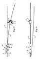
- FIG. 1is a schematic view of various anatomical structures of the female pelvic region, including urinary and reproductive systems.
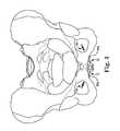
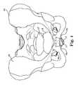
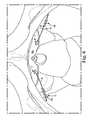
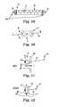
- FIGS. 2-6are schematic views of various anatomical structures of the female pelvic region, and bilateral implants having medial and lateral anchors, in accordance with embodiments of the present invention.
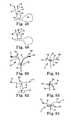
- FIG. 7shows a side view of an implant system having medial and lateral anchors, and sutures, in accordance with embodiments of the present invention.
- FIG. 8is a top view of an implant system having medial and lateral anchors, in accordance with embodiments of the present invention.
- FIG. 9is a perspective view of a distal anchor/barb in accordance with embodiments of the present invention.
- FIG. 10is a partial sectional view of the distal anchor/barb of FIG. 9 .
- FIG. 11is a side schematic view of a distal anchor array and suture within a delivery needle device, in accordance with embodiments of the present invention.
- FIG. 12is a side view of a distal anchor array and suture deployed from a delivery needle device, in accordance with embodiments of the present invention.
- FIG. 13is a perspective view of a distal anchor array and suture within a slotted needle device, in accordance with embodiments of the present invention.
- FIG. 14is a side schematic view of a distal anchor array and suture within a slotted needle device, in accordance with embodiments of the present invention.
- FIG. 15is a side view of a distal anchor array and suture deployed from a slotted needle device, in accordance with embodiments of the present invention.
- FIG. 16is a side view of a distal anchor array and suture at least partially tensioned, in accordance with embodiments of the present invention.
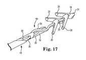
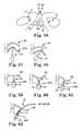
- FIG. 17is a perspective view of a distal anchor array and dual suture, in accordance with embodiments of the present invention.
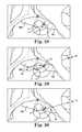
- FIG. 19is a side view of distal anchor array and suture in a collapsed state, in accordance with embodiments of the present invention.
- FIG. 21is a side view of a distal anchor array and suture, showing exemplary load and moment data, in accordance with embodiments of the present invention.
- FIG. 22is a perspective schematic view of a distal anchor array, with dual barbs, and a suture partially within a needle device, in accordance with embodiments of the present invention.
- FIG. 23is a perspective view of a distal anchor, or anchor portion, having dual barbs, in accordance with embodiments of the present invention.
- FIG. 24is a side schematic view of a medial anchor and suture within a delivery tube or oversleeve, in accordance with embodiments of the present invention.
- FIG. 25is a sectional schematic view of a medial anchor and suture within a delivery tube or oversleeve, in accordance with embodiments of the present invention.
- FIG. 27is a perspective schematic view of a medial anchor and suture, with the medial anchor expanded and deployed from a delivery tube or oversleeve, in accordance with embodiments of the present invention.
- FIGS. 28-30are schematic views of various anatomical structures of the female pelvic region, and an implant system having medial and lateral anchors, and the deployment method, in accordance with embodiments of the present invention.
- FIGS. 31-38are schematic views of paraurethral implant systems and methods, with a suture or extension member interwoven or thread through tissue, in accordance with embodiments of the present invention.
- FIG. 39is a schematic view of a coil spring device operably connected to one or more of anchors for use in an implant system and method in accordance with embodiments of the present invention.
- FIG. 40is a schematic view of tissue engagement devices for a medial anchor, in accordance with embodiments of the present invention.
- FIGS. 41-42are schematic views of a dilating medial anchor device in accordance with embodiments of the present invention.
- FIGS. 43-45are schematic views of a grasping medial anchor device, and implantation method, in accordance with embodiments of the present invention.
- FIGS. 46-47are views of a generally star-shaped medial anchor device, in accordance with embodiments of the present invention.
- FIG. 55is a schematic view of various anatomical structures of the female pelvic region, and an implant system having medial anchors, lateral anchors, and crossing extension members, in accordance with embodiments of the present invention.
- FIG. 56is a schematic view of an implant system having suture anchors in the perineal membrane, in accordance with embodiments of the present invention.
- FIG. 57-62are schematic views of an anchor and needle introduction system for use with embodiments of the present invention.
- FIG. 68is a schematic view of a suture locking device for use with embodiments of the present invention.
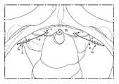
- FIG. 1shows a schematic view of relevant portions of the female pelvic region, and the urinary and reproductive system, including the pelvis PE, vagina V, uterus UT, urethra U, bladder B and the deep clitoral vein C. Further, a portion of the perineal membrane PM is shown at the midurethra/distal location, providing a viable paraurethral target for stabilizing or controlling the position and movement of the urethra to assist in restoring continence.
- Embodiments of the present inventioncan include apparatus and methods for treating urinary incontinence, fecal incontinence, and other pelvic defects or dysfunctions, in both males and females using one or more lateral implants to reinforce the supportive tissue of the urethra.
- One or more implant devices 10are configured to engage and pull (e.g., pull up) or reposition support tissue (e.g., paraurethral), such as the perineal membrane, uterovaginal fascia, endopelvic fascia, or other anatomical features at which connective support of the urethra can be established.
- extension members 30can be constructed of a suture, a thin flat member, braided fibers, braided nano-fibers, an elongate mesh and other various materials and constructs.
- the extension member 30can enhance and draw more collagen-producing cells to the material to promote tissue ingrown and healing.
- the extension member 30 of certain embodiments of the present inventioncan be constructed to be generally flexible, or to have limited elasticity—e.g., bungee type attributes.
- the member 30 extending between the anchors 14 and anchors 16can be an elongate member constructed of an elastomeric material having desirable tensile properties. As such, the member 30 can be stretched out for deployment and then released to provide desirable taut tension.
- the travel or stretching/rebound characteristics of the member 30can vary depending on the particular elastomeric materials used in its construction.
- the extension member 30such as a suture, can further include various extending tines or barbs to facilitate the tissue traction and grabbing during and after deployment.
- One or more opposing anchors 14 , 16 or tissue engagement portionscan be employed to attach and stabilize the implants to the tissue, as well as provide selective adjustment.
- the anchors or engagement portionscan be configured to engage soft tissue and can include various barbs, tines, serrated edges, extending fibers, or other similar structural feature to promote tissue fixation.
- the anchorscan be implanted in a direction lateral from the urethra.
- the anchorscan generally be small enough for to be unnoticeable by both the patient and the patient's sexual partner.
- the anchors and other devices and components of the system 10may be constructed from various biocompatible materials, such as known polymers and metals that promote long-term resilience, or other materials known to those skilled in the art.
- tissue constraining or positioning implant system 10having one or more attachment points in one or more membranes or other target tissue locations.
- Embodimentscan function to restrict, limit or control movement of the mid or distal urethra, or surrounding tissue. Further, embodiments can assist in resisting forward rotational movement of the urethra or surrounding tissue, and can provide support and tension during events, such as coughing or physical activity.
- Various advantages of the implant 10 embodiments depicted hereininclude, frontal access and simpler anatomy to address, less vascularity and bleeding, reduced risk of creating retention and de novo urge, and the ability to test for continence before surgery. Additionally, the implants 10 act to oppose rotational movement of the urethra, thereby eliminating or lessening the effects of stress urinary incontinence.
- a viable treatmenttherefore, will be one that most efficiently opposes rotational movement of the urethra.
- Urethral rotationstill occurs in non-hypermobile patients, just to a lesser extent.
- the concept of rotational mechanicsalso suggests that obturator anchored implants should be placed with higher initial tension and be positioned as far distally as possible on the urethra. Applying this principle of rotational resistance gives rise to devices in accordance with the present invention whereby urethral movement is inhibited near the distal urethra, while the bladder neck continues to move.
- the midurethraWhen a midurethral support is implanted in the female patient, the midurethra is restrained from movement. However, the bladder neck remains mobile and moves downward during a stress event due to elevated abdominal pressure. The resultant effect is that the urethra can be kinked at the midurethra location, causing a closure of the urethra. Like kinking a garden hose, the flow of fluid is restricted or prevented.
- the implant system 10can include one or more anchor devices 12 adapted for use with various embodiments of the present invention, including those adapted to penetrate tissue or soft tissue as disclosed herein.
- Certain of the devices 12e.g., the lateral anchor 16 , can be generally provided in a back-to-back serial configuration, with a suture or like extension member extending to provide adjustable support between the anchor devices 12 .
- the anchor devices 12can include one or more first medial or proximal anchor devices 14 , and one or more second lateral or distal anchor devices 16 .
- the lateral anchor devices 16can include a body portion 18 , one or more expandable barbs 20 , a thru-aperture 22 , and an opposing end 24 .
- a suture 30 or like memberis adapted to string or thread through the respective apertures 22 of a series or array 16 n of such anchors to define the general elongate and expandable configuration shown.
- the array of anchors 16 ncan be inserted within and along the interior lumen 41 of a needle 40 , cannula or like inserter or delivery tool.
- the lateral anchor devices 16can be directed for engagement with tissue distal the anchors 14 at target sites such as the obturator foramen, obturator internus muscle, sacrospinous ligament, prepubic fascia or muscle, abdominal fascia, rectus fascia, puboprostatic ligament, the tendinous arch of the levator ani, the Cooper's ligament, and the pubic symphysis.
- target sitessuch as the obturator foramen, obturator internus muscle, sacrospinous ligament, prepubic fascia or muscle, abdominal fascia, rectus fascia, puboprostatic ligament, the tendinous arch of the levator ani, the Cooper's ligament, and the pubic symphysis.
- Other distal target tissue sites for the anchors 16capable of permitting tensioning support for the perineal membrane or other urethra-supporting tissue is envisioned as well.
- the path from the perineal membrane to the distal anchor 16 of the present inventioncan follow a generally straight line into the obturator internus muscle, or like distal tissue. Furthermore, because it intersects the muscle at an oblique angle, more tissue can be engaged for securement.
- the needle 40can include an exit or opening 42 at a needle tip 42 a .
- a series of anchors 16such as the anchor array 16 n , can include a lead anchor 16 a adapted to first exit through the opening 42 upon deployment.
- the suture 30 pathis generally undulating while within the needle 40 , and even upon initial departure from the needle 40 ( FIGS. 11 and 13-15 ), while it is generally brought into a straight or taut state upon full deployment from the needle 40 ( FIGS. 7, 12 and 16 ).
- the end portion 24 of the lead anchor 16 acan be permanently attached to the end of the suture 30 via bonding, adhesive, welding, knotting, or the like.
- the anchor 16 or its respective componentscan be molded together or otherwise attached to create the construct depicted and disclosed.
- Each successive anchor 16e.g., after lead anchor 16 a , is alternately arranged such that they can be closely aligned along or within the lumen 41 of the delivery needle 40 .
- the suture 30passes through these anchors 16 , and the anchors 16 can be adapted to slide on the suture 30 .
- the suture 30can follow a serpentine or otherwise undulating path.
- a pusher rod 43or like mechanism or device may be biased or pushed against the proximate anchor 16 b (e.g., opposite end from the lead anchor 16 a ), as illustrated in FIGS.
- the array 16 ncan be deployed in various ways.
- the pusher 43simply forces the anchors 16 n out of the lumen of the needle 40 . Some suture 30 tension can be maintained so that the anchors 16 n are efficiently driven out in a straight line or path.
- the position of the anchors 16 n relative to the tissueremains fixed or stationary (e.g., with the aid of the pusher 43 ) while the needle 40 is retracted back or away (e.g., slid) from the array 16 n such that the anchors 16 are deployed from the lumen 41 .
- the pusher rod or member 43can be a wire or tube that fits inside and through the proximal end of the needle 40 , through the lumen 41 , and acts against at least one of the anchors, directly or indirectly, including the most proximal anchor.
- FIGS. 18-20can include an extended thru-aperture 22 , generally along the length of the body 18 of the anchor 15 , that allows the suture 30 to pass in a generally straight line through the array 16 n while in the lumen 41 of the needle 40 , and bend or reform into a general serpentine or undulating shape after the anchors 16 have been deployed and the barbs 20 expand outward for tissue engagement.
- This approachcan have the advantage of utilizing the suture 30 tension to resist the rotation of the barbs 20 working against tissue.
- the barbs 20when the barbs 20 are engaged within the target tissue site, they can be subjected to a moment load M. This moment is opposed by the suture line tension moment which is proportional to T*L, where T is the tension and L is the moment arm. This moment arm increases as the angle ⁇ of the serpentine or undulation increases.
- Tthe tension
- Lthe moment arm
- FIGS. 22-23Another embodiment of the anchors 16 and anchor array 16 n configuration is provided in FIGS. 22-23 .
- a dual-tipped/barbed anchor 60is included that flexes about a hinge 62 at the base 18 .
- the anchors 60are provided in an array 60 n with a lead anchor 60 a .
- the array of barbs 16 nare spaced apart along the suture 30 while inside the lumen 41 of the needle 40 , and condense or compress together as they are pushed out of the needle 40 , or the needle 40 is removed, and put under tension.
- the two barbs 20 of each anchor 60are forced further open by the body portion 18 of the adjacent abutting anchor 60 .
- the body portion 18 of the preceding anchor 60can slide into the gap between the two barbs 20 of the next leading anchor 60 to cause those barbs 20 to flare out or expand, as shown in FIG. 20 .
- This configurationmay be more compact than the single barb designed anchor 16 and also may not depend as much on the suture 30 tension to oppose the load on the tips. Namely, the tissue fixation may better resist backing out of the tissue target site compared to various other embodiments for cases where an increased degree of fixation is required or desired.
- the anchors 16 , or anchors 60can be fabricated using a metal injection molding process, or from a molded resin material (e.g., 720FC resin, polycarbonate, PEEK, nylon), with an exemplary Prolene monofilament, or braided, suture 30 threaded therethrough.
- the componentscan be easily inserted through the lumen 41 of the needle 40 and arranged in an alternating pattern—e.g., angular orientation pattern—along the suture 30 .
- the alternating angular pattern of the anchors 16 in FIG. 7is approximately 180 degrees
- the alternating angular pattern of the anchors 60 in FIG. 22is approximately 90 degrees.
- the suture 30can be lightly tensioned to bring all the anchors 16 in the array 16 n together while holding the pusher 43 in place. Again, while holding the pusher 43 stationary, the needle or cannula 40 can be retracted, leaving the array 16 n , or 60 n , and the respective anchor barbs 20 embedded in tissue. A slight tug of the suture 30 can bring the anchors together and take up any initial slack in the suture line 30 .
- embodiments of the tissue anchoring devices and methodscan include a reduced trauma explantation (e.g., removal from tissue) configuration and mechanism for the barbed soft tissue anchors, e.g., the anchors 16 , described and depicted herein.
- a reduced trauma explantatione.g., removal from tissue
- the anchors 16e.g., the anchors 16 , described and depicted herein.
- one solutionis to attach an explantation tether 50 to the leading anchor 16 a of the array 16 n . This could be in the form of a suture, or continuation of the existing traction suture 30 that leads back out of the implantation path.
- the physiciansimply pulls on this tether 50 , causing the anchor 16 to double-back on itself and pull out atraumatically—e.g., through the defined tissue path or tissue penetration site. This could be done during the initial implantation procedure or at a later time in the event that the device 16 , or implant 10 , must be disengaged or removed.
- the average normal pullout force of an engaged anchor 16 or anchor array 16 ncan be approximately 4 to 7 lbs with various embodiments, while the explantation force of the embodiments having the explantation tether 50 can be around 1 to 3 lbs.
- the physiciancan pull out the entire assembly by tugging on it. Pulling up on the explant suture or tether 50 causes the anchors to double-back on themselves and continue out of the tissue in the non-resistant direction, lead by anchor 16 a . This reduces the removal force and associated tissue trauma.
- This type of explant methodcan be used with any implanted device that has an anchoring end that is generally flexible or segmented enough (e.g., series of separate anchors 16 strung along the member 30 of the anchor array 16 n ) to allow doubling-back.
- the implantcan be designed such that the free end of the explant tether 50 , generally opposite the end attached to or proximate the lead anchor 16 a , can be accessed by a physician. This could be done by leaving the suture 50 end hanging out of the implantation puncture. The free end can be later trimmed or removed when explantation is no longer needed.
- the tether 50can be constructed of an absorbable material.
- a tag or loopcan be included at the free end of the tether 50 . This tag or loop can remain just below the skin surface and can be accessed later if the implant 10 or device 16 require explantation.
- the anchor array 16 nis thread or otherwise provided along the suture 30 , or paired sutures 30 (e.g., FIG. 17 ), and can be delivered via a percutaneous passage inside a hypotube or the needle 40 . This allows for controlled delivery of the anchor array 16 n such that the needle tip 42 a can be selectively repositioned before the anchors are set in soft tissue.
- embodiments of the needle system 40can include a slotted needle configuration, with the needle 40 including a slot or groove 64 along a distal or end portion of the needle 40 body such that a portion of the suture 30 can pass outside the lumen 41 of the needle 40 during deployment.
- the slot 64can be created in or along a portion of the needle 40 by milling, laser cutting, EDM machining, or using other similar fabrication, manufacturing or formation methods.
- the slot machiningmay be done before or after the bending operation for the needle 40 .
- the slot 64With a curved needle 40 , the slot 64 can be on the outer side of the bend. This, in turn, can promote keeping the portion of the suture 30 that lies in the slot 64 to stay inside the lumen 41 when under tension.
- a pair of sutures 30can pass through each anchor 16 in the array 16 n , and follow a serpentine or undulating path within the needle 40 .
- a knot, bead, stop, member or like structure 31 at the distal end of the suture 30 proximate or in front of the leading anchor 16 acan act as a stop for the lead anchor 16 a .
- a second knot, bead, stop, member or like structure 31 acan be included at a portion of the suture 30 , or paired suture, near the most proximal anchor 16 b , and/or outside the slot 64 (e.g., FIGS. 13-14 ).
- Embodiments employing a paired suture 30 configurationcan include members, structures or other constructs, including the apertures 22 of the anchors 16 , having a generally rectangular or oval shape such that the pair can be passed through to hold them side-by-side. By doing this, the barbs 20 are less able to rotate on the axis of the suture line 30 and will stay properly oriented. By creating anchors 16 that have different through hole angles or shapes it is also possible to fix the angular position of the anchors 16 n to create a larger anchor spread.
- a pusher 43can be positioned behind, or abut against, the proximal anchor 16 b that acts as a stop (prevent backing out of array 16 n within needle lumen 41 ) during the needle insertion and deployment process.
- the pusher 43can also serve to maintain the anchors 16 and respective barbs 20 barbs in a fixed position, relative to the tissue, as the needle 40 is retracted or pulled away from the array 16 n .
- the slot 64can be sized to allow the suture 30 to freely pass through, but does not allow the anchors 16 or respective barbs 20 out through that portion of the needle 40 .
- the length of the slot 64can assume many size configurations, depending on the size of the anchors 16 in the array 16 n and the number of serially aligned anchors 16 in the array 16 n .
- the slot 64could also extend along the entire length of the entire needle 40 in certain embodiments, or take on various other size and shape configurations depending on particular device and application needs.
- the slot 64 lengthcan be defined by the anticipated length of depth of the tissue targeted for penetration.
- embodiments of the needle 40 including the slot 64 configurationcan facilitate easier and more efficient use of a medial anchor 14 .
- the medial anchor 14is attached to, or threaded or provided along a portion of the suture 30 that does not need to be constrained or fit within the relatively thin and small needle 40 or lumen 41 .
- the slot 64provides a length of suture 30 that can ride outside of the lumen 41 , with the medial anchor 14 attached or provided along that external length of suture 30 . This provides greater flexibility for the design and construct of the medial anchor 12 and the respective delivery method.
- the pusher 43will not interfere (e.g., traverse alongside) with the proximal length of the suture 30 provided before the anchor array 16 , as the proximal portion of the needle lumen 41 will be free of the suture 30 .
- the slot 64provides more room for movement and spreading compared to non-slotted embodiments of the needle 40 where everything within the lumen 41 is confined to the lumen walls.
- the slot 64keeps the exiting suture 30 portion from unwanted turning and twisting, thereby assisting in keeping the attached anchors 16 also fixed in a preferred angular orientation within the needle 40 .
- the proximal stop 31 acan also be used to keep the anchor barbs 20 from spreading apart during assembly and during the deployment of the anchors 16 n .
- the knot, bead or stop 31 acan be positioned either inside or outside the lumen 41 .
- One advantage for positioning the stop 31 a outsideis that it can introduce enough drag to enable retraction of the needle 40 while still keeping the anchors 16 in place. In certain embodiments, this can preclude the need for an internal pusher 43 to hold the anchors 16 in place upon deployment.
- the stop 31 acould take on nearly any size or shape, and material.
- the anchor systemcan include intermediate knots, beads or stops 31 that separate smaller or distinct groupings of anchors 16 .
- a stop 31 between a fourth and fifth anchor 16can result in two groups of four barbs forming on the suture line 30 .
- Such a grouping configurationcan assist in redistributing the tension on the suture line 30 in stages or segments, as well as to reduce the amount of pulling that is required to initially set the anchors 16 in tissue. It can also improve the overall tissue anchoring force and stability.
- Embodiments of the lateral anchors 16can include self-expanding structures or materials such that the anchors 16 , or anchor array 16 n , can be generally collapsed or reduced in sized during deployment, with or without a needle device 40 , and expanded after penetration in the target tissue site to provide desired tissue engagement.
- Certain anchors 16can include one or more shape memory portions, or living hinges, to facilitate this structural self-expansion upon deployment and tissue engagement.
- embodiments of the lateral anchor 16 , or anchor array 16 ncan include helical portions, threaded portions, hooks, clips, flexible barbs, textured surfaces, and like members or structures to promote tissue engagement.
- lateral anchor 16can be adapted to include a plurality of anchors, extending from one or more separate members 30 , spread out into multiple anchoring features for deployment into tissue to provide support and treatment.
- a spanning multi-anchor device 16can create a neo-ligament to reduce or eliminate rotation of the urethra U or surrounding tissue through the use of multiple anchoring spots.
- the medial anchors 14can include metal or like members or tubing, such as nitinol, stainless steel, titanium, or polymer, laser cut or formed into longitudinal petals 44 that are capable of selectively collapsing and expanding in a flex leaf construct.
- the cuttingmay be done such that any number of petals is formed.
- Exemplary embodimentscan include two, three or four petal constructs.
- the formation and configuration of the petals 44can be accomplished using a heat-treatment process where the petals 44 are set in the opened state, and whereby the ends of the petals 45 a distance from the hinges 48 are radially expanded and set in that position.
- these petals 44can be temporarily closed, facilitating the insertion of the anchor 14 into a small puncture, such as within the tissue of the perineal membrane PM.
- the anchor 14can self-expand, e.g., via shape memory, to the opened state to provide tissue engagement and fixation.
- Various embodiments of the medial anchors 14can include slots 49 (e.g., laser cut) in the petals 44 to develop the bend or living hinges, as shown in FIGS. 26-27 .
- These slots 49can be very narrow (e.g., approximately 0.001′′-0.003′′ wide) and allow a limited degree of localized bending along a portion of the corresponding petal 44 .
- the petals 44can easily flex to the opened state, but can require a greater load to move beyond that point where the slots 49 have closed together.
- the petal 44is less likely to overbend or flip out of position.
- Another advantageis that this embodiment can be easily adapted to a variety of flexibility configurations depending on the desired attributes.
- the number, sizing, and spacing of the slots 49can be varied to adjust the amount of allowable bending and the load required to achieve that bend.
- the component or petal 44can be heat-set to the opened position so that it self-expands from a closed position.
- Embodiments of the medial anchor 14can include a body portion 47 and one or more apertures therethrough to receive or connect with the suture 30 .
- a medial needle device or oversleeve 59can be included with embodiments of the invention to facilitate introduction and deployment of the medial anchor 14 .
- FIGS. 24-25show the anchor 14 in a contracted state within the lumen of the needle 59 during deployment, with FIGS. 26-27 showing the anchor 14 expanding when pushed, pulled or otherwise removed from the inner constraints of the needle 59 .
- Exemplary anchors 14can include substances, such as an adhesive (e.g., light activated adhesive) to assist in the tissue engagement process.
- FIGS. 28-30depicts a medial anchor 14 at the perineal membrane PM implanted via a transverse insertion through the anterior vaginal wall, between the perineal membrane PM and the superficial skin lateral to the urethral meatus, according to an embodiment of the implant 10 .
- a needle 40 passis made from the lateral side of the meatus that intercepts the medial anchor 14 and passes through a pre-made hole or punctures the anchor 14 .
- the needle 40 passcontinues on to the obturator muscle, or other distal tissue, where the lateral anchor 16 , or anchor array 16 n , is delivered.
- the medial anchor 14can be placed in the superficial space between the perineal membrane PM and the distal skin layer at the meatus.
- the small medial anchor 14can be held with a custom holder or forceps until the intersecting needle 40 pass.
- a trailing suture 30can include a one-way slider 90 , or like sliding or locking device, on it.
- the slider 90can be pushed along the suture 30 , mating with the anchor 14 .
- Tensionis increased by pushing the slider 90 farther along the suture, thereby pressing tighter against the anchor 14 , and/or the surrounding tissue.
- an anchor 14 that is passed through a skin puncture lateral to the meatusmay need to be turned such that its major axis is parallel to the puncture hole, then rotated or expanded to face the perineal membrane.
- the medial anchor 10 for this transvaginal methodgenerally would not have the same size or orientation limits and could therefore be substantially larger and better able to resist pull-through failure.
- the medial anchor 14 positioncould in fact be in the anterior wall of the vagina V for any of the disclosed treatment and anchoring embodiments.
- the traction force vectorwill be more parallel to the vaginal wall with the disclosed embodiments of the present invention so that an optimal rotational resistance of the urethra U can be achieved.
- the anchor 14can be implanted via a puncture at the skin surface, but the introducer needle 40 can still be passed within the thickness of the vaginal wall as described.
- the anchor 14can be constructed of a flexible material to reduce the sensation or recognition of the anchor 14 to the patient or the patient's sexual partner. Furthermore, while the anchor 14 is under tension it can bend such that the line of force is generally more parallel to the suture (e.g., reducing the “cheese-cutter” effect).
- the two ends of the medial anchor 14can be relatively rigid but flexible in a middle body portion, such that the urethral kinking effect is enhanced when there is tension (e.g., if the flex is near the perineal membrane).
- the support or extension members 30can apply mechanical traction to the urethra in a manner similar to a mini-sling device.
- a benefit of embodiments of the present inventionis that the transvaginal placement of the structures and devices does not leave exposed material (e.g., implant mesh) inside the vaginal cavity.
- the implanted device 10 positionis generally blind and lies beyond the superficial mucosal layer of the vaginal wall. Reducing or eliminating the exposed material minimizes the risk of infection, irritation at the surface of the vaginal wall, and provides cosmetic improvement and reduces interference with sexual activity.
- various embodiments of the implant system 10can include anchoring elements or portion is fixed on each side of the urethra, on the far side of a tissue layer that is known to have relatively high strength and toughness.
- the medial or proximal anchor 14can include a “toggle” anchor, which is a small, elongated structure that can be placed through a small puncture or like incision and then rotates after deployment so that it cannot back out through the incision hole.
- Other anchoring devices and methodscan be employed with the present invention as well.
- the suture 30can weave or thread in and out of, and along, the tissue, e.g., the perineal membrane, to provide a supportive undulating layout for the suture 30 and implant 14 combination. This can facilitate attachment, better distribute pulling force on or along the tissue, and provide like support benefits.
- one or more sutures 30can be woven or interwoven (e.g., in and out) of the perineal membrane, with anchors 14 engaging at or proximate the posterior symphysis. When tightened, the suture 30 pulls and compresses tissue toward the posterior symphysis to provide support and strength.
- a suture lock device or techniquecan be included to fix the tension or support adjustment of the sutures 30 .
- the distal or anchor device 16is placed in a lateral or superior position such that a connection (e.g., suture 30 or wire connection) between the medial and lateral anchors 14 , 16 can provide tensile support for the urethra during stress events.
- the anchor device 16can be fixated to, or engaged with, the obturator membrane, obturator internus, tendinous arch of the levator ani (white line), the Cooper's ligament, sacrospinous ligament, prepubic fascia or muscle, the pubic symphysis cartilage, abdominal fascia, or other stable anatomical features.
- the final position of the implanted device 10creates a support structure that is similar to a needle suspension.
- the medial anchor 14can spread or better distribute the tension load over a larger surface (as opposed to a thin suture cutting edge surface) than other procedures and devices. This, in turn, promotes stability of the anchor and connecting suture or spanning support member.
- the medial toggle anchor 14is implanted, a needle 70 is withdrawn, a free suture or connector end is delivered through the insertion opening, the lateral (e.g., obturator) anchor 16 is delivered and implanted, and the connecting suture 30 is properly tensioned between the anchors 14 , 16 to provide proper support.
- the needle 70can be adapted to contain the toggle anchor 14 with a tip 70 a directed for penetration through the perineal membrane or like paraurethral support tissue.
- the toggle anchor 14can include a sharp or beveled tip to facilitate penetrating through the target tissue.
- the needle 70is then withdrawn with the suture 30 extending therefrom and a distal anchor 16 attached or provided at the opposing free end of the suture 30 .
- the anchor 16can be small enough to pass through the small punctures in the tissue and deploy at the obturator foramen or like tissue targets.
- Various delivery tools and devices disclosed herein, or known,can be used to direct and deploy the anchor 16 .
- the suture 30can be tensioned to provide the desired level of support for the paraurethral tissue ( FIG. 35 ).
- the medial anchor 14can include a one-way tension holding feature (e.g., zip tie-like) that allows the physician to pull out excess suture material without the excess slipping back through the anchor 14 .
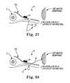
- FIGS. 36-38show another embodiment of the implant 10 having a toggle anchor configuration.
- a suture needle 72can be utilized to create a loop or “bite” 71 through the paraurethral tissue to provide additional stability and anchoring support.
- the suture needle 72can be included with the suture 30 , and can be removable with certain embodiments.
- the suture 30can be woven in and out of multiple portions of the perineal membrane or like tissue, with or without a medial anchor 14 , such that pulling on the suture 30 can compress or cinch up the tissue.
- One or more suture locking devices 77e.g., diametric suture lock of FIG. 68 , can be inserted through a portion (e.g., free end) of the suture 30 to selectively lock the suture in place after the desired tension is obtained.
- the suture locking devices 77can include a one-way locking mechanism constructed of a generally cylindrical body having one or more cuts 77 a (e.g., laser cut) defined in a surface or portion of the device 77 to define inward extending tines 77 b .
- the tines 77 bcan permit the suture 30 to ride or slide within the device 77 in a direction not against the direction of the tine 77 b angles. However, backing out of the suture 30 will be prevented in the opposite direction when the tines 77 b grab onto the suture (e.g., braided suture) and restrict movement in that direction.
- Other suture locking devicesare envisioned for use with various embodiment of the present invention as well.
- embodiments of the present inventioncan include a coil spring device 76 operably connected to one or more of the anchors 14 , 16 .
- the device 76can be held in a slightly extended state that generates a preload tension.
- the device 76can include a spring mechanism 78 , which is connected at either end to the suture 30 and is forced open with stops 80 that impose a fixed extension against a rigid structure or housing 82 .
- the spring 78may extend, but only after the suture tension exceeds the preload force.
- a conventional coil extension springcan be employed such that when the spring relaxes to its solid height, some pretensioning load is maintained—e.g., it is unable to further contract due to the coils being in full contact.
- Other methods for mechanically creating initial tension and load controlare envisioned as well.
- Preloadingcan be achieved by pretensioning the suture during the implantation procedure or could be achieved by creating mechanical pretension internally in the anchor devices 14 , 16 , or mechanisms operably connected to the devices 14 , 16 . As such, a constant rest load against tissue (which might stretch) can be provided.
- embodiments ofcan include one or more generally C-shaped, or like shaped, medial anchors 14 .
- the anchors 14can be placed adjacent the urethra U to provide a medial anchor configuration.
- the sutures 30can extend from the anchors 14 to one or more distal anchors 16 .
- FIGS. 41-42show an embodiment of the medial anchor device 14 having an anchor base portion 14 a and a dilating anchor portion 14 b .
- the dilating anchor portion 14 bis adapted for deployment to rest or otherwise stop or abut against a portion of the perineal membrane PM, or other like tissue.
- the suture or other extension member 30can extend between the two anchors 14 , 16 .
- the dilating anchor portion 14 bis not generally applying pressure to the urethra U.
- the dilating anchor portion 14 bexpands (e.g., expands under compression) to apply a level of pressure on the urethra U to promote continence, as depicted in FIG. 42 .
- FIGS. 43-45are directed to a medial anchor device adapted to puncture through the skin and into the perineal membrane PM or like tissue. Such a superficial skin puncture can be targeted at the crease between the labia minora and majus, directed at the space between the bulb of vestibule and clitoral crus, in certain embodiments.
- a tube or delivery needle 84can include or contain a pair of anchoring or stop members 86 a , 86 b and at least a portion of the member 30 . The needle 84 operably connects or carries the stop members 86 a , 86 b and extends beyond them.
- a clip, tie, washer, lock mechanism or other devices and techniquescan be employed to secure the stop members 86 a , 86 b in place against the perineal membrane.
- the devicecan provide tension and a twisting motion on the membrane, thereby translating to rotational torque.
- the free end of the extension member 30can include one or more lateral anchor devices 16 as disclosed herein.
- the medial anchor 14can include structures adapted to attach to or span across a portion of the perineal membrane, or like tissue, to facilitate engagement, compression or anchoring with the tissue.
- a plate, mesh material, tissue cinching device, stent-like device, ring, clip, coil, spring, strap, pad, patches, or similar structurescan be affixed to, directly or indirectly, the perineal membrane, with such anchors 14 then being connected to the lateral anchor 16 via the extension member 30 .
- These structurescan be attached to tissue via sutures, anchors, and similar tissue engagement devices.
- the anchors 14 and related structurescan include rigid, semi-rigid or flexible polymer or metal materials.
- One-way locking devicescan be incorporated with any of the anchors 14 , 16 , or along (e.g., thread along) the member 30 such that the physician can adjust the tensioning of the implant 10 to the desired level and fix the tension for optimal support and the promotion of continence.
- a patientcould be placed in a lithotomy position for the implantation procedure.
- a physicianmay make one or more incisions through the perineal tissue lateral to the urethra of the patient.
- the physicianmay make one or more vaginal incisions to access the tissue superior to the urethra.
- the physicianmay use the needle delivery device 40 to implant the devices or anchors.
- the medial or proximal anchor 14can then be implanted through the perineal incision, thereby reducing the invasiveness of the procedure.
- the delivery device 40may be configured to allow insertion through a single or multiple perineal or transvaginal incisions.
- needle 40can be directed “outside-in,” from the skin through the obturator membrane, then with an anchor 14 engaged with the perineal membrane.
- the anchor 14can include suture loops. The loops can be tied from the peritoneum side. From the obturator side, the multiple loops or sutures 30 can then be tied around the anchor for fixation.
- the full implant 10can be implanted but left in a loosened state with the free end of the suture 30 left hanging or free. After several weeks, the implant 10 can be tightened by pulling the suture 30 (or by adjusting a locking or securement device) further out and then trimming it off.
- the SPPis the compartment of the perineum that lies between the perineal membrane and the perineal fascia (Colles fascia).
- the anterior SPPcan be dissected from the frontal side in order to expose the tissue in this space.
- This areagenerally consists of layers of connective tissue and fascia, and some thin muscle. The tissue in this region is easily penetrated or compressed with a blunt needle, until reaching the perineal membrane. Thus, there can be ample room and favorable tissue properties in this region to accommodate the implant 10 or anchoring devices.
- FIGS. 48-54depict implants 10 adapted for fixation with the perineal membrane to provide adjustment (such as lift), to thereby provide support and adjustment of the patient's urethra U.
- FIGS. 50-51show an embodiment having two perineal membrane PM piercing or penetration points, with the implant including a small cinch ring 100 adapted to pull on the members 30 (such as suture, strand, mesh, etc.) to provide the desired adjustment—e.g., along a suture loop.
- Lateral anchors 16can be adapted to penetrate through the perineal membrane PM for fixation and securement distal the paraurethral tissue.
- the cinch ring 100can slide up to the desired position along the members 30 to provide selective adjustment and tensioning, as shown in 51 .
- the ring 100can include a stop button or collar having a one-way draw string or device to provide securement as well.
- FIGS. 52-53show an embodiment of the implant adapted for a single perineal membrane piercing or penetration. Further, FIG. 54 depicts a curved profile for the ring 100 to distribute pressure from the implant 10 .
- embodiments of the implant system 10can include members, such as sutures 30 , adapted to cross over each other, with distal end anchors 16 fixated in tissue away from the perineal membrane PM. This, in turn, can coapt or compress the urethra while providing tension and desirable support characteristics, while reducing undesirable rotation.
- Medial anchors 14such as patches or like anchor structures, can press against, affix or engage the perineal membrane to provide a larger anchoring area.
- Certain embodimentscan engage and anchor to the perineal membrane with only sutures 30 .
- a length of suture 30is pushed through the tissue (e.g., one on each side of the urethra U in the perineal membrane PM) with an end anchored in the disclosed distal anchor target tissue via anchor 16 (e.g., obturator or like target locations) and another end anchor 102 anchored in other tissue, such as the rectus fascia or like tissue.
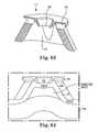
- FIGS. 57-62are directed various embodiments of anchor introduction system 110 for use with embodiments disclosed herein.
- FIGS. 57-58show problems with tissue deflection that can occur when the needle 40 punctures the skin, for those embodiments employing percutaneous anchor or implant deployment, as the needle 40 approaches the perineal membrane PM, next to the urethra U.
- the needle 40can be undesirably diverted laterally before puncturing the perineal membrane PM.
- Embodiments of the system 110are therefore provided to limit the movement of tissue being punctured or traversed by a needle deployment device 40 to ensure that a proper and desirable puncture depth is achieved.
- the system 110can include the needle 112 having a helical or corkscrew-like end member 114 .
- the needle pathis controlled such that it does not divert laterally.
- the coil member 114will engage the perineal membrane PM ( FIG. 61 ) and provide an operative path for the needle device 40 to enter through the skin and through the membrane PM ( FIG. 62 ).
- the member 114can be constructed of a bioabsorbable material, such as a bioabsorbable polymer, and left behind in the patient. As such, the member 114 can serve as a temporary support for an anchor device 14 inserted via the needle.
- a tissue separation device 120can be constructed as a rigid or elastic part that includes a vacuum chamber 122 into which tissue can be drawn, one or more vacuum ports 124 connected to an external vacuum source, and an aperture 126 through which an introduction or delivery device 40 can pass.
- the device 120can be particularly useful in creating appropriate needle pathways for minimally invasive (percutaneous) insertion of implants or anchors.
- the traction provided by the vacuumalso serves to hold the tissue stationary so that minimal “tenting” or displacement of the tissue occurs during the needle penetration.
- the tissue separation device 120includes features specifically directed to creating separation of tissue layers.
- the aperture 126can be surrounded by a perimeter configuration that is angled down and adapted to contact tissue to form an interior vacuum seal.
- the perimetercan be constructed of a rigid or elastic material that is suitable for forming a good seal against tissue, such as the perineal membrane or the vaginal mucosal layer.
- the interior geometry of the device 120is sized and shaped such that only the thin tissue layer is drawn into the cup space.
- the tissue wallis drawn in because it is more elastic and is thin enough to fold into the space.
- the underlying muscularisis too thick and stiff to fit into the space. As such, a separation of the tissue layers is created.
- a trocarslidably housed within the needle to control and facilitate tissue traversal and piercing or penetration at the target site.
- a single medial anchor 14 and lateral anchor 16 , or lateral anchor array 16 ncan be connected by a suture 30 to support and adjust the perineal membrane, above, below, or on a side of the urethra.
Landscapes
- Health & Medical Sciences (AREA)
- Life Sciences & Earth Sciences (AREA)
- Urology & Nephrology (AREA)
- Animal Behavior & Ethology (AREA)
- General Health & Medical Sciences (AREA)
- Engineering & Computer Science (AREA)
- Biomedical Technology (AREA)
- Heart & Thoracic Surgery (AREA)
- Veterinary Medicine (AREA)
- Public Health (AREA)
- Surgery (AREA)
- Cardiology (AREA)
- Oral & Maxillofacial Surgery (AREA)
- Vascular Medicine (AREA)
- Transplantation (AREA)
- Nuclear Medicine, Radiotherapy & Molecular Imaging (AREA)
- Medical Informatics (AREA)
- Molecular Biology (AREA)
- Rheumatology (AREA)
- Pathology (AREA)
- Prostheses (AREA)
- Surgical Instruments (AREA)
Abstract
Description
Claims (36)
Priority Applications (8)
| Application Number | Priority Date | Filing Date | Title |
|---|---|---|---|
| US13/556,167US9414903B2 (en) | 2011-07-22 | 2012-07-23 | Pelvic implant system and method |
| EP12819552.6AEP2739243B8 (en) | 2011-08-04 | 2012-08-06 | Pelvic implant system |
| PCT/US2012/049759WO2013020134A1 (en) | 2011-08-04 | 2012-08-06 | Pelvic implant system and method |
| AU2012289851AAU2012289851A1 (en) | 2011-08-04 | 2012-08-06 | Pelvic implant system and method |
| HK14112008.9AHK1198511A1 (en) | 2011-08-04 | 2012-08-06 | Pelvic implant system and method |
| US15/217,854US10314681B2 (en) | 2011-07-22 | 2016-07-22 | Pelvic implant system and method |
| AU2016273999AAU2016273999B2 (en) | 2011-08-04 | 2016-12-19 | Pelvic implant system and method |
| US16/404,458US11284983B2 (en) | 2011-07-22 | 2019-05-06 | Pelvic implant system and method |
Applications Claiming Priority (14)
| Application Number | Priority Date | Filing Date | Title |
|---|---|---|---|
| US201161510726P | 2011-07-22 | 2011-07-22 | |
| US201161515180P | 2011-08-04 | 2011-08-04 | |
| US201161545104P | 2011-10-07 | 2011-10-07 | |
| US201161547503P | 2011-10-14 | 2011-10-14 | |
| US201161547467P | 2011-10-14 | 2011-10-14 | |
| US201261607332P | 2012-03-06 | 2012-03-06 | |
| US201261607891P | 2012-03-07 | 2012-03-07 | |
| US201261608478P | 2012-03-08 | 2012-03-08 | |
| US201261608436P | 2012-03-08 | 2012-03-08 | |
| US201261653213P | 2012-05-30 | 2012-05-30 | |
| US201261653224P | 2012-05-30 | 2012-05-30 | |
| US201261653199P | 2012-05-30 | 2012-05-30 | |
| US201261653236P | 2012-05-30 | 2012-05-30 | |
| US13/556,167US9414903B2 (en) | 2011-07-22 | 2012-07-23 | Pelvic implant system and method |
Related Child Applications (1)
| Application Number | Title | Priority Date | Filing Date |
|---|---|---|---|
| US15/217,854ContinuationUS10314681B2 (en) | 2011-07-22 | 2016-07-22 | Pelvic implant system and method |
Publications (2)
| Publication Number | Publication Date |
|---|---|
| US20130023724A1 US20130023724A1 (en) | 2013-01-24 |
| US9414903B2true US9414903B2 (en) | 2016-08-16 |
Family
ID=47629726
Family Applications (3)
| Application Number | Title | Priority Date | Filing Date |
|---|---|---|---|
| US13/556,167ActiveUS9414903B2 (en) | 2011-07-22 | 2012-07-23 | Pelvic implant system and method |
| US13/568,093Expired - Fee RelatedUS9827083B2 (en) | 2011-08-04 | 2012-08-06 | Pelvic implant system and method |
| US15/217,854Expired - Fee RelatedUS10314681B2 (en) | 2011-07-22 | 2016-07-22 | Pelvic implant system and method |
Family Applications After (2)
| Application Number | Title | Priority Date | Filing Date |
|---|---|---|---|
| US13/568,093Expired - Fee RelatedUS9827083B2 (en) | 2011-08-04 | 2012-08-06 | Pelvic implant system and method |
| US15/217,854Expired - Fee RelatedUS10314681B2 (en) | 2011-07-22 | 2016-07-22 | Pelvic implant system and method |
Country Status (5)
| Country | Link |
|---|---|
| US (3) | US9414903B2 (en) |
| EP (1) | EP2739243B8 (en) |
| AU (2) | AU2012289851A1 (en) |
| HK (1) | HK1198511A1 (en) |
| WO (1) | WO2013020134A1 (en) |
Cited By (1)
| Publication number | Priority date | Publication date | Assignee | Title |
|---|---|---|---|---|
| US9974640B2 (en) | 2011-09-22 | 2018-05-22 | Boston Scientific Scimed, Inc. | Pelvic implant and treatment method |
Families Citing this family (23)
| Publication number | Priority date | Publication date | Assignee | Title |
|---|---|---|---|---|
| EP2734148B1 (en) | 2011-07-22 | 2019-06-05 | Boston Scientific Scimed, Inc. | Pelvic implant system |
| US9414903B2 (en) | 2011-07-22 | 2016-08-16 | Astora Women's Health, Llc | Pelvic implant system and method |
| US20140005469A1 (en)* | 2012-06-27 | 2014-01-02 | Urova Medical, Inc. | Implants for stress urinary incontinence treatments and related methods |
| US9409009B2 (en)* | 2012-11-07 | 2016-08-09 | The Florida International University | Multi-lead multi-electrode management system |
| US12064329B2 (en)* | 2013-03-15 | 2024-08-20 | Boston Scientific Scimed, Inc. | Surgical implant system and method |
| WO2014190080A1 (en)* | 2013-05-21 | 2014-11-27 | Ams Research Corporation | Surgical implant system and method |
| US10299828B2 (en) | 2013-08-02 | 2019-05-28 | Escala Medical Ltd. | Anchor delivery system and method |
| WO2016014244A1 (en)* | 2014-07-20 | 2016-01-28 | Riina Howard Anthony | Anchored suture line, device and method for surgical suturing |
| AU2015301398B2 (en)* | 2014-08-15 | 2020-05-21 | Axonics Modulation Technologies, Inc. | Implantable lead affixation structure for nerve stimulation to alleviate bladder dysfunction and other indications |
| US20160116274A1 (en)* | 2014-10-27 | 2016-04-28 | At&T Mobility Ii Llc | Mobility based location determination |
| US9687223B2 (en) | 2014-12-04 | 2017-06-27 | Coloplast A/S | Tissue anchor system |
| WO2019165108A1 (en) | 2018-02-22 | 2019-08-29 | Axonics Modulation Technologies, Inc. | Neurostimulation leads for trial nerve stimulation and methods of use |
| EP4406491A3 (en) | 2018-03-09 | 2024-10-30 | Coloplast A/S | Tissue anchor, tissue anchor assembly, and tissue anchor system |
| US11173299B2 (en)* | 2018-12-03 | 2021-11-16 | Advanced Bionics Ag | Electrode leads configured to engage with a fixing element and methods for manufacturing the same |
| CA3142732A1 (en)* | 2019-06-06 | 2020-12-10 | Guillermo DAVILA | System and method for paraurethral support restoration to treat stress incontinence |
| USD980424S1 (en) | 2020-04-29 | 2023-03-07 | Coloplast A/S | Tissue anchor |
| US11944288B2 (en) | 2020-04-29 | 2024-04-02 | Coloplast A/S | Tissue anchor system including a fixation device and a delivery tool |
| US12336897B2 (en) | 2020-06-30 | 2025-06-24 | The Brigham And Women's Hospital, Inc. | Systems and methods for treating stress urinary incontinence |
| WO2022006350A1 (en)* | 2020-06-30 | 2022-01-06 | Einarsson Jon I | Systems and methods for treating stress urinary incontinence |
| WO2022022821A1 (en)* | 2020-07-29 | 2022-02-03 | Promedon Gmbh | A human implant arrangement for constricting a human tissue structure |
| US12420103B1 (en) | 2020-08-20 | 2025-09-23 | Axonics, Inc. | Neurostimulation leads with reduced current leakage |
| US12178424B2 (en)* | 2021-02-04 | 2024-12-31 | Aurora Medical Technologies Corp | Suturing breakaway anchor strip |
| US20230064774A1 (en)* | 2021-09-02 | 2023-03-02 | Simplified Surgical Solutions, LLC | System and method for paraurethral support restoration to treat stress incontinence |
Citations (343)
| Publication number | Priority date | Publication date | Assignee | Title |
|---|---|---|---|---|
| US2738790A (en) | 1954-08-12 | 1956-03-20 | George P Pilling & Son Company | Suturing instrument |
| US3124136A (en) | 1964-03-10 | Method of repairing body tissue | ||
| US3182662A (en) | 1962-07-25 | 1965-05-11 | Vithal N Shirodkar | Plastic prosthesis useful in gynaecological surgery |
| US3311110A (en) | 1964-07-15 | 1967-03-28 | American Cyanamid Co | Flexible composite suture having a tandem linkage |
| US3384073A (en) | 1964-04-21 | 1968-05-21 | Ethicon Inc | Surgical device for correction of urinary incontinence |
| US3472232A (en) | 1967-05-31 | 1969-10-14 | Abbott Lab | Catheter insertion device |
| US3580313A (en) | 1969-01-07 | 1971-05-25 | Mcknight Charles A | Surgical instrument |
| US3763860A (en) | 1971-08-26 | 1973-10-09 | H Clarke | Laparoscopy instruments and method for suturing and ligation |
| US3789828A (en) | 1972-09-01 | 1974-02-05 | Heyer Schulte Corp | Urethral prosthesis |
| US3815576A (en) | 1973-01-26 | 1974-06-11 | D Balaban | Artificial sphincter |
| DE2305815A1 (en) | 1973-02-07 | 1974-08-08 | Kurt Seuberth | DEVICE FOR SEPARATING SURGICAL FEEDS |
| US3858783A (en) | 1972-11-20 | 1975-01-07 | Nikolai Nikolaevich Kapitanov | Surgical instrument for stitching up tissues with lengths of suture wire |
| US3924633A (en) | 1974-01-31 | 1975-12-09 | Cook Inc | Apparatus and method for suprapubic catheterization |
| US3995619A (en) | 1975-10-14 | 1976-12-07 | Glatzer Stephen G | Combination subcutaneous suture remover, biopsy sampler and syringe |
| US4019499A (en) | 1976-04-22 | 1977-04-26 | Heyer-Schulte Corporation | Compression implant for urinary incontinence |
| US4037603A (en) | 1975-05-13 | 1977-07-26 | Wendorff Erwin R | Metallic surgical suture |
| US4128100A (en) | 1976-10-08 | 1978-12-05 | Wendorff Erwin R | Suture |
| US4172458A (en) | 1977-11-07 | 1979-10-30 | Pereyra Armand J | Surgical ligature carrier |
| US4235238A (en) | 1978-05-11 | 1980-11-25 | Olympus Optical Co., Ltd. | Apparatus for suturing coeliac tissues |
| US4246660A (en) | 1978-12-26 | 1981-01-27 | Queen's University At Kingston | Artificial ligament |
| US4441497A (en) | 1982-10-21 | 1984-04-10 | Paudler Franklin T | Universal suture passer |
| US4509516A (en) | 1983-02-24 | 1985-04-09 | Stryker Corporation | Ligament tunneling instrument |
| US4548202A (en) | 1983-06-20 | 1985-10-22 | Ethicon, Inc. | Mesh tissue fasteners |
| SU1225547A1 (en) | 1984-08-03 | 1986-04-23 | Московский Городской Ордена Ленина И Ордена Трудового Красного Знамени Научно-Исследовательский Институт Скорой Помощи Им.Н.В.Склифосовского | Surgical instrument |
| US4632100A (en) | 1985-08-29 | 1986-12-30 | Marlowe E. Goble | Suture anchor assembly |
| EP0248544A1 (en) | 1986-05-07 | 1987-12-09 | National Research Development Corporation | Urinary incontinence prostheses |
| US4775380A (en) | 1985-10-17 | 1988-10-04 | Seedhom Bahaa B | Surgical replacement of ligaments |
| US4865031A (en) | 1985-07-12 | 1989-09-12 | Keeffe Paul J O | Fabric and method of use for treatment of scars |
| US4873976A (en) | 1984-02-28 | 1989-10-17 | Schreiber Saul N | Surgical fasteners and method |
| US4920986A (en) | 1986-10-14 | 1990-05-01 | Zedlani Pty. Limited | Urinary incontinence device |
| US4932962A (en) | 1989-05-16 | 1990-06-12 | Inbae Yoon | Suture devices particularly useful in endoscopic surgery and methods of suturing |
| US4938760A (en) | 1989-03-29 | 1990-07-03 | American Medical Systems, Inc. | Female suspension procedure |
| US5007894A (en) | 1989-02-10 | 1991-04-16 | Goran Enhorning | Female incontinence device |
| US5013292A (en) | 1989-02-24 | 1991-05-07 | R. Laborie Medical Corporation | Surgical correction of female urinary stress incontinence and kit therefor |
| US5013316A (en) | 1990-03-26 | 1991-05-07 | Marlowe Goble E | Soft tissue anchor system |
| US5012822A (en) | 1988-10-11 | 1991-05-07 | Schwarz Gerald R | Method for controlling urinary incontinence |
| US5019032A (en) | 1990-04-03 | 1991-05-28 | Robertson Jack R | Refined suspension procedure with implement for treating female stress incontinence |
| US5032508A (en) | 1988-09-08 | 1991-07-16 | Marrow-Tech, Inc. | Three-dimensional cell and tissue culture system |
| US5053043A (en) | 1990-09-28 | 1991-10-01 | Vance Products Incorporated | Suture guide and method of placing sutures through a severed duct |
| US5085661A (en) | 1990-10-29 | 1992-02-04 | Gerald Moss | Surgical fastener implantation device |
| US5112344A (en) | 1988-10-04 | 1992-05-12 | Petros Peter E | Surgical instrument and method of utilization of such |
| US5123428A (en) | 1988-10-11 | 1992-06-23 | Schwarz Gerald R | Laparoscopically implanting bladder control apparatus |
| US5141520A (en) | 1991-10-29 | 1992-08-25 | Marlowe Goble E | Harpoon suture anchor |
| US5149329A (en) | 1990-12-12 | 1992-09-22 | Wayne State University | Surgical suture carrier and method for urinary bladder neck suspension |
| US5188636A (en) | 1992-05-07 | 1993-02-23 | Ethicon, Inc. | Purse string suture instrument |
| US5209756A (en) | 1989-11-03 | 1993-05-11 | Bahaa Botros Seedhom | Ligament fixation staple |
| WO1993017635A1 (en) | 1992-03-04 | 1993-09-16 | C.R. Bard, Inc. | Composite prosthesis and method for limiting the incidence of postoperative adhesions |
| US5250033A (en) | 1992-10-28 | 1993-10-05 | Interventional Thermodynamics, Inc. | Peel-away introducer sheath having proximal fitting |
| WO1993019678A2 (en) | 1991-12-03 | 1993-10-14 | Vesitec Medical, Inc. | Surgical treatment of stress urinary incontinence |
| US5256133A (en) | 1990-09-05 | 1993-10-26 | Spitz Robert M | Device for correcting stress urinary incontinence |
| US5269783A (en) | 1991-05-13 | 1993-12-14 | United States Surgical Corporation | Device and method for repairing torn tissue |
| GB2268690A (en) | 1992-07-15 | 1994-01-19 | Lopez Francisco Garcia | Vaginal autosuture device |
| US5281237A (en) | 1992-09-25 | 1994-01-25 | Gimpelson Richard J | Surgical stitching device and method of use |
| DE4220283C2 (en) | 1992-06-20 | 1994-05-19 | Singer Spezialnadelfab | Surgical needle-thread combination |
| US5328077A (en) | 1992-11-19 | 1994-07-12 | Lou Ek Seng | Method and apparatus for treating female urinary incontinence |
| US5337736A (en) | 1992-09-30 | 1994-08-16 | Reddy Pratap K | Method of using a laparoscopic retractor |
| US5362294A (en) | 1992-09-25 | 1994-11-08 | Seitzinger Michael R | Sling for positioning internal organ during laparoscopic surgery and method of use |
| US5368595A (en) | 1990-09-06 | 1994-11-29 | United States Surgical Corporation | Implant assist apparatus |
| US5370662A (en) | 1993-06-23 | 1994-12-06 | Kevin R. Stone | Suture anchor assembly |
| US5370650A (en) | 1992-02-24 | 1994-12-06 | United States Surgical Corporation | Articulating mesh deployment apparatus |
| US5376097A (en) | 1991-04-05 | 1994-12-27 | Phillips; Edward H. | Surgical fastener system |
| EP0632999A1 (en) | 1993-07-01 | 1995-01-11 | United States Surgical Corporation | Soft tissue repair system and method |
| US5383904A (en) | 1992-10-13 | 1995-01-24 | United States Surgical Corporation | Stiffened surgical device |
| US5386836A (en) | 1986-10-14 | 1995-02-07 | Zedlani Pty Limited | Urinary incontinence device |
| EP0643945A2 (en) | 1993-08-20 | 1995-03-22 | United States Surgical Corporation | Apparatus and method for applying and adjusting an anchoring device |
| US5403328A (en) | 1992-04-22 | 1995-04-04 | United States Surgical Corporation | Surgical apparatus and method for suturing body tissue |
| EP0650703A1 (en) | 1993-07-06 | 1995-05-03 | Antoine Jean Henri Robert | Adjustable periurethral sphincter |
| WO1995011631A1 (en) | 1993-10-28 | 1995-05-04 | Javin Pierce | A suture anchor |
| US5413598A (en) | 1993-03-25 | 1995-05-09 | C. R. Bard, Inc. | Vascular graft |
| US5439467A (en) | 1991-12-03 | 1995-08-08 | Vesica Medical, Inc. | Suture passer |
| WO1995025469A1 (en) | 1994-03-21 | 1995-09-28 | The Anspach Effort, Inc. | Fastener for attaching objects to bones |
| US5474543A (en) | 1993-05-17 | 1995-12-12 | Mckay; Hunter A. | Single needle apparatus and method for performing retropublic urethropexy |
| US5474518A (en) | 1992-10-05 | 1995-12-12 | Farrer Velazquez; Francisco | Corrective device of urinary incontinence in women |
| US5518504A (en) | 1993-12-28 | 1996-05-21 | American Medical Systems, Inc. | Implantable sphincter system utilizing lifting means |
| US5520700A (en) | 1992-11-13 | 1996-05-28 | Technion Research & Development Foundation, Ltd. | Stapler device particularly useful in medical suturing |
| US5520606A (en)* | 1990-10-18 | 1996-05-28 | Schoolman; Arnold | Mechanical urinary sphincter device |
| US5520703A (en) | 1993-06-07 | 1996-05-28 | Essig; Mitchell N. | Laparoscopic deschamp and associated suturing technique |
| US5527342A (en) | 1993-12-14 | 1996-06-18 | Pietrzak; William S. | Method and apparatus for securing soft tissues, tendons and ligaments to bone |
| US5571139A (en) | 1995-05-19 | 1996-11-05 | Jenkins, Jr.; Joseph R. | Bidirectional suture anchor |
| US5582188A (en) | 1991-12-03 | 1996-12-10 | Benderev; Theodore V. | Method of tensioning a suspended tissue mass |
| US5591163A (en) | 1995-06-14 | 1997-01-07 | Incont, Inc. | Apparatus and method for laparoscopic urethropexy |
| US5591206A (en) | 1993-09-30 | 1997-01-07 | Moufarr+E,Gra E+Ee Ge; Richard | Method and device for closing wounds |
| DE19544162C1 (en) | 1995-11-17 | 1997-04-24 | Ethicon Gmbh | Implant for suspension of the bladder in urinary incontinence in women |
| WO1997016121A1 (en) | 1995-10-31 | 1997-05-09 | Karl Christopher Texler | Surgical instruments |
| US5628756A (en) | 1993-01-06 | 1997-05-13 | Smith & Nephew Richards Inc. | Knotted cable attachment apparatus formed of braided polymeric fibers |
| US5633286A (en) | 1977-03-17 | 1997-05-27 | Applied Elastomerics, Inc. | Gelatinous elastomer articles |
| US5643320A (en) | 1995-03-13 | 1997-07-01 | Depuy Inc. | Soft tissue anchor and method |
| WO1997030638A1 (en) | 1996-02-23 | 1997-08-28 | Influence, Inc. | Non-linear anchor inserter device and bone anchors |
| US5669935A (en) | 1995-07-28 | 1997-09-23 | Ethicon, Inc. | One-way suture retaining device for braided sutures |
| US5683349A (en) | 1993-02-22 | 1997-11-04 | Valleylab Inc | Laparoscopic dissection tension retractor device and method |
| US5697931A (en) | 1995-06-14 | 1997-12-16 | Incont, Inc. | Apparatus and method for laparoscopic urethopexy |
| WO1997047244A1 (en) | 1996-06-12 | 1997-12-18 | Mitek Surgical Products, Inc. | Apparatus and method for binding a first layer of material to a second layer of material |
| US5709708A (en) | 1997-01-31 | 1998-01-20 | Thal; Raymond | Captured-loop knotless suture anchor assembly |
| US5725541A (en) | 1996-01-22 | 1998-03-10 | The Anspach Effort, Inc. | Soft tissue fastener device |
| US5741282A (en) | 1996-01-22 | 1998-04-21 | The Anspach Effort, Inc. | Soft tissue fastener device |
| WO1998019606A1 (en) | 1996-11-08 | 1998-05-14 | Boston Scientific Corporation | Transvaginal anchor implantation device |
| US5782916A (en) | 1996-08-13 | 1998-07-21 | Galt Laboratories, Inc. | Device for maintaining urinary continence |
| US5785640A (en) | 1996-05-23 | 1998-07-28 | Kresch; Arnold J. | Method for treating female incontinence |
| WO1998035632A1 (en) | 1997-02-13 | 1998-08-20 | Boston Scientific Ireland Limited | Stabilization sling for use in minimally invasive pelvic surgery |
| WO1998035616A1 (en) | 1997-02-13 | 1998-08-20 | Boston Scientific Ireland Limited, Barbados Head Office | Percutaneous and hiatal devices and methods for use in minimally invasive pelvic surgery |
| WO1998042261A1 (en) | 1997-03-25 | 1998-10-01 | Ethicon, Inc. | System for anchoring tissue to bone |
| WO1998053746A1 (en) | 1997-06-02 | 1998-12-03 | Martello Jeannette M D | Soft tissue securing anchor |
| WO1999016381A1 (en) | 1997-10-01 | 1999-04-08 | Boston Scientific Limited | Pelvic floor reconstruction |
| US5899909A (en) | 1994-08-30 | 1999-05-04 | Medscand Medical Ab | Surgical instrument for treating female urinary incontinence |
| US5919232A (en) | 1994-05-27 | 1999-07-06 | Cogent | Prosthetic fabric |
| US5922026A (en) | 1997-05-01 | 1999-07-13 | Origin Medsystems, Inc. | Surgical method and prosthetic strip therefor |
| US5925047A (en) | 1998-10-19 | 1999-07-20 | Third Millennium Engineering, Llc | Coupled rod, anterior vertebral body screw, and staple assembly |
| WO1999037217A1 (en) | 1998-01-27 | 1999-07-29 | Scimed Life Systems, Inc. | Bone anchor with implantation device |
| US5935122A (en) | 1991-12-13 | 1999-08-10 | Endovascular Technologies, Inc. | Dual valve, flexible expandable sheath and method |
| US5934283A (en) | 1997-04-15 | 1999-08-10 | Uroplasty, Inc. | Pubovaginal sling device |
| US5944732A (en) | 1997-08-27 | 1999-08-31 | Medical Components, Inc. | Subcutaneous tunnelling device and methods of forming a subcutaneous tunnel |
| US5954057A (en) | 1997-02-12 | 1999-09-21 | Li Medical Technologies, Inc. | Soft tissue suspension clip, clip assembly, emplacement tool and method |
| WO1999052450A1 (en) | 1998-04-16 | 1999-10-21 | Axya Medical, Inc. | A suture tensioning and fixation device |
| WO1999053844A1 (en) | 1998-04-21 | 1999-10-28 | Tornier S.A. | Suture anchor with reversible expansion |
| US5980558A (en) | 1997-09-30 | 1999-11-09 | Biomet Inc. | Suture anchor system |
| US5984927A (en) | 1998-03-03 | 1999-11-16 | Ethicon, Inc. | Device for sutureless attachment of soft tissue to bone |
| US5988171A (en) | 1997-06-26 | 1999-11-23 | Influence Medical Technologies, Ltd. | Methods and devices for the treatment of airway obstruction, sleep apnea and snoring |
| WO1999059477A1 (en) | 1998-05-21 | 1999-11-25 | Walshe Christopher J | A tissue anchor system |
| US5993459A (en) | 1996-10-04 | 1999-11-30 | Larsen; Scott | Suture anchor installation system with insertion tool |
| US5997554A (en) | 1995-06-14 | 1999-12-07 | Medworks Corporation | Surgical template and surgical method employing same |
| US6010447A (en) | 1998-07-31 | 2000-01-04 | Kardjian; Paul M. | Bladder sling |
| US6027523A (en) | 1997-10-06 | 2000-02-22 | Arthrex, Inc. | Suture anchor with attached disk |
| US6031148A (en) | 1990-12-06 | 2000-02-29 | W. L. Gore & Associates, Inc. | Implantable bioabsorbable article |
| US6030393A (en) | 1998-09-15 | 2000-02-29 | Corlew; Earvin L. | Needle and procedure for relieving urinary incontinence |
| IT1299162B1 (en) | 1998-04-17 | 2000-02-29 | Mauro Cervigni | PROSTHETIC ASSEMBLY TO BE USED IN SURGICAL THERAPY OF UROGENITAL PROLAPSE |
| US6036701A (en) | 1994-01-13 | 2000-03-14 | Ethicon, Inc. | Spiral surgical tack |
| WO2000013601A1 (en) | 1998-09-08 | 2000-03-16 | Pierson Raymond H Iii | Medical tensioning system |
| US6039686A (en) | 1997-03-18 | 2000-03-21 | Kovac; S. Robert | System and a method for the long term cure of recurrent urinary female incontinence |
| US6042583A (en) | 1995-06-14 | 2000-03-28 | Medworks Corporation | Bone anchor-insertion tool and surgical method employing same |
| US6042536A (en) | 1998-08-13 | 2000-03-28 | Contimed, Inc. | Bladder sling |
| WO2000018319A1 (en) | 1998-09-30 | 2000-04-06 | Burger, Nicolaas, Daniel, Lombard | Distensible sling for urinary incontinence |
| US6048351A (en) | 1992-09-04 | 2000-04-11 | Scimed Life Systems, Inc. | Transvaginal suturing system |
| US6050937A (en) | 1998-09-21 | 2000-04-18 | Benderev; Theodore V. | Surgical tension/pressure monitor |
| WO2000027304A1 (en) | 1998-11-10 | 2000-05-18 | Sofradim Production | Suspension device for treating prolapsus and urinary incontinence |
| US6068591A (en) | 1998-02-17 | 2000-05-30 | Bruckner; Norman I. | Pubo-urethral support harness apparatus for percutaneous treatment of female stress urinary incontinence |
| US6071290A (en) | 1995-09-26 | 2000-06-06 | Compton; Jeffrey Spencer | Surgical clip set |
| US6074341A (en) | 1998-06-09 | 2000-06-13 | Timm Medical Technologies, Inc. | Vessel occlusive apparatus and method |
| FR2787990A1 (en) | 1998-12-30 | 2000-07-07 | Medical Research & Management | Prosthesis for correcting female urinary incontinence made in two halves connecting vaginal wall on either side of bladder to abdominal muscles |
| WO2000040158A2 (en) | 1999-01-08 | 2000-07-13 | Influence Medical Technologies, Ltd. | Surgical tack |
| US6099538A (en) | 1999-02-02 | 2000-08-08 | T.A.G. Medical Products | Set of surgical tools and surgical method for connecting soft bone parts to one another or to connective tissue |
| US6099552A (en) | 1997-11-12 | 2000-08-08 | Boston Scientific Corporation | Gastrointestinal copression clips |
| US6099551A (en) | 1998-03-12 | 2000-08-08 | Shelhigh, Inc. | Pericardial strip and stapler assembly for dividing and sealing visceral tissues and method of use thereof |
| US6117067A (en) | 1998-03-10 | 2000-09-12 | Gil-Vernet; Josema | Device for the height-adjustable fixing and support of internal anatomical organs |
| US6127597A (en) | 1997-03-07 | 2000-10-03 | Discotech N.V. | Systems for percutaneous bone and spinal stabilization, fixation and repair |
| WO2000057812A1 (en) | 1999-03-26 | 2000-10-05 | Ethicon, Inc. | Knitted surgical mesh |
| WO2000064370A1 (en) | 1999-04-27 | 2000-11-02 | Sofradim Production | Device for treating a prolapse by vaginal suspension |
| WO2000066030A1 (en) | 1999-04-30 | 2000-11-09 | Uromedica, Inc. | Method and apparatus for adjustable sling for treatment of urinary stress incontinence |
| DE20016866U1 (en) | 2000-09-30 | 2000-12-14 | Wendt, Adalbert, 50127 Bergheim | Tool to unlock a lock cylinder |
| WO2000074613A1 (en) | 1999-06-09 | 2000-12-14 | Ethicon, Inc. | Visually-directed surgical instrument and method for treating female urinary incontinence |
| WO2000074633A2 (en) | 1999-06-09 | 2000-12-14 | Ethicon, Inc. | Method and apparatus for adjusting flexible areal polymer implants |
| WO2000074594A1 (en) | 1999-06-09 | 2000-12-14 | Ethicon, Inc. | Surgical instrument for treating female urinary incontinence |
| US6168611B1 (en) | 1999-09-08 | 2001-01-02 | Syed Rizvi | Suturing needle assemblies and methods of use thereof |
| WO2001006951A1 (en) | 1999-07-27 | 2001-02-01 | Angiologica B.M. S.R.L. | Corrective mesh for body tissues |
| GB2353220A (en) | 1999-10-22 | 2001-02-21 | George Kaladelfos | A surgical instrument and method of treating female urinary incontinence |
| US6200330B1 (en) | 1998-11-23 | 2001-03-13 | Theodore V. Benderev | Systems for securing sutures, grafts and soft tissue to bone and periosteum |
| WO2001026581A1 (en) | 1999-10-11 | 2001-04-19 | Uromedica, Inc. | Adjustable implantable genitourinary device |
| US6221005B1 (en) | 1998-02-17 | 2001-04-24 | Norman I. Bruckner | Pubo-urethral support harness apparatus for percutaneous treatment of female stress urinary incontinence with urethal hypemobility |
| US6241736B1 (en) | 1998-05-12 | 2001-06-05 | Scimed Life Systems, Inc. | Manual bone anchor placement devices |
| WO2001039670A1 (en) | 1999-11-30 | 2001-06-07 | Shlomo Gabbay | Implantable support apparatus and method of using same |
| WO2001045589A1 (en) | 1999-12-22 | 2001-06-28 | Promedon | Sling with pad treatment of urinary incontinence |
| US6264676B1 (en) | 1996-11-08 | 2001-07-24 | Scimed Life Systems, Inc. | Protective sheath for transvaginal anchor implantation devices |
| WO2001056499A1 (en) | 2000-01-31 | 2001-08-09 | Ethicon Gmbh | Areal implant with x-ray-visible elements |
| US6302840B1 (en) | 1998-09-21 | 2001-10-16 | Theodore V. Benderev | Surgical monitor |
| US6306079B1 (en) | 1999-12-07 | 2001-10-23 | Arnaldo F. Trabucco | Mesh pubovaginal sling |
| US6328744B1 (en) | 1999-06-04 | 2001-12-11 | American Medical Systems, Inc. | Bone suturing device |
| US20020007222A1 (en) | 2000-04-11 | 2002-01-17 | Ashvin Desai | Method and apparatus for supporting a body organ |
| US6352553B1 (en) | 1995-12-14 | 2002-03-05 | Gore Enterprise Holdings, Inc. | Stent-graft deployment apparatus and method |
| US20020028980A1 (en) | 2000-09-07 | 2002-03-07 | American Medical Systems | Implantable article and method |
| WO2002028315A2 (en) | 2000-10-06 | 2002-04-11 | American Medical Systems, Inc. | Sling adjustment and tensioning accessory |
| WO2002028312A1 (en) | 2000-10-05 | 2002-04-11 | Sofradim Production | Sub-urethral supporting assembly for treating female stress urinary incontinence |
| WO2002030293A1 (en) | 2000-10-12 | 2002-04-18 | Gyne Ideas Limited | Apparatus and method for treating female urinary incontinence |
| WO2002032284A2 (en) | 2000-10-18 | 2002-04-25 | Ethicon, Inc. | Apparatus and method for treating female urinary incontinence |
| WO2002034124A2 (en) | 2000-10-20 | 2002-05-02 | Ethicon Gmbh | System with a surgical needle and a handle |
| US6382214B1 (en) | 1998-04-24 | 2002-05-07 | American Medical Systems, Inc. | Methods and apparatus for correction of urinary and gynecological pathologies including treatment of male incontinence and female cystocele |
| US6387041B1 (en) | 1999-01-08 | 2002-05-14 | Boaz Harari | Tack device |
| WO2002038079A2 (en) | 2000-11-13 | 2002-05-16 | Ethicon Gmbh | Implant for holding the female bladder |
| WO2002039890A2 (en) | 2000-11-20 | 2002-05-23 | Ethicon, Inc. | Surgical instrument and method for treating female urinary incontinence |
| US6406423B1 (en) | 2000-01-21 | 2002-06-18 | Sofradim Production | Method for surgical treatment of urinary incontinence and device for carrying out said method |
| US6406480B1 (en) | 1992-11-13 | 2002-06-18 | American Med Syst | Bone anchor inserter with retractable shield |
| US6414179B1 (en) | 2000-02-18 | 2002-07-02 | Bristol-Myers Squibb Company | Alpha-and beta-substituted trifluoromethyl ketones as phospholipase inhibitors |
| WO2002058563A1 (en) | 2001-01-23 | 2002-08-01 | American Medical Systems, Inc. | Implantable sling |
| US20020128670A1 (en) | 2000-11-22 | 2002-09-12 | Ulf Ulmsten | Surgical instrument and method for treating female urinary incontinence |
| US6451024B1 (en) | 1995-06-14 | 2002-09-17 | Dexterity Surgical, Inc. | Surgical method for treating urinary incontinence, and apparatus for use in same |
| WO2002071953A2 (en) | 2001-03-09 | 2002-09-19 | Scimed Life Systems, Inc. | System for implanting an implant and method thereof |
| US20020143234A1 (en)* | 2001-04-03 | 2002-10-03 | Lovuolo Michael | Method and device for anchor implantation and support of bodily structures |
| EP1060714A3 (en) | 1999-06-08 | 2002-10-09 | Ethicon, Inc. | Knitted surgical mesh |
| US20020147382A1 (en) | 2001-01-23 | 2002-10-10 | Neisz Johann J. | Surgical articles and methods |
| WO2002078552A1 (en) | 2001-03-28 | 2002-10-10 | George Kaladelfos | Treatment of vault prolapse |
| US20020151762A1 (en) | 2001-01-23 | 2002-10-17 | Rocheleau Gary A. | Sling assembly with secure and convenient attachment |
| US20020151909A1 (en) | 2001-03-09 | 2002-10-17 | Gellman Barry N. | System for implanting an implant and method thereof |
| US20020161382A1 (en) | 2001-03-29 | 2002-10-31 | Neisz Johann J. | Implant inserted without bone anchors |
| US6478727B2 (en) | 2000-01-21 | 2002-11-12 | Sofradim Production | Percutaneous device and method for treating urinary stress incontinence in women using a sub-urethral tape |
| WO2002089704A2 (en) | 2001-05-07 | 2002-11-14 | Bernard Guerquin | Intra-vaginal device for preventing female stress incontinence |
| US6482214B1 (en) | 2000-04-27 | 2002-11-19 | Medtronic, Inc. | Intravascular seal with mesh reinforcement and method for using same |
| US6491703B1 (en) | 1995-10-09 | 2002-12-10 | Ethicon, Inc. | Surgical instrument for treating female urinary incontinence |
| US6494906B1 (en) | 2000-07-25 | 2002-12-17 | Advanced Cardiovascular Systems, Inc. | Stent fold clip |
| US20030004581A1 (en) | 2001-06-27 | 2003-01-02 | Rousseau Robert A. | Implantable prosthetic mesh system |
| WO2003003778A1 (en) | 2001-06-29 | 2003-01-09 | Siemens Mobile Communications S.P.A. | Method to control the packet switching traffic in digital cellular communication systems |
| WO2003017848A1 (en) | 2001-08-23 | 2003-03-06 | Ethicon Gmbh | System with a surgical needle and a handle |
| US6530943B1 (en) | 1999-12-15 | 2003-03-11 | Ethicon, Gmbh | Surgical needle for implanting a tape |
| US20030065402A1 (en) | 2001-10-03 | 2003-04-03 | Ameican Medical Systems | Implantable article |
| WO2003028585A2 (en) | 2001-10-04 | 2003-04-10 | Ethicon, Inc. | Mesh for pelvic floor repair |
| WO2003037215A2 (en) | 2001-10-29 | 2003-05-08 | Ethicon Gmbh | Areal implant |
| WO2003041613A1 (en) | 2001-11-14 | 2003-05-22 | Ethicon Gmbh | Areal implant |
| WO2003047435A1 (en) | 2001-12-03 | 2003-06-12 | Ethicon Gmbh | Surgical auxiliary instrument |
| US6582443B2 (en) | 2000-12-27 | 2003-06-24 | Ams Research Corporation | Apparatus and methods for enhancing the functional longevity and for facilitating the implantation of medical devices |
| US6596001B2 (en) | 2000-05-01 | 2003-07-22 | Ethicon, Inc. | Aiming device for surgical instrument and method for use for treating female urinary incontinence |
| US6599235B2 (en) | 1997-03-18 | 2003-07-29 | American Medical Systems Inc. | Transvaginal bone anchor implantation device |
| US6599323B2 (en) | 2000-12-21 | 2003-07-29 | Ethicon, Inc. | Reinforced tissue implants and methods of manufacture and use |
| US6602260B2 (en) | 2001-02-02 | 2003-08-05 | Ams Research Corporation | Powered bone screw device |
| WO2003068107A1 (en) | 2002-02-14 | 2003-08-21 | Ethicon, Inc. | Surgical instrument and method for treating organ prolapse conditions |
| US20030176875A1 (en) | 2002-03-07 | 2003-09-18 | Anderson Kimberly A. | Transobturator surgical articles and methods |
| WO2003075792A1 (en) | 2002-03-07 | 2003-09-18 | Ams Research Corporation | Transobturator surgical articles and methods |
| DE10211360A1 (en) | 2002-03-14 | 2003-10-09 | Ethicon Gmbh | Implantate band and surgical needle system to support female urethra has distal end region of needle narrower than implantate band |
| US6638211B2 (en) | 2000-07-05 | 2003-10-28 | Mentor Corporation | Method for treating urinary incontinence in women and implantable device intended to correct urinary incontinence |
| US6638210B2 (en) | 2000-09-26 | 2003-10-28 | Ethicon, Inc. | Surgical apparatus and methods for delivery of a sling in the treatment of female urinary incontinence |
| WO2003092546A2 (en) | 2002-04-30 | 2003-11-13 | Cook Urological Inc. | Sling for supporting tissue |
| US6652450B2 (en) | 2001-01-23 | 2003-11-25 | American Medical Systems, Inc. | Implantable article and method for treating urinary incontinence using means for repositioning the implantable article |
| WO2003096929A1 (en) | 2002-05-15 | 2003-11-27 | American Medical Systems, Inc. | Improved pelvic health implants and methods |
| US6673010B2 (en) | 2001-10-22 | 2004-01-06 | T. A. G. Medical Products Ltd. | Biological vessel suspending assembly and systems and methods utilizing same |
| US6689047B2 (en) | 2000-11-15 | 2004-02-10 | Scimed Life Systems, Inc. | Treating urinary incontinence |
| WO2004012626A1 (en) | 2002-08-01 | 2004-02-12 | Fin Med S.A.S. Di Grondelli & C. | Device for the surgical treatment of female prolapse |
| WO2004016196A2 (en) | 2002-08-14 | 2004-02-26 | Boston Scientific Limited | Systems, methods and devices relating to delivery of medical implants |
| US6699175B2 (en) | 2000-10-23 | 2004-03-02 | Ethicon, Inc. | Apparatus and method for the measurement of the resistance of the urethral sphincter |
| WO2004017862A2 (en) | 2002-08-22 | 2004-03-04 | Ams Research Corporation | Composite sling and sling assembly tool for treating incontinence |
| US20040073235A1 (en) | 2001-10-01 | 2004-04-15 | Lund Robert E. | Surgical article |
| WO2004034912A1 (en) | 2002-10-17 | 2004-04-29 | Ams Research Corporation | Surgical instruments |
| US20040106847A1 (en) | 1998-11-23 | 2004-06-03 | Benderev Theodore V. | System for securing sutures, grafts and soft tissue to bone and periosteum |
| US6755781B2 (en) | 2001-07-27 | 2004-06-29 | Scimed Life Systems, Inc. | Medical slings |
| FR2852813A1 (en) | 2003-03-28 | 2004-10-01 | Analytic Biosurgical Solutions | Reinforcement band placing device for treating cystocele, has cutting unit, cutting body of introducer into two parts by traction exerted on traction unit, having two openings in cavity for passage of cutting tool |
| US20040225181A1 (en) | 2003-04-25 | 2004-11-11 | Scimed Life Systems, Inc. | Systems and methods for sling delivery and placement |
| US6830052B2 (en) | 2001-10-03 | 2004-12-14 | Solarant Medical, Inc. | Urethral support for incontinence |
| US20050004427A1 (en) | 2003-04-30 | 2005-01-06 | Mauro Cervigni | Prosthesis for use in the surgical treatment of urogenital prolapse and female urinary incontinence |
| US20050004576A1 (en) | 1998-11-23 | 2005-01-06 | Benderev Theodore V. | System for securing sutures, grafts and soft tissue to bone and periosteum |
| US20050033363A1 (en) | 1999-12-02 | 2005-02-10 | Ray Bojarski | Methods and devices for tissue repair |
| US20050038451A1 (en) | 2003-08-14 | 2005-02-17 | Scimed Life Systems, Inc. | Medical slings |
| US20050055104A1 (en) | 2003-04-24 | 2005-03-10 | Ams Research Corporation | Male urethral prosthesis |
| US6881184B2 (en) | 2002-07-16 | 2005-04-19 | Stephen M. Zappala | Absorbable pubovaginal sling system and method |
| WO2005037132A2 (en) | 2003-10-14 | 2005-04-28 | Caldera Medical, Inc. | Implantable sling having bladder support |
| US20050131393A1 (en) | 2001-03-09 | 2005-06-16 | Scimed Life Systems, Inc. | Systems, methods and devices relating to delivery of medical implants |
| US20050131391A1 (en) | 2001-03-09 | 2005-06-16 | Scimed Life Systems, Inc. | Systems, methods and devices relating to delivery of medical implants |
| US6908473B2 (en) | 2000-04-14 | 2005-06-21 | Jeffry B. Skiba | Tissue anchoring devices, biological vessel suspending devices and systems and methods utilizing same |
| US6908425B2 (en) | 1999-06-09 | 2005-06-21 | Ethicon Inc. | Surgical instrument and method for treating female urinary incontinence |
| US6911003B2 (en) | 2002-03-07 | 2005-06-28 | Ams Research Corporation | Transobturator surgical articles and methods |
| US6932759B2 (en) | 1999-06-09 | 2005-08-23 | Gene W. Kammerer | Surgical instrument and method for treating female urinary incontinence |
| WO2005079702A1 (en) | 2004-02-19 | 2005-09-01 | Ams Research Corporation | Prolapse repair |
| US20050199249A1 (en) | 2004-03-15 | 2005-09-15 | Karram Mickey M. | Apparatus and method for incision-free vaginal prolapse repair |
| US20050245787A1 (en) | 2004-04-30 | 2005-11-03 | Ams Research Corporation | Method and apparatus for treating pelvic organ prolapse |
| US20050256530A1 (en) | 2002-08-13 | 2005-11-17 | Petros Peter E | Anchoring device and its implementation |
| US6974462B2 (en) | 2001-12-19 | 2005-12-13 | Boston Scientific Scimed, Inc. | Surgical anchor implantation device |
| US20050277806A1 (en) | 2004-06-10 | 2005-12-15 | Cristalli Bernard G R | Prosthetic implant for sub-urethral support, an instrument, an insertion kit, and a surgical method for implanting it |
| US20050278037A1 (en) | 2004-06-11 | 2005-12-15 | Analytic Biosurgical Solutions-Abiss | Implant for the treatment of cystocele and rectocele |
| US20050283189A1 (en) | 1999-03-31 | 2005-12-22 | Rosenblatt Peter L | Systems and methods for soft tissue reconstruction |
| WO2005122954A1 (en) | 2004-06-14 | 2005-12-29 | Boston Scientific Scimed, Inc. | Systems, methods and devices relating to implantable supportive slings |
| US6981983B1 (en) | 1999-03-31 | 2006-01-03 | Rosenblatt Peter L | System and methods for soft tissue reconstruction |
| US6981944B2 (en) | 2003-07-07 | 2006-01-03 | Ethicon, Inc. | Implantable surgical mesh having a lubricious coating |
| US20060015010A1 (en) | 2000-04-03 | 2006-01-19 | Ross Jaffe | Endoscope having a guide tube |
| US20060028828A1 (en) | 2003-10-31 | 2006-02-09 | Phillips Todd L | Light fixture candle assembly |
| WO2006015031A2 (en) | 2004-07-28 | 2006-02-09 | Ethicon, Inc. | Minimally invasive medical implant and insertion device and method for using the same |
| US20060058578A1 (en) | 2002-04-11 | 2006-03-16 | Gyne Ideas Limited | Apparatus and method for treating female urinary incontinence |
| US7025063B2 (en) | 2000-09-07 | 2006-04-11 | Ams Research Corporation | Coated sling material |
| US20060089524A1 (en) | 2004-10-25 | 2006-04-27 | Chu Michael S H | Systems and methods for sling delivery and placement |
| US20060122457A1 (en) | 2004-10-05 | 2006-06-08 | Kovac S R | Method for supporting vaginal cuff |
| US7070558B2 (en) | 2001-07-27 | 2006-07-04 | Boston Scientific Scimed, Inc. | Medical slings |
| US20060173237A1 (en) | 2002-03-01 | 2006-08-03 | Bernard Jacquetin | Method and apparatus for treating pelvic organ prolapses in female patients |
| US20060195011A1 (en) | 2005-02-04 | 2006-08-31 | Arnal Kevin R | Pelvic implants and related methods |
| US20060195007A1 (en) | 2002-03-07 | 2006-08-31 | Anderson Kimberly A | Transobturator surgical articles and methods |
| US20060205995A1 (en) | 2000-10-12 | 2006-09-14 | Gyne Ideas Limited | Apparatus and method for treating female urinary incontinence |
| US20060217589A1 (en) | 2005-03-22 | 2006-09-28 | Wan Shaw P | Pubovaginal sling implanter and procedure for the usage |
| WO2006108145A1 (en) | 2005-04-06 | 2006-10-12 | Boston Scientific Scimed, Inc. | Systems, devices, and methods for treating pelvic floor disorders |
| US20060252980A1 (en) | 2005-05-04 | 2006-11-09 | Arnal Kevin R | Methods and Apparatus for Securing and Tensioning a Urethral Sling to Pubic Bone |
| US20060287571A1 (en) | 2005-02-04 | 2006-12-21 | Christian Gozzi | Transobturator methods for installing sling to treat incontinence, and related devices |
| EP1342450B1 (en) | 2002-03-08 | 2007-01-10 | Cl Medical | Surgical instrument and tape for treating female urinary stress incontinence |
| US20070015953A1 (en) | 2005-07-13 | 2007-01-18 | Boston Scientific Scimed, Inc. | Snap fit sling anchor system and related methods |
| WO2007011341A1 (en) | 2005-07-15 | 2007-01-25 | Boston Scientific Scimed, Inc. | A tension-adjustable surgical sling assembly |
| WO2007014241A1 (en) | 2005-07-25 | 2007-02-01 | Boston Scientific Scimed, Inc. | Pelvic floor repair system |
| WO2007016083A1 (en) | 2005-07-26 | 2007-02-08 | Ams Research Corporation | Methods and systems for treatment of prolapse |
| US7198597B2 (en) | 2002-05-07 | 2007-04-03 | Ams Research Corporation | Male urethral prosthesis with tensioning member |
| US20070078295A1 (en) | 2003-04-03 | 2007-04-05 | Susanne Landgrebe | Surgical implant system for treating female urinary incontinence |
| WO2007059199A2 (en) | 2005-11-14 | 2007-05-24 | C.R. Bard, Inc. | Sling anchor system |
| US7226407B2 (en) | 1999-06-09 | 2007-06-05 | Ethicon, Inc. | Surgical instrument and method for treating female urinary incontinence |
| US7229404B2 (en) | 2002-08-30 | 2007-06-12 | Bernard Bouffier | Surgical prosthesis-forming device used to implant an organ support in a mammal |
| WO2007081955A1 (en) | 2006-01-10 | 2007-07-19 | American Medical Systems, Inc. | Levator for repair of perineal prolapse |
| US20070173864A1 (en)* | 2006-01-13 | 2007-07-26 | Boston Scientific Scimed, Inc. | Placing fixation devices |
| US7261723B2 (en) | 2003-11-12 | 2007-08-28 | Ethicon, Inc. | Surgical instrument and method for the treatment of urinary incontinence |
| WO2007097994A3 (en) | 2006-02-16 | 2007-10-11 | Ams Res Corp | Surgical articles and methods for treating pelvic conditions |
| US7299803B2 (en) | 2000-10-09 | 2007-11-27 | Ams Research Corporation | Pelvic surgery drape |
| WO2007137226A2 (en) | 2006-05-19 | 2007-11-29 | Ams Research Corporation | Method and articles for treatment of stress urinary incontinence |
| US7303525B2 (en) | 2003-08-22 | 2007-12-04 | Ams Research Corporation | Surgical article and methods for treating female urinary incontinence |
| WO2007146784A2 (en) | 2006-06-08 | 2007-12-21 | Ams Research Corporation | Method and apparatus for levator distension repair |
| WO2007149555A2 (en) | 2006-06-22 | 2007-12-27 | Ams Research Corporation | Adjustable tension incontinence sling assemblies |
| WO2007149348A2 (en) | 2006-06-16 | 2007-12-27 | Ams Research Corporation | Surgical implants and tools for treating pelvic conditions |
| US20080039678A1 (en) | 2004-05-07 | 2008-02-14 | Ams Research Corporation | Method and Apparatus for Treatment of Vaginal Anterior Repairs |
| US20080045782A1 (en) | 2006-06-26 | 2008-02-21 | Ams Research Corporation | Floating Sling for Treatment of Incontinence |
| US20080057261A1 (en) | 2006-08-29 | 2008-03-06 | Mmi-Ipco, Llc | Temperature Responsive Smart Textile |
| US7347812B2 (en) | 2003-09-22 | 2008-03-25 | Ams Research Corporation | Prolapse repair |
| US7351197B2 (en) | 2004-05-07 | 2008-04-01 | Ams Research Corporation | Method and apparatus for cystocele repair |
| US7357773B2 (en) | 2002-03-07 | 2008-04-15 | Ams Research Corporation | Handle and surgical article |
| US7364541B2 (en) | 2001-03-09 | 2008-04-29 | Boston Scientific Scimed, Inc. | Systems, methods and devices relating to delivery of medical implants |
| US7371245B2 (en) | 2002-08-02 | 2008-05-13 | C R Bard, Inc | Transobturator introducer system for sling suspension system |
| WO2008057261A2 (en) | 2006-10-26 | 2008-05-15 | Ams Research Corporation | Surgical articles for treating pelvic conditions |
| US7387634B2 (en) | 1998-11-23 | 2008-06-17 | Benderev Theodore V | System for securing sutures, grafts and soft tissue to bone and periosteum |
| US7393320B2 (en) | 2005-04-29 | 2008-07-01 | Ams Research Corporation | Pelvic floor health articles and procedures |
| US20080207988A1 (en) | 2007-02-28 | 2008-08-28 | Hanes Charles R | Apparatus and method for organ suspension |
| US7431690B2 (en) | 2005-04-30 | 2008-10-07 | Coloplast A/S | Implantable sling for the treatment of male incontinence and method of using the same |
| WO2008124056A1 (en) | 2007-04-04 | 2008-10-16 | Ams Research Corporation | Kit for levator avulsion repair |
| US20080300607A1 (en) | 2005-12-28 | 2008-12-04 | C.R. Bard, Inc. | Apparatus and Method for Introducing Implants |
| WO2008152435A1 (en) | 2007-06-12 | 2008-12-18 | PROMEDON DO BRASIL PRODUTOS MéDICO HOSPITALARES LTDA. | Micro sling and implantation tool for urinary incontinence and faecal incontinence treatment |
| US20090005634A1 (en) | 2006-01-10 | 2009-01-01 | Ajay Rane | Multi-Leveled Transgluteal Tension-Free Levatorplasty For Treatment of Rectocele |
| US20090012353A1 (en) | 2006-01-10 | 2009-01-08 | Beyer Roger D | Apparatus for Posterior Pelvic Floor Repair |
| WO2009005714A2 (en) | 2007-06-29 | 2009-01-08 | Ams Research Corporation | Surgical articles and methods for treating pelvic conditions |
| WO2009017680A2 (en) | 2007-07-27 | 2009-02-05 | Ams Research Corporation | Pelvic floor treatments and related tools and implants |
| US7494495B2 (en) | 2003-03-28 | 2009-02-24 | Coloplast A/S | Method and implant for curing cystocele |
| US7513865B2 (en) | 2005-12-20 | 2009-04-07 | Boston Scientific Scimed, Inc. | Flattened tubular mesh sling and related methods |
| US7527588B2 (en) | 2004-09-15 | 2009-05-05 | Ethicon, Inc. | System and method for surgical implant placement |
| US20090182190A1 (en) | 2008-01-11 | 2009-07-16 | Ams Research Corporation | Surgical Tools and Methods for Treating Incontinence and Related Pelvic Conditions |
| US20090222025A1 (en)* | 2005-05-20 | 2009-09-03 | Neotract, Inc. | Devices, systems and methods for retracting, lifting, compressing, supporting or repositioning tissues or anatomical structures |
| US7588598B2 (en) | 2003-03-28 | 2009-09-15 | Coloplast A/S | Implant for treating rectocele and a device for putting said implant into place |
| US7611454B2 (en) | 2002-08-29 | 2009-11-03 | Universite De Liege | Surgical procedure for the treatment of female urinary incontinence: tension-free inside-out transobturator urethral suspension |
| US7637860B2 (en) | 2005-11-16 | 2009-12-29 | Boston Scientific Scimed, Inc. | Devices for minimally invasive pelvic surgery |
| US20100105979A1 (en) | 2008-10-27 | 2010-04-29 | Ams Research Corporation | Surgical Needle and Anchor System with Retractable Features |
| US7722527B2 (en) | 2004-05-03 | 2010-05-25 | Ams Research Corporation | Surgical implants and related methods |
| US7740576B2 (en) | 2005-04-05 | 2010-06-22 | Ams Research Corporation | Articles, devices, and methods for pelvic surgery |
| US20100174134A1 (en) | 2006-10-26 | 2010-07-08 | Anderson Kimberly A | Surgical articles and methods for treating pelvic conditions |
| US20100179575A1 (en) | 2008-11-10 | 2010-07-15 | Von Pechmann Walter | Adjustable sacral mesh fixation device and method |
| US20100197999A1 (en)* | 2009-02-05 | 2010-08-05 | Coloplast A/S | Minimally invasive adjustable support |
| US20100280627A1 (en) | 2009-04-30 | 2010-11-04 | Boston Scientific Scimed, Inc. | Dissection and retraction device for vaginal sacral colpopexy |
| US20100305695A1 (en) | 2007-11-15 | 2010-12-02 | Marian Devonec | Device for the modelling or the lifting of normal or distended tissue in human being |
| US20100331612A1 (en)* | 2008-10-31 | 2010-12-30 | Alure Medical, Inc. | Minimally invasive tissue support system and method with a superior tissue support and an inferior anchor |
| US20110082328A1 (en)* | 2007-01-03 | 2011-04-07 | Christian Gozzi | Methods for installing sling to treat fecal incontinence, and related devices |
| US20110144417A1 (en) | 2009-11-23 | 2011-06-16 | Ams Research Corporation | Patterned Implant and Method |
| WO2011082350A1 (en) | 2009-12-30 | 2011-07-07 | Ams Research Corporation | Systems, implants, tools, and methods for treatments of pelvic conditions |
| US7981024B2 (en) | 2003-11-17 | 2011-07-19 | Prolapse Inc. | Pelvic implant with anchoring frame |
| US20110174313A1 (en) | 2008-12-12 | 2011-07-21 | Von Pechmann Walter | Endoscopic mesh delivery system with integral mesh stabilizer and vaginal probe |
| US20110201876A1 (en) | 2008-08-25 | 2011-08-18 | Ams Research Corporation | Minimally invasive implant and method |
| US20110230707A1 (en) | 2008-09-15 | 2011-09-22 | Roll Jessica L | Pelvic implant system and method |
| US20120016185A1 (en) | 2010-07-16 | 2012-01-19 | Charles Sherts | Vaginal Manipulator Tips and Related Systems and Methods |
| US8172745B2 (en) | 2004-12-20 | 2012-05-08 | Ams Research Corporation | Treatment of anal incontinence and defecatory dysfunction |
| US20120130424A1 (en) | 2010-11-23 | 2012-05-24 | Depuy Mitek, Inc. | Surgical filament snare assemblies |
| US20120203060A1 (en) | 2008-08-25 | 2012-08-09 | Ams Research Corporation | Minimally Invasive Implant and Method |
| US20120303059A1 (en) | 2004-05-07 | 2012-11-29 | Usgi Medical Inc. | Apparatus and methods for positioning and securing anchors |
| US20130023724A1 (en) | 2011-07-22 | 2013-01-24 | Ams Research Corporation | Pelvic Implant System and Method |
| WO2013016306A1 (en) | 2011-07-22 | 2013-01-31 | Ams Research Corporation | Pelvic implant system and method |
Family Cites Families (10)
| Publication number | Priority date | Publication date | Assignee | Title |
|---|---|---|---|---|
| CA2477220C (en)* | 2002-03-14 | 2007-11-06 | Jeffrey E. Yeung | Suture anchor and approximating device |
| US8562646B2 (en)* | 2002-12-19 | 2013-10-22 | Boston Scientific Scimed, Inc. | Anchoring to soft tissue |
| US7909836B2 (en)* | 2005-05-20 | 2011-03-22 | Neotract, Inc. | Multi-actuating trigger anchor delivery system |
| US8945152B2 (en)* | 2005-05-20 | 2015-02-03 | Neotract, Inc. | Multi-actuating trigger anchor delivery system |
| US7896891B2 (en)* | 2005-05-20 | 2011-03-01 | Neotract, Inc. | Apparatus and method for manipulating or retracting tissue and anatomical structure |
| US8157815B2 (en)* | 2005-05-20 | 2012-04-17 | Neotract, Inc. | Integrated handle assembly for anchor delivery system |
| US20080004686A1 (en)* | 2006-06-30 | 2008-01-03 | Cook Incorporated | Implantable device with light-transmitting material |
| CA2734026C (en)* | 2007-01-02 | 2015-06-09 | Boston Scientific Scimed, Inc. | Reinforced mesh for retropubic implants |
| US8430807B2 (en)* | 2007-12-28 | 2013-04-30 | Boston Scientific Scimed, Inc. | Devices and methods for treating pelvic floor dysfunctions |
| US9060837B2 (en)* | 2009-11-23 | 2015-06-23 | Ams Research Corporation | Patterned sling implant and method |
- 2012
- 2012-07-23USUS13/556,167patent/US9414903B2/enactiveActive
- 2012-08-06WOPCT/US2012/049759patent/WO2013020134A1/enactiveApplication Filing
- 2012-08-06AUAU2012289851Apatent/AU2012289851A1/ennot_activeAbandoned
- 2012-08-06HKHK14112008.9Apatent/HK1198511A1/enunknown
- 2012-08-06USUS13/568,093patent/US9827083B2/ennot_activeExpired - Fee Related
- 2012-08-06EPEP12819552.6Apatent/EP2739243B8/ennot_activeNot-in-force
- 2016
- 2016-07-22USUS15/217,854patent/US10314681B2/ennot_activeExpired - Fee Related
- 2016-12-19AUAU2016273999Apatent/AU2016273999B2/ennot_activeCeased
Patent Citations (452)
| Publication number | Priority date | Publication date | Assignee | Title |
|---|---|---|---|---|
| US3124136A (en) | 1964-03-10 | Method of repairing body tissue | ||
| US2738790A (en) | 1954-08-12 | 1956-03-20 | George P Pilling & Son Company | Suturing instrument |
| US3182662A (en) | 1962-07-25 | 1965-05-11 | Vithal N Shirodkar | Plastic prosthesis useful in gynaecological surgery |
| US3384073A (en) | 1964-04-21 | 1968-05-21 | Ethicon Inc | Surgical device for correction of urinary incontinence |
| US3311110A (en) | 1964-07-15 | 1967-03-28 | American Cyanamid Co | Flexible composite suture having a tandem linkage |
| US3472232A (en) | 1967-05-31 | 1969-10-14 | Abbott Lab | Catheter insertion device |
| US3580313A (en) | 1969-01-07 | 1971-05-25 | Mcknight Charles A | Surgical instrument |
| US3763860A (en) | 1971-08-26 | 1973-10-09 | H Clarke | Laparoscopy instruments and method for suturing and ligation |
| US3789828A (en) | 1972-09-01 | 1974-02-05 | Heyer Schulte Corp | Urethral prosthesis |
| US3858783A (en) | 1972-11-20 | 1975-01-07 | Nikolai Nikolaevich Kapitanov | Surgical instrument for stitching up tissues with lengths of suture wire |
| US3815576A (en) | 1973-01-26 | 1974-06-11 | D Balaban | Artificial sphincter |
| DE2305815A1 (en) | 1973-02-07 | 1974-08-08 | Kurt Seuberth | DEVICE FOR SEPARATING SURGICAL FEEDS |
| US3924633A (en) | 1974-01-31 | 1975-12-09 | Cook Inc | Apparatus and method for suprapubic catheterization |
| US4037603A (en) | 1975-05-13 | 1977-07-26 | Wendorff Erwin R | Metallic surgical suture |
| US3995619A (en) | 1975-10-14 | 1976-12-07 | Glatzer Stephen G | Combination subcutaneous suture remover, biopsy sampler and syringe |
| US4019499A (en) | 1976-04-22 | 1977-04-26 | Heyer-Schulte Corporation | Compression implant for urinary incontinence |
| US4128100A (en) | 1976-10-08 | 1978-12-05 | Wendorff Erwin R | Suture |
| US5633286B1 (en) | 1977-03-17 | 2000-10-10 | Applied Elastomerics Inc | Gelatinous elastomer articles |
| US5633286A (en) | 1977-03-17 | 1997-05-27 | Applied Elastomerics, Inc. | Gelatinous elastomer articles |
| US4172458A (en) | 1977-11-07 | 1979-10-30 | Pereyra Armand J | Surgical ligature carrier |
| US4235238A (en) | 1978-05-11 | 1980-11-25 | Olympus Optical Co., Ltd. | Apparatus for suturing coeliac tissues |
| US4246660A (en) | 1978-12-26 | 1981-01-27 | Queen's University At Kingston | Artificial ligament |
| US4441497A (en) | 1982-10-21 | 1984-04-10 | Paudler Franklin T | Universal suture passer |
| US4509516A (en) | 1983-02-24 | 1985-04-09 | Stryker Corporation | Ligament tunneling instrument |
| US4548202A (en) | 1983-06-20 | 1985-10-22 | Ethicon, Inc. | Mesh tissue fasteners |
| US4873976A (en) | 1984-02-28 | 1989-10-17 | Schreiber Saul N | Surgical fasteners and method |
| SU1225547A1 (en) | 1984-08-03 | 1986-04-23 | Московский Городской Ордена Ленина И Ордена Трудового Красного Знамени Научно-Исследовательский Институт Скорой Помощи Им.Н.В.Склифосовского | Surgical instrument |
| US4865031A (en) | 1985-07-12 | 1989-09-12 | Keeffe Paul J O | Fabric and method of use for treatment of scars |
| US4632100A (en) | 1985-08-29 | 1986-12-30 | Marlowe E. Goble | Suture anchor assembly |
| US4775380A (en) | 1985-10-17 | 1988-10-04 | Seedhom Bahaa B | Surgical replacement of ligaments |
| US4857041A (en) | 1986-05-07 | 1989-08-15 | National Research Development Corporation | Urinary incontinence prostheses |
| EP0248544A1 (en) | 1986-05-07 | 1987-12-09 | National Research Development Corporation | Urinary incontinence prostheses |
| US4920986A (en) | 1986-10-14 | 1990-05-01 | Zedlani Pty. Limited | Urinary incontinence device |
| US5036867A (en) | 1986-10-14 | 1991-08-06 | Zedlani Pty. Limited | Urinary incontinence device |
| US5386836A (en) | 1986-10-14 | 1995-02-07 | Zedlani Pty Limited | Urinary incontinence device |
| US5032508A (en) | 1988-09-08 | 1991-07-16 | Marrow-Tech, Inc. | Three-dimensional cell and tissue culture system |
| US5112344A (en) | 1988-10-04 | 1992-05-12 | Petros Peter E | Surgical instrument and method of utilization of such |
| US5012822A (en) | 1988-10-11 | 1991-05-07 | Schwarz Gerald R | Method for controlling urinary incontinence |
| US5123428A (en) | 1988-10-11 | 1992-06-23 | Schwarz Gerald R | Laparoscopically implanting bladder control apparatus |
| US5007894A (en) | 1989-02-10 | 1991-04-16 | Goran Enhorning | Female incontinence device |
| US5013292A (en) | 1989-02-24 | 1991-05-07 | R. Laborie Medical Corporation | Surgical correction of female urinary stress incontinence and kit therefor |
| EP0470308A1 (en) | 1989-02-24 | 1992-02-12 | R. Laborie Medical Corporation | Surgical correction of female urinary stress incontinence and kit therefor |
| US4938760A (en) | 1989-03-29 | 1990-07-03 | American Medical Systems, Inc. | Female suspension procedure |
| US4969892A (en) | 1989-03-29 | 1990-11-13 | Ams, Inc. | Suturing anchoring device for use in a female suspension procedure |
| US4932962A (en) | 1989-05-16 | 1990-06-12 | Inbae Yoon | Suture devices particularly useful in endoscopic surgery and methods of suturing |
| US5209756A (en) | 1989-11-03 | 1993-05-11 | Bahaa Botros Seedhom | Ligament fixation staple |
| US5013316A (en) | 1990-03-26 | 1991-05-07 | Marlowe Goble E | Soft tissue anchor system |
| US5019032A (en) | 1990-04-03 | 1991-05-28 | Robertson Jack R | Refined suspension procedure with implement for treating female stress incontinence |
| US5256133A (en) | 1990-09-05 | 1993-10-26 | Spitz Robert M | Device for correcting stress urinary incontinence |
| US5368595A (en) | 1990-09-06 | 1994-11-29 | United States Surgical Corporation | Implant assist apparatus |
| US5053043A (en) | 1990-09-28 | 1991-10-01 | Vance Products Incorporated | Suture guide and method of placing sutures through a severed duct |
| US5520606A (en)* | 1990-10-18 | 1996-05-28 | Schoolman; Arnold | Mechanical urinary sphincter device |
| US5085661A (en) | 1990-10-29 | 1992-02-04 | Gerald Moss | Surgical fastener implantation device |
| US6031148A (en) | 1990-12-06 | 2000-02-29 | W. L. Gore & Associates, Inc. | Implantable bioabsorbable article |
| US5149329A (en) | 1990-12-12 | 1992-09-22 | Wayne State University | Surgical suture carrier and method for urinary bladder neck suspension |
| US5376097A (en) | 1991-04-05 | 1994-12-27 | Phillips; Edward H. | Surgical fastener system |
| US5269783A (en) | 1991-05-13 | 1993-12-14 | United States Surgical Corporation | Device and method for repairing torn tissue |
| US5141520A (en) | 1991-10-29 | 1992-08-25 | Marlowe Goble E | Harpoon suture anchor |
| US5836315A (en) | 1991-12-03 | 1998-11-17 | Boston Scientific Technology, Inc. | Method of tensioning a suspended tissue mass |
| US6056688A (en) | 1991-12-03 | 2000-05-02 | Boston Scientific Technology, Inc. | Device and method for transvaginally suspending the bladder neck |
| US5611515A (en) | 1991-12-03 | 1997-03-18 | Boston Scientic Corporation | Bladder neck suspension procedure |
| WO1993019678A2 (en) | 1991-12-03 | 1993-10-14 | Vesitec Medical, Inc. | Surgical treatment of stress urinary incontinence |
| US6077216A (en) | 1991-12-03 | 2000-06-20 | Boston Scientific Technology, Inc. | Device for transvaginally suspending the bladder neck |
| US5439467A (en) | 1991-12-03 | 1995-08-08 | Vesica Medical, Inc. | Suture passer |
| US5544664A (en) | 1991-12-03 | 1996-08-13 | Benderev; Theodore V. | Method of advancing a suture through tissue |
| US5582188A (en) | 1991-12-03 | 1996-12-10 | Benderev; Theodore V. | Method of tensioning a suspended tissue mass |
| US5836314A (en) | 1991-12-03 | 1998-11-17 | Boston Scientific Technology, Inc. | Surgical treatment of stress urinary incontinence |
| US5860425A (en) | 1991-12-03 | 1999-01-19 | Boston Scientific Technology, Inc. | Bladder neck suspension procedure |
| US5842478A (en) | 1991-12-03 | 1998-12-01 | Boston Scientific Technology, Inc. | Method of securing a bone anchor |
| US5935122A (en) | 1991-12-13 | 1999-08-10 | Endovascular Technologies, Inc. | Dual valve, flexible expandable sheath and method |
| US5370650A (en) | 1992-02-24 | 1994-12-06 | United States Surgical Corporation | Articulating mesh deployment apparatus |
| WO1993017635A1 (en) | 1992-03-04 | 1993-09-16 | C.R. Bard, Inc. | Composite prosthesis and method for limiting the incidence of postoperative adhesions |
| US5403328A (en) | 1992-04-22 | 1995-04-04 | United States Surgical Corporation | Surgical apparatus and method for suturing body tissue |
| US5188636A (en) | 1992-05-07 | 1993-02-23 | Ethicon, Inc. | Purse string suture instrument |
| DE4220283C2 (en) | 1992-06-20 | 1994-05-19 | Singer Spezialnadelfab | Surgical needle-thread combination |
| GB2268690A (en) | 1992-07-15 | 1994-01-19 | Lopez Francisco Garcia | Vaginal autosuture device |
| US6048351A (en) | 1992-09-04 | 2000-04-11 | Scimed Life Systems, Inc. | Transvaginal suturing system |
| US5362294A (en) | 1992-09-25 | 1994-11-08 | Seitzinger Michael R | Sling for positioning internal organ during laparoscopic surgery and method of use |
| US5281237A (en) | 1992-09-25 | 1994-01-25 | Gimpelson Richard J | Surgical stitching device and method of use |
| US5337736A (en) | 1992-09-30 | 1994-08-16 | Reddy Pratap K | Method of using a laparoscopic retractor |
| US5474518A (en) | 1992-10-05 | 1995-12-12 | Farrer Velazquez; Francisco | Corrective device of urinary incontinence in women |
| US5383904A (en) | 1992-10-13 | 1995-01-24 | United States Surgical Corporation | Stiffened surgical device |
| US5250033A (en) | 1992-10-28 | 1993-10-05 | Interventional Thermodynamics, Inc. | Peel-away introducer sheath having proximal fitting |
| US5520700A (en) | 1992-11-13 | 1996-05-28 | Technion Research & Development Foundation, Ltd. | Stapler device particularly useful in medical suturing |
| US5972000A (en) | 1992-11-13 | 1999-10-26 | Influence Medical Technologies, Ltd. | Non-linear anchor inserter device and bone anchors |
| US6592610B2 (en) | 1992-11-13 | 2003-07-15 | Ams Research Corporation | Medical sling procedures and anchor insertion methods and devices |
| US5807403A (en) | 1992-11-13 | 1998-09-15 | Technion Research And Development Foundation, Ltd. | Medical anchor device with suture thread and method for implantation into bone |
| US6406480B1 (en) | 1992-11-13 | 2002-06-18 | American Med Syst | Bone anchor inserter with retractable shield |
| US6334446B1 (en) | 1992-11-13 | 2002-01-01 | American Medical Systems, Inc. | Medical sling procedures and anchor insertion methods and devices |
| US5328077A (en) | 1992-11-19 | 1994-07-12 | Lou Ek Seng | Method and apparatus for treating female urinary incontinence |
| US5628756A (en) | 1993-01-06 | 1997-05-13 | Smith & Nephew Richards Inc. | Knotted cable attachment apparatus formed of braided polymeric fibers |
| US5683349A (en) | 1993-02-22 | 1997-11-04 | Valleylab Inc | Laparoscopic dissection tension retractor device and method |
| US5413598A (en) | 1993-03-25 | 1995-05-09 | C. R. Bard, Inc. | Vascular graft |
| US5474543A (en) | 1993-05-17 | 1995-12-12 | Mckay; Hunter A. | Single needle apparatus and method for performing retropublic urethropexy |
| US5520703A (en) | 1993-06-07 | 1996-05-28 | Essig; Mitchell N. | Laparoscopic deschamp and associated suturing technique |
| US5370662A (en) | 1993-06-23 | 1994-12-06 | Kevin R. Stone | Suture anchor assembly |
| EP0632999A1 (en) | 1993-07-01 | 1995-01-11 | United States Surgical Corporation | Soft tissue repair system and method |
| EP0650703A1 (en) | 1993-07-06 | 1995-05-03 | Antoine Jean Henri Robert | Adjustable periurethral sphincter |
| EP0643945A2 (en) | 1993-08-20 | 1995-03-22 | United States Surgical Corporation | Apparatus and method for applying and adjusting an anchoring device |
| US5562689A (en) | 1993-08-20 | 1996-10-08 | United States Surgical Corporation | Apparatus and method for applying and adjusting an anchoring device |
| US5591206A (en) | 1993-09-30 | 1997-01-07 | Moufarr+E,Gra E+Ee Ge; Richard | Method and device for closing wounds |
| WO1995011631A1 (en) | 1993-10-28 | 1995-05-04 | Javin Pierce | A suture anchor |
| US5527342A (en) | 1993-12-14 | 1996-06-18 | Pietrzak; William S. | Method and apparatus for securing soft tissues, tendons and ligaments to bone |
| US5518504A (en) | 1993-12-28 | 1996-05-21 | American Medical Systems, Inc. | Implantable sphincter system utilizing lifting means |
| US6036701A (en) | 1994-01-13 | 2000-03-14 | Ethicon, Inc. | Spiral surgical tack |
| WO1995025469A1 (en) | 1994-03-21 | 1995-09-28 | The Anspach Effort, Inc. | Fastener for attaching objects to bones |
| US5919232A (en) | 1994-05-27 | 1999-07-06 | Cogent | Prosthetic fabric |
| US5899909A (en) | 1994-08-30 | 1999-05-04 | Medscand Medical Ab | Surgical instrument for treating female urinary incontinence |
| US5643320A (en) | 1995-03-13 | 1997-07-01 | Depuy Inc. | Soft tissue anchor and method |
| US5571139A (en) | 1995-05-19 | 1996-11-05 | Jenkins, Jr.; Joseph R. | Bidirectional suture anchor |
| US6042583A (en) | 1995-06-14 | 2000-03-28 | Medworks Corporation | Bone anchor-insertion tool and surgical method employing same |
| US5997554A (en) | 1995-06-14 | 1999-12-07 | Medworks Corporation | Surgical template and surgical method employing same |
| US5697931A (en) | 1995-06-14 | 1997-12-16 | Incont, Inc. | Apparatus and method for laparoscopic urethopexy |
| US6451024B1 (en) | 1995-06-14 | 2002-09-17 | Dexterity Surgical, Inc. | Surgical method for treating urinary incontinence, and apparatus for use in same |
| US5591163A (en) | 1995-06-14 | 1997-01-07 | Incont, Inc. | Apparatus and method for laparoscopic urethropexy |
| US5669935A (en) | 1995-07-28 | 1997-09-23 | Ethicon, Inc. | One-way suture retaining device for braided sutures |
| US6071290A (en) | 1995-09-26 | 2000-06-06 | Compton; Jeffrey Spencer | Surgical clip set |
| US6491703B1 (en) | 1995-10-09 | 2002-12-10 | Ethicon, Inc. | Surgical instrument for treating female urinary incontinence |
| WO1997016121A1 (en) | 1995-10-31 | 1997-05-09 | Karl Christopher Texler | Surgical instruments |
| DE19544162C1 (en) | 1995-11-17 | 1997-04-24 | Ethicon Gmbh | Implant for suspension of the bladder in urinary incontinence in women |
| US5840011A (en) | 1995-11-17 | 1998-11-24 | Ethicon, Inc. | Implant for suspension of the urinary bladder in cases of incontinence of urine in women |
| US6352553B1 (en) | 1995-12-14 | 2002-03-05 | Gore Enterprise Holdings, Inc. | Stent-graft deployment apparatus and method |
| US5741282A (en) | 1996-01-22 | 1998-04-21 | The Anspach Effort, Inc. | Soft tissue fastener device |
| US5725541A (en) | 1996-01-22 | 1998-03-10 | The Anspach Effort, Inc. | Soft tissue fastener device |
| WO1997030638A1 (en) | 1996-02-23 | 1997-08-28 | Influence, Inc. | Non-linear anchor inserter device and bone anchors |
| US5785640A (en) | 1996-05-23 | 1998-07-28 | Kresch; Arnold J. | Method for treating female incontinence |
| WO1997047244A1 (en) | 1996-06-12 | 1997-12-18 | Mitek Surgical Products, Inc. | Apparatus and method for binding a first layer of material to a second layer of material |
| US5782916A (en) | 1996-08-13 | 1998-07-21 | Galt Laboratories, Inc. | Device for maintaining urinary continence |
| US5993459A (en) | 1996-10-04 | 1999-11-30 | Larsen; Scott | Suture anchor installation system with insertion tool |
| US6053935A (en) | 1996-11-08 | 2000-04-25 | Boston Scientific Corporation | Transvaginal anchor implantation device |
| WO1998019606A1 (en) | 1996-11-08 | 1998-05-14 | Boston Scientific Corporation | Transvaginal anchor implantation device |
| US6264676B1 (en) | 1996-11-08 | 2001-07-24 | Scimed Life Systems, Inc. | Protective sheath for transvaginal anchor implantation devices |
| US5709708A (en) | 1997-01-31 | 1998-01-20 | Thal; Raymond | Captured-loop knotless suture anchor assembly |
| US5954057A (en) | 1997-02-12 | 1999-09-21 | Li Medical Technologies, Inc. | Soft tissue suspension clip, clip assembly, emplacement tool and method |
| WO1998035632A1 (en) | 1997-02-13 | 1998-08-20 | Boston Scientific Ireland Limited | Stabilization sling for use in minimally invasive pelvic surgery |
| US7691050B2 (en) | 1997-02-13 | 2010-04-06 | Boston Scientific Scimed, Inc. | Devices for minimally invasive pelvic surgery |
| WO1998035616A1 (en) | 1997-02-13 | 1998-08-20 | Boston Scientific Ireland Limited, Barbados Head Office | Percutaneous and hiatal devices and methods for use in minimally invasive pelvic surgery |
| US6423080B1 (en) | 1997-02-13 | 2002-07-23 | Scimed Life Systems, Inc. | Percutaneous and hiatal devices and methods for use in minimally invasive pelvic surgery |
| US6042534A (en) | 1997-02-13 | 2000-03-28 | Scimed Life Systems, Inc. | Stabilization sling for use in minimally invasive pelvic surgery |
| US6752814B2 (en) | 1997-02-13 | 2004-06-22 | Scimed Life Systems, Inc. | Devices for minimally invasive pelvic surgery |
| US7413540B2 (en) | 1997-02-13 | 2008-08-19 | Boston Scientific Scimed, Inc. | Devices for minimally invasive pelvic surgery |
| US6127597A (en) | 1997-03-07 | 2000-10-03 | Discotech N.V. | Systems for percutaneous bone and spinal stabilization, fixation and repair |
| US6322492B1 (en) | 1997-03-18 | 2001-11-27 | American Medical Systems, Inc. | Transvaginal method for securing a bone anchor |
| US6599235B2 (en) | 1997-03-18 | 2003-07-29 | American Medical Systems Inc. | Transvaginal bone anchor implantation device |
| US6641524B2 (en) | 1997-03-18 | 2003-11-04 | Ams Research Corporation | Sling system for treating incontinence |
| US6328686B1 (en) | 1997-03-18 | 2001-12-11 | American Medical Systems, Inc. | Transvaginal system and method for treating female urinary incontinence |
| US6039686A (en) | 1997-03-18 | 2000-03-21 | Kovac; S. Robert | System and a method for the long term cure of recurrent urinary female incontinence |
| WO1998042261A1 (en) | 1997-03-25 | 1998-10-01 | Ethicon, Inc. | System for anchoring tissue to bone |
| US5934283A (en) | 1997-04-15 | 1999-08-10 | Uroplasty, Inc. | Pubovaginal sling device |
| US5922026A (en) | 1997-05-01 | 1999-07-13 | Origin Medsystems, Inc. | Surgical method and prosthetic strip therefor |
| WO1998053746A1 (en) | 1997-06-02 | 1998-12-03 | Martello Jeannette M D | Soft tissue securing anchor |
| US5988171A (en) | 1997-06-26 | 1999-11-23 | Influence Medical Technologies, Ltd. | Methods and devices for the treatment of airway obstruction, sleep apnea and snoring |
| US5944732A (en) | 1997-08-27 | 1999-08-31 | Medical Components, Inc. | Subcutaneous tunnelling device and methods of forming a subcutaneous tunnel |
| US5980558A (en) | 1997-09-30 | 1999-11-09 | Biomet Inc. | Suture anchor system |
| WO1999016381A1 (en) | 1997-10-01 | 1999-04-08 | Boston Scientific Limited | Pelvic floor reconstruction |
| US6027523A (en) | 1997-10-06 | 2000-02-22 | Arthrex, Inc. | Suture anchor with attached disk |
| US6099552A (en) | 1997-11-12 | 2000-08-08 | Boston Scientific Corporation | Gastrointestinal copression clips |
| WO1999037217A1 (en) | 1998-01-27 | 1999-07-29 | Scimed Life Systems, Inc. | Bone anchor with implantation device |
| US6221005B1 (en) | 1998-02-17 | 2001-04-24 | Norman I. Bruckner | Pubo-urethral support harness apparatus for percutaneous treatment of female stress urinary incontinence with urethal hypemobility |
| US6068591A (en) | 1998-02-17 | 2000-05-30 | Bruckner; Norman I. | Pubo-urethral support harness apparatus for percutaneous treatment of female stress urinary incontinence |
| US5984927A (en) | 1998-03-03 | 1999-11-16 | Ethicon, Inc. | Device for sutureless attachment of soft tissue to bone |
| US6117067A (en) | 1998-03-10 | 2000-09-12 | Gil-Vernet; Josema | Device for the height-adjustable fixing and support of internal anatomical organs |
| US6099551A (en) | 1998-03-12 | 2000-08-08 | Shelhigh, Inc. | Pericardial strip and stapler assembly for dividing and sealing visceral tissues and method of use thereof |
| WO1999052450A1 (en) | 1998-04-16 | 1999-10-21 | Axya Medical, Inc. | A suture tensioning and fixation device |
| US6106545A (en) | 1998-04-16 | 2000-08-22 | Axya Medical, Inc. | Suture tensioning and fixation device |
| IT1299162B1 (en) | 1998-04-17 | 2000-02-29 | Mauro Cervigni | PROSTHETIC ASSEMBLY TO BE USED IN SURGICAL THERAPY OF UROGENITAL PROLAPSE |
| WO1999053844A1 (en) | 1998-04-21 | 1999-10-28 | Tornier S.A. | Suture anchor with reversible expansion |
| US6691711B2 (en) | 1998-04-24 | 2004-02-17 | Ams Research Corporation | Method for correction of urinary and gynecological pathologies including treatment of incontinence |
| US6502578B2 (en) | 1998-04-24 | 2003-01-07 | Ams Research Corporation | Method and apparatus for correction for gynecological pathologies including treatment of female cystocele |
| US6382214B1 (en) | 1998-04-24 | 2002-05-07 | American Medical Systems, Inc. | Methods and apparatus for correction of urinary and gynecological pathologies including treatment of male incontinence and female cystocele |
| US6241736B1 (en) | 1998-05-12 | 2001-06-05 | Scimed Life Systems, Inc. | Manual bone anchor placement devices |
| WO1999059477A1 (en) | 1998-05-21 | 1999-11-25 | Walshe Christopher J | A tissue anchor system |
| US6506190B1 (en) | 1998-05-21 | 2003-01-14 | Christopher J. Walshe | Tissue anchor system |
| US7056333B2 (en) | 1998-05-21 | 2006-06-06 | Walshe Christopher J | Tissue anchor system |
| US20100022822A1 (en) | 1998-05-21 | 2010-01-28 | Walshe Christopher J | Tissue Anchor System |
| US6074341A (en) | 1998-06-09 | 2000-06-13 | Timm Medical Technologies, Inc. | Vessel occlusive apparatus and method |
| US6010447A (en) | 1998-07-31 | 2000-01-04 | Kardjian; Paul M. | Bladder sling |
| US6042536A (en) | 1998-08-13 | 2000-03-28 | Contimed, Inc. | Bladder sling |
| US6110101A (en) | 1998-08-13 | 2000-08-29 | Conticare Medical, Inc. | Bladder sling |
| WO2000013601A1 (en) | 1998-09-08 | 2000-03-16 | Pierson Raymond H Iii | Medical tensioning system |
| US6030393A (en) | 1998-09-15 | 2000-02-29 | Corlew; Earvin L. | Needle and procedure for relieving urinary incontinence |
| US6050937A (en) | 1998-09-21 | 2000-04-18 | Benderev; Theodore V. | Surgical tension/pressure monitor |
| US6302840B1 (en) | 1998-09-21 | 2001-10-16 | Theodore V. Benderev | Surgical monitor |
| WO2000018319A1 (en) | 1998-09-30 | 2000-04-06 | Burger, Nicolaas, Daniel, Lombard | Distensible sling for urinary incontinence |
| US5925047A (en) | 1998-10-19 | 1999-07-20 | Third Millennium Engineering, Llc | Coupled rod, anterior vertebral body screw, and staple assembly |
| WO2000027304A1 (en) | 1998-11-10 | 2000-05-18 | Sofradim Production | Suspension device for treating prolapsus and urinary incontinence |
| US6575897B1 (en) | 1998-11-10 | 2003-06-10 | Sofradim Production | Suspension device for treating prolapse and urinary incontinence |
| US6200330B1 (en) | 1998-11-23 | 2001-03-13 | Theodore V. Benderev | Systems for securing sutures, grafts and soft tissue to bone and periosteum |
| US7410460B2 (en) | 1998-11-23 | 2008-08-12 | Benderev Theodore V | System for securing sutures, grafts and soft tissue to bone and periosteum |
| US7326213B2 (en) | 1998-11-23 | 2008-02-05 | Benderev Theodore V | Systems for securing sutures, grafts and soft tissue to bone and periosteum |
| US7387634B2 (en) | 1998-11-23 | 2008-06-17 | Benderev Theodore V | System for securing sutures, grafts and soft tissue to bone and periosteum |
| US20050004576A1 (en) | 1998-11-23 | 2005-01-06 | Benderev Theodore V. | System for securing sutures, grafts and soft tissue to bone and periosteum |
| US20040106847A1 (en) | 1998-11-23 | 2004-06-03 | Benderev Theodore V. | System for securing sutures, grafts and soft tissue to bone and periosteum |
| FR2787990A1 (en) | 1998-12-30 | 2000-07-07 | Medical Research & Management | Prosthesis for correcting female urinary incontinence made in two halves connecting vaginal wall on either side of bladder to abdominal muscles |
| WO2000040158A2 (en) | 1999-01-08 | 2000-07-13 | Influence Medical Technologies, Ltd. | Surgical tack |
| US6387041B1 (en) | 1999-01-08 | 2002-05-14 | Boaz Harari | Tack device |
| US7226408B2 (en) | 1999-01-08 | 2007-06-05 | Ams Research Corporation | Tack device with shield |
| US6099538A (en) | 1999-02-02 | 2000-08-08 | T.A.G. Medical Products | Set of surgical tools and surgical method for connecting soft bone parts to one another or to connective tissue |
| WO2000057812A1 (en) | 1999-03-26 | 2000-10-05 | Ethicon, Inc. | Knitted surgical mesh |
| US6981983B1 (en) | 1999-03-31 | 2006-01-03 | Rosenblatt Peter L | System and methods for soft tissue reconstruction |
| US20050283189A1 (en) | 1999-03-31 | 2005-12-22 | Rosenblatt Peter L | Systems and methods for soft tissue reconstruction |
| WO2000064370A1 (en) | 1999-04-27 | 2000-11-02 | Sofradim Production | Device for treating a prolapse by vaginal suspension |
| WO2000066030A1 (en) | 1999-04-30 | 2000-11-09 | Uromedica, Inc. | Method and apparatus for adjustable sling for treatment of urinary stress incontinence |
| US6328744B1 (en) | 1999-06-04 | 2001-12-11 | American Medical Systems, Inc. | Bone suturing device |
| US6638284B1 (en) | 1999-06-08 | 2003-10-28 | Ethicon, Inc. | Knitted surgical mesh |
| EP1060714A3 (en) | 1999-06-08 | 2002-10-09 | Ethicon, Inc. | Knitted surgical mesh |
| WO2000074633A2 (en) | 1999-06-09 | 2000-12-14 | Ethicon, Inc. | Method and apparatus for adjusting flexible areal polymer implants |
| US7226407B2 (en) | 1999-06-09 | 2007-06-05 | Ethicon, Inc. | Surgical instrument and method for treating female urinary incontinence |
| US20010049467A1 (en) | 1999-06-09 | 2001-12-06 | Jorn Lehe | Surgical instrument and method for treating female urinary incontinence |
| US7121997B2 (en) | 1999-06-09 | 2006-10-17 | Ethicon, Inc. | Surgical instrument and method for treating female urinary incontinence |
| US7083637B1 (en) | 1999-06-09 | 2006-08-01 | Tannhauser Robert J | Method and apparatus for adjusting flexible areal polymer implants |
| US6908425B2 (en) | 1999-06-09 | 2005-06-21 | Ethicon Inc. | Surgical instrument and method for treating female urinary incontinence |
| WO2000074613A1 (en) | 1999-06-09 | 2000-12-14 | Ethicon, Inc. | Visually-directed surgical instrument and method for treating female urinary incontinence |
| US6932759B2 (en) | 1999-06-09 | 2005-08-23 | Gene W. Kammerer | Surgical instrument and method for treating female urinary incontinence |
| US6475139B1 (en) | 1999-06-09 | 2002-11-05 | Ethicon, Inc. | Visually-directed surgical instrument and method for treating female urinary incontinence |
| WO2000074594A1 (en) | 1999-06-09 | 2000-12-14 | Ethicon, Inc. | Surgical instrument for treating female urinary incontinence |
| US6273852B1 (en) | 1999-06-09 | 2001-08-14 | Ethicon, Inc. | Surgical instrument and method for treating female urinary incontinence |
| WO2001006951A1 (en) | 1999-07-27 | 2001-02-01 | Angiologica B.M. S.R.L. | Corrective mesh for body tissues |
| US6168611B1 (en) | 1999-09-08 | 2001-01-02 | Syed Rizvi | Suturing needle assemblies and methods of use thereof |
| WO2001026581A1 (en) | 1999-10-11 | 2001-04-19 | Uromedica, Inc. | Adjustable implantable genitourinary device |
| EP1093758A1 (en) | 1999-10-22 | 2001-04-25 | Kaladelfos, George | Suture insertion device for the treatment of urinary stress incontinence |
| GB2353220A (en) | 1999-10-22 | 2001-02-21 | George Kaladelfos | A surgical instrument and method of treating female urinary incontinence |
| WO2001039670A1 (en) | 1999-11-30 | 2001-06-07 | Shlomo Gabbay | Implantable support apparatus and method of using same |
| US20050033363A1 (en) | 1999-12-02 | 2005-02-10 | Ray Bojarski | Methods and devices for tissue repair |
| US6306079B1 (en) | 1999-12-07 | 2001-10-23 | Arnaldo F. Trabucco | Mesh pubovaginal sling |
| US6530943B1 (en) | 1999-12-15 | 2003-03-11 | Ethicon, Gmbh | Surgical needle for implanting a tape |
| US6911002B2 (en) | 1999-12-22 | 2005-06-28 | Promedon S.A. | Sling with pad for treatment of urinary incontinence |
| WO2001045589A1 (en) | 1999-12-22 | 2001-06-28 | Promedon | Sling with pad treatment of urinary incontinence |
| US20030036676A1 (en) | 2000-01-21 | 2003-02-20 | Sofradim Production | Percutaneous device and method for treating urinary stress incontinence in women using a sub-urethral tape |
| US6478727B2 (en) | 2000-01-21 | 2002-11-12 | Sofradim Production | Percutaneous device and method for treating urinary stress incontinence in women using a sub-urethral tape |
| US6406423B1 (en) | 2000-01-21 | 2002-06-18 | Sofradim Production | Method for surgical treatment of urinary incontinence and device for carrying out said method |
| WO2001056499A1 (en) | 2000-01-31 | 2001-08-09 | Ethicon Gmbh | Areal implant with x-ray-visible elements |
| US6414179B1 (en) | 2000-02-18 | 2002-07-02 | Bristol-Myers Squibb Company | Alpha-and beta-substituted trifluoromethyl ketones as phospholipase inhibitors |
| US7131943B2 (en) | 2000-03-09 | 2006-11-07 | Ethicon, Inc. | Surgical instrument and method for treating organ prolapse conditions |
| US20040267088A1 (en) | 2000-03-09 | 2004-12-30 | Kammerer Gene W | Surgical instrument and method for treating organ prolapse conditions |
| US20060015010A1 (en) | 2000-04-03 | 2006-01-19 | Ross Jaffe | Endoscope having a guide tube |
| US20020007222A1 (en) | 2000-04-11 | 2002-01-17 | Ashvin Desai | Method and apparatus for supporting a body organ |
| US6908473B2 (en) | 2000-04-14 | 2005-06-21 | Jeffry B. Skiba | Tissue anchoring devices, biological vessel suspending devices and systems and methods utilizing same |
| US6482214B1 (en) | 2000-04-27 | 2002-11-19 | Medtronic, Inc. | Intravascular seal with mesh reinforcement and method for using same |
| US6596001B2 (en) | 2000-05-01 | 2003-07-22 | Ethicon, Inc. | Aiming device for surgical instrument and method for use for treating female urinary incontinence |
| US6638211B2 (en) | 2000-07-05 | 2003-10-28 | Mentor Corporation | Method for treating urinary incontinence in women and implantable device intended to correct urinary incontinence |
| US7621864B2 (en) | 2000-07-05 | 2009-11-24 | Coloplast A/S | Method for treating urinary incontinence in women and implantable device intended to correct urinary incontinence |
| US6494906B1 (en) | 2000-07-25 | 2002-12-17 | Advanced Cardiovascular Systems, Inc. | Stent fold clip |
| US7025063B2 (en) | 2000-09-07 | 2006-04-11 | Ams Research Corporation | Coated sling material |
| US20020028980A1 (en) | 2000-09-07 | 2002-03-07 | American Medical Systems | Implantable article and method |
| US6592515B2 (en) | 2000-09-07 | 2003-07-15 | Ams Research Corporation | Implantable article and method |
| US6884212B2 (en) | 2000-09-07 | 2005-04-26 | Ams Research Corporation | Implantable article and method |
| US6638210B2 (en) | 2000-09-26 | 2003-10-28 | Ethicon, Inc. | Surgical apparatus and methods for delivery of a sling in the treatment of female urinary incontinence |
| DE20016866U1 (en) | 2000-09-30 | 2000-12-14 | Wendt, Adalbert, 50127 Bergheim | Tool to unlock a lock cylinder |
| US6685629B2 (en) | 2000-10-05 | 2004-02-03 | Sofradim Production | Suburethral support assembly in treatment of female urinary stress incontinence |
| WO2002028312A1 (en) | 2000-10-05 | 2002-04-11 | Sofradim Production | Sub-urethral supporting assembly for treating female stress urinary incontinence |
| US6702827B1 (en) | 2000-10-06 | 2004-03-09 | American Medical Systems | Sling adjustment and tensioning accessory |
| WO2002028315A2 (en) | 2000-10-06 | 2002-04-11 | American Medical Systems, Inc. | Sling adjustment and tensioning accessory |
| US7299803B2 (en) | 2000-10-09 | 2007-11-27 | Ams Research Corporation | Pelvic surgery drape |
| US7789821B2 (en) | 2000-10-12 | 2010-09-07 | Gyne Ideas Ltd. | Apparatus and method for treating female urinary incontinence |
| WO2002030293A1 (en) | 2000-10-12 | 2002-04-18 | Gyne Ideas Limited | Apparatus and method for treating female urinary incontinence |
| US6960160B2 (en) | 2000-10-12 | 2005-11-01 | Gyneideas, Limited | Apparatus and method for treating female urinary incontinence |
| US20060205995A1 (en) | 2000-10-12 | 2006-09-14 | Gyne Ideas Limited | Apparatus and method for treating female urinary incontinence |
| WO2002032284A2 (en) | 2000-10-18 | 2002-04-25 | Ethicon, Inc. | Apparatus and method for treating female urinary incontinence |
| WO2002034124A2 (en) | 2000-10-20 | 2002-05-02 | Ethicon Gmbh | System with a surgical needle and a handle |
| US6699175B2 (en) | 2000-10-23 | 2004-03-02 | Ethicon, Inc. | Apparatus and method for the measurement of the resistance of the urethral sphincter |
| WO2002038079A2 (en) | 2000-11-13 | 2002-05-16 | Ethicon Gmbh | Implant for holding the female bladder |
| US7014607B2 (en) | 2000-11-15 | 2006-03-21 | Boston Scientific Scimed, Inc. | Treating urinary incontinence |
| US6689047B2 (en) | 2000-11-15 | 2004-02-10 | Scimed Life Systems, Inc. | Treating urinary incontinence |
| WO2002039890A2 (en) | 2000-11-20 | 2002-05-23 | Ethicon, Inc. | Surgical instrument and method for treating female urinary incontinence |
| WO2002069781A3 (en) | 2000-11-22 | 2003-02-06 | Ethicon Inc | Improved surgical instrument and method for treating female urinary incontinence |
| US20020128670A1 (en) | 2000-11-22 | 2002-09-12 | Ulf Ulmsten | Surgical instrument and method for treating female urinary incontinence |
| US7112210B2 (en) | 2000-11-22 | 2006-09-26 | Ethicon, Inc. | Surgical instrument and method for treating female urinary incontinence |
| US6599323B2 (en) | 2000-12-21 | 2003-07-29 | Ethicon, Inc. | Reinforced tissue implants and methods of manufacture and use |
| US6582443B2 (en) | 2000-12-27 | 2003-06-24 | Ams Research Corporation | Apparatus and methods for enhancing the functional longevity and for facilitating the implantation of medical devices |
| US6971986B2 (en) | 2001-01-23 | 2005-12-06 | American Medical Systems, Inc. | Sling delivery system and method of use |
| US6641525B2 (en) | 2001-01-23 | 2003-11-04 | Ams Research Corporation | Sling assembly with secure and convenient attachment |
| US6652450B2 (en) | 2001-01-23 | 2003-11-25 | American Medical Systems, Inc. | Implantable article and method for treating urinary incontinence using means for repositioning the implantable article |
| US20020147382A1 (en) | 2001-01-23 | 2002-10-10 | Neisz Johann J. | Surgical articles and methods |
| US7083568B2 (en) | 2001-01-23 | 2006-08-01 | American Medical Systems | Implantable article for treatment of urinary incontinence |
| US7762942B2 (en) | 2001-01-23 | 2010-07-27 | Ams Research Corporation | Implantable article for the treatment of incontinence |
| US7229453B2 (en) | 2001-01-23 | 2007-06-12 | Ams Research Corporation | Pelvic floor implant system and method of assembly |
| US20040015057A1 (en) | 2001-01-23 | 2004-01-22 | Ams Research Corporation | Sling assembly with secure and convenient attachment |
| WO2002062237A1 (en) | 2001-01-23 | 2002-08-15 | American Medical Systems, Inc. | Surgical sling delivery system |
| US6802807B2 (en) | 2001-01-23 | 2004-10-12 | American Medical Systems, Inc. | Surgical instrument and method |
| WO2002058563A1 (en) | 2001-01-23 | 2002-08-01 | American Medical Systems, Inc. | Implantable sling |
| US6612977B2 (en) | 2001-01-23 | 2003-09-02 | American Medical Systems Inc. | Sling delivery system and method of use |
| CA2404459C (en) | 2001-01-23 | 2005-08-30 | American Medical Systems, Inc. | Surgical sling delivery system |
| US7048682B2 (en) | 2001-01-23 | 2006-05-23 | American Medical Systems, Inc. | Surgical articles and methods |
| US20020151762A1 (en) | 2001-01-23 | 2002-10-17 | Rocheleau Gary A. | Sling assembly with secure and convenient attachment |
| AU2002241673B2 (en) | 2001-01-23 | 2005-08-11 | American Medical Systems, Inc. | Surgical sling delivery system |
| US6602260B2 (en) | 2001-02-02 | 2003-08-05 | Ams Research Corporation | Powered bone screw device |
| US7364541B2 (en) | 2001-03-09 | 2008-04-29 | Boston Scientific Scimed, Inc. | Systems, methods and devices relating to delivery of medical implants |
| US6936052B2 (en) | 2001-03-09 | 2005-08-30 | Boston Scientific Scimed, Inc. | System for implanting an implant and method thereof |
| US7025772B2 (en) | 2001-03-09 | 2006-04-11 | Scimed Life Systems, Inc. | System for implanting an implant and method thereof |
| WO2002071953A2 (en) | 2001-03-09 | 2002-09-19 | Scimed Life Systems, Inc. | System for implanting an implant and method thereof |
| US7235043B2 (en) | 2001-03-09 | 2007-06-26 | Boston Scientific Scimed Inc. | System for implanting an implant and method thereof |
| US20050131393A1 (en) | 2001-03-09 | 2005-06-16 | Scimed Life Systems, Inc. | Systems, methods and devices relating to delivery of medical implants |
| US20050131391A1 (en) | 2001-03-09 | 2005-06-16 | Scimed Life Systems, Inc. | Systems, methods and devices relating to delivery of medical implants |
| US6953428B2 (en) | 2001-03-09 | 2005-10-11 | Boston Scientific Scimed, Inc. | Medical slings |
| US6991597B2 (en) | 2001-03-09 | 2006-01-31 | Boston Scientific Scimed, Inc. | System for implanting an implant and method thereof |
| US20020151909A1 (en) | 2001-03-09 | 2002-10-17 | Gellman Barry N. | System for implanting an implant and method thereof |
| US7175591B2 (en) | 2001-03-28 | 2007-02-13 | George Kaladelfos | Treatment of vault prolapse |
| WO2002078552A1 (en) | 2001-03-28 | 2002-10-10 | George Kaladelfos | Treatment of vault prolapse |
| US20020161382A1 (en) | 2001-03-29 | 2002-10-31 | Neisz Johann J. | Implant inserted without bone anchors |
| US20020143234A1 (en)* | 2001-04-03 | 2002-10-03 | Lovuolo Michael | Method and device for anchor implantation and support of bodily structures |
| WO2002089704A2 (en) | 2001-05-07 | 2002-11-14 | Bernard Guerquin | Intra-vaginal device for preventing female stress incontinence |
| US20030004581A1 (en) | 2001-06-27 | 2003-01-02 | Rousseau Robert A. | Implantable prosthetic mesh system |
| WO2003003778A1 (en) | 2001-06-29 | 2003-01-09 | Siemens Mobile Communications S.P.A. | Method to control the packet switching traffic in digital cellular communication systems |
| US20080140218A1 (en) | 2001-07-27 | 2008-06-12 | Ams Research Corporation | Pelvic health implants and methods |
| US7407480B2 (en) | 2001-07-27 | 2008-08-05 | Ams Research Corporation | Method and apparatus for correction of urinary and gynecological pathologies, including treatment of incontinence cystocele |
| US7037255B2 (en) | 2001-07-27 | 2006-05-02 | Ams Research Corporation | Surgical instruments for addressing pelvic disorders |
| US20040039453A1 (en) | 2001-07-27 | 2004-02-26 | Anderson Kimberly A. | Pelvic health implants and methods |
| US6755781B2 (en) | 2001-07-27 | 2004-06-29 | Scimed Life Systems, Inc. | Medical slings |
| US7070558B2 (en) | 2001-07-27 | 2006-07-04 | Boston Scientific Scimed, Inc. | Medical slings |
| WO2003017848A1 (en) | 2001-08-23 | 2003-03-06 | Ethicon Gmbh | System with a surgical needle and a handle |
| US20040073235A1 (en) | 2001-10-01 | 2004-04-15 | Lund Robert E. | Surgical article |
| US6830052B2 (en) | 2001-10-03 | 2004-12-14 | Solarant Medical, Inc. | Urethral support for incontinence |
| US20030065402A1 (en) | 2001-10-03 | 2003-04-03 | Ameican Medical Systems | Implantable article |
| US6648921B2 (en) | 2001-10-03 | 2003-11-18 | Ams Research Corporation | Implantable article |
| US7087065B2 (en) | 2001-10-04 | 2006-08-08 | Ethicon, Inc. | Mesh for pelvic floor repair |
| WO2003028585A2 (en) | 2001-10-04 | 2003-04-10 | Ethicon, Inc. | Mesh for pelvic floor repair |
| US6673010B2 (en) | 2001-10-22 | 2004-01-06 | T. A. G. Medical Products Ltd. | Biological vessel suspending assembly and systems and methods utilizing same |
| WO2003037215A2 (en) | 2001-10-29 | 2003-05-08 | Ethicon Gmbh | Areal implant |
| WO2003041613A1 (en) | 2001-11-14 | 2003-05-22 | Ethicon Gmbh | Areal implant |
| WO2003047435A1 (en) | 2001-12-03 | 2003-06-12 | Ethicon Gmbh | Surgical auxiliary instrument |
| US6974462B2 (en) | 2001-12-19 | 2005-12-13 | Boston Scientific Scimed, Inc. | Surgical anchor implantation device |
| US7686759B2 (en) | 2001-12-19 | 2010-03-30 | Boston Scientific Scimed, Inc. | Sling assemblies for treating urinary incontinence and methods related thereto |
| WO2003068107A1 (en) | 2002-02-14 | 2003-08-21 | Ethicon, Inc. | Surgical instrument and method for treating organ prolapse conditions |
| US7131944B2 (en) | 2002-03-01 | 2006-11-07 | Ethicon, Inc. | Method and apparatus for treating pelvic organ prolapses in female patients |
| US20060173237A1 (en) | 2002-03-01 | 2006-08-03 | Bernard Jacquetin | Method and apparatus for treating pelvic organ prolapses in female patients |
| US20060195007A1 (en) | 2002-03-07 | 2006-08-31 | Anderson Kimberly A | Transobturator surgical articles and methods |
| US20030176875A1 (en) | 2002-03-07 | 2003-09-18 | Anderson Kimberly A. | Transobturator surgical articles and methods |
| WO2003075792A1 (en) | 2002-03-07 | 2003-09-18 | Ams Research Corporation | Transobturator surgical articles and methods |
| US7070556B2 (en) | 2002-03-07 | 2006-07-04 | Ams Research Corporation | Transobturator surgical articles and methods |
| US6911003B2 (en) | 2002-03-07 | 2005-06-28 | Ams Research Corporation | Transobturator surgical articles and methods |
| US7357773B2 (en) | 2002-03-07 | 2008-04-15 | Ams Research Corporation | Handle and surgical article |
| EP1342450B1 (en) | 2002-03-08 | 2007-01-10 | Cl Medical | Surgical instrument and tape for treating female urinary stress incontinence |
| DE10211360A1 (en) | 2002-03-14 | 2003-10-09 | Ethicon Gmbh | Implantate band and surgical needle system to support female urethra has distal end region of needle narrower than implantate band |
| US20060058578A1 (en) | 2002-04-11 | 2006-03-16 | Gyne Ideas Limited | Apparatus and method for treating female urinary incontinence |
| US7766926B2 (en) | 2002-04-30 | 2010-08-03 | Vance Products Incorporated | Sling for supporting tissue |
| WO2003092546A2 (en) | 2002-04-30 | 2003-11-13 | Cook Urological Inc. | Sling for supporting tissue |
| US7753839B2 (en) | 2002-05-07 | 2010-07-13 | Ams Research Corporation | Male urethral prosthesis with tensioning member |
| US7198597B2 (en) | 2002-05-07 | 2007-04-03 | Ams Research Corporation | Male urethral prosthesis with tensioning member |
| WO2003096929A1 (en) | 2002-05-15 | 2003-11-27 | American Medical Systems, Inc. | Improved pelvic health implants and methods |
| US6881184B2 (en) | 2002-07-16 | 2005-04-19 | Stephen M. Zappala | Absorbable pubovaginal sling system and method |
| WO2004012626A1 (en) | 2002-08-01 | 2004-02-12 | Fin Med S.A.S. Di Grondelli & C. | Device for the surgical treatment of female prolapse |
| US7371245B2 (en) | 2002-08-02 | 2008-05-13 | C R Bard, Inc | Transobturator introducer system for sling suspension system |
| US20050256530A1 (en) | 2002-08-13 | 2005-11-17 | Petros Peter E | Anchoring device and its implementation |
| WO2004016196A2 (en) | 2002-08-14 | 2004-02-26 | Boston Scientific Limited | Systems, methods and devices relating to delivery of medical implants |
| WO2004017862A2 (en) | 2002-08-22 | 2004-03-04 | Ams Research Corporation | Composite sling and sling assembly tool for treating incontinence |
| WO2004017862A3 (en) | 2002-08-22 | 2004-07-01 | Ams Res Corp | Composite sling and sling assembly tool for treating incontinence |
| US7611454B2 (en) | 2002-08-29 | 2009-11-03 | Universite De Liege | Surgical procedure for the treatment of female urinary incontinence: tension-free inside-out transobturator urethral suspension |
| US7229404B2 (en) | 2002-08-30 | 2007-06-12 | Bernard Bouffier | Surgical prosthesis-forming device used to implant an organ support in a mammal |
| WO2004034912A1 (en) | 2002-10-17 | 2004-04-29 | Ams Research Corporation | Surgical instruments |
| FR2852813A1 (en) | 2003-03-28 | 2004-10-01 | Analytic Biosurgical Solutions | Reinforcement band placing device for treating cystocele, has cutting unit, cutting body of introducer into two parts by traction exerted on traction unit, having two openings in cavity for passage of cutting tool |
| US7588598B2 (en) | 2003-03-28 | 2009-09-15 | Coloplast A/S | Implant for treating rectocele and a device for putting said implant into place |
| US20050000523A1 (en) | 2003-03-28 | 2005-01-06 | Analytic Biosurgical Solutions- Abiss | Introducer and perforator guide for placing a tape in the human body |
| US7494495B2 (en) | 2003-03-28 | 2009-02-24 | Coloplast A/S | Method and implant for curing cystocele |
| US20070078295A1 (en) | 2003-04-03 | 2007-04-05 | Susanne Landgrebe | Surgical implant system for treating female urinary incontinence |
| US20050055104A1 (en) | 2003-04-24 | 2005-03-10 | Ams Research Corporation | Male urethral prosthesis |
| US20040225181A1 (en) | 2003-04-25 | 2004-11-11 | Scimed Life Systems, Inc. | Systems and methods for sling delivery and placement |
| US20050004427A1 (en) | 2003-04-30 | 2005-01-06 | Mauro Cervigni | Prosthesis for use in the surgical treatment of urogenital prolapse and female urinary incontinence |
| US6981944B2 (en) | 2003-07-07 | 2006-01-03 | Ethicon, Inc. | Implantable surgical mesh having a lubricious coating |
| US20050038451A1 (en) | 2003-08-14 | 2005-02-17 | Scimed Life Systems, Inc. | Medical slings |
| US7303525B2 (en) | 2003-08-22 | 2007-12-04 | Ams Research Corporation | Surgical article and methods for treating female urinary incontinence |
| US7347812B2 (en) | 2003-09-22 | 2008-03-25 | Ams Research Corporation | Prolapse repair |
| WO2005037132A2 (en) | 2003-10-14 | 2005-04-28 | Caldera Medical, Inc. | Implantable sling having bladder support |
| US20060028828A1 (en) | 2003-10-31 | 2006-02-09 | Phillips Todd L | Light fixture candle assembly |
| US7261723B2 (en) | 2003-11-12 | 2007-08-28 | Ethicon, Inc. | Surgical instrument and method for the treatment of urinary incontinence |
| US7981024B2 (en) | 2003-11-17 | 2011-07-19 | Prolapse Inc. | Pelvic implant with anchoring frame |
| WO2005079702A1 (en) | 2004-02-19 | 2005-09-01 | Ams Research Corporation | Prolapse repair |
| US20050199249A1 (en) | 2004-03-15 | 2005-09-15 | Karram Mickey M. | Apparatus and method for incision-free vaginal prolapse repair |
| US7500945B2 (en) | 2004-04-30 | 2009-03-10 | Ams Research Corporation | Method and apparatus for treating pelvic organ prolapse |
| US20050245787A1 (en) | 2004-04-30 | 2005-11-03 | Ams Research Corporation | Method and apparatus for treating pelvic organ prolapse |
| US7722527B2 (en) | 2004-05-03 | 2010-05-25 | Ams Research Corporation | Surgical implants and related methods |
| US20120303059A1 (en) | 2004-05-07 | 2012-11-29 | Usgi Medical Inc. | Apparatus and methods for positioning and securing anchors |
| US7351197B2 (en) | 2004-05-07 | 2008-04-01 | Ams Research Corporation | Method and apparatus for cystocele repair |
| US20080039678A1 (en) | 2004-05-07 | 2008-02-14 | Ams Research Corporation | Method and Apparatus for Treatment of Vaginal Anterior Repairs |
| US20050277806A1 (en) | 2004-06-10 | 2005-12-15 | Cristalli Bernard G R | Prosthetic implant for sub-urethral support, an instrument, an insertion kit, and a surgical method for implanting it |
| US20050278037A1 (en) | 2004-06-11 | 2005-12-15 | Analytic Biosurgical Solutions-Abiss | Implant for the treatment of cystocele and rectocele |
| WO2005122954A1 (en) | 2004-06-14 | 2005-12-29 | Boston Scientific Scimed, Inc. | Systems, methods and devices relating to implantable supportive slings |
| US20060089525A1 (en)* | 2004-06-14 | 2006-04-27 | Boston Scientific Scimed, Inc. | Systems, methods and devices relating to implantable supportive slings |
| WO2006015031A2 (en) | 2004-07-28 | 2006-02-09 | Ethicon, Inc. | Minimally invasive medical implant and insertion device and method for using the same |
| US7297102B2 (en) | 2004-07-28 | 2007-11-20 | Ethicon, Inc. | Minimally invasive medical implant and insertion device and method for using the same |
| US7601118B2 (en) | 2004-07-28 | 2009-10-13 | Ethicon, Inc. | Minimally invasive medical implant and insertion device and method for using the same |
| US7527588B2 (en) | 2004-09-15 | 2009-05-05 | Ethicon, Inc. | System and method for surgical implant placement |
| US20060122457A1 (en) | 2004-10-05 | 2006-06-08 | Kovac S R | Method for supporting vaginal cuff |
| US20060089524A1 (en) | 2004-10-25 | 2006-04-27 | Chu Michael S H | Systems and methods for sling delivery and placement |
| US8172745B2 (en) | 2004-12-20 | 2012-05-08 | Ams Research Corporation | Treatment of anal incontinence and defecatory dysfunction |
| US20060195011A1 (en) | 2005-02-04 | 2006-08-31 | Arnal Kevin R | Pelvic implants and related methods |
| US7722528B2 (en) | 2005-02-04 | 2010-05-25 | Ams Research Corporation | Surgical implants and related methods and systems |
| US20060195010A1 (en)* | 2005-02-04 | 2006-08-31 | Arnal Kevin R | Surgical implants and related methods and systems |
| US7422557B2 (en) | 2005-02-04 | 2008-09-09 | Ams Research Corporation | Needle design for male transobturator sling |
| US20100210897A1 (en) | 2005-02-04 | 2010-08-19 | Arnal Kevin R | Surgical implants and related methods and systems |
| US20060287571A1 (en) | 2005-02-04 | 2006-12-21 | Christian Gozzi | Transobturator methods for installing sling to treat incontinence, and related devices |
| US20060217589A1 (en) | 2005-03-22 | 2006-09-28 | Wan Shaw P | Pubovaginal sling implanter and procedure for the usage |
| US7740576B2 (en) | 2005-04-05 | 2010-06-22 | Ams Research Corporation | Articles, devices, and methods for pelvic surgery |
| US20060229596A1 (en) | 2005-04-06 | 2006-10-12 | Boston Scientific Scimed, Inc. | Systems, devices, and methods for treating pelvic floor disorders |
| US20060229493A1 (en) | 2005-04-06 | 2006-10-12 | Boston Scientific Scimed, Inc. | Systems, devices, and methods for sub-urethral support |
| WO2006108145A1 (en) | 2005-04-06 | 2006-10-12 | Boston Scientific Scimed, Inc. | Systems, devices, and methods for treating pelvic floor disorders |
| US7393320B2 (en) | 2005-04-29 | 2008-07-01 | Ams Research Corporation | Pelvic floor health articles and procedures |
| US7431690B2 (en) | 2005-04-30 | 2008-10-07 | Coloplast A/S | Implantable sling for the treatment of male incontinence and method of using the same |
| US20060252980A1 (en) | 2005-05-04 | 2006-11-09 | Arnal Kevin R | Methods and Apparatus for Securing and Tensioning a Urethral Sling to Pubic Bone |
| US20090222025A1 (en)* | 2005-05-20 | 2009-09-03 | Neotract, Inc. | Devices, systems and methods for retracting, lifting, compressing, supporting or repositioning tissues or anatomical structures |
| US20070015953A1 (en) | 2005-07-13 | 2007-01-18 | Boston Scientific Scimed, Inc. | Snap fit sling anchor system and related methods |
| WO2007011341A1 (en) | 2005-07-15 | 2007-01-25 | Boston Scientific Scimed, Inc. | A tension-adjustable surgical sling assembly |
| WO2007014241A1 (en) | 2005-07-25 | 2007-02-01 | Boston Scientific Scimed, Inc. | Pelvic floor repair system |
| WO2007016083A1 (en) | 2005-07-26 | 2007-02-08 | Ams Research Corporation | Methods and systems for treatment of prolapse |
| WO2007059199A2 (en) | 2005-11-14 | 2007-05-24 | C.R. Bard, Inc. | Sling anchor system |
| US20090221868A1 (en) | 2005-11-14 | 2009-09-03 | Evans Douglas G | Sling anchor system |
| US7637860B2 (en) | 2005-11-16 | 2009-12-29 | Boston Scientific Scimed, Inc. | Devices for minimally invasive pelvic surgery |
| US7513865B2 (en) | 2005-12-20 | 2009-04-07 | Boston Scientific Scimed, Inc. | Flattened tubular mesh sling and related methods |
| US20080300607A1 (en) | 2005-12-28 | 2008-12-04 | C.R. Bard, Inc. | Apparatus and Method for Introducing Implants |
| US20090012353A1 (en) | 2006-01-10 | 2009-01-08 | Beyer Roger D | Apparatus for Posterior Pelvic Floor Repair |
| US20090005634A1 (en) | 2006-01-10 | 2009-01-01 | Ajay Rane | Multi-Leveled Transgluteal Tension-Free Levatorplasty For Treatment of Rectocele |
| WO2007081955A1 (en) | 2006-01-10 | 2007-07-19 | American Medical Systems, Inc. | Levator for repair of perineal prolapse |
| US20070173864A1 (en)* | 2006-01-13 | 2007-07-26 | Boston Scientific Scimed, Inc. | Placing fixation devices |
| WO2007097994A3 (en) | 2006-02-16 | 2007-10-11 | Ams Res Corp | Surgical articles and methods for treating pelvic conditions |
| US20100010631A1 (en) | 2006-05-19 | 2010-01-14 | John Fritz Otte | Method and articles for treatment of stress urinary incontinence |
| WO2007137226A2 (en) | 2006-05-19 | 2007-11-29 | Ams Research Corporation | Method and articles for treatment of stress urinary incontinence |
| WO2007146784A2 (en) | 2006-06-08 | 2007-12-21 | Ams Research Corporation | Method and apparatus for levator distension repair |
| US20110112357A1 (en) | 2006-06-16 | 2011-05-12 | Ams Research Corporation | Surgical implants, tools, and methods for treating pelvic conditions |
| WO2007149348A2 (en) | 2006-06-16 | 2007-12-27 | Ams Research Corporation | Surgical implants and tools for treating pelvic conditions |
| US20110124954A1 (en) | 2006-06-22 | 2011-05-26 | Ams Research Corporation | Adjustable tension incontinence sling assemblies |
| WO2007149555A2 (en) | 2006-06-22 | 2007-12-27 | Ams Research Corporation | Adjustable tension incontinence sling assemblies |
| US20100261950A1 (en) | 2006-06-22 | 2010-10-14 | Ams Research Corporation | Adjustable Tension Incontinence Sling Assemblies |
| US20080045782A1 (en) | 2006-06-26 | 2008-02-21 | Ams Research Corporation | Floating Sling for Treatment of Incontinence |
| US20080057261A1 (en) | 2006-08-29 | 2008-03-06 | Mmi-Ipco, Llc | Temperature Responsive Smart Textile |
| US20100174134A1 (en) | 2006-10-26 | 2010-07-08 | Anderson Kimberly A | Surgical articles and methods for treating pelvic conditions |
| WO2008057261A2 (en) | 2006-10-26 | 2008-05-15 | Ams Research Corporation | Surgical articles for treating pelvic conditions |
| US20110082328A1 (en)* | 2007-01-03 | 2011-04-07 | Christian Gozzi | Methods for installing sling to treat fecal incontinence, and related devices |
| US20080207988A1 (en) | 2007-02-28 | 2008-08-28 | Hanes Charles R | Apparatus and method for organ suspension |
| WO2008124056A1 (en) | 2007-04-04 | 2008-10-16 | Ams Research Corporation | Kit for levator avulsion repair |
| WO2008152435A1 (en) | 2007-06-12 | 2008-12-18 | PROMEDON DO BRASIL PRODUTOS MéDICO HOSPITALARES LTDA. | Micro sling and implantation tool for urinary incontinence and faecal incontinence treatment |
| US20100256443A1 (en) | 2007-06-12 | 2010-10-07 | Promedon S.A. | Micro sling and implantation tool for urinary incontinence and faecal incontinence treatment |
| WO2009005714A2 (en) | 2007-06-29 | 2009-01-08 | Ams Research Corporation | Surgical articles and methods for treating pelvic conditions |
| WO2009017680A2 (en) | 2007-07-27 | 2009-02-05 | Ams Research Corporation | Pelvic floor treatments and related tools and implants |
| US20100305695A1 (en) | 2007-11-15 | 2010-12-02 | Marian Devonec | Device for the modelling or the lifting of normal or distended tissue in human being |
| US20090182190A1 (en) | 2008-01-11 | 2009-07-16 | Ams Research Corporation | Surgical Tools and Methods for Treating Incontinence and Related Pelvic Conditions |
| US20120203060A1 (en) | 2008-08-25 | 2012-08-09 | Ams Research Corporation | Minimally Invasive Implant and Method |
| US20110201876A1 (en) | 2008-08-25 | 2011-08-18 | Ams Research Corporation | Minimally invasive implant and method |
| US20150238297A1 (en) | 2008-08-25 | 2015-08-27 | Ams Research Corporation | Minimally Invasive Implant and Method |
| US20110230707A1 (en) | 2008-09-15 | 2011-09-22 | Roll Jessica L | Pelvic implant system and method |
| US20100105979A1 (en) | 2008-10-27 | 2010-04-29 | Ams Research Corporation | Surgical Needle and Anchor System with Retractable Features |
| US20100331612A1 (en)* | 2008-10-31 | 2010-12-30 | Alure Medical, Inc. | Minimally invasive tissue support system and method with a superior tissue support and an inferior anchor |
| US20100179575A1 (en) | 2008-11-10 | 2010-07-15 | Von Pechmann Walter | Adjustable sacral mesh fixation device and method |
| US20110174313A1 (en) | 2008-12-12 | 2011-07-21 | Von Pechmann Walter | Endoscopic mesh delivery system with integral mesh stabilizer and vaginal probe |
| US20100197999A1 (en)* | 2009-02-05 | 2010-08-05 | Coloplast A/S | Minimally invasive adjustable support |
| US20100280627A1 (en) | 2009-04-30 | 2010-11-04 | Boston Scientific Scimed, Inc. | Dissection and retraction device for vaginal sacral colpopexy |
| US20110144417A1 (en) | 2009-11-23 | 2011-06-16 | Ams Research Corporation | Patterned Implant and Method |
| WO2011082350A1 (en) | 2009-12-30 | 2011-07-07 | Ams Research Corporation | Systems, implants, tools, and methods for treatments of pelvic conditions |
| US20120016185A1 (en) | 2010-07-16 | 2012-01-19 | Charles Sherts | Vaginal Manipulator Tips and Related Systems and Methods |
| US20120130424A1 (en) | 2010-11-23 | 2012-05-24 | Depuy Mitek, Inc. | Surgical filament snare assemblies |
| US20130023724A1 (en) | 2011-07-22 | 2013-01-24 | Ams Research Corporation | Pelvic Implant System and Method |
| WO2013016306A1 (en) | 2011-07-22 | 2013-01-31 | Ams Research Corporation | Pelvic implant system and method |
| US20130204075A1 (en) | 2011-08-04 | 2013-08-08 | Ams Research Corporation | Pelvic Implant System and Method |
Non-Patent Citations (209)
| Title |
|---|
| "Access Instrument System with AlloSling Fascia" (5 pages with two pages of Instructions for Use). |
| "Introducing: AlloSling Fascia the Natural Choice for Suburethral Sling Procedures", Advertisement from UroMed Corporation (1 page). |
| "We're staying ahead of the curve" Introducing the IVS Tunneller Device for Tension Free Procedures, Tyco Healthcare, 3 pages (2002). |
| Advantage A/T(TM), Surgical Mesh Sling Kit, Boston Scientific, 6 pages (2002). |
| Advantage A/T™, Surgical Mesh Sling Kit, Boston Scientific, 6 pages (2002). |
| Albert H. Aldridge, B.S., M.D., F.A.C.S., Transplantation of Fascia for Relief of Urinary Stress Incontinence, American Journal of Obstetrics and Gynecology, V. 44, pp. 398-411, (1948). |
| AlloSource product literature (11pages). |
| Amundsen, Cindy L. et al., Anatomical Correction of Vaginal Vault Prolapse by Uterosacral Ligament Fixation in Women Who Also Require a Pubovaginal Sling, The Journal of Urology, vol. 169, pp. 1770-1774, (May 2003). |
| Araki, Tohru et al., The Loop-Loosening Procedure for Urination Difficulties After Stamey Suspension of the Vesical Neck, The Journal of Urology, vol. 144, pp. 319-323 (Aug. 1990). |
| Asmussen, M. et.al., Simultaneous Urethro-Cystometry With a New Technique, Scand J Urol Nephrol 10, p. 7-11 (1976). |
| Beck, Peter R. et al., Treatment of Urinary Stress Incontinence With Anterior Colporrhaphy, Obstetrics and Gynecology, vol. 59 (No. 3), pp. 269-274 (Mar. 1982). |
| Benderev, Theodore V., MD, A Modified Percutaneous Outpatient Bladder Neck Suspension System, Journal of Urology, vol. 152, pp. 2316-2320 (Dec. 1994). |
| Benderev, Theodore V., MD, Anchor Fixation and Other Modifications of Endoscopic Bladder Neck Suspension, Urology, vol. 40, No. 5, pp. 409-418 (Nov. 1992). |
| Bergman, Arieh et al., Three Surgical Procedures for Genuine Stress Incontinence: Five-Year Follow-Up of a Prospective Randomized Study, Am J Obstet Gynecol, vol. 173 No. 1, pp. 66-71 (Jul. 1995). |
| Blaivas, Jerry et al., Pubovaginal Fascial Sling for the Treatment of Complicated Stress Urinary Incontinence, The Journal of Urology, vol. 145, pp. 1214-1218 (Jun. 1991). |
| Blaivas, Jerry et al., Type III Stress Urinary Incontinence: Importance of Proper Diagnosis and Treatment, Surgical Forum, pp. 473-475, (1984). |
| Blaivas, Jerry, Commentary: Pubovaginal Sling Procedure, Experience with Pubovaginal Slings, pp. 93-101 (1990). |
| Boyles, Sarah Hamilton et al., Procedures for Urinary Incontinence in the United States, 1979-1997, Am J Obstet Gynecol, vol. 189, n. 1, pp. 70-75 (Jul. 2003). |
| Bryans, Fred E., Marlex Gauze Hammock Sling Operation With Cooper'S Ligament Attachment in the Management of Recurrent Urinary Stress Incontinence, American Journal of Obstetrics and Gynecology, vol. 133, pp. 292-294 (Feb. 1979). |
| Burch, John C., Urethrovaginal Fixation to Cooper'S Ligament for Correction of Stress Incontinence, Cystocele, and Prolapse, Am. J. Obst. & Gyn, vol. 31, pp. 281-290 (1961). |
| Capio(TM) CL-Transvaginal Suture Capturing Device-Transvaginal Suture Fixation to Cooper's Ligament for Sling Procedures, Boston Scientific, Microvasive®, 8 pages, (2002). |
| Capio™ CL-Transvaginal Suture Capturing Device-Transvaginal Suture Fixation to Cooper's Ligament for Sling Procedures, Boston Scientific, Microvasive®, 8 pages, (2002). |
| Cervigni, Mauro et al., The Use of Synthetics in the Treatment of Pelvic Organ Prolapse, Voiding Dysfunction and Female Urology, vol. 11, pp. 429-435 (2001). |
| Choe, Jong M. et al., Gore-Tex Patch Sling: 7 Years Later, Urology, vol. 54, pp. 641-646 (1999). |
| Comparison of Tissue Reaction of Monofilament and Multifilament Polypropylene Mesh-A Case Report, Tyco Healthcare, United States Surgical, 4 pages (no date). |
| Cook/Ob Gyn®, Urogynecology, Copyright Cook Urological Inc., pp. 1-36 (1996). |
| Dargent, D. et al., Insertion of a Suburethral Sling Through the Obturator Membrane in the Treatment of Female Urinary Incontinence, Gynecol Obstet Fertil, vol. 30, pp. 576-582 (2002). |
| Das, Sakti et al., Laparoscopic Colpo-Suspension, The Journal of Urology, vol. 154, pp. 1119-1121 (Sep. 1995). |
| Debodinance, Philipp et al., "Tolerance of Synthetic Tissues in Touch With Vaginal Scars: Review to the Point of 287 Cases", Europeon Journal of Obstetrics & Gynecology and Reproductive Biology 87 (1999) pp. 23-30. |
| Decter, Ross M., Use of the Fascial Sling for Neurogenic Incontinence: Lessons Learned, The Journal of Urology, vol. 150, pp. 683-686 (Aug. 1993). |
| Delancey, John, MD, Structural Support of the Urethra as It Relates to Stress Urinary Incontinence: The Hammock Hypothesis, Am J Obstet Gynecol, vol. 170 No. 6, pp. 1713-1723 (Jun. 1994). |
| Delorme, Emmanuel, Trans-Obturator Sling: A Minimal Invasive Procedure to Treat Female Stress Urinary Incontinence, Progres en Urologie, vol. 11, pp. 1306-1313 (2001) English Abstract attached. |
| Diana, et al., Treatment of Vaginal Vault Prolapse With Abdominal Sacral Colpopexy Using Prolene Mesh, American Journal of Surgery, vol. 179, pp. 126-128, (Feb. 2000). |
| Drutz, H.P. et al., Clinical and Urodynamic Re-Evaluation of Combined Abdominovaginal Marlex Sling Operations for Recurrent Stress Urinary Incontinence, International Urogynecology Journal, vol. 1, pp. 70-73 (1990). |
| Eglin et al., Transobturator Subvesical Mesh. Tolerance and short-term results of a 103 case continuous series, Gynecologie Obstetrique & Fertilite, vol. 31, Issue 1, pp. 14-19 (Jan. 2003). |
| Enzelsberger, H. et al., Urodynamic and Radiologic Parameters Before and After Loop Surgery for Recurrent Urinary Stress Incontinence, Acta Obstet Gynecol Scand, 69, pp. 51-54 (1990). |
| Eriksen, Bjarne C. et al., Long-Term Effectiveness of the Burch Colposuspension in Female Urinary Stress Incontinence, Acta Obstet Gynecol Scand, 69, pp. 45-50 (1990). |
| Falconer, C. et al., Clinical Outcome and Changes in Connective Tissue Metabolism After Intravaginal Slingplasty in Stress Incontinence Women, International Urogynecology Journal, pp. 133-137 (1966). |
| Falconer, C. et al., Influence of Different Sling Materials of Connective Tissue Metabolism in Stress Urinary Incontinent Women, International Urogynecology Journal, Supp. 2, pp. S19-S23 (2001). |
| Farnsworth, B.N., Posterior Intravaginal Slingplasty (Infracoccygeal Sacropexy) for Sever Posthysterectomy Vaginal Vault Prolapse-A Preliminary Report on Efficacy and Safety, Int Urogynecology J, vol. 13, pp. 4-8 (2002). |
| Farquhar, Cynthia M. et al., Hysterectomy Rates in the United States 1990-1997, Obstetrics & Gynecology, vol. 99, n. 2, pp. 229-234 (Feb. 2002). |
| Fidela, Marie R. et al., Pelvic Support Defects and Visceral and Sexual Function in Women Treated With Sacrospinous Ligament Suspension and Pelvic Reconstruction, Am J Obstet Gynecol, vol. 175, n. 6 (Dec. 1996). |
| Flood, C.G. et al., Anterior Colporrhaphy Reinforce With Marlex Mesh for the Treatment of Cystoceles, International Urogynecology Journal, vol. 9, pp. 200-204 (1998). |
| Gilja, Ivan et al., A Modified Raz Bladder Neck Suspension Operation (Transvaginal Burch), The Journal of Urology, vol. 153, pp. 1455-1457 (May 1995). |
| Gittes, Ruben F. et al., No-Incision Pubovaginal Suspension for Stress Incontinence, The Journal of Urology, vol. 138 (Sep. 1987). |
| Guner, et al., Transvaginal Sacrospinous Colpopexy for Marked Uterovaginal and Vault Prolapse, Inter J of Gynec & Obstetrics, vol. 74, pp. 165-170 (2001). |
| Gynecare TVT Tension-Free Support for Incontinence, The tension-free solution to female Incontinence, Gynecare Worldwide,6 pages, (2002). |
| Handa, Victoria L. et al, Banked Human Fascia Lata for the Suburethral Sling Procedure: A Preliminary Report, Obstetrics & Gynecology, vol. 88 No. 6, 5 pages (Dec. 1996). |
| Heit, Michael et al., Predicting Treatment Choice for Patients With Pelvic Organ Prolapse, Obstetrics & Gynecology, vol. 101, n. 6, pp. 1279-1284 (Jun. 2003). |
| Henriksson, L. et al., A Urodynamic Evaluation of the Effects of Abdominal Urethrocystopexy and Vaginal Sling Urethroplasty in Women With Stress Incontinence, Am. J. Obstet. Gynecol. vol. 131, No. 1, pp. 77-82 (Mar. 1, 1978). |
| Hodgkinson, C. Paul et.al., Urinary Stress Incontinence in the Female, Department of Gynecology and Obstetrics, Henry Ford Hospital, vol. 10, No. 5, p. 493-499, (Nov. 1957). |
| Holschneider, C. H., et al., The Modified Pereyra Procedure in Recurrent Stress Urinary Incontinence: A 15-year Review, Obstetrics & Gynecology, vol. 83, No. 4, pp. 573-578 (Apr. 1994). |
| Horbach, Nicollette S., et al., Instruments and Methods, A Suburethral Sling Procedure with Polytetrafluoroethylene for the Treatment of Genuine Stress Incontinence in Patients with Low Urethral Closure Pressure, Obstetrics & Gynecology, vol. 71, No. 4, pp. 648-652 (Apr. 1998). |
| Horbach, Nicollette, Suburethral Sling Procedures, Genuine Stress Incontinence, Chapter 42, pp. 569-579. |
| Ingelman-Sunberg, A. et al., Surgical Treatment of Female Urinary Stress Incontinence, Contr. Gynec. Obstet., vol. 10, pp. 51-69 (1983). |
| Internal Written Opinion rendered by the International Searching Authority on Sep. 27, 2012 for PCT Application No. PCT/US12/49759, 13 Pages. |
| International Search Report and Written Opinion rendered by the International Searching Authority on Sep. 17, 2012 for PCT Application No. PCT/US12/47899, 11 Pages. |
| International Search Report rendered by the International Searching Authority on Sep. 27, 2012 for PCT Application No. PCT/US12/49759, 2 Pages. |
| Intramesh L.I.F.T. Siliconized polyester, Cousin Biotech, 1 page (no date). |
| Intramesh® L.I.F.T.® Polypropylene Less Invasive Free Tape, Cousin Biotech, 2 pages (no date). |
| IVS Tunneller, AMA, (no date) 4 pages. |
| IVS Tunneller, Australian Medical Design Breakthrough for GSI, mixed incontinence and vault prolapse, AMA Medical Products, 4 pages (no date). |
| IVS Tunneller-A Universal instrument for anterior and posterior intra-vaginal tape placement, Tyco Healthcare, 4 pages (Aug. 2002). |
| IVS Tunneller-ein universelles Instrument fur die Intra Vaginal Schlingenplastik, Tyco Healthcare, 4 pages (2001). |
| Jeffcoate, T.N.A. et al., The Results of the Aldridge Sling Operation for Stress Incontinence, Journal of Obstetrics and Gynaecology, pp. 36-39 (1956). |
| Jones, N.H.J. Reay et al., Pelvic Connective Tissue Resilience Decreases With Vaginal Delivery, Menopause and Uterine Prolapse, Br J Surg, vol. 90, n. 4, pp. 466-472 (Apr. 2003). |
| Julian, Thomas, The Efficacy of Marlex Mesh in the Repair of Sever, Recurrent Vaginal Prolapse of the Anterior Midvaginal Wall, Am J Obstet Gynecol, vol. 175, n. 6, pp. 1472-1475 (Dec. 1996). |
| Karram, Mickey et al., Patch Procedure: Modified Transvaginal Fascia Lata Sling for Recurrent for Severe Stress Urinary Incontinence, vol. 75, pp. 461-463 (Mar. 1990). |
| Karram, Mickey M. et al., Chapter 19 Surgical Treatment of Vaginal Vault Prolapse, Urogynecology and Reconstructive Pelvic Surgery, (Walters & Karram eds.) pp. 235-256 (Mosby 1999). |
| Kersey, J., The Gauze Hammock Sling Operation in the Treatment of Stress Incontintence, British Journal of Obstetrics and Gynaecology, vol. 90, pp. 945-949 (Oct. 1983). |
| Klutke, Carl et al., The Anatomy of Stress Incontinence: Magentic Resonance Imaging of the Female Bladder Neck and Urethra, The Journal of Urology, vol. 143, pp. 563-566 (Mar. 1990). |
| Klutke, John James et al., Transvaginal Bladder Neck Suspension to Cooper's Ligament: A Modified Pereyra Procedure, Obstetrics & Gynecology, vol. 88, No. 2, pp. 294-296 (Aug. 1996). |
| Klutke, John M.D. et al, The promise of tension-free vaginal tape for female SUI, Contemporary Urology, 7 pages (Oct. 2000). |
| Korda, A. et al., Experience With Silastic Slings for Female Urinary Incontience, Aust NZ J. Obstet Gynaecol, vol. 29, pp. 150-154 (May 1989). |
| Kovac, S. Robert, et al, Pubic Bone Suburethral Stabilization Sling for Recurrent Urinary Incontinence, Obstetrics & Gynecology, vol. 89, No. 4, pp. 624-627 (Apr. 1997). |
| Kovac, S. Robert, et al, Pubic Bone Suburethral Stabilization Sling: A Long Term Cure for SUI?, Contemporary OB/GYN, 10 pages (Feb. 1998). |
| Kovac, S. Robert, Follow-up of the Pubic Bone Suburethral Stabilization Sling Operation for Recurrent Urinary Incontinence (Kovac Procedure), Journal of Pelvic Surgery, pp. 156-160 (May 1999). |
| Kovac, Stephen Robert, M.D., Cirriculum Vitae, pp. 1-33 (Jun. 18, 1999). |
| Le point sur l'incontinence urinaire, Expertise et Practiques en Urologie, No. 3. Dr. Sophie Conquy [Hospital Cochin, Paris]. pp. 17-19. |
| Leach, Gary E., et al., Female Stress Urinary Incontinence Clinical Guidelines Panel Report on Surgical Management of Female Stress Urinary Incontinence, American Urological Association, vol. 158, pp. 875-880 (Sep. 1997). |
| Leach, Gary E., MD, Bone Fixation Technique for Transvaginal Needle Suspension, Urology vol. XXXI, No. 5, pp. 388-390 (May 1988). |
| Lichtenstein, Irving L. et al, The Tension Free Hernioplasty, The American Journal of Surgery, vol. 157 pp. 188-193 (Feb. 1989). |
| LigiSure Atlas(TM), Tyco Healthcare, Valleylab®, 2 pages (no date). |
| LigiSure Atlas™, Tyco Healthcare, Valleylab®, 2 pages (no date). |
| Loughlin, Kevin R. et al., Review of an 8-Year Experience With Modifications of Endoscopic Suspension of the Bladder Neck for Female Stress Incontinence, The Journal of Uroloyg, vol. 143, pp. 44-45 (1990). |
| Luber, Karl M. et al., The Demographics of Pelvic Floor Disorders; Current Observations and Future Projections, Am J Obstet Gynecol, vol. 184, n. 7, pp. 1496-1503 (Jun. 2001). |
| Mage, Technique Chirurgicale, L'Interpostion D'Un Treillis Synthetique Dans La Cure Par Vote Vaginale Des Prolapsus Genitaux, J Gynecol Obstet Biol Reprod, vol. 28, pp. 825-829 (1999). |
| Marchionni, Mauro et al., True Incidence of Vaginal Vault Prolapse-Thirteen Years of Experience, Journal of Reproductive Medicine, vol. 44, n. 8, pp. 679-684 (Aug. 199). |
| Marinkovic, Serge Peter et al., Triple Compartment Prolapse: Sacrocolpopexy With Anterior and Posterior Mesh Extensions, Br J Obstet Gynaecol, vol. 110, pp. 323-326 (Mar. 2003). |
| Marshall, Victor Fray et al. The Correction of Stress Incontinence by Simple Vesicourethral Suspension, Surgery, Gynecology and Obstetrics, vol. 88, pp. 509-518 (1949). |
| McGuire(TM) Suture Buide, The McGuire(TM) Suture Guide, a single use instrument designed for the placement of a suburethral sling, Bard, 2 pages (2001). |
| McGuire, Edward J. et al., Abdominal Procedures for Stress Incontinence, Urologic Clinics of North America, pp. 285-290, vol. 12, No. 2 (May 1985). |
| McGuire, Edward J. et al., Experience With Pubovaginal Slings for Urinary Incontinence at the University of Michigan, Journal of Urology, vol. 138, pp. 90-93(1987). |
| McGuire, Edward J. et al., Pubovaginal Sling Procedure for Stress Incontinence, The Journal of Urology, vol. 119, pp. 82-84 (Jan. 1978). |
| McGuire, Edwared J. et al., Abdominal Fascial Slings, Slings, Raz Female Urology, p. 369-375 (1996). |
| McGuire, Edwared J., M.D., The Sling Procedure for Urinary Stress Incontinence, Profiles in Urology, pp. 3-18. |
| McGuire™ Suture Buide, The McGuire™ Suture Guide, a single use instrument designed for the placement of a suburethral sling, Bard, 2 pages (2001). |
| McIndoe, G. A. et al., The Aldridge Sling Procedure in the Treatment of Urinary Stress Incontinence, Aust. N Z Journal of Obstet Gynecology, pp. 238-239 (Aug. 1987). |
| McKiel, Charles F. Jr., et al, Marshall-Marchetti Procedure Modification, vol. 96, pp. 737-739 (Nov. 1966). |
| Mentor Porges, Uratape, ICS/IUGA Symp, Jul. 2002. |
| Migliari, Roberto et al., Tension-Free Vaginal Mesh Repair for Anterior Vaginal Wall Prolapse, Eur Urol, vol. 38, pp. 151-155 (Oct. 1999). |
| Migliari, Roberto et al., Treatment Results Using a Mixed Fiber Mesh in Patients With Grade IV Cystocele, Journal of Urology, vol. 161, pp. 1255-1258 (Apr. 1999). |
| Mitek Brochure, Therapy of Urinary Stess Incontinence in Women Using Mitek GIII Anchors, by Valenzio C. Mascio, MD. |
| Moir, J. Chassar et.al., The Gauze-Hammock Operation, The Journal of Obstetrics and Gynaecology of British Commonwealth, vol. 75 No. 1, pp. 1-9 (Jan. 1968). |
| Morgan, J. E. et al., The Marlex Sling Operation for the Treatment of Recurrent Stress Urinary Incontinence: A 16-Year Review, American Obstetrics Gynecology, vol. 151, No. 2, pp. 224-226 (Jan. 1998). |
| Morgan, J. E., A Sling Operation, Using Marlex Polypropylene Mesh, for the Treatment of Recurrent Stress Incontinence, Am. J. Obst. & Gynecol, pp. 369-377 (Feb. 1970). |
| Morley, George W. et al., Sacrospinous Ligament Fixations for Eversion of the Vagina, Am J Obstet Gyn, vol. 158, n. 4, pp. 872-881 (Apr. 1988). |
| Mouly, Patrick et al., Vaginal Reconstruction of a Complete Vaginal Prolapse: The Trans Obturator Repair, Journal of Urology, vol. 169, p. 183 (Apr. 2003). |
| Narik, G. et.al., A Simplified Sling Operation Suitable for Routine Use, Gynecological and Obstetrical Clinic, University of Vienna, vol. 84, No. 3, p. 400-405, (Aug. 1, 1962). |
| Natale, F. et al., Tension Free Cystocele Repair (TCR): Long-Term Follow-Up, International Urogynecology Journal, vol. 11, supp. 1, p. S51 (Oct. 2000). |
| Nichols, David H., The Mersilene Mesh Gauze-Hammock for Severe Urinary Stress Incontinence, Obstetrics and Gynecology, vol. 41, pp. 88-93 (Jan. 1973). |
| Nicita, Giulio, A New Operation for Genitourinary Prolapse, Journal of Urology, vol. 160, pp. 741-745 (Sep. 1998). |
| Niknejad, Kathleen et al., Autologous and Synthetic Urethral Slings for Female Incontinence, Urol Clin N Am, vol. 29, pp. 597-611 (2002). |
| Norris, Jeffrey P. et al., Use of Synthetic Material in Sling Surgery: A Minimally Invasive Approach, Journal of Endouroloy, vol. 10, pp. 227-230 (Jun. 1996). |
| O'Donnell, Pat, Combined Raz Urethral Suspension and McGuire Pubovaginal Sling for Treatment of Complicated Stress Urinary Incontinence, Journal Arkansas Medical Society, vol. 88, pp. 389-392 (Jan. 1992). |
| Ostergard, Donald R. et al., Urogynecology and Urodynamics Theory and Practice, pp. 569-579 (1996). |
| Paraiso et al., Laparoscopic Surgery for Enterocele, Vaginal Apex Prolapse and Rectocele, Int. Urogynecol J, vol. 10, pp. 223-229 (1999). |
| Parra, R. O., et al, Experience With a Simplified Technique for the Treatment of Female Stress Urinary Incontinence, British Journal of Urology, pp. 615-617 (1990). |
| Pelosi, Marco Antonio III et al., Pubic Bone Suburethral Stabilization Sling: Laparoscopic Assessment of a Transvaginal Operation for the Treatment of Stress Urinary Incontinence, Journal of Laparoendoscopic & Advaned Surgical Techniques, vol. 9, No. 1 pp. 45-50 (1999). |
| Pereyra, Armand J. et al, Pubourethral Supports in Perspective: Modified Pereyra Procedure for Urinary Incontinence, Obstetrics and Gynecology, vol. 59, No. 5, pp. 643-648 (May 1982). |
| Pereyra, Armand J., M.D., F.A.C.S., A Simplified Surgical Procedure for Correction of Stress Incontinence in Women, West.J.Surg., Obst. & Gynec, p. 223-226, (Jul.-Aug. 1959). |
| Peter E. Papa Petros et al., Cure of Stress Incontinence by Repair of External Anal Sphincter, Acta Obstet Gynecol Scand, vol. 69, Sup 153, p. 75 (1990). |
| Peter Petros et al., Anchoring the Midurethra Restores Bladder-Neck Anatomy and Continence, The Lancet, vol. 354, pp. 997-998 (Sep. 18, 1999). |
| Petros, Papa PE et al., An Integral Theory and Its Method for the Diagnosis and Management of Female Urinary Incontinence, Scandinavian Journal of Urology and Nephrology, Supplement 153: p. 1 (1993). |
| Petros, Peter E. Papa et al., An Analysis of Rapid Pad Testing and the History for the Diagnosis of Stress Incontinence, Acta Obstet Gynecol Scand, vol. 71, pp. 529-536 (1992). |
| Petros, Peter E. Papa et al., An Anatomical Basis for Success and Failure of Female Incontinence Surgery, Scandinavian Journal of Neurourology and Urodynamics, Sup 153, pp. 55-60 (1993). |
| Petros, Peter E. Papa et al., An Integral Therory of Female Urinary Incontinence, Acta Obstetricia et Gynecologica Scandinavica, vol. 69 Sup. 153, pp. 7-31 (1990). |
| Petros, Peter E. Papa et al., Bladder Instability in Women: A Premature Activation of the Micturition Reflex, Scandinavian Journal of Neurourology and Urodynamics, Sup 153, pp. 235-239 (1993). |
| Petros, Peter E. Papa et al., Cough Transmission Ratio: An Indicator of Suburethral Vaginal Wall Tension Rather Than Urethral Closure, Acta Obstet Gynecol Scand, vol. 69, Sup 153, pp. 37-39 (1990). |
| Petros, Peter E. Papa et al., Cure of Urge Incontinence by the Combined Intravaginal Sling and Tuck Operation, Acta Obstet Gynecol Scand, vol. 69, Sup 153, pp. 61-62 (1990). |
| Petros, Peter E. Papa et al., Further Development of the Intravaginal Slingplasty Procedure-IVS III-(With Midline "Tuck"), Scandinavian Journal of Neurourology and Urodynamics, Sup 153, p. 69-71 (1993). |
| Petros, Peter E. Papa et al., Medium-Term Follow-Up of the Intravaginal Slingplasty Operation Indicates Minimal Deterioration of Urinary Continence With Time, (3 pages) (1999). |
| Petros, Peter E. Papa et al., Non Stress Non Urge Female Urinary Incontinence-Diagnosis and Cure: A Preliminary Report, Acta Obstet Gynecol Scand, vol. 69, Sup 153, pp. 69-70 (1990). |
| Petros, Peter E. Papa et al., Part I: Theoretical, Morphological, Radiographical Correlations and Clinical Perspective, Scandinavian Journal of Neurourology and Urodynamics, Sup 153, pp. 5-28 (1993). |
| Petros, Peter E. Papa et al., Part II: The Biomechanics of Vaginal Tissue and Supporting Ligaments With Special Relevance to the Pathogenesis of Female Urinary Incontinence, Scandinavian Journal of Neurourology and Urodynamics, Sup 153, pp. 29-40 plus cover sheet (1993). |
| Petros, Peter E. Papa et al., Part III: Surgical Principles Deriving From the Theory, Scandinavian Journal of Neurourology and Urodynamics, Sup 153, pp. 41-52 (1993). |
| Petros, Peter E. Papa et al., Part IV: Surgical Appliations of the Theory-Development of the Intravaginal Sling Pklasty (IVS) Procedure, Scandinavian Journal of Neurourology and Urodynamics, Sup 153, pp. 53-54 (1993). |
| Petros, Peter E. Papa et al., Pelvic Floor Rehabilitation According to the Integrated Theory of Female Urinary Incontinence, Chapter 7, pp. 249-258 (book chapter). |
| Petros, Peter E. Papa et al., Pinch Test for Diagnosis of Stress Urinary Incontinence, Acta Obstet Gynecol Scand, vol. 69, Sup 153, pp. 33-35 (1990). |
| Petros, Peter E. Papa et al., Pregnancy Effects on the Intravaginal Sling Operation, Acta Obstet Gynecol Scand, vol. 69, Sup 153, pp. 77-79 (1990). |
| Petros, Peter E. Papa et al., The Autogenic Ligament Procedure: A Technique for Planned Formation of an Artificial Neo-Ligament, Acta Obstet Gynecol Scand, vol. 69, Sup 153, pp. 43-51 (1990). |
| Petros, Peter E. Papa et al., The Combined Intravaginal Sling and Tuck Operation an Ambulatory Procedure for Cure of Stress and Urge Incontinence, Acta Obstet Gynecol Scand, vol. 69, Sup 153, pp. 53-59 (1990). |
| Petros, Peter E. Papa et al., The Development of the Intravaginal Slingplasty Procedure: IVS II-(With Bilateral "Tucks"), Scandinavian Journal of Neurourology and Urodynamics, Sup 153, pp. 61-67 (1993). |
| Petros, Peter E. Papa et al., The Free Graft Procedure for Cure of the Tethered Vagina Syndrome, Scandinavian Journal of Neurourology and Urodynamics, Sup 153, pp. 85-87(1993). |
| Petros, Peter E. Papa et al., The Further Development of the Intravaginal Slingplasty Procedure-IVS IV- (With "Double Breasted" Unattached Vaginal Flap Repair and "Free" Vaginal Tapes), Scandinavian Journal of Neurourology and Urodynamics, Sup 153, p. 73-75 (1993). |
| Petros, Peter E. Papa et al., The Further Development of the Intravaginal Slingplasty Procedure-IVS V-With "Double Breasted" Unattached Vaginal Flap Repair and Permanent Sling)., Scandinavian Journal of Neurourology and Urodynamics, Sup 153, pp. 77-79 (1993). |
| Petros, Peter E. Papa et al., The Intravaginal Slingplasty Operation, a Minimally Invasive Technique for Cure of Urinary Incontinence in the Female, Aust. NZ J Obstet Gynaecol, vol. 36, n. 4, pp. 453-461 (1996). |
| Petros, Peter E. Papa et al., The Intravaginal Slingplasty Procedure: IVS VI-Further Development of the "Double Breasted" Vaginal Flap Repair-Attached Flap, Scandinavian Journal of Neurourology and Urodynamics, Sup 153, pp. 81-84 (1993). |
| Petros, Peter E. Papa et al., The Posterior Fornix Syndrome: A Multiple Symptom Complex of Pelvic Pain and Abnormal Urinary Symptoms Deriving From Laxity in the Posterior Fornix of Vagina, Scandinavian Journal of Neurourology and Urodynamics, Sup 153, pp. 89-93 (1993). |
| Petros, Peter E. Papa et al., The Role of a Lax Posterior Vaginal Fornix in the Causation of Stress and Urgency Symptoms: A Preliminary Report, Acta Obstet Gynecol Scand, vol. 69, Sup 153, pp. 71-73 (1990). |
| Petros, Peter E. Papa et al., The Tethered Vagina Syndrome, Post Surgical Incontinence and I-Plasty Operation for Cure, Acta Obstet Gynecol Scand, vol. 69, Sup 153, pp. 63-67 (1990). |
| Petros, Peter E. Papa et al., The Tuck Procedure: A Simplified Vaginal Repair for Treatment of Female Urinary Incontinence, Acta Obstet Gynecol Scand, vol. 69, Sup 153, pp. 41-42 (1990). |
| Petros, Peter E. Papa et al., Urethral Pressure Increase on Effort Originates From Within the Urethra, and Continence From Musculovaginal Closure, Scandinavian Journal of Neurourology and Urodynamics, pp. 337-350 (1995). |
| Petros, Peter E. Papa, Development of Generic Models for Ambulatory Vaginal Surgery-Preliminary Report,International Urogynecology Journal, pp. 20-27 (1998). |
| Petros, Peter E. Papa, New Ambulatory Surgical Methods Using an Anatomical Classification of Urinary Dysfunction Improve Stress, Urge and Abnormal Emptying, Int. Urogynecology Journal Pelvic Floor Dystfunction, vol. 8 (5), pp. 270-278, (1997). |
| Petros, Peter E. Papa, Vault Prolapse II; Restoration of Dynamic Vaginal Supports by Infracoccygeal Sacropexy, an Axial Day-Case Vaginal Procedure, Int Urogynecol J, vol. 12, pp. 296-303 (2001). |
| Pourdeyhimi, B, Porosity of Surgical Mesh Fabrics: New Technology, J. Biomed. Mater. Res.: Applied Biomaterials, vol. 23, No. A1, pp. 145-152 (1989). |
| Rackley, Raymond R. et al., Tension-Free Vaginal Tape and Percutaneous Vaginal Tape Sling Procedures, Techniques in Urology, vol. 7, No. 2, pp. 90-100 (2001). |
| Rackley, Raymond R. M.D., Synthetic Slings: Five Steps for Successful Placement, Urology Times, p. 46,48,49 (Jun. 2000). |
| Raz, Shlomo, et al., The Raz Bladder Neck Suspension Results in 206 Patients, The Journal of Urology, pp. 845-846 (1992). |
| Raz, Shlomo, Female Urology, pp. 80-86, 369-398, 435-442 (1996). |
| Raz, Shlomo, MD, Modified Bladder Neck Suspension for Female Stress Incontinence, Urology, vol. XVII, No. 1, pp. 82-85 (Jan. 1981). |
| Readjustable REMEEX® system, Neomedic International, 8 pages (no date). |
| Richardson, David A. et al., Delayed Reaction to the Dacron Buttress Used in Urethropexy, The Journal of Reproductive Medicine, pp. 689-692, vol. 29, No. 9 (Sep. 1984). |
| Richter, K., Massive Eversion of the Vagina: Pathogenesis, Diagnosis and Therapy of the "True" Prolapse of the Vaginal Stump, Clin obstet gynecol, vol. 25, pp. 897-912 (1982). |
| Ridley, John H., Appraisal of the Goebell-Frangenheim-Stoeckel Sling Procedure, American Journal Obst & Gynec., vol. 95, No. 5, pp. 741-721 (Jul. 1, 1986). |
| Roberts, Henry, M.D., Cystourethrography in Women, Deptment of Obstetrics and Gynaecology, University of Liverpool, May 1952, vol. XXXV, No. 293, pp. 253-259. |
| SABRE™ Bioabsorbable Sling, Generation Now, Mentor, 4 pages (May 2002). |
| SABRE™ Surgical Procedure, Mentor, 6 pages (Aug. 2002). |
| Sanz, Luis E. et al., Modification of Abdominal Sacrocolpopexy Using a Suture Anchor System, The Journal of Reproductive Medicine, vol. 48, n. 7, pp. 496-500 (Jul. 2003). |
| Seim, Arnfinn et al., A Study of Female Urinary Incontinence in General Practice-Demography, Medical History, and Clinical Findings, Scand J Urol Nephrol, vol. 30, pp. 465-472 (1996). |
| Sergent, F. et al., Prosthetic Restoration of the Pelvic Diaphragm in Genital Urinary Prolapse Surgery: Transobturator and Infacoccygeal Hammock Technique, J Gynecol Obstet Biol Reprod, vol. 32, pp. 120-126 (Apr. 2003). |
| Sloan W. R. et al., Stress Incontinence of Urine: A Retrospective Study of the Complications and Late Results of Simple Suprapubic Suburethral Fascial Slings, The Journal of Urology, vol. 110, pp. 533-536 (Nov. 1973). |
| Spencer, Julia R. et al., A Comparison of Endoscopic Suspension of the Vesical Neck With Suprapubic Vesicourethropexy for Treatment of Stress Urinary Incontinence, The Journal of Urology, vol. 137, pp. 411-415 (Mar. 1987). |
| Stamey, Thomas A., M.D., Endoscopic Suspension of the Vesical Neck for Urinary Incontinence in Females, Ann. Surgery, vol. 192 No. 4, pp. 465-471 (Oct. 1980). |
| Stanton, Stuart L., Suprapubic Approaches for Stress Incontinence in Women, Journal of American Geriatrics Society, vol. 38, No. 3, pp. 348-351 (Mar. 1990). |
| Stanton, Stuart, Springer-Veglag, Surgery of Female Incontinence, pp. 105-113 (1986). |
| Staskin et al., A Comparison of Tensile Strength among Three Preparations of Irradiated and Non-Irradiated Human Fascia Lata Allografts (2 pages). |
| Staskin, David R. et al., The Gore-Tex Sling Procedure for Female Sphincteric Incontinence: Indications, Technique, and Results, World Journal of Urology, vol. 15, pp. 295-299 (1997). |
| Studdiford, William E., Transplantation of Abdominal Fascia for the Relief of Urinary Stress Incontinence, American Journal of Obstetrics and Gynecology, pp. 764-775 (1944). |
| Subak, Leslee L. et al., Cost of Pelvic Organ Prolapse Surgery in the United States, Obstetrics & Gynecology, vol. 98, n. 4, pp. 646-651 (Oct. 2001). |
| Sullivan, Eugene S. et al., Total Pelvic Mesh Repair a Ten-Year Experience, Dis. Colon Rectum, vol. 44, No. 6, pp. 857-863 (Jun. 2001). |
| Suport™ , Sub-Urethral Perineal Retro-Pubic Tensionless Sling, Matrix Medical (Pty) Ltd, (no date), 1 pg. |
| Swift, S.E., et al., Case-Control Study of Etiologic Factors in the Development of Sever Pelvic Organ Prolapse, Int Urogynecol J, vol. 12, pp. 187-192 (2001). |
| T-Sling® (Totally Tension-free) Urinary Incontinence Procedure, Herniamesh, 2 pages (no date). |
| TVT Tension-free Vaginal Tape, Gynecare, Ethicon, Inc., 23 pages (1999). |
| Ulmsten, U. et al., A Multicenter Study of Tension-Free Vaginal Tape (TVT) for Surgical Treatment of Stress Urinary Incontinence, International Urogynecology Journal, vol. 9, pp. 210-213 (1998). |
| Ulmsten, U. et al., An Ambulatory Surgical Procedure Under Local Anesthesia for Treatment of Female Urinary Incontinence, International Urogynecology Journal, vol. 7, pp. 81-86 (May 1996). |
| Ulmsten, U., Female Urinary Incontinence-A Symptom, Not a Urodynamic Disease. Some Theoretical and Practical Aspects on the Diagnosis a Treatment of Female Urinary Incontinence, International Urogynecology Journal, vol. 6, pp. 2-3 (1995). |
| Ulmsten, Ulf et al., A Three Year Follow Up of Tension Free Vaginal Tape for Surgical Treatment of Female Stress Urinary Incontinence, British Journal of Obstetrics and Gynaecology, vol. 106, pp. 345-350 (1999). |
| Ulmsten, Ulf et al., Different Biochemical Composition of Connective Tissue in Continent, Acta Obstet Gynecol Scand, pp. 455-457 (1987). |
| Ulmsten, Ulf et al., Intravaginal Slingplasty (IVS): An Ambulatory Surgical Procedure for Treatment of Female Urinary Incontinence, Scand J Urol Nephrol, vol. 29, pp. 75-82 (1995). |
| Ulmsten, Ulf et al., The Unstable Female Urethra, Am. J. Obstet. Gynecol., vol. 144 No. 1, pp. 93-97 (Sep. 1, 1982). |
| UroMed Access Instrument System for the Sub-urethral Sling Procedure Catalog No. 120235, Directions for Use, (3 pages). |
| Vesica® Percutaneous Bladder Neck Stabilization Kit, A New Approach to Bladder Neck Suspenison, Microvasive® Boston Scientific Corporation, 4 pages (1995). |
| Vesica® Sling Kits, Simplifying Sling Procedures, Microvasive® Boston Scientific Corporation, 4 pages (1998). |
| Villet, R., Reponse De R. Villet À L'Article De D. Dargent et al., Gynécolgie Obstétrique & Fertilité, vol. 31, p. 96 (2003). |
| Visco, Anthony G. et al., Vaginal Mesh Erosion After Abdominal Sacral Colpopexy, Am J Obstet Gynecol, vol. 184, n. 3, pp. 297-302 (297-302). |
| Walters, Mark D., Percutaneous Suburethral Slings: State of the Art, Presented at the conference of the American Urogynecologic Society, Chicago, 29 pages (Oct. 2001). |
| Waxman, Steve et al., Advanced Urologic Surgery for Urinary Incontinence, The Female Patient, pp. 93-100, vol. 21 (Mar. 1996). |
| Weber, Anne M. et al., Anterior Vaginal Prolapse: Review of Anatomy and Techniques of Surgical Repair, Obstetrics and Gynecology, vol. 89, n. 2, pp. 311-318 (Feb. 1997). |
| Webster, George D., Female Urinary Incontinence, Urologic Surgery, pp. 665-679. |
| Webster, George et al., Voiding Dysfunction Following Cystourethropexy: Its Evaluation and Management, The Journal of Urology, vol. 144, pp. 670-673 (Sep. 1990). |
| Winter, Chester C., Peripubic Urethropexy for Urinary Stress Incontinence in Women, Urology, vol. XX, No. 4, pp. 408-411 (Oct. 1982). |
| Winters et al., Abdominal Sacral Colpopexy and Abdominal Enterocele Repair in the Management of Vaginal Vault Prolapse, Urology, vol. 56, supp. 6A, pp. 55-63 (2000). |
| Woodside, Jeffrey R. et al., Suprapubic Endoscopic Vesical Neck Suspension for the Management of Urinary Incontinence in Myelodysplastic Girls, The Journal of Urology, vol. 135, pp. 97-99 (Jan. 1986). |
| Zacharin, Robert et al., Pulsion Enterocele: Long-Term Results of an Abdominoperineal Technique, Obstetrics & Gynecology, vol. 55 No. 2, pp. 141-148 (Feb. 1980). |
| Zacharin, Robert, The Suspensory Mechanism of the Female Urethra, Journal of Anatomy, vol. 97, Part 3, pp. 423-427 (1963). |
| Zimmern, Phillippe E. et al., Four-Corner Bladder Neck Suspension, Vaginal Surgery for the Urologist, vol. 2, No. 1, pp. 29-36 (Apr. 1994). |
Cited By (1)
| Publication number | Priority date | Publication date | Assignee | Title |
|---|---|---|---|---|
| US9974640B2 (en) | 2011-09-22 | 2018-05-22 | Boston Scientific Scimed, Inc. | Pelvic implant and treatment method |
Also Published As
| Publication number | Publication date |
|---|---|
| AU2016273999A1 (en) | 2017-01-12 |
| EP2739243A4 (en) | 2015-01-14 |
| WO2013020134A1 (en) | 2013-02-07 |
| US20160324615A1 (en) | 2016-11-10 |
| HK1198511A1 (en) | 2015-05-15 |
| EP2739243B8 (en) | 2017-12-13 |
| EP2739243B1 (en) | 2017-11-01 |
| US9827083B2 (en) | 2017-11-28 |
| AU2012289851A1 (en) | 2014-02-06 |
| AU2016273999B2 (en) | 2019-07-11 |
| EP2739243A1 (en) | 2014-06-11 |
| US20130204075A1 (en) | 2013-08-08 |
| US10314681B2 (en) | 2019-06-11 |
| US20130023724A1 (en) | 2013-01-24 |
Similar Documents
| Publication | Publication Date | Title |
|---|---|---|
| US10314681B2 (en) | Pelvic implant system and method | |
| AU2019201115B2 (en) | Surgical implant system and method | |
| US9918816B2 (en) | Minimally invasive implant and method | |
| US10842479B2 (en) | Adjustable suture restriction system and method | |
| US11547542B2 (en) | Minimally invasive implant and method | |
| US11284983B2 (en) | Pelvic implant system and method | |
| WO2013016306A1 (en) | Pelvic implant system and method | |
| US20160095586A1 (en) | Surgical implant system and method | |
| AU2016231641B2 (en) | Pelvic implant system and method | |
| AU2015202916B2 (en) | Minimally invasive implant and method |
Legal Events
| Date | Code | Title | Description |
|---|---|---|---|
| AS | Assignment | Owner name:AMS RESEARCH CORPORATION, MINNESOTA Free format text:ASSIGNMENT OF ASSIGNORS INTEREST;ASSIGNORS:ALLEN, JOHN J.;MUJWID, JAMES R.;ARNAL, KEVIN R.;AND OTHERS;SIGNING DATES FROM 20121010 TO 20121018;REEL/FRAME:029403/0059 | |
| AS | Assignment | Owner name:DEUTSCHE BANK AG NEW YORK BRANCH, AS COLLATERAL AGENT, NEW YORK Free format text:GRANT OF SECURITY INTEREST IN PATENTS;ASSIGNORS:ENDO PHARMACEUTICALS SOLUTIONS, INC.;ENDO PHARMACEUTICALS, INC.;AMS RESEARCH CORPORATION;AND OTHERS;REEL/FRAME:032491/0440 Effective date:20140228 Owner name:DEUTSCHE BANK AG NEW YORK BRANCH, AS COLLATERAL AG Free format text:GRANT OF SECURITY INTEREST IN PATENTS;ASSIGNORS:ENDO PHARMACEUTICALS SOLUTIONS, INC.;ENDO PHARMACEUTICALS, INC.;AMS RESEARCH CORPORATION;AND OTHERS;REEL/FRAME:032491/0440 Effective date:20140228 | |
| AS | Assignment | Owner name:AMERICAN MEDICAL SYSTEMS, LLC, MINNESOTA Free format text:RELEASE BY SECURED PARTY;ASSIGNOR:DEUTSCHE BANK AG NEW YORK BRANCH;REEL/FRAME:036285/0146 Effective date:20150803 Owner name:LASERSCOPE, CALIFORNIA Free format text:RELEASE BY SECURED PARTY;ASSIGNOR:DEUTSCHE BANK AG NEW YORK BRANCH;REEL/FRAME:036285/0146 Effective date:20150803 Owner name:AMS RESEARCH, LLC, MINNESOTA Free format text:RELEASE BY SECURED PARTY;ASSIGNOR:DEUTSCHE BANK AG NEW YORK BRANCH;REEL/FRAME:036285/0146 Effective date:20150803 | |
| AS | Assignment | Owner name:AMS RESEARCH, LLC, MINNESOTA Free format text:CHANGE OF NAME;ASSIGNOR:AMS RESEARCH CORPATION;REEL/FRAME:037300/0199 Effective date:20141217 | |
| AS | Assignment | Owner name:ASTORA WOMEN'S HEALTH, LLC, MINNESOTA Free format text:CHANGE OF NAME;ASSIGNOR:APHRODITE WOMEN'S HEALTH, LLC;REEL/FRAME:037473/0919 Effective date:20150929 Owner name:APHRODITE WOMEN'S HEALTH, LLC, MINNESOTA Free format text:ASSIGNMENT OF ASSIGNORS INTEREST;ASSIGNOR:AMS RESEARCH, LLC;REEL/FRAME:037473/0745 Effective date:20150227 | |
| AS | Assignment | Owner name:AMS RESEARCH, LLC, MINNESOTA Free format text:CORRECTIVE ASSIGNMENT TO CORRECT THE CONVEYING PARTY DATA; PREVIOUSLY RECORDED ON REEL 037300 FRAME 0199. ASSIGNOR(S) HEREBY CONFIRMS THE CHANGE OF NAME;ASSIGNOR:AMS RESEARCH CORPORATION;REEL/FRAME:037916/0028 Effective date:20141217 | |
| STCF | Information on status: patent grant | Free format text:PATENTED CASE | |
| AS | Assignment | Owner name:WILMINGTON TRUST, NATIONAL ASSOCIATION, AS COLLATERAL TRUSTEE, DELAWARE Free format text:SECURITY INTEREST;ASSIGNOR:ASTORA WOMEN'S HEALTH, LLC;REEL/FRAME:042743/0278 Effective date:20170427 Owner name:WILMINGTON TRUST, NATIONAL ASSOCIATION, AS COLLATE Free format text:SECURITY INTEREST;ASSIGNOR:ASTORA WOMEN'S HEALTH, LLC;REEL/FRAME:042743/0278 Effective date:20170427 | |
| AS | Assignment | Owner name:BOSTON SCIENTIFIC CORPORATION, MASSACHUSETTS Free format text:ASSIGNMENT OF ASSIGNORS INTEREST;ASSIGNORS:ASTORA WOMEN'S HEALTH, LLC;ENDO HEALTH SOLUTIONS INC.;ASTORA WOMEN'S HEALTH HOLDINGS, LLC;REEL/FRAME:043777/0895 Effective date:20161222 Owner name:BOSTON SCIENTIFIC SCIMED, INC., MINNESOTA Free format text:ASSIGNMENT OF ASSIGNORS INTEREST;ASSIGNOR:BOSTON SCIENTIFIC CORPORATION;REEL/FRAME:043778/0167 Effective date:20161222 | |
| MAFP | Maintenance fee payment | Free format text:PAYMENT OF MAINTENANCE FEE, 4TH YEAR, LARGE ENTITY (ORIGINAL EVENT CODE: M1551); ENTITY STATUS OF PATENT OWNER: LARGE ENTITY Year of fee payment:4 | |
| MAFP | Maintenance fee payment | Free format text:PAYMENT OF MAINTENANCE FEE, 8TH YEAR, LARGE ENTITY (ORIGINAL EVENT CODE: M1552); ENTITY STATUS OF PATENT OWNER: LARGE ENTITY Year of fee payment:8 | |
| AS | Assignment | Owner name:VINTAGE PHARMACEUTICALS, LLC, NEW YORK Free format text:RELEASE BY SECURED PARTY;ASSIGNOR:WILMINGTON TRUST, NATIONAL ASSOCIATION;REEL/FRAME:067240/0001 Effective date:20240423 Owner name:SLATE PHARMACEUTICALS, LLC, PENNSYLVANIA Free format text:RELEASE BY SECURED PARTY;ASSIGNOR:WILMINGTON TRUST, NATIONAL ASSOCIATION;REEL/FRAME:067240/0001 Effective date:20240423 Owner name:QUARTZ SPECIALTY PHARMACEUTICALS, LLC, NEW YORK Free format text:RELEASE BY SECURED PARTY;ASSIGNOR:WILMINGTON TRUST, NATIONAL ASSOCIATION;REEL/FRAME:067240/0001 Effective date:20240423 Owner name:PAR STERILE PRODUCTS, LLC (FORMERLY KNOWN AS JHP PHARMACEUTICALS, LLC), NEW YORK Free format text:RELEASE BY SECURED PARTY;ASSIGNOR:WILMINGTON TRUST, NATIONAL ASSOCIATION;REEL/FRAME:067240/0001 Effective date:20240423 Owner name:PAR PHARMACEUTICAL, INC., NEW YORK Free format text:RELEASE BY SECURED PARTY;ASSIGNOR:WILMINGTON TRUST, NATIONAL ASSOCIATION;REEL/FRAME:067240/0001 Effective date:20240423 Owner name:GENERICS INTERNATIONAL (US), INC., NEW YORK Free format text:RELEASE BY SECURED PARTY;ASSIGNOR:WILMINGTON TRUST, NATIONAL ASSOCIATION;REEL/FRAME:067240/0001 Effective date:20240423 Owner name:GENERICS BIDCO I, LLC, NEW YORK Free format text:RELEASE BY SECURED PARTY;ASSIGNOR:WILMINGTON TRUST, NATIONAL ASSOCIATION;REEL/FRAME:067240/0001 Effective date:20240423 Owner name:ENDO PHARMACEUTICALS SOLUTIONS INC. (FORMERLY KNOWN AS INDEVUS PHARMACEUTICALS, INC.), PENNSYLVANIA Free format text:RELEASE BY SECURED PARTY;ASSIGNOR:WILMINGTON TRUST, NATIONAL ASSOCIATION;REEL/FRAME:067240/0001 Effective date:20240423 Owner name:ENDO PHARMACEUTICALS INC., PENNSYLVANIA Free format text:RELEASE BY SECURED PARTY;ASSIGNOR:WILMINGTON TRUST, NATIONAL ASSOCIATION;REEL/FRAME:067240/0001 Effective date:20240423 Owner name:ENDO GENERIC HOLDINGS, INC. (FORMERLY KNOWN AS PAR PHARMACEUTICALS COMPANIES, INC.), PENNSYLVANIA Free format text:RELEASE BY SECURED PARTY;ASSIGNOR:WILMINGTON TRUST, NATIONAL ASSOCIATION;REEL/FRAME:067240/0001 Effective date:20240423 Owner name:DAVA PHARMACEUTICALS, LLC, NEW YORK Free format text:RELEASE BY SECURED PARTY;ASSIGNOR:WILMINGTON TRUST, NATIONAL ASSOCIATION;REEL/FRAME:067240/0001 Effective date:20240423 Owner name:DAVA INTERNATIONAL, LLC, PENNSYLVANIA Free format text:RELEASE BY SECURED PARTY;ASSIGNOR:WILMINGTON TRUST, NATIONAL ASSOCIATION;REEL/FRAME:067240/0001 Effective date:20240423 Owner name:BIOSPECIFICS TECHNOLOGIES LLC, PENNSYLVANIA Free format text:RELEASE BY SECURED PARTY;ASSIGNOR:WILMINGTON TRUST, NATIONAL ASSOCIATION;REEL/FRAME:067240/0001 Effective date:20240423 Owner name:BIOSPECIFICS TECHNOLOGIES CORP., PENNSYLVANIA Free format text:RELEASE BY SECURED PARTY;ASSIGNOR:WILMINGTON TRUST, NATIONAL ASSOCIATION;REEL/FRAME:067240/0001 Effective date:20240423 Owner name:AUXILIUM US HOLDINGS, LLC, PENNSYLVANIA Free format text:RELEASE BY SECURED PARTY;ASSIGNOR:WILMINGTON TRUST, NATIONAL ASSOCIATION;REEL/FRAME:067240/0001 Effective date:20240423 Owner name:AUXILIUM PHARMACEUTICALS, LLC, PENNSYLVANIA Free format text:RELEASE BY SECURED PARTY;ASSIGNOR:WILMINGTON TRUST, NATIONAL ASSOCIATION;REEL/FRAME:067240/0001 Effective date:20240423 Owner name:ASTORA WOMEN'S HEALTH LLC, PENNSYLVANIA Free format text:RELEASE BY SECURED PARTY;ASSIGNOR:WILMINGTON TRUST, NATIONAL ASSOCIATION;REEL/FRAME:067240/0001 Effective date:20240423 Owner name:ASTORA WOMEN'S HEALTH HOLDINGS, LLC, MINNESOTA Free format text:RELEASE BY SECURED PARTY;ASSIGNOR:WILMINGTON TRUST, NATIONAL ASSOCIATION;REEL/FRAME:067240/0001 Effective date:20240423 Owner name:ACTIENT PHARMACEUTICALS LLC, PENNSYLVANIA Free format text:RELEASE BY SECURED PARTY;ASSIGNOR:WILMINGTON TRUST, NATIONAL ASSOCIATION;REEL/FRAME:067240/0001 Effective date:20240423 |