US9414832B2 - Apparatus and method for securing the stomach to the diaphragm for use, for example, in treating hiatal hernias and gastroesophageal reflux disease - Google Patents
Apparatus and method for securing the stomach to the diaphragm for use, for example, in treating hiatal hernias and gastroesophageal reflux diseaseDownload PDFInfo
- Publication number
- US9414832B2 US9414832B2US12/660,367US66036710AUS9414832B2US 9414832 B2US9414832 B2US 9414832B2US 66036710 AUS66036710 AUS 66036710AUS 9414832 B2US9414832 B2US 9414832B2
- Authority
- US
- United States
- Prior art keywords
- stomach
- tissue
- esophagus
- fastener
- diaphragm
- Prior art date
- Legal status (The legal status is an assumption and is not a legal conclusion. Google has not performed a legal analysis and makes no representation as to the accuracy of the status listed.)
- Expired - Lifetime
Links
Images


Classifications
- A—HUMAN NECESSITIES
- A61—MEDICAL OR VETERINARY SCIENCE; HYGIENE
- A61B—DIAGNOSIS; SURGERY; IDENTIFICATION
- A61B17/00—Surgical instruments, devices or methods
- A61B17/04—Surgical instruments, devices or methods for suturing wounds; Holders or packages for needles or suture materials
- A61B17/0401—Suture anchors, buttons or pledgets, i.e. means for attaching sutures to bone, cartilage or soft tissue; Instruments for applying or removing suture anchors
- A—HUMAN NECESSITIES
- A61—MEDICAL OR VETERINARY SCIENCE; HYGIENE
- A61B—DIAGNOSIS; SURGERY; IDENTIFICATION
- A61B17/00—Surgical instruments, devices or methods
- A61B17/068—Surgical staplers, e.g. containing multiple staples or clamps
- A—HUMAN NECESSITIES
- A61—MEDICAL OR VETERINARY SCIENCE; HYGIENE
- A61B—DIAGNOSIS; SURGERY; IDENTIFICATION
- A61B17/00—Surgical instruments, devices or methods
- A61B17/00234—Surgical instruments, devices or methods for minimally invasive surgery
- A—HUMAN NECESSITIES
- A61—MEDICAL OR VETERINARY SCIENCE; HYGIENE
- A61B—DIAGNOSIS; SURGERY; IDENTIFICATION
- A61B17/00—Surgical instruments, devices or methods
- A61B17/04—Surgical instruments, devices or methods for suturing wounds; Holders or packages for needles or suture materials
- A61B17/0469—Suturing instruments for use in minimally invasive surgery, e.g. endoscopic surgery
- A—HUMAN NECESSITIES
- A61—MEDICAL OR VETERINARY SCIENCE; HYGIENE
- A61B—DIAGNOSIS; SURGERY; IDENTIFICATION
- A61B17/00—Surgical instruments, devices or methods
- A61B17/064—Surgical staples, i.e. penetrating the tissue
- A61B17/0644—Surgical staples, i.e. penetrating the tissue penetrating the tissue, deformable to closed position
- A—HUMAN NECESSITIES
- A61—MEDICAL OR VETERINARY SCIENCE; HYGIENE
- A61B—DIAGNOSIS; SURGERY; IDENTIFICATION
- A61B17/00—Surgical instruments, devices or methods
- A61B17/11—Surgical instruments, devices or methods for performing anastomosis; Buttons for anastomosis
- A61B17/1114—Surgical instruments, devices or methods for performing anastomosis; Buttons for anastomosis of the digestive tract, e.g. bowels or oesophagus
- A—HUMAN NECESSITIES
- A61—MEDICAL OR VETERINARY SCIENCE; HYGIENE
- A61B—DIAGNOSIS; SURGERY; IDENTIFICATION
- A61B17/00—Surgical instruments, devices or methods
- A61B17/28—Surgical forceps
- A61B17/29—Forceps for use in minimally invasive surgery
- A—HUMAN NECESSITIES
- A61—MEDICAL OR VETERINARY SCIENCE; HYGIENE
- A61B—DIAGNOSIS; SURGERY; IDENTIFICATION
- A61B17/00—Surgical instruments, devices or methods
- A61B17/00234—Surgical instruments, devices or methods for minimally invasive surgery
- A61B2017/00292—Surgical instruments, devices or methods for minimally invasive surgery mounted on or guided by flexible, e.g. catheter-like, means
- A61B2017/003—Steerable
- A—HUMAN NECESSITIES
- A61—MEDICAL OR VETERINARY SCIENCE; HYGIENE
- A61B—DIAGNOSIS; SURGERY; IDENTIFICATION
- A61B17/00—Surgical instruments, devices or methods
- A61B17/00234—Surgical instruments, devices or methods for minimally invasive surgery
- A61B2017/00349—Needle-like instruments having hook or barb-like gripping means, e.g. for grasping suture or tissue
- A—HUMAN NECESSITIES
- A61—MEDICAL OR VETERINARY SCIENCE; HYGIENE
- A61B—DIAGNOSIS; SURGERY; IDENTIFICATION
- A61B17/00—Surgical instruments, devices or methods
- A61B17/00234—Surgical instruments, devices or methods for minimally invasive surgery
- A61B2017/00353—Surgical instruments, devices or methods for minimally invasive surgery one mechanical instrument performing multiple functions, e.g. cutting and grasping
- A—HUMAN NECESSITIES
- A61—MEDICAL OR VETERINARY SCIENCE; HYGIENE
- A61B—DIAGNOSIS; SURGERY; IDENTIFICATION
- A61B17/00—Surgical instruments, devices or methods
- A61B2017/00367—Details of actuation of instruments, e.g. relations between pushing buttons, or the like, and activation of the tool, working tip, or the like
- A—HUMAN NECESSITIES
- A61—MEDICAL OR VETERINARY SCIENCE; HYGIENE
- A61B—DIAGNOSIS; SURGERY; IDENTIFICATION
- A61B17/00—Surgical instruments, devices or methods
- A61B2017/00743—Type of operation; Specification of treatment sites
- A61B2017/00818—Treatment of the gastro-intestinal system
- A61B2017/00827—Treatment of gastro-esophageal reflux
- A—HUMAN NECESSITIES
- A61—MEDICAL OR VETERINARY SCIENCE; HYGIENE
- A61B—DIAGNOSIS; SURGERY; IDENTIFICATION
- A61B17/00—Surgical instruments, devices or methods
- A61B17/04—Surgical instruments, devices or methods for suturing wounds; Holders or packages for needles or suture materials
- A61B17/0401—Suture anchors, buttons or pledgets, i.e. means for attaching sutures to bone, cartilage or soft tissue; Instruments for applying or removing suture anchors
- A61B2017/0409—Instruments for applying suture anchors
- A—HUMAN NECESSITIES
- A61—MEDICAL OR VETERINARY SCIENCE; HYGIENE
- A61B—DIAGNOSIS; SURGERY; IDENTIFICATION
- A61B17/00—Surgical instruments, devices or methods
- A61B17/04—Surgical instruments, devices or methods for suturing wounds; Holders or packages for needles or suture materials
- A61B17/0401—Suture anchors, buttons or pledgets, i.e. means for attaching sutures to bone, cartilage or soft tissue; Instruments for applying or removing suture anchors
- A61B2017/0419—H-fasteners
- A—HUMAN NECESSITIES
- A61—MEDICAL OR VETERINARY SCIENCE; HYGIENE
- A61B—DIAGNOSIS; SURGERY; IDENTIFICATION
- A61B17/00—Surgical instruments, devices or methods
- A61B17/04—Surgical instruments, devices or methods for suturing wounds; Holders or packages for needles or suture materials
- A61B17/0401—Suture anchors, buttons or pledgets, i.e. means for attaching sutures to bone, cartilage or soft tissue; Instruments for applying or removing suture anchors
- A61B2017/0445—Suture anchors, buttons or pledgets, i.e. means for attaching sutures to bone, cartilage or soft tissue; Instruments for applying or removing suture anchors cannulated, e.g. with a longitudinal through-hole for passage of an instrument
- A—HUMAN NECESSITIES
- A61—MEDICAL OR VETERINARY SCIENCE; HYGIENE
- A61B—DIAGNOSIS; SURGERY; IDENTIFICATION
- A61B17/00—Surgical instruments, devices or methods
- A61B17/064—Surgical staples, i.e. penetrating the tissue
- A61B2017/0647—Surgical staples, i.e. penetrating the tissue having one single leg, e.g. tacks
- A—HUMAN NECESSITIES
- A61—MEDICAL OR VETERINARY SCIENCE; HYGIENE
- A61B—DIAGNOSIS; SURGERY; IDENTIFICATION
- A61B17/00—Surgical instruments, devices or methods
- A61B17/08—Wound clamps or clips, i.e. not or only partly penetrating the tissue ; Devices for bringing together the edges of a wound
- A61B2017/081—Tissue approximator
Definitions
- the present inventiongenerally relates to securing the stomach to the diaphragm to treat (or as integral part of treating . . . ), for example, hiatal hernias and gastroesophageal reflux disease.
- the present inventionmore particularly relates to fixing the stomach to the diaphragm transorally without the need for invasive surgical incisions.
- a hiatal herniais an anatomical abnormality in which part of the stomach protrudes through the diaphragm and up into the chest. Hiatal hernias are present in approximately 15% of the population and its occurrence increases with age. Recent studies estimate that it is present in 60-65% of those over 60 years of age.
- the esophagus or food tubepasses down through the chest, crosses the diaphragm, and enters the abdomen through a hole in the diaphragm called the esophageal hiatus.
- This “hole”is a muscular tube or channel of about two to three vertebrae in length.
- the esophagusjoins the stomach at the gastroesophageal junction.
- the opening of the esophageal hiatus(hiatal opening) is larger than normal, and a portion of the upper stomach slips up or passes (herniates) through the hiatus and into the chest.
- hiatal herniasare occasionally seen in infants where they probably have been present from birth, most hiatal hernias in adults are believed to have developed over many years.
- Hiatal herniasare categorized as being either sliding or para-esophageal. Sliding hiatal hernias are those in which the junction of the esophagus and stomach, referred to as the gastro-esophageal junction, and part of the stomach protrude into the chest. The junction may reside permanently in the chest, but often it juts into the chest only during a swallow or if the patient is in a recumbent position. This occurs because with each swallow the muscle of the esophagus contracts causing the esophagus to shorten and to pull up the stomach through the widened diaphragm.
- Para-esophageal herniasare hernias in which the gastro-esophageal junction stays where it belongs (attached at the level of the diaphragm), but part of the stomach passes or bulges into the chest beside the esophagus.
- the para-esophageal herniasthemselves may remain in the chest at all times and are not affected by swallows.
- hiatal herniasare of the sliding type. The larger the hernia, the more likely it is to cause symptoms. When hiatal hernias produce symptoms, they may also be associated with gastro-esophageal reflux disease (GERD), to be described herein after, or its complications. GERD can occur because the formation of the hernia often interferes with the natural barrier, which prevents acid from refluxing from the stomach into the esophagus. Patients with GERD are much more likely to have a hiatal hernia than individuals not afflicted by GERD. Thus, it is clear that hiatal hernias contribute to GERD.
- GSDgastro-esophageal reflux disease
- esophagusNormally, there are several mechanisms to prevent acid from flowing backwards (refluxing) up into the esophagus.
- One mechanisminvolves a band of esophageal muscle where the esophagus joins the stomach called the lower esophageal sphincter that remains contracted most of the time to prevent acid from refluxing or regurgitating. The sphincter only relaxes when food is swallowed so that the food can pass from the esophagus and into the stomach.
- the area of the sphincternormally is attached firmly to the diaphragm in the hiatus through the phrenoesophageal membrane, and the muscle of the diaphragm, also called the crura of the diaphragm, wraps around the gastroesophageal junction and the sphincter, much like a scarf.
- the muscle that wraps around the diaphragmaugments the pressure of the contracted sphincter and the gastroesophageal junction to further prevent reflux of acid.
- valve-like tissueat the junction of the esophagus and stomach just below the sphincter.
- the esophagusnormally enters the stomach tangentially so that there is a sharp angle between the esophagus and stomach.
- the piece of tissue in this anglecomposed of esophageal and stomach wall, forms a valve that can close off the opening to the esophagus at all times and even more, when pressure increases in the stomach, for example, during eating, when the stomach is filled. It can however open to allow gastric air or contents to pass into the esophagus in a healthy subject, e.g. during belching or vomiting.
- an “acid chamber”may result, leading to severe esophagitis with periesophagitis and potentially ulceration and bleeding. Due to the periesophagitis, the crura also retract, leading to a widening of the diaphragmatic opening over time and worsening of the hiatus hernia.
- Hiatal herniasare diagnosed incidentally when an upper gastrointestinal x-ray or endoscopy is done during testing to determine the cause of upper gastrointestinal symptoms such as upper abdominal pain. On both the x-ray and endoscopy, the hiatal hernia appears as a separate “sac” lying between what is clearly the esophagus and what is clearly the stomach. This sac is delineated by the lower esophageal sphincter above and the diaphragm below.
- stomachTreatment of large para-esophageal hernias causing symptoms requires surgery.
- the stomachis accessed invasively through incisions made in the abdomen or in the chest.
- the stomachis pulled down into the abdomen, the esophageal hiatus is made smaller, and the esophagus is attached to the diaphragm with sutures.
- the procedurerestores the normal anatomy, it is invasive, requiring weeks or even months of recovery before all normal activity may be resumed.
- the present inventionprovides an alternative procedure for treating hiatal hernias.
- the new proceduremay be performed transorally without the need for invasive incisions.
- patientsare able to recover much more quickly and return to normal activity within a few days.
- Gastroesophageal reflux diseaseis a chronic condition caused by the failure of the anti-reflux barrier located at the gastroesophageal junction to keep the contents of the stomach from splashing into the esophagus.
- the splashingis known as gastroesophageal reflux.
- the stomach acidis designed to digest meat and other foods, and will digest esophageal tissue when persistently splashed into the esophagus.
- a principal reason for regurgitation associated with GERDis the mechanical failure of a deteriorated gastroesophageal valve to close and seal against high pressure in the stomach. Due to reasons including lifestyle, a Grade I normal gastroesophageal valve may deteriorate into a malfunctioning Grade III or absent valve Grade IV. With a deteriorated gastroesophageal valve, the stomach contents are more likely to be regurgitated into the esophagus, the mouth, and even the lungs. The regurgitation is referred to as “heartburn” because the most common symptom is a burning discomfort in the chest under the breastbone.
- GSDgastroesophageal reflux disease
- Esophagitisinflammation of the esophagus
- erosions and ulcerationsbreaks in the lining of the esophagus
- GERDhas been shown to be one of the most important risk factors for the development of esophageal adenocarcinoma.
- GERDGERD recurrent pneumonia
- asthmawheezing
- a chronic coughfrom acid backing up into the esophagus and all the way up through the upper esophageal sphincter into the lungs. In many instances, this occurs at night, while the person is in a supine position and sleeping.
- a person with severe GERDwill be awakened from sleep with a choking sensation. Hoarseness can also occur due to acid reaching the vocal cords, causing a chronic inflammation or injury.
- GERDnever improves without intervention. Life style changes combined with both medical and surgical treatments exist for GERD.
- Medical therapiesinclude antacids and proton pump inhibitors. However, the medical therapies only mask the reflux. Patients still get reflux and perhaps emphysema because of particles refluxed into the lungs. Barrett's esophagus results in about 10% of the GERD cases. The esophageal epithelium changes into tissue that tends to become cancerous from repeated acid washing despite the medication.
- the Nissen approachtypically involves a 360-degree wrap of the fundus around the gastroesophageal junction. The procedure has a high incidence of postoperative complications.
- the Nissen approachcreates a 360-degree moveable valve without a fixed portion. Hence, Nissen does not restore the normal movable valve. The patient cannot burp because the fundus was used to make the repair, and may frequently experience dysphagia.
- Another surgical approach to treating GERDis the Belsey Mark IV (Belsey) fundoplication.
- the Belsey procedureinvolves creating a valve by suturing a portion of the stomach to an anterior surface of the esophagus.
- New, less surgically invasive approaches to treating GERDinvolve transoral endoscopic procedures.
- One procedurecontemplates a machine device with robotic arms that is inserted transorally into the stomach. While observing through an endoscope, an endoscopist guides the machine within the stomach to engage a portion of the fundus with a corkscrew-like device at the hinge point. The arm then pulls on the engaged portion to create a fold of tissue or radial plication at the gastroesophageal junction. The angle of His or the valve remain unaltered. Another arm of the machine pinches the excess tissue together and fastens the excess tissue with one pre-tied implant. This procedure does not restore normal anatomy. The fold created does not have anything in common with a valve. In fact, the direction of the radial fold prevents the fold or plication from acting as a flap of a valve.
- Another transoral procedurecontemplates making a fold of fundus tissue near the deteriorated gastroesophageal flap to recreate the lower esophageal sphincter (LES).
- the procedurerequires placing multiple U-shaped tissue clips around the folded fundus to hold it in shape and in place.
- Esophageal tissueis fragile and weak, in part due to the fact, that the esophagus is not covered by serosa, a layer of very sturdy, yet very thin tissue, covering and stabilizing all intraabdominal organs, similar like a fascia covering and stabilizing muscle.
- Involvement of esophageal tissue in the repair of a gastroesophageal valveposes unnecessary risks to the patient, such as an increased risk of fistulas between the esophagus and the stomach.
- a new and improved apparatus and method for restoration of a gastroesophageal flap valveis fully disclosed in U.S. Pat. No. 6,790,214, issued Sep. 14, 2004, is assigned to the assignee of this invention, and is incorporated herein by reference. That apparatus and method provides a transoral endoscopic gastroesophageal flap valve restoration.
- a longitudinal member arranged for transoral placement into a stomachcarries a tissue shaper that non-invasively grips and shapes stomach tissue.
- a tissue fixation deviceis then deployed to maintain the shaped stomach tissue in a shape approximating a gastroesophageal flap.
- transoral GEFV restorationis a certain reality, it would be most desirable to be able to treat potentially related hiatal hernias in a similar manner to avoid invasive surgery altogether.
- the present inventionaddresses this and other issues.
- the inventionprovides a method comprising visualizing a wall of a patient's stomach adjacent the patient's diaphragm from within the patient's stomach, inserting a fastener deployment apparatus down the patient's esophagus and into the patient's stomach, and fastening the patient's stomach to the patient's diaphragm with the fastener deployment apparatus and from within the stomach.
- the fastening stepmay include fastening the patient's stomach to a crus of the patient's diaphragm, such as the right crus or to the muscular or tendenous portion of the diaphragm.
- the fastening stepmay in addition or alternatively include fastening the fundus of the patient's stomach to the patient's diaphragm.
- the inventionfurther provides an assembly comprising an elongated member including a proximal end and a distal end, and a fastener deployer carried at the distal end of the elongated member.
- the elongated member and fastener deployerare arranged to feed the fastener deployer down a throat and esophagus into a stomach.
- the fastener deployeris further arranged to fasten the stomach to an adjacent diaphragm.
- the assemblyfurther comprises a visualization device that enables visualization of the stomach being fastened to the adjacent diaphragm.
- the fastener deployermay be arranged to fasten the stomach to a crus of the diaphragm, such as the right crus.
- the fastener deployermay alternatively or additionally be arranged to fasten the fundus of the stomach to the diaphragm.
- the fastener deployermay comprise an elongated arm that positions the fastener deployer spaced away from the esophageal opening to the stomach and in contact with the fundus.
- the visualization devicemay comprise an endoscope.
- the elongated membermay include a guide that guides the visualization device into the stomach.
- the fastener deployermay include at least one fastener to be deployed.
- the fastenermay comprise a first member, a second member, the first and second members having first and second ends, and a connecting member fixed to each of the first and second members intermediate the first and second ends and extending between the first and second members.
- the first and second membersare separated by the connecting member.
- One of the first and second membershas a through channel along the axis arranged to be slidingly received on a tissue piercing deployment wire.
- the fastenermay further comprise a slit extending between the first and second ends and communicating with the through channel.
- the fastener deployermay further comprise a deployment wire arranged to be slidingly received by the through channel of the one of the first and second members, to pierce into the tissue to be fastened, and to guide the fastener into the tissue.
- the fastener deployermay further comprise a pusher that pushes the one of first and second members into the tissue while on the deployment wire and a guide tube extending over the deployment wire and the fastener that guides the deployment wire and fastener to the tissue.
- the fastener deployermay still further comprise an elongated arm that supports and positions the guide tube spaced away from the esophageal opening to the stomach and in proximity with fundus of the stomach.
- the inventionstill further provides a method of treating a stomach disorder.
- the methodcomprises providing a transoral gastroesophageal valve restoration device, feeding the device down the esophagus into the stomach, forming a gastroesophageal valve with the device from within the stomach, fastening stomach tissue to maintain the gastroesophageal valve, and securing the stomach to the diaphragm from within the stomach.
- the step of securing the stomach to the diaphragmmay include fastening the stomach to a crus of the diaphragm, such as the right crus.
- the step of securing the stomach to the diaphragmmay alternatively or additionally include fastening fundus of the stomach to the diaphragm.
- the methodmay further comprise gripping the esophagus and displacing the esophagus until the stomach is completing within the diaphragm before securing the stomach to the diaphragm.
- the steps of gripping the esophagus and displacing the esophagus until the stomach is completing within the diaphragmmay be performed before the step of forming a gastroesophageal valve with the device from within the stomach.
- the inventionstill further provides a method of treating a hiatal hernia of a patient associated with the patient's esophagus, stomach, and diaphragm.
- the methodcomprises the steps of gripping the esophagus, displacing the esophagus towards the diaphragm until the stomach is completely positioned below the diaphragm, and securing the stomach to the diaphragm.
- the gripping, displacing, and securing stepsare performed transorally.
- the step of securing the stomach to the diaphragmmay include fastening the stomach to the right crus of the diaphragm.
- the step of securing the stomach to the diaphragmmay alternatively or additionally include fastening fundus of the stomach to the diaphragm.
- the gripping stepmay include gripping sidewalls of the esophagus.
- the sidewalls of the stomachmay be gripped with a vacuum.
- the inventionfurther provides a fastener deployment apparatus that deploys a fastener in body tissue.
- the apparatuscomprises a window permitting visualization of internal body anatomy when placed in a body, a location marker viewable in the window, and a fastener deployer having a predetermined orientation relative to the location marker that ejects a fastener for deployment at a predetermined location relative to the location marker.
- the inventionfurther provides a method of treating a hiatal hernia and restoring a gastroesophageal valve of a patient.
- the methodcomprises the steps of gripping the esophagus, displacing the esophagus aborally to reduce the hiatal hernia, and manipulating tissue of the stomach while maintaining a grip on the esophagus to restore the gastroesophageal valve.
- the gripping, displacing, and manipulating stepsare performed transorally.
- the methodmay further comprise the step of securing the stomach to the diaphragm.
- the manipulating stepmay include forming a gastroesophageal valve flap and the method may further comprise deploying at lease one fastener through the gastroesophageal valve flap to maintain the valve and the reduced hiatal hernia.
- FIG. 1is a front cross-sectional view of the esophageal-gastro-intestinal tract from a lower portion of the esophagus to the duodenum;
- FIG. 2is a partial perspective view with portions cut away of a stomach, esophagus, and diaphragm illustrating a hiatal hernia which may be treated according to an embodiment of the invention
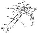
- FIG. 3is a side view of an apparatus for restoring a GEFV and securing the stomach to the diaphragm according to an embodiment of the invention
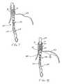
- FIG. 4is a side view of the apparatus of FIG. 3 securing the stomach to the diaphragm according to an embodiment of the invention
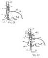
- FIG. 5is a perspective view, with portions cut away, of a device capable of securing the fundus of the stomach to the diaphragm according to another embodiment of the invention
- FIG. 6is a simplified side view of an apparatus according to an embodiment of the invention being fed down an oral and esophageal passage of a patient;
- FIG. 7is a side view, partly in cross-section, of a device according to an embodiment of the invention after having been initially fed into a stomach to initiate a GERD treatment procedure according to an embodiment of the invention;
- FIG. 8is a view similar to FIG. 7 showing the device and stomach after the stomach has been inflated to a first pressure
- FIG. 9is a view similar to FIG. 7 showing the device and stomach at a further stage of the procedure
- FIG. 10is a view similar to FIG. 7 showing the device centered and gripping the esophagus
- FIG. 11is a view similar to FIG. 7 showing the device initially gripping the stomach tissue after the stomach has been reinflated to a second, higher pressure;
- FIG. 12is a view similar to FIG. 7 showing the stomach partially deflated and gripped stomach tissue being pulled aborally towards the device;
- FIG. 13is a view similar to FIG. 7 showing the gripped stomach tissue being pulled to almost within the device;
- FIG. 14is a view similar to FIG. 7 showing the gripped stomach tissue within the device, being molded, and ready to receive a fastener;
- FIG. 15is a view similar to FIG. 7 showing the molded stomach tissue after receiving a fastener
- FIG. 16is a perspective view illustrating a manner in which the devices of FIGS. 3 and 5 may deploy a fastener for securing the stomach to the diaphragm, for example;
- FIG. 17is a perspective view showing a fastener fully deployed.
- FIG. 1is a front cross-sectional view of the esophageal-gastro-intestinal tract 40 from a lower portion of the esophagus 41 to the duodenum 42 .
- the stomach 43is characterized by the greater curvature 44 on the anatomical left side and the lesser curvature 45 on the anatomical right side.
- the tissue of the outer surfaces of those curvaturesis referred to in the art as serosa tissue. As will be seen subsequently, the nature of the serosa tissue is used to advantage for its ability to bond to like serosa tissue.
- the fundus 46 of the greater curvature 44forms the superior portion of the stomach 43 , and traps gas and air bubbles for burping.
- the esophageal tract 41enters the stomach 43 at an esophageal orifice below the superior portion of the fundus 46 , forming a cardiac notch 47 and an acute angle with respect to the fundus 46 known as the Angle of His 57 .
- the lower esophageal sphincter (LES) 48is a discriminating sphincter able to distinguish between burping gas, liquids, and solids, and works in conjunction with the fundus 46 to burp.
- the gastroesophageal flap valve (GEFV) 49includes a moveable portion and an opposing more stationary portion.
- the moveable portion of the GEFV 49is an approximately 180 degree, semicircular, gastroesophageal flap 50 (alternatively referred to as a “normal moveable flap” or “moveable flap”) formed of tissue at the intersection between the esophagus 41 and the stomach 43 .
- the opposing more stationary portion of the GEFV 49comprises a portion of the lesser curvature 45 of the stomach 43 adjacent to its junction with the esophagus 41 .
- the gastroesophageal flap 50 of the GEFV 49principally comprises tissue adjacent to the fundus 46 portion of the stomach 43 . It is about 4 to 5 cm long ( 51 ) at it longest portion, and its length may taper at its anterior and posterior ends.
- the gastroesophageal flap 50is partially held against the lesser curvature 45 portion of the stomach 43 by the pressure differential between the stomach 43 and the thorax, and partially by the resiliency and the anatomical structure of the GEFV 49 , thus providing the valving function.
- the GEFV 49is similar to a flutter valve, with the gastroesophageal flap 50 being flexible and closeable against the other more stationary side.
- the esophageal tractis controlled by an upper esophageal sphincter (UES) in the neck near the mouth for swallowing, and by the LES 48 and the GEFV 49 at the stomach.
- the normal anti-reflux barrieris primarily formed by the LES 48 and the GEFV 49 acting in concert to allow food and liquid to enter the stomach, and to considerably resist reflux of stomach contents into the esophagus 41 past the gastroesophageal tissue junction 52 .
- Tissue aboral of the gastroesophageal tissue junction 52is generally considered part of the stomach because the tissue protected from stomach acid by its own protective mechanisms.
- Tissue oral of the gastroesophageal junction 52is generally considered part of the esophagus and it is not protected from injury by prolonged exposure to stomach acid.
- the juncture of the stomach and esophageal tissuesform a zigzag line, which is sometimes referred to as the “Z-line.”
- “stomach”means the tissue aboral of the gastroesophageal junction 52 .
- FIG. 2is a perspective view, with portions cut away, of stomach 43 , esophagus 41 , diaphragm 53 , and hiatal hernia 61 which may be treated according to an embodiment of the present invention.
- a principal reason for regurgitation associated with GERDis the mechanical failure of the deteriorated (or reflux appearance) gastroesophageal flap of the GEFV to close and seal against the higher pressure in the stomach. Due to reasons including lifestyle, a Grade I normal gastroesophageal flap of the GEFV may deteriorate into a Grade III deteriorated gastroesophageal flap.
- the anatomical results of the deteriorationinclude moving a portion of the esophagus 41 that includes the gastroesophageal junction 52 and LES (not shown) toward the mouth through the hiatus 63 into the chest to create the hiatal hernia 61 .
- L. D. Hill, et al.The gastroesophageal flap valve: in vitro and in vivo observations , Gastrointestinal Endoscopy 1996:44:541-547.
- the normal movable flap 50 of the GEFV 49 illustrated in FIG. 1is a Grade I flap valve that is the least likely to experience reflux.
- the deteriorated gastroesophageal flap 55 of the GEFV 49 illustrated in FIG. 2is a Grade IV flap valve.
- the Grade IV flap valveis the most likely to experience reflux.
- Grades II and IIIreflect intermediate grades of deterioration and, as in the case of III, a high likelihood of experiencing reflux.
- the deteriorated GEFV represented by deteriorated gastroesophageal flap 55 and the fundus 46 moved inferiorthe stomach contents are presented a funnel-like opening directing the contents into the esophagus 41 and the greatest likelihood of experiencing reflux.
- a device, assembly, and methodwhich may be employed to advantage according to an embodiment of the invention to treat the hiatal hernia 61 and restore the normal gastroesophageal flap valve anatomy.
- the device 100includes a longitudinal member 102 for transoral placement of the device 100 into the stomach.
- the devicefurther includes a first member 104 , hereinafter referred to as the chassis, and a second member 106 , hereinafter referred to as the bail.
- the chassis 104 and bailare hingedly coupled at 107 .
- the chassis 104 and bail 106form a tissue shaper which, as described subsequently in accordance with this embodiment of the present invention, shapes tissue of the stomach into the flap of a restored gastroesophageal flap valve.
- the chassis 104 and bail 106are carried at the distal end of the longitudinal member 102 for placement in the stomach.
- the device 100has a longitudinal passage 101 to permit an endoscope 110 to be guided through the device and into the stomach. This permits the endoscope to service as a guide for guiding the device 100 through the patient's throat, down the esophagus, and into the stomach. It also permits the gastroesophageal flap valve restoration procedure to be viewed at each stage of the procedure.
- the stomach tissueis drawn in between the chassis 104 and the bail 106 .
- the stomach tissueis pulled down so that the fold line is substantially juxtaposed to the opening of the esophagus into the stomach.
- the stomachis first gripped at a point out and away from the esophagus and the grip point is pulled to almost the hinged connection 107 of the chassis 104 and bail 106 .
- the device 100is fed down the esophagus with the bail 106 substantially in line with the chassis 104 .
- the chassis 104 and bail 106are rendered flexible.
- the chassis 104is rendered flexible by the slots 108 and the bail 106 is rendered flexible by the hingedly coupled links 112 . Further details concerning the flexibility of the chassis 104 and the bail 106 may be found in the aforementioned referenced application.
- the devicefurther includes a tissue gripper 114 .
- the gripper 114in this embodiment, comprises a helical coil 115 .
- the coil 115is carried at the end of a cable 116 and may be attached to the end of the cable or be formed from the cable.
- the helical coil 115is attached to the cable 116 and is preceded by a guide 118 whose function will be described subsequently.
- the helical coil 115is shown in an approximate position to engage the stomach tissue out and away from the opening of the esophagus to the stomach.
- the helical coil 115is guided into position by a guide structure 120 carried on the bail 106 .
- the guide structure 120comprises a guide tube 122 .
- the guide tubeincludes a longitudinal slit 126 having a circuitous configuration.
- the slit 126permits the end of the cable to release or disassociate from the bail after the stomach tissue is gripped.
- the circuitous configuration of the slit 126assures confinement of the cable 116 within the guide tube 122 until release of the cable is desired.
- the proximal end of the slit 126has an enlarged portion or opening (not shown). This opening permits the cable and helical coil to reenter the lumen when the device 100 is readied for a repeated stomach tissue shaping procedure.
- the guide 118has a conical surface that serves to guide the cable end back into the opening of the slit 126 .
- the device 100further comprises a fastener deployer 140 .
- the fastener deployerincludes at least one fastener deployment guide 142 .
- the fastener deployment guide 142takes the form of a guide lumen. Although only one guide lumen 142 is shown, it will be appreciated that the device 100 may include a plurality of such lumens without departing from the invention.
- the guide lumenterminates at a delivery point 144 where a fastener is driven from the device 100 and into, for example, the molded stomach tissue.
- the fastener deployermay also be used, according to an embodiment, to secure the stomach to the diaphragm.
- the device 100further includes a window 130 within the chassis 104 .
- the windowis formed of a transparent or semi-transparent material. This permits gastroesophageal anatomy, and more importantly the gastroesophageal junction (Z-line) to be viewed with the endoscope 110 .
- the windowincludes a location marker 132 which has a know position relative to the fastener delivery point 144 . Hence, by aligning the marker with a known anatomical structure, the fastener will be delivered a known distance from or at a location having a predetermined relation to the marker. For example, by aligning the marker with the Z-line, it will be know that the fastener will be placed aboral of the Z-line and that serosa tissue will be fastened to serosa tissue. As previously mentioned, this has many attendant benefits.
- the device 100further includes an invaginator 145 including a plurality of orifices 146 .
- These orifices 146which alternatively may be employed on the longitudinal member 102 , are used to pull a vacuum to cause the device 100 to grip the inner wall surface of the esophagus. This will serve to stabilize the esophagus and maintain device positioning during the procedure.
- This vacuum gripping of the esophagusmay also be used to particular advantage in the treatment of a hiatal hernia. Upon being thus gripped, the esophagus may be moved downwardly with the device toward the stomach to pull the stomach to within the diaphragm to eliminate the hiatal hernia.
- FIG. 4it shows the device 100 in position to secure the stomach 43 to the diaphragm 53 following a successful restoration of a GEFV flap and/or to treat a hiatal hernia. More particularly, the device 100 of FIG. 4 is shown positioned in the stomach 43 by the elongated member 102 . It is also rotated by about 180 degrees from its position shown in FIG. 3 to align the guide channel 142 with the lesser curve. This will enable the fastener deployer 140 to deploy at least one fastener 200 to secure the lesser curve 45 of the stomach 43 to the right crus 59 of the diaphragm 53 .
- the endoscope 110is positioned in the stomach 43 and brought to a reflexed view as illustrated so that it may look back on the device 100 for visualization of the procedure.
- the invaginator 145has vacuum gripped the sidewalls of the esophagus. This permits the device to be used for displacing the esophagus aborally towards the stomach for reducing the hiatal hernia. Preferably the esophagus is displaced sufficiently so that the stomach is behind or within the diaphragm 53 . The esophagus is held in this position throughout the procedure.
- the fastener deployerdeploys the at least one fastener 200 as illustrated.
- a deployment procedure for the applicationis described in greater detail herein after.
- the fasteneris deployed to secure the lesser curve 45 of the stomach 43 to the right crus 59 of the diaphragm 53 .
- a plurality of spaced fastenerswould be deployed.
- the device 100is removed from the stomach 43 . This may be accomplished by first aligning the bail 106 with the chassis 104 of the device 100 .
- the endoscopemay be used as a guide to guide the device out of the stomach and through the esophagus, throat, and mouth.
- FIG. 5it shows another device 300 according to an embodiment of the invention for securing the stomach 43 to the diaphragm 53 .
- the fundus 46 of the stomach 43is being secured to the diaphragm 53 .
- the device 300may be employed for the restoration of a GEFV 49 and/or to treat a hiatal hernia.
- the device 300is carried at the distal end of an elongated member 302 for being transorally placed in the stomach 43 . It preferably includes an invaginator 345 of the type previously described for gripping the esophagus 41 and displacing it and the stomach aborally towards the diaphragm to reduce or eliminate the hiatal hernia.
- the invaginatormay also be used to grip the esophagus during the restoration of the GEFV 49 after reduction of a hiatal hernia.
- the deviceincludes a support arm 312 that supports a fastener deployer 340 in close proximity to the fundus 46 of the stomach 43 .
- the fastener deployerincludes a guide tube 342 supported by the arm 312 .
- the guide tube 342guides the tissue piercing wire 364 and the fasteners 200 to the location where they are to be deployed. Again, a suitable deployment procedure and related deployment assembly are described herein after.
- the device 300further carries an endoscope 310 .
- the endoscopeis positioned to enable visualization of the procedure. It is guided by a guide channel 301 in the elongated member 302 .
- the arm 312is arranged for pivotal movement at 307 to enable proper positioning of the fastener deployer 340 . To that end, it may be noted that the arm reaches outwardly to displace the fastener deployer 340 and the fasteners 200 spaced away from the esophageal opening 39 to the stomach 43 .
- the procedure for restoring the flap of a gastroesophageal flap valvebegins with loading a fastener or a plurality of fasteners into the device 100 .
- the fastener deployerincludes a stylet, which guides each fastener into the tissue to be fastened.
- the process of loading a fastenerincludes snapping a fastener onto the stylet. A representative fastener and stylet will be described subsequently with respect to FIGS. 16 and 17 .
- the bail 106is moved to be substantially in line with the chassis 104 .
- the endoscope 110is inserted into the device with an appropriate lubricant on the endoscope.
- a bite blockof the type well known in the art, is inserted into the patient's mouth. A lubricant may be applied to the device and the device may now be inserted through the bite block in the subject's mouth.
- the endoscope leading the device as illustrated in FIG. 6the endoscope and device combination are fed down the esophagus 141 into the stomach.
- the device 100may be further advanced on the endoscope utilizing the endoscope as a guide to within the stomach of the patient.
- the device 100is able to clear the bend in the patient's throat by virtue of being flexible as previously described.
- the endoscopeserving as a guide tube, very little force should be needed to get the device around the neck into the pharynx and down into the esophagus.
- FIG. 7shows the device 100 upon reaching the interior of the stomach 43 .
- the bail 106is substantially in line with the chassis 104 .
- the endoscope 110remains within the device 100 .
- the stomachis deflated. This is the normal condition of the stomach when the stomach is empty.
- the stomachis inflated as shown in FIG. 8 by passing air through the endoscope into the stomach.
- the inflation of the stomachmay be noted by the outward arcuate deflection of the stomach 43 .
- the stomachshould be inflated to a first pressure just sufficient to open the stomach and provide good visibility of gastric folds on the interior wall 59 of the stomach. Visualization of such gastric folds permits discernment of a proper point to grip the stomach for forming the gastroesophageal flap valve flap in a manner to be described hereinafter.
- the deviceis placed in a desired position relative to the Z-line by placing the marker of the window 130 in a desired position relative to the Z-line 52 marking the transition from the esophagus 41 to the stomach 43 .
- the marker 132is aligned with the Z-line 52 .
- the endoscope 110is pulled back into the device 100 and more particular adjacent the marker 132 to visualize when the marker is aligned with the Z-line 52 .
- the distance from the marker 132 to a proximal point of the elongated member 102 relative to a rather fixed anatomy site of the patient, such as an incisormay be measured. This measurement may be marked on the elongated member 102 and later utilized for positioning the marker 132 adjacent the Z-line 52 .
- the endoscopeis positioned inside the device just past the hinged connection 107 of the bail 106 and chassis 104 .
- the bailis then actuated to an approximally one-half closed position as illustrated.
- the bailshould be watched to make sure that it moves towards the greater curve 56 so it can move freely in the open space of the gastric cavity.
- the bailshould be visible at all times.
- the endoscope 110is advanced back into the stomach 43 and brought to a reflexed view as illustrated so that it may look back on the device 100 .
- the device 100With the operating end of the device in clear view, the device 100 is positioned in the center of the gastroesophageal flap valve to be formed where the posterior and anterior groove should be. This position is typically opposite the lesser curve 45 .
- the device positioning relative to the Z-line 52is checked to make sure that the marker 132 is in its desired position relative to the Z-line 52 .
- the marker 132is placed adjacent or is aligned with the Z-line 52 .
- a vacuum pump communicating with orifices 146is energized to pull a vacuum through the orifices 146 .
- Thiscauses the orifices to engage the wall of the esophagus 41 for gripping the esophagus.
- this invaginationpermits the esophagus to be pushed into the stomach by distal movement of the elongated member 102 to treat a hiatal hernia and to stabilize the position of the device within the stomach.
- the vacuumis continued to be pulled through the orifices 146 until the vacuum is above the 50 kps mark on the vacuum pump.
- the deviceis then pushed gently aborally to reposition the esophagus to correct a hiatal hernia. It may be noted that this maneuver can also be used to visually check the position of the faster delivery point 144 relative to the Z-line. During this maneuver, the esophagus may roll back on itself and expose the esophageal Mucosa and the Z-line adjacent to the fastener delivery ports.
- the area in which the helical coil is to be engagedmay be identified.
- the gripping locationmay be largely determined by the size or length of the flap to be restored of the restored gastroesophageal flap valve. This of course may differ from one patient to another depending on the severity of the hiatal hernia and the degree of valve degradation.
- the stomach 43is inflated to a second and higher pressure. The inflation pressure of the stomach is increased to the second and higher pressure so that the Mucosa appears tight and the folds essentially flatten.
- the bail 106is moved to position the tip of a helical coil 115 at the correct gripping spot.
- the device 100is gently pulled upwardly or orally until the bail contacts the tissue at the desired gripping spot.
- the helix 115is advanced by the pushing of the cable 116 until the helix pushes into the Mucosa.
- the cable 116is turned to likewise turn the helix 115 in a clockwise direction to screw the helix into the tissue. As the cable is turned, some wind-up may be filled in the helix drive cable.
- the device 100may be advanced slightly orally while at the same time the bail 116 may be opened slightly. This releases the cable 116 from the guide tube which has now been pulled back into the bail 106 .
- the cable 116exits the guide tube 122 ( FIG. 3 ) by slipping through the circuitous slit 126 . This operation is more particularly described in the aforementioned U.S. patent application Ser. No. 11/061,318, filed Feb. 18, 2005, incorporated herein by reference. Also at this time, the correct positioning of the device relative to the Z-line may be verified.
- the interior of the stomachis now deflated through the endoscope 110 .
- the stomachshould be deflated such that the tissue appears loose and collapsed with the Mucosa folds being prominent. However, enough room should be left to view the device.
- the gastric tissueis now gently pulled with the helix 115 and cable 116 towards the hinged connection 107 and the valve mold to be formed by the chassis 104 and closing bail 106 .
- the helixis fully retracted into the bail 116 , it is locked in place.
- the bail 106may now be closed and the device and anatomy will appear as shown in FIG. 14 .
- the stomach tissue aboral of the Z-line 52is confined between the bail 106 and chassis 104 to create a fold 150 .
- the foldis also adjacent the fastener delivery point 144 at the end of the fastener guide lumen.
- the fastener deployment point 144is a known predetermined distance from the marker 132 of the window 130 , and since the marker 132 is aligned with the Z-line 52 , when a fastener is delivered from the fastener deployer of the device, the fastener will exit the fastener delivery point 144 at a point known to be aboral of the Z-line 52 . This assures that only serosa tissue is being adhered to serosa tissue in the fixation of the stomach tissue in creating the flap 150 .
- the flap 150comprises layers 180 and 182 of stomach tissue.
- the bail 106may now be locked with respect to the chassis 104 . It is now time to fasten the tissue layers 180 and 182 together by ejecting a fastener from the fastener deployer lumen 142 through the flap 150 from the fastener delivery point 144 . The fastener thus deployed will serve to maintain the restored GEFV and the reduced hiatal hernia.
- the stomachis once again inflated through the endoscope 110 .
- the stomachis inflated to a point where one has a good view of the tissue fold and bail 106 .
- FIGS. 16 and 17illustrate a manner in which the fasteners 200 may be deployed to fasten tissue layers 180 and 182 .
- the tissue layers 180 and 182are meant to be merely representative of any tissue layers which may be fastened together, whether they be stomach tissue layers from forming a flap or stomach and diaphragm tissue layers fastened to secure the stomach to the diaphragm.
- the fastener 200generally includes a first member 202 , a second member 204 , and a connecting member 206 . As may be noted in FIG. 15 , the first member 202 and second member 204 are substantially parallel to each other and substantially perpendicular to the connecting member 206 which connects the first member 202 to the second member 204 .
- the first member 202is generally cylindrical or can any shape. It has a channel 212 that extends therethrough.
- the though channel 112is dimensioned to be slidingly received on a tissue piercing deployment wire 264 .
- the first member 202includes a pointed tip 224 .
- the tip 224may be conical and more particularly takes the shape of a truncated cone.
- the tipcan also be shaped to have a cutting edge in order to reduce tissue resistance.
- the first member 202also has a continuous lengthwise slit 225 .
- the slit 225includes an optional slot 226 that communicates with the through channel 212 .
- the slot 226has a transverse dimension for more readily enabling receipt of the tissue piercing deployment wire 264 during deployment of the fastener 200 .
- the fastener member 202is formed of flexible material, the slit 225 may be made larger through separation to allow the deployment wire to be snapped into and released from the through channel 212 .
- the assembly shown in FIGS. 16 and 17further includes a pusher 266 and a guide tube 268 .
- the subassembly of the tissue piercing wire 264 , fastener 200 , and pusher 266may be guided to its intended location relative to the tissue layers 180 and 182 by the guide tube 268 .
- the tissue piercing wire 264 , fastener 200 , and the pusher 266are all initially within the guide tube 268 .
- the guide tube 268is representative of the fastener deployment guide and to that end, includes the fastener deployment guide lumen 142 .
- the subassembly of the tissue piercing wire 264 , fastener 200 , and pusher 266may be guided to its intended location relative to the tissue layers 180 and 182 by the guide lumen 142 .
- the tissue piercing wire 264has a tip 270 helping it pierce the tissue layers 180 and 182 that will form the restored gastroesophageal flap valve flap 150 .
- the pusher 266has pushed the first member 202 of the fastener 200 through the tissue layers 180 and 182 on the tissue piercing wire 264 . This may be accomplished by moving the wire 264 and the pusher 266 together.
- the first member 202is clearing the wire 264 and tissue layer 182 .
- the tissue piercing wire 264may now be retracted into the pusher 266 and the tissue piercing wire 264 and pusher 266 may be withdrawn.
- FIG. 17illustrates the fastener 200 in its fully deployed position. It will be noted that the fastener has returned to its original shape.
- the tissue layers 180 and 182are fastened together between the first member 202 of the fastener 200 and the second member 204 of the fastener 200 .
- the connecting member 206extends through the tissue layers 180 and 182 .
- the tissue piercing wire 264may be first advanced through the tissue layers 180 and 182 by a full stroke and then locked. The tip 270 of the deployment wire 264 should extend through the bail 206 with minimal tenting of the tissue. Next, the pusher 266 is advanced. Visual confirmation that the first fastener member 202 is through the tissue is then made. In doing so, the very distal end of the pusher 266 may be visible when the first member 202 of the fastener 200 is fully deployed. Next, while holding the pusher 266 at the last noted position, the tissue piercing wire 264 is retracted.
- the first member 202 of the fastener 200will fall to the side when the tissue piercing wire 264 reaches the pusher 266 .
- the pusher 266is pulled back with the tissue piercing wire. If additional fastener deployment guides are provided, the foregoing steps for deploying a fastener such as fastener 200 may be repeated.
- the vacuum pull through orifices 146may now be turned off to release the device from the esophagus wall as illustrated in FIG. 15 .
- the bail 106 of the device 100may be slightly opened and the helical coil 115 may be released from the stomach tissue.
- the procedure just describedresults in a flap 150 to be formed.
- an additional fastener or fastenersmay be loaded onto the tissue piercing deployment wire 264 at the proximal end of the longitudinal member 102 .
- the device 102To render the flap uniform about the opening of the orifice into the stomach, it is necessary at this time to rotate the device 102 and repeat the previously described procedure for forming a further flap portion. Before this is done, however, it is desirable to position the bail 106 to an almost closed position. Then, the device 100 is moved aborally further into the stomach until the tip end 107 of the bail 106 comes to rest on the tip 151 of the newly formed flap portion. This is the location where the helical coil 115 will next engage the stomach tissue for molding and fixating as previously described.
- valve flapis formed.
- the helical coil 115is reloaded back into its original position with the device 100 .
- the vacuum suction through orifices 146is turned off to release the wall of the esophagus from the device.
- the bail 106is then moved to a fully opened position as seen, for example, in FIG. 7 .
- the endoscopemay now be retracted along with the stylet and pusher controls. With the retraction of the foregoing verified, the stomach may now be deflated and the device 100 may be removed from the stomach and esophagus. This then completes the GEFV restoration procedure according to this embodiment of the invention.
Landscapes
- Health & Medical Sciences (AREA)
- Life Sciences & Earth Sciences (AREA)
- Surgery (AREA)
- Molecular Biology (AREA)
- Engineering & Computer Science (AREA)
- Biomedical Technology (AREA)
- Heart & Thoracic Surgery (AREA)
- Medical Informatics (AREA)
- Nuclear Medicine, Radiotherapy & Molecular Imaging (AREA)
- Animal Behavior & Ethology (AREA)
- General Health & Medical Sciences (AREA)
- Public Health (AREA)
- Veterinary Medicine (AREA)
- Physiology (AREA)
- Rheumatology (AREA)
- Surgical Instruments (AREA)
Abstract
Description
Claims (2)
Priority Applications (3)
| Application Number | Priority Date | Filing Date | Title |
|---|---|---|---|
| US12/660,367US9414832B2 (en) | 2005-08-12 | 2010-02-24 | Apparatus and method for securing the stomach to the diaphragm for use, for example, in treating hiatal hernias and gastroesophageal reflux disease |
| US15/232,676US10772624B2 (en) | 2005-08-12 | 2016-08-09 | Apparatus and method for securing the stomach to the diaphragm for use, for example, in treating hiatal hernias and gastroesophageal reflux disease |
| US17/014,813US11627958B2 (en) | 2005-08-12 | 2020-09-08 | Apparatus and method for securing the stomach to the diaphragm for use, for example, in treating hiatal hernias and gastroesophageal reflux disease |
Applications Claiming Priority (2)
| Application Number | Priority Date | Filing Date | Title |
|---|---|---|---|
| US11/203,680US20070038232A1 (en) | 2005-08-12 | 2005-08-12 | Apparatus and method for securing the stomach to the diaphragm for use, for example, in treating hiatal hernias and gastroesophageal reflux disease |
| US12/660,367US9414832B2 (en) | 2005-08-12 | 2010-02-24 | Apparatus and method for securing the stomach to the diaphragm for use, for example, in treating hiatal hernias and gastroesophageal reflux disease |
Related Parent Applications (2)
| Application Number | Title | Priority Date | Filing Date |
|---|---|---|---|
| US11/203,680DivisionUS20070038232A1 (en) | 2005-08-12 | 2005-08-12 | Apparatus and method for securing the stomach to the diaphragm for use, for example, in treating hiatal hernias and gastroesophageal reflux disease |
| US11/203,680ContinuationUS20070038232A1 (en) | 2005-08-12 | 2005-08-12 | Apparatus and method for securing the stomach to the diaphragm for use, for example, in treating hiatal hernias and gastroesophageal reflux disease |
Related Child Applications (1)
| Application Number | Title | Priority Date | Filing Date |
|---|---|---|---|
| US15/232,676DivisionUS10772624B2 (en) | 2005-08-12 | 2016-08-09 | Apparatus and method for securing the stomach to the diaphragm for use, for example, in treating hiatal hernias and gastroesophageal reflux disease |
Publications (2)
| Publication Number | Publication Date |
|---|---|
| US20100168507A1 US20100168507A1 (en) | 2010-07-01 |
| US9414832B2true US9414832B2 (en) | 2016-08-16 |
Family
ID=37743505
Family Applications (4)
| Application Number | Title | Priority Date | Filing Date |
|---|---|---|---|
| US11/203,680AbandonedUS20070038232A1 (en) | 2005-08-12 | 2005-08-12 | Apparatus and method for securing the stomach to the diaphragm for use, for example, in treating hiatal hernias and gastroesophageal reflux disease |
| US12/660,367Expired - LifetimeUS9414832B2 (en) | 2005-08-12 | 2010-02-24 | Apparatus and method for securing the stomach to the diaphragm for use, for example, in treating hiatal hernias and gastroesophageal reflux disease |
| US15/232,676Active2026-06-04US10772624B2 (en) | 2005-08-12 | 2016-08-09 | Apparatus and method for securing the stomach to the diaphragm for use, for example, in treating hiatal hernias and gastroesophageal reflux disease |
| US17/014,813Active2026-11-05US11627958B2 (en) | 2005-08-12 | 2020-09-08 | Apparatus and method for securing the stomach to the diaphragm for use, for example, in treating hiatal hernias and gastroesophageal reflux disease |
Family Applications Before (1)
| Application Number | Title | Priority Date | Filing Date |
|---|---|---|---|
| US11/203,680AbandonedUS20070038232A1 (en) | 2005-08-12 | 2005-08-12 | Apparatus and method for securing the stomach to the diaphragm for use, for example, in treating hiatal hernias and gastroesophageal reflux disease |
Family Applications After (2)
| Application Number | Title | Priority Date | Filing Date |
|---|---|---|---|
| US15/232,676Active2026-06-04US10772624B2 (en) | 2005-08-12 | 2016-08-09 | Apparatus and method for securing the stomach to the diaphragm for use, for example, in treating hiatal hernias and gastroesophageal reflux disease |
| US17/014,813Active2026-11-05US11627958B2 (en) | 2005-08-12 | 2020-09-08 | Apparatus and method for securing the stomach to the diaphragm for use, for example, in treating hiatal hernias and gastroesophageal reflux disease |
Country Status (4)
| Country | Link |
|---|---|
| US (4) | US20070038232A1 (en) |
| EP (1) | EP1919371B1 (en) |
| ES (1) | ES2487491T3 (en) |
| WO (1) | WO2007022029A2 (en) |
Families Citing this family (33)
| Publication number | Priority date | Publication date | Assignee | Title |
|---|---|---|---|---|
| US6450173B1 (en) | 1999-08-12 | 2002-09-17 | Obtech Medical Ag | Heartburn and reflux disease treatment with controlled wireless energy supply |
| US6482145B1 (en) | 2000-02-14 | 2002-11-19 | Obtech Medical Ag | Hydraulic anal incontinence treatment |
| US6471635B1 (en) | 2000-02-10 | 2002-10-29 | Obtech Medical Ag | Anal incontinence disease treatment with controlled wireless energy supply |
| US6464628B1 (en) | 1999-08-12 | 2002-10-15 | Obtech Medical Ag | Mechanical anal incontinence |
| CA2635435C (en)* | 2000-02-10 | 2010-05-25 | Potencia Medical Ag | Controlled urinary incontinence treatment |
| AU764705B2 (en) | 2000-02-10 | 2003-08-28 | Implantica Patent Ltd. | Urinary incontinence treatment with wireless energy supply |
| ATE391468T1 (en)* | 2000-02-10 | 2008-04-15 | Potencia Medical Ag | MECHANICAL DEVICE FOR IMPOTENCY TREATMENT |
| ATE416743T1 (en)* | 2000-02-11 | 2008-12-15 | Potentica Ag | DEVICE WITH ENERGY CONVERSION MEANS FOR TREATING IMPOTENCY |
| CA2396224C (en)* | 2000-02-11 | 2011-07-12 | Potencia Medical Ag | Controlled impotence treatment |
| US20030100929A1 (en)* | 2000-02-14 | 2003-05-29 | Peter Forsell | Controlled penile prosthesis |
| WO2001047440A2 (en)* | 2000-02-14 | 2001-07-05 | Potencia Medical Ag | Male impotence prosthesis apparatus with wireless energy supply |
| US7442165B2 (en) | 2000-02-14 | 2008-10-28 | Obtech Medical Ag | Penile prosthesis |
| US20070088373A1 (en)* | 2005-10-18 | 2007-04-19 | Endogastric Solutions, Inc. | Invaginator for gastroesophageal flap valve restoration device |
| US9161754B2 (en)* | 2012-12-14 | 2015-10-20 | Endogastric Solutions, Inc. | Apparatus and method for concurrently forming a gastroesophageal valve and tightening the lower esophageal sphincter |
| US9155532B2 (en)* | 2007-05-25 | 2015-10-13 | Cook Medical Technologies Llc | Medical devices, systems and methods for closing perforations |
| US8034063B2 (en)* | 2007-07-13 | 2011-10-11 | Xlumena, Inc. | Methods and systems for treating hiatal hernias |
| US7841347B2 (en) | 2007-09-10 | 2010-11-30 | Medigus Ltd. | Method of performing surgical procedures on patients suffering from hiatal hernia |
| WO2009082596A1 (en)* | 2007-12-18 | 2009-07-02 | Wilson-Cook Medical, Inc. | Device and method for placement of tissue anchors |
| WO2009096851A1 (en)* | 2008-01-28 | 2009-08-06 | Milux Holding Sa | A drainage device comprising a filter cleaning device |
| MX2010008003A (en)* | 2008-01-29 | 2010-09-24 | Milux Holding Sa | Apparatus for treating obesity. |
| WO2009097585A1 (en)* | 2008-02-01 | 2009-08-06 | Endometabolic Solutions, Inc. | Methods and devices for performing gastroplasty |
| EP3851076A1 (en) | 2008-10-10 | 2021-07-21 | MedicalTree Patent Ltd. | An improved artificial valve |
| US8874215B2 (en) | 2008-10-10 | 2014-10-28 | Peter Forsell | System, an apparatus, and a method for treating a sexual dysfunctional female patient |
| WO2010042011A1 (en)* | 2008-10-10 | 2010-04-15 | Milux Holding Sa | Heart help device, system, and method |
| WO2010042018A1 (en)* | 2008-10-10 | 2010-04-15 | Milux Holding S.A. | Heart help device, system and method |
| AU2009302955B2 (en)* | 2008-10-10 | 2017-01-05 | Implantica Patent Ltd. | Fastening means for implantable medical control assembly |
| WO2010042046A1 (en) | 2008-10-10 | 2010-04-15 | Milux Holding S.A. | Apparatus, system and operation method for the treatment of female sexual dysfunction |
| US10952836B2 (en)* | 2009-07-17 | 2021-03-23 | Peter Forsell | Vaginal operation method for the treatment of urinary incontinence in women |
| US9949812B2 (en) | 2009-07-17 | 2018-04-24 | Peter Forsell | Vaginal operation method for the treatment of anal incontinence in women |
| US12059149B2 (en)* | 2011-09-09 | 2024-08-13 | Endogastric Solutions, Inc. | Methods and devices for manipulating and fastening tissue |
| EP3400884B1 (en)* | 2016-01-06 | 2021-06-23 | Olympus Corporation | Device for placing tissue fastener |
| US10966717B2 (en)* | 2016-01-07 | 2021-04-06 | Covidien Lp | Surgical fastener apparatus |
| RU2751739C1 (en)* | 2020-11-03 | 2021-07-16 | Федеральное государственное бюджетное образовательное учреждение высшего образования «Национальный исследовательский Мордовский государственный университет им. Н.П. Огарёва» | Method for surgical treatment of hernia of esophageal orifice of diaphragm and barrett's esophagus |
Citations (144)
| Publication number | Priority date | Publication date | Assignee | Title |
|---|---|---|---|---|
| US2753870A (en) | 1955-03-15 | 1956-07-10 | James A Muffly | Instrument for probing the reticulum |
| US3875928A (en) | 1973-08-16 | 1975-04-08 | Angelchik Jean P | Method for maintaining the reduction of a sliding esophageal hiatal hernia |
| US4006747A (en) | 1975-04-23 | 1977-02-08 | Ethicon, Inc. | Surgical method |
| US4271828A (en) | 1979-09-13 | 1981-06-09 | Angelchik Jean P | Method for maintaining the reduction of a sliding esophageal hiatal hernia |
| US4576772A (en) | 1984-07-20 | 1986-03-18 | Warner-Lambert Technologies, Inc. | Catheter with optimum resistance to bending and method of manufacture |
| US4595007A (en) | 1983-03-14 | 1986-06-17 | Ethicon, Inc. | Split ring type tissue fastener |
| US4669473A (en) | 1985-09-06 | 1987-06-02 | Acufex Microsurgical, Inc. | Surgical fastener |
| US4696300A (en) | 1985-04-11 | 1987-09-29 | Dennison Manufacturing Company | Fastener for joining materials |
| EP0252607A2 (en) | 1986-06-07 | 1988-01-13 | Ethicon, Inc. | Pad-like implant |
| US4846836A (en) | 1988-10-03 | 1989-07-11 | Reich Jonathan D | Artificial lower gastrointestinal valve |
| US4895148A (en) | 1986-05-20 | 1990-01-23 | Concept, Inc. | Method of joining torn parts of bodily tissue in vivo with a biodegradable tack member |
| US4921479A (en) | 1987-10-02 | 1990-05-01 | Joseph Grayzel | Catheter sheath with longitudinal seam |
| US5006106A (en) | 1990-10-09 | 1991-04-09 | Angelchik Jean P | Apparatus and method for laparoscopic implantation of anti-reflux prosthesis |
| US5041129A (en)* | 1990-07-02 | 1991-08-20 | Acufex Microsurgical, Inc. | Slotted suture anchor and method of anchoring a suture |
| US5080543A (en) | 1990-01-08 | 1992-01-14 | Engineered Construction Components (America) Inc. | Fastening sleeves and fastening systems employing same |
| US5088979A (en) | 1990-10-11 | 1992-02-18 | Wilson-Cook Medical Inc. | Method for esophageal invagination and devices useful therein |
| US5254126A (en) | 1992-06-24 | 1993-10-19 | Ethicon, Inc. | Endoscopic suture punch |
| US5314473A (en) | 1989-07-20 | 1994-05-24 | Godin Norman J | Prosthesis for preventing gastric reflux into the esophagus |
| US5403326A (en) | 1993-02-01 | 1995-04-04 | The Regents Of The University Of California | Method for performing a gastric wrap of the esophagus for use in the treatment of esophageal reflux |
| US5411508A (en) | 1991-10-29 | 1995-05-02 | The Trustees Of Columbia University In The City Of New York | Gastrointestinal approximating and tissue attaching device |
| US5411520A (en) | 1991-11-08 | 1995-05-02 | Kensey Nash Corporation | Hemostatic vessel puncture closure system utilizing a plug located within the puncture tract spaced from the vessel, and method of use |
| US5549621A (en) | 1993-05-14 | 1996-08-27 | Byron C. Sutherland | Apparatus and method for performing vertical banded gastroplasty |
| US5571116A (en) | 1994-10-02 | 1996-11-05 | United States Surgical Corporation | Non-invasive treatment of gastroesophageal reflux disease |
| US5571074A (en) | 1992-07-30 | 1996-11-05 | Temple University-Of The Commonwealth System Of Higher Education | Inflatable and expandable direct manual cardiac compression device |
| US5626614A (en)* | 1995-12-22 | 1997-05-06 | Applied Medical Resources Corporation | T-anchor suturing device and method for using same |
| US5713903A (en) | 1991-03-22 | 1998-02-03 | United States Surgical Corporation | Orthopedic fastener |
| US5759151A (en) | 1995-06-07 | 1998-06-02 | Carnegie Mellon University | Flexible steerable device for conducting exploratory procedures |
| US5810882A (en) | 1994-08-05 | 1998-09-22 | Origin Medsystems, Inc. | Surgical helical fastener with applicator and method of use |
| US5814054A (en) | 1996-09-23 | 1998-09-29 | Symbiosis Corporation | Automatic needle-passer suturing instrument |
| US5861036A (en) | 1995-03-28 | 1999-01-19 | Biomedix S.A. Switzerland | Medical prosthesis for preventing gastric reflux in the esophagus |
| US5879372A (en) | 1993-09-20 | 1999-03-09 | Bartlett; Edwin C. | Apparatus and method for anchoring sutures |
| US5887594A (en) | 1997-09-22 | 1999-03-30 | Beth Israel Deaconess Medical Center Inc. | Methods and devices for gastroesophageal reflux reduction |
| WO1999022649A2 (en) | 1997-11-03 | 1999-05-14 | Symbiosis Corporation | Flexible endoscopic instrument for invagination and fundoplication |
| US5938668A (en) | 1994-10-07 | 1999-08-17 | United States Surgical | Surgical suturing apparatus |
| WO1999060931A1 (en) | 1998-05-26 | 1999-12-02 | Boston Scientific Limited | Implantable tissue fastener and system for treating gastroesophageal reflux disease |
| US6091995A (en)* | 1996-11-08 | 2000-07-18 | Surx, Inc. | Devices, methods, and systems for shrinking tissues |
| US6098629A (en) | 1999-04-07 | 2000-08-08 | Endonetics, Inc. | Submucosal esophageal bulking device |
| US6113611A (en) | 1998-05-28 | 2000-09-05 | Advanced Vascular Technologies, Llc | Surgical fastener and delivery system |
| WO2000053102A1 (en) | 1999-03-12 | 2000-09-14 | Mohamed Mounir El Gazayerli | Method and apparatus for minimally-invasive fundoplication |
| US6142957A (en) | 1993-09-20 | 2000-11-07 | Boston Scientific Corporation | Multiple biopsy sampling device |
| WO2000078227A1 (en) | 1999-06-22 | 2000-12-28 | Ndo Surgical, Inc. | Gerd treatment apparatus and method |
| WO2001032084A1 (en) | 1999-10-29 | 2001-05-10 | Biomedix S.A. | Endoscopic suturing instrument |
| WO2001035834A1 (en) | 1999-11-12 | 2001-05-25 | Boston Scientific Limited | Gerd treatment apparatus and method |
| US6254642B1 (en) | 1997-12-09 | 2001-07-03 | Thomas V. Taylor | Perorally insertable gastroesophageal anti-reflux valve prosthesis and tool for implantation thereof |
| US6264700B1 (en) | 1998-08-27 | 2001-07-24 | Endonetics, Inc. | Prosthetic gastroesophageal valve |
| WO2001064964A1 (en) | 2000-02-29 | 2001-09-07 | General Electric Company | Nickel base superalloys and turbine components fabricated therefrom |
| WO2001067964A2 (en) | 2000-03-16 | 2001-09-20 | Medigus Ltd. | Fundoplication apparatus and method |
| US6302917B1 (en) | 1998-08-31 | 2001-10-16 | Wilson-Cook Medical Incorporated | Anti-reflux esophageal prosthesis |
| US6302311B1 (en) | 1996-06-14 | 2001-10-16 | Boston Scientific Corporation | Endoscopic stapler |
| US6315789B1 (en) | 1999-02-08 | 2001-11-13 | Andrew H. Cragg | Medical device anchoring system and method |
| WO2001085034A1 (en) | 2000-05-10 | 2001-11-15 | Boston Scientific Limited | Devices and related methods for securing a tissue fold |
| WO2001089391A1 (en) | 2000-05-19 | 2001-11-29 | Rajiv Doshi | Articulated member surgical device and methods |
| US20020022853A1 (en) | 1998-11-06 | 2002-02-21 | St. Jude Medical Cardiovascular Group, Inc. | Medical anastomosis apparatus |
| WO2002024080A2 (en) | 2000-09-22 | 2002-03-28 | Boston Scientific Limited | Methods and devices for folding and securing tissue |
| WO2002024058A2 (en) | 2000-09-21 | 2002-03-28 | Medigus Ltd. | Multiple view endoscopes |
| US20020040226A1 (en) | 1999-06-22 | 2002-04-04 | Laufer Michael D. | Tissue reconfiguration |
| WO2002028289A1 (en) | 2000-09-29 | 2002-04-11 | Boston Scientific Limited | Method for performing endoluminal fundoplication and apparatus for use in the method |
| US20020055772A1 (en) | 2000-06-26 | 2002-05-09 | Rex Medical | Vascular device with valve for approximating vessel wall |
| US20020072761A1 (en) | 1998-05-11 | 2002-06-13 | Surgical Connections, Inc. | Surgical stabilizer devices and methods |
| US20020078967A1 (en) | 2000-12-06 | 2002-06-27 | Robert Sixto | Methods for the endoluminal treatment of gastroesophageal reflux disease (GERD) |
| US6419669B1 (en) | 1999-09-20 | 2002-07-16 | Appriva Medical, Inc. | Method and apparatus for patching a tissue opening |
| US6428548B1 (en) | 1999-11-18 | 2002-08-06 | Russell F. Durgin | Apparatus and method for compressing body tissue |
| US6447524B1 (en) | 2000-10-19 | 2002-09-10 | Ethicon Endo-Surgery, Inc. | Fastener for hernia mesh fixation |
| US20020143349A1 (en) | 1999-06-02 | 2002-10-03 | Concentric Medical, Inc. | Devices and methods for treating vascular malformations |
| WO2002082621A1 (en) | 2001-04-03 | 2002-10-17 | Emerson Electric Co. | Electric power steering system including a segmented stator switched reluctance motor |
| WO2002096327A2 (en) | 2001-05-30 | 2002-12-05 | Satiety, Inc. | Obesity treatment tools and methods |
| US20020198541A1 (en) | 2001-06-25 | 2002-12-26 | Smith Kevin W. | Flexible surgical clip applier |
| US6506196B1 (en)* | 1999-06-22 | 2003-01-14 | Ndo Surgical, Inc. | Device and method for correction of a painful body defect |
| US20030023230A1 (en) | 1998-02-03 | 2003-01-30 | Salient Interventional Systems, Inc. | Methods and systems for treating ischemia |
| US20030055442A1 (en) | 1999-06-22 | 2003-03-20 | Laufer Michael D. | Method and devices for tissue reconfiguration |
| US6547776B1 (en)* | 2000-01-03 | 2003-04-15 | Curon Medical, Inc. | Systems and methods for treating tissue in the crura |
| US20030093117A1 (en) | 1999-06-25 | 2003-05-15 | Vahid Saadat | Implantable artificial partition and methods of use |
| US20030120292A1 (en) | 2001-06-20 | 2003-06-26 | Park Medical, Llc | Anastomotic device |
| US20030120289A1 (en) | 2001-12-20 | 2003-06-26 | Mcguckin James F | Apparatus and method for treating gastroesophageal reflux disease |
| WO2003061480A1 (en) | 2001-10-20 | 2003-07-31 | Applied Medical Resources Corporation | Wound retraction apparatus and method |
| US20030171760A1 (en) | 2000-05-19 | 2003-09-11 | Gambale Richard A | Tissue capturing and suturing device and method |
| US20030187465A1 (en) | 2002-03-08 | 2003-10-02 | Sofradim Production | Appliance for storing, distributing and placing couched I-shaped surgical fasteners |
| US20030191497A1 (en) | 2002-04-05 | 2003-10-09 | Cook Incorporated | Sliding suture anchor |
| US20030216754A1 (en)* | 2002-05-17 | 2003-11-20 | Scout Medical Technologies, Llc | Transoral endoscopic gastroesophageal flap valve restoration device, assembly, system and method |
| US20030216613A1 (en) | 2002-03-19 | 2003-11-20 | Anthony Kalloo | Anastomosis system |
| US20030220657A1 (en) | 2001-05-23 | 2003-11-27 | Ronald Adams | Endoluminal fundoplication device and related method |
| US20030220660A1 (en)* | 2002-04-24 | 2003-11-27 | Kortenbach Juergen A. | Tissue fastening devices and processes that promote tissue adhesion |
| US6663639B1 (en)* | 1999-06-22 | 2003-12-16 | Ndo Surgical, Inc. | Methods and devices for tissue reconfiguration |
| US20040044364A1 (en)* | 2002-08-29 | 2004-03-04 | Devries Robert | Tissue fasteners and related deployment systems and methods |
| US20040044304A1 (en) | 2000-01-21 | 2004-03-04 | Hill John S | Local drug delivery using photosensitizer-mediated and electromagnetic radiation enhanced vascular permeability |
| WO2004019788A2 (en) | 2002-08-29 | 2004-03-11 | Boston Scientific Limited | Devices and methods for fastening tissue layers |
| US20040093024A1 (en) | 2000-09-01 | 2004-05-13 | James Lousararian | Advanced wound site management systems and methods |
| US6743239B1 (en) | 2000-05-25 | 2004-06-01 | St. Jude Medical, Inc. | Devices with a bendable tip for medical procedures |
| US20040116949A1 (en)* | 2002-12-11 | 2004-06-17 | Ewers Richard C. | Apparatus and methods for forming gastrointestinal tissue approximations |
| WO2004049982A2 (en) | 2002-12-02 | 2004-06-17 | Gi Dynamics, Inc. | Bariatric sleeve |
| US20040133236A1 (en) | 2001-12-19 | 2004-07-08 | Nmt Medical, Inc. | PFO closure device with flexible thrombogenic joint and improved dislodgement resistance |
| US20040138529A1 (en) | 2003-01-15 | 2004-07-15 | Usgi Medical Corp. | Endoluminal tool deployment system |
| US20040147958A1 (en) | 2002-12-11 | 2004-07-29 | Usgi Medical | Apparatus and methods for forming and securing gastrointestinal tissue folds |
| US20040148034A1 (en) | 2002-11-01 | 2004-07-29 | Jonathan Kagan | Apparatus and methods for treatment of morbid obesity |
| US20040153102A1 (en) | 2001-06-21 | 2004-08-05 | Michel Therin | Kit comprising a medical fixing element and a device for placing said fixing element |
| US20040153103A1 (en) | 1998-12-30 | 2004-08-05 | Schwartz Herbert E. | Soft tissue locking device |
| US6773440B2 (en) | 2002-07-02 | 2004-08-10 | Satiety, Inc. | Method and device for use in tissue approximation and fixation |
| WO2004069055A2 (en) | 2003-02-04 | 2004-08-19 | Ev3 Sunnyvale Inc. | Patent foramen ovale closure system |
| US20040162465A1 (en)* | 2003-02-19 | 2004-08-19 | Oscar Carrillo | Guidewire locking device and method |
| US20040162568A1 (en) | 1999-06-25 | 2004-08-19 | Usgi Medical | Apparatus and methods for forming and securing gastrointestinal tissue folds |
| US20050004575A1 (en) | 2000-11-24 | 2005-01-06 | Jean-Claude Sgro | Fastener for fixing a prosthesis, and device for delivering this fastener |
| US20050017781A1 (en) | 2003-07-24 | 2005-01-27 | Oki Electric Industry Co., Ltd. | Complementary signal generator |
| US20050043759A1 (en) | 2003-07-14 | 2005-02-24 | Nmt Medical, Inc. | Tubular patent foramen ovale (PFO) closure device with catch system |
| US20050085829A1 (en) | 2002-05-17 | 2005-04-21 | Esophyx, Inc. | Transoral endoscopic gastroesophageal flap valve restoration device, assembly, system and method |
| WO2005065412A2 (en) | 2003-12-31 | 2005-07-21 | Valentx, Inc. | Devices and methods for treating morbid obesity |
| US6921361B2 (en) | 2000-07-24 | 2005-07-26 | Olympus Corporation | Endoscopic instrument for forming an artificial valve |
| US20050177176A1 (en) | 2004-02-05 | 2005-08-11 | Craig Gerbi | Single-fold system for tissue approximation and fixation |
| US20050187565A1 (en)* | 2004-02-20 | 2005-08-25 | Baker Steve G. | Tissue fixation devices and a transoral endoscopic gastroesophageal flap valve restoration device and assembly using same |
| US20050203547A1 (en) | 2004-03-09 | 2005-09-15 | Gary Weller | Devices and methods for placement of partitions within a hollow body organ |
| US20050216040A1 (en) | 2004-03-23 | 2005-09-29 | Michael Gertner | Devices and methods to treat a patient |
| US20050228413A1 (en) | 2004-04-12 | 2005-10-13 | Binmoeller Kenneth F | Automated transluminal tissue targeting and anchoring devices and methods |
| US20050251176A1 (en) | 2004-05-07 | 2005-11-10 | Usgi Medical Inc. | System for treating gastroesophageal reflux disease |
| US20050247320A1 (en) | 2003-10-10 | 2005-11-10 | Stack Richard S | Devices and methods for retaining a gastro-esophageal implant |
| US20060009789A1 (en) | 2002-09-06 | 2006-01-12 | C. R. Bard, Inc. | Tissue capturing devices |
| WO2006034484A2 (en) | 2004-09-23 | 2006-03-30 | Endogastric Solutions, Inc. | Tissue fixation devices and assemblies for deploying the same |
| US7022118B2 (en) | 2001-06-29 | 2006-04-04 | Terumo Kabushiki Kaisha | Medical energy irradiation apparatus |
| WO2006081368A2 (en) | 2005-01-25 | 2006-08-03 | Endogastric Solutions, Inc. | Slitted tissue fixation devices and assemblies for deploying the same |
| US20060190018A1 (en) | 2005-02-18 | 2006-08-24 | Esophyx, Inc. | Transoral endoscopic gastroesophageal flap valve restoration device having a guided tissue gripper |
| US20060253142A1 (en) | 2005-05-04 | 2006-11-09 | Bernard Medical, Llc | Endoluminal gastric ring and method |
| US20060253130A1 (en)* | 2005-05-03 | 2006-11-09 | Esophyx, Inc. | Tissue fixation assemblies having a plurality of fasteners ready for serial deployment |
| US20060253131A1 (en)* | 2005-05-03 | 2006-11-09 | Endogastric Solutions, Inc. | Tissue fixation assemblies providing single stroke deployment |
| WO2007002817A2 (en) | 2005-06-29 | 2007-01-04 | Endogastric Solutions, Inc. | Tissue fixation assembly having prepositioned fasteners and method |
| US20070005080A1 (en)* | 2005-06-29 | 2007-01-04 | Esophyx, Inc. | Bolt action fastener delivery assembly |
| US20070021756A1 (en) | 2002-05-01 | 2007-01-25 | Scimed Life Systems, Inc. | Tissue fastening devices and related insertion tools and methods |
| US20070021760A1 (en) | 2005-07-19 | 2007-01-25 | Brian Kelleher | Methods and apparatus for securing an anchor to soft tissue |
| US20070112363A1 (en) | 2005-11-15 | 2007-05-17 | Endogastric Solutions, Inc. | Apparatus including multiple invaginators for restoring a gastroesophageal flap valve and method |
| US20070129738A1 (en) | 2005-12-01 | 2007-06-07 | Endogastric Solutions, Inc. | Apparatus and method for concurrently forming a gastroesophageal valve and tightening the lower esophageal sphincter |
| US20070191871A1 (en) | 2006-02-10 | 2007-08-16 | Endogastric Solutions, Inc. | Transesophageal gastric reduction method and device for reducing the size of a previously formed gastric reduction pouch |
| US20070191870A1 (en) | 2006-02-10 | 2007-08-16 | Endogastric Solutions, Inc. | Transesophageal gastric reduction method and device for practicing same |
| US20070276409A1 (en) | 2006-05-25 | 2007-11-29 | Ethicon Endo-Surgery, Inc. | Endoscopic gastric restriction methods and devices |
| US20080015618A1 (en) | 2006-07-16 | 2008-01-17 | Elazar Sonnenschein | Devices and methods for treating morbid obesity |
| US7347863B2 (en) | 2004-05-07 | 2008-03-25 | Usgi Medical, Inc. | Apparatus and methods for manipulating and securing tissue |
| US7452364B2 (en)* | 2005-04-22 | 2008-11-18 | Helmut Schreiber | Surgical marker/connector |
| US20080294179A1 (en) | 2007-05-12 | 2008-11-27 | Balbierz Daniel J | Devices and methods for stomach partitioning |
| US20090236388A1 (en) | 2008-03-18 | 2009-09-24 | David Cole | Endoscopic stapling devices and methods |
| WO2010087756A1 (en) | 2009-01-29 | 2010-08-05 | Milux Holding S.A. | Stomach instrument and method |
| US20100241139A1 (en) | 2009-03-18 | 2010-09-23 | Scott Harshman | Methods and devices for forming a tissue fold |
| US7850704B2 (en) | 2004-09-27 | 2010-12-14 | Theranova, Llc | Method and apparatus for anchoring implants |
| US7857184B2 (en) | 2002-10-04 | 2010-12-28 | Tyco Healthcare Group Lp | Tool assembly for surgical stapling device |
| US7866526B2 (en) | 1991-10-18 | 2011-01-11 | Tyco Healthcare Group Lp | Apparatus for applying surgical fasteners to body tissue |
| US7954687B2 (en) | 2007-11-06 | 2011-06-07 | Tyco Healthcare Group Lp | Coated surgical staples and an illuminated staple cartridge for a surgical stapling instrument |
| US20110196391A1 (en) | 2008-10-10 | 2011-08-11 | Milux Holding Sa | Apparatus for treating gerd |
| US8252009B2 (en) | 2004-03-09 | 2012-08-28 | Ethicon Endo-Surgery, Inc. | Devices and methods for placement of partitions within a hollow body organ |
| US8308765B2 (en) | 2004-05-07 | 2012-11-13 | Usgi Medical, Inc. | Apparatus and methods for positioning and securing anchors |
Family Cites Families (3)
| Publication number | Priority date | Publication date | Assignee | Title |
|---|---|---|---|---|
| US6131805A (en) | 1999-10-22 | 2000-10-17 | Georgia-Pacific Corporation | Container with reinforced cornerpost/wall structures and blank for forming same |
| US20060020167A1 (en)* | 2004-06-30 | 2006-01-26 | James Sitzmann | Medical devices for minimally invasive surgeries and other internal procedures |
| US20070005082A1 (en)* | 2005-06-29 | 2007-01-04 | Esophyx, Inc. | Apparatus and method for manipulating stomach tissue and treating gastroesophageal reflux disease |
- 2005
- 2005-08-12USUS11/203,680patent/US20070038232A1/ennot_activeAbandoned
- 2006
- 2006-08-11WOPCT/US2006/031541patent/WO2007022029A2/enactiveApplication Filing
- 2006-08-11ESES06801361.4Tpatent/ES2487491T3/enactiveActive
- 2006-08-11EPEP06801361.4Apatent/EP1919371B1/enactiveActive
- 2010
- 2010-02-24USUS12/660,367patent/US9414832B2/ennot_activeExpired - Lifetime
- 2016
- 2016-08-09USUS15/232,676patent/US10772624B2/enactiveActive
- 2020
- 2020-09-08USUS17/014,813patent/US11627958B2/enactiveActive
Patent Citations (192)
| Publication number | Priority date | Publication date | Assignee | Title |
|---|---|---|---|---|
| US2753870A (en) | 1955-03-15 | 1956-07-10 | James A Muffly | Instrument for probing the reticulum |
| US3875928A (en) | 1973-08-16 | 1975-04-08 | Angelchik Jean P | Method for maintaining the reduction of a sliding esophageal hiatal hernia |
| US4006747A (en) | 1975-04-23 | 1977-02-08 | Ethicon, Inc. | Surgical method |
| US4271828A (en) | 1979-09-13 | 1981-06-09 | Angelchik Jean P | Method for maintaining the reduction of a sliding esophageal hiatal hernia |
| US4595007A (en) | 1983-03-14 | 1986-06-17 | Ethicon, Inc. | Split ring type tissue fastener |
| US4576772A (en) | 1984-07-20 | 1986-03-18 | Warner-Lambert Technologies, Inc. | Catheter with optimum resistance to bending and method of manufacture |
| US4696300A (en) | 1985-04-11 | 1987-09-29 | Dennison Manufacturing Company | Fastener for joining materials |
| US4669473A (en) | 1985-09-06 | 1987-06-02 | Acufex Microsurgical, Inc. | Surgical fastener |
| US4895148A (en) | 1986-05-20 | 1990-01-23 | Concept, Inc. | Method of joining torn parts of bodily tissue in vivo with a biodegradable tack member |
| EP0252607A2 (en) | 1986-06-07 | 1988-01-13 | Ethicon, Inc. | Pad-like implant |
| US4921479A (en) | 1987-10-02 | 1990-05-01 | Joseph Grayzel | Catheter sheath with longitudinal seam |
| US4846836A (en) | 1988-10-03 | 1989-07-11 | Reich Jonathan D | Artificial lower gastrointestinal valve |
| US5314473A (en) | 1989-07-20 | 1994-05-24 | Godin Norman J | Prosthesis for preventing gastric reflux into the esophagus |
| US5080543A (en) | 1990-01-08 | 1992-01-14 | Engineered Construction Components (America) Inc. | Fastening sleeves and fastening systems employing same |
| US5041129A (en)* | 1990-07-02 | 1991-08-20 | Acufex Microsurgical, Inc. | Slotted suture anchor and method of anchoring a suture |
| US5006106A (en) | 1990-10-09 | 1991-04-09 | Angelchik Jean P | Apparatus and method for laparoscopic implantation of anti-reflux prosthesis |
| US5088979A (en) | 1990-10-11 | 1992-02-18 | Wilson-Cook Medical Inc. | Method for esophageal invagination and devices useful therein |
| US5713903A (en) | 1991-03-22 | 1998-02-03 | United States Surgical Corporation | Orthopedic fastener |
| US7866526B2 (en) | 1991-10-18 | 2011-01-11 | Tyco Healthcare Group Lp | Apparatus for applying surgical fasteners to body tissue |
| US5411508A (en) | 1991-10-29 | 1995-05-02 | The Trustees Of Columbia University In The City Of New York | Gastrointestinal approximating and tissue attaching device |
| US5411520A (en) | 1991-11-08 | 1995-05-02 | Kensey Nash Corporation | Hemostatic vessel puncture closure system utilizing a plug located within the puncture tract spaced from the vessel, and method of use |
| US5254126A (en) | 1992-06-24 | 1993-10-19 | Ethicon, Inc. | Endoscopic suture punch |
| US5571074A (en) | 1992-07-30 | 1996-11-05 | Temple University-Of The Commonwealth System Of Higher Education | Inflatable and expandable direct manual cardiac compression device |
| US5403326A (en) | 1993-02-01 | 1995-04-04 | The Regents Of The University Of California | Method for performing a gastric wrap of the esophagus for use in the treatment of esophageal reflux |
| US5549621A (en) | 1993-05-14 | 1996-08-27 | Byron C. Sutherland | Apparatus and method for performing vertical banded gastroplasty |
| US5879372A (en) | 1993-09-20 | 1999-03-09 | Bartlett; Edwin C. | Apparatus and method for anchoring sutures |
| US6142957A (en) | 1993-09-20 | 2000-11-07 | Boston Scientific Corporation | Multiple biopsy sampling device |
| US5810882A (en) | 1994-08-05 | 1998-09-22 | Origin Medsystems, Inc. | Surgical helical fastener with applicator and method of use |
| US5571116A (en) | 1994-10-02 | 1996-11-05 | United States Surgical Corporation | Non-invasive treatment of gastroesophageal reflux disease |
| US5676674A (en) | 1994-10-02 | 1997-10-14 | Bolanos; Henry | Non-invasive treatment of gastroesophageal reflux disease |
| US5897562A (en) | 1994-10-02 | 1999-04-27 | United States Surgical Corporation | Non-invasive apparatus for treatment of gastroesophageal reflux disease |
| US5938668A (en) | 1994-10-07 | 1999-08-17 | United States Surgical | Surgical suturing apparatus |
| US5861036A (en) | 1995-03-28 | 1999-01-19 | Biomedix S.A. Switzerland | Medical prosthesis for preventing gastric reflux in the esophagus |
| US5759151A (en) | 1995-06-07 | 1998-06-02 | Carnegie Mellon University | Flexible steerable device for conducting exploratory procedures |
| US5626614A (en)* | 1995-12-22 | 1997-05-06 | Applied Medical Resources Corporation | T-anchor suturing device and method for using same |
| US6302311B1 (en) | 1996-06-14 | 2001-10-16 | Boston Scientific Corporation | Endoscopic stapler |
| US5814054A (en) | 1996-09-23 | 1998-09-29 | Symbiosis Corporation | Automatic needle-passer suturing instrument |
| US6091995A (en)* | 1996-11-08 | 2000-07-18 | Surx, Inc. | Devices, methods, and systems for shrinking tissues |
| US5887594A (en) | 1997-09-22 | 1999-03-30 | Beth Israel Deaconess Medical Center Inc. | Methods and devices for gastroesophageal reflux reduction |
| US6086600A (en) | 1997-11-03 | 2000-07-11 | Symbiosis Corporation | Flexible endoscopic surgical instrument for invagination and fundoplication |
| US6312437B1 (en) | 1997-11-03 | 2001-11-06 | Symbiosis Corporation | Flexible endoscopic surgical instrument for invagination and fundoplication |
| WO1999022649A2 (en) | 1997-11-03 | 1999-05-14 | Symbiosis Corporation | Flexible endoscopic instrument for invagination and fundoplication |
| US20020035370A1 (en) | 1997-11-03 | 2002-03-21 | Symbiosis Corporation | End errector for use with a flexible endoscopic surgical instrument for invagination and fundoplication |
| US6254642B1 (en) | 1997-12-09 | 2001-07-03 | Thomas V. Taylor | Perorally insertable gastroesophageal anti-reflux valve prosthesis and tool for implantation thereof |
| US20030023230A1 (en) | 1998-02-03 | 2003-01-30 | Salient Interventional Systems, Inc. | Methods and systems for treating ischemia |
| US20020072761A1 (en) | 1998-05-11 | 2002-06-13 | Surgical Connections, Inc. | Surgical stabilizer devices and methods |
| US6113609A (en) | 1998-05-26 | 2000-09-05 | Scimed Life Systems, Inc. | Implantable tissue fastener and system for treating gastroesophageal reflux disease |
| WO1999060931A1 (en) | 1998-05-26 | 1999-12-02 | Boston Scientific Limited | Implantable tissue fastener and system for treating gastroesophageal reflux disease |
| US20020183765A1 (en) | 1998-05-26 | 2002-12-05 | Scimed Life Systems, Inc. | Implantable tissue fastener and system for treating gastroesophageal reflux disease |
| US6113611A (en) | 1998-05-28 | 2000-09-05 | Advanced Vascular Technologies, Llc | Surgical fastener and delivery system |
| US6264700B1 (en) | 1998-08-27 | 2001-07-24 | Endonetics, Inc. | Prosthetic gastroesophageal valve |
| US6302917B1 (en) | 1998-08-31 | 2001-10-16 | Wilson-Cook Medical Incorporated | Anti-reflux esophageal prosthesis |
| US20020022853A1 (en) | 1998-11-06 | 2002-02-21 | St. Jude Medical Cardiovascular Group, Inc. | Medical anastomosis apparatus |
| US20040153103A1 (en) | 1998-12-30 | 2004-08-05 | Schwartz Herbert E. | Soft tissue locking device |
| US6315789B1 (en) | 1999-02-08 | 2001-11-13 | Andrew H. Cragg | Medical device anchoring system and method |
| WO2000053102A1 (en) | 1999-03-12 | 2000-09-14 | Mohamed Mounir El Gazayerli | Method and apparatus for minimally-invasive fundoplication |
| US6098629A (en) | 1999-04-07 | 2000-08-08 | Endonetics, Inc. | Submucosal esophageal bulking device |
| US20020143349A1 (en) | 1999-06-02 | 2002-10-03 | Concentric Medical, Inc. | Devices and methods for treating vascular malformations |
| US7713277B2 (en) | 1999-06-22 | 2010-05-11 | Ethicon Endo-Surgery, Inc. | Tissue reconfiguration |
| US7776057B2 (en) | 1999-06-22 | 2010-08-17 | Ethicon Endo-Surgery, Inc. | Methods and devices for tissue reconfiguration |
| US7857823B2 (en) | 1999-06-22 | 2010-12-28 | Ethicon Endo-Surgery, Inc. | Tissue reconfiguration |
| US20020040226A1 (en) | 1999-06-22 | 2002-04-04 | Laufer Michael D. | Tissue reconfiguration |
| US6663639B1 (en)* | 1999-06-22 | 2003-12-16 | Ndo Surgical, Inc. | Methods and devices for tissue reconfiguration |
| US6506196B1 (en)* | 1999-06-22 | 2003-01-14 | Ndo Surgical, Inc. | Device and method for correction of a painful body defect |
| WO2000078227A1 (en) | 1999-06-22 | 2000-12-28 | Ndo Surgical, Inc. | Gerd treatment apparatus and method |
| US20090198254A1 (en) | 1999-06-22 | 2009-08-06 | Ethicon Endo-Surgery, Inc. | Methods and Devices for Tissue Reconfiguration |
| US6835200B2 (en) | 1999-06-22 | 2004-12-28 | Ndo Surgical. Inc. | Method and devices for tissue reconfiguration |
| US8277468B2 (en) | 1999-06-22 | 2012-10-02 | Ethicon Endo-Surgery, Inc. | Tissue reconfiguration |
| US8057494B2 (en) | 1999-06-22 | 2011-11-15 | Ethicon Endo-Surgery, Inc. | Methods and devices for tissue reconfiguration |
| US20030055442A1 (en) | 1999-06-22 | 2003-03-20 | Laufer Michael D. | Method and devices for tissue reconfiguration |
| US6773441B1 (en) | 1999-06-22 | 2004-08-10 | Ndo Surgical, Inc. | Methods and devices for tissue reconfiguration |
| US7955340B2 (en) | 1999-06-25 | 2011-06-07 | Usgi Medical, Inc. | Apparatus and methods for forming and securing gastrointestinal tissue folds |
| US20050075653A1 (en) | 1999-06-25 | 2005-04-07 | Usgi Medical Inc. | Apparatus and methods for forming and securing gastrointestinal tissue folds |
| US20030093117A1 (en) | 1999-06-25 | 2003-05-15 | Vahid Saadat | Implantable artificial partition and methods of use |
| US20040162568A1 (en) | 1999-06-25 | 2004-08-19 | Usgi Medical | Apparatus and methods for forming and securing gastrointestinal tissue folds |
| US8343175B2 (en) | 1999-06-25 | 2013-01-01 | Usgi Medical, Inc. | Apparatus and methods for forming and securing gastrointestinal tissue folds |
| US8574243B2 (en) | 1999-06-25 | 2013-11-05 | Usgi Medical, Inc. | Apparatus and methods for forming and securing gastrointestinal tissue folds |
| US6419669B1 (en) | 1999-09-20 | 2002-07-16 | Appriva Medical, Inc. | Method and apparatus for patching a tissue opening |
| WO2001032084A1 (en) | 1999-10-29 | 2001-05-10 | Biomedix S.A. | Endoscopic suturing instrument |
| WO2001035834A1 (en) | 1999-11-12 | 2001-05-25 | Boston Scientific Limited | Gerd treatment apparatus and method |
| US6428548B1 (en) | 1999-11-18 | 2002-08-06 | Russell F. Durgin | Apparatus and method for compressing body tissue |
| US6547776B1 (en)* | 2000-01-03 | 2003-04-15 | Curon Medical, Inc. | Systems and methods for treating tissue in the crura |
| US20040044304A1 (en) | 2000-01-21 | 2004-03-04 | Hill John S | Local drug delivery using photosensitizer-mediated and electromagnetic radiation enhanced vascular permeability |
| WO2001064964A1 (en) | 2000-02-29 | 2001-09-07 | General Electric Company | Nickel base superalloys and turbine components fabricated therefrom |
| WO2001067964A2 (en) | 2000-03-16 | 2001-09-20 | Medigus Ltd. | Fundoplication apparatus and method |
| WO2001085034A1 (en) | 2000-05-10 | 2001-11-15 | Boston Scientific Limited | Devices and related methods for securing a tissue fold |
| US7951157B2 (en) | 2000-05-19 | 2011-05-31 | C.R. Bard, Inc. | Tissue capturing and suturing device and method |
| US20030171760A1 (en) | 2000-05-19 | 2003-09-11 | Gambale Richard A | Tissue capturing and suturing device and method |
| US7220266B2 (en) | 2000-05-19 | 2007-05-22 | C. R. Bard, Inc. | Tissue capturing and suturing device and method |
| WO2001089391A1 (en) | 2000-05-19 | 2001-11-29 | Rajiv Doshi | Articulated member surgical device and methods |
| US20070219566A1 (en) | 2000-05-19 | 2007-09-20 | C. R. Bard, Inc. | Tissue capturing and suturing device and method |
| US6743239B1 (en) | 2000-05-25 | 2004-06-01 | St. Jude Medical, Inc. | Devices with a bendable tip for medical procedures |
| US20020055772A1 (en) | 2000-06-26 | 2002-05-09 | Rex Medical | Vascular device with valve for approximating vessel wall |
| US6921361B2 (en) | 2000-07-24 | 2005-07-26 | Olympus Corporation | Endoscopic instrument for forming an artificial valve |
| US20040093024A1 (en) | 2000-09-01 | 2004-05-13 | James Lousararian | Advanced wound site management systems and methods |
| WO2002024058A2 (en) | 2000-09-21 | 2002-03-28 | Medigus Ltd. | Multiple view endoscopes |
| US20020082621A1 (en) | 2000-09-22 | 2002-06-27 | Schurr Marc O. | Methods and devices for folding and securing tissue |
| WO2002024080A2 (en) | 2000-09-22 | 2002-03-28 | Boston Scientific Limited | Methods and devices for folding and securing tissue |
| US6736828B1 (en)* | 2000-09-29 | 2004-05-18 | Scimed Life Systems, Inc. | Method for performing endoluminal fundoplication and apparatus for use in the method |
| US7074229B2 (en) | 2000-09-29 | 2006-07-11 | Scimed Life Systems, Inc. | Device for performing endoluminal fundoplication |
| WO2002028289A1 (en) | 2000-09-29 | 2002-04-11 | Boston Scientific Limited | Method for performing endoluminal fundoplication and apparatus for use in the method |
| US20040158264A1 (en)* | 2000-09-29 | 2004-08-12 | Scimed Life Systems Inc. | Device for performing endoluminal fundoplication |
| US6447524B1 (en) | 2000-10-19 | 2002-09-10 | Ethicon Endo-Surgery, Inc. | Fastener for hernia mesh fixation |
| US20050004575A1 (en) | 2000-11-24 | 2005-01-06 | Jean-Claude Sgro | Fastener for fixing a prosthesis, and device for delivering this fastener |
| US20020078967A1 (en) | 2000-12-06 | 2002-06-27 | Robert Sixto | Methods for the endoluminal treatment of gastroesophageal reflux disease (GERD) |
| WO2002082621A1 (en) | 2001-04-03 | 2002-10-17 | Emerson Electric Co. | Electric power steering system including a segmented stator switched reluctance motor |
| US20030220657A1 (en) | 2001-05-23 | 2003-11-27 | Ronald Adams | Endoluminal fundoplication device and related method |
| US6916332B2 (en) | 2001-05-23 | 2005-07-12 | Scimed Life Systems, Inc. | Endoluminal fundoplication device and related method for installing tissue fastener |
| US20030065359A1 (en) | 2001-05-30 | 2003-04-03 | Gary Weller | Overtube apparatus for insertion into a body |
| WO2002096327A2 (en) | 2001-05-30 | 2002-12-05 | Satiety, Inc. | Obesity treatment tools and methods |
| US20030120292A1 (en) | 2001-06-20 | 2003-06-26 | Park Medical, Llc | Anastomotic device |
| US20040153102A1 (en) | 2001-06-21 | 2004-08-05 | Michel Therin | Kit comprising a medical fixing element and a device for placing said fixing element |
| US20020198541A1 (en) | 2001-06-25 | 2002-12-26 | Smith Kevin W. | Flexible surgical clip applier |
| US7022118B2 (en) | 2001-06-29 | 2006-04-04 | Terumo Kabushiki Kaisha | Medical energy irradiation apparatus |
| WO2003061480A1 (en) | 2001-10-20 | 2003-07-31 | Applied Medical Resources Corporation | Wound retraction apparatus and method |
| US20040133236A1 (en) | 2001-12-19 | 2004-07-08 | Nmt Medical, Inc. | PFO closure device with flexible thrombogenic joint and improved dislodgement resistance |
| US20030120289A1 (en) | 2001-12-20 | 2003-06-26 | Mcguckin James F | Apparatus and method for treating gastroesophageal reflux disease |
| US20030187465A1 (en) | 2002-03-08 | 2003-10-02 | Sofradim Production | Appliance for storing, distributing and placing couched I-shaped surgical fasteners |
| US20030216613A1 (en) | 2002-03-19 | 2003-11-20 | Anthony Kalloo | Anastomosis system |
| US20030191497A1 (en) | 2002-04-05 | 2003-10-09 | Cook Incorporated | Sliding suture anchor |
| US20030220660A1 (en)* | 2002-04-24 | 2003-11-27 | Kortenbach Juergen A. | Tissue fastening devices and processes that promote tissue adhesion |
| US20070021756A1 (en) | 2002-05-01 | 2007-01-25 | Scimed Life Systems, Inc. | Tissue fastening devices and related insertion tools and methods |
| US7942887B2 (en) | 2002-05-17 | 2011-05-17 | Endogastric Solutions, Inc. | Transoral endoscopic gastroesophageal flap valve restoration device, assembly, system and method |
| US20050085829A1 (en) | 2002-05-17 | 2005-04-21 | Esophyx, Inc. | Transoral endoscopic gastroesophageal flap valve restoration device, assembly, system and method |
| US20040243223A1 (en) | 2002-05-17 | 2004-12-02 | Esophyx, Inc. | Transoral endoscopic gastroesophageal flap valve restoration device, assembly, system and method |
| US20050154405A1 (en) | 2002-05-17 | 2005-07-14 | Esophyx, Inc. | Transoral endoscopic gastroesophageal flap valve restoration device, assembly, system and method |
| US20110213390A1 (en) | 2002-05-17 | 2011-09-01 | Kraemer Stefan J M | Transoral endoscopic gastroesophageal flap valve restoration device, assembly, system and method |
| US20030216754A1 (en)* | 2002-05-17 | 2003-11-20 | Scout Medical Technologies, Llc | Transoral endoscopic gastroesophageal flap valve restoration device, assembly, system and method |
| US20080287966A1 (en) | 2002-05-17 | 2008-11-20 | Kraemer Stefan J M | Transoral endoscopic gastroesophageal flap valve restoration device, assembly, system and method |
| US20040236357A1 (en) | 2002-05-17 | 2004-11-25 | Esophyx, Inc. | Transoral endoscopic gastroesophageal flap valve restoration device, assembly, system and method |
| US6790214B2 (en) | 2002-05-17 | 2004-09-14 | Esophyx, Inc. | Transoral endoscopic gastroesophageal flap valve restoration device, assembly, system and method |
| WO2003099140A1 (en) | 2002-05-17 | 2003-12-04 | Esophyx, Inc. | Transoral endoscopic gastroesophageal flap valve restoration device, assembly, system and method |
| US6773440B2 (en) | 2002-07-02 | 2004-08-10 | Satiety, Inc. | Method and device for use in tissue approximation and fixation |
| US20040215216A1 (en) | 2002-07-02 | 2004-10-28 | Jamy Gannoe | Method and device for use in tissue approximation and fixation |
| WO2004019787A2 (en) | 2002-08-29 | 2004-03-11 | Boston Scientific Limited | Tissue fasteners and related deployment systems and methods |
| US7083630B2 (en) | 2002-08-29 | 2006-08-01 | Scimed Life Systems, Inc. | Devices and methods for fastening tissue layers |
| US20040087976A1 (en) | 2002-08-29 | 2004-05-06 | Devries Robert B. | Devices and methods for fastening tissue layers |
| WO2004019788A2 (en) | 2002-08-29 | 2004-03-11 | Boston Scientific Limited | Devices and methods for fastening tissue layers |
| US20040044364A1 (en)* | 2002-08-29 | 2004-03-04 | Devries Robert | Tissue fasteners and related deployment systems and methods |
| US20060009789A1 (en) | 2002-09-06 | 2006-01-12 | C. R. Bard, Inc. | Tissue capturing devices |
| US7857184B2 (en) | 2002-10-04 | 2010-12-28 | Tyco Healthcare Group Lp | Tool assembly for surgical stapling device |
| US7037344B2 (en) | 2002-11-01 | 2006-05-02 | Valentx, Inc. | Apparatus and methods for treatment of morbid obesity |
| US20040148034A1 (en) | 2002-11-01 | 2004-07-29 | Jonathan Kagan | Apparatus and methods for treatment of morbid obesity |
| WO2004049982A2 (en) | 2002-12-02 | 2004-06-17 | Gi Dynamics, Inc. | Bariatric sleeve |
| US20040147958A1 (en) | 2002-12-11 | 2004-07-29 | Usgi Medical | Apparatus and methods for forming and securing gastrointestinal tissue folds |
| US7618426B2 (en) | 2002-12-11 | 2009-11-17 | Usgi Medical, Inc. | Apparatus and methods for forming gastrointestinal tissue approximations |
| US20040116949A1 (en)* | 2002-12-11 | 2004-06-17 | Ewers Richard C. | Apparatus and methods for forming gastrointestinal tissue approximations |
| US20040138529A1 (en) | 2003-01-15 | 2004-07-15 | Usgi Medical Corp. | Endoluminal tool deployment system |
| WO2004069055A2 (en) | 2003-02-04 | 2004-08-19 | Ev3 Sunnyvale Inc. | Patent foramen ovale closure system |
| US20040162465A1 (en)* | 2003-02-19 | 2004-08-19 | Oscar Carrillo | Guidewire locking device and method |
| US20050043759A1 (en) | 2003-07-14 | 2005-02-24 | Nmt Medical, Inc. | Tubular patent foramen ovale (PFO) closure device with catch system |
| US7678123B2 (en) | 2003-07-14 | 2010-03-16 | Nmt Medical, Inc. | Tubular patent foramen ovale (PFO) closure device with catch system |
| US20050017781A1 (en) | 2003-07-24 | 2005-01-27 | Oki Electric Industry Co., Ltd. | Complementary signal generator |
| US20050247320A1 (en) | 2003-10-10 | 2005-11-10 | Stack Richard S | Devices and methods for retaining a gastro-esophageal implant |
| WO2005065412A2 (en) | 2003-12-31 | 2005-07-21 | Valentx, Inc. | Devices and methods for treating morbid obesity |
| US20050177176A1 (en) | 2004-02-05 | 2005-08-11 | Craig Gerbi | Single-fold system for tissue approximation and fixation |
| WO2005081817A2 (en) | 2004-02-20 | 2005-09-09 | Esophyx, Inc. | Tissue fixation devices and a transoral endoscopic gastroesophageal flap valve restoration device and assembly using same |
| US20050187565A1 (en)* | 2004-02-20 | 2005-08-25 | Baker Steve G. | Tissue fixation devices and a transoral endoscopic gastroesophageal flap valve restoration device and assembly using same |
| US7632287B2 (en) | 2004-02-20 | 2009-12-15 | Endogastric Solutions, Inc. | Tissue fixation devices and assemblies for deploying the same |
| US20050203547A1 (en) | 2004-03-09 | 2005-09-15 | Gary Weller | Devices and methods for placement of partitions within a hollow body organ |
| US8252009B2 (en) | 2004-03-09 | 2012-08-28 | Ethicon Endo-Surgery, Inc. | Devices and methods for placement of partitions within a hollow body organ |
| US20050216040A1 (en) | 2004-03-23 | 2005-09-29 | Michael Gertner | Devices and methods to treat a patient |
| US20050228413A1 (en) | 2004-04-12 | 2005-10-13 | Binmoeller Kenneth F | Automated transluminal tissue targeting and anchoring devices and methods |
| US7347863B2 (en) | 2004-05-07 | 2008-03-25 | Usgi Medical, Inc. | Apparatus and methods for manipulating and securing tissue |
| US20050251176A1 (en) | 2004-05-07 | 2005-11-10 | Usgi Medical Inc. | System for treating gastroesophageal reflux disease |
| US8308765B2 (en) | 2004-05-07 | 2012-11-13 | Usgi Medical, Inc. | Apparatus and methods for positioning and securing anchors |
| WO2006023764A2 (en) | 2004-08-18 | 2006-03-02 | Endogastric Solutions, Inc. | Transoral endoscopic gastroesophageal flap valve restoration device, assembly, system and method |
| WO2006034484A2 (en) | 2004-09-23 | 2006-03-30 | Endogastric Solutions, Inc. | Tissue fixation devices and assemblies for deploying the same |
| US7850704B2 (en) | 2004-09-27 | 2010-12-14 | Theranova, Llc | Method and apparatus for anchoring implants |
| WO2006081368A2 (en) | 2005-01-25 | 2006-08-03 | Endogastric Solutions, Inc. | Slitted tissue fixation devices and assemblies for deploying the same |
| US20060190018A1 (en) | 2005-02-18 | 2006-08-24 | Esophyx, Inc. | Transoral endoscopic gastroesophageal flap valve restoration device having a guided tissue gripper |
| US7452364B2 (en)* | 2005-04-22 | 2008-11-18 | Helmut Schreiber | Surgical marker/connector |
| US20060253130A1 (en)* | 2005-05-03 | 2006-11-09 | Esophyx, Inc. | Tissue fixation assemblies having a plurality of fasteners ready for serial deployment |
| US20060253131A1 (en)* | 2005-05-03 | 2006-11-09 | Endogastric Solutions, Inc. | Tissue fixation assemblies providing single stroke deployment |
| US20060253142A1 (en) | 2005-05-04 | 2006-11-09 | Bernard Medical, Llc | Endoluminal gastric ring and method |
| WO2007002817A2 (en) | 2005-06-29 | 2007-01-04 | Endogastric Solutions, Inc. | Tissue fixation assembly having prepositioned fasteners and method |
| US20070005080A1 (en)* | 2005-06-29 | 2007-01-04 | Esophyx, Inc. | Bolt action fastener delivery assembly |
| US20070021760A1 (en) | 2005-07-19 | 2007-01-25 | Brian Kelleher | Methods and apparatus for securing an anchor to soft tissue |
| US20070112363A1 (en) | 2005-11-15 | 2007-05-17 | Endogastric Solutions, Inc. | Apparatus including multiple invaginators for restoring a gastroesophageal flap valve and method |
| US20090177214A1 (en) | 2005-11-15 | 2009-07-09 | Adams John M | Apparatus including multiple invaginators for restoring a gastroesophageal flap valve and method |
| WO2007064713A2 (en) | 2005-12-01 | 2007-06-07 | Endogastric Solutions, Inc. | Apparatus and method for concurrently forming a gastroesophageal valve and tightening the lower esophageal sphincter |
| US20070129738A1 (en) | 2005-12-01 | 2007-06-07 | Endogastric Solutions, Inc. | Apparatus and method for concurrently forming a gastroesophageal valve and tightening the lower esophageal sphincter |
| US20070191871A1 (en) | 2006-02-10 | 2007-08-16 | Endogastric Solutions, Inc. | Transesophageal gastric reduction method and device for reducing the size of a previously formed gastric reduction pouch |
| US20070191870A1 (en) | 2006-02-10 | 2007-08-16 | Endogastric Solutions, Inc. | Transesophageal gastric reduction method and device for practicing same |
| US20070276409A1 (en) | 2006-05-25 | 2007-11-29 | Ethicon Endo-Surgery, Inc. | Endoscopic gastric restriction methods and devices |
| US20080015618A1 (en) | 2006-07-16 | 2008-01-17 | Elazar Sonnenschein | Devices and methods for treating morbid obesity |
| US20080294179A1 (en) | 2007-05-12 | 2008-11-27 | Balbierz Daniel J | Devices and methods for stomach partitioning |
| US7954687B2 (en) | 2007-11-06 | 2011-06-07 | Tyco Healthcare Group Lp | Coated surgical staples and an illuminated staple cartridge for a surgical stapling instrument |
| US20090236388A1 (en) | 2008-03-18 | 2009-09-24 | David Cole | Endoscopic stapling devices and methods |
| US20110196391A1 (en) | 2008-10-10 | 2011-08-11 | Milux Holding Sa | Apparatus for treating gerd |
| WO2010087756A1 (en) | 2009-01-29 | 2010-08-05 | Milux Holding S.A. | Stomach instrument and method |
| US20100241139A1 (en) | 2009-03-18 | 2010-09-23 | Scott Harshman | Methods and devices for forming a tissue fold |
Non-Patent Citations (6)
| Title |
|---|
| Chuttani, MD. et al., "A novel endoscopic full-thickness plicator for treatment of GERD: an animal model study". Gastrointestinal Endoscopy, vol. 56, No. 1, 2002, pp. 116-122; abstract. |
| International Search Report for PCT/US2012/054328. |
| Jobe, et al., "Endoscopic Appraisal of the Gastroesophageal Valve After Antireflux Surgery", American Journal of Gastroenterology, ISSN 0002-9270; abstract. |
| Reappraisal of the flap valve mechanism in the gastroesophageal junction: A study of a new valvuloplasty procedure in cadavers; KjellB.A. Thor et al.; Acta Chir Scand 153:25-28, 1987; abstract. |
| The gastroesophageal flap valve: in vitro and in vivo observations; Lucius D. Hill et al.; Gastrointestinal Endoscopy; vol. 44, No. 5, 1996; pp. 541-547; abstract. |
| The Plicator Procedure; 1 page; abstract. |
Also Published As
| Publication number | Publication date |
|---|---|
| EP1919371B1 (en) | 2014-04-30 |
| ES2487491T3 (en) | 2014-08-21 |
| US20160345968A1 (en) | 2016-12-01 |
| WO2007022029A3 (en) | 2007-06-14 |
| US20100168507A1 (en) | 2010-07-01 |
| US20070038232A1 (en) | 2007-02-15 |
| WO2007022029A2 (en) | 2007-02-22 |
| US11627958B2 (en) | 2023-04-18 |
| EP1919371A2 (en) | 2008-05-14 |
| EP1919371A4 (en) | 2013-04-03 |
| US10772624B2 (en) | 2020-09-15 |
| US20200397428A1 (en) | 2020-12-24 |
Similar Documents
| Publication | Publication Date | Title |
|---|---|---|
| US11627958B2 (en) | Apparatus and method for securing the stomach to the diaphragm for use, for example, in treating hiatal hernias and gastroesophageal reflux disease | |
| US11395672B2 (en) | Apparatus and method for manipulating stomach tissue and treating gastroesophageal reflux disease | |
| US11627968B2 (en) | Invaginator for gastroesophageal flap valve restoration device | |
| US20130144401A1 (en) | Apparatus including multiple invaginators for restoring a gastroesophageal flap valve and method | |
| EP1954200B1 (en) | Apparatus for concurrently forming a gastroesophageal valve and tightening the lower esophageal sphincter | |
| US11759304B2 (en) | Apparatus and method for concurrently forming a gastroesophageal valve and tightening the lower esophageal sphincter | |
| US20070005080A1 (en) | Bolt action fastener delivery assembly | |
| EP1853173A2 (en) | Transoral endoscopic gastroesophageal flap valve restoration device having a guided tissue gripper |
Legal Events
| Date | Code | Title | Description |
|---|---|---|---|
| AS | Assignment | Owner name:CRG SERVICING LLC, TEXAS Free format text:SECURITY INTEREST;ASSIGNOR:ENDOGASTRIC SOLUTIONS, INC.;REEL/FRAME:037172/0673 Effective date:20151125 | |
| STCF | Information on status: patent grant | Free format text:PATENTED CASE | |
| MAFP | Maintenance fee payment | Free format text:PAYMENT OF MAINTENANCE FEE, 4TH YR, SMALL ENTITY (ORIGINAL EVENT CODE: M2551); ENTITY STATUS OF PATENT OWNER: SMALL ENTITY Year of fee payment:4 | |
| MAFP | Maintenance fee payment | Free format text:PAYMENT OF MAINTENANCE FEE, 8TH YR, SMALL ENTITY (ORIGINAL EVENT CODE: M2552); ENTITY STATUS OF PATENT OWNER: SMALL ENTITY Year of fee payment:8 | |
| AS | Assignment | Owner name:ENDOGASTRIC SOLUTIONS, INC., WASHINGTON Free format text:ASSIGNMENT OF ASSIGNORS INTEREST;ASSIGNOR:KRAEMER, STEFAN J.M.;REEL/FRAME:067820/0040 Effective date:20050819 | |
| AS | Assignment | Owner name:ENDOGASTRIC SOLUTIONS, INC., WASHINGTON Free format text:RELEASE BY SECURED PARTY;ASSIGNOR:CRG SERVICING LLC;REEL/FRAME:068118/0166 Effective date:20240701 | |
| AS | Assignment | Owner name:ENDOGASTRIC SOLUTIONS, INC., WASHINGTON Free format text:RELEASE BY SECURED PARTY;ASSIGNOR:CRG SERVICING LLC;REEL/FRAME:068047/0253 Effective date:20240701 | |
| AS | Assignment | Owner name:MERIT MEDICAL SYSTEMS, INC., UTAH Free format text:ASSIGNMENT OF ASSIGNORS INTEREST;ASSIGNOR:ENDOGASTRIC SOLUTIONS, INC.;REEL/FRAME:068325/0865 Effective date:20240701 | |
| FEPP | Fee payment procedure | Free format text:ENTITY STATUS SET TO UNDISCOUNTED (ORIGINAL EVENT CODE: BIG.); ENTITY STATUS OF PATENT OWNER: LARGE ENTITY | |
| AS | Assignment | Owner name:WELLS FARGO BANK, NATIONAL ASSOCIATION, AS ADMINISTRATIVE AGENT, NORTH CAROLINA Free format text:SECURITY INTEREST;ASSIGNOR:MERIT MEDICAL SYSTEMS, INC.;REEL/FRAME:070060/0361 Effective date:20250128 |