US9408956B2 - Cellular control and tissue regeneration systems and methods - Google Patents
Cellular control and tissue regeneration systems and methodsDownload PDFInfo
- Publication number
- US9408956B2 US9408956B2US13/245,677US201113245677AUS9408956B2US 9408956 B2US9408956 B2US 9408956B2US 201113245677 AUS201113245677 AUS 201113245677AUS 9408956 B2US9408956 B2US 9408956B2
- Authority
- US
- United States
- Prior art keywords
- therapy
- therapy zone
- tissue
- endotube
- manifold
- Prior art date
- Legal status (The legal status is an assumption and is not a legal conclusion. Google has not performed a legal analysis and makes no representation as to the accuracy of the status listed.)
- Expired - Fee Related, expires
Links
- 230000001413cellular effectEffects0.000titleclaimsabstractdescription32
- 238000000034methodMethods0.000titleabstractdescription18
- 230000017423tissue regenerationEffects0.000titleabstractdescription12
- 238000002560therapeutic procedureMethods0.000claimsabstractdescription75
- 239000012530fluidSubstances0.000claimsabstractdescription32
- 239000002407tissue scaffoldSubstances0.000claimsabstractdescription9
- 238000001727in vivoMethods0.000claimsabstractdescription5
- 239000000560biocompatible materialSubstances0.000claimsabstractdescription3
- 210000004324lymphatic systemAnatomy0.000claimsabstract2
- 239000000126substanceSubstances0.000claimsdescription7
- 238000011065in-situ storageMethods0.000claimsdescription6
- 230000000144pharmacologic effectEffects0.000claimsdescription6
- 238000010899nucleationMethods0.000claimsdescription5
- 238000004873anchoringMethods0.000claimsdescription4
- 230000000845anti-microbial effectEffects0.000claimsdescription3
- 239000006261foam materialSubstances0.000claimsdescription3
- 238000002604ultrasonographyMethods0.000claimsdescription3
- 230000001926lymphatic effectEffects0.000claimsdescription2
- 230000003204osmotic effectEffects0.000claimsdescription2
- 230000004044responseEffects0.000claimsdescription2
- 230000005672electromagnetic fieldEffects0.000claims2
- 210000002345respiratory systemAnatomy0.000claims1
- 210000001519tissueAnatomy0.000description51
- 210000004027cellAnatomy0.000description16
- 239000000463materialSubstances0.000description10
- 206010052428WoundDiseases0.000description8
- 208000027418Wounds and injuryDiseases0.000description8
- 230000035876healingEffects0.000description8
- 210000003491skinAnatomy0.000description8
- 230000006870functionEffects0.000description7
- 230000004888barrier functionEffects0.000description5
- 238000011069regeneration methodMethods0.000description5
- 239000004744fabricSubstances0.000description4
- 230000002439hemostatic effectEffects0.000description4
- 230000008929regenerationEffects0.000description4
- 230000008439repair processEffects0.000description4
- 230000008878couplingEffects0.000description3
- 238000010168coupling processMethods0.000description3
- 238000005859coupling reactionMethods0.000description3
- 239000003814drugSubstances0.000description3
- 238000003384imaging methodMethods0.000description3
- 230000001225therapeutic effectEffects0.000description3
- 238000012546transferMethods0.000description3
- 208000002847Surgical WoundDiseases0.000description2
- 230000012292cell migrationEffects0.000description2
- 238000010276constructionMethods0.000description2
- 238000010586diagramMethods0.000description2
- 230000000694effectsEffects0.000description2
- 210000002615epidermisAnatomy0.000description2
- 239000003102growth factorSubstances0.000description2
- 238000009434installationMethods0.000description2
- 230000002262irrigationEffects0.000description2
- 238000003973irrigationMethods0.000description2
- 210000004185liverAnatomy0.000description2
- 239000011159matrix materialSubstances0.000description2
- 239000012528membraneSubstances0.000description2
- 230000000813microbial effectEffects0.000description2
- 230000005012migrationEffects0.000description2
- 238000013508migrationMethods0.000description2
- 239000000203mixtureSubstances0.000description2
- 210000003205muscleAnatomy0.000description2
- 210000000056organAnatomy0.000description2
- 230000000149penetrating effectEffects0.000description2
- 238000011160researchMethods0.000description2
- 238000007920subcutaneous administrationMethods0.000description2
- 230000009471actionEffects0.000description1
- 239000003242anti bacterial agentSubstances0.000description1
- 230000002421anti-septic effectEffects0.000description1
- 229940088710antibiotic agentDrugs0.000description1
- 229940064004antiseptic throat preparationsDrugs0.000description1
- QVGXLLKOCUKJST-UHFFFAOYSA-Natomic oxygenChemical compound[O]QVGXLLKOCUKJST-UHFFFAOYSA-N0.000description1
- 230000009286beneficial effectEffects0.000description1
- 230000008901benefitEffects0.000description1
- 210000004369bloodAnatomy0.000description1
- 239000008280bloodSubstances0.000description1
- 230000036772blood pressureEffects0.000description1
- 238000005266castingMethods0.000description1
- 230000005779cell damageEffects0.000description1
- 230000010261cell growthEffects0.000description1
- 208000037887cell injuryDiseases0.000description1
- 230000009087cell motilityEffects0.000description1
- 239000003795chemical substances by applicationSubstances0.000description1
- 230000006835compressionEffects0.000description1
- 238000007906compressionMethods0.000description1
- 230000001351cycling effectEffects0.000description1
- 230000007547defectEffects0.000description1
- 210000004207dermisAnatomy0.000description1
- 238000007599dischargingMethods0.000description1
- 229940079593drugDrugs0.000description1
- 230000002500effect on skinEffects0.000description1
- 238000005516engineering processMethods0.000description1
- 238000000605extractionMethods0.000description1
- 239000006260foamSubstances0.000description1
- 230000008676importEffects0.000description1
- 230000006872improvementEffects0.000description1
- 208000015181infectious diseaseDiseases0.000description1
- 208000014674injuryDiseases0.000description1
- 230000003993interactionEffects0.000description1
- -1irrigationSubstances0.000description1
- 238000004519manufacturing processMethods0.000description1
- 239000002906medical wasteSubstances0.000description1
- 238000002483medicationMethods0.000description1
- 238000012544monitoring processMethods0.000description1
- 210000001087myotubuleAnatomy0.000description1
- 229910052760oxygenInorganic materials0.000description1
- 239000001301oxygenSubstances0.000description1
- 238000002559palpationMethods0.000description1
- 206010033675panniculitisDiseases0.000description1
- 230000007170pathologyEffects0.000description1
- 230000035790physiological processes and functionsEffects0.000description1
- 239000011148porous materialSubstances0.000description1
- 230000002980postoperative effectEffects0.000description1
- 238000012545processingMethods0.000description1
- 230000010349pulsationEffects0.000description1
- 238000005086pumpingMethods0.000description1
- 230000037309reepithelializationEffects0.000description1
- 230000001172regenerating effectEffects0.000description1
- 230000000241respiratory effectEffects0.000description1
- 230000000717retained effectEffects0.000description1
- 238000005096rolling processMethods0.000description1
- 239000007787solidSubstances0.000description1
- 210000004304subcutaneous tissueAnatomy0.000description1
- 239000013589supplementSubstances0.000description1
- 238000001356surgical procedureMethods0.000description1
- 238000010361transductionMethods0.000description1
- 230000008733traumaEffects0.000description1
- 239000002699waste materialSubstances0.000description1
Images


Classifications
- A—HUMAN NECESSITIES
- A61—MEDICAL OR VETERINARY SCIENCE; HYGIENE
- A61M—DEVICES FOR INTRODUCING MEDIA INTO, OR ONTO, THE BODY; DEVICES FOR TRANSDUCING BODY MEDIA OR FOR TAKING MEDIA FROM THE BODY; DEVICES FOR PRODUCING OR ENDING SLEEP OR STUPOR
- A61M1/00—Suction or pumping devices for medical purposes; Devices for carrying-off, for treatment of, or for carrying-over, body-liquids; Drainage systems
- A61M1/90—Negative pressure wound therapy devices, i.e. devices for applying suction to a wound to promote healing, e.g. including a vacuum dressing
- A61M1/0084—
- A—HUMAN NECESSITIES
- A61—MEDICAL OR VETERINARY SCIENCE; HYGIENE
- A61M—DEVICES FOR INTRODUCING MEDIA INTO, OR ONTO, THE BODY; DEVICES FOR TRANSDUCING BODY MEDIA OR FOR TAKING MEDIA FROM THE BODY; DEVICES FOR PRODUCING OR ENDING SLEEP OR STUPOR
- A61M1/00—Suction or pumping devices for medical purposes; Devices for carrying-off, for treatment of, or for carrying-over, body-liquids; Drainage systems
- A61M1/84—Drainage tubes; Aspiration tips
- A61M1/85—Drainage tubes; Aspiration tips with gas or fluid supply means, e.g. for supplying rinsing fluids or anticoagulants
- A—HUMAN NECESSITIES
- A61—MEDICAL OR VETERINARY SCIENCE; HYGIENE
- A61M—DEVICES FOR INTRODUCING MEDIA INTO, OR ONTO, THE BODY; DEVICES FOR TRANSDUCING BODY MEDIA OR FOR TAKING MEDIA FROM THE BODY; DEVICES FOR PRODUCING OR ENDING SLEEP OR STUPOR
- A61M1/00—Suction or pumping devices for medical purposes; Devices for carrying-off, for treatment of, or for carrying-over, body-liquids; Drainage systems
- A61M1/90—Negative pressure wound therapy devices, i.e. devices for applying suction to a wound to promote healing, e.g. including a vacuum dressing
- A61M1/91—Suction aspects of the dressing
- A61M1/915—Constructional details of the pressure distribution manifold
- A—HUMAN NECESSITIES
- A61—MEDICAL OR VETERINARY SCIENCE; HYGIENE
- A61M—DEVICES FOR INTRODUCING MEDIA INTO, OR ONTO, THE BODY; DEVICES FOR TRANSDUCING BODY MEDIA OR FOR TAKING MEDIA FROM THE BODY; DEVICES FOR PRODUCING OR ENDING SLEEP OR STUPOR
- A61M1/00—Suction or pumping devices for medical purposes; Devices for carrying-off, for treatment of, or for carrying-over, body-liquids; Drainage systems
- A61M1/90—Negative pressure wound therapy devices, i.e. devices for applying suction to a wound to promote healing, e.g. including a vacuum dressing
- A61M1/91—Suction aspects of the dressing
- A61M1/916—Suction aspects of the dressing specially adapted for deep wounds
- A—HUMAN NECESSITIES
- A61—MEDICAL OR VETERINARY SCIENCE; HYGIENE
- A61M—DEVICES FOR INTRODUCING MEDIA INTO, OR ONTO, THE BODY; DEVICES FOR TRANSDUCING BODY MEDIA OR FOR TAKING MEDIA FROM THE BODY; DEVICES FOR PRODUCING OR ENDING SLEEP OR STUPOR
- A61M1/00—Suction or pumping devices for medical purposes; Devices for carrying-off, for treatment of, or for carrying-over, body-liquids; Drainage systems
- A61M1/90—Negative pressure wound therapy devices, i.e. devices for applying suction to a wound to promote healing, e.g. including a vacuum dressing
- A61M1/92—Negative pressure wound therapy devices, i.e. devices for applying suction to a wound to promote healing, e.g. including a vacuum dressing with liquid supply means
- A—HUMAN NECESSITIES
- A61—MEDICAL OR VETERINARY SCIENCE; HYGIENE
- A61F—FILTERS IMPLANTABLE INTO BLOOD VESSELS; PROSTHESES; DEVICES PROVIDING PATENCY TO, OR PREVENTING COLLAPSING OF, TUBULAR STRUCTURES OF THE BODY, e.g. STENTS; ORTHOPAEDIC, NURSING OR CONTRACEPTIVE DEVICES; FOMENTATION; TREATMENT OR PROTECTION OF EYES OR EARS; BANDAGES, DRESSINGS OR ABSORBENT PADS; FIRST-AID KITS
- A61F13/00—Bandages or dressings; Absorbent pads
- A61F13/00051—Accessories for dressings
- A61F13/00068—
- A—HUMAN NECESSITIES
- A61—MEDICAL OR VETERINARY SCIENCE; HYGIENE
- A61F—FILTERS IMPLANTABLE INTO BLOOD VESSELS; PROSTHESES; DEVICES PROVIDING PATENCY TO, OR PREVENTING COLLAPSING OF, TUBULAR STRUCTURES OF THE BODY, e.g. STENTS; ORTHOPAEDIC, NURSING OR CONTRACEPTIVE DEVICES; FOMENTATION; TREATMENT OR PROTECTION OF EYES OR EARS; BANDAGES, DRESSINGS OR ABSORBENT PADS; FIRST-AID KITS
- A61F13/00—Bandages or dressings; Absorbent pads
- A61F13/05—Bandages or dressings; Absorbent pads specially adapted for use with sub-pressure or over-pressure therapy, wound drainage or wound irrigation, e.g. for use with negative-pressure wound therapy [NPWT]
- A—HUMAN NECESSITIES
- A61—MEDICAL OR VETERINARY SCIENCE; HYGIENE
- A61F—FILTERS IMPLANTABLE INTO BLOOD VESSELS; PROSTHESES; DEVICES PROVIDING PATENCY TO, OR PREVENTING COLLAPSING OF, TUBULAR STRUCTURES OF THE BODY, e.g. STENTS; ORTHOPAEDIC, NURSING OR CONTRACEPTIVE DEVICES; FOMENTATION; TREATMENT OR PROTECTION OF EYES OR EARS; BANDAGES, DRESSINGS OR ABSORBENT PADS; FIRST-AID KITS
- A61F13/00—Bandages or dressings; Absorbent pads
- A61F2013/00089—Wound bandages
- A61F2013/0017—Wound bandages possibility of applying fluid
- A61F2013/00174—Wound bandages possibility of applying fluid possibility of applying pressure
- A61M1/008—
- A61M1/0088—
- A61M1/009—
- A61M1/0092—
- A—HUMAN NECESSITIES
- A61—MEDICAL OR VETERINARY SCIENCE; HYGIENE
- A61M—DEVICES FOR INTRODUCING MEDIA INTO, OR ONTO, THE BODY; DEVICES FOR TRANSDUCING BODY MEDIA OR FOR TAKING MEDIA FROM THE BODY; DEVICES FOR PRODUCING OR ENDING SLEEP OR STUPOR
- A61M1/00—Suction or pumping devices for medical purposes; Devices for carrying-off, for treatment of, or for carrying-over, body-liquids; Drainage systems
- A61M1/90—Negative pressure wound therapy devices, i.e. devices for applying suction to a wound to promote healing, e.g. including a vacuum dressing
- A61M1/96—Suction control thereof
- A61M1/962—Suction control thereof having pumping means on the suction site, e.g. miniature pump on dressing or dressing capable of exerting suction
- A—HUMAN NECESSITIES
- A61—MEDICAL OR VETERINARY SCIENCE; HYGIENE
- A61M—DEVICES FOR INTRODUCING MEDIA INTO, OR ONTO, THE BODY; DEVICES FOR TRANSDUCING BODY MEDIA OR FOR TAKING MEDIA FROM THE BODY; DEVICES FOR PRODUCING OR ENDING SLEEP OR STUPOR
- A61M1/00—Suction or pumping devices for medical purposes; Devices for carrying-off, for treatment of, or for carrying-over, body-liquids; Drainage systems
- A61M1/90—Negative pressure wound therapy devices, i.e. devices for applying suction to a wound to promote healing, e.g. including a vacuum dressing
- A61M1/96—Suction control thereof
- A61M1/964—Suction control thereof having venting means on or near the dressing
- A—HUMAN NECESSITIES
- A61—MEDICAL OR VETERINARY SCIENCE; HYGIENE
- A61M—DEVICES FOR INTRODUCING MEDIA INTO, OR ONTO, THE BODY; DEVICES FOR TRANSDUCING BODY MEDIA OR FOR TAKING MEDIA FROM THE BODY; DEVICES FOR PRODUCING OR ENDING SLEEP OR STUPOR
- A61M37/00—Other apparatus for introducing media into the body; Percutany, i.e. introducing medicines into the body by diffusion through the skin
- A61M2037/0007—Other apparatus for introducing media into the body; Percutany, i.e. introducing medicines into the body by diffusion through the skin having means for enhancing the permeation of substances through the epidermis, e.g. using suction or depression, electric or magnetic fields, sound waves or chemical agents
- A—HUMAN NECESSITIES
- A61—MEDICAL OR VETERINARY SCIENCE; HYGIENE
- A61M—DEVICES FOR INTRODUCING MEDIA INTO, OR ONTO, THE BODY; DEVICES FOR TRANSDUCING BODY MEDIA OR FOR TAKING MEDIA FROM THE BODY; DEVICES FOR PRODUCING OR ENDING SLEEP OR STUPOR
- A61M37/00—Other apparatus for introducing media into the body; Percutany, i.e. introducing medicines into the body by diffusion through the skin
- A61M37/0092—Other apparatus for introducing media into the body; Percutany, i.e. introducing medicines into the body by diffusion through the skin using ultrasonic, sonic or infrasonic vibrations, e.g. phonophoresis
Definitions
- the present inventionrelates generally to tissue repair, regeneration and engineering, cellular management devices and methods, and in particular to internal implantable and external surface-mount tissue generative devices accommodating cellular manipulative influence factors, which collectively can be introduced into and applied to tissue generation zones.
- tissue reconstruction, closure, healing and repairare important aspects of many medical procedures.
- Such broad intentionsgenerally involve control and manipulation at the cellular level, including the application of various influence factors known to signal cells to grow, reproduce, migrate, align and otherwise respond positively.
- Applying properly indicated influence factorsincluding pharmacological, chemical, antimicrobial, electromagnetic force (EMF), pressure differential (negative and positive), bioengineered cells for seeding, thermal energy, acoustic energy (e.g., ultrasound), mechanical and other influence factors, has been shown to significantly improve patient outcomes across a wide range of medical conditions and treatment procedures.
- the prior artincludes technologies and methodologies for positively influencing cellular migration and regeneration.
- the Zamierowski U.S. Pat. No. 4,969,880; U.S. Pat. No. 5,100,396; U.S. Pat. No. 5,261,893; U.S. Pat. No. 5,527,293; and U.S. Pat. No. 6,071,267are incorporated herein by reference and disclose the use of pressure gradients, i.e., vacuum/negative and positive pressure, to effect wound closure and fluid drainage from wounds, including surgical incision sites.
- Such pressure gradientscan be established by applying porous foam material either internally or externally to a wound, covering same with a permeable, semi-permeable, or impervious membrane, and connecting a suction vacuum source thereto. Fluid drawn from the patient is collected for disposal.
- Another aspect of fluid management, postoperative and otherwise,relates to the application of fluids to wound sites for purposes of irrigation, infection control, pain control, growth factor application, etc.
- Wound drainage devicesare also used to achieve fixation and immobility of the tissues, thus aiding healing and closure. This can be accomplished by both internal closed wound drainage and external vacuum devices. Fixation of tissues in apposition can also be achieved by bolus tie-over dressings (e.g., Stent dressings), taping, strapping and (contact) casting.
- Cellscan be subjected to physical forces and/or chemical signals in order to achieve desired endpoints or therapy goals.
- mechano-transduction force signal characteristicsare known to influence cell behavior.
- Tension, compression and shear mechanical forcescan be applied to encourage tissue regeneration and wound closure.
- electro-magnetic force (EMF)is known to encourage cellular growth and closure.
- the present inventionapplies various forces and other influences to accomplish cell migration in order to achieve closure and healing. In order for a cell to accomplish repair of an injured tissue, it must “migrate” into the defect and replace the missing cells and/or their functions in the damaged tissue. The same property is required for tissue engineering schema. Cells must multiply and migrate into desired shapes, beds or scaffolding to create a desired engineered tissue result.
- the present inventionaddresses regenerating and repairing a wide range of tissue types in connection with a virtually unlimited range of medical treatment procedures and desired outcomes.
- a medical devicefor implanting in a tissue space wherein regeneration is indicated under one or more influence factors.
- the implantable devicecan include a plate providing a differentiating barrier for controlling pressure, fluid flow, cells and other influence factors as input and output to an in-situ therapy zone, which can be internal or external or both relative to the patient.
- the platecan be absorbable or non-absorbable and autologous or non-autologous.
- Tissue regeneration/healing/repair scaffoldingprovides an interface between the plate and a tissue contact layer and can facilitate tissue regeneration with a matrix composition.
- An external cell-manipulating factor interfacecomprises fluid-conveying tubing, pressure (positive and negative) application components and EMF connections with the therapy zone.
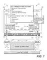
- FIG. 1is a schematic diagram of a cellular control system embodying an aspect of the present invention.
- FIG. 2is a perspective view of an inter-tissue application of the cellular control system, including a fluid/pressure interface subsystem and an endotube.
- FIG. 3shows an alternative aspect including a cover adapted for rolling or furling on an access line or conduit.
- FIG. 3Ashows a conduit of the cellular control system extending through an incision in the skin surface.
- FIG. 4shows an implanted plate and a conduit position for placing a furled cover.
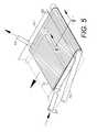
- FIG. 5shows the cover extending over a therapy zone.
- FIG. 6is a cross-sectional view thereof taken generally along line 6 - 6 in FIG. 5 .
- FIG. 7shows another alternative aspect including fluid/pressure inlet and outlet conduits with manifolds engaging the plate.
- FIG. 8shows a flexible barrier film furled on a conduit and in position for extending over the plate.
- FIG. 9shows the flexible barrier film extending over the plate.
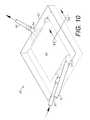
- FIG. 10shows the therapy zone closed by a tissue overlay.
- FIG. 11is a cross-sectional view taken generally along line 11 - 11 and FIG. 10 .
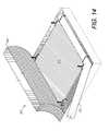
- FIG. 12shows another alternative aspect including scaffolding installed with an endotube.
- FIG. 13shows an absorbable fabric hemostatic layer being applied over the scaffolding via the endotube.
- FIG. 14shows the completed assembly of the system in the therapy zone.
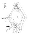
- FIG. 15shows the therapy zone covered by a tissue trapdoor plate.
- FIG. 16shows another alternative aspect of the present invention with inflow/outflow conduits extending into the therapy zone.
- FIG. 17is a cross-sectional view taken generally along line 17 - 17 in FIG. 16 .
- FIG. 18shows another alternative aspect of the present invention with scaffolding located in the therapy zone including couplings.
- FIG. 19shows another aspect of the invention with multiple bellows-type pumps or pillars in the therapy zone.
- FIG. 20shows another aspect of the invention with a closed-loop endotube assembly in the therapy zone.
- FIG. 21is a cross-sectional view taken generally along line 21 - 21 in FIG. 20 .
- FIG. 22is a schematic diagram similar to FIG. 1 showing another tissue regeneration and cellular control system embodying an alternative aspect of the present invention.
- the reference numeral 2generally designates a medical cellular control or tissue regeneration system embodying an aspect of the present invention.
- tissue regenerationwhich is broadly used to include tissue engineering, organ construction and tissue culture manufacturing.
- a primary application disclosed hereinis for controlling cellular regeneration and closure in an inter-tissue or intra-tissue space 4 , which can be generally located between a contact layer 6 and an in-situ tissue surface 8 , and is generally referred to as a “therapy zone.”
- the therapy zone 4can be located at various treatment sites in or on a patient, although typically it will be at a pathology location which is the object of a medical procedure involving cellular manipulation by one or more of the factors identified at 12 , including mechano/transductive, electro-magnetic force (EMF), pharmacological, chemical/antimicrobial, fluidic, bioengineered cells for seeding, thermal energy, acoustic energy (e.g., ultrasound), osmotic, oncotic, fluid pressure differential and others.
- EMFelectro-magnetic force
- FIG. 1shows a general interface 10 for applying the factors 12 to the therapy zone 4 .
- the interface 10includes a supply or inlet side 14 and an outlet side 16 .
- the inlet side 14can include a preprogrammed, digital controller 18 connected to and controlling a pump 20 , which delivers the contents of a supply reservoir 22 to an inflow manifold 24 for application to tissue regeneration/healing/repair scaffolding 26 .
- a suitable inlet conduit subsystem 28is provided for delivering factors 12 via the inlet side 14 .
- the inlet side 14also includes a positive pressure conduit 30 , which can be connected to a plate structure 32 in a plate area 27 of the therapy zone 4 via the controller 18 and the pump 20 . Fluid flow in the plate area 27 can be influenced and directed by the plate structure 32 .
- An outlet side 16 of the interface 10includes an outlet conduit subsystem 34 connected to an outflow manifold 36 from the scaffolding 26 and discharging to a collection reservoir 38 .
- a negative pressure (NP) pressure conduit 40connects the plate structure 32 to the factors 12 , which can include a negative pressure source.
- one or more pumps 20can be located on either or both sides of the plate structure 32 .
- FIG. 2shows a general configuration for the system 2 including a tissue bed 42 forming the tissue contact layer 6 and located below a skin surface 44 .
- the inflow and outflow sides 14 , 16 of the interface 10can include respective inflow and outflow conduits 30 , 40 extending through openings 45 in the skin surface 44 under the scaffolding 26 to the therapy zone 4 .
- the scaffolding 26can be retained in place on the tissue bed 42 by suitable anchors, such as scaffolding anchor clips 50 , which can comprise staples, sutures or other suitable in-situ fasteners.
- An endotube 52also extends through a skin surface opening 45 and is secured in place by endotube fasteners 54 (staples are shown) adjacent to scaffolding 56 located over the therapy zone 4 .
- the endotube 52is adapted for serving multiple functions, including placing and anchoring the scaffolding 56 , and introducing multiple factors 12 into the therapy zone 4 via a lumen 53 .
- FIG. 3shows a cellular control system 60 comprising another aspect of the invention with scaffolding 61 secured to the tissue bed 42 by the scaffolding fasteners 50 and positioned between inflow and outflow manifolds 62 , 64 , which are connected to inflow and outflow conduits 30 , 40 .
- the manifolds 62 , 64can be perforated, porous, semi-permeable or otherwise configured for communicating factors 12 with the scaffolding 61 .
- a tissue flap or trapdoor plate 66can be surgically opened by the incision 67 for access to the therapy zone 4 and closed as shown in FIG. 4 with a suture line 68 with the conduits 30 , 40 extending through the flap incision lines 67 on either side of the tissue flap plate 66 .
- a furled cover 72is wrapped around an endotube 70 with an endotube bore 71 for placement in the therapy zone 4 and can be extended to a covering position generally over the scaffolding 61 ( FIG. 5 ). As shown in FIG. 6 , the cover 72 is adapted for covering the suture line 68 during healing and can comprise various suitable wound-dressing materials, including membranes and bio-absorbable dressings.
- FIGS. 7-11show another aspect of the invention comprising a cellular control system 80 with a fluid transfer element 81 inflow and outflow manifolds 82 , 84 connected to conduits 30 , 40 respectively and including respective manifold branches 86 , 88 penetrating scaffolding 89 for communicating fluids, pressure and other factors 12 .
- the fluid transfer element 81can comprise open-cell foam or some other suitable fluid-transferring material.
- an endotube 70 with a furled cover 72can be placed within the therapy zone 4 and covered by the tissue flap 66 whereby the cellular control system 60 is substantially contained within the enclosed therapy zone 4 . Within such a closed environment, the cover 72 can be unfurled and extended by rotating the endotube 70 ( FIG. 11 ).
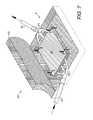
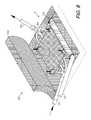
- FIGS. 12-17show a cellular control system 90 comprising another aspect of the invention and including scaffolding 92 adapted for placement in the therapy zone 4 on the tissue bed 6 , which can be surgically exposed by lifting a tissue flap plate or trapdoor 94 .
- the scaffolding 92can be placed with the endotube 52 , which is positioned in the therapy zone 4 and in turn positions the scaffolding 92 over the tissue bed 6 .
- An absorbable fabric hemostatic layer 96is extended over the scaffolding 92 as shown in FIG. 13 and is secured to the tissue bed 6 with suitable fasteners 50 , such as sutures or staples.
- the trapdoor 94functions as the plate in this configuration and is placed over the scaffolding 92 , the endotube 52 and the fabric hemostatic layer 96 , as shown in FIG. 15 .
- the tissue flap trapdoor plate 94can be sutured in place over the therapy zone 4 .
- Inflow and outflow conduits 30 , 40are inserted through openings 45 in the tissue flap plate 94 as shown in FIG. 16 and can underlie the scaffold 94 .
- the flow conduits 30 , 40can be placed before the scaffolding 92 is placed.
- the tissue flap plate 94can be formed in subcutaneous tissue, with the flow conduits 46 , 48 extending through skin surface openings 98 and penetrating to an appropriate depth to reach the therapy zone 4 .
- the tissue flap plate 94can comprise the dermal and epidermal layers.
- the hemostatic fabric layer 96can be wrapped around the endotube 52 for placement over the scaffolding 92 .
- the endotube 52can be slotted at 98 for accessing the lumen 53 , which can receive the scaffolding 92 in a compression-rolled configuration 92 a for unrolling into the therapy zone 4 , for example, by a flexible rod extending through the endotube 52 for twisting externally to the patient.
- FIG. 18shows a cellular control system 102 comprising another modified aspect of the invention and including scaffolding 104 with inflow and outflow female couplings 106 , 108 , which connect to the inflow and outflow conduits 30 , 40 respectively via male couplings 110 , 112 .
- a barbed-strand, self-anchoring surgical suture 114is shown being extended into the therapy zone 4 from the endotube 52 .
- Such suturesare available from Quill Medical, Inc. of Research Triangle Park, N.C. See, for example, U.S. Pat. No. 7,056,331, which is incorporated herein by reference.
- the endotube 52facilitates inserting the barbed suture 114 and “setting” its prongs by tugging on the outer end extending from the endotube 52 external to the patient for self-anchoring the suture 114 .
- FIG. 19shows a cellular control system 120 comprising another modified aspect of the present invention and including multiple bellows-action pillars 122 located below the scaffold 104 and fluidly connected to the inflow and outflow conduits 30 , 40 respectively.
- the pillars 122can reciprocably compress and expand in response to various pressures associated with the therapy zone 4 .
- Such pressurescan be externally-generated, e.g., by one or more of the factors 12 , or internal pressures generated by the patient.
- Such pillars 122can facilitate a “pumping” action with the cellular control system 120 by alternately expanding and contracting in order to move fluid into and out of the therapy zone 4 .
- FIGS. 20 and 21show a cellular control system 130 with a continuous loop endotube 132 forming the scaffolding 26 within a therapy zone 134 generally formed along the path of the endotube 132 through tissue 136 .
- the endotube 132includes a lumen 138 , which can function as a conduit for introducing pharmacological and other substances 140 , and/or extracting fluid from the patient.
- the endotube 132can be preloaded with cells for seeding the therapy zone 134 .
- the endotube 132forms inflow and outflow conduits 142 , 144 with interchangeable functions.
- the endotube 132includes an outer contact surface 146 , which is adapted for engaging the tissue 136 .
- the endotube 132can be bioabsorbable, permanently implanted or extracted after completing a procedure. Moreover, the endotube 132 can be fabricated from a wide range of suitable materials chosen for compatibility with the therapeutic objectives of particular procedures. For example, semi-permeable materials can form pressure differentials and selectively transfer fluids. The endotube 132 can be perforated or slotted for fluid collection or dispersal. The external conduits 142 , 144 can be connected to negative and/or positive pressure sources external to the therapy zone 134 . Placement of the endotube 132 can be accomplished with a Trocar instrument, by surgical incision or placement under a tissue flap or trapdoor 66 .
- An open mesh 148comprising a matrix of threads or capillary-type tubes 150 forms a cellular control sleeve 152 over an endotube outer contact surface 146 .
- the mesh 148can introduce cells, facilitate cellular ingrowth, channel fluid evacuation, enhance tissue contact interaction and otherwise facilitate the treatment objectives.
- the range of suitable materialsincludes bioabsorbable materials, pharmacological release materials (e.g., antibiotics, growth factors, antiseptics, imaging materials and other suitable materials) and hollow tubes for communicating fluids.
- the mesh 148can be extracted with the endotube 132 , or left in place after extraction. Still further, the mesh 148 can comprise closure members, such as the barbed suture strands 114 available from Quill Medical, Inc., which are described above.
- the tubular or thread configuration shown in FIGS. 20 and 21includes the system and method embodiments described above, with their components formed in tubular shapes. These embodiments can include conduit size components (cm to mm range diameters), capillary size (mm range diameters) and nano size (micron diameters). Length can generally be any suitable length.
- the endotubes 132can be fabricated and installed in various configurations, including straight, linearly-connected (series), parallel configurations, spiral, coil, circular, wave-like, etc. with the intention of optimizing recipient tissue bed positioning and ease of installation. Installation can be accomplished manually by palpation, visually, with imaging techniques, endoscopically assisted or using open surgical techniques.
- Manipulative factors 12can be introduced or applied, typically at one or both ends of the conduits 142 , 144 with external (percutaneous) connections of the tubes, conduits or threads.
- the outer barrier or sheath of the tube (equivalent to the plate described above) and the makeup of the inner core (equivalent to the scaffolding described above)depend on the therapy intentions and the method of introduction, including placement, manipulation and control. With the system in a tubular configuration, the outer barrier is also the contact layer.
- the tubecan be placed in solid tissue, such as muscle or the liver using imaging techniques with a series of guide wires, followers and dilators, similarly to techniques for endovascular access.
- imaging techniqueswith a series of guide wires, followers and dilators, similarly to techniques for endovascular access.
- both entrance and exit areasare more feasible and more easily accomplished with a single guide wire or thin trocar.
- Input and outputcan thus be provided at opposite poles as the simplest and most efficient system for fluid manipulation.
- a single external conduitcould serve as both input and output ports by alternating the functions or by use as a conduit carrying side-by-side smaller input/output lines that would travel in a preconfigured fashion through the outer sheath and inner core whereby the input would be instilled at one end and the output would be withdrawn from the opposite end and these functions could travel side-by-side in the single conduit separately contained.
- the outer sheathcould have a pore size sufficient to be able to remove the blood and cell damage from placement. This could take an estimated one to two days or until the effluent is clear. The cell seeding then starts and is continued until it also comes out the effluent.
- the inner coreis a scaffolding material that is biodegradable and chosen for its affinity to the cells to be seated.
- the outer sheathis in removed and the inner core, now seeded with cells, is left in place to grow and “take” as a graft of bioengineered tissue grown in-situ. If a single port is used, the inner core can be cut below the skin line and allowed to retract. If a double (2-ended) port is used, the output port is cut below the skin line and the outer sheath is then pulled out through the outer port. If the core is in the port, it is also cut off below the skin and allowed to retract. The end result is that the nonabsorbable outer sheath is removed and the absorbable scaffolding is left in a subcutaneous (inter-tissue) position.
- FIG. 22shows a modified control system 202 comprising an alternative aspect of the present invention.
- the system 202includes an inter-tissue space/therapy zone 204 , which also defines a flow layer(s) for fluids generated internally and/or introduced externally.
- the tissue contact layer 6can be located anywhere appropriate for treatment with the systems 2 and 202 , including subdermal, subcutaneous, externally and internally; and in or on body cavities, organs, muscle fibers, ligamentous and osseous (skeletal) structure, etc.
- a plate/tissue component 208can comprise a physical structure, such as a biocompatible material adapted for placement in or on the therapy zone 204 .
- the component 208can comprise a patient's tissue layer, such as the dermis, epidermis, etc.
- the component 208cooperates with a pressure differential manifold 232 to facilitate and direct the flow of fluid, microbial agents, medications, irrigation, and other substances in the therapy zone 204 .
- Either or both of the tissue scaffolding 226 and the pressure differential manifold 232can comprise cellular matrices, synthetic tissue, living tissue or derivatives of living tissue.
- the system 202can include a variety of configurations with the plate/tissue component 208 cooperating with the manifold 232 and scaffolding 226 to form the pressure differential zone 204 .
- Fluid pulse wavescan be introduced to the therapy zone 204 by cycling a pump 220 with a controller 218 and pulsing fluid through various tubing and manifold configurations, including those shown in FIGS. 2-21 .
- a sensor suite 242is connected to the controller 218 and can include multiple sensor suite feeds 244 extending to various components and areas of the therapy zone 204 .
- the sensor suite 242can include sensors for monitoring various operating parameters, including pressure, temperature, microbial activity, chemical composition (e.g., oxygen and CO 2 levels), etc.
- Sensor inputs to the controller 218can be digitized for processing by the microprocessor controller 218 .
- the sensor signal input informationcan be utilized by the controller 218 for controlling various operating parameters of the system 202 , such as the pump 220 , the inflow/outflow lines 230 / 240 and the factor source 212 .
- tubing and manifold elements shown thereincan be rearranged and reconfigured as necessary to achieve a wide range of alternative systems for accommodating various patient conditions and treatment objectives.
- Relatively small-amplitude pressure changes of, for example, a few mm Hgcan be sufficient for achieving desired therapeutic results. More specifically, such pressure changes can stimulate cellular activity, reepithelialization, cell migration, regeneration and other physiological changes associated with the healing process.
- components of the system 202such as the bellows-equipped pillars 122 shown in FIG. 19 , can provide or supplement such pressure waves, for example with the blood pressure cycles of the circulatory system or similar pressure-varying, dynamic physiological functions, such as musculature, lymphatic, respiratory, etc.
- the system 202can thus operate using the dynamic pulsations naturally occurring in-vivo, and/or with externally-applied forces, such as the pump 220 .
- system 202is adaptable for benchtop, tissue culture, tissue engineering, ex-vivo and other applications for a wide range of research, bioengineering, tissue culture and other useful applications, which share a common element of cellular control and manipulation.
- a general interface 210can comprise a wide range of suitable component/patient interface constructions, such as internal/external dressings, closure screens, etc.
- suitable component/patient interface constructionssuch as internal/external dressings, closure screens, etc.
- suitable component/patient interface constructionssuch as internal/external dressings, closure screens, etc.
- FIG. 210An exemplary list of cell manipulating factors as shown at 212 for application to the therapy zone 204 via the interface 210 , and is not to be construed as limiting.
- a controller 218can be provided for preprogramming to control various components and operating parameters of the system 202 , such as a pump 224 for delivering fluids and other factors from the source 212 to the pressure differential manifold 232 via inlet lines 228 and to tissue scaffolding 226 via therapy inflow input lines 230 .
- line 234is connected to the pressure differential manifold 232 and returns to the source 212 .
- the therapy outflow line 240is connected to the tissue scaffolding 226 and returns to the source 212 .
- An optional supply reservoir 222can be connected to the therapy inflow line 230 and can provide a secondary or alternative source of pharmacological and other factors for input to the therapy zone 204 via the therapy inflow line 230 .
- a corresponding collection reservoir 238can receive fluid from the therapy zone 204 via the therapy outflow line 240 .
- collected waste fluidcan be disposed of using established medical waste disposal procedures.
- planar orientations of the system componentscan be rearranged and reconfigured in-situ as determined by the medical practitioner.
- Alternative orientationscan include inverted, vertical, horizontal, etc.
- the orientations discussed aboveare for illustration and could vary depending upon the position of the patient.
- the pressure differential manifold 232could be formed within or below the tissue scaffolding 226 and in various spatial relationships to the plate/tissue 208 .
- the component configurationscan assume any appropriate configuration, such as tubular, spiral, circular, etc.
Landscapes
- Health & Medical Sciences (AREA)
- Heart & Thoracic Surgery (AREA)
- Vascular Medicine (AREA)
- Engineering & Computer Science (AREA)
- Anesthesiology (AREA)
- Biomedical Technology (AREA)
- Hematology (AREA)
- Life Sciences & Earth Sciences (AREA)
- Animal Behavior & Ethology (AREA)
- General Health & Medical Sciences (AREA)
- Public Health (AREA)
- Veterinary Medicine (AREA)
- Oral & Maxillofacial Surgery (AREA)
- Pulmonology (AREA)
- Surgery (AREA)
- External Artificial Organs (AREA)
Abstract
Description
Claims (13)
Priority Applications (3)
| Application Number | Priority Date | Filing Date | Title |
|---|---|---|---|
| US13/245,677US9408956B2 (en) | 2010-09-24 | 2011-09-26 | Cellular control and tissue regeneration systems and methods |
| US14/217,219US9456930B2 (en) | 2011-07-12 | 2014-03-17 | Topical vacuum-press surgical incisional dressings, surgical adjuncts, hybrids and composites |
| US15/232,612US20160346444A1 (en) | 2010-09-24 | 2016-08-09 | Cellular control and tissue regeneration systems and methods |
Applications Claiming Priority (2)
| Application Number | Priority Date | Filing Date | Title |
|---|---|---|---|
| US38638010P | 2010-09-24 | 2010-09-24 | |
| US13/245,677US9408956B2 (en) | 2010-09-24 | 2011-09-26 | Cellular control and tissue regeneration systems and methods |
Related Child Applications (1)
| Application Number | Title | Priority Date | Filing Date |
|---|---|---|---|
| US15/232,612Continuation-In-PartUS20160346444A1 (en) | 2010-09-24 | 2016-08-09 | Cellular control and tissue regeneration systems and methods |
Publications (2)
| Publication Number | Publication Date |
|---|---|
| US20120078379A1 US20120078379A1 (en) | 2012-03-29 |
| US9408956B2true US9408956B2 (en) | 2016-08-09 |
Family
ID=45871422
Family Applications (1)
| Application Number | Title | Priority Date | Filing Date |
|---|---|---|---|
| US13/245,677Expired - Fee RelatedUS9408956B2 (en) | 2010-09-24 | 2011-09-26 | Cellular control and tissue regeneration systems and methods |
Country Status (1)
| Country | Link |
|---|---|
| US (1) | US9408956B2 (en) |
Cited By (4)
| Publication number | Priority date | Publication date | Assignee | Title |
|---|---|---|---|---|
| US20160038626A1 (en)* | 2014-08-11 | 2016-02-11 | Kci Licensing, Inc. | Protease modulating wound interface layer for use with negative pressure wound therapy |
| US10292709B1 (en)* | 2018-11-13 | 2019-05-21 | King Saud University | Device for sutureless repair of an injured nerve |
| US11051509B2 (en) | 2016-11-22 | 2021-07-06 | William Lafayette Mondy | Method and apparatus for keeping artificially created tissues alive |
| US11298453B2 (en) | 2003-10-28 | 2022-04-12 | Smith & Nephew Plc | Apparatus and method for wound cleansing with actives |
Families Citing this family (2)
| Publication number | Priority date | Publication date | Assignee | Title |
|---|---|---|---|---|
| US9569566B2 (en)* | 2011-12-12 | 2017-02-14 | Zam Research Llc | Simulation and control system and method using contact, pressure waves and factor controls for cell regeneration, tissue closure and related applications |
| US20150064141A1 (en) | 2012-04-05 | 2015-03-05 | The Regents Of The University Of California | Regenerative sera cells and mesenchymal stem cells |
Citations (199)
| Publication number | Priority date | Publication date | Assignee | Title |
|---|---|---|---|---|
| US221427A (en) | 1879-11-11 | Improvement in barb-fence links | ||
| US1355846A (en) | 1920-02-06 | 1920-10-19 | David A Rannells | Medical appliance |
| US2547758A (en) | 1949-01-05 | 1951-04-03 | Wilmer B Keeling | Instrument for treating the male urethra |
| US2632443A (en) | 1949-04-18 | 1953-03-24 | Eleanor P Lesher | Surgical dressing |
| GB692578A (en) | 1949-09-13 | 1953-06-10 | Minnesota Mining & Mfg | Improvements in or relating to drape sheets for surgical use |
| US2682873A (en) | 1952-07-30 | 1954-07-06 | Johnson & Johnson | General purpose protective dressing |
| US2910763A (en) | 1955-08-17 | 1959-11-03 | Du Pont | Felt-like products |
| US2969057A (en) | 1957-11-04 | 1961-01-24 | Brady Co W H | Nematodic swab |
| US3066672A (en) | 1960-09-27 | 1962-12-04 | Jr William H Crosby | Method and apparatus for serial sampling of intestinal juice |
| US3115138A (en) | 1960-07-14 | 1963-12-24 | Mcelvenny | Evacuator |
| US3367332A (en) | 1965-08-27 | 1968-02-06 | Gen Electric | Product and process for establishing a sterile area of skin |
| US3520300A (en) | 1967-03-15 | 1970-07-14 | Amp Inc | Surgical sponge and suction device |
| US3568675A (en) | 1968-08-30 | 1971-03-09 | Clyde B Harvey | Fistula and penetrating wound dressing |
| US3648692A (en) | 1970-12-07 | 1972-03-14 | Parke Davis & Co | Medical-surgical dressing for burns and the like |
| US3682180A (en) | 1970-06-08 | 1972-08-08 | Coilform Co Inc | Drain clip for surgical drain |
| US3826254A (en) | 1973-02-26 | 1974-07-30 | Verco Ind | Needle or catheter retaining appliance |
| US3981051A (en) | 1970-03-16 | 1976-09-21 | Brumlik George C | Bristle-like gripping device |
| US4080970A (en) | 1976-11-17 | 1978-03-28 | Miller Thomas J | Post-operative combination dressing and internal drain tube with external shield and tube connector |
| US4096853A (en) | 1975-06-21 | 1978-06-27 | Hoechst Aktiengesellschaft | Device for the introduction of contrast medium into an anus praeter |
| US4139004A (en) | 1977-02-17 | 1979-02-13 | Gonzalez Jr Harry | Bandage apparatus for treating burns |
| US4165748A (en) | 1977-11-07 | 1979-08-28 | Johnson Melissa C | Catheter tube holder |
| US4184510A (en) | 1977-03-15 | 1980-01-22 | Fibra-Sonics, Inc. | Valued device for controlling vacuum in surgery |
| DE2640413C3 (en) | 1976-09-08 | 1980-03-27 | Richard Wolf Gmbh, 7134 Knittlingen | Catheter monitor |
| WO1980002182A1 (en) | 1979-04-06 | 1980-10-16 | J Moss | Portable suction device for collecting fluids from a closed wound |
| US4233969A (en) | 1976-11-11 | 1980-11-18 | Lock Peter M | Wound dressing materials |
| US4245630A (en) | 1976-10-08 | 1981-01-20 | T. J. Smith & Nephew, Ltd. | Tearable composite strip of materials |
| US4248232A (en) | 1977-09-13 | 1981-02-03 | Eckart Engelbrecht | Method of dissolving the bond between interconnected components |
| US4256109A (en) | 1978-07-10 | 1981-03-17 | Nichols Robert L | Shut off valve for medical suction apparatus |
| US4259959A (en) | 1978-12-20 | 1981-04-07 | Walker Wesley W | Suturing element |
| US4261363A (en) | 1979-11-09 | 1981-04-14 | C. R. Bard, Inc. | Retention clips for body fluid drains |
| US4275721A (en) | 1978-11-28 | 1981-06-30 | Landstingens Inkopscentral Lic, Ekonomisk Forening | Vein catheter bandage |
| US4284079A (en) | 1979-06-28 | 1981-08-18 | Adair Edwin Lloyd | Method for applying a male incontinence device |
| US4297995A (en) | 1980-06-03 | 1981-11-03 | Key Pharmaceuticals, Inc. | Bandage containing attachment post |
| US4333468A (en) | 1980-08-18 | 1982-06-08 | Geist Robert W | Mesentery tube holder apparatus |
| US4373519A (en) | 1981-06-26 | 1983-02-15 | Minnesota Mining And Manufacturing Company | Composite wound dressing |
| US4382441A (en) | 1978-12-06 | 1983-05-10 | Svedman Paul | Device for treating tissues, for example skin |
| US4392853A (en) | 1981-03-16 | 1983-07-12 | Rudolph Muto | Sterile assembly for protecting and fastening an indwelling device |
| US4392858A (en) | 1981-07-16 | 1983-07-12 | Sherwood Medical Company | Wound drainage device |
| US4419097A (en) | 1981-07-31 | 1983-12-06 | Rexar Industries, Inc. | Attachment for catheter tube |
| US4419093A (en) | 1980-01-21 | 1983-12-06 | American Hospital Supply Corporation | Method of receiving and disposing of fluids from the body |
| US4475909A (en) | 1982-05-06 | 1984-10-09 | Eisenberg Melvin I | Male urinary device and method for applying the device |
| US4480638A (en) | 1980-03-11 | 1984-11-06 | Eduard Schmid | Cushion for holding an element of grafted skin |
| US4525374A (en) | 1984-02-27 | 1985-06-25 | Manresa, Inc. | Treating hydrophobic filters to render them hydrophilic |
| US4525166A (en) | 1981-11-21 | 1985-06-25 | Intermedicat Gmbh | Rolled flexible medical suction drainage device |
| US4540412A (en) | 1983-07-14 | 1985-09-10 | The Kendall Company | Device for moist heat therapy |
| US4543100A (en) | 1983-11-01 | 1985-09-24 | Brodsky Stuart A | Catheter and drain tube retainer |
| US4548202A (en) | 1983-06-20 | 1985-10-22 | Ethicon, Inc. | Mesh tissue fasteners |
| US4551139A (en) | 1982-02-08 | 1985-11-05 | Marion Laboratories, Inc. | Method and apparatus for burn wound treatment |
| EP0100148B1 (en) | 1982-07-06 | 1986-01-08 | Dow Corning Limited | Medical-surgical dressing and a process for the production thereof |
| US4569348A (en) | 1980-02-22 | 1986-02-11 | Velcro Usa Inc. | Catheter tube holder strap |
| AU550575B2 (en) | 1981-08-07 | 1986-03-27 | Richard Christian Wright | Wound drainage device |
| US4605399A (en) | 1984-12-04 | 1986-08-12 | Complex, Inc. | Transdermal infusion device |
| US4608041A (en) | 1981-10-14 | 1986-08-26 | Frese Nielsen | Device for treatment of wounds in body tissue of patients by exposure to jets of gas |
| US4640688A (en) | 1985-08-23 | 1987-02-03 | Mentor Corporation | Urine collection catheter |
| US4655754A (en) | 1984-11-09 | 1987-04-07 | Stryker Corporation | Vacuum wound drainage system and lipids baffle therefor |
| US4664662A (en) | 1984-08-02 | 1987-05-12 | Smith And Nephew Associated Companies Plc | Wound dressing |
| WO1987004626A1 (en) | 1986-01-31 | 1987-08-13 | Osmond, Roger, L., W. | Suction system for wound and gastro-intestinal drainage |
| US4696301A (en) | 1986-07-16 | 1987-09-29 | Barabe David J | Wound closing method |
| US4710165A (en) | 1985-09-16 | 1987-12-01 | Mcneil Charles B | Wearable, variable rate suction/collection device |
| US4733659A (en) | 1986-01-17 | 1988-03-29 | Seton Company | Foam bandage |
| US4743232A (en) | 1986-10-06 | 1988-05-10 | The Clinipad Corporation | Package assembly for plastic film bandage |
| US4758220A (en) | 1985-09-26 | 1988-07-19 | Alcon Laboratories, Inc. | Surgical cassette proximity sensing and latching apparatus |
| US4775909A (en) | 1984-02-03 | 1988-10-04 | Canon Denshi Kabushiki Kaisha | Magnetic head using a magnetic thin film and first, second and third core members |
| US4787888A (en) | 1987-06-01 | 1988-11-29 | University Of Connecticut | Disposable piezoelectric polymer bandage for percutaneous delivery of drugs and method for such percutaneous delivery (a) |
| US4826494A (en) | 1984-11-09 | 1989-05-02 | Stryker Corporation | Vacuum wound drainage system |
| US4828546A (en) | 1987-08-21 | 1989-05-09 | Surgidyne, Inc. | Bulb evacuator for closed wound suction |
| US4838883A (en) | 1986-03-07 | 1989-06-13 | Nissho Corporation | Urine-collecting device |
| US4840187A (en) | 1986-09-11 | 1989-06-20 | Bard Limited | Sheath applicator |
| EP0117632B1 (en) | 1983-01-27 | 1989-08-16 | Johnson & Johnson Products Inc. | Adhesive film dressing |
| US4863449A (en) | 1987-07-06 | 1989-09-05 | Hollister Incorporated | Adhesive-lined elastic condom cathether |
| US4872450A (en) | 1984-08-17 | 1989-10-10 | Austad Eric D | Wound dressing and method of forming same |
| EP0161865B1 (en) | 1984-05-03 | 1989-10-11 | Smith and Nephew Associated Companies p.l.c. | Adhesive wound dressing |
| US4878901A (en) | 1986-10-10 | 1989-11-07 | Sachse Hans Ernst | Condom catheter, a urethral catheter for the prevention of ascending infections |
| GB2220357A (en) | 1988-05-28 | 1990-01-10 | Smiths Industries Plc | Medico-surgical containers |
| US4897081A (en) | 1984-05-25 | 1990-01-30 | Thermedics Inc. | Percutaneous access device |
| US4906240A (en) | 1988-02-01 | 1990-03-06 | Matrix Medica, Inc. | Adhesive-faced porous absorbent sheet and method of making same |
| US4906233A (en) | 1986-05-29 | 1990-03-06 | Terumo Kabushiki Kaisha | Method of securing a catheter body to a human skin surface |
| US4919654A (en) | 1988-08-03 | 1990-04-24 | Kalt Medical Corporation | IV clamp with membrane |
| CA2005436A1 (en) | 1988-12-13 | 1990-06-13 | Glenda G. Kalt | Transparent tracheostomy tube dressing |
| US4941882A (en) | 1987-03-14 | 1990-07-17 | Smith And Nephew Associated Companies, P.L.C. | Adhesive dressing for retaining a cannula on the skin |
| US4953565A (en) | 1986-11-26 | 1990-09-04 | Shunro Tachibana | Endermic application kits for external medicines |
| WO1990010424A1 (en) | 1989-03-16 | 1990-09-20 | Smith & Nephew Plc | Absorbent devices and precursors therefor |
| US4969880A (en) | 1989-04-03 | 1990-11-13 | Zamierowski David S | Wound dressing and treatment method |
| GB2197789B (en) | 1986-11-28 | 1990-11-28 | Smiths Industries Plc | Anti-foaming agents |
| US4976726A (en) | 1989-04-27 | 1990-12-11 | Haverstock Charles B | Skin closure devices |
| US4985019A (en) | 1988-03-11 | 1991-01-15 | Michelson Gary K | X-ray marker |
| GB2235877A (en) | 1989-09-18 | 1991-03-20 | Antonio Talluri | Closed wound suction apparatus |
| US5007921A (en) | 1989-10-26 | 1991-04-16 | Brown Alan W | Surgical staple |
| US5007936A (en) | 1988-02-18 | 1991-04-16 | Cemax, Inc. | Surgical method for hip joint replacement |
| GB2195255B (en) | 1986-09-30 | 1991-05-01 | Vacutec Uk Limited | Apparatus for vacuum treatment of an epidermal surface |
| US5019083A (en) | 1989-01-31 | 1991-05-28 | Advanced Osseous Technologies, Inc. | Implanting and removal of orthopedic prostheses |
| US5037397A (en) | 1985-05-03 | 1991-08-06 | Medical Distributors, Inc. | Universal clamp |
| US5045054A (en) | 1990-02-06 | 1991-09-03 | Advanced Osseous Technologies Inc. | Apparatus for implantation and extraction of osteal prostheses |
| US5086170A (en) | 1989-01-16 | 1992-02-04 | Roussel Uclaf | Process for the preparation of azabicyclo compounds |
| US5092858A (en) | 1990-03-20 | 1992-03-03 | Becton, Dickinson And Company | Liquid gelling agent distributor device |
| US5100396A (en) | 1989-04-03 | 1992-03-31 | Zamierowski David S | Fluidic connection system and method |
| US5112338A (en) | 1991-02-11 | 1992-05-12 | Anspach Iii William E | Surgical instrument for removing artificial acetabular cups |
| US5134994A (en) | 1990-02-12 | 1992-08-04 | Say Sam L | Field aspirator in a soft pack with externally mounted container |
| US5139023A (en) | 1989-06-02 | 1992-08-18 | Theratech Inc. | Apparatus and method for noninvasive blood glucose monitoring |
| US5149331A (en) | 1991-05-03 | 1992-09-22 | Ariel Ferdman | Method and device for wound closure |
| US5167613A (en) | 1992-03-23 | 1992-12-01 | The Kendall Company | Composite vented wound dressing |
| US5169399A (en) | 1991-10-17 | 1992-12-08 | Boehringer Mannheim Corporation | Acetabular cup impactor |
| US5176663A (en) | 1987-12-02 | 1993-01-05 | Pal Svedman | Dressing having pad with compressibility limiting elements |
| WO1993009727A1 (en) | 1991-11-14 | 1993-05-27 | Wake Forest University | Method and apparatus for treating tissue damage |
| US5215522A (en) | 1984-07-23 | 1993-06-01 | Ballard Medical Products | Single use medical aspirating device and method |
| USD337639S (en) | 1990-11-21 | 1993-07-20 | Zimmer, Inc. | Combined impactor and extractor for prosthetic implants |
| US5232453A (en) | 1989-07-14 | 1993-08-03 | E. R. Squibb & Sons, Inc. | Catheter holder |
| US5261893A (en) | 1989-04-03 | 1993-11-16 | Zamierowski David S | Fastening system and method |
| US5278100A (en) | 1991-11-08 | 1994-01-11 | Micron Technology, Inc. | Chemical vapor deposition technique for depositing titanium silicide on semiconductor wafers |
| US5279550A (en) | 1991-12-19 | 1994-01-18 | Gish Biomedical, Inc. | Orthopedic autotransfusion system |
| US5298015A (en) | 1989-07-11 | 1994-03-29 | Nippon Zeon Co., Ltd. | Wound dressing having a porous structure |
| US5318570A (en) | 1989-01-31 | 1994-06-07 | Advanced Osseous Technologies, Inc. | Ultrasonic tool |
| US5342376A (en) | 1993-05-03 | 1994-08-30 | Dermagraphics, Inc. | Inserting device for a barbed tissue connector |
| US5344415A (en) | 1993-06-15 | 1994-09-06 | Deroyal Industries, Inc. | Sterile system for dressing vascular access site |
| DE4306478A1 (en) | 1993-03-02 | 1994-09-08 | Wolfgang Dr Wagner | Drainage device, in particular pleural drainage device, and drainage method |
| WO1994020041A1 (en) | 1993-03-09 | 1994-09-15 | Wake Forest University | Wound treatment employing reduced pressure |
| US5358494A (en) | 1989-07-11 | 1994-10-25 | Svedman Paul | Irrigation dressing |
| US5383897A (en) | 1992-10-19 | 1995-01-24 | Shadyside Hospital | Method and apparatus for closing blood vessel punctures |
| US5423885A (en) | 1992-01-31 | 1995-06-13 | Advanced Cardiovascular Systems, Inc. | Stent capable of attachment within a body lumen |
| US5437622A (en) | 1992-04-29 | 1995-08-01 | Laboratoire Hydrex (Sa) | Transparent adhesive dressing with reinforced starter cuts |
| US5437651A (en) | 1993-09-01 | 1995-08-01 | Research Medical, Inc. | Medical suction apparatus |
| DE29504378U1 (en) | 1995-03-15 | 1995-09-14 | MTG Medizinisch, technische Gerätebau GmbH, 66299 Friedrichsthal | Electronically controlled low-vacuum pump for chest and wound drainage |
| WO1996005873A1 (en) | 1994-08-22 | 1996-02-29 | Kinetic Concepts Inc. | Wound drainage equipment |
| US5507833A (en) | 1992-02-10 | 1996-04-16 | Kim-Med, Inc. | Hip replacement system and method for implanting the same |
| US5522901A (en) | 1992-06-26 | 1996-06-04 | Eska Implants Gmbh | Implant for replacing a rear patella part |
| US5527293A (en) | 1989-04-03 | 1996-06-18 | Kinetic Concepts, Inc. | Fastening system and method |
| USD372309S (en) | 1995-07-06 | 1996-07-30 | Zimmer, Inc. | Orthopaedic broach impactor |
| US5549584A (en) | 1994-02-14 | 1996-08-27 | The Kendall Company | Apparatus for removing fluid from a wound |
| US5556375A (en) | 1994-06-16 | 1996-09-17 | Hercules Incorporated | Wound dressing having a fenestrated base layer |
| US5580353A (en) | 1994-04-19 | 1996-12-03 | Mendes; David | Prosthetic patella implant of the knee joint |
| US5584859A (en) | 1993-10-12 | 1996-12-17 | Brotz; Gregory R. | Suture assembly |
| US5607388A (en) | 1994-06-16 | 1997-03-04 | Hercules Incorporated | Multi-purpose wound dressing |
| US5630819A (en) | 1994-08-11 | 1997-05-20 | Howmedica International | Acetabular bone graft impactor |
| WO1997018007A1 (en) | 1995-11-14 | 1997-05-22 | Kci Medical Limited | Portable wound treatment apparatus |
| US5716360A (en) | 1995-06-30 | 1998-02-10 | U.S. Medical Products | Patella recession instrument and method for anatomically-shaped patellar prostheses |
| US5738686A (en) | 1993-04-03 | 1998-04-14 | Joachim Theusner | Artificial joint to replace the human patella |
| US5785700A (en) | 1992-06-03 | 1998-07-28 | Zimmer Patient Care, A Division Of Zimmer, Inc. | Autotransfusion system with portable detachable vacuum source |
| US5800546A (en) | 1995-08-14 | 1998-09-01 | Smith & Nephew, Inc. | Impactor apparatus for assembling modular orthopedic prosthesis components |
| US5827246A (en) | 1996-02-28 | 1998-10-27 | Tecnol Medical Products, Inc. | Vacuum pad for collecting potentially hazardous fluids |
| US5846244A (en) | 1995-09-18 | 1998-12-08 | Exactech, Inc. | Counter-balanced oscillating surgical saw |
| WO1999013793A1 (en) | 1997-09-12 | 1999-03-25 | Kci Medical Limited | Surgical drape and suction head for wound treatment |
| US5911222A (en) | 1992-08-07 | 1999-06-15 | Bristol-Myers Squibb | Liquid removal system |
| US5921972A (en) | 1996-01-11 | 1999-07-13 | Skow; Joseph I. | Surgical wicking and fluid removal swab |
| US5931855A (en) | 1997-05-21 | 1999-08-03 | Frank Hoffman | Surgical methods using one-way suture |
| US5941859A (en) | 1997-03-17 | 1999-08-24 | Lerman; Benjamin S. | Wound irrigation shield with fluid scavenging |
| US6071267A (en) | 1998-02-06 | 2000-06-06 | Kinetic Concepts, Inc. | Medical patient fluid management interface system and method |
| US6113618A (en) | 1999-01-13 | 2000-09-05 | Stryker Corporation | Surgical saw with spring-loaded, low-noise cutting blade |
| US6126659A (en) | 1998-09-30 | 2000-10-03 | Depuy Orthopaedics, Inc. | Impaction instruments |
| US6135116A (en) | 1997-07-28 | 2000-10-24 | Kci Licensing, Inc. | Therapeutic method for treating ulcers |
| US6146423A (en) | 1999-01-28 | 2000-11-14 | Implex Corporation | Patella replacement apparatus |
| US6159246A (en) | 1994-04-19 | 2000-12-12 | Mendes; David | Surgical method and tool for repairing a patella of the knee joint |
| US6174306B1 (en) | 1995-05-13 | 2001-01-16 | Wim Fleischmann | Device for vacuum-sealing an injury |
| US6179804B1 (en) | 1999-08-18 | 2001-01-30 | Oxypatch, Llc | Treatment apparatus for wounds |
| US6190392B1 (en) | 1999-02-03 | 2001-02-20 | Biomet, Inc. | Method and apparatus for ultrasonic removal of bone cement material |
| US6190391B1 (en) | 1999-10-01 | 2001-02-20 | Bristol-Myers Squibb Company | Method of preparing a resected posterior surface of a patella to receive a prosthetic element |
| US6203563B1 (en) | 1999-05-26 | 2001-03-20 | Ernesto Ramos Fernandez | Healing device applied to persistent wounds, fistulas, pancreatitis, varicose ulcers, and other medical or veterinary pathologies of a patient |
| US6241747B1 (en) | 1993-05-03 | 2001-06-05 | Quill Medical, Inc. | Barbed Bodily tissue connector |
| US6270517B1 (en) | 2000-02-04 | 2001-08-07 | Gregory R. Brotz | Suture assembly and method |
| USRE37358E1 (en) | 1994-10-07 | 2001-09-04 | The Anspach Effort, Inc. | Tool holding mechanism for a motor driven surgical instrument |
| US6287316B1 (en) | 1999-03-26 | 2001-09-11 | Ethicon, Inc. | Knitted surgical mesh |
| US6293929B1 (en) | 1997-09-02 | 2001-09-25 | Steven M. Smith | Wound irrigation apparatus |
| US20020022861A1 (en) | 2000-05-19 | 2002-02-21 | Daniel Jacobs | Multi-point tissue tension distribution device, a combined orbital rim repair and suspension variation, and a method of tissue approximation using the device |
| US20020029063A1 (en) | 2000-09-05 | 2002-03-07 | Wittmann Dietmar H. | Prosthesis for abdominal surgery |
| US6355215B1 (en) | 1999-03-10 | 2002-03-12 | Implex Corp. | Wear-resistant olefinic medical implant and thermal treatment container therefor |
| US6377653B1 (en) | 1999-12-10 | 2002-04-23 | Electronics And Telecommunications Research Institute | Method for calibrating trabecular index using sawtooth-shaped rack |
| US6398767B1 (en) | 1997-05-27 | 2002-06-04 | Wilhelm Fleischmann | Process and device for application of active substances to a wound surface area |
| US20020077661A1 (en) | 2000-12-20 | 2002-06-20 | Vahid Saadat | Multi-barbed device for retaining tissue in apposition and methods of use |
| US6430427B1 (en) | 1999-02-25 | 2002-08-06 | Electronics And Telecommunications Research Institute | Method for obtaining trabecular index using trabecular pattern and method for estimating bone mineral density using trabecular indices |
| US20020115951A1 (en) | 2001-02-22 | 2002-08-22 | Core Products International, Inc. | Ankle brace providing upper and lower ankle adjustment |
| US20020120185A1 (en) | 2000-05-26 | 2002-08-29 | Kci Licensing, Inc. | System for combined transcutaneous blood gas monitoring and vacuum assisted wound closure |
| US20020143286A1 (en) | 2001-03-05 | 2002-10-03 | Kci Licensing, Inc. | Vacuum assisted wound treatment apparatus and infection identification system and method |
| US6488643B1 (en) | 1998-10-08 | 2002-12-03 | Kci Licensing, Inc. | Wound healing foot wrap |
| US6493568B1 (en) | 1994-07-19 | 2002-12-10 | Kci Licensing, Inc. | Patient interface system |
| AU755496B2 (en) | 1997-09-12 | 2002-12-12 | Kci Licensing, Inc. | Surgical drape and suction head for wound treatment |
| US6500209B1 (en) | 2000-09-29 | 2002-12-31 | Depuy Orthopaedics, Inc. | Intramedullary centralizer having fins of varying length and associated method of implanting an orthopaedic component into a patient |
| US6503281B1 (en) | 2000-08-25 | 2003-01-07 | Thomas H. Mallory | Total hip replacement |
| US6589285B2 (en) | 2001-11-20 | 2003-07-08 | Centerpulse Orthopedics Inc. | Apparatus for, and method of, providing hip prosthesis implantation |
| US6620132B1 (en) | 1996-01-11 | 2003-09-16 | Joseph I. Skow | Surgical irrigation device |
| US6626891B2 (en) | 1997-07-03 | 2003-09-30 | Polymedics N.V. | Drainage system to be used with an open wound, an element which is used thereby for placing a drainage tube or hose, and a method of using said drainage system |
| US6645226B1 (en) | 2000-05-19 | 2003-11-11 | Coapt Systems, Inc. | Multi-point tension distribution system device and method of tissue approximation using that device to improve wound healing |
| US6669735B1 (en) | 1998-07-31 | 2003-12-30 | Davol, Inc. | Prosthesis for surgical treatment of hernia |
| US6685681B2 (en) | 2000-11-29 | 2004-02-03 | Hill-Rom Services, Inc. | Vacuum therapy and cleansing dressing for wounds |
| US6695823B1 (en) | 1999-04-09 | 2004-02-24 | Kci Licensing, Inc. | Wound therapy device |
| US6695824B2 (en) | 2001-04-16 | 2004-02-24 | The United States Of America As Represented By The Secretary Of The Army | Wound dressing system |
| US6726706B2 (en) | 2001-06-26 | 2004-04-27 | Steven Dominguez | Suture tape and method for use |
| US6764462B2 (en) | 2000-11-29 | 2004-07-20 | Hill-Rom Services Inc. | Wound treatment apparatus |
| US6800074B2 (en) | 1999-11-29 | 2004-10-05 | Hill-Rom Services, Inc. | Wound treatment apparatus |
| US6824533B2 (en) | 2000-11-29 | 2004-11-30 | Hill-Rom Services, Inc. | Wound treatment apparatus |
| US6828468B2 (en) | 2001-05-11 | 2004-12-07 | Basf Aktiengesellschaft | Preparation of higher α, β-unsaturated alcohols |
| US20050043818A1 (en) | 2001-08-03 | 2005-02-24 | Bellon Caneiro Juan Manuel | Wall prosthesis that can be implanted in the center of a wound to reinforce abdominal wall closure |
| US6860903B2 (en) | 2000-04-26 | 2005-03-01 | Zimmer Technology, Inc. | Method and apparatus for performing a minimally invasive total hip arthroplasty |
| US6936037B2 (en) | 2002-12-31 | 2005-08-30 | Kci Licensing, Inc. | Tissue closure treatment system, patient interface and method |
| US7105021B2 (en) | 2002-04-25 | 2006-09-12 | Scimed Life Systems, Inc. | Implantable textile prostheses having PTFE cold drawn yarns |
| US7108683B2 (en) | 2001-04-30 | 2006-09-19 | Kci Licensing, Inc | Wound therapy and tissue management system and method with fluid differentiation |
| US20070066945A1 (en)* | 2003-10-28 | 2007-03-22 | Martin Robin P | Wound cleansing apparatus with scaffold |
| US7381211B2 (en)* | 2002-08-21 | 2008-06-03 | Kci Licensing, Inc. | Medical closure screen device and method |
| JP4129536B2 (en) | 2000-02-24 | 2008-08-06 | ヴェネテック インターナショナル,インコーポレイテッド | Highly compatible catheter anchoring system |
| US7494482B2 (en)* | 2001-05-15 | 2009-02-24 | The Brigham And Women's Hospital, Inc. | Methods and apparatus for application of micro-mechanical forces to tissues |
| US20100168625A1 (en)* | 2008-12-31 | 2010-07-01 | Swain Larry D | System for providing fluid flow to nerve tissues |
- 2011
- 2011-09-26USUS13/245,677patent/US9408956B2/ennot_activeExpired - Fee Related
Patent Citations (218)
| Publication number | Priority date | Publication date | Assignee | Title |
|---|---|---|---|---|
| US221427A (en) | 1879-11-11 | Improvement in barb-fence links | ||
| US1355846A (en) | 1920-02-06 | 1920-10-19 | David A Rannells | Medical appliance |
| US2547758A (en) | 1949-01-05 | 1951-04-03 | Wilmer B Keeling | Instrument for treating the male urethra |
| US2632443A (en) | 1949-04-18 | 1953-03-24 | Eleanor P Lesher | Surgical dressing |
| GB692578A (en) | 1949-09-13 | 1953-06-10 | Minnesota Mining & Mfg | Improvements in or relating to drape sheets for surgical use |
| US2682873A (en) | 1952-07-30 | 1954-07-06 | Johnson & Johnson | General purpose protective dressing |
| US2910763A (en) | 1955-08-17 | 1959-11-03 | Du Pont | Felt-like products |
| US2969057A (en) | 1957-11-04 | 1961-01-24 | Brady Co W H | Nematodic swab |
| US3115138A (en) | 1960-07-14 | 1963-12-24 | Mcelvenny | Evacuator |
| US3066672A (en) | 1960-09-27 | 1962-12-04 | Jr William H Crosby | Method and apparatus for serial sampling of intestinal juice |
| US3367332A (en) | 1965-08-27 | 1968-02-06 | Gen Electric | Product and process for establishing a sterile area of skin |
| US3520300A (en) | 1967-03-15 | 1970-07-14 | Amp Inc | Surgical sponge and suction device |
| US3568675A (en) | 1968-08-30 | 1971-03-09 | Clyde B Harvey | Fistula and penetrating wound dressing |
| US3981051A (en) | 1970-03-16 | 1976-09-21 | Brumlik George C | Bristle-like gripping device |
| US3682180A (en) | 1970-06-08 | 1972-08-08 | Coilform Co Inc | Drain clip for surgical drain |
| US3648692A (en) | 1970-12-07 | 1972-03-14 | Parke Davis & Co | Medical-surgical dressing for burns and the like |
| US3826254A (en) | 1973-02-26 | 1974-07-30 | Verco Ind | Needle or catheter retaining appliance |
| US4096853A (en) | 1975-06-21 | 1978-06-27 | Hoechst Aktiengesellschaft | Device for the introduction of contrast medium into an anus praeter |
| DE2640413C3 (en) | 1976-09-08 | 1980-03-27 | Richard Wolf Gmbh, 7134 Knittlingen | Catheter monitor |
| US4245630A (en) | 1976-10-08 | 1981-01-20 | T. J. Smith & Nephew, Ltd. | Tearable composite strip of materials |
| US4233969A (en) | 1976-11-11 | 1980-11-18 | Lock Peter M | Wound dressing materials |
| US4080970A (en) | 1976-11-17 | 1978-03-28 | Miller Thomas J | Post-operative combination dressing and internal drain tube with external shield and tube connector |
| US4139004A (en) | 1977-02-17 | 1979-02-13 | Gonzalez Jr Harry | Bandage apparatus for treating burns |
| US4184510A (en) | 1977-03-15 | 1980-01-22 | Fibra-Sonics, Inc. | Valued device for controlling vacuum in surgery |
| US4248232A (en) | 1977-09-13 | 1981-02-03 | Eckart Engelbrecht | Method of dissolving the bond between interconnected components |
| US4165748A (en) | 1977-11-07 | 1979-08-28 | Johnson Melissa C | Catheter tube holder |
| US4256109A (en) | 1978-07-10 | 1981-03-17 | Nichols Robert L | Shut off valve for medical suction apparatus |
| US4275721A (en) | 1978-11-28 | 1981-06-30 | Landstingens Inkopscentral Lic, Ekonomisk Forening | Vein catheter bandage |
| US4382441A (en) | 1978-12-06 | 1983-05-10 | Svedman Paul | Device for treating tissues, for example skin |
| US4259959A (en) | 1978-12-20 | 1981-04-07 | Walker Wesley W | Suturing element |
| WO1980002182A1 (en) | 1979-04-06 | 1980-10-16 | J Moss | Portable suction device for collecting fluids from a closed wound |
| US4284079A (en) | 1979-06-28 | 1981-08-18 | Adair Edwin Lloyd | Method for applying a male incontinence device |
| US4261363A (en) | 1979-11-09 | 1981-04-14 | C. R. Bard, Inc. | Retention clips for body fluid drains |
| US4419093A (en) | 1980-01-21 | 1983-12-06 | American Hospital Supply Corporation | Method of receiving and disposing of fluids from the body |
| US4569348A (en) | 1980-02-22 | 1986-02-11 | Velcro Usa Inc. | Catheter tube holder strap |
| US4480638A (en) | 1980-03-11 | 1984-11-06 | Eduard Schmid | Cushion for holding an element of grafted skin |
| US4297995A (en) | 1980-06-03 | 1981-11-03 | Key Pharmaceuticals, Inc. | Bandage containing attachment post |
| US4333468A (en) | 1980-08-18 | 1982-06-08 | Geist Robert W | Mesentery tube holder apparatus |
| US4392853A (en) | 1981-03-16 | 1983-07-12 | Rudolph Muto | Sterile assembly for protecting and fastening an indwelling device |
| US4373519A (en) | 1981-06-26 | 1983-02-15 | Minnesota Mining And Manufacturing Company | Composite wound dressing |
| US4392858A (en) | 1981-07-16 | 1983-07-12 | Sherwood Medical Company | Wound drainage device |
| US4419097A (en) | 1981-07-31 | 1983-12-06 | Rexar Industries, Inc. | Attachment for catheter tube |
| AU550575B2 (en) | 1981-08-07 | 1986-03-27 | Richard Christian Wright | Wound drainage device |
| US4608041A (en) | 1981-10-14 | 1986-08-26 | Frese Nielsen | Device for treatment of wounds in body tissue of patients by exposure to jets of gas |
| US4525166A (en) | 1981-11-21 | 1985-06-25 | Intermedicat Gmbh | Rolled flexible medical suction drainage device |
| US4551139A (en) | 1982-02-08 | 1985-11-05 | Marion Laboratories, Inc. | Method and apparatus for burn wound treatment |
| US4475909A (en) | 1982-05-06 | 1984-10-09 | Eisenberg Melvin I | Male urinary device and method for applying the device |
| EP0100148B1 (en) | 1982-07-06 | 1986-01-08 | Dow Corning Limited | Medical-surgical dressing and a process for the production thereof |
| EP0117632B1 (en) | 1983-01-27 | 1989-08-16 | Johnson & Johnson Products Inc. | Adhesive film dressing |
| US4548202A (en) | 1983-06-20 | 1985-10-22 | Ethicon, Inc. | Mesh tissue fasteners |
| US4540412A (en) | 1983-07-14 | 1985-09-10 | The Kendall Company | Device for moist heat therapy |
| US4543100A (en) | 1983-11-01 | 1985-09-24 | Brodsky Stuart A | Catheter and drain tube retainer |
| US4775909A (en) | 1984-02-03 | 1988-10-04 | Canon Denshi Kabushiki Kaisha | Magnetic head using a magnetic thin film and first, second and third core members |
| US4525374A (en) | 1984-02-27 | 1985-06-25 | Manresa, Inc. | Treating hydrophobic filters to render them hydrophilic |
| EP0161865B1 (en) | 1984-05-03 | 1989-10-11 | Smith and Nephew Associated Companies p.l.c. | Adhesive wound dressing |
| US4897081A (en) | 1984-05-25 | 1990-01-30 | Thermedics Inc. | Percutaneous access device |
| US5215522A (en) | 1984-07-23 | 1993-06-01 | Ballard Medical Products | Single use medical aspirating device and method |
| US4664662A (en) | 1984-08-02 | 1987-05-12 | Smith And Nephew Associated Companies Plc | Wound dressing |
| US4872450A (en) | 1984-08-17 | 1989-10-10 | Austad Eric D | Wound dressing and method of forming same |
| US4655754A (en) | 1984-11-09 | 1987-04-07 | Stryker Corporation | Vacuum wound drainage system and lipids baffle therefor |
| US4826494A (en) | 1984-11-09 | 1989-05-02 | Stryker Corporation | Vacuum wound drainage system |
| US4605399A (en) | 1984-12-04 | 1986-08-12 | Complex, Inc. | Transdermal infusion device |
| US5037397A (en) | 1985-05-03 | 1991-08-06 | Medical Distributors, Inc. | Universal clamp |
| US4640688A (en) | 1985-08-23 | 1987-02-03 | Mentor Corporation | Urine collection catheter |
| US4710165A (en) | 1985-09-16 | 1987-12-01 | Mcneil Charles B | Wearable, variable rate suction/collection device |
| US4758220A (en) | 1985-09-26 | 1988-07-19 | Alcon Laboratories, Inc. | Surgical cassette proximity sensing and latching apparatus |
| US4733659A (en) | 1986-01-17 | 1988-03-29 | Seton Company | Foam bandage |
| WO1987004626A1 (en) | 1986-01-31 | 1987-08-13 | Osmond, Roger, L., W. | Suction system for wound and gastro-intestinal drainage |
| US4838883A (en) | 1986-03-07 | 1989-06-13 | Nissho Corporation | Urine-collecting device |
| US4906233A (en) | 1986-05-29 | 1990-03-06 | Terumo Kabushiki Kaisha | Method of securing a catheter body to a human skin surface |
| US4696301A (en) | 1986-07-16 | 1987-09-29 | Barabe David J | Wound closing method |
| US4840187A (en) | 1986-09-11 | 1989-06-20 | Bard Limited | Sheath applicator |
| GB2195255B (en) | 1986-09-30 | 1991-05-01 | Vacutec Uk Limited | Apparatus for vacuum treatment of an epidermal surface |
| US4743232A (en) | 1986-10-06 | 1988-05-10 | The Clinipad Corporation | Package assembly for plastic film bandage |
| US4878901A (en) | 1986-10-10 | 1989-11-07 | Sachse Hans Ernst | Condom catheter, a urethral catheter for the prevention of ascending infections |
| US4953565A (en) | 1986-11-26 | 1990-09-04 | Shunro Tachibana | Endermic application kits for external medicines |
| GB2197789B (en) | 1986-11-28 | 1990-11-28 | Smiths Industries Plc | Anti-foaming agents |
| US4941882A (en) | 1987-03-14 | 1990-07-17 | Smith And Nephew Associated Companies, P.L.C. | Adhesive dressing for retaining a cannula on the skin |
| US4787888A (en) | 1987-06-01 | 1988-11-29 | University Of Connecticut | Disposable piezoelectric polymer bandage for percutaneous delivery of drugs and method for such percutaneous delivery (a) |
| US4863449A (en) | 1987-07-06 | 1989-09-05 | Hollister Incorporated | Adhesive-lined elastic condom cathether |
| US4828546A (en) | 1987-08-21 | 1989-05-09 | Surgidyne, Inc. | Bulb evacuator for closed wound suction |
| US5176663A (en) | 1987-12-02 | 1993-01-05 | Pal Svedman | Dressing having pad with compressibility limiting elements |
| US4906240A (en) | 1988-02-01 | 1990-03-06 | Matrix Medica, Inc. | Adhesive-faced porous absorbent sheet and method of making same |
| US5007936A (en) | 1988-02-18 | 1991-04-16 | Cemax, Inc. | Surgical method for hip joint replacement |
| US4985019A (en) | 1988-03-11 | 1991-01-15 | Michelson Gary K | X-ray marker |
| GB2220357A (en) | 1988-05-28 | 1990-01-10 | Smiths Industries Plc | Medico-surgical containers |
| EP0358302A3 (en) | 1988-05-28 | 1990-11-22 | Smiths Industries Public Limited Company | Medico-surgical suction container |
| US4919654A (en) | 1988-08-03 | 1990-04-24 | Kalt Medical Corporation | IV clamp with membrane |
| CA2005436A1 (en) | 1988-12-13 | 1990-06-13 | Glenda G. Kalt | Transparent tracheostomy tube dressing |
| US5086170A (en) | 1989-01-16 | 1992-02-04 | Roussel Uclaf | Process for the preparation of azabicyclo compounds |
| US5019083A (en) | 1989-01-31 | 1991-05-28 | Advanced Osseous Technologies, Inc. | Implanting and removal of orthopedic prostheses |
| US5318570A (en) | 1989-01-31 | 1994-06-07 | Advanced Osseous Technologies, Inc. | Ultrasonic tool |
| WO1990010424A1 (en) | 1989-03-16 | 1990-09-20 | Smith & Nephew Plc | Absorbent devices and precursors therefor |
| US5527293A (en) | 1989-04-03 | 1996-06-18 | Kinetic Concepts, Inc. | Fastening system and method |
| US5100396A (en) | 1989-04-03 | 1992-03-31 | Zamierowski David S | Fluidic connection system and method |
| US4969880A (en) | 1989-04-03 | 1990-11-13 | Zamierowski David S | Wound dressing and treatment method |
| US5261893A (en) | 1989-04-03 | 1993-11-16 | Zamierowski David S | Fastening system and method |
| US4976726A (en) | 1989-04-27 | 1990-12-11 | Haverstock Charles B | Skin closure devices |
| US5291887A (en) | 1989-06-02 | 1994-03-08 | Anesta Corporation | Apparatus and methods for noninvasive blood substance monitoring |
| US5139023A (en) | 1989-06-02 | 1992-08-18 | Theratech Inc. | Apparatus and method for noninvasive blood glucose monitoring |
| US5358494A (en) | 1989-07-11 | 1994-10-25 | Svedman Paul | Irrigation dressing |
| US5298015A (en) | 1989-07-11 | 1994-03-29 | Nippon Zeon Co., Ltd. | Wound dressing having a porous structure |
| US5232453A (en) | 1989-07-14 | 1993-08-03 | E. R. Squibb & Sons, Inc. | Catheter holder |
| GB2235877A (en) | 1989-09-18 | 1991-03-20 | Antonio Talluri | Closed wound suction apparatus |
| US5007921A (en) | 1989-10-26 | 1991-04-16 | Brown Alan W | Surgical staple |
| US5045054A (en) | 1990-02-06 | 1991-09-03 | Advanced Osseous Technologies Inc. | Apparatus for implantation and extraction of osteal prostheses |
| US5134994A (en) | 1990-02-12 | 1992-08-04 | Say Sam L | Field aspirator in a soft pack with externally mounted container |
| US5092858A (en) | 1990-03-20 | 1992-03-03 | Becton, Dickinson And Company | Liquid gelling agent distributor device |
| USD337639S (en) | 1990-11-21 | 1993-07-20 | Zimmer, Inc. | Combined impactor and extractor for prosthetic implants |
| US5112338A (en) | 1991-02-11 | 1992-05-12 | Anspach Iii William E | Surgical instrument for removing artificial acetabular cups |
| US5149331A (en) | 1991-05-03 | 1992-09-22 | Ariel Ferdman | Method and device for wound closure |
| US5169399A (en) | 1991-10-17 | 1992-12-08 | Boehringer Mannheim Corporation | Acetabular cup impactor |
| US5278100A (en) | 1991-11-08 | 1994-01-11 | Micron Technology, Inc. | Chemical vapor deposition technique for depositing titanium silicide on semiconductor wafers |
| WO1993009727A1 (en) | 1991-11-14 | 1993-05-27 | Wake Forest University | Method and apparatus for treating tissue damage |
| US5645081A (en) | 1991-11-14 | 1997-07-08 | Wake Forest University | Method of treating tissue damage and apparatus for same |
| US5636643A (en) | 1991-11-14 | 1997-06-10 | Wake Forest University | Wound treatment employing reduced pressure |
| US5279550A (en) | 1991-12-19 | 1994-01-18 | Gish Biomedical, Inc. | Orthopedic autotransfusion system |
| US5423885A (en) | 1992-01-31 | 1995-06-13 | Advanced Cardiovascular Systems, Inc. | Stent capable of attachment within a body lumen |
| US5507833A (en) | 1992-02-10 | 1996-04-16 | Kim-Med, Inc. | Hip replacement system and method for implanting the same |
| US5167613A (en) | 1992-03-23 | 1992-12-01 | The Kendall Company | Composite vented wound dressing |
| US5437622A (en) | 1992-04-29 | 1995-08-01 | Laboratoire Hydrex (Sa) | Transparent adhesive dressing with reinforced starter cuts |
| US5785700A (en) | 1992-06-03 | 1998-07-28 | Zimmer Patient Care, A Division Of Zimmer, Inc. | Autotransfusion system with portable detachable vacuum source |
| US5522901A (en) | 1992-06-26 | 1996-06-04 | Eska Implants Gmbh | Implant for replacing a rear patella part |
| US5911222A (en) | 1992-08-07 | 1999-06-15 | Bristol-Myers Squibb | Liquid removal system |
| US5383897A (en) | 1992-10-19 | 1995-01-24 | Shadyside Hospital | Method and apparatus for closing blood vessel punctures |
| DE4306478A1 (en) | 1993-03-02 | 1994-09-08 | Wolfgang Dr Wagner | Drainage device, in particular pleural drainage device, and drainage method |
| WO1994020041A1 (en) | 1993-03-09 | 1994-09-15 | Wake Forest University | Wound treatment employing reduced pressure |
| US5738686A (en) | 1993-04-03 | 1998-04-14 | Joachim Theusner | Artificial joint to replace the human patella |
| US6241747B1 (en) | 1993-05-03 | 2001-06-05 | Quill Medical, Inc. | Barbed Bodily tissue connector |
| US5342376A (en) | 1993-05-03 | 1994-08-30 | Dermagraphics, Inc. | Inserting device for a barbed tissue connector |
| US5344415A (en) | 1993-06-15 | 1994-09-06 | Deroyal Industries, Inc. | Sterile system for dressing vascular access site |
| US5437651A (en) | 1993-09-01 | 1995-08-01 | Research Medical, Inc. | Medical suction apparatus |
| US5584859A (en) | 1993-10-12 | 1996-12-17 | Brotz; Gregory R. | Suture assembly |
| US5549584A (en) | 1994-02-14 | 1996-08-27 | The Kendall Company | Apparatus for removing fluid from a wound |
| US5580353A (en) | 1994-04-19 | 1996-12-03 | Mendes; David | Prosthetic patella implant of the knee joint |
| US6159246A (en) | 1994-04-19 | 2000-12-12 | Mendes; David | Surgical method and tool for repairing a patella of the knee joint |
| US5607388A (en) | 1994-06-16 | 1997-03-04 | Hercules Incorporated | Multi-purpose wound dressing |
| US5556375A (en) | 1994-06-16 | 1996-09-17 | Hercules Incorporated | Wound dressing having a fenestrated base layer |
| US6493568B1 (en) | 1994-07-19 | 2002-12-10 | Kci Licensing, Inc. | Patient interface system |
| US5630819A (en) | 1994-08-11 | 1997-05-20 | Howmedica International | Acetabular bone graft impactor |
| WO1996005873A1 (en) | 1994-08-22 | 1996-02-29 | Kinetic Concepts Inc. | Wound drainage equipment |
| USRE37358E1 (en) | 1994-10-07 | 2001-09-04 | The Anspach Effort, Inc. | Tool holding mechanism for a motor driven surgical instrument |
| DE29504378U1 (en) | 1995-03-15 | 1995-09-14 | MTG Medizinisch, technische Gerätebau GmbH, 66299 Friedrichsthal | Electronically controlled low-vacuum pump for chest and wound drainage |
| US6174306B1 (en) | 1995-05-13 | 2001-01-16 | Wim Fleischmann | Device for vacuum-sealing an injury |
| US5716360A (en) | 1995-06-30 | 1998-02-10 | U.S. Medical Products | Patella recession instrument and method for anatomically-shaped patellar prostheses |
| USD372309S (en) | 1995-07-06 | 1996-07-30 | Zimmer, Inc. | Orthopaedic broach impactor |
| US5800546A (en) | 1995-08-14 | 1998-09-01 | Smith & Nephew, Inc. | Impactor apparatus for assembling modular orthopedic prosthesis components |
| US5846244A (en) | 1995-09-18 | 1998-12-08 | Exactech, Inc. | Counter-balanced oscillating surgical saw |
| WO1997018007A1 (en) | 1995-11-14 | 1997-05-22 | Kci Medical Limited | Portable wound treatment apparatus |
| US6142982A (en) | 1995-11-14 | 2000-11-07 | Kci Medical Limited | Portable wound treatment apparatus |
| US6620132B1 (en) | 1996-01-11 | 2003-09-16 | Joseph I. Skow | Surgical irrigation device |
| US5921972A (en) | 1996-01-11 | 1999-07-13 | Skow; Joseph I. | Surgical wicking and fluid removal swab |
| US5827246A (en) | 1996-02-28 | 1998-10-27 | Tecnol Medical Products, Inc. | Vacuum pad for collecting potentially hazardous fluids |
| US5941859A (en) | 1997-03-17 | 1999-08-24 | Lerman; Benjamin S. | Wound irrigation shield with fluid scavenging |
| US5931855A (en) | 1997-05-21 | 1999-08-03 | Frank Hoffman | Surgical methods using one-way suture |
| US6398767B1 (en) | 1997-05-27 | 2002-06-04 | Wilhelm Fleischmann | Process and device for application of active substances to a wound surface area |
| US6626891B2 (en) | 1997-07-03 | 2003-09-30 | Polymedics N.V. | Drainage system to be used with an open wound, an element which is used thereby for placing a drainage tube or hose, and a method of using said drainage system |
| US6135116A (en) | 1997-07-28 | 2000-10-24 | Kci Licensing, Inc. | Therapeutic method for treating ulcers |
| US6293929B1 (en) | 1997-09-02 | 2001-09-25 | Steven M. Smith | Wound irrigation apparatus |
| US6553998B2 (en) | 1997-09-12 | 2003-04-29 | Kci Licensing, Inc. | Surgical drape and suction head for wound treatment |
| AU745271B2 (en) | 1997-09-12 | 2002-03-14 | Kci Licensing, Inc. | Surgical drape and suction head for wound treatment |
| AU755496B2 (en) | 1997-09-12 | 2002-12-12 | Kci Licensing, Inc. | Surgical drape and suction head for wound treatment |
| WO1999013793A1 (en) | 1997-09-12 | 1999-03-25 | Kci Medical Limited | Surgical drape and suction head for wound treatment |
| US6814079B2 (en) | 1997-09-12 | 2004-11-09 | Kci Licensing, Inc. | Surgical drape and suction head for wound treatment |
| GB2329127B (en) | 1997-09-12 | 2000-08-16 | Kci Medical Ltd | Surgical drape and suction head for wound treatment |
| US6345623B1 (en) | 1997-09-12 | 2002-02-12 | Keith Patrick Heaton | Surgical drape and suction head for wound treatment |
| EP1018967B1 (en) | 1997-09-12 | 2004-08-18 | KCI Licensing, Inc. | Suction head for wound treatment and combination with a surgical drape |
| GB2333965B (en) | 1997-09-12 | 2002-03-20 | Kci Medical Ltd | Surgical Drape for Wound Treatment |
| US6071267A (en) | 1998-02-06 | 2000-06-06 | Kinetic Concepts, Inc. | Medical patient fluid management interface system and method |
| US6669735B1 (en) | 1998-07-31 | 2003-12-30 | Davol, Inc. | Prosthesis for surgical treatment of hernia |
| US6126659A (en) | 1998-09-30 | 2000-10-03 | Depuy Orthopaedics, Inc. | Impaction instruments |
| US6488643B1 (en) | 1998-10-08 | 2002-12-03 | Kci Licensing, Inc. | Wound healing foot wrap |
| US6113618A (en) | 1999-01-13 | 2000-09-05 | Stryker Corporation | Surgical saw with spring-loaded, low-noise cutting blade |
| US6146423A (en) | 1999-01-28 | 2000-11-14 | Implex Corporation | Patella replacement apparatus |
| US6190392B1 (en) | 1999-02-03 | 2001-02-20 | Biomet, Inc. | Method and apparatus for ultrasonic removal of bone cement material |
| US6430427B1 (en) | 1999-02-25 | 2002-08-06 | Electronics And Telecommunications Research Institute | Method for obtaining trabecular index using trabecular pattern and method for estimating bone mineral density using trabecular indices |
| US6355215B1 (en) | 1999-03-10 | 2002-03-12 | Implex Corp. | Wear-resistant olefinic medical implant and thermal treatment container therefor |
| US6287316B1 (en) | 1999-03-26 | 2001-09-11 | Ethicon, Inc. | Knitted surgical mesh |
| US6695823B1 (en) | 1999-04-09 | 2004-02-24 | Kci Licensing, Inc. | Wound therapy device |
| US6203563B1 (en) | 1999-05-26 | 2001-03-20 | Ernesto Ramos Fernandez | Healing device applied to persistent wounds, fistulas, pancreatitis, varicose ulcers, and other medical or veterinary pathologies of a patient |
| US6179804B1 (en) | 1999-08-18 | 2001-01-30 | Oxypatch, Llc | Treatment apparatus for wounds |
| US6190391B1 (en) | 1999-10-01 | 2001-02-20 | Bristol-Myers Squibb Company | Method of preparing a resected posterior surface of a patella to receive a prosthetic element |
| US6800074B2 (en) | 1999-11-29 | 2004-10-05 | Hill-Rom Services, Inc. | Wound treatment apparatus |
| US6377653B1 (en) | 1999-12-10 | 2002-04-23 | Electronics And Telecommunications Research Institute | Method for calibrating trabecular index using sawtooth-shaped rack |
| US6270517B1 (en) | 2000-02-04 | 2001-08-07 | Gregory R. Brotz | Suture assembly and method |
| JP4129536B2 (en) | 2000-02-24 | 2008-08-06 | ヴェネテック インターナショナル,インコーポレイテッド | Highly compatible catheter anchoring system |
| US6953480B2 (en) | 2000-04-26 | 2005-10-11 | Zimmer Technology, Inc. | Method and apparatus for performing a minimally invasive total hip arthroplasty |
| US6860903B2 (en) | 2000-04-26 | 2005-03-01 | Zimmer Technology, Inc. | Method and apparatus for performing a minimally invasive total hip arthroplasty |
| US20020022861A1 (en) | 2000-05-19 | 2002-02-21 | Daniel Jacobs | Multi-point tissue tension distribution device, a combined orbital rim repair and suspension variation, and a method of tissue approximation using the device |
| US6645226B1 (en) | 2000-05-19 | 2003-11-11 | Coapt Systems, Inc. | Multi-point tension distribution system device and method of tissue approximation using that device to improve wound healing |
| US20020120185A1 (en) | 2000-05-26 | 2002-08-29 | Kci Licensing, Inc. | System for combined transcutaneous blood gas monitoring and vacuum assisted wound closure |
| US6856821B2 (en) | 2000-05-26 | 2005-02-15 | Kci Licensing, Inc. | System for combined transcutaneous blood gas monitoring and vacuum assisted wound closure |
| US6503281B1 (en) | 2000-08-25 | 2003-01-07 | Thomas H. Mallory | Total hip replacement |
| US20020029063A1 (en) | 2000-09-05 | 2002-03-07 | Wittmann Dietmar H. | Prosthesis for abdominal surgery |
| US6500209B1 (en) | 2000-09-29 | 2002-12-31 | Depuy Orthopaedics, Inc. | Intramedullary centralizer having fins of varying length and associated method of implanting an orthopaedic component into a patient |
| US6824533B2 (en) | 2000-11-29 | 2004-11-30 | Hill-Rom Services, Inc. | Wound treatment apparatus |
| US6752794B2 (en) | 2000-11-29 | 2004-06-22 | Hill-Rom Services, Inc. | Vacuum therapy and cleansing dressing for wounds |
| US6764462B2 (en) | 2000-11-29 | 2004-07-20 | Hill-Rom Services Inc. | Wound treatment apparatus |
| US6685681B2 (en) | 2000-11-29 | 2004-02-03 | Hill-Rom Services, Inc. | Vacuum therapy and cleansing dressing for wounds |
| US6991643B2 (en) | 2000-12-20 | 2006-01-31 | Usgi Medical Inc. | Multi-barbed device for retaining tissue in apposition and methods of use |
| US20020077661A1 (en) | 2000-12-20 | 2002-06-20 | Vahid Saadat | Multi-barbed device for retaining tissue in apposition and methods of use |
| US6540705B2 (en) | 2001-02-22 | 2003-04-01 | Core Products International, Inc. | Ankle brace providing upper and lower ankle adjustment |
| US20020115951A1 (en) | 2001-02-22 | 2002-08-22 | Core Products International, Inc. | Ankle brace providing upper and lower ankle adjustment |
| US20020143286A1 (en) | 2001-03-05 | 2002-10-03 | Kci Licensing, Inc. | Vacuum assisted wound treatment apparatus and infection identification system and method |
| US6695824B2 (en) | 2001-04-16 | 2004-02-24 | The United States Of America As Represented By The Secretary Of The Army | Wound dressing system |
| US7108683B2 (en) | 2001-04-30 | 2006-09-19 | Kci Licensing, Inc | Wound therapy and tissue management system and method with fluid differentiation |
| US6828468B2 (en) | 2001-05-11 | 2004-12-07 | Basf Aktiengesellschaft | Preparation of higher α, β-unsaturated alcohols |
| US7494482B2 (en)* | 2001-05-15 | 2009-02-24 | The Brigham And Women's Hospital, Inc. | Methods and apparatus for application of micro-mechanical forces to tissues |
| US6726706B2 (en) | 2001-06-26 | 2004-04-27 | Steven Dominguez | Suture tape and method for use |
| US20050043818A1 (en) | 2001-08-03 | 2005-02-24 | Bellon Caneiro Juan Manuel | Wall prosthesis that can be implanted in the center of a wound to reinforce abdominal wall closure |
| US6589285B2 (en) | 2001-11-20 | 2003-07-08 | Centerpulse Orthopedics Inc. | Apparatus for, and method of, providing hip prosthesis implantation |
| US7105021B2 (en) | 2002-04-25 | 2006-09-12 | Scimed Life Systems, Inc. | Implantable textile prostheses having PTFE cold drawn yarns |
| EP1513478B1 (en) | 2002-04-30 | 2009-12-16 | KCI Licensing, Inc. | Wound therapy and tissue treatment management system and method with fluid differentiation |
| US7381211B2 (en)* | 2002-08-21 | 2008-06-03 | Kci Licensing, Inc. | Medical closure screen device and method |
| US6951553B2 (en) | 2002-12-31 | 2005-10-04 | Kci Licensing, Inc | Tissue closure treatment system and method with externally-applied patient interface |
| US6936037B2 (en) | 2002-12-31 | 2005-08-30 | Kci Licensing, Inc. | Tissue closure treatment system, patient interface and method |
| US20070066945A1 (en)* | 2003-10-28 | 2007-03-22 | Martin Robin P | Wound cleansing apparatus with scaffold |
| US20100168625A1 (en)* | 2008-12-31 | 2010-07-01 | Swain Larry D | System for providing fluid flow to nerve tissues |
Non-Patent Citations (108)
| Title |
|---|
| "Algorithm for Abdominal Wall Construction", Plastic and Reconstructive Surgery, (Jan. 2000),207-209. |
| "All Silicone Jackson Pratt Style Flat Drain", C. Daniel Medical, Inc., retrieved from internet Mar. 15, 2007, http://www.cdanielmedical.com/flat-drain.html, 1-2. |
| "All Silicone Jackson Pratt Style Round Drain", C. Daniel Medical, Inc., retrieved from internet Mar. 15, 2007, http://www.cdanielmedical.com/round-drain.html, 1-2. |
| "Hydrophobic Rigid Cannisters", http://www.bemishealthcare.com/docs/anisterHydrophobic; Retrieved from Internet Mar. 15, 2007, 1-1. |
| "NPD 1000 Negative Pressure Wound Therapy System", Kalypto Medical: www.kalyptomedical.com, (Sep. 2008),1-4. |
| "Patenee's Observations on the Oppositions", KCI Licensing, Inc. Response to Opponents Smith & Nephew, Inc., and Paul Hartmann Aktiengesellschaft Oppositions, EP 1513478 Wound Therapy and Tissue Treatment Management System and Method with Fluid Differentiation,(Apr. 21, 2011),1-15. |
| "PCT/GB95/01983", International Search Report, Nov. 23, 1995. |
| "PCT/GB96/02802", PCT International Examination and Search Report; Jan. 15, 1998 and Apr. 29, 1997. |
| "PCT/GB96/028202 International Application", PCT Written Opinion, Sep. 3, 1997. |
| "PCT/GB98/02713 International Applicaiton", PCT Written Opinion, Jun. 8, 1999. |
| "PCT/GB98/02713", PCT International Search Report, Jan. 8, 1999. |
| "Search Report and Written Opinion of the International Search Authority", International Applicaiton No. PCT/US06/38855 filed Oct. 3, 2006, report issued Aug. 8, 2007. |
| "Specific Dressing Techniques and Specialty Dressings", 25. |
| "V.A.C. Therapy Clinical Guidelines: A Reference Source for Clinicans", Jul. 2007. |
| Aktiengesellschaft, Paul H., "Opposition to EP1513478", (Sep. 16, 2010). |
| Ambrosio, Archel et al., "V.A.C. GranuFoam Silver Dressing a New Antimicrobial Silver Foam Dressing Specifically Engineered for Use with V.A.C. Therapy", http://silverlon.com/fda.html, retrieved from the internet Jul. 27, 2006, 1-71. |
| Anderson, Eric J., et al., "Design of Tissue Engineering Scaffolds as Delivery Devices for Mechanical and Mechanically Modulated Signals", Tissue Engineering, vol. 13, No. 10, (2007),2525-2539. |
| Antibacterial Silver Wound Dressing, Bandage, Gauze and Adhesive Strips; Silverlon Woundcare Products; retrieved from internet Jul. 27, 2006 http://www.silverlon.com/wound.htm, 1-5. |
| Arcand, N. et al., "Negative Pressure Wound Therapy and Its Application to Orthopaedics. Part II: Clinical Application", Osteo Trauma Care, (2006),254-258. |
| Argenta, Louis C., et al., "Vacuum-Assisted Closure: A New Method for Wound Control and Treatment: Clinical Experience", Annals of Plastic Surgery, vol. 38, No. 6, Jun. 1997, 563-576. |
| Armstrong, David G., et al., "Planter Pressure Changes Using a Novel Negative Pressure Wound Therapy Technique", Journal of the Am. Podiatric Med. Assoc., vol. 94, No. 5, (Sep. 2004),456-460. |
| Arnljots, Bjorn et al., "Irrigation Treatment in Split-Thickness Skin Grafting of Intractable Leg Ulcers", Scand J. Plast. Reconstr. Surg., 19, (Nov. 19, 1984),211-213. |
| Bagautdinov, N. A., "Variant of External Aspiration in the Treatment of Purulent Diseases of Soft Tissues", Ministry of Higher and Secondary Education of the RSFSR I.N Ulyanov Chuvash State University,Variant of External Aspiration in the Treatment of Purulent Diseases of Soft Tissues,94-96. |
| Baig, M. K., et al., "Percutaneous Postoperative Intra-Abdominal Abscess Drainage After Elective Colorectal Surgery", Tech Coloproctol, vol. 6, (2002),159-164. |
| Barker, Donald E., et al., "Vacuum Pack Technique of Temporary Abdominal Closure: A 7-Year Experience with 112 Patients", The Journal Trauma: Injury, Infection and Critical Care, vol. 48, No. 2, (Feb. 2000), 201-207. |
| Blackburn, II, MD, James H., "Negative-Pressure Dressings as a bolster for Skin Grafts", Annals of Plastic Surgery, vol. 40, No. 5, May 1998, 453-457. |
| Boersma, Saskia M., et al., "Photogrammetric Wound Measurement with a Three-Camera Vision System", IAPRS, vol. 33, (2000). |
| Brabmamdam, Pavan et al., "Critical Care I", Surg. Forum Abstracts, vol. 207, No. 3S, (Sep. 2008),S34-S35. |
| Brock, Bradford et al., "Temporary Closure of Open Abdominal Wounds: The Vacuum Pack", The Am. Surgeon., vol. 61, No. 1,(Jan. 1995),30-35. |
| Brody, Sarah et al., "Approaches to Heart Valve Tissue Engineering Scaffold Design", Journal of Biomedical Materials Research Part B: Applied Biomaterials, (2006),16-43. |
| Burdette, Steven D., et al., "Systemic Inflammatory Response Syndrome", eMedicine Critical Care; http://emedicine.medscape.com/article/168943-print, (Apr. 16, 2007),1-19. |
| Chariker, Mark E., et al., "Effective Managment of Incisional and Cutaneous Fistulae with Closed Suction Wound Drainage", Contemporary Surgery, vol. 34, (Jun. 1989),59-63. |
| Cheboksary, "Current Problems in Modern Clinincal Surgery Interdepartmental Collection", Ministry of Higher and Secondary Education of the RSFSR I.N. Ulyanov Chuvash State University, (May 21, 1986),1-153. |
| Chinn, Steven D., et al., "Closed Wound Suction Drainage", The Journal of Foot Surgery, vol. 1, No. 1, (1985),76-81. |
| Culliford, Alfred T., "A Novel Technique for Vacuum Assisted Closure Device Application in Noncontiguous Wounds", Journal of Plastic, Reconstructive and Aesthetic Surgery, (2006),1-2. |
| Cunningham, Kim "Development of in-vitro Model to Simulate Dermal Wound Bed Interaction with Granufoam and Gauze Dressing Under Sub Atmospheric Pressure", Micro CT Study-Test Cell Development, Report, (Jul. 30, 2006),1-19. |
| Dattilo, Jr., Philip P., et al., "Medical Textiles: Applications of an Absorable Barbed Bi-directional Surgical Suture", Journal of Textile and Apparel, Technology and Management, vol. 2, Issue 2, Spring 2002, 1-5. |
| Davydov, Yu A., et al., "Bacteriological and Cytological Assessment of Vacuum Therapy of Purulent Wounds", Vestnik Khirurgi, Oct. 1998, 48-52. |
| Davydov, Yu A., et al., "Concepts for the Clinical-Biological Management of the Wound Process in the Treatment of Purulent Wounds by Means of Vacuum Therapy", Vestnik Khirurgi, Jul. 7, 1980, 132-136. |
| Davydov, Yu A., et al., "Vacuum Therapy in the Treatment of Purulent Lactation Mastitis", Vestnik Khirurgi, May 14, 1986, 66-70. |
| Dee, A. "The Successful Managment of a dehisced Surgical Wound with TNP Following Femoropopliteal Bypass", Journal of Wound Care, vol. 16, No. 1, (Jan. 2007),42-44. |
| Delalleau, Alexandre et al., "Characterization of the Mechanical Properties of Skin by Inverse Analysis Combined with the Indentation Test", Journal of Biomechanics, vol. 39, (2006),1603-1610. |
| Diridollou, S. et al., "In vivo Model of the Mechanical Properties of the Human Skin Under Suction", Skin Research and Technology, vol. 6, (2000),214-221. |
| Dubick, Michael A., et al., "Issues of Concern Regarding the Use of Hypertonic/Hyperoncotic Fluid Resuscitation of Hemorrahagic Hypotension", Shock, vol. 25, No. 4, (2006),321-328. |
| Egnell Minor, "Addition to the User's Manual Concerning Overflow Protection", Industrigaton2, 461, 37 Trollhattan, (Feb. 3, 1983),2. |
| Egnell Minor, "Egnell Minor Instruction Book, 1st Edition, 300 7502", (Feb. 1975),1-24. |
| Garner, Glen et al., "Vacuum-Assisted Wound Closure Provides Early Fascial Reapproximation in Trauma Patients with Open Abdomens", The Am. Journ. Surg, vol. 182, (2001),630-638. |
| Gemmiti, Christopher V., et al., "Fluid Flow Increases Type II Collagen Deposition and Tensile Mechanical Properties in Bioreactor-Grown Tissue-Engineered Cartilage", Tissue Engineering, vol. 12, No. 3, (2006),469-479. |
| Greer, S. E., et al., "The Use of Subatmospheric Pressure Dressing Therapy to Close Lymphocutaneous Fistulas of the Groin", British Journal of Plastic Surgery, (2000), 53, Article No. BJPS2000, 3360,484-487. |
| Gupta, Subhas et al., "Guidelines for Managing Pressure Ulcers with Negative Pressure Wound Therapy", Supplement to Advances in Skin and Wound Care, vol. 17, Supp. 2, (Nov. 2004),1-16. |
| Herte, Mary C., et al., "Comparative Wound Healing in Animal Subjects Using the Cuba System VS Conventional Surgical instruments", The American Society of Plastic and Reconstructive Surgeons, (Nov. 1978),1-19. |
| Jeschke, Marc G., et al., "Development of New Reconstructive Techniques: Use of Integra in Combination with Fibrin Glue and Negative-Pressure Therapy fro Reconstruction of Acute and Chronic Wounds", Departments of General Surgery and Trauma and Reconstructive Surgery, University of Regensburg, (Jan. 15, 2003),525-530. |
| Jeter, Katherine F., et al., "Managing Draining Wounds and Fistulae: New and Established Methods", Chronic Wound Care: Health Management Publications, (1990),240-246. |
| Johnson, Frank E., "An Improved Technique for Skin Graft Placement Using a Suction Drain", Surgery, Gynecology & Obstetrics, vol. 159, (Dec. 1984),585-586. |
| Kaplan, Mark et al., "Guidelines for the Management of the Open Abdomen", Supplement to Wounds, (Oct. 2005),1-26. |
| Khatyr, Fouad "Model of the Viscoelastic Behaviour of Skin in vivo and Study of Anisotropy", Skin Research and Technology, vol. 10, (2004),96-103. |
| Kostyuchenok, B. M., et al., "Vacuum Treatment in the Surgical Management of Purulent Wounds", Vestnik Khirugi, Sep. 1986, 18-21. |
| Kuznetsov, V A., et al., "Vacuum and Vacuum-Sorption Treatment of open Septic Wounds, Appendix B", II All-Union Conference on Wounds and Wound Infections: Presentation Abstracts Moscow, U.S.S.R., (Oct. 29, 1986),91-92. |
| Kwan, Michael K., et al., "A Structural Model to Describe the Nonlinear stress-Strain Behavior for Parellel-Fibered Collagenous Tissues", Journal of Biomechanical Engineering, vol. 111, (Nov. 1989),361-363. |
| Lago, Natalia et al., "Neurobiological Assessment of Regenerative Electrodes for Bidirectional Interfacing Injured Peripheral Nerves", IEEE Transactions on Biomedical Engineering, vol. 54, No. 6, (Jun. 2007),1129-1137. |
| Laskin, Richard S., "Minimally Invasive Total Knee Replacement Using a Mini-Mid Vastus Incision Technique and Results", Surgical Technology Internatinal, vol. 13, (2004),231-238. |
| Latenser, Barbara A., et al., "A Pilot Study Comparing Percutaneous Decompression with Decompressive Laparotomy for Acute Abdominal Compartment Syndrome in Thermal Injury", Journal of Burn Care & Rehab., vol. 23, No. 3, (May/Jun. 2002),190-195. |
| Lavery, Lawrence A., et al., "Emerging Concepts with VAC Therapy", Podiatry Today, vol. 20, (Jul. 1, 2007),1-6. |
| Letsou, M.D., George V., et al., "Stimulation of Adenylate Cyclase Activity in Cultured Endothelial Cells Subjected to Cyclic Stretch", Journal of Cardiovascular Surgery, 31, 1990, 534-539. |
| Manwaring, Michael E., et al., "Characterization of Rat Meningeal Cultures on Materials of Differing Surface Chemistry", Biomaterials, vol. 22, (2001). |
| Manwaring, Michael E., et al., "Contact Guidance Induced Organization of Extracellular Matrix", Biomaterials, vol. 25, (2003),3631-3638. |
| Masters, John "Letter to the Editor", British Journal of Plastic Surgery, vol. 51(3), 1998; Elsevier Science/The British Association of Plastic Surgeons, UK, 267. |
| Mendez-Eastman, RN, Susan "When Wounds Won't Heal", RN, Jan. 1998, vol. 61(1), Medical Economics Company, Inc., Montvale, NJ, USA, 20-24. |
| Mercier, Nichole R., et al., "Poly(lactide-co-glycolide) microspheres as a moldable scaffold for Cartilage Tissure Engineering", Biomaterials, vol. 26, (2005),1945-1952. |
| Merriam Webster Online Dictionary; http: www.merriam-webster.com/dictionary/occlude-http: www.merriam-webster.com/dictionary/occlusion retrieved from internet Mar. 4, 2008. |
| Merriam-Webster dictionary, definition of "furl", http://www.merriam-webster.com/dictionary/furl as accessed on Mar. 31, 2016.* |
| Meyer, P. et al., "A New Abdominal Drain for Overflowing Lavage in Instances of Severe Pancreatitis with Persistent Peritonel Contamination", Surgery,Gyneology & Obstetrics, vol. 165, (Sep. 1987). |
| Meyer, Willy et al., "Selections from Bier's Hyperemic Treatment in Surgery Medicine, and the Specialties: A Manual of Its Practical Application", W.B. Sunders Co., 2 Ed., (1909),17-25, 44-64, 90-96, 167-170, and 210-211. |
| Mikos, Antonios G., et al., "Preparation of Poly(glycolic acid) Bonded Fiber Structures for Cell Attachment and Transplantation", Journal of Biomedical Materials Research, vol. 27, (1993),183-189. |
| Miyauchi, Takayuki et al., "Repair of Incisional Hernia with Prolene Hernia System", The Journal of Medical Investigation, vol. 50, p. 108-111, 2003; received for publication Aug. 8, 2002. |
| Morykwas, Michael J., et al., "Vacuum-Assisted Closure: A new Method for Wound Control and Treatment: Animal Studies and Basic Foundation", Annals of Plastic Surgery, vol. 38, No. 6, (1997),553-562. |
| Norman, James J., et al., "Methods for Fabrication of Nanoscale Topography for Tissue Engineering Scaffolds", Annals of Biomedical Engineering, vol. 34, No. 1, (Jan. 2006),89-101. |
| Orringer, Jay et al., "Management of Wounds in Patients with Complex Enterocutaneous Fistulas", Surgery, Gynecology & Obstertics, vol. 165, Jul. 1987, 79-80. |
| Pailler-Mattei, C. et al., "Study of Adhesion Forces and Mechanical Properties of Human Skin in vivo", J. Adhesion Sci. Technol., vol. 18, No. 15-16, (2004),1739-1758. |
| Pfister, Bryan J., et al., "Neural Engineering to Produce in Vitro Nerve Constructs and Neurointerface", Neurosurger: www.neurosurgery-online.com, (2007),137-142. |
| Poritz, Lisa S., et al., "Percutaneous Drainage and Ileocolectomy for Spontaneous Intraabdominal Abscess in Chrohn's Disease", J. Gas. Surg., vol. 11, (Jan. 19, 2007),204-207. |
| Puyana, "Resuscitation of Hypovolemic Shock", Textbook of Critical Care, 5th Ed., Ch. 229, (2005),1933-1943. |
| Reckard, Justin M., et al., "Management of Intraabdominal Hypertension by Percutaneous Catheter Drainage", JVIR, vol. 16, No. 7, (Jul. 2005),1019-1021. |
| Robledo-Ogazon, Felipe et al., "Using the Vacuum Assisted Closure System VAC in the Treatment of Infected Surgical Wounds. Clinical Experience", madigraphic Artemisa, vol. 74, No. 2, (Mar.-Apr. 2006),107-113 |
| Sachlos, E. et al., "Making Tissue Engineering Scaffolds WOrk. Review on the Application of Solid Freeform Fabrication Technology to the Production of Tissue Engineering Scaffolds", European Cells and Materials, vol. 5, (2003),29-40. |
| Safronov, A. A., "Vacuum Therapy of Trophic Ulcer of the Lower Leg with Simultaneous Autoplasty of the Skin", Ministry of Public Health of the USSR, (1967),1-50. |
| Schein, M. et al., "The 'sandwich technique' Management of the Open Abdomen", Br. J. Surg., vol. 73, (May 1986),369-370. |
| Segvich, Sharon et al., "Uniform Deposition of Protein Incorporated Mineral Layer on Three-Dimensional Porous Polymer Scaffolds", Journal of Biomedical Materials Research Part B: Applied Biomaterials 84B(2): , (May 8, 2007),340-349. |
| Segvich, Sharon et al., "Uniform Deposition of Protein Incorporated Mineral Layer on Three-Dimensional Porous Polymer Scaffolds", Journal of Biomedical Materials Research Part B: Applied Biomaterials 84B(2): <http://hdl.handle.net/2027.42/57926>, (May 8, 2007),340-349. |
| Sherck, John et al., "Covering the "Open Abdomen": A Better Technique", The American Surgeon, vol. 64, (Sep. 1998). |
| Shimko, Daniel A., et al., "Effect of Porosity on the Fluid Flow Characteristics and Mechanical Properties of Tantalum Scaffolds", Journal of Biomedical Materials Research, Part B, Applied Biomaterials, (Sep. 24, 2004),315-324. |
| Smith & Nephew, Inc. Opposition against EP 1,513,478, (Sep. 16, 2010). |
| Solovev, V. A., et al., "The Method of Treatment of Immature External Fistulas in the Upper Gastrointestinal Tract", S.M. Kirov Gorky State Medical Institute, (1987),1-20. |
| Solovev, Vyacheslav A., "Treatment and Prevention of Suture Failures After Gastric Resection", S.M. Kirov Gorky State Medical Institute, (1988),1-55. |
| Svedman, Pal "A Dressing Allowing Continuous Treatment of a Biosurface", IRCS Medical Science: Biomedical Technology; Clinical Medicine; Surgery and Transplantation, (Jul. 1979),221. |
| Svedman, Pal "Irrigation Treatment of Leg Ulcers", The Lancet, vol. 322, Issue 8349, (Sep. 3, 1983),532-534. |
| Svedman, Pal et al., "A Dressing System Providing Fluid Supply and Suction Drainage Used for Continuous or Intermittent Irrigation", Annals of Plastic Surgery, vol. 17, No. 2, (Aug. 1986),125-133. |
| Takahashi, Kazutoshi et al., "Induction of Pluripotent Stem Cells from Mouse Embryonic and Adult Fibroblast Cultures by Defined Factors", Cells, vol. 126, (Aug. 25, 2006),663-676. |
| Tan, S. D., et al., "Inhibition of Osteocyte Apoptosis by Fluid Flow is Mediated by Nitric Oxide", Biochemical and Biophysical Research Communications, vol. 369, Issue 4, (May 16, 2008),1150-1154. |
| Tan, S. D., et al., "Osteocytes Subjected to Fluid Flow Inhibit Osteoclast Formation and Bone Resorption", Bone, vol. 4, (Jul. 27, 2007),745-751. |
| Tennant, C. E., "The Use of Hyperemia in the Postoperative Treatment of Lesions of the Extremities and Thorax", Jour. A.M.A., (May 8, 1915),1548-1549. |
| Timmenga, E. J. F., et al., "The Effect of Mechanical Stress on Healing Skin Wounds: An Experimental Study of Rabbits Using Tissure Expansion", British Journal of Plastic Surgery, vol. 44, (1991),514-519. |
| Tribble, David E., "An Improved Sump Drain-Irrigation Device of Simple Construction", Arch. Surg., vol. 105, (Sep. 1972),511-513. |
| Venturi, Mark L., et al., "Mechanisms and CLinical Applications of the Vacuum-Assisted Closure (VAC) Device", Am. J. Clin. Dermatol., vol. 6 (3), (2005),185-194. |
| Walsh, Jennifer F., et al., "Directional Neurite Outgrowth is Enhanced by Engineered Meningeal Cell-Coated Substrates", Tissue Engineering, vol. 11, No. 7/8, Mary Ann Liebert, Inc., (2005),1085-1095. |
| Wilkes, R. et al., "3D Strain Measurement in Soft Tissue: Demonstration of Novel Inverse Finite Element Model Algorithm on MicroCT Images of a Tissue Phantom Exposed to Negative Pressure Wound Therapy", Journal of the Mechanical Behavior of Biomedical Materials, (Nov. 5, 2008),1-16. |
| Yusupov, Yu N., et al., "Active Wound Drainage", Vestnik Khirurgi, vol. 138, Issue 4, 1987, 42-46. |
| Zivadinovic, Gorica et al., "Vacuum Therapy in the Treatment of Peripheral Blood Vessels", Conference Papers of the 5th Timok Medical Days, Timok Medical Journal, Majdanpek, Copy and Certified Translation, (1986),161-164. |
Cited By (5)
| Publication number | Priority date | Publication date | Assignee | Title |
|---|---|---|---|---|
| US11298453B2 (en) | 2003-10-28 | 2022-04-12 | Smith & Nephew Plc | Apparatus and method for wound cleansing with actives |
| US20160038626A1 (en)* | 2014-08-11 | 2016-02-11 | Kci Licensing, Inc. | Protease modulating wound interface layer for use with negative pressure wound therapy |
| US10076587B2 (en)* | 2014-08-11 | 2018-09-18 | Kci Licensing, Inc. | Protease modulating wound interface layer for use with negative pressure wound therapy |
| US11051509B2 (en) | 2016-11-22 | 2021-07-06 | William Lafayette Mondy | Method and apparatus for keeping artificially created tissues alive |
| US10292709B1 (en)* | 2018-11-13 | 2019-05-21 | King Saud University | Device for sutureless repair of an injured nerve |
Also Published As
| Publication number | Publication date |
|---|---|
| US20120078379A1 (en) | 2012-03-29 |
Similar Documents
| Publication | Publication Date | Title |
|---|---|---|
| US20160346444A1 (en) | Cellular control and tissue regeneration systems and methods | |
| JP5016143B2 (en) | Modular reduced pressure wound closure system and method | |
| US9408956B2 (en) | Cellular control and tissue regeneration systems and methods | |
| US7413571B2 (en) | Flexible medical closure screen and method | |
| JP5611980B2 (en) | Tissue roll scaffold | |
| US7381211B2 (en) | Medical closure screen device and method | |
| US6203563B1 (en) | Healing device applied to persistent wounds, fistulas, pancreatitis, varicose ulcers, and other medical or veterinary pathologies of a patient | |
| US20140316330A1 (en) | Pressurized oxygen delivery system | |
| TW201200176A (en) | Dressings, systems, and methods for treating a tissue site | |
| CN110996866A (en) | Wound pressure boosting therapy | |
| CN111511433A (en) | Fluid Drainage or Delivery Devices for Treatment Sites | |
| TW201909936A (en) | Deformation resistant wound therapy apparatus and related methods of use | |
| KR102490033B1 (en) | Medical devices for treating fistulas | |
| US20210085947A1 (en) | Implantable interface device | |
| US20140024112A1 (en) | In-vivo bioreactor system and method for tissue engineering | |
| KR20230147041A (en) | Fluid drainage and delivery device for wound care | |
| CN204798437U (en) | Intervene little circulation system | |
| TWI507181B (en) | Intra-abdominal mechanical stimulation of systems for manufacturing high-strength tissue engineering materials | |
| Peramo | Novel Double Lumen Catheter for Drug Delivery at the Skin-Catheter Interface | |
| JP2012050840A (en) | Medical closure clip system |
Legal Events
| Date | Code | Title | Description |
|---|---|---|---|
| AS | Assignment | Owner name:KCI LICENSING, INC., TEXAS Free format text:NUNC PRO TUNC ASSIGNMENT;ASSIGNOR:ZAMPAT, L.L.C.;REEL/FRAME:035549/0631 Effective date:20150324 Owner name:ZAMPAT, L.L.C., KANSAS Free format text:NUNC PRO TUNC ASSIGNMENT;ASSIGNOR:ZAMIEROWSKI, DAVID S.;REEL/FRAME:035549/0505 Effective date:20150324 | |
| ZAAA | Notice of allowance and fees due | Free format text:ORIGINAL CODE: NOA | |
| ZAAB | Notice of allowance mailed | Free format text:ORIGINAL CODE: MN/=. | |
| ZAAA | Notice of allowance and fees due | Free format text:ORIGINAL CODE: NOA | |
| ZAAB | Notice of allowance mailed | Free format text:ORIGINAL CODE: MN/=. | |
| STCF | Information on status: patent grant | Free format text:PATENTED CASE | |
| AS | Assignment | Owner name:WILMINGTON TRUST, NATIONAL ASSOCIATION, AS COLLATERAL AGENT, MINNESOTA Free format text:SECOND LIEN SECURITY AGREEMENT;ASSIGNORS:KCI USA, INC.;LIFECELL CORPORATION;KCI LICENSING, INC.;REEL/FRAME:040098/0268 Effective date:20160920 Owner name:WILMINGTON TRUST, NATIONAL ASSOCIATION, AS COLLATE Free format text:SECOND LIEN SECURITY AGREEMENT;ASSIGNORS:KCI USA, INC.;LIFECELL CORPORATION;KCI LICENSING, INC.;REEL/FRAME:040098/0268 Effective date:20160920 | |
| AS | Assignment | Owner name:WILMINGTON TRUST, NATIONAL ASSOCIATION, AS COLLATERAL AGENT, MINNESOTA Free format text:LIMITED THIRD LIEN INTELLECTUAL PROPERTY SECURITY AGREEMENT;ASSIGNORS:KCI USA, INC.;LIFECELL CORPORATION;KCI LICENSING, INC.;REEL/FRAME:040291/0237 Effective date:20161006 Owner name:WILMINGTON TRUST, NATIONAL ASSOCIATION, AS COLLATE Free format text:LIMITED THIRD LIEN INTELLECTUAL PROPERTY SECURITY AGREEMENT;ASSIGNORS:KCI USA, INC.;LIFECELL CORPORATION;KCI LICENSING, INC.;REEL/FRAME:040291/0237 Effective date:20161006 | |
| AS | Assignment | Owner name:KCI USA, INC., TEXAS Free format text:RELEASE OF SECURITY INTEREST REEL/FRAME 040098/0268;ASSIGNOR:WILMINGTON TRUST;REEL/FRAME:041666/0320 Effective date:20170203 | |
| AS | Assignment | Owner name:KCI USA, INC., TEXAS Free format text:RELEASE OF SECURITY INTEREST IN PATENTS;ASSIGNOR:WILMINGTON TRUST, NATIONAL ASSOCIATION;REEL/FRAME:050966/0547 Effective date:20191011 Owner name:KCI LICENSING, INC., TEXAS Free format text:RELEASE OF SECURITY INTEREST IN PATENTS;ASSIGNOR:WILMINGTON TRUST, NATIONAL ASSOCIATION;REEL/FRAME:050966/0547 Effective date:20191011 | |
| MAFP | Maintenance fee payment | Free format text:PAYMENT OF MAINTENANCE FEE, 4TH YEAR, LARGE ENTITY (ORIGINAL EVENT CODE: M1551); ENTITY STATUS OF PATENT OWNER: LARGE ENTITY Year of fee payment:4 | |
| AS | Assignment | Owner name:3M INNOVATIVE PROPERTIES COMPANY, MINNESOTA Free format text:ASSIGNMENT OF ASSIGNORS INTEREST;ASSIGNOR:KCI LICENSING, INC.;REEL/FRAME:060641/0043 Effective date:20220726 | |
| FEPP | Fee payment procedure | Free format text:MAINTENANCE FEE REMINDER MAILED (ORIGINAL EVENT CODE: REM.); ENTITY STATUS OF PATENT OWNER: LARGE ENTITY | |
| LAPS | Lapse for failure to pay maintenance fees | Free format text:PATENT EXPIRED FOR FAILURE TO PAY MAINTENANCE FEES (ORIGINAL EVENT CODE: EXP.); ENTITY STATUS OF PATENT OWNER: LARGE ENTITY | |
| STCH | Information on status: patent discontinuation | Free format text:PATENT EXPIRED DUE TO NONPAYMENT OF MAINTENANCE FEES UNDER 37 CFR 1.362 | |
| FP | Lapsed due to failure to pay maintenance fee | Effective date:20240809 |