US9402767B2 - Ocular implant architectures - Google Patents
Ocular implant architecturesDownload PDFInfo
- Publication number
- US9402767B2 US9402767B2US13/763,394US201313763394AUS9402767B2US 9402767 B2US9402767 B2US 9402767B2US 201313763394 AUS201313763394 AUS 201313763394AUS 9402767 B2US9402767 B2US 9402767B2
- Authority
- US
- United States
- Prior art keywords
- spine
- ocular implant
- frame
- schlemm
- canal
- Prior art date
- Legal status (The legal status is an assumption and is not a legal conclusion. Google has not performed a legal analysis and makes no representation as to the accuracy of the status listed.)
- Active, expires
Links
Images


Classifications
- A—HUMAN NECESSITIES
- A61—MEDICAL OR VETERINARY SCIENCE; HYGIENE
- A61M—DEVICES FOR INTRODUCING MEDIA INTO, OR ONTO, THE BODY; DEVICES FOR TRANSDUCING BODY MEDIA OR FOR TAKING MEDIA FROM THE BODY; DEVICES FOR PRODUCING OR ENDING SLEEP OR STUPOR
- A61M5/00—Devices for bringing media into the body in a subcutaneous, intra-vascular or intramuscular way; Accessories therefor, e.g. filling or cleaning devices, arm-rests
- A—HUMAN NECESSITIES
- A61—MEDICAL OR VETERINARY SCIENCE; HYGIENE
- A61F—FILTERS IMPLANTABLE INTO BLOOD VESSELS; PROSTHESES; DEVICES PROVIDING PATENCY TO, OR PREVENTING COLLAPSING OF, TUBULAR STRUCTURES OF THE BODY, e.g. STENTS; ORTHOPAEDIC, NURSING OR CONTRACEPTIVE DEVICES; FOMENTATION; TREATMENT OR PROTECTION OF EYES OR EARS; BANDAGES, DRESSINGS OR ABSORBENT PADS; FIRST-AID KITS
- A61F9/00—Methods or devices for treatment of the eyes; Devices for putting in contact-lenses; Devices to correct squinting; Apparatus to guide the blind; Protective devices for the eyes, carried on the body or in the hand
- A61F9/007—Methods or devices for eye surgery
- A61F9/00781—Apparatus for modifying intraocular pressure, e.g. for glaucoma treatment
- A—HUMAN NECESSITIES
- A61—MEDICAL OR VETERINARY SCIENCE; HYGIENE
- A61F—FILTERS IMPLANTABLE INTO BLOOD VESSELS; PROSTHESES; DEVICES PROVIDING PATENCY TO, OR PREVENTING COLLAPSING OF, TUBULAR STRUCTURES OF THE BODY, e.g. STENTS; ORTHOPAEDIC, NURSING OR CONTRACEPTIVE DEVICES; FOMENTATION; TREATMENT OR PROTECTION OF EYES OR EARS; BANDAGES, DRESSINGS OR ABSORBENT PADS; FIRST-AID KITS
- A61F2/00—Filters implantable into blood vessels; Prostheses, i.e. artificial substitutes or replacements for parts of the body; Appliances for connecting them with the body; Devices providing patency to, or preventing collapsing of, tubular structures of the body, e.g. stents
- A61F2/02—Prostheses implantable into the body
- A61F2/14—Eye parts, e.g. lenses or corneal implants; Artificial eyes
- A—HUMAN NECESSITIES
- A61—MEDICAL OR VETERINARY SCIENCE; HYGIENE
- A61F—FILTERS IMPLANTABLE INTO BLOOD VESSELS; PROSTHESES; DEVICES PROVIDING PATENCY TO, OR PREVENTING COLLAPSING OF, TUBULAR STRUCTURES OF THE BODY, e.g. STENTS; ORTHOPAEDIC, NURSING OR CONTRACEPTIVE DEVICES; FOMENTATION; TREATMENT OR PROTECTION OF EYES OR EARS; BANDAGES, DRESSINGS OR ABSORBENT PADS; FIRST-AID KITS
- A61F2250/00—Special features of prostheses classified in groups A61F2/00 - A61F2/26 or A61F2/82 or A61F9/00 or A61F11/00 or subgroups thereof
- A61F2250/0058—Additional features; Implant or prostheses properties not otherwise provided for
- A61F2250/0067—Means for introducing or releasing pharmaceutical products into the body
Definitions
- the present inventionrelates generally to devices that are implanted within the eye. More particularly, the present invention relates to devices that facilitate the transfer of fluid from within one area of the eye to another area of the eye.
- glaucomais now the leading cause of irreversible blindness worldwide and the second leading cause of blindness, behind cataract, in the world.
- NHIHNational Eye Institute
- Glaucoma researchershave found a strong correlation between high intraocular pressure and glaucoma. For this reason, eye care professionals routinely screen patients for glaucoma by measuring intraocular pressure using a device known as a tonometer. Many modern tonometers make this measurement by blowing a sudden puff of air against the outer surface of the eye.
- the eyecan be conceptualized as a ball filled with fluid.
- fluidThere are two types of fluid inside the eye.
- the cavity behind the lensis filled with a viscous fluid known as vitreous humor.
- the cavities in front of the lensare filled with a fluid know as aqueous humor. Whenever a person views an object, he or she is viewing that object through both the vitreous humor and the aqueous humor.
- the cornea and the lenscan include no blood vessels. Accordingly, no blood flows through the cornea and the lens to provide nutrition to these tissues and to remove wastes from these tissues. Instead, these functions are performed by the aqueous humor.
- a continuous flow of aqueous humor through the eyeprovides nutrition to portions of the eye (e.g., the cornea and the lens) that have no blood vessels. This flow of aqueous humor also removes waste from these tissues.
- Aqueous humoris produced by an organ known as the ciliary body.
- the ciliary bodyincludes epithelial cells that continuously secrete aqueous humor.
- a stream of aqueous humorflows out of the anterior chamber of the eye through the trabecular meshwork and into Schlemm's canal as new aqueous humor is secreted by the epithelial cells of the ciliary body.
- This excess aqueous humorenters the venous blood stream from Schlemm's canal and is carried along with the venous blood leaving the eye.
- shuntswere implanted to direct aqueous humor from the anterior chamber to the extraocular vein (Lee and Scheppens, “Aqueous-venous shunt and intraocular pressure,” Investigative Ophthalmology (February 1966)).
- Other early glaucoma treatment implantsled from the anterior chamber to a sub-conjunctival bleb (e.g., U.S. Pat. No. 4,968,296 and U.S. Pat. No. 5,180,362).
- One aspect of the inventionprovides an ocular implant having a first spine; a second spine; a first strut extending in an axial direction Z between the first spine and the second spine; a second strut extending in an axial direction Z between the first spine and the second spine; wherein an angular dimension ⁇ of a first edge of each strut undulates as the strut extends in the axial direction Z between the first spine and the second spine; and wherein a radius r of an outer surface of each strut remains substantially constant as the strut extends the axial direction Z between the first spine and the second spine.
- Yet another aspect of the inventionprovides an ocular implant having a first spine section; a second spine section; and a first frame extending between the first spine section and the second spine section, the frame having a diameter of between 0.005 inches and 0.04 inches, the ocular implant being adapted to be disposed within a canal of Schlemm in a human eye.
- first spine section, the second spine section, and the first frameform portions of a single tubular wall.
- Each spine sectionmay optionally have only a single spine.
- each spine sectionhas an arcuate shape in lateral cross section.
- the first spinehas a first circumferential extent and the first frame has a second circumferential extent, wherein the second circumferential extent is greater than the first circumferential extent.
- the first framehas a first strut and a second strut and may have only two struts.
- Each strutmay optionally have an arcuate shape in lateral cross section.
- an angular dimension ⁇ of the first edgemay undulate as the strut extends in an axial direction Z between the first spine and the second spine.
- An angular dimension ⁇ of the first edgemay also first increase, then decrease, as the strut extends in an axial direction Z between the first spine and the second spine.
- a radius r of the first edgemay remain substantially constant as the strut extends in axial dimension Z between the first spine and the second spine.
- the first struthas a thickness that is substantially constant in a radial direction. In some embodiments, the first strut has a width extending in an arc along a circumferential direction. In some embodiments, the first strut has a length extending in an axial direction that is generally parallel to a longitudinal axis of the ocular implant.
- the first spine section and the second spine sectionmay be axially aligned with one another.
- a shape of the second strutmay also be a mirror image of a shape of the first strut.
- Some embodiments of the ocular implanthave a second frame extending between the second spine and a third spine. Some embodiments of the ocular implant have a first opening extending between the first edge of the first strut and the first edge of the second strut. In some embodiments, a second edge of the first strut and a second edge of the second strut defining a second opening. In some embodiments, the first strut, the second strut, the first spine section, and the second spine section all define a cylindrical volume.
- Some embodiments of the ocular implanthave a therapeutic agent (e.g., an anti-glaucoma drug such as a prostaglandin analog like latanprost) deposited on the frame and spine sections.
- a therapeutic agente.g., an anti-glaucoma drug such as a prostaglandin analog like latanprost
- Still another aspect of the inventionprovides an ocular implant having a first spine; a second spine; a first frame comprising a first strut and a second strut; each strut extending in an axial direction Z between the first spine and the second spine; a first opening of the ocular implant extending between a first edge of the first strut and a first edge of the second strut; a second edge of the first strut and a second edge of the second strut defining a second opening; wherein an angular dimension ⁇ of the first edge of each strut undulates as the strut extends in the axial direction Z between the first spine and the second spine; and wherein a radius r of an outer surface of each strut remains substantially constant as the strut extends the axial direction Z between the first spine and the second spine.
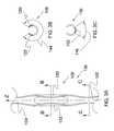
- FIG. 1is an isometric view showing a body that may be used to form an ocular implant in accordance with one exemplary embodiment of the invention.
- the bodycomprises a first spine, a second spine, and a first frame disposed between the first spine and the second spine.
- the first framecomprises a first strut and a second strut.
- FIG. 2is an isometric view of the body shown in the previous figure.
- the bodyis shaped to form an ocular implant having an outer surface defining a generally cylindrical volume.
- An inner surface of the bodydefines an elongate channel.
- the ocular implantmay be inserted into Schlemm's canal of a human eye to facilitate the flow of aqueous humor out of the anterior chamber.
- FIG. 3Ais a plan view showing a portion of the ocular implant shown in the previous figure.
- the ocular implantincludes a first frame comprising a first strut and a second strut.
- each strutundulates in a circumferential direction while, at the same time, extending in the axial direction Z between a first spine and a second spine.
- FIG. 3Bis a lateral cross-sectional view of the ocular implant shown in the previous figure.
- Section line B-Bintersects the first strut and second strut of the ocular implant at the point where the circumferential undulation of these struts is at it's maximum.
- These strutsform a frame having circumferential extent that is illustrated using dimension lines in FIG. 3B .
- FIG. 3Cis a lateral cross-sectional view of the ocular implant of FIG. 3A taken along section line C-C.
- Section line C-Cintersects a spine of the ocular implant at the point where the width of the spine is at a minimum.
- a circumferential extent of the spineillustrated using dimension lines in FIG. 3C .
- the circumferential extent of frameis greater than the circumferential extent of the spine.
- FIG. 4is an isometric view showing a portion of the ocular implant shown in the previous figure.
- the outer surfaces of the first spine, the second spine, the first strut, and the second strutdefine a generally cylindrical volume V.
- the shape of the ocular implantmay be described using the cylindrical coordinates shown in FIG. 4 .
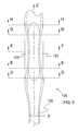
- FIG. 5is an enlarged plan view showing a portion of the ocular implant shown in the previous figure.
- a number of section linesare shown crossing the first strut and the second strut of the ocular implant.
- each strutundulates in a circumferential direction while, at the same time, extending in axial direction Z between the first spine and the second spine.
- the circumferential undulation of the first strutis illustrated in FIG. 6 using lateral cross-sectional drawings labeled with cylindrical coordinates.
- FIG. 6A through 6Eare lateral cross-sectional views of the ocular implant shown in the previous figure. These cross-sectional views correspond to the section lines shown in the previous figure. With reference to these cross-sectional views, it will be appreciated that the angular dimension ⁇ associated with a first edge of the first strut undulates as the first strut extends in an axial direction Z between the first spine and the second spine. In the embodiment of FIG. 6 , the radius r of the outer surface of the first strut remains substantially constant as the first strut extends in the axial direction Z between the first spine and the second spine.
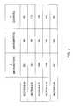
- FIG. 7shows a plurality of cylindrical coordinate values corresponding with the cross-sectional views shown in the previous figure.
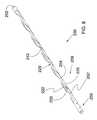
- FIG. 8is an isometric view of an ocular implant in accordance with an additional exemplary embodiment of the invention.
- FIG. 9is a plan view of the ocular implant shown in the previous figure.
- the ocular implanthas an at rest shape that is generally curved.
- FIG. 10shows the ocular implant of the previous figure in place within a human eye.
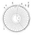
- FIG. 11is an enlarged plan view showing a portion of the eye shown in the previous figure.
- FIG. 1is an isometric view showing a body 100 that may be used to form an ocular implant in accordance with one exemplary embodiment of the invention.
- Body 100comprises a first spine 102 , a second spine 104 , and a first frame 106 disposed between first spine 102 and second spine 104 .
- first frame 106comprises a first strut 120 and a second strut 122 .
- each strutextends between first spine 102 and second spine 104 .
- First strut 120 of first frame 106comprises a first edge 124 A and a second edge 126 A.
- second strut 122has a shape that is a mirror image of the shape of first strut 120 .
- Second strut 122comprises a first edge 124 B and a second edge 126 B.
- Second edge 126 B of second strut 122 and second edge 126 A of first strut 120define a second opening 130 .
- Second opening 130generally divides first frame 106 into first strut 120 and second strut 122 .
- body 100comprises a plurality of spines and a plurality of frames.
- these spines and framesare arranged in an ABAB pattern.
- Each spinehas a first lateral extent 132 and each frame has a second lateral extent 134 .
- second lateral extent 134is greater than first lateral extent 132 .
- FIG. 2is an isometric view of body 100 shown in the previous figure.
- body 100is shaped to form an ocular implant 136 having an outer surface 138 defining a generally cylindrical volume.
- An inner surface 140 of body 100defines an elongate channel 142 .
- Ocular implant 136may be inserted into Schlemm's canal of a human eye to facilitate the flow of aqueous humor out of the anterior chamber. This flow may include axial flow along Schlemm's canal, flow from the anterior chamber into Schlemm's canal, and flow leaving Schlemm's canal via outlets communicating with Schlemm's canal.
- ocular implant 136When in place within the eye, ocular implant 136 will support trabecular mesh tissue and Schlemm's canal tissue and will provide for improved communication between the anterior chamber and Schlemm's canal (via the trabecular meshwork) and between pockets or compartments along Schlemm's canal.
- Elongate channel 142 of ocular implant 136fluidly communicates with a first opening 128 as well as inlet portion 101 .
- Various fabrication techniquesmay be used to fabricate ocular implant 136 .
- ocular implant 136can be fabricated by providing a generally flat sheet of material and laser cutting the sheet of material to form body 100 shown in FIG. 1 . The body 100 may then be formed into a generally tubular shape as shown in FIG. 2 . Any adjoining edges (such as edges 103 ) may be, optionally, welded.
- ocular implant 136may be fabricated by providing a tube and laser cutting openings in the tube to form the shape shown in FIG. 2 .
- ocular implant 136comprises a first spine 102 and a second spine 104 .
- a first frame 106 of ocular implant 136is disposed between first spine 102 and second spine 104 .
- first frame 106comprises a first strut 120 that extends between first spine 102 and second spine 104 .
- First frame 106also comprises a second strut 122 .
- Second strut 122also extends between first spine 102 and second spine 104
- First strut 120 of first frame 106comprises a first edge 124 A and a second edge 126 A. As shown in FIG. 1 , first edge 124 A has a convex surface, and second edge 126 A has a concave surface. Second strut 122 has a shape that is a mirror image of the shape of first strut 120 . In FIG. 2 , first opening 128 of ocular implant 136 can be seen extending between first edge 124 A of first strut 120 and a first edge 124 B of second strut 122 . A second edge 126 B of second strut 122 and second edge 126 A of first strut 120 define a second opening 130 .
- Second opening 130 and additional openings (e.g., first opening 128 ) defined by ocular implant 136allow aqueous humor to flow laterally across and/or laterally through ocular implant 136 .
- openings 128 and 130are shorter than the opening of elongate channel 142 extending along one side of implant 136 .
- Ocular implant 136can be fabricated from various biocompatible materials possessing the necessary structural and mechanical attributes. Both metallic and non-metallic materials may be suitable. Examples of metallic materials include stainless steel, tantalum, gold, titanium, and nickel-titanium alloys known in the art as Nitinol. Nitinol is commercially available from Memry Technologies (Brookfield, Conn.), TiNi Alloy Company (San Leandro, Calif.), and Shape Memory Applications (Sunnyvale, Calif.).
- Ocular implant 136may include one or more therapeutic agents.
- One or more therapeutic agentsmay, for example, be incorporated into a polymeric coating that is deposited onto the outer surfaces of the struts and spines of the ocular implant.
- the therapeutic agentmay comprise, for example, an anti-glaucoma drug.
- anti-glaucoma drugsinclude prostaglandin analogs.
- prostaglandin analogsinclude latanprost.
- FIG. 3Ais a plan view showing a portion of ocular implant 136 shown in the previous figure.
- Body 100 of ocular implant 136comprises a first spine 102 , a second spine 104 , and a first frame 106 disposed between first spine 102 and second spine 104 .
- first frame 106comprises a first strut 120 and a second strut 122 . As shown, each strut undulates in a circumferential direction while, at the same time, extending in the axial direction Z between first spine 102 and second spine 104 .
- FIG. 3Bis a lateral cross-sectional view of ocular implant 136 taken along section line B-B.
- Section line B-Bintersects first strut 120 and second strut 122 at the point where the circumferential undulation of these struts is at its maximum.
- First strut 120 and second strut 122form first frame 106 .
- First frame 106has a first circumferential extent 144 in the embodiment of FIG. 3B .
- FIG. 3Cis a lateral cross-sectional view of ocular implant 136 taken along section line C-C.
- Section line C-Cintersects first spine 102 at the point where the width of first spine 102 is at a minimum.
- first spine 102has a second circumferential extent 146 .
- Second circumferential extent 146 of first spine 102is illustrated using dimension lines in FIG. 3C .
- first circumferential extent 144 of first frame 106is greater than second circumferential extent 146 of first spine 102 .
- FIG. 4is an isometric view showing a portion of ocular implant 136 shown in the previous figure.
- the shape of ocular implant 136may be described using the cylindrical coordinates shown in FIG. 4 . These cylindrical coordinates include a radius r, an angle ⁇ and an axial dimension Z. Cylindrical coordinates may be conceptualized as an extension of two dimensional polar coordinates to include a longitudinal or axial dimension Z. The two dimensions of a typical polar coordinate system are radius r and angle ⁇ . In the embodiment of FIG. 4 , dimension Z extends along a longitudinal axis 148 of cylindrical volume V.
- first strut 120extends in axial direction Z between first spine 102 and second spine 104 .
- Second strut 122also extends between first spine 102 and second spine 104 .
- the radius r of the outer surface of each strutremains substantially constant.
- the angular dimension ⁇ of a first edge 124 A of first strutvaries as first strut 120 extends in the axial direction Z between first spine 102 and second spine 104 .
- the angular dimension ⁇ of a second edge 126 A of second strutvaries as second strut 122 extends in the axial direction Z between first spine 102 and second spine 104 .
- FIG. 5is an enlarged plan view showing a portion of ocular implant 136 shown in the previous figure.
- a number of section linesare shown crossing first strut 120 and second strut 122 of ocular implant 136 .
- each strutundulates in a circumferential direction while, at the same time, extending in axial direction Z between first spine 102 and second spine 104 .
- the circumferential undulation of first strut 120is illustrated in the next figure using lateral cross-sectional drawings labeled with cylindrical coordinates.
- FIGS. 6A through 6Eare lateral cross-sectional views of ocular implant 136 shown in the previous figure. These cross-sectional views correspond to the section lines shown in the previous figure. With reference to these cross-sectional views, it will be appreciated that the angular dimension ⁇ associated with first edge 124 A of first strut 120 undulates as first strut 120 extends in an axial direction Z between the first spine and the second spine. In the embodiment of FIG. 6 , the radius r of the outer surface of first strut 120 remains substantially constant as first strut 120 extends in axial direction Z between the first spine and the second spine.
- FIG. 7shows a plurality of cylindrical coordinate values corresponding with the cross-sectional views shown in the previous figure.
- the numerical value of angular dimension ⁇ of first edge 124first increases, then decreases, as first strut 120 extends in an axial direction Z between the first spine and the second spine.
- the numerical value rremains constant as first strut 120 extends in axial direction Z between the first spine and the second spine.
- FIG. 8is an isometric view of an ocular implant 236 in accordance with an additional exemplary embodiment of the invention.
- ocular implant 236comprises a first spine 202 and a second spine 204 .
- a first frame 206 of ocular implant 236is disposed between first spine 202 and second spine 204 .
- first frame 206comprises a first strut 220 that extends between first spine 202 and second spine 204 .
- First frame 206also comprises a second strut 222 .
- second strut 222also extends between first spine 202 and second spine 204 .
- Ocular implant 236 of FIG. 8defines a channel 242 that opens into a first opening 228 .
- first opening 228 of ocular implant 236can be seen extending between first strut 220 and second strut 222 .
- First strut 220 and second strut 222also define a second opening 230 .
- First opening 228 , second opening 230 , and the additional openings shown in FIG. 8allow aqueous humor to flow laterally across and/or laterally through ocular implant 236 .
- an inlet portion 250is formed near a proximal end of ocular implant 236 .
- Inlet portion 250may extend through the trabecular meshwork into the anterior chamber of the eye when a portion of the ocular implant lies in Schlemm's canal.
- blunt tip 252is disposed at a distal end of ocular implant 236 .
- blunt tip 252has a generally rounded shape.
- blunt tip 252has a generally hemispherical shape. The generally rounded shape of blunt tip 252 may increase the likelihood that body 200 will track Schlemm's canal as ocular implant 236 is advanced into the canal during an implant procedure.
- ocular implant 236is pictured assuming a generally straight shape.
- Embodiments of ocular implant 236are possible which have a generally curved resting shape.
- Ocular implant 236may be fabricated, for example, by laser cutting a tube to create the shape shown in FIG. 8 . When this is the case, it may be desirable to rotate a straight tubular workpiece during the laser cutting process. After the laser cutting process, the ocular implant can be heat-set so that the ocular implant is biased to assume a selected shape (e.g., a generally curved shape).
- FIG. 9is a plan view of ocular implant 236 shown in the previous figure.
- ocular implant 236has an at rest shape that is generally curved. This at rest shape can be established, for example, using a heat-setting process.
- the ocular implant shape shown in FIG. 9includes a distal radius RA, a proximal radius RC, and an intermediate radius RB.
- distal radius RAis larger than both intermediate radius RB and proximal radius RC.
- intermediate radius RBis larger than proximal radius RC and smaller than distal radius RA.
- distal radius RAis about 0.310 inches
- intermediate radius RBis about 0.215 inches
- proximal radius RCis about 0.105 inches.
- a distal portion of the ocular implantfollows distal radius RA along an arc extending across an angle AA.
- a proximal portion of the ocular implantfollows proximal radius RC along an arc extending across an angle AC.
- An intermediate portion of the ocular implantis disposed between the proximal portion and the distal portion. The intermediate portion follows radius RB and extends across an angle AB.
- angle AAis about 55 degrees
- angle ABis about 79 degrees
- angle ACis about 60 degrees.
- Ocular implant 236may be used in conjunction with a method of treating a patient. Some such methods may include the step of inserting a core member into a lumen defined by ocular implant 236 .
- the core membermay comprise, for example, a wire or tube.
- the distal end of the ocular implantmay be inserted into Schlemm's canal.
- the ocular implant and the core membermay then be advanced into Schlemm's canal until the ocular implant has reached a desired position.
- the core membermay then be withdrawn from the ocular implant.
- FIG. 10shows ocular implant 236 of the previous figure in place within a human eye.
- the eye of FIG. 10includes an anterior chamber that is covered by a cornea.
- the iris of the eyeis visible through the cornea and the anterior chamber.
- the anterior chamberis filled with aqueous humor which helps maintain the generally hemispherical shape of the cornea.
- the cornea and the lenscan include no blood vessels. Accordingly, no blood flows through the cornea and the lens to provide nutrition to these tissues and to remove wastes from these tissues. Instead, these functions are performed by the aqueous humor.
- a continuous flow of aqueous humor through the eyeprovides nutrition to portions of the eye (e.g., the cornea and the lens) that have no blood vessels. This flow of aqueous humor also removes waste from these tissues.
- Aqueous humoris produced by an organ known as the ciliary body.
- the ciliary bodyincludes epithelial cells that continuously secrete aqueous humor.
- a stream of aqueous humorflows out of the eye as new aqueous humor is secreted by the epithelial cells of the ciliary body. This excess aqueous humor enters the blood stream and is carried away by venous blood leaving the eye.
- the structures that drain aqueous humor from the anterior chamberinclude Schlemm's canal and a large number of veins that communicate with Schlemm's canal via a plurality of outlets.
- Schlemm's canal 20can be seen encircling the iris of the eye.
- Ocular implant 236may be inserted into Schlemm's canal 20 to facilitate the flow of aqueous humor out of the anterior chamber. This flow may include axial flow along Schlemm's canal, flow from the anterior chamber into Schlemm's canal, and flow leaving Schlemm's canal via outlets communicating with Schlemm's canal.
- ocular implant 236When in place within the eye, ocular implant 236 will support trabecular mesh tissue and Schlemm's canal tissue and will provide for improved communication between the anterior chamber and Schlemm's canal (via the trabecular meshwork) and between pockets or compartments along Schlemm's canal.
- FIG. 11is an enlarged plan view showing a portion of the eye shown in the previous figure.
- ocular implant 236extends through Schlemm's canal 20 across an angle G.
- Various implant sizesare possible, and different implant sizes may span a different angle G when placed in Schlemm's canal. Examples of angular spans that may be suitable in some applications include 60°, 90°, 150° and 180°.
- an inlet portion 250 of ocular implant 236is shown extending through trabecular mesh 22 .
- Aqueous humormay exit anterior chamber 24 and enter Schlemm's canal 20 by flowing through inlet portion 250 of ocular implant 236 .
- Aqueous humormay also exit anterior chamber 24 and enter Schlemm's canal 20 by flowing through the trabecular mesh 22 of the eye.
- the spines of ocular implant 236support trabecular mesh 22 .
- each outlet from Schlemm's canalmay drain only a portion of Schlemm's canal.
- This conditionmay be improved by placing ocular implant 236 in Schlemm's canal.
- Ocular implant 236 shown in FIG. 11includes a plurality of struts, spines and openings. When in place within the eye, ocular implant 236 will support trabecular mesh tissue and Schlemm's canal tissue and will provide for improved communication between the anterior chamber and Schlemm's canal and between pockets or compartments along Schlemm's canal.
- first opening 228 of ocular implant 236is shown facing radially outward in Schlemm's canal 20 .
- Aqueous humorcan exit Schlemm's canal 20 by flowing through outlets that radiate away from and communicate with Schlemm's canal 20 . After flowing through these outlets, this excess aqueous humor can enter the venous bloodstream be carried out of the eye by venous blood flow.
- the diameter of ocular implant 236can range from 0.005 inches to 0.04 inches, preferably from 0.005 inches to 0.02 inches, in order to lie within and support Schlemm's canal.
Landscapes
- Health & Medical Sciences (AREA)
- Ophthalmology & Optometry (AREA)
- Life Sciences & Earth Sciences (AREA)
- Public Health (AREA)
- Engineering & Computer Science (AREA)
- Biomedical Technology (AREA)
- Heart & Thoracic Surgery (AREA)
- Vascular Medicine (AREA)
- Veterinary Medicine (AREA)
- Animal Behavior & Ethology (AREA)
- General Health & Medical Sciences (AREA)
- Surgery (AREA)
- Nuclear Medicine, Radiotherapy & Molecular Imaging (AREA)
- Cardiology (AREA)
- Oral & Maxillofacial Surgery (AREA)
- Transplantation (AREA)
- Anesthesiology (AREA)
- Hematology (AREA)
- Prostheses (AREA)
Abstract
Description
Claims (19)
Priority Applications (1)
| Application Number | Priority Date | Filing Date | Title |
|---|---|---|---|
| US13/763,394US9402767B2 (en) | 2007-09-24 | 2013-02-08 | Ocular implant architectures |
Applications Claiming Priority (5)
| Application Number | Priority Date | Filing Date | Title |
|---|---|---|---|
| US11/860,318US7740604B2 (en) | 2007-09-24 | 2007-09-24 | Ocular implants for placement in schlemm's canal |
| US3374608P | 2008-03-04 | 2008-03-04 | |
| US12/236,254US20090082862A1 (en) | 2007-09-24 | 2008-09-23 | Ocular Implant Architectures |
| US13/366,073US8372026B2 (en) | 2007-09-24 | 2012-02-03 | Ocular implant architectures |
| US13/763,394US9402767B2 (en) | 2007-09-24 | 2013-02-08 | Ocular implant architectures |
Related Parent Applications (1)
| Application Number | Title | Priority Date | Filing Date |
|---|---|---|---|
| US13/366,073ContinuationUS8372026B2 (en) | 2007-09-24 | 2012-02-03 | Ocular implant architectures |
Publications (2)
| Publication Number | Publication Date |
|---|---|
| US20130150959A1 US20130150959A1 (en) | 2013-06-13 |
| US9402767B2true US9402767B2 (en) | 2016-08-02 |
Family
ID=40472564
Family Applications (3)
| Application Number | Title | Priority Date | Filing Date |
|---|---|---|---|
| US12/236,254AbandonedUS20090082862A1 (en) | 2007-09-24 | 2008-09-23 | Ocular Implant Architectures |
| US13/366,073ActiveUS8372026B2 (en) | 2007-09-24 | 2012-02-03 | Ocular implant architectures |
| US13/763,394Active2028-05-19US9402767B2 (en) | 2007-09-24 | 2013-02-08 | Ocular implant architectures |
Family Applications Before (2)
| Application Number | Title | Priority Date | Filing Date |
|---|---|---|---|
| US12/236,254AbandonedUS20090082862A1 (en) | 2007-09-24 | 2008-09-23 | Ocular Implant Architectures |
| US13/366,073ActiveUS8372026B2 (en) | 2007-09-24 | 2012-02-03 | Ocular implant architectures |
Country Status (1)
| Country | Link |
|---|---|
| US (3) | US20090082862A1 (en) |
Cited By (22)
| Publication number | Priority date | Publication date | Assignee | Title |
|---|---|---|---|---|
| US9855167B2 (en) | 2012-03-20 | 2018-01-02 | Sight Sciences, Inc. | Ocular delivery systems and methods |
| US10159601B2 (en) | 2000-05-19 | 2018-12-25 | Ivantis, Inc. | Delivery system and method of use for the eye |
| US10299958B2 (en) | 2015-03-31 | 2019-05-28 | Sight Sciences, Inc. | Ocular delivery systems and methods |
| US10314742B2 (en) | 2006-06-26 | 2019-06-11 | Sight Sciences, Inc. | Intraocular implants and methods and kits therefor |
| US10406030B2 (en) | 2010-02-05 | 2019-09-10 | Sight Sciences, Inc. | Intraocular implants and related kits and methods |
| US10492949B2 (en) | 2009-07-09 | 2019-12-03 | Ivantis, Inc. | Single operator device for delivering an ocular implant |
| US10537474B2 (en) | 2008-03-05 | 2020-01-21 | Ivantis, Inc. | Methods and apparatus for treating glaucoma |
| US10709547B2 (en) | 2014-07-14 | 2020-07-14 | Ivantis, Inc. | Ocular implant delivery system and method |
| US11197779B2 (en) | 2015-08-14 | 2021-12-14 | Ivantis, Inc. | Ocular implant with pressure sensor and delivery system |
| US11504270B1 (en) | 2019-09-27 | 2022-11-22 | Sight Sciences, Inc. | Ocular delivery systems and methods |
| US11540940B2 (en) | 2021-01-11 | 2023-01-03 | Alcon Inc. | Systems and methods for viscoelastic delivery |
| US11596546B2 (en) | 2009-07-09 | 2023-03-07 | Alcon Inc. | Ocular implants and methods for delivering ocular implants into the eye |
| US11712369B2 (en) | 2012-11-28 | 2023-08-01 | Alcon Inc. | Apparatus for delivering ocular implants into an anterior chamber of the eye |
| US11744734B2 (en) | 2007-09-24 | 2023-09-05 | Alcon Inc. | Method of implanting an ocular implant |
| US11938058B2 (en) | 2015-12-15 | 2024-03-26 | Alcon Inc. | Ocular implant and delivery system |
| US11992437B2 (en) | 2012-04-18 | 2024-05-28 | Alcon Inc. | Ocular implants for delivery into an anterior chamber of the eye |
| US12029683B2 (en) | 2018-02-22 | 2024-07-09 | Alcon Inc. | Ocular implant and delivery system |
| US12076273B2 (en) | 2011-12-19 | 2024-09-03 | Alcon Inc. | Delivering ocular implants into the eye |
| US12083044B2 (en) | 2019-07-10 | 2024-09-10 | Aquea Health, Inc. | Eye stents and delivery systems |
| US12226309B2 (en) | 2013-03-15 | 2025-02-18 | Alcon Inc. | Intraocular lens storage and loading devices and methods of use |
| US12318279B2 (en) | 2007-07-23 | 2025-06-03 | Alcon Inc. | Lens delivery system |
| US12440377B2 (en) | 2024-11-13 | 2025-10-14 | Aquea Health, Inc. | Eye stents and delivery systems and methods |
Families Citing this family (25)
| Publication number | Priority date | Publication date | Assignee | Title |
|---|---|---|---|---|
| KR20020035476A (en) | 1999-04-26 | 2002-05-11 | 지엠피 비젼 솔루션즈 인코포레이티드 | Shunt device and method for treating glaucoma |
| US7867186B2 (en) | 2002-04-08 | 2011-01-11 | Glaukos Corporation | Devices and methods for treatment of ocular disorders |
| US6638239B1 (en) | 2000-04-14 | 2003-10-28 | Glaukos Corporation | Apparatus and method for treating glaucoma |
| AU2002258754B2 (en) | 2001-04-07 | 2006-08-17 | Glaukos Corporation | Glaucoma stent and methods thereof for glaucoma treatment |
| US7431710B2 (en) | 2002-04-08 | 2008-10-07 | Glaukos Corporation | Ocular implants with anchors and methods thereof |
| US7331984B2 (en) | 2001-08-28 | 2008-02-19 | Glaukos Corporation | Glaucoma stent for treating glaucoma and methods of use |
| US7740604B2 (en)* | 2007-09-24 | 2010-06-22 | Ivantis, Inc. | Ocular implants for placement in schlemm's canal |
| US8734377B2 (en) | 2007-09-24 | 2014-05-27 | Ivantis, Inc. | Ocular implants with asymmetric flexibility |
| US20090082862A1 (en) | 2007-09-24 | 2009-03-26 | Schieber Andrew T | Ocular Implant Architectures |
| US8512404B2 (en) | 2007-11-20 | 2013-08-20 | Ivantis, Inc. | Ocular implant delivery system and method |
| US8808222B2 (en) | 2007-11-20 | 2014-08-19 | Ivantis, Inc. | Methods and apparatus for delivering ocular implants into the eye |
| CN102238926B (en)* | 2008-12-05 | 2015-09-16 | 伊万提斯公司 | Methods and devices for delivering ocular implants into the eye |
| CN102647960A (en)* | 2009-10-23 | 2012-08-22 | 伊万提斯公司 | Ocular implant system and method |
| US8545430B2 (en) | 2010-06-09 | 2013-10-01 | Transcend Medical, Inc. | Expandable ocular devices |
| US9510973B2 (en) | 2010-06-23 | 2016-12-06 | Ivantis, Inc. | Ocular implants deployed in schlemm's canal of the eye |
| US8657776B2 (en) | 2011-06-14 | 2014-02-25 | Ivantis, Inc. | Ocular implants for delivery into the eye |
| US8765210B2 (en) | 2011-12-08 | 2014-07-01 | Aquesys, Inc. | Systems and methods for making gelatin shunts |
| CA2868341C (en) | 2012-03-26 | 2021-01-12 | Glaukos Corporation | System and method for delivering multiple ocular implants |
| US9125723B2 (en) | 2013-02-19 | 2015-09-08 | Aquesys, Inc. | Adjustable glaucoma implant |
| US10159600B2 (en) | 2013-02-19 | 2018-12-25 | Aquesys, Inc. | Adjustable intraocular flow regulation |
| US10517759B2 (en) | 2013-03-15 | 2019-12-31 | Glaukos Corporation | Glaucoma stent and methods thereof for glaucoma treatment |
| EP3068354B1 (en) | 2013-11-14 | 2023-06-28 | Aquesys, Inc. | Intraocular shunt inserter |
| EP3677229A1 (en) | 2014-05-29 | 2020-07-08 | Glaukos Corporation | Implants with controlled drug delivery features |
| US11925578B2 (en) | 2015-09-02 | 2024-03-12 | Glaukos Corporation | Drug delivery implants with bi-directional delivery capacity |
| US11116625B2 (en) | 2017-09-28 | 2021-09-14 | Glaukos Corporation | Apparatus and method for controlling placement of intraocular implants |
Citations (246)
| Publication number | Priority date | Publication date | Assignee | Title |
|---|---|---|---|---|
| US703296A (en) | 1901-06-15 | 1902-06-24 | Arnold Nueesch | Cattle-probe. |
| US1601709A (en) | 1924-01-28 | 1926-10-05 | Anderson Windom Edward | Retainable needle construction for syringes |
| US2716983A (en) | 1952-10-08 | 1955-09-06 | Abbott Lab | Piercing needle |
| US3071135A (en) | 1960-01-27 | 1963-01-01 | Mfg Process Lab Inc | Hollow needle |
| US3788327A (en) | 1971-03-30 | 1974-01-29 | H Donowitz | Surgical implant device |
| US3811442A (en) | 1972-03-23 | 1974-05-21 | A Maroth | Hypodermic syringe holder and applicator |
| US3948271A (en) | 1972-11-07 | 1976-04-06 | Taichiro Akiyama | Drain for the eardrum and apparatus for introducing the same |
| US4037604A (en) | 1976-01-05 | 1977-07-26 | Newkirk John B | Artifical biological drainage device |
| US4428746A (en) | 1981-07-29 | 1984-01-31 | Antonio Mendez | Glaucoma treatment device |
| US4457757A (en) | 1981-07-20 | 1984-07-03 | Molteno Anthony C B | Device for draining aqueous humour |
| US4601713A (en) | 1985-06-11 | 1986-07-22 | Genus Catheter Technologies, Inc. | Variable diameter catheter |
| US4689040A (en) | 1985-04-29 | 1987-08-25 | Thompson Robert J | Tip for a phacoemulsification needle |
| US4699140A (en) | 1985-07-10 | 1987-10-13 | Iolab Corporation | Instrument for inserting an intraocular lens |
| US4706669A (en) | 1984-01-30 | 1987-11-17 | Schlegel Hans Joachim | Device for perforating the lens capsule front wall in the eye of living beings |
| US4722724A (en) | 1986-06-23 | 1988-02-02 | Stanley Schocket | Anterior chamber tube shunt to an encircling band, and related surgical procedure |
| US4733665A (en) | 1985-11-07 | 1988-03-29 | Expandable Grafts Partnership | Expandable intraluminal graft, and method and apparatus for implanting an expandable intraluminal graft |
| US4750901A (en) | 1986-03-07 | 1988-06-14 | Molteno Anthony C B | Implant for drainage of aqueous humour |
| US4826478A (en) | 1986-06-23 | 1989-05-02 | Stanley Schocket | Anterior chamber tube shunt to an encircling band, and related surgical procedure |
| US4861341A (en) | 1988-07-18 | 1989-08-29 | Woodburn Robert T | Subcutaneous venous access device and needle system |
| US4880000A (en) | 1987-12-15 | 1989-11-14 | Iolab Corporation | Lens insertion instrument |
| US4886488A (en) | 1987-08-06 | 1989-12-12 | White Thomas C | Glaucoma drainage the lacrimal system and method |
| US4919130A (en) | 1986-11-07 | 1990-04-24 | Nestle S.A. | Tool for inserting compressible intraocular lenses into the eye and method |
| US4934809A (en) | 1988-06-24 | 1990-06-19 | Volk Donald A | Lens positioning device for indirect biomicroscopy of the eye |
| US4934363A (en) | 1987-12-15 | 1990-06-19 | Iolab Corporation | Lens insertion instrument |
| US4936825A (en) | 1988-04-11 | 1990-06-26 | Ungerleider Bruce A | Method for reducing intraocular pressure caused by glaucoma |
| US4946436A (en) | 1989-11-17 | 1990-08-07 | Smith Stewart G | Pressure-relieving device and process for implanting |
| US4968296A (en) | 1989-12-20 | 1990-11-06 | Robert Ritch | Transscleral drainage implant device for the treatment of glaucoma |
| US5092837A (en) | 1989-12-20 | 1992-03-03 | Robert Ritch | Method for the treatment of glaucoma |
| US5127901A (en) | 1990-05-18 | 1992-07-07 | Odrich Ronald B | Implant with subconjunctival arch |
| US5178604A (en) | 1990-05-31 | 1993-01-12 | Iovision, Inc. | Glaucoma implant |
| US5180362A (en) | 1990-04-03 | 1993-01-19 | Worst J G F | Gonio seton |
| US5190552A (en) | 1992-02-04 | 1993-03-02 | Kelman Charles D | Slotted tube injector for an intraocular lens |
| US5213569A (en) | 1992-03-31 | 1993-05-25 | Davis Peter L | Tip for a tissue phacoemulsification device |
| DE4226476C1 (en) | 1992-08-10 | 1993-08-12 | Hans Dr.Med. 3015 Wennigsen De Haindl | |
| US5246452A (en) | 1992-04-13 | 1993-09-21 | Impra, Inc. | Vascular graft with removable sheath |
| US5290267A (en) | 1991-01-17 | 1994-03-01 | Fresenius Ag | Hypodermic needle |
| US5360399A (en) | 1992-01-10 | 1994-11-01 | Robert Stegmann | Method and apparatus for maintaining the normal intraocular pressure |
| US5445637A (en) | 1993-12-06 | 1995-08-29 | American Cyanamid Company | Method and apparatus for preventing posterior capsular opacification |
| US5454796A (en) | 1991-04-09 | 1995-10-03 | Hood Laboratories | Device and method for controlling intraocular fluid pressure |
| US5458615A (en) | 1993-07-06 | 1995-10-17 | Advanced Cardiovascular Systems, Inc. | Stent delivery system |
| US5536259A (en) | 1995-07-28 | 1996-07-16 | Medisystems Technology Corp | Hypodermic cannula |
| US5575780A (en) | 1995-04-28 | 1996-11-19 | Saito; Yoshikuni | Medical hollow needle and a method of producing thereof |
| US5591223A (en) | 1992-11-23 | 1997-01-07 | Children's Medical Center Corporation | Re-expandable endoprosthesis |
| US5613972A (en) | 1992-07-15 | 1997-03-25 | The University Of Miami | Surgical cutting heads with curled cutting wings |
| US5626558A (en) | 1995-05-05 | 1997-05-06 | Suson; John | Adjustable flow rate glaucoma shunt and method of using same |
| US5653753A (en) | 1994-04-29 | 1997-08-05 | Allergan | Method and apparatus for folding of intraocular lenses |
| US5676669A (en) | 1993-04-30 | 1997-10-14 | Colvard; Michael | Intraocular capsular shield |
| JPH10504978A (en) | 1994-09-01 | 1998-05-19 | ユニバーシティ オブ マイアミ | Adjustable keratoplasty syringe with gel injection |
| US5792099A (en) | 1995-02-14 | 1998-08-11 | Decamp; Dennis | Syringe and cannula for insertion of viscoelastic material into an eye and method of using same |
| US5807302A (en) | 1996-04-01 | 1998-09-15 | Wandel; Thaddeus | Treatment of glaucoma |
| US5865831A (en) | 1996-04-17 | 1999-02-02 | Premier Laser Systems, Inc. | Laser surgical procedures for treatment of glaucoma |
| US5868697A (en) | 1995-05-14 | 1999-02-09 | Optonol Ltd. | Intraocular implant |
| US5879319A (en) | 1994-06-22 | 1999-03-09 | Chauvin Opsia | Sclerotomy implant |
| US5893837A (en) | 1997-02-28 | 1999-04-13 | Staar Surgical Company, Inc. | Glaucoma drain implanting device and method |
| JPH11123205A (en) | 1997-08-15 | 1999-05-11 | Grieshaber & Co Ag Schatzhausen | Device for improving drainage of aqueous humor in eyeball |
| US5919171A (en) | 1994-08-03 | 1999-07-06 | Kanegafuchi Kagaku Kogyo Kabushiki Kaisha | Microcatheter |
| US5948427A (en) | 1996-04-25 | 1999-09-07 | Point Medical Corporation | Microparticulate surgical adhesive |
| US5968058A (en) | 1996-03-27 | 1999-10-19 | Optonol Ltd. | Device for and method of implanting an intraocular implant |
| US6007511A (en) | 1991-05-08 | 1999-12-28 | Prywes; Arnold S. | Shunt valve and therapeutic delivery system for treatment of glaucoma and methods and apparatus for its installation |
| WO2000007525A1 (en) | 1998-08-05 | 2000-02-17 | Keravision, Inc. | Corneal implant with migration preventer |
| DE19840047A1 (en) | 1998-09-02 | 2000-03-23 | Thomas Neuhann | Device for the targeted improvement and / or permanent guarantee of the permeability for eye chamber water through the trabecular mechanism in the Schlemm canal |
| US6050970A (en) | 1997-05-08 | 2000-04-18 | Pharmacia & Upjohn Company | Method and apparatus for inserting a glaucoma implant in an anterior and posterior segment of the eye |
| US6102045A (en) | 1994-07-22 | 2000-08-15 | Premier Laser Systems, Inc. | Method and apparatus for lowering the intraocular pressure of an eye |
| WO2000064389A1 (en) | 1999-04-26 | 2000-11-02 | Lynch Mary G | Trabeculotomy device and method for treating glaucoma |
| US6186974B1 (en) | 1997-01-10 | 2001-02-13 | University College London And Moorfields Eye Hospital Nhs Trust | Device for use in the eye |
| US6217584B1 (en) | 1996-05-09 | 2001-04-17 | Aharon Lehrer | Method and a system for performing cataract surgery |
| US6221078B1 (en) | 1999-06-25 | 2001-04-24 | Stephen S. Bylsma | Surgical implantation apparatus |
| US6238409B1 (en) | 1997-03-10 | 2001-05-29 | Johnson & Johnson Interventional Systems Co. | Articulated expandable intraluminal stent |
| US20010002438A1 (en) | 1994-12-22 | 2001-05-31 | Ivan Sepetka | Implant delivery assembly with expandable coupling/decoupling mechanism |
| US6241721B1 (en) | 1998-10-09 | 2001-06-05 | Colette Cozean | Laser surgical procedures for treatment of glaucoma |
| USD444874S1 (en) | 2000-07-31 | 2001-07-10 | Allergan Sales, Inc. | Self instill twist housing eye drop dispenser |
| US6328747B1 (en) | 1996-05-09 | 2001-12-11 | Itos Innovative Technology In Ocular Surgery, Ltd. | Method and a system for performing cataract surgery |
| WO2001097727A1 (en) | 2000-06-19 | 2001-12-27 | Glaukos Corporation | Stented trabecular shunt and methods thereof |
| US20020003546A1 (en) | 2000-05-31 | 2002-01-10 | Agency Of Industrial Science And Technology | Virtual shape generation method and device using the same |
| US20020013572A1 (en) | 2000-05-19 | 2002-01-31 | Berlin Michael S. | Delivery system and method of use for the eye |
| US6375642B1 (en) | 2000-02-15 | 2002-04-23 | Grieshaber & Co. Ag Schaffhausen | Method of and device for improving a drainage of aqueous humor within the eye |
| US20020052653A1 (en) | 1998-07-06 | 2002-05-02 | Russell Durgin | Implant system and method for bulking tissue |
| WO2002036052A1 (en) | 2000-11-01 | 2002-05-10 | Glaukos Corporation | Glaucoma treatment device |
| US20020072673A1 (en) | 1999-12-10 | 2002-06-13 | Yamamoto Ronald K. | Treatment of ocular disease |
| US6409752B1 (en) | 1993-07-23 | 2002-06-25 | Cook Incorporated | Flexible stent having a pattern formed from a sheet of material |
| US20020133168A1 (en) | 2001-03-16 | 2002-09-19 | Smedley Gregory T. | Applicator and methods for placing a trabecular shunt for glaucoma treatment |
| US20020143284A1 (en) | 2001-04-03 | 2002-10-03 | Hosheng Tu | Drug-releasing trabecular implant for glaucoma treatment |
| WO2002080811A2 (en) | 2001-04-07 | 2002-10-17 | Glaukos Corporation | Glaucoma stent and methods thereof for glaucoma treatment |
| US6471666B1 (en) | 2000-02-24 | 2002-10-29 | Steven A. Odrich | Injectable glaucoma device |
| US20020165504A1 (en) | 2000-06-09 | 2002-11-07 | Inviro Medical Devices Ltd. | Cannula for use with a medical syringe |
| JP2002542872A (en) | 1999-05-03 | 2002-12-17 | ベントリカ, インコーポレイテッド | Methods and apparatus for placing a conduit in fluid communication with a target vessel |
| US20020193805A1 (en) | 2001-03-19 | 2002-12-19 | Allergan Sales, Inc. | IOL insertion apparatus with IOL engagement structure and method for using same |
| US20030004457A1 (en) | 2001-06-26 | 2003-01-02 | Andersson Stig O. | Hypodermic implant device |
| US6517523B1 (en) | 1999-03-15 | 2003-02-11 | Kaneko Kogyo Inc. | Needle for injection syringe and method for manufacturing the same |
| US20030040754A1 (en) | 1999-03-18 | 2003-02-27 | Michael Mitchell | Radially expanding stents |
| WO2003015659A2 (en) | 2001-08-16 | 2003-02-27 | Gmp Vision Solutions, Inc. | Improved shunt device and method for treating glaucoma |
| US6533764B1 (en) | 2000-11-06 | 2003-03-18 | Allergan, Inc. | Twist housing apparatus for instilling a medication into an eye |
| US6533768B1 (en) | 2000-04-14 | 2003-03-18 | The Regents Of The University Of California | Device for glaucoma treatment and methods thereof |
| US20030055372A1 (en) | 1999-04-26 | 2003-03-20 | Lynch Mary G. | Shunt device and method for treating glaucoma |
| US20030060748A1 (en) | 2001-01-19 | 2003-03-27 | Georges Baikoff | Techniques and implants for correcting presbyopia |
| US20030060752A1 (en) | 2000-04-14 | 2003-03-27 | Olav Bergheim | Glaucoma device and methods thereof |
| US20030060784A1 (en) | 1999-02-04 | 2003-03-27 | Hilgers Michael Edward | Needle for body fluid tester |
| US6544208B2 (en) | 2000-12-29 | 2003-04-08 | C. Ross Ethier | Implantable shunt device |
| US6544249B1 (en) | 1996-11-29 | 2003-04-08 | The Lions Eye Institute Of Western Australia Incorporated | Biological microfistula tube and implantation method and apparatus |
| US6551289B1 (en) | 1999-09-14 | 2003-04-22 | Dr. Japan Co., Ltd. | Outer needle of anesthetic needle assembly for epidural |
| US20030093084A1 (en) | 2001-11-13 | 2003-05-15 | Optonol Ltd. | Delivery devices for flow regulating implants |
| US20030097151A1 (en) | 2001-10-25 | 2003-05-22 | Smedley Gregory T. | Apparatus and mitochondrial treatment for glaucoma |
| WO2003045290A1 (en) | 2001-11-21 | 2003-06-05 | Iscience Corporation | Ophthalmic microsurgical system |
| US20030181848A1 (en) | 2000-04-14 | 2003-09-25 | Bergheim Olav B. | Implant with drug coating |
| US20030229303A1 (en) | 2002-03-22 | 2003-12-11 | Haffner David S. | Expandable glaucoma implant and methods of use |
| US6666841B2 (en) | 2001-05-02 | 2003-12-23 | Glaukos Corporation | Bifurcatable trabecular shunt for glaucoma treatment |
| US20030236483A1 (en) | 2002-06-25 | 2003-12-25 | Ren David H | Dual drainage ocular shunt for glaucoma |
| US20040024453A1 (en) | 2001-08-03 | 2004-02-05 | Glaucoma Research Technologies, Inc. | Method and intra sclera implant for treatment of glaucoma and presbyopia |
| US20040024345A1 (en) | 2002-04-19 | 2004-02-05 | Morteza Gharib | Glaucoma implant with valveless flow bias |
| US20040030302A1 (en) | 2001-03-28 | 2004-02-12 | Miyako Kamata | Medical syringe, and method of producing the same |
| US6699211B2 (en) | 2000-08-22 | 2004-03-02 | James A. Savage | Method and apparatus for treatment of glaucoma |
| US6699210B2 (en) | 1999-04-27 | 2004-03-02 | The Arizona Board Of Regents | Glaucoma shunt and a method of making and surgically implanting the same |
| US6726676B2 (en) | 2000-01-05 | 2004-04-27 | Grieshaber & Co. Ag Schaffhausen | Method of and device for improving the flow of aqueous humor within the eye |
| US6730056B1 (en) | 2000-09-21 | 2004-05-04 | Motorola, Inc. | Eye implant for treating glaucoma and method for manufacturing same |
| USD490152S1 (en) | 2003-02-28 | 2004-05-18 | Glaukos Corporation | Surgical handpiece |
| US20040098124A1 (en) | 2002-11-19 | 2004-05-20 | Freeman Jerre M. | Elongate scleral implants for the treatment of eye disorders such as presbyopia and glaucoma |
| US20040102729A1 (en) | 2002-04-08 | 2004-05-27 | David Haffner | Devices and methods for glaucoma treatment |
| US20040106975A1 (en) | 2001-03-20 | 2004-06-03 | Gmp/Cardiac Care, Inc. | Rail stent |
| US20040111050A1 (en) | 2000-04-14 | 2004-06-10 | Gregory Smedley | Implantable ocular pump to reduce intraocular pressure |
| US20040122380A1 (en) | 2002-12-19 | 2004-06-24 | Utterberg David S. | Blunt cannula with bent tip |
| WO2004054643A1 (en) | 2002-12-13 | 2004-07-01 | Terumo Kabushiki Kaisha | Needle body for medical use and liquid-introducing tool |
| US20040127843A1 (en) | 2000-04-14 | 2004-07-01 | Hosheng Tu | Glaucoma implant with therapeutic agents |
| US20040147870A1 (en) | 2002-04-08 | 2004-07-29 | Burns Thomas W. | Glaucoma treatment kit |
| US20040193095A1 (en) | 2003-03-29 | 2004-09-30 | Shadduck John H. | Implants for treating ocular hypertension, methods of use and methods of fabrication |
| US20040193262A1 (en) | 2003-03-29 | 2004-09-30 | Shadduck John H. | Implants for treating ocular hypertension, methods of use and methods of fabrication |
| US20040199171A1 (en) | 2003-04-04 | 2004-10-07 | Takayuki Akahoshi | Phacoemulsification needle |
| US20040210181A1 (en) | 2001-04-26 | 2004-10-21 | Clemens Vass | Drainage implant for draining aqueous humour from the anterior aqueous chamber of the eye into schlemm's canal |
| US20040216749A1 (en) | 2003-01-23 | 2004-11-04 | Hosheng Tu | Vasomodulation during glaucoma surgery |
| WO2004093761A1 (en) | 2003-04-16 | 2004-11-04 | Iscience Surgical Corporation | Opthalmic microsurgical instruments |
| US20040225357A1 (en) | 2002-01-23 | 2004-11-11 | Ophtec B.V. | Fixation of an intraocular implant to the iris |
| US20040254517A1 (en) | 2003-02-18 | 2004-12-16 | Hugo Quiroz-Mercado | Methods and devices for draining fluids and lowering intraocular pressure |
| US20050041200A1 (en) | 2001-11-07 | 2005-02-24 | Darren Rich | Gonioscopy assembly |
| US20050049578A1 (en) | 2000-04-14 | 2005-03-03 | Hosheng Tu | Implantable ocular pump to reduce intraocular pressure |
| US6881198B2 (en) | 2001-01-09 | 2005-04-19 | J. David Brown | Glaucoma treatment device and method |
| US20050101967A1 (en) | 2002-09-18 | 2005-05-12 | Weber David A. | Methods and apparatus for delivery of ocular implants |
| US20050107734A1 (en) | 2003-11-14 | 2005-05-19 | Coroneo Minas T. | Ocular pressure regulation |
| US6899717B2 (en) | 2002-09-18 | 2005-05-31 | Allergan, Inc. | Methods and apparatus for delivery of ocular implants |
| US20050119636A1 (en) | 2001-05-02 | 2005-06-02 | David Haffner | Implant with intraocular pressure sensor for glaucoma treatment |
| US20050125003A1 (en) | 2003-12-05 | 2005-06-09 | Leonard Pinchuk | Glaucoma implant device |
| US20050131514A1 (en) | 1999-05-20 | 2005-06-16 | Hijlkema Lukas J. | Delivery system for endoluminal implant |
| US20050149114A1 (en) | 2002-08-29 | 2005-07-07 | Cartledge Richard G. | Apparatus for implanting surgical devices for controlling the internal circumference of an anatomic orifice or lumen |
| US20050154443A1 (en) | 2004-01-09 | 2005-07-14 | Rubicon Medical, Inc. | Stent delivery device |
| US20050165385A1 (en) | 2004-01-22 | 2005-07-28 | Solx, Inc. | Glaucoma treatment method |
| US20050192527A1 (en) | 2001-05-02 | 2005-09-01 | Morteza Gharib | Glaucoma implant with extending members |
| US6939298B2 (en) | 2002-02-28 | 2005-09-06 | Gmp Vision Solutions, Inc | Device and method for monitoring aqueous flow within the eye |
| US20050197667A1 (en) | 2004-03-02 | 2005-09-08 | Scimed Life Systems, Inc. | Occlusion balloon catheter with external inflation lumen |
| US20050203542A1 (en) | 2002-09-18 | 2005-09-15 | Allergan, Inc. | Apparatus for delivery of ocular implants with reduced incidence of ocular adverse events |
| US20050244464A1 (en) | 2004-04-30 | 2005-11-03 | Allergan, Inc. | Hypotensive lipid-containing biodegradable intraocular implants and related methods |
| US6962573B1 (en) | 2000-10-18 | 2005-11-08 | Wilcox Michael J | C-shaped cross section tubular ophthalmic implant for reduction of intraocular pressure in glaucomatous eyes and method of use |
| WO2005105197A2 (en) | 2003-02-28 | 2005-11-10 | Gmp Vision Solutions, Inc. | Indwelling shunt device and methods for treating glaucoma |
| US20050250788A1 (en) | 2004-01-30 | 2005-11-10 | Hosheng Tu | Aqueous outflow enhancement with vasodilated aqueous cavity |
| US20050260186A1 (en) | 2003-03-05 | 2005-11-24 | Halozyme, Inc. | Soluble glycosaminoglycanases and methods of preparing and using soluble glycosaminoglycanases |
| US20050266047A1 (en) | 2002-04-08 | 2005-12-01 | Hosheng Tu | Injectable glaucoma implants with multiple openings |
| US20050271704A1 (en) | 2002-04-08 | 2005-12-08 | Hosheng Tu | Injectable glaucoma implants with multiple openings |
| US20050273033A1 (en) | 2002-05-29 | 2005-12-08 | Grahn Bruce H | Shunt and method treatment of glaucoma |
| US20050277864A1 (en) | 2000-04-14 | 2005-12-15 | David Haffner | Injectable gel implant for glaucoma treatment |
| US20050288745A1 (en) | 2004-06-28 | 2005-12-29 | Andersen Dan E | Method and device for optical ophthalmic therapy |
| US6989007B2 (en) | 2001-02-21 | 2006-01-24 | Solx, Inc. | Devices and techniques for treating glaucoma |
| US20060020247A1 (en) | 2002-11-01 | 2006-01-26 | Jonathan Kagan | Devices and methods for attaching an endolumenal gastrointestinal implant |
| US20060032507A1 (en) | 2004-08-11 | 2006-02-16 | Hosheng Tu | Contrast-enhanced ocular imaging |
| US20060052879A1 (en) | 2003-12-05 | 2006-03-09 | Fossa Medical, Inc. | Open lumen stents |
| US20060069340A1 (en) | 2003-06-16 | 2006-03-30 | Solx, Inc. | Shunt for the treatment of glaucoma |
| US20060106370A1 (en) | 2001-01-18 | 2006-05-18 | The Regents Of The University Of California | Minimally invasive glaucoma surgical instrument and method |
| US20060116626A1 (en) | 2002-03-07 | 2006-06-01 | Gregory Smedley | Fluid infusion methods for glaucoma treatment |
| WO2006066103A2 (en) | 2004-12-16 | 2006-06-22 | Iscience Interventional Corporation | Ophthalmic implant for treatment of glaucoma |
| US20060154981A1 (en) | 2005-01-12 | 2006-07-13 | Alcon, Inc. | Method of reducing intraocular pressure and treating glaucoma |
| US20060155238A1 (en) | 2002-07-19 | 2006-07-13 | Shields Milton B | Uveoscleral drainage device |
| US20060155300A1 (en) | 2002-09-17 | 2006-07-13 | Iscience Surgical, Inc. | Apparatus and method for surgical bypass of aqueous humor |
| US20060167466A1 (en) | 2005-01-21 | 2006-07-27 | Vaclav Dusek | Intraocular lens inserter system components |
| US20060167421A1 (en) | 2005-01-21 | 2006-07-27 | Radius International Ltd. Partnership | Catheter with insert-molded tip |
| US20060173397A1 (en) | 2004-11-23 | 2006-08-03 | Hosheng Tu | Ophthalmology implants and methods of manufacture |
| US20060178674A1 (en) | 2005-02-08 | 2006-08-10 | Mcintyre John | Surgical apparatus having configurable portions |
| US7094225B2 (en) | 2001-05-03 | 2006-08-22 | Glaukos Corporation | Medical device and methods of use of glaucoma treatment |
| US20060189916A1 (en) | 2005-02-21 | 2006-08-24 | Bas Arturo M A | Device for draining aqueous humor in cases of glaucoma |
| US20060189915A1 (en) | 2005-02-23 | 2006-08-24 | Camras Carl B | Method and apparatus for reducing intraocular pressure |
| US20060189917A1 (en) | 2003-01-23 | 2006-08-24 | Austria Wirtsservice Gellschaft Mit | Implant for draining chamber water from the front eye chamber into the episcleral veins |
| US20060200113A1 (en) | 2002-03-07 | 2006-09-07 | David Haffner | Liquid jet for glaucoma treatment |
| US20060241749A1 (en) | 2001-08-28 | 2006-10-26 | Hosheng Tu | Glaucoma stent system |
| JP2006289075A (en) | 2005-03-28 | 2006-10-26 | Alcon Inc | Tip for cleaning |
| US20060264971A1 (en) | 2005-05-18 | 2006-11-23 | Takayuki Akahoshi | Intraocular lens injection nozzle |
| US20060276759A1 (en) | 2003-01-21 | 2006-12-07 | Peter Kinast | Needle for penetrating a membrane |
| US7147650B2 (en) | 2003-10-30 | 2006-12-12 | Woojin Lee | Surgical instrument |
| US7163543B2 (en) | 2001-11-08 | 2007-01-16 | Glaukos Corporation | Combined treatment for cataract and glaucoma treatment |
| US20070027452A1 (en) | 2005-05-18 | 2007-02-01 | Varner Signe E | Insertion instrument for non-linear medical devices |
| US7192412B1 (en) | 2002-09-14 | 2007-03-20 | Glaukos Corporation | Targeted stent placement and multi-stent therapy |
| WO2007035356A2 (en) | 2005-09-16 | 2007-03-29 | Bg Implant, Inc. | Glaucoma treatment devices and methods |
| US7207965B2 (en) | 2003-06-16 | 2007-04-24 | Solx, Inc. | Shunt for the treatment of glaucoma |
| US7207980B2 (en) | 2004-01-23 | 2007-04-24 | Iscience Surgical Corporation | Composite ophthalmic microcannula |
| WO2007047744A2 (en) | 2005-10-14 | 2007-04-26 | Alcon, Inc. | Method for treating primary and secondary forms of glaucoma |
| US20070106200A1 (en) | 2005-11-08 | 2007-05-10 | Brian Levy | Intraocular shunt device and method |
| US20070118147A1 (en) | 2002-03-15 | 2007-05-24 | Smedley Gregory T | Combined treatment for cataract and glaucoma treatment |
| US20070135681A1 (en) | 2005-12-08 | 2007-06-14 | Yem Chin | Flexible needle |
| WO2007087061A2 (en) | 2006-01-17 | 2007-08-02 | Transcend Medical, Inc. | Glaucoma treatment device |
| US20070179520A1 (en) | 2006-01-31 | 2007-08-02 | Stephen West | Embolic device delivery system |
| US20070202186A1 (en) | 2006-02-22 | 2007-08-30 | Iscience Interventional Corporation | Apparatus and formulations for suprachoroidal drug delivery |
| US20070219509A1 (en) | 2006-03-16 | 2007-09-20 | Fujifilm Corporation | Blood collecting needle, syringe needle, winged needle, test kit and blood collecting kit |
| US20070265582A1 (en) | 2002-06-12 | 2007-11-15 | University Of Southern California | Injection Devices for Unimpeded Target Location Testing |
| US20070270945A1 (en) | 2006-05-18 | 2007-11-22 | Kenichi Kobayashi | Insertion device for intraocular lens |
| US20070293872A1 (en) | 2006-06-20 | 2007-12-20 | Minu, L.L.C. | Ocular Drainage Device |
| US20070293807A1 (en) | 2006-05-01 | 2007-12-20 | Lynch Mary G | Dual drainage pathway shunt device and method for treating glaucoma |
| US20070298068A1 (en) | 2006-06-26 | 2007-12-27 | Badawi David Y | Intraocular implants and methods and kits therefor |
| WO2008005873A2 (en) | 2006-06-30 | 2008-01-10 | Aquesys Inc. | Methods, systems and apparatus for relieving pressure in an organ |
| US20080058704A1 (en) | 2004-04-29 | 2008-03-06 | Michael Hee | Apparatus and Method for Ocular Treatment |
| US20080228127A1 (en) | 2006-11-10 | 2008-09-18 | Glaukos Corporation | Uveoscleral shunt and methods for implanting same |
| US20080288082A1 (en) | 2007-05-14 | 2008-11-20 | Travis Deal | Open lumen stent |
| US20080312661A1 (en) | 2007-06-12 | 2008-12-18 | Downer David A | Lens Injector Lumen Tip for Wound Assisted Delivery |
| US20090005852A1 (en) | 1999-01-15 | 2009-01-01 | Gittings Darin C | Methods and Devices for Placing a Conduit in Fluid Communication with a Target Vessel |
| US20090030363A1 (en) | 2002-10-30 | 2009-01-29 | Gellman Barry N | Linearly expandable ureteral stent |
| US20090028953A1 (en) | 1999-12-10 | 2009-01-29 | Yamamoto Ronald K | Method of treatment using microparticulate biomaterial composition |
| US20090030381A1 (en) | 2007-07-23 | 2009-01-29 | Lind Casey J | Arced Hypodermic Needle |
| US20090036843A1 (en) | 2005-09-30 | 2009-02-05 | Erskine Timothy J | Needle-based medical device including needle guide and method for constructing |
| US7488303B1 (en) | 2002-09-21 | 2009-02-10 | Glaukos Corporation | Ocular implant with anchor and multiple openings |
| US20090043321A1 (en) | 2004-04-29 | 2009-02-12 | Iscience Interventional Corporation | Apparatus And Method For Surgical Enhancement Of Aqueous Humor Drainage |
| US20090054723A1 (en) | 1999-08-09 | 2009-02-26 | Alexander Khairkhahan | Retrievable devices for improving cardiac function |
| US20090069786A1 (en) | 2006-07-05 | 2009-03-12 | Medical Research Products-B, Inc. | Medical apparatus and method for facilitating the management of long term tunneled conduits |
| US20090082860A1 (en) | 2007-09-24 | 2009-03-26 | Schieber Andrew T | Ocular Implants with Asymmetric Flexibility |
| US20090082862A1 (en) | 2007-09-24 | 2009-03-26 | Schieber Andrew T | Ocular Implant Architectures |
| US20090104248A1 (en) | 2007-09-07 | 2009-04-23 | Qlt Plug Delivery, Inc. -Qpdi | Lacrimal implants and related methods |
| US20090132040A1 (en) | 2007-11-20 | 2009-05-21 | Ivantis, Inc. | Ocular Implant Delivery System and Method |
| US20090182421A1 (en) | 2007-07-17 | 2009-07-16 | Tom Silvestrini | Ocular implant with hydrogel expansion capabilities |
| US20090198248A1 (en) | 2008-02-04 | 2009-08-06 | Yeung Jeffrey E | Device for disc shunt implantation and peri-shunt injection |
| US20090204053A1 (en) | 2008-02-11 | 2009-08-13 | Optonol Ltd. | Devices and methods for opening fluid passageways |
| WO2009120960A2 (en) | 2008-03-27 | 2009-10-01 | Iscience Interventional Corporation | Microliter injector |
| US20090259126A1 (en) | 2008-04-02 | 2009-10-15 | Laurimed, Llc | Methods and devices for delivering injections |
| US20090281520A1 (en) | 2007-11-08 | 2009-11-12 | Brian Highley | Ocular Implantation Device |
| US20100057072A1 (en) | 2008-09-02 | 2010-03-04 | Medtronic, Inc. | Irrigated Ablation Catheter System and Methods |
| US20100114309A1 (en) | 2006-12-26 | 2010-05-06 | De Juan Jr Eugene | Drug delivery implants for inhibition of optical defects |
| US20100137981A1 (en) | 2008-06-25 | 2010-06-03 | Silvestrini Thomas A | Ocular implant with shape change capabilities |
| EP2193821A1 (en) | 2004-04-29 | 2010-06-09 | iScience Interventional Corporation | Apparatus for ocular treatment |
| US7740604B2 (en) | 2007-09-24 | 2010-06-22 | Ivantis, Inc. | Ocular implants for placement in schlemm's canal |
| US20100173866A1 (en) | 2004-04-29 | 2010-07-08 | Iscience Interventional Corporation | Apparatus and method for ocular treatment |
| US20100234726A1 (en) | 1998-12-24 | 2010-09-16 | Sirimanne D Laksen | Device and method for safe location and marking of a biopsy cavity |
| US20110009874A1 (en) | 2009-07-09 | 2011-01-13 | John Wardle | Single Operator Device for Delivering an Ocular Implant |
| US7931596B2 (en) | 2002-07-12 | 2011-04-26 | Iscience Interventional Corporation | Ultrasound interfacing device for tissue imaging |
| US20110098809A1 (en) | 2009-10-23 | 2011-04-28 | John Wardle | Ocular Implant System and Method |
| US7967772B2 (en) | 2004-01-12 | 2011-06-28 | Iscience Interventional Corporation | Injector for viscous materials |
| US8012115B2 (en) | 2003-02-18 | 2011-09-06 | S.K. Pharmaceuticals, Inc. | Optic nerve implants |
| US20110319806A1 (en) | 2010-06-23 | 2011-12-29 | John Wardle | Ocular Implants Deployed in Schlemm's Canal of the Eye |
| US20120010702A1 (en) | 2004-12-16 | 2012-01-12 | Iscience Interventional Corporation | Ophthalmic implant for treatment of glaucoma |
| US8267882B2 (en) | 2008-03-05 | 2012-09-18 | Ivantis, Inc. | Methods and apparatus for treating glaucoma |
| US20120323159A1 (en) | 2011-06-14 | 2012-12-20 | John Wardle | Ocular Implants for Delivery Into the Eye |
| US8337509B2 (en) | 2007-11-20 | 2012-12-25 | Ivantis, Inc. | Methods and apparatus for delivering ocular implants into the eye |
| US8425449B2 (en) | 2009-07-09 | 2013-04-23 | Ivantis, Inc. | Ocular implants and methods for delivering ocular implants into the eye |
| US20130338563A1 (en) | 2007-11-20 | 2013-12-19 | Andrew T. Schieber | Methods and Apparatus for Delivering Ocular Implants Into the Eye |
| US20140066821A1 (en) | 2005-07-18 | 2014-03-06 | Tearscience, Inc. | Methods, apparatuses, and systems for reducing intraocular pressure as a means of preventing or treating open-angle glaucoma |
| US8945038B2 (en) | 2003-05-05 | 2015-02-03 | Transcend Medical, Inc. | Internal shunt and method for treating glaucoma |
- 2008
- 2008-09-23USUS12/236,254patent/US20090082862A1/ennot_activeAbandoned
- 2012
- 2012-02-03USUS13/366,073patent/US8372026B2/enactiveActive
- 2013
- 2013-02-08USUS13/763,394patent/US9402767B2/enactiveActive
Patent Citations (335)
| Publication number | Priority date | Publication date | Assignee | Title |
|---|---|---|---|---|
| US703296A (en) | 1901-06-15 | 1902-06-24 | Arnold Nueesch | Cattle-probe. |
| US1601709A (en) | 1924-01-28 | 1926-10-05 | Anderson Windom Edward | Retainable needle construction for syringes |
| US2716983A (en) | 1952-10-08 | 1955-09-06 | Abbott Lab | Piercing needle |
| US3071135A (en) | 1960-01-27 | 1963-01-01 | Mfg Process Lab Inc | Hollow needle |
| US3788327A (en) | 1971-03-30 | 1974-01-29 | H Donowitz | Surgical implant device |
| US3811442A (en) | 1972-03-23 | 1974-05-21 | A Maroth | Hypodermic syringe holder and applicator |
| US3948271A (en) | 1972-11-07 | 1976-04-06 | Taichiro Akiyama | Drain for the eardrum and apparatus for introducing the same |
| US4037604A (en) | 1976-01-05 | 1977-07-26 | Newkirk John B | Artifical biological drainage device |
| US4457757A (en) | 1981-07-20 | 1984-07-03 | Molteno Anthony C B | Device for draining aqueous humour |
| US4428746A (en) | 1981-07-29 | 1984-01-31 | Antonio Mendez | Glaucoma treatment device |
| US4706669A (en) | 1984-01-30 | 1987-11-17 | Schlegel Hans Joachim | Device for perforating the lens capsule front wall in the eye of living beings |
| US4689040A (en) | 1985-04-29 | 1987-08-25 | Thompson Robert J | Tip for a phacoemulsification needle |
| US4601713A (en) | 1985-06-11 | 1986-07-22 | Genus Catheter Technologies, Inc. | Variable diameter catheter |
| US4699140A (en) | 1985-07-10 | 1987-10-13 | Iolab Corporation | Instrument for inserting an intraocular lens |
| US4733665C2 (en) | 1985-11-07 | 2002-01-29 | Expandable Grafts Partnership | Expandable intraluminal graft and method and apparatus for implanting an expandable intraluminal graft |
| US4733665A (en) | 1985-11-07 | 1988-03-29 | Expandable Grafts Partnership | Expandable intraluminal graft, and method and apparatus for implanting an expandable intraluminal graft |
| US4733665B1 (en) | 1985-11-07 | 1994-01-11 | Expandable Grafts Partnership | Expandable intraluminal graft,and method and apparatus for implanting an expandable intraluminal graft |
| US4750901A (en) | 1986-03-07 | 1988-06-14 | Molteno Anthony C B | Implant for drainage of aqueous humour |
| US4826478A (en) | 1986-06-23 | 1989-05-02 | Stanley Schocket | Anterior chamber tube shunt to an encircling band, and related surgical procedure |
| US4722724A (en) | 1986-06-23 | 1988-02-02 | Stanley Schocket | Anterior chamber tube shunt to an encircling band, and related surgical procedure |
| US4919130A (en) | 1986-11-07 | 1990-04-24 | Nestle S.A. | Tool for inserting compressible intraocular lenses into the eye and method |
| US4886488A (en) | 1987-08-06 | 1989-12-12 | White Thomas C | Glaucoma drainage the lacrimal system and method |
| US4880000A (en) | 1987-12-15 | 1989-11-14 | Iolab Corporation | Lens insertion instrument |
| US4934363A (en) | 1987-12-15 | 1990-06-19 | Iolab Corporation | Lens insertion instrument |
| US4936825A (en) | 1988-04-11 | 1990-06-26 | Ungerleider Bruce A | Method for reducing intraocular pressure caused by glaucoma |
| US5372577A (en) | 1988-04-11 | 1994-12-13 | Ungerleider; Bruce A. | Apparatus for reducing intraocular pressure |
| US4934809A (en) | 1988-06-24 | 1990-06-19 | Volk Donald A | Lens positioning device for indirect biomicroscopy of the eye |
| US4861341A (en) | 1988-07-18 | 1989-08-29 | Woodburn Robert T | Subcutaneous venous access device and needle system |
| US4946436A (en) | 1989-11-17 | 1990-08-07 | Smith Stewart G | Pressure-relieving device and process for implanting |
| US4968296A (en) | 1989-12-20 | 1990-11-06 | Robert Ritch | Transscleral drainage implant device for the treatment of glaucoma |
| US5092837A (en) | 1989-12-20 | 1992-03-03 | Robert Ritch | Method for the treatment of glaucoma |
| US5180362A (en) | 1990-04-03 | 1993-01-19 | Worst J G F | Gonio seton |
| US5127901A (en) | 1990-05-18 | 1992-07-07 | Odrich Ronald B | Implant with subconjunctival arch |
| US5178604A (en) | 1990-05-31 | 1993-01-12 | Iovision, Inc. | Glaucoma implant |
| US5290267A (en) | 1991-01-17 | 1994-03-01 | Fresenius Ag | Hypodermic needle |
| US5454796A (en) | 1991-04-09 | 1995-10-03 | Hood Laboratories | Device and method for controlling intraocular fluid pressure |
| US6007511A (en) | 1991-05-08 | 1999-12-28 | Prywes; Arnold S. | Shunt valve and therapeutic delivery system for treatment of glaucoma and methods and apparatus for its installation |
| US5360399A (en) | 1992-01-10 | 1994-11-01 | Robert Stegmann | Method and apparatus for maintaining the normal intraocular pressure |
| US5190552A (en) | 1992-02-04 | 1993-03-02 | Kelman Charles D | Slotted tube injector for an intraocular lens |
| US5213569A (en) | 1992-03-31 | 1993-05-25 | Davis Peter L | Tip for a tissue phacoemulsification device |
| US5246452A (en) | 1992-04-13 | 1993-09-21 | Impra, Inc. | Vascular graft with removable sheath |
| US5613972A (en) | 1992-07-15 | 1997-03-25 | The University Of Miami | Surgical cutting heads with curled cutting wings |
| DE4226476C1 (en) | 1992-08-10 | 1993-08-12 | Hans Dr.Med. 3015 Wennigsen De Haindl | |
| US5591223A (en) | 1992-11-23 | 1997-01-07 | Children's Medical Center Corporation | Re-expandable endoprosthesis |
| US5676669A (en) | 1993-04-30 | 1997-10-14 | Colvard; Michael | Intraocular capsular shield |
| US5458615A (en) | 1993-07-06 | 1995-10-17 | Advanced Cardiovascular Systems, Inc. | Stent delivery system |
| US6409752B1 (en) | 1993-07-23 | 2002-06-25 | Cook Incorporated | Flexible stent having a pattern formed from a sheet of material |
| US5445637A (en) | 1993-12-06 | 1995-08-29 | American Cyanamid Company | Method and apparatus for preventing posterior capsular opacification |
| US5653753A (en) | 1994-04-29 | 1997-08-05 | Allergan | Method and apparatus for folding of intraocular lenses |
| US5879319A (en) | 1994-06-22 | 1999-03-09 | Chauvin Opsia | Sclerotomy implant |
| US6102045A (en) | 1994-07-22 | 2000-08-15 | Premier Laser Systems, Inc. | Method and apparatus for lowering the intraocular pressure of an eye |
| US5919171A (en) | 1994-08-03 | 1999-07-06 | Kanegafuchi Kagaku Kogyo Kabushiki Kaisha | Microcatheter |
| JPH10504978A (en) | 1994-09-01 | 1998-05-19 | ユニバーシティ オブ マイアミ | Adjustable keratoplasty syringe with gel injection |
| US20010002438A1 (en) | 1994-12-22 | 2001-05-31 | Ivan Sepetka | Implant delivery assembly with expandable coupling/decoupling mechanism |
| US5792099A (en) | 1995-02-14 | 1998-08-11 | Decamp; Dennis | Syringe and cannula for insertion of viscoelastic material into an eye and method of using same |
| US5575780A (en) | 1995-04-28 | 1996-11-19 | Saito; Yoshikuni | Medical hollow needle and a method of producing thereof |
| US5626558A (en) | 1995-05-05 | 1997-05-06 | Suson; John | Adjustable flow rate glaucoma shunt and method of using same |
| US6508779B1 (en) | 1995-05-05 | 2003-01-21 | John Suson | Adjustable flow rate glaucoma shunt and method of using same |
| US5868697A (en) | 1995-05-14 | 1999-02-09 | Optonol Ltd. | Intraocular implant |
| US20040088048A1 (en) | 1995-05-14 | 2004-05-06 | Jacob Richter | Intraocular implant, delivery device, and method of implantation |
| US5536259A (en) | 1995-07-28 | 1996-07-16 | Medisystems Technology Corp | Hypodermic cannula |
| US5968058A (en) | 1996-03-27 | 1999-10-19 | Optonol Ltd. | Device for and method of implanting an intraocular implant |
| US5807302A (en) | 1996-04-01 | 1998-09-15 | Wandel; Thaddeus | Treatment of glaucoma |
| US5865831A (en) | 1996-04-17 | 1999-02-02 | Premier Laser Systems, Inc. | Laser surgical procedures for treatment of glaucoma |
| US5948427A (en) | 1996-04-25 | 1999-09-07 | Point Medical Corporation | Microparticulate surgical adhesive |
| US6217584B1 (en) | 1996-05-09 | 2001-04-17 | Aharon Lehrer | Method and a system for performing cataract surgery |
| US6328747B1 (en) | 1996-05-09 | 2001-12-11 | Itos Innovative Technology In Ocular Surgery, Ltd. | Method and a system for performing cataract surgery |
| US6544249B1 (en) | 1996-11-29 | 2003-04-08 | The Lions Eye Institute Of Western Australia Incorporated | Biological microfistula tube and implantation method and apparatus |
| US6186974B1 (en) | 1997-01-10 | 2001-02-13 | University College London And Moorfields Eye Hospital Nhs Trust | Device for use in the eye |
| US5893837A (en) | 1997-02-28 | 1999-04-13 | Staar Surgical Company, Inc. | Glaucoma drain implanting device and method |
| US6238409B1 (en) | 1997-03-10 | 2001-05-29 | Johnson & Johnson Interventional Systems Co. | Articulated expandable intraluminal stent |
| US6050970A (en) | 1997-05-08 | 2000-04-18 | Pharmacia & Upjohn Company | Method and apparatus for inserting a glaucoma implant in an anterior and posterior segment of the eye |
| JPH11123205A (en) | 1997-08-15 | 1999-05-11 | Grieshaber & Co Ag Schatzhausen | Device for improving drainage of aqueous humor in eyeball |
| US20020013546A1 (en) | 1997-08-15 | 2002-01-31 | Grieshaber & Co. Ag Schaffhausen | Method and device to improve aqueous humor drainage in an eye |
| US20020052653A1 (en) | 1998-07-06 | 2002-05-02 | Russell Durgin | Implant system and method for bulking tissue |
| WO2000007525A1 (en) | 1998-08-05 | 2000-02-17 | Keravision, Inc. | Corneal implant with migration preventer |
| DE19840047A1 (en) | 1998-09-02 | 2000-03-23 | Thomas Neuhann | Device for the targeted improvement and / or permanent guarantee of the permeability for eye chamber water through the trabecular mechanism in the Schlemm canal |
| US6494857B1 (en) | 1998-09-02 | 2002-12-17 | Thomas Neuhann | Device for improving in a targeted manner and/or permanently ensuring the ability of the aqueous humor to pass through the trabecular meshwork |
| US6241721B1 (en) | 1998-10-09 | 2001-06-05 | Colette Cozean | Laser surgical procedures for treatment of glaucoma |
| US20100234726A1 (en) | 1998-12-24 | 2010-09-16 | Sirimanne D Laksen | Device and method for safe location and marking of a biopsy cavity |
| US20090005852A1 (en) | 1999-01-15 | 2009-01-01 | Gittings Darin C | Methods and Devices for Placing a Conduit in Fluid Communication with a Target Vessel |
| US20030060784A1 (en) | 1999-02-04 | 2003-03-27 | Hilgers Michael Edward | Needle for body fluid tester |
| US6517523B1 (en) | 1999-03-15 | 2003-02-11 | Kaneko Kogyo Inc. | Needle for injection syringe and method for manufacturing the same |
| US20030040754A1 (en) | 1999-03-18 | 2003-02-27 | Michael Mitchell | Radially expanding stents |
| US7220238B2 (en) | 1999-04-26 | 2007-05-22 | Gmp Vision Solutions, Inc. | Shunt device and method for treating glaucoma |
| US20050119601A9 (en) | 1999-04-26 | 2005-06-02 | Lynch Mary G. | Shunt device and method for treating glaucoma |
| US20050090807A1 (en) | 1999-04-26 | 2005-04-28 | Gmp Vision Solutions, Inc. | Shunt device and method for treating glaucoma |
| US20050090806A1 (en) | 1999-04-26 | 2005-04-28 | Gmp Vision Solutions Inc. | Shunt device and method for treating glaucoma |
| US20040260228A1 (en) | 1999-04-26 | 2004-12-23 | Lynch Mary G. | Stent device and method for treating glaucoma |
| US6464724B1 (en) | 1999-04-26 | 2002-10-15 | Gmp Vision Solutions, Inc. | Stent device and method for treating glaucoma |
| US20030236484A1 (en) | 1999-04-26 | 2003-12-25 | Gmp Vision Solutions, Inc. | Inflatable device and method for treating glaucoma |
| WO2000064389A1 (en) | 1999-04-26 | 2000-11-02 | Lynch Mary G | Trabeculotomy device and method for treating glaucoma |
| US6827699B2 (en) | 1999-04-26 | 2004-12-07 | Gmp Vision Solutions, Inc. | Shunt device and method for treating glaucoma |
| JP2002541976A (en) | 1999-04-26 | 2002-12-10 | ジーエムピー ヴィジョン ソルーションズ インコーポレイテッド | Stent device and method for treating glaucoma |
| JP2002541977A (en) | 1999-04-26 | 2002-12-10 | ジーエムピー ヴィジョン ソルーションズ インコーポレイテッド | Shunt device and method for treating glaucoma |
| WO2000064393A1 (en) | 1999-04-26 | 2000-11-02 | Lynch Mary G | Shunt device and method for treating glaucoma |
| US6626858B2 (en) | 1999-04-26 | 2003-09-30 | Gmp Vision Solutions, Inc. | Shunt device and method for treating glaucoma |
| US6827700B2 (en) | 1999-04-26 | 2004-12-07 | Gmp Vision Solutions, Inc. | Shunt device and method for treating glaucoma |
| US20030055372A1 (en) | 1999-04-26 | 2003-03-20 | Lynch Mary G. | Shunt device and method for treating glaucoma |
| US6450984B1 (en) | 1999-04-26 | 2002-09-17 | Gmp Vision Solutions, Inc. | Shunt device and method for treating glaucoma |
| US6783544B2 (en) | 1999-04-26 | 2004-08-31 | Gmp Vision Solutions, Inc. | Stent device and method for treating glaucoma |
| US6524275B1 (en) | 1999-04-26 | 2003-02-25 | Gmp Vision Solutions, Inc. | Inflatable device and method for treating glaucoma |
| US20070088432A1 (en) | 1999-04-26 | 2007-04-19 | Kenneth Solovay | Indwelling shunt device and methods for treating glaucoma |
| US20100004580A1 (en) | 1999-04-26 | 2010-01-07 | Glaukos Corporation | Shunt device and method for treating ocular disorders |
| US6699210B2 (en) | 1999-04-27 | 2004-03-02 | The Arizona Board Of Regents | Glaucoma shunt and a method of making and surgically implanting the same |
| JP2002542872A (en) | 1999-05-03 | 2002-12-17 | ベントリカ, インコーポレイテッド | Methods and apparatus for placing a conduit in fluid communication with a target vessel |
| US20050131514A1 (en) | 1999-05-20 | 2005-06-16 | Hijlkema Lukas J. | Delivery system for endoluminal implant |
| US6221078B1 (en) | 1999-06-25 | 2001-04-24 | Stephen S. Bylsma | Surgical implantation apparatus |
| US20090054723A1 (en) | 1999-08-09 | 2009-02-26 | Alexander Khairkhahan | Retrievable devices for improving cardiac function |
| US6551289B1 (en) | 1999-09-14 | 2003-04-22 | Dr. Japan Co., Ltd. | Outer needle of anesthetic needle assembly for epidural |
| US20090028953A1 (en) | 1999-12-10 | 2009-01-29 | Yamamoto Ronald K | Method of treatment using microparticulate biomaterial composition |
| US8123729B2 (en) | 1999-12-10 | 2012-02-28 | Iscience Interventional Corporation | Treatment of ocular disease |
| US20020072673A1 (en) | 1999-12-10 | 2002-06-13 | Yamamoto Ronald K. | Treatment of ocular disease |
| US6726676B2 (en) | 2000-01-05 | 2004-04-27 | Grieshaber & Co. Ag Schaffhausen | Method of and device for improving the flow of aqueous humor within the eye |
| US6375642B1 (en) | 2000-02-15 | 2002-04-23 | Grieshaber & Co. Ag Schaffhausen | Method of and device for improving a drainage of aqueous humor within the eye |
| US6471666B1 (en) | 2000-02-24 | 2002-10-29 | Steven A. Odrich | Injectable glaucoma device |
| US6533768B1 (en) | 2000-04-14 | 2003-03-18 | The Regents Of The University Of California | Device for glaucoma treatment and methods thereof |
| US20090138081A1 (en) | 2000-04-14 | 2009-05-28 | Glaukos Copporation | Ocular implant with anchor and methods thereof |
| US20030187384A1 (en) | 2000-04-14 | 2003-10-02 | Bergheim Olav B. | Implant with a micropump |
| US6638239B1 (en) | 2000-04-14 | 2003-10-28 | Glaukos Corporation | Apparatus and method for treating glaucoma |
| US20060195056A1 (en) | 2000-04-14 | 2006-08-31 | Bergheim Olav B | Shunt device and method for treating glaucoma |
| US20060195055A1 (en) | 2000-04-14 | 2006-08-31 | Bergheim Olav B | Shunt device and method for treating glaucoma |
| US20040254519A1 (en) | 2000-04-14 | 2004-12-16 | Hosheng Tu | Glaucoma treatment device |
| US20060084907A1 (en) | 2000-04-14 | 2006-04-20 | Bergheim Olav B | Shunt device and method for treating glaucoma |
| US20030181848A1 (en) | 2000-04-14 | 2003-09-25 | Bergheim Olav B. | Implant with drug coating |
| US20060074375A1 (en) | 2000-04-14 | 2006-04-06 | Bergheim Olav B | Shunt device and method for treating glaucoma |
| US20080045878A1 (en) | 2000-04-14 | 2008-02-21 | Glaukos Corporation | Therapeutic shunt device and method for treating glaucoma |
| US20040249333A1 (en) | 2000-04-14 | 2004-12-09 | Bergheim Olav B. | Glaucoma implant with bi-directional flow |
| US20070282244A1 (en) | 2000-04-14 | 2007-12-06 | Glaukos Corporation | Glaucoma implant with anchor |
| US20040210185A1 (en) | 2000-04-14 | 2004-10-21 | Hosheng Tu | Glaucoma implant kit |
| US20070282245A1 (en) | 2000-04-14 | 2007-12-06 | Glaukos Corporation | Glaucoma implant with valve |
| US20050049578A1 (en) | 2000-04-14 | 2005-03-03 | Hosheng Tu | Implantable ocular pump to reduce intraocular pressure |
| US20050209549A1 (en) | 2000-04-14 | 2005-09-22 | Bergheim Olav B | Glaucoma implant with multiple openings |
| US6736791B1 (en) | 2000-04-14 | 2004-05-18 | Glaukos Corporation | Glaucoma treatment device |
| US20030060752A1 (en) | 2000-04-14 | 2003-03-27 | Olav Bergheim | Glaucoma device and methods thereof |
| US20050277864A1 (en) | 2000-04-14 | 2005-12-15 | David Haffner | Injectable gel implant for glaucoma treatment |
| US20050209550A1 (en) | 2000-04-14 | 2005-09-22 | Bergheim Olav B | Method of treating glaucoma using an implant having a uniform diameter between the anterior chamber and Schlemm's canal |
| US7297130B2 (en) | 2000-04-14 | 2007-11-20 | Glaukos Corporation | Implant with anchor |
| US20040111050A1 (en) | 2000-04-14 | 2004-06-10 | Gregory Smedley | Implantable ocular pump to reduce intraocular pressure |
| US20100234790A1 (en) | 2000-04-14 | 2010-09-16 | Glaukos Corporation | Ocular implant with therapeutic agents and methods thereof |
| US6780164B2 (en) | 2000-04-14 | 2004-08-24 | Glaukos Corporation | L-shaped implant with bi-directional flow |
| US20040127843A1 (en) | 2000-04-14 | 2004-07-01 | Hosheng Tu | Glaucoma implant with therapeutic agents |
| US6955656B2 (en) | 2000-04-14 | 2005-10-18 | Glaukos Corporation | Apparatus and method for treating glaucoma |
| US20020013572A1 (en) | 2000-05-19 | 2002-01-31 | Berlin Michael S. | Delivery system and method of use for the eye |
| US20040082939A1 (en) | 2000-05-19 | 2004-04-29 | Berlin Michael S. | Delivery system and method of use for the eye |
| US20020003546A1 (en) | 2000-05-31 | 2002-01-10 | Agency Of Industrial Science And Technology | Virtual shape generation method and device using the same |
| US20020165504A1 (en) | 2000-06-09 | 2002-11-07 | Inviro Medical Devices Ltd. | Cannula for use with a medical syringe |
| WO2001097727A1 (en) | 2000-06-19 | 2001-12-27 | Glaukos Corporation | Stented trabecular shunt and methods thereof |
| USD444874S1 (en) | 2000-07-31 | 2001-07-10 | Allergan Sales, Inc. | Self instill twist housing eye drop dispenser |
| US6699211B2 (en) | 2000-08-22 | 2004-03-02 | James A. Savage | Method and apparatus for treatment of glaucoma |
| US6730056B1 (en) | 2000-09-21 | 2004-05-04 | Motorola, Inc. | Eye implant for treating glaucoma and method for manufacturing same |
| US6962573B1 (en) | 2000-10-18 | 2005-11-08 | Wilcox Michael J | C-shaped cross section tubular ophthalmic implant for reduction of intraocular pressure in glaucomatous eyes and method of use |
| WO2002036052A1 (en) | 2000-11-01 | 2002-05-10 | Glaukos Corporation | Glaucoma treatment device |
| US6533764B1 (en) | 2000-11-06 | 2003-03-18 | Allergan, Inc. | Twist housing apparatus for instilling a medication into an eye |
| US6544208B2 (en) | 2000-12-29 | 2003-04-08 | C. Ross Ethier | Implantable shunt device |
| US20060079828A1 (en) | 2001-01-09 | 2006-04-13 | Brown J D | Glaucoma treatment device and method |
| US6881198B2 (en) | 2001-01-09 | 2005-04-19 | J. David Brown | Glaucoma treatment device and method |
| US20060106370A1 (en) | 2001-01-18 | 2006-05-18 | The Regents Of The University Of California | Minimally invasive glaucoma surgical instrument and method |
| US20030060748A1 (en) | 2001-01-19 | 2003-03-27 | Georges Baikoff | Techniques and implants for correcting presbyopia |
| US6989007B2 (en) | 2001-02-21 | 2006-01-24 | Solx, Inc. | Devices and techniques for treating glaucoma |
| US20020133168A1 (en) | 2001-03-16 | 2002-09-19 | Smedley Gregory T. | Applicator and methods for placing a trabecular shunt for glaucoma treatment |
| WO2002074052A2 (en) | 2001-03-16 | 2002-09-26 | Glaukos Corporation | Applicator and methods for placing a trabecular shunt for glaucoma treatment |
| US20020193805A1 (en) | 2001-03-19 | 2002-12-19 | Allergan Sales, Inc. | IOL insertion apparatus with IOL engagement structure and method for using same |
| US20040106975A1 (en) | 2001-03-20 | 2004-06-03 | Gmp/Cardiac Care, Inc. | Rail stent |
| US20040030302A1 (en) | 2001-03-28 | 2004-02-12 | Miyako Kamata | Medical syringe, and method of producing the same |
| US20020143284A1 (en) | 2001-04-03 | 2002-10-03 | Hosheng Tu | Drug-releasing trabecular implant for glaucoma treatment |
| WO2002080811A2 (en) | 2001-04-07 | 2002-10-17 | Glaukos Corporation | Glaucoma stent and methods thereof for glaucoma treatment |
| US20040254520A1 (en) | 2001-04-07 | 2004-12-16 | Eric Porteous | Coil implant for glaucoma treatment |
| US7135009B2 (en) | 2001-04-07 | 2006-11-14 | Glaukos Corporation | Glaucoma stent and methods thereof for glaucoma treatment |
| US20070112292A1 (en) | 2001-04-07 | 2007-05-17 | Hosheng Tu | Glaucoma stent and methods thereof for glaucoma treatment |
| US20040210181A1 (en) | 2001-04-26 | 2004-10-21 | Clemens Vass | Drainage implant for draining aqueous humour from the anterior aqueous chamber of the eye into schlemm's canal |
| US20050119636A1 (en) | 2001-05-02 | 2005-06-02 | David Haffner | Implant with intraocular pressure sensor for glaucoma treatment |
| US20050192527A1 (en) | 2001-05-02 | 2005-09-01 | Morteza Gharib | Glaucoma implant with extending members |
| US6981958B1 (en) | 2001-05-02 | 2006-01-03 | Glaukos Corporation | Implant with pressure sensor for glaucoma treatment |
| US6666841B2 (en) | 2001-05-02 | 2003-12-23 | Glaukos Corporation | Bifurcatable trabecular shunt for glaucoma treatment |
| US20050288619A1 (en) | 2001-05-02 | 2005-12-29 | Morteza Gharib | Biodegradable glaucoma implant |
| US20080015488A1 (en) | 2001-05-03 | 2008-01-17 | Glaukos Corporation | Glaucoma implant with double anchor mechanism |
| US7094225B2 (en) | 2001-05-03 | 2006-08-22 | Glaukos Corporation | Medical device and methods of use of glaucoma treatment |
| US7273475B2 (en) | 2001-05-03 | 2007-09-25 | Glaukos Corporation | Medical device and methods of use for glaucoma treatment |
| US20030004457A1 (en) | 2001-06-26 | 2003-01-02 | Andersson Stig O. | Hypodermic implant device |
| US20040024453A1 (en) | 2001-08-03 | 2004-02-05 | Glaucoma Research Technologies, Inc. | Method and intra sclera implant for treatment of glaucoma and presbyopia |
| WO2003015659A2 (en) | 2001-08-16 | 2003-02-27 | Gmp Vision Solutions, Inc. | Improved shunt device and method for treating glaucoma |
| US20060241749A1 (en) | 2001-08-28 | 2006-10-26 | Hosheng Tu | Glaucoma stent system |
| US7331984B2 (en) | 2001-08-28 | 2008-02-19 | Glaukos Corporation | Glaucoma stent for treating glaucoma and methods of use |
| US20070010827A1 (en) | 2001-08-28 | 2007-01-11 | Hosheng Tu | Glaucoma stent system |
| US20030097151A1 (en) | 2001-10-25 | 2003-05-22 | Smedley Gregory T. | Apparatus and mitochondrial treatment for glaucoma |
| US20050041200A1 (en) | 2001-11-07 | 2005-02-24 | Darren Rich | Gonioscopy assembly |
| US7163543B2 (en) | 2001-11-08 | 2007-01-16 | Glaukos Corporation | Combined treatment for cataract and glaucoma treatment |
| US20030093084A1 (en) | 2001-11-13 | 2003-05-15 | Optonol Ltd. | Delivery devices for flow regulating implants |
| US20060149194A1 (en) | 2001-11-21 | 2006-07-06 | Conston Stanley R | Ophthalmic microsurgical system |
| WO2003045290A1 (en) | 2001-11-21 | 2003-06-05 | Iscience Corporation | Ophthalmic microsurgical system |
| US20040225357A1 (en) | 2002-01-23 | 2004-11-11 | Ophtec B.V. | Fixation of an intraocular implant to the iris |
| US6939298B2 (en) | 2002-02-28 | 2005-09-06 | Gmp Vision Solutions, Inc | Device and method for monitoring aqueous flow within the eye |
| US20060200113A1 (en) | 2002-03-07 | 2006-09-07 | David Haffner | Liquid jet for glaucoma treatment |
| US20060116626A1 (en) | 2002-03-07 | 2006-06-01 | Gregory Smedley | Fluid infusion methods for glaucoma treatment |
| US7186232B1 (en) | 2002-03-07 | 2007-03-06 | Glaukoa Corporation | Fluid infusion methods for glaucoma treatment |
| US20070118147A1 (en) | 2002-03-15 | 2007-05-24 | Smedley Gregory T | Combined treatment for cataract and glaucoma treatment |
| US20030229303A1 (en) | 2002-03-22 | 2003-12-11 | Haffner David S. | Expandable glaucoma implant and methods of use |
| US20050266047A1 (en) | 2002-04-08 | 2005-12-01 | Hosheng Tu | Injectable glaucoma implants with multiple openings |
| US20040147870A1 (en) | 2002-04-08 | 2004-07-29 | Burns Thomas W. | Glaucoma treatment kit |
| US20070276315A1 (en) | 2002-04-08 | 2007-11-29 | Glaukos Corporation | Devices and methods for glaucoma treatment |
| US20050271704A1 (en) | 2002-04-08 | 2005-12-08 | Hosheng Tu | Injectable glaucoma implants with multiple openings |
| US20040102729A1 (en) | 2002-04-08 | 2004-05-27 | David Haffner | Devices and methods for glaucoma treatment |
| US20070276316A1 (en) | 2002-04-08 | 2007-11-29 | Glaukos Corporation | Devices and methods for glaucoma treatment |
| US20040024345A1 (en) | 2002-04-19 | 2004-02-05 | Morteza Gharib | Glaucoma implant with valveless flow bias |
| US20050273033A1 (en) | 2002-05-29 | 2005-12-08 | Grahn Bruce H | Shunt and method treatment of glaucoma |
| US20070265582A1 (en) | 2002-06-12 | 2007-11-15 | University Of Southern California | Injection Devices for Unimpeded Target Location Testing |
| US20030236483A1 (en) | 2002-06-25 | 2003-12-25 | Ren David H | Dual drainage ocular shunt for glaucoma |
| US7931596B2 (en) | 2002-07-12 | 2011-04-26 | Iscience Interventional Corporation | Ultrasound interfacing device for tissue imaging |
| US20060155238A1 (en) | 2002-07-19 | 2006-07-13 | Shields Milton B | Uveoscleral drainage device |
| US20050149114A1 (en) | 2002-08-29 | 2005-07-07 | Cartledge Richard G. | Apparatus for implanting surgical devices for controlling the internal circumference of an anatomic orifice or lumen |
| US7192412B1 (en) | 2002-09-14 | 2007-03-20 | Glaukos Corporation | Targeted stent placement and multi-stent therapy |
| US20060155300A1 (en) | 2002-09-17 | 2006-07-13 | Iscience Surgical, Inc. | Apparatus and method for surgical bypass of aqueous humor |
| US7699882B2 (en) | 2002-09-17 | 2010-04-20 | Iscience Interventional Corporation | Apparatus and method for surgical bypass of aqueous humor |
| US6899717B2 (en) | 2002-09-18 | 2005-05-31 | Allergan, Inc. | Methods and apparatus for delivery of ocular implants |
| US20050203542A1 (en) | 2002-09-18 | 2005-09-15 | Allergan, Inc. | Apparatus for delivery of ocular implants with reduced incidence of ocular adverse events |
| US20050101967A1 (en) | 2002-09-18 | 2005-05-12 | Weber David A. | Methods and apparatus for delivery of ocular implants |
| US7488303B1 (en) | 2002-09-21 | 2009-02-10 | Glaukos Corporation | Ocular implant with anchor and multiple openings |
| US20090030363A1 (en) | 2002-10-30 | 2009-01-29 | Gellman Barry N | Linearly expandable ureteral stent |
| US20060020247A1 (en) | 2002-11-01 | 2006-01-26 | Jonathan Kagan | Devices and methods for attaching an endolumenal gastrointestinal implant |
| US20040098124A1 (en) | 2002-11-19 | 2004-05-20 | Freeman Jerre M. | Elongate scleral implants for the treatment of eye disorders such as presbyopia and glaucoma |
| WO2004054643A1 (en) | 2002-12-13 | 2004-07-01 | Terumo Kabushiki Kaisha | Needle body for medical use and liquid-introducing tool |
| US20040122380A1 (en) | 2002-12-19 | 2004-06-24 | Utterberg David S. | Blunt cannula with bent tip |
| US20060276759A1 (en) | 2003-01-21 | 2006-12-07 | Peter Kinast | Needle for penetrating a membrane |
| US20060189917A1 (en) | 2003-01-23 | 2006-08-24 | Austria Wirtsservice Gellschaft Mit | Implant for draining chamber water from the front eye chamber into the episcleral veins |
| US20040216749A1 (en) | 2003-01-23 | 2004-11-04 | Hosheng Tu | Vasomodulation during glaucoma surgery |
| US20040254517A1 (en) | 2003-02-18 | 2004-12-16 | Hugo Quiroz-Mercado | Methods and devices for draining fluids and lowering intraocular pressure |
| JP2006517848A (en) | 2003-02-18 | 2006-08-03 | カラジョージアン、ハンパー | Method and apparatus for draining fluid and reducing intraocular pressure |
| US8012115B2 (en) | 2003-02-18 | 2011-09-06 | S.K. Pharmaceuticals, Inc. | Optic nerve implants |
| USD490152S1 (en) | 2003-02-28 | 2004-05-18 | Glaukos Corporation | Surgical handpiece |
| WO2005105197A2 (en) | 2003-02-28 | 2005-11-10 | Gmp Vision Solutions, Inc. | Indwelling shunt device and methods for treating glaucoma |
| US20050260186A1 (en) | 2003-03-05 | 2005-11-24 | Halozyme, Inc. | Soluble glycosaminoglycanases and methods of preparing and using soluble glycosaminoglycanases |
| US20040193095A1 (en) | 2003-03-29 | 2004-09-30 | Shadduck John H. | Implants for treating ocular hypertension, methods of use and methods of fabrication |
| US20040193262A1 (en) | 2003-03-29 | 2004-09-30 | Shadduck John H. | Implants for treating ocular hypertension, methods of use and methods of fabrication |
| US20040199171A1 (en) | 2003-04-04 | 2004-10-07 | Takayuki Akahoshi | Phacoemulsification needle |
| US20070073275A1 (en) | 2003-04-16 | 2007-03-29 | Conston Stanley R | Ophthalmic microsurgical instruments |
| EP1615604B1 (en) | 2003-04-16 | 2009-08-12 | iScience Interventional Corporation | Opthalmic microsurgical instruments |
| WO2004093761A1 (en) | 2003-04-16 | 2004-11-04 | Iscience Surgical Corporation | Opthalmic microsurgical instruments |
| US8945038B2 (en) | 2003-05-05 | 2015-02-03 | Transcend Medical, Inc. | Internal shunt and method for treating glaucoma |
| US20060069340A1 (en) | 2003-06-16 | 2006-03-30 | Solx, Inc. | Shunt for the treatment of glaucoma |
| US7207965B2 (en) | 2003-06-16 | 2007-04-24 | Solx, Inc. | Shunt for the treatment of glaucoma |
| US7147650B2 (en) | 2003-10-30 | 2006-12-12 | Woojin Lee | Surgical instrument |
| US20070106236A1 (en) | 2003-11-14 | 2007-05-10 | Coroneo Minas T | Ocular Pressure Regulation |
| US20050107734A1 (en) | 2003-11-14 | 2005-05-19 | Coroneo Minas T. | Ocular pressure regulation |
| US20060052879A1 (en) | 2003-12-05 | 2006-03-09 | Fossa Medical, Inc. | Open lumen stents |
| US20050125003A1 (en) | 2003-12-05 | 2005-06-09 | Leonard Pinchuk | Glaucoma implant device |
| US20050154443A1 (en) | 2004-01-09 | 2005-07-14 | Rubicon Medical, Inc. | Stent delivery device |
| US7967772B2 (en) | 2004-01-12 | 2011-06-28 | Iscience Interventional Corporation | Injector for viscous materials |
| US20050165385A1 (en) | 2004-01-22 | 2005-07-28 | Solx, Inc. | Glaucoma treatment method |
| EP1715827B1 (en) | 2004-01-23 | 2010-12-29 | iScience Interventional Corporation | Composite ophthalmic microcannula |
| US7207980B2 (en) | 2004-01-23 | 2007-04-24 | Iscience Surgical Corporation | Composite ophthalmic microcannula |
| US20050250788A1 (en) | 2004-01-30 | 2005-11-10 | Hosheng Tu | Aqueous outflow enhancement with vasodilated aqueous cavity |
| US20050197667A1 (en) | 2004-03-02 | 2005-09-08 | Scimed Life Systems, Inc. | Occlusion balloon catheter with external inflation lumen |
| EP2193821A1 (en) | 2004-04-29 | 2010-06-09 | iScience Interventional Corporation | Apparatus for ocular treatment |
| US20090043321A1 (en) | 2004-04-29 | 2009-02-12 | Iscience Interventional Corporation | Apparatus And Method For Surgical Enhancement Of Aqueous Humor Drainage |
| EP2380622A1 (en) | 2004-04-29 | 2011-10-26 | iScience Interventional Corporation | Method for ocular treatment |
| EP2468327A1 (en) | 2004-04-29 | 2012-06-27 | iScience Interventional Corporation | Apparatus and method for surgical enhancement of aqueous humor drainage |
| EP2471563A1 (en) | 2004-04-29 | 2012-07-04 | iScience Interventional Corporation | Apparatus and method for surgical enhancement of aqueous humor drainage |
| US20080058704A1 (en) | 2004-04-29 | 2008-03-06 | Michael Hee | Apparatus and Method for Ocular Treatment |
| US20100173866A1 (en) | 2004-04-29 | 2010-07-08 | Iscience Interventional Corporation | Apparatus and method for ocular treatment |
| US20050244464A1 (en) | 2004-04-30 | 2005-11-03 | Allergan, Inc. | Hypotensive lipid-containing biodegradable intraocular implants and related methods |
| CN1950091A (en) | 2004-04-30 | 2007-04-18 | 阿勒根公司 | Biodegradable intraocular implants containing prostamides |
| US20050288745A1 (en) | 2004-06-28 | 2005-12-29 | Andersen Dan E | Method and device for optical ophthalmic therapy |
| US20060032507A1 (en) | 2004-08-11 | 2006-02-16 | Hosheng Tu | Contrast-enhanced ocular imaging |
| US20060173397A1 (en) | 2004-11-23 | 2006-08-03 | Hosheng Tu | Ophthalmology implants and methods of manufacture |
| US8034105B2 (en) | 2004-12-16 | 2011-10-11 | Iscience Interventional Corporation | Ophthalmic implant for treatment of glaucoma |
| US20060195187A1 (en) | 2004-12-16 | 2006-08-31 | Iscience Surgical Corporation | Ophthalmic implant for treatment of glaucoma |
| EP1833440B1 (en) | 2004-12-16 | 2012-08-22 | Iscience Interventional Corporation | Ophthalmic implant for treatment of glaucoma |
| US20120010702A1 (en) | 2004-12-16 | 2012-01-12 | Iscience Interventional Corporation | Ophthalmic implant for treatment of glaucoma |
| WO2006066103A2 (en) | 2004-12-16 | 2006-06-22 | Iscience Interventional Corporation | Ophthalmic implant for treatment of glaucoma |
| US20060154981A1 (en) | 2005-01-12 | 2006-07-13 | Alcon, Inc. | Method of reducing intraocular pressure and treating glaucoma |
| US20060167466A1 (en) | 2005-01-21 | 2006-07-27 | Vaclav Dusek | Intraocular lens inserter system components |
| US20060167421A1 (en) | 2005-01-21 | 2006-07-27 | Radius International Ltd. Partnership | Catheter with insert-molded tip |
| US20060178674A1 (en) | 2005-02-08 | 2006-08-10 | Mcintyre John | Surgical apparatus having configurable portions |
| US20060189916A1 (en) | 2005-02-21 | 2006-08-24 | Bas Arturo M A | Device for draining aqueous humor in cases of glaucoma |
| US20060189915A1 (en) | 2005-02-23 | 2006-08-24 | Camras Carl B | Method and apparatus for reducing intraocular pressure |
| JP2006289075A (en) | 2005-03-28 | 2006-10-26 | Alcon Inc | Tip for cleaning |
| US20060264971A1 (en) | 2005-05-18 | 2006-11-23 | Takayuki Akahoshi | Intraocular lens injection nozzle |
| US20070027452A1 (en) | 2005-05-18 | 2007-02-01 | Varner Signe E | Insertion instrument for non-linear medical devices |
| US20140066821A1 (en) | 2005-07-18 | 2014-03-06 | Tearscience, Inc. | Methods, apparatuses, and systems for reducing intraocular pressure as a means of preventing or treating open-angle glaucoma |
| WO2007035356A2 (en) | 2005-09-16 | 2007-03-29 | Bg Implant, Inc. | Glaucoma treatment devices and methods |
| US20090036843A1 (en) | 2005-09-30 | 2009-02-05 | Erskine Timothy J | Needle-based medical device including needle guide and method for constructing |
| WO2007047744A2 (en) | 2005-10-14 | 2007-04-26 | Alcon, Inc. | Method for treating primary and secondary forms of glaucoma |
| US20070106200A1 (en) | 2005-11-08 | 2007-05-10 | Brian Levy | Intraocular shunt device and method |
| US20070135681A1 (en) | 2005-12-08 | 2007-06-14 | Yem Chin | Flexible needle |
| WO2007087061A2 (en) | 2006-01-17 | 2007-08-02 | Transcend Medical, Inc. | Glaucoma treatment device |
| US20070191863A1 (en) | 2006-01-17 | 2007-08-16 | De Juan Eugene Jr | Glaucoma Treatment Device |
| US20070179520A1 (en) | 2006-01-31 | 2007-08-02 | Stephen West | Embolic device delivery system |
| US20070202186A1 (en) | 2006-02-22 | 2007-08-30 | Iscience Interventional Corporation | Apparatus and formulations for suprachoroidal drug delivery |
| US20070219509A1 (en) | 2006-03-16 | 2007-09-20 | Fujifilm Corporation | Blood collecting needle, syringe needle, winged needle, test kit and blood collecting kit |
| US20070293807A1 (en) | 2006-05-01 | 2007-12-20 | Lynch Mary G | Dual drainage pathway shunt device and method for treating glaucoma |
| US20070270945A1 (en) | 2006-05-18 | 2007-11-22 | Kenichi Kobayashi | Insertion device for intraocular lens |
| US20070293872A1 (en) | 2006-06-20 | 2007-12-20 | Minu, L.L.C. | Ocular Drainage Device |
| US20070298068A1 (en) | 2006-06-26 | 2007-12-27 | Badawi David Y | Intraocular implants and methods and kits therefor |
| WO2008002377A1 (en) | 2006-06-26 | 2008-01-03 | Badawi David Y | Intraocular implants and methods and kits therefor |
| WO2008005873A2 (en) | 2006-06-30 | 2008-01-10 | Aquesys Inc. | Methods, systems and apparatus for relieving pressure in an organ |
| US20090069786A1 (en) | 2006-07-05 | 2009-03-12 | Medical Research Products-B, Inc. | Medical apparatus and method for facilitating the management of long term tunneled conduits |
| JP2010509003A (en) | 2006-11-10 | 2010-03-25 | グローコス コーポレーション | Uveal sclera shunt and method for transplanting the shunt |
| US20080228127A1 (en) | 2006-11-10 | 2008-09-18 | Glaukos Corporation | Uveoscleral shunt and methods for implanting same |
| US20100114309A1 (en) | 2006-12-26 | 2010-05-06 | De Juan Jr Eugene | Drug delivery implants for inhibition of optical defects |
| US20080288082A1 (en) | 2007-05-14 | 2008-11-20 | Travis Deal | Open lumen stent |
| US20080312661A1 (en) | 2007-06-12 | 2008-12-18 | Downer David A | Lens Injector Lumen Tip for Wound Assisted Delivery |
| US20090182421A1 (en) | 2007-07-17 | 2009-07-16 | Tom Silvestrini | Ocular implant with hydrogel expansion capabilities |
| US20090030381A1 (en) | 2007-07-23 | 2009-01-29 | Lind Casey J | Arced Hypodermic Needle |
| US20090104248A1 (en) | 2007-09-07 | 2009-04-23 | Qlt Plug Delivery, Inc. -Qpdi | Lacrimal implants and related methods |
| US20090082862A1 (en) | 2007-09-24 | 2009-03-26 | Schieber Andrew T | Ocular Implant Architectures |
| US8414518B2 (en) | 2007-09-24 | 2013-04-09 | Ivantis, Inc. | Glaucoma treatment method |
| US8282592B2 (en) | 2007-09-24 | 2012-10-09 | Ivantis, Inc. | Glaucoma treatment method |
| US8372026B2 (en) | 2007-09-24 | 2013-02-12 | Ivantis, Inc. | Ocular implant architectures |
| US7740604B2 (en) | 2007-09-24 | 2010-06-22 | Ivantis, Inc. | Ocular implants for placement in schlemm's canal |
| US20090082860A1 (en) | 2007-09-24 | 2009-03-26 | Schieber Andrew T | Ocular Implants with Asymmetric Flexibility |
| US20090281520A1 (en) | 2007-11-08 | 2009-11-12 | Brian Highley | Ocular Implantation Device |
| JP2011502649A (en) | 2007-11-08 | 2011-01-27 | アリメラ サイエンス、インコーポレイテッド | Implant device for the eye |
| US20090132040A1 (en) | 2007-11-20 | 2009-05-21 | Ivantis, Inc. | Ocular Implant Delivery System and Method |
| US20130079701A1 (en) | 2007-11-20 | 2013-03-28 | Andrew T. Schieber | Methods and Apparatus for Delivering Ocular Implants Into the Eye |
| US20140323944A1 (en) | 2007-11-20 | 2014-10-30 | Andrew T. Schieber | Methods and apparatus for delivering ocular implants into the eye |
| US20130338563A1 (en) | 2007-11-20 | 2013-12-19 | Andrew T. Schieber | Methods and Apparatus for Delivering Ocular Implants Into the Eye |
| US8337509B2 (en) | 2007-11-20 | 2012-12-25 | Ivantis, Inc. | Methods and apparatus for delivering ocular implants into the eye |
| US20090198248A1 (en) | 2008-02-04 | 2009-08-06 | Yeung Jeffrey E | Device for disc shunt implantation and peri-shunt injection |
| US20090204053A1 (en) | 2008-02-11 | 2009-08-13 | Optonol Ltd. | Devices and methods for opening fluid passageways |
| US20130006165A1 (en) | 2008-03-05 | 2013-01-03 | Euteneuer Charles L | Methods and Apparatus for Treating Glaucoma |
| US20130331761A1 (en) | 2008-03-05 | 2013-12-12 | Charles L. Euteneuer | Methods and Apparatus for Treating Glaucoma |
| US8267882B2 (en) | 2008-03-05 | 2012-09-18 | Ivantis, Inc. | Methods and apparatus for treating glaucoma |
| US20090247955A1 (en) | 2008-03-27 | 2009-10-01 | Iscience Interventional Corporation | Microliter injector |
| WO2009120960A2 (en) | 2008-03-27 | 2009-10-01 | Iscience Interventional Corporation | Microliter injector |
| US20090259126A1 (en) | 2008-04-02 | 2009-10-15 | Laurimed, Llc | Methods and devices for delivering injections |
| US20100137981A1 (en) | 2008-06-25 | 2010-06-03 | Silvestrini Thomas A | Ocular implant with shape change capabilities |
| US20100057072A1 (en) | 2008-09-02 | 2010-03-04 | Medtronic, Inc. | Irrigated Ablation Catheter System and Methods |
| US8425449B2 (en) | 2009-07-09 | 2013-04-23 | Ivantis, Inc. | Ocular implants and methods for delivering ocular implants into the eye |
| US20130231603A1 (en) | 2009-07-09 | 2013-09-05 | John Wardle | Ocular Implants and Methods for Delivering Ocular Implants Into the Eye |
| US20110009874A1 (en) | 2009-07-09 | 2011-01-13 | John Wardle | Single Operator Device for Delivering an Ocular Implant |
| US20140249463A1 (en) | 2009-10-23 | 2014-09-04 | John Wardle | Ocular implant system and method |
| US20110098809A1 (en) | 2009-10-23 | 2011-04-28 | John Wardle | Ocular Implant System and Method |
| US20110319806A1 (en) | 2010-06-23 | 2011-12-29 | John Wardle | Ocular Implants Deployed in Schlemm's Canal of the Eye |
| US20120323159A1 (en) | 2011-06-14 | 2012-12-20 | John Wardle | Ocular Implants for Delivery Into the Eye |
Non-Patent Citations (22)
| Title |
|---|
| Bahler, et al.; Trabecular bypass stents decrease intraocular pressure in cultured human anterior segments; Amer. Journal of Ophthalmology; vol. 138, No. 6; pp. 988-994.e2; Dec. 2004. |
| D'Ermo, et al.; Our results with the operation of ab externo trabeculotomy; Ophthalmologica; vol. 163; pp. 347-355; Feb. 1971. |
| Ellingsen et al.; Trabeculotomy and sinusotomy in enucleated human eyes; Investigative Ophthalmology; vol. 11; pp. 21-28; Jan. 1972. |
| Grant; Experimental aqueous perfusion in enucleated human eyes; Archives of Ophthalmology; vol. 69; pp. 783-801; Jun. 1963. |
| Johnstone et al.; "Microsurgery of Schlemm's Canal and the Human Aqueous Outflow System;" American Journal of Ophthalmology, vol. 76 (6): 906-917; Dec. 1973. |
| Lee et al.; Aqueous-venous shunt and intraocular pressure. Preliminary report of animal studies; Investigative Ophthalmology; vol. 5; No. 1; pp. 59-64; Feb. 1966. |
| Lynch, Mary G.; U.S. Appl. No. 60/131,030 entitled "Devices and methods for treating glaucoma by enhancing aqueous outflow through schlemm's canal and anterior chamber angle ," filed Apr. 26, 1999. |
| Mäepea et al.; The pressures in the episcleral veins, schlemm's canal and the trabecular meshwork in monkeys: effects of changes in intraocular pressure; Exp. Eye Res.; vol. 49; pp. 645-663; Oct. 1989. |
| Moses, Robert; The effect of intraocular pressure on resistance to outflow; Survey of Ophthalmology; vol. 22; No. 2; pp. 88-100; Sep.-Oct. 1977. |
| Rosenquist et al.; Outflow resistance of enucleated human eyes at two different perfusion pressures and different extents of trabeculotomy; Current Eye Res.; vol. 8; No. 12; pp. 1233-1240; Dec. 1989. |
| Savage, James; Gonioscopy in the management of glaucoma; Am. Academy of Ophthalmology; Focal Points; vol. XXIV; No. 3; pp. 1-14; Mar. 2006. |
| Schieber et al.; U.S. Appl. No. 13/776,592 entitled "Glaucoma Treatment Method," filed Feb. 25, 2013. |
| Schieber et al.; U.S. Appl. No. 14/246,363 entitled "Ocular implants with asymmetric flexibility," filed Apr. 7, 2014. |
| Schultz, Jared; Canaloplasty procedure shows promise for open-angle glaucoma in European study; Ocular Surgery News; vol. 34; Mar. 1, 2007. |
| Smit et al.; Effects of viscoelastic injection into schlemm's canal in primate and human eyes; J. Am. Academy of Ophthalmology; vol. 109; No. 4; pp. 786-792; Apr. 2002. |
| Spiegel et al.; Schlemm's canal implant: a new method to lower intraocular pressure in patients with POAG?; Ophthalmic Surgery and Lasers; vol. 30; No. 6; pp. 492-494; Jun. 1999. |
| Wardle et al.; U.S. Appl. No. 13/330,592 entitled "Delivering Ocular Implants Into the Eye," filed Dec. 19, 2011. |
| Wardle et al.; U.S. Appl. No. 13/744,351 entitled "Suspended goniolens system," filed Jan. 17, 2013. |
| Wardle et al.; U.S. Appl. No. 13/793,638 entitled "Ocular Implants for Delivery into an Anterior Chamber of the Eye," filed Mar. 11, 2013. |
| Wardle et al.; U.S. Appl. No. 14/139,403 entitled "Ocular implants for delivery into the eye," filed Dec. 23, 2013. |
| Wardle et al.; U.S. Appl. No. 14/146,587 entitled "Delivering Ocular Implants Into the Eye," filed Jan. 2, 2014. |
| Wardle et al.; U.S. Appl. No. 14/363,409 entitled "Delivering ocular implants into the eye," filed Jun. 6, 2014. |
Cited By (55)
| Publication number | Priority date | Publication date | Assignee | Title |
|---|---|---|---|---|
| US10335314B2 (en) | 2000-05-19 | 2019-07-02 | Ivantis, Inc. | Delivery system and method of use for the eye |
| US10687978B2 (en) | 2000-05-19 | 2020-06-23 | Ivantis, Inc. | Delivery system and method of use for the eye |
| US10159601B2 (en) | 2000-05-19 | 2018-12-25 | Ivantis, Inc. | Delivery system and method of use for the eye |
| US10390993B1 (en) | 2000-05-19 | 2019-08-27 | Ivantis, Inc. | Delivery system and method of use for the eye |
| US10398597B2 (en) | 2006-06-26 | 2019-09-03 | Sight Sciences, Inc. | Intraocular implants and methods and kits therefor |
| US10314742B2 (en) | 2006-06-26 | 2019-06-11 | Sight Sciences, Inc. | Intraocular implants and methods and kits therefor |
| US11389328B2 (en) | 2006-06-26 | 2022-07-19 | Sight Sciences, Inc. | Intraocular implants and methods and kits therefor |
| US11865041B2 (en) | 2006-06-26 | 2024-01-09 | Sight Sciences, Inc. | Intraocular implants and methods and kits therefor |
| US12318279B2 (en) | 2007-07-23 | 2025-06-03 | Alcon Inc. | Lens delivery system |
| US12016796B2 (en) | 2007-09-24 | 2024-06-25 | Alcon Inc. | Methods and devices for increasing aqueous humor outflow |
| US11744734B2 (en) | 2007-09-24 | 2023-09-05 | Alcon Inc. | Method of implanting an ocular implant |
| US10537474B2 (en) | 2008-03-05 | 2020-01-21 | Ivantis, Inc. | Methods and apparatus for treating glaucoma |
| US11504275B2 (en) | 2008-03-05 | 2022-11-22 | Alcon Inc. | Methods and apparatus for treating glaucoma |
| US11918514B2 (en) | 2009-07-09 | 2024-03-05 | Alcon Inc. | Single operator device for delivering an ocular implant |
| US10492949B2 (en) | 2009-07-09 | 2019-12-03 | Ivantis, Inc. | Single operator device for delivering an ocular implant |
| US11596546B2 (en) | 2009-07-09 | 2023-03-07 | Alcon Inc. | Ocular implants and methods for delivering ocular implants into the eye |
| US12409067B2 (en) | 2009-07-09 | 2025-09-09 | Alcon Inc. | Single operator device for delivering an ocular implant |
| US11464675B2 (en) | 2009-07-09 | 2022-10-11 | Alcon Inc. | Single operator device for delivering an ocular implant |
| US11166847B2 (en) | 2010-02-05 | 2021-11-09 | Sight Sciences, Inc. | Intraocular implants and related kits and methods |
| US12171689B2 (en) | 2010-02-05 | 2024-12-24 | Sight Sciences, Inc. | Intraocular implants and related kits and methods |
| US10406030B2 (en) | 2010-02-05 | 2019-09-10 | Sight Sciences, Inc. | Intraocular implants and related kits and methods |
| US12076273B2 (en) | 2011-12-19 | 2024-09-03 | Alcon Inc. | Delivering ocular implants into the eye |
| US12127973B2 (en) | 2012-03-20 | 2024-10-29 | Sight Sciences, Inc. | Ocular delivery systems and methods |
| US11617679B2 (en) | 2012-03-20 | 2023-04-04 | Sight Sciences, Inc. | Ocular delivery systems and methods |
| US11471324B2 (en) | 2012-03-20 | 2022-10-18 | Sight Sciences, Inc. | Ocular delivery systems and methods |
| US10888453B2 (en) | 2012-03-20 | 2021-01-12 | Sight Sciences, Inc. | Ocular delivery systems and methods |
| US12127974B2 (en) | 2012-03-20 | 2024-10-29 | Sight Sciences, Inc. | Ocular delivery systems and methods |
| US11951037B2 (en) | 2012-03-20 | 2024-04-09 | Sight Sciences, Inc. | Ocular delivery systems and methods |
| US10857027B2 (en) | 2012-03-20 | 2020-12-08 | Sight Sciences, Inc. | Ocular delivery systems and methods |
| US11116660B2 (en) | 2012-03-20 | 2021-09-14 | Sight Sciences, Inc. | Ocular delivery systems and methods |
| US11389327B2 (en) | 2012-03-20 | 2022-07-19 | Sight Sciences, Inc. | Ocular delivery systems and methods |
| US12350192B2 (en) | 2012-03-20 | 2025-07-08 | Sight Sciences, Inc. | Ocular delivery systems and methods |
| US12042428B2 (en) | 2012-03-20 | 2024-07-23 | Sight Sciences, Inc. | Ocular delivery systems and methods |
| US11344447B2 (en) | 2012-03-20 | 2022-05-31 | Sight Sciences, Inc. | Ocular delivery systems and methods |
| US10179066B2 (en) | 2012-03-20 | 2019-01-15 | Sight Sciences, Inc. | Ocular delivery systems and methods |
| US9895258B2 (en) | 2012-03-20 | 2018-02-20 | Sight Sciences, Inc. | Ocular delivery systems and methods |
| US9855167B2 (en) | 2012-03-20 | 2018-01-02 | Sight Sciences, Inc. | Ocular delivery systems and methods |
| US11992437B2 (en) | 2012-04-18 | 2024-05-28 | Alcon Inc. | Ocular implants for delivery into an anterior chamber of the eye |
| US12376988B2 (en) | 2012-11-28 | 2025-08-05 | Alcon Inc. | Apparatus for delivering ocular implants into an anterior chamber of the eye |
| US11712369B2 (en) | 2012-11-28 | 2023-08-01 | Alcon Inc. | Apparatus for delivering ocular implants into an anterior chamber of the eye |
| US12226309B2 (en) | 2013-03-15 | 2025-02-18 | Alcon Inc. | Intraocular lens storage and loading devices and methods of use |
| US10709547B2 (en) | 2014-07-14 | 2020-07-14 | Ivantis, Inc. | Ocular implant delivery system and method |
| US12213914B2 (en) | 2015-03-31 | 2025-02-04 | Sight Sciences, Inc. | Ocular delivery systems and methods |
| US11872158B2 (en) | 2015-03-31 | 2024-01-16 | Sight Sciences, Inc. | Ocular delivery systems and methods |
| US10299958B2 (en) | 2015-03-31 | 2019-05-28 | Sight Sciences, Inc. | Ocular delivery systems and methods |
| US11090188B2 (en) | 2015-03-31 | 2021-08-17 | Sight Sciences, Inc. | Ocular delivery systems and methods |
| US11197779B2 (en) | 2015-08-14 | 2021-12-14 | Ivantis, Inc. | Ocular implant with pressure sensor and delivery system |
| US11938058B2 (en) | 2015-12-15 | 2024-03-26 | Alcon Inc. | Ocular implant and delivery system |
| US12029683B2 (en) | 2018-02-22 | 2024-07-09 | Alcon Inc. | Ocular implant and delivery system |
| US12083044B2 (en) | 2019-07-10 | 2024-09-10 | Aquea Health, Inc. | Eye stents and delivery systems |
| US11504270B1 (en) | 2019-09-27 | 2022-11-22 | Sight Sciences, Inc. | Ocular delivery systems and methods |
| US11857460B2 (en) | 2019-09-27 | 2024-01-02 | Sight Sciences, Inc. | Ocular delivery systems and methods |
| US11540940B2 (en) | 2021-01-11 | 2023-01-03 | Alcon Inc. | Systems and methods for viscoelastic delivery |
| US12336933B2 (en) | 2021-01-11 | 2025-06-24 | Alcon Inc. | Systems and methods for viscoelastic delivery |
| US12440377B2 (en) | 2024-11-13 | 2025-10-14 | Aquea Health, Inc. | Eye stents and delivery systems and methods |
Also Published As
| Publication number | Publication date |
|---|---|
| US20090082862A1 (en) | 2009-03-26 |
| US20130150959A1 (en) | 2013-06-13 |
| US8372026B2 (en) | 2013-02-12 |
| US20120136439A1 (en) | 2012-05-31 |
Similar Documents
| Publication | Publication Date | Title |
|---|---|---|
| US9402767B2 (en) | Ocular implant architectures | |
| US20240293260A1 (en) | Method of implanting an ocular implant | |
| EP2649970B1 (en) | Ocular implants and methods | |
| US9610196B2 (en) | Ocular implants with asymmetric flexibility | |
| US11504275B2 (en) | Methods and apparatus for treating glaucoma | |
| CA2700503C (en) | Ocular implants and methods | |
| ES2251999T3 (en) | STENT DEVICE TO TREAT GLAUCOMA. | |
| AU2016203606B2 (en) | Ocular implants and methods | |
| AU2014200768B2 (en) | Ocular implants and methods |
Legal Events
| Date | Code | Title | Description |
|---|---|---|---|
| AS | Assignment | Owner name:IVANTIS, INC., CALIFORNIA Free format text:ASSIGNMENT OF ASSIGNORS INTEREST;ASSIGNORS:SCHIEBER, ANDREW T.;EUTENEUER, CHARLES L.;SIGNING DATES FROM 20081023 TO 20081027;REEL/FRAME:030880/0374 | |
| STCF | Information on status: patent grant | Free format text:PATENTED CASE | |
| MAFP | Maintenance fee payment | Free format text:PAYMENT OF MAINTENANCE FEE, 4TH YR, SMALL ENTITY (ORIGINAL EVENT CODE: M2551); ENTITY STATUS OF PATENT OWNER: SMALL ENTITY Year of fee payment:4 | |
| AS | Assignment | Owner name:ALCON INC., SWITZERLAND Free format text:ASSIGNMENT OF ASSIGNORS INTEREST;ASSIGNOR:IVANTIS, INC.;REEL/FRAME:061161/0697 Effective date:20220719 | |
| FEPP | Fee payment procedure | Free format text:ENTITY STATUS SET TO UNDISCOUNTED (ORIGINAL EVENT CODE: BIG.); ENTITY STATUS OF PATENT OWNER: LARGE ENTITY | |
| MAFP | Maintenance fee payment | Free format text:PAYMENT OF MAINTENANCE FEE, 8TH YEAR, LARGE ENTITY (ORIGINAL EVENT CODE: M1552); ENTITY STATUS OF PATENT OWNER: LARGE ENTITY Year of fee payment:8 |