US9386909B2 - System and method for deposition and removal of an optical element on an endoscope objective - Google Patents
System and method for deposition and removal of an optical element on an endoscope objectiveDownload PDFInfo
- Publication number
- US9386909B2 US9386909B2US13/853,656US201313853656AUS9386909B2US 9386909 B2US9386909 B2US 9386909B2US 201313853656 AUS201313853656 AUS 201313853656AUS 9386909 B2US9386909 B2US 9386909B2
- Authority
- US
- United States
- Prior art keywords
- endoscope
- applicator
- optical element
- filter
- distal end
- Prior art date
- Legal status (The legal status is an assumption and is not a legal conclusion. Google has not performed a legal analysis and makes no representation as to the accuracy of the status listed.)
- Expired - Fee Related, expires
Links
Images


Classifications
- A—HUMAN NECESSITIES
- A61—MEDICAL OR VETERINARY SCIENCE; HYGIENE
- A61B—DIAGNOSIS; SURGERY; IDENTIFICATION
- A61B1/00—Instruments for performing medical examinations of the interior of cavities or tubes of the body by visual or photographical inspection, e.g. endoscopes; Illuminating arrangements therefor
- A61B1/00064—Constructional details of the endoscope body
- A61B1/00071—Insertion part of the endoscope body
- A61B1/0008—Insertion part of the endoscope body characterised by distal tip features
- A61B1/00101—Insertion part of the endoscope body characterised by distal tip features the distal tip features being detachable
- A—HUMAN NECESSITIES
- A61—MEDICAL OR VETERINARY SCIENCE; HYGIENE
- A61B—DIAGNOSIS; SURGERY; IDENTIFICATION
- A61B1/00—Instruments for performing medical examinations of the interior of cavities or tubes of the body by visual or photographical inspection, e.g. endoscopes; Illuminating arrangements therefor
- A61B1/00064—Constructional details of the endoscope body
- A61B1/00071—Insertion part of the endoscope body
- A61B1/0008—Insertion part of the endoscope body characterised by distal tip features
- A61B1/00096—Optical elements
- A—HUMAN NECESSITIES
- A61—MEDICAL OR VETERINARY SCIENCE; HYGIENE
- A61B—DIAGNOSIS; SURGERY; IDENTIFICATION
- A61B1/00—Instruments for performing medical examinations of the interior of cavities or tubes of the body by visual or photographical inspection, e.g. endoscopes; Illuminating arrangements therefor
- A61B1/00064—Constructional details of the endoscope body
- A61B1/0011—Manufacturing of endoscope parts
- A—HUMAN NECESSITIES
- A61—MEDICAL OR VETERINARY SCIENCE; HYGIENE
- A61B—DIAGNOSIS; SURGERY; IDENTIFICATION
- A61B1/00—Instruments for performing medical examinations of the interior of cavities or tubes of the body by visual or photographical inspection, e.g. endoscopes; Illuminating arrangements therefor
- A61B1/00112—Connection or coupling means
- A61B1/00121—Connectors, fasteners and adapters, e.g. on the endoscope handle
- A61B1/00126—Connectors, fasteners and adapters, e.g. on the endoscope handle optical, e.g. for light supply cables
- A—HUMAN NECESSITIES
- A61—MEDICAL OR VETERINARY SCIENCE; HYGIENE
- A61B—DIAGNOSIS; SURGERY; IDENTIFICATION
- A61B1/00—Instruments for performing medical examinations of the interior of cavities or tubes of the body by visual or photographical inspection, e.g. endoscopes; Illuminating arrangements therefor
- A61B1/00142—Instruments for performing medical examinations of the interior of cavities or tubes of the body by visual or photographical inspection, e.g. endoscopes; Illuminating arrangements therefor with means for preventing contamination, e.g. by using a sanitary sheath
- A61B1/00144—Hygienic packaging
- A—HUMAN NECESSITIES
- A61—MEDICAL OR VETERINARY SCIENCE; HYGIENE
- A61B—DIAGNOSIS; SURGERY; IDENTIFICATION
- A61B1/00—Instruments for performing medical examinations of the interior of cavities or tubes of the body by visual or photographical inspection, e.g. endoscopes; Illuminating arrangements therefor
- A61B1/00163—Optical arrangements
- A61B1/00186—Optical arrangements with imaging filters
- A—HUMAN NECESSITIES
- A61—MEDICAL OR VETERINARY SCIENCE; HYGIENE
- A61B—DIAGNOSIS; SURGERY; IDENTIFICATION
- A61B1/00—Instruments for performing medical examinations of the interior of cavities or tubes of the body by visual or photographical inspection, e.g. endoscopes; Illuminating arrangements therefor
- A61B1/04—Instruments for performing medical examinations of the interior of cavities or tubes of the body by visual or photographical inspection, e.g. endoscopes; Illuminating arrangements therefor combined with photographic or television appliances
- A61B1/042—Instruments for performing medical examinations of the interior of cavities or tubes of the body by visual or photographical inspection, e.g. endoscopes; Illuminating arrangements therefor combined with photographic or television appliances characterised by a proximal camera, e.g. a CCD camera
- A—HUMAN NECESSITIES
- A61—MEDICAL OR VETERINARY SCIENCE; HYGIENE
- A61B—DIAGNOSIS; SURGERY; IDENTIFICATION
- A61B1/00—Instruments for performing medical examinations of the interior of cavities or tubes of the body by visual or photographical inspection, e.g. endoscopes; Illuminating arrangements therefor
- A61B1/04—Instruments for performing medical examinations of the interior of cavities or tubes of the body by visual or photographical inspection, e.g. endoscopes; Illuminating arrangements therefor combined with photographic or television appliances
- A61B1/043—Instruments for performing medical examinations of the interior of cavities or tubes of the body by visual or photographical inspection, e.g. endoscopes; Illuminating arrangements therefor combined with photographic or television appliances for fluorescence imaging
- A—HUMAN NECESSITIES
- A61—MEDICAL OR VETERINARY SCIENCE; HYGIENE
- A61B—DIAGNOSIS; SURGERY; IDENTIFICATION
- A61B1/00—Instruments for performing medical examinations of the interior of cavities or tubes of the body by visual or photographical inspection, e.g. endoscopes; Illuminating arrangements therefor
- A61B1/04—Instruments for performing medical examinations of the interior of cavities or tubes of the body by visual or photographical inspection, e.g. endoscopes; Illuminating arrangements therefor combined with photographic or television appliances
- A61B1/05—Instruments for performing medical examinations of the interior of cavities or tubes of the body by visual or photographical inspection, e.g. endoscopes; Illuminating arrangements therefor combined with photographic or television appliances characterised by the image sensor, e.g. camera, being in the distal end portion
- A—HUMAN NECESSITIES
- A61—MEDICAL OR VETERINARY SCIENCE; HYGIENE
- A61B—DIAGNOSIS; SURGERY; IDENTIFICATION
- A61B1/00—Instruments for performing medical examinations of the interior of cavities or tubes of the body by visual or photographical inspection, e.g. endoscopes; Illuminating arrangements therefor
- A61B1/06—Instruments for performing medical examinations of the interior of cavities or tubes of the body by visual or photographical inspection, e.g. endoscopes; Illuminating arrangements therefor with illuminating arrangements
- A61B1/0646—Instruments for performing medical examinations of the interior of cavities or tubes of the body by visual or photographical inspection, e.g. endoscopes; Illuminating arrangements therefor with illuminating arrangements with illumination filters
- Y—GENERAL TAGGING OF NEW TECHNOLOGICAL DEVELOPMENTS; GENERAL TAGGING OF CROSS-SECTIONAL TECHNOLOGIES SPANNING OVER SEVERAL SECTIONS OF THE IPC; TECHNICAL SUBJECTS COVERED BY FORMER USPC CROSS-REFERENCE ART COLLECTIONS [XRACs] AND DIGESTS
- Y10—TECHNICAL SUBJECTS COVERED BY FORMER USPC
- Y10T—TECHNICAL SUBJECTS COVERED BY FORMER US CLASSIFICATION
- Y10T156/00—Adhesive bonding and miscellaneous chemical manufacture
- Y10T156/17—Surface bonding means and/or assemblymeans with work feeding or handling means
- Y10T156/1798—Surface bonding means and/or assemblymeans with work feeding or handling means with liquid adhesive or adhesive activator applying means
- Y—GENERAL TAGGING OF NEW TECHNOLOGICAL DEVELOPMENTS; GENERAL TAGGING OF CROSS-SECTIONAL TECHNOLOGIES SPANNING OVER SEVERAL SECTIONS OF THE IPC; TECHNICAL SUBJECTS COVERED BY FORMER USPC CROSS-REFERENCE ART COLLECTIONS [XRACs] AND DIGESTS
- Y10—TECHNICAL SUBJECTS COVERED BY FORMER USPC
- Y10T—TECHNICAL SUBJECTS COVERED BY FORMER US CLASSIFICATION
- Y10T156/00—Adhesive bonding and miscellaneous chemical manufacture
- Y10T156/18—Surface bonding means and/or assembly means with handle or handgrip
Definitions
- the inventionrelates to medical imaging systems and more particularly, to a video endoscope capable of operating in multiple imaging modes to acquire color or multi-channel fluorescence and reflectance images.
- Fluorescence endoscopyutilizes differences in the fluorescence response of normal tissue and tissue suspicious for early cancer as a tool in the detection and localization of such cancer.
- the fluorescing compounds or fluorophores that are excited during fluorescence endoscopymay be exogenously applied photo-active drugs that accumulate preferentially in suspicious tissues, or they may be the endogenous fluorophores that are present in all tissue. In the latter case, the fluorescence from the tissue is typically referred to as autofluorescence or native fluorescence.
- Tissue autofluorescenceis typically due to fluorophores with absorption bands in the UV and blue portion of the visible spectrum and emission bands in the green to red portions of the visible spectrum. In tissue suspicious for early cancer, the green portion of the autofluorescence spectrum is significantly suppressed. Fluorescence endoscopy that is based on tissue autofluorescence utilizes this spectral difference to distinguish normal from suspicious tissue.
- a fluorescence endoscopy video systemtypically includes an endoscopic light source that is capable of operating in multiple modes to produce white light, reflectance light, fluorescence excitation light, or fluorescence excitation light with reference reflectance light, depending on particular filters inserted between the light source and the illuminated tissue, and between the tissue and the imaging sensor, respectively.
- a compact camera with the imaging sensorsuch as a color CCD imager, can be disposed in the insertion portion (distal end or tip) of the endoscope; alternatively, the camera can be placed at the proximal end of the endoscope, in which case the acquired image can be transmitted from the distal end of the endoscope to the proximal end through a light guide.
- the filtermay be mounted in a frame that is snap-fitted over the distal end face of an endoscope.
- the filter and/or framemay be secured to the endoscope with a mechanical, adhesive, magnetic force or other means.
- the tips of video endoscopesare typically 5-13 mm across and may include multiple closely spaced illumination ports, water ports, and one or more additional working channels, all sharing space with the objective lens.
- the inventionis directed to an applicator for precisely and reliably placing and attaching an optical element to the distal end of an endoscope in a clinical setting.
- an applicator for attaching an optical element to an optical port disposed on a distal end of an endoscopeincludes a base having an opening, with the optical element releasably supported in the opening, one or more alignment members extending from the proximal side of the base, and an actuatable element disposed on the distal side of the base and adapted to transmit a force to the optical element, wherein the optical element is released in a proximal direction by actuating the force-transmitting element.
- an applicatoris provided that is adapted to engage with a distal end of an endoscope, wherein the distal end includes a registration feature.
- the applicatorincludes a base having at least one alignment member that is configured to engage with the registration feature of the endoscope's distal end, and an optical element releasably disposed in or on an opening of the base and in alignment with an optical port in the endoscope's distal end when the at least one alignment member engages with the registration feature of the endoscope's distal end.
- the applicatorfurther includes an actuator disposed on or in the base and configured to release the optical element from the base and to urge the optical element in a proximal direction under an external force applied to the actuator.
- an applicatorin a method for attaching an optical element to an optical port disposed on a distal end of an endoscope with one or more registration features, is provided which releasably holds the optical element, which has an adhesive side facing the endoscope's distal end and an opposing non-adhesive side.
- the applicatorcomprises one or more alignment members.
- the methodfurther includes the steps of engaging one or more of the alignment members disposed on the applicator with corresponding ones of the registration features disposed at the endoscope's distal end, thereby aligning the optical element with the optical port, and applying with the applicator an external force against the non-adhesive side of the optical element so as to disengage the optical element from the applicator and urge the adhesive side of the optical element into contact with the optical port.
- the inventionis also directed to a kit for converting a white-light endoscope to an endoscope for combined white-light/fluorescence imaging.
- the kitincludes the aforedescribed applicator in a sterile package and one or more cleaning implements as well as a removal tool to remove the optical element from the optical port at the distal end of the endoscope after use.
- the kitmay further include a cleaning solution that may be impregnated on or in the cleaning implement, or that may be available for such impregnation by the user.
- Embodiments of the inventionmay include one or more of the following features.
- the actuatable elementmay be implemented as a piston slideably disposed in a piston housing that extends from the distal side of the base, with the piston adapted to transmit the force to the optical element.
- the actuatable elementmay also be configured as a lever arm or a membrane; a compliant tip made, for example, of silicone rubber, may be interposed between an end of the piston, lever arm or membrane facing the first major surface of the optical element and the optical element.
- the one or more alignment membersmay be integrally formed with the base and configured to engage with a corresponding registration feature disposed on the distal end of the endoscope, which may be ports other than the optical port, or other structural surface features formed on the endoscope's distal end.
- the basemay also include one or more gripping surfaces for handling the applicator.
- the optical elementmay be an optical filter fabricated, for example, of polycarbonate.
- the optical elementmay have a first major surface that is non-adhesive and a second major surface of the optical element facing the optical port (when the endoscope and applicator are registered) that is adhesive for attachment to, for example, an imaging lens.
- the adhesive side of the optical elementmay supported on a shoulder or recess extending radially inward in the opening; alternatively, the adhesive side of the optical element may supported by a metal disk which is disposed on the base and includes the opening for the optical element.
- the inventionprovides an endoscope with one of the applicators described herein.
- FIG. 1Ais a block diagram of a fluorescence endoscopy video system in accordance with one embodiment of the present invention
- FIG. 1Bshows the distal end of the fluorescence endoscopy video system of FIG. 1A with a filter incorporated inside the camera;
- FIG. 1Cshows the distal end of the fluorescence endoscopy video system of FIG. 1A with an external distal filter that allows a conventional endoscope to perform both fluorescence and white light imaging;
- FIGS. 2A and 2Bshow operation of the distal filter of FIG. 1B for fluorescence ( FIG. 2A ) and white light mode imaging ( FIG. 2B );
- FIG. 3shows schematically an exemplary embodiment of a distal filter according to the invention
- FIG. 4shows an exemplary configuration of a distal end of an endoscope with optical and working ports
- FIG. 5shows an embodiment of an applicator according to the invention placed over the distal end of an endoscope
- FIG. 6shows the applicator of FIG. 5 in a cross-sectional view and as an inset, at an enlarged scale, the support for the distal filter and a piston for urging the filter against the optical port;
- FIG. 6Ashows another embodiment of the support for the distal filter
- FIG. 6Bshows another embodiment of an applicator according to the invention placed over the distal end of an endoscope
- FIG. 6Cshows yet another embodiment of an applicator according to the invention placed over the distal end of an endoscope
- FIG. 6Dshows sequential steps for applying an optical element on the distal end of an endoscope with the applicator of FIG. 5 ;
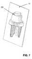
- FIG. 7shows the applicator according to the invention enclosed in a sterile package
- FIG. 8shows a sterile kit with the applicator according to the invention and a filter cleaning and removal tool.
- FIG. 1Ais a block diagram of an exemplary fluorescence endoscopy video system 10 which includes a multi-mode light source 102 that generates light for obtaining color and fluorescence images.
- a multi-mode light source 102that generates light for obtaining color and fluorescence images.
- the use of light source 102 for obtaining different types of imageswill be described in further detail below.
- Light from the light source 102is supplied to an illumination guide 104 of an endoscope 110 , which then illuminates a tissue sample 12 to be imaged.
- the systemalso includes a camera 100 , such as a color CCD camera, in particular a color CCD camera capable of operating under low light conditions, located at the insertion end, also referred to as distal end or tip, of the endoscope 110 .
- the light from the tissue 12is captured by the camera 100 .
- the resulting endoscope 110can be characterized as a fluorescence video endoscope, similar to video endoscopes currently on the market (such as the Olympus CF-240L) in utility.
- the endoscopecan be utilized for fluorescence/reflectance and/or fluorescence/fluorescence imaging. Locating the camera at the insertion end of the endoscope improves the light sensitivity and image resolution compared to endoscopes having an imaging guide or a relay lens.
- a processor/controller 106communicates with the camera 100 and the light source 102 via electrical and/or optical signal transmission lines routed within the endoscope, and produces video signals that can be displayed on a video monitor 108 .
- the camera 100can communicate with the processor/controller 106 over a wireless link (not shown).
- the exemplary camera 100includes a single color CCD sensor 103 and suitable imaging optics 113 .
- Each of the pixel elements on the single color CCD sensor 103is covered by an integrated filter, typically red, green or blue. These filters define the wavelength bands of fluorescence and reflectance light that reach the individual pixel elements. Such filters typically have considerable spectral overlap between the red, green, and blue passbands, which can lead to considerable crosstalk when imaging low-intensity fluorescence light in the presence of the much more intense reflected excitation light.
- the color image sensor 103may be a three-CCD color image sensor assembly, a color CMOS image sensor, or a three-CMOS color image sensor assembly, optionally with charge carrier multiplication.
- the filter 118 in current fluorescence video-endoscopy systemsis typically built in to the endoscope's distal end (typically between the objective lens and the low light image sensor), thus requiring dedicated endoscopes.
- Filter 118blocks the strong excitation light used to excite the tissue fluorescence these systems are intended to image.
- This built-in filterdistinguishes these endoscopes from those used for conventional video-endoscopy.
- an externally mounted filter 202can instead be applied externally to a conventional video-endoscope that contains a sufficiently sensitive image sensor to also image tissue fluorescence, as shown in FIG. 1C .
- This combination of a conventional video endoscope and an externally mounted filter 202can be used in conjunction with an appropriate endoscopy light source 102 to image both in both color (or white light) and fluorescence modes.
- FIG. 1Cillustrates one embodiment of a conventional video-endoscope with a distal filter 202 placed over the distal end or tip to allow the endoscope to perform fluorescence and white light examinations of a patient.
- the filter 202blocks excess excitation light from reaching the image sensor 103 , but allows sufficient blue light intensity to pass in order to render white light images with proper white balance.
- FIGS. 2A and 2Billustrate the operation of the distal filter 202 in further detail.
- An exemplary endoscope 250includes illumination ports 252 , 254 that supply illumination light to an area of interest.
- Another port 256is the distal entrance to a working channel through which endoscopy tools can be passed in order to perform a desired task such as obtaining a biopsy sample, marking tissue with dye or perform some other diagnostic or therapeutic procedures.
- Light that is reflected from the tissue 350is captured by a lens 258 that images the light onto a color sensor 262 .
- the color light sensoris a CCD, CMOS or equivalent image sensor 262 with a mosaic filter 260 , such as a RGB filter, positioned in front of sensor 262 .
- Image signals produced by the image sensorare transmitted to image processor 106 ( FIG. 1A ) that converts the signals into video signals that can be displayed on monitor 108 , transmitted to another location, for example, for remote diagnosis, or recorded on a storage medium for later review or comparison.
- the endoscope 250is shown as being fitted with the distal end filter 202 .
- the filter 202is designed as a single-use device that is applied to the tip of the conventional color CCD video endoscope prior to endoscopy and that is removed following endoscopy.
- the filtercovers the field of view of the video endoscope objective lens 258 , but does not block the illumination ports 252 , 254 or working channel 256 , water ports (not shown) or other features that may be located on the endoscope tip.
- the filterremains firmly attached to the endoscope tip, preferably directly to lens 258 , until it is deliberately removed.
- the filter 202is positioned in front of the imaging lens 258 and prevents excitation light from reaching the image sensor.
- the filterremoves the excitation light having wavelengths in the UV/blue spectral range, for example a wavelength range between about 370 nm and about 450 nm, or some subset of this wavelength range, but passes some blue light, for example 451-480 nm, for use in white light imaging, as will be described below.
- the filtershould block excitation light over a correspondingly wide field of view.
- the filtershould also be thin enough so as not to introduce optical aberrations or interfere with the mechanical properties and maneuverability of the endoscope tip. Examples of suitable filters are, for example, dye-based absorption filters designed to block the desired range of excitation light and to operate over a wide field of view, such as Kodak Wratten gel filters.
- the distal filter 202allows fluorescence images to be obtained in at least two modes.
- excitation lighttypically in the UV/blue wavelength spectral band
- a light sourcenot shown
- a portion of the excitation lightcauses the tissue sample to produce fluorescence light while another portion of the excitation light is reflected off the surface of the tissue 350 .
- Excitation light reflected from the surface of the tissue 350is blocked by the filter 202 , whereas the fluorescence light and light in other spectral bands passes through filter 202 .
- the tissueis illuminated with the excitation light and some amount of reflectance light.
- the reflectance lightis in the red spectral band.
- the reflectance lightpasses through filter 202 and the mosaic filter 260 disposed in front of image sensor 262 . Images of the tissue are obtained by combining, for example, the green pixels from image sensor 262 to obtain a fluorescence image in the green spectral band and the red pixels from image sensor 262 to obtain a reflectance image in the red spectral band.
- the reflectance lightmay be provided in the green spectral band and a fluorescence image in the red spectral band.
- the reflectance imagecan be produced from blue reflectance light having wavelengths that are not filtered by filter 202 .
- the fluorescence and reflectance imagesmay be combined and displayed on video monitor 108 ( FIG. 1A ). To prevent the intense light from the reflectance image from overpowering the low light level of the fluorescence image, the amount of reflectance light supplied from the light source is selected to be comparable to the amount of fluorescence light received by image sensor 262 .
- endoscope 250can also be used to obtain white light images of the tissue 350 by illuminating the tissue with light having red, green and blue spectral components.
- the illumination lightis reflected by the tissue sample 350 , passes through filter 202 and is focused onto image sensor 262 by the endoscope's imaging lens 258 . Because the filter 202 removes most of the blue reflectance light, the light received by image sensor 262 includes excess red and green light. To compensate for the reduced blue light intensity at the image sensor, the illumination light should either contain excess blue light intensity in the band that passes through the filter 202 or contain reduced green and red light intensity so that the resulting image produced by the image sensor may be white balanced. Additional fine-tuning of the white balance of the images produced by the image sensor 262 can be accomplished by image processing software in the image processor 106 ( FIG. 1A ).
- the filter 202may be secured to the endoscope's distal end with an adhesive.
- filter 202may be composed of various flexible, optically transparent materials, such as a dyed polycarbonate filter disk 202 a with a diameter of several millimeters.
- the filter disk 202 amay be laminated to an optically clear adhesive 202 b for a total thickness of approximately 100 ⁇ m.
- the filterSince the filter is applied externally to the endoscope's distal end in a clinical environment and for medical purposes, the attachment must be reliable, simple, accurate and free of contaminants. In that regard, the small size of the filter represents a handling challenge. Endoscopy suites are not equipped with tools for such purposes, medical staff is frequently gloved and pre-endoscopy preparation time is often limited. Even more challenging is the accuracy required for placing the filter over the video endoscope objective lens with sufficient precision.
- the tips of video endoscopes 60are typically 5-13 mm across and can be very crowded with multiple illumination ports 252 , 254 , water ports, and one or more working channels 256 , all sharing space with the objective lens 258 . (See FIGS. 2A and 2B and FIG. 4 ).
- Endoscope objectiveshave a wide field of view (typically between about 120° and about 170°) and the optical aperture of the filter should not occlude any part of that field. Moreover, trapping of contaminants or air between the filter and the objective lens which could degrade the optical performance of the video endoscope should also be prevented or at least minimized.
- endoscopesare typically cleaned, washed and disinfected by specialized washing machines, it is not uncommon for some form of residue to remain on the objective lens. Such residue needs to be cleaned prior to endoscopy and in the case where a filter is to be fitted to the objective lens, such cleaning would need to take place prior to fitting the filter.
- any remaining contaminants between the filter and the objectivemay also adversely affect the adhesion of the filter to the endoscope objective.
- the filterneeds to remain firmly and securely attached throughout the endoscopy procedure.
- the filtersince the filter will contact mucous membranes, it is desirable that the filter itself should be sterile and remain so until the endoscope is inserted into the patient at the beginning of the procedure.
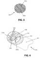
- FIG. 4shows in a perspective view another exemplary configuration of the distal end 402 of an endoscope 110 with an optical port 404 , illumination ports 406 , working port 408 , water ports 410 a , 410 b and other features, such as tip surface contours 412 , which may aid in placing filter 202 on the endoscope's distal end 402 .
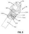
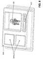
- FIG. 5 and FIGS. 6 through 6Bshow an applicator 50 according to the invention.
- Applicator 50when properly registered with endoscope's distal end 402 , can be used to accurately place filter 202 (or any other optical element such as a lens adapter or relay optics) on optical port 404 of distal end 402 .
- Applicator 50is supplied with filter 202 already mounted therein, as described below in more detail with reference to FIG. 6 .
- the exemplary embodiment of applicator 50 illustrated in FIG. 5has a substantially round base section 510 with gripping surface(s) 520 to aid an operator in handling the applicator 50 .
- base section 510transitions into or is attached, for example by a snap-in connection, to a longitudinally extending, substantially cylindrical projection 516 adapted to receive a piston 518 which is movable in the longitudinal direction, as indicated by arrow A in FIG. 6 .
- Alignment memberssuch as projections 512 a , 512 b , 514 , are integrally formed on, such as molded, or attached to the proximal end of base section 510 .
- the exemplary projection 514is adapted to engage with, for example, the working channel 408 , whereas the projections are adapted to engage with a circumferential shoulder 414 of endoscope distal end 402 to position adapter 50 on distal end 402 with the proper orientation.
- Other embodimentsmay use other locating features, such as the water ports 410 a , 410 b and tip surface contours 412 .
- the piston 518may be held in place by a small detent 515 located, for example, near the top of the piston housing.
- Detent 515prevents the piston 518 from being inadvertently activated and prematurely ejecting the filter before a force is intentionally applied to the piston to place the optical element 202 on optical port 404 .
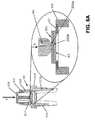
- FIG. 6shows applicator 50 in cross-section, with the insert showing the placement of filter 202 on base section 510 in more detail.
- Base section 510has an opening 62 which optionally includes an annular inward recess or shoulder 66 .
- the diameter of opening 62is preferably slightly smaller than the diameter of filter 202 , thereby precisely and securely supporting filter 202 in opening 62 .
- the top of the basemay be provided with a metal disk 67 having a hole that will be precisely positioned by the applicator 50 over the endoscope's optical port 404 .
- the hole in the diskis slightly smaller in diameter than the optical element that is centered over the hole.
- the disk 67may be made, for example, of a thin brass sheet or another material capable of holding the optical element before its release by the force applied to the piston.
- a membrane 517covers substantially the entire opening in base 510 .
- a compliant tip 64 , 64 ais formed in the center of the membrane 517 on the side facing the optical element 202 .
- the tip 64 , 64 apresses against the center of optical element 202 when an external force is applied to piston 518 in the direction of arrow A.
- the optical elementis pressed against the optical port 404 (or a lens in the optical port) from the center of the lens outwardly, thereby eliminating trapped air bubbles.
- the tipmay be disposed on the end of the piston facing the optical element without an interposed membrane.
- FIGS. 6B and 6Care examples of other possible embodiments of the applicator 50 , indicated by the reference symbols 50 ′ ( FIG. 6B ) and 50 ′′ ( FIG. 6C ), respectively.
- the difference between these embodiments and the applicator 50 of FIG. 6is modifications in the actuation mechanism for releasing the optical element 202 .
- applicator 50 ′is shown to include a resilient strut or lever arm 68 , wherein one end of the lever arm 68 may be attached to the base, and the other end of the lever arm 68 is in force-transmitting communication with a resilient tip 64 a , which may be constructed similar to the tip 64 in FIG. 6A .
- Applicator 50 ′′may include a dome-shaped membrane which may also include a tip 64 b facing the optical element 202 , with the tip 64 b pushing the optical element 202 out of an opening against the optical port (in response to force exerted on membrane 68 b ), as analogously described above with reference to FIGS. 6 and 6A .
- actuation mechanismscan be contemplated for use with the aforedescribed actuator, as long as an optical element can be precisely positioned over an optical port of an endoscope.
- FIG. 6Dillustrates a sequence of steps for positioning optical applicator 50 , 50 ′ or 50 ′′ on the distal end of the endoscope.
- One of the alignment members 514(see FIG. 5 ) is pushed into, for example, working port 408 , while the outside projections 512 a , 512 b grip around the periphery of the distal end.
- an operatorpresses down on piston 518 in the direction indicated by arrow A.
- the compliant, rounded tip 64then presses against the center of the non-adhesive side 202 a of filter 202 , also in the direction indicated by arrow A.
- Piston 518then pushes filter 202 through the opening 62 against the optical port or objective lens 404 , thereby ejecting the filter through the opening 62 towards the optical port 404 .
- the piston 518may be composed of silicone rubber, but other suitable elastomer compounds can also be used.
- filter 202contacts the endoscope objective from the center of the lens outwardly toward the periphery of the lens, thereby removing air bubbles trapped between the filter and the objective lens.
- applicator 50is preferably preassembled with optical filter 202 and sealed in a sterile package 70 .
- the applicator containing the filter 202Prior to endoscopy, the applicator containing the filter 202 is removed from the sterile package 70 and positioned over the endoscope's distal end 402 (see FIG. 5 ).
- Applicator 50is then pushed over distal end 402 , with the interior projection(s) 514 engaging with the mating port(s) 408 and the outside projections 512 a , 512 b slidingly engaging with a portion of the exterior peripheral surface of distal end 402 .
- the filter 202is then applied to optical port 404 as described above.
- applicator 50may be supplied as a kit 800 which may include:
Landscapes
- Health & Medical Sciences (AREA)
- Life Sciences & Earth Sciences (AREA)
- Surgery (AREA)
- Engineering & Computer Science (AREA)
- Biomedical Technology (AREA)
- Molecular Biology (AREA)
- Pathology (AREA)
- Radiology & Medical Imaging (AREA)
- Nuclear Medicine, Radiotherapy & Molecular Imaging (AREA)
- Biophysics (AREA)
- Physics & Mathematics (AREA)
- Heart & Thoracic Surgery (AREA)
- Medical Informatics (AREA)
- Optics & Photonics (AREA)
- Animal Behavior & Ethology (AREA)
- General Health & Medical Sciences (AREA)
- Public Health (AREA)
- Veterinary Medicine (AREA)
- Manufacturing & Machinery (AREA)
- Endoscopes (AREA)
- Instruments For Viewing The Inside Of Hollow Bodies (AREA)
Abstract
Description
- A first solvent-impregnated swab or wipe804aimpregnated with a volatile solvent, such as alcohol or acetone, for preparing endoscope
distal end 402 and removing any residue, oil or dust from the endoscope's optical port orobjective lens 404 prior to application offilter 202; - A
removal tool 802 that aids in the removal offilter 802 after the endoscopy. A suitable tool is, for example, a plastic spatula-shaped instrument, such as a polypropylene nail cleaner/pick available from Qosina, Edgewood, N.Y.; - A second solvent-impregnated swab, (e.g. an alcohol-impregnated wipe)804bfor removing residual adhesive or other contaminants from the endoscope objective lens after removal of
filter 202 and prior to processing of the endoscope for reuse.
- A first solvent-impregnated swab or wipe804aimpregnated with a volatile solvent, such as alcohol or acetone, for preparing endoscope
Claims (7)
Priority Applications (1)
| Application Number | Priority Date | Filing Date | Title |
|---|---|---|---|
| US13/853,656US9386909B2 (en) | 2006-07-28 | 2013-03-29 | System and method for deposition and removal of an optical element on an endoscope objective |
Applications Claiming Priority (3)
| Application Number | Priority Date | Filing Date | Title |
|---|---|---|---|
| US83389706P | 2006-07-28 | 2006-07-28 | |
| US11/830,323US8408269B2 (en) | 2006-07-28 | 2007-07-30 | System and method for deposition and removal of an optical element on an endoscope objective |
| US13/853,656US9386909B2 (en) | 2006-07-28 | 2013-03-29 | System and method for deposition and removal of an optical element on an endoscope objective |
Related Parent Applications (1)
| Application Number | Title | Priority Date | Filing Date |
|---|---|---|---|
| US11/830,323DivisionUS8408269B2 (en) | 2006-07-28 | 2007-07-30 | System and method for deposition and removal of an optical element on an endoscope objective |
Publications (2)
| Publication Number | Publication Date |
|---|---|
| US20130237762A1 US20130237762A1 (en) | 2013-09-12 |
| US9386909B2true US9386909B2 (en) | 2016-07-12 |
Family
ID=38981098
Family Applications (2)
| Application Number | Title | Priority Date | Filing Date |
|---|---|---|---|
| US11/830,323Expired - Fee RelatedUS8408269B2 (en) | 2006-07-28 | 2007-07-30 | System and method for deposition and removal of an optical element on an endoscope objective |
| US13/853,656Expired - Fee RelatedUS9386909B2 (en) | 2006-07-28 | 2013-03-29 | System and method for deposition and removal of an optical element on an endoscope objective |
Family Applications Before (1)
| Application Number | Title | Priority Date | Filing Date |
|---|---|---|---|
| US11/830,323Expired - Fee RelatedUS8408269B2 (en) | 2006-07-28 | 2007-07-30 | System and method for deposition and removal of an optical element on an endoscope objective |
Country Status (4)
| Country | Link |
|---|---|
| US (2) | US8408269B2 (en) |
| EP (1) | EP2051603B1 (en) |
| JP (1) | JP4948602B2 (en) |
| WO (1) | WO2008011722A1 (en) |
Cited By (13)
| Publication number | Priority date | Publication date | Assignee | Title |
|---|---|---|---|---|
| US9642532B2 (en) | 2008-03-18 | 2017-05-09 | Novadaq Technologies Inc. | Imaging system for combined full-color reflectance and near-infrared imaging |
| US9814378B2 (en) | 2011-03-08 | 2017-11-14 | Novadaq Technologies Inc. | Full spectrum LED illuminator having a mechanical enclosure and heatsink |
| US9877654B2 (en) | 2006-02-07 | 2018-01-30 | Novadaq Technologies Inc. | Near infrared imaging |
| US9968244B2 (en) | 2000-07-14 | 2018-05-15 | Novadaq Technologies ULC | Compact fluorescence endoscopy video system |
| US10182709B2 (en) | 2002-01-15 | 2019-01-22 | Novadaq Technologies ULC | Filter for use with imaging endoscopes |
| US10293122B2 (en) | 2016-03-17 | 2019-05-21 | Novadaq Technologies ULC | Endoluminal introducer with contamination avoidance |
| US10694152B2 (en) | 2006-12-22 | 2020-06-23 | Novadaq Technologies ULC | Imaging systems and methods for displaying fluorescence and visible images |
| US10869645B2 (en) | 2016-06-14 | 2020-12-22 | Stryker European Operations Limited | Methods and systems for adaptive imaging for low light signal enhancement in medical visualization |
| USD916294S1 (en) | 2016-04-28 | 2021-04-13 | Stryker European Operations Limited | Illumination and imaging device |
| US10980420B2 (en) | 2016-01-26 | 2021-04-20 | Stryker European Operations Limited | Configurable platform |
| US10992848B2 (en) | 2017-02-10 | 2021-04-27 | Novadaq Technologies ULC | Open-field handheld fluorescence imaging systems and methods |
| US11691363B2 (en) | 2020-03-20 | 2023-07-04 | Steris Instrument Management Services, Inc. | Method of forming and incorporating a polymeric lens within a lens housing |
| US11930278B2 (en) | 2015-11-13 | 2024-03-12 | Stryker Corporation | Systems and methods for illumination and imaging of a target |
Families Citing this family (17)
| Publication number | Priority date | Publication date | Assignee | Title |
|---|---|---|---|---|
| EP2051603B1 (en)* | 2006-07-28 | 2019-09-11 | Novadaq Technologies ULC | System and method for deposition and removal of an optical element on an endoscope objective |
| DE102009020252B4 (en)* | 2009-05-07 | 2012-01-12 | Krohne Optosens Gmbh | Device for measuring the fluorescence of a medium |
| MX350734B (en) | 2010-09-08 | 2017-09-15 | Covidien Lp | Catheter with imaging assembly. |
| USD716841S1 (en) | 2012-09-07 | 2014-11-04 | Covidien Lp | Display screen with annotate file icon |
| US9517184B2 (en) | 2012-09-07 | 2016-12-13 | Covidien Lp | Feeding tube with insufflation device and related methods therefor |
| USD717340S1 (en) | 2012-09-07 | 2014-11-11 | Covidien Lp | Display screen with enteral feeding icon |
| US9198835B2 (en) | 2012-09-07 | 2015-12-01 | Covidien Lp | Catheter with imaging assembly with placement aid and related methods therefor |
| USD735343S1 (en) | 2012-09-07 | 2015-07-28 | Covidien Lp | Console |
| US10064545B2 (en) | 2012-10-18 | 2018-09-04 | The Arizona Board Of Regents On Behalf Of The University Of Arizona | Multi-resolution foveated endoscope/laparoscope |
| US9389369B2 (en)* | 2012-12-13 | 2016-07-12 | Corning Cable Systems Llc | Optical port having minimalist footprint |
| US9395497B2 (en)* | 2012-12-13 | 2016-07-19 | Corning Optical Communications LLC | Optical port having one or more alignment features |
| US9526404B2 (en)* | 2013-10-06 | 2016-12-27 | Gyrus Acmi, Inc. | Endoscope illumination system |
| WO2016118423A1 (en)* | 2015-01-22 | 2016-07-28 | Arizona Board Of Regents On Behalf Of The University Of Arizona | Low-cost compact disposable imaging probe |
| CN118177700A (en) | 2018-01-05 | 2024-06-14 | 波士顿科学国际有限公司 | Fluorophore imaging device, system and method for endoscopic surgery |
| US20210228062A1 (en)* | 2020-01-28 | 2021-07-29 | Gyrus Acmi, Inc. D/B/A Olympus Surgical Technologies America | Endoscope and endoscope attachments |
| EP3871585B1 (en) | 2020-02-28 | 2025-04-09 | Gyrus ACMI, Inc. d/b/a Olympus Surgical Technologies America | Electrosurgical attachment device |
| US12337190B2 (en)* | 2022-10-06 | 2025-06-24 | Olympus Medical Systems Corp. | Endoscope system and phototherapy method having a correction operation when an observation mode is set to a thermal suppression mode |
Citations (204)
| Publication number | Priority date | Publication date | Assignee | Title |
|---|---|---|---|---|
| US3215029A (en)* | 1960-11-23 | 1965-11-02 | American Optical Corp | Fiber optical image transfer devices and method of making the same |
| US3971068A (en) | 1975-08-22 | 1976-07-20 | The United States Of America As Represented By The Secretary Of The Navy | Image processing system |
| US4037866A (en) | 1976-07-26 | 1977-07-26 | Price Edward E | Contact lens applicator |
| US4066330A (en)* | 1976-06-14 | 1978-01-03 | Karl Storz Endoscopy-America, Inc. | Coupler for joining optical devices |
| US4115812A (en) | 1973-11-26 | 1978-09-19 | Hitachi, Ltd. | Automatic gain control circuit |
| US4149190A (en) | 1977-10-17 | 1979-04-10 | Xerox Corporation | Automatic gain control for video amplifier |
| US4200801A (en) | 1979-03-28 | 1980-04-29 | The United States Of America As Represented By The United States Department Of Energy | Portable spotter for fluorescent contaminants on surfaces |
| US4318395A (en)* | 1979-05-23 | 1982-03-09 | Olympus Optical Co., Ltd. | Endoscope ocular accessory mounting device |
| US4355325A (en) | 1980-03-24 | 1982-10-19 | Sony Corporation | White balance control system |
| US4378571A (en) | 1981-07-06 | 1983-03-29 | Xerox Corporation | Serial analog video processor for charge coupled device imagers |
| US4449535A (en) | 1981-03-25 | 1984-05-22 | Compagnie Industrielle Des Lasers Cilas Alcatel | Apparatus for measuring in situ the state of oxidation-reduction of a living organ |
| US4471766A (en)* | 1977-11-24 | 1984-09-18 | Inbae Yoon | Ring applicator with an endoscope |
| US4532918A (en) | 1983-10-07 | 1985-08-06 | Welch Allyn Inc. | Endoscope signal level control |
| US4556057A (en) | 1982-08-31 | 1985-12-03 | Hamamatsu Tv Co., Ltd. | Cancer diagnosis device utilizing laser beam pulses |
| JPS60246733A (en) | 1984-05-21 | 1985-12-06 | 熊谷 博彰 | Optical photographing apparatus of organism tissue |
| JPS61159936A (en) | 1985-07-02 | 1986-07-19 | 熊谷 博彰 | Spectral image pick-up apparatus of biological tissue |
| US4611888A (en) | 1983-10-17 | 1986-09-16 | Mp Video, Inc. | Coupler for surgical endoscope and video camera |
| US4638365A (en) | 1984-01-31 | 1987-01-20 | Canon Kabushiki Kaisha | Image sensing device |
| US4660982A (en)* | 1984-05-09 | 1987-04-28 | Olympus Optical Co., Ltd. | Adaptor for measuring length optically for endoscope |
| US4768513A (en) | 1986-04-21 | 1988-09-06 | Agency Of Industrial Science And Technology | Method and device for measuring and processing light |
| US4786813A (en) | 1984-10-22 | 1988-11-22 | Hightech Network Sci Ab | Fluorescence imaging system |
| US4821117A (en) | 1986-11-12 | 1989-04-11 | Kabushiki Kaisha Toshiba | Endoscopic system for producing fluorescent and visible images |
| JPH01135349A (en) | 1987-11-19 | 1989-05-29 | Maaketsuto Bureinzu:Kk | Contact lens attaching and detaching instrument |
| US4837625A (en) | 1987-02-20 | 1989-06-06 | Sgs-Thomson Microelectronics S.A. | Automatic gain control device for video signals |
| US4856495A (en) | 1986-09-25 | 1989-08-15 | Olympus Optical Co., Ltd. | Endoscope apparatus |
| US4895145A (en) | 1985-05-28 | 1990-01-23 | Surgical Laser Technologies, Inc. | Two-piece disposable laser delivery system |
| US4930516A (en) | 1985-11-13 | 1990-06-05 | Alfano Robert R | Method for detecting cancerous tissue using visible native luminescence |
| US4951135A (en) | 1988-01-11 | 1990-08-21 | Olympus Optical Co., Ltd. | Electronic-type endoscope system having capability of setting AGC variation region |
| US4954897A (en) | 1987-05-22 | 1990-09-04 | Nikon Corporation | Electronic still camera system with automatic gain control of image signal amplifier before image signal recording |
| US4974936A (en) | 1989-03-15 | 1990-12-04 | Richard Wolf Gmbh | Device for supplying light to endoscopes with rotary filter plate and faster rotating runner plate with at least one opaque region |
| US5001556A (en) | 1987-09-30 | 1991-03-19 | Olympus Optical Co., Ltd. | Endoscope apparatus for processing a picture image of an object based on a selected wavelength range |
| US5007408A (en) | 1989-03-16 | 1991-04-16 | Olympus Optical Co., Ltd. | Endoscope light source apparatus |
| JPH0397441A (en) | 1989-09-08 | 1991-04-23 | Olympus Optical Co Ltd | Endoscope for fluorescent observation |
| JPH0397442A (en) | 1989-09-08 | 1991-04-23 | Olympus Optical Co Ltd | Endoscope device for fluorescent observation |
| JPH0397439A (en) | 1989-09-08 | 1991-04-23 | Olympus Optical Co Ltd | Endoscope device for fluorescent observation |
| US5034888A (en) | 1988-02-26 | 1991-07-23 | Olympus Optical Co., Ltd. | Electronic endoscope apparatus having different image processing characteristics for a moving image and a still image |
| FR2671405A1 (en) | 1991-01-04 | 1992-07-10 | Inst Nat Sante Rech Med | DEVICE FOR MEASURING THE PH OF A TARGET, METHOD OF USING SAID DEVICE AND ITS APPLICATIONS. |
| US5134662A (en) | 1985-11-04 | 1992-07-28 | Cell Analysis Systems, Inc. | Dual color camera microscope and methodology for cell staining and analysis |
| EP0512965A1 (en) | 1991-05-08 | 1992-11-11 | Xillix Technologies Corporation | Endoscopic imaging system for diseased tissue |
| US5165079A (en) | 1989-02-02 | 1992-11-17 | Linotype-Hell Ag | Optical color-splitter arrangement |
| WO1993004648A1 (en) | 1991-09-03 | 1993-03-18 | William Frank Clymer | A contact lens applicator |
| US5205280A (en)* | 1990-12-21 | 1993-04-27 | Mp Video, Inc. | Quick-release endoscopic coupling assembly |
| JPH05115435A (en) | 1991-10-25 | 1993-05-14 | Olympus Optical Co Ltd | Solid-state image pickup device |
| US5214503A (en) | 1992-01-31 | 1993-05-25 | The United States Of America As Represented By The Secretary Of The Army | Color night vision camera system |
| US5225883A (en) | 1991-06-05 | 1993-07-06 | The Babcock & Wilcox Company | Video temperature monitor |
| US5255087A (en) | 1986-11-29 | 1993-10-19 | Olympus Optical Co., Ltd. | Imaging apparatus and endoscope apparatus using the same |
| US5278642A (en) | 1992-02-26 | 1994-01-11 | Welch Allyn, Inc. | Color imaging system |
| JPH06125911A (en) | 1992-10-15 | 1994-05-10 | Hamamatsu Photonics Kk | Endoscopic device |
| US5365057A (en) | 1993-07-02 | 1994-11-15 | Litton Systems, Inc. | Light-weight night vision device |
| US5371355A (en) | 1993-07-30 | 1994-12-06 | Litton Systems, Inc. | Night vision device with separable modular image intensifier assembly |
| US5377686A (en) | 1991-10-11 | 1995-01-03 | The University Of Connecticut | Apparatus for detecting leakage from vascular tissue |
| US5408263A (en) | 1992-06-16 | 1995-04-18 | Olympus Optical Co., Ltd. | Electronic endoscope apparatus |
| US5410363A (en) | 1992-12-08 | 1995-04-25 | Lightwave Communications, Inc. | Automatic gain control device for transmitting video signals between two locations by use of a known reference pulse during vertical blanking period so as to control the gain of the video signals at the second location |
| US5420628A (en) | 1990-01-16 | 1995-05-30 | Research Development Foundation | Video densitometer with determination of color composition |
| US5419323A (en) | 1988-12-21 | 1995-05-30 | Massachusetts Institute Of Technology | Method for laser induced fluorescence of tissue |
| US5421337A (en) | 1989-04-14 | 1995-06-06 | Massachusetts Institute Of Technology | Spectral diagnosis of diseased tissue |
| US5424841A (en) | 1993-05-28 | 1995-06-13 | Molecular Dynamics | Apparatus for measuring spatial distribution of fluorescence on a substrate |
| JPH07155292A (en) | 1993-12-03 | 1995-06-20 | Olympus Optical Co Ltd | Fluorescence observing apparatus |
| JPH07155291A (en) | 1993-12-03 | 1995-06-20 | Olympus Optical Co Ltd | Fluorescence observation apparatus |
| JPH07155290A (en) | 1993-12-03 | 1995-06-20 | Olympus Optical Co Ltd | Endoscope apparatus |
| JPH07155285A (en) | 1993-12-03 | 1995-06-20 | Olympus Optical Co Ltd | Fluorescence observing endoscope apparatus |
| JPH07155286A (en) | 1993-12-03 | 1995-06-20 | Olympus Optical Co Ltd | Fluorescence observing apparatus |
| US5430476A (en) | 1992-06-24 | 1995-07-04 | Richard Wolf Gmbh | Device for supplying light to endoscopes |
| JPH07204156A (en) | 1993-12-03 | 1995-08-08 | Olympus Optical Co Ltd | Fluorescence observation device |
| JPH07222712A (en) | 1994-02-10 | 1995-08-22 | Olympus Optical Co Ltd | Fluorescent endoscope system |
| EP0672379A1 (en) | 1994-03-15 | 1995-09-20 | Welch Allyn, Inc. | Dental imaging system |
| JPH07250812A (en) | 1994-03-15 | 1995-10-03 | Olympus Optical Co Ltd | Fluorescence diagnosing apparatus |
| JPH07250804A (en) | 1994-03-15 | 1995-10-03 | Olympus Optical Co Ltd | Fluorescence observer |
| WO1995026673A2 (en) | 1994-03-28 | 1995-10-12 | Xillix Technologies Corporation | Apparatus and method for imaging diseased tissue using integrated autofluorescence |
| JPH07327913A (en) | 1994-06-13 | 1995-12-19 | Olympus Optical Co Ltd | Cover type endoscope |
| US5485203A (en) | 1991-08-12 | 1996-01-16 | Olympus Optical Co., Ltd. | Color misregistration easing system which corrects on a pixel or block basis only when necessary |
| US5490015A (en) | 1993-03-04 | 1996-02-06 | Olympus Optical Co., Ltd. | Actuator apparatus |
| DE19535114A1 (en) | 1994-09-21 | 1996-03-28 | Asahi Optical Co Ltd | Endoscope with illumination source of multiple wavelengths |
| JPH08126605A (en) | 1994-11-01 | 1996-05-21 | Olympus Optical Co Ltd | Endoscope |
| JPH08140928A (en) | 1994-09-21 | 1996-06-04 | Asahi Optical Co Ltd | Electronic endoscope system for fluorescence diagnosis |
| JPH08140929A (en) | 1994-09-21 | 1996-06-04 | Asahi Optical Co Ltd | Electronic endoscope system for fluorescence diagnosis |
| US5536236A (en) | 1993-02-12 | 1996-07-16 | Olympus Optical Co., Ltd. | Covered endoscope system |
| JPH08224240A (en) | 1995-02-22 | 1996-09-03 | Olympus Optical Co Ltd | Fluorescent diagnosing device |
| JPH08224209A (en) | 1995-02-23 | 1996-09-03 | Olympus Optical Co Ltd | Fluorescence observing device |
| JPH08224208A (en) | 1995-02-22 | 1996-09-03 | Olympus Optical Co Ltd | Fluorescence observing endoscope device |
| JPH08224210A (en) | 1995-02-23 | 1996-09-03 | Olympus Optical Co Ltd | Fluorescence observing device |
| DE19608027A1 (en) | 1995-03-03 | 1996-09-05 | Asahi Optical Co Ltd | Biological fluorescence diagnosis device for medical use |
| JPH08252218A (en) | 1995-03-16 | 1996-10-01 | Olympus Optical Co Ltd | Fluorescent observing endoscope device |
| US5585846A (en) | 1991-12-05 | 1996-12-17 | Samsung Electronics Co., Ltd. | Image signal processing circuit in a digital camera having gain and gamma control |
| US5596654A (en) | 1987-04-20 | 1997-01-21 | Fuji Photo Film Co., Ltd. | Method of determining desired image signal range based on histogram data |
| JPH0966023A (en) | 1994-09-21 | 1997-03-11 | Asahi Optical Co Ltd | Video processor device of electronic endoscope for fluorescence diagnosis |
| JPH0970384A (en) | 1994-09-21 | 1997-03-18 | Asahi Optical Co Ltd | Electronic endoscope system for fluorescence diagnosis |
| EP0774685A2 (en) | 1995-11-14 | 1997-05-21 | Kodak Limited | Photographic materials for use in redox amplification and process |
| EP0774865A2 (en) | 1995-11-17 | 1997-05-21 | SANYO ELECTRIC Co., Ltd. | Video camera with high speed mode |
| US5646680A (en) | 1994-10-20 | 1997-07-08 | Olympus Optical Co., Ltd. | Endoscope system having a switch forcibly set to display video signals not passed through outer peripheral apparatus |
| US5647840A (en) | 1994-09-14 | 1997-07-15 | Circon Corporation | Endoscope having a distally heated distal lens |
| US5647368A (en) | 1996-02-28 | 1997-07-15 | Xillix Technologies Corp. | Imaging system for detecting diseased tissue using native fluorsecence in the gastrointestinal and respiratory tract |
| US5667472A (en) | 1994-03-18 | 1997-09-16 | Clarus Medical Systems, Inc. | Surgical instrument and method for use with a viewing system |
| US5695049A (en) | 1996-10-10 | 1997-12-09 | Johnson & Johnson Vision Products, Inc. | Contact lens package with insertion feature |
| US5697373A (en) | 1995-03-14 | 1997-12-16 | Board Of Regents, The University Of Texas System | Optical method and apparatus for the diagnosis of cervical precancers using raman and fluorescence spectroscopies |
| US5713364A (en) | 1995-08-01 | 1998-02-03 | Medispectra, Inc. | Spectral volume microprobe analysis of materials |
| US5749830A (en) | 1993-12-03 | 1998-05-12 | Olympus Optical Co., Ltd. | Fluorescent endoscope apparatus |
| JPH10127563A (en) | 1996-10-30 | 1998-05-19 | Olympus Optical Co Ltd | Endoscope unit |
| JPH10151104A (en) | 1996-11-25 | 1998-06-09 | Olympus Optical Co Ltd | Fluorescent endoscope device |
| WO1998024360A1 (en) | 1996-12-04 | 1998-06-11 | Harvey Lui | Fluorescence scope system for dermatologic diagnosis |
| US5772355A (en) | 1996-12-19 | 1998-06-30 | Precision Optics Corporation | Quick attach/release adapter mechanism |
| JPH10201700A (en) | 1997-01-20 | 1998-08-04 | Olympus Optical Co Ltd | Fluoroscopic endoscope device |
| JPH10225426A (en) | 1997-02-17 | 1998-08-25 | Olympus Optical Co Ltd | Fluorescence observing device |
| JPH10243920A (en) | 1997-03-07 | 1998-09-14 | Olympus Optical Co Ltd | Fluorescent observation endoscope device |
| JPH10243915A (en) | 1997-03-04 | 1998-09-14 | Olympus Optical Co Ltd | Fluorescent observation device |
| US5833617A (en) | 1996-03-06 | 1998-11-10 | Fuji Photo Film Co., Ltd. | Fluorescence detecting apparatus |
| JPH10308114A (en) | 1997-05-06 | 1998-11-17 | Olympus Optical Co Ltd | Light source equipment |
| JPH10309282A (en) | 1997-05-13 | 1998-11-24 | Olympus Optical Co Ltd | Fluorescent observation device |
| JPH10309281A (en) | 1997-05-13 | 1998-11-24 | Olympus Optical Co Ltd | Fluorescent diagnostic device |
| JPH10328129A (en) | 1997-06-02 | 1998-12-15 | Olympus Optical Co Ltd | Fluorescent observing device |
| US5852498A (en) | 1997-04-04 | 1998-12-22 | Kairos Scientific Inc. | Optical instrument having a variable optical filter |
| WO1999001749A1 (en) | 1997-07-03 | 1999-01-14 | Smith & Nephew, Inc. | Fluorescence imaging system |
| JPH1147079A (en) | 1997-08-06 | 1999-02-23 | Olympus Optical Co Ltd | Endoscope system |
| US5891016A (en) | 1995-11-09 | 1999-04-06 | Asahi Kogaku Kogyo Kabushiki Kaisha | Fluorescence endoscope having an exciting light filter and a fluorescence filter |
| JPH11104070A (en) | 1997-10-02 | 1999-04-20 | Olympus Optical Co Ltd | Endoscope |
| JPH11104060A (en) | 1997-10-03 | 1999-04-20 | Olympus Optical Co Ltd | Fluorescent observation device |
| JPH11104061A (en) | 1997-10-02 | 1999-04-20 | Olympus Optical Co Ltd | Trans-endoscopic fluorescent observation device |
| JPH11104059A (en) | 1997-10-02 | 1999-04-20 | Olympus Optical Co Ltd | Fluorescent observation device |
| JPH11113839A (en) | 1997-10-14 | 1999-04-27 | Olympus Optical Co Ltd | Endoscope device |
| JPH11155812A (en) | 1997-12-02 | 1999-06-15 | Olympus Optical Co Ltd | Fluorescent observation device |
| JPH11244220A (en) | 1998-03-03 | 1999-09-14 | Fuji Photo Film Co Ltd | Fluorescent endoscope |
| US5971918A (en) | 1996-10-02 | 1999-10-26 | Richard Wolf Gmbh | Device for the photodynamic endoscopic diagnosis of tumor tissue |
| WO1999053832A1 (en) | 1998-04-20 | 1999-10-28 | Xillix Technologies Corp. | Imaging system with automatic gain control for reflectance and fluorescence endoscopy |
| US5984861A (en) | 1997-09-29 | 1999-11-16 | Boston Scientific Corporation | Endofluorescence imaging module for an endoscope |
| US5990996A (en) | 1996-05-14 | 1999-11-23 | Colorlink, Inc. | Color selective light modulators employing birefringent stacks |
| US5999240A (en) | 1995-05-23 | 1999-12-07 | Colorlink, Inc. | Optical retarder stack pair for transforming input light into polarization states having saturated color spectra |
| JPH11332819A (en) | 1998-05-28 | 1999-12-07 | Olympus Optical Co Ltd | Fluorescent image device |
| US6002137A (en) | 1997-02-13 | 1999-12-14 | Fuji Photo Film Co., Ltd. | Fluorescence detecting system |
| US6004263A (en) | 1996-03-13 | 1999-12-21 | Hihon Kohden Corporation | Endoscope with detachable operation unit and insertion unit |
| US6008889A (en) | 1997-04-16 | 1999-12-28 | Zeng; Haishan | Spectrometer system for diagnosis of skin disease |
| US6028622A (en) | 1997-04-25 | 2000-02-22 | Olympus Optical Co., Ltd. | Observation apparatus for endoscopes |
| US6061591A (en) | 1996-03-29 | 2000-05-09 | Richard Wolf Gmbh | Arrangement and method for diagnosing malignant tissue by fluorescence observation |
| US6059720A (en) | 1997-03-07 | 2000-05-09 | Asahi Kogaku Kogyo Kabushiki Kaisha | Endoscope system with amplification of fluorescent image |
| WO2000042910A1 (en) | 1999-01-26 | 2000-07-27 | Newton Laboratories, Inc. | Autofluorescence imaging system for endoscopy |
| US6110106A (en) | 1998-06-24 | 2000-08-29 | Biomax Technologies, Inc. | Endoscopes and methods relating to direct viewing of a target tissue |
| JP2000245693A (en) | 1999-03-03 | 2000-09-12 | Olympus Optical Co Ltd | Endoscope device |
| US6120435A (en) | 1997-07-16 | 2000-09-19 | Olympus Optical Co., Ltd. | Endoscope system in which operation switch sets designed to function and be handled same way are included in endoscope and image processing apparatus respectively |
| WO2000054652A1 (en) | 1999-03-17 | 2000-09-21 | Ekapot Bhunachet | Method for superimposing fluorescent image on background image by ccd and viewing the images on the same screen simultaneously |
| US6148227A (en) | 1998-01-07 | 2000-11-14 | Richard Wolf Gmbh | Diagnosis apparatus for the picture providing recording of fluorescing biological tissue regions |
| US6161035A (en) | 1997-04-30 | 2000-12-12 | Asahi Kogaku Kogyo Kabushiki Kaisha | Fluorescence diagnostic apparatus |
| JP2000354583A (en) | 1999-06-15 | 2000-12-26 | Olympus Optical Co Ltd | Endoscope fluoroscopic apparatus |
| US6192267B1 (en) | 1994-03-21 | 2001-02-20 | Scherninski Francois | Endoscopic or fiberscopic imaging device using infrared fluorescence |
| US6212425B1 (en) | 1995-09-26 | 2001-04-03 | Karl Storz Gmbh & Co., Kg | Apparatus for photodynamic diagnosis |
| US6258576B1 (en) | 1996-06-19 | 2001-07-10 | Board Of Regents, The University Of Texas System | Diagnostic method and apparatus for cervical squamous intraepithelial lesions in vitro and in vivo using fluorescence spectroscopy |
| US20010016679A1 (en) | 2000-02-02 | 2001-08-23 | Olympus Optical Co., Ltd Tokyo Japan | Endoscope apparatus |
| US6280378B1 (en) | 1998-05-29 | 2001-08-28 | Fuji Photo Film Co., Ltd. | Fluorescence endoscope |
| US6293911B1 (en) | 1996-11-20 | 2001-09-25 | Olympus Optical Co., Ltd. | Fluorescent endoscope system enabling simultaneous normal light observation and fluorescence observation in infrared spectrum |
| US6332092B1 (en) | 1998-07-08 | 2001-12-18 | Lifespex, Incorporated | Optical probe having and methods for uniform light irradiation and/or light collection over a volume |
| WO2002007587A2 (en) | 2000-07-14 | 2002-01-31 | Xillix Technologies Corporation | Compact fluorescent endoscopy video system |
| US6364829B1 (en) | 1999-01-26 | 2002-04-02 | Newton Laboratories, Inc. | Autofluorescence imaging system for endoscopy |
| US6419628B1 (en) | 1997-03-29 | 2002-07-16 | Karl Storz Gmbh & Co. Kg | Endoscope with longitudinal compensation capability in response to thermal stress |
| US6422994B1 (en) | 1997-09-24 | 2002-07-23 | Olympus Optical Co., Ltd. | Fluorescent diagnostic system and method providing color discrimination enhancement |
| JP2002244122A (en) | 2001-02-22 | 2002-08-28 | Sharp Corp | Display device |
| US20020138008A1 (en) | 2000-01-13 | 2002-09-26 | Kazuhiro Tsujita | Method and apparatus for displaying fluorescence images and method and apparatus for acquiring endoscope images |
| US20020161283A1 (en) | 2001-04-27 | 2002-10-31 | Fuji Photo Film Co., Ltd. | Image obtaining method and apparatus of an endoscope apparatus |
| US20020161284A1 (en)* | 2001-04-04 | 2002-10-31 | Olympus Optical Co., Ltd. | Endoscopic optical adapter freely attachable to and detachable from endoscope |
| US20020177778A1 (en) | 2001-03-16 | 2002-11-28 | Averback Paul A. | Method of detecting amyloid-containing lesions by autofluorescence |
| US20020175993A1 (en) | 2001-05-16 | 2002-11-28 | Olympus Optical Co., Ltd. | Endoscope system using normal light and fluorescence |
| US20020186478A1 (en) | 2001-06-07 | 2002-12-12 | Fuji Photo Optical Co., Ltd. | Lens assembly for endoscopic lens system |
| US20030002036A1 (en) | 1998-05-13 | 2003-01-02 | Harald Haan | Endoscope for inspection of an observation cavity |
| US6526213B1 (en) | 1998-05-22 | 2003-02-25 | Fiberstars Incorporated | Light pipe composition |
| US6529768B1 (en) | 1999-11-18 | 2003-03-04 | Fuji Photo Film Co., Ltd. | Method and apparatus for acquiring fluorescence images |
| US20030042493A1 (en) | 2001-08-31 | 2003-03-06 | Yuri Kazakevich | Solid-state light source |
| US6537211B1 (en) | 1998-01-26 | 2003-03-25 | Massachusetts Institute Of Technology | Flourescence imaging endoscope |
| US6544102B2 (en)* | 2000-02-24 | 2003-04-08 | Loh Optikmaschinen Ag | Device for centering clamping of workpieces, in particular optical lenses, for edge machining thereof |
| US20030135092A1 (en) | 2002-01-15 | 2003-07-17 | Xillix Technologies Corporation | Fluorescence endoscopy video systems with no moving parts in the camera |
| US6603552B1 (en) | 1999-12-22 | 2003-08-05 | Xillix Technologies Corp. | Portable system for detecting skin abnormalities based on characteristic autofluorescence |
| US20030153811A1 (en) | 2002-02-12 | 2003-08-14 | Olympus Winter & Ibe Gmbh | Fluorescence endoscope with inserted/retracted short-pass filter |
| US20030229270A1 (en) | 2002-06-05 | 2003-12-11 | Takayuki Suzuki | Endoscope apparatus and diagnosing method using it |
| EP1374755A1 (en) | 2002-06-26 | 2004-01-02 | Olympus Optical Corporation Limited | Image processing device for fluorescence observation |
| US20040010183A1 (en) | 2001-01-17 | 2004-01-15 | Dhindsa Avtar S. | Endoscope valve assembly and method |
| US20040021859A1 (en) | 2002-08-01 | 2004-02-05 | Cunningham David W. | Method for controlling the luminous flux spectrum of a lighting fixture |
| US20040046865A1 (en) | 2001-05-16 | 2004-03-11 | Olympus Optical Co., Ltd. | Endoscope device, endoscope and image processing device for endoscope |
| JP2004094043A (en) | 2002-09-02 | 2004-03-25 | Olympus Corp | Optical unit, its assembling method and optical unit assembly device |
| JP2004163902A (en) | 2002-08-30 | 2004-06-10 | Mitsubishi Chemicals Corp | Color liquid crystal display device and photosensitive color resin composition |
| US20040133073A1 (en)* | 2002-10-31 | 2004-07-08 | George Berci | Camera unit with a coupling for a detachable light and image guide |
| US6772003B2 (en) | 2001-08-03 | 2004-08-03 | Olympus Corporation | Endoscope apparatus |
| US6773392B2 (en) | 2001-03-12 | 2004-08-10 | Olympus Corporation | Endoscope |
| US20040156124A1 (en) | 2003-02-10 | 2004-08-12 | Pentax Corporation | Objective lens unit for endoscope |
| JP2004247156A (en) | 2003-02-13 | 2004-09-02 | Matsushita Electric Works Ltd | Optical filter and lighting equipment using the same |
| JP2004292722A (en) | 2003-03-28 | 2004-10-21 | Mitsubishi Chemicals Corp | Coating solution for die coater |
| US20040218115A1 (en) | 2002-08-30 | 2004-11-04 | Mitsubishi Chemical Corporation | Color liquid crystal display devices |
| JP2005010315A (en) | 2003-06-17 | 2005-01-13 | Scalar Corp | Optical element filter and optical element filter set |
| US20050027166A1 (en) | 2003-06-17 | 2005-02-03 | Shinya Matsumoto | Endoscope system for fluorescent observation |
| US20050096505A1 (en) | 2003-10-30 | 2005-05-05 | Olympus Corporation | Image processing apparatus and endoscope system |
| US6898458B2 (en) | 2000-12-19 | 2005-05-24 | Haishan Zeng | Methods and apparatus for fluorescence and reflectance imaging and spectroscopy and for contemporaneous measurements of electromagnetic radiation with multiple measuring devices |
| US20050154319A1 (en) | 2002-01-15 | 2005-07-14 | Xillix Technologies Corporation | Fluorescence endoscopy video systems with no moving parts in the camera |
| US20050182291A1 (en) | 2003-12-19 | 2005-08-18 | Olympus Corporation | Endoscope apparatus |
| JP2005292404A (en) | 2004-03-31 | 2005-10-20 | Canon Inc | Accessory device |
| US20050256373A1 (en) | 2004-05-13 | 2005-11-17 | Sightline Technologies Ltd. | Disposable set for use with an endoscope |
| US20060017913A1 (en) | 2003-06-25 | 2006-01-26 | Olympus Corporation | Flourescence observation equipment |
| US20060089554A1 (en) | 2004-10-26 | 2006-04-27 | Olympus Corporation | Image generating device for generating a fluorescence image |
| US7043291B2 (en) | 2001-05-07 | 2006-05-09 | Fuji Photo Film Co., Ltd. | Fluorescence image display apparatus |
| US20060146322A1 (en) | 2003-03-25 | 2006-07-06 | Riken | Raman probe and Raman spectrum measuring apparatus utilizing the same |
| US20060211915A1 (en) | 2005-03-04 | 2006-09-21 | Fujinon Corporation | Endoscope apparatus |
| US20060217594A1 (en) | 2005-03-24 | 2006-09-28 | Ferguson Gary W | Endoscopy device with removable tip |
| US20060241496A1 (en) | 2002-01-15 | 2006-10-26 | Xillix Technologies Corp. | Filter for use with imaging endoscopes |
| US7236815B2 (en) | 1995-03-14 | 2007-06-26 | The Board Of Regents Of The University Of Texas System | Method for probabilistically classifying tissue in vitro and in vivo using fluorescence spectroscopy |
| US20080021274A1 (en) | 2005-01-05 | 2008-01-24 | Avantis Medical Systems, Inc. | Endoscopic medical device with locking mechanism and method |
| US20080027280A1 (en)* | 2006-07-28 | 2008-01-31 | Novadaq Technologies Inc. | System and method for deposition and removal of an optical element on an endoscope objective |
| US7385772B2 (en)* | 2003-03-25 | 2008-06-10 | Precision Optics Corporation | Optical device with lens positioning and method of making the same |
| US20090012361A1 (en) | 2003-09-26 | 2009-01-08 | Tidal Photonics, Inc. | Apparatus and methods relating to color imaging endoscope systems |
| US20100125164A1 (en)* | 2008-11-18 | 2010-05-20 | Labombard Denis | Adapter for attaching devices to endoscopes |
| US20100277817A1 (en)* | 2009-04-30 | 2010-11-04 | Durell William E | Lens mounting system for use in lens relay systems |
Family Cites Families (4)
| Publication number | Priority date | Publication date | Assignee | Title |
|---|---|---|---|---|
| JPH10258034A (en)* | 1997-03-19 | 1998-09-29 | Olympus Optical Co Ltd | Photographing apparatus for endoscope |
| US6059719A (en)* | 1997-08-06 | 2000-05-09 | Olympus Optical Co., Ltd. | Endoscope system |
| DE10121450A1 (en)* | 2001-04-27 | 2002-11-21 | Storz Endoskop Gmbh Schaffhaus | Optical instrument, in particular an endoscope, with an exchangeable head |
| US7648457B2 (en)* | 2005-05-13 | 2010-01-19 | Ethicon Endo-Surgery, Inc. | Method of positioning a device on an endoscope |
- 2007
- 2007-07-30EPEP07785001.4Apatent/EP2051603B1/ennot_activeCeased
- 2007-07-30USUS11/830,323patent/US8408269B2/ennot_activeExpired - Fee Related
- 2007-07-30WOPCT/CA2007/001335patent/WO2008011722A1/enactiveApplication Filing
- 2007-07-30JPJP2009521077Apatent/JP4948602B2/ennot_activeExpired - Fee Related
- 2013
- 2013-03-29USUS13/853,656patent/US9386909B2/ennot_activeExpired - Fee Related
Patent Citations (248)
| Publication number | Priority date | Publication date | Assignee | Title |
|---|---|---|---|---|
| US3215029A (en)* | 1960-11-23 | 1965-11-02 | American Optical Corp | Fiber optical image transfer devices and method of making the same |
| US4115812A (en) | 1973-11-26 | 1978-09-19 | Hitachi, Ltd. | Automatic gain control circuit |
| US3971068A (en) | 1975-08-22 | 1976-07-20 | The United States Of America As Represented By The Secretary Of The Navy | Image processing system |
| US4066330A (en)* | 1976-06-14 | 1978-01-03 | Karl Storz Endoscopy-America, Inc. | Coupler for joining optical devices |
| US4037866A (en) | 1976-07-26 | 1977-07-26 | Price Edward E | Contact lens applicator |
| US4149190A (en) | 1977-10-17 | 1979-04-10 | Xerox Corporation | Automatic gain control for video amplifier |
| US4471766A (en)* | 1977-11-24 | 1984-09-18 | Inbae Yoon | Ring applicator with an endoscope |
| US4200801A (en) | 1979-03-28 | 1980-04-29 | The United States Of America As Represented By The United States Department Of Energy | Portable spotter for fluorescent contaminants on surfaces |
| US4318395A (en)* | 1979-05-23 | 1982-03-09 | Olympus Optical Co., Ltd. | Endoscope ocular accessory mounting device |
| US4355325A (en) | 1980-03-24 | 1982-10-19 | Sony Corporation | White balance control system |
| US4449535A (en) | 1981-03-25 | 1984-05-22 | Compagnie Industrielle Des Lasers Cilas Alcatel | Apparatus for measuring in situ the state of oxidation-reduction of a living organ |
| US4378571A (en) | 1981-07-06 | 1983-03-29 | Xerox Corporation | Serial analog video processor for charge coupled device imagers |
| US4556057A (en) | 1982-08-31 | 1985-12-03 | Hamamatsu Tv Co., Ltd. | Cancer diagnosis device utilizing laser beam pulses |
| US4532918A (en) | 1983-10-07 | 1985-08-06 | Welch Allyn Inc. | Endoscope signal level control |
| US4611888A (en) | 1983-10-17 | 1986-09-16 | Mp Video, Inc. | Coupler for surgical endoscope and video camera |
| US4638365A (en) | 1984-01-31 | 1987-01-20 | Canon Kabushiki Kaisha | Image sensing device |
| US4660982A (en)* | 1984-05-09 | 1987-04-28 | Olympus Optical Co., Ltd. | Adaptor for measuring length optically for endoscope |
| JPS60246733A (en) | 1984-05-21 | 1985-12-06 | 熊谷 博彰 | Optical photographing apparatus of organism tissue |
| US4786813A (en) | 1984-10-22 | 1988-11-22 | Hightech Network Sci Ab | Fluorescence imaging system |
| US4895145A (en) | 1985-05-28 | 1990-01-23 | Surgical Laser Technologies, Inc. | Two-piece disposable laser delivery system |
| JPS61159936A (en) | 1985-07-02 | 1986-07-19 | 熊谷 博彰 | Spectral image pick-up apparatus of biological tissue |
| US5134662A (en) | 1985-11-04 | 1992-07-28 | Cell Analysis Systems, Inc. | Dual color camera microscope and methodology for cell staining and analysis |
| US4930516B1 (en) | 1985-11-13 | 1998-08-04 | Laser Diagnostic Instr Inc | Method for detecting cancerous tissue using visible native luminescence |
| US4930516A (en) | 1985-11-13 | 1990-06-05 | Alfano Robert R | Method for detecting cancerous tissue using visible native luminescence |
| US4768513A (en) | 1986-04-21 | 1988-09-06 | Agency Of Industrial Science And Technology | Method and device for measuring and processing light |
| US4856495A (en) | 1986-09-25 | 1989-08-15 | Olympus Optical Co., Ltd. | Endoscope apparatus |
| US4821117A (en) | 1986-11-12 | 1989-04-11 | Kabushiki Kaisha Toshiba | Endoscopic system for producing fluorescent and visible images |
| US5255087A (en) | 1986-11-29 | 1993-10-19 | Olympus Optical Co., Ltd. | Imaging apparatus and endoscope apparatus using the same |
| US4837625A (en) | 1987-02-20 | 1989-06-06 | Sgs-Thomson Microelectronics S.A. | Automatic gain control device for video signals |
| US5596654A (en) | 1987-04-20 | 1997-01-21 | Fuji Photo Film Co., Ltd. | Method of determining desired image signal range based on histogram data |
| US4954897A (en) | 1987-05-22 | 1990-09-04 | Nikon Corporation | Electronic still camera system with automatic gain control of image signal amplifier before image signal recording |
| US5001556A (en) | 1987-09-30 | 1991-03-19 | Olympus Optical Co., Ltd. | Endoscope apparatus for processing a picture image of an object based on a selected wavelength range |
| JPH01135349A (en) | 1987-11-19 | 1989-05-29 | Maaketsuto Bureinzu:Kk | Contact lens attaching and detaching instrument |
| US4951135A (en) | 1988-01-11 | 1990-08-21 | Olympus Optical Co., Ltd. | Electronic-type endoscope system having capability of setting AGC variation region |
| US5034888A (en) | 1988-02-26 | 1991-07-23 | Olympus Optical Co., Ltd. | Electronic endoscope apparatus having different image processing characteristics for a moving image and a still image |
| US5419323A (en) | 1988-12-21 | 1995-05-30 | Massachusetts Institute Of Technology | Method for laser induced fluorescence of tissue |
| US5165079A (en) | 1989-02-02 | 1992-11-17 | Linotype-Hell Ag | Optical color-splitter arrangement |
| US4974936A (en) | 1989-03-15 | 1990-12-04 | Richard Wolf Gmbh | Device for supplying light to endoscopes with rotary filter plate and faster rotating runner plate with at least one opaque region |
| US5007408A (en) | 1989-03-16 | 1991-04-16 | Olympus Optical Co., Ltd. | Endoscope light source apparatus |
| US5421337A (en) | 1989-04-14 | 1995-06-06 | Massachusetts Institute Of Technology | Spectral diagnosis of diseased tissue |
| JPH0397442A (en) | 1989-09-08 | 1991-04-23 | Olympus Optical Co Ltd | Endoscope device for fluorescent observation |
| JPH0397439A (en) | 1989-09-08 | 1991-04-23 | Olympus Optical Co Ltd | Endoscope device for fluorescent observation |
| JPH0397441A (en) | 1989-09-08 | 1991-04-23 | Olympus Optical Co Ltd | Endoscope for fluorescent observation |
| US5420628A (en) | 1990-01-16 | 1995-05-30 | Research Development Foundation | Video densitometer with determination of color composition |
| US5205280A (en)* | 1990-12-21 | 1993-04-27 | Mp Video, Inc. | Quick-release endoscopic coupling assembly |
| FR2671405A1 (en) | 1991-01-04 | 1992-07-10 | Inst Nat Sante Rech Med | DEVICE FOR MEASURING THE PH OF A TARGET, METHOD OF USING SAID DEVICE AND ITS APPLICATIONS. |
| US5507287A (en) | 1991-05-08 | 1996-04-16 | Xillix Technologies Corporation | Endoscopic imaging system for diseased tissue |
| EP0512965A1 (en) | 1991-05-08 | 1992-11-11 | Xillix Technologies Corporation | Endoscopic imaging system for diseased tissue |
| US5225883A (en) | 1991-06-05 | 1993-07-06 | The Babcock & Wilcox Company | Video temperature monitor |
| US5485203A (en) | 1991-08-12 | 1996-01-16 | Olympus Optical Co., Ltd. | Color misregistration easing system which corrects on a pixel or block basis only when necessary |
| WO1993004648A1 (en) | 1991-09-03 | 1993-03-18 | William Frank Clymer | A contact lens applicator |
| US5377686A (en) | 1991-10-11 | 1995-01-03 | The University Of Connecticut | Apparatus for detecting leakage from vascular tissue |
| JPH05115435A (en) | 1991-10-25 | 1993-05-14 | Olympus Optical Co Ltd | Solid-state image pickup device |
| US5585846A (en) | 1991-12-05 | 1996-12-17 | Samsung Electronics Co., Ltd. | Image signal processing circuit in a digital camera having gain and gamma control |
| US5214503A (en) | 1992-01-31 | 1993-05-25 | The United States Of America As Represented By The Secretary Of The Army | Color night vision camera system |
| US5278642A (en) | 1992-02-26 | 1994-01-11 | Welch Allyn, Inc. | Color imaging system |
| US5408263A (en) | 1992-06-16 | 1995-04-18 | Olympus Optical Co., Ltd. | Electronic endoscope apparatus |
| US5430476A (en) | 1992-06-24 | 1995-07-04 | Richard Wolf Gmbh | Device for supplying light to endoscopes |
| JPH06125911A (en) | 1992-10-15 | 1994-05-10 | Hamamatsu Photonics Kk | Endoscopic device |
| US5410363A (en) | 1992-12-08 | 1995-04-25 | Lightwave Communications, Inc. | Automatic gain control device for transmitting video signals between two locations by use of a known reference pulse during vertical blanking period so as to control the gain of the video signals at the second location |
| US5536236A (en) | 1993-02-12 | 1996-07-16 | Olympus Optical Co., Ltd. | Covered endoscope system |
| US5490015A (en) | 1993-03-04 | 1996-02-06 | Olympus Optical Co., Ltd. | Actuator apparatus |
| US5424841A (en) | 1993-05-28 | 1995-06-13 | Molecular Dynamics | Apparatus for measuring spatial distribution of fluorescence on a substrate |
| US5365057A (en) | 1993-07-02 | 1994-11-15 | Litton Systems, Inc. | Light-weight night vision device |
| US5371355A (en) | 1993-07-30 | 1994-12-06 | Litton Systems, Inc. | Night vision device with separable modular image intensifier assembly |
| JPH07204156A (en) | 1993-12-03 | 1995-08-08 | Olympus Optical Co Ltd | Fluorescence observation device |
| US5749830A (en) | 1993-12-03 | 1998-05-12 | Olympus Optical Co., Ltd. | Fluorescent endoscope apparatus |
| JPH07155292A (en) | 1993-12-03 | 1995-06-20 | Olympus Optical Co Ltd | Fluorescence observing apparatus |
| JPH07155291A (en) | 1993-12-03 | 1995-06-20 | Olympus Optical Co Ltd | Fluorescence observation apparatus |
| JPH07155290A (en) | 1993-12-03 | 1995-06-20 | Olympus Optical Co Ltd | Endoscope apparatus |
| JPH07155285A (en) | 1993-12-03 | 1995-06-20 | Olympus Optical Co Ltd | Fluorescence observing endoscope apparatus |
| JPH07155286A (en) | 1993-12-03 | 1995-06-20 | Olympus Optical Co Ltd | Fluorescence observing apparatus |
| JPH07222712A (en) | 1994-02-10 | 1995-08-22 | Olympus Optical Co Ltd | Fluorescent endoscope system |
| EP0672379A1 (en) | 1994-03-15 | 1995-09-20 | Welch Allyn, Inc. | Dental imaging system |
| JPH07250812A (en) | 1994-03-15 | 1995-10-03 | Olympus Optical Co Ltd | Fluorescence diagnosing apparatus |
| JPH07250804A (en) | 1994-03-15 | 1995-10-03 | Olympus Optical Co Ltd | Fluorescence observer |
| US5667472A (en) | 1994-03-18 | 1997-09-16 | Clarus Medical Systems, Inc. | Surgical instrument and method for use with a viewing system |
| US6192267B1 (en) | 1994-03-21 | 2001-02-20 | Scherninski Francois | Endoscopic or fiberscopic imaging device using infrared fluorescence |
| US5827190A (en) | 1994-03-28 | 1998-10-27 | Xillix Technologies Corp. | Endoscope having an integrated CCD sensor |
| WO1995026673A2 (en) | 1994-03-28 | 1995-10-12 | Xillix Technologies Corporation | Apparatus and method for imaging diseased tissue using integrated autofluorescence |
| US5590660A (en) | 1994-03-28 | 1997-01-07 | Xillix Technologies Corp. | Apparatus and method for imaging diseased tissue using integrated autofluorescence |
| JPH07327913A (en) | 1994-06-13 | 1995-12-19 | Olympus Optical Co Ltd | Cover type endoscope |
| US5647840A (en) | 1994-09-14 | 1997-07-15 | Circon Corporation | Endoscope having a distally heated distal lens |
| JPH08140928A (en) | 1994-09-21 | 1996-06-04 | Asahi Optical Co Ltd | Electronic endoscope system for fluorescence diagnosis |
| JPH08140929A (en) | 1994-09-21 | 1996-06-04 | Asahi Optical Co Ltd | Electronic endoscope system for fluorescence diagnosis |
| DE19535114A1 (en) | 1994-09-21 | 1996-03-28 | Asahi Optical Co Ltd | Endoscope with illumination source of multiple wavelengths |
| JPH0966023A (en) | 1994-09-21 | 1997-03-11 | Asahi Optical Co Ltd | Video processor device of electronic endoscope for fluorescence diagnosis |
| JPH0970384A (en) | 1994-09-21 | 1997-03-18 | Asahi Optical Co Ltd | Electronic endoscope system for fluorescence diagnosis |
| US6099466A (en) | 1994-09-21 | 2000-08-08 | Asahi Kogaku Kogyo Kabushiki Kaisha | Fluorescence diagnosis endoscope system |
| US5646680A (en) | 1994-10-20 | 1997-07-08 | Olympus Optical Co., Ltd. | Endoscope system having a switch forcibly set to display video signals not passed through outer peripheral apparatus |
| JPH08126605A (en) | 1994-11-01 | 1996-05-21 | Olympus Optical Co Ltd | Endoscope |
| JPH08224208A (en) | 1995-02-22 | 1996-09-03 | Olympus Optical Co Ltd | Fluorescence observing endoscope device |
| JPH08224240A (en) | 1995-02-22 | 1996-09-03 | Olympus Optical Co Ltd | Fluorescent diagnosing device |
| JPH08224210A (en) | 1995-02-23 | 1996-09-03 | Olympus Optical Co Ltd | Fluorescence observing device |
| JPH08224209A (en) | 1995-02-23 | 1996-09-03 | Olympus Optical Co Ltd | Fluorescence observing device |
| DE19608027A1 (en) | 1995-03-03 | 1996-09-05 | Asahi Optical Co Ltd | Biological fluorescence diagnosis device for medical use |
| US5772580A (en) | 1995-03-03 | 1998-06-30 | Asahi Kogaku Kogyo Kabushiki Kaisha | Biological fluorescence diagnostic apparatus with distinct pickup cameras |
| US7236815B2 (en) | 1995-03-14 | 2007-06-26 | The Board Of Regents Of The University Of Texas System | Method for probabilistically classifying tissue in vitro and in vivo using fluorescence spectroscopy |
| US5697373A (en) | 1995-03-14 | 1997-12-16 | Board Of Regents, The University Of Texas System | Optical method and apparatus for the diagnosis of cervical precancers using raman and fluorescence spectroscopies |
| US6095982A (en) | 1995-03-14 | 2000-08-01 | Board Of Regents, The University Of Texas System | Spectroscopic method and apparatus for optically detecting abnormal mammalian epithelial tissue |
| JPH08252218A (en) | 1995-03-16 | 1996-10-01 | Olympus Optical Co Ltd | Fluorescent observing endoscope device |
| US5999240A (en) | 1995-05-23 | 1999-12-07 | Colorlink, Inc. | Optical retarder stack pair for transforming input light into polarization states having saturated color spectra |
| US5713364A (en) | 1995-08-01 | 1998-02-03 | Medispectra, Inc. | Spectral volume microprobe analysis of materials |
| US6212425B1 (en) | 1995-09-26 | 2001-04-03 | Karl Storz Gmbh & Co., Kg | Apparatus for photodynamic diagnosis |
| US5891016A (en) | 1995-11-09 | 1999-04-06 | Asahi Kogaku Kogyo Kabushiki Kaisha | Fluorescence endoscope having an exciting light filter and a fluorescence filter |
| EP0774685A2 (en) | 1995-11-14 | 1997-05-21 | Kodak Limited | Photographic materials for use in redox amplification and process |
| EP0774865A2 (en) | 1995-11-17 | 1997-05-21 | SANYO ELECTRIC Co., Ltd. | Video camera with high speed mode |
| EP0792618A1 (en) | 1996-02-28 | 1997-09-03 | Xillix Technologies Corporation | Imaging system for detecting diseased tissue using native fluorescence in the gastrointestinal and respiratory tract |
| US5647368A (en) | 1996-02-28 | 1997-07-15 | Xillix Technologies Corp. | Imaging system for detecting diseased tissue using native fluorsecence in the gastrointestinal and respiratory tract |
| US5833617A (en) | 1996-03-06 | 1998-11-10 | Fuji Photo Film Co., Ltd. | Fluorescence detecting apparatus |
| US6070096A (en) | 1996-03-06 | 2000-05-30 | Fuji Photo Film Co., Ltd. | Fluorescence detecting apparatus |
| US6004263A (en) | 1996-03-13 | 1999-12-21 | Hihon Kohden Corporation | Endoscope with detachable operation unit and insertion unit |
| US6061591A (en) | 1996-03-29 | 2000-05-09 | Richard Wolf Gmbh | Arrangement and method for diagnosing malignant tissue by fluorescence observation |
| US5990996A (en) | 1996-05-14 | 1999-11-23 | Colorlink, Inc. | Color selective light modulators employing birefringent stacks |
| US6258576B1 (en) | 1996-06-19 | 2001-07-10 | Board Of Regents, The University Of Texas System | Diagnostic method and apparatus for cervical squamous intraepithelial lesions in vitro and in vivo using fluorescence spectroscopy |
| US5971918A (en) | 1996-10-02 | 1999-10-26 | Richard Wolf Gmbh | Device for the photodynamic endoscopic diagnosis of tumor tissue |
| JP2000504968A (en) | 1996-10-10 | 2000-04-25 | ジョンソン アンド ジョンソン ビジョン プロダクツ,インコーポレイテッド | Contact lens container device with insertion means |
| US5695049A (en) | 1996-10-10 | 1997-12-09 | Johnson & Johnson Vision Products, Inc. | Contact lens package with insertion feature |
| JPH10127563A (en) | 1996-10-30 | 1998-05-19 | Olympus Optical Co Ltd | Endoscope unit |
| US6293911B1 (en) | 1996-11-20 | 2001-09-25 | Olympus Optical Co., Ltd. | Fluorescent endoscope system enabling simultaneous normal light observation and fluorescence observation in infrared spectrum |
| JPH10151104A (en) | 1996-11-25 | 1998-06-09 | Olympus Optical Co Ltd | Fluorescent endoscope device |
| WO1998024360A1 (en) | 1996-12-04 | 1998-06-11 | Harvey Lui | Fluorescence scope system for dermatologic diagnosis |
| US6021344A (en) | 1996-12-04 | 2000-02-01 | Derma Technologies, Inc. | Fluorescence scope system for dermatologic diagnosis |
| US5772355A (en) | 1996-12-19 | 1998-06-30 | Precision Optics Corporation | Quick attach/release adapter mechanism |
| US5897269A (en) | 1996-12-19 | 1999-04-27 | Precision Optics Corporation | Quick attach/release adapter mechanism |
| JPH10201700A (en) | 1997-01-20 | 1998-08-04 | Olympus Optical Co Ltd | Fluoroscopic endoscope device |
| US6002137A (en) | 1997-02-13 | 1999-12-14 | Fuji Photo Film Co., Ltd. | Fluorescence detecting system |
| JPH10225426A (en) | 1997-02-17 | 1998-08-25 | Olympus Optical Co Ltd | Fluorescence observing device |
| JPH10243915A (en) | 1997-03-04 | 1998-09-14 | Olympus Optical Co Ltd | Fluorescent observation device |
| US6059720A (en) | 1997-03-07 | 2000-05-09 | Asahi Kogaku Kogyo Kabushiki Kaisha | Endoscope system with amplification of fluorescent image |
| JPH10243920A (en) | 1997-03-07 | 1998-09-14 | Olympus Optical Co Ltd | Fluorescent observation endoscope device |
| US6419628B1 (en) | 1997-03-29 | 2002-07-16 | Karl Storz Gmbh & Co. Kg | Endoscope with longitudinal compensation capability in response to thermal stress |
| US5852498A (en) | 1997-04-04 | 1998-12-22 | Kairos Scientific Inc. | Optical instrument having a variable optical filter |
| US6008889A (en) | 1997-04-16 | 1999-12-28 | Zeng; Haishan | Spectrometer system for diagnosis of skin disease |
| US6069689A (en) | 1997-04-16 | 2000-05-30 | Derma Technologies, Inc. | Apparatus and methods relating to optical systems for diagnosis of skin diseases |
| US6028622A (en) | 1997-04-25 | 2000-02-22 | Olympus Optical Co., Ltd. | Observation apparatus for endoscopes |
| US6161035A (en) | 1997-04-30 | 2000-12-12 | Asahi Kogaku Kogyo Kabushiki Kaisha | Fluorescence diagnostic apparatus |
| JPH10308114A (en) | 1997-05-06 | 1998-11-17 | Olympus Optical Co Ltd | Light source equipment |
| JPH10309281A (en) | 1997-05-13 | 1998-11-24 | Olympus Optical Co Ltd | Fluorescent diagnostic device |
| JPH10309282A (en) | 1997-05-13 | 1998-11-24 | Olympus Optical Co Ltd | Fluorescent observation device |
| JPH10328129A (en) | 1997-06-02 | 1998-12-15 | Olympus Optical Co Ltd | Fluorescent observing device |
| WO1999001749A1 (en) | 1997-07-03 | 1999-01-14 | Smith & Nephew, Inc. | Fluorescence imaging system |
| US5986271A (en) | 1997-07-03 | 1999-11-16 | Lazarev; Victor | Fluorescence imaging system |
| US6120435A (en) | 1997-07-16 | 2000-09-19 | Olympus Optical Co., Ltd. | Endoscope system in which operation switch sets designed to function and be handled same way are included in endoscope and image processing apparatus respectively |
| JPH1147079A (en) | 1997-08-06 | 1999-02-23 | Olympus Optical Co Ltd | Endoscope system |
| US6422994B1 (en) | 1997-09-24 | 2002-07-23 | Olympus Optical Co., Ltd. | Fluorescent diagnostic system and method providing color discrimination enhancement |
| US6364831B1 (en) | 1997-09-29 | 2002-04-02 | Boston Scientific Corporation | Endofluorescence imaging module for an endoscope |
| US5984861A (en) | 1997-09-29 | 1999-11-16 | Boston Scientific Corporation | Endofluorescence imaging module for an endoscope |
| JPH11104059A (en) | 1997-10-02 | 1999-04-20 | Olympus Optical Co Ltd | Fluorescent observation device |
| JPH11104061A (en) | 1997-10-02 | 1999-04-20 | Olympus Optical Co Ltd | Trans-endoscopic fluorescent observation device |
| JPH11104070A (en) | 1997-10-02 | 1999-04-20 | Olympus Optical Co Ltd | Endoscope |
| JPH11104060A (en) | 1997-10-03 | 1999-04-20 | Olympus Optical Co Ltd | Fluorescent observation device |
| JPH11113839A (en) | 1997-10-14 | 1999-04-27 | Olympus Optical Co Ltd | Endoscope device |
| JPH11155812A (en) | 1997-12-02 | 1999-06-15 | Olympus Optical Co Ltd | Fluorescent observation device |
| US6148227A (en) | 1998-01-07 | 2000-11-14 | Richard Wolf Gmbh | Diagnosis apparatus for the picture providing recording of fluorescing biological tissue regions |
| US6537211B1 (en) | 1998-01-26 | 2003-03-25 | Massachusetts Institute Of Technology | Flourescence imaging endoscope |
| US7235045B2 (en) | 1998-01-26 | 2007-06-26 | Massachusetts Institute Of Technology | Fluorescence imaging endoscope |
| JPH11244220A (en) | 1998-03-03 | 1999-09-14 | Fuji Photo Film Co Ltd | Fluorescent endoscope |
| WO1999053832A1 (en) | 1998-04-20 | 1999-10-28 | Xillix Technologies Corp. | Imaging system with automatic gain control for reflectance and fluorescence endoscopy |
| US20030002036A1 (en) | 1998-05-13 | 2003-01-02 | Harald Haan | Endoscope for inspection of an observation cavity |
| US6639664B2 (en) | 1998-05-13 | 2003-10-28 | Storz Endoskop Gmbh | Endoscope for inspection of an observation cavity |
| US6526213B1 (en) | 1998-05-22 | 2003-02-25 | Fiberstars Incorporated | Light pipe composition |
| JPH11332819A (en) | 1998-05-28 | 1999-12-07 | Olympus Optical Co Ltd | Fluorescent image device |
| US6280378B1 (en) | 1998-05-29 | 2001-08-28 | Fuji Photo Film Co., Ltd. | Fluorescence endoscope |
| US6110106A (en) | 1998-06-24 | 2000-08-29 | Biomax Technologies, Inc. | Endoscopes and methods relating to direct viewing of a target tissue |
| US6332092B1 (en) | 1998-07-08 | 2001-12-18 | Lifespex, Incorporated | Optical probe having and methods for uniform light irradiation and/or light collection over a volume |
| US6364829B1 (en) | 1999-01-26 | 2002-04-02 | Newton Laboratories, Inc. | Autofluorescence imaging system for endoscopy |
| WO2000042910A1 (en) | 1999-01-26 | 2000-07-27 | Newton Laboratories, Inc. | Autofluorescence imaging system for endoscopy |
| JP2000245693A (en) | 1999-03-03 | 2000-09-12 | Olympus Optical Co Ltd | Endoscope device |
| WO2000054652A1 (en) | 1999-03-17 | 2000-09-21 | Ekapot Bhunachet | Method for superimposing fluorescent image on background image by ccd and viewing the images on the same screen simultaneously |
| JP2000354583A (en) | 1999-06-15 | 2000-12-26 | Olympus Optical Co Ltd | Endoscope fluoroscopic apparatus |
| US6529768B1 (en) | 1999-11-18 | 2003-03-04 | Fuji Photo Film Co., Ltd. | Method and apparatus for acquiring fluorescence images |
| US6603552B1 (en) | 1999-12-22 | 2003-08-05 | Xillix Technologies Corp. | Portable system for detecting skin abnormalities based on characteristic autofluorescence |
| US20040148141A1 (en) | 2000-01-13 | 2004-07-29 | Fuji Photo Film Co., Ltd. | Method and apparatus for displaying fluorescence images and method and apparatus for acquiring endoscope images |
| US20020138008A1 (en) | 2000-01-13 | 2002-09-26 | Kazuhiro Tsujita | Method and apparatus for displaying fluorescence images and method and apparatus for acquiring endoscope images |
| US20010016679A1 (en) | 2000-02-02 | 2001-08-23 | Olympus Optical Co., Ltd Tokyo Japan | Endoscope apparatus |
| US6544102B2 (en)* | 2000-02-24 | 2003-04-08 | Loh Optikmaschinen Ag | Device for centering clamping of workpieces, in particular optical lenses, for edge machining thereof |
| US7722534B2 (en) | 2000-07-14 | 2010-05-25 | Novadaq Technologies, Inc. | Compact fluorescence endoscopy video system |
| US20020035330A1 (en) | 2000-07-14 | 2002-03-21 | Xillix Technologies Corporation | Compact fluorescent endoscopy video system |
| US20150230698A1 (en) | 2000-07-14 | 2015-08-20 | Novadaq Technologies Inc. | Compact Fluorescence Endoscopy Video System |
| US6821245B2 (en) | 2000-07-14 | 2004-11-23 | Xillix Technologies Corporation | Compact fluorescence endoscopy video system |
| US20100198010A1 (en) | 2000-07-14 | 2010-08-05 | Novadaq Technologies Inc. | Compact fluorescence endoscopy video system |
| US8961403B2 (en) | 2000-07-14 | 2015-02-24 | Novadaq Technologies Inc. | Compact fluorescence endoscopy video system |
| WO2002007587A2 (en) | 2000-07-14 | 2002-01-31 | Xillix Technologies Corporation | Compact fluorescent endoscopy video system |
| US7341557B2 (en) | 2000-07-14 | 2008-03-11 | Novadaq Technologies Inc. | Compact fluorescence endoscopy video system |
| US6898458B2 (en) | 2000-12-19 | 2005-05-24 | Haishan Zeng | Methods and apparatus for fluorescence and reflectance imaging and spectroscopy and for contemporaneous measurements of electromagnetic radiation with multiple measuring devices |
| US20040010183A1 (en) | 2001-01-17 | 2004-01-15 | Dhindsa Avtar S. | Endoscope valve assembly and method |
| US6786865B2 (en) | 2001-01-17 | 2004-09-07 | Innon Holdings, Llc | Endoscope valve assembly and method |
| JP2002244122A (en) | 2001-02-22 | 2002-08-28 | Sharp Corp | Display device |
| US6773392B2 (en) | 2001-03-12 | 2004-08-10 | Olympus Corporation | Endoscope |
| US20020177778A1 (en) | 2001-03-16 | 2002-11-28 | Averback Paul A. | Method of detecting amyloid-containing lesions by autofluorescence |
| US20020161284A1 (en)* | 2001-04-04 | 2002-10-31 | Olympus Optical Co., Ltd. | Endoscopic optical adapter freely attachable to and detachable from endoscope |
| US20020161283A1 (en) | 2001-04-27 | 2002-10-31 | Fuji Photo Film Co., Ltd. | Image obtaining method and apparatus of an endoscope apparatus |
| US7043291B2 (en) | 2001-05-07 | 2006-05-09 | Fuji Photo Film Co., Ltd. | Fluorescence image display apparatus |
| US20020175993A1 (en) | 2001-05-16 | 2002-11-28 | Olympus Optical Co., Ltd. | Endoscope system using normal light and fluorescence |
| US6960165B2 (en) | 2001-05-16 | 2005-11-01 | Olympus Corporation | Endoscope with a single image pick-up element for fluorescent and normal-light images |
| US20040046865A1 (en) | 2001-05-16 | 2004-03-11 | Olympus Optical Co., Ltd. | Endoscope device, endoscope and image processing device for endoscope |
| US20020186478A1 (en) | 2001-06-07 | 2002-12-12 | Fuji Photo Optical Co., Ltd. | Lens assembly for endoscopic lens system |
| US6772003B2 (en) | 2001-08-03 | 2004-08-03 | Olympus Corporation | Endoscope apparatus |
| US20030042493A1 (en) | 2001-08-31 | 2003-03-06 | Yuri Kazakevich | Solid-state light source |
| WO2003059159A8 (en) | 2002-01-15 | 2008-05-29 | Xillix Technologies Corp | Fluorescent endoscopy video systems with no moving parts in the camera |
| US20030135092A1 (en) | 2002-01-15 | 2003-07-17 | Xillix Technologies Corporation | Fluorescence endoscopy video systems with no moving parts in the camera |
| US20060241496A1 (en) | 2002-01-15 | 2006-10-26 | Xillix Technologies Corp. | Filter for use with imaging endoscopes |
| US20050154319A1 (en) | 2002-01-15 | 2005-07-14 | Xillix Technologies Corporation | Fluorescence endoscopy video systems with no moving parts in the camera |
| WO2003059159A2 (en) | 2002-01-15 | 2003-07-24 | Xillix Technologies Corp. | Fluorescent endoscopy video systems with no moving parts in the camera |
| US20050143627A1 (en) | 2002-01-15 | 2005-06-30 | Xillix Technologies Corporation | Fluorescence endoscopy video systems with no moving parts in the camera |
| US6899675B2 (en) | 2002-01-15 | 2005-05-31 | Xillix Technologies Corp. | Fluorescence endoscopy video systems with no moving parts in the camera |
| US20030153811A1 (en) | 2002-02-12 | 2003-08-14 | Olympus Winter & Ibe Gmbh | Fluorescence endoscope with inserted/retracted short-pass filter |
| US7704206B2 (en) | 2002-06-05 | 2010-04-27 | Olympus Corporation | Endoscope that provides selection of each illumination mode of four different illumination modes |
| US20030229270A1 (en) | 2002-06-05 | 2003-12-11 | Takayuki Suzuki | Endoscope apparatus and diagnosing method using it |
| JP2004024611A (en) | 2002-06-26 | 2004-01-29 | Olympus Corp | Image processor for fluorescent observation |
| US20040037454A1 (en) | 2002-06-26 | 2004-02-26 | Olympus Optical Co., Ltd. | Image processing device for fluorescence observation |
| EP1374755A1 (en) | 2002-06-26 | 2004-01-02 | Olympus Optical Corporation Limited | Image processing device for fluorescence observation |
| US7324674B2 (en) | 2002-06-26 | 2008-01-29 | Olympus Corporation | Image processing device for fluorescence observation |
| US20040021859A1 (en) | 2002-08-01 | 2004-02-05 | Cunningham David W. | Method for controlling the luminous flux spectrum of a lighting fixture |
| US20040218115A1 (en) | 2002-08-30 | 2004-11-04 | Mitsubishi Chemical Corporation | Color liquid crystal display devices |
| JP2004163902A (en) | 2002-08-30 | 2004-06-10 | Mitsubishi Chemicals Corp | Color liquid crystal display device and photosensitive color resin composition |
| JP2004094043A (en) | 2002-09-02 | 2004-03-25 | Olympus Corp | Optical unit, its assembling method and optical unit assembly device |
| US20040133073A1 (en)* | 2002-10-31 | 2004-07-08 | George Berci | Camera unit with a coupling for a detachable light and image guide |
| US20040156124A1 (en) | 2003-02-10 | 2004-08-12 | Pentax Corporation | Objective lens unit for endoscope |
| JP2004247156A (en) | 2003-02-13 | 2004-09-02 | Matsushita Electric Works Ltd | Optical filter and lighting equipment using the same |
| US20060146322A1 (en) | 2003-03-25 | 2006-07-06 | Riken | Raman probe and Raman spectrum measuring apparatus utilizing the same |
| US7385772B2 (en)* | 2003-03-25 | 2008-06-10 | Precision Optics Corporation | Optical device with lens positioning and method of making the same |
| JP2004292722A (en) | 2003-03-28 | 2004-10-21 | Mitsubishi Chemicals Corp | Coating solution for die coater |
| US20100168588A1 (en) | 2003-06-17 | 2010-07-01 | Olympus Corporation | Endoscope system for fluorescent observation |
| US20050027166A1 (en) | 2003-06-17 | 2005-02-03 | Shinya Matsumoto | Endoscope system for fluorescent observation |
| JP2005010315A (en) | 2003-06-17 | 2005-01-13 | Scalar Corp | Optical element filter and optical element filter set |
| US20060017913A1 (en) | 2003-06-25 | 2006-01-26 | Olympus Corporation | Flourescence observation equipment |
| US20090012361A1 (en) | 2003-09-26 | 2009-01-08 | Tidal Photonics, Inc. | Apparatus and methods relating to color imaging endoscope systems |
| US20050096505A1 (en) | 2003-10-30 | 2005-05-05 | Olympus Corporation | Image processing apparatus and endoscope system |
| US20050182291A1 (en) | 2003-12-19 | 2005-08-18 | Olympus Corporation | Endoscope apparatus |
| JP2005292404A (en) | 2004-03-31 | 2005-10-20 | Canon Inc | Accessory device |
| US20050256373A1 (en) | 2004-05-13 | 2005-11-17 | Sightline Technologies Ltd. | Disposable set for use with an endoscope |
| US20060089554A1 (en) | 2004-10-26 | 2006-04-27 | Olympus Corporation | Image generating device for generating a fluorescence image |
| US7798955B2 (en) | 2004-10-26 | 2010-09-21 | Olympus Corporation | Image generating device for generating a fluorescence image |
| US20080021274A1 (en) | 2005-01-05 | 2008-01-24 | Avantis Medical Systems, Inc. | Endoscopic medical device with locking mechanism and method |
| US20060211915A1 (en) | 2005-03-04 | 2006-09-21 | Fujinon Corporation | Endoscope apparatus |
| US20060217594A1 (en) | 2005-03-24 | 2006-09-28 | Ferguson Gary W | Endoscopy device with removable tip |
| WO2006116847A1 (en) | 2005-05-04 | 2006-11-09 | Novadaq Technologies Inc. | Filter for use with imaging endoscopes |
| US8630698B2 (en) | 2005-05-04 | 2014-01-14 | Novadaq Technologies, Inc. | Filter for use with imaging endoscopes |
| US20140194687A1 (en) | 2005-05-04 | 2014-07-10 | Novadaq Technologies Inc. | Filter for use with imaging endoscopes |
| EP1883337A1 (en) | 2005-05-04 | 2008-02-06 | Novadaq Technologies Inc. | Filter for use with imaging endoscopes |
| EP2051603A1 (en) | 2006-07-28 | 2009-04-29 | Novadaq Technologies Inc. | System and method for deposition and removal of an optical element on an endoscope objective |
| WO2008011722A1 (en) | 2006-07-28 | 2008-01-31 | Novadaq Technologies Inc. | System and method for deposition and removal of an optical element on an endoscope objective |
| US8408269B2 (en) | 2006-07-28 | 2013-04-02 | Novadaq Technologies, Inc. | System and method for deposition and removal of an optical element on an endoscope objective |
| US20080027280A1 (en)* | 2006-07-28 | 2008-01-31 | Novadaq Technologies Inc. | System and method for deposition and removal of an optical element on an endoscope objective |
| US20100125164A1 (en)* | 2008-11-18 | 2010-05-20 | Labombard Denis | Adapter for attaching devices to endoscopes |
| US20100277817A1 (en)* | 2009-04-30 | 2010-11-04 | Durell William E | Lens mounting system for use in lens relay systems |
Non-Patent Citations (54)
| Title |
|---|
| Alfano, R.R. et al. (1987). "Fluorescence Spectra From Cancerous and Normal Human Breast and Lung Tissues," IEEE Journal of Quantum Electronics QE-23(10):1806-1811. |
| Andersson-Engels, S. et al. (1989). "Tissue Diagnostics Using Laser Induced Fluorescence," Ber. Bunsenges Physical Chemistry 93:335-342. |
| Bhunchet, E. et al. (2002). "Fluorescein Electronic Endoscopy: A Novel Method for Detection of Early Stage Gastric Cancer Not Evident to Routine Endoscopy," Gastrointestinal Endoscopy 55(4):562-571. |
| European Office Action mailed on Nov. 19, 2015, for EP Application No. 07 785 001.4, filed on Jul. 30, 2007, four pages. |
| Extended European Search Report mailed on Jan. 24, 2012 for EP Application No. 07 785 001.4, filed on Jul. 30, 2007, seven pages. |
| Final Office Action mailed on Jul. 23, 2008, for U.S. Appl. No. 11/122,267 (6 pages). |
| Final Office Action mailed on Jun. 18, 2015, for U.S. Appl. No. 14/154,177 (8 pages). |
| Final Office Action mailed on Jun. 18, 2015, for U.S. Appl. No. 14/154,177, eight pages. |
| Final Office Action mailed on Jun. 5, 2014, for U.S. Appl. No. 12/761,462, fourteen pages. |
| Final Office Action mailed on May 11, 2011, for U.S. Appl. No. 11/412,715 (8 pages). |
| Final Office Action mailed on Nov. 24, 2009, for U.S. Appl. No. 11/009,965, fourteen pages. |
| Hung, J. et al. (1991). "Autofluorescence of Normal and Malignant Bronchial Tissue," Lasers in Surgery and Medicine 11:99-105. |
| International Preliminary Report on Patentability mailed on Feb. 3, 2009, for International Application No. PCT/CA2007/001335 (5 pages). |
| International Preliminary Report on Patentability mailed on Nov. 6, 2007, for International Application No. PCT/CA2006/000669 (9 pages). |
| International Search Report mailed on Aug. 3, 2006, for International Application No. PCT/CA2006/000669 (3 pages). |
| International Search Report mailed on Dec. 7, 2007, for International Application No. PCT/CA2007/001335 (2 pages). |
| International Search Report mailed on Jan. 21, 2002, for International Application No. PCT/US2001/022198, filed on Jul. 13, 2001, two pages. |
| Japanese Final Office Action mailed on Aug. 2, 2013, for Japanese Patent Application No. 2008-509275, filed on Apr. 27, 2006, four pages. |
| Japanese Office Action mailed on Feb. 17, 2012, for Japanese Patent Application No. 2008-509275, filed on Apr. 27, 2006, six pages. |
| Japanese Office Action mailed on Nov. 11, 2011, for Japanese Patent Application No. 2009-521077, filed on Jul. 30, 2007, four pages. |
| Japanese Office Action mailed on Sep. 14, 2012, for Japanese Patent Application No. 2008-509275, filed on Apr. 27, 2006, seven pages. |
| Japanese Office Action mailed on Sep. 19, 2014, for Japanese Patent Application No. 2013-246636, filed on Apr. 27, 2006, six pages. |
| Non-Final Office Action mailed on Apr. 2, 2009, for U.S. Appl. No. 11/009,965, thirteen pages. |
| Non-Final Office Action mailed on Aug. 16, 2013, for U.S. Appl. No. 12/761,462, ten pages. |
| Non-Final Office Action mailed on Aug. 16, 2013, for U.S. Appl. No. 12/761,523, nine pages. |
| Non-Final Office Action mailed on Dec. 10, 2010, for U.S. Appl. No. 11,412,715, ten pages. |
| Non-Final Office Action mailed on Dec. 10, 2010, for U.S. Appl. No. 11/412,715 (10 pages). |
| Non-Final Office Action mailed on Dec. 14, 2011, for U.S. Appl. No. 11,412,715, eight pages. |
| Non-Final Office Action mailed on Dec. 14, 2011, for U.S. Appl. No. 11/412,715 (8 pages). |
| Non-Final Office Action mailed on Jan. 2, 2008, for U.S. Appl. No. 11/122,267 (5 pages). |
| Non-Final Office Action mailed on Jan. 2, 2008, for U.S. Appl. No. 11/122,267, five pages. |
| Non-Final Office Action mailed on Jan. 20, 2016, for U.S. Appl. No. 14/629,473, fourteen pages. |
| Non-Final Office Action mailed on Jul. 17, 2003, for U.S. Appl. No. 09/905,642, six pages. |
| Non-Final Office Action mailed on Jun. 1, 2007, for U.S. Appl. No. 10/899,648, seven pages. |
| Non-Final Office Action mailed on Jun. 20, 2008, for U.S. Appl. No. 11/009,398, fifteen pages. |
| Non-Final Office Action mailed on Jun. 9, 2011, for U.S. Appl. No. 11/830,323 (5 pages). |
| Non-Final Office Action mailed on May 18, 2004, for U.S. Appl. No. 10/050,601, eight pages. |
| Non-Final Office Action mailed on Nov. 23, 2009, for U.S. Appl. No. 11/969,974, seven pages. |
| Non-Final Office Action mailed on Sep. 12, 2014, for U.S. Appl. No. 14/154,177 (4 pages). |
| Non-Final Office Action mailed on Sep. 12, 2014, for U.S. Appl. No. 14/154,177, four pages. |
| Non-Final Office Action with Restriction Requirement mailed on Mar. 4, 2011, for U.S. Appl. No. 11/830,323 (9 pages). |
| Notice of Allowance mailed on Apr. 7, 2004, for U.S. Appl. No. 09/905,642, six pages. |
| Notice of Allowance mailed on Aug. 26, 2004, for U.S. Appl. No. 10/050,601, eight pages. |
| Notice of Allowance mailed on Feb. 25, 2010, for U.S. Appl. No. 11/969,974, four pages. |
| Notice of Allowance mailed on Jan. 2, 2008, for U.S. Appl. No. 10/899,648, three pages. |
| Notice of Allowance mailed on Mar. 10, 2005, for U.S. Appl. No. 10/050,601, five pages. |
| Notice of Allowance mailed on Oct. 10, 2014, for U.S. Appl. No. 12/761,462, ten pages. |
| Notice of Allowance mailed on Oct. 5, 2007, for U.S. Appl. No. 10/899,648, six pages. |
| Notice of Allowance mailed on Sep. 10, 2013, for U.S. Appl. No. 11/412,715 (8 pages). |
| Notice of Allowance mailed on Sep. 14, 2012, for U.S. Appl. No. 11/830,323 (8 pages). |
| Supplemental European Search Report mailed on Jan. 24, 2012, for European Patent Application No. 07785001.4 (6 pages). |
| Supplemental European Search Report mailed on Oct. 9, 2013, for European Patent Application No. 06721854.5 (6 pages). |
| Written Opinion of the International Searching Authority mailed on Aug. 3, 2006, for International Application No. PCT/CA2006/000669 (8 pages). |
| Written Opinion of the International Searching Authority mailed on Dec. 7, 2007, for International Application No. PCT/CA2007/001335 (4 pages). |
Cited By (23)
| Publication number | Priority date | Publication date | Assignee | Title |
|---|---|---|---|---|
| US9968244B2 (en) | 2000-07-14 | 2018-05-15 | Novadaq Technologies ULC | Compact fluorescence endoscopy video system |
| US10182709B2 (en) | 2002-01-15 | 2019-01-22 | Novadaq Technologies ULC | Filter for use with imaging endoscopes |
| US9877654B2 (en) | 2006-02-07 | 2018-01-30 | Novadaq Technologies Inc. | Near infrared imaging |
| US10694152B2 (en) | 2006-12-22 | 2020-06-23 | Novadaq Technologies ULC | Imaging systems and methods for displaying fluorescence and visible images |
| US11025867B2 (en) | 2006-12-22 | 2021-06-01 | Stryker European Operations Limited | Imaging systems and methods for displaying fluorescence and visible images |
| US10694151B2 (en) | 2006-12-22 | 2020-06-23 | Novadaq Technologies ULC | Imaging system with a single color image sensor for simultaneous fluorescence and color video endoscopy |
| US11770503B2 (en) | 2006-12-22 | 2023-09-26 | Stryker European Operations Limited | Imaging systems and methods for displaying fluorescence and visible images |
| US9642532B2 (en) | 2008-03-18 | 2017-05-09 | Novadaq Technologies Inc. | Imaging system for combined full-color reflectance and near-infrared imaging |
| US10779734B2 (en) | 2008-03-18 | 2020-09-22 | Stryker European Operations Limited | Imaging system for combine full-color reflectance and near-infrared imaging |
| US9814378B2 (en) | 2011-03-08 | 2017-11-14 | Novadaq Technologies Inc. | Full spectrum LED illuminator having a mechanical enclosure and heatsink |
| US11930278B2 (en) | 2015-11-13 | 2024-03-12 | Stryker Corporation | Systems and methods for illumination and imaging of a target |
| US10980420B2 (en) | 2016-01-26 | 2021-04-20 | Stryker European Operations Limited | Configurable platform |
| US11298024B2 (en) | 2016-01-26 | 2022-04-12 | Stryker European Operations Limited | Configurable platform |
| US10293122B2 (en) | 2016-03-17 | 2019-05-21 | Novadaq Technologies ULC | Endoluminal introducer with contamination avoidance |
| USD977480S1 (en) | 2016-04-28 | 2023-02-07 | Stryker European Operations Limited | Device for illumination and imaging of a target |
| USD916294S1 (en) | 2016-04-28 | 2021-04-13 | Stryker European Operations Limited | Illumination and imaging device |
| USD1065550S1 (en) | 2016-04-28 | 2025-03-04 | Stryker Corporation | Device for illumination and imaging of a target |
| US11756674B2 (en) | 2016-06-14 | 2023-09-12 | Stryker European Operations Limited | Methods and systems for adaptive imaging for low light signal enhancement in medical visualization |
| US10869645B2 (en) | 2016-06-14 | 2020-12-22 | Stryker European Operations Limited | Methods and systems for adaptive imaging for low light signal enhancement in medical visualization |
| US11140305B2 (en) | 2017-02-10 | 2021-10-05 | Stryker European Operations Limited | Open-field handheld fluorescence imaging systems and methods |
| US10992848B2 (en) | 2017-02-10 | 2021-04-27 | Novadaq Technologies ULC | Open-field handheld fluorescence imaging systems and methods |
| US12028600B2 (en) | 2017-02-10 | 2024-07-02 | Stryker Corporation | Open-field handheld fluorescence imaging systems and methods |
| US11691363B2 (en) | 2020-03-20 | 2023-07-04 | Steris Instrument Management Services, Inc. | Method of forming and incorporating a polymeric lens within a lens housing |
Also Published As
| Publication number | Publication date |
|---|---|
| EP2051603A4 (en) | 2012-02-22 |
| EP2051603A1 (en) | 2009-04-29 |
| JP2009544373A (en) | 2009-12-17 |
| US20080027280A1 (en) | 2008-01-31 |
| EP2051603B1 (en) | 2019-09-11 |
| JP4948602B2 (en) | 2012-06-06 |
| US20130237762A1 (en) | 2013-09-12 |
| US8408269B2 (en) | 2013-04-02 |
| WO2008011722A1 (en) | 2008-01-31 |
Similar Documents
| Publication | Publication Date | Title |
|---|---|---|
| US9386909B2 (en) | System and method for deposition and removal of an optical element on an endoscope objective | |
| JP7522118B2 (en) | Modular endoscopy system for disease visualization | |
| US8587718B2 (en) | Image pickup unit and manufacturing method of image pickup unit | |
| US10194136B2 (en) | Apparatus for recording an image of an object field on a human or animal body | |
| EP3692887B1 (en) | Imaging apparatus which utilizes multidirectional field of view endoscopy | |
| US20130070071A1 (en) | Single-Use Bispectral Optical Head for Videoendoscope and Associated Videoendoscope | |
| EP1665978B1 (en) | Endoscope | |
| US8303489B2 (en) | Endoscope with built-in filtering means | |
| JP2009240634A (en) | Endoscope apparatus | |
| GB2313010A (en) | Hand held diagnostic instrument with video imaging | |
| WO2002074339B1 (en) | Deveice and method for the photodynamic diagnosis of tumor tissue | |
| JP2005058618A (en) | Endoscope and cap | |
| WO2018058013A1 (en) | Endoscopic fluorescence imaging | |
| JP4637604B2 (en) | Endoscope color balance adjuster | |
| JPWO2013069463A1 (en) | Endoscope illumination optical system unit and manufacturing method thereof | |
| KR101630849B1 (en) | endoscope | |
| JP2010213950A (en) | Endoscope auxiliary device and endoscope system using the same | |
| JP4868870B2 (en) | Endoscope | |
| JPH11249030A (en) | Imaging device for endoscope | |
| US20240219707A1 (en) | Optical system for an endoscope | |
| JP2008012099A (en) | Portable endoscope apparatus | |
| WO2022168865A1 (en) | Endoscope | |
| JP2002238843A (en) | Electronic endoscope | |
| JPH09105873A (en) | Endoscope | |
| JPH01307720A (en) | Light source device for endoscope |
Legal Events
| Date | Code | Title | Description |
|---|---|---|---|
| AS | Assignment | Owner name:NOVADAQ TECHNOLOGIES INC., CANADA Free format text:ASSIGNMENT OF ASSIGNORS INTEREST;ASSIGNORS:FENGLER, JOHN;SENGER, JOHN;JENNE, GARY;SIGNING DATES FROM 20070907 TO 20070913;REEL/FRAME:030696/0746 | |
| STCF | Information on status: patent grant | Free format text:PATENTED CASE | |
| AS | Assignment | Owner name:MIDCAP FINANCIAL TRUST, AS AGENT, MARYLAND Free format text:SECURITY AGREEMENT (TERM);ASSIGNORS:NOVADAQ TECHNOLOGIES;NOVADAQ CORP.;REEL/FRAME:041306/0189 Effective date:20170106 Owner name:MIDCAP FINANCIAL TRUST, AS AGENT, MARYLAND Free format text:SECURITY AGREEMENT (REVOLVING);ASSIGNORS:NOVADAQ TECHNOLOGIES;NOVADAQ CORP.;REEL/FRAME:041306/0172 Effective date:20170106 | |
| AS | Assignment | Owner name:NOVADAQ TECHNOLOGIES INC., CANADA Free format text:RELEASE BY SECURED PARTY;ASSIGNOR:MIDCAP FUNDING IV TRUST (AS SUCCESSOR AGENT TO MIDCAP FINANCIAL TRUST);REEL/FRAME:043786/0344 Effective date:20170901 Owner name:NOVADAQ CORP., CANADA Free format text:RELEASE BY SECURED PARTY;ASSIGNOR:MIDCAP FUNDING IV TRUST (AS SUCCESSOR AGENT TO MIDCAP FINANCIAL TRUST);REEL/FRAME:043786/0344 Effective date:20170901 Owner name:NOVADAQ CORP., CANADA Free format text:RELEASE BY SECURED PARTY;ASSIGNOR:MIDCAP FINANCIAL TRUST;REEL/FRAME:043788/0799 Effective date:20170901 Owner name:NOVADAQ TECHNOLOGIES INC., CANADA Free format text:RELEASE BY SECURED PARTY;ASSIGNOR:MIDCAP FINANCIAL TRUST;REEL/FRAME:043788/0799 Effective date:20170901 | |
| AS | Assignment | Owner name:NOVADAQ TECHNOLOGIES ULC, CANADA Free format text:MERGER AND CHANGE OF NAME;ASSIGNORS:NOVADAQ TECHNOLOGIES INC.;STRYKER CANADA OPERATIONS ULC;REEL/FRAME:044910/0507 Effective date:20170927 | |
| MAFP | Maintenance fee payment | Free format text:PAYMENT OF MAINTENANCE FEE, 4TH YEAR, LARGE ENTITY (ORIGINAL EVENT CODE: M1551); ENTITY STATUS OF PATENT OWNER: LARGE ENTITY Year of fee payment:4 | |
| AS | Assignment | Owner name:STRYKER EUROPEAN HOLDINGS I, LLC, MICHIGAN Free format text:NUNC PRO TUNC ASSIGNMENT;ASSIGNOR:NOVADAQ TECHNOLOGIES ULC;REEL/FRAME:052873/0548 Effective date:20200520 Owner name:STRYKER EUROPEAN HOLDINGS III, LLC, DELAWARE Free format text:NUNC PRO TUNC ASSIGNMENT;ASSIGNOR:STRYKER EUROPEAN HOLDINGS IV, LLC;REEL/FRAME:052873/0597 Effective date:20200519 Owner name:STRYKER EUROPEAN OPERATIONS LIMITED, IRELAND Free format text:NUNC PRO TUNC ASSIGNMENT;ASSIGNOR:STRYKER EUROPEAN HOLDINGS LLC;REEL/FRAME:052860/0900 Effective date:20200604 Owner name:STRYKER EUROPEAN HOLDINGS IV, LLC, DELAWARE Free format text:NUNC PRO TUNC ASSIGNMENT;ASSIGNOR:STRYKER EUROPEAN HOLDINGS I, LLC;REEL/FRAME:053318/0612 Effective date:20200519 Owner name:STRYKER EUROPEAN HOLDINGS LLC, DELAWARE Free format text:NUNC PRO TUNC ASSIGNMENT;ASSIGNOR:STRYKER EUROPEAN HOLDINGS III, LLC;REEL/FRAME:053823/0445 Effective date:20200604 | |
| AS | Assignment | Owner name:NOVADAQ TECHNOLOGIES ULC, CANADA Free format text:CORRECTIVE ASSIGNMENT TO CORRECT THE ERRONEOUSLY FILED AGAINST APPLICATION NO. 15/570,072 PREVIOUSLY RECORDED AT REEL: 044910 FRAME: 0507. ASSIGNOR(S) HEREBY CONFIRMS THE MERGER AND CHANGE OF NAME;ASSIGNOR:NOVADAQ TECHNOLOGIES INC.;REEL/FRAME:056425/0530 Effective date:20170927 Owner name:STRYKER EUROPEAN HOLDINGS I, LLC, MICHIGAN Free format text:CORRECTIVE ASSIGNMENT TO CORRECT THE ERRONEOUSLY FILED AGAINST APPLICATION NO. 15/570,072 PREVIOUSLY RECORDED AT REEL: 052873 FRAME: 0548. ASSIGNOR(S) HEREBY CONFIRMS THE NUNC PRO TUNC ASSIGNMENT EFFECTIVE 11/30/2017;ASSIGNOR:NOVADAQ TECHNOLOGIES ULC;REEL/FRAME:056425/0616 Effective date:20200520 Owner name:STRYKER EUROPEAN HOLDINGS IV, LLC, DELAWARE Free format text:CORRECTIVE ASSIGNMENT TO CORRECT THE ERRONEOUSLY FILED AGAINST APPLICAITON NO. 15/570,072 PREVIOUSLY RECORDED AT REEL: 053318 FRAME: 0612. ASSIGNOR(S) HEREBY CONFIRMS THE NUNC PRO TUNC ASSIGNMENT EFFECTIVE 09/05/2018;ASSIGNOR:STRYKER EUROPEAN HOLDINGS I, LLC;REEL/FRAME:056425/0731 Effective date:20200519 Owner name:STRYKER EUROPEAN HOLDINGS III, LLC, DELAWARE Free format text:CORRECTIVE ASSIGNMENT TO CORRECT THE ERRONEOUSLY FILED AGAINST APPLICATION NO. 15/570,072 PREVIOUSLY RECORDED AT REEL: 052873 FRAME: 0597. ASSIGNOR(S) HEREBY CONFIRMS THE NUNC PRO TUNC ASSIGNMENT EFFECTIVE 09/05/2018;ASSIGNOR:STRYKER EUROPEAN HOLDINGS IV, LLC;REEL/FRAME:056426/0352 Effective date:20200519 Owner name:STRYKER EUROPEAN HOLDINGS LLC, DELAWARE Free format text:CORRECTIVE ASSIGNMENT TO CORRECT THE ERRONEOUSLY FILED AGAINST APPLICATION NO. 15/570,072 PREVIOUSLY RECORDED AT REEL: 053823 FRAME: 0445. ASSIGNOR(S) HEREBY CONFIRMS THE NUNC PRO TUNC ASSIGNMENT EFFECTIVE 11/29/2018;ASSIGNOR:STRYKER EUROPEAN HOLDINGS III, LLC;REEL/FRAME:056426/0496 Effective date:20200604 Owner name:STRYKER EUROPEAN OPERATIONS LIMITED, IRELAND Free format text:CORRECTIVE ASSIGNMENT TO CORRECT THE ERRONEOUSLY FILED AGAINST APPLICATION NO. 15/570,072 PREVIOUSLY RECORDED AT REEL: 052860 FRAME: 0900. ASSIGNOR(S) HEREBY CONFIRMS THE NUNC PRO TUNC ASSIGNMENT EFFECTIVE 12/31/2018;ASSIGNOR:STRYKER EUROPEAN HOLDINGS LLC;REEL/FRAME:056426/0585 Effective date:20200604 | |
| AS | Assignment | Owner name:STRYKER CORPORATION, MICHIGAN Free format text:ASSIGNMENT OF ASSIGNORS INTEREST;ASSIGNOR:STRYKER EUROPEAN OPERATIONS LIMITED;REEL/FRAME:066140/0647 Effective date:20231213 | |
| FEPP | Fee payment procedure | Free format text:MAINTENANCE FEE REMINDER MAILED (ORIGINAL EVENT CODE: REM.); ENTITY STATUS OF PATENT OWNER: LARGE ENTITY | |
| LAPS | Lapse for failure to pay maintenance fees | Free format text:PATENT EXPIRED FOR FAILURE TO PAY MAINTENANCE FEES (ORIGINAL EVENT CODE: EXP.); ENTITY STATUS OF PATENT OWNER: LARGE ENTITY | |
| STCH | Information on status: patent discontinuation | Free format text:PATENT EXPIRED DUE TO NONPAYMENT OF MAINTENANCE FEES UNDER 37 CFR 1.362 | |
| FP | Lapsed due to failure to pay maintenance fee | Effective date:20240712 |