US9380998B2 - Subcutaneous cavity marking device and method - Google Patents
Subcutaneous cavity marking device and methodDownload PDFInfo
- Publication number
- US9380998B2 US9380998B2US14/185,097US201414185097AUS9380998B2US 9380998 B2US9380998 B2US 9380998B2US 201414185097 AUS201414185097 AUS 201414185097AUS 9380998 B2US9380998 B2US 9380998B2
- Authority
- US
- United States
- Prior art keywords
- marker
- cavity
- marking device
- biopsy
- pores
- Prior art date
- Legal status (The legal status is an assumption and is not a legal conclusion. Google has not performed a legal analysis and makes no representation as to the accuracy of the status listed.)
- Expired - Fee Related
Links
Images


Classifications
- A—HUMAN NECESSITIES
- A61—MEDICAL OR VETERINARY SCIENCE; HYGIENE
- A61B—DIAGNOSIS; SURGERY; IDENTIFICATION
- A61B8/00—Diagnosis using ultrasonic, sonic or infrasonic waves
- A61B8/48—Diagnostic techniques
- A61B8/481—Diagnostic techniques involving the use of contrast agents, e.g. microbubbles introduced into the bloodstream
- A—HUMAN NECESSITIES
- A61—MEDICAL OR VETERINARY SCIENCE; HYGIENE
- A61K—PREPARATIONS FOR MEDICAL, DENTAL OR TOILETRY PURPOSES
- A61K49/00—Preparations for testing in vivo
- A61K49/001—Preparation for luminescence or biological staining
- A61K49/006—Biological staining of tissues in vivo, e.g. methylene blue or toluidine blue O administered in the buccal area to detect epithelial cancer cells, dyes used for delineating tissues during surgery
- A61B19/54—
- A—HUMAN NECESSITIES
- A61—MEDICAL OR VETERINARY SCIENCE; HYGIENE
- A61B—DIAGNOSIS; SURGERY; IDENTIFICATION
- A61B90/00—Instruments, implements or accessories specially adapted for surgery or diagnosis and not covered by any of the groups A61B1/00 - A61B50/00, e.g. for luxation treatment or for protecting wound edges
- A61B90/39—Markers, e.g. radio-opaque or breast lesions markers
- A—HUMAN NECESSITIES
- A61—MEDICAL OR VETERINARY SCIENCE; HYGIENE
- A61K—PREPARATIONS FOR MEDICAL, DENTAL OR TOILETRY PURPOSES
- A61K49/00—Preparations for testing in vivo
- A61K49/22—Echographic preparations; Ultrasound imaging preparations ; Optoacoustic imaging preparations
- A61K49/222—Echographic preparations; Ultrasound imaging preparations ; Optoacoustic imaging preparations characterised by a special physical form, e.g. emulsions, liposomes
- A61B2019/5408—
- A61B2019/5487—
- A—HUMAN NECESSITIES
- A61—MEDICAL OR VETERINARY SCIENCE; HYGIENE
- A61B—DIAGNOSIS; SURGERY; IDENTIFICATION
- A61B90/00—Instruments, implements or accessories specially adapted for surgery or diagnosis and not covered by any of the groups A61B1/00 - A61B50/00, e.g. for luxation treatment or for protecting wound edges
- A61B90/39—Markers, e.g. radio-opaque or breast lesions markers
- A61B2090/3904—Markers, e.g. radio-opaque or breast lesions markers specially adapted for marking specified tissue
- A61B2090/3908—Soft tissue, e.g. breast tissue
- A—HUMAN NECESSITIES
- A61—MEDICAL OR VETERINARY SCIENCE; HYGIENE
- A61B—DIAGNOSIS; SURGERY; IDENTIFICATION
- A61B90/00—Instruments, implements or accessories specially adapted for surgery or diagnosis and not covered by any of the groups A61B1/00 - A61B50/00, e.g. for luxation treatment or for protecting wound edges
- A61B90/39—Markers, e.g. radio-opaque or breast lesions markers
- A61B2090/3987—Applicators for implanting markers
Definitions
- This inventionis directed to subcutaneous cavity marking devices and methods. More particularly, a cavity marking device and method is disclosed that enable one to determine the location, orientation, and periphery of the cavity by radiographic, mammographic, echographic, or other non-invasive techniques.
- the inventiontypically is made up of one or more resilient bodies and a radiopaque or echogenic marker.
- the physicianmay be concerned that the initial biopsy failed to remove a sufficient amount of the lesion.
- a lesionis colloquially referred to as a “dirty lesion” or “dirty margin” and requires follow-up observation of any suspect tissue growth in the surrounding marginal area of the initial biopsy site.
- a re-excision of the original biopsy sitemust often be performed.
- the perimeter of the cavitymust be identified since the cavity may contain cancerous cells. Identification of the cavity perimeter is necessary to avoid the risk of opening the cavity, which could release and spread cancerous cells.
- the site of the re-excised procedureitself requires follow-up examination, providing further impetus for accurate identification of the location of the re-excised site. Therefore, a new marker will be placed after re-excision.
- Prior methods of marking biopsy cavitiesutilize one or more tissue marking clips as the biopsy site marking device. Most commonly, these marker clips have a “horseshoe” configuration. The marker clips attach to the walls of the cavity when the free ends or limbs of the “horseshoe” are pinched together, trapping the tissue. This device has significant drawbacks.
- the siteprior to placing the marker clip at the cavity site, the site must be thoroughly cleaned, typically by vacuum, to remove any residual tissue debris. This minimizes the possibility that the marker clip attaches to any loose tissue as opposed to the cavity wall.
- the clipOnce the cavity is prepared, the clip must be examined to ensure that the limbs of the clip are substantially straight. If the limbs have been prematurely bent together, the clip will be discarded since it will most likely not attach properly to the cavity wall. Actual placement of the clip often requires additional vacuum of the cavity wall to draw the wall into the aperture between the limbs of the marking clip so that a better grip is obtained between the limbs of the clip. Additionally, there is always the possibility that the clip may detach from the cavity wall during or after withdrawal of the tools used to place the clip into the cavity.
- the marking clipcan identify a biopsy cavity. As the marking clip must trap tissue for proper attachment, in cases of endoscopic placement, the clip can only be placed on a wall of the cavity substantially opposite to the opening of the cavity.
- the medical practitioneris forced to identify the outline of a three dimensional cavity by a single point as defined by the marking clip. Obviously, determination of the periphery of a biopsy cavity from one point of the periphery is not possible.
- the marker clipdoes not aid in the healing process of the biopsy wound. Complications may arise if the marker strays from its original placement site. As described above, if a re-excision of the site is required, the marker clip may also interfere when excision of a target lesion is sought.
- biopsy aidsare directed to assisting in the healing and closure of the biopsy wound; thus they do not aid the clinical need or desirability of accurately preserving the location and orientation of the biopsy cavity. See, e.g., U.S. Pat. Nos. 4,347,234, 5,388,588, 5,326,350, 5,394,886, 5,467,780, 5,571,181, and 5,676,146.
- the inventive deviceis a biopsy cavity-marking body made of a resilient, preferably bioabsorbable material having at least one preferably radiopaque or echogenic marker.
- the devicemay take on a variety of shapes and sizes tailored for the specific biopsy cavity to be filled.
- the device in its simplest formis a spherical or cylindrical collagen sponge having a single radiopaque or echogenic marker located in its geometric center.
- the bodymay have multiple components linked together with multiple radiopaque or echogenic markers.
- a further aspect of the inventionallows the marker or the body, singly or in combination, to be constructed to have a varying rate of degradation or bioabsorption.
- the bodymay be constructed to have a layer of bioabsorbable material as an outer “shell.” Accordingly, prior to degradation of the shell, the body is palpable. Upon degradation of the shell, the remainder of the body would degrade at an accelerated rate in comparison to the outer shell.
- the devicemay additionally contain a variety of drugs, such as hemostatic agents, pain-killing substances, or even healing or therapeutic agents that may be delivered directly to the biopsy cavity.
- drugssuch as hemostatic agents, pain-killing substances, or even healing or therapeutic agents that may be delivered directly to the biopsy cavity.
- the deviceis capable of accurately marking a specific location, such as the center, of the biopsy cavity, and providing other information about the patient or the particular biopsy or device deployed.
- the deviceis preferably, although not necessarily, delivered immediately after removal of the tissue specimen using the same device used to remove the tissue specimen itself.
- Such devicesare described in pending U.S. patent application Ser. No. 09/145,487, filed Sep. 1, 1998 and entitled “PERCUTANEOUS TISSUE REMOVAL DEVICE”, and pending U.S. patent application Ser. No. 09/184,766, filed Nov. 2, 1998 and entitled “EXPANDABLE RING PERCUTANEOUS TISSUE REMOVAL DEVICE” the entirety of each are which hereby incorporated by reference.
- the deviceis compressed and loaded into the access device and percutaneously advanced to the biopsy site where, upon exiting from the access device, it expands to substantially fill the cavity of the biopsy.
- Follow-up noninvasive detection techniquessuch as x-ray mammography or ultrasound may then be used by the physician to identify, locate, and monitor the biopsy cavity site over a preferred period of time.
- the deviceis usually inserted into the body either surgically via an opening in the body cavity, or through a minimally invasive procedure using such devices as a catheter, introducer or similar type device.
- the resiliency of the bodyallows the device to be compressed upon placement in a delivery device.
- the resiliency of the bodycauses the cavity marking device to self-expand, substantially filling the cavity.
- the resiliency of the bodycan be further pre-determined so that the body is palpable, thus allowing tactile location by a surgeon in subsequent follow-up examinations.
- the filler bodyis required to be palpable for approximately 3 months. However, this period may be increased or decreased as needed.
- the expansion of the resilient bodycan be aided by the addition of a bio-compatible fluid which is absorbed into the body.
- the fluidcan be a saline solution, a painkilling substance, a healing agent, a therapeutic fluid, or any combination of such fluids.
- the fluid or combination of fluidsmay be added to and absorbed by the body of the device before or after deployment of the device into a cavity.
- the body of the devicemay be pre-soaked with the fluid and then delivered into the cavity.
- the fluidaids the expansion of the body of the device upon deployment.
- Another exampleis provided as the device is delivered into the cavity without being pre-soaked. In such a case, fluid is delivered into the cavity after the body of the device is deployed into the cavity.
- the body of the devicesoaks the fluid, thereby aiding the expansion of the cavity marking device as it expands to fit the cavity.
- the fluidmay be, but is not limited to being, delivered by the access device.
- bio-compatible fluidis a liquid, solution, or suspension that may contain inorganic or organic material.
- the bio-compatible fluidis preferably saline solution, but may be water or contain adjuvants such as medications to prevent infection, reduce pain, or the like.
- the liquidis intended to be a type that does no harm to the body.
- the bioabsorbable bodyAfter placement of the cavity marking device into the cavity, the bioabsorbable body degrades at a predetermined rate. As the body of the cavity marking device is absorbed, tissue is substituted for the bioabsorbable material. Moreover, while the body degrades, the marker, which is usually suspended substantially in the volumetric center of the body of the device, is left in the center of the cavity. Thus, during a subsequent examination, a medical practitioner having knowledge of the dimensions of the body of the cavity marking device can determine the location as well as the periphery of the biopsy cavity. The orientation of the cavity is self-evident as the marker is left in substantially the center of the cavity. For the case where multiple markers are used, the markers are usually placed in a manner showing directionality.
- Both the body and the markercan be made, via radiopaque or echogenic coatings or in situ, to degrade and absorb into the patient's body over a predetermined period of time. It is generally preferred that if the marker's radiopacity or echogenicity is chosen to degrade over time, such degradation does not take place within at least one year after implantation of the inventive device. In this way, if a new lump or calcification (in the case of a breast biopsy) is discovered after the biopsy, such a marker will allow the physician to know the relation of such new growth in relation to the region of excised tissue. On the other hand, and as discussed below, a bioabsorption period of three months is preferred for any such coatings on the perimeter of the body itself
- This inventionfurther includes the act of filling the biopsy cavity with a bioabsorbable liquid, aerosol or gelatinous material, preferably gelatinous collagen, allowing the material to partially solidify or gel and then placing a marker, which may have a configuration as described above, into the center of the bioabsorbable material.
- the gelmay also be made radiopaque or echogenic by the addition of radiopaque materials, such as barium- or bismuth-containing compounds and the like, as well as particulate radio-opaque fillers, e.g., powdered tantalum or tungsten, barium carbonate, bismuth oxide, barium sulfate, to the gel.
- This methodmay be combined with any aspect of the previously described devices as needed. For instance, one could insert a hemostatic or pain-killing substance as described above into the biopsy cavity along with the bioabsorbable material. Alternatively, a bioabsorbable marker could be inserted into a predetermined location, such as the center, of the body of bioabsorbable material.
- either or both of the marker or markers and the bioabsorbable bodymay be radioactive, if a regimen of treatment using radioactivity is contemplated.
- This proceduremay be used in any internal, preferably soft, tissue, but is most useful in breast tissue, lung tissue, prostate tissue, lymph gland tissue, etc. Obviously, though, treatment and diagnosis of breast tissue problems forms the central theme of the invention.
- the cavity marking devicehas the obvious advantage of marking the geometric center of a biopsy cavity. Also, unlike the marking clip which has the potential of attaching to loose tissue and moving after initial placement, the marking device self-expands upon insertion into the cavity, thus providing resistance against the walls of the cavity thereby anchoring itself within the cavity.
- the marking devicemay be configured to be substantially smaller, larger, or equal to the size of the cavity, however, in some cases the device will be configured to be larger than the cavity.
- This aspect of the biopsy marking deviceprovides a cosmetic benefit to the patient, especially when the biopsy is taken from the breast. For example, the resistance provided by the cavity marking device against the walls of the cavity may minimize any “dimpling” effect observed in the skin when large pieces of tissue are removed, as, for example, during excisional biopsies.
- subcutaneous cavity marking device and method described aboveare suited for percutaneous placement of the marker within a biopsy cavity it is not intended that the invention is limited to such placement.
- the device and methodare also appropriate for intra-operative or surgical placement of the marker within a biopsy cavity.
- FIG. 1Aillustrates a tissue cavity marking device with a spherical body and a single centrally-located marker.
- FIG. 1Bshows a tissue cavity marking device with a cylindrical body and two ring-shaped markers aligned near the cylinder's longitudinal axis.
- FIG. 1Cshows another tissue cavity marking device with a multi-faced or irregular body and a single centrally-located marker.
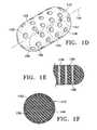
- FIG. 1Dillustrates a tissue cavity marking device with a body having pores.
- FIG. 1Eis a partial cross-sectional view of FIG. 1D .
- FIG. 1Fillustrates a tissue cavity marking device with a body having an outer shell of a bioabsorbable material.
- FIGS. 2A-2Fillustrate various configurations of the marker.
- FIG. 3Aillustrates a cavity marking device having multiple body components traversed by a single wire or suture marker, or multiple wires or suture markers.
- FIG. 3Billustrates a cavity marking device having a helically wound wire or suture marker.
- FIG. 3Cillustrates a cavity marking device having wire or suture markers on the perimeter of the body.
- FIG. 3Dillustrates a cavity marking device having wire or markers on the ends of the body.
- FIGS. 4A-4Cillustrate a method of marking a biopsy tissue cavity with the device of the present invention.
- FIGS. 4D-4Fillustrate a method of marking a biopsy tissue cavity with the device of the present invention wherein a bio-compatible fluid is delivered to the cavity marking device after placement.
- FIGS. 4G-41illustrate a method of marking a biopsy tissue cavity with the device of the present invention wherein a bio-compatible fluid is used to push the cavity marking device out of the access device and into the biopsy tissue cavity.
- FIGS. 4J-4Killustrate a method of marking a biopsy tissue cavity with the device of the present invention wherein the body material of the marking device is deposited into the biopsy cavity prior to the placement of the marker within the biopsy device.
- FIG. 4Lillustrates the placement of the marker into the body material.
- FIGS. 5A-Billustrate a spherical wire marking device for deployment without a filler body into a tissue cavity.
- FIG. 5Cillustrates a cylindrical wire marking device for deployment without a filler body into a tissue cavity.
- FIGS. 5D-Eillustrate a helical coil wire marking device for deployment without a filler body into a tissue cavity.
- FIGS. 1A-1Cshow various configurations of a preferred subcutaneous cavity marking device of the present invention.
- the marking device ( 100 )is displayed as having either a generally spherical body ( 102 ) ( FIG. 1A ), a generally cylindrical body ( 104 ) ( FIG. 1B ), or a multi-faced or irregular body ( 106 ) ( FIG. 1C ).
- the bodymay assume a variety of shapes.
- the bodymay be constructed to have substantially curved surfaces, such as the preferred spherical ( 102 ) and cylindrical ( 104 ) bodies of FIGS. 1A and 1B , respectively.
- the bodymay have conical or ellipsoidal, etc. shapes as well.
- the bodymay have substantially planar surfaces, such as polyhedric (i.e. cubic, tetrahedral, etc.) or prismatic, etc. forms.
- the bodymay also have an irregular or random shape, in the case of a gel, combining features of various curved and planar surfaces.
- Body ( 106 ) of FIG. 1Cis an example of such an irregular body shape.
- the particular body shapewill be chosen to best match to the biopsy cavity in which the device is placed.
- the body shapecan be chosen to be considerably larger than the cavity. Therefore, expansion of the device will provide a significant resistance against the walls of the cavity.
- the aspect ratio of the deviceis not limited to what is displayed in the figures.
- the cylindrical body ( 104 )may have a shorter or longer length as required.
- the generally spherical marker ( 150 )is located at or near the geometric center of the body. Such a configuration will aid the physician in determining the exact location of the biopsy cavity, even after the body degrades and is absorbed into the human or mammalian body.
- the ring-shaped markers ( 154 ) of FIG. 1Bare generally aligned along the longitudinal axis ( 114 ) of body ( 104 ). Note that although the ring-shaped markers ( 154 ) are spatially oriented so that the longitudinal axis ( 114 ) of the body ( 104 ) lies along the longitudinal axes (not shown) of each marker ( 154 ), each marker may individually or together assume a wide variety of random or predetermined spatial orientations other than the aligned orientation as seen in FIG. 1B . It can be appreciated that any asymmetric marker such as marker ( 154 ) is useful in aiding a physician to determine the spatial orientation of the deployed inventive device.
- marker ( 150 ), ( 154 )may reside in locations other than those demonstrated in FIGS. 1A-1C . It is, however, preferred that markers ( 150 ), ( 154 ) dwell in a predetermined, preferably central, location and orientation in the device body so to aid the physician in determining the location and orientation of the biopsy cavity.
- the markers herein describedmay be affixed to the interior or on the surface of the body by any number of suitable methods.
- the markermay be merely suspended in the interior of the body (especially in the case where the body is a gel), it may be woven into the body (especially in the case where the marker is a wire or suture), it may be press fit onto the body (especially in the case where the marker is a ring or band), or it may affixed to the body by a biocompatible adhesive. Any suitable means to affix or suspend the marker into the body in the preferred location is within the scope of the present invention.
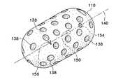
- Tissue regrowth in a particular orientationcan also be promoted by a body design shown in FIG. 1D .
- body ( 110 )contains a number of pores ( 138 ) through which tissue may grow.
- the poresmay also be aligned in a substantially parallel fashion, traversing the thickness of the body so that tissue may regrow from one side of the body through to the other side.
- FIG. 1Eshows a portion ( 130 ) of FIG. 1D in partial longitudinal cross section, complete with pores ( 138 ) traversing through the thickness of portion ( 130 ).
- Such pores ( 138 )can be parallel to each other as shown in FIG. 1E , or they may be perpendicularly, radially, or even randomly oriented in the device body.
- FIG. 1DA trio of markers is also shown in FIG. 1D evenly aligned along the body longitudinal axis ( 140 ).
- Barb marker ( 156 ), spherical marker ( 150 ), and ring-shaped marker ( 154 )demonstrate the use of different multiple markers in a single body ( 110 ). As previously described, such a design helps a physician to determine the spatial orientation of the inventive device when it is deployed in a biopsy cavity.
- the barb marker ( 156 )is illustrated in a ‘V’ configuration, it is an important aspect of the barb marker ( 156 ) to have a shape that is clearly not spherical. This allows the barb marker ( 156 ) to be easily distinguished from calcifications that may be observed during any non-invasive imaging techniques.
- FIG. 1Fdepicts a further embodiment of the present invention in which body ( 112 ) is enveloped in a outer shell ( 142 ) consisting of a layer of bioabsorbable material such those mentioned above.
- a shell ( 142 )consisting of a layer of bioabsorbable material such those mentioned above.
- This configurationallows the perimeter of the biopsy cavity to be marked to avoid exposing the cavity, in the case of a “dirty” margin where re-excision may be necessary, to remaining cancerous cells as the tissue begins to re-grow into the cavity.
- a shell ( 142 )can be radiopaque and/or echogenic in situ, or it may be augmented with an additional coating of an echogenic and/or radiopaque material.
- the shell ( 142 )can also be made to be palpable so that the physician or patient can be further aided in determining the location and integrity of the implanted inventive device.
- Shell ( 142 )may be designed to have a varying bioabsorption rate depending upon the thickness and type of material making up the shell ( 142 ).
- the shellcan be designed to degrade over a period ranging from as long as a year or more to as little as several months, weeks, or even days. It is preferred that such a bioabsorbable shell be designed to degrade between two and six months; especially preferred is three months.
- interior ( 144 ) of body ( 112 )may be a cross-linked, collagenous material that is readily absorbed by the human or mammalian body once the shell ( 142 ) degrades.
- Interior ( 144 )may be filled with a solid or gelatinous material that can be optionally made radiopaque by any number of techniques herein described.
- marker ( 150 ) in the device shown in FIG. 1Fmay be permanently radiopaque or echogenic, or it also may be bioabsorbable and optionally coated with a radiopaque and/or echogenic coating that similarly degrades over a predetermined period of time. It is more important from a clinical standpoint that the marker remain detectable either permanently or, if the patient is uncomfortable with such a scenario, for at least a period of about one to five years so that the physician may follow up with the patient to ensure the health of the tissue in the vicinity of the biopsy cavity. Especially preferable is a marker whose radiopacity or echogenicity lasts from between about one and three years.
- Each of the bodies depicted in FIG. 1A-1Emay be made from a wide variety of solid, liquid, aerosol-spray, spongy, or expanding gelatinous bioabsorbable materials such as collagen, cross-linked collagen, regenerated cellulose, synthetic polymers, synthetic proteins, and combinations thereof. Also contemplated is a body made from a fibrin-collagen matrix, which further prevent unnecessary bleeding, and minimizes the possibility of hematoma formation.
- Examples of synthetic bioabsorbable polymers that may be used for the body of the deviceare polyglycolide, or polyglycolic acid (PGA), polylactide, or polylactic acid (PLA), poly .epsilon.-caprolactone, polydioxanone, polylactide-co-glycolide, e.g., block or random copolymers of PGA and PLA, and other commercial bioabsorbable medical polymers.
- Preferredis spongy collagen or cellulose.
- materials such as hemostatic and pain-killing substancesmay be incorporated into the body and marker of the cavity marking device.
- hemostasis-promoting agentsprovides an obvious benefit as the device not only marks the site of the biopsy cavity but it aids in healing the cavity as well. Furthermore, such agents help to avoid hematomas.
- hemostatic agentsmay include AVITENE Microfibrillar Collagen Hemostat, ACTIFOAM collagen sponge, sold by C. R. Bard Inc., GELFOAM, manufactured by Upjohn Company, SURGICEL Fibrillar from Ethicon Endosurgeries, Inc., and TISSEEL VH, a surgical fibrin sealant sold by Baxter Healthcare Corp.
- the devicemay also be made to emit therapeutic radiation to preferentially treat any suspect tissue remaining in or around the margin of the biopsy cavity.
- the markerwould be the best vehicle for dispensing such local radiation treatment or similar therapy.
- the body itselfmay be. adapted to have radiopaque, echogenic, or other characteristics that allow the body to be located by non-invasive technique without the use of a marker. Such characteristics permit the possibility of locating and substantially identifying the cavity periphery after deployment but prior to absorption of the device.
- an echogenic coatingmay be placed over the radiopaque marker to increase the accuracy of locating the marker during ultrasound imaging.
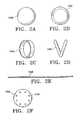
- FIGS. 2A-2Fillustrate various forms of the marker ( 110 ).
- the marker ( 110 )may be in the form of a sphere ( 150 ) ( FIG. 2A ), a hollow sphere ( 152 ) ( FIG. 2B ), a ring or band ( 154 ) ( FIG. 2C ), a barb ( 156 ) ( FIG. 2D ), or a flexible suture or flexible wire ( 158 ) ( FIG. 2E ).
- the markermay have a distinguishing mark ( 170 ) ( FIG. 2F ).
- the barb ( 156 )is illustrated in FIG. 2D as having a “V” shape.
- the barb ( 156 )is intended to distinguish the marker from calcifications when viewed under non-invasive imaging techniques. As such, the barb ( 156 ) is not limited to the “V” shape, rather it has a shape that is easily distinguishable from a spherical or oval calcification.
- the hollow sphere ( 152 ) marker design of FIG. 2Bis more susceptible to detection by ultrasound than the solid sphere ( 150 ) of FIG. 2A .
- Such sphere markers ( 150 , 152 )can be a silicon bead, for instance.
- the body of the cavity marking devicemay be woven or placed through the band/ring ( 154 ).
- the markermay also be a wire or suture ( 158 ) as shown in FIG. 2E and as discussed in greater detail below.
- the marker ( 158 )may be affixed to the exterior perimeter of the body by an adhesive or woven through the body.
- the marker wire or suture ( 158 )being configured in a particular pattern within the body of the device, e.g., wrapping around the body in a helical manner.
- the suture or wire markercan be deployed as a loosely wound ball or mass of suture that when deployed into a tissue cavity, fills the cavity.
- the suture or wirecan also looped through the band/ring ( 154 ); in this configuration (not shown), the suture or wire can also act as the body of the inventive device.
- the suture or wire ( 158 )is flexible to facilitate the expansion of the body while in the cavity. In the case of the marker ( 150 ) shown in FIG.
- distinguishing or identifying mark ( 170 )can be in the form of simple marks as shown, or it may be one or more numbers, letters, symbols, or combinations thereof. These marks ( 160 ) are preferably located in more than one location on the marker ( 150 ) so that the marker may be readily and simply identified from multiple orientations under a variety of viewing conditions. Such a mark ( 170 ) can be used to identify the patient and her condition, provide information about the marker and body of the tissue cavity marking device, provide information about the circumstances and date of the implantation, who performed the procedure, where the procedure was performed, etc. In the case of multiple biopsy sites, this distinguishing mark ( 170 ) permits one to differentiate and identify each different site.
- the mark ( 170 )may be applied via any number of techniques such as physical inscription, physical or plasma deposition, casting, adhesives, etc.
- the mark ( 170 )may also be an electronic chip providing any necessary information in electronic form that can be remotely detected by appropriate means.
- the markermay be radiopaque, echogenic, mammographic, etc. so that it can be located by non-invasive techniques. Such a feature can be an inherent property of the material used for the marker. Alternatively, a coating or the like can be added to the marker to render the marker detectable or to enhance its detectability.
- the markermay be made of a non-bioabsorbable radiopaque material such as platinum, platinum-iridium, platinum-nickel, platinum-tungsten, gold, silver, rhodium, tungsten, tantalum, titanium, nickel, nickel-titanium, their alloys, and stainless steel or any combination of these metals.
- mammographicwe mean that the component described is visible under radiography or any other traditional or advanced mammography technique in which breast tissue is imaged.
- the markercan alternatively be made of or coated with a bioabsorbable material.
- the markercan, for instance, be made from an additive-loaded polymer.
- the additiveis a radiopaque, echogenic, or other type of substance that allows for the non-invasive detection of the marker.
- radiopaque additiveselements such as barium- and bismuth-containing compounds, as well as particulate radio-opaque fillers, e.g., powdered tantalum or tungsten, barium carbonate, bismuth oxide, barium sulfate, etc. are preferred.
- any component of the devicemay be combined with an echogenic coating.
- ECHO-COATfrom STS Biopolymers.
- Such coatingscontain echogenic features which provide the coated item with an acoustically reflective interface and a large acoustical impedance differential.
- an echogenic coatingmay be placed over a radiopaque marker to increase the accuracy of locating the marker during ultrasound imaging.
- the radiopacity and echogenicity described herein for the marker and the bodyare not mutually exclusive. It is within the scope of the present invention for the marker or the body to be radiopaque but not necessarily echogenic, and for the marker or the body to be echogenic but not necessarily radiopaque. It is also within the scope of the invention that the marker and the body are both capable of being simultaneously radiopaque and echogenic. For example, if a platinum ring marker were coated with an echogenic coating, such a marker would be readily visible under x-ray and ultrasonic energy. A similar configuration can be envisioned for the body or for a body coating.
- the markeris preferably large enough to be readily visible to the physician under x-ray or ultrasonic viewing, for example, yet be small enough to be able to be percutaneously deployed into the biopsy cavity and to not cause any difficulties with the patient. More specifically, the marker will not be large enough to be palpable or felt by the patient.
- FIG. 3AAnother useful version of the invention is shown in FIG. 3A .
- the body members ( 302 )can individually or together take on a variety of sizes and shapes as discussed above depending on the characteristics of the biopsy cavity to be filled.
- the body members ( 302 )may uniformly or in combination be made of one or more materials suitable for use in a biopsy cavity as previously described.
- one or more markersmay traverse two or more body member segments through the interior of the body members ( 302 ) as shown in FIG. 3A .
- markers ( 318 )are located substantially parallel to the longitudinal axis ( 320 ) of each right cylindrical body member ( 302 ) in their interior, connecting each body member ( 302 ) while marking their geometric center as between the markers.
- Such a marker ( 318 )may be used in conjunction with the other markers as described above and may also be accompanied by one or more additional markers arranged randomly or in a predetermined pattern to variously mark particular sections of the device. Alternately, such a marker may, singly or in combination with other markers, be affixed on or near the surface of the sponge so as to mark the perimeter of the body member ( 302 ).
- marker ( 318 )when used in conjunction with other connecting markers, marker ( 318 ) need not necessarily connect each body member; it may be used solely to indicate the orientation or location of each individual sponge or the entire device, depending on the material, geometry, size, orientation, etc. of marker ( 318 ). When not used in this connecting function, therefore, marker ( 318 ) need not traverse two body members ( 302 ) as shown in FIG. 3A .
- a marker ( 322 )can wrap around the body ( 302 ) in a helical pattern ( FIG. 3B ), or it can be used in conjunction with other markers ( 324 ) in a pattern parallel to the longitudinal axis ( 320 ) of the body ( 302 ) ( FIG. 3C ).
- Another useful perimeter marking patternis shown in FIG. 3D , where marker segments ( 326 ) are affixed at or near the surface of the circular bases of the cylindrical body ( 302 ) in a cross pattern, indicating the ends of the sponge and their center.
- the marker(s)may, but do not necessarily, have some texture. Any marker pattern, internal or external to the body, is within the scope of the present invention.
- the markerbe a radiopaque or echogenic wire or suture.
- FIG. 3BAnother possible configuration is obtained by combining the suture or wire markers ( 158 ) in a body with any other type marker ( 150 , 152 , 154 , or 156 ) or vice versa.
- a spherical marker ( 150 )may be placed in the center of the cylindrical body ( 302 .) Therefore, the cylindrical body ( 302 ) would contain the suture or wire marker ( 322 ) wrapped helically adjacent to the outer perimeter, and a marker ( 150 ) would be placed in the center of the cylindrical body ( 302 ).
- Such a combinationmay be obtained with any of the body and marker configurations as defined above.
- the markers ( 150 or 154 )may be substituted with one or more suture or wire markers ( 158 ) preferably, but not exclusively, extending through the center and pointing radially away from the center. This configuration allows marking of the cavity perimeter and establishing of the directionality of the cavity itself.
- any of the previously-described additional features of the inventive devicesuch as presence of pain-killing or hemostatic drugs, the capacity for the marker to emit therapeutic radiation for the treatment of various cancers, the various materials that may make up the marker and body, as well as their size, shape, orientation, geometry, etc. may be incorporated into the device described above in conjunction with FIGS. 3A-3D .
- FIG. 4Adetails the marking device ( 402 ) just prior to delivery into a tissue cavity ( 404 ) of human or other mammalian tissue, preferably breast tissue ( 406 ).
- the step illustrated in FIG. 4Ashows a suitable tubular percutaneous access device ( 400 ), such as a catheter or delivery tube, with a distal end ( 408 ) disposed in the interior of cavity ( 404 ).
- the marking device ( 402 )may be delivered percutaneously through the same access device ( 400 ) used to perform the biopsy in which tissue was removed from cavity ( 404 ). Although this is not necessary, it is less traumatic to the patient and allows more precise placement of the marking device ( 402 ) before fluid begins to fill the cavity ( 400 ).
- marking device ( 402 )is shown being pushed out of the distal end ( 408 ) of access device ( 400 ) by a pusher ( 412 ) and resiliently expanding to substantially fill the tissue cavity ( 404 ).
- access device ( 400 )is withdrawn from the breast tissue, leaving marking device ( 402 ) deployed to substantially fill the entire cavity ( 404 ) with radiopaque or echogenic marker ( 410 ) suspended in the geometric center of the marking device ( 402 ) and the cavity ( 404 ).
- the marking device ( 402 )may be sized to be larger than the cavity ( 404 ) thus providing a significant resistance against the walls of the cavity ( 404 ).
- FIGS. 4D-4Fshow a method of delivering the marking device ( 402 ) into a tissue cavity ( 404 ) by a plunger ( 414 ) that is capable of both advancing the marking device ( 402 ) and delivering a bio-compatible fluid ( 416 ).
- the “bio-compatible fluid”is a liquid, solution, or suspension that may contain inorganic or organic material.
- the fluid ( 416 )is preferably a saline solution, but may be water or contain adjuvants such as medications to prevent infection, reduce pain, or the like. Obviously, the fluid ( 416 ) is intended to be a type that does no harm to the body.
- FIG. 4Ddetails the marking device ( 402 ) prior to delivery into the tissue cavity ( 404 ).
- a plunger ( 414 )pushes the marking device ( 402 ) out of the access device ( 400 ).
- the marking device ( 402 )begins resiliently expanding to substantially fill the cavity ( 404 ).
- FIG. 4Fshows the plunger ( 414 ) delivering the bio-compatible fluid ( 416 ) into the cavity ( 404 ).
- the fluid ( 416 )aids the marking device ( 402 ) in expanding to substantially fill the cavity ( 404 ).
- the bio-compatible fluid ( 416 )is delivered subsequent to the placement of the marking device ( 402 ) in the cavity ( 404 ).
- the marking device ( 402 )may also be soaked with fluid ( 416 ) prior to placement in the cavity ( 404 ).
- FIGS. 4G-41show another method of delivering the marking device ( 402 ) into the tissue cavity ( 404 ) by using the bio-compatible fluid ( 416 ) as the force to deliver the marking device ( 402 ) into the tissue cavity ( 404 ).
- FIG. 4Gdetails the marking device ( 402 ) prior to delivery into the tissue cavity ( 404 ).
- FIG. 4Hillustrates flow of the bio-compatible fluid ( 416 ) in the access device ( 400 ), the fluid ( 416 ) flow then pushes the marking device ( 402 ) out of the access device ( 400 ).
- FIG. 41shows the delivery device ( 400 ) continuing to deliver the bio-compatible fluid ( 416 ) into the cavity ( 404 ).
- the fluid ( 416 )aids the marking device ( 402 ) in expanding to substantially fill the cavity ( 404 ).
- the bio-compatible fluid ( 416 )is delivered after the placement of the marking device ( 402 ) in the cavity ( 404 ) although the invention is not limited to the continued delivery of the fluid ( 416 ).
- FIG. 4J-4Kshows the method of delivering the body ( 418 ) of the cavity marking device directly into the cavity ( 404 ) prior to the placement of the marker ( 410 ) in the device ( 402 ).
- FIG. 4Jshows the deposit of the body material ( 418 ) into the cavity ( 404 ).
- the body material ( 418 )may be a gel type material as described above.
- FIG. 4Kdetails the filling of the cavity ( 404 ) with the body material ( 418 ).
- the delivery device(not shown in FIG. 4K ) may be withdrawn.
- FIG. 4Ldetails the placement of the marker ( 410 ) into the body material ( 418 ).
- FIGS. 5A-5Eshow yet another version of the invention in which a marker, preferably consisting of a radiopaque or echogenic wire, is deployed alone into a tissue cavity without the use of any body.
- the markercan be made of a shape memory material, such as a nickel-titanium alloy, which when deployed into the biopsy cavity, assumes a predetermined configuration to substantially fill the cavity, mark the cavity location and margin, and indicate the orientation of the marker inside the cavity.
- marker ( 500 )is a three-dimensional sphere consisting of two rings ( 502 ), ( 504 ) pivotally connected at ends ( 506 ), ( 508 ) so to assume a spherical shape.
- a markercan be made of a shape memory metal so that when it is placed in a deployment tube ( 510 ) shown in FIG. 5B , marker ( 500 ) assumes a collapsed profile suitable for deployment through tube ( 510 ) by pusher ( 512 ).
- marker ( 500 )Upon exiting into the tissue cavity (not shown), marker ( 500 ) assumes the spherical shape of FIG. 5A to fill the cavity.
- the marker ( 500 )may also be shaped into any similar shape such as an ellipsoidal shape.
- FIG. 5Ca marker ( 520 ) in the form of a wire cylinder is shown.
- this deviceis structurally configured to assume the depicted cylindrical configuration when deployed in the tissue cavity, but may be (as described above) “collapsed” into a deployment tube for percutaneous delivery.
- This deviceis especially suitable for marking the distal and proximal ends of the tissue cavity due to its asymmetrical shape.
- FIG. 5Dshows a shape memory marker ( 530 ) in the form of a helical coil deployed into tissue cavity ( 532 ).
- a shape memory marker ( 530 )in the form of a helical coil deployed into tissue cavity ( 532 ).
- such a marker ( 530 )may be deployed through delivery tube ( 510 ) by pusher ( 512 ) in a substantially elongated, straightened form, only to substantially assume the shape of the cavity ( 532 ) as shown in FIG. 5D .
- Any suitable delivery device or pusher ( 512 ) capable of deploying marker ( 530 ) into cavity ( 532 )is within the scope of this invention.
- Each of the markers shown in FIGS. 5A-5Eis preferably a shape memory material coated or supplemented with a radiopacity-enhancing material, such as gold, platinum, or any other radiopaque material herein discussed.
- the markersmay singly, or in combination with being radiopaque, be echogenic or be made echogenic by any of the materials or methods herein described.
- the inventionprovides an improved subcutaneous cavity marking device and method. While the above descriptions have described the invention for use in the marking of biopsy cavities, the invention is not limited to such. One such application is evident as the invention may further be used as a lumpectomy site marker. In this use, the cavity marking device yield an improved benefit by marking the perimeter of the lumpectomy cavity.
Landscapes
- Health & Medical Sciences (AREA)
- Life Sciences & Earth Sciences (AREA)
- Public Health (AREA)
- Engineering & Computer Science (AREA)
- General Health & Medical Sciences (AREA)
- Biomedical Technology (AREA)
- Nuclear Medicine, Radiotherapy & Molecular Imaging (AREA)
- Animal Behavior & Ethology (AREA)
- Veterinary Medicine (AREA)
- Physics & Mathematics (AREA)
- Surgery (AREA)
- Epidemiology (AREA)
- Radiology & Medical Imaging (AREA)
- Molecular Biology (AREA)
- Heart & Thoracic Surgery (AREA)
- Medical Informatics (AREA)
- Pathology (AREA)
- Optics & Photonics (AREA)
- Oncology (AREA)
- Biodiversity & Conservation Biology (AREA)
- Acoustics & Sound (AREA)
- Oral & Maxillofacial Surgery (AREA)
- Hematology (AREA)
- Biophysics (AREA)
- Media Introduction/Drainage Providing Device (AREA)
- Surgical Instruments (AREA)
Abstract
Description
This application is a continuation of U.S. Ser. No. 14/067,270, filed Oct. 30, 2013, which is currently pending, which is a continuation of U.S. Ser. No. 13/604,158, filed Sep. 5, 2012, now U.S. Pat. No. 8,600,481, which is a continuation of U.S. Ser. No. 09/805,652, filed Mar. 13, 2001, now U.S. Pat. No. 8,320,993, which is a continuation of U.S. Ser. No. 09/285,329, filed Apr. 2, 1999, now U.S. Pat. No. 6,356,782, which is a continuation-in-part of U.S. Ser. No. 09/220,618, filed Dec. 24, 1998, which is abandoned.
This invention is directed to subcutaneous cavity marking devices and methods. More particularly, a cavity marking device and method is disclosed that enable one to determine the location, orientation, and periphery of the cavity by radiographic, mammographic, echographic, or other non-invasive techniques. The invention typically is made up of one or more resilient bodies and a radiopaque or echogenic marker.
Over 1.1 million breast biopsies are performed each year in the United States alone. Of these, about 80% of the lesions excised during biopsy are found to be benign while about 20% of these lesions are malignant.
In the field of breast cancer, stereotactically guided and percutaneous biopsy procedures have increased in frequency as well as in accuracy as modem imaging techniques allow the physician to locate lesions with ever-increasing precision. However, for any given biopsy procedure, a subsequent examination of the biopsy site is very often desirable. There is an important need to determine the location, most notably the center, as well as the orientation and periphery (margins) of the subcutaneous cavity from which the lesion is removed.
In those cases where the lesion is found to be benign, for example, a follow-up examination of the biopsy site is often performed to ensure the absence of any suspect tissue and the proper healing of the cavity from which the tissue was removed. This is also the case where the lesion is found to be malignant and the physician is confident that all suspect tissue was removed and the tissue in the region of the perimeter or margins of the cavity is “clean”.
In some cases, however, the physician may be concerned that the initial biopsy failed to remove a sufficient amount of the lesion. Such a lesion is colloquially referred to as a “dirty lesion” or “dirty margin” and requires follow-up observation of any suspect tissue growth in the surrounding marginal area of the initial biopsy site. Thus, a re-excision of the original biopsy site must often be performed. In such a case, the perimeter of the cavity must be identified since the cavity may contain cancerous cells. Identification of the cavity perimeter is necessary to avoid the risk of opening the cavity, which could release and spread cancerous cells. Moreover, the site of the re-excised procedure itself requires follow-up examination, providing further impetus for accurate identification of the location of the re-excised site. Therefore, a new marker will be placed after re-excision.
Prior methods of marking biopsy cavities utilize one or more tissue marking clips as the biopsy site marking device. Most commonly, these marker clips have a “horseshoe” configuration. The marker clips attach to the walls of the cavity when the free ends or limbs of the “horseshoe” are pinched together, trapping the tissue. This device has significant drawbacks.
For instance, prior to placing the marker clip at the cavity site, the site must be thoroughly cleaned, typically by vacuum, to remove any residual tissue debris. This minimizes the possibility that the marker clip attaches to any loose tissue as opposed to the cavity wall. Once the cavity is prepared, the clip must be examined to ensure that the limbs of the clip are substantially straight. If the limbs have been prematurely bent together, the clip will be discarded since it will most likely not attach properly to the cavity wall. Actual placement of the clip often requires additional vacuum of the cavity wall to draw the wall into the aperture between the limbs of the marking clip so that a better grip is obtained between the limbs of the clip. Additionally, there is always the possibility that the clip may detach from the cavity wall during or after withdrawal of the tools used to place the clip into the cavity.
Aside from the problems inherent in the placement of the marking clip, there are also limitations associated with how well the marking clip can identify a biopsy cavity. As the marking clip must trap tissue for proper attachment, in cases of endoscopic placement, the clip can only be placed on a wall of the cavity substantially opposite to the opening of the cavity.
Moreover, patient concern limits the number of clips that may be placed in a cavity. As a result, the medical practitioner is forced to identify the outline of a three dimensional cavity by a single point as defined by the marking clip. Obviously, determination of the periphery of a biopsy cavity from one point of the periphery is not possible.
These limitations are compounded as the biopsy cavity fills within a few hours with bodily fluids, which eventually renders the cavity invisible to non-invasive techniques. Another difficulty in viewing the clip stems from the fact that the clip is attached to the side, not the center, of the cavity. This makes determining the spatial orientation and position of the cavity difficult if not impossible during follow-up examination. Additionally, during a stereotactic breast biopsy procedure, the breast is under compression when the marking clip is placed. Upon release of the compressive force, determining the location of the clip can be unpredictable, and the orientation as well as the location of the periphery of the cavity are lost.
The marker clip does not aid in the healing process of the biopsy wound. Complications may arise if the marker strays from its original placement site. As described above, if a re-excision of the site is required, the marker clip may also interfere when excision of a target lesion is sought.
Other devices pertaining to biopsy aids are directed to assisting in the healing and closure of the biopsy wound; thus they do not aid the clinical need or desirability of accurately preserving the location and orientation of the biopsy cavity. See, e.g., U.S. Pat. Nos. 4,347,234, 5,388,588, 5,326,350, 5,394,886, 5,467,780, 5,571,181, and 5,676,146.
This invention relates to devices and procedures for percutaneously marking a biopsy cavity. In particular, the inventive device is a biopsy cavity-marking body made of a resilient, preferably bioabsorbable material having at least one preferably radiopaque or echogenic marker. The device may take on a variety of shapes and sizes tailored for the specific biopsy cavity to be filled. For example, the device in its simplest form is a spherical or cylindrical collagen sponge having a single radiopaque or echogenic marker located in its geometric center. Alternatively, the body may have multiple components linked together with multiple radiopaque or echogenic markers.
A further aspect of the invention allows the marker or the body, singly or in combination, to be constructed to have a varying rate of degradation or bioabsorption. For instance, the body may be constructed to have a layer of bioabsorbable material as an outer “shell.” Accordingly, prior to degradation of the shell, the body is palpable. Upon degradation of the shell, the remainder of the body would degrade at an accelerated rate in comparison to the outer shell.
The device may additionally contain a variety of drugs, such as hemostatic agents, pain-killing substances, or even healing or therapeutic agents that may be delivered directly to the biopsy cavity. Importantly, the device is capable of accurately marking a specific location, such as the center, of the biopsy cavity, and providing other information about the patient or the particular biopsy or device deployed.
The device is preferably, although not necessarily, delivered immediately after removal of the tissue specimen using the same device used to remove the tissue specimen itself. Such devices are described in pending U.S. patent application Ser. No. 09/145,487, filed Sep. 1, 1998 and entitled “PERCUTANEOUS TISSUE REMOVAL DEVICE”, and pending U.S. patent application Ser. No. 09/184,766, filed Nov. 2, 1998 and entitled “EXPANDABLE RING PERCUTANEOUS TISSUE REMOVAL DEVICE” the entirety of each are which hereby incorporated by reference. The device is compressed and loaded into the access device and percutaneously advanced to the biopsy site where, upon exiting from the access device, it expands to substantially fill the cavity of the biopsy. Follow-up noninvasive detection techniques, such as x-ray mammography or ultrasound may then be used by the physician to identify, locate, and monitor the biopsy cavity site over a preferred period of time.
The device is usually inserted into the body either surgically via an opening in the body cavity, or through a minimally invasive procedure using such devices as a catheter, introducer or similar type device. When inserted via the minimally invasive procedure, the resiliency of the body allows the device to be compressed upon placement in a delivery device. Upon insertion of the cavity marking device into the cavity, the resiliency of the body causes the cavity marking device to self-expand, substantially filling the cavity. The resiliency of the body can be further pre-determined so that the body is palpable, thus allowing tactile location by a surgeon in subsequent follow-up examinations. Typically, the filler body is required to be palpable for approximately 3 months. However, this period may be increased or decreased as needed.
The expansion of the resilient body can be aided by the addition of a bio-compatible fluid which is absorbed into the body. For instance, the fluid can be a saline solution, a painkilling substance, a healing agent, a therapeutic fluid, or any combination of such fluids. The fluid or combination of fluids may be added to and absorbed by the body of the device before or after deployment of the device into a cavity. For example, the body of the device may be pre-soaked with the fluid and then delivered into the cavity. In this instance, the fluid aids the expansion of the body of the device upon deployment. Another example is provided as the device is delivered into the cavity without being pre-soaked. In such a case, fluid is delivered into the cavity after the body of the device is deployed into the cavity. Upon delivery of the fluid, the body of the device soaks the fluid, thereby aiding the expansion of the cavity marking device as it expands to fit the cavity. The fluid may be, but is not limited to being, delivered by the access device.
By “bio-compatible fluid” what is meant is a liquid, solution, or suspension that may contain inorganic or organic material. For instance, the bio-compatible fluid is preferably saline solution, but may be water or contain adjuvants such as medications to prevent infection, reduce pain, or the like. Obviously, the liquid is intended to be a type that does no harm to the body.
After placement of the cavity marking device into the cavity, the bioabsorbable body degrades at a predetermined rate. As the body of the cavity marking device is absorbed, tissue is substituted for the bioabsorbable material. Moreover, while the body degrades, the marker, which is usually suspended substantially in the volumetric center of the body of the device, is left in the center of the cavity. Thus, during a subsequent examination, a medical practitioner having knowledge of the dimensions of the body of the cavity marking device can determine the location as well as the periphery of the biopsy cavity. The orientation of the cavity is self-evident as the marker is left in substantially the center of the cavity. For the case where multiple markers are used, the markers are usually placed in a manner showing directionality.
Both the body and the marker can be made, via radiopaque or echogenic coatings or in situ, to degrade and absorb into the patient's body over a predetermined period of time. It is generally preferred that if the marker's radiopacity or echogenicity is chosen to degrade over time, such degradation does not take place within at least one year after implantation of the inventive device. In this way, if a new lump or calcification (in the case of a breast biopsy) is discovered after the biopsy, such a marker will allow the physician to know the relation of such new growth in relation to the region of excised tissue. On the other hand, and as discussed below, a bioabsorption period of three months is preferred for any such coatings on the perimeter of the body itself
This invention further includes the act of filling the biopsy cavity with a bioabsorbable liquid, aerosol or gelatinous material, preferably gelatinous collagen, allowing the material to partially solidify or gel and then placing a marker, which may have a configuration as described above, into the center of the bioabsorbable material. The gel may also be made radiopaque or echogenic by the addition of radiopaque materials, such as barium- or bismuth-containing compounds and the like, as well as particulate radio-opaque fillers, e.g., powdered tantalum or tungsten, barium carbonate, bismuth oxide, barium sulfate, to the gel.
This method may be combined with any aspect of the previously described devices as needed. For instance, one could insert a hemostatic or pain-killing substance as described above into the biopsy cavity along with the bioabsorbable material. Alternatively, a bioabsorbable marker could be inserted into a predetermined location, such as the center, of the body of bioabsorbable material.
It is within the scope of this invention that either or both of the marker or markers and the bioabsorbable body may be radioactive, if a regimen of treatment using radioactivity is contemplated.
This procedure may be used in any internal, preferably soft, tissue, but is most useful in breast tissue, lung tissue, prostate tissue, lymph gland tissue, etc. Obviously, though, treatment and diagnosis of breast tissue problems forms the central theme of the invention.
In contrast to the marker clips as described above, the cavity marking device has the obvious advantage of marking the geometric center of a biopsy cavity. Also, unlike the marking clip which has the potential of attaching to loose tissue and moving after initial placement, the marking device self-expands upon insertion into the cavity, thus providing resistance against the walls of the cavity thereby anchoring itself within the cavity. The marking device may be configured to be substantially smaller, larger, or equal to the size of the cavity, however, in some cases the device will be configured to be larger than the cavity. This aspect of the biopsy marking device provides a cosmetic benefit to the patient, especially when the biopsy is taken from the breast. For example, the resistance provided by the cavity marking device against the walls of the cavity may minimize any “dimpling” effect observed in the skin when large pieces of tissue are removed, as, for example, during excisional biopsies.
Although the subcutaneous cavity marking device and method described above are suited for percutaneous placement of the marker within a biopsy cavity it is not intended that the invention is limited to such placement. The device and method are also appropriate for intra-operative or surgical placement of the marker within a biopsy cavity.
In the bodies ofFIGS. 1A and 1C , the generally spherical marker (150) is located at or near the geometric center of the body. Such a configuration will aid the physician in determining the exact location of the biopsy cavity, even after the body degrades and is absorbed into the human or mammalian body.
In the case of the ring-shaped markers (154) ofFIG. 1B , they are generally aligned along the longitudinal axis (114) of body (104). Note that although the ring-shaped markers (154) are spatially oriented so that the longitudinal axis (114) of the body (104) lies along the longitudinal axes (not shown) of each marker (154), each marker may individually or together assume a wide variety of random or predetermined spatial orientations other than the aligned orientation as seen inFIG. 1B . It can be appreciated that any asymmetric marker such as marker (154) is useful in aiding a physician to determine the spatial orientation of the deployed inventive device.
Obviously, marker (150), (154) may reside in locations other than those demonstrated inFIGS. 1A-1C . It is, however, preferred that markers (150), (154) dwell in a predetermined, preferably central, location and orientation in the device body so to aid the physician in determining the location and orientation of the biopsy cavity. The markers herein described may be affixed to the interior or on the surface of the body by any number of suitable methods. For instance, the marker may be merely suspended in the interior of the body (especially in the case where the body is a gel), it may be woven into the body (especially in the case where the marker is a wire or suture), it may be press fit onto the body (especially in the case where the marker is a ring or band), or it may affixed to the body by a biocompatible adhesive. Any suitable means to affix or suspend the marker into the body in the preferred location is within the scope of the present invention.
Tissue regrowth in a particular orientation can also be promoted by a body design shown inFIG. 1D . Here, body (110) contains a number of pores (138) through which tissue may grow. The pores may also be aligned in a substantially parallel fashion, traversing the thickness of the body so that tissue may regrow from one side of the body through to the other side. This is demonstrated in insetFIG. 1E , which shows a portion (130) ofFIG. 1D in partial longitudinal cross section, complete with pores (138) traversing through the thickness of portion (130). Such pores (138) can be parallel to each other as shown inFIG. 1E , or they may be perpendicularly, radially, or even randomly oriented in the device body.
A trio of markers is also shown inFIG. 1D evenly aligned along the body longitudinal axis (140). Barb marker (156), spherical marker (150), and ring-shaped marker (154) demonstrate the use of different multiple markers in a single body (110). As previously described, such a design helps a physician to determine the spatial orientation of the inventive device when it is deployed in a biopsy cavity. Although the barb marker (156) is illustrated in a ‘V’ configuration, it is an important aspect of the barb marker (156) to have a shape that is clearly not spherical. This allows the barb marker (156) to be easily distinguished from calcifications that may be observed during any non-invasive imaging techniques.
Shell (142) may be designed to have a varying bioabsorption rate depending upon the thickness and type of material making up the shell (142). In general, the shell can be designed to degrade over a period ranging from as long as a year or more to as little as several months, weeks, or even days. It is preferred that such a bioabsorbable shell be designed to degrade between two and six months; especially preferred is three months. In the design ofFIG. 1F , interior (144) of body (112) may be a cross-linked, collagenous material that is readily absorbed by the human or mammalian body once the shell (142) degrades. Interior (144) may be filled with a solid or gelatinous material that can be optionally made radiopaque by any number of techniques herein described.
As will be described in additional detail with respect toFIGS. 2A-2F , marker (150) in the device shown inFIG. 1F may be permanently radiopaque or echogenic, or it also may be bioabsorbable and optionally coated with a radiopaque and/or echogenic coating that similarly degrades over a predetermined period of time. It is more important from a clinical standpoint that the marker remain detectable either permanently or, if the patient is uncomfortable with such a scenario, for at least a period of about one to five years so that the physician may follow up with the patient to ensure the health of the tissue in the vicinity of the biopsy cavity. Especially preferable is a marker whose radiopacity or echogenicity lasts from between about one and three years.
Each of the bodies depicted inFIG. 1A-1E may be made from a wide variety of solid, liquid, aerosol-spray, spongy, or expanding gelatinous bioabsorbable materials such as collagen, cross-linked collagen, regenerated cellulose, synthetic polymers, synthetic proteins, and combinations thereof. Also contemplated is a body made from a fibrin-collagen matrix, which further prevent unnecessary bleeding, and minimizes the possibility of hematoma formation.
Examples of synthetic bioabsorbable polymers that may be used for the body of the device are polyglycolide, or polyglycolic acid (PGA), polylactide, or polylactic acid (PLA), poly .epsilon.-caprolactone, polydioxanone, polylactide-co-glycolide, e.g., block or random copolymers of PGA and PLA, and other commercial bioabsorbable medical polymers. Preferred is spongy collagen or cellulose. As mentioned above, materials such as hemostatic and pain-killing substances may be incorporated into the body and marker of the cavity marking device. The use of hemostasis-promoting agents provides an obvious benefit as the device not only marks the site of the biopsy cavity but it aids in healing the cavity as well. Furthermore, such agents help to avoid hematomas. These hemostatic agents may include AVITENE Microfibrillar Collagen Hemostat, ACTIFOAM collagen sponge, sold by C. R. Bard Inc., GELFOAM, manufactured by Upjohn Company, SURGICEL Fibrillar from Ethicon Endosurgeries, Inc., and TISSEEL VH, a surgical fibrin sealant sold by Baxter Healthcare Corp. The device may also be made to emit therapeutic radiation to preferentially treat any suspect tissue remaining in or around the margin of the biopsy cavity. It is envisioned that the marker would be the best vehicle for dispensing such local radiation treatment or similar therapy. Also, the body itself may be. adapted to have radiopaque, echogenic, or other characteristics that allow the body to be located by non-invasive technique without the use of a marker. Such characteristics permit the possibility of locating and substantially identifying the cavity periphery after deployment but prior to absorption of the device. Furthermore, an echogenic coating may be placed over the radiopaque marker to increase the accuracy of locating the marker during ultrasound imaging.
The hollow sphere (152) marker design ofFIG. 2B is more susceptible to detection by ultrasound than the solid sphere (150) ofFIG. 2A . Such sphere markers (150,152) can be a silicon bead, for instance. In the case of a ring or band marker (154) seen inFIG. 2C , the body of the cavity marking device may be woven or placed through the band/ring (154). The marker may also be a wire or suture (158) as shown inFIG. 2E and as discussed in greater detail below. In such a case, the marker (158) may be affixed to the exterior perimeter of the body by an adhesive or woven through the body. Another improvement may arise from the marker wire or suture (158) being configured in a particular pattern within the body of the device, e.g., wrapping around the body in a helical manner. Further, the suture or wire marker can be deployed as a loosely wound ball or mass of suture that when deployed into a tissue cavity, fills the cavity. The suture or wire can also looped through the band/ring (154); in this configuration (not shown), the suture or wire can also act as the body of the inventive device. The suture or wire (158) is flexible to facilitate the expansion of the body while in the cavity. In the case of the marker (150) shown inFIG. 2F , distinguishing or identifying mark (170) can be in the form of simple marks as shown, or it may be one or more numbers, letters, symbols, or combinations thereof. These marks (160) are preferably located in more than one location on the marker (150) so that the marker may be readily and simply identified from multiple orientations under a variety of viewing conditions. Such a mark (170) can be used to identify the patient and her condition, provide information about the marker and body of the tissue cavity marking device, provide information about the circumstances and date of the implantation, who performed the procedure, where the procedure was performed, etc. In the case of multiple biopsy sites, this distinguishing mark (170) permits one to differentiate and identify each different site. The mark (170) may be applied via any number of techniques such as physical inscription, physical or plasma deposition, casting, adhesives, etc. The mark (170) may also be an electronic chip providing any necessary information in electronic form that can be remotely detected by appropriate means.
An important aspect of the invention is that the marker may be radiopaque, echogenic, mammographic, etc. so that it can be located by non-invasive techniques. Such a feature can be an inherent property of the material used for the marker. Alternatively, a coating or the like can be added to the marker to render the marker detectable or to enhance its detectability. For radiopacity, the marker may be made of a non-bioabsorbable radiopaque material such as platinum, platinum-iridium, platinum-nickel, platinum-tungsten, gold, silver, rhodium, tungsten, tantalum, titanium, nickel, nickel-titanium, their alloys, and stainless steel or any combination of these metals. By mammographic we mean that the component described is visible under radiography or any other traditional or advanced mammography technique in which breast tissue is imaged.
As previously discussed, the marker can alternatively be made of or coated with a bioabsorbable material. In this case, the marker can, for instance, be made from an additive-loaded polymer. The additive is a radiopaque, echogenic, or other type of substance that allows for the non-invasive detection of the marker. In the case of radiopaque additives, elements such as barium- and bismuth-containing compounds, as well as particulate radio-opaque fillers, e.g., powdered tantalum or tungsten, barium carbonate, bismuth oxide, barium sulfate, etc. are preferred. To aid in detection by ultrasound or similar imaging techniques, any component of the device may be combined with an echogenic coating. One such coating is ECHO-COAT from STS Biopolymers. Such coatings contain echogenic features which provide the coated item with an acoustically reflective interface and a large acoustical impedance differential. As stated above, an echogenic coating may be placed over a radiopaque marker to increase the accuracy of locating the marker during ultrasound imaging.
Note that the radiopacity and echogenicity described herein for the marker and the body are not mutually exclusive. It is within the scope of the present invention for the marker or the body to be radiopaque but not necessarily echogenic, and for the marker or the body to be echogenic but not necessarily radiopaque. It is also within the scope of the invention that the marker and the body are both capable of being simultaneously radiopaque and echogenic. For example, if a platinum ring marker were coated with an echogenic coating, such a marker would be readily visible under x-ray and ultrasonic energy. A similar configuration can be envisioned for the body or for a body coating.
The marker is preferably large enough to be readily visible to the physician under x-ray or ultrasonic viewing, for example, yet be small enough to be able to be percutaneously deployed into the biopsy cavity and to not cause any difficulties with the patient. More specifically, the marker will not be large enough to be palpable or felt by the patient.
Another useful version of the invention is shown inFIG. 3A . In this device, there are several cylindrical body members (302); however, there is no limit to the number of body members that can make up the device. The body members (302) can individually or together take on a variety of sizes and shapes as discussed above depending on the characteristics of the biopsy cavity to be filled. The body members (302) may uniformly or in combination be made of one or more materials suitable for use in a biopsy cavity as previously described.
Here one or more markers may traverse two or more body member segments through the interior of the body members (302) as shown inFIG. 3A . Here, markers (318) are located substantially parallel to the longitudinal axis (320) of each right cylindrical body member (302) in their interior, connecting each body member (302) while marking their geometric center as between the markers. Such a marker (318) may be used in conjunction with the other markers as described above and may also be accompanied by one or more additional markers arranged randomly or in a predetermined pattern to variously mark particular sections of the device. Alternately, such a marker may, singly or in combination with other markers, be affixed on or near the surface of the sponge so as to mark the perimeter of the body member (302).
Of course, when used in conjunction with other connecting markers, marker (318) need not necessarily connect each body member; it may be used solely to indicate the orientation or location of each individual sponge or the entire device, depending on the material, geometry, size, orientation, etc. of marker (318). When not used in this connecting function, therefore, marker (318) need not traverse two body members (302) as shown inFIG. 3A .
A variety of patterns can be envisioned in which all or part of the perimeter of the sponge body is marked. For example, a marker (322) can wrap around the body (302) in a helical pattern (FIG. 3B ), or it can be used in conjunction with other markers (324) in a pattern parallel to the longitudinal axis (320) of the body (302) (FIG. 3C ). Another useful perimeter marking pattern is shown inFIG. 3D , where marker segments (326) are affixed at or near the surface of the circular bases of the cylindrical body (302) in a cross pattern, indicating the ends of the sponge and their center. As seen from the figures, the marker(s) may, but do not necessarily, have some texture. Any marker pattern, internal or external to the body, is within the scope of the present invention. For the applications depicted inFIGS. 3A-3D , it is preferred that the marker be a radiopaque or echogenic wire or suture.
Another possible configuration is obtained by combining the suture or wire markers (158) in a body with any other type marker (150,152,154, or156) or vice versa. For example, inFIG. 3B , a spherical marker (150) may be placed in the center of the cylindrical body (302.) Therefore, the cylindrical body (302) would contain the suture or wire marker (322) wrapped helically adjacent to the outer perimeter, and a marker (150) would be placed in the center of the cylindrical body (302). Such a combination may be obtained with any of the body and marker configurations as defined above.
Also, turning back to the marking device (100) inFIG. 1A or the marking device (100) ofFIG. 11B , the markers (150 or154) may be substituted with one or more suture or wire markers (158) preferably, but not exclusively, extending through the center and pointing radially away from the center. This configuration allows marking of the cavity perimeter and establishing of the directionality of the cavity itself.
Any of the previously-described additional features of the inventive device, such as presence of pain-killing or hemostatic drugs, the capacity for the marker to emit therapeutic radiation for the treatment of various cancers, the various materials that may make up the marker and body, as well as their size, shape, orientation, geometry, etc. may be incorporated into the device described above in conjunction withFIGS. 3A-3D .
Turning now toFIGS. 4A-4C , a method of delivering the inventive device ofFIG. 1A is shown.FIG. 4A details the marking device (402) just prior to delivery into a tissue cavity (404) of human or other mammalian tissue, preferably breast tissue (406). As can be seen, the step illustrated inFIG. 4A shows a suitable tubular percutaneous access device (400), such as a catheter or delivery tube, with a distal end (408) disposed in the interior of cavity (404). As previously described, the marking device (402) may be delivered percutaneously through the same access device (400) used to perform the biopsy in which tissue was removed from cavity (404). Although this is not necessary, it is less traumatic to the patient and allows more precise placement of the marking device (402) before fluid begins to fill the cavity (400).
InFIG. 4B , marking device (402) is shown being pushed out of the distal end (408) of access device (400) by a pusher (412) and resiliently expanding to substantially fill the tissue cavity (404).
Finally, inFIG. 4C , access device (400) is withdrawn from the breast tissue, leaving marking device (402) deployed to substantially fill the entire cavity (404) with radiopaque or echogenic marker (410) suspended in the geometric center of the marking device (402) and the cavity (404). As mentioned above, the marking device (402) may be sized to be larger than the cavity (404) thus providing a significant resistance against the walls of the cavity (404).
InFIG. 5A , marker (500) is a three-dimensional sphere consisting of two rings (502), (504) pivotally connected at ends (506), (508) so to assume a spherical shape. Such a marker can be made of a shape memory metal so that when it is placed in a deployment tube (510) shown inFIG. 5B , marker (500) assumes a collapsed profile suitable for deployment through tube (510) by pusher (512). Upon exiting into the tissue cavity (not shown), marker (500) assumes the spherical shape ofFIG. 5A to fill the cavity. The marker (500) may also be shaped into any similar shape such as an ellipsoidal shape.
Turning now toFIG. 5C , a marker (520) in the form of a wire cylinder is shown. Again, this device is structurally configured to assume the depicted cylindrical configuration when deployed in the tissue cavity, but may be (as described above) “collapsed” into a deployment tube for percutaneous delivery. This device is especially suitable for marking the distal and proximal ends of the tissue cavity due to its asymmetrical shape.
Each of the markers shown inFIGS. 5A-5E is preferably a shape memory material coated or supplemented with a radiopacity-enhancing material, such as gold, platinum, or any other radiopaque material herein discussed. The markers may singly, or in combination with being radiopaque, be echogenic or be made echogenic by any of the materials or methods herein described.
From the foregoing, it is understood that the invention provides an improved subcutaneous cavity marking device and method. While the above descriptions have described the invention for use in the marking of biopsy cavities, the invention is not limited to such. One such application is evident as the invention may further be used as a lumpectomy site marker. In this use, the cavity marking device yield an improved benefit by marking the perimeter of the lumpectomy cavity.
The invention herein has been described by examples and a particularly desired way of practicing the invention has been described. However, the invention as claimed herein is not limited to that specific description in any manner. Equivalence to the description as hereinafter claimed is considered to be within the scope of protection of this patent.
Claims (19)
1. A biopsy marking device comprising:
a cylindrical body having an exterior surface and a central longitudinal axis, the body comprising a polymer;
at least one echogenic marker arranged along the central longitudinal axis;
wherein the body includes at least one hollow pore extending through the body at an angle to the central longitudinal axis,
wherein the at least one pore has an opening at the exterior surface of the body; and wherein the body is echogenic.
2. The biopsy marking device according toclaim 1 , wherein the body comprises a synthetic polymer.
3. The biopsy marking device ofclaim 1 , wherein the body comprises a biodegradable polymer.
4. The biopsy marking device ofclaim 1 , wherein the at least one hollow pore extends completely through the body.
5. The biopsy marking device ofclaim 1 , wherein the at least one hollow pore includes at least two parallel pores extending through the body at an angle to the central longitudinal axis.
6. The biopsy marking device ofclaim 1 , wherein the at least one hollow pore comprises a first set of pores comprising at least two parallel pores extending through the body at a first angle with respect to the central longitudinal axis and wherein the at least one hollow pore comprises a second set of pores comprising at least two parallel pores extending through the body at a second angle with respect to the central longitudinal axis.
7. The biopsy marking device ofclaim 6 , wherein each of the pores extend completely through the body.
8. The biopsy marking device ofclaim 1 , wherein the at least one hollow pore comprises is three parallel spaced pores.
9. The biopsy marking device ofclaim 1 , wherein the at least one hollow pore comprises at least two parallel pores extending through the body at an angle perpendicular to the central longitudinal axis.
10. The biopsy marking device ofclaim 1 , further including a tubular percutaneous access device in which the body is positioned, the tubular percutaneous access device including a pusher.
11. The biopsy marking device ofclaim 1 , wherein the at least one echogenic marker comprises a plurality of markers, and wherein each marker of the plurality of markers is arranged along the central longitudinal axis.
12. The biopsy marking device ofclaim 11 , wherein at least two markers of the plurality of markers have different shapes.
13. A biopsy marking device comprising:
a cylindrical polymer body having an exterior surface and a central longitudinal axis;
at least one marker arranged along the central longitudinal axis;
the body including parallel spaced hollow pores extending through the body perpendicular to the central longitudinal axis forming a first set of pores; and
wherein each of the pores has an opening at the exterior surface of the body.
14. The biopsy marking device according toclaim 13 , further including a second set of pores, the second set of pores including parallel spaced pores extending through the body perpendicular to the central longitudinal axis and perpendicular to the first set of pores.
15. The biopsy marker according toclaim 14 , wherein each of the pores of the second set of pores has an opening at the exterior surface of the body.
16. The biopsy marking device ofclaim 13 , further including a tubular percutaneous access device in which the body is positioned, the tubular percutaneous access device including a pusher.
17. The biopsy marking device ofclaim 13 , wherein the body is echogenic.
18. The biopsy marking device ofclaim 13 , wherein the at least one marker comprises a plurality of markers, and wherein each marker of the plurality of markers is arranged along the central longitudinal axis.
19. The biopsy marking device ofclaim 18 , wherein at least two markers of the plurality of markers have different shapes.
Priority Applications (1)
| Application Number | Priority Date | Filing Date | Title |
|---|---|---|---|
| US14/185,097US9380998B2 (en) | 1998-12-24 | 2014-02-20 | Subcutaneous cavity marking device and method |
Applications Claiming Priority (6)
| Application Number | Priority Date | Filing Date | Title |
|---|---|---|---|
| US22061898A | 1998-12-24 | 1998-12-24 | |
| US09/285,329US6356782B1 (en) | 1998-12-24 | 1999-04-02 | Subcutaneous cavity marking device and method |
| US09/805,652US8320993B2 (en) | 1998-12-24 | 2001-03-13 | Subcutaneous cavity marking device |
| US13/604,158US8600481B2 (en) | 1998-12-24 | 2012-09-05 | Subcutaneous cavity marking device |
| US14/067,270US20140051996A1 (en) | 1998-12-24 | 2013-10-30 | Subcutaneous cavity marking device and method |
| US14/185,097US9380998B2 (en) | 1998-12-24 | 2014-02-20 | Subcutaneous cavity marking device and method |
Related Parent Applications (1)
| Application Number | Title | Priority Date | Filing Date |
|---|---|---|---|
| US14/067,270ContinuationUS20140051996A1 (en) | 1998-12-24 | 2013-10-30 | Subcutaneous cavity marking device and method |
Publications (2)
| Publication Number | Publication Date |
|---|---|
| US20140194741A1 US20140194741A1 (en) | 2014-07-10 |
| US9380998B2true US9380998B2 (en) | 2016-07-05 |
Family
ID=26915025
Family Applications (14)
| Application Number | Title | Priority Date | Filing Date |
|---|---|---|---|
| US09/285,329Expired - LifetimeUS6356782B1 (en) | 1998-12-24 | 1999-04-02 | Subcutaneous cavity marking device and method |
| US09/805,652Expired - Fee RelatedUS8320993B2 (en) | 1998-12-24 | 2001-03-13 | Subcutaneous cavity marking device |
| US10/114,712AbandonedUS20020107437A1 (en) | 1998-12-24 | 2002-04-01 | Subcutaneous cavity marking device and method |
| US10/960,618Expired - Fee RelatedUS8320994B2 (en) | 1998-12-24 | 2004-10-06 | Biopsy cavity marking device and method |
| US10/960,622AbandonedUS20050059888A1 (en) | 1998-12-24 | 2004-10-06 | Biopsy cavity marking device and method |
| US10/961,491Expired - Fee RelatedUS7668582B2 (en) | 1998-12-24 | 2004-10-07 | Biopsy site marker |
| US10/961,581AbandonedUS20050080338A1 (en) | 1998-12-24 | 2004-10-08 | Biopsy cavity marking device and method |
| US10/961,979Expired - Fee RelatedUS9986974B2 (en) | 1998-12-24 | 2004-10-08 | Biopsy cavity marking device |
| US11/241,500Expired - Fee RelatedUS8306602B2 (en) | 1998-12-24 | 2005-09-30 | Biopsy cavity marking device |
| US11/283,310AbandonedUS20060079770A1 (en) | 1998-12-24 | 2005-11-18 | Biopsy site marker |
| US13/604,158Expired - Fee RelatedUS8600481B2 (en) | 1998-12-24 | 2012-09-05 | Subcutaneous cavity marking device |
| US13/604,111AbandonedUS20130066195A1 (en) | 1998-12-24 | 2012-09-05 | Device and method for safe location and marking of a cavity and sentinel lymph nodes |
| US14/067,270AbandonedUS20140051996A1 (en) | 1998-12-24 | 2013-10-30 | Subcutaneous cavity marking device and method |
| US14/185,097Expired - Fee RelatedUS9380998B2 (en) | 1998-12-24 | 2014-02-20 | Subcutaneous cavity marking device and method |
Family Applications Before (13)
| Application Number | Title | Priority Date | Filing Date |
|---|---|---|---|
| US09/285,329Expired - LifetimeUS6356782B1 (en) | 1998-12-24 | 1999-04-02 | Subcutaneous cavity marking device and method |
| US09/805,652Expired - Fee RelatedUS8320993B2 (en) | 1998-12-24 | 2001-03-13 | Subcutaneous cavity marking device |
| US10/114,712AbandonedUS20020107437A1 (en) | 1998-12-24 | 2002-04-01 | Subcutaneous cavity marking device and method |
| US10/960,618Expired - Fee RelatedUS8320994B2 (en) | 1998-12-24 | 2004-10-06 | Biopsy cavity marking device and method |
| US10/960,622AbandonedUS20050059888A1 (en) | 1998-12-24 | 2004-10-06 | Biopsy cavity marking device and method |
| US10/961,491Expired - Fee RelatedUS7668582B2 (en) | 1998-12-24 | 2004-10-07 | Biopsy site marker |
| US10/961,581AbandonedUS20050080338A1 (en) | 1998-12-24 | 2004-10-08 | Biopsy cavity marking device and method |
| US10/961,979Expired - Fee RelatedUS9986974B2 (en) | 1998-12-24 | 2004-10-08 | Biopsy cavity marking device |
| US11/241,500Expired - Fee RelatedUS8306602B2 (en) | 1998-12-24 | 2005-09-30 | Biopsy cavity marking device |
| US11/283,310AbandonedUS20060079770A1 (en) | 1998-12-24 | 2005-11-18 | Biopsy site marker |
| US13/604,158Expired - Fee RelatedUS8600481B2 (en) | 1998-12-24 | 2012-09-05 | Subcutaneous cavity marking device |
| US13/604,111AbandonedUS20130066195A1 (en) | 1998-12-24 | 2012-09-05 | Device and method for safe location and marking of a cavity and sentinel lymph nodes |
| US14/067,270AbandonedUS20140051996A1 (en) | 1998-12-24 | 2013-10-30 | Subcutaneous cavity marking device and method |
Country Status (1)
| Country | Link |
|---|---|
| US (14) | US6356782B1 (en) |
Cited By (4)
| Publication number | Priority date | Publication date | Assignee | Title |
|---|---|---|---|---|
| USD896383S1 (en) | 2018-09-13 | 2020-09-15 | Allergan, Inc. | Tissue expansion device |
| US11160630B2 (en) | 2018-09-13 | 2021-11-02 | Allergan, Inc. | Tissue expansion device |
| US11191611B2 (en) | 2016-06-03 | 2021-12-07 | Somatex Medical Technologies Gmbh | Marking device and implantation system |
| US11877898B2 (en) | 2016-11-23 | 2024-01-23 | Hologic, Inc. | Biopsy site marker |
Families Citing this family (290)
| Publication number | Priority date | Publication date | Assignee | Title |
|---|---|---|---|---|
| JPH10508504A (en)* | 1994-09-16 | 1998-08-25 | バイオプシス メディカル インコーポレイテッド | Method and apparatus for identifying and marking tissue |
| US8288745B2 (en)* | 1997-10-10 | 2012-10-16 | Senorx, Inc. | Method of utilizing an implant for targeting external beam radiation |
| US8668737B2 (en) | 1997-10-10 | 2014-03-11 | Senorx, Inc. | Tissue marking implant |
| US7637948B2 (en) | 1997-10-10 | 2009-12-29 | Senorx, Inc. | Tissue marking implant |
| US6602204B2 (en)* | 1998-02-10 | 2003-08-05 | Artemis Medical, Inc | Intraoperative tissue treatment methods |
| US20040010206A1 (en)* | 1998-02-10 | 2004-01-15 | Dubrul William R. | Intraoperative tissue treatment methods |
| US6270464B1 (en)* | 1998-06-22 | 2001-08-07 | Artemis Medical, Inc. | Biopsy localization method and device |
| DE69839888D1 (en)* | 1997-11-12 | 2008-09-25 | Genesis Technologies Llc | DEVICE FOR REMOVING OCCLUSIONS IN BIOLOGICAL PASSES |
| US9498604B2 (en) | 1997-11-12 | 2016-11-22 | Genesis Technologies Llc | Medical device and method |
| US20100030256A1 (en) | 1997-11-12 | 2010-02-04 | Genesis Technologies Llc | Medical Devices and Methods |
| US6347241B2 (en)* | 1999-02-02 | 2002-02-12 | Senorx, Inc. | Ultrasonic and x-ray detectable biopsy site marker and apparatus for applying it |
| US6161034A (en)* | 1999-02-02 | 2000-12-12 | Senorx, Inc. | Methods and chemical preparations for time-limited marking of biopsy sites |
| US20010045575A1 (en)* | 1998-05-01 | 2001-11-29 | Mark Ashby | Device and method for facilitating hemostasis of a biopsy tract |
| US20020058882A1 (en)* | 1998-06-22 | 2002-05-16 | Artemis Medical, Incorporated | Biopsy localization method and device |
| US6496717B2 (en)* | 1998-10-06 | 2002-12-17 | University Of South Florida | Radio guided seed localization of imaged lesions |
| US8114006B2 (en)* | 1998-10-06 | 2012-02-14 | University Of South Florida | Radio guided seed localization of imaged lesions |
| US8177762B2 (en) | 1998-12-07 | 2012-05-15 | C. R. Bard, Inc. | Septum including at least one identifiable feature, access ports including same, and related methods |
| US6356782B1 (en)* | 1998-12-24 | 2002-03-12 | Vivant Medical, Inc. | Subcutaneous cavity marking device and method |
| US6371904B1 (en)* | 1998-12-24 | 2002-04-16 | Vivant Medical, Inc. | Subcutaneous cavity marking device and method |
| US9669113B1 (en) | 1998-12-24 | 2017-06-06 | Devicor Medical Products, Inc. | Device and method for safe location and marking of a biopsy cavity |
| US20080039819A1 (en)* | 2006-08-04 | 2008-02-14 | Senorx, Inc. | Marker formed of starch or other suitable polysaccharide |
| US8361082B2 (en) | 1999-02-02 | 2013-01-29 | Senorx, Inc. | Marker delivery device with releasable plug |
| US9820824B2 (en) | 1999-02-02 | 2017-11-21 | Senorx, Inc. | Deployment of polysaccharide markers for treating a site within a patent |
| US6862470B2 (en) | 1999-02-02 | 2005-03-01 | Senorx, Inc. | Cavity-filling biopsy site markers |
| US6725083B1 (en)* | 1999-02-02 | 2004-04-20 | Senorx, Inc. | Tissue site markers for in VIVO imaging |
| US8498693B2 (en) | 1999-02-02 | 2013-07-30 | Senorx, Inc. | Intracorporeal marker and marker delivery device |
| US7651505B2 (en) | 2002-06-17 | 2010-01-26 | Senorx, Inc. | Plugged tip delivery for marker placement |
| US20090030309A1 (en)* | 2007-07-26 | 2009-01-29 | Senorx, Inc. | Deployment of polysaccharide markers |
| US7983734B2 (en)* | 2003-05-23 | 2011-07-19 | Senorx, Inc. | Fibrous marker and intracorporeal delivery thereof |
| US6575991B1 (en) | 1999-06-17 | 2003-06-10 | Inrad, Inc. | Apparatus for the percutaneous marking of a lesion |
| US6782289B1 (en)* | 1999-10-08 | 2004-08-24 | The Board Of Trustees Of The Leland Stanford Junior University | Methods and apparatus for characterizing lesions in blood vessels and other body lumens |
| CN1229078C (en)* | 2000-05-09 | 2005-11-30 | 成象诊断系统公司 | Medical optical imaging scanner using multiple wavelength simultaneous data acquisition for breast imaging |
| US6635064B2 (en)* | 2000-10-04 | 2003-10-21 | Macropore Biosurgery, Inc. | Non-scatterable, radio-opaque material for medical imaging applications |
| CA2659518A1 (en) | 2000-11-20 | 2002-05-30 | Senorx, Inc. | Tissue site markers for in vivo imaging |
| US6632235B2 (en)* | 2001-04-19 | 2003-10-14 | Synthes (U.S.A.) | Inflatable device and method for reducing fractures in bone and in treating the spine |
| US6855153B2 (en)* | 2001-05-01 | 2005-02-15 | Vahid Saadat | Embolic balloon |
| GB0120645D0 (en) | 2001-08-24 | 2001-10-17 | Smiths Group Plc | Medico-surgical devices |
| US6648862B2 (en) | 2001-11-20 | 2003-11-18 | Spheric Products, Ltd. | Personally portable vacuum desiccator |
| EP1467661A4 (en)* | 2001-12-19 | 2008-11-05 | Nmt Medical Inc | Septal occluder and associated methods |
| US7318833B2 (en) | 2001-12-19 | 2008-01-15 | Nmt Medical, Inc. | PFO closure device with flexible thrombogenic joint and improved dislodgement resistance |
| US7280865B2 (en)* | 2001-12-20 | 2007-10-09 | Accuray Incorporated | Anchored fiducial apparatus and method |
| EP1471835A4 (en)* | 2002-01-14 | 2008-03-19 | Nmt Medical Inc | Patent foramen ovale (pfo) closure method and device |
| JP2005521447A (en)* | 2002-03-25 | 2005-07-21 | エヌエムティー メディカル インコーポレイテッド | Closure clip of patent foramen ovale (PFO) |
| AU2003240549A1 (en) | 2002-06-05 | 2003-12-22 | Nmt Medical, Inc. | Patent foramen ovale (pfo) closure device with radial and circumferential support |
| ATE383111T1 (en)* | 2002-08-01 | 2008-01-15 | James E Selis | BIOPSY DEVICES |
| US8027712B2 (en)* | 2002-10-11 | 2011-09-27 | Ion Beam Applications S.A. | Elongated markers for soft tissue volume identification |
| EP1556117A1 (en) | 2002-10-25 | 2005-07-27 | NMT Medical, Inc. | Expandable sheath tubing |
| US20040093017A1 (en)* | 2002-11-06 | 2004-05-13 | Nmt Medical, Inc. | Medical devices utilizing modified shape memory alloy |
| US6994712B1 (en)* | 2002-11-12 | 2006-02-07 | Biopsy Sciences, Llc | Bioabsorbable marker having external anchoring means |
| WO2004045480A2 (en) | 2002-11-18 | 2004-06-03 | Inrad, Inc. | Apparatus for implanting a preloaded localization wire |
| US20060036158A1 (en) | 2003-11-17 | 2006-02-16 | Inrad, Inc. | Self-contained, self-piercing, side-expelling marking apparatus |
| WO2004049982A2 (en) | 2002-12-02 | 2004-06-17 | Gi Dynamics, Inc. | Bariatric sleeve |
| US7025791B2 (en) | 2002-12-02 | 2006-04-11 | Gi Dynamics, Inc. | Bariatric sleeve |
| US20070032879A1 (en)* | 2002-12-02 | 2007-02-08 | Levine Andy H | Anti-buckling sleeve |
| US7695446B2 (en)* | 2002-12-02 | 2010-04-13 | Gi Dynamics, Inc. | Methods of treatment using a bariatric sleeve |
| US7766973B2 (en) | 2005-01-19 | 2010-08-03 | Gi Dynamics, Inc. | Eversion resistant sleeves |
| US7678068B2 (en)* | 2002-12-02 | 2010-03-16 | Gi Dynamics, Inc. | Atraumatic delivery devices |
| US7608114B2 (en) | 2002-12-02 | 2009-10-27 | Gi Dynamics, Inc. | Bariatric sleeve |
| WO2004052213A1 (en)* | 2002-12-09 | 2004-06-24 | Nmt Medical, Inc. | Septal closure devices |
| WO2004087233A2 (en)* | 2003-03-28 | 2004-10-14 | Gi Dynamics, Inc. | Sleeve for delayed introduction of enzymes into the intestine |
| ATE422858T1 (en)* | 2003-03-28 | 2009-03-15 | Gi Dynamics Inc | ANTI-OBESITY DEVICES |
| GB0307350D0 (en) | 2003-03-29 | 2003-05-07 | Smiths Group Plc | Catheters |
| US7877133B2 (en) | 2003-05-23 | 2011-01-25 | Senorx, Inc. | Marker or filler forming fluid |
| US20050119562A1 (en)* | 2003-05-23 | 2005-06-02 | Senorx, Inc. | Fibrous marker formed of synthetic polymer strands |
| US7783336B2 (en)* | 2003-06-06 | 2010-08-24 | Ethicon Endo-Surgery, Inc. | Subcutaneous biopsy cavity marker device |
| US7704225B2 (en)* | 2005-07-29 | 2010-04-27 | L-Vad Technology, Inc. | Percutaneous access device system facilitating cell growth thereon |
| US7976452B2 (en)* | 2003-06-11 | 2011-07-12 | L.Vad Technology, Inc. | Long term ambulatory intro-aortic balloon pump with percutaneous access device |
| WO2004112661A1 (en)* | 2003-06-20 | 2004-12-29 | Myers Thomas H | Method and apparatus for strengthening the biomechanical properties of implants |
| US20040260201A1 (en)* | 2003-06-23 | 2004-12-23 | Mueller Richard L. | Cytology brush with releasable end portion |
| US8480706B2 (en)* | 2003-07-14 | 2013-07-09 | W.L. Gore & Associates, Inc. | Tubular patent foramen ovale (PFO) closure device with catch system |
| EP2481356B1 (en) | 2003-07-14 | 2013-09-11 | W.L. Gore & Associates, Inc. | Tubular patent foramen ovale (PFO) closure device with catch system |
| US9861346B2 (en) | 2003-07-14 | 2018-01-09 | W. L. Gore & Associates, Inc. | Patent foramen ovale (PFO) closure device with linearly elongating petals |
| US7537788B2 (en) | 2003-07-25 | 2009-05-26 | Rubicor Medical, Inc. | Post-biopsy cavity treatment implants and methods |
| US20050020899A1 (en)* | 2003-07-25 | 2005-01-27 | Rubicor Medical, Inc. | Post-biopsy cavity treatmetn implants and methods |
| US20050033157A1 (en)* | 2003-07-25 | 2005-02-10 | Klein Dean A. | Multi-modality marking material and method |
| US7744852B2 (en) | 2003-07-25 | 2010-06-29 | Rubicor Medical, Llc | Methods and systems for marking post biopsy cavity sites |
| US7001341B2 (en)* | 2003-08-13 | 2006-02-21 | Scimed Life Systems, Inc. | Marking biopsy sites |
| CA2536368A1 (en)* | 2003-08-19 | 2005-03-03 | Nmt Medical, Inc. | Expandable sheath tubing |
| US20050065484A1 (en)* | 2003-09-10 | 2005-03-24 | Watson Richard L. | Wound healing apparatus with bioabsorbable material and suction tubes |
| US20050273002A1 (en)* | 2004-06-04 | 2005-12-08 | Goosen Ryan L | Multi-mode imaging marker |
| US8057420B2 (en) | 2003-12-09 | 2011-11-15 | Gi Dynamics, Inc. | Gastrointestinal implant with drawstring |
| US20060212042A1 (en)* | 2005-03-17 | 2006-09-21 | Lamport Ronald B | Removal and repositioning device |
| AU2004305450B2 (en)* | 2003-12-09 | 2009-01-08 | Gi Dynamics, Inc. | Intestinal sleeve |
| US20050273119A1 (en)* | 2003-12-09 | 2005-12-08 | Nmt Medical, Inc. | Double spiral patent foramen ovale closure clamp |
| US20050148963A1 (en)* | 2004-01-05 | 2005-07-07 | Brennan H. G. | Bioabsorbable surgical sponge |
| JP2008528162A (en)* | 2004-01-29 | 2008-07-31 | スマート インプラント パブリック リミティド カンパニー | Prosthesis and method for producing the prosthesis |
| JP4578817B2 (en)* | 2004-02-06 | 2010-11-10 | オリンパス株式会社 | Surgical lesion identification system |
| EP1737349A1 (en)* | 2004-03-03 | 2007-01-03 | NMT Medical, Inc. | Delivery/recovery system for septal occluder |
| US7657298B2 (en) | 2004-03-11 | 2010-02-02 | Stryker Leibinger Gmbh & Co. Kg | System, device, and method for determining a position of an object |
| US20050234336A1 (en)* | 2004-03-26 | 2005-10-20 | Beckman Andrew T | Apparatus and method for marking tissue |
| US7790945B1 (en) | 2004-04-05 | 2010-09-07 | Kci Licensing, Inc. | Wound dressing with absorption and suction capabilities |
| US20050267524A1 (en)* | 2004-04-09 | 2005-12-01 | Nmt Medical, Inc. | Split ends closure device |
| US8361110B2 (en)* | 2004-04-26 | 2013-01-29 | W.L. Gore & Associates, Inc. | Heart-shaped PFO closure device |
| US8308760B2 (en) | 2004-05-06 | 2012-11-13 | W.L. Gore & Associates, Inc. | Delivery systems and methods for PFO closure device with two anchors |
| US7842053B2 (en) | 2004-05-06 | 2010-11-30 | Nmt Medical, Inc. | Double coil occluder |
| EP1748732A1 (en) | 2004-05-07 | 2007-02-07 | NMT Medical, Inc. | Catching mechanisms for tubular septal occluder |
| US9638770B2 (en) | 2004-05-21 | 2017-05-02 | Devicor Medical Products, Inc. | MRI biopsy apparatus incorporating an imageable penetrating portion |
| US7708751B2 (en)* | 2004-05-21 | 2010-05-04 | Ethicon Endo-Surgery, Inc. | MRI biopsy device |
| US8075568B2 (en)* | 2004-06-11 | 2011-12-13 | Selis James E | Biopsy devices and methods |
| DE102004030347B4 (en)* | 2004-06-18 | 2006-08-03 | Aesculap Ag & Co. Kg | implant |
| EP1768747B1 (en)* | 2004-06-24 | 2013-08-07 | Calypso Medical Technologies, INC. | Systems for treating a lung of a patient using guided radiation therapy or surgery |
| ATE506042T1 (en)* | 2004-07-09 | 2011-05-15 | Gi Dynamics Inc | DEVICES FOR PLACEMENT OF A GASTROINTESTINAL SLEEVE |
| US20090216115A1 (en)* | 2004-07-23 | 2009-08-27 | Calypso Medical Technologies, Inc. | Anchoring wirless markers within a human body |
| US20060079923A1 (en)* | 2004-08-09 | 2006-04-13 | Manik Chhabra | Aneurysm treatment using semi-compliant balloon |
| EP1799145B1 (en)* | 2004-09-17 | 2016-12-21 | GI Dynamics, Inc. | Gastrointestinal anchor |
| EP1827247B8 (en)* | 2004-09-24 | 2020-05-06 | W.L. Gore & Associates, Inc. | Occluder device double securement system for delivery/recovery of such occluder device |
| US8442623B2 (en)* | 2004-10-13 | 2013-05-14 | Suros Surgical Systems, Inc. | Site marker visible under multiple modalities |
| US8060183B2 (en) | 2004-10-13 | 2011-11-15 | Suros Surgical Systems, Inc. | Site marker visible under multiple modalities |
| US8280486B2 (en)* | 2004-10-13 | 2012-10-02 | Suros Surgical Systems, Inc. | Site marker visable under multiple modalities |
| US8433391B2 (en)* | 2004-10-13 | 2013-04-30 | Suros Surgical Systems, Inc. | Site marker |
| US20060079805A1 (en)* | 2004-10-13 | 2006-04-13 | Miller Michael E | Site marker visable under multiple modalities |
| US20060094957A1 (en)* | 2004-11-01 | 2006-05-04 | Mueller Richard L Jr | Marker and cut down guide assembly for human mammary duct procedures and method |
| US8419656B2 (en) | 2004-11-22 | 2013-04-16 | Bard Peripheral Vascular, Inc. | Post decompression marker introducer system |
| US8409111B2 (en) | 2004-11-22 | 2013-04-02 | Bard Peripheral Vascular, Inc. | Removable localizing wire |
| US7771382B2 (en)* | 2005-01-19 | 2010-08-10 | Gi Dynamics, Inc. | Resistive anti-obesity devices |
| US9474888B2 (en) | 2005-03-04 | 2016-10-25 | C. R. Bard, Inc. | Implantable access port including a sandwiched radiopaque insert |
| JP5484674B2 (en) | 2005-03-04 | 2014-05-07 | シー・アール・バード・インコーポレーテッド | Access port and identification method |
| US8029482B2 (en) | 2005-03-04 | 2011-10-04 | C. R. Bard, Inc. | Systems and methods for radiographically identifying an access port |
| US7947022B2 (en) | 2005-03-04 | 2011-05-24 | C. R. Bard, Inc. | Access port identification systems and methods |
| WO2006102213A1 (en) | 2005-03-18 | 2006-09-28 | Nmt Medical, Inc. | Catch member for pfo occluder |
| DE102005012574A1 (en) | 2005-03-18 | 2006-09-21 | Bkh - Technotransfer Gmbh | Markers for animal and human tissue, in particular soft tissue |
| US10357328B2 (en) | 2005-04-20 | 2019-07-23 | Bard Peripheral Vascular, Inc. and Bard Shannon Limited | Marking device with retractable cannula |
| US7931662B2 (en)* | 2005-04-22 | 2011-04-26 | Helmut Schreiber | Surgical marker/connector and method of installation |
| US7452364B2 (en)* | 2005-04-22 | 2008-11-18 | Helmut Schreiber | Surgical marker/connector |
| EP1874393B1 (en) | 2005-04-27 | 2017-09-06 | C.R.Bard, Inc. | Infusion apparatuses |
| EP3884989B1 (en) | 2005-04-27 | 2022-07-13 | C. R. Bard, Inc. | Vascular access port |
| US10307581B2 (en) | 2005-04-27 | 2019-06-04 | C. R. Bard, Inc. | Reinforced septum for an implantable medical device |
| US7831293B2 (en)* | 2005-05-10 | 2010-11-09 | Advanced Clinical Solutions, Inc. | Method of defining a biological target for treatment |
| DE102005022901B4 (en)* | 2005-05-18 | 2014-10-30 | Siemens Aktiengesellschaft | Method and device for orientation determination of an instrument located in an object |
| US7976488B2 (en)* | 2005-06-08 | 2011-07-12 | Gi Dynamics, Inc. | Gastrointestinal anchor compliance |
| US20070010844A1 (en)* | 2005-07-08 | 2007-01-11 | Gorman Gong | Radiopaque expandable body and methods |
| US8052658B2 (en) | 2005-10-07 | 2011-11-08 | Bard Peripheral Vascular, Inc. | Drug-eluting tissue marker |
| WO2007120186A2 (en)* | 2005-10-24 | 2007-10-25 | Nmt Medical, Inc. | Radiopaque bioabsorbable occluder |
| US7702378B2 (en) | 2005-11-17 | 2010-04-20 | Breast-Med, Inc. | Tissue marker for multimodality radiographic imaging |
| WO2007061890A2 (en) | 2005-11-17 | 2007-05-31 | Calypso Medical Technologies, Inc. | Apparatus and methods for using an electromagnetic transponder in orthopedic procedures |
| US11241296B2 (en) | 2005-11-17 | 2022-02-08 | Breast-Med, Inc. | Imaging fiducial markers and methods |
| US20070167981A1 (en)* | 2005-12-22 | 2007-07-19 | Nmt Medical, Inc. | Catch members for occluder devices |
| US8349443B2 (en)* | 2006-02-23 | 2013-01-08 | Meadwestvaco Corporation | Method for treating a substrate |
| US20170066162A9 (en)* | 2006-03-28 | 2017-03-09 | Devicor Medical Products, Inc. | Method of Enhancing Ultrasound Visibility of Hyperechoic Materials |
| US8551135B2 (en)* | 2006-03-31 | 2013-10-08 | W.L. Gore & Associates, Inc. | Screw catch mechanism for PFO occluder and method of use |
| US8870913B2 (en) | 2006-03-31 | 2014-10-28 | W.L. Gore & Associates, Inc. | Catch system with locking cap for patent foramen ovale (PFO) occluder |
| US8814947B2 (en)* | 2006-03-31 | 2014-08-26 | W.L. Gore & Associates, Inc. | Deformable flap catch mechanism for occluder device |
| US7819836B2 (en)* | 2006-06-23 | 2010-10-26 | Gi Dynamics, Inc. | Resistive anti-obesity devices |
| US10639452B2 (en)* | 2006-07-13 | 2020-05-05 | Best Medical International, Inc. | Echo-opaque urethral catheter |
| US20080294039A1 (en)* | 2006-08-04 | 2008-11-27 | Senorx, Inc. | Assembly with hemostatic and radiographically detectable pellets |
| WO2008042311A1 (en)* | 2006-09-28 | 2008-04-10 | Nmt Medical. Inc. | Perforated expandable implant recovery sheath |
| WO2008051749A2 (en) | 2006-10-23 | 2008-05-02 | C. R. Bard, Inc. | Breast marker |
| US9265912B2 (en) | 2006-11-08 | 2016-02-23 | C. R. Bard, Inc. | Indicia informative of characteristics of insertable medical devices |
| US9642986B2 (en) | 2006-11-08 | 2017-05-09 | C. R. Bard, Inc. | Resource information key for an insertable medical device |
| US9579077B2 (en) | 2006-12-12 | 2017-02-28 | C.R. Bard, Inc. | Multiple imaging mode tissue marker |
| WO2008076973A2 (en) | 2006-12-18 | 2008-06-26 | C.R.Bard Inc. | Biopsy marker with in situ-generated imaging properties |
| US20080177179A1 (en)* | 2006-12-19 | 2008-07-24 | Cytyc Corporation | Target Tissue Locator for Image Guided Radiotherapy |
| EP1959391A1 (en)* | 2007-02-13 | 2008-08-20 | BrainLAB AG | Determination of the three dimensional contour path of an anatomical structure |
| US8801647B2 (en)* | 2007-02-22 | 2014-08-12 | Gi Dynamics, Inc. | Use of a gastrointestinal sleeve to treat bariatric surgery fistulas and leaks |
| US9005242B2 (en) | 2007-04-05 | 2015-04-14 | W.L. Gore & Associates, Inc. | Septal closure device with centering mechanism |
| US9138562B2 (en) | 2007-04-18 | 2015-09-22 | W.L. Gore & Associates, Inc. | Flexible catheter system |
| US8641711B2 (en) | 2007-05-04 | 2014-02-04 | Covidien Lp | Method and apparatus for gastrointestinal tract ablation for treatment of obesity |
| ES2651269T3 (en) | 2007-06-20 | 2018-01-25 | Medical Components, Inc. | Venous reservoir with molded indications and / or radiopacas |
| CN102688092B (en)* | 2007-07-06 | 2015-04-22 | 柯惠有限合伙公司 | Ablation in the gastrointestinal tract to achieve hemostasis and eradicate lesions with a propensity for bleeding |
| WO2009009443A1 (en) | 2007-07-06 | 2009-01-15 | Barrx Medical, Inc. | Method and apparatus for gastrointestinal tract ablation to achieve loss of persistent and/or recurrent excess body weight following a weight-loss operation |
| US20090024225A1 (en)* | 2007-07-16 | 2009-01-22 | Stubbs James B | Implant for Targeting Therapeutic Procedure |
| US9610432B2 (en) | 2007-07-19 | 2017-04-04 | Innovative Medical Devices, Llc | Venous access port assembly with X-ray discernable indicia |
| WO2009012385A1 (en) | 2007-07-19 | 2009-01-22 | Medical Components, Inc. | Venous access port assembly with x-ray discernable indicia |
| US9579496B2 (en) | 2007-11-07 | 2017-02-28 | C. R. Bard, Inc. | Radiopaque and septum-based indicators for a multi-lumen implantable port |
| US20090149746A1 (en)* | 2007-11-19 | 2009-06-11 | Rubicor Medical, Inc. | Post-biopsy cavity treatment implants and methods |
| US8298211B2 (en)* | 2007-11-21 | 2012-10-30 | Suros Surgical Systems, Inc. | Push button pull back device |
| US9592100B2 (en)* | 2007-12-31 | 2017-03-14 | St. Jude Medical, Atrial Fibrillation Division, Inc. | Method and apparatus for encoding catheters with markers for identifying with imaging systems |
| US8202316B2 (en)* | 2008-01-29 | 2012-06-19 | Ledergerber Walter J | Modulating buttress saline mammary prosthesis |
| WO2009099767A2 (en) | 2008-01-31 | 2009-08-13 | C.R. Bard, Inc. | Biopsy tissue marker |
| US8068895B2 (en)* | 2008-02-25 | 2011-11-29 | Devicor Medical Products, Inc. | Biopsy site marker deployment instrument |
| US8079964B2 (en)* | 2008-02-25 | 2011-12-20 | Devicor Medical Products, Inc. | Method and apparatus for inserting biopsy site marker in marker body |
| US8849377B2 (en)* | 2008-03-03 | 2014-09-30 | Ethicon Endo-Surgery, Inc. | Intraluminal tissue markers |
| US20130165967A1 (en) | 2008-03-07 | 2013-06-27 | W.L. Gore & Associates, Inc. | Heart occlusion devices |
| JP5015087B2 (en)* | 2008-08-04 | 2012-08-29 | オリンパスメディカルシステムズ株式会社 | Transendoscopic medical device |
| US9327061B2 (en) | 2008-09-23 | 2016-05-03 | Senorx, Inc. | Porous bioabsorbable implant |
| US11298113B2 (en) | 2008-10-01 | 2022-04-12 | Covidien Lp | Device for needle biopsy with integrated needle protection |
| US8968210B2 (en) | 2008-10-01 | 2015-03-03 | Covidien LLP | Device for needle biopsy with integrated needle protection |
| US9186128B2 (en) | 2008-10-01 | 2015-11-17 | Covidien Lp | Needle biopsy device |
| US9782565B2 (en) | 2008-10-01 | 2017-10-10 | Covidien Lp | Endoscopic ultrasound-guided biliary access system |
| US9332973B2 (en) | 2008-10-01 | 2016-05-10 | Covidien Lp | Needle biopsy device with exchangeable needle and integrated needle protection |
| ES2906416T3 (en) | 2008-10-31 | 2022-04-18 | Bard Inc C R | Systems and methods to identify an access road |
| US8932271B2 (en) | 2008-11-13 | 2015-01-13 | C. R. Bard, Inc. | Implantable medical devices including septum-based indicators |
| US11890443B2 (en) | 2008-11-13 | 2024-02-06 | C. R. Bard, Inc. | Implantable medical devices including septum-based indicators |
| US20110237976A1 (en)* | 2008-12-03 | 2011-09-29 | The Regents Of The University Of Michigan | Biopsy device having hemostatic control |
| EP4215147A3 (en) | 2008-12-30 | 2023-10-18 | C. R. Bard, Inc. | Marker delivery device for tissue marker placement |
| US9943704B1 (en) | 2009-01-21 | 2018-04-17 | Varian Medical Systems, Inc. | Method and system for fiducials contained in removable device for radiation therapy |
| US8241298B2 (en)* | 2009-03-27 | 2012-08-14 | Depuy Mitek, Inc. | Methods and devices for delivering and affixing tissue scaffolds |
| US8308814B2 (en) | 2009-03-27 | 2012-11-13 | Depuy Mitek, Inc. | Methods and devices for preparing and implanting tissue scaffolds |
| US9014787B2 (en)* | 2009-06-01 | 2015-04-21 | Focal Therapeutics, Inc. | Bioabsorbable target for diagnostic or therapeutic procedure |
| US20120029556A1 (en) | 2009-06-22 | 2012-02-02 | Masters Steven J | Sealing device and delivery system |
| US9636094B2 (en) | 2009-06-22 | 2017-05-02 | W. L. Gore & Associates, Inc. | Sealing device and delivery system |
| EP2451512A1 (en) | 2009-07-07 | 2012-05-16 | C.R. Bard Inc. | Extensible internal bolster for a medical device |
| US20110028831A1 (en)* | 2009-07-30 | 2011-02-03 | Kent James P | Permanently visible implantable fiduciary tissue marker |
| US8721621B2 (en)* | 2009-09-10 | 2014-05-13 | Taris Biomedical, Inc. | Systems and methods for deploying devices to genitourinary sites |
| USD625005S1 (en)* | 2009-09-21 | 2010-10-05 | Hologic, Inc. | Biopsy site marker |
| ES2695907T3 (en) | 2009-11-17 | 2019-01-11 | Bard Inc C R | Overmolded access port that includes anchoring and identification features |
| US9427186B2 (en)* | 2009-12-04 | 2016-08-30 | Endomagnetics Ltd. | Magnetic probe apparatus |
| US10634741B2 (en) | 2009-12-04 | 2020-04-28 | Endomagnetics Ltd. | Magnetic probe apparatus |
| FR2957777B1 (en)* | 2010-03-23 | 2012-05-25 | Nova Therma | DEVICE FOR ADMINISTERING CALORIES IN HUMAN OR ANIMAL TISSUE, VESSEL OR CAVITY |
| US9561094B2 (en) | 2010-07-23 | 2017-02-07 | Nfinium Vascular Technologies, Llc | Devices and methods for treating venous diseases |
| USD643118S1 (en)* | 2010-11-12 | 2011-08-09 | Hologic, Inc. | Biopsy site marker |
| WO2012071224A1 (en)* | 2010-11-24 | 2012-05-31 | Cook Medical Technologies Llc | Dome-shaped vascular filter |
| WO2012088426A2 (en)* | 2010-12-23 | 2012-06-28 | Caterpillar Inc. | Method and apparatus for measuring ash deposit levels in a particulate filter |
| USD682416S1 (en) | 2010-12-30 | 2013-05-14 | C. R. Bard, Inc. | Implantable access port |
| USD676955S1 (en) | 2010-12-30 | 2013-02-26 | C. R. Bard, Inc. | Implantable access port |
| US8980982B2 (en) | 2011-03-25 | 2015-03-17 | Illinois Tool Works, Inc. | Electromagnetic spectrally detectable plastic packaging components |
| US9770232B2 (en) | 2011-08-12 | 2017-09-26 | W. L. Gore & Associates, Inc. | Heart occlusion devices |
| US20140066774A1 (en)* | 2012-08-31 | 2014-03-06 | Elwha LLC., a limited liability company of the State of Delaware | Biocompatible and ultrasound-differentiable micro-objects suitable for implantation in a vertebrate subject |
| US9943706B2 (en) | 2012-01-12 | 2018-04-17 | Surgical Radiation Products, Llc | Targeting implant for external beam radiation |
| US9320517B2 (en) | 2012-01-12 | 2016-04-26 | Surgical Radiation Products, Llc | Targeting implant for external beam radiation |
| AU2013212592B2 (en)* | 2012-01-24 | 2016-06-30 | Lifecell Corporation | Elongated tissue matrices |
| US20130289389A1 (en)* | 2012-04-26 | 2013-10-31 | Focal Therapeutics | Surgical implant for marking soft tissue |
| WO2013192606A1 (en) | 2012-06-22 | 2013-12-27 | Leica Biosystems Nussloch Gmbh | Biopsy tissue sample transport device and method of using thereof |
| EP2864754B1 (en) | 2012-06-22 | 2020-08-26 | Leica Biosystems Nussloch GmbH | Tissue sample container |
| ES2409691B1 (en)* | 2012-11-16 | 2013-11-21 | Federico MAYO MARTIN | BREAST IMPLANT |
| WO2014081940A1 (en) | 2012-11-21 | 2014-05-30 | Trustees Of Boston University | Tissue markers and uses thereof |
| US10828019B2 (en) | 2013-01-18 | 2020-11-10 | W.L. Gore & Associates, Inc. | Sealing device and delivery system |
| EP2945543B1 (en) | 2013-01-18 | 2021-03-10 | The Johns Hopkins University | Ultrasound-detectable markers |
| EP2968560B1 (en) | 2013-03-11 | 2020-04-29 | Endomagnetics Ltd. | Hypoosmotic solutions for lymph node detection |
| US9239314B2 (en) | 2013-03-13 | 2016-01-19 | Endomagnetics Ltd. | Magnetic detector |
| US9234877B2 (en) | 2013-03-13 | 2016-01-12 | Endomagnetics Ltd. | Magnetic detector |
| US9301723B2 (en) | 2013-03-15 | 2016-04-05 | Covidien Lp | Microwave energy-delivery device and system |
| US9119650B2 (en) | 2013-03-15 | 2015-09-01 | Covidien Lp | Microwave energy-delivery device and system |
| EP2996570B1 (en)* | 2013-03-15 | 2018-12-26 | Devicor Medical Products, Inc. | Biopsy site marker applier |
| US9161814B2 (en)* | 2013-03-15 | 2015-10-20 | Covidien Lp | Microwave energy-delivery device and system |
| USD715942S1 (en) | 2013-09-24 | 2014-10-21 | C. R. Bard, Inc. | Tissue marker for intracorporeal site identification |
| USD715442S1 (en) | 2013-09-24 | 2014-10-14 | C. R. Bard, Inc. | Tissue marker for intracorporeal site identification |
| USD716451S1 (en) | 2013-09-24 | 2014-10-28 | C. R. Bard, Inc. | Tissue marker for intracorporeal site identification |
| USD716450S1 (en) | 2013-09-24 | 2014-10-28 | C. R. Bard, Inc. | Tissue marker for intracorporeal site identification |
| US10619268B2 (en) | 2013-11-13 | 2020-04-14 | Illinois Tool Works, Inc. | Metal detectable fiber and articles formed from the same |
| US9861449B2 (en) | 2014-04-01 | 2018-01-09 | University Of Utah Research Foundation | Radiopaque marking implement |
| US9486309B2 (en) | 2014-04-25 | 2016-11-08 | Allergan, Inc. | Lighter weight implant |
| US9636210B2 (en) | 2014-05-19 | 2017-05-02 | Mentor Worldwide Llc | Injection zone markers for biomedical implants |
| US9993232B2 (en) | 2014-05-22 | 2018-06-12 | Andrew N. Ellingson | Biopsy with marker device and method |
| US10683119B2 (en) | 2014-05-23 | 2020-06-16 | Merit Medical Systems, Inc. | Marker element, device for making a marker element, and method for making a marker element |
| EP3151904A4 (en) | 2014-06-04 | 2018-02-14 | Nfinium Vascular Technologies, LLC | Low radial force vascular device and method of occlusion |
| US9808230B2 (en) | 2014-06-06 | 2017-11-07 | W. L. Gore & Associates, Inc. | Sealing device and delivery system |
| CA2955956C (en) | 2014-07-25 | 2022-10-18 | Focal Therapeutics, Inc. | Implantable devices and techniques for oncoplastic surgery |
| US11542634B2 (en) | 2014-07-25 | 2023-01-03 | Illinois Tool Works Inc. | Particle-filled fiber and articles formed from the same |
| US10753022B2 (en) | 2014-07-25 | 2020-08-25 | Illinois Tool Works, Inc. | Particle-filled fiber and articles formed from the same |
| US11227427B2 (en) | 2014-08-11 | 2022-01-18 | Covidien Lp | Treatment procedure planning system and method |
| US9795455B2 (en) | 2014-08-22 | 2017-10-24 | Breast-Med, Inc. | Tissue marker for multimodality radiographic imaging |
| US10624697B2 (en) | 2014-08-26 | 2020-04-21 | Covidien Lp | Microwave ablation system |
| CN104856759B (en)* | 2015-03-26 | 2017-11-28 | 张海 | Multimode mammary gland development target and special device for releasing target |
| US20160287794A1 (en)* | 2015-04-02 | 2016-10-06 | Xend Medical Systems, Llc | Hypodermic needle system activation mechanism |
| JP6821665B2 (en) | 2015-06-04 | 2021-01-27 | エンドマグネティクス リミテッド | Magnetic marker |
| EP3322345A4 (en)* | 2015-07-16 | 2019-03-06 | Sonavex, Inc. | POLYMERIC MEDICAL DEVICES CONTAINING MICROCAVITIES FOR ENHANCED ULTRASONIC ECHOGENICITY |
| WO2017083412A1 (en) | 2015-11-11 | 2017-05-18 | Devicor Medical Products, Inc. | Marker delivery device and method of deploying a marker |
| US10813692B2 (en) | 2016-02-29 | 2020-10-27 | Covidien Lp | 90-degree interlocking geometry for introducer for facilitating deployment of microwave radiating catheter |
| US10335124B1 (en) | 2016-02-29 | 2019-07-02 | Devicor Medical Products, Inc. | Marker delivery device with adaptor for biopsy site marking and method of use thereof |
| US10610841B1 (en) | 2016-06-30 | 2020-04-07 | Devicor Medical Products, Inc. | Marker having enhanced ultrasound visibility and method of manufacturing the same |
| WO2018187594A2 (en) | 2017-04-07 | 2018-10-11 | View Point Medical, Inc. | Multi-mode imaging markers |
| US11666783B2 (en)* | 2017-07-25 | 2023-06-06 | Kazuki TERASHIMA | Vascular marker for radiotherapy, radiotherapy assistance method, radiation irradiation control device, and vascular marker indwelling assistance device |
| US11219502B2 (en) | 2017-09-11 | 2022-01-11 | Medtronic Advanced Energy, Llc | Transformative shape-memory polymer tissue cavity marker devices, systems and deployment methods |
| US11877857B2 (en)* | 2017-09-13 | 2024-01-23 | Hologic, Inc. | Wireless active monitoring implant system |
| US11090132B2 (en) | 2017-09-15 | 2021-08-17 | Devicor Medical Products, Inc. | Method for manufacturing marker with aerated hydrogel |
| US10925628B2 (en) | 2017-09-18 | 2021-02-23 | Novuson Surgical, Inc. | Tissue engagement apparatus for theapeutic ultrasound apparatus and method |
| WO2019067441A1 (en) | 2017-09-26 | 2019-04-04 | Devicor Medical Products, Inc. | Biopsy site marker with microsphere coating |
| US11413112B2 (en)* | 2017-10-13 | 2022-08-16 | Viscus Biologics, Llc | Radiopaque tissue marker |
| US11324567B2 (en) | 2018-02-01 | 2022-05-10 | Medtronic Advanced Energy, Llc | Expandable tissue cavity marker devices, systems and deployment methods |
| US10947664B2 (en) | 2018-02-19 | 2021-03-16 | Illinois Tool Works Inc. | Metal detectable scouring pad |
| PL4193932T3 (en) | 2018-12-10 | 2024-11-12 | Devicor Medical Products, Inc. | Biopsy system with end deploy needle |
| PL3876849T3 (en) | 2018-12-17 | 2025-06-02 | Devicor Medical Products, Inc. | Apparatus for delivering biopsy cavity marker |
| EP3924009A4 (en)* | 2019-02-14 | 2022-11-16 | Videra Surgical Inc. | MARKER FOR ONCOLOGICAL AND OTHER PROCEDURES |
| US12402867B2 (en) | 2019-02-15 | 2025-09-02 | C.R. Bard, Inc. | Hemostatic biopsy tract article |
| EP3893799B1 (en) | 2019-02-15 | 2024-09-04 | Devicor Medical Products, Inc. | Marker delivery device with sterile guide |
| WO2020243474A1 (en) | 2019-05-30 | 2020-12-03 | Devicor Medical Products, Inc. | Biopsy site marker for limited migration |
| CN117958999A (en)* | 2019-05-30 | 2024-05-03 | Devicor医疗产业收购公司 | Method and apparatus for direct marking |
| WO2020243386A1 (en) | 2019-05-30 | 2020-12-03 | Devicor Medical Products, Inc. | Shape memory marker deployment device |
| DE102019210963A1 (en) | 2019-07-24 | 2021-01-28 | Bip Biomed.-Instrumente & Produkte Gmbh | Implantable marker |
| CN114929152B (en) | 2019-08-28 | 2025-03-25 | 观点医疗有限公司 | Ultrasonic marker detection, markers, and associated systems, methods, and articles |
| US11406489B2 (en) | 2019-10-07 | 2022-08-09 | Cornell University | Implant with fiducial markers |
| US11872092B2 (en)* | 2019-11-26 | 2024-01-16 | Board Of Regents, The University Of Texas System | Methods for surgical guidance in breast cancer surgery and lymph node dissection using two or more implantation devices comprising a capsule and a population of ultrasound-switchable fluorophores incorporated in the capsule |
| US11903767B2 (en) | 2019-11-27 | 2024-02-20 | View Point Medical, Inc. | Composite tissue markers detectable via multiple detection modalities |
| US11882992B2 (en) | 2019-11-27 | 2024-01-30 | View Point Medical, Inc. | Composite tissue markers detectable via multiple detection modalities including radiopaque element |
| CN115135276A (en) | 2020-01-15 | 2022-09-30 | Devicor医疗产业收购公司 | Marker delivery device with push rod with actuation feature |
| JP7739321B2 (en) | 2020-03-17 | 2025-09-16 | デビコー・メディカル・プロダクツ・インコーポレイテッド | Biopsy site marker with non-moving features |
| WO2022119911A1 (en) | 2020-12-02 | 2022-06-09 | Devicor Medical Products, Inc. | A marker delivery device configured to decouple plunger and push rod |
| DE102021116873A1 (en) | 2021-06-30 | 2023-01-05 | Thomas Heske | Implantable marker |
| CN114376625A (en)* | 2022-01-14 | 2022-04-22 | 上海立升医疗科技有限公司 | Biopsy data visualization system and biopsy device |
| JP2025514027A (en) | 2022-05-03 | 2025-05-02 | デビコー・メディカル・プロダクツ・インコーポレイテッド | Biopsy site marker with enhanced visibility and non-migrating characteristics - Patent Application 20070229633 |
| WO2023249760A1 (en) | 2022-06-23 | 2023-12-28 | Devicor Medical Products, Inc. | Biopsy site marker with expandable mesh |
| WO2024039561A1 (en) | 2022-08-16 | 2024-02-22 | Devicor Medical Products, Inc. | Biopsy site marker having movable portions |
| US20240058092A1 (en) | 2022-08-16 | 2024-02-22 | Devicor Medical Products, Inc. | Biopsy site marker having expandable portion |
| EP4604847A1 (en) | 2022-10-21 | 2025-08-27 | Devicor Medical Products, Inc. | Biopsy device with end deployment for marker delivery |
| US20240225779A9 (en) | 2022-10-21 | 2024-07-11 | Devicor Medical Products, Inc. | Fluid deployment mechanism for biopsy site marker |
| WO2024178158A2 (en) | 2023-02-21 | 2024-08-29 | Hacker Robert I | Multi-axis tumor margin marker system |
| WO2024206458A1 (en) | 2023-03-28 | 2024-10-03 | Devicor Medical Products, Inc. | Biopsy site marker with light emission |
| WO2025072139A1 (en) | 2023-09-28 | 2025-04-03 | Devicor Medical Products, Inc. | Biopsy cavity irrigation |
| WO2025106581A1 (en)* | 2023-11-13 | 2025-05-22 | Cook Medical Technologies Llc | Method of marking lesions |
Citations (219)
| Publication number | Priority date | Publication date | Assignee | Title |
|---|---|---|---|---|
| US2609347A (en) | 1948-05-27 | 1952-09-02 | Wilson Christopher Lumley | Method of making expanded polyvinyl alcohol-formaldehyde reaction product and product resulting therefrom |
| US2653917A (en) | 1950-06-15 | 1953-09-29 | Christopher L Wilson | Method of making an expanded material and the product resulting therefrom |
| US2659935A (en) | 1950-03-18 | 1953-11-24 | Christopher L Wilson | Method of making compressed sponges |
| US2664366A (en) | 1949-09-19 | 1953-12-29 | Wilson Christopher Lumley | Plasticized sponge material and method of making same |
| US2664367A (en) | 1949-09-19 | 1953-12-29 | Wilson Christopher Lumley | Plasticized sponge material and method of making same |
| DE935625C (en) | 1952-10-18 | 1955-11-24 | Guenther Bodendieck | Excision device |
| US2740405A (en) | 1948-08-04 | 1956-04-03 | Howard C Riordan | Surgical sponge with radioactive tracer |
| US2846407A (en) | 1954-01-13 | 1958-08-05 | Wilson Christopher Lumley | Method of making a detergent and solvent resistant sponge material |
| US2972350A (en) | 1957-12-30 | 1961-02-21 | Johnson & Johnson | Surgical sponge |
| US3001522A (en) | 1957-12-26 | 1961-09-26 | Silverman Irving | Biopsy device |
| US3194239A (en) | 1963-01-16 | 1965-07-13 | Cornelius J P Sullivan | Suture provided with radiopaque free metal |
| US3592185A (en) | 1967-04-18 | 1971-07-13 | Yeda Res & Dev | Ferromagnetic contrast media and method of use |
| US3823212A (en) | 1968-11-27 | 1974-07-09 | Freudenberg C Fa | Process for the production of collagen fiber fabrics in the form of felt-like membranes or sponge-like layers |
| US3844272A (en) | 1969-02-14 | 1974-10-29 | A Banko | Surgical instruments |
| US4087791A (en) | 1974-09-09 | 1978-05-02 | Minnesota Mining And Manufacturing Company | Electromagnetically responsive device and system for detecting the same |
| US4114601A (en) | 1976-08-09 | 1978-09-19 | Micro Tec Instrumentation, Inc. | Medical and surgical implement detection system |
| US4127110A (en) | 1976-05-24 | 1978-11-28 | Huntington Institute Of Applied Medical Research | Implantable pressure transducer |
| US4197846A (en) | 1974-10-09 | 1980-04-15 | Louis Bucalo | Method for structure for situating in a living body agents for treating the body |
| US4202349A (en) | 1978-04-24 | 1980-05-13 | Jones James W | Radiopaque vessel markers |
| US4230123A (en) | 1978-10-31 | 1980-10-28 | Hawkins Jr Irvin F | Needle sheath complex and process for decompression and biopsy |
| US4291013A (en) | 1978-10-09 | 1981-09-22 | Merck Patent Gesellschaft Mit Beschrankter Haftung | Medicinally useful, shaped mass of collagen resorbable in the body |
| US4298998A (en) | 1980-12-08 | 1981-11-10 | Naficy Sadeque S | Breast prosthesis with biologically absorbable outer container |
| US4320321A (en) | 1980-03-25 | 1982-03-16 | Alexandrov Vitaly V | Hollow-cathode gas-discharge tube |
| US4356572A (en) | 1979-07-12 | 1982-11-02 | Etablissement Public Dit: Agence Nationale De Valorisation De La Recherche (Anvar) | Biodegradable implant useable as a bone prosthesis |
| EP0146699A1 (en) | 1983-12-22 | 1985-07-03 | GebràDer Sulzer Aktiengesellschaft | Implanted marker |
| US4541438A (en) | 1983-06-02 | 1985-09-17 | The Johns Hopkins University | Localization of cancerous tissue by monitoring infrared fluorescence emitted by intravenously injected porphyrin tumor-specific markers excited by long wavelength light |
| GB2132091B (en) | 1982-12-07 | 1986-05-14 | Bard Inc C R | Ureteral stent |
| US4626251A (en) | 1985-02-22 | 1986-12-02 | Albert Shen | Surgical sponge |
| US4628944A (en) | 1982-02-08 | 1986-12-16 | Cordis Corporation | Cardiac pacing lead with biodegradable fixation structure |
| US4636208A (en) | 1984-10-05 | 1987-01-13 | Ewald Rath | Surgical sponge |
| US4639253A (en) | 1984-04-30 | 1987-01-27 | Johnson & Johnson Products, Inc. | Nonwoven surgical sponge with X-ray detectable element |
| US4645499A (en) | 1983-08-22 | 1987-02-24 | The Kendall Company | Surgical sponge |
| US4682606A (en) | 1986-02-03 | 1987-07-28 | Decaprio Vincent H | Localizing biopsy apparatus |
| US4693237A (en) | 1986-01-21 | 1987-09-15 | Hoffman Richard B | Radiopaque coded ring markers for use in identifying surgical grafts |
| US4704109A (en) | 1983-08-22 | 1987-11-03 | The Kendall Company | Surgical sponge |
| US4718897A (en) | 1985-09-18 | 1988-01-12 | Chicopee | Nonwoven surgical sponge with x-ray detectable element |
| US4735210A (en) | 1985-07-05 | 1988-04-05 | Immunomedics, Inc. | Lymphographic and organ imaging method and kit |
| US4735796A (en) | 1983-12-08 | 1988-04-05 | Gordon Robert T | Ferromagnetic, diamagnetic or paramagnetic particles useful in the diagnosis and treatment of disease |
| EP0255123A3 (en) | 1986-07-30 | 1988-04-20 | Sumitomo Pharmaceuticals Company, Limited | Solid preparation administering instrument |
| US4744364A (en) | 1987-02-17 | 1988-05-17 | Intravascular Surgical Instruments, Inc. | Device for sealing percutaneous puncture in a vessel |
| US4787391A (en) | 1985-06-17 | 1988-11-29 | Elefteriades John A | Anastomotic marking device and related method |
| US4789401A (en) | 1985-06-25 | 1988-12-06 | Merz+Co. Gmbh & Co. | Soluble collagen sponge |
| US4795463A (en) | 1984-10-03 | 1989-01-03 | Baylor College Of Medicine | Labeled breast prosthesis and methods for detecting and predicting rupture of the prosthesis |
| US4803075A (en) | 1986-06-25 | 1989-02-07 | Collagen Corporation | Injectable implant composition having improved intrudability |
| US4812120A (en) | 1987-11-02 | 1989-03-14 | Flanagan Dennis F | Implantable percutaneous device |
| US4832686A (en) | 1986-06-24 | 1989-05-23 | Anderson Mark E | Method for administering interleukin-2 |
| US4852568A (en) | 1987-02-17 | 1989-08-01 | Kensey Nash Corporation | Method and apparatus for sealing an opening in tissue of a living being |
| US4863470A (en) | 1985-03-19 | 1989-09-05 | Medical Engineering Corporation | Identification marker for a breast prosthesis |
| US4909250A (en) | 1988-11-14 | 1990-03-20 | Smith Joseph R | Implant system for animal identification |
| US4917694A (en) | 1982-05-19 | 1990-04-17 | The Kendall Company | Surgical sponge |
| US4944308A (en) | 1987-11-19 | 1990-07-31 | C. R. Bard, Inc. | Tissue sampling device |
| US4966583A (en) | 1989-02-03 | 1990-10-30 | Elie Debbas | Apparatus for locating a breast mass |
| US4970298A (en) | 1984-03-27 | 1990-11-13 | University Of Medicine And Dentistry Of New Jersey | Biodegradable matrix and methods for producing same |
| WO1990015576A1 (en) | 1989-06-15 | 1990-12-27 | Research Corporation Technologies, Inc. | Lesion localization device and method of using |
| US4985019A (en) | 1988-03-11 | 1991-01-15 | Michelson Gary K | X-ray marker |
| US4986682A (en) | 1989-12-19 | 1991-01-22 | Lu Shuenn Chin | Retractable finger pen |
| US5002548A (en) | 1986-10-06 | 1991-03-26 | Bio Medic Data Systems, Inc. | Animal marker implanting system |
| US5041103A (en) | 1983-08-22 | 1991-08-20 | The Kendall Company | Surgical sponge and method of making |
| US5041826A (en) | 1984-02-15 | 1991-08-20 | Destron/Idi Inc. | Identification system |
| US5045080A (en) | 1986-12-22 | 1991-09-03 | Johnson & Johnson Medical, Inc. | Surgical fabric with printed X-ray marker |
| US5057095A (en) | 1989-11-16 | 1991-10-15 | Fabian Carl E | Surgical implement detector utilizing a resonant marker |
| US5059197A (en) | 1989-04-15 | 1991-10-22 | Urie Robert G | Lesion location device |
| US5085629A (en) | 1988-10-06 | 1992-02-04 | Medical Engineering Corporation | Biodegradable stent |
| US5101827A (en) | 1985-07-05 | 1992-04-07 | Immunomedics, Inc. | Lymphographic and organ imaging method and kit |
| EP0481685A1 (en) | 1990-10-15 | 1992-04-22 | Cook Incorporated | Medical device for localizing a lesion |
| US5108421A (en) | 1990-10-01 | 1992-04-28 | Quinton Instrument Company | Insertion assembly and method of inserting a vessel plug into the body of a patient |
| US5112325A (en) | 1989-02-17 | 1992-05-12 | Deroyal Industries, Inc. | Surgical sponge with plurality of radiopaque monofilaments |
| US5111828A (en) | 1990-09-18 | 1992-05-12 | Peb Biopsy Corporation | Device for percutaneous excisional breast biopsy |
| US5114703A (en) | 1989-05-30 | 1992-05-19 | Alliance Pharmaceutical Corp. | Percutaneous lymphography using particulate fluorocarbon emulsions |
| US5120802A (en) | 1987-12-17 | 1992-06-09 | Allied-Signal Inc. | Polycarbonate-based block copolymers and devices |
| US5127916A (en) | 1991-01-22 | 1992-07-07 | Medical Device Technologies, Inc. | Localization needle assembly |
| EP0350043B1 (en) | 1988-07-08 | 1992-08-19 | Aubrey M. Palestrant | Mechanically locking blood clot filter |
| US5148813A (en) | 1990-11-20 | 1992-09-22 | Bucalo Brian D | Biopsy instrument with tissue specimen retaining and retrieval device |
| US5183463A (en) | 1989-02-03 | 1993-02-02 | Elie Debbas | Apparatus for locating a breast mass |
| US5192300A (en) | 1990-10-01 | 1993-03-09 | Quinton Instrument Company | Insertion assembly and method of inserting a vessel plug into the body of a patient |
| US5195540A (en) | 1991-08-12 | 1993-03-23 | Samuel Shiber | Lesion marking process |
| US5195988A (en) | 1988-05-26 | 1993-03-23 | Haaga John R | Medical needle with removable sheath |
| EP0534696A1 (en) | 1991-09-23 | 1993-03-31 | Jay Erlebacher | A sealing device and an insertion tool therefor |
| US5201314A (en) | 1989-03-09 | 1993-04-13 | Vance Products Incorporated | Echogenic devices, material and method |
| US5204382A (en) | 1992-02-28 | 1993-04-20 | Collagen Corporation | Injectable ceramic compositions and methods for their preparation and use |
| US5207705A (en) | 1988-12-08 | 1993-05-04 | Brigham And Women's Hospital | Prosthesis of foam polyurethane and collagen and uses thereof |
| EP0293605B1 (en) | 1987-05-22 | 1993-08-11 | Boston Scientific Corporation | Implantable filter |
| WO1993019803A1 (en) | 1992-03-31 | 1993-10-14 | Boston Scientific Corporation | Medical wire |
| US5300120A (en) | 1992-08-24 | 1994-04-05 | Lipomatrix Incorporated | Implant with electrical transponder marker |
| US5301682A (en) | 1989-02-03 | 1994-04-12 | Elie Debbas | Method for locating a breast mass |
| US5326350A (en) | 1992-05-11 | 1994-07-05 | Li Shu Tung | Soft tissue closure systems |
| US5329944A (en) | 1989-11-16 | 1994-07-19 | Fabian Carl E | Surgical implement detector utilizing an acoustic marker |
| US5334216A (en) | 1992-12-10 | 1994-08-02 | Howmedica Inc. | Hemostatic plug |
| US5353804A (en) | 1990-09-18 | 1994-10-11 | Peb Biopsy Corporation | Method and device for percutaneous exisional breast biopsy |
| US5374246A (en) | 1989-11-07 | 1994-12-20 | Ray; Joel W. | Method and device for delivering a hemostatic agent to an operating status |
| US5376376A (en) | 1992-01-13 | 1994-12-27 | Li; Shu-Tung | Resorbable vascular wound dressings |
| US5380646A (en) | 1992-10-19 | 1995-01-10 | Temple University Of The Commonwealth System Of Higher Education | Thrombus detection using radiolabelled disintegrins |
| US5382251A (en) | 1989-01-31 | 1995-01-17 | Biomet, Inc. | Plug pulling method |
| US5388588A (en) | 1993-05-04 | 1995-02-14 | Nabai; Hossein | Biopsy wound closure device and method |
| US5394886A (en) | 1993-09-20 | 1995-03-07 | Nabai; Hossein | Skin biopsy plug and method |
| DE4330958A1 (en) | 1993-09-09 | 1995-03-16 | Schering Ag | Novel microparticles containing active compound, media containing these, their use for the ultrasonically controlled release of active compounds and process for the production thereof |
| US5403306A (en) | 1993-06-22 | 1995-04-04 | Vanderbilt University | Laser surgery method |
| US5405402A (en) | 1993-04-14 | 1995-04-11 | Intermedics Orthopedics, Inc. | Implantable prosthesis with radiographic marker |
| US5409004A (en) | 1993-06-11 | 1995-04-25 | Cook Incorporated | Localization device with radiopaque markings |
| US5422636A (en) | 1990-08-03 | 1995-06-06 | Bio Medic Data Systems, Inc. | System monitoring programmable implantable transponder |
| US5433751A (en) | 1992-04-03 | 1995-07-18 | Inoteb | Bone prosthesis material containing calcium carbonate particles dispersed in a bioresorbable polymer matrix |
| US5437279A (en) | 1992-07-02 | 1995-08-01 | Board Of Regents, The University Of Texas System | Method of predicting carcinomic metastases |
| DE4403789A1 (en) | 1994-02-03 | 1995-08-10 | Schering Ag | Means for visually marking body tissues |
| US5444113A (en) | 1988-08-08 | 1995-08-22 | Ecopol, Llc | End use applications of biodegradable polymers |
| US5451406A (en) | 1994-07-14 | 1995-09-19 | Advanced Uroscience, Inc. | Tissue injectable composition and method of use |
| US5456718A (en) | 1992-11-17 | 1995-10-10 | Szymaitis; Dennis W. | Apparatus for detecting surgical objects within the human body |
| US5456693A (en) | 1992-09-21 | 1995-10-10 | Vitaphore Corporation | Embolization plugs for blood vessels |
| US5460182A (en) | 1992-09-14 | 1995-10-24 | Sextant Medical Corporation | Tissue penetrating apparatus and methods |
| US5482040A (en) | 1994-04-14 | 1996-01-09 | The Ohio State University Research Foundation | Biostaging of adenocarcinomas utilizing radiolabeled tumor-associated glycoprotein antibodies |
| US5487392A (en) | 1993-11-15 | 1996-01-30 | Haaga; John R. | Biopxy system with hemostatic insert |
| FR2714284B1 (en) | 1993-12-23 | 1996-03-08 | Hubert Petitier | Prosthesis for the closure of ruptures of the cardiac walls, in particular interventricular septal ruptures. |
| WO1996008208A1 (en) | 1994-09-16 | 1996-03-21 | Biopsys Medical, Inc. | Methods and devices for defining and marking tissue |
| US5507813A (en) | 1993-12-09 | 1996-04-16 | Osteotech, Inc. | Shaped materials derived from elongate bone particles |
| US5511566A (en) | 1994-06-02 | 1996-04-30 | Hauni Maschinenbau Ag | Distributor for particles of tobacco and the like |
| US5514379A (en) | 1992-08-07 | 1996-05-07 | The General Hospital Corporation | Hydrogel compositions and methods of use |
| US5546957A (en) | 1993-09-09 | 1996-08-20 | Norbert Heske | Biopsy needle |
| WO1996027328A1 (en) | 1995-03-08 | 1996-09-12 | Bouquet De La Joliniere Jean H | Device for locating suspicious breast lesions, and apparatus for positioning same |
| US5555885A (en) | 1988-12-21 | 1996-09-17 | Non-Invasive Technology, Inc. | Examination of breast tissue using time-resolved spectroscopy |
| US5560373A (en) | 1994-04-11 | 1996-10-01 | De Santis; Stephen A. | Needle core biopsy instrument with durable or disposable cannula assembly |
| US5571182A (en) | 1988-12-12 | 1996-11-05 | Ersek; Robert A. | Textured micro implants |
| US5575781A (en) | 1995-10-05 | 1996-11-19 | Deroyal Industries, Inc. | Absorbent article useful in medical applications |
| US5582172A (en) | 1992-07-21 | 1996-12-10 | The General Hospital Corporation | System of drug delivery to the lymphatic tissues |
| US5595177A (en) | 1994-06-03 | 1997-01-21 | Harbor-Ucla Research And Education Institute, Inc. | Scintigraphy guided stereotaxic localizations apparatus for breast carcinomas |
| CA2071840C (en) | 1989-11-06 | 1997-02-04 | Edith Mathiowitz | Method for producing protein microspheres |
| US5626611A (en) | 1994-02-10 | 1997-05-06 | United States Surgical Corporation | Composite bioabsorbable materials and surgical articles made therefrom |
| US5628744A (en) | 1993-12-21 | 1997-05-13 | Laserscope | Treatment beam handpiece |
| US5632775A (en) | 1994-07-27 | 1997-05-27 | Helix Medical, Inc. | Extended dwell voice prosthesis |
| US5645566A (en) | 1995-09-15 | 1997-07-08 | Sub Q Inc. | Apparatus and method for percutaneous sealing of blood vessel punctures |
| US5662712A (en) | 1993-04-28 | 1997-09-02 | Focal, Inc. | Apparatus for intraluminal photothermoforming |
| US5664582A (en) | 1992-11-17 | 1997-09-09 | Szymaitis; Dennis W. | Method for detecting, distinguishing and counting objects |
| US5665063A (en) | 1994-06-24 | 1997-09-09 | Focal, Inc. | Methods for application of intraluminal photopolymerized gels |
| US5670161A (en) | 1996-05-28 | 1997-09-23 | Healy; Kevin E. | Biodegradable stent |
| US5676146A (en) | 1996-09-13 | 1997-10-14 | Osteotech, Inc. | Surgical implant containing a resorbable radiopaque marker and method of locating such within a body |
| US5693085A (en) | 1994-04-29 | 1997-12-02 | Scimed Life Systems, Inc. | Stent with collagen |
| US5697902A (en) | 1985-07-05 | 1997-12-16 | Immunomedics, Inc. | Method for imaging and treating organs and tissues |
| US5707393A (en) | 1991-11-08 | 1998-01-13 | Kensey Nash Corporation | Hemostatic puncture closure system and method of use |
| US5709676A (en) | 1990-02-14 | 1998-01-20 | Alt; Eckhard | Synergistic treatment of stenosed blood vessels using shock waves and dissolving medication |
| US5714551A (en) | 1995-10-02 | 1998-02-03 | Ethicon, Inc. | High strength, melt processable, lactide-rich, poly (lactide-co-p-dioxanone) copolymers |
| US5716404A (en) | 1994-12-16 | 1998-02-10 | Massachusetts Institute Of Technology | Breast tissue engineering |
| US5716407A (en) | 1992-08-24 | 1998-02-10 | Lipomatrix, Incorporated | Method of rendering identifiable a living tissue implant using an electrical transponder marker |
| US5720772A (en) | 1992-10-20 | 1998-02-24 | Esc Medical Systems Ltd. | Method and apparatus for therapeutic electromagnetic treatment |
| WO1998009247A1 (en) | 1996-08-30 | 1998-03-05 | The Cleveland Clinic Foundation | System and method for staging regional lymph nodes using quantitative analysis of endoscopic ultrasound images |
| US5725578A (en) | 1992-08-24 | 1998-03-10 | Lipomatrix Incoporated | Temporary implant with transponder and methods for locating and indentifying |
| US5725517A (en) | 1995-10-05 | 1998-03-10 | Deroyal Industries, Inc. | Absorbent woven article including radiopaque element woven therein and anchored at the ends thereof |
| US5732704A (en) | 1995-10-13 | 1998-03-31 | Neoprobe Corporation | Radiation based method locating and differentiating sentinel nodes |
| US5752974A (en) | 1995-12-18 | 1998-05-19 | Collagen Corporation | Injectable or implantable biomaterials for filling or blocking lumens and voids of the body |
| US5795308A (en) | 1995-03-09 | 1998-08-18 | Russin; Lincoln D. | Apparatus for coaxial breast biopsy |
| US5803913A (en) | 1994-06-03 | 1998-09-08 | Khalkhali; Iraj | Nuclear medicine stereotaxic localization apparatus for breast carcinomas and method |
| US5807276A (en) | 1995-03-09 | 1998-09-15 | Russin; Lincoln David | Biopsy device and method |
| US5807581A (en) | 1994-02-09 | 1998-09-15 | Collagen Corporation | Collagen-based injectable drug delivery system and its use |
| US5810806A (en) | 1996-08-29 | 1998-09-22 | Ethicon Endo-Surgery | Methods and devices for collection of soft tissue |
| WO1998043090A2 (en) | 1997-03-21 | 1998-10-01 | GSF - Forschungszentrum für Umwelt und Gesundheit GmbH | In vitro method for prognosis of carcinoma of the breast |
| US5817033A (en) | 1994-04-11 | 1998-10-06 | Desantis; Stephen A. | Needle core biopsy device |
| WO1998043900A1 (en) | 1997-03-27 | 1998-10-08 | Remco International, Inc. | Method of and device for suction transporting |
| US5823198A (en) | 1996-07-31 | 1998-10-20 | Micro Therapeutics, Inc. | Method and apparatus for intravasculer embolization |
| US5827531A (en) | 1994-12-02 | 1998-10-27 | The United States Of America As Represented By The Administrator Of The National Aeronautics And Space Administration | Microcapsules and methods for making |
| WO1998047430A1 (en) | 1997-04-23 | 1998-10-29 | Vascular Science Inc. | Medical plug |
| US5853366A (en) | 1996-07-08 | 1998-12-29 | Kelsey, Inc. | Marker element for interstitial treatment and localizing device and method using same |
| US5856367A (en) | 1994-02-18 | 1999-01-05 | Minnesota Mining And Manufacturing Company | Biocompatible porous matrix of bioabsorbable material |
| US5855609A (en) | 1992-08-24 | 1999-01-05 | Lipomatrix, Incorporated (Bvi) | Medical information transponder implant and tracking system |
| US5857463A (en) | 1995-10-13 | 1999-01-12 | Neoprobe Corporation | Remotely controlled apparatus and system for tracking and locating a source of photoemissions |
| US5869080A (en) | 1995-05-30 | 1999-02-09 | Johnson & Johnson Medical, Inc. | Absorbable implant materials having controlled porosity |
| US5868778A (en) | 1995-10-27 | 1999-02-09 | Vascular Solutions, Inc. | Vascular sealing apparatus and method |
| US5871501A (en) | 1994-01-18 | 1999-02-16 | Datascope Investment Corp. | Guide wire with releasable barb anchor |
| US5871535A (en) | 1990-02-28 | 1999-02-16 | Medtronic, Inc. | Intralumenal drug eluting prosthesis |
| US5873904A (en) | 1995-06-07 | 1999-02-23 | Cook Incorporated | Silver implantable medical device |
| US5879357A (en) | 1995-10-20 | 1999-03-09 | United States Surgical Corporation | Apparatus for marking tissue location |
| WO1999011196A1 (en) | 1997-09-04 | 1999-03-11 | Point Biomedical Corporation | Injectable tissue reconstruction material |
| WO1998052616A3 (en) | 1997-05-21 | 1999-04-01 | Diagnostikforschung Inst | Nonsteroidal antirheumatic agents or thrombocyte-agglutination inhibitors to improve representation of vessels, lymph nodes and bone marrow with pharmaceutical preparations containing particles, vesicles of polymers |
| US5895640A (en) | 1994-06-03 | 1999-04-20 | Harbor-Ucla Research And Education Institute | Nuclear medicine techniques for detecting carcinoma in the dense breast |
| US5895395A (en) | 1997-07-17 | 1999-04-20 | Yeung; Teresa T. | Partial to full thickness suture device & method for endoscopic surgeries |
| WO1999025248A1 (en) | 1997-11-18 | 1999-05-27 | Care Wise Medical Products Corporation | Minimally invasive surgical probe for tissue identification and retrieval and method of use |
| US5913857A (en) | 1996-08-29 | 1999-06-22 | Ethicon End0-Surgery, Inc. | Methods and devices for collection of soft tissue |
| US5922024A (en) | 1993-09-07 | 1999-07-13 | Datascope Investment Corp. | Soft tissue implant |
| WO1998052617A3 (en) | 1997-05-22 | 1999-08-12 | Nycomed Imaging As | Contrast media for imaging of lung draining lymph nodes, administered through the lungs |
| US5941910A (en) | 1993-07-12 | 1999-08-24 | The Regents Of The University Of California | Soft tissue augmentation apparatus |
| US5941890A (en) | 1998-06-26 | 1999-08-24 | Ethicon Endo-Surgery, Inc. | Implantable surgical marker |
| US5970986A (en) | 1996-09-20 | 1999-10-26 | Biotronik Mess- Und Therapiegerate Gmbh & Co. Ingenieurburo Berlin | Apparatus for rejection diagnostics after organ transplants |
| WO1999046284A9 (en) | 1998-03-13 | 1999-11-18 | Burnham Inst | Molecules that home to various selected organs or tissues |
| US6006750A (en) | 1996-04-30 | 1999-12-28 | Medtronic, Inc. | Position sensing system and method for using the same |
| US6007495A (en) | 1998-01-22 | 1999-12-28 | United States Surgical Corporation | Biopsy apparatus and method |
| WO1999066834A1 (en) | 1998-06-22 | 1999-12-29 | Richard Eustis Fulton, Iii | Biopsy localization method and device |
| US6030333A (en) | 1997-10-24 | 2000-02-29 | Radiomed Corporation | Implantable radiotherapy device |
| US6056700A (en) | 1998-10-13 | 2000-05-02 | Emx, Inc. | Biopsy marker assembly and method of use |
| US6057122A (en) | 1996-05-03 | 2000-05-02 | Abbott Laboratories | Antiangiogenic peptides polynucleotides encoding same and methods for inhibiting angiogenesis |
| US6066325A (en) | 1996-08-27 | 2000-05-23 | Fusion Medical Technologies, Inc. | Fragmented polymeric compositions and methods for their use |
| US6068857A (en) | 1993-09-09 | 2000-05-30 | Schering Aktiengesellchaft | Microparticles containing active ingredients, agents containing these microparticles, their use for ultrasound-controlled release of active ingredients, as well as a process for their production |
| US6071301A (en) | 1998-05-01 | 2000-06-06 | Sub Q., Inc. | Device and method for facilitating hemostasis of a biopsy tract |
| WO2000032253A1 (en) | 1998-12-01 | 2000-06-08 | Cook Biotech Incorporated | Radiopaque implantable collagenous biomaterial device |
| US6080099A (en) | 1998-08-12 | 2000-06-27 | Syntheon, Llc | Radioactive therapeutic seeds |
| US6083167A (en)* | 1998-02-10 | 2000-07-04 | Emory University | Systems and methods for providing radiation therapy and catheter guides |
| US6083522A (en) | 1997-01-09 | 2000-07-04 | Neucoll, Inc. | Devices for tissue repair and methods for preparation and use thereof |
| US6092009A (en) | 1995-07-31 | 2000-07-18 | Alliedsignal | Aircraft terrain information system |
| WO2000045858A1 (en) | 1999-02-08 | 2000-08-10 | Collagenesis, Inc. | Collagen-based delivery of radioactivity for use in brachytherapy |
| US6106473A (en) | 1996-11-06 | 2000-08-22 | Sts Biopolymers, Inc. | Echogenic coatings |
| US6126675A (en) | 1999-01-11 | 2000-10-03 | Ethicon, Inc. | Bioabsorbable device and method for sealing vascular punctures |
| WO2000024320A9 (en) | 1998-10-23 | 2000-11-02 | United States Surgical Corp | Site marker device |
| US6159165A (en) | 1997-12-05 | 2000-12-12 | Micrus Corporation | Three dimensional spherical micro-coils manufactured from radiopaque nickel-titanium microstrand |
| US6161034A (en) | 1999-02-02 | 2000-12-12 | Senorx, Inc. | Methods and chemical preparations for time-limited marking of biopsy sites |
| US6162192A (en) | 1998-05-01 | 2000-12-19 | Sub Q, Inc. | System and method for facilitating hemostasis of blood vessel punctures with absorbable sponge |
| WO2001000101A1 (en) | 1999-06-30 | 2001-01-04 | Senorx, Inc. | Biopsy site marker and process and apparatus for applying it |
| US6174330B1 (en) | 1997-08-01 | 2001-01-16 | Schneider (Usa) Inc | Bioabsorbable marker having radiopaque constituents |
| WO2000038579B1 (en) | 1998-12-24 | 2001-01-18 | Vivant Medical Inc | Device and method for safe location and marking of a cavity and sentinel lymph nodes |
| US6181960B1 (en) | 1998-01-15 | 2001-01-30 | University Of Virginia Patent Foundation | Biopsy marker device |
| US6183497B1 (en) | 1998-05-01 | 2001-02-06 | Sub-Q, Inc. | Absorbable sponge with contrasting agent |
| US6197324B1 (en) | 1997-12-18 | 2001-03-06 | C. R. Bard, Inc. | System and methods for local delivery of an agent |
| US6200328B1 (en) | 1998-05-01 | 2001-03-13 | Sub Q, Incorporated | Device and method for facilitating hemostasis of a biopsy tract |
| US6214045B1 (en) | 1997-10-10 | 2001-04-10 | John D. Corbitt, Jr. | Bioabsorbable breast implant |
| US6231834B1 (en) | 1995-06-07 | 2001-05-15 | Imarx Pharmaceutical Corp. | Methods for ultrasound imaging involving the use of a contrast agent and multiple images and processing of same |
| US6234177B1 (en) | 1999-08-12 | 2001-05-22 | Thomas Barsch | Apparatus and method for deploying an expandable biopsy marker |
| US6251135B1 (en)* | 1997-08-01 | 2001-06-26 | Schneider (Usa) Inc | Radiopaque marker system and method of use |
| US6304766B1 (en) | 1998-08-26 | 2001-10-16 | Sensors For Medicine And Science | Optical-based sensing devices, especially for in-situ sensing in humans |
| US6309420B1 (en) | 1997-10-14 | 2001-10-30 | Parallax Medical, Inc. | Enhanced visibility materials for implantation in hard tissue |
| US20010038823A1 (en)* | 1996-03-14 | 2001-11-08 | Georg Rossling | Microparticles, process for their production, and their use in ultrasound diagnosis |
| US20020012652A1 (en) | 1998-04-23 | 2002-01-31 | Robert J. Levy | Microspheres containing condensed polyanionic bioactive agents and methods for their production |
| US6356782B1 (en) | 1998-12-24 | 2002-03-12 | Vivant Medical, Inc. | Subcutaneous cavity marking device and method |
| US6666811B1 (en) | 1990-08-13 | 2003-12-23 | Endotech, Inc. | Endocurietherapy |
| US6749554B1 (en) | 1999-02-25 | 2004-06-15 | Amersham Plc | Medical tools and devices with improved ultrasound visibility |
| US20040193044A1 (en) | 1999-02-02 | 2004-09-30 | Senorx, Inc. | Tissue site markers for in vivo imaging |
Family Cites Families (52)
| Publication number | Priority date | Publication date | Assignee | Title |
|---|---|---|---|---|
| US557118A (en) | 1896-03-31 | Mucilage-bottle | ||
| US3314420A (en) | 1961-10-23 | 1967-04-18 | Haeger Potteries Inc | Prosthetic parts and methods of making the same |
| US3736935A (en) | 1971-02-17 | 1973-06-05 | Codman & Shurtleff | Surgical sponge |
| US5633286B1 (en)* | 1977-03-17 | 2000-10-10 | Applied Elastomerics Inc | Gelatinous elastomer articles |
| DE2943520C2 (en)* | 1979-10-27 | 1982-05-19 | Fa. Carl Freudenberg, 6940 Weinheim | Process for the production of collagen sponge for medical or cosmetic purposes |
| DE3379413D1 (en)* | 1982-08-26 | 1989-04-20 | Shoyo Engineering Company Limi | Shaft coupling |
| US5117146A (en)* | 1988-04-29 | 1992-05-26 | The United States Of America As Represented By The United States Department Of Energy | Acoustic wave device using plate modes with surface-parallel displacement |
| US5162430A (en)* | 1988-11-21 | 1992-11-10 | Collagen Corporation | Collagen-polymer conjugates |
| US5100429A (en) | 1989-04-28 | 1992-03-31 | C. R. Bard, Inc. | Endovascular stent and delivery system |
| US5197482A (en)* | 1989-06-15 | 1993-03-30 | Research Corporation Technologies, Inc. | Helical-tipped lesion localization needle device and method of using the same |
| US5962572A (en)* | 1994-04-19 | 1999-10-05 | Applied Elastomerics, Inc. | Oriented gel and oriented gel articles |
| US6148830A (en)* | 1994-04-19 | 2000-11-21 | Applied Elastomerics, Inc. | Tear resistant, multiblock copolymer gels and articles |
| US6117176A (en)* | 1993-11-15 | 2000-09-12 | Applied Elastomerics, Inc. | Elastic-crystal gel |
| US5235326A (en) | 1991-08-15 | 1993-08-10 | Avid Corporation | Multi-mode identification system |
| US5320100A (en) | 1991-09-16 | 1994-06-14 | Atrium Medical Corporation | Implantable prosthetic device having integral patency diagnostic indicia |
| US5518730A (en) | 1992-06-03 | 1996-05-21 | Fuisz Technologies Ltd. | Biodegradable controlled release flash flow melt-spun delivery system |
| US5281197A (en)* | 1992-07-27 | 1994-01-25 | Symbiosis Corporation | Endoscopic hemostatic agent delivery system |
| JP3020376B2 (en) | 1993-03-26 | 2000-03-15 | サージミヤワキ株式会社 | Internal body identification device for animals |
| US5531716A (en) | 1993-09-29 | 1996-07-02 | Hercules Incorporated | Medical devices subject to triggered disintegration |
| US5723004A (en)* | 1993-10-21 | 1998-03-03 | Corvita Corporation | Expandable supportive endoluminal grafts |
| US5394875A (en) | 1993-10-21 | 1995-03-07 | Lewis; Judith T. | Automatic ultrasonic localization of targets implanted in a portion of the anatomy |
| US5817017A (en)* | 1994-04-12 | 1998-10-06 | Pharmacyclics, Inc. | Medical devices and materials having enhanced magnetic images visibility |
| US7093599B2 (en)* | 1994-04-19 | 2006-08-22 | Applied Elastomerics, Inc. | Gels, composites, and health care articles |
| US5857998A (en)* | 1994-06-30 | 1999-01-12 | Boston Scientific Corporation | Stent and therapeutic delivery system |
| US5923001A (en) | 1994-08-05 | 1999-07-13 | Surgical Resources, L.L.C. | Automatic surgical sponge counter and blood loss determination system |
| IT1268777B1 (en) | 1994-10-05 | 1997-03-06 | Fogazzi Di Venturelli Andrea & | HYDRAULIC STENT INTRODUCER |
| IL116561A0 (en) | 1994-12-30 | 1996-03-31 | Target Therapeutics Inc | Severable joint for detachable devices placed within the body |
| US5702449A (en)* | 1995-06-07 | 1997-12-30 | Danek Medical, Inc. | Reinforced porous spinal implants |
| FR2737969B1 (en) | 1995-08-24 | 1998-01-30 | Rieu Regis | INTRALUMINAL ENDOPROSTHESIS IN PARTICULAR FOR ANGIOPLASTY |
| US6068325A (en)* | 1995-09-21 | 2000-05-30 | Hughes; Charles A. | Golf club cover for golf carts |
| US5833603A (en) | 1996-03-13 | 1998-11-10 | Lipomatrix, Inc. | Implantable biosensing transponder |
| JP3418497B2 (en)* | 1996-04-16 | 2003-06-23 | 松下電器産業株式会社 | Component recognition method |
| US5912367A (en)* | 1997-07-01 | 1999-06-15 | Arco Chemical Technology, L.P. | High efficiency epoxidation process |
| US5980564A (en) | 1997-08-01 | 1999-11-09 | Schneider (Usa) Inc. | Bioabsorbable implantable endoprosthesis with reservoir |
| JP4292710B2 (en) | 1997-09-24 | 2009-07-08 | エム イー ディ インスチィチュート インク | Radially expandable stent |
| US6638308B2 (en)* | 1997-10-10 | 2003-10-28 | John D. Corbitt, Jr. | Bioabsorbable breast implant |
| US6409674B1 (en) | 1998-09-24 | 2002-06-25 | Data Sciences International, Inc. | Implantable sensor with wireless communication |
| US6168570B1 (en) | 1997-12-05 | 2001-01-02 | Micrus Corporation | Micro-strand cable with enhanced radiopacity |
| US6241691B1 (en) | 1997-12-05 | 2001-06-05 | Micrus Corporation | Coated superelastic stent |
| US6026818A (en) | 1998-03-02 | 2000-02-22 | Blair Port Ltd. | Tag and detection device |
| US6013031A (en) | 1998-03-09 | 2000-01-11 | Mendlein; John D. | Methods and devices for improving ultrasonic measurements using anatomic landmarks and soft tissue correction |
| US6206914B1 (en) | 1998-04-30 | 2001-03-27 | Medtronic, Inc. | Implantable system with drug-eluting cells for on-demand local drug delivery |
| JP4583597B2 (en)* | 1998-05-05 | 2010-11-17 | ボストン サイエンティフィック リミテッド | Smooth end stent |
| US6363940B1 (en)* | 1998-05-14 | 2002-04-02 | Calypso Medical Technologies, Inc. | System and method for bracketing and removing tissue |
| US6179860B1 (en)* | 1998-08-19 | 2001-01-30 | Artemis Medical, Inc. | Target tissue localization device and method |
| US6120533A (en) | 1998-11-13 | 2000-09-19 | Isostent, Inc. | Stent delivery system for a radioisotope stent |
| WO2000030534A1 (en) | 1998-11-25 | 2000-06-02 | Ball Semiconductor, Inc. | Spherically-shaped biomedical ic |
| AU1923800A (en) | 1998-11-25 | 2000-06-19 | Ball Semiconductor Inc. | Internal thermometer |
| US6232139B1 (en)* | 1999-01-29 | 2001-05-15 | Sandia Corporation | Method of making suspended thin-film semiconductor piezoelectric devices |
| US6766186B1 (en) | 1999-06-16 | 2004-07-20 | C. R. Bard, Inc. | Post biospy tissue marker and method of use |
| US6811776B2 (en)* | 2000-12-27 | 2004-11-02 | The Regents Of The University Of Michigan | Process for ex vivo formation of mammalian bone and uses thereof |
| EP2130511A1 (en)* | 2000-11-17 | 2009-12-09 | Calypso Medical, Inc | System for locating and defining a target location within a human body |
- 1999
- 1999-04-02USUS09/285,329patent/US6356782B1/ennot_activeExpired - Lifetime
- 2001
- 2001-03-13USUS09/805,652patent/US8320993B2/ennot_activeExpired - Fee Related
- 2002
- 2002-04-01USUS10/114,712patent/US20020107437A1/ennot_activeAbandoned
- 2004
- 2004-10-06USUS10/960,618patent/US8320994B2/ennot_activeExpired - Fee Related
- 2004-10-06USUS10/960,622patent/US20050059888A1/ennot_activeAbandoned
- 2004-10-07USUS10/961,491patent/US7668582B2/ennot_activeExpired - Fee Related
- 2004-10-08USUS10/961,581patent/US20050080338A1/ennot_activeAbandoned
- 2004-10-08USUS10/961,979patent/US9986974B2/ennot_activeExpired - Fee Related
- 2005
- 2005-09-30USUS11/241,500patent/US8306602B2/ennot_activeExpired - Fee Related
- 2005-11-18USUS11/283,310patent/US20060079770A1/ennot_activeAbandoned
- 2012
- 2012-09-05USUS13/604,158patent/US8600481B2/ennot_activeExpired - Fee Related
- 2012-09-05USUS13/604,111patent/US20130066195A1/ennot_activeAbandoned
- 2013
- 2013-10-30USUS14/067,270patent/US20140051996A1/ennot_activeAbandoned
- 2014
- 2014-02-20USUS14/185,097patent/US9380998B2/ennot_activeExpired - Fee Related
Patent Citations (248)
| Publication number | Priority date | Publication date | Assignee | Title |
|---|---|---|---|---|
| US2609347A (en) | 1948-05-27 | 1952-09-02 | Wilson Christopher Lumley | Method of making expanded polyvinyl alcohol-formaldehyde reaction product and product resulting therefrom |
| US2740405A (en) | 1948-08-04 | 1956-04-03 | Howard C Riordan | Surgical sponge with radioactive tracer |
| US2664366A (en) | 1949-09-19 | 1953-12-29 | Wilson Christopher Lumley | Plasticized sponge material and method of making same |
| US2664367A (en) | 1949-09-19 | 1953-12-29 | Wilson Christopher Lumley | Plasticized sponge material and method of making same |
| US2659935A (en) | 1950-03-18 | 1953-11-24 | Christopher L Wilson | Method of making compressed sponges |
| US2653917A (en) | 1950-06-15 | 1953-09-29 | Christopher L Wilson | Method of making an expanded material and the product resulting therefrom |
| DE935625C (en) | 1952-10-18 | 1955-11-24 | Guenther Bodendieck | Excision device |
| US2846407A (en) | 1954-01-13 | 1958-08-05 | Wilson Christopher Lumley | Method of making a detergent and solvent resistant sponge material |
| US3001522A (en) | 1957-12-26 | 1961-09-26 | Silverman Irving | Biopsy device |
| US2972350A (en) | 1957-12-30 | 1961-02-21 | Johnson & Johnson | Surgical sponge |
| US3194239A (en) | 1963-01-16 | 1965-07-13 | Cornelius J P Sullivan | Suture provided with radiopaque free metal |
| US3592185A (en) | 1967-04-18 | 1971-07-13 | Yeda Res & Dev | Ferromagnetic contrast media and method of use |
| US3823212A (en) | 1968-11-27 | 1974-07-09 | Freudenberg C Fa | Process for the production of collagen fiber fabrics in the form of felt-like membranes or sponge-like layers |
| US3844272A (en) | 1969-02-14 | 1974-10-29 | A Banko | Surgical instruments |
| US4087791A (en) | 1974-09-09 | 1978-05-02 | Minnesota Mining And Manufacturing Company | Electromagnetically responsive device and system for detecting the same |
| US4197846A (en) | 1974-10-09 | 1980-04-15 | Louis Bucalo | Method for structure for situating in a living body agents for treating the body |
| US4127110A (en) | 1976-05-24 | 1978-11-28 | Huntington Institute Of Applied Medical Research | Implantable pressure transducer |
| US4114601A (en) | 1976-08-09 | 1978-09-19 | Micro Tec Instrumentation, Inc. | Medical and surgical implement detection system |
| US4347234A (en) | 1978-01-09 | 1982-08-31 | Merck Patent Gesellschaft Mit Beschrankter Haftung | Medicinally useful, shaped mass of collagen resorbable in the body |
| US4202349A (en) | 1978-04-24 | 1980-05-13 | Jones James W | Radiopaque vessel markers |
| US4291013A (en) | 1978-10-09 | 1981-09-22 | Merck Patent Gesellschaft Mit Beschrankter Haftung | Medicinally useful, shaped mass of collagen resorbable in the body |
| US4230123A (en) | 1978-10-31 | 1980-10-28 | Hawkins Jr Irvin F | Needle sheath complex and process for decompression and biopsy |
| US4356572A (en) | 1979-07-12 | 1982-11-02 | Etablissement Public Dit: Agence Nationale De Valorisation De La Recherche (Anvar) | Biodegradable implant useable as a bone prosthesis |
| US4320321A (en) | 1980-03-25 | 1982-03-16 | Alexandrov Vitaly V | Hollow-cathode gas-discharge tube |
| US4298998A (en) | 1980-12-08 | 1981-11-10 | Naficy Sadeque S | Breast prosthesis with biologically absorbable outer container |
| US4628944A (en) | 1982-02-08 | 1986-12-16 | Cordis Corporation | Cardiac pacing lead with biodegradable fixation structure |
| US4917694A (en) | 1982-05-19 | 1990-04-17 | The Kendall Company | Surgical sponge |
| GB2132091B (en) | 1982-12-07 | 1986-05-14 | Bard Inc C R | Ureteral stent |
| US4541438A (en) | 1983-06-02 | 1985-09-17 | The Johns Hopkins University | Localization of cancerous tissue by monitoring infrared fluorescence emitted by intravenously injected porphyrin tumor-specific markers excited by long wavelength light |
| US5041103A (en) | 1983-08-22 | 1991-08-20 | The Kendall Company | Surgical sponge and method of making |
| US4645499A (en) | 1983-08-22 | 1987-02-24 | The Kendall Company | Surgical sponge |
| US4704109A (en) | 1983-08-22 | 1987-11-03 | The Kendall Company | Surgical sponge |
| US4735796A (en) | 1983-12-08 | 1988-04-05 | Gordon Robert T | Ferromagnetic, diamagnetic or paramagnetic particles useful in the diagnosis and treatment of disease |
| EP0146699A1 (en) | 1983-12-22 | 1985-07-03 | GebràDer Sulzer Aktiengesellschaft | Implanted marker |
| US5041826A (en) | 1984-02-15 | 1991-08-20 | Destron/Idi Inc. | Identification system |
| US4970298A (en) | 1984-03-27 | 1990-11-13 | University Of Medicine And Dentistry Of New Jersey | Biodegradable matrix and methods for producing same |
| US4639253A (en) | 1984-04-30 | 1987-01-27 | Johnson & Johnson Products, Inc. | Nonwoven surgical sponge with X-ray detectable element |
| US4795463A (en) | 1984-10-03 | 1989-01-03 | Baylor College Of Medicine | Labeled breast prosthesis and methods for detecting and predicting rupture of the prosthesis |
| US4636208A (en) | 1984-10-05 | 1987-01-13 | Ewald Rath | Surgical sponge |
| US4626251A (en) | 1985-02-22 | 1986-12-02 | Albert Shen | Surgical sponge |
| US4863470A (en) | 1985-03-19 | 1989-09-05 | Medical Engineering Corporation | Identification marker for a breast prosthesis |
| US4787391A (en) | 1985-06-17 | 1988-11-29 | Elefteriades John A | Anastomotic marking device and related method |
| US4789401A (en) | 1985-06-25 | 1988-12-06 | Merz+Co. Gmbh & Co. | Soluble collagen sponge |
| US5101827A (en) | 1985-07-05 | 1992-04-07 | Immunomedics, Inc. | Lymphographic and organ imaging method and kit |
| US5776093A (en) | 1985-07-05 | 1998-07-07 | Immunomedics, Inc. | Method for imaging and treating organs and tissues |
| US5776094A (en) | 1985-07-05 | 1998-07-07 | Immunomedics, Inc. | Method and kit for imaging and treating organs and tissues |
| US4735210A (en) | 1985-07-05 | 1988-04-05 | Immunomedics, Inc. | Lymphographic and organ imaging method and kit |
| US5697902A (en) | 1985-07-05 | 1997-12-16 | Immunomedics, Inc. | Method for imaging and treating organs and tissues |
| US4718897A (en) | 1985-09-18 | 1988-01-12 | Chicopee | Nonwoven surgical sponge with x-ray detectable element |
| US4693237A (en) | 1986-01-21 | 1987-09-15 | Hoffman Richard B | Radiopaque coded ring markers for use in identifying surgical grafts |
| US4682606A (en) | 1986-02-03 | 1987-07-28 | Decaprio Vincent H | Localizing biopsy apparatus |
| US4832686A (en) | 1986-06-24 | 1989-05-23 | Anderson Mark E | Method for administering interleukin-2 |
| US4803075A (en) | 1986-06-25 | 1989-02-07 | Collagen Corporation | Injectable implant composition having improved intrudability |
| EP0255123A3 (en) | 1986-07-30 | 1988-04-20 | Sumitomo Pharmaceuticals Company, Limited | Solid preparation administering instrument |
| US5002548A (en) | 1986-10-06 | 1991-03-26 | Bio Medic Data Systems, Inc. | Animal marker implanting system |
| US5045080A (en) | 1986-12-22 | 1991-09-03 | Johnson & Johnson Medical, Inc. | Surgical fabric with printed X-ray marker |
| US4852568A (en) | 1987-02-17 | 1989-08-01 | Kensey Nash Corporation | Method and apparatus for sealing an opening in tissue of a living being |
| US4744364A (en) | 1987-02-17 | 1988-05-17 | Intravascular Surgical Instruments, Inc. | Device for sealing percutaneous puncture in a vessel |
| EP0293605B1 (en) | 1987-05-22 | 1993-08-11 | Boston Scientific Corporation | Implantable filter |
| US4812120A (en) | 1987-11-02 | 1989-03-14 | Flanagan Dennis F | Implantable percutaneous device |
| US4944308A (en) | 1987-11-19 | 1990-07-31 | C. R. Bard, Inc. | Tissue sampling device |
| US5120802A (en) | 1987-12-17 | 1992-06-09 | Allied-Signal Inc. | Polycarbonate-based block copolymers and devices |
| US4985019A (en) | 1988-03-11 | 1991-01-15 | Michelson Gary K | X-ray marker |
| US5195988A (en) | 1988-05-26 | 1993-03-23 | Haaga John R | Medical needle with removable sheath |
| EP0350043B1 (en) | 1988-07-08 | 1992-08-19 | Aubrey M. Palestrant | Mechanically locking blood clot filter |
| US5444113A (en) | 1988-08-08 | 1995-08-22 | Ecopol, Llc | End use applications of biodegradable polymers |
| US5085629A (en) | 1988-10-06 | 1992-02-04 | Medical Engineering Corporation | Biodegradable stent |
| US4909250A (en) | 1988-11-14 | 1990-03-20 | Smith Joseph R | Implant system for animal identification |
| US5207705A (en) | 1988-12-08 | 1993-05-04 | Brigham And Women's Hospital | Prosthesis of foam polyurethane and collagen and uses thereof |
| US5571182A (en) | 1988-12-12 | 1996-11-05 | Ersek; Robert A. | Textured micro implants |
| US5899865A (en) | 1988-12-21 | 1999-05-04 | Non-Invasive Technology, Inc. | Localization of abnormal breast tissue using time-resolved spectroscopy |
| US5555885A (en) | 1988-12-21 | 1996-09-17 | Non-Invasive Technology, Inc. | Examination of breast tissue using time-resolved spectroscopy |
| US5382251A (en) | 1989-01-31 | 1995-01-17 | Biomet, Inc. | Plug pulling method |
| US5183463A (en) | 1989-02-03 | 1993-02-02 | Elie Debbas | Apparatus for locating a breast mass |
| US5301682A (en) | 1989-02-03 | 1994-04-12 | Elie Debbas | Method for locating a breast mass |
| US4966583A (en) | 1989-02-03 | 1990-10-30 | Elie Debbas | Apparatus for locating a breast mass |
| US5112325A (en) | 1989-02-17 | 1992-05-12 | Deroyal Industries, Inc. | Surgical sponge with plurality of radiopaque monofilaments |
| US5201314A (en) | 1989-03-09 | 1993-04-13 | Vance Products Incorporated | Echogenic devices, material and method |
| US5059197A (en) | 1989-04-15 | 1991-10-22 | Urie Robert G | Lesion location device |
| US5114703A (en) | 1989-05-30 | 1992-05-19 | Alliance Pharmaceutical Corp. | Percutaneous lymphography using particulate fluorocarbon emulsions |
| US5496536A (en) | 1989-05-30 | 1996-03-05 | Wolf; Gerald | Percutaneous lymphography |
| US5018530A (en) | 1989-06-15 | 1991-05-28 | Research Corporation Technologies, Inc. | Helical-tipped lesion localization needle device and method of using the same |
| WO1990015576A1 (en) | 1989-06-15 | 1990-12-27 | Research Corporation Technologies, Inc. | Lesion localization device and method of using |
| CA2071840C (en) | 1989-11-06 | 1997-02-04 | Edith Mathiowitz | Method for producing protein microspheres |
| US5374246A (en) | 1989-11-07 | 1994-12-20 | Ray; Joel W. | Method and device for delivering a hemostatic agent to an operating status |
| US5057095A (en) | 1989-11-16 | 1991-10-15 | Fabian Carl E | Surgical implement detector utilizing a resonant marker |
| US5329944A (en) | 1989-11-16 | 1994-07-19 | Fabian Carl E | Surgical implement detector utilizing an acoustic marker |
| US4986682A (en) | 1989-12-19 | 1991-01-22 | Lu Shuenn Chin | Retractable finger pen |
| US5709676A (en) | 1990-02-14 | 1998-01-20 | Alt; Eckhard | Synergistic treatment of stenosed blood vessels using shock waves and dissolving medication |
| US5871535A (en) | 1990-02-28 | 1999-02-16 | Medtronic, Inc. | Intralumenal drug eluting prosthesis |
| US5997468A (en) | 1990-02-28 | 1999-12-07 | Medtronic, Inc. | Intraluminal drug eluting prosthesis method |
| US5422636A (en) | 1990-08-03 | 1995-06-06 | Bio Medic Data Systems, Inc. | System monitoring programmable implantable transponder |
| US6666811B1 (en) | 1990-08-13 | 2003-12-23 | Endotech, Inc. | Endocurietherapy |
| US5111828A (en) | 1990-09-18 | 1992-05-12 | Peb Biopsy Corporation | Device for percutaneous excisional breast biopsy |
| US5353804A (en) | 1990-09-18 | 1994-10-11 | Peb Biopsy Corporation | Method and device for percutaneous exisional breast biopsy |
| US5197484A (en) | 1990-09-18 | 1993-03-30 | Peb Biopsy Corporation | Method and device for precutaneous excisional breast biopsy |
| US5275616B1 (en) | 1990-10-01 | 1996-01-23 | Quinton Instr | Insertion assembly and method of inserting a vessel plug into the body of a patient |
| US5108421A (en) | 1990-10-01 | 1992-04-28 | Quinton Instrument Company | Insertion assembly and method of inserting a vessel plug into the body of a patient |
| US5192300A (en) | 1990-10-01 | 1993-03-09 | Quinton Instrument Company | Insertion assembly and method of inserting a vessel plug into the body of a patient |
| US5478352A (en) | 1990-10-01 | 1995-12-26 | Quinton Instrument Company | Insertion assembly and method of inserting a vessel plug into the body of a patient |
| US5275616A (en) | 1990-10-01 | 1994-01-04 | Quinton Instrument Company | Insertion assembly and method of inserting a vessel plug into the body of a patient |
| EP0481685A1 (en) | 1990-10-15 | 1992-04-22 | Cook Incorporated | Medical device for localizing a lesion |
| US5221269A (en) | 1990-10-15 | 1993-06-22 | Cook Incorporated | Guide for localizing a nonpalpable breast lesion |
| US5148813A (en) | 1990-11-20 | 1992-09-22 | Bucalo Brian D | Biopsy instrument with tissue specimen retaining and retrieval device |
| US5127916A (en) | 1991-01-22 | 1992-07-07 | Medical Device Technologies, Inc. | Localization needle assembly |
| US5195540A (en) | 1991-08-12 | 1993-03-23 | Samuel Shiber | Lesion marking process |
| EP0534696A1 (en) | 1991-09-23 | 1993-03-31 | Jay Erlebacher | A sealing device and an insertion tool therefor |
| US5707393A (en) | 1991-11-08 | 1998-01-13 | Kensey Nash Corporation | Hemostatic puncture closure system and method of use |
| US5376376A (en) | 1992-01-13 | 1994-12-27 | Li; Shu-Tung | Resorbable vascular wound dressings |
| US5204382A (en) | 1992-02-28 | 1993-04-20 | Collagen Corporation | Injectable ceramic compositions and methods for their preparation and use |
| WO1993019803A1 (en) | 1992-03-31 | 1993-10-14 | Boston Scientific Corporation | Medical wire |
| US5433751A (en) | 1992-04-03 | 1995-07-18 | Inoteb | Bone prosthesis material containing calcium carbonate particles dispersed in a bioresorbable polymer matrix |
| US5571181A (en) | 1992-05-11 | 1996-11-05 | Li; Shu-Tung | Soft tissue closure systems |
| US5326350A (en) | 1992-05-11 | 1994-07-05 | Li Shu Tung | Soft tissue closure systems |
| US5437279A (en) | 1992-07-02 | 1995-08-01 | Board Of Regents, The University Of Texas System | Method of predicting carcinomic metastases |
| US5579766A (en) | 1992-07-02 | 1996-12-03 | Board Of Regents, The University Of Texas System | Method of predicting carcinomic metastases |
| US5582172A (en) | 1992-07-21 | 1996-12-10 | The General Hospital Corporation | System of drug delivery to the lymphatic tissues |
| US5514379A (en) | 1992-08-07 | 1996-05-07 | The General Hospital Corporation | Hydrogel compositions and methods of use |
| US5716407A (en) | 1992-08-24 | 1998-02-10 | Lipomatrix, Incorporated | Method of rendering identifiable a living tissue implant using an electrical transponder marker |
| US5855609A (en) | 1992-08-24 | 1999-01-05 | Lipomatrix, Incorporated (Bvi) | Medical information transponder implant and tracking system |
| US5674288A (en) | 1992-08-24 | 1997-10-07 | Lipomatrix, Inc. | Implant with transponder marker |
| US5977431A (en) | 1992-08-24 | 1999-11-02 | Lipomatrix, Incorporated | Living tissue implant with electrical transponder marker |
| US5725578A (en) | 1992-08-24 | 1998-03-10 | Lipomatrix Incoporated | Temporary implant with transponder and methods for locating and indentifying |
| US5300120A (en) | 1992-08-24 | 1994-04-05 | Lipomatrix Incorporated | Implant with electrical transponder marker |
| US5460182A (en) | 1992-09-14 | 1995-10-24 | Sextant Medical Corporation | Tissue penetrating apparatus and methods |
| US5456693A (en) | 1992-09-21 | 1995-10-10 | Vitaphore Corporation | Embolization plugs for blood vessels |
| US5380646A (en) | 1992-10-19 | 1995-01-10 | Temple University Of The Commonwealth System Of Higher Education | Thrombus detection using radiolabelled disintegrins |
| US5720772A (en) | 1992-10-20 | 1998-02-24 | Esc Medical Systems Ltd. | Method and apparatus for therapeutic electromagnetic treatment |
| US5456718A (en) | 1992-11-17 | 1995-10-10 | Szymaitis; Dennis W. | Apparatus for detecting surgical objects within the human body |
| US5664582A (en) | 1992-11-17 | 1997-09-09 | Szymaitis; Dennis W. | Method for detecting, distinguishing and counting objects |
| US5334216A (en) | 1992-12-10 | 1994-08-02 | Howmedica Inc. | Hemostatic plug |
| US5405402A (en) | 1993-04-14 | 1995-04-11 | Intermedics Orthopedics, Inc. | Implantable prosthesis with radiographic marker |
| US5662712A (en) | 1993-04-28 | 1997-09-02 | Focal, Inc. | Apparatus for intraluminal photothermoforming |
| US5467780A (en) | 1993-05-04 | 1995-11-21 | Nabai; Hossein | Biopsy wound closure device and method |
| US5388588A (en) | 1993-05-04 | 1995-02-14 | Nabai; Hossein | Biopsy wound closure device and method |
| US5409004A (en) | 1993-06-11 | 1995-04-25 | Cook Incorporated | Localization device with radiopaque markings |
| US5403306A (en) | 1993-06-22 | 1995-04-04 | Vanderbilt University | Laser surgery method |
| US5941910A (en) | 1993-07-12 | 1999-08-24 | The Regents Of The University Of California | Soft tissue augmentation apparatus |
| US5922024A (en) | 1993-09-07 | 1999-07-13 | Datascope Investment Corp. | Soft tissue implant |
| US5546957A (en) | 1993-09-09 | 1996-08-20 | Norbert Heske | Biopsy needle |
| DE4330958A1 (en) | 1993-09-09 | 1995-03-16 | Schering Ag | Novel microparticles containing active compound, media containing these, their use for the ultrasonically controlled release of active compounds and process for the production thereof |
| US6068857A (en) | 1993-09-09 | 2000-05-30 | Schering Aktiengesellchaft | Microparticles containing active ingredients, agents containing these microparticles, their use for ultrasound-controlled release of active ingredients, as well as a process for their production |
| US5394886A (en) | 1993-09-20 | 1995-03-07 | Nabai; Hossein | Skin biopsy plug and method |
| US5718237A (en) | 1993-11-15 | 1998-02-17 | Haaga; John R. | Biopsy needle |
| US5487392A (en) | 1993-11-15 | 1996-01-30 | Haaga; John R. | Biopxy system with hemostatic insert |
| US5507813A (en) | 1993-12-09 | 1996-04-16 | Osteotech, Inc. | Shaped materials derived from elongate bone particles |
| US5628744A (en) | 1993-12-21 | 1997-05-13 | Laserscope | Treatment beam handpiece |
| FR2714284B1 (en) | 1993-12-23 | 1996-03-08 | Hubert Petitier | Prosthesis for the closure of ruptures of the cardiac walls, in particular interventricular septal ruptures. |
| US5871501A (en) | 1994-01-18 | 1999-02-16 | Datascope Investment Corp. | Guide wire with releasable barb anchor |
| DE4403789A1 (en) | 1994-02-03 | 1995-08-10 | Schering Ag | Means for visually marking body tissues |
| US5807581A (en) | 1994-02-09 | 1998-09-15 | Collagen Corporation | Collagen-based injectable drug delivery system and its use |
| US5626611A (en) | 1994-02-10 | 1997-05-06 | United States Surgical Corporation | Composite bioabsorbable materials and surgical articles made therefrom |
| US5856367A (en) | 1994-02-18 | 1999-01-05 | Minnesota Mining And Manufacturing Company | Biocompatible porous matrix of bioabsorbable material |
| US5817033A (en) | 1994-04-11 | 1998-10-06 | Desantis; Stephen A. | Needle core biopsy device |
| US5560373A (en) | 1994-04-11 | 1996-10-01 | De Santis; Stephen A. | Needle core biopsy instrument with durable or disposable cannula assembly |
| US5482040A (en) | 1994-04-14 | 1996-01-09 | The Ohio State University Research Foundation | Biostaging of adenocarcinomas utilizing radiolabeled tumor-associated glycoprotein antibodies |
| US5693085A (en) | 1994-04-29 | 1997-12-02 | Scimed Life Systems, Inc. | Stent with collagen |
| US5511566A (en) | 1994-06-02 | 1996-04-30 | Hauni Maschinenbau Ag | Distributor for particles of tobacco and the like |
| US5803913A (en) | 1994-06-03 | 1998-09-08 | Khalkhali; Iraj | Nuclear medicine stereotaxic localization apparatus for breast carcinomas and method |
| US5595177A (en) | 1994-06-03 | 1997-01-21 | Harbor-Ucla Research And Education Institute, Inc. | Scintigraphy guided stereotaxic localizations apparatus for breast carcinomas |
| US5895640A (en) | 1994-06-03 | 1999-04-20 | Harbor-Ucla Research And Education Institute | Nuclear medicine techniques for detecting carcinoma in the dense breast |
| US5665063A (en) | 1994-06-24 | 1997-09-09 | Focal, Inc. | Methods for application of intraluminal photopolymerized gels |
| US5451406A (en) | 1994-07-14 | 1995-09-19 | Advanced Uroscience, Inc. | Tissue injectable composition and method of use |
| US5632775A (en) | 1994-07-27 | 1997-05-27 | Helix Medical, Inc. | Extended dwell voice prosthesis |
| WO1996008208A1 (en) | 1994-09-16 | 1996-03-21 | Biopsys Medical, Inc. | Methods and devices for defining and marking tissue |
| US6228055B1 (en) | 1994-09-16 | 2001-05-08 | Ethicon Endo-Surgery, Inc. | Devices for marking and defining particular locations in body tissue |
| US5827531A (en) | 1994-12-02 | 1998-10-27 | The United States Of America As Represented By The Administrator Of The National Aeronautics And Space Administration | Microcapsules and methods for making |
| US5716404A (en) | 1994-12-16 | 1998-02-10 | Massachusetts Institute Of Technology | Breast tissue engineering |
| WO1996027328A1 (en) | 1995-03-08 | 1996-09-12 | Bouquet De La Joliniere Jean H | Device for locating suspicious breast lesions, and apparatus for positioning same |
| US5989265A (en) | 1995-03-08 | 1999-11-23 | Bouquet De La Joliniere; Jean Henri | Device for pinpointing suspect lesions of the breast and apparatus for positioning it |
| US5795308A (en) | 1995-03-09 | 1998-08-18 | Russin; Lincoln D. | Apparatus for coaxial breast biopsy |
| US5807276A (en) | 1995-03-09 | 1998-09-15 | Russin; Lincoln David | Biopsy device and method |
| US5869080A (en) | 1995-05-30 | 1999-02-09 | Johnson & Johnson Medical, Inc. | Absorbable implant materials having controlled porosity |
| US6231834B1 (en) | 1995-06-07 | 2001-05-15 | Imarx Pharmaceutical Corp. | Methods for ultrasound imaging involving the use of a contrast agent and multiple images and processing of same |
| US5873904A (en) | 1995-06-07 | 1999-02-23 | Cook Incorporated | Silver implantable medical device |
| US6092009A (en) | 1995-07-31 | 2000-07-18 | Alliedsignal | Aircraft terrain information system |
| US5645566A (en) | 1995-09-15 | 1997-07-08 | Sub Q Inc. | Apparatus and method for percutaneous sealing of blood vessel punctures |
| US5714551A (en) | 1995-10-02 | 1998-02-03 | Ethicon, Inc. | High strength, melt processable, lactide-rich, poly (lactide-co-p-dioxanone) copolymers |
| US5725517A (en) | 1995-10-05 | 1998-03-10 | Deroyal Industries, Inc. | Absorbent woven article including radiopaque element woven therein and anchored at the ends thereof |
| US5575781A (en) | 1995-10-05 | 1996-11-19 | Deroyal Industries, Inc. | Absorbent article useful in medical applications |
| US5857463A (en) | 1995-10-13 | 1999-01-12 | Neoprobe Corporation | Remotely controlled apparatus and system for tracking and locating a source of photoemissions |
| US5732704A (en) | 1995-10-13 | 1998-03-31 | Neoprobe Corporation | Radiation based method locating and differentiating sentinel nodes |
| US5879357A (en) | 1995-10-20 | 1999-03-09 | United States Surgical Corporation | Apparatus for marking tissue location |
| EP0769281B1 (en) | 1995-10-20 | 2003-07-30 | United States Surgical Corporation | Surgical apparatus for marking tissue location |
| US5868778A (en) | 1995-10-27 | 1999-02-09 | Vascular Solutions, Inc. | Vascular sealing apparatus and method |
| US5752974A (en) | 1995-12-18 | 1998-05-19 | Collagen Corporation | Injectable or implantable biomaterials for filling or blocking lumens and voids of the body |
| US20010038823A1 (en)* | 1996-03-14 | 2001-11-08 | Georg Rossling | Microparticles, process for their production, and their use in ultrasound diagnosis |
| US6006750A (en) | 1996-04-30 | 1999-12-28 | Medtronic, Inc. | Position sensing system and method for using the same |
| US6057122A (en) | 1996-05-03 | 2000-05-02 | Abbott Laboratories | Antiangiogenic peptides polynucleotides encoding same and methods for inhibiting angiogenesis |
| US5670161A (en) | 1996-05-28 | 1997-09-23 | Healy; Kevin E. | Biodegradable stent |
| US5853366A (en) | 1996-07-08 | 1998-12-29 | Kelsey, Inc. | Marker element for interstitial treatment and localizing device and method using same |
| US5823198A (en) | 1996-07-31 | 1998-10-20 | Micro Therapeutics, Inc. | Method and apparatus for intravasculer embolization |
| US6066325A (en) | 1996-08-27 | 2000-05-23 | Fusion Medical Technologies, Inc. | Fragmented polymeric compositions and methods for their use |
| US5810806A (en) | 1996-08-29 | 1998-09-22 | Ethicon Endo-Surgery | Methods and devices for collection of soft tissue |
| US5913857A (en) | 1996-08-29 | 1999-06-22 | Ethicon End0-Surgery, Inc. | Methods and devices for collection of soft tissue |
| WO1998009247A1 (en) | 1996-08-30 | 1998-03-05 | The Cleveland Clinic Foundation | System and method for staging regional lymph nodes using quantitative analysis of endoscopic ultrasound images |
| US5676146B1 (en) | 1996-09-13 | 2000-04-18 | Osteotech Inc | Surgical implant containing a resorbable radiopaque marker and method of locating such within a body |
| US5676146A (en) | 1996-09-13 | 1997-10-14 | Osteotech, Inc. | Surgical implant containing a resorbable radiopaque marker and method of locating such within a body |
| US5970986A (en) | 1996-09-20 | 1999-10-26 | Biotronik Mess- Und Therapiegerate Gmbh & Co. Ingenieurburo Berlin | Apparatus for rejection diagnostics after organ transplants |
| US6106473A (en) | 1996-11-06 | 2000-08-22 | Sts Biopolymers, Inc. | Echogenic coatings |
| US6083522A (en) | 1997-01-09 | 2000-07-04 | Neucoll, Inc. | Devices for tissue repair and methods for preparation and use thereof |
| WO1998043090A2 (en) | 1997-03-21 | 1998-10-01 | GSF - Forschungszentrum für Umwelt und Gesundheit GmbH | In vitro method for prognosis of carcinoma of the breast |
| WO1998043900A1 (en) | 1997-03-27 | 1998-10-08 | Remco International, Inc. | Method of and device for suction transporting |
| WO1998047430A1 (en) | 1997-04-23 | 1998-10-29 | Vascular Science Inc. | Medical plug |
| WO1998052616A3 (en) | 1997-05-21 | 1999-04-01 | Diagnostikforschung Inst | Nonsteroidal antirheumatic agents or thrombocyte-agglutination inhibitors to improve representation of vessels, lymph nodes and bone marrow with pharmaceutical preparations containing particles, vesicles of polymers |
| WO1998052617A3 (en) | 1997-05-22 | 1999-08-12 | Nycomed Imaging As | Contrast media for imaging of lung draining lymph nodes, administered through the lungs |
| US5895395A (en) | 1997-07-17 | 1999-04-20 | Yeung; Teresa T. | Partial to full thickness suture device & method for endoscopic surgeries |
| US6340367B1 (en) | 1997-08-01 | 2002-01-22 | Boston Scientific Scimed, Inc. | Radiopaque markers and methods of using the same |
| US6174330B1 (en) | 1997-08-01 | 2001-01-16 | Schneider (Usa) Inc | Bioabsorbable marker having radiopaque constituents |
| US6251135B1 (en)* | 1997-08-01 | 2001-06-26 | Schneider (Usa) Inc | Radiopaque marker system and method of use |
| WO1999011196A1 (en) | 1997-09-04 | 1999-03-11 | Point Biomedical Corporation | Injectable tissue reconstruction material |
| US6214045B1 (en) | 1997-10-10 | 2001-04-10 | John D. Corbitt, Jr. | Bioabsorbable breast implant |
| US6309420B1 (en) | 1997-10-14 | 2001-10-30 | Parallax Medical, Inc. | Enhanced visibility materials for implantation in hard tissue |
| US6030333A (en) | 1997-10-24 | 2000-02-29 | Radiomed Corporation | Implantable radiotherapy device |
| WO1999025248A1 (en) | 1997-11-18 | 1999-05-27 | Care Wise Medical Products Corporation | Minimally invasive surgical probe for tissue identification and retrieval and method of use |
| US6159165A (en) | 1997-12-05 | 2000-12-12 | Micrus Corporation | Three dimensional spherical micro-coils manufactured from radiopaque nickel-titanium microstrand |
| US6197324B1 (en) | 1997-12-18 | 2001-03-06 | C. R. Bard, Inc. | System and methods for local delivery of an agent |
| US6181960B1 (en) | 1998-01-15 | 2001-01-30 | University Of Virginia Patent Foundation | Biopsy marker device |
| US6007495A (en) | 1998-01-22 | 1999-12-28 | United States Surgical Corporation | Biopsy apparatus and method |
| US6083167A (en)* | 1998-02-10 | 2000-07-04 | Emory University | Systems and methods for providing radiation therapy and catheter guides |
| WO1999046284A9 (en) | 1998-03-13 | 1999-11-18 | Burnham Inst | Molecules that home to various selected organs or tissues |
| US20020012652A1 (en) | 1998-04-23 | 2002-01-31 | Robert J. Levy | Microspheres containing condensed polyanionic bioactive agents and methods for their production |
| US6183497B1 (en) | 1998-05-01 | 2001-02-06 | Sub-Q, Inc. | Absorbable sponge with contrasting agent |
| US6162192A (en) | 1998-05-01 | 2000-12-19 | Sub Q, Inc. | System and method for facilitating hemostasis of blood vessel punctures with absorbable sponge |
| US6200328B1 (en) | 1998-05-01 | 2001-03-13 | Sub Q, Incorporated | Device and method for facilitating hemostasis of a biopsy tract |
| US6071301A (en) | 1998-05-01 | 2000-06-06 | Sub Q., Inc. | Device and method for facilitating hemostasis of a biopsy tract |
| US6270464B1 (en) | 1998-06-22 | 2001-08-07 | Artemis Medical, Inc. | Biopsy localization method and device |
| WO1999066834A1 (en) | 1998-06-22 | 1999-12-29 | Richard Eustis Fulton, Iii | Biopsy localization method and device |
| US5941890A (en) | 1998-06-26 | 1999-08-24 | Ethicon Endo-Surgery, Inc. | Implantable surgical marker |
| US6080099A (en) | 1998-08-12 | 2000-06-27 | Syntheon, Llc | Radioactive therapeutic seeds |
| US6304766B1 (en) | 1998-08-26 | 2001-10-16 | Sensors For Medicine And Science | Optical-based sensing devices, especially for in-situ sensing in humans |
| US6056700A (en) | 1998-10-13 | 2000-05-02 | Emx, Inc. | Biopsy marker assembly and method of use |
| WO2000024320A9 (en) | 1998-10-23 | 2000-11-02 | United States Surgical Corp | Site marker device |
| WO2000032253A1 (en) | 1998-12-01 | 2000-06-08 | Cook Biotech Incorporated | Radiopaque implantable collagenous biomaterial device |
| US20020107437A1 (en) | 1998-12-24 | 2002-08-08 | Sirimanne D. Laksen | Subcutaneous cavity marking device and method |
| US6356782B1 (en) | 1998-12-24 | 2002-03-12 | Vivant Medical, Inc. | Subcutaneous cavity marking device and method |
| WO2000038579B1 (en) | 1998-12-24 | 2001-01-18 | Vivant Medical Inc | Device and method for safe location and marking of a cavity and sentinel lymph nodes |
| US8320993B2 (en) | 1998-12-24 | 2012-11-27 | Devicor Medical Products, Inc. | Subcutaneous cavity marking device |
| US6371904B1 (en) | 1998-12-24 | 2002-04-16 | Vivant Medical, Inc. | Subcutaneous cavity marking device and method |
| US6126675A (en) | 1999-01-11 | 2000-10-03 | Ethicon, Inc. | Bioabsorbable device and method for sealing vascular punctures |
| WO2000045854A3 (en) | 1999-02-02 | 2001-04-26 | Senorx Inc | Preparations for time-limited marking of biopsy sites |
| US6347241B2 (en) | 1999-02-02 | 2002-02-12 | Senorx, Inc. | Ultrasonic and x-ray detectable biopsy site marker and apparatus for applying it |
| US6161034A (en) | 1999-02-02 | 2000-12-12 | Senorx, Inc. | Methods and chemical preparations for time-limited marking of biopsy sites |
| US20040193044A1 (en) | 1999-02-02 | 2004-09-30 | Senorx, Inc. | Tissue site markers for in vivo imaging |
| WO2000045858A1 (en) | 1999-02-08 | 2000-08-10 | Collagenesis, Inc. | Collagen-based delivery of radioactivity for use in brachytherapy |
| US6749554B1 (en) | 1999-02-25 | 2004-06-15 | Amersham Plc | Medical tools and devices with improved ultrasound visibility |
| WO2001000101A1 (en) | 1999-06-30 | 2001-01-04 | Senorx, Inc. | Biopsy site marker and process and apparatus for applying it |
| US6234177B1 (en) | 1999-08-12 | 2001-05-22 | Thomas Barsch | Apparatus and method for deploying an expandable biopsy marker |
Non-Patent Citations (17)
| Title |
|---|
| Alesch et al., (1992) "Marking of the Stereotactic Target Point by a Radiopaque Silicone Sphere" Acta Neurochirurgica vol. 115, 149-151. |
| Alliance Pharmaceutical Corp., "Imagent Product Summary," 5 pages (no date) http:www.allp.com/Imagent/IM.sub.--SUM.HTM. |
| Brannon-Peppas, Lisa. (1997). "Polymers in Controlled Drug Delivery" Medical Plastics and Biomaterials, pp. 34-44. |
| Burbank, Fred and Forcier, Nancy. (1997). Tissue Marking Clip for Stereotactic Breast Biopsy: Initial Placement Accuracy, Long-term Stability, and Usefulness as a Guide . . . . |
| Clarkson, "Sponge Implants for Flat Breasts," Proceedings of the Royal Society of Medicine, (1960) vol. 53: 880-881. |
| Collagen Facts provided by Ajay Kumar and Biocore, Inc., 2 pages. (no date) |
| Dufrane, P.et al. (1990). "Prebiopsy Localization of Non-Palpable Breast Cancer," J. Beige De Radiologie 73(5):401-404. |
| Fournier, C. et al., "Experimental Studies and Preliminary Clinical Trial of Vinorelbine-loaded Polymeric Bioresorable Implants for the Local treatment of Solid Tumors," Cancer Research, (1991) 51:5384-5391. |
| Freiherr, Greg. (1997). "Biotech Devices Promise Benefits in Wound Repair and Surgery," Medical Device & Diagnostic Industry Magazine, 6 pages (www.devicelink.com). |
| Hachisu, et al., "Endoscopic Clip-Marking of Lesions Using the Newly Developed HX-3L Clip," Surgical Endoscopy (1989) vol. 3, pp. 142-147. |
| Hoffman, "Biodegradable Implants in Orthopaedic Surgery-A Review on the State-of-the-Art", Clinical Materials 10 (1992), p. 75-80. |
| Hofman, "Biodegradable Implants in Orthopaedic Surgery-A Review on the State-of-the-Art," Clinical Materials (1992) 10:75-80. |
| Hofmann: "Biodegradable Implants in Traumatology: Review on the State-of-the-Art,"; Arch Orthop Trauma Surg (1995) 114:123-132. |
| Katz, Jon and Spera, Gabriel. (1998). "Biomaterials Research Focuses on Developing New Applications," Medical Device & Diagnostic Industry Magazine, 8 pgs (www.devicelink.com). |
| Middleton, John and Tipton, Arthur. (1998). "Synthetic Biodegradable Polymers as Medical Devices," Medical Plastics and Biomaterials Magazine, 17 pages (www.devicelink.com). |
| Pangman, "The Use of Plastic Prosthesis in Breast Plastic and Other Soft Tissue Surgery." The Western Journal of Surgery, Obstetrics and Gynecology, (Aug. 1995) 508. |
| Storey, Robson and Wiggins, Jeffrey. (1996), "Design and Fabrication of Polyester-Fiber and Matrix Composites for Totally Absorbable Biomaterials," Medical Plastics and . . . . |
Cited By (9)
| Publication number | Priority date | Publication date | Assignee | Title |
|---|---|---|---|---|
| US11191611B2 (en) | 2016-06-03 | 2021-12-07 | Somatex Medical Technologies Gmbh | Marking device and implantation system |
| US11779432B2 (en) | 2016-06-03 | 2023-10-10 | Somatex Medical Technologies Gmbh | Marking device and implantation system |
| US12150822B2 (en) | 2016-06-03 | 2024-11-26 | Somatex Medical Technologies Gmbh | Marking device and implantation system |
| US11877898B2 (en) | 2016-11-23 | 2024-01-23 | Hologic, Inc. | Biopsy site marker |
| US12263048B2 (en) | 2016-11-23 | 2025-04-01 | Hologic, Inc. | Biopsy site marker |
| USD896383S1 (en) | 2018-09-13 | 2020-09-15 | Allergan, Inc. | Tissue expansion device |
| USD926984S1 (en) | 2018-09-13 | 2021-08-03 | Allergan, Inc. | Tissue expansion device |
| US11160630B2 (en) | 2018-09-13 | 2021-11-02 | Allergan, Inc. | Tissue expansion device |
| USD977647S1 (en) | 2018-09-13 | 2023-02-07 | Allergan, Inc. | Tissue expansion device |
Also Published As
| Publication number | Publication date |
|---|---|
| US6356782B1 (en) | 2002-03-12 |
| US20050085724A1 (en) | 2005-04-21 |
| US20140194741A1 (en) | 2014-07-10 |
| US20050059888A1 (en) | 2005-03-17 |
| US8306602B2 (en) | 2012-11-06 |
| US20120330153A1 (en) | 2012-12-27 |
| US20130066195A1 (en) | 2013-03-14 |
| US9986974B2 (en) | 2018-06-05 |
| US7668582B2 (en) | 2010-02-23 |
| US20140051996A1 (en) | 2014-02-20 |
| US20050080339A1 (en) | 2005-04-14 |
| US8320993B2 (en) | 2012-11-27 |
| US20050080337A1 (en) | 2005-04-14 |
| US8600481B2 (en) | 2013-12-03 |
| US20060079770A1 (en) | 2006-04-13 |
| US20050080338A1 (en) | 2005-04-14 |
| US8320994B2 (en) | 2012-11-27 |
| US20020107437A1 (en) | 2002-08-08 |
| US20020035324A1 (en) | 2002-03-21 |
| US20060036159A1 (en) | 2006-02-16 |
Similar Documents
| Publication | Publication Date | Title |
|---|---|---|
| US9380998B2 (en) | Subcutaneous cavity marking device and method | |
| US6371904B1 (en) | Subcutaneous cavity marking device and method | |
| US20080188768A1 (en) | Biopsy marker delivery system | |
| US9456878B2 (en) | Subcutaneous biopsy cavity marker device | |
| WO2001008578A1 (en) | Device and method for safe location and marking of a cavity and sentinel lymph nodes | |
| JP2004511269A (en) | Methods and devices for producing adhesions in utero | |
| US9669113B1 (en) | Device and method for safe location and marking of a biopsy cavity |
Legal Events
| Date | Code | Title | Description |
|---|---|---|---|
| STCF | Information on status: patent grant | Free format text:PATENTED CASE | |
| FEPP | Fee payment procedure | Free format text:MAINTENANCE FEE REMINDER MAILED (ORIGINAL EVENT CODE: REM.); ENTITY STATUS OF PATENT OWNER: LARGE ENTITY | |
| LAPS | Lapse for failure to pay maintenance fees | Free format text:PATENT EXPIRED FOR FAILURE TO PAY MAINTENANCE FEES (ORIGINAL EVENT CODE: EXP.); ENTITY STATUS OF PATENT OWNER: LARGE ENTITY | |
| STCH | Information on status: patent discontinuation | Free format text:PATENT EXPIRED DUE TO NONPAYMENT OF MAINTENANCE FEES UNDER 37 CFR 1.362 | |
| FP | Lapsed due to failure to pay maintenance fee | Effective date:20200705 |