US9351819B2 - Device and method for treatment of incision or hernia - Google Patents
Device and method for treatment of incision or herniaDownload PDFInfo
- Publication number
- US9351819B2 US9351819B2US13/714,606US201213714606AUS9351819B2US 9351819 B2US9351819 B2US 9351819B2US 201213714606 AUS201213714606 AUS 201213714606AUS 9351819 B2US9351819 B2US 9351819B2
- Authority
- US
- United States
- Prior art keywords
- sheet
- elongate element
- tissue
- sheets
- incision
- Prior art date
- Legal status (The legal status is an assumption and is not a legal conclusion. Google has not performed a legal analysis and makes no representation as to the accuracy of the status listed.)
- Active
Links
- 206010019909HerniaDiseases0.000titleclaimsabstractdescription7
- 238000000034methodMethods0.000titleabstractdescription29
- 238000011282treatmentMethods0.000titledescription11
- 210000003205muscleAnatomy0.000claimsdescription27
- 210000001519tissueAnatomy0.000claimsdescription23
- 239000011159matrix materialSubstances0.000claimsdescription18
- 210000001139rectus abdominisAnatomy0.000claimsdescription13
- 210000002808connective tissueAnatomy0.000claimsdescription9
- 208000021970Abdominal wall defectDiseases0.000claimsdescription7
- 230000000735allogeneic effectEffects0.000claimsdescription6
- 230000002950deficientEffects0.000claimsdescription5
- 230000002500effect on skinEffects0.000claimsdescription3
- 230000010261cell growthEffects0.000claimsdescription2
- 210000002435tendonAnatomy0.000claimsdescription2
- 230000017423tissue regenerationEffects0.000claimsdescription2
- 239000007943implantSubstances0.000abstractdescription29
- 210000003815abdominal wallAnatomy0.000abstractdescription28
- 230000003187abdominal effectEffects0.000abstractdescription12
- 210000003489abdominal muscleAnatomy0.000abstractdescription3
- 238000011321prophylaxisMethods0.000abstractdescription3
- 210000003195fasciaAnatomy0.000description32
- VYQNWZOUAUKGHI-UHFFFAOYSA-NmonobenzoneChemical compoundC1=CC(O)=CC=C1OCC1=CC=CC=C1VYQNWZOUAUKGHI-UHFFFAOYSA-N0.000description9
- 238000001356surgical procedureMethods0.000description8
- 210000004303peritoneumAnatomy0.000description5
- 239000004743PolypropyleneSubstances0.000description4
- 239000000427antigenSubstances0.000description4
- 108091007433antigensProteins0.000description4
- 102000036639antigensHuman genes0.000description4
- 239000000463materialSubstances0.000description4
- -1polypropylenePolymers0.000description4
- 229920001155polypropylenePolymers0.000description4
- 210000003491skinAnatomy0.000description4
- 206010021620Incisional herniasDiseases0.000description3
- 241000282887SuidaeSpecies0.000description3
- 208000015181infectious diseaseDiseases0.000description3
- 239000003106tissue adhesiveSubstances0.000description3
- 229940075469tissue adhesivesDrugs0.000description3
- 108010023728AllodermProteins0.000description2
- 241001465754MetazoaSpecies0.000description2
- 239000004792ProleneSubstances0.000description2
- 208000002847Surgical WoundDiseases0.000description2
- 239000000560biocompatible materialSubstances0.000description2
- 239000012620biological materialSubstances0.000description2
- 230000007547defectEffects0.000description2
- 230000023753dehiscenceEffects0.000description2
- 230000002163immunogenEffects0.000description2
- 238000010348incorporationMethods0.000description2
- 238000004519manufacturing processMethods0.000description2
- 230000002956necrotizing effectEffects0.000description2
- 230000002787reinforcementEffects0.000description2
- 210000004003subcutaneous fatAnatomy0.000description2
- 229920002994synthetic fiberPolymers0.000description2
- 206010060954Abdominal HerniaDiseases0.000description1
- 108090000790EnzymesProteins0.000description1
- 102000004190EnzymesHuman genes0.000description1
- 241000282412HomoSpecies0.000description1
- 239000004677NylonSubstances0.000description1
- 206010060872Transplant failureDiseases0.000description1
- 208000035091Ventral HerniaDiseases0.000description1
- 206010052428WoundDiseases0.000description1
- 208000027418Wounds and injuryDiseases0.000description1
- 210000001015abdomenAnatomy0.000description1
- 210000003484anatomyAnatomy0.000description1
- 238000004873anchoringMethods0.000description1
- 230000015572biosynthetic processEffects0.000description1
- 239000008280bloodSubstances0.000description1
- 210000004369bloodAnatomy0.000description1
- 239000002729catgutSubstances0.000description1
- 230000001413cellular effectEffects0.000description1
- 210000004207dermisAnatomy0.000description1
- 230000003292diminished effectEffects0.000description1
- 201000010099diseaseDiseases0.000description1
- 208000037265diseases, disorders, signs and symptomsDiseases0.000description1
- 238000002224dissectionMethods0.000description1
- 210000002615epidermisAnatomy0.000description1
- 239000000835fiberSubstances0.000description1
- 230000035876healingEffects0.000description1
- 230000028993immune responseEffects0.000description1
- 238000003780insertionMethods0.000description1
- 230000037431insertionEffects0.000description1
- 230000010354integrationEffects0.000description1
- 210000003041ligamentAnatomy0.000description1
- 239000000203mixtureSubstances0.000description1
- 229920001778nylonPolymers0.000description1
- 230000035479physiological effects, processes and functionsEffects0.000description1
- 229920001343polytetrafluoroethylenePolymers0.000description1
- 108090000623proteins and genesProteins0.000description1
- 238000007634remodelingMethods0.000description1
- 230000000250revascularizationEffects0.000description1
- 230000000087stabilizing effectEffects0.000description1
- 230000007847structural defectEffects0.000description1
- 229920001059synthetic polymerPolymers0.000description1
- 230000007838tissue remodelingEffects0.000description1
- 210000001113umbilicusAnatomy0.000description1
- 230000009278visceral effectEffects0.000description1
Images


Classifications
- A—HUMAN NECESSITIES
- A61—MEDICAL OR VETERINARY SCIENCE; HYGIENE
- A61F—FILTERS IMPLANTABLE INTO BLOOD VESSELS; PROSTHESES; DEVICES PROVIDING PATENCY TO, OR PREVENTING COLLAPSING OF, TUBULAR STRUCTURES OF THE BODY, e.g. STENTS; ORTHOPAEDIC, NURSING OR CONTRACEPTIVE DEVICES; FOMENTATION; TREATMENT OR PROTECTION OF EYES OR EARS; BANDAGES, DRESSINGS OR ABSORBENT PADS; FIRST-AID KITS
- A61F2/00—Filters implantable into blood vessels; Prostheses, i.e. artificial substitutes or replacements for parts of the body; Appliances for connecting them with the body; Devices providing patency to, or preventing collapsing of, tubular structures of the body, e.g. stents
- A61F2/0063—Implantable repair or support meshes, e.g. hernia meshes
- A—HUMAN NECESSITIES
- A61—MEDICAL OR VETERINARY SCIENCE; HYGIENE
- A61B—DIAGNOSIS; SURGERY; IDENTIFICATION
- A61B17/00—Surgical instruments, devices or methods
- A61B17/0057—Implements for plugging an opening in the wall of a hollow or tubular organ, e.g. for sealing a vessel puncture or closing a cardiac septal defect
- A—HUMAN NECESSITIES
- A61—MEDICAL OR VETERINARY SCIENCE; HYGIENE
- A61B—DIAGNOSIS; SURGERY; IDENTIFICATION
- A61B17/00—Surgical instruments, devices or methods
- A61B17/04—Surgical instruments, devices or methods for suturing wounds; Holders or packages for needles or suture materials
- A61B17/0469—Suturing instruments for use in minimally invasive surgery, e.g. endoscopic surgery
- A—HUMAN NECESSITIES
- A61—MEDICAL OR VETERINARY SCIENCE; HYGIENE
- A61B—DIAGNOSIS; SURGERY; IDENTIFICATION
- A61B17/00—Surgical instruments, devices or methods
- A61B17/0057—Implements for plugging an opening in the wall of a hollow or tubular organ, e.g. for sealing a vessel puncture or closing a cardiac septal defect
- A61B2017/00575—Implements for plugging an opening in the wall of a hollow or tubular organ, e.g. for sealing a vessel puncture or closing a cardiac septal defect for closure at remote site, e.g. closing atrial septum defects
- A61B2017/00606—Implements H-shaped in cross-section, i.e. with occluders on both sides of the opening
- A—HUMAN NECESSITIES
- A61—MEDICAL OR VETERINARY SCIENCE; HYGIENE
- A61F—FILTERS IMPLANTABLE INTO BLOOD VESSELS; PROSTHESES; DEVICES PROVIDING PATENCY TO, OR PREVENTING COLLAPSING OF, TUBULAR STRUCTURES OF THE BODY, e.g. STENTS; ORTHOPAEDIC, NURSING OR CONTRACEPTIVE DEVICES; FOMENTATION; TREATMENT OR PROTECTION OF EYES OR EARS; BANDAGES, DRESSINGS OR ABSORBENT PADS; FIRST-AID KITS
- A61F2/00—Filters implantable into blood vessels; Prostheses, i.e. artificial substitutes or replacements for parts of the body; Appliances for connecting them with the body; Devices providing patency to, or preventing collapsing of, tubular structures of the body, e.g. stents
- A61F2/0063—Implantable repair or support meshes, e.g. hernia meshes
- A61F2002/0068—Implantable repair or support meshes, e.g. hernia meshes having a special mesh pattern
- A—HUMAN NECESSITIES
- A61—MEDICAL OR VETERINARY SCIENCE; HYGIENE
- A61F—FILTERS IMPLANTABLE INTO BLOOD VESSELS; PROSTHESES; DEVICES PROVIDING PATENCY TO, OR PREVENTING COLLAPSING OF, TUBULAR STRUCTURES OF THE BODY, e.g. STENTS; ORTHOPAEDIC, NURSING OR CONTRACEPTIVE DEVICES; FOMENTATION; TREATMENT OR PROTECTION OF EYES OR EARS; BANDAGES, DRESSINGS OR ABSORBENT PADS; FIRST-AID KITS
- A61F2/00—Filters implantable into blood vessels; Prostheses, i.e. artificial substitutes or replacements for parts of the body; Appliances for connecting them with the body; Devices providing patency to, or preventing collapsing of, tubular structures of the body, e.g. stents
- A61F2/0063—Implantable repair or support meshes, e.g. hernia meshes
- A61F2002/0072—Delivery tools therefor
- A—HUMAN NECESSITIES
- A61—MEDICAL OR VETERINARY SCIENCE; HYGIENE
- A61F—FILTERS IMPLANTABLE INTO BLOOD VESSELS; PROSTHESES; DEVICES PROVIDING PATENCY TO, OR PREVENTING COLLAPSING OF, TUBULAR STRUCTURES OF THE BODY, e.g. STENTS; ORTHOPAEDIC, NURSING OR CONTRACEPTIVE DEVICES; FOMENTATION; TREATMENT OR PROTECTION OF EYES OR EARS; BANDAGES, DRESSINGS OR ABSORBENT PADS; FIRST-AID KITS
- A61F2250/00—Special features of prostheses classified in groups A61F2/00 - A61F2/26 or A61F2/82 or A61F9/00 or A61F11/00 or subgroups thereof
- A61F2250/0014—Special features of prostheses classified in groups A61F2/00 - A61F2/26 or A61F2/82 or A61F9/00 or A61F11/00 or subgroups thereof having different values of a given property or geometrical feature, e.g. mechanical property or material property, at different locations within the same prosthesis
- A61F2250/0051—Special features of prostheses classified in groups A61F2/00 - A61F2/26 or A61F2/82 or A61F9/00 or A61F11/00 or subgroups thereof having different values of a given property or geometrical feature, e.g. mechanical property or material property, at different locations within the same prosthesis differing in tissue ingrowth capacity, e.g. made from both ingrowth-promoting and ingrowth-preventing parts
Definitions
- This disclosurerelates generally to devices and methods for treating abdominal incisions or defects, and more particularly, to prosthetic devices for treating midline incisions or hernias.
- the present disclosureprovides methods and devices for improved closure of surgical incisions and/or repair of abdominal wall defects.
- a device for treating abdominal incision or herniacomprises an elongate element having a first longitudinal axis and at least one sheet connected to the elongate element along the first longitudinal axis, wherein the at least one sheet comprises a porous biocompatible material and the elongate element has a strength that is at least equal to that of the at least one sheet.
- a method for treating a midline incisionincludes providing an implant comprising an elongate element having a first longitudinal axis and at least one sheet connected to the elongate element along the first longitudinal axis.
- the elongate elementis positioned along the midline incision, and the at least one sheet is positioned on the anterior and/or posterior sides of rectus abdominis muscle surrounding the midline incision.
- FIG. 1Ashows an exemplary embodiment of an implant for treatment of an incision or hernia
- FIG. 1Bis a cross-sectional view of the embodiment depicted in FIG. 1A taken along line AA′;
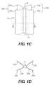
- FIG. 1Cdepicts an alternate configuration of the implant depicted in FIG. 1A ;
- FIG. 1Dis a cross-sectional view of the configuration depicted in FIG. 1C taken along line BB′;
- FIG. 2Ais a cross-sectional view of the abdominal wall
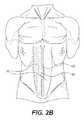
- FIG. 2Billustrates an outline of a midline abdominal incision
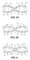
- FIG. 3Ashows a method of positioning an implant in the abdominal wall for a midline incision closure, in accordance with an exemplary embodiment of the present disclosure
- FIG. 3Billustrates an alternate method of positioning an implant in the abdominal wall for a midline incision closure, in accordance with an exemplary embodiment of the present disclosure
- FIG. 4illustrates a method of suturing an implant to the abdominal wall for a midline incision closure, in accordance with an exemplary embodiment of the present disclosure
- FIG. 5is a flowchart showing the steps of treating an abdominal incision using an exemplary implant of the present disclosure.
- the present disclosureprovides methods and devices for effective incision closure following a surgical procedure and/or abdominal wall treatment for structural abdominal wall defects.
- the devices and methods disclosed hereinare used for closing abdominal incisions when primary suture closure is not feasible due to loss of abdominal muscle and/or fascia.
- the devices and methods disclosed hereinare used for midline incision closure following a surgical procedure.
- the devices and methodsare used as a treatment for ventral or incisional hernia, e.g., for treatment of midline incisional hernia.
- the methods and devices of the present disclosurecan be used for prophylactic treatment, e.g., to prevent incisional hernia.
- the methods and devices of the present disclosurecan be used to treat preexisting abdominal wall defects or to assist in closure of incisions or hernias where insufficient abdominal tissue is present.
- incision closureis performed using an implant comprising an elongate element and at least one sheet connected to the elongate element along a longitudinal axis of the elongate element.
- the elongate elementis positioned along the incision line, and the at least one sheet is positioned on the anterior and/or posterior side of the abdominal muscles surrounding the incision.
- the biomechanical propertiese.g., the tensile strength, compressive strength, torsional strength, toughness of the material, etc.
- the elongate elementare equal to that of the at least one sheet.
- certain biomechanical propertiese.g., tensile strength, stiffness, tear strength
- the relative mechanical properties of the elongate element and the sheetmay be determined based on the specific application of the implant. For example, in one embodiment, a strength of the elongate element is twice that of the at least one sheet. In another embodiment, a strength of the elongate element is at least three times that of the at least one sheet.
- the sheetcomprises a porous biological material.
- the sheetcomprises a collagenous material.
- the sheetcomprises an acellular tissue matrix, which may support revascularization and repopulation of the implanted matrix with the patient's own cells to further strengthen the treatment site and lower the risk of matrix dislodgement.
- the acellular tissue matrixincludes a dermal matrix.
- the sheetis derived from human skin (e.g. ALLODERM®, LifeCell Corp, Branchburg, N.J.) that has been processed to remove both the epidermis and the cells that can lead to tissue rejection and graft failure, without damaging the dermal matrix.
- the sheetis derived from porcine dermis (e.g. STRATTICETM, LifeCell Corp, Branchburg, N.J.), which is processed to remove cells and tissue antigens while maintaining the ability to support cellular growth and tissue regeneration and/or remodeling.
- porcine dermise.g. STRATTICETM, LifeCell Corp, Branchburg, N.J.
- the sheet of the implantcomprises a porous synthetic mesh (e.g., polypropylene, prolene, or polytetrafluroetheylene mesh).
- the synthetic meshfacilitates tissue incorporation into the implant.
- the elongate elementis a cylindrical body comprising an immunologically inert and biocompatible material.
- the materialmay be a synthetic polymer (such as, polypropylene, polytetrafluroetheylene, etc.) or a non-synthetic material.
- the elongate elementcomprises a biological mesh that is rolled concentrically into a cylindrical structure.
- the biological meshcomprises an acellular tissue matrix (such as, ALLODERM® or STRATTICETM).
- the elongate element and sheetscan be produced from tissue that is allogeneic or xenogenic to a human recipient.
- Allogeneic sourcesmay be obtained from living donors or cadavers.
- Xenogeneic sourcescan include a variety of different non-human mammals.
- one suitable biologic material for production of the sheets and elongate elementis STRATTICETM, which is a porcine-derived tissue matrix.
- STRATTICETMis a porcine-derived tissue matrix.
- other xenograft sourcescan be used.
- Allogeneic or xenogenic tissuescan be processed to remove antigens known to elicit an immune response in the recipient.
- various decellularization processes or enzyme treatmentsare known that allow removal of cellular and/or extracellular antigens that may be immunogenic.
- the tissuescan be derived from animals that are genetically modified or altered to have diminished expression of antigens known to be immunogenic in humans.
- the tissuesare harvested from an ⁇ 1,3-galactosyltransferase ( ⁇ 1,3GT) deficient pig or other animal to prevent hyperacute rejection of the implant by the recipient.
- ⁇ 1,3GT⁇ 1,3-galactosyltransferase
- the elongate elementcan include a strip or piece of connective tissue from an allogeneic or xenogeneic source.
- suitable connective tissuescan include ligament, tendon, and/or fascia.
- the prosthetic implantis used for treatment of a midline abdominal incision, which is a vertical incision made along the linea alba between the two rectus abdominis muscles of the abdominal wall.
- a midline abdominal incisionis a vertical incision made along the linea alba between the two rectus abdominis muscles of the abdominal wall.
- the linea albamay be absent or shifted from the abdominal midline due to abdominal wall defects.
- the midline incisionmay be performed along the anatomical midline of the abdominal wall, instead of the linea alba (if present).
- the elongate elementis positioned along the incision, and the at least one sheet is positioned over and/or under the rectus abdominis muscles surrounding the incision.
- the linea alba located in the abdominal midlineis a three-dimensional composition of connective tissue fibers from abdominal wall muscles, which plays a significant role in stabilizing the abdominal wall.
- Use of a connective tissue material for the elongate elementtherefore, facilitates reliable closure of the midline incision and provides good aesthetic results.
- the elongate elementmay, however, comprise any biological or synthetic material that is immunologically compatible, is structurally and functionally similar to the linea alba, and facilitates duplication of the abdominal wall physiology.
- FIG. 1Ashows an exemplary embodiment of a prosthetic implant 10 in accordance with the present disclosure.
- FIG. 1Billustrates a cross-sectional view of the embodiment depicted in FIG. 1A taken along line AA′.
- FIG. 1Cdepicts an alternate configuration of implant 10
- FIG. 1Dis a cross-sectional view of the configuration depicted in FIG. 1C taken along line BB′.
- Implant 10comprises an elongate element 12 and at least two sheets 20 a and 20 b connected to the outer surface of elongate element 12 on opposite sides of a longitudinal axis 14 of the central element.
- the sheetscan be connected to elongate element 12 using absorbable biological sutures (e.g.
- sheets 20 a and 20 bare connected to elongate element 12 along the midline of the sheets. In such an embodiment, attachment of sheets 20 a and 20 b to elongate element 12 divides the sheets into two halves.
- sheet 20 aforms two flaps 21 a and 22 a of equal dimensions upon attachment to element 12 .
- sheet 20 bforms two symmetrical flaps 21 b and 22 b .
- Sheets 20 a and 20 bmay, however, be connected to elongate element 12 along any longitudinal axial line of the sheets. In some embodiments consistent with the present disclosure, the flaps formed by connection of the sheets to the elongate element are asymmetrical.
- sheets 20 a and 20 bextend along a plane substantially parallel to longitudinal axis 14 , as illustrated in FIGS. 1A and 1B .
- sheets 20 a and 20 bform two pockets between flaps 21 a and 21 b and 22 a and 22 b , respectively, when attached to elongate element 12 .
- sheets 20 a and 20 bare folded away from elongate element 12 , as illustrated in FIG. 1C and FIG. 1D , such that sheets 20 a and 20 b form two pockets between flaps 21 a and 22 a and 21 b and 22 b , respectively.
- sheets 20 a and 20 bare generally shown in a rectangular form in the figures, the depiction is only for illustrative purposes, and any suitable size, shape and form can be used depending upon the specifications of the surgical procedure and the extent of repair necessary.
- the lengths of both sheets 20 a and 20 bare equal to the length of elongate element 12 .
- the lengths of elongate element 12 and sheets 20 a and 20 bare selected to span the length of the incision.
- the length of implant 10may be shorter than the length of the incision, depending on the extent of the restorative procedure and the amount of reinforcement required in the abdominal wall.
- implant 10may be tailored to the intended application prior to the surgery or during the surgical procedure. Further, in some embodiments consistent with the present disclosure, implant 10 comprises multiple sheets that are superimposed, or layered, to provide additional reinforcement to the abdominal wall. The multiple sheets may be chemically or physically bonded to each other to facilitate positioning of implant 10 in the abdominal wall.
- FIGS. 2A and 2Billustrate the anatomy of the abdominal wall and an abdominal midline incision outline, respectively.
- FIG. 2Ashows a cross-sectional view of the abdominal wall comprising the rectus abdominis muscles 50 , linea alba 52 , transversalis fascia 54 , peritoneum 56 , subcutaneous fat 58 , skin 60 , anterior layer of fascia 62 and posterior layer of fascia 64 .
- FIG. 2Bshows a midline incision 100 made through linea alba 52 between rectus abdominis muscles 50 and around umbilicus 66 . Further, although this method is described for midline closure, the method of incision closure disclosed can be applied for the treatment of any incision type and/or to treat other structural abdominal wall defects.
- elongate element 12 of implant 10is positioned along the line of incision, and sheets 20 a and 20 b are positioned on the anterior and/or posterior sides of rectus abdominis muscles 50 .
- potential spaces for the placement of the sheets 20 a and 20 bmay be created by dissection around the incision using a blunt instrument or the surgeon's fingers.
- spaces between the rectus abdominis muscles 50 and the anterior and posterior layers of fascia 62 , 64may be dissected for placement of the sheets 20 a and 20 b .
- Elongate element 12is then aligned with the midline incision, and the sheets are inserted into the dissected pockets, as shown in FIG. 3A .
- the posterior layer of fascia 64is not present, and therefore, the posterior flaps 21 b and 22 b are positioned in spaces dissected between rectus abdominis muscles 50 and transversalis fascia 54 or positioned to cover the transversalis fascia 54 or peritoneum 56 .
- sheets 20 a and/or 20 boverlay anterior layer of fascia 62 , and on the posterior side, the sheets are positioned between posterior layer of fascia 64 and transversalis fascia 54 or overlying the transversalis fascia or peritoneum.
- the anterior and/or posterior layers of fascia 62 , 64may be partially or completely lost due to multiple prior surgeries, necrotizing infections, and/or other complications.
- sheets 20 a and 20 bare simply positioned in proximity to the rectus muscles 50 . If the fascia is partially present, it may be abraded by the surgeon to expose the rectus muscles and release blood to initiate the would healing process. The sheets then spontaneously anneal with the rectus muscles. When the sheets comprise a biological matrix material, the sheets may be remodeled by the patient's own cells, thereby, facilitating rapid integration of the sheets into the repair site.
- elongate element 12is aligned with the midline incision in accordance with the configuration shown in FIGS. 1C and 1D , such that the rectus muscles 50 are supported between flaps 21 a and 22 a of sheet 20 a on one side of the incision, and flaps 21 b and 22 b of sheet 20 b on the other side.
- the sheetsare secured to the rectus muscles.
- the sheets 20 a and 20 bcan be secured using a variety of anchoring systems, including, for example, sutures, staples, clips, and/or tissue adhesives.
- anchoring systemsincluding, for example, sutures, staples, clips, and/or tissue adhesives.
- securing the sheets 20 a and 20 b to the rectus muscleswill be understood to include securing the sheets 20 a and 20 b directly to the rectus muscles and to include securing the sheets to a layer of the abdominal wall adjacent to the rectus muscles, including a layer of rectus fascia, transversalis fascia, or peritoneum.
- the sheetsare secured using sutures that pass through a portion of the sheet, the rectus abdominis muscles, and/or the posterior and/or anterior layer of fascia.
- the suturing needleis inserted through layers of the abdominal wall and sheets 20 a and 20 b , and then reversed to retrace the path through the abdominal wall layers and sheets.
- the free ends of sutures 70are tied together on the anterior side of sheets 21 a and 22 a .
- the suturesmay be tied anterior to the anterior layer of fascia 62 . Sufficient care is taken to avoid incorporation of visceral or other structures into the suture line. Sutures are placed along the line of incision to ensure complete reapproximation of the abdominal wall. The frequency of the lateral suture anchors is determined by the surgeon during the procedure.
- the reapproximationis performed using continuous or interrupted loop sutures 80 , as shown in FIG. 4 .
- Suture loops 80incorporate elongate element 12 in order to secure the fascia and or rectus muscles to the elongate element 12 . This applies tension to the abdominal wall structures and draws them medially to close the incision.
- Loop sutures 80may be used in conjunction with interrupted sutures 70 , as described above, or alone.
- FIG. 5is a flowchart diagramming the steps of treating an abdominal wall following a midline incision using implant 10 .
- the methodcan be used for prophylactic treatment of the incision (e.g., to prevent dehiscence) or to facilitate closure of incisions or defects that are difficult to close with sutures alone.
- the first step in closing the midline incisionincludes optionally creating potential spaces for the placement of sheets 20 a and 20 b on both sides of the midline incision (step 510 ) if needed.
- spaces between rectus muscles 50 and anterior layer of fascia 62are dissected for placement of the sheets on the anterior side of the ventral abdominal wall, and spaces between rectus muscles 50 and posterior layer of fascia 64 or transversalis fascia 54 are created for placement of the sheets in the posterior side of the abdominal wall.
- the sheetsare positioned over anterior layer of fascia 62 and below posterior layer of fascia 64 . In such a procedure, the surgeon creates spaces between fascia 62 and subcutaneous fat 58 on the anterior side on the abdominal wall.
- Step 510may not be required if an adequate region for attachment of the sheets to rectus abdominis muscles is already present or if a patient is suffering from substantial loss of fascia 62 , 64 .
- the second stepcomprises positioning elongate element 12 of implant 10 along the midline incision (step 520 ) to begin approximation of the wound margins.
- the cranial end of elongate element 12is aligned with the superior end of the midline incision, and the caudal end of the implant is aligned with the inferior end of the incision.
- the size of the implantcould be adjusted by the surgeon either before the procedure, or during the placement of the implant.
- the next stepis the insertion of the sheets into the spaces created in step 510 (step 530 ) or on the appropriate sides of the rectus muscles if no spaces have been created.
- the sheetsare secured in place (step 540 ).
- the sheetscan be secured using, sutures, staples, clips, tissue adhesives, or other suitable means.
- the method of securing the device and the suturing techniqueare determined by the surgeon during the procedure. This is followed by closure of the skin using either non-absorbable or absorbable sutures. Alternatively, surgeons may use surgical staples for skin closure due to speed of application and ease of removal.
Landscapes
- Health & Medical Sciences (AREA)
- Life Sciences & Earth Sciences (AREA)
- Engineering & Computer Science (AREA)
- Public Health (AREA)
- Biomedical Technology (AREA)
- Heart & Thoracic Surgery (AREA)
- Veterinary Medicine (AREA)
- Cardiology (AREA)
- Animal Behavior & Ethology (AREA)
- General Health & Medical Sciences (AREA)
- Surgery (AREA)
- Vascular Medicine (AREA)
- Transplantation (AREA)
- Oral & Maxillofacial Surgery (AREA)
- Nuclear Medicine, Radiotherapy & Molecular Imaging (AREA)
- Medical Informatics (AREA)
- Molecular Biology (AREA)
- Prostheses (AREA)
- Surgical Instruments (AREA)
Abstract
Description
Claims (17)
Priority Applications (3)
| Application Number | Priority Date | Filing Date | Title |
|---|---|---|---|
| US13/714,606US9351819B2 (en) | 2009-07-02 | 2012-12-14 | Device and method for treatment of incision or hernia |
| US15/142,005US10238480B2 (en) | 2009-07-02 | 2016-04-29 | Device and method for treatment of incision or hernia |
| US16/295,493US20190201179A1 (en) | 2009-07-02 | 2019-03-07 | Device and method for treatment of incision or hernia |
Applications Claiming Priority (3)
| Application Number | Priority Date | Filing Date | Title |
|---|---|---|---|
| US22269109P | 2009-07-02 | 2009-07-02 | |
| US12/828,452US8357172B2 (en) | 2009-07-02 | 2010-07-01 | Device and method for treatment of incision or hernia |
| US13/714,606US9351819B2 (en) | 2009-07-02 | 2012-12-14 | Device and method for treatment of incision or hernia |
Related Parent Applications (1)
| Application Number | Title | Priority Date | Filing Date |
|---|---|---|---|
| US12/828,452ContinuationUS8357172B2 (en) | 2009-07-02 | 2010-07-01 | Device and method for treatment of incision or hernia |
Related Child Applications (1)
| Application Number | Title | Priority Date | Filing Date |
|---|---|---|---|
| US15/142,005ContinuationUS10238480B2 (en) | 2009-07-02 | 2016-04-29 | Device and method for treatment of incision or hernia |
Publications (2)
| Publication Number | Publication Date |
|---|---|
| US20130103061A1 US20130103061A1 (en) | 2013-04-25 |
| US9351819B2true US9351819B2 (en) | 2016-05-31 |
Family
ID=42727557
Family Applications (4)
| Application Number | Title | Priority Date | Filing Date |
|---|---|---|---|
| US12/828,452Active2031-01-09US8357172B2 (en) | 2009-07-02 | 2010-07-01 | Device and method for treatment of incision or hernia |
| US13/714,606ActiveUS9351819B2 (en) | 2009-07-02 | 2012-12-14 | Device and method for treatment of incision or hernia |
| US15/142,005Active2031-02-22US10238480B2 (en) | 2009-07-02 | 2016-04-29 | Device and method for treatment of incision or hernia |
| US16/295,493AbandonedUS20190201179A1 (en) | 2009-07-02 | 2019-03-07 | Device and method for treatment of incision or hernia |
Family Applications Before (1)
| Application Number | Title | Priority Date | Filing Date |
|---|---|---|---|
| US12/828,452Active2031-01-09US8357172B2 (en) | 2009-07-02 | 2010-07-01 | Device and method for treatment of incision or hernia |
Family Applications After (2)
| Application Number | Title | Priority Date | Filing Date |
|---|---|---|---|
| US15/142,005Active2031-02-22US10238480B2 (en) | 2009-07-02 | 2016-04-29 | Device and method for treatment of incision or hernia |
| US16/295,493AbandonedUS20190201179A1 (en) | 2009-07-02 | 2019-03-07 | Device and method for treatment of incision or hernia |
Country Status (9)
| Country | Link |
|---|---|
| US (4) | US8357172B2 (en) |
| EP (1) | EP2448497B1 (en) |
| JP (1) | JP2012531956A (en) |
| CN (1) | CN102469994B (en) |
| AU (1) | AU2010266266B2 (en) |
| CA (2) | CA2766390C (en) |
| ES (1) | ES2862549T3 (en) |
| SG (1) | SG176931A1 (en) |
| WO (1) | WO2011002962A1 (en) |
Cited By (10)
| Publication number | Priority date | Publication date | Assignee | Title |
|---|---|---|---|---|
| US20160242890A1 (en)* | 2009-07-02 | 2016-08-25 | Lifecell Corporation | Device and method for treatment of incision or hernia |
| US10307237B2 (en) | 2015-05-15 | 2019-06-04 | Lifecell Corporation | Tissue matrices and methods of treatment |
| USD856517S1 (en) | 2016-06-03 | 2019-08-13 | Musculoskeletal Transplant Foundation | Asymmetric tissue graft |
| US10537665B2 (en) | 2015-09-11 | 2020-01-21 | Lifecell Corporation | Perforated tissue matrix |
| USD895812S1 (en) | 2018-09-07 | 2020-09-08 | Musculoskeletal Transplant Foundation | Soft tissue repair graft |
| US10813743B2 (en) | 2018-09-07 | 2020-10-27 | Musculoskeletal Transplant Foundation | Soft tissue repair grafts and processes for preparing and using same |
| US10869745B2 (en) | 2016-10-06 | 2020-12-22 | Lifecell Corporation | Tissue matrix with preformed openings or pilot openings |
| US10945831B2 (en) | 2016-06-03 | 2021-03-16 | Musculoskeletal Transplant Foundation | Asymmetric tissue graft |
| US11298220B2 (en) | 2019-05-03 | 2022-04-12 | Lifecell Corporation | Breast treatment device |
| US11998654B2 (en) | 2018-07-12 | 2024-06-04 | Bard Shannon Limited | Securing implants and medical devices |
Families Citing this family (18)
| Publication number | Priority date | Publication date | Assignee | Title |
|---|---|---|---|---|
| US10478168B2 (en)* | 2009-07-02 | 2019-11-19 | Lifecell Corporation | Device and method for treatment of incision or hernia |
| EP2536340B1 (en)* | 2010-02-19 | 2018-10-17 | LifeCell Corporation | Abdominal wall treatment devices |
| AU2011232374B2 (en)* | 2010-03-25 | 2015-06-25 | Lifecell Corporation | Preparation of regenerative tissue scaffolds |
| US8777965B2 (en)* | 2011-04-15 | 2014-07-15 | Usgi Medical, Inc. | Devices and methods for laparoscopic hernia repair |
| WO2013009993A1 (en)* | 2011-07-12 | 2013-01-17 | Bengtson Bradley P | Surgical fixation devices, systems, and methods |
| US9162011B2 (en) | 2011-12-19 | 2015-10-20 | Allosource | Flowable matrix compositions and methods |
| US9820838B2 (en) | 2012-04-10 | 2017-11-21 | Ethicon, Inc. | Single plane tissue repair patch |
| US9820837B2 (en) | 2012-04-10 | 2017-11-21 | Ethicon, Inc. | Single plane tissue repair patch |
| US9820839B2 (en) | 2012-04-10 | 2017-11-21 | Ethicon, Inc. | Single plane tissue repair patch having a locating structure |
| US10130346B2 (en) | 2012-07-24 | 2018-11-20 | Omrix Biopharmaceuticals Ltd. | Device and method for the application of a curable fluid composition to a bodily organ |
| WO2014099895A1 (en)* | 2012-12-17 | 2014-06-26 | Atex Technologies, Inc. | Medical textile and methods of making the same |
| KR102312720B1 (en) | 2013-03-15 | 2021-10-13 | 알로소스 | Cell repopulated collagen matrix for soft tissue repair and regeneration |
| US9801910B2 (en) | 2014-03-17 | 2017-10-31 | Ethicon, Inc. | Decellularized pleural matrix |
| US9895212B2 (en) | 2014-10-31 | 2018-02-20 | Prevent Patch LLC | Devices and methods for preventing incisional hernias |
| US9622844B2 (en) | 2014-10-31 | 2017-04-18 | Prevent Patch, LLC | Devices and methods for preventing incisional hernias |
| WO2016141183A1 (en)* | 2015-03-03 | 2016-09-09 | The Trustees Of The University Of Pennsylvania | Systems and methods for mesh augmentation and prevention of incisional hernia |
| US10603154B2 (en) | 2015-03-31 | 2020-03-31 | Prevent Patch, LLC | Devices and methods for preventing incisional hernias |
| SG11202011238SA (en)* | 2018-02-20 | 2020-12-30 | Univ Southern California | Instruments and methods for the implantation of cell-seeded ultra-thin substrates |
Citations (27)
| Publication number | Priority date | Publication date | Assignee | Title |
|---|---|---|---|---|
| US4813958A (en) | 1986-10-14 | 1989-03-21 | Hancock Jaffe Laboratories | Crosslinked anisotropic mammalian diaphragm in surgical reconstruction |
| US5254133A (en)* | 1991-04-24 | 1993-10-19 | Seid Arnold S | Surgical implantation device and related method of use |
| US5397353A (en) | 1984-05-24 | 1995-03-14 | Oliver; Roy F. | Implant tissue |
| US5733337A (en)* | 1995-04-07 | 1998-03-31 | Organogenesis, Inc. | Tissue repair fabric |
| WO2000016822A1 (en) | 1998-09-21 | 2000-03-30 | The Brigham And Women's Hospital, Inc. | Compositions and methods for tissue repair |
| US6113623A (en) | 1994-04-20 | 2000-09-05 | Cabinet Beau De Lomenie | Prosthetic device and method for eventration repair |
| US6171318B1 (en) | 1994-09-29 | 2001-01-09 | Bard Asdi Inc. | Hernia mesh patch with stiffening layer |
| US6176863B1 (en) | 1994-09-29 | 2001-01-23 | Bard Asdi Inc. | Hernia mesh patch with I-shaped filament |
| US6383201B1 (en)* | 1999-05-14 | 2002-05-07 | Tennison S. Dong | Surgical prosthesis for repairing a hernia |
| US20030023316A1 (en)* | 2000-08-04 | 2003-01-30 | Brown Laura Jean | Hybrid biologic-synthetic bioabsorable scaffolds |
| US20030225355A1 (en) | 1998-10-01 | 2003-12-04 | Butler Charles E. | Composite material for wound repair |
| US20040098042A1 (en)* | 2002-06-03 | 2004-05-20 | Devellian Carol A. | Device with biological tissue scaffold for percutaneous closure of an intracardiac defect and methods thereof |
| EP1421916A1 (en) | 2001-08-03 | 2004-05-26 | Bard de Espana, S.A. | Wall prosthesis that can be implanted in the centre of a wound in order to reinforce the abdominal wall closure |
| US20040209538A1 (en)* | 2001-06-26 | 2004-10-21 | Uwe Klinge | Textile implant |
| US20040260315A1 (en) | 2003-06-17 | 2004-12-23 | Dell Jeffrey R. | Expandable tissue support member and method of forming the support member |
| US20050240075A1 (en) | 2001-10-05 | 2005-10-27 | Scimed Life Systems, Inc. | Expandable surgical implants and methods of using them |
| US20070118176A1 (en)* | 2005-10-24 | 2007-05-24 | Opolski Steven W | Radiopaque bioabsorbable occluder |
| WO2009003726A1 (en) | 2007-07-02 | 2009-01-08 | Loehde Eckhard | Three-dimensional hernia mesh |
| US20090234461A1 (en) | 2008-03-14 | 2009-09-17 | Rehnke Robert D | Apparatus and method for use of a biosurgical prosthetic rectus sheath |
| US20100082114A1 (en) | 2008-04-29 | 2010-04-01 | Peter Gingras | Tissue repair implant |
| US20100124563A1 (en) | 2008-11-17 | 2010-05-20 | Ingeneron, Inc. | Biomatrix Composition and Methods of Biomatrix Seeding |
| US20100185219A1 (en) | 2007-04-25 | 2010-07-22 | Musculosketetal Transplant Foundation | Reinforced biological mesh for surgical reinforcement |
| US20110004306A1 (en)* | 2009-07-02 | 2011-01-06 | Lifecell Corporation | Device and method for treatment of incision or hernia |
| US20110015760A1 (en)* | 2007-12-07 | 2011-01-20 | Kullas Karen E | Implantable prosthesis |
| US20120059411A1 (en)* | 2009-07-02 | 2012-03-08 | Sun Wenquan | Device and method for treatment of incision or hernia |
| US20120221118A1 (en) | 2011-02-25 | 2012-08-30 | Obi Biologics, Inc. | Materials for soft and hard tissue repair |
| US8324449B2 (en)* | 2003-07-21 | 2012-12-04 | Lifecell Corporation | Acellular tissue matrices made from alpha-1,3-galactose-deficient tissue |
Family Cites Families (1)
| Publication number | Priority date | Publication date | Assignee | Title |
|---|---|---|---|---|
| CA2857051A1 (en)* | 2004-03-17 | 2005-09-29 | David Ayares | Tissue products derived from animals lacking any expression of functional alpha 1,3 galactosyltransferase |
- 2010
- 2010-07-01CNCN201080028596.6Apatent/CN102469994B/ennot_activeExpired - Fee Related
- 2010-07-01USUS12/828,452patent/US8357172B2/enactiveActive
- 2010-07-01WOPCT/US2010/040717patent/WO2011002962A1/enactiveApplication Filing
- 2010-07-01CACA2766390Apatent/CA2766390C/enactiveActive
- 2010-07-01CACA3000888Apatent/CA3000888A1/ennot_activeAbandoned
- 2010-07-01EPEP10730954.4Apatent/EP2448497B1/ennot_activeNot-in-force
- 2010-07-01JPJP2012517904Apatent/JP2012531956A/ennot_activeWithdrawn
- 2010-07-01AUAU2010266266Apatent/AU2010266266B2/ennot_activeCeased
- 2010-07-01SGSG2011094422Apatent/SG176931A1/enunknown
- 2010-07-01ESES10730954Tpatent/ES2862549T3/enactiveActive
- 2012
- 2012-12-14USUS13/714,606patent/US9351819B2/enactiveActive
- 2016
- 2016-04-29USUS15/142,005patent/US10238480B2/enactiveActive
- 2019
- 2019-03-07USUS16/295,493patent/US20190201179A1/ennot_activeAbandoned
Patent Citations (29)
| Publication number | Priority date | Publication date | Assignee | Title |
|---|---|---|---|---|
| US5397353A (en) | 1984-05-24 | 1995-03-14 | Oliver; Roy F. | Implant tissue |
| US4813958A (en) | 1986-10-14 | 1989-03-21 | Hancock Jaffe Laboratories | Crosslinked anisotropic mammalian diaphragm in surgical reconstruction |
| US5254133A (en)* | 1991-04-24 | 1993-10-19 | Seid Arnold S | Surgical implantation device and related method of use |
| US6113623A (en) | 1994-04-20 | 2000-09-05 | Cabinet Beau De Lomenie | Prosthetic device and method for eventration repair |
| US6171318B1 (en) | 1994-09-29 | 2001-01-09 | Bard Asdi Inc. | Hernia mesh patch with stiffening layer |
| US6176863B1 (en) | 1994-09-29 | 2001-01-23 | Bard Asdi Inc. | Hernia mesh patch with I-shaped filament |
| US5733337A (en)* | 1995-04-07 | 1998-03-31 | Organogenesis, Inc. | Tissue repair fabric |
| WO2000016822A1 (en) | 1998-09-21 | 2000-03-30 | The Brigham And Women's Hospital, Inc. | Compositions and methods for tissue repair |
| US20030225355A1 (en) | 1998-10-01 | 2003-12-04 | Butler Charles E. | Composite material for wound repair |
| US6383201B1 (en)* | 1999-05-14 | 2002-05-07 | Tennison S. Dong | Surgical prosthesis for repairing a hernia |
| US20030023316A1 (en)* | 2000-08-04 | 2003-01-30 | Brown Laura Jean | Hybrid biologic-synthetic bioabsorable scaffolds |
| US20040209538A1 (en)* | 2001-06-26 | 2004-10-21 | Uwe Klinge | Textile implant |
| EP1421916A1 (en) | 2001-08-03 | 2004-05-26 | Bard de Espana, S.A. | Wall prosthesis that can be implanted in the centre of a wound in order to reinforce the abdominal wall closure |
| US20050043818A1 (en)* | 2001-08-03 | 2005-02-24 | Bellon Caneiro Juan Manuel | Wall prosthesis that can be implanted in the center of a wound to reinforce abdominal wall closure |
| US20050240075A1 (en) | 2001-10-05 | 2005-10-27 | Scimed Life Systems, Inc. | Expandable surgical implants and methods of using them |
| US20040098042A1 (en)* | 2002-06-03 | 2004-05-20 | Devellian Carol A. | Device with biological tissue scaffold for percutaneous closure of an intracardiac defect and methods thereof |
| US20040260315A1 (en) | 2003-06-17 | 2004-12-23 | Dell Jeffrey R. | Expandable tissue support member and method of forming the support member |
| US8324449B2 (en)* | 2003-07-21 | 2012-12-04 | Lifecell Corporation | Acellular tissue matrices made from alpha-1,3-galactose-deficient tissue |
| US20070118176A1 (en)* | 2005-10-24 | 2007-05-24 | Opolski Steven W | Radiopaque bioabsorbable occluder |
| US20100185219A1 (en) | 2007-04-25 | 2010-07-22 | Musculosketetal Transplant Foundation | Reinforced biological mesh for surgical reinforcement |
| WO2009003726A1 (en) | 2007-07-02 | 2009-01-08 | Loehde Eckhard | Three-dimensional hernia mesh |
| US20110015760A1 (en)* | 2007-12-07 | 2011-01-20 | Kullas Karen E | Implantable prosthesis |
| US20090234461A1 (en) | 2008-03-14 | 2009-09-17 | Rehnke Robert D | Apparatus and method for use of a biosurgical prosthetic rectus sheath |
| US20100082114A1 (en) | 2008-04-29 | 2010-04-01 | Peter Gingras | Tissue repair implant |
| US20100124563A1 (en) | 2008-11-17 | 2010-05-20 | Ingeneron, Inc. | Biomatrix Composition and Methods of Biomatrix Seeding |
| US20110004306A1 (en)* | 2009-07-02 | 2011-01-06 | Lifecell Corporation | Device and method for treatment of incision or hernia |
| US20120059411A1 (en)* | 2009-07-02 | 2012-03-08 | Sun Wenquan | Device and method for treatment of incision or hernia |
| US8357172B2 (en)* | 2009-07-02 | 2013-01-22 | Lifecell Corporation | Device and method for treatment of incision or hernia |
| US20120221118A1 (en) | 2011-02-25 | 2012-08-30 | Obi Biologics, Inc. | Materials for soft and hard tissue repair |
Non-Patent Citations (22)
| Title |
|---|
| Buinewicz, B., et al., "Accellular Cadaveric Dermis (AlloDerm): A New Alternative for Abdominal Hernia Repair", Annals of Plastic Surgery, vol. 52, No. 2, Feb. 2004, pp. 188-194. |
| Burns, Nadja K., et al., "Non-Cross-Linked Porcine Acellular Dermal Matrices for Abdominal Wall Reconstruction", Plast Reconstr Surg. 2009: PMID:19910855. |
| Butler, Charles E., "The Role of Bioprosthetics in Abdominal Wall Reconstruction", Clin Plastic Surg, 33, 2006, pp. 199-211. |
| Capito, Anthony E., et al., "Evaluation of Host Tissue Integration, Revascularization, and Cellular Infiltration Within Various Dermal Substrates", Annals of Plastic Surgery, vol. 68, No. 5, May 2012, pp. 495-500. |
| Connor, J. et al., "Retention of Structural and Biochemical Integrity in a Biological Mesh Supports Tissue Remodeling in a Primate Abdominal Wall Model", Regen Med. 2009:4(2):185-195. |
| Dai, Y. et al., "Targeted disruption of the alpha1,3-galactosyltransferase gene in cloned pigs," Nat. Biotechnology 20, 251-255 (2002). |
| Dai, Y. et al., "Targeted disruption of the α1,3-galactosyltransferase gene in cloned pigs," Nat. Biotechnology 20, 251-255 (2002). |
| de Vries Reilingh T. S. et al., "Repair of Giant Midline Abdominal Wall Hernia: 'Components Separation Technique' versus Prosthetic Repair," World J. Surg 31, 756-763 (2007). |
| Harty, M. et al., "Regeneration or Scarring: An Immunologic Perspective", Developmental Dynamics, 226:268-279 (2003). |
| International Search Report and Written Opinion issued by European Patent Office for International Application No. PCT/US2010/040717, mailing date Sep. 28, 2010. |
| J Biomech. Oct. 2002;35(10):1417-25, http://www.ncbi.nlm.nih.gov/pubmed/12231288 as accessed on May 6, 2015.* |
| Kolker, A. R., et al., "Multilayer Reconstruction of Abdominal Wall Defects With Acellular Dermal Allograft (AlloDerm) and Component Separation", Annals of Plastic Surgery, vol. 55, No. 1, Jul. 2005, pp. 36-42. |
| MakeItFrom.com, Polyglycolic Acid, http://www.makeitfrom.com/material-properties/Polyglycolic-Acid-PGA-Polyglycolide/ as accessed on May 6, 2015.* |
| Menon, N. G., "Revascularization of Human Acellular Dermis in Full-Thickness Abdominal Wall Reconstruction in the Rabbit Model", Ann Plast Surg. 2003; 50(5):pp. 523-527. Erratum in Ann Plast Surg. Aug. 2003 51(2): p. 228. |
| Phelps, C. J. et al., "Production of alpha1,3-Galactosyltransferase-Deficient Pigs," Science 299, 411-414 (2003). |
| Phelps, C. J. et al., "Production of α1,3-Galactosyltransferase-Deficient Pigs," Science 299, 411-414 (2003). |
| Sandor, M. et al., "Host Response to Implanted Porcine-Derived biologic Materials in a Primate Model of Abdominal Wall Repair", Tissue Engineering: Part A, vol. 14, No. 12, pp. 2021-2031 (2008). |
| Sandor, Maryellen, et al., "Host Response to Implanted Porcine-Derrived Biologic Materials in a Primate Model of Abdominal Wall Repair", Tissue Engineering: Part A, vol. 14, No. 12, 2008, pp. 2021-2031. |
| Silverman, R. P., et al., Ventral hernia repair using allogenic acellular dermal matrix in a swine model, Hernia, (2004) 8: pp. 336-342. |
| Voyles C. R. et al., "Emergency Abdominal Wall Reconstruction with Polypropylene Mesh," Ann. Surg. 194(2), 219-223 (1981). |
| Xu, Hui, et al., "A Porcine-Derived Acellular Dermal Scaffold That Supports Soft Tissue Regeneration: Removal of Terminal Galactose-alpha-(1,3)-Galactose and Retention of Matrix Structure", Tissue Engineering: Part A, vol. 15, No. 7, 2009, pp. 1807-1819. |
| Xu, Hui, et al., "A Porcine-Derived Acellular Dermal Scaffold That Supports Soft Tissue Regeneration: Removal of Terminal Galactose-α-(1,3)-Galactose and Retention of Matrix Structure", Tissue Engineering: Part A, vol. 15, No. 7, 2009, pp. 1807-1819. |
Cited By (13)
| Publication number | Priority date | Publication date | Assignee | Title |
|---|---|---|---|---|
| US10238480B2 (en)* | 2009-07-02 | 2019-03-26 | Lifecell Corporation | Device and method for treatment of incision or hernia |
| US20160242890A1 (en)* | 2009-07-02 | 2016-08-25 | Lifecell Corporation | Device and method for treatment of incision or hernia |
| US10307237B2 (en) | 2015-05-15 | 2019-06-04 | Lifecell Corporation | Tissue matrices and methods of treatment |
| US11383007B2 (en) | 2015-09-11 | 2022-07-12 | Lifecell Corporation | Perforated tissue matrix |
| US10537665B2 (en) | 2015-09-11 | 2020-01-21 | Lifecell Corporation | Perforated tissue matrix |
| USD856517S1 (en) | 2016-06-03 | 2019-08-13 | Musculoskeletal Transplant Foundation | Asymmetric tissue graft |
| US10945831B2 (en) | 2016-06-03 | 2021-03-16 | Musculoskeletal Transplant Foundation | Asymmetric tissue graft |
| US10869745B2 (en) | 2016-10-06 | 2020-12-22 | Lifecell Corporation | Tissue matrix with preformed openings or pilot openings |
| US11998654B2 (en) | 2018-07-12 | 2024-06-04 | Bard Shannon Limited | Securing implants and medical devices |
| US10813743B2 (en) | 2018-09-07 | 2020-10-27 | Musculoskeletal Transplant Foundation | Soft tissue repair grafts and processes for preparing and using same |
| US11642216B2 (en) | 2018-09-07 | 2023-05-09 | Musculoskeletal Transplant Foundation | Soft tissue repair grafts and processes for preparing and using same |
| USD895812S1 (en) | 2018-09-07 | 2020-09-08 | Musculoskeletal Transplant Foundation | Soft tissue repair graft |
| US11298220B2 (en) | 2019-05-03 | 2022-04-12 | Lifecell Corporation | Breast treatment device |
Also Published As
| Publication number | Publication date |
|---|---|
| WO2011002962A1 (en) | 2011-01-06 |
| US20130103061A1 (en) | 2013-04-25 |
| EP2448497B1 (en) | 2020-12-16 |
| US8357172B2 (en) | 2013-01-22 |
| CA2766390A1 (en) | 2011-01-06 |
| CN102469994A (en) | 2012-05-23 |
| US20190201179A1 (en) | 2019-07-04 |
| ES2862549T3 (en) | 2021-10-07 |
| AU2010266266A1 (en) | 2012-01-19 |
| CA3000888A1 (en) | 2011-01-06 |
| CA2766390C (en) | 2018-05-29 |
| US10238480B2 (en) | 2019-03-26 |
| EP2448497A1 (en) | 2012-05-09 |
| CN102469994B (en) | 2015-01-07 |
| JP2012531956A (en) | 2012-12-13 |
| SG176931A1 (en) | 2012-01-30 |
| AU2010266266B2 (en) | 2014-10-09 |
| US20160242890A1 (en) | 2016-08-25 |
| US20110004306A1 (en) | 2011-01-06 |
Similar Documents
| Publication | Publication Date | Title |
|---|---|---|
| US10238480B2 (en) | Device and method for treatment of incision or hernia | |
| US10478168B2 (en) | Device and method for treatment of incision or hernia | |
| US12274814B2 (en) | Materials for soft and hard tissue repair | |
| US20220304794A1 (en) | Tissue matrices and methods of treatment | |
| US7105001B2 (en) | Surgical method and composition utilizing submucosal tissue to prevent incisional hernias | |
| JP7668323B2 (en) | Connectors and wraps for end-to-side nerve anastomosis | |
| JP2016504120A (en) | Muscle wall defect prosthesis and placement system | |
| US20190099520A1 (en) | Reinforced tissue matrices | |
| RU2371106C1 (en) | Inguinal hernia repair technique | |
| US20200315776A1 (en) | Composite tissue product anchor bolster for three-dimensional biologic scaffolds and related methods | |
| Harsha | A Study of Immediate Intra and Post Operative Complications in Laparoscopic Intraperitoneal Ventral Hernia Repair |
Legal Events
| Date | Code | Title | Description |
|---|---|---|---|
| AS | Assignment | Owner name:LIFECELL CORPORATION, NEW JERSEY Free format text:ASSIGNMENT OF ASSIGNORS INTEREST;ASSIGNOR:HARPER, JOHN R;REEL/FRAME:035682/0017 Effective date:20090903 | |
| FEPP | Fee payment procedure | Free format text:PAYOR NUMBER ASSIGNED (ORIGINAL EVENT CODE: ASPN); ENTITY STATUS OF PATENT OWNER: LARGE ENTITY | |
| AS | Assignment | Owner name:WILMINGTON TRUST, NATIONAL ASSOCIATION, MINNESOTA Free format text:SECURITY INTEREST;ASSIGNORS:KINETIC CONCEPTS, INC.;KCI USA, INC.;ACELITY L.P. INC.;AND OTHERS;REEL/FRAME:037845/0497 Effective date:20160209 | |
| STCF | Information on status: patent grant | Free format text:PATENTED CASE | |
| AS | Assignment | Owner name:WILMINGTON TRUST, NATIONAL ASSOCIATION, AS COLLATERAL AGENT, MINNESOTA Free format text:SECOND LIEN SECURITY AGREEMENT;ASSIGNORS:KCI USA, INC.;LIFECELL CORPORATION;KCI LICENSING, INC.;REEL/FRAME:040098/0268 Effective date:20160920 Owner name:WILMINGTON TRUST, NATIONAL ASSOCIATION, AS COLLATE Free format text:SECOND LIEN SECURITY AGREEMENT;ASSIGNORS:KCI USA, INC.;LIFECELL CORPORATION;KCI LICENSING, INC.;REEL/FRAME:040098/0268 Effective date:20160920 | |
| AS | Assignment | Owner name:WILMINGTON TRUST, NATIONAL ASSOCIATION, AS COLLATERAL AGENT, MINNESOTA Free format text:LIMITED THIRD LIEN INTELLECTUAL PROPERTY SECURITY AGREEMENT;ASSIGNORS:KCI USA, INC.;LIFECELL CORPORATION;KCI LICENSING, INC.;REEL/FRAME:040291/0237 Effective date:20161006 Owner name:WILMINGTON TRUST, NATIONAL ASSOCIATION, AS COLLATE Free format text:LIMITED THIRD LIEN INTELLECTUAL PROPERTY SECURITY AGREEMENT;ASSIGNORS:KCI USA, INC.;LIFECELL CORPORATION;KCI LICENSING, INC.;REEL/FRAME:040291/0237 Effective date:20161006 | |
| AS | Assignment | Owner name:LIFECELL CORPORATION, NEW JERSEY Free format text:RELEASE OF SECURITY INTEREST 037845/0497;ASSIGNOR:WILMINGTON TRUST;REEL/FRAME:041608/0603 Effective date:20170131 Owner name:LIFECELL CORPORATION, NEW JERSEY Free format text:RELEASE OF SECURITY INTEREST 040098/0268;ASSIGNOR:WILMINGTON TRUST;REEL/FRAME:041608/0554 Effective date:20170131 Owner name:LIFECELL CORPORATION, NEW JERSEY Free format text:RELEASE OF SECURITY INTEREST 040291/0237;ASSIGNOR:WILMINGTON TRUST;REEL/FRAME:041608/0702 Effective date:20170131 | |
| CC | Certificate of correction | ||
| MAFP | Maintenance fee payment | Free format text:PAYMENT OF MAINTENANCE FEE, 4TH YEAR, LARGE ENTITY (ORIGINAL EVENT CODE: M1551); ENTITY STATUS OF PATENT OWNER: LARGE ENTITY Year of fee payment:4 | |
| MAFP | Maintenance fee payment | Free format text:PAYMENT OF MAINTENANCE FEE, 8TH YEAR, LARGE ENTITY (ORIGINAL EVENT CODE: M1552); ENTITY STATUS OF PATENT OWNER: LARGE ENTITY Year of fee payment:8 |