US9326995B2 - Oxides for wound healing and body repair - Google Patents
Oxides for wound healing and body repairDownload PDFInfo
- Publication number
- US9326995B2 US9326995B2US11/464,825US46482506AUS9326995B2US 9326995 B2US9326995 B2US 9326995B2US 46482506 AUS46482506 AUS 46482506AUS 9326995 B2US9326995 B2US 9326995B2
- Authority
- US
- United States
- Prior art keywords
- oxide
- calcium
- blood
- zinc
- magnesium
- Prior art date
- Legal status (The legal status is an assumption and is not a legal conclusion. Google has not performed a legal analysis and makes no representation as to the accuracy of the status listed.)
- Active, expires
Links
Images


Classifications
- A—HUMAN NECESSITIES
- A61—MEDICAL OR VETERINARY SCIENCE; HYGIENE
- A61K—PREPARATIONS FOR MEDICAL, DENTAL OR TOILETRY PURPOSES
- A61K33/00—Medicinal preparations containing inorganic active ingredients
- A—HUMAN NECESSITIES
- A61—MEDICAL OR VETERINARY SCIENCE; HYGIENE
- A61K—PREPARATIONS FOR MEDICAL, DENTAL OR TOILETRY PURPOSES
- A61K33/00—Medicinal preparations containing inorganic active ingredients
- A61K33/06—Aluminium, calcium or magnesium; Compounds thereof, e.g. clay
- A—HUMAN NECESSITIES
- A61—MEDICAL OR VETERINARY SCIENCE; HYGIENE
- A61K—PREPARATIONS FOR MEDICAL, DENTAL OR TOILETRY PURPOSES
- A61K33/00—Medicinal preparations containing inorganic active ingredients
- A61K33/24—Heavy metals; Compounds thereof
- A—HUMAN NECESSITIES
- A61—MEDICAL OR VETERINARY SCIENCE; HYGIENE
- A61K—PREPARATIONS FOR MEDICAL, DENTAL OR TOILETRY PURPOSES
- A61K33/00—Medicinal preparations containing inorganic active ingredients
- A61K33/24—Heavy metals; Compounds thereof
- A61K33/26—Iron; Compounds thereof
- A—HUMAN NECESSITIES
- A61—MEDICAL OR VETERINARY SCIENCE; HYGIENE
- A61K—PREPARATIONS FOR MEDICAL, DENTAL OR TOILETRY PURPOSES
- A61K33/00—Medicinal preparations containing inorganic active ingredients
- A61K33/24—Heavy metals; Compounds thereof
- A61K33/30—Zinc; Compounds thereof
- A—HUMAN NECESSITIES
- A61—MEDICAL OR VETERINARY SCIENCE; HYGIENE
- A61K—PREPARATIONS FOR MEDICAL, DENTAL OR TOILETRY PURPOSES
- A61K33/00—Medicinal preparations containing inorganic active ingredients
- A61K33/42—Phosphorus; Compounds thereof
- A—HUMAN NECESSITIES
- A61—MEDICAL OR VETERINARY SCIENCE; HYGIENE
- A61K—PREPARATIONS FOR MEDICAL, DENTAL OR TOILETRY PURPOSES
- A61K45/00—Medicinal preparations containing active ingredients not provided for in groups A61K31/00 - A61K41/00
- A61K45/06—Mixtures of active ingredients without chemical characterisation, e.g. antiphlogistics and cardiaca
- A—HUMAN NECESSITIES
- A61—MEDICAL OR VETERINARY SCIENCE; HYGIENE
- A61L—METHODS OR APPARATUS FOR STERILISING MATERIALS OR OBJECTS IN GENERAL; DISINFECTION, STERILISATION OR DEODORISATION OF AIR; CHEMICAL ASPECTS OF BANDAGES, DRESSINGS, ABSORBENT PADS OR SURGICAL ARTICLES; MATERIALS FOR BANDAGES, DRESSINGS, ABSORBENT PADS OR SURGICAL ARTICLES
- A61L24/00—Surgical adhesives or cements; Adhesives for colostomy devices
- A61L24/02—Surgical adhesives or cements; Adhesives for colostomy devices containing inorganic materials
- A—HUMAN NECESSITIES
- A61—MEDICAL OR VETERINARY SCIENCE; HYGIENE
- A61L—METHODS OR APPARATUS FOR STERILISING MATERIALS OR OBJECTS IN GENERAL; DISINFECTION, STERILISATION OR DEODORISATION OF AIR; CHEMICAL ASPECTS OF BANDAGES, DRESSINGS, ABSORBENT PADS OR SURGICAL ARTICLES; MATERIALS FOR BANDAGES, DRESSINGS, ABSORBENT PADS OR SURGICAL ARTICLES
- A61L26/00—Chemical aspects of, or use of materials for, wound dressings or bandages in liquid, gel or powder form
- A61L26/0004—Chemical aspects of, or use of materials for, wound dressings or bandages in liquid, gel or powder form containing inorganic materials
- A—HUMAN NECESSITIES
- A61—MEDICAL OR VETERINARY SCIENCE; HYGIENE
- A61L—METHODS OR APPARATUS FOR STERILISING MATERIALS OR OBJECTS IN GENERAL; DISINFECTION, STERILISATION OR DEODORISATION OF AIR; CHEMICAL ASPECTS OF BANDAGES, DRESSINGS, ABSORBENT PADS OR SURGICAL ARTICLES; MATERIALS FOR BANDAGES, DRESSINGS, ABSORBENT PADS OR SURGICAL ARTICLES
- A61L2400/00—Materials characterised by their function or physical properties
- A61L2400/04—Materials for stopping bleeding
Definitions
- the invention disclosed hereinrelates to compositions and methods for modulating the blood coagulation cascade, accelerating bone generation, and assisting in wound healing and body repair. Both the materials selected for the hemostatic composition and the method for regulating hemostasis provide novel means for predictable control over blood coagulation, allowing for both accelerating and slowing or stopping blood flow.
- U.S. Pat. No. 4,822,349 issued to Hursey, et. al.describes reduction of blood flow by application of a dehydrated zeolite material to the site of blood flow.
- a particular calcium rich zeolite formulation of the class Linde Type 5Ahas been utilized as an external application to a traumatically wounded individual to induce hemostasis through dehydration of the wounded area and induction of a blood clot formation (Breck, D W et al., J. Am. Chem. Soc. 78, 23 (1950) 5963.).
- the apatite-like layerpromotes the adhesion of bioactive glass to tissues and avoids the formation of an intervening fibrous layer. This has been shown to decreases the failure possibilities of prostheses and influence the deposition rate of secondary bone and tissue growth.
- SiO 2 —CaO—P 2 O 5 -MO BG systemhas been synthesized by the melting-quenching method (Hench et al., 1971, supra) or by the sol-gel method (P. Sepulveda et al., J. Biomed. Mater. Rev. 2002, 59:340; P. Saravanapavan and L. L. Hench, J. Biomed. Mater. Res. 2001, 54:608).
- sol-gel techniqueswere developed in the past decade to produce the same material at a lower working temperature. Sol-gel techniques also allow a greater degree of functionalization to be incorporated into the bioactive glass material to increase the rate of apatite-like layer growth as well as afford a wider range of bioactivity.
- the inventionprovides a homogeneous composition comprising a hemostatically effective amount of a charged oxide, wherein the composition has an isoelectric point, as measured in a calcium chloride solution, below 7.3 or above 7.4.
- the charged oxideis selected from the group consisting of silaceous oxides, titanium oxides, aluminum oxides, calcium oxides, zinc oxides, nickel oxides and iron oxides.
- the compositionfurther comprises a second oxide selected from the group consisting of calcium oxide, sodium oxide, magnesium oxide, zinc oxide, phosphorus oxide and alumina.
- the charged oxideis silaceous oxide
- the second oxidecomprises calcium oxide and the ratio, by molar ratio, of silaceous oxide to calcium oxide is 0.25 to 15.
- the compositionfurther comprises phosphorous oxide.
- the composition of the inventioncan be free of sodium oxide.
- the charged oxidecan be porous or nonporous.
- the charged oxidecomprises glass beads that are from about 10 nm to about 100 microns in diameter, typically from about 3 to about 10 microns in diameter.
- the oxideis a layered clay such as the aluminosilcate Kaolin.
- the charged oxideis porous, having pores of 2-100 nm diameter, typically 100-200 ⁇ m diameter. The greater the porosity, the greater the surface area.
- the internal surface areacan be between 1 and 1500 square meters per gram as determined by BET N 2 adsorption.
- non-porous bioactive glasstypically has a surface area around 20-30 square meters per grain
- mesoporous bioactive glassis distinct because its surface area is greater than 200 square meters per gram. In a typical embodiment, the surface area is between 300 and 1000 square meters per gram.
- Additional components that can be included in a composition of the inventioninclude a zeolite and/or an inorganic salt.
- an inorganic saltinclude, but are not limited to, a divalent ion selected from the group consisting of zinc, copper, magnesium, calcium and nickel, as well as the following. CaO, CaCl 2 , AgNO 3 , Ca(NO 3 ) 2 , Mg(NO 3 ) 2 , Zn(NO 3 ) 2 , NH 4 NO 3 , AgCl, Ag 2 O, zinc acetate, magnesium acetate, calcium citrate, zinc citrate, magnesium citrate, magnesium chloride, magnesium bromide, zinc chloride, zinc bromide, calcium bromide, calcium acetate and calcium phosphate.
- the charged oxideis hydrated to between 0.1% and 25%, typically between 0.5% and 5% w/w.
- the composition of the inventioncan be prepared as a sol-gel.
- the compositionfurther comprises an ammonium phosphate buffer.
- the inventionadditionally provides a method of modulating hemostasis comprising contacting blood with a composition described herein.
- the modulatingcan comprise decreasing blood coagulation time, for which purpose the composition has an isoelectric point below 7.3.
- materials with an isoelectric point below 7.3include, but are not limited to, silaceous oxides, titanium oxides, and aluminosilicates.
- the modulatingcomprises increasing blood coagulation time and the composition has an isoelectric point above 7.4.
- materials with an isoelectric point above 7.4include, but are not limited to, Al 2 O 3 and related aluminum oxides, calcium oxides, zinc oxides, nickel oxides, and magnetite and related iron oxides.
- Also provided is a method of preparing a hemostatic compositioncomprising: co-assembling a bioactive glass sol with a structure-directing amount of a triblock copolymer of poly(ethylene oxide)-poly(propylene oxide)-poly(ethylene oxide) to form a gel; and calcining the gel so produced at a temperature sufficiently high to remove the block copolymer and form mesopores; wherein the bioactive glass has all isoelectric point below the pH of blood.
- the inventionprovides a method of preparing a passivated surface composition for minimizing coagulation upon contact of blood with the surface.
- the methodcomprises co-assembling a bioactive glass sol with a structure-directing amount of a triblock copolymer of poly(ethylene oxide)-polypropylene oxide)-poly(ethylene oxide) to form a gel; and calcining the gel produced in step (a) at a temperature sufficiently high (typically 300-700° C.) to remove the block copolymer, form mesopores and create a highly hydroxylated surface; wherein the bioactive glass has an isoelectric point above the pH of blood.
- the inventionprovides a method of preparing a hemostatic composition.
- This methodcomprises passing a carrier gas through a solution comprising a bioactive glass sol to produce droplets; and spraying the droplets down a furnace.
- a carrier gasinclude, but are not limited to, air, nitrogen, oxygen, or natural gas.
- the solutionfurther comprises a block copolymer.
- the inventionprovides a method of preparing a hemostatic composition of micropores.
- the methodcomprises cooling a solution comprising silicic acid and calcium salts to below 0° C. to form a gel; and freeze-drying the gel to form micropores.
- the cooling stepcomprises cooling the solution to ⁇ 70° C. to ⁇ 200° C.
- the solutionfurther comprises a phosphorous oxide, typically in the form of a phosphate group.
- the solutionfurther comprises chitosan.
- the methodcan further comprise calcining the gel at 300 to 900° C.
- the coolingcomprises direction freezing.
- the micropores produced by the methodare 1 to 200 microns in diameter.
- the inventionfurther provides a method of modulating hemostasis comprising contacting blood with a composition prepared by one of the methods described herein.
- the inventionprovides a medical device that has been coated with a composition of the invention, such as a composition having an isoelectric point above the pH of blood.
- tissuecomprising contacting the composition of the invention with a hydroxyapatite precursor solution.
- the tissuecan comprise, for example, artificial bone, artificial skin, or a component thereof.
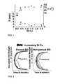
- FIG. 1is a plot of both clot detection time, R, (filled shapes) and rate of coagulation, ⁇ , (un-filled shapes) vs. BG Si:Ca. Data represents the mean of four trials. ⁇ Porous BG; ⁇ Non-porous BG; ⁇ Spherical BG; +No HA.
- FIG. 2is a Thrombelastograph®plot of bioactive hemostatic agents. Inner Thromboelastograph plot on both plots is sheep blood without a HA added.
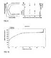
- FIG. 3is a Thrombelastograph® plot of bioactive glass, QuikClotTM, and sheep's blood alone.
- FIG. 4is a thermogravimetric analysis and differential scanning calorimetry of the dehydration of porous and non-porous bioactive glass. 90 J/g (Non-porous Bioactive glass) and 450 J/g (Porous Bioactive glass).
- FIG. 5is a Thrombelastograph® plot of mesoporous bioactive glass with varying SiO2:CaO ratios.
- BG 80has a molar ratio of SiO2:CaO of 80:16.
- BG 60has a molar ratio of SiO2:CaO of 60:16.
- FIG. 6is a Thrombelastograph® plot of non-porous bioactive glass with varying SiO2:CaO ratios.
- BG NP 80has a molar ratio of SiO2:CaO of 80:16.
- BG NP 70has a molar ratio of SiO2:CaO of 70:16.
- BG NP 60has a molar ratio of SiO2:CaO of 60:16.
- FIG. 7is a thermogravimetric analysis and differential scanning calorimetry of the dehydration process for a hydrated mesoporous bioactive glass and a non-porous bioactive glass.
- FIG. 8is a compilation of the heat of hydration and hydration capacity of bioactive glass.
- BG80has a molar ratio of SiO2:CaO of 80:16.
- BG60has a molar ratio of SiO2:CaO of 60:16.
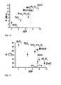
- FIG. 9Ashows a Thromboelastograph® plot of the hemostatic activity MBGM-80 induced coagulation vs. blood w/o MBGM-80.
- FIG. 9Bshows a plot of both clot detection time, R, (filled shapes) and rate of coagulation, ⁇ , (un-filled shapes) vs. amount of mesoporous bioactive microspheres. Data represents the mean of four trials. ⁇ MBGM-60, ⁇ MBGM-80, ⁇ MBGM-60 Non-porous, ⁇ MBGM-80 Non-porous, +Sheep Blood w/o MBGM.
- FIG. 10shows BET adsorption-desorption isotherm of bioactive glass.
- FIG. 11shows pore size distribution of mesoporous bioactive glass.
- FIG. 12shows BET surface area and pore diameter calculations.
- FIG. 13shows wide angle x-ray diffraction of bioactive glass substrates pre- and post-immersion in simulated body fluids for 1 hour.
- FIG. 14is a Thrombelastograph® plot of oxides with an isoelectric point below the pH of blood.
- FIG. 15is a Thrombelastograph® plot of oxides with a isoelectric point above the pH of blood.
- FIG. 16shows R (min), onset of clot detection, versus the metal oxide's isoelectric point for low-surface area metal oxides.
- FIG. 17shows ⁇ (°), rate of coagulation, versus the metal oxide's isoelectric point, for low-surface area metal oxides.
- the inventionis based on the discovery that oxide materials can be prepared to modulate hemostasis on the basis of surface charge. This modulation enables the synthesis of materials that are pro-coagulants; or, alternatively other materials that are anticoagulants. The latter are of importance with respect to die oxide coatings that form on metal medical implant devices.
- the methods of preparing oxide compositions of the inventionavoid problems associated with longer setting times and also produce materials having better performance characteristics.
- the methods of the inventionproduce materials that offer superior compositional and structural homogeneity and higher surface area, which provide more effective materials.
- one embodiment of the inventionprovides a rapid-setting, mesoporous, bioactive glass cement that exhibits excellent plasticity, superior bioactivity and is mechanically robust.
- the oxide compositions of the inventioncan be used for growth and repair of bone and other tissues as well as in drug delivery.
- high surface area mesoporous bioactive glasshas been prepared by a sol-gel template directed assembly.
- This materialhas the ability to conform and adhere to wounded tissue to promote blood clot formation.
- This specific materialhas a distinct morphological advantage over previous bioactive glass materials in that it can conform and adhere to any wound cavity geometry.
- a bioactive glass cementcan be formulated that has a predictable set time and accelerates the deposition of new apatite, layers when in contact with biological fluids.
- Mesoporous bioactive glass (MBG) cementsare malleable before setting and retain their shape and mechanical strength without crumbling after setting. Furthermore, mesoporous bioactive glass has demonstrated a high osteoconductive property.
- This materialcan be formulated in a variety of compositions for applications as a rapid acting hemostatic agent, template for the growth of artificial bone, and the generation of tissue.
- Bioactive glasscan be formulated for a variety of distinct wound healing scenarios and can elicit a predictable wound healing response, for both controlling the flow of blood as well as controlling the rate of apatite deposition, as a function of agents chemical composition and Si to Ca ratio.
- this inventionprovides a method by which materials can be selected based on their isoelectric point to induce a predictable hemostatic response.
- the isoelectric point of an oxidewill determine both the sign and magnitude of the initial surface charge density upon exposure to biological fluids. Oxides have been identified that will induce coagulation upon exposure to blood. Oxides have also been identified that will prevent or slow down the coagulation response of blood in contact with the surface of the oxide. A strategy to produce both rapid acting hemostatic agents and passivated medical device surfaces is described based on the selection criteria.
- a “hemostatically effective amount”means an amount sufficient to initiate detectable blood clotting (R) within 2 minutes, and/or achieve a rate of clotting ( ⁇ ) of 50° or greater, and/or achieve a clot strength (MA) of ⁇ 50, as determined by Thromboelastograph® measurements. Assays for determining hemostatic effectiveness are known in the art, and described in die Examples below.
- Thromboelastographrefers to measurements typically taken using about 5-30 mg of material mixed with 340 microliters of citrate stabilized blood. Calcium ions are re-supplied to the citrate stabilized blood prior to measurements to replace the calcium ions chelated by citrate.
- isoelectric pointrefers to the pH at which the zeta-potential equals zero in an aqueous electrolyte such as 2 mM CaCl 2 .
- the zeta potentialis the surface charge density of a metal oxide in aqueous suspension, measured as a function of pH by the electrophoretic method using the Smoluchowski equation (Cocera, M. et al., Langmuir 1999, 15, 2230-2233). Unless specifically indicated otherwise, the zeta potential of the metal oxide is measured in a CaCl 2 electrolyte that mimics the Ca 2+ concentration in blood.
- homogeneousmeans an absence of phase separation (e.g., separation of a silicate phase and a phosphate phase); the materials are not phase segregated when examined by energy-dispersive x-ray analysis (EDX) using scanning electron microscopy (SEM) with a resolution limit of 0.5 microns.
- EDXenergy-dispersive x-ray analysis
- SEMscanning electron microscopy
- a “bioactive glass sol”means a colloidal suspension containing silica precursors and calcium salts that can be gelled to form bioactive glass solid, wherein the solvent can be water, ethanol or other substance that can dissolve silica precursors and calcium species.
- the high surface area mesoporous bioactive glass described hereinhas a unique morphology with advantages over these methods including higher surface area and ease of functionalization of the final material.
- This functionalizationincludes, but is not limited to, the surface immobilization and the controlled release of biologically relevant molecules. Molecules such as phospholipids, fibrin, collagen, clotting zymogens, heat shock proteins, antibacterial peptides, and silver, magnesium, calcium, sodium, zinc, chloride, and phosphate ions can be controllably released to effect an optimal bio-response.
- a surface area of about 300 m 2 /ghas been attained with bioactive glass prepared from P123, while low molecular weight polymers, such as L43, can produce much higher surface area (in the range of 900 m 2 /g).
- the high surface areaprovides for optimal pore volume.
- bioactive glassrelated to rapid acting hemostatic agents for the treatment of traumatic injuries.
- the traumatic wound healing scenariois distinct from prior medical applications for bioactive glass-like materials.
- bioactive glasshas been loosely applied to many composites of calcium oxide, silicon dioxide, phosphorous oxide and other metal oxides, the combination of which is able to promote the growth of bone and tissue.
- the invention described in U.S. provisional patent application No. 60/668,022, filed Apr. 4, 2005provides a calcium loaded zeolite linde type A that is ion exchanged with aqueous solutions of alkali, alkaline earth, and transition metal cations to specific ion formulations.
- This ion exchanged zeolitecan be mixed with neutral inorganic salts like calcium chloride, aluminum sulfite, and silver nitrate and dehydrated to remove water.
- the dehydrated inorganic materialsare sealed in mylar foil bags to prevent rehydration until required during medical application. At the time of medical application, the mylar bag can be opened and the inorganic contents poured into the traumatically injured site.
- the present inventionprovides the bioactive glass in a gel) liquid, cement, paste or powder form, which allows for greater ease of use and better conformation to a desired area to be treated.
- a gelliquid, cement, paste or powder form
- the material in gel (or cement) formfor example, it can be applied to a greater variety of surfaces, increasing its availability for use in numerous contexts, including application to medical devices and drug delivery.
- Porous bioactive glass materialshave been designed to treat traumatically injured tissue by inducing hemostasis through contact activation and release of coagulation co-factors.
- the compositions of the present inventionprovide a uniform pore size that further optimizes its use for regulation of hemostasis.
- the hemostatic activity of bioactive glassis dependent on the material's chemical composition.
- Si:Ca:P atomic _ ratio 60:36:4 to 90:6:4the range of chemically distinct bioactive glass agents studied (Si:Ca:P atomic _ ratio 60:36:4 to 90:6:4)
- the onset time for contact-activated coagulation, rate of coagulation of post-initiation, and ultimate clot strengthwas found to be dependent on the material's Si:Ca ratio, porosity, and heat of hydration.
- the onset time for contact-activated coagulationwas found to decrease in an increasing Si:Ca ratio.
- the rate of coagulation post-initiationwas found to increase with an increasing Si:Ca ratio. Porous bioactive glass was found to have a greater procoagulant tendency than non-porous bioactive glass.
- the bone-generating activity of bioactive glassis dependent on the material's chemical composition.
- the deposition rate of hydroxyapatite deposition in biological fluidsis related to the material's Si:Ca ratio and particle size and shape.
- the rate of deposition of hydroxyapatitewas observed to be faster for bioactive glass samples with a lower Si:Ca ratio (e.g. BG60:36:4 faster than BG80:16:4).
- the isoelectric point of a materialis a critical material parameter that can be utilized to select oxides that can either promote or prevent the induction of hemostasis. Rapid acting hemostatic agents and passivated medical devices are applications intended for this material.
- the present inventorshave discovered that the oxide's initial surface charge, driven by the isoelectric point of the material relative to the pH of the immersing biological medium, is the key factor in controlling hemostatic efficacy of the composition.
- the onset time for contact-activated coagulation, rate of coagulation post-initiation, and ultimate clot strengthare found to be dependent on the initial surface charge density of the metal oxide when exposed to blood, which is related to the oxide's acid-base nature and is quantitatively described by its isoelectric point. Wee found, that for polar metal-oxide substrates, the time to initiate contact-activated coagulation increases with the increase in the metal oxide's isoelectric point.
- Bloodis usually the first fluid an implanted foreign body encounters, and thus the thrombotic complications which arise from metallic implants (chronic inflamatory response), and inorganic-based extracorporeal circulating devices parts, arterial stents, and catheters is related to the chemistry that occurs during the initial exposure of blood to a foreign oxide surface.
- metallic implantschronic inflamatory response
- inorganic-based extracorporeal circulating devices partse.g. blood pressure
- arterial stentse.g. blood a blood circulating blood
- catheterse.g. blood a blood fibroblasts, and the initial surface charge density of a metal oxide surface will affect the selective adhesion of oppositely charged molecules and biological media (e.g. cells and larger proteins) immediately upon contact with blood.
- Negatively-charged surfacesare known to initiate the intrinsic pathway of the blood coagulation cascade, a network of feedback-dependent reactions that when activated results in a blood clot.
- the activation of this process by a foreign bodyis referred to as contact-activation of coagulation.
- the same network of coagulation reactionsalso can be activated via the extrinsic pathway, which occurs when a breach in the endothelium allows the exposure of platelets to tissue factor bearing cells.
- metal oxidesare inherently polar surfaces. Their surface chemistry is all the more complicated due to the presence of “dangling” terminal hydroxyl groups on unsaturated metal sites and related defect sites.
- the surface charge of metal oxidesis known to be pH dependent and is thought to result from either the amphoteric dissociation of surface MOH groups or the adsorption of metal hydroxo complexes derived from the hydrolysis product of material dissolved from the metal oxide.
- the pH at which the sum total of negative and positive surface charges equals zero, ⁇ (z ⁇ n)M z+ (OH) n z ⁇ n0, is called the isoelectric point.
- Thromboelastograph®The in vitro hemostatic activity of metal-oxide hemostatic agents was evaluated as previously described using a Thromboelastograph®, a clinical instrument that monitors the change in viscoelasticity of blood as a function of time. Briefly, 340 ⁇ L of 4% v/v citrate-stabilized sheep blood (Quad Five of Ryegate, Mont.) was introduced into the sample cup of a Thromboelastograph®, Haemoscope model 5000, along with 20 ⁇ L of 0.2M CaCl 2 (aq) and 5-20 mg of a tested metal-oxide in a powder morphology. The 20 ⁇ L of 0.2 M CaCl 2 (aq) was added to the stabilized blood to replenish the Ca 2+ ions chelated by citrate, which was added to prevent coagulation of stored blood. Blood was stored at 8° C. prior to use.
- the Thromboelastograph® sample cupis rotated ⁇ 5° about a vertical torsion wire suspended in the middle of the cup.
- the change in viscoelastic clot strengthis monitored as a function of time.
- the time until the bimodal symmetric viscoelasticity curve's amplitude is 2 mmis referred to as R (minutes), and represents the initial detection of clot formation.
- the angle between the tangent to the curve and the horizontalis referred to as ⁇ (°), and is related to the rate of coagulation.
- the maximum amplitude of the curvesis referred to as MA (mm) and represents the maximum clot strengths.
- Thromboelastograph® clotting parameters reportedrepresent the mean of four reproducible trials. A summary of the hemostatic properties of metal-oxides with variable isoelectric points is described in Table 1.
- compositions that modulate homeostasiscan be prepared by the methods described in the Examples below, including aerosol synthesis and use of sol-gel chemistry.
- Sol-gel chemistrycan be used to produce bioactive glass.
- a hot furnacee.g., 400° C.
- spherical bioactive glass particlesare produced. These bioactive glass particles can be as small as 10-50 nm in diameter, or smaller, or as large as about 100 ⁇ m or larger. In one embodiment, the particles are 50-200 nm in diameter.
- the method of producing a composition of the inventioninvolves starting from a bioglass sol, wherein the solvent is ethanol (or another solvent that can dissolve precursors and has a low boiling point).
- a block copolymercan be used as all additive to provide a pore-forming gent.
- the ideal solventis water rather than ethanol because the melting point of ethanol is very low.
- the difference in solventtypically calls for some difference in the method.
- most PEO—PPO-PEO block copolymerscannot dissolve in water.
- chitosancan be incorporated into the system because it doesn't dissolve in the ethanol, and chitosan plays an important role in modulating blood coagulation.
- the silica and phosphous precursorsare different from those in an ethanol-based method and phosphorous oxide is not required in the starting sol, as would be the typical case when starting with a bioglass sol.
- the method of modulating hemostasiscomprises decreasing blood coagulation time.
- the time to initiate detectable coagulation (R), as measured by Thromboelastograph®is less than 2 minutes, and can be less than 1.8 minutes.
- the rate of coagulation ( ⁇ ), as measured by Thromboelastograph®is more than 50°. Coagulation rates of more than 55°, and of more than 65° have been achieved.
- the coagulationresults in a maximum clot strength (MA), as measured by Thromboelastograph®, of 55 to 100 mm, and can be less than 75 nm.
- the modulatingcomprises increasing blood coagulation time. Increased coagulation time is desirable, for example, when clotting poses a health risk to the subject.
- Oxides with an isoelectric point below the pH of bloodcan be formulated for action to induce blood clot formation faster than blood would naturally do in the absence of an oxide-contact activator.
- the materialscan be applied both externally and internally as agents to induce hemostasis and reduce the flow of blood in a particular area of the body.
- Oxides with an isoelectric point above the pH of bloodcan be formulated to induce blood clot formation slower than blood would naturally do in the absence of an oxide-contact activator, and therefore would be suitable as passivated surfaces for medical devices.
- the inventionprovides a medical device and methods of coating a medical device with a composition of the invention. Coatings can be prepared from a composition in powder form or using sol-gel chemistry, using conventional methods known in the art. In one embodiment, the coating reduces coagulation of blood in contact with the device.
- the medical devicesinclude, but are not limited to, arterial and venal stents, catheters, shunts, and any medical machinery that will contact blood during invasive medical procedures.
- Oxides with an isoelectric point above the pH of bloodcan be formulated for devices that require a positively charged surface to interface with biological tissue and fluids.
- Oxides with an isoelectric point below the pH of bloodcan be formulated for devices that require a negatively charged surface to interface with biological tissue and fluids.
- a bioactive glass cementWhen mixed with an ammonium phosphate buffer solution, a bioactive glass cement can be prepared with a controllable set time.
- Bioactive glass, and particularly, bioactive glass cementcan be prepared with a flexible morphology that allows for conformation and adhesion to any wound geometry.
- the bioactive glass cementcan be molded in a variety of shapes that retain their mechanical integrity post-setting.
- the bioactive glass cementscan accelerate the deposition of an apatite layer compared to the bioactive glass agent alone.
- Mesoporous bioactive glasscan be formulated as a rapid acting hemostatic agent. This material can predictably warm injured tissue to promote wound healing.
- Mesoporous bioactive glasscan be formulated to promote the formation of artificial bone. This same material can be used to generate tissue including, but not limited to, artificial skin and structural elements such as fibrin and collagen.
- the internal porous architecturecan be loaded with biologically relevant molecules and cofactors for controlled released during wound healing and body repair.
- biologically relevant molecules and cofactorsinclude, but are not limited to, phospholipids, blood coagulation factors, fibrin, collagen, blood clotting zymogens, silver ions, magnesium ions, and calcium ions.
- the internal porous architecturecan be loaded with antibacterial peptides and silver ions for a controlled release of antibacterial agents.
- Non-porous bioactive glasscan be formulated as a rapid acting hemostatic agent. This material can predictably warm injured tissue to promote wound healing.
- Non-porous bioactive glasscan be formulated to promote the formation of artificial bone. This same material can be used to generate tissue including, but not limited to, artificial skin and structural elements such as fibrin and collagen.
- the hemostatic activity of bioactive glasscan be controlled and optimized for a variety of wound healing scenarios by manipulating the ratio of Si to Ca in the chemical composition of both porous and non-porous bioactive glass.
- the bone-generating activity of bioactive glasscan be controlled and optimized for a variety of wound healing scenarios by manipulating the ratio of Si to Ca in the chemical composition of both porous and non-porous bioactive glass.
- the time until clot detection, Rdecreases for increasing Si:Ca ratios in BG ( FIGS. 1, 2 ). R is reduced by a factor of 2 when the Si:Ca ratio is doubled over the range studied.
- BGcan perform the dual role of providing surface area for thrombosis and supplying Ca 2+ ions; hence there will be an optimum ratio of SiO 2 to Ca 2+ ions, which are co-factors throughout the clotting cascade, for the fastest hemostatic response.
- the unique formulation of high surface area mesoporous bioactive glass that we have preparedhas the ability to rapidly induce a blood clot when exposed to blood.
- the formulation we have preparedhas a faster clotting time and results in a stronger clot than QuikClotTM, the leading inorganic hemostatic agent currently available (see FIG. 3 ).
- Both the porous and non-porous formulations of bioactive glasspossess this ability to rapidly promote blood coagulation. Because the porous and non-porous formulations of bioactive glass can be hydrated to different degrees, and consequently will deliver different amounts of heat upon hydration during medical application to a wound site, we can further tailor the rate of blood coagulation. Combinations of porous and non-porous bioactive glass can be formulated to the desired specifications of hydration and delivery of heat (see FIG. 4 ).
- This exampleshows that one make the bioactive glass with varying ratios of SiO2:CaO.
- SiO2:CaO ratiosmore silica
- the materialtends to clot blood faster. This is illustrated in both FIGS. 5 and 6 .
- the amount of SiO2 relative to the amount of CaOis reduced, the kinetics of clot formation are much slower.
- the difference in clotting kinetics between two bioactive glass samples with different SiO2:CaOis more pronounced with the non-porous samples.
- the mesoporous bioactive glassis a faster clotting agent than the non-porous samples, but the difference between samples is greater within the non-porous samples.
- This examplealso shows that one can use combinations of porous and non-porous bioactive glass, as well as composites with multiple bioactive glasses of different SiO2:CaO ratios, to achieve any desired hydration capacity and heating response when in contact with blood (see FIGS. 7 and 8 ).
- Spherical Bioactive glassis produced by an aerosol assisted method and with the same sol-gel precursor solution employed for bioactive glass previously described. Spherical bioactive glass accelerates the formation of a contact-activated clot. The activity of bioactive glass is dependent on the relative amount of contact activating agent to the surrounding blood volume ( FIG. 9 ).
- the porous architecture of mesoporous bioactive glassis ideal for the controlled release of biomolecules. These molecules can be immobilized on the oxide surface of bioactive glass or solvated with surfactants inside the pores, or loaded alone in die pores. Each of these formulations will have a unique release profile with regard to concentration and rate of release.
- the combination of porous bioactive glass and biomoleculesis referred to as a host-guest composite.
- Host-guest compositescan also be prepared to release ions including, but not limited to, silver, magnesium and calcium ions.
- Silver ionshave been shown to be antibacterial at parts per billion concentration in biological fluids.
- Magnesium and calcium ionsare essential cofactors during the coagulation of blood.
- Certain formulations of porous bioactive glasscan also sequester magnesium and calcium from blood to delay the coagulation response.
- MBGsMesoporous bioactive glasses
- EISAevaporation-induced self-assembly
- the dried gelwas calcined at high temperature to remove the block copolymer and form mesopores.
- the final MBGswere ground into powders.
- the as-calcined MBGshave more accessible mesopore surface area and ordered pore structure.
- In vitro studyshowed a greater bone-forming bioactivity than conventional sol-gel derived BGs by fast formation of an amorphous bioactive HA layer.
- Bioactive glass cementswere prepared by mixing bioactive glass powders with an ammonium phosphate buffer solution.
- the liquid component of MBGCsan ammonium phosphate buffer solution, was prepared by dissolving 60.1 g (NH 4 ) 2 HPO 4 and 5.0 g NH 4 H 2 PO 4 in 100 mL water. The pH of the resulting solution was ⁇ 7.3.
- MBGC cementswere made by mixing the solid and liquid components at the ratio of 1 g to 1 mL. The cements were kept in the ambient environment to set. Before setting fully, they were soft enough to be kneaded or molded. Structural characterizations were typically carried out at ⁇ 1 h after the mixing of the solid and liquid components of MBG, and no structural changes were observed after 1 h after mixing.
- SBFcontained 142.0 mM Na + , 5.0 mM K + , 1.5 mM Mg + , 2.5 mM Ca 2+ , 147.8 mM Cl ⁇ , 4.2 mM HCO 3 ⁇ , 1.0 mM HPO 4 2 ⁇ , and 0.5 mM SO 4 2 ⁇ .
- Its chemical compositionis similar to that of human plasma.
- the solutionhad a pH of 7.3-7.4 and was kept at 37° C. before use.
- This examplepresents data on the surface area measurements that have been made of the mesoporous bioactive glass of the invention.
- FIG. 10the adsorption-desorption isotherm is presented. The lack of hysteresis suggests a channel-like structure without internal cages.
- This adsorption-desorption isothermcan be used to calculate the pore size distribution of the mesoporous bioactive glass based on the BJH model.
- a plot of the pore size distributionis illustrated in FIG. 11 .
- Bioactive glasscan be formulated with a surface area ranging from 300 m 2 /g to 1000 m 2 /g.
- the sample that was used for the measurements described in this examplehad a surface area of 960 nm/g.
- the internal pore diameterwas calculated to be 3.1 nm based oil the BJH model and 2.5 nm based on the BET model.
- SBFsimulated body fluids
- the average hydroxyapatite crystal size nucleated after immersing BG80:16:4 in simulated body fluids for one dayis 32 nm. Faster rates of hydroxyapatite were observed with BG60:36:4 compared to BG80:16:4.
- Every oxide materialwill possess an initial surface charge that is a function of both the isoelectric point of the material and the pH conditions of the immersing solution (see FIG. 14 ).
- the rate of coagulation of blood upon exposure to a variety of inorganic oxideswe have observed that those materials with an isoelectric point below the pH of blood accelerate the coagulation response (see FIG. 14 ).
- those materials with an isoelectric point above the pH of bloodare observed to decelerate the coagulation response (see FIG. 15 ).
- NiOwhich has the closest isoelectric point to the pH of blood but will be positively charged after immediately contacting blood, was observed to reduce the rate of coagulation.
Landscapes
- Health & Medical Sciences (AREA)
- Chemical & Material Sciences (AREA)
- Public Health (AREA)
- Epidemiology (AREA)
- Life Sciences & Earth Sciences (AREA)
- Animal Behavior & Ethology (AREA)
- General Health & Medical Sciences (AREA)
- Veterinary Medicine (AREA)
- Inorganic Chemistry (AREA)
- Medicinal Chemistry (AREA)
- Pharmacology & Pharmacy (AREA)
- Surgery (AREA)
- Engineering & Computer Science (AREA)
- Materials Engineering (AREA)
- Materials For Medical Uses (AREA)
Abstract
Description
| TABLE 1 |
| Summary of Metal-Oxide Contact-Activated Coagulation |
| Low-surface-area metal | High-surface-area metal | |
| Clotting Metric | oxides | oxides |
| Onset of coagulation; R | Coagulation onset time | Coagulation onset time for |
| (min) | increased or of equal value | positively charged surface |
| Initially Positively | compared to blood alone | similar to blood alone |
| Charged Metal Oxide | for positively charged | |
| surface, and slowest for the | ||
| most positive surface | ||
| Initially Negatively | Coagulation onset time | Coagulation onset time |
| Charged Metal Oxide | reduced for negatively | reduced for negatively |
| charged surfaces, and | charged surfaces | |
| fastest for most negative | ||
| substrate | ||
| Rate of coagulation post- | Positively-charged surfaces | Positively-charged surfaces |
| initiation; α (°) | decelerate the rate of | decelerate the rate of |
| Initially Positively | coagulation | coagulation |
| Charged Metal Oxide | ||
| Initially Negatively | Negatively-charged | Negatively-charged |
| Charged Metal Oxide | surfaces accelerate the rate | surfaces accelerate the rate |
| of coagulation | of coagulation in the | |
| presence of sufficient | ||
| Ca2+ ions | ||
| Isoelectric Point Below | Isoelectric Point Above | |
| Clotting Metric | the pH of Blood | the pH of Blood |
| Onset of coagulation; R | Coagulation onset time | Coagulation onset time |
| (min) | reduced for negatively | increased or of equal value |
| charged surfaces, and | compared to blood alone | |
| fastest for most negative | for positively charged | |
| substrate | surface, and slowest for the | |
| most positive surface | ||
| Rate of coagulation post- | Negatively-charged | Positively-charged surfaces |
| initiation; α (°) | surfaces accelerate the rate | decelerate the rate of |
| of coagulation | coagulation | |
| Ultimate clot strength | Most negative oxide | Induced blood clots are |
| (MA) | resulted in steongest blood | less than or equal in |
| clots and least negative | strength to naturally | |
| oxide resulted in weakest | formed blood clots | |
| blood clot | ||
Methods
Claims (14)
Priority Applications (1)
| Application Number | Priority Date | Filing Date | Title |
|---|---|---|---|
| US11/464,825US9326995B2 (en) | 2005-04-04 | 2006-08-15 | Oxides for wound healing and body repair |
Applications Claiming Priority (4)
| Application Number | Priority Date | Filing Date | Title |
|---|---|---|---|
| US66802205P | 2005-04-04 | 2005-04-04 | |
| US70820605P | 2005-08-15 | 2005-08-15 | |
| US11/398,161US7858123B2 (en) | 2005-04-04 | 2006-04-04 | Inorganic materials for hemostatic modulation and therapeutic wound healing |
| US11/464,825US9326995B2 (en) | 2005-04-04 | 2006-08-15 | Oxides for wound healing and body repair |
Related Parent Applications (1)
| Application Number | Title | Priority Date | Filing Date |
|---|---|---|---|
| US11/398,161Continuation-In-PartUS7858123B2 (en) | 2005-04-04 | 2006-04-04 | Inorganic materials for hemostatic modulation and therapeutic wound healing |
Publications (3)
| Publication Number | Publication Date |
|---|---|
| US20070154564A1 US20070154564A1 (en) | 2007-07-05 |
| US20100209531A2 US20100209531A2 (en) | 2010-08-19 |
| US9326995B2true US9326995B2 (en) | 2016-05-03 |
Family
ID=38224738
Family Applications (1)
| Application Number | Title | Priority Date | Filing Date |
|---|---|---|---|
| US11/464,825Active2029-05-26US9326995B2 (en) | 2005-04-04 | 2006-08-15 | Oxides for wound healing and body repair |
Country Status (1)
| Country | Link |
|---|---|
| US (1) | US9326995B2 (en) |
Cited By (11)
| Publication number | Priority date | Publication date | Assignee | Title |
|---|---|---|---|---|
| US10647962B2 (en) | 2016-05-27 | 2020-05-12 | Corning Incorporated | Bioactive aluminoborate glasses |
| US10676713B2 (en) | 2016-05-27 | 2020-06-09 | Corning Incorporated | Bioactive borophosphate glasses |
| US10751367B2 (en) | 2016-05-27 | 2020-08-25 | Corning Incorporated | Bioactive glass microspheres |
| US10857259B2 (en) | 2017-11-28 | 2020-12-08 | Corning Incorporated | Chemically strengthened bioactive glass-ceramics |
| WO2021162736A1 (en)* | 2020-02-14 | 2021-08-19 | Collidion, Inc. | Compositions, kits, methods and uses for cleaning, disinfecting, sterilizing and/or treating |
| US11198638B2 (en) | 2017-11-28 | 2021-12-14 | Corning Incorporated | Bioactive borate glass and methods thereof |
| US11274059B2 (en) | 2017-11-28 | 2022-03-15 | Corning Incorporated | Bioactive glass compositions and dentin hypersensitivity remediation |
| US11384009B2 (en) | 2017-11-28 | 2022-07-12 | Corning Incorporated | High liquidus viscosity bioactive glass |
| RU2805923C1 (en)* | 2023-04-21 | 2023-10-24 | Общество С Ограниченной Ответственностью "Фарм-Медик" | Fluid hemostatic agent |
| US11814649B2 (en) | 2016-05-27 | 2023-11-14 | Corning Incorporated | Lithium disilicate glass-ceramic compositions and methods thereof |
| US12402628B2 (en) | 2018-01-14 | 2025-09-02 | Collidion, Inc. | Compositions, kits, methods and uses for cleaning, disinfecting, sterilizing and/or treating |
Families Citing this family (31)
| Publication number | Priority date | Publication date | Assignee | Title |
|---|---|---|---|---|
| EP1667623B1 (en) | 2003-09-12 | 2010-11-24 | Z-Medica Corporation | Partially hydrated hemostatic agent |
| WO2005027808A1 (en) | 2003-09-12 | 2005-03-31 | Z-Medica Corporation | Calcium zeolite hemostatic agent |
| KR100561646B1 (en)* | 2003-10-23 | 2006-03-20 | 엘지.필립스 엘시디 주식회사 | Thin film transistor substrate for display element and manufacturing method thereof |
| US20060178609A1 (en)* | 2005-02-09 | 2006-08-10 | Z-Medica, Llc | Devices and methods for the delivery of molecular sieve materials for the formation of blood clots |
| CA2597940A1 (en) | 2005-02-15 | 2006-08-24 | Virginia Commonwealth University | Mineral technologies (mt) for acute hemostasis and for the treatment of acute wounds and chronic ulcers |
| US7858123B2 (en)* | 2005-04-04 | 2010-12-28 | The Regents Of The University Of California | Inorganic materials for hemostatic modulation and therapeutic wound healing |
| US9326995B2 (en) | 2005-04-04 | 2016-05-03 | The Regents Of The University Of California | Oxides for wound healing and body repair |
| US20060282046A1 (en)* | 2005-04-13 | 2006-12-14 | Horn Jeffrey L | Device and method for subcutaneous delivery of blood clotting agent |
| US20080306168A1 (en)* | 2005-12-29 | 2008-12-11 | Craig Bradley D | Dental Compositions with a Water Scavenger |
| US20070154509A1 (en)* | 2005-12-30 | 2007-07-05 | Wilcher Steve A | Adsorbent-Containing Hemostatic Devices |
| US20070154510A1 (en)* | 2005-12-30 | 2007-07-05 | Wilcher Steve A | Adsorbent-Containing Hemostatic Devices |
| US8938898B2 (en) | 2006-04-27 | 2015-01-27 | Z-Medica, Llc | Devices for the identification of medical products |
| US20070276308A1 (en)* | 2006-05-26 | 2007-11-29 | Huey Raymond J | Hemostatic agents and devices for the delivery thereof |
| US8202532B2 (en) | 2006-05-26 | 2012-06-19 | Z-Medica Corporation | Clay-based hemostatic agents and devices for the delivery thereof |
| US7968114B2 (en)* | 2006-05-26 | 2011-06-28 | Z-Medica Corporation | Clay-based hemostatic agents and devices for the delivery thereof |
| US7604819B2 (en)* | 2006-05-26 | 2009-10-20 | Z-Medica Corporation | Clay-based hemostatic agents and devices for the delivery thereof |
| WO2008036225A2 (en)* | 2006-09-20 | 2008-03-27 | Entek Manufacturing, Inc. | Conformable structured therapeutic dressing |
| US20080206134A1 (en)* | 2007-02-22 | 2008-08-28 | Denny Lo | Radio-opaque hemostatic agents and devices and methods for the delivery thereof |
| US20080228123A1 (en)* | 2007-03-14 | 2008-09-18 | Noble Fiber Technologies, Llc | Bandage with a hydrophilic foam containing silver and a hemastatic agent |
| US20080317831A1 (en)* | 2007-06-21 | 2008-12-25 | Denny Lo | Hemostatic sponge and method of making the same |
| WO2009023745A1 (en)* | 2007-08-14 | 2009-02-19 | The Regents Of The University Of California | Mesocellular oxide foams as hemostatic compositions and methods of use |
| US20090162406A1 (en)* | 2007-09-05 | 2009-06-25 | Z-Medica Corporation | Wound healing with zeolite-based hemostatic devices |
| EP2219572A4 (en) | 2007-12-06 | 2014-05-28 | Nanosys Inc | Resorbable nanoenhanced hemostatic structures and bandage materials |
| US8319002B2 (en) | 2007-12-06 | 2012-11-27 | Nanosys, Inc. | Nanostructure-enhanced platelet binding and hemostatic structures |
| US8858969B2 (en) | 2010-09-22 | 2014-10-14 | Z-Medica, Llc | Hemostatic compositions, devices, and methods |
| CN101991875B (en)* | 2010-10-29 | 2014-01-22 | 上海昊海生物科技股份有限公司 | Mesoporous bioactive glass and chitosan composite porous hemostatic material and preparation method thereof |
| EP2741788A4 (en)* | 2011-08-14 | 2015-03-25 | Materials Modification Inc | Method and composition for in situ formation of an artificial blockage to control blood loss |
| MX376779B (en) | 2012-06-22 | 2025-03-07 | Teleflex Tech Llc | HEMOSTATIC DEVICES. |
| EP4159248A1 (en)* | 2013-12-10 | 2023-04-05 | Institut National de la Santé et de la Recherche Médicale (INSERM) | Methods for adhering tissue surfaces and materials and biomedical uses thereof |
| CN107213508B (en)* | 2017-06-09 | 2018-04-06 | 中南大学 | A kind of oxides-containing iron/nano kaoline compound hemostatic agent and preparation method thereof |
| CN109481731B (en)* | 2019-01-23 | 2020-03-27 | 中南大学 | A kind of nano-oxide/kaolin composite hemostatic and antibacterial material, hemostatic and healing-promoting dressing and preparation method thereof |
Citations (134)
| Publication number | Priority date | Publication date | Assignee | Title |
|---|---|---|---|---|
| US2688586A (en) | 1950-03-17 | 1954-09-07 | Johnson & Johnson | Improved hemostatic alginic surgical dressings and method of making |
| US2882243A (en) | 1953-12-24 | 1959-04-14 | Union Carbide Corp | Molecular sieve adsorbents |
| US3122140A (en) | 1962-03-29 | 1964-02-25 | Johnson & Johnson | Flexible absorbent sheet |
| US3181231A (en) | 1963-08-06 | 1965-05-04 | Union Carbide Corp | Molecular sieve-metal agglomerates and their preparation |
| US3366578A (en) | 1964-12-07 | 1968-01-30 | Universal Oil Prod Co | Zeolite and method for making the improved zeolite |
| US3538508A (en) | 1968-08-08 | 1970-11-10 | Samuel Young | Combination pillow and crash helmet |
| US3723352A (en) | 1971-01-25 | 1973-03-27 | Air Prod & Chem | Supported silver catalysts |
| US3979335A (en) | 1974-12-27 | 1976-09-07 | Georgy Anatolievich Golovko | Process for the preparation of synthetic zeolites |
| US4373519A (en) | 1981-06-26 | 1983-02-15 | Minnesota Mining And Manufacturing Company | Composite wound dressing |
| US4374044A (en) | 1981-01-19 | 1983-02-15 | General Motors Corporation | Cordierite bead catalyst support and method of preparation |
| US4514510A (en) | 1983-09-08 | 1985-04-30 | American Colloid Company | Hydrogen enriched water swellable clay having reduced acid demand and stable at low pH |
| US4525410A (en) | 1982-08-24 | 1985-06-25 | Kanebo, Ltd. | Particle-packed fiber article having antibacterial property |
| JPS61145120U (en) | 1985-02-28 | 1986-09-08 | ||
| US4626550A (en) | 1985-01-14 | 1986-12-02 | Pq Corporation | Zeolite for personal care products |
| US4631845A (en) | 1985-05-17 | 1986-12-30 | Intermec Corporation | Luggage tag |
| US4728323A (en)* | 1986-07-24 | 1988-03-01 | Minnesota Mining And Manufacturing Company | Antimicrobial wound dressings |
| US4744805A (en) | 1986-05-22 | 1988-05-17 | Air Products And Chemicals, Inc. | Selective adsorption process using an oxidized ion-exchanged dehydrated chabizite adsorbent |
| US4748978A (en) | 1984-09-27 | 1988-06-07 | Kamp Herman F | Therapeutic dressing having mineral components |
| US4822349A (en) | 1984-04-25 | 1989-04-18 | Hursey Francis X | Method of treating wounds |
| US4828832A (en)* | 1983-09-07 | 1989-05-09 | Laboratorios Biochemie De Mexico | Method of manufacturing a composition for treating skin lesions |
| US4828081A (en) | 1988-03-04 | 1989-05-09 | Samsonite Corporation | Luggage identification system |
| US4911898A (en) | 1983-01-21 | 1990-03-27 | Kanebo Limited | Zeolite particles retaining silver ions having antibacterial properties |
| US4938958A (en) | 1986-12-05 | 1990-07-03 | Shinagawa Fuel Co., Ltd. | Antibiotic zeolite |
| US4956350A (en) | 1988-08-18 | 1990-09-11 | Minnesota Mining And Manufacturing Company | Wound filling compositions |
| WO1995005445A1 (en) | 1993-08-18 | 1995-02-23 | Unilever Plc | Granular detergent compositions containing zeolite and process for their preparation |
| US5436362A (en) | 1992-11-20 | 1995-07-25 | Chiyoda Corporation | Method of producing dialkylcarbonate |
| US5474545A (en) | 1992-12-07 | 1995-12-12 | Chikazawa; Osamu | Diaper and/or sanitary napkin |
| US5486195A (en) | 1993-07-26 | 1996-01-23 | Myers; Gene | Method and apparatus for arteriotomy closure |
| US5556699A (en) | 1987-06-30 | 1996-09-17 | Shingawa Fuel Co. Ltd. | Antibiotic zeolite-containing film |
| WO1996040285A1 (en) | 1995-06-07 | 1996-12-19 | Imarx Pharmaceutical Corp. | Novel targeted compositions for diagnostic and therapeutic use |
| US5599578A (en) | 1986-04-30 | 1997-02-04 | Butland; Charles L. | Technique for labeling an object for its identification and/or verification |
| WO1997017401A1 (en) | 1995-11-09 | 1997-05-15 | William Bonfield | Bioactive composite material for repair of hard and soft tissues |
| US5696101A (en) | 1996-04-16 | 1997-12-09 | Eastman Chemical Company | Oxidized cellulose and vitamin E blend for topical hemostatic applications |
| US5716337A (en) | 1992-06-10 | 1998-02-10 | Johnson & Johnson Medical, Inc. | Absorbent product |
| US5725551A (en) | 1993-07-26 | 1998-03-10 | Myers; Gene | Method and apparatus for arteriotomy closure |
| US5801116A (en) | 1995-04-07 | 1998-09-01 | Rhodia Inc. | Process for producing polysaccharides and their use as absorbent materials |
| US5826543A (en) | 1995-01-20 | 1998-10-27 | Ralston Purina Company | Clumpable animal litter containing a dust reducing agent |
| WO1998047465A1 (en) | 1997-04-21 | 1998-10-29 | Ylaenen Heimo | A novel composite and its use |
| WO1999013918A3 (en) | 1997-09-18 | 1999-06-03 | Univ Pittsburgh | Icam-1 selective echogenic microbubbles |
| US5941897A (en) | 1997-05-09 | 1999-08-24 | Myers; Gene E. | Energy activated fibrin plug |
| US5964349A (en) | 1994-08-31 | 1999-10-12 | Sony Corporation | Cassette, storage case, and label to be applied to such cassette and storage case |
| US5981052A (en) | 1996-08-27 | 1999-11-09 | Rengo Co., Ltd. | Inorganic porous crystals-hydrophilic macromolecule composite |
| JPH11332909A (en) | 1998-05-22 | 1999-12-07 | Frontier:Kk | Absorbent for absorption of salt-containing solution |
| WO2000009176A1 (en)* | 1998-08-11 | 2000-02-24 | Susanna Elizabeth Chalmers | A wound treatment composition and a wound dressing containing it |
| US6037280A (en) | 1997-03-21 | 2000-03-14 | Koala Konnection | Ultraviolet ray (UV) blocking textile containing particles |
| US6060461A (en) | 1999-02-08 | 2000-05-09 | Drake; James Franklin | Topically applied clotting material |
| US6123925A (en) | 1998-07-27 | 2000-09-26 | Healthshield Technologies L.L.C. | Antibiotic toothpaste |
| WO2000066086A1 (en) | 1999-04-29 | 2000-11-09 | Usbiomaterials Corporation | Anti-inflammatory bioactive glass particulates |
| US6159232A (en) | 1997-12-16 | 2000-12-12 | Closys Corporation | Clotting cascade initiating apparatus and methods of use and methods of closing wounds |
| WO2000076486A1 (en) | 1999-06-14 | 2000-12-21 | Imperial College Innnovations | Silver-containing, sol-gel derived bioglass compositions |
| GB2314842B (en) | 1996-06-28 | 2001-01-17 | Johnson & Johnson Medical | Collagen-oxidized regenerated cellulose complexes |
| US6187347B1 (en) | 2000-02-09 | 2001-02-13 | Ecosafe, Llc. | Composition for arresting the flow of blood and method |
| US6203512B1 (en) | 1999-06-28 | 2001-03-20 | The Procter & Gamble Company | Method for opening a packaging device and retrieving an interlabial absorbent article placed therein |
| US6251423B1 (en)* | 1995-05-20 | 2001-06-26 | Smith & Nephew Plc | Sterilizable paste product for topical application |
| WO2001082896A1 (en) | 2000-04-28 | 2001-11-08 | Biolife, L.L.C | Hemostatic agent, method and carrier for applying a blood clotting agent |
| JP3272770B2 (en) | 1992-05-11 | 2002-04-08 | 東芝ケミカル株式会社 | Liquid epoxy resin composition |
| US6372333B1 (en) | 1998-02-25 | 2002-04-16 | Rengo Co., Ltd. | Composition containing inorganic porous crystals-hydrophilic macromolecule composite and product made therefrom |
| WO2002030479A1 (en) | 2000-10-13 | 2002-04-18 | On Site Gas Systems, Inc. | Bandage using molecular sieves |
| US6428800B2 (en) | 1996-09-19 | 2002-08-06 | Usbiomaterials Corporation | Composition and method for acceleration of wound and burn healing |
| WO2002060367A1 (en) | 2001-01-31 | 2002-08-08 | Missak Kechichian | Absorbent product |
| US6450537B2 (en) | 2000-01-24 | 2002-09-17 | Polaroid Corporation | Self-service postage stamp assemblage |
| WO2002074325A1 (en) | 2001-03-19 | 2002-09-26 | Iomai Corporation | Patch for transcutaneous immunization |
| US6475470B1 (en) | 1998-09-25 | 2002-11-05 | Kao Corporation | Compositions for oral cavity |
| US6481134B1 (en) | 2001-04-02 | 2002-11-19 | Alicia Aledo | Tag for attaching to a garment having an attribute and identifying the attribute to a person unable to visually identify the attribute |
| US6495367B1 (en) | 1994-09-19 | 2002-12-17 | Sekisui Kagaku Kogyo Kabushiki Kaisha | Method of accelerating blood coagulation using an antimicrobial metal |
| US20020197302A1 (en) | 1998-11-12 | 2002-12-26 | Cochrum Kent C. | Hemostatic polymer useful for rapid blood coagulation and hemostasis |
| EP0888783B1 (en) | 1997-06-30 | 2003-01-02 | Johnson & Johnson Medical Ltd. | Use of molecular sieves to promote chronic wound healing |
| US6573419B2 (en) | 2000-08-25 | 2003-06-03 | Sody Naimer | Elastic adhesive wound dressing for control of bleeding and for dressing bleeding wounds |
| US20030176828A1 (en) | 2002-02-04 | 2003-09-18 | Damage Control Surgical Technologies, Inc. | Method and apparatus for improved hemostasis and damage control operations |
| US6630140B1 (en) | 1998-03-10 | 2003-10-07 | The Children's Hospital Of Philadelphia | Compositions and methods for treatment of asthma |
| US20030199922A1 (en) | 2002-04-22 | 2003-10-23 | Buckman James S. | Pneumatic pressure bandage for medical applications |
| US20030198660A1 (en) | 2001-01-30 | 2003-10-23 | Janas Victor F. | Glass scaffolds with controlled resorption rates and methods for making same |
| US20030208150A1 (en) | 2000-09-15 | 2003-11-06 | Bruder Mark H. | Wound and therapy compress and dressing |
| US20040005350A1 (en) | 2002-06-28 | 2004-01-08 | Looney Dwayne Lee | Hemostatic wound dressings and methods of making same |
| WO2003074566A3 (en) | 2002-03-01 | 2004-03-04 | Immunomedics Inc | Rs7 antibodies |
| US20040043053A1 (en) | 2002-09-02 | 2004-03-04 | Yu Hyun Seung | Biodegradable and bioactive glass-ceramics, and method for fabricating the same |
| JP2004123651A (en) | 2002-10-04 | 2004-04-22 | Kenji Nakamura | Heat insulation deodorant disinfectant compound and heat insulation deodorant disinfectant |
| US6745720B2 (en) | 2002-10-29 | 2004-06-08 | Cycle Group Limited Of Delaware | Clumping animal litter and method of making same |
| WO2004005533A3 (en) | 2002-07-10 | 2004-07-08 | Univ Florida | Sol-gel derived bioactive glass polymer composite |
| US6767550B1 (en) | 2000-06-30 | 2004-07-27 | Berkeley Advanced Biomaterials, Inc. | Hydroxyapatite based drug delivery implant for cancer treatment |
| US20040166172A1 (en) | 2001-03-27 | 2004-08-26 | Coni Rosati | Bioctive tissue abrasives |
| WO2004071542A8 (en) | 2003-02-14 | 2004-10-14 | North West London Hospitals Nh | Bioactive material for use in stimulating vascularization |
| WO2004103421A1 (en) | 2003-05-22 | 2004-12-02 | Artoss Gmbh | Inorganic resorbable bone substitute material |
| US20040243043A1 (en) | 2002-06-14 | 2004-12-02 | Mccarthy Simon J, | Wound dressing and method for controlling severe, life-threatening bleeding |
| US20050058721A1 (en) | 2003-09-12 | 2005-03-17 | Hursey Francis X. | Partially hydrated hemostatic agent |
| WO2005012493A3 (en) | 2003-07-31 | 2005-03-24 | Immunomedics Inc | Anti-cd19 antibodies |
| US20050065214A1 (en) | 2003-09-23 | 2005-03-24 | Kronenthal Richard L. | Absorbable implants and methods for their use in hemostasis and in the treatment of osseous defects |
| WO2005027808A1 (en) | 2003-09-12 | 2005-03-31 | Z-Medica Corporation | Calcium zeolite hemostatic agent |
| US20050118230A1 (en) | 2003-10-22 | 2005-06-02 | Encelle, Inc. | Methods and compositions for regenerating connective tissue |
| US20050137512A1 (en) | 2003-12-23 | 2005-06-23 | Campbell Todd D. | Wound dressing and method for controlling severe, life-threatening bleeding |
| US20050143689A1 (en) | 2003-08-17 | 2005-06-30 | Ramsey Maynard Iii | Internal compression tourniquet catheter system and method for wound track navigation and hemorrhage control |
| WO2005087280A1 (en) | 2004-03-11 | 2005-09-22 | Medtrade Products Limited | Compositions of alpha and beta chitosan and methods of preparing them |
| US20060007862A1 (en) | 2004-07-06 | 2006-01-12 | Sayeedi Shahab M | Method and apparatus for managing packet data loss in a wireless network |
| CN1727011A (en) | 2005-06-16 | 2006-02-01 | 复旦大学 | Novel mesoporous molecular sieve hemostatic material and preparation method thereof |
| WO2006012218A1 (en) | 2004-06-24 | 2006-02-02 | California Institute Of Technology | Aluminophosphate-based materials for the treatment of wounds |
| US20060034935A1 (en) | 2004-07-22 | 2006-02-16 | Pronovost Allan D | Compositions and methods for treating excessive bleeding |
| US20060078628A1 (en) | 2004-10-09 | 2006-04-13 | Karl Koman | Wound treating agent |
| US20060116635A1 (en) | 2004-11-29 | 2006-06-01 | Med Enclosure L.L.C. | Arterial closure device |
| US20060141018A1 (en) | 2001-12-31 | 2006-06-29 | Crosslink-D, Incorporated, A Delaware Corporation | Hemostatic compositions and methods for controlling bleeding |
| US20060141060A1 (en) | 2004-12-27 | 2006-06-29 | Z-Medica, Llc | Molecular sieve materials having increased particle size for the formation of blood clots |
| US20060155235A1 (en) | 2004-12-17 | 2006-07-13 | Sawyer Evelyn S | Hemostatic compression bandage |
| US20060172000A1 (en) | 2002-09-18 | 2006-08-03 | Cullen Breda M | Compositions for wound treatment |
| US20060211965A1 (en) | 2005-03-16 | 2006-09-21 | Z-Medica, Llc | Device for the delivery of blood clotting materials to a wound site |
| US20060211971A1 (en) | 2005-03-16 | 2006-09-21 | Z-Medica, Llc | Pillow for the delivery of blood clotting materials to a wound site |
| WO2006110393A1 (en) | 2005-04-04 | 2006-10-19 | The Regents Of The University Of California | Inorganic materials for hemostatic modulation and therapeutic wound healing |
| EP1714642A1 (en) | 2005-04-18 | 2006-10-25 | Bracco Research S.A. | Pharmaceutical composition comprising gas-filled microcapsules for ultrasound mediated delivery |
| US20060271094A1 (en) | 1998-04-08 | 2006-11-30 | Arthrocare Corporation | Hemostatic system for body cavities |
| US20070004995A1 (en) | 2005-06-30 | 2007-01-04 | Horn Jeffrey L | Swab device and kit for the delivery of blood clotting materials to a wound site |
| EP1159972B1 (en) | 2000-05-31 | 2007-04-04 | Jentec, Inc. | Anti-microbial dressing using metallic compounds |
| WO2007022264A9 (en) | 2005-08-15 | 2007-04-19 | Univ California | Oxides for wound healing and body repair |
| US20070104268A1 (en) | 2005-11-10 | 2007-05-10 | Seok Jin W | Method of estimating coded block pattern and method of determining block mode using the same for moving picture encoder |
| WO2006088912A3 (en) | 2005-02-15 | 2007-06-07 | Univ Virginia Commonwealth | Mineral technologies (mt) for acute hemostasis and for the treatment of acute wounds and chronic ulcers |
| US20070154510A1 (en) | 2005-12-30 | 2007-07-05 | Wilcher Steve A | Adsorbent-Containing Hemostatic Devices |
| US20070154564A1 (en) | 2005-04-04 | 2007-07-05 | The Regents Of The University Of California | Oxides for wound healing and body repair |
| US20070154509A1 (en) | 2005-12-30 | 2007-07-05 | Wilcher Steve A | Adsorbent-Containing Hemostatic Devices |
| US20070160638A1 (en) | 2006-01-09 | 2007-07-12 | Jack Mentkow | Hemostatic agent delivery system |
| US20070167971A1 (en) | 2006-01-17 | 2007-07-19 | Raymond Huey | Devices and methods for promoting the formation of blood clots in esophageal varices |
| EP1810697A2 (en) | 2005-11-07 | 2007-07-25 | Jeffrey L. Horn | Devices for the delivery of molecular sieve materials for the formation of blood clots |
| US20070275073A1 (en) | 2006-05-26 | 2007-11-29 | Z-Medica Corporation | Clay-based hemostatic agents and devices for the delivery thereof |
| WO2008017203A1 (en) | 2006-08-01 | 2008-02-14 | Unilever Plc | Biomaterials, their preparation and use |
| WO2008036225A2 (en) | 2006-09-20 | 2008-03-27 | Entek Manufacturing, Inc. | Conformable structured therapeutic dressing |
| US20080097271A1 (en) | 2006-10-20 | 2008-04-24 | Z-Medica Corporation | Devices and methods for the delivery of hemostatic agents to bleeding wounds |
| US20080125686A1 (en) | 2006-11-29 | 2008-05-29 | Denny Lo | Heat mitigating hemostatic agent |
| US20080145455A1 (en) | 2006-12-13 | 2008-06-19 | Bedard Robert L | Combination of Inorganic Hemostatic Agents with Other Hemostatic Agents |
| US20080145447A1 (en) | 2006-12-13 | 2008-06-19 | Bedard Robert L | Inorganic Solids That Accelerate Coagulation of Blood |
| US20080199539A1 (en) | 2007-02-21 | 2008-08-21 | Sarah Baker | Hemostatic compositions and methods of use |
| JP4146218B2 (en) | 2002-11-28 | 2008-09-10 | 三菱電線工業株式会社 | Radio wave absorption panel |
| US20080254147A1 (en) | 2007-04-13 | 2008-10-16 | Z-Medica Corporation | Method of providing hemostasis in anti-coagulated blood |
| US20080299226A1 (en) | 2006-01-09 | 2008-12-04 | Jack Mentkow | Hemostatic Agent Composition and Method of Delivery |
| US20080317831A1 (en) | 2007-06-21 | 2008-12-25 | Denny Lo | Hemostatic sponge and method of making the same |
| EP1690553B1 (en) | 2005-02-09 | 2009-01-07 | Z-Medica Corporation | Devices for the delivery of molecular sieve materials for the formation of blood clots |
| US20090047366A1 (en) | 2007-08-15 | 2009-02-19 | Bedard Robert L | Inorganic Coagulation Accelerators for Individuals taking Platelet Blockers or Anticoagulants |
| US20090123525A1 (en) | 2007-11-09 | 2009-05-14 | Bedard Robert L | Adsorbent-Containing Hemostatic Devices |
| US20090162406A1 (en) | 2007-09-05 | 2009-06-25 | Z-Medica Corporation | Wound healing with zeolite-based hemostatic devices |
- 2006
- 2006-08-15USUS11/464,825patent/US9326995B2/enactiveActive
Patent Citations (145)
| Publication number | Priority date | Publication date | Assignee | Title |
|---|---|---|---|---|
| US2688586A (en) | 1950-03-17 | 1954-09-07 | Johnson & Johnson | Improved hemostatic alginic surgical dressings and method of making |
| US2882243A (en) | 1953-12-24 | 1959-04-14 | Union Carbide Corp | Molecular sieve adsorbents |
| US3122140A (en) | 1962-03-29 | 1964-02-25 | Johnson & Johnson | Flexible absorbent sheet |
| US3181231A (en) | 1963-08-06 | 1965-05-04 | Union Carbide Corp | Molecular sieve-metal agglomerates and their preparation |
| US3366578A (en) | 1964-12-07 | 1968-01-30 | Universal Oil Prod Co | Zeolite and method for making the improved zeolite |
| US3538508A (en) | 1968-08-08 | 1970-11-10 | Samuel Young | Combination pillow and crash helmet |
| US3723352A (en) | 1971-01-25 | 1973-03-27 | Air Prod & Chem | Supported silver catalysts |
| US3979335A (en) | 1974-12-27 | 1976-09-07 | Georgy Anatolievich Golovko | Process for the preparation of synthetic zeolites |
| US4374044A (en) | 1981-01-19 | 1983-02-15 | General Motors Corporation | Cordierite bead catalyst support and method of preparation |
| US4373519A (en) | 1981-06-26 | 1983-02-15 | Minnesota Mining And Manufacturing Company | Composite wound dressing |
| US4525410A (en) | 1982-08-24 | 1985-06-25 | Kanebo, Ltd. | Particle-packed fiber article having antibacterial property |
| US4911898A (en) | 1983-01-21 | 1990-03-27 | Kanebo Limited | Zeolite particles retaining silver ions having antibacterial properties |
| US4828832A (en)* | 1983-09-07 | 1989-05-09 | Laboratorios Biochemie De Mexico | Method of manufacturing a composition for treating skin lesions |
| US4514510A (en) | 1983-09-08 | 1985-04-30 | American Colloid Company | Hydrogen enriched water swellable clay having reduced acid demand and stable at low pH |
| US4822349A (en) | 1984-04-25 | 1989-04-18 | Hursey Francis X | Method of treating wounds |
| US4748978A (en) | 1984-09-27 | 1988-06-07 | Kamp Herman F | Therapeutic dressing having mineral components |
| US4626550A (en) | 1985-01-14 | 1986-12-02 | Pq Corporation | Zeolite for personal care products |
| JPS61145120U (en) | 1985-02-28 | 1986-09-08 | ||
| US4631845A (en) | 1985-05-17 | 1986-12-30 | Intermec Corporation | Luggage tag |
| US5599578A (en) | 1986-04-30 | 1997-02-04 | Butland; Charles L. | Technique for labeling an object for its identification and/or verification |
| US4744805A (en) | 1986-05-22 | 1988-05-17 | Air Products And Chemicals, Inc. | Selective adsorption process using an oxidized ion-exchanged dehydrated chabizite adsorbent |
| US4728323A (en)* | 1986-07-24 | 1988-03-01 | Minnesota Mining And Manufacturing Company | Antimicrobial wound dressings |
| US4938958A (en) | 1986-12-05 | 1990-07-03 | Shinagawa Fuel Co., Ltd. | Antibiotic zeolite |
| US5556699A (en) | 1987-06-30 | 1996-09-17 | Shingawa Fuel Co. Ltd. | Antibiotic zeolite-containing film |
| US4828081A (en) | 1988-03-04 | 1989-05-09 | Samsonite Corporation | Luggage identification system |
| US4956350A (en) | 1988-08-18 | 1990-09-11 | Minnesota Mining And Manufacturing Company | Wound filling compositions |
| JP3272770B2 (en) | 1992-05-11 | 2002-04-08 | 東芝ケミカル株式会社 | Liquid epoxy resin composition |
| US5716337A (en) | 1992-06-10 | 1998-02-10 | Johnson & Johnson Medical, Inc. | Absorbent product |
| US5436362A (en) | 1992-11-20 | 1995-07-25 | Chiyoda Corporation | Method of producing dialkylcarbonate |
| US5474545A (en) | 1992-12-07 | 1995-12-12 | Chikazawa; Osamu | Diaper and/or sanitary napkin |
| US5486195A (en) | 1993-07-26 | 1996-01-23 | Myers; Gene | Method and apparatus for arteriotomy closure |
| US5725551A (en) | 1993-07-26 | 1998-03-10 | Myers; Gene | Method and apparatus for arteriotomy closure |
| WO1995005445A1 (en) | 1993-08-18 | 1995-02-23 | Unilever Plc | Granular detergent compositions containing zeolite and process for their preparation |
| US5964349A (en) | 1994-08-31 | 1999-10-12 | Sony Corporation | Cassette, storage case, and label to be applied to such cassette and storage case |
| US6495367B1 (en) | 1994-09-19 | 2002-12-17 | Sekisui Kagaku Kogyo Kabushiki Kaisha | Method of accelerating blood coagulation using an antimicrobial metal |
| US5826543A (en) | 1995-01-20 | 1998-10-27 | Ralston Purina Company | Clumpable animal litter containing a dust reducing agent |
| US5801116A (en) | 1995-04-07 | 1998-09-01 | Rhodia Inc. | Process for producing polysaccharides and their use as absorbent materials |
| US6251423B1 (en)* | 1995-05-20 | 2001-06-26 | Smith & Nephew Plc | Sterilizable paste product for topical application |
| WO1996040285A1 (en) | 1995-06-07 | 1996-12-19 | Imarx Pharmaceutical Corp. | Novel targeted compositions for diagnostic and therapeutic use |
| WO1997017401A1 (en) | 1995-11-09 | 1997-05-15 | William Bonfield | Bioactive composite material for repair of hard and soft tissues |
| US5696101A (en) | 1996-04-16 | 1997-12-09 | Eastman Chemical Company | Oxidized cellulose and vitamin E blend for topical hemostatic applications |
| GB2314842B (en) | 1996-06-28 | 2001-01-17 | Johnson & Johnson Medical | Collagen-oxidized regenerated cellulose complexes |
| US5981052A (en) | 1996-08-27 | 1999-11-09 | Rengo Co., Ltd. | Inorganic porous crystals-hydrophilic macromolecule composite |
| EP0826822B1 (en) | 1996-08-27 | 2004-06-09 | Rengo Co., Ltd. | Inorganic porous crystals-hydrophilic macromolecular substrate composite |
| US6428800B2 (en) | 1996-09-19 | 2002-08-06 | Usbiomaterials Corporation | Composition and method for acceleration of wound and burn healing |
| US6037280A (en) | 1997-03-21 | 2000-03-14 | Koala Konnection | Ultraviolet ray (UV) blocking textile containing particles |
| WO1998047465A1 (en) | 1997-04-21 | 1998-10-29 | Ylaenen Heimo | A novel composite and its use |
| US5941897A (en) | 1997-05-09 | 1999-08-24 | Myers; Gene E. | Energy activated fibrin plug |
| EP0888783B1 (en) | 1997-06-30 | 2003-01-02 | Johnson & Johnson Medical Ltd. | Use of molecular sieves to promote chronic wound healing |
| WO1999013918A3 (en) | 1997-09-18 | 1999-06-03 | Univ Pittsburgh | Icam-1 selective echogenic microbubbles |
| US6159232A (en) | 1997-12-16 | 2000-12-12 | Closys Corporation | Clotting cascade initiating apparatus and methods of use and methods of closing wounds |
| US6372333B1 (en) | 1998-02-25 | 2002-04-16 | Rengo Co., Ltd. | Composition containing inorganic porous crystals-hydrophilic macromolecule composite and product made therefrom |
| US6630140B1 (en) | 1998-03-10 | 2003-10-07 | The Children's Hospital Of Philadelphia | Compositions and methods for treatment of asthma |
| US20060271094A1 (en) | 1998-04-08 | 2006-11-30 | Arthrocare Corporation | Hemostatic system for body cavities |
| JPH11332909A (en) | 1998-05-22 | 1999-12-07 | Frontier:Kk | Absorbent for absorption of salt-containing solution |
| US6123925A (en) | 1998-07-27 | 2000-09-26 | Healthshield Technologies L.L.C. | Antibiotic toothpaste |
| WO2000009176A1 (en)* | 1998-08-11 | 2000-02-24 | Susanna Elizabeth Chalmers | A wound treatment composition and a wound dressing containing it |
| US6475470B1 (en) | 1998-09-25 | 2002-11-05 | Kao Corporation | Compositions for oral cavity |
| US20020197302A1 (en) | 1998-11-12 | 2002-12-26 | Cochrum Kent C. | Hemostatic polymer useful for rapid blood coagulation and hemostasis |
| US6060461A (en) | 1999-02-08 | 2000-05-09 | Drake; James Franklin | Topically applied clotting material |
| WO2000066086A1 (en) | 1999-04-29 | 2000-11-09 | Usbiomaterials Corporation | Anti-inflammatory bioactive glass particulates |
| WO2000076486A1 (en) | 1999-06-14 | 2000-12-21 | Imperial College Innnovations | Silver-containing, sol-gel derived bioglass compositions |
| US6203512B1 (en) | 1999-06-28 | 2001-03-20 | The Procter & Gamble Company | Method for opening a packaging device and retrieving an interlabial absorbent article placed therein |
| US6450537B2 (en) | 2000-01-24 | 2002-09-17 | Polaroid Corporation | Self-service postage stamp assemblage |
| US6187347B1 (en) | 2000-02-09 | 2001-02-13 | Ecosafe, Llc. | Composition for arresting the flow of blood and method |
| WO2001082896A1 (en) | 2000-04-28 | 2001-11-08 | Biolife, L.L.C | Hemostatic agent, method and carrier for applying a blood clotting agent |
| EP1159972B1 (en) | 2000-05-31 | 2007-04-04 | Jentec, Inc. | Anti-microbial dressing using metallic compounds |
| US6767550B1 (en) | 2000-06-30 | 2004-07-27 | Berkeley Advanced Biomaterials, Inc. | Hydroxyapatite based drug delivery implant for cancer treatment |
| US6573419B2 (en) | 2000-08-25 | 2003-06-03 | Sody Naimer | Elastic adhesive wound dressing for control of bleeding and for dressing bleeding wounds |
| US20030208150A1 (en) | 2000-09-15 | 2003-11-06 | Bruder Mark H. | Wound and therapy compress and dressing |
| WO2002030479A1 (en) | 2000-10-13 | 2002-04-18 | On Site Gas Systems, Inc. | Bandage using molecular sieves |
| US20030133990A1 (en) | 2000-10-13 | 2003-07-17 | Hursey Francis X. | Bandage using molecular sieves |
| US20030198660A1 (en) | 2001-01-30 | 2003-10-23 | Janas Victor F. | Glass scaffolds with controlled resorption rates and methods for making same |
| WO2002060367A1 (en) | 2001-01-31 | 2002-08-08 | Missak Kechichian | Absorbent product |
| WO2002074325A1 (en) | 2001-03-19 | 2002-09-26 | Iomai Corporation | Patch for transcutaneous immunization |
| US20040166172A1 (en) | 2001-03-27 | 2004-08-26 | Coni Rosati | Bioctive tissue abrasives |
| US6481134B1 (en) | 2001-04-02 | 2002-11-19 | Alicia Aledo | Tag for attaching to a garment having an attribute and identifying the attribute to a person unable to visually identify the attribute |
| US20060141018A1 (en) | 2001-12-31 | 2006-06-29 | Crosslink-D, Incorporated, A Delaware Corporation | Hemostatic compositions and methods for controlling bleeding |
| US6998510B2 (en) | 2002-02-04 | 2006-02-14 | Damage Control Surgical Technologies, Inc. | Method and apparatus for improved hemostasis and damage control operations |
| US20030176828A1 (en) | 2002-02-04 | 2003-09-18 | Damage Control Surgical Technologies, Inc. | Method and apparatus for improved hemostasis and damage control operations |
| WO2003074566A3 (en) | 2002-03-01 | 2004-03-04 | Immunomedics Inc | Rs7 antibodies |
| US20030199922A1 (en) | 2002-04-22 | 2003-10-23 | Buckman James S. | Pneumatic pressure bandage for medical applications |
| US20040243043A1 (en) | 2002-06-14 | 2004-12-02 | Mccarthy Simon J, | Wound dressing and method for controlling severe, life-threatening bleeding |
| US7371403B2 (en) | 2002-06-14 | 2008-05-13 | Providence Health System-Oregon | Wound dressing and method for controlling severe, life-threatening bleeding |
| US20080146984A1 (en) | 2002-06-14 | 2008-06-19 | Hemcon Medical Technologies, Inc. | Method for preparing a compressed wound dressing |
| US20040005350A1 (en) | 2002-06-28 | 2004-01-08 | Looney Dwayne Lee | Hemostatic wound dressings and methods of making same |
| WO2004005533A3 (en) | 2002-07-10 | 2004-07-08 | Univ Florida | Sol-gel derived bioactive glass polymer composite |
| US20040043053A1 (en) | 2002-09-02 | 2004-03-04 | Yu Hyun Seung | Biodegradable and bioactive glass-ceramics, and method for fabricating the same |
| US20060172000A1 (en) | 2002-09-18 | 2006-08-03 | Cullen Breda M | Compositions for wound treatment |
| JP2004123651A (en) | 2002-10-04 | 2004-04-22 | Kenji Nakamura | Heat insulation deodorant disinfectant compound and heat insulation deodorant disinfectant |
| US6745720B2 (en) | 2002-10-29 | 2004-06-08 | Cycle Group Limited Of Delaware | Clumping animal litter and method of making same |
| JP4146218B2 (en) | 2002-11-28 | 2008-09-10 | 三菱電線工業株式会社 | Radio wave absorption panel |
| WO2004071542A8 (en) | 2003-02-14 | 2004-10-14 | North West London Hospitals Nh | Bioactive material for use in stimulating vascularization |
| WO2004103421A1 (en) | 2003-05-22 | 2004-12-02 | Artoss Gmbh | Inorganic resorbable bone substitute material |
| US20050070693A1 (en) | 2003-07-31 | 2005-03-31 | Immunomedics, Inc. | Therapy using anti-CD-19 antibodies |
| WO2005012493A3 (en) | 2003-07-31 | 2005-03-24 | Immunomedics Inc | Anti-cd19 antibodies |
| US20050143689A1 (en) | 2003-08-17 | 2005-06-30 | Ramsey Maynard Iii | Internal compression tourniquet catheter system and method for wound track navigation and hemorrhage control |
| US20050074505A1 (en) | 2003-09-12 | 2005-04-07 | Hursey Francis X. | Calcium zeolite hemostatic agent |
| WO2005027808A1 (en) | 2003-09-12 | 2005-03-31 | Z-Medica Corporation | Calcium zeolite hemostatic agent |
| US20050058721A1 (en) | 2003-09-12 | 2005-03-17 | Hursey Francis X. | Partially hydrated hemostatic agent |
| US20050065214A1 (en) | 2003-09-23 | 2005-03-24 | Kronenthal Richard L. | Absorbable implants and methods for their use in hemostasis and in the treatment of osseous defects |
| US20050118230A1 (en) | 2003-10-22 | 2005-06-02 | Encelle, Inc. | Methods and compositions for regenerating connective tissue |
| US20050137512A1 (en) | 2003-12-23 | 2005-06-23 | Campbell Todd D. | Wound dressing and method for controlling severe, life-threatening bleeding |
| WO2005087280A1 (en) | 2004-03-11 | 2005-09-22 | Medtrade Products Limited | Compositions of alpha and beta chitosan and methods of preparing them |
| WO2006012218A1 (en) | 2004-06-24 | 2006-02-02 | California Institute Of Technology | Aluminophosphate-based materials for the treatment of wounds |
| US20060007862A1 (en) | 2004-07-06 | 2006-01-12 | Sayeedi Shahab M | Method and apparatus for managing packet data loss in a wireless network |
| US20060034935A1 (en) | 2004-07-22 | 2006-02-16 | Pronovost Allan D | Compositions and methods for treating excessive bleeding |
| US20060078628A1 (en) | 2004-10-09 | 2006-04-13 | Karl Koman | Wound treating agent |
| US20060116635A1 (en) | 2004-11-29 | 2006-06-01 | Med Enclosure L.L.C. | Arterial closure device |
| US20060155235A1 (en) | 2004-12-17 | 2006-07-13 | Sawyer Evelyn S | Hemostatic compression bandage |
| US20060141060A1 (en) | 2004-12-27 | 2006-06-29 | Z-Medica, Llc | Molecular sieve materials having increased particle size for the formation of blood clots |
| EP1690553B1 (en) | 2005-02-09 | 2009-01-07 | Z-Medica Corporation | Devices for the delivery of molecular sieve materials for the formation of blood clots |
| US20090155342A1 (en) | 2005-02-15 | 2009-06-18 | Virginia Commonwealth University | Mineral technologies (MT) for acute hemostasis and for the treatment of acute wounds and chronic ulcers |
| WO2006088912A3 (en) | 2005-02-15 | 2007-06-07 | Univ Virginia Commonwealth | Mineral technologies (mt) for acute hemostasis and for the treatment of acute wounds and chronic ulcers |
| WO2006102008A1 (en) | 2005-03-16 | 2006-09-28 | Horn Jeffrey L | Device for the delivery of blood clotting materials to a wound site |
| US20060211971A1 (en) | 2005-03-16 | 2006-09-21 | Z-Medica, Llc | Pillow for the delivery of blood clotting materials to a wound site |
| US20060211965A1 (en) | 2005-03-16 | 2006-09-21 | Z-Medica, Llc | Device for the delivery of blood clotting materials to a wound site |
| WO2006110393A1 (en) | 2005-04-04 | 2006-10-19 | The Regents Of The University Of California | Inorganic materials for hemostatic modulation and therapeutic wound healing |
| US20070154564A1 (en) | 2005-04-04 | 2007-07-05 | The Regents Of The University Of California | Oxides for wound healing and body repair |
| US20070031515A1 (en) | 2005-04-04 | 2007-02-08 | Stucky Galen D | Inorganic materials for hemostatic modulation and therapeutic wound healing |
| EP1714642A1 (en) | 2005-04-18 | 2006-10-25 | Bracco Research S.A. | Pharmaceutical composition comprising gas-filled microcapsules for ultrasound mediated delivery |
| CN1727011A (en) | 2005-06-16 | 2006-02-01 | 复旦大学 | Novel mesoporous molecular sieve hemostatic material and preparation method thereof |
| US20070004995A1 (en) | 2005-06-30 | 2007-01-04 | Horn Jeffrey L | Swab device and kit for the delivery of blood clotting materials to a wound site |
| WO2007022264A9 (en) | 2005-08-15 | 2007-04-19 | Univ California | Oxides for wound healing and body repair |
| EP1810697A2 (en) | 2005-11-07 | 2007-07-25 | Jeffrey L. Horn | Devices for the delivery of molecular sieve materials for the formation of blood clots |
| US20070104268A1 (en) | 2005-11-10 | 2007-05-10 | Seok Jin W | Method of estimating coded block pattern and method of determining block mode using the same for moving picture encoder |
| WO2007120342A2 (en) | 2005-12-30 | 2007-10-25 | Uop Llc | Adsorbent-containing hemostatic devices |
| US20070154509A1 (en) | 2005-12-30 | 2007-07-05 | Wilcher Steve A | Adsorbent-Containing Hemostatic Devices |
| US20070154510A1 (en) | 2005-12-30 | 2007-07-05 | Wilcher Steve A | Adsorbent-Containing Hemostatic Devices |
| US20070160638A1 (en) | 2006-01-09 | 2007-07-12 | Jack Mentkow | Hemostatic agent delivery system |
| US20080299226A1 (en) | 2006-01-09 | 2008-12-04 | Jack Mentkow | Hemostatic Agent Composition and Method of Delivery |
| US20070167971A1 (en) | 2006-01-17 | 2007-07-19 | Raymond Huey | Devices and methods for promoting the formation of blood clots in esophageal varices |
| US20070275073A1 (en) | 2006-05-26 | 2007-11-29 | Z-Medica Corporation | Clay-based hemostatic agents and devices for the delivery thereof |
| WO2008017203A1 (en) | 2006-08-01 | 2008-02-14 | Unilever Plc | Biomaterials, their preparation and use |
| WO2008036225A2 (en) | 2006-09-20 | 2008-03-27 | Entek Manufacturing, Inc. | Conformable structured therapeutic dressing |
| US20080097271A1 (en) | 2006-10-20 | 2008-04-24 | Z-Medica Corporation | Devices and methods for the delivery of hemostatic agents to bleeding wounds |
| US20080125686A1 (en) | 2006-11-29 | 2008-05-29 | Denny Lo | Heat mitigating hemostatic agent |
| US20080145447A1 (en) | 2006-12-13 | 2008-06-19 | Bedard Robert L | Inorganic Solids That Accelerate Coagulation of Blood |
| US20080145455A1 (en) | 2006-12-13 | 2008-06-19 | Bedard Robert L | Combination of Inorganic Hemostatic Agents with Other Hemostatic Agents |
| US20080199539A1 (en) | 2007-02-21 | 2008-08-21 | Sarah Baker | Hemostatic compositions and methods of use |
| US20080254147A1 (en) | 2007-04-13 | 2008-10-16 | Z-Medica Corporation | Method of providing hemostasis in anti-coagulated blood |
| US20080317831A1 (en) | 2007-06-21 | 2008-12-25 | Denny Lo | Hemostatic sponge and method of making the same |
| US20090047366A1 (en) | 2007-08-15 | 2009-02-19 | Bedard Robert L | Inorganic Coagulation Accelerators for Individuals taking Platelet Blockers or Anticoagulants |
| US20090162406A1 (en) | 2007-09-05 | 2009-06-25 | Z-Medica Corporation | Wound healing with zeolite-based hemostatic devices |
| US20090123525A1 (en) | 2007-11-09 | 2009-05-14 | Bedard Robert L | Adsorbent-Containing Hemostatic Devices |
Non-Patent Citations (46)
| Title |
|---|
| Abe, Y., et al. Effect of saliva on an antimicrobial tissue conditioner containing silver-zeolite. Journal of Oral Rehabilitation. 2004, vol. 31, pp. 568-573. |
| Acheson, E., et al. Comparison of hemorrhage control agents applied to lethal extremity arterial hemorrhages in swine. Journal of Trauma Injury, Infection and Critical Care. 2005, vol. 59, pp. 865-875. |
| Ahuja, N., et al. Testing of modified zeolite hemostatic dressings in a large animal model of lethal groin injury. Journal of Trauma Injury, Infection and Critical Care. 2006, vol. 61, pp. 1312-1320. |
| Alam, H. et al. Application of a zeolite hemostatic agent achieves 100% survival in a lethal model of complex groin injury in swine. Journal of Trauma Injury, Infection and Critical Care. 2004, vol. 56, pp. 974-983. |
| Alam, H., et al. Comparative analysis of hemostatic agents in a swine model of lethal groin injury. Journal of Trauma Injury, Infection and Critical Care. 2003, vol. 54, pp. 1077-1082. |
| Alam, H., et al. Hemorrhage control in the battlefield: role of new hemostatic agents. Military Medicine. 2005, vol. 170, pp. 63-69. |
| Breck et al. Crystalline Zeolites. I. The Properties of a New Synthetic Zeolite, Type A. Journal of the American Chemical Society, vol. 78, No. 23, Dec. 1956. |
| Browne, J., et al. Characterization and adsorptive properties of pharmaceutical grade clays. Journal of Pharmaceutical Sciences. 1980, vol. 69, No. 7, pp. 816-823. |
| Carrado, K.A., et al. (2004) "Effects of Surface Functionalization and Organo-Tailoring of Synthetic Layer Silicates on the Immobilization of Cytochrome c" Chem. Mater. 16(13):2559-2566. |
| Davie, E., et al. The coagulation cascade: initiation, maintenance and regulation. Biochemistry. 1991, vol. 30, No. 43, pp. 10363-10370. |
| Drebushchak, V., et al. Measurements of heat of zeolite dehydration by scanning heating. Journal of Thermal Analysis and Calorimetry. 1999, vol. 58, pp. 653-662. |
| Dyer, A., et al. Diffusion in heteroionic zeolites: part 1-Diffusion of water in heteroionic natrolites. Microporuous and Mesoporous Materials. 1998, vol. 21, pp. 27-38. |
| Ha, K., et al. (2000) "Facile Assembly of Zeolite Monolayers on Glass, Silica, Alumina, and Other Zeolites Using 3-Halopropylsilyl Reagents as Covalent Linkers" Advanced Materials 12(15):1114-1117. |
| Hench et al. "Bonding Mechanisms at the Interface of Ceramic Prosthetic Materials," J. Biomed. Mater. Res. Symposium, No. 2 (Part 1) pp. 117-14, 1971. |
| Hench, L., et al. Bioceramics. Journal of the American Ceramic Society. 1998, vol. 81, No. 7, pp. 1705-1728. |
| Hoffman, M., et al. Remodeling the blood coagulation cascade. Journal of Thrombosis and Thrombolysis. 2003, vol. 16, pp. 17-20. |
| Huo, Q., et al. Generalized synthesis of periodic surfactant/inorganic composite materials. Nature. 1994, vol. 368, pp. 317-321. |
| Jalilehvand, F., et al. Hydration of the calcium ion. An EXAFS, large-angle x-ray scattering, and molecular dynamics simulation study. Journal of the American Chemical Society. 2001, vol. 123, pp. 431-441. |
| Jerez, J., et al. (2006) "Coating of Silica Sand with Aluminosilicate Clay" Journal of Colloid and Interface Science 294(1):155-164. |
| Kawahara, K., et al. Antibacterial effect of silver-zeolite on oral bacteria under anaerobic conditions. Dental Materials. 2000, vol. 16, pp. 452-455. |
| Kheirabadi, B., et al. Hemostatic efficacy of two advanced dressing in an aortic hemorrhage model in swine. Journal of Trauma Injury, Infection and Critical Care. 2005, vol. 59, pp. 25-35. |
| Koper, O., et al. Alkaline-earth oxide nanoparticles obtained in aerogel methods. Characterization and rational for unexpectedly high surface chemical reactivities. Chemistry of Materials. 1997, vol. 9, pp. 2468-2480. |
| Lin, J.J., et al. (2007) "Preparation of Protein-Silicate Hybrids from Polyamine Intercalation of Layered Montmorillonite" Langmuir 23(4):1995-1999. |
| Liu, H., et al. (2003) "Synthesis and Characterization of Kaolin/NaY/MCM-41 Composites" Microporous and Mesoporous Materials 66(1):117-125. |
| Lu, H., et al. Surface characterization of hydroxyapatite and related calcium phosphates by XPS and TOF-SIMS. Analytical Chemistry. 2000, vol. 72, pp. 2886-2894. |
| Mao, R.L.V., et al. (1993) "Mesoporous Aluminosilicates Prepared from Zeolites by Treatment with Ammonium Fluorosilicate" J. Mater. Chem. 3(6):679-683. |
| Mineral Herbal Medicine, edited by Lanzhong Guo, 1995, pp. 59-61. |
| Mizota, T., et al. Hydration enthalpies of synthetic Na-A, cation-exchanged-A and some natural zeolites for evaluating as heat exchange absorbents. Thermochimica Acta. 1995, vol. 266, pp. 331-341. |
| Naskar, M.K., et al. (2008) "Significant Improvement in the Pore Properties of SBA-15 Brought About by Carboxylic Acids and Hydrothermal Treatment" J. Chem. Sci. 120(1):181-186. |
| Ostomel, T., et al. Host-guest composites for induced hemostasis and therapeutic healing in traumatic injuries. Journal of thrombosis and thrombolysis. 2006, vol. 22, pp. 55-67. |
| Ostomel, T., et al. Oxide hemostatic activity. Journal of the American Chemical Society. 2006, vol. 128, pp. 8384-8385. |
| Ostomel, T., et al. Spherical bioactive glass with enhanced rates of hydroxyapatite deposition and hemostatic activity. Small. 2006, vol. 2, No. 11, pp. 1261-1265. |
| Perez-Pariente, J., et al. Surface and chemical study of SiO2-P2O5-CaO-(MgO) Bioactive Glasses. Chemistry of Materials. 2000, vol. 12, pp. 750-755. |
| Portier, J., et al. Acid-base behavior of oxides and their electronic structure. Solid State Sciences. 2003, vol. 5, pp. 695-699. |
| Saravanapavan, P., et al., "Low-temperature Synthesis, Structure, and Bioactivity of Gel-derived . . . system", Mar. 2001, J. Biomed Mater Res., 54(4):608-18 (1pg Abstract only). |
| Sekiyah, F., et al. Magnesium (II) is a crucial constituent of the blood coagulation cascade. Journal of Biological Chemistry. 1996, vol. 271, No. 15, pp. 8541-8544. |
| Sepulveda, Pilar, et al., "Bioactive Sol-gel Foams for Tissue Repair", Center for Tissue Engineering and Repair, London/UK, John Wiley & Sons, Inc. Jun. 2001, p. 340-348. |
| Shimojima, A., et al. Direct formation of mesostructured silica-based hybrids from novel siloxane oligomers with long alkyl chains. Angewandte Chemie. 2003, vol. 42, pp. 4057-4060. |
| Top, A., et al. Silver, zinc, and copper exchange in a Na-clinoptilolite and resulting effect on antibacterial activity. Applied Clay Science. 2004, vol. 27, pp. 13-19. |
| Wolberg, A., et al. A systematic evaluation of the effect of temperature on coagulation enzyme activity and platelet function. Journal of Trauma Injury, Infection and Critical Care. 2004, vol. 56, pp. 1221-1228. |
| Wright, J. K., et al. Thermal injury resulting from application of a granular mineral hemostatic agent. The Journal of Trauma Injury, Infection and Critical Care. 2004, vol. 57, No. 2, pp. 224-230. |
| Wright, James K., et al., "Thermal Injury Resulting from Application of a Granular Mineral Hemostatic Agent", Aug. 2004, The Journal of Trauma, 57(2). |
| Yang, Y., et al. H NMR spectroscopic evidence of interaction between ibuprofen and lipoproteins in human blood plasma. Analytical Biochemistry. 2004, vol. 324, pp. 292-297. |
| Yoon, K.B. (2007) "Organization of Zeolite Microcrystals for Production of Functional Materials" Acc. Chem. Res. 40:29-40. |
| Yu, B., et al. A thermoanalytical study of dehydration and NaA, MgNaA and SrNaA zeolites. Thermochimica Acta. 1992, vol. 200, pp. 299-308. |
| Zhao, D., et al. Nonionic triblock and star diblock copolymer and oligomeric surfactant syntheses of highly ordered, hydrothermally stable, mesoporous silica structures. Journal of the American Chemical Society. 1998, vol. 120, pp. 6024-6036. |
Cited By (13)
| Publication number | Priority date | Publication date | Assignee | Title |
|---|---|---|---|---|
| US10647962B2 (en) | 2016-05-27 | 2020-05-12 | Corning Incorporated | Bioactive aluminoborate glasses |
| US10676713B2 (en) | 2016-05-27 | 2020-06-09 | Corning Incorporated | Bioactive borophosphate glasses |
| US10751367B2 (en) | 2016-05-27 | 2020-08-25 | Corning Incorporated | Bioactive glass microspheres |
| US11814649B2 (en) | 2016-05-27 | 2023-11-14 | Corning Incorporated | Lithium disilicate glass-ceramic compositions and methods thereof |
| US11274059B2 (en) | 2017-11-28 | 2022-03-15 | Corning Incorporated | Bioactive glass compositions and dentin hypersensitivity remediation |
| US11198638B2 (en) | 2017-11-28 | 2021-12-14 | Corning Incorporated | Bioactive borate glass and methods thereof |
| US11384009B2 (en) | 2017-11-28 | 2022-07-12 | Corning Incorporated | High liquidus viscosity bioactive glass |
| US11446410B2 (en) | 2017-11-28 | 2022-09-20 | Corning Incorporated | Chemically strengthened bioactive glass-ceramics |
| US10857259B2 (en) | 2017-11-28 | 2020-12-08 | Corning Incorporated | Chemically strengthened bioactive glass-ceramics |
| US11999653B2 (en) | 2017-11-28 | 2024-06-04 | Corning Incorporated | High liquidus viscosity bioactive glass |
| US12402628B2 (en) | 2018-01-14 | 2025-09-02 | Collidion, Inc. | Compositions, kits, methods and uses for cleaning, disinfecting, sterilizing and/or treating |
| WO2021162736A1 (en)* | 2020-02-14 | 2021-08-19 | Collidion, Inc. | Compositions, kits, methods and uses for cleaning, disinfecting, sterilizing and/or treating |
| RU2805923C1 (en)* | 2023-04-21 | 2023-10-24 | Общество С Ограниченной Ответственностью "Фарм-Медик" | Fluid hemostatic agent |
Also Published As
| Publication number | Publication date |
|---|---|
| US20100209531A2 (en) | 2010-08-19 |
| US20070154564A1 (en) | 2007-07-05 |
Similar Documents
| Publication | Publication Date | Title |
|---|---|---|
| US9326995B2 (en) | Oxides for wound healing and body repair | |
| WO2007022264A2 (en) | Oxides for wound healing and body repair | |
| Ostomel et al. | Spherical bioactive glass with enhanced rates of hydroxyapatite deposition and hemostatic activity | |
| US7858123B2 (en) | Inorganic materials for hemostatic modulation and therapeutic wound healing | |
| Pourshahrestani et al. | Well-ordered mesoporous silica and bioactive glasses: promise for improved hemostasis | |
| Li et al. | Composite nanocoatings of biomedical magnesium alloy implants: advantages, mechanisms, and design strategies | |
| Kolanthai et al. | Synthesis of nanosized hydroxyapatite/agarose powders for bone filler and drug delivery application | |
| WO2004024201A2 (en) | Calcium phosphate coated implantable medical devices and processes for making same | |
| US9539359B2 (en) | Mesoporous calcium silicate compositions and methods for synthesis of mesoporous calcium silicate for controlled release of bioactive agents | |
| JP7583848B2 (en) | Zirconium and Titanium Phosphate Coatings for Implants and Other Substrates | |
| KR102318002B1 (en) | Implant coated with polyphenol-based metal-organic framework | |
| Desante et al. | Surface multifunctionalization of inert ceramic implants by calcium phosphate biomimetic coating doped with nanoparticles encapsulating antibiotics | |
| Siavashy et al. | Titanium dioxide nanotubes incorporated bioactive glass nanocomposites: synthesis, characterization, bioactivity evaluation and drug loading | |
| Munyensanga et al. | In vitro assessment and surface morphology of copper-silver co-doped ordered mesoporous antifungal bioactive glasses | |
| Sampath Kumar et al. | Antibacterial potential of nanobioceramics used as drug carriers | |
| Areva et al. | Ensuring Implant Fixation and Sol-Gel Derived Ceramic Coatings | |
| Rahman | Preliminary investigation of the potential of mesoporous bioactive glasses to treat hemorrhage | |
| Leite Ferreira et al. | Influence of albumin on mineralization of PMMA-based/glass composites | |
| Yakar et al. | in Hard Tissue Repair | |
| Kokubo | Novel Bioactive Materials neither Based on Calcium Phosphate Nor Silicate: Titanium Oxide | |
| Goel et al. | Bioactive Glasses: Synthesis, Characterization, and Bioactivity |
Legal Events
| Date | Code | Title | Description |
|---|---|---|---|
| AS | Assignment | Owner name:THE REGENTS OF THE UNIVERSITY OF CALIFORNIA, CALIF Free format text:ASSIGNMENT OF ASSIGNORS INTEREST;ASSIGNORS:STUCKY, GALEN D.;OSTOMEL, TODD A.;SHI, QIHUI;SIGNING DATES FROM 20060824 TO 20060828;REEL/FRAME:018218/0351 Owner name:THE REGENTS OF THE UNIVERSITY OF CALIFORNIA, CALIF Free format text:ASSIGNMENT OF ASSIGNORS INTEREST;ASSIGNORS:STUCKY, GALEN D.;OSTOMEL, TODD A.;SHI, QIHUI;REEL/FRAME:018218/0351;SIGNING DATES FROM 20060824 TO 20060828 | |
| AS | Assignment | Owner name:THE REGENTS OF THE UNIVERSITY OF CALIFORNIA, CALIF Free format text:ASSIGNMENT OF ASSIGNORS INTEREST;ASSIGNORS:STUCKY, GALEN D.;OSTOMEL, TODD A.;SHI, QIHUI;AND OTHERS;REEL/FRAME:023403/0758;SIGNING DATES FROM 20090810 TO 20090812 Owner name:THE REGENTS OF THE UNIVERSITY OF CALIFORNIA, CALIF Free format text:ASSIGNMENT OF ASSIGNORS INTEREST;ASSIGNORS:STUCKY, GALEN D.;OSTOMEL, TODD A.;SHI, QIHUI;AND OTHERS;SIGNING DATES FROM 20090810 TO 20090812;REEL/FRAME:023403/0758 | |
| AS | Assignment | Owner name:NAVY, SECRETARY OF THE UNITED STATES OF AMERICA, V Free format text:CONFIRMATORY LICENSE;ASSIGNOR:CALIFORNIA, UNIVERSITY OF;REEL/FRAME:030559/0634 Effective date:20051121 | |
| AS | Assignment | Owner name:NAVY, SECRETARY OF THE UNITED STATES OF AMERICA, V Free format text:CONFIRMATORY LICENSE;ASSIGNOR:CALIFORNIA, UNIVERSITY OF;REEL/FRAME:031703/0057 Effective date:20061121 | |
| STCF | Information on status: patent grant | Free format text:PATENTED CASE | |
| CC | Certificate of correction | ||
| MAFP | Maintenance fee payment | Free format text:PAYMENT OF MAINTENANCE FEE, 4TH YR, SMALL ENTITY (ORIGINAL EVENT CODE: M2551); ENTITY STATUS OF PATENT OWNER: SMALL ENTITY Year of fee payment:4 | |
| FEPP | Fee payment procedure | Free format text:ENTITY STATUS SET TO UNDISCOUNTED (ORIGINAL EVENT CODE: BIG.); ENTITY STATUS OF PATENT OWNER: LARGE ENTITY | |
| MAFP | Maintenance fee payment | Free format text:PAYMENT OF MAINTENANCE FEE UNDER 1.28(C) (ORIGINAL EVENT CODE: M1559); ENTITY STATUS OF PATENT OWNER: LARGE ENTITY | |
| FEPP | Fee payment procedure | Free format text:PETITION RELATED TO MAINTENANCE FEES GRANTED (ORIGINAL EVENT CODE: PTGR); ENTITY STATUS OF PATENT OWNER: LARGE ENTITY | |
| MAFP | Maintenance fee payment | Free format text:PAYMENT OF MAINTENANCE FEE, 8TH YEAR, LARGE ENTITY (ORIGINAL EVENT CODE: M1552); ENTITY STATUS OF PATENT OWNER: LARGE ENTITY Year of fee payment:8 |