US9326808B2 - System for providing computer guided ablation of tissue - Google Patents
System for providing computer guided ablation of tissueDownload PDFInfo
- Publication number
- US9326808B2 US9326808B2US12/025,619US2561908AUS9326808B2US 9326808 B2US9326808 B2US 9326808B2US 2561908 AUS2561908 AUS 2561908AUS 9326808 B2US9326808 B2US 9326808B2
- Authority
- US
- United States
- Prior art keywords
- treatment
- imaging
- ablative
- operator
- providing
- Prior art date
- Legal status (The legal status is an assumption and is not a legal conclusion. Google has not performed a legal analysis and makes no representation as to the accuracy of the status listed.)
- Expired - Lifetime, expires
Links
- 238000002679ablationMethods0.000titleclaimsabstractdescription28
- 238000012545processingMethods0.000claimsabstractdescription8
- 239000000523sampleSubstances0.000claimsdescription61
- 238000003384imaging methodMethods0.000claimsdescription47
- 210000001519tissueAnatomy0.000claimsdescription25
- 210000002307prostateAnatomy0.000claimsdescription24
- 238000002604ultrasonographyMethods0.000claimsdescription18
- 238000000034methodMethods0.000claimsdescription17
- 239000000835fiberSubstances0.000claimsdescription2
- 238000002603single-photon emission computed tomographyMethods0.000claimsdescription2
- 230000000007visual effectEffects0.000claims3
- 210000003708urethraAnatomy0.000description11
- 238000002681cryosurgeryMethods0.000description6
- 238000010586diagramMethods0.000description6
- 210000004977neurovascular bundleAnatomy0.000description6
- 238000001356surgical procedureMethods0.000description4
- 238000005259measurementMethods0.000description3
- 210000000664rectumAnatomy0.000description3
- 210000005070sphincterAnatomy0.000description3
- XKRFYHLGVUSROY-UHFFFAOYSA-NArgonChemical compound[Ar]XKRFYHLGVUSROY-UHFFFAOYSA-N0.000description2
- IJGRMHOSHXDMSA-UHFFFAOYSA-NAtomic nitrogenChemical compoundN#NIJGRMHOSHXDMSA-UHFFFAOYSA-N0.000description2
- 241001457926BrachysSpecies0.000description2
- 210000003484anatomyAnatomy0.000description2
- 238000004364calculation methodMethods0.000description2
- 238000001816coolingMethods0.000description2
- 238000007710freezingMethods0.000description2
- 230000008014freezingEffects0.000description2
- 239000000126substanceSubstances0.000description2
- 238000010792warmingMethods0.000description2
- LFQSCWFLJHTTHZ-UHFFFAOYSA-NEthanolChemical compoundCCOLFQSCWFLJHTTHZ-UHFFFAOYSA-N0.000description1
- 206010028980NeoplasmDiseases0.000description1
- 229910052786argonInorganic materials0.000description1
- 210000000481breastAnatomy0.000description1
- 201000011510cancerDiseases0.000description1
- 239000003795chemical substances by applicationSubstances0.000description1
- 230000002708enhancing effectEffects0.000description1
- 210000003195fasciaAnatomy0.000description1
- 239000007789gasSubstances0.000description1
- 210000004907glandAnatomy0.000description1
- 238000002847impedance measurementMethods0.000description1
- 238000003780insertionMethods0.000description1
- 230000037431insertionEffects0.000description1
- 239000007788liquidSubstances0.000description1
- 210000004185liverAnatomy0.000description1
- 238000013507mappingMethods0.000description1
- 238000012544monitoring processMethods0.000description1
- 229910052757nitrogenInorganic materials0.000description1
- 230000002285radioactive effectEffects0.000description1
- 238000009877renderingMethods0.000description1
- 238000004088simulationMethods0.000description1
Images


Classifications
- A—HUMAN NECESSITIES
- A61—MEDICAL OR VETERINARY SCIENCE; HYGIENE
- A61B—DIAGNOSIS; SURGERY; IDENTIFICATION
- A61B18/00—Surgical instruments, devices or methods for transferring non-mechanical forms of energy to or from the body
- A61B18/02—Surgical instruments, devices or methods for transferring non-mechanical forms of energy to or from the body by cooling, e.g. cryogenic techniques
- A—HUMAN NECESSITIES
- A61—MEDICAL OR VETERINARY SCIENCE; HYGIENE
- A61B—DIAGNOSIS; SURGERY; IDENTIFICATION
- A61B17/00—Surgical instruments, devices or methods
- A61B2017/00017—Electrical control of surgical instruments
- A61B2017/00022—Sensing or detecting at the treatment site
- A61B2017/00084—Temperature
- A—HUMAN NECESSITIES
- A61—MEDICAL OR VETERINARY SCIENCE; HYGIENE
- A61B—DIAGNOSIS; SURGERY; IDENTIFICATION
- A61B17/00—Surgical instruments, devices or methods
- A61B17/00234—Surgical instruments, devices or methods for minimally invasive surgery
- A61B2017/00238—Type of minimally invasive operation
- A61B2017/00274—Prostate operation, e.g. prostatectomy, turp, bhp treatment
- A—HUMAN NECESSITIES
- A61—MEDICAL OR VETERINARY SCIENCE; HYGIENE
- A61B—DIAGNOSIS; SURGERY; IDENTIFICATION
- A61B17/00—Surgical instruments, devices or methods
- A61B17/34—Trocars; Puncturing needles
- A61B17/3403—Needle locating or guiding means
- A61B2017/3405—Needle locating or guiding means using mechanical guide means
- A61B2017/3411—Needle locating or guiding means using mechanical guide means with a plurality of holes, e.g. holes in matrix arrangement
- A—HUMAN NECESSITIES
- A61—MEDICAL OR VETERINARY SCIENCE; HYGIENE
- A61B—DIAGNOSIS; SURGERY; IDENTIFICATION
- A61B18/00—Surgical instruments, devices or methods for transferring non-mechanical forms of energy to or from the body
- A61B2018/00315—Surgical instruments, devices or methods for transferring non-mechanical forms of energy to or from the body for treatment of particular body parts
- A61B2018/00547—Prostate
- A—HUMAN NECESSITIES
- A61—MEDICAL OR VETERINARY SCIENCE; HYGIENE
- A61B—DIAGNOSIS; SURGERY; IDENTIFICATION
- A61B90/00—Instruments, implements or accessories specially adapted for surgery or diagnosis and not covered by any of the groups A61B1/00 - A61B50/00, e.g. for luxation treatment or for protecting wound edges
- A61B90/36—Image-producing devices or illumination devices not otherwise provided for
- A61B90/37—Surgical systems with images on a monitor during operation
- A61B2090/378—Surgical systems with images on a monitor during operation using ultrasound
- A—HUMAN NECESSITIES
- A61—MEDICAL OR VETERINARY SCIENCE; HYGIENE
- A61B—DIAGNOSIS; SURGERY; IDENTIFICATION
- A61B34/00—Computer-aided surgery; Manipulators or robots specially adapted for use in surgery
- A61B34/10—Computer-aided planning, simulation or modelling of surgical operations
- A—HUMAN NECESSITIES
- A61—MEDICAL OR VETERINARY SCIENCE; HYGIENE
- A61B—DIAGNOSIS; SURGERY; IDENTIFICATION
- A61B8/00—Diagnosis using ultrasonic, sonic or infrasonic waves
- A61B8/08—Clinical applications
- A—HUMAN NECESSITIES
- A61—MEDICAL OR VETERINARY SCIENCE; HYGIENE
- A61B—DIAGNOSIS; SURGERY; IDENTIFICATION
- A61B90/00—Instruments, implements or accessories specially adapted for surgery or diagnosis and not covered by any of the groups A61B1/00 - A61B50/00, e.g. for luxation treatment or for protecting wound edges
- A61B90/10—Instruments, implements or accessories specially adapted for surgery or diagnosis and not covered by any of the groups A61B1/00 - A61B50/00, e.g. for luxation treatment or for protecting wound edges for stereotaxic surgery, e.g. frame-based stereotaxis
- A61B90/11—Instruments, implements or accessories specially adapted for surgery or diagnosis and not covered by any of the groups A61B1/00 - A61B50/00, e.g. for luxation treatment or for protecting wound edges for stereotaxic surgery, e.g. frame-based stereotaxis with guides for needles or instruments, e.g. arcuate slides or ball joints
- A—HUMAN NECESSITIES
- A61—MEDICAL OR VETERINARY SCIENCE; HYGIENE
- A61B—DIAGNOSIS; SURGERY; IDENTIFICATION
- A61B90/00—Instruments, implements or accessories specially adapted for surgery or diagnosis and not covered by any of the groups A61B1/00 - A61B50/00, e.g. for luxation treatment or for protecting wound edges
- A61B90/36—Image-producing devices or illumination devices not otherwise provided for
- A61B90/37—Surgical systems with images on a monitor during operation
Definitions
- the present inventionrelates to cancer surgery and more particularly to a computer guided system for ablative surgery with enhanced feedback.
- U.S. Pat. No. 6,139, 544issued to P. W. Mikus et al, discloses a system for assisting surgeons in performing cryosurgery of the prostate by calculating optimal positions for cryoprobes and providing display based templates for overlay over an ultrasound image display, and displaying actual cryoprobe ultrasound images together with template images so that the surgeon may compare suggested and actual placement of the cryoprobes, and adjust placement accordingly.
- the presently utilized CryoCare® Surgical Systemwhich is currently manufactured and marketed by Endocare, Inc., Irvine, Calif., utilizes cryoprobes to deliver cold temperatures to the targeted tissue and temperature probes (marketed under the trademark TempProbe®) to monitor temperatures in the surrounding tissue.
- cryoprobesto deliver cold temperatures to the targeted tissue and temperature probes (marketed under the trademark TempProbe®) to monitor temperatures in the surrounding tissue.
- TempProbe®temperature probes
- the CryoCare® Surgical Systempresently requires a certain degree of skill for operation since the physician requires an understanding of the temperature mapping of the cryoprobes in order to operate them to deliver an effective treatment.
- the present inventionis a system for providing computer guided ablation of tissue of a patient.
- the systemincludes, in a broad aspect, an imaging device, an ablative surgical computer system, and a set of surgical devices.
- the imaging devicereceives imaging data from a treatment region of a patient, processes the imaging data and provides imaging output data and imaging signals.
- the imaging output datais available to an operator.
- the ablative surgical computer systemincludes a guidance module for processing the imaging signals and providing a treatment guidance plan to the operator; and, a treatment module for acquiring and processing surgical device output data, for optimally controlling treatment parameters and providing feedback information to the operator based on the treatment guidance plan.
- the set of surgical devicesincludes at least one ablative device for providing ablation of the treatment region based on the treatment parameters and operator input; and, at least one temperature sensing device for acquiring temperature data from the treatment region and providing a temperature sensing device output signal.
- the temperature sensing device output signalis a portion of the surgical device output data.
- the treatment guidance planis utilized for placing the ablative device and the temperature sensing device into the treatment region.
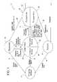
- FIG. 1is an overall system schematic of the present invention.
- FIG. 2is a schematic perspective view, partially in cross section of the components of the system for providing computer guided ablation, of the present invention.
- FIG. 3is a sample display screen of the computer system of the present invention.
- FIG. 4is a flow diagram of the treatment module of the present invention.
- FIG. 5is an illustration of the prostate showing cryoprobe and temperature probe placement.
- FIG. 6is a flow diagram of the overall ablation cycle of the present invention.
- FIG. 7 ais flow diagram of the freeze cycle for the first anterior cryoprobe and the second anterior cryoprobe.
- FIG. 7 bis a flow diagram of the freeze cycle for the first posterior lateral cryoprobe and the second posterior lateral cryoprobe.
- FIG. 8is a front view of the alignment assembly showing the cryoprobes and temperature probes being placed at selected locations.
- FIG. 1illustrates a preferred embodiment of system for providing computer guided ablation of tissue, of the present invention, designated generally as 10 .
- the system 10includes an imaging device 12 , such as ultrasound, MRI, CT, PET, SPECT, X-ray (including fluoroscope) or other suitable imaging device.
- the imaging device 12receives imaging data 14 from a treatment region of a patient 16 .
- the treatment regionmay be, for example, the prostate region, breast region, liver region, etc.
- the imaging device 12provides imaging output data 18 to the physician or other operator 20 and imaging signals 22 to an ablative surgical computer system, designated generally as 24 .
- the ablative surgical computer system 24includes a guidance module 26 for processing the imaging signals 22 and providing a treatment guidance plan 23 to the operator 20 .
- the computer system 24also includes a treatment module 28 for acquiring and processing surgical device output data 30 , for optimally controlling treatment parameters 32 and providing feedback information 34 to the operator 20 based on the treatment guidance plan 23 .
- a set of surgical devicesdesignated generally as 36 , includes at least one ablative device 38 for providing ablation of the treatment region based on the treatment parameters 32 and operator input 40 .
- the set 36 of surgical devicesalso includes at least one temperature sensing device 52 for acquiring temperature data 42 from the treatment region of the patient 16 .
- the set 36 of surgical devicesprovides the surgical device output data 30 .
- a temperature sensing device output signalis provided which is a portion of the surgical device output data 30 .
- the ablative devices 38are cryosurgical probes, as will be explained in detail below. However, it is understood that various other types of ablative devices 38 may be used in accordance with the principles of the present invention to provide the necessary ablation.
- the ablative devices 38may comprise, for example, radio frequency electrodes, laser fibers, microwave catheters, high-intensity focused ultrasound, and other suitable ablative devices.
- FIG. 2utilization of the present system with ablative devices 38 , for example, cryosurgical probes, which function to ablate tissue, is illustrated, designated generally as 46 .
- the surgical computer system 24in present applicants' present application provides guidance as to recommended ablative element placement within a prostate 13 , based on images of the prostate acquired from the imaging system, such as an ultrasound system, designated generally as 48 .
- the computer system 24is programmed with software capable of: determining the dimensions of the prostate; determining the dimensions of a treatment zone; and, utilizing the determined dimensions of the prostate and treatment zone for computing the number and location of ablative elements needed to treat the treatment zone.
- An IBM-compatible microprocessorserves as the host computer.
- the transrectal ultrasound probe 48is used to visualize the prostate and the cryosurgical probes.
- a stepper assembly 50provides the required advance.
- the ablative devicese.g. cryoprobes 38
- the set of surgical devicesi.e ablative devices and temperature sensing devices, are introduced through a grid (i.e. reference plate) 54 .
- Treatment planningpreferably includes the following steps:
- the live ultrasound imageis displayed in the ultrasound image window.
- At least one imageis captured.
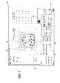
- FIG. 3a sample display screen, designated generally as 56 , of the computer system 24 showing treatment planning is illustrated.
- the display screen 56contains various sections. For example, a thumbnail section 58 displays thumbnail images.
- Another section on the display screen 48is the instruction box 60 that provides the user with detailed instructions at each step and makes the system easier to use. Additionally, the system has controls for specifying the patient details (name, age, etc.), calibration, adding/deleting probes and for the simulation of the ablation. The system also provides a pull down menu for switching rendering views and to toggle the display of the probe placements.
- the ultrasound probe image 48can be seen in FIG. 3 . Furthermore, parts of the anatomy can be seen, such as the urethra 62 and the rectum 64 .
- the temperature probesare denoted A, B, C, D and E.
- the cryoprobesare denoted by numeral designations 1-6.
- the grid being usedis also shown in this display, as denoted by numeral designation 66 . As noted above, the grid 66 can, optionally be deleted from the display by selecting the “hide grid” option 67 .
- the treatment module 70is used by the operator to deliver the treatment to the patient.
- the systemprovides a user interface for the operator to enter the target temperatures for the treatment of the patient.
- Each of the temperature probesare therefore assigned a target temperature which is then used to determine the operation of the ablative devices.
- cryoprobes and temperature probesare displayed relative to the prostate and other anatomical structures of interest.
- the target temperatures for each of the temperature probesare also displayed.
- the cryoprobesare numbered 1-6 in this figure.
- the temperature probesare designated A-D.
- the step of displaying the cryoprobes and temperature probesis denoted by block 74 .
- the ablation cycleis started based on user input (block 76 ).
- the ablation cycleis ended, based on user input (block 78 ) or upon reaching target temperatures.
- a freeze cycleis started for a first anterior cryoprobe and a second anterior cryoprobe, i.e. probes 1 and 2 (block 82 ).
- a freeze cycleis started for a first posterior lateral cryoprobe and a second posterior lateral cryoprobe, i.e. probes 3 and 4 (block 84 ).
- a freeze cycleis started for a first posterior medial cryoprobe and a second posterior lateral cryoprobe, i.e. probes 5 and 6 (block 86 ).
- the cryoprobesare operated based on the temperature data from the temperature sensing devices, i.e. temperature probe in (block 88 ).
- the operatoris informed if all target temperatures have been reached (block 90 ).
- a thaw cycleis started for the cryoprobes based on operator input.
- the freeze cycle for the first anterior cryoprobe and the second anterior cryoprobeis illustrated, designated generally as 92 . It involves the following steps:
- the freeze cycle for the first posterior lateral cryoprobe and the second posterior lateral cryoprobe, and for the first posterior medial cryoprobe and the second posterior lateral cryoprobe,are illustrated, designated generally as 106 .
- These cyclesinvolve the following steps:
- an alignment assembly(also referred to as a reference plate, grid or template) is illustrated, designated generally as 54 .
- the alignment assembly 54utilizes an orthogonal coordinate system to position the cryoprobes and temperature probes. Use of this alignment assembly 54 makes it possible for the cryoprobes and temperature probes to be placed at the locations determined by the guidance module.
- the cryoprobes particularly adapted for this computer guided placementare those manufactured by the present assignee, Endocare, Inc., Irvine, Calif.
- the urethrawhich passes through the prostate, is one of the anatomic structures that usually should not be frozen during this surgery. Accordingly, the urethra is protected and kept warm with the urethral warming catheter.
- the bladder neck sphincter and the external sphincterare also structures that should be protected from freezing, and these are protected from freezing by the warming catheter.
- a transrectal probeis inserted into the rectum in order to visualize the placement of the probes and the growth of the iceballs formed by the cryoprobes.
- a template 21is used which supports the probes 22 during insertion and while they are installed in the body.
- the patientis placed in the lithotomic position, i.e. horizontally on an operating table with legs positioned to provide access for the ultrasound probe to be inserted into the rectum and cryoprobes to be inserted through the perineal area into the prostate.
- the systemcools the probes to cryosurgically effective temperatures (typically below ⁇ 120° C.) through Joule-Thomson cooling within the probe tips.
- cryosurgically effective temperaturestypically below ⁇ 120° C.
- the systemmay be implemented with other cooling systems such as liquid nitrogen cryoprobes and mixed gas cryoprobes.
- the placement of probesis calculated based on this system, and the calculations may be adjusted for different systems and numbers of probes.
- the systemmay be adapted to other forms of ablation and treatment of the prostate, with adjustments in the calculations being made to account for the ablative range of the devices.
- Other ablative elementsmay include, for example, radio frequency devices, microwave devices, high intensity focused ultrasound devices, lasers, radioactive seeds and ablation agents such as chemicals, e.g. alcohol-based substances.
- the treatment modulemay alternatively control the ablative elements automatically based upon a sensing device output signal such as, but not limited to, temperature sensing device measurements, ultrasound images of the rate of ice growth, tissue impedance measurements within the treatment zone. Such a feedback could direct the system to stop the treatment resulting in the system turning off one or more ablative elements automatically without the need for operator intervention.
- a sensing device output signalsuch as, but not limited to, temperature sensing device measurements, ultrasound images of the rate of ice growth, tissue impedance measurements within the treatment zone.
Landscapes
- Health & Medical Sciences (AREA)
- Surgery (AREA)
- Life Sciences & Earth Sciences (AREA)
- Nuclear Medicine, Radiotherapy & Molecular Imaging (AREA)
- Medical Informatics (AREA)
- Engineering & Computer Science (AREA)
- Biomedical Technology (AREA)
- Heart & Thoracic Surgery (AREA)
- Otolaryngology (AREA)
- Molecular Biology (AREA)
- Animal Behavior & Ethology (AREA)
- General Health & Medical Sciences (AREA)
- Public Health (AREA)
- Veterinary Medicine (AREA)
- Surgical Instruments (AREA)
- Thermotherapy And Cooling Therapy Devices (AREA)
Abstract
Description
- A button entitled CAPTURE will appear at the bottom of the display.
- A brachy-type grid, i.e. grid having an orthogonal reference system, should be displayed on the ultrasound image before the first image is captured.
- Using the Capture window, click CAPTURE to capture the first image at the widest cross section of the prostate. Once CAPTURE is selected, the image will be frozen and displayed as a thumbnail image on the right-hand side of the screen.
- You can now remove the brachy grid display for the remaining captures.
- You are asked to click on the four outer points of the prostate image displayed.
- Start by clicking on the top edge of the prostate.
- Next click on the outer most right hand side of the prostate.
- Repeat this action on the bottom edge and left hand outer edge of the prostate as directed in the
step 3 window text and illustrations. - When you have clicked on all four points, click the right mouse button to complete the outline.
- At anytime during the outlining process, the UNDO button in the
step 3 window can be selected to remove the last point placed. - When the prostate outline is completed, the system will move to the URETHRA contour mode.
- To outline the urethra click on the center of the urethra and a circle will be placed.
- You can adjust the urethra contour location by clicking in the center of the circle and dragging the circle to a new location holding the mouse button down.
- You can adjust the size of the urethra outline by clicking on one of the four white dots displayed outside of the outline and moving it inward to reduce the size or pulling it outward to increase the size.
- You must click the right mouse button to complete the urethral outline.
- When the urethra outline is completed, the system will move to the RECTAL WALL contour mode.
- To outline the rectal wall, click on the left top edge of the rectal wall and then click on the right top edge of the rectal wall.
- You can adjust the rectal wall outline by clicking on any of the points in the outline and dragging them to a different location.
- When the rectal wall outline is complete, right click to move to the next step.
- When all outlines on the first image are complete, the system will ask you to outline the urethra on the base and apex images.
- Outline the urethra in each additional image using the same method described previously.
- Right click each time an outline has been completed.
- The
step 4 window and suggested probe placement will appear next to thestep 3 window when the outlining is complete. This window allows you to move, add or delete probes if desired. - Probe grid coordinates will also be displayed on the far right hand side of the screen.
- To move a probe from the suggested probe placement, click and drag the probe points displayed on the image. This will result in the probe coordinates changing to the new location.
- To add or delete probes, click the add or delete button and then click on the location on the image where you want to add a probe or click on the probe you want to delete. This will add or remove the probe to the coordinate display on the right hand side of the screen.
- Once the probes are in the desired locations, click on the accept button to proceed to step 6.
- The
Step 6 allows the user to place a TempProbe® temperature probe in the desired location on the image and displays the grid coordinate points that correspond to that placement.- The user is prompted to click on the locations for the right neurovascular bundle (RNVB), left neurovascular bundle (LNVB), Apex and External Sphincter (ES) temperature probes in the image.
- For each placement the user must click on the add button in the
step 6 window and then click on the location for placement in the image. - A minimum of four temperature probes should be placed.
- TempProbe® grid coordinates are displayed on the right hand side of the screen next to Cryoprobe coordinates.
- The user can click on the LIVE button in the bottom right hand corner of the screen to overlay the probe placement locations and grid on top of the live ultrasound image.
- The user can click on the same button that is now labeled captured images to return to the captured image display.
- The user can click on the Hide Grid/Display Grid button in the bottom right hand corner of the screen to toggle the Cryogrid overlay on and off.
- a) turning on the first anterior cryoprobe and the second anterior cryoprobe (block94);
- b) determining if an anterior target temperature has been reached (block96);
- c) operating the first anterior cryoprobe and the second anterior cryoprobe at a maximum rate if an anterior target temperature has not been reached (block98);
- d) operating the first anterior cryoprobe and the second anterior cryoprobe at a substantially zero rate if an anterior target temperature has been reached (block100); and,
- e) determining if the anterior target temperature has reached substantially 0° C. (block102). If yes, probes3 and4 are turned on (block104).
- a) turning on the first posterior lateral cryoprobe and the second posterior lateral cryoprobe and operating them at a maximum rate (block108);
- b) determining if a first neurovascular bundle target temperature has been reached (block110);
- c) turning off the first posterior lateral cryoprobe if the first neurovascular bundle target temperature has been reached (block112);
- d) determining if a second neurovascular bundle target temperature has been reached (block114);
- e) operating the second posterior lateral cryoprobe at a substantially zero rate if the second neurovascular bundle target temperature has been reached (block116);
- f) turning on the first posterior medial cryoprobe and the second posterior medial cryoprobe after the neurovascular temperature probes are substantially close to their target temperatures (block118);
- g) operating the first posterior medial cryoprobe and the second posterior medial cryoprobe at a power rate in a range of about 15-35%, preferably about 25% (block124); and,
- h) setting the first posterior medial cryoprobe and the second posterior medial cryoprobe to a substantially zero rate (block120) if a Denon Vieller's fascia target temperature has been reached (block122).
Claims (27)
Priority Applications (1)
| Application Number | Priority Date | Filing Date | Title |
|---|---|---|---|
| US12/025,619US9326808B2 (en) | 1999-05-26 | 2008-02-04 | System for providing computer guided ablation of tissue |
Applications Claiming Priority (6)
| Application Number | Priority Date | Filing Date | Title |
|---|---|---|---|
| US09/318,710US6139544A (en) | 1999-05-26 | 1999-05-26 | Computer guided cryosurgery |
| US09/699,938US6485422B1 (en) | 1999-05-26 | 2000-10-30 | Computer guided cryosurgery |
| US09/957,306US6544176B2 (en) | 1999-05-26 | 2001-09-20 | Computer guided cryosurgery |
| US10/307,036US6643535B2 (en) | 1999-05-26 | 2002-11-27 | System for providing computer guided ablation of tissue |
| US10/700,326US7363071B2 (en) | 1999-05-26 | 2003-03-11 | Computer guided ablation of tissue using integrated ablative/temperature sensing devices |
| US12/025,619US9326808B2 (en) | 1999-05-26 | 2008-02-04 | System for providing computer guided ablation of tissue |
Related Parent Applications (1)
| Application Number | Title | Priority Date | Filing Date |
|---|---|---|---|
| US10/700,326ContinuationUS7363071B2 (en) | 1999-05-26 | 2003-03-11 | Computer guided ablation of tissue using integrated ablative/temperature sensing devices |
Publications (2)
| Publication Number | Publication Date |
|---|---|
| US20080154253A1 US20080154253A1 (en) | 2008-06-26 |
| US9326808B2true US9326808B2 (en) | 2016-05-03 |
Family
ID=46299048
Family Applications (2)
| Application Number | Title | Priority Date | Filing Date |
|---|---|---|---|
| US10/700,326Expired - LifetimeUS7363071B2 (en) | 1999-05-26 | 2003-03-11 | Computer guided ablation of tissue using integrated ablative/temperature sensing devices |
| US12/025,619Expired - LifetimeUS9326808B2 (en) | 1999-05-26 | 2008-02-04 | System for providing computer guided ablation of tissue |
Family Applications Before (1)
| Application Number | Title | Priority Date | Filing Date |
|---|---|---|---|
| US10/700,326Expired - LifetimeUS7363071B2 (en) | 1999-05-26 | 2003-03-11 | Computer guided ablation of tissue using integrated ablative/temperature sensing devices |
Country Status (1)
| Country | Link |
|---|---|
| US (2) | US7363071B2 (en) |
Cited By (3)
| Publication number | Priority date | Publication date | Assignee | Title |
|---|---|---|---|---|
| US20240032980A1 (en)* | 2006-12-29 | 2024-02-01 | Varian Medical Systems, Inc. | Variable Cryosurgical Probe Planning System |
| US11974816B2 (en) | 2018-12-21 | 2024-05-07 | R2 Technologies, Inc. | Automated control and positioning systems for dermatological cryospray devices |
| US12133669B2 (en) | 2018-12-21 | 2024-11-05 | R2 Technologies, Inc. | Automated dermatological cryospray treatment planning system |
Families Citing this family (42)
| Publication number | Priority date | Publication date | Assignee | Title |
|---|---|---|---|---|
| WO2002009571A2 (en)* | 2000-07-31 | 2002-02-07 | Galil Medical Ltd. | Planning and facilitation systems and methods for cryosurgery |
| WO2004000098A2 (en) | 2002-06-19 | 2003-12-31 | Palomar Medical Technologies, Inc. | Method and apparatus for treatment of cutaneous and subcutaneous conditions |
| US20050283074A1 (en)* | 2004-06-22 | 2005-12-22 | Siemens Medical Solutions Usa, Inc. | Ultrasound feedback for tissue ablation procedures |
| US7833221B2 (en)* | 2004-10-22 | 2010-11-16 | Ethicon Endo-Surgery, Inc. | System and method for treatment of tissue using the tissue as a fiducial |
| US7452357B2 (en) | 2004-10-22 | 2008-11-18 | Ethicon Endo-Surgery, Inc. | System and method for planning treatment of tissue |
| US9031667B2 (en)* | 2005-03-04 | 2015-05-12 | InterventionTechnology Pty Ltd | Minimal device and method for effecting hyperthermia derived anesthesia |
| US7856985B2 (en) | 2005-04-22 | 2010-12-28 | Cynosure, Inc. | Method of treatment body tissue using a non-uniform laser beam |
| JP4999012B2 (en) | 2005-06-06 | 2012-08-15 | インチュイティブ サージカル,インコーポレイテッド | Laparoscopic ultrasonic robotic surgical system |
| US11259870B2 (en) | 2005-06-06 | 2022-03-01 | Intuitive Surgical Operations, Inc. | Interactive user interfaces for minimally invasive telesurgical systems |
| WO2007036925A1 (en)* | 2005-09-29 | 2007-04-05 | Corindus Ltd. | Methods and apparatuses for treatment of hollow organs |
| WO2007047247A1 (en)* | 2005-10-14 | 2007-04-26 | University Of Utah Research Foundation | Minimum time feedback control of efficacy and safety of thermal therapies |
| US20070156125A1 (en)* | 2005-12-30 | 2007-07-05 | Russell Delonzor | Encodable cryogenic device |
| WO2007129308A2 (en)* | 2006-05-02 | 2007-11-15 | Galil Medical Ltd. | Cryotherapy planning and control system |
| ES2524303T3 (en)* | 2006-05-08 | 2014-12-05 | C.R. Bard, Inc. | User interface and methods for an ultrasound presentation device |
| WO2008014482A2 (en)* | 2006-07-27 | 2008-01-31 | Personics Holdings Inc. | Method and device of customizing headphones |
| US7586957B2 (en) | 2006-08-02 | 2009-09-08 | Cynosure, Inc | Picosecond laser apparatus and methods for its operation and use |
| WO2008154007A1 (en)* | 2007-06-08 | 2008-12-18 | Cynosure, Inc. | Surgical waveguide |
| DE102008030242A1 (en)* | 2008-06-25 | 2010-01-07 | Siemens Aktiengesellschaft | Method for monitoring the image of an irreversible electroporation treatment and associated device |
| US20100087806A1 (en)* | 2008-10-07 | 2010-04-08 | Vandolay, Inc. | Automated Cryogenic Skin Treatment |
| WO2010052596A1 (en)* | 2008-11-04 | 2010-05-14 | Koninklijke Philips Electronics, N.V. | Method and system for ultrasound therapy |
| CA2746114C (en)* | 2008-12-23 | 2016-03-22 | Cryomedix Llc | Isotherm-based tissue ablation control system and method |
| WO2010144402A2 (en) | 2009-06-08 | 2010-12-16 | Surgivision, Inc. | Mri-guided surgical systems with preset scan planes |
| CN102625670B (en) | 2009-06-16 | 2015-07-15 | 核磁共振成像介入技术有限公司 | MRI-guided devices and MRI-guided interventional systems that can track and generate dynamic visualizations of the devices in near real time |
| US9750563B2 (en) | 2009-09-22 | 2017-09-05 | Mederi Therapeutics, Inc. | Systems and methods for treating tissue with radiofrequency energy |
| JP5764564B2 (en) | 2009-09-22 | 2015-08-19 | メデリ セラピューティクス インコーポレイテッド | Systems and methods for controlling the use and operation of various therapeutic device groups |
| US9775664B2 (en) | 2009-09-22 | 2017-10-03 | Mederi Therapeutics, Inc. | Systems and methods for treating tissue with radiofrequency energy |
| US9474565B2 (en) | 2009-09-22 | 2016-10-25 | Mederi Therapeutics, Inc. | Systems and methods for treating tissue with radiofrequency energy |
| US10386990B2 (en) | 2009-09-22 | 2019-08-20 | Mederi Rf, Llc | Systems and methods for treating tissue with radiofrequency energy |
| US20130018368A1 (en)* | 2011-07-13 | 2013-01-17 | Galil Medical Inc. | User interface for operating and monitoring a cryosurgical system |
| US10459043B2 (en) | 2011-08-30 | 2019-10-29 | Profound Medical Inc. | Real time control of high intensity focused ultrasound using magnetic resonance imaging |
| EP2839552A4 (en) | 2012-04-18 | 2015-12-30 | Cynosure Inc | PICOSECOND LASER APPARATUS AND METHOD OF PROCESSING TARGET TISSUES USING THE SAME |
| US10285757B2 (en) | 2013-03-15 | 2019-05-14 | Cynosure, Llc | Picosecond optical radiation systems and methods of use |
| CN105658166B (en)* | 2013-08-23 | 2018-12-11 | 皇家飞利浦有限公司 | Medical instrument with multiple displays for controlling disposal system |
| EP3122274B1 (en)* | 2014-03-22 | 2021-08-04 | Varian Medical Systems, Inc. | System and methods for ablation treatment planning |
| WO2018183217A1 (en)* | 2017-03-25 | 2018-10-04 | Bianco Fernando J | System and method for prostate cancer treatment under local anesthesia |
| WO2019100212A1 (en)* | 2017-11-21 | 2019-05-31 | 深圳迈瑞生物医疗电子股份有限公司 | Ultrasonic system and method for planning ablation |
| CN115486927B (en)* | 2017-11-27 | 2025-02-25 | 海杰亚(北京)医疗器械有限公司 | Cryoablation needle |
| WO2019165426A1 (en) | 2018-02-26 | 2019-08-29 | Cynosure, Inc. | Q-switched cavity dumped sub-nanosecond laser |
| EP3685778A1 (en)* | 2019-01-25 | 2020-07-29 | Koninklijke Philips N.V. | Apparatus for determining a position of a temperature probe during a planning for an ablation procedure |
| JP7335439B2 (en) | 2019-10-28 | 2023-08-29 | ボストン サイエンティフィック ニューロモデュレイション コーポレイション | RF electrode cannula |
| US12329445B2 (en) | 2020-12-28 | 2025-06-17 | Boston Scientific Neuromodulation Corporation | RF ablation systems and methods using an integrated cannula and electrode |
| US12433646B2 (en) | 2023-02-21 | 2025-10-07 | Boston Scientific Neuromodulation Corporation | Interspinous spacer with actuator locking arrangements and methods and systems |
Citations (56)
| Publication number | Priority date | Publication date | Assignee | Title |
|---|---|---|---|---|
| US4565200A (en) | 1980-09-24 | 1986-01-21 | Cosman Eric R | Universal lesion and recording electrode system |
| US4672963A (en) | 1985-06-07 | 1987-06-16 | Israel Barken | Apparatus and method for computer controlled laser surgery |
| US4776334A (en) | 1985-03-22 | 1988-10-11 | Stanford University | Catheter for treatment of tumors |
| US5222953A (en) | 1991-10-02 | 1993-06-29 | Kambiz Dowlatshahi | Apparatus for interstitial laser therapy having an improved temperature sensor for tissue being treated |
| US5334181A (en)* | 1990-09-26 | 1994-08-02 | Cryomedical Sciences, Inc. | Cryosurgical system for destroying tumors by freezing |
| US5454371A (en) | 1993-11-29 | 1995-10-03 | London Health Association | Method and system for constructing and displaying three-dimensional images |
| US5494039A (en) | 1993-07-16 | 1996-02-27 | Cryomedical Sciences, Inc. | Biopsy needle insertion guide and method of use in prostate cryosurgery |
| US5531742A (en) | 1992-01-15 | 1996-07-02 | Barken; Israel | Apparatus and method for computer controlled cryosurgery |
| US5562095A (en) | 1992-12-24 | 1996-10-08 | Victoria Hospital Corporation | Three dimensional ultrasound imaging system |
| US5647868A (en) | 1994-02-02 | 1997-07-15 | Chinn; Douglas Owen | Cryosurgical integrated control and monitoring system and method |
| US5706810A (en) | 1993-03-23 | 1998-01-13 | The Regents Of The University Of California | Magnetic resonance imaging assisted cryosurgery |
| WO1998023214A1 (en) | 1996-11-29 | 1998-06-04 | Life Imaging Systems Inc. | System, employing three-dimensional ultrasonographic imaging, for assisting in guiding and placing medical instruments |
| US5800487A (en) | 1996-07-23 | 1998-09-01 | Endocare, Inc. | Cryoprobe |
| US5827204A (en) | 1996-11-26 | 1998-10-27 | Grandia; Willem | Medical noninvasive operations using focused modulated high power ultrasound |
| US5865788A (en) | 1992-08-12 | 1999-02-02 | Vidamed, Inc. | Self-contained power sypply and monitoring station for RF tissue ablation |
| US5882306A (en) | 1997-04-11 | 1999-03-16 | Acuson Corporation | Ultrasound imaging methods and systems |
| US5897495A (en) | 1993-03-10 | 1999-04-27 | Kabushiki Kaisha Toshiba | Ultrasonic wave medical treatment apparatus suitable for use under guidance of magnetic resonance imaging |
| US5899860A (en) | 1996-09-12 | 1999-05-04 | Siemens Elema Ab | Method and device for determining the position of a catheter inside the body of a patient |
| US5910104A (en) | 1996-12-26 | 1999-06-08 | Cryogen, Inc. | Cryosurgical probe with disposable sheath |
| US5976092A (en) | 1998-06-15 | 1999-11-02 | Chinn; Douglas O. | Combination stereotactic surgical guide and ultrasonic probe |
| US5978697A (en) | 1998-01-05 | 1999-11-02 | Galil Medical Ltd. | System and method for MRI-guided cryosurgery |
| US6071280A (en) | 1993-11-08 | 2000-06-06 | Rita Medical Systems, Inc. | Multiple electrode ablation apparatus |
| US6083166A (en) | 1997-12-02 | 2000-07-04 | Situs Corporation | Method and apparatus for determining a measure of tissue manipulation |
| US6095975A (en) | 1997-05-27 | 2000-08-01 | Silvern; David A. | Apparatus and method for determining optimal locations to place radioactive seeds at a cancerous site |
| US6129670A (en) | 1997-11-24 | 2000-10-10 | Burdette Medical Systems | Real time brachytherapy spatial registration and visualization system |
| US6139544A (en)* | 1999-05-26 | 2000-10-31 | Endocare, Inc. | Computer guided cryosurgery |
| US6190378B1 (en) | 1997-12-05 | 2001-02-20 | Massachusetts Institute Of Technology | Cryosurgical instrument and related techniques |
| US6235018B1 (en) | 1999-10-29 | 2001-05-22 | Cryoflex, Inc. | Method and apparatus for monitoring cryosurgical operations |
| US6241725B1 (en) | 1993-12-15 | 2001-06-05 | Sherwood Services Ag | High frequency thermal ablation of cancerous tumors and functional targets with image data assistance |
| US6248101B1 (en) | 1997-01-22 | 2001-06-19 | Barzell Whitmore Maroon Bells, Inc. | Omni-directional precision instrument platform |
| US6256529B1 (en)* | 1995-07-26 | 2001-07-03 | Burdette Medical Systems, Inc. | Virtual reality 3D visualization for surgical procedures |
| US6306129B1 (en) | 1997-09-22 | 2001-10-23 | Femrx, Inc. | Cryosurgical system and method |
| US6311084B1 (en)* | 1998-05-04 | 2001-10-30 | Robert A. Cormack | Radiation seed implant method and apparatus |
| US20020022869A1 (en) | 2000-07-03 | 2002-02-21 | Norihiko Hareyama | Thermal treatment apparatus |
| US20020022832A1 (en) | 1998-06-19 | 2002-02-21 | Mikus Paul W. | Cryoprobe assembly with detachable sheath |
| US6539247B2 (en) | 1998-02-27 | 2003-03-25 | Varian Medical Systems, Inc. | Brachytherapy system for prostate cancer treatment with computer implemented systems and processes to facilitate pre-implantation planning and post-implantation evaluations with storage of multiple plan variations for a single patient |
| US6767346B2 (en) | 2001-09-20 | 2004-07-27 | Endocare, Inc. | Cryosurgical probe with bellows shaft |
| US6865412B2 (en) | 2001-04-13 | 2005-03-08 | Kelsey, Inc. | Apparatus and method for delivering ablative laser energy and determining the volume of tumor mass destroyed |
| US6936045B2 (en) | 2001-09-20 | 2005-08-30 | Endocare, Inc. | Malleable cryosurgical probe |
| US6958062B1 (en) | 1993-11-08 | 2005-10-25 | Rita Medical Systems, Inc. | Multiple antenna ablation apparatus and method |
| US7036516B1 (en) | 1996-10-30 | 2006-05-02 | Xantech Pharmaceuticals, Inc. | Treatment of pigmented tissues using optical energy |
| US20070010738A1 (en) | 2004-10-14 | 2007-01-11 | Mark Joseph L | Surgical site marker delivery system |
| US7167741B2 (en) | 2000-06-15 | 2007-01-23 | Monteris Medical, Inc. | Hyperthermia treatment and probe therefor |
| US20070021741A1 (en) | 2003-07-11 | 2007-01-25 | Cryocath Technologies Inc. | Method and device for epicardial ablation |
| US7171257B2 (en) | 2003-06-11 | 2007-01-30 | Accuray Incorporated | Apparatus and method for radiosurgery |
| US7172589B2 (en) | 2002-08-30 | 2007-02-06 | Scimed Life Systems, Inc. | Cryo ablation coil |
| US20070032783A1 (en) | 2004-03-23 | 2007-02-08 | Cryocath Technologies Inc. | Method and apparatus for inflating and deflating balloon catheters |
| US20070043342A1 (en) | 2005-08-16 | 2007-02-22 | Galil Medical Ltd. | Cryoprobe with reduced adhesion to frozen tissue, and cryosurgical methods utilizing same |
| US20070049912A1 (en) | 2003-06-25 | 2007-03-01 | Endocare, Inc. | Detachable cryosurgical probe |
| US7189228B2 (en) | 2003-06-25 | 2007-03-13 | Endocare, Inc. | Detachable cryosurgical probe with breakaway handle |
| US7189227B2 (en) | 2002-04-19 | 2007-03-13 | Boston Scientific Scimed, Inc. | Cryo balloon |
| US7195625B2 (en) | 2002-12-11 | 2007-03-27 | Cryocor, Inc. | Catheter system for performing a single step cryoablation |
| US7204833B1 (en) | 2002-10-11 | 2007-04-17 | Flint Hills Scientific Llc | Multi-modal system for detection and control of changes in brain state |
| US20070088247A1 (en) | 2000-10-24 | 2007-04-19 | Galil Medical Ltd. | Apparatus and method for thermal ablation of uterine fibroids |
| US7207986B2 (en) | 1999-01-25 | 2007-04-24 | Cryocath Technologies Inc. | Cooling system |
| US7207985B2 (en) | 2003-06-25 | 2007-04-24 | Endocare, Inc. | Detachable cryosurgical probe |
Family Cites Families (1)
| Publication number | Priority date | Publication date | Assignee | Title |
|---|---|---|---|---|
| US6887235B2 (en)* | 1999-03-24 | 2005-05-03 | Micrus Corporation | Variable stiffness heating catheter |
- 2003
- 2003-03-11USUS10/700,326patent/US7363071B2/ennot_activeExpired - Lifetime
- 2008
- 2008-02-04USUS12/025,619patent/US9326808B2/ennot_activeExpired - Lifetime
Patent Citations (60)
| Publication number | Priority date | Publication date | Assignee | Title |
|---|---|---|---|---|
| US4565200A (en) | 1980-09-24 | 1986-01-21 | Cosman Eric R | Universal lesion and recording electrode system |
| US4776334A (en) | 1985-03-22 | 1988-10-11 | Stanford University | Catheter for treatment of tumors |
| US4672963A (en) | 1985-06-07 | 1987-06-16 | Israel Barken | Apparatus and method for computer controlled laser surgery |
| US5334181A (en)* | 1990-09-26 | 1994-08-02 | Cryomedical Sciences, Inc. | Cryosurgical system for destroying tumors by freezing |
| US5222953A (en) | 1991-10-02 | 1993-06-29 | Kambiz Dowlatshahi | Apparatus for interstitial laser therapy having an improved temperature sensor for tissue being treated |
| US5531742A (en) | 1992-01-15 | 1996-07-02 | Barken; Israel | Apparatus and method for computer controlled cryosurgery |
| US5865788A (en) | 1992-08-12 | 1999-02-02 | Vidamed, Inc. | Self-contained power sypply and monitoring station for RF tissue ablation |
| US5562095A (en) | 1992-12-24 | 1996-10-08 | Victoria Hospital Corporation | Three dimensional ultrasound imaging system |
| US5897495A (en) | 1993-03-10 | 1999-04-27 | Kabushiki Kaisha Toshiba | Ultrasonic wave medical treatment apparatus suitable for use under guidance of magnetic resonance imaging |
| US5706810A (en) | 1993-03-23 | 1998-01-13 | The Regents Of The University Of California | Magnetic resonance imaging assisted cryosurgery |
| US5494039A (en) | 1993-07-16 | 1996-02-27 | Cryomedical Sciences, Inc. | Biopsy needle insertion guide and method of use in prostate cryosurgery |
| US6958062B1 (en) | 1993-11-08 | 2005-10-25 | Rita Medical Systems, Inc. | Multiple antenna ablation apparatus and method |
| US6071280A (en) | 1993-11-08 | 2000-06-06 | Rita Medical Systems, Inc. | Multiple electrode ablation apparatus |
| US5454371A (en) | 1993-11-29 | 1995-10-03 | London Health Association | Method and system for constructing and displaying three-dimensional images |
| US6241725B1 (en) | 1993-12-15 | 2001-06-05 | Sherwood Services Ag | High frequency thermal ablation of cancerous tumors and functional targets with image data assistance |
| US5647868A (en) | 1994-02-02 | 1997-07-15 | Chinn; Douglas Owen | Cryosurgical integrated control and monitoring system and method |
| US6256529B1 (en)* | 1995-07-26 | 2001-07-03 | Burdette Medical Systems, Inc. | Virtual reality 3D visualization for surgical procedures |
| US7171255B2 (en) | 1995-07-26 | 2007-01-30 | Computerized Medical Systems, Inc. | Virtual reality 3D visualization for surgical procedures |
| US5800487A (en) | 1996-07-23 | 1998-09-01 | Endocare, Inc. | Cryoprobe |
| US5899860A (en) | 1996-09-12 | 1999-05-04 | Siemens Elema Ab | Method and device for determining the position of a catheter inside the body of a patient |
| US7036516B1 (en) | 1996-10-30 | 2006-05-02 | Xantech Pharmaceuticals, Inc. | Treatment of pigmented tissues using optical energy |
| US5827204A (en) | 1996-11-26 | 1998-10-27 | Grandia; Willem | Medical noninvasive operations using focused modulated high power ultrasound |
| US6423009B1 (en) | 1996-11-29 | 2002-07-23 | Life Imaging Systems, Inc. | System, employing three-dimensional ultrasonographic imaging, for assisting in guiding and placing medical instruments |
| WO1998023214A1 (en) | 1996-11-29 | 1998-06-04 | Life Imaging Systems Inc. | System, employing three-dimensional ultrasonographic imaging, for assisting in guiding and placing medical instruments |
| US5910104A (en) | 1996-12-26 | 1999-06-08 | Cryogen, Inc. | Cryosurgical probe with disposable sheath |
| US6248101B1 (en) | 1997-01-22 | 2001-06-19 | Barzell Whitmore Maroon Bells, Inc. | Omni-directional precision instrument platform |
| US5882306A (en) | 1997-04-11 | 1999-03-16 | Acuson Corporation | Ultrasound imaging methods and systems |
| US6095975A (en) | 1997-05-27 | 2000-08-01 | Silvern; David A. | Apparatus and method for determining optimal locations to place radioactive seeds at a cancerous site |
| US6306129B1 (en) | 1997-09-22 | 2001-10-23 | Femrx, Inc. | Cryosurgical system and method |
| US6129670A (en) | 1997-11-24 | 2000-10-10 | Burdette Medical Systems | Real time brachytherapy spatial registration and visualization system |
| US6083166A (en) | 1997-12-02 | 2000-07-04 | Situs Corporation | Method and apparatus for determining a measure of tissue manipulation |
| US6190378B1 (en) | 1997-12-05 | 2001-02-20 | Massachusetts Institute Of Technology | Cryosurgical instrument and related techniques |
| US5978697A (en) | 1998-01-05 | 1999-11-02 | Galil Medical Ltd. | System and method for MRI-guided cryosurgery |
| US6539247B2 (en) | 1998-02-27 | 2003-03-25 | Varian Medical Systems, Inc. | Brachytherapy system for prostate cancer treatment with computer implemented systems and processes to facilitate pre-implantation planning and post-implantation evaluations with storage of multiple plan variations for a single patient |
| US6311084B1 (en)* | 1998-05-04 | 2001-10-30 | Robert A. Cormack | Radiation seed implant method and apparatus |
| US5976092A (en) | 1998-06-15 | 1999-11-02 | Chinn; Douglas O. | Combination stereotactic surgical guide and ultrasonic probe |
| US20020022832A1 (en) | 1998-06-19 | 2002-02-21 | Mikus Paul W. | Cryoprobe assembly with detachable sheath |
| US20070093799A1 (en) | 1999-01-25 | 2007-04-26 | Marwan Abboud | Cooling system |
| US7207986B2 (en) | 1999-01-25 | 2007-04-24 | Cryocath Technologies Inc. | Cooling system |
| US20020016540A1 (en) | 1999-05-26 | 2002-02-07 | Mikus Paul W. | Computer Guided cryosurgery |
| US6139544A (en)* | 1999-05-26 | 2000-10-31 | Endocare, Inc. | Computer guided cryosurgery |
| US6235018B1 (en) | 1999-10-29 | 2001-05-22 | Cryoflex, Inc. | Method and apparatus for monitoring cryosurgical operations |
| US7167741B2 (en) | 2000-06-15 | 2007-01-23 | Monteris Medical, Inc. | Hyperthermia treatment and probe therefor |
| US20020022869A1 (en) | 2000-07-03 | 2002-02-21 | Norihiko Hareyama | Thermal treatment apparatus |
| US20070088247A1 (en) | 2000-10-24 | 2007-04-19 | Galil Medical Ltd. | Apparatus and method for thermal ablation of uterine fibroids |
| US6865412B2 (en) | 2001-04-13 | 2005-03-08 | Kelsey, Inc. | Apparatus and method for delivering ablative laser energy and determining the volume of tumor mass destroyed |
| US6767346B2 (en) | 2001-09-20 | 2004-07-27 | Endocare, Inc. | Cryosurgical probe with bellows shaft |
| US6936045B2 (en) | 2001-09-20 | 2005-08-30 | Endocare, Inc. | Malleable cryosurgical probe |
| US7189227B2 (en) | 2002-04-19 | 2007-03-13 | Boston Scientific Scimed, Inc. | Cryo balloon |
| US7172589B2 (en) | 2002-08-30 | 2007-02-06 | Scimed Life Systems, Inc. | Cryo ablation coil |
| US7204833B1 (en) | 2002-10-11 | 2007-04-17 | Flint Hills Scientific Llc | Multi-modal system for detection and control of changes in brain state |
| US7195625B2 (en) | 2002-12-11 | 2007-03-27 | Cryocor, Inc. | Catheter system for performing a single step cryoablation |
| US7171257B2 (en) | 2003-06-11 | 2007-01-30 | Accuray Incorporated | Apparatus and method for radiosurgery |
| US20070049912A1 (en) | 2003-06-25 | 2007-03-01 | Endocare, Inc. | Detachable cryosurgical probe |
| US7189228B2 (en) | 2003-06-25 | 2007-03-13 | Endocare, Inc. | Detachable cryosurgical probe with breakaway handle |
| US7207985B2 (en) | 2003-06-25 | 2007-04-24 | Endocare, Inc. | Detachable cryosurgical probe |
| US20070021741A1 (en) | 2003-07-11 | 2007-01-25 | Cryocath Technologies Inc. | Method and device for epicardial ablation |
| US20070032783A1 (en) | 2004-03-23 | 2007-02-08 | Cryocath Technologies Inc. | Method and apparatus for inflating and deflating balloon catheters |
| US20070010738A1 (en) | 2004-10-14 | 2007-01-11 | Mark Joseph L | Surgical site marker delivery system |
| US20070043342A1 (en) | 2005-08-16 | 2007-02-22 | Galil Medical Ltd. | Cryoprobe with reduced adhesion to frozen tissue, and cryosurgical methods utilizing same |
Non-Patent Citations (4)
| Title |
|---|
| Endocare, Cryocare Surgical System 400 Series Operator's Manual (Nov. 2001). |
| Onik, Cohen, et al. Transrectal Ultrasound-Guided Percutaneous Radical Cryosurgical Ablation of the Prostate, 72 Cancer 1291 (1993). |
| Onik, Ultrasound-Guided Cryosurgery, Scientific American at 62 (Jan. 1996). |
| Wong, et al. Cryosurgery as a Treatment for Prostate Carcinoma, 79 Cancer 963 (Mar. 1997). |
Cited By (4)
| Publication number | Priority date | Publication date | Assignee | Title |
|---|---|---|---|---|
| US20240032980A1 (en)* | 2006-12-29 | 2024-02-01 | Varian Medical Systems, Inc. | Variable Cryosurgical Probe Planning System |
| US12433660B2 (en)* | 2006-12-29 | 2025-10-07 | Varian Medical Systems, Inc. | Variable cryosurgical probe planning system |
| US11974816B2 (en) | 2018-12-21 | 2024-05-07 | R2 Technologies, Inc. | Automated control and positioning systems for dermatological cryospray devices |
| US12133669B2 (en) | 2018-12-21 | 2024-11-05 | R2 Technologies, Inc. | Automated dermatological cryospray treatment planning system |
Also Published As
| Publication number | Publication date |
|---|---|
| US7363071B2 (en) | 2008-04-22 |
| US20040143181A1 (en) | 2004-07-22 |
| US20080154253A1 (en) | 2008-06-26 |
Similar Documents
| Publication | Publication Date | Title |
|---|---|---|
| US9326808B2 (en) | System for providing computer guided ablation of tissue | |
| US6643535B2 (en) | System for providing computer guided ablation of tissue | |
| US6694170B1 (en) | Computer guided surgery for prostatic nerve sparing | |
| US6139544A (en) | Computer guided cryosurgery | |
| US12082789B2 (en) | Method and apparatus for laser ablation under ultrasound guidance | |
| US9144461B2 (en) | Feedback system for integrating interventional planning and navigation | |
| CA2271651C (en) | System, employing three-dimensional ultrasonographic imaging, for assisting in guiding and placing medical instruments | |
| US7399298B2 (en) | Planning and facilitation systems and methods for cryosurgery | |
| CN113491577B (en) | Multi-needle combined cryoablation path planning equipment | |
| WO1998023214A9 (en) | System, employing three-dimensional ultrasonographic imaging, for assisting in guiding and placing medical instruments | |
| US12053235B2 (en) | System and method for the ablation of uterine fibroids | |
| CA2507289C (en) | System for providing computer guided ablation of tissue | |
| CN118234441A (en) | Determination device for determining a virtual position of a virtual guiding means |
Legal Events
| Date | Code | Title | Description |
|---|---|---|---|
| AS | Assignment | Owner name:ENDOCARE, INC., CALIFORNIA Free format text:ASSIGNMENT OF ASSIGNORS INTEREST;ASSIGNORS:DAMASCO, SANFORD D.;DUONG, THACH;EUM, JAY J.;AND OTHERS;REEL/FRAME:021274/0462;SIGNING DATES FROM 20070912 TO 20080421 Owner name:ENDOCARE, INC., CALIFORNIA Free format text:ASSIGNMENT OF ASSIGNORS INTEREST;ASSIGNORS:DAMASCO, SANFORD D.;DUONG, THACH;EUM, JAY J.;AND OTHERS;SIGNING DATES FROM 20070912 TO 20080421;REEL/FRAME:021274/0462 | |
| AS | Assignment | Owner name:REGIONS BANK, TENNESSEE Free format text:SECURITY AGREEMENT;ASSIGNOR:ENDOCARE, INC.;REEL/FRAME:032372/0077 Effective date:20140203 | |
| AS | Assignment | Owner name:MIDCAP FINANCIAL TRUST, AS ADMINISTRATIVE AGENT, MARYLAND Free format text:SECURITY INTEREST;ASSIGNORS:HEALTHTRONICS, INC.;ENDOCARE, INC.;REEL/FRAME:038063/0201 Effective date:20160308 Owner name:MIDCAP FINANCIAL TRUST, AS ADMINISTRATIVE AGENT, M Free format text:SECURITY INTEREST;ASSIGNORS:HEALTHTRONICS, INC.;ENDOCARE, INC.;REEL/FRAME:038063/0201 Effective date:20160308 | |
| STCF | Information on status: patent grant | Free format text:PATENTED CASE | |
| AS | Assignment | Owner name:ENDOCARE, INC., TEXAS Free format text:RELEASE BY SECURED PARTY;ASSIGNOR:MIDCAP FINANCIAL TRUST, AS ADMINISTRATIVE AGENT;REEL/FRAME:046429/0787 Effective date:20180625 | |
| AS | Assignment | Owner name:HEALTHTRONICS, INC., TEXAS Free format text:RELEASE BY SECURED PARTY;ASSIGNOR:REGIONS BANK;REEL/FRAME:046500/0151 Effective date:20160308 Owner name:ENDOCARE, INC., TEXAS Free format text:RELEASE BY SECURED PARTY;ASSIGNOR:REGIONS BANK;REEL/FRAME:046500/0151 Effective date:20160308 | |
| MAFP | Maintenance fee payment | Free format text:PAYMENT OF MAINTENANCE FEE, 4TH YEAR, LARGE ENTITY (ORIGINAL EVENT CODE: M1551); ENTITY STATUS OF PATENT OWNER: LARGE ENTITY Year of fee payment:4 | |
| AS | Assignment | Owner name:VARIAN MEDICAL SYSTEMS, INC., CALIFORNIA Free format text:ASSIGNMENT OF ASSIGNORS INTEREST;ASSIGNOR:ENDOCARE, INC.;REEL/FRAME:056940/0327 Effective date:20210308 | |
| MAFP | Maintenance fee payment | Free format text:PAYMENT OF MAINTENANCE FEE, 8TH YEAR, LARGE ENTITY (ORIGINAL EVENT CODE: M1552); ENTITY STATUS OF PATENT OWNER: LARGE ENTITY Year of fee payment:8 |