US9326683B2 - Systems and methods for detection of wound fluid blood and application of phototherapy in conjunction with reduced pressure wound treatment system - Google Patents
Systems and methods for detection of wound fluid blood and application of phototherapy in conjunction with reduced pressure wound treatment systemDownload PDFInfo
- Publication number
- US9326683B2 US9326683B2US13/089,077US201113089077AUS9326683B2US 9326683 B2US9326683 B2US 9326683B2US 201113089077 AUS201113089077 AUS 201113089077AUS 9326683 B2US9326683 B2US 9326683B2
- Authority
- US
- United States
- Prior art keywords
- wound
- blood
- rpwt
- wave
- light
- Prior art date
- Legal status (The legal status is an assumption and is not a legal conclusion. Google has not performed a legal analysis and makes no representation as to the accuracy of the status listed.)
- Expired - Fee Related, expires
Links
Images


Classifications
- A—HUMAN NECESSITIES
- A61—MEDICAL OR VETERINARY SCIENCE; HYGIENE
- A61B—DIAGNOSIS; SURGERY; IDENTIFICATION
- A61B5/00—Measuring for diagnostic purposes; Identification of persons
- A61B5/0059—Measuring for diagnostic purposes; Identification of persons using light, e.g. diagnosis by transillumination, diascopy, fluorescence
- A—HUMAN NECESSITIES
- A61—MEDICAL OR VETERINARY SCIENCE; HYGIENE
- A61B—DIAGNOSIS; SURGERY; IDENTIFICATION
- A61B5/00—Measuring for diagnostic purposes; Identification of persons
- A61B5/145—Measuring characteristics of blood in vivo, e.g. gas concentration or pH-value ; Measuring characteristics of body fluids or tissues, e.g. interstitial fluid or cerebral tissue
- A61B5/14542—Measuring characteristics of blood in vivo, e.g. gas concentration or pH-value ; Measuring characteristics of body fluids or tissues, e.g. interstitial fluid or cerebral tissue for measuring blood gases
- A—HUMAN NECESSITIES
- A61—MEDICAL OR VETERINARY SCIENCE; HYGIENE
- A61B—DIAGNOSIS; SURGERY; IDENTIFICATION
- A61B5/00—Measuring for diagnostic purposes; Identification of persons
- A61B5/145—Measuring characteristics of blood in vivo, e.g. gas concentration or pH-value ; Measuring characteristics of body fluids or tissues, e.g. interstitial fluid or cerebral tissue
- A61B5/1468—Measuring characteristics of blood in vivo, e.g. gas concentration or pH-value ; Measuring characteristics of body fluids or tissues, e.g. interstitial fluid or cerebral tissue using chemical or electrochemical methods, e.g. by polarographic means
- A—HUMAN NECESSITIES
- A61—MEDICAL OR VETERINARY SCIENCE; HYGIENE
- A61B—DIAGNOSIS; SURGERY; IDENTIFICATION
- A61B5/00—Measuring for diagnostic purposes; Identification of persons
- A61B5/44—Detecting, measuring or recording for evaluating the integumentary system, e.g. skin, hair or nails
- A61B5/441—Skin evaluation, e.g. for skin disorder diagnosis
- A61B5/445—Evaluating skin irritation or skin trauma, e.g. rash, eczema, wound, bed sore
- A—HUMAN NECESSITIES
- A61—MEDICAL OR VETERINARY SCIENCE; HYGIENE
- A61B—DIAGNOSIS; SURGERY; IDENTIFICATION
- A61B5/00—Measuring for diagnostic purposes; Identification of persons
- A61B5/68—Arrangements of detecting, measuring or recording means, e.g. sensors, in relation to patient
- A61B5/6801—Arrangements of detecting, measuring or recording means, e.g. sensors, in relation to patient specially adapted to be attached to or worn on the body surface
- A61B5/683—Means for maintaining contact with the body
- A61B5/6834—Means for maintaining contact with the body using vacuum
- A61M1/0025—
- A61M1/0088—
- A—HUMAN NECESSITIES
- A61—MEDICAL OR VETERINARY SCIENCE; HYGIENE
- A61M—DEVICES FOR INTRODUCING MEDIA INTO, OR ONTO, THE BODY; DEVICES FOR TRANSDUCING BODY MEDIA OR FOR TAKING MEDIA FROM THE BODY; DEVICES FOR PRODUCING OR ENDING SLEEP OR STUPOR
- A61M1/00—Suction or pumping devices for medical purposes; Devices for carrying-off, for treatment of, or for carrying-over, body-liquids; Drainage systems
- A61M1/71—Suction drainage systems
- A61M1/73—Suction drainage systems comprising sensors or indicators for physical values
- A—HUMAN NECESSITIES
- A61—MEDICAL OR VETERINARY SCIENCE; HYGIENE
- A61M—DEVICES FOR INTRODUCING MEDIA INTO, OR ONTO, THE BODY; DEVICES FOR TRANSDUCING BODY MEDIA OR FOR TAKING MEDIA FROM THE BODY; DEVICES FOR PRODUCING OR ENDING SLEEP OR STUPOR
- A61M1/00—Suction or pumping devices for medical purposes; Devices for carrying-off, for treatment of, or for carrying-over, body-liquids; Drainage systems
- A61M1/90—Negative pressure wound therapy devices, i.e. devices for applying suction to a wound to promote healing, e.g. including a vacuum dressing
- A61M1/95—Negative pressure wound therapy devices, i.e. devices for applying suction to a wound to promote healing, e.g. including a vacuum dressing with sensors for exudate composition
- A—HUMAN NECESSITIES
- A61—MEDICAL OR VETERINARY SCIENCE; HYGIENE
- A61N—ELECTROTHERAPY; MAGNETOTHERAPY; RADIATION THERAPY; ULTRASOUND THERAPY
- A61N5/00—Radiation therapy
- A61N5/06—Radiation therapy using light
- A61N2005/065—Light sources therefor
- A61N2005/0651—Diodes
Definitions
- the present inventionrelates to generally to optical systems and methods for detecting the presence of blood in fluids derived from the body.
- the present inventionrelates more specifically to a wound fluid blood detection device and method for use in conjunction with reduced pressure wound treatment (RPWT) systems and related systems.
- the detection deviceis operable to provide a notification signal to a health care provider and/or the patient of the presence of wound fluid with excess blood and/or is operable to modify or cease the reduced pressure wound treatment.
- Wound closuregenerally involves the inward migration of epithelial and subcutaneous tissue adjacent the wound. This migration is ordinarily assisted by the inflammatory process, whereby blood flow is increased and various functional cell types are activated. Through the inflammatory process, blood flow through damaged or broken vessels is stopped by capillary level occlusion, where after cleanup and rebuilding operations may begin. Unfortunately, this process is hampered when a wound is large or has become infected. In such wounds, a zone of stasis (i.e. an area in which localized swelling of tissue restricts the flow of blood to the tissues) forms near the surface of the wound.
- the epithelial and subcutaneous tissues surrounding the woundnot only receive diminished oxygen and nutrients, but are also less able to successfully fight bacterial infection and thus are less able to naturally close the wound.
- sutures or staplesAlthough still widely practiced and often effective, such mechanical closure techniques suffer a major disadvantage in that they produce tension on the skin tissue adjacent the wound.
- the tensile force required in order to achieve closure using sutures or staplescauses very high localized stresses at the suture or staple insertion point. These stresses commonly result in the rupture of the tissue at the insertion points, which can eventually cause wound dehiscence and additional tissue loss.
- wounds not reparable by suturing or staplingoften require prolonged hospitalization, with its attendant high cost, and major surgical procedures, such as grafts of surrounding tissues.
- Examples of wounds not readily treatable with staples or suturinginclude large, deep, open wounds; decubitus ulcers; ulcers resulting from chronic osteomyelitis; and partial thickness burns that subsequently develop into full thickness burns.
- Vacuum or reduced pressure induced healing of open woundshas recently been popularized by Kinetic Concepts, Inc. of San Antonio, Tex., through its commercially available RPWT systems product line.
- the reduced pressure induced healing processhas been described in commonly assigned U.S. Pat. No. 4,969,880, issued on Nov. 13, 1990 to Zamierowski, as well as in its related patents, including U.S. Pat. No. 5,100,396, issued on Mar. 31, 1992;U.S. Pat. No. 5,261,893, issued on Nov. 16, 1993; and U.S. Pat. No. 5,527,293 issued Jun. 18, 1996, the disclosures of which are each incorporated herein by reference. Further improvements and modifications of the RPWT process are also described in U.S.
- RPWThas been highly successful in the promotion of wound closure, healing many wounds previously thought largely untreatable, some difficulties remain. Because the very nature of RPWT dictates an atmospherically sealed wound site, the therapy must often be performed to the exclusion of other beneficial and therefore desirable, wound treatment modalities and wound monitoring processes.
- Two such monitoring processes addressed in the present disclosureinclude wound fluid blood detection and blood gas monitoring.
- One such treatment modality addressed in the present disclosureis phototherapy—a method for wound treatment wherein appropriate wavelengths of light are directed into or about the wound bed.
- Processes for analyzing the composition of fluids from the bodyare generally well developed in the art as long as the fluid may be removed as an in-vitro sample and analyzed remote from the patient.
- Various spectral absorption measurement techniquesmay be applied to body fluids to determine their composition and content.
- near infrared spectroscopy and optical detectionhave been used in the past in oximetry measurements associated with blood fluids.
- Colorimetric oximetry systemsmonitor the O 2 saturation percentage in blood by comparing absorption in a red spectral band to absorption saturation at the isosbestic point of hemoglobin and deoxyhemoglobin and are typically employed in co-oximeters and cardiac bypass pump circuits.
- Photometric detection of hemoglobinhas been accurately used for calculating the hematocrit levels at multiple ( ⁇ 3) wavelengths at 570 nm, 640 nm and 805 nm typically in blood. These methods require removal of blood fluids from the body. Pulse oximetry methods allow for saturation measurements in-vivo but reliable hematocrit measurements have proven to be problematic due to scattering entities in whole anatomy such as skin and bone.
- the present inventionprovides wound fluid blood detection systems and methods operable in conjunction with reduced pressure wound treatment (RPWT) systems, as well as additional ancillary therapy and monitoring systems applied with RPWT systems.
- the blood detection monitoroperates by optically characterizing the content of wound fluids to the extent of identifying a percentage blood content in the fluids.
- the optical identificationrelies upon the transmission of select wavelengths of light across a volume of wound fluid to a photo detector system capable of quantifying the absorption characteristics of the fluid at the select wavelengths.
- the use of at least two discreet wavelength ranges associated with characterizing hemoglobin in bloodallows for the identification and discrimination of blood content from other opaque materials present in the wound fluid.
- Light emitting diodesconfigured to provide illumination in the specified wavelengths are directed across a volume of wound fluid toward a photo detector, also configured to be sensitive in the select wavelengths. Blood that is present in the wound fluid absorbs light in the select wavelength ranges, which absorption can be measured (and quantified by reference to a calibrated norm) to identify an increased level of blood present in the wound fluid.
- optical detection systemsare implemented in conjunction with either a fluid flow conduit associated with drainage from the wound (i.e. the reduced pressure tubing directed away from the wound dressing) or more directly in association with the materials that comprise the wound dressing positioned within the wound bed itself.
- the optical detection arrangementmay be direct as between the LEDs and the photo detector, or may be indirect as conducted by fiber optics from LEDs at a remote location to the monitoring location and then back again to a remotely positioned photo detector.
- the present inventionis configured to operate in conjunction, not only with the components present in typical RPWT systems, but also with the system components present in blood gas monitoring systems such as those described in the parent application identified above.
- the systems appropriate for implementation of the specific wavelength illumination of the wound fluids, and in one preferred embodiment, of the wound bed itself,lend themselves to use in conjunction with illumination in discreet light wavelengths suitable for the application of phototherapy to the wound bed.
- the primary objective of the present inventionis to develop an optical technique to detect blood in wound fluids while applying RPWT during the wound healing process and thereby detect hemorrhage events in the wounds.
- the primary action within this conceptis to identify the spectral properties of blood and use these properties as the basis for an algorithm for quantifying a percentage blood content and for determining when such content is above an acceptable level such that notification to a healthcare provider or the patient and/or modification of the RPWT can be effected.
- RPWTmay be implemented in conjunction with a variety of wound conditions and wound types. In addition, such therapy may be initiated at different stages during the healing process. Although the level of reduced pressure typically associated with such therapies is not extreme (i.e. the level of reduced pressure is seldom in the range that would cause disruption of tissue, even in the delicate wound bed environment) the presence of an open blood vessel within the wound could result in excessive flow of blood facilitated by the reduced pressure. RPWT is typically contraindicated when a wound is actively bleeding and instructions are generally provided to take steps to stop such bleeding before application of the RPWT dressing is made. Despite this cautionary action, any wounds subjected to RPWT remain susceptible to the initiation of bleeding within the wound by way of a number of factors unassociated with the RPWT itself.
- Two aspects of the present inventionlead to integration of the blood detection system described with other valuable ancillary systems previously identified (in the parent applications hereto) as being beneficial and operable in conjunction with RPWT systems. These features include: first, the ability of the present blood detection system to operate in conjunction with the components of a blood gas monitoring system that itself has been configured to operate in conjunction with an RPWT system; and second, the ability of certain components within the present blood detection system to serve a dual function as both a component of the blood detection system and a component of a phototherapy system operable simultaneously with, or in the alternative to, the blood detection system. These aspects also lead to the ability to include pressure responsive elements (sensors) in conjunction with the optical devices to obtain a measure of wound bed pressure at the same time as blood detection occurs.
- FIG. 1is a schematic partial cross-sectional view of a first embodiment of the system of the present invention utilizing a cuvette shunt.
- FIG. 2is a schematic partial cross-sectional view of a second embodiment of the system of the present invention utilizing a silicon tube shunt.
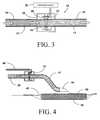
- FIG. 3is a schematic partial cross-sectional view of a third embodiment of the present invention showing its application directly on RPWT tubing.
- FIG. 4is a cross-sectional view of an application of the embodiment shown in FIG. 3 in conjunction with an RPWT wound dressing.
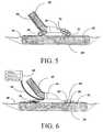
- FIG. 5is a detailed cross-sectional view of a further embodiment of the present invention located in conjunction with an RPWT wound dressing.
- FIG. 6is a detailed cross-sectional view of a further alternative embodiment of the present invention utilizing fiber optics for illumination and detection within the RPWT wound dressing.
- FIG. 7is a perspective cut away view of a section of tubing appropriate for use in conjunction with the alternative body of the present embodiment of the present invention shown in FIG. 6 .
- FIG. 8is a longitudinal cross-sectional view of RPWT tubing further modified to provide yet another alternative embodiment of the present invention.
- the systems and methods of the present invention as shown in the attached figuresemploy photometric or optical methods for detecting the presence (and ultimately, the concentration) of blood in wound fluid being drawn away from the wound by Reduced Pressure Wound Treatment (RPWT) devices and systems.
- RWTReduced Pressure Wound Treatment
- LEDs in the 540/560/580/620/640/660 nm and 800 nm rangesare used as the emitters and a photo detector sensitive to the same range of wavelengths is used as the receptor.
- These solid state optical componentsare positioned across a flow stream of the wound fluid and measurements are taken of the absorption of the illuminating light in a manner that specifically identifies and quantifies the presence of blood in the fluid.
- Variations in the systeminclude different structures to hold or contain the wound fluid while optical measurements are being made as well as different placements of the detection site.
- One objective common to each implementation of the various embodiments that followis to allow for either the activation of a caregiver or patient notification signal and/or the automatic modification or cessation of the RPWT.
- the detection system of the present inventionis capable of providing a digital output signal suitable for triggering any of a number of different caregiver/patient notification signaling devices or suitable for modifying the RPWT operation.
- a notification signalwould be associated with the identification of a wound fluid blood content that exceeded a pre-set level (>30% as an example) indicative of an abnormal condition (excessive bleeding) in the wound. Different types of wounds would merit different settings in this regard as would differing stages of wound healing.
- the detection systemcould generate a staged signal that provided more refined “instructions” to the RPWT system being implemented. For example, a given wound fluid blood concentration level could trigger a reduction in the reduced pressure level of the therapy without altogether ceasing the therapy. A greater concentration or a sudden change in concentration could instead trigger the cessation of the therapy (most likely in conjunction with a notification signal). Because there exists a variety of RPWT regimens, a variety of modifications to these regimens, as triggered by wound fluid blood concentration levels, are anticipated.
- FIG. 1shows the basic components associated with a first implementation of the preferred embodiment of the present invention.
- blood detector 10is connected in line with reduced pressure wound treatment (RPWT) tubing to form a shunt for the flow of wound fluids in a manner that allows accurate measurement of the percentage blood content of the wound fluids.
- Tubing connector 12has an inlet port 18 that is connected to a section of RPWT tubing 14 that is in turn connected to a typical RPWT dressing port (not shown).
- An opposite end of tubing connector 12has an outlet port 20 connected to a continuation of RPWT tubing 16 which connects to the typical reduced pressure fluid collection canister (not shown).
- Arrows in this viewindicate the flow of wound fluids through the system under the influence of the reduced pressure source drawing the fluids away from the wound.
- connector tubes 22 and 24Provided to form a shunt for a portion of the flow of fluid from the main channels are connector tubes 22 and 24 .
- Wound fluidflows into the detector of the system through connector tube 22 and into detection cuvette 26 where the optical components of the system serve to analyze the wound fluid in the manner described in more detail below. From detection cuvette 26 the diverted flow continues into connector tube 24 back into the wound fluid flow stream in tubing connector 12 and from there back into the RPWT tubing 16 .
- Detection cuvette 26provides a detection chamber with a known geometry (i.e. a consistent diameter and volume) such that quantitative measurements of the absorption of select wavelengths of light can be made.
- two light emitting diodes (LEDs)capable of emitting light in select narrow wavelength bands are positioned on one side of detection cuvette 26 and positioned so as to direct light across the clear containment enclosure of the cuvette.
- a first LED 30is, in the preferred embodiment, selected to emit light in the 805 nanometer (nm) wavelength range, while a second LED 32 is configured to emit light in the 542/576/740 nm wavelength range. LEDs with such specifications are readily available in the industry.
- the light from each of the LEDsis alternately transmitted across detection cuvette 26 to a photo detector 34 positioned opposite the LEDs. Photo detector 34 is sensitive to a full range of wavelengths from 500-850 nm. Again, such electronic devices are readily available in the industry.
- LEDs 30 and 32are driven by appropriate electronic circuitry (not shown but well known in the art) and the output signal from photo detector 34 is likewise amplified, conditioned and processed by the appropriate electronic circuitry (not shown).
- the output of photo detector 34is eventually received and analyzed by microprocessor 36 , which also serves to control the illumination within the detector by driving LEDs 30 and 32 .
- the entire LED/photo detector systemis enclosed in LED enclosure 28 that additionally surrounds cuvette 26 and prevents extraneous light from entering into the detection system.
- the LEDs in the systemmay be pulsed sequentially (under the control of microprocessor 36 ) to effect absorption measurements by photo detector 34 .
- the absorption measured for the 542/576/740 nm and the 805 nm wavelengths of lightis then used (according to a reference value calibrated previously in conjunction with the specific cuvette geometry) to estimate the concentration (percentage) of blood in the wound fluid.
- FIG. 1makes use of a “laboratory grade” chamber within detection cuvette 26 to measure the light absorption of the wound fluid under consistently precise conditions.
- FIG. 2provides an alternative to the embodiment shown in FIG. 1 wherein the use of a cuvette is dispensed with and a section of clear silicon based tubing 38 is utilized in its place. Clear silicon based tubing 38 can provide adequate light transmission, and further can provide sufficiently consistent geometry, as to permit accurate readings for the same LED/photo detector arrangement shown and described above in conjunction with FIG. 1 . The same LED wavelengths and photo detector sensitivities may be utilized with accommodations for the differences in geometry and tubing wall transmission characteristics being made during the calibration and referencing process.
- FIG. 3discloses a third implementation of a preferred embodiment of the present invention wherein the wound fluid blood detector 15 may be positioned around a section of tubing 17 as might be typically utilized in standard RPWT systems.
- the flow of wound fluid 13 in this viewis from a first end 19 of tubing section 17 connected to the wound dressing (not shown) through to a second end 21 of tubing section 17 connected to the reduced pressure source (typically through a fluid collection canister) not shown.
- the reduced pressure sourcetypically through a fluid collection canister
- the arrangement shown in FIG. 3may, in the preferred embodiment, be implemented using a cylindrical “clamshell” structure for LED enclosure 28 .
- the devicemay be positioned and secured to any of a number of locations on the tubing either adjacent or distant from the wound itself.
- the low voltage/low current connections to the microprocessormay be structured with anything from a simple electrical conductor bundle that would follow the RPWT system tubing up to the balance of the “remote” equipment (reduced pressure source, etc.).
- the low power electronics (LEDs and photo detector) of the devicecould be locally powered (as by an onboard battery) and a wireless signal communication could be structured between the detector device (acting essentially as a remote blood sensor) and the signal processing instrumentation containing the microprocessor.
- FIG. 4shows the device represented in detail in FIG. 3 as implemented in conjunction with a RPWT dressing.
- blood detector 15is connected by way of signal wires 48 to a microprocessor based signal conditioning system (not shown) as described above.
- RPWT tubing 17is connected to wound dressing vacuum port 44 positioned as is known in the art in conjunction with porous foam dressing 40 positioned in wound bed 38 .
- An adhesive impermeable drape 42is positioned over porous foam dressing 40 and seals the dressing within wound bed 38 .
- Vacuum port 44extends into the enclosed wound bed volume and serves to provide the reduced pressure to the enclosed dressing.
- FIG. 4shows that the device of the present invention may be positioned in close proximity to the wound dressing positioned on the patient in order to detect elevated levels of blood concentration in the wound fluids at the earliest possible point in the system.
- the presence of abnormal levels of blood in the wound fluidcould be delayed by positioning the detection device further from the wound dressing.
- the following further embodiments of the present inventionintegrate the detection device into the wound dressing itself and further expand the functionality of the device to include phototherapy capabilities.
- FIG. 5shows an alternative preferred embodiment of the present invention wherein the LEDs and photo detector of the system are positioned in direct proximity to the wound bed and the RPWT dressing on the patient.
- wound bed 38has positioned within it a layer of transparent or translucent porous foam 50 , the foam being made of a material capable of transmitting light of the frequencies emitted by LEDs 30 and 32 .
- light from the LEDsis directed through the clear components of dressing vacuum port 44 into translucent foam 50 at an angle that generally directs it towards an area of the foam where illumination of the foam is detected by photo detector 34 .
- a further alternative embodiment of the present inventioninvolves conducting the light generated by each of the LEDs and the light to be received by the photo detector, to and from the wound bed at a remote location by way of optical fibers.
- FIG. 6shows one such implementation of this embodiment.
- remote, circuitry 60provides the placement of LEDs 30 and 32 as well as the placement of photo detector 34 in a remote location apart from the patient.
- Optical fiber lines 62are connected to each of the three discreet devices associated with the operation of the system of the present invention. Fiber optic cable 62 connects remote circuitry 60 by way of light channels to appropriate fiber optic terminal light ports positioned more directly in association with the RPWT dressing, and may also include the necessary connections for direct measurement of wound dressing pressures.
- an illumination ring 64positioned in close proximity to adhesive drape 42 (which in the preferred embodiment is typically transparent) positioned over translucent porous foam 50 .
- the fiber optic linesprovide the necessary illumination source for fiber optic ports that effectively function in the manner of the LEDs and the photo detector to transmit and receive light waves into and from the wound fluid saturated foam within the dressing.
- Fiber optic port 66for example, provides light at a wavelength associated with LED 32 while fiber optic port 68 provides light of a wavelength associated with LED 30 .
- fiber optic port 70collects the light scattered (and partially absorbed) by the wound fluids within translucent foam 50 and directs it by fiber optic cable 62 up to photo detector 34 .
- FIG. 6utilizes separate optic lines for the two discrete LEDs providing light of the indicated discrete wavelengths.
- a single optic wave guideoptical fiber
- fiber optic ports 66 and 68may simply be two of an array of ports all connected to the same source (at a particular instant in time). In this manner a greater illumination of the wound bed might be achieved.
- the phototherapy aspects of the present inventiondiscussed in more detail below, may be more easily implemented with this multiple source—single optic fiber arrangement.
- the wound bedmight be illuminated in one instant with light of the first LED wavelength (805 nm) followed by light in the next instant of the second LED wavelengths (540/576/740 nm) followed by light in a subsequent period of time in a wavelength spectrum appropriate for phototherapy.
- Illumination ring 64may also include the necessary sensors for returning temperature and/or pressure measurements of the wound bed.
- the fiber optic connections shown in FIG. 6may be loosely associated with the tubing connecting the RPWT dressing to the reduced pressure source or they may be integrated into the tubing utilized in the reduced pressure wound treatment system in the manner described in more detail below.
- the illumination ring 64 shown in the embodiment of FIG. 6may be integrally constructed with dressing vacuum port 44 or may be configured as an attachable ring that surrounds or sits adjacent to the port. The association with vacuum port 44 is two-fold, first to centrally position both the illumination and the reception of light waves within the boundaries of the wound dressing and second to provide an existing connecting line (the tubing) with which to direct the fiber optic bundle away from the wound.
- optical fiber 62 shown in FIG. 6is a structure wherein the optical fibers are incorporated into the walls of RPWT tubing 80 as disclosed in FIG. 7 . This manner of placement eliminates the need for an additional line connecting to the patient and integrates the optical system fully into the structure of the dressing vacuum port 44 .
- FIG. 7shows a perspective partial cross-sectional view of a section of RPWT tubing with integrated fiber optic signal lines.
- the view in FIG. 7shows tubing with an oval cross-section to accommodate the additional conductors on either side of the primary lumen although tubing of circular (or other geometric) cross section and adequate diameter overall may likewise be suitable for the purposes of the present invention.
- tubing 80is comprised of primary vacuum lumen 82 and two associated fiber optic light conductors 84 and 86 .
- Fiber optic conductor 84comprises the illuminating light associated with the wavelengths generated by the LEDs as described above.
- Fiber optic conductor 86comprises the return line from the detection port to the photo detector.
- Appropriate terminal connections for the integrated fiber optic wave guidesare positioned in conjunction with the LEDs and the photo detector as described and shown in FIG. 6 .
- the fiber optic linesterminate in the dressing vacuum port directly into the fiber optic ports positioned in the illumination ring over the wound dressing.
- Such terminals and connectors associated with fiber optic signal lines appropriate for use in conjunction with the system of the present inventionare well known in the art.
- FIG. 7The structure of RPWT tubing shown in FIG. 7 is directed to communicating light waves to and from the wound dressing where, in conjunction with the translucent foam positioned within the wound bed, a measurement of the absorption properties of the wound fluid can be made.
- a similar but functionally distinct structuremay be utilized in conjunction with the system of the present invention where the measured wound fluid is again associated with the RPWT tubing instead of the wound bed.
- FIG. 8for a longitudinal cross-section of a further alternate tubing structure for use in conjunction with the present invention.
- the tubing shown in FIG. 8is configured to integrate the blood detection system directly into the walls of a section of the tubing 90 associated with the RPWT system.
- a primary lumen 92is shown wherein wound fluids are transported (by means of reduced pressure) from the wound dressing to the RPWT collection canister.
- On either side of this primary lumen 92are specially configured optical fiber bundles 94 and 96 that connect individual optical fibers to individual optical ports positioned along the length of the tubing.
- these ports 102 and 106serve as illumination points directing light at the select wavelengths across the primary lumen 92 (and therefore across the flow of wound fluid) to the opposite side of the tubing 90 .
- optical reception ports 104 and 108aligned with the transmission ports 102 and 106 , and which are each connected by return fiber optic lines to the photo detector of the system of the present invention.
- the structure shown in FIG. 8integrates an array of illuminating optical ports positioned opposite an array of receiving optical ports within the structure of the RPWT tubing connecting the wound dressing to the reduced pressure source.
- This structureis a preferred embodiment because of its known geometry and the ability of the system to discriminate between discrete locations along the length of the tubing. By isolating the absorption measurement to a specific set of optical ports additional important information can be acquired regarding the condition of the system at a particular point along the length of the tubing.
- Each of the transmission optical portsis associated with a direct optical fiber that is “addressable” by the electronics associated with the LED light sources.
- the system and method of measurementare operable by simply switching the transmission line to a known optical fiber/optical port combination and maintaining a single optical fiber connection to the reception ports as a whole. Monitoring of individual port pairs would allow for measurement of the location or speed (or both) of blood containing fluid boluses within the primary lumen.
- the array structure disclosed in FIG. 8allows individual locations along the tubing to be identified, and from this information, a flow characteristic of the wound fluid may also be derived. Therefore, in addition to the ability to detect the presence of blood in the wound fluids that pass a particular point in the tubing, the system is capable of analyzing patterns in light absorption and identifying flow rates as a result. In other words, patterned (sequential) responses to high absorption rates would indicate the passage of a quantity of fluid of a particular character past multiple points in the system, which would provide the basis for measurements of volume and flow rates. Such measurement could factor into a decision regarding the urgency of a bleeding condition within the wound and help dictate the nature of a system response to elevated wound fluid blood content.
- the present inventionfinds particular application in conjunction with RPWT systems, there are other fields where the systems and methods of the present invention can likewise be applied.
- the systemcould, for example, be used for the purpose of detecting blood in urine, especially in catheterized patients.
- the systems and methodscould be applied to hemodialysis systems where the blood can be monitored continuously during dialysis.
- the methodcan be applied to any mixture of unknown blood and body fluids where a controlled geometry can be established for the illuminated volume of fluid. Such conditions are typically present whenever fluids are being drained from, or circulated from, the body through translucent or transparent conductors.
- Other applicationsfurther include the detection of clots in the above described systems.
- the LED light sourcescould easily be configured to operate at the wavelengths suitable for application of the phototherapy regimens.
- the systems described abovecould easily function (in addition or in the alternative) as phototherapy systems. Modification of the wavelength specifications for the LEDs and of the synchronization programming for the control of the LEDs would be all that is required to implement such a system.
- the same structures, again with specific wavelengths,could also provide a system implementing antimicrobial light application.
- a number of locations within the RPWT system that provide access to fluids to the blood gas monitorsare appropriate for placement of the inter-tubular embodiments of the present blood detection system. Whether integrating cuvette elements ( FIG. 1 ), shunt tubes ( FIG. 2 ), or directly connected to the blood gas monitoring tubes (as in FIGS. 3 and 4 herein), the detection system and method may be appropriately applied. Wherever the detection device is placed in conjunction with the RPWT tubing in the present invention, it might easily be placed in similar fashion with blood gas monitoring components in place.
- color responsive chemical sensorsmay be incorporated in any of the sensing methods described to monitor chemical species in the wound fluid.
- speciesmight include cytokines, creatinine, urea, among other chemicals of interest to those clinicians guiding the normal healing process of the wound.
Landscapes
- Health & Medical Sciences (AREA)
- Life Sciences & Earth Sciences (AREA)
- Heart & Thoracic Surgery (AREA)
- Engineering & Computer Science (AREA)
- Biomedical Technology (AREA)
- Animal Behavior & Ethology (AREA)
- General Health & Medical Sciences (AREA)
- Public Health (AREA)
- Veterinary Medicine (AREA)
- Physics & Mathematics (AREA)
- Molecular Biology (AREA)
- Surgery (AREA)
- Medical Informatics (AREA)
- Pathology (AREA)
- Biophysics (AREA)
- Hematology (AREA)
- Anesthesiology (AREA)
- Vascular Medicine (AREA)
- Optics & Photonics (AREA)
- General Chemical & Material Sciences (AREA)
- Chemical Kinetics & Catalysis (AREA)
- Chemical & Material Sciences (AREA)
- Dermatology (AREA)
- Measurement Of The Respiration, Hearing Ability, Form, And Blood Characteristics Of Living Organisms (AREA)
Abstract
Description
Claims (11)
Priority Applications (1)
| Application Number | Priority Date | Filing Date | Title |
|---|---|---|---|
| US13/089,077US9326683B2 (en) | 1999-04-06 | 2011-04-18 | Systems and methods for detection of wound fluid blood and application of phototherapy in conjunction with reduced pressure wound treatment system |
Applications Claiming Priority (9)
| Application Number | Priority Date | Filing Date | Title |
|---|---|---|---|
| US12793699P | 1999-04-06 | 1999-04-06 | |
| US09/544,399US6994702B1 (en) | 1999-04-06 | 2000-04-06 | Vacuum assisted closure pad with adaptation for phototherapy |
| US57975500A | 2000-05-26 | 2000-05-26 | |
| US27358701P | 2001-03-05 | 2001-03-05 | |
| US10/085,321US6856821B2 (en) | 2000-05-26 | 2002-02-28 | System for combined transcutaneous blood gas monitoring and vacuum assisted wound closure |
| US10/090,358US7799004B2 (en) | 2001-03-05 | 2002-03-04 | Negative pressure wound treatment apparatus and infection identification system and method |
| US10/867,990US7524286B2 (en) | 1999-05-27 | 2004-06-15 | System for combined transcutaneous blood gas monitoring and negative pressure wound treatment |
| US11/327,662US7947033B2 (en) | 1999-04-06 | 2006-01-06 | Systems and methods for detection of wound fluid blood and application of phototherapy in conjunction with reduced pressure wound treatment system |
| US13/089,077US9326683B2 (en) | 1999-04-06 | 2011-04-18 | Systems and methods for detection of wound fluid blood and application of phototherapy in conjunction with reduced pressure wound treatment system |
Related Parent Applications (2)
| Application Number | Title | Priority Date | Filing Date |
|---|---|---|---|
| US09/544,399Continuation-In-PartUS6994702B1 (en) | 1999-04-06 | 2000-04-06 | Vacuum assisted closure pad with adaptation for phototherapy |
| US11/327,662ContinuationUS7947033B2 (en) | 1999-04-06 | 2006-01-06 | Systems and methods for detection of wound fluid blood and application of phototherapy in conjunction with reduced pressure wound treatment system |
Publications (2)
| Publication Number | Publication Date |
|---|---|
| US20110196284A1 US20110196284A1 (en) | 2011-08-11 |
| US9326683B2true US9326683B2 (en) | 2016-05-03 |
Family
ID=46323551
Family Applications (2)
| Application Number | Title | Priority Date | Filing Date |
|---|---|---|---|
| US11/327,662Expired - Fee RelatedUS7947033B2 (en) | 1999-04-06 | 2006-01-06 | Systems and methods for detection of wound fluid blood and application of phototherapy in conjunction with reduced pressure wound treatment system |
| US13/089,077Expired - Fee RelatedUS9326683B2 (en) | 1999-04-06 | 2011-04-18 | Systems and methods for detection of wound fluid blood and application of phototherapy in conjunction with reduced pressure wound treatment system |
Family Applications Before (1)
| Application Number | Title | Priority Date | Filing Date |
|---|---|---|---|
| US11/327,662Expired - Fee RelatedUS7947033B2 (en) | 1999-04-06 | 2006-01-06 | Systems and methods for detection of wound fluid blood and application of phototherapy in conjunction with reduced pressure wound treatment system |
Country Status (1)
| Country | Link |
|---|---|
| US (2) | US7947033B2 (en) |
Cited By (2)
| Publication number | Priority date | Publication date | Assignee | Title |
|---|---|---|---|---|
| US11357906B2 (en) | 2016-02-12 | 2022-06-14 | Smith & Nephew, Inc. | Systems and methods for detecting operational conditions of reduced pressure therapy |
| US11602461B2 (en) | 2016-05-13 | 2023-03-14 | Smith & Nephew, Inc. | Automatic wound coupling detection in negative pressure wound therapy systems |
Families Citing this family (85)
| Publication number | Priority date | Publication date | Assignee | Title |
|---|---|---|---|---|
| US6458109B1 (en) | 1998-08-07 | 2002-10-01 | Hill-Rom Services, Inc. | Wound treatment apparatus |
| US7947033B2 (en) | 1999-04-06 | 2011-05-24 | Kci Licensing Inc. | Systems and methods for detection of wound fluid blood and application of phototherapy in conjunction with reduced pressure wound treatment system |
| US6824533B2 (en) | 2000-11-29 | 2004-11-30 | Hill-Rom Services, Inc. | Wound treatment apparatus |
| US6764462B2 (en) | 2000-11-29 | 2004-07-20 | Hill-Rom Services Inc. | Wound treatment apparatus |
| US20010043943A1 (en) | 2000-05-22 | 2001-11-22 | Coffey Arthur C. | Combination SIS and vacuum bandage and method |
| US6855135B2 (en) | 2000-11-29 | 2005-02-15 | Hill-Rom Services, Inc. | Vacuum therapy and cleansing dressing for wounds |
| US6685681B2 (en) | 2000-11-29 | 2004-02-03 | Hill-Rom Services, Inc. | Vacuum therapy and cleansing dressing for wounds |
| WO2003030966A1 (en) | 2001-10-11 | 2003-04-17 | Hill-Rom Services, Inc. | Waste container for negative pressure therapy |
| AU2002359829A1 (en) | 2001-12-26 | 2003-07-24 | Hill-Rom Services, Inc. | Vacuum bandage packing |
| AU2002359828A1 (en) | 2001-12-26 | 2003-07-24 | Hill-Rom Services Inc. | Vented vacuum bandage and method |
| EP1461113A4 (en) | 2001-12-26 | 2009-05-06 | Hill Rom Services Inc | Wound vacuum therapy dressing kit |
| US8168848B2 (en) | 2002-04-10 | 2012-05-01 | KCI Medical Resources, Inc. | Access openings in vacuum bandage |
| JP2005536275A (en) | 2002-08-21 | 2005-12-02 | ヒル−ロム サービシズ,インコーポレイテッド | Wound packing to prevent wound closure |
| GB0224986D0 (en) | 2002-10-28 | 2002-12-04 | Smith & Nephew | Apparatus |
| US10058642B2 (en) | 2004-04-05 | 2018-08-28 | Bluesky Medical Group Incorporated | Reduced pressure treatment system |
| US7909805B2 (en) | 2004-04-05 | 2011-03-22 | Bluesky Medical Group Incorporated | Flexible reduced pressure treatment appliance |
| US8062272B2 (en) | 2004-05-21 | 2011-11-22 | Bluesky Medical Group Incorporated | Flexible reduced pressure treatment appliance |
| US8781545B2 (en)* | 2006-06-12 | 2014-07-15 | Koninklijke Philips N.V. | Body monitoring device, body data acquiring method and method of determining the presence, location and/or stage of a wound |
| US8025650B2 (en) | 2006-06-12 | 2011-09-27 | Wound Care Technologies, Inc. | Negative pressure wound treatment device, and methods |
| US9854975B2 (en)* | 2006-06-12 | 2018-01-02 | Koninklijke Philips N.V. | Skin monitoring device, method of monitoring the skin, monitoring device, method of irradiating the skin, and use of an OLED |
| TWI293887B (en)* | 2006-07-28 | 2008-03-01 | Edison Opto Corp | Light modulating system for medical treatment and health care |
| WO2008036361A2 (en) | 2006-09-19 | 2008-03-27 | Kci Licensing Inc. | Reduced pressure treatment system having blockage clearing and dual-zone pressure protection capabilities |
| US8366690B2 (en) | 2006-09-19 | 2013-02-05 | Kci Licensing, Inc. | System and method for determining a fill status of a canister of fluid in a reduced pressure treatment system |
| US20090036918A1 (en)* | 2006-11-13 | 2009-02-05 | Burgess James E | Method and Apparatus for the Containment of a Surgical Site |
| US7931651B2 (en) | 2006-11-17 | 2011-04-26 | Wake Lake University Health Sciences | External fixation assembly and method of use |
| US8377016B2 (en) | 2007-01-10 | 2013-02-19 | Wake Forest University Health Sciences | Apparatus and method for wound treatment employing periodic sub-atmospheric pressure |
| RU2428208C2 (en) | 2007-02-09 | 2011-09-10 | КейСиАй Лайсензинг Инк. | System and method of low pressure control in tissue area |
| AU2008219034B2 (en) | 2007-02-20 | 2012-01-19 | Solventum Intellectual Properties Company | System and method for distinguishing leaks from a disengaged canister condition in a reduced pressure treatment system |
| US20080269849A1 (en)* | 2007-04-19 | 2008-10-30 | Mergenet Medical, Inc. | Temporal control in phototherapy |
| US9023001B2 (en)* | 2007-09-12 | 2015-05-05 | Heal-Ex, Llc | Systems and methods for providing a debriding wound vacuum |
| US8187184B2 (en)* | 2007-09-21 | 2012-05-29 | Baxter International, Inc. | Access disconnect system with optical and other sensors |
| KR101600041B1 (en) | 2007-10-10 | 2016-03-03 | 웨이크 포리스트 유니버시티 헬스 사이언시즈 | Devices and methods for treating spinal cord tissue |
| AU2008311789A1 (en)* | 2007-10-18 | 2009-04-23 | Convatec Technologies Inc. | Aspiration system for removing liquid discharged by the human body, and liquid sensor therefor |
| US8366692B2 (en) | 2008-01-08 | 2013-02-05 | Richard Scott Weston | Sustained variable negative pressure wound treatment and method of controlling same |
| US20090177051A1 (en)* | 2008-01-09 | 2009-07-09 | Heal-Ex, Llc | Systems and methods for providing sub-dressing wound analysis and therapy |
| RU2517588C2 (en) | 2008-01-09 | 2014-05-27 | Уэйк Форест Юниверсити Хелс Сайенсиз | Device and method for treating pathologies of central nervous system |
| US9782300B2 (en)* | 2008-02-01 | 2017-10-10 | Kci Licensing, Inc. | Fiber-microsphere bioresorbable composite scaffold for wound healing |
| AU2009223037A1 (en)* | 2008-03-12 | 2009-09-17 | Smith & Nephew Plc | Negative pressure dressing and method of using same |
| US8523797B2 (en)* | 2008-05-08 | 2013-09-03 | Hospira, Inc. | Automated point-of-care fluid testing device and method of using the same |
| BRPI0912824B8 (en)* | 2008-05-21 | 2021-06-22 | Topaz Morris | wound healing device |
| CN102036699B (en) | 2008-05-30 | 2013-08-21 | 凯希特许有限公司 | Decompression Linear Wound Therapy System |
| EP2278949B1 (en) | 2008-05-30 | 2016-07-06 | KCI Licensing, Inc. | Reduced-pressure, linear wound closing bolsters |
| ES2633142T3 (en) | 2008-07-18 | 2017-09-19 | Wake Forest University Health Sciences | Apparatus for modulation of cardiac tissue through topical application of vacuum to minimize death and cell damage |
| US8366691B2 (en) | 2008-08-08 | 2013-02-05 | Kci Licensing, Inc | Reduced-pressure treatment systems with reservoir control |
| BRPI0913748A2 (en)* | 2008-10-03 | 2019-09-24 | Kci Licensing Inc | "reduced pressure release system for treating a patient's tissue site, method for treating a patient's tissue site, and reduced pressure therapy system for removing exudate fluid from a patient's tissue and treating tissue" |
| US8690844B2 (en)* | 2009-08-27 | 2014-04-08 | Kci Licensing, Inc. | Re-epithelialization wound dressings and systems |
| US20110054420A1 (en)* | 2009-08-27 | 2011-03-03 | Christopher Brian Locke | Reduced-pressure wound dressings and systems for re-epithelialization and granulation |
| US20110178375A1 (en)* | 2010-01-19 | 2011-07-21 | Avery Dennison Corporation | Remote physiological monitoring |
| US8623047B2 (en) | 2010-04-30 | 2014-01-07 | Kci Licensing, Inc. | System and method for sealing an incisional wound |
| GB201011173D0 (en) | 2010-07-02 | 2010-08-18 | Smith & Nephew | Provision of wound filler |
| GB201020005D0 (en) | 2010-11-25 | 2011-01-12 | Smith & Nephew | Composition 1-1 |
| CA2819032C (en) | 2010-11-25 | 2020-06-23 | Smith & Nephew Plc | Composition i-ii and products and uses thereof |
| US10207031B2 (en)* | 2010-12-08 | 2019-02-19 | Convatec Technologies Inc. | Integrated system for assessing wound exudates |
| ES2748519T3 (en) | 2010-12-08 | 2020-03-17 | Convatec Technologies Inc | Wound exudate system accessory |
| US9050175B2 (en) | 2011-01-20 | 2015-06-09 | Scott Stephan | Therapeutic treatment pad |
| US20150159066A1 (en) | 2011-11-25 | 2015-06-11 | Smith & Nephew Plc | Composition, apparatus, kit and method and uses thereof |
| EP2759310B2 (en)* | 2013-01-28 | 2023-11-08 | Mölnlycke Health Care AB | Suction device |
| USD738487S1 (en) | 2013-01-28 | 2015-09-08 | Molnlycke Health Care Ab | Suction device for negative pressure therapy |
| US20160120706A1 (en) | 2013-03-15 | 2016-05-05 | Smith & Nephew Plc | Wound dressing sealant and use thereof |
| EP2968647B1 (en) | 2013-03-15 | 2022-06-29 | Smith & Nephew plc | Wound dressing sealant and use thereof |
| US9375586B2 (en)* | 2013-03-15 | 2016-06-28 | Pavel V. Efremkin | Apparatus and method for treatment of foot and nail diseases |
| US20150133861A1 (en) | 2013-11-11 | 2015-05-14 | Kevin P. McLennan | Thermal management system and method for medical devices |
| US10143795B2 (en) | 2014-08-18 | 2018-12-04 | Icu Medical, Inc. | Intravenous pole integrated power, control, and communication system and method for an infusion pump |
| CN106215254B (en)* | 2014-11-20 | 2018-10-26 | 樊理华 | A kind of application method of the pressure control pipe of autogenous vein |
| US20160199665A1 (en)* | 2015-01-08 | 2016-07-14 | Photomed Technologies, Inc. | Treatment of wounds using electromagnetic radiation |
| NZ737340A (en) | 2015-05-26 | 2019-06-28 | Icu Medical Inc | Disposable infusion fluid delivery device for programmable large volume drug delivery |
| WO2017010942A1 (en)* | 2015-07-14 | 2017-01-19 | Singapore University Of Technology And Design | On-site device for detecting presence of a liquid |
| EP3714916A1 (en) | 2015-07-29 | 2020-09-30 | Innovative Therapies Inc. | Wound therapy device pressure monitoring and control system |
| US10369376B2 (en)* | 2016-04-29 | 2019-08-06 | NeoLight LLC | Phototherapy apparatuses and methods |
| MX2019000232A (en)* | 2016-07-08 | 2019-11-12 | Convatec Technologies Inc | Fluid flow sensing. |
| US20180042484A1 (en)* | 2016-08-11 | 2018-02-15 | Charles River Analytics, Inc. | PORTABLE, DURABLE, RUGGED, FUNCTIONAL NEAR-INFRARED SPECTROSCOPY (fNIRS) SENSOR |
| US10596388B2 (en) | 2016-09-21 | 2020-03-24 | Epistar Corporation | Therapeutic light-emitting module |
| AU201716716S (en) | 2017-05-11 | 2017-11-21 | MAƒA¶LNLYCKE HEALTH CARE AB | Wound dressings |
| EP3406273B1 (en)* | 2017-05-23 | 2022-03-30 | Sofradim Production | A surgical drain |
| RU175660U1 (en)* | 2017-07-25 | 2017-12-13 | Федеральное государственное бюджетное образовательное учреждение высшего образования "Амурская государственная медицинская академия" Министерства здравоохранения Российской Федерации | Device for recording the appearance of exudate in a postoperative wound |
| US11073478B2 (en)* | 2017-08-14 | 2021-07-27 | Brenton Ferguson | Fluid monitoring system and method |
| JP7183294B2 (en)* | 2018-04-12 | 2022-12-05 | ラジオメーター・メディカル・アー・ペー・エス | Porous membrane sensor element |
| US12097040B2 (en) | 2018-06-15 | 2024-09-24 | Coloplast A/S | Wound dressing system and method with data collection based on environmental factor of geographic location |
| WO2019238195A1 (en)* | 2018-06-15 | 2019-12-19 | Coloplast A/S | Wound dressing system, monitor device and related methods |
| WO2020064937A1 (en)* | 2018-09-28 | 2020-04-02 | T.J.Smith And Nephew,Limited | Optical fibers for optically sensing through wound dressings |
| USD939079S1 (en) | 2019-08-22 | 2021-12-21 | Icu Medical, Inc. | Infusion pump |
| CN114904145A (en)* | 2021-02-09 | 2022-08-16 | 明基材料股份有限公司 | wound treatment system |
| TWI759106B (en)* | 2021-02-09 | 2022-03-21 | 明基材料股份有限公司 | Wound treatment system |
| USD1052728S1 (en) | 2021-11-12 | 2024-11-26 | Icu Medical, Inc. | Medical fluid infusion pump |
| TWI824735B (en)* | 2022-09-22 | 2023-12-01 | 友達光電股份有限公司 | Light dispersion apparatus and hemodialysis system |
Citations (140)
| Publication number | Priority date | Publication date | Assignee | Title |
|---|---|---|---|---|
| US1355846A (en) | 1920-02-06 | 1920-10-19 | David A Rannells | Medical appliance |
| US2547758A (en) | 1949-01-05 | 1951-04-03 | Wilmer B Keeling | Instrument for treating the male urethra |
| US2632443A (en) | 1949-04-18 | 1953-03-24 | Eleanor P Lesher | Surgical dressing |
| GB692578A (en) | 1949-09-13 | 1953-06-10 | Minnesota Mining & Mfg | Improvements in or relating to drape sheets for surgical use |
| US2682873A (en) | 1952-07-30 | 1954-07-06 | Johnson & Johnson | General purpose protective dressing |
| US2910763A (en) | 1955-08-17 | 1959-11-03 | Du Pont | Felt-like products |
| US2969057A (en) | 1957-11-04 | 1961-01-24 | Brady Co W H | Nematodic swab |
| US3066672A (en) | 1960-09-27 | 1962-12-04 | Jr William H Crosby | Method and apparatus for serial sampling of intestinal juice |
| US3367332A (en) | 1965-08-27 | 1968-02-06 | Gen Electric | Product and process for establishing a sterile area of skin |
| US3520300A (en) | 1967-03-15 | 1970-07-14 | Amp Inc | Surgical sponge and suction device |
| US3568675A (en) | 1968-08-30 | 1971-03-09 | Clyde B Harvey | Fistula and penetrating wound dressing |
| US3648692A (en) | 1970-12-07 | 1972-03-14 | Parke Davis & Co | Medical-surgical dressing for burns and the like |
| US3682180A (en) | 1970-06-08 | 1972-08-08 | Coilform Co Inc | Drain clip for surgical drain |
| US3826254A (en) | 1973-02-26 | 1974-07-30 | Verco Ind | Needle or catheter retaining appliance |
| DE2640413A1 (en) | 1976-09-08 | 1978-03-09 | Wolf Gmbh Richard | CATHETER MONITORING DEVICE |
| US4080970A (en) | 1976-11-17 | 1978-03-28 | Miller Thomas J | Post-operative combination dressing and internal drain tube with external shield and tube connector |
| US4096853A (en) | 1975-06-21 | 1978-06-27 | Hoechst Aktiengesellschaft | Device for the introduction of contrast medium into an anus praeter |
| US4139004A (en) | 1977-02-17 | 1979-02-13 | Gonzalez Jr Harry | Bandage apparatus for treating burns |
| US4165748A (en) | 1977-11-07 | 1979-08-28 | Johnson Melissa C | Catheter tube holder |
| US4184510A (en) | 1977-03-15 | 1980-01-22 | Fibra-Sonics, Inc. | Valued device for controlling vacuum in surgery |
| WO1980002182A1 (en) | 1979-04-06 | 1980-10-16 | J Moss | Portable suction device for collecting fluids from a closed wound |
| US4233969A (en) | 1976-11-11 | 1980-11-18 | Lock Peter M | Wound dressing materials |
| US4245630A (en) | 1976-10-08 | 1981-01-20 | T. J. Smith & Nephew, Ltd. | Tearable composite strip of materials |
| US4256109A (en) | 1978-07-10 | 1981-03-17 | Nichols Robert L | Shut off valve for medical suction apparatus |
| US4261363A (en) | 1979-11-09 | 1981-04-14 | C. R. Bard, Inc. | Retention clips for body fluid drains |
| US4275721A (en) | 1978-11-28 | 1981-06-30 | Landstingens Inkopscentral Lic, Ekonomisk Forening | Vein catheter bandage |
| US4284079A (en) | 1979-06-28 | 1981-08-18 | Adair Edwin Lloyd | Method for applying a male incontinence device |
| US4297995A (en) | 1980-06-03 | 1981-11-03 | Key Pharmaceuticals, Inc. | Bandage containing attachment post |
| US4333468A (en) | 1980-08-18 | 1982-06-08 | Geist Robert W | Mesentery tube holder apparatus |
| US4373519A (en) | 1981-06-26 | 1983-02-15 | Minnesota Mining And Manufacturing Company | Composite wound dressing |
| US4382441A (en) | 1978-12-06 | 1983-05-10 | Svedman Paul | Device for treating tissues, for example skin |
| US4392858A (en) | 1981-07-16 | 1983-07-12 | Sherwood Medical Company | Wound drainage device |
| US4392853A (en) | 1981-03-16 | 1983-07-12 | Rudolph Muto | Sterile assembly for protecting and fastening an indwelling device |
| US4419097A (en) | 1981-07-31 | 1983-12-06 | Rexar Industries, Inc. | Attachment for catheter tube |
| EP0100148A1 (en) | 1982-07-06 | 1984-02-08 | Dow Corning Limited | Medical-surgical dressing and a process for the production thereof |
| US4465485A (en) | 1981-03-06 | 1984-08-14 | Becton, Dickinson And Company | Suction canister with unitary shut-off valve and filter features |
| EP0117632A2 (en) | 1983-01-27 | 1984-09-05 | Johnson & Johnson Products Inc. | Adhesive film dressing |
| US4475909A (en) | 1982-05-06 | 1984-10-09 | Eisenberg Melvin I | Male urinary device and method for applying the device |
| US4480638A (en) | 1980-03-11 | 1984-11-06 | Eduard Schmid | Cushion for holding an element of grafted skin |
| US4525374A (en) | 1984-02-27 | 1985-06-25 | Manresa, Inc. | Treating hydrophobic filters to render them hydrophilic |
| US4525166A (en) | 1981-11-21 | 1985-06-25 | Intermedicat Gmbh | Rolled flexible medical suction drainage device |
| US4540412A (en) | 1983-07-14 | 1985-09-10 | The Kendall Company | Device for moist heat therapy |
| US4543100A (en) | 1983-11-01 | 1985-09-24 | Brodsky Stuart A | Catheter and drain tube retainer |
| US4548202A (en) | 1983-06-20 | 1985-10-22 | Ethicon, Inc. | Mesh tissue fasteners |
| US4551139A (en) | 1982-02-08 | 1985-11-05 | Marion Laboratories, Inc. | Method and apparatus for burn wound treatment |
| EP0161865A2 (en) | 1984-05-03 | 1985-11-21 | Smith and Nephew Associated Companies p.l.c. | Adhesive wound dressing |
| US4569348A (en) | 1980-02-22 | 1986-02-11 | Velcro Usa Inc. | Catheter tube holder strap |
| US4605399A (en) | 1984-12-04 | 1986-08-12 | Complex, Inc. | Transdermal infusion device |
| US4608041A (en) | 1981-10-14 | 1986-08-26 | Frese Nielsen | Device for treatment of wounds in body tissue of patients by exposure to jets of gas |
| US4640688A (en) | 1985-08-23 | 1987-02-03 | Mentor Corporation | Urine collection catheter |
| US4655754A (en) | 1984-11-09 | 1987-04-07 | Stryker Corporation | Vacuum wound drainage system and lipids baffle therefor |
| US4664662A (en) | 1984-08-02 | 1987-05-12 | Smith And Nephew Associated Companies Plc | Wound dressing |
| WO1987004626A1 (en) | 1986-01-31 | 1987-08-13 | Osmond, Roger, L., W. | Suction system for wound and gastro-intestinal drainage |
| US4710165A (en) | 1985-09-16 | 1987-12-01 | Mcneil Charles B | Wearable, variable rate suction/collection device |
| US4733659A (en) | 1986-01-17 | 1988-03-29 | Seton Company | Foam bandage |
| GB2195255A (en) | 1986-09-30 | 1988-04-07 | Vacutec Uk Limited | Method and apparatus for vacuum treatment of an epidermal surface |
| US4743232A (en) | 1986-10-06 | 1988-05-10 | The Clinipad Corporation | Package assembly for plastic film bandage |
| GB2197789A (en) | 1986-11-28 | 1988-06-02 | Smiths Industries Plc | Anti-foaming disinfectants used in surgical suction apparatus |
| US4758220A (en) | 1985-09-26 | 1988-07-19 | Alcon Laboratories, Inc. | Surgical cassette proximity sensing and latching apparatus |
| US4787888A (en) | 1987-06-01 | 1988-11-29 | University Of Connecticut | Disposable piezoelectric polymer bandage for percutaneous delivery of drugs and method for such percutaneous delivery (a) |
| US4826494A (en) | 1984-11-09 | 1989-05-02 | Stryker Corporation | Vacuum wound drainage system |
| US4838883A (en) | 1986-03-07 | 1989-06-13 | Nissho Corporation | Urine-collecting device |
| US4840187A (en) | 1986-09-11 | 1989-06-20 | Bard Limited | Sheath applicator |
| US4863449A (en) | 1987-07-06 | 1989-09-05 | Hollister Incorporated | Adhesive-lined elastic condom cathether |
| US4872450A (en) | 1984-08-17 | 1989-10-10 | Austad Eric D | Wound dressing and method of forming same |
| US4878901A (en) | 1986-10-10 | 1989-11-07 | Sachse Hans Ernst | Condom catheter, a urethral catheter for the prevention of ascending infections |
| GB2220357A (en) | 1988-05-28 | 1990-01-10 | Smiths Industries Plc | Medico-surgical containers |
| US4897081A (en) | 1984-05-25 | 1990-01-30 | Thermedics Inc. | Percutaneous access device |
| US4906240A (en) | 1988-02-01 | 1990-03-06 | Matrix Medica, Inc. | Adhesive-faced porous absorbent sheet and method of making same |
| US4906233A (en) | 1986-05-29 | 1990-03-06 | Terumo Kabushiki Kaisha | Method of securing a catheter body to a human skin surface |
| US4919654A (en) | 1988-08-03 | 1990-04-24 | Kalt Medical Corporation | IV clamp with membrane |
| CA2005436A1 (en) | 1988-12-13 | 1990-06-13 | Glenda G. Kalt | Transparent tracheostomy tube dressing |
| US4941882A (en) | 1987-03-14 | 1990-07-17 | Smith And Nephew Associated Companies, P.L.C. | Adhesive dressing for retaining a cannula on the skin |
| US4953565A (en) | 1986-11-26 | 1990-09-04 | Shunro Tachibana | Endermic application kits for external medicines |
| WO1990010424A1 (en) | 1989-03-16 | 1990-09-20 | Smith & Nephew Plc | Absorbent devices and precursors therefor |
| US4969880A (en) | 1989-04-03 | 1990-11-13 | Zamierowski David S | Wound dressing and treatment method |
| US4969702A (en) | 1989-05-22 | 1990-11-13 | Tektronix, Inc. | Laser pigtail assembly and method of manufacture |
| US4985019A (en) | 1988-03-11 | 1991-01-15 | Michelson Gary K | X-ray marker |
| GB2235877A (en) | 1989-09-18 | 1991-03-20 | Antonio Talluri | Closed wound suction apparatus |
| US5037397A (en) | 1985-05-03 | 1991-08-06 | Medical Distributors, Inc. | Universal clamp |
| US5066859A (en) | 1990-05-18 | 1991-11-19 | Karkar Maurice N | Hematocrit and oxygen saturation blood analyzer |
| US5086170A (en) | 1989-01-16 | 1992-02-04 | Roussel Uclaf | Process for the preparation of azabicyclo compounds |
| US5092858A (en) | 1990-03-20 | 1992-03-03 | Becton, Dickinson And Company | Liquid gelling agent distributor device |
| US5100429A (en) | 1989-04-28 | 1992-03-31 | C. R. Bard, Inc. | Endovascular stent and delivery system |
| US5100396A (en) | 1989-04-03 | 1992-03-31 | Zamierowski David S | Fluidic connection system and method |
| US5134994A (en) | 1990-02-12 | 1992-08-04 | Say Sam L | Field aspirator in a soft pack with externally mounted container |
| US5149331A (en) | 1991-05-03 | 1992-09-22 | Ariel Ferdman | Method and device for wound closure |
| US5167613A (en) | 1992-03-23 | 1992-12-01 | The Kendall Company | Composite vented wound dressing |
| US5176663A (en) | 1987-12-02 | 1993-01-05 | Pal Svedman | Dressing having pad with compressibility limiting elements |
| WO1993009727A1 (en) | 1991-11-14 | 1993-05-27 | Wake Forest University | Method and apparatus for treating tissue damage |
| US5215522A (en) | 1984-07-23 | 1993-06-01 | Ballard Medical Products | Single use medical aspirating device and method |
| US5232453A (en) | 1989-07-14 | 1993-08-03 | E. R. Squibb & Sons, Inc. | Catheter holder |
| US5261893A (en) | 1989-04-03 | 1993-11-16 | Zamierowski David S | Fastening system and method |
| US5278100A (en) | 1991-11-08 | 1994-01-11 | Micron Technology, Inc. | Chemical vapor deposition technique for depositing titanium silicide on semiconductor wafers |
| US5279550A (en) | 1991-12-19 | 1994-01-18 | Gish Biomedical, Inc. | Orthopedic autotransfusion system |
| US5298015A (en) | 1989-07-11 | 1994-03-29 | Nippon Zeon Co., Ltd. | Wound dressing having a porous structure |
| US5342376A (en) | 1993-05-03 | 1994-08-30 | Dermagraphics, Inc. | Inserting device for a barbed tissue connector |
| US5344415A (en) | 1993-06-15 | 1994-09-06 | Deroyal Industries, Inc. | Sterile system for dressing vascular access site |
| DE4306478A1 (en) | 1993-03-02 | 1994-09-08 | Wolfgang Dr Wagner | Drainage device, in particular pleural drainage device, and drainage method |
| WO1994020041A1 (en) | 1993-03-09 | 1994-09-15 | Wake Forest University | Wound treatment employing reduced pressure |
| US5358494A (en) | 1989-07-11 | 1994-10-25 | Svedman Paul | Irrigation dressing |
| US5358503A (en) | 1994-01-25 | 1994-10-25 | Bertwell Dale E | Photo-thermal therapeutic device and method |
| US5437651A (en) | 1993-09-01 | 1995-08-01 | Research Medical, Inc. | Medical suction apparatus |
| US5437622A (en) | 1992-04-29 | 1995-08-01 | Laboratoire Hydrex (Sa) | Transparent adhesive dressing with reinforced starter cuts |
| DE29504378U1 (en) | 1995-03-15 | 1995-09-14 | MTG Medizinisch, technische Gerätebau GmbH, 66299 Friedrichsthal | Electronically controlled low-vacuum pump for chest and wound drainage |
| US5474528A (en) | 1994-03-21 | 1995-12-12 | Dusa Pharmaceuticals, Inc. | Combination controller and patch for the photodynamic therapy of dermal lesion |
| WO1996005873A1 (en) | 1994-08-22 | 1996-02-29 | Kinetic Concepts Inc. | Wound drainage equipment |
| US5527293A (en) | 1989-04-03 | 1996-06-18 | Kinetic Concepts, Inc. | Fastening system and method |
| US5549584A (en) | 1994-02-14 | 1996-08-27 | The Kendall Company | Apparatus for removing fluid from a wound |
| US5556375A (en) | 1994-06-16 | 1996-09-17 | Hercules Incorporated | Wound dressing having a fenestrated base layer |
| US5584296A (en) | 1992-12-01 | 1996-12-17 | Somanetics Corporation | Patient sensor for optical cerebral oximeters and the like |
| US5607388A (en) | 1994-06-16 | 1997-03-04 | Hercules Incorporated | Multi-purpose wound dressing |
| WO1997018007A1 (en) | 1995-11-14 | 1997-05-22 | Kci Medical Limited | Portable wound treatment apparatus |
| US5766233A (en) | 1994-01-20 | 1998-06-16 | Biolight Patent Holding Ab | Device for wound healing by means of light |
| WO1999013793A1 (en) | 1997-09-12 | 1999-03-25 | Kci Medical Limited | Surgical drape and suction head for wound treatment |
| US5974338A (en)* | 1997-04-15 | 1999-10-26 | Toa Medical Electronics Co., Ltd. | Non-invasive blood analyzer |
| US5976175A (en) | 1995-06-26 | 1999-11-02 | Lederle (Japan), Ltd. | Fiber optic laser conducting probe for photodynamic therapy |
| US6071267A (en) | 1998-02-06 | 2000-06-06 | Kinetic Concepts, Inc. | Medical patient fluid management interface system and method |
| US6128797A (en) | 1997-12-22 | 2000-10-10 | Shaffer; Timothy A. | Face down tanning and massage pad |
| US6135116A (en) | 1997-07-28 | 2000-10-24 | Kci Licensing, Inc. | Therapeutic method for treating ulcers |
| US6159236A (en) | 1999-01-28 | 2000-12-12 | Advanced Photodynamic Technologies, Inc. | Expandable treatment device for photodynamic therapy and method of using same |
| US6168591B1 (en) | 1994-09-09 | 2001-01-02 | Cardiofocus, Inc. | Guide for penetrating phototherapy |
| US6187029B1 (en) | 1999-03-02 | 2001-02-13 | Physician's Technology, Llc | Photo-thermal treatment device |
| US6241747B1 (en) | 1993-05-03 | 2001-06-05 | Quill Medical, Inc. | Barbed Bodily tissue connector |
| US6287316B1 (en) | 1999-03-26 | 2001-09-11 | Ethicon, Inc. | Knitted surgical mesh |
| US6350168B1 (en) | 1997-09-11 | 2002-02-26 | Kroll Family Trust | Light selective sport garments |
| US20020077661A1 (en) | 2000-12-20 | 2002-06-20 | Vahid Saadat | Multi-barbed device for retaining tissue in apposition and methods of use |
| US20020111537A1 (en) | 1996-02-20 | 2002-08-15 | Taylor Charles S. | Surgical instruments and procedures for stabilizing the beating heart during coronary artery bypass graft surgery |
| US20020115951A1 (en) | 2001-02-22 | 2002-08-22 | Core Products International, Inc. | Ankle brace providing upper and lower ankle adjustment |
| US20020120185A1 (en) | 2000-05-26 | 2002-08-29 | Kci Licensing, Inc. | System for combined transcutaneous blood gas monitoring and vacuum assisted wound closure |
| US20020143286A1 (en) | 2001-03-05 | 2002-10-03 | Kci Licensing, Inc. | Vacuum assisted wound treatment apparatus and infection identification system and method |
| US6488643B1 (en) | 1998-10-08 | 2002-12-03 | Kci Licensing, Inc. | Wound healing foot wrap |
| US6493568B1 (en) | 1994-07-19 | 2002-12-10 | Kci Licensing, Inc. | Patient interface system |
| AU755496B2 (en) | 1997-09-12 | 2002-12-12 | Kci Licensing, Inc. | Surgical drape and suction head for wound treatment |
| US6562013B1 (en) | 1996-07-11 | 2003-05-13 | Pulsecare Medical Llc | Kit assembly for complete wound treatment |
| US20030187486A1 (en) | 2002-03-29 | 2003-10-02 | Savage Henry C. | Portable light delivery apparatus and methods |
| US20050010270A1 (en) | 1998-06-10 | 2005-01-13 | Asthmatx, Inc. | Method of treating airways in the lung |
| US6994702B1 (en) | 1999-04-06 | 2006-02-07 | Kci Licensing, Inc. | Vacuum assisted closure pad with adaptation for phototherapy |
| JP4129536B2 (en) | 2000-02-24 | 2008-08-06 | ヴェネテック インターナショナル,インコーポレイテッド | Highly compatible catheter anchoring system |
| US7947033B2 (en) | 1999-04-06 | 2011-05-24 | Kci Licensing Inc. | Systems and methods for detection of wound fluid blood and application of phototherapy in conjunction with reduced pressure wound treatment system |
- 2006
- 2006-01-06USUS11/327,662patent/US7947033B2/ennot_activeExpired - Fee Related
- 2011
- 2011-04-18USUS13/089,077patent/US9326683B2/ennot_activeExpired - Fee Related
Patent Citations (151)
| Publication number | Priority date | Publication date | Assignee | Title |
|---|---|---|---|---|
| US1355846A (en) | 1920-02-06 | 1920-10-19 | David A Rannells | Medical appliance |
| US2547758A (en) | 1949-01-05 | 1951-04-03 | Wilmer B Keeling | Instrument for treating the male urethra |
| US2632443A (en) | 1949-04-18 | 1953-03-24 | Eleanor P Lesher | Surgical dressing |
| GB692578A (en) | 1949-09-13 | 1953-06-10 | Minnesota Mining & Mfg | Improvements in or relating to drape sheets for surgical use |
| US2682873A (en) | 1952-07-30 | 1954-07-06 | Johnson & Johnson | General purpose protective dressing |
| US2910763A (en) | 1955-08-17 | 1959-11-03 | Du Pont | Felt-like products |
| US2969057A (en) | 1957-11-04 | 1961-01-24 | Brady Co W H | Nematodic swab |
| US3066672A (en) | 1960-09-27 | 1962-12-04 | Jr William H Crosby | Method and apparatus for serial sampling of intestinal juice |
| US3367332A (en) | 1965-08-27 | 1968-02-06 | Gen Electric | Product and process for establishing a sterile area of skin |
| US3520300A (en) | 1967-03-15 | 1970-07-14 | Amp Inc | Surgical sponge and suction device |
| US3568675A (en) | 1968-08-30 | 1971-03-09 | Clyde B Harvey | Fistula and penetrating wound dressing |
| US3682180A (en) | 1970-06-08 | 1972-08-08 | Coilform Co Inc | Drain clip for surgical drain |
| US3648692A (en) | 1970-12-07 | 1972-03-14 | Parke Davis & Co | Medical-surgical dressing for burns and the like |
| US3826254A (en) | 1973-02-26 | 1974-07-30 | Verco Ind | Needle or catheter retaining appliance |
| US4096853A (en) | 1975-06-21 | 1978-06-27 | Hoechst Aktiengesellschaft | Device for the introduction of contrast medium into an anus praeter |
| DE2640413A1 (en) | 1976-09-08 | 1978-03-09 | Wolf Gmbh Richard | CATHETER MONITORING DEVICE |
| US4245630A (en) | 1976-10-08 | 1981-01-20 | T. J. Smith & Nephew, Ltd. | Tearable composite strip of materials |
| US4233969A (en) | 1976-11-11 | 1980-11-18 | Lock Peter M | Wound dressing materials |
| US4080970A (en) | 1976-11-17 | 1978-03-28 | Miller Thomas J | Post-operative combination dressing and internal drain tube with external shield and tube connector |
| US4139004A (en) | 1977-02-17 | 1979-02-13 | Gonzalez Jr Harry | Bandage apparatus for treating burns |
| US4184510A (en) | 1977-03-15 | 1980-01-22 | Fibra-Sonics, Inc. | Valued device for controlling vacuum in surgery |
| US4165748A (en) | 1977-11-07 | 1979-08-28 | Johnson Melissa C | Catheter tube holder |
| US4256109A (en) | 1978-07-10 | 1981-03-17 | Nichols Robert L | Shut off valve for medical suction apparatus |
| US4275721A (en) | 1978-11-28 | 1981-06-30 | Landstingens Inkopscentral Lic, Ekonomisk Forening | Vein catheter bandage |
| US4382441A (en) | 1978-12-06 | 1983-05-10 | Svedman Paul | Device for treating tissues, for example skin |
| WO1980002182A1 (en) | 1979-04-06 | 1980-10-16 | J Moss | Portable suction device for collecting fluids from a closed wound |
| US4284079A (en) | 1979-06-28 | 1981-08-18 | Adair Edwin Lloyd | Method for applying a male incontinence device |
| US4261363A (en) | 1979-11-09 | 1981-04-14 | C. R. Bard, Inc. | Retention clips for body fluid drains |
| US4569348A (en) | 1980-02-22 | 1986-02-11 | Velcro Usa Inc. | Catheter tube holder strap |
| US4480638A (en) | 1980-03-11 | 1984-11-06 | Eduard Schmid | Cushion for holding an element of grafted skin |
| US4297995A (en) | 1980-06-03 | 1981-11-03 | Key Pharmaceuticals, Inc. | Bandage containing attachment post |
| US4333468A (en) | 1980-08-18 | 1982-06-08 | Geist Robert W | Mesentery tube holder apparatus |
| US4465485A (en) | 1981-03-06 | 1984-08-14 | Becton, Dickinson And Company | Suction canister with unitary shut-off valve and filter features |
| US4392853A (en) | 1981-03-16 | 1983-07-12 | Rudolph Muto | Sterile assembly for protecting and fastening an indwelling device |
| US4373519A (en) | 1981-06-26 | 1983-02-15 | Minnesota Mining And Manufacturing Company | Composite wound dressing |
| US4392858A (en) | 1981-07-16 | 1983-07-12 | Sherwood Medical Company | Wound drainage device |
| US4419097A (en) | 1981-07-31 | 1983-12-06 | Rexar Industries, Inc. | Attachment for catheter tube |
| US4608041A (en) | 1981-10-14 | 1986-08-26 | Frese Nielsen | Device for treatment of wounds in body tissue of patients by exposure to jets of gas |
| US4525166A (en) | 1981-11-21 | 1985-06-25 | Intermedicat Gmbh | Rolled flexible medical suction drainage device |
| US4551139A (en) | 1982-02-08 | 1985-11-05 | Marion Laboratories, Inc. | Method and apparatus for burn wound treatment |
| US4475909A (en) | 1982-05-06 | 1984-10-09 | Eisenberg Melvin I | Male urinary device and method for applying the device |
| EP0100148A1 (en) | 1982-07-06 | 1984-02-08 | Dow Corning Limited | Medical-surgical dressing and a process for the production thereof |
| EP0117632A2 (en) | 1983-01-27 | 1984-09-05 | Johnson & Johnson Products Inc. | Adhesive film dressing |
| US4548202A (en) | 1983-06-20 | 1985-10-22 | Ethicon, Inc. | Mesh tissue fasteners |
| US4540412A (en) | 1983-07-14 | 1985-09-10 | The Kendall Company | Device for moist heat therapy |
| US4543100A (en) | 1983-11-01 | 1985-09-24 | Brodsky Stuart A | Catheter and drain tube retainer |
| US4525374A (en) | 1984-02-27 | 1985-06-25 | Manresa, Inc. | Treating hydrophobic filters to render them hydrophilic |
| EP0161865A2 (en) | 1984-05-03 | 1985-11-21 | Smith and Nephew Associated Companies p.l.c. | Adhesive wound dressing |
| US4897081A (en) | 1984-05-25 | 1990-01-30 | Thermedics Inc. | Percutaneous access device |
| US5215522A (en) | 1984-07-23 | 1993-06-01 | Ballard Medical Products | Single use medical aspirating device and method |
| US4664662A (en) | 1984-08-02 | 1987-05-12 | Smith And Nephew Associated Companies Plc | Wound dressing |
| US4872450A (en) | 1984-08-17 | 1989-10-10 | Austad Eric D | Wound dressing and method of forming same |
| US4655754A (en) | 1984-11-09 | 1987-04-07 | Stryker Corporation | Vacuum wound drainage system and lipids baffle therefor |
| US4826494A (en) | 1984-11-09 | 1989-05-02 | Stryker Corporation | Vacuum wound drainage system |
| US4605399A (en) | 1984-12-04 | 1986-08-12 | Complex, Inc. | Transdermal infusion device |
| US5037397A (en) | 1985-05-03 | 1991-08-06 | Medical Distributors, Inc. | Universal clamp |
| US4640688A (en) | 1985-08-23 | 1987-02-03 | Mentor Corporation | Urine collection catheter |
| US4710165A (en) | 1985-09-16 | 1987-12-01 | Mcneil Charles B | Wearable, variable rate suction/collection device |
| US4758220A (en) | 1985-09-26 | 1988-07-19 | Alcon Laboratories, Inc. | Surgical cassette proximity sensing and latching apparatus |
| US4733659A (en) | 1986-01-17 | 1988-03-29 | Seton Company | Foam bandage |
| WO1987004626A1 (en) | 1986-01-31 | 1987-08-13 | Osmond, Roger, L., W. | Suction system for wound and gastro-intestinal drainage |
| US4838883A (en) | 1986-03-07 | 1989-06-13 | Nissho Corporation | Urine-collecting device |
| US4906233A (en) | 1986-05-29 | 1990-03-06 | Terumo Kabushiki Kaisha | Method of securing a catheter body to a human skin surface |
| US4840187A (en) | 1986-09-11 | 1989-06-20 | Bard Limited | Sheath applicator |
| GB2195255A (en) | 1986-09-30 | 1988-04-07 | Vacutec Uk Limited | Method and apparatus for vacuum treatment of an epidermal surface |
| US4743232A (en) | 1986-10-06 | 1988-05-10 | The Clinipad Corporation | Package assembly for plastic film bandage |
| US4878901A (en) | 1986-10-10 | 1989-11-07 | Sachse Hans Ernst | Condom catheter, a urethral catheter for the prevention of ascending infections |
| US4953565A (en) | 1986-11-26 | 1990-09-04 | Shunro Tachibana | Endermic application kits for external medicines |
| GB2197789A (en) | 1986-11-28 | 1988-06-02 | Smiths Industries Plc | Anti-foaming disinfectants used in surgical suction apparatus |
| US4941882A (en) | 1987-03-14 | 1990-07-17 | Smith And Nephew Associated Companies, P.L.C. | Adhesive dressing for retaining a cannula on the skin |
| US4787888A (en) | 1987-06-01 | 1988-11-29 | University Of Connecticut | Disposable piezoelectric polymer bandage for percutaneous delivery of drugs and method for such percutaneous delivery (a) |
| US4863449A (en) | 1987-07-06 | 1989-09-05 | Hollister Incorporated | Adhesive-lined elastic condom cathether |
| US5176663A (en) | 1987-12-02 | 1993-01-05 | Pal Svedman | Dressing having pad with compressibility limiting elements |
| US4906240A (en) | 1988-02-01 | 1990-03-06 | Matrix Medica, Inc. | Adhesive-faced porous absorbent sheet and method of making same |
| US4985019A (en) | 1988-03-11 | 1991-01-15 | Michelson Gary K | X-ray marker |
| GB2220357A (en) | 1988-05-28 | 1990-01-10 | Smiths Industries Plc | Medico-surgical containers |
| EP0358302A2 (en) | 1988-05-28 | 1990-03-14 | Smiths Industries Public Limited Company | Medico-surgical suction container |
| US4919654A (en) | 1988-08-03 | 1990-04-24 | Kalt Medical Corporation | IV clamp with membrane |
| CA2005436A1 (en) | 1988-12-13 | 1990-06-13 | Glenda G. Kalt | Transparent tracheostomy tube dressing |
| US5086170A (en) | 1989-01-16 | 1992-02-04 | Roussel Uclaf | Process for the preparation of azabicyclo compounds |
| WO1990010424A1 (en) | 1989-03-16 | 1990-09-20 | Smith & Nephew Plc | Absorbent devices and precursors therefor |
| US4969880A (en) | 1989-04-03 | 1990-11-13 | Zamierowski David S | Wound dressing and treatment method |
| US5261893A (en) | 1989-04-03 | 1993-11-16 | Zamierowski David S | Fastening system and method |
| US5527293A (en) | 1989-04-03 | 1996-06-18 | Kinetic Concepts, Inc. | Fastening system and method |
| US5100396A (en) | 1989-04-03 | 1992-03-31 | Zamierowski David S | Fluidic connection system and method |
| US5100429A (en) | 1989-04-28 | 1992-03-31 | C. R. Bard, Inc. | Endovascular stent and delivery system |
| US4969702A (en) | 1989-05-22 | 1990-11-13 | Tektronix, Inc. | Laser pigtail assembly and method of manufacture |
| US5298015A (en) | 1989-07-11 | 1994-03-29 | Nippon Zeon Co., Ltd. | Wound dressing having a porous structure |
| US5358494A (en) | 1989-07-11 | 1994-10-25 | Svedman Paul | Irrigation dressing |
| US5232453A (en) | 1989-07-14 | 1993-08-03 | E. R. Squibb & Sons, Inc. | Catheter holder |
| GB2235877A (en) | 1989-09-18 | 1991-03-20 | Antonio Talluri | Closed wound suction apparatus |
| US5134994A (en) | 1990-02-12 | 1992-08-04 | Say Sam L | Field aspirator in a soft pack with externally mounted container |
| US5092858A (en) | 1990-03-20 | 1992-03-03 | Becton, Dickinson And Company | Liquid gelling agent distributor device |
| US5066859A (en) | 1990-05-18 | 1991-11-19 | Karkar Maurice N | Hematocrit and oxygen saturation blood analyzer |
| US5149331A (en) | 1991-05-03 | 1992-09-22 | Ariel Ferdman | Method and device for wound closure |
| US5278100A (en) | 1991-11-08 | 1994-01-11 | Micron Technology, Inc. | Chemical vapor deposition technique for depositing titanium silicide on semiconductor wafers |
| WO1993009727A1 (en) | 1991-11-14 | 1993-05-27 | Wake Forest University | Method and apparatus for treating tissue damage |
| US5636643A (en) | 1991-11-14 | 1997-06-10 | Wake Forest University | Wound treatment employing reduced pressure |
| US5645081A (en) | 1991-11-14 | 1997-07-08 | Wake Forest University | Method of treating tissue damage and apparatus for same |
| US5279550A (en) | 1991-12-19 | 1994-01-18 | Gish Biomedical, Inc. | Orthopedic autotransfusion system |
| US5167613A (en) | 1992-03-23 | 1992-12-01 | The Kendall Company | Composite vented wound dressing |
| US5437622A (en) | 1992-04-29 | 1995-08-01 | Laboratoire Hydrex (Sa) | Transparent adhesive dressing with reinforced starter cuts |
| US5584296A (en) | 1992-12-01 | 1996-12-17 | Somanetics Corporation | Patient sensor for optical cerebral oximeters and the like |
| DE4306478A1 (en) | 1993-03-02 | 1994-09-08 | Wolfgang Dr Wagner | Drainage device, in particular pleural drainage device, and drainage method |
| WO1994020041A1 (en) | 1993-03-09 | 1994-09-15 | Wake Forest University | Wound treatment employing reduced pressure |
| US5342376A (en) | 1993-05-03 | 1994-08-30 | Dermagraphics, Inc. | Inserting device for a barbed tissue connector |
| US6241747B1 (en) | 1993-05-03 | 2001-06-05 | Quill Medical, Inc. | Barbed Bodily tissue connector |
| US5344415A (en) | 1993-06-15 | 1994-09-06 | Deroyal Industries, Inc. | Sterile system for dressing vascular access site |
| US5437651A (en) | 1993-09-01 | 1995-08-01 | Research Medical, Inc. | Medical suction apparatus |
| US5766233A (en) | 1994-01-20 | 1998-06-16 | Biolight Patent Holding Ab | Device for wound healing by means of light |
| US5358503A (en) | 1994-01-25 | 1994-10-25 | Bertwell Dale E | Photo-thermal therapeutic device and method |
| US5549584A (en) | 1994-02-14 | 1996-08-27 | The Kendall Company | Apparatus for removing fluid from a wound |
| US5474528A (en) | 1994-03-21 | 1995-12-12 | Dusa Pharmaceuticals, Inc. | Combination controller and patch for the photodynamic therapy of dermal lesion |
| US5607388A (en) | 1994-06-16 | 1997-03-04 | Hercules Incorporated | Multi-purpose wound dressing |
| US5556375A (en) | 1994-06-16 | 1996-09-17 | Hercules Incorporated | Wound dressing having a fenestrated base layer |
| US6493568B1 (en) | 1994-07-19 | 2002-12-10 | Kci Licensing, Inc. | Patient interface system |
| WO1996005873A1 (en) | 1994-08-22 | 1996-02-29 | Kinetic Concepts Inc. | Wound drainage equipment |
| US6168591B1 (en) | 1994-09-09 | 2001-01-02 | Cardiofocus, Inc. | Guide for penetrating phototherapy |
| DE29504378U1 (en) | 1995-03-15 | 1995-09-14 | MTG Medizinisch, technische Gerätebau GmbH, 66299 Friedrichsthal | Electronically controlled low-vacuum pump for chest and wound drainage |
| US5976175A (en) | 1995-06-26 | 1999-11-02 | Lederle (Japan), Ltd. | Fiber optic laser conducting probe for photodynamic therapy |
| WO1997018007A1 (en) | 1995-11-14 | 1997-05-22 | Kci Medical Limited | Portable wound treatment apparatus |
| US6142982A (en) | 1995-11-14 | 2000-11-07 | Kci Medical Limited | Portable wound treatment apparatus |
| US20020111537A1 (en) | 1996-02-20 | 2002-08-15 | Taylor Charles S. | Surgical instruments and procedures for stabilizing the beating heart during coronary artery bypass graft surgery |
| US6562013B1 (en) | 1996-07-11 | 2003-05-13 | Pulsecare Medical Llc | Kit assembly for complete wound treatment |
| US5974338A (en)* | 1997-04-15 | 1999-10-26 | Toa Medical Electronics Co., Ltd. | Non-invasive blood analyzer |
| US6135116A (en) | 1997-07-28 | 2000-10-24 | Kci Licensing, Inc. | Therapeutic method for treating ulcers |
| US6350168B1 (en) | 1997-09-11 | 2002-02-26 | Kroll Family Trust | Light selective sport garments |
| GB2333965A (en) | 1997-09-12 | 1999-08-11 | Kci Medical Ltd | Surgical drape |
| US6814079B2 (en) | 1997-09-12 | 2004-11-09 | Kci Licensing, Inc. | Surgical drape and suction head for wound treatment |
| EP1018967B1 (en) | 1997-09-12 | 2004-08-18 | KCI Licensing, Inc. | Suction head for wound treatment and combination with a surgical drape |
| US6345623B1 (en) | 1997-09-12 | 2002-02-12 | Keith Patrick Heaton | Surgical drape and suction head for wound treatment |
| AU745271B2 (en) | 1997-09-12 | 2002-03-14 | Kci Licensing, Inc. | Surgical drape and suction head for wound treatment |
| GB2329127B (en) | 1997-09-12 | 2000-08-16 | Kci Medical Ltd | Surgical drape and suction head for wound treatment |
| US6553998B2 (en) | 1997-09-12 | 2003-04-29 | Kci Licensing, Inc. | Surgical drape and suction head for wound treatment |
| AU755496B2 (en) | 1997-09-12 | 2002-12-12 | Kci Licensing, Inc. | Surgical drape and suction head for wound treatment |
| WO1999013793A1 (en) | 1997-09-12 | 1999-03-25 | Kci Medical Limited | Surgical drape and suction head for wound treatment |
| US6128797A (en) | 1997-12-22 | 2000-10-10 | Shaffer; Timothy A. | Face down tanning and massage pad |
| US6071267A (en) | 1998-02-06 | 2000-06-06 | Kinetic Concepts, Inc. | Medical patient fluid management interface system and method |
| US20050010270A1 (en) | 1998-06-10 | 2005-01-13 | Asthmatx, Inc. | Method of treating airways in the lung |
| US6488643B1 (en) | 1998-10-08 | 2002-12-03 | Kci Licensing, Inc. | Wound healing foot wrap |
| US6159236A (en) | 1999-01-28 | 2000-12-12 | Advanced Photodynamic Technologies, Inc. | Expandable treatment device for photodynamic therapy and method of using same |
| US6187029B1 (en) | 1999-03-02 | 2001-02-13 | Physician's Technology, Llc | Photo-thermal treatment device |
| US6287316B1 (en) | 1999-03-26 | 2001-09-11 | Ethicon, Inc. | Knitted surgical mesh |
| US6994702B1 (en) | 1999-04-06 | 2006-02-07 | Kci Licensing, Inc. | Vacuum assisted closure pad with adaptation for phototherapy |
| US7947033B2 (en) | 1999-04-06 | 2011-05-24 | Kci Licensing Inc. | Systems and methods for detection of wound fluid blood and application of phototherapy in conjunction with reduced pressure wound treatment system |
| JP4129536B2 (en) | 2000-02-24 | 2008-08-06 | ヴェネテック インターナショナル,インコーポレイテッド | Highly compatible catheter anchoring system |
| US20020120185A1 (en) | 2000-05-26 | 2002-08-29 | Kci Licensing, Inc. | System for combined transcutaneous blood gas monitoring and vacuum assisted wound closure |
| US20020077661A1 (en) | 2000-12-20 | 2002-06-20 | Vahid Saadat | Multi-barbed device for retaining tissue in apposition and methods of use |
| US20020115951A1 (en) | 2001-02-22 | 2002-08-22 | Core Products International, Inc. | Ankle brace providing upper and lower ankle adjustment |
| US20020143286A1 (en) | 2001-03-05 | 2002-10-03 | Kci Licensing, Inc. | Vacuum assisted wound treatment apparatus and infection identification system and method |
| US20030187486A1 (en) | 2002-03-29 | 2003-10-02 | Savage Henry C. | Portable light delivery apparatus and methods |
Non-Patent Citations (62)
| Title |
|---|
| A.A. Safronov, Dissertation Abstract, Vacuum Therapy of Trophic Ulcers of the Lower Leg with Simultaneous Autoplasty of the Skin (Central Scientific Research Institute of Traumatology and Orthopedics, Moscow, U.S.S.R. 1967). |
| Advisory Action date mailed Nov. 12, 2004 for U.S. Appl. No. 09/544,399. |
| Arnljots, Björn et al.: "Irrigation Treatment in Split-Thickness Skin Grafting of Intractable Leg Ulcers", Scand J. Plast Reconstr. Surg., No. 19, 1985, pp. 211-213. |
| C.E. Tennants, "The Use of Hypermia in the Postoperative Treatment of Lesions of the Extremities and Thorax," Journal of the American Medical Association 64 (1915), pp. 1548-1549. |
| Chariker, Mark E., M.D., et al; "Effective Management of incisional and cutaneous fistulae with closed suction wound drainage"; Contemporary Surgery, vol. 34, Jun. 1989, pp. 59-63. |
| Chinn, Steven D. et al.: "Closed Wound Suction Drainage", The Journal of Foot Surgery, vol. 24, No. 1, 1985, pp. 76-81. |
| D.E. Tribble, An Improved Sump Drain-Irrigation Device of Simple Construction, Archives of Surgery 105 (1972) pp. 511-513. |
| Dattilo, Philip P., Jr., et al; "Medical Textiles: Application of an Absorbable Barbed Bi-directional Surgical Suture"; Journal of Textile and Apparel, Technology and Management, vol. 2, Issue 2, Spring 2002, pp. 1-5. |
| Davydov, Yu. A., et al; "Bacteriological and Cytological Assessment of Vacuum Therapy for Purulent Wounds"; Vestnik Khirurgi, Oct. 1988, pp. 48-52, and 8 page English translation thereof. |
| Davydov, Yu. A., et al; "Concepts for the Clinical-Biological Management of the Wound Process in the Treatment of Purulent Wounds by Means of Vacuum Therapy"; Vestnik Khirurgi, Jul. 7, 1980, pp. 132-136, and 8 page English translation thereof. |
| Davydov, Yu. A., et al; "Vacuum Therapy in the Treatment of Purulent Lactation Mastitis"; Vestnik Khirurgi, May 14, 1986, pp. 66-70, and 9 page English translation thereof. |
| Egnell Minor, Instruction Book, First Edition, 300 7502, Feb. 1975, pp. 24. |
| Egnell Minor: Addition to the Users Manual Concerning Overflow Protection-Concerns all Egnell Pumps, Feb. 3, 1983, pp. 2. |
| F.E. Johnson, "An Improved Technique for Skin Graft Placement Using a Suction Drain," Surgery, Gynecology, and Obstetrics 159 (1984), pp. 584-585. |
| Final Office Action date mailed Dec. 13, 2010 for U.S. Appl. No. 11/327,662. |
| Final Office Action date mailed Sep. 14, 2004 for U.S. Appl. No. 09/544,399. |
| G. {hacek over (Z)}ivadinovic, V. ukic, {hacek over (Z)}. Maksimovic, . Radak, and P. Pe{hacek over (s)}ka, "Vacuum Therapy in the Treatment of Peripheral Blood Vessels," Timok Medical Journal 11 (1986), pp. 161-164. |
| G. {hacek over (Z)}ivadinović, V. ukić, {hacek over (Z)}. Maksimović, . Radak, and P. Pe{hacek over (s)}ka, "Vacuum Therapy in the Treatment of Peripheral Blood Vessels," Timok Medical Journal 11 (1986), pp. 161-164. |
| George V. Letsou, MD., et al; "Stimulation of Adenylate Cyclase Activity in Cultured Endothelial Cells Subjected to Cyclic Stretch"; Journal of Cardiovascular Surgery, 31, 1990, pp. 634-639. |
| International Search Report for PCT International Application PCT/GB95/01983; Nov. 23, 1995. |
| James H. Blackburn, II, MD, et al; "Negative-Pressure Dressings as a Bolster for Skin Grafts"; Annals of Plastic Surgery, vol. 40, No. 5, May 1998, pp. 453-457. |
| John Masters; "Reliable, Inexpensive and Simple Suction Dressings"; Letter to the Editor, British Journal of Plastic Surgery, 1998, vol. 51 (3), p. 267; Elsevier Science/The British Association of Plastic Surgeons, UK. |
| K.F. Jeter, T.E. Tintle, and M. Chariker, "Managing Draining Wounds and Fistulae: New and Established Methods," Chronic Wound Care, edited by D. Krasner (Health Management Publications, Inc., King of Prussia, PA 1990), pp. 240-246. |
| Kostyuchenok, B.M., et al; "Vacuum Treatment in the Surgical Management of Purulent Wounds"; Vestnik Khirurgi, Sep. 1986, pp. 18-21 and 6 page English translation thereof. |
| Louis C. Argenta, MD and Michael J. Morykwas, PhD; "Vacuum-Assisted Closure: A New Method for Wound Control and Treatment: Clinical Experience"; Annals of Plastic Surgery, vol. 38, No. 6, Jun. 1997; pp. 563-576. |
| M. Schein, R. Saadia, J.R. Jamieson, and G.A.G. Decker, "The 'Sandwich Technique' in the Management of the Open Abdomen," British Journal of Surgery 73(1986), pp. 369-370. |
| M.J. Morykwas, L.C. Argenta, E.I. Shelton-Brown, and W. McGuirt, "Vacuum-Assisted Closure: A New Method for Wound Control and Treatment: Animal Studies and Basic Foundation," Annals of Plastic Surgery 38 (1997), pp. 553-562 (Morykwas I). |
| N.A. Bagautdinov, "Variant of External Vacuum Aspiration in the Treatment of Purulent Diseases of the Soft Tissues," Current Problems in Modern Clinical Surgery: Interdepartmental Collection, edited by V. Ye Volkov et al. (Chuvashia State University, Cheboksary, U.S.S.R. 1986);pp. 94-96. |
| Non-Final Office Action date mailed Aug. 11, 2010 for U.S. Appl. No. 11/327,662. |
| Non-Final Office Action date mailed Dec. 12, 2001 for U.S. Appl. No. 09/544,399. |
| Non-Final Office Action date mailed Dec. 17, 2003 for U.S. Appl. No. 09/544,399. |
| Non-Final Office Action date mailed Jun. 18, 2003 for U.S. Appl. No. 09/544,399. |
| Notice of Allowance date mailed Dec. 3, 2004 for U.S. Appl. No. 09/544,399. |
| Notice of Allowance date mailed Jan. 31, 2011 for U.S. Appl. No. 11/327,662. |
| Notice of Allowance date mailed Jun. 24, 2005 for U.S. Appl. No. 09/544,399. |
| Orringer, Jay, et al; "Management of Wounds in Patients with Complex Enterocutaneous Fistulas"; Surgery, Gynecology & Obstetrics, Jul. 1987, vol. 165, pp. 79-80. |
| PCT International Examination and Search Report, PCT International Application PCT/GB96/02802; Jan. 15, 1998 & Apr. 29, 1997. |
| PCT International Search Report for PCT International Application PCT/GB98/02713; Jan. 8, 1999. |
| PCT Written Opinion, PCT International Application PCT/GB96/02802; Sep. 3, 1997. |
| PCT Written Opinion; PCT International Application PCT/GB98/02713; Jun. 8, 1999. |
| RCE and Amendment filed Apr. 7, 2005 for U.S. Appl. No. 09/544,399. |
| Response filed Jan. 13, 2011 for U.S. Appl. No. 11/327,662. |
| Response filed Jun. 11, 2002 for U.S. Appl. No. 09/544,399. |
| Response filed Jun. 16, 2004 for U.S. Appl. No. 09/544,399. |
| Response filed May 4, 2010 to Restriction Requirement for U.S. Appl. No. 11/327,662. |
| Response filed Nov. 5, 2004 for U.S. Appl. No. 09/544,399. |
| Response filed Oct. 15, 2010 for U.S. Appl. No. 11/327,662. |
| Response filed Sep. 5, 2003 for U.S. Appl. No. 09/544,399. |
| Restriction Requirement date mailed Apr. 8, 2010 for U.S. Appl. No. 11/327,662. |
| S.E. Greer, et al "The Use of Subatmospheric Pressure Dressing Therapy to Close Lymphocutaneous Fistulas of the Groin" British Journal of Plastic Surgery (2000), 53, pp. 484-487. |
| Selections from W. Meyer and V. Schmieden, Bier's Hyperemic Treatment in Surgery, Medicine, and the Specialties: A Manual of Its Practical Application, (W.B. Saunders Co., Philadelphia, PA 1909), pp. 17-25, 44-64, 90-96, 167-170, and 210-211. |
| Supplemental Notice of Allowance date mailed Jul. 21, 2005 for U.S. Appl. No. 09/544,399. |
| Supplemental Notice of Allowance date mailed Mar. 10, 2005 for U.S. Appl. No. 09/544,399. |
| Susan Mendez-Eastmen, RN; "When Wounds Won't Heal" RN Jan. 1998, vol. 61(1); Medical Economics Company, Inc., Montvale, NJ, USA; pp. 20-24. |
| Svedman, P. et al.: "A Dressing System Providing Fluid Supply and Suction Drainage Used for Continuous or Intermittent Irrigation", Annals of Plastic Surgery, vol. 17, No. 2, Aug. 1986, pp. 125-133. |
| Svedman, P.: "A Dressing Allowing Continuous Treatment of a Biosurface", IRCS Medical Science: Biomedical Technology, Clinical Medicine, Surgery and Transplantation, vol. 7, 1979, p. 221. |
| Svedman, P.: "Irrigation Treatment of Leg Ulcers", The Lancet, Sep. 3, 1983, pp. 532-534. |
| V.A. Kuznetsov & N.A. Bagautdinov, "Vacuum and Vacuum-Sorption Treatment of Open Septic Wounds," in II All-Union Conference on Wounds and Wound Infections: Presentation Abstracts, edited by B.M. Kostyuchenok et al. (Moscow, U.S.S.R. Oct. 28-29, 1986) pp. 91-92 ("Bagautdinov II"). |
| V.A. Solovev et al., Guidelines, The Method of Treatment of Immature External Fistulas in the Upper Gastrointestinal Tract, editor-in-chief Prov. V.I. Parahonyak (S.M. Kirov Gorky State Medical Institute, Gorky, U.S.S.R. 1987) ("Solovev Guidelines"). |
| V.A. Solovev, Dissertation Abstract, Treatment and Prevention of Suture Failures after Gastric Resection (S.M. Kirov Gorky State Medical Institute, Gorky, U.S.S.R. 1988) ("Solovev Abstract"). |
| V.A.C.® Therapy Clinical Guidelines: A Reference Source for Clinicians (Jul. 2007.) |
| Yusupov. Yu. N., et al; "Active Wound Drainage", Vestnik Khirurgi, vol. 138, Issue 4, 1987, and 7 page English translation thereof. |
Cited By (2)
| Publication number | Priority date | Publication date | Assignee | Title |
|---|---|---|---|---|
| US11357906B2 (en) | 2016-02-12 | 2022-06-14 | Smith & Nephew, Inc. | Systems and methods for detecting operational conditions of reduced pressure therapy |
| US11602461B2 (en) | 2016-05-13 | 2023-03-14 | Smith & Nephew, Inc. | Automatic wound coupling detection in negative pressure wound therapy systems |
Also Published As
| Publication number | Publication date |
|---|---|
| US7947033B2 (en) | 2011-05-24 |
| US20110196284A1 (en) | 2011-08-11 |
| US20060173253A1 (en) | 2006-08-03 |
Similar Documents
| Publication | Publication Date | Title |
|---|---|---|
| US9326683B2 (en) | Systems and methods for detection of wound fluid blood and application of phototherapy in conjunction with reduced pressure wound treatment system | |
| US4877034A (en) | Method and device for detection of tissue infiltration | |
| US6687519B2 (en) | System and method for measuring blood urea nitrogen, blood osmolarity, plasma free hemoglobin and tissue water content | |
| EP2263082B1 (en) | Blood viscosity analysis | |
| US7162290B1 (en) | Method and apparatus for blood glucose testing from a reversible infusion line | |
| US20090069743A1 (en) | Infusion therapy sensor system | |
| CA2501360C (en) | Method and device for measurements in blood | |
| US9265872B2 (en) | Device and method for measuring a blood constituent in blood for an extracorporeal blood treating device | |
| US10117590B2 (en) | Transcutaneous measurement of hemoglobin changes to calculate estimated blood volume change during peritoneal dialysis | |
| JP2013543578A (en) | Blood chamber for optical blood monitoring system | |
| WO2002053025A2 (en) | Sensor for transcutaneous measurement of vascular access blood flow | |
| JP2025085096A (en) | Blood Testing Equipment | |
| JPH0898882A (en) | Excreted fluid m0nitor device in peritoneal dialysis | |
| GB2269012A (en) | Colour sensor; Fetal blood oximeter | |
| WO2007052255A2 (en) | A system and method for external continuous blood content measurement and injection of pharmaceuticals | |
| EP4078138B1 (en) | A cell counter and diagnostic device | |
| CA2748952C (en) | Device for measuring at least one parameter of an arterial blood sample | |
| US20230293018A1 (en) | Whole blood sampling and monitoring device, method and software | |
| WO1993012710A1 (en) | Gastric probe | |
| KR101886791B1 (en) | Portable blood distinction kit | |
| TW202525361A (en) | External ventricular drain system and monitoring module thereof | |
| WO2024201052A1 (en) | A cell counter and diagnostic device | |
| CA2411049A1 (en) | System and method for measuring blood urea nitrogen, blood osmolarity, plasma free haemoglobin and tissue water content | |
| HK40083555B (en) | A cell counter and diagnostic device | |
| HK40083555A (en) | A cell counter and diagnostic device |
Legal Events
| Date | Code | Title | Description |
|---|---|---|---|
| AS | Assignment | Owner name:KCI LICENSING, INC., TEXAS Free format text:ASSIGNMENT OF ASSIGNORS INTEREST;ASSIGNORS:GANAPATHY, PREMNARAYAN;JOHNSON, ROYCE;ROBINSON, TIM;AND OTHERS;SIGNING DATES FROM 20060213 TO 20060321;REEL/FRAME:026145/0261 | |
| ZAAA | Notice of allowance and fees due | Free format text:ORIGINAL CODE: NOA | |
| ZAAB | Notice of allowance mailed | Free format text:ORIGINAL CODE: MN/=. | |
| STCF | Information on status: patent grant | Free format text:PATENTED CASE | |
| AS | Assignment | Owner name:WILMINGTON TRUST, NATIONAL ASSOCIATION, AS COLLATERAL AGENT, MINNESOTA Free format text:SECOND LIEN SECURITY AGREEMENT;ASSIGNORS:KCI USA, INC.;LIFECELL CORPORATION;KCI LICENSING, INC.;REEL/FRAME:040098/0268 Effective date:20160920 Owner name:WILMINGTON TRUST, NATIONAL ASSOCIATION, AS COLLATE Free format text:SECOND LIEN SECURITY AGREEMENT;ASSIGNORS:KCI USA, INC.;LIFECELL CORPORATION;KCI LICENSING, INC.;REEL/FRAME:040098/0268 Effective date:20160920 | |
| AS | Assignment | Owner name:WILMINGTON TRUST, NATIONAL ASSOCIATION, AS COLLATERAL AGENT, MINNESOTA Free format text:LIMITED THIRD LIEN INTELLECTUAL PROPERTY SECURITY AGREEMENT;ASSIGNORS:KCI USA, INC.;LIFECELL CORPORATION;KCI LICENSING, INC.;REEL/FRAME:040291/0237 Effective date:20161006 Owner name:WILMINGTON TRUST, NATIONAL ASSOCIATION, AS COLLATE Free format text:LIMITED THIRD LIEN INTELLECTUAL PROPERTY SECURITY AGREEMENT;ASSIGNORS:KCI USA, INC.;LIFECELL CORPORATION;KCI LICENSING, INC.;REEL/FRAME:040291/0237 Effective date:20161006 | |
| FEPP | Fee payment procedure | Free format text:PAYOR NUMBER ASSIGNED (ORIGINAL EVENT CODE: ASPN); ENTITY STATUS OF PATENT OWNER: LARGE ENTITY | |
| AS | Assignment | Owner name:KCI USA, INC., TEXAS Free format text:RELEASE OF SECURITY INTEREST REEL/FRAME 040098/0268;ASSIGNOR:WILMINGTON TRUST;REEL/FRAME:041666/0320 Effective date:20170203 | |
| MAFP | Maintenance fee payment | Free format text:PAYMENT OF MAINTENANCE FEE, 4TH YEAR, LARGE ENTITY (ORIGINAL EVENT CODE: M1551); ENTITY STATUS OF PATENT OWNER: LARGE ENTITY Year of fee payment:4 | |
| AS | Assignment | Owner name:KCI LICENSING, INC., TEXAS Free format text:RELEASE OF SECURITY INTEREST IN PATENTS;ASSIGNOR:WILMINGTON TRUST, NATIONAL ASSOCIATION;REEL/FRAME:050966/0547 Effective date:20191011 Owner name:KCI USA, INC., TEXAS Free format text:RELEASE OF SECURITY INTEREST IN PATENTS;ASSIGNOR:WILMINGTON TRUST, NATIONAL ASSOCIATION;REEL/FRAME:050966/0547 Effective date:20191011 | |
| AS | Assignment | Owner name:3M INNOVATIVE PROPERTIES COMPANY, MINNESOTA Free format text:ASSIGNMENT OF ASSIGNORS INTEREST;ASSIGNOR:KCI LICENSING, INC.;REEL/FRAME:064790/0085 Effective date:20230830 | |
| FEPP | Fee payment procedure | Free format text:MAINTENANCE FEE REMINDER MAILED (ORIGINAL EVENT CODE: REM.); ENTITY STATUS OF PATENT OWNER: LARGE ENTITY | |
| LAPS | Lapse for failure to pay maintenance fees | Free format text:PATENT EXPIRED FOR FAILURE TO PAY MAINTENANCE FEES (ORIGINAL EVENT CODE: EXP.); ENTITY STATUS OF PATENT OWNER: LARGE ENTITY | |
| STCH | Information on status: patent discontinuation | Free format text:PATENT EXPIRED DUE TO NONPAYMENT OF MAINTENANCE FEES UNDER 37 CFR 1.362 | |
| FP | Lapsed due to failure to pay maintenance fee | Effective date:20240503 |