US9308091B2 - Devices and methods for treatment of facet and other joints - Google Patents
Devices and methods for treatment of facet and other jointsDownload PDFInfo
- Publication number
- US9308091B2 US9308091B2US12/464,763US46476309AUS9308091B2US 9308091 B2US9308091 B2US 9308091B2US 46476309 AUS46476309 AUS 46476309AUS 9308091 B2US9308091 B2US 9308091B2
- Authority
- US
- United States
- Prior art keywords
- implant
- joint
- articular
- facet joint
- articular surface
- Prior art date
- Legal status (The legal status is an assumption and is not a legal conclusion. Google has not performed a legal analysis and makes no representation as to the accuracy of the status listed.)
- Expired - Lifetime
Links
Images


Classifications
- A—HUMAN NECESSITIES
- A61—MEDICAL OR VETERINARY SCIENCE; HYGIENE
- A61F—FILTERS IMPLANTABLE INTO BLOOD VESSELS; PROSTHESES; DEVICES PROVIDING PATENCY TO, OR PREVENTING COLLAPSING OF, TUBULAR STRUCTURES OF THE BODY, e.g. STENTS; ORTHOPAEDIC, NURSING OR CONTRACEPTIVE DEVICES; FOMENTATION; TREATMENT OR PROTECTION OF EYES OR EARS; BANDAGES, DRESSINGS OR ABSORBENT PADS; FIRST-AID KITS
- A61F2/00—Filters implantable into blood vessels; Prostheses, i.e. artificial substitutes or replacements for parts of the body; Appliances for connecting them with the body; Devices providing patency to, or preventing collapsing of, tubular structures of the body, e.g. stents
- A61F2/02—Prostheses implantable into the body
- A61F2/30—Joints
- A61F2/30756—Cartilage endoprostheses
- A—HUMAN NECESSITIES
- A61—MEDICAL OR VETERINARY SCIENCE; HYGIENE
- A61F—FILTERS IMPLANTABLE INTO BLOOD VESSELS; PROSTHESES; DEVICES PROVIDING PATENCY TO, OR PREVENTING COLLAPSING OF, TUBULAR STRUCTURES OF THE BODY, e.g. STENTS; ORTHOPAEDIC, NURSING OR CONTRACEPTIVE DEVICES; FOMENTATION; TREATMENT OR PROTECTION OF EYES OR EARS; BANDAGES, DRESSINGS OR ABSORBENT PADS; FIRST-AID KITS
- A61F2/00—Filters implantable into blood vessels; Prostheses, i.e. artificial substitutes or replacements for parts of the body; Appliances for connecting them with the body; Devices providing patency to, or preventing collapsing of, tubular structures of the body, e.g. stents
- A61F2/02—Prostheses implantable into the body
- A61F2/30—Joints
- A61F2/46—Special tools for implanting artificial joints
- A61F2/4657—Measuring instruments used for implanting artificial joints
- A—HUMAN NECESSITIES
- A61—MEDICAL OR VETERINARY SCIENCE; HYGIENE
- A61F—FILTERS IMPLANTABLE INTO BLOOD VESSELS; PROSTHESES; DEVICES PROVIDING PATENCY TO, OR PREVENTING COLLAPSING OF, TUBULAR STRUCTURES OF THE BODY, e.g. STENTS; ORTHOPAEDIC, NURSING OR CONTRACEPTIVE DEVICES; FOMENTATION; TREATMENT OR PROTECTION OF EYES OR EARS; BANDAGES, DRESSINGS OR ABSORBENT PADS; FIRST-AID KITS
- A61F2/00—Filters implantable into blood vessels; Prostheses, i.e. artificial substitutes or replacements for parts of the body; Appliances for connecting them with the body; Devices providing patency to, or preventing collapsing of, tubular structures of the body, e.g. stents
- A61F2/02—Prostheses implantable into the body
- A61F2/30—Joints
- A61F2/38—Joints for elbows or knees
- A61F2/389—Tibial components
- A—HUMAN NECESSITIES
- A61—MEDICAL OR VETERINARY SCIENCE; HYGIENE
- A61F—FILTERS IMPLANTABLE INTO BLOOD VESSELS; PROSTHESES; DEVICES PROVIDING PATENCY TO, OR PREVENTING COLLAPSING OF, TUBULAR STRUCTURES OF THE BODY, e.g. STENTS; ORTHOPAEDIC, NURSING OR CONTRACEPTIVE DEVICES; FOMENTATION; TREATMENT OR PROTECTION OF EYES OR EARS; BANDAGES, DRESSINGS OR ABSORBENT PADS; FIRST-AID KITS
- A61F2/00—Filters implantable into blood vessels; Prostheses, i.e. artificial substitutes or replacements for parts of the body; Appliances for connecting them with the body; Devices providing patency to, or preventing collapsing of, tubular structures of the body, e.g. stents
- A61F2/02—Prostheses implantable into the body
- A61F2/30—Joints
- A61F2/44—Joints for the spine, e.g. vertebrae, spinal discs
- A61F2/4405—Joints for the spine, e.g. vertebrae, spinal discs for apophyseal or facet joints, i.e. between adjacent spinous or transverse processes
- A—HUMAN NECESSITIES
- A61—MEDICAL OR VETERINARY SCIENCE; HYGIENE
- A61F—FILTERS IMPLANTABLE INTO BLOOD VESSELS; PROSTHESES; DEVICES PROVIDING PATENCY TO, OR PREVENTING COLLAPSING OF, TUBULAR STRUCTURES OF THE BODY, e.g. STENTS; ORTHOPAEDIC, NURSING OR CONTRACEPTIVE DEVICES; FOMENTATION; TREATMENT OR PROTECTION OF EYES OR EARS; BANDAGES, DRESSINGS OR ABSORBENT PADS; FIRST-AID KITS
- A61F2/00—Filters implantable into blood vessels; Prostheses, i.e. artificial substitutes or replacements for parts of the body; Appliances for connecting them with the body; Devices providing patency to, or preventing collapsing of, tubular structures of the body, e.g. stents
- A61F2/02—Prostheses implantable into the body
- A61F2/30—Joints
- A61F2/44—Joints for the spine, e.g. vertebrae, spinal discs
- A61F2/441—Joints for the spine, e.g. vertebrae, spinal discs made of inflatable pockets or chambers filled with fluid, e.g. with hydrogel
- A—HUMAN NECESSITIES
- A61—MEDICAL OR VETERINARY SCIENCE; HYGIENE
- A61F—FILTERS IMPLANTABLE INTO BLOOD VESSELS; PROSTHESES; DEVICES PROVIDING PATENCY TO, OR PREVENTING COLLAPSING OF, TUBULAR STRUCTURES OF THE BODY, e.g. STENTS; ORTHOPAEDIC, NURSING OR CONTRACEPTIVE DEVICES; FOMENTATION; TREATMENT OR PROTECTION OF EYES OR EARS; BANDAGES, DRESSINGS OR ABSORBENT PADS; FIRST-AID KITS
- A61F2/00—Filters implantable into blood vessels; Prostheses, i.e. artificial substitutes or replacements for parts of the body; Appliances for connecting them with the body; Devices providing patency to, or preventing collapsing of, tubular structures of the body, e.g. stents
- A61F2/02—Prostheses implantable into the body
- A61F2/30—Joints
- A61F2002/30001—Additional features of subject-matter classified in A61F2/28, A61F2/30 and subgroups thereof
- A61F2002/30003—Material related properties of the prosthesis or of a coating on the prosthesis
- A61F2002/3006—Properties of materials and coating materials
- A61F2002/30062—(bio)absorbable, biodegradable, bioerodable, (bio)resorbable, resorptive
- A—HUMAN NECESSITIES
- A61—MEDICAL OR VETERINARY SCIENCE; HYGIENE
- A61F—FILTERS IMPLANTABLE INTO BLOOD VESSELS; PROSTHESES; DEVICES PROVIDING PATENCY TO, OR PREVENTING COLLAPSING OF, TUBULAR STRUCTURES OF THE BODY, e.g. STENTS; ORTHOPAEDIC, NURSING OR CONTRACEPTIVE DEVICES; FOMENTATION; TREATMENT OR PROTECTION OF EYES OR EARS; BANDAGES, DRESSINGS OR ABSORBENT PADS; FIRST-AID KITS
- A61F2/00—Filters implantable into blood vessels; Prostheses, i.e. artificial substitutes or replacements for parts of the body; Appliances for connecting them with the body; Devices providing patency to, or preventing collapsing of, tubular structures of the body, e.g. stents
- A61F2/02—Prostheses implantable into the body
- A61F2/30—Joints
- A61F2002/30001—Additional features of subject-matter classified in A61F2/28, A61F2/30 and subgroups thereof
- A61F2002/30108—Shapes
- A61F2002/3011—Cross-sections or two-dimensional shapes
- A61F2002/30112—Rounded shapes, e.g. with rounded corners
- A61F2002/30125—Rounded shapes, e.g. with rounded corners elliptical or oval
- A—HUMAN NECESSITIES
- A61—MEDICAL OR VETERINARY SCIENCE; HYGIENE
- A61F—FILTERS IMPLANTABLE INTO BLOOD VESSELS; PROSTHESES; DEVICES PROVIDING PATENCY TO, OR PREVENTING COLLAPSING OF, TUBULAR STRUCTURES OF THE BODY, e.g. STENTS; ORTHOPAEDIC, NURSING OR CONTRACEPTIVE DEVICES; FOMENTATION; TREATMENT OR PROTECTION OF EYES OR EARS; BANDAGES, DRESSINGS OR ABSORBENT PADS; FIRST-AID KITS
- A61F2/00—Filters implantable into blood vessels; Prostheses, i.e. artificial substitutes or replacements for parts of the body; Appliances for connecting them with the body; Devices providing patency to, or preventing collapsing of, tubular structures of the body, e.g. stents
- A61F2/02—Prostheses implantable into the body
- A61F2/30—Joints
- A61F2002/30001—Additional features of subject-matter classified in A61F2/28, A61F2/30 and subgroups thereof
- A61F2002/30108—Shapes
- A61F2002/3011—Cross-sections or two-dimensional shapes
- A61F2002/30159—Concave polygonal shapes
- A61F2002/30179—X-shaped
- A—HUMAN NECESSITIES
- A61—MEDICAL OR VETERINARY SCIENCE; HYGIENE
- A61F—FILTERS IMPLANTABLE INTO BLOOD VESSELS; PROSTHESES; DEVICES PROVIDING PATENCY TO, OR PREVENTING COLLAPSING OF, TUBULAR STRUCTURES OF THE BODY, e.g. STENTS; ORTHOPAEDIC, NURSING OR CONTRACEPTIVE DEVICES; FOMENTATION; TREATMENT OR PROTECTION OF EYES OR EARS; BANDAGES, DRESSINGS OR ABSORBENT PADS; FIRST-AID KITS
- A61F2/00—Filters implantable into blood vessels; Prostheses, i.e. artificial substitutes or replacements for parts of the body; Appliances for connecting them with the body; Devices providing patency to, or preventing collapsing of, tubular structures of the body, e.g. stents
- A61F2/02—Prostheses implantable into the body
- A61F2/30—Joints
- A61F2002/30001—Additional features of subject-matter classified in A61F2/28, A61F2/30 and subgroups thereof
- A61F2002/30316—The prosthesis having different structural features at different locations within the same prosthesis; Connections between prosthetic parts; Special structural features of bone or joint prostheses not otherwise provided for
- A61F2002/30329—Connections or couplings between prosthetic parts, e.g. between modular parts; Connecting elements
- A61F2002/30383—Connections or couplings between prosthetic parts, e.g. between modular parts; Connecting elements made by laterally inserting a protrusion, e.g. a rib into a complementarily-shaped groove
- A—HUMAN NECESSITIES
- A61—MEDICAL OR VETERINARY SCIENCE; HYGIENE
- A61F—FILTERS IMPLANTABLE INTO BLOOD VESSELS; PROSTHESES; DEVICES PROVIDING PATENCY TO, OR PREVENTING COLLAPSING OF, TUBULAR STRUCTURES OF THE BODY, e.g. STENTS; ORTHOPAEDIC, NURSING OR CONTRACEPTIVE DEVICES; FOMENTATION; TREATMENT OR PROTECTION OF EYES OR EARS; BANDAGES, DRESSINGS OR ABSORBENT PADS; FIRST-AID KITS
- A61F2/00—Filters implantable into blood vessels; Prostheses, i.e. artificial substitutes or replacements for parts of the body; Appliances for connecting them with the body; Devices providing patency to, or preventing collapsing of, tubular structures of the body, e.g. stents
- A61F2/02—Prostheses implantable into the body
- A61F2/30—Joints
- A61F2/30767—Special external or bone-contacting surface, e.g. coating for improving bone ingrowth
- A61F2/30771—Special external or bone-contacting surface, e.g. coating for improving bone ingrowth applied in original prostheses, e.g. holes or grooves
- A61F2002/30878—Special external or bone-contacting surface, e.g. coating for improving bone ingrowth applied in original prostheses, e.g. holes or grooves with non-sharp protrusions, for instance contacting the bone for anchoring, e.g. keels, pegs, pins, posts, shanks, stems, struts
- A61F2002/30884—Fins or wings, e.g. longitudinal wings for preventing rotation within the bone cavity
- A—HUMAN NECESSITIES
- A61—MEDICAL OR VETERINARY SCIENCE; HYGIENE
- A61F—FILTERS IMPLANTABLE INTO BLOOD VESSELS; PROSTHESES; DEVICES PROVIDING PATENCY TO, OR PREVENTING COLLAPSING OF, TUBULAR STRUCTURES OF THE BODY, e.g. STENTS; ORTHOPAEDIC, NURSING OR CONTRACEPTIVE DEVICES; FOMENTATION; TREATMENT OR PROTECTION OF EYES OR EARS; BANDAGES, DRESSINGS OR ABSORBENT PADS; FIRST-AID KITS
- A61F2/00—Filters implantable into blood vessels; Prostheses, i.e. artificial substitutes or replacements for parts of the body; Appliances for connecting them with the body; Devices providing patency to, or preventing collapsing of, tubular structures of the body, e.g. stents
- A61F2/02—Prostheses implantable into the body
- A61F2/30—Joints
- A61F2/30767—Special external or bone-contacting surface, e.g. coating for improving bone ingrowth
- A61F2002/3092—Special external or bone-contacting surface, e.g. coating for improving bone ingrowth having an open-celled or open-pored structure
- A—HUMAN NECESSITIES
- A61—MEDICAL OR VETERINARY SCIENCE; HYGIENE
- A61F—FILTERS IMPLANTABLE INTO BLOOD VESSELS; PROSTHESES; DEVICES PROVIDING PATENCY TO, OR PREVENTING COLLAPSING OF, TUBULAR STRUCTURES OF THE BODY, e.g. STENTS; ORTHOPAEDIC, NURSING OR CONTRACEPTIVE DEVICES; FOMENTATION; TREATMENT OR PROTECTION OF EYES OR EARS; BANDAGES, DRESSINGS OR ABSORBENT PADS; FIRST-AID KITS
- A61F2/00—Filters implantable into blood vessels; Prostheses, i.e. artificial substitutes or replacements for parts of the body; Appliances for connecting them with the body; Devices providing patency to, or preventing collapsing of, tubular structures of the body, e.g. stents
- A61F2/02—Prostheses implantable into the body
- A61F2/30—Joints
- A61F2/3094—Designing or manufacturing processes
- A61F2/30942—Designing or manufacturing processes for designing or making customized prostheses, e.g. using templates, CT or NMR scans, finite-element analysis or CAD-CAM techniques
- A61F2002/30952—Designing or manufacturing processes for designing or making customized prostheses, e.g. using templates, CT or NMR scans, finite-element analysis or CAD-CAM techniques using CAD-CAM techniques or NC-techniques
- A—HUMAN NECESSITIES
- A61—MEDICAL OR VETERINARY SCIENCE; HYGIENE
- A61F—FILTERS IMPLANTABLE INTO BLOOD VESSELS; PROSTHESES; DEVICES PROVIDING PATENCY TO, OR PREVENTING COLLAPSING OF, TUBULAR STRUCTURES OF THE BODY, e.g. STENTS; ORTHOPAEDIC, NURSING OR CONTRACEPTIVE DEVICES; FOMENTATION; TREATMENT OR PROTECTION OF EYES OR EARS; BANDAGES, DRESSINGS OR ABSORBENT PADS; FIRST-AID KITS
- A61F2/00—Filters implantable into blood vessels; Prostheses, i.e. artificial substitutes or replacements for parts of the body; Appliances for connecting them with the body; Devices providing patency to, or preventing collapsing of, tubular structures of the body, e.g. stents
- A61F2/02—Prostheses implantable into the body
- A61F2/30—Joints
- A61F2/3094—Designing or manufacturing processes
- A61F2/30942—Designing or manufacturing processes for designing or making customized prostheses, e.g. using templates, CT or NMR scans, finite-element analysis or CAD-CAM techniques
- A61F2002/30962—Designing or manufacturing processes for designing or making customized prostheses, e.g. using templates, CT or NMR scans, finite-element analysis or CAD-CAM techniques using stereolithography
- A—HUMAN NECESSITIES
- A61—MEDICAL OR VETERINARY SCIENCE; HYGIENE
- A61F—FILTERS IMPLANTABLE INTO BLOOD VESSELS; PROSTHESES; DEVICES PROVIDING PATENCY TO, OR PREVENTING COLLAPSING OF, TUBULAR STRUCTURES OF THE BODY, e.g. STENTS; ORTHOPAEDIC, NURSING OR CONTRACEPTIVE DEVICES; FOMENTATION; TREATMENT OR PROTECTION OF EYES OR EARS; BANDAGES, DRESSINGS OR ABSORBENT PADS; FIRST-AID KITS
- A61F2/00—Filters implantable into blood vessels; Prostheses, i.e. artificial substitutes or replacements for parts of the body; Appliances for connecting them with the body; Devices providing patency to, or preventing collapsing of, tubular structures of the body, e.g. stents
- A61F2/02—Prostheses implantable into the body
- A61F2/30—Joints
- A61F2/3094—Designing or manufacturing processes
- A61F2002/30971—Laminates, i.e. layered products
- A—HUMAN NECESSITIES
- A61—MEDICAL OR VETERINARY SCIENCE; HYGIENE
- A61F—FILTERS IMPLANTABLE INTO BLOOD VESSELS; PROSTHESES; DEVICES PROVIDING PATENCY TO, OR PREVENTING COLLAPSING OF, TUBULAR STRUCTURES OF THE BODY, e.g. STENTS; ORTHOPAEDIC, NURSING OR CONTRACEPTIVE DEVICES; FOMENTATION; TREATMENT OR PROTECTION OF EYES OR EARS; BANDAGES, DRESSINGS OR ABSORBENT PADS; FIRST-AID KITS
- A61F2210/00—Particular material properties of prostheses classified in groups A61F2/00 - A61F2/26 or A61F2/82 or A61F9/00 or A61F11/00 or subgroups thereof
- A61F2210/0004—Particular material properties of prostheses classified in groups A61F2/00 - A61F2/26 or A61F2/82 or A61F9/00 or A61F11/00 or subgroups thereof bioabsorbable
- A—HUMAN NECESSITIES
- A61—MEDICAL OR VETERINARY SCIENCE; HYGIENE
- A61F—FILTERS IMPLANTABLE INTO BLOOD VESSELS; PROSTHESES; DEVICES PROVIDING PATENCY TO, OR PREVENTING COLLAPSING OF, TUBULAR STRUCTURES OF THE BODY, e.g. STENTS; ORTHOPAEDIC, NURSING OR CONTRACEPTIVE DEVICES; FOMENTATION; TREATMENT OR PROTECTION OF EYES OR EARS; BANDAGES, DRESSINGS OR ABSORBENT PADS; FIRST-AID KITS
- A61F2220/00—Fixations or connections for prostheses classified in groups A61F2/00 - A61F2/26 or A61F2/82 or A61F9/00 or A61F11/00 or subgroups thereof
- A61F2220/0025—Connections or couplings between prosthetic parts, e.g. between modular parts; Connecting elements
- A—HUMAN NECESSITIES
- A61—MEDICAL OR VETERINARY SCIENCE; HYGIENE
- A61F—FILTERS IMPLANTABLE INTO BLOOD VESSELS; PROSTHESES; DEVICES PROVIDING PATENCY TO, OR PREVENTING COLLAPSING OF, TUBULAR STRUCTURES OF THE BODY, e.g. STENTS; ORTHOPAEDIC, NURSING OR CONTRACEPTIVE DEVICES; FOMENTATION; TREATMENT OR PROTECTION OF EYES OR EARS; BANDAGES, DRESSINGS OR ABSORBENT PADS; FIRST-AID KITS
- A61F2230/00—Geometry of prostheses classified in groups A61F2/00 - A61F2/26 or A61F2/82 or A61F9/00 or A61F11/00 or subgroups thereof
- A61F2230/0002—Two-dimensional shapes, e.g. cross-sections
- A61F2230/0004—Rounded shapes, e.g. with rounded corners
- A61F2230/0008—Rounded shapes, e.g. with rounded corners elliptical or oval
- A—HUMAN NECESSITIES
- A61—MEDICAL OR VETERINARY SCIENCE; HYGIENE
- A61F—FILTERS IMPLANTABLE INTO BLOOD VESSELS; PROSTHESES; DEVICES PROVIDING PATENCY TO, OR PREVENTING COLLAPSING OF, TUBULAR STRUCTURES OF THE BODY, e.g. STENTS; ORTHOPAEDIC, NURSING OR CONTRACEPTIVE DEVICES; FOMENTATION; TREATMENT OR PROTECTION OF EYES OR EARS; BANDAGES, DRESSINGS OR ABSORBENT PADS; FIRST-AID KITS
- A61F2230/00—Geometry of prostheses classified in groups A61F2/00 - A61F2/26 or A61F2/82 or A61F9/00 or A61F11/00 or subgroups thereof
- A61F2230/0002—Two-dimensional shapes, e.g. cross-sections
- A61F2230/0028—Shapes in the form of latin or greek characters
- A61F2230/0058—X-shaped
- A—HUMAN NECESSITIES
- A61—MEDICAL OR VETERINARY SCIENCE; HYGIENE
- A61F—FILTERS IMPLANTABLE INTO BLOOD VESSELS; PROSTHESES; DEVICES PROVIDING PATENCY TO, OR PREVENTING COLLAPSING OF, TUBULAR STRUCTURES OF THE BODY, e.g. STENTS; ORTHOPAEDIC, NURSING OR CONTRACEPTIVE DEVICES; FOMENTATION; TREATMENT OR PROTECTION OF EYES OR EARS; BANDAGES, DRESSINGS OR ABSORBENT PADS; FIRST-AID KITS
- A61F2310/00—Prostheses classified in A61F2/28 or A61F2/30 - A61F2/44 being constructed from or coated with a particular material
- A61F2310/00005—The prosthesis being constructed from a particular material
- A61F2310/00011—Metals or alloys
- A61F2310/00017—Iron- or Fe-based alloys, e.g. stainless steel
- A—HUMAN NECESSITIES
- A61—MEDICAL OR VETERINARY SCIENCE; HYGIENE
- A61F—FILTERS IMPLANTABLE INTO BLOOD VESSELS; PROSTHESES; DEVICES PROVIDING PATENCY TO, OR PREVENTING COLLAPSING OF, TUBULAR STRUCTURES OF THE BODY, e.g. STENTS; ORTHOPAEDIC, NURSING OR CONTRACEPTIVE DEVICES; FOMENTATION; TREATMENT OR PROTECTION OF EYES OR EARS; BANDAGES, DRESSINGS OR ABSORBENT PADS; FIRST-AID KITS
- A61F2310/00—Prostheses classified in A61F2/28 or A61F2/30 - A61F2/44 being constructed from or coated with a particular material
- A61F2310/00005—The prosthesis being constructed from a particular material
- A61F2310/00179—Ceramics or ceramic-like structures
- A—HUMAN NECESSITIES
- A61—MEDICAL OR VETERINARY SCIENCE; HYGIENE
- A61F—FILTERS IMPLANTABLE INTO BLOOD VESSELS; PROSTHESES; DEVICES PROVIDING PATENCY TO, OR PREVENTING COLLAPSING OF, TUBULAR STRUCTURES OF THE BODY, e.g. STENTS; ORTHOPAEDIC, NURSING OR CONTRACEPTIVE DEVICES; FOMENTATION; TREATMENT OR PROTECTION OF EYES OR EARS; BANDAGES, DRESSINGS OR ABSORBENT PADS; FIRST-AID KITS
- A61F2310/00—Prostheses classified in A61F2/28 or A61F2/30 - A61F2/44 being constructed from or coated with a particular material
- A61F2310/00005—The prosthesis being constructed from a particular material
- A61F2310/00365—Proteins; Polypeptides; Degradation products thereof
Definitions
- the present inventionrelates to orthopedic methods, systems and prosthetic devices and more particularly relates to methods, systems and devices associated with treatment of facet joints.
- Hyaline cartilageis found at the articular surfaces of bones, e.g., in the joints, and is responsible for providing the smooth gliding motion characteristic of moveable joints.
- Articular cartilageis firmly attached to the underlying bones and measures typically less than 5 mm in thickness in human joints, with considerable variation depending on the joint and the site within the joint.
- joint repaircan be addressed through a number of approaches.
- One approachincludes the use of matrices, tissue scaffolds or other carriers implanted with cells (e.g., chondrocytes, chondrocyte progenitors, stromal cells, mesenchymal stem cells, etc.). These solutions have been described as a potential treatment for cartilage and meniscal repair or replacement. See, also, International Publications WO 99/51719 to Fofonoff, published Oct. 14, 1999; WO01/91672 to Simon et al., published Dec. 6, 2001; and WO01/17463 to Mannsmann, published Mar. 15, 2001; U.S. Pat. No.
- Implantation of these prosthetic devicesis usually associated with loss of underlying tissue and bone without recovery of the full function allowed by the original cartilage and, with some devices, serious long-term complications associated with the loss of significant amount of tissue and bone can include infection, osteolysis and also loosening of the implant.
- joint arthroplastiesare highly invasive and require surgical resection of the entire articular surface of one or more bones, or a majority thereof. With these procedures, the marrow space is often reamed to fit the stem of the prosthesis. The reaming results in a loss of the patient's bone stock.
- U.S. Pat. No. 5,593,450 to Scott et al. issued Jan. 14, 1997discloses an oval domed shaped patella prosthesis.
- the prosthesishas a femoral component that includes two condyles as articulating surfaces. The two condyles meet to form a second trochlear groove and ride on a tibial component that articulates with respect to the femoral component.
- a patella componentis provided to engage the trochlear groove.
- U.S. Pat. No. 6,090,144 to Letot et al. issued Jul. 18, 2000discloses a knee prosthesis that includes a tibial component and a meniscal component that is adapted to be engaged with the tibial component through an asymmetrical engagement.
- a variety of materialscan be used in replacing a joint with a prosthetic, for example, silicone, e.g. for cosmetic repairs, or suitable metal alloys are appropriate. See, e.g., U.S. Pat. No. 6,443,991 B1 to Running issued Sep. 3, 2002, U.S. Pat. No. 6,387,131 B1 to Miehike et al. issued May 14, 2002; U.S. Pat. No. 6,383,228 to Schmotzer issued May 7, 2002; U.S. Pat. No. 6,344,059 B1 to Krakovits et al. issued Feb. 5, 2002; U.S. Pat. No. 6,203,576 to Afriat et al. issued Mar. 20, 2001; U.S. Pat. No.
- Implantation of these prosthetic devicesis usually associated with loss of underlying tissue and bone without recovery of the full function allowed by the original cartilage and, with some devices, serious long-term complications associated with the loss of significant amounts of tissue and bone can cause loosening of the implant.
- One such complicationis osteolysis. Once the prosthesis becomes loosened from the joint, regardless of the cause, the prosthesis will then need to be replaced. Since the patient's bone stock is limited, the number of possible replacement surgeries is also limited for joint arthroplasty.
- joint arthroplastiesare highly invasive and require surgical resection of the entire, or a majority of, the articular surface of one or more bones involved in the repair.
- the marrow spaceis fairly extensively reamed in order to fit the stem of the prosthesis within the bone. Reaming results in a loss of the patient's bone stock and over time subsequent osteolysis will frequently lead to loosening of the prosthesis. Further, the area where the implant and the bone mate degrades over time requiring the prosthesis to eventually be replaced. Since the patient's bone stock is limited, the number of possible replacement surgeries is also limited for joint arthroplasty. In short, over the course of 15 to 20 years, and in some cases even shorter time periods, the patient can run out of therapeutic options ultimately resulting in a painful, nonfunctional joint.
- This prosthesisis described as substantially elliptical in shape with one or more straight edges. Accordingly, these devices are not designed to substantially conform to the actual shape (contour) of the remaining cartilage in vivo and/or the underlying bone.
- integration of the implantcan be extremely difficult due to differences in thickness and curvature between the patient's surrounding cartilage and/or the underlying subchondral bone and the prosthesis.
- U.S. Pat. No. 6,554,866 to Aicher, et al. issued Apr. 29, 2003describes a mono-condylar knee joint prosthesis.
- U.S. Pat. No. 4,502,161 to Wall issued Mar. 5, 1985describes a prosthetic meniscus constructed from materials such as silicone rubber or Teflon with reinforcing materials of stainless steel or nylon strands.
- U.S. Pat. No. 4,085,466 to Goodfellow et al. issued Mar. 25, 1978describes a meniscal component made from plastic materials. Reconstruction of meniscal lesions has also been attempted with carbon-fiber-polyurethane-poly (L-lactide). Leeslag, et al., Biological and Biomechanical Performance of Biomaterials (Christel et al., eds.) Elsevier Science Publishers B.V., Amsterdam. 1986. pp. 347-352. Reconstruction of meniscal lesions is also possible with bioresorbable materials and tissue scaffolds.
- compositions for repair of facet joints, uncovertebral joints, and costovertebral jointsamong others.
- an implant or implant systemthat improves the anatomic result of the joint correction procedure by providing surfaces that more closely resemble the joint anatomy of a patient.
- an implant or implant systemthat provides an improved functional facet, uncovertebral, and costovertebral joint.
- the embodiments described hereinprovide novel devices and methods for replacing a portion (e.g., diseased area and/or area slightly larger than the diseased area) of a facet joint, uncovertebral joint, or costovertebral joint (e.g., cartilage, and/or bone) with one or more implants.
- the implant(s)achieve an anatomic or near anatomic fit with the surrounding structures and tissues.
- One embodimentis an interpositional device for use in a facet joint.
- the deviceincludes a body that is configured to be inserted between the articular surfaces of the facet joint.
- the bodyhas first and second articular surfaces and first and second ends.
- the first articular surfacehas contours configured to resist motion relative to an opposing articular surface when the device is inserted between articular surfaces of the facet joint.
- the second articular surfaceis configured to facilitate motion relative to another opposing articular surface when the device is inserted between articular surfaces of a facet joint.
- the first endcan be enlarged relative to a middle portion of the device.
- the first endcan form a lip configured to engage at least one surface of the facet joint and thereby resist motion of the device relative to the facet joint.
- the first endcan be configured to be located in an inter-articular position when the interpositional device is implanted in the facet joint, or, alternatively, the first end can be configured to be located in an extra-articular position when the interpositional device is implanted in the facet joint.
- the contours of at least a portion of the first articular surfacesubstantially can be made to conform to corresponding contours of the opposing articular surface. At least a portion of the first articular surface substantially can form a negative of a corresponding portion of the opposing articular surface.
- the first articular surfacecan be configured to secure the device when implanted in the facet joint. At least a portion of the first articular surface can be configured to interdigitate with a corresponding portion of the opposing articular surface when the device is implanted in the facet joint.
- the second articular surfacecan be substantially smooth.
- the second endcan be enlarged relative to a middle portion of the device such that is enlarges a space between first and second surfaces of the facet joint.
- the devicecan also include an attachment mechanism to secure the device in a facet join, for example, without limitation, a pin, fin, screw or suture.
- the implantincludes a body configured to be inserted between articular surfaces of the facet joint and that has first and second articular surfaces. At least a portion of the first articular surface substantially conforms to a corresponding portion of a first articular surface of the facet joint.
- the second articular surfacehas a substantially smooth portion configured to engage and move relative to a second articular surface of the facet joint when the implant is placed between the first and second articular surfaces of the facet joint.
- the implantmay include a first end that is enlarged relative to a middle portion of the device.
- the first endmay form a cap configured to engage part of the facet joint and thereby resist motion of the device relative to the facet joint.
- the first endcan be configured to be located in an inter-articular position when the interpositional device is implanted in the facet joint.
- the first endcan alternatively be configured to be located in an extra-articular position when the interpositional device is implanted in the facet joint.
- the first articular surfacecan be used to secure the device when implanted in the facet joint.
- the devicecan further include first and second ends that are enlarged relative to a middle portion of the device.
- the devicecan also include an attachment mechanism to secure the device in a facet joint, such as, without limitation, a pin, fin, or screw.
- the deviceincludes a body that can be inserted between articular surfaces of a facet joint.
- the bodyhas first and second articular surfaces, a first end portion and a middle portion.
- the first articular surfaceis configured to move relative to an opposing articular surface when the device is in place.
- the first end portionhas a thickness measured in a direction between the first and second articular surfaces that is larger than a corresponding thickness of the middle portion.
- the first end portionis configured to resist motion of the body relative to the facet joint in at least one direction when the interpositional device is implanted.
- Alternate embodimentsmay include one or more of the following features.
- At least a portion of the second articular surfacecan include contours that substantially conform to corresponding contours of a second articular surface. At least a portion of the second articular surface can substantially form a negative of a corresponding portion of a second articular surface.
- the second articular surfacecan be configured to secure the device when implanted in the facet joint. At least a portion of the second articular surface can be configured to interdigitate with a corresponding portion of a second articular surface when the device is implanted in the facet joint.
- the first articular surfacecan be substantially smooth.
- the devicehas a body configured to be inserted between articular surfaces of a facet joint.
- the bodyhas first and second articular surfaces and an end portion.
- the first articular surfaceis configured to engage a corresponding first surface of the facet joint when the device is implanted.
- the second articular surfaceis configured to engage a corresponding second surface of the facet joint when the device is implanted.
- the end portionis relatively thicker in a direction between the first and second surfaces than an adjacent portion of the device, and thereby increases the spacing between the first and second surfaces of the facet joint when the device is implanted in the facet joint.
- Another embodimentis a method for decompressing a portion of a spine.
- the methodincludes inserting an implant between first and second articular surfaces of a facet joint to expand a space between the articular surfaces; and stabilizing the implant in the facet joint such that the first and second surfaces remain in an expanded position for a period of time. As a result, the portion of the spine decompresses.
- Alternate embodimentsmay include one or more of the following features.
- the pressure on an adjacent interstitial space of the spinecan be reduced. Pain associated with pressure on the spine can be subsequently reduced.
- FIG. 1Ais a block diagram of a method for assessing a joint in need of repair according to embodiments of the invention wherein the existing joint surface is unaltered, or substantially unaltered, prior to receiving the selected implant.
- FIG. 1Bis a block diagram of a method for assessing a joint in need of repair according to embodiments of the invention wherein the existing joint surface is unaltered, or substantially unaltered, prior to designing an implant suitable to achieve the repair.
- FIG. 1Cis a block diagram of a method for developing an implant and using the implant in a patient.
- FIG. 2Ais a perspective view of a joint implant of embodiments of the invention suitable for implantation in a joint.
- FIG. 2Bis a top view of the implant of FIG. 2A .
- FIG. 2Cis a cross-sectional view of the implant of FIG. 2A along the lines 2 C- 2 C shown in FIG. 2B .
- FIG. 2Dis a cross-sectional view along the lines 2 D- 2 D shown in FIG. 2B .
- FIG. 2Eis a cross-sectional view along the lines 2 E- 2 E shown in FIG. 2B .
- FIG. 2Fis a side view of the implant of FIG. 2A .
- FIG. 2Gis a cross-sectional view of the implant of FIG. 2A shown implanted taken along a plane parallel to the sagittal plane.
- FIG. 2His a cross-sectional view of the implant of FIG. 2A shown implanted taken along a plane parallel to the coronal plane.
- FIG. 2Iis a cross-sectional view of the implant of FIG. 2A shown implanted taken along a plane parallel to the axial plane.
- FIG. 2Jshows a slightly larger implant that extends closer to the bone medially (towards the edge of the tibial plateau) and anteriorly and posteriorly.
- FIG. 2Kis a side view of an alternate embodiment of the joint implant of FIG. 2A showing an anchor in the form of a keel.
- FIG. 2Lis a bottom view of an alternate embodiment of the joint implant of FIG. 2A showing an anchor.
- FIG. 2Mshows an anchor in the form of a cross-member.
- FIGS. 2 N 1 -O 1 and 2 N 2 -O 2are alternative embodiments of the implant showing the lower surface have a trough for receiving a cross-bar.
- FIG. 2Pillustrates a variety of cross-bars.
- FIGS. 2Q-Rillustrate the device implanted within a joint.
- FIGS. 2S( 1 - 9 ) illustrate another implant suitable for the tibial plateau further having a chamfer cut along one edge.
- FIG. 2T( 1 - 8 ) illustrate an alternate embodiment of the tibial implant wherein the surface of the joint is altered to create a flat or angled surface for the implant to mate with.
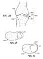
- FIG. 3Ais an example of a cross-section of a vertebra demonstrating one normal and one degenerated facet joint.
- FIG. 3Bis an expanded view of the degenerated facet joint shown in FIG. 3A .
- FIG. 4is an example of a surgical instrument for removal of bone overgrowth and spurs.
- FIGS. 5A-Care examples of various embodiments of a surgical instrument for shaping and smoothing the articular surface.
- FIGS. 6A-Dare examples of various embodiments of an instrument for shaping a facet or other joint and for inserting an implant.
- the instrumentmay have a round 601 A or tapered tip 601 B.
- FIG. 7is an example of an instrument with a shaver 700 .
- FIGS. 8A-Care examples of various embodiments of a distraction device for preparing a joint for implant insertion.
- FIGS. 9A-Dshow various embodiments for distracting the joint and facilitating implant insertion.
- FIGS. 10A-Fshow various embodiments describing various types of implant margin, including tapered designs 1001 and round designs 1002 .
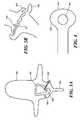
- FIG. 11shows a facet device for implantation into a facet joint, in accordance with one embodiment of the invention.
- FIG. 12shows a facet device having a dumbbell shape, in accordance with one embodiment of the invention.
- FIG. 13shows a facet device having a dumbbell shape, in accordance with one embodiment of the invention.
- FIG. 14shows a bone spur on a surface associated with a facet joint.
- FIG. 15shows a facet device capable of changing position or orientation of one or two facet joints, optionally by moving bone spurs or bony excrescences, thereby reducing spinal stenosis, in accordance with one embodiment of the invention.
- inventions of the present inventioncan employ, unless otherwise indicated, conventional and digital methods of x-ray imaging and processing, x-ray tomosynthesis, ultrasound including A-scan, B-scan and C-scan, computed tomography (CT scan), magnetic resonance imaging (MRI), optical coherence tomography, single photon emission tomography (SPECT) and positron emission tomography (PET) within the skill of the art.
- CT scancomputed tomography
- MRImagnetic resonance imaging
- SPECTsingle photon emission tomography
- PETpositron emission tomography
- the present inventionprovides methods and compositions for repairing joints, particularly for repairing articular cartilage and subchondral bone and for facilitating the integration of a wide variety of cartilage and subchondral bone repair materials into a subject.
- the techniques described hereinallow for the customization of cartilage or subchondral bone repair material to suit a particular subject, for example in terms of size, cartilage thickness and/or curvature including subchondral bone curvature.
- shapee.g., size, thickness and/or curvature
- the success of repairis enhanced.
- the repair materialcan be shaped prior to implantation and such shaping can be based, for example, on electronic images that provide information regarding curvature or thickness of any “normal” cartilage surrounding the defect and/or on curvature of the bone underlying the defect.
- embodiments of the inventionprovide, among other things, for minimally invasive methods for partial or complete joint replacement with attached and interpositional designs. The methods require only minimal or, in some instances, no loss in bone stock. Additionally, unlike with current techniques, the methods described herein will help to restore the integrity of the articular surface by achieving an exact or near anatomic match between the implant and the surrounding or adjacent cartilage and/or subchondral bone.
- Advantages of embodiments of the present inventioncan include, but are not limited to, (i) optional customization of joint repair, thereby enhancing the efficacy and comfort level for the patient following the repair procedure; (ii) optional eliminating the need for a surgeon to measure the defect to be repaired intraoperatively in some embodiments; (iii) optional eliminating the need for a surgeon to shape the material during the implantation procedure; (iv) providing methods of evaluating curvature of the repair material based on bone or tissue images or based on intraoperative probing techniques; (v) providing methods of repairing joints with only minimal or, in some instances, no loss in bone stock; (vi) improving postoperative joint congruity; (vii) improving the postoperative patient recovery in some embodiments and (viii) improving postoperative function, such as range of motion.
- the methods described hereinallow for the design and use of joint repair material that more precisely fits the defect (e.g. site of implantation) or the articular surface(s) and, accordingly, provides improved repair of the joint.
- the methods and compositions described hereincan be used to treat defects resulting from disease of the cartilage (e.g., osteoarthritis), bone damage, cartilage damage, trauma, and/or degeneration due to overuse or age.
- Embodiments of the inventionallow, among other things, a health practitioner to evaluate and treat such defects.
- the size, volume and shape of the area of interestcan include only the region of cartilage that has the defect, but preferably will also include contiguous parts of the cartilage surrounding the cartilage defect.
- size, curvature and/or thickness measurementscan be obtained using any suitable technique.
- one-dimensional, two-dimensional, and/or three-dimensional measurementscan be obtained using suitable mechanical means, laser devices, electromagnetic or optical tracking systems, molds, materials applied to the articular surface that harden and “memorize the surface contour,” and/or one or more imaging techniques known in the art. Measurements can be obtained non-invasively and/or intraoperatively (e.g., using a probe or other surgical device).
- the thickness of the repair devicecan vary at any given point depending upon patient's anatomy and/or the depth of the damage to the cartilage and/or bone to be corrected at any particular location on an articular surface.
- FIG. 1Ais a flow chart showing steps taken by a practitioner in assessing a joint.
- a practitionerobtains a measurement of a target joint 10 .
- the step of obtaining a measurementcan be accomplished by taking an image of the joint. This step can be repeated, as necessary, 11 to obtain a plurality of images in order to further refine the joint assessment process.
- the informationis used to generate a model representation of the target joint being assessed 30 .
- This model representationcan be in the form of a topographical map or image.
- the model representation of the jointcan be in one, two, or three dimensions. It can include a physical model. More than one model can be created 31 , if desired.
- Either the original model, or a subsequently created model, or bothcan be used.
- the practitionercan optionally generate a projected model representation of the target joint in a corrected condition 40 , e.g., from the existing cartilage on the joint surface, by providing a mirror of the opposing joint surface, or a combination thereof. Again, this step can be repeated 41 , as necessary or desired.
- the practitionercan then select a joint implant 50 that is suitable to achieve the corrected joint anatomy. As will be appreciated by those of skill in the art, the selection process 50 can be repeated 51 as often as desired to achieve the desired result.
- a practitionercan obtain a measurement of a target joint 10 by obtaining, for example, an x-ray, and then select a suitable joint replacement implant 50 .
- the practitionercan proceed directly from the step of generating a model representation of the target joint 30 to the step of selecting a suitable joint replacement implant 50 as shown by the arrow 32 . Additionally, following selection of suitable joint replacement implant 50 , the steps of obtaining measurement of target joint 10 , generating model representation of target joint 30 and generating projected model 40 , can be repeated in series or parallel as shown by the flow 24 , 25 , 26 .
- FIG. 1Bis an alternate flow chart showing steps taken by a practitioner in assessing a joint.
- a practitionerobtains a measurement of a target joint 10 .
- the step of obtaining a measurementcan be accomplished by taking an image of the joint.
- This stepcan be repeated, as necessary, 11 to obtain a plurality of images in order to further refine the joint assessment process.
- the informationis used to generate a model representation of the target joint being assessed 30 .
- This model representationcan be in the form of a topographical map or image.
- the model representation of the jointcan be in one, two, or three dimensions.
- the processcan be repeated 31 as necessary or desired. It can include a physical model.
- the practitionercan optionally generate a projected model representation of the target joint in a corrected condition 40 . This step can be repeated 41 as necessary or desired.
- the practitionercan then design a joint implant 52 that is suitable to achieve the corrected joint anatomy, repeating the design process 53 as often as necessary to achieve the desired implant design.
- the practitionercan also assess whether providing additional features, such as rails, keels, lips, pegs, cruciate stems, or anchors, cross-bars, etc. will enhance the implants' performance in the target joint.
- the practitionercan proceed directly from the step of generating a model representation of the target joint 30 to the step of designing a suitable joint replacement implant 52 as shown by the arrow 38 . Similar to the flow shown above, following the design of a suitable joint replacement implant 52 , the steps of obtaining measurement of target joint 10 , generating model representation of target joint 30 and generating projected model 40 , can be repeated in series or parallel as shown by the flow 42 , 43 , 44 .
- FIG. 1Cis a flow chart illustrating the process of selecting an implant for a patient.
- the size of area of diseased cartilage or cartilage lossis measured 100 . This step can be repeated multiple times 101 , as desired.
- the thickness of adjacent cartilagecan optionally be measured 110 .
- This processcan also be repeated as desired 111 .
- the curvature of the articular surfaceis then measured 120 .
- the subchondral bonecan be measured. As will be appreciated measurements can be taken of the surface of the joint being repaired, or of the mating surface in order to facilitate development of the best design for the implant surface.
- the usereither selects the best fitting implant contained in a library of implants 130 or generates a patient-specific implant 132 . These steps can be repeated as desired or necessary to achieve the best fitting implant for a patient, 131 , 133 .
- the process of selecting or designing an implantcan be tested against the information contained in the MRI or x-ray of the patient to ensure that the surfaces of the device achieves a good fit relative to the patient's joint surface. Testing can be accomplished by, for example, superimposing the implant image over the image for the patient's joint.
- the implant sitecan be prepared 140 , for example by removing cartilage or bone from the joint surface, or the implant can be placed into the joint 150 .
- the joint implant selected or designedachieves anatomic or near anatomic fit with the existing surface of the joint while presenting a mating surface for the opposing joint surface that replicates the natural joint anatomy.
- both the existing surface of the jointcan be assessed as well as the desired resulting surface of the joint. This technique is particularly useful for implants that are not anchored into the bone.
- the physicianor other person practicing embodiments of the invention, can obtain a measurement of a target joint 10 and then either design 52 or select 50 a suitable joint replacement implant.
- a wide variety of materialsfind use in the practice of the present invention, including, but not limited to, plastics, metals, crystal free metals, ceramics, biological materials (e.g., collagen or other extracellular matrix materials), hydroxyapatite, cells (e.g., stem cells, chondrocyte cells or the like), or combinations thereof.
- a repair materialcan be formed or selected.
- a cartilage or bone replacement or regenerating materialhaving a curvature that will fit into a particular cartilage defect or onto a particular bone surface, will follow the contour and shape of the articular surface, and will optionally match the thickness of the surrounding cartilage.
- the repair materialcan include any combination of materials, and typically includes at least one non-pliable material, for example materials that are not easily bent or changed.
- joint repair systemsoften employ metal and/or polymeric materials including, for example, prostheses which are anchored into the underlying bone (e.g., a femur in the case of a knee prosthesis).
- metal and/or polymeric materialsincluding, for example, prostheses which are anchored into the underlying bone (e.g., a femur in the case of a knee prosthesis).
- a femurin the case of a knee prosthesis.
- a wide-variety of metalsare useful in the practice of embodiments of the present invention, and can be selected based on any criteria. For example, material selection can be based on resiliency to impart a desired degree of rigidity.
- Non-limiting examples of suitable metalsinclude silver, gold, platinum, palladium, iridium, copper, tin, lead, antimony, bismuth, zinc, titanium, cobalt, stainless steel, nickel, iron alloys, cobalt alloys, such as Elgiloy®), a cobalt-chromium-nickel alloy, and MP35N, a nickel-cobalt-chromium-molybdenum alloy, and Nitinol®, a nickel-titanium alloy, aluminum, manganese, iron, tantalum, crystal free metals, such as Liquidmetal® alloys (available from LiquidMetal Technologies, www.liquidmetal.com), other metals that can slowly form polyvalent metal ions, for example to inhibit calcification of implanted substrates in contact with a patient's bodily fluids or tissues, and combinations thereof.
- suitable metalsinclude silver, gold, platinum, palladium, iridium, copper, tin, lead, antimony, bismuth, zinc
- Suitable synthetic polymersinclude, without limitation, polyamides (e.g., nylon), polyesters, polystyrenes, polyacrylates, vinyl polymers (e.g., polyethylene, polytetrafluoroethylene, polypropylene and polyvinyl chloride), polycarbonates, polyurethanes, poly dimethyl siloxanes, cellulose acetates, polymethyl methacrylates, polyether ether ketones, ethylene vinyl acetates, polysulfones, nitrocelluloses, similar copolymers and mixtures thereof.
- polyamidese.g., nylon
- polyesterse.g., polystyrenes
- polyacrylatese.g., polyethylene, polytetrafluoroethylene, polypropylene and polyvinyl chloride
- polycarbonatese.g., polycarbonates, polyurethanes, poly dimethyl siloxanes, cellulose acetates, polymethyl methacrylates, polyether ether ketones, ethylene vinyl a
- Bioresorbable synthetic polymerscan also be used such as dextran, hydroxyethyl starch, derivatives of gelatin, polyvinylpyrrolidone, polyvinyl alcohol, poly[N-(2-hydroxypropyl) methacrylamide], poly(hydroxy acids), poly(epsilon-caprolactone), polylactic acid, polyglycolic acid, poly(dimethyl glycolic acid), poly(hydroxy butyrate), and similar copolymers can also be used.
- PEEK®polyetheretherketone
- PEEK 450Gis an unfilled PEEK approved for medical implantation available from Victrex of Lancashire, Great Britain. (Victrex is located at www.matweb.com or see Boedeker www.boedeker.com).
- Other sources of this materialinclude Gharda located in Panoli, India (www.ghardapolymers.com).
- the material selectedcan also be filled.
- other grades of PEEKare also available and contemplated, such as 30% glass-filled or 30% carbon filled, provided such materials are cleared for use in implantable devices by the FDA, or other regulatory body.
- Glass filled PEEKreduces the expansion rate and increases the flexural modulus of PEEK relative to that portion which is unfilled.
- the resulting productis known to be ideal for improved strength, stiffness, or stability.
- Carbon filled PEEKis known to enhance the compressive strength and stiffness of PEEK and lower its expansion rate. Carbon filled PEEK offers wear resistance and load carrying capability.
- thermoplastic or thermoplastic polycondensate materialsthat resist fatigue, have good memory, are flexible, and/or deflectable have very low moisture absorption, and good wear and/or abrasion resistance, can be used without departing from the scope of embodiments of the invention.
- the implantcan also be comprised of polyetherketoneketone (PEKK).
- PEKpolyetherketone
- PEKEKKpolyetherketoneetherketoneketone
- PEEKKpolyetherketoneketone
- the polymerscan be prepared by any of a variety of approaches including conventional polymer processing methods.
- Preferred approachesinclude, for example, injection molding, which is suitable for the production of polymer components with significant structural features, and rapid prototyping approaches, such as reaction injection molding and stereo-lithography.
- the substratecan be textured or made porous by either physical abrasion or chemical alteration to facilitate incorporation of the metal coating. Other processes are also appropriate, such as extrusion, injection, compression molding and/or machining techniques.
- the polymeris chosen for its physical and mechanical properties and is suitable for carrying and spreading the physical load between the joint surfaces.
- More than one metal and/or polymercan be used in combination with each other.
- one or more metal-containing substratescan be coated with polymers in one or more regions or, alternatively, one or more polymer-containing substrate can be coated in one or more regions with one or more metals.
- the system or prosthesiscan be porous or porous coated.
- the porous surface componentscan be made of various materials including metals, ceramics, and polymers. These surface components can, in turn, be secured by various means to a multitude of structural cores formed of various metals.
- Suitable porous coatingsinclude, but are not limited to, metal, ceramic, polymeric (e.g., biologically neutral elastomers such as silicone rubber, polyethylene terephthalate and/or combinations thereof or combinations thereof. See, e.g., U.S. Pat. No. 3,605,123 to Hahn, issued Sep. 20, 1971. U.S. Pat. No. 3,808,606 to Tronzo issued May 7, 1974 and U.S. Pat. No. 3,843,975 to Tronzo issued Oct.
- the coatingcan be applied by surrounding a core with powdered polymer and heating until cured to form a coating with an internal network of interconnected pores.
- the tortuosity of the porese.g., a measure of length to diameter of the paths through the pores
- the porous coatingcan be applied in the form of a powder and the article as a whole subjected to an elevated temperature that bonds the powder to the substrate. Selection of suitable polymers and/or powder coatings can be determined in view of the teachings and references cited herein, for example based on the melt index of each.
- Repair materialscan also include one or more biological material either alone or in combination with non-biological materials.
- any base materialcan be designed or shaped and suitable cartilage replacement or regenerating material(s) such as fetal cartilage cells can be applied to be the base.
- the cellscan be then be grown in conjunction with the base until the thickness (and/or curvature) of the cartilage surrounding the cartilage defect has been reached.
- Conditions for growing cellse.g., chondrocytes
- various substrates in culture, ex vivo and in vivoare described, for example, in U.S. Pat. No. 5,478,739 to Slivka et al. issued Dec. 26, 1995; U.S. Pat. No. 5,842,477 to Naughton et al. issued Dec.
- Non-limiting examples of suitable substratesinclude plastic, tissue scaffold, a bone replacement material (e.g., a hydroxyapatite, a bioresorbable material), or any other material suitable for growing a cartilage replacement or regenerating material on it.
- a bone replacement materiale.g., a hydroxyapatite, a bioresorbable material
- Bio polymerscan be naturally occurring or produced in vitro by fermentation and the like. Suitable biological polymers include, without limitation, collagen, elastin, silk, keratin, gelatin, polyamino acids, cat gut sutures, polysaccharides (e.g., cellulose and starch) and mixtures thereof. Biological polymers can be bioresorbable.
- Biological materials used in the methods described hereincan be autografts (from the same subject); allografts (from another individual of the same species) and/or xenografts (from another species). See, also, International Patent Publications WO 02/22014 to Alexander et al. published Mar. 21, 2002 and WO 97/27885 to Lee published Aug. 7, 1997.
- autologous materialsare preferred, as they can carry a reduced risk of immunological complications to the host, including re-absorption of the materials, inflammation and/or scarring of the tissues surrounding the implant site.
- a probeis used to harvest tissue from a donor site and to prepare a recipient site.
- the donor sitecan be located in a xenograft, an allograft or an autograft.
- the probeis used to achieve a good anatomic match between the donor tissue sample and the recipient site.
- the probeis specifically designed to achieve a seamless or near seamless match between the donor tissue sample and the recipient site.
- the probecan, for example, be cylindrical.
- the distal end of the probeis typically sharp in order to facilitate tissue penetration. Additionally, the distal end of the probe is typically hollow in order to accept the tissue.
- the probecan have an edge at a defined distance from its distal end, e.g., at 1 cm distance from the distal end and the edge can be used to achieve a defined depth of tissue penetration for harvesting.
- the edgecan be external or can be inside the hollow portion of the probe.
- an orthopedic surgeoncan take the probe and advance it with physical pressure into the cartilage, the subchondral bone and the underlying marrow in the case of a joint such as a knee joint. The surgeon can advance the probe until the external or internal edge reaches the cartilage surface. At that point, the edge will prevent further tissue penetration thereby achieving a constant and reproducible tissue penetration.
- the distal end of the probecan include one or more blades, saw-like structures, or tissue cutting mechanism.
- the distal end of the probecan include an iris-like mechanism consisting of several small blades.
- the blade or bladescan be moved using a manual, motorized or electrical mechanism thereby cutting through the tissue and separating the tissue sample from the underlying tissue. Typically, this will be repeated in the donor and the recipient.
- an iris-shaped blade mechanismthe individual blades can be moved so as to close the iris thereby separating the tissue sample from the donor site.
- a laser device or a radiofrequency devicecan be integrated inside the distal end of the probe.
- the laser device or the radiofrequency devicecan be used to cut through the tissue and to separate the tissue sample from the underlying tissue.
- the same probecan be used in the donor and in the recipient.
- similarly shaped probes of slightly different physical dimensionscan be used.
- the probe used in the recipientcan be slightly smaller than that used in the donor thereby achieving a tight fit between the tissue sample or tissue transplant and the recipient site.
- the probe used in the recipientcan also be slightly shorter than that used in the donor thereby correcting for any tissue lost during the separation or cutting of the tissue sample from the underlying tissue in the donor material.
- Any biological repair materialcan be sterilized to inactivate biological contaminants such as bacteria, viruses, yeasts, molds, mycoplasmas and parasites. Sterilization can be performed using any suitable technique, for example radiation, such as gamma radiation.
- Any of the biological materials described hereincan be harvested with use of a robotic device.
- the robotic devicecan use information from an electronic image for tissue harvesting.
- the cartilage replacement materialhas a particular biochemical composition.
- the biochemical composition of the cartilage surrounding a defectcan be assessed by taking tissue samples and chemical analysis or by imaging techniques.
- WO 02/22014 to Alexanderdescribes the use of gadolinium for imaging of articular cartilage to monitor glycosaminoglycan content within the cartilage.
- the cartilage replacement or regenerating materialcan then be made or cultured in a manner, to achieve a biochemical composition similar to that of the cartilage surrounding the implantation site.
- the culture conditions used to achieve the desired biochemical compositionscan include, for example, varying concentrations.
- Biochemical composition of the cartilage replacement or regenerating materialcan, for example, be influenced by controlling concentrations and exposure times of certain nutrients and growth factors.
- a physical model of the surfaces of the articular cartilage and of the underlying bonecan be created.
- This physical modelcan be representative of a limited area within the joint or it can encompass the entire joint.
- This modelcan also take into consideration the presence or absence of a meniscus as well as the presence or absence of some or all of the cartilage.
- the physical modelcan encompass only the medial or lateral femoral condyle, both femoral condyles and the notch region, the medial tibial plateau, the lateral tibial plateau, the entire tibial plateau, the medial patella, the lateral patella, the entire patella or the entire joint.
- the location of a diseased area of cartilagecan be determined, for example using a 3D coordinate system or a 3D Euclidian distance as described in WO 02/22014.
- the size of the defect to be repairedcan be determined. This process takes into account that, for example, roughly 80% of patients have a healthy lateral component. As will be apparent, some, but not all, defects will include less than the entire cartilage. Thus, in one embodiment of the invention, the thickness of the normal or only mildly diseased cartilage surrounding one or more cartilage defects is measured. This thickness measurement can be obtained at a single point or, preferably, at multiple points, for example 2 point, 4-6 points, 7-10 points, more than 10 points or over the length of the entire remaining cartilage. Furthermore, once the size of the defect is determined, an appropriate therapy (e.g., articular repair system) can be selected such that as much as possible of the healthy, surrounding tissue is preserved.
- an appropriate therapye.g., articular repair system
- the curvature of the articular surface or subchondral bonecan be measured to design and/or shape the repair material. Further, both the thickness of the remaining cartilage and the curvature of the articular surface including bone can be measured to design and/or shape the repair material.
- the curvature of the subchondral bonecan be measured and the resultant measurement(s) can be used to either select or shape a cartilage replacement material.
- the contour of the subchondral bonecan be used to re-create a virtual cartilage surface: the margins of an area of diseased cartilage can be identified. The subchondral bone shape in the diseased areas can be measured.
- a virtual contourcan then be created by copying the subchondral bone surface into the cartilage surface, whereby the copy of the subchondral bone surface connects the margins of the area of diseased cartilage.
- the contourscan be configured to mate with existing cartilage or to account for the removal of some or all of the cartilage.
- FIG. 2Ashows a slightly perspective top view of a joint implant 200 of one embodiment of the invention suitable for implantation in a joint such as a facet joint, an uncovertebral joint of a costovertebral joint.
- the implantcan be generated using, for example, a dual surface assessment, as described above with respect to FIGS. 1A and B.
- the implant 200has an upper or frontal surface 202 , a lower or posterior surface 204 and, optionally, a peripheral edge 206 .
- the upper or frontal surface 202is formed so that it forms a mating surface for receiving the opposing joint surface; in this instance partially concave to receive a femur, although other joints such as a facet joint, an uncovertebral joint or a costovertebral joint are possible.
- the concave surfacecan be variably concave such that it presents a surface to the opposing joint surface, e.g. a negative surface of the mating surface of the femur it communicates with. As will be appreciated by those of skill in the art, the negative impression need not be a perfect one.
- the upper or frontal surface 202 of the implant 200can be shaped by any of a variety of means.
- the upper or frontal surface 202can be shaped by projecting the surface from the existing cartilage and/or bone surfaces on the articular surface such as a tibial plateau or the surface of a facet joint, or it can be shaped to mirror the femoral condyle in order to optimize the complimentary surface of the implant when it engages the femoral condyle.
- the superior surface 202can be configured to mate with an inferior surface of an implant configured for the opposing femoral condyle.
- the lower or posterior surface 204has optionally a convex surface that matches, or nearly matches, the surface of the joint, e.g. a tibial plateau or a facet or uncovertebral or costovertebral joint, such that it creates an anatomic or near anatomic fit with the tibial plateau or other relevant or applicable articular surface.
- the lower or posterior surfacecan be partially convex as well.
- the lower or posterior surface 204presents a surface to the tibial plateau or applicable articular surface that fits within the existing surface. It can be formed to match the existing surface or to match the surface after articular resurfacing.
- the convex surface of the lower or posterior surface 204need not be perfectly convex. Rather, the lower or posterior surface 204 is more likely consist of convex and concave portions that fit within the existing surface of the tibial plateau or the re-surfaced plateau or re-surfaced applicable articular surface. Thus, the surface is essentially variably convex and concave.
- FIG. 2Bshows a top view of the joint implant of FIG. 2A .
- the exterior shape 208 of the implantcan be elongated.
- the elongated formcan take a variety of shapes including elliptical, quasi-elliptical, race-track, etc.
- the exterior dimensioncan be irregular thus not forming a true geometric shape, e.g., ellipse.
- the actual exterior shape of an implantcan vary depending on the nature of the joint defect to be corrected.
- the ratio of the length L to the width Wcan vary from, for example, between about 0.25 to about 2.0, and more specifically from about 0.5 to about 1.5.
- the length across an axis of the implant 200varies when taken at points along the width of the implant. For example, as shown in FIG. 2B , L.sub.1.noteq.L.sub.2.noteq.L.sub.3.
- FIGS. 2C-Ecross-sections of the implant shown in FIG. 2B are depicted along the lines of 2 C- 2 C, 2 D- 2 D, and 2 E- 2 E.
- the implanthas a thickness t 1 , t 2 and t 3 respectively.
- the thickness of the implantvaries along both its length L and width W.
- the actual thickness at a particular location of the implant 200is a function of the thickness of the cartilage and/or bone to be replaced and the joint mating surface to be replicated.
- the profile of the implant 200 at any location along its length L or width Wis a function of the cartilage and/or bone to be replaced.
- FIG. 2Fis a lateral view of the implant 200 of FIG. 2A .
- the height of the implant 200 at a first end h.sub.1is different than the height of the implant at a second end h.sub.2.
- the upper edge 208can have an overall slope in a downward direction.
- the actual slope of the upper edge 208varies along its length and can, in some instances, be a positive slope.
- the lower edge 210can have an overall slope in a downward direction.
- the actual slope of the lower edge 210varies along its length and can, in some instances, be a positive slope.
- an implantcan be created wherein h.sub.1 and h.sub.2 are equivalent, or substantially equivalent without departing from the scope of the invention.
- FIG. 2Gis a cross-section taken along a sagittal plane in a body showing the implant 200 implanted within a knee joint 1020 such that the lower surface 204 of the implant 200 lies on the tibial plateau 1022 and the femur 1024 rests on the upper surface 202 of the implant 200 .
- FIG. 2His a cross-section taken along a coronal plane in a body showing the implant 200 implanted within a knee joint 1020 .
- the implant 200is positioned so that it fits within a superior articular surface 224 .
- the articular surfacecould be the medial or lateral facet, as needed.
- FIG. 2Iis a view along an axial plane of the body showing the implant 200 implanted within a knee joint 1020 showing the view taken from an aerial, or upper, view.
- FIG. 2Jis a view of an alternate embodiment where the implant is a bit larger such that it extends closer to the bone medially, i.e., towards the edge 1023 of the tibial plateau, as well as extending anteriorly and posteriorly.
- FIG. 2Kis a cross-section of an implant 200 of one embodiment of the invention, e.g., for a facet joint, an uncovertebral or a costovertebral joint, according to an alternate embodiment.
- the lower surface 204further includes a joint anchor 212 .
- the joint anchor 212forms a protrusion, keel or vertical member that extends from the lower surface 204 of the implant 200 and projects into, for example, the bone of the joint.
- the keelcan be perpendicular or lie within a plane of the body.
- the joint anchor 212can have a cross-member 214 so that from a bottom perspective, the joint anchor 212 has the appearance of a cross or an “x.”
- the joint anchor 212could take on a variety of other forms while still accomplishing the same objective of providing increased stability of the implant 200 in the joint. These forms include, but are not limited to, pins, bulbs, balls, teeth, etc. Additionally, one or more joint anchors 212 can be provided as desired.
- FIGS. 2M and Nillustrate cross-sections of alternate embodiments of a dual component implant from a side view and a front view.
- the cross-membercan be formed integral to the surface of the implant or can be one or more separate pieces that fit within a groove 222 on the lower surface 204 of the implant 200 .
- the groovecan form a single channel as shown in FIG. 2 N 1 , or can have more than one channel as shown in FIG. 2 O 1 . In either event, the cross-bar then fits within the channel as shown in FIGS. 2 N 2 -O 2 .
- the cross-bar members 220can form a solid or hollow tube or pipe structure as shown in FIG. 2P . Where two, or more, tubes 220 communicate to provide translation, a groove 221 can be provided along the surface of one or both cross-members to interlock the tubes into a cross-bar member further stabilizing the motion of the cross-bar relative to the implant 200 . As will be appreciated by those of skill in the art, the cross-bar member 220 can be formed integrally with the implant without departing from the scope of the invention.
- the surface of the tibial plateauwill be prepared by forming channels thereon to receive the cross-bar members.
- the surface of the tibial plateauwill be prepared by forming channels thereon to receive the cross-bar members.
- FIG. 2S ( 1 - 9 )illustrate an alternate embodiment of implant 200 . As illustrated in FIG. 2S the edges are beveled to relax a sharp corner.
- FIG. 2S ( 1 )illustrates an implant having a single fillet or bevel 230 . As shown in FIG. 2S ( 2 ) two fillets 230 , 231 are provided and used for the posterior chamfer. In FIG. 2S ( 3 ) a third fillet 234 is provided to create two cut surfaces for the posterior chamfer.
- the chamfercan assist with insertion of the implant: as the implant is advanced into the joint, the chamfer will assist with distracting the joint until the implant is successfully seated in situ.
- FIG. 2S ( 4 )a tangent of the implant is deselected, leaving three posterior curves.
- FIG. 2S ( 5 )shows the result of tangent propagation.
- FIG. 2S ( 6 )illustrates the effect on the design when the bottom curve is selected without tangent propagation.
- the result of tangent propagation and selectionis shown in FIG. 2S ( 7 ).
- the resulting cornerhas a softer edge but sacrifices less than 0.5 mm of joint space.
- additional cutting planescan be added without departing from the scope of the invention.
- FIGS. 2T ( 1 - 8 )illustrate an alternate embodiment of an implant 200 wherein the surface of the tibial plateau 250 is altered to accommodate the implant.
- the tibial plateaucan be altered for only half of the joint surface 251 or for the full surface 252 .
- the posterior-anterior surfacecan be flat 260 or graded 262 . Grading can be either positive or negative relative to the anterior surface. Grading can also be used with respect to the implants of FIG. 2T where the grading either lies within a plane or a body or is angled relative to a plane of the body. Additionally, attachment mechanisms can be provided to anchor the implant to the altered surface. As shown in FIG.
- FIG. 2T ( 5 - 7 ) keels 264can be provided.
- the keels 264can either sit within a plane, e.g. sagittal or coronal plane, or not sit within a plane (as shown in FIG. 2T ( 7 )).
- FIG. 2T ( 8 )illustrates an implant which covers the entire tibial plateau.
- the upper surface of these implantsare designed to conform to the projected shape of the joint as determined under the steps described with respect to FIG. 1 , while the lower surface is designed to be flat, or substantially flat to correspond to the modified surface of the joint.
- Embodiments of the current inventionprovide for multiple devices including implants for treating facet joints, uncovertebral joints and costovertebral joints and methods enabling or facilitating this treatment.
- An implantcan be any device or repair system for treating a facet joint, uncovertebral joint and costovertebral or any other joint.
- a distraction devicecan be used to facilitate insertion of an implant into a facet joint.
- a distraction devicecan be particularly useful for placement of a balloon or an interpositional implant into the facet joint.
- the distraction devicecan include two or more prongs 800 .
- One or more prongscan be straight 801 ( FIG. 8A ) or curved 802 ( FIG. 8B ) in one or more dimensions ( FIG. 8C ). It can be concave 803 A with a mating concave surface 803 B.
- the curvaturecan be adapted for a facet joint. It can be tapered in the front 804 . It can also be round at the tip 805 . Preferably, the curvature will be similar to or substantially match that of a facet joint.
- One or more prongscan be straight or curved, or partially straight and partially curved. Concave and convex shapes are possible can be present at the same time. Irregular shapes can be used.
- the distraction devicecan include two plates at the distal tip.
- the platescan be substantially solid 900 .
- the platescan also be open on one or more sides 901 ( FIG. 9B ).
- the distraction devicecan have an opening 902 that allows for insertion or placement of an implant 903 after distraction of the joint ( FIG. 9C ).
- Various shapes of the distraction device 904are possible ( FIG. 9D ).
- the distance between the platescan be substantially zero at the outset. This facilitates insertion into the joint. Once inserted, the distance between the plates can be increased, for example, using a telescoping or jack or ratchet like mechanism.
- the distracting mechanismcan be located within the joint, preferably between the two plates, or external to the joint, for example near the grip of the device.
- the two platescan be flat or curved, or partially flat and partially curved.
- the curvaturewill be similar to or substantially match that of a facet joint.
- Concave and convex shapesare possible can be present at the same time. Irregular shapes can be used.
- the area of the distraction devicecan be slightly smaller than a facet joint, the same as the facet joint or slightly larger than a facet joint.
- the distraction devicecan be hollow in the center or, alternatively, create a hollow, open space to accept a balloon or an implant.
- the distraction devicecan have an opening in the rear at the side pointing dorsally or externally to allow insertion of the balloon or implant while the distraction device is inside the joint.
- the distal portion of the distraction devicecan be wider, typically between two prongs, than the widest width of the implant in the same dimension, typically supero-inferior, to facilitate removal of the distraction device with the implant remaining in situ.
- Degenerated facet joints, uncovertebral joints or costovertebral jointscan demonstrate new bone formation, bone remodeling, hypertrophy, bony overgrowth and/or bone spurs. Facet joints, uncovertebral joints or costovertebral joints can enlarge due to new bone formation, bone remodeling, hypertrophy, bony overgrowth and/or bone spurs formation and spur formation.
- FIGS. 3A and Bdemonstrate a vertebral body 300 , a thecal sac 301 , which is deformed on the right side by a bone spur 308 , which arises from a facet joint 303 .
- the facet joint on the right side 303is degenerated, while the facet joint on the left side 302 is relatively normal in shape still.
- a spinous processis seen posteriorly 304 .
- the degenerated facet joint 303demonstrates multiple peripheral bone spurs 306 which can lead to an enlargement of the joint.
- Bone growthcan cause difficulties during insertion of an implant or balloon. Moreover, bone growth can cause spinal stenosis, including foraminal stenosis, lateral recess stenosis and central stenosis. Thus, while an implant or balloon device designed to alleviate pain originating from the affected joint, i.e., a facet joint, uncovertebral joint or costovertebral joint, the patient may still suffer from back pain and even sciatica after the procedure. The surgeon can optionally consider reshaping the joint and/or removing one or more bone growths.
- an instrument(see 400 in FIG. 4 ) is used for the reshaping of the joint or the removal of one or more bone growths.
- the instrumentcan, for example, have a ring shape at the tip 401 .
- the external aspect of the ringcan be blunt 401 in order to minimize potential damage to the thecal sac or the nerve roots.
- the internal portion 402 of the ringcan be sharp. All or part of the external portion can be blunt. All or part of the internal portion can be sharp.
- the opening of the ringcan then be placed over the bone growth and the instrument can be pulled back, thereby removing all or part of the bone growth.
- the instrumentcan include a rough surface creating a rasp-like instrument.
- FIGS. 5A-Cvarious embodiments of an instrument 500 with a rough, rasp-like surface 501 are seen.
- the portion of the implant that inserted into the joint 502can have different shapes, e.g., convex or concave in one or more planes ( FIGS. 5A-5C ).
- the instrumentcan have an optional handle 503 .
- the rough surface 501can be moved over one or two of the articular surfaces of the joint to remove any surface irregularities and to create a new, smooth bearing surface on at least one or, optionally both sides of the joint.
- the underlying curvature 504 of the rough surfacewill determine the shape of the articular surface after smoothing it.
- any mechanical device or electrical mechanism capable of removing bonecan be utilized in combination with one or more of the embodiments above and below.
- an instrument with a rotating mill or an oscillating saw or a shaver 700can be utilized.
- This instrumentcan be curved at the tip 701 , thereby protecting the thecal sac 702 .
- the instrumentcan be in a protective cover 703 .
- the instrumentis typically inserted via a hole in the ligamentum flavum, although it can also be inserted through the joint or both.
- the distal portion of the instrumentcan be tapered, preferably with a rounded tip.
- the tapered designcan facilitate insertion into the joint if the instrument has to be passed through the joint.
- the rounded or blunt tipcan help avoid injury to a nerve root or the thecal sac.
- the instrumentcan be curved in one or more dimensions.
- One or more convex portionscan be included.
- One or more concave portionscan be included. Convex and concave portions can be present in the same device.
- an instrument 600is seen that has a tapered front portion 601 .
- the tapered front portioncan be rounded 601 A ( FIG. 6A ), or triangular 601 B ( FIG. 6B ). While the front of the instrument can be tapered, its side portion can optionally have a sharp recess 602 ( FIGS. 6C and D). The sharp recess can assist in removing some bone overgrowth on or adjacent to the articular surface.
- the instrumentcan also be curved 603 near or at its tip 601 ( FIG. 6D ).
- the instrument shapemirrors the shape of the articular surface.
- the instrumentcan be available in various sizes, thicknesses, lengths and shapes.
- the instrumentcan have a tip that can be bent backward in an angle equal to or greater than 90 degrees.
- the implantis passed past the bone growth.
- the tipis then brought in contact with the bone growth and the bone growth is removed.
- the instrumentcan include one or more tubes for suction.
- suctioncan be performed, also using standard suction devices.
- the instrument to remove or reduce bone growthcan be used in conjunction with the distraction device.
- bothcan be integrated.
- Degenerated facet joints, uncovertebral joints or costovertebral jointscan demonstrate new bone formation, bone remodeling, hypertrophy, bony overgrowth and/or bone spurs. Facet joints, uncovertebral joints or costovertebral joints can enlarge due to new bone formation, bone remodeling, hypertrophy, bony overgrowth and/or bone spurs formation and spur formation.
- an implant or a repair device including an injectable material sized only to the articular surface, i.e., portion of the joint that is covered by cartilage prior to the onset of the degenerative and arthritic changes,would not cover all of the load bearing surface.
- an implant or a repair device including a balloon or an injectable materialcan be oversized to account for the enlargement of the joint and the greater dimension of the load bearing surface in patients with degenerative or arthritic changes of the facet joints, uncovertebral joints or costovertebral joints.
- the dimensions of the implant or a repair device including a balloon or an injectable materialcan be increased in one or more dimensions.
- the shape of the implantcan be adjusted to account for the bone growth and for irregularities in joint shape as a result of the bone growth.
- the implant sizecan be selected or adjusted to account for a reduction in size of the joint after removal of a bone growth or to account for a reduction in size of the joint after partial resection of the joint or the articular process.
- These adjustmentscan be made intraoperatively, for example using measuring or sizing devices (see below). Alternatively, these adjustments can be made using imaging software. For example, using CT or MRI data the severity of a spinal stenosis can be estimated. In a second step, resection of a bone growth or partial resection of a joint or articular process can be simulated and it can be determined what the optimal implant size or shape is following these adjustments.
- the implantcan be patient specific with each implant custom manufactured, for example using CAD/CAM and rapid prototyping and/or casting techniques.
- the implantcan be selected from a pre-existing library or assortment of implants.
- the library of implantswill typically cover a range of sizes and shapes applicable to most patients and also allowing for oversizing consistent with the embodiment above.
- repair systems of various sizes, curvatures and thicknessescan be obtained. These repair systems can be catalogued and stored to create a library of systems from which an appropriate system for an individual patient can then be selected. In other words, a defect, or an articular surface, is assessed in a particular subject and a pre-existing repair system having a suitable shape and size is selected from the library for further manipulation (e.g., shaping) and implantation.
- imaging techniquescan be used for delivering a device.
- the imaging techniquescan include the use of x-rays, CT scans, fluoroscopy, C-arms, CT fluoroscopy, C-arms with CT like cross-sectional reconstruction and MRI.
- surgical navigation systemsfor example using radiofrequency or optical object location and reference means, can be used.
- a sizing toolis used to determine the optimal shape of the implant.
- the sizing toolcan be applied at a first procedure, for example using percutaneous need guidance.
- the sizing toolis used at the time of the procedure for insertion of the therapeutic device into the facet joint, uncovertebral joint or costovertebral joint.
- the sizing toolcan include various tools for measuring implant dimensions.
- a ruler or a calipercan be part of the sizing tool.
- the sizing toolcan be used to measure and estimate preferred dimensions of the device, e.g., in superoinferior or mediolateral dimension. It can be used to estimate implant thickness, in one or more locations.
- the sizing toolcan also be used to measure implant curvature.
- the sizing toolcan be partially or completely deformable.
- the sizing toolis inserted into the joint, thereby taking the natural shape of the joint.
- the sizing toolis then withdrawn from the joint.
- the resultant shape of the sizing toolis then compared against a library or assortment of premanufactured implants and the best fitting implant with the best match relative to the sizing tool is selected.
- the sizing toolcan include a gauge to measure implant dimensions in antero-posterior or supero-inferior or medio-lateral direction or combinations thereof.
- This gaugecan, for example, be a ruler integrated into the sizing tool. The sizing tool is inserted into the joint. The area where the dorsal portion of the articular surface aligns with the first visible tick mark on the ruler indicates typically the preferred implant length.
- the sizing toolcan also include a gauge in superoinferior or any other dimension.
- the sizing toolcan include one or more dimensions of one or more of the pre-manufactured implants in the implant library or assortment.
- the sizing toolcan be available in various shapes. For example, a T or t-shape can provide dimensions in two or more directions. The thickness of the sizing tool can be used to estimate the preferred thickness of the implant.
- Sizing toolscan be made with various different curvatures and radii, typically resembling the radii of the implant. By inserting sizing tools of different radii, the optimal radius for the implant can be determined.
- the implant shape and its radiican be determined using an imaging test.
- Trial implantscan substantially match the size and shape of the implants in the pre-manufactured library of implants or assortment of implants.
- the sizing toolcan be malleable and/or deformable.
- the surgeonmay elect to flatten the articular surface, to shape it, to increase its curvature or to roughen the articular surface or to remove the cartilage.
- the shapingcan be advantageous for improving the fit between the implant and the articular surface. Roughening of the articular surface can improve the conformance of the implant to the articular surface and can help reduce the risk of implant dislocation.
- the marks and trackscan be used for stabilizing the implant on one side.
- the implantis then typically made to mate with the marks and tracks on one side of the joint thereby achieving a highly conforming surface, and, effectively, a significant constraint to assist with reducing possible implant motion on this side of the joint.
- the opposing articular surfaceneeds to be minimally constraining in order to enable movement between the implant surface and the opposing articular surface.
- the opposing surfacecan therefore be treated and shaped to remove any marks and tracks and to re-establish a smooth gliding surface.
- the opposing surfacewill be made to match the smooth surface of the implant on this side.
- An instrument for preparing the articular surfacecan be slightly smaller than a facet, uncovertebral or costovertebral joint, similarly sized or larger in size than the respective joint.
- the instrumentcan be curved or flat.
- the implantcan be curved in more than one dimension.
- the instrumentcan be a rasp or mill-like device. It can be mechanical or electrical in nature.
- the device shape and sizeis substantially matched to one or more articular surface.
- the implantcan fill the space between two opposing articular surfaces partially or completely.
- the implantcan have extenders outside the articular surface for stabilizing implant position and for minimizing the risk of implant dislocation.
- Such an extendercan be intra- or extra-articular in location. If it is extra-articular, it will typically extend outside the joint capsule.
- the extendercan be plate or disc or umbrella shaped, covering at least a portion of the bone outside the articular surface.
- the extendercan be only partially plate or disc or umbrella shaped.
- the plate or disc or umbrella shaped extenderwill typically be oriented at an angle to the intra-articular portion of the implant, whereby said angle is substantially different from 180 degrees, more preferred less than 150 degrees, more preferred less than 120 degrees, more preferred less than 100 degrees. In some embodiments, the angle may be equal or less than 90 degrees.
- the extendercan have a constant or variable radii in one or more dimensions.
- the extendercan be adapted to the patient's anatomy.
- the ballooncan have a second, separate portion or a second balloon can be attached, whereby the resultant cavity that will be filled with the self-hardening material can be located outside the articular surface area and can be even external to the joint capsule.
- a lip or ridge or extendercan be created in this manner that can help stabilize the resultant repair device against the adjacent bone or soft-tissues.
- the device or implantcan be shaped to protect the neural structures.
- the ventral portion of the implantcan be rounded to avoid any damage to the neural structures in the event the implant moves or subluxes or dislocates anteriorly.
- the dorsal and superior and inferior marginscan also be rounded in order to avoid damage to neural structures in the event of a subluxation or dislocation into the epidural space. Moreover, a round margin can help minimize chronic wear due to pressure onto the joint capsule.
- the margin of the implantcan be round along the entire implant perimeter or along a portion of the perimeter.
- the implant sidewallcan be straight or alternatively, it can be slanted with an angle other than 90 degrees.
- the implant thicknesscan vary along the perimeter.
- the thickness of the implantcan be thinner at the margin than in the center, along the entire implant margin or in portions of the implant margin.
- the thickness of the implantcan be thicker at the margin than in the center, along the entire implant margin or in portions of the implant margin.
- the implant shapecan be adapted to facilitate insertion into the joint.
- the portion of the implant 1000 that will face forward, first entering the jointcan be tapered 1001 relative to portions or all of the implant, thereby facilitating insertion.
- the tapered tipcan be pointed 1001 or round 1002 ( FIG. 10B ).
- a round tipis preferably since it can help reduce the risk of damage to adjacent structures.
- the implantcan have a sharp edge 1003 ( FIG. 10C ) or a rounded edge 1004 ( FIG. 10D ). A rounded edge is typically preferred.
- the implantcan have a substantially straight margin 1005 ( FIG. 10E ) or a substantially tapered margin 1006 ( FIG. 10F ).
- the arthroplastycan have two or more components, one essentially mating with the tibial surface and the other substantially articulating with the femoral component.
- the two componentscan have a flat opposing surface.
- the opposing surfacecan be curved.
- the curvaturecan be a reflection of the tibial shape, the femoral shape including during joint motion, and the meniscal shape and combinations thereof.
- single-component systemsinclude, but are not limited to, a plastic, a polymer, a metal, a metal alloy, crystal free metals, a biologic material or combinations thereof.
- the surface of the repair system facing the underlying bonecan be smooth.
- the surface of the repair system facing the underlying bonecan be porous or porous-coated.
- the surface of the repair system facing the underlying boneis designed with one or more grooves, for example to facilitate the in-growth of the surrounding tissue.
- the external surface of the devicecan have a step-like design, which can be advantageous for altering biomechanical stresses.
- flangescan also be added at one or more positions on the device (e.g., to prevent the repair system from rotating, to control toggle and/or prevent settling into the marrow cavity).
- the flangescan be part of a conical or a cylindrical design.
- a portion or all of the repair system facing the underlying bonecan also be flat which can help to control depth of the implant and to prevent toggle.
- Non-limiting examples of multiple-component systemsinclude combinations of metal, plastic, metal alloys, crystal free metals, and one or more biological materials.
- One or more components of the articular surface repair systemcan be composed of a biologic material (e.g., a tissue scaffold with cells such as cartilage cells or stem cells alone or seeded within a substrate such as a bioresorable material or a tissue scaffold, allograft, autograft or combinations thereof) and/or a non-biological material (e.g., polyethylene or a chromium alloy such as chromium cobalt).
- a biologic materiale.g., a tissue scaffold with cells such as cartilage cells or stem cells alone or seeded within a substrate such as a bioresorable material or a tissue scaffold, allograft, autograft or combinations thereof
- a non-biological materiale.g., polyethylene or a chromium alloy such as chromium cobalt
- the repair systemcan include one or more areas of a single material or a combination of materials, for example, the articular surface repair system can have a first and a second component.
- the first componentis typically designed to have size, thickness and curvature similar to that of the cartilage tissue lost while the second component is typically designed to have a curvature similar to the subchondral bone.
- the first componentcan have biomechanical properties similar to articular cartilage, including but not limited to similar elasticity and resistance to axial loading or shear forces.
- the first and the second componentcan consist of two different metals or metal alloys.
- One or more components of the systemcan be composed of a biologic material including, but not limited to bone, or a non-biologic material including, but not limited to hydroxyapatite, tantalum, a chromium alloy, chromium cobalt or other metal alloys.
- One or more regions of the articular surface repair systemcan be bioresorbable, for example to allow the interface between the articular surface repair system and the patient's normal cartilage, over time, to be filled in with hyaline or fibrocartilage.
- one or more regionscan be porous.
- the degree of porositycan change throughout the porous region, linearly or non-linearly, for where the degree of porosity will typically decrease towards the center of the articular surface repair system.
- the porescan be designed for in-growth of cartilage cells, cartilage matrix, and connective tissue thereby achieving a smooth interface between the articular surface repair system and the surrounding cartilage.
- the repair system(e.g., the second component in multiple component systems) can be attached to the patient's bone with use of a cement-like material such as methylmethacrylate, injectable hydroxy- or calcium-apatite materials and the like.
- one or more portions of the articular surface repair systemcan be pliable or liquid or deformable at the time of implantation and can harden later. Hardening can occur, for example, within 1 second to 2 hours (or any time period therebetween), preferably with in 1 second to 30 minutes (or any time period therebetween), more preferably between 1 second and 10 minutes (or any time period therebetween).
- FIG. 11shows an implant device 1100 for implantation into a facet joint in accordance with one embodiment of the invention.
- the facet jointincludes opposing articular surfaces 1107 and 1108 .
- device 1100is an interpositional device that is configured to be inserted between opposing articular surfaces 1107 and 1108 .
- one side 1103 of the device 1100forms an articular surface in the present embodiment that is substantially smooth and that is configured to oppose facet joint surface 1107 when implanted.
- Articular surface 1103can include convex and concave shapes and combinations thereof.
- Surface 1103need not be smooth, but, preferably, the surface is configured to provide a suitable bearing surface on which the articular surface 1103 can glide, thereby moving relative to the surface of the opposing articular surface 1107 . Preferably, this reduces friction between the opposing surfaces of the joint and the device and thereby reduces wear both on the device and the joint surface as well as facilitates improved articulation of the joint surface with the device.
- the opposite side 1105 of device 1100also forms an articular surface.
- the surface 1105substantially conforms to the corresponding joint surface 1108 . This allows surface 1105 to resist motion relative to the opposing surface 1108 .
- the surface 1105incorporates the general shape of the joint as a negative such that the surface 1105 and joint surface 1108 couple together.
- the surface 1105may also optionally account for irregularities caused by the arthritis.
- Such an irregularitymay include, without limitation, ridges, crescents, spurs, osteophytes, cysts and the like.
- the surfacemay not be specific to the joint, but may instead include predefined patterns of ridges, points, cones, and/or other shapes or contours, either alone or in combination, to provide a surface that resists motion.
- patient specific featurescan be included with standardized feature.
- the articular surface of the devicecan generally match the opposing articular surface of the joint to include general contours of the joint without providing a specific match or mating to the articular surface, and the standardized contours can be included on the surface such that the surface has a close but not perfect fit sufficient to engage the standardized resistive surface against the opposing articular surface.
- the devicecan have a portion of the articular surface that is conforming and a portion that is not. Either or both portions can also optionally include non-patient specific elements that further assist in providing stability to the device.
- the devicecan include an anchoring device such as a pin or fin that can be engaged with a portion of the facet joint.
- Either side 1103 or 1105can include various combinations of one or more convexities and/or concavities, for example, to provide a conforming surface or generally conforming surface.
- Embodiments of the devicemay have a first shape, and a second side of the implant can, optionally be, at least in part, a mirror image of said first side or said first shape.
- the device 1100may be sized to incorporate marginal and other osteophytes.
- the devicemay also be sized and shaped to exclude such osteophytes. The latter approach is typically preferred when these osteophytes or spurs are removed intraoperatively.
- the devicecan include osteophytes or bone spurs.
- the devicecan be shaped to exclude osteophytes.
- the articular surface 1107is able to move smoothly relative to surface 1103 of the device 1100 .
- the shape of the smooth surface 1100 as well as the opposite surfacemay be obtained with use of an imaging test.
- the imaging testmay be, without limitation, a CT or MRI, a digital tomosynthesis or methods using injection of imaging contrast media, intra-articular or intravenous, ultrasound, or any other imaging modality known in the art.
- the bony or cartilaginous landmarksmay be measured on the scan.
- the imagemay be manipulated to remove overhanging osteophytes or other articular abnormalities virtually.
- a smoothing functionmay be applied to the measured or derived articular surface, for example by obtaining an image, e.g., a CT or MRI images, deriving the articular surface shape, e.g. cartilage or bone, and then applying a smoothing function.
- the best fitting pre-manufactured devicemay be selected from a library of implants. These pre-manufactured implants may already include at least one smooth bearing surface.
- the dimensions of the jointmay be derived from an image, and a virtual model of an implant can be fitted to match that shape by deforming said virtual 3D model.
- the device 1100can be used unilaterally, e.g., in a left or right facet joint, or bilaterally.
- the devicecan be used at a single spinal level or multiple spinal levels.
- the devicecan be used in a cervical, a thoracic and/or a lumbar spine.
- the device 1100may be designed to be substantially a mirror image of the two articular surfaces or to reflect the shape of at least one articular surface.
- the device 1100may include at least one or more convexity, concavity and/or or variation in thickness, diameter and/or general shape. Said at least one or more convexity, concavity and/or variation in thickness, diameter and/or general shape may be designed or selected to change the spatial relationship between the two opposing articular surfaces of the facet joint.
- methods and devices for repairing facet and other jointsallow for the customization of implants to suit a particular subject, for example in terms of size, thickness and/or curvature.
- shapee.g., size, thickness and/or curvature
- the repair materialmay be shaped prior to implantation and such shaping can be based, for example, on electronic images that provide information regarding curvature or thickness of any “normal” cartilage surrounding the defect and/or on curvature of the bone underlying the defect.
- an implantcan include various combinations of both patient-specific and non-patient-specific features.
- implantscan be generated using machining, metal casting, rapid-prototyping, digital printing or other digital manufacturing methods.
- generic implants or blankscan be manufactured to a particular specification, and one or more patient-specific dimensions or other characteristics can be included by, for example, machining the blank to include the desired dimension or other characteristic or to create and entirely patient specific implant.
- a devicecould have a standardized smooth articular surface and a customized opposite surface for stabilizing the implant that is machined into the implant during the manufacturing process.
- a blank implantcan include standard surfaces and be sized and/or shaped about a perimeter of the device to match the dimensions of the facet joints. Other embodiments may be sized to a specific length or thickness.
- a set of patient-specific implants of varying thicknessescan be provided, for example, in a kit or package, to allow a surgeon to select the desired thickness during surgery to provide appropriate spacing and/or to adequately relieve pressure on the interstitial space adjacent the implant (as discussed below).
- the implantsmay be manufactured using a series of two-dimensional (2D) and/or three-dimensional (3D) images.
- a 3D model of the jointcan be created using a set of 2D images built up in slices to provide a 3D image.
- stereoscopic imaging modalitiesmay be utilized.
- Stereoscopic imagingis any technique capable of recording three-dimensional information from two two-dimensional, projectional imaging.
- Traditional stereoscopic imagingincludes creating a 3D visualization or representation starting from a pair of 2D images.
- the projection path of the 2D imagesis offset. The offset is, for example, designed to create an impression of object depth for the eyes of the viewer.
- the offset or minor deviation between the two imagesis similar to the prospectors that both eyes naturally receive in binocular vision.
- Using two or more images with an offset or minor deviation in perspectiveit is possible to generate a point cloud or 3D surface or 3D visualization of a joint or an articular surface, which can then be input into a manufacturing system such as a rapid prototyping or milling machine.
- Dual or more light path, as well as single light path, systemsalso can be employed
- Softwaremay be used to allow automatic, semi-automatic or manual identification of relevant anatomic points to calculate the anatomic and biomechanical parameters, in accordance with various embodiments of the invention.
- thiscan be done in 3D rather than only in 2D.
- the relevant anatomic pointscan be selected manually and the axes can be calculated.
- Identification and preparation of the implant site and insertion of the implantalso can be supported by a surgical navigation system.
- the position or orientation of a surgical instrument with respect to the patient's anatomycan be tracked in real-time in one or more 2D or 3D images. These 2D or 3D images can be calculated from images that were acquired preoperatively, such as MR or CT images.
- Non-image based surgical navigation systemsthat find axes or anatomical structures, for example with use of joint motion, can also be used.
- the position and orientation of the surgical instrument as well as the mold including alignment guides, surgical instrument guides, reaming guides, drill guides, saw guides, etc.can be determined from markers attached to these devices. These markers can be located by a detector using, for example, optical, acoustical or electromagnetic signals.
- the surgeonmay optionally elect intraoperatively to further smoother the articular surface 1107 .
- a rasp or a sharp curette or any other device known in the artmay be used to modify the articular surface.
- implant 1100includes an anchor 1106 that serves as an anchor, stopper, cap and/or lip that engages the outer surface(s) of the facet joint to prevent the implant from sliding or otherwise moving through the joint space or dislodging in the direction opposite anchor 1106 .
- the anchor 1106may be designed to prevent forward subluxation into the epidural space or into neural foramina, and reduces the chance of or prevents potential nerve root or cord injury.
- the anchor 1106will typically be placed at the posterior facing portion of the device.
- the anchor 1106may be intra-articular in location, i.e. contained within the synovial lining of the joint, or may extend external to the joint.
- the anchormay optionally be attached to the joint capsule, e.g., with a suture or clip.
- the anchor 1106may extend beyond a joint margin or an articular surface.
- the anchor 1106may be anchored against a bone spur, and/or attached to a fascia.
- the anchor 1106may be symmetrical or asymmetrical in shape. It can include at least one convex and/or one concave portion.
- the anchor 1106may be modeled to a particular patient and spinal level, for example using data from a CT scan or MRI scan. All or portions of the anchor 1106 can be pre-shaped or pre-manufactured to the patient's specific anatomy.
- a device 1200may have a dumbbell-shaped or drumstick-shaped end 1215 with a thicker portion relative to a middle portion 1216 of device 1200 .
- the thicker portions 1215 and 1315may be located, without limitation, anteriorly or posteriorly, inferiorly or superiorly, medially or laterally.
- thicker portion 1215may be about 1.5 mm, with the thinner portion 1216 being approximately 1 mm.
- one articular facetfor example the inferior facet of the superior vertebral body, may be moved anteriorly or posteriorly.
- the spinal canalcan be decompressed.
- the spinal canalmay be decompressed.
- the change in dimensions of the spinal canalcan be beneficial for treating stenosis involving at least one of a central canal, a lateral recess, a neural foraminal space or an extraforaminal space.
- the device shapemay be chosen to alter the spatial relationship of the different components of the facet joints favorably in order to help move individual portions of the facet joints and to reduce spinal stenosis caused or aggravated by facet joint arthritis.
- This movement of one or more articular surface of the facet jointmay take into account bone spurs or osteophytes and help move them further away from the epidural space or nerve roots.
- Preoperative image simulationcan be used to simulate the effect of device insertion. This simulation can be performed by simulating various types of motion, eg flexion, extension, bending, lateral bending or rotation or by simulating various types of loading including axial loading of the spine or facet joints. In this manner, dimensional changes on the spinal canal invoked by device insertion can be assessed for different positions or different conditions.
- this analysiscan assist in identifying a device shape most suitable for a patient, including a device shape that is least likely to sublux or most beneficial for changing spinal canal or foraminal dimensions.
- implants of various lengths and or thicknessescan be provided to allow a surgeon to select a device that provides the desired positioning of the bones of the facet joints.
- an adjustable devicemay be provided.
- a device 1100includes a standard end 1115 with an anchor 1106
- device 1200includes a dumbbell-shaped end 1215 and an anchor 1206
- two devices 1200 and 1300each include anchors 1206 and 1306 and dumbbell-shaped ends 1215 and 1315 that are thicker relative to middle portions 1216 and 1316 respectively.
- device 1200includes two smooth articular surfaces 1203 and 1217
- device 1300includes one smooth articular surface 1303 and a conforming articular surface 1317 .
- FIG. 14shows a bone spur 1402 on a surface associated with a facet joint.
- FIG. 15shows a device 1500 that changes the alignment and/or position of a facet joint, changes the dimension of the spinal canal and, optionally, pushes the spur away, in accordance with one embodiment of the invention.
- the desired movementmay be determined intraoperatively.
- the desired movementmay be simulated preoperatively in a virtual environment by manipulating 2D or 3D images and objects derived from such images.
- the surgeonmay also elect to resect bone spurs arising from a facet joint.
- the surgeonwill access such bone spurs through the facet joint without opening the anterior capsule of the facet joint.
- Spacersmay also be employed in this and other embodiments. Such spacers may optionally be attached to the device and they can help change the spatial position of different components of the facet joints as well.
- a ratchet or jack-like devicecan be inserted into the facet joint, optionally with a tapered tip. The ratchet or jack-like device is then used for opening the facet joint for insertion of the device.
- the device 1100may be designed so that it can be inserted without breaching the anterior capsule of the facet joint(s) leaving the epidural space untouched.
- This approachhas the benefit of avoiding or reducing intraoperative or epidural bleeding, thereby potentially reducing the incidence of postoperative scar, fibrosis and arachnoiditis, all dreaded complications of spinal surgery.
- surgeondesires to remove any bone spurs, or to smooth one or more articular surfaces, or to resect one or more portions of the joint, she may advantageously approach the joint through a posterior approach, entering through the posterior capsule and performing these steps without breaching the anterior joint capsule.
- Instrumentsmay be designed for this purpose.
- the instrumentsmay have sizes and shapes that substantially match the shape of the facet joints.
- the instrumentsmay optionally be made individually for each patient, for example, by obtaining a preoperative scan and then machining or rapid prototyping the instrument with a shape adapted to the joint(s) of a particular patient.
- the instruments and/or instrument shapecan be selected based on the articular shape determined on the imaging test.
- the devicemay be made of a metal, ceramic, and/or polymer.
- metalsinclude silver, gold, platinum, palladium, iridium, copper, tin, lead, antimony, bismuth, zinc, titanium, cobalt, stainless steel, nickel, iron alloys, cobalt alloys, such as Elgiloy®, a cobalt-chromium-nickel alloy, and MP35N, a nickel-cobalt-chromium-molybdenum alloy, and NitinolTM, a nickel-titanium alloy, aluminum, manganese, iron, tantalum, crystal free metals, such as Liquidmetal® alloys (available from LiquidMetal Technologies, www.liquidmetal.com), other metals that can slowly form polyvalent metal ions, for example to inhibit calcification of implanted substrates in contact with a patient's bodily fluids or tissues, and combinations thereof.
- Liquidmetal® alloysavailable from LiquidMetal Technologies, www.liquidmet
- Nitinolcan be advantageous if the device is inserted through a minimally invasive port including an endoscope. The device can then “unfold” intraoperatively and adapt to the patient's shape.
- Suitable synthetic polymersinclude, without limitation, polyamides (e.g., nylon), polyesters, polystyrenes, polyacrylates, vinyl polymers (e.g., polyethylene, polytetrafluoroethylene, polypropylene and polyvinyl chloride), polycarbonates, polyurethanes, poly dimethyl siloxanes, cellulose acetates, polymethyl methacrylates, polyether ether ketones, ethylene vinyl acetates, polysulfones, nitrocelluloses, similar copolymers and mixtures thereof.
- polyamidese.g., nylon
- polyesterse.g., polystyrenes
- polyacrylatese.g., polyethylene, polytetrafluoroethylene, polypropylene and polyvinyl chloride
- polycarbonatese.g., polycarbonates, polyurethanes, poly dimethyl siloxanes, cellulose acetates, polymethyl methacrylates, polyether ether ketones, ethylene vinyl a
- Bioresorbable synthetic polymerscan also be used such as dextran, hydroxyethyl starch, derivatives of gelatin, polyvinylpyrrolidone, polyvinyl alcohol, poly[N-(2-hydroxypropyl) methacrylamide], poly(hydroxy acids), poly(epsilon-caprolactone), polylactic acid, polyglycolic acid, poly(dimethyl glycolic acid), poly(hydroxy butyrate), and similar copolymers can also be used.
- PEEKTMpolyetheretherketone
- PEEK 450Gis an unfilled PEEK approved for medical implantation available from Victrex of Lancashire, Great Britain.
- Victrexis located at www.matweb.com or see Boedeker www.boedeker.com).
- Other sources of this materialinclude Gharda located in Panoli, India (www.ghardapolymers.com).
- pyrolytic carboncan be used.
- the material selectedcan also be filled.
- other grades of PEEKare also available and contemplated, such as 30% glass-filled or 30% carbon filled, provided such materials are cleared for use in implantable devices by the FDA, or other regulatory body.
- Glass filled PEEKreduces the expansion rate and increases the flexural modulus of PEEK relative to that portion which is unfilled.
- the resulting productis known to be ideal for improved strength, stiffness, or stability.
- Carbon filled PEEKis known to enhance the compressive strength and stiffness of PEEK and lower its expansion rate. Carbon filled PEEK offers wear resistance and load carrying capability.
- the implantcan also be comprised of polyetherketoneketone (PEKK).
- PEKKpolyetherketoneketone
- Other materials that can be usedinclude polyetherketone (PEK), polyetherketoneetherketoneketone (PEKEKK), and polyetheretherketoneketone (PEEKK), and generally a polyaryletheretherketone. Further other polyketones can be used as well as other thermoplastics.
- the polymerscan be prepared by any of a variety of approaches including conventional polymer processing methods.
- Preferred approachesinclude, for example, injection molding, which is suitable for the production of polymer components with significant structural features, and rapid prototyping approaches, such as reaction injection molding and stereo-lithography.
- the substratecan be textured or made porous by either physical abrasion or chemical alteration to facilitate incorporation of the metal coating. Other processes are also appropriate, such as extrusion, injection, compression molding and/or machining techniques.
- the polymeris chosen for its physical and mechanical properties and is suitable for carrying and spreading the physical load between the joint surfaces.
- More than one metal, ceramic and/or polymercan be used in combination.
- one or more metal-containing substratescan be coated with polymers in one or more regions or, alternatively, one or more polymer-containing substrate can be coated in one or more regions with one or more metals.
- cross-linking techniques known in the artcan be applied. Mild, moderate or high cross-linking can be used.
- the system or prosthesiscan be porous or porous coated.
- the porous surface componentscan be made of various materials including metals, ceramics, and polymers. These surface components can, in turn, be secured by various means to a multitude of structural cores formed of various metals.
- Suitable porous coatingsinclude, but are not limited to, metal, ceramic, polymeric (e.g., biologically neutral elastomers such as silicone rubber, polyethylene terephthalate and/or combinations thereof) or combinations thereof. See, e.g., U.S. Pat. No. 3,605,123 to Hahn, issued Sep. 20, 1971. U.S. Pat. No. 3,808,606 to Tronzo issued May 7, 1974 and U.S. Pat. No. 3,843,975 to Tronzo issued Oct.
- the coatingcan be applied by surrounding a core with powdered polymer and heating until cured to form a coating with an internal network of interconnected pores.
- the tortuosity of the porese.g., a measure of length to diameter of the paths through the pores
- the porous coatingcan be applied in the form of a powder and the article as a whole subjected to an elevated temperature that bonds the powder to the substrate. Selection of suitable polymers and/or powder coatings can be determined in view of the teachings and references cited herein, for example based on the melt index of each.
- Such materialsinclude, without limitation, PEEK, pyrolytic carbone, bioresorbables (e.g., polylactic acid), metals, liquid metals, ceramics, polymers, alloys or combinations thereof.
Landscapes
- Health & Medical Sciences (AREA)
- Orthopedic Medicine & Surgery (AREA)
- Life Sciences & Earth Sciences (AREA)
- Transplantation (AREA)
- Engineering & Computer Science (AREA)
- Biomedical Technology (AREA)
- Veterinary Medicine (AREA)
- Public Health (AREA)
- Cardiology (AREA)
- Oral & Maxillofacial Surgery (AREA)
- General Health & Medical Sciences (AREA)
- Animal Behavior & Ethology (AREA)
- Heart & Thoracic Surgery (AREA)
- Vascular Medicine (AREA)
- Surgery (AREA)
- Biophysics (AREA)
- Nuclear Medicine, Radiotherapy & Molecular Imaging (AREA)
- Physical Education & Sports Medicine (AREA)
- Rheumatology (AREA)
- Prostheses (AREA)
Abstract
Description
Claims (9)
Priority Applications (1)
| Application Number | Priority Date | Filing Date | Title |
|---|---|---|---|
| US12/464,763US9308091B2 (en) | 2001-05-25 | 2009-05-12 | Devices and methods for treatment of facet and other joints |
Applications Claiming Priority (19)
| Application Number | Priority Date | Filing Date | Title |
|---|---|---|---|
| US29348801P | 2001-05-25 | 2001-05-25 | |
| US36352702P | 2002-03-12 | 2002-03-12 | |
| US38069202P | 2002-05-14 | 2002-05-14 | |
| US38069502P | 2002-05-14 | 2002-05-14 | |
| US10/160,667US20030055502A1 (en) | 2001-05-25 | 2002-05-28 | Methods and compositions for articular resurfacing |
| US41660102P | 2002-10-07 | 2002-10-07 | |
| US42496402P | 2002-11-07 | 2002-11-07 | |
| US10/305,652US7468075B2 (en) | 2001-05-25 | 2002-11-27 | Methods and compositions for articular repair |
| US46768603P | 2003-05-02 | 2003-05-02 | |
| US10/681,750US20040133276A1 (en) | 2002-10-07 | 2003-10-07 | Minimally invasive joint implant with 3-Dimensional geometry matching the articular surfaces |
| US10/681,749US7799077B2 (en) | 2002-10-07 | 2003-10-07 | Minimally invasive joint implant with 3-dimensional geometry matching the articular surfaces |
| US70420813A | 2003-11-07 | 2003-11-07 | |
| US10/724,010US7618451B2 (en) | 2001-05-25 | 2003-11-25 | Patient selectable joint arthroplasty devices and surgical tools facilitating increased accuracy, speed and simplicity in performing total and partial joint arthroplasty |
| US10/752,438US8545569B2 (en) | 2001-05-25 | 2004-01-05 | Patient selectable knee arthroplasty devices |
| US10/997,407US8882847B2 (en) | 2001-05-25 | 2004-11-24 | Patient selectable knee joint arthroplasty devices |
| US74032305P | 2005-11-21 | 2005-11-21 | |
| US11/602,713US20070083266A1 (en) | 2001-05-25 | 2006-11-21 | Devices and methods for treating facet joints, uncovertebral joints, costovertebral joints and other joints |
| US5246808P | 2008-05-12 | 2008-05-12 | |
| US12/464,763US9308091B2 (en) | 2001-05-25 | 2009-05-12 | Devices and methods for treatment of facet and other joints |
Related Parent Applications (2)
| Application Number | Title | Priority Date | Filing Date |
|---|---|---|---|
| US10/681,750Continuation-In-PartUS20040133276A1 (en) | 1997-01-08 | 2003-10-07 | Minimally invasive joint implant with 3-Dimensional geometry matching the articular surfaces |
| US11/602,713Continuation-In-PartUS20070083266A1 (en) | 2001-05-25 | 2006-11-21 | Devices and methods for treating facet joints, uncovertebral joints, costovertebral joints and other joints |
Publications (2)
| Publication Number | Publication Date |
|---|---|
| US20090276045A1 US20090276045A1 (en) | 2009-11-05 |
| US9308091B2true US9308091B2 (en) | 2016-04-12 |
Family
ID=41319026
Family Applications (1)
| Application Number | Title | Priority Date | Filing Date |
|---|---|---|---|
| US12/464,763Expired - LifetimeUS9308091B2 (en) | 2001-05-25 | 2009-05-12 | Devices and methods for treatment of facet and other joints |
Country Status (5)
| Country | Link |
|---|---|
| US (1) | US9308091B2 (en) |
| EP (1) | EP2303193A4 (en) |
| JP (1) | JP2011519713A (en) |
| AU (1) | AU2009246474B2 (en) |
| WO (1) | WO2009140294A1 (en) |
Cited By (40)
| Publication number | Priority date | Publication date | Assignee | Title |
|---|---|---|---|---|
| US20160262901A1 (en)* | 2013-03-14 | 2016-09-15 | Microaire Surgical Instruments Llc | Balloon Implant Device |
| US9495483B2 (en) | 2001-05-25 | 2016-11-15 | Conformis, Inc. | Automated Systems for manufacturing patient-specific orthopedic implants and instrumentation |
| US9603711B2 (en) | 2001-05-25 | 2017-03-28 | Conformis, Inc. | Patient-adapted and improved articular implants, designs and related guide tools |
| US9737406B2 (en) | 2013-08-21 | 2017-08-22 | Laboratories Bodycad Inc. | Anatomically adapted orthopedic implant and method of manufacturing same |
| US9775680B2 (en) | 2001-05-25 | 2017-10-03 | Conformis, Inc. | Patient-adapted and improved articular implants, designs and related guide tools |
| USD808524S1 (en) | 2016-11-29 | 2018-01-23 | Laboratoires Bodycad Inc. | Femoral implant |
| US9913723B2 (en) | 2001-05-25 | 2018-03-13 | Conformis, Inc. | Patient selectable knee arthroplasty devices |
| US10045824B2 (en) | 2013-10-18 | 2018-08-14 | Medicrea International | Methods, systems, and devices for designing and manufacturing a rod to support a vertebral column of a patient |
| US10085839B2 (en) | 2004-01-05 | 2018-10-02 | Conformis, Inc. | Patient-specific and patient-engineered orthopedic implants |
| US10292770B2 (en) | 2017-04-21 | 2019-05-21 | Medicrea International | Systems, methods, and devices for developing patient-specific spinal treatments, operations, and procedures |
| US10318655B2 (en) | 2013-09-18 | 2019-06-11 | Medicrea International | Method making it possible to produce the ideal curvature of a rod of vertebral osteosynthesis material designed to support a patient's vertebral column |
| US10405993B2 (en) | 2013-11-13 | 2019-09-10 | Tornier Sas | Shoulder patient specific instrument |
| US10456261B2 (en) | 2012-01-20 | 2019-10-29 | Conformis, Inc. | Devices, systems and methods for manufacturing orthopedic implants |
| US10456211B2 (en) | 2015-11-04 | 2019-10-29 | Medicrea International | Methods and apparatus for spinal reconstructive surgery and measuring spinal length and intervertebral spacing, tension and rotation |
| US10667829B2 (en) | 2013-08-21 | 2020-06-02 | Laboratoires Bodycad Inc. | Bone resection guide and method |
| US10687955B2 (en) | 2018-02-02 | 2020-06-23 | Ayman H. Al-Jazaeri | Distally expanding facet implant with integrated plate and delivery device |
| US10716676B2 (en) | 2008-06-20 | 2020-07-21 | Tornier Sas | Method for modeling a glenoid surface of a scapula, apparatus for implanting a glenoid component of a shoulder prosthesis, and method for producing such a component |
| US10918422B2 (en) | 2017-12-01 | 2021-02-16 | Medicrea International | Method and apparatus for inhibiting proximal junctional failure |
| US10959742B2 (en) | 2017-07-11 | 2021-03-30 | Tornier, Inc. | Patient specific humeral cutting guides |
| US11000384B2 (en) | 2019-10-15 | 2021-05-11 | Ayman H. Al-Jazaeri | Distally expanding facet joint implant and delivery device |
| US11065016B2 (en) | 2015-12-16 | 2021-07-20 | Howmedica Osteonics Corp. | Patient specific instruments and methods for joint prosthesis |
| US11166733B2 (en) | 2017-07-11 | 2021-11-09 | Howmedica Osteonics Corp. | Guides and instruments for improving accuracy of glenoid implant placement |
| US11324525B1 (en) | 2021-06-30 | 2022-05-10 | Kinos Medical Inc. | Surgical alignment guide assembly for total ankle replacement and method of using the same |
| US11612436B2 (en) | 2016-12-12 | 2023-03-28 | Medicrea International | Systems, methods, and devices for developing patient-specific medical treatments, operations, and procedures |
| US11769251B2 (en) | 2019-12-26 | 2023-09-26 | Medicrea International | Systems and methods for medical image analysis |
| US11806028B1 (en) | 2022-10-04 | 2023-11-07 | Restor3D, Inc. | Surgical guides and processes for producing and using the same |
| US11877801B2 (en) | 2019-04-02 | 2024-01-23 | Medicrea International | Systems, methods, and devices for developing patient-specific spinal implants, treatments, operations, and/or procedures |
| USD1013876S1 (en) | 2020-01-08 | 2024-02-06 | Restor3D, Inc. | Osteotomy wedge |
| USD1013875S1 (en) | 2020-01-08 | 2024-02-06 | Restor3D, Inc. | Spinal implant |
| US11925417B2 (en) | 2019-04-02 | 2024-03-12 | Medicrea International | Systems, methods, and devices for developing patient-specific spinal implants, treatments, operations, and/or procedures |
| US11960266B1 (en) | 2023-08-23 | 2024-04-16 | Restor3D, Inc. | Patient-specific medical devices and additive manufacturing processes for producing the same |
| US12070272B2 (en) | 2013-10-10 | 2024-08-27 | Stryker European Operations Limited | Methods, systems and devices for pre-operatively planned shoulder surgery guides and implants |
| US12133688B2 (en) | 2013-11-08 | 2024-11-05 | Stryker European Operations Limited | Methods, systems and devices for pre-operatively planned adaptive glenoid implants |
| USD1051384S1 (en) | 2023-03-24 | 2024-11-12 | Restor3D, Inc. | Bone fixation pin |
| USD1052732S1 (en) | 2023-05-25 | 2024-11-26 | Restor3D, Inc. | Subtalar wedge |
| USD1053353S1 (en) | 2023-03-24 | 2024-12-03 | Restor3D, Inc. | Orthopedic screw |
| US12193939B2 (en) | 2017-12-29 | 2025-01-14 | Howmedica Osteonics Corp. | Patient specific humeral implant components |
| USD1071220S1 (en) | 2020-01-08 | 2025-04-15 | Restor3D, Inc. | Osteotomy wedge |
| US12274511B2 (en) | 2019-04-02 | 2025-04-15 | Medicrea International | Systems and methods for medical image analysis |
| US12318144B2 (en) | 2021-06-23 | 2025-06-03 | Medicrea International SA | Systems and methods for planning a patient-specific spinal correction |
Families Citing this family (90)
| Publication number | Priority date | Publication date | Assignee | Title |
|---|---|---|---|---|
| US8771365B2 (en) | 2009-02-25 | 2014-07-08 | Conformis, Inc. | Patient-adapted and improved orthopedic implants, designs, and related tools |
| US8735773B2 (en) | 2007-02-14 | 2014-05-27 | Conformis, Inc. | Implant device and method for manufacture |
| US8882847B2 (en) | 2001-05-25 | 2014-11-11 | Conformis, Inc. | Patient selectable knee joint arthroplasty devices |
| US8556983B2 (en) | 2001-05-25 | 2013-10-15 | Conformis, Inc. | Patient-adapted and improved orthopedic implants, designs and related tools |
| US8617242B2 (en) | 2001-05-25 | 2013-12-31 | Conformis, Inc. | Implant device and method for manufacture |
| US20030055502A1 (en) | 2001-05-25 | 2003-03-20 | Philipp Lang | Methods and compositions for articular resurfacing |
| US7239908B1 (en) | 1998-09-14 | 2007-07-03 | The Board Of Trustees Of The Leland Stanford Junior University | Assessing the condition of a joint and devising treatment |
| WO2000035346A2 (en) | 1998-09-14 | 2000-06-22 | Stanford University | Assessing the condition of a joint and preventing damage |
| ATE426357T1 (en) | 2000-09-14 | 2009-04-15 | Univ Leland Stanford Junior | ASSESSING THE CONDITION OF A JOINT AND PLANNING TREATMENT |
| DE60136474D1 (en) | 2000-09-14 | 2008-12-18 | Univ R | ASSESSMENT OF THE CONDITION OF A JOINT AND LOSS OF CARTEL TISSUE |
| US8801720B2 (en) | 2002-05-15 | 2014-08-12 | Otismed Corporation | Total joint arthroplasty system |
| JP2006501977A (en) | 2002-10-07 | 2006-01-19 | コンフォーミス・インコーポレイテッド | Minimally invasive joint implant with a three-dimensional profile that conforms to the joint surface |
| JP2006505366A (en) | 2002-11-07 | 2006-02-16 | コンフォーミス・インコーポレイテッド | Method of determining meniscus size and shape and devised treatment |
| US9808262B2 (en) | 2006-02-15 | 2017-11-07 | Howmedica Osteonics Corporation | Arthroplasty devices and related methods |
| CA2642615A1 (en) | 2006-02-15 | 2007-08-30 | Otismed Corp | Arthroplasty jigs and related methods |
| US8460302B2 (en) | 2006-12-18 | 2013-06-11 | Otismed Corporation | Arthroplasty devices and related methods |
| US8831302B2 (en) | 2007-08-17 | 2014-09-09 | Mohamed Rashwan Mahfouz | Implant design analysis suite |
| US8460303B2 (en) | 2007-10-25 | 2013-06-11 | Otismed Corporation | Arthroplasty systems and devices, and related methods |
| USD642263S1 (en) | 2007-10-25 | 2011-07-26 | Otismed Corporation | Arthroplasty jig blank |
| US10582934B2 (en) | 2007-11-27 | 2020-03-10 | Howmedica Osteonics Corporation | Generating MRI images usable for the creation of 3D bone models employed to make customized arthroplasty jigs |
| US8715291B2 (en) | 2007-12-18 | 2014-05-06 | Otismed Corporation | Arthroplasty system and related methods |
| US8221430B2 (en) | 2007-12-18 | 2012-07-17 | Otismed Corporation | System and method for manufacturing arthroplasty jigs |
| US8545509B2 (en) | 2007-12-18 | 2013-10-01 | Otismed Corporation | Arthroplasty system and related methods |
| US8737700B2 (en) | 2007-12-18 | 2014-05-27 | Otismed Corporation | Preoperatively planning an arthroplasty procedure and generating a corresponding patient specific arthroplasty resection guide |
| US8777875B2 (en) | 2008-07-23 | 2014-07-15 | Otismed Corporation | System and method for manufacturing arthroplasty jigs having improved mating accuracy |
| US8617171B2 (en) | 2007-12-18 | 2013-12-31 | Otismed Corporation | Preoperatively planning an arthroplasty procedure and generating a corresponding patient specific arthroplasty resection guide |
| US8480679B2 (en) | 2008-04-29 | 2013-07-09 | Otismed Corporation | Generation of a computerized bone model representative of a pre-degenerated state and useable in the design and manufacture of arthroplasty devices |
| US8160345B2 (en) | 2008-04-30 | 2012-04-17 | Otismed Corporation | System and method for image segmentation in generating computer models of a joint to undergo arthroplasty |
| US8311306B2 (en) | 2008-04-30 | 2012-11-13 | Otismed Corporation | System and method for image segmentation in generating computer models of a joint to undergo arthroplasty |
| US8734455B2 (en) | 2008-02-29 | 2014-05-27 | Otismed Corporation | Hip resurfacing surgical guide tool |
| WO2009111626A2 (en) | 2008-03-05 | 2009-09-11 | Conformis, Inc. | Implants for altering wear patterns of articular surfaces |
| US8549888B2 (en) | 2008-04-04 | 2013-10-08 | Nuvasive, Inc. | System and device for designing and forming a surgical implant |
| WO2009140294A1 (en) | 2008-05-12 | 2009-11-19 | Conformis, Inc. | Devices and methods for treatment of facet and other joints |
| US7976578B2 (en)* | 2008-06-04 | 2011-07-12 | James Marvel | Buffer for a human joint and method of arthroscopically inserting |
| US8617175B2 (en) | 2008-12-16 | 2013-12-31 | Otismed Corporation | Unicompartmental customized arthroplasty cutting jigs and methods of making the same |
| CA2753488C (en) | 2009-02-25 | 2014-04-29 | Mohamed Rashwan Mahfouz | Customized orthopaedic implants and related methods |
| US9078755B2 (en) | 2009-02-25 | 2015-07-14 | Zimmer, Inc. | Ethnic-specific orthopaedic implants and custom cutting jigs |
| CA2778057C (en) | 2009-10-29 | 2019-02-19 | Zimmer, Inc. | Patient-specific mill guide |
| US9901455B2 (en)* | 2009-11-25 | 2018-02-27 | Nathan C. Moskowitz | Total artificial spino-laminar prosthetic replacement |
| FR2953123B1 (en)* | 2009-11-27 | 2012-01-27 | Jean Pierre Gemon | INDIVIDUALIZED RE-SURFACING PROSTHESIS |
| GB0922640D0 (en)* | 2009-12-29 | 2010-02-10 | Mobelife Nv | Customized surgical guides, methods for manufacturing and uses thereof |
| US9451924B2 (en)* | 2009-12-30 | 2016-09-27 | General Electric Company | Single screen multi-modality imaging displays |
| GB201004068D0 (en) | 2010-03-11 | 2010-04-28 | Goodfellow John | Tibial prosthetic component for a partial or unicondylar meniscal bearing knee replacement,method of selecting such a tibial prosthetic component |
| US9717508B2 (en) | 2010-10-29 | 2017-08-01 | The Cleveland Clinic Foundation | System of preoperative planning and provision of patient-specific surgical aids |
| EP2632349B1 (en) | 2010-10-29 | 2018-03-07 | The Cleveland Clinic Foundation | System for assisting with attachment of a stock implant to a patient tissue |
| US9254155B2 (en) | 2010-10-29 | 2016-02-09 | The Cleveland Clinic Foundation | System and method for assisting with arrangement of a stock instrument with respect to a patient tissue |
| CA3054709C (en) | 2010-10-29 | 2022-04-12 | The Cleveland Clinic Foundation | System and method for association of a guiding aid with a patient tissue |
| WO2012112694A2 (en) | 2011-02-15 | 2012-08-23 | Conformis, Inc. | Medeling, analyzing and using anatomical data for patient-adapted implants. designs, tools and surgical procedures |
| US10130378B2 (en) | 2011-05-11 | 2018-11-20 | The Cleveland Clinic Foundation | Generating patient specific instruments for use as surgical aids |
| US8935628B2 (en)* | 2011-05-12 | 2015-01-13 | Jonathan Chernilo | User interface for medical diagnosis |
| CA2836535C (en) | 2011-05-19 | 2019-09-24 | The Cleveland Clinic Foundation | Apparatus and method for providing a reference indication to a patient tissue |
| WO2012160265A1 (en)* | 2011-05-20 | 2012-11-29 | Jean-Pierre Gemon | Individualized resurfacing prosthesis |
| US9625258B2 (en)* | 2011-06-06 | 2017-04-18 | 3Shape A/S | Dual-resolution 3D scanner |
| US9554910B2 (en)* | 2011-10-27 | 2017-01-31 | Biomet Manufacturing, Llc | Patient-specific glenoid guide and implants |
| US9451973B2 (en) | 2011-10-27 | 2016-09-27 | Biomet Manufacturing, Llc | Patient specific glenoid guide |
| US9301812B2 (en) | 2011-10-27 | 2016-04-05 | Biomet Manufacturing, Llc | Methods for patient-specific shoulder arthroplasty |
| KR20130046337A (en) | 2011-10-27 | 2013-05-07 | 삼성전자주식회사 | Multi-view device and contol method thereof, display apparatus and contol method thereof, and display system |
| WO2013062848A1 (en) | 2011-10-27 | 2013-05-02 | Biomet Manufacturing Corporation | Patient-specific glenoid guides |
| EP2806810B1 (en) | 2012-01-24 | 2018-03-28 | Zimmer, Inc. | Method and system for creating patient-specific instrumentation for chondral graft transfer |
| US20140188240A1 (en)* | 2012-02-07 | 2014-07-03 | Conformis, Inc. | Methods and devices related to patient-adapted hip joint implants |
| US11207132B2 (en) | 2012-03-12 | 2021-12-28 | Nuvasive, Inc. | Systems and methods for performing spinal surgery |
| JP6166775B2 (en) | 2012-03-28 | 2017-07-19 | オーソソフト インコーポレイティド | Glenoid implants using patient-specific instruments |
| EP2836168B1 (en)* | 2012-04-13 | 2016-09-28 | ConforMIS, Inc. | Methods for additive manufacturing of implant components |
| US10327786B2 (en) | 2012-05-24 | 2019-06-25 | Zimmer, Inc. | Patient-specific instrumentation and method for articular joint repair |
| CA2873074C (en) | 2012-07-23 | 2020-09-22 | Orthosoft Inc. | Patient-specific instrumentation for implant revision surgery |
| AU2013296108B2 (en) | 2012-07-24 | 2017-08-31 | Orthosoft Ulc | Patient specific instrumentation with mems in surgery |
| US9402637B2 (en) | 2012-10-11 | 2016-08-02 | Howmedica Osteonics Corporation | Customized arthroplasty cutting guides and surgical methods using the same |
| US9968408B1 (en) | 2013-03-15 | 2018-05-15 | Nuvasive, Inc. | Spinal balance assessment |
| US10124124B2 (en) | 2013-06-11 | 2018-11-13 | Zimmer, Inc. | Computer assisted subchondral injection |
| CN105246433B (en) | 2013-06-11 | 2018-06-05 | 奥尔索夫特公司 | Acetabular cup prosthesis positioning apparatus and method |
| US9924950B2 (en) | 2013-09-25 | 2018-03-27 | Zimmer, Inc. | Patient specific instrumentation (PSI) for orthopedic surgery and systems and methods for using X-rays to produce same |
| US9848922B2 (en) | 2013-10-09 | 2017-12-26 | Nuvasive, Inc. | Systems and methods for performing spine surgery |
| US10350022B2 (en) | 2014-04-30 | 2019-07-16 | Zimmer, Inc. | Acetabular cup impacting using patient-specific instrumentation |
| US10217530B2 (en) | 2014-06-03 | 2019-02-26 | Zimmer, Inc. | Patient-specific cutting block and method of manufacturing same |
| US10709509B2 (en) | 2014-06-17 | 2020-07-14 | Nuvasive, Inc. | Systems and methods for planning, performing, and assessing spinal correction during surgery |
| US10433893B1 (en) | 2014-10-17 | 2019-10-08 | Nuvasive, Inc. | Systems and methods for performing spine surgery |
| CN107205783B (en) | 2015-02-02 | 2020-04-14 | 奥尔索夫特无限责任公司 | Acetabular rim digitization device and method |
| US10695099B2 (en) | 2015-02-13 | 2020-06-30 | Nuvasive, Inc. | Systems and methods for planning, performing, and assessing spinal correction during surgery |
| US10117752B2 (en)* | 2015-02-26 | 2018-11-06 | Uncinate Joint, Llc | Uncinate joint stabilizers and associated systems and methods |
| US10433829B2 (en) | 2015-02-26 | 2019-10-08 | Uncinate Joint, Llc | Stabilization of cervical spine utilizing stabilization of uncinate joints and across disc space |
| WO2016149824A1 (en) | 2015-03-25 | 2016-09-29 | Orthosoft Inc. | Method and system for assisting implant placement in thin bones such as scapula |
| US10271858B2 (en) | 2015-05-28 | 2019-04-30 | Zimmer, Inc. | Patient-specific bone grafting system and method |
| AU2016290962B2 (en) | 2015-07-08 | 2021-04-08 | Zimmer, Inc. | Patient-specific instrumentation for implant revision surgery |
| EP3355831A4 (en) | 2015-09-30 | 2020-05-13 | Zimmer, Inc. | Patient-specific instrumentation for patellar resurfacing surgery and method |
| US10624764B2 (en) | 2015-11-26 | 2020-04-21 | Orthosoft Ulc | System and method for the registration of an anatomical feature |
| AU2016369593B2 (en) | 2015-12-16 | 2021-04-01 | Nuvasive, Inc. | Porous spinal fusion implant |
| WO2017151949A1 (en) | 2016-03-02 | 2017-09-08 | Nuvasive, Inc. | Systems and methods for spinal correction surgical planning |
| RU2696924C2 (en)* | 2017-11-21 | 2019-08-07 | Алексей Сергеевич Нехлопочин | Method of anterior spondylosyndesis |
| EP3498197A3 (en) | 2017-12-12 | 2019-10-16 | Orthosoft, Inc. | Patient-specific instrumentation for implant revision surgery |
| USD930834S1 (en) | 2019-12-10 | 2021-09-14 | David Scott Nutter | Anatomic great toe joint |
Citations (587)
| Publication number | Priority date | Publication date | Assignee | Title |
|---|---|---|---|---|
| US3314420A (en) | 1961-10-23 | 1967-04-18 | Haeger Potteries Inc | Prosthetic parts and methods of making the same |
| US3605123A (en) | 1969-04-29 | 1971-09-20 | Melpar Inc | Bone implant |
| US3694820A (en) | 1969-08-25 | 1972-10-03 | Nat Res Dev | Prosthetic shoulder joint |
| US3798679A (en) | 1971-07-09 | 1974-03-26 | Ewald Frederick | Joint prostheses |
| US3808606A (en) | 1972-02-22 | 1974-05-07 | R Tronzo | Bone implant with porous exterior surface |
| US3816855A (en) | 1971-06-01 | 1974-06-18 | Nat Res Dev | Knee joint prosthesis |
| US3843975A (en) | 1973-04-09 | 1974-10-29 | R Tronzo | Prosthesis for femoral shaft |
| US3852830A (en) | 1973-02-15 | 1974-12-10 | Richards Mfg Co | Knee prosthesis |
| US3855638A (en) | 1970-06-04 | 1974-12-24 | Ontario Research Foundation | Surgical prosthetic device with porous metal coating |
| DE2306552B2 (en) | 1973-02-10 | 1975-07-03 | Friedrichsfeld Gmbh Steinzeug- Und Kunststoffwerke, 6800 Mannheim | Joint endoprosthesis |
| US3938198A (en) | 1970-08-04 | 1976-02-17 | Cutter Laboratories, Inc. | Hip joint prosthesis |
| US3982281A (en) | 1975-07-25 | 1976-09-28 | Giliberty Richard P | Hip-joint prosthesis device |
| US3987499A (en) | 1973-08-10 | 1976-10-26 | Sybron Corporation | Surgical implant and method for its production |
| US3991425A (en) | 1975-11-20 | 1976-11-16 | Minnesota Mining And Manufacturing Company | Prosthetic bone joint devices |
| US4000525A (en) | 1975-08-21 | 1977-01-04 | The United States Of America As Represented By The Secretary Of The Navy | Ceramic prosthetic implant suitable for a knee joint plateau |
| US4052753A (en) | 1976-08-02 | 1977-10-11 | Dedo Richard G | Knee spacer and method of reforming sliding body surfaces |
| US4055862A (en) | 1976-01-23 | 1977-11-01 | Zimmer Usa, Inc. | Human body implant of graphitic carbon fiber reinforced ultra-high molecular weight polyethylene |
| US4085466A (en) | 1974-11-18 | 1978-04-25 | National Research Development Corporation | Prosthetic joint device |
| US4098626A (en) | 1976-11-15 | 1978-07-04 | Thiokol Corporation | Hydroxy terminated polybutadiene based polyurethane bound propellant grains |
| US4164793A (en) | 1978-04-26 | 1979-08-21 | Swanson Alfred B | Lunate implant |
| US4178641A (en) | 1977-01-26 | 1979-12-18 | Schutt and Grundei O.H.G. | Knee-joint-endoprothese |
| US4203444A (en) | 1977-11-07 | 1980-05-20 | Dyonics, Inc. | Surgical instrument suitable for closed surgery such as of the knee |
| US4207627A (en) | 1979-01-18 | 1980-06-17 | Cloutier Jean Marie | Knee prosthesis |
| US4211228A (en) | 1979-01-24 | 1980-07-08 | Cloutier Jean Marie | Multipurpose tibial template |
| US4213816A (en) | 1978-06-12 | 1980-07-22 | Glasrock Products, Inc. | Method for bonding porous coating to rigid structural member |
| US4219893A (en) | 1977-09-01 | 1980-09-02 | United States Surgical Corporation | Prosthetic knee joint |
| JPS5683343U (en) | 1979-11-30 | 1981-07-04 | ||
| US4280231A (en) | 1979-06-14 | 1981-07-28 | Swanson Alfred B | Elbow prosthesis |
| US4309778A (en) | 1979-07-02 | 1982-01-12 | Biomedical Engineering Corp. | New Jersey meniscal bearing knee replacement |
| US4340978A (en) | 1979-07-02 | 1982-07-27 | Biomedical Engineering Corp. | New Jersey meniscal bearing knee replacement |
| US4344193A (en) | 1980-11-28 | 1982-08-17 | Kenny Charles H | Meniscus prosthesis |
| US4368040A (en) | 1981-06-01 | 1983-01-11 | Ipco Corporation | Dental impression tray for forming a dental prosthesis in situ |
| US4436684A (en) | 1982-06-03 | 1984-03-13 | Contour Med Partners, Ltd. | Method of forming implantable prostheses for reconstructive surgery |
| US4459985A (en) | 1983-03-04 | 1984-07-17 | Howmedica Inc. | Tibial prosthesis extractor and method for extracting a tibial implant |
| US4502161A (en) | 1981-09-21 | 1985-03-05 | Wall W H | Prosthetic meniscus for the repair of joints |
| US4575805A (en) | 1980-12-24 | 1986-03-11 | Moermann Werner H | Method and apparatus for the fabrication of custom-shaped implants |
| US4586496A (en) | 1984-03-05 | 1986-05-06 | Waldemar Link Gmbh & Co. | Surgical chisel |
| US4594380A (en) | 1985-05-01 | 1986-06-10 | At&T Bell Laboratories | Elastomeric controlled release formulation and article comprising same |
| US4601290A (en) | 1983-10-11 | 1986-07-22 | Cabot Medical Corporation | Surgical instrument for cutting body tissue from a body area having a restricted space |
| US4609551A (en) | 1984-03-20 | 1986-09-02 | Arnold Caplan | Process of and material for stimulating growth of cartilage and bony tissue at anatomical sites |
| JPS61247448A (en) | 1985-04-25 | 1986-11-04 | 日石三菱株式会社 | Production of artificial joint |
| DE3516743A1 (en) | 1985-05-09 | 1986-11-13 | orthoplant Endoprothetik GmbH, 2800 Bremen | Endoprosthesis for a femoral head |
| US4627853A (en) | 1985-05-29 | 1986-12-09 | American Hospital Supply Corporation | Method of producing prostheses for replacement of articular cartilage and prostheses so produced |
| US4655227A (en) | 1985-06-06 | 1987-04-07 | Diagnospine Research Inc. | Equipment for the detection of mechanical injuries in the lumbar spine of a patient, using a mathematical model |
| US4662889A (en) | 1983-04-28 | 1987-05-05 | Feldmuhle Aktiengesellschaft | Knee joint prosthesis |
| FR2589720A1 (en) | 1985-11-14 | 1987-05-15 | Aubaniac Jean | KNEE JOINT PROSTHETIC ASSEMBLY |
| US4699156A (en) | 1985-06-06 | 1987-10-13 | Diagnospine Research Inc. | Non invasive method and equipment for the detection of torsional injuries in the lumar spine of a patient |
| CN86209787U (en) | 1986-11-29 | 1987-11-18 | 于也宽 | Sleeve-shaped artificial elbow joint |
| US4714474A (en) | 1986-05-12 | 1987-12-22 | Dow Corning Wright Corporation | Tibial knee joint prosthesis with removable articulating surface insert |
| US4714472A (en) | 1987-01-20 | 1987-12-22 | Osteonics Corp. | Knee prosthesis with accommodation for angular misalignment |
| US4769040A (en) | 1986-11-18 | 1988-09-06 | Queen's University At Kingston | Tibial prosthesis |
| US4813436A (en) | 1987-07-30 | 1989-03-21 | Human Performance Technologies, Inc. | Motion analysis system employing various operating modes |
| US4822365A (en) | 1986-05-30 | 1989-04-18 | Walker Peter S | Method of design of human joint prosthesis |
| US4823807A (en) | 1988-02-11 | 1989-04-25 | Board Of Regents, Univ. Of Texas System | Device for non-invasive diagnosis and monitoring of articular and periarticular pathology |
| US4846835A (en) | 1987-06-15 | 1989-07-11 | Grande Daniel A | Technique for healing lesions in cartilage |
| US4865607A (en) | 1985-10-02 | 1989-09-12 | Ulrich Witzel | Tibial plate for a knee-joint endoprosthesis |
| DE8909091U1 (en) | 1988-07-27 | 1989-09-21 | Howmedica International Inc., Shannon, Clare | Tibial component for a knee replacement prosthesis |
| JPH01249049A (en) | 1988-03-30 | 1989-10-04 | Kyocera Corp | artificial knee joint |
| US4872452A (en) | 1989-01-09 | 1989-10-10 | Minnesota Mining And Manufacturing Company | Bone rasp |
| US4880429A (en) | 1987-07-20 | 1989-11-14 | Stone Kevin R | Prosthetic meniscus |
| US4883488A (en) | 1988-06-13 | 1989-11-28 | Harrington Arthritis Research Center | Tibial component for a knee prosthesis |
| US4888021A (en) | 1988-02-02 | 1989-12-19 | Joint Medical Products Corporation | Knee and patellar prosthesis |
| US4936862A (en) | 1986-05-30 | 1990-06-26 | Walker Peter S | Method of designing and manufacturing a human joint prosthesis |
| US4936853A (en) | 1989-01-11 | 1990-06-26 | Kirschner Medical Corporation | Modular knee prosthesis |
| US4944757A (en) | 1988-11-07 | 1990-07-31 | Martinez David M | Modulator knee prosthesis system |
| WO1990009769A1 (en) | 1989-03-02 | 1990-09-07 | Regen Corporation | Prosthetic meniscus |
| US4979949A (en) | 1988-04-26 | 1990-12-25 | The Board Of Regents Of The University Of Washington | Robot-aided system for surgery |
| US5019103A (en) | 1990-02-05 | 1991-05-28 | Boehringer Mannheim Corporation | Tibial wedge system |
| US5021061A (en) | 1990-09-26 | 1991-06-04 | Queen's University At Kingston | Prosthetic patello-femoral joint |
| US5041138A (en) | 1986-11-20 | 1991-08-20 | Massachusetts Institute Of Technology | Neomorphogenesis of cartilage in vivo from cell culture |
| US5059216A (en) | 1989-09-29 | 1991-10-22 | Winters Thomas F | Knee joint replacement apparatus |
| US5067964A (en) | 1989-12-13 | 1991-11-26 | Stryker Corporation | Articular surface repair |
| WO1992003108A1 (en) | 1990-08-28 | 1992-03-05 | British Technology Group Ltd | Prosthetic femoral components |
| US5099859A (en) | 1988-12-06 | 1992-03-31 | Bell Gene D | Method and apparatus for comparative analysis of videofluoroscopic joint motion |
| US5108452A (en) | 1989-02-08 | 1992-04-28 | Smith & Nephew Richards Inc. | Modular hip prosthesis |
| US5123927A (en) | 1990-12-05 | 1992-06-23 | University Of British Columbia | Method and apparatus for antibiotic knee prothesis |
| US5129908A (en) | 1990-01-23 | 1992-07-14 | Petersen Thomas D | Method and instruments for resection of the patella |
| US5133759A (en) | 1991-05-24 | 1992-07-28 | Turner Richard H | Asymmetrical femoral condye total knee arthroplasty prosthesis |
| US5150304A (en) | 1989-10-28 | 1992-09-22 | Metalpraecis Berchem+Schaberg Gesellschaft Fur Metallformgebung Mbh | Method of making an implantable joint prosthesis |
| US5152797A (en) | 1991-10-25 | 1992-10-06 | Johnson & Johnson Orthopaedics, Inc. | Modular prosthesis |
| US5154178A (en) | 1990-10-09 | 1992-10-13 | Sri International | Method and apparatus for obtaining in-vivo nmr data from a moving subject |
| US5162430A (en) | 1988-11-21 | 1992-11-10 | Collagen Corporation | Collagen-polymer conjugates |
| US5171322A (en) | 1990-02-13 | 1992-12-15 | Kenny Charles H | Stabilized meniscus prosthesis |
| US5171244A (en) | 1990-01-08 | 1992-12-15 | Caspari Richard B | Methods and apparatus for arthroscopic prosthetic knee replacement |
| EP0528080A1 (en) | 1989-12-13 | 1993-02-24 | Stryker Corporation | Articular cartilage repair piece |
| US5197985A (en) | 1990-11-16 | 1993-03-30 | Caplan Arnold I | Method for enhancing the implantation and differentiation of marrow-derived mesenchymal cells |
| WO1993004710A3 (en) | 1991-09-06 | 1993-04-15 | Shaw Robert F | Methods and compositions for the treatment and repair of defects or lesions in cartilage or bone |
| US5206023A (en) | 1991-01-31 | 1993-04-27 | Robert F. Shaw | Method and compositions for the treatment and repair of defects or lesions in cartilage |
| WO1993009819A1 (en) | 1991-11-18 | 1993-05-27 | Michael Braden | The use of biomaterials for tissue repair |
| US5226914A (en) | 1990-11-16 | 1993-07-13 | Caplan Arnold I | Method for treating connective tissue disorders |
| JPH05184612A (en) | 1991-06-17 | 1993-07-27 | Andre Bahler | Artificial joint with which natural joint is replaced, particularly artificial knee joint |
| US5234433A (en) | 1989-09-26 | 1993-08-10 | Kirschner Medical Corporation | Method and instrumentation for unicompartmental total knee arthroplasty |
| US5236461A (en) | 1991-03-22 | 1993-08-17 | Forte Mark R | Totally posterior stabilized knee prosthesis |
| US5245282A (en) | 1991-06-28 | 1993-09-14 | University Of Virginia Alumni Patents Foundation | Three-dimensional magnetic resonance imaging |
| US5246013A (en) | 1989-12-22 | 1993-09-21 | Massachusetts Institute Of Technology | Probe, system and method for detecting cartilage degeneration |
| US5246530A (en) | 1990-04-20 | 1993-09-21 | Dynamet Incorporated | Method of producing porous metal surface |
| US5258032A (en) | 1992-04-03 | 1993-11-02 | Bertin Kim C | Knee prosthesis provisional apparatus and resection guide and method of use in knee replacement surgery |
| WO1993025157A1 (en) | 1992-06-18 | 1993-12-23 | Klaus Radermacher | Template for treatment tools and method for the treatment of osseous structures |
| US5274565A (en) | 1990-10-03 | 1993-12-28 | Board Of Regents, The University Of Texas System | Process for making custom joint replacements |
| US5288797A (en) | 1991-04-30 | 1994-02-22 | Tremco Ltd. | Moisture curable polyurethane composition |
| US5299288A (en) | 1990-05-11 | 1994-03-29 | International Business Machines Corporation | Image-directed robotic system for precise robotic surgery including redundant consistency checking |
| US5303148A (en) | 1987-11-27 | 1994-04-12 | Picker International, Inc. | Voice actuated volume image controller and display controller |
| US5306311A (en) | 1987-07-20 | 1994-04-26 | Regen Corporation | Prosthetic articular cartilage |
| US5306307A (en) | 1991-07-22 | 1994-04-26 | Calcitek, Inc. | Spinal disk implant |
| US5314482A (en) | 1988-02-05 | 1994-05-24 | British Technology Group Ltd. | Femoral component, tool and method |
| US5314478A (en) | 1991-03-29 | 1994-05-24 | Kyocera Corporation | Artificial bone connection prosthesis |
| US5320102A (en) | 1992-11-18 | 1994-06-14 | Ciba-Geigy Corporation | Method for diagnosing proteoglycan deficiency in cartilage based on magnetic resonance image (MRI) |
| US5326365A (en) | 1992-04-10 | 1994-07-05 | Alvine Franklin G | Ankle implant |
| US5326363A (en) | 1992-09-14 | 1994-07-05 | Zimmer, Inc. | Provisional implant |
| US5344459A (en) | 1991-12-03 | 1994-09-06 | Swartz Stephen J | Arthroscopically implantable prosthesis |
| US5360446A (en) | 1992-12-18 | 1994-11-01 | Zimmer, Inc. | Interactive prosthesis design system for implantable prosthesis |
| US5365996A (en) | 1992-06-10 | 1994-11-22 | Amei Technologies Inc. | Method and apparatus for making customized fixation devices |
| US5405395A (en) | 1993-05-03 | 1995-04-11 | Wright Medical Technology, Inc. | Modular femoral implant |
| US5413116A (en) | 1993-06-24 | 1995-05-09 | Bioresearch | Method and apparatus for diagnosing joints |
| US5423828A (en) | 1992-05-14 | 1995-06-13 | Bentwood Place, Inc. | Method and apparatus for simplifying prosthetic joint replacements |
| US5433215A (en) | 1992-04-21 | 1995-07-18 | Board Of Regents, University Of Texas System | Arthroscopic indenter |
| US5445152A (en) | 1992-11-23 | 1995-08-29 | Resonex Holding Company | Kinematic device for producing precise incremental flexing of the knee |
| JPH07236648A (en) | 1994-03-01 | 1995-09-12 | Sankyo Seiki Mfg Co Ltd | Artificial bone material and its processing method |
| WO1995027450A1 (en) | 1994-04-11 | 1995-10-19 | Corin Medical Limited | Unicompartmental knee prosthesis________________________________ |
| WO1995028688A1 (en) | 1994-04-19 | 1995-10-26 | Materialise, Naamloze Vennootschap | Method for making a perfected medical model on the basis of digital image information of a part of the body |
| EP0681817A1 (en) | 1993-11-25 | 1995-11-15 | Carlos Maria Pichel Moure | Articular prosthesis of the patella and distal femur |
| WO1995030390A1 (en) | 1994-05-04 | 1995-11-16 | Ysebaert S.A. | Knee prosthesis with a movable meniscus |
| WO1995032623A1 (en) | 1994-05-27 | 1995-12-07 | Regen Biologics, Inc. | Meniscal augmentation device |
| US5478739A (en) | 1992-10-23 | 1995-12-26 | Advanced Tissue Sciences, Inc. | Three-dimensional stromal cell and tissue culture system |
| US5489309A (en) | 1993-01-06 | 1996-02-06 | Smith & Nephew Richards Inc. | Modular humeral component system |
| US5501687A (en) | 1992-11-20 | 1996-03-26 | Sulzer Medizinaltechnik Ag | Body for distributing bone cement for the anchoring of implants |
| US5503162A (en) | 1992-04-21 | 1996-04-02 | Board Of Regents, University Of Texas System | Arthroscopic cartilage evaluator and method for using the same |
| EP0704193A1 (en) | 1994-09-27 | 1996-04-03 | Schuster, Luis, Dr.med. | Process for making an endoprosthesis suited for knee joint replacement |
| US5507820A (en) | 1992-12-14 | 1996-04-16 | Biomedical Engineering Trust I | Fixed bearing joint endoprosthesis |
| US5510121A (en) | 1988-11-21 | 1996-04-23 | Rhee; Woonza M. | Glycosaminoglycan-synthetic polymer conjugates |
| US5523843A (en) | 1990-07-09 | 1996-06-04 | Canon Kabushiki Kaisha | Position detecting system |
| US5522900A (en) | 1993-12-17 | 1996-06-04 | Avanta Orthopaedics | Prosthetic joint and method of manufacture |
| JPH08173465A (en) | 1994-12-26 | 1996-07-09 | Kyocera Corp | Artificial knee joint |
| US5541515A (en) | 1994-01-11 | 1996-07-30 | Kabushiki Kaisha Toshiba | MRI joint imaging system |
| WO1996024302A1 (en) | 1995-02-07 | 1996-08-15 | Matrix Biotechnologies, Inc. | Surgical implantation of cartilage repair unit |
| US5554190A (en) | 1992-04-24 | 1996-09-10 | Draenert; Klaus | Prosthesis component and a method of producing it |
| US5556432A (en) | 1991-12-11 | 1996-09-17 | Joachim Theusner | Artificial joint |
| US5560096A (en) | 1995-01-23 | 1996-10-01 | Smith & Nephew Richards Inc. | Method of manufacturing femoral knee implant |
| GB2291355B (en) | 1994-07-06 | 1996-10-02 | British Tech Group | Endoprosthetic knee joint device |
| US5564437A (en) | 1992-12-15 | 1996-10-15 | Universite Joseph Fourier | Method and system for determining the fixation point on the femur of a crossed ligament of the knee |
| US5571205A (en) | 1991-12-05 | 1996-11-05 | James; Kelvin B. | System for controlling artificial knee joint action in an above knee prosthesis |
| US5571191A (en)* | 1995-03-16 | 1996-11-05 | Fitz; William R. | Artificial facet joint |
| US5609640A (en) | 1991-07-05 | 1997-03-11 | Johnson; David P. | Patella prostheses |
| US5611802A (en) | 1995-02-14 | 1997-03-18 | Samuelson; Kent M. | Method and apparatus for resecting bone |
| US5616146A (en) | 1994-05-16 | 1997-04-01 | Murray; William M. | Method and apparatus for machining bone to fit an orthopedic surgical implant |
| JPH09108249A (en) | 1995-09-29 | 1997-04-28 | Johnson & Johnson Professional Inc | Articulation protuberance design of femur part for knee joint |
| EP0600806B1 (en) | 1992-12-01 | 1997-06-04 | Medinov Sa | Tricompartimental knee prosthesis |
| EP0626156B1 (en) | 1991-02-04 | 1997-07-23 | WALKER, Peter Stanley, The Institute of Orthopaedics Department of Biomedical Engineering | Prosthesis for knee replacement |
| WO1997027885A1 (en) | 1996-01-29 | 1997-08-07 | John Michael Lee | Preparation of biological material for implants |
| JPH09206322A (en) | 1996-02-02 | 1997-08-12 | Naohide Tomita | Artificial knee articulation |
| WO1997029703A1 (en) | 1996-02-20 | 1997-08-21 | Hudson Surgical Design, Inc. | Prosthetic implant |
| US5671741A (en) | 1995-08-04 | 1997-09-30 | The Regents Of The University Of California | Magnetic resonance imaging technique for tissue characterization |
| WO1997038676A1 (en) | 1996-04-16 | 1997-10-23 | Depuy Orthopaedics, Inc. | Bioerodable polymeric adhesives for tissue repair |
| US5681354A (en) | 1996-02-20 | 1997-10-28 | Board Of Regents, University Of Colorado | Asymmetrical femoral component for knee prosthesis |
| US5683466A (en) | 1996-03-26 | 1997-11-04 | Vitale; Glenn C. | Joint surface replacement system |
| US5682886A (en) | 1995-12-26 | 1997-11-04 | Musculographics Inc | Computer-assisted surgical system |
| US5684562A (en) | 1994-12-12 | 1997-11-04 | Nidek Company, Ltd. | Ophthalmic apparatus |
| US5683468A (en) | 1995-03-13 | 1997-11-04 | Pappas; Michael J. | Mobile bearing total joint replacement |
| US5687210A (en) | 1993-05-28 | 1997-11-11 | Europ Scan | Method and device for determining the attenuation function of an object with respect to the transmission of a reference material thickness |
| WO1997046665A1 (en) | 1996-06-04 | 1997-12-11 | Sulzer Orthopedics Ltd. | Method for making cartilage and implants |
| US5702463A (en) | 1996-02-20 | 1997-12-30 | Smith & Nephew Inc. | Tibial prosthesis with polymeric liner and liner insertion/removal instrument |
| FR2740326B1 (en) | 1995-10-31 | 1998-02-20 | Osteal Medical Lab | FEMORO-PATELLAR PROSTHESIS OF THE KNEE |
| US5723331A (en) | 1994-05-05 | 1998-03-03 | Genzyme Corporation | Methods and compositions for the repair of articular cartilage defects in mammals |
| US5728162A (en) | 1993-01-28 | 1998-03-17 | Board Of Regents Of University Of Colorado | Asymmetric condylar and trochlear femoral knee component |
| WO1998012994A1 (en) | 1996-09-27 | 1998-04-02 | Ingvar Eriksson | Orthopedic device |
| US5741215A (en) | 1993-09-10 | 1998-04-21 | The University Of Queensland | Stereolithographic anatomical modelling process |
| WO1998008469A3 (en) | 1996-08-30 | 1998-04-23 | Vts Holdings Ltd | Method, instruments and kit for autologous transplantation |
| US5743918A (en) | 1996-05-13 | 1998-04-28 | Wright Medical Technology, Inc. | Instrumentation for and method for implanting a spherical prosthesis |
| US5749874A (en) | 1995-02-07 | 1998-05-12 | Matrix Biotechnologies, Inc. | Cartilage repair unit and method of assembling same |
| US5749362A (en) | 1992-05-27 | 1998-05-12 | International Business Machines Corporation | Method of creating an image of an anatomical feature where the feature is within a patient's body |
| US5749876A (en) | 1994-07-12 | 1998-05-12 | Biomicron | Apparatus for resection of the condyles of the knee for putting a prosthesis into place, and method for putting such an apparatus into position |
| WO1998020816A1 (en) | 1996-11-13 | 1998-05-22 | Theusner, Joachim | Artificial joint, in particular endoprosthesis for replacing natural joints |
| US5759205A (en) | 1994-01-21 | 1998-06-02 | Brown University Research Foundation | Negatively charged polymeric electret implant |
| US5762125A (en) | 1996-09-30 | 1998-06-09 | Johnson & Johnson Professional, Inc. | Custom bioimplantable article |
| US5769899A (en) | 1994-08-12 | 1998-06-23 | Matrix Biotechnologies, Inc. | Cartilage repair unit |
| US5769092A (en) | 1996-02-22 | 1998-06-23 | Integrated Surgical Systems, Inc. | Computer-aided system for revision total hip replacement surgery |
| US5772595A (en) | 1993-04-06 | 1998-06-30 | Fonar Corporation | Multipositional MRI for kinematic studies of movable joints |
| US5779651A (en) | 1997-02-07 | 1998-07-14 | Bio Syntech | Medical apparatus for the diagnosis of cartilage degeneration via spatial mapping of compression-induced electrical potentials |
| WO1998030617A1 (en) | 1997-01-11 | 1998-07-16 | Smith & Nephew Plc | Hydrogels |
| US5824085A (en) | 1996-09-30 | 1998-10-20 | Integrated Surgical Systems, Inc. | System and method for cavity generation for surgical planning and initial placement of a bone prosthesis |
| US5824102A (en) | 1992-06-19 | 1998-10-20 | Buscayret; Christian | Total knee prosthesis |
| US5827289A (en) | 1994-01-26 | 1998-10-27 | Reiley; Mark A. | Inflatable device for use in surgical protocols relating to treatment of fractured or diseased bones |
| US5832422A (en) | 1995-04-11 | 1998-11-03 | Wiedenhoefer; Curt | Measuring device |
| US5835619A (en) | 1996-03-29 | 1998-11-10 | Teijin Limited | Method of processing a sectional image of a sample bone including a cortical bone portion and a cancellous bone portion |
| WO1998052498A1 (en) | 1997-05-23 | 1998-11-26 | Volkmar Jansson | Replacement structures for bones and cartilage |
| US5842477A (en) | 1996-02-21 | 1998-12-01 | Advanced Tissue Sciences, Inc. | Method for repairing cartilage |
| US5847804A (en) | 1994-10-28 | 1998-12-08 | Eyesys Technologies, Inc. | Multi-camera corneal analysis system |
| US5853746A (en) | 1991-01-31 | 1998-12-29 | Robert Francis Shaw | Methods and compositions for the treatment and repair of defects or lesions in cartilage or bone using functional barrier |
| WO1999002654A1 (en) | 1997-07-14 | 1999-01-21 | Depuy Orthopaedics, Inc. | Isolation of bone and cartilage precursor cells |
| JPH1119104A (en) | 1997-06-30 | 1999-01-26 | Kazumasa Itokazu | Artificial bone replacement material for knee tibial condylar fracture |
| GB2304051B (en) | 1995-08-09 | 1999-01-27 | Corin Medical Ltd | A knee prosthesis |
| CN2305966Y (en) | 1997-06-09 | 1999-02-03 | 山东省文登整骨医院 | Bone and joint generating body |
| US5871540A (en) | 1996-07-30 | 1999-02-16 | Osteonics Corp. | Patellar implant component and method |
| WO1999008728A1 (en) | 1997-08-14 | 1999-02-25 | Sulzer Innotec Ag | Composition and device for in vivo cartilage repair |
| WO1999008598A1 (en) | 1997-08-19 | 1999-02-25 | Mendlein John D | Ultrasonic transmission films and devices, particularly for hygienic transducer surfaces |
| US5880976A (en) | 1997-02-21 | 1999-03-09 | Carnegie Mellon University | Apparatus and method for facilitating the implantation of artificial components in joints |
| US5885296A (en) | 1995-11-02 | 1999-03-23 | Medidea, Llc | Bone cutting guides with removable housings for use in the implantation of prosthetic joint components |
| US5885298A (en) | 1994-02-23 | 1999-03-23 | Biomet, Inc. | Patellar clamp and reamer with adjustable stop |
| US5900245A (en) | 1996-03-22 | 1999-05-04 | Focal, Inc. | Compliant tissue sealants |
| US5906934A (en) | 1995-03-14 | 1999-05-25 | Morphogen Pharmaceuticals, Inc. | Mesenchymal stem cells for cartilage repair |
| US5906643A (en) | 1994-07-28 | 1999-05-25 | Walker; Peter Stanley | Stabilised mobile bearing knee |
| US5913821A (en) | 1997-10-14 | 1999-06-22 | Cornell Research Foundation, Inc. | Diagnostic method and apparatus for assessing canine hip dysplasia |
| US5916220A (en) | 1998-02-02 | 1999-06-29 | Medidea, Llc | Bone cutting guide and method to accommodate different-sized implants |
| US5928945A (en) | 1996-11-20 | 1999-07-27 | Advanced Tissue Sciences, Inc. | Application of shear flow stress to chondrocytes or chondrocyte stem cells to produce cartilage |
| DE19803673A1 (en) | 1998-01-30 | 1999-08-05 | Norbert M Dr Meenen | Biohybrid joint replacement |
| US5939323A (en) | 1996-05-28 | 1999-08-17 | Brown University | Hyaluronan based biodegradable scaffolds for tissue repair |
| WO1999042061A1 (en) | 1998-02-17 | 1999-08-26 | Krehl Frieder W | Method for guiding external air from the outside through a window in a forced ventilated space and window for the same |
| WO1999047186A1 (en) | 1998-03-18 | 1999-09-23 | University Of Pittsburgh | Chitosan-based composite materials containing glycosaminoglycan for cartilage repair |
| JPH11276510A (en) | 1998-03-31 | 1999-10-12 | Kyocera Corp | Surface modified bone prosthesis member and method of manufacturing the same |
| WO1999051719A1 (en) | 1998-04-07 | 1999-10-14 | Tissue Engineering, Inc. | Methods and apparatus for the conditioning of cartilage replacement tissue |
| US5968051A (en) | 1995-07-27 | 1999-10-19 | Johnson & Johnson Professional, Inc. | Patella clamping device |
| US5968099A (en) | 1997-01-17 | 1999-10-19 | Ceramtec Ag Innovative Ceramic Engineering | Fixation of a tibial part on a tibial plate of a knee-joint endoprosthesis |
| US5972385A (en) | 1997-01-15 | 1999-10-26 | Orquest, Inc. | Collagen-polysaccharide matrix for bone and cartilage repair |
| US5997577A (en) | 1992-02-10 | 1999-12-07 | Herrington; Stephen Michael | Knee joint prosthesis |
| US6013103A (en) | 1996-07-11 | 2000-01-11 | Wright Medical Technology, Inc. | Medial pivot knee prosthesis |
| WO2000013616A1 (en) | 1998-09-04 | 2000-03-16 | Merchant Alan C | Modular knee implant system |
| WO2000015153A1 (en) | 1998-09-10 | 2000-03-23 | Mansmann Kevin A | Arthroscopic replacement of cartilage using flexible inflatable envelopes |
| US6046379A (en) | 1995-06-07 | 2000-04-04 | Stone; Kevin R. | Meniscal xenografts |
| US6057927A (en) | 1998-02-25 | 2000-05-02 | American Iron And Steel Institute | Laser-ultrasound spectroscopy apparatus and method with detection of shear resonances for measuring anisotropy, thickness, and other properties |
| WO2000009179A3 (en) | 1998-08-14 | 2000-06-02 | Verigen Transplantation Serv | Methods, instruments and materials for chondrocyte cell transplantation |
| EP0672397B1 (en) | 1994-03-15 | 2000-06-07 | Sulzer Orthopädie AG | Tibial plate for an artificial knee joint |
| US6078680A (en) | 1997-07-25 | 2000-06-20 | Arch Development Corporation | Method, apparatus, and storage medium for detection of nodules in biological tissue using wavelet snakes to characterize features in radiographic images |
| US6081577A (en) | 1998-07-24 | 2000-06-27 | Wake Forest University | Method and system for creating task-dependent three-dimensional images |
| US6082364A (en) | 1997-12-15 | 2000-07-04 | Musculoskeletal Development Enterprises, Llc | Pluripotential bone marrow cell line and methods of using the same |
| WO2000019911A3 (en) | 1998-10-02 | 2000-07-13 | Synthes Ag | Spinal disc space distractor |
| US6090144A (en) | 1998-05-12 | 2000-07-18 | Letot; Patrick | Synthetic knee system |
| US6093204A (en) | 1995-06-07 | 2000-07-25 | Crosscart, Inc. | Meniscal heterografts |
| US6102955A (en) | 1995-01-19 | 2000-08-15 | Mendes; David | Surgical method, surgical tool and artificial implants for repairing knee joints |
| US6110209A (en) | 1997-08-07 | 2000-08-29 | Stone; Kevin R. | Method and paste for articular cartilage transplantation |
| US6120541A (en) | 1999-03-23 | 2000-09-19 | Johnson; Lanny L. | Apparatus for use in grafting articular cartilage |
| US6120543A (en) | 1995-06-14 | 2000-09-19 | Joachim Theusner | Artificial joint, in particular endoprosthesis for replacing natural joints |
| US6126690A (en) | 1996-07-03 | 2000-10-03 | The Trustees Of Columbia University In The City Of New York | Anatomically correct prosthesis and method and apparatus for manufacturing prosthesis |
| WO2000059411A1 (en) | 1999-04-02 | 2000-10-12 | Fell Barry M | Surgically implantable knee prosthesis |
| WO2000035346A3 (en) | 1998-09-14 | 2000-10-26 | Univ Stanford | Assessing the condition of a joint and preventing damage |
| US6139578A (en) | 1995-05-19 | 2000-10-31 | Etex Corporation | Preparation of cell seeded ceramic compositions |
| US6146422A (en) | 1999-01-25 | 2000-11-14 | Lawson; Kevin Jon | Prosthetic nucleus replacement for surgical reconstruction of intervertebral discs and treatment method |
| WO2000068749A1 (en) | 1999-05-11 | 2000-11-16 | 3Di Gmbh | Method for generating patient-specific implants |
| US6151521A (en) | 1997-11-19 | 2000-11-21 | Mitsubishi Denki Kabushiki Kaisha | Medical support system |
| US6152960A (en) | 1998-10-13 | 2000-11-28 | Biomedical Engineering Trust I | Femoral component for knee endoprosthesis |
| US6156069A (en) | 1999-02-04 | 2000-12-05 | Amstutz; Harlan C. | Precision hip joint replacement method |
| US6161080A (en) | 1997-11-17 | 2000-12-12 | The Trustees Of Columbia University In The City Of New York | Three dimensional multibody modeling of anatomical joints |
| WO2000048550A3 (en) | 1999-02-16 | 2000-12-14 | Sulzer Biolog Inc | Device and method for regeneration and repair of cartilage lesions |
| DE19926083A1 (en) | 1999-06-08 | 2000-12-14 | Universitaetsklinikum Freiburg | Biological joint construct |
| US6162208A (en) | 1997-09-11 | 2000-12-19 | Genzyme Corporation | Articulating endoscopic implant rotator surgical apparatus and method for using same |
| WO2000076428A1 (en) | 1999-06-16 | 2000-12-21 | Btg International Limited | Tibial component of a knee prosthesis |
| US6165221A (en) | 1996-11-14 | 2000-12-26 | Plus Endoprothetik Ag | Implant |
| US6171340B1 (en) | 1998-02-27 | 2001-01-09 | Mcdowell Charles L. | Method and device for regenerating cartilage in articulating joints |
| US6175655B1 (en) | 1996-09-19 | 2001-01-16 | Integrated Medical Systems, Inc. | Medical imaging system for displaying, manipulating and analyzing three-dimensional images |
| US6178225B1 (en) | 1999-06-04 | 2001-01-23 | Edge Medical Devices Ltd. | System and method for management of X-ray imaging facilities |
| US6197325B1 (en) | 1990-11-27 | 2001-03-06 | The American National Red Cross | Supplemented and unsupplemented tissue sealants, methods of their production and use |
| GB2348373B (en) | 1999-03-09 | 2001-03-14 | Corin Medical Ltd | A knee prosthesis |
| WO2001017463A1 (en) | 1999-09-10 | 2001-03-15 | Mansmann Kevin A | Improved resorbable scaffolds to promote cartilage regeneration |
| US6205411B1 (en) | 1997-02-21 | 2001-03-20 | Carnegie Mellon University | Computer-assisted surgery planner and intra-operative guidance system |
| US6203576B1 (en) | 1996-12-09 | 2001-03-20 | Groupe Controle Dedienne Gcd Societe De Droit Francais | Complete knee joint prosthesis |
| US6206927B1 (en) | 1999-04-02 | 2001-03-27 | Barry M. Fell | Surgically implantable knee prothesis |
| US6219571B1 (en) | 1998-04-06 | 2001-04-17 | Board Of Trustees Of The Leland Stanford Junior University | Magnetic resonance imaging using driven equilibrium fourier transform |
| US6224632B1 (en) | 1994-10-27 | 2001-05-01 | Biomedical Engineering Trust I | Prosthesis fixturing device |
| WO2001035968A1 (en) | 1999-11-19 | 2001-05-25 | Children's Medical Center Corporation | Methods for inducing chondrogenesis and producing de novo cartilage in vitro |
| US6249692B1 (en) | 2000-08-17 | 2001-06-19 | The Research Foundation Of City University Of New York | Method for diagnosis and management of osteoporosis |
| US6251143B1 (en) | 1999-06-04 | 2001-06-26 | Depuy Orthopaedics, Inc. | Cartilage repair unit |
| WO2001045764A1 (en) | 1999-12-20 | 2001-06-28 | Verigen Transplantation Service International Ag | Cellular matrix |
| US6254639B1 (en) | 1996-09-25 | 2001-07-03 | Ninian Peckitt | Prosthetic implants |
| WO2001010356A3 (en) | 1999-08-10 | 2001-08-23 | Nicolaas Jacobus J Verdonschot | Method and apparatus for delivering cement to bones and/or positioning components |
| US6281195B1 (en) | 1997-02-07 | 2001-08-28 | Stryker Corporation | Matrix-free osteogenic devices, implants and methods of use thereof |
| US6289115B1 (en) | 1998-02-20 | 2001-09-11 | Fuji Photo Film Co., Ltd. | Medical network system |
| US6289753B1 (en) | 1997-02-14 | 2001-09-18 | The United States Of America As Represented By The Department Of Health And Human Services | Method for measuring mechanical properties of the collagen network in cartilage |
| WO2001068800A1 (en) | 2000-03-11 | 2001-09-20 | The Trustees Of Columbia University In The City Of New York | Bioreactor for generating functional cartilaginous tissue |
| WO2001070142A1 (en) | 2000-03-17 | 2001-09-27 | Kinamed, Inc. | A custom replacement device for resurfacing a femur and method of making the same |
| US6299645B1 (en) | 1999-07-23 | 2001-10-09 | William S. Ogden | Dove tail total knee replacement unicompartmental |
| US6302582B1 (en) | 1998-12-22 | 2001-10-16 | Bio-Imaging Technologies, Inc. | Spine phantom simulating cortical and trabecular bone for calibration of dual energy x-ray bone densitometers |
| WO2001019254A3 (en) | 1999-09-17 | 2001-10-25 | Genzyme Corp | Surgical fastener and device and method for applying it |
| US6310619B1 (en) | 1998-11-10 | 2001-10-30 | Robert W. Rice | Virtual reality, tissue-specific body model having user-variable tissue-specific attributes and a system and method for implementing the same |
| US6310477B1 (en) | 1999-05-10 | 2001-10-30 | General Electric Company | MR imaging of lesions and detection of malignant tumors |
| US20010039455A1 (en) | 2000-03-14 | 2001-11-08 | Timothy Simon | Cartilage repair plug |
| US6316153B1 (en) | 1998-04-21 | 2001-11-13 | The University Of Connecticut | Free-form fabricaton using multi-photon excitation |
| US6322588B1 (en) | 1999-08-17 | 2001-11-27 | St. Jude Medical, Inc. | Medical devices with metal/polymer composites |
| US6325828B1 (en) | 1997-12-02 | 2001-12-04 | Rose Biomedical Research | Apparatus for knee prosthesis |
| US6328765B1 (en) | 1998-12-03 | 2001-12-11 | Gore Enterprise Holdings, Inc. | Methods and articles for regenerating living tissue |
| US6334006B1 (en) | 1998-06-29 | 2001-12-25 | Mitsubishi Denki Kabushiki Kaisha | Loading device and electronic equipment system |
| US6334066B1 (en) | 1999-12-21 | 2001-12-25 | Siemens Aktiengesellschaft | Method for monitoring growth disorder therapy |
| US6342075B1 (en) | 2000-02-18 | 2002-01-29 | Macarthur A. Creig | Prosthesis and methods for total knee arthroplasty |
| US20020013626A1 (en) | 2000-07-19 | 2002-01-31 | Peter Geistlich | Bone material and collagen combination for repair of injured joints |
| US6344043B1 (en) | 1997-11-18 | 2002-02-05 | Michael J. Pappas | Anterior-posterior femoral resection guide with set of detachable collets |
| US6344059B1 (en) | 1996-02-26 | 2002-02-05 | Gabor Krakovits | Knee surface replacement prosthesis |
| US20020016543A1 (en) | 2000-04-06 | 2002-02-07 | Tyler Jenny A. | Method for diagnosis of and prognosis for damaged tissue |
| WO2002009623A1 (en) | 2000-08-01 | 2002-02-07 | Aubaniac Jean Manuel | Unicompartmental knee prosthesis |
| US20020022884A1 (en) | 2000-03-27 | 2002-02-21 | Mansmann Kevin A. | Meniscus-type implant with hydrogel surface reinforced by three-dimensional mesh |
| US6352558B1 (en) | 1996-02-22 | 2002-03-05 | Ed. Geistlich Soehne Ag Fuer Chemische Industrie | Method for promoting regeneration of surface cartilage in a damage joint |
| US6358253B1 (en) | 1997-02-11 | 2002-03-19 | Smith & Newhew Inc | Repairing cartilage |
| WO2002022013A1 (en) | 2000-09-14 | 2002-03-21 | The Board Of Trustees Of The Leland Stanford Junior University | Assessing condition of a joint and cartilage loss |
| WO2002022014A1 (en) | 2000-09-14 | 2002-03-21 | The Board Of Trustees Of The Leland Stanford Junior University | Assessing the condition of a joint and devising treatment |
| US6365405B1 (en) | 1999-09-16 | 2002-04-02 | Universite Pierre Et Marie Curie (Paris Iv) | Compositions of chondrocytes, preparation and utilization |
| WO2001082677A3 (en) | 2000-05-01 | 2002-04-11 | Std Mfg Inc | System and method for joint resurface repair |
| US6371958B1 (en) | 2000-03-02 | 2002-04-16 | Ethicon, Inc. | Scaffold fixation device for use in articular cartilage repair |
| US6373250B1 (en) | 2000-05-19 | 2002-04-16 | Ramot University Authority For Applied Research And Industrial Development Ltd. | Method of magnetic resonance imaging |
| US6375658B1 (en) | 2000-04-28 | 2002-04-23 | Smith & Nephew, Inc. | Cartilage grafting |
| US6379388B1 (en) | 1999-12-08 | 2002-04-30 | Ortho Development Corporation | Tibial prosthesis locking system and method of repairing knee joint |
| US20020052606A1 (en) | 2000-01-14 | 2002-05-02 | Bonutti Peter M. | Method of performing surgery |
| US6383228B1 (en) | 1998-02-11 | 2002-05-07 | Plus Endoprothetik Ag | Femoral hip-joint prosthesis with a logarithmically extending curvature |
| US6382028B1 (en) | 2000-02-23 | 2002-05-07 | Massachusetts Institute Of Technology | Ultrasonic defect detection system |
| WO2002036147A1 (en) | 2000-10-31 | 2002-05-10 | Orquest, Inc. | Mineralized collagen-polysaccharide matrix for bone and cartilage repair |
| US6387131B1 (en) | 1997-10-21 | 2002-05-14 | Aesculap | Knee prosthesis comprising a tibial wedge |
| US20020059049A1 (en) | 2000-04-05 | 2002-05-16 | Therics, Inc | System and method for rapidly customizing design, manufacture and/or selection of biomedical devices |
| US20020067798A1 (en) | 2000-08-29 | 2002-06-06 | Philipp Lang | Methods and devices for quantitative analysis of x-ray images |
| US20020068979A1 (en) | 2000-12-05 | 2002-06-06 | Brown David Ray | Unicondylar femoral prosthesis and instruments |
| US6402786B1 (en) | 1998-03-10 | 2002-06-11 | Zimmer, Inc. | Four compartment knee |
| US20020072821A1 (en) | 2000-12-11 | 2002-06-13 | Baker Gregg S. | Parametric input to a design and production system |
| US20020082741A1 (en) | 2000-07-27 | 2002-06-27 | Jyoti Mazumder | Fabrication of biomedical implants using direct metal deposition |
| US20020082703A1 (en) | 2000-12-26 | 2002-06-27 | Repicci John A. | Prosthetic knee |
| WO2002002021A3 (en) | 2000-06-29 | 2002-07-18 | Richard V Williamson | Instruments and methods for use in performing knee surgery |
| US6429013B1 (en) | 1999-08-19 | 2002-08-06 | Artecel Science, Inc. | Use of adipose tissue-derived stromal cells for chondrocyte differentiation and cartilage repair |
| US20020106625A1 (en) | 2002-02-07 | 2002-08-08 | Hung Clark T. | Bioreactor for generating functional cartilaginous tissue |
| US20020111694A1 (en) | 2000-12-06 | 2002-08-15 | Bioti As | Medical prosthetic devices and implants having improved biocompatibility |
| US20020114425A1 (en) | 2000-10-11 | 2002-08-22 | Philipp Lang | Methods and devices for analysis of X-ray images |
| US20020120274A1 (en) | 2001-02-26 | 2002-08-29 | Ethicon, Inc. | Tissue scaffold anchor for cartilage repair |
| US20020120281A1 (en) | 2001-02-26 | 2002-08-29 | Ethicon, Inc. | Scaffold fixation device for use in articular cartilage repair |
| US6443988B2 (en) | 1994-05-06 | 2002-09-03 | Disc Dynamics, Inc. | Mold apparatus and kit for in situ tissue repair |
| US6443991B1 (en) | 1998-09-21 | 2002-09-03 | Depuy Orthopaedics, Inc. | Posterior stabilized mobile bearing knee |
| US6444222B1 (en) | 2001-05-08 | 2002-09-03 | Verigen Transplantation Services International Ag | Reinforced matrices |
| WO2002037423A3 (en) | 2000-10-31 | 2002-09-06 | Ecole Technologie Superieure | High precision modeling of a body part using a 3d imaging system |
| US20020127264A1 (en) | 2000-08-28 | 2002-09-12 | Felt Jeffrey C. | Method and system for mammalian joint resurfacing |
| US6450978B1 (en) | 1998-05-28 | 2002-09-17 | Orthosoft, Inc. | Interactive computer-assisted surgical system and method thereof |
| US20020143402A1 (en) | 1995-09-04 | 2002-10-03 | Limber Ltd. | Hip joint prostheses |
| EP0833620B1 (en) | 1995-06-12 | 2002-10-09 | Yeda Research And Development Co. Ltd. | Fgfr3 as a marker for mesenchymal skeletal progenitor cells |
| EP0809987B1 (en) | 1996-05-28 | 2002-10-09 | Howmedica International S. De R.L. | Tibial element for a replacement knee prosthesis |
| US20020147392A1 (en) | 2000-09-14 | 2002-10-10 | Daniel Steines | Technique for manipulating medical images |
| US20020151986A1 (en) | 1996-08-30 | 2002-10-17 | Asculai Samuel S. | Method, Instruments,and kit for autologous tansplantation |
| US6479996B1 (en) | 2000-07-10 | 2002-11-12 | Koninklijke Philips Electronics | Magnetic resonance imaging of several volumes |
| US6482209B1 (en) | 2001-06-14 | 2002-11-19 | Gerard A. Engh | Apparatus and method for sculpting the surface of a joint |
| WO2002061688A8 (en) | 2001-01-29 | 2002-11-28 | Acrobot Company Ltd | Modelling for surgery |
| US20030014122A1 (en) | 2001-04-02 | 2003-01-16 | Whiteside Biomechanics, Inc. | Tray and liner for joint replacement system |
| US6510334B1 (en) | 2000-11-14 | 2003-01-21 | Luis Schuster | Method of producing an endoprosthesis as a joint substitute for a knee joint |
| US20030015208A1 (en) | 2001-05-25 | 2003-01-23 | Philipp Lang | Methods to diagnose treat and prevent bone loss |
| US20030031292A1 (en) | 2000-08-29 | 2003-02-13 | Philipp Lang | Methods and devices for quantitative analysis of x-ray images |
| US6520964B2 (en) | 2000-05-01 | 2003-02-18 | Std Manufacturing, Inc. | System and method for joint resurface repair |
| WO2003013373A1 (en) | 2001-08-10 | 2003-02-20 | Depuy International Limited | Guide for locating femur resection plane |
| WO2002034310B1 (en) | 2000-10-25 | 2003-02-20 | Sdgi Holdings Inc | Self-forming orthopedic implants |
| US20030035773A1 (en) | 2001-07-27 | 2003-02-20 | Virtualscopics Llc | System and method for quantitative assessment of joint diseases and the change over time of joint diseases |
| WO2001077988A3 (en) | 2000-04-05 | 2003-03-06 | Therics Inc | System and method for rapidly customizing a design and remotely manufacturing biomedical devices using a computer system |
| US20030045935A1 (en) | 2001-02-28 | 2003-03-06 | Angelucci Christopher M. | Laminoplasty implants and methods of use |
| US20030055500A1 (en) | 1999-05-10 | 2003-03-20 | Fell Barry M. | Surgically implantable knee prosthesis having two-piece keyed components |
| US20030055501A1 (en) | 1999-05-10 | 2003-03-20 | Fell Barry M. | Surgically implantable knee prosthesis having different tibial and femoral surface profiles |
| US20030055502A1 (en) | 2001-05-25 | 2003-03-20 | Philipp Lang | Methods and compositions for articular resurfacing |
| US20030060882A1 (en) | 1999-05-10 | 2003-03-27 | Fell Barry M. | Surgically implantable knee prosthesis having medially shifted tibial surface |
| US20030060885A1 (en) | 1999-05-10 | 2003-03-27 | Fell Barry M. | Surgically implantable knee prosthesis having enlarged femoral surface |
| US20030060883A1 (en) | 1999-05-10 | 2003-03-27 | Fell Barry M. | Surgically implantable knee prosthesis having attachment apertures |
| US20030060884A1 (en) | 1999-05-10 | 2003-03-27 | Fell Barry M. | Surgically implantable knee prosthesis having keels |
| US20030063704A1 (en) | 2000-08-29 | 2003-04-03 | Philipp Lang | Methods and devices for quantitative analysis of x-ray images |
| US20030069591A1 (en) | 2001-02-27 | 2003-04-10 | Carson Christopher Patrick | Computer assisted knee arthroplasty instrumentation, systems, and processes |
| US6556855B2 (en) | 2000-07-25 | 2003-04-29 | Siemens Aktiengesellschaft | Method for the implementation of a perfusion measurement with magnetic resonance imaging |
| US6560476B1 (en) | 1999-11-01 | 2003-05-06 | Arthrovision, Inc. | Evaluating disease progression using magnetic resonance imaging |
| US6558421B1 (en) | 2000-09-19 | 2003-05-06 | Barry M. Fell | Surgically implantable knee prosthesis |
| WO2003037192A1 (en) | 2001-11-02 | 2003-05-08 | Eugene Sherry | Apparatus and methods for bone surgery |
| WO2003039377A1 (en) | 2001-11-05 | 2003-05-15 | Depuy (Ireland) Limited | Method for selecting knee prosthesis elements and device therefor |
| WO2003007788A3 (en) | 2001-07-16 | 2003-05-22 | Depuy Products Inc | Unitary surgical device and method |
| US20030100953A1 (en) | 2001-11-28 | 2003-05-29 | Rosa Richard A. | Knee joint prostheses |
| US6575980B1 (en) | 1997-01-28 | 2003-06-10 | New York Society For The Ruptured And Crippled Maintaining The Hospital For Special Surgery | Method and apparatus for femoral resection |
| WO2003047470A2 (en) | 2001-12-04 | 2003-06-12 | Discure Ltd. | Cushion bearing implants for load bearing applications |
| US6591581B2 (en) | 2000-03-08 | 2003-07-15 | Arthrex, Inc. | Method for preparing and inserting round, size specific osteochondral cores in the knee |
| US6592624B1 (en) | 1999-11-24 | 2003-07-15 | Depuy Acromed, Inc. | Prosthetic implant element |
| US20030158606A1 (en) | 2002-02-20 | 2003-08-21 | Coon Thomas M. | Knee arthroplasty prosthesis and method |
| US6623526B1 (en) | 1999-01-08 | 2003-09-23 | Corin Limited | Knee prosthesis |
| US6632235B2 (en) | 2001-04-19 | 2003-10-14 | Synthes (U.S.A.) | Inflatable device and method for reducing fractures in bone and in treating the spine |
| US20030216669A1 (en) | 2001-05-25 | 2003-11-20 | Imaging Therapeutics, Inc. | Methods and compositions for articular repair |
| US20030225457A1 (en) | 2002-05-24 | 2003-12-04 | Justin Daniel F. | Femoral components for knee arthroplasty |
| US20040006393A1 (en) | 2002-07-03 | 2004-01-08 | Brian Burkinshaw | Implantable prosthetic knee for lateral compartment |
| US6679917B2 (en) | 2000-05-01 | 2004-01-20 | Arthrosurface, Incorporated | System and method for joint resurface repair |
| US6690816B2 (en) | 2000-04-07 | 2004-02-10 | The University Of North Carolina At Chapel Hill | Systems and methods for tubular object processing |
| US6692448B2 (en) | 2000-09-18 | 2004-02-17 | Fuji Photo Film Co., Ltd. | Artificial bone template selection system, artificial bone template display system, artificial bone template storage system and artificial bone template recording medium |
| US6702821B2 (en) | 2000-01-14 | 2004-03-09 | The Bonutti 2003 Trust A | Instrumentation for minimally invasive joint replacement and methods for using same |
| CN1480111A (en) | 2003-07-08 | 2004-03-10 | 西安交通大学 | Preparation method of individualized artificial half-articular surface substitute and artificial half-joint substitute |
| WO2003099106A3 (en) | 2002-05-24 | 2004-03-18 | Medicinelodge Inc | Modular femoral components for knee arthroplasty |
| US6719794B2 (en) | 2001-05-03 | 2004-04-13 | Synthes (U.S.A.) | Intervertebral implant for transforaminal posterior lumbar interbody fusion procedure |
| US20040098132A1 (en) | 2002-11-19 | 2004-05-20 | Thomas Andriacchi | Femoral prosthesis |
| WO2004006811A3 (en) | 2002-07-11 | 2004-05-21 | Advanced Bio Surfaces Inc | Method and kit for interpositional arthroplasty |
| US20040102852A1 (en) | 2002-11-22 | 2004-05-27 | Johnson Erin M. | Modular knee prosthesis |
| WO2004043305A1 (en) | 2002-11-07 | 2004-05-27 | Conformis, Inc. | Methods for determing meniscal size and shape and for devising treatment |
| WO2004032806A9 (en) | 2002-10-07 | 2004-05-27 | Imaging Therapeutics Inc | Minimally invasive joint implant with 3-dimensional geometry matching the articular surfaces |
| US20040102851A1 (en) | 2002-11-22 | 2004-05-27 | Joseph Saladino | Modular knee prosthesis |
| US20040117023A1 (en) | 2002-12-13 | 2004-06-17 | Gerbec Daniel E. | Modular implant for joint reconstruction and method of use |
| US20040117015A1 (en) | 2000-12-04 | 2004-06-17 | Spineco | Molded surgical implant and method |
| US20040122521A1 (en) | 2002-12-20 | 2004-06-24 | Lee Chelynne Nicole | Prosthetic knee implant with modular augment |
| US20040136583A1 (en) | 2001-04-26 | 2004-07-15 | Yoshifumi Harada | Three-dimesional joint structure measuring method |
| US20040153162A1 (en) | 2003-02-04 | 2004-08-05 | Sanford Adam H. | Provisional orthopedic prosthesis for partially resected bone |
| US20040153164A1 (en) | 2003-02-03 | 2004-08-05 | Adam Sanford | Mobile bearing unicompartmental knee |
| US20040167630A1 (en) | 2003-02-20 | 2004-08-26 | Rolston Lindsey R. | Device and method for bicompartmental arthroplasty |
| US20040167390A1 (en) | 1998-09-14 | 2004-08-26 | Alexander Eugene J. | Assessing the condition of a joint and devising treatment |
| US20040193280A1 (en) | 2003-02-03 | 2004-09-30 | Webster Vincent A. | Mobile bearing unicondylar tibial knee prosthesis |
| US20040199249A1 (en) | 1999-05-10 | 2004-10-07 | Fell Barry M. | Surgically implantable knee prosthesis |
| US20040199258A1 (en) | 2001-07-23 | 2004-10-07 | Frederique Macara | Modular acetabular cup and anchoring screw for fixing a prosphetic implant such as said acetabular cup |
| US20040204644A1 (en) | 2002-12-04 | 2004-10-14 | Imaging Therapeutics, Inc. | Fusion of multiple imaging planes for isotropic imaging in MRI and quantitative image analysis using isotropic or near-isotropic imaging |
| US20040204766A1 (en) | 2003-04-08 | 2004-10-14 | Thomas Siebel | Anatomical knee prosthesis |
| US20040204760A1 (en) | 2001-05-25 | 2004-10-14 | Imaging Therapeutics, Inc. | Patient selectable knee arthroplasty devices |
| US6816607B2 (en) | 2001-05-16 | 2004-11-09 | Siemens Corporate Research, Inc. | System for modeling static and dynamic three dimensional anatomical structures by 3-D models |
| US20040236424A1 (en) | 2001-05-25 | 2004-11-25 | Imaging Therapeutics, Inc. | Patient selectable joint arthroplasty devices and surgical tools facilitating increased accuracy, speed and simplicity in performing total and partial joint arthroplasty |
| EP1437101A3 (en) | 2002-12-31 | 2004-12-22 | DePuy Spine, Inc. | Prosthetic facet joint ligament |
| US6835377B2 (en) | 1998-05-13 | 2004-12-28 | Osiris Therapeutics, Inc. | Osteoarthritis cartilage regeneration |
| US20050010106A1 (en) | 2003-03-25 | 2005-01-13 | Imaging Therapeutics, Inc. | Methods for the compensation of imaging technique in the processing of radiographic images |
| WO2005002473A1 (en) | 2003-07-02 | 2005-01-13 | Biomet Merck Limited | Prosthesis with artificial ligament |
| US20050015153A1 (en) | 2002-05-24 | 2005-01-20 | Medicine Lodge, Inc. | Implants and related methods and apparatus for securing an implant on an articulating surface of an orthopedic joint |
| US20050021042A1 (en) | 2003-07-21 | 2005-01-27 | Theirry Marnay | Instruments and method for inserting an intervertebral implant |
| US20050033424A1 (en) | 1999-05-10 | 2005-02-10 | Fell Barry M. | Surgically implantable knee prosthesis |
| US20050043807A1 (en) | 2003-08-18 | 2005-02-24 | Wood David John | Two-thirds prosthetic arthroplasty |
| CN1586432A (en) | 2004-09-10 | 2005-03-02 | 北京工业大学 | Method for preparing titanium alloy skull repairing body |
| WO2004073550A3 (en) | 2003-02-20 | 2005-03-03 | Ian P Murray | Knee spacer |
| US20050055028A1 (en) | 1994-09-02 | 2005-03-10 | Hudson Surgical Design, Inc. | Methods and apparatus for femoral and tibial resection |
| US6873741B2 (en) | 2002-01-10 | 2005-03-29 | Sharp Laboratories Of America | Nonlinear edge-enhancement filter |
| US20050078802A1 (en) | 2000-08-29 | 2005-04-14 | Philipp Lang | Calibration devices and methods of use thereof |
| WO2003061522A9 (en) | 2002-01-22 | 2005-04-28 | Advanced Bio Surfaces Inc | Interpositional arthroplasty system and method |
| WO2005016175A3 (en) | 2003-06-27 | 2005-05-12 | Advanced Bio Surfaces Inc | Meniscus preserving implant method and apparatus |
| US6893467B1 (en) | 1999-07-26 | 2005-05-17 | Michel Bercovy | Knee prosthesis |
| US20050107883A1 (en) | 2003-11-18 | 2005-05-19 | Goodfried Gary P. | Modular implant system with fully porous coated sleeve |
| US6902582B2 (en) | 2002-07-11 | 2005-06-07 | Hjs Gelenk System Gmbh | Artificial joint particularly suitable for use as an endoprosthesis for a human knee joint |
| US20050125029A1 (en) | 1999-07-09 | 2005-06-09 | Bernard Pierre M. | Anatomical interbody implant and gripper for same |
| US20050148843A1 (en) | 2003-12-30 | 2005-07-07 | Roose Jeffrey R. | System and method of designing and manufacturing customized instrumentation for accurate implantation of prosthesis by utilizing computed tomography data |
| US20050154471A1 (en) | 2004-01-12 | 2005-07-14 | Luke Aram | Systems and methods for compartmental replacement in a knee |
| US6923817B2 (en) | 2001-02-27 | 2005-08-02 | Smith & Nephew, Inc. | Total knee arthroplasty systems and processes |
| US20050197814A1 (en) | 2004-03-05 | 2005-09-08 | Aram Luke J. | System and method for designing a physiometric implant system |
| EP1070487B1 (en) | 1999-07-23 | 2005-09-14 | Ethicon, Inc. | Graft fixation device |
| US20050203384A1 (en) | 2002-06-21 | 2005-09-15 | Marwan Sati | Computer assisted system and method for minimal invasive hip, uni knee and total knee replacement |
| US20050216305A1 (en) | 2002-02-08 | 2005-09-29 | Are Funderud | System for preparation and transmission of specification for customized prostheses |
| WO2005020850B1 (en) | 2003-08-29 | 2005-09-29 | Gerraspine A G | Facet implant |
| EP1074229B1 (en) | 1999-08-04 | 2005-10-05 | Schuster, Luis, Dr.med. | Method of manufacturing a knee joint replacing endoprosthesis |
| US20050234461A1 (en) | 2001-05-25 | 2005-10-20 | Burdulis Albert G Jr | Surgical tools facilitating increased accuracy, speed and simplicity in performing joint arthroplasty |
| US20050244239A1 (en) | 2002-05-30 | 2005-11-03 | Shimp Lawrence A | Method and apparatus for machining a surgical implant |
| US20050267584A1 (en) | 2001-05-25 | 2005-12-01 | Burdulis Albert G Jr | Patient selectable knee joint arthroplasty devices |
| US6978188B1 (en) | 2002-09-30 | 2005-12-20 | Medical Modeling, Llc | Method for contouring bone reconstruction plates |
| US6984981B2 (en) | 2000-03-31 | 2006-01-10 | Virtualscopics, Llc | Magnetic resonance method and system forming an isotropic, high resolution, three-dimensional diagnostic image of a subject from two-dimensional image data scans |
| US20060009853A1 (en) | 2003-12-30 | 2006-01-12 | Medicinelodge, Inc. | Tethered joint bearing implants and systems |
| DE10135771B4 (en) | 2001-07-23 | 2006-02-16 | Aesculap Ag & Co. Kg | Facet joint implant |
| US20060058884A1 (en) | 2004-01-12 | 2006-03-16 | Luke Aram | Systems and methods for compartmental replacement in a knee |
| US7020314B1 (en) | 2001-11-13 | 2006-03-28 | Koninklijke Philips Electronics N.V. | Black blood angiography method and apparatus |
| EP1327423B1 (en) | 2002-01-11 | 2006-03-29 | Zimmer GmbH | A surgically implantable knee prosthesis having keels |
| US20060069318A1 (en) | 2004-09-30 | 2006-03-30 | The Regents Of The University Of California | Method for assessment of the structure-function characteristics of structures in a human or animal body |
| US7048741B2 (en) | 2002-05-10 | 2006-05-23 | Swanson Todd V | Method and apparatus for minimally invasive knee arthroplasty |
| US20060111722A1 (en) | 2004-11-19 | 2006-05-25 | Hacene Bouadi | Surgical cutting tool |
| US20060111781A1 (en)* | 2004-11-22 | 2006-05-25 | Orthopedic Development Corporation | Implant device used in minimally invasive facet joint hemi-arthroplasty |
| US7058209B2 (en) | 2001-09-20 | 2006-06-06 | Eastman Kodak Company | Method and computer program product for locating facial features |
| US20060129246A1 (en) | 2004-10-20 | 2006-06-15 | Zimmer Technology, Inc. | Mobile bearing unicondylar knee prosthesis |
| US20060136058A1 (en) | 2004-12-17 | 2006-06-22 | William Pietrzak | Patient specific anatomically correct implants to repair or replace hard or soft tissue |
| WO2006065774A1 (en) | 2004-12-13 | 2006-06-22 | St. Francis Medical Technologies, Inc. | Inter-facet implant |
| US20060149374A1 (en) | 2004-12-13 | 2006-07-06 | St. Francis Medical Technologies, Inc. | Inter-cervical facet implant and method |
| US20060190086A1 (en) | 2005-02-22 | 2006-08-24 | Mako Surgical Corporation | Knee implant |
| WO2006092600A1 (en) | 2005-03-01 | 2006-09-08 | Kings College London | Surgical planning |
| US20060265078A1 (en) | 2005-05-19 | 2006-11-23 | Mcminn Derek J W | Knee prosthesis |
| US20070005143A1 (en) | 2004-11-22 | 2007-01-04 | Ek Steven W | Articular surface implant and delivery system |
| US20070015995A1 (en) | 1998-09-14 | 2007-01-18 | Philipp Lang | Joint and cartilage diagnosis, assessment and modeling |
| US7174282B2 (en) | 2001-06-22 | 2007-02-06 | Scott J Hollister | Design methodology for tissue engineering scaffolds and biomaterial implants |
| US7172596B2 (en) | 2002-03-05 | 2007-02-06 | Coon Thomas M | Minimally invasive total knee arthroplasty method and instrumentation |
| WO2005076974A3 (en) | 2004-02-06 | 2007-02-22 | Quantum Orthopedics | Vertebral facet joint prosthesis and method of fixation |
| EP1329205B1 (en) | 2002-01-18 | 2007-02-28 | Finsbury (Development) Limited | Prosthesis |
| US20070047794A1 (en) | 2000-10-11 | 2007-03-01 | Philipp Lang | Methods and devices for analysis of x-ray images |
| US20070083266A1 (en) | 2001-05-25 | 2007-04-12 | Vertegen, Inc. | Devices and methods for treating facet joints, uncovertebral joints, costovertebral joints and other joints |
| WO2007041375A2 (en) | 2005-09-30 | 2007-04-12 | Conformis, Inc. | Joint arthroplasty devices |
| US7204807B2 (en) | 2001-07-24 | 2007-04-17 | Sunlight Medical Ltd. | Joint analysis using ultrasound |
| US20070112428A1 (en)* | 2005-11-15 | 2007-05-17 | Zimmer Spine, Inc. | Facet repair and stabilization |
| US20070118222A1 (en) | 2005-11-21 | 2007-05-24 | Vertegen, Inc. | Intervertebral devices and methods |
| US20070118243A1 (en) | 2005-10-14 | 2007-05-24 | Vantus Technology Corporation | Personal fit medical implants and orthopedic surgical instruments and methods for making |
| US20070118055A1 (en) | 2005-11-04 | 2007-05-24 | Smith & Nephew, Inc. | Systems and methods for facilitating surgical procedures involving custom medical implants |
| US7238203B2 (en) | 2001-12-12 | 2007-07-03 | Vita Special Purpose Corporation | Bioactive spinal implants and method of manufacture thereof |
| US20070156171A1 (en) | 2001-05-25 | 2007-07-05 | Conformis, Inc. | Implant Grasper |
| US7244273B2 (en) | 1999-12-17 | 2007-07-17 | Cartificial A/S | Prosthetic device |
| US20070190108A1 (en) | 2004-05-17 | 2007-08-16 | Arindam Datta | High performance reticulated elastomeric matrix preparation, properties, reinforcement, and use in surgical devices, tissue augmentation and/or tissue repair |
| WO2006012370A3 (en) | 2004-07-20 | 2007-08-16 | Alexandria Res Tech Llc | Modular apparatus for sculpting the surface of a joint |
| US20070198022A1 (en) | 2001-05-25 | 2007-08-23 | Conformis, Inc. | Patient Selectable Joint Arthroplasty Devices and Surgical Tools |
| WO2007106172A1 (en) | 2006-03-14 | 2007-09-20 | Mako Surgical Corporation | Prosthetic device and system and method for implanting prosthetic device |
| US20070233156A1 (en) | 2006-02-16 | 2007-10-04 | Robert Metzger | Surgical instrument |
| US20070233269A1 (en) | 2001-05-25 | 2007-10-04 | Conformis, Inc. | Interpositional Joint Implant |
| US20070239165A1 (en) | 2006-03-29 | 2007-10-11 | Farid Amirouche | Device and method of spacer and trial design during joint arthroplasty |
| US20070255288A1 (en) | 2006-03-17 | 2007-11-01 | Zimmer Technology, Inc. | Methods of predetermining the contour of a resected bone surface and assessing the fit of a prosthesis on the bone |
| US20070276501A1 (en) | 2006-05-25 | 2007-11-29 | Spinemedica Corp. | Patient-specific spinal implants and related systems and methods |
| US20070282451A1 (en) | 2006-05-31 | 2007-12-06 | Biomet Manufacturing Corp. | Prosthesis and implementation system |
| WO2007109641A3 (en) | 2006-03-21 | 2007-12-06 | Conformis Inc | Interpositional joint implant |
| US20080009950A1 (en) | 2005-01-12 | 2008-01-10 | Richardson Rodney Lan W | Prosthetic Knee |
| US7326252B2 (en) | 2002-12-20 | 2008-02-05 | Smith & Nephew, Inc. | High performance knee prostheses |
| US20080031412A1 (en) | 2003-09-19 | 2008-02-07 | Imaging Therapeutics, Inc. | Method for Bone Structure Prognosis and Simulated Bone Remodeling |
| WO2008021494A2 (en) | 2006-08-18 | 2008-02-21 | Smith & Nephew, Inc. | Systems and methods for designing, analyzing and using orthopaedic devices |
| US20080058613A1 (en) | 2003-09-19 | 2008-03-06 | Imaging Therapeutics, Inc. | Method and System for Providing Fracture/No Fracture Classification |
| US20080058945A1 (en) | 2006-03-13 | 2008-03-06 | Mako Surgical Corp. | Prosthetic device and system and method for implanting prosthetic device |
| US20080119938A1 (en) | 2006-11-17 | 2008-05-22 | Sang Soo Oh | Knee joint prosthesis for bi-compartmental knee replacement and surgical devices thereof |
| US20080140212A1 (en) | 2001-05-15 | 2008-06-12 | Robert Metzger | Elongated femoral component |
| US20080147072A1 (en) | 2006-12-18 | 2008-06-19 | Ilwhan Park | Arthroplasty devices and related methods |
| WO2008055161A3 (en) | 2006-10-30 | 2008-07-17 | St Francis Medical Tech Inc | Facet joint implant sizing tool |
| US20080172125A1 (en) | 2000-05-01 | 2008-07-17 | Arthrosurface Incorporated | System and Method for Joint Resurface Repair |
| US20080195108A1 (en) | 2007-02-12 | 2008-08-14 | Jmea Corporation | Total Knee Arthoplasty System |
| US20080195216A1 (en) | 2001-05-25 | 2008-08-14 | Conformis, Inc. | Implant Device and Method for Manufacture |
| US20080208348A1 (en) | 2005-02-11 | 2008-08-28 | Wolfgang Fitz | Apparatus and Method for Shoulder Arthroplasty |
| WO2008117028A1 (en) | 2007-03-23 | 2008-10-02 | University Of Leeds | Surgical templates |
| US20080243127A1 (en) | 2001-05-25 | 2008-10-02 | Conformis, Inc. | Surgical Tools for Arthroplasty |
| US20080255445A1 (en) | 2006-08-08 | 2008-10-16 | Timo Neubauer | Planning method and system for free-form implant modification |
| US7438685B2 (en) | 2001-11-05 | 2008-10-21 | Computerized Medical Systems, Inc. | Apparatus and method for registration, guidance and targeting of external beam radiation therapy |
| CN101288597A (en) | 2008-06-20 | 2008-10-22 | 周一新 | Knee-joint prosthesis implantation process, osteotomy module thereof and device thereof |
| US20080262624A1 (en) | 2007-04-17 | 2008-10-23 | Biomet Manufacturing Corp. | Patient-Modified Implant And Associated Method |
| US20080275452A1 (en) | 2001-05-25 | 2008-11-06 | Conformis, Inc. | Surgical Cutting Guide |
| EP1120087B1 (en) | 2000-01-28 | 2008-11-12 | Depuy Orthopaedics, Inc. | Apparatus for affixing tissue repair material to bone |
| US20080319448A1 (en) | 2006-12-12 | 2008-12-25 | Perception Raisonnement Action En Medecine | System and method for determining an optimal type and position of an implant |
| US20090062925A1 (en) | 2007-08-27 | 2009-03-05 | Samuelson Kent M | Systems and methods for providing deeper knee flexion capabilities for knee prosthesis patients |
| US20090076508A1 (en) | 2005-11-07 | 2009-03-19 | Ft Innovations (Fti) B.V. | Implantable prosthesis |
| US20090088865A1 (en) | 2006-04-19 | 2009-04-02 | Peter Brehm | Modular Hip Implant |
| US20090131941A1 (en) | 2002-05-15 | 2009-05-21 | Ilwhan Park | Total joint arthroplasty system |
| WO2009068892A1 (en) | 2007-11-29 | 2009-06-04 | Ortho-Pro-Teknica Limited | Method for manufacturing orthodontic appliances |
| US20090149977A1 (en) | 2007-11-06 | 2009-06-11 | Schendel Stephen A | Methods, systems, and computer program products for shaping medical implants directly from virtual reality models |
| US20090151736A1 (en) | 2007-04-17 | 2009-06-18 | Biomet Manufacturing Corp. | Method And Apparatus For Manufacturing An Implant |
| US7572293B2 (en) | 2005-06-30 | 2009-08-11 | Depuy Products, Inc. | Tibial insert and associated surgical method |
| WO2009105495A1 (en) | 2008-02-18 | 2009-08-27 | Maxx Orthopedics, Inc. | Total knee replacement prosthesis with high order nurbs surfaces |
| US20090222103A1 (en) | 2001-05-25 | 2009-09-03 | Conformis, Inc. | Articular Implants Providing Lower Adjacent Cartilage Wear |
| US20090228111A1 (en) | 2008-03-04 | 2009-09-10 | Mako Surgical Corp. | Multi-compartmental prosthetic device with patellar component transition |
| US20090226068A1 (en) | 2008-03-05 | 2009-09-10 | Conformis, Inc. | Implants for Altering Wear Patterns of Articular Surfaces |
| US20090228113A1 (en) | 2008-03-05 | 2009-09-10 | Comformis, Inc. | Edge-Matched Articular Implant |
| US7603192B2 (en) | 2007-02-13 | 2009-10-13 | Orthohelix Surgical Designs, Inc. | Method of making orthopedic implants and the orthopedic implants |
| US20090270868A1 (en) | 2008-04-29 | 2009-10-29 | Otismed Corporation | Generation of a computerized bone model representative of a pre-degenerated state and useable in the design and manufacture of arthroplasty devices |
| US7611653B1 (en) | 2008-04-09 | 2009-11-03 | Active Implants Corporation | Manufacturing and material processing for prosthetic devices |
| US20090276045A1 (en) | 2001-05-25 | 2009-11-05 | Conformis, Inc. | Devices and Methods for Treatment of Facet and Other Joints |
| US7615054B1 (en) | 2000-01-14 | 2009-11-10 | Martec, LLC | Bicompartmental knee implant and method |
| US20090326670A1 (en) | 2008-06-30 | 2009-12-31 | Keefer Ryan C | Acetabular prosthesis system |
| US20090326666A1 (en) | 2008-06-30 | 2009-12-31 | Wyss Joseph G | Posterior stabilized orthopaedic prosthesis |
| US20100042105A1 (en) | 2007-12-18 | 2010-02-18 | Otismed Corporation | Arthroplasty system and related methods |
| US7718109B2 (en) | 2005-02-14 | 2010-05-18 | Mayo Foundation For Medical Education And Research | Tissue support structure |
| CN101278866B (en) | 2008-03-18 | 2010-06-09 | 中国人民解放军第四军医大学 | Customized half knee joint prosthesis and preparation method thereof |
| US20100217270A1 (en) | 2009-02-25 | 2010-08-26 | Conformis, Inc. | Integrated Production of Patient-Specific Implants and Instrumentation |
| WO2010099231A2 (en) | 2009-02-24 | 2010-09-02 | Conformis, Inc. | Automated systems for manufacturing patient-specific orthopedic implants and instrumentation |
| WO2010099359A1 (en) | 2009-02-25 | 2010-09-02 | Mohamed Rashwan Mahfouz | Customized orthopaedic implants and related methods |
| WO2010099353A1 (en) | 2009-02-25 | 2010-09-02 | Conformis, Inc. | Patient-adapted and improved orthopedic implants, designs and related tools |
| WO2010140036A1 (en) | 2009-06-05 | 2010-12-09 | Stellenbosch University | A method of designing a knee prosthesis |
| WO2010151564A1 (en) | 2009-06-24 | 2010-12-29 | Bojarski Raymond A | Patient-adapted and improved orthopedic implants, designs and related tools |
| US20100332194A1 (en) | 2009-04-29 | 2010-12-30 | Lifemodeler, Inc. | Implant training system |
| US20100331991A1 (en) | 2009-05-29 | 2010-12-30 | Zachary Christopher Wilkinson | Methods and Apparatus for Performing Knee Arthroplasty |
| US20110022179A1 (en) | 2009-07-27 | 2011-01-27 | Andriacchi Thomas P | Knee replacement system and method for enabling natural knee movement |
| US20110029091A1 (en) | 2009-02-25 | 2011-02-03 | Conformis, Inc. | Patient-Adapted and Improved Orthopedic Implants, Designs, and Related Tools |
| US20110046735A1 (en) | 2006-02-27 | 2011-02-24 | Biomet Manufacturing Corp. | Patient-Specific Implants |
| WO2011028624A1 (en) | 2009-08-26 | 2011-03-10 | Conformis, Inc. | Patient-specific orthopedic implants and models |
| US20110071802A1 (en) | 2009-02-25 | 2011-03-24 | Ray Bojarski | Patient-adapted and improved articular implants, designs and related guide tools |
| US20110071645A1 (en) | 2009-02-25 | 2011-03-24 | Ray Bojarski | Patient-adapted and improved articular implants, designs and related guide tools |
| US20110087465A1 (en) | 2007-08-17 | 2011-04-14 | Mohamed Rashwan Mahfouz | Implant design analysis suite |
| US20110087332A1 (en) | 2001-05-25 | 2011-04-14 | Ray Bojarski | Patient-adapted and improved articular implants, designs and related guide tools |
| US20110093108A1 (en) | 2008-02-27 | 2011-04-21 | Depuy International Ltd | Customised surgical apparatus |
| US20110092804A1 (en) | 2006-02-27 | 2011-04-21 | Biomet Manufacturing Corp. | Patient-Specific Pre-Operative Planning |
| US7935151B2 (en) | 2001-03-05 | 2011-05-03 | Hudson Surgical Design, Inc. | Femoral prosthetic implant |
| WO2011056995A2 (en) | 2009-11-04 | 2011-05-12 | Conformis, Inc. | Patient-adapted and improved orthopedic implants, designs and related tools |
| US20110144760A1 (en) | 2004-01-05 | 2011-06-16 | Conformis, Inc. | Patient-Specific and Patient-Engineered Orthopedic Implants |
| WO2011075697A2 (en) | 2009-12-18 | 2011-06-23 | Conformis, Inc. | Patient-adapted and improved orthopedic implants, designs and related tools |
| US7983777B2 (en) | 2005-08-19 | 2011-07-19 | Mark Melton | System for biomedical implant creation and procurement |
| WO2011101474A1 (en) | 2010-02-19 | 2011-08-25 | Materialise Dental N.V. | Method and system for archiving subject-specific, three-dimensional information about the geometry of part of the body |
| US20110218635A1 (en) | 2008-07-10 | 2011-09-08 | Andrew Arthur Amis | Modular knee implants |
| US20110264097A1 (en) | 2008-06-27 | 2011-10-27 | Zimmer, Inc. | Acl accommodating tibial design |
| US20110266265A1 (en) | 2007-02-14 | 2011-11-03 | Conformis, Inc. | Implant Device and Method for Manufacture |
| US20110288669A1 (en) | 2008-12-02 | 2011-11-24 | Zimmer, Inc. | Mass production of individualized medical devices |
| US20110295378A1 (en) | 2001-05-25 | 2011-12-01 | Conformis, Inc. | Patient-Adapted and Improved Orthopedic Implants, Designs and Related Tools |
| US8070821B2 (en) | 2005-12-27 | 2011-12-06 | Howmedica Osteonics Corp. | Hybrid femoral implant |
| US8086336B2 (en) | 2002-09-30 | 2011-12-27 | Medical Modeling Inc. | Method for design and production of a custom-fit prosthesis |
| EP2173260B1 (en) | 2007-06-25 | 2012-01-11 | Depuy International Limited | Surgical instrument |
| US20120022659A1 (en) | 2010-07-24 | 2012-01-26 | Zimmer, Inc. | Asymmetric tibial components for a knee prosthesis |
| WO2012027150A2 (en) | 2010-08-25 | 2012-03-01 | Siemens Corporation | Personalized orthopedic implant cad model generation |
| WO2012027185A1 (en) | 2010-08-25 | 2012-03-01 | Siemens Corporation | Semi-automatic customization of plates for internal fracture fixation |
| US20120116203A1 (en) | 2010-11-10 | 2012-05-10 | Wilfried Vancraen | Additive manufacturing flow for the production of patient-specific devices comprising unique patient-specific identifiers |
| US8192498B2 (en) | 2008-06-30 | 2012-06-05 | Depuy Products, Inc. | Posterior cructiate-retaining orthopaedic knee prosthesis having controlled condylar curvature |
| US8211181B2 (en) | 2005-12-14 | 2012-07-03 | New York University | Surface guided knee replacement |
| US8236061B2 (en) | 2008-06-30 | 2012-08-07 | Depuy Products, Inc. | Orthopaedic knee prosthesis having controlled condylar curvature |
| US20120209394A1 (en) | 1997-01-08 | 2012-08-16 | Conformis, Inc. | Patient-Adapted and Improved Articular Implants, Designs and Related Guide Tools |
| US20120265496A1 (en) | 2009-02-25 | 2012-10-18 | Mohamed Rashwan Mahfouz | Ethnic-Specific Orthopaedic Implants and Custom Cutting Jigs |
| US8311306B2 (en) | 2008-04-30 | 2012-11-13 | Otismed Corporation | System and method for image segmentation in generating computer models of a joint to undergo arthroplasty |
| US8337508B2 (en) | 2006-03-20 | 2012-12-25 | Perception Raisonnement Action En Medecine | Distractor system |
| US8361076B2 (en) | 2007-09-30 | 2013-01-29 | Depuy Products, Inc. | Patient-customizable device and system for performing an orthopaedic surgical procedure |
| US20130035766A1 (en) | 2011-08-04 | 2013-02-07 | Biomet Manufacturing Corp. | Patient-specific pelvic implants for acetabular reconstruction |
| WO2013020026A1 (en) | 2011-08-03 | 2013-02-07 | Conformis, Inc. | Automated design, selection, manufacturing and implantation of patient-adapted and improved articular implants, designs and related guide tools |
| US8377073B2 (en) | 2008-04-21 | 2013-02-19 | Ray Wasielewski | Method of designing orthopedic implants using in vivo data |
| US8380471B2 (en) | 2007-03-06 | 2013-02-19 | The Cleveland Clinic Foundation | Method and apparatus for preparing for a surgical procedure |
| WO2013025814A1 (en) | 2011-08-15 | 2013-02-21 | Conformis, Inc. | Revision systems, tools and methods for revising joint arthroplasty implants |
| US8407067B2 (en) | 2007-04-17 | 2013-03-26 | Biomet Manufacturing Corp. | Method and apparatus for manufacturing an implant |
| WO2013056036A1 (en) | 2011-10-14 | 2013-04-18 | Conformis, Inc. | Methods and systems for identification, assessment, modeling, and repair of anatomical disparities in joint replacement |
| US8439926B2 (en) | 2001-05-25 | 2013-05-14 | Conformis, Inc. | Patient selectable joint arthroplasty devices and surgical tools |
| US8457930B2 (en) | 2009-04-15 | 2013-06-04 | James Schroeder | Personalized fit and functional designed medical prostheses and surgical instruments and methods for making |
| US20130144570A1 (en) | 2007-08-07 | 2013-06-06 | Stryker Leibinger Gmbh & Co. Kg | Method of and System for Planning a Surgery |
| US8473305B2 (en) | 2007-04-17 | 2013-06-25 | Biomet Manufacturing Corp. | Method and apparatus for manufacturing an implant |
| US20130165939A1 (en) | 2011-02-14 | 2013-06-27 | Imds Corporation | Patient Specific Implants and Instrumentation For Patellar Prostheses |
| US8500740B2 (en) | 2006-02-06 | 2013-08-06 | Conformis, Inc. | Patient-specific joint arthroplasty devices for ligament repair |
| US20130203031A1 (en) | 2010-08-13 | 2013-08-08 | Smith & Nephew, Inc. | Systems and methods for optimizing parameters of orthopaedic procedures |
| US20130199259A1 (en) | 2012-02-07 | 2013-08-08 | Biomet Manufacturing Corporation | Acetabular Prosthesis |
| US20130211531A1 (en) | 2001-05-25 | 2013-08-15 | Conformis, Inc. | Patient-adapted and improved articular implants, designs and related guide tools |
| US8521492B2 (en) | 2008-09-19 | 2013-08-27 | Smith & Nephew, Inc. | Tuning implants for increased performance |
| WO2013131066A1 (en) | 2012-03-02 | 2013-09-06 | Conformis, Inc. | Patient-adapted posterior stabilized knee implants, designs and related methods and tools |
| US8532807B2 (en) | 2011-06-06 | 2013-09-10 | Biomet Manufacturing, Llc | Pre-operative planning and manufacturing method for orthopedic procedure |
| WO2013152341A1 (en) | 2012-04-06 | 2013-10-10 | Conformis, Inc. | Advanced methods, techniques, devices, and systems for cruciate retaining knee implants |
| US20130297031A1 (en) | 2012-05-02 | 2013-11-07 | Conformis, Inc. | Patient specific instruments and related methods for joint replacement |
| US8623026B2 (en) | 2006-02-06 | 2014-01-07 | Conformis, Inc. | Patient selectable joint arthroplasty devices and surgical tools incorporating anatomical relief |
| WO2014035991A1 (en) | 2012-08-27 | 2014-03-06 | Conformis, Inc. | Methods, devices and techniques for improved placement and fixation of shoulder implant components |
| US20140086780A1 (en) | 2012-09-21 | 2014-03-27 | Conformis, Inc. | Methods and systems for optimizing design and manufacture of implant components using solid freeform fabrication |
Family Cites Families (2)
| Publication number | Priority date | Publication date | Assignee | Title |
|---|---|---|---|---|
| US6282003B1 (en)* | 1998-02-02 | 2001-08-28 | Uniphase Corporation | Method and apparatus for optimizing SBS performance in an optical communication system using at least two phase modulation tones |
| US6783926B2 (en)* | 2001-09-15 | 2004-08-31 | Kenneth Reece | Circuit board IC card connector |
- 2009
- 2009-05-12WOPCT/US2009/043656patent/WO2009140294A1/enactiveApplication Filing
- 2009-05-12EPEP09747378Apatent/EP2303193A4/ennot_activeWithdrawn
- 2009-05-12USUS12/464,763patent/US9308091B2/ennot_activeExpired - Lifetime
- 2009-05-12JPJP2011509619Apatent/JP2011519713A/enactivePending
- 2009-05-12AUAU2009246474Apatent/AU2009246474B2/enactiveActive
Patent Citations (863)
| Publication number | Priority date | Publication date | Assignee | Title |
|---|---|---|---|---|
| US3314420A (en) | 1961-10-23 | 1967-04-18 | Haeger Potteries Inc | Prosthetic parts and methods of making the same |
| US3605123A (en) | 1969-04-29 | 1971-09-20 | Melpar Inc | Bone implant |
| US3694820A (en) | 1969-08-25 | 1972-10-03 | Nat Res Dev | Prosthetic shoulder joint |
| US3855638A (en) | 1970-06-04 | 1974-12-24 | Ontario Research Foundation | Surgical prosthetic device with porous metal coating |
| US3938198A (en) | 1970-08-04 | 1976-02-17 | Cutter Laboratories, Inc. | Hip joint prosthesis |
| US3816855A (en) | 1971-06-01 | 1974-06-18 | Nat Res Dev | Knee joint prosthesis |
| US3798679A (en) | 1971-07-09 | 1974-03-26 | Ewald Frederick | Joint prostheses |
| US3808606A (en) | 1972-02-22 | 1974-05-07 | R Tronzo | Bone implant with porous exterior surface |
| GB1451283A (en) | 1973-02-10 | 1976-09-29 | Friedrichsfeld Gmbh | Joint endoprostheses |
| DE2306552B2 (en) | 1973-02-10 | 1975-07-03 | Friedrichsfeld Gmbh Steinzeug- Und Kunststoffwerke, 6800 Mannheim | Joint endoprosthesis |
| US3852830A (en) | 1973-02-15 | 1974-12-10 | Richards Mfg Co | Knee prosthesis |
| US3843975A (en) | 1973-04-09 | 1974-10-29 | R Tronzo | Prosthesis for femoral shaft |
| US3987499A (en) | 1973-08-10 | 1976-10-26 | Sybron Corporation | Surgical implant and method for its production |
| US4085466A (en) | 1974-11-18 | 1978-04-25 | National Research Development Corporation | Prosthetic joint device |
| US3982281A (en) | 1975-07-25 | 1976-09-28 | Giliberty Richard P | Hip-joint prosthesis device |
| US4000525A (en) | 1975-08-21 | 1977-01-04 | The United States Of America As Represented By The Secretary Of The Navy | Ceramic prosthetic implant suitable for a knee joint plateau |
| US3991425A (en) | 1975-11-20 | 1976-11-16 | Minnesota Mining And Manufacturing Company | Prosthetic bone joint devices |
| US4055862A (en) | 1976-01-23 | 1977-11-01 | Zimmer Usa, Inc. | Human body implant of graphitic carbon fiber reinforced ultra-high molecular weight polyethylene |
| US4052753A (en) | 1976-08-02 | 1977-10-11 | Dedo Richard G | Knee spacer and method of reforming sliding body surfaces |
| US4098626A (en) | 1976-11-15 | 1978-07-04 | Thiokol Corporation | Hydroxy terminated polybutadiene based polyurethane bound propellant grains |
| US4178641A (en) | 1977-01-26 | 1979-12-18 | Schutt and Grundei O.H.G. | Knee-joint-endoprothese |
| US4219893A (en) | 1977-09-01 | 1980-09-02 | United States Surgical Corporation | Prosthetic knee joint |
| US4203444A (en) | 1977-11-07 | 1980-05-20 | Dyonics, Inc. | Surgical instrument suitable for closed surgery such as of the knee |
| US4203444B1 (en) | 1977-11-07 | 1987-07-21 | ||
| US4164793A (en) | 1978-04-26 | 1979-08-21 | Swanson Alfred B | Lunate implant |
| US4213816A (en) | 1978-06-12 | 1980-07-22 | Glasrock Products, Inc. | Method for bonding porous coating to rigid structural member |
| US4207627A (en) | 1979-01-18 | 1980-06-17 | Cloutier Jean Marie | Knee prosthesis |
| US4211228A (en) | 1979-01-24 | 1980-07-08 | Cloutier Jean Marie | Multipurpose tibial template |
| US4280231A (en) | 1979-06-14 | 1981-07-28 | Swanson Alfred B | Elbow prosthesis |
| US4309778A (en) | 1979-07-02 | 1982-01-12 | Biomedical Engineering Corp. | New Jersey meniscal bearing knee replacement |
| US4340978A (en) | 1979-07-02 | 1982-07-27 | Biomedical Engineering Corp. | New Jersey meniscal bearing knee replacement |
| JPS5683343U (en) | 1979-11-30 | 1981-07-04 | ||
| US4344193A (en) | 1980-11-28 | 1982-08-17 | Kenny Charles H | Meniscus prosthesis |
| US4575805A (en) | 1980-12-24 | 1986-03-11 | Moermann Werner H | Method and apparatus for the fabrication of custom-shaped implants |
| US4368040A (en) | 1981-06-01 | 1983-01-11 | Ipco Corporation | Dental impression tray for forming a dental prosthesis in situ |
| US4502161B1 (en) | 1981-09-21 | 1989-07-25 | ||
| US4502161A (en) | 1981-09-21 | 1985-03-05 | Wall W H | Prosthetic meniscus for the repair of joints |
| US4436684B1 (en) | 1982-06-03 | 1988-05-31 | ||
| US4436684A (en) | 1982-06-03 | 1984-03-13 | Contour Med Partners, Ltd. | Method of forming implantable prostheses for reconstructive surgery |
| US4459985A (en) | 1983-03-04 | 1984-07-17 | Howmedica Inc. | Tibial prosthesis extractor and method for extracting a tibial implant |
| US4662889A (en) | 1983-04-28 | 1987-05-05 | Feldmuhle Aktiengesellschaft | Knee joint prosthesis |
| US4601290A (en) | 1983-10-11 | 1986-07-22 | Cabot Medical Corporation | Surgical instrument for cutting body tissue from a body area having a restricted space |
| US4586496A (en) | 1984-03-05 | 1986-05-06 | Waldemar Link Gmbh & Co. | Surgical chisel |
| US4609551A (en) | 1984-03-20 | 1986-09-02 | Arnold Caplan | Process of and material for stimulating growth of cartilage and bony tissue at anatomical sites |
| JPS61247448A (en) | 1985-04-25 | 1986-11-04 | 日石三菱株式会社 | Production of artificial joint |
| US4594380A (en) | 1985-05-01 | 1986-06-10 | At&T Bell Laboratories | Elastomeric controlled release formulation and article comprising same |
| DE3516743A1 (en) | 1985-05-09 | 1986-11-13 | orthoplant Endoprothetik GmbH, 2800 Bremen | Endoprosthesis for a femoral head |
| US4627853A (en) | 1985-05-29 | 1986-12-09 | American Hospital Supply Corporation | Method of producing prostheses for replacement of articular cartilage and prostheses so produced |
| US4655227A (en) | 1985-06-06 | 1987-04-07 | Diagnospine Research Inc. | Equipment for the detection of mechanical injuries in the lumbar spine of a patient, using a mathematical model |
| US4699156A (en) | 1985-06-06 | 1987-10-13 | Diagnospine Research Inc. | Non invasive method and equipment for the detection of torsional injuries in the lumar spine of a patient |
| US4865607A (en) | 1985-10-02 | 1989-09-12 | Ulrich Witzel | Tibial plate for a knee-joint endoprosthesis |
| FR2589720A1 (en) | 1985-11-14 | 1987-05-15 | Aubaniac Jean | KNEE JOINT PROSTHETIC ASSEMBLY |
| WO1987002882A1 (en) | 1985-11-14 | 1987-05-21 | Jean Manuel Aubaniac | Prosthetic assembly for the knee joint |
| US4714474A (en) | 1986-05-12 | 1987-12-22 | Dow Corning Wright Corporation | Tibial knee joint prosthesis with removable articulating surface insert |
| US4822365A (en) | 1986-05-30 | 1989-04-18 | Walker Peter S | Method of design of human joint prosthesis |
| US4936862A (en) | 1986-05-30 | 1990-06-26 | Walker Peter S | Method of designing and manufacturing a human joint prosthesis |
| US4769040A (en) | 1986-11-18 | 1988-09-06 | Queen's University At Kingston | Tibial prosthesis |
| US5041138A (en) | 1986-11-20 | 1991-08-20 | Massachusetts Institute Of Technology | Neomorphogenesis of cartilage in vivo from cell culture |
| CN86209787U (en) | 1986-11-29 | 1987-11-18 | 于也宽 | Sleeve-shaped artificial elbow joint |
| US4714472A (en) | 1987-01-20 | 1987-12-22 | Osteonics Corp. | Knee prosthesis with accommodation for angular misalignment |
| US4846835A (en) | 1987-06-15 | 1989-07-11 | Grande Daniel A | Technique for healing lesions in cartilage |
| US4880429A (en) | 1987-07-20 | 1989-11-14 | Stone Kevin R | Prosthetic meniscus |
| US5306311A (en) | 1987-07-20 | 1994-04-26 | Regen Corporation | Prosthetic articular cartilage |
| US4813436A (en) | 1987-07-30 | 1989-03-21 | Human Performance Technologies, Inc. | Motion analysis system employing various operating modes |
| US5303148A (en) | 1987-11-27 | 1994-04-12 | Picker International, Inc. | Voice actuated volume image controller and display controller |
| US4888021A (en) | 1988-02-02 | 1989-12-19 | Joint Medical Products Corporation | Knee and patellar prosthesis |
| US5314482A (en) | 1988-02-05 | 1994-05-24 | British Technology Group Ltd. | Femoral component, tool and method |
| US4823807A (en) | 1988-02-11 | 1989-04-25 | Board Of Regents, Univ. Of Texas System | Device for non-invasive diagnosis and monitoring of articular and periarticular pathology |
| JPH01249049A (en) | 1988-03-30 | 1989-10-04 | Kyocera Corp | artificial knee joint |
| US5403319A (en) | 1988-04-26 | 1995-04-04 | Board Of Regents Of The University Of Washington | Bone imobilization device |
| US4979949A (en) | 1988-04-26 | 1990-12-25 | The Board Of Regents Of The University Of Washington | Robot-aided system for surgery |
| US5690635A (en) | 1988-04-26 | 1997-11-25 | The Board Of Regents Of The University Of Washington | Prosthesis template for use in a robot-aided system for surgery |
| US4883488A (en) | 1988-06-13 | 1989-11-28 | Harrington Arthritis Research Center | Tibial component for a knee prosthesis |
| DE8909091U1 (en) | 1988-07-27 | 1989-09-21 | Howmedica International Inc., Shannon, Clare | Tibial component for a knee replacement prosthesis |
| US5047057A (en) | 1988-07-27 | 1991-09-10 | Peter Lawes | Tibial component for a replacement knee prosthesis |
| US4944757A (en) | 1988-11-07 | 1990-07-31 | Martinez David M | Modulator knee prosthesis system |
| US5162430A (en) | 1988-11-21 | 1992-11-10 | Collagen Corporation | Collagen-polymer conjugates |
| US5510121A (en) | 1988-11-21 | 1996-04-23 | Rhee; Woonza M. | Glycosaminoglycan-synthetic polymer conjugates |
| US5099859A (en) | 1988-12-06 | 1992-03-31 | Bell Gene D | Method and apparatus for comparative analysis of videofluoroscopic joint motion |
| US4872452A (en) | 1989-01-09 | 1989-10-10 | Minnesota Mining And Manufacturing Company | Bone rasp |
| US4936853A (en) | 1989-01-11 | 1990-06-26 | Kirschner Medical Corporation | Modular knee prosthesis |
| US5108452A (en) | 1989-02-08 | 1992-04-28 | Smith & Nephew Richards Inc. | Modular hip prosthesis |
| WO1990009769A1 (en) | 1989-03-02 | 1990-09-07 | Regen Corporation | Prosthetic meniscus |
| US5234433A (en) | 1989-09-26 | 1993-08-10 | Kirschner Medical Corporation | Method and instrumentation for unicompartmental total knee arthroplasty |
| US5059216A (en) | 1989-09-29 | 1991-10-22 | Winters Thomas F | Knee joint replacement apparatus |
| US5150304A (en) | 1989-10-28 | 1992-09-22 | Metalpraecis Berchem+Schaberg Gesellschaft Fur Metallformgebung Mbh | Method of making an implantable joint prosthesis |
| US5067964A (en) | 1989-12-13 | 1991-11-26 | Stryker Corporation | Articular surface repair |
| EP0528080A1 (en) | 1989-12-13 | 1993-02-24 | Stryker Corporation | Articular cartilage repair piece |
| US5246013A (en) | 1989-12-22 | 1993-09-21 | Massachusetts Institute Of Technology | Probe, system and method for detecting cartilage degeneration |
| US5171244A (en) | 1990-01-08 | 1992-12-15 | Caspari Richard B | Methods and apparatus for arthroscopic prosthetic knee replacement |
| US5129908A (en) | 1990-01-23 | 1992-07-14 | Petersen Thomas D | Method and instruments for resection of the patella |
| US5019103A (en) | 1990-02-05 | 1991-05-28 | Boehringer Mannheim Corporation | Tibial wedge system |
| US5171322A (en) | 1990-02-13 | 1992-12-15 | Kenny Charles H | Stabilized meniscus prosthesis |
| US5246530A (en) | 1990-04-20 | 1993-09-21 | Dynamet Incorporated | Method of producing porous metal surface |
| US5299288A (en) | 1990-05-11 | 1994-03-29 | International Business Machines Corporation | Image-directed robotic system for precise robotic surgery including redundant consistency checking |
| US5523843A (en) | 1990-07-09 | 1996-06-04 | Canon Kabushiki Kaisha | Position detecting system |
| WO1992003108A1 (en) | 1990-08-28 | 1992-03-05 | British Technology Group Ltd | Prosthetic femoral components |
| US5021061A (en) | 1990-09-26 | 1991-06-04 | Queen's University At Kingston | Prosthetic patello-femoral joint |
| US5448489A (en) | 1990-10-03 | 1995-09-05 | Board Of Regents, The University Of Texas System | Process for making custom joint replacements |
| US5274565A (en) | 1990-10-03 | 1993-12-28 | Board Of Regents, The University Of Texas System | Process for making custom joint replacements |
| US5154178A (en) | 1990-10-09 | 1992-10-13 | Sri International | Method and apparatus for obtaining in-vivo nmr data from a moving subject |
| US5197985A (en) | 1990-11-16 | 1993-03-30 | Caplan Arnold I | Method for enhancing the implantation and differentiation of marrow-derived mesenchymal cells |
| US5226914A (en) | 1990-11-16 | 1993-07-13 | Caplan Arnold I | Method for treating connective tissue disorders |
| US6197325B1 (en) | 1990-11-27 | 2001-03-06 | The American National Red Cross | Supplemented and unsupplemented tissue sealants, methods of their production and use |
| US5123927A (en) | 1990-12-05 | 1992-06-23 | University Of British Columbia | Method and apparatus for antibiotic knee prothesis |
| US5368858A (en) | 1991-01-31 | 1994-11-29 | Robert F. Shaw | Methods and compositions for the treatment and repair of defects or lesions in cartilage |
| US5206023A (en) | 1991-01-31 | 1993-04-27 | Robert F. Shaw | Method and compositions for the treatment and repair of defects or lesions in cartilage |
| US5853746A (en) | 1991-01-31 | 1998-12-29 | Robert Francis Shaw | Methods and compositions for the treatment and repair of defects or lesions in cartilage or bone using functional barrier |
| EP0626156B1 (en) | 1991-02-04 | 1997-07-23 | WALKER, Peter Stanley, The Institute of Orthopaedics Department of Biomedical Engineering | Prosthesis for knee replacement |
| US5236461A (en) | 1991-03-22 | 1993-08-17 | Forte Mark R | Totally posterior stabilized knee prosthesis |
| US5314478A (en) | 1991-03-29 | 1994-05-24 | Kyocera Corporation | Artificial bone connection prosthesis |
| US5288797A (en) | 1991-04-30 | 1994-02-22 | Tremco Ltd. | Moisture curable polyurethane composition |
| US5133759A (en) | 1991-05-24 | 1992-07-28 | Turner Richard H | Asymmetrical femoral condye total knee arthroplasty prosthesis |
| US5282868A (en) | 1991-06-17 | 1994-02-01 | Andre Bahler | Prosthetic arrangement for a complex joint, especially knee joint |
| JPH05184612A (en) | 1991-06-17 | 1993-07-27 | Andre Bahler | Artificial joint with which natural joint is replaced, particularly artificial knee joint |
| US5245282A (en) | 1991-06-28 | 1993-09-14 | University Of Virginia Alumni Patents Foundation | Three-dimensional magnetic resonance imaging |
| US5609640A (en) | 1991-07-05 | 1997-03-11 | Johnson; David P. | Patella prostheses |
| US5306307A (en) | 1991-07-22 | 1994-04-26 | Calcitek, Inc. | Spinal disk implant |
| WO1993004710A3 (en) | 1991-09-06 | 1993-04-15 | Shaw Robert F | Methods and compositions for the treatment and repair of defects or lesions in cartilage or bone |
| US5270300A (en) | 1991-09-06 | 1993-12-14 | Robert Francis Shaw | Methods and compositions for the treatment and repair of defects or lesions in cartilage or bone |
| EP0530804B1 (en) | 1991-09-06 | 2004-06-02 | Shaw, Robert Francis | Kits and compositions for the treatment and repair of defects or lesions in cartilage or bone |
| US5152797A (en) | 1991-10-25 | 1992-10-06 | Johnson & Johnson Orthopaedics, Inc. | Modular prosthesis |
| EP0613380B1 (en) | 1991-11-18 | 1999-12-29 | BRADEN, Michael | The use of monomeric/polymeric materials in the preparation of a curable composition for cartilage repair |
| WO1993009819A1 (en) | 1991-11-18 | 1993-05-27 | Michael Braden | The use of biomaterials for tissue repair |
| US5468787A (en) | 1991-11-18 | 1995-11-21 | Braden; Michael | Biomaterials for tissue repair |
| US5344459A (en) | 1991-12-03 | 1994-09-06 | Swartz Stephen J | Arthroscopically implantable prosthesis |
| US5571205A (en) | 1991-12-05 | 1996-11-05 | James; Kelvin B. | System for controlling artificial knee joint action in an above knee prosthesis |
| US5879390A (en) | 1991-12-11 | 1999-03-09 | Joachim Theusner | Artificial joint |
| US5556432A (en) | 1991-12-11 | 1996-09-17 | Joachim Theusner | Artificial joint |
| US5997577A (en) | 1992-02-10 | 1999-12-07 | Herrington; Stephen Michael | Knee joint prosthesis |
| US5258032A (en) | 1992-04-03 | 1993-11-02 | Bertin Kim C | Knee prosthesis provisional apparatus and resection guide and method of use in knee replacement surgery |
| US5326365A (en) | 1992-04-10 | 1994-07-05 | Alvine Franklin G | Ankle implant |
| US5433215A (en) | 1992-04-21 | 1995-07-18 | Board Of Regents, University Of Texas System | Arthroscopic indenter |
| US5503162A (en) | 1992-04-21 | 1996-04-02 | Board Of Regents, University Of Texas System | Arthroscopic cartilage evaluator and method for using the same |
| US5554190A (en) | 1992-04-24 | 1996-09-10 | Draenert; Klaus | Prosthesis component and a method of producing it |
| US5423828A (en) | 1992-05-14 | 1995-06-13 | Bentwood Place, Inc. | Method and apparatus for simplifying prosthetic joint replacements |
| US5749362A (en) | 1992-05-27 | 1998-05-12 | International Business Machines Corporation | Method of creating an image of an anatomical feature where the feature is within a patient's body |
| US5452407A (en) | 1992-06-10 | 1995-09-19 | Amei Technologies Inc. | Method for representing a patient's treatment site as data for use with a CAD or CAM device |
| US5365996A (en) | 1992-06-10 | 1994-11-22 | Amei Technologies Inc. | Method and apparatus for making customized fixation devices |
| WO1993025157A1 (en) | 1992-06-18 | 1993-12-23 | Klaus Radermacher | Template for treatment tools and method for the treatment of osseous structures |
| US5824102A (en) | 1992-06-19 | 1998-10-20 | Buscayret; Christian | Total knee prosthesis |
| US5326363A (en) | 1992-09-14 | 1994-07-05 | Zimmer, Inc. | Provisional implant |
| US5478739A (en) | 1992-10-23 | 1995-12-26 | Advanced Tissue Sciences, Inc. | Three-dimensional stromal cell and tissue culture system |
| US5320102A (en) | 1992-11-18 | 1994-06-14 | Ciba-Geigy Corporation | Method for diagnosing proteoglycan deficiency in cartilage based on magnetic resonance image (MRI) |
| US5501687A (en) | 1992-11-20 | 1996-03-26 | Sulzer Medizinaltechnik Ag | Body for distributing bone cement for the anchoring of implants |
| US5445152A (en) | 1992-11-23 | 1995-08-29 | Resonex Holding Company | Kinematic device for producing precise incremental flexing of the knee |
| EP0600806B1 (en) | 1992-12-01 | 1997-06-04 | Medinov Sa | Tricompartimental knee prosthesis |
| US5507820A (en) | 1992-12-14 | 1996-04-16 | Biomedical Engineering Trust I | Fixed bearing joint endoprosthesis |
| US5564437A (en) | 1992-12-15 | 1996-10-15 | Universite Joseph Fourier | Method and system for determining the fixation point on the femur of a crossed ligament of the knee |
| US5360446A (en) | 1992-12-18 | 1994-11-01 | Zimmer, Inc. | Interactive prosthesis design system for implantable prosthesis |
| US5489309A (en) | 1993-01-06 | 1996-02-06 | Smith & Nephew Richards Inc. | Modular humeral component system |
| US5728162A (en) | 1993-01-28 | 1998-03-17 | Board Of Regents Of University Of Colorado | Asymmetric condylar and trochlear femoral knee component |
| US5772595A (en) | 1993-04-06 | 1998-06-30 | Fonar Corporation | Multipositional MRI for kinematic studies of movable joints |
| US5810006A (en) | 1993-04-06 | 1998-09-22 | Fonar Corporation | Multipositional MRI for kinematic studies of movable joints |
| US5899859A (en) | 1993-04-06 | 1999-05-04 | Fonar Corporation | Multipositional MRI for kinematic studies of movable joints |
| US5405395A (en) | 1993-05-03 | 1995-04-11 | Wright Medical Technology, Inc. | Modular femoral implant |
| US5687210A (en) | 1993-05-28 | 1997-11-11 | Europ Scan | Method and device for determining the attenuation function of an object with respect to the transmission of a reference material thickness |
| US5413116A (en) | 1993-06-24 | 1995-05-09 | Bioresearch | Method and apparatus for diagnosing joints |
| US6112109A (en) | 1993-09-10 | 2000-08-29 | The University Of Queensland | Constructive modelling of articles |
| US5741215A (en) | 1993-09-10 | 1998-04-21 | The University Of Queensland | Stereolithographic anatomical modelling process |
| EP0681817A1 (en) | 1993-11-25 | 1995-11-15 | Carlos Maria Pichel Moure | Articular prosthesis of the patella and distal femur |
| JPH08506042A (en) | 1993-11-25 | 1996-07-02 | ピッチェル・モウレ,カルロス・エメ | Prosthetics for the patella and distal femur |
| US5549690A (en) | 1993-12-17 | 1996-08-27 | Avanta Orthopaedics | Prosthetic thumb joint and method of manufacture |
| US5522900A (en) | 1993-12-17 | 1996-06-04 | Avanta Orthopaedics | Prosthetic joint and method of manufacture |
| US5541515A (en) | 1994-01-11 | 1996-07-30 | Kabushiki Kaisha Toshiba | MRI joint imaging system |
| US5759205A (en) | 1994-01-21 | 1998-06-02 | Brown University Research Foundation | Negatively charged polymeric electret implant |
| US5827289A (en) | 1994-01-26 | 1998-10-27 | Reiley; Mark A. | Inflatable device for use in surgical protocols relating to treatment of fractured or diseased bones |
| US5885298A (en) | 1994-02-23 | 1999-03-23 | Biomet, Inc. | Patellar clamp and reamer with adjustable stop |
| JPH07236648A (en) | 1994-03-01 | 1995-09-12 | Sankyo Seiki Mfg Co Ltd | Artificial bone material and its processing method |
| EP0672397B1 (en) | 1994-03-15 | 2000-06-07 | Sulzer Orthopädie AG | Tibial plate for an artificial knee joint |
| WO1995027450A1 (en) | 1994-04-11 | 1995-10-19 | Corin Medical Limited | Unicompartmental knee prosthesis________________________________ |
| WO1995028688A1 (en) | 1994-04-19 | 1995-10-26 | Materialise, Naamloze Vennootschap | Method for making a perfected medical model on the basis of digital image information of a part of the body |
| US5768134A (en) | 1994-04-19 | 1998-06-16 | Materialise, Naamloze Vennootschap | Method for making a perfected medical model on the basis of digital image information of a part of the body |
| WO1995030390A1 (en) | 1994-05-04 | 1995-11-16 | Ysebaert S.A. | Knee prosthesis with a movable meniscus |
| US5723331A (en) | 1994-05-05 | 1998-03-03 | Genzyme Corporation | Methods and compositions for the repair of articular cartilage defects in mammals |
| US5786217A (en) | 1994-05-05 | 1998-07-28 | Genzyme Corporation | Methods and compositions for the repair of articular cartilage defects in mammals |
| US6443988B2 (en) | 1994-05-06 | 2002-09-03 | Disc Dynamics, Inc. | Mold apparatus and kit for in situ tissue repair |
| US5616146A (en) | 1994-05-16 | 1997-04-01 | Murray; William M. | Method and apparatus for machining bone to fit an orthopedic surgical implant |
| WO1995032623A1 (en) | 1994-05-27 | 1995-12-07 | Regen Biologics, Inc. | Meniscal augmentation device |
| US5871542A (en) | 1994-07-06 | 1999-02-16 | British Technology Group Limited | Endoprosthetic knee joint device |
| GB2291355B (en) | 1994-07-06 | 1996-10-02 | British Tech Group | Endoprosthetic knee joint device |
| US5749876A (en) | 1994-07-12 | 1998-05-12 | Biomicron | Apparatus for resection of the condyles of the knee for putting a prosthesis into place, and method for putting such an apparatus into position |
| US5906643A (en) | 1994-07-28 | 1999-05-25 | Walker; Peter Stanley | Stabilised mobile bearing knee |
| US5769899A (en) | 1994-08-12 | 1998-06-23 | Matrix Biotechnologies, Inc. | Cartilage repair unit |
| US6197064B1 (en) | 1994-09-02 | 2001-03-06 | Hudson Surgical Design, Inc. | Prosthetic implant |
| US20050055028A1 (en) | 1994-09-02 | 2005-03-10 | Hudson Surgical Design, Inc. | Methods and apparatus for femoral and tibial resection |
| EP0704193A1 (en) | 1994-09-27 | 1996-04-03 | Schuster, Luis, Dr.med. | Process for making an endoprosthesis suited for knee joint replacement |
| US5735277A (en) | 1994-09-27 | 1998-04-07 | Schuster; Luis | Method of producing an endoprosthesis as a joint substitute for knee-joints |
| DE4434539C2 (en) | 1994-09-27 | 1998-06-04 | Luis Dr Med Schuster | Process for the production of an endoprosthesis as a joint replacement for knee joints |
| US6224632B1 (en) | 1994-10-27 | 2001-05-01 | Biomedical Engineering Trust I | Prosthesis fixturing device |
| US5847804A (en) | 1994-10-28 | 1998-12-08 | Eyesys Technologies, Inc. | Multi-camera corneal analysis system |
| US5684562A (en) | 1994-12-12 | 1997-11-04 | Nidek Company, Ltd. | Ophthalmic apparatus |
| JPH08173465A (en) | 1994-12-26 | 1996-07-09 | Kyocera Corp | Artificial knee joint |
| US6102955A (en) | 1995-01-19 | 2000-08-15 | Mendes; David | Surgical method, surgical tool and artificial implants for repairing knee joints |
| US5560096B1 (en) | 1995-01-23 | 1998-03-10 | Smith & Nephew Richards Inc | Method of manufacturing femoral knee implant |
| US5560096A (en) | 1995-01-23 | 1996-10-01 | Smith & Nephew Richards Inc. | Method of manufacturing femoral knee implant |
| WO1997025942A1 (en) | 1995-02-07 | 1997-07-24 | Matrix Biotechnologies, Inc. | Cartilage repair unit |
| US5749874A (en) | 1995-02-07 | 1998-05-12 | Matrix Biotechnologies, Inc. | Cartilage repair unit and method of assembling same |
| US5632745A (en) | 1995-02-07 | 1997-05-27 | R&D Biologicals, Inc. | Surgical implantation of cartilage repair unit |
| WO1996024302A1 (en) | 1995-02-07 | 1996-08-15 | Matrix Biotechnologies, Inc. | Surgical implantation of cartilage repair unit |
| EP0814731B1 (en) | 1995-02-07 | 2002-08-21 | Matrix Biotechnologies, Inc. | Cartilage repair unit |
| US5611802A (en) | 1995-02-14 | 1997-03-18 | Samuelson; Kent M. | Method and apparatus for resecting bone |
| US5683468A (en) | 1995-03-13 | 1997-11-04 | Pappas; Michael J. | Mobile bearing total joint replacement |
| EP0732091B1 (en) | 1995-03-13 | 2001-12-05 | Michael J. Pappas | Mobile bearing total joint replacement |
| US5906934A (en) | 1995-03-14 | 1999-05-25 | Morphogen Pharmaceuticals, Inc. | Mesenchymal stem cells for cartilage repair |
| US6214369B1 (en) | 1995-03-14 | 2001-04-10 | Morphogen Pharmaceuticals, Inc. | Mesenchymal stem cells for cartilage repair |
| US5571191A (en)* | 1995-03-16 | 1996-11-05 | Fitz; William R. | Artificial facet joint |
| US5832422A (en) | 1995-04-11 | 1998-11-03 | Wiedenhoefer; Curt | Measuring device |
| US6139578A (en) | 1995-05-19 | 2000-10-31 | Etex Corporation | Preparation of cell seeded ceramic compositions |
| US6277151B1 (en) | 1995-05-19 | 2001-08-21 | Etex Corporation | Cartilage growth from cell seeded ceramic compositions |
| US6046379A (en) | 1995-06-07 | 2000-04-04 | Stone; Kevin R. | Meniscal xenografts |
| US6093204A (en) | 1995-06-07 | 2000-07-25 | Crosscart, Inc. | Meniscal heterografts |
| EP0833620B1 (en) | 1995-06-12 | 2002-10-09 | Yeda Research And Development Co. Ltd. | Fgfr3 as a marker for mesenchymal skeletal progenitor cells |
| US6120543A (en) | 1995-06-14 | 2000-09-19 | Joachim Theusner | Artificial joint, in particular endoprosthesis for replacing natural joints |
| US5968051A (en) | 1995-07-27 | 1999-10-19 | Johnson & Johnson Professional, Inc. | Patella clamping device |
| US5671741A (en) | 1995-08-04 | 1997-09-30 | The Regents Of The University Of California | Magnetic resonance imaging technique for tissue characterization |
| GB2304051B (en) | 1995-08-09 | 1999-01-27 | Corin Medical Ltd | A knee prosthesis |
| US20020143402A1 (en) | 1995-09-04 | 2002-10-03 | Limber Ltd. | Hip joint prostheses |
| US5871546A (en) | 1995-09-29 | 1999-02-16 | Johnson & Johnson Professional, Inc. | Femoral component condyle design for knee prosthesis |
| JPH09108249A (en) | 1995-09-29 | 1997-04-28 | Johnson & Johnson Professional Inc | Articulation protuberance design of femur part for knee joint |
| FR2740326B1 (en) | 1995-10-31 | 1998-02-20 | Osteal Medical Lab | FEMORO-PATELLAR PROSTHESIS OF THE KNEE |
| US20010001120A1 (en) | 1995-11-02 | 2001-05-10 | Medldea, Llc | Apparatus and method for preparing box cuts in a distal femur with a cutting guide attached to an intramedullary stem |
| US5961523A (en) | 1995-11-02 | 1999-10-05 | Medidea, Llc | Bone cutting guides to accommodate variable thicknesses of augmentation for use in the implantation of prosthetic joint components |
| US6102916A (en) | 1995-11-02 | 2000-08-15 | Medidea, Llc | Bone cutting guides with removable housings for use in the implantation of prosthetic joint components |
| US6187010B1 (en) | 1995-11-02 | 2001-02-13 | Medidea, Llc | Bone cutting guides for use in the implantation of prosthetic joint components |
| US5885296A (en) | 1995-11-02 | 1999-03-23 | Medidea, Llc | Bone cutting guides with removable housings for use in the implantation of prosthetic joint components |
| US5897559A (en) | 1995-11-02 | 1999-04-27 | Medidea, Llc | Bone cutting guides for use in the implantation of prosthetic joint components |
| US5682886A (en) | 1995-12-26 | 1997-11-04 | Musculographics Inc | Computer-assisted surgical system |
| US5871018A (en) | 1995-12-26 | 1999-02-16 | Delp; Scott L. | Computer-assisted surgical method |
| US6200606B1 (en) | 1996-01-16 | 2001-03-13 | Depuy Orthopaedics, Inc. | Isolation of precursor cells from hematopoietic and nonhematopoietic tissues and their use in vivo bone and cartilage regeneration |
| WO1997027885A1 (en) | 1996-01-29 | 1997-08-07 | John Michael Lee | Preparation of biological material for implants |
| JPH09206322A (en) | 1996-02-02 | 1997-08-12 | Naohide Tomita | Artificial knee articulation |
| WO1997029703A1 (en) | 1996-02-20 | 1997-08-21 | Hudson Surgical Design, Inc. | Prosthetic implant |
| US5681354A (en) | 1996-02-20 | 1997-10-28 | Board Of Regents, University Of Colorado | Asymmetrical femoral component for knee prosthesis |
| US5702463A (en) | 1996-02-20 | 1997-12-30 | Smith & Nephew Inc. | Tibial prosthesis with polymeric liner and liner insertion/removal instrument |
| US5842477A (en) | 1996-02-21 | 1998-12-01 | Advanced Tissue Sciences, Inc. | Method for repairing cartilage |
| US6352558B1 (en) | 1996-02-22 | 2002-03-05 | Ed. Geistlich Soehne Ag Fuer Chemische Industrie | Method for promoting regeneration of surface cartilage in a damage joint |
| US5769092A (en) | 1996-02-22 | 1998-06-23 | Integrated Surgical Systems, Inc. | Computer-aided system for revision total hip replacement surgery |
| US6344059B1 (en) | 1996-02-26 | 2002-02-05 | Gabor Krakovits | Knee surface replacement prosthesis |
| US5900245A (en) | 1996-03-22 | 1999-05-04 | Focal, Inc. | Compliant tissue sealants |
| US6217894B1 (en) | 1996-03-22 | 2001-04-17 | Focal, Inc. | Compliant tissue sealants |
| US5683466A (en) | 1996-03-26 | 1997-11-04 | Vitale; Glenn C. | Joint surface replacement system |
| US5835619A (en) | 1996-03-29 | 1998-11-10 | Teijin Limited | Method of processing a sectional image of a sample bone including a cortical bone portion and a cancellous bone portion |
| WO1997038676A1 (en) | 1996-04-16 | 1997-10-23 | Depuy Orthopaedics, Inc. | Bioerodable polymeric adhesives for tissue repair |
| US6299905B1 (en) | 1996-04-16 | 2001-10-09 | Depuy Orthopaedics, Inc. | Bioerodable polymeric adhesives for tissue repair |
| US5743918A (en) | 1996-05-13 | 1998-04-28 | Wright Medical Technology, Inc. | Instrumentation for and method for implanting a spherical prosthesis |
| US5939323A (en) | 1996-05-28 | 1999-08-17 | Brown University | Hyaluronan based biodegradable scaffolds for tissue repair |
| EP0809987B1 (en) | 1996-05-28 | 2002-10-09 | Howmedica International S. De R.L. | Tibial element for a replacement knee prosthesis |
| WO1997046665A1 (en) | 1996-06-04 | 1997-12-11 | Sulzer Orthopedics Ltd. | Method for making cartilage and implants |
| US6126690A (en) | 1996-07-03 | 2000-10-03 | The Trustees Of Columbia University In The City Of New York | Anatomically correct prosthesis and method and apparatus for manufacturing prosthesis |
| US6459948B1 (en) | 1996-07-03 | 2002-10-01 | The Trustees Of Columbia University In The City Of New York | Anatomically correct prosthesis and method and apparatus for manufacturing prosthesis |
| US6013103A (en) | 1996-07-11 | 2000-01-11 | Wright Medical Technology, Inc. | Medial pivot knee prosthesis |
| US5871540A (en) | 1996-07-30 | 1999-02-16 | Osteonics Corp. | Patellar implant component and method |
| US20020151986A1 (en) | 1996-08-30 | 2002-10-17 | Asculai Samuel S. | Method, Instruments,and kit for autologous tansplantation |
| US6283980B1 (en) | 1996-08-30 | 2001-09-04 | Verigen Transplantation Services Internt'l | Method, instruments, and kit for autologous transplantation |
| US6379367B1 (en) | 1996-08-30 | 2002-04-30 | Verigen Transplantation Service International (Vtsi) Ag | Method instruments and kit for autologous transplantation |
| WO1998008469A3 (en) | 1996-08-30 | 1998-04-23 | Vts Holdings Ltd | Method, instruments and kit for autologous transplantation |
| US6175655B1 (en) | 1996-09-19 | 2001-01-16 | Integrated Medical Systems, Inc. | Medical imaging system for displaying, manipulating and analyzing three-dimensional images |
| US6254639B1 (en) | 1996-09-25 | 2001-07-03 | Ninian Peckitt | Prosthetic implants |
| WO1998012994A1 (en) | 1996-09-27 | 1998-04-02 | Ingvar Eriksson | Orthopedic device |
| US5762125A (en) | 1996-09-30 | 1998-06-09 | Johnson & Johnson Professional, Inc. | Custom bioimplantable article |
| US5824085A (en) | 1996-09-30 | 1998-10-20 | Integrated Surgical Systems, Inc. | System and method for cavity generation for surgical planning and initial placement of a bone prosthesis |
| WO1998020816A1 (en) | 1996-11-13 | 1998-05-22 | Theusner, Joachim | Artificial joint, in particular endoprosthesis for replacing natural joints |
| US6235060B1 (en) | 1996-11-13 | 2001-05-22 | Hjs Gelenk System Gmbh | Artificial joint, in particular endoprosthesis for replacing natural joints |
| US6165221A (en) | 1996-11-14 | 2000-12-26 | Plus Endoprothetik Ag | Implant |
| US5928945A (en) | 1996-11-20 | 1999-07-27 | Advanced Tissue Sciences, Inc. | Application of shear flow stress to chondrocytes or chondrocyte stem cells to produce cartilage |
| US6203576B1 (en) | 1996-12-09 | 2001-03-20 | Groupe Controle Dedienne Gcd Societe De Droit Francais | Complete knee joint prosthesis |
| US20120209394A1 (en) | 1997-01-08 | 2012-08-16 | Conformis, Inc. | Patient-Adapted and Improved Articular Implants, Designs and Related Guide Tools |
| WO1998030617A1 (en) | 1997-01-11 | 1998-07-16 | Smith & Nephew Plc | Hydrogels |
| US5972385A (en) | 1997-01-15 | 1999-10-26 | Orquest, Inc. | Collagen-polysaccharide matrix for bone and cartilage repair |
| US5968099A (en) | 1997-01-17 | 1999-10-19 | Ceramtec Ag Innovative Ceramic Engineering | Fixation of a tibial part on a tibial plate of a knee-joint endoprosthesis |
| US6575980B1 (en) | 1997-01-28 | 2003-06-10 | New York Society For The Ruptured And Crippled Maintaining The Hospital For Special Surgery | Method and apparatus for femoral resection |
| US6281195B1 (en) | 1997-02-07 | 2001-08-28 | Stryker Corporation | Matrix-free osteogenic devices, implants and methods of use thereof |
| US5779651A (en) | 1997-02-07 | 1998-07-14 | Bio Syntech | Medical apparatus for the diagnosis of cartilage degeneration via spatial mapping of compression-induced electrical potentials |
| US6358253B1 (en) | 1997-02-11 | 2002-03-19 | Smith & Newhew Inc | Repairing cartilage |
| US6289753B1 (en) | 1997-02-14 | 2001-09-18 | The United States Of America As Represented By The Department Of Health And Human Services | Method for measuring mechanical properties of the collagen network in cartilage |
| US6205411B1 (en) | 1997-02-21 | 2001-03-20 | Carnegie Mellon University | Computer-assisted surgery planner and intra-operative guidance system |
| US6002859A (en) | 1997-02-21 | 1999-12-14 | Carnegie Mellon University | Apparatus and method facilitating the implantation of artificial components in joints |
| US5880976A (en) | 1997-02-21 | 1999-03-09 | Carnegie Mellon University | Apparatus and method for facilitating the implantation of artificial components in joints |
| US5995738A (en) | 1997-02-21 | 1999-11-30 | Carnegie Mellon University | Apparatus and method for facilitating the implantation of artificial components in joints |
| WO1998052498A1 (en) | 1997-05-23 | 1998-11-26 | Volkmar Jansson | Replacement structures for bones and cartilage |
| CN2305966Y (en) | 1997-06-09 | 1999-02-03 | 山东省文登整骨医院 | Bone and joint generating body |
| JPH1119104A (en) | 1997-06-30 | 1999-01-26 | Kazumasa Itokazu | Artificial bone replacement material for knee tibial condylar fracture |
| WO1999002654A1 (en) | 1997-07-14 | 1999-01-21 | Depuy Orthopaedics, Inc. | Isolation of bone and cartilage precursor cells |
| US6078680A (en) | 1997-07-25 | 2000-06-20 | Arch Development Corporation | Method, apparatus, and storage medium for detection of nodules in biological tissue using wavelet snakes to characterize features in radiographic images |
| US6110209A (en) | 1997-08-07 | 2000-08-29 | Stone; Kevin R. | Method and paste for articular cartilage transplantation |
| US6514514B1 (en) | 1997-08-14 | 2003-02-04 | Sùlzer Biologics Inc. | Device and method for regeneration and repair of cartilage lesions |
| WO1999008728A1 (en) | 1997-08-14 | 1999-02-25 | Sulzer Innotec Ag | Composition and device for in vivo cartilage repair |
| EP0896825B1 (en) | 1997-08-14 | 2002-07-17 | Sulzer Innotec Ag | Composition and device for in vivo cartilage repair comprising nanocapsules with osteoinductive and/or chondroinductive factors |
| WO1999008598A1 (en) | 1997-08-19 | 1999-02-25 | Mendlein John D | Ultrasonic transmission films and devices, particularly for hygienic transducer surfaces |
| US6162208A (en) | 1997-09-11 | 2000-12-19 | Genzyme Corporation | Articulating endoscopic implant rotator surgical apparatus and method for using same |
| US5913821A (en) | 1997-10-14 | 1999-06-22 | Cornell Research Foundation, Inc. | Diagnostic method and apparatus for assessing canine hip dysplasia |
| US6387131B1 (en) | 1997-10-21 | 2002-05-14 | Aesculap | Knee prosthesis comprising a tibial wedge |
| US6161080A (en) | 1997-11-17 | 2000-12-12 | The Trustees Of Columbia University In The City Of New York | Three dimensional multibody modeling of anatomical joints |
| US6344043B1 (en) | 1997-11-18 | 2002-02-05 | Michael J. Pappas | Anterior-posterior femoral resection guide with set of detachable collets |
| US6151521A (en) | 1997-11-19 | 2000-11-21 | Mitsubishi Denki Kabushiki Kaisha | Medical support system |
| US6325828B1 (en) | 1997-12-02 | 2001-12-04 | Rose Biomedical Research | Apparatus for knee prosthesis |
| US6082364A (en) | 1997-12-15 | 2000-07-04 | Musculoskeletal Development Enterprises, Llc | Pluripotential bone marrow cell line and methods of using the same |
| US6319712B1 (en) | 1998-01-30 | 2001-11-20 | Deutsche Institute Fur Textil-Und Faserforschung Stuttgart | Biohybrid articular surface replacement |
| DE19803673A1 (en) | 1998-01-30 | 1999-08-05 | Norbert M Dr Meenen | Biohybrid joint replacement |
| US5916220A (en) | 1998-02-02 | 1999-06-29 | Medidea, Llc | Bone cutting guide and method to accommodate different-sized implants |
| US6383228B1 (en) | 1998-02-11 | 2002-05-07 | Plus Endoprothetik Ag | Femoral hip-joint prosthesis with a logarithmically extending curvature |
| WO1999042061A1 (en) | 1998-02-17 | 1999-08-26 | Krehl Frieder W | Method for guiding external air from the outside through a window in a forced ventilated space and window for the same |
| US6289115B1 (en) | 1998-02-20 | 2001-09-11 | Fuji Photo Film Co., Ltd. | Medical network system |
| US6057927A (en) | 1998-02-25 | 2000-05-02 | American Iron And Steel Institute | Laser-ultrasound spectroscopy apparatus and method with detection of shear resonances for measuring anisotropy, thickness, and other properties |
| US6171340B1 (en) | 1998-02-27 | 2001-01-09 | Mcdowell Charles L. | Method and device for regenerating cartilage in articulating joints |
| US6402786B1 (en) | 1998-03-10 | 2002-06-11 | Zimmer, Inc. | Four compartment knee |
| WO1999047186A1 (en) | 1998-03-18 | 1999-09-23 | University Of Pittsburgh | Chitosan-based composite materials containing glycosaminoglycan for cartilage repair |
| JPH11276510A (en) | 1998-03-31 | 1999-10-12 | Kyocera Corp | Surface modified bone prosthesis member and method of manufacturing the same |
| US6219571B1 (en) | 1998-04-06 | 2001-04-17 | Board Of Trustees Of The Leland Stanford Junior University | Magnetic resonance imaging using driven equilibrium fourier transform |
| WO1999051719A1 (en) | 1998-04-07 | 1999-10-14 | Tissue Engineering, Inc. | Methods and apparatus for the conditioning of cartilage replacement tissue |
| US6316153B1 (en) | 1998-04-21 | 2001-11-13 | The University Of Connecticut | Free-form fabricaton using multi-photon excitation |
| US6090144A (en) | 1998-05-12 | 2000-07-18 | Letot; Patrick | Synthetic knee system |
| US6835377B2 (en) | 1998-05-13 | 2004-12-28 | Osiris Therapeutics, Inc. | Osteoarthritis cartilage regeneration |
| US6450978B1 (en) | 1998-05-28 | 2002-09-17 | Orthosoft, Inc. | Interactive computer-assisted surgical system and method thereof |
| US6533737B1 (en) | 1998-05-28 | 2003-03-18 | Orthosoft, Inc. | Interactive computer-assisted surgical system and method thereof |
| US6334006B1 (en) | 1998-06-29 | 2001-12-25 | Mitsubishi Denki Kabushiki Kaisha | Loading device and electronic equipment system |
| US6081577A (en) | 1998-07-24 | 2000-06-27 | Wake Forest University | Method and system for creating task-dependent three-dimensional images |
| US20020045940A1 (en) | 1998-08-14 | 2002-04-18 | Bruno Giannetti | Methods, instruments and materials for chondrocyte cell transplantation |
| WO2000009179A3 (en) | 1998-08-14 | 2000-06-02 | Verigen Transplantation Serv | Methods, instruments and materials for chondrocyte cell transplantation |
| WO2000013616A1 (en) | 1998-09-04 | 2000-03-16 | Merchant Alan C | Modular knee implant system |
| WO2000015153A1 (en) | 1998-09-10 | 2000-03-23 | Mansmann Kevin A | Arthroscopic replacement of cartilage using flexible inflatable envelopes |
| US6132468A (en) | 1998-09-10 | 2000-10-17 | Mansmann; Kevin A. | Arthroscopic replacement of cartilage using flexible inflatable envelopes |
| US8265730B2 (en) | 1998-09-14 | 2012-09-11 | The Board Of Trustees Of The Leland Stanford Junior University | Assessing the condition of a joint and preventing damage |
| US20020177770A1 (en) | 1998-09-14 | 2002-11-28 | Philipp Lang | Assessing the condition of a joint and assessing cartilage loss |
| US20070203430A1 (en) | 1998-09-14 | 2007-08-30 | The Board Of Trustees Of The Leland Stanford Junior University | Assessing the Condition of a Joint and Assessing Cartilage Loss |
| US20070276224A1 (en) | 1998-09-14 | 2007-11-29 | The Board Of Trustees Of The Leland Stanford Junior University | Assessing the Condition of a Joint and Devising Treatment |
| US20080015433A1 (en) | 1998-09-14 | 2008-01-17 | The Board Of Trustees Of The Leland Stanford Junior University | Assessing the Condition of a Joint and Devising Treatment |
| US7239908B1 (en) | 1998-09-14 | 2007-07-03 | The Board Of Trustees Of The Leland Stanford Junior University | Assessing the condition of a joint and devising treatment |
| US7184814B2 (en) | 1998-09-14 | 2007-02-27 | The Board Of Trustees Of The Leland Stanford Junior University | Assessing the condition of a joint and assessing cartilage loss |
| US20020087274A1 (en) | 1998-09-14 | 2002-07-04 | Alexander Eugene J. | Assessing the condition of a joint and preventing damage |
| US20070015995A1 (en) | 1998-09-14 | 2007-01-18 | Philipp Lang | Joint and cartilage diagnosis, assessment and modeling |
| WO2000035346A3 (en) | 1998-09-14 | 2000-10-26 | Univ Stanford | Assessing the condition of a joint and preventing damage |
| US20090076371A1 (en) | 1998-09-14 | 2009-03-19 | The Board Of Trustees Of The Leland Stanford Junior University | Joint and Cartilage Diagnosis, Assessment and Modeling |
| US20130006598A1 (en) | 1998-09-14 | 2013-01-03 | The Board Of Trustees Of The Leland Stanford Junior University | Assessing the Condition of a Joint and Preventing Damage |
| US8369926B2 (en) | 1998-09-14 | 2013-02-05 | The Board Of Trustees Of The Leland Stanford Junior University | Assessing the condition of a joint and devising treatment |
| USRE43282E1 (en) | 1998-09-14 | 2012-03-27 | The Board Of Trustees Of The Leland Stanford Junior University | Assessing the condition of a joint and devising treatment |
| US8112142B2 (en) | 1998-09-14 | 2012-02-07 | The Board Of Trustees Of The Leland Stanford Junior University | Assessing the condition of a joint and devising treatment |
| US8036729B2 (en) | 1998-09-14 | 2011-10-11 | The Board Of Trustees Of The Leland Stanford Junior University | Assessing the condition of a joint and devising treatment |
| US20040167390A1 (en) | 1998-09-14 | 2004-08-26 | Alexander Eugene J. | Assessing the condition of a joint and devising treatment |
| US20130071828A1 (en) | 1998-09-14 | 2013-03-21 | The Board Of Trustees Of The Leland Stanford Junior University | Assessing the Condition of a Joint and Devising Treatment |
| US20110125009A1 (en) | 1998-09-14 | 2011-05-26 | The Board Of Trustees Of The Leland Stanford Junior University | Assessing the Condition of a Joint and Devising Treatment |
| US7881768B2 (en) | 1998-09-14 | 2011-02-01 | The Board Of Trustees Of The Leland Stanford Junior University | Assessing the condition of a joint and devising treatment |
| US8306601B2 (en) | 1998-09-14 | 2012-11-06 | The Board Of Trustees Of The Leland Stanford Junior University | Assessing the condition of a joint and devising treatment |
| US6443991B1 (en) | 1998-09-21 | 2002-09-03 | Depuy Orthopaedics, Inc. | Posterior stabilized mobile bearing knee |
| WO2000019911A3 (en) | 1998-10-02 | 2000-07-13 | Synthes Ag | Spinal disc space distractor |
| US6261296B1 (en) | 1998-10-02 | 2001-07-17 | Synthes U.S.A. | Spinal disc space distractor |
| US6152960A (en) | 1998-10-13 | 2000-11-28 | Biomedical Engineering Trust I | Femoral component for knee endoprosthesis |
| US6310619B1 (en) | 1998-11-10 | 2001-10-30 | Robert W. Rice | Virtual reality, tissue-specific body model having user-variable tissue-specific attributes and a system and method for implementing the same |
| US6328765B1 (en) | 1998-12-03 | 2001-12-11 | Gore Enterprise Holdings, Inc. | Methods and articles for regenerating living tissue |
| US6302582B1 (en) | 1998-12-22 | 2001-10-16 | Bio-Imaging Technologies, Inc. | Spine phantom simulating cortical and trabecular bone for calibration of dual energy x-ray bone densitometers |
| US6623526B1 (en) | 1999-01-08 | 2003-09-23 | Corin Limited | Knee prosthesis |
| US6146422A (en) | 1999-01-25 | 2000-11-14 | Lawson; Kevin Jon | Prosthetic nucleus replacement for surgical reconstruction of intervertebral discs and treatment method |
| US6156069A (en) | 1999-02-04 | 2000-12-05 | Amstutz; Harlan C. | Precision hip joint replacement method |
| WO2000048550A3 (en) | 1999-02-16 | 2000-12-14 | Sulzer Biolog Inc | Device and method for regeneration and repair of cartilage lesions |
| GB2348373B (en) | 1999-03-09 | 2001-03-14 | Corin Medical Ltd | A knee prosthesis |
| US6120541A (en) | 1999-03-23 | 2000-09-19 | Johnson; Lanny L. | Apparatus for use in grafting articular cartilage |
| US6206927B1 (en) | 1999-04-02 | 2001-03-27 | Barry M. Fell | Surgically implantable knee prothesis |
| WO2000059411A1 (en) | 1999-04-02 | 2000-10-12 | Fell Barry M | Surgically implantable knee prosthesis |
| US6855165B2 (en) | 1999-05-10 | 2005-02-15 | Barry M. Fell | Surgically implantable knee prosthesis having enlarged femoral surface |
| US6911044B2 (en) | 1999-05-10 | 2005-06-28 | Barry M. Fell | Surgically implantable knee prosthesis having medially shifted tibial surface |
| US20050033424A1 (en) | 1999-05-10 | 2005-02-10 | Fell Barry M. | Surgically implantable knee prosthesis |
| US20030060884A1 (en) | 1999-05-10 | 2003-03-27 | Fell Barry M. | Surgically implantable knee prosthesis having keels |
| US20030060883A1 (en) | 1999-05-10 | 2003-03-27 | Fell Barry M. | Surgically implantable knee prosthesis having attachment apertures |
| US20030060885A1 (en) | 1999-05-10 | 2003-03-27 | Fell Barry M. | Surgically implantable knee prosthesis having enlarged femoral surface |
| US20030060882A1 (en) | 1999-05-10 | 2003-03-27 | Fell Barry M. | Surgically implantable knee prosthesis having medially shifted tibial surface |
| US6893463B2 (en) | 1999-05-10 | 2005-05-17 | Barry M. Fell | Surgically implantable knee prosthesis having two-piece keyed components |
| US6310477B1 (en) | 1999-05-10 | 2001-10-30 | General Electric Company | MR imaging of lesions and detection of malignant tumors |
| US20030055501A1 (en) | 1999-05-10 | 2003-03-20 | Fell Barry M. | Surgically implantable knee prosthesis having different tibial and femoral surface profiles |
| US6923831B2 (en) | 1999-05-10 | 2005-08-02 | Barry M. Fell | Surgically implantable knee prosthesis having attachment apertures |
| US20030055500A1 (en) | 1999-05-10 | 2003-03-20 | Fell Barry M. | Surgically implantable knee prosthesis having two-piece keyed components |
| US6966928B2 (en) | 1999-05-10 | 2005-11-22 | Fell Barry M | Surgically implantable knee prosthesis having keels |
| US20040199249A1 (en) | 1999-05-10 | 2004-10-07 | Fell Barry M. | Surgically implantable knee prosthesis |
| WO2000068749A1 (en) | 1999-05-11 | 2000-11-16 | 3Di Gmbh | Method for generating patient-specific implants |
| US6932842B1 (en) | 1999-05-11 | 2005-08-23 | 3Di Gmbh | Method for generating patient-specific implants |
| US20010010023A1 (en) | 1999-06-04 | 2001-07-26 | Schwartz Robert E. | Cartilage repair unit |
| US6468314B2 (en) | 1999-06-04 | 2002-10-22 | Depuy Orthopaedics, Inc. | Cartilage repair unit |
| US6251143B1 (en) | 1999-06-04 | 2001-06-26 | Depuy Orthopaedics, Inc. | Cartilage repair unit |
| WO2000074554A3 (en) | 1999-06-04 | 2003-11-06 | Depuy Orthopaedics Inc | Cartilage repair unit |
| US6178225B1 (en) | 1999-06-04 | 2001-01-23 | Edge Medical Devices Ltd. | System and method for management of X-ray imaging facilities |
| WO2000074741A3 (en) | 1999-06-08 | 2002-07-18 | Universitaetsklinikum Freiburg | Biological joint construct |
| DE19926083A1 (en) | 1999-06-08 | 2000-12-14 | Universitaetsklinikum Freiburg | Biological joint construct |
| US7060101B2 (en) | 1999-06-16 | 2006-06-13 | Btg International Limited | Tibial component |
| WO2000076428A1 (en) | 1999-06-16 | 2000-12-21 | Btg International Limited | Tibial component of a knee prosthesis |
| US20040138755A1 (en) | 1999-06-16 | 2004-07-15 | Btg International Limited | Tibial component |
| US6964687B1 (en) | 1999-07-09 | 2005-11-15 | Scient'x | Anatomical interbody implant and gripper for same |
| US20050125029A1 (en) | 1999-07-09 | 2005-06-09 | Bernard Pierre M. | Anatomical interbody implant and gripper for same |
| EP1070487B1 (en) | 1999-07-23 | 2005-09-14 | Ethicon, Inc. | Graft fixation device |
| US6299645B1 (en) | 1999-07-23 | 2001-10-09 | William S. Ogden | Dove tail total knee replacement unicompartmental |
| US6893467B1 (en) | 1999-07-26 | 2005-05-17 | Michel Bercovy | Knee prosthesis |
| EP1074229B1 (en) | 1999-08-04 | 2005-10-05 | Schuster, Luis, Dr.med. | Method of manufacturing a knee joint replacing endoprosthesis |
| WO2001010356A3 (en) | 1999-08-10 | 2001-08-23 | Nicolaas Jacobus J Verdonschot | Method and apparatus for delivering cement to bones and/or positioning components |
| US6322588B1 (en) | 1999-08-17 | 2001-11-27 | St. Jude Medical, Inc. | Medical devices with metal/polymer composites |
| US20020115647A1 (en) | 1999-08-19 | 2002-08-22 | Halvorsen Yuan-Di C. | Use of adipose tissue-derived stromal cells for chondrocyte differentiation and cartilage repair |
| US6429013B1 (en) | 1999-08-19 | 2002-08-06 | Artecel Science, Inc. | Use of adipose tissue-derived stromal cells for chondrocyte differentiation and cartilage repair |
| EP1077253B1 (en) | 1999-08-19 | 2006-06-14 | Zen-Bio, Inc. | Use of adipose tissue-derived stromal cells for chondrocyte differentiation and cartilage repair |
| WO2001017463A1 (en) | 1999-09-10 | 2001-03-15 | Mansmann Kevin A | Improved resorbable scaffolds to promote cartilage regeneration |
| US6365405B1 (en) | 1999-09-16 | 2002-04-02 | Universite Pierre Et Marie Curie (Paris Iv) | Compositions of chondrocytes, preparation and utilization |
| WO2001019254A3 (en) | 1999-09-17 | 2001-10-25 | Genzyme Corp | Surgical fastener and device and method for applying it |
| US6560476B1 (en) | 1999-11-01 | 2003-05-06 | Arthrovision, Inc. | Evaluating disease progression using magnetic resonance imaging |
| WO2001035968A1 (en) | 1999-11-19 | 2001-05-25 | Children's Medical Center Corporation | Methods for inducing chondrogenesis and producing de novo cartilage in vitro |
| US6592624B1 (en) | 1999-11-24 | 2003-07-15 | Depuy Acromed, Inc. | Prosthetic implant element |
| US6379388B1 (en) | 1999-12-08 | 2002-04-30 | Ortho Development Corporation | Tibial prosthesis locking system and method of repairing knee joint |
| US7244273B2 (en) | 1999-12-17 | 2007-07-17 | Cartificial A/S | Prosthetic device |
| WO2001045764A1 (en) | 1999-12-20 | 2001-06-28 | Verigen Transplantation Service International Ag | Cellular matrix |
| US6334066B1 (en) | 1999-12-21 | 2001-12-25 | Siemens Aktiengesellschaft | Method for monitoring growth disorder therapy |
| US6770078B2 (en) | 2000-01-14 | 2004-08-03 | Peter M. Bonutti | Movable knee implant and methods therefor |
| US20020052606A1 (en) | 2000-01-14 | 2002-05-02 | Bonutti Peter M. | Method of performing surgery |
| US6702821B2 (en) | 2000-01-14 | 2004-03-09 | The Bonutti 2003 Trust A | Instrumentation for minimally invasive joint replacement and methods for using same |
| US7806896B1 (en) | 2000-01-14 | 2010-10-05 | Marctec, Llc | Knee arthroplasty method |
| US7615054B1 (en) | 2000-01-14 | 2009-11-10 | Martec, LLC | Bicompartmental knee implant and method |
| EP1120087B1 (en) | 2000-01-28 | 2008-11-12 | Depuy Orthopaedics, Inc. | Apparatus for affixing tissue repair material to bone |
| US6342075B1 (en) | 2000-02-18 | 2002-01-29 | Macarthur A. Creig | Prosthesis and methods for total knee arthroplasty |
| US6382028B1 (en) | 2000-02-23 | 2002-05-07 | Massachusetts Institute Of Technology | Ultrasonic defect detection system |
| US6371958B1 (en) | 2000-03-02 | 2002-04-16 | Ethicon, Inc. | Scaffold fixation device for use in articular cartilage repair |
| EP1129675A3 (en) | 2000-03-02 | 2003-02-26 | Ethicon, Inc. | Scaffold fixation device for use in articular cartilage repair |
| US6591581B2 (en) | 2000-03-08 | 2003-07-15 | Arthrex, Inc. | Method for preparing and inserting round, size specific osteochondral cores in the knee |
| WO2001068800A1 (en) | 2000-03-11 | 2001-09-20 | The Trustees Of Columbia University In The City Of New York | Bioreactor for generating functional cartilaginous tissue |
| WO2001091672A1 (en) | 2000-03-14 | 2001-12-06 | Timothy Simon | Cartilage repair plug |
| US20010039455A1 (en) | 2000-03-14 | 2001-11-08 | Timothy Simon | Cartilage repair plug |
| US6626945B2 (en) | 2000-03-14 | 2003-09-30 | Chondrosite, Llc | Cartilage repair plug |
| WO2001070142A1 (en) | 2000-03-17 | 2001-09-27 | Kinamed, Inc. | A custom replacement device for resurfacing a femur and method of making the same |
| US20050119664A1 (en) | 2000-03-17 | 2005-06-02 | Kinamed, Inc. | Marking template for installing a custom replacement device for resurfacing a femur and associated installation method |
| US6712856B1 (en)* | 2000-03-17 | 2004-03-30 | Kinamed, Inc. | Custom replacement device for resurfacing a femur and method of making the same |
| US6905514B2 (en) | 2000-03-17 | 2005-06-14 | Kinamed, Inc. | Custom replacement device for resurfacing a femur and method of making the same |
| US20040098133A1 (en) | 2000-03-17 | 2004-05-20 | Roger Carignan | Custom replacement device for resurfacing a femur and method of making the same |
| US20080215059A1 (en) | 2000-03-17 | 2008-09-04 | Kinamed, Inc. | Marking template for installing a custom replacement device for resurfacing a femur and associated installation method |
| US20020022884A1 (en) | 2000-03-27 | 2002-02-21 | Mansmann Kevin A. | Meniscus-type implant with hydrogel surface reinforced by three-dimensional mesh |
| US6998841B1 (en) | 2000-03-31 | 2006-02-14 | Virtualscopics, Llc | Method and system which forms an isotropic, high-resolution, three-dimensional diagnostic image of a subject from two-dimensional image data scans |
| US6984981B2 (en) | 2000-03-31 | 2006-01-10 | Virtualscopics, Llc | Magnetic resonance method and system forming an isotropic, high resolution, three-dimensional diagnostic image of a subject from two-dimensional image data scans |
| US20020059049A1 (en) | 2000-04-05 | 2002-05-16 | Therics, Inc | System and method for rapidly customizing design, manufacture and/or selection of biomedical devices |
| WO2001077988A3 (en) | 2000-04-05 | 2003-03-06 | Therics Inc | System and method for rapidly customizing a design and remotely manufacturing biomedical devices using a computer system |
| US6772026B2 (en) | 2000-04-05 | 2004-08-03 | Therics, Inc. | System and method for rapidly customizing design, manufacture and/or selection of biomedical devices |
| US20020016543A1 (en) | 2000-04-06 | 2002-02-07 | Tyler Jenny A. | Method for diagnosis of and prognosis for damaged tissue |
| US6690816B2 (en) | 2000-04-07 | 2004-02-10 | The University Of North Carolina At Chapel Hill | Systems and methods for tubular object processing |
| US6375658B1 (en) | 2000-04-28 | 2002-04-23 | Smith & Nephew, Inc. | Cartilage grafting |
| WO2001082677A3 (en) | 2000-05-01 | 2002-04-11 | Std Mfg Inc | System and method for joint resurface repair |
| WO2003051210A2 (en) | 2000-05-01 | 2003-06-26 | Arthrosurface, Incorporated | System and method for joint resurface repair |
| US20080172125A1 (en) | 2000-05-01 | 2008-07-17 | Arthrosurface Incorporated | System and Method for Joint Resurface Repair |
| US6520964B2 (en) | 2000-05-01 | 2003-02-18 | Std Manufacturing, Inc. | System and method for joint resurface repair |
| US6679917B2 (en) | 2000-05-01 | 2004-01-20 | Arthrosurface, Incorporated | System and method for joint resurface repair |
| US6373250B1 (en) | 2000-05-19 | 2002-04-16 | Ramot University Authority For Applied Research And Industrial Development Ltd. | Method of magnetic resonance imaging |
| WO2002002021A3 (en) | 2000-06-29 | 2002-07-18 | Richard V Williamson | Instruments and methods for use in performing knee surgery |
| US6479996B1 (en) | 2000-07-10 | 2002-11-12 | Koninklijke Philips Electronics | Magnetic resonance imaging of several volumes |
| US20020013626A1 (en) | 2000-07-19 | 2002-01-31 | Peter Geistlich | Bone material and collagen combination for repair of injured joints |
| US6556855B2 (en) | 2000-07-25 | 2003-04-29 | Siemens Aktiengesellschaft | Method for the implementation of a perfusion measurement with magnetic resonance imaging |
| US20020082741A1 (en) | 2000-07-27 | 2002-06-27 | Jyoti Mazumder | Fabrication of biomedical implants using direct metal deposition |
| WO2002009623A1 (en) | 2000-08-01 | 2002-02-07 | Aubaniac Jean Manuel | Unicompartmental knee prosthesis |
| US6249692B1 (en) | 2000-08-17 | 2001-06-19 | The Research Foundation Of City University Of New York | Method for diagnosis and management of osteoporosis |
| US20020127264A1 (en) | 2000-08-28 | 2002-09-12 | Felt Jeffrey C. | Method and system for mammalian joint resurfacing |
| US20020183850A1 (en) | 2000-08-28 | 2002-12-05 | Felt Jeffrey C. | Method and system for mammalian joint resurfacing |
| US7914582B2 (en) | 2000-08-28 | 2011-03-29 | Vertebral Technologies, Inc. | Method and system for mammalian joint resurfacing |
| US6652587B2 (en) | 2000-08-28 | 2003-11-25 | Advanced Bio Surfaces, Inc. | Method and system for mammalian joint resurfacing |
| US20020173852A1 (en) | 2000-08-28 | 2002-11-21 | Felt Jeffrey C. | Method and system for mammalian joint resurfacing |
| US7379529B2 (en) | 2000-08-29 | 2008-05-27 | Imaging Therapeutics, Inc. | Methods and devices for quantitative analysis of x-ray images |
| US20070274444A1 (en) | 2000-08-29 | 2007-11-29 | Imaging Therapeutics, Inc. | Methods and Devices for Quantitative Analysis of X-Ray Images |
| US20030063704A1 (en) | 2000-08-29 | 2003-04-03 | Philipp Lang | Methods and devices for quantitative analysis of x-ray images |
| US20080219412A1 (en) | 2000-08-29 | 2008-09-11 | Imaging Therapeutics, Inc. | Methods and Devices for Quantitative Analysis of X-Ray Images |
| US20050226374A1 (en) | 2000-08-29 | 2005-10-13 | Philipp Lang | Methods and devices for quantitative analysis of x-ray images |
| US7467892B2 (en) | 2000-08-29 | 2008-12-23 | Imaging Therapeutics, Inc. | Calibration devices and methods of use thereof |
| US20050078802A1 (en) | 2000-08-29 | 2005-04-14 | Philipp Lang | Calibration devices and methods of use thereof |
| US7050534B2 (en) | 2000-08-29 | 2006-05-23 | Imaging Therapeutics, Inc. | Methods and devices for quantitative analysis of x-ray images |
| US20080170659A1 (en) | 2000-08-29 | 2008-07-17 | Imaging Therapeutics, Inc. | Calibration Devices and Methods of use Thereof |
| US7058159B2 (en) | 2000-08-29 | 2006-06-06 | Imaging Therapeutics, Inc. | Methods and devices for quantitative analysis of x-ray images |
| US20060210018A1 (en) | 2000-08-29 | 2006-09-21 | Imaging Therapeutics, Inc. | Methods and devices for quantitative analysis of x-ray images |
| US20060210017A1 (en) | 2000-08-29 | 2006-09-21 | Imaging Therapeutics, Inc. | Methods and devices for quantitative analysis of x-ray images |
| US7245697B2 (en) | 2000-08-29 | 2007-07-17 | Imaging Therapeutics, Inc. | Methods and devices for quantitative analysis of x-ray images |
| US20080025463A1 (en) | 2000-08-29 | 2008-01-31 | Imaging Therapeutics, Inc. | Methods and Devices for Quantitative Analysis of X-Ray Images |
| US7292674B2 (en) | 2000-08-29 | 2007-11-06 | Imaging Therapeutics, Inc. | Methods and devices for quantitative analysis of x-ray images |
| US20030031292A1 (en) | 2000-08-29 | 2003-02-13 | Philipp Lang | Methods and devices for quantitative analysis of x-ray images |
| US20020067798A1 (en) | 2000-08-29 | 2002-06-06 | Philipp Lang | Methods and devices for quantitative analysis of x-ray images |
| WO2002022013A1 (en) | 2000-09-14 | 2002-03-21 | The Board Of Trustees Of The Leland Stanford Junior University | Assessing condition of a joint and cartilage loss |
| WO2002022014A1 (en) | 2000-09-14 | 2002-03-21 | The Board Of Trustees Of The Leland Stanford Junior University | Assessing the condition of a joint and devising treatment |
| US6799066B2 (en) | 2000-09-14 | 2004-09-28 | The Board Of Trustees Of The Leland Stanford Junior University | Technique for manipulating medical images |
| WO2002023483A3 (en) | 2000-09-14 | 2003-03-27 | Univ Leland Stanford Junior | Technique for manipulating medical images |
| US20020147392A1 (en) | 2000-09-14 | 2002-10-10 | Daniel Steines | Technique for manipulating medical images |
| US6692448B2 (en) | 2000-09-18 | 2004-02-17 | Fuji Photo Film Co., Ltd. | Artificial bone template selection system, artificial bone template display system, artificial bone template storage system and artificial bone template recording medium |
| US6558421B1 (en) | 2000-09-19 | 2003-05-06 | Barry M. Fell | Surgically implantable knee prosthesis |
| US20040081287A1 (en) | 2000-10-11 | 2004-04-29 | Imaging Therapeutics, Inc. | Methods and devices for analysis of x-ray images |
| US20040062358A1 (en) | 2000-10-11 | 2004-04-01 | Imaging Therapeutics, Inc. | Methods and devices for analysis of X-ray images |
| US20070047794A1 (en) | 2000-10-11 | 2007-03-01 | Philipp Lang | Methods and devices for analysis of x-ray images |
| US20020114425A1 (en) | 2000-10-11 | 2002-08-22 | Philipp Lang | Methods and devices for analysis of X-ray images |
| WO2002034310B1 (en) | 2000-10-25 | 2003-02-20 | Sdgi Holdings Inc | Self-forming orthopedic implants |
| WO2002037423A3 (en) | 2000-10-31 | 2002-09-06 | Ecole Technologie Superieure | High precision modeling of a body part using a 3d imaging system |
| US20030236473A1 (en) | 2000-10-31 | 2003-12-25 | Sylvie Dore | High precision modeling of a body part using a 3D imaging system |
| WO2002036147A1 (en) | 2000-10-31 | 2002-05-10 | Orquest, Inc. | Mineralized collagen-polysaccharide matrix for bone and cartilage repair |
| US6510334B1 (en) | 2000-11-14 | 2003-01-21 | Luis Schuster | Method of producing an endoprosthesis as a joint substitute for a knee joint |
| US20040117015A1 (en) | 2000-12-04 | 2004-06-17 | Spineco | Molded surgical implant and method |
| US20020068979A1 (en) | 2000-12-05 | 2002-06-06 | Brown David Ray | Unicondylar femoral prosthesis and instruments |
| US20020111694A1 (en) | 2000-12-06 | 2002-08-15 | Bioti As | Medical prosthetic devices and implants having improved biocompatibility |
| US20020072821A1 (en) | 2000-12-11 | 2002-06-13 | Baker Gregg S. | Parametric input to a design and production system |
| US20020133230A1 (en) | 2000-12-26 | 2002-09-19 | Repicci John A. | Prosthetic knee |
| US20020082703A1 (en) | 2000-12-26 | 2002-06-27 | Repicci John A. | Prosthetic knee |
| US20040102866A1 (en)* | 2001-01-29 | 2004-05-27 | Harris Simon James | Modelling for surgery |
| WO2002061688A8 (en) | 2001-01-29 | 2002-11-28 | Acrobot Company Ltd | Modelling for surgery |
| US20020120274A1 (en) | 2001-02-26 | 2002-08-29 | Ethicon, Inc. | Tissue scaffold anchor for cartilage repair |
| EP1234552B1 (en) | 2001-02-26 | 2006-08-16 | Ethicon, Inc. | Scaffold fixation device for use in articular cartilage repair |
| EP1234555B1 (en) | 2001-02-26 | 2007-02-07 | Ethicon, Inc. | Tissue scaffold anchor for cartilage repair |
| US20020120281A1 (en) | 2001-02-26 | 2002-08-29 | Ethicon, Inc. | Scaffold fixation device for use in articular cartilage repair |
| US20030069591A1 (en) | 2001-02-27 | 2003-04-10 | Carson Christopher Patrick | Computer assisted knee arthroplasty instrumentation, systems, and processes |
| US6923817B2 (en) | 2001-02-27 | 2005-08-02 | Smith & Nephew, Inc. | Total knee arthroplasty systems and processes |
| US20030045935A1 (en) | 2001-02-28 | 2003-03-06 | Angelucci Christopher M. | Laminoplasty implants and methods of use |
| US7935151B2 (en) | 2001-03-05 | 2011-05-03 | Hudson Surgical Design, Inc. | Femoral prosthetic implant |
| US20030014122A1 (en) | 2001-04-02 | 2003-01-16 | Whiteside Biomechanics, Inc. | Tray and liner for joint replacement system |
| US6632235B2 (en) | 2001-04-19 | 2003-10-14 | Synthes (U.S.A.) | Inflatable device and method for reducing fractures in bone and in treating the spine |
| US20040136583A1 (en) | 2001-04-26 | 2004-07-15 | Yoshifumi Harada | Three-dimesional joint structure measuring method |
| US6719794B2 (en) | 2001-05-03 | 2004-04-13 | Synthes (U.S.A.) | Intervertebral implant for transforaminal posterior lumbar interbody fusion procedure |
| US6444222B1 (en) | 2001-05-08 | 2002-09-03 | Verigen Transplantation Services International Ag | Reinforced matrices |
| US20080140212A1 (en) | 2001-05-15 | 2008-06-12 | Robert Metzger | Elongated femoral component |
| US6816607B2 (en) | 2001-05-16 | 2004-11-09 | Siemens Corporate Research, Inc. | System for modeling static and dynamic three dimensional anatomical structures by 3-D models |
| US20080281329A1 (en) | 2001-05-25 | 2008-11-13 | Conformis, Inc. | Patient Selectable Joint Arthroplasty Devices and Surgical Tools |
| US8556983B2 (en) | 2001-05-25 | 2013-10-15 | Conformis, Inc. | Patient-adapted and improved orthopedic implants, designs and related tools |
| US20120197408A1 (en) | 2001-05-25 | 2012-08-02 | Conformis, Inc. | Joint Arthroplasty Devices |
| US8234097B2 (en) | 2001-05-25 | 2012-07-31 | Conformis, Inc. | Automated systems for manufacturing patient-specific orthopedic implants and instrumentation |
| US20120191205A1 (en) | 2001-05-25 | 2012-07-26 | Bojarski Raymond A | Patient-adapted and improved articular implants, designs and related guide tools |
| US20120191420A1 (en) | 2001-05-25 | 2012-07-26 | Bojarski Raymond A | Patient-adapted and improved articular implants, designs and related guide tools |
| US20120232671A1 (en) | 2001-05-25 | 2012-09-13 | Bojarski Raymond A | Patient-adapted and improved articular implants, designs and related guide tools |
| US20120232669A1 (en) | 2001-05-25 | 2012-09-13 | Bojarski Raymond A | Patient-adapted and improved articular implants, designs and related guide tools |
| US20030055502A1 (en) | 2001-05-25 | 2003-03-20 | Philipp Lang | Methods and compositions for articular resurfacing |
| US20120232670A1 (en) | 2001-05-25 | 2012-09-13 | Bojarski Raymond A | Patient-adapted and improved orthopedic implants, designs and related tools |
| US8926706B2 (en) | 2001-05-25 | 2015-01-06 | Conformis, Inc. | Patient-adapted and improved articular implants, designs and related guide tools |
| US8906107B2 (en) | 2001-05-25 | 2014-12-09 | Conformis, Inc. | Patient-adapted and improved orthopedic implants, designs and related tools |
| US8105330B2 (en) | 2001-05-25 | 2012-01-31 | Conformis, Inc. | Patient selectable joint arthroplasty devices and surgical tools |
| US8337501B2 (en) | 2001-05-25 | 2012-12-25 | Conformis, Inc. | Patient selectable joint arthroplasty devices and surgical tools |
| US8337507B2 (en) | 2001-05-25 | 2012-12-25 | Conformis, Inc. | Methods and compositions for articular repair |
| US8083745B2 (en) | 2001-05-25 | 2011-12-27 | Conformis, Inc. | Surgical tools for arthroplasty |
| US8343218B2 (en) | 2001-05-25 | 2013-01-01 | Conformis, Inc. | Methods and compositions for articular repair |
| US20050234461A1 (en) | 2001-05-25 | 2005-10-20 | Burdulis Albert G Jr | Surgical tools facilitating increased accuracy, speed and simplicity in performing joint arthroplasty |
| US20110295378A1 (en) | 2001-05-25 | 2011-12-01 | Conformis, Inc. | Patient-Adapted and Improved Orthopedic Implants, Designs and Related Tools |
| US8066708B2 (en) | 2001-05-25 | 2011-11-29 | Conformis, Inc. | Patient selectable joint arthroplasty devices and surgical tools |
| US20080195216A1 (en) | 2001-05-25 | 2008-08-14 | Conformis, Inc. | Implant Device and Method for Manufacture |
| US20050267584A1 (en) | 2001-05-25 | 2005-12-01 | Burdulis Albert G Jr | Patient selectable knee joint arthroplasty devices |
| US8062302B2 (en) | 2001-05-25 | 2011-11-22 | Conformis, Inc. | Surgical tools for arthroplasty |
| US8366771B2 (en) | 2001-05-25 | 2013-02-05 | Conformis, Inc. | Surgical tools facilitating increased accuracy, speed and simplicity in performing joint arthroplasty |
| US8377129B2 (en) | 2001-05-25 | 2013-02-19 | Conformis, Inc. | Joint arthroplasty devices and surgical tools |
| US20140336774A1 (en) | 2001-05-25 | 2014-11-13 | Conformis, Inc. | Patient Selectable Knee Arthroplasty Devices |
| WO2002096268A9 (en) | 2001-05-25 | 2003-04-10 | Imaging Therapeutics | Methods and compositions for articular resurfacing |
| US8882847B2 (en) | 2001-05-25 | 2014-11-11 | Conformis, Inc. | Patient selectable knee joint arthroplasty devices |
| US20130103363A1 (en) | 2001-05-25 | 2013-04-25 | Conformis, Inc. | Methods and Compositions for Articular Repair |
| US20130110471A1 (en) | 2001-05-25 | 2013-05-02 | Conformis, Inc. | Methods and Compositions for Articular Repair |
| US7981158B2 (en) | 2001-05-25 | 2011-07-19 | Conformis, Inc. | Patient selectable joint arthroplasty devices and surgical tools |
| US8439926B2 (en) | 2001-05-25 | 2013-05-14 | Conformis, Inc. | Patient selectable joint arthroplasty devices and surgical tools |
| US20030216669A1 (en) | 2001-05-25 | 2003-11-20 | Imaging Therapeutics, Inc. | Methods and compositions for articular repair |
| US8460304B2 (en) | 2001-05-25 | 2013-06-11 | Conformis, Inc. | Joint arthroplasty devices and surgical tools |
| US8480754B2 (en) | 2001-05-25 | 2013-07-09 | Conformis, Inc. | Patient-adapted and improved articular implants, designs and related guide tools |
| US20110087332A1 (en) | 2001-05-25 | 2011-04-14 | Ray Bojarski | Patient-adapted and improved articular implants, designs and related guide tools |
| US20130197870A1 (en) | 2001-05-25 | 2013-08-01 | Conformis, Inc. | Automated Systems for Manufacturing Patient-Specific Orthopedic Implants and Instrumentation |
| US20080243127A1 (en) | 2001-05-25 | 2008-10-02 | Conformis, Inc. | Surgical Tools for Arthroplasty |
| US20130211531A1 (en) | 2001-05-25 | 2013-08-15 | Conformis, Inc. | Patient-adapted and improved articular implants, designs and related guide tools |
| US20110029093A1 (en) | 2001-05-25 | 2011-02-03 | Ray Bojarski | Patient-adapted and improved articular implants, designs and related guide tools |
| US8529630B2 (en) | 2001-05-25 | 2013-09-10 | Conformis, Inc. | Patient selectable joint arthroplasty devices and surgical tools |
| US20130245803A1 (en) | 2001-05-25 | 2013-09-19 | Conformis, Inc. | Implant device and method for manufacture |
| US8545569B2 (en) | 2001-05-25 | 2013-10-01 | Conformis, Inc. | Patient selectable knee arthroplasty devices |
| US20100329530A1 (en) | 2001-05-25 | 2010-12-30 | Conformis, Inc. | Patient Selectable Knee Joint Arthroplasty Devices |
| US8551102B2 (en) | 2001-05-25 | 2013-10-08 | Conformis, Inc. | Joint arthroplasty devices and surgical tools |
| US8551099B2 (en) | 2001-05-25 | 2013-10-08 | Conformis, Inc. | Surgical tools for arthroplasty |
| US8551103B2 (en) | 2001-05-25 | 2013-10-08 | Conformis, Inc. | Joint arthroplasty devices and surgical tools |
| US20100303313A1 (en) | 2001-05-25 | 2010-12-02 | Conformis, Inc. | Methods and Compositions for Articular Repair |
| US20100303324A1 (en) | 2001-05-25 | 2010-12-02 | Conformis, Inc. | Methods and Compositions for Articular Repair |
| US20100305907A1 (en) | 2001-05-25 | 2010-12-02 | Conformis, Inc. | Patient Selectable Knee Arthroplasty Devices |
| US20100305708A1 (en) | 2001-05-25 | 2010-12-02 | Conformis, Inc. | Patient Selectable Knee Joint Arthroplasty Devices |
| US20100274534A1 (en) | 2001-05-25 | 2010-10-28 | Conformis, Inc. | Automated Systems for Manufacturing Patient-Specific Orthopedic Implants and Instrumentation |
| US8551169B2 (en) | 2001-05-25 | 2013-10-08 | Conformis, Inc. | Joint arthroplasty devices and surgical tools |
| US8945230B2 (en) | 2001-05-25 | 2015-02-03 | Conformis, Inc. | Patient selectable knee joint arthroplasty devices |
| US20140303629A1 (en) | 2001-05-25 | 2014-10-09 | Conformis, Inc. | Methods and Compositions for Articular Repair |
| US8556906B2 (en) | 2001-05-25 | 2013-10-15 | Conformis, Inc. | Joint arthroplasty devices and surgical tools |
| US8556971B2 (en) | 2001-05-25 | 2013-10-15 | Conformis, Inc. | Joint arthroplasty devices formed in situ |
| US8556907B2 (en) | 2001-05-25 | 2013-10-15 | Conformis, Inc. | Joint arthroplasty devices and surgical tools |
| US20140194996A1 (en) | 2001-05-25 | 2014-07-10 | Conformis, Inc. | Patient-adapted and improved articular implants, designs and related guide tools |
| US8562611B2 (en) | 2001-05-25 | 2013-10-22 | Conformis, Inc. | Joint arthroplasty devices and surgical tools |
| US20040236424A1 (en) | 2001-05-25 | 2004-11-25 | Imaging Therapeutics, Inc. | Patient selectable joint arthroplasty devices and surgical tools facilitating increased accuracy, speed and simplicity in performing total and partial joint arthroplasty |
| US8561278B2 (en) | 2001-05-25 | 2013-10-22 | Conformis, Inc. | Joint arthroplasty devices and surgical tools |
| US20040204760A1 (en) | 2001-05-25 | 2004-10-14 | Imaging Therapeutics, Inc. | Patient selectable knee arthroplasty devices |
| US8562618B2 (en) | 2001-05-25 | 2013-10-22 | Conformis, Inc. | Joint arthroplasty devices and surgical tools |
| US8568480B2 (en) | 2001-05-25 | 2013-10-29 | Conformis, Inc. | Joint arthroplasty devices and surgical tools |
| US20090312805A1 (en) | 2001-05-25 | 2009-12-17 | Conformis, Inc. | Methods and compositions for articular repair |
| US20070083266A1 (en) | 2001-05-25 | 2007-04-12 | Vertegen, Inc. | Devices and methods for treating facet joints, uncovertebral joints, costovertebral joints and other joints |
| US20090306676A1 (en) | 2001-05-25 | 2009-12-10 | Conformis, Inc. | Methods and compositions for articular repair |
| US8974539B2 (en) | 2001-05-25 | 2015-03-10 | Conformis, Inc. | Patient-adapted and improved articular implants, designs and related guide tools |
| US20070100462A1 (en) | 2001-05-25 | 2007-05-03 | Conformis, Inc | Joint Arthroplasty Devices |
| US8568479B2 (en) | 2001-05-25 | 2013-10-29 | Conformis, Inc. | Joint arthroplasty devices and surgical tools |
| US8617172B2 (en) | 2001-05-25 | 2013-12-31 | Conformis, Inc. | Joint arthroplasty devices and surgical tools |
| US7618451B2 (en) | 2001-05-25 | 2009-11-17 | Conformis, Inc. | Patient selectable joint arthroplasty devices and surgical tools facilitating increased accuracy, speed and simplicity in performing total and partial joint arthroplasty |
| US20090276045A1 (en) | 2001-05-25 | 2009-11-05 | Conformis, Inc. | Devices and Methods for Treatment of Facet and Other Joints |
| US8617242B2 (en) | 2001-05-25 | 2013-12-31 | Conformis, Inc. | Implant device and method for manufacture |
| US20140005792A1 (en) | 2001-05-25 | 2014-01-02 | Conformis, Inc. | Methods and compositions for articular resurfacing |
| US20070156171A1 (en) | 2001-05-25 | 2007-07-05 | Conformis, Inc. | Implant Grasper |
| US20150157461A1 (en) | 2001-05-25 | 2015-06-11 | Conformis, Inc. | Patient Selectable Knee Joint Arthroplasty Devices |
| US20140029814A1 (en) | 2001-05-25 | 2014-01-30 | Conformis, Inc. | Patient Selectable Knee Arthroplasty Devices |
| US20090222103A1 (en) | 2001-05-25 | 2009-09-03 | Conformis, Inc. | Articular Implants Providing Lower Adjacent Cartilage Wear |
| US8641716B2 (en) | 2001-05-25 | 2014-02-04 | Conformis, Inc. | Joint arthroplasty devices and surgical tools |
| US20140039631A1 (en) | 2001-05-25 | 2014-02-06 | Conformis, Inc. | Patient-adapted and improved articular implants, designs and related guide tools |
| US20070198022A1 (en) | 2001-05-25 | 2007-08-23 | Conformis, Inc. | Patient Selectable Joint Arthroplasty Devices and Surgical Tools |
| US20030015208A1 (en) | 2001-05-25 | 2003-01-23 | Philipp Lang | Methods to diagnose treat and prevent bone loss |
| US8657827B2 (en) | 2001-05-25 | 2014-02-25 | Conformis, Inc. | Surgical tools for arthroplasty |
| US8690945B2 (en) | 2001-05-25 | 2014-04-08 | Conformis, Inc. | Patient selectable knee arthroplasty devices |
| US7534263B2 (en) | 2001-05-25 | 2009-05-19 | Conformis, Inc. | Surgical tools facilitating increased accuracy, speed and simplicity in performing joint arthroplasty |
| US20070233269A1 (en) | 2001-05-25 | 2007-10-04 | Conformis, Inc. | Interpositional Joint Implant |
| US20080275452A1 (en) | 2001-05-25 | 2008-11-06 | Conformis, Inc. | Surgical Cutting Guide |
| US20070250169A1 (en) | 2001-05-25 | 2007-10-25 | Philipp Lang | Joint arthroplasty devices formed in situ |
| US20140109384A1 (en) | 2001-05-25 | 2014-04-24 | Conformis, Inc. | Implant device and method for manufacture |
| US7468075B2 (en) | 2001-05-25 | 2008-12-23 | Conformis, Inc. | Methods and compositions for articular repair |
| US20080281328A1 (en) | 2001-05-25 | 2008-11-13 | Conformis, Inc. | Surgical Tools for Arthroplasty |
| US9055953B2 (en) | 2001-05-25 | 2015-06-16 | Conformis, Inc. | Methods and compositions for articular repair |
| US20080281426A1 (en) | 2001-05-25 | 2008-11-13 | Conformis, Inc. | Patient Selectable Joint Arthroplasty Devices and Surgical Tools |
| US7520901B2 (en) | 2001-06-14 | 2009-04-21 | Alexandria Research Technologies, Inc. | Bicompartmental implants and method of use |
| US7115131B2 (en) | 2001-06-14 | 2006-10-03 | Alexandria Research Technologies, Llc | Apparatus and method for sculpting the surface of a joint |
| US6482209B1 (en) | 2001-06-14 | 2002-11-19 | Gerard A. Engh | Apparatus and method for sculpting the surface of a joint |
| US7174282B2 (en) | 2001-06-22 | 2007-02-06 | Scott J Hollister | Design methodology for tissue engineering scaffolds and biomaterial implants |
| WO2003007788A3 (en) | 2001-07-16 | 2003-05-22 | Depuy Products Inc | Unitary surgical device and method |
| US20040199258A1 (en) | 2001-07-23 | 2004-10-07 | Frederique Macara | Modular acetabular cup and anchoring screw for fixing a prosphetic implant such as said acetabular cup |
| DE10135771B4 (en) | 2001-07-23 | 2006-02-16 | Aesculap Ag & Co. Kg | Facet joint implant |
| US7204807B2 (en) | 2001-07-24 | 2007-04-17 | Sunlight Medical Ltd. | Joint analysis using ultrasound |
| US20030035773A1 (en) | 2001-07-27 | 2003-02-20 | Virtualscopics Llc | System and method for quantitative assessment of joint diseases and the change over time of joint diseases |
| WO2003013373A1 (en) | 2001-08-10 | 2003-02-20 | Depuy International Limited | Guide for locating femur resection plane |
| US7058209B2 (en) | 2001-09-20 | 2006-06-06 | Eastman Kodak Company | Method and computer program product for locating facial features |
| WO2003037192A1 (en) | 2001-11-02 | 2003-05-08 | Eugene Sherry | Apparatus and methods for bone surgery |
| US7438685B2 (en) | 2001-11-05 | 2008-10-21 | Computerized Medical Systems, Inc. | Apparatus and method for registration, guidance and targeting of external beam radiation therapy |
| US7611519B2 (en) | 2001-11-05 | 2009-11-03 | Depuy (Ireland) Limited | Methods for selecting knee prosthesis elements and device therefor |
| WO2003039377A1 (en) | 2001-11-05 | 2003-05-15 | Depuy (Ireland) Limited | Method for selecting knee prosthesis elements and device therefor |
| US7020314B1 (en) | 2001-11-13 | 2006-03-28 | Koninklijke Philips Electronics N.V. | Black blood angiography method and apparatus |
| US20030100953A1 (en) | 2001-11-28 | 2003-05-29 | Rosa Richard A. | Knee joint prostheses |
| WO2003047470A2 (en) | 2001-12-04 | 2003-06-12 | Discure Ltd. | Cushion bearing implants for load bearing applications |
| US7238203B2 (en) | 2001-12-12 | 2007-07-03 | Vita Special Purpose Corporation | Bioactive spinal implants and method of manufacture thereof |
| US6873741B2 (en) | 2002-01-10 | 2005-03-29 | Sharp Laboratories Of America | Nonlinear edge-enhancement filter |
| EP1327423B1 (en) | 2002-01-11 | 2006-03-29 | Zimmer GmbH | A surgically implantable knee prosthesis having keels |
| EP1329205B1 (en) | 2002-01-18 | 2007-02-28 | Finsbury (Development) Limited | Prosthesis |
| WO2003061522A9 (en) | 2002-01-22 | 2005-04-28 | Advanced Bio Surfaces Inc | Interpositional arthroplasty system and method |
| US20020106625A1 (en) | 2002-02-07 | 2002-08-08 | Hung Clark T. | Bioreactor for generating functional cartilaginous tissue |
| US20050216305A1 (en) | 2002-02-08 | 2005-09-29 | Are Funderud | System for preparation and transmission of specification for customized prostheses |
| US20030158606A1 (en) | 2002-02-20 | 2003-08-21 | Coon Thomas M. | Knee arthroplasty prosthesis and method |
| US7172596B2 (en) | 2002-03-05 | 2007-02-06 | Coon Thomas M | Minimally invasive total knee arthroplasty method and instrumentation |
| US7048741B2 (en) | 2002-05-10 | 2006-05-23 | Swanson Todd V | Method and apparatus for minimally invasive knee arthroplasty |
| US20090131941A1 (en) | 2002-05-15 | 2009-05-21 | Ilwhan Park | Total joint arthroplasty system |
| WO2003099106A3 (en) | 2002-05-24 | 2004-03-18 | Medicinelodge Inc | Modular femoral components for knee arthroplasty |
| US20030225457A1 (en) | 2002-05-24 | 2003-12-04 | Justin Daniel F. | Femoral components for knee arthroplasty |
| US20050015153A1 (en) | 2002-05-24 | 2005-01-20 | Medicine Lodge, Inc. | Implants and related methods and apparatus for securing an implant on an articulating surface of an orthopedic joint |
| US20050244239A1 (en) | 2002-05-30 | 2005-11-03 | Shimp Lawrence A | Method and apparatus for machining a surgical implant |
| US20050203384A1 (en) | 2002-06-21 | 2005-09-15 | Marwan Sati | Computer assisted system and method for minimal invasive hip, uni knee and total knee replacement |
| US20040006393A1 (en) | 2002-07-03 | 2004-01-08 | Brian Burkinshaw | Implantable prosthetic knee for lateral compartment |
| WO2004006811A3 (en) | 2002-07-11 | 2004-05-21 | Advanced Bio Surfaces Inc | Method and kit for interpositional arthroplasty |
| US20060111726A1 (en) | 2002-07-11 | 2006-05-25 | Advanced Bio Surfaces, Inc. | Method and kit for interpositional arthroplasty |
| US6902582B2 (en) | 2002-07-11 | 2005-06-07 | Hjs Gelenk System Gmbh | Artificial joint particularly suitable for use as an endoprosthesis for a human knee joint |
| US6978188B1 (en) | 2002-09-30 | 2005-12-20 | Medical Modeling, Llc | Method for contouring bone reconstruction plates |
| US8086336B2 (en) | 2002-09-30 | 2011-12-27 | Medical Modeling Inc. | Method for design and production of a custom-fit prosthesis |
| US7799077B2 (en) | 2002-10-07 | 2010-09-21 | Conformis, Inc. | Minimally invasive joint implant with 3-dimensional geometry matching the articular surfaces |
| WO2004032806A9 (en) | 2002-10-07 | 2004-05-27 | Imaging Therapeutics Inc | Minimally invasive joint implant with 3-dimensional geometry matching the articular surfaces |
| US8709089B2 (en) | 2002-10-07 | 2014-04-29 | Conformis, Inc. | Minimally invasive joint implant with 3-dimensional geometry matching the articular surfaces |
| US20110066245A1 (en) | 2002-10-07 | 2011-03-17 | Conformis, Inc. | Minimally Invasive Joint Implant with 3-Dimensional Geometry Matching the Articular Surfaces |
| US20040133276A1 (en) | 2002-10-07 | 2004-07-08 | Imaging Therapeutics, Inc. | Minimally invasive joint implant with 3-Dimensional geometry matching the articular surfaces |
| US20040138754A1 (en) | 2002-10-07 | 2004-07-15 | Imaging Therapeutics, Inc. | Minimally invasive joint implant with 3-Dimensional geometry matching the articular surfaces |
| US20140250676A1 (en) | 2002-10-07 | 2014-09-11 | Conformis, Inc. | Minimally Invasive Joint Implant with 3-Dimensional Geometry Matching the Articular Surfaces |
| US8634617B2 (en) | 2002-11-07 | 2014-01-21 | Conformis, Inc. | Methods for determining meniscal size and shape and for devising treatment |
| US7796791B2 (en) | 2002-11-07 | 2010-09-14 | Conformis, Inc. | Methods for determining meniscal size and shape and for devising treatment |
| US20120093377A1 (en) | 2002-11-07 | 2012-04-19 | Conformis, Inc. | Methods for determining meniscal size and shape and for devising treatment |
| US8932363B2 (en) | 2002-11-07 | 2015-01-13 | Conformis, Inc. | Methods for determining meniscal size and shape and for devising treatment |
| US8077950B2 (en) | 2002-11-07 | 2011-12-13 | Conformis, Inc. | Methods for determining meniscal size and shape and for devising treatment |
| US20100303317A1 (en) | 2002-11-07 | 2010-12-02 | Conformis, Inc. | Methods for Determining Meniscal Size and Shape and for Devising Treatment |
| US8965088B2 (en) | 2002-11-07 | 2015-02-24 | Conformis, Inc. | Methods for determining meniscal size and shape and for devising treatment |
| US20040147927A1 (en) | 2002-11-07 | 2004-07-29 | Imaging Therapeutics, Inc. | Methods for determining meniscal size and shape and for devising treatment |
| WO2004043305A1 (en) | 2002-11-07 | 2004-05-27 | Conformis, Inc. | Methods for determing meniscal size and shape and for devising treatment |
| US20140153798A1 (en) | 2002-11-07 | 2014-06-05 | Conformis, Inc. | Methods for determining meniscal size and shape and for devising treatment |
| US20040153079A1 (en) | 2002-11-07 | 2004-08-05 | Imaging Therapeutics, Inc. | Methods for determining meniscal size and shape and for devising treatment |
| US20150216615A1 (en) | 2002-11-07 | 2015-08-06 | Conformis, Inc. | Methods for Determining Meniscal Size and Shape and for Devising Treatment |
| US20040098132A1 (en) | 2002-11-19 | 2004-05-20 | Thomas Andriacchi | Femoral prosthesis |
| US20050278034A1 (en) | 2002-11-22 | 2005-12-15 | Johnson Erin M | Modular knee prosthesis |
| US20040102851A1 (en) | 2002-11-22 | 2004-05-27 | Joseph Saladino | Modular knee prosthesis |
| US20040102852A1 (en) | 2002-11-22 | 2004-05-27 | Johnson Erin M. | Modular knee prosthesis |
| US7105026B2 (en) | 2002-11-22 | 2006-09-12 | Zimmer Technology, Inc. | Modular knee prosthesis |
| US20050107884A1 (en) | 2002-11-22 | 2005-05-19 | Johnson Erin M. | Modular knee prosthesis |
| WO2004049981A3 (en) | 2002-11-27 | 2005-04-14 | Conformis Inc | Patient selectable joint arthroplasty devices and surgical tools facilitating increased accuracy, speed and simplicity in performing total and partial joint arthroplasty |
| US20040204644A1 (en) | 2002-12-04 | 2004-10-14 | Imaging Therapeutics, Inc. | Fusion of multiple imaging planes for isotropic imaging in MRI and quantitative image analysis using isotropic or near-isotropic imaging |
| US20140115872A1 (en) | 2002-12-04 | 2014-05-01 | Conformis, Inc. | Fusion of Multiple Imaging Planes for Isotropic Imaging in MRI and Quantitative Image Analysis using Isotropic or Near-isotropic Imaging |
| US7634119B2 (en) | 2002-12-04 | 2009-12-15 | Conformis, Inc. | Fusion of multiple imaging planes for isotropic imaging in MRI and quantitative image analysis using isotropic or near-isotropic imaging |
| US8094900B2 (en) | 2002-12-04 | 2012-01-10 | Conformis, Inc. | Fusion of multiple imaging planes for isotropic imaging in MRI and quantitative image analysis using isotropic or near-isotropic imaging |
| US8638998B2 (en) | 2002-12-04 | 2014-01-28 | Conformis, Inc. | Fusion of multiple imaging planes for isotropic imaging in MRI and quantitative image analysis using isotropic or near-isotropic imaging |
| US20120201440A1 (en) | 2002-12-04 | 2012-08-09 | Conformis, Inc. | Fusion of Multiple Imaging Planes for Isotropic Imaging in MRI and Quantitative Image Analysis using Isotropic or Near-isotropic Imaging |
| US20100054572A1 (en) | 2002-12-04 | 2010-03-04 | Conformis, Inc. | Fusion of Multiple Imaging Planes for Isotropic Imaging in MRI and Quantitative Image Analysis using Isotropic or Near-isotropic Imaging |
| WO2004051301A3 (en) | 2002-12-04 | 2004-12-02 | Conformis Inc | Fusion of multiple imaging planes for isotropic imaging in mri and quantitative image analysis using isotropic or near-isotropic imaging |
| US20040117023A1 (en) | 2002-12-13 | 2004-06-17 | Gerbec Daniel E. | Modular implant for joint reconstruction and method of use |
| US20040122521A1 (en) | 2002-12-20 | 2004-06-24 | Lee Chelynne Nicole | Prosthetic knee implant with modular augment |
| US7326252B2 (en) | 2002-12-20 | 2008-02-05 | Smith & Nephew, Inc. | High performance knee prostheses |
| US20080119940A1 (en) | 2002-12-20 | 2008-05-22 | Otto Jason K | High performance knee prostheses |
| EP1437101A3 (en) | 2002-12-31 | 2004-12-22 | DePuy Spine, Inc. | Prosthetic facet joint ligament |
| US20040153164A1 (en) | 2003-02-03 | 2004-08-05 | Adam Sanford | Mobile bearing unicompartmental knee |
| US20040193280A1 (en) | 2003-02-03 | 2004-09-30 | Webster Vincent A. | Mobile bearing unicondylar tibial knee prosthesis |
| US20040153162A1 (en) | 2003-02-04 | 2004-08-05 | Sanford Adam H. | Provisional orthopedic prosthesis for partially resected bone |
| US20040167630A1 (en) | 2003-02-20 | 2004-08-26 | Rolston Lindsey R. | Device and method for bicompartmental arthroplasty |
| WO2004073550A3 (en) | 2003-02-20 | 2005-03-03 | Ian P Murray | Knee spacer |
| US20050171612A1 (en) | 2003-02-20 | 2005-08-04 | Rolston Lindsey R. | Device and method for bicompartmental arthroplasty |
| US6916341B2 (en) | 2003-02-20 | 2005-07-12 | Lindsey R. Rolston | Device and method for bicompartmental arthroplasty |
| US20050010106A1 (en) | 2003-03-25 | 2005-01-13 | Imaging Therapeutics, Inc. | Methods for the compensation of imaging technique in the processing of radiographic images |
| US20040204766A1 (en) | 2003-04-08 | 2004-10-14 | Thomas Siebel | Anatomical knee prosthesis |
| WO2005016175A3 (en) | 2003-06-27 | 2005-05-12 | Advanced Bio Surfaces Inc | Meniscus preserving implant method and apparatus |
| US20070067032A1 (en) | 2003-06-27 | 2007-03-22 | Felt Jeffrey C | Meniscus preserving implant method and apparatus |
| WO2005002473A1 (en) | 2003-07-02 | 2005-01-13 | Biomet Merck Limited | Prosthesis with artificial ligament |
| CN1480111A (en) | 2003-07-08 | 2004-03-10 | 西安交通大学 | Preparation method of individualized artificial half-articular surface substitute and artificial half-joint substitute |
| US20050021042A1 (en) | 2003-07-21 | 2005-01-27 | Theirry Marnay | Instruments and method for inserting an intervertebral implant |
| US20050043807A1 (en) | 2003-08-18 | 2005-02-24 | Wood David John | Two-thirds prosthetic arthroplasty |
| WO2005020850B1 (en) | 2003-08-29 | 2005-09-29 | Gerraspine A G | Facet implant |
| US20080031412A1 (en) | 2003-09-19 | 2008-02-07 | Imaging Therapeutics, Inc. | Method for Bone Structure Prognosis and Simulated Bone Remodeling |
| US20080058613A1 (en) | 2003-09-19 | 2008-03-06 | Imaging Therapeutics, Inc. | Method and System for Providing Fracture/No Fracture Classification |
| US20050107883A1 (en) | 2003-11-18 | 2005-05-19 | Goodfried Gary P. | Modular implant system with fully porous coated sleeve |
| WO2005051240A1 (en) | 2003-11-25 | 2005-06-09 | Conformis, Inc. | Patient selectable knee joint arthroplasty devices |
| WO2005051239A1 (en) | 2003-11-25 | 2005-06-09 | Conformis, Inc. | Joint arthroplasty devices formed in situ |
| US20050148843A1 (en) | 2003-12-30 | 2005-07-07 | Roose Jeffrey R. | System and method of designing and manufacturing customized instrumentation for accurate implantation of prosthesis by utilizing computed tomography data |
| US20060009853A1 (en) | 2003-12-30 | 2006-01-12 | Medicinelodge, Inc. | Tethered joint bearing implants and systems |
| US20110144760A1 (en) | 2004-01-05 | 2011-06-16 | Conformis, Inc. | Patient-Specific and Patient-Engineered Orthopedic Implants |
| US20060058884A1 (en) | 2004-01-12 | 2006-03-16 | Luke Aram | Systems and methods for compartmental replacement in a knee |
| US20050154471A1 (en) | 2004-01-12 | 2005-07-14 | Luke Aram | Systems and methods for compartmental replacement in a knee |
| WO2005067521A3 (en) | 2004-01-12 | 2006-07-27 | Depuy Products Inc | Systems and methods for compartmental replacement in a knee |
| WO2005076974A3 (en) | 2004-02-06 | 2007-02-22 | Quantum Orthopedics | Vertebral facet joint prosthesis and method of fixation |
| JP2007521881A (en) | 2004-02-06 | 2007-08-09 | スパイナル・エレメンツ・インコーポレーテッド | Vertebral facet prosthesis and its fixation method |
| US7846183B2 (en) | 2004-02-06 | 2010-12-07 | Spinal Elements, Inc. | Vertebral facet joint prosthesis and method of fixation |
| US20050197814A1 (en) | 2004-03-05 | 2005-09-08 | Aram Luke J. | System and method for designing a physiometric implant system |
| US20090118830A1 (en) | 2004-04-26 | 2009-05-07 | Fell Barry M | Surgically Implantable Knee Prosthesis |
| US20070190108A1 (en) | 2004-05-17 | 2007-08-16 | Arindam Datta | High performance reticulated elastomeric matrix preparation, properties, reinforcement, and use in surgical devices, tissue augmentation and/or tissue repair |
| WO2006012370A3 (en) | 2004-07-20 | 2007-08-16 | Alexandria Res Tech Llc | Modular apparatus for sculpting the surface of a joint |
| CN1586432A (en) | 2004-09-10 | 2005-03-02 | 北京工业大学 | Method for preparing titanium alloy skull repairing body |
| US20060069318A1 (en) | 2004-09-30 | 2006-03-30 | The Regents Of The University Of California | Method for assessment of the structure-function characteristics of structures in a human or animal body |
| US20060129246A1 (en) | 2004-10-20 | 2006-06-15 | Zimmer Technology, Inc. | Mobile bearing unicondylar knee prosthesis |
| US20060111722A1 (en) | 2004-11-19 | 2006-05-25 | Hacene Bouadi | Surgical cutting tool |
| US8529568B2 (en) | 2004-11-19 | 2013-09-10 | Conformis, Inc. | Surgical cutting tool |
| US20060111781A1 (en)* | 2004-11-22 | 2006-05-25 | Orthopedic Development Corporation | Implant device used in minimally invasive facet joint hemi-arthroplasty |
| US7517358B2 (en) | 2004-11-22 | 2009-04-14 | Orthopedic Development Corporation | Implant device used in minimally invasive facet joint hemi-arthroplasty |
| US20070005143A1 (en) | 2004-11-22 | 2007-01-04 | Ek Steven W | Articular surface implant and delivery system |
| WO2006058057A8 (en) | 2004-11-24 | 2006-07-13 | Conformis Inc | Patient selectable knee joint arthroplasty devices |
| EP2324799A2 (en) | 2004-11-24 | 2011-05-25 | Conformis, Inc. | Patient selectable knee joint arthroplasty devices |
| WO2006060795A1 (en) | 2004-12-02 | 2006-06-08 | Conformis, Inc. | Surgical tools facilitating increased accuracy, speed and simplicity in performing joint arthroplasty |
| US20060149374A1 (en) | 2004-12-13 | 2006-07-06 | St. Francis Medical Technologies, Inc. | Inter-cervical facet implant and method |
| WO2006065774A1 (en) | 2004-12-13 | 2006-06-22 | St. Francis Medical Technologies, Inc. | Inter-facet implant |
| US20060136058A1 (en) | 2004-12-17 | 2006-06-22 | William Pietrzak | Patient specific anatomically correct implants to repair or replace hard or soft tissue |
| US20080009950A1 (en) | 2005-01-12 | 2008-01-10 | Richardson Rodney Lan W | Prosthetic Knee |
| US20080208348A1 (en) | 2005-02-11 | 2008-08-28 | Wolfgang Fitz | Apparatus and Method for Shoulder Arthroplasty |
| US7718109B2 (en) | 2005-02-14 | 2010-05-18 | Mayo Foundation For Medical Education And Research | Tissue support structure |
| US20060190086A1 (en) | 2005-02-22 | 2006-08-24 | Mako Surgical Corporation | Knee implant |
| WO2006092600A1 (en) | 2005-03-01 | 2006-09-08 | Kings College London | Surgical planning |
| US20060265078A1 (en) | 2005-05-19 | 2006-11-23 | Mcminn Derek J W | Knee prosthesis |
| US7572293B2 (en) | 2005-06-30 | 2009-08-11 | Depuy Products, Inc. | Tibial insert and associated surgical method |
| US7983777B2 (en) | 2005-08-19 | 2011-07-19 | Mark Melton | System for biomedical implant creation and procurement |
| WO2007041375A2 (en) | 2005-09-30 | 2007-04-12 | Conformis, Inc. | Joint arthroplasty devices |
| US20070118243A1 (en) | 2005-10-14 | 2007-05-24 | Vantus Technology Corporation | Personal fit medical implants and orthopedic surgical instruments and methods for making |
| US20070118055A1 (en) | 2005-11-04 | 2007-05-24 | Smith & Nephew, Inc. | Systems and methods for facilitating surgical procedures involving custom medical implants |
| US20090076508A1 (en) | 2005-11-07 | 2009-03-19 | Ft Innovations (Fti) B.V. | Implantable prosthesis |
| US20070112428A1 (en)* | 2005-11-15 | 2007-05-17 | Zimmer Spine, Inc. | Facet repair and stabilization |
| WO2007062079A3 (en) | 2005-11-21 | 2007-09-07 | Philipp Lang | Devices and methods for treating facet joints, uncovertebral joints, costovertebral joints and other joints |
| US20070118222A1 (en) | 2005-11-21 | 2007-05-24 | Vertegen, Inc. | Intervertebral devices and methods |
| US8211181B2 (en) | 2005-12-14 | 2012-07-03 | New York University | Surface guided knee replacement |
| US8070821B2 (en) | 2005-12-27 | 2011-12-06 | Howmedica Osteonics Corp. | Hybrid femoral implant |
| US8500740B2 (en) | 2006-02-06 | 2013-08-06 | Conformis, Inc. | Patient-specific joint arthroplasty devices for ligament repair |
| US8623026B2 (en) | 2006-02-06 | 2014-01-07 | Conformis, Inc. | Patient selectable joint arthroplasty devices and surgical tools incorporating anatomical relief |
| WO2007092841A3 (en) | 2006-02-06 | 2007-11-08 | Conformis Inc | Patient selectable joint arthroplasty devices and surgical tools |
| US20070233156A1 (en) | 2006-02-16 | 2007-10-04 | Robert Metzger | Surgical instrument |
| US20110046735A1 (en) | 2006-02-27 | 2011-02-24 | Biomet Manufacturing Corp. | Patient-Specific Implants |
| US20110092804A1 (en) | 2006-02-27 | 2011-04-21 | Biomet Manufacturing Corp. | Patient-Specific Pre-Operative Planning |
| US20080058945A1 (en) | 2006-03-13 | 2008-03-06 | Mako Surgical Corp. | Prosthetic device and system and method for implanting prosthetic device |
| WO2007106172A1 (en) | 2006-03-14 | 2007-09-20 | Mako Surgical Corporation | Prosthetic device and system and method for implanting prosthetic device |
| US7842092B2 (en) | 2006-03-14 | 2010-11-30 | Mako Surgical Corp. | Prosthetic device and system and method for implanting prosthetic device |
| US20070255288A1 (en) | 2006-03-17 | 2007-11-01 | Zimmer Technology, Inc. | Methods of predetermining the contour of a resected bone surface and assessing the fit of a prosthesis on the bone |
| US8337508B2 (en) | 2006-03-20 | 2012-12-25 | Perception Raisonnement Action En Medecine | Distractor system |
| WO2007109641A3 (en) | 2006-03-21 | 2007-12-06 | Conformis Inc | Interpositional joint implant |
| US20070239165A1 (en) | 2006-03-29 | 2007-10-11 | Farid Amirouche | Device and method of spacer and trial design during joint arthroplasty |
| US20090088865A1 (en) | 2006-04-19 | 2009-04-02 | Peter Brehm | Modular Hip Implant |
| US20070276501A1 (en) | 2006-05-25 | 2007-11-29 | Spinemedica Corp. | Patient-specific spinal implants and related systems and methods |
| US20070282451A1 (en) | 2006-05-31 | 2007-12-06 | Biomet Manufacturing Corp. | Prosthesis and implementation system |
| EP1886640B1 (en) | 2006-08-08 | 2009-11-18 | BrainLAB AG | Planning method and system for adjusting a free-shaped bone implant |
| US20080255445A1 (en) | 2006-08-08 | 2008-10-16 | Timo Neubauer | Planning method and system for free-form implant modification |
| WO2008021494A2 (en) | 2006-08-18 | 2008-02-21 | Smith & Nephew, Inc. | Systems and methods for designing, analyzing and using orthopaedic devices |
| WO2008055161A3 (en) | 2006-10-30 | 2008-07-17 | St Francis Medical Tech Inc | Facet joint implant sizing tool |
| US20080119938A1 (en) | 2006-11-17 | 2008-05-22 | Sang Soo Oh | Knee joint prosthesis for bi-compartmental knee replacement and surgical devices thereof |
| US20080319448A1 (en) | 2006-12-12 | 2008-12-25 | Perception Raisonnement Action En Medecine | System and method for determining an optimal type and position of an implant |
| US20080147072A1 (en) | 2006-12-18 | 2008-06-19 | Ilwhan Park | Arthroplasty devices and related methods |
| US20080195108A1 (en) | 2007-02-12 | 2008-08-14 | Jmea Corporation | Total Knee Arthoplasty System |
| US7603192B2 (en) | 2007-02-13 | 2009-10-13 | Orthohelix Surgical Designs, Inc. | Method of making orthopedic implants and the orthopedic implants |
| WO2008101090A2 (en) | 2007-02-14 | 2008-08-21 | Conformis, Inc. | Implant device and method for manufacture |
| US8735773B2 (en) | 2007-02-14 | 2014-05-27 | Conformis, Inc. | Implant device and method for manufacture |
| US20110266265A1 (en) | 2007-02-14 | 2011-11-03 | Conformis, Inc. | Implant Device and Method for Manufacture |
| US20140250677A1 (en) | 2007-02-14 | 2014-09-11 | Conformis, Inc. | Implant Device and Method for Manufacture |
| US8380471B2 (en) | 2007-03-06 | 2013-02-19 | The Cleveland Clinic Foundation | Method and apparatus for preparing for a surgical procedure |
| WO2008117028A1 (en) | 2007-03-23 | 2008-10-02 | University Of Leeds | Surgical templates |
| US20100191244A1 (en) | 2007-03-23 | 2010-07-29 | Derrick White | Surgical templates |
| US20110184526A1 (en) | 2007-04-17 | 2011-07-28 | Biomet Manufacturing Corp. | Patient-modified implant |
| US20090151736A1 (en) | 2007-04-17 | 2009-06-18 | Biomet Manufacturing Corp. | Method And Apparatus For Manufacturing An Implant |
| US8486150B2 (en) | 2007-04-17 | 2013-07-16 | Biomet Manufacturing Corp. | Patient-modified implant |
| US8407067B2 (en) | 2007-04-17 | 2013-03-26 | Biomet Manufacturing Corp. | Method and apparatus for manufacturing an implant |
| US20080262624A1 (en) | 2007-04-17 | 2008-10-23 | Biomet Manufacturing Corp. | Patient-Modified Implant And Associated Method |
| US8473305B2 (en) | 2007-04-17 | 2013-06-25 | Biomet Manufacturing Corp. | Method and apparatus for manufacturing an implant |
| US20130158671A1 (en) | 2007-04-17 | 2013-06-20 | Biomet Manufacturing Corp. | Method And Apparatus For Manufacturing An Implant |
| WO2008157412A2 (en) | 2007-06-13 | 2008-12-24 | Conformis, Inc. | Surgical cutting guide |
| EP2173260B1 (en) | 2007-06-25 | 2012-01-11 | Depuy International Limited | Surgical instrument |
| US20130144570A1 (en) | 2007-08-07 | 2013-06-06 | Stryker Leibinger Gmbh & Co. Kg | Method of and System for Planning a Surgery |
| US20110087465A1 (en) | 2007-08-17 | 2011-04-14 | Mohamed Rashwan Mahfouz | Implant design analysis suite |
| US20090062925A1 (en) | 2007-08-27 | 2009-03-05 | Samuelson Kent M | Systems and methods for providing deeper knee flexion capabilities for knee prosthesis patients |
| US8361076B2 (en) | 2007-09-30 | 2013-01-29 | Depuy Products, Inc. | Patient-customizable device and system for performing an orthopaedic surgical procedure |
| US20090149977A1 (en) | 2007-11-06 | 2009-06-11 | Schendel Stephen A | Methods, systems, and computer program products for shaping medical implants directly from virtual reality models |
| WO2009068892A1 (en) | 2007-11-29 | 2009-06-04 | Ortho-Pro-Teknica Limited | Method for manufacturing orthodontic appliances |
| US20100042105A1 (en) | 2007-12-18 | 2010-02-18 | Otismed Corporation | Arthroplasty system and related methods |
| WO2009105495A1 (en) | 2008-02-18 | 2009-08-27 | Maxx Orthopedics, Inc. | Total knee replacement prosthesis with high order nurbs surfaces |
| US20110093108A1 (en) | 2008-02-27 | 2011-04-21 | Depuy International Ltd | Customised surgical apparatus |
| US20090228111A1 (en) | 2008-03-04 | 2009-09-10 | Mako Surgical Corp. | Multi-compartmental prosthetic device with patellar component transition |
| US20140207243A1 (en) | 2008-03-05 | 2014-07-24 | Conformis, Inc. | Implants for Altering Wear Patterns of Articular Surfaces |
| US8682052B2 (en) | 2008-03-05 | 2014-03-25 | Conformis, Inc. | Implants for altering wear patterns of articular surfaces |
| US20090228113A1 (en) | 2008-03-05 | 2009-09-10 | Comformis, Inc. | Edge-Matched Articular Implant |
| US20090226068A1 (en) | 2008-03-05 | 2009-09-10 | Conformis, Inc. | Implants for Altering Wear Patterns of Articular Surfaces |
| CN101278866B (en) | 2008-03-18 | 2010-06-09 | 中国人民解放军第四军医大学 | Customized half knee joint prosthesis and preparation method thereof |
| US7611653B1 (en) | 2008-04-09 | 2009-11-03 | Active Implants Corporation | Manufacturing and material processing for prosthetic devices |
| US8377073B2 (en) | 2008-04-21 | 2013-02-19 | Ray Wasielewski | Method of designing orthopedic implants using in vivo data |
| US20090270868A1 (en) | 2008-04-29 | 2009-10-29 | Otismed Corporation | Generation of a computerized bone model representative of a pre-degenerated state and useable in the design and manufacture of arthroplasty devices |
| US8311306B2 (en) | 2008-04-30 | 2012-11-13 | Otismed Corporation | System and method for image segmentation in generating computer models of a joint to undergo arthroplasty |
| WO2009140294A1 (en) | 2008-05-12 | 2009-11-19 | Conformis, Inc. | Devices and methods for treatment of facet and other joints |
| CN101288597A (en) | 2008-06-20 | 2008-10-22 | 周一新 | Knee-joint prosthesis implantation process, osteotomy module thereof and device thereof |
| US20110264097A1 (en) | 2008-06-27 | 2011-10-27 | Zimmer, Inc. | Acl accommodating tibial design |
| US8192498B2 (en) | 2008-06-30 | 2012-06-05 | Depuy Products, Inc. | Posterior cructiate-retaining orthopaedic knee prosthesis having controlled condylar curvature |
| US8236061B2 (en) | 2008-06-30 | 2012-08-07 | Depuy Products, Inc. | Orthopaedic knee prosthesis having controlled condylar curvature |
| US20090326670A1 (en) | 2008-06-30 | 2009-12-31 | Keefer Ryan C | Acetabular prosthesis system |
| US20090326666A1 (en) | 2008-06-30 | 2009-12-31 | Wyss Joseph G | Posterior stabilized orthopaedic prosthesis |
| US20110218635A1 (en) | 2008-07-10 | 2011-09-08 | Andrew Arthur Amis | Modular knee implants |
| US8521492B2 (en) | 2008-09-19 | 2013-08-27 | Smith & Nephew, Inc. | Tuning implants for increased performance |
| US20110288669A1 (en) | 2008-12-02 | 2011-11-24 | Zimmer, Inc. | Mass production of individualized medical devices |
| WO2010099231A2 (en) | 2009-02-24 | 2010-09-02 | Conformis, Inc. | Automated systems for manufacturing patient-specific orthopedic implants and instrumentation |
| US20140136154A1 (en) | 2009-02-24 | 2014-05-15 | Conformis, Inc. | Patient-adapted and improved articular implants, designs and related guide tools |
| WO2010099353A1 (en) | 2009-02-25 | 2010-09-02 | Conformis, Inc. | Patient-adapted and improved orthopedic implants, designs and related tools |
| US20110071645A1 (en) | 2009-02-25 | 2011-03-24 | Ray Bojarski | Patient-adapted and improved articular implants, designs and related guide tools |
| US20100217270A1 (en) | 2009-02-25 | 2010-08-26 | Conformis, Inc. | Integrated Production of Patient-Specific Implants and Instrumentation |
| US20150032217A1 (en) | 2009-02-25 | 2015-01-29 | Conformis, Inc. | Patient-Adapted and Improved Orthopedic Implants, Designs and Related Tools |
| US20110029091A1 (en) | 2009-02-25 | 2011-02-03 | Conformis, Inc. | Patient-Adapted and Improved Orthopedic Implants, Designs, and Related Tools |
| US20110305379A1 (en) | 2009-02-25 | 2011-12-15 | Mohamed Rashwan Mahfouz | DEFORMABLE ARTICULATING TEMPLATE (formerly: CUSTOMIZED ORTHOPAEDIC IMPLANTS & RELATED METHODS) |
| US20120265496A1 (en) | 2009-02-25 | 2012-10-18 | Mohamed Rashwan Mahfouz | Ethnic-Specific Orthopaedic Implants and Custom Cutting Jigs |
| US8771365B2 (en) | 2009-02-25 | 2014-07-08 | Conformis, Inc. | Patient-adapted and improved orthopedic implants, designs, and related tools |
| WO2010099359A1 (en) | 2009-02-25 | 2010-09-02 | Mohamed Rashwan Mahfouz | Customized orthopaedic implants and related methods |
| US20110071802A1 (en) | 2009-02-25 | 2011-03-24 | Ray Bojarski | Patient-adapted and improved articular implants, designs and related guide tools |
| US8457930B2 (en) | 2009-04-15 | 2013-06-04 | James Schroeder | Personalized fit and functional designed medical prostheses and surgical instruments and methods for making |
| US20100332194A1 (en) | 2009-04-29 | 2010-12-30 | Lifemodeler, Inc. | Implant training system |
| US20100331991A1 (en) | 2009-05-29 | 2010-12-30 | Zachary Christopher Wilkinson | Methods and Apparatus for Performing Knee Arthroplasty |
| WO2010140036A1 (en) | 2009-06-05 | 2010-12-09 | Stellenbosch University | A method of designing a knee prosthesis |
| WO2010151564A1 (en) | 2009-06-24 | 2010-12-29 | Bojarski Raymond A | Patient-adapted and improved orthopedic implants, designs and related tools |
| US20110022179A1 (en) | 2009-07-27 | 2011-01-27 | Andriacchi Thomas P | Knee replacement system and method for enabling natural knee movement |
| WO2011028624A1 (en) | 2009-08-26 | 2011-03-10 | Conformis, Inc. | Patient-specific orthopedic implants and models |
| WO2011056995A2 (en) | 2009-11-04 | 2011-05-12 | Conformis, Inc. | Patient-adapted and improved orthopedic implants, designs and related tools |
| WO2011072235A3 (en) | 2009-12-11 | 2011-12-15 | Conformis, Inc. | Patient-specific and patient-engineered orthopedic implants |
| WO2011075697A2 (en) | 2009-12-18 | 2011-06-23 | Conformis, Inc. | Patient-adapted and improved orthopedic implants, designs and related tools |
| WO2011101474A1 (en) | 2010-02-19 | 2011-08-25 | Materialise Dental N.V. | Method and system for archiving subject-specific, three-dimensional information about the geometry of part of the body |
| US20120022659A1 (en) | 2010-07-24 | 2012-01-26 | Zimmer, Inc. | Asymmetric tibial components for a knee prosthesis |
| US20130203031A1 (en) | 2010-08-13 | 2013-08-08 | Smith & Nephew, Inc. | Systems and methods for optimizing parameters of orthopaedic procedures |
| WO2012027185A1 (en) | 2010-08-25 | 2012-03-01 | Siemens Corporation | Semi-automatic customization of plates for internal fracture fixation |
| WO2012027150A2 (en) | 2010-08-25 | 2012-03-01 | Siemens Corporation | Personalized orthopedic implant cad model generation |
| US20120116203A1 (en) | 2010-11-10 | 2012-05-10 | Wilfried Vancraen | Additive manufacturing flow for the production of patient-specific devices comprising unique patient-specific identifiers |
| US20130165939A1 (en) | 2011-02-14 | 2013-06-27 | Imds Corporation | Patient Specific Implants and Instrumentation For Patellar Prostheses |
| WO2012112698A2 (en) | 2011-02-15 | 2012-08-23 | Conformis, Inc. | Patient-adapted and improved articular implants, procedures and tools to address, assess, correct, modify and/or accommodate anatomical variation and/or asymmetry |
| WO2012112702A3 (en) | 2011-02-15 | 2013-01-17 | Conformis, Inc. | Patient-adapted and improved articular implants, designs and related guide tools |
| WO2012112701A2 (en) | 2011-02-15 | 2012-08-23 | Conformis, Inc. | Patent-adapted and improved articular implants, designs, surgical procedures and related guide tools |
| WO2012112694A2 (en) | 2011-02-15 | 2012-08-23 | Conformis, Inc. | Medeling, analyzing and using anatomical data for patient-adapted implants. designs, tools and surgical procedures |
| US8532807B2 (en) | 2011-06-06 | 2013-09-10 | Biomet Manufacturing, Llc | Pre-operative planning and manufacturing method for orthopedic procedure |
| WO2013020026A1 (en) | 2011-08-03 | 2013-02-07 | Conformis, Inc. | Automated design, selection, manufacturing and implantation of patient-adapted and improved articular implants, designs and related guide tools |
| US20130035766A1 (en) | 2011-08-04 | 2013-02-07 | Biomet Manufacturing Corp. | Patient-specific pelvic implants for acetabular reconstruction |
| WO2013025814A1 (en) | 2011-08-15 | 2013-02-21 | Conformis, Inc. | Revision systems, tools and methods for revising joint arthroplasty implants |
| WO2013056036A1 (en) | 2011-10-14 | 2013-04-18 | Conformis, Inc. | Methods and systems for identification, assessment, modeling, and repair of anatomical disparities in joint replacement |
| US20130199259A1 (en) | 2012-02-07 | 2013-08-08 | Biomet Manufacturing Corporation | Acetabular Prosthesis |
| WO2013131066A1 (en) | 2012-03-02 | 2013-09-06 | Conformis, Inc. | Patient-adapted posterior stabilized knee implants, designs and related methods and tools |
| WO2013152341A1 (en) | 2012-04-06 | 2013-10-10 | Conformis, Inc. | Advanced methods, techniques, devices, and systems for cruciate retaining knee implants |
| US20130297031A1 (en) | 2012-05-02 | 2013-11-07 | Conformis, Inc. | Patient specific instruments and related methods for joint replacement |
| WO2014035991A1 (en) | 2012-08-27 | 2014-03-06 | Conformis, Inc. | Methods, devices and techniques for improved placement and fixation of shoulder implant components |
| US20140086780A1 (en) | 2012-09-21 | 2014-03-27 | Conformis, Inc. | Methods and systems for optimizing design and manufacture of implant components using solid freeform fabrication |
Non-Patent Citations (461)
| Title |
|---|
| Adam et al., "MR Imaging of the Knee: Three-Dimensional Volume Imaging Combined with Fast Processing,"J. Compt. Asst. Tomogr., 13(6): 984-988 (1989). |
| Adam et al., "NMR tomography of the cartilage structures of the knee joint with 3-D volume image combined with a rapid optical-imaging computer," ROFO Fortschr. Geb. Rontgenstr. Nuklearmed., 150(1): 44-48 (1989) Abstract Only. |
| Adams et al., "Quantitative Imaging of Osteoarthritis," Semin Arthritis Rheum, 20(6) Suppl. 2: 26-39 (Jun. 1991). |
| Ahmad et al., "Biomechanical and Topographic Considerations for Autologous Osteochondral Grafting in the Knee," Am J Sports Med, 29(2): 201-206 (Mar.-Apr. 2001). |
| Alexander et al. "Dynamic Functional Imaging of the Musculoskeletal System," ASME Winter International Congress and Exposition, Nashville, TN (1999). |
| Alexander et al., "Correcting for deformation in skin-based marker systems," Proceedings of the 3rd Annual Gait and Clinical Movement Analysis Meeting, San Diego, CA (1998). |
| Alexander et al., "Internal to external correspondence in the analysis of lower limb bone motion," Proceedings of the 1999 ASME Summer Bioengineering Conference, Big Sky, Montana (1999). |
| Alexander et al., "Optimization techniques for skin deformation correction," International Symposium on 3-D Human Movement Conference, Chattanooga, TN, (1998). |
| Alexander et al., "State estimation theory in human movement analysis," Proceedings of the ASME International Mechanical Engineering Congress (1998). |
| Alexander, "Estimating the motion of bones from markers on the skin," University of Illinois at Chicago (Doctoral Dissertation) (1998). |
| Allen et al., "Late degenerative changes after meniscectomy 5 factors affecting the knee after operations," J Bone Joint Surg 66B: 666-671 (1984). |
| Alley et al., "Ultrafast contrast-enhanced three dimensional MR Aagiography: State of the art," Radiographics 18: 273-285 (1998). |
| Andersson et al., "Macintosh Arthroplasty in Rheumatoid Arthritis," Acta. Orthrop. Scand. 45(2):245-259 (1974). |
| Andriacchi et al., "Gait analysis as a tool to assess joint kinetics biomechanics of normal and pathological human articulating joints," Nijhoff, Series E 93: 83-102 (1985). |
| Andriacchi et al., "In vivo measurement of six-degrees-of-freedom knee movement during functional testing," Transactions of the Orthopedic Research Society 698 (1995). |
| Andriacchi, "Dynamics of knee Malalignment," Orthop Clin North Am 25: 395-403 (1994). |
| Andriacchi, et al., "A point cluster method for in vivo motion analysis: Applied to a study of knee kinematics," J. Biomech Eng 120(12): 743-749 (1998). |
| Andriacchi, et al., "Methods for evaluating the progression of Osterarthiritis," Journal of Rehabilitation Research and Development 37(2): 163-170 (2000). |
| Aragenson et al., "Is There a Place for Patellofemoral Arthroplasty?," Clinical Orthopaedics and Related Research No. 321, pp. 162-167 (1995). |
| Aro et al., "Clinical Use of Bone Allografts," Ann Med 25:403-412 (1993). |
| Bashir, "Validation of Gadolinium-Enhanced MRI of FAF Measurement in Human Cartilage," Intl. Soc. Mag. Resonance Med. (1998). |
| Beaulieu et al., "Dynamic imaging of glenohumeral instability with open MRI," Int. Society for Magnetic Resonance in Medicine Sydney, Australia (1998). |
| Beaulieu et al., "Glenohumeral relationships during physiological shoulder motion and stress testing: Initial experience with open MRI and active Scan-25 plane registration," Radiology (1999). |
| Beckmann et al., "Noninvasive 3D MR Microscopy as Tool in Pharmacological Research: Application to a Model of Rheumatoid Arthritis," Magn Reson Imaging 13(7): 1013-1017 (1995). |
| Billet, Philippe, French Version-"Gliding Knee Prostheses-Analysis of Mechanical Failures", Thesis, Medical School of Marseilles, 1982, 64 pages. |
| Billet, Philippe, Translated Version-"Gliding Knee Prostheses-Analysis of Mechanical Failures", Thesis, Medical School of Marseilles, 1982, 93 pages. |
| Blazina et al., "Patellofemoral replacement: Utilizing a customized femoral groove replacement," 5(1)53-55 (1990). |
| Blum et al., "Knee Arthroplasty in Patients with Rheumatoid Arthritis," ANN. Rheum. Dis. 33 (1): 1-11 (1974). |
| Bobic, "Arthroscopic osteochondral autogaft transplantation in anterior cruciate ligament reconstruction: a preliminary clinical study," Knee Surg Sports Traumatol Arthrosc 3(4): 262-264 (1996). |
| Boe et al., "Arthroscopic partial meniscectomy in patients aged over 50," J. Bone Joint Surg 68B: 707 (1986). |
| Bogoch, et al., "Supracondylar Fractures of the Femur Adjacent to Resurfacing and Macintosh Arthroplasties of the Knee in Patients with Rheumatoid Arthritis," Clin. Orthop. (229):213-220 (Apr. 1988). |
| Borthakur et al., "In Vivo Triple Quantum Filtered Sodium MRI of Human Articular Cartilage," Proc. Intl. Soc. Mag. Resonance Med., 7:549 (1999). |
| Brandt et al., English Translation with Certification: "CRIGOS-Development of a Compact Robot System for Image-Guided Orthopedic Surgery," Der Orthopäde, Springer-Verlag, vol. 29, No. 7, pp. 645-649 (Jul. 2000). |
| Brandt et al., In German: "CRIGOS-Development of a Compact Robot System for Image-Guided Orthopedic Surgery," Der Orthopäde, Springer-Verlag, vol. 29, No. 7, pp. 645-649 (Jul. 2000). |
| Bregler et al., "Recovering non-rigid 3D shape from image streams," Proc. IEEE Conference on Computer Vision and Pattern Recognition (Jun. 2000). |
| Brett et al., "Quantitative Analysis of Biomedical Images," Univ. of Manchester, Zeneca Pharmaceuticals, IBM UK, http://www.wiau.man.ac.uk/~ads/imv (1998). |
| Brett et al., "Quantitative Analysis of Biomedical Images," Univ. of Manchester, Zeneca Pharmaceuticals, IBM UK, http://www.wiau.man.ac.uk/˜ads/imv (1998). |
| Brittberg et al., "A critical analysis of cartilage repair," Acta Orthop Scand 68(2): 186-191 (1997). |
| Brittberg et al., "Treatment of deep cartilage defects in the knee with autologous chrondrocyte transplantation," N Engl J Med 331(14): 889-895 (1994). |
| Broderick et al., "Severity of articular cartilage abnormality in patients with osteoarthritis: evaluation with fast spin-echo MR vs. arthroscopy," AJR 162: 99-103 (1994). |
| Bromberg & Sunstein LLP, Amendment After Final Rejection dated Aug. 26, 2008, pertaining to U.S. Appl. No. 10/305,652, 17 pages. |
| Bromberg & Sunstein LLP, Amendment dated Jun. 30, 2009, pertaining to U.S. Appl. No. 11/410,515, 18 pages. |
| Bromberg & Sunstein LLP, Preliminary Amendment dated Aug. 22, 2006, pertaining to U.S. Appl. No. 11/410,515, 10 pages. |
| Bromberg & Sunstein LLP, Request for Continued Examination dated May 24, 2007, pertaining to U.S. Appl. No. 10/305,652, 21 pages. |
| Bromberg & Sunstein LLP, Response to Office Action dated Aug. 13, 2007, pertaining to U.S. Appl. No. 10/305,652, 10 pages. |
| Bromberg & Sunstein LLP, Response to Office Action dated Dec. 19, 2007, pertaining to U.S. Appl. No. 10/305,652, 17 pages. |
| Bromberg & Sunstein LLP, Response to Office Action dated Jan. 9, 2009, pertaining to U.S. Appl. No. 10/764,010 (US Patent Publication No. US 2004/0167390), 25 pages. |
| Bromberg & Sunstein LLP, Supplemental Response dated May 2, 2008, pertaining to U.S. Appl. No. 10/305,652, 12 pages. |
| Brown, Ph.D., et al., "MRI Basic Principles and Applications", Second Ed., Mark A. Brown and Richard C. Semelka, 1999, Wiley-Liss Inc., Title Page and Table of Contents Pages Only (ISBN 0471330620). |
| Buechel et al., "Meniscal-Bearing Total Knee Arthroplasty", Chapter 10 of Surgical Techniques in Total Knee Arthroplasty, pp. 81-89, 2002. |
| Burgkart et al., "Magnetic Resonance Imaging-Based Assessment of Cartilage Loss in Severe Osteoarthritis," Arth Rheum; 44(9): 2072-2077 (Sep. 2001). |
| Butterworth et al., "A T1O2 Dielectric-Filled Toroidal Resonator," Depts of Biomedical Engineering, Medicine, Neurology, & Center for Nuclear Imaging Research, U. of Alabama at Birmingham, USA, 1 Page (1999). |
| Butts et al., "Real-Time MR imaging of joint motion on an open MR imaging scanner," Radiological Society of North America, 83rd Scientific Assembly and Annual Meeting, Chicago, IL (1997). |
| Cameron, et al., "Review of a Failed Knee Replacement and Some Observations on the Design of a Knee Resurfacing Prosthesis," Arch. Orthop Trauma Surg. 97(2):87-89 (1980). |
| CAOS, "MIS meets CAOS Spring 2005 Symposium Schedule", CAOS Spring 2005 Symposium, pp. 1-9, May 19, 2005. |
| Carano et al., "Estimation of Erosive Changes in Rheumatoid Arthritis by Temporal Multispectral Analysis," Proc. Intl. Soc. Mag. Resonance Med., 7:408 (1999). |
| Carr et al., "Surface Interpolation with Radial Basis Functions for Medical Imaging," IEEE Transactions on Medical Imaging, IEEE, Inc. New York, vol. 16, pp. 96-107 (Feb. 1997). |
| Castriota-Scanderbeg et al., "Precision of Sonographic Measurement of Articular Cartilage: Inter-and Intraobserver Analysis," Skeletal Radiol 25: 545-549 (1996). |
| Chan et al., "Osteoarthritis of the Knee: Comparison of Radiography, CT and MR Imaging to Asses Extent and Severity," AJR Am J Roentgenol 157(4): 799-806 (1991). |
| Chelute et al., "Patient-Specific Template to Preserve Bone Stock in Total Knee Replacement: Preliminary Results", 15th Annual ISTA Symposium, Sep. 2002, 1 page. |
| Clarke et al., "Human Hip Joint Geometry and Hemiarthroplasty Selection," The Hip. C.V. Mosby, St. Louis 63-89 (1975). |
| Clary et al., "Experience with the MacIntosh Knee Prosthesis," South Med. J. 65(3):265-272 (1972). |
| Cohen et al., "Computer-Aided Planning of Patellofemoral Joint OA Surgery: Developing Physical Models from Patient MRI", MICCAI, Oct. 11-13, 1998, 13 pages. |
| Cohen et al., "Knee cartilage topography, thickness, and contact areas from MRI: in-vitro calibration and in-vivo measurements," Osteoarthritis and Cartilage 7: 95-109 (1999). |
| Conaty, et al., "Surgery of the Hip and Knee in Patients with Rheumatoid Arthritis," J. Bone Joint Surg. Am. 55(2):301-314 (1973). |
| Creamer et al., "Quantitative Magnetic Resonance Imaging of the Knee: A Method of Measuring Response to Intra-Articular Treatments," Ann Rheum Dis. 378-381 (1997). |
| Daniel et al., "Breast cancer-gadolinium-enhanced MR imaging with a 0.5T open imager and three-point Dixon technique," Radiology 207(1): 183-190 (1998). |
| Dardzinski et al., "Entropy Mapping of Articular Cartilage", ISMRM Seventh Scientific Meeting, Philadelphia, PA (1999) T. 41, V. II. |
| Dardzinski et al., "T1-T2 Comparison in Adult Articular Cartilage," ISMRM Seventh Scientific Meeting, Philadelphia, PA (May 22-28, 1999). |
| De Winter et al., "The Richards Type II Patellofemoral Arthroplasty", Acta Orthop Scand 2001; 72 (5): 487-490. |
| Delp et al., "A Graphics-Based Software System to Develop and Analyze Models of Musculoskeletal Structures," Comput. Biol. Med., vol. 25, No. 1, pp. 21-34, 1995. |
| Delp et al., "Computer Assisted Knee Replacement", Clinical Orthopaedics, pp. 49-56, Sep. 1998. |
| Disler et al., "Detection of knee hyaline cartilage defects using fat-suppressed three-dimensional spoiled gradient-echo MR imaging: comparison with standard MR imaging and correlation with arthroscopy," AJR 165: 377-382 (1995). |
| Disler et al., "Fat-suppressed three-dimensional spoiled gradient-echo MR imaging of hyaline cartilage defects in the knee: comparison with standard MR imaging and arthroscopy," AJR 167: 127-132 (1996). |
| Disler, "Fat-suppressed three-dimensional spoiled gradient-recalled MR imaging: assessment of articular and physeal hyaline cartilage," AJR 169: 1117-1123 (1997). |
| Doherty et al., Osteoarthritis, Oxford Textbook of Theumatology, Oxford University Press 959-983 (1993). |
| Dougados et al., "Longitudinal radiologic evaluation of osteoarthritis of the knee," J Theumatol 19: 378-384 (1992). |
| Du et al., "Reduction of partial-volume artifacts with zero filled interpolation in three-dimensional MR Angiography," J Magn Res Imaging 4: 733-741 (1994). |
| Du et al., "Vessel enhancement filtering in three-dimensional MR angiography," J. Magn Res Imaging 5: 151-157 (1995). |
| Dufour et al., "A Technique for the Dynamical Evaluation of the Acromiohumeral Distance of the Shoulder in the Seated Position under Open-field MRI," Proc. Intl. Soc. Mag. Resonance Med., 7:406 (1999). |
| Dumoulin et al., "Real-time position monitoring of invasive devises using magnetic resonance," Magn Reson Med 29: 411-5 (1993). |
| Dupuy et al., "Quantification of Articular Cartilage in the Knee with Three-Dimensional MR Imaging," Acad Radiol 3: 919-924 (1996). |
| Eckstein et al., "Determination of Knee Joint Cartilage Thickness Using Three-Dimensional Magnetic Resonance Chondro-Crassometry (3D MR-CCM)," Magn. Reson. Med. 36(2):256-265, (1996). |
| Eckstein et al., "Effect of Gradient and Section Orientation on Quantitative Analyses of Knee Joint Cartilage," Journal of Magnetic Resonance Imaging 11: 161-167 (2000). |
| Eckstein et al., "Effect of Physical Exercise on Cartilage Volume and Thickness In Vivo: An MR Imaging Study," Radiology 207: 243-248 (1998). |
| Eckstein et al., "Functional Analysis of Articular Cartilage Deformation, Recovery, and Fluid Flow Following Dynamic Exercise In Vivo," Anatomy and Embryology 200: 419-424 (1999). |
| Eckstein et al., "In Vivo Reproducibility of Three-Dimensional Cartilage Volume and Thickness Measurements With MR Imaging", AJR 170(3): 593-597 (1998). |
| Eckstein et al., "Magnetic Resonance Chondro-Crassometry (MR CCM): A Method for Accurate Determination of Articular Cartilage Thickness?" Magn. Reson. Med. 35: 89-96 (1996). |
| Eckstein et al., "New Quantitative Approaches With 3-D MRI: Cartilage Morphology, Function and Degeneration", Medical Imaging International, Nov.-Dec. 1998. |
| Eckstein et al., "Side Differences of Knee Joint Cartilage Volume, Thickness, and Surface Area, and Correlation With Lower Limb Dominance-An MRI-Based Study," Osteoarthritis and Cartilage 10: 914-921 (2002). |
| Eckstein et al., "The Influence of Geometry on the Stress Distribution in Joints-A Finite Element Analysis," Anat Embryol, 189: 545-552 (1994). |
| Eckstein et al., "The Morphology of Articular Cartilage Assessed by Magnetic Resonance Imaging: Reproducibility and Anatomical Correlation," Sur. Radiol Anat 16: 429-438 (1994). |
| Eckstein et al., Accuracy of Cartilage Volume and Thickness Measurements with Magnetic Resonance Imaging, Clin. Orthop. 1998; 352: 137-148 T. 60 V. II. |
| Elting et al., "Unilateral frame distraction: proximal tibial valgus osteotomy for medial gonarthritis," Contemp Orthrop 27(6): 522-524 (1993). |
| European Patent Office, European Search Report-Application No. 10192339.9-1257, dated Jan. 23, 2013, 5 pages. |
| European Patent Office, European Search Report-Application No. 10829105.5-1654 dated Nov. 5, 2013, 3 pages. |
| European Patent Office, European Search Report-Application No. 12170854.9-1526, dated Oct. 9, 2012, 6 pages. |
| European Patent Office, European Search Report-Application No. EP 03790194, dated Jul. 13, 2006, 7 pages. |
| European Patent Office, European Search Report-European Application No. 12000991.5 dated May 23, 2014, 6 pages. |
| European Patent Office, European Search Report-International Application No. PCT/US2006/045131 dated Mar. 3, 2010, 6 pages. |
| European Patent Office, Extended European Search Report European Application No. 06815884.9-2310, dated Sep. 14, 2010, 7 pages. |
| European Patent Office, Extended European Search Report-Application No. 10746859.7-1654 dated Mar. 4, 2013, 7 pages. |
| European Patent Office, Extended European Search Report-Application No. 10792589.3-2310, dated Feb. 7, 2013, 9 pages. |
| European Patent Office, Extended European Search Report-Application No. 10836760.8-1654 dated Apr. 11, 2014, 6 pages. |
| European Patent Office, Extended European Search Report-Application No. 10838327.4-1654 dated Nov. 14, 2013, 6 pages. |
| European Patent Office, Extended European Search Report-Application No. 12192903.8-1654 dated Apr. 17, 2013, 8 pages. |
| European Patent Office, Extended European Search Report-Application No. 12820490.6-1654, dated Jun. 26, 2015, 6 pages. |
| European Patent Office, Extended European Search Report-European Application No. 06815884.9-2310, dated Sep. 14, 2010, 7 pages. |
| European Patent Office, Extended European Search Report-European Application No. 10012404.9-2310, dated Apr. 1, 2011, 7 pages. |
| European Patent Office, Supplementary European Search Report-Application No. 03713907.8, dated Dec. 6, 2006, 3 pages. |
| European Patent Office, Supplementary European Search Report-Application No. 04812273.3, dated Oct. 8, 2007, 5 pages. |
| European Patent Office, Supplementary European Search Report-Application No. 04812273.3-2310, dated Dec. 10, 2007, 7 pages. |
| European Patent Office, Supplementary Partial European Search Report-Application No. 02737254.9, dated Mar. 2, 2007, 5 pages. |
| Faber et al., "Gender Differences in Knee Joint Cartilage Thickness, Volume and Articular Surface Areas: Assessment With Quantitative Three-Dimensional MR Imaging," Skeletal Radiology 30 (3): 144-150 (2001). |
| Faber et al., "Quantitative Changes of Articular Cartilage Microstructure During Compression of an Intact Joint," Proc. Intl. Soc. Mag. Resonance Med., 7:547 (1999). |
| Falcao et al., "User-steered image segmentation paradigms: Live wire and live lane," Graphical Models and Image Processing 60: 233-260 (1998). |
| Felson et al., "Weight Loss Reduces the risk for symptomatic knee osteoarthritis in women: the Framingham study," Ann Intern Med 116: 535-539 (1992). |
| Gandy et al., "One-Year Longitudinal Study of Femoral Cartilage Lesions in Knee Arthritis," Proc. Intl. Soc. Mag. Resonance Med., 7:1032 (1999). |
| Garrett, "Osteochondral allografts for reconstruction of articular defects of the knee," Instr Course Lect 47:517-522 (1998). |
| Gerscovich, "A Radiologist's Guide to the Imaging in the Diagnosis and Treatment of Developmental Dysplasia of the Hip," Skeletal Radiol 26: 447-456 (1997). |
| Ghelman et al., "Kinematics of the Knee After Prosthetic Replacements", Clin. Orthop. May 1975: (108): 149-157. |
| Ghosh et al., "Watershed Segmentation of High Resolution Articular Cartilage Images for Assessment of Osteoarthritis," International Society for Magnetic Resonance in Medicine, Philadelphia, (1999). |
| Glaser et al., "Optimization and Validation of a Rapid Highresolution T1-W 3-D Flash Waterexcitation MR Sequence for the Quantitative Assessment of Articular Cartilage Volume and Thickness," Magnetic Resonance Imaging 19: 177-185 (2001). |
| Goodwin et al., "MR Imaging of Articular Cartilage: Striations in the Radial Layer Reflect the Fibrous Structure of Cartilage," Proc. Intl. Soc. Mag. Resonance Med., 7:546 (1999). |
| Gouraud, "Continuous shading of curved surfaces," IEEE Trans on Computers C-20(6) (1971). |
| Graichen et al., "Three-Dimensional Analysis of the Width of the Subacromial Space in Healthy Subjects and Patients With Impingement Syndrome," American Journal of Roentgenology 172: 1081-1086 (1999). |
| Hafez et al., "Computer Assisted Total Knee Replacement: Could a Two-Piece Custom Template Replace the Complex Conventional Instrumentations?" Session 6: Novel Instruments; Computer Aided Surgery, Session 6, vol. 9, No. 3, pp. 93-94 (Jun. 2004). |
| Hafez et al., "Computer-assisted Total Knee Arthroplasty Using Patient-specific Templating," Clinical Orthopaedics and Related Research, No. 444, pp. 184-192 (Mar. 2006). |
| Hall et al., "Quantitative MRI for Clinical Drug Trials of Joint Diseases; Virtual Biopsy of Articular Cartilage" NIH-FDA Conf. on Biomarkers and Surrogate Endpoints: Advancing Clinical Research and Applications (1998). |
| Hardy et al., "Measuring the Thickness of Articular Cartilage From MR Images," J. Magnetic Resonance Imaging 13: 120-126 (2001). |
| Hardy et al., "The Influence of the Resolution and Contrast on Measuring the Articular Cartilage Volume in Magnetic Resonance Images" Magn Reson Imaging. 18(8): 965-972 (Oct. 2000). |
| Hargreaves et al., "Imaging of articular cartilage using driven equilibrium," International Society for Magnetic Resonance in Medicine, Sydney, Australia (Apr. 17-24, 1998). |
| Hargreaves et al., "MR Imaging of Articular Cartilage Using Driven Equilibrium," Magnetic Resonance in Medicine 42(4): 695-703 (Oct. 1999). |
| Hargreaves et al., "Technical considerations for DEFT imaging," International Society for Magnetic Resonance in Medicine, Sydney, Australia (Apr. 17-24, 1998). |
| Harryson et al., "Custom-Designed Orthopedic Implants Evaluated Using Finite Element Analysis of Patient-Specific Computed Tomoraphy Data: Femoral-Component Case Study", BMC Musculoskeletal Disorders, vol. 8(91), Sep. 2007, 10 pages. |
| Hastings et al., "Double Hemiarthroplasty of the Knee in Rheumatoid Arthritis," A Survey of Fifty Consecutive Cases, J. Bone Joint Surg. Br. 55(1):112-118 (1973). |
| Haubner M, et al., "A Non-Invasive Technique for 3-Dimensional Assessment of Articular Cartilage Thickness Based on MRI Part @: Validation Using CT Arthrography," Magn Reson Imaging; 15(7): 805-813 (1997). |
| Haut et al., "A High Accuracy Three-Dimensional Coordinate Digitizing System for Reconstructing the Geometry of Diarthrodial Joints," J. Biomechanics 31: 571-577 (1998). |
| Hayes et al., "Evaluation of Articular Cartilage: Radiographic and Cross-Sectional Imaging Techniques," Radiographics 12: 409-428 (1992). |
| Henderson et al., "Experience with the Use of the Macintosh Prosthesis in Knees of Patients with Pheumatoid Arthritis," South. Med. J. 62(11):1311-1315 (1969). |
| Henkelman, "Anisotropy of NMR Properties of Tissues", Magn Res Med. 32: 592-601 (1994). |
| Herberhold et al., "An MR-Based Technique for Quantifying the Deformation of Articular Cartilage During Mechanical Loading in an Intact Cadaver Joint," Magnetic Resonance in Medicine 39(5): 843-850 (1998). |
| Herberhold, "In Situ Measurement of Articular Cartilage Deformation in Intact Femorapatellar Joints Under Static Loading", Journal of biomechanics 32: 1287-1295 (1999). |
| Herrmann et al., "High Resolution Imaging of Normal and Osteoarthritic Cartilage with Optical Coherence Tomogrqaphy," J. Rheumatoil 26: 627-635 (1999). |
| High et al., "Early Macromolecular Collagen Changes in Articular Cartilage of Osteoarthritis (OA): An In Vivo MT-MRI and Histopathologic Study," Proc. Intl. Soc. Mag. Resonance Med., 7:550 (1999). |
| Hohe, "Surface Size, Curvature Analysis, and Assessment of Knee Joint Incongruity With MR Imaging In Vivo", Magnetic Resonance in Medicine, 47: 554-561 (2002). |
| Holdsworth et al., "Benefits of Articular Cartilage Imaging at 4 Tesla: An In Vivo Study of Normal Volunteers," Proc. Intl. Soc. Mag. Resonance Med., 7:1028 (1999). |
| Hughes et al., "Technical Note: A Technique for Measuring the Surface Area of Articular Cartilage in Acetabular Fractures," Br. J. Radiol; 67: 584-588 (1994). |
| Husmann et al., "Three-Dimensional Morphology of the Proximal Femur," J. Arthroplasty; 12(4): 444-450 (Jun. 1997). |
| Hyhlik-Durr et al., "Precision of Tibial Cartilage Morphometry with a coronal water-excitation MR sequence," European Radiology 10(2): 297-303 (2000). |
| Ihara H., "Double-Contrast CT Arthrography of the Cartilage of the Patellofemoral Joint," Clin. Orthop.; 198: 50-55 (Sep. 1985). |
| Iida et al., "Socket Location in Total Hip Replacement: Preoperative Computed Tomography and Computer Simulation" Acta Orthop Scand; 59(1): 1-5 (1998). |
| Intellectual Property Office of Singapore, Search Report and Written Opinion dated Jun. 10, 2015, pertaining to Singapore Application No. 11201405753X, 33 pages. |
| Intellectual Property Office of Singapore, Search Report and Written Opinion dated Jun. 9, 2015, pertaining to Singapore Application No. 10201400158U, 15 pages. |
| Intergraph Corp. and Surgicad Corp., "Surgicad Design Combines 3-D Visualization with CAD Tools", Intergraph Corp. and Surgicad Corp. News Brief, 2 pages, Sep. 1993. |
| International Searching Authority, Great Britain Search and Examination Report-Application No. GB1201112.8 dated Feb. 3, 2014, 4 pages. |
| International Searching Authority, International Preliminary Report on Patentability-International Application No. PCT/US2006/045131, dated Jun. 5, 2008, together with the Written Opinion of the International Searching Authority, 6 pages. |
| International Searching Authority, International Search Report-International Application No. PCT/US02/16945, dated Mar. 26, 2003, 6 pages. |
| International Searching Authority, International Search Report-International Application No. PCT/US03/32123, dated Mar. 17, 2004, 7 pages. |
| International Searching Authority, International Search Report-International Application No. PCT/US03/36079, dated Apr. 15, 2004, 7 pages. |
| International Searching Authority, International Search Report-International Application No. PCT/US03/38158, dated Feb. 23, 2005, 7 pages. |
| International Searching Authority, International Search Report-International Application No. PCT/US04/39714, dated May 13, 2005, together with the Written Opinion of the International Searching Authority, 8 pages. |
| International Searching Authority, International Search Report-International Application No. PCT/US06/38212, dated Apr. 22, 2008, together with the Written Opinion of the International Searching Authority, 7 pages. |
| International Searching Authority, International Search Report-International Application No. PCT/US06/45131, dated Jul. 11, 2007, together with the Written Opinion of the International Searching Authority, 6 pages. |
| International Searching Authority, International Search Report-International Application No. PCT/US12/59936 dated Jan. 9, 2013, together with the Written Opinion of the International Searching Authority, 11 pages. |
| International Searching Authority, International Search Report-International Application No. PCT/US13/56841 dated Feb. 12, 2014, together with the Written Opinion of the International Searching Authority, 9 pages. |
| International Searching Authority, International Search Report-International Application No. PCT/US14/27446, dated Aug. 11, 2014, together with the Written Opinion of the International Searching Authority, 14 pages. |
| International Searching Authority, International Search Report-International Application No. PCT/US2005/042421, dated May 18, 2006, together with the Written Opinion of the International Searching Authority, 7 pages. |
| International Searching Authority, International Search Report-International Application No. PCT/US2007/064349, dated Oct. 12, 2007, together with the Written Opinion of the International Searching Authority, 20 pages. |
| International Searching Authority, International Search Report-International Application No. PCT/US2009/036165, dated May 7, 2009, together with the Written Opinion of the International Searching Authority, 9 pages. |
| International Searching Authority, International Search Report-International Application No. PCT/US2009/043656, dated Jul. 9, 2009, together with the Written Opinion of the International Searching Authority, 8 pages. |
| International Searching Authority, International Search Report-International Application No. PCT/US2010/025274, dated Sep. 20, 2010, together with the Written Opinion of the International Searching Authority, 18 pages. |
| International Searching Authority, International Search Report-International Application No. PCT/US2010/025459, dated Apr. 20, 2010, together with the Written Opinion of the International Searching Authority, 15 pages. |
| International Searching Authority, International Search Report-International Application No. PCT/US2010/039587, dated Aug. 19, 2010, together with the Written Opinion of the International Searching Authority, 15 pages. |
| International Searching Authority, International Search Report-International Application No. PCT/US2010/046868, dated Jan. 7, 2011, together with the Written Opinion of the International Searching Authority, 11 pages. |
| International Searching Authority, International Search Report-International Application No. PCT/US2010/055483, dated Jul. 28, 2011, together with the Written Opinion of the International Searching Authority, 9 pages. |
| International Searching Authority, International Search Report-International Application No. PCT/US2010/059910 dated Oct. 25, 2011, together with the Written Opinion of the International Searching Authority, 9 pages. |
| International Searching Authority, International Search Report-International Application No. PCT/US2010/061141, dated Aug. 31, 2011, together with the Written Opinion of the International Searching Authority, 8 pages. |
| International Searching Authority, International Search Report-International Application No. PCT/US2012/025269 dated Aug. 31, 2012, together with the Written Opinion of the International Searching Authority, 14 pages. |
| International Searching Authority, International Search Report-International Application No. PCT/US2012/025274, dated Oct. 25, 2012, together with the Written Opinion of the International Searching Authority, 12 pages. |
| International Searching Authority, International Search Report-International Application No. PCT/US2012/025277, dated Oct. 25, 2012, together with the Written Opinion of the International Searching Authority, 12 pages. |
| International Searching Authority, International Search Report-International Application No. PCT/US2012/025280 dated Oct. 25, 2012, together with the Written Opinion of the International Searching Authority, 11 pages. |
| International Searching Authority, International Search Report-International Application No. PCT/US2012/049472 dated Oct. 16, 2012, together with the Written Opinion of the International Searching Authority, 12 pages. |
| International Searching Authority, International Search Report-International Application No. PCT/US2012/050964 dated Oct. 22, 2012, together with the Written Opinion of the International Searching Authority, 13 pages. |
| International Searching Authority, International Search Report-International Application No. PCT/US2013/028762 dated Jun. 21, 2013, 9 pages. |
| International Searching Authority, International Search Report-International Application No. PCT/US2013/035536 dated Jul. 18, 2013, 3 pages. |
| International Searching Authority, International Search Report-International Application No. PCT/US2013/061042 dated Jan. 10, 2014, together with the Written Opinion of the International Searching Authority, 12 pages. |
| International Searching Authority, International Search Report-International Application No. PCT/US2014/023235 dated Sep. 24, 2014, together with the Written Opinion of the International Searching Authority, 15 pages. |
| International Searching Authority, International Search Report-International Application No. PCT/US2014/030015 dated Aug. 27, 2014, together with the Written Opinion of the International Searching Authority, 9 pages. |
| International Searching Authority, International Search Report-International Application No. PCT/US2014/031487 dated Sep. 2, 2014, together with the Written Opinion of the International Searching Authority, 17 pages. |
| International Searching Authority, Invitation to Pay Additional Fees-International Application No. PCT/US2007/064349 dated Aug. 7, 2007, 8 pages. |
| Irarrazabal et al., "Fast three-dimensional magnetic resonance imaging," Mag Res. Med. 33: 656-662 (1995). |
| Jessop et al., "Follow-up of the Macintosh Arthroplasty of the Knee Joint," Rheumatol Phys. Med. 11(5):217-224 (1972). |
| Johnson et al., "Development of a knee wear method based on prosthetic in vivo slip velocity," Transaction of the Orthopedic Research Society, 46th Annual Meeting (Mar. 2000). |
| Johnson et al., "The distribution of load across the knee. A comparison of static and dynamic measurements," J. Bone Joint Surg 62B: 346-349 (1980). |
| Johnson, "In vivo contact kinematics of the knee joint: Advancing the point cluster technique," Ph.D. Thesis, University of Minnesota (1999). |
| Jonsson et al., "Precision of Hyaline Cartilage Thickness Measurements," Acta Radiol 1992; 33(3): 234-239 (1992). |
| Kaneuji et al., "Three Dimensional Morphological Analysis of the Proximal Femoral Canal, Using Computer-Aided Design System, in Japanese Patients with Osteoarthrosis of the Hip," J. Orthop Sci; 5(4): 361-368 (2000). |
| Karvonen et al., "Articular Cartilage Defects of the Knee: Correlation Between Magnetic Resonance Imaging and Gross Pathology," Aim Rheum Dis. 49: 672-675 (1990). |
| Kass et al., "Snakes: Active contour models.," Int J Comput Vision 1: 321-331 (1988). |
| Kates, et al., "Experiences of Arthroplasty of the Rheumatoid Knee Using Macintosh Prostheses," Ann. Rheum. Dis. 28(3):328 (1969). |
| Kaufman et al., "Articular Cartilage Sodium content as a function of compression" Seventh Scientific Meeting of ISMRM, p. 1022, 1999 T. 105, V. III. |
| Kay et al., The MacIntosh Tibial Plateau Hemiprosthesis for the Rheumatoid Knee, J. Bone Joint Surg. Br. 54(2):256-262 (1972). |
| Kidder et al., "3D Model Acquisition, Design, Planning and Manufacturing of Orthopaedic Devices: A Framework," Proceedings of the SPIE-Advanced Sensor and Control-System Interface, Boston, MA, vol. 2911, pp. 9-22, 21 (Nov. 1996). |
| Klosterman et al., "T2 Measurements in Adult Patellar Cartilage at 1.5 and 3.0 Tesla," ISMRM Seventh Scientific Meeting, Philadelphia, PA, (May 22-28, 1999). |
| Knauss et al., "Self-Diffusion of Water in Cartilage and Cartilage Components as Studied by Pulsed Field Gradient NMR," Magnetic Resonance in Medicine 41:285-292 (1999). |
| Koh et al. "Visualization by Magnetic Resonance Imaging of Focal Cartilage Lesions in the Excised Mini-Pig Knee," J. Orthop. Res; 14(4): 554-561 (Jul. 1996). |
| Korhonen et al., "Importance of the Superficial Tissue Layer for the Indentation Stiffness of Articular Cartilage," Med. Eng. Phys; 24(2): 99-108 (Mar. 2002). |
| Korkala et al., "Autogenous Osteoperiosteal Grafts in the Reconstruction of Full-Thickness Joint Surface Defects," Int. Orthop.; 15(3): 233-237 (1991). |
| Kshirsagar et al., "Measurement of Localized Cartilage Volume and Thickness of Human Knee Joints by Computer Analysis of Three-Dimensional Magnetic Resonance Images," Invest Radiol. 33(5): 289-299 (May 1998). |
| Kwak et al., "Anatomy of Human Patellofemoral Joint Articular Cartilage: Surface Curvature Analysis," J. Orthop. Res.; 15: 468-472 (1997). |
| LaFortune et al., "Three dimensional kinematics of the human knee during walking," J. Biomechanics 25: 347-357 (1992). |
| Lam et al., "X-Ray Diagnosis: A Physician's Approach", Editor Lam, 1998, Springer-Verlag publishers, Title page and Table of Contents pgs. Only (ISBN 9813083247). |
| Lang et al., "Functional joint imaging: a new technique integrating MRI and biomotion studies," International Society for Magnetic Resonance in Medicine, Denver (Apr. 18-24, 2000). |
| Lang et al., Cartilage imaging: comparison of driven equilibrium with gradient-echo, SPAR, and fast spin-echo sequences. International Society for Magnetic Resonance in Medicine, Sydney, Australia, (Apr. 17-24, 1998). |
| Lang et al., Risk factors for progression of cartilage loss: a longitudinal MRI study. European Society of Musculoskeletal Radiology, 6th Annual Meeting, Edinburgh, Scotland (1999). |
| Ledingham et al. "Factors affecting radiographic progression of knee osteoarthritis," Ann Rheum Dis 54: 53-58 (1995). |
| Leenslag et al., "A Porous Composite for Reconstruction of Meniscus Lesions," Biological and Biomechanical Perform. of Biomaterials, Elsevier Science Publishers Amsterdam pp. 147-152 (1986). |
| Lefebvre et al., "Automatic Three-Dimensional Reconstruction and Characterization of Articular Cartilage from High-Resolution Ultrasound Acquisitions," Ultrasound Med. Biol.; 24(9): 1369-1381 (Nov. 1998). |
| Li et al., A Boundary Optimization Algorithm for Delineating Brain Objects from CT Scans: Nuclear Science Symposium and Medical Imaging Conference 1993 IEEE Conference Record, San Francisco, CA (1993). |
| Lin et al., "Three-Dimensional Characteristics of Cartilagenous and Bony Components of Dysplastic Hips in Children: Three-Dimensional Computed Tomography Quantitative Analysis," J. Pediatr. Orthop.; 17: 152-157 (1997). |
| Lombardi, Jr. et al., "Patient-Specific Approach in Total Knee Arthroplasty", Orthopedics, vol. 31, Issue 9, Sep. 2008, 8 pages. |
| Lorensen et al., "Marching cubes: a high resolution 3d surface construction algorithm," Comput Graph 21: 163-169 (1987). |
| Losch et al., "A non-invasive technique for 3-dimensional assessment of articular cartilage thickness based on MRI part 1: development of a computational method," Magn Res Imaging 15(7): 795-804 (1997). |
| Lu et al., "Bone position estimation from skin marker coordinates using globals optimization with joint constraints," J Biomechanics 32: 129-134 (1999). |
| Lu et al., "In vitro degradation of porous poly(L-lactic acid) foams", Biomaterials, 21(15):1595-1605, Aug. 2000. |
| Lucchetti et al., "Skin movement artifact assessment and compensation in the estimation of knee-joint kinematics," J Biomechanics 31: 977-984 (1998). |
| Lusse et al., "Measurement of Distribution of Water Content of Human Articular Cartilage Based on Transverse Relaxation Times: An In Vitro Study," Seventh Scientific Meeting of ISMRM, p. 1020 (1999). |
| Lynch et al., "Cartilage segmentation of 3D MRI scans of the osteoarthritic knee combining user knowledge and active contours," Proc. SPIE 3979 Medical Imaging, San Diego pp. 925-935 (Feb. 2000). |
| MacIntosh et al., "The Use of the Hemiarthroplasty Prosthesis for Advanced Osteoarthritis and Rheumatoid Arthritis of the Knee," J. of Bone & Joint Surg., vol. 54B, No. 2, pp. 244-255 (1972). |
| MacIntosh, "Arthroplasty of the Knee in Rheumatoid Arthritis Using the Hemiarthroplasty Prosthesis," Synovectomy and Arthroplasty in Rheumatoid Arthritis pp. 79-80, Second Int'l. Symposium, Jan. 27-29, 1967 (Basle, Switzerland). |
| MacIntosh, "Arthroplasty of the Knee in Rheumatoid Arthritis," Proceedings and Reports of Councils and Assotions, J. Bone & Joint Surg., vol. 48B No. (1): 179 (Feb. 1996). |
| MacIntosh, "Hemiarthroplasty of the Knee Using a Space Occupying Prosthesis for Painful Varus and Valgus Deformities," J. Bone Joint Surg. Am. Dec. 1958:40-A:1431. |
| Mahaisavariya et al., "Morphological Study of the Proximal Femur: A New Method of Geometrical Assessment Using 3 Dimensional Reverse Engineering", Med. Eng. and Phys., vol. 24, pp. 617-622, 2002. |
| Maki et al., "SNR improvement in NMR microscopy using DEFT," J Mag Res; pp. 482-492 (1988). |
| Marler et al., "Soft-Tissue Augmentation with Injectable Alginate and Syngeneic Fibroblasts", Plastic & Reconstructive Surgery, 105(6):2049-2058, May, 2000. |
| Marshall et al., "Quantitation of Articular Cartilage Using Magnetic Resonance Imaging and Three-Dimensional Reconstruction," J. Orthop. Res.; 13: 814-823 (1995). |
| Matsen, III et al., "Robotic Assistance in Orthopaedic Surgery: A Proof of Principle Using Distal Femoral Arthroplasty", Clinical Ortho. and Related Research, 296:178-186 (1993). |
| Mattila et al., "Massive Osteoarticular Knee Allografts: Structural Changes Evaluated with CT," Radiology; 196: 657-660 (1995). |
| McCollum et al., "Tibial Plateau Prosthesis in Arthroplasty of the Knee," J. Bone Joint Surg. Am. 1970 52(4):827-8 (Feb. 1996). |
| McKeever "The Classic Tibial Plateau Prosthesis," Clin. Orthop. Relat. Res. (192):3-12 (1985). |
| Merkle et al., "A Transceiver Coil Assembly for Hetero-Nuclear Investigations of Human Breast at 4T," Proc. Intl. Soc. Mag. Resonance Med., 7:170 (1999). |
| Meyer et al., "Simultaneous spatial and spectral selective excitation," Magn Res Med 15: 287-304 (1990). |
| Mills et al., "Magnetic Resonance Imaging of the Knee: Evaluation of Meniscal Disease," Curr. Opin. Radiol. 4(6): 77-82 (1992). |
| Milz et al., "The Thickness of the Subchondral Plate and Its Correlation with the thickness of the Uncalcified Articular Cartilage in the Human Patella," Anat. Embryol.; 192: 437-444 (1995). |
| Minas, "Chondrocyte Implantation in the Repair of Chondral Lesions of the Knee: Economics and Quality of Life", Am. J. Orthop. Nov. 1998; 27: 739-744. |
| Modest et al., "Optical Verification of a Technique for In Situ Ultrasonic Measurement of Articular Cartilage Thickness," J. Biomechanics 22(2): 171-176 (1989). |
| Mollica et al., "Surgical treatment of arthritic varus knee by tibial corticotomy and angular distraction with an external fixator," Ital J Orthrop Traumatol 18(1): 17-23 (1992). |
| Moussa, "Rotational Malalignment and Femoral Torsion in Osteoarthritic Knees with Patellofemoral Joint Imvolvement: A CT Scan Study," Clin. Orthop.; 304: 176-183 (Jul. 1994). |
| Müller-Wittig et al., "A Computer-Assisted Planning System for Total Knee Replacement", CG Topics, pp. 17-19, Jun. 2000. |
| Mundinger et al., "Magnetic Resonance Tomography in the Diagnosis of Peripheral Joints," Schweiz Med. Wochenschr. 121(15): 517-527 (1991) (Abstract Only). |
| Myers et al., "Experimental Assessment by High Frequency Ultrasound of Articular Cartilage Thickness and Osteoarthritic Changes," J. Rheumatol; 22: 109-116 (1995). |
| Nelson et al., "Arthroplasty and Arthrodesis of the Knee Joint," Orthop. Clin. North Am. 2 (1): 245-64 (1971). |
| Nieminen et al., "T2 Indicates Incompletely the Biomechanical Status of Enzymatically Degraded Articular Cartilage of 9.4T," Proc. Intl. Soc. Mag. Resonance Med., 7:551 (1999). |
| Nishii et al., "Three Dimensional Evaluation of the Acetabular and Femoral Articular Cartilage in the Osteoarthritis of the Hip Joint," Proc. Intl. Soc. Mag. Resonance Med., 7:1030 (1999). |
| Nizard, "Role of tibial osteotomy in the treatment of medical femorotibial osteoarthritis," Rev Rhum Engl Ed 65(7-9): 443-446 (1998). |
| Noll et al., "Homodyne detection in magnetic resonance imaging," IEEE Trans Med Imag 10(2): 154-163 (1991). |
| Ogilvie-Harris et al., "Arthroscopic management of the degenerative knee," Arthroscopy 7: 151-157 (1991). |
| Overhoff et al., "Total Knee Arthroplasty: Coordinate System Definition and Planning Based on 3-D Ultrasound Image Volumes", CARS 2001, pp. 283-288. |
| Parkkinen et al., "A Mechanical Apparatus With Microprocessor Controlled Stress Profile for Cyclic Compression of Cultured Articular Cartilage Explants," J. Biomech.; 22 (11-12): 1285-1290 (1989). |
| Pearle et al., "Use of an external MR-tracking coil for active scan plane registration during dynamic Musculoskeletal MR imaging in a vertically open MR unit," American Roentgen Ray Society, San Francisco, CA (1998). |
| Peterfy et al., "Emerging Applications of Magnetic Resonance Imaging in the Evaluation of Articular Cartilage," Radiol Clin North Am.; 34(2): 195-213 (Mar. 1996). |
| Peterfy et al., "MR Imaging of the arthritic knee: improved discrimination of cartilage, synovium, and effusion with pulsed saturation transfer and fat-suppressed TI-weighted sequences," Radiology 191(2): 413-419 (1994). |
| Peterfy et al., "Quantification of articular cartilage in the knee with pulsed saturation transfer subtraction and fat-suppressed MR imaging: optimization and validation," Radiology 192(2): 485-491 (1994). |
| Peterfy et al., "Quantification of the volume of articular cartilage in the carpophalangeal joints of the hand: accuracy and precision of three-dimensional MR imaging," AJR 165: 371-375 (1995). |
| Pilch et al., "Assessment of Cartilage Volume in the Femorotibial Joint With Magnetic Resonance Imaging and 3D Computer Reconstruction," J. Rheumatol. 21(12): 2307-2319 (1994). |
| Piplani et al., "Articular cartilage volume in the knee: semi-automated determination from three-dimensional reformations of MR images," Radiology 198: 855-859 (1996). |
| Platt et al., "Mould Arthroplasty of the Knee: A Ten-Yr Follow-up Study," Oxford Regional Rheumatic Diseases Resch. Ctre, J. of Bone & Joint Surg., vol. 51B, pp. 76-87 (1969). |
| Porter et al., "Macintosh Arthroplasty: A Long-Term Review," J. R. Coll. Surg. Edin. (192):199-201 (1988). |
| Portheine et al., "CT-Based Planning and Individual Template Navigation in TKA", Navigation and Robotics in Total Joint and Spine Surgery, Springer, 48:336-342 (2004). |
| Portheine et al., "Development of a Clinical Demonstrator for Computer Assisted Orthopedic Surgery with CT Image Based Individual Templates." In Lemke HU, Vannier MW, Inamura K (eds). Computer Assisted Radiology and Surgery. Amsterdam, Elsevier 944-949, 1997. |
| Potter et al., "Arthroplasty of the Knee in Rheumatoid Arthritis and Osteoarthritis: A Follow-up Study After Implantation of the McKeever and Macintosh Prostheses," J. Bone Joint Surg. Am. 54(1):1-24 (1972). |
| Potter et al., "Magnetic resonance imaging of articular cartilage in the knee: an evaluation with use of fast-spin-echo imaging," J Bone Joint Surg 80-A(9): 1276-1284 (1998). |
| Potter et al., "Sensitivity of Quantitative NMR Imaging to Matrix Composition in Engineered Cartilage Tissue" Proc. Intl. Soc. Mag. Resonance Med., 7:552 (1999). |
| Potter, "Arthroplasty of the Knee With Tibial Metallic Implants of the McKeever and Macintosh Design," Sug. Clin. North Am. 49(4):903-915 (1969). |
| Probst et al., "Technique for Measuring the Area of Canine Articular Surfaces," Am. J. Vet. Res. 48(4): 608-609 (1987). |
| Prodromos et al., "A relationship between gait and clinical changes following high tibial osteotomy," J Bone Joint Surg 67A: 1188-1194 (1985). |
| Radermacher et al., "Computer Assisted Orthopedic Surgery by Means of Individual Templates •Aspects and Analysis of Potential Applications •" Proceedings of the First International Symposium on Medical Robotics and Computer Assisted Surgery, vol. 1: Sessions I-III, MRCAS '94, Pittsburgh, PA, pp. 42-48 (Sep. 22-24, 1994). |
| Radermacher et al., "Computer Integrated Orthopedic Surgery-Connection of Planning and Execution in Surgical Inventions." In Taylor, R., Lavallee, S., Burdea G. Mosges, R. (eds). Computer Integrated Surgery. Cambridge, MIT press 451-463, 1996. |
| Radermacher et al., "CT Image Based Planning and Execution of Interventions in Orthopedic Surgery Using Individual Templates-Experimental Results and Aspects of Clinical Applications." In Nolte LP, Ganz, R. (eds). CAOS-Computer Assisted Orthopaedic Surgery. Bern, Hans Huber (In Press) 1998. |
| Radermacher et al., "Image Guided Orthopedic Surgery Using Individual Templates-Experimental Results and Aspects of the Development of a Demonstrator for Pelvis Surgery." In Troccaz J. Grimson E., Mosges R (eds). Computer Vision, Virtual Reality and Robotics in Medicine and Medical Robotics and Computer Assisted Surgery, Lecture Notes in Computer Science. Berlin, Springer-Verlag 606-615, 1997. |
| Radermacher et al., "Technique for Better Execution of CT Scan Planned Orthopedic Surgery on Bone Structures." In Lemke HW, Inamura, K., Jaffe, CC, Vannier, MW (eds). Computer Assisted Radiology, Berlin, Springer 933-938, 1995. |
| Radermacher, "Computer Assisted Orthopaedic Surgery With Image Based Individual Templates" Clinical Orthopaedics, Sep. 1998, vol. 354, pp. 28-38. |
| Radermacher, English Translation: Helmholtz Institute of Biomedical Technology, "Computer-Assisted Planning and Execution of Orthopedic Surgery Using Individual Surgical Templates", May 18, 1999. |
| Radermacher, German Version: Helmholtz Institute of Biomedical Technology, "Computer-Assisted Planning and Execution of Orthopedic Surgery Using Individual Surgical Templates", May 18, 1999. |
| Radin et al., "Mechanical Determination of Osteoarthrosis," Sem Arthr Rheum 21(3): 12-21 (1991). |
| Radin et al., Characteristics of Joint Loading as it Applies to Osteoarthrosis in: Mow VC, Woo S.Y., Ratcliffe T., eds. Symposium on Biomechanics of Diathrodial Joints, vol. 2, New York, NY: Springer-Verlag, pp. 437-451 (1990). |
| Ranawat et al., "Macintosh Hemiarthroplasty in Rheumatoid Knee," Acta Orthop Belg., 39 (1): 1-11 (1973). |
| Recht et al., "Accuracy of fat-suppressed three-dimensional spoiled gradient-echo FLASH MR imaging in the detection of patellofemoral articular cartilage abnormalities," Radiology 198: 209-212 (1996). |
| Recht et al., "MR imaging of articular cartilage: current status and future directions," AJR 163: 283-290 (1994). |
| Reiser et al., "Magnetic Resonance in Cartilaginous Lesions of the Knee Joint With Three-Dimensional Gradient-Echo Imaging," Skeletal Radiol. 17(7): 465-471, (1988). |
| Ritter et al., "Postoperative alignment of total knee replacement," Clin Orthop 299: 153-156 (1994). |
| Robarts Research Institute, Abstract #1028 (1999). |
| Robinson et al., "The Early Innovators of Today's Resurfacing Condylar Knees", The Journal of Arthroplasty, vol. 20, No. 1, Suppl. 1, 2005. |
| Robson et al., "A Combined Analysis and Magnetic Resonance Imaging Technique for Computerized Automatic Measurement of Cartilage Thickness in Distal Interphalangeal Joint," Magnetic Resonance Imaging 13(5): 709-718 (1995). |
| Rushfeldt et al., "Improved Techniques for Measuring In Vitro the Geometry and Pressure Distribution in the Human Acetabulum-1. Ultrasonic Measurement of Acetabular Surfaces, Sphericity and Cartilage Thickness," J. Biomech; 14(4): 253-260 (1981). |
| Saied, "Assessment of Articular Cartilage and Subchondral Bone: Subtle and Progressive Changes in Experimental Osteoarthritis Using 50 MHz Echography In Vitro", J. Bone Miner Res. 1997; 12(9): 1378-1386. |
| Saito et al., "New algorithms for Euclidean distance transformation of an-dimensional digitized picture with applications," Pattern Recognition 27(11): 1551-1565 (1994). |
| Schiffers et al., English Translation with Certification: "Planning and execution of orthopedic surgery using individualized templates," Der Orthopäde, Springer-Verlag, vol. 29, No. 7, pp. 636-640, (Jul. 2000). |
| Schiffers et al., In German: "Planning and execution of orthopedic surgery using individualized templates," Der Orthopäde, Springer-Verlag, vol. 29, No. 7, pp. 636-640, (Jul. 2000). |
| Schipplein et al., "Interaction between active and passive knee stabilizers during level walking," J Orthop Res 9: 113-119 (1991). |
| Schorn et al., "Macintosh Arthroplasty in Rheumatoid Arthritis," Rheumatol Rehabil. Aug. 1978:17(3):155-163. |
| Schouten et al., "A 12 year follow up study in the general population on prognostic factors of cartilage loss in osteoarthritis of the knee," Ann Rheum Dis 51: 932-937 (1992). |
| Shapiro et al., "In-Vivo Evaluation of Human Cartilage Compression and Recovery using 1H and 23Na MRI," Proc. Intl. Soc. Mag. Resonance Med., 7:548 (1999). |
| Sharif et al., "Serum hyaluronic acid level as a predictor of disease progression in osteoarthritis of the knee," Arthritis Rheum 38: 760-767 (1995). |
| Sharma et al., "Knee adduction moment, serum hyaluronic acid level, and disease severity in medial tibiofemoral osteoarthritis," Arthritis and Rheumatism 41(7): 1233-40 (1998). |
| Shoup et al., "The driven equilibrium Fourier transform NMR technique: an experimental study," J Mag Res p. 298-310 (1972). |
| Sittek et al., "Assessment of Normal Patellar Cartilage Volume and Thickness Using MRI: an Analysis of Currently Available Pulse Sequences", Skeletal Radiol 1996; 25: 55-61. |
| Slemenda et al., "Lower extremity lean tissue mass strength predict increases in pain and in functional impairment in knee osteoarthritis," Arthritis Rheum 39(suppl): S212 (1996). |
| Slemenda et al., "Lower extremity strength, lean tissue mass and bone density in progression of knee osteoarthritis," Arthritis Rheum 39(suppl): S169 (1996). |
| Slone et al., "Body Ct: A Practical Approach", Editor Slone, 1999 McGraw-Hill publishers, Title page and Table of Contents pgs. Only (ISBN 007058219). |
| Solloway et al., "The use of active shape models for making thickness measurements of articular cartilage from MR images," Mag Res Med 37: 943-952 (1997). |
| Soslowsky et al., "Articular Geometry of the Glenohumeral Joint," Clin. Orthop.; 285: 181-190 (Dec. 1992). |
| Spoor et al., "Rigid body motion calculated from spatial coordinates of markers," J. Biomechanics 13: 391-393 (1980). |
| Stammberger et al., "A Method for Quantifying Time Dependent Changes in MR Signal Intensity of Articular Cartilage as a Function of Tissue Deformation in Intact Joints" Medical Engineering & Physics 20: 741-749 (1998). |
| Stammberger et al., "A New Method for 3D Cartilage Thickness Measurement with MRI, Based on Euclidean Distance Transformation, and its Reproducibility in the Living," Proc. Intl. Soc. Mag. Resonance Med., 6:562 (1998). |
| Stammberger et al., "Determination of 3D cartilage thickness data from MR imaging: computational method and reproducibility in the living," Mag Res Med 41: 529-536 (1999). |
| Stammberger et al., "Elastic Registration of 3D Cartilage Surfaces From MR Image Data for Detecting Local Changes of the Cartilage Thickness," Magnetic Resonance in Medicine 44: 592-601 (2000). |
| Stammberger et al., "Interobserver to reproducibility of quantitative cartilage measurements: Comparison of B-spline snakes and manual segmentation," Mag Res Imaging 17: 1033-1042 (1999). |
| Stauffer et al., "The MacIntosh Prosthesis. Prospective Clinical and Gait Evaluation," Arch. Surg. 110(6):717-720 (1975). |
| Steines et al., "Measuring volume of articular cartilage defects in osteoarthritis using MRI," ACR 64th Annual Scientific Meeting, Philadelphia, (Oct. 2000). |
| Steines et al., "Segmentation of osteoarthritis femoral cartilage from MR images," CARS-Computer-Assisted Radiology and Surgery, pp. 578-583, San Francisco (2000). |
| Steines et al., Segmentation of osteoarthritic femoral cartilage using live wire, Proc. Intl. Soc. Mag. Resonance Med., 8:220 (2000). |
| Stevenson et al., "The fate of articular cartilage after transplantation of fresh and cryopreserved tissue-antigen-matched and mismatched osteochondral allografts in dogs," J. Bone Joint Surg 71(9): 1297-1307 (1989). |
| Stout et al., "X-Ray Structure Determination: A Practical Guide", 2nd Ed. Editors Stout and Jensen, 1989, John Wiley & Sons, Title page and Table of Contents pgs. Only (ISBN 0471607118). |
| Sunstein Kann Murphy & Timbers LLP, Amendment dated Apr. 6, 2011 pertaining to U.S. Appl. No. 11/410,515, 12 pages. |
| Sunstein Kann Murphy & Timbers LLP, Amendment dated Feb. 2, 2011 pertaining to U.S. Appl. No. 11/769,434, 44 pages. |
| Sunstein Kann Murphy & Timbers LLP, Amendment dated Jan. 11, 2010, pertaining to U.S. Appl. No. 10/160,667, 12 pages. |
| Sunstein Kann Murphy & Timbers LLP, Amendment dated Jun. 28, 2010 pertaining to U.S. Appl. No. 11/410,515, 16 pages. |
| Sunstein Kann Murphy & Timbers LLP, Amendment to Office Action dated Jan. 9, 2007, pertaining to U.S. Appl. No. 10/681,749, 21 pages. |
| Sunstein Kann Murphy & Timbers LLP, Preliminary Amendment dated Aug. 12, 2011, pertaining to U.S. Appl. No. 13/017,886, 13 pages. |
| Sunstein Kann Murphy & Timbers LLP, Preliminary Amendment dated Jul. 31, 2009 pertaining to U.S. Appl. No. 11/769,434, 44 pages. |
| Sunstein Kann Murphy & Timbers LLP, Preliminary Amendment dated Jul. 31, 2009, pertaining to U.S. Appl. No. 11/739,326, 19 pages. |
| Sunstein Kann Murphy & Timbers LLP, Request for Continued Examination and Response dated Apr. 26, 2013, pertaining to U.S. Appl. No. 11/602,713, 18 pages. |
| Sunstein Kann Murphy & Timbers LLP, Request for Continued Examination and Response dated Aug. 12, 2015, pertaining to U.S. Appl. No. 11/602,713, 13 pages. |
| Sunstein Kann Murphy & Timbers LLP, Request for Continued Examination and Response dated Aug. 22, 2011, pertaining to U.S. Appl. No. 11/602,713, 16 pages. |
| Sunstein Kann Murphy & Timbers LLP, Request for Continued Examination and Response dated Aug. 27, 2009 pertaining to U.S. Appl. No. 10/752,438, 22 pages. |
| Sunstein Kann Murphy & Timbers LLP, Request for Continued Examination and Response dated Dec. 2, 2014, pertaining to U.S. Appl. No. 11/602,713, 14 pages. |
| Sunstein Kann Murphy & Timbers LLP, Request for Continued Examination and Response dated Dec. 9, 2009, pertaining to U.S. Appl. No. 11/602,713, 15 pages. |
| Sunstein Kann Murphy & Timbers LLP, Request for Continued Examination and Response dated Feb. 29, 2008, pertaining to U.S. Appl. No. 10/681,749, 21 pages. |
| Sunstein Kann Murphy & Timbers LLP, Request for Continued Examination and Response dated Jul. 27, 2009 pertaining to U.S. Appl. No. 10/997,407, 26 pages. |
| Sunstein Kann Murphy & Timbers LLP, Request for Continued Examination and Response dated Jun. 6, 2011, pertaining to U.S. Appl. No. 11/688,340, 23 pages. |
| Sunstein Kann Murphy & Timbers LLP, Request for Continued Examination and Response dated May 13, 2009, pertaining to U.S. Appl. No. 10/681,749, 24 pages. |
| Sunstein Kann Murphy & Timbers LLP, Request for Continued Examination and Response dated Oct. 26, 2009, pertaining to U.S. Appl. No. 10/704,208, 17 pages. |
| Sunstein Kann Murphy & Timbers LLP, Request for Continued Examination and Response dated Oct. 6, 2015, pertaining to U.S. Appl. No. 11/688,340, 21 pages. |
| Sunstein Kann Murphy & Timbers LLP, Response to Office Action dated Apr. 20, 2010, pertaining to U.S. Appl. No. 11/739,326, 22 pages. |
| Sunstein Kann Murphy & Timbers LLP, Response to Office Action dated Apr. 23, 2012, pertaining to U.S. Appl. No. 11/602,713, 15 pages. |
| Sunstein Kann Murphy & Timbers LLP, Response to Office Action dated Aug. 2, 2010, pertaining to U.S. Appl. No. 12/317,472, 15 pages. |
| Sunstein Kann Murphy & Timbers LLP, Response to Office Action dated Aug. 4, 2009, pertaining to U.S. Appl. No. 10/704,325, 15 pages. |
| Sunstein Kann Murphy & Timbers LLP, Response to Office Action dated Aug. 6, 2009, pertaining to U.S. Appl. No. 10/681,749, 18 pages. |
| Sunstein Kann Murphy & Timbers LLP, Response to Office Action dated Dec. 6, 2010, pertaining to U.S. Appl. No. 12/853,599, 16 pages. |
| Sunstein Kann Murphy & Timbers LLP, Response to Office Action dated Jul. 13, 2006, pertaining to U.S. Appl. No. 10/681,749, 21 pages. |
| Sunstein Kann Murphy & Timbers LLP, Response to Office Action dated Jul. 30, 2009, pertaining to U.S. Appl. No. 11/537,318, 9 pages. |
| Sunstein Kann Murphy & Timbers LLP, Response to Office Action dated Jun. 17, 2014, pertaining to U.S. Appl. No. 11/688,340, 20 pages. |
| Sunstein Kann Murphy & Timbers LLP, Response to Office Action dated Mar. 28, 2007, pertaining to U.S. Appl. No. 10/681,749, 25 pages. |
| Sunstein Kann Murphy & Timbers LLP, Response to Office Action dated Mar. 4, 2010, pertaining to U.S. Appl. No. 11/688,340, 22 pages. |
| Sunstein Kann Murphy & Timbers LLP, Response to Office Action dated May 17, 2010, pertaining to U.S. Appl. No. 10,764,010, 21 pages. |
| Sunstein Kann Murphy & Timbers LLP, Response to Office Action dated May 20, 2008, pertaining to U.S. Appl. No. 10/681,749, 18 pages. |
| Sunstein Kann Murphy & Timbers LLP, Response to Office Action dated May 26, 2010, pertaining to U.S. Appl. No. 11/602,713, 18 pages. |
| Sunstein Kann Murphy & Timbers LLP, Response to Office Action dated Nov. 25, 2008, pertaining to U.S. Appl. No. 10/681,750, 17 pages. |
| Sunstein Kann Murphy & Timbers LLP, Response to Office Action dated Oct. 15, 2008, pertaining to U.S. Appl. No. 11/602,713, 17 pages. |
| Sunstein Kann Murphy & Timbers LLP, Response to Office Action dated Oct. 29, 2013 pertaining to U.S. Appl. No. 11/602,713, 11 pages. |
| Sunstein Kann Murphy & Timbers LLP, Supplemental Amendment dated Aug. 26, 2009, pertaining to U.S. Appl. No. 11/410,515, 11 pages. |
| Sunstein Kann Murphy & Timbers LLP, Supplemental Amendment dated Sep. 21, 2009, pertaining to U.S. Appl. No. 11/410,515, 11 pages. |
| Taha et al., "Modeling and Design of a Custom Made Cranium Implant for Large Skull Reconstruction Before a Tumor Removal", Phidias Newsletter No. 6, pp. 3, 6, Jun. 2001. Retrieved from the Internet: URL:http://www.materialise.com/medical/files/pdf. |
| Tamez-Pena et al., MRI Isotropic Resolution Reconstruction from two Orthogonal Scans:, Proceedings of the SPIE-The International Society for Optical Engineering SOIE-OMT. vol. 4322, pp. 87-97, 2001. |
| Tebben et al., "Three-Dimensional Computerized Reconstruction. Illustration of Incremental Articular Cartilage Thinning," Invest. Radiol. 32(8): 475-484 (1997). |
| Thoma et al., English Translation with Certification: "Custom-made knee endoprosthetics using subtraction data of three-dimensional CT scans-A new approach," Der Orthopäde, Springer-Verlag, vol. 29, No. 7, pp. 641-644, (Jul. 2000). |
| Thoma et al., English Translation with Certification: "Use of a New Subtraction Procedure Based on Three-Dimensional CT Scans for the Individual Treatment of Bone Defects in the Hip and Knee," Journal DGPW, No. 17, pp. 27-28 (May 1999). |
| Thoma et al., In German: "Custom-made knee endoprosthetics using subtraction data of three-dimensional CT scans-A new approach," Der Orthopäde, Springer-Verlag, vol. 29, No. 7, pp. 641-644, (Jul. 2000). |
| Thoma et al., In German: "Use of a New Subtraction Procedure Based on Three-Dimensional CT Scans for the Individual Treatment of Bone Defects in the Hip and Knee," Journal DGPW, No. 17, pp. 27-28 (May 1999). |
| Tieschky et al., "Repeatability of patellar cartilage thickness patterns in the living, using a fat-suppressed magnetic resonance imaging sequence with short acquisition time and three-dimensional data processing," J. Orthop Res 15(6): 808-813 (1997). |
| Tomasi et al., "Shape and motion from image streams under orthography-a factorization method," Proc. Nat. Acad. Sci. 90(21): 9795-9802 (1993). |
| Tsai et al., "Accurate Surface Voxelization for Manipulating Volumetric Surfaces and Solids with Application in Simulating Musculoskeletal Surgery", Inst. of Information and Computer Engineering, pp. 234-243, 2001. |
| Tsai et al., "Application of a flexible loop-gap resonator for MR imaging of articular cartilage at 3.TO," International Society for Magnetic Resonance in Medicine, Denver (Apr. 24-28, 2000) 8:2127. |
| Tyler et al., "Detection and Monitoring of Progressive Degeneration of Osteoarthritic Cartilage by MRI," Acta Orthop Scand 1995; 66 Suppl. 266: 130-138 (1995). |
| Udupa et al., "3D Imaging in Medicine", Second Edition, pp. 84-86, 2000. |
| United States Patent and Trademark Office, Interview Summary dated Feb. 11, 2008, pertaining to U.S. Appl. No. 10/681,749, 2 pages. |
| United States Patent and Trademark Office, Interview Summary dated May 23, 2011, pertaining to U.S. Appl. No. 11/688,340, 3 pages. |
| United States Patent and Trademark Office, Notice of Allowance and Fee(s) Due dated Feb. 25, 2010, pertaining to U.S. Appl. No. 10/681,749, 7 pages. |
| United States Patent and Trademark Office, Notice of Allowance dated Aug. 5, 2011, pertaining to U.S. Appl. No. 10/764,010, 14 pages. |
| United States Patent and Trademark Office, Notice of Allowance dated Dec. 16, 2010, pertaining to U.S. Appl. No. 10/764,010, 11 pages. |
| United States Patent and Trademark Office, Notice of Allowance dated May 17, 2010, pertaining to U.S. Appl. No. 10/704,325, 20 pages. |
| United States Patent and Trademark Office, Notice of Allowance dated Nov. 24, 2010, pertaining to U.S. Appl. No. 11/739,326, 8 pages. |
| United States Patent and Trademark Office, Notice of Allowance dated Sep. 14, 2011, pertaining to U.S. Appl. No. 12/853,599, 9 pages. |
| United States Patent and Trademark Office, Office Action dated Apr. 20, 2010, pertaining to U.S. Appl. No. 11/739,326, 13 pages. |
| United States Patent and Trademark Office, Office Action dated Apr. 23, 2012, pertaining to U.S. Appl. No. 11/602,713, 13 pages. |
| United States Patent and Trademark Office, Office Action dated Apr. 24, 2009, pertaining to U.S. Appl. No. 10/704,208, 23 pages. |
| United States Patent and Trademark Office, Office Action dated Apr. 26, 2010, pertaining to U.S. Appl. No. 10/160,667, 11 pages. |
| United States Patent and Trademark Office, Office Action dated Apr. 6, 2015, pertaining to U.S. Appl. No. 11/688,340, 15 pages. |
| United States Patent and Trademark Office, Office Action dated Aug. 13, 2007, pertaining to U.S. Appl. No. 10/305,652, 6 pages. |
| United States Patent and Trademark Office, Office Action dated Aug. 2, 2010 pertaining to U.S. Appl. No. 11/769,434, 83 pages. |
| United States Patent and Trademark Office, Office Action dated Aug. 2, 2010, pertaining to U.S. Appl. No. 12/317,472, 7 pages. |
| United States Patent and Trademark Office, Office Action dated Aug. 4, 2009, pertaining to U.S. Appl. No. 10/704,325, 11 pages. |
| United States Patent and Trademark Office, Office Action dated Aug. 5, 2010, pertaining to U.S. Appl. No. 10/997,407, 12 pages. |
| United States Patent and Trademark Office, Office Action dated Aug. 6, 2009, pertaining to U.S. Appl. No. 10/681,749, 6 pages. |
| United States Patent and Trademark Office, Office Action dated Dec. 14, 2007, pertaining to U.S. Appl. No. 10/681,749, 8 pages. |
| United States Patent and Trademark Office, Office Action dated Dec. 19, 2007, pertaining to U.S. Appl. No. 10/305,652, 6 pages. |
| United States Patent and Trademark Office, Office Action dated Dec. 28, 2009, pertaining to U.S. Appl. No. 11/410,515, 43 pages. |
| United States Patent and Trademark Office, Office Action dated Dec. 30, 2008, pertaining to U.S. Appl. No. 11/410,515, 32 pages. |
| United States Patent and Trademark Office, Office Action dated Dec. 30, 2009, pertaining to U.S. Appl. No. 10/704,208, 10 pages. |
| United States Patent and Trademark Office, Office Action dated Dec. 6, 2010, pertaining to U.S. Appl. No. 11/688,340, 16 pages. |
| United States Patent and Trademark Office, Office Action dated Dec. 6, 2010, pertaining to U.S. Appl. No. 12/853,599, 11 pages. |
| United States Patent and Trademark Office, Office Action dated Feb. 10, 2011, pertaining to U.S. Appl. No. 12/317,416, 10 pages. |
| United States Patent and Trademark Office, Office Action dated Feb. 12, 2015, pertaining to U.S. Appl. No. 11/602,713, 8 pages. |
| United States Patent and Trademark Office, Office Action dated Feb. 22, 2011, pertaining to U.S. Appl. No. 11/602,713, 10 pages. |
| United States Patent and Trademark Office, Office Action dated Feb. 24, 2011, pertaining to U.S. Appl. No. 12/317,472, 12 pages. |
| United States Patent and Trademark Office, Office Action dated Jan. 9, 2007, pertaining to U.S. Appl. No. 10/681,749, 7 pages. |
| United States Patent and Trademark Office, Office Action dated Jan. 9, 2009, pertaining to U.S. Appl. No. 10/764,010 (US Patent Publication No. US 2004/0167390), 11 pages. |
| United States Patent and Trademark Office, Office Action dated Jul. 13, 2006, pertaining to U.S. Appl. No. 10/681,749, 6 pages. |
| United States Patent and Trademark Office, Office Action dated Jul. 23, 2010, pertaining to U.S. Appl. No. 12/317,416, 7 pages. |
| United States Patent and Trademark Office, Office Action dated Jul. 29, 2008, pertaining to U.S. Appl. No. 10/305,652, 10 pages. |
| United States Patent and Trademark Office, Office Action dated Jul. 30, 2009, pertaining to U.S. Appl. No. 11/537,318, 56 pages. |
| United States Patent and Trademark Office, Office Action dated Jul. 9, 2009, pertaining to U.S. Appl. No. 10/160,667, 5 pages. |
| United States Patent and Trademark Office, Office Action dated Jun. 17, 2014, pertaining to U.S. Appl. No. 11/688,340, 15 pages. |
| United States Patent and Trademark Office, Office Action dated Jun. 2. 2014, pertaining to U.S. Appl. No. 11/602,713, 8 pages. |
| United States Patent and Trademark Office, Office Action dated Jun. 23, 2011 pertaining to U.S. Appl. No. 11/410,515, 13 pages. |
| United States Patent and Trademark Office, Office Action dated Jun. 28, 2010, pertaining to U.S. Appl. No. 10/752,438, 9 pages. |
| United States Patent and Trademark Office, Office Action dated Jun. 3, 2010, pertaining to U.S. Appl. No. 11/537,318, 10 pages. |
| United States Patent and Trademark Office, Office Action dated Jun. 9, 2009, pertaining to U.S. Appl. No. 11/602,713, 12 pages. |
| United States Patent and Trademark Office, Office Action dated Mar. 2, 2011, pertaining to U.S. Appl. No. 10/752,438, 8 pages. |
| United States Patent and Trademark Office, Office Action dated Mar. 28, 2007, pertaining to U.S. Appl. No. 10/681,749, 6 pages. |
| United States Patent and Trademark Office, Office Action dated Mar. 4, 2010, pertaining to U.S. Appl. No. 11/688,340, 15 pages. |
| United States Patent and Trademark Office, Office Action dated May 17, 2010, pertaining to U.S. Appl. No. 10/764,010, 12 pages. |
| United States Patent and Trademark Office, Office Action dated May 20, 2008, pertaining to U.S. Appl. No. 10/681,749, 7 pages. |
| United States Patent and Trademark Office, Office Action dated May 26, 2010, pertaining to U.S. Appl. No. 11/602,713, 10 pages. |
| United States Patent and Trademark Office, Office Action dated Nov. 10, 2009 pertaining to U.S. Appl. No. 10/752,438, 8 pages. |
| United States Patent and Trademark Office, Office Action dated Nov. 13, 2008, pertaining to U.S. Appl. No. 10/681,749, 9 pages. |
| United States Patent and Trademark Office, Office Action dated Nov. 24, 2009 pertaining to U.S. Appl. No. 10/997,407, 14 pages. |
| United States Patent and Trademark Office, Office Action dated Nov. 25, 2008, pertaining to U.S. Appl. No. 10/681,750, 21 pages. |
| United States Patent and Trademark Office, Office Action dated Oct. 15, 2008, pertaining to U.S. Appl. No. 11/602,713, 10 pages. |
| United States Patent and Trademark Office, Office Action dated Oct. 23, 2009, pertaining to U.S. Appl. No. 10/764,010 (US Patent Publication No. US 2004/0167390), 13 pages. |
| United States Patent and Trademark Office, Office Action dated Oct. 26, 2012, pertaining to U.S. Appl. No. 11/602,713, 11 pages. |
| United States Patent and Trademark Office, Office Action dated Oct. 29, 2013 pertaining to U.S. Appl. No. 11/602,713, 8 pages. |
| United States Patent and Trademark Office, Office Action dated Oct. 6, 2010 pertaining to U.S. Appl. No. 11/410,515, 20 pages. |
| United States Patent and Trademark Office, Office Action dated Sep. 15, 2010, pertaining to U.S. Appl. No. 10/704,208, 13 pages. |
| United States Patent and Trademark Office, Office Action dated Sep. 15, 2011, pertaining to U.S. Appl. No. 10/997,407, 13 pages. |
| United States Patent and Trademark Office, Office Action dated Sep. 22, 2009, pertaining to U.S. Appl. No. 10/681,750, 21 pages. |
| Van der Linden et al., "MR Imaging of Hyaline Cartilage at 0.5 T: A Quantitative and Qualitative in vitro Evaluation of Three Types of Sequences" pp. 297-305 (Jun. 1998). |
| Van Leersum et al., "Thickness of Patellofemoral Articular Cartilage as Measured on MR Imaging: Sequence Comparison of accuracy, reproducibility, and interobserver variation," Skeletal Radiol 1995; 24: 431-435 (1995). |
| Vandeberg et al., "Assessment of Knee Cartilage in Cadavers with Dual-Detector Sprial CT ARthrography and MR Imaging", Radiology, Feb. 2002: 222(2): 430-435 T. 195, V.V. |
| Velyvis et al., "Evaluation of Articular Cartilage with Delayed Gd(DTPA)2-Enhanced MRI: Promise and Pitfalls," Proc. Intl. Soc. Mag. Resonance Med., 7:554 (1999). |
| Wang et al., "The influence of walking mechanics and time on the results of proximal tibial osteotomy," J. Bone Joint Surg 72A: 905-909 (1990). |
| Warfield et al., "Adaptive Template Moderated Spatially Varying Statistical Classification," Proc. First International Conference on Medical Image Computing and Computer Assisted, MICCAI, pp. 231-238 (1998). |
| Warfield et al., "Adaptive, Template Moderated Spatially Varying Statistical Classification," Medical Image Analysis 4(1): 43-55 (2000). |
| Warfield et al., "Automatic Segmentation of MRI of the Knee," ISMRM Sixth Scientific Meeting and Exhibition p. 563, Sydney, Australia (Apr. 17-24, 1998). |
| Waterton et al., "Diurnal variation in the femoral articular cartilage of the knee in young adult humans," Mag Res Med 43: 126-132 (2000). |
| Waterton et al., "Magnetic Resonance Methods for Measurement of Disease Progression in Rheumatoid Arthritis," Mag. Res. Imaging; 11: 1033-1038 (1993). |
| Watson et al., "MR Protocols for Imaging the Guinea Pig Knee," Mag. Res. Imaging 15(8): 957-970 (1997). |
| Wayne et al., "Finite Element Analyses of Repaired Articular Surfaces," Proc. Instn. Mech. Eng.; 205(3): 155-162 (1991). |
| Wayne et al., "Measurement of Articular Cartilage Thickness in the Articulated Knee," ANN Biomed Eng.; 26(1): 96-102 (1998). |
| Wiese et al., "Biomaterial properties and biocompatibility in cell culture of a novel self-inflating hydrogel tissue expander", J. Biomedical Materials Research Part A, 54(2):179-188, Nov. 2000. |
| Wolff et al., "Magnetization transfer contrast: MR imaging of the knee," Radiology 179: 623-628 (1991). |
| Wordsworth et al., "MacIntosh Arthroplasty for the Rheumatoid Knee: A 10-year Follow Up," Ann. Rheum. Dis. 44(11):738-741 (1985). |
| Worring et al., "Digital curvature estimation. CVGIP," Image Understanding 58(3): 366-382 (1993). |
| Yan, "Measuring changes in local volumetric bone density," new approaches to quantitative computed tomography, Ph.D. thesis, Dept. of Electrical Engineering, Stanford University (1998). |
| Yao et al., "Incidental magnetization transfer contrast in fast spin-echo imaging of cartilage," J. Magn Reson Imaging 6(1): 180-184 (1996). |
| Yao et al., "MR imaging of joints: analytic optimization of GRE techniques at 1.5T," AJR 158(2): 339-345 (1992). |
| Yasuda et al., "A 10 to 15 year follow up observation of high tibial osteotomy in medial compartment osteoarthritis," Clin Orthop 282: 186-195 (1992). |
| Yusof et al., "Preparation and characterization of chitin beads as a wound dressing precursor", J. Biomedical Materials Research Part A, 54(1):59-68, Oct. 2000. |
| Zimmer, Inc., "There's a New Addition to the Flex Family! The Zimmer® Unicompartmental Knee System", pp. 1-8 (2004). |
Cited By (85)
| Publication number | Priority date | Publication date | Assignee | Title |
|---|---|---|---|---|
| US9775680B2 (en) | 2001-05-25 | 2017-10-03 | Conformis, Inc. | Patient-adapted and improved articular implants, designs and related guide tools |
| US9495483B2 (en) | 2001-05-25 | 2016-11-15 | Conformis, Inc. | Automated Systems for manufacturing patient-specific orthopedic implants and instrumentation |
| US9603711B2 (en) | 2001-05-25 | 2017-03-28 | Conformis, Inc. | Patient-adapted and improved articular implants, designs and related guide tools |
| US9913723B2 (en) | 2001-05-25 | 2018-03-13 | Conformis, Inc. | Patient selectable knee arthroplasty devices |
| US10085839B2 (en) | 2004-01-05 | 2018-10-02 | Conformis, Inc. | Patient-specific and patient-engineered orthopedic implants |
| US12156815B2 (en) | 2008-06-20 | 2024-12-03 | Tornier Sas | Method for modeling a glenoid surface of a scapula, apparatus for implanting a glenoid component of a shoulder prosthesis, and method for producing such a component |
| US12268608B2 (en) | 2008-06-20 | 2025-04-08 | Tornier Sas | Method for modeling a glenoid surface of a scapula, apparatus for implanting a glenoid component of a shoulder prosthesis, and method for producing such a component |
| US11432930B2 (en) | 2008-06-20 | 2022-09-06 | Tornier Sas | Method for modeling a glenoid surface of a scapula, apparatus for implanting a glenoid component of a shoulder prosthesis, and method for producing such a component |
| US10716676B2 (en) | 2008-06-20 | 2020-07-21 | Tornier Sas | Method for modeling a glenoid surface of a scapula, apparatus for implanting a glenoid component of a shoulder prosthesis, and method for producing such a component |
| US10456261B2 (en) | 2012-01-20 | 2019-10-29 | Conformis, Inc. | Devices, systems and methods for manufacturing orthopedic implants |
| US11419726B2 (en) | 2012-01-20 | 2022-08-23 | Conformis, Inc. | Systems and methods for manufacturing, preparation and use of blanks in orthopedic implants |
| US9949838B2 (en)* | 2013-03-14 | 2018-04-24 | Microaire Surgical Instruments Llc | Balloon implant device |
| US20160262901A1 (en)* | 2013-03-14 | 2016-09-15 | Microaire Surgical Instruments Llc | Balloon Implant Device |
| US10667829B2 (en) | 2013-08-21 | 2020-06-02 | Laboratoires Bodycad Inc. | Bone resection guide and method |
| US11583298B2 (en) | 2013-08-21 | 2023-02-21 | Laboratoires Bodycad Inc. | Bone resection guide and method |
| US9737406B2 (en) | 2013-08-21 | 2017-08-22 | Laboratories Bodycad Inc. | Anatomically adapted orthopedic implant and method of manufacturing same |
| US12417323B2 (en) | 2013-09-18 | 2025-09-16 | Medicrea International | Method of making it possible to produce and ideal curvature of a rod of vertebral osteosynthesis material designed to support a patient's vertebral column |
| US12019955B2 (en) | 2013-09-18 | 2024-06-25 | Medicrea International | Method making it possible to produce the ideal curvature of a rod of vertebral osteosynthesis material designed to support a patient's vertebral column |
| US10970426B2 (en) | 2013-09-18 | 2021-04-06 | Medicrea International SA | Methods, systems, and devices for designing and manufacturing a spinal rod |
| US10318655B2 (en) | 2013-09-18 | 2019-06-11 | Medicrea International | Method making it possible to produce the ideal curvature of a rod of vertebral osteosynthesis material designed to support a patient's vertebral column |
| US12070272B2 (en) | 2013-10-10 | 2024-08-27 | Stryker European Operations Limited | Methods, systems and devices for pre-operatively planned shoulder surgery guides and implants |
| US12133691B2 (en) | 2013-10-10 | 2024-11-05 | Stryker European Operations Limited | Methods, systems and devices for pre-operatively planned shoulder surgery guides and implants |
| US12137982B2 (en) | 2013-10-10 | 2024-11-12 | Stryker European Operations Limited | Methods, systems and devices for pre-operatively planned shoulder surgery guides and implants |
| US11197719B2 (en) | 2013-10-18 | 2021-12-14 | Medicrea International | Methods, systems, and devices for designing and manufacturing a spinal rod |
| US11197718B2 (en) | 2013-10-18 | 2021-12-14 | Medicrea Iniernational | Methods, systems, and devices for designing and manufacturing a spinal rod |
| US10420615B1 (en) | 2013-10-18 | 2019-09-24 | Medicrea International | Methods, systems, and devices for designing and manufacturing a spinal rod |
| US12257000B2 (en) | 2013-10-18 | 2025-03-25 | Medicrea International | Methods, systems, and devices for designing and manufacturing a spinal rod |
| US10426553B2 (en) | 2013-10-18 | 2019-10-01 | Medicrea International | Methods, systems, and devices for designing and manufacturing a spinal rod |
| US10973582B2 (en) | 2013-10-18 | 2021-04-13 | Medicrea International | Methods, systems, and devices for designing and manufacturing a spinal rod |
| US10045824B2 (en) | 2013-10-18 | 2018-08-14 | Medicrea International | Methods, systems, and devices for designing and manufacturing a rod to support a vertebral column of a patient |
| US10433912B1 (en) | 2013-10-18 | 2019-10-08 | Medicrea International | Methods, systems, and devices for designing and manufacturing a spinal rod |
| US10413365B1 (en) | 2013-10-18 | 2019-09-17 | Medicrea International | Methods, systems, and devices for designing and manufacturing a spinal rod |
| US11918295B2 (en) | 2013-10-18 | 2024-03-05 | Medicrea International | Methods, systems, and devices for designing and manufacturing a spinal rod |
| US10433913B2 (en) | 2013-10-18 | 2019-10-08 | Medicrea International | Methods, systems, and devices for designing and manufacturing a spinal rod |
| US10314657B2 (en) | 2013-10-18 | 2019-06-11 | Medicrea International | Methods, systems, and devices for designing and manufacturing a spinal rod |
| US10441363B1 (en) | 2013-10-18 | 2019-10-15 | Medicrea International | Methods, systems, and devices for designing and manufacturing a spinal rod |
| US12133688B2 (en) | 2013-11-08 | 2024-11-05 | Stryker European Operations Limited | Methods, systems and devices for pre-operatively planned adaptive glenoid implants |
| US12178517B2 (en) | 2013-11-08 | 2024-12-31 | Stryker European Operations Limited | Methods, systems and devices for pre-operatively planned adaptive glenoid implants |
| US12097129B2 (en) | 2013-11-13 | 2024-09-24 | Tornier Sas | Shoulder patient specific instrument |
| US11179249B2 (en) | 2013-11-13 | 2021-11-23 | Tornier Sas | Shoulder patient specific instrument |
| US10405993B2 (en) | 2013-11-13 | 2019-09-10 | Tornier Sas | Shoulder patient specific instrument |
| US10456211B2 (en) | 2015-11-04 | 2019-10-29 | Medicrea International | Methods and apparatus for spinal reconstructive surgery and measuring spinal length and intervertebral spacing, tension and rotation |
| US11980377B2 (en) | 2015-12-16 | 2024-05-14 | Howmedica Osteonics Corp. | Patient specific instruments and methods for joint prosthesis |
| US11065016B2 (en) | 2015-12-16 | 2021-07-20 | Howmedica Osteonics Corp. | Patient specific instruments and methods for joint prosthesis |
| USD808524S1 (en) | 2016-11-29 | 2018-01-23 | Laboratoires Bodycad Inc. | Femoral implant |
| US12178516B2 (en) | 2016-12-12 | 2024-12-31 | Medicrea International | Systems, methods, and devices for developing patient-specific medical treatments, operations, and procedures |
| US11612436B2 (en) | 2016-12-12 | 2023-03-28 | Medicrea International | Systems, methods, and devices for developing patient-specific medical treatments, operations, and procedures |
| US10292770B2 (en) | 2017-04-21 | 2019-05-21 | Medicrea International | Systems, methods, and devices for developing patient-specific spinal treatments, operations, and procedures |
| US12004814B2 (en) | 2017-04-21 | 2024-06-11 | Medicrea International | Systems, methods, and devices for developing patient-specific spinal treatments, operations, and procedures |
| US11185369B2 (en) | 2017-04-21 | 2021-11-30 | Medicrea Nternational | Systems, methods, and devices for developing patient-specific spinal treatments, operations, and procedures |
| US11076873B2 (en) | 2017-07-11 | 2021-08-03 | Howmedica Osteonics Corp. | Patient specific humeral cutting guides |
| US12251118B2 (en) | 2017-07-11 | 2025-03-18 | Howmedica Osteonics Corp. | Guides and instruments for improving accuracy of glenoid implant placement |
| US11234721B2 (en) | 2017-07-11 | 2022-02-01 | Howmedica Osteonics Corp. | Guides and instruments for improving accuracy of glenoid implant placement |
| US11918239B2 (en) | 2017-07-11 | 2024-03-05 | Howmedica Osteonics Corp. | Guides and instruments for improving accuracy of glenoid implant placement |
| US12324598B2 (en) | 2017-07-11 | 2025-06-10 | Howmedica Osteonics Corp. | Guides and instruments for improving accuracy of glenoid implant placement |
| US11399851B2 (en) | 2017-07-11 | 2022-08-02 | Howmedica Osteonics Corp. | Guides and instruments for improving accuracy of glenoid implant placement |
| US10959742B2 (en) | 2017-07-11 | 2021-03-30 | Tornier, Inc. | Patient specific humeral cutting guides |
| US11278299B2 (en) | 2017-07-11 | 2022-03-22 | Howmedica Osteonics Corp | Guides and instruments for improving accuracy of glenoid implant placement |
| US12178455B2 (en) | 2017-07-11 | 2024-12-31 | Howmedica Osteonics Corp. | Guides and instruments for improving accuracy of glenoid implant placement |
| US12035929B2 (en) | 2017-07-11 | 2024-07-16 | Howmedica Osteonics Corp. | Patient specific humeral cutting guides |
| US12178456B2 (en) | 2017-07-11 | 2024-12-31 | Howmedica Osteonics Corp. | Guides and instruments for improving accuracy of glenoid implant placement |
| US11166733B2 (en) | 2017-07-11 | 2021-11-09 | Howmedica Osteonics Corp. | Guides and instruments for improving accuracy of glenoid implant placement |
| US10918422B2 (en) | 2017-12-01 | 2021-02-16 | Medicrea International | Method and apparatus for inhibiting proximal junctional failure |
| US12193939B2 (en) | 2017-12-29 | 2025-01-14 | Howmedica Osteonics Corp. | Patient specific humeral implant components |
| US10687955B2 (en) | 2018-02-02 | 2020-06-23 | Ayman H. Al-Jazaeri | Distally expanding facet implant with integrated plate and delivery device |
| US12251165B2 (en) | 2019-04-02 | 2025-03-18 | Medicrea International | Systems, methods, and devices for developing patient-specific spinal implants, treatments, operations, and/or procedures |
| US11925417B2 (en) | 2019-04-02 | 2024-03-12 | Medicrea International | Systems, methods, and devices for developing patient-specific spinal implants, treatments, operations, and/or procedures |
| US12274511B2 (en) | 2019-04-02 | 2025-04-15 | Medicrea International | Systems and methods for medical image analysis |
| US11877801B2 (en) | 2019-04-02 | 2024-01-23 | Medicrea International | Systems, methods, and devices for developing patient-specific spinal implants, treatments, operations, and/or procedures |
| US11648129B2 (en) | 2019-10-15 | 2023-05-16 | Ayman H. Al-Jazaeri | Distally expanding facet joint implant and delivery device |
| US11000384B2 (en) | 2019-10-15 | 2021-05-11 | Ayman H. Al-Jazaeri | Distally expanding facet joint implant and delivery device |
| US11769251B2 (en) | 2019-12-26 | 2023-09-26 | Medicrea International | Systems and methods for medical image analysis |
| USD1071220S1 (en) | 2020-01-08 | 2025-04-15 | Restor3D, Inc. | Osteotomy wedge |
| USD1013876S1 (en) | 2020-01-08 | 2024-02-06 | Restor3D, Inc. | Osteotomy wedge |
| USD1013875S1 (en) | 2020-01-08 | 2024-02-06 | Restor3D, Inc. | Spinal implant |
| US12318144B2 (en) | 2021-06-23 | 2025-06-03 | Medicrea International SA | Systems and methods for planning a patient-specific spinal correction |
| US11324525B1 (en) | 2021-06-30 | 2022-05-10 | Kinos Medical Inc. | Surgical alignment guide assembly for total ankle replacement and method of using the same |
| US11839388B2 (en) | 2021-06-30 | 2023-12-12 | Restor3D, Inc. | Surgical alignment guide assembly for total ankle replacement and method of using the same |
| US11806028B1 (en) | 2022-10-04 | 2023-11-07 | Restor3D, Inc. | Surgical guides and processes for producing and using the same |
| US12042159B1 (en) | 2022-10-04 | 2024-07-23 | Restor3D, Inc. | Surgical guides and processes for producing and using the same |
| USD1051384S1 (en) | 2023-03-24 | 2024-11-12 | Restor3D, Inc. | Bone fixation pin |
| USD1053353S1 (en) | 2023-03-24 | 2024-12-03 | Restor3D, Inc. | Orthopedic screw |
| USD1052732S1 (en) | 2023-05-25 | 2024-11-26 | Restor3D, Inc. | Subtalar wedge |
| US11960266B1 (en) | 2023-08-23 | 2024-04-16 | Restor3D, Inc. | Patient-specific medical devices and additive manufacturing processes for producing the same |
| US12265373B1 (en) | 2023-08-23 | 2025-04-01 | Restor3D, Inc. | Patient-specific medical devices and additive manufacturing processes for producing the same |
Also Published As
| Publication number | Publication date |
|---|---|
| AU2009246474A1 (en) | 2009-11-19 |
| EP2303193A4 (en) | 2012-03-21 |
| AU2009246474B2 (en) | 2015-04-16 |
| EP2303193A1 (en) | 2011-04-06 |
| JP2011519713A (en) | 2011-07-14 |
| WO2009140294A1 (en) | 2009-11-19 |
| US20090276045A1 (en) | 2009-11-05 |
Similar Documents
| Publication | Publication Date | Title |
|---|---|---|
| US20220061995A1 (en) | Devices and Methods for Treating Facet Joints, Uncovertebral Joints, Costovertebral Joints and Other Joints | |
| US9308091B2 (en) | Devices and methods for treatment of facet and other joints | |
| US9913723B2 (en) | Patient selectable knee arthroplasty devices | |
| US8882847B2 (en) | Patient selectable knee joint arthroplasty devices | |
| AU2012216829A1 (en) | Patient selectable knee joint arthroplasty devices | |
| AU2014200032B9 (en) | Devices and methods for treating facet joints, uncovertebral joints, costovertebral joints and other joints | |
| AU2018241176A1 (en) | Patient Selectable Knee Joint Arthroplasty Devices | |
| AU2014200032B2 (en) | Devices and methods for treating facet joints, uncovertebral joints, costovertebral joints and other joints | |
| HK1176848B (en) | Methods for treating facet joints, uncovertebral joints, costovertebral joints and other joints | |
| HK1176848A (en) | Methods for treating facet joints, uncovertebral joints, costovertebral joints and other joints | |
| HK1158923B (en) | Patient selectable knee joint arthoplasty devices | |
| HK1087323B (en) | Patient selectable knee joint arthroplasty devices | |
| HK1158923A (en) | Patient selectable knee joint arthoplasty devices | |
| HK1101123B (en) | Patient selectable knee joint arthroplasty devices | |
| HK1155634A (en) | Patient selectable knee joint arthroplasty devices |
Legal Events
| Date | Code | Title | Description |
|---|---|---|---|
| AS | Assignment | Owner name:CONFORMIS, INC., MASSACHUSETTS Free format text:ASSIGNMENT OF ASSIGNORS INTEREST;ASSIGNOR:LANG, PHILIPP;REEL/FRAME:022978/0061 Effective date:20090619 | |
| AS | Assignment | Owner name:VENTURE LENDING & LEASING V, INC., CALIFORNIA Free format text:SECURITY AGREEMENT;ASSIGNOR:CONFORMIS, INC.;REEL/FRAME:023133/0872 Effective date:20090811 Owner name:VENTURE LENDING & LEASING V, INC.,CALIFORNIA Free format text:SECURITY AGREEMENT;ASSIGNOR:CONFORMIS, INC.;REEL/FRAME:023133/0872 Effective date:20090811 | |
| AS | Assignment | Owner name:VENTURE LENDING & LEASING V, INC., CALIFORNIA Free format text:SECURITY AGREEMENT;ASSIGNOR:CONFORMIS, INC.;REEL/FRAME:025833/0753 Effective date:20110216 Owner name:VENTURE LENDING & LEASING VI, INC., CALIFORNIA Free format text:SECURITY AGREEMENT;ASSIGNOR:CONFORMIS, INC.;REEL/FRAME:025833/0753 Effective date:20110216 | |
| AS | Assignment | Owner name:CONFORMIS, INC., MASSACHUSETTS Free format text:RELEASE OF SECURITY INTEREST;ASSIGNOR:VENTURE LENDING & LEASING V, INC.;REEL/FRAME:033453/0153 Effective date:20140730 | |
| AS | Assignment | Owner name:CONFORMIS, INC., MASSACHUSETTS Free format text:RELEASE OF SECURITY INTEREST;ASSIGNOR:VENTURE LENDING & LEASING V, INC. & VENTURE LENDING & LEASING VI, INC.;REEL/FRAME:033460/0396 Effective date:20140730 | |
| STCF | Information on status: patent grant | Free format text:PATENTED CASE | |
| AS | Assignment | Owner name:CONFORMIS, INC., MASSACHUSETTS Free format text:CORRECTIVE ASSIGNMENT TO CORRECT THE INCORRECT SERIAL NUMBER 13/013466 PREVIOUSLY RECORDED AT REEL: 033460 FRAME: 0396. ASSIGNOR(S) HEREBY CONFIRMS THE RELEASE OF SECURITY INTEREST;ASSIGNORS:VENTURE LENDING & LEASING V, INC.;VENTURE LENDING & LEASING VI, INC.;REEL/FRAME:040424/0437 Effective date:20140730 | |
| AS | Assignment | Owner name:OXFORD FINANCE LLC, AS COLLATERAL AGENT, VIRGINIA Free format text:SECURITY INTEREST;ASSIGNOR:CONFORMIS, INC.;REEL/FRAME:046676/0152 Effective date:20180731 | |
| AS | Assignment | Owner name:INNOVATUS LIFE SCIENCES LENDING FUND I, LP, AS COL Free format text:SECURITY INTEREST;ASSIGNORS:CONFORMIS, INC.;IMATX, INC.;CONFORMIS CARES LLC;REEL/FRAME:049588/0288 Effective date:20190625 Owner name:INNOVATUS LIFE SCIENCES LENDING FUND I, LP, AS COLLATERAL AGENT, NEW YORK Free format text:SECURITY INTEREST;ASSIGNORS:CONFORMIS, INC.;IMATX, INC.;CONFORMIS CARES LLC;REEL/FRAME:049588/0288 Effective date:20190625 | |
| AS | Assignment | Owner name:CONFORMIS, INC., MASSACHUSETTS Free format text:RELEASE BY SECURED PARTY;ASSIGNOR:OXFORD FINANCE LLC, AS COLLATERAL AGENT;REEL/FRAME:049599/0084 Effective date:20190625 | |
| AS | Assignment | Owner name:CONFORMIS, INC., MASSACHUSETTS Free format text:RELEASE BY SECURED PARTY;ASSIGNORS:VENTURE LENDING & LEASING V, INC.;VENTURE LENDING & LEASING VI, INC.;REEL/FRAME:050352/0372 Effective date:20190909 Owner name:CONFORMIS, INC., MASSACHUSETTS Free format text:RELEASE BY SECURED PARTY;ASSIGNOR:VENTURE LENDING & LEASING V, INC.;REEL/FRAME:051897/0504 Effective date:20190909 | |
| MAFP | Maintenance fee payment | Free format text:PAYMENT OF MAINTENANCE FEE, 4TH YR, SMALL ENTITY (ORIGINAL EVENT CODE: M2551); ENTITY STATUS OF PATENT OWNER: SMALL ENTITY Year of fee payment:4 | |
| AS | Assignment | Owner name:MIDCAP FINANCIAL TRUST, AS AGENT, MARYLAND Free format text:SECURITY INTEREST;ASSIGNORS:CONFORMIS, INC.;IMATX, INC.;REEL/FRAME:058219/0346 Effective date:20211122 | |
| AS | Assignment | Owner name:INNOVATUS LIFE SCIENCES LENDING FUND I, LP, NEW YORK Free format text:RELEASE BY SECURED PARTY;ASSIGNORS:CONFORMIS, INC.;IMATX, INC.;CONFORMIS CARES LLC;REEL/FRAME:058234/0292 Effective date:20211122 | |
| AS | Assignment | Owner name:IMATX, INC., NORTH CAROLINA Free format text:RELEASE BY SECURED PARTY;ASSIGNOR:MIDCAP FINANCIAL TRUST, AS AGENT FOR LENDERS;REEL/FRAME:064818/0713 Effective date:20230905 Owner name:CONFORMIS, INC., NORTH CAROLINA Free format text:RELEASE BY SECURED PARTY;ASSIGNOR:MIDCAP FINANCIAL TRUST, AS AGENT FOR LENDERS;REEL/FRAME:064818/0713 Effective date:20230905 | |
| MAFP | Maintenance fee payment | Free format text:PAYMENT OF MAINTENANCE FEE, 8TH YR, SMALL ENTITY (ORIGINAL EVENT CODE: M2552); ENTITY STATUS OF PATENT OWNER: SMALL ENTITY Year of fee payment:8 |