US9283116B2 - Intraocular shunt deployment device - Google Patents
Intraocular shunt deployment deviceDownload PDFInfo
- Publication number
- US9283116B2 US9283116B2US14/263,957US201414263957AUS9283116B2US 9283116 B2US9283116 B2US 9283116B2US 201414263957 AUS201414263957 AUS 201414263957AUS 9283116 B2US9283116 B2US 9283116B2
- Authority
- US
- United States
- Prior art keywords
- shaft
- component
- housing
- shunt
- deployment
- Prior art date
- Legal status (The legal status is an assumption and is not a legal conclusion. Google has not performed a legal analysis and makes no representation as to the accuracy of the status listed.)
- Active, expires
Links
Images


Classifications
- A—HUMAN NECESSITIES
- A61—MEDICAL OR VETERINARY SCIENCE; HYGIENE
- A61F—FILTERS IMPLANTABLE INTO BLOOD VESSELS; PROSTHESES; DEVICES PROVIDING PATENCY TO, OR PREVENTING COLLAPSING OF, TUBULAR STRUCTURES OF THE BODY, e.g. STENTS; ORTHOPAEDIC, NURSING OR CONTRACEPTIVE DEVICES; FOMENTATION; TREATMENT OR PROTECTION OF EYES OR EARS; BANDAGES, DRESSINGS OR ABSORBENT PADS; FIRST-AID KITS
- A61F9/00—Methods or devices for treatment of the eyes; Devices for putting in contact-lenses; Devices to correct squinting; Apparatus to guide the blind; Protective devices for the eyes, carried on the body or in the hand
- A61F9/007—Methods or devices for eye surgery
- A61F9/00781—Apparatus for modifying intraocular pressure, e.g. for glaucoma treatment
- A—HUMAN NECESSITIES
- A61—MEDICAL OR VETERINARY SCIENCE; HYGIENE
- A61F—FILTERS IMPLANTABLE INTO BLOOD VESSELS; PROSTHESES; DEVICES PROVIDING PATENCY TO, OR PREVENTING COLLAPSING OF, TUBULAR STRUCTURES OF THE BODY, e.g. STENTS; ORTHOPAEDIC, NURSING OR CONTRACEPTIVE DEVICES; FOMENTATION; TREATMENT OR PROTECTION OF EYES OR EARS; BANDAGES, DRESSINGS OR ABSORBENT PADS; FIRST-AID KITS
- A61F9/00—Methods or devices for treatment of the eyes; Devices for putting in contact-lenses; Devices to correct squinting; Apparatus to guide the blind; Protective devices for the eyes, carried on the body or in the hand
- A61F9/0008—Introducing ophthalmic products into the ocular cavity or retaining products therein
Definitions
- the inventiongenerally relates to devices for deploying an intraocular shunt within an eye.
- Glaucomais a disease of the eye that affects millions of people. Glaucoma is associated with an increase in intraocular pressure resulting either from a failure of a drainage system of an eye to adequately remove aqueous humor from an anterior chamber of the eye or overproduction of aqueous humor by a ciliary body in the eye. Build-up of aqueous humor and resulting intraocular pressure may result in irreversible damage to the optic nerve and the retina, which may lead to irreversible retinal damage and blindness.
- Glaucomamay be treated by surgical intervention that involves placing a shunt in the eye to result in production of fluid flow pathways between the anterior chamber and various structures of the eye involved in aqueous humor drainage (e.g., Schlemm's canal, the sclera, or the subconjunctival space). Such fluid flow pathways allow for aqueous humor to exit the anterior chamber.
- aqueous humor drainagee.g., Schlemm's canal, the sclera, or the subconjunctival space.
- the surgical intervention to implant the shuntinvolves inserting into the eye a deployment device that holds an intraocular shunt, and deploying the shunt within the eye.
- a deployment device holding the shuntenters the eye through a cornea (ab interno approach), and is advanced across the anterior chamber.
- the deployment deviceis advanced through the sclera until a distal portion of the device is in proximity to a drainage structure of the eye.
- the shuntis then deployed from the deployment device, producing a conduit between the anterior chamber and various structures of the eye involved in aqueous humor drainage (e.g., Schlemm's canal, the sclera, or the subconjunctival space). See for example, Prywes (U.S. Pat. No. 6,007,511).
- a problem associated with such surgical interventionsis ensuring that placement of the shunt does not change during deployment of the shunt from the deployment device.
- Deployment devices that are used to place the shunt in the eyegenerally rely on multiple moving components in order to deploy the shunt. Movement of the components of the deployment device shifts the position of the deployment device within the eye during the deployment process, and thus shifts the position of the shunt as it is being deployed. Such movement leads to improper placement of the shunt within the eye.
- the inventiongenerally relates to deployment devices that are designed to minimize movement of the device during deployment of an intraocular shunt from the device, thereby ensuring proper placement of the shunt within the eye.
- deployment devices of the inventioninclude a housing, a deployment mechanism at least partially disposed within the housing, and a hollow shaft coupled to the deployment mechanism, in which the shaft is configured to hold an intraocular shunt.
- rotation of the deployment mechanismresults in deployment of the shunt.
- Such rotational movementis translated into axial movement for deploying the shunt from the device.
- axial movement of the deployment deviceis minimized, ensuring proper placement of the shunt within the eye.
- devices for deploying an intraocular shuntincluding a housing, a deployment mechanism at least partially disposed within the housing, in which the deployment mechanism includes a two stage system, and a hollow shaft coupled to the deployment mechanism, in which the shaft is configured to hold an intraocular shunt.
- Another aspect of the inventionincludes devices for deploying an intraocular shunt including a housing, a deployment mechanism at least partially disposed within the housing, and a hollow shaft coupled inside the housing to the deployment mechanism, wherein the shaft is configured to hold an intraocular shunt, in which the device includes an insertion configuration and a deployment configuration and the deployment configuration includes a proximal portion of the shaft being at least partially retracted to within the housing.
- the insertion configurationincludes a distal portion of the shaft being disposed within the housing and a proximal portion of the shaft extending beyond the housing.
- the shaftis configured to at least partially retract to within the housing. However, it will be appreciated that the shaft may fully retract to within the housing.
- the devicefurther includes the intraocular shunt.
- the shuntmay be completely disposed within the hollow shaft of the device. Alternatively, the shunt is partially disposed within the hollow shaft of the device.
- the deployment mechanismmay include a two stage system.
- the first stageis a pusher component and the second stage is a retraction component.
- rotation of the deployment mechanismsequentially engages the pusher component and then the retraction component.
- the pusher componentpushes the shunt to partially deploy the shunt from within the shaft, and the retraction component retracts the shaft from around the shunt, thereby deploying the shunt.
- the deployment mechanismmay additionally include at least one member that limits axial movement of the shaft.
- the hollow shaft of the deployment devicemay include a beveled distal end.
- An exemplary hollow shaftis a needle.
- Devices of the inventionmay be completely automated, partially automated, or completely manual. Devices of the invention may be connected to larger robotic systems or may be used as stand-alone handheld deployment devices. In particular embodiments, the device is a handheld device.
- Devices of the inventionmay include an indicator that provides feedback to an operator as to the state of the deployment mechanism.
- the indicatormay be any type of indicator known in the art, for example a visual indicator, an audio indicator, or a tactile indicator.
- the indicatoris a visual indicator.
- aspects of the inventionalso include methods for deploying an intraocular shunt within an eye. These methods involve using devices described herein to deploy an intraocular shunt from the device within the eye. Generally, deploying the shunt results in a flow path from an anterior chamber of the eye to an area of lower pressure. Exemplary areas of lower pressure include intra-Tenon's space, the subconjunctival space, the episcleral vein, the suprachoroidal space, and Schlemm's canal. In certain embodiments, the area of lower pressure is the subarachnoid space.
- devices of the inventionmay be inserted into the eye using an ab externo approach (entering through the conjunctiva) or an ab interno approach (entering through the cornea).
- FIG. 2shows an exploded view of the device shown in FIG. 1 .
- FIGS. 3A-3Dare schematics showing different enlarged views of the deployment mechanism of the deployment device.
- FIGS. 4A-4Care schematics showing interaction of the deployment mechanism with a portion of the housing of the deployment device.
- FIG. 5shows a cross sectional view of the deployment mechanism of the deployment device.
- FIGS. 6A-6Bshow schematics of the deployment mechanism in a pre-deployment configuration.
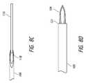
- FIG. 6Cshows an enlarged view of the distal portion of the deployment device of FIG. 6A .
- This figureshows an intraocular shunt loaded within a hollow shaft of the deployment device.
- FIGS. 7A-7Bshow schematics of the deployment mechanism at the end of the first stage of deployment of the shunt from the deployment device.
- FIG. 7Cshows an enlarged view of the distal portion of the deployment device of FIG. 7A .
- This figureshows an intraocular shunt partially deployed from within a hollow shaft of the deployment device.
- FIG. 8Ashows a schematic of the deployment device after deployment of the shunt from the device.
- FIG. 8Bshow a schematic of the deployment mechanism at the end of the second stage of deployment of the shunt from the deployment device.
- FIG. 8Cshows an enlarged view of the distal portion of the deployment device after retraction of the shaft with the pusher abutting the shunt.
- FIG. 8Dshows an enlarged view of the distal portion of the deployment device after deployment of the shunt.
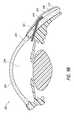
- FIGS. 9A-9Bshow an intraocular shunt deployed within the eye.
- a proximal portion of the shuntresides in the anterior chamber and a distal portion of the shunt resides within the intra-Tenon's space.
- a middle portion of the shuntresides in the sclera.
- FIG. 10depicts a schematic of an exemplary intraocular shunt.
- FIG. 1shows an embodiment of a shunt deployment device 100 according to the invention. While FIG. 1 shows a handheld manually operated shunt deployment device, it will be appreciated that devices of the invention may be coupled with robotic systems and may be completely or partially automated.
- deployment device 100includes a generally cylindrical body or housing 101 ; however, the body shape of housing 101 could be other than cylindrical. Housing 101 may have an ergonomical shape, allowing for comfortable grasping by an operator. Housing 101 is shown with optional grooves 102 to allow for easier gripping by a surgeon.
- Housing 101is shown having a larger proximal portion that tapers to a distal portion.
- the distal portionincludes a hollow sleeve 105 .
- the hollow sleeve 105is configured for insertion into an eye and to extend into an anterior chamber of an eye.
- the hollow sleeveis visible within an anterior chamber of an eye.
- the sleeve 105provides a visual preview for an operator as to placement of the proximal portion of the shunt within the anterior chamber of an eye.
- the sleeve 105provides a visual reference point that may be used by an operator to hold device 100 steady during the shunt deployment process, thereby assuring optimal longitudinal placement of the shunt within the eye.
- the sleeve 105may include an edge 131 at a distal end that provides resistance feedback to an operator upon insertion of the deployment device 100 within an eye of a person. Upon advancement of the device 100 across an anterior chamber of the eye, the hollow sleeve 105 will eventually contact the sclera 134 , providing resistance feedback to an operator that no further advancement of the device 100 is necessary. The edge 131 of the sleeve 105 prevents the shaft 104 from accidentally being pushed too far through the sclera.
- a temporary guard 108is configured to fit around sleeve 105 and extend beyond an end of sleeve 105 . The guard is used during shipping of the device and protects an operator from a distal end of a hollow shaft 104 that extends beyond the end of the sleeve 105 . The guard is removed prior to use of the device.
- Housing 101is open at its proximal end, such that a portion of a deployment mechanism 103 may extend from the proximal end of the housing 101 .
- a distal end of housing 101is also open such that at least a portion of a hollow shaft 104 may extend through and beyond the distal end of the housing 101 .
- Housing 101further includes a slot 106 through which an operator, such as a surgeon, using the device 100 may view an indicator 107 on the deployment mechanism 103 .
- Housing 101may be made of any material that is suitable for use in medical devices.
- housing 101may be made of a lightweight aluminum or a biocompatible plastic material.
- suitable plastic materialsinclude polycarbonate and other polymeric resins such as DELRIN and ULTEM.
- housing 101is made of a material that may be autoclaved, and thus allow for housing 101 to be re-usable.
- device 100may be sold as a one-time-use device, and thus the material of the housing does not need to be a material that is autoclavable.
- Housing 101may be made of multiple components that connect together to form the housing.
- FIG. 2shows an exploded view of deployment device 100 .
- housing 101is shown having three components 101 a , 101 b , and 101 c .
- the componentsare designed to screw together to form housing 101 .
- FIG. 2also shows deployment mechanism 103 .
- the housing 101is designed such that deployment mechanism 103 fits within assembled housing 101 .
- Housing 101is designed such that components of deployment mechanism 103 are movable within housing 101 .
- FIGS. 3A-3Dshow different enlarged views of the deployment mechanism 103 .
- Deployment mechanism 103may be made of any material that is suitable for use in medical devices.
- deployment mechanism 103may be made of a lightweight aluminum or or a biocompatible plastic material. Examples of such suitable plastic materials include polycarbonate and other polymeric resins such as DELRIN and ULTEM.
- deployment mechanism 103is made of a material that may be autoclaved, and thus allow for deployment mechanism 103 to be re-usable.
- device 100may be sold as a one-time-use device, and thus the material of the deployment mechanism does not need to be a material that is autoclavable.
- Deployment mechanism 103includes a distal portion 109 and a proximal portion 110 .
- the deployment mechanism 103is configured such that distal portion 109 is movable within proximal portion 110 . More particularly, distal portion 109 is capable of partially retracting to within proximal portion 110 .
- the distal portion 109is shown to taper to a connection with a hollow shaft 104 .
- This embodimentis illustrated such that the connection between the hollow shaft 104 and the distal portion 109 of the deployment mechanism 103 occurs inside the housing 101 .
- the connection between hollow shaft 104 and the proximal portion 109 of the deployment mechanism 103may occur outside of the housing 101 .
- Hollow shaft 104may be removable from the distal portion 109 of the deployment mechanism 103 .
- the hollow shaft 104may be permanently coupled to the distal portion 109 of the deployment mechanism 103 .
- hollow shaft 104is configured to hold an intraocular shunt 115 .
- An exemplary intraocular shunt 115is shown in FIG. 11 .
- Other exemplary intraocular shuntsare shown in Yu et al. (U.S. Patent Application No. 2008/0108933).
- intraocular shuntsare of a cylindrical shape and have an outside cylindrical wall and a hollow interior. The shunt may have an inner diameter of approximately 50 ⁇ m to approximately 250 ⁇ m, an outside diameter of approximately 190 ⁇ m to approximately 300 ⁇ m, and a length of approximately 0.5 mm to about 20 mm.
- hollow shaft 104is configured to at least hold a shunt of such shape and such dimensions.
- hollow shaft 104may be configured to hold shunts of different shapes and different dimensions than those described above, and the invention encompasses a shaft 104 that may be configured to hold any shaped or dimensioned intraocular shunt.
- the shafthas an inner diameter of approximately 200 ⁇ m to approximately 400 ⁇ m.
- the shaft 104may be any length.
- a usable length of the shaftmay be anywhere from about 5 mm to about 40 mm, and is 15 mm in certain embodiments.
- the shaftis straight.
- shaftis of a shape other than straight, for example a shaft having a bend along its length or a shaft having an arcuate portion. Exemplary shaped shafts are shown for example in Yu et al. (U.S. Patent Application No. 2008/0108933).
- the shaftincludes a bend at a distal portion of the shaft.
- a distal end of the shaft 104is beveled or is sharpened to a point to assist in piercing the sclera and advancing the distal end of the shaft 104 through the sclera.
- the distal end of the shaft 104has a double bevel. The double bevel provides an angle at the distal end of the shaft 104 such that upon entry of the shaft into intra-Tenon's space, the distal end of shaft 104 will by parallel with Tenon's capsule and will thus not pierce Tenon's capsule and enter the subconjunctival space.
- shunt 115may be placed within the eye
- devices of the inventionare not limited to placing shunts within intra-Tenon's space and may be used to place shunts into many other areas of the eye, such as Schlemm's canal, the subconjunctival space, the episcleral vein, or the suprachoroidal space.
- the shaft 104may hold the shunt at least partially within the hollow interior of the shaft 104 .
- the shuntis held completely within the hollow interior of the shaft 104 .
- the hollow shaftmay hold the shunt on an outer surface of the shaft 104 .
- the shuntis held within the hollow interior of the shaft 104 .
- the hollow shaftis a needle having a hollow interior. Needles that are configured to hold an intraocular shunt are commercially available from Terumo Medical Corp. (Elkington, Md.).
- a proximal portion of the deployment mechanismincludes optional grooves 116 to allow for easier gripping by an operator for easier rotation of the deployment mechanism, which will be discussed in more detail below.
- the proximal portion 110 of the deployment mechanismalso includes at least one indicator that provides feedback to an operator as to the state of the deployment mechanism.
- the indicatormay be any type of indicator known in the art, for example, a visual indicator, an audio indicator, or a tactile indicator.
- FIG. 3Ashows a deployment mechanism having two indicators, a ready indicator 111 and a deployed indicator 119 .
- Ready indicator 111provides feedback to an operator that the deployment mechanism is in a configuration for deployment of an intraocular shunt from the deployment device 100 .
- the indicator 111is shown in this embodiment as a green oval having a triangle within the oval.
- Deployed indicator 119provides feedback to the operator that the deployment mechanism has been fully engaged and has deployed the shunt from the deployment device 100 .
- the deployed indicator 119is shown in this embodiment as a yellow oval having a black square within the oval.
- the indicatorsare located on the deployment mechanism such that when assembled, the indicators 111 and 119 may be seen through slot 106 in housing 101 .
- the proximal portion 110includes a stationary portion 110 b and a rotating portion 110 a .
- the proximal portion 110includes a channel 112 that runs part of the length of stationary portion 110 b and the entire length of rotating portion 110 a .
- the channel 112is configured to interact with a protrusion 117 on an interior portion of housing component 101 a ( FIGS. 4A and 4B ).
- the protrusion 117 on housing component 101 ais aligned with channel 112 on the stationary portion 110 b and rotating portion 110 a of the deployment mechanism 103 .
- the proximal portion 110 of deployment mechanism 103is slid within housing component 101 a until the protrusion 117 sits within stationary portion 110 b ( FIG. 4C ).
- the protrusion 117interacts with the stationary portion 110 b of the deployment mechanism 103 and prevents rotation of stationary portion 110 b .
- rotating portion 110 ais free to rotate within housing component 101 a.
- the rotating portion 110 a of proximal portion 110 of deployment mechanism 103also includes channels 113 a , 113 b , and 113 c .
- Channel 113 aincludes a first portion 113 a 1 that is straight and runs perpendicular to the length of the rotating portion 110 a , and a second portion 113 a 2 that runs diagonally along the length of rotating portion 110 a , downwardly toward a proximal end of the deployment mechanism 103 .
- Channel 113 bincludes a first portion 113 b 1 that runs diagonally along the length of the rotating portion 110 a , downwardly toward a distal end of the deployment mechanism 103 , and a second portion that is straight and runs perpendicular to the length of the rotating portion 110 a .
- the point at which first portion 113 a 1 transitions to second portion 113 a 2 along channel 113 ais the same as the point at which first portion 113 b 1 transitions to second portion 113 b 2 along channel 113 b.
- Channel 113 cis straight and runs perpendicular to the length of the rotating portion 110 a .
- channels 113 a , 113 b , and 113 csit members 114 a , 114 b , and 114 c respectively.
- Members 114 a , 114 b , and 114 care movable within channels 113 a , 113 b , and 113 c .
- Members 114 a , 114 b , and 114 calso act as stoppers that limit movement of rotating portion 110 a , which thereby limits axial movement of the shaft 104 .
- FIG. 5shows a cross-sectional view of deployment mechanism 103 .
- Member 114 ais connected to the distal portion 109 of the deployment mechanism 103 . Movement of member 114 a results in retraction of the distal portion 109 of the deployment mechanism 103 to within the proximal portion 110 of the deployment mechanism 103 .
- Member 114 bis connected to a pusher component 118 .
- the pusher component 118extends through the distal portion 109 of the deployment mechanism 103 and extends into a portion of hollow shaft 104 .
- the pusher componentis involved in deployment of a shunt from the hollow shaft 104 .
- An exemplary pusher componentis a plunger. Movement of member 114 b engages pusher 118 and results in pusher 118 advancing within hollow shaft 104 .
- FIG. 6Ashows deployment device 100 is a pre-deployment configuration.
- shunt 115is loaded within hollow shaft 104 ( FIG. 6C ).
- shunt 115is only partially within shaft 104 , such that a portion of the shunt is exposed. However, the shunt 115 does not extend beyond the end of the shaft 104 .
- the shunt 115is completely disposed within hollow shaft 104 .
- the shunt 115is loaded into hollow shaft 104 such that the shunt abuts pusher component 118 within hollow shaft 104 .
- a distal end of shaft 104is beveled to assist in piercing tissue of the eye.
- a portion of the shaft 104extends beyond the sleeve 105 ( FIG. 6C ).
- the deployment mechanismis configured such that member 114 a abuts a distal end of the first portion 113 a 1 of channel 113 a , and member 114 b abut a proximal end of the first portion 113 b 1 of channel 113 b ( FIG. 6B ).
- the ready indicator 111is visible through slot 106 of the housing 101 , providing feedback to an operator that the deployment mechanism is in a configuration for deployment of an intraocular shunt from the deployment device 100 ( FIG. 6A ).
- the device 100is ready for insertion into an eye (insertion configuration or pre-deployment configuration). Methods for inserting and implanting shunts are discussed in further detail below.
- the deployment mechanism 103is a two-stage system. The first stage is engagement of the pusher component 118 and the second stage is retraction of the distal portion 109 to within the proximal portion 110 of the deployment mechanism 103 . Rotation of the rotating portion 110 a of the proximal portion 110 of the deployment mechanism 103 sequentially engages the pusher component and then the retraction component.
- the pusher componentIn the first stage of shunt deployment, the pusher component is engaged and the pusher partially deploys the shunt from the deployment device.
- rotating portion 110 a of the proximal portion 110 of the deployment mechanism 103is rotated, resulting in movement of members 114 a and 114 b along first portions 113 a 1 and 113 b 1 in channels 113 a and 113 b . Since the first portion 113 a 1 of channel 113 a is straight and runs perpendicular to the length of the rotating portion 110 a , rotation of rotating portion 110 a does not cause axial movement of member 114 a .
- FIGS. 7A-7Cshow schematics of the deployment mechanism at the end of the first stage of deployment of the shunt from the deployment device.
- members 114 a and 114 bhave finished traversing along first portions 113 a 1 and 113 b 1 of channels 113 a and 113 b .
- pusher component 118has advanced within hollow shaft 104 ( FIG. 7B ), and shunt 115 has been partially deployed from the hollow shaft 104 ( FIG. 7C ). As is shown in these figures, a portion of the shunt 115 extends beyond an end of the shaft 104 .
- the retraction componentis engaged and the distal portion of the deployment mechanism is retracted to within the proximal portion of the deployment mechanism, thereby completing deployment of the shunt from the deployment device.
- rotating portion 110 a of the proximal portion 110 of the deployment mechanism 103is further rotated, resulting in movement of members 114 a and 114 b along second portions 113 a 2 and 113 b 2 in channels 113 a and 113 b . Since the second portion 113 b 2 of channel 113 b is straight and runs perpendicular to the length of the rotating portion 110 a , rotation of rotating portion 110 a does not cause axial movement of member 114 b .
- the shunt 115Since the shunt 115 abuts the pusher component 118 , the shunt remains stationary as the hollow shaft 104 retracts from around the shunt 115 ( FIG. 8C ). The shaft 104 retracts almost completely to within the sleeve 105 . During both stages of the deployment process, the sleeve 105 remains stationary and in a fixed position.
- FIG. 8Ashows a schematic of the device 100 after deployment of the shunt 115 from the device 100 .
- FIG. 8Bshows a schematic of the deployment mechanism at the end of the second stage of deployment of the shunt from the deployment device. As is shown in FIG. 8B , members 114 a and 114 b have finished traversing along second portions 113 a 2 and 113 b 2 of channels 113 a and 113 b . Additionally, distal portion 109 has retracted to within proximal portion 110 , thus resulting in retraction of the hollow shaft 104 to within the sleeve 105 .
- FIG. 8Dshows an enlarged view of the distal portion of the deployment device after deployment of the shunt. This figure shows that the hollow shaft 104 is not fully retracted to within the sleeve 105 of the deployment device 100 . However, in certain embodiments, the shaft 104 may completely retract to within the sleeve 105 .
- the deployed indicator 119is visible through slot 106 of the housing 101 , providing feedback to the operator that the deployment mechanism has been fully engaged and that the shunt 115 has been deployed from the deployment device 100 .
- devices of the inventionmay be inserted into the eye using an ab externo approach (entering through the conjunctiva) or an ab interno approach (entering through the cornea).
- devices of the inventionare inserted into the eye using an ab interno approach.
- Ab interno approaches for implanting an intraocular shuntare shown for example in Yu et al. (U.S. Pat. No. 6,544,249 and U.S. Patent Application No. 2008/0108933) and Prywes (U.S. Pat. No. 6,007,511), the content of each of which is incorporated by reference herein in its entirety.
- Devices of the inventionmay be inserted into the eye to deploy shunts that create fluid drainage passageways from the anterior chamber of the eye to various drainage structures of the eye.
- Exemplary drainage structuresinclude Schlemm's canal, the subconjunctival space, the episcleral vein, the suprachoroidal space, or the intra-Tenon's space.
- fluidis drained to the subarachnoid space.
- devices of the inventionare inserted into the eye to deploy shunts that create fluid drainage passageways from the anterior chamber to the intra-Tenon's space.
- a membraneknown as the conjunctiva
- the region below the conjunctivais known as the subconjunctival space.
- a membraneknown as Tenon's capsule.
- Tenon's adhesionsthat connect the Tenon's capsule to the sclera.
- the space between Tenon's capsule and the sclera where the Tenon's adhesions connect the Tenon's capsule to the sclerais known as the intra-Tenon's space.
- FIGS. 9A-9Bshow an intraocular shunt placed into the eye using devices of the invention such that the shunt forms a passage for fluid drainage from the anterior chamber to the intra-Tenon's space.
- a surgical intervention to implant the shuntis performed that involves inserting into the eye 202 a deployment device 200 that holds an intraocular shunt 201 , and deploying at least a portion of the shunt 201 within intra-Tenon's space 208 , within the subconjunctival space 209 and below the conjunctiva 210 .
- a hollow shaft 206 of a deployment device 200 holding the shunt 201enters the eye 202 through the cornea 203 (ab interno approach).
- the shaft 206is advanced across the anterior chamber 204 (as depicted by the broken line) in what is referred to as a transpupil implant insertion.
- the shaft 206is advanced through the sclera 205 until a distal portion of the shaft 206 is in proximity to Tenon's capsule 207 .
- the shunt 201is then deployed from the shaft 206 of the deployment device 200 , producing a conduit between the anterior chamber 204 and the intra-Tenon's space 208 to allow aqueous humor to drain from the anterior chamber 204 (see FIGS. 9A and 9B ).
Landscapes
- Health & Medical Sciences (AREA)
- Ophthalmology & Optometry (AREA)
- Animal Behavior & Ethology (AREA)
- Engineering & Computer Science (AREA)
- Biomedical Technology (AREA)
- Heart & Thoracic Surgery (AREA)
- Vascular Medicine (AREA)
- Life Sciences & Earth Sciences (AREA)
- General Health & Medical Sciences (AREA)
- Public Health (AREA)
- Veterinary Medicine (AREA)
- Surgery (AREA)
- Nuclear Medicine, Radiotherapy & Molecular Imaging (AREA)
- Prostheses (AREA)
Abstract
Description
This application is a continuation of U.S. patent application Ser. No. 12/946,645, filed on Nov. 15, 2010, the entirety of which is incorporated herein by reference.
1. Field of the Invention
The invention generally relates to devices for deploying an intraocular shunt within an eye.
2. Description of the Related Art
Glaucoma is a disease of the eye that affects millions of people. Glaucoma is associated with an increase in intraocular pressure resulting either from a failure of a drainage system of an eye to adequately remove aqueous humor from an anterior chamber of the eye or overproduction of aqueous humor by a ciliary body in the eye. Build-up of aqueous humor and resulting intraocular pressure may result in irreversible damage to the optic nerve and the retina, which may lead to irreversible retinal damage and blindness.
Glaucoma may be treated by surgical intervention that involves placing a shunt in the eye to result in production of fluid flow pathways between the anterior chamber and various structures of the eye involved in aqueous humor drainage (e.g., Schlemm's canal, the sclera, or the subconjunctival space). Such fluid flow pathways allow for aqueous humor to exit the anterior chamber. Generally, the surgical intervention to implant the shunt involves inserting into the eye a deployment device that holds an intraocular shunt, and deploying the shunt within the eye. A deployment device holding the shunt enters the eye through a cornea (ab interno approach), and is advanced across the anterior chamber. The deployment device is advanced through the sclera until a distal portion of the device is in proximity to a drainage structure of the eye. The shunt is then deployed from the deployment device, producing a conduit between the anterior chamber and various structures of the eye involved in aqueous humor drainage (e.g., Schlemm's canal, the sclera, or the subconjunctival space). See for example, Prywes (U.S. Pat. No. 6,007,511).
A problem associated with such surgical interventions is ensuring that placement of the shunt does not change during deployment of the shunt from the deployment device. Deployment devices that are used to place the shunt in the eye generally rely on multiple moving components in order to deploy the shunt. Movement of the components of the deployment device shifts the position of the deployment device within the eye during the deployment process, and thus shifts the position of the shunt as it is being deployed. Such movement leads to improper placement of the shunt within the eye.
The invention generally relates to deployment devices that are designed to minimize movement of the device during deployment of an intraocular shunt from the device, thereby ensuring proper placement of the shunt within the eye.
In certain aspects, deployment devices of the invention include a housing, a deployment mechanism at least partially disposed within the housing, and a hollow shaft coupled to the deployment mechanism, in which the shaft is configured to hold an intraocular shunt. With such devices, rotation of the deployment mechanism results in deployment of the shunt. Such rotational movement is translated into axial movement for deploying the shunt from the device. By utilizing rotational movement for the deployment mechanism, axial movement of the deployment device is minimized, ensuring proper placement of the shunt within the eye.
Other aspects of the invention provide devices for deploying an intraocular shunt including a housing, a deployment mechanism at least partially disposed within the housing, in which the deployment mechanism includes a two stage system, and a hollow shaft coupled to the deployment mechanism, in which the shaft is configured to hold an intraocular shunt.
Another aspect of the invention includes devices for deploying an intraocular shunt including a housing, a deployment mechanism at least partially disposed within the housing, and a hollow shaft coupled inside the housing to the deployment mechanism, wherein the shaft is configured to hold an intraocular shunt, in which the device includes an insertion configuration and a deployment configuration and the deployment configuration includes a proximal portion of the shaft being at least partially retracted to within the housing. In certain embodiments, the insertion configuration includes a distal portion of the shaft being disposed within the housing and a proximal portion of the shaft extending beyond the housing.
In certain embodiments, the shaft is configured to at least partially retract to within the housing. However, it will be appreciated that the shaft may fully retract to within the housing. In certain embodiments, the device further includes the intraocular shunt. The shunt may be completely disposed within the hollow shaft of the device. Alternatively, the shunt is partially disposed within the hollow shaft of the device.
The deployment mechanism may include a two stage system. In such embodiments, the first stage is a pusher component and the second stage is a retraction component. In this embodiment, rotation of the deployment mechanism sequentially engages the pusher component and then the retraction component. The pusher component pushes the shunt to partially deploy the shunt from within the shaft, and the retraction component retracts the shaft from around the shunt, thereby deploying the shunt. In certain embodiments, the deployment mechanism may additionally include at least one member that limits axial movement of the shaft.
The hollow shaft of the deployment device may include a beveled distal end. An exemplary hollow shaft is a needle. Devices of the invention may be completely automated, partially automated, or completely manual. Devices of the invention may be connected to larger robotic systems or may be used as stand-alone handheld deployment devices. In particular embodiments, the device is a handheld device.
Devices of the invention may include an indicator that provides feedback to an operator as to the state of the deployment mechanism. The indicator may be any type of indicator known in the art, for example a visual indicator, an audio indicator, or a tactile indicator. In certain embodiments, the indicator is a visual indicator.
Aspects of the invention also include methods for deploying an intraocular shunt within an eye. These methods involve using devices described herein to deploy an intraocular shunt from the device within the eye. Generally, deploying the shunt results in a flow path from an anterior chamber of the eye to an area of lower pressure. Exemplary areas of lower pressure include intra-Tenon's space, the subconjunctival space, the episcleral vein, the suprachoroidal space, and Schlemm's canal. In certain embodiments, the area of lower pressure is the subarachnoid space.
Any of a variety of methods known in the art may be used to insert devices of the invention into an eye. In certain embodiments, devices of the invention may be inserted into the eye using an ab externo approach (entering through the conjunctiva) or an ab interno approach (entering through the cornea).
Reference is now made toFIG. 1 , which shows an embodiment of ashunt deployment device 100 according to the invention. WhileFIG. 1 shows a handheld manually operated shunt deployment device, it will be appreciated that devices of the invention may be coupled with robotic systems and may be completely or partially automated. As shown inFIG. 1 ,deployment device 100 includes a generally cylindrical body orhousing 101; however, the body shape ofhousing 101 could be other than cylindrical.Housing 101 may have an ergonomical shape, allowing for comfortable grasping by an operator.Housing 101 is shown withoptional grooves 102 to allow for easier gripping by a surgeon.
Thesleeve 105 may include anedge 131 at a distal end that provides resistance feedback to an operator upon insertion of thedeployment device 100 within an eye of a person. Upon advancement of thedevice 100 across an anterior chamber of the eye, thehollow sleeve 105 will eventually contact the sclera134, providing resistance feedback to an operator that no further advancement of thedevice 100 is necessary. Theedge 131 of thesleeve 105 prevents theshaft 104 from accidentally being pushed too far through the sclera. Atemporary guard 108 is configured to fit aroundsleeve 105 and extend beyond an end ofsleeve 105. The guard is used during shipping of the device and protects an operator from a distal end of ahollow shaft 104 that extends beyond the end of thesleeve 105. The guard is removed prior to use of the device.
In this embodiment, thedistal portion 109 is shown to taper to a connection with ahollow shaft 104. This embodiment is illustrated such that the connection between thehollow shaft 104 and thedistal portion 109 of thedeployment mechanism 103 occurs inside thehousing 101. In other embodiments, the connection betweenhollow shaft 104 and theproximal portion 109 of thedeployment mechanism 103 may occur outside of thehousing 101.Hollow shaft 104 may be removable from thedistal portion 109 of thedeployment mechanism 103. Alternatively, thehollow shaft 104 may be permanently coupled to thedistal portion 109 of thedeployment mechanism 103.
Generally,hollow shaft 104 is configured to hold anintraocular shunt 115. An exemplaryintraocular shunt 115 is shown inFIG. 11 . Other exemplary intraocular shunts are shown in Yu et al. (U.S. Patent Application No. 2008/0108933). Generally, in one embodiment, intraocular shunts are of a cylindrical shape and have an outside cylindrical wall and a hollow interior. The shunt may have an inner diameter of approximately 50 μm to approximately 250 μm, an outside diameter of approximately 190 μm to approximately 300 μm, and a length of approximately 0.5 mm to about 20 mm. Thus,hollow shaft 104 is configured to at least hold a shunt of such shape and such dimensions. However,hollow shaft 104 may be configured to hold shunts of different shapes and different dimensions than those described above, and the invention encompasses ashaft 104 that may be configured to hold any shaped or dimensioned intraocular shunt. In particular embodiments, the shaft has an inner diameter of approximately 200 μm to approximately 400 μm.
Theshaft 104 may be any length. A usable length of the shaft may be anywhere from about 5 mm to about 40 mm, and is 15 mm in certain embodiments. In certain embodiments, the shaft is straight. In other embodiments, shaft is of a shape other than straight, for example a shaft having a bend along its length or a shaft having an arcuate portion. Exemplary shaped shafts are shown for example in Yu et al. (U.S. Patent Application No. 2008/0108933).
In particular embodiments, the shaft includes a bend at a distal portion of the shaft. In other embodiments, a distal end of theshaft 104 is beveled or is sharpened to a point to assist in piercing the sclera and advancing the distal end of theshaft 104 through the sclera. In particular embodiments, the distal end of theshaft 104 has a double bevel. The double bevel provides an angle at the distal end of theshaft 104 such that upon entry of the shaft into intra-Tenon's space, the distal end ofshaft 104 will by parallel with Tenon's capsule and will thus not pierce Tenon's capsule and enter the subconjunctival space. This ensures proper deployment of the shunt such that a distal end of theshunt 115 is deployed within the intra-Tenon's space, rather than deployment of the distal end of theshunt 115 within the subconjunctival space. Changing the angle of the bevel allows for placement ofshunt 115 within other areas of lower pressure than the anterior chamber, such as the subconjunctival space. It will be understood that implanting into intra-Tenon's space merely one embodiment of whereshunt 115 may be placed within the eye, and that devices of the invention are not limited to placing shunts within intra-Tenon's space and may be used to place shunts into many other areas of the eye, such as Schlemm's canal, the subconjunctival space, the episcleral vein, or the suprachoroidal space.
Theshaft 104 may hold the shunt at least partially within the hollow interior of theshaft 104. In other embodiments, the shunt is held completely within the hollow interior of theshaft 104. Alternatively, the hollow shaft may hold the shunt on an outer surface of theshaft 104. In particular embodiments, the shunt is held within the hollow interior of theshaft 104. In certain embodiments, the hollow shaft is a needle having a hollow interior. Needles that are configured to hold an intraocular shunt are commercially available from Terumo Medical Corp. (Elkington, Md.).
A proximal portion of the deployment mechanism includesoptional grooves 116 to allow for easier gripping by an operator for easier rotation of the deployment mechanism, which will be discussed in more detail below. Theproximal portion 110 of the deployment mechanism also includes at least one indicator that provides feedback to an operator as to the state of the deployment mechanism. The indicator may be any type of indicator known in the art, for example, a visual indicator, an audio indicator, or a tactile indicator.FIG. 3A shows a deployment mechanism having two indicators, aready indicator 111 and a deployedindicator 119.Ready indicator 111 provides feedback to an operator that the deployment mechanism is in a configuration for deployment of an intraocular shunt from thedeployment device 100. Theindicator 111 is shown in this embodiment as a green oval having a triangle within the oval. Deployedindicator 119 provides feedback to the operator that the deployment mechanism has been fully engaged and has deployed the shunt from thedeployment device 100. The deployedindicator 119 is shown in this embodiment as a yellow oval having a black square within the oval. The indicators are located on the deployment mechanism such that when assembled, theindicators slot 106 inhousing 101.
Theproximal portion 110 includes astationary portion 110band arotating portion 110a. Theproximal portion 110 includes achannel 112 that runs part of the length ofstationary portion 110band the entire length of rotatingportion 110a. Thechannel 112 is configured to interact with aprotrusion 117 on an interior portion ofhousing component 101a(FIGS. 4A and 4B ). During assembly, theprotrusion 117 onhousing component 101ais aligned withchannel 112 on thestationary portion 110bandrotating portion 110aof thedeployment mechanism 103. Theproximal portion 110 ofdeployment mechanism 103 is slid withinhousing component 101auntil theprotrusion 117 sits withinstationary portion 110b(FIG. 4C ). Assembled, theprotrusion 117 interacts with thestationary portion 110bof thedeployment mechanism 103 and prevents rotation ofstationary portion 110b. In this configuration, rotatingportion 110ais free to rotate withinhousing component 101a.
Referring back toFIGS. 3A-3D , the rotatingportion 110aofproximal portion 110 ofdeployment mechanism 103 also includeschannels Channel 113aincludes afirst portion 113a1 that is straight and runs perpendicular to the length of therotating portion 110a, and asecond portion 113a2 that runs diagonally along the length of rotatingportion 110a, downwardly toward a proximal end of thedeployment mechanism 103.Channel 113bincludes afirst portion 113b1 that runs diagonally along the length of therotating portion 110a, downwardly toward a distal end of thedeployment mechanism 103, and a second portion that is straight and runs perpendicular to the length of therotating portion 110a. The point at whichfirst portion 113a1 transitions tosecond portion 113a2 alongchannel 113a, is the same as the point at whichfirst portion 113b1 transitions tosecond portion 113b2 alongchannel 113b.Channel 113cis straight and runs perpendicular to the length of therotating portion 110a. Within each ofchannels members Members channels Members portion 110a, which thereby limits axial movement of theshaft 104.
Reference is now made toFIGS. 6A-8D , which accompany the following discussion regarding deployment of ashunt 115 fromdeployment device 100.FIG. 6A showsdeployment device 100 is a pre-deployment configuration. In this configuration,shunt 115 is loaded within hollow shaft104 (FIG. 6C ). As shown inFIG. 6C ,shunt 115 is only partially withinshaft 104, such that a portion of the shunt is exposed. However, theshunt 115 does not extend beyond the end of theshaft 104. In other embodiments, theshunt 115 is completely disposed withinhollow shaft 104. Theshunt 115 is loaded intohollow shaft 104 such that the shunt abutspusher component 118 withinhollow shaft 104. A distal end ofshaft 104 is beveled to assist in piercing tissue of the eye.
Additionally, in the pre-deployment configuration, a portion of theshaft 104 extends beyond the sleeve105 (FIG. 6C ). The deployment mechanism is configured such thatmember 114aabuts a distal end of thefirst portion 113a1 ofchannel 113a, andmember 114babut a proximal end of thefirst portion 113b1 ofchannel 113b(FIG. 6B ). In this configuration, theready indicator 111 is visible throughslot 106 of thehousing 101, providing feedback to an operator that the deployment mechanism is in a configuration for deployment of an intraocular shunt from the deployment device100 (FIG. 6A ). In this configuration, thedevice 100 is ready for insertion into an eye (insertion configuration or pre-deployment configuration). Methods for inserting and implanting shunts are discussed in further detail below.
Once the device has been inserted into the eye and advanced to a location to where the shunt will be deployed, theshunt 115 may be deployed from thedevice 100. Thedeployment mechanism 103 is a two-stage system. The first stage is engagement of thepusher component 118 and the second stage is retraction of thedistal portion 109 to within theproximal portion 110 of thedeployment mechanism 103. Rotation of therotating portion 110aof theproximal portion 110 of thedeployment mechanism 103 sequentially engages the pusher component and then the retraction component.
In the first stage of shunt deployment, the pusher component is engaged and the pusher partially deploys the shunt from the deployment device. During the first stage, rotatingportion 110aof theproximal portion 110 of thedeployment mechanism 103 is rotated, resulting in movement ofmembers first portions 113a1 and113b1 inchannels first portion 113a1 ofchannel 113ais straight and runs perpendicular to the length of therotating portion 110a, rotation of rotatingportion 110adoes not cause axial movement ofmember 114a. Without axial movement ofmember 114a, there is no retraction of thedistal portion 109 to within theproximal portion 110 of thedeployment mechanism 103. Since thefirst portion 113b1 ofchannel 113bruns diagonally along the length of therotating portion 110a, upwardly toward a distal end of thedeployment mechanism 103, rotation of rotatingportion 110acauses axial movement ofmember 114btoward a distal end of the device. Axial movement ofmember 114btoward a distal end of the device results in forward advancement of thepusher component 118 within thehollow shaft 104. Such movement ofpusher component 118 results in partially deployment of theshunt 115 from theshaft 104.
In the second stage of shunt deployment, the retraction component is engaged and the distal portion of the deployment mechanism is retracted to within the proximal portion of the deployment mechanism, thereby completing deployment of the shunt from the deployment device. During the second stage, rotatingportion 110aof theproximal portion 110 of thedeployment mechanism 103 is further rotated, resulting in movement ofmembers second portions 113a2 and113b2 inchannels second portion 113b2 ofchannel 113bis straight and runs perpendicular to the length of therotating portion 110a, rotation of rotatingportion 110adoes not cause axial movement ofmember 114b. Without axial movement ofmember 114b, there is no further advancement ofpusher 118. Since thesecond portion 113a2 ofchannel 113aruns diagonally along the length of therotating portion 110a, downwardly toward a proximal end of thedeployment mechanism 103, rotation of rotatingportion 110acauses axial movement ofmember 114atoward a proximal end of the device. Axial movement ofmember 114atoward a proximal end of the device results in retraction of thedistal portion 109 to within theproximal portion 110 of thedeployment mechanism 103. Retraction of thedistal portion 109, results in retraction of thehollow shaft 104. Since theshunt 115 abuts thepusher component 118, the shunt remains stationary as thehollow shaft 104 retracts from around the shunt115 (FIG. 8C ). Theshaft 104 retracts almost completely to within thesleeve 105. During both stages of the deployment process, thesleeve 105 remains stationary and in a fixed position.
Referring toFIG. 8A , in the post-deployment configuration, the deployedindicator 119 is visible throughslot 106 of thehousing 101, providing feedback to the operator that the deployment mechanism has been fully engaged and that theshunt 115 has been deployed from thedeployment device 100.
Any of a variety of methods known in the art may be used to insert devices of the invention into an eye. In certain embodiments, devices of the invention may be inserted into the eye using an ab externo approach (entering through the conjunctiva) or an ab interno approach (entering through the cornea).
In certain embodiments, devices of the invention are inserted into the eye using an ab interno approach. Ab interno approaches for implanting an intraocular shunt are shown for example in Yu et al. (U.S. Pat. No. 6,544,249 and U.S. Patent Application No. 2008/0108933) and Prywes (U.S. Pat. No. 6,007,511), the content of each of which is incorporated by reference herein in its entirety.
Devices of the invention may be inserted into the eye to deploy shunts that create fluid drainage passageways from the anterior chamber of the eye to various drainage structures of the eye. Exemplary drainage structures include Schlemm's canal, the subconjunctival space, the episcleral vein, the suprachoroidal space, or the intra-Tenon's space. In certain embodiments, fluid is drained to the subarachnoid space.
In particular embodiments, devices of the invention are inserted into the eye to deploy shunts that create fluid drainage passageways from the anterior chamber to the intra-Tenon's space. Within an eye, there is a membrane known as the conjunctiva, and the region below the conjunctiva is known as the subconjunctival space. Within the subconjunctival space is a membrane known as Tenon's capsule. Below Tenon's capsule there are Tenon's adhesions that connect the Tenon's capsule to the sclera. The space between Tenon's capsule and the sclera where the Tenon's adhesions connect the Tenon's capsule to the sclera is known as the intra-Tenon's space.
Once a distal portion of thehollow shaft 206 is within theintra-Tenon's space 208, theshunt 201 is then deployed from theshaft 206 of thedeployment device 200, producing a conduit between theanterior chamber 204 and theintra-Tenon's space 208 to allow aqueous humor to drain from the anterior chamber204 (seeFIGS. 9A and 9B ).
As will be appreciated by one skilled in the art, individual features of the invention may be used separately or in any combination. Particularly, it is contemplated that one or more features of the individually described above embodiments may be combined into a single shunt.
References and citations to other documents, such as patents, patent applications, patent publications, journals, books, papers, web contents, have been made throughout this disclosure. All such documents are hereby incorporated herein by reference in their entirety for all purposes.
The invention may be embodied in other specific forms without departing from the spirit or essential characteristics thereof. The foregoing embodiments are therefore to be considered in all respects illustrative rather than limiting on the invention described herein.
Claims (20)
1. A device for deploying an intraocular shunt, the device comprising:
a housing;
a hollow shaft, coupled to the housing, configured to hold an intraocular shunt;
a plunger, slidably disposed within the shaft;
a rotating component, coupled to the housing, having first and second slots;
a pusher component, coupled to the plunger, having a first member that resides within the first slot to couple the pusher component with the rotating component such that rotation of the rotating component causes distal axial movement of the first member, thereby urging the plunger in a distal axial direction to advance the shunt partially from within the shaft; and
a retraction component, coupled to the shaft, having a second member that resides within the second slot to couple the retraction component with the rotating component such that further rotation of the rotating component causes proximal axial movement of the second member, thereby urging the shaft in a proximal axial direction relative to the shunt.
2. The device according toclaim 1 , wherein at least a portion of the first slot and at least a portion of the second slot extend diagonally relative to a longitudinal axis of the rotating component.
3. The device according toclaim 1 , wherein the shaft is configured to at least partially retract to within the housing.
4. The device according toclaim 3 , wherein the shaft fully retracts to within the housing.
5. The device according toclaim 1 , further comprising an intraocular shunt that is at least partially disposed within the shaft.
6. The device according toclaim 1 , wherein the deployment mechanism further comprises at least one member that limits axial movement of the shaft.
7. The device according toclaim 1 , wherein the hollow shaft comprises a needle.
8. The device according toclaim 1 , wherein a distal portion of the housing comprises a sleeve and the hollow shaft is movable within the sleeve.
9. A device for deploying an intraocular shunt, the device comprising:
a housing;
a hollow shaft, coupled to the housing, configured to hold an intraocular shunt;
a plunger slidably disposed within the shaft;
a deployment mechanism at least partially disposed within the housing, the deployment mechanism comprising a rotating component, a pusher component coupled to the plunger, and a retraction component coupled to the shaft, the rotating component being operably coupled to the pusher and retraction components by first and second slots and first and second protruding members slidably disposed within the respective first and second slots;
wherein rotation of the rotating component causes (i) axial movement of the pusher component to urge the plunger distally relative to the shaft and the housing and (ii) axial movement of the retraction component to urge the shaft proximally relative to the plunger and the housing.
10. The device according toclaim 9 , wherein the rotating component is configured to sequentially engage the pusher component and then the refraction component.
11. The device according toclaim 9 , wherein rotating component comprises the first and second slots.
12. The device according toclaim 11 , wherein the pusher component comprises the first protrusion and the retraction component comprises the second protrusion.
13. The device according toclaim 9 , wherein the shaft fully retracts to within the housing.
14. The device according toclaim 9 , further comprising an intraocular shunt that is at least partially disposed within the shaft.
15. The device according toclaim 9 , wherein the deployment mechanism further comprises at least one member that limits axial movement of the shaft.
16. The device according toclaim 9 , wherein a distal portion of the housing comprises a sleeve and the hollow shaft is movable within the sleeve.
17. A device for deploying an intraocular shunt, the device comprising:
a housing;
a deployment mechanism at least partially disposed within the housing, the deployment mechanism comprising a rotating component, a pusher component, and a retraction component, the rotating component being operably coupled to the pusher and retraction components by first and second slots and first and second protruding members slidably disposed within the respective first and second slots;
wherein rotation of the rotating component causes (i) distal axial movement of the pusher component relative to the housing to push a stent within the device, and (ii) proximal axial movement of the retraction component to expose the stent from the device.
18. The device according toclaim 17 , further comprising a hollow shaft coupled to the housing and a plunger slidably disposed within the shaft, wherein the shaft is coupled to the retraction component and the plunger is coupled to the pusher component.
19. The device according toclaim 17 , wherein the housing comprises a sleeve and the pusher and retraction components are movable relative to the sleeve.
20. The device according toclaim 17 , wherein the rotating component comprises the first and second slots, the pusher component comprises the first protruding member, and the retraction component comprises the second protruding member.
Priority Applications (11)
| Application Number | Priority Date | Filing Date | Title |
|---|---|---|---|
| US14/263,957US9283116B2 (en) | 2010-11-15 | 2014-04-28 | Intraocular shunt deployment device |
| US14/313,970US10085884B2 (en) | 2006-06-30 | 2014-06-24 | Intraocular devices |
| US15/005,983US10004638B2 (en) | 2010-11-15 | 2016-01-25 | Intraocular shunt delivery |
| US15/153,648US20160256319A1 (en) | 2010-11-15 | 2016-05-12 | Intraocular shunt placement in the suprachoroidal space |
| US15/153,651US20160256320A1 (en) | 2010-11-15 | 2016-05-12 | Intraocular shunt placement in the suprachoroidal space |
| US15/153,630US20160256317A1 (en) | 2010-11-15 | 2016-05-12 | Methods for implanting intraocular shunts |
| US15/153,646US10842671B2 (en) | 2010-11-15 | 2016-05-12 | Intraocular shunt placement in the suprachoroidal space |
| US16/387,411US20190240069A1 (en) | 2010-11-15 | 2019-04-17 | Methods for implanting intraocular shunts |
| US17/197,002US20210205132A1 (en) | 2010-11-15 | 2021-03-09 | Methods for implanting intraocular shunts |
| US18/494,154US20240197531A1 (en) | 2010-11-15 | 2023-10-25 | Methods for implanting intraocular shunts |
| US18/954,054US20250221854A1 (en) | 2010-11-15 | 2024-11-20 | Methods for implanting intraocular shunts |
Applications Claiming Priority (2)
| Application Number | Priority Date | Filing Date | Title |
|---|---|---|---|
| US12/946,645US8721702B2 (en) | 2010-11-15 | 2010-11-15 | Intraocular shunt deployment devices |
| US14/263,957US9283116B2 (en) | 2010-11-15 | 2014-04-28 | Intraocular shunt deployment device |
Related Parent Applications (2)
| Application Number | Title | Priority Date | Filing Date |
|---|---|---|---|
| US12/946,645ContinuationUS8721702B2 (en) | 2006-06-30 | 2010-11-15 | Intraocular shunt deployment devices |
| US14/313,970ContinuationUS10085884B2 (en) | 2006-06-30 | 2014-06-24 | Intraocular devices |
Related Child Applications (2)
| Application Number | Title | Priority Date | Filing Date |
|---|---|---|---|
| US12/946,556Continuation-In-PartUS20120123317A1 (en) | 2006-06-30 | 2010-11-15 | Methods for implanation of glaucoma shunts |
| US15/005,983ContinuationUS10004638B2 (en) | 2010-11-15 | 2016-01-25 | Intraocular shunt delivery |
Publications (2)
| Publication Number | Publication Date |
|---|---|
| US20140236065A1 US20140236065A1 (en) | 2014-08-21 |
| US9283116B2true US9283116B2 (en) | 2016-03-15 |
Family
ID=46048491
Family Applications (3)
| Application Number | Title | Priority Date | Filing Date |
|---|---|---|---|
| US12/946,645Active2032-05-09US8721702B2 (en) | 2006-06-30 | 2010-11-15 | Intraocular shunt deployment devices |
| US14/263,957Active2030-12-23US9283116B2 (en) | 2006-06-30 | 2014-04-28 | Intraocular shunt deployment device |
| US15/005,983Active2031-05-28US10004638B2 (en) | 2010-11-15 | 2016-01-25 | Intraocular shunt delivery |
Family Applications Before (1)
| Application Number | Title | Priority Date | Filing Date |
|---|---|---|---|
| US12/946,645Active2032-05-09US8721702B2 (en) | 2006-06-30 | 2010-11-15 | Intraocular shunt deployment devices |
Family Applications After (1)
| Application Number | Title | Priority Date | Filing Date |
|---|---|---|---|
| US15/005,983Active2031-05-28US10004638B2 (en) | 2010-11-15 | 2016-01-25 | Intraocular shunt delivery |
Country Status (13)
| Country | Link |
|---|---|
| US (3) | US8721702B2 (en) |
| EP (3) | EP4029481A3 (en) |
| JP (3) | JP6423590B2 (en) |
| CN (2) | CN106264856B (en) |
| CA (1) | CA2818560C (en) |
| DK (1) | DK2640455T3 (en) |
| ES (1) | ES2725550T3 (en) |
| HU (1) | HUE043303T2 (en) |
| PL (1) | PL2640455T3 (en) |
| PT (1) | PT2640455T (en) |
| SI (1) | SI2640455T1 (en) |
| TR (1) | TR201906873T4 (en) |
| WO (1) | WO2012068130A1 (en) |
Cited By (26)
| Publication number | Priority date | Publication date | Assignee | Title |
|---|---|---|---|---|
| US20150011926A1 (en)* | 2006-06-30 | 2015-01-08 | Aquesys, Inc. | Intraocular devices |
| US20160135994A1 (en)* | 2010-11-15 | 2016-05-19 | Aquesys, Inc. | Intraocular shunt delivery |
| US9585790B2 (en) | 2013-11-14 | 2017-03-07 | Aquesys, Inc. | Intraocular shunt inserter |
| US9610195B2 (en) | 2013-02-27 | 2017-04-04 | Aquesys, Inc. | Intraocular shunt implantation methods and devices |
| US9693901B2 (en) | 2010-11-15 | 2017-07-04 | Aquesys, Inc. | Shunt placement through the sclera |
| US9808373B2 (en) | 2013-06-28 | 2017-11-07 | Aquesys, Inc. | Intraocular shunt implantation |
| US9877866B2 (en) | 2010-11-15 | 2018-01-30 | Aquesys, Inc. | Intraocular shunt placement |
| US9883969B2 (en) | 2011-12-08 | 2018-02-06 | Aquesys, Inc. | Intrascleral shunt placement |
| US10080682B2 (en) | 2011-12-08 | 2018-09-25 | Aquesys, Inc. | Intrascleral shunt placement |
| US10307293B2 (en) | 2010-11-15 | 2019-06-04 | Aquesys, Inc. | Methods for intraocular shunt placement |
| US10463537B2 (en) | 2015-06-03 | 2019-11-05 | Aquesys Inc. | Ab externo intraocular shunt placement |
| US10667947B2 (en) | 2016-06-02 | 2020-06-02 | Aquesys, Inc. | Intraocular drug delivery |
| US10842671B2 (en) | 2010-11-15 | 2020-11-24 | Aquesys, Inc. | Intraocular shunt placement in the suprachoroidal space |
| US10952898B2 (en) | 2018-03-09 | 2021-03-23 | Aquesys, Inc. | Intraocular shunt inserter |
| US11058581B2 (en) | 2017-07-20 | 2021-07-13 | Shifamed Holdings, Llc | Adjustable flow glaucoma shunts and methods for making and using same |
| US11135089B2 (en) | 2018-03-09 | 2021-10-05 | Aquesys, Inc. | Intraocular shunt inserter |
| US11166849B2 (en) | 2017-07-20 | 2021-11-09 | Shifamed Holdings, Llc | Adjustable flow glaucoma shunts and methods for making and using same |
| US11246753B2 (en) | 2017-11-08 | 2022-02-15 | Aquesys, Inc. | Manually adjustable intraocular flow regulation |
| US11291585B2 (en) | 2020-02-14 | 2022-04-05 | Shifamed Holdings, Llc | Shunting systems with rotation-based flow control assemblies, and associated systems and methods |
| US11517477B2 (en) | 2019-10-10 | 2022-12-06 | Shifamed Holdings, Llc | Adjustable flow glaucoma shunts and associated systems and methods |
| US11529258B2 (en) | 2020-01-23 | 2022-12-20 | Shifamed Holdings, Llc | Adjustable flow glaucoma shunts and associated systems and methods |
| US11596550B2 (en) | 2020-04-16 | 2023-03-07 | Shifamed Holdings, Llc | Adjustable glaucoma treatment devices and associated systems and methods |
| US11737920B2 (en) | 2020-02-18 | 2023-08-29 | Shifamed Holdings, Llc | Adjustable flow glaucoma shunts having non-linearly arranged flow control elements, and associated systems and methods |
| US11766355B2 (en) | 2020-03-19 | 2023-09-26 | Shifamed Holdings, Llc | Intraocular shunts with low-profile actuation elements and associated systems and methods |
| US11865283B2 (en) | 2021-01-22 | 2024-01-09 | Shifamed Holdings, Llc | Adjustable shunting systems with plate assemblies, and associated systems and methods |
| US12329682B2 (en) | 2019-01-18 | 2025-06-17 | Shifamed Holdings, Llc | Adjustable flow glaucoma shunts and methods for making and using same |
Families Citing this family (49)
| Publication number | Priority date | Publication date | Assignee | Title |
|---|---|---|---|---|
| US6638239B1 (en) | 2000-04-14 | 2003-10-28 | Glaukos Corporation | Apparatus and method for treating glaucoma |
| US7867186B2 (en) | 2002-04-08 | 2011-01-11 | Glaukos Corporation | Devices and methods for treatment of ocular disorders |
| AU2002258754B2 (en) | 2001-04-07 | 2006-08-17 | Glaukos Corporation | Glaucoma stent and methods thereof for glaucoma treatment |
| US7331984B2 (en) | 2001-08-28 | 2008-02-19 | Glaukos Corporation | Glaucoma stent for treating glaucoma and methods of use |
| US7909789B2 (en) | 2006-06-26 | 2011-03-22 | Sight Sciences, Inc. | Intraocular implants and methods and kits therefor |
| US9095411B2 (en) | 2010-11-15 | 2015-08-04 | Aquesys, Inc. | Devices for deploying intraocular shunts |
| US8308701B2 (en) | 2010-11-15 | 2012-11-13 | Aquesys, Inc. | Methods for deploying intraocular shunts |
| US8801766B2 (en) | 2010-11-15 | 2014-08-12 | Aquesys, Inc. | Devices for deploying intraocular shunts |
| US8974511B2 (en) | 2010-11-15 | 2015-03-10 | Aquesys, Inc. | Methods for treating closed angle glaucoma |
| JP5396272B2 (en) | 2006-06-30 | 2014-01-22 | アクエシス インコーポレイテッド | Method, system and apparatus for reducing pressure in an organ |
| US8852137B2 (en) | 2010-11-15 | 2014-10-07 | Aquesys, Inc. | Methods for implanting a soft gel shunt in the suprachoroidal space |
| US8828070B2 (en) | 2010-11-15 | 2014-09-09 | Aquesys, Inc. | Devices for deploying intraocular shunts |
| US8758290B2 (en) | 2010-11-15 | 2014-06-24 | Aquesys, Inc. | Devices and methods for implanting a shunt in the suprachoroidal space |
| EP2088976B1 (en) | 2006-11-10 | 2019-07-03 | Glaukos Corporation | Uveoscleral shunt |
| WO2012071476A2 (en) | 2010-11-24 | 2012-05-31 | David Haffner | Drug eluting ocular implant |
| US8529622B2 (en) | 2010-02-05 | 2013-09-10 | Sight Sciences, Inc. | Intraocular implants and related kits and methods |
| US8585629B2 (en) | 2010-11-15 | 2013-11-19 | Aquesys, Inc. | Systems for deploying intraocular shunts |
| US8765210B2 (en) | 2011-12-08 | 2014-07-01 | Aquesys, Inc. | Systems and methods for making gelatin shunts |
| US9510945B2 (en)* | 2011-12-20 | 2016-12-06 | Boston Scientific Scimed Inc. | Medical device handle |
| ES2842454T3 (en) | 2012-03-20 | 2021-07-14 | Sight Sciences Inc | Eye delivery systems |
| CA2868341C (en) | 2012-03-26 | 2021-01-12 | Glaukos Corporation | System and method for delivering multiple ocular implants |
| WO2014111911A1 (en)* | 2013-01-18 | 2014-07-24 | Javelin Medical Ltd. | Monofilament implants and systems for delivery thereof |
| EP2854700B1 (en) | 2012-05-31 | 2021-07-07 | Javelin Medical Ltd. | Devices for embolic protection |
| ES2610198T3 (en)* | 2012-06-04 | 2017-04-26 | Alcon Pharmaceuticals Ltd. | Intraocular lens insertion device and method for unloading an intraocular lens from a cartridge |
| US10159600B2 (en) | 2013-02-19 | 2018-12-25 | Aquesys, Inc. | Adjustable intraocular flow regulation |
| US9125723B2 (en) | 2013-02-19 | 2015-09-08 | Aquesys, Inc. | Adjustable glaucoma implant |
| US9592151B2 (en) | 2013-03-15 | 2017-03-14 | Glaukos Corporation | Systems and methods for delivering an ocular implant to the suprachoroidal space within an eye |
| US9592110B1 (en) | 2013-12-06 | 2017-03-14 | Javelin Medical, Ltd. | Systems and methods for implant delivery |
| NZ724010A (en)* | 2014-02-26 | 2022-05-27 | Allergan Inc | Intraocular implant delivery apparatus and methods of use thereof |
| JP6599889B2 (en) | 2014-04-04 | 2019-10-30 | アルコン ファーマシューティカルズ リミティド | Intraocular lens inserter |
| CN104826220B (en)* | 2014-10-23 | 2017-09-22 | 重庆金山科技(集团)有限公司 | A kind of pH operating capsule handles with interlock |
| US10299958B2 (en) | 2015-03-31 | 2019-05-28 | Sight Sciences, Inc. | Ocular delivery systems and methods |
| US10779940B2 (en) | 2015-09-03 | 2020-09-22 | Boston Scientific Scimed, Inc. | Medical device handle |
| US10172706B2 (en) | 2015-10-31 | 2019-01-08 | Novartis Ag | Intraocular lens inserter |
| US11547555B2 (en) | 2015-12-17 | 2023-01-10 | Atrion Medical Products, Inc. | Intraocular lens delivery device and method of use |
| US10722347B2 (en)* | 2015-12-17 | 2020-07-28 | Atrion Medical Products, Inc. | Intraocular lens delivery device and method of use |
| US10583005B2 (en) | 2016-05-13 | 2020-03-10 | Boston Scientific Scimed, Inc. | Medical device handle |
| EP4331536A3 (en) | 2016-10-21 | 2024-05-22 | Javelin Medical Ltd. | Systems, methods and devices for embolic protection |
| RU2019115938A (en)* | 2016-11-02 | 2020-12-03 | Ликид Медикал Проприетари Лимитед | SHUNT SYSTEM, SHUNT AND METHOD FOR TREATMENT OF EYE DISORDER |
| US11000367B2 (en) | 2017-01-13 | 2021-05-11 | Alcon Inc. | Intraocular lens injector |
| CN107174399B (en)* | 2017-05-27 | 2019-07-12 | 天津优视眼科技术有限公司 | What a kind of interior road was implemented applies Lai Mushi pipe operation transportation system |
| CN110573117B (en) | 2017-10-06 | 2021-10-26 | 格劳科斯公司 | Systems and methods for delivering multiple ocular implants |
| USD846738S1 (en) | 2017-10-27 | 2019-04-23 | Glaukos Corporation | Implant delivery apparatus |
| JP7092896B2 (en)* | 2018-03-09 | 2022-06-28 | アクシス、インコーポレイテッド | Intraocular shunt inserter |
| US11504270B1 (en) | 2019-09-27 | 2022-11-22 | Sight Sciences, Inc. | Ocular delivery systems and methods |
| DE102020122597B3 (en)* | 2020-08-28 | 2021-08-05 | Carl Zeiss Meditec Ag | Ophthalmic Surgical Injector |
| EP4251106A1 (en) | 2020-11-25 | 2023-10-04 | Oslo Universitetssykehus HF | Multi-lumen glaucoma stent |
| CA3220359A1 (en) | 2021-06-30 | 2023-01-05 | Laszlo Romoda | Glaucoma treatment systems and procedures |
| KR102821957B1 (en)* | 2022-09-13 | 2025-06-24 | 서울대학교병원 | Apparatus for injecting drainage device into eyeball for glaucoma treatment |
Citations (219)
| Publication number | Priority date | Publication date | Assignee | Title |
|---|---|---|---|---|
| US3788327A (en) | 1971-03-30 | 1974-01-29 | H Donowitz | Surgical implant device |
| US3960150A (en) | 1971-09-09 | 1976-06-01 | Alza Corporation | Bioerodible ocular device |
| US4090530A (en) | 1975-12-30 | 1978-05-23 | Hooker Chemicals & Plastics Corp. | Apparatus for multi-phase fluid systems |
| US4722724A (en) | 1986-06-23 | 1988-02-02 | Stanley Schocket | Anterior chamber tube shunt to an encircling band, and related surgical procedure |
| US4750901A (en) | 1986-03-07 | 1988-06-14 | Molteno Anthony C B | Implant for drainage of aqueous humour |
| US4787885A (en) | 1984-04-06 | 1988-11-29 | Binder Perry S | Hydrogel seton |
| US4804382A (en) | 1986-06-02 | 1989-02-14 | Sulzer Brothers Limited | Artificial vessel |
| US4820626A (en) | 1985-06-06 | 1989-04-11 | Thomas Jefferson University | Method of treating a synthetic or naturally occuring surface with microvascular endothelial cells, and the treated surface itself |
| US4826478A (en) | 1986-06-23 | 1989-05-02 | Stanley Schocket | Anterior chamber tube shunt to an encircling band, and related surgical procedure |
| US4836201A (en) | 1988-03-24 | 1989-06-06 | Patton Medical Technologies, Inc. | "Envelope" apparatus for inserting intra-ocular lens into the eye |
| US4863457A (en) | 1986-11-24 | 1989-09-05 | Lee David A | Drug delivery device |
| US4902292A (en) | 1987-03-30 | 1990-02-20 | Joseph Neil H | Vitreous body prosthesis device |
| US4911161A (en) | 1987-04-29 | 1990-03-27 | Noetix, Inc. | Capsulectomy cutting apparatus |
| US4915684A (en) | 1988-06-21 | 1990-04-10 | Mackeen Donald L | Method and apparatus for modulating the flow of lacrimal fluid through a punctum and associated canaliculus |
| US4934363A (en) | 1987-12-15 | 1990-06-19 | Iolab Corporation | Lens insertion instrument |
| US4936825A (en) | 1988-04-11 | 1990-06-26 | Ungerleider Bruce A | Method for reducing intraocular pressure caused by glaucoma |
| US4946436A (en) | 1989-11-17 | 1990-08-07 | Smith Stewart G | Pressure-relieving device and process for implanting |
| US4968296A (en) | 1989-12-20 | 1990-11-06 | Robert Ritch | Transscleral drainage implant device for the treatment of glaucoma |
| US4978352A (en) | 1989-06-07 | 1990-12-18 | Fedorov Svjatoslav N | Process for producing collagen-based cross-linked biopolymer, an implant from said biopolymer, method for producing said implant, and method for hermetization of corneal or scleral wounds involved in eye injuries, using said implant |
| US5041081A (en) | 1990-05-18 | 1991-08-20 | Odrich Ronald B | Ocular implant for controlling glaucoma |
| US5057098A (en) | 1987-05-01 | 1991-10-15 | Ophthalmocare, Inc. | Apparatus and method for extracting cataract tissue |
| US5071408A (en) | 1988-10-07 | 1991-12-10 | Ahmed Abdul Mateen | Medical valve |
| US5092837A (en) | 1989-12-20 | 1992-03-03 | Robert Ritch | Method for the treatment of glaucoma |
| US5098443A (en) | 1989-03-23 | 1992-03-24 | University Of Miami | Method of implanting intraocular and intraorbital implantable devices for the controlled release of pharmacological agents |
| US5178604A (en) | 1990-05-31 | 1993-01-12 | Iovision, Inc. | Glaucoma implant |
| US5180362A (en) | 1990-04-03 | 1993-01-19 | Worst J G F | Gonio seton |
| US5201750A (en) | 1989-09-06 | 1993-04-13 | Vascomed Institut Fur Kathetertechnologie Gmbh | Dilation catheter with motor drive for dilation of blood vessels |
| US5207660A (en) | 1991-04-26 | 1993-05-04 | Cornell Research Foundation, Inc. | Method for the delivery of compositions to the ocular tissues |
| US5275622A (en) | 1983-12-09 | 1994-01-04 | Harrison Medical Technologies, Inc. | Endovascular grafting apparatus, system and method and devices for use therewith |
| US5290295A (en) | 1992-07-15 | 1994-03-01 | Querals & Fine, Inc. | Insertion tool for an intraluminal graft procedure |
| US5300020A (en) | 1991-05-31 | 1994-04-05 | Medflex Corporation | Surgically implantable device for glaucoma relief |
| WO1994013234A1 (en) | 1992-12-17 | 1994-06-23 | Michael Andrew Coote | Implant device and method for treatment of glaucoma |
| US5333619A (en) | 1992-02-19 | 1994-08-02 | Burgio Vito L | Method for rigid tissue biopsy |
| US5338291A (en) | 1993-02-03 | 1994-08-16 | Pudenz-Schulte Medical Research Corporation | Glaucoma shunt and method for draining aqueous humor |
| US5342370A (en) | 1993-03-19 | 1994-08-30 | University Of Miami | Method and apparatus for implanting an artifical meshwork in glaucoma surgery |
| WO1994021205A1 (en) | 1993-03-16 | 1994-09-29 | Photogenesis, Incorporated | Method for preparation and transplantation of volute grafts and surgical instrument therefor |
| US5360339A (en) | 1994-03-01 | 1994-11-01 | Rosenberg Neil A | Dental prophy cup having a paste-distributing channel arrangement |
| US5370607A (en) | 1992-10-28 | 1994-12-06 | Annuit Coeptis, Inc. | Glaucoma implant device and method for implanting same |
| WO1995008310A1 (en) | 1993-09-22 | 1995-03-30 | Voir Et Vivre E U R L | Implantable device for the treatment of ×demas |
| US5443505A (en) | 1993-11-15 | 1995-08-22 | Oculex Pharmaceuticals, Inc. | Biocompatible ocular implants |
| US5476445A (en) | 1990-05-31 | 1995-12-19 | Iovision, Inc. | Glaucoma implant with a temporary flow restricting seal |
| US5516522A (en) | 1994-03-14 | 1996-05-14 | Board Of Supervisors Of Louisiana State University | Biodegradable porous device for long-term drug delivery with constant rate release and method of making the same |
| US5520631A (en) | 1994-07-22 | 1996-05-28 | Wound Healing Of Oklahoma | Method and apparatus for lowering the intraocular pressure of an eye |
| GB2296663A (en) | 1995-01-03 | 1996-07-10 | Ahmed Salih Mahmud | Drainage device for alleviating excess ophthalmic fluid pressure |
| US5558630A (en) | 1994-12-30 | 1996-09-24 | Fisher; Bret L. | Intrascleral implant and method for the regulation of intraocular pressure |
| US5601094A (en) | 1994-11-22 | 1997-02-11 | Reiss; George R. | Ophthalmic shunt |
| US5656026A (en) | 1995-10-27 | 1997-08-12 | Joseph; Neil H. | Method of in vitro testing one-way pressure gradient limiting valved glaucoma drainage implants |
| US5665114A (en) | 1994-08-12 | 1997-09-09 | Meadox Medicals, Inc. | Tubular expanded polytetrafluoroethylene implantable prostheses |
| US5665093A (en) | 1995-12-11 | 1997-09-09 | Atkins; Joseph R. | Surgical implantation method and apparatus |
| US5670161A (en) | 1996-05-28 | 1997-09-23 | Healy; Kevin E. | Biodegradable stent |
| US5688562A (en) | 1995-06-20 | 1997-11-18 | E. I. Du Pont De Nemours And Company | Method for modifying uncoated synthetic polymer fibers using a Langmuir-Blodgett film coating process |
| US5695474A (en) | 1995-09-18 | 1997-12-09 | Becton Dickinson And Company | Needle shield with collapsible cover |
| US5704907A (en) | 1994-07-22 | 1998-01-06 | Wound Healing Of Oklahoma | Method and apparatus for lowering the intraocular pressure of an eye |
| US5707376A (en) | 1992-08-06 | 1998-01-13 | William Cook Europe A/S | Stent introducer and method of use |
| US5722948A (en) | 1996-02-14 | 1998-03-03 | Gross; Fredric J. | Covering for an ocular device |
| WO1998023237A1 (en) | 1996-11-29 | 1998-06-04 | The Lions Eye Institute Of Western Australia Incorporated | Biological microfistula tube and implantation method and apparatus |
| US5763491A (en) | 1994-12-09 | 1998-06-09 | The Regents Of The University Of California | Method for enhancing outflow of aqueous humor in treatment of glaucoma |
| US5868697A (en) | 1995-05-14 | 1999-02-09 | Optonol Ltd. | Intraocular implant |
| US5908449A (en) | 1994-04-29 | 1999-06-01 | W. L. Gore & Associates, Inc. | Blood contact surfaces using extracellular matrix synthesized in vitro |
| US5932299A (en) | 1996-04-23 | 1999-08-03 | Katoot; Mohammad W. | Method for modifying the surface of an object |
| US6007578A (en) | 1997-10-08 | 1999-12-28 | Ras Holding Corp | Scleral prosthesis for treatment of presbyopia and other eye disorders |
| US6007511A (en) | 1991-05-08 | 1999-12-28 | Prywes; Arnold S. | Shunt valve and therapeutic delivery system for treatment of glaucoma and methods and apparatus for its installation |
| US6050970A (en) | 1997-05-08 | 2000-04-18 | Pharmacia & Upjohn Company | Method and apparatus for inserting a glaucoma implant in an anterior and posterior segment of the eye |
| US6086543A (en) | 1998-06-24 | 2000-07-11 | Rubicor Medical, Inc. | Fine needle and core biopsy devices and methods |
| US6102045A (en) | 1994-07-22 | 2000-08-15 | Premier Laser Systems, Inc. | Method and apparatus for lowering the intraocular pressure of an eye |
| WO2000056255A1 (en) | 1999-03-23 | 2000-09-28 | Tatiyana Valentinovna Kozlova | Method for treating glaucoma, device for realising the same and variants |
| US6146366A (en) | 1998-11-03 | 2000-11-14 | Ras Holding Corp | Device for the treatment of macular degeneration and other eye disorders |
| US6165210A (en) | 1994-04-01 | 2000-12-26 | Gore Enterprise Holdings, Inc. | Self-expandable helical intravascular stent and stent-graft |
| US6203513B1 (en) | 1997-11-20 | 2001-03-20 | Optonol Ltd. | Flow regulating implant, method of manufacture, and delivery device |
| US6228873B1 (en) | 1994-12-09 | 2001-05-08 | The Regents Of The University Of California | Method for enhancing outflow of aqueous humor in treatment of glaucoma |
| US6261256B1 (en) | 1996-12-20 | 2001-07-17 | Abdul Mateen Ahmed | Pocket medical valve & method |
| US6264665B1 (en) | 1996-04-17 | 2001-07-24 | The Lions Eye Institute Of Western Australia Incorporated | System for ocular ultramicrosurgery |
| US20010025150A1 (en) | 2000-01-03 | 2001-09-27 | De Juan Eugene | Surgical devices and methods of use thereof for enhanced tactile perception |
| US20010056254A1 (en) | 1998-05-01 | 2001-12-27 | Cragg Andrew H. | System and method for facilitating hemostasis of blood vessel punctures with absorbable sponge |
| US6413540B1 (en) | 1999-10-21 | 2002-07-02 | Alcon Universal Ltd. | Drug delivery device |
| US20020099434A1 (en) | 1991-10-04 | 2002-07-25 | Scimed Life Systems, Inc., Boston Scientific Corporation | Biodegradable drug delivery vascular stent |
| US6450937B1 (en) | 1999-12-17 | 2002-09-17 | C. R. Bard, Inc. | Needle for implanting brachytherapy seeds |
| US20020133168A1 (en) | 2001-03-16 | 2002-09-19 | Smedley Gregory T. | Applicator and methods for placing a trabecular shunt for glaucoma treatment |
| US6471666B1 (en) | 2000-02-24 | 2002-10-29 | Steven A. Odrich | Injectable glaucoma device |
| US20030015203A1 (en) | 1995-12-01 | 2003-01-23 | Joshua Makower | Device, system and method for implantation of filaments and particles in the body |
| US6524275B1 (en) | 1999-04-26 | 2003-02-25 | Gmp Vision Solutions, Inc. | Inflatable device and method for treating glaucoma |
| US20030050574A1 (en) | 2000-04-18 | 2003-03-13 | John Krueger | Bone biopsy instrument having improved sample retention |
| US6533768B1 (en) | 2000-04-14 | 2003-03-18 | The Regents Of The University Of California | Device for glaucoma treatment and methods thereof |
| US20030060752A1 (en) | 2000-04-14 | 2003-03-27 | Olav Bergheim | Glaucoma device and methods thereof |
| US6558342B1 (en) | 1999-06-02 | 2003-05-06 | Optonol Ltd. | Flow control device, introducer and method of implanting |
| US20030093084A1 (en) | 2001-11-13 | 2003-05-15 | Optonol Ltd. | Delivery devices for flow regulating implants |
| US20030097053A1 (en) | 2001-11-19 | 2003-05-22 | Canon Kabushiki Kaisha | Noncontact ophthalmotonometer |
| US6595945B2 (en) | 2001-01-09 | 2003-07-22 | J. David Brown | Glaucoma treatment device and method |
| US20030187383A1 (en) | 1999-12-09 | 2003-10-02 | The Regents Of The University Of California | Liposuction cannula device and method |
| US6638239B1 (en) | 2000-04-14 | 2003-10-28 | Glaukos Corporation | Apparatus and method for treating glaucoma |
| US20030236483A1 (en) | 2002-06-25 | 2003-12-25 | Ren David H | Dual drainage ocular shunt for glaucoma |
| US6699210B2 (en) | 1999-04-27 | 2004-03-02 | The Arizona Board Of Regents | Glaucoma shunt and a method of making and surgically implanting the same |
| US20040077987A1 (en) | 1998-02-13 | 2004-04-22 | Ventrica, Inc., Corporation Of Delaware | Delivering a conduit into a heart wall to place a coronary vessel in communication with a heart chamber and removing tissue from the vessel or heart wall to facilitate such communication |
| USD490152S1 (en) | 2003-02-28 | 2004-05-18 | Glaukos Corporation | Surgical handpiece |
| US20040147870A1 (en) | 2002-04-08 | 2004-07-29 | Burns Thomas W. | Glaucoma treatment kit |
| US20040210209A1 (en) | 2001-02-13 | 2004-10-21 | Yeung Jeffrey E. | Treating back pain by re-establishing the exchange of nutrient & waste |
| US20040215133A1 (en) | 2002-09-18 | 2004-10-28 | Allergan, Inc. | Apparatus for delivery of ocular implants |
| US20040216749A1 (en) | 2003-01-23 | 2004-11-04 | Hosheng Tu | Vasomodulation during glaucoma surgery |
| US20040254521A1 (en) | 2003-06-16 | 2004-12-16 | Solx, Inc. | Shunt for the treatment of glaucoma |
| US20040260227A1 (en) | 2002-12-19 | 2004-12-23 | Lisk James R. | Article and method for ocular aqueous drainage |
| US20050049578A1 (en) | 2000-04-14 | 2005-03-03 | Hosheng Tu | Implantable ocular pump to reduce intraocular pressure |
| US6881198B2 (en) | 2001-01-09 | 2005-04-19 | J. David Brown | Glaucoma treatment device and method |
| US20050101967A1 (en) | 2002-09-18 | 2005-05-12 | Weber David A. | Methods and apparatus for delivery of ocular implants |
| US20050143363A1 (en) | 2002-09-29 | 2005-06-30 | Innorx, Inc. | Method for subretinal administration of therapeutics including steroids; method for localizing pharmacodynamic action at the choroid of the retina; and related methods for treatment and/or prevention of retinal diseases |
| US6939298B2 (en) | 2002-02-28 | 2005-09-06 | Gmp Vision Solutions, Inc | Device and method for monitoring aqueous flow within the eye |
| US20050246023A1 (en) | 2001-02-13 | 2005-11-03 | Yeung Jeffrey E | Disc shunt for treating back pain |
| US20050267398A1 (en) | 2004-05-27 | 2005-12-01 | Dimitri Protopsaltis | Glaucoma shunt |
| US20050271704A1 (en) | 2002-04-08 | 2005-12-08 | Hosheng Tu | Injectable glaucoma implants with multiple openings |
| US20050277864A1 (en) | 2000-04-14 | 2005-12-15 | David Haffner | Injectable gel implant for glaucoma treatment |
| US7008396B1 (en) | 1999-09-03 | 2006-03-07 | Restorvision, Inc. | Ophthalmic device and method of manufacture and use |
| US20060052721A1 (en) | 2002-10-25 | 2006-03-09 | Thomas Dunker | Biopsy holder for a biopsy cannula |
| US20060064112A1 (en) | 2004-09-08 | 2006-03-23 | Edward Perez | Ocular device applicator |
| US7037335B2 (en) | 2002-11-19 | 2006-05-02 | Eagle Vision, Inc. | Bulbous scleral implants for the treatment of eye disorders such as presbyopia and glaucoma |
| US7041077B2 (en) | 2002-07-19 | 2006-05-09 | Yale University | Uveoscleral drainage device |
| US20060106370A1 (en) | 2001-01-18 | 2006-05-18 | The Regents Of The University Of California | Minimally invasive glaucoma surgical instrument and method |
| US20060116625A1 (en) | 1998-09-10 | 2006-06-01 | Percardia, Inc. | TMR shunt |
| US20060149194A1 (en) | 2001-11-21 | 2006-07-06 | Conston Stanley R | Ophthalmic microsurgical system |
| US20060173446A1 (en) | 2005-01-28 | 2006-08-03 | Alcon, Inc. | Surgical apparatus |
| US20060173397A1 (en) | 2004-11-23 | 2006-08-03 | Hosheng Tu | Ophthalmology implants and methods of manufacture |
| US7094225B2 (en) | 2001-05-03 | 2006-08-22 | Glaukos Corporation | Medical device and methods of use of glaucoma treatment |
| US20060200113A1 (en) | 2002-03-07 | 2006-09-07 | David Haffner | Liquid jet for glaucoma treatment |
| US7118547B2 (en) | 2000-09-01 | 2006-10-10 | Ioltechnologie-Production | Glaucoma drain |
| US7135009B2 (en) | 2001-04-07 | 2006-11-14 | Glaukos Corporation | Glaucoma stent and methods thereof for glaucoma treatment |
| US7163543B2 (en) | 2001-11-08 | 2007-01-16 | Glaukos Corporation | Combined treatment for cataract and glaucoma treatment |
| US7186232B1 (en) | 2002-03-07 | 2007-03-06 | Glaukoa Corporation | Fluid infusion methods for glaucoma treatment |
| US7207980B2 (en) | 2004-01-23 | 2007-04-24 | Iscience Surgical Corporation | Composite ophthalmic microcannula |
| US20070093783A1 (en) | 2005-09-12 | 2007-04-26 | Kugler Chad J | Endovascular devices and methods |
| US20070118065A1 (en) | 2004-12-03 | 2007-05-24 | Leonard Pinchuk | Glaucoma Implant Device |
| US20070141116A1 (en) | 2004-12-03 | 2007-06-21 | Leonard Pinchuk | Glaucoma Implant Device |
| US20070172903A1 (en) | 2002-09-27 | 2007-07-26 | The General Hospital Corporation | Microfluidic device for cell separation and uses thereof |
| WO2007087061A2 (en) | 2006-01-17 | 2007-08-02 | Transcend Medical, Inc. | Glaucoma treatment device |
| US7291125B2 (en) | 2003-11-14 | 2007-11-06 | Transcend Medical, Inc. | Ocular pressure regulation |
| US20070293872A1 (en) | 2006-06-20 | 2007-12-20 | Minu, L.L.C. | Ocular Drainage Device |
| US7331984B2 (en) | 2001-08-28 | 2008-02-19 | Glaukos Corporation | Glaucoma stent for treating glaucoma and methods of use |
| US20080057106A1 (en) | 2006-08-29 | 2008-03-06 | Erickson Signe R | Low profile bioactive agent delivery device |
| US20080108933A1 (en) | 2006-06-30 | 2008-05-08 | Dao-Yi Yu | Methods, Systems and Apparatus for Relieving Pressure in an Organ |
| US20080147001A1 (en) | 2002-03-22 | 2008-06-19 | Laila Al-Marashi | Rapid-exchange balloon catheter shaft and method |
| US20080181929A1 (en) | 2006-12-01 | 2008-07-31 | Allergan, Inc. | Method for determining optimum intraocular locations for drug delivery implant |
| US7431710B2 (en) | 2002-04-08 | 2008-10-07 | Glaukos Corporation | Ocular implants with anchors and methods thereof |
| US7431709B2 (en) | 2003-12-05 | 2008-10-07 | Innfocus, Llc | Glaucoma implant device |
| US20080249467A1 (en) | 2007-04-05 | 2008-10-09 | Daniel Rogers Burnett | Device and Method for Safe Access to a Body Cavity |
| US20080281277A1 (en) | 2005-02-22 | 2008-11-13 | Reinhard Thyzel | Device for Sealing an Opening in a Human or Animal Eye |
| US20080312661A1 (en) | 2007-06-12 | 2008-12-18 | Downer David A | Lens Injector Lumen Tip for Wound Assisted Delivery |
| US20090036818A1 (en) | 2002-05-29 | 2009-02-05 | University Of Saskatchewan | Shunt and Method Treatment of Glaucoma |
| US7488303B1 (en) | 2002-09-21 | 2009-02-10 | Glaukos Corporation | Ocular implant with anchor and multiple openings |
| US20090043321A1 (en) | 2004-04-29 | 2009-02-12 | Iscience Interventional Corporation | Apparatus And Method For Surgical Enhancement Of Aqueous Humor Drainage |
| US20090124973A1 (en) | 2007-11-09 | 2009-05-14 | D Agostino Eduardo | Insertion mechanism for use with a syringe |
| US20090182421A1 (en) | 2007-07-17 | 2009-07-16 | Tom Silvestrini | Ocular implant with hydrogel expansion capabilities |
| US20090209910A1 (en) | 2008-02-05 | 2009-08-20 | Chad John Kugler | Crossing occlusions in blood vessels |
| US7594899B2 (en) | 2004-12-03 | 2009-09-29 | Innfocus, Llc | Glaucoma implant device |
| US20090264813A1 (en) | 2006-06-19 | 2009-10-22 | Allergan, Inc. | Apparatus and methods for implanting particulate ocular implants |
| US20090270890A1 (en) | 2008-04-28 | 2009-10-29 | David Bryan Robinson | Methods and apparatus for crossing occlusions in blood vessels |
| US20090281520A1 (en) | 2007-11-08 | 2009-11-12 | Brian Highley | Ocular Implantation Device |
| US20090287136A1 (en) | 2008-05-16 | 2009-11-19 | David Castillejos | Method and apparatus for fluid drainage of the eye |
| US7625384B2 (en) | 2003-02-27 | 2009-12-01 | Wright Medical Technology, Inc. | Method and apparatus for processing dermal tissue |
| US20100004581A1 (en) | 2008-01-07 | 2010-01-07 | Salutarismd | Methods and devices for minimally-invasive extraocular delivery of radiation to the posterior portion of the eye |
| US20100063478A1 (en) | 2008-09-09 | 2010-03-11 | Thomas Vaino Selkee | Force-sensing catheter with bonded center strut |
| US20100098772A1 (en) | 2008-10-21 | 2010-04-22 | Allergan, Inc. | Drug delivery systems and methods for treating neovascularization |
| US7708711B2 (en) | 2000-04-14 | 2010-05-04 | Glaukos Corporation | Ocular implant with therapeutic agents and methods thereof |
| US20100114006A1 (en) | 2008-11-05 | 2010-05-06 | Advanced Medical Optics, Inc. | Glaucoma drainage shunts and methods of use |
| US20100134759A1 (en) | 2008-06-26 | 2010-06-03 | Silvestrini Thomas A | Digital imaging system for eye procedures |
| US20100137981A1 (en) | 2008-06-25 | 2010-06-03 | Silvestrini Thomas A | Ocular implant with shape change capabilities |
| US20100173866A1 (en) | 2004-04-29 | 2010-07-08 | Iscience Interventional Corporation | Apparatus and method for ocular treatment |
| US20100191103A1 (en) | 2002-09-17 | 2010-07-29 | Iscience Interventional Corporation | Apparatus and method for surgical bypass of aqueous humor |
| US20100249691A1 (en) | 2009-03-26 | 2010-09-30 | Abbott Medical Optics Inc. | Glaucoma shunts with flow management and improved surgical performance |
| US7837644B2 (en) | 2004-12-03 | 2010-11-23 | Innfocus, Llc | Glaucoma implant device |
| US7867186B2 (en) | 2002-04-08 | 2011-01-11 | Glaukos Corporation | Devices and methods for treatment of ocular disorders |
| US20110009874A1 (en) | 2009-07-09 | 2011-01-13 | John Wardle | Single Operator Device for Delivering an Ocular Implant |
| US20110046536A1 (en) | 2009-08-20 | 2011-02-24 | Grieshaber Ophthalmic Research Foundation | Method and device for the treatment of glaucoma |
| US20110098627A1 (en) | 2000-10-18 | 2011-04-28 | Aqueous Biomedical, Inc. | C-Shaped Cross Section Tubular Ophthalmic Implant for Reduction of Intraocular Pressure in Glaucomatous Eyes and Method of Use |
| US20110105990A1 (en) | 2009-11-04 | 2011-05-05 | Silvestrini Thomas A | Zonal drug delivery device and method |
| US20110118835A1 (en) | 2009-08-13 | 2011-05-19 | Matthew Silvestrini | Branched ocular implant |
| US20110234976A1 (en) | 2008-12-03 | 2011-09-29 | University Of Miami | Retinal imaging system for the mouse or rat or other small animals |
| US8109896B2 (en) | 2008-02-11 | 2012-02-07 | Optonol Ltd. | Devices and methods for opening fluid passageways |
| US20120123434A1 (en) | 2010-11-15 | 2012-05-17 | Aquesys, Inc. | Devices for deploying intraocular shunts |
| US20120123438A1 (en) | 2010-11-15 | 2012-05-17 | Aquesys, Inc. | Methods for intra-tenon's shunt placement |
| US20120123439A1 (en) | 2010-11-15 | 2012-05-17 | Aquesys, Inc. | Intraocular shunt deployment devices |
| US20120123316A1 (en) | 2010-11-15 | 2012-05-17 | Aquesys, Inc. | Intraocular shunts for placement in the intra-tenon's space |
| US20120123436A1 (en) | 2010-11-15 | 2012-05-17 | Aquesys, Inc. | Devices for deploying intraocular shunts |
| US20120123317A1 (en) | 2010-11-15 | 2012-05-17 | Aquesys, Inc. | Methods for implanation of glaucoma shunts |
| US20120123435A1 (en) | 2010-11-15 | 2012-05-17 | Aquesys, Inc. | Devices for deploying intraocular shunts |
| US20120123440A1 (en) | 2010-11-15 | 2012-05-17 | Aquesys, Inc. | Methods for deploying an intraocular shunt from a deployment device and into an eye |
| US20120123430A1 (en) | 2010-11-15 | 2012-05-17 | Aquesys, Inc. | Devices for deploying intraocular shunts |
| US20120123433A1 (en) | 2010-11-15 | 2012-05-17 | Aquesys, Inc. | Methods for deploying intraocular shunts |
| US20120123437A1 (en) | 2010-11-15 | 2012-05-17 | Aquesys, Inc. | Methods for treating closed angle glaucoma |
| US20120123315A1 (en) | 2010-11-15 | 2012-05-17 | Aquesys, Inc. | Intraocular shunts |
| US20120165720A1 (en) | 2010-11-15 | 2012-06-28 | Aquesys, Inc. | Intraocular shunts |
| US20120165933A1 (en) | 2009-02-13 | 2012-06-28 | Dose Medical Corporation | Uveoscleral drug delivery implant and methods for implanting the same |
| US20120165721A1 (en) | 2010-11-15 | 2012-06-28 | Aquesys, Inc. | Systems for deploying intraocular shunts |
| US20120165722A1 (en) | 2010-11-15 | 2012-06-28 | Aquesys, Inc. | Devices and methods for implanting a shunt in the suprachoroidal space |
| US20120165723A1 (en) | 2010-11-15 | 2012-06-28 | Aquesys, Inc. | Methods for implanting a soft gel shunt in the suprachoroidal space |
| US20120197175A1 (en) | 2006-06-30 | 2012-08-02 | Aquesys, Inc. | Methods, systems and apparatus for relieving pressure in an organ |
| US20120226150A1 (en) | 2009-10-30 | 2012-09-06 | The Johns Hopkins University | Visual tracking and annotaton of clinically important anatomical landmarks for surgical interventions |
| US8267882B2 (en) | 2008-03-05 | 2012-09-18 | Ivantis, Inc. | Methods and apparatus for treating glaucoma |
| US8277437B2 (en) | 2008-04-02 | 2012-10-02 | Laurimed, Llc | Method of accessing two lateral recesses |
| US8313454B2 (en) | 1997-11-20 | 2012-11-20 | Optonol Ltd. | Fluid drainage device, delivery device, and associated methods of use and manufacture |
| US8337393B2 (en) | 2009-04-03 | 2012-12-25 | Transcend Medical, Inc. | Ocular implant delivery systems and methods |
| US8337509B2 (en) | 2007-11-20 | 2012-12-25 | Ivantis, Inc. | Methods and apparatus for delivering ocular implants into the eye |
| US8377122B2 (en) | 2009-01-28 | 2013-02-19 | Transcend Medical, Inc. | Ocular implant with stiffness qualities, methods of implantation and system |
| US8425449B2 (en) | 2009-07-09 | 2013-04-23 | Ivantis, Inc. | Ocular implants and methods for delivering ocular implants into the eye |
| US8444589B2 (en) | 2010-06-09 | 2013-05-21 | Transcend Medical, Inc. | Ocular implant with fluid outflow pathways having microporous membranes |
| US20130149429A1 (en) | 2011-12-08 | 2013-06-13 | Aquesys, Inc. | Systems and methods for making gelatin shunts |
| US20130150770A1 (en) | 2011-12-08 | 2013-06-13 | Aquesys, Inc. | Methods for placing a shunt into the intra-scleral space |
| US8506515B2 (en) | 2006-11-10 | 2013-08-13 | Glaukos Corporation | Uveoscleral shunt and methods for implanting same |
| US8512404B2 (en) | 2007-11-20 | 2013-08-20 | Ivantis, Inc. | Ocular implant delivery system and method |
| US8529492B2 (en) | 2009-12-23 | 2013-09-10 | Trascend Medical, Inc. | Drug delivery devices and methods |
| US8535333B2 (en) | 2009-07-29 | 2013-09-17 | Transcend Medical, Inc. | Ocular implant applier and methods of use |
| US20130245573A1 (en) | 2009-01-29 | 2013-09-19 | Forsight Vision4, Inc. | Posterior Segment Drug Delivery |
| US8545430B2 (en) | 2010-06-09 | 2013-10-01 | Transcend Medical, Inc. | Expandable ocular devices |
| US20140236067A1 (en) | 2013-02-19 | 2014-08-21 | Aquesys, Inc. | Adjustable glaucoma implant |
| US20140236066A1 (en) | 2013-02-19 | 2014-08-21 | Aquesys, Inc. | Adjustable intraocular flow regulation |
| US20140243730A1 (en) | 2013-02-27 | 2014-08-28 | Aquesys, Inc. | Intraocular shunt implantation methods and devices |
| US20140276332A1 (en) | 2013-03-15 | 2014-09-18 | Glaukos Corporation | Glaucoma stent and methods thereof for glaucoma treatment |
| US20140275923A1 (en) | 2013-03-13 | 2014-09-18 | David S. Haffner | Intraocular physiological sensor |
| US20140303544A1 (en) | 2009-05-18 | 2014-10-09 | Dose Medical Corporation | Implants with controlled drug delivery features and methods of using same |
| US20140323995A1 (en) | 2013-04-24 | 2014-10-30 | Transcend Medical, Inc. | Targeted Drug Delivery Devices and Methods |
| US20140371651A1 (en) | 2013-06-13 | 2014-12-18 | Innfocus, Inc. | Inserter for Tubular Medical Implant Devices |
| US20150005689A1 (en) | 2013-06-28 | 2015-01-01 | AqueSys, Inc | Intraocular shunt implantation |
| US20150011926A1 (en) | 2006-06-30 | 2015-01-08 | Aquesys, Inc. | Intraocular devices |
Family Cites Families (56)
| Publication number | Priority date | Publication date | Assignee | Title |
|---|---|---|---|---|
| US4402308A (en) | 1980-11-03 | 1983-09-06 | Scott Walter P | Medical implantation device |
| US4562463A (en) | 1981-05-15 | 1985-12-31 | Stereographics Corp. | Stereoscopic television system with field storage for sequential display of right and left images |
| US4583117A (en) | 1984-07-17 | 1986-04-15 | Stereographics Corporation | Stereoscopic video camera |
| EP0208950A3 (en) | 1985-06-27 | 1987-12-16 | Patrik Dr. med. Gründler | Apparatus for human cornea transplantation |
| US4700692A (en) | 1985-12-23 | 1987-10-20 | Baumgartner George C | Surgical implantation method and apparatus |
| US4848340A (en) | 1988-02-10 | 1989-07-18 | Intelligent Surgical Lasers | Eyetracker and method of use |
| US5098426A (en) | 1989-02-06 | 1992-03-24 | Phoenix Laser Systems, Inc. | Method and apparatus for precision laser surgery |
| US6514238B1 (en) | 1989-08-14 | 2003-02-04 | Photogenesis, Inc. | Method for preparation and transplantation of volute grafts and surgical instrument therefor |
| US5162641A (en) | 1991-02-19 | 1992-11-10 | Phoenix Laser Systems, Inc. | System and method for detecting, correcting and measuring depth movement of target tissue in a laser surgical system |
| US5217453A (en) | 1991-03-18 | 1993-06-08 | Wilk Peter J | Automated surgical system and apparatus |
| FR2691093B1 (en) | 1992-05-12 | 1996-06-14 | Univ Joseph Fourier | ROBOT FOR GUIDANCE OF GESTURES AND CONTROL METHOD. |
| US5410638A (en) | 1993-05-03 | 1995-04-25 | Northwestern University | System for positioning a medical instrument within a biotic structure using a micromanipulator |
| US5743868A (en) | 1994-02-14 | 1998-04-28 | Brown; Reay H. | Corneal pressure-regulating implant device |
| AU5857396A (en) | 1995-05-14 | 1996-11-29 | Optonol Ltd. | Intraocular implant, delivery device, and method of implanta tion |
| US5968058A (en) | 1996-03-27 | 1999-10-19 | Optonol Ltd. | Device for and method of implanting an intraocular implant |
| US5938583A (en) | 1997-12-29 | 1999-08-17 | Grimm; Peter D. | Precision implant needle and method of using same in seed implant treatment of prostate cancer |
| US5964747A (en) | 1998-03-23 | 1999-10-12 | Duke University | Lighting instrument, in particular for use in ophthalmologic microsurgery |
| US6936053B1 (en) | 1998-07-02 | 2005-08-30 | Jeffrey N. Weiss | Ocular implant needle |
| US6377699B1 (en) | 1998-11-25 | 2002-04-23 | Iridian Technologies, Inc. | Iris imaging telephone security module and method |
| US6228023B1 (en) | 1999-02-17 | 2001-05-08 | Abiomed, Inc. | Tissue pick and method for use in minimally invasive surgical procedures |
| US20050119601A9 (en) | 1999-04-26 | 2005-06-02 | Lynch Mary G. | Shunt device and method for treating glaucoma |
| US6159218A (en) | 1999-05-19 | 2000-12-12 | Aramant; Robert B. | Retinal tissue implantation tool |
| WO2001028631A1 (en) | 1999-10-15 | 2001-04-26 | Deschutes Medical Products, Inc. | Brachytherapy instrument and methods |
| US6666841B2 (en) | 2001-05-02 | 2003-12-23 | Glaukos Corporation | Bifurcatable trabecular shunt for glaucoma treatment |
| US6981958B1 (en) | 2001-05-02 | 2006-01-03 | Glaukos Corporation | Implant with pressure sensor for glaucoma treatment |
| US20080015633A1 (en) | 2001-09-06 | 2008-01-17 | Ryan Abbott | Systems and Methods for Treating Septal Defects |
| US7951155B2 (en) | 2002-03-15 | 2011-05-31 | Glaukos Corporation | Combined treatment for cataract and glaucoma treatment |
| US20030229303A1 (en) | 2002-03-22 | 2003-12-11 | Haffner David S. | Expandable glaucoma implant and methods of use |
| US20040024345A1 (en) | 2002-04-19 | 2004-02-05 | Morteza Gharib | Glaucoma implant with valveless flow bias |
| US20040199130A1 (en) | 2003-04-03 | 2004-10-07 | Chornenky Victor I. | Apparatus and method for treatment of macular degeneration |
| US20040236343A1 (en) | 2003-05-23 | 2004-11-25 | Taylor Jon B. | Insertion tool for ocular implant and method for using same |
| EP1786314B1 (en) | 2004-07-08 | 2015-04-08 | Costruzioni Strumenti Oftalmici C.S.O. S.r.l. | Reflection microscope for examination of the corneal endothelium and method of operating same |
| EP1830713B1 (en) | 2004-11-29 | 2011-03-16 | Granit Medical Innovations, LLC | Rotating fine needle for core tissue sampling |
| US7892282B2 (en) | 2005-04-08 | 2011-02-22 | Abbott Medical Optics Inc. | Methods and apparatus for inserting an intraocular lens into an eye |
| US10357328B2 (en) | 2005-04-20 | 2019-07-23 | Bard Peripheral Vascular, Inc. and Bard Shannon Limited | Marking device with retractable cannula |
| RU2313315C2 (en) | 2005-07-11 | 2007-12-27 | Государственное образовательное учреждение высшего профессионального образования Российский государственный медицинский университет Федерального агентства по здравоохранению и социальному развитию | Method for surgical treatment of cataract in combination with glaucoma and device for fulfilling the method |
| US20070202186A1 (en) | 2006-02-22 | 2007-08-30 | Iscience Interventional Corporation | Apparatus and formulations for suprachoroidal drug delivery |
| US7740604B2 (en) | 2007-09-24 | 2010-06-22 | Ivantis, Inc. | Ocular implants for placement in schlemm's canal |
| JP5209341B2 (en) | 2008-02-27 | 2013-06-12 | 株式会社ニデック | Non-contact tonometer |
| US8070289B2 (en) | 2009-06-26 | 2011-12-06 | Peyman Gholam A | Non-contact optical coherence tomography imaging of the central and peripheral retina |
| US20160256320A1 (en) | 2010-11-15 | 2016-09-08 | Aquesys, Inc. | Intraocular shunt placement in the suprachoroidal space |
| WO2012167167A2 (en) | 2011-06-02 | 2012-12-06 | Silvestrini Thomas A | Eye shunt with porous structure |
| US10080682B2 (en) | 2011-12-08 | 2018-09-25 | Aquesys, Inc. | Intrascleral shunt placement |
| US8663150B2 (en) | 2011-12-19 | 2014-03-04 | Ivantis, Inc. | Delivering ocular implants into the eye |
| US9101444B2 (en) | 2012-01-12 | 2015-08-11 | Innfocus, Inc. | Method, surgical kit and device for treating glaucoma |
| CA2868341C (en) | 2012-03-26 | 2021-01-12 | Glaukos Corporation | System and method for delivering multiple ocular implants |
| US10085633B2 (en) | 2012-04-19 | 2018-10-02 | Novartis Ag | Direct visualization system for glaucoma treatment |
| US9241832B2 (en) | 2012-04-24 | 2016-01-26 | Transcend Medical, Inc. | Delivery system for ocular implant |
| US20140066833A1 (en) | 2012-08-28 | 2014-03-06 | Clearlix Ltd. | Expandable fluid drainage implants and associated delivery devices and methods |
| EP2895123B1 (en) | 2012-09-17 | 2017-06-07 | Novartis Ag | Expanding ocular implant devices |
| US9763829B2 (en) | 2012-11-14 | 2017-09-19 | Novartis Ag | Flow promoting ocular implant |
| US10154924B2 (en) | 2013-01-28 | 2018-12-18 | Novartis Ag | Schlemm's canal devices and method for improving fluid flow |
| US9827121B2 (en) | 2013-03-15 | 2017-11-28 | Cook Medical Technologies Llc | Quick release deployment handle for medical devices |
| EP3068354B1 (en) | 2013-11-14 | 2023-06-28 | Aquesys, Inc. | Intraocular shunt inserter |
| MA42406A (en) | 2015-06-03 | 2018-05-16 | Aquesys Inc | IMPLEMENTATION OF INTRAOCULAR AB EXTERNO SHUNT |
| MX2018014763A (en) | 2016-06-02 | 2019-04-29 | Aquesys Inc | Intraocular drug delivery. |
- 2010
- 2010-11-15USUS12/946,645patent/US8721702B2/enactiveActive
- 2011
- 2011-11-15SISI201131724Tpatent/SI2640455T1/enunknown
- 2011-11-15DKDK11841296.4Tpatent/DK2640455T3/enactive
- 2011-11-15PTPT11841296Tpatent/PT2640455T/enunknown
- 2011-11-15EPEP22159799.0Apatent/EP4029481A3/enactivePending
- 2011-11-15CNCN201610594690.7Apatent/CN106264856B/enactiveActive
- 2011-11-15TRTR2019/06873Tpatent/TR201906873T4/enunknown
- 2011-11-15WOPCT/US2011/060817patent/WO2012068130A1/enactiveApplication Filing
- 2011-11-15PLPL11841296Tpatent/PL2640455T3/enunknown
- 2011-11-15JPJP2013538996Apatent/JP6423590B2/enactiveActive
- 2011-11-15HUHUE11841296Apatent/HUE043303T2/enunknown
- 2011-11-15EPEP11841296.4Apatent/EP2640455B1/enactiveActive
- 2011-11-15EPEP18209761.8Apatent/EP3488834A1/ennot_activeWithdrawn
- 2011-11-15CACA2818560Apatent/CA2818560C/enactiveActive
- 2011-11-15ESES11841296Tpatent/ES2725550T3/enactiveActive
- 2011-11-15CNCN201180064968.5Apatent/CN103313751B/enactiveActive
- 2014
- 2014-04-28USUS14/263,957patent/US9283116B2/enactiveActive
- 2016
- 2016-01-25USUS15/005,983patent/US10004638B2/enactiveActive
- 2017
- 2017-03-24JPJP2017059154Apatent/JP6552538B2/enactiveActive
- 2018
- 2018-05-30JPJP2018103367Apatent/JP2018130580A/enactivePending
Patent Citations (257)
| Publication number | Priority date | Publication date | Assignee | Title |
|---|---|---|---|---|
| US3788327A (en) | 1971-03-30 | 1974-01-29 | H Donowitz | Surgical implant device |
| US3960150A (en) | 1971-09-09 | 1976-06-01 | Alza Corporation | Bioerodible ocular device |
| US4090530A (en) | 1975-12-30 | 1978-05-23 | Hooker Chemicals & Plastics Corp. | Apparatus for multi-phase fluid systems |
| US5275622A (en) | 1983-12-09 | 1994-01-04 | Harrison Medical Technologies, Inc. | Endovascular grafting apparatus, system and method and devices for use therewith |
| US4787885A (en) | 1984-04-06 | 1988-11-29 | Binder Perry S | Hydrogel seton |
| US4820626A (en) | 1985-06-06 | 1989-04-11 | Thomas Jefferson University | Method of treating a synthetic or naturally occuring surface with microvascular endothelial cells, and the treated surface itself |
| US4750901A (en) | 1986-03-07 | 1988-06-14 | Molteno Anthony C B | Implant for drainage of aqueous humour |
| US4804382A (en) | 1986-06-02 | 1989-02-14 | Sulzer Brothers Limited | Artificial vessel |
| US4722724A (en) | 1986-06-23 | 1988-02-02 | Stanley Schocket | Anterior chamber tube shunt to an encircling band, and related surgical procedure |
| US4826478A (en) | 1986-06-23 | 1989-05-02 | Stanley Schocket | Anterior chamber tube shunt to an encircling band, and related surgical procedure |
| US4863457A (en) | 1986-11-24 | 1989-09-05 | Lee David A | Drug delivery device |
| US4902292A (en) | 1987-03-30 | 1990-02-20 | Joseph Neil H | Vitreous body prosthesis device |
| US4911161A (en) | 1987-04-29 | 1990-03-27 | Noetix, Inc. | Capsulectomy cutting apparatus |
| US5057098A (en) | 1987-05-01 | 1991-10-15 | Ophthalmocare, Inc. | Apparatus and method for extracting cataract tissue |
| US4934363A (en) | 1987-12-15 | 1990-06-19 | Iolab Corporation | Lens insertion instrument |
| US4836201A (en) | 1988-03-24 | 1989-06-06 | Patton Medical Technologies, Inc. | "Envelope" apparatus for inserting intra-ocular lens into the eye |
| US4936825A (en) | 1988-04-11 | 1990-06-26 | Ungerleider Bruce A | Method for reducing intraocular pressure caused by glaucoma |
| US4915684A (en) | 1988-06-21 | 1990-04-10 | Mackeen Donald L | Method and apparatus for modulating the flow of lacrimal fluid through a punctum and associated canaliculus |
| US5071408A (en) | 1988-10-07 | 1991-12-10 | Ahmed Abdul Mateen | Medical valve |
| US5098443A (en) | 1989-03-23 | 1992-03-24 | University Of Miami | Method of implanting intraocular and intraorbital implantable devices for the controlled release of pharmacological agents |
| US4978352A (en) | 1989-06-07 | 1990-12-18 | Fedorov Svjatoslav N | Process for producing collagen-based cross-linked biopolymer, an implant from said biopolymer, method for producing said implant, and method for hermetization of corneal or scleral wounds involved in eye injuries, using said implant |
| US5201750A (en) | 1989-09-06 | 1993-04-13 | Vascomed Institut Fur Kathetertechnologie Gmbh | Dilation catheter with motor drive for dilation of blood vessels |
| US4946436A (en) | 1989-11-17 | 1990-08-07 | Smith Stewart G | Pressure-relieving device and process for implanting |
| US5092837A (en) | 1989-12-20 | 1992-03-03 | Robert Ritch | Method for the treatment of glaucoma |
| US4968296A (en) | 1989-12-20 | 1990-11-06 | Robert Ritch | Transscleral drainage implant device for the treatment of glaucoma |
| US5180362A (en) | 1990-04-03 | 1993-01-19 | Worst J G F | Gonio seton |
| US5041081A (en) | 1990-05-18 | 1991-08-20 | Odrich Ronald B | Ocular implant for controlling glaucoma |
| US5178604A (en) | 1990-05-31 | 1993-01-12 | Iovision, Inc. | Glaucoma implant |
| US5476445A (en) | 1990-05-31 | 1995-12-19 | Iovision, Inc. | Glaucoma implant with a temporary flow restricting seal |
| US5558629A (en) | 1990-05-31 | 1996-09-24 | Iovision, Inc. | Glaucoma implant |
| US5207660A (en) | 1991-04-26 | 1993-05-04 | Cornell Research Foundation, Inc. | Method for the delivery of compositions to the ocular tissues |
| US6007511A (en) | 1991-05-08 | 1999-12-28 | Prywes; Arnold S. | Shunt valve and therapeutic delivery system for treatment of glaucoma and methods and apparatus for its installation |
| US5300020A (en) | 1991-05-31 | 1994-04-05 | Medflex Corporation | Surgically implantable device for glaucoma relief |
| US20020099434A1 (en) | 1991-10-04 | 2002-07-25 | Scimed Life Systems, Inc., Boston Scientific Corporation | Biodegradable drug delivery vascular stent |
| US5333619A (en) | 1992-02-19 | 1994-08-02 | Burgio Vito L | Method for rigid tissue biopsy |
| US5290295A (en) | 1992-07-15 | 1994-03-01 | Querals & Fine, Inc. | Insertion tool for an intraluminal graft procedure |
| US5707376A (en) | 1992-08-06 | 1998-01-13 | William Cook Europe A/S | Stent introducer and method of use |
| US5370607A (en) | 1992-10-28 | 1994-12-06 | Annuit Coeptis, Inc. | Glaucoma implant device and method for implanting same |
| WO1994013234A1 (en) | 1992-12-17 | 1994-06-23 | Michael Andrew Coote | Implant device and method for treatment of glaucoma |
| US5338291A (en) | 1993-02-03 | 1994-08-16 | Pudenz-Schulte Medical Research Corporation | Glaucoma shunt and method for draining aqueous humor |
| WO1994021205A1 (en) | 1993-03-16 | 1994-09-29 | Photogenesis, Incorporated | Method for preparation and transplantation of volute grafts and surgical instrument therefor |
| US5342370A (en) | 1993-03-19 | 1994-08-30 | University Of Miami | Method and apparatus for implanting an artifical meshwork in glaucoma surgery |
| WO1995008310A1 (en) | 1993-09-22 | 1995-03-30 | Voir Et Vivre E U R L | Implantable device for the treatment of ×demas |
| US5443505A (en) | 1993-11-15 | 1995-08-22 | Oculex Pharmaceuticals, Inc. | Biocompatible ocular implants |
| US5360339A (en) | 1994-03-01 | 1994-11-01 | Rosenberg Neil A | Dental prophy cup having a paste-distributing channel arrangement |
| US5516522A (en) | 1994-03-14 | 1996-05-14 | Board Of Supervisors Of Louisiana State University | Biodegradable porous device for long-term drug delivery with constant rate release and method of making the same |
| US6165210A (en) | 1994-04-01 | 2000-12-26 | Gore Enterprise Holdings, Inc. | Self-expandable helical intravascular stent and stent-graft |
| US5908449A (en) | 1994-04-29 | 1999-06-01 | W. L. Gore & Associates, Inc. | Blood contact surfaces using extracellular matrix synthesized in vitro |
| US5704907A (en) | 1994-07-22 | 1998-01-06 | Wound Healing Of Oklahoma | Method and apparatus for lowering the intraocular pressure of an eye |
| US5520631A (en) | 1994-07-22 | 1996-05-28 | Wound Healing Of Oklahoma | Method and apparatus for lowering the intraocular pressure of an eye |
| US6102045A (en) | 1994-07-22 | 2000-08-15 | Premier Laser Systems, Inc. | Method and apparatus for lowering the intraocular pressure of an eye |
| US5665114A (en) | 1994-08-12 | 1997-09-09 | Meadox Medicals, Inc. | Tubular expanded polytetrafluoroethylene implantable prostheses |
| US5601094A (en) | 1994-11-22 | 1997-02-11 | Reiss; George R. | Ophthalmic shunt |
| US5763491A (en) | 1994-12-09 | 1998-06-09 | The Regents Of The University Of California | Method for enhancing outflow of aqueous humor in treatment of glaucoma |
| US6228873B1 (en) | 1994-12-09 | 2001-05-08 | The Regents Of The University Of California | Method for enhancing outflow of aqueous humor in treatment of glaucoma |
| US5558630A (en) | 1994-12-30 | 1996-09-24 | Fisher; Bret L. | Intrascleral implant and method for the regulation of intraocular pressure |
| GB2296663A (en) | 1995-01-03 | 1996-07-10 | Ahmed Salih Mahmud | Drainage device for alleviating excess ophthalmic fluid pressure |
| US5868697A (en) | 1995-05-14 | 1999-02-09 | Optonol Ltd. | Intraocular implant |
| US5688562A (en) | 1995-06-20 | 1997-11-18 | E. I. Du Pont De Nemours And Company | Method for modifying uncoated synthetic polymer fibers using a Langmuir-Blodgett film coating process |
| US5695474A (en) | 1995-09-18 | 1997-12-09 | Becton Dickinson And Company | Needle shield with collapsible cover |
| US5656026A (en) | 1995-10-27 | 1997-08-12 | Joseph; Neil H. | Method of in vitro testing one-way pressure gradient limiting valved glaucoma drainage implants |
| US20030015203A1 (en) | 1995-12-01 | 2003-01-23 | Joshua Makower | Device, system and method for implantation of filaments and particles in the body |
| US5665093A (en) | 1995-12-11 | 1997-09-09 | Atkins; Joseph R. | Surgical implantation method and apparatus |
| US5722948A (en) | 1996-02-14 | 1998-03-03 | Gross; Fredric J. | Covering for an ocular device |
| US6264665B1 (en) | 1996-04-17 | 2001-07-24 | The Lions Eye Institute Of Western Australia Incorporated | System for ocular ultramicrosurgery |
| US5932299A (en) | 1996-04-23 | 1999-08-03 | Katoot; Mohammad W. | Method for modifying the surface of an object |
| US5670161A (en) | 1996-05-28 | 1997-09-23 | Healy; Kevin E. | Biodegradable stent |
| US6544249B1 (en) | 1996-11-29 | 2003-04-08 | The Lions Eye Institute Of Western Australia Incorporated | Biological microfistula tube and implantation method and apparatus |
| WO1998023237A1 (en) | 1996-11-29 | 1998-06-04 | The Lions Eye Institute Of Western Australia Incorporated | Biological microfistula tube and implantation method and apparatus |
| US6261256B1 (en) | 1996-12-20 | 2001-07-17 | Abdul Mateen Ahmed | Pocket medical valve & method |
| US6050970A (en) | 1997-05-08 | 2000-04-18 | Pharmacia & Upjohn Company | Method and apparatus for inserting a glaucoma implant in an anterior and posterior segment of the eye |
| US6007578A (en) | 1997-10-08 | 1999-12-28 | Ras Holding Corp | Scleral prosthesis for treatment of presbyopia and other eye disorders |
| US6280468B1 (en) | 1997-10-08 | 2001-08-28 | Ras Holding Corp | Scleral prosthesis for treatment of presbyopia and other eye disorders |
| US6203513B1 (en) | 1997-11-20 | 2001-03-20 | Optonol Ltd. | Flow regulating implant, method of manufacture, and delivery device |
| US8313454B2 (en) | 1997-11-20 | 2012-11-20 | Optonol Ltd. | Fluid drainage device, delivery device, and associated methods of use and manufacture |
| US20040077987A1 (en) | 1998-02-13 | 2004-04-22 | Ventrica, Inc., Corporation Of Delaware | Delivering a conduit into a heart wall to place a coronary vessel in communication with a heart chamber and removing tissue from the vessel or heart wall to facilitate such communication |
| US20010056254A1 (en) | 1998-05-01 | 2001-12-27 | Cragg Andrew H. | System and method for facilitating hemostasis of blood vessel punctures with absorbable sponge |
| US6086543A (en) | 1998-06-24 | 2000-07-11 | Rubicor Medical, Inc. | Fine needle and core biopsy devices and methods |
| US20060116625A1 (en) | 1998-09-10 | 2006-06-01 | Percardia, Inc. | TMR shunt |
| US6146366A (en) | 1998-11-03 | 2000-11-14 | Ras Holding Corp | Device for the treatment of macular degeneration and other eye disorders |
| WO2000056255A1 (en) | 1999-03-23 | 2000-09-28 | Tatiyana Valentinovna Kozlova | Method for treating glaucoma, device for realising the same and variants |
| US6524275B1 (en) | 1999-04-26 | 2003-02-25 | Gmp Vision Solutions, Inc. | Inflatable device and method for treating glaucoma |
| US20030236484A1 (en) | 1999-04-26 | 2003-12-25 | Gmp Vision Solutions, Inc. | Inflatable device and method for treating glaucoma |
| US6699210B2 (en) | 1999-04-27 | 2004-03-02 | The Arizona Board Of Regents | Glaucoma shunt and a method of making and surgically implanting the same |
| US6726664B2 (en) | 1999-06-02 | 2004-04-27 | Optonol Ltd. | Flow control device, introducer and method of implanting |
| US6558342B1 (en) | 1999-06-02 | 2003-05-06 | Optonol Ltd. | Flow control device, introducer and method of implanting |
| US7008396B1 (en) | 1999-09-03 | 2006-03-07 | Restorvision, Inc. | Ophthalmic device and method of manufacture and use |
| US6413540B1 (en) | 1999-10-21 | 2002-07-02 | Alcon Universal Ltd. | Drug delivery device |
| US20030187383A1 (en) | 1999-12-09 | 2003-10-02 | The Regents Of The University Of California | Liposuction cannula device and method |
| US6450937B1 (en) | 1999-12-17 | 2002-09-17 | C. R. Bard, Inc. | Needle for implanting brachytherapy seeds |
| US20010025150A1 (en) | 2000-01-03 | 2001-09-27 | De Juan Eugene | Surgical devices and methods of use thereof for enhanced tactile perception |
| US6471666B1 (en) | 2000-02-24 | 2002-10-29 | Steven A. Odrich | Injectable glaucoma device |
| US6638239B1 (en) | 2000-04-14 | 2003-10-28 | Glaukos Corporation | Apparatus and method for treating glaucoma |
| US6736791B1 (en) | 2000-04-14 | 2004-05-18 | Glaukos Corporation | Glaucoma treatment device |
| US20050277864A1 (en) | 2000-04-14 | 2005-12-15 | David Haffner | Injectable gel implant for glaucoma treatment |
| US7708711B2 (en) | 2000-04-14 | 2010-05-04 | Glaukos Corporation | Ocular implant with therapeutic agents and methods thereof |
| US20030060752A1 (en) | 2000-04-14 | 2003-03-27 | Olav Bergheim | Glaucoma device and methods thereof |
| US6533768B1 (en) | 2000-04-14 | 2003-03-18 | The Regents Of The University Of California | Device for glaucoma treatment and methods thereof |
| US20050049578A1 (en) | 2000-04-14 | 2005-03-03 | Hosheng Tu | Implantable ocular pump to reduce intraocular pressure |
| US20030050574A1 (en) | 2000-04-18 | 2003-03-13 | John Krueger | Bone biopsy instrument having improved sample retention |
| US7118547B2 (en) | 2000-09-01 | 2006-10-10 | Ioltechnologie-Production | Glaucoma drain |
| US20110098627A1 (en) | 2000-10-18 | 2011-04-28 | Aqueous Biomedical, Inc. | C-Shaped Cross Section Tubular Ophthalmic Implant for Reduction of Intraocular Pressure in Glaucomatous Eyes and Method of Use |
| US6595945B2 (en) | 2001-01-09 | 2003-07-22 | J. David Brown | Glaucoma treatment device and method |
| US6881198B2 (en) | 2001-01-09 | 2005-04-19 | J. David Brown | Glaucoma treatment device and method |
| US20060106370A1 (en) | 2001-01-18 | 2006-05-18 | The Regents Of The University Of California | Minimally invasive glaucoma surgical instrument and method |
| US20040210209A1 (en) | 2001-02-13 | 2004-10-21 | Yeung Jeffrey E. | Treating back pain by re-establishing the exchange of nutrient & waste |
| US20050246023A1 (en) | 2001-02-13 | 2005-11-03 | Yeung Jeffrey E | Disc shunt for treating back pain |
| WO2002074052A2 (en) | 2001-03-16 | 2002-09-26 | Glaukos Corporation | Applicator and methods for placing a trabecular shunt for glaucoma treatment |
| US20020133168A1 (en) | 2001-03-16 | 2002-09-19 | Smedley Gregory T. | Applicator and methods for placing a trabecular shunt for glaucoma treatment |
| US7135009B2 (en) | 2001-04-07 | 2006-11-14 | Glaukos Corporation | Glaucoma stent and methods thereof for glaucoma treatment |
| US7094225B2 (en) | 2001-05-03 | 2006-08-22 | Glaukos Corporation | Medical device and methods of use of glaucoma treatment |
| US7331984B2 (en) | 2001-08-28 | 2008-02-19 | Glaukos Corporation | Glaucoma stent for treating glaucoma and methods of use |
| US7163543B2 (en) | 2001-11-08 | 2007-01-16 | Glaukos Corporation | Combined treatment for cataract and glaucoma treatment |
| US20030093084A1 (en) | 2001-11-13 | 2003-05-15 | Optonol Ltd. | Delivery devices for flow regulating implants |
| US20030097053A1 (en) | 2001-11-19 | 2003-05-22 | Canon Kabushiki Kaisha | Noncontact ophthalmotonometer |
| US20060149194A1 (en) | 2001-11-21 | 2006-07-06 | Conston Stanley R | Ophthalmic microsurgical system |
| US6939298B2 (en) | 2002-02-28 | 2005-09-06 | Gmp Vision Solutions, Inc | Device and method for monitoring aqueous flow within the eye |
| US7186232B1 (en) | 2002-03-07 | 2007-03-06 | Glaukoa Corporation | Fluid infusion methods for glaucoma treatment |
| US20060200113A1 (en) | 2002-03-07 | 2006-09-07 | David Haffner | Liquid jet for glaucoma treatment |
| US20080147001A1 (en) | 2002-03-22 | 2008-06-19 | Laila Al-Marashi | Rapid-exchange balloon catheter shaft and method |
| US7867186B2 (en) | 2002-04-08 | 2011-01-11 | Glaukos Corporation | Devices and methods for treatment of ocular disorders |
| US20040147870A1 (en) | 2002-04-08 | 2004-07-29 | Burns Thomas W. | Glaucoma treatment kit |
| US7431710B2 (en) | 2002-04-08 | 2008-10-07 | Glaukos Corporation | Ocular implants with anchors and methods thereof |
| US20050271704A1 (en) | 2002-04-08 | 2005-12-08 | Hosheng Tu | Injectable glaucoma implants with multiple openings |
| US20090036818A1 (en) | 2002-05-29 | 2009-02-05 | University Of Saskatchewan | Shunt and Method Treatment of Glaucoma |
| US20030236483A1 (en) | 2002-06-25 | 2003-12-25 | Ren David H | Dual drainage ocular shunt for glaucoma |
| US7041077B2 (en) | 2002-07-19 | 2006-05-09 | Yale University | Uveoscleral drainage device |
| US20060155238A1 (en) | 2002-07-19 | 2006-07-13 | Shields Milton B | Uveoscleral drainage device |
| US20100191103A1 (en) | 2002-09-17 | 2010-07-29 | Iscience Interventional Corporation | Apparatus and method for surgical bypass of aqueous humor |
| US20040215133A1 (en) | 2002-09-18 | 2004-10-28 | Allergan, Inc. | Apparatus for delivery of ocular implants |
| US20050101967A1 (en) | 2002-09-18 | 2005-05-12 | Weber David A. | Methods and apparatus for delivery of ocular implants |
| US7488303B1 (en) | 2002-09-21 | 2009-02-10 | Glaukos Corporation | Ocular implant with anchor and multiple openings |
| US20070172903A1 (en) | 2002-09-27 | 2007-07-26 | The General Hospital Corporation | Microfluidic device for cell separation and uses thereof |
| US20050143363A1 (en) | 2002-09-29 | 2005-06-30 | Innorx, Inc. | Method for subretinal administration of therapeutics including steroids; method for localizing pharmacodynamic action at the choroid of the retina; and related methods for treatment and/or prevention of retinal diseases |
| US20060052721A1 (en) | 2002-10-25 | 2006-03-09 | Thomas Dunker | Biopsy holder for a biopsy cannula |
| US7037335B2 (en) | 2002-11-19 | 2006-05-02 | Eagle Vision, Inc. | Bulbous scleral implants for the treatment of eye disorders such as presbyopia and glaucoma |
| US20040260227A1 (en) | 2002-12-19 | 2004-12-23 | Lisk James R. | Article and method for ocular aqueous drainage |
| US20040216749A1 (en) | 2003-01-23 | 2004-11-04 | Hosheng Tu | Vasomodulation during glaucoma surgery |
| US7625384B2 (en) | 2003-02-27 | 2009-12-01 | Wright Medical Technology, Inc. | Method and apparatus for processing dermal tissue |
| USD490152S1 (en) | 2003-02-28 | 2004-05-18 | Glaukos Corporation | Surgical handpiece |
| US20040254521A1 (en) | 2003-06-16 | 2004-12-16 | Solx, Inc. | Shunt for the treatment of glaucoma |
| US7291125B2 (en) | 2003-11-14 | 2007-11-06 | Transcend Medical, Inc. | Ocular pressure regulation |
| US8486000B2 (en) | 2003-11-14 | 2013-07-16 | Transcend Medical, Inc. | Ocular pressure regulation |
| US7431709B2 (en) | 2003-12-05 | 2008-10-07 | Innfocus, Llc | Glaucoma implant device |
| US7207980B2 (en) | 2004-01-23 | 2007-04-24 | Iscience Surgical Corporation | Composite ophthalmic microcannula |
| US20100173866A1 (en) | 2004-04-29 | 2010-07-08 | Iscience Interventional Corporation | Apparatus and method for ocular treatment |
| US20090043321A1 (en) | 2004-04-29 | 2009-02-12 | Iscience Interventional Corporation | Apparatus And Method For Surgical Enhancement Of Aqueous Humor Drainage |
| US20050267398A1 (en) | 2004-05-27 | 2005-12-01 | Dimitri Protopsaltis | Glaucoma shunt |
| US20060064112A1 (en) | 2004-09-08 | 2006-03-23 | Edward Perez | Ocular device applicator |
| US20060173397A1 (en) | 2004-11-23 | 2006-08-03 | Hosheng Tu | Ophthalmology implants and methods of manufacture |
| US7837644B2 (en) | 2004-12-03 | 2010-11-23 | Innfocus, Llc | Glaucoma implant device |
| US20070118065A1 (en) | 2004-12-03 | 2007-05-24 | Leonard Pinchuk | Glaucoma Implant Device |
| US20070141116A1 (en) | 2004-12-03 | 2007-06-21 | Leonard Pinchuk | Glaucoma Implant Device |
| US7594899B2 (en) | 2004-12-03 | 2009-09-29 | Innfocus, Llc | Glaucoma implant device |
| US20060173446A1 (en) | 2005-01-28 | 2006-08-03 | Alcon, Inc. | Surgical apparatus |
| US20080281277A1 (en) | 2005-02-22 | 2008-11-13 | Reinhard Thyzel | Device for Sealing an Opening in a Human or Animal Eye |
| US20110230890A1 (en) | 2005-02-22 | 2011-09-22 | Reinhard Thyzel | Method for sealing an opening in a human or animal eye |
| US20070093783A1 (en) | 2005-09-12 | 2007-04-26 | Kugler Chad J | Endovascular devices and methods |
| WO2007087061A2 (en) | 2006-01-17 | 2007-08-02 | Transcend Medical, Inc. | Glaucoma treatment device |
| US20070191863A1 (en) | 2006-01-17 | 2007-08-16 | De Juan Eugene Jr | Glaucoma Treatment Device |
| US20090264813A1 (en) | 2006-06-19 | 2009-10-22 | Allergan, Inc. | Apparatus and methods for implanting particulate ocular implants |
| US7458953B2 (en) | 2006-06-20 | 2008-12-02 | Gholam A. Peyman | Ocular drainage device |
| US20070293872A1 (en) | 2006-06-20 | 2007-12-20 | Minu, L.L.C. | Ocular Drainage Device |
| US20100119696A1 (en) | 2006-06-30 | 2010-05-13 | Aquesys, Inc. | Manufacture of an organ implant |
| US20120197175A1 (en) | 2006-06-30 | 2012-08-02 | Aquesys, Inc. | Methods, systems and apparatus for relieving pressure in an organ |
| US20080108933A1 (en) | 2006-06-30 | 2008-05-08 | Dao-Yi Yu | Methods, Systems and Apparatus for Relieving Pressure in an Organ |
| US20150011926A1 (en) | 2006-06-30 | 2015-01-08 | Aquesys, Inc. | Intraocular devices |
| US20100100104A1 (en) | 2006-06-30 | 2010-04-22 | Aquesys, Inc. | Systems for reducing pressure in an organ |
| US20110118745A1 (en) | 2006-06-30 | 2011-05-19 | Aquesys, Inc. | Methods, systems and apparatus for relieving pressure in an organ |
| US20100121249A1 (en) | 2006-06-30 | 2010-05-13 | Aquesys, Inc. | Methods for reducing pressure in an organ |
| US20100121248A1 (en) | 2006-06-30 | 2010-05-13 | Aquesys, Inc. | Apparatus for reducing pressure in an organ |
| US20080057106A1 (en) | 2006-08-29 | 2008-03-06 | Erickson Signe R | Low profile bioactive agent delivery device |
| US8506515B2 (en) | 2006-11-10 | 2013-08-13 | Glaukos Corporation | Uveoscleral shunt and methods for implanting same |
| US20080181929A1 (en) | 2006-12-01 | 2008-07-31 | Allergan, Inc. | Method for determining optimum intraocular locations for drug delivery implant |
| US20080249467A1 (en) | 2007-04-05 | 2008-10-09 | Daniel Rogers Burnett | Device and Method for Safe Access to a Body Cavity |
| US20080312661A1 (en) | 2007-06-12 | 2008-12-18 | Downer David A | Lens Injector Lumen Tip for Wound Assisted Delivery |
| US20090182421A1 (en) | 2007-07-17 | 2009-07-16 | Tom Silvestrini | Ocular implant with hydrogel expansion capabilities |
| US20090281520A1 (en) | 2007-11-08 | 2009-11-12 | Brian Highley | Ocular Implantation Device |
| US20090124973A1 (en) | 2007-11-09 | 2009-05-14 | D Agostino Eduardo | Insertion mechanism for use with a syringe |
| US8512404B2 (en) | 2007-11-20 | 2013-08-20 | Ivantis, Inc. | Ocular implant delivery system and method |
| US8337509B2 (en) | 2007-11-20 | 2012-12-25 | Ivantis, Inc. | Methods and apparatus for delivering ocular implants into the eye |
| US20100004581A1 (en) | 2008-01-07 | 2010-01-07 | Salutarismd | Methods and devices for minimally-invasive extraocular delivery of radiation to the posterior portion of the eye |
| US20090209910A1 (en) | 2008-02-05 | 2009-08-20 | Chad John Kugler | Crossing occlusions in blood vessels |
| US8109896B2 (en) | 2008-02-11 | 2012-02-07 | Optonol Ltd. | Devices and methods for opening fluid passageways |
| US8267882B2 (en) | 2008-03-05 | 2012-09-18 | Ivantis, Inc. | Methods and apparatus for treating glaucoma |
| US8277437B2 (en) | 2008-04-02 | 2012-10-02 | Laurimed, Llc | Method of accessing two lateral recesses |
| US20090270890A1 (en) | 2008-04-28 | 2009-10-29 | David Bryan Robinson | Methods and apparatus for crossing occlusions in blood vessels |
| US20090287136A1 (en) | 2008-05-16 | 2009-11-19 | David Castillejos | Method and apparatus for fluid drainage of the eye |
| US20100137981A1 (en) | 2008-06-25 | 2010-06-03 | Silvestrini Thomas A | Ocular implant with shape change capabilities |
| US20100134759A1 (en) | 2008-06-26 | 2010-06-03 | Silvestrini Thomas A | Digital imaging system for eye procedures |
| US20100063478A1 (en) | 2008-09-09 | 2010-03-11 | Thomas Vaino Selkee | Force-sensing catheter with bonded center strut |
| US20100098772A1 (en) | 2008-10-21 | 2010-04-22 | Allergan, Inc. | Drug delivery systems and methods for treating neovascularization |
| US20100114006A1 (en) | 2008-11-05 | 2010-05-06 | Advanced Medical Optics, Inc. | Glaucoma drainage shunts and methods of use |
| US20110234976A1 (en) | 2008-12-03 | 2011-09-29 | University Of Miami | Retinal imaging system for the mouse or rat or other small animals |
| US8377122B2 (en) | 2009-01-28 | 2013-02-19 | Transcend Medical, Inc. | Ocular implant with stiffness qualities, methods of implantation and system |
| US20130245573A1 (en) | 2009-01-29 | 2013-09-19 | Forsight Vision4, Inc. | Posterior Segment Drug Delivery |
| US20120165933A1 (en) | 2009-02-13 | 2012-06-28 | Dose Medical Corporation | Uveoscleral drug delivery implant and methods for implanting the same |
| US20100249691A1 (en) | 2009-03-26 | 2010-09-30 | Abbott Medical Optics Inc. | Glaucoma shunts with flow management and improved surgical performance |
| US8337393B2 (en) | 2009-04-03 | 2012-12-25 | Transcend Medical, Inc. | Ocular implant delivery systems and methods |
| US20140303544A1 (en) | 2009-05-18 | 2014-10-09 | Dose Medical Corporation | Implants with controlled drug delivery features and methods of using same |
| US8425449B2 (en) | 2009-07-09 | 2013-04-23 | Ivantis, Inc. | Ocular implants and methods for delivering ocular implants into the eye |
| US20110009874A1 (en) | 2009-07-09 | 2011-01-13 | John Wardle | Single Operator Device for Delivering an Ocular Implant |
| US8535333B2 (en) | 2009-07-29 | 2013-09-17 | Transcend Medical, Inc. | Ocular implant applier and methods of use |
| US20110118835A1 (en) | 2009-08-13 | 2011-05-19 | Matthew Silvestrini | Branched ocular implant |
| US20110046536A1 (en) | 2009-08-20 | 2011-02-24 | Grieshaber Ophthalmic Research Foundation | Method and device for the treatment of glaucoma |
| US20120226150A1 (en) | 2009-10-30 | 2012-09-06 | The Johns Hopkins University | Visual tracking and annotaton of clinically important anatomical landmarks for surgical interventions |
| US20110105990A1 (en) | 2009-11-04 | 2011-05-05 | Silvestrini Thomas A | Zonal drug delivery device and method |
| US8529492B2 (en) | 2009-12-23 | 2013-09-10 | Trascend Medical, Inc. | Drug delivery devices and methods |
| US8444589B2 (en) | 2010-06-09 | 2013-05-21 | Transcend Medical, Inc. | Ocular implant with fluid outflow pathways having microporous membranes |
| US8545430B2 (en) | 2010-06-09 | 2013-10-01 | Transcend Medical, Inc. | Expandable ocular devices |
| US8852256B2 (en) | 2010-11-15 | 2014-10-07 | Aquesys, Inc. | Methods for intraocular shunt placement |
| US20120123316A1 (en) | 2010-11-15 | 2012-05-17 | Aquesys, Inc. | Intraocular shunts for placement in the intra-tenon's space |
| US20120123437A1 (en) | 2010-11-15 | 2012-05-17 | Aquesys, Inc. | Methods for treating closed angle glaucoma |
| US20120123433A1 (en) | 2010-11-15 | 2012-05-17 | Aquesys, Inc. | Methods for deploying intraocular shunts |
| US20120123430A1 (en) | 2010-11-15 | 2012-05-17 | Aquesys, Inc. | Devices for deploying intraocular shunts |
| US20120123440A1 (en) | 2010-11-15 | 2012-05-17 | Aquesys, Inc. | Methods for deploying an intraocular shunt from a deployment device and into an eye |
| US20120123435A1 (en) | 2010-11-15 | 2012-05-17 | Aquesys, Inc. | Devices for deploying intraocular shunts |
| US20120123315A1 (en) | 2010-11-15 | 2012-05-17 | Aquesys, Inc. | Intraocular shunts |
| US9095411B2 (en)* | 2010-11-15 | 2015-08-04 | Aquesys, Inc. | Devices for deploying intraocular shunts |
| US9017276B2 (en) | 2010-11-15 | 2015-04-28 | Aquesys, Inc. | Shunt placement through the sclera |
| US20120123317A1 (en) | 2010-11-15 | 2012-05-17 | Aquesys, Inc. | Methods for implanation of glaucoma shunts |
| US20120123436A1 (en) | 2010-11-15 | 2012-05-17 | Aquesys, Inc. | Devices for deploying intraocular shunts |
| US20140180189A1 (en) | 2010-11-15 | 2014-06-26 | Aquesys, Inc. | Intraocular shunt placement |
| US20120165720A1 (en) | 2010-11-15 | 2012-06-28 | Aquesys, Inc. | Intraocular shunts |
| US20120123439A1 (en) | 2010-11-15 | 2012-05-17 | Aquesys, Inc. | Intraocular shunt deployment devices |
| US20120123438A1 (en) | 2010-11-15 | 2012-05-17 | Aquesys, Inc. | Methods for intra-tenon's shunt placement |
| US20130253406A1 (en) | 2010-11-15 | 2013-09-26 | Aquesys, Inc. | Methods for deploying intraocular shunts |
| US20120165721A1 (en) | 2010-11-15 | 2012-06-28 | Aquesys, Inc. | Systems for deploying intraocular shunts |
| US8585629B2 (en) | 2010-11-15 | 2013-11-19 | Aquesys, Inc. | Systems for deploying intraocular shunts |
| US20140018720A1 (en) | 2010-11-15 | 2014-01-16 | Aquesys, Inc. | Shunt placement through the sclera |
| US8663303B2 (en) | 2010-11-15 | 2014-03-04 | Aquesys, Inc. | Methods for deploying an intraocular shunt from a deployment device and into an eye |
| US8721702B2 (en) | 2010-11-15 | 2014-05-13 | Aquesys, Inc. | Intraocular shunt deployment devices |
| US8308701B2 (en) | 2010-11-15 | 2012-11-13 | Aquesys, Inc. | Methods for deploying intraocular shunts |
| US8758290B2 (en) | 2010-11-15 | 2014-06-24 | Aquesys, Inc. | Devices and methods for implanting a shunt in the suprachoroidal space |
| US8974511B2 (en) | 2010-11-15 | 2015-03-10 | Aquesys, Inc. | Methods for treating closed angle glaucoma |
| US8801766B2 (en) | 2010-11-15 | 2014-08-12 | Aquesys, Inc. | Devices for deploying intraocular shunts |
| US20150057591A1 (en) | 2010-11-15 | 2015-02-26 | Aquesys, Inc. | Methods for intraocular shunt placement |
| US20120165723A1 (en) | 2010-11-15 | 2012-06-28 | Aquesys, Inc. | Methods for implanting a soft gel shunt in the suprachoroidal space |
| US20120123434A1 (en) | 2010-11-15 | 2012-05-17 | Aquesys, Inc. | Devices for deploying intraocular shunts |
| US8828070B2 (en) | 2010-11-15 | 2014-09-09 | Aquesys, Inc. | Devices for deploying intraocular shunts |
| US20120165722A1 (en) | 2010-11-15 | 2012-06-28 | Aquesys, Inc. | Devices and methods for implanting a shunt in the suprachoroidal space |
| US8852137B2 (en) | 2010-11-15 | 2014-10-07 | Aquesys, Inc. | Methods for implanting a soft gel shunt in the suprachoroidal space |
| US20150045714A1 (en) | 2011-12-08 | 2015-02-12 | Aquesys, Inc. | Intrascleral shunt placement |
| US20140287077A1 (en) | 2011-12-08 | 2014-09-25 | Aquesys, Inc. | Intraocular shunt manufacture |
| US8765210B2 (en) | 2011-12-08 | 2014-07-01 | Aquesys, Inc. | Systems and methods for making gelatin shunts |
| US20140272102A1 (en) | 2011-12-08 | 2014-09-18 | Aquesys, Inc. | Intraocular shunt manufacture |
| US8852136B2 (en) | 2011-12-08 | 2014-10-07 | Aquesys, Inc. | Methods for placing a shunt into the intra-scleral space |
| US20130149429A1 (en) | 2011-12-08 | 2013-06-13 | Aquesys, Inc. | Systems and methods for making gelatin shunts |
| US20130150770A1 (en) | 2011-12-08 | 2013-06-13 | Aquesys, Inc. | Methods for placing a shunt into the intra-scleral space |
| US20140236066A1 (en) | 2013-02-19 | 2014-08-21 | Aquesys, Inc. | Adjustable intraocular flow regulation |
| US20140236067A1 (en) | 2013-02-19 | 2014-08-21 | Aquesys, Inc. | Adjustable glaucoma implant |
| US20140243730A1 (en) | 2013-02-27 | 2014-08-28 | Aquesys, Inc. | Intraocular shunt implantation methods and devices |
| US20140275923A1 (en) | 2013-03-13 | 2014-09-18 | David S. Haffner | Intraocular physiological sensor |
| US20140276332A1 (en) | 2013-03-15 | 2014-09-18 | Glaukos Corporation | Glaucoma stent and methods thereof for glaucoma treatment |
| US20140323995A1 (en) | 2013-04-24 | 2014-10-30 | Transcend Medical, Inc. | Targeted Drug Delivery Devices and Methods |
| US20140371651A1 (en) | 2013-06-13 | 2014-12-18 | Innfocus, Inc. | Inserter for Tubular Medical Implant Devices |
| US20150005689A1 (en) | 2013-06-28 | 2015-01-01 | AqueSys, Inc | Intraocular shunt implantation |
Non-Patent Citations (2)
| Title |
|---|
| U.S. Appl. No. 14/541,070, filed Nov. 13, 2014, entitled "Intraocular Shunt Inserter". |
| U.S. Appl. No. 14/697,295, filed Apr. 27, 2015, entitled "Shunt Placement Through the Sclera". |
Cited By (42)
| Publication number | Priority date | Publication date | Assignee | Title |
|---|---|---|---|---|
| US20150011926A1 (en)* | 2006-06-30 | 2015-01-08 | Aquesys, Inc. | Intraocular devices |
| US10085884B2 (en)* | 2006-06-30 | 2018-10-02 | Aquesys, Inc. | Intraocular devices |
| US20160135994A1 (en)* | 2010-11-15 | 2016-05-19 | Aquesys, Inc. | Intraocular shunt delivery |
| US10940040B2 (en) | 2010-11-15 | 2021-03-09 | Aquesys, Inc. | Intraocular shunt placement |
| US10842671B2 (en) | 2010-11-15 | 2020-11-24 | Aquesys, Inc. | Intraocular shunt placement in the suprachoroidal space |
| US9693901B2 (en) | 2010-11-15 | 2017-07-04 | Aquesys, Inc. | Shunt placement through the sclera |
| US10307293B2 (en) | 2010-11-15 | 2019-06-04 | Aquesys, Inc. | Methods for intraocular shunt placement |
| US9877866B2 (en) | 2010-11-15 | 2018-01-30 | Aquesys, Inc. | Intraocular shunt placement |
| US9980854B2 (en) | 2010-11-15 | 2018-05-29 | Aquesys, Inc. | Shunt placement through the sclera |
| US10004638B2 (en)* | 2010-11-15 | 2018-06-26 | Aquesys, Inc. | Intraocular shunt delivery |
| US10080682B2 (en) | 2011-12-08 | 2018-09-25 | Aquesys, Inc. | Intrascleral shunt placement |
| US9883969B2 (en) | 2011-12-08 | 2018-02-06 | Aquesys, Inc. | Intrascleral shunt placement |
| US9610195B2 (en) | 2013-02-27 | 2017-04-04 | Aquesys, Inc. | Intraocular shunt implantation methods and devices |
| US10524959B2 (en) | 2013-02-27 | 2020-01-07 | Aquesys, Inc. | Intraocular shunt implantation methods and devices |
| US10369048B2 (en) | 2013-06-28 | 2019-08-06 | Aquesys, Inc. | Intraocular shunt implantation |
| US9808373B2 (en) | 2013-06-28 | 2017-11-07 | Aquesys, Inc. | Intraocular shunt implantation |
| US11298264B2 (en) | 2013-06-28 | 2022-04-12 | Aquesys, Inc. | Intraocular shunt implantation |
| US10470928B2 (en) | 2013-11-14 | 2019-11-12 | Aquesys, Inc. | Intraocular shunt inserter |
| US10653555B2 (en) | 2013-11-14 | 2020-05-19 | Aquesys, Inc. | Intraocular shunt insertion techniques |
| US9585790B2 (en) | 2013-11-14 | 2017-03-07 | Aquesys, Inc. | Intraocular shunt inserter |
| US11938059B2 (en) | 2013-11-14 | 2024-03-26 | Aquesys, Inc. | Intraocular shunt insertion techniques |
| US10463537B2 (en) | 2015-06-03 | 2019-11-05 | Aquesys Inc. | Ab externo intraocular shunt placement |
| US10470927B2 (en) | 2015-06-03 | 2019-11-12 | Aquesys, Inc. | AB externo intraocular shunt placement |
| US11612517B2 (en) | 2015-06-03 | 2023-03-28 | Aquesys, Inc. | Ab externo intraocular shunt placement |
| US10667947B2 (en) | 2016-06-02 | 2020-06-02 | Aquesys, Inc. | Intraocular drug delivery |
| US11166849B2 (en) | 2017-07-20 | 2021-11-09 | Shifamed Holdings, Llc | Adjustable flow glaucoma shunts and methods for making and using same |
| US11166848B2 (en) | 2017-07-20 | 2021-11-09 | Shifamed Holdings, Llc | Adjustable flow glaucoma shunts and methods for making and using same |
| US12226343B2 (en) | 2017-07-20 | 2025-02-18 | Shifamed Holdings, Llc | Adjustable flow glaucoma shunts and methods for making and using same |
| US12220350B2 (en) | 2017-07-20 | 2025-02-11 | Shifamed Holdings, Llc | Adjustable flow glaucoma shunts and methods for making and using same |
| US11058581B2 (en) | 2017-07-20 | 2021-07-13 | Shifamed Holdings, Llc | Adjustable flow glaucoma shunts and methods for making and using same |
| US11246753B2 (en) | 2017-11-08 | 2022-02-15 | Aquesys, Inc. | Manually adjustable intraocular flow regulation |
| US10952898B2 (en) | 2018-03-09 | 2021-03-23 | Aquesys, Inc. | Intraocular shunt inserter |
| US11135089B2 (en) | 2018-03-09 | 2021-10-05 | Aquesys, Inc. | Intraocular shunt inserter |
| US12329682B2 (en) | 2019-01-18 | 2025-06-17 | Shifamed Holdings, Llc | Adjustable flow glaucoma shunts and methods for making and using same |
| US11517477B2 (en) | 2019-10-10 | 2022-12-06 | Shifamed Holdings, Llc | Adjustable flow glaucoma shunts and associated systems and methods |
| US11529258B2 (en) | 2020-01-23 | 2022-12-20 | Shifamed Holdings, Llc | Adjustable flow glaucoma shunts and associated systems and methods |
| US11291585B2 (en) | 2020-02-14 | 2022-04-05 | Shifamed Holdings, Llc | Shunting systems with rotation-based flow control assemblies, and associated systems and methods |
| US12370085B2 (en) | 2020-02-14 | 2025-07-29 | Shifamed Holdings, Llc | Shunting systems with rotation-based flow control assemblies, and associated systems and methods |
| US11737920B2 (en) | 2020-02-18 | 2023-08-29 | Shifamed Holdings, Llc | Adjustable flow glaucoma shunts having non-linearly arranged flow control elements, and associated systems and methods |
| US11766355B2 (en) | 2020-03-19 | 2023-09-26 | Shifamed Holdings, Llc | Intraocular shunts with low-profile actuation elements and associated systems and methods |
| US11596550B2 (en) | 2020-04-16 | 2023-03-07 | Shifamed Holdings, Llc | Adjustable glaucoma treatment devices and associated systems and methods |
| US11865283B2 (en) | 2021-01-22 | 2024-01-09 | Shifamed Holdings, Llc | Adjustable shunting systems with plate assemblies, and associated systems and methods |
Also Published As
| Publication number | Publication date |
|---|---|
| CA2818560A1 (en) | 2012-05-24 |
| EP2640455B1 (en) | 2019-02-20 |
| CN103313751A (en) | 2013-09-18 |
| PT2640455T (en) | 2019-05-30 |
| EP2640455A1 (en) | 2013-09-25 |
| WO2012068130A1 (en) | 2012-05-24 |
| CA2818560C (en) | 2021-02-23 |
| US10004638B2 (en) | 2018-06-26 |
| DK2640455T3 (en) | 2019-05-13 |
| US8721702B2 (en) | 2014-05-13 |
| JP6423590B2 (en) | 2018-11-14 |
| TR201906873T4 (en) | 2019-06-21 |
| JP2017104737A (en) | 2017-06-15 |
| JP2018130580A (en) | 2018-08-23 |
| CN106264856A (en) | 2017-01-04 |
| EP2640455A4 (en) | 2017-01-11 |
| JP2014500759A (en) | 2014-01-16 |
| PL2640455T3 (en) | 2019-08-30 |
| HUE043303T2 (en) | 2019-08-28 |
| SI2640455T1 (en) | 2019-07-31 |
| EP4029481A3 (en) | 2022-10-19 |
| JP6552538B2 (en) | 2019-07-31 |
| US20120123439A1 (en) | 2012-05-17 |
| ES2725550T3 (en) | 2019-09-24 |
| US20160135994A1 (en) | 2016-05-19 |
| US20140236065A1 (en) | 2014-08-21 |
| EP3488834A1 (en) | 2019-05-29 |
| CN106264856B (en) | 2018-07-31 |
| CN103313751B (en) | 2016-07-27 |
| EP4029481A2 (en) | 2022-07-20 |
| HK1189179A1 (en) | 2014-05-30 |
Similar Documents
| Publication | Publication Date | Title |
|---|---|---|
| US10940040B2 (en) | Intraocular shunt placement | |
| US9283116B2 (en) | Intraocular shunt deployment device | |
| US9326891B2 (en) | Methods for deploying intraocular shunts | |
| US9095411B2 (en) | Devices for deploying intraocular shunts | |
| US8828070B2 (en) | Devices for deploying intraocular shunts | |
| US8801766B2 (en) | Devices for deploying intraocular shunts | |
| US8585629B2 (en) | Systems for deploying intraocular shunts | |
| US8974511B2 (en) | Methods for treating closed angle glaucoma | |
| US20120123434A1 (en) | Devices for deploying intraocular shunts | |
| HK40069360A (en) | Intraocular shunt deployment devices | |
| HK1232763B (en) | Intraocular shunt deployment devices | |
| HK1232763A1 (en) | Intraocular shunt deployment devices | |
| HK1189179B (en) | Intraocular shunt deployment devices | |
| WO2012068132A1 (en) | Devices for deploying intraocular shunts | |
| WO2012068120A1 (en) | Devices for deploying intraocular shunts |
Legal Events
| Date | Code | Title | Description |
|---|---|---|---|
| AS | Assignment | Owner name:AQUESYS, INC., CALIFORNIA Free format text:ASSIGNMENT OF ASSIGNORS INTEREST;ASSIGNORS:ROMODA, LASZLO O.;HORVATH, CHRISTOPHER;REEL/FRAME:037420/0381 Effective date:20101130 | |
| STCF | Information on status: patent grant | Free format text:PATENTED CASE | |
| MAFP | Maintenance fee payment | Free format text:PAYMENT OF MAINTENANCE FEE, 4TH YEAR, LARGE ENTITY (ORIGINAL EVENT CODE: M1551); ENTITY STATUS OF PATENT OWNER: LARGE ENTITY Year of fee payment:4 | |
| MAFP | Maintenance fee payment | Free format text:PAYMENT OF MAINTENANCE FEE, 8TH YEAR, LARGE ENTITY (ORIGINAL EVENT CODE: M1552); ENTITY STATUS OF PATENT OWNER: LARGE ENTITY Year of fee payment:8 |