US9282931B2 - Methods for tissue analysis - Google Patents
Methods for tissue analysisDownload PDFInfo
- Publication number
- US9282931B2 US9282931B2US13/251,594US201113251594AUS9282931B2US 9282931 B2US9282931 B2US 9282931B2US 201113251594 AUS201113251594 AUS 201113251594AUS 9282931 B2US9282931 B2US 9282931B2
- Authority
- US
- United States
- Prior art keywords
- tissue
- speckle
- light
- tissue structure
- plaque
- Prior art date
- Legal status (The legal status is an assumption and is not a legal conclusion. Google has not performed a legal analysis and makes no representation as to the accuracy of the status listed.)
- Expired - Fee Related
Links
Images


Classifications
- A—HUMAN NECESSITIES
- A61—MEDICAL OR VETERINARY SCIENCE; HYGIENE
- A61B—DIAGNOSIS; SURGERY; IDENTIFICATION
- A61B5/00—Measuring for diagnostic purposes; Identification of persons
- A61B5/68—Arrangements of detecting, measuring or recording means, e.g. sensors, in relation to patient
- A61B5/6846—Arrangements of detecting, measuring or recording means, e.g. sensors, in relation to patient specially adapted to be brought in contact with an internal body part, i.e. invasive
- A61B5/6885—Monitoring or controlling sensor contact pressure
- A—HUMAN NECESSITIES
- A61—MEDICAL OR VETERINARY SCIENCE; HYGIENE
- A61B—DIAGNOSIS; SURGERY; IDENTIFICATION
- A61B1/00—Instruments for performing medical examinations of the interior of cavities or tubes of the body by visual or photographical inspection, e.g. endoscopes; Illuminating arrangements therefor
- A61B1/00064—Constructional details of the endoscope body
- A61B1/00071—Insertion part of the endoscope body
- A61B1/0008—Insertion part of the endoscope body characterised by distal tip features
- A61B1/00082—Balloons
- A—HUMAN NECESSITIES
- A61—MEDICAL OR VETERINARY SCIENCE; HYGIENE
- A61B—DIAGNOSIS; SURGERY; IDENTIFICATION
- A61B1/00—Instruments for performing medical examinations of the interior of cavities or tubes of the body by visual or photographical inspection, e.g. endoscopes; Illuminating arrangements therefor
- A61B1/06—Instruments for performing medical examinations of the interior of cavities or tubes of the body by visual or photographical inspection, e.g. endoscopes; Illuminating arrangements therefor with illuminating arrangements
- A61B1/0615—Instruments for performing medical examinations of the interior of cavities or tubes of the body by visual or photographical inspection, e.g. endoscopes; Illuminating arrangements therefor with illuminating arrangements for radial illumination
- A—HUMAN NECESSITIES
- A61—MEDICAL OR VETERINARY SCIENCE; HYGIENE
- A61B—DIAGNOSIS; SURGERY; IDENTIFICATION
- A61B1/00—Instruments for performing medical examinations of the interior of cavities or tubes of the body by visual or photographical inspection, e.g. endoscopes; Illuminating arrangements therefor
- A61B1/06—Instruments for performing medical examinations of the interior of cavities or tubes of the body by visual or photographical inspection, e.g. endoscopes; Illuminating arrangements therefor with illuminating arrangements
- A61B1/07—Instruments for performing medical examinations of the interior of cavities or tubes of the body by visual or photographical inspection, e.g. endoscopes; Illuminating arrangements therefor with illuminating arrangements using light-conductive means, e.g. optical fibres
- A—HUMAN NECESSITIES
- A61—MEDICAL OR VETERINARY SCIENCE; HYGIENE
- A61B—DIAGNOSIS; SURGERY; IDENTIFICATION
- A61B5/00—Measuring for diagnostic purposes; Identification of persons
- A61B5/0059—Measuring for diagnostic purposes; Identification of persons using light, e.g. diagnosis by transillumination, diascopy, fluorescence
- A61B5/0082—Measuring for diagnostic purposes; Identification of persons using light, e.g. diagnosis by transillumination, diascopy, fluorescence adapted for particular medical purposes
- A61B5/0084—Measuring for diagnostic purposes; Identification of persons using light, e.g. diagnosis by transillumination, diascopy, fluorescence adapted for particular medical purposes for introduction into the body, e.g. by catheters
- A—HUMAN NECESSITIES
- A61—MEDICAL OR VETERINARY SCIENCE; HYGIENE
- A61B—DIAGNOSIS; SURGERY; IDENTIFICATION
- A61B5/00—Measuring for diagnostic purposes; Identification of persons
- A61B5/02—Detecting, measuring or recording for evaluating the cardiovascular system, e.g. pulse, heart rate, blood pressure or blood flow
- A61B5/02007—Evaluating blood vessel condition, e.g. elasticity, compliance
- A—HUMAN NECESSITIES
- A61—MEDICAL OR VETERINARY SCIENCE; HYGIENE
- A61B—DIAGNOSIS; SURGERY; IDENTIFICATION
- A61B5/00—Measuring for diagnostic purposes; Identification of persons
- A61B5/72—Signal processing specially adapted for physiological signals or for diagnostic purposes
- A61B5/7203—Signal processing specially adapted for physiological signals or for diagnostic purposes for noise prevention, reduction or removal
- A61B5/7207—Signal processing specially adapted for physiological signals or for diagnostic purposes for noise prevention, reduction or removal of noise induced by motion artifacts
- G—PHYSICS
- G01—MEASURING; TESTING
- G01N—INVESTIGATING OR ANALYSING MATERIALS BY DETERMINING THEIR CHEMICAL OR PHYSICAL PROPERTIES
- G01N21/00—Investigating or analysing materials by the use of optical means, i.e. using sub-millimetre waves, infrared, visible or ultraviolet light
- G01N21/17—Systems in which incident light is modified in accordance with the properties of the material investigated
- G01N21/47—Scattering, i.e. diffuse reflection
- G01N21/49—Scattering, i.e. diffuse reflection within a body or fluid
- A—HUMAN NECESSITIES
- A61—MEDICAL OR VETERINARY SCIENCE; HYGIENE
- A61B—DIAGNOSIS; SURGERY; IDENTIFICATION
- A61B5/00—Measuring for diagnostic purposes; Identification of persons
- A61B5/0059—Measuring for diagnostic purposes; Identification of persons using light, e.g. diagnosis by transillumination, diascopy, fluorescence
- A61B5/0062—Arrangements for scanning
- A61B5/0066—Optical coherence imaging
- A—HUMAN NECESSITIES
- A61—MEDICAL OR VETERINARY SCIENCE; HYGIENE
- A61B—DIAGNOSIS; SURGERY; IDENTIFICATION
- A61B5/00—Measuring for diagnostic purposes; Identification of persons
- A61B5/0059—Measuring for diagnostic purposes; Identification of persons using light, e.g. diagnosis by transillumination, diascopy, fluorescence
- A61B5/0062—Arrangements for scanning
- A61B5/0068—Confocal scanning
- A—HUMAN NECESSITIES
- A61—MEDICAL OR VETERINARY SCIENCE; HYGIENE
- A61B—DIAGNOSIS; SURGERY; IDENTIFICATION
- A61B5/00—Measuring for diagnostic purposes; Identification of persons
- A61B5/0059—Measuring for diagnostic purposes; Identification of persons using light, e.g. diagnosis by transillumination, diascopy, fluorescence
- A61B5/0075—Measuring for diagnostic purposes; Identification of persons using light, e.g. diagnosis by transillumination, diascopy, fluorescence by spectroscopy, i.e. measuring spectra, e.g. Raman spectroscopy, infrared absorption spectroscopy
- A—HUMAN NECESSITIES
- A61—MEDICAL OR VETERINARY SCIENCE; HYGIENE
- A61B—DIAGNOSIS; SURGERY; IDENTIFICATION
- A61B5/00—Measuring for diagnostic purposes; Identification of persons
- A61B5/72—Signal processing specially adapted for physiological signals or for diagnostic purposes
- A61B5/7235—Details of waveform analysis
- A61B5/7253—Details of waveform analysis characterised by using transforms
- A61B5/7257—Details of waveform analysis characterised by using transforms using Fourier transforms
Definitions
- the inventionrelates to tissue analysis, and more particularly to characterizing tissue by analyzing speckle patterns formed by light reflected from tissue.
- “Speckle”is an interference phenomenon that occurs when coherent light (e.g., laser light) is reflected from a rough or multiply scattering sample onto a detection plane. Due to scattering of photons from and within the sample, different photons travel different distances to the detection plane. As a result, the light reflected or backscattered from the sample, if spatially and temporally coherent, interferes at the detection plane, producing a grainy pattern known as “speckle.”
- specklehas been used to measure vibrations of tissue, V. Tuchin et al., “Speckle interferometry in the measurements of biotissues vibrations,” SPIE, 1647: 125 (1992), and to measure strain in vascular and cortical tissue in response to forced movement of the tissue, Sean J. Kirpatrick et al., “Laser Speckle Microstrain Measurement in Vascular Tissue,” SPIE, 3598: 121-128 (1999); and Sean J. Kirkpatrick and Brent W. Brooks, “Micromechanical Behavior of Cortical Bone as Inferred from Laser Speckle Data,” J. Biomedical Materials Research, 39(3): 373-79 (1998).
- the inventionis based on the discovery that tissues can be analyzed in vivo using laser speckle to measure microscopic motion, e.g., Brownian motion, of structures and characteristics within the tissue.
- microscopic motione.g., Brownian motion
- the inventionfeatures a method of analyzing tissue, e.g., in vivo, by illuminating a tissue with coherent light, such as laser light, or partially coherent light; receiving light reflected from the tissue at a detector to form a series of speckle patterns; and analyzing changes in the speckle patterns at time intervals sufficient to measure changes caused by motion of objects within the tissue on a microscopic scale, e.g., less than about 1 mm (e.g., less than about 500 or 100 microns), such as Brownian motion of molecules or macromolecules, or motion of cells or cellular organelles, or other non-random forms of motion such as lymph or intracellular transmembrane flow, while eliminating motion on a macroscopic scale, e.g., greater than about 1 mm.
- a microscopic scalee.g., less than about 1 mm (e.g., less than about 500 or 100 microns), such as Brownian motion of molecules or macromolecules, or motion of cells or cellular organelles,
- the speckle patternscan be measured at a near field or at a far field and imaged onto the detector. “Near field” is measurement of the speckle distribution less than one wavelength of light from the surface of a tissue, while “far field” speckle is the interference pattern formed greater than one wavelength of light from the surface.
- the methodcan further include compensating for macroscopic or extrinsic motion, such as a heartbeat, patient motion, or peristalsis, to isolate the microscopic, e.g., Brownian, motion.
- the illuminating stepcan include providing an invasive device coupled to a light source, passing the device into a patient, placing the device in proximity to the tissue, and shining coherent light or partially coherent light from the light source onto the tissue.
- the invasive devicecan be, e.g., a catheter, an endoscope, or a laparoscope.
- the devicecan be placed in direct contact with the tissue (to measure a near field speckle pattern) or may be a given distance from the tissue (to measure a far field or near field speckle pattern).
- the devicecan include a catheter having a first fiber (or fiber array or bundle) that transmits light from the light source to the tissue, and a fiber array or single fiber that receives light remitted from the tissue.
- the fiber arrayscan be one or two-dimensional.
- the analyzing stepcan include comparing each of the series of speckle patterns to a series of reference speckle patterns, and quantifying the temporal correlation differences between the patterns and the reference patterns.
- the analyzing stepcan include digitizing each of the speckle patterns as a function of time and space, and the quantifying step can include evaluating a cross-correlation between the patterns and the reference patterns.
- the analyzing stepcan further include determining a decorrelation rate for the speckle patterns, or analyzing spatial characteristics of the speckle pattern to deduce structural and/or biomechanical characteristics of the tissue. Biomechanical characteristics can include, for example, compliance, elasticity, stress, strain, and viscosity.
- speckle pattern datais a snapshot taken at a specific point in time.
- Speckle pattern correlation datais a measurement of cross-correlation of the speckle pattern as a function of time.
- the methodcan include illuminating multiple locations of the tissue in succession, forming a separate series of speckle patterns for each respective location of the tissue, and then analyzing each separate series of speckle patterns and comparing the separate series to deduce structural and/or biomechanical differences between the respective locations of the tissue.
- the methodincludes gathering reflected light at a light receptor and transmitting the gathered light to the detector, and compensating for macroscopic motion by coupling the receptor to the tissue. Compensating for macroscopic motion can also be done by excluding changes in the speckle patterns caused by non-random motion during the analysis step. Macroscopic or extrinsic motion can also result, for example, from blood flowing between the tissue and the reflector. In those cases, the compensating step can include replacing the blood with a transparent solution and/or eliminating correlated speckle pattern information corresponding to directional blood flow.
- the inventionfeatures a method of analyzing a tissue structure, e.g., for determining the susceptibility to rupture of an atherosclerotic plaque having a lipid pool and a fibrous cap.
- the methodincludes illuminating the tissue structure, e.g., plaque, with coherent or partially coherent light; receiving light reflected from the tissue structure at a detector to form a series of speckle patterns; gathering speckle pattern data at time intervals sufficient to measure microscopic motion, e.g., Brownian motion or other forms of microscopic motion, within the tissue structure or tissue adjacent the tissue structure, such as a lipid pool; and assessing the tissue structure, e.g., assessing a plaque's vulnerability to rupture from the amount of Brownian motion.
- microscopic motione.g., Brownian motion or other forms of microscopic motion
- the methodcan further include analyzing spatial characteristics of the speckle pattern data to determine structural and/or biomechanical characteristics of the tissue structure, e.g., plaque, for example, by assessing the thickness of the tissue structure, e.g., fibrous cap.
- the thickness of the tissuecan be determined by measuring the spatial and temporal decorrelation of the speckle pattern as a function of distance from the incident beam entry point. Near the beam entry point, the speckle pattern will be more stationary. Far away from the bean entry point, the speckle pattern will decorrelate more rapidly. The location of the transition is an indication of thickness. Other methods for determining thickness are described herein.
- a plaqueis considered vulnerable to rupture if the thickness of the fibrous cap is less than about 60 microns.
- the methodcan also be used to assess the viscosity of the lipid pool, wherein the plaque is considered vulnerable to rupture if the viscosity of the lipid pool has a time constant of less than about 200 milliseconds, and considered likely to rupture if the viscosity of the lipid pool has a time constant of less than about 100 milliseconds.
- the inventionalso includes a method of detecting a vulnerable atherosclerotic plaque having a lipid pool and a fibrous cap within a blood vessel by illuminating a segment of the blood vessel in vivo with coherent or partially coherent light; receiving light reflected from the interior vessel wall of the segment at a detector to form a series of speckle patterns; gathering speckle pattern data at time intervals sufficient to measure microscopic, e.g., Brownian, motion within the interior vessel wall; and comparing the speckle pattern correlation data to a known speckle pattern time correlation data.
- One means for comparing the measured speckle pattern correlation data with a reference speckle pattern correlation datais by the time constant, or the time it takes for the speckle pattern to decorrelate by 1/e.
- the decorrelation time constant for any given segment of vesselmay be measured and compared to known time constants for normal vessels, atherosclerotic vessels, lipid pools with thick fibrous caps and lipid pools with thin fibrous caps (vulnerable plaques). If the time constant indicates the presence of a lipid pool ( ⁇ 100 ms), with a thin fibrous cap, spatial characteristics of the speckle pattern data can be further analyzed to determine structural characteristics of the plaque as described herein. In addition, the first (mean) and second (standard deviation) of the probability distribution function pattern (histogram) of the speckle pattern is unique for different plaque types.
- the inventionfeatures a fiber optic probe for detecting speckle patterns in a sample.
- the probeincludes a catheter including a rotatable inner shaft and a transparent outer sheath; a fiber array or single fiber housed within the shaft and comprising one or more first optical fibers for transmitting incident light to the sample, and one or more second optical fibers for transmitting light remitted from the sample; and a mirror arranged near a distal end of the shaft to reflect light passing through the fiber array onto a sample outside the transparent outer sheath and back from the sample through the fiber array.
- the fiber arraycan include one (or several) incident light transmitting fiber, one (or more) remitted light transmitting fiber, and the incident light transmitting fiber can be selected from the array, and thereafter a different fiber can be selected, e.g., in series, to scan the incident light across the sample without moving the probe.
- the beam emanating from the one or more first optical fiberscan be focused onto the tissue by a lens, and the speckle pattern can be imaged onto the detection fiber array or onto a single detection fiber by a lens.
- the shaftcan rotate 360 degrees within the sheath, and an inflatable balloon can be connected to the sheath.
- the inventionfurther includes an optical system for detecting speckle patterns in a sample.
- the systemhas a fiber optic probe as described herein; a coherent or partially coherent light source connected to the central optical fiber within the fiber array; a detector to receive light remitted from the sample; and a processor to process the remitted light and to analyze speckle patterns remitted from the sample.
- the processorcan include reference speckle pattern time constants or a whole library of reference speckle pattern time constants, or reference speckle pattern correlation curves, e.g., for healthy and diseased tissue.
- the systemcan also include an analog-digital converter to convert the analog remitted light into a digital signal.
- tissuemeans any biological structure in or on a body. Tissue includes aggregates of cells, growths, and deposits such as plaque that may contain lipids or other components. Specific components of plaques that can be investigated include lipid pools, calcifications, fibrous regions, and fibrous caps.
- “Speckle”is an interference phenomenon that occurs when coherent or partially coherent light is reflected from a rough or multiply scattering sample onto a detection plane.
- a “speckle pattern”is the intensity pattern that results from interference.
- “Brownian motion”is the random motion of cells, molecules, and other subcomponents within tissue.
- Coherenceis the property of light that allows interference of two or more optical waves. “Partial coherence” refers to waves that can interfere with each other if the path traveled by each wave is equivalent to or within the temporal coherence length of the light at any given point in the specimen.
- FIG. 1is a cross-sectional schematic of an optical catheter for gathering speckle data from tissue in vivo.
- FIG. 2Ais a cross-sectional schematic illustrating the catheter of FIG. 1 , with an attached angioplasty balloon, inserted within a blood vessel, with the balloon deflated.
- FIG. 2Bis a cross-sectional schematic illustrating the catheter of FIG. 1 , with an attached angioplasty balloon, inserted within a blood vessel, with the balloon inflated.
- FIG. 4is a schematic illustrating reflectance of incident light from an atherosclerotic plaque.
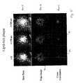
- FIG. 5is representative raw data speckle images, edge images, and cross-correlation images used to assess the viscosity of a lipid-rich, atherosclerotic plaque in a human aorta.
- FIG. 6is representative raw data speckle images, edge images, and cross-correlation images used to assess the viscosity of normal human aorta tissue.
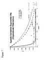
- FIG. 7is an exponential graph showing speckle decorrelation of a thin cap atherosclerotic plaque, a thick cap atherosclerotic plaque, and normal aortic tissue over a time interval.
- tissueis not static. Individual cells move within intercellular fluids, cellular organelles move within cells, and large molecules move back and forth between cells. In non-cellular tissue deposits such as plaques, components such as proteins, lipids, and other molecules also exhibit local motion. These local microscopic motions include “Brownian motion” and are essentially random in nature. Measuring and characterizing the microscopic motion of tissues can provide useful information about the structure, composition, biomechanical characteristics, and stability of the tissue.
- the inventionrelates to using laser speckle to measure microscopic motion, including Brownian motion, of tissue in vivo to gather information about the tissue.
- coherent or partially coherent lightis reflected from a tissue to form a speckle pattern at a detector. Due to motion of reflectors within the tissue, the speckle pattern changes over time, or is “decorrelated.” By monitoring the rate of decorrelation, while compensating for “extrinsic,” macroscopic motion of the tissue, microscopic motion in the tissue can be isolated and measured.
- the partially coherent lightcan provide more information about optical properties of the tissue than completely coherent light.
- speckle analysisis used to measure microscopic, e.g., Brownian, motion in atherosclerotic plaques to detect plaques that are vulnerable to rupture, and, more specifically, to determine the plaque's vulnerability to rupture.
- a modified optical catheter (probe) or other instrumentis inserted into a blood vessel (e.g., artery) to locate these plaques, and once a plaque is located, the probe is moved into the proximity of the specific atherosclerotic plaque.
- Light reflected from the interior wall of the blood vessels, and/or from a plaqueis collected and transmitted to a detector, where a speckle pattern is formed.
- speckle patterns of normal tissue and plaque tissueare different, and these differences can be used to detect the plaques. Thereafter, e.g., while compensating for macroscopic motion of the plaque, the speckle pattern is monitored over time to calculate the pattern's rate of decorrelation. From this decorrelation rate, the degree of microscopic motion in the plaque, and therefore the plaque's vulnerability to rupture, can be assessed.
- Atherosclerotic plaquecan lead to acute myocardial infarction, which is a leading cause of death in industrialized countries.
- myocardial infarctionWhen an atherosclerotic plaque ruptures, lipids from the plaque enter the vessel lumen, potentially causing thrombosis, arterial occlusion, myocardial ischemia, and infarction.
- plaques vulnerable to rupturegenerally have a thin, unstable, fibrous cap and a compliant, or less “viscous,” lipid pool.
- lipid poole.g., Virmani et al., “Lesions from sudden coronary death: A comprehensive morphological classification scheme for atherosclerotic lesions,” Arterioscler. Thromb. Vasc. Bio., 20:1262-75 (2000) and Lee et al., “The Unstable Atheroma,” Arteriosclerosis, Thrombosis & Vascular Biology, 17:1859-67 (1997).
- the less viscous lipid poolapplies force to the fibrous cap, compromising the cap and causing rupture.
- a specially modified optical catheter 10includes a rotatable inner shaft 12 and a transparent outer sheath 14 .
- the inner shaft 12houses a fiber array 15 and a mirror 16 near its distal end 18 .
- a central fiber 20 in the fiber arrayconnects to a fixed optical fiber 21 that extends from the catheter proximally to a light source 22 .
- coherent lightsuch as laser light
- beam-splitter 22 acoherent light from light source 22 is transmitted via beam-splitter 22 a , through the fixed optical fiber 21 and central fiber 20 and onto center 23 of mirror 16 .
- the lightis reflected to a tissue sample 24 , such as a layer of static tissue over a layer of moving tissue, such as an atherosclerotic plaque.
- Outer sheath 14can be placed directly in contact with sample 24 (near field), or can be positioned a short distance, e.g., 1 mm to 10 cm away from the sample (far field).
- Light remitted from the sample(arrows 26 ) reflects from mirror 16 to the fibers of array 15 , and is then transmitted by array 15 to a planar charge-coupled device (CCD), or a linear or two-dimensional detector 22 b , via a beam-splitter 22 a , e.g., located within light source 22 .
- CCDcharge-coupled device
- a beam-splitter 22 ae.g., located within light source 22 .
- There may be one or multiple fibers for detection and illumination and detectionmay occur from the same fiber.
- illuminationmay occur through a fiber array where each fiber is selectively illuminated to generate multiple speckle patterns as a function of position on the sample.
- This methodcan provide a scanning of the incident light across a sample while keeping the probe stationary by illuminating one fiber after another in series.
- a speckle patternfauns at the CCD detector.
- the resulting speckle patternis then digitized by an analog-digital converter, and analyzed using the procedures described in the analysis section below.
- the entire shaft 12can rotate 360 degrees in the direction of arrow R, allowing catheter 10 to gather images around the entire circumference of a sample.
- catheter 10can gather images of a plaque around the circumference of a vessel wall.
- the diameter of the cathetercan be less than 500 ⁇ m. Larger diameters are also possible.
- the optics of catheter 10can be integrated into other types of instruments, such as endoscopes or laparoscopes.
- the opticscan also form a stand-alone unit passed into the accessory port of standard endoscopes or laparoscopes, or integrated into another type of catheter, such as dual-purpose intravascular ultrasound catheter.
- the opticscan also include a lens that focuses the remitted light 26 onto the distal ends of the fibers in array 15 .
- the lenswould allow formation of a “near field image” (near the sample sight less than one wavelength) rather than a “far field image” (at the detector set more than a wavelength away from the surface of the tissue).
- the cathetercan include a polarization filter to remove all but a certain type of polarized light.
- a cross-polarized filterwould allow only light having a polarization perpendicular to the incident light to reach the detector, while a parallel polarized filter would allow only light having the same polarization as the incident light to pass.
- multiply scattered lightis less likely to retain its initial polarization than single scattered light
- polarization filterscan be used to bias the data toward multiply scattered or single scattered light. Such bias can be used to deduce information about the structure of the sample, since light which has penetrated deeper into the sample will be more highly scattered than light reflected from the surface or remitted from near the surface.
- the detectorcan be, e.g., a photographic plate, an array of photodetectors, or a single detector.
- the light sourcecan illuminate the sample with continuous light or synchronized pulses.
- the light sourcecan be, e.g., as far as one meter, or more, away from the sample.
- the temporal changes in the patternshould indicate movement of reflectors within the plaque, but not indicate movement of the plaque itself or movement of reflectors between the detector and the plaque.
- the changes in the plaque's speckle patternpreferably reflect microscopic or Brownian motion, but not macroscopic motion.
- the intervalFor a time interval to be sufficient to detect microscopic Brownian motion, the interval must be long enough to allow for movement of reflectors in the tissue, such as a lipid pool, but short enough that the random Brownian movements do not cancel out.
- an appropriate time intervalis about 1-200 ms. Shorter time periods may also be possible. If the time intervals are longer, then changes in the speckle pattern may not adequately differentiate rapid Brownian movement (indicating low viscosity) from slower Brownian movement (indicating high viscosity).
- the fiber array 15can be coupled to the plaque tissue using, e.g., an angioplasty balloon. This technique also compensates for minor patient movements.
- a balloon 28is attached to outer sheath 14 , on a far side 30 of the catheter. Once the catheter is positioned within a blood vessel in proximity to the plaque, the balloon is inflated. Referring to FIG. 2B , the inflated balloon abuts the vessel wall 32 , and presses the catheter against plaque 24 , such that a distal region of outer shaft 14 is in direct contact with the plaque. With the catheter coupled to plaque 24 as shown in FIG. 2B , fiber array 15 will move with the plaque when the heart beats, and the gross motion of the plaque will not significantly affect the speckle pattern.
- the ballooncan surround the catheter.
- a transparent balloonsurrounds outer sheath 14 , but is also attached to the sheath.
- the balloonis squeezed between plaque 24 and wall 32 of the vessel. The balloon, therefore will be in direct contact with the plaque, and will move with the plaque when the heart beats. Since the balloon is attached to shaft 14 , and shaft 14 is coupled to array 15 , movement of the vessel wall will not significantly affect the speckle pattern. Additional methods of coupling the catheter to tissue can also be used, including methods that do not employ an angioplasty balloon.
- a second method of compensating for movement caused by heartbeatsis to gather data between heartbeats.
- datais gathered during the relatively still PR interval of the diastole of the heartbeat (when the left ventricle is filling with blood).
- the PR intervallasts for about 0.12-0.2 seconds, providing sufficient time to detect Brownian motion.
- the timingcan be computer-controlled or the detector can be linked to an ECG signal, and programmed to gather data only during the PR interval. Similar techniques can be used to compensate for other bodily movements such as peristalsis.
- the cathetercan be placed in direct contact with the plaque tissue, as described above, thereby preventing blood from flowing between the detector and the plaque.
- blood flowing between the plaque and the cathetercan be removed and replaced with clear saline solution or other clear solutions such as optically transparent blood substitutes.
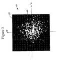
- FIG. 3illustrates a typical speckle pattern 40 formed by reflecting light from the wall of a healthy blood vessel.
- X and Y coordinatesoverlay pattern 40 to facilitate mathematical description of the pattern.
- the patternincludes dark patches, where destructive interference dominates, and brighter patches, where constructive interference dominates. Very subtle movements of reflectors within the multiply scattering sample alter the speckle pattern.
- plaque's vulnerability to rupturecan be assessed.
- This methodincludes: (1) gathering a series of speckle images at short, discrete time intervals; (2) eliminating diffuse reflectance from the data; (3) creating cross-correlation images comparing the speckle images in the series; (4) calculating the maximum correlation between each pair of images to create a one-dimensional data set over time; (5) calculating the rate of decorrelation from the data set; and (6) from the rate of decorrelation, assessing the plaque's viscosity and vulnerability to rupture.
- speckle imagesare gathered for a plaque at discrete intervals over a period of time.
- speckle imagescan be gathered, e.g., at intervals of every 1, 5, 10, 20, or 30 ms for a time period of, e.g., 200 ms.
- time periode.g. 200 ms.
- the shorter the time intervalsthe shorter the time period over which data can be gathered.
- time intervalssuch as 30 ms
- datacan be gathered for, e.g., 1-2 seconds.
- the background, non-coherent diffuse reflectanceis eliminated from the images.
- a number of techniquescan be used to eliminate the tissue's diffuse reflectance.
- the raw data speckle imagescan be converted to edge images.
- Edge imagesare spatial derivatives of the raw data images; an edge image (high pass filter) reflects the change in intensity of an image as a function of space, at all points in the image, rather than the intensity itself.
- Known techniques of edge detectioninclude convolution of the image by a kernel (e.g., Sobel or Robert), Morph gradient (subtraction of an eroded, dilated, closed, or opened image by its original), or high pass filtering.
- the cross-correlation imagesreflect the degree of correlation between the two images as a function of space.
- g(t)is the cross-correlation function
- I(x, y)is intensity of the interference at a point (x, y) in the pattern
- tis time.
- Two-dimensional cross-correlation functionsare described generally in Jae S.
- the cross-correlation imagesare reduced to a one-dimensional data set as a function of time (i.e., a series of correlation values, each value associated with a time t). From this series of correlation values, a time constant, ⁇ , is calculated, where ⁇ represents the rate of decorrelation.
- the time constantis the amount of time it takes g(t) to reach (1/e)g(0).
- the max function of equation (1)is not the only possible mechanism for reducing the cross-correlation images to a number.
- a point to reduce the image to a valuerather than a max function, would not compensate for the “memory effect” of first order correlation of speckle patterns in turbid media. This “memory effect” is described in Feng et al., Science 251: 633-39 (1991). Advantages to using a point are that a minimum number of fibers and detectors can be used.
- the viscosity of the plaque's lipid poolcan be assessed.
- the larger ⁇the lower the Brownian motion in the lipid pool, and the greater the pool's viscosity.
- the smaller ⁇the greater the Brownian motion in the lipid pool, and the lower the viscosity. The lower the viscosity, the more stresses are exerted on the cap, making the plaque more vulnerable
- This informationcan be used to identify plaques likely or vulnerable to rupture. Specifically, if i is about 40-100 ms or lower, then the plaque is likely to rupture, and intervention is warranted. If the plaque ⁇ is about 100-200 ms, then the plaque is somewhat vulnerable, but not yet likely to rupture, and should be monitored over time. If the plaque ⁇ is about 200-300 ms, then the plaque is less vulnerable. Non-plaque covered, healthy vessel wall generally has a time constant greater than 300 ms or 500 ms.
- the speckle datacan be analyzed to deduce spatial characteristics of the plaque, including the thickness of the fibrous cap, or the thickness of any tissue structure or tissue layer for that matter. As discussed above, a thin fibrous cap is another indication that a plaque is vulnerable to rupture. The combination of data relating to viscosity and cap thickness provides the most accurate assessment of plaque vulnerability, although the two characteristics can be assessed and analyzed independently.
- a typical plaque 50includes a fibrous cap 52 and a lipid pool 54 .
- Photons that enter plaque 50(arrow A) are internally scattered by reflectors within the plaque, such as collagen in fibrous cap 52 and lipids in pool 54 .
- the various photonstherefore, exit the plaque at different locations (arrows B).
- the speckle pattern(see FIG. 2 ) has a diameter considerably larger than the width of the original light beam.
- the thickness of fibrous cap 52can be deduced by comparing different regions of the resulting speckle pattern. Referring again to FIG. 3 , light forming intensity signals in the outer portion 60 of the pattern traveled greater distances than light forming signals near the center 62 of the pattern. Thus, outer portion 60 of the pattern is formed by photons that, on average, penetrated deeper into the plaque than photons forming center 62 . By calculating separate time constants for separate regions of the speckle pattern, the viscosity of the plaque at different depths can be determined. Since the fibrous cap generally exhibits less Brownian motion than the lipid pool, the thickness of the fibrous cap can be estimated from spatially dependent data.
- Time constantsare then calculated from the cross-correlation data for each window, in the manner described above.
- the variation of ⁇ as a function of the distance from the center of the speckle patterni.e., as a function of (x o 2 +y o 2 ) 1/2
- ⁇can then be analyzed to determine the thickness of the fibrous cap.
- Plaque cap thickness of less than about 60 ⁇ mis considered to be vulnerable, but this number can vary to some extent depending on the specific patient.
- any tissue structuree.g., a tissue layer that overlies or is adjacent to a different tissue, e.g., a plaque fibrous cap over a lipid pool
- a tissue layer that overlies or is adjacent to a different tissuee.g., a plaque fibrous cap over a lipid pool
- ⁇ (r) and the optical propertiese.g., ⁇ eff
- ⁇ (r) and the optical propertiese.g., ⁇ eff
- r 0 and ⁇ effmay be used as inputs to a look-up table containing tissue layer thickness values.
- separately analyzing different regions of a speckle patternalso allows decorrelation caused by macroscopic motion to be identified and removed from the analysis.
- macroscopic motion caused by gross movement of the plaque tissue or blood flowwill be directional, non-random, and global.
- Brownian motionwill be non-directional and non-uniform (or random).
- the position of maximums of cross-correlation functionswill shift along a vector ⁇ , which relates extrinsic motion of the sample with respect to the catheter or detection.
- Brownian motionwill decorrelate the speckle patterns in many random directions, and will result in a broadening of the cross-correlation peak and a decrease in correlation maximum above that predicted by linear motion.
- the rate of decorrelationcan be estimated from single pixel speckle images, rather than full, two-dimensional speckle patterns.
- a catheter with a single optical fibercould transmit data to a single detector, such as a photodiode.
- the speckle data gatheredwould be intensity at the spot as a function of time. From this data, a rate of decorrelation can be calculated directly or only as a function of time as opposed to space, without any spatial cross-correlation analysis.
- Imaging methods that detect single scattered lightsuch as optical coherence tomography (OCT) and confocal microscopy, can also be used. While these imaging methods are less sensitive to speckle modulation than the multiple scattering methods described above, they have the advantage of allowing localization of data to a single point within the sample. Such localization would allow measurement of biomechanical properties of the tissue in three dimensions.
- OCToptical coherence tomography
- motion of the scattererscan produce a Doppler shift on the returned light. The Doppler shift can provide a further basis for measuring viscosity in the sample.
- speckle images formed by reflecting laser light from a cadaveric atherosclerotic plaque in a human aortawere analyzed to assess the plaque's viscosity. A portion of normal aorta was also analyzed for comparison.
- edge detectionwas performed on the 60 raw speckle images, generating 60 edge images.
- the edge imagesreflect the spatial derivative of the raw speckle images (i.e., the light patches in the edge images of row B are locations where the intensity is changing as a function of space).
- Each cross-correlation imagewas generated by multiplying the Fourier transform of the reference image 70 by the complex conjugate of the Fourier transform of the image in question, and then calculating an inverse Fourier transform of the product.
- Image 72was formed by multiplying the Fourier transform of reference image 70 by the complex conjugate of the Fourier transform of image 70 , and then calculating the inverse Fourier transform of the product.
- Each cross-correlation imagerepresents the degree of correlation between the corresponding edge image and the reference edge image 70 (i.e., brighter spots are locations where there is a higher degree of correlation than at darker spots).
- the maximum cross-correlation peak(i.e., the correlation at the maximum point of correlation) was calculated using equation (1).
- the resulting data setincluded 60 cross-correlation values, each value associated with a time t.
- FIG. 6A set of images of normal aorta tissue is shown in FIG. 6 . These images are comparable to the set of images in FIG. 5 for a lipid-rich plaque in the same aorta and were imaged and processed in the same manner.
- the decorrelation raterepresented by the time constant ⁇
- the time constantwas 40 ms.
- the time constantwas 500 ms.
- the plaquewas borderline vulnerable. Thus, had this plaque been analyzed in vivo, using the procedures described above, a physician would have determined that the plaque was a possible candidate for rupture, and may have chosen to intervene, preventing a possible infarction.
- the methods described hereincan also be used to characterize diseased tissue other than atherosclerotic plaques.
- the microscopic and macroscopic constituents of diseased tissuediffer from normal non-pathologic counterparts.
- speckle patternscan be used to diagnose and characterize other tissue pathology such as neoplasia (cancer), infection, tissue viability, or healing response to injury.
- neoplasiacancer
- tumorstypically have an abnormal abundance of one cell type (clonal) and a surrounding abnormal supporting matrix. This cell type may produce and secrete a viscous fluid, such as mucin in adenocarcinoma, which would result in lower speckle decorrelation time constants than normal non-cancerous tissue.
- the surrounding matrixmay be composed of necrotic tissue and an abundance of abnormal vessels that would also serve to decrease the speckle decorrelation time constant.
- Other tumorslike osteosarcoma, produce osteoid or immature bone that would increase the time constant compared to normal tissue.
- Other forms of neoplasiawould have increased time constants due to desmoplastic (abundant) fibrous stroma initiated by cytokines produced by the tumor. Indeed, many tumors, including bronchogenic carcinomas and breast carcinomas are firm upon gross examination due to the fibrous stroma surrounding the malignant cells. This fibrous stroma would increase the time constant relative to surrounding normal tissue.
- abscesseswill be less viscous than surrounding tissue, enabling identification of the infected region by measuring a decrease in the time constant.
- Inflammationmanifested by the influx of activated inflammatory cells will be characterized by a decrease in the speckle decorrelation time constant as these cells degrade the normal supporting tissue in response to a the presence of bacterial, viral, or foreign body antigens.
- Necrotic tissuesuch as burn eschar, diabetic ulcers, necrotic bowel, and ischemic myocardium will have longer time constants than viable tissue from the same organ due to the lack of intravascular and extravascular fluid and flow in these extracellular spaces.
- Speckle decorrelation timesmay also be used to estimate tissue hydration and provide a means for quantifying the state of hydration in a patient. While the above examples elucidate some of the mechanisms that explain how disease affects the biomechanical properties of pathologic tissue, many more exist and are well known in the field of gross anatomic pathology. These differing biomechanical properties and characteristics can be measured by speckle for the purpose of screening, intraoperative margin (e.g., tumor margin) identification, and primary diagnosis.
- intraoperative margine.g., tumor margin
Landscapes
- Health & Medical Sciences (AREA)
- Life Sciences & Earth Sciences (AREA)
- Surgery (AREA)
- Engineering & Computer Science (AREA)
- Physics & Mathematics (AREA)
- General Health & Medical Sciences (AREA)
- Pathology (AREA)
- Public Health (AREA)
- Biomedical Technology (AREA)
- Heart & Thoracic Surgery (AREA)
- Medical Informatics (AREA)
- Molecular Biology (AREA)
- Animal Behavior & Ethology (AREA)
- Biophysics (AREA)
- Veterinary Medicine (AREA)
- Radiology & Medical Imaging (AREA)
- Optics & Photonics (AREA)
- Nuclear Medicine, Radiotherapy & Molecular Imaging (AREA)
- Signal Processing (AREA)
- Physiology (AREA)
- Computer Vision & Pattern Recognition (AREA)
- Artificial Intelligence (AREA)
- Psychiatry (AREA)
- Vascular Medicine (AREA)
- Cardiology (AREA)
- Chemical & Material Sciences (AREA)
- Analytical Chemistry (AREA)
- Biochemistry (AREA)
- General Physics & Mathematics (AREA)
- Immunology (AREA)
- Investigating Or Analysing Materials By Optical Means (AREA)
- Investigating Or Analysing Biological Materials (AREA)
- Investigating, Analyzing Materials By Fluorescence Or Luminescence (AREA)
Abstract
Description
This application claims benefit of priority from U.S. Provisional Patent Application Ser. No. 60/244,255, filed on Oct. 30, 2000. The present application is also a continuation of U.S. patent application Ser. No. 11/534,095 filed Sep. 21, 2006 that issued as U.S. Pat. No. 8,032,200 on Oct. 4, 2011, which is a continuation of U.S. patent application Ser. No. 10/016,244 filed Oct. 30, 2001 which issued as U.S. Pat. No. 7,231,243 on Jun. 12, 2007. The entire disclosures of the applications referenced herein are incorporated herein by reference.
The invention relates to tissue analysis, and more particularly to characterizing tissue by analyzing speckle patterns formed by light reflected from tissue.
“Speckle” is an interference phenomenon that occurs when coherent light (e.g., laser light) is reflected from a rough or multiply scattering sample onto a detection plane. Due to scattering of photons from and within the sample, different photons travel different distances to the detection plane. As a result, the light reflected or backscattered from the sample, if spatially and temporally coherent, interferes at the detection plane, producing a grainy pattern known as “speckle.”
Researchers have used speckle pattern analysis to study dynamic movement of tissue in vivo. For example, speckle has been used to measure vibrations of tissue, V. Tuchin et al., “Speckle interferometry in the measurements of biotissues vibrations,”SPIE,1647: 125 (1992), and to measure strain in vascular and cortical tissue in response to forced movement of the tissue, Sean J. Kirpatrick et al., “Laser Speckle Microstrain Measurement in Vascular Tissue,”SPIE,3598: 121-128 (1999); and Sean J. Kirkpatrick and Brent W. Brooks, “Micromechanical Behavior of Cortical Bone as Inferred from Laser Speckle Data,”J. Biomedical Materials Research,39(3): 373-79 (1998). Researchers have also used speckle to study blood flow and lymph flow. B. Ruth, “Blood Flow Determination by the Laser Speckle Method,”Int'l J. Microcirc: Clinical and Experimental,9(1): 21-45 (1990); and A. A. Bednov et al., “Investigation of Statistical Properties of Lymph Flow Dynamics Using Speckle-Microscopy,”SPIE,2981: 181-90 (1997).
The invention is based on the discovery that tissues can be analyzed in vivo using laser speckle to measure microscopic motion, e.g., Brownian motion, of structures and characteristics within the tissue.
In general, the invention features a method of analyzing tissue, e.g., in vivo, by illuminating a tissue with coherent light, such as laser light, or partially coherent light; receiving light reflected from the tissue at a detector to form a series of speckle patterns; and analyzing changes in the speckle patterns at time intervals sufficient to measure changes caused by motion of objects within the tissue on a microscopic scale, e.g., less than about 1 mm (e.g., less than about 500 or 100 microns), such as Brownian motion of molecules or macromolecules, or motion of cells or cellular organelles, or other non-random forms of motion such as lymph or intracellular transmembrane flow, while eliminating motion on a macroscopic scale, e.g., greater than about 1 mm.
For example, the speckle patterns can be measured at a near field or at a far field and imaged onto the detector. “Near field” is measurement of the speckle distribution less than one wavelength of light from the surface of a tissue, while “far field” speckle is the interference pattern formed greater than one wavelength of light from the surface. The method can further include compensating for macroscopic or extrinsic motion, such as a heartbeat, patient motion, or peristalsis, to isolate the microscopic, e.g., Brownian, motion.
In this method, the illuminating step can include providing an invasive device coupled to a light source, passing the device into a patient, placing the device in proximity to the tissue, and shining coherent light or partially coherent light from the light source onto the tissue.
The invasive device can be, e.g., a catheter, an endoscope, or a laparoscope. The device can be placed in direct contact with the tissue (to measure a near field speckle pattern) or may be a given distance from the tissue (to measure a far field or near field speckle pattern). The device can include a catheter having a first fiber (or fiber array or bundle) that transmits light from the light source to the tissue, and a fiber array or single fiber that receives light remitted from the tissue. The fiber arrays can be one or two-dimensional. The analyzing step can include comparing each of the series of speckle patterns to a series of reference speckle patterns, and quantifying the temporal correlation differences between the patterns and the reference patterns. For example, the analyzing step can include digitizing each of the speckle patterns as a function of time and space, and the quantifying step can include evaluating a cross-correlation between the patterns and the reference patterns. The analyzing step can further include determining a decorrelation rate for the speckle patterns, or analyzing spatial characteristics of the speckle pattern to deduce structural and/or biomechanical characteristics of the tissue. Biomechanical characteristics can include, for example, compliance, elasticity, stress, strain, and viscosity. In these methods, speckle pattern data is a snapshot taken at a specific point in time. Speckle pattern correlation data is a measurement of cross-correlation of the speckle pattern as a function of time.
In variations, the method can include illuminating multiple locations of the tissue in succession, forming a separate series of speckle patterns for each respective location of the tissue, and then analyzing each separate series of speckle patterns and comparing the separate series to deduce structural and/or biomechanical differences between the respective locations of the tissue.
In certain embodiments, the method includes gathering reflected light at a light receptor and transmitting the gathered light to the detector, and compensating for macroscopic motion by coupling the receptor to the tissue. Compensating for macroscopic motion can also be done by excluding changes in the speckle patterns caused by non-random motion during the analysis step. Macroscopic or extrinsic motion can also result, for example, from blood flowing between the tissue and the reflector. In those cases, the compensating step can include replacing the blood with a transparent solution and/or eliminating correlated speckle pattern information corresponding to directional blood flow.
In another embodiment, the invention features a method of analyzing a tissue structure, e.g., for determining the susceptibility to rupture of an atherosclerotic plaque having a lipid pool and a fibrous cap. The method includes illuminating the tissue structure, e.g., plaque, with coherent or partially coherent light; receiving light reflected from the tissue structure at a detector to form a series of speckle patterns; gathering speckle pattern data at time intervals sufficient to measure microscopic motion, e.g., Brownian motion or other forms of microscopic motion, within the tissue structure or tissue adjacent the tissue structure, such as a lipid pool; and assessing the tissue structure, e.g., assessing a plaque's vulnerability to rupture from the amount of Brownian motion.
The method can further include analyzing spatial characteristics of the speckle pattern data to determine structural and/or biomechanical characteristics of the tissue structure, e.g., plaque, for example, by assessing the thickness of the tissue structure, e.g., fibrous cap. The thickness of the tissue can be determined by measuring the spatial and temporal decorrelation of the speckle pattern as a function of distance from the incident beam entry point. Near the beam entry point, the speckle pattern will be more stationary. Far away from the bean entry point, the speckle pattern will decorrelate more rapidly. The location of the transition is an indication of thickness. Other methods for determining thickness are described herein. A plaque is considered vulnerable to rupture if the thickness of the fibrous cap is less than about 60 microns. The method can also be used to assess the viscosity of the lipid pool, wherein the plaque is considered vulnerable to rupture if the viscosity of the lipid pool has a time constant of less than about 200 milliseconds, and considered likely to rupture if the viscosity of the lipid pool has a time constant of less than about 100 milliseconds.
The invention also includes a method of detecting a vulnerable atherosclerotic plaque having a lipid pool and a fibrous cap within a blood vessel by illuminating a segment of the blood vessel in vivo with coherent or partially coherent light; receiving light reflected from the interior vessel wall of the segment at a detector to form a series of speckle patterns; gathering speckle pattern data at time intervals sufficient to measure microscopic, e.g., Brownian, motion within the interior vessel wall; and comparing the speckle pattern correlation data to a known speckle pattern time correlation data. One means for comparing the measured speckle pattern correlation data with a reference speckle pattern correlation data is by the time constant, or the time it takes for the speckle pattern to decorrelate by 1/e. For example, the decorrelation time constant for any given segment of vessel may be measured and compared to known time constants for normal vessels, atherosclerotic vessels, lipid pools with thick fibrous caps and lipid pools with thin fibrous caps (vulnerable plaques). If the time constant indicates the presence of a lipid pool (τ<100 ms), with a thin fibrous cap, spatial characteristics of the speckle pattern data can be further analyzed to determine structural characteristics of the plaque as described herein. In addition, the first (mean) and second (standard deviation) of the probability distribution function pattern (histogram) of the speckle pattern is unique for different plaque types.
In another aspect, the invention features a fiber optic probe for detecting speckle patterns in a sample. The probe includes a catheter including a rotatable inner shaft and a transparent outer sheath; a fiber array or single fiber housed within the shaft and comprising one or more first optical fibers for transmitting incident light to the sample, and one or more second optical fibers for transmitting light remitted from the sample; and a mirror arranged near a distal end of the shaft to reflect light passing through the fiber array onto a sample outside the transparent outer sheath and back from the sample through the fiber array. The fiber array can include one (or several) incident light transmitting fiber, one (or more) remitted light transmitting fiber, and the incident light transmitting fiber can be selected from the array, and thereafter a different fiber can be selected, e.g., in series, to scan the incident light across the sample without moving the probe.
The beam emanating from the one or more first optical fibers can be focused onto the tissue by a lens, and the speckle pattern can be imaged onto the detection fiber array or onto a single detection fiber by a lens. In some embodiments, the shaft can rotate 360 degrees within the sheath, and an inflatable balloon can be connected to the sheath.
The invention further includes an optical system for detecting speckle patterns in a sample. The system has a fiber optic probe as described herein; a coherent or partially coherent light source connected to the central optical fiber within the fiber array; a detector to receive light remitted from the sample; and a processor to process the remitted light and to analyze speckle patterns remitted from the sample. For example, the processor can include reference speckle pattern time constants or a whole library of reference speckle pattern time constants, or reference speckle pattern correlation curves, e.g., for healthy and diseased tissue. The system can also include an analog-digital converter to convert the analog remitted light into a digital signal.
As used herein, “tissue” means any biological structure in or on a body. Tissue includes aggregates of cells, growths, and deposits such as plaque that may contain lipids or other components. Specific components of plaques that can be investigated include lipid pools, calcifications, fibrous regions, and fibrous caps.
“Speckle” is an interference phenomenon that occurs when coherent or partially coherent light is reflected from a rough or multiply scattering sample onto a detection plane. A “speckle pattern” is the intensity pattern that results from interference.
“Brownian motion” is the random motion of cells, molecules, and other subcomponents within tissue.
“Coherence” is the property of light that allows interference of two or more optical waves. “Partial coherence” refers to waves that can interfere with each other if the path traveled by each wave is equivalent to or within the temporal coherence length of the light at any given point in the specimen.
Unless otherwise defined, all technical and scientific terms used herein have the same meaning as commonly understood by one of ordinary skill in the art to which this invention belongs. Although methods and materials similar or equivalent to those described herein can be used in the practice or testing of the present invention, suitable methods and materials are described below. All publications, patent applications, patents, and other references mentioned herein are incorporated by reference in their entirety. In case of conflict, the present specification, including definitions, will control. In addition, the materials, methods, and examples are illustrative only and are not intended to be limiting.
Other features and advantages of the invention will be apparent from the following detailed description and from the claims.
At a microscopic level, most tissue is not static. Individual cells move within intercellular fluids, cellular organelles move within cells, and large molecules move back and forth between cells. In non-cellular tissue deposits such as plaques, components such as proteins, lipids, and other molecules also exhibit local motion. These local microscopic motions include “Brownian motion” and are essentially random in nature. Measuring and characterizing the microscopic motion of tissues can provide useful information about the structure, composition, biomechanical characteristics, and stability of the tissue.
The invention relates to using laser speckle to measure microscopic motion, including Brownian motion, of tissue in vivo to gather information about the tissue. In general, coherent or partially coherent light is reflected from a tissue to form a speckle pattern at a detector. Due to motion of reflectors within the tissue, the speckle pattern changes over time, or is “decorrelated.” By monitoring the rate of decorrelation, while compensating for “extrinsic,” macroscopic motion of the tissue, microscopic motion in the tissue can be isolated and measured. The partially coherent light can provide more information about optical properties of the tissue than completely coherent light.
In some embodiments of the invention, speckle analysis is used to measure microscopic, e.g., Brownian, motion in atherosclerotic plaques to detect plaques that are vulnerable to rupture, and, more specifically, to determine the plaque's vulnerability to rupture. In these embodiments, a modified optical catheter (probe) or other instrument is inserted into a blood vessel (e.g., artery) to locate these plaques, and once a plaque is located, the probe is moved into the proximity of the specific atherosclerotic plaque. Light reflected from the interior wall of the blood vessels, and/or from a plaque, is collected and transmitted to a detector, where a speckle pattern is formed. The speckle patterns of normal tissue and plaque tissue (especially vulnerable plaque tissue) are different, and these differences can be used to detect the plaques. Thereafter, e.g., while compensating for macroscopic motion of the plaque, the speckle pattern is monitored over time to calculate the pattern's rate of decorrelation. From this decorrelation rate, the degree of microscopic motion in the plaque, and therefore the plaque's vulnerability to rupture, can be assessed.
I. Atherosclerotic Plaques
Rupture of an atherosclerotic plaque can lead to acute myocardial infarction, which is a leading cause of death in industrialized countries. When an atherosclerotic plaque ruptures, lipids from the plaque enter the vessel lumen, potentially causing thrombosis, arterial occlusion, myocardial ischemia, and infarction.
According to recent research, plaques vulnerable to rupture generally have a thin, unstable, fibrous cap and a compliant, or less “viscous,” lipid pool. See, e.g., Virmani et al., “Lesions from sudden coronary death: A comprehensive morphological classification scheme for atherosclerotic lesions,” Arterioscler. Thromb. Vasc. Bio., 20:1262-75 (2000) and Lee et al., “The Unstable Atheroma,” Arteriosclerosis, Thrombosis & Vascular Biology, 17:1859-67 (1997). The less viscous lipid pool applies force to the fibrous cap, compromising the cap and causing rupture. The greater the Brownian motion in the lipid pool, the lower the “viscosity” of the pool, and the more likely the plaque will rupture. Assessing Brownian motion in the lipid pool and measuring the thickness of the fibrous cap in vivo, therefore, helps to identify plaques likely to rupture, allowing intervention.
II. Speckle Image Formation
Referring toFIG. 1 , a specially modifiedoptical catheter 10 includes a rotatableinner shaft 12 and a transparentouter sheath 14. Theinner shaft 12 houses afiber array 15 and amirror 16 near itsdistal end 18. Acentral fiber 20 in the fiber array connects to a fixedoptical fiber 21 that extends from the catheter proximally to a light source22.
In operation, coherent light, such as laser light, from light source22 is transmitted via beam-splitter22a, through the fixedoptical fiber 21 andcentral fiber 20 and ontocenter 23 ofmirror 16. Frommirror 16, the light is reflected to atissue sample 24, such as a layer of static tissue over a layer of moving tissue, such as an atherosclerotic plaque.Outer sheath 14 can be placed directly in contact with sample24 (near field), or can be positioned a short distance, e.g., 1 mm to 10 cm away from the sample (far field). Light enterssample 24, where it is reflected by molecules, cellular debris, proteins, compounds (e.g., cholesterol crystals), and cellular microstructures (such as organelles, microtubules) within the sample. Light remitted from the sample (arrows26) reflects frommirror 16 to the fibers ofarray 15, and is then transmitted byarray 15 to a planar charge-coupled device (CCD), or a linear or two-dimensional detector 22b, via a beam-splitter22a, e.g., located within light source22. There may be one or multiple fibers for detection and illumination and detection may occur from the same fiber. Alternatively, illumination may occur through a fiber array where each fiber is selectively illuminated to generate multiple speckle patterns as a function of position on the sample. This method can provide a scanning of the incident light across a sample while keeping the probe stationary by illuminating one fiber after another in series.
Due to interference, a speckle pattern fauns at the CCD detector. The resulting speckle pattern is then digitized by an analog-digital converter, and analyzed using the procedures described in the analysis section below.
Theentire shaft 12 can rotate360 degrees in the direction of arrow R, allowingcatheter 10 to gather images around the entire circumference of a sample. For example,catheter 10 can gather images of a plaque around the circumference of a vessel wall.
Since only a few fibers are required to gather adequate speckle data, the diameter of the catheter can be less than 500 μm. Larger diameters are also possible.
Many other types of instruments can be used to gather speckle data. For example, the optics ofcatheter 10 can be integrated into other types of instruments, such as endoscopes or laparoscopes. The optics can also form a stand-alone unit passed into the accessory port of standard endoscopes or laparoscopes, or integrated into another type of catheter, such as dual-purpose intravascular ultrasound catheter.
The optics can also include a lens that focuses the remitted light26 onto the distal ends of the fibers inarray 15. The lens would allow formation of a “near field image” (near the sample sight less than one wavelength) rather than a “far field image” (at the detector set more than a wavelength away from the surface of the tissue).
The catheter can include a polarization filter to remove all but a certain type of polarized light. For example, a cross-polarized filter would allow only light having a polarization perpendicular to the incident light to reach the detector, while a parallel polarized filter would allow only light having the same polarization as the incident light to pass. Since multiply scattered light is less likely to retain its initial polarization than single scattered light, polarization filters can be used to bias the data toward multiply scattered or single scattered light. Such bias can be used to deduce information about the structure of the sample, since light which has penetrated deeper into the sample will be more highly scattered than light reflected from the surface or remitted from near the surface.
Instead of a CCD, the detector can be, e.g., a photographic plate, an array of photodetectors, or a single detector. The light source can illuminate the sample with continuous light or synchronized pulses.
Rather than transmitting the light to the sample through optical fibers, it is also possible to shine light onto a sample in free space. For example, in an open surgical procedure, coherent light in free space could be directed onto a sample with mirrors, and the remitted light then directed to a fiber array. In such free space embodiments, the light source can be, e.g., as far as one meter, or more, away from the sample.
III. Isolation of Microscopic Motion
To simplify determining the viscosity of a moving tissue or liquid (e.g., a plaque's lipid pool) under a static tissue (e.g., a plaque cap) from changes in a speckle pattern, the temporal changes in the pattern should indicate movement of reflectors within the plaque, but not indicate movement of the plaque itself or movement of reflectors between the detector and the plaque. In other words, the changes in the plaque's speckle pattern preferably reflect microscopic or Brownian motion, but not macroscopic motion.
To isolate microscopic motion, data is gathered: (1) at time intervals sufficient to detect microscopic motion; and (2) in a manner that compensates for macroscopic (e.g., extrinsic) motion.
For a time interval to be sufficient to detect microscopic Brownian motion, the interval must be long enough to allow for movement of reflectors in the tissue, such as a lipid pool, but short enough that the random Brownian movements do not cancel out. For atherosclerotic plaque, an appropriate time interval is about 1-200 ms. Shorter time periods may also be possible. If the time intervals are longer, then changes in the speckle pattern may not adequately differentiate rapid Brownian movement (indicating low viscosity) from slower Brownian movement (indicating high viscosity).
In the atherosclerotic plaque and other examples, two common sources of macroscopic motion are gross movement of the vessel lumen and plaque tissue due to heartbeats, and blood flow between the plaque and the catheter. Patient movement can also be an issue.
To compensate for gross motion of a target tissue (e.g., a plaque) due to heartbeats, at least two alternatives are possible. First, thefiber array 15 can be coupled to the plaque tissue using, e.g., an angioplasty balloon. This technique also compensates for minor patient movements. Referring toFIG. 2A , in one embodiment, aballoon 28 is attached toouter sheath 14, on afar side 30 of the catheter. Once the catheter is positioned within a blood vessel in proximity to the plaque, the balloon is inflated. Referring toFIG. 2B , the inflated balloon abuts thevessel wall 32, and presses the catheter againstplaque 24, such that a distal region ofouter shaft 14 is in direct contact with the plaque. With the catheter coupled toplaque 24 as shown inFIG. 2B ,fiber array 15 will move with the plaque when the heart beats, and the gross motion of the plaque will not significantly affect the speckle pattern.
Other methods of coupling the catheter to the plaque are also possible. For example, instead of placing the balloon to the side of the catheter, the balloon can surround the catheter. In this arrangement, a transparent balloon surroundsouter sheath 14, but is also attached to the sheath. When the balloon is inflated, the balloon is squeezed betweenplaque 24 andwall 32 of the vessel. The balloon, therefore will be in direct contact with the plaque, and will move with the plaque when the heart beats. Since the balloon is attached toshaft 14, andshaft 14 is coupled toarray 15, movement of the vessel wall will not significantly affect the speckle pattern. Additional methods of coupling the catheter to tissue can also be used, including methods that do not employ an angioplasty balloon.
A second method of compensating for movement caused by heartbeats is to gather data between heartbeats. In this method, data is gathered during the relatively still PR interval of the diastole of the heartbeat (when the left ventricle is filling with blood). The PR interval lasts for about 0.12-0.2 seconds, providing sufficient time to detect Brownian motion. To insure that data is gathered during diastole, the timing can be computer-controlled or the detector can be linked to an ECG signal, and programmed to gather data only during the PR interval. Similar techniques can be used to compensate for other bodily movements such as peristalsis.
To compensate for blood flow between the catheter and the plaque, the catheter can be placed in direct contact with the plaque tissue, as described above, thereby preventing blood from flowing between the detector and the plaque. Alternatively, blood flowing between the plaque and the catheter can be removed and replaced with clear saline solution or other clear solutions such as optically transparent blood substitutes.
Finally, rather than compensating for macroscopic motion while gathering data, one can compensate for this motion during the analysis phase by mathematically excluding the macroscopic (extrinsic) motion from the analysis, as described below.
IV. Analysis of Speckle Data
By analyzing a series of speckle patterns formed from light reflected from a plaque, one can estimate: (a) the viscosity of a plaque's lipid pool; and (b) the thickness of plaque's fibrous cap. From either or both of these types of data, the plaque's vulnerability to rupture can be assessed.
A. Determining Viscosity of a Plaque's Lipid Pool
There are a number of methods of analyzing speckle data to determine the viscosity of a plaque's lipid pool. By way of example, one method is described in detail in this section and in the Example section below. This method includes: (1) gathering a series of speckle images at short, discrete time intervals; (2) eliminating diffuse reflectance from the data; (3) creating cross-correlation images comparing the speckle images in the series; (4) calculating the maximum correlation between each pair of images to create a one-dimensional data set over time; (5) calculating the rate of decorrelation from the data set; and (6) from the rate of decorrelation, assessing the plaque's viscosity and vulnerability to rupture.
First, using the detection system described above, a series of speckle images are gathered for a plaque at discrete intervals over a period of time. For example, speckle images can be gathered, e.g., at intervals of every 1, 5, 10, 20, or 30 ms for a time period of, e.g., 200 ms. In general, the shorter the time intervals, the shorter the time period over which data can be gathered. For longer time intervals, such as 30 ms, data can be gathered for, e.g., 1-2 seconds.
Second, to isolate the speckle pattern, the background, non-coherent diffuse reflectance is eliminated from the images. A number of techniques can be used to eliminate the tissue's diffuse reflectance. For example, the raw data speckle images can be converted to edge images. Edge images are spatial derivatives of the raw data images; an edge image (high pass filter) reflects the change in intensity of an image as a function of space, at all points in the image, rather than the intensity itself. Known techniques of edge detection include convolution of the image by a kernel (e.g., Sobel or Robert), Morph gradient (subtraction of an eroded, dilated, closed, or opened image by its original), or high pass filtering. Other methods of eliminating background diffuse reflectance include homomorphic filtering, local histogram equalization, or using an optical setup with a small aperture. All of these techniques are well known, and are described, e.g., in Gonzalez, R. C. and Wintz, P., “Digital Image Processing” (Addison-Wesley Publishing Company, Reading Mass., 1987) and Jain, Anil, K., “Fundamentals of Digital Image Processing” (Prentice Hall, Englewood Cliffs, N.J., 1987).
After eliminating the non-coherent background reflectance, each speckle image (or edge image) is compared to a reference image in the series (e.g., the t=0 image) to create a series of cross-correlation images. The cross-correlation images reflect the degree of correlation between the two images as a function of space. From each cross-correlation image, the maximum correlation peak (i.e., the amount of correlation at the point of maximum correlation) is determined using the equation:
g(t)=max[∫∫I(x, y,0)I(x+x′, y+y′, t)dx′dy′] (1)
where g(t) is the cross-correlation function, I(x, y) is intensity of the interference at a point (x, y) in the pattern, and t is time. Two-dimensional cross-correlation functions are described generally in Jae S. Lim, “Two-Dimensional Signal Processing” (Prentice Hall, Englewood Cliffs, N.J., 1990) and Jain, Anil, K., “Fundamentals of Digital Image Processing” (Prentice Hall, Englewood Cliffs, N.J., 1987).
By performing the maximum correlation calculations, the cross-correlation images are reduced to a one-dimensional data set as a function of time (i.e., a series of correlation values, each value associated with a time t). From this series of correlation values, a time constant, τ, is calculated, where τ represents the rate of decorrelation. The time constant is the amount of time it takes g(t) to reach (1/e)g(0).
The max function of equation (1) is not the only possible mechanism for reducing the cross-correlation images to a number. For example, image comparisons can be reduced to a representative value by evaluating the cross-correlation function:
g(x, y,t)=∫∫I(x, y,0)I(x+x′, y+y′, t)dx′dy′ (2)
at a point, such as x=y=0. However, using a point to reduce the image to a value, rather than a max function, would not compensate for the “memory effect” of first order correlation of speckle patterns in turbid media. This “memory effect” is described in Feng et al.,Science251: 633-39 (1991). Advantages to using a point are that a minimum number of fibers and detectors can be used.
From the rate of decorrelation, represented by time constant T, the viscosity of the plaque's lipid pool can be assessed. In general, the larger τ, the lower the Brownian motion in the lipid pool, and the greater the pool's viscosity. On the other hand, the smaller τ, the greater the Brownian motion in the lipid pool, and the lower the viscosity. The lower the viscosity, the more stresses are exerted on the cap, making the plaque more vulnerable
This information, the viscosity of the plaque's lipid pool, can be used to identify plaques likely or vulnerable to rupture. Specifically, if i is about 40-100 ms or lower, then the plaque is likely to rupture, and intervention is warranted. If the plaque τ is about 100-200 ms, then the plaque is somewhat vulnerable, but not yet likely to rupture, and should be monitored over time. If the plaque τ is about 200-300 ms, then the plaque is less vulnerable. Non-plaque covered, healthy vessel wall generally has a time constant greater than 300 ms or 500 ms.
B. Determining Thickness of a Tissue Structure Such as a Plaque's Fibrous Cap
In addition to determining the viscosity of the lipid pool in the plaque, the speckle data can be analyzed to deduce spatial characteristics of the plaque, including the thickness of the fibrous cap, or the thickness of any tissue structure or tissue layer for that matter. As discussed above, a thin fibrous cap is another indication that a plaque is vulnerable to rupture. The combination of data relating to viscosity and cap thickness provides the most accurate assessment of plaque vulnerability, although the two characteristics can be assessed and analyzed independently.
Referring toFIG. 4 , a typical plaque50 includes afibrous cap 52 and alipid pool 54. Photons that enter plaque50 (arrow A) are internally scattered by reflectors within the plaque, such as collagen infibrous cap 52 and lipids inpool 54. The various photons, therefore, exit the plaque at different locations (arrows B). As a result, the speckle pattern (seeFIG. 2 ) has a diameter considerably larger than the width of the original light beam.
The thickness offibrous cap 52 can be deduced by comparing different regions of the resulting speckle pattern. Referring again toFIG. 3 , light forming intensity signals in theouter portion 60 of the pattern traveled greater distances than light forming signals near thecenter 62 of the pattern. Thus,outer portion 60 of the pattern is formed by photons that, on average, penetrated deeper into the plaque thanphotons forming center 62. By calculating separate time constants for separate regions of the speckle pattern, the viscosity of the plaque at different depths can be determined. Since the fibrous cap generally exhibits less Brownian motion than the lipid pool, the thickness of the fibrous cap can be estimated from spatially dependent data.
To estimate the thickness of the fibrous cap, separate max cross-correlation functions are described for separate, small regions of the pattern. Each region is defined by a window, w, centered at (xoyo):
g(xo,yo,t)=max [∫∫w(x′−xo,y′−yo)I(x, y,0)I(x+x′, y+y′, t)dx′dy′] (3)
g(xo,yo,t)=max [∫∫w(x′−xo,y′−yo)I(x, y,0)I(x+x′, y+y′, t)dx′dy′] (3)
Time constants are then calculated from the cross-correlation data for each window, in the manner described above. The variation of τ as a function of the distance from the center of the speckle pattern (i.e., as a function of (xo2+yo2)1/2) can then be analyzed to determine the thickness of the fibrous cap. Plaque cap thickness of less than about 60 μm is considered to be vulnerable, but this number can vary to some extent depending on the specific patient.
In general, the thickness of any tissue structure, e.g., a tissue layer that overlies or is adjacent to a different tissue, e.g., a plaque fibrous cap over a lipid pool, can be measured using the following algorithm:
1. Measure the decorrelation time constant τ as a function of τ=(xo2+yo2)1/2
2. Measure the optical properties (e.g., effective attenuation coefficient μeff) of the tissue layer by computing the first and second order statistics of a speckle probability distribution function (PDF)(histogram), or by using diffuse reflectance spectrophotometry.
3. Compare τ(r) and the optical properties (e.g., μeff) to previously computed Monte Carlo or Diffusion theory simulations of τ(r) and μeffas a function of tissue layer thickness. Alternatively, if r0is defined as the cutoff between static and non-stationary speckle, r0and μeffmay be used as inputs to a look-up table containing tissue layer thickness values.
C. Mathematical Compensation for Macroscopic Motion
In addition to providing information about the structural features of a tissue such as a plaque, separately analyzing different regions of a speckle pattern also allows decorrelation caused by macroscopic motion to be identified and removed from the analysis. In general, macroscopic motion caused by gross movement of the plaque tissue or blood flow will be directional, non-random, and global. By contrast, Brownian motion will be non-directional and non-uniform (or random). Thus, by calculating separate decorrelation functions for different regions of the speckle pattern, decorrelation due to extrinsic motion can be identified and subtracted from the functions, allowing isolation of random, Brownian motion. For example, the position of maximums of cross-correlation functions will shift along a vectorν , which relates extrinsic motion of the sample with respect to the catheter or detection. Brownian motion will decorrelate the speckle patterns in many random directions, and will result in a broadening of the cross-correlation peak and a decrease in correlation maximum above that predicted by linear motion. These two behaviors for intrinsic and extrinsic linear motion should be separable from the cross-correlation function.
V. Additional Imaging Methods
In a simplified system, the rate of decorrelation can be estimated from single pixel speckle images, rather than full, two-dimensional speckle patterns. In this system, a catheter with a single optical fiber could transmit data to a single detector, such as a photodiode. The speckle data gathered would be intensity at the spot as a function of time. From this data, a rate of decorrelation can be calculated directly or only as a function of time as opposed to space, without any spatial cross-correlation analysis.
Imaging methods that detect single scattered light, such as optical coherence tomography (OCT) and confocal microscopy, can also be used. While these imaging methods are less sensitive to speckle modulation than the multiple scattering methods described above, they have the advantage of allowing localization of data to a single point within the sample. Such localization would allow measurement of biomechanical properties of the tissue in three dimensions. In addition, in methods that use heterodyne detection, such as OCT, motion of the scatterers can produce a Doppler shift on the returned light. The Doppler shift can provide a further basis for measuring viscosity in the sample. For Brownian motion the velocities would be distributed over a range of velocities and directions causing multiple Doppler shifts and a broadening of the frequency bandwidth distribution. The mathematics for OCT and confocal microscopy based imaging techniques would be substantially similar to the mathematics described above.
In this example, speckle images formed by reflecting laser light from a cadaveric atherosclerotic plaque in a human aorta were analyzed to assess the plaque's viscosity. A portion of normal aorta was also analyzed for comparison.
At a temperature of 37° C., light from a helium-neon laser (λ=632.8 nm) was shined on a cadaveric aortic plaque for two seconds. Light reflected from the plaque was received at a CCD camera with a shutter speed of30 frames per second, through a cross-polarization filter. During the two seconds, the CCD camera recorded a series of 60 speckle images at intervals of 33 ms. Three of the 60 raw data images, corresponding to times t=0, t=150 ms, and t=300 ms, are shown in row A ofFIG. 5 .
Using IPLab® Spectrum® imaging software, edge detection was performed on the 60 raw speckle images, generating 60 edge images. The three edge images for times t=0, t=150 ms, and t=300 ms are shown in row B ofFIG. 5 . As discussed above, the edge images reflect the spatial derivative of the raw speckle images (i.e., the light patches in the edge images of row B are locations where the intensity is changing as a function of space).
Using the same software, each of the 60 edge images was then compared to the t=0edge image 70 to form 60 cross-correlation images. Each cross-correlation image was generated by multiplying the Fourier transform of thereference image 70 by the complex conjugate of the Fourier transform of the image in question, and then calculating an inverse Fourier transform of the product. For example, referring to row C ofFIG. 5 , image72 is an autocorrelation of the t=0 edge image. Image72 was formed by multiplying the Fourier transform ofreference image 70 by the complex conjugate of the Fourier transform ofimage 70, and then calculating the inverse Fourier transform of the product.Image 74 was formed by multiplying the Fourier transform ofimage 70 by the complex conjugate of the Fourier transform of the t=150 ms edge image, and then calculating the inverse Fourier transform of the product.
Each cross-correlation image represents the degree of correlation between the corresponding edge image and the reference edge image70 (i.e., brighter spots are locations where there is a higher degree of correlation than at darker spots).
From each cross-correlation image, the maximum cross-correlation peak (i.e., the correlation at the maximum point of correlation) was calculated using equation (1). The resulting data set included 60 cross-correlation values, each value associated with a time t.
A set of images of normal aorta tissue is shown inFIG. 6 . These images are comparable to the set of images inFIG. 5 for a lipid-rich plaque in the same aorta and were imaged and processed in the same manner.
The maximum cross-correlation data for the lipid-rich plaque and the normal aorta tissue were then fit to an exponential cross-correlation function, G(τ), using Igor Pro®, v 3.01 software (Wavemetrics, Inc.). The resulting exponential function was graphed inFIG. 7 ,curve 80. By way of comparison, the exponential cross-correlation function for speckle data taken from healthy cadaveric aortic tissue is shown incurve 82. The data forcurve 82 was gathered and processed using the same procedures as the data forcurve 80.FIG. 7 also shows the data for a thick-capped plaque (curve81). The time constant for this non-vulnerable plaque was 400 ms, so the plaque would not need intervention. Again, the same techniques were used to generate this curve ascurves
From the cross-correlation data, the decorrelation rate, represented by the time constant τ, was calculated. For the plaque, the time constant was 40 ms. For the aortic tissue, the time constant was 500 ms.
Based on these data, the plaque was borderline vulnerable. Thus, had this plaque been analyzed in vivo, using the procedures described above, a physician would have determined that the plaque was a possible candidate for rupture, and may have chosen to intervene, preventing a possible infarction.
As noted throughout, the methods described herein can also be used to characterize diseased tissue other than atherosclerotic plaques. The microscopic and macroscopic constituents of diseased tissue differ from normal non-pathologic counterparts. For example, speckle patterns can be used to diagnose and characterize other tissue pathology such as neoplasia (cancer), infection, tissue viability, or healing response to injury. In the case of neoplasia, tumors typically have an abnormal abundance of one cell type (clonal) and a surrounding abnormal supporting matrix. This cell type may produce and secrete a viscous fluid, such as mucin in adenocarcinoma, which would result in lower speckle decorrelation time constants than normal non-cancerous tissue. Moreover, the surrounding matrix may be composed of necrotic tissue and an abundance of abnormal vessels that would also serve to decrease the speckle decorrelation time constant. Other tumors, like osteosarcoma, produce osteoid or immature bone that would increase the time constant compared to normal tissue. Other forms of neoplasia would have increased time constants due to desmoplastic (abundant) fibrous stroma initiated by cytokines produced by the tumor. Indeed, many tumors, including bronchogenic carcinomas and breast carcinomas are firm upon gross examination due to the fibrous stroma surrounding the malignant cells. This fibrous stroma would increase the time constant relative to surrounding normal tissue.
In other examples, in the case of infection, abscesses will be less viscous than surrounding tissue, enabling identification of the infected region by measuring a decrease in the time constant. Inflammation, manifested by the influx of activated inflammatory cells will be characterized by a decrease in the speckle decorrelation time constant as these cells degrade the normal supporting tissue in response to a the presence of bacterial, viral, or foreign body antigens. Necrotic tissue, such as burn eschar, diabetic ulcers, necrotic bowel, and ischemic myocardium will have longer time constants than viable tissue from the same organ due to the lack of intravascular and extravascular fluid and flow in these extracellular spaces.
In the case of healing, fibrosis and fibrous remodeling will likely have longer time constants due to the abundance of collagen matrix and granulation tissue, which would not be present in uninjured tissue. Speckle decorrelation times may also be used to estimate tissue hydration and provide a means for quantifying the state of hydration in a patient. While the above examples elucidate some of the mechanisms that explain how disease affects the biomechanical properties of pathologic tissue, many more exist and are well known in the field of gross anatomic pathology. These differing biomechanical properties and characteristics can be measured by speckle for the purpose of screening, intraoperative margin (e.g., tumor margin) identification, and primary diagnosis.
The foregoing detailed description is intended to illustrate and not limit the scope of the invention, which is defined by the appended claims. Other aspects, advantages, and modifications are within the scope of the claims.
Claims (17)
1. A method of analyzing a tissue structure, comprising:
illuminating the tissue structure with at least one of a coherent light or a partially coherent light from a light source onto the tissue structure;
receiving a further light reflected from the tissue structure and forming a series of speckle patterns based on the further light; and
obtaining speckle pattern data from the series of speckle patterns at time intervals sufficient to measure a microscopic motion within the tissue structure or within a tissue adjacent to the structure; and
assessing the tissue structure by analyzing spatial characteristics of the speckle pattern data, and determining at least one of structural characteristics or biomechanical characteristics of the tissue structure which is in vivo tissue based on the assessment of the tissue structure.
2. The method ofclaim 1 , wherein the microscopic motion is at least one of a Brownian motion or a motion of cells or cellular organelles.
3. The method ofclaim 1 , further comprising providing a device which includes the light source in direct contact with the tissue structure.
4. The method ofclaim 1 , further comprising providing a detector which is configured to detect a far field speckle from the further light, and which is located farther than one wavelength of light from the tissue structure.
5. The method ofclaim 1 , wherein further comprising providing a detector which is configured to detect a near field speckle from the further light, and which is located within one wavelength of light from the tissue structure.
6. The method ofclaim 1 , wherein the assessing procedure comprises comparing each of the speckle patterns to a series of reference speckle patterns, and quantifying temporal correlation differences between the speckle patterns and the reference patterns.
7. The method ofclaim 6 , wherein the assessing procedure further comprises digitizing each of the speckle patterns, and the quantifying subprocedure comprises evaluating a cross-correlation between the speckle patterns and the reference patterns.
8. The method ofclaim 7 , wherein the assessing procedure further comprises determining a decorrelation rate for the speckle patterns.
9. The method ofclaim 6 , wherein the assessing procedure further comprises digitizing each of the speckle patterns, and the quantifying substep comprises evaluating a maximum cross-correlation between the speckle patterns and the reference patterns.
10. The method ofclaim 1 , wherein the assessing procedure further comprises analyzing spatial characteristics of the speckle patterns to determine structural characteristics of the tissue structure.
11. The method ofclaim 10 , wherein the illuminating procedure comprises illuminating multiple locations of the tissue structure in succession, the receiving procedure comprises forming a separate series of speckle patterns for each respective location of the tissue structure, and the analyzing procedure comprises analyzing each separate series of the speckle patterns and comparing the separate series to determine structural differences between the respective locations of the tissue structure.
12. The method ofclaim 1 , wherein the assessing of the tissue is performed by a computer structural arrangement that analyzes the changes in the speckle patterns at time intervals sufficient to measure the changes caused by the microscopic motion of the objects.
13. The method ofclaim 1 , wherein the assessing step includes assessing the tissue structure that is plaque.
14. The method ofclaim 1 , wherein the assessing step includes assessing the tissue structure that is a blood vessel.
15. The method ofclaim 1 , wherein the assessing step includes assessing the tissue structure that is a coronary artery.
16. The method ofclaim 1 , the assessing of the tissue is performed by a computer structural arrangement that receives data associated with heart beats.
17. A method for analyzing tissue, comprising:
illuminating the tissue with at least one of a coherent light or a partially coherent light from a light source onto the tissue;
receiving a further light reflected from the tissue and form a series of speckle patterns based on the further light; and
analyzing changes in the speckle patterns at time intervals sufficient to measure changes caused by a microscopic motion of objects within the tissue which is in vivo tissue.
Priority Applications (1)
| Application Number | Priority Date | Filing Date | Title |
|---|---|---|---|
| US13/251,594US9282931B2 (en) | 2000-10-30 | 2011-10-03 | Methods for tissue analysis |
Applications Claiming Priority (4)
| Application Number | Priority Date | Filing Date | Title |
|---|---|---|---|
| US24425500P | 2000-10-30 | 2000-10-30 | |
| US10/016,244US7231243B2 (en) | 2000-10-30 | 2001-10-30 | Optical methods for tissue analysis |
| US11/534,095US8032200B2 (en) | 2000-10-30 | 2006-09-21 | Methods and systems for tissue analysis |
| US13/251,594US9282931B2 (en) | 2000-10-30 | 2011-10-03 | Methods for tissue analysis |
Related Parent Applications (1)
| Application Number | Title | Priority Date | Filing Date |
|---|---|---|---|
| US11/534,095ContinuationUS8032200B2 (en) | 2000-10-30 | 2006-09-21 | Methods and systems for tissue analysis |
Publications (2)
| Publication Number | Publication Date |
|---|---|
| US20120022381A1 US20120022381A1 (en) | 2012-01-26 |
| US9282931B2true US9282931B2 (en) | 2016-03-15 |
Family
ID=22922006
Family Applications (3)
| Application Number | Title | Priority Date | Filing Date |
|---|---|---|---|
| US10/016,244Expired - Fee RelatedUS7231243B2 (en) | 2000-10-30 | 2001-10-30 | Optical methods for tissue analysis |
| US11/534,095Expired - Fee RelatedUS8032200B2 (en) | 2000-10-30 | 2006-09-21 | Methods and systems for tissue analysis |
| US13/251,594Expired - Fee RelatedUS9282931B2 (en) | 2000-10-30 | 2011-10-03 | Methods for tissue analysis |
Family Applications Before (2)
| Application Number | Title | Priority Date | Filing Date |
|---|---|---|---|
| US10/016,244Expired - Fee RelatedUS7231243B2 (en) | 2000-10-30 | 2001-10-30 | Optical methods for tissue analysis |
| US11/534,095Expired - Fee RelatedUS8032200B2 (en) | 2000-10-30 | 2006-09-21 | Methods and systems for tissue analysis |
Country Status (7)
| Country | Link |
|---|---|
| US (3) | US7231243B2 (en) |
| EP (1) | EP1434522B1 (en) |
| JP (1) | JP4241038B2 (en) |
| AT (1) | ATE454845T1 (en) |
| AU (1) | AU2002230842A1 (en) |
| DE (1) | DE60141090D1 (en) |
| WO (1) | WO2002036015A1 (en) |
Cited By (13)
| Publication number | Priority date | Publication date | Assignee | Title |
|---|---|---|---|---|
| US20160058300A1 (en)* | 2014-09-03 | 2016-03-03 | Samsung Electronics Co., Ltd. | Apparatus for and method of monitoring blood pressure and wearable device having function of monitoring blood pressure |
| US10016137B1 (en) | 2017-11-22 | 2018-07-10 | Hi Llc | System and method for simultaneously detecting phase modulated optical signals |
| US10178959B1 (en)* | 2014-07-10 | 2019-01-15 | Verily Life Sciences Llc | Non-invasive flow monitoring |
| US10299682B1 (en) | 2017-11-22 | 2019-05-28 | Hi Llc | Pulsed ultrasound modulated optical tomography with increased optical/ultrasound pulse ratio |
| US10349847B2 (en) | 2015-01-15 | 2019-07-16 | Samsung Electronics Co., Ltd. | Apparatus for detecting bio-information |
| US10357165B2 (en) | 2015-09-01 | 2019-07-23 | Samsung Electronics Co., Ltd. | Method and apparatus for acquiring bioinformation and apparatus for testing bioinformation |
| US10368752B1 (en) | 2018-03-08 | 2019-08-06 | Hi Llc | Devices and methods to convert conventional imagers into lock-in cameras |
| US10405806B2 (en) | 2015-03-06 | 2019-09-10 | Samsung Electronics Co., Ltd. | Apparatus for and method of measuring blood pressure |
| US10542961B2 (en) | 2015-06-15 | 2020-01-28 | The Research Foundation For The State University Of New York | System and method for infrasonic cardiac monitoring |
| US10747999B2 (en)* | 2017-10-18 | 2020-08-18 | The Trustees Of Columbia University In The City Of New York | Methods and systems for pattern characteristic detection |
| US10820858B2 (en) | 2016-10-12 | 2020-11-03 | Samsung Electronics Co., Ltd. | Apparatus and method for estimating biometric information |
| US11206985B2 (en) | 2018-04-13 | 2021-12-28 | Hi Llc | Non-invasive optical detection systems and methods in highly scattering medium |
| US11857316B2 (en) | 2018-05-07 | 2024-01-02 | Hi Llc | Non-invasive optical detection system and method |
Families Citing this family (346)
| Publication number | Priority date | Publication date | Assignee | Title |
|---|---|---|---|---|
| US7231243B2 (en) | 2000-10-30 | 2007-06-12 | The General Hospital Corporation | Optical methods for tissue analysis |
| US9295391B1 (en) | 2000-11-10 | 2016-03-29 | The General Hospital Corporation | Spectrally encoded miniature endoscopic imaging probe |
| WO2002088684A1 (en) | 2001-04-30 | 2002-11-07 | The General Hospital Corporation | Method and apparatus for improving image clarity and sensitivity in optical coherence tomography using dynamic feedback to control focal properties and coherence gating |
| AT503309B1 (en) | 2001-05-01 | 2011-08-15 | Gen Hospital Corp | DEVICE FOR DETERMINING ATHEROSCLEROTIC BEARING BY MEASURING OPTICAL TISSUE PROPERTIES |
| WO2004065943A1 (en)* | 2003-01-22 | 2004-08-05 | Art, Advanced Research Technologies, Inc. | Simultaneous acquisition of different time-gates in tpsf-based optical imaging |
| US6980299B1 (en) | 2001-10-16 | 2005-12-27 | General Hospital Corporation | Systems and methods for imaging a sample |
| CA2413483A1 (en) | 2001-12-26 | 2003-06-26 | Kevin R. Forrester | Motion measuring device |
| EP1324051A1 (en)* | 2001-12-26 | 2003-07-02 | Kevin R. Forrester | Motion measuring device |
| EP1468245B1 (en) | 2002-01-11 | 2011-03-30 | The General Hospital Corporation | Apparatus for OCT imaging with axial line focus for improved resolution and depth of field |
| US7355716B2 (en) | 2002-01-24 | 2008-04-08 | The General Hospital Corporation | Apparatus and method for ranging and noise reduction of low coherence interferometry LCI and optical coherence tomography OCT signals by parallel detection of spectral bands |
| CA2478085C (en)* | 2002-03-08 | 2013-05-28 | University Of Virginia Patent Foundation | An intuitive ultrasonic imaging system and related method thereof |
| FR2842407B1 (en)* | 2002-07-18 | 2005-05-06 | Mauna Kea Technologies | "METHOD AND APPARATUS OF FIBROUS CONFOCAL FLUORESCENCE IMAGING" |
| US11243494B2 (en) | 2002-07-31 | 2022-02-08 | Abs Global, Inc. | Multiple laminar flow-based particle and cellular separation with laser steering |
| US7123363B2 (en)* | 2003-01-03 | 2006-10-17 | Rose-Hulman Institute Of Technology | Speckle pattern analysis method and system |
| CA2514189A1 (en) | 2003-01-24 | 2004-08-12 | The General Hospital Corporation | System and method for identifying tissue using low-coherence interferometry |
| US8054468B2 (en) | 2003-01-24 | 2011-11-08 | The General Hospital Corporation | Apparatus and method for ranging and noise reduction of low coherence interferometry LCI and optical coherence tomography OCT signals by parallel detection of spectral bands |
| CN100569187C (en)* | 2003-02-25 | 2009-12-16 | 皇家飞利浦电子股份有限公司 | Intravascular imaging |
| EP2436307B1 (en) | 2003-03-31 | 2015-10-21 | The General Hospital Corporation | Speckle reduction in optical coherence tomography by path length encoded angular compounding |
| KR101386971B1 (en) | 2003-06-06 | 2014-04-18 | 더 제너럴 하스피탈 코포레이션 | Process and apparatus for a wavelength tunning source |
| US20040260182A1 (en)* | 2003-06-23 | 2004-12-23 | Zuluaga Andres F. | Intraluminal spectroscope with wall contacting probe |
| JP2007516009A (en)* | 2003-10-03 | 2007-06-21 | アカデミッシュ メディシュ セントラム | Systems and methods for image processing of substrate reflections. |
| US7061618B2 (en) | 2003-10-17 | 2006-06-13 | Axsun Technologies, Inc. | Integrated spectroscopy system |
| EP2280256B1 (en) | 2003-10-27 | 2016-11-16 | The General Hospital Corporation | Method and apparatus for performing optical imaging using frequency-domain interferometry |
| US20050107694A1 (en)* | 2003-11-17 | 2005-05-19 | Jansen Floribertus H. | Method and system for ultrasonic tagging of fluorescence |
| JP5214883B2 (en)* | 2003-11-28 | 2013-06-19 | ザ ジェネラル ホスピタル コーポレイション | Method and apparatus for three-dimensional spectrally encoded imaging |
| US7460737B2 (en) | 2004-02-12 | 2008-12-02 | Hoshiko Llc | Method and apparatus for photograph finding |
| US8060172B2 (en)* | 2004-03-29 | 2011-11-15 | Olympus Corporation | In-vivo information measurement apparatus |
| JP4772036B2 (en)* | 2004-04-29 | 2011-09-14 | コーニンクレッカ フィリップス エレクトロニクス エヌ ヴィ | Apparatus and method for detecting blood flow |
| KR101239250B1 (en) | 2004-05-29 | 2013-03-05 | 더 제너럴 하스피탈 코포레이션 | Process, system and software arrangement for a chromatic dispersion compensation using reflective layers in optical coherence tomography (oct) imaging |
| AU2005270037B2 (en) | 2004-07-02 | 2012-02-09 | The General Hospital Corporation | Endoscopic imaging probe comprising dual clad fibre |
| EP1782020B1 (en) | 2004-08-06 | 2012-10-03 | The General Hospital Corporation | Process, system and software arrangement for determining at least one location in a sample using an optical coherence tomography |
| EP2272421A1 (en) | 2004-08-24 | 2011-01-12 | The General Hospital Corporation | Method and apparatus for imaging of vessel segments |
| WO2006024014A2 (en)* | 2004-08-24 | 2006-03-02 | The General Hospital Corporation | Process, system and software arrangement for measuring a mechanical strain and elastic properties of a sample |
| US7365859B2 (en) | 2004-09-10 | 2008-04-29 | The General Hospital Corporation | System and method for optical coherence imaging |
| KR101257100B1 (en)* | 2004-09-29 | 2013-04-22 | 더 제너럴 하스피탈 코포레이션 | System and Method for Optical Coherence Imaging |
| US20060241364A1 (en)* | 2004-10-01 | 2006-10-26 | Academisch Medisch Centrum Of The University Van Amsterdam | System and method for imaging the reflectance of a substrate |
| WO2006050320A2 (en)* | 2004-10-29 | 2006-05-11 | The General Hospital Corporation | Polarization-sensitive optical coherence tomography |
| WO2006050453A1 (en) | 2004-11-02 | 2006-05-11 | The General Hospital Corporation | Fiber-optic rotational device, optical system and method for imaging a sample |
| WO2006058049A1 (en)* | 2004-11-24 | 2006-06-01 | The General Hospital Corporation | Common-path interferometer for endoscopic oct |
| WO2006058346A1 (en) | 2004-11-29 | 2006-06-01 | The General Hospital Corporation | Arrangements, devices, endoscopes, catheters and methods for performing optical imaging by simultaneously illuminating and detecting multiple points on a sample |
| US9131861B2 (en)* | 2004-11-30 | 2015-09-15 | Academisch Medisch Centrum | Pulsed lighting imaging systems and methods |
| US7303280B2 (en) | 2004-12-17 | 2007-12-04 | The Regents Of The University Of California | High-resolution ophthalmic imaging system |
| WO2006069443A1 (en)* | 2004-12-27 | 2006-07-06 | Bc Cancer Agency | Surface roughness measurement methods and apparatus |
| US7336366B2 (en)* | 2005-01-20 | 2008-02-26 | Duke University | Methods and systems for reducing complex conjugate ambiguity in interferometric data |
| US7962198B2 (en)* | 2005-04-27 | 2011-06-14 | The Trustees Of Dartmouth College | System and method for spectral-encoded high-rate hemodynamic tomography |
| ES2337497T3 (en) | 2005-04-28 | 2010-04-26 | The General Hospital Corporation | EVALUATION OF CHARACTERISTICS OF THE IMAGE OF AN ANATOMICAL STRUCTURE IN IMAGES OF TOMOGRAPHY OF OPTICAL COHERENCE. |
| US7859679B2 (en)* | 2005-05-31 | 2010-12-28 | The General Hospital Corporation | System, method and arrangement which can use spectral encoding heterodyne interferometry techniques for imaging |
| US9060689B2 (en) | 2005-06-01 | 2015-06-23 | The General Hospital Corporation | Apparatus, method and system for performing phase-resolved optical frequency domain imaging |
| WO2007014213A2 (en)* | 2005-07-25 | 2007-02-01 | Massachusetts Institute Of Technology | Tri modal spectroscopic imaging |
| EP2267404B1 (en) | 2005-08-09 | 2016-10-05 | The General Hospital Corporation | Apparatus and method for performing polarization-based quadrature demodulation in optical coherence tomography |
| US8784336B2 (en) | 2005-08-24 | 2014-07-22 | C. R. Bard, Inc. | Stylet apparatuses and methods of manufacture |
| US7843572B2 (en) | 2005-09-29 | 2010-11-30 | The General Hospital Corporation | Method and apparatus for optical imaging via spectral encoding |
| US7911207B2 (en)* | 2005-11-16 | 2011-03-22 | Board Of Regents, The University Of Texas System | Method for determining location and movement of a moving object |
| EP1971848B1 (en) | 2006-01-10 | 2019-12-04 | The General Hospital Corporation | Systems and methods for generating data based on one or more spectrally-encoded endoscopy techniques |
| DK1973466T3 (en) | 2006-01-19 | 2021-02-01 | Massachusetts Gen Hospital | BALLOON IMAGING CATHETER |
| US8145018B2 (en) | 2006-01-19 | 2012-03-27 | The General Hospital Corporation | Apparatus for obtaining information for a structure using spectrally-encoded endoscopy techniques and methods for producing one or more optical arrangements |
| JP5680829B2 (en) | 2006-02-01 | 2015-03-04 | ザ ジェネラル ホスピタル コーポレイション | A device that irradiates a sample with multiple electromagnetic radiations |
| JP2009537024A (en) | 2006-02-01 | 2009-10-22 | ザ ジェネラル ホスピタル コーポレイション | Apparatus for controlling at least one of at least two sites of at least one fiber |
| WO2007149602A2 (en) | 2006-02-01 | 2007-12-27 | The General Hospital Corporation | Methods and systems for providing electromagnetic radiation to at least one portion of a sample using conformal laser therapy procedures |
| US9777053B2 (en) | 2006-02-08 | 2017-10-03 | The General Hospital Corporation | Methods, arrangements and systems for obtaining information associated with an anatomical sample using optical microscopy |
| EP2982929A1 (en) | 2006-02-24 | 2016-02-10 | The General Hospital Corporation | Methods and systems for performing angle-resolved fourier-domain optical coherence tomography |
| JP5135324B2 (en) | 2006-04-05 | 2013-02-06 | ザ ジェネラル ホスピタル コーポレイション | Method, arrangement and system for polarization sensitive optical frequency domain imaging of samples |
| WO2007133961A2 (en) | 2006-05-10 | 2007-11-22 | The General Hospital Corporation | Processes, arrangements and systems for providing frequency domain imaging of a sample |
| US7782464B2 (en)* | 2006-05-12 | 2010-08-24 | The General Hospital Corporation | Processes, arrangements and systems for providing a fiber layer thickness map based on optical coherence tomography images |
| US7920271B2 (en) | 2006-08-25 | 2011-04-05 | The General Hospital Corporation | Apparatus and methods for enhancing optical coherence tomography imaging using volumetric filtering techniques |
| US8838213B2 (en) | 2006-10-19 | 2014-09-16 | The General Hospital Corporation | Apparatus and method for obtaining and providing imaging information associated with at least one portion of a sample, and effecting such portion(s) |
| US7794407B2 (en) | 2006-10-23 | 2010-09-14 | Bard Access Systems, Inc. | Method of locating the tip of a central venous catheter |
| US8388546B2 (en) | 2006-10-23 | 2013-03-05 | Bard Access Systems, Inc. | Method of locating the tip of a central venous catheter |
| US7935060B2 (en) | 2006-11-08 | 2011-05-03 | Lightlab Imaging, Inc. | Opto-acoustic imaging devices and methods |
| US8000774B2 (en)* | 2007-01-03 | 2011-08-16 | Infraredx, Inc. | Method and system for intra luminal thrombus detection |
| EP2104968A1 (en) | 2007-01-19 | 2009-09-30 | The General Hospital Corporation | Rotating disk reflection for fast wavelength scanning of dispersed broadband light |
| WO2008089406A2 (en) | 2007-01-19 | 2008-07-24 | The General Hospital Corporation | Apparatus and method for simultaneous inspection at different depths based on the principle of frequency domain optical coherence tomography |
| US20080234586A1 (en)* | 2007-03-19 | 2008-09-25 | The General Hospital Corporation | System and method for providing noninvasive diagnosis of compartment syndrome using exemplary laser speckle imaging procedure |
| US9176319B2 (en) | 2007-03-23 | 2015-11-03 | The General Hospital Corporation | Methods, arrangements and apparatus for utilizing a wavelength-swept laser using angular scanning and dispersion procedures |
| US10534129B2 (en)* | 2007-03-30 | 2020-01-14 | The General Hospital Corporation | System and method providing intracoronary laser speckle imaging for the detection of vulnerable plaque |
| US8045177B2 (en) | 2007-04-17 | 2011-10-25 | The General Hospital Corporation | Apparatus and methods for measuring vibrations using spectrally-encoded endoscopy |
| FR2915077B1 (en)* | 2007-04-19 | 2010-09-10 | Inst Radioprot Et De Surete Nu | DEVICE FOR ASSISTING THE DIAGNOSIS AND PROGNOSIS OF PHYSIOPATHOLOGICAL MODIFICATIONS OF TISSUES. |
| JP2010525361A (en)* | 2007-04-26 | 2010-07-22 | コーニンクレッカ フィリップス エレクトロニクス エヌ ヴィ | Positioning system |
| WO2008137637A2 (en) | 2007-05-04 | 2008-11-13 | The General Hospital Corporation | Methods, arrangements and systems for obtaining information associated with a sample using brillouin microscopy |
| EP2160217A1 (en) | 2007-06-08 | 2010-03-10 | Prescient Medical, Inc. | Optical catheter configurations combining raman spectroscopy with optical fiber-based low coherence reflectometry |
| WO2009005748A1 (en)* | 2007-06-29 | 2009-01-08 | The Trustees Of Columbia University In The City Ofnew York | Optical imaging or spectroscopy systems and methods |
| US7566173B2 (en)* | 2007-07-09 | 2009-07-28 | Alcon, Inc. | Multi-spot ophthalmic laser probe |
| US20090024040A1 (en)* | 2007-07-20 | 2009-01-22 | Prescient Medical, Inc. | Wall-Contacting Intravascular Ultrasound Probe Catheters |
| US9375158B2 (en) | 2007-07-31 | 2016-06-28 | The General Hospital Corporation | Systems and methods for providing beam scan patterns for high speed doppler optical frequency domain imaging |
| WO2009029843A1 (en) | 2007-08-31 | 2009-03-05 | The General Hospital Corporation | System and method for self-interference fluoresence microscopy, and computer-accessible medium associated therewith |
| WO2009039303A1 (en)* | 2007-09-19 | 2009-03-26 | State University Of New York At Stony Brook | Optical coherence tomography systems and methods |
| WO2009049296A2 (en)* | 2007-10-12 | 2009-04-16 | The General Hospital Corporation | Systems and processes for optical imaging of luminal anatomic structures |
| EP3229010A3 (en) | 2007-10-25 | 2018-01-10 | Washington University in St. Louis | Confocal photoacoustic microscopy with optical lateral resolution |
| US7933021B2 (en) | 2007-10-30 | 2011-04-26 | The General Hospital Corporation | System and method for cladding mode detection |
| US7813609B2 (en) | 2007-11-12 | 2010-10-12 | Lightlab Imaging, Inc. | Imaging catheter with integrated reference reflector |
| US9521961B2 (en) | 2007-11-26 | 2016-12-20 | C. R. Bard, Inc. | Systems and methods for guiding a medical instrument |
| US9649048B2 (en) | 2007-11-26 | 2017-05-16 | C. R. Bard, Inc. | Systems and methods for breaching a sterile field for intravascular placement of a catheter |
| US10751509B2 (en) | 2007-11-26 | 2020-08-25 | C. R. Bard, Inc. | Iconic representations for guidance of an indwelling medical device |
| US10449330B2 (en) | 2007-11-26 | 2019-10-22 | C. R. Bard, Inc. | Magnetic element-equipped needle assemblies |
| US9636031B2 (en) | 2007-11-26 | 2017-05-02 | C.R. Bard, Inc. | Stylets for use with apparatus for intravascular placement of a catheter |
| US8781555B2 (en) | 2007-11-26 | 2014-07-15 | C. R. Bard, Inc. | System for placement of a catheter including a signal-generating stylet |
| ES2465915T3 (en) | 2007-11-26 | 2014-06-09 | C.R. Bard, Inc. | Integrated system for intravascular catheter placement |
| US8849382B2 (en) | 2007-11-26 | 2014-09-30 | C. R. Bard, Inc. | Apparatus and display methods relating to intravascular placement of a catheter |
| US10524691B2 (en) | 2007-11-26 | 2020-01-07 | C. R. Bard, Inc. | Needle assembly including an aligned magnetic element |
| US8280496B2 (en)* | 2007-12-13 | 2012-10-02 | Boston Scientific Scimed, Inc. | Extended spectral sensitivity endoscope system and method of using the same |
| US11123047B2 (en) | 2008-01-28 | 2021-09-21 | The General Hospital Corporation | Hybrid systems and methods for multi-modal acquisition of intravascular imaging data and counteracting the effects of signal absorption in blood |
| US9332942B2 (en) | 2008-01-28 | 2016-05-10 | The General Hospital Corporation | Systems, processes and computer-accessible medium for providing hybrid flourescence and optical coherence tomography imaging |
| US8478382B2 (en) | 2008-02-11 | 2013-07-02 | C. R. Bard, Inc. | Systems and methods for positioning a catheter |
| US20090236541A1 (en)* | 2008-03-24 | 2009-09-24 | General Electric Company | System and Methods for Optical Imaging |
| EP2274572A4 (en) | 2008-05-07 | 2013-08-28 | Gen Hospital Corp | SYSTEM, METHOD AND COMPUTER MEDIUM FOR MONITORING THE MOVEMENT OF VESSELS DURING A THREE-DIMENSIONAL MICROSCOPY EXAMINATION OF CORONARY ARTERIES |
| US8694266B2 (en)* | 2008-06-05 | 2014-04-08 | The Regents Of The University Of Michigan | Multimodal spectroscopic systems and methods for classifying biological tissue |
| US8861910B2 (en) | 2008-06-20 | 2014-10-14 | The General Hospital Corporation | Fused fiber optic coupler arrangement and method for use thereof |
| WO2010004365A1 (en) | 2008-07-10 | 2010-01-14 | Ecole Polytechnique Federale De Lausanne (Epfl) | Functional optical coherent imaging |
| WO2010009136A2 (en) | 2008-07-14 | 2010-01-21 | The General Hospital Corporation | Apparatus and methods for color endoscopy |
| US9901714B2 (en) | 2008-08-22 | 2018-02-27 | C. R. Bard, Inc. | Catheter assembly including ECG sensor and magnetic assemblies |
| KR101037628B1 (en)* | 2008-08-28 | 2011-05-30 | 숭실대학교산학협력단 | Laser speckle endoscope |
| DE102008045634A1 (en) | 2008-09-03 | 2010-03-04 | Ludwig-Maximilians-Universität München | Wavelength tunable light source |
| US8437833B2 (en) | 2008-10-07 | 2013-05-07 | Bard Access Systems, Inc. | Percutaneous magnetic gastrostomy |
| WO2010068486A1 (en)* | 2008-11-25 | 2010-06-17 | Trustees Of Boston University | Apparatus and method of analyzing arterial plaque |
| JP5731394B2 (en) | 2008-12-10 | 2015-06-10 | ザ ジェネラル ホスピタル コーポレイション | System, apparatus and method for extending imaging depth range of optical coherence tomography through optical subsampling |
| WO2010080991A2 (en) | 2009-01-09 | 2010-07-15 | Washington University In St. Louis | Miniaturized photoacoustic imaging apparatus including a rotatable reflector |
| US8097864B2 (en) | 2009-01-26 | 2012-01-17 | The General Hospital Corporation | System, method and computer-accessible medium for providing wide-field superresolution microscopy |
| CA2749670A1 (en) | 2009-02-04 | 2010-08-12 | The General Hospital Corporation | Apparatus and method for utilization of a high-speed optical wavelength tuning source |
| WO2010105197A2 (en)* | 2009-03-12 | 2010-09-16 | The General Hospital Corporation | Non-contact optical system, computer-accessible medium and method for measuring at least one mechanical property of tissue using coherent speckle techniques(s) |
| JP5795576B2 (en) | 2009-06-12 | 2015-10-14 | バード・アクセス・システムズ,インコーポレーテッド | Method of operating a computer-based medical device that uses an electrocardiogram (ECG) signal to position an intravascular device in or near the heart |
| US9532724B2 (en) | 2009-06-12 | 2017-01-03 | Bard Access Systems, Inc. | Apparatus and method for catheter navigation using endovascular energy mapping |
| JP5819823B2 (en)* | 2009-07-14 | 2015-11-24 | ザ ジェネラル ホスピタル コーポレイション | Device for measuring the flow and pressure inside a blood vessel and method of operating the device |
| EP2464407A4 (en) | 2009-08-10 | 2014-04-02 | Bard Access Systems Inc | Devices and methods for endovascular electrography |
| US8526472B2 (en)* | 2009-09-03 | 2013-09-03 | Axsun Technologies, Inc. | ASE swept source with self-tracking filter for OCT medical imaging |
| US8670129B2 (en)* | 2009-09-03 | 2014-03-11 | Axsun Technologies, Inc. | Filtered ASE swept source for OCT medical imaging |
| EP2742858B1 (en) | 2009-09-23 | 2024-06-05 | Light-Lab Imaging Inc. | Lumen morphology and vascular resistance measurements data collection systems, apparatus and methods |
| CA2765410C (en)* | 2009-09-23 | 2015-05-05 | Lightlab Imaging, Inc. | Apparatus, systems, and methods of in-vivo blood clearing in a lumen |
| US12426789B2 (en) | 2009-09-23 | 2025-09-30 | Lightlab Imaging, Inc. | Blood vessel lumen morphology and minimum lumen area measurements data collection by intravascular imaging systems for stenosis or stent planning |
| US8923945B2 (en)* | 2009-09-24 | 2014-12-30 | Covidien Lp | Determination of a physiological parameter |
| CA2771856A1 (en) | 2009-09-24 | 2011-03-31 | Nellcor Puritan Bennett Llc | Determination of a physiological parameter |
| WO2011044421A1 (en) | 2009-10-08 | 2011-04-14 | C. R. Bard, Inc. | Spacers for use with an ultrasound probe |
| BR112012012240A2 (en)* | 2009-11-24 | 2016-04-12 | Alcon Res Ltd | ophthalmic illuminator and method |
| US8619237B2 (en) | 2009-12-04 | 2013-12-31 | The Trustees Of Columbia University In The City Of New York | Laser-scanning intersecting plane tomography such as for high speed volumetric optical imaging |
| CN102655906B (en)* | 2009-12-15 | 2015-03-25 | 爱尔康研究有限公司 | Multi-spot laser probe |
| US8206377B2 (en)* | 2009-12-22 | 2012-06-26 | Lightlab Imaging, Inc. | Torque limiter for an OCT catheter |
| US8926590B2 (en) | 2009-12-22 | 2015-01-06 | Lightlab Imaging, Inc. | Torque limiter for an OCT catheter |
| WO2011097312A1 (en) | 2010-02-02 | 2011-08-11 | C.R. Bard, Inc. | Apparatus and method for catheter navigation and tip location |
| KR20130004326A (en) | 2010-03-05 | 2013-01-09 | 더 제너럴 하스피탈 코포레이션 | Systems, methods and computer-accessible medium which provide microscopic images of at least one anatomical structure at a particular resolution |
| ES2645392T3 (en) | 2010-03-17 | 2017-12-05 | Lightlab Imaging, Inc. | Methods and apparatus for intensity noise reduction for interferometric detection and imaging systems |
| EP2551664B1 (en)* | 2010-03-24 | 2014-11-12 | Shimadzu Corporation | Measuring system |
| US9086365B2 (en) | 2010-04-09 | 2015-07-21 | Lihong Wang | Quantification of optical absorption coefficients using acoustic spectra in photoacoustic tomography |
| US9069130B2 (en) | 2010-05-03 | 2015-06-30 | The General Hospital Corporation | Apparatus, method and system for generating optical radiation from biological gain media |
| US11105686B2 (en) | 2010-05-10 | 2021-08-31 | University of Pittshurgh-Of the Commonwealth System of Higher Education | Spatial-domain low-coherence quantitative phase microscopy |
| EP2575598A2 (en) | 2010-05-25 | 2013-04-10 | The General Hospital Corporation | Apparatus, systems, methods and computer-accessible medium for spectral analysis of optical coherence tomography images |
| EP2575597B1 (en) | 2010-05-25 | 2022-05-04 | The General Hospital Corporation | Apparatus for providing optical imaging of structures and compositions |
| EP4122385A1 (en) | 2010-05-28 | 2023-01-25 | C. R. Bard, Inc. | Insertion guidance system for needles and medical components |
| EP2912999B1 (en) | 2010-05-28 | 2022-06-29 | C. R. Bard, Inc. | Apparatus for use with needle insertion guidance system |
| JP6066901B2 (en) | 2010-06-03 | 2017-01-25 | ザ ジェネラル ホスピタル コーポレイション | Method for apparatus and device for imaging structures in or in one or more luminal organs |
| JP5417268B2 (en)* | 2010-06-28 | 2014-02-12 | 富士フイルム株式会社 | Endoscope system |
| CN103228219B (en) | 2010-08-09 | 2016-04-27 | C·R·巴德股份有限公司 | Support and Covering Structures for Ultrasound Probe Heads |
| BR112013002431B1 (en) | 2010-08-20 | 2021-06-29 | C.R. Bard, Inc | SYSTEM FOR RECONFIRMING THE POSITION OF A CATHETER INSIDE A PATIENT |
| JP5631671B2 (en) | 2010-09-07 | 2014-11-26 | オリンパス株式会社 | Optical imaging device |
| WO2012058381A2 (en) | 2010-10-27 | 2012-05-03 | The General Hospital Corporation | Apparatus, systems and methods for measuring blood pressure within at least one vessel |
| US8801693B2 (en) | 2010-10-29 | 2014-08-12 | C. R. Bard, Inc. | Bioimpedance-assisted placement of a medical device |
| US20120225475A1 (en)* | 2010-11-16 | 2012-09-06 | 1087 Systems, Inc. | Cytometry system with quantum cascade laser source, acoustic detector, and micro-fluidic cell handling system configured for inspection of individual cells |
| US10908066B2 (en) | 2010-11-16 | 2021-02-02 | 1087 Systems, Inc. | Use of vibrational spectroscopy for microfluidic liquid measurement |
| FR2968762B1 (en)* | 2010-12-10 | 2014-06-20 | Centre Nat Rech Scient | METHOD AND DEVICE FOR CHARACTERIZING THE INTERNAL DYNAMICS OF A MATERIAL SAMPLE IN THE PRESENCE OF RIGID DISPLACEMENT |
| US9063074B2 (en)* | 2010-12-30 | 2015-06-23 | Empire Technology Development Llc | Analyzing tissue morphology in three dimensions |
| EP2661211B1 (en)* | 2011-01-05 | 2022-03-30 | Bar-Ilan University | Imaging system and method using multicore fiber |
| EP2663222B1 (en) | 2011-01-10 | 2021-10-27 | East Carolina University | Methods and systems for non-invasive determination of blood flow distribution using speckle imaging techniques and hemodynamic modeling |
| US9226673B2 (en) | 2011-01-10 | 2016-01-05 | East Carolina University | Methods, systems and computer program products for non-invasive determination of blood flow distribution using speckle imaging techniques and hemodynamic modeling |
| WO2012101644A2 (en)* | 2011-01-28 | 2012-08-02 | Bar Ilan University | Method and system for non-invasively monitoring biological or biochemical parameters of individual |
| US9636041B2 (en) | 2011-01-28 | 2017-05-02 | Bar Ilan University | Method and system for non-invasively monitoring biological or biochemical parameters of individual |
| WO2014020611A1 (en)* | 2012-08-01 | 2014-02-06 | Bar Ilan University | Method and system for non-invasively monitoring biological or biochemical parameters of individual |
| US8997572B2 (en) | 2011-02-11 | 2015-04-07 | Washington University | Multi-focus optical-resolution photoacoustic microscopy with ultrasonic array detection |
| EP2676123B1 (en)* | 2011-02-18 | 2024-07-17 | The General Hospital Corporation | Method for measuring mechanical properties of biological tissue via a laser speckle interferometric measurement |
| US8582619B2 (en) | 2011-03-15 | 2013-11-12 | Lightlab Imaging, Inc. | Methods, systems, and devices for timing control in electromagnetic radiation sources |
| US9164240B2 (en) | 2011-03-31 | 2015-10-20 | Lightlab Imaging, Inc. | Optical buffering methods, apparatus, and systems for increasing the repetition rate of tunable light sources |
| US9138148B2 (en)* | 2011-04-13 | 2015-09-22 | St. Jude Medical, Inc. | High speed elastographic property mapping of lumens utilizing micropalpation delivered from an OCT-equipped catheter tip |
| US9179843B2 (en) | 2011-04-21 | 2015-11-10 | Hassan Ghaderi MOGHADDAM | Method and system for optically evaluating proximity to the inferior alveolar nerve in situ |
| JP6240064B2 (en) | 2011-04-29 | 2017-11-29 | ザ ジェネラル ホスピタル コーポレイション | Method for determining depth-resolved physical and / or optical properties of a scattering medium |
| US20140094697A1 (en) | 2011-05-27 | 2014-04-03 | Lightlab Imaging, Inc. | Optical coherence tomography and pressure based systems and methods |
| CN103959043B (en) | 2011-05-31 | 2016-11-02 | 光学实验室成像公司 | Multimodal imaging systems, devices and methods |
| RU2609203C2 (en) | 2011-07-06 | 2017-01-30 | Си.Ар. Бард, Инк. | Determination and calibration of needle length for needle guidance system |
| US9330092B2 (en) | 2011-07-19 | 2016-05-03 | The General Hospital Corporation | Systems, methods, apparatus and computer-accessible-medium for providing polarization-mode dispersion compensation in optical coherence tomography |
| US8582109B1 (en) | 2011-08-01 | 2013-11-12 | Lightlab Imaging, Inc. | Swept mode-hopping laser system, methods, and devices for frequency-domain optical coherence tomography |
| US10648918B2 (en) | 2011-08-03 | 2020-05-12 | Lightlab Imaging, Inc. | Systems, methods and apparatus for determining a fractional flow reserve (FFR) based on the minimum lumen area (MLA) and the constant |
| USD699359S1 (en) | 2011-08-09 | 2014-02-11 | C. R. Bard, Inc. | Ultrasound probe head |
| USD724745S1 (en) | 2011-08-09 | 2015-03-17 | C. R. Bard, Inc. | Cap for an ultrasound probe |
| EP3835718B1 (en) | 2011-08-25 | 2023-07-26 | The General Hospital Corporation | Apparatus for providing micro-optical coherence tomography inside a respiratory system |
| JP2013075035A (en)* | 2011-09-30 | 2013-04-25 | Canon Inc | Ophthalmic apparatus, ophthalmic image processing method, and recording medium |
| JP2015502562A (en) | 2011-10-18 | 2015-01-22 | ザ ジェネラル ホスピタル コーポレイション | Apparatus and method for generating and / or providing recirculating optical delay |
| US8953911B1 (en) | 2011-10-28 | 2015-02-10 | Lightlab Imaging, Inc. | Spectroscopic imaging probes, devices, and methods |
| US9211107B2 (en) | 2011-11-07 | 2015-12-15 | C. R. Bard, Inc. | Ruggedized ultrasound hydrogel insert |
| US8831321B1 (en) | 2011-11-07 | 2014-09-09 | Lightlab Imaging, Inc. | Side branch detection methods, systems and devices |
| JP6045510B2 (en)* | 2012-01-31 | 2016-12-14 | オリンパス株式会社 | Insertion part detection device and insertion part detection system |
| WO2013148306A1 (en) | 2012-03-30 | 2013-10-03 | The General Hospital Corporation | Imaging system, method and distal attachment for multidirectional field of view endoscopy |
| US10835127B2 (en)* | 2012-04-13 | 2020-11-17 | Baker Idi Heart & Diabetes Institute Holdings Limited | Atherosclerotic plaque detection |
| WO2013160861A1 (en) | 2012-04-27 | 2013-10-31 | Aïmago S.A. | Optical coherent imaging medical device |
| US9186053B2 (en)* | 2012-05-03 | 2015-11-17 | Covidien Lp | Methods of using light to repair hernia defects |
| JP2015517387A (en) | 2012-05-21 | 2015-06-22 | ザ ジェネラル ホスピタル コーポレイション | Apparatus, device and method for capsule microscopy |
| EP2861153A4 (en) | 2012-06-15 | 2016-10-19 | Bard Inc C R | Apparatus and methods for detection of a removable cap on an ultrasound probe |
| EP2872035B1 (en) | 2012-07-10 | 2020-09-30 | Aïmago S.A. | Perfusion assessment multi-modality optical medical device |
| JP6227652B2 (en) | 2012-08-22 | 2017-11-08 | ザ ジェネラル ホスピタル コーポレイション | System, method, and computer-accessible medium for fabricating a miniature endoscope using soft lithography |
| WO2014063005A1 (en) | 2012-10-18 | 2014-04-24 | Washington University | Transcranialphotoacoustic/thermoacoustic tomography brain imaging informed by adjunct image data |
| EP2919658B1 (en) | 2012-11-19 | 2024-03-20 | Lightlab Imaging, Inc. | Interface devices, systems and methods for multimodal probes |
| EP2929327B1 (en) | 2012-12-05 | 2019-08-14 | Perimeter Medical Imaging, Inc. | System and method for wide field oct imaging |
| EP2931115B1 (en) | 2012-12-12 | 2017-07-26 | Lightlab Imaging, Inc. | Apparatus for automated determination of a lumen contour of a blood vessel |
| US10245181B2 (en) | 2012-12-21 | 2019-04-02 | Alcon Research, Ltd. | Grin fiber multi-spot laser probe |
| WO2014120791A1 (en) | 2013-01-29 | 2014-08-07 | The General Hospital Corporation | Apparatus, systems and methods for providing information regarding the aortic valve |
| US11179028B2 (en) | 2013-02-01 | 2021-11-23 | The General Hospital Corporation | Objective lens arrangement for confocal endomicroscopy |
| US9173591B2 (en) | 2013-03-08 | 2015-11-03 | Lightlab Imaging, Inc. | Stent visualization and malapposition detection systems, devices, and methods |
| WO2014163601A1 (en) | 2013-03-11 | 2014-10-09 | Lightlab Imaging, Inc. | Friction torque limiter for an imaging catheter |
| US9069396B2 (en) | 2013-03-12 | 2015-06-30 | Lightlab Imaging, Inc. | Controller and user interface device, systems, and methods |
| US9351698B2 (en) | 2013-03-12 | 2016-05-31 | Lightlab Imaging, Inc. | Vascular data processing and image registration systems, methods, and apparatuses |
| US9400237B2 (en) | 2013-03-15 | 2016-07-26 | The United States Of America As Represented By The Administrator Of The National Aeronautics And Space Administration | Optical method for detecting displacements and strains at ultra-high temperatures during thermo-mechanical testing |
| US10478072B2 (en) | 2013-03-15 | 2019-11-19 | The General Hospital Corporation | Methods and system for characterizing an object |
| US9833221B2 (en) | 2013-03-15 | 2017-12-05 | Lightlab Imaging, Inc. | Apparatus and method of image registration |
| US9702762B2 (en) | 2013-03-15 | 2017-07-11 | Lightlab Imaging, Inc. | Calibration and image processing devices, methods, and systems |
| EP2973405B1 (en)* | 2013-03-15 | 2025-01-29 | Seno Medical Instruments, Inc. | System and method for diagnostic vector classification support |
| US9476962B2 (en)* | 2013-05-02 | 2016-10-25 | The Boeing Company | Device, system and methods using angle of arrival measurements for ADS-B authentication and navigation |
| EP2997354A4 (en) | 2013-05-13 | 2017-01-18 | The General Hospital Corporation | Detecting self-interefering fluorescence phase and amplitude |
| US20140378845A1 (en) | 2013-06-19 | 2014-12-25 | The General Hospital Corporation | Apparatus, devices and methods for obtaining omnidirectional viewing by a catheter |
| JP6470273B2 (en)* | 2013-06-19 | 2019-02-13 | ザ ジェネラル ホスピタル コーポレイション | Omnidirectional visual device |
| US8961904B2 (en) | 2013-07-16 | 2015-02-24 | Premium Genetics (Uk) Ltd. | Microfluidic chip |
| EP3021735A4 (en) | 2013-07-19 | 2017-04-19 | The General Hospital Corporation | Determining eye motion by imaging retina. with feedback |
| WO2015009932A1 (en) | 2013-07-19 | 2015-01-22 | The General Hospital Corporation | Imaging apparatus and method which utilizes multidirectional field of view endoscopy |
| WO2015013651A2 (en) | 2013-07-26 | 2015-01-29 | The General Hospital Corporation | System, apparatus and method utilizing optical dispersion for fourier-domain optical coherence tomography |
| FR3011635B1 (en)* | 2013-10-09 | 2017-01-27 | Areva | ULTRASONIC PROBE FOR ULTRASONIC OBJECT EXAMINATION AND CORRESPONDING EXAMINATION METHOD |
| US11796449B2 (en) | 2013-10-30 | 2023-10-24 | Abs Global, Inc. | Microfluidic system and method with focused energy apparatus |
| EP2871461A1 (en)* | 2013-11-06 | 2015-05-13 | F. Hoffmann-La Roche AG | Method for examining a plurality of cultured cells for the presence of periodic structures of at least one target component contained in the cultured cells |
| JP6238690B2 (en) | 2013-11-13 | 2017-11-29 | オリンパス株式会社 | Endoscope system |
| WO2015077355A1 (en) | 2013-11-19 | 2015-05-28 | Washington University | Systems and methods of grueneisen-relaxation photoacoustic microscopy and photoacoustic wavefront shaping |
| KR20150077184A (en) | 2013-12-27 | 2015-07-07 | 삼성전자주식회사 | Apparatus and Method for determining similarity between lesions in medical image |
| WO2015105870A1 (en) | 2014-01-08 | 2015-07-16 | The General Hospital Corporation | Method and apparatus for microscopic imaging |
| US10736494B2 (en) | 2014-01-31 | 2020-08-11 | The General Hospital Corporation | System and method for facilitating manual and/or automatic volumetric imaging with real-time tension or force feedback using a tethered imaging device |
| WO2015120256A2 (en) | 2014-02-06 | 2015-08-13 | C.R. Bard, Inc. | Systems and methods for guidance and placement of an intravascular device |
| WO2015153982A1 (en) | 2014-04-04 | 2015-10-08 | The General Hospital Corporation | Apparatus and method for controlling propagation and/or transmission of electromagnetic radiation in flexible waveguide(s) |
| CN106456026B (en) | 2014-04-04 | 2020-09-18 | 圣犹达医疗系统公司 | Intravascular pressure and flow data diagnostic system, device and method |
| WO2015167005A1 (en)* | 2014-05-02 | 2015-11-05 | 興和株式会社 | Image-processing device, image-processing method and image-processing program |
| JP6463903B2 (en)* | 2014-05-14 | 2019-02-06 | オリンパス株式会社 | Endoscope system |
| US20170181636A1 (en)* | 2014-05-23 | 2017-06-29 | Covidien Lp | Systems for imaging of blood flow in laparoscopy |
| US10869623B2 (en)* | 2014-05-28 | 2020-12-22 | Santec Corporation | Non-invasive optical measurement of blood analyte |
| JP6682526B2 (en) | 2014-07-24 | 2020-04-15 | ライトラボ・イメージング・インコーポレーテッド | Method of operating a processor-based automated system for assessing endovascular stent deployment and processor-based automated system for assessing intravascular stent deployment |
| US10912462B2 (en) | 2014-07-25 | 2021-02-09 | The General Hospital Corporation | Apparatus, devices and methods for in vivo imaging and diagnosis |
| ES2912027T3 (en) | 2014-08-27 | 2022-05-24 | St Jude Medical Systems Ab | System for evaluating a cardiac system by determining the minimum ratio of PD/PA (distal pressure/arterial pressure) |
| US11311200B1 (en) | 2014-08-27 | 2022-04-26 | Lightlab Imaging, Inc. | Systems and methods to measure physiological flow in coronary arteries |
| US10624611B2 (en)* | 2014-08-29 | 2020-04-21 | Siemens Medical Solutions Usa, Inc. | Dynamic power doppler imaging in ultrasound |
| US10499813B2 (en) | 2014-09-12 | 2019-12-10 | Lightlab Imaging, Inc. | Methods, systems and apparatus for temporal calibration of an intravascular imaging system |
| US11553844B2 (en) | 2014-10-14 | 2023-01-17 | East Carolina University | Methods, systems and computer program products for calculating MetaKG signals for regions having multiple sets of optical characteristics |
| JP6813245B2 (en) | 2014-10-14 | 2021-01-13 | イースト カロライナ ユニバーシティ | How to operate a system to combine anatomical and physiological data on a single image, a computer system, and a program to combine anatomical and physiological data on a single image. Recording medium on which |
| WO2016061041A1 (en) | 2014-10-14 | 2016-04-21 | East Carolina University | Methods, systems and computer program products for determining hemodynamic status parameters using signals derived from multispectral blood flow and perfusion imaging |
| US10161855B2 (en) | 2014-10-16 | 2018-12-25 | National Institute Of Advanced Industrial Science And Technology | Optical tomographic imaging method, optical tomographic imaging apparatus, and program |
| EP3229663B1 (en) | 2014-12-12 | 2021-02-24 | Lightlab Imaging, Inc. | Method to detect and display endovascular features |
| US10973584B2 (en) | 2015-01-19 | 2021-04-13 | Bard Access Systems, Inc. | Device and method for vascular access |
| JP2018509615A (en) | 2015-02-19 | 2018-04-05 | プレミアム ジェネティクス (ユーケー) リミテッド | Scanning infrared measurement system |
| US10058256B2 (en) | 2015-03-20 | 2018-08-28 | East Carolina University | Multi-spectral laser imaging (MSLI) methods and systems for blood flow and perfusion imaging and quantification |
| US10390718B2 (en) | 2015-03-20 | 2019-08-27 | East Carolina University | Multi-spectral physiologic visualization (MSPV) using laser imaging methods and systems for blood flow and perfusion imaging and quantification in an endoscopic design |
| EP3260037A4 (en)* | 2015-03-31 | 2018-11-07 | Sony Corporation | Illumination light transmission device and illumination light transmission method |
| US10548520B2 (en) | 2015-04-01 | 2020-02-04 | Santec Corporation | Non-invasive optical measurement of blood analyte |
| EP4035586A1 (en) | 2015-04-16 | 2022-08-03 | Gentuity LLC | Micro-optic probes for neurology |
| JP6501915B2 (en) | 2015-05-07 | 2019-04-17 | ノバダック テクノロジーズ ユーエルシー | Method and system for laser speckle imaging of tissue using color image sensor |
| US10140712B2 (en) | 2015-05-17 | 2018-11-27 | Lightlab Imaging, Inc. | Detection of stent struts relative to side branches |
| US10222956B2 (en) | 2015-05-17 | 2019-03-05 | Lightlab Imaging, Inc. | Intravascular imaging user interface systems and methods |
| US10646198B2 (en) | 2015-05-17 | 2020-05-12 | Lightlab Imaging, Inc. | Intravascular imaging and guide catheter detection methods and systems |
| US9996921B2 (en) | 2015-05-17 | 2018-06-12 | LIGHTLAB IMAGING, lNC. | Detection of metal stent struts |
| US10109058B2 (en) | 2015-05-17 | 2018-10-23 | Lightlab Imaging, Inc. | Intravascular imaging system interfaces and stent detection methods |
| IL239113A (en) | 2015-06-01 | 2016-12-29 | Elbit Systems Land & C4I Ltd | System and method for determining audio characteristics from within a body |
| JP6713149B2 (en) | 2015-06-01 | 2020-06-24 | サンテック株式会社 | Optical coherence tomography system for combining two wavelengths |
| WO2016210325A1 (en) | 2015-06-26 | 2016-12-29 | C.R. Bard, Inc. | Connector interface for ecg-based catheter positioning system |
| EP3324830B1 (en)* | 2015-07-25 | 2023-01-04 | Lightlab Imaging, Inc. | Intravascular data visualization method and device |
| CN107920747B (en) | 2015-07-25 | 2021-08-20 | 光学实验室成像公司 | Guide wire detection system, method and device |
| US10631718B2 (en) | 2015-08-31 | 2020-04-28 | Gentuity, Llc | Imaging system includes imaging probe and delivery devices |
| EP3350542B1 (en)* | 2015-09-17 | 2022-07-27 | Technion Research & Development Foundation Limited | Reflectance confocal microscopy of blood cells |
| US10551293B2 (en)* | 2015-11-17 | 2020-02-04 | Korea Advanced Institute Of Science And Technology | Apparatus for detecting sample properties using chaotic wave sensor |
| EP3379234B1 (en)* | 2015-11-17 | 2025-08-27 | Korea Advanced Institute of Science and Technology | Apparatus for detecting sample properties using chaotic wave sensor |
| KR101686766B1 (en)* | 2015-11-17 | 2016-12-15 | 한국과학기술원 | Apparatus and method for detecting microbes or bacteria |
| EP3871589B1 (en) | 2015-11-18 | 2025-03-05 | Lightlab Imaging, Inc. | Detection of stent struts relative to side branches |
| JP6894896B2 (en) | 2015-11-18 | 2021-06-30 | ライトラボ・イメージング・インコーポレーテッド | X-ray image feature detection and alignment systems and methods |
| ES2851548T3 (en) | 2015-11-23 | 2021-09-07 | Lightlab Imaging Inc | Shadow detection and validation in intravascular images |
| TWI568408B (en)* | 2015-12-23 | 2017-02-01 | 財團法人工業技術研究院 | Intraocular pressure detecting device and detecting method thereof |
| US11000207B2 (en) | 2016-01-29 | 2021-05-11 | C. R. Bard, Inc. | Multiple coil system for tracking a medical device |
| JP7027331B2 (en) | 2016-04-14 | 2022-03-01 | ライトラボ・イメージング・インコーポレーテッド | Identification of blood vessel branches |
| EP3446478B1 (en) | 2016-04-20 | 2022-03-23 | Laser Associated Sciences, Inc. | Systems and methods for calibrating and correcting a speckle contrast flowmeter |
| US10677580B2 (en) | 2016-04-27 | 2020-06-09 | Santec Corporation | Optical coherence tomography system using polarization switching |
| US10631754B2 (en) | 2016-05-16 | 2020-04-28 | Lightlab Imaging, Inc. | Intravascular absorbable stent detection and diagnostic methods and systems |
| GB201610594D0 (en)* | 2016-06-17 | 2016-08-03 | Ucl Business Plc | Method and apparatus for estimating the value of a physical parameter in a biological tissue |
| US9993153B2 (en) | 2016-07-06 | 2018-06-12 | Santec Corporation | Optical coherence tomography system and method with multiple apertures |
| WO2018064397A1 (en)* | 2016-09-28 | 2018-04-05 | The Regents Of The University Of Colorado, A Body Corporate | High resolution photoacoustic imaging in scattering media using structured illumination |
| JP7431578B2 (en) | 2016-09-28 | 2024-02-15 | ライトラボ・イメージング・インコーポレーテッド | How to use the stent planning system and vessel representation |
| KR101920852B1 (en)* | 2016-11-14 | 2018-11-21 | 주식회사 더웨이브톡 | packaging container for detecting the presence of microorganisms, system for detecting the presence of microorganisms including the same and method for detecting the presence of a microorganism using the same |
| CN110100198B (en)* | 2016-12-23 | 2022-03-22 | 生物辐射实验室股份有限公司 | Reduction of background signal in blot images |
| US11069055B2 (en)* | 2017-01-24 | 2021-07-20 | Case Western Reserve University | Phase-decorrelation optical coherence tomography-based parameter measurement |
| EP3606592B1 (en) | 2017-04-07 | 2025-01-08 | Bard Access Systems, Inc. | Optical fiber-based medical device tracking and monitoring system |
| US11672426B2 (en) | 2017-05-10 | 2023-06-13 | California Institute Of Technology | Snapshot photoacoustic photography using an ergodic relay |
| US10426337B2 (en) | 2017-06-01 | 2019-10-01 | Santec Corporation | Flow imaging in an optical coherence tomography (OCT) system |
| US10959627B2 (en) | 2017-06-20 | 2021-03-30 | Boston Scientific Scimed, Inc. | Devices and methods for determining blood flow around a body lumen |
| US10408600B2 (en) | 2017-06-22 | 2019-09-10 | Santec Corporation | Optical coherence tomography with a fizeau-type interferometer |
| US10206567B2 (en) | 2017-07-12 | 2019-02-19 | Santec Corporation | Dual wavelength resampling system and method |
| WO2019014767A1 (en) | 2017-07-18 | 2019-01-24 | Perimeter Medical Imaging, Inc. | Sample container for stabilizing and aligning excised biological tissue samples for ex vivo analysis |
| US20190099237A1 (en) | 2017-10-02 | 2019-04-04 | Lightlab Imaging, Inc. | Intravascular Data Collection Probes and Related Assemblies |
| US10502546B2 (en) | 2017-11-07 | 2019-12-10 | Santec Corporation | Systems and methods for variable-range fourier domain imaging |
| GB201718699D0 (en)* | 2017-11-13 | 2017-12-27 | Rolls-Royce Ltd | Measuring surface roughness |
| JP7160935B2 (en) | 2017-11-28 | 2022-10-25 | ジェンテュイティ・リミテッド・ライアビリティ・カンパニー | Imaging system |
| US11213200B2 (en) | 2018-03-22 | 2022-01-04 | Santec Corporation | Topographical imaging using combined sensing inputs |
| US10838047B2 (en) | 2018-04-17 | 2020-11-17 | Santec Corporation | Systems and methods for LIDAR scanning of an environment over a sweep of wavelengths |
| US11067671B2 (en) | 2018-04-17 | 2021-07-20 | Santec Corporation | LIDAR sensing arrangements |
| US11957441B2 (en) | 2018-05-10 | 2024-04-16 | ContinUse Biometrics Ltd. | System and method for depth flow inspection |
| EP3796998A1 (en) | 2018-05-23 | 2021-03-31 | ABS Global, Inc. | Systems and methods for particle focusing in microchannels |
| JP7304367B2 (en) | 2018-05-29 | 2023-07-06 | ライトラボ・イメージング・インコーポレーテッド | Stent expansion display, system and method |
| US11213206B2 (en)* | 2018-07-17 | 2022-01-04 | Hi Llc | Non-invasive measurement systems with single-photon counting camera |
| EP3836831A4 (en) | 2018-08-14 | 2022-05-18 | California Institute of Technology | MULTIFOCAL PHOTOACOUSTIC MICROSCOPY THROUGH AN ERGODIC RELAY |
| WO2020051246A1 (en) | 2018-09-04 | 2020-03-12 | California Institute Of Technology | Enhanced-resolution infrared photoacoustic microscopy and spectroscopy |
| WO2020061001A1 (en) | 2018-09-17 | 2020-03-26 | Gentuity, Llc | Imaging system with optical pathway |
| US10992079B2 (en) | 2018-10-16 | 2021-04-27 | Bard Access Systems, Inc. | Safety-equipped connection systems and methods thereof for establishing electrical connections |
| US10667693B1 (en)* | 2018-11-16 | 2020-06-02 | Perkinelmer Health Sciences, Inc. | Systems, methods, and apparatus for interference filter correction based on angle of incidence |
| WO2020112500A1 (en)* | 2018-11-30 | 2020-06-04 | Verily Life Sciences Llc | Endoscopic systems including a multimode optical fiber and a machine learning classifier for interrogating a portion of a body |
| US11369280B2 (en) | 2019-03-01 | 2022-06-28 | California Institute Of Technology | Velocity-matched ultrasonic tagging in photoacoustic flowgraphy |
| KR102055310B1 (en)* | 2019-03-15 | 2019-12-13 | 주식회사 더웨이브톡 | Antibiotics Suitability Test Apparatus With Chaotic Sensor |
| EP4245140A3 (en) | 2019-04-18 | 2024-01-17 | ABS Global, Inc. | System and process for continuous addition of cryoprotectant |
| US12364385B2 (en) | 2019-04-30 | 2025-07-22 | Gentuity, Llc | Imaging probe with fluid pressurization element |
| US12239412B2 (en) | 2019-05-21 | 2025-03-04 | Spryte Medical, Inc. | Systems and methods for OCT-guided treatment of a patient |
| EP4013338A4 (en) | 2019-08-12 | 2023-08-30 | Bard Access Systems, Inc. | SHAPE DETECTION SYSTEMS AND METHODS FOR MEDICAL DEVICES |
| WO2021092250A1 (en) | 2019-11-05 | 2021-05-14 | California Institute Of Technology | Spatiotemporal antialiasing in photoacoustic computed tomography |
| EP4061272A4 (en) | 2019-11-25 | 2023-11-22 | Bard Access Systems, Inc. | Shape-sensing systems with filters and methods thereof |
| CN112826497B (en) | 2019-11-25 | 2025-09-09 | 巴德阿克塞斯系统股份有限公司 | Optical tip tracking system and method thereof |
| EP4087474A4 (en) | 2020-01-08 | 2024-03-06 | Activ Surgical, Inc. | Laser speckle force feedback estimation |
| US11628439B2 (en) | 2020-01-13 | 2023-04-18 | Abs Global, Inc. | Single-sheath microfluidic chip |
| IL295510B2 (en)* | 2020-02-14 | 2025-08-01 | Activ Surgical Inc | Systems and methods for processing laser spot signals |
| US11474310B2 (en) | 2020-02-28 | 2022-10-18 | Bard Access Systems, Inc. | Optical connection systems and methods thereof |
| CN215461207U (en) | 2020-02-28 | 2022-01-11 | 巴德阿克塞斯系统股份有限公司 | Catheter and medical instrument monitoring system |
| CN113332561A (en) | 2020-03-03 | 2021-09-03 | 巴德阿克塞斯系统股份有限公司 | System and method for optical shape sensing and electrical signal conduction |
| WO2021202589A1 (en) | 2020-03-30 | 2021-10-07 | Bard Access Systems, Inc. | Optical and electrical diagnostic systems and methods thereof |
| CN113842536A (en) | 2020-06-26 | 2021-12-28 | 巴德阿克塞斯系统股份有限公司 | Dislocation detection system |
| WO2022005870A1 (en) | 2020-06-29 | 2022-01-06 | Bard Access Systems, Inc. | Automatic dimensional frame reference for fiber optic |
| WO2022011287A1 (en) | 2020-07-10 | 2022-01-13 | Bard Access Systems, Inc. | Continuous fiber optic functionality monitoring and self-diagnostic reporting system |
| WO2022031613A1 (en) | 2020-08-03 | 2022-02-10 | Bard Access Systems, Inc. | Bragg grated fiber optic fluctuation sensing and monitoring system |
| US20220061644A1 (en)* | 2020-08-27 | 2022-03-03 | Nokia Technologies Oy | Holographic endoscope |
| CN114246583A (en) | 2020-09-25 | 2022-03-29 | 巴德阿克塞斯系统股份有限公司 | Fiber Optic Oximetry Systems for Detection and Confirmation |
| CN114344514A (en) | 2020-10-13 | 2022-04-15 | 巴德阿克塞斯系统股份有限公司 | Disinfection enclosure for fiber optic connectors and method thereof |
| CN114518075A (en) | 2020-11-18 | 2022-05-20 | 巴德阿克塞斯系统股份有限公司 | fiber optic stylet holder |
| EP4248190A4 (en) | 2020-11-23 | 2024-12-25 | ABS Global, Inc. | Modular flow cytometry systems and methods of processing samples |
| WO2022115624A1 (en) | 2020-11-24 | 2022-06-02 | Bard Access Systems, Inc. | Steerable fiber optic shape sensing enabled elongated medical instrument |
| WO2022150411A1 (en) | 2021-01-06 | 2022-07-14 | Bard Access Systems, Inc. | Needle guidance using fiber optic shape sensing |
| US12426954B2 (en) | 2021-01-26 | 2025-09-30 | Bard Access Systems, Inc. | Fiber optic shape sensing system associated with port placement |
| USD1083949S1 (en) | 2021-10-15 | 2025-07-15 | Lightlab Imaging, Inc. | Display screen or portion thereof with graphical user interface |
| US12419694B2 (en) | 2021-10-25 | 2025-09-23 | Bard Access Systems, Inc. | Reference plane for medical device placement |
| US12318149B2 (en) | 2022-03-08 | 2025-06-03 | Bard Access Systems, Inc. | Medical shape sensing devices and systems |
| US12426956B2 (en) | 2022-03-16 | 2025-09-30 | Bard Access Systems, Inc. | Medical system and method for monitoring medical device insertion and illumination patterns |
| US12089815B2 (en) | 2022-03-17 | 2024-09-17 | Bard Access Systems, Inc. | Fiber optic medical systems and devices with atraumatic tip |
| US12343117B2 (en) | 2022-06-28 | 2025-07-01 | Bard Access Systems, Inc. | Fiber optic medical systems and methods for identifying blood vessels |
| US12349984B2 (en) | 2022-06-29 | 2025-07-08 | Bard Access Systems, Inc. | System, method, and apparatus for improved confirm of an anatomical position of a medical instrument |
| US20240125694A1 (en)* | 2022-10-12 | 2024-04-18 | Ciena Corporation | Method and apparatus for coherent spread spectrum optical spectroscopy |
| CN115615590B (en)* | 2022-12-02 | 2023-08-22 | 中国科学院武汉岩土力学研究所 | Micro-drilling stress measurement device and method based on multi-camera micro-deformation monitoring |
| US12313883B2 (en) | 2023-03-03 | 2025-05-27 | santec Holdings Corporation | Photonic beam steering device with wavelength sweep |
| CN117517261A (en)* | 2024-01-03 | 2024-02-06 | 中国科学院长春光学精密机械与物理研究所 | Multi-frame imaging device through scattering media for optical detection and imaging method thereof |
Citations (604)
| Publication number | Priority date | Publication date | Assignee | Title |
|---|---|---|---|---|
| US2339754A (en) | 1941-03-04 | 1944-01-25 | Westinghouse Electric & Mfg Co | Supervisory apparatus |
| US3090753A (en) | 1960-08-02 | 1963-05-21 | Exxon Research Engineering Co | Ester oil compositions containing acid anhydride |
| US3601480A (en) | 1968-07-10 | 1971-08-24 | Physics Int Co | Optical tunnel high-speed camera system |
| GB1257778A (en) | 1967-12-07 | 1971-12-22 | ||
| US3856000A (en) | 1972-06-19 | 1974-12-24 | Machido Seisakusho Kk | Endoscope |
| US3872407A (en) | 1972-09-01 | 1975-03-18 | Us Navy | Rapidly tunable laser |
| US3941121A (en) | 1974-12-20 | 1976-03-02 | The University Of Cincinnati | Focusing fiber-optic needle endoscope |
| US4030827A (en) | 1973-12-03 | 1977-06-21 | Institut National De La Sante Et De La Recherche Medicale (Inserm) | Apparatus for the non-destructive examination of heterogeneous samples |
| US4030831A (en) | 1976-03-22 | 1977-06-21 | The United States Of America As Represented By The Secretary Of The Navy | Phase detector for optical figure sensing |
| US4140364A (en) | 1973-06-23 | 1979-02-20 | Olympus Optical Co., Ltd. | Variable field optical system for endoscopes |
| US4141362A (en) | 1977-05-23 | 1979-02-27 | Richard Wolf Gmbh | Laser endoscope |
| WO1979000841A1 (en) | 1978-03-09 | 1979-10-18 | Nat Res Dev | Speckle interferometric measurement of small oscillatory movements |
| GB2030313A (en) | 1978-06-29 | 1980-04-02 | Wolf Gmbh Richard | Endoscopes |
| US4224929A (en) | 1977-11-08 | 1980-09-30 | Olympus Optical Co., Ltd. | Endoscope with expansible cuff member and operation section |
| US4295738A (en) | 1979-08-30 | 1981-10-20 | United Technologies Corporation | Fiber optic strain sensor |
| US4300816A (en) | 1979-08-30 | 1981-11-17 | United Technologies Corporation | Wide band multicore optical fiber |
| US4479499A (en) | 1982-01-29 | 1984-10-30 | Alfano Robert R | Method and apparatus for detecting the presence of caries in teeth using visible light |
| EP0110201A3 (en) | 1982-11-25 | 1985-01-23 | Központi Elelmiszeripari Kutato Intezet | Apparatus for providing radiation of controlled spectral composition |
| US4585349A (en) | 1983-09-12 | 1986-04-29 | Battelle Memorial Institute | Method of and apparatus for determining the position of a device relative to a reference |
| US4601036A (en) | 1982-09-30 | 1986-07-15 | Honeywell Inc. | Rapidly tunable laser |
| US4607622A (en) | 1985-04-11 | 1986-08-26 | Charles D. Fritch | Fiber optic ocular endoscope |
| US4631498A (en) | 1985-04-26 | 1986-12-23 | Hewlett-Packard Company | CW Laser wavemeter/frequency locking technique |
| US4639999A (en) | 1984-11-02 | 1987-02-03 | Xerox Corporation | High resolution, high efficiency I.R. LED printing array fabrication method |
| US4650327A (en) | 1985-10-28 | 1987-03-17 | Oximetrix, Inc. | Optical catheter calibrating assembly |
| JPS62188001U (en) | 1986-05-20 | 1987-11-30 | ||
| US4734578A (en) | 1985-03-27 | 1988-03-29 | Olympus Optical Co., Ltd. | Two-dimensional scanning photo-electric microscope |
| US4744656A (en) | 1986-12-08 | 1988-05-17 | Spectramed, Inc. | Disposable calibration boot for optical-type cardiovascular catheter |
| US4746211A (en)* | 1984-01-18 | 1988-05-24 | Gesellschaft fur Strahlen- und Unweltforschung mbH | Real time velocity determination |
| US4751706A (en) | 1986-12-31 | 1988-06-14 | The United States Of America As Represented By The Secretary Of The Army | Laser for providing rapid sequence of different wavelengths |
| US4763977A (en) | 1985-01-09 | 1988-08-16 | Canadian Patents And Development Limited-Societe | Optical fiber coupler with tunable coupling ratio and method of making |
| US4770492A (en) | 1986-10-28 | 1988-09-13 | Spectran Corporation | Pressure or strain sensitive optical fiber |
| US4827907A (en) | 1986-11-28 | 1989-05-09 | Teac Optical Co., Ltd. | Intra-observation apparatus |
| US4834111A (en) | 1987-01-12 | 1989-05-30 | The Trustees Of Columbia University In The City Of New York | Heterodyne interferometer |
| US4868834A (en) | 1988-09-14 | 1989-09-19 | The United States Of America As Represented By The Secretary Of The Army | System for rapidly tuning a low pressure pulsed laser |
| US4890901A (en) | 1987-12-22 | 1990-01-02 | Hughes Aircraft Company | Color corrector for embedded prisms |
| US4905169A (en) | 1988-06-02 | 1990-02-27 | The United States Of America As Represented By The United States Department Of Energy | Method and apparatus for simultaneously measuring a plurality of spectral wavelengths present in electromagnetic radiation |
| US4909631A (en) | 1987-12-18 | 1990-03-20 | Tan Raul Y | Method for film thickness and refractive index determination |
| US4925302A (en) | 1988-04-13 | 1990-05-15 | Hewlett-Packard Company | Frequency locking device |
| US4928005A (en) | 1988-01-25 | 1990-05-22 | Thomson-Csf | Multiple-point temperature sensor using optic fibers |
| US4940328A (en) | 1988-11-04 | 1990-07-10 | Georgia Tech Research Corporation | Optical sensing apparatus and method |
| US4965441A (en) | 1988-01-27 | 1990-10-23 | Commissariat A L'energie Atomique | Method for the scanning confocal light-optical microscopic and indepth examination of an extended field and devices for implementing said method |
| US4966589A (en) | 1988-11-14 | 1990-10-30 | Hemedix International, Inc. | Intravenous catheter placement device |
| US4984888A (en) | 1989-12-13 | 1991-01-15 | Imo Industries, Inc. | Two-dimensional spectrometer |
| US4993834A (en) | 1988-10-03 | 1991-02-19 | Fried. Krupp Gmbh | Spectrometer for the simultaneous measurement of intensity in various spectral regions |
| US4998972A (en) | 1988-04-28 | 1991-03-12 | Thomas J. Fogarty | Real time angioscopy imaging system |
| US5039193A (en) | 1990-04-03 | 1991-08-13 | Focal Technologies Incorporated | Fibre optic single mode rotary joint |
| US5040889A (en) | 1986-05-30 | 1991-08-20 | Pacific Scientific Company | Spectrometer with combined visible and ultraviolet sample illumination |
| US5045936A (en) | 1988-07-25 | 1991-09-03 | Keymed (Medical And Industrial Equipment) Limited | Laser scanning imaging apparatus and method of ranging |
| US5046501A (en) | 1989-01-18 | 1991-09-10 | Wayne State University | Atherosclerotic identification |
| GB2209221B (en) | 1987-09-01 | 1991-10-23 | Litton Systems Inc | Hydrophone demodulator circuit and method |
| US5065331A (en) | 1981-05-18 | 1991-11-12 | Vachon Reginald I | Apparatus and method for determining the stress and strain in pipes, pressure vessels, structural members and other deformable bodies |
| US5085496A (en) | 1989-03-31 | 1992-02-04 | Sharp Kabushiki Kaisha | Optical element and optical pickup device comprising it |
| WO1992001966A1 (en) | 1990-07-18 | 1992-02-06 | Medical Research Council | Confocal scanning optical microscope |
| US5120953A (en) | 1988-07-13 | 1992-06-09 | Harris Martin R | Scanning confocal microscope including a single fibre for transmitting light to and receiving light from an object |
| US5121983A (en) | 1989-12-14 | 1992-06-16 | Goldstar Co., Ltd. | Stereoscopic projector |
| US5127730A (en) | 1990-08-10 | 1992-07-07 | Regents Of The University Of Minnesota | Multi-color laser scanning confocal imaging system |
| JPH0456907B2 (en) | 1986-12-22 | 1992-09-09 | Daikin Mfg Co Ltd | |
| WO1992016865A1 (en) | 1991-03-22 | 1992-10-01 | Hicks John W | Multifiber endoscope with multiple scanning modes to produce an image free of fixed pattern noise |
| WO1992019930A1 (en) | 1991-04-29 | 1992-11-12 | Massachusetts Institute Of Technology | Method and apparatus for optical imaging and measurement |
| US5177488A (en) | 1991-10-08 | 1993-01-05 | Hughes Aircraft Company | Programmable fiber optic delay line, and radar target simulation system incorporating the same |
| WO1993003672A1 (en) | 1991-08-20 | 1993-03-04 | Redd Douglas C B | Optical histochemical analysis, in vivo detection and real-time guidance for ablation of abnormal tissues using a raman spectroscopic detection system |
| US5197470A (en) | 1990-07-16 | 1993-03-30 | Eastman Kodak Company | Near infrared diagnostic method and instrument |
| US5202931A (en) | 1987-10-06 | 1993-04-13 | Cell Analysis Systems, Inc. | Methods and apparatus for the quantitation of nuclear protein |
| US5202745A (en) | 1990-11-07 | 1993-04-13 | Hewlett-Packard Company | Polarization independent optical coherence-domain reflectometry |
| US5208651A (en) | 1991-07-16 | 1993-05-04 | The Regents Of The University Of California | Apparatus and method for measuring fluorescence intensities at a plurality of wavelengths and lifetimes |
| US5212667A (en) | 1992-02-03 | 1993-05-18 | General Electric Company | Light imaging in a scattering medium, using ultrasonic probing and speckle image differencing |
| US5214538A (en) | 1988-07-25 | 1993-05-25 | Keymed (Medical And Industrial Equipment) Limited | Optical apparatus |
| US5217456A (en) | 1992-02-24 | 1993-06-08 | Pdt Cardiovascular, Inc. | Device and method for intra-vascular optical radial imaging |
| US5241364A (en) | 1990-10-19 | 1993-08-31 | Fuji Photo Film Co., Ltd. | Confocal scanning type of phase contrast microscope and scanning microscope |
| US5248876A (en) | 1992-04-21 | 1993-09-28 | International Business Machines Corporation | Tandem linear scanning confocal imaging system with focal volumes at different heights |
| US5251009A (en) | 1990-01-22 | 1993-10-05 | Ciba-Geigy Corporation | Interferometric measuring arrangement for refractive index measurements in capillary tubes |
| US5250186A (en) | 1990-10-23 | 1993-10-05 | Cetus Corporation | HPLC light scattering detector for biopolymers |
| US5262644A (en) | 1990-06-29 | 1993-11-16 | Southwest Research Institute | Remote spectroscopy for raman and brillouin scattering |
| EP0251062B1 (en) | 1986-06-20 | 1993-12-22 | Fujitsu Limited | Dual balanced optical signal receiver |
| US5275594A (en) | 1990-11-09 | 1994-01-04 | C. R. Bard, Inc. | Angioplasty system having means for identification of atherosclerotic plaque |
| US5281811A (en) | 1991-06-17 | 1994-01-25 | Litton Systems, Inc. | Digital wavelength division multiplex optical transducer having an improved decoder |
| US5283795A (en) | 1992-04-21 | 1994-02-01 | Hughes Aircraft Company | Diffraction grating driven linear frequency chirped laser |
| US5291885A (en) | 1990-11-27 | 1994-03-08 | Kowa Company Ltd. | Apparatus for measuring blood flow |
| US5293872A (en) | 1991-04-03 | 1994-03-15 | Alfano Robert R | Method for distinguishing between calcified atherosclerotic tissue and fibrous atherosclerotic tissue or normal cardiovascular tissue using Raman spectroscopy |
| US5293873A (en) | 1991-08-29 | 1994-03-15 | Siemens Aktiengesellschaft | Measuring arrangement for tissue-optical examination of a subject with visible, NIR or IR light |
| US5302025A (en) | 1982-08-06 | 1994-04-12 | Kleinerman Marcos Y | Optical systems for sensing temperature and other physical parameters |
| US5304173A (en) | 1985-03-22 | 1994-04-19 | Massachusetts Institute Of Technology | Spectral diagonostic and treatment system |
| US5305759A (en) | 1990-09-26 | 1994-04-26 | Olympus Optical Co., Ltd. | Examined body interior information observing apparatus by using photo-pulses controlling gains for depths |
| US5317389A (en) | 1989-06-12 | 1994-05-31 | California Institute Of Technology | Method and apparatus for white-light dispersed-fringe interferometric measurement of corneal topography |
| US5333144A (en) | 1991-12-30 | 1994-07-26 | U.S. Philips Corporation | Diode laser device having a reflecting feedback element, and apparatus using the device |
| US5348003A (en) | 1992-09-03 | 1994-09-20 | Sirraya, Inc. | Method and apparatus for chemical analysis |
| US5353790A (en) | 1992-01-17 | 1994-10-11 | Board Of Regents, The University Of Texas System | Method and apparatus for optical measurement of bilirubin in tissue |
| US5383467A (en) | 1992-11-18 | 1995-01-24 | Spectrascience, Inc. | Guidewire catheter and apparatus for diagnostic imaging |
| US5394235A (en) | 1993-03-17 | 1995-02-28 | Ando Electric Co., Ltd. | Apparatus for measuring distortion position of optical fiber |
| US5400771A (en) | 1993-01-21 | 1995-03-28 | Pirak; Leon | Endotracheal intubation assembly and related method |
| US5404415A (en) | 1993-01-27 | 1995-04-04 | Shin-Etsu Chemical Co., Ltd. | Optical fiber coupler and method for preparing same |
| US5411016A (en) | 1994-02-22 | 1995-05-02 | Scimed Life Systems, Inc. | Intravascular balloon catheter for use in combination with an angioscope |
| US5414509A (en) | 1993-03-08 | 1995-05-09 | Associated Universities, Inc. | Optical pressure/density measuring means |
| US5419323A (en) | 1988-12-21 | 1995-05-30 | Massachusetts Institute Of Technology | Method for laser induced fluorescence of tissue |
| US5424827A (en) | 1993-04-30 | 1995-06-13 | Litton Systems, Inc. | Optical system and method for eliminating overlap of diffraction spectra |
| US5439000A (en) | 1992-11-18 | 1995-08-08 | Spectrascience, Inc. | Method of diagnosing tissue with guidewire |
| US5441053A (en) | 1991-05-03 | 1995-08-15 | University Of Kentucky Research Foundation | Apparatus and method for multiple wavelength of tissue |
| US5450203A (en) | 1993-12-22 | 1995-09-12 | Electroglas, Inc. | Method and apparatus for determining an objects position, topography and for imaging |
| US5454807A (en) | 1993-05-14 | 1995-10-03 | Boston Scientific Corporation | Medical treatment of deeply seated tissue using optical radiation |
| US5459325A (en) | 1994-07-19 | 1995-10-17 | Molecular Dynamics, Inc. | High-speed fluorescence scanner |
| US5465147A (en) | 1991-04-29 | 1995-11-07 | Massachusetts Institute Of Technology | Method and apparatus for acquiring images using a ccd detector array and no transverse scanner |
| US5486701A (en) | 1992-06-16 | 1996-01-23 | Prometrix Corporation | Method and apparatus for measuring reflectance in two wavelength bands to enable determination of thin film thickness |
| WO1996002184A1 (en) | 1994-07-14 | 1996-02-01 | Washington Research Foundation | Method and apparatus for detecting barrett's metaplasia of the esophagus |
| US5491552A (en) | 1993-03-29 | 1996-02-13 | Bruker Medizintechnik | Optical interferometer employing mutually coherent light source and an array detector for imaging in strongly scattered media |
| US5491524A (en) | 1994-10-05 | 1996-02-13 | Carl Zeiss, Inc. | Optical coherence tomography corneal mapping apparatus |
| WO1996004839A1 (en) | 1994-08-08 | 1996-02-22 | Computed Anatomy, Incorporated | Processing of keratoscopic images using local spatial phase |
| US5522004A (en) | 1993-04-30 | 1996-05-28 | Telefonaktiebolaget Lm Ericsson | Device and method for dispersion compensation in a fiber optic transmission system |
| JPH08136345A (en) | 1994-11-10 | 1996-05-31 | Anritsu Corp | Double monochromator |
| US5526338A (en) | 1995-03-10 | 1996-06-11 | Yeda Research & Development Co. Ltd. | Method and apparatus for storage and retrieval with multilayer optical disks |
| JPH08160129A (en) | 1994-12-05 | 1996-06-21 | Uniden Corp | Speed detector |
| US5555087A (en) | 1993-06-15 | 1996-09-10 | Fuji Photo Film Co., Ltd. | Method and apparatus for employing a light source and heterodyne interferometer for obtaining information representing the microstructure of a medium at various depths therein |
| US5565986A (en) | 1994-03-30 | 1996-10-15 | Kn+E,Uml U+Ee Ttel; Alexander | Stationary optical spectroscopic imaging in turbid objects by special light focusing and signal detection of light with various optical wavelengths |
| US5565983A (en) | 1995-05-26 | 1996-10-15 | The Perkin-Elmer Corporation | Optical spectrometer for detecting spectra in separate ranges |
| US5566267A (en) | 1994-12-15 | 1996-10-15 | Ceram Optec Industries Inc. | Flat surfaced optical fibers and diode laser medical delivery devices |
| US5583342A (en) | 1993-06-03 | 1996-12-10 | Hamamatsu Photonics K.K. | Laser scanning optical system and laser scanning optical apparatus |
| US5590660A (en) | 1994-03-28 | 1997-01-07 | Xillix Technologies Corp. | Apparatus and method for imaging diseased tissue using integrated autofluorescence |
| US5600486A (en) | 1995-01-30 | 1997-02-04 | Lockheed Missiles And Space Company, Inc. | Color separation microlens |
| US5621830A (en) | 1995-06-07 | 1997-04-15 | Smith & Nephew Dyonics Inc. | Rotatable fiber optic joint |
| GB2298054B (en) | 1995-02-01 | 1997-04-16 | Nikon Precision Inc | Apparatus and method for delivering orthogonally polarised light beam(s) for interferometery |
| US5623336A (en) | 1993-04-30 | 1997-04-22 | Raab; Michael | Method and apparatus for analyzing optical fibers by inducing Brillouin spectroscopy |
| US5628313A (en) | 1992-06-30 | 1997-05-13 | Cordis Webster, Inc. | Cardiovascular catheter with laterally stable basket-shaped electrode array |
| US5635830A (en) | 1993-03-29 | 1997-06-03 | Matsushita Electric Industrial Co., Ltd. | Optical magnetic field sensor employing differently sized transmission lines |
| US5649924A (en) | 1988-06-10 | 1997-07-22 | Trimedyne, Inc. | Medical device for irradiation of tissue |
| WO1997032182A1 (en) | 1996-02-27 | 1997-09-04 | Massachusetts Institute Of Technology | Method and apparatus for performing optical measurements using a fiber optic imaging guidewire, catheter or endoscope |
| JPH09230248A (en) | 1995-11-07 | 1997-09-05 | Univ Leland Stanford Jr | Small scanning confocal microscope |
| FR2738343B1 (en) | 1995-08-30 | 1997-10-24 | Cohen Sabban Joseph | OPTICAL MICROSTRATIGRAPHY DEVICE |
| US5697373A (en) | 1995-03-14 | 1997-12-16 | Board Of Regents, The University Of Texas System | Optical method and apparatus for the diagnosis of cervical precancers using raman and fluorescence spectroscopies |
| US5698397A (en) | 1995-06-07 | 1997-12-16 | Sri International | Up-converting reporters for biological and other assays using laser excitation techniques |
| US5701155A (en) | 1992-09-11 | 1997-12-23 | Welch Allyn, Inc. | Processor module for video inspection probe |
| WO1998000057A1 (en) | 1996-06-28 | 1998-01-08 | Board Of Regents, The University Of Texas System | Spectroscopic probe for in vivo measurement of raman signals |
| WO1998001074A1 (en) | 1996-07-08 | 1998-01-15 | Boston Scientific Corporation | Diagnosing and performing interventional procedures on tissue in vivo |
| US5710630A (en) | 1994-05-05 | 1998-01-20 | Boehringer Mannheim Gmbh | Method and apparatus for determining glucose concentration in a biological sample |
| US5719399A (en) | 1995-12-18 | 1998-02-17 | The Research Foundation Of City College Of New York | Imaging and characterization of tissue based upon the preservation of polarized light transmitted therethrough |
| US5730731A (en) | 1988-04-28 | 1998-03-24 | Thomas J. Fogarty | Pressure-based irrigation accumulator |
| US5735276A (en) | 1995-03-21 | 1998-04-07 | Lemelson; Jerome | Method and apparatus for scanning and evaluating matter |
| WO1998014132A1 (en) | 1996-10-01 | 1998-04-09 | Leica Lasertechnik Gmbh | Confocal surface-measuring device |
| US5740808A (en) | 1996-10-28 | 1998-04-21 | Ep Technologies, Inc | Systems and methods for guilding diagnostic or therapeutic devices in interior tissue regions |
| US5748598A (en) | 1995-12-22 | 1998-05-05 | Massachusetts Institute Of Technology | Apparatus and methods for reading multilayer storage media using short coherence length sources |
| US5748318A (en) | 1996-01-23 | 1998-05-05 | Brown University Research Foundation | Optical stress generator and detector |
| US5752518A (en) | 1996-10-28 | 1998-05-19 | Ep Technologies, Inc. | Systems and methods for visualizing interior regions of the body |
| EP0590268B1 (en) | 1985-03-22 | 1998-07-01 | Massachusetts Institute Of Technology | Fiber Optic Probe System for Spectrally Diagnosing Tissue |
| US5784352A (en) | 1995-07-21 | 1998-07-21 | Massachusetts Institute Of Technology | Apparatus and method for accessing data on multilayered optical media |
| US5785651A (en) | 1995-06-07 | 1998-07-28 | Keravision, Inc. | Distance measuring confocal microscope |
| JPH10213485A (en) | 1997-01-29 | 1998-08-11 | Seitai Hikarijoho Kenkyusho:Kk | Light measuring apparatus |
| US5795295A (en) | 1996-06-25 | 1998-08-18 | Carl Zeiss, Inc. | OCT-assisted surgical microscope with multi-coordinate manipulator |
| US5801831A (en) | 1996-09-20 | 1998-09-01 | Institute For Space And Terrestrial Science | Fabry-Perot spectrometer for detecting a spatially varying spectral signature of an extended source |
| US5801826A (en) | 1997-02-18 | 1998-09-01 | Williams Family Trust B | Spectrometric device and method for recognizing atomic and molecular signatures |
| US5803082A (en) | 1993-11-09 | 1998-09-08 | Staplevision Inc. | Omnispectramammography |
| WO1998038907A1 (en) | 1997-03-06 | 1998-09-11 | Massachusetts Institute Of Technology | Instrument for optically scanning of living tissue |
| US5807261A (en) | 1992-09-14 | 1998-09-15 | Sextant Medical Corporation | Noninvasive system for characterizing tissue in vivo |
| US5810719A (en) | 1992-08-25 | 1998-09-22 | Fuji Photo Film Co., Ltd. | Endoscope |
| US5817144A (en) | 1994-10-25 | 1998-10-06 | Latis, Inc. | Method for contemporaneous application OF laser energy and localized pharmacologic therapy |
| JPH10267830A (en) | 1997-03-26 | 1998-10-09 | Kowa Co | Optical measuring device |
| JPH10267631A (en) | 1997-03-26 | 1998-10-09 | Kowa Co | Optical measuring device |
| WO1998046123A1 (en) | 1997-04-11 | 1998-10-22 | Imperial College Of Science, Technology And Medicine | Anatomical probe |
| WO1998035203A3 (en) | 1997-02-07 | 1998-10-29 | Massachusetts Inst Technology | Method and apparatus for performing optical measurements using a rapidly frequency-tuned laser |
| WO1998048846A1 (en) | 1997-04-29 | 1998-11-05 | Nycomed Imaging As | Light imaging contrast agents |
| US5836877A (en) | 1997-02-24 | 1998-11-17 | Lucid Inc | System for facilitating pathological examination of a lesion in tissue |
| US5840023A (en) | 1996-01-31 | 1998-11-24 | Oraevsky; Alexander A. | Optoacoustic imaging for medical diagnosis |
| US5840031A (en) | 1993-07-01 | 1998-11-24 | Boston Scientific Corporation | Catheters for imaging, sensing electrical potentials and ablating tissue |
| US5843000A (en) | 1996-05-07 | 1998-12-01 | The General Hospital Corporation | Optical biopsy forceps and method of diagnosing tissue |
| US5843052A (en) | 1996-10-04 | 1998-12-01 | Benja-Athon; Anuthep | Irrigation kit for application of fluids and chemicals for cleansing and sterilizing wounds |
| US5847827A (en) | 1995-06-23 | 1998-12-08 | Carl Zeiss Jena Gmbh | Coherence biometry and coherence tomography with dynamic coherent |
| US5862273A (en) | 1996-02-23 | 1999-01-19 | Kaiser Optical Systems, Inc. | Fiber optic probe with integral optical filtering |
| US5867268A (en) | 1995-03-01 | 1999-02-02 | Optical Coherence Technologies, Inc. | Optical fiber interferometer with PZT scanning of interferometer arm optical length |
| US5865754A (en) | 1995-08-24 | 1999-02-02 | Purdue Research Foundation Office Of Technology Transfer | Fluorescence imaging system and method |
| US5871449A (en) | 1996-12-27 | 1999-02-16 | Brown; David Lloyd | Device and method for locating inflamed plaque in an artery |
| US5872879A (en) | 1996-11-25 | 1999-02-16 | Boston Scientific Corporation | Rotatable connecting optical fibers |
| DE19542955C2 (en) | 1995-11-17 | 1999-02-18 | Schwind Gmbh & Co Kg Herbert | endoscope |
| US5877856A (en) | 1996-05-14 | 1999-03-02 | Carl Zeiss Jena Gmbh | Methods and arrangement for increasing contrast in optical coherence tomography by means of scanning an object with a dual beam |
| US5887009A (en) | 1997-05-22 | 1999-03-23 | Optical Biopsy Technologies, Inc. | Confocal optical scanning system employing a fiber laser |
| US5892583A (en) | 1997-08-21 | 1999-04-06 | Li; Ming-Chiang | High speed inspection of a sample using superbroad radiation coherent interferometer |
| US5910839A (en) | 1996-02-05 | 1999-06-08 | The Regents Of The University Of California | White light velocity interferometer |
| WO1999028856A1 (en) | 1997-12-04 | 1999-06-10 | Applied Spectral Imaging Ltd. | Method of cancer cell detection |
| US5912764A (en) | 1996-09-18 | 1999-06-15 | Olympus Optical Co., Ltd. | Endoscope optical system and image pickup apparatus |
| US5920373A (en) | 1997-09-24 | 1999-07-06 | Heidelberg Engineering Optische Messysteme Gmbh | Method and apparatus for determining optical characteristics of a cornea |
| US5920390A (en) | 1997-06-26 | 1999-07-06 | University Of North Carolina | Fiberoptic interferometer and associated method for analyzing tissue |
| US5921926A (en) | 1997-07-28 | 1999-07-13 | University Of Central Florida | Three dimensional optical imaging colposcopy |
| US5926592A (en) | 1995-03-24 | 1999-07-20 | Optiscan Pty Ltd | Optical fibre confocal imager with variable near-confocal control |
| WO1999044089A1 (en) | 1998-02-26 | 1999-09-02 | The General Hospital Corporation | Confocal microscopy with multi-spectral encoding |
| WO1999045338A1 (en) | 1998-03-06 | 1999-09-10 | Optical Coherence Technologies, Inc. | Optical coherent tomography apparatus, fiberoptic lateral scanner and method for studying biological tissues in vivo |
| US5951482A (en) | 1997-10-03 | 1999-09-14 | Intraluminal Therapeutics, Inc. | Assemblies and methods for advancing a guide wire through body tissue |
| US5955737A (en) | 1997-10-27 | 1999-09-21 | Systems & Processes Engineering Corporation | Chemometric analysis for extraction of individual fluorescence spectrum and lifetimes from a target mixture |
| US5956447A (en)* | 1996-05-07 | 1999-09-21 | Univ Central Florida | Device and method for image acquisition through multi-mode fiber |
| US5968064A (en) | 1997-02-28 | 1999-10-19 | Lumend, Inc. | Catheter system for treating a vascular occlusion |
| US5975699A (en) | 1998-04-29 | 1999-11-02 | Carl Zeiss, Inc. | Method and apparatus for simultaneously measuring the length and refractive error of an eye |
| US5975697A (en) | 1998-11-25 | 1999-11-02 | Oti Ophthalmic Technologies, Inc. | Optical mapping apparatus with adjustable depth resolution |
| US5983125A (en) | 1993-12-13 | 1999-11-09 | The Research Foundation Of City College Of New York | Method and apparatus for in vivo examination of subcutaneous tissues inside an organ of a body using optical spectroscopy |
| WO1999057507A1 (en) | 1998-05-01 | 1999-11-11 | Board Of Regents, The University Of Texas System | Method and apparatus for subsurface imaging |
| US5987346A (en) | 1993-02-26 | 1999-11-16 | Benaron; David A. | Device and method for classification of tissue |
| US5991697A (en) | 1996-12-31 | 1999-11-23 | The Regents Of The University Of California | Method and apparatus for optical Doppler tomographic imaging of fluid flow velocity in highly scattering media |
| US5995223A (en) | 1998-06-01 | 1999-11-30 | Power; Joan Fleurette | Apparatus for rapid phase imaging interferometry and method therefor |
| US5994690A (en) | 1997-03-17 | 1999-11-30 | Kulkarni; Manish D. | Image enhancement in optical coherence tomography using deconvolution |
| US6002480A (en) | 1997-06-02 | 1999-12-14 | Izatt; Joseph A. | Depth-resolved spectroscopic optical coherence tomography |
| US6004314A (en) | 1994-08-18 | 1999-12-21 | Carl Zeiss, Inc. | Optical coherence tomography assisted surgical apparatus |
| US6006128A (en) | 1997-06-02 | 1999-12-21 | Izatt; Joseph A. | Doppler flow imaging using optical coherence tomography |
| EP0617286B1 (en) | 1993-03-18 | 1999-12-22 | Wallac Oy | Biospecific solid phase carrier |
| US6007996A (en) | 1995-12-12 | 1999-12-28 | Applied Spectral Imaging Ltd. | In situ method of analyzing cells |
| US6010449A (en) | 1997-02-28 | 2000-01-04 | Lumend, Inc. | Intravascular catheter system for treating a vascular occlusion |
| US6014214A (en) | 1997-08-21 | 2000-01-11 | Li; Ming-Chiang | High speed inspection of a sample using coherence processing of scattered superbroad radiation |
| US6016197A (en) | 1995-08-25 | 2000-01-18 | Ceramoptec Industries Inc. | Compact, all-optical spectrum analyzer for chemical and biological fiber optic sensors |
| JP2000023978A (en) | 1998-05-19 | 2000-01-25 | Koninkl Philips Electron Nv | Elastic deformation detecting method and ultrasonograph |
| US6020963A (en) | 1996-06-04 | 2000-02-01 | Northeastern University | Optical quadrature Interferometer |
| US6025956A (en) | 1995-12-26 | 2000-02-15 | Olympus Optical Co., Ltd. | Incident-light fluorescence microscope |
| JP2000046729A (en) | 1998-07-31 | 2000-02-18 | Takahisa Mitsui | Apparatus and method for high-speed measurement of optical topographic image by using wavelength dispersion |
| US6033721A (en) | 1994-10-26 | 2000-03-07 | Revise, Inc. | Image-based three-axis positioner for laser direct write microchemical reaction |
| US6037579A (en) | 1997-11-13 | 2000-03-14 | Biophotonics Information Laboratories, Ltd. | Optical interferometer employing multiple detectors to detect spatially distorted wavefront in imaging of scattering media |
| US6044288A (en) | 1996-11-08 | 2000-03-28 | Imaging Diagnostics Systems, Inc. | Apparatus and method for determining the perimeter of the surface of an object being scanned |
| US6045511A (en) | 1995-02-24 | 2000-04-04 | Dipl-Ing. Lutz Ott | Device and evaluation procedure for the depth-selective, noninvasive detection of the blood flow and/or intra and/or extra-corporeally flowing liquids in biological tissue |
| JP2000504234A (en) | 1996-03-04 | 2000-04-11 | イノテック ユーエスエイ インコーポレイテッド | Laser surgical device and method of using the same |
| US6048742A (en) | 1998-02-26 | 2000-04-11 | The United States Of America As Represented By The Secretary Of The Air Force | Process for measuring the thickness and composition of thin semiconductor films deposited on semiconductor wafers |
| US6052186A (en) | 1997-11-05 | 2000-04-18 | Excel Precision, Inc. | Dual laser system for extended heterodyne interferometry |
| US6053613A (en) | 1998-05-15 | 2000-04-25 | Carl Zeiss, Inc. | Optical coherence tomography with new interferometer |
| WO1998048838A8 (en) | 1997-04-29 | 2000-04-27 | Nycomed Imaging As | Compounds |
| JP2000121961A (en) | 1998-10-13 | 2000-04-28 | Olympus Optical Co Ltd | Confocal optical scanning probe system |
| JP2000126116A (en) | 1998-10-28 | 2000-05-09 | Olympus Optical Co Ltd | Photo-diagnosis system |
| RU2149464C1 (en) | 1999-01-19 | 2000-05-20 | Таганрогский государственный радиотехнический университет | Dynamic memory unit for storage of radio signals |
| US6069698A (en) | 1997-08-28 | 2000-05-30 | Olympus Optical Co., Ltd. | Optical imaging apparatus which radiates a low coherence light beam onto a test object, receives optical information from light scattered by the object, and constructs therefrom a cross-sectional image of the object |
| US6078047A (en) | 1997-03-14 | 2000-06-20 | Lucent Technologies Inc. | Method and apparatus for terahertz tomographic imaging |
| US6091496A (en) | 1997-01-28 | 2000-07-18 | Zetetic Institute | Multiple layer, multiple track optical disk access by confocal interference microscopy using wavenumber domain reflectometry and background amplitude reduction and compensation |
| US6091984A (en) | 1997-10-10 | 2000-07-18 | Massachusetts Institute Of Technology | Measuring tissue morphology |
| US6094274A (en) | 1998-06-05 | 2000-07-25 | Olympus Optical Co., Ltd. | Fluorescence detecting device |
| WO2000043730A1 (en) | 1999-01-20 | 2000-07-27 | Lightlab Imaging | Methods and apparatus for high-speed longitudinal scanning in imaging systems |
| US6107048A (en) | 1997-11-20 | 2000-08-22 | Medical College Of Georgia Research Institute, Inc. | Method of detecting and grading dysplasia in epithelial tissue |
| US6111645A (en) | 1991-04-29 | 2000-08-29 | Massachusetts Institute Of Technology | Grating based phase control optical delay line |
| US6117128A (en) | 1997-04-30 | 2000-09-12 | Kenton W. Gregory | Energy delivery catheter and method for the use thereof |
| US6120516A (en) | 1997-02-28 | 2000-09-19 | Lumend, Inc. | Method for treating vascular occlusion |
| WO2000058766A1 (en) | 1999-03-29 | 2000-10-05 | Scimed Life Systems, Inc. | Single mode optical fiber coupling systems |
| US6134010A (en) | 1997-11-07 | 2000-10-17 | Lucid, Inc. | Imaging system using polarization effects to enhance image quality |
| US6134033A (en) | 1998-02-26 | 2000-10-17 | Tyco Submarine Systems Ltd. | Method and apparatus for improving spectral efficiency in wavelength division multiplexed transmission systems |
| US6151522A (en) | 1998-03-16 | 2000-11-21 | The Research Foundation Of Cuny | Method and system for examining biological materials using low power CW excitation raman spectroscopy |
| US6159445A (en) | 1994-07-20 | 2000-12-12 | Nycomed Imaging As | Light imaging contrast agents |
| US6161031A (en) | 1990-08-10 | 2000-12-12 | Board Of Regents Of The University Of Washington | Optical imaging methods |
| US6166373A (en) | 1998-07-21 | 2000-12-26 | The Institute For Technology Development | Focal plane scanner with reciprocating spatial window |
| WO2001001111A1 (en) | 1999-06-28 | 2001-01-04 | University College London | Optical fibre probe for photoacoustic material analysis |
| JP2001500026A (en) | 1996-02-27 | 2001-01-09 | アータン ラボラトリーズ | Prostate imaging method and device |
| JP2001004447A (en) | 1999-06-23 | 2001-01-12 | Yokogawa Electric Corp | Spectroscope |
| US6174291B1 (en) | 1998-03-09 | 2001-01-16 | Spectrascience, Inc. | Optical biopsy system and methods for tissue diagnosis |
| US6175669B1 (en) | 1998-03-30 | 2001-01-16 | The Regents Of The Universtiy Of California | Optical coherence domain reflectometry guidewire |
| WO2001004828A1 (en) | 1999-07-13 | 2001-01-18 | Chromavision Medical Systems, Inc. | Automated detection of objects in a biological sample |
| US6185271B1 (en) | 1999-02-16 | 2001-02-06 | Richard Estyn Kinsinger | Helical computed tomography with feedback scan control |
| US6193676B1 (en) | 1997-10-03 | 2001-02-27 | Intraluminal Therapeutics, Inc. | Guide wire assembly |
| WO2000042906B1 (en) | 1999-01-22 | 2001-03-01 | Massachusetts Inst Technology | Fiber optic needle probes for optical coherence tomography imaging |
| US6198956B1 (en) | 1999-09-30 | 2001-03-06 | Oti Ophthalmic Technologies Inc. | High speed sector scanning apparatus having digital electronic control |
| US6201989B1 (en) | 1997-03-13 | 2001-03-13 | Biomax Technologies Inc. | Methods and apparatus for detecting the rejection of transplanted tissue |
| US6208415B1 (en) | 1997-06-12 | 2001-03-27 | The Regents Of The University Of California | Birefringence imaging in biological tissue using polarization sensitive optical coherent tomography |
| US6208887B1 (en) | 1999-06-24 | 2001-03-27 | Richard H. Clarke | Catheter-delivered low resolution Raman scattering analyzing system for detecting lesions |
| WO2001027679A1 (en) | 1999-10-15 | 2001-04-19 | Cellavision Ab | Microscope and method for manufacturing a composite image with a high resolution |
| WO2001033215A1 (en) | 1999-11-01 | 2001-05-10 | Keren Mechkarim Ichilov, Pnimit D' | System and method for generating a profile of particulate components of a body fluid sample |
| WO2001038820A1 (en) | 1999-11-24 | 2001-05-31 | Haag-Streit Ag | Method and device for measuring the optical properties of at least two regions located at a distance from one another in a transparent and/or diffuse object |
| US6245026B1 (en) | 1996-07-29 | 2001-06-12 | Farallon Medsystems, Inc. | Thermography catheter |
| WO2001042735A1 (en) | 1999-12-09 | 2001-06-14 | Oti Ophthalmic Technologies Inc. | Optical mapping apparatus with adjustable depth resolution |
| US6249349B1 (en) | 1996-09-27 | 2001-06-19 | Vincent Lauer | Microscope generating a three-dimensional representation of an object |
| US6249630B1 (en) | 1996-12-13 | 2001-06-19 | Imra America, Inc. | Apparatus and method for delivery of dispersion-compensated ultrashort optical pulses with high peak power |
| US6249381B1 (en) | 1998-05-13 | 2001-06-19 | Sony Corporation | Illuminating method and illuminating device |
| JP2001508340A (en) | 1997-01-13 | 2001-06-26 | メディスペクトラ インコーポレーテッド | Spatially resolved optical measurements |
| JP2001174404A (en) | 1999-12-15 | 2001-06-29 | Takahisa Mitsui | Apparatus and method for measuring optical tomographic image |
| JP2001174744A (en) | 1999-10-06 | 2001-06-29 | Olympus Optical Co Ltd | Optical scanning probe device |
| US6264610B1 (en) | 1999-05-05 | 2001-07-24 | The University Of Connecticut | Combined ultrasound and near infrared diffused light imaging system |
| JP2001212086A (en) | 2000-02-07 | 2001-08-07 | Japan Science & Technology Corp | Tomographic image forming method and apparatus therefor |
| US6272268B1 (en) | 1996-12-31 | 2001-08-07 | Corning Incorporated | Optical couplers with multilayer fibers |
| US6272376B1 (en) | 1999-01-22 | 2001-08-07 | Cedars-Sinai Medical Center | Time-resolved, laser-induced fluorescence for the characterization of organic material |
| US6274871B1 (en) | 1998-10-22 | 2001-08-14 | Vysis, Inc. | Method and system for performing infrared study on a biological sample |
| US20010020126A1 (en) | 1996-10-28 | 2001-09-06 | David K. Swanson | Systems and methods for visualizing tissue during diagnostic or therapeutic procedures |
| JP2001264246A (en) | 2000-03-21 | 2001-09-26 | Olympus Optical Co Ltd | Optical imaging device |
| US6297018B1 (en) | 1998-04-17 | 2001-10-02 | Ljl Biosystems, Inc. | Methods and apparatus for detecting nucleic acid polymorphisms |
| US6301048B1 (en) | 2000-05-19 | 2001-10-09 | Avanex Corporation | Tunable chromatic dispersion and dispersion slope compensator utilizing a virtually imaged phased array |
| US6308092B1 (en) | 1999-10-13 | 2001-10-23 | C. R. Bard Inc. | Optical fiber tissue localization device |
| US6324419B1 (en) | 1998-10-27 | 2001-11-27 | Nejat Guzelsu | Apparatus and method for non-invasive measurement of stretch |
| US20010047137A1 (en) | 1998-10-08 | 2001-11-29 | University Of Kentucky Research Foundation, Kentucky Corporation | Methods and apparatus for in vivo identification and characterization of vulnerable atherosclerotic plaques |
| US20010055462A1 (en) | 2000-06-19 | 2001-12-27 | Seibel Eric J. | Medical imaging, diagnosis, and therapy using a scanning single optical fiber system |
| JP2002035005A (en) | 2000-07-21 | 2002-02-05 | Olympus Optical Co Ltd | Therapeutic device |
| US20020016533A1 (en) | 2000-05-03 | 2002-02-07 | Marchitto Kevin S. | Optical imaging of subsurface anatomical structures and biomolecules |
| US20020024015A1 (en) | 2000-08-30 | 2002-02-28 | Juergen Hoffmann | Device and method for the excitation of fluorescent labels and scanning microscope |
| US6353693B1 (en) | 1999-05-31 | 2002-03-05 | Sanyo Electric Co., Ltd. | Optical communication device and slip ring unit for an electronic component-mounting apparatus |
| US20020037252A1 (en) | 2000-09-26 | 2002-03-28 | Fuji Photo Film Co., Ltd. | Method and apparatus for obtaining an optical tomographic image of a sentinel lymph node |
| US6374128B1 (en) | 1998-11-20 | 2002-04-16 | Fuji Photo Film Co., Ltd. | Blood vessel imaging system |
| JP2002113017A (en) | 2000-10-05 | 2002-04-16 | Fuji Photo Film Co Ltd | Laser treatment device |
| US6377349B1 (en) | 1998-03-30 | 2002-04-23 | Carl Zeiss Jena Gmbh | Arrangement for spectral interferometric optical tomography and surface profile measurement |
| US20020048026A1 (en) | 2000-03-28 | 2002-04-25 | Fumio Isshiki | Laser interferometer displacement measuring system, exposure apparatus, and elecron beam lithography apparatus |
| US20020048025A1 (en) | 2000-08-12 | 2002-04-25 | Hideyuki Takaoka | Optical system and optical apparatus |
| US20020052547A1 (en) | 2000-10-31 | 2002-05-02 | Fuji Photo Film Co., Ltd. | Endoscope apparatus |
| US6384915B1 (en) | 1998-03-30 | 2002-05-07 | The Regents Of The University Of California | Catheter guided by optical coherence domain reflectometry |
| WO2002036015A1 (en) | 2000-10-30 | 2002-05-10 | The General Hospital Corporation | Optical methods and systems for tissue analysis |
| US20020057431A1 (en) | 1999-04-09 | 2002-05-16 | Fateley William G. | System and method for encoded spatio-spectral information processing |
| US6393312B1 (en) | 1999-10-13 | 2002-05-21 | C. R. Bard, Inc. | Connector for coupling an optical fiber tissue localization device to a light source |
| JP2002148185A (en) | 2000-11-08 | 2002-05-22 | Fuji Photo Film Co Ltd | Oct apparatus |
| US6396941B1 (en) | 1996-08-23 | 2002-05-28 | Bacus Research Laboratories, Inc. | Method and apparatus for internet, intranet, and local viewing of virtual microscope slides |
| US6394964B1 (en) | 1998-03-09 | 2002-05-28 | Spectrascience, Inc. | Optical forceps system and method of diagnosing and treating tissue |
| US20020064341A1 (en) | 2000-11-27 | 2002-05-30 | Fauver Mark E. | Micro-fabricated optical waveguide for use in scanning fiber displays and scanned fiber image acquisition |
| JP2002516586A (en) | 1997-06-23 | 2002-06-04 | ティーエイチエス インターナショナル,インコーポレイテッド | Method and apparatus for providing acoustic hemostasis |
| WO2002045572A2 (en) | 2000-12-06 | 2002-06-13 | Spentech, Inc. | Doppler ultrasound method for detecting emboli |
| US20020076152A1 (en) | 2000-12-14 | 2002-06-20 | Hughes Richard P. | Optical fiber termination |
| US20020085209A1 (en) | 2000-11-14 | 2002-07-04 | Mittleman Daniel M. | Interferometric imaging system and method |
| US20020086347A1 (en) | 1999-06-23 | 2002-07-04 | Johnson Peter C. | Method for quantitative analysis of blood vessel structure |
| WO2002053050A1 (en) | 2000-12-28 | 2002-07-11 | Palomar Medical Technologies, Inc. | Method and apparatus for therapeutic emr treatment of the skin |
| US20020091322A1 (en) | 1998-12-03 | 2002-07-11 | Joseph Chaiken | Method and apparatus for noninvasive assessment of skin condition and diagnosis of skin abnormalities |
| WO2002054027A1 (en) | 2000-12-28 | 2002-07-11 | Coretek, Inc. | Wavelength monitoring system |
| JP2002205434A (en) | 2001-01-10 | 2002-07-23 | Seiko Epson Corp | Image output device and printing system |
| JP2002214128A (en) | 2001-01-16 | 2002-07-31 | Japan Science & Technology Corp | Heterodyne beat image synchronous measurement method and apparatus |
| US20020109851A1 (en) | 2000-08-08 | 2002-08-15 | Deck Leslie L. | Frequency transform phase shifting interferometry |
| US6437867B2 (en) | 1996-12-04 | 2002-08-20 | The Research Foundation Of The City University Of New York | Performing selected optical measurements with optical coherence domain reflectometry |
| US20020113965A1 (en) | 2000-02-18 | 2002-08-22 | Roche John W. | High numerical aperture flow cytometer and method of using same |
| US6441959B1 (en) | 2000-05-19 | 2002-08-27 | Avanex Corporation | Method and system for testing a tunable chromatic dispersion, dispersion slope, and polarization mode dispersion compensator utilizing a virtually imaged phased array |
| US6441892B2 (en) | 1999-11-19 | 2002-08-27 | Jobin Yvon, Inc. | Compact spectrofluorometer |
| US6445485B1 (en) | 2000-01-21 | 2002-09-03 | At&T Corp. | Micro-machine polarization-state controller |
| US6445944B1 (en) | 1999-02-01 | 2002-09-03 | Scimed Life Systems | Medical scanning system and related method of scanning |
| US6445939B1 (en) | 1999-08-09 | 2002-09-03 | Lightlab Imaging, Llc | Ultra-small optical probes, imaging optics, and methods for using same |
| US20020122246A1 (en) | 1998-02-26 | 2002-09-05 | Tearney Guillermo J. | Confocal microscopy with multi-spectral encoding and system and apparatus for spectroscopically encoded confocal microscopy |
| US20020122182A1 (en) | 2001-03-01 | 2002-09-05 | Carl Zeiss, Inc. | Optical delay line |
| WO2002068853A1 (en) | 2001-02-22 | 2002-09-06 | Expro North Sea Limited | Improved tubing coupling |
| US20020140942A1 (en) | 2001-02-17 | 2002-10-03 | Fee Michale Sean | Acousto-optic monitoring and imaging in a depth sensitive manner |
| US6463313B1 (en) | 1997-07-09 | 2002-10-08 | Thomas R. Winston | Systems for guiding a medical instrument through a body |
| US6469846B2 (en) | 2000-06-29 | 2002-10-22 | Riken | Grism |
| US20020155737A1 (en) | 2000-08-21 | 2002-10-24 | The Cleveland Clinic Foundation | Microneedle array module and method of fabricating the same |
| WO2002083003A1 (en) | 2001-04-11 | 2002-10-24 | Clarke Dana S | Tissue structure identification in advance of instrument |
| WO2002084263A1 (en) | 2001-04-17 | 2002-10-24 | Medizinisches Laserzentrum Lübeck GmbH | Interferometric arrangement for determining the transit time of light in a sample |
| US6475159B1 (en) | 1995-09-20 | 2002-11-05 | S. Ward Casscells | Method of detecting vulnerable atherosclerotic plaque |
| US6475210B1 (en) | 2000-02-11 | 2002-11-05 | Medventure Technology Corp | Light treatment of vulnerable atherosclerosis plaque |
| US6477403B1 (en) | 1999-08-09 | 2002-11-05 | Asahi Kogaku Kogyo Kabushiki Kaisha | Endoscope system |
| US20020163622A1 (en) | 2001-04-06 | 2002-11-07 | Paul Magnin | Apparatus and method for selective data collection and signal to noise ratio enhancement using optical coherence tomography |
| US20020168158A1 (en) | 1999-09-02 | 2002-11-14 | Asahi Kogaku Kogyo Kabushiki Kaisha | Fiber bundle and endoscope apparatus |
| US20020166946A1 (en) | 2001-03-12 | 2002-11-14 | Olympus Optical Co., Ltd. | Optical scanning probe device using low coherence light |
| US20020172485A1 (en) | 2001-04-02 | 2002-11-21 | Keaton Gregory L. | Optical wavelength filtering apparatus with depressed-index claddings |
| US6485413B1 (en) | 1991-04-29 | 2002-11-26 | The General Hospital Corporation | Methods and apparatus for forward-directed optical scanning instruments |
| US6485482B1 (en) | 1999-07-30 | 2002-11-26 | Scimed Life Systems, Inc. | Rotational and translational drive coupling for catheter assembly |
| US20020183623A1 (en) | 2001-05-31 | 2002-12-05 | Jing Tang | Multi-path optical catheter |
| US20020188204A1 (en) | 2001-06-07 | 2002-12-12 | Mcnamara Edward I. | Fiber optic endoscopic gastrointestinal probe |
| US20020196446A1 (en) | 2001-01-22 | 2002-12-26 | Roth Jonathan E. | Method and apparatus for polarization-sensitive optical coherence tomography |
| US20020198457A1 (en) | 2001-04-30 | 2002-12-26 | Tearney Guillermo J. | Method and apparatus for improving image clarity and sensitivity in optical coherence tomography using dynamic feedback to control focal properties and coherence gating |
| US6501551B1 (en) | 1991-04-29 | 2002-12-31 | Massachusetts Institute Of Technology | Fiber optic imaging endoscope interferometer with at least one faraday rotator |
| US20030001071A1 (en) | 2000-07-28 | 2003-01-02 | Mandella Michael J. | Fiber-coupled, high-speed, angled-dual-axis optical coherence scanning microscopes |
| JP2003014585A (en) | 2001-06-07 | 2003-01-15 | Agilent Technol Inc | Determination of optical device characteristics |
| US20030013973A1 (en) | 2001-01-19 | 2003-01-16 | Massachusetts Institute Of Technology | System and methods of fluorescence, reflectance and light scattering spectroscopy for measuring tissue characteristics |
| US6516014B1 (en) | 1998-11-13 | 2003-02-04 | The Research And Development Institute, Inc. | Programmable frequency reference for laser frequency stabilization, and arbitrary optical clock generator, using persistent spectral hole burning |
| US20030026735A1 (en) | 2001-06-22 | 2003-02-06 | Nolte David D. | Bio-optical compact disk system |
| US20030025917A1 (en) | 2001-07-18 | 2003-02-06 | Avraham Suhami | Method and apparatus for dispersion compensated reflected time-of-flight tomography |
| US20030028114A1 (en) | 1995-09-20 | 2003-02-06 | Texas Heart Institute | Method and apparatus for detecting vulnerable atherosclerotic plaque |
| JP2003035659A (en) | 2001-06-15 | 2003-02-07 | Carl Zeiss Jena Gmbh | Lateral numerical compensation by the stereo variable correlation kernel of dispersion in PCI measurement signal and OCT A scan signal |
| US6517532B1 (en) | 1997-05-15 | 2003-02-11 | Palomar Medical Technologies, Inc. | Light energy delivery head |
| US20030030816A1 (en) | 2001-08-11 | 2003-02-13 | Eom Tae Bong | Nonlinearity error correcting method and phase angle measuring method for displacement measurement in two-freqency laser interferometer and displacement measurement system using the same |
| WO2002038040A3 (en) | 2000-11-10 | 2003-02-27 | Gen Hospital Corp | Spectrally encoded miniature endoscopic imaging probe |
| US20030053673A1 (en) | 2001-09-18 | 2003-03-20 | Piet Dewaele | Radiographic scoring method |
| US6538817B1 (en) | 1999-10-25 | 2003-03-25 | Aculight Corporation | Method and apparatus for optical coherence tomography with a multispectral laser source |
| US6540391B2 (en) | 2000-04-27 | 2003-04-01 | Iridex Corporation | Method and apparatus for real-time detection, control and recording of sub-clinical therapeutic laser lesions during ocular laser photocoagulation |
| JP2003512085A (en) | 1998-09-11 | 2003-04-02 | スペクトルックス・インコーポレイテッド | Multi-mode optical tissue diagnosis system |
| JP2003102672A (en) | 2001-10-01 | 2003-04-08 | Japan Science & Technology Corp | Method and apparatus for automatically detecting and treating or collecting a target site such as a lesion |
| US20030067607A1 (en) | 2001-10-09 | 2003-04-10 | Ralf Wolleschensky | Method and arrangement for the depth-resolved detection of specimens |
| US6549801B1 (en) | 1998-06-11 | 2003-04-15 | The Regents Of The University Of California | Phase-resolved optical coherence tomography and optical doppler tomography for imaging fluid flow in tissue with fast scanning speed and high velocity sensitivity |
| US6556305B1 (en) | 2000-02-17 | 2003-04-29 | Veeco Instruments, Inc. | Pulsed source scanning interferometer |
| US6556853B1 (en) | 1995-12-12 | 2003-04-29 | Applied Spectral Imaging Ltd. | Spectral bio-imaging of the eye |
| US20030082105A1 (en) | 2001-06-04 | 2003-05-01 | Alan Fischman | Methods and devices for detection and therapy of atheromatous plaque |
| US6560259B1 (en) | 2000-05-31 | 2003-05-06 | Applied Optoelectronics, Inc. | Spatially coherent surface-emitting, grating coupled quantum cascade laser with unstable resonance cavity |
| US6558324B1 (en) | 2000-11-22 | 2003-05-06 | Siemens Medical Solutions, Inc., Usa | System and method for strain image display |
| US6564089B2 (en) | 1999-02-04 | 2003-05-13 | University Hospital Of Cleveland | Optical imaging device |
| US6567585B2 (en) | 2000-04-04 | 2003-05-20 | Optiscan Pty Ltd | Z sharpening for fibre confocal microscopes |
| US20030097048A1 (en) | 2001-05-31 | 2003-05-22 | Ryan S. Eric | Referencing optical catheters |
| WO2003046495A1 (en) | 2001-11-21 | 2003-06-05 | Delta E | Refractive-diffractive spectrometer |
| US20030108911A1 (en) | 2001-08-01 | 2003-06-12 | Chromeon Gmbh | Arrangement and method for multiple-fluorescence measurement |
| US20030120137A1 (en) | 2001-12-21 | 2003-06-26 | Romuald Pawluczyk | Raman spectroscopic system with integrating cavity |
| EP1324051A1 (en) | 2001-12-26 | 2003-07-02 | Kevin R. Forrester | Motion measuring device |
| US6593101B2 (en) | 2000-03-28 | 2003-07-15 | Board Of Regents, The University Of Texas System | Enhancing contrast in biological imaging |
| US20030135101A1 (en) | 2001-12-21 | 2003-07-17 | Advanced Cardiovascular Systems, Inc. | System and methods for imaging within a body lumen |
| US20030137669A1 (en) | 2001-08-03 | 2003-07-24 | Rollins Andrew M. | Aspects of basic OCT engine technologies for high speed optical coherence tomography and light source and other improvements in optical coherence tomography |
| RU2209094C2 (en) | 1996-12-26 | 2003-07-27 | Мединол Лтд | Method and equipment for manufacturing stents |
| WO2002037075A3 (en) | 2000-10-31 | 2003-08-14 | Forskningsct Risoe | Optical amplification in coherent optical frequency modulated continuous wave reflectometry |
| US6611833B1 (en) | 1999-06-23 | 2003-08-26 | Tissueinformatics, Inc. | Methods for profiling and classifying tissue using a database that includes indices representative of a tissue population |
| US6615071B1 (en) | 1995-09-20 | 2003-09-02 | Board Of Regents, The University Of Texas System | Method and apparatus for detecting vulnerable atherosclerotic plaque |
| US20030165263A1 (en) | 2002-02-19 | 2003-09-04 | Hamer Michael J. | Histological assessment |
| US20030164952A1 (en) | 2000-08-25 | 2003-09-04 | Nikolaj Deichmann | Method and apparatus for three-dimensional optical scanning of interior surfaces |
| US20030171691A1 (en) | 1999-06-25 | 2003-09-11 | Casscells S. Ward | Method and apparatus for detecting vulnerable atherosclerotic plaque |
| US20030174339A1 (en) | 2002-02-14 | 2003-09-18 | Feldchtein Felix I. | Method for studying a sample and optical interferometer for doing the same |
| US6622732B2 (en) | 1998-07-15 | 2003-09-23 | Corazon Technologies, Inc. | Methods and devices for reducing the mineral content of vascular calcified lesions |
| RU2213421C1 (en) | 2002-06-21 | 2003-09-27 | Южно-Российский государственный университет экономики и сервиса | Dynamic radio-signal memory device |
| WO2003088826A1 (en) | 2001-10-24 | 2003-10-30 | Scimed Life Systems, Inc. | Optical catheter connector |
| US20030220749A1 (en) | 2002-04-09 | 2003-11-27 | Zhongping Chen | Phase-resolved functional optical coherence tomography: simultaneous imaging of the stokes vectors, structure, blood flow velocity, standard deviation and birefringence in biological samples |
| US20030218756A1 (en) | 2002-01-16 | 2003-11-27 | Zhongping Chen | High resolution optical coherence tomography with an improved depth range using an axicon lens |
| US6657730B2 (en) | 2001-01-04 | 2003-12-02 | Tilman Pfau | Interferometer with angled beam entry |
| US6658278B2 (en) | 2001-10-17 | 2003-12-02 | Terumo Cardiovascular Systems Corporation | Steerable infrared imaging catheter having steering fins |
| WO2003062802A3 (en) | 2002-01-24 | 2003-12-04 | Gen Hospital Corp | Apparatus and method for rangings and noise reduction of low coherence interferometry lci and optical coherence tomography (oct) signals by parallel detection of spectral bands |
| WO2003003903A3 (en) | 2001-07-02 | 2003-12-11 | Palomar Medical Tech Inc | Laser device for medical/cosmetic procedures |
| US20030236443A1 (en) | 2002-04-19 | 2003-12-25 | Cespedes Eduardo Ignacio | Methods and apparatus for the identification and stabilization of vulnerable plaque |
| US20040002650A1 (en) | 2001-06-20 | 2004-01-01 | Evgenia Mandrusov | Agents that stimulate therapeutic angiogenesis and techniques and devices that enable their delivery |
| US6680780B1 (en) | 1999-12-23 | 2004-01-20 | Agere Systems, Inc. | Interferometric probe stabilization relative to subject movement |
| WO2003020119A3 (en) | 2001-09-04 | 2004-01-29 | Bioluminate Inc | Multisensor probe for tissue identification |
| JP2004028970A (en) | 2002-06-21 | 2004-01-29 | Institute Of Tsukuba Liaison Co Ltd | Polarization-sensitive optical spectrum interference coherence tomography apparatus and method for measuring polarization information inside sample using the apparatus |
| US6687010B1 (en) | 1999-09-09 | 2004-02-03 | Olympus Corporation | Rapid depth scanning optical imaging device |
| US6687007B1 (en) | 2000-12-14 | 2004-02-03 | Kestrel Corporation | Common path interferometer for spectral image generation |
| JP2004037165A (en) | 2002-07-01 | 2004-02-05 | Fuji Photo Optical Co Ltd | Interferometer device |
| US6692430B2 (en) | 2000-04-10 | 2004-02-17 | C2Cure Inc. | Intra vascular imaging apparatus |
| JP2004056907A (en) | 2002-07-19 | 2004-02-19 | Nippon Steel Corp | Synchronous machine |
| JP2004057652A (en) | 2002-07-31 | 2004-02-26 | Takeshi Shiina | Ultrasonographic system, distortion distribution display method, and elastic modulus distribution display method |
| US20040039298A1 (en) | 1996-09-04 | 2004-02-26 | Abreu Marcio Marc | Noninvasive measurement of chemical substances |
| US20040039252A1 (en) | 2002-06-27 | 2004-02-26 | Koch Kenneth Elmon | Self-navigating endotracheal tube |
| US20040054268A1 (en) | 2000-03-01 | 2004-03-18 | Rinat Esenaliev | Continuous optoacoustic monitoring of hemoglobin concentration and hematocrit |
| WO2003053226A3 (en) | 2001-12-11 | 2004-03-18 | Cbyond Inc | Apparatus, method and system for intravascular photographic imaging |
| WO2003105678A3 (en) | 2002-06-12 | 2004-04-01 | Advanced Res & Tech Inst | Method and apparatus for improving both lateral and axial resolution in ophthalmoscopy |
| US6721094B1 (en) | 2001-03-05 | 2004-04-13 | Sandia Corporation | Long working distance interference microscope |
| US20040072200A1 (en) | 2000-11-13 | 2004-04-15 | Rudolf Rigler | Detection of nucleic acid polymorphisms |
| US6725073B1 (en) | 1999-08-17 | 2004-04-20 | Board Of Regents, The University Of Texas System | Methods for noninvasive analyte sensing |
| US20040076940A1 (en) | 1998-01-28 | 2004-04-22 | Immersion Medical, Inc. | Interface device and method for interfacing instruments to medical procedure simulation systems |
| US20040077949A1 (en) | 2001-01-11 | 2004-04-22 | Blofgett David W. | Assessment of tooth structure using laser based ultrasonics |
| US20040075841A1 (en) | 2002-10-16 | 2004-04-22 | Fiso Technologies, Inc. | System and method for measuring an optical path difference in a sensing interferometer |
| US20040085540A1 (en) | 2000-12-28 | 2004-05-06 | Lapotko Dmitri Olegovich | Method and device for photothermal examination of microinhomogeneities |
| US20040086245A1 (en) | 2002-03-19 | 2004-05-06 | Farroni Julia A. | Optical fiber |
| US6738653B1 (en)* | 1999-04-12 | 2004-05-18 | The State Of Israel, Atomic Energy Commission, Soreq Nuclear Research Center | Metabolism monitoring of body organs |
| US6738144B1 (en) | 1999-12-17 | 2004-05-18 | University Of Central Florida | Non-invasive method and low-coherence apparatus system analysis and process control |
| JP2004514920A (en) | 2000-05-03 | 2004-05-20 | ダーク・ソーンクセン | Fully automatic slide scanner for microscope |
| US20040095464A1 (en) | 2002-11-18 | 2004-05-20 | Kunihiko Miyagi | Vibrating object observing system and vocal cord observing processing apparatus |
| US6741884B1 (en) | 1998-09-03 | 2004-05-25 | Hypermed, Inc. | Infrared endoscopic balloon probes |
| US20040100681A1 (en) | 2000-08-11 | 2004-05-27 | Anders Bjarklev | Optical wavelength converter |
| US20040100631A1 (en) | 2002-11-27 | 2004-05-27 | Mark Bashkansky | Method and apparatus for reducing speckle in optical coherence tomography images |
| WO2004043251A1 (en) | 2002-11-07 | 2004-05-27 | Infraredx, Inc. | Spectroscope with modified field-of-view |
| EP1426799A2 (en) | 2002-11-29 | 2004-06-09 | Matsushita Electric Industrial Co., Ltd. | Optical demultiplexer, optical multi-/demultiplexer, and optical device |
| US20040110206A1 (en) | 2002-09-26 | 2004-06-10 | Bio Techplex Corporation | Waveform modulated light emitting diode (LED) light source for use in a method of and apparatus for screening to identify drug candidates |
| US6757467B1 (en) | 2000-07-25 | 2004-06-29 | Optical Air Data Systems, Lp | Optical fiber system |
| US20040126048A1 (en) | 2001-01-12 | 2004-07-01 | Dave Digant P. | Fiber-based optical low coherence tomography |
| US20040126120A1 (en) | 2001-03-08 | 2004-07-01 | Xtellus, Inc. | Dynamic gain equalizer |
| US20040152989A1 (en) | 2003-01-03 | 2004-08-05 | Jayanth Puttappa | Speckle pattern analysis method and system |
| US20040150830A1 (en) | 2003-01-29 | 2004-08-05 | Chan Winston Kong | Interferometer having a scanning mirror |
| US20040165184A1 (en) | 2003-02-24 | 2004-08-26 | Pentax Corporation | Confocal probe |
| US20040166593A1 (en) | 2001-06-22 | 2004-08-26 | Nolte David D. | Adaptive interferometric multi-analyte high-speed biosensor |
| US6790175B1 (en) | 1999-10-28 | 2004-09-14 | Pentax Corporation | Endoscope system |
| US20040189999A1 (en) | 2003-03-06 | 2004-09-30 | De Groot Peter J. | Profiling complex surface structures using scanning interferometry |
| US20040204651A1 (en) | 1998-09-03 | 2004-10-14 | Freeman Jenny E. | Infrared endoscopic balloon probes |
| JP2004317437A (en) | 2003-04-18 | 2004-11-11 | Olympus Corp | Optical imaging apparatus |
| WO2004100789A1 (en) | 2003-05-14 | 2004-11-25 | Spectracure Ab | System and method for therapy and diagnosis comprising optical components for distribution of radiation |
| CN1550203A (en) | 2003-05-12 | 2004-12-01 | ��ʿд������ʽ���� | Air bag type endoscope |
| WO2004073501A3 (en) | 2003-02-20 | 2004-12-02 | Gutin Mikhail | Optical coherence tomography with 3d coherence scanning |
| US20040239938A1 (en) | 2003-05-28 | 2004-12-02 | Duke University | System for fourier domain optical coherence tomography |
| US20040246583A1 (en) | 2001-12-14 | 2004-12-09 | Emmerich Mueller | Retro-reflecting device in particular for tunable lasers |
| WO2004105598A1 (en) | 2003-05-22 | 2004-12-09 | Boston Scientific Limited | Systems and methods for dynamic optical imaging |
| US20040246490A1 (en) | 2003-06-04 | 2004-12-09 | Feiling Wang | Measurements of substances using two different propagation modes of light through a common optical path |
| US20040254474A1 (en) | 2001-05-07 | 2004-12-16 | Eric Seibel | Optical fiber scanner for performing multimodal optical imaging |
| RU2242710C2 (en) | 2002-06-07 | 2004-12-20 | Геликонов Григорий Валентинович | Method and device for building object image and device for delivering low coherence optical radiation |
| US20040258106A1 (en) | 2003-06-19 | 2004-12-23 | Araujo Francisco M. | Laser source with configurable output beam characteristics |
| WO2004066824A3 (en) | 2003-01-24 | 2004-12-29 | Gen Hospital Corp | System and method for identifying tissue using low-coherence interferometry |
| US20040263843A1 (en) | 2003-04-18 | 2004-12-30 | Knopp Kevin J. | Raman spectroscopy system and method and specimen holder therefor |
| WO2005000115A1 (en) | 2003-06-23 | 2005-01-06 | Infraredx, Inc. | Intraluminal spectroscope with wall-contacting probe |
| WO2004057266A8 (en) | 2002-12-20 | 2005-01-20 | Zeiss Carl | Interferometer system and measuring device |
| US20050018133A1 (en) | 2003-05-01 | 2005-01-27 | The Cleveland Clinic Foundation | Method and apparatus for measuring a retinal sublayer characteristic |
| US20050018201A1 (en) | 2002-01-24 | 2005-01-27 | De Boer Johannes F | Apparatus and method for ranging and noise reduction of low coherence interferometry lci and optical coherence tomography oct signals by parallel detection of spectral bands |
| US20050018200A1 (en) | 2002-01-11 | 2005-01-27 | Guillermo Tearney J. | Apparatus for low coherence ranging |
| US20050035295A1 (en) | 2003-06-06 | 2005-02-17 | Brett Bouma | Process and apparatus for a wavelength tuning source |
| US20050036150A1 (en) | 2003-01-24 | 2005-02-17 | Duke University | Method for optical coherence tomography imaging with molecular contrast |
| US20050046837A1 (en) | 2003-09-03 | 2005-03-03 | Fujitsu Limited | Spectroscopic apparatus |
| JP2005062850A (en) | 2003-07-29 | 2005-03-10 | Olympus Corp | Confocal microscope |
| US20050057680A1 (en) | 2003-09-16 | 2005-03-17 | Agan Martin J. | Method and apparatus for controlling integration time in imagers |
| US20050059894A1 (en) | 2003-09-16 | 2005-03-17 | Haishan Zeng | Automated endoscopy device, diagnostic method, and uses |
| US20050057756A1 (en) | 2001-12-18 | 2005-03-17 | Massachusetts Institute Of Technology | Systems and methods for phase measurements |
| US20050065421A1 (en) | 2003-09-19 | 2005-03-24 | Siemens Medical Solutions Usa, Inc. | System and method of measuring disease severity of a patient before, during and after treatment |
| WO2004088361A3 (en) | 2003-03-31 | 2005-04-07 | Gen Hospital Corp | Speckle reduction in optical coherence tomography by path length encoded angular compounding |
| JP2005110208A (en) | 2003-09-30 | 2005-04-21 | Lucent Technol Inc | High-speed modulation for optical subcarrier |
| WO2005045362A1 (en) | 2003-10-23 | 2005-05-19 | Carl Zeiss Meditec Ag | Apparatus for interferometric eye length measurement with increased sensitivity |
| WO2005047813A1 (en) | 2003-10-27 | 2005-05-26 | The General Hospital Corporation | Method and apparatus for performing optical imaging using frequency-domain interferometry |
| US6900899B2 (en) | 2001-08-20 | 2005-05-31 | Agilent Technologies, Inc. | Interferometers with coated polarizing beam splitters that are rotated to optimize extinction ratios |
| US20050119567A1 (en) | 2002-04-25 | 2005-06-02 | Cardiac Pacemakers, Inc. | Methods using a dual balloon telescoping guiding catheter |
| US20050128488A1 (en) | 2003-11-28 | 2005-06-16 | Dvir Yelin | Method and apparatus for three-dimensional spectrally encoded imaging |
| JP2005156540A (en) | 2003-09-26 | 2005-06-16 | Nippon Telegr & Teleph Corp <Ntt> | Variable wavelength light generator and optical interference tomography device for optical interference tomography |
| US6909105B1 (en) | 1999-03-02 | 2005-06-21 | Max-Planck-Gesellschaft Zur Forderung Der Wissenschaften E.V. | Method and device for representing an object |
| RU2255426C1 (en) | 2004-02-19 | 2005-06-27 | Южно-Российский государственный университет экономики и сервиса | Radio-signal dynamic memory device having series binary fiber- optic system |
| JP2005195485A (en) | 2004-01-08 | 2005-07-21 | Olympus Corp | Confocal microscopic spectroscope |
| US20050165303A1 (en) | 2003-12-15 | 2005-07-28 | Martin Kleen | Catheter device |
| US20050171438A1 (en) | 2003-12-09 | 2005-08-04 | Zhongping Chen | High speed spectral domain functional optical coherence tomography and optical doppler tomography for in vivo blood flow dynamics and tissue structure |
| WO2004037068A3 (en) | 2002-10-22 | 2005-09-01 | Reliant Technologies Inc | Method and apparatus for treating skin using patterns of optical energy |
| US20050190372A1 (en) | 2003-08-14 | 2005-09-01 | Aristide Dogariu | Interferometric sensor for characterizing materials |
| US20050197530A1 (en) | 2003-09-25 | 2005-09-08 | Wallace Daniel T. | Balloon visualization for traversing a tissue wall |
| JP2005241872A (en) | 2004-02-25 | 2005-09-08 | Fujitsu Ltd | Microscope image photographing system and method |
| WO2005082225A1 (en) | 2004-02-27 | 2005-09-09 | Optiscan Pty Ltd | Optical element |
| US6949072B2 (en) | 2003-09-22 | 2005-09-27 | Infraredx, Inc. | Devices for vulnerable plaque detection |
| US20050221270A1 (en) | 2001-03-07 | 2005-10-06 | Connelly Patrick R | Process for identifying and treating cells types within a living organism |
| DE10351319B4 (en) | 2003-10-31 | 2005-10-20 | Med Laserzentrum Luebeck Gmbh | Interferometer for optical coherence tomography |
| US6961123B1 (en) | 2001-09-28 | 2005-11-01 | The Texas A&M University System | Method and apparatus for obtaining information from polarization-sensitive optical coherence tomography |
| US20050254059A1 (en) | 2004-05-14 | 2005-11-17 | Alphonse Gerard A | Low coherence interferometric system for optical metrology |
| US20050254061A1 (en) | 2004-05-14 | 2005-11-17 | Alphonse Gerard A | Low coherence interferometry for detecting and characterizing plaques |
| JP2005516187A5 (en) | 2003-01-24 | 2005-12-22 | ||
| US6980299B1 (en) | 2001-10-16 | 2005-12-27 | General Hospital Corporation | Systems and methods for imaging a sample |
| US20060020172A1 (en) | 2004-07-21 | 2006-01-26 | Rowiak Gmbh. | OCT laryngoscope |
| US6996549B2 (en) | 1998-05-01 | 2006-02-07 | Health Discovery Corporation | Computer-aided image analysis |
| WO2006014392A1 (en) | 2004-07-02 | 2006-02-09 | The General Hospital Corporation | Endoscopic imaging probe comprising dual clad fibre |
| US20060033923A1 (en) | 2002-05-17 | 2006-02-16 | Japan Science And Technology Agency | Autonomous ultra-short optical pulse compression, phase compensating and waveform shaping device |
| US20060039004A1 (en) | 2004-08-06 | 2006-02-23 | The General Hospital Corporation | Process, system and software arrangement for determining at least one location in a sample using an optical coherence tomography |
| US7006232B2 (en) | 2002-04-05 | 2006-02-28 | Case Western Reserve University | Phase-referenced doppler optical coherence tomography |
| US7006231B2 (en) | 2001-10-18 | 2006-02-28 | Scimed Life Systems, Inc. | Diffraction grating based interferometric systems and methods |
| US7019838B2 (en) | 2003-05-30 | 2006-03-28 | Duke University | System and method for low coherence broadband quadrature interferometry |
| US7027633B2 (en) | 2000-11-30 | 2006-04-11 | Foran David J | Collaborative diagnostic systems |
| WO2006038876A1 (en) | 2004-10-08 | 2006-04-13 | Trajan Badju | A method and a system for generating three- or two-dimensional images |
| WO2006020605A3 (en) | 2004-08-10 | 2006-05-04 | Univ California | Device and method for the delivery and/or elimination of compounds in tissue |
| US20060093276A1 (en) | 2004-11-02 | 2006-05-04 | The General Hospital Corporation | Fiber-optic rotational device, optical system and method for imaging a sample |
| US20060106375A1 (en) | 2004-11-15 | 2006-05-18 | Werneth Randell L | Ablation system with feedback |
| US20060103850A1 (en) | 2004-11-12 | 2006-05-18 | Alphonse Gerard A | Single trace multi-channel low coherence interferometric sensor |
| DE4309056B4 (en) | 1993-03-20 | 2006-05-24 | Häusler, Gerd, Prof. Dr. | Method and device for determining the distance and scattering intensity of scattering points |
| WO2006059109A1 (en) | 2004-12-03 | 2006-06-08 | Ic Innovations Limited | Method for the analysis of cells |
| US7072047B2 (en) | 2002-07-12 | 2006-07-04 | Case Western Reserve University | Method and system for quantitative image correction for optical coherence tomography |
| US20060146339A1 (en) | 2004-12-06 | 2006-07-06 | Fujinon Corporation | Optical tomographic apparatus |
| WO2006039091A3 (en) | 2004-09-10 | 2006-07-27 | Gen Hospital Corp | System and method for optical coherence imaging |
| US20060167363A1 (en) | 2001-10-11 | 2006-07-27 | Osypka Medical Gmbh | System and apparatus for determining the left-ventricular ejection time TLVE of a heart of a subject |
| US20060164639A1 (en) | 2005-01-21 | 2006-07-27 | Horn Jochen M M | Cross-dispersed spectrometer in a spectral domain optical coherence tomography system |
| US20060171503A1 (en) | 2005-01-21 | 2006-08-03 | O'hara Keith E | Method to suppress artifacts in frequency-domain optical coherence tomography |
| US20060184048A1 (en) | 2005-02-02 | 2006-08-17 | Vahid Saadat | Tissue visualization and manipulation system |
| US20060189928A1 (en) | 2005-02-18 | 2006-08-24 | Siemens Aktiengesellschaft | Catheter device |
| US7099358B1 (en) | 2005-08-05 | 2006-08-29 | Santec Corporation | Tunable laser light source |
| US20060193352A1 (en) | 2005-02-25 | 2006-08-31 | Changho Chong | Tunable fiber laser light source |
| US7113625B2 (en) | 2004-10-01 | 2006-09-26 | U.S. Pathology Labs, Inc. | System and method for image analysis of slides |
| WO2006050320A3 (en) | 2004-10-29 | 2006-10-19 | Gen Hospital Corp | Polarization-sensitive optical coherence tomography |
| US7130320B2 (en) | 2003-11-13 | 2006-10-31 | Mitutoyo Corporation | External cavity laser with rotary tuning element |
| US7139598B2 (en) | 2002-04-04 | 2006-11-21 | Veralight, Inc. | Determination of a measure of a glycation end-product or disease state using tissue fluorescence |
| WO2006124860A1 (en) | 2005-05-13 | 2006-11-23 | The General Hospital Corporation | Arrangements, systems and methods capable of providing spectral-domain optical coherence reflectometry for a sensitive detection of chemical and biological sample |
| US7142835B2 (en) | 2003-09-29 | 2006-11-28 | Silicon Laboratories, Inc. | Apparatus and method for digital image correction in a receiver |
| US20060279742A1 (en) | 2005-06-01 | 2006-12-14 | The General Hospital Corporation | Apparatus, method and system for performing phase-resolved optical frequency domain imaging |
| US20070002435A1 (en) | 2003-05-29 | 2007-01-04 | The Regents Of The University Of Michigan | Double-clad fiber scanning microscope |
| WO2006130797A3 (en) | 2005-05-31 | 2007-01-18 | Gen Hospital Corp | Spectral encoding heterodyne interferometry techniques for imaging |
| US20070019208A1 (en) | 2004-12-10 | 2007-01-25 | Fuji Photo Film Co., Ltd. | Optical tomography apparatus |
| US20070024860A1 (en) | 2005-08-01 | 2007-02-01 | Mitutoyo Corporation | Dual laser high precision interferometer |
| US20070035743A1 (en) | 2005-08-09 | 2007-02-15 | The General Hospital Corporation | Apparatus, methods and storage medium for performing polarization-based quadrature demodulation in optical coherence tomography |
| US20070038040A1 (en) | 2005-04-22 | 2007-02-15 | The General Hospital Corporation | Arrangements, systems and methods capable of providing spectral-domain polarization-sensitive optical coherence tomography |
| DE102005034443A1 (en) | 2005-07-22 | 2007-02-22 | Carl Zeiss Jena Gmbh | Sample e.g. cell particle, luminescence microscopy method, involves prevailing one of sample regions for image of sample, so that image has local resolution which is enhanced in relation to excitation radiation distribution |
| US20070048818A1 (en) | 1999-03-12 | 2007-03-01 | Human Genome Sciences, Inc. | Human secreted proteins |
| WO2006058187A8 (en) | 2004-11-23 | 2007-03-15 | Robert Eric Betzig | Optical lattice microscopy |
| WO2007028531A1 (en) | 2005-09-09 | 2007-03-15 | Carl Zeiss Meditec Ag | Method of bioimage data processing for revealing more meaningful anatomic features of diseased tissues |
| JP2007075403A (en) | 2005-09-15 | 2007-03-29 | Pentax Corp | OCT observation instrument, fixing instrument, and OCT system |
| US20070070496A1 (en) | 2005-09-23 | 2007-03-29 | Gweon Dae G | Confocal self-interference microscopy from which side lobe has been removed |
| US20070076217A1 (en) | 2005-10-05 | 2007-04-05 | Chris Baker | Optical coherence tomography for eye-length measurement |
| US20070086017A1 (en) | 2005-10-07 | 2007-04-19 | Bioptigen, Inc. | Imaging Systems Using Unpolarized Light And Related Methods And Controllers |
| US20070086013A1 (en) | 2005-10-11 | 2007-04-19 | Zygo Corporation | Interferometry method and system including spectral decomposition |
| US20070091317A1 (en) | 2005-10-26 | 2007-04-26 | Klaus Freischlad | Method and apparatus for optically analyzing a surface |
| WO2006131859A3 (en) | 2005-06-07 | 2007-04-26 | Philips Intellectual Property | Laser optical feedback tomography sensor and method |
| US20070133002A1 (en) | 2005-10-11 | 2007-06-14 | Duke University | Systems and methods for endoscopic angle-resolved low coherence interferometry |
| US7236637B2 (en) | 1999-11-24 | 2007-06-26 | Ge Medical Systems Information Technologies, Inc. | Method and apparatus for transmission and display of a compressed digitized image |
| US7242480B2 (en) | 2004-05-14 | 2007-07-10 | Medeikon Corporation | Low coherence interferometry for detecting and characterizing plaques |
| WO2007083138A1 (en) | 2006-01-20 | 2007-07-26 | Perkinelmer Singapore Pte Ltd. | Improvements in and relating to imaging of biological samples |
| US20070188855A1 (en) | 2006-01-19 | 2007-08-16 | The General Hospital Corporation | Apparatus for obtaining information for a structure using spectrally-encoded endoscopy teachniques and methods for producing one or more optical arrangements |
| US20070203404A1 (en) | 2006-01-31 | 2007-08-30 | Zysk Adam M | Method and apparatus for measurement of optical properties in tissue |
| US20070208225A1 (en) | 2006-02-23 | 2007-09-06 | Atmos Medizintechnik Gmbh & Co. Kg | Process and arrangement for the production of a signal that corresponds to the opening status of the vocal cords of the larynx |
| US7267494B2 (en) | 2005-02-01 | 2007-09-11 | Finisar Corporation | Fiber stub for cladding mode coupling reduction |
| US7272252B2 (en) | 2002-06-12 | 2007-09-18 | Clarient, Inc. | Automated system for combining bright field and fluorescent microscopy |
| US20070224053A1 (en) | 2006-03-24 | 2007-09-27 | Honda Motor Co., Ltd. | Oil pump |
| US20070223006A1 (en) | 2006-01-19 | 2007-09-27 | The General Hospital Corporation | Systems and methods for performing rapid fluorescence lifetime, excitation and emission spectral measurements |
| US20070233056A1 (en) | 2006-02-08 | 2007-10-04 | The General Hospital Corporation | Methods, arrangements and systems for obtaining information associated with an anatomical sample using optical microscopy |
| US20070233396A1 (en) | 2005-09-29 | 2007-10-04 | The General Hospital Corporation | Method and apparatus for optical imaging via spectral encoding |
| US20070236700A1 (en) | 2006-04-05 | 2007-10-11 | The General Hospital Corporation | Methods, arrangements and systems for polarization-sensitive optical frequency domain imaging of a sample |
| JP2007271761A (en) | 2006-03-30 | 2007-10-18 | Fujitsu Ltd | Spectroscopic device and wavelength dispersion control device |
| US20070253901A1 (en) | 2006-04-27 | 2007-11-01 | David Deng | Atherosclerosis genes and related reagents and methods of use thereof |
| US20070258094A1 (en) | 2006-04-28 | 2007-11-08 | Bioptigen, Inc. | Methods, systems and computer program products for optical coherence tomography (OCT) using automatic dispersion compensation |
| US20070291277A1 (en) | 2006-06-20 | 2007-12-20 | Everett Matthew J | Spectral domain optical coherence tomography system |
| WO2007030835A3 (en) | 2005-09-10 | 2008-01-03 | Stephen C Baer | High resolution microscopy using an optically switchable fluorophore |
| US20080002197A1 (en) | 2006-06-19 | 2008-01-03 | Ke-Xun Sun | Grating angle magnification enhanced angular sensor and scanner |
| US20080021275A1 (en) | 2006-01-19 | 2008-01-24 | The General Hospital Corporation | Methods and systems for optical imaging or epithelial luminal organs by beam scanning thereof |
| US7336366B2 (en) | 2005-01-20 | 2008-02-26 | Duke University | Methods and systems for reducing complex conjugate ambiguity in interferometric data |
| US20080049220A1 (en) | 2006-08-28 | 2008-02-28 | Federico Izzia | Spectroscopic microscopy with image-driven analysis |
| US20080070323A1 (en) | 2005-05-23 | 2008-03-20 | Robert Betzig | Optical microscopy with phototransformable optical labels |
| US7355721B2 (en) | 2003-05-05 | 2008-04-08 | D4D Technologies, Llc | Optical coherence tomography imaging |
| US20080097225A1 (en) | 2006-10-19 | 2008-04-24 | The General Hospital Corporation | Apparatus and method for obtaining and providing imaging information associated with at least one portion of a sample, and effecting such portion(s) |
| US20080094613A1 (en) | 2003-01-24 | 2008-04-24 | The General Hospital Corporation | Apparatus and method for ranging and noise reduction of low coherence interferometry lci and optical coherence tomography oct signals by parallel detection of spectral bands |
| US7366376B2 (en) | 2004-09-29 | 2008-04-29 | The General Hospital Corporation | System and method for optical coherence imaging |
| US20080139906A1 (en) | 2006-11-20 | 2008-06-12 | Karlheinz Bussek | Device and method for non-invasive optical detection of chemical and physical blood values and body content substances |
| US7391520B2 (en) | 2005-07-01 | 2008-06-24 | Carl Zeiss Meditec, Inc. | Fourier domain optical coherence tomography employing a swept multi-wavelength laser and a multi-channel receiver |
| US20080154090A1 (en) | 2005-01-04 | 2008-06-26 | Dune Medical Devices Ltd. | Endoscopic System for In-Vivo Procedures |
| US20080192236A1 (en) | 2004-11-18 | 2008-08-14 | Michelson Diagnostics Limited | Interference Apparatus And Method And Probe |
| JP4135550B2 (en) | 2003-04-18 | 2008-08-20 | 日立電線株式会社 | Semiconductor light emitting device |
| JP2008533712A (en) | 2005-03-09 | 2008-08-21 | ビーエーエスエフ ソシエタス・ヨーロピア | Photocell containing a photoactive semiconductor material |
| US20080201081A1 (en) | 2004-10-22 | 2008-08-21 | Fermiscan Australia Pty Ltd | Analytical Method and Apparatus |
| US20080204762A1 (en) | 2007-01-17 | 2008-08-28 | Duke University | Methods, systems, and computer program products for removing undesired artifacts in fourier domain optical coherence tomography (FDOCT) systems using integrating buckets |
| US20080218696A1 (en) | 2005-07-01 | 2008-09-11 | Jose Mir | Non-Invasive Monitoring System |
| US20080228086A1 (en) | 2006-02-15 | 2008-09-18 | Olusegun Johnson Ilegbusi | Systems and methods for evaluating vessels |
| US20080234560A1 (en) | 2007-03-23 | 2008-09-25 | Hitachi, Ltd. | Optical measurement instrument for living body semiconductor laser installation for living body light measuring device |
| US20080265130A1 (en) | 2005-02-23 | 2008-10-30 | Tristan Colomb | Wave Front Sensing Method and Apparatus |
| US7458683B2 (en) | 2003-06-16 | 2008-12-02 | Amo Manufacturing Usa, Llc | Methods and devices for registering optical measurement datasets of an optical system |
| US20080308730A1 (en) | 2005-01-27 | 2008-12-18 | Vizi Szilveszter E | Real-Time, 3D, Non-Linear Microscope Measuring System and Method for Application of the Same |
| US20090005691A1 (en) | 2007-04-10 | 2009-01-01 | University Of Southern California | Methods and systems for blood flow measurement using doppler optical coherence tomography |
| US20090011948A1 (en) | 2005-04-25 | 2009-01-08 | Unlu M Selim | Structured Substrates for Optical Surface Profiling |
| US20090044799A1 (en) | 2007-08-15 | 2009-02-19 | Chunyuan Qiu | Systems and methods for intubation |
| US20090051923A1 (en) | 2005-09-30 | 2009-02-26 | Infraredx, Inc. | Arterial probe for oct |
| WO2009033064A2 (en) | 2007-09-05 | 2009-03-12 | The General Hospital Corporation | Systems, methods and computer-accessible medium for providing spectral-domain optical coherence phase microscopy for cell and deep tissue imaging |
| US7530948B2 (en) | 2005-02-28 | 2009-05-12 | University Of Washington | Tethered capsule endoscope for Barrett's Esophagus screening |
| US20090131801A1 (en) | 2007-10-12 | 2009-05-21 | The General Hospital Corporation | Systems and processes for optical imaging of luminal anatomic structures |
| US7539530B2 (en) | 2003-08-22 | 2009-05-26 | Infraredx, Inc. | Method and system for spectral examination of vascular walls through blood during cardiac motion |
| US20090192358A1 (en) | 2008-01-28 | 2009-07-30 | The General Hospital Corporation | Systems, processes and computer-accessible medium for providing hybrid flourescence and optical coherence tomography imaging |
| US20090196477A1 (en) | 2004-05-29 | 2009-08-06 | The General Hospital Corporation | Process, System And Software Arrangement For A Chromatic Dispersion Compensation Using Reflective Layers In Optical Coherence Tomography (OCT) Imaging |
| JP4315551B2 (en) | 1999-12-14 | 2009-08-19 | イビデン株式会社 | Ceramic molded body drying equipment |
| US20090209834A1 (en) | 2006-10-30 | 2009-08-20 | Elfi-Tech Ltd. | System and Method for In Vivo Measurement of Biological Parameters |
| US20090273777A1 (en) | 2008-04-30 | 2009-11-05 | The General Hospital Corporation | Apparatus and method for cross axis parallel spectroscopy |
| US20090281390A1 (en) | 2006-05-26 | 2009-11-12 | Stichting Voor De Technische Wetenschappen | Optical Triggering System For Stroboscopy, And A Stroboscopic System |
| US20090290156A1 (en) | 2008-05-21 | 2009-11-26 | The Board Of Trustee Of The University Of Illinois | Spatial light interference microscopy and fourier transform light scattering for cell and tissue characterization |
| US20090305309A1 (en) | 2005-09-15 | 2009-12-10 | The Regents Of The University Of California | Methods and Compositions for Detecting Neoplastic Cells |
| WO2009153929A1 (en) | 2008-06-19 | 2009-12-23 | 株式会社トプコン | Optical image measuring device |
| US20090323056A1 (en) | 2007-05-04 | 2009-12-31 | The General Hospital Corporation | Methods, arrangements and systems for obtaining information associated with a sample using optical microscopy |
| US20100002241A1 (en) | 2008-07-07 | 2010-01-07 | Canon Kabushiki Kaisha | Optical coherence tomographic imaging apparatus and optical coherence tomographic imaging method |
| US7646905B2 (en) | 2002-12-23 | 2010-01-12 | Qinetiq Limited | Scoring estrogen and progesterone receptors expression based on image analysis |
| US7664300B2 (en) | 2005-02-03 | 2010-02-16 | Sti Medical Systems, Llc | Uterine cervical cancer computer-aided-diagnosis (CAD) |
| US20100086251A1 (en) | 2006-10-26 | 2010-04-08 | Chris Xu | Production of optical pulses at a desired wavelength using solition self-frequency shift in higher-order-mode fiber |
| US20100150467A1 (en) | 2008-07-21 | 2010-06-17 | Mingtao Zhao | Methods, systems, and computer readable media for synthetic wavelength-based phase unwrapping in optical coherence tomography and spectral domain phase microscopy |
| US7782464B2 (en) | 2006-05-12 | 2010-08-24 | The General Hospital Corporation | Processes, arrangements and systems for providing a fiber layer thickness map based on optical coherence tomography images |
| US7799558B1 (en) | 2007-05-22 | 2010-09-21 | Dultz Shane C | Ligand binding assays on microarrays in closed multiwell plates |
| US7805034B2 (en) | 2008-01-29 | 2010-09-28 | Fujifilm Corporation | OCT probe for eliminating ghost images |
| US20100261995A1 (en) | 2009-04-08 | 2010-10-14 | Nellcor Puritan Bennett Llc | Medical device and technique for using the same |
| US20110028967A1 (en) | 2009-07-31 | 2011-02-03 | Case Western Reserve University | Characterizing ablation lesions using optical coherence tomography (oct) |
| US7911621B2 (en) | 2007-01-19 | 2011-03-22 | The General Hospital Corporation | Apparatus and method for controlling ranging depth in optical frequency domain imaging |
| WO2011055376A1 (en) | 2009-11-09 | 2011-05-12 | Tata Institute Of Fundamental Research | Biological laser plasma x-ray point source |
| US20110160681A1 (en) | 2008-12-04 | 2011-06-30 | Searete Llc, A Limited Liability Corporation Of The State Of Delaware | Systems, devices, and methods including catheters having light removable coatings based on a sensed condition |
| US7973936B2 (en) | 2001-01-30 | 2011-07-05 | Board Of Trustees Of Michigan State University | Control system and apparatus for use with ultra-fast laser |
| US20110218403A1 (en) | 2010-03-05 | 2011-09-08 | The General Hospital Corporation | Systems, methods and computer-accessible medium which provide microscopic images of at least one anatomical structure at a particular resolution |
| US8315282B2 (en) | 2005-01-20 | 2012-11-20 | Massachusetts Institute Of Technology | Fourier domain mode locking: method and apparatus for control and improved performance |
| JP5509417B2 (en) | 2007-03-26 | 2014-06-04 | 国立大学法人東京海洋大学 | Germ cell marker using fish Vasa gene |
Family Cites Families (1)
| Publication number | Priority date | Publication date | Assignee | Title |
|---|---|---|---|---|
| JP4135551B2 (en) | 2002-05-07 | 2008-08-20 | 松下電工株式会社 | Position sensor |
- 2001
- 2001-10-30USUS10/016,244patent/US7231243B2/ennot_activeExpired - Fee Related
- 2001-10-30AUAU2002230842Apatent/AU2002230842A1/ennot_activeAbandoned
- 2001-10-30JPJP2002538830Apatent/JP4241038B2/ennot_activeExpired - Fee Related
- 2001-10-30WOPCT/US2001/048332patent/WO2002036015A1/enactiveApplication Filing
- 2001-10-30DEDE60141090Tpatent/DE60141090D1/ennot_activeExpired - Lifetime
- 2001-10-30ATAT01991092Tpatent/ATE454845T1/ennot_activeIP Right Cessation
- 2001-10-30EPEP01991092Apatent/EP1434522B1/ennot_activeExpired - Lifetime
- 2006
- 2006-09-21USUS11/534,095patent/US8032200B2/ennot_activeExpired - Fee Related
- 2011
- 2011-10-03USUS13/251,594patent/US9282931B2/ennot_activeExpired - Fee Related
Patent Citations (692)
| Publication number | Priority date | Publication date | Assignee | Title |
|---|---|---|---|---|
| US2339754A (en) | 1941-03-04 | 1944-01-25 | Westinghouse Electric & Mfg Co | Supervisory apparatus |
| US3090753A (en) | 1960-08-02 | 1963-05-21 | Exxon Research Engineering Co | Ester oil compositions containing acid anhydride |
| GB1257778A (en) | 1967-12-07 | 1971-12-22 | ||
| US3601480A (en) | 1968-07-10 | 1971-08-24 | Physics Int Co | Optical tunnel high-speed camera system |
| US3856000A (en) | 1972-06-19 | 1974-12-24 | Machido Seisakusho Kk | Endoscope |
| US3872407A (en) | 1972-09-01 | 1975-03-18 | Us Navy | Rapidly tunable laser |
| US4140364A (en) | 1973-06-23 | 1979-02-20 | Olympus Optical Co., Ltd. | Variable field optical system for endoscopes |
| US4030827A (en) | 1973-12-03 | 1977-06-21 | Institut National De La Sante Et De La Recherche Medicale (Inserm) | Apparatus for the non-destructive examination of heterogeneous samples |
| US3941121A (en) | 1974-12-20 | 1976-03-02 | The University Of Cincinnati | Focusing fiber-optic needle endoscope |
| US4030831A (en) | 1976-03-22 | 1977-06-21 | The United States Of America As Represented By The Secretary Of The Navy | Phase detector for optical figure sensing |
| US4141362A (en) | 1977-05-23 | 1979-02-27 | Richard Wolf Gmbh | Laser endoscope |
| US4224929A (en) | 1977-11-08 | 1980-09-30 | Olympus Optical Co., Ltd. | Endoscope with expansible cuff member and operation section |
| WO1979000841A1 (en) | 1978-03-09 | 1979-10-18 | Nat Res Dev | Speckle interferometric measurement of small oscillatory movements |
| GB2030313A (en) | 1978-06-29 | 1980-04-02 | Wolf Gmbh Richard | Endoscopes |
| US4295738A (en) | 1979-08-30 | 1981-10-20 | United Technologies Corporation | Fiber optic strain sensor |
| US4300816A (en) | 1979-08-30 | 1981-11-17 | United Technologies Corporation | Wide band multicore optical fiber |
| US5065331A (en) | 1981-05-18 | 1991-11-12 | Vachon Reginald I | Apparatus and method for determining the stress and strain in pipes, pressure vessels, structural members and other deformable bodies |
| US4479499A (en) | 1982-01-29 | 1984-10-30 | Alfano Robert R | Method and apparatus for detecting the presence of caries in teeth using visible light |
| US5302025A (en) | 1982-08-06 | 1994-04-12 | Kleinerman Marcos Y | Optical systems for sensing temperature and other physical parameters |
| US4601036A (en) | 1982-09-30 | 1986-07-15 | Honeywell Inc. | Rapidly tunable laser |
| EP0110201A3 (en) | 1982-11-25 | 1985-01-23 | Központi Elelmiszeripari Kutato Intezet | Apparatus for providing radiation of controlled spectral composition |
| US4585349A (en) | 1983-09-12 | 1986-04-29 | Battelle Memorial Institute | Method of and apparatus for determining the position of a device relative to a reference |
| US4746211A (en)* | 1984-01-18 | 1988-05-24 | Gesellschaft fur Strahlen- und Unweltforschung mbH | Real time velocity determination |
| US4639999A (en) | 1984-11-02 | 1987-02-03 | Xerox Corporation | High resolution, high efficiency I.R. LED printing array fabrication method |
| US4763977A (en) | 1985-01-09 | 1988-08-16 | Canadian Patents And Development Limited-Societe | Optical fiber coupler with tunable coupling ratio and method of making |
| EP0590268B1 (en) | 1985-03-22 | 1998-07-01 | Massachusetts Institute Of Technology | Fiber Optic Probe System for Spectrally Diagnosing Tissue |
| US5304173A (en) | 1985-03-22 | 1994-04-19 | Massachusetts Institute Of Technology | Spectral diagonostic and treatment system |
| US5318024A (en) | 1985-03-22 | 1994-06-07 | Massachusetts Institute Of Technology | Laser endoscope for spectroscopic imaging |
| US4734578A (en) | 1985-03-27 | 1988-03-29 | Olympus Optical Co., Ltd. | Two-dimensional scanning photo-electric microscope |
| US4607622A (en) | 1985-04-11 | 1986-08-26 | Charles D. Fritch | Fiber optic ocular endoscope |
| US4631498A (en) | 1985-04-26 | 1986-12-23 | Hewlett-Packard Company | CW Laser wavemeter/frequency locking technique |
| US4650327A (en) | 1985-10-28 | 1987-03-17 | Oximetrix, Inc. | Optical catheter calibrating assembly |
| JPS62188001U (en) | 1986-05-20 | 1987-11-30 | ||
| US5040889A (en) | 1986-05-30 | 1991-08-20 | Pacific Scientific Company | Spectrometer with combined visible and ultraviolet sample illumination |
| EP0251062B1 (en) | 1986-06-20 | 1993-12-22 | Fujitsu Limited | Dual balanced optical signal receiver |
| US4770492A (en) | 1986-10-28 | 1988-09-13 | Spectran Corporation | Pressure or strain sensitive optical fiber |
| US4827907A (en) | 1986-11-28 | 1989-05-09 | Teac Optical Co., Ltd. | Intra-observation apparatus |
| US4744656A (en) | 1986-12-08 | 1988-05-17 | Spectramed, Inc. | Disposable calibration boot for optical-type cardiovascular catheter |
| JPH0456907B2 (en) | 1986-12-22 | 1992-09-09 | Daikin Mfg Co Ltd | |
| US4751706A (en) | 1986-12-31 | 1988-06-14 | The United States Of America As Represented By The Secretary Of The Army | Laser for providing rapid sequence of different wavelengths |
| US4834111A (en) | 1987-01-12 | 1989-05-30 | The Trustees Of Columbia University In The City Of New York | Heterodyne interferometer |
| GB2209221B (en) | 1987-09-01 | 1991-10-23 | Litton Systems Inc | Hydrophone demodulator circuit and method |
| US5202931A (en) | 1987-10-06 | 1993-04-13 | Cell Analysis Systems, Inc. | Methods and apparatus for the quantitation of nuclear protein |
| US4909631A (en) | 1987-12-18 | 1990-03-20 | Tan Raul Y | Method for film thickness and refractive index determination |
| US4890901A (en) | 1987-12-22 | 1990-01-02 | Hughes Aircraft Company | Color corrector for embedded prisms |
| US4928005A (en) | 1988-01-25 | 1990-05-22 | Thomson-Csf | Multiple-point temperature sensor using optic fibers |
| US4965441A (en) | 1988-01-27 | 1990-10-23 | Commissariat A L'energie Atomique | Method for the scanning confocal light-optical microscopic and indepth examination of an extended field and devices for implementing said method |
| US4925302A (en) | 1988-04-13 | 1990-05-15 | Hewlett-Packard Company | Frequency locking device |
| US4998972A (en) | 1988-04-28 | 1991-03-12 | Thomas J. Fogarty | Real time angioscopy imaging system |
| US5730731A (en) | 1988-04-28 | 1998-03-24 | Thomas J. Fogarty | Pressure-based irrigation accumulator |
| US4905169A (en) | 1988-06-02 | 1990-02-27 | The United States Of America As Represented By The United States Department Of Energy | Method and apparatus for simultaneously measuring a plurality of spectral wavelengths present in electromagnetic radiation |
| US5649924A (en) | 1988-06-10 | 1997-07-22 | Trimedyne, Inc. | Medical device for irradiation of tissue |
| US5120953A (en) | 1988-07-13 | 1992-06-09 | Harris Martin R | Scanning confocal microscope including a single fibre for transmitting light to and receiving light from an object |
| US5045936A (en) | 1988-07-25 | 1991-09-03 | Keymed (Medical And Industrial Equipment) Limited | Laser scanning imaging apparatus and method of ranging |
| US5214538A (en) | 1988-07-25 | 1993-05-25 | Keymed (Medical And Industrial Equipment) Limited | Optical apparatus |
| US4868834A (en) | 1988-09-14 | 1989-09-19 | The United States Of America As Represented By The Secretary Of The Army | System for rapidly tuning a low pressure pulsed laser |
| US4993834A (en) | 1988-10-03 | 1991-02-19 | Fried. Krupp Gmbh | Spectrometer for the simultaneous measurement of intensity in various spectral regions |
| US4940328A (en) | 1988-11-04 | 1990-07-10 | Georgia Tech Research Corporation | Optical sensing apparatus and method |
| US4966589A (en) | 1988-11-14 | 1990-10-30 | Hemedix International, Inc. | Intravenous catheter placement device |
| US5562100A (en) | 1988-12-21 | 1996-10-08 | Massachusetts Institute Of Technology | Method for laser induced fluorescence of tissue |
| US5419323A (en) | 1988-12-21 | 1995-05-30 | Massachusetts Institute Of Technology | Method for laser induced fluorescence of tissue |
| US5046501A (en) | 1989-01-18 | 1991-09-10 | Wayne State University | Atherosclerotic identification |
| US5085496A (en) | 1989-03-31 | 1992-02-04 | Sharp Kabushiki Kaisha | Optical element and optical pickup device comprising it |
| US5317389A (en) | 1989-06-12 | 1994-05-31 | California Institute Of Technology | Method and apparatus for white-light dispersed-fringe interferometric measurement of corneal topography |
| US4984888A (en) | 1989-12-13 | 1991-01-15 | Imo Industries, Inc. | Two-dimensional spectrometer |
| US5121983A (en) | 1989-12-14 | 1992-06-16 | Goldstar Co., Ltd. | Stereoscopic projector |
| US5251009A (en) | 1990-01-22 | 1993-10-05 | Ciba-Geigy Corporation | Interferometric measuring arrangement for refractive index measurements in capillary tubes |
| US5039193A (en) | 1990-04-03 | 1991-08-13 | Focal Technologies Incorporated | Fibre optic single mode rotary joint |
| US5262644A (en) | 1990-06-29 | 1993-11-16 | Southwest Research Institute | Remote spectroscopy for raman and brillouin scattering |
| US5197470A (en) | 1990-07-16 | 1993-03-30 | Eastman Kodak Company | Near infrared diagnostic method and instrument |
| US5304810A (en) | 1990-07-18 | 1994-04-19 | Medical Research Council | Confocal scanning optical microscope |
| WO1992001966A1 (en) | 1990-07-18 | 1992-02-06 | Medical Research Council | Confocal scanning optical microscope |
| US6161031A (en) | 1990-08-10 | 2000-12-12 | Board Of Regents Of The University Of Washington | Optical imaging methods |
| US5127730A (en) | 1990-08-10 | 1992-07-07 | Regents Of The University Of Minnesota | Multi-color laser scanning confocal imaging system |
| US5305759A (en) | 1990-09-26 | 1994-04-26 | Olympus Optical Co., Ltd. | Examined body interior information observing apparatus by using photo-pulses controlling gains for depths |
| US5241364A (en) | 1990-10-19 | 1993-08-31 | Fuji Photo Film Co., Ltd. | Confocal scanning type of phase contrast microscope and scanning microscope |
| US5250186A (en) | 1990-10-23 | 1993-10-05 | Cetus Corporation | HPLC light scattering detector for biopolymers |
| US5202745A (en) | 1990-11-07 | 1993-04-13 | Hewlett-Packard Company | Polarization independent optical coherence-domain reflectometry |
| US5275594A (en) | 1990-11-09 | 1994-01-04 | C. R. Bard, Inc. | Angioplasty system having means for identification of atherosclerotic plaque |
| US5291885A (en) | 1990-11-27 | 1994-03-08 | Kowa Company Ltd. | Apparatus for measuring blood flow |
| WO1992016865A1 (en) | 1991-03-22 | 1992-10-01 | Hicks John W | Multifiber endoscope with multiple scanning modes to produce an image free of fixed pattern noise |
| US5293872A (en) | 1991-04-03 | 1994-03-15 | Alfano Robert R | Method for distinguishing between calcified atherosclerotic tissue and fibrous atherosclerotic tissue or normal cardiovascular tissue using Raman spectroscopy |
| US6501551B1 (en) | 1991-04-29 | 2002-12-31 | Massachusetts Institute Of Technology | Fiber optic imaging endoscope interferometer with at least one faraday rotator |
| WO1992019930A1 (en) | 1991-04-29 | 1992-11-12 | Massachusetts Institute Of Technology | Method and apparatus for optical imaging and measurement |
| US6564087B1 (en) | 1991-04-29 | 2003-05-13 | Massachusetts Institute Of Technology | Fiber optic needle probes for optical coherence tomography imaging |
| US6421164B2 (en) | 1991-04-29 | 2002-07-16 | Massachusetts Institute Of Technology | Interferometeric imaging with a grating based phase control optical delay line |
| US5321501A (en) | 1991-04-29 | 1994-06-14 | Massachusetts Institute Of Technology | Method and apparatus for optical imaging with means for controlling the longitudinal range of the sample |
| US20010036002A1 (en) | 1991-04-29 | 2001-11-01 | Guillermo Tearney | Grating based phase control optical delay line |
| US5956355A (en) | 1991-04-29 | 1999-09-21 | Massachusetts Institute Of Technology | Method and apparatus for performing optical measurements using a rapidly frequency-tuned laser |
| US6282011B1 (en) | 1991-04-29 | 2001-08-28 | Massachusetts Institute Of Technology | Grating based phase control optical delay line |
| US6485413B1 (en) | 1991-04-29 | 2002-11-26 | The General Hospital Corporation | Methods and apparatus for forward-directed optical scanning instruments |
| US5465147A (en) | 1991-04-29 | 1995-11-07 | Massachusetts Institute Of Technology | Method and apparatus for acquiring images using a ccd detector array and no transverse scanner |
| US5459570A (en) | 1991-04-29 | 1995-10-17 | Massachusetts Institute Of Technology | Method and apparatus for performing optical measurements |
| US6160826A (en) | 1991-04-29 | 2000-12-12 | Massachusetts Institute Of Technology | Method and apparatus for performing optical frequency domain reflectometry |
| US6111645A (en) | 1991-04-29 | 2000-08-29 | Massachusetts Institute Of Technology | Grating based phase control optical delay line |
| US6134003A (en) | 1991-04-29 | 2000-10-17 | Massachusetts Institute Of Technology | Method and apparatus for performing optical measurements using a fiber optic imaging guidewire, catheter or endoscope |
| US5441053A (en) | 1991-05-03 | 1995-08-15 | University Of Kentucky Research Foundation | Apparatus and method for multiple wavelength of tissue |
| US5281811A (en) | 1991-06-17 | 1994-01-25 | Litton Systems, Inc. | Digital wavelength division multiplex optical transducer having an improved decoder |
| US5208651A (en) | 1991-07-16 | 1993-05-04 | The Regents Of The University Of California | Apparatus and method for measuring fluorescence intensities at a plurality of wavelengths and lifetimes |
| WO1993003672A1 (en) | 1991-08-20 | 1993-03-04 | Redd Douglas C B | Optical histochemical analysis, in vivo detection and real-time guidance for ablation of abnormal tissues using a raman spectroscopic detection system |
| US5293873A (en) | 1991-08-29 | 1994-03-15 | Siemens Aktiengesellschaft | Measuring arrangement for tissue-optical examination of a subject with visible, NIR or IR light |
| US5177488A (en) | 1991-10-08 | 1993-01-05 | Hughes Aircraft Company | Programmable fiber optic delay line, and radar target simulation system incorporating the same |
| US5333144A (en) | 1991-12-30 | 1994-07-26 | U.S. Philips Corporation | Diode laser device having a reflecting feedback element, and apparatus using the device |
| US5353790A (en) | 1992-01-17 | 1994-10-11 | Board Of Regents, The University Of Texas System | Method and apparatus for optical measurement of bilirubin in tissue |
| US5212667A (en) | 1992-02-03 | 1993-05-18 | General Electric Company | Light imaging in a scattering medium, using ultrasonic probing and speckle image differencing |
| US5217456A (en) | 1992-02-24 | 1993-06-08 | Pdt Cardiovascular, Inc. | Device and method for intra-vascular optical radial imaging |
| US5248876A (en) | 1992-04-21 | 1993-09-28 | International Business Machines Corporation | Tandem linear scanning confocal imaging system with focal volumes at different heights |
| US5283795A (en) | 1992-04-21 | 1994-02-01 | Hughes Aircraft Company | Diffraction grating driven linear frequency chirped laser |
| US5486701A (en) | 1992-06-16 | 1996-01-23 | Prometrix Corporation | Method and apparatus for measuring reflectance in two wavelength bands to enable determination of thin film thickness |
| US5628313A (en) | 1992-06-30 | 1997-05-13 | Cordis Webster, Inc. | Cardiovascular catheter with laterally stable basket-shaped electrode array |
| US5810719A (en) | 1992-08-25 | 1998-09-22 | Fuji Photo Film Co., Ltd. | Endoscope |
| US5348003A (en) | 1992-09-03 | 1994-09-20 | Sirraya, Inc. | Method and apparatus for chemical analysis |
| US5701155A (en) | 1992-09-11 | 1997-12-23 | Welch Allyn, Inc. | Processor module for video inspection probe |
| US5807261A (en) | 1992-09-14 | 1998-09-15 | Sextant Medical Corporation | Noninvasive system for characterizing tissue in vivo |
| US5439000A (en) | 1992-11-18 | 1995-08-08 | Spectrascience, Inc. | Method of diagnosing tissue with guidewire |
| US5383467A (en) | 1992-11-18 | 1995-01-24 | Spectrascience, Inc. | Guidewire catheter and apparatus for diagnostic imaging |
| US5601087A (en) | 1992-11-18 | 1997-02-11 | Spectrascience, Inc. | System for diagnosing tissue with guidewire |
| US5400771A (en) | 1993-01-21 | 1995-03-28 | Pirak; Leon | Endotracheal intubation assembly and related method |
| US5404415A (en) | 1993-01-27 | 1995-04-04 | Shin-Etsu Chemical Co., Ltd. | Optical fiber coupler and method for preparing same |
| US5987346A (en) | 1993-02-26 | 1999-11-16 | Benaron; David A. | Device and method for classification of tissue |
| US5414509A (en) | 1993-03-08 | 1995-05-09 | Associated Universities, Inc. | Optical pressure/density measuring means |
| US5394235A (en) | 1993-03-17 | 1995-02-28 | Ando Electric Co., Ltd. | Apparatus for measuring distortion position of optical fiber |
| EP0617286B1 (en) | 1993-03-18 | 1999-12-22 | Wallac Oy | Biospecific solid phase carrier |
| DE4309056B4 (en) | 1993-03-20 | 2006-05-24 | Häusler, Gerd, Prof. Dr. | Method and device for determining the distance and scattering intensity of scattering points |
| US5491552A (en) | 1993-03-29 | 1996-02-13 | Bruker Medizintechnik | Optical interferometer employing mutually coherent light source and an array detector for imaging in strongly scattered media |
| US5635830A (en) | 1993-03-29 | 1997-06-03 | Matsushita Electric Industrial Co., Ltd. | Optical magnetic field sensor employing differently sized transmission lines |
| US5623336A (en) | 1993-04-30 | 1997-04-22 | Raab; Michael | Method and apparatus for analyzing optical fibers by inducing Brillouin spectroscopy |
| US5522004A (en) | 1993-04-30 | 1996-05-28 | Telefonaktiebolaget Lm Ericsson | Device and method for dispersion compensation in a fiber optic transmission system |
| US5424827A (en) | 1993-04-30 | 1995-06-13 | Litton Systems, Inc. | Optical system and method for eliminating overlap of diffraction spectra |
| US5454807A (en) | 1993-05-14 | 1995-10-03 | Boston Scientific Corporation | Medical treatment of deeply seated tissue using optical radiation |
| US5583342A (en) | 1993-06-03 | 1996-12-10 | Hamamatsu Photonics K.K. | Laser scanning optical system and laser scanning optical apparatus |
| US5555087A (en) | 1993-06-15 | 1996-09-10 | Fuji Photo Film Co., Ltd. | Method and apparatus for employing a light source and heterodyne interferometer for obtaining information representing the microstructure of a medium at various depths therein |
| US5840031A (en) | 1993-07-01 | 1998-11-24 | Boston Scientific Corporation | Catheters for imaging, sensing electrical potentials and ablating tissue |
| US5803082A (en) | 1993-11-09 | 1998-09-08 | Staplevision Inc. | Omnispectramammography |
| US5983125A (en) | 1993-12-13 | 1999-11-09 | The Research Foundation Of City College Of New York | Method and apparatus for in vivo examination of subcutaneous tissues inside an organ of a body using optical spectroscopy |
| US5450203A (en) | 1993-12-22 | 1995-09-12 | Electroglas, Inc. | Method and apparatus for determining an objects position, topography and for imaging |
| US5411016A (en) | 1994-02-22 | 1995-05-02 | Scimed Life Systems, Inc. | Intravascular balloon catheter for use in combination with an angioscope |
| US5590660A (en) | 1994-03-28 | 1997-01-07 | Xillix Technologies Corp. | Apparatus and method for imaging diseased tissue using integrated autofluorescence |
| US5565986A (en) | 1994-03-30 | 1996-10-15 | Kn+E,Uml U+Ee Ttel; Alexander | Stationary optical spectroscopic imaging in turbid objects by special light focusing and signal detection of light with various optical wavelengths |
| US5710630A (en) | 1994-05-05 | 1998-01-20 | Boehringer Mannheim Gmbh | Method and apparatus for determining glucose concentration in a biological sample |
| WO1995033971A1 (en) | 1994-06-02 | 1995-12-14 | Massachusetts Institute Of Technology | Method and apparatus for acquiring images |
| WO1996002184A1 (en) | 1994-07-14 | 1996-02-01 | Washington Research Foundation | Method and apparatus for detecting barrett's metaplasia of the esophagus |
| US5459325A (en) | 1994-07-19 | 1995-10-17 | Molecular Dynamics, Inc. | High-speed fluorescence scanner |
| US6159445A (en) | 1994-07-20 | 2000-12-12 | Nycomed Imaging As | Light imaging contrast agents |
| WO1996004839A1 (en) | 1994-08-08 | 1996-02-22 | Computed Anatomy, Incorporated | Processing of keratoscopic images using local spatial phase |
| US6004314A (en) | 1994-08-18 | 1999-12-21 | Carl Zeiss, Inc. | Optical coherence tomography assisted surgical apparatus |
| US5491524A (en) | 1994-10-05 | 1996-02-13 | Carl Zeiss, Inc. | Optical coherence tomography corneal mapping apparatus |
| US5817144A (en) | 1994-10-25 | 1998-10-06 | Latis, Inc. | Method for contemporaneous application OF laser energy and localized pharmacologic therapy |
| US6033721A (en) | 1994-10-26 | 2000-03-07 | Revise, Inc. | Image-based three-axis positioner for laser direct write microchemical reaction |
| JPH08136345A (en) | 1994-11-10 | 1996-05-31 | Anritsu Corp | Double monochromator |
| JPH08160129A (en) | 1994-12-05 | 1996-06-21 | Uniden Corp | Speed detector |
| US5566267A (en) | 1994-12-15 | 1996-10-15 | Ceram Optec Industries Inc. | Flat surfaced optical fibers and diode laser medical delivery devices |
| US5600486A (en) | 1995-01-30 | 1997-02-04 | Lockheed Missiles And Space Company, Inc. | Color separation microlens |
| GB2298054B (en) | 1995-02-01 | 1997-04-16 | Nikon Precision Inc | Apparatus and method for delivering orthogonally polarised light beam(s) for interferometery |
| US6045511A (en) | 1995-02-24 | 2000-04-04 | Dipl-Ing. Lutz Ott | Device and evaluation procedure for the depth-selective, noninvasive detection of the blood flow and/or intra and/or extra-corporeally flowing liquids in biological tissue |
| EP0728440B1 (en) | 1995-02-24 | 2001-10-04 | Lutz Ott | Method and device for deep-selective, non-invasive detection of muscle activity |
| US5867268A (en) | 1995-03-01 | 1999-02-02 | Optical Coherence Technologies, Inc. | Optical fiber interferometer with PZT scanning of interferometer arm optical length |
| US5526338A (en) | 1995-03-10 | 1996-06-11 | Yeda Research & Development Co. Ltd. | Method and apparatus for storage and retrieval with multilayer optical disks |
| US5697373A (en) | 1995-03-14 | 1997-12-16 | Board Of Regents, The University Of Texas System | Optical method and apparatus for the diagnosis of cervical precancers using raman and fluorescence spectroscopies |
| US5735276A (en) | 1995-03-21 | 1998-04-07 | Lemelson; Jerome | Method and apparatus for scanning and evaluating matter |
| US5926592A (en) | 1995-03-24 | 1999-07-20 | Optiscan Pty Ltd | Optical fibre confocal imager with variable near-confocal control |
| US5565983A (en) | 1995-05-26 | 1996-10-15 | The Perkin-Elmer Corporation | Optical spectrometer for detecting spectra in separate ranges |
| US5785651A (en) | 1995-06-07 | 1998-07-28 | Keravision, Inc. | Distance measuring confocal microscope |
| US5621830A (en) | 1995-06-07 | 1997-04-15 | Smith & Nephew Dyonics Inc. | Rotatable fiber optic joint |
| US5698397A (en) | 1995-06-07 | 1997-12-16 | Sri International | Up-converting reporters for biological and other assays using laser excitation techniques |
| US5847827A (en) | 1995-06-23 | 1998-12-08 | Carl Zeiss Jena Gmbh | Coherence biometry and coherence tomography with dynamic coherent |
| US5784352A (en) | 1995-07-21 | 1998-07-21 | Massachusetts Institute Of Technology | Apparatus and method for accessing data on multilayered optical media |
| US5865754A (en) | 1995-08-24 | 1999-02-02 | Purdue Research Foundation Office Of Technology Transfer | Fluorescence imaging system and method |
| US6016197A (en) | 1995-08-25 | 2000-01-18 | Ceramoptec Industries Inc. | Compact, all-optical spectrum analyzer for chemical and biological fiber optic sensors |
| FR2738343B1 (en) | 1995-08-30 | 1997-10-24 | Cohen Sabban Joseph | OPTICAL MICROSTRATIGRAPHY DEVICE |
| US6615071B1 (en) | 1995-09-20 | 2003-09-02 | Board Of Regents, The University Of Texas System | Method and apparatus for detecting vulnerable atherosclerotic plaque |
| US20030028114A1 (en) | 1995-09-20 | 2003-02-06 | Texas Heart Institute | Method and apparatus for detecting vulnerable atherosclerotic plaque |
| US6475159B1 (en) | 1995-09-20 | 2002-11-05 | S. Ward Casscells | Method of detecting vulnerable atherosclerotic plaque |
| JPH09230248A (en) | 1995-11-07 | 1997-09-05 | Univ Leland Stanford Jr | Small scanning confocal microscope |
| DE19542955C2 (en) | 1995-11-17 | 1999-02-18 | Schwind Gmbh & Co Kg Herbert | endoscope |
| US6007996A (en) | 1995-12-12 | 1999-12-28 | Applied Spectral Imaging Ltd. | In situ method of analyzing cells |
| US6556853B1 (en) | 1995-12-12 | 2003-04-29 | Applied Spectral Imaging Ltd. | Spectral bio-imaging of the eye |
| US5719399A (en) | 1995-12-18 | 1998-02-17 | The Research Foundation Of City College Of New York | Imaging and characterization of tissue based upon the preservation of polarized light transmitted therethrough |
| US5748598A (en) | 1995-12-22 | 1998-05-05 | Massachusetts Institute Of Technology | Apparatus and methods for reading multilayer storage media using short coherence length sources |
| US6025956A (en) | 1995-12-26 | 2000-02-15 | Olympus Optical Co., Ltd. | Incident-light fluorescence microscope |
| US5748318A (en) | 1996-01-23 | 1998-05-05 | Brown University Research Foundation | Optical stress generator and detector |
| US5840023A (en) | 1996-01-31 | 1998-11-24 | Oraevsky; Alexander A. | Optoacoustic imaging for medical diagnosis |
| US5910839A (en) | 1996-02-05 | 1999-06-08 | The Regents Of The University Of California | White light velocity interferometer |
| US5862273A (en) | 1996-02-23 | 1999-01-19 | Kaiser Optical Systems, Inc. | Fiber optic probe with integral optical filtering |
| JP2002214127A (en) | 1996-02-27 | 2002-07-31 | Massachusetts Inst Of Technol <Mit> | Method and apparatus for making optical measurements using fiber optic imaging guidewires, catheters or endoscopes |
| JP2001500026A (en) | 1996-02-27 | 2001-01-09 | アータン ラボラトリーズ | Prostate imaging method and device |
| WO1997032182A1 (en) | 1996-02-27 | 1997-09-04 | Massachusetts Institute Of Technology | Method and apparatus for performing optical measurements using a fiber optic imaging guidewire, catheter or endoscope |
| JP2000504234A (en) | 1996-03-04 | 2000-04-11 | イノテック ユーエスエイ インコーポレイテッド | Laser surgical device and method of using the same |
| US5956447A (en)* | 1996-05-07 | 1999-09-21 | Univ Central Florida | Device and method for image acquisition through multi-mode fiber |
| US5843000A (en) | 1996-05-07 | 1998-12-01 | The General Hospital Corporation | Optical biopsy forceps and method of diagnosing tissue |
| US5877856A (en) | 1996-05-14 | 1999-03-02 | Carl Zeiss Jena Gmbh | Methods and arrangement for increasing contrast in optical coherence tomography by means of scanning an object with a dual beam |
| US6020963A (en) | 1996-06-04 | 2000-02-01 | Northeastern University | Optical quadrature Interferometer |
| US5795295A (en) | 1996-06-25 | 1998-08-18 | Carl Zeiss, Inc. | OCT-assisted surgical microscope with multi-coordinate manipulator |
| US5842995A (en) | 1996-06-28 | 1998-12-01 | Board Of Regents, The Univerisity Of Texas System | Spectroscopic probe for in vivo measurement of raman signals |
| WO1998000057A1 (en) | 1996-06-28 | 1998-01-08 | Board Of Regents, The University Of Texas System | Spectroscopic probe for in vivo measurement of raman signals |
| WO1998001074A1 (en) | 1996-07-08 | 1998-01-15 | Boston Scientific Corporation | Diagnosing and performing interventional procedures on tissue in vivo |
| US6245026B1 (en) | 1996-07-29 | 2001-06-12 | Farallon Medsystems, Inc. | Thermography catheter |
| US6396941B1 (en) | 1996-08-23 | 2002-05-28 | Bacus Research Laboratories, Inc. | Method and apparatus for internet, intranet, and local viewing of virtual microscope slides |
| US20040039298A1 (en) | 1996-09-04 | 2004-02-26 | Abreu Marcio Marc | Noninvasive measurement of chemical substances |
| US5912764A (en) | 1996-09-18 | 1999-06-15 | Olympus Optical Co., Ltd. | Endoscope optical system and image pickup apparatus |
| US5801831A (en) | 1996-09-20 | 1998-09-01 | Institute For Space And Terrestrial Science | Fabry-Perot spectrometer for detecting a spatially varying spectral signature of an extended source |
| US6249349B1 (en) | 1996-09-27 | 2001-06-19 | Vincent Lauer | Microscope generating a three-dimensional representation of an object |
| WO1998014132A1 (en) | 1996-10-01 | 1998-04-09 | Leica Lasertechnik Gmbh | Confocal surface-measuring device |
| US6263234B1 (en) | 1996-10-01 | 2001-07-17 | Leica Microsystems Heidelberg Gmbh | Confocal surface-measuring device |
| US5843052A (en) | 1996-10-04 | 1998-12-01 | Benja-Athon; Anuthep | Irrigation kit for application of fluids and chemicals for cleansing and sterilizing wounds |
| US5752518A (en) | 1996-10-28 | 1998-05-19 | Ep Technologies, Inc. | Systems and methods for visualizing interior regions of the body |
| US5740808A (en) | 1996-10-28 | 1998-04-21 | Ep Technologies, Inc | Systems and methods for guilding diagnostic or therapeutic devices in interior tissue regions |
| US20010020126A1 (en) | 1996-10-28 | 2001-09-06 | David K. Swanson | Systems and methods for visualizing tissue during diagnostic or therapeutic procedures |
| US6044288A (en) | 1996-11-08 | 2000-03-28 | Imaging Diagnostics Systems, Inc. | Apparatus and method for determining the perimeter of the surface of an object being scanned |
| US5872879A (en) | 1996-11-25 | 1999-02-16 | Boston Scientific Corporation | Rotatable connecting optical fibers |
| JP2001507251A (en) | 1996-11-25 | 2001-06-05 | ボストン サイエンティフィック コーポレイション | Device for rotatably connecting optical fiber |
| US5949929A (en) | 1996-11-25 | 1999-09-07 | Boston Scientific Corporation | Rotatably connecting optical fibers |
| US6437867B2 (en) | 1996-12-04 | 2002-08-20 | The Research Foundation Of The City University Of New York | Performing selected optical measurements with optical coherence domain reflectometry |
| US6249630B1 (en) | 1996-12-13 | 2001-06-19 | Imra America, Inc. | Apparatus and method for delivery of dispersion-compensated ultrashort optical pulses with high peak power |
| RU2209094C2 (en) | 1996-12-26 | 2003-07-27 | Мединол Лтд | Method and equipment for manufacturing stents |
| US5871449A (en) | 1996-12-27 | 1999-02-16 | Brown; David Lloyd | Device and method for locating inflamed plaque in an artery |
| US6272268B1 (en) | 1996-12-31 | 2001-08-07 | Corning Incorporated | Optical couplers with multilayer fibers |
| US5991697A (en) | 1996-12-31 | 1999-11-23 | The Regents Of The University Of California | Method and apparatus for optical Doppler tomographic imaging of fluid flow velocity in highly scattering media |
| JP2001508340A (en) | 1997-01-13 | 2001-06-26 | メディスペクトラ インコーポレーテッド | Spatially resolved optical measurements |
| US6091496A (en) | 1997-01-28 | 2000-07-18 | Zetetic Institute | Multiple layer, multiple track optical disk access by confocal interference microscopy using wavenumber domain reflectometry and background amplitude reduction and compensation |
| JPH10213485A (en) | 1997-01-29 | 1998-08-11 | Seitai Hikarijoho Kenkyusho:Kk | Light measuring apparatus |
| WO1998035203A3 (en) | 1997-02-07 | 1998-10-29 | Massachusetts Inst Technology | Method and apparatus for performing optical measurements using a rapidly frequency-tuned laser |
| US5801826A (en) | 1997-02-18 | 1998-09-01 | Williams Family Trust B | Spectrometric device and method for recognizing atomic and molecular signatures |
| US5836877A (en) | 1997-02-24 | 1998-11-17 | Lucid Inc | System for facilitating pathological examination of a lesion in tissue |
| US6120516A (en) | 1997-02-28 | 2000-09-19 | Lumend, Inc. | Method for treating vascular occlusion |
| US5968064A (en) | 1997-02-28 | 1999-10-19 | Lumend, Inc. | Catheter system for treating a vascular occlusion |
| US6010449A (en) | 1997-02-28 | 2000-01-04 | Lumend, Inc. | Intravascular catheter system for treating a vascular occlusion |
| WO1998038907A1 (en) | 1997-03-06 | 1998-09-11 | Massachusetts Institute Of Technology | Instrument for optically scanning of living tissue |
| JP2001515382A (en) | 1997-03-06 | 2001-09-18 | マサチューセッツ インスティチュート オブ テクノロジー | Equipment for optical scanning of living tissue |
| US6201989B1 (en) | 1997-03-13 | 2001-03-13 | Biomax Technologies Inc. | Methods and apparatus for detecting the rejection of transplanted tissue |
| US6078047A (en) | 1997-03-14 | 2000-06-20 | Lucent Technologies Inc. | Method and apparatus for terahertz tomographic imaging |
| US5994690A (en) | 1997-03-17 | 1999-11-30 | Kulkarni; Manish D. | Image enhancement in optical coherence tomography using deconvolution |
| JPH10267830A (en) | 1997-03-26 | 1998-10-09 | Kowa Co | Optical measuring device |
| JPH10267631A (en) | 1997-03-26 | 1998-10-09 | Kowa Co | Optical measuring device |
| WO1998046123A1 (en) | 1997-04-11 | 1998-10-22 | Imperial College Of Science, Technology And Medicine | Anatomical probe |
| WO1998048838A8 (en) | 1997-04-29 | 2000-04-27 | Nycomed Imaging As | Compounds |
| WO1998048846A1 (en) | 1997-04-29 | 1998-11-05 | Nycomed Imaging As | Light imaging contrast agents |
| US6117128A (en) | 1997-04-30 | 2000-09-12 | Kenton W. Gregory | Energy delivery catheter and method for the use thereof |
| US6517532B1 (en) | 1997-05-15 | 2003-02-11 | Palomar Medical Technologies, Inc. | Light energy delivery head |
| US5887009A (en) | 1997-05-22 | 1999-03-23 | Optical Biopsy Technologies, Inc. | Confocal optical scanning system employing a fiber laser |
| US6006128A (en) | 1997-06-02 | 1999-12-21 | Izatt; Joseph A. | Doppler flow imaging using optical coherence tomography |
| JP2002503134A (en) | 1997-06-02 | 2002-01-29 | イザット,ジョーゼフ,エイ. | Imaging of Doppler flow using optical coherence tomography |
| US6002480A (en) | 1997-06-02 | 1999-12-14 | Izatt; Joseph A. | Depth-resolved spectroscopic optical coherence tomography |
| US20030023153A1 (en) | 1997-06-02 | 2003-01-30 | Joseph A. Izatt | Doppler flow imaging using optical coherence tomography |
| US6208415B1 (en) | 1997-06-12 | 2001-03-27 | The Regents Of The University Of California | Birefringence imaging in biological tissue using polarization sensitive optical coherent tomography |
| JP2002516586A (en) | 1997-06-23 | 2002-06-04 | ティーエイチエス インターナショナル,インコーポレイテッド | Method and apparatus for providing acoustic hemostasis |
| US5920390A (en) | 1997-06-26 | 1999-07-06 | University Of North Carolina | Fiberoptic interferometer and associated method for analyzing tissue |
| US6463313B1 (en) | 1997-07-09 | 2002-10-08 | Thomas R. Winston | Systems for guiding a medical instrument through a body |
| US5921926A (en) | 1997-07-28 | 1999-07-13 | University Of Central Florida | Three dimensional optical imaging colposcopy |
| US6141577A (en) | 1997-07-28 | 2000-10-31 | University Of Central Florida | Three dimensional optical imaging colposcopy |
| US6014214A (en) | 1997-08-21 | 2000-01-11 | Li; Ming-Chiang | High speed inspection of a sample using coherence processing of scattered superbroad radiation |
| US5892583A (en) | 1997-08-21 | 1999-04-06 | Li; Ming-Chiang | High speed inspection of a sample using superbroad radiation coherent interferometer |
| US6069698A (en) | 1997-08-28 | 2000-05-30 | Olympus Optical Co., Ltd. | Optical imaging apparatus which radiates a low coherence light beam onto a test object, receives optical information from light scattered by the object, and constructs therefrom a cross-sectional image of the object |
| US5920373A (en) | 1997-09-24 | 1999-07-06 | Heidelberg Engineering Optische Messysteme Gmbh | Method and apparatus for determining optical characteristics of a cornea |
| US5951482A (en) | 1997-10-03 | 1999-09-14 | Intraluminal Therapeutics, Inc. | Assemblies and methods for advancing a guide wire through body tissue |
| US6193676B1 (en) | 1997-10-03 | 2001-02-27 | Intraluminal Therapeutics, Inc. | Guide wire assembly |
| US6091984A (en) | 1997-10-10 | 2000-07-18 | Massachusetts Institute Of Technology | Measuring tissue morphology |
| US5955737A (en) | 1997-10-27 | 1999-09-21 | Systems & Processes Engineering Corporation | Chemometric analysis for extraction of individual fluorescence spectrum and lifetimes from a target mixture |
| US6052186A (en) | 1997-11-05 | 2000-04-18 | Excel Precision, Inc. | Dual laser system for extended heterodyne interferometry |
| US6134010A (en) | 1997-11-07 | 2000-10-17 | Lucid, Inc. | Imaging system using polarization effects to enhance image quality |
| US6037579A (en) | 1997-11-13 | 2000-03-14 | Biophotonics Information Laboratories, Ltd. | Optical interferometer employing multiple detectors to detect spatially distorted wavefront in imaging of scattering media |
| US6107048A (en) | 1997-11-20 | 2000-08-22 | Medical College Of Georgia Research Institute, Inc. | Method of detecting and grading dysplasia in epithelial tissue |
| JP2001525580A (en) | 1997-12-04 | 2001-12-11 | アプライド スペクトラル イメージング リミテッド | Method for detecting cancer cells |
| WO1999028856A1 (en) | 1997-12-04 | 1999-06-10 | Applied Spectral Imaging Ltd. | Method of cancer cell detection |
| US20040076940A1 (en) | 1998-01-28 | 2004-04-22 | Immersion Medical, Inc. | Interface device and method for interfacing instruments to medical procedure simulation systems |
| US6341036B1 (en) | 1998-02-26 | 2002-01-22 | The General Hospital Corporation | Confocal microscopy with multi-spectral encoding |
| US20020122246A1 (en) | 1998-02-26 | 2002-09-05 | Tearney Guillermo J. | Confocal microscopy with multi-spectral encoding and system and apparatus for spectroscopically encoded confocal microscopy |
| WO1999044089A1 (en) | 1998-02-26 | 1999-09-02 | The General Hospital Corporation | Confocal microscopy with multi-spectral encoding |
| US6134033A (en) | 1998-02-26 | 2000-10-17 | Tyco Submarine Systems Ltd. | Method and apparatus for improving spectral efficiency in wavelength division multiplexed transmission systems |
| US6048742A (en) | 1998-02-26 | 2000-04-11 | The United States Of America As Represented By The Secretary Of The Air Force | Process for measuring the thickness and composition of thin semiconductor films deposited on semiconductor wafers |
| US6831781B2 (en) | 1998-02-26 | 2004-12-14 | The General Hospital Corporation | Confocal microscopy with multi-spectral encoding and system and apparatus for spectroscopically encoded confocal microscopy |
| WO1999045338A1 (en) | 1998-03-06 | 1999-09-10 | Optical Coherence Technologies, Inc. | Optical coherent tomography apparatus, fiberoptic lateral scanner and method for studying biological tissues in vivo |
| US6174291B1 (en) | 1998-03-09 | 2001-01-16 | Spectrascience, Inc. | Optical biopsy system and methods for tissue diagnosis |
| US6394964B1 (en) | 1998-03-09 | 2002-05-28 | Spectrascience, Inc. | Optical forceps system and method of diagnosing and treating tissue |
| US6151522A (en) | 1998-03-16 | 2000-11-21 | The Research Foundation Of Cuny | Method and system for examining biological materials using low power CW excitation raman spectroscopy |
| US6377349B1 (en) | 1998-03-30 | 2002-04-23 | Carl Zeiss Jena Gmbh | Arrangement for spectral interferometric optical tomography and surface profile measurement |
| US6384915B1 (en) | 1998-03-30 | 2002-05-07 | The Regents Of The University Of California | Catheter guided by optical coherence domain reflectometry |
| US6175669B1 (en) | 1998-03-30 | 2001-01-16 | The Regents Of The Universtiy Of California | Optical coherence domain reflectometry guidewire |
| US6297018B1 (en) | 1998-04-17 | 2001-10-02 | Ljl Biosystems, Inc. | Methods and apparatus for detecting nucleic acid polymorphisms |
| US5975699A (en) | 1998-04-29 | 1999-11-02 | Carl Zeiss, Inc. | Method and apparatus for simultaneously measuring the length and refractive error of an eye |
| US6996549B2 (en) | 1998-05-01 | 2006-02-07 | Health Discovery Corporation | Computer-aided image analysis |
| WO1999057507A1 (en) | 1998-05-01 | 1999-11-11 | Board Of Regents, The University Of Texas System | Method and apparatus for subsurface imaging |
| US6249381B1 (en) | 1998-05-13 | 2001-06-19 | Sony Corporation | Illuminating method and illuminating device |
| US6053613A (en) | 1998-05-15 | 2000-04-25 | Carl Zeiss, Inc. | Optical coherence tomography with new interferometer |
| JP2000023978A (en) | 1998-05-19 | 2000-01-25 | Koninkl Philips Electron Nv | Elastic deformation detecting method and ultrasonograph |
| US5995223A (en) | 1998-06-01 | 1999-11-30 | Power; Joan Fleurette | Apparatus for rapid phase imaging interferometry and method therefor |
| US6094274A (en) | 1998-06-05 | 2000-07-25 | Olympus Optical Co., Ltd. | Fluorescence detecting device |
| US6549801B1 (en) | 1998-06-11 | 2003-04-15 | The Regents Of The University Of California | Phase-resolved optical coherence tomography and optical doppler tomography for imaging fluid flow in tissue with fast scanning speed and high velocity sensitivity |
| US6622732B2 (en) | 1998-07-15 | 2003-09-23 | Corazon Technologies, Inc. | Methods and devices for reducing the mineral content of vascular calcified lesions |
| US6166373A (en) | 1998-07-21 | 2000-12-26 | The Institute For Technology Development | Focal plane scanner with reciprocating spatial window |
| JP2000046729A (en) | 1998-07-31 | 2000-02-18 | Takahisa Mitsui | Apparatus and method for high-speed measurement of optical topographic image by using wavelength dispersion |
| US6741884B1 (en) | 1998-09-03 | 2004-05-25 | Hypermed, Inc. | Infrared endoscopic balloon probes |
| US20040204651A1 (en) | 1998-09-03 | 2004-10-14 | Freeman Jenny E. | Infrared endoscopic balloon probes |
| JP2003512085A (en) | 1998-09-11 | 2003-04-02 | スペクトルックス・インコーポレイテッド | Multi-mode optical tissue diagnosis system |
| US20010047137A1 (en) | 1998-10-08 | 2001-11-29 | University Of Kentucky Research Foundation, Kentucky Corporation | Methods and apparatus for in vivo identification and characterization of vulnerable atherosclerotic plaques |
| US6816743B2 (en) | 1998-10-08 | 2004-11-09 | University Of Kentucky Research Foundation | Methods and apparatus for in vivo identification and characterization of vulnerable atherosclerotic plaques |
| JP2000121961A (en) | 1998-10-13 | 2000-04-28 | Olympus Optical Co Ltd | Confocal optical scanning probe system |
| US6274871B1 (en) | 1998-10-22 | 2001-08-14 | Vysis, Inc. | Method and system for performing infrared study on a biological sample |
| US6324419B1 (en) | 1998-10-27 | 2001-11-27 | Nejat Guzelsu | Apparatus and method for non-invasive measurement of stretch |
| JP2000126116A (en) | 1998-10-28 | 2000-05-09 | Olympus Optical Co Ltd | Photo-diagnosis system |
| US6516014B1 (en) | 1998-11-13 | 2003-02-04 | The Research And Development Institute, Inc. | Programmable frequency reference for laser frequency stabilization, and arbitrary optical clock generator, using persistent spectral hole burning |
| US6374128B1 (en) | 1998-11-20 | 2002-04-16 | Fuji Photo Film Co., Ltd. | Blood vessel imaging system |
| US5975697A (en) | 1998-11-25 | 1999-11-02 | Oti Ophthalmic Technologies, Inc. | Optical mapping apparatus with adjustable depth resolution |
| US20020091322A1 (en) | 1998-12-03 | 2002-07-11 | Joseph Chaiken | Method and apparatus for noninvasive assessment of skin condition and diagnosis of skin abnormalities |
| RU2149464C1 (en) | 1999-01-19 | 2000-05-20 | Таганрогский государственный радиотехнический университет | Dynamic memory unit for storage of radio signals |
| WO2000043730A1 (en) | 1999-01-20 | 2000-07-27 | Lightlab Imaging | Methods and apparatus for high-speed longitudinal scanning in imaging systems |
| US6191862B1 (en) | 1999-01-20 | 2001-02-20 | Lightlab Imaging, Llc | Methods and apparatus for high speed longitudinal scanning in imaging systems |
| WO2000042906B1 (en) | 1999-01-22 | 2001-03-01 | Massachusetts Inst Technology | Fiber optic needle probes for optical coherence tomography imaging |
| US6272376B1 (en) | 1999-01-22 | 2001-08-07 | Cedars-Sinai Medical Center | Time-resolved, laser-induced fluorescence for the characterization of organic material |
| US6445944B1 (en) | 1999-02-01 | 2002-09-03 | Scimed Life Systems | Medical scanning system and related method of scanning |
| US6564089B2 (en) | 1999-02-04 | 2003-05-13 | University Hospital Of Cleveland | Optical imaging device |
| US6185271B1 (en) | 1999-02-16 | 2001-02-06 | Richard Estyn Kinsinger | Helical computed tomography with feedback scan control |
| US6909105B1 (en) | 1999-03-02 | 2005-06-21 | Max-Planck-Gesellschaft Zur Forderung Der Wissenschaften E.V. | Method and device for representing an object |
| US20070048818A1 (en) | 1999-03-12 | 2007-03-01 | Human Genome Sciences, Inc. | Human secreted proteins |
| WO2000058766A1 (en) | 1999-03-29 | 2000-10-05 | Scimed Life Systems, Inc. | Single mode optical fiber coupling systems |
| US20020057431A1 (en) | 1999-04-09 | 2002-05-16 | Fateley William G. | System and method for encoded spatio-spectral information processing |
| US6738653B1 (en)* | 1999-04-12 | 2004-05-18 | The State Of Israel, Atomic Energy Commission, Soreq Nuclear Research Center | Metabolism monitoring of body organs |
| US6264610B1 (en) | 1999-05-05 | 2001-07-24 | The University Of Connecticut | Combined ultrasound and near infrared diffused light imaging system |
| US6353693B1 (en) | 1999-05-31 | 2002-03-05 | Sanyo Electric Co., Ltd. | Optical communication device and slip ring unit for an electronic component-mounting apparatus |
| JP2001004447A (en) | 1999-06-23 | 2001-01-12 | Yokogawa Electric Corp | Spectroscope |
| US20020086347A1 (en) | 1999-06-23 | 2002-07-04 | Johnson Peter C. | Method for quantitative analysis of blood vessel structure |
| US6611833B1 (en) | 1999-06-23 | 2003-08-26 | Tissueinformatics, Inc. | Methods for profiling and classifying tissue using a database that includes indices representative of a tissue population |
| US6208887B1 (en) | 1999-06-24 | 2001-03-27 | Richard H. Clarke | Catheter-delivered low resolution Raman scattering analyzing system for detecting lesions |
| US20030171691A1 (en) | 1999-06-25 | 2003-09-11 | Casscells S. Ward | Method and apparatus for detecting vulnerable atherosclerotic plaque |
| WO2001001111A1 (en) | 1999-06-28 | 2001-01-04 | University College London | Optical fibre probe for photoacoustic material analysis |
| US6839496B1 (en) | 1999-06-28 | 2005-01-04 | University College Of London | Optical fibre probe for photoacoustic material analysis |
| JP2003504627A (en) | 1999-07-13 | 2003-02-04 | クロマビジョン メディカル システムズ インコーポレイテッド | Automatic detection of objects in biological samples |
| WO2001004828A1 (en) | 1999-07-13 | 2001-01-18 | Chromavision Medical Systems, Inc. | Automated detection of objects in a biological sample |
| US6485482B1 (en) | 1999-07-30 | 2002-11-26 | Scimed Life Systems, Inc. | Rotational and translational drive coupling for catheter assembly |
| US6477403B1 (en) | 1999-08-09 | 2002-11-05 | Asahi Kogaku Kogyo Kabushiki Kaisha | Endoscope system |
| US6445939B1 (en) | 1999-08-09 | 2002-09-03 | Lightlab Imaging, Llc | Ultra-small optical probes, imaging optics, and methods for using same |
| US6725073B1 (en) | 1999-08-17 | 2004-04-20 | Board Of Regents, The University Of Texas System | Methods for noninvasive analyte sensing |
| US20020168158A1 (en) | 1999-09-02 | 2002-11-14 | Asahi Kogaku Kogyo Kabushiki Kaisha | Fiber bundle and endoscope apparatus |
| US6687010B1 (en) | 1999-09-09 | 2004-02-03 | Olympus Corporation | Rapid depth scanning optical imaging device |
| US6198956B1 (en) | 1999-09-30 | 2001-03-06 | Oti Ophthalmic Technologies Inc. | High speed sector scanning apparatus having digital electronic control |
| JP2001174744A (en) | 1999-10-06 | 2001-06-29 | Olympus Optical Co Ltd | Optical scanning probe device |
| US6308092B1 (en) | 1999-10-13 | 2001-10-23 | C. R. Bard Inc. | Optical fiber tissue localization device |
| US6393312B1 (en) | 1999-10-13 | 2002-05-21 | C. R. Bard, Inc. | Connector for coupling an optical fiber tissue localization device to a light source |
| WO2001027679A1 (en) | 1999-10-15 | 2001-04-19 | Cellavision Ab | Microscope and method for manufacturing a composite image with a high resolution |
| US6538817B1 (en) | 1999-10-25 | 2003-03-25 | Aculight Corporation | Method and apparatus for optical coherence tomography with a multispectral laser source |
| US6790175B1 (en) | 1999-10-28 | 2004-09-14 | Pentax Corporation | Endoscope system |
| JP2003513278A (en) | 1999-11-01 | 2003-04-08 | ケレン メッカリム イチロブ, プニミット デー | System and method for generating a particulate component profile of a body fluid sample |
| WO2001033215A1 (en) | 1999-11-01 | 2001-05-10 | Keren Mechkarim Ichilov, Pnimit D' | System and method for generating a profile of particulate components of a body fluid sample |
| US6441892B2 (en) | 1999-11-19 | 2002-08-27 | Jobin Yvon, Inc. | Compact spectrofluorometer |
| US6806963B1 (en) | 1999-11-24 | 2004-10-19 | Haag-Streit Ag | Method and device for measuring the optical properties of at least two regions located at a distance from one another in a transparent and/or diffuse object |
| WO2001038820A1 (en) | 1999-11-24 | 2001-05-31 | Haag-Streit Ag | Method and device for measuring the optical properties of at least two regions located at a distance from one another in a transparent and/or diffuse object |
| US7236637B2 (en) | 1999-11-24 | 2007-06-26 | Ge Medical Systems Information Technologies, Inc. | Method and apparatus for transmission and display of a compressed digitized image |
| WO2001042735A1 (en) | 1999-12-09 | 2001-06-14 | Oti Ophthalmic Technologies Inc. | Optical mapping apparatus with adjustable depth resolution |
| JP2003516531A (en) | 1999-12-09 | 2003-05-13 | オーティーアイ オフサルミック テクノロジーズ インク | Optical mapping device with variable depth resolution |
| JP4315551B2 (en) | 1999-12-14 | 2009-08-19 | イビデン株式会社 | Ceramic molded body drying equipment |
| JP2001174404A (en) | 1999-12-15 | 2001-06-29 | Takahisa Mitsui | Apparatus and method for measuring optical tomographic image |
| US6738144B1 (en) | 1999-12-17 | 2004-05-18 | University Of Central Florida | Non-invasive method and low-coherence apparatus system analysis and process control |
| US6680780B1 (en) | 1999-12-23 | 2004-01-20 | Agere Systems, Inc. | Interferometric probe stabilization relative to subject movement |
| US6445485B1 (en) | 2000-01-21 | 2002-09-03 | At&T Corp. | Micro-machine polarization-state controller |
| JP2001212086A (en) | 2000-02-07 | 2001-08-07 | Japan Science & Technology Corp | Tomographic image forming method and apparatus therefor |
| US6475210B1 (en) | 2000-02-11 | 2002-11-05 | Medventure Technology Corp | Light treatment of vulnerable atherosclerosis plaque |
| US6556305B1 (en) | 2000-02-17 | 2003-04-29 | Veeco Instruments, Inc. | Pulsed source scanning interferometer |
| US20020113965A1 (en) | 2000-02-18 | 2002-08-22 | Roche John W. | High numerical aperture flow cytometer and method of using same |
| US20040054268A1 (en) | 2000-03-01 | 2004-03-18 | Rinat Esenaliev | Continuous optoacoustic monitoring of hemoglobin concentration and hematocrit |
| JP2001264246A (en) | 2000-03-21 | 2001-09-26 | Olympus Optical Co Ltd | Optical imaging device |
| US20020048026A1 (en) | 2000-03-28 | 2002-04-25 | Fumio Isshiki | Laser interferometer displacement measuring system, exposure apparatus, and elecron beam lithography apparatus |
| US6593101B2 (en) | 2000-03-28 | 2003-07-15 | Board Of Regents, The University Of Texas System | Enhancing contrast in biological imaging |
| US6567585B2 (en) | 2000-04-04 | 2003-05-20 | Optiscan Pty Ltd | Z sharpening for fibre confocal microscopes |
| US6692430B2 (en) | 2000-04-10 | 2004-02-17 | C2Cure Inc. | Intra vascular imaging apparatus |
| US6540391B2 (en) | 2000-04-27 | 2003-04-01 | Iridex Corporation | Method and apparatus for real-time detection, control and recording of sub-clinical therapeutic laser lesions during ocular laser photocoagulation |
| WO2001082786A3 (en) | 2000-05-03 | 2002-05-23 | Stephen T Flock | Optical imaging of subsurface anatomical structures and biomolecules |
| JP2004514920A (en) | 2000-05-03 | 2004-05-20 | ダーク・ソーンクセン | Fully automatic slide scanner for microscope |
| US20020016533A1 (en) | 2000-05-03 | 2002-02-07 | Marchitto Kevin S. | Optical imaging of subsurface anatomical structures and biomolecules |
| US6441959B1 (en) | 2000-05-19 | 2002-08-27 | Avanex Corporation | Method and system for testing a tunable chromatic dispersion, dispersion slope, and polarization mode dispersion compensator utilizing a virtually imaged phased array |
| US6301048B1 (en) | 2000-05-19 | 2001-10-09 | Avanex Corporation | Tunable chromatic dispersion and dispersion slope compensator utilizing a virtually imaged phased array |
| US6560259B1 (en) | 2000-05-31 | 2003-05-06 | Applied Optoelectronics, Inc. | Spatially coherent surface-emitting, grating coupled quantum cascade laser with unstable resonance cavity |
| US20010055462A1 (en) | 2000-06-19 | 2001-12-27 | Seibel Eric J. | Medical imaging, diagnosis, and therapy using a scanning single optical fiber system |
| US6469846B2 (en) | 2000-06-29 | 2002-10-22 | Riken | Grism |
| JP2002035005A (en) | 2000-07-21 | 2002-02-05 | Olympus Optical Co Ltd | Therapeutic device |
| US6757467B1 (en) | 2000-07-25 | 2004-06-29 | Optical Air Data Systems, Lp | Optical fiber system |
| US20030001071A1 (en) | 2000-07-28 | 2003-01-02 | Mandella Michael J. | Fiber-coupled, high-speed, angled-dual-axis optical coherence scanning microscopes |
| US20020109851A1 (en) | 2000-08-08 | 2002-08-15 | Deck Leslie L. | Frequency transform phase shifting interferometry |
| US6882432B2 (en) | 2000-08-08 | 2005-04-19 | Zygo Corporation | Frequency transform phase shifting interferometry |
| US20040100681A1 (en) | 2000-08-11 | 2004-05-27 | Anders Bjarklev | Optical wavelength converter |
| US20020048025A1 (en) | 2000-08-12 | 2002-04-25 | Hideyuki Takaoka | Optical system and optical apparatus |
| US20020155737A1 (en) | 2000-08-21 | 2002-10-24 | The Cleveland Clinic Foundation | Microneedle array module and method of fabricating the same |
| US20030164952A1 (en) | 2000-08-25 | 2003-09-04 | Nikolaj Deichmann | Method and apparatus for three-dimensional optical scanning of interior surfaces |
| US20020024015A1 (en) | 2000-08-30 | 2002-02-28 | Juergen Hoffmann | Device and method for the excitation of fluorescent labels and scanning microscope |
| JP2002095663A (en) | 2000-09-26 | 2002-04-02 | Fuji Photo Film Co Ltd | Method of acquiring optical tomographic image of sentinel lymph node and its device |
| US20020037252A1 (en) | 2000-09-26 | 2002-03-28 | Fuji Photo Film Co., Ltd. | Method and apparatus for obtaining an optical tomographic image of a sentinel lymph node |
| JP2002113017A (en) | 2000-10-05 | 2002-04-16 | Fuji Photo Film Co Ltd | Laser treatment device |
| US7231243B2 (en)* | 2000-10-30 | 2007-06-12 | The General Hospital Corporation | Optical methods for tissue analysis |
| US8032200B2 (en)* | 2000-10-30 | 2011-10-04 | The General Hospital Corporation | Methods and systems for tissue analysis |
| WO2002036015A1 (en) | 2000-10-30 | 2002-05-10 | The General Hospital Corporation | Optical methods and systems for tissue analysis |
| WO2002037075A3 (en) | 2000-10-31 | 2003-08-14 | Forskningsct Risoe | Optical amplification in coherent optical frequency modulated continuous wave reflectometry |
| US20020052547A1 (en) | 2000-10-31 | 2002-05-02 | Fuji Photo Film Co., Ltd. | Endoscope apparatus |
| JP2002148185A (en) | 2000-11-08 | 2002-05-22 | Fuji Photo Film Co Ltd | Oct apparatus |
| WO2002038040A3 (en) | 2000-11-10 | 2003-02-27 | Gen Hospital Corp | Spectrally encoded miniature endoscopic imaging probe |
| US20040072200A1 (en) | 2000-11-13 | 2004-04-15 | Rudolf Rigler | Detection of nucleic acid polymorphisms |
| US20020085209A1 (en) | 2000-11-14 | 2002-07-04 | Mittleman Daniel M. | Interferometric imaging system and method |
| US6558324B1 (en) | 2000-11-22 | 2003-05-06 | Siemens Medical Solutions, Inc., Usa | System and method for strain image display |
| US20020064341A1 (en) | 2000-11-27 | 2002-05-30 | Fauver Mark E. | Micro-fabricated optical waveguide for use in scanning fiber displays and scanned fiber image acquisition |
| US7027633B2 (en) | 2000-11-30 | 2006-04-11 | Foran David J | Collaborative diagnostic systems |
| WO2002045572A2 (en) | 2000-12-06 | 2002-06-13 | Spentech, Inc. | Doppler ultrasound method for detecting emboli |
| US20020076152A1 (en) | 2000-12-14 | 2002-06-20 | Hughes Richard P. | Optical fiber termination |
| US6687007B1 (en) | 2000-12-14 | 2004-02-03 | Kestrel Corporation | Common path interferometer for spectral image generation |
| US6501878B2 (en) | 2000-12-14 | 2002-12-31 | Nortel Networks Limited | Optical fiber termination |
| WO2002054027A1 (en) | 2000-12-28 | 2002-07-11 | Coretek, Inc. | Wavelength monitoring system |
| WO2002053050A1 (en) | 2000-12-28 | 2002-07-11 | Palomar Medical Technologies, Inc. | Method and apparatus for therapeutic emr treatment of the skin |
| US20020161357A1 (en) | 2000-12-28 | 2002-10-31 | Anderson R. Rox | Method and apparatus for EMR treatment |
| US7230708B2 (en) | 2000-12-28 | 2007-06-12 | Dmitri Olegovich Lapotko | Method and device for photothermal examination of microinhomogeneities |
| US20040085540A1 (en) | 2000-12-28 | 2004-05-06 | Lapotko Dmitri Olegovich | Method and device for photothermal examination of microinhomogeneities |
| US6657730B2 (en) | 2001-01-04 | 2003-12-02 | Tilman Pfau | Interferometer with angled beam entry |
| JP2002205434A (en) | 2001-01-10 | 2002-07-23 | Seiko Epson Corp | Image output device and printing system |
| US20040077949A1 (en) | 2001-01-11 | 2004-04-22 | Blofgett David W. | Assessment of tooth structure using laser based ultrasonics |
| US20040126048A1 (en) | 2001-01-12 | 2004-07-01 | Dave Digant P. | Fiber-based optical low coherence tomography |
| JP2002214128A (en) | 2001-01-16 | 2002-07-31 | Japan Science & Technology Corp | Heterodyne beat image synchronous measurement method and apparatus |
| US20030013973A1 (en) | 2001-01-19 | 2003-01-16 | Massachusetts Institute Of Technology | System and methods of fluorescence, reflectance and light scattering spectroscopy for measuring tissue characteristics |
| US20020196446A1 (en) | 2001-01-22 | 2002-12-26 | Roth Jonathan E. | Method and apparatus for polarization-sensitive optical coherence tomography |
| US7973936B2 (en) | 2001-01-30 | 2011-07-05 | Board Of Trustees Of Michigan State University | Control system and apparatus for use with ultra-fast laser |
| US20020140942A1 (en) | 2001-02-17 | 2002-10-03 | Fee Michale Sean | Acousto-optic monitoring and imaging in a depth sensitive manner |
| WO2002068853A1 (en) | 2001-02-22 | 2002-09-06 | Expro North Sea Limited | Improved tubing coupling |
| US20020122182A1 (en) | 2001-03-01 | 2002-09-05 | Carl Zeiss, Inc. | Optical delay line |
| US6654127B2 (en) | 2001-03-01 | 2003-11-25 | Carl Zeiss Ophthalmic Systems, Inc. | Optical delay line |
| US6721094B1 (en) | 2001-03-05 | 2004-04-13 | Sandia Corporation | Long working distance interference microscope |
| US20050221270A1 (en) | 2001-03-07 | 2005-10-06 | Connelly Patrick R | Process for identifying and treating cells types within a living organism |
| US20040126120A1 (en) | 2001-03-08 | 2004-07-01 | Xtellus, Inc. | Dynamic gain equalizer |
| US20020166946A1 (en) | 2001-03-12 | 2002-11-14 | Olympus Optical Co., Ltd. | Optical scanning probe device using low coherence light |
| US20020172485A1 (en) | 2001-04-02 | 2002-11-21 | Keaton Gregory L. | Optical wavelength filtering apparatus with depressed-index claddings |
| US6552796B2 (en) | 2001-04-06 | 2003-04-22 | Lightlab Imaging, Llc | Apparatus and method for selective data collection and signal to noise ratio enhancement using optical coherence tomography |
| US20020163622A1 (en) | 2001-04-06 | 2002-11-07 | Paul Magnin | Apparatus and method for selective data collection and signal to noise ratio enhancement using optical coherence tomography |
| WO2002083003A1 (en) | 2001-04-11 | 2002-10-24 | Clarke Dana S | Tissue structure identification in advance of instrument |
| WO2002084263A1 (en) | 2001-04-17 | 2002-10-24 | Medizinisches Laserzentrum Lübeck GmbH | Interferometric arrangement for determining the transit time of light in a sample |
| US20040150829A1 (en) | 2001-04-17 | 2004-08-05 | Peter Koch | Interferometric arrangement for determining the transit time of light in a sample |
| US20020198457A1 (en) | 2001-04-30 | 2002-12-26 | Tearney Guillermo J. | Method and apparatus for improving image clarity and sensitivity in optical coherence tomography using dynamic feedback to control focal properties and coherence gating |
| US20040254474A1 (en) | 2001-05-07 | 2004-12-16 | Eric Seibel | Optical fiber scanner for performing multimodal optical imaging |
| US6701181B2 (en) | 2001-05-31 | 2004-03-02 | Infraredx, Inc. | Multi-path optical catheter |
| US20030097048A1 (en) | 2001-05-31 | 2003-05-22 | Ryan S. Eric | Referencing optical catheters |
| US20020183623A1 (en) | 2001-05-31 | 2002-12-05 | Jing Tang | Multi-path optical catheter |
| US20030082105A1 (en) | 2001-06-04 | 2003-05-01 | Alan Fischman | Methods and devices for detection and therapy of atheromatous plaque |
| JP2003014585A (en) | 2001-06-07 | 2003-01-15 | Agilent Technol Inc | Determination of optical device characteristics |
| US20020188204A1 (en) | 2001-06-07 | 2002-12-12 | Mcnamara Edward I. | Fiber optic endoscopic gastrointestinal probe |
| JP2003035659A (en) | 2001-06-15 | 2003-02-07 | Carl Zeiss Jena Gmbh | Lateral numerical compensation by the stereo variable correlation kernel of dispersion in PCI measurement signal and OCT A scan signal |
| US20030043381A1 (en) | 2001-06-15 | 2003-03-06 | Carl Zeiss Jena Gmbh | Numerical a posteriori dispersion compensation in PCI measurement signals and OCT A-scan signals with spatially variant correlation core |
| US7113288B2 (en) | 2001-06-15 | 2006-09-26 | Carl Zeiss Jena Gmbh | Numerical a posteriori dispersion compensation in PCI measurement signals and OCT A-scan signals with spatially variant correlation core |
| US20040002650A1 (en) | 2001-06-20 | 2004-01-01 | Evgenia Mandrusov | Agents that stimulate therapeutic angiogenesis and techniques and devices that enable their delivery |
| US6685885B2 (en) | 2001-06-22 | 2004-02-03 | Purdue Research Foundation | Bio-optical compact dist system |
| US20040166593A1 (en) | 2001-06-22 | 2004-08-26 | Nolte David D. | Adaptive interferometric multi-analyte high-speed biosensor |
| US20030026735A1 (en) | 2001-06-22 | 2003-02-06 | Nolte David D. | Bio-optical compact disk system |
| WO2003003903A3 (en) | 2001-07-02 | 2003-12-11 | Palomar Medical Tech Inc | Laser device for medical/cosmetic procedures |
| US20030025917A1 (en) | 2001-07-18 | 2003-02-06 | Avraham Suhami | Method and apparatus for dispersion compensated reflected time-of-flight tomography |
| US20030108911A1 (en) | 2001-08-01 | 2003-06-12 | Chromeon Gmbh | Arrangement and method for multiple-fluorescence measurement |
| US20030137669A1 (en) | 2001-08-03 | 2003-07-24 | Rollins Andrew M. | Aspects of basic OCT engine technologies for high speed optical coherence tomography and light source and other improvements in optical coherence tomography |
| US7061622B2 (en) | 2001-08-03 | 2006-06-13 | Case Western Reserve University | Aspects of basic OCT engine technologies for high speed optical coherence tomography and light source and other improvements in optical coherence tomography |
| WO2003012405A3 (en) | 2001-08-03 | 2004-02-26 | Andrew M Rollins | Aspects of basic oct engine technologies for high speed optical coherence tomography and light source and other improvements in oct |
| WO2003013624A3 (en) | 2001-08-10 | 2004-08-05 | C2Cure Inc | Intra vascular imaging method and apparatus |
| US20030030816A1 (en) | 2001-08-11 | 2003-02-13 | Eom Tae Bong | Nonlinearity error correcting method and phase angle measuring method for displacement measurement in two-freqency laser interferometer and displacement measurement system using the same |
| US6900899B2 (en) | 2001-08-20 | 2005-05-31 | Agilent Technologies, Inc. | Interferometers with coated polarizing beam splitters that are rotated to optimize extinction ratios |
| WO2003020119A3 (en) | 2001-09-04 | 2004-01-29 | Bioluminate Inc | Multisensor probe for tissue identification |
| US20030053673A1 (en) | 2001-09-18 | 2003-03-20 | Piet Dewaele | Radiographic scoring method |
| US6961123B1 (en) | 2001-09-28 | 2005-11-01 | The Texas A&M University System | Method and apparatus for obtaining information from polarization-sensitive optical coherence tomography |
| JP2003102672A (en) | 2001-10-01 | 2003-04-08 | Japan Science & Technology Corp | Method and apparatus for automatically detecting and treating or collecting a target site such as a lesion |
| US20030067607A1 (en) | 2001-10-09 | 2003-04-10 | Ralf Wolleschensky | Method and arrangement for the depth-resolved detection of specimens |
| US20060167363A1 (en) | 2001-10-11 | 2006-07-27 | Osypka Medical Gmbh | System and apparatus for determining the left-ventricular ejection time TLVE of a heart of a subject |
| US6980299B1 (en) | 2001-10-16 | 2005-12-27 | General Hospital Corporation | Systems and methods for imaging a sample |
| US7148970B2 (en) | 2001-10-16 | 2006-12-12 | The General Hospital Corporation | Systems and methods for imaging a sample |
| US6658278B2 (en) | 2001-10-17 | 2003-12-02 | Terumo Cardiovascular Systems Corporation | Steerable infrared imaging catheter having steering fins |
| US7006231B2 (en) | 2001-10-18 | 2006-02-28 | Scimed Life Systems, Inc. | Diffraction grating based interferometric systems and methods |
| WO2003088826A1 (en) | 2001-10-24 | 2003-10-30 | Scimed Life Systems, Inc. | Optical catheter connector |
| WO2003046495A1 (en) | 2001-11-21 | 2003-06-05 | Delta E | Refractive-diffractive spectrometer |
| WO2003046636A9 (en) | 2001-11-29 | 2004-02-26 | Gen Hospital Corp | Confocal microscopy with multi-spectral encoding and system and apparatus for spectroscopically encoded confocal microscopy |
| WO2003053226A3 (en) | 2001-12-11 | 2004-03-18 | Cbyond Inc | Apparatus, method and system for intravascular photographic imaging |
| US20040246583A1 (en) | 2001-12-14 | 2004-12-09 | Emmerich Mueller | Retro-reflecting device in particular for tunable lasers |
| US20050057756A1 (en) | 2001-12-18 | 2005-03-17 | Massachusetts Institute Of Technology | Systems and methods for phase measurements |
| US7365858B2 (en) | 2001-12-18 | 2008-04-29 | Massachusetts Institute Of Technology | Systems and methods for phase measurements |
| US20030135101A1 (en) | 2001-12-21 | 2003-07-17 | Advanced Cardiovascular Systems, Inc. | System and methods for imaging within a body lumen |
| US20030120137A1 (en) | 2001-12-21 | 2003-06-26 | Romuald Pawluczyk | Raman spectroscopic system with integrating cavity |
| EP1324051A1 (en) | 2001-12-26 | 2003-07-02 | Kevin R. Forrester | Motion measuring device |
| US20050018200A1 (en) | 2002-01-11 | 2005-01-27 | Guillermo Tearney J. | Apparatus for low coherence ranging |
| US7310150B2 (en) | 2002-01-11 | 2007-12-18 | The General Hospital Corporation | Apparatus and method for low coherence ranging |
| US20030218756A1 (en) | 2002-01-16 | 2003-11-27 | Zhongping Chen | High resolution optical coherence tomography with an improved depth range using an axicon lens |
| US20080100837A1 (en) | 2002-01-24 | 2008-05-01 | The General Hospital Corporation | Apparatus and method for ranging and noise reduction of low coherence interferometry lci and optical coherence tomography oct signals by parallel detection of spectral bands |
| US20100094576A1 (en) | 2002-01-24 | 2010-04-15 | The General Hospital Corporation | Apparatus and method for ranging and noise reduction of low coherence interferometry lci and optical coherence tomography oct signals by parallel detection of spectral bands |
| US20080097709A1 (en) | 2002-01-24 | 2008-04-24 | The General Hospital Corporation | Apparatus and method for rangings and noise reduction of low coherence interferometry lci and optical coherence tomography oct signals by parallel detection of spectral bands |
| US7630083B2 (en) | 2002-01-24 | 2009-12-08 | The General Hospital Corporation | Apparatus and method for ranging and noise reduction of low coherence interferometry LCI and optical coherence tomography OCT signals by parallel detection of spectral bands |
| US20050018201A1 (en) | 2002-01-24 | 2005-01-27 | De Boer Johannes F | Apparatus and method for ranging and noise reduction of low coherence interferometry lci and optical coherence tomography oct signals by parallel detection of spectral bands |
| US7355716B2 (en) | 2002-01-24 | 2008-04-08 | The General Hospital Corporation | Apparatus and method for ranging and noise reduction of low coherence interferometry LCI and optical coherence tomography OCT signals by parallel detection of spectral bands |
| US20080152353A1 (en) | 2002-01-24 | 2008-06-26 | The General Hospital Corporation | Apparatus and method for ranging and noise reduction of low coherence interferometry lci and optical coherence tomography oct signals by parallel detection of spectral bands |
| US7643152B2 (en) | 2002-01-24 | 2010-01-05 | The General Hospital Corporation | Apparatus and method for ranging and noise reduction of low coherence interferometry LCI and optical coherence tomography OCT signals by parallel detection of spectral bands |
| WO2003062802A3 (en) | 2002-01-24 | 2003-12-04 | Gen Hospital Corp | Apparatus and method for rangings and noise reduction of low coherence interferometry lci and optical coherence tomography (oct) signals by parallel detection of spectral bands |
| US20030174339A1 (en) | 2002-02-14 | 2003-09-18 | Feldchtein Felix I. | Method for studying a sample and optical interferometer for doing the same |
| US20030165263A1 (en) | 2002-02-19 | 2003-09-04 | Hamer Michael J. | Histological assessment |
| US20040086245A1 (en) | 2002-03-19 | 2004-05-06 | Farroni Julia A. | Optical fiber |
| US7139598B2 (en) | 2002-04-04 | 2006-11-21 | Veralight, Inc. | Determination of a measure of a glycation end-product or disease state using tissue fluorescence |
| US7006232B2 (en) | 2002-04-05 | 2006-02-28 | Case Western Reserve University | Phase-referenced doppler optical coherence tomography |
| US20030220749A1 (en) | 2002-04-09 | 2003-11-27 | Zhongping Chen | Phase-resolved functional optical coherence tomography: simultaneous imaging of the stokes vectors, structure, blood flow velocity, standard deviation and birefringence in biological samples |
| US20030236443A1 (en) | 2002-04-19 | 2003-12-25 | Cespedes Eduardo Ignacio | Methods and apparatus for the identification and stabilization of vulnerable plaque |
| US20050119567A1 (en) | 2002-04-25 | 2005-06-02 | Cardiac Pacemakers, Inc. | Methods using a dual balloon telescoping guiding catheter |
| US7177027B2 (en) | 2002-05-17 | 2007-02-13 | Japan Science And Technology Agency | Autonomous ultra-short optical pulse compression, phase compensating and waveform shaping device |
| US20060033923A1 (en) | 2002-05-17 | 2006-02-16 | Japan Science And Technology Agency | Autonomous ultra-short optical pulse compression, phase compensating and waveform shaping device |
| RU2242710C2 (en) | 2002-06-07 | 2004-12-20 | Геликонов Григорий Валентинович | Method and device for building object image and device for delivering low coherence optical radiation |
| WO2003105678A3 (en) | 2002-06-12 | 2004-04-01 | Advanced Res & Tech Inst | Method and apparatus for improving both lateral and axial resolution in ophthalmoscopy |
| US7272252B2 (en) | 2002-06-12 | 2007-09-18 | Clarient, Inc. | Automated system for combining bright field and fluorescent microscopy |
| RU2213421C1 (en) | 2002-06-21 | 2003-09-27 | Южно-Российский государственный университет экономики и сервиса | Dynamic radio-signal memory device |
| JP2004028970A (en) | 2002-06-21 | 2004-01-29 | Institute Of Tsukuba Liaison Co Ltd | Polarization-sensitive optical spectrum interference coherence tomography apparatus and method for measuring polarization information inside sample using the apparatus |
| US20040039252A1 (en) | 2002-06-27 | 2004-02-26 | Koch Kenneth Elmon | Self-navigating endotracheal tube |
| JP2004037165A (en) | 2002-07-01 | 2004-02-05 | Fuji Photo Optical Co Ltd | Interferometer device |
| US7072047B2 (en) | 2002-07-12 | 2006-07-04 | Case Western Reserve University | Method and system for quantitative image correction for optical coherence tomography |
| JP2004056907A (en) | 2002-07-19 | 2004-02-19 | Nippon Steel Corp | Synchronous machine |
| JP2004057652A (en) | 2002-07-31 | 2004-02-26 | Takeshi Shiina | Ultrasonographic system, distortion distribution display method, and elastic modulus distribution display method |
| US20040110206A1 (en) | 2002-09-26 | 2004-06-10 | Bio Techplex Corporation | Waveform modulated light emitting diode (LED) light source for use in a method of and apparatus for screening to identify drug candidates |
| US20040075841A1 (en) | 2002-10-16 | 2004-04-22 | Fiso Technologies, Inc. | System and method for measuring an optical path difference in a sensing interferometer |
| WO2004037068A3 (en) | 2002-10-22 | 2005-09-01 | Reliant Technologies Inc | Method and apparatus for treating skin using patterns of optical energy |
| WO2004043251A1 (en) | 2002-11-07 | 2004-05-27 | Infraredx, Inc. | Spectroscope with modified field-of-view |
| US20040095464A1 (en) | 2002-11-18 | 2004-05-20 | Kunihiko Miyagi | Vibrating object observing system and vocal cord observing processing apparatus |
| US20040100631A1 (en) | 2002-11-27 | 2004-05-27 | Mark Bashkansky | Method and apparatus for reducing speckle in optical coherence tomography images |
| EP1426799A2 (en) | 2002-11-29 | 2004-06-09 | Matsushita Electric Industrial Co., Ltd. | Optical demultiplexer, optical multi-/demultiplexer, and optical device |
| WO2004057266A8 (en) | 2002-12-20 | 2005-01-20 | Zeiss Carl | Interferometer system and measuring device |
| US7646905B2 (en) | 2002-12-23 | 2010-01-12 | Qinetiq Limited | Scoring estrogen and progesterone receptors expression based on image analysis |
| US20040152989A1 (en) | 2003-01-03 | 2004-08-05 | Jayanth Puttappa | Speckle pattern analysis method and system |
| WO2004066824A3 (en) | 2003-01-24 | 2004-12-29 | Gen Hospital Corp | System and method for identifying tissue using low-coherence interferometry |
| US20080094613A1 (en) | 2003-01-24 | 2008-04-24 | The General Hospital Corporation | Apparatus and method for ranging and noise reduction of low coherence interferometry lci and optical coherence tomography oct signals by parallel detection of spectral bands |
| US20050004453A1 (en) | 2003-01-24 | 2005-01-06 | Tearney Guillermo J. | System and method for identifying tissue using low-coherence interferometry |
| US7643153B2 (en) | 2003-01-24 | 2010-01-05 | The General Hospital Corporation | Apparatus and method for ranging and noise reduction of low coherence interferometry LCI and optical coherence tomography OCT signals by parallel detection of spectral bands |
| US20050036150A1 (en) | 2003-01-24 | 2005-02-17 | Duke University | Method for optical coherence tomography imaging with molecular contrast |
| JP2005516187A5 (en) | 2003-01-24 | 2005-12-22 | ||
| US7075658B2 (en) | 2003-01-24 | 2006-07-11 | Duke University | Method for optical coherence tomography imaging with molecular contrast |
| US20080094637A1 (en) | 2003-01-24 | 2008-04-24 | The General Hospital Corporation | Apparatus and method for ranging and noise reduction of low coherence interferometry lci and optical coherence tomography oct signals by parallel detection of spectral bands |
| US20040150830A1 (en) | 2003-01-29 | 2004-08-05 | Chan Winston Kong | Interferometer having a scanning mirror |
| WO2004073501A3 (en) | 2003-02-20 | 2004-12-02 | Gutin Mikhail | Optical coherence tomography with 3d coherence scanning |
| US20040165184A1 (en) | 2003-02-24 | 2004-08-26 | Pentax Corporation | Confocal probe |
| JP2004258144A (en) | 2003-02-24 | 2004-09-16 | Pentax Corp | Confocal probe and confocal microscope |
| US20040189999A1 (en) | 2003-03-06 | 2004-09-30 | De Groot Peter J. | Profiling complex surface structures using scanning interferometry |
| WO2004088361A3 (en) | 2003-03-31 | 2005-04-07 | Gen Hospital Corp | Speckle reduction in optical coherence tomography by path length encoded angular compounding |
| JP2004317437A (en) | 2003-04-18 | 2004-11-11 | Olympus Corp | Optical imaging apparatus |
| JP4135550B2 (en) | 2003-04-18 | 2008-08-20 | 日立電線株式会社 | Semiconductor light emitting device |
| US20040247268A1 (en) | 2003-04-18 | 2004-12-09 | Olympus Corporation | Optical imaging system |
| US20040263843A1 (en) | 2003-04-18 | 2004-12-30 | Knopp Kevin J. | Raman spectroscopy system and method and specimen holder therefor |
| US20050018133A1 (en) | 2003-05-01 | 2005-01-27 | The Cleveland Clinic Foundation | Method and apparatus for measuring a retinal sublayer characteristic |
| US7355721B2 (en) | 2003-05-05 | 2008-04-08 | D4D Technologies, Llc | Optical coherence tomography imaging |
| CN1550203A (en) | 2003-05-12 | 2004-12-01 | ��ʿд������ʽ���� | Air bag type endoscope |
| WO2004100789A1 (en) | 2003-05-14 | 2004-11-25 | Spectracure Ab | System and method for therapy and diagnosis comprising optical components for distribution of radiation |
| WO2004105598A1 (en) | 2003-05-22 | 2004-12-09 | Boston Scientific Limited | Systems and methods for dynamic optical imaging |
| US20040239938A1 (en) | 2003-05-28 | 2004-12-02 | Duke University | System for fourier domain optical coherence tomography |
| US20070002435A1 (en) | 2003-05-29 | 2007-01-04 | The Regents Of The University Of Michigan | Double-clad fiber scanning microscope |
| US7019838B2 (en) | 2003-05-30 | 2006-03-28 | Duke University | System and method for low coherence broadband quadrature interferometry |
| US20040246490A1 (en) | 2003-06-04 | 2004-12-09 | Feiling Wang | Measurements of substances using two different propagation modes of light through a common optical path |
| US20050035295A1 (en) | 2003-06-06 | 2005-02-17 | Brett Bouma | Process and apparatus for a wavelength tuning source |
| US7458683B2 (en) | 2003-06-16 | 2008-12-02 | Amo Manufacturing Usa, Llc | Methods and devices for registering optical measurement datasets of an optical system |
| US20040258106A1 (en) | 2003-06-19 | 2004-12-23 | Araujo Francisco M. | Laser source with configurable output beam characteristics |
| JP2007524455A (en) | 2003-06-23 | 2007-08-30 | インフレアデックス, インク. | Intraluminal spectrometer with wall contact probe |
| WO2005000115A1 (en) | 2003-06-23 | 2005-01-06 | Infraredx, Inc. | Intraluminal spectroscope with wall-contacting probe |
| JP2005062850A (en) | 2003-07-29 | 2005-03-10 | Olympus Corp | Confocal microscope |
| US20050190372A1 (en) | 2003-08-14 | 2005-09-01 | Aristide Dogariu | Interferometric sensor for characterizing materials |
| US7539530B2 (en) | 2003-08-22 | 2009-05-26 | Infraredx, Inc. | Method and system for spectral examination of vascular walls through blood during cardiac motion |
| US20050046837A1 (en) | 2003-09-03 | 2005-03-03 | Fujitsu Limited | Spectroscopic apparatus |
| US7304798B2 (en) | 2003-09-03 | 2007-12-04 | Fujitsu Limited | Spectroscopic apparatus |
| US20050059894A1 (en) | 2003-09-16 | 2005-03-17 | Haishan Zeng | Automated endoscopy device, diagnostic method, and uses |
| US20050057680A1 (en) | 2003-09-16 | 2005-03-17 | Agan Martin J. | Method and apparatus for controlling integration time in imagers |
| US20050065421A1 (en) | 2003-09-19 | 2005-03-24 | Siemens Medical Solutions Usa, Inc. | System and method of measuring disease severity of a patient before, during and after treatment |
| US6949072B2 (en) | 2003-09-22 | 2005-09-27 | Infraredx, Inc. | Devices for vulnerable plaque detection |
| US20050197530A1 (en) | 2003-09-25 | 2005-09-08 | Wallace Daniel T. | Balloon visualization for traversing a tissue wall |
| JP2005156540A (en) | 2003-09-26 | 2005-06-16 | Nippon Telegr & Teleph Corp <Ntt> | Variable wavelength light generator and optical interference tomography device for optical interference tomography |
| US7142835B2 (en) | 2003-09-29 | 2006-11-28 | Silicon Laboratories, Inc. | Apparatus and method for digital image correction in a receiver |
| JP2005110208A (en) | 2003-09-30 | 2005-04-21 | Lucent Technol Inc | High-speed modulation for optical subcarrier |
| WO2005045362A1 (en) | 2003-10-23 | 2005-05-19 | Carl Zeiss Meditec Ag | Apparatus for interferometric eye length measurement with increased sensitivity |
| US7733497B2 (en) | 2003-10-27 | 2010-06-08 | The General Hospital Corporation | Method and apparatus for performing optical imaging using frequency-domain interferometry |
| US20060244973A1 (en) | 2003-10-27 | 2006-11-02 | Seok-Hyun Yun | Method and apparatus for performing optical imaging using frequency-domain interferometry |
| US7969578B2 (en) | 2003-10-27 | 2011-06-28 | The General Hospital Corporation | Method and apparatus for performing optical imaging using frequency-domain interferometry |
| US20100309477A1 (en) | 2003-10-27 | 2010-12-09 | The General Hospital Corporation | Method and apparatus for performing optical imaging using frequency-domain interferometry |
| WO2005047813A1 (en) | 2003-10-27 | 2005-05-26 | The General Hospital Corporation | Method and apparatus for performing optical imaging using frequency-domain interferometry |
| DE10351319B4 (en) | 2003-10-31 | 2005-10-20 | Med Laserzentrum Luebeck Gmbh | Interferometer for optical coherence tomography |
| US7130320B2 (en) | 2003-11-13 | 2006-10-31 | Mitutoyo Corporation | External cavity laser with rotary tuning element |
| US20050128488A1 (en) | 2003-11-28 | 2005-06-16 | Dvir Yelin | Method and apparatus for three-dimensional spectrally encoded imaging |
| WO2005054780A1 (en) | 2003-11-28 | 2005-06-16 | The General Hospital Corporation | Method and apparatus for three-dimensional spectrally encoded imaging |
| US20050171438A1 (en) | 2003-12-09 | 2005-08-04 | Zhongping Chen | High speed spectral domain functional optical coherence tomography and optical doppler tomography for in vivo blood flow dynamics and tissue structure |
| US7359062B2 (en) | 2003-12-09 | 2008-04-15 | The Regents Of The University Of California | High speed spectral domain functional optical coherence tomography and optical doppler tomography for in vivo blood flow dynamics and tissue structure |
| US20050165303A1 (en) | 2003-12-15 | 2005-07-28 | Martin Kleen | Catheter device |
| JP2005195485A (en) | 2004-01-08 | 2005-07-21 | Olympus Corp | Confocal microscopic spectroscope |
| RU2255426C1 (en) | 2004-02-19 | 2005-06-27 | Южно-Российский государственный университет экономики и сервиса | Radio-signal dynamic memory device having series binary fiber- optic system |
| JP2005241872A (en) | 2004-02-25 | 2005-09-08 | Fujitsu Ltd | Microscope image photographing system and method |
| WO2005082225A1 (en) | 2004-02-27 | 2005-09-09 | Optiscan Pty Ltd | Optical element |
| US7190464B2 (en) | 2004-05-14 | 2007-03-13 | Medeikon Corporation | Low coherence interferometry for detecting and characterizing plaques |
| US20050254059A1 (en) | 2004-05-14 | 2005-11-17 | Alphonse Gerard A | Low coherence interferometric system for optical metrology |
| US20050254061A1 (en) | 2004-05-14 | 2005-11-17 | Alphonse Gerard A | Low coherence interferometry for detecting and characterizing plaques |
| US7242480B2 (en) | 2004-05-14 | 2007-07-10 | Medeikon Corporation | Low coherence interferometry for detecting and characterizing plaques |
| US20090196477A1 (en) | 2004-05-29 | 2009-08-06 | The General Hospital Corporation | Process, System And Software Arrangement For A Chromatic Dispersion Compensation Using Reflective Layers In Optical Coherence Tomography (OCT) Imaging |
| WO2006004743A3 (en) | 2004-06-28 | 2007-04-19 | Univ Washington | Optical fiber scanner for performing multimodal optical imaging |
| WO2006014392A1 (en) | 2004-07-02 | 2006-02-09 | The General Hospital Corporation | Endoscopic imaging probe comprising dual clad fibre |
| US20060020172A1 (en) | 2004-07-21 | 2006-01-26 | Rowiak Gmbh. | OCT laryngoscope |
| US20060039004A1 (en) | 2004-08-06 | 2006-02-23 | The General Hospital Corporation | Process, system and software arrangement for determining at least one location in a sample using an optical coherence tomography |
| WO2006020605A3 (en) | 2004-08-10 | 2006-05-04 | Univ California | Device and method for the delivery and/or elimination of compounds in tissue |
| WO2006039091A3 (en) | 2004-09-10 | 2006-07-27 | Gen Hospital Corp | System and method for optical coherence imaging |
| US7366376B2 (en) | 2004-09-29 | 2008-04-29 | The General Hospital Corporation | System and method for optical coherence imaging |
| US7113625B2 (en) | 2004-10-01 | 2006-09-26 | U.S. Pathology Labs, Inc. | System and method for image analysis of slides |
| WO2006038876A1 (en) | 2004-10-08 | 2006-04-13 | Trajan Badju | A method and a system for generating three- or two-dimensional images |
| US20080201081A1 (en) | 2004-10-22 | 2008-08-21 | Fermiscan Australia Pty Ltd | Analytical Method and Apparatus |
| US20080007734A1 (en) | 2004-10-29 | 2008-01-10 | The General Hospital Corporation | System and method for providing Jones matrix-based analysis to determine non-depolarizing polarization parameters using polarization-sensitive optical coherence tomography |
| WO2006050320A3 (en) | 2004-10-29 | 2006-10-19 | Gen Hospital Corp | Polarization-sensitive optical coherence tomography |
| US20060093276A1 (en) | 2004-11-02 | 2006-05-04 | The General Hospital Corporation | Fiber-optic rotational device, optical system and method for imaging a sample |
| US20060103850A1 (en) | 2004-11-12 | 2006-05-18 | Alphonse Gerard A | Single trace multi-channel low coherence interferometric sensor |
| US20060106375A1 (en) | 2004-11-15 | 2006-05-18 | Werneth Randell L | Ablation system with feedback |
| US20080192236A1 (en) | 2004-11-18 | 2008-08-14 | Michelson Diagnostics Limited | Interference Apparatus And Method And Probe |
| WO2006058187A8 (en) | 2004-11-23 | 2007-03-15 | Robert Eric Betzig | Optical lattice microscopy |
| US7609391B2 (en) | 2004-11-23 | 2009-10-27 | Robert Eric Betzig | Optical lattice microscopy |
| WO2006059109A1 (en) | 2004-12-03 | 2006-06-08 | Ic Innovations Limited | Method for the analysis of cells |
| US20060146339A1 (en) | 2004-12-06 | 2006-07-06 | Fujinon Corporation | Optical tomographic apparatus |
| US20070019208A1 (en) | 2004-12-10 | 2007-01-25 | Fuji Photo Film Co., Ltd. | Optical tomography apparatus |
| US20080154090A1 (en) | 2005-01-04 | 2008-06-26 | Dune Medical Devices Ltd. | Endoscopic System for In-Vivo Procedures |
| US8315282B2 (en) | 2005-01-20 | 2012-11-20 | Massachusetts Institute Of Technology | Fourier domain mode locking: method and apparatus for control and improved performance |
| US7336366B2 (en) | 2005-01-20 | 2008-02-26 | Duke University | Methods and systems for reducing complex conjugate ambiguity in interferometric data |
| US7342659B2 (en) | 2005-01-21 | 2008-03-11 | Carl Zeiss Meditec, Inc. | Cross-dispersed spectrometer in a spectral domain optical coherence tomography system |
| US20060164639A1 (en) | 2005-01-21 | 2006-07-27 | Horn Jochen M M | Cross-dispersed spectrometer in a spectral domain optical coherence tomography system |
| US20060171503A1 (en) | 2005-01-21 | 2006-08-03 | O'hara Keith E | Method to suppress artifacts in frequency-domain optical coherence tomography |
| US7330270B2 (en) | 2005-01-21 | 2008-02-12 | Carl Zeiss Meditec, Inc. | Method to suppress artifacts in frequency-domain optical coherence tomography |
| US20080308730A1 (en) | 2005-01-27 | 2008-12-18 | Vizi Szilveszter E | Real-Time, 3D, Non-Linear Microscope Measuring System and Method for Application of the Same |
| US7267494B2 (en) | 2005-02-01 | 2007-09-11 | Finisar Corporation | Fiber stub for cladding mode coupling reduction |
| US20060184048A1 (en) | 2005-02-02 | 2006-08-17 | Vahid Saadat | Tissue visualization and manipulation system |
| US7664300B2 (en) | 2005-02-03 | 2010-02-16 | Sti Medical Systems, Llc | Uterine cervical cancer computer-aided-diagnosis (CAD) |
| US20060189928A1 (en) | 2005-02-18 | 2006-08-24 | Siemens Aktiengesellschaft | Catheter device |
| US7649160B2 (en) | 2005-02-23 | 2010-01-19 | Lyncee Tec S.A. | Wave front sensing method and apparatus |
| US20080265130A1 (en) | 2005-02-23 | 2008-10-30 | Tristan Colomb | Wave Front Sensing Method and Apparatus |
| US20060193352A1 (en) | 2005-02-25 | 2006-08-31 | Changho Chong | Tunable fiber laser light source |
| JP2006237359A (en) | 2005-02-25 | 2006-09-07 | Sun Tec Kk | Wavelength scanning fiber laser light source |
| US7382809B2 (en) | 2005-02-25 | 2008-06-03 | Santec Corporation | Tunable fiber laser light source |
| US7530948B2 (en) | 2005-02-28 | 2009-05-12 | University Of Washington | Tethered capsule endoscope for Barrett's Esophagus screening |
| JP2008533712A (en) | 2005-03-09 | 2008-08-21 | ビーエーエスエフ ソシエタス・ヨーロピア | Photocell containing a photoactive semiconductor material |
| US20070038040A1 (en) | 2005-04-22 | 2007-02-15 | The General Hospital Corporation | Arrangements, systems and methods capable of providing spectral-domain polarization-sensitive optical coherence tomography |
| US20090011948A1 (en) | 2005-04-25 | 2009-01-08 | Unlu M Selim | Structured Substrates for Optical Surface Profiling |
| WO2006124860A1 (en) | 2005-05-13 | 2006-11-23 | The General Hospital Corporation | Arrangements, systems and methods capable of providing spectral-domain optical coherence reflectometry for a sensitive detection of chemical and biological sample |
| US20080070323A1 (en) | 2005-05-23 | 2008-03-20 | Robert Betzig | Optical microscopy with phototransformable optical labels |
| WO2006130797A3 (en) | 2005-05-31 | 2007-01-18 | Gen Hospital Corp | Spectral encoding heterodyne interferometry techniques for imaging |
| US20060279742A1 (en) | 2005-06-01 | 2006-12-14 | The General Hospital Corporation | Apparatus, method and system for performing phase-resolved optical frequency domain imaging |
| WO2006131859A3 (en) | 2005-06-07 | 2007-04-26 | Philips Intellectual Property | Laser optical feedback tomography sensor and method |
| US20080218696A1 (en) | 2005-07-01 | 2008-09-11 | Jose Mir | Non-Invasive Monitoring System |
| US7391520B2 (en) | 2005-07-01 | 2008-06-24 | Carl Zeiss Meditec, Inc. | Fourier domain optical coherence tomography employing a swept multi-wavelength laser and a multi-channel receiver |
| DE102005034443A1 (en) | 2005-07-22 | 2007-02-22 | Carl Zeiss Jena Gmbh | Sample e.g. cell particle, luminescence microscopy method, involves prevailing one of sample regions for image of sample, so that image has local resolution which is enhanced in relation to excitation radiation distribution |
| US20070024860A1 (en) | 2005-08-01 | 2007-02-01 | Mitutoyo Corporation | Dual laser high precision interferometer |
| US7099358B1 (en) | 2005-08-05 | 2006-08-29 | Santec Corporation | Tunable laser light source |
| US20070035743A1 (en) | 2005-08-09 | 2007-02-15 | The General Hospital Corporation | Apparatus, methods and storage medium for performing polarization-based quadrature demodulation in optical coherence tomography |
| WO2007028531A1 (en) | 2005-09-09 | 2007-03-15 | Carl Zeiss Meditec Ag | Method of bioimage data processing for revealing more meaningful anatomic features of diseased tissues |
| WO2007030835A3 (en) | 2005-09-10 | 2008-01-03 | Stephen C Baer | High resolution microscopy using an optically switchable fluorophore |
| JP2007075403A (en) | 2005-09-15 | 2007-03-29 | Pentax Corp | OCT observation instrument, fixing instrument, and OCT system |
| US20090305309A1 (en) | 2005-09-15 | 2009-12-10 | The Regents Of The University Of California | Methods and Compositions for Detecting Neoplastic Cells |
| US20070070496A1 (en) | 2005-09-23 | 2007-03-29 | Gweon Dae G | Confocal self-interference microscopy from which side lobe has been removed |
| US20070233396A1 (en) | 2005-09-29 | 2007-10-04 | The General Hospital Corporation | Method and apparatus for optical imaging via spectral encoding |
| US20090051923A1 (en) | 2005-09-30 | 2009-02-26 | Infraredx, Inc. | Arterial probe for oct |
| US20070076217A1 (en) | 2005-10-05 | 2007-04-05 | Chris Baker | Optical coherence tomography for eye-length measurement |
| US20070086017A1 (en) | 2005-10-07 | 2007-04-19 | Bioptigen, Inc. | Imaging Systems Using Unpolarized Light And Related Methods And Controllers |
| US20070086013A1 (en) | 2005-10-11 | 2007-04-19 | Zygo Corporation | Interferometry method and system including spectral decomposition |
| US20070133002A1 (en) | 2005-10-11 | 2007-06-14 | Duke University | Systems and methods for endoscopic angle-resolved low coherence interferometry |
| US20070091317A1 (en) | 2005-10-26 | 2007-04-26 | Klaus Freischlad | Method and apparatus for optically analyzing a surface |
| US20070188855A1 (en) | 2006-01-19 | 2007-08-16 | The General Hospital Corporation | Apparatus for obtaining information for a structure using spectrally-encoded endoscopy teachniques and methods for producing one or more optical arrangements |
| US20080021275A1 (en) | 2006-01-19 | 2008-01-24 | The General Hospital Corporation | Methods and systems for optical imaging or epithelial luminal organs by beam scanning thereof |
| WO2007084995A3 (en) | 2006-01-19 | 2008-07-03 | Gen Hospital Corp | Methods and systems for optical imaging of epithelial luminal organs by beam scanning thereof |
| US20070223006A1 (en) | 2006-01-19 | 2007-09-27 | The General Hospital Corporation | Systems and methods for performing rapid fluorescence lifetime, excitation and emission spectral measurements |
| WO2007083138A1 (en) | 2006-01-20 | 2007-07-26 | Perkinelmer Singapore Pte Ltd. | Improvements in and relating to imaging of biological samples |
| US20070203404A1 (en) | 2006-01-31 | 2007-08-30 | Zysk Adam M | Method and apparatus for measurement of optical properties in tissue |
| US20070233056A1 (en) | 2006-02-08 | 2007-10-04 | The General Hospital Corporation | Methods, arrangements and systems for obtaining information associated with an anatomical sample using optical microscopy |
| US20080228086A1 (en) | 2006-02-15 | 2008-09-18 | Olusegun Johnson Ilegbusi | Systems and methods for evaluating vessels |
| US20070208225A1 (en) | 2006-02-23 | 2007-09-06 | Atmos Medizintechnik Gmbh & Co. Kg | Process and arrangement for the production of a signal that corresponds to the opening status of the vocal cords of the larynx |
| US20070224053A1 (en) | 2006-03-24 | 2007-09-27 | Honda Motor Co., Ltd. | Oil pump |
| JP2007271761A (en) | 2006-03-30 | 2007-10-18 | Fujitsu Ltd | Spectroscopic device and wavelength dispersion control device |
| US20070236700A1 (en) | 2006-04-05 | 2007-10-11 | The General Hospital Corporation | Methods, arrangements and systems for polarization-sensitive optical frequency domain imaging of a sample |
| US20070253901A1 (en) | 2006-04-27 | 2007-11-01 | David Deng | Atherosclerosis genes and related reagents and methods of use thereof |
| US20070258094A1 (en) | 2006-04-28 | 2007-11-08 | Bioptigen, Inc. | Methods, systems and computer program products for optical coherence tomography (OCT) using automatic dispersion compensation |
| US7782464B2 (en) | 2006-05-12 | 2010-08-24 | The General Hospital Corporation | Processes, arrangements and systems for providing a fiber layer thickness map based on optical coherence tomography images |
| US20090281390A1 (en) | 2006-05-26 | 2009-11-12 | Stichting Voor De Technische Wetenschappen | Optical Triggering System For Stroboscopy, And A Stroboscopic System |
| US20080002197A1 (en) | 2006-06-19 | 2008-01-03 | Ke-Xun Sun | Grating angle magnification enhanced angular sensor and scanner |
| US20070291277A1 (en) | 2006-06-20 | 2007-12-20 | Everett Matthew J | Spectral domain optical coherence tomography system |
| US20080049220A1 (en) | 2006-08-28 | 2008-02-28 | Federico Izzia | Spectroscopic microscopy with image-driven analysis |
| US20080097225A1 (en) | 2006-10-19 | 2008-04-24 | The General Hospital Corporation | Apparatus and method for obtaining and providing imaging information associated with at least one portion of a sample, and effecting such portion(s) |
| US20100086251A1 (en) | 2006-10-26 | 2010-04-08 | Chris Xu | Production of optical pulses at a desired wavelength using solition self-frequency shift in higher-order-mode fiber |
| US20090209834A1 (en) | 2006-10-30 | 2009-08-20 | Elfi-Tech Ltd. | System and Method for In Vivo Measurement of Biological Parameters |
| US20080139906A1 (en) | 2006-11-20 | 2008-06-12 | Karlheinz Bussek | Device and method for non-invasive optical detection of chemical and physical blood values and body content substances |
| US20080204762A1 (en) | 2007-01-17 | 2008-08-28 | Duke University | Methods, systems, and computer program products for removing undesired artifacts in fourier domain optical coherence tomography (FDOCT) systems using integrating buckets |
| US7911621B2 (en) | 2007-01-19 | 2011-03-22 | The General Hospital Corporation | Apparatus and method for controlling ranging depth in optical frequency domain imaging |
| US20080234560A1 (en) | 2007-03-23 | 2008-09-25 | Hitachi, Ltd. | Optical measurement instrument for living body semiconductor laser installation for living body light measuring device |
| JP5509417B2 (en) | 2007-03-26 | 2014-06-04 | 国立大学法人東京海洋大学 | Germ cell marker using fish Vasa gene |
| US20090005691A1 (en) | 2007-04-10 | 2009-01-01 | University Of Southern California | Methods and systems for blood flow measurement using doppler optical coherence tomography |
| US20090323056A1 (en) | 2007-05-04 | 2009-12-31 | The General Hospital Corporation | Methods, arrangements and systems for obtaining information associated with a sample using optical microscopy |
| US7799558B1 (en) | 2007-05-22 | 2010-09-21 | Dultz Shane C | Ligand binding assays on microarrays in closed multiwell plates |
| US20090044799A1 (en) | 2007-08-15 | 2009-02-19 | Chunyuan Qiu | Systems and methods for intubation |
| WO2009033064A2 (en) | 2007-09-05 | 2009-03-12 | The General Hospital Corporation | Systems, methods and computer-accessible medium for providing spectral-domain optical coherence phase microscopy for cell and deep tissue imaging |
| US20090131801A1 (en) | 2007-10-12 | 2009-05-21 | The General Hospital Corporation | Systems and processes for optical imaging of luminal anatomic structures |
| US20090192358A1 (en) | 2008-01-28 | 2009-07-30 | The General Hospital Corporation | Systems, processes and computer-accessible medium for providing hybrid flourescence and optical coherence tomography imaging |
| US7805034B2 (en) | 2008-01-29 | 2010-09-28 | Fujifilm Corporation | OCT probe for eliminating ghost images |
| US20090273777A1 (en) | 2008-04-30 | 2009-11-05 | The General Hospital Corporation | Apparatus and method for cross axis parallel spectroscopy |
| US20090290156A1 (en) | 2008-05-21 | 2009-11-26 | The Board Of Trustee Of The University Of Illinois | Spatial light interference microscopy and fourier transform light scattering for cell and tissue characterization |
| WO2009153929A1 (en) | 2008-06-19 | 2009-12-23 | 株式会社トプコン | Optical image measuring device |
| US20100002241A1 (en) | 2008-07-07 | 2010-01-07 | Canon Kabushiki Kaisha | Optical coherence tomographic imaging apparatus and optical coherence tomographic imaging method |
| US20100150467A1 (en) | 2008-07-21 | 2010-06-17 | Mingtao Zhao | Methods, systems, and computer readable media for synthetic wavelength-based phase unwrapping in optical coherence tomography and spectral domain phase microscopy |
| US20110160681A1 (en) | 2008-12-04 | 2011-06-30 | Searete Llc, A Limited Liability Corporation Of The State Of Delaware | Systems, devices, and methods including catheters having light removable coatings based on a sensed condition |
| US20100261995A1 (en) | 2009-04-08 | 2010-10-14 | Nellcor Puritan Bennett Llc | Medical device and technique for using the same |
| US20110028967A1 (en) | 2009-07-31 | 2011-02-03 | Case Western Reserve University | Characterizing ablation lesions using optical coherence tomography (oct) |
| WO2011055376A1 (en) | 2009-11-09 | 2011-05-12 | Tata Institute Of Fundamental Research | Biological laser plasma x-ray point source |
| US20110218403A1 (en) | 2010-03-05 | 2011-09-08 | The General Hospital Corporation | Systems, methods and computer-accessible medium which provide microscopic images of at least one anatomical structure at a particular resolution |
Non-Patent Citations (982)
| Title |
|---|
| A. Ymeti et al., "Integration of microfluidics with a four-channel integrated optical Young interferometer immunosensor", Biosensors and Bioelectronics, Elsevier Science Publishers, 2005, pp. 1417-1421. |
| A.A. Bednov et al., "Investigation of Statistical Properties of Lymph Flow Dynamics Using Speckle-Microscopy," SPIE, 2981: 181-90 (1997). |
| Abbas, G.L., V.W.S. Chan et al., "Local-Oscillator Excess-Noise Suppression for Homodyne and Heterodyne-Detection," Optics Letters, vol. 8, pp. 419-421, Aug. 1983 issue. |
| Acioli, L. H., M. Ulman, et al. (1991). "Femtosecond Temporal Encoding in Barium-Titanate." Optics Letters 16(24): 1984-1986. |
| Action dated Feb. 23, 2009 for U.S. Appl. No. 11/956,129. |
| Adrain, Alyn L. et al. (1997) "High-Resolution Endoluminal Sonography is a Sensitive Modality for the Identification of Barrett's Meaplasia" Gastrointestinal Endoscopy vol. 46, No. 2, pp. 147-151. |
| Agrawal, G.P., "Population Pulsations and Nondegenerate 4-Wave Mixing in Semiconductor-Lasers and Amplifiers," Journal of the Optical Society of America B-Optical Physics, vol. 5, pp. 147-159, Jan. 1998. |
| Aigouy, L., A. Lahrech, et al. (1999). "Polarization effects in apertureless scannling near-field optical microscopy: an experimental study." Optics Letters 24(4): 187-189. |
| Aizu, Yet al. (1991) "Bio-Speckle Phenomena and Their Application to the Evaluation of Blood Flow" Optics and Laser Technology, vol. 23, No. 4, Aug. 1, 1991. |
| Akiba, M., K. P. Chan, et al. (2003). "Full-field optical coherence tomography by tow-dimensional heterodyne detection with a pair CCD cameras." Optics Letters 28(10):816-818. |
| Akiba, Masahiro et al. "En-face optical coherence imaging for three-dimensional microscopy", SPIE, 2002, pp. 8-15. |
| Akkin, T., D. P. Dave, et al. (2003). "Imaging tissue response to electrical and photothermal stimulation with nanometer sensitivity." Lasers in Surgery and Medicine 33(4): 219-225. |
| Akkin, T., D. P. Dave, et al. (2003). "Surface analysis using phase sensitive optical low coherence reflectometry." Lasers in Surgery and Medicine: 4-4. |
| Akkin, T., D. P. Dave, et al. (2004). "Detection of neural activity using phase-sensitive optical low-coherence reflectometry." Optics Express 12(11): 2377-2386. |
| Akkin, T., T. E. Milner, et al. (2002). "Phase-sensitivemeasurement of birefringence change as an indication of neural functionality and diseases." Lasers in Surgery and Medicine: 6-6. |
| Anderson, R. Rox et al. (1983) "Selective Photothermolysis Precise Microsurgery by Selective Absorption of Pulsed Radiation" Science vol. 220, No. 4596, pp. 524-527. |
| Andreas Zumbusch et al. "Three-dimensional vibrational imaging by coherent anti-Stokes Raman scattering", Physical Review Letters 1999,82 (20): 4142-4145. |
| Andretzky, P. et al., "Optical Coherence Tomography by Spectral Radar: Improvement of Signal-to-Noise Ratio," The International Society for Optical Engineering, USA, vol. 3915, 2000. |
| Andretzky, P., Lindner, M.W., Herrmann, J.M., Schultz, A., Konzog, M., Kiesewetter, F., Haeusler, G. (1999). "Optical coherence tomography by 'spectral radar': Dynamic range estimation and in vivo measurements of skin." Proceedings of SPIE-The International Society for Optical Engineering 3567: pp. 78-87. |
| Antcliff, R. J., M. R. Stanford, et al. (2000). "Comparison between optical coherence tomography and fundus fluorescein angiography for the detection of cystoid macular edema in patients with uveitis," Ophthalmology 107(3):593-9. |
| Antcliff, R. J., T. J. ffytche, et al. (2000). "Optical coherence tomography of melanocytoma." American Journal of Ophthalmology 130(6): 845-7. |
| Anvari, B., B. S. Tanenbaum, et al. (1995). "A Theoretical-Study of the Thermal Response of Skin to Cryogen Spray Cooling and Pulsed-Laser Irradiation-Implications for Treatment of Port-Wine Stain Birthmarks." Physics in Medicine and Biology 40(9): 1451-1465. |
| Anvari, B., T. E. Milner, et al. (1995). "Selective Cooling of Biological Tissues-A Application for Thermally Mediated Therapuetic Procedures." Physics in Medicine and Biology 40(2): 241-252. |
| Arend, O., M. Ruffer, et al. (2000). "Macular circulation in patients mellitus with and without arterial hypertension." British Journal of Ophthalmology 84(12): 1392-1396. |
| Arimoto, H. and Y. Ohtsuka (1997). "Measurements of the complex degree of spectral coherence by use of a wave-front-folded interferometer." Optics Letters 22(13): 958-960. |
| Athey, B.D. et al., "Development and Demonstration of a Networked Telepathology 3-D Imaging, Databasing, and Communication System", 1998 ("C2") , pp. 5-17. |
| Australian Examiner's Report mailed May 27, 2008 for Australian patent application No. 2003210669. |
| Azzolini, C., F. Patelli, et al. (2001). "Correlation between optical coherence tomography data and biomicroscopic interpretation of idiopathic macular hole." American Journal of Ophtalmology 132(3): 348-55. |
| B. Bailey et al. "Enhancement of Axial Resolution in Fluorescence Microscopy by Standing-Wave Excitation," Nature 366:44 (1993). |
| Baba, T., K. Ohno-Matsui, et al. (2002). "Optical coherence tomography of choroidal neovascularization in high myopia." Acta Ophthalmoloqica Scandinavica 80(1): 82-7. |
| Bachmann A.H. et al: "Heterodyne Fourier Domain Optical Coherence Tomography for Full Range Probing with High Axial Resolution", Optics Express, OSA, vol. 14, No. 4, Feb. 20, 2006. |
| Bail, M. A. H., Gerd; Herrmann, Juergen M.; Lindner, Michael W.; Ringler, R. (1996). "Optical coherence tomography with the "spectral radar": fast optical analysis in volume scatterers by short-coherence interferometry." Proc. SPIE, 2925: p. 298-303. |
| Ballif, J. et al., "Rapid and Scalable Scans at 21 m/s in optical Low-Coherence Reflectometry," Optics Letters, vol. 22, pp. 757-759, Jun. 1997. |
| Baney, D. M. and W. V. Sorin (1993). "Extended-Range Optical Low-Coherence Relflectometry Using a Recirculating Delay Technique." Ieee Photonics Technology Letters 5(9): 1109-1112. |
| Baney, D. M., B. Szafraniec, et al. (2002). "Coherent optical spectrum analyser." Ieee Photonics Technology Letters 14(3): 355-357. |
| Barakat, R. (1981). "Bilinear Constraints between Elements of the 4by4 Mueller-Jones Transfer-Matrix of Polarization Theory." Optics Communications 38(3): 15-161. |
| Barakat, R. (1993). "Analytic Proofs of the Arago-Fresnel Laws for the Interference of Polarized-Light." Journal of the Optical Society of America a-Optics Image Science and Vision 10(1): 180-185. |
| Barakat, Richard, "Statistics of the Stokes Parameters," J. Opt. Soc. Am. B., vol. 4, No. 7, Jul. 1987, pp. 1256-1263. |
| Barbastathis, G. and D. J. Brady (1999). "Multidimensional tomographic imaging using volume holography." Proceedings of the leee 87(12): 2098-2120. |
| Bardal, S., A. Kamal, et al. (1992). "Photoinduced Birefringence in Optical Fibers-a Comparative-Study of Low-Birefringence and High-Birefringence Fibers." Optics Letters 17(6):411-413. |
| Barfuss et al (1989) "Modified Optical Frequency Domain Reflectometry with High spatial Resolution for Components of integrated optic Systems", Journal of Lightwave Technology, IEEE vol. 7., No. 1. |
| Barfuss H. et al., "Modified Optical Frequency-Domain Reflectometry with High Spatial-Resolution for Components of Integrated Optic Systems," Journal of Lightwave Technology, vol. 7, pp. 3-10, Jan. 1989. |
| Barr, H et al. (2005) "Endoscopic Therapy for Barrett's Oesophaugs" Gut vol. 54:875-884. |
| Barry Cense et al., "Spectral-domain polarization-sensitive optical coherence tomography at 850nm", Coherence Domain Optical Methods and Optical Coherence Tomography in Biomedicine IX, 2005, pp. 159-162. |
| Barsky, S. H., S. Rosen, et al. (1980). "Nature and Evolution of Port Wine Stains-Computer-Assisted Study." Journal of Investigative Dermatology 74(3): 154-157. |
| Barton, J. K., A. J. Welch, et al. (1998). "Investigating pulsed dye laser-blood vessel interaction with color Doppler optical coherence tomography." Optics Express 3. |
| Barton, J. K., A. Rollins, et al. (2001). "Photothermal coagulation of blood vessels: a comparison of high-speed optical coherence tomography and numerical modelling." Physics in Medicine and Biology 46. |
| Barton, J. K., J. A. Izatt, et al. (1999). "Three-dimensional reconstruction of blood vessels from in vivo color Doppler optical coherence tomography images." Dermatology 198(4): 355-361. |
| Bashkansky, et al., "Signal Processing for Improving Field Cross-Correlation Function in Optical Coherence Tomography," Optics & Photonics News, pp. 8137-8138, May 1998. |
| Bashkansky, M. and J. Reintjes (2000). "Statistics and reduction of speckle in optical coherence tomography." Optics Letters 25(8): 545-547. |
| Bashkansky, M., M. D. Duncan, et al. (1997). "Subsurface defect detection in ceramics by high-speed high-resolution optical coherent tomography." Optics Letters 22 (1): 61-63. |
| Baumgartner, A., C. K. Hitzenberger, et al. (1998). "Signal and resolution enhancements in dual beam optical coherence tomography of the human eye." Journal of Biomedical Optics 3(1): 45-54. |
| Baumgartner, A., C. K. Hitzenberger, et al. (2000). "Resolution-improved dual-beam and standard optical coherence tomography: a comparison." Graefes Archive for Clinical and Experimental Ophthalmology 238(5): 385-392. |
| Baumgartner, A., S. Dichtl, et al. (2000). "Polarization-sensitive optical coherence tomography of dental structures." Caries Research 34(1): 59-69. |
| Beaud, P. et al., "Optical Reflectometry with Micrometer Resolution for the Investigation of Integrated Optical-Devices," Leee Journal of Quantum Electronics, vol. 25, pp. 755-759, Apr. 1989. |
| Beaurepaire, E., L. Moreaux, et al. (1999). "Combined scanning optical coherence and two-photon-excited fluorescence microscophy." Optics Letters 24(14): 969-971. |
| Beaurepaire, E., P. Gleyzes, et at. (1998). Optical coherence microscopy for the in-depth study of biological structures: System based on a parallel detection scheme, Proceedings of SPIE-The International Society for Optical Engineering. |
| Bechara, F. G., T. Gambichler, et al. (2004). "Histomorphologic correlation with routine histology and optical coherence tomography." Skin Research and Technology 10 (3): 169-173. |
| Bechmann, M., M. J. Thiel, et al. (2000). "Central corneal thickness determined with optical coherence tomography in various types glaucoma. [see comments]." British Journal of Ophthalmology 84(11): 1233-7. |
| Beddow et al, (May 2002) "Improved Performance Interferomater Designs for Optical Coherence Tomography", IEEE Optical Fiber Sensors Conference, pp. 527-530. |
| Bek, T. and M. Kandi (2000). "Quantitative anomalsocopy and optical coherence tomography scanning in central serous chorioretinopathy." Acta Ophthalmologica Scandinavica 78(6): 632-7. |
| Benoit, A. M., K. Naoun, et al. (2001). "Linear dichroism of the retinal nerve fiber layer expressed with Mueller matrices." Applied Optics 40(4): 565-569. |
| Bernet, S et al: "Quantitative Imaging of Complex Samples by Spiral Phase Contrast Microscopy", Optics Express, May 9, 2006. |
| Berovic, N. "Observation of Brillion scattering from single muscle fibers", European Biophysics Journal, 1989, vol. 17, pp. 69-74. |
| Bickel, S. William et al., "Stokes Vectors, Mueller Matrices, and Polarized Scattered Light," Am. J. Phys., vol. 53, No. 5, May 1985 pp. 468-478. |
| Bicout, D., C. Brosseau, et al. (1994). "Depolarization of Multiply Scattered Waves by Spherical Diffusers-Influence of the Size Parameter." Physical Review E 49(2): 1767-1770. |
| Bilenca a et al: "The Role of Amplitude and phase in Fluorescence Coherence Imaging: From Wide Filed to Nanometer Depth Profiling", Optics IEEE, May 5, 2007. |
| Bingid U. et al., "Fibre-Optic Laser-Assisted Infrared Tumour Diagnostics (FLAIR); Infrared Tomour Diagnostics" Journal of Physics D. Applied Physics, vol. 38, No. 15, Aug. 7, 2005. |
| Blanchot, L., M. Lebec, et al. (1997). Low-coherence in depth microscopy for biological tissues imaging: Design of a real time control system. Proceedings of SPIE-The International Society for Optical Engineering. |
| Blumenthal, E. Z. and R. N. Weinreb (2001). "Assessment of the retinal nerve fiber layer in clinical trials of glaucoma neuroprotection. [Review] [36 refs]." Survey of Ophtalmology 45(Suppl 3): S305-12; discussion S332-4. |
| Blumenthal, E. Z., J. M. Williams, et al. "Reproducibility of nerve fiber layer thickness meaurements by use of optical coherence tomography." Ophthamology 107(12): 2278-82. |
| Boas et al., "Diffusing temporal light correlation for burn diagnosis", SPIE, 1999, 2979:468-477. |
| Boogert, Jolanda Van Den et al. (1999) "Endoscopic Ablation Therapy for Barrett's Esophagua with High-Grade Dysplasia: A Review" The American Journal of Gastroenterology vol. 94, No. 5, pp. 1153-1160. |
| Boppart, S. A., B. E. Bouma, et al. (1996). "Imaging developing neural morphology using optical coherence tomography." Journal of Neuroscience Methods 70. |
| Boppart, S. A., B. E. Bouma, et al. (1997). "Forward-imaging instruments for optical coherence tomography." Optics Letters 22. |
| Boppart, S. A., B. E. Bouma, et al. (1998). "Intraoperative assessment of microsurgery with three-dimensional optical coherence tomography." Radiology 208: 81-86. |
| Boppart, S. A., J. Herrmann, et al. (1999). "High-resolution optical coherence tomography-guided laser ablation of surgical tissue." Journal of Surgical Research 82(2): 275-84. |
| Bouma B E et al: Diagnosis of Specialized Intestinal Metaplasia of the Esophagus with Optical Coherence Tomography, Conference on Lasers and Electro-Optics. Technical Digest. OSA, US, vol. 56, May 6, 2001. |
| Bouma, B et al. (1994) "Hybrid Mode Locking of a Flash-Lamp-Pumped Ti: Al2O3 Laser" Optics Letters vol. 19, No. 22, pp. 1858-1860. |
| Bouma, B et al. (1995) "High Resolution Optical Coherence Tomography Imaging Using a Mode-Locked Ti: Al2O3 Laser Source" Optics Letters vol. 20, No. 13, pp. 1486-1488. |
| Bouma, B. E. and G. J. Tearney (2002). "Clinical imaging with optical coherence tomography." Academic Radiology 9(8): 942-953. |
| Bouma, B. E. and J. G. Fujimoto (1996). "Compact Kerr-lens mode-locked resonators." Optics Letters 21. 134-136. |
| Bouma, B. E., G. J. Tearney, et al. (1996). "Self-phase-modulated Kerr-lens mode-locked Cr:forsterite laser source for optical coherence tomography." Optics Letters 21(22): 1839. |
| Bouma, B. E., G. J. Tearney, et al. (2000). "High-resolution imaging of the human esophagus and stomach in vivo using optical coherence tomography." Gastrointestinal Endoscopy 51(4): 467-474. |
| Bouma, B. E., G. J. Tearney, et al. (2003). "Evaluation of intracoronary stenting by intravascular optical coherence tomography." Heart 89(3): 317-320. |
| Bouma, B. E., L. E. Nelson, et al. (1998). "Optical coherence tomographic imaging of human tissue at 1.55 mu m and 1.81 mu m using Er and Tm-doped fiber sources." Journal of Biomedical Optics 3. 76-79. |
| Bouma, B. E., M. Ramaswamy-Paye, et al. (1997). "Compact resonator designs of mode-locked solid-state lasers." Applied Physics B (Lasers and Optics) B65. 213-220. |
| Bouma, Brett et al., "Power-Efficient Nonreciprocal Interferometer and Linear-Scanning Fiber-Optic Catheter for Optical Coherence Tomography," Optics Letters, vol. 24, pp. 531-533, Apr. 1999. |
| Bourquin, S., P. Seitz, et al. (2001). "Optical coherence topography based on a two-dimensional smart detector array." Optics Letters 26(8): 512-514. |
| Bourquin, S., V. Monterosso, et al. (2000). "Video-rate optical low-coherence refectometry based on a linear smart detector array." Optics Letters 25(2): 102-104. |
| Bouzid, A., M. A. G. Abushagur, et al. (1995). "Fiber-optic four-detector polarimeter." Optics Communications 118(3-4): 329-334. |
| Bowd, C., L. M. Zangwill, et al. (2001). "Detecting early glaucoma by assessment of retinal nerve fiber layer thickness and visual function." Investigative Ophthalmology & Visual Science 42(9): 1993-2003. |
| Bowd, C., L. M. Zangwill, et al. (2002). "Imaging of the optic disc and retinal nerve fiber layer: the effects of age, optic disc area, refractive error, and gender." Journal of the Optical Society of America, A, Optics, Image Science, & Vision 19(1): 197-207. |
| Bowd, C., R. N. Weinreb, et al. (2000). "The retinal nerve fiber layer thickness in ocular hypertensive, normal, and glaucomatous eyes with optical coherence tomography." Archives of Ophthalmology 118(1): 22-6. |
| Brand, S., J. M. Poneros, et al. (2000). "Optical coherence tomography in the gastrointestinal tract." Endoscopy 32(10): 796-803. |
| Brehonnet, F. Le Roy et al., "Optical Media and Target Characterization by Mueller Matrix Decomposition," J. Phys. D: Appl. Phys. 29, 1996, pp. 34-38. |
| Brezinski, M. E. and J. G. Fujimoto (1999). "Optical coherence tomography: high-resolution imaging in nontransparent tissue." IEEE Journal of Selected Topics in Quantum Electronics 5(4): 1185-1192. |
| Brezinski, M. E., G. J. Tearney, et al. (1996). "Imaging of coronary artery artery microstructure (in vitro) with optical coherence tomography." American Journal of Cardiology 77 (1): 92-93. |
| Brezinski, M. E., G. J. Tearney, et al. (1996). "Optical coherence tomography for optical biopsy-Properties and demonstration of vascular pathology." Circulation 93(6): 1206-1213. |
| Brezinski, M. E., G. J. Tearney, et al. (1997). "Assessing atherosclerotic plaque morphology: Comparison of optical coherence tomography and high frequency intravascular ultrasound." Heart 77(5): 397-403. |
| Brink, H. B. K. and G. J. Vanblokland (1988). "Birefringence of the Human Foveal Area Assessed Invivo with Mueller-Matrix Ellipsometry." Journal of the Optical Society of America a-Optics Image Science and Vision 5(1): 49-57. |
| Brinkman, Ralf et al. (1996) "Analysis of Cavitation Dynamics During Pulsed Laser Tissue Ablation by Optical On-Line Monitoring" IEEE Journal of Selected Topics in Quantum Electronics vol. 2, No. 4, pp. 826-835. |
| Brinkmeyer, E. et al., "Efficient Algorithm for Non-Equidistant Interpolation of Sampled Data," Electronics Letters, vol. 28, p. 693, Mar. 1992. |
| Brinkmeyer, E. et al., "High-Resolution OCDR in Dispersive Wave-Guides," Electronics Letters, vol. 26, pp. 413-414, Mar. 1990. |
| Brosseau, C. and D. Bicout (1994). "Entropy Production in Multiple-Scattering of Light by a Spatially Random Medium." Physical Review E 50(6): 4997-5005. |
| Brown, Stanley B. et al. (2004) "The Present and Future Role of Photodynamic Therapy in Cancer Treatment" The Lancet Oncology vol. 5, pp. 497-508. |
| Burgoyne, C. F., D. E. Mercante, et al. (2002). "Change detection in regional and volumetric disc parameters using longitudinal confocal scanning laser tomography." Ophthalmology 109(3): 455-66. |
| Cameron, Brent D. et al., "Measurement and Calculation of the Two-Dimensional Backscattering Mueller Matrix of a Turbid Medium," Optic Letters, vol. 23, No. 7, Apr. 1, 1998, pp. 485-487. |
| Canadian Office Action dated Oct. 10, 2012 for 2,514,189. |
| Candido, R. and T. J. Allen (2002). "Haemodynamics in microvascular complications in type 1 diabetes." Diabetes-Metabolism Research and Reviews 18(4): 286-304. |
| Canto, Marcia Irene et al (1999) "Vital Staining and Barrett's Esophagus" Gastrointenstinal Endoscopy vol. 49, No. 3, part 3, pp. 12-16. |
| Cense, B., N. Nassif, et al. (2004). "Ultrahigh-Resolution High-Speed Retinal Imaging Using Spectral-Domain Optical Coherence Tomography." Optics Express 12(11): 2435-2447. |
| Cense, B., T. C. Chen, et al. (2004). "Thickness and birefringence of healthy retinal nerve fiber layer tissue measured with polarization-sensitive optical coherence tomography." Investigative Ophthalmology & Visual Science 45(8): 2606-2612. |
| Cense, Barry et al., "In Vivo Birefringence and Thickness Measurements of the Human Retinal Nerve Fiber Layer Using Polarization-Sensitive Optical Coherence Tomography," Journal of Biomedical Optics, vol. 9, No. 1, Jan./Feb. 2004, pp. 121-125. |
| Cense, Barry et al., "In Vivo Depth-Resolved Birefringence Measurements of the Human Retinal Nerve Fiber Layer by Polarization-Sensitive Optical Coherence Tomography," Optcis Letters, vol. 27, No. 18, Sep. 15, 2002, pp. 1610-1612. |
| Chance, B., J. S. Leigh, et al. (1988). "Comparison of Time-Resolved and Time-Unresolved Measurements of Deoxyhemoglobin Brain." Proceedings of the National Academy of Sciences of in the United States of America 85(14): 4971-4975. |
| Chang, E. P., D. A. Keedy, et al. (1974). "Ultrastructures of Rabbit Corneal Stroma-Mapping of Optical and Morphological Anistrophies." Biochimica Et Biophysica Acta 343(3): 615-626. |
| Chartier, T., A. Hideur, et al. (2001). "Measurement of the elliptical birefringence of single-mode Otpical fibers." Applied Optics 5343-5353. |
| Chauhan, B. C., J. W. Blanchard, et al. (2000). "Technique for Detecting Serial Topgraphic Changes in the Optic Disc and Peripapillary Retina Using Scanning Laser Topographic Tomograph." Invest Ophthalmol Vis Sci 41: 775-782. |
| Chen, Z. P., T. E. Milner, et al. (1997). "Noninvasive imaging of in vivo blood flow velocity using optical Doppler tomography." Optics Letters 22(14): 1119-1121. |
| Chen, Z. P., T. E. Milner, et al. (1997). "Optical Doppler tomographic imaging of fluid flow velocity in highly scattering media." Optics Letters 22(1): 64-66. |
| Chen, Z. P., Y. H. Zhao, et al. (1999). "Optical Doppler tomography." Ieee Journal of Selected Topics in Quantum Electronics 5(4): 1134-1142. |
| Cheong, W. F., S. A. Prahl, et al. (1990). "A Review of the Optical-Properties of Biological Tissues." Ieee Journal of Quantum Electronics 26(12): 2166-2185. |
| Chernikov, S. V., Y. Zhu, et al. (1997). "Supercontinuum self-Q-switched ytterbium fiber laser." Optics Letters 22(5): 298-300. |
| Chinese office action dated Aug. 4, 2010 for CN 200780005949.9. |
| Chinese office action dated Aug. 4, 2010 for CN 200780016266.3. |
| Chinn, S.R. et al., "Optical Coherence Tomography Using a Frequency-Tunable Optical Source," Optics Letters, vol. 22, pp. 340-342, Mar. 1997. |
| Cho, S. H., B. E. Bouma, et al. (1999). "Low-repetition-rate high-peak-power Kerr-lens mode-locked Ti:AI/sub 2/0/sub 3/ laser with a multiple-pass cavity." Optics Letters 24(6): 417-419. |
| Choma, M. A., C. H. Yang, et al. (2003). "Instantaneous quadrature low-coherence interferometry with 3×3 fiber-optic couplers." Optics Letters 28(22): 2162-2164. |
| Choma, M. A., M. V. Sarunic, et al. (2003). "Sensitivity advantage of swept source and Fourier domain optical coherence tomography." Optics Express 11(18): 2183-2189. |
| Choplin, N. T. and D. C. Lundy (2001). "The sensitivity and specificity of scanning laser in a clinical polarimetry in the dectection of glaucoma in a clinical setting." Ophthalmology 108 (5): 899-904. |
| Christens Barry, W. A., W. J. Green, et al. (1996). "Spatial mapping of polarized light transmission in the central rabbit cornea." Experimental Eye Research 62(6): 651-662. |
| Chvapil, M., D. P. Speer, et al. (1984). "Identification of the depth of burn injury by collagen stainability." Plastic & Reconstructive Surgery 73(3): 438-41. |
| Cioffi, G. A. (2001). "Three common assumptions about ocular blood flow and glaucoma." Survey of Ophthalmology 45: S325-S331. |
| Clark et al., "Tracking Speckle Patterns with Optical Correlation", SPIE, 1992, 1772:77-87. |
| Coleman, A. L. (1999). "Glaucoma." Lancet 354(9192): 1803-10. |
| Collaborative Normal-Tension Glaucoma Study Group (1998). "Comparison Glaucomatous Progression Between Untreated Patients With Normal Tension Glaucoma and Patients with Therapeutically Reduced Intraocular Pressures." Am J Ophthalmol 126: 487-97. |
| Collaborative Normal-Tension Glaucoma Study Group (1998). "The effectiveness of intraocular pressure reduction in the treatment of normal-tension glaucoma." Am J Ophthalmol 126: 498-505. |
| Collaborative Normal-Tension Glaucoma Study Group (2001). "Natural History of Normal-Tension Glaucoma." Ophthalmology 108: 247-253. |
| Colston, B. W., M. J. Everett, et al. (1998). "Imaging of hard- and soft-tissue structure in the oral cavity by optical coherence tomography." Applied Optics 7(16): 3582-3585. |
| Colston, B. W., U. S. Sathyam, et al. (1998). "Dental OCT" Optics Express 3(6): 230-238. |
| Congdon, N. G., D. S. Friedman, et al. (2003). "Important causes of visual impairment in the world today." Jama-Journal of the American Medical Association 290(15): 2057-2060. |
| Constance R. Chu et al: Arthroscopic Microscopy of Articular Cartilage Using Optical Coherence Tomography, American Journal of Sports Medicine, American Orthopedic Society for Sports Medicine, Waltham, MA, Vo. 32, No. 9, Apr. 1, 2004. |
| Cregan, R. F., B. J. Mangan, et al. (1999). "Single-mode photonic band gap guidance of light in air." Science 285(5433): 1537-1539. |
| D. Fu et al., "Non-invasive quantitative reconstruction of tissue elasticity using an iterative forward approach", Phys. Med. Biol. 2000 (45): 1495-1509. |
| D. Huang et al., "Optical Coherence Tomography," Science, vol. 254, pp. 1178-1181, Nov. 1991. |
| D. Yelin et al., "Three-dimensional imaging using spectral encoding heterodyne interferometry", Optics Letters, Jul. 15, 2005, vol. 30, No. 14, pp. 1794-1796. |
| D.J. Bowery et al., (1999) "Patterns of Gastritis in Patients with Gastro-Oesophageal Reflux Disease,", Gut, vol. 45, pp. 798-803. |
| DalMolin, M., A. Galtarossa, et al. (1997). "Experimental investigation of linear polarization in high-birefringence single-mode fibers." Applied Optics 36(12): 2526-2528. |
| D'Amico, A. V., et al., "Optical Coherence Tomography as a Method for Identifying Benign and Maliganat Microscopic Structures in the Prostate Gland", Urology, vol. 55, Issue 5, May 2000 ("C3"), pp. 783-787. |
| Danielson, B. L. and C. D. Whittenberg Resolution (1987). "Guided-Wave Reflectometry with Micrometer Resolution." Applied Optics 26(14): 2836-2842. |
| Danielson, B.L. et al., "Absolute Optical Ranging Using Low Coherence Interferometry," Applied Optics, vol. 30, p. 2975, Jul. 1991. |
| Database Biosis Biosciences Information Service, Philadelphia, PA, US; Oct. 2006, Yelin D. et al: "Three-Dimensional Miniature Endoscopy". |
| Database Compendex Engineering Information, Inc., New York, NY, US; Mar. 5, 2007, Yelin, Dvir et al: "Spectral-Domain Spectrally-Encoded Endoscopy". |
| Dave, D. P. and T. E. Milner (2000). "Doppler-angle measurement in highly scattering media." Optics Letters 25(20): 1523-1525. |
| Davé, Digant P. et al., "Polarization-Maintaining Fiber-Based Optical Low-Coherence Reflectometer for Characterization and Ranging of Birefringence," Optics Letters, vol. 28, No. 19, Oct. 1, 2003, pp. 1775-1777. |
| David J. Briers, "Speckle fluctuations and biomedical optics: implications and applications", Optical Engineering, 1993, 32(2):277-283.Clark et al., "Tracking Speckle Patterns with Optical Correlation", SPIE, 1992, 1772:77-87. |
| de Boer, J. F., C. E. Saxer, et al. (2001). "Stable carrier generation and phase-resolved digital data processing in optical coherence tomography." Applied Optics 40(31): 5787-5790. |
| de Boer, J. F., T. E. Milner, et al. (1998). Two dimensional birefringence imaging in biological tissue using phase and polarization sensitive optical coherence tomography. Trends in Optics and Photonics (TOPS): Advances in Optical Imaging and Photon Migration, Orlando, USA, Optical Society of America, Washington, DC 1998. |
| De Boer, Johannes F. et al., "Determination of the Depth-Resolved Stokes Parameters of Light Backscattered from Turbid Media by use of Polarization-Sensitive Optical Coherence Tomography," Optics Letters, vol. 24, No. 5, Mar. 1, 1999, pp. 300-302. |
| De Boer, Johannes F. et al., "Imaging Thermally Damaged Tissue by Polarization Sensitive Optical Coherence Tomography," Optics Express, vol. 3, No. 6, Sep 14, 1998, pp. 212-218. |
| De Boer, Johannes F. et al., "Improved Signal-to-Noise Ratio in Spectral-Domain Compared with Time-Domain Optical Coherence Tomography," Optics Letters, vol. 28, No. 21, Nov. 1, 2003, pp. 2067-2069. |
| De Boer, Johannes F. et al., "Polarization Effects in Optical Coherence Tomography of Various Viological Tissues," IEEE Journal of Selected Topics in Quantum Electronics, vol. 5, No. 4, Jul./Aug. 1999, pp. 1200-1204. |
| De Boer, Johannes F. et al., "Review of Polarization Sensitive Optical Coherence Tomography and Stokes Vector Determination," Journal of Biomedical Optics, Jul. 2002, vol. 7, No. 3, pp. 359-371. |
| De Boer, Johannes F. et al., "Review of Polarization Sensitive Optical Coherence Tomography and Stokes Vector Determination," Journal of Biomedical Optics, vol. 7, No. 3, Jul. 2002, pp. 359-371. |
| De Boer, Johannes F. et al., "Two-Dimensional Birefringence Imaging in Biological Tissue by Polarization-Sensitive Optical Coherence Tomography," Optics Letters, vol. 22, No. 12, Jun. 15, 1997, pp. 934-936. |
| Deckelbaum, Lawrence I. (1994) "Coronary Laser Angioplasty" Lasers in Surgery and Medicine vol. 14, pp. 101-110. |
| Degroot, P. and L. Deck (1993). "3-Dimensional Imaging by Sub-Nyquist Sampling of White-Light Interferograms." Optics Letters 18(17): 1462-1464. |
| Denk, W., J. H. Strickler, et al. (1990). "2-Photon Laser Scanning Fluorescence Microscopy." Science 248(4951): 73-76. |
| Descour, M. R., A. H. O. Karkkainen, et al. (2002). "Toward the development of miniaturized Imaging systems for detection of pre-cancer." Ieee Journal of Quantum Electronics 38(2): 122-130. |
| Desjardins A.E., et al., "Speckle reduction in OCT using massively-parallel detection and frequency-domain ranging", Optics Express, May 15, 2006, vol. 14, No. 11, pp. 4736-4745. |
| Dettwiller, L. (1997). "Polarization state interference: A general investigation." Pure and Applied Optics 6(1):41-53. |
| Devesa, Susan S. et al. (1998) "Changing Patterns in the Incidence of Esophegeal and Gastric Carcinoma in the United States." American Cancer Society vol. 83, No. 10 pp. 2049-2053. |
| DiCarlo, C. D., W. P. Roach, et al. (1999). "Comparison of optical coherence tomogrpahy imaging of cataracts with histopathology." Journal of Biomedical Optics 4. |
| Ding, Z., Y. Zhao, et al. (2002). "Real-time phase-resolved optical coherence tomography and optical Doppler tomography." Optics Express 10(5): 236-245. |
| Dobrin, P. B. (1996). "Effect of histologic preparation on the cross-sectional area of arterial rings." Journal of Surgical Research 61(2): 413-5. |
| Donohue, D. J., B. J. Stoyanov, et al. (1995). "Numerical Modeling of the Corneas Lammellar Structure and Birefringence Properties." Journal of the Optical Society of America a-Optics Image Science and Vision 12(7): 1425-1438. |
| Doornbos, R. M. P., R. Lang, et al. (1999). "The determination of in vivo human tissue optical properties and absolute chromophore concentrations using spatially resolved steady-state diffuse reflectance spectroscopy." Physics in Medicine and Biology 44(4): 967-981. |
| Dorrer, C. et al., "Spectral Resolution and Sampling Issues in Fourier-Transform Spectral Interferometry," Journal of the Optical Society of America B-Optical Physics, vol. 17, pp. 1795-1802, Oct. 2000. |
| Drexler, W., A. Baumgartner, et al. (1997). "Biometric investigation of changes in the anterior eye segment during accommodation." Vision Research 37(19): 2789-2800. |
| Drexler, W., A. Baumgartner, et al. (1997). "Submicrometer precision biometry of the anterior segment of the human eye." Investigative Ophthalmology & Visual Science 38(7): 1304-1313. |
| Drexler, W., A. Baumgartner, et al. (1998). "Dual beam optical coherence tomography: signal indentification for ophthalmolgic diagnosis." Journal of Biomedical Optics 3 (1): 55-65. |
| Drexler, W., C. K. Hitzenberger, et aI. (1995). "Measurement of the Thickness of Fundus Layers by Partial Coherence Tomography." Optical Engineering 34(3): 701-710. |
| Drexler, W., C. K. Hitzenberger, et al. (1996). "(Sub)micrometer precision biometry of the human eye by optical coherence tomography and topography." Investigative Ophthalmology & Visual Science 37(3): 4374-4374. |
| Drexler, W., C. K. Hitzenberger, et al. (1998). "Investigation of dispersion effects in ocular media by multiple wavelength partial coherence interferometry." Experimental Eye Research 66(1): 25-33. |
| Drexler, W., D. Stamper, et al. (2001). "Correlation of collagen organization with polarization sensitive imaging of in vitro cartilage: implications for osteoarthritis." Journal of Rheumatology 28(6): 1311-8. |
| Drexler, W., H. Sattmann, et al. (2003) "Enhanced visualization of macular pathology with the use of ultrahigh-resolution optical coherence tomography." Archives of Ophthalmology 121(5): 695-706. |
| Drexler, W., O. Findl, et al. (1997). "Clinical feasibility of dual beam optical coherence topography and tomography for ophthalmologic diagnosis." Investigative Ophthalmology & Visual Science 38(4): 1038-1038. |
| Drexler, W., O. Findl, et al. (1998). "Partial coherence interferometry: A novel approach to biometry in cataract surgery." American Journal of Ophthalmology 126(4): 524-534. |
| Drexler, W., U. Morgner, et al. (2001). "Ultrahigh-resolution ophthalmic optical coherence tomography. [erratum appears in Nat Med May 2001;7(5):636.]." Nature Medicine 7(4): 502-7. |
| Drexler, W., U. Morgner, et al. (2001). "Ultrahigh-resolution ophthalmic optical coherence tomography." Nature Medicine 7(4): 502-507. |
| Droog, E. J., W. Steenbergen, et al. (2001). "Mearsurement of depth of burns by laser Doppler perfusion imaging." Burns 27(6): 561-8. |
| Dubois, A., K. Grieve, et al. (2004). "Ultrahigh-resolution full-field optical coherence tomography." Applied Optics 43(14): 2874-2883. |
| Dubois, A., L. Vabre, et al. (2002). "High-resolution full-field optical coherence tomography with a Linnik microscope." Applied Optics 41(4): 805-812. |
| Ducros, M. G., J. D. Marsack, et al. (2001). "Primate retina imaging with polarization-sensitive optical coherence tomography." Journal of the Optical Society of America a-Optics Image Science and Vision 18(12): 2945-2956. |
| Ducros, M., M. Laubscher, et al. (2002). "Parallel optical coherence tomography in scattering samples using a two-dimensional smart-pixel detector array." Optics Communications 202(1-3): 29-35. |
| Ducros, Mathieu G. et al., "Polarization Sensitive Optical Coherence Tomography of the Rabbit Eye," IEEE Journal of Selected Topics in Quantum Electronics, vol. 5, No. 4, Jul./Aug. 1999, pp. 1159-1167. |
| Dudley, J.M. et al., "Cross-Correlation Frequency Resolved Optical Gating Analysis of Broadband Continuum Generation in Photonic Crystal Fiber: Simulations and Experiments," Optics Express , vol. 10, p. 1215, Oct. 2002. |
| Duncan, A., J. H. Meek, et al. (1995). "Optical Pathlength Measurements on Adult Head, Calf and Forearm and the Head of the Newborn-Infant Using Phase-Resolved Optical Spectroscopy." Physics in Medicine and Biology 40(2): 295-304. |
| E. Betzig et al. "Imaging intracellular fluorescent proteins at nanometer resolution," Science 313:1642 (2006), stochastic optical reconstruction microscopy (STORM). |
| E. Montgomery et al., "Reproducibility of the Diagnosis of Dysplasia in Barrett Esophagus: A Reaffirmation," Apr. 2001, Human Pathology, vol. 32, No. 4, pp. 368-378. |
| Eickhoff, W. et al., "Optical Frequency-Domain Reflectometry in Single-Mode Fiber," Applied Physics Letters, vol. 39, pp. 693-695, 1981. |
| Eigensee, A., G. Haeusler, et al. (1996). "New method of short-coherence interferometry in human skin (in vivo) and in solid volume scatterers." Proceedings of SPIE-The International Society for Optical Engineering 2925: 169-178. |
| Eisenbeiss, W., J. Marotz, et al. (1999). "Reflection-optical multispectral imaging method for objective determination of burn depth." Burns 25(8): 697-704. |
| Elbaum, M., M. King, et al. (1972). "Wavelength-Diversity Technique for Reduction of Speckle Size." Journal of the Optical Society of America 62(5): 732-&. |
| Elena Zagaynova et al: "Optical Coherence Tomography: Potentialities in Clinical Practice", Proceedings of SPIE, Aug. 20, 2004. |
| Elliott, K. H. "The use of commercial CCD cameras as linear detectors in the physics undergraduate teaching laboratory", European Journal of Physics 19, 1998, pp. 107-117. |
| Erdelyi et al. "Generation of diffraction-free beams for applications in optical microlithography", J. Vac. Sci. Technol. B 15 (12), Mar./Apr. 1997, pp. 287-292. |
| Ervin, J. C., H. G. Lemij, et al. (2002). "Clinician change detection viewing longitudinal stereophotographs compared to confocal scanning laser tomography in the LSU Experimental Glaucoma (LEG) Study." Ophthalmology 109(3): 467-81. |
| Essenpreis, M., C. E. Elwell, et al. (1993). "Spectral Dependence of Temporal Point Spread Functions in Human Tissues." Applied Optics 32(4): 418-425. |
| Eun, H. C. (1995). "Evaluation of skin blood flow by laser Doppler flowmetry. [Review] [151 refs]." Clinics in Dermatology 13(4): 337-47. |
| European communication dated May 15, 2008 for European patent application No. 05819917.5. |
| European Communication Pursuant to EPC Article 94(3) for EP 07843206.7 dated Aug. 30, 2012. |
| European Communication Pursuant to EPC Article 94(3) for EP 07845206.7 dated Aug. 30, 2012. |
| European Extended Search Report mailed Mar. 26, 2013 for EP 09825421.1. |
| European Extended Search Report mailed on Feb. 1, 2013 for EP 12171521.3. |
| European Official Action dated Dec. 2, 2008 for EP 07718117.0. |
| European Official Communication dated Aug. 1, 2012 for EP 10193526.0. |
| European Official Communication dated Feb. 6, 2013 for 04822169.1. |
| European Patent Office Search report for Application No. 01991092.6-2305 dated Jan. 12, 2006. |
| European Search Report dated Jun. 25, 2010 for EP 10733985.5. |
| European Search Report dated Jun. 25, 2012 for EP 10733985.5. |
| European Search Report for 12194876.4 dated Feb. 1, 2013. |
| European Search Report issued May 5, 2009 for European Application No. 01991471.2. |
| European Search Report mailed on Mar. 11, 2013 doe EP 10739129.4. |
| Evans, J. A., J. M. Poneros, et al. (2004). "Application of a histopathologic scoring system to optical coherence tomography (OCT) images to identify high-grade dysplasia in Barrett's esophagus." Gastroenterology 126(4): A51-A51. |
| Evans, John A. et al. (2006) "Optical Coherence Tompography to Identify Intramuscoal Carcinoma and High-Grade Dysplasia in Barrett's Esophagus" Clinical Gastroenterology and Hepatology vol. 4, pp. 38-43. |
| Everett, M.J. et al., "Birefringence Characterization of Biological Tissue by Use of Optical Coherence Tomography," Optics Letters, vol. 23, No. 3, Feb. 1, 1998, pp. 228-230. |
| Extended European Search Report mailed Dec. 14, 2010 for EP 10182301.1. |
| Facchini et al., "An endoscopic system for DSPI", Optik, 1993, 95(1):27-30. |
| Falk, Gary W. et al. (1997) "Surveillance of Patients with Barrett's Esophagus for Dysplasia and Cancer with Ballon Cytology" Gastrorenterology vol. 112, pp. 1787-1797. |
| Feldchtein, F. I., G. V. Gelikonov, et al. (1998). "Endoscopic applications of optical coherence tomography." Optics Express 3(6): 257-270. (1998). |
| Feldchtein, F. I., G. V. Gelikonov, et al. (1998). "In vivo OCT imaging of hard and soft tissue of the oral cavity." Optics Express 3(6): 239-250. |
| Feng et al., "Mesocopic Conductors and Correlations in Laser Speckle Patters" Science, New Series, vol. 251, No. 4994, pp. 633-639 (Feb. 8, 1991). |
| Fercher, A. F., C. Hitzenberger, et al. (1991). "Measurement of Intraocular Optical Distances Using Partially Coherent Laser-Light." Journal of Modern Optics 38(7): 1327-1333. |
| Fercher, A. F., C. K. Hitzenberger, et al. (1993). "In-Vivo Optical Coherence Tomography." American Journal of Ophthalmology 116(1): 113-115. |
| Fercher, A. F., C. K. Hitzenberger, et al. (1994). In-vivo dual-beam optical coherence tomography. Proceedings of SPIE-The International Society for Optical Engineering. |
| Fercher, A. F., C. K. Hitzenberger, et al. (1995). "Measurement of Intraocular Distances by Backscattering Spectral Interferometry." Optics Communications 117(1-2): 43-48. |
| Fercher, A. F., C. K. Hitzenberger, et al. (1996). Ocular partial coherence interferometry. Proceedings of SPIE-The International Society for Optical Engineering. |
| Fercher, A. F., C. K. Hitzenberger, et al. (2000). "A thermal light source for optical technique coherence tomography." Optics Communications 185(1-3): 57-64. |
| Fercher, A. F., C. K. Hitzenberger, et al. (2001). "Numerical dispersion compensation for Partial Coherence Interferometry and Optical Coherence Tomography." Optics Express 9(12): 610-615. |
| Fercher, A. F., C. K. Hitzenberger, et al. (2002). "Dispersion compensation for optical coherence tomography depth-scan signals by a numerical technique." Optics Communications 204(1-6): 67-74. |
| Fercher, A. F., H. C. Li, et al. (1993). "Slit Lamp Laser-Doppler Interferometer." Lasers in Surgery and Medicine 13(4): 447-452.. |
| Fercher, A. F., K. Mengedoht, et at. (1988). "Eye-Length Measurement by Interferometry with Partially Coherent-Light." Optics Letters 13(3): 186-188. |
| Fercher, A. F., W. Drexler, et al. (1994). Measurement of optical distances by optical spectrum modulation. Proceedings of SPIE-The International Society for Optical Engineering. |
| Fercher, A. F., W. Drexler, et al. (1997). "Optical ocular tomography." Neuro-Ophthalmology 18(2): 39-49. |
| Fercher, A. F., W. Drexler, et al. (2003). "Optical coherence tomography-principles and applications." Reports on Progress in Physics 66(2): 239-303. |
| Fercher, Adolf "Optical Coherence Tomography," Journal of Biomedical Optics, vol. 1, pp. 157-173, Apr. 1996. |
| Fernández, Cabrera Delia et al. "Automated detection of retinal layer structures on optical coherence tomography images", Optics Express vol. 13, No. 25, Oct. 4, 2005, pp. 10200-10216. |
| Fernandez-Suarez, M. et al., "Fluorescent Probes for Super-Resolution Imaging in Living Cells" Nature Reviews Molecular Cell Biology vol. 9, Dec. 2008. |
| Ferreira, L.A. et al., "Polarization-Insensitive Fiberoptic White-Light Interferometry," Optics Communications, vol. 114, pp. 386-392, Feb. 1995. |
| Ferro, P., M. Haelterman, et al. (1991). "All-Optical Polarization Switch Long Low-Birefringence Fiber." Electronics Letters 27(16): 1407-1408. |
| Fetterman, M. R., D. Goswami, et al. (1998). "Ultrafast pulse shaping: amplification and characterization." Optics Express 3(10):366-375. |
| Findl, O., W. Drexler, et al. (2001). "Improved prediction of intraocular lens power using partial coherence interferometry." Journal of Cataract and Refractive Surgery 27 (6): 861-867. |
| Fork, R. L., C. H. B. Cruz, et al. (1987). "Compression of Optical Pulses to 6 Femtoseconds by Using Cubic Phase Compensation." Optics Letters 12(7): 483-485. |
| Foschini, G. J. and C. D. Poole (1991). "Statistical-Theory of Polarization Dipserion in Single-Mode Fibers." Journal of Lightwave Technology 9(11): 1439-1456. |
| Fox, J.A. et al; "A New Galvanometric Scanner for Rapid tuning of C02 Lasers" New York, IEEE, US vol. Apr. 7, 1991. |
| Francia, C., F. Bruyere, et al. (1998). "PMD second-order effects on pulse propagation in single-mode optical fibers." Ieee Photonics Technology Letters 10(12): 1739-1741. |
| French, P.M.W. et al. (1993) "Continuous-wave Mode-Locked Cr4+: YAG Laser" Optics Letters vol. 18, No.1, pp. 39-41. |
| Fried, D., R. E. Glena, et al. (1995). "Nature of Light-Scattering in Dental Enamel and Dentin at Visible and near-Infrared Wavelengths." Applied Optics 34(7): 1278-1285. |
| Fried, Daniel et al., "Imaging Caries Lesions and Lesion Progression with Polarization Sensitive Optical Coherence Tomography," Journal of Biomedical Optical, vol. 7, No. 4, Oct. 2002, pp. 618-627. |
| Froehly, J. et al. (2003) "Multiplexed 3D Imaging Using Wavelength Encoded Spectral Interferometry: A Proof of Principle" Optics Communications vol. 222, pp. 127-136. |
| Fu L e tal: Double-Clad Photonic Crystal Fiber Coupler for compact Nonlinear Optical Microscopy Imaging, Optics Letters, OSA, Optical Society of America, vol. 31, No. 10, May 15, 2006, pp. 1471-1473. |
| Fujii, Yohji, "High-Isolation Polarization-Independent Optical Circulator", Journal of Lightwave Technology, vol. 9, pp. 1239-1243. Oct. 1991. |
| Fujimoto et al., "High Resolution in Vivo Intra-Arterial Imaging with Optical Coherence Tomography," Official Journal of the British Cardiac Society, vol. 82, pp. 128-133 Heart, 1999. |
| Fujimoto, J. G., M. E. Brezinski, et al. (1995). "Optical Biopsy and Imaging Using Optical Coherence Tomography." Nature Medicine 1(9): 970-972. |
| Fukasawa, A. and H. lijima (2002). "Optical coherence tomography of choroidal osteoma." American Journal of Ophthalmology 133(3): 419-21. |
| Fymat, A. L. (1981). "High-Resolution Interferometric Spectrophotopolarimetry." Optical Engineering 20(1): 25-30. |
| Galtarossa, A., L. Palmieri, et al. (2000). "Measurements of beat length and perturbaion lenth in long single-mode fibers." Optics Letters 25(6): 384-386. |
| Galtarossa, A., L. Palmieri, et al. (2000). "Statistical characterization of fiber random birefringence." Optics Letters 25(18): 1322-1324. |
| Gandjbakhche, A. H., P. Mills, et al. (1994). "Light-Scattering Technique for the Study of Orientation and Deformation of Red-Blood-Cells in a Concentrated Suspension." Applied Optics 33(6): 1070-1078. |
| Ganz, Robert A. et al. (2004) "Complete Ablation of Esophageal Epithelium with a Balloon-based Bipolar Electrode: A Phased Evaluation in the Porcine and in the Human Esophagus" Gastrointestinal Endoscopy vol. 60, No. 6, pp. 1002-1010. |
| Garcia, N. and M. Nieto-Vesperinas (2002). "Left-handed materials do not make a perfect lens." Physical Review Letters 88 (20). |
| Ge Z et al: "Identification of Colonic Dysplasia and Neoplasia by Diffuse Reflectance Spectroscopy and Pattern Recognition Techniques", Applied Spectroscopy, The Society for Applied Spectroscopy, vol. 52, No. 6, Jun. 1, 1998. |
| Gelikono, V. M. et al. Oct. 1, 2004 "Two-Wavelength Optical Coherence Tomography" Radio physics and Quantum Electronics, Kluwer Academic Publishers-Consultants. vol. 47, No. 10-1. |
| Gelikonov, V. M., G. V. Gelikonov, et al. (1995). "Coherent Optical Tomography of Microscopic Inhomogeneities in Biological Tissues." Jetp Letters 61(2): 158-162. |
| Georgakoudi, Irene et al. (2001) "Fluorescence, Reflectance, and Light-Scattering Spectroscopy for Evaluating Dysplasia in Patients with Barrett's Esophagus" Gastroenterology vol. 120, pp. 1620-1629. |
| George, N. and A. Jain (1973). "Speckle Reduction Using Multiple Tones of Illumination." Applied Optics 12(6): 1202-1212. |
| Gibson, G. N., R. Klank, et al. (1996). "Electro-optically cavity-dumped ultrashort-pulse Ti:sapphire oscillator." Optics Letters 21(14): 1055. |
| Gil, J. J. (2000). "Characteristic properties of Mueller matrices." Journal of the Optical Society of America a-Optics Image Science and of Vision 17(2): 328-334. |
| Gil, J. J. and E. Bernabeu (1987). "Obtainment of the Polarizing and Retardation Parameters of a Nondepolarizing Optical-System from the Polar Decomposition of Its Mueller Matrix." Optik 76(2): 67-71. |
| Giuliano Scarcelli et al., "Three-Dimensional Brillouin Confocal Microscopy". Optical Society of American, 2007, CtuV5. |
| Giuliano, Scarcelli et al., "Confocal Brillouin Microscopy for Three-Dimensional Mechanical Imaging." Nat Photonis, Dec. 9, 2007. |
| Giuliano, Scarcelli et al., "Three-Dimensional Brillouin Confocal Microscopy". Optical Society of American, 2007, CtuV5. |
| Gladkova, N. D., G. A. Petrova, et al. (2000). "In vivo optical coherence tomography imaging of human skin: norm and pathology." Skin Research and Technology 6 (1): 6-16. |
| Glaessl, A., A. G. Schreyer, et al. (2001). "Laser surgical planning with magnetic resonance imaging-based 3-dimensional reconstructions for intralesional Nd : YAG laser therapy of a venous malformation of the neck." Archives of Dermatology 137(10): 1331-1335. |
| Glance, B., "Polarization Independent Coherent Optical Receiver," Journal of Lightwave Technology, vol. LT-5, p. 274, Feb. 1987. |
| Gloesmann, M., B. Hermann, et al. (2003). "Histologic correlation of pig retina radial stratification with ultrahigh-resolution optical coherence tomography." Investigative Ophthalmology & Visual Science 44(4): 1696-1703. |
| Glombitza, U., "Coherent Frequency-Domain Reflectometry for Characterization of Single-Mode Integrated-Optical Wave-Guides," Journal of Lightwave Technology, vol. 11, pp. 1377-1384, Aug. 1993. |
| Goldberg, L. and D. Mehuys (1994). "High-Power Superluminescent Diode Source." Electronics Letters 30(20): 1682-1684. |
| Goldsmith, J. A., Y. Li, et al. (2005). "Anterior chamber width measurement by high speed optical coherence tomography." Ophthalmology 112(2): 238-244. |
| Goldstein, L. E., J. A. Muffat, et al. (2003). "Cytosolic beta-amyloid deposition and supranuclear cataracts in lenses from people with Alzheimer's disease." Lancet 361(9365): 1258-1265. |
| Golubovic, B. et al., "Optical Frequency-Domain Reflectometry Using Rapid Wavelength Tuning of a Cr4+:Forsterite Laser," Optics Letters, vol. 11, pp. 1704-1706, Nov. 1997. |
| Golubovic, B., B. E. Bouma, et al. (1996). "Thin crystal, room-temperature Cr/sup 4 +/:forstefite laser using near-infrared pumping." Optics Letters 21(24): 1993-1995. |
| Gonick, Maria M., et al (2002) "Visualization of Blood Microcirculation Parameters in Human Tissues by Time Integrated Dynamic Speckles Analysis" vol. 972, No. 1, Oct. 1, 2002. |
| Gonzalez, R.C. and Wintz, P., "Digital Image Processing" Addison-Wesley Publishing Company, Reading MA, 1987. |
| Gonzalez, S. and Z. Tannous (2002). "Real-time, in vivo confocal reflectance microscopy of basal cell carcinoma." Journal of the American Academy of Dermatology 47(6): 869-874. |
| Gordon, M. O. and M. A. Kass (1999). "The Ocular Hypertension Treatment Study: design and baseline description of the participants." Archives of Ophthalmology 117(5): 573-83. |
| Götzinger, Erich et al., "Measurement and Imaging of Birefringent Properties of the Human Cornea with Phase-Resolved, Polarization-Sensitive Optical Coherence Tomography," Journal of Biomedical Optics, vol. 9, No. 1, Jan./Feb. 2004, pp. 94-102. |
| Grayson, T. P., J. R. Torgerson, et al. (1994). "Observation of a Nonlocal Pancharatnam Phase-Shift in the Process of Induced Coherence without Induced Emission." Physical Review A 49(1): 626-628. |
| Greaney, M. J., D. C. Hoffman, et al. (2002). "Comparison of optic nerve imaging methods to distinguish normal eyes from those with glaucoma." Investigative Ophthalmology & Visual Science 43(1): 140-5. |
| Greenfield, D. S., H. Bagga, et al. (2003). "Macular thickness changes in glaucomatous optic neuropathy detected using optical coherence tomography." Archives of Ophthalmology 121(1): 41-46. |
| Greenfield, D. S., R. W. Knighton, et al. (2000). "Effect of corneal polarization axis on assessment of retinal nerve fiber layer thickness scanning laser polarimetry." American Journal of by Ophthalmology 129(6): 715-722. |
| Griffin, R. A., D. D. Sampson, et al. (1995). "Coherence Coding for Photonic Code-Division Multiple-Access Networks." Journal of Lightwave Technology 13(9): 1826-1837. |
| Groner, Warren et al., "Orthogonal Polarization Spectral Imaging: A New Method for Study of the Microcirculation," Nature Medicine Inc., vol. 5 No. 10, Oct. 1999, pp. 1209-1213. |
| Guedes, V., J. S. Schuman, et al. (2003). "Optical coherence tomography measurement of macular and nerve fiber layer thickness in normal and glaucomatous human eyes." Ophthalmology 110(1): 177-189. |
| Gueugniaud, P. Y., H. Carsin, et al. (2000). "Current advances in the initial management of major thermal bums. [Review] [76 refs]." Intensive Care Medicine 26(7): 848-56. |
| Guido, S. and R. T. Tranquillo (1993). "A Methodology for the Systematic and Quantitative Study of Cell Contact Guidance in Oriented Collagen Gels-Correlation of Fibroblast Orientation and Gel Birefringence." Journal of Cell Science 105: 317-331. |
| Guiliano, Scarcelli et al., "Confocal Brillouin Microscopy for Three-Dimensional Mechanical Imaging." Nat Phofonis, Dec. 9, 2007. |
| Guo, Bujin et al., "Laser-based mid-infrared reflectance imaging of biological tissues", Optics Express, Jan. 12, 2004, vol. 12, No. 1, pp. 208-219. |
| Guo, Shuguang et al., "Depth-Resolved Birefringence and Differential Optical Axis Orientation Measurements with Finer-based Polarization-Sensitive Optical Coherence Tomography," Optics Letters, vol. 29, No. 17, Sep. 1, 2004, pp. 2025-2027. |
| Gurses-Ozden, R., H. Ishikawa, et al. (1999). "Increasing sampling density improves reproducibility of optical coherence tomography measurements." Journal of Glaucoma 8(4): 238-41. |
| Guzzi, R. (1998). "Scattering Theory from Homogeneous and Coated Spheres." 1-11. |
| H. Geddert et al., "Expression of Cyclin B1 in the Metaplasia-Dysphasia-Carcinoma Sequence of Barrrett Esophagus," Jan. 2002, Cancer, vol. 94, No. 1, pp. 212-218. |
| Haberland, U. B., Vladimir; Schmitt, Hans J. (1996). "Optical coherent tomography of scattering media using electrically tunable near-infrared semiconductor laser." Applied Optics Draft. |
| Haberland, U. H. P. et al., "Chirp Optical Coherence Tomography of Layered Scattering Media," Journal of Biomedical Optics, vol. 3, pp. 259-266, Jul. 1998. |
| Haberland, U. R., Walter; Blazek, Vladimir; Schmitt, Hans J. (1995). "Investigation scattering media using near-infrared continuous wave tunable semiconductor of highly laser." Proc. SPIE , 2389: 503-512. |
| Haggitt et al: "Barrett's Esophaagus, Dysplasia, and Adenocarcinoma", Human Pathology, Saunders, Philadelphia, PA, US, vol. 25, No. 10, Oct. 1, 1994. |
| Hajime Tanaka et al., "New Method of Superheterodyne Light Beating Spectroscopy for Brillouin-Scattering Using Frequency-Tunable Lasers", Physical Review Letters 1995, 74 (9): 1609-1612. |
| Hale, G. M. and M. R. Querry (1973). "Optical-Constants of Water in 200-Nm to 200-Mum Wavelength Region." Applied Optics 12(3): 555-563. |
| Hammer, D. X., R. D. Ferguson, et al. (2002). "Image stabilization for scanning laser ophthalimoscopy." Optic Express 10(26): 1542. |
| Hammer, Daniel X. et al., "Spectrally Resolved White-Light Interferometry for Measurement of Ocular Dispersion," Journal of the Optical Society of America A-Optics Image Science and Vision, vol. 16, pp. 2092-2102, Sep. 1999. |
| Hara, T., Y. Ooi, et al. (1989). "Transfer Characteristics of the Microchannel Spatial Light-Modulater." Applied Optics 28 (22): 4781-4786. |
| Hariri, Lida P. et al. "Endoscopic Optical Coherence Tomography and Laser-Induced Fluorescence Spectroscopy in a Murine Colon Cancer Model", Laser in Surgery and Medicine, vol. 38, 2006, pp. 305-313. |
| Harland, C. C., S. G. Kale, et al. (2000). "Differentiation of common benign pigmented skin lesions from melanoma by high-resolution ultrasound." British Journal of Dermatology143(2): 281-289. |
| Hartl, I., X. D. Li, et al. (2001). "Ultrahigh-resolution optical coherence tomography using continuum generation in an air-silica microstructure optical fiber." Optics Letters 26(9): 608-610. |
| Harvey, K. C. et al., "External-Cavity Diode-Laser Using a Grazing-Incidence Diffraction Grating," Optics Letters, vol. 16, pp. 910-912, Jun. 1991. |
| Hassenstein, A., A. A. Bialasiewicz, et al. (2000). "Optical coherence tomography in uveitis patients." American Journal of Ophthalmoloqv 130(5): 669-70. |
| Hattenhauer, M. G., D. H. Johnson, et al. (1998). "The probability of blindness from open-angle glaucoma. [see comments]." Ophthalmology 105(11): 2099-104. |
| Hausler, G., J. M. Herrmann, et al. (1996). "Observation of light propagation in volume scatterers with 10(11)-fold slow motion." Optics Letters 21(14): 1087-1089. |
| Hausler, Gerd et al., " 'Coherence Radar' and 'Spectral Radar' New Tools for Dermatological Diagnosis," Journal of Biomedical Optics, vol. 3, pp. 21-31, Jan. 1998. |
| Hazebroek, H. F. and A. A. Holscher (1973). "Interferometric Ellipsometry." Journal of Physics E-Scientific Instruments 6(9): 822-826. |
| Hazebroek, H. F. and W. M. Visser (1983). "Automated Laser Interferometric Ellipsometry and Precision Reflectometry." Journal of Physics E-Scientific Instruments 16(7): 654-661. |
| He, Z. Y., N. Mukohzaka, et al. (1997). "Selective image extraction by synthesis of the coherence function using two-dimensional optical lock-in amplifier with microchannel spatial light modulator." Ieee Photonics Technology Letters 9(4): 514-516. |
| Hee, M. R., C. A. Puliafito, et al. (1995). "Quantitative assessment of macular edema with opticalcoherence tomography." Archives of Ophthalmology 113(8): 1019-29. |
| Hee, M. R., C. A. Puliafito, et al. (1998). "Topography of diabetic macular edema with optical coherence tomography." Ophthalmology 105(2): 360-70. |
| Hee, M. R., J. A. Izatt, et al. (1993). "Femtosecond Transillumination Optical Coherence Tomography." Optics Letters 18(12): 950-952. |
| Hee, M. R., J. A. Izatt, et al. (1995). "Optical coherence tomography of the human retina." Archives of Ophthalmology 113(3): 325-32. |
| Hee, Michael R. et al., "Polarization-Sensitive Low-Coherence Reflectometer for Birefringence Characterization and Ranging," J. Opt. Soc. Am. B., vol. 9, No. 6, Jun. 1992, pp. 903-908. |
| Hee, Michael R. et al., "Polarization-Sensitive Low-Coherence Reflectometer for Birefringence Characterization and Ranging," Journal of the Optical Society of America B (Optical Physics) , vol. 9, p. 903-908, Jun. 1992. |
| Hellmuth, T. and M. Welle (1998). "Simultaneous measurement of dispersion, spectrum, and distance with a fourier transform spectrometer." Journal of Biomedical Optics 3(1): 7-11. |
| Hemenger, R. P. (1989). "Birefringence of a medium of tenuous parallel cylinders." Applied Optics 28(18): 4030-4034. |
| Hendrik Verschueren, "Interference Reflection Microscopy in Cell Biology", J. Cell Sci. 75, 1985, pp. 289-301. |
| Henry, M. (1981). "Fresnel-Arago Laws for Interference in Polarized-Light-Demonstration Experiment." American Journal of Physics 49(7): 690-691. |
| Herz, P. R., Y. Chen, et al. (2004). "Micromotor endoscope catheter for in vivo, ultrahigh-resolution optical coherence tomography." Optics Letters 29(19): 2261-2263. |
| Hess, S.T. et al. "Ultra-high Resolution Imaging by Fluorescence Photoactivation Localization Microscopy" Biophysical Journal vol. 91, Dec. 2006, 4258-4272. |
| Hirakawa, H., H. Iijima, et al. (1999). "Optical coherence tomography of cystoid macular edema associated with retinitis pigmentosa." American Journal of Ophthalmology 128(2): 185-91. |
| Hitzenberger, C. K. and A. F. Fercher (1999). "Differential phase contrast in optical coherence tomography." Optics Letters 24(9): 622-624. |
| Hitzenberger, C. K., A. Baumgartner, et al. (1994). "Interferometric Measurement of Corneal Thickness with Micrometer Precision." American Journal of Ophthalmology 118(4): 468-476. |
| Hitzenberger, C. K., A. Baumgartner, et al. (1998). "Dispersion induced multiple signal peak splitting in partial coherence interferometry." Optics Communications 154 (4): 179-185. |
| Hitzenberger, C. K., A. Baumgartner, et al. (1999). "Dispersion effects in partial coherence interferometry: Implications for intraocular ranging." Journal of Biomedical Optics 4(1): 144-151. |
| Hitzenberger, C. K., M. Danner, et al. (1999). "Measurement of the spatial coherence of superluminescent diodes." Journal of Modern Optics 46(12): 1763-1774. |
| Hitzenberger, C. K., M. Sticker, et al. (2001). "Differential phase measurements in low-coherence interferometry without 2 pi ambiguity." Optics Letters 26(23): 1864-1866. |
| Hitzenberger, Christopher K. et al., "Measurement and Imaging of Birefringence and Optic Axis Orientation by Phase Resolved Polarization Sensitive Optical Coherence Tomography," Optics Express, vol. 9, No. 13, Dec. 17, 2001, pp. 780-790. |
| Ho, W. Y. et al. (2005) "115 KHz Tuning Repetition Rate Ultrahigh-Speed Wavelength-Swept Semiconductor Laser" Optics Letters Col. 30, No. 23, pp. 3159-3161. |
| Hoeling, B. M., A. D. Fernandez, et al. (2000). "An optical coherence microscope for 3-dimensional imaging in developmental biology." Optics Express 6(7): 136-146. |
| Hoerauf, H., C. Scholz, et al. (2002). "Transscleral optical coherence tomography: a new imaging method for the anterior segment of the eye." Archives of Ophthalmology 120(6): 816-9. |
| Hoffmann, K., M. Happe, et al. (1998). "Optical coherence tomography (OCT) in dermatology." Journal of Investigative Dermatology 110(4): 583-583. |
| Hoh, S. T., D. S. Greenfield, et al. (2000). "Optical coherence tomography and scanning laser polarimetry in normal, ocular hypertensive, and glaucomatous eyes." American Journal of Ophthalmology 129(2): 129-35. |
| Hohenleutner, U., M. Hilbert, et al. (1995). "Epidermal Damage and Limited Coagulation Depth with the Flashlamp-Pumped Pulsed Dye-Laser-a Histochemical-Study." Journal of Investigative Dermatology 104(5): 798-802. |
| Holland, A. J. A., H. C. O. Martin, et al. (2002). "Laser Doppler imaging prediction of burn wound outcome in children." Burns 28(1): 11-17. |
| Hotate Kazuo et al., "Optical Coherence Domain Reflectometry by Synthesis of Coherence Function," Journal of Lightwave Technology, vol. 11, pp. 1701-1710, Oct. 1993. |
| Hotate, K. and T. Okugawa (1994). "Optical Information-Processing by Synthesis of the Coherence Function." Journal of Lightwave Technology 12(7): 1247-1255. |
| Hourdakis, C. J. and A. Perris (1995). "A Monte-Carlo Estimation of Tissue Optical-Properties for Use in Laser Dosimetry." Physics in Medicine and Biology 40(3): 351-364. |
| Hrabovsky, M., "Theory of speckle dispacement and decorrelation: application in mechanics", SPIE, 1998, 3479:345-354. |
| Hu, Z., F. Li, et al. (2000). "Wavelength-tunable narrow-linewidth semiconductor fiber-ring laser." IEEE Photonics Technology Letters 12(8): 977-979. |
| Huang, F., W. Yang, et al. (2001). "Quadrature spectral interferometric detection and pulse shaping." Optics Letters 26(6): 382-384. |
| Huang, X. R. and R. W. Knighton (2002). "Linear birefringence of the retinal nerve fiber layer measured in vitro with a multispectral imaging micropolarimeter." Journal of Biomedical Optics 7(2): 199-204. |
| Huang, Xiang-Run et al.,"Variation of Peripapillary Retinal Nerve Fiber Layer Birefringence in Normal Human Subjects," Investigative Ophthalmology & Visual Science, vol. 45, No. 9, Sep. 2004, pp. 3073-3080. |
| Huber, R et al: "Fourier Domain Mode Locked Lasers for OCT Imaging at up to 290 kHz Sweep Rates", Proceedings of SPIE, SPIE-International Society for Optical Engineering, US, vol. 5861, No. 1, Jan. 1, 2005. |
| Huber, R., M. Wojtkowski, et al. (2005). "Amplified, frequency swept lasers for frequency domain and reflectometry and OCT imaging: design scaling principles." Optics Express 13(9): 3513-3528. |
| Hunter, D. G., J. C. Sandruck, et al. (1999). "Mathematical modeling of retinal birefringence scanning" Journal of the Optical Society of America a-Optics Image Science and Vision 16(9): 2103-2111. |
| Hurwitz, H. H. and R. C. Jones (1941). "A new calculus for the treatment of optical systems II. Proof of three general equivalence theorems." Journal of the Optical Society of America 31(7): 493-499. |
| Huttner, B., B. Gisin, et al. (1999). "Distributed PMD measurement with a polarization-OTDR in optical fibers." Journal of Lightwave Technology 17(10): 1843-1848. |
| Huttner, B., C. De Barros, et al. (1999). "Polarization-induced pulse spreading in birefringent optical fibers with zero differential group delay." Optics Letters 24(6): 370-372. |
| Huttner, B., J. Reecht, et al. (1998). "Local birefringence measurements in single-mode fibers with coherent optical frequency-domain reflectometry." Ieee Photonics Technology Letters 10(10): 1458-1460. |
| Hyde, S. C. W., N. P. Barry, et al. (1995). "Depth-Resolved Holographic Imaging through Scattering Media by Photorefraction." Optics Letters 20(11): 1331-1333. |
| Hyde, S. C. W., N. P. Barry, et al. (1995). "Sub-100-Mu-M Depth-Resolved Holographic Imaging through Scattering Media in the near-Infrared." Optics Letters 20(22): 2330-2332. |
| Iftimia, N. V., B. E. Bouma, et al. (2004). "Adaptive ranging for optical coherence tomography." Optics Express 12(17): 4025-4034. |
| Iida, T., N. Hagimura, et al. (2000). "Evaluation of central serous chorioretinopathy with optical coherence tomography." American Journal of Ophthalmology 129(1): 16-20. |
| Imai, M., H. Iijima, et al. (2001). "Optical coherence tomography of tractional macular elevations in eyes with proliferative diabetic retinopathy. [republished in Am J Ophthalmol. Sep. 2001;132(3):458-61 ; 11530091.]." American Journal of Ophthalmology 132(1): 81-4. |
| Indebetouw, G. and P. Klysubun (2000). "Imaging through scattering media with depth resolution by use of low-coherence gating in spatiotemporal digital holography." Optics Letters 25(4): 212-214. |
| Inoue, Kyo et al., "Nearly Degenerate 4-Wave-Mixing in a Traveling-Wave Semiconductor-Laser Amplifier," Applied Physics Letters, vol. 51, pp. 1051-1053, 1987. |
| Inoue, Yusuke et al: "Varible Phase-Contrast fluorescence Spectrometry for Fluorescently Strained Cells", Applied Physics Letters, Sep. 18, 2006. |
| International Preliminary Report on Patentability dated Jun. 7, 2007 for PCT/US2005/042408. |
| International Search Report and Written Opinion dated Aug. 11, 2008 for International Application No. PCT/US2008/058703. |
| International Search Report and Written Opinion dated Dec. 20, 2004 for PCT/US04/10152. |
| International Search Report and Written Opinion dated Feb. 2, 2009 for International Application No. PCT/US2008/071786. |
| International Search Report and Written Opinion dated Feb. 24, 2009 for PCT/US2008/076447. |
| International Search Report and Written Opinion dated Feb. 28, 2007 for International Application No. PCT/US2006/038277. |
| International Search Report and Written Opinion dated Jan. 15, 2009 for International Application No. PCT/US2008/074863. |
| International Search Report and Written Opinion dated Jan. 30, 2009 for International Application No. PCT/US2008/081834. |
| International Search Report and Written Opinion dated Jul. 17, 2008 for International Application No. PCT/US2008/057450. |
| International Search Report and Written Opinion dated Jun. 10, 2009 for PCT/US08/075456. |
| International Search Report and Written Opinion dated Mar. 23, 2006 for PCT/US2005/042408. |
| International Search Report and Written Opinion dated May 26, 2008 for International Application No. PCT/US2008/051404. |
| International Search Report and Written Opinion for PCT/US2007/081982 dated Oct. 19, 2007. |
| International Search Report and Written Opinion for PCT/US2013/022136. |
| International Search Report and Written Opinion mailed Aug. 30, 2012 for PCT/US2012/035234. |
| International Search Report and Written Opinion mailed Feb. 9, 2012 based on PCT/US2011/034810. |
| International Search Report and Written Opinion mailed Jan. 31, 2013 for PCT/US2012/060843. |
| International Search Report and Written Opinion mailed Jul. 18, 2008 for PCT/US2008/057533. |
| International Search Report and Written Opinion mailed Jul. 4, 2008 for PCT/US2008/051432. |
| International Search Report and Written Opinion mailed Jun. 10, 2008 for PCT/US2008/051335. |
| International Search Report and Written Opinion mailed Mar. 14, 2005 for PCT/US2004/018045. |
| International Search Report and Written Opinion mailed Mar. 7, 2006 for PCT/US2005/035711. |
| International Search Report and Written Opinion mailed Oct. 25, 2012 for PCT/US2010/047415. |
| International Search Report and Written Opinion mailed Oct. 25, 2012 for PCT/US2012/047415. |
| International Search report dated Apr. 29, 2011 for PCT/US2010/051715. |
| International Search Report dated Feb. 23, 2010 for U.S. Appl. No. 11/445,131. |
| International Search Report dated Jan. 27, 2010 for PCT/US2009/047988. |
| International Search Report dated Jan. 27, 2010 for PCT/US2009/050553. |
| International Search Report dated Jul. 28, 2011 for PCT/US2010/059534. |
| International Search Report dated May 27, 2010 for PCT/US2009/063420. |
| International Search report dated Nov. 18, 2011 for PCT/US2011/027437. |
| International Search report dated Nov. 18, 2011 for PCT/US2011/027450. |
| International Search report dated Nov. 22, 2011 for PCT/US2011/027421. |
| International Search report dated Sep. 13, 2010 for PCT/US2010/023215. |
| International Search Report for International Patent application No. PCT/US2001/049704. |
| International Search Report for International Patent application No. PCT/US2004/039454. |
| International Search Report for International Patent application No. PCT/US2005/023664. |
| International Search Report for International Patent application No. PCT/US2005/030294. |
| International Search Report for International Patent application No. PCT/US2005/039740. |
| International Search Report for International Patent application No. PCT/US2005/043951. |
| International Search Report mailed Jan. 31, 2013 for PCT/US2012/061135. |
| International Written Opinion for International Patent application No. PCT/US2004/039454. |
| International Written Opinion for International Patent application No. PCT/US2005/023664. |
| International Written Opinion for International Patent application No. PCT/US2005/039740. |
| International Written Opinion for International Patent application No. PCT/US2005/043951. |
| International Written Opinion for International Patent application No. PCT/US2006/016677 filed Apr. 28, 2006. |
| International Written Opinion for International Patent application No. PCT/US2006/018865 filed May 5, 2006. |
| Invitation of Pay Additional Fees mailed Aug. 7, 2008 for International Application No. PCT/US2008/062354. |
| Invitation of Pay Additional Fees mailed Jul. 20, 2008 for International Application No. PCT/US2007/081982. |
| Ip, M. S., B. J. Baker, et al. (2002). "Anatomical outcomes of surgery for idiopathic macular hole as determined by optical coherence tomography." Archives of Ophthalmology 120(1): 29-35. |
| Ishikawa, Hiroshi et al. "Macular Segmentation with optical coherence tomography", Investigative Ophthalmology & Visual Science, vol. 46, No. 6, Jun. 2005, pp. 2012-2017. |
| Ismail, R., V. Tanner, et al. (2002). "Optical coherence tomography imaging of severe commotio retinae and associated macular hole." British Journal of Ophthalmology 86(4): 473-4. |
| Ivanov, A. P. et al., "Interferometric Study of the Spatial Structure of a Light-Scattering Medium," Journal of Applied Spectroscopy, vol. 28, pp. 518-525, 1978. |
| Ivanov, A. P. et al., "New Method for High-Range Resolution Measurements of Light Scattering in Optically Dense Inhomogeneous Media," Optics Letters, vol. 1, pp. 226-228, Dec. 1977. |
| Izatt, J. A., M. D. Kulkami, et al. (1997)."In vivo bidirectional color Doppler flow imaging of picoliter blood volumes using optical coherence tomography." Optics Letters 22(18): 1439-1441. |
| Izatt, J. A., M. D. Kulkarni, et al. (1996). "Optical coherence tomography and microscopy in gastrointestinal tissues." IEEE Journal of Selected Topics in Quantum Electronics 2(4): 1017. |
| Izatt, J. A., M. R. Hee, et al. (1994). "Micrometer-scale resolution imaging of the anterior eye in vivo with optical coherence tomography." Archives of Ophthalmology 112 (12): 1584-9. |
| Izatt, J. A., M. R. Hee, et al. (1994). "Optical Coherence Microscopy in Scattering Media." Optics Letters 19(8): 590-592. |
| J. M. Schmitt et al., "Speckle in Optical Coherence Tomography: An Overview", SPIE vol. 3726, pp. 450-461. |
| J.R. Turner et al., MN Antigen Expression in Normal Preneoplastic, and Neoplastic Esophagus: A Clinicopathological Study of a New Cancer-Associated Biomarker,: Jun. 1997, Human Pathology, vol. 28, No. 6, pp. 740-744. |
| Jacques, S. L., J. R. Roman, et al. (2000). "Imaging superficial tissues with polarized light." Lasers in Surgery and Medicine 26(2): 119-129. |
| Jacques, S. L., J. S. Nelson, et al. (1993). "Pulsed Photothermal Radiometry of Port-Wine-Stain Lesions." Applied Optics 32(13): 2439-2446. |
| Jacques, Steven L. (1993) "Role of Tissue Optics and Pulse Duration on Tissue Effects During High-Power Laser Irradiation" Applied Optics vol. 32, No. 13, pp. 2447-2454. |
| Jang, I. K., B. D. MacNeill, et al. (2002). "In-vivo characterization of coronary plaques in patients with ST elevation acute myocardial infarction using optical coherence tomography (OCT)." Circulation 106(19): 698-698 3440 Suppl. S. |
| Jang, I. K., B. E. Bouma, et al. (2002). "Visualization of coronary atherosclerotic plaques in patients using optical coherence tomography: Comparison with intravascular ultrasound." Journal of the American College of Cardiology 39(4): 604-609. |
| Jang, I. K., G. J. Tearney, et al. (2000). "Comparison of optical coherence tomography and intravascular ultrasound for detection of coronary plaques with large lipid-core in living patients."Circulation 102(18): 509-509. |
| Japanese language Appeal Decision dated Jan. 10, 2012 for JP 2006-503161. |
| Japanese Notice of Grounds for Rejection dated Dec. 28, 2011 for JP2008-535793. |
| Japanese Notice of Grounds for Rejection dated Oct. 28, 2011 for JP2009-294737. |
| Japanese Notice of Reasons for Rejectins dated Oct. 2, 2012 for 2007-543626. |
| Japanese Notice of Reasons for Rejection dated Dec. 12, 2011 for JP 2008-533712. |
| Japanese Notice of Reasons for Rejection dated Jul. 14, 2009 for Japanese Patent application No. 2006-503161. |
| Japanese Notice of Reasons for Rejection dated Jun. 20, 2012 for JP 2009-546534. |
| Japanese Notice of Reasons for Rejection dated Mar. 27, 2012 for JP 2003-102672. |
| Japanese Notice of Reasons for Rejection dated May 21, 2012 for JP 2008-551523. |
| Japanese Notice of Reasons for Rejection dated May 8, 2012 for JP 2008-533727. |
| Japanese Notice of Reasons for Rejections dated Nov. 27, 2012 for JP 2009-554772. |
| Japanese Notice of Reasons for Rejections dated Nov. 9, 2012 for JP 2007-530134. |
| Japanese Notice of Reasons for Rejections dated Oct. 10, 2012 for 2008-553311. |
| Japanese Notice of Reasons for Rejections dated Oct. 10, 2012 for 2008-553511. |
| Japanese Notice of Reasons for Rejections dated Oct. 11, 2012 for JP 2008-533712. |
| Japanese Notice of Reasons for Rejections dated Oct. 2, 2012 for 2007-543626. |
| Japanese Office Actron dated Apr. 13, 2010 Japanese Patent application No. 2007-515029. |
| Jeng, J. C., A. Bridgeman, et al. (2003). "Laser Doppler imaging determines need for excision and grafting in advance of clinical judgment: a prospective blinded trial." Burns 29(7): 665-670. |
| Jerath, Maya R. et al (1992) "Dynamic Optical Property Changes: Implications for Reflectance Feedback Control of Photcoagulation" Journal of Photochemical,.Photobiology. B: Biol vol. 16, pp. 113-126. |
| Jerath, Maya R. et al. (1993) "Calibrated Real-Time Control of Lesion Size Based on Reflectance Images" Applied Optics vol. 32, No. 7, pp. 1200-1209. |
| Jesser, C. A., S. A. Boppart, et al. (1999). "High resolution imaging of transitional cell carcinoma with optical coherence tomography: feasibility for the evaluation of bladder pathology." British Journal of Radiology 72: 1170-1176. |
| Jiao, Shuliang et al., "Contrast Mechanisms in Polarization-Sensitive Mueller-Matrix Optical Coherence Tomography and Application in Burn Imaging," Applied Optics, vol. 42, No. 25, Sep. 1, 2003, pp. 5191-5197. |
| Jiao, Shuliang et al., "Depth-Resolved Two-Dimensional Stokes Vectors of Backscattered Light and Mueller Matrices of Biological Tissue Measured with Optical Coherence Tomography," Applied Optics, vol. 39, No. 34, Dec. 1, 2000, pp. 6318-6324. |
| Jiao, Shuliang et al., "Jones-Matrix Imaging of Biological Tissues with Quadruple-Channel Optical Coherence Tomography," Journal of Biomedical Optics, vol. 7, No. 3, Jul. 2002, pp. 350-358. |
| Jiao, Shuliang et al., "Optical-Fiber-Based Mueller Optical Coherence Tomography," Optics Letters, vol. 28, No. 14, Jul. 15, 2003, pp. 1206-1208. |
| Jiao, Shuliang et al., "Two-Dimensional Depth-Resolved Mueller Matrix of Biological Tissue Measured with Double-Beam Polarization-Sensitive Optical Coherence Tomography," Optics Letters, vol. 27, No. 2, Jan. 15, 2002, pp. 101-103. |
| John M. Poneros, "Diagnosis of Barrett's esophagus using optical coherence tomography", Gastrointestinal Endoscopy clinics of North America, 14 (2004) pp. 573-588. |
| Johnson, C. A., J. L. Keltner, et al. (2002). "Baseline visual field characteristics in the ocular hypertension treatment study." Ophthalmology 109(3): 432-7. |
| Johnston, Mark H.(2005) "Technology Insight: Ablative Techniques for Barrett's Espophagus-Current and Emerging Trends" www.Nature.com/clinicalpractice/gasthep. |
| Jonathan, Enock (2005) "Dual Reference Arm Low-Coherence Interferometer-Based Reflectometer for Optical Coherence Tomography (OCT) Application" Optics Communications vol. 252. |
| Jones, R. C. (1941). "A new calculus for the treatment of optical systems I. Description and discussion of the calculus." Journal of the Optical Society of America 31(7): 488-493. |
| Jones, R. C. (1941). "A new calculus for the treatment of optical systems III. The Sohncke theory of optical activity." Journal of the Optical Society of America 31 (7): 500-503. |
| Jones, R. C. (1942). "A new calculus for the treatment of optical systems. IV." Journal of the Optical Society of America 32(8): 486-493. |
| Jones, R. C. (1947). "A New Calculus for the Treatment of Optical Systems .5. A More General Formulation, and Description of Another Calculus." Journal of the Optical Society of America 37(2): 107-110. |
| Jones, R. C. (1947). "A New Calculus for the Treatment of Optical Systems .6. Experimental Determination of the Matrix." Journal of the Optical Society of America 37(2): 110-112. |
| Jones, R. C. (1948). "A New Calculus for the Treatment of Optical Systems .7. Properties of the N-Matrices." Journal of the Optical Society of America 38(8): 671-685. |
| Jones, R. C. (1956). "New Calculus for the Treatment of Optical Systems .8. Electromagnetic Theory." Journal of the Optical Society of America 46(2): 126-131. |
| Joo, Chulmin et al., Spectral-domain optical coherence phase microscopy for quantitative phase-contrast imaging, Optics Letters, Aug. 15, 2005, vol. 30, No. 16, pp. 2131-2133. |
| Joo, et al. "Spectral Domain optical coherence phase and multiphoton microscopy," Optics Letters 32:623 (2007). |
| Jopson, R. M., L. E. Nelson, et al. (1999). "Measurement of second-order polarization-mode dispersion vectors in optical fibers." Ieee Photonics Technolo Letters 11 (9): 1153-1155. |
| Joshua, Fox et al: "Measuring Primate RNFL Thickness with OCT", IEEE Journal of Selected Topics in Quantum Electronics, IEEE Service Center, Piscataway, NJ, US, vol. 7,No. 6, Nov. 1, 2001. |
| Jost, B. M., A. V. Sergienko, et al. (1998). "Spatial correlations of spontaneously down-converted photon pairs detected with a single-photon-sensitive CCD camera." Optics Express 3(2): 81-88. |
| Jun Zhang et al. "Full Range Polarization-Sensitive Fourier Domain Optical Coherence Tomography" Optics Express, vol. 12, No. 24. Nov. 29, 2004 0120. |
| K. Drabe et al. "Localization of Spontaneous Emission in front of a mirror," Optics Communications 73:91 (1989). |
| K.J. Koski et al., "Brillouin imaging" Applied Physics Letters 87, 2005. |
| Kaplan, B., E. Compain, et al. (2000). "Phase-modulated Mueller ellipsometry characterization of scattering by latex sphere suspensions." Applied Optics 39 (4): 629-636. |
| Kass, M. A., D. K. Heuer, et al. (2002). "The Ocular Hypertension Treatment Study: a randomized trial determines that topical ocular hypotensive medication delays or prevents the onset of primary open-angle glaucoma." Archives of Ophthalmology 120(6): 701-13; discussion 829-30. |
| Kasuga, Y., J. Arai, et al. (2000). "Optical coherence tomograghy to confirm early closure of macular holes." American Journal of Ophthalmology 130(5): 675-6. |
| Katrin Kneipp et al., "Single molecule detection using surface-enhanced Raman scattering (SERS)", Physical Review Letters 1997, 78 (9): 1667-1670. |
| Kaufman, T., S. N. Lusthaus, et al. (1990). "Deep Partial Skin Thickness Burns-a Reproducible Animal-Model to Study Burn Wound-Healing." Burns 16(1): 13-16. |
| Kazovsky, L. G. et al., "Heterodyne Detection Through Rain, Snow, and Turbid Media: Effective Receiver Size at Optical Through Millimeter Wavelenghths," Applied Optics, vol. 22, pp. 706-710, Mar. 1983 |
| Kemp, N. J., J. Park, et al. (2005). "High-sensitivity determination of birefringence in turbid media with enhanced polarization-sensitive optical of coherence tomography." Journal of the Optical Society America a-Optics Image Science and Vision 22(3): 552-560. |
| Kerrigan-Baumrind, L. A., H. A. Quigley, et al. (2000). "Number of ganglion cells in glaucoma eyes compared with threshold visual field tests in the same persons." Investigative Ophthalmology & Visual Science 41(3): 741-8. |
| Kersey, A. D. et al., "Adaptive Polarization Diversity Receiver Configuration for Coherent Optical Fiber Communications," Electronics Letters, vol. 25, pp. 275-277, Feb. 1989. |
| Kesen, M. R., G. L. Spaeth, et al. (2002). "The Heidelberg Retina Tomograph vs clinical impression in the diagnosis of glaucoma." American Journal of Ophthalmology 133(5): 613-6. |
| Khan, Misban Huzaira et al. (2005) "Intradermally Focused Infrared Laser Pulses: Thermal Effects at Defined Tissue Depths" Lasers in Surgery and Medicine vol. 36, pp. 270-280. |
| Kienle, A. and R. Hibst (1995). "A New Optimal Wavelength for Treatment of Port-Wine Stains." Physics in Medicine and Biology 40(10): 1559-1576. |
| Kienle, A., L. Lilge, et al. (1996). "Spatially resolved absolute diffuse reflectance measurements for noninvasive determination of the optical scattering and absorption coefficients of biological tissue." Applied Optics 35(13): 2304-2314. |
| Kim, B. Y. and S. S. Choi (1981). "Analysis and Measurement of Birefringence in Single-Mode Fibers Using the Backscattering Method." Optics Letters 6(11): 578-580. |
| Kim, B.M. et al. (1998) "Optical Feedback Signal for Ultrashort Laser Pulse Ablation of Tissue" Applied Surface Science vol. 127-129, pp. 857-862. |
| Kimel, S., L. O. Svaasand, et al. (1994). "Differential Vascular-Response to Laser Photothermolysis." Journal of Investigative Dermatology 103(5): 693-700. |
| Kinoshita, Masaya et al., "Optical Frequency-Domain Imaging Microprofilmetry with a Frequency-Tunable Liquid-Crystal Fbry-Perot Etalon Device" Applied Optics, vol. 38, No. 34, Dec. 1, 1999. |
| Kloppenberg, F. W. H., G. Beerthuizen, et al. (2001). "Perfusion of burn wounds assessed by Laser Doppler Imaging is related to burn depth and healing time." Burns 27(4): 359-363. |
| Knighton, R. W. and X. R. Huang (2002). "Analytical methods for scanning laser polarimetry." Optics Express 10(21): 1179-1189. |
| Knighton, R. W., X. R. Huang, et al. (2002). "Analytical model of scanning laser polarimetry for retinal nerve fiber layer assessment." Investigative Ophthalmology & Visual Science 43(2): 383-392. |
| Knuettel, A. R. S., Joseph M.: Shay, M.; Knutson, Jay R. (1994). "Stationary Low-coherence light imaging and spectroscopy using a CCD camera." Proc. SPIE. vol. 2135: p. 239-250. |
| Knuttel, A. and J. M. Schmitt (1993). "Stationary Depth-Profiling Reflectometer Based on Low-Coherence Interferometry." Optics Communications 102(3-4): 193-198. |
| Knuttel, A. and M. Boehlau-Godau (2000). "Spatially confined and temporally resolved refractive index and scattering evaluation in human skin performed with optical coherence tomography." Journal of Biomedical Optics 5(1): 83-92. |
| Knuttel, A., J. M. Schmitt, et al. (1994). "Low-Coherence Reflectometry for Stationary Lateral and Depth Profiling with Acoustooptic Deflectors and a Ccd Camera." Optics Letters 19(4): 302-304. |
| Ko T et al., "Ultrahigh resolution in vivo versus ex vivo OCT imaging and tissue preservation", Conference on Lasers and electro-optics, 2001, pp. 252-253. |
| Kobayashi, M., H. Hanafusa, et al. (1991). "Polarization-Independent Interferometric Optical-Time-Domain Reflectometer." Journal of Lightwave Technology 9(5): 623-628. |
| Kohlhaas, Andreas et al., "High-Resolution OCDR for Testing Integrated-Optical Waveguides: Dispersion-Corrupted Experimental Data Corrected by a Numberical Algorithm," Journal of Lightwave Technology, vol. 9, pp. 1493-1502, Nov. 1991. |
| Kolios, M. C., M. D. Sherar, et al. (1995). "Large Blood-Vessel Cooling in Heated Tissues-a Numerical Study." Physics in Medicine and Biology 40(4): 477-494. |
| Koozekanani, D., K. Boyer, et al. (2001). "Retinal thickness measurements from optical coherence model tomography using a Markov boundary model." Ieee Transactions on Medical Imaging 20(9): 900-916. |
| Kop, R. H. J. and R. Sprik (1995). "Phase-sensitive interferometry with ultrashort optical pulses." Review of Scientific Instruments 66(12): 5459-5463. |
| Korean Office Action dated May 25, 2012 for KR 10-2007-7008116. |
| Kramer, R. Z., J. Bella, et al. (1999). "Sequence dependent conformational variations of collagen triple-helical structure." Nature Structural Biology 6(5): 454-7. |
| Kubba A.K. et al. (1999) "Role of p53 Assessment in Management of Barrett's Esophagus" Digestive Disease and Sciences vol. 44, No. 4. pp. 659-667. |
| Kuipers E.J et al. (2005) "Diagnostic and Therapeutic Endoscopy" Journal of Surgical Oncology vol. 92, pp. 203-209. |
| Kulkarni, et al., "Image Enhancement in Optical Coherence Tomography Using Deconvolution," Electronics Letters, vol. 33, pp. 1365-1367, Jul. 1997. |
| Kulkarni, M. D., T. G. van Leeuwen, et al. (1998). "Velocity-estimation accuracy and frame-rate limitations in color Doppler optical coherence tomography." Optics Letters 23(13): 1057-1059. |
| Kuranov, R.V. et al., "Complementary Use of Cross-Polarization and Standard OCT for Differential Diagnosis of Pathological Tissues," Optics Express, vol. 10, No. 15, Jul. 29, 2002, pp. 707-713. |
| Kwon, Y. H., C. S. Kim, et al. (2001). "Rate of visual field loss and long-term visual outcome in primary open-angle glaucoma." American Journal of Ophthalmology 132(1): 47-56. |
| Kwong, K. F., D. Yankelevich, et al. (1993). "400-Hz Mechanical Scanning Optical Delay-Line." Optics Letters 18(7): 558-560. |
| Landers, J., I. Goldberg, et al. (2002). "Analysis of risk factors that may be associated with to progression from ocular hypertension primary open angle glaucoma." Clin Experiment Ophthalmogy 30(4): 242-7. |
| Larkin, Kieran G., "Efficient Nonlinear Algorithm for Envelope Detection in White Light Interferometry," Journal of the Optical Society of America A-Optics Image Science and Vision, vol. 13, pp. 832-843, Apr. 1996. |
| Laszlo, A. and A. Venetianer (1998). "Heat resistance in mammalian cells: lession and challenges. [Review] [52 refs]." Annals of the New York Academy of Sciences 851: 169-78. |
| Laszlo, A. and A. Venetianer (1998). Heat resistance in mammalian cells: Lessons and challenges. Stress of Life. 851: 169-178. |
| Lauer, V. "New approach to optical diffraction tomography yielding a vector equation of diffraction tomography and a novel tomographic microscope", Journal of Microscopy vol. 205, Issue 2, 2002, pp. 165-176. |
| Laufer, J., R. Simpson, et al. (1998). "Effect of temperature on the optical properties of ex vivo human dermis and subdermic." Physics in Medicine and Biology 43(9): 2479-2489. |
| Lederer, D. E., J. S. Schuman, et al. (2003). "Analysis of macular volume in normal and glaucomatous eyes using optical coherence tomography." American Journal of Ophthalmology 135(6): 838-843. |
| Lee et al., "The Unstable Atheroma," Arteriosclerosis, Thrombosis & Vascular Biology, 17:1859-67 (1997). |
| Lee, P. P., Z. W. Feldman, et al. (2003). "Longitudinal prevalence of major eye diseases." Archives of Ophthalmology 121(9): 1303-1310. |
| Lee, Seok-Jeong et al., "Ultrahigh Scanning Speed Optical Coherence Tomography Using Optical Frequency Comb Generators", The Japan Soceity of Applied Physics, vol. 40 (2001). |
| Lees, S. et al., "Studies of Compact Hard Tissues and Collagen by Means of Brillouin Light Scattering", Connective Tissue Research, 1990, vol. 24, pp. 187-205. |
| Lehrer, M. S., T. T. Sun, et al. (1998). "Strategies of epithelial repair: modulation of stem cell and transit amplifying cell proliferation." Journal of Cell Science 111(Pt 19): 2867-75. |
| Leibowitz, H. M., D. E. Krueger, et al. (1980). "The Framingham Eye Study monograph: An ophthalmological and epidemiological study of cataract, glaucoma, diabetic retinopathy, macular degeneration, and visual acuity in a general population of 2631 adults, 1973-1975." Survey of Ophthalmology 24(Suppl): 335-610. |
| Leitgeb, R. A., C. K. Hitzenberger, et al. (2003). "Phase-shifting algorithm to achieve high-speed long-depth-range probing by frequency-domain optical coherence tomography." Optics Letters 28(22): 2201-2203. |
| Leitgeb, R. A., L. Schmetterer, et al. (2003). "Real-time assessment of retinal blood flow with ultrafast acquisition by color Doppler Fourier domain optical coherence tomography." Optics Express 11(23): 3116-3121. |
| Leitgeb, R. A., L. Schmetterer, et al. (2004). "Real-time measurement of in vitro flow by Fourier-domain color Doppler optical coherence tomography." Optics Letters 29 (2): 171-173. |
| Leitgeb, R. A., W. Drexler, et al. (2004). "Ultrahigh resolution Fourier domain optical coherence tomography." Optics Express 12(10): 2156-2165. |
| Leitgeb, R. et al., "Spectral measurement of Absorption by Spectroscopic Frequency-Domain Optical Coherence Tomography," Optics Letters, vol. 25, pp. 820-822, Jun. 2000. |
| Leitgeb, R., C. K. Hitzenberger, et al. (2003). "Performance of fourier domain vs. time domain optical coherence tomography." Optics Express 11(8): 889-894. |
| Leitgeb, R., L. F. Schmetterer, et al. (2002). "Flow velocity measurements by frequency domain short coherence interferometry." Proc. SPIE 4619: 16-21. |
| LeRoyBrehonnet, F. and B. LeJeune (1997). "Utilization of Mueller matrix formalism to obtain optical targets depolarization and polarization properties." Progress in Quantum Electronics 21(2): 109-151. |
| Leske, M. C., A. Heijl, et al. (1999). "Early Manifest Glaucoma Trial. Design and Baseline Data." Ophthalmology 106(11): 2144-2153. |
| Leske, M. C., A. M. Connell, et al. (1995). "Risk factors for open-angle glaucoma. The Barbados Eye Study. [see comments]." Archives of Ophthalmology 113(7): 918-24. |
| Leske, M. C., A. M. Connell, et al. (2001). "Incidence of open-angle glaucoma: the Barbados Eye Studies. The Barbados Eye Studies Group. [see comments]." Archives of Ophthalmology 119(1): 89-95. |
| Lewis, Neil E. et al., "Applications of Fourier Transform Infrared Imaging Microscopy in Neurotoxicity", Annals New York Academy of Sciences, pp. 234-246. |
| Lewis, S. E., J. R. DeBoer, et al. (2005). "Sensitive, selective, and analytical improvements to a porous silicon gas sensor." Sensors and Actuators B: Chemical 110(1): 54-65. |
| Lexer, F. et al., "Wavelength-Tuning Interferometry of Intraocular Distances," Applied Optics,vol. 36, pp. 6548-6553, Sep. 1997. |
| Lexer, F., C. K. Hitzenberger, et al. (1999). "Dynamic coherent focus OCT with depth-independent transversal resolution." Journal of Modern Optics 46(3): 541-553. |
| Li, X., C. Chudoba, et al. (2000). "Imaging needle for optical coherence tomography." Optics Letters 25: 1520-1522. |
| Li, X., T. H. Ko, et al. (2001). "Intraluminal fiber-optic Doppler imaging catheter for structural and functional optical coherence tomography." Optics Letters 26: 1906-1908. |
| Liddington, M. I. and P. G. Shakespeare (1996). "Timing of the thermographic assessment of burns." Burns 22(1): 26-8. |
| Lin, Stollen et al., (1977) "A CW Tunable Near-infrared (1.085-1.175-um) Raman Oscillator," Optics Letters, vol. 1, 96. |
| Lindmo, T., D. J. Smithies, et al. (1998). "Accuracy and noise in optical Doppler tomography studied by Monte Carlo simulation." Physics in Medicine and Biology 43(10): 3045-3064. |
| Liptak David C. et al., (2007) "On the Development of a Confocal Rayleigh-Brillouin Microscope" American Institute of Physics vol. 78, 016106. |
| Liu, J., X. Chen, et al. (1999). "New thermal wave aspects on burn evaluation of skin subjected to instantaneous heating." IEEE Transactions on Biomedical Engineering 46(4): 420-8. |
| Loree et al., "Mechanical Properties of Model Atherosclerotic Lesion Lipid Pools", Arteriosclerosis and Thrombosis, 1994, 14(2):230-234. |
| Lu, Shih-Yau et al., "Homogeneous and Inhomogeneous Jones Matrices," J. Opt. Soc. Am. A., vol. 11, No. 2, Feb. 1994, pp. 766-773. |
| Luke, D. G., R. McBride, et al. (1995). "Polarization mode dispersion minimization in fiber-wound piezoelectric cylinders." Optics Letters 20(24): 2550-2552. |
| M. Gustafsson "Nonlinear structured illumination microscopy: Wide-field fluorescence imaging with theoretically unlimited resolution," PNAS 102:13081 (2005)). |
| M. Gustafsson "Surpassing the lateral resolution limit by a factor of two using structured illumination microscopy," Journal of Microscopy 198:82 (2000). |
| M. Kourogi et al: "Programmable High Speed (1MHz) Vernier-mode-locked Frequency-Swept Laser for OCT Imaging", Proceedings of SPIE, vol. 6847, Feb. 7, 2008. |
| M. Rust et al. "Sub-diffraction-limited imaging by stochastic optical reconstruction microscopy (STORM)," Nature Methods 3:783 (2006), and structured illumination microscopy (SIM). |
| M. Wussling et al., "Laser diffraction and speckling studies in skeletal and heart muscle", Biomed, Biochim, Acta, 1986, 45(1/2):S 23-S 27. |
| M.I. Canto et al., (1999) "Vital Staining and Barrett's Esophagus," Gastrointestinal Endoscopy, vol. 49, No. 3, Part 2, pp. S12-S16. |
| MacNeill, B. D., I. K. Jang, et al. (2004). "Focal and multi-focal plaque distributions in patients with macrophage acute and stable presentations of coronary artery disease." Journal of the American College of Cardiology 44(5): 972-979. |
| Mahgerefteh, D. and C. R. Menyuk (1999). "Effect of first-order PMD compensation on the statistics of pulse broadening in a fiber with randomly varying birefringence." Ieee Photonics Technology Letters 11(3): 340-342. |
| Maitland, D. J. and J. T. Walsh, Jr. (1997). "Quantitative measurements of linear birefringence during heating of native collagen." Lasers in Surgery & Medicine 20 (3): 310-8. |
| Majaron, B., S. M. Srinivas, et al. (2000). "Deep coagulation of dermal collagen YAG laser irradiation." Lasers in Surgery and Medicine 26(2): 215-222. |
| Mansuripur, M. (1991). "Effects of High-Numerical-Aperture Focusing on the State of Polarization in Optical and Magnetooptic Data-Storage Systems." Applied Optics 30(22): 3154-3162. |
| Marc Nikles et al., "Brillouin gain spectrum characterization in single-mode optical fibers", Journalof Lightwave Technology 1997, 15 (10): 1842-1851. |
| Marshall, G. W., S. J. Marshall, et al. (1997). "The dentin substrate: structure and properties related to bonding." Journal of Dentistry 25 25(6): 441-458. |
| Martin, P. (1997). "Wound healing-Aiming for perfect skin regeneration." Science 276 (5309): 75-81. |
| Martinez, O. E. (1987). "3000 Times Grating Compressor with Positive Group-Velocity Dispersion-Applications to Fiber Compensation in 1.3-1.6 Mu-M Region." Ieee Journal of Quantum Electronics 23(1): 59-94. |
| Martinez, O. E., J. P. Gordon, et al. (1984). "Negative Group-Velocity Dispersion Using Refraction." Journal of the Optical Society of America a-Optics Image Science and Vision 1(10): 1003-1006. |
| Masahiro, Yamanari et al: "polarization-Sensitive Swept-Source Optical Coherence Tomography with Continuous Source Polarization Modulation", Optics Express, vol. 16, No. 8, Apr. 14, 2008. |
| Matcher, Stephen J. et al., "The Collagen Structure of Bovine Intervertebral Disc Studied Using Polarization-Sensitive Optical Coherence Tomography," Physics in Medicine and Biology, 2004, pp. 1295-1306. |
| McKenzie, A. L. (1990) "Physics of Thermal Processes in Laser-Tissue Interaction" Phys. Med. Biol vol. 35, No. 9, pp. 1175-1209. |
| McKinney, J. D., M. A. Webster, et al. (2000). "Characterization and imaging in optically scattering media by use of laser speckle and a variable-coherence source." Optics Letters 25(1): 4-6. |
| Miglior, S., M. Casula, et al. (2001). "Clinical ability of Heidelberg retinal tomopgraph examination to detect glaucomatous visual field changes." Ophthalmology 108 (9): 1621-7. |
| Milner, T. E., D. J. Smithies, et al. (1996). "Depth determination of chromophores in human skin by pulsed photothermal radiometry." Applied Optics 35(19): 3379-3385. |
| Milner, T. E., D. M. Goodman, et al. (1995). "Depth Profiling of Laser-Heated Chromophores in Biological Tissues by Pulsed Photothermal Radiometry." Journal of the Optical Society of America a-Optics Image Science and Vision 12(7): 1479-1488. |
| Milner, T. E., D. M. Goodman, et al. (1996). "Imaging laser heated subsurface chromophores in biological materials: Determination of lateral physical dimensions." Physics in Medicine and Biology 41(1): 31-44. |
| Mishchenko, M. I. and J. W. Hovenier (1995). "Depolarization of Light Backscattered by Randomly Oriented Nonspherical Particles." Optics Letters 20(12): 1356-&. |
| Mistlberger, A., J. M. Liebmann, et al. (1999). "Heidelberg retina tomography and optical coherence tomography in normal, ocular-hypertensive, and glaucomatous eyes." Ophthalmology 106(10): 2027-32. |
| Mitsui, T. (1999). "High-speed detection of ballistic photons propagating through spectral interferometry." Japanese Journal of Applied Physics Part 1-Regular Papers Short Notes & Review Papers 38(5A): 2978-2982. |
| Mitsui, Takahisa, "Dynamic Range Optical Reflectometry with Spectral Interferometry," Japanese Journal of Applied Physics 1-Regular Papers Short Notes & Review Papers, vol. 38, pp. 6133-6137, 1999. |
| Moiseev et al., "Spectral Self-Intertence Fluorescence Microscopy", J. Appl. Phys. 96 (9) 2004, pp. 5311-5315. |
| Molteno, A. C., N. J. Bosma, et al. (1999). "Otago glaucoma surgery outcome study: long-term results of trabeculectomy-1976 to 1995." Ophthalmology 106(9): 1742-50. |
| Montag Ethan D., "Parts of the Eye" online textbook for JIMG 774: Vision & Psycophysics, download on Jun. 23, 2010 from http://www.cis.rit.edu/people/faculty/montag/vandplite/pages/chap-8/ch8p3.html. |
| Moreau, Julien et al., "Full-Field Birefringence Imaging by Thermal-Light Polarization-Sensitive Optical Coherence Tomography. I. Theory," Applied Optics, vol. 42, No. 19, Jul. 1, 2003, pp. 3800-3810. |
| Moreau, Julien et al., "Full-Field Birefringence Imaging by Thermal-Light Polarization-Sensitive Optical Coherence Tomography. II. Instrument and Results," Applied Optics, vol. 42, No. 19, Jul. 1, 2003, pp. 3811-3818. |
| Morelli, J.G., et al (1986) "Tunable Dye Laser (577 nm) Treatment of Port Wine Stains" Lasers in Surgery and Medicine vol. 6, pp. 94-99. |
| Morgan, Stephen P. et al., "Surface-Reflection Elimination in Polarization Imaging of Superficial Tissue," Optics Letters, vol. 28, No. 2, Jan. 15, 2003, pp. 114-116. |
| Morgner, U., F. X. Kartner, et al. (1999). "Sub-two-cycle pulses from a Kerr-lens mode-locked Ti : sapphire laser (vol. 24, p. 411, 1999)." Optics Letters 24(13): 920-920. |
| Morgner, U., W. Drexler, et al. (2000). "Spectroscopic optical coherence tomography." Optics Letters 25(2): 111-113. |
| Motaghian Nezam, S.M. et al: "High-speed Wavelength-Swept Semiconductor laser using a Diffrection er Grating and a Polygon Scanner in Littro Configuration" Optical Fiber Communication and the National Fiber Optic Engineers Conference Mar. 29, 2007. |
| Motaghian Nezam, S.M.R. (2007) "increased Ranging Depth in optical Frequency Domain Imaging by Frequency Encoding" Optics Letters, vol. 32, No. 19, Oct. 1, 2007. |
| Motz, J.T. et al: "Spectral-and Frequency-Encoded Fluorescence Imaging" Optics Letters, OSA, Optical Society of America, Washington, DC, US, vol. 30, No. 20, Oct. 15, 2005, pp. 2760-2762. |
| Mourant, J. R., A. H. Hielscher, et al. (1998). "Evidence of intrinsic differences in the light scattering properties of tumorigenic and nontumorigenic cells." Cancer Cytopathology 84(6): 366-374. |
| Muller, M., J. Squier, et al. (1998). "Dispersion pre-compensation of 15 femtosecond optical pulses for high-numerical-aperture objectives." Journal of Microscopy-Oxford 191: 141-150. |
| Muscat, S., N. McKay, et al. (2002). "Repeatability and reproducibility of corneal thickness measurements by optical coherence tomography." Investigative Ophthalmology & Visual Science 43(6): 1791-5. |
| Musch, D. C., P. R. Lichter, et al. (1999). "The Collaborative Initial Glaucoma Treatment Study. Study Design, Methods, and Baseline Characteristics of Enrolled Patients." Ophthalmology 106:653-662. |
| N.V. Salunke et al., "Biomechanics of Atherosclerotic Plaque" Critical Reviews™ in Biomedical Engineering 1997, 25(3):243-285. |
| Nadkarni, Seemantini K. et al (2005) "Charaterization of Atherosclerotic Plaques by Laser Speckle Imaging" Circulation vol. 112, pp. 885-892. |
| Nadkarni, Seemantini K., et al., "Measurement of fibrous cap thickness in atherosclerotic plaques by spatiotemporal analysis of laser speckle images", Journal of Biomedical Optics, vol. 11 Mar./Apr. 2006, pp. 021006-1-8. |
| Naganuma, Kazunori et al., "Group-Delay Measurement Using the Fourier-Transform of an Interferometric Cross-Correlation Generated by White Light," Optics Letters, vol. 15, pp. 393-395, Apr. 1990. |
| Nahen, Kester et al. (1999) "Investigations on Acosustic On-Line Monitoring of IR Laser Ablation of burned Skin" Lasers in Surgery and Medicine vol. 25, pp. 69-78. |
| Nakamura, Koichiro et al., "A New Technique of Optical Ranging by a Frequency-Shifted Feedback Laser", IEEE Phontonics Technology Letters, vol. 10, No. 12, pp. 1041-1135, Dec. 1998. |
| Nassif, N.A. et al., "In Vivo High-Resolution Video-Rate Spectral-Domain Optical Coherence Tomography of the Human Retina and Optic Nerve," Optics Express, vol. 12, No. 3, Feb. 9, 2004, pp. 367-376. |
| Nassif, Nader et al., "In Vivo Human Retinal Imaging by Ultrahigh-Speed Spectral Domain Optical Coherence Tomography," Optics Letters, vol. 29, No. 5, Mar. 1, 2004, pp. 480-482. |
| Neerken, S., Lucassen, G.W., Bisschop, M.A., Lenderink, E., Nuijs, T.A.M. (2004). "Characterization of age-related effects in human skin: A comparative study that applies confocal laser scanning microscopy and optical coherence tomography." Journal of Biomedical Optics 9(2): 274-281. |
| Nelson, J. S., K. M. Kelly, et al. (2001). "Imaging blood flow in human port-wine stain in situ and in real time using optical Doppler tomography." Archives of Dermatology 137(6): 741-744. |
| Neumann, R.A. et al. (1991) "Enzyme Histochemical Analysis of Cell Viability After Argon Laser-Induced Coagulation Necrosis of the Skin" Journal of the American Academy of Dermatology vol. 25, No. 6, pp. 991-998. |
| Newson, T. P., F. Farahi, et al. (1988). "Combined Interferometric and Polarimetric Fiber Optic Temperature Sensor with a Short Coherence Length Source." Optics Communications 68(3): 161-165. |
| Nicusor V. Iftimia et al., "A Portable, Low Coherence Interferometry Based Instrument for Fine Needle Aspiration Biopsy Guidance," Accepted to Review of Scientific Instruments, 2005. |
| Notice of Allowance dated Oct. 3, 2007 for U.S. Appl. No. 11/225,840. |
| Notice of Allowance mailed Jun. 4, 2008 for U.S. Appl. No. 11/174,425. |
| Notice of Reasons for Rejection and English translation for Japanese Patent Application No. 2002-538830. |
| Notice of Reasons for Rejection dated Feb. 19, 2013 for JP 2008-507983. |
| Notice of Reasons for Rejection dated Feb. 5, 2013 for JP 2008-509233. |
| Notice of Reasons for Rejection mailed Dec. 2, 2008 for Japanese patent application No. 2000-533782. |
| Notice of Reasons for Rejection mailed on Apr. 16, 2013 for JP 2009-510092. |
| Notification Concerning Transmittal of Copy of International Preliminary Report on Patentability dated Oct. 13, 2005 for PCT/US04/10152. |
| Notification of the international Preliminary Report on Patentability mailed Oct. 21, 2005. |
| November, L. J. (1993). "Recovery of the Matrix Operators in the Similarity and Congruency Transformations-Applications in Polarimetry." Journal of the Optical Society of America a-Optics Image Science and Vision 10(4): 719-739. |
| Office Action dated Apr. 15, 2009 for U.S. Appl. No. 12/205,775. |
| Office Action dated Apr. 17, 2009 for U.S. Appl. No. 11/537,343. |
| Office Action dated Apr. 8, 2010 of U.S. Appl. No. 11/414,564. |
| Office Action dated Aug. 10, 2007 for U.S. Appl. No. 10/997,789. |
| Office Action dated Aug. 13, 2009 for U.S. Appl. No. 10/136,813. |
| Office Action dated Aug. 18, 2009 for U.S. Appl. No. 12/277,178. |
| Office Action dated Aug. 21, 2008 for U.S. Appl. No. 11/505,700. |
| Office Action dated Aug. 21, 2008 for U.S. Appl. No. 11/956,079. |
| Office Action dated Aug. 24, 2006 for U.S. Appl. No. 10/137,749. |
| Office Action dated Aug. 25, 2008 for U.S. Appl. No. 11/264,655. |
| Office Action dated Aug. 27, 2010 for U.S. Appl. No. 11/569,790. |
| Office Action dated Aug. 31, 2010 for U.S. Appl. No. 11/677,278. |
| Office Action dated Aug. 5, 2010 for U.S. Appl. No. 11/623,852. |
| Office Action dated Aug. 6, 2009 for U.S. Appl. No. 11/624,455. |
| Office Action dated Dec. 15, 2009 for U.S. Appl. No. 11/549,397. |
| Office Action dated Dec. 18, 2006 for U.S. Appl. No. 10/501,276. |
| Office Action dated Dec. 23, 2008 for U.S. Appl. No. 11/780,261. |
| Office Action dated Dec. 6, 2006 for U.S. Appl. No. 10/997,789. |
| Office Action dated Dec. 9, 2008 for U.S. Appl. No. 09/709,162. |
| Office Action dated Feb. 17, 2009 for U.S. Appl. No. 11/211,483. |
| Office Action dated Feb. 18, 2009 for U.S. Appl. No. 11/285,301. |
| Office Action dated Feb. 18, 2009 for U.S. Appl. No. 11/697,012. |
| Office Action dated Feb. 2, 2007 for U.S. Appl. No. 11/174,425. |
| Office Action dated Jan. 25, 2010 for U.S. Appl. No. 11/537,048. |
| Office Action dated Jan. 9, 2010 for U.S. Appl. No. 11/624,455. |
| Office Action dated Jul. 16, 2010 for U.S. Appl. No. 11/445,990. |
| Office Action dated Jul. 20, 2010 for U.S. Appl No. 11/625,135. |
| Office Action dated Jul. 7, 2008 for U.S. Appl. No. 10/551,735. |
| Office Action dated Jul. 7, 2010 for U.S. Appl. No. 11/624,277. |
| Office Action dated Jun. 2, 2010 for U.S. Appl. No. 12/112,205. |
| Office Action dated Jun. 30, 2008 for U.S. Appl. No. 11/670,058. |
| Office Action dated Mar. 16, 2009 for U.S. Appl. No. 11/621,694. |
| Office Action dated Mar. 18, 2010 of U.S. Appl. No. 11/844,454. |
| Office Action dated Mar. 28, 2007 for U.S. Appl. No. 11/241,907. |
| Office Action dated May 15, 2009 for U.S. Appl. No. 11/537,123. |
| Office Action dated May 23, 2007 for U.S. Appl. No. 10/406,751. |
| Office Action dated May 23, 2007 for U.S. Appl. No. 10/551,735. |
| Office Action dated May 28, 2010 for U.S. Appl. No. 12/015,642. |
| Office Action dated Nov. 13, 2006 for U.S. Appl. No. 10/501,268. |
| Office Action dated Nov. 20, 2006 for U.S. Appl. No. 09/709,162. |
| Office Action dated Oct. 1, 2009 for U.S. Appl. No. 11/677,278. |
| Office Action dated Oct. 11, 2007 for U.S. Appl. No. 11/534,095. |
| Office Action dated Oct. 30, 2007 for U.S. Appl. No. 11/670,069. |
| Office Action dated Oct. 6, 2009 for U.S. Appl. No. 12/015,642. |
| Office Action dated Oct. 9, 2007 for U.S. Appl. No. 09/709,162. |
| Office Action dated Sep. 11, 2008 for U.S. Appl. No. 11/624,334. |
| Office Action dated Sep. 3, 2010 for U.S. Appl. No. 12/139,314. |
| Office Action mailed Oct. 1, 2008 for U.S. Appl. No. 11/955,986. |
| Oh, Jung-Taek et al., "Polarization-Sensitive Optical Coherence Tomography for Photoelasticity Testing of Glass/Epoxy Composites," Optics Express, vol. 11, No. 14, Jul. 14, 2003, pp. 1669-1676. |
| Oh, W. Y., S. H. Yun, et al. (2005). "Wide tuning range wavelength-swept laser with two semiconductor optical amplifiers." Ieee Photonics Technology Letters 17(3): 678-680. |
| Oh. W.Y. et al (2006) "Ultrahigh-Speed Optical Frequency Domain Imaging and Application to laser Ablation Monitoring" Applied Physics Letters, vol. 88. |
| Oka, K. and T. Kato (1999). "Spectroscopic polarimetry with a channeled spectrum." Optics Letters 24(21): 1475-1477. |
| Okoshi,Takanori, "Polarization-State Control Schemes for Heterodyne or Homodyne Optical Fiber Communications," Journal of Lightwave Technology, vol. LT-3, pp. 1232-1237, Dec. 1995. |
| Okugawa, T. and K. Rotate (1996). "Real-time optical image processing by synthesis of the coherence function using real-time holography." Ieee Photonics Technology Letters 8(2): 257-259. |
| O'Reich et al., (2000) "Expression of Oestrogen and Progesterone Receptors in Low-Grade Endometrial Stromal Sarcomas,", British Journal of Cancer, vol. 82, No. 5, pp. 1030-1034. |
| Oscar Eduardo Martinez, "3000 Times Grating Compress or with Positive Group Velocity Dipersion," IEEE, vol. QE-23, pp. 59-64, Jan. 1987. |
| Oshima, M., R. Torii, et al. (2001). "Finite element simulation of blood flow in the cerebral artery." Computer Methods in Applied Mechanics and Engineering 191 (6-7): 661-671. |
| Overholt, Bergein F. et al. (1999) "Photodynamic Therapy for Barrett's Esophagus: Follow-Up in 100 Patients" Gastrointestinal Endoscopy vol. 49, No. 1, pp. 1-7. |
| P. Pfau et al., (2003) "Criteria for the Diagnosis of Dysphasia by Endoscopic Optical Coherence Tomography," Gastrointestinal Endoscopy, vol. 58, No. 2, pp. 196-2002. |
| P.F. Escobar et al., "Diagnostic efficacy of optical coherence tomography in the management of preinvasive and invasive cancer of uterine cervix and vulva", Int. Journal of Gynecological Cancer 2004, 14, pp. 470-474. |
| Pan, Y. T., H. K. Xie, et al. (2001). "Endoscopic optical coherence tomography based on a microelectromechanical mirror." Optics Letters 26(24): 1966-1968. |
| Parisi, V., G. Manni, et al. (2001). "Correlation between optical coherence tomography, pattern electroretinogram, and visual evoked potentials in open-angle glaucoma patients." Ophthalmology 105(5): 905-12. |
| Park et al., "Diffraction Phase and Fluorescence Microscopy", Opt. Expr. 14 (18) 2006, pp. 8263-8268. |
| Park, B. H., M. C. Pierce, et al. (2005). "Real-time fiber-based multi-functional spectral-domain optical coherence tomography at 1.3 mu m." Optics Express 13(11):391-3944. |
| Park, B. Hyle et al., "Comment on Optical-Fiber-Based Mueller Optical Coherence Tomography," Optics Letters, vol. 29, No. 24, Dec. 15, 2004, pp. 2873-2874. |
| Park, B. Hyle et al., "In Vivo Burn Depth Determination by High-Speed Fiber-Based Polarization Sensitive Optical Coherence Tomography," Journal of Biomedical Optics, vol. 6 No. 4, Oct. 2001, pp. 474-479. |
| Park, B. Hyle et al., "Jones Matrix Analysis for a Polarization-Sensitive Optical Coherence Tomography System Using Fiber-Optic Components," Optics Letters, vol. 29, No. 21, Nov. 1, 2004, pp. 2512-2514 . |
| Park, B. Hyle et al., "Real-Time Multi-Functional Optical Coherence Tomography," Optics Express, vol. 11, No. 7, Apr. 7, 2003, pp. 782-793. |
| Park, D. H., J. W. Hwang, et al. (1998). "Use of laser Doppler flowmetry for estimation of the depth of burns." Plastic and Reconstructive Surgery 101(6): 1516-1523. |
| Passy, R. et al., "Experimental and Theoretical Investigations of Coherent OFDR with Semiconductor-Laser Sources," Journal of Lightwave Technology, vol. 12, pp. 1622-1630, Sep. 1994. |
| Paul M. Ripley et al., "A comparison of Artificial Intelligence techniques for spectral classification in the diagnosis of human pathologies based upon optical biopsy", Journal of Optical Society of America, 2000, pp. 217-219. |
| PCT International Preliminary Report on Patentability for International Application No. PCT/US2004/038404 dated Jun. 2, 2006. |
| PCT International Search Report and Written Opinion for Application No. PCT/US2004/023585 filed Jul. 23, 2004. |
| PCT International Search Report and Written Opinion for Application No. PCT/US2006/031905 dated May 3, 2007. |
| PCT International Search Report and Written Opinion for Application No. PCT/US2007/060319 dated Jun. 6, 2007. |
| PCT International Search Report and Written Opinion for Application No. PCT/US2007/060481 dated May 23, 2007. |
| PCT International Search Report and Written Opinion for Application No. PCT/US2007/060657 dated Aug. 13, 2007. |
| PCT International Search Report and Written Opinion for Application No. PCT/US2007/060670 dated Sep. 21, 2007. |
| PCT International Search Report and Written Opinion for Application No. PCT/US2007/060717 dated May 24, 2007. |
| PCT International Search Report and Written Opinion for Application No. PCT/US2007/061815 dated Aug. 2, 2007. |
| PCT International Search Report and Written Opinion for Application No. PCT/US2007/062465 dated Aug. 8, 2007. |
| PCT International Search Report and Written Opinion for Application No. PCT/US2007/066017 dated Aug. 30, 2007. |
| PCT International Search Report for Application No. PCT/US2006/016677 filed Apr. 28, 2006. |
| PCT International Search Report for Application No. PCT/US2006/018865 filed May 5, 2006. |
| Pendry, J. B., A. J. Holden, et al. (1999). "Magnetism from conductors and enhanced nonlinear phenomena." Ieee Transactions on Microwave Theory and Techniques 47(11): 2075-2084. |
| Penninckx, D. and V. Morenas (1999). "Jones matrix of polarization mode dispersion." Optics Letters 24(13): 875-877. |
| Pfefer, Jorje at al. (2006) "Performance of the Aer-O-Scope, A Pneumatic, Self Propelling, Self Navigating Colonoscope in Animal Experiments" Gastrointestinal Endoscopy vol. 63, No. 5, pp. AB223. |
| Pierce, M. C., M. Shishkov, et al. (2005). "Effects of sample arm motion in polarization-sensitive optical coherence endoscopic tomography." Optics Express 13(15): 5739-5749. |
| Pierce, Mark C. et al., "Advances in Optical Coherence Tomography Imaging for Dermatology," The Society for Investigative Dermatology, Inc. 2004, pp. 458-463. |
| Pierce, Mark C. et al., "Birefringence Measurements in Human Skin Using Polarization-Sensitive Optical Coherence Tomography," Journal of Biomedical Optics, vol. 9, No. 2, Mar./Apr. 2004, pp. 287-291. |
| Pierce, Mark C. et al., "Collagen Denaturation can be Quantified in Burned Human Skin Using Polarization-Sensitive Optical Coherence Tomography," Elsevier, Burns, 2004, pp. 511-517. |
| Pierce, Mark C. et al., "Simultaneous Intensity, Birefringence, and Flow Measurements with High-Speed Fiber-Based Optical Coherence Tomography," Optics Letters, vol. 27, No. 17, Sep. 1, 2002, pp. 1534-1536. |
| Pircher, M., E. Gotzinger, et al. (2003). "Measurement and imaging of water concentration in human cornea with differential absorption optical coherence tomography." Optics Express 11(18): 2190-2197. |
| Pircher, M., E. Gotzinger, et al. (2003). "Speckle reduction in optical coherence tomography by frequency compounding." Journal of Biomedical Optics 8(3): 565-569. |
| Pircher, Michael et al., "Imaging of Polarization Properties of Human Retina in Vivo with Phase Resolved Transversal PS-OCT," Optics Express, vol. 12, No. 24, Nov. 29, 2004 pp. 5940-5951. |
| Pircher, Michael et al., "Three Dimensional Polarization Sensitive OCT of Human Skin In Vivo," 2004, Optical Society of America. |
| Pircher, Michael et al., "Transversal Phase Resolved Polarization Sensitive Optical Coherence Tomography," Physics in Medicine & Biology, 2004, pp. 1257-1263. |
| Podbielska, H. "Interferometric Methods and Biomedical Research", SPIE, 1999, 2732:134-141. |
| Podoleanu, A. G. and D. A. Jackson (1999). "Noise analysis of a combined optical coherence tomograph and a confocal scanning ophthalmoscope." Applied Optics 38(10): 2116-2127. |
| Podoleanu, A. G., G. M. Dobre, et al. (1998). "En-face coherence imaging using galvanometer scanner modulation." Optics Letters 23(3): 147-149. |
| Podoleanu, A. G., J. A. Rogers, et al. (2000). "Three dimensional OCT images from retina and skin." Optics Express 7(9): 292-298. |
| Podoleanu, A. G., M. Seeger, et al. (1998). "Transversal and longitudinal images from the retina of the living eye using low coherence reflectometry." Journal of Biomedical Optics 3(1): 12-20. |
| Podoleanu, Adrian G., "Unbalanced Versus Balanced Operation in an Optical Coherence Tomography System," Applied Optics, vol. 39, pp. 173-182, Jan. 2000. |
| Poneros er al: "Optical Coherence Tomography of the Biliary Tree During ERCP", Gastrointestinal Endoscopy, Elsevier, NL, vol. 55, No. 1, Jan. 1, 2002, pp. 84-88. |
| Poneros, John M. et al. (2001) "Diagnosis of Specialized Intestinal Metaplasia by Optical Coherence Tomography" Gastroenterology vol. 120, pp. 7-12. |
| Poole, C. D. (1988). "Statistical Treatment of Polarization Dispersion in Single-Mode Fiber." Optics Letters 13(8): 687-689. |
| Povazay, B., K. Bizheva, et al. (2002). "Submicrometer axial resolution optical coherence tompgraphy." Optics Letters 27(20): 1800-1802. |
| Price, J. H. V. et al., "Tunable, Femtosecond Pulse Source Operating in the Range 1.06-1.33 mu m Based on an Yb3+-doped Holey Fiber Amplifier," Journal of the Optical Society of America B-Optical Physics, vol. 19, pp .1286-1294, Jun. 2002. |
| Pyhtila John W. et al., "Determining nuclear morphology using an improved angle-resolved low coherence interferometry system", Optics Express, Dec. 15, 2003, vol. 11, No. 25, pp. 3473-3484. |
| Pyhtila John W. et al., "Rapid, depth-resolved light scattering measurements using Fourier domain, angle-resolved low coherence interferometry", Optics Society of America, 2004. |
| Qi, B., A. P. Himmer, et al. (2004). "Dynamic focus control in high-speed optical coherence tomography based on a microelectromechanical mirror." Optics Communications 232(1-6): 123-128. |
| R. Haggitt et al., "Barrett's Esophagus Correlation Between Mucin Histochemistry, Flow Cytometry, and Histological Diagnosis for Predicting Increased Cancer Risk," Apr. 1988, American Journal of Pathology, vol. 131, No. 1, pp. 53-61. |
| R. Kiesslich et al., (2004) "Confocal Laser Endocscopy for Diagnosing Intrapithelial Neoplasias and Colorectal Cancer in Vivo," Gastroenterolgy, vol. 127, No. 3, pp. 706-713. |
| R. Thompson et al. "Precise nanometer localization analysis for individual fluorescent probes," Biophysical Journal 82:2775 (2002). |
| R.H. Hardwick et al., (1995) "c-erbB-2 Overexpression in the Dysplasia/Carcinoma Sequence of Barrett's Oesophagus," Journal of Pathology, vol. 48, No. 2, pp. 129-132. |
| Radhakrishnan, S., A. M. Rollins, et al. (2001). "Real-time optical coherence tomography of the anterior segment at 1310 nm." Archives of Ophthalmology 119(8): 1179-1185. |
| Ramasamy Manoharan et al., "Biochemical analysis and mapping of atherosclerotic human artery using FT-IR microspectroscopy", Atherosclerosis, May 1993, 181-1930. |
| Reid, Brian J. (2001) "p53 and Neoplastic Progression in Barrett's Esophagus" The American Journal of Gastroenterology vol. 96, No. 5, pp. 1321-1323. |
| Ren, Hongwu et al., "Phase-Resolved Functional Optical Coherence Tomography: Simultaneous Imaging of In Situ Tissue Structure, Blood Flow Velocity, Standard Deviation, Birefringence, and Stokes Vectors in Human Skin," Optics Letters, vol. 27, No. 19, Oct. 1, 2002, pp. 1702-1704. |
| Richards G.J. et al. (1997) "Laser Speckle Contrast Analysis (LASCA): A Technique for Measuring Capillary Blood Flow Using the First Order Statistics of Laser Speckle Patterns" Apr. 2, 1997. |
| Richards-Kortum et al., "Spectral diagnosis of atherosclerosis using an optical fiber laser catheter", American Heart Journal, 1989, 118(2):381-391. |
| Rogers, A. J. (1981). "Polarization-Optical Time Domain Reflectometry-a Technique for the Measurement of Field Distributions." Applied Optics 20(6): 1060-1074. |
| Rollins, A. M. and J. A. Izatt (1999). "Optimal interferometer designs for optical coherence tomography." Optics Letters 24(21): 1484-1486. |
| Rollins, A. M., R. Ung-arunyawee, et al. (1999). "Real-time in vivo imaging of human gastrointestinal ultrastructure by use of endoscopic optical coherence tomography with a novel efficient interferometer design." Optics Letters 24(19): 1358-1360. |
| Rollins, A. M., S. Yazdanfar, et al. (2000). "Imaging of human retinal hemodynamics using color Doppler optical coherence tomography." Investigative Ophthalmology & Visual Science 41(4): S548-S548. |
| Rollins, A. M., S. Yazdanfar, et al. (2002). "Real-time in vivo colors Doppler optical coherence tomography." Journal of Biomedical Optics 7(1): 123-129. |
| Rollins, et al., "In Vivo Video Optical Coherence Tomography," Optics Express, vol. 3, pp. 219-229, Sep. 1998. |
| Roth, Jonathan E. et al., "Simplified Method for Polarization-Sensitive Optical Coherence Tomography," Optics Letters, vol. 26, No. 14, Jul. 15, 2001, pp. 1069-1071. |
| Ruth, B. "Blood flow determination by the laser speckle method", Int J Microcirc: Clin Exp, 1990, 9:21-45. |
| S. Hell et al. "Ground-State-Depletion fluorescence microscopy-a concept for breaking the diffraction resolution limit," Applied Physics B. 60:780 (1994)) fluorescence microscopy, photo-activated localization microscopy (PALM). |
| S. Hell et al., "Breaking the diffraction resolution limit by stimulated-emission-stimulated-emission-depletion fluorescence microscopy," Optics Letters. 19:495 (1995) and Ground State Depletion (GSD). |
| S. Jackle et al., (2000) "In Vivo Endoscopic Optical Coherence Tomography of the Human Gastrointestinal Tract-Toward Optical Biopsy," Enoscopy, vol. 32, No. 10, pp. 743-749. |
| S.B. Adams Jr. et al., "The use of polarization sensitive optical coherence tomography and elastography to assess connective tissue", Optical Soc. of American Washington 2002, p. 3. |
| Sampliner, Richard E. (2004) "Endoscopic StatusAblative Therapy for Barrett's Esophagus: Current Status" Gastrointestinal Endoscopy vol. 59, No. 1, pp. 66-69. |
| Sampliner, Richard E. et al. (1996) "Reversal Barrett's Esophagus with Acid Suppression and Multipolar Electrocoagulation: Preliminary Results" Gastrointestinal Endoscopy vol. 44, No. 5, pp. 532-535. |
| Sanchez et al."Near-Field Fluorescence Microscopy Based on Two-Photon Excvitation with Metal Tips", Phys. Rev. Lett. 82 (20) 1999, pp. 4014-4017. |
| Sandoz, P. (1997). "Wavelet transform as a processing tool in white-light interferometry." Optics Letters 22(14): 1065-1067. |
| Sankaran, V., J. T. Walsh, et al. (2000). "Polarized light propagation through tissue phanto, ehms containing densely packed scatterers." Optics Letters 25(4): 239-241. |
| Sankaran, V., M. J. Everett, et al. (1999). "Comparison of polarized-light propagation in biological tissue and phantoms." Optics Letters 24(15): 1044-1046. |
| Sarunic, M. V., M. A. Choma, et al. (2005). "Instantaneous complex conjugate domain and swept-source OCT using 3×3 fiber couplers." Optics Express resolved spectral 13(3): 957-967. |
| Sathyam, U. S., B. W. Colston, et al. (1999). "Evaluation of optical coherence quantitation of analytes in turbid media by use of two wavelengths." Applied Optics 38(10): 2097-2104. |
| Saxer, et al., High Speed Fiber-Based Polarization-Sensitive Optical Coherence Tomography of in Vivo Human Skin, Optical Society of America, vol. 25, pp. 1355-1357, Sep. 2000. |
| Schmitt, J. M. (1997). "Array detection for speckle reduction in optical coherence microscopy." Physics in Medicine and Biology 42(7): 1427-1439. |
| Schmitt, J. M. (1999). "Optical coherence tomography (OCT): A review." Ieee Journal of Selected Topics in Quantum Electronics 5(4): 1205-1215. |
| Schmitt, J. M. and A. Knuttel (1997). "Model of optical coherence tomography of heterogeneous tissue." Journal of the Optical Society of America a-Optics Image Science and Vision 14(6): 1231-1242. |
| Schmitt, J. M. et al, "Measurement of Optical-Properties of Biological Tissues by Low-Coherence Reflectometry," Applied Optics, vol. 32, pp. 6032-6042, Oct. 1993. |
| Schmitt, J. M., M. J. Yadlowsky, et al. (1995). "Subsurface Imaging of Living Skin with Optical Coherence Microscopy." Dermatology 191(2): 93-98. |
| Schmitt, J. M., S. H. Xiang, et aI. (1999). "Speckle in optical coherence tomography." Journal of Biomedical Optics 4(1): 95-105. |
| Schmitt, J. M., S. H. Xiang, et al. (1998). "Differential absorption imaging with optical coherence tomography." Journal of the Optical Society of America a-Optics Image Science and Vision 15(9): 2288-2296. |
| Schmitt, J. M., S. L. Lee, et al. (1997). "An optical coherence microscope with enhanced resolving power in thick tissue." Optics Communications 142(4-6): 203-207. |
| Schmitt, J.M. et al., "Cross-Polarized Backscatter in Optical Coherence Tomography of Biological Tissue," Optics Letters, vol. 23, No. 13, Jul. 1, 1998, pp. 1060-1062. |
| Schoenenberger, Klaus et al., "Mapping of Birefringence and Thermal Damage in Tissue by use of Polarization-Sensitive Optical Coherence Tomography," Applied Optics, vol. 37, No. 25, Sep. 1, 1998, pp. 6026-6036. |
| Sean J. Kirkpatrick et al., "Laser speckle microstrain measurements in vascular tissue", SPIE, 1999, 3598:121-129. |
| Sean J. Kirkpatrick et al., "Micromechanical behavior of cortical bone as inferred from laser speckle data", Journal of Biomedical Materials Research, 1998, 39(3):373-379. |
| Seltzer et al., (1991) "160 nm Continuous Tuning of a MQW Laser in an External Cavity Across the Entire 1.3 μm Communications Window," Electronics Letters, vol. 27, pp. 95-96. |
| Sennaroglu, Alphan at al. (1995) "Efficient Continuous-Wave Chromium-Doped YAG Laser" Journal of Optical Society of America vol. 12, No. 5, pp. 930-937. |
| Sepchler, Stuart Jon. (1997) "Barrett's Esophagus: Should We Brush off this Balloning Problem?" Gastroenterology vol. 112, pp. 2138-2152. |
| Shapo et al., "Intravascular strain imaging: Experiments on an Inhomogeneous Phantom", IEEE Ultrasonics Symposium 1996, 2:1177-1180. |
| Shapo et al., "Ultrasonic displacement and strain imaging of coronary arteries with a catheter array", IEEE Ultrasonics Symposium 1995, 2:1511-1514. |
| Sharma, P. et al.(2003) "Magnification Chromoendoscopy for the Detection of Intestinal Metaplasia and Dysplasia in Barrett's Oesophagus" Gut vol. 52, pp. 24-27. |
| Shen et al: "Ex Vivo Histology-Correlated Optical Coherence Tomography in the Detection of Transmural Inflammation in Crohn's Disease", Clinical Gastroenterology and Heptalogy, vol. 2, No. 9, Sep. 1, 2004. |
| Shen et al: "In Vivo Colonscopic Optical Coherence Tomography for Transmural Inflammation in Inflammatory Bowel Disease", Clinical Gastroenterology and Hepatology, American Gastroenterological Association, US, vol. 2, No. 12, Dec. 1, 2004. |
| Shi, H., I. Nitta, et al. (1999). "Demonstration of phase correlation in multiwavelength mode-locked semiconductor diode lasers." Optics Letters 24(4): 238-240. |
| Shi, H., J. Finlay, et al. (1997). "Multiwavelength 10-GHz picosecond pulse generation from a single-stripe semiconductor diode laser." Ieee Photonics Technology Letters 9(11): 1439-1441. |
| Shim M.G. et al., "Study of Fiber-Optic Probes for In vivo Medical Raman Spectroscopy" Applied Spectroscopy. vol. 53, No. 6, Jun. 1999. |
| Shribak, Michael et al., "Techniques for Fast and Sensitive Measurements of Two-Dimensional Birefringence Distributions," Applied Optics, vol. 42, No. 16, Jun. 1, 2003, pp. 3009-3017. |
| Siavash et al., "Self-Referenced Doppler Optical Coherence Tomography" Optics Letters, vol. 27, No. 23, Dec. 1, 2002. |
| Siavash Yazdanfar et al., "In Vivo imaging in blood flow in human retinal vessels using color Doppler optical coherence tomography", SPIE, 1999 vol. 3598, pp. 177-184. |
| Silberberg, Y. et al., "Passive-Mode Locking of a Semiconductor Diode-Laser," Optics Letters, vol. 9, pp. 507-509, Nov. 1984. |
| Simon, R. (1982). "The Connection between Mueller and Jones Matrices of Polarization Optics." Optics Communications 42(5): 293-297. |
| Sir Randall, John et al., "Brillouin scattering in systems of biological significance", Phil. Trans. R. Soc. Lond. A 293, 1979, pp. 341-348. |
| Smith, L. Montgomery et al., "Absolute Displacement Measurements Using Modulaitons of the Spectrum of White-light in a Michelson Interferometer," Applied Optics, vol. 28, pages 3339-3342, Aug. 1989. |
| Smith, P. J. M., E.M.; Taylor, C.M.; Selviah, D.R.; Day, S.E.; Commander, L.G. "Variable-Focus Microlenses as a Potential Technology for Endoscopy." |
| Smithies, D. J., T. Lindmo, et al. (1998). "Signal attenuation and localization in optical coherence tomography studied by Monte Carlo simulation." Physics in Medicine and Biology 43(10): 3025-3044. |
| Soetikno, Roy M. et al. (2003) "Endoscopic Mucosal resection" Gastrointestinal Endoscopy vol. 57, No. 4, pp. 567-579. |
| Somervell, A.R.D. et al., "Direct Measurement of Fringe Amplitude and Phase Using a Heterodyne Interferometer Operating in Broadland Light," Elsevier, Optics Communications,Oct. 2003. |
| Sonnenschein, C. M. et al., "Signal-To-Noise Relationships for Coaxial Systems that Heterodyne Backscatter from Atmosphere," Applied Optics, vol. 10, pp. 1600-1604, Jul. 1971. |
| Sorin, W. V. and D. F. Gray (1992). "Simultaneous Thickness and Group Index Measurement Using Optical Low-Coherence Reflectometry." Ieee Photonics Technology Letters 4(1): 105-107. |
| Sorin, W. V. et al., "A Simple Intensity Noise-Reduction Technique for Optical Low-Coherence Reflectometry," IEEE Photonics Technology Letters, vol. 4, pp. 1404-1406, Dec. 1992. |
| Sorin, W. V. et al., "Measurement of Rayleigh Backscattering at 1.55 mu m with 32 mu m Spatial Resolution," IEEE Photonics Technology Letters, vol. 4, pp. 374-376, Apr. 1992. |
| Srinivas, Shyam M. et al., "Determination of Burn Depth by Polarization-Sensitive Optical Coherence Tomography," Journal of Biomedical Optics, vol. 9, No. 1, Jan./Feb. 2004, pp. 207-212. |
| Sticker, M., C. K. Hitzenberger, et al. (2001). "Quantitative differential phase measurement and imaging in transparent and turbid media by optical coherence tomography." Optics Letters 26(8): 518-520. |
| Sticker, M., M. Pircher, et al. (2002). "En face imaging of single cell layers by differential phase-contrast optical coherence microscopy." Optics Letters 27(13): 1126-1128. |
| Sticker, Markus (2002) En Face Imaging of Single Cell layers by Differential Phase-Contrast Optical Coherence Microscopy) Optics Letters, col. 27, No. 13, Jul. 1, 2002. |
| Stifter, D. et al., "Polarisation-Sensitive Optical Coherence Tomography for Material Characterisation and Strain-Field Mapping," Applied Physics A 76, Materials Science & Processing, Jan. 2003, pp. 947-951. |
| Stoller, P., B. M. Kim, et al. (2002). "Polarization-dependent optical second-harmonic imaging of a rat-tail tendon." Journal of Biomedical Optics 7(2): 205-214. |
| Strasswimmer, John et al., "Polarization-Sensitive Optical Coherence Tomography of Invasive Basal Cell Carcinoma," Journal of Biomedical Optics, vol. 9, No. 2, Mar./Apr. 2004, pp. 292-298. |
| Summons to attend Oral Proceedings dated Oct. 9, 2009 for European patent application No. 06813365.1. |
| Sun, C. S. (2003). "Multiplexing of fiber-optic acoustic sensors in a Michelson interferometer configuration." Optics Letters 28(12): 1001-1003. |
| Swan et al,. "High Resolution Spectral Self-Interference Fluorescence Microscopy", Proc. SPIE 4621, 2002, pp. 77-85. |
| Swan et al. "Toward nanometer-scale resolution in fluorescence microscopy using spectral self-interference," IEEE Quantum Electronics 9:294 (2003). |
| Swan et al., "Toward Nanometer-Scale Resolution in Fluorescence Microscopy using Spectral Self-Interference" IEEE Journal. Selected Topics in Quantum Electronics 9 (2) 2003, pp. 294-300. |
| Swanson, E. A. et al., "High-Speed Optical Coherence Domain Reflectometry," Optics Letters, vol. 17, pp. 151-153, Jan. 1992. |
| Swanson, E. A., J. A. Izatt, et al. (1993). "In-Vivo Retinal Imaging by Optical Coherence Tomography." Optics Letters 18(21): 1864-1866. |
| T. Yoshimura et al., "Statistical properties of dynamic speckles", J. Opt. Soc. Am A. 1986, 3(7):1032-1054. |
| Takada, K. et al., "High-Resolution OFDR with Incorporated Fiberoptic Frequency Encoder," IEEE Photonics Technology Letters, vol. 4, pp. 1069-1072, Sep. 1992. |
| Takada, K., A. Himeno, et al. (1991). "Phase-Noise and Shot-Noise Limited Operations of Low Coherence Optical-Time Domain Reflectometry." Applied Physics Letters 59(20): 2483-2485. |
| Takada, Kazumasa et al., "Narrow-Band light Source with Acoustooptic Tunable Filter for Optical Low-Coherence Reflectometry," IEEE Photonics Technology Letters, vol. 8, pp. 658-660, May 1996. |
| Takada, Kazumasa et al., "New Measurement System for Fault Location in Optical Wave-Guide Devices Based on an Interometric-Technique," Applied Optics, vol. 26, pp. 1603-1606, May 1987. |
| Takagi, Yasunari, "Application of a microscope to Brillouin scattering spectroscopy", Review of Scientific Instruments, No. 12, Dec. 1992, pp. 5552-5555. |
| Takenaka, H. (1973). "Unified Formalism for Polarization Optics by Using Group-Theory I (Theory)." Japanese Journal of Applied Physics 12(2): 226-231. |
| Tan-no, N., T. Ichimura, et al. (1994). "Optical Multimode Frequency-Domain Reflectometer." Optics Letters 19(8): 587-589. |
| Tanno, N., T. Ichimura, et al. (1994). "Optical Multimode Frequency-Domain Reflectometer." Optics Letters 19(8): 587-589. |
| Targowski, P., M. Wojtkowski, et al. (2004). "Complex spectral OCT in human eye imaging in vivo." Optics Communications 229(1-6): 79-84. |
| Tateda, Mitsuhiro et al., "Interferometric Method for Chromatic Dispersion Measurement in a Single-Mode Optical Fiber," IEEE Journal of Quantum Electronics, vol. 17, pp. 404-407, Mar. 1981. |
| Teamey, G. J., B. E. Bouma, et al. (1996). "Rapid acquisition of in vivo biological images by use of optical coherence tomography." Optics Letters 21(17): 1408-1410. |
| Teamey, G. J., I. K. Jang, et al. (2000). "Porcine coronary imaging in vivo by optical coherence tomography." Acta Cardiologica 55(4): 233-237. |
| Teamey, G. J., M. E. Brezinski, et al. (1997). "In vivo endoscopic optical biopsy with optical coherence tomography." Science 276(5321): 2037-9. |
| Teamey, G. J., S. A. Boppart, et al. (1996). "Scanning single-mode fiber optic catheter-endoscope for optical coherence tomography (vol. 21, p. 543, 1996)." Optics Letters 21(12): 912-912. |
| Tearney et al., "High-Speed Phase-and Group Delay Scanning with a Grating Based Phase Control Delay Line," Optics Letters, vol. 22, pp. 1811-1813, Dec. 1997. |
| Tearney et al., "Spectrally encoded miniature endoscopy" Optical Society of America; Optical Letters vol. 27, No. 6, Mar. 15, 2002; pp. 412-414. |
| Tearney, et al., "In Vivo Endoscopic Optical Biopsy with Optical Coherence Tomography," Science, vol. 276, Jun. 1997. |
| Tearney, G. J., B. E. Bouma, et al. (1997). "In vivo endoscopic optical biopsy with optical coherence tomogrpahy." Science 276(531): 2037-2039. |
| Tearney, G. J., H. Yabushita, et al. (2003). "Quantification of macrophage content in atherosclerotic plaques by optical coherence tomography." Circulation 107(1): 113-119. |
| Tearney, G. J., M. E. Brezinski, et al. (1995). "Determination of the refractive index of highly scattering human tissue by optical coherence tomography." Optics Letters 20(21): 2258-2260. |
| Tearney, G. J., M. E. Brezinski, et al. (1996). "Catheter-based optical imaging of a human coronary artery." Circulation 94(11): 3013-3013. |
| Tearney, G. J., M. E. Brezinski, et al. (1997). "Optical biopsy in human gastrointestinal tissue using optical coherence tomography." American Journal of Gastroenterology 92(10): 1800-1804. |
| Tearney, G. J., R. H. Webb, et al. (1998). "Spectrally encoded confocal microscopy." Optics Letters 23(15): 1152-1154. |
| Tearney, G.J. et al., "In Vivo Endoscopic Optical Biopsy with Optical Coherence Tomography", Science, vol. 276, No. 5321, Jun. 27, 1997 ("C6"), pp. 2037-2039. |
| Thomas J. Flotte: "Pathology Correlations with Optical Biopsy Techniques", Annals of the New York Academy of Sciences, Wiley-Blackwell Publishing, Inc. SU, vol. 838, No. 1, Feb. 1, 1998, pp. 143-149. |
| Thompson et al., "Diffusive media characterization with laser speckle", Applied Optics, 1997, 36(16):3726-3734. |
| Thompson et al., "Imaging in scattering media by use of laser speckle", Opt. Soc. Am. A., 1997, 14(9):2269-2277. |
| Thomsen, Sharon et al. (1990) "Microscopic Correlates of Macroscopic Optical Property Changes During Thermal Coagulation of Myocardium" SPIE vol. 1202, pp. 2-11. |
| Todorovi{hacek over (c)}, Milo{hacek over (s)} et al., "Determination of Local Polarization Properties of Biological Samples in the Presence of Diattenuation by use of Mueller Optical Coherence Tomography," Optics Letters, vol. 29, No. 20, Oct. 15, 2004, pp. 2402-2404. |
| Toide, M. et al., "Two-Dimensional Coherent Detection Imaging in Multiple Scattering Media Based the Directional Resolution Capability of the Optical Heterodyne Method," Applied Physics B (Photophysics and Laser Chemistry), vol. B52, pp. 391-394, 1991. |
| Tower, T. T. and R. T. Tranquillo (2001). "Alignment maps of tissues: I. Microscopic elliptical polarimetry." Biophysical Journal 81(5): 2954-2963. |
| Tower, T. T. and R. T. Tranquillo (2001). "Alignment maps of tissues: II. Fast harmonic analysis for imaging." Biophysical Journal 81(5): 2964-2971. |
| Tripathi, Renu et al., "Spectral Shaping for Non-Gaussian Source Spectra in Optical Coherence Tomography," Optics Letters, vol. 27, No. 6, Mar. 15, 2002, pp. 406-408. |
| Troy, T. L. and S. N. Thennadil (2001). "Optical properties of human skin in the near infared range of 1000 to 2200 nm." Journal of Biomedical Optics 6 (2): 167-176. |
| Trutna, W. R. et al., "Continuously Tuned External-Cavity Semiconductor-Laser," Journal of Lightwave Technology, vol. 11, pp. 1279-1286, Aug. 1993. |
| Tsuyoshi Sonehara et al., "Forced Brillouin Spectroscopy Using Frequency-Tunable Continuous-Wave Lasers", Physical Review Letters 1995, 75 (23): 4234-4237. |
| Tuchin, Valery V., "Coherent Optical Techniques for the Analysis of Tissue Structure and Dynamics," Journal of Biomedical Optics, 1999, 4(1):106-124. |
| US National Library of Medicine (NLM), Bethesda, MD, US; Oct. 2007, "Abstracts of the 19th Annual Symposium of Transcatheter Cardiovascular Therapeutics, Oct. 20-25, 2007, Washington, DC, USA." |
| Uttam, Deepak et al., "Precision Time Domain Reflectometry in Optical Fiber Systems Using a Frequency Modulated Continuous Wave RangingTechnology," Journal of Lightwave Technology, vol. 3, pp. 971-977, Oct. 1985. |
| V. Tuchin et al., "Speckle interferometry in the measurements ofbiotissues vibrations," SPIE, 1647: 125 (1992). |
| V. Tuchin et al., "Speckle interferometry in the measurements ofbiotissues vibrations," SPIE, 1647:125 (1992). |
| Vabre, L., A. Dubois, et al. (2002). "Thermal-light full-field optical coherence tomography." Optics Letters 27(7): 530-532. |
| Vakhtin, A. B., D. J. Kane, et al. (2003). "Common-path interferometer for frequency-domain optical coherence tomography." Applied Optics 42(34): 6953-6958. |
| Vakhtin, A. B., K. A. Peterson, et al. (2003). "Differential spectral interferometry: an imaging technique for biomedical applications." Optic Letters 28(15): 1332-1334. |
| Vakoc, B. J., S. H. Yun, et al. (2005). "Phase-resolved optical frequency domain imaging." Optics Express 13(14): 5483-5493. |
| van Leeuwen, T. G., M. D. Kulkarni, et al. (1999). "High-flow-velocity and shear-rate imaging by use of color Doppler optical coherence tomography." Optics Letters 24(22): 1584-1586. |
| Vansteenkiste, N., P. Vignolo, et al. (1993). "Optical Reversibility Theorems for Polarization-Application to Remote-Control of Polarization." Journal of the Optical Society of America a-Optics Image Science and Vision 10(10): 2240-2245. |
| Vargas, O., E. K. Chan, et al. (1999). "Use of an agent to reduce scattering in skin." Lasers in Surgery and Medicine 24(2): 133-141. |
| Vaughan, J.M. et al., "Brillouin Scattering, Density and Elastic Properties of the Lens and Cornea of the Eye", Nature, vol. 284, Apr. 3, 1980, pp. 489-491. |
| Victor S. Y. Lin et al., "A Porous Silicon-Based Optical Interferometric Biosensor," Science Magazine, vol. 278, pp. 840-843, Oct. 31, 1997. |
| Viliyam K. Pratt. Lazernye SistemySvyazi. Moskva, Izdatelstvo "Svyaz", 1972. p. 68-70. |
| Virmani et al., "Lesions from sudden coronary death: A comprehensive morphological classification scheme for atherosclerotic lesions," Arterioscler. Thromb. Vase. Bio., 20:1262-75 (2000). |
| Vogel, Alfred et al. (2003) "Mechanisms of Pulsed Laser Ablation of Biological Tissues" American Chemical Society vol. 103, pp. 577-644. |
| Von Der Weid, J. P. et al., "On the Characterization of Optical Fiber Network Components with Optical Frequency Domain Reflectometry," Journal of Lightwave Technology, vol. 15, pp. 1131-1141, Jul. 1997. |
| W. Drexler et al., "In Vivo Ultrahigh-Resolution Optical Coherence Tomography," Optics Letters vol. 24, pp. 1221-1223, Sep. 1999. |
| W. Polkowski et al, (1998) Clinical Decision making in Barrett's Oesophagus can be supported by Computerized Immunoquantitation and Morphometry of Features Assoicated with Proliferation and Differentiation, Journal of pathololgy, vol. 184, pp. 161-168. |
| W.Y. Oh et al: "High-Speed Polarization Sensitive Optical Frequency Domain Imaging with Frequency Multiplexing", Optics Express, vol. 16, No. 2, Jan. 1, 2008. |
| Wang, R. K. (1999). "Resolution improved optical coherence-gated tomography for imaging through biological tissues." Journal of Modern Optics 46(13): 1905-1912. |
| Wang, X. J., T. E. Milner, et al. (1995). "Characterization of Fluid-Flow Velocity by Optical Doppler Tomogrpahy." Optics Letters 20(11): 1337-1339. |
| Wang, X. J., T. E. Milner, et al. (1997). "Measurement of fluid-flow-velocity profile in turbid media by the use of optical Doppler tomography." Applied Optics 36(1): 144-149. |
| Wang, Xiao-Jun et al., "Characterization of Dentin and Enamel by Use of Optical Coherence Tomography," Applied Optics, vol. 38, No. 10, Apr. 1, 1999, pp. 2092-2096. |
| Wang, Xueding et al., "Propagation of Polarized Light in Birefringent Turbid Media: Time-Resolved Simulations," Optical Imaging Laboratory, Biomedical Engineering Program, Texas A&M University. |
| Wang, Y. M., J. S. Nelson, et al. (2003). "Optimal wavelength for ultrahigh-resolution optical coherence tomography." Optics Express 11(12): 1411-1417. |
| Wang, Y. M., Y. H Zhao, et al. (2003). "Ultrahigh-resolution optical coherence tomography by broadband continuum generation from a photonic crystal fiber." Optics Letters 28(3): 182-184. |
| Watkins, L. R., S. M. Tan, et al. (1999). "Determination of interferometer phase distributions by use of wavelets." Optics Letters 24(13): 905-907. |
| Webb RH et al. "Confocal Scanning Laser Ophthalmoscope", Applied Optics 1987, 26 (8): 1492-1499. |
| Wentworth, R. H. (1989). "Theoretical Noise Performance of Coherence-Multiplexed Interferometric Sensors." Journal of Lightwave Technology 7(6): 941-956. |
| Westphal et al: "Correlation of Endoscopic Optical Coherence Tomography with Histology in the Lower-GI Tract", Gastrointestinal Endoscopy, Elsevier, NL, vol. 61, No. 4, Apr. 1, 2005. |
| Westphal, V., A. M. Rollins, et al. (2002). "Correction of geometric and refractive image distortions in optical coherence tomography applying Fermat's principle." Optics Express 10(9): 397-404. |
| Westphal, V., S. Yazdanfar, et al. (2002). "Real-time, high velocity-resolution color Doppler optical coherence tomography." Optics Letters 27(1): 34-36. |
| Wetzel, J. (2001). "Optical coherence tomography in dermatology: a review." Skin Research and Technology 7(1): 1-9. |
| Whelan, W.M. et al. (2005) "A novel Strategy for Monitoring Laser Thermal Therapy Based on Changes in Optothermal Properties of Heated Tissues" International Journal of Thermophysics vol. 26., No. 1, pp. 233-241. |
| White, Brian R. et al., "In Vivo Dynamic Human Retinal Blood Flow Imaging Using Ultra-High-Speed Spectral Domain Optical Doppler Tomography," Optics Express, vol. 11, No. 25, Dec. 15, 2003, pp. 3490-3497. |
| Wieser, Wolfgang et al., "Multi-Megahertz OCT: High Quality 3D Imaging at 20 million A-Scans and 4.5 Gvoxels Per Second" Jul. 5, 2010, vol. 18, No. 14, Optics Express. |
| Williams, P. A. (1999). "Rotating-wave-plate Stokes polarimeter for differential group delay measurements of polarization-mode dispersion." Applied Optics 38(31): 6508-6515. |
| Wojtkowski, M., A. Kowalczyk, et al. (2002). "Full range complex spectral optical coherence tomography technique in eye imaging." Optics Letters 27(16): 1415-1417. |
| Wojtkowski, M., R. Leitgeb, et al. (2002). "Fourier domain OCT imaging of the human eye in vivo." Proc. SPIE 4619: 230-236. |
| Wojtkowski, M., R. Leitgeb, et al. (2002). "In vivo human retinal imaging by Fourier domain optical coherence tomography." Journal of Biomedical Optics 7(3): 457-463. |
| Wojtkowski, M., T. Bajraszewski, et al. (2003). "Real-time in vivo imaging by high-speed spectral optical coherence tomography." Optics Letters 28(19): 1745-1747. |
| Wojtkowski, M., V. J. Srinivasan, et al. (2004). "Ultrahigh-resolution, high-speed, Fourier domain optical coherence tomography and methods for dispersion compensation." Optics Express 12(11): 2404-2422. |
| Wojtkowski, Maciej, Ph.D. "Three-Dimensional Retinal Imaging with High-Speed Ultrahigh-Resolution Optical Coherence Tomography" Ophthalmology, Oct. 2005, 112(10): 1734-1746. |
| Wolfgang Drexler et al., "Ultrahigh-resolution optical coherence tomography", Journal of Biomedical Optics Spie USA, 2004, pp. 47-74. |
| Wong, B. J. F., Y. H. Zhao, et al. (2004). "Imaging the internal structure of the rat cochlea using optical coherence tomography at 0.827 mu m and 1.3 mu m." Otolaryngology-Head and Neck Surger 130(3): 334-338. |
| Wong, Brian J.F. et al., "Optical Coherence Tomography of the Rat Cochlea," Journal of Biomedical Optics, vol. 5, No. 4, Oct. 2000, pp. 367-370. |
| Wysocki, P.F. et al., "Broad-Spectrum, Wavelength-Swept, Erbium-Doped Fiber Laser at 1.55-Mu-M," Optics Letters, vol. 15, pp. 879-881, Aug. 1990. |
| X. Qi et al., (2004) "Computer Aided Diagnosis of Dysphasia in Barrett's Esophagus Using Endoscopic Optical Coherence Tomography," SPIE, Coherence Domain Optical Methods and Optical Coherence Tomography in Biomedicine VIII. Proc. of Conference on., vol. 5316, pp. 33-40. |
| Yabushita, H. B., B.E.; Houser, S.L.; Aretz, H.T.; Jang, I.; Schlendorf, K.H.; Kauffman, C.R.; Shishkov, M.; Halpern, E.F.; Tearney, G.J. "Measurement of Thin Fibrous Caps in Atherosclerotic Plaques by Optical Coherence Tomography." |
| Yamanari M. et al., "Polarization sensitive Fourier domain optical coherence tomography with continuous polarization modulation", Proc. of SPIE, vol. 6079, 2006. |
| Yang, C. H., A. Wax, et al. (2000). "Interferometric phase-dispersion microscopy." Optics Letters 25(20): 1526-1528. |
| Yang, C. H., A. Wax, et al. (2001). "Phase-dispersion optical tomography." Optics Letters 26(10): 686-688. |
| Yang, C., A. Wax, et al. (2001). "Phase-dispersion optical tomography." Optics Letters 26(10): 686-688. |
| Yang, C., A. Wax, et al. (2001). "Phase-referenced interferometer with subwavelength and subhertz sensitivity applied to the study of cell membrane dynamics." Optics Letters 26(16): 1271-1273. |
| Yang, V. X. D., B. Qi, et al. (2003). "In vivo feasibility of endoscopic catheter-based Doppler optical coherence tomography." Gastroenterology 124(4): A49-A50. |
| Yang, V. X. D., M. L. Gordon, et al. (2002). "Improved phase-resolved optical Doppler tomography using the Kasai velocity estimator and histogram segmentation." Optics Communications 208(4-6): 209-214. |
| Yang, V. X. D., M. L. Gordon, et al. (2003). "High speed, wide velocity dynamic range Doppler optical coherence tomography (Part I): System design, signal processing, and performance." Optics Express 11(7): 794-809. |
| Yang, V. X. D., M. L. Gordon, et al. (2003). "High speed, wide velocity dynamic range Doppler optical coherence tomography (Part II): Imaging in vivo cardiac dynamics of Xenopus laevis." Optics Express 11(14): 1650-1658. |
| Yang, V. X. D., M. L. Gordon, et al. (2003). "High speed, wide velocity dynamic range Doppler optical coherence tomography (Part III): in vivo endoscopic imaging of blood flow in the rat and human gastrointestinal tracts." Optics Express 11(19): 2416-2424. |
| Yang, Ying et al., "Observations of Birefringence in Tissues from Optic-Fibre-Based Optical Coherence Tomography," Measurement Science and Technology, Nov. 2002, pp. 41-46. |
| Yao, G. and L. H. V. Wang (2000). "Theoretical and experimental studies of ultrasound-modulated optical tomography in biological tissue." Applied Optics 39(4): 659-664. |
| Yao, Gang et al., "Propagation of Polarized Light in Turbid Media: Simulated Animation Sequences," Optics Express, vol. 7, No. 5, Aug. 28, 2000, pp. 198-203. |
| Yao, Gang et al., "Two-Dimensional Depth-Resolved Mueller Matrix Characterization of Biological Tissue by Optical Coherence Tomography," Optics Letters, Apr. 15, 1999, vol. 24, No. 8, pp. 537-539. |
| Yaqoob et al., (Jun. 2002) "High-Speed Wavelength-Multiplexed Fiber-Optic Sensors for Biomedicine," Sensors Proceedings of the IEEE, pp. 325-330. |
| Yasuno, Y. et al., "Birefringence Imaging of Human Skin by Polarization-Sensitive Spectral Interferometric Optical Coherence Tomography," Optics Letters, vol. 27, No. 20, Oct. 15, 2002 pp. 1803-1805. |
| Yasuno, Yoshiaki et al., "Polarization-Sensitive Complex Fourier Domain Optical Coherence Tomography for Jones Matrix Imaging of Biological Samples," Applied Physics Letters, vol. 85, No. 15, Oct. 11, 2004, pp. 3023-3025. |
| Yazdanfar, S. and J. A. Izatt (2002). "Self-referenced Doppler optical coherence tomography." Optics Letters 27(23): 2085-2087. |
| Yazdanfar, S., A. M. Rollins, et al. (2000). "Imaging and velocimetry of the human retinal circulation with color Doppler optical coherence tomography." Optics Letters 25(19): 1448-1450. |
| Yazdanfar, S., A. M. Rollins, et al. (2000). "Noninvasive imaging and velocimetry of human retinal blood flow using color Doppler optical coherence tomography." Investigative Ophthalmology & Visual Science 41(4): S548-S548. |
| Yazdanfar, S., A. M. Rollins, et al. (2003). "In vivo imaging of human retinal flow dynamics by color Doppler optical coherence tomography." Archives of Ophthalmolqy 121(2): 235-239. |
| Yazdanfar, S., C. H. Yang, et al. (2005). "Frequency estimation precision in Doppler optical coherence tomography using the Cramer-Rao lower bound." Optics Express 13(2): 410-416. |
| Yazdanfar, S., M. D. Kulkarni, et al. (1997). "High resolution imaging of in vivo cardiac dynamics using color Doppler optical coherence tomography." Optics Express 1 (13) : 424-431. |
| Yelin et al., "Double-clad Fiber for Endoscopy" Optical Society of America; Optical Letters vol. 29, No. 20, Oct. 16, 2005; pp. 2408-2410. |
| Yoden, K. et al. "An Approach to Optical Reflection Tomography Along the Geometrial Thickness," Optical Review, vol. 7, No. 5, Oct. 1, 2000. |
| Yong Zhao et al: "Virtual Data Grid Middleware Services for Data-Intensive Science", Concurrency and Computation: Practice and Experience, Wiley, London, GB, Jan. 1, 2000, pp. 1-7, pp. 1532-0626. |
| Yonghua et al., "Real-Time Phase-Resolved Functional Optical Hilbert Transformation" Optics Letters, vol. 27, No. 2, Jan. 15, 2002. |
| Youngquist, Robert C. et al., "Optical Coherence-Domain Reflectometry-A New Optical Evaluation Technique," Optics Letters, vol. 12, pp. 158-160, Mar. 1987. |
| Yu, P. et al. "Imaging of tumor necroses using full-frame optical coherence imaging", Proceedings of SPIE vol. 4956, 2003, pp. 34-41. |
| Yun et al., (2004) "Removing the Depth-Degeneracy in Optical Frequency Domain Imaging with Frequency Shifting", Optics Express, vol. 12, No. 20. |
| Yun, S. H. et al., "Interrogation of Fiber Grating Sensor Arrays with a Wavelength-Swept Fiber Laser," Optics Letters, vol. 23, pp. 843-845, Jun. 1998. |
| Yun, S. H. et al., "Wavelength-Swept Fiber Laser with Frequency Shifted Feedback and Resonantly Swept Intra-Cavity Acoustooptic Tunable Filter," IEEE Journal of Selected Topics in Quantum Electronics, vol. 3, pp. 1087-1096, Aug. 1997. |
| Yun, S. H., C. Boudoux, et al. (2003). "High-speed wavelength-swept semiconductor laser with a polygon-scanner-based wavelength filter." Optics Letters 28(20): 1981-1983. |
| Yun, S. H., C. Boudoux, et al. (2004). "Extended-cavity semiconductor wavelength-swept laser for biomedical imaging." Ieee Photonics Technology Letters 16(1): 293-295. |
| Yun, S. H., G. J. Tearney, et al. (2004). "Motion artifacts in optical coherence tomography with frequency-domain ranging." Optics Express 12(13): 2977-2998. |
| Yun, S. H., G. J. Tearney, et al. (2004). "Pulsed-source and swept-source spectral-domain optical coherence tomography with reduced motion artifacts." Optics Express 12(23): 5614-5624. |
| Yun, S. H., G. J. Tearney, et al. (2004). "Removing the depth-degeneracy in optical frequency domain imaging with frequency shifting." Optics Express 12(20): 4822-4828. |
| Yun, S.H. et al., "High-Speed Optical Frequency-Domain Imaging," Optics Express, vol. 11, No. 22, Nov. 3, 2003, pp. 2953-2963. |
| Yun, S.H. et al., "High-Speed Spectral-Domain Optical Coherence Tomography at 1.3 μm Wavelength," Optics Express, vol. 11, No. 26, Dec. 29, 2003, pp. 3598-3604. |
| Yung et al., "Phase-Domain Processing Journal of Optical Coherence Tomography Images," Journal of Biomedical Optics, vol. 4, pp. 125-136, Jan. 1999. |
| Yung, K. M., "Phase-Domain Processing of Optical Coherence Tomography Images," Journal of Biomedical Optics, vol. 4, pp. 125-136, Jan. 1999. |
| Zhang et al, (Sep. 2004), "Fourier Domain Functional Optical Coherence Tomography", Saratov Fall Meeting 2004, pp. 8-14. |
| Zhang et al., "Full Range Polarization-Sensitive Fourier Domain Optical Coherence Tomography" Optics Express, Nov. 29, 2004, vol. 12, No. 24. |
| Zhang Jun et al., "Full range polarization-sensitive Fourier domain optical coherence tomography", Optics Express, Nov. 29, 2004, vol. 12, No. 24, pp. 6033-6039. |
| Zhang, J., J. S. Nelson, et al. (2005). "Removal of a mirror image and enhancement of the signal-to-noise ratio in Fourier-domain optical coherence tomography using an electro-optic phase modulator." Optics Letters 30(2): 147-149. |
| Zhang, Jun et al., "Determination of Birefringence and Absolute Optic Axis Orientation Using Polarization-Sensitive Optical Coherence Tomography with PM Fibers," Optics Express, vol. 11, No. 24, Dec. 1, 2003, pp. 3262-3270. |
| Zhang, Y., M. Sato, et al. (2001). "Numerical investigations of optimal synthesis of several low coherence sources for resolution improvement." Optics Communications 192(3-6): 183-192. |
| Zhang, Y., M. Sato, et al. (2001). "Resolution improvement in optical coherence tomography by optimal synthesis of light-emitting diodes." Optics Letters 26(4): 205-207. |
| Zhao, Y. et al. "Three-dimensional reconstruction of in vivo blood vessels in human skin using phase-resolved optical Doppler tomography", IEEE Journal of Selected Topics in Quantum Electronics 7.6 (2001): 931-935. |
| Zhao, Y. H., Z. P. Chen, et al. (2000). "Doppler standard deviation imaging for clinical monitoring of in vivo human skin blood flow." Optics Letters 25(18): 1358-1360. |
| Zhao, Y. H., Z. P. Chen, et al. (2000). "Phase-resolved optical coherence tomography and optical Doppler tomography for imaging blood flow in human skin with fast scanning speed and high velocity sensitivity." Optics Letters 25(2): 114-116. |
| Zhao, Y., Z. Chen, et al. (2002). "Real-time phase-resolved functional optical coherence tomography by use of optical Hilbert transformation." Optics Letters 27(2): 98-100. |
| Zhou, D., P. R. Prucnal, et al. (1998). "A widely tunable narrow linewidth semiconductor fiber ring laser." IEEE Photonics Technolo Letters 10(6): 781-783. |
| Zhou, Xiao-Qun et al., "Extended-Range FMCW Reflectometry Using an optical Loop with a Frequency Shifter," IEEE Photonics Technology Letters, vol. 8, pp. 248-250, Feb. 1996. |
| Zimnyakov et al., "A study of statistical properties of partially developed speckle fields as applied to the diagnosis of structural changes in human skin", Optics and Spectroscopy, 1994, 76(5): 747-753. |
| Zimnyakov et al., "Spatial speckle correlometry in applications to tissue structure monitoring", Applied Optics 1997, 36(22): 5594-5607. |
| Zimnyakov et al., "Speckle patterns polarization analysis as an approach to turbid tissue structure monitoring", SPIE 1999, 2981:172-180. |
| Zimnyakov, Dmitry A. et al (2002) "Speckle-Contrast Monitoring of Tissue Thermal Modification" Applied Optics vol. 41, No. 28, pp. 5989-5996. |
| Zorabedian, Paul et al., "Tuning Fidelity of Acoustooptically Controlled External Cavity Semiconductor-Lasers," Journal of Lightwave Technology, vol. 13, pp. 62-66, Jan. 1995. |
| Zuluaga, A. F. and R. Richards-Kortum (1999). "Spatially resolved spectral interferometry for determination of subsurface structure." Optics Letters 24(8): 519-521. |
| Zvyagin, A. V., J. B. FitzGerald, et al. (2000). "Real-time detection technique for Doppler optical coherence tomography." Optics Letters 25(22): 1645-1647. |
Cited By (21)
| Publication number | Priority date | Publication date | Assignee | Title |
|---|---|---|---|---|
| US11857301B1 (en) | 2014-07-10 | 2024-01-02 | Verily Life Sciences Llc | Non-invasive flow monitoring |
| US10178959B1 (en)* | 2014-07-10 | 2019-01-15 | Verily Life Sciences Llc | Non-invasive flow monitoring |
| US10568527B2 (en)* | 2014-09-03 | 2020-02-25 | Samsung Electronics Co., Ltd. | Apparatus for and method of monitoring blood pressure and wearable device having function of monitoring blood pressure |
| US20160058300A1 (en)* | 2014-09-03 | 2016-03-03 | Samsung Electronics Co., Ltd. | Apparatus for and method of monitoring blood pressure and wearable device having function of monitoring blood pressure |
| US10349847B2 (en) | 2015-01-15 | 2019-07-16 | Samsung Electronics Co., Ltd. | Apparatus for detecting bio-information |
| US10405806B2 (en) | 2015-03-06 | 2019-09-10 | Samsung Electronics Co., Ltd. | Apparatus for and method of measuring blood pressure |
| US10542961B2 (en) | 2015-06-15 | 2020-01-28 | The Research Foundation For The State University Of New York | System and method for infrasonic cardiac monitoring |
| US11478215B2 (en) | 2015-06-15 | 2022-10-25 | The Research Foundation for the State University o | System and method for infrasonic cardiac monitoring |
| US10357165B2 (en) | 2015-09-01 | 2019-07-23 | Samsung Electronics Co., Ltd. | Method and apparatus for acquiring bioinformation and apparatus for testing bioinformation |
| US11666277B2 (en) | 2016-10-12 | 2023-06-06 | Samsung Electronics Co., Ltd. | Apparatus and method for estimating biometric information |
| US10820858B2 (en) | 2016-10-12 | 2020-11-03 | Samsung Electronics Co., Ltd. | Apparatus and method for estimating biometric information |
| US10747999B2 (en)* | 2017-10-18 | 2020-08-18 | The Trustees Of Columbia University In The City Of New York | Methods and systems for pattern characteristic detection |
| US10335036B2 (en) | 2017-11-22 | 2019-07-02 | Hi Llc | Pulsed ultrasound modulated optical tomography using lock-in camera |
| US10299682B1 (en) | 2017-11-22 | 2019-05-28 | Hi Llc | Pulsed ultrasound modulated optical tomography with increased optical/ultrasound pulse ratio |
| US11058301B2 (en) | 2017-11-22 | 2021-07-13 | Hi Llc | System and method for simultaneously detecting phase modulated optical signals |
| US10420469B2 (en) | 2017-11-22 | 2019-09-24 | Hi Llc | Optical detection system for determining neural activity in brain based on water concentration |
| US10016137B1 (en) | 2017-11-22 | 2018-07-10 | Hi Llc | System and method for simultaneously detecting phase modulated optical signals |
| US11291370B2 (en) | 2018-03-08 | 2022-04-05 | Hi Llc | Devices and methods to convert conventional imagers into lock-in cameras |
| US10368752B1 (en) | 2018-03-08 | 2019-08-06 | Hi Llc | Devices and methods to convert conventional imagers into lock-in cameras |
| US11206985B2 (en) | 2018-04-13 | 2021-12-28 | Hi Llc | Non-invasive optical detection systems and methods in highly scattering medium |
| US11857316B2 (en) | 2018-05-07 | 2024-01-02 | Hi Llc | Non-invasive optical detection system and method |
Also Published As
| Publication number | Publication date |
|---|---|
| AU2002230842A1 (en) | 2002-05-15 |
| EP1434522A1 (en) | 2004-07-07 |
| US20120022381A1 (en) | 2012-01-26 |
| US20020183601A1 (en) | 2002-12-05 |
| JP2004512538A (en) | 2004-04-22 |
| US20070073162A1 (en) | 2007-03-29 |
| EP1434522B1 (en) | 2010-01-13 |
| US7231243B2 (en) | 2007-06-12 |
| JP4241038B2 (en) | 2009-03-18 |
| EP1434522A4 (en) | 2007-01-03 |
| ATE454845T1 (en) | 2010-01-15 |
| DE60141090D1 (en) | 2010-03-04 |
| US8032200B2 (en) | 2011-10-04 |
| WO2002036015A1 (en) | 2002-05-10 |
Similar Documents
| Publication | Publication Date | Title |
|---|---|---|
| US9282931B2 (en) | Methods for tissue analysis | |
| US10314490B2 (en) | Method and device for multi-spectral photonic imaging | |
| US8571640B2 (en) | Catheter based mid-infrared reflectance and reflectance generated absorption spectroscopy | |
| US6507747B1 (en) | Method and apparatus for concomitant structural and biochemical characterization of tissue | |
| US7072045B2 (en) | High resolution optical coherence tomography with an improved depth range using an axicon lens | |
| JP4870356B2 (en) | Use of high wave number Raman spectroscopy to measure tissue | |
| US6091984A (en) | Measuring tissue morphology | |
| JP4217403B2 (en) | System for characterization and mapping of tissue lesions | |
| JP5579606B2 (en) | Apparatus, system and method for low coherence interferometry (LCI) | |
| US7006220B2 (en) | Apparatus and method for determining tissue characteristics | |
| US6975899B2 (en) | Multi-modal optical tissue diagnostic system | |
| Yuan et al. | Co-registered optical coherence tomography and fluorescence molecular imaging for simultaneous morphological and molecular imaging | |
| US20070073156A1 (en) | Combined visual-optic and passive infra-red technologies and the corresponding systems for detection and identification of skin cancer precursors, nevi and tumors for early diagnosis | |
| US20040073120A1 (en) | Systems and methods for spectroscopy of biological tissue | |
| JP2006138860A (en) | Optical microprobe and spectral analysis method of material | |
| CN112168144A (en) | Optical coherence tomography system for burned skin | |
| Pope et al. | Dual imaging of arterial walls: intravascular ultrasound and fluorescence spectroscopy | |
| Stoykova et al. | USING OPTICAL TECHNIQUES IN SEPSIS DIAGNOSIS |
Legal Events
| Date | Code | Title | Description |
|---|---|---|---|
| AS | Assignment | Owner name:THE GENERAL HOSPITAL CORPORATION, MASSACHUSETTS Free format text:ASSIGNMENT OF ASSIGNORS INTEREST;ASSIGNORS:BOUMA, BRETT E.;TEARNEY, GUILLERMO J.;REEL/FRAME:032600/0890 Effective date:20020617 | |
| STCF | Information on status: patent grant | Free format text:PATENTED CASE | |
| FEPP | Fee payment procedure | Free format text:MAINTENANCE FEE REMINDER MAILED (ORIGINAL EVENT CODE: REM.); ENTITY STATUS OF PATENT OWNER: SMALL ENTITY | |
| LAPS | Lapse for failure to pay maintenance fees | Free format text:PATENT EXPIRED FOR FAILURE TO PAY MAINTENANCE FEES (ORIGINAL EVENT CODE: EXP.); ENTITY STATUS OF PATENT OWNER: SMALL ENTITY | |
| STCH | Information on status: patent discontinuation | Free format text:PATENT EXPIRED DUE TO NONPAYMENT OF MAINTENANCE FEES UNDER 37 CFR 1.362 | |
| FP | Lapsed due to failure to pay maintenance fee | Effective date:20200315 |