US9271676B2 - Method for measuring of edema - Google Patents
Method for measuring of edemaDownload PDFInfo
- Publication number
- US9271676B2 US9271676B2US10/670,144US67014403AUS9271676B2US 9271676 B2US9271676 B2US 9271676B2US 67014403 AUS67014403 AUS 67014403AUS 9271676 B2US9271676 B2US 9271676B2
- Authority
- US
- United States
- Prior art keywords
- probe
- tissue
- electrode
- edema
- subcutaneous fat
- Prior art date
- Legal status (The legal status is an assumption and is not a legal conclusion. Google has not performed a legal analysis and makes no representation as to the accuracy of the status listed.)
- Expired - Lifetime, expires
Links
- 206010030113OedemaDiseases0.000titleclaimsabstractdescription53
- 238000000034methodMethods0.000titleclaimsabstractdescription35
- 239000000523sampleSubstances0.000claimsabstractdescription55
- 210000001519tissueAnatomy0.000claimsabstractdescription49
- 210000004003subcutaneous fatAnatomy0.000claimsabstractdescription24
- XLYOFNOQVPJJNP-UHFFFAOYSA-NwaterSubstancesOXLYOFNOQVPJJNP-UHFFFAOYSA-N0.000claimsabstractdescription18
- 210000003491skinAnatomy0.000claimsdescription45
- 238000005259measurementMethods0.000claimsdescription14
- 210000004207dermisAnatomy0.000claimsdescription13
- 230000005684electric fieldEffects0.000claimsdescription9
- 239000012212insulatorSubstances0.000claimsdescription8
- 230000005672electromagnetic fieldEffects0.000claimsdescription7
- 229920006395saturated elastomerPolymers0.000claimsdescription3
- 230000001902propagating effectEffects0.000claimsdescription2
- 230000001419dependent effectEffects0.000description4
- 239000008280bloodSubstances0.000description3
- 210000004369bloodAnatomy0.000description3
- 230000017531blood circulationEffects0.000description3
- 230000000694effectsEffects0.000description3
- 239000007788liquidSubstances0.000description3
- 206010061218InflammationDiseases0.000description2
- 230000008901benefitEffects0.000description2
- 239000004020conductorSubstances0.000description2
- 238000010586diagramMethods0.000description2
- 230000004054inflammatory processEffects0.000description2
- 230000003834intracellular effectEffects0.000description2
- 208000014987limb edemaDiseases0.000description2
- 230000035699permeabilityEffects0.000description2
- 238000011282treatmentMethods0.000description2
- 206010048962Brain oedemaDiseases0.000description1
- 206010058679Skin oedemaDiseases0.000description1
- 239000004809TeflonSubstances0.000description1
- 229920006362Teflon®Polymers0.000description1
- 208000007536ThrombosisDiseases0.000description1
- 238000009825accumulationMethods0.000description1
- 239000012620biological materialSubstances0.000description1
- 210000004204blood vesselAnatomy0.000description1
- 210000004958brain cellAnatomy0.000description1
- 208000006752brain edemaDiseases0.000description1
- 230000008859changeEffects0.000description1
- 238000002591computed tomographyMethods0.000description1
- 230000003247decreasing effectEffects0.000description1
- 238000002059diagnostic imagingMethods0.000description1
- 230000010339dilationEffects0.000description1
- 229940079593drugDrugs0.000description1
- 239000003814drugSubstances0.000description1
- 239000000835fiberSubstances0.000description1
- 238000001914filtrationMethods0.000description1
- 239000012530fluidSubstances0.000description1
- 230000003993interactionEffects0.000description1
- 230000037311normal skinEffects0.000description1
- 210000000056organAnatomy0.000description1
- 238000001356surgical procedureMethods0.000description1
- 208000024891symptomDiseases0.000description1
- 230000008719thickeningEffects0.000description1
- 230000009772tissue formationEffects0.000description1
- 210000003462veinAnatomy0.000description1
Images


Classifications
- A—HUMAN NECESSITIES
- A61—MEDICAL OR VETERINARY SCIENCE; HYGIENE
- A61B—DIAGNOSIS; SURGERY; IDENTIFICATION
- A61B5/00—Measuring for diagnostic purposes; Identification of persons
- A61B5/48—Other medical applications
- A61B5/4869—Determining body composition
- A61B5/4875—Hydration status, fluid retention of the body
- A61B5/4878—Evaluating oedema
- A—HUMAN NECESSITIES
- A61—MEDICAL OR VETERINARY SCIENCE; HYGIENE
- A61B—DIAGNOSIS; SURGERY; IDENTIFICATION
- A61B5/00—Measuring for diagnostic purposes; Identification of persons
- A61B5/05—Detecting, measuring or recording for diagnosis by means of electric currents or magnetic fields; Measuring using microwaves or radio waves
- A61B5/053—Measuring electrical impedance or conductance of a portion of the body
- A61B5/0531—Measuring skin impedance
- A—HUMAN NECESSITIES
- A61—MEDICAL OR VETERINARY SCIENCE; HYGIENE
- A61B—DIAGNOSIS; SURGERY; IDENTIFICATION
- A61B5/00—Measuring for diagnostic purposes; Identification of persons
- A61B5/05—Detecting, measuring or recording for diagnosis by means of electric currents or magnetic fields; Measuring using microwaves or radio waves
- A61B5/053—Measuring electrical impedance or conductance of a portion of the body
- A61B5/0537—Measuring body composition by impedance, e.g. tissue hydration or fat content
Definitions
- the present inventionrelates to a method for measuring tissue edema.
- Edema in biological materialis a state where more water is accumulated in the tissue than in a normal physiological situation. An accumulation of extra water in soft organs leads to an increase in volume. Water in tissue is either intracellular or extracellular. It is carried to the tissue in the blood, the plasma in the blood is continually exchanging with the water in the tissue.
- Edemadevelops if more water is imported to tissue than exported from it.
- the reason for edemamay be a constriction or a thrombus in the vein transporting the blood from the tissue, the increased permeability of plasma from the blood vessels, inflammation of the tissue or dilation of vessels caused by an internal or external reason.
- Edemais always a serious symptom of a disturbance in blood circulation, increased permeability of vessels or inflammations. Therefore the measurement of edema is of great medical significance.
- Edema in limbsis usually measured by using a tape measure.
- U.S. Pat. No. 5,891,059describes a method where limb edema is detected by measuring the circumference of the limb and comparing the result against a control value. The difference between the readings describes the value of edema in the patient. The increase of circumference describes then the general limb edema, but does not give knowledge of the edema in difference tissue formations.
- the increase in tissue volume caused by edemacan be detected by medical imaging devices such as computer tomography or MRI. However, these methods are expensive.
- Edemacan also be measured by weighting the body mass. It can also be followed up by fluid balance calculation where all the liquids taken internally are measured and compared with natural liquid losses.
- the dielectric constant of biological tissueshas been measured with electrodes placed inside the tissue.
- the benefit of these methodsis the close contact of the electrodes with the target volume.
- the measurementis made by sending an oscillating electromagnetic field into the tissue. From the interaction of the electric field and the tissue the dielectric properties of the tissue can be calculated as a function of frequency.
- the result of the dielectric measurementis usually a value measured by one or more frequencies. It is proportional to the complex permittivity, dielectric constant or conductivity of the tissue.
- the disadvantage of these techniquesis that the electrodes, usually 2-4, have to be place invasively into the tissue, hence damaging the tissue.
- U.S. Pat. No. 5,580,727,0describes an electric measuring device for brain edema, by which the intracellular edema in brain cells can be monitored for hours or days.
- the deviceis electrically insulated from the mains voltage and feeds an AC current of 1 mA at a frequency of 200 Hz to the outer electrodes of a four-electrode system on the skin surface.
- the middle electrodesare located inside the scull.
- the object of the present inventionis to provide a method, which obviates the shortcomings of the present systems. Furthermore the object of the invention is to provide an advantageous method for measuring a local tissue edema from the skin surface of person non-invasively and continuously or instantly. It is a further objective to provide a method, which does not impose any restrictions on the measurement site or the type of edema.
- an electromagnetic probeis placed in contact with the skin, in which case the capacitance of the probe is proportional to the dielectric constant of the underlying tissue, which is further proportional to the water content of skin, and the edema is scored by measuring the capacitance of the probe at a high frequency, approximately 20-500 MHz.
- the probeis an open-ended coaxial cable.
- a coaxial electrodeis placed on the skin, and an electromagnetic field, with a high frequency (20-500 MHz), is transmitted through the skin and subcutaneous fat tissue.
- the field that is reflected back from the tissueis measured.
- the dielectric constant of the skincan be calculated.
- the dielectric constantis proportional to the relative water content of the skin, which increases as the edema develops.
- the measured valueis partly affected by the dielectric constant of the subcutaneous fat tissue, which is low because of its low water content.
- the skinbecomes thicker as the edema increases and the fat tissue moves further from the probe with the result that its effect is decreased. Therefore the two effects of the edema, the increased dielectric constant of skin and the thickening of the skin change the measured value in the same direction.
- An essential feature of the inventionis the high radio frequency (approximately 20-500 MHz), because at these frequencies the electric field penetrates deeply into the skin and subcutaneous fat tissue. At lower frequencies the electric field is concentrated on the superficial layers of the skin and the measurement of edema is not possible.
- Edemacan be monitored locally from the surface of the skin by placing the electrode on the measuring site for a long time. Edema can be monitored without any invasive operation of a part of the measuring device. Measurements taken by the device according the invention do not disturb the edema in any way. With the invention the assessment of the effect of medical proceedings, liquid treatments, medication and physical treatments on the edema can be improved.
- the measurementis made manually with only a few seconds measurement. In this way a local edema can be rapidly detected.
- the measurementis made by placing the probe on the skin for a long time for instance hours or days, with an attachment, such as a strap. In this way the edema can be monitored continuously for a long time.
- the deviceoperates only at one exactly pre-selected frequency.
- the electrical properties of a tissueare dependent on the frequency and therefore reliable and comparable information from the tissue can be obtained by measuring with only one pre-selected frequency.
- the edemais measured from the upper layers of the skin by using 20-50 MHz radio frequencies, in which case the electric field is concentrated on these layers. In this way the upper layers of the skin can be measured without any delay and reliably.
- the edemais measured from deeper layers of the skin by using 50-500 MHz radio frequencies, in which case the electric field penetrates deeply into the skin tissue (dermis) and the underlying subcutaneous fat tissue. In this way the deeper layers of the skin and the underlying fat tissue can be measured without any delay and reliably.
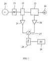
- FIG. 1shows a block diagram showing the operation of the device
- FIG. 2shows the probe connected to the electronic unit by a coaxial cable
- FIG. 3shows the result of an example case of developing edema in pig skin caused by controlling blood circulation at sites A and B. Sites C and D are the controls.
- FIG. 1shows an probe including an oscillator 20 , an attenuator 21 , a power splitter 22 , a directional coupler 23 , a probe 24 , amplifiers 25 and 26 , a phase detector 27 , a low pass filter 28 and a digital electronic unit 29 .
- the block diagram in FIG. 1can vary in different applications of the method. It may also be utilised in other ways.
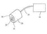
- FIG. 2shows the probe 24 , including an inner electrode 30 , a Teflon insulator 31 , an outer electrode 32 , a coaxial cable 33 and an electronic unit 34 comprising the components of FIG. 1 excluding the probe 24 .
- an essential feature of the device according to the inventionis that the coaxial probe is large enough, in order for the electric field to penetrate up to the subcutaneous fat tissue.
- the distance between the two electrodes of the probeshould be about 2-10 mm.
- the deviceoperates so that the sinusoidal high frequency (20-500 MHz) signal from the oscillator 20 is led through the attenuator 21 , power splitter 22 and directional coupler 23 to the probe 24 .
- the signalis reflected back from the probe. Part of this reflected signal is led through the directional coupler 23 to the amplifier 26 and further to one input of the phase detector 27 .
- the signal coming straight from the oscillator 20is led through the power splitter 22 to the other input of the phase detector 27 .
- the output from the phase detectoris led to the low pass filter 28 , whose output is a DC voltage proportional to the capacitance of the probe 24 . This voltage is further led to the digital electronic unit, where it is AD-converted, scaled and recorded.
- the output of the phase detector 27 after the low pass filteringis proportional to the phase difference, which is only dependent on the capacitance of the probe 24 .
- the deviceoperates on a single precisely set frequency and therefore the result is only dependent on the dielectric properties of the tissue and not on the conductivity.
- the probe 24is connected to the directional coupler 23 via the coaxial cable so that the signal is connected to the inner conductor of the cable and further to the inner electrode 30 of the probe 24 , and the ground signal is connected to the outer conductor of the cable and further to the outer electrode 32 of the probe 24 .
- FIG. 1shows only one example of the high frequency implementation of the method. It is made using known techniques. The essential feature is that the capacitance of the probe is measured at a high frequency 20-500 MHz.
- the high frequency unit of the devicecomprising of parts 20 - 27 , is realised using standard radio techniques.
- the fact that the dimensions of the circuit are small compared to the wavelengthdoes not in any way affect the operation of the high frequency components.
- phase detector 27measures only the phase difference of the incoming signals. This phase difference is proportional to the capacitance of the probe 24 and fiber proportional to the dielectric constant of the tissue. The dielectric constant is dependent on the water content of the skin.
- the attenuator 21between the oscillator 20 and the power splitter 22 . Its purpose is to prevent the access of the signal reflected from the probe to the amplifier 25 . Under the influence of the amplifier the signal reflected from the probe goes twice through the attenuator 21 when propagating to the input of the amplifier 25 . Therefore, if the attenuation of the attenuator 21 is for instance 6 dB, the total attenuation of this signal is 12 dB, which is adequate.
- FIG. 3shows as an example case of a measurement of a developing edema in pig skin, where by controlling the blood circulation a local edema is caused at sites A and B.
- Sites C and Dare the controls. It can be seen that at the sites with edema (A and B) the dielectric constant is increased by over 40% compared to the baseline. At the control area (sites C and D) where the developing edema does not exist the measured values remain unchanged. The measurement reacts rapidly to the edema, even before it is noticeable for instance by finger pressure.
Landscapes
- Health & Medical Sciences (AREA)
- Life Sciences & Earth Sciences (AREA)
- Surgery (AREA)
- Biomedical Technology (AREA)
- General Health & Medical Sciences (AREA)
- Biophysics (AREA)
- Pathology (AREA)
- Engineering & Computer Science (AREA)
- Animal Behavior & Ethology (AREA)
- Heart & Thoracic Surgery (AREA)
- Medical Informatics (AREA)
- Molecular Biology (AREA)
- Physics & Mathematics (AREA)
- Veterinary Medicine (AREA)
- Public Health (AREA)
- Nuclear Medicine, Radiotherapy & Molecular Imaging (AREA)
- Radiology & Medical Imaging (AREA)
- Dermatology (AREA)
- Measuring And Recording Apparatus For Diagnosis (AREA)
- Investigating Or Analysing Biological Materials (AREA)
- Investigating Or Analyzing Materials By The Use Of Electric Means (AREA)
- Measurement Of Optical Distance (AREA)
- Investigating Or Analysing Materials By Optical Means (AREA)
- Measuring Fluid Pressure (AREA)
- Electrotherapy Devices (AREA)
- Measurement And Recording Of Electrical Phenomena And Electrical Characteristics Of The Living Body (AREA)
Abstract
Description
Claims (20)
Priority Applications (1)
| Application Number | Priority Date | Filing Date | Title |
|---|---|---|---|
| US10/670,144US9271676B2 (en) | 2001-03-23 | 2003-09-23 | Method for measuring of edema |
Applications Claiming Priority (4)
| Application Number | Priority Date | Filing Date | Title |
|---|---|---|---|
| FI20010601 | 2001-03-23 | ||
| FI20010601AFI109651B (en) | 2001-03-23 | 2001-03-23 | Procedure for measuring edema in tissues |
| PCT/FI2002/000234WO2002080770A1 (en) | 2001-03-23 | 2002-03-21 | Method for measuring of edema |
| US10/670,144US9271676B2 (en) | 2001-03-23 | 2003-09-23 | Method for measuring of edema |
Related Parent Applications (1)
| Application Number | Title | Priority Date | Filing Date |
|---|---|---|---|
| PCT/FI2002/000234Continuation-In-PartWO2002080770A1 (en) | 2001-03-23 | 2002-03-21 | Method for measuring of edema |
Publications (2)
| Publication Number | Publication Date |
|---|---|
| US20050177061A1 US20050177061A1 (en) | 2005-08-11 |
| US9271676B2true US9271676B2 (en) | 2016-03-01 |
Family
ID=8560823
Family Applications (1)
| Application Number | Title | Priority Date | Filing Date |
|---|---|---|---|
| US10/670,144Expired - LifetimeUS9271676B2 (en) | 2001-03-23 | 2003-09-23 | Method for measuring of edema |
Country Status (6)
| Country | Link |
|---|---|
| US (1) | US9271676B2 (en) |
| EP (1) | EP1372475B1 (en) |
| AT (1) | ATE435612T1 (en) |
| DE (1) | DE60232863D1 (en) |
| FI (1) | FI109651B (en) |
| WO (1) | WO2002080770A1 (en) |
Cited By (16)
| Publication number | Priority date | Publication date | Assignee | Title |
|---|---|---|---|---|
| US9763596B2 (en) | 2015-04-24 | 2017-09-19 | Bruin Biometrics, Llc | Apparatus and methods for determining damaged tissue using sub-epidermal moisture measurements |
| US9980673B2 (en) | 2010-05-08 | 2018-05-29 | The Regents Of The University Of California | SEM scanner sensing apparatus, system and methodology for early detection of ulcers |
| US10898129B2 (en) | 2017-11-16 | 2021-01-26 | Bruin Biometrics, Llc | Strategic treatment of pressure ulcer using sub-epidermal moisture values |
| US10921244B2 (en) | 2018-11-16 | 2021-02-16 | Spectroflow, Inc. | Method and system for measuring fluid status |
| US10950960B2 (en) | 2018-10-11 | 2021-03-16 | Bruin Biometrics, Llc | Device with disposable element |
| US10959664B2 (en) | 2017-02-03 | 2021-03-30 | Bbi Medical Innovations, Llc | Measurement of susceptibility to diabetic foot ulcers |
| US11191442B2 (en)* | 2015-01-14 | 2021-12-07 | Cardioset Ltd. | Method and system for monitoring internal electrical impedance of a biological object |
| US11304652B2 (en) | 2017-02-03 | 2022-04-19 | Bbi Medical Innovations, Llc | Measurement of tissue viability |
| US11337651B2 (en) | 2017-02-03 | 2022-05-24 | Bruin Biometrics, Llc | Measurement of edema |
| US11406324B2 (en) | 2018-11-16 | 2022-08-09 | Spectroflow, Inc. | Method and system for measuring fluid status |
| US11471094B2 (en) | 2018-02-09 | 2022-10-18 | Bruin Biometrics, Llc | Detection of tissue damage |
| US11583038B2 (en) | 2020-07-23 | 2023-02-21 | Koya Medical, Inc. | Quick connect anchoring buckle |
| US11642075B2 (en) | 2021-02-03 | 2023-05-09 | Bruin Biometrics, Llc | Methods of treating deep and early-stage pressure induced tissue damage |
| US11672729B2 (en) | 2014-02-11 | 2023-06-13 | Koya Medical, Inc. | Compression garment |
| US11707405B2 (en) | 2017-02-16 | 2023-07-25 | Koya Medical, Inc. | Compression garment |
| US11903895B2 (en) | 2014-02-11 | 2024-02-20 | Koya Medical, Inc. | Compression garment apparatus |
Families Citing this family (24)
| Publication number | Priority date | Publication date | Assignee | Title |
|---|---|---|---|---|
| EP1329190B1 (en)* | 2002-12-14 | 2010-10-27 | Research Institute of Tsinghua University in Shenzhen | Apparatus and method for monitoring body composition by measuring body dielectric constant and body impedance based on digital frequency sampling |
| FI20030806A0 (en)* | 2003-05-28 | 2003-05-28 | Delfin Technologies Ltd | A method for measuring the amount of water in existing fat tissues and apparatus for applying the method |
| JP2005253840A (en)* | 2004-03-15 | 2005-09-22 | Tanita Corp | Skin condition estimation device |
| EP2460468A1 (en) | 2005-07-01 | 2012-06-06 | Impedimed Limited | Monitoring system |
| US20110054343A1 (en) | 2005-07-01 | 2011-03-03 | Impedimed Limited | Monitoring system |
| JP5208749B2 (en) | 2005-10-11 | 2013-06-12 | インペダイムド・リミテッド | Hydration status monitoring |
| US20110152661A1 (en)* | 2005-12-22 | 2011-06-23 | Board Of Regents, The University Of Texas System | Method and apparatus for identifying the viability of ischemic myocardium of a patient |
| WO2008128281A1 (en) | 2007-04-20 | 2008-10-30 | Impedimed Limited | Monitoring system and probe |
| US20110046505A1 (en) | 2007-08-09 | 2011-02-24 | Impedimed Limited | Impedance measurement process |
| ES2664239T3 (en)* | 2007-09-05 | 2018-04-18 | Sensible Medical Innovations Ltd. | Method and apparatus for using electromagnetic radiation to monitor a user's tissue |
| US10667715B2 (en)* | 2008-08-20 | 2020-06-02 | Sensible Medical Innovations Ltd. | Methods and devices of cardiac tissue monitoring and analysis |
| EP2403401B1 (en) | 2009-03-04 | 2017-05-10 | Sensible Medical Innovations Ltd. | System for monitoring intrabody tissues |
| US8907682B2 (en)* | 2009-07-30 | 2014-12-09 | Sensible Medical Innovations Ltd. | System and method for calibration of measurements of interacted EM signals in real time |
| US9615767B2 (en) | 2009-10-26 | 2017-04-11 | Impedimed Limited | Fluid level indicator determination |
| CA2778770A1 (en) | 2009-11-18 | 2011-05-26 | Chung Shing Fan | Signal distribution for patient-electrode measurements |
| GB201000532D0 (en)* | 2010-01-14 | 2010-03-03 | Univ City | Method for monitoring of blood components |
| FI125005B (en) | 2010-01-29 | 2015-04-30 | Hld Healthy Life Devices Oy | Method and apparatus for measuring the pressure in tissue |
| AU2015275343C1 (en)* | 2010-05-08 | 2018-10-04 | Bruin Biometrics, Llc | SEM scanner sensing apparatus, system and methodology for early detection of ulcers |
| US8827930B2 (en) | 2011-01-10 | 2014-09-09 | Bioguidance Llc | System and method for patient monitoring |
| EP2790576A4 (en) | 2011-12-14 | 2015-07-08 | Intersection Medical Inc | Devices, systems and methods for determining the relative spatial change in subsurface resistivities across frequencies in tissue |
| US20180360344A1 (en)* | 2017-06-19 | 2018-12-20 | Bruin Biometrics, Llc | Apparatus and methods for determining damaged tissue using sub-epidermal moisture measurements |
| GB2569921B (en)* | 2017-02-03 | 2022-06-01 | Bruin Biometrics Llc | Bisymmetric comparison of sub-epidermal moisture values |
| WO2020142403A1 (en)* | 2018-12-31 | 2020-07-09 | Massachusetts Institute Of Technology | Non-invasive skin contact sensor |
| US12156739B2 (en)* | 2021-07-16 | 2024-12-03 | Eli Lilly And Company | Methods and systems for selecting an injection site |
Citations (46)
| Publication number | Priority date | Publication date | Assignee | Title |
|---|---|---|---|---|
| US2184511A (en) | 1937-10-28 | 1939-12-26 | Samuel M Bagno | Method and apparatus for measuring impedance |
| US3316896A (en) | 1962-10-18 | 1967-05-02 | Thomasset Auguste Louis | Apparatus and methods for the measure of the electrical impedance of living organisms |
| US3340867A (en) | 1964-08-19 | 1967-09-12 | Univ Minnesota | Impedance plethysmograph |
| US3347223A (en) | 1964-01-08 | 1967-10-17 | Universal Match Corp | Pneumograph |
| US3452743A (en) | 1965-03-01 | 1969-07-01 | Gen Electric | Body impedance bridge |
| US3608543A (en) | 1968-10-03 | 1971-09-28 | Univ Carnegie Mellon | Physiological impedance-measuring apparatus |
| US3677261A (en) | 1970-04-03 | 1972-07-18 | American Optical Corp | Impedance pneumograph |
| US3730171A (en) | 1971-09-23 | 1973-05-01 | R Namon | Impedance related blood flow measuring device |
| US3742936A (en) | 1969-10-23 | 1973-07-03 | Ieram Sarl | Rheoplethysmographic device and method of operation |
| US3750649A (en) | 1971-06-25 | 1973-08-07 | Univ California | Pulmonary impedance bridge |
| US3789834A (en)* | 1971-11-15 | 1974-02-05 | J Duroux | Processes and apparatus for the investigation of internal physiological phenomena based on measurements of the impedance variation of the surface of the body |
| US3851641A (en)* | 1973-11-29 | 1974-12-03 | J Toole | Method and apparatus for determining internal impedance of animal body part |
| US3871359A (en) | 1973-06-25 | 1975-03-18 | Interscience Technology Corp | Impedance measuring system |
| US3894532A (en) | 1974-01-17 | 1975-07-15 | Acupulse Inc | Instruments for transcutaneous and subcutaneous investigation and treatment |
| US3949736A (en) | 1974-07-15 | 1976-04-13 | Vyvojova A Provozni Zakladna Vyzkumnych Ustavu | Circuit for automatically deriving and measuring relative voltages associated with impedance components of a biological object |
| GB1432316A (en) | 1973-04-19 | 1976-04-14 | Asrican M | Plethysmographs |
| US4008712A (en) | 1975-11-14 | 1977-02-22 | J. M. Richards Laboratories | Method for monitoring body characteristics |
| US4013065A (en) | 1976-02-23 | 1977-03-22 | American Cyanamid Company | Moisture dermatometer |
| US4116231A (en) | 1975-12-10 | 1978-09-26 | Tokyo Shibaura Electric Co., Ltd. | Living body function measuring apparatus |
| US4240445A (en) | 1978-10-23 | 1980-12-23 | University Of Utah | Electromagnetic energy coupler/receiver apparatus and method |
| US4364008A (en)* | 1980-10-02 | 1982-12-14 | Jacques Steven L | Focusing probe for moisture measurement device |
| US4640290A (en)* | 1985-04-25 | 1987-02-03 | Westinghouse Electric Corp. | Shielded, self-preparing electrode suitable for electroencephalographic mapping |
| US4793362A (en) | 1982-04-22 | 1988-12-27 | Karolinska Institutet | Method and apparatus for monitoring the fluid balance of the body |
| US4819648A (en) | 1985-10-28 | 1989-04-11 | The Johns Hopkins University | Non-invasive electromagnetic technique for monitoring time-trends of physiological changes at a particular location in the brain |
| US4860753A (en)* | 1987-11-04 | 1989-08-29 | The Gillette Company | Monitoring apparatus |
| US4911175A (en) | 1987-09-17 | 1990-03-27 | Diana Twyman | Method for measuring total body cell mass and total extracellular mass by bioelectrical resistance and reactance |
| US4918375A (en)* | 1987-07-03 | 1990-04-17 | Polska Akademia Nauk Instytut Agrofizyki | Reflectometric moisture meter for capillary-porous materials, especially for the soil |
| US5063937A (en) | 1990-09-12 | 1991-11-12 | Wright State University | Multiple frequency bio-impedance measurement system |
| US5086781A (en) | 1989-11-14 | 1992-02-11 | Bookspan Mark A | Bioelectric apparatus for monitoring body fluid compartments |
| US5280429A (en) | 1991-04-30 | 1994-01-18 | Xitron Technologies | Method and apparatus for displaying multi-frequency bio-impedance |
| GB2272526A (en) | 1992-10-30 | 1994-05-18 | British Tech Group | Investigation of a body by electrical impedance measurement |
| US5738107A (en) | 1994-10-11 | 1998-04-14 | Martinsen; Orjan G. | Measurement of moisture content in skin |
| US5749369A (en) | 1996-08-09 | 1998-05-12 | R.S. Medical Monitoring Ltd. | Method and device for stable impedance plethysmography |
| US5833686A (en)* | 1994-09-01 | 1998-11-10 | Zhao; Xinhua | Ultra-high-frequency cosmetic apparatus |
| WO1999008597A1 (en)* | 1997-08-19 | 1999-02-25 | Mendlein John D | Multi-site ultrasound methods and devices, particularly for measurement of fluid regulation |
| US6125297A (en) | 1998-02-06 | 2000-09-26 | The United States Of America As Represented By The United States National Aeronautics And Space Administration | Body fluids monitor |
| WO2000079255A1 (en) | 1999-06-22 | 2000-12-28 | The University Of Queensland | A method and device for measuring tissue oedema |
| EP1118308A1 (en) | 2000-01-21 | 2001-07-25 | Tanita Corporation | Apparatus for measuring the degree of edema |
| US6292690B1 (en) | 2000-01-12 | 2001-09-18 | Measurement Specialities Inc. | Apparatus and method for measuring bioelectric impedance |
| US6339722B1 (en) | 1995-09-26 | 2002-01-15 | A. J. Van Liebergen Holding B.V. | Apparatus for the in-vivo non-invasive measurement of a biological parameter concerning a bodily fluid of a person or animal |
| US6370426B1 (en)* | 1999-04-20 | 2002-04-09 | Nova Technology Corporation | Method and apparatus for measuring relative hydration of a substrate |
| US20030015024A1 (en)* | 2001-07-13 | 2003-01-23 | Campbell Gaylon S. | Moisture detection apparatus and method |
| US20030214312A1 (en) | 1997-04-10 | 2003-11-20 | Khatchatrian Robert G. | Diagnostic complex for measurement of the condition of biological tissues and liquids |
| US6823212B2 (en)* | 2001-06-13 | 2004-11-23 | The Procter & Gamble Company | Method and apparatus for measuring properties of a target surface |
| US20060200033A1 (en) | 2003-05-12 | 2006-09-07 | Cheetah Medical Inc. C/O Pepper Hamiton | System, method and apparatus for measuring blood flow and blood volume |
| US7297123B2 (en) | 2004-04-19 | 2007-11-20 | Nova Technology Corporation | Method and apparatus for accessing oral mucositis |
- 2001
- 2001-03-23FIFI20010601Apatent/FI109651B/enactive
- 2002
- 2002-03-21DEDE60232863Tpatent/DE60232863D1/ennot_activeExpired - Lifetime
- 2002-03-21ATAT02708389Tpatent/ATE435612T1/ennot_activeIP Right Cessation
- 2002-03-21EPEP02708389Apatent/EP1372475B1/ennot_activeExpired - Lifetime
- 2002-03-21WOPCT/FI2002/000234patent/WO2002080770A1/ennot_activeApplication Discontinuation
- 2003
- 2003-09-23USUS10/670,144patent/US9271676B2/ennot_activeExpired - Lifetime
Patent Citations (47)
| Publication number | Priority date | Publication date | Assignee | Title |
|---|---|---|---|---|
| US2184511A (en) | 1937-10-28 | 1939-12-26 | Samuel M Bagno | Method and apparatus for measuring impedance |
| US3316896A (en) | 1962-10-18 | 1967-05-02 | Thomasset Auguste Louis | Apparatus and methods for the measure of the electrical impedance of living organisms |
| US3347223A (en) | 1964-01-08 | 1967-10-17 | Universal Match Corp | Pneumograph |
| US3340867A (en) | 1964-08-19 | 1967-09-12 | Univ Minnesota | Impedance plethysmograph |
| US3452743A (en) | 1965-03-01 | 1969-07-01 | Gen Electric | Body impedance bridge |
| US3608543A (en) | 1968-10-03 | 1971-09-28 | Univ Carnegie Mellon | Physiological impedance-measuring apparatus |
| US3742936A (en) | 1969-10-23 | 1973-07-03 | Ieram Sarl | Rheoplethysmographic device and method of operation |
| US3677261A (en) | 1970-04-03 | 1972-07-18 | American Optical Corp | Impedance pneumograph |
| US3750649A (en) | 1971-06-25 | 1973-08-07 | Univ California | Pulmonary impedance bridge |
| US3730171A (en) | 1971-09-23 | 1973-05-01 | R Namon | Impedance related blood flow measuring device |
| US3789834A (en)* | 1971-11-15 | 1974-02-05 | J Duroux | Processes and apparatus for the investigation of internal physiological phenomena based on measurements of the impedance variation of the surface of the body |
| GB1432316A (en) | 1973-04-19 | 1976-04-14 | Asrican M | Plethysmographs |
| US3871359A (en) | 1973-06-25 | 1975-03-18 | Interscience Technology Corp | Impedance measuring system |
| US3851641A (en)* | 1973-11-29 | 1974-12-03 | J Toole | Method and apparatus for determining internal impedance of animal body part |
| US3894532A (en) | 1974-01-17 | 1975-07-15 | Acupulse Inc | Instruments for transcutaneous and subcutaneous investigation and treatment |
| US3949736A (en) | 1974-07-15 | 1976-04-13 | Vyvojova A Provozni Zakladna Vyzkumnych Ustavu | Circuit for automatically deriving and measuring relative voltages associated with impedance components of a biological object |
| US4008712A (en) | 1975-11-14 | 1977-02-22 | J. M. Richards Laboratories | Method for monitoring body characteristics |
| US4116231A (en) | 1975-12-10 | 1978-09-26 | Tokyo Shibaura Electric Co., Ltd. | Living body function measuring apparatus |
| US4013065A (en) | 1976-02-23 | 1977-03-22 | American Cyanamid Company | Moisture dermatometer |
| US4240445A (en) | 1978-10-23 | 1980-12-23 | University Of Utah | Electromagnetic energy coupler/receiver apparatus and method |
| US4364008A (en)* | 1980-10-02 | 1982-12-14 | Jacques Steven L | Focusing probe for moisture measurement device |
| US4793362A (en) | 1982-04-22 | 1988-12-27 | Karolinska Institutet | Method and apparatus for monitoring the fluid balance of the body |
| US4640290A (en)* | 1985-04-25 | 1987-02-03 | Westinghouse Electric Corp. | Shielded, self-preparing electrode suitable for electroencephalographic mapping |
| US4819648A (en) | 1985-10-28 | 1989-04-11 | The Johns Hopkins University | Non-invasive electromagnetic technique for monitoring time-trends of physiological changes at a particular location in the brain |
| US4918375A (en)* | 1987-07-03 | 1990-04-17 | Polska Akademia Nauk Instytut Agrofizyki | Reflectometric moisture meter for capillary-porous materials, especially for the soil |
| US4911175A (en) | 1987-09-17 | 1990-03-27 | Diana Twyman | Method for measuring total body cell mass and total extracellular mass by bioelectrical resistance and reactance |
| US4860753A (en)* | 1987-11-04 | 1989-08-29 | The Gillette Company | Monitoring apparatus |
| US5086781A (en) | 1989-11-14 | 1992-02-11 | Bookspan Mark A | Bioelectric apparatus for monitoring body fluid compartments |
| US5063937A (en) | 1990-09-12 | 1991-11-12 | Wright State University | Multiple frequency bio-impedance measurement system |
| US5280429A (en) | 1991-04-30 | 1994-01-18 | Xitron Technologies | Method and apparatus for displaying multi-frequency bio-impedance |
| GB2272526A (en) | 1992-10-30 | 1994-05-18 | British Tech Group | Investigation of a body by electrical impedance measurement |
| US5833686A (en)* | 1994-09-01 | 1998-11-10 | Zhao; Xinhua | Ultra-high-frequency cosmetic apparatus |
| US5738107A (en) | 1994-10-11 | 1998-04-14 | Martinsen; Orjan G. | Measurement of moisture content in skin |
| US6339722B1 (en) | 1995-09-26 | 2002-01-15 | A. J. Van Liebergen Holding B.V. | Apparatus for the in-vivo non-invasive measurement of a biological parameter concerning a bodily fluid of a person or animal |
| US5749369A (en) | 1996-08-09 | 1998-05-12 | R.S. Medical Monitoring Ltd. | Method and device for stable impedance plethysmography |
| US20030214312A1 (en) | 1997-04-10 | 2003-11-20 | Khatchatrian Robert G. | Diagnostic complex for measurement of the condition of biological tissues and liquids |
| WO1999008597A1 (en)* | 1997-08-19 | 1999-02-25 | Mendlein John D | Multi-site ultrasound methods and devices, particularly for measurement of fluid regulation |
| US6125297A (en) | 1998-02-06 | 2000-09-26 | The United States Of America As Represented By The United States National Aeronautics And Space Administration | Body fluids monitor |
| US6370426B1 (en)* | 1999-04-20 | 2002-04-09 | Nova Technology Corporation | Method and apparatus for measuring relative hydration of a substrate |
| WO2000079255A1 (en) | 1999-06-22 | 2000-12-28 | The University Of Queensland | A method and device for measuring tissue oedema |
| US20040186392A1 (en) | 1999-06-22 | 2004-09-23 | The University Of Queensland | Method and device for measuring tissue oedema |
| US6292690B1 (en) | 2000-01-12 | 2001-09-18 | Measurement Specialities Inc. | Apparatus and method for measuring bioelectric impedance |
| EP1118308A1 (en) | 2000-01-21 | 2001-07-25 | Tanita Corporation | Apparatus for measuring the degree of edema |
| US6823212B2 (en)* | 2001-06-13 | 2004-11-23 | The Procter & Gamble Company | Method and apparatus for measuring properties of a target surface |
| US20030015024A1 (en)* | 2001-07-13 | 2003-01-23 | Campbell Gaylon S. | Moisture detection apparatus and method |
| US20060200033A1 (en) | 2003-05-12 | 2006-09-07 | Cheetah Medical Inc. C/O Pepper Hamiton | System, method and apparatus for measuring blood flow and blood volume |
| US7297123B2 (en) | 2004-04-19 | 2007-11-20 | Nova Technology Corporation | Method and apparatus for accessing oral mucositis |
Non-Patent Citations (8)
| Title |
|---|
| Esko et al, Measurement of Dielectric Properties of Subcutaneous Fat with Open-Ended Coaxial Sensors, 1998, Phys. Med. Biol. 43, p. 475-485.* |
| Esko et al, Penetration of Electromagnetic Fields of an Open-Ended Coaxial Probe between 1 MHz and 1 GHz in Dielectric Skin Measurements, 1999, Phys. Med. Biol. 44, p. N169-N176.* |
| Esko et al, Variational Formulation of Open-Ended Coaxial Line in Contact with Layered Biological Medium, Oct. 1998, IEEE Transactions on Biomedical Engineering vol. 45, p. 1241-1248.* |
| H. H. Woltjer, H. J. Bogaard and P. M. J. M. de Vries; "The technique of impedance cardiography," European Heart Journal (1997) 18, 1396-1403. |
| Jan Nyboer, Marian M. Kreider and Leonard Hannapel; "Electrical Impedance Plethysmography : A Physical and Physiologic Approach to Peripheral Vascular Study," Circulation-Journal of the American Heart Association, 1950, 2: 811-821. |
| Kao, H. Pin, et al., "Correlation of Permittivity and Water Content During Cerebral Edema", IEEE Transactions on Biomedical Engineering, vol. 46, No. 9, Sep. 1999. |
| Sherwood, et al. "Methodological Guidelines for Impedence Cardiography," Psychophysiology, vol. 27, No. 1: 1-23. |
| Zuhdi Lababidi, D. A. Ehmke, Robert E. Durnin, Paul E. Leaverton and Ronald M. Lauer; "The First Derivative Thoracic Impedance Cardiogram," Circulation-Journal of the American Heart Association, 1970; 41: 651-658. |
Cited By (40)
| Publication number | Priority date | Publication date | Assignee | Title |
|---|---|---|---|---|
| US9980673B2 (en) | 2010-05-08 | 2018-05-29 | The Regents Of The University Of California | SEM scanner sensing apparatus, system and methodology for early detection of ulcers |
| US10188340B2 (en) | 2010-05-08 | 2019-01-29 | Bruin Biometrics, Llc | SEM scanner sensing apparatus, system and methodology for early detection of ulcers |
| US11779265B2 (en) | 2010-05-08 | 2023-10-10 | Bruin Biometrics, Llc | SEM scanner sensing apparatus, system and methodology for early detection of ulcers |
| US11253192B2 (en) | 2010-05-08 | 2022-02-22 | Bruain Biometrics, LLC | SEM scanner sensing apparatus, system and methodology for early detection of ulcers |
| US12383459B2 (en) | 2014-02-11 | 2025-08-12 | Koya Medical, Inc. | Compression garment apparatus |
| US11903895B2 (en) | 2014-02-11 | 2024-02-20 | Koya Medical, Inc. | Compression garment apparatus |
| US11672729B2 (en) | 2014-02-11 | 2023-06-13 | Koya Medical, Inc. | Compression garment |
| US11191442B2 (en)* | 2015-01-14 | 2021-12-07 | Cardioset Ltd. | Method and system for monitoring internal electrical impedance of a biological object |
| US11850032B2 (en) | 2015-01-14 | 2023-12-26 | Cardioset Ltd. | Method and system for monitoring internal electrical impedance of a biological object |
| US11534077B2 (en) | 2015-04-24 | 2022-12-27 | Bruin Biometrics, Llc | Apparatus and methods for determining damaged tissue using sub epidermal moisture measurements |
| US9763596B2 (en) | 2015-04-24 | 2017-09-19 | Bruin Biometrics, Llc | Apparatus and methods for determining damaged tissue using sub-epidermal moisture measurements |
| US10485447B2 (en) | 2015-04-24 | 2019-11-26 | Bruin Biometrics, Llc | Apparatus and methods for determining damaged tissue using sub-epidermal moisture measurements |
| US10182740B2 (en) | 2015-04-24 | 2019-01-22 | Bruin Biometrics, Llc | Apparatus and methods for determining damaged tissue using sub-epidermal moisture measurements |
| US11284810B2 (en) | 2015-04-24 | 2022-03-29 | Bruin Biometrics, Llc | Apparatus and methods for determining damaged tissue using sub-epidermal moisture measurements |
| US11832929B2 (en) | 2015-04-24 | 2023-12-05 | Bruin Biometrics, Llc | Apparatus and methods for determining damaged tissue using sub-epidermal moisture measurements |
| US10178961B2 (en) | 2015-04-24 | 2019-01-15 | Bruin Biometrics, Llc | Apparatus and methods for determining damaged tissue using sub-epidermal moisture measurements |
| US11304652B2 (en) | 2017-02-03 | 2022-04-19 | Bbi Medical Innovations, Llc | Measurement of tissue viability |
| US11337651B2 (en) | 2017-02-03 | 2022-05-24 | Bruin Biometrics, Llc | Measurement of edema |
| US12290377B2 (en) | 2017-02-03 | 2025-05-06 | Bbi Medical Innovations, Llc | Measurement of tissue viability |
| US11627910B2 (en) | 2017-02-03 | 2023-04-18 | Bbi Medical Innovations, Llc | Measurement of susceptibility to diabetic foot ulcers |
| US10959664B2 (en) | 2017-02-03 | 2021-03-30 | Bbi Medical Innovations, Llc | Measurement of susceptibility to diabetic foot ulcers |
| US11707405B2 (en) | 2017-02-16 | 2023-07-25 | Koya Medical, Inc. | Compression garment |
| US11191477B2 (en) | 2017-11-16 | 2021-12-07 | Bruin Biometrics, Llc | Strategic treatment of pressure ulcer using sub-epidermal moisture values |
| US11426118B2 (en) | 2017-11-16 | 2022-08-30 | Bruin Biometrics, Llc | Strategic treatment of pressure ulcer using sub-epidermal moisture values |
| US12336837B2 (en) | 2017-11-16 | 2025-06-24 | Bruin Biometrics, Llc | Providing a continuity of care across multiple care settings |
| US10898129B2 (en) | 2017-11-16 | 2021-01-26 | Bruin Biometrics, Llc | Strategic treatment of pressure ulcer using sub-epidermal moisture values |
| US11471094B2 (en) | 2018-02-09 | 2022-10-18 | Bruin Biometrics, Llc | Detection of tissue damage |
| US11980475B2 (en) | 2018-02-09 | 2024-05-14 | Bruin Biometrics, Llc | Detection of tissue damage |
| US12350064B2 (en) | 2018-02-09 | 2025-07-08 | Bruin Biometrics, Llc | Detection of tissue damage |
| US11600939B2 (en) | 2018-10-11 | 2023-03-07 | Bruin Biometrics, Llc | Device with disposable element |
| US11824291B2 (en) | 2018-10-11 | 2023-11-21 | Bruin Biometrics, Llc | Device with disposable element |
| US12132271B2 (en) | 2018-10-11 | 2024-10-29 | Bruin Biometrics, Llc | Device with disposable element |
| US11342696B2 (en) | 2018-10-11 | 2022-05-24 | Bruin Biometrics, Llc | Device with disposable element |
| US10950960B2 (en) | 2018-10-11 | 2021-03-16 | Bruin Biometrics, Llc | Device with disposable element |
| US11406324B2 (en) | 2018-11-16 | 2022-08-09 | Spectroflow, Inc. | Method and system for measuring fluid status |
| US10921244B2 (en) | 2018-11-16 | 2021-02-16 | Spectroflow, Inc. | Method and system for measuring fluid status |
| US12156571B2 (en) | 2020-07-23 | 2024-12-03 | Koya Medical, Inc. | Quick connect anchoring buckle |
| US11583038B2 (en) | 2020-07-23 | 2023-02-21 | Koya Medical, Inc. | Quick connect anchoring buckle |
| US12097041B2 (en) | 2021-02-03 | 2024-09-24 | Bruin Biometrics, Llc | Methods of treating deep and early-stage pressure induced tissue damage |
| US11642075B2 (en) | 2021-02-03 | 2023-05-09 | Bruin Biometrics, Llc | Methods of treating deep and early-stage pressure induced tissue damage |
Also Published As
| Publication number | Publication date |
|---|---|
| FI20010601A0 (en) | 2001-03-23 |
| DE60232863D1 (en) | 2009-08-20 |
| EP1372475A1 (en) | 2004-01-02 |
| WO2002080770A1 (en) | 2002-10-17 |
| FI109651B (en) | 2002-09-30 |
| EP1372475B1 (en) | 2009-07-08 |
| ATE435612T1 (en) | 2009-07-15 |
| US20050177061A1 (en) | 2005-08-11 |
Similar Documents
| Publication | Publication Date | Title |
|---|---|---|
| US9271676B2 (en) | Method for measuring of edema | |
| AU2019257529B2 (en) | Diagnostic method for detection of fluid changes | |
| CA2470801C (en) | Detection of fluids in tissue | |
| JP6321090B2 (en) | Method and system for determining cardiovascular volume in a mammal | |
| KR101527664B1 (en) | Armband comprising a detection device for the detection of a blood count parameter | |
| US6603997B2 (en) | Probe penetration detector and method of operation | |
| JP5993372B2 (en) | Embedded dielectric measurement system | |
| US4240445A (en) | Electromagnetic energy coupler/receiver apparatus and method | |
| KR101562442B1 (en) | Detection device for the detection of a blood count parameter | |
| US6440118B2 (en) | Device and method to sense body substance transition | |
| JP2010088898A (en) | Apparatus, system, and method for monitoring tissue during electrosurgical procedure | |
| JP2005512663A (en) | Moisture probe | |
| Ramesh et al. | Radiometric analysis of ankle edema via RZF antenna for biomedical applications | |
| KR100438829B1 (en) | Apparatus and method for obtaining data for diagnosing living body using ultra high frequency signal | |
| EP2408361A1 (en) | Device for electrically measuring at least one parameter of a mammal's tissue | |
| WO2004105602A1 (en) | Method for measuring water content of subcutaneous fat and apparatus for applying of the method | |
| US20190307357A1 (en) | Electrically resonant electrode configuration for monitoring of a tissue | |
| KR20110059143A (en) | Human blood glucose sensor and real time blood glucose measurement device | |
| Ganguly et al. | Sensitive transmit receive architecture for body wearable RF plethysmography sensor | |
| US20230079881A1 (en) | Devices, systems and methods for sensing and discerning between fat and muscle tissue during medical procedures | |
| Bing et al. | A Study of Continuous Noninvasive RF Hydration Sensing on Human Forearm | |
| CN115998253B (en) | Monitoring method and monitoring device for peritoneal effusion change condition | |
| US20170112437A1 (en) | Measurement of Hydration, Edema, and Bioelectrical Impedance | |
| HK1225263A1 (en) | A method of and a system for determining a cardiovascular quantity of a mammal | |
| HK1195864B (en) | A method of and a system for determining a cardiovascular quantity of a mammal |
Legal Events
| Date | Code | Title | Description |
|---|---|---|---|
| AS | Assignment | Owner name:DELFIN TECHNOLOGIES LTD., FINLAND Free format text:ASSIGNMENT OF ASSIGNORS INTEREST;ASSIGNORS:ALANEN, ESKO;LAHTINEN, AULIS TAPANI;NUUTINEN, JOUNI;REEL/FRAME:015340/0802;SIGNING DATES FROM 20031014 TO 20031028 Owner name:DELFIN TECHNOLOGIES LTD., FINLAND Free format text:ASSIGNMENT OF ASSIGNORS INTEREST;ASSIGNORS:ALANEN, ESKO;LAHTINEN, AULIS TAPANI;NUUTINEN, JOUNI;SIGNING DATES FROM 20031014 TO 20031028;REEL/FRAME:015340/0802 | |
| STCF | Information on status: patent grant | Free format text:PATENTED CASE | |
| MAFP | Maintenance fee payment | Free format text:PAYMENT OF MAINTENANCE FEE, 4TH YR, SMALL ENTITY (ORIGINAL EVENT CODE: M2551); ENTITY STATUS OF PATENT OWNER: SMALL ENTITY Year of fee payment:4 | |
| FEPP | Fee payment procedure | Free format text:7.5 YR SURCHARGE - LATE PMT W/IN 6 MO, SMALL ENTITY (ORIGINAL EVENT CODE: M2555); ENTITY STATUS OF PATENT OWNER: SMALL ENTITY | |
| MAFP | Maintenance fee payment | Free format text:PAYMENT OF MAINTENANCE FEE, 8TH YR, SMALL ENTITY (ORIGINAL EVENT CODE: M2552); ENTITY STATUS OF PATENT OWNER: SMALL ENTITY Year of fee payment:8 |