US9238793B2 - Method for enzymatic treatment of tissue products - Google Patents
Method for enzymatic treatment of tissue productsDownload PDFInfo
- Publication number
- US9238793B2 US9238793B2US14/019,274US201314019274AUS9238793B2US 9238793 B2US9238793 B2US 9238793B2US 201314019274 AUS201314019274 AUS 201314019274AUS 9238793 B2US9238793 B2US 9238793B2
- Authority
- US
- United States
- Prior art keywords
- tissue
- tissue matrix
- matrix
- collagen
- enzyme
- Prior art date
- Legal status (The legal status is an assumption and is not a legal conclusion. Google has not performed a legal analysis and makes no representation as to the accuracy of the status listed.)
- Active, expires
Links
Images


Classifications
- A—HUMAN NECESSITIES
- A61—MEDICAL OR VETERINARY SCIENCE; HYGIENE
- A61L—METHODS OR APPARATUS FOR STERILISING MATERIALS OR OBJECTS IN GENERAL; DISINFECTION, STERILISATION OR DEODORISATION OF AIR; CHEMICAL ASPECTS OF BANDAGES, DRESSINGS, ABSORBENT PADS OR SURGICAL ARTICLES; MATERIALS FOR BANDAGES, DRESSINGS, ABSORBENT PADS OR SURGICAL ARTICLES
- A61L27/00—Materials for grafts or prostheses or for coating grafts or prostheses
- A61L27/36—Materials for grafts or prostheses or for coating grafts or prostheses containing ingredients of undetermined constitution or reaction products thereof, e.g. transplant tissue, natural bone, extracellular matrix
- A61L27/3604—Materials for grafts or prostheses or for coating grafts or prostheses containing ingredients of undetermined constitution or reaction products thereof, e.g. transplant tissue, natural bone, extracellular matrix characterised by the human or animal origin of the biological material, e.g. hair, fascia, fish scales, silk, shellac, pericardium, pleura, renal tissue, amniotic membrane, parenchymal tissue, fetal tissue, muscle tissue, fat tissue, enamel
- C—CHEMISTRY; METALLURGY
- C12—BIOCHEMISTRY; BEER; SPIRITS; WINE; VINEGAR; MICROBIOLOGY; ENZYMOLOGY; MUTATION OR GENETIC ENGINEERING
- C12N—MICROORGANISMS OR ENZYMES; COMPOSITIONS THEREOF; PROPAGATING, PRESERVING, OR MAINTAINING MICROORGANISMS; MUTATION OR GENETIC ENGINEERING; CULTURE MEDIA
- C12N5/00—Undifferentiated human, animal or plant cells, e.g. cell lines; Tissues; Cultivation or maintenance thereof; Culture media therefor
- C12N5/0068—General culture methods using substrates
- A—HUMAN NECESSITIES
- A61—MEDICAL OR VETERINARY SCIENCE; HYGIENE
- A61L—METHODS OR APPARATUS FOR STERILISING MATERIALS OR OBJECTS IN GENERAL; DISINFECTION, STERILISATION OR DEODORISATION OF AIR; CHEMICAL ASPECTS OF BANDAGES, DRESSINGS, ABSORBENT PADS OR SURGICAL ARTICLES; MATERIALS FOR BANDAGES, DRESSINGS, ABSORBENT PADS OR SURGICAL ARTICLES
- A61L27/00—Materials for grafts or prostheses or for coating grafts or prostheses
- A61L27/36—Materials for grafts or prostheses or for coating grafts or prostheses containing ingredients of undetermined constitution or reaction products thereof, e.g. transplant tissue, natural bone, extracellular matrix
- A61L27/3683—Materials for grafts or prostheses or for coating grafts or prostheses containing ingredients of undetermined constitution or reaction products thereof, e.g. transplant tissue, natural bone, extracellular matrix subjected to a specific treatment prior to implantation, e.g. decellularising, demineralising, grinding, cellular disruption/non-collagenous protein removal, anti-calcification, crosslinking, supercritical fluid extraction, enzyme treatment
- A61L27/3687—Materials for grafts or prostheses or for coating grafts or prostheses containing ingredients of undetermined constitution or reaction products thereof, e.g. transplant tissue, natural bone, extracellular matrix subjected to a specific treatment prior to implantation, e.g. decellularising, demineralising, grinding, cellular disruption/non-collagenous protein removal, anti-calcification, crosslinking, supercritical fluid extraction, enzyme treatment characterised by the use of chemical agents in the treatment, e.g. specific enzymes, detergents, capping agents, crosslinkers, anticalcification agents
- A—HUMAN NECESSITIES
- A61—MEDICAL OR VETERINARY SCIENCE; HYGIENE
- A61L—METHODS OR APPARATUS FOR STERILISING MATERIALS OR OBJECTS IN GENERAL; DISINFECTION, STERILISATION OR DEODORISATION OF AIR; CHEMICAL ASPECTS OF BANDAGES, DRESSINGS, ABSORBENT PADS OR SURGICAL ARTICLES; MATERIALS FOR BANDAGES, DRESSINGS, ABSORBENT PADS OR SURGICAL ARTICLES
- A61L27/00—Materials for grafts or prostheses or for coating grafts or prostheses
- A61L27/50—Materials characterised by their function or physical properties, e.g. injectable or lubricating compositions, shape-memory materials, surface modified materials
- A61L27/56—Porous materials, e.g. foams or sponges
- A—HUMAN NECESSITIES
- A61—MEDICAL OR VETERINARY SCIENCE; HYGIENE
- A61L—METHODS OR APPARATUS FOR STERILISING MATERIALS OR OBJECTS IN GENERAL; DISINFECTION, STERILISATION OR DEODORISATION OF AIR; CHEMICAL ASPECTS OF BANDAGES, DRESSINGS, ABSORBENT PADS OR SURGICAL ARTICLES; MATERIALS FOR BANDAGES, DRESSINGS, ABSORBENT PADS OR SURGICAL ARTICLES
- A61L2430/00—Materials or treatment for tissue regeneration
- A61L2430/40—Preparation and treatment of biological tissue for implantation, e.g. decellularisation, cross-linking
Definitions
- the present disclosurerelates to tissue matrices, and more particularly, to methods for controlling the pliability and/or immunogenicity of tissue matrices by treating the matrices with proteolytic enzymes.
- tissue-derived productsare used to regenerate, repair, or otherwise treat diseased or damaged tissues and organs.
- Such productscan include intact tissue grafts and/or acellular or reconstituted acellular tissues (e.g., acellular tissue matrices from skin, intestine, or other tissues, with or without cell seeding).
- tissue productsgenerally have mechanical properties determined by the tissue source (i.e., tissue type and animal from which it originated) and the processing parameters used to produce the tissue products. Since tissue products are often used for surgical applications and/or tissue replacement or augmentation, the mechanical properties of the tissue products are important. For example, surgeons generally prefer tissues that feel like natural tissues and/or are easy to handle during surgical procedures. Some tissue products, however, are undesirably stiff and/or have an unnatural feel. Accordingly, methods for treating tissue products to produce more desirable mechanical properties are provided.
- tissue products derived from exogenous materialsmay elicit an inflammatory or immune response in the recipient.
- an excessive immune responsemay be detrimental, causing the implant to form undesirable scar tissue, or preventing suitable regeneration of tissue at the site of implantation. Accordingly, methods for treating tissue products to reduce or control the immune response of the tissue products upon implantation are provided.
- a method for treating a tissue matrixcan comprise selecting a collagen-containing tissue matrix and contacting the tissue matrix with a proteolytic enzyme under conditions sufficient to produce a desired level of pliability in the tissue matrix.
- a method for treating a tissue matrixcan comprise selecting a collagen-containing acellular tissue matrix and contacting the tissue matrix with a proteolytic enzyme under conditions sufficient to produce a desired level of pliability in the tissue matrix and to increase the porosity of the tissue matrix.
- an acellular tissue matrixis provided.
- the matrixcan be prepared by a process comprising selecting an acellular tissue matrix and contacting the tissue matrix with a proteolytic enzyme under conditions sufficient to produce a desired level of pliability in the tissue matrix.
- a method for treating a tissue matrixcan include selecting at least one collagen-containing tissue matrix; contacting the at least one collagen-containing tissue matrix with a proteolytic enzyme; and performing an assay to determine if contacting the at least one collagen-containing tissue matrix with the at least one proteolytic enzyme has altered the at least one collagen-containing tissue matrix to reduce a human immune response to the tissue matrix when the tissue matrix is implanted in a human body.
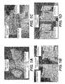
- FIGS. 1A-1Dshow acellular tissue matrices after treatment with enzymes using methods of described in Example 1, as well as untreated controls.
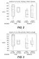
- FIG. 2is a box plot of tensile strength testing data for treated and control samples, according to the methods of Example 1.
- FIG. 3is a box plot of suture strength testing data for treated and control samples, according to the methods of Example 1.
- FIG. 4is a box plot of tear strength testing data for treated and control samples, according to the methods of Example 1.
- FIG. 5is a box plot of elasticity testing data for treated and control samples, according to the methods of Example 1.
- FIG. 6is a box plot of creep resistance testing data for treated and control samples, according to the methods of Example 1.
- FIG. 7illustrates DSC thermograms for untreated tissues and tissues treated using bromelain and alcalase according to Example 2.2.
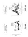
- FIGS. 8A-8Billustrate expression patterns of activation markers CD14 (A) and CD163 (B) in monocytes co-cultured with various tissues, using the monocyte activation assay described in Example 2.4.
- FIGS. 9A-Fare hematoxylin & eosin (H&E) sections of untreated pADM ( 9 A- 9 C) and enzyme treated pADM ( 9 D- 9 F) after explant, as described in Example 2.5.
- H&Ehematoxylin & eosin
- FIGS. 10A-Dare H&E sections of untreated pADM ( 9 A- 9 B) and enzyme treated pADM ( 9 C- 9 D) explants, as describe in Example 2.5.
- tissue productwill refer to any human or animal tissue that contains extracellular matrix proteins.
- tissue productscan include acellular or partially decellularized tissue matrices, decellularized tissue matrices that have been repopulated with exogenous cells, and/or cellular tissues.
- tissue products for treating patientscan be used to produce products for treating patients.
- tissue products for regeneration, repair, augmentation, reinforcement, and/or treatment of human tissues that have been damaged or lost due to various diseases and/or structural damagehave been produced.
- Such productscan include, for example, acellular tissue matrices, tissue allografts or xenografts, and/or reconstituted tissues (i.e., at least partially decellularized tissues that have been seeded with cells to produce viable materials).
- tissue productsthat have certain mechanical properties.
- the tissue productwhich may include a sheet of material, should possess sufficient strength to withstand the intended use.
- certain tissue productsmay be used to repair defects (e.g., hernias), to support surrounding tissues or implants (e.g., for breast augmentation and/or reconstruction), or to replace damaged or lost tissue (e.g., after trauma or surgical resection).
- the tissue productshould have sufficient strength, elasticity, and/or other mechanical properties to function until tissue regeneration and/or repair occurs.
- tissue productsshould have a desirable feel.
- surgeonsgenerally prefer materials that have a natural tissue-like feel (e.g., are sufficiently soft, pliable, and/or elastic).
- tissue productsafter implantation, it is desirable for tissue products to feel more natural.
- tissues used for breast augmentationshould not be excessively stiff so that upon implantation they produce a more natural feeling breast.
- tissue productscan be excessively stiff.
- porcine-derived dermal materialssuch as STRATTICETM are less pliable than human dermal products such as ALLODERM®.
- Processes for improving the feel of such productsshould not adversely affect the biological and/or mechanical properties of the products. Specifically, processing of the products to improve the feel of the products should not produce an undesirable decrease in other mechanical properties such as tensile strength, and should not alter the protein matrix in such a way that the material does not support tissue regeneration and/or repair.
- the present disclosureprovides methods for treating tissues to improve the feel of tissue products produced from the tissues.
- the disclosurealso provides tissue products produced using the methods of treatment.
- the present disclosureprovides methods of treating tissues to control the porosity of tissue products produced from the tissues. In some cases, controlling the porosity can improve cellular infiltration and tissue regeneration and/or repair.
- the present disclosureprovides methods for controlling or reducing an immune response to tissue matrices when implanted in a body.
- the immune responsecan be measured using a number of immunoassays, including monocyte activation assays, phagocytosis assays, and/or oxidative burst assays.
- the immunogenicitymay also be controlled while treating the tissue to improve the feel of the tissue, to control mechanical properties of the tissue (including any mechanical property listed herein), and/or controlling porosity of the tissue.
- the matricesmay be subjected to an assay to determine if the immunogenicity of the tissue has been altered in a desirable way.
- a method for treating a tissue matrixcan comprise selecting a collagen-containing tissue matrix and contacting the tissue matrix with a proteolytic enzyme under conditions sufficient to produce a desired level of pliability in the tissue matrix.
- a method for treating a tissue matrixis provided. The method can comprise selecting a collagen-containing acellular tissue matrix and contacting the tissue matrix with a proteolytic enzyme under conditions sufficient to produce a desired level of pliability in the tissue matrix and to increase the porosity of the tissue matrix.
- FIGS. 1A-1Dshow acellular tissue matrices (STRATTICETM) after treatment with enzymes using methods of the present disclosure, as well as untreated controls. As shown, the treated samples are significantly more pliable that the untreated samples.
- a method for treating a tissue matrixcan include selecting at least one collagen-containing tissue matrix; contacting the at least one collagen-containing tissue matrix with a proteolytic enzyme; and performing an assay to determine if contacting the at least one collagen-containing tissue matrix with the at least one proteolytic enzyme has altered the at least one collagen-containing tissue matrix to reduce a human immune response to the tissue matrix when the tissue matrix is implanted in a human body.
- treatment of tissue matrices with proteolytic enzymesprovides improved mechanical properties without causing degradation in one or biological properties.
- treatment of tissue matricescan produce desired stiffness, feel, tactile properties, and/or desired porosity without causing increased inflammation or scar formation and/or without causing a reduction in the tissue matrices' ability to promote cell ingrowth and regeneration.
- the tissuescan be selected to provide a variety of different biological and mechanical properties.
- an acellular tissue matrix or other tissue productcan be selected to allow tissue in-growth and remodeling to assist in regeneration of tissue normally found at the site where the matrix is implanted.
- an acellular tissue matrixwhen implanted on or into fascia, may be selected to allow regeneration of the fascia without excessive fibrosis or scar formation.
- the tissue productcan be formed from ALLODERM® or STRATTICETM, which are human and porcine acellular dermal matrices respectively.
- ALLODERM® or STRATTICETMare human and porcine acellular dermal matrices respectively.
- other suitable acellular tissue matricescan be used, as described further below.
- the tissuescan be selected from a variety of tissue sources including skin (dermis or whole skin), fascia, pericardial tissue, dura, umbilical cord tissue, placental tissue, cardiac valve tissue, ligament tissue, adipose tissue, tendon tissue, arterial tissue, venous tissue, neural connective tissue, urinary bladder tissue, ureter tissue, and intestinal tissue.
- the methods described hereincan be used to process any collagenous tissue type, and for any tissue matrix product.
- a number of biological scaffold materialsare described by Badylak et al., and the methods of the present disclosure can be used to treat those or other tissue products known in the art.
- Badylak et al.“Extracellular Matrix as a Biological Scaffold Material: Structure and Function,” Acta Biomaterialia (2008), doi:10.1016/j.actbio.2008.09.013.
- the tissue productcan be provided as a decellularized tissue matrix. Suitable acellular tissue matrices are described further below.
- the methodcan further include processing intact tissue to remove cells or other materials.
- the tissuescan be completely or partially decellularized to yield acellular tissue matrices or extracellular tissue materials to be used for patients.
- various tissuessuch as skin, intestine, bone, cartilage, adipose tissue, nerve tissue (e.g., nerve fibers or dura), tendons, ligaments, or other tissues can be completely or partially decellularized to produce tissue products useful for patients.
- these decellularized productscan be used without addition of exogenous cellular materials (e.g., stem cells).
- these decellularized productscan be seeded with cells from autologous sources or other sources to facilitate treatment. Suitable processes for producing acellular tissue matrices are described below.
- suitable enzymescan include sulfhydryl proteases such as bromelain.
- theycan include bromelain, papain, ficin, actinidin, alcalase, trypsin or combinations thereof.
- the enzymescan be purchased commercially or extracted from fruit sources.
- one source of bromelainis MCCORMICK MEAT TENDERIZER®, but the enzymes can also be extracted from pineapple and/or purchased in a medical-grade formulation.
- the enzymescan be contacted with the tissues to increase the pliability of the tissue without causing undesirable degradation in other mechanical and/or biological properties.
- the enzyme treatmentswill not produce an undesirable change in at least one of tensile strength, tear strength, suture strength, creep resistance, elasticity, collagenase susceptibility, glycosaminoglycan content, lectin content, burst strength, thermal transition temperature, or combinations thereof.
- an undesirable changeis a statistically significant reduction in any one of tensile strength, tear strength, suture strength, creep resistance, glycosaminoglycan content, lectin content, burst strength, an increase in collagenase susceptibility or a change (upward or downward) in thermal transition temperature (as measured using differential scanning calorimetry).
- the tissuesare treated with an enzyme to increase the porosity of the tissue.
- increasing the porosity of the tissueis performed to increase the number and/or size of channels, which can improve the rate of cellular infiltration and tissue regeneration.
- the enzymesare selected such that they cause site specific cleavage of proteins within the tissues. For example, it has been found that treatment of porcine dermal materials with bromelain does not cause further alterations in the matrix structure after a certain amount of treatment. Therefore, treatment of dermis with bromelain does not cause further change in the matrix with prolonged exposure or after extended periods of time.
- the enzymecan be applied to the tissues in a variety of suitable solutions.
- bromelainhas been found to be effective when applied to tissues in normal saline, but other suitable buffers (e.g., PBS) can be used.
- an assaymay be performed to determine if contacting the at least one collagen-containing tissue matrix with the at least one proteolytic enzyme has altered the at least one collagen-containing tissue matrix to reduce a human immune response to the tissue matrix when the tissue matrix is implanted in a human body.
- suitable assayscan include monocyte activation assays, phagocytosis assays, and oxidative burst assays.
- the assaymay be performed on a segment or portion of the processed tissue, and other portions of the tissue may be used in subsequent medical or surgical procedures. In other embodiments, the assay may be performed on one or more samples from a batch of multiple samples, and samples not subjected to the assay may be subsequently selected for use in treating a patient.
- the enzyme treatmentis selected to remove collagen or other proteins in the material that have a thermal transition temperature that will permit denaturation at body temperature.
- dermal acellular tissuesare selected, and the enzymatic treatment is selected to remove a thermal peak, as measured using DSC, between about 30 degrees and 40 degrees Celsius. This small ECM peak is expected to denature spontaneously at the human body temperature and may contribute to the different inflammatory
- acellular tissue matrixrefers generally to any tissue matrix that is substantially free of cells and/or cellular components. Skin, parts of skin (e.g., dermis), and other tissues such as blood vessels, heart valves, fascia, cartilage, adipose tissue, bone, and nerve connective tissue may be used to create acellular matrices within the scope of the present disclosure. Acellular tissue matrices can be tested or evaluated to determine if they are substantially free of cell and/or cellular components in a number of ways. For example, processed tissues can be inspected with light microscopy to determine if cells (live or dead) and/or cellular components remain. In addition, certain assays can be used to identify the presence of cells or cellular components.
- DNA or other nucleic acid assayscan be used to quantify remaining nuclear materials within the tissue matrices. Generally, the absence of remaining DNA or other nucleic acids will be indicative of complete decellularization (i.e., removal of cells and/or cellular components).
- cell-specific componentse.g., surface antigens
- skin, parts of skin (e.g., dermis), and other tissuessuch as blood vessels, heart valves, fascia, cartilage, bone, and nerve connective tissue may be used to create acellular matrices within the scope of the present disclosure.
- the steps involved in the production of an acellular tissue matrixinclude harvesting the tissue from a donor (e.g., a human cadaver or animal source) and cell removal under conditions that preserve biological and structural function.
- the processincludes chemical treatment to stabilize the tissue and avoid biochemical and structural degradation together with or before cell removal.
- the stabilizing solutionarrests and prevents osmotic, hypoxic, autolytic, and proteolytic degradation, protects against microbial contamination, and reduces mechanical damage that can occur with tissues that contain, for example, smooth muscle components (e.g., blood vessels).
- the stabilizing solutionmay contain an appropriate buffer, one or more antioxidants, one or more oncotic agents, one or more antibiotics, one or more protease inhibitors, and/or one or more smooth muscle relaxants.
- the tissueis then placed in a decellularization solution to remove viable cells (e.g., epithelial cells, endothelial cells, smooth muscle cells, and fibroblasts) from the structural matrix without damaging the biological and structural integrity of the collagen matrix.
- the decellularization solutionmay contain an appropriate buffer, salt, an antibiotic, one or more detergents (e.g., TRITON X-100TM, sodium deoxycholate, polyoxyethylene (20) sorbitan mono-oleate), one or more agents to prevent cross-linking, one or more protease inhibitors, and/or one or more enzymes.
- the decellularization solutioncomprises 1% TRITON X-100TM in RPMI media with Gentamicin and 25 mM EDTA (ethylenediaminetetraacetic acid).
- the tissueis incubated in the decellularization solution overnight at 37° C. with gentle shaking at 90 rpm.
- additional detergentsmay be used to remove fat from the tissue sample. For example, in some embodiments, 2% sodium deoxycholate is added to the decellularization solution.
- the tissue sampleis washed thoroughly with saline.
- the decellularized tissueis then treated overnight at room temperature with a deoxyribonuclease (DNase) solution.
- DNasedeoxyribonuclease
- the tissue sampleis treated with a DNase solution prepared in DNase buffer (20 mM HEPES (4-(2-hydroxyethyl)-1-piperazineethanesulfonic acid), 20 mM CaCl 2 and 20 mM MgCl 2 ).
- an antibiotic solutione.g., Gentamicin
- Any suitable buffercan be used as long as the buffer provides suitable DNase activity.
- an acellular tissue matrixmay be made from one or more individuals of the same species as the recipient of the acellular tissue matrix graft, this is not necessarily the case.
- an acellular tissue matrixmay be made from porcine tissue and implanted in a human patient.
- Species that can serve as recipients of acellular tissue matrix and donors of tissues or organs for the production of the acellular tissue matrixinclude, without limitation, mammals, such as humans, nonhuman primates (e.g., monkeys, baboons, or chimpanzees), pigs, cows, horses, goats, sheep, dogs, cats, rabbits, guinea pigs, gerbils, hamsters, rats, or mice.
- Elimination of the ⁇ -gal epitopes from the collagen-containing materialmay diminish the immune response against the collagen-containing material.
- the ⁇ -gal epitopeis expressed in non-primate mammals and in New World monkeys (monkeys of South America) as well as on macromolecules such as proteoglycans of the extracellular components.
- Anti-gal antibodiesare produced in humans and primates as a result of an immune response to ⁇ -gal epitope carbohydrate structures on gastrointestinal bacteria.
- non-primate mammalse.g., pigs
- xenotransplantation of collagen-containing material from these mammals into primatesoften results in rejection because of primate anti-Gal antibody binding to these epitopes on the collagen-containing material.
- the bindingresults in the destruction of the collagen-containing material by complement fixation and by antibody dependent cell cytotoxicity.
- U. Galili et al.Immunology Today 14: 480 (1993); M. Sandrin et al., Proc. Natl. Acad. Sci. USA 90: 11391 (1993); H. Good et al., Transplant. Proc. 24: 559 (1992); B. H. Collins et al., J. Immunol.
- xenotransplantationresults in major activation of the immune system to produce increased amounts of high affinity anti-gal antibodies.
- tissue sourcewhen animals that produce ⁇ -gal epitopes are used as the tissue source, the substantial elimination of ⁇ -gal epitopes from cells and from extracellular components of the collagen-containing material, and the prevention of re-expression of cellular ⁇ -gal epitopes can diminish the immune response against the collagen-containing material associated with anti-gal antibody binding to ⁇ -gal epitopes.
- the tissue samplemay be subjected to one or more enzymatic treatments to remove certain immunogenic antigens, if present in the sample.
- the tissue samplemay be treated with an ⁇ -galactosidase enzyme to eliminate ⁇ -gal epitopes if present in the tissue.
- the tissue sampleis treated with ⁇ -galactosidase at a concentration of 300 U/L prepared in 100 mM phosphate buffer at pH 6.0. In other embodiments, the concentration of ⁇ -galactosidase is increased to 400 U/L for adequate removal of the ⁇ -gal epitopes from the harvested tissue. Any suitable enzyme concentration and buffer can be used as long as it is sufficient removal of antigens is achieved.
- animals that have been genetically modified to lack one or more antigenic epitopesmay be selected as the tissue source.
- animalse.g., pigs
- animalsthat have been genetically engineered to lack the terminal ⁇ -galactose moiety can be selected as the tissue source.
- histocompatible, viable cellsmay optionally be seeded in the acellular tissue matrix to produce a graft that may be further remodeled by the host.
- histocompatible viable cellsmay be added to the matrices by standard in vitro cell co-culturing techniques prior to transplantation, or by in vivo repopulation following transplantation. In vivo repopulation can be by the recipient's own cells migrating into the acellular tissue matrix or by infusing or injecting cells obtained from the recipient or histocompatible cells from another donor into the acellular tissue matrix in situ.
- Various cell typescan be used, including embryonic stem cells, adult stem cells (e.g.
- the cellscan be directly applied to the inner portion of the acellular tissue matrix just before or after implantation. In certain embodiments, the cells can be placed within the acellular tissue matrix to be implanted, and cultured prior to implantation.
- the following exampleillustrates a process for treating materials comprising porcine dermal acellular tissue matrices with bromelain to increase the pliability of the material.
- the treatmentdid not cause an undesirable change in various mechanical properties.
- the treatmentincreases the porosity of the material, which may improve the rate of cellular infiltration and tissue regeneration.
- STRATTICETMacellular tissue matrices, as obtained from LIFECELL CORPORATION (Branchburg, N.J.) were used. STRATTICETM is available in a pliable form and a more firm version, depending on the anatomic location of the pig from which the material was obtained. Both types were used for this experiment. The samples used for testing were cut into quarters, and three quarters were treated. Untreated samples (1 quarter) were used as controls. The controls were refrigerated during treatment. STRATTICETM is packaged in a solution, and therefore, does not require rehydration. The treated samples were placed in 0.5 liters of cold tap water containing 55 g of MCCORMICK MEAT TENDERIZER.
- FIGS. 1A-1Dshow acellular tissue matrices after treatment with enzymes using methods of the present disclosure, as well as untreated controls.
- FIGS. 2-6are box plots of tensile strengths, suture strengths, tear strengths, elasticity, and creep resistance for each treated and control samples. The treated samples had a noticeably increased pliability compared to controls, but did not have significant reduction in other mechanical properties. In addition, no significant change in thermal transition temperature or collagenase susceptibility was found. Overall paired T-Test showed no statistical difference between control and treatment groups.
- Porcine skinwas collected from an abattoir and split to 1.3 mm by physically removing the epidermis and subcutaneous fat. The remaining dermal tissue was de-contaminated at 3° C. in PBS containing antibiotics for 24 hours.
- porcine acellular dermal matrixwas stored at ambient temperature until use.
- pADMwas treated with one of two protease enzymes (alcalase or bromelain) overnight at 37° C.
- Bromelainat a concentration of 100 units/liter, was used to treat pADM either before the decellularization or after the DNAse/ ⁇ -galactosidase step.
- Alcalasewas used at a concentration of 0.1% to treat pADM before the decellularization step. Similar in vivo and in vitro results were obtained regardless of the enzymes used or the process step at which they were introduced.
- thermograms for untreated tissues and tissues treated using the enzymes (bromelain and alcalase) according to Example 2.2are shown in FIG. 7 .
- the thermogramsdemonstrated a few alterations in the tissue ECM after enzyme treatment.
- the enzyme-treated ECMdid not show the small peak ( ⁇ 2%) between 30° C. and 40° C.
- This small ECM peakis expected to denature spontaneously at the human body temperature and may contribute to different inflammatory responses elicited by untreated and enzyme treated pADM.
- the two major ECM peaks above 55° C.were depressed slightly by 0.9° C. on average, possibly due to the breaking of a few inter-molecular cross links by enzymes.
- the overall ECM denaturation enthalpywas increased by ⁇ 7.5% on average after enzyme treatment because enzyme treatment eliminated some partially unfolded collagen and some non-collagenous elements in the decellularized tissue ECM.
- Monocytesare white blood cells that form part of the innate immune system. In response to inflammatory agents, they are rapidly activated and initiate an inflammatory response. To predict human inflammatory responses to enzyme-treated and untreated tissues, monocytes were isolated from human peripheral blood and incubated with the tissues overnight. Following incubation, cells were washed and stained with antibodies against two surface markers used to monitor activation, CD14 and CD163.
- FIGS. 8A-8Billustrate expression patterns of activation markers CD14 (A) and CD163 (B) in monocytes co-cultured with various tissues.
- the expression levels of CD14 and CD163 in un-induced monocytes (black)serve as baseline negative controls.
- the expression patterns of these markers in un-induced monocytesserved as negative controls.
- Enzyme-treated pADMinduced a much lower level of activation than untreated pADM, as evidenced by the expression pattern of both markers.
- FIGS. 9A-Fare hematoxylin & eosin (H&E) sections of untreated pADM ( 9 A- 9 C) and enzyme treated pADM ( 9 D- 9 F) after explant, as described in above.
- FIGS. 10A-Dare H&E section of untreated pADM ( 9 A- 9 B) and enzyme treated pADM (C-D) explants, as described above.
- Enzyme treated pADMinduced minimal to no inflammation, while untreated pADM induced moderate to high inflammatory responses characterized by the presence of abundant of immune cells.
- enzyme-treated pADM explantsexhibited enhanced cellular repopulation and revascularization compared to untreated pADM.
- fibroblast-like cells and vascular structureswere present mainly on the periphery of the tissue. In contrast, those same cells and vascular structures were observed throughout the enzyme treated pADM, including in the middle of the tissue.
- Enzyme treatmentdid not negatively impact the susceptibility of pADMs to collagenase digestion. Both pADMs and enzyme treated pADMs were digested by collagenase Type I to the same degree and at the same rate. Accordingly, the methods of the present disclosure have been found to provide improved immunological properties upon implantation without causing a degradation in collagenase susceptibility.
Landscapes
- Health & Medical Sciences (AREA)
- Life Sciences & Earth Sciences (AREA)
- Chemical & Material Sciences (AREA)
- Engineering & Computer Science (AREA)
- Biomedical Technology (AREA)
- General Health & Medical Sciences (AREA)
- Animal Behavior & Ethology (AREA)
- Chemical Kinetics & Catalysis (AREA)
- Veterinary Medicine (AREA)
- Dermatology (AREA)
- Medicinal Chemistry (AREA)
- Oral & Maxillofacial Surgery (AREA)
- Transplantation (AREA)
- Epidemiology (AREA)
- Public Health (AREA)
- Zoology (AREA)
- Botany (AREA)
- Molecular Biology (AREA)
- Organic Chemistry (AREA)
- Biotechnology (AREA)
- Bioinformatics & Cheminformatics (AREA)
- Genetics & Genomics (AREA)
- Wood Science & Technology (AREA)
- General Chemical & Material Sciences (AREA)
- Dispersion Chemistry (AREA)
- Urology & Nephrology (AREA)
- Cell Biology (AREA)
- General Engineering & Computer Science (AREA)
- Biochemistry (AREA)
- Microbiology (AREA)
- Materials For Medical Uses (AREA)
Abstract
Description
Claims (14)
Priority Applications (30)
| Application Number | Priority Date | Filing Date | Title |
|---|---|---|---|
| US14/019,274US9238793B2 (en) | 2011-04-28 | 2013-09-05 | Method for enzymatic treatment of tissue products |
| CA2919257ACA2919257C (en) | 2013-09-05 | 2014-09-05 | Method for alcalase treatment of tissue products |
| KR1020217000816AKR102263617B1 (en) | 2013-09-05 | 2014-09-05 | Method for enzymatic treatment of tissue products |
| KR1020167008929AKR102080371B1 (en) | 2013-09-05 | 2014-09-05 | Method for enzymatic treatment of tissue products |
| KR1020207004278AKR102203782B1 (en) | 2013-09-05 | 2014-09-05 | Method for enzymatic treatment of tissue products |
| ES14776766TES2743441T3 (en) | 2013-09-05 | 2014-09-05 | Enzymatic treatment procedure of tissue products |
| JP2016540403AJP6524560B2 (en) | 2013-09-05 | 2014-09-05 | Enzyme treatment method for tissue products |
| SG11201600353QASG11201600353QA (en) | 2013-09-05 | 2014-09-05 | Method for enzymatic treatment of tissue products |
| EP24181813.7AEP4438069A3 (en) | 2013-09-05 | 2014-09-05 | Method for enzymatic treatment of tissue products |
| HUE14776766AHUE045484T2 (en) | 2013-09-05 | 2014-09-05 | Method for enzymatic treatment of tissue products |
| PL14776766TPL3030272T3 (en) | 2013-09-05 | 2014-09-05 | Method for enzymatic treatment of tissue products |
| EP22169285.8AEP4052735A1 (en) | 2013-09-05 | 2014-09-05 | Method for enzymatic treatment of tissue products |
| RU2016104899ARU2681530C2 (en) | 2013-09-05 | 2014-09-05 | Method for enzymatic treatment of tissue products |
| CN202210815348.0ACN115154668A (en) | 2013-09-05 | 2014-09-05 | Method for enzymatic treatment of tissue products |
| SG10202004124UASG10202004124UA (en) | 2013-09-05 | 2014-09-05 | Method for enzymatic treatment of tissue products |
| AU2014315111AAU2014315111B2 (en) | 2013-09-05 | 2014-09-05 | Method for enzymatic treatment of tissue products |
| PCT/US2014/054206WO2015035115A1 (en) | 2013-09-05 | 2014-09-05 | Method for enzymatic treatment of tissue products |
| DK14776766.9TDK3030272T3 (en) | 2013-09-05 | 2014-09-05 | PROCEDURE FOR ENZYMATIC TREATMENT OF TISSUE PRODUCTS |
| EP19174739.3AEP3549616B1 (en) | 2013-09-05 | 2014-09-05 | Method for enzymatic treatment of tissue products |
| US14/478,373US10207025B2 (en) | 2011-04-28 | 2014-09-05 | Method for enzymatic treatment of tissue products |
| CN201480048332.5ACN105492034A (en) | 2013-09-05 | 2014-09-05 | Method for enzymatic treatment of tissue products |
| EP14776766.9AEP3030272B1 (en) | 2013-09-05 | 2014-09-05 | Method for enzymatic treatment of tissue products |
| US14/962,125US9957477B2 (en) | 2011-04-28 | 2015-12-08 | Method for enzymatic treatment of tissue products |
| IL243674AIL243674B (en) | 2013-09-05 | 2016-01-19 | Method for enzymatic treatment of tissue products |
| AU2017272156AAU2017272156B2 (en) | 2013-09-05 | 2017-12-05 | Method for enzymatic treatment of tissue products |
| US15/935,740US20180216062A1 (en) | 2011-04-28 | 2018-03-26 | Method for Enzymatic Treatment of Tissue Products |
| US16/254,939US20190151507A1 (en) | 2011-04-28 | 2019-01-23 | Method for enzymatic treatment of tissue products |
| JP2019079093AJP6816202B2 (en) | 2013-09-05 | 2019-04-18 | Enzyme treatment method for tissue products |
| AU2020200601AAU2020200601A1 (en) | 2013-09-05 | 2020-01-29 | Method for enzymatic treatment of tissue products |
| US19/028,828US20250288722A1 (en) | 2011-04-28 | 2025-01-17 | Method for enzymatic treatment of tissue products |
Applications Claiming Priority (3)
| Application Number | Priority Date | Filing Date | Title |
|---|---|---|---|
| US201161479937P | 2011-04-28 | 2011-04-28 | |
| US13/457,791US9206442B2 (en) | 2011-04-28 | 2012-04-27 | Method for enzymatic treatment of tissue products |
| US14/019,274US9238793B2 (en) | 2011-04-28 | 2013-09-05 | Method for enzymatic treatment of tissue products |
Related Parent Applications (1)
| Application Number | Title | Priority Date | Filing Date |
|---|---|---|---|
| US13/457,791Continuation-In-PartUS9206442B2 (en) | 2011-04-28 | 2012-04-27 | Method for enzymatic treatment of tissue products |
Related Child Applications (2)
| Application Number | Title | Priority Date | Filing Date |
|---|---|---|---|
| US14/478,373Continuation-In-PartUS10207025B2 (en) | 2011-04-28 | 2014-09-05 | Method for enzymatic treatment of tissue products |
| US14/962,125ContinuationUS9957477B2 (en) | 2011-04-28 | 2015-12-08 | Method for enzymatic treatment of tissue products |
Publications (2)
| Publication Number | Publication Date |
|---|---|
| US20140004549A1 US20140004549A1 (en) | 2014-01-02 |
| US9238793B2true US9238793B2 (en) | 2016-01-19 |
Family
ID=49778515
Family Applications (3)
| Application Number | Title | Priority Date | Filing Date |
|---|---|---|---|
| US14/019,274Active2032-09-23US9238793B2 (en) | 2011-04-28 | 2013-09-05 | Method for enzymatic treatment of tissue products |
| US14/962,125ActiveUS9957477B2 (en) | 2011-04-28 | 2015-12-08 | Method for enzymatic treatment of tissue products |
| US15/935,740AbandonedUS20180216062A1 (en) | 2011-04-28 | 2018-03-26 | Method for Enzymatic Treatment of Tissue Products |
Family Applications After (2)
| Application Number | Title | Priority Date | Filing Date |
|---|---|---|---|
| US14/962,125ActiveUS9957477B2 (en) | 2011-04-28 | 2015-12-08 | Method for enzymatic treatment of tissue products |
| US15/935,740AbandonedUS20180216062A1 (en) | 2011-04-28 | 2018-03-26 | Method for Enzymatic Treatment of Tissue Products |
Country Status (1)
| Country | Link |
|---|---|
| US (3) | US9238793B2 (en) |
Cited By (17)
| Publication number | Priority date | Publication date | Assignee | Title |
|---|---|---|---|---|
| US20140377833A1 (en)* | 2011-04-28 | 2014-12-25 | Lifecell Corporation | Method for enzymatic treatment of tissue products |
| US9592254B2 (en) | 2013-02-06 | 2017-03-14 | Lifecell Corporation | Methods for localized modification of tissue products |
| KR101751718B1 (en) | 2017-02-17 | 2017-06-30 | 주식회사 에스제이인터내셔널 | Method for production of low molecular enzyme hydrolysate of horse placenta with function of anti-wrinkle |
| US9957477B2 (en) | 2011-04-28 | 2018-05-01 | Lifecell Corporation | Method for enzymatic treatment of tissue products |
| US9956316B2 (en) | 2011-04-28 | 2018-05-01 | Lifecell Corporation | Method for enzymatic treatment of tissue products |
| US10092600B2 (en) | 2013-07-30 | 2018-10-09 | Musculoskeletal Transplant Foundation | Method of preparing an adipose tissue derived matrix |
| US10307510B2 (en) | 2013-11-04 | 2019-06-04 | Lifecell Corporation | Methods of removing alpha-galactose |
| USD856517S1 (en) | 2016-06-03 | 2019-08-13 | Musculoskeletal Transplant Foundation | Asymmetric tissue graft |
| US10413634B2 (en) | 2017-01-30 | 2019-09-17 | Lifecell Corporation | Transglutaminase treated products |
| USD895812S1 (en) | 2018-09-07 | 2020-09-08 | Musculoskeletal Transplant Foundation | Soft tissue repair graft |
| US10792394B2 (en) | 2016-06-03 | 2020-10-06 | Lifecell Corporation | Methods for localized modification of tissue products |
| US10813743B2 (en) | 2018-09-07 | 2020-10-27 | Musculoskeletal Transplant Foundation | Soft tissue repair grafts and processes for preparing and using same |
| US10912864B2 (en) | 2015-07-24 | 2021-02-09 | Musculoskeletal Transplant Foundation | Acellular soft tissue-derived matrices and methods for preparing same |
| US10940184B2 (en) | 2017-01-30 | 2021-03-09 | Lifecell Corporation | Tissue matrix materials and enzymatic adhesives |
| US10945831B2 (en) | 2016-06-03 | 2021-03-16 | Musculoskeletal Transplant Foundation | Asymmetric tissue graft |
| US11045583B2 (en) | 2016-12-22 | 2021-06-29 | Lifecell Corporation | Devices and methods for tissue cryomilling |
| US11052175B2 (en) | 2015-08-19 | 2021-07-06 | Musculoskeletal Transplant Foundation | Cartilage-derived implants and methods of making and using same |
Families Citing this family (6)
| Publication number | Priority date | Publication date | Assignee | Title |
|---|---|---|---|---|
| EP3520828B1 (en) | 2012-07-06 | 2020-09-09 | LifeCell Corporation | Decellularized muscle matrix |
| JP6055034B1 (en)* | 2015-06-26 | 2016-12-27 | 株式会社Nttドコモ | User apparatus and uplink control information bit width determination method |
| EP3481446B1 (en) | 2016-07-05 | 2020-09-30 | LifeCell Corporation | Tissue matrices incorporating multiple tissue types |
| CN110573188B (en) | 2017-01-30 | 2022-02-01 | 生命细胞公司 | Device comprising a muscle matrix, method for production and use |
| KR101982760B1 (en)* | 2017-06-13 | 2019-05-28 | 한국과학기술연구원 | Preparation method of hydrogel based on decellularized tissue using supercritical fluid-organic solvent system |
| CN114276974A (en)* | 2021-12-24 | 2022-04-05 | 上海理工大学 | Interstitial material for encapsulating cells, preparation method and application thereof |
Citations (25)
| Publication number | Priority date | Publication date | Assignee | Title |
|---|---|---|---|---|
| US5336616A (en) | 1990-09-12 | 1994-08-09 | Lifecell Corporation | Method for processing and preserving collagen-based tissues for transplantation |
| US5364756A (en) | 1990-09-12 | 1994-11-15 | Lifecell | Method of cryopreserving a suspension of biological material |
| WO1999044533A1 (en) | 1998-03-06 | 1999-09-10 | Crosscart, Inc. | Soft tissue xenografts |
| CN1266716A (en) | 1999-03-12 | 2000-09-20 | 中国人民解放军第四军医大学第一附属医院 | Cross-linking type acellular pork skin |
| US6166288A (en) | 1995-09-27 | 2000-12-26 | Nextran Inc. | Method of producing transgenic animals for xenotransplantation expressing both an enzyme masking or reducing the level of the gal epitope and a complement inhibitor |
| WO2001091671A1 (en) | 2000-06-01 | 2001-12-06 | Crosscart, Inc. | Xenograft heart valves |
| US6381026B1 (en) | 1999-03-15 | 2002-04-30 | Lifecell Corp. | Method of measuring the contour of a biological surface |
| US20030035843A1 (en) | 1990-09-12 | 2003-02-20 | Lifecell Corporation, A Delaware Corporation | Method for processing and preserving collagen-based tissues for transplantation |
| US20030143207A1 (en) | 2001-10-18 | 2003-07-31 | Livesey Stephen A. | Remodeling of tissues and organ |
| WO2003097694A1 (en) | 2002-05-21 | 2003-11-27 | Colltech Australia Ltd | Collagen and method for producing same |
| US20050028228A1 (en) | 2003-07-21 | 2005-02-03 | Lifecell Corporation | Acellular tissue matrices made from alpa-1,3-galactose-deficient tissue |
| US6933326B1 (en) | 1998-06-19 | 2005-08-23 | Lifecell Coporation | Particulate acellular tissue matrix |
| US20060073592A1 (en) | 2004-10-06 | 2006-04-06 | Wendell Sun | Methods of storing tissue matrices |
| US20060127375A1 (en) | 1998-05-26 | 2006-06-15 | Lifecell Corporation | Cryopreservation of human red blood cells |
| US20060272102A1 (en)* | 2002-04-05 | 2006-12-07 | Novozymes North America, Inc. | Strength and abrasion resistance of durable press finished cellulosic materials |
| US20070009586A1 (en) | 2000-02-29 | 2007-01-11 | Cohen Kelman I | Wound dressings containing complexes of transition metals and alginate for elastase sequestering |
| WO2007043513A1 (en) | 2005-10-14 | 2007-04-19 | Japan Health Sciences Foundation | Method for production of bio-derived scaffold |
| US20070248575A1 (en) | 2006-04-19 | 2007-10-25 | Jerome Connor | Bone graft composition |
| US20080027562A1 (en) | 2006-07-31 | 2008-01-31 | Toshiya Fujisato | Method for preparing biological scaffold materials |
| WO2008125850A2 (en) | 2007-04-16 | 2008-10-23 | Tissue Science Laboratories Plc | Methods and compositions for tissue regeneration |
| US20090130221A1 (en) | 2006-03-29 | 2009-05-21 | Fiona Bolland | Decellularisation of tissue matrices for bladder implantation |
| US20090306790A1 (en) | 2008-06-06 | 2009-12-10 | Wendell Sun | Elastase Treatment of Tissue Matrices |
| US20100233235A1 (en) | 2009-02-18 | 2010-09-16 | Matheny Robert G | Compositions and methods for preventing cardiac arrhythmia |
| US20110021753A1 (en) | 2009-07-27 | 2011-01-27 | National Cheng Kung University | Method for Preparing Porous Collagen Matrices |
| US20120276213A1 (en) | 2011-04-28 | 2012-11-01 | Lifecell Corporation | Method for enzymatic treatment of tissue products |
Family Cites Families (31)
| Publication number | Priority date | Publication date | Assignee | Title |
|---|---|---|---|---|
| JPS5230097B2 (en) | 1972-06-06 | 1977-08-05 | ||
| JPS5230097A (en) | 1975-09-02 | 1977-03-07 | Kaneyasu Miyata | Method of mounting different substitute blood vessel |
| US5547681A (en) | 1994-07-14 | 1996-08-20 | Union Carbide Chemicals & Plastics Technology Corporation | Dermal patch |
| ZA963151B (en) | 1995-04-19 | 1997-04-24 | St Jude Medical | Matrix substrate for a viable body tissue-derived prosthesis and method for making the same |
| JP4638562B2 (en) | 1996-12-10 | 2011-02-23 | パーデュー・リサーチ・ファウンデーション | Biological material derived from vertebrate liver tissue |
| GB2345638A (en) | 1998-09-11 | 2000-07-19 | Tissue Science Lab Limited | Injectable collagen compositions |
| US20030068815A1 (en) | 1999-02-11 | 2003-04-10 | Stone Kevin R. | Sterilized xenograft tissue |
| US6267786B1 (en) | 1999-02-11 | 2001-07-31 | Crosscart, Inc. | Proteoglycan-reduced soft tissue xenografts |
| US6962814B2 (en) | 2000-08-16 | 2005-11-08 | Duke University | Decellularized tissue engineered constructs and tissues |
| CA2446362A1 (en) | 2001-05-07 | 2002-11-14 | Crosscart, Inc. | Submucosal xenografts |
| TWI284665B (en) | 2001-08-17 | 2007-08-01 | Univ Nat Cheng Kung | Fabrication method of the porous collagen matrix |
| US7153518B2 (en) | 2001-08-27 | 2006-12-26 | Regeneration Technologies, Inc. | Processed soft tissue for topical or internal application |
| US6835385B2 (en) | 2002-06-14 | 2004-12-28 | Carol J. Buck | Compositions and methods for softening, thinning and removing hyperkeratotic tissue |
| WO2004020470A1 (en) | 2002-08-28 | 2004-03-11 | Tissue Engineering Initiative Co., Ltd. | Process for producing collagen treated with cysteine protease and collagen treated with cysteine protease |
| JP3658385B2 (en) | 2002-09-20 | 2005-06-08 | 佳宏 高見 | Method for decellularization of skin, decellularized dermal matrix by the method, method for producing the same, and complex cultured skin using the matrix |
| US20040191226A1 (en) | 2002-12-04 | 2004-09-30 | Badylak Stephen F. | Method for repair of body wall |
| US20050186286A1 (en)* | 2004-02-25 | 2005-08-25 | Yoshihiro Takami | Skin decellularization method, acellular dermal matrix and production method therefore employing said decellularization method, and composite cultured skin employing said matrix |
| CA2857051A1 (en) | 2004-03-17 | 2005-09-29 | David Ayares | Tissue products derived from animals lacking any expression of functional alpha 1,3 galactosyltransferase |
| US20070010897A1 (en) | 2005-01-06 | 2007-01-11 | Stone Kevin R | Immunochemically modified and sterilized xenografts and allografts |
| US9216236B2 (en) | 2005-03-07 | 2015-12-22 | Technion Research & Development Foundation Limited | Natural tissue-derived decellularized matrix and methods of generating and using same |
| ATE552007T1 (en) | 2005-08-26 | 2012-04-15 | Univ Minnesota | DECELLULARIZATION AND RECELLULARIZATION OF ORGANS AND TISSUES |
| US20070167740A1 (en) | 2005-12-30 | 2007-07-19 | Grunewald Debby E | Magnetic stabilization of catheter location sensor |
| EP2167149A2 (en) | 2007-04-16 | 2010-03-31 | Tissue Science Laboratories PLC | Vascular implant |
| KR20100005105A (en) | 2007-05-06 | 2010-01-13 | 민병현 | Therapeutic composite for cartilage disorder using extracellular matrix (ecm) scaffold |
| CZ301086B6 (en) | 2007-10-17 | 2009-11-04 | Bio-Skin, A. S. | Sterile autogenous, allogenic or xenogenic implant and process for preparing thereof |
| TWI353845B (en) | 2008-03-19 | 2011-12-11 | Food Industry Res & Dev Inst | Process for preparing peptide products for promoti |
| US9155323B2 (en) | 2009-05-15 | 2015-10-13 | Siebte Pmi Verwaltungs Gmbh | Aqueous process for preparing protein isolate and hydrolyzed protein from an oilseed |
| US9888999B2 (en) | 2009-08-11 | 2018-02-13 | Aziyo Biologics, Inc. | Acellular dermal allografts and method of preparation |
| WO2011132089A2 (en) | 2010-02-26 | 2011-10-27 | Dalhousie University | Methods for tissue decellularization |
| GB2482166A (en)* | 2010-07-22 | 2012-01-25 | Tissue Science Lablratories Ltd | Manufacture of collagenous material from use in therapy from collagen particles |
| US9238793B2 (en) | 2011-04-28 | 2016-01-19 | Lifecell Corporation | Method for enzymatic treatment of tissue products |
- 2013
- 2013-09-05USUS14/019,274patent/US9238793B2/enactiveActive
- 2015
- 2015-12-08USUS14/962,125patent/US9957477B2/enactiveActive
- 2018
- 2018-03-26USUS15/935,740patent/US20180216062A1/ennot_activeAbandoned
Patent Citations (27)
| Publication number | Priority date | Publication date | Assignee | Title |
|---|---|---|---|---|
| US20030035843A1 (en) | 1990-09-12 | 2003-02-20 | Lifecell Corporation, A Delaware Corporation | Method for processing and preserving collagen-based tissues for transplantation |
| US5364756A (en) | 1990-09-12 | 1994-11-15 | Lifecell | Method of cryopreserving a suspension of biological material |
| US20060210960A1 (en) | 1990-09-12 | 2006-09-21 | Lifecell Corporation, A Texas Corporation | Method for processing and preserving collagen-based tissues for transplantation |
| US5336616A (en) | 1990-09-12 | 1994-08-09 | Lifecell Corporation | Method for processing and preserving collagen-based tissues for transplantation |
| US6166288A (en) | 1995-09-27 | 2000-12-26 | Nextran Inc. | Method of producing transgenic animals for xenotransplantation expressing both an enzyme masking or reducing the level of the gal epitope and a complement inhibitor |
| WO1999044533A1 (en) | 1998-03-06 | 1999-09-10 | Crosscart, Inc. | Soft tissue xenografts |
| US20060127375A1 (en) | 1998-05-26 | 2006-06-15 | Lifecell Corporation | Cryopreservation of human red blood cells |
| US7358284B2 (en) | 1998-06-19 | 2008-04-15 | Lifecell Corporation | Particulate acellular tissue matrix |
| US6933326B1 (en) | 1998-06-19 | 2005-08-23 | Lifecell Coporation | Particulate acellular tissue matrix |
| CN1266716A (en) | 1999-03-12 | 2000-09-20 | 中国人民解放军第四军医大学第一附属医院 | Cross-linking type acellular pork skin |
| US6381026B1 (en) | 1999-03-15 | 2002-04-30 | Lifecell Corp. | Method of measuring the contour of a biological surface |
| US20070009586A1 (en) | 2000-02-29 | 2007-01-11 | Cohen Kelman I | Wound dressings containing complexes of transition metals and alginate for elastase sequestering |
| WO2001091671A1 (en) | 2000-06-01 | 2001-12-06 | Crosscart, Inc. | Xenograft heart valves |
| US20030143207A1 (en) | 2001-10-18 | 2003-07-31 | Livesey Stephen A. | Remodeling of tissues and organ |
| US20060272102A1 (en)* | 2002-04-05 | 2006-12-07 | Novozymes North America, Inc. | Strength and abrasion resistance of durable press finished cellulosic materials |
| WO2003097694A1 (en) | 2002-05-21 | 2003-11-27 | Colltech Australia Ltd | Collagen and method for producing same |
| US20050028228A1 (en) | 2003-07-21 | 2005-02-03 | Lifecell Corporation | Acellular tissue matrices made from alpa-1,3-galactose-deficient tissue |
| US20060073592A1 (en) | 2004-10-06 | 2006-04-06 | Wendell Sun | Methods of storing tissue matrices |
| WO2007043513A1 (en) | 2005-10-14 | 2007-04-19 | Japan Health Sciences Foundation | Method for production of bio-derived scaffold |
| US20090130221A1 (en) | 2006-03-29 | 2009-05-21 | Fiona Bolland | Decellularisation of tissue matrices for bladder implantation |
| US20070248575A1 (en) | 2006-04-19 | 2007-10-25 | Jerome Connor | Bone graft composition |
| US20080027562A1 (en) | 2006-07-31 | 2008-01-31 | Toshiya Fujisato | Method for preparing biological scaffold materials |
| WO2008125850A2 (en) | 2007-04-16 | 2008-10-23 | Tissue Science Laboratories Plc | Methods and compositions for tissue regeneration |
| US20090306790A1 (en) | 2008-06-06 | 2009-12-10 | Wendell Sun | Elastase Treatment of Tissue Matrices |
| US20100233235A1 (en) | 2009-02-18 | 2010-09-16 | Matheny Robert G | Compositions and methods for preventing cardiac arrhythmia |
| US20110021753A1 (en) | 2009-07-27 | 2011-01-27 | National Cheng Kung University | Method for Preparing Porous Collagen Matrices |
| US20120276213A1 (en) | 2011-04-28 | 2012-11-01 | Lifecell Corporation | Method for enzymatic treatment of tissue products |
Non-Patent Citations (21)
Cited By (29)
| Publication number | Priority date | Publication date | Assignee | Title |
|---|---|---|---|---|
| US10207025B2 (en)* | 2011-04-28 | 2019-02-19 | Lifecell Corporation | Method for enzymatic treatment of tissue products |
| US9957477B2 (en) | 2011-04-28 | 2018-05-01 | Lifecell Corporation | Method for enzymatic treatment of tissue products |
| US9956316B2 (en) | 2011-04-28 | 2018-05-01 | Lifecell Corporation | Method for enzymatic treatment of tissue products |
| US20140377833A1 (en)* | 2011-04-28 | 2014-12-25 | Lifecell Corporation | Method for enzymatic treatment of tissue products |
| US9592254B2 (en) | 2013-02-06 | 2017-03-14 | Lifecell Corporation | Methods for localized modification of tissue products |
| US10568988B2 (en) | 2013-02-06 | 2020-02-25 | Lifecell Corporation | Methods for localized modification of tissue products |
| US10092600B2 (en) | 2013-07-30 | 2018-10-09 | Musculoskeletal Transplant Foundation | Method of preparing an adipose tissue derived matrix |
| US11191788B2 (en) | 2013-07-30 | 2021-12-07 | Musculoskeletal Transplant Foundation | Acellular soft tissue-derived matrices and methods for preparing same |
| US11779610B2 (en) | 2013-07-30 | 2023-10-10 | Musculoskeletal Transplant Foundation | Acellular soft tissue-derived matrices and methods for using same |
| US10596201B2 (en) | 2013-07-30 | 2020-03-24 | Musculoskeletal Transplant Foundation | Delipidated, decellularized adipose tissue matrix |
| US10307510B2 (en) | 2013-11-04 | 2019-06-04 | Lifecell Corporation | Methods of removing alpha-galactose |
| US10912864B2 (en) | 2015-07-24 | 2021-02-09 | Musculoskeletal Transplant Foundation | Acellular soft tissue-derived matrices and methods for preparing same |
| US11524093B2 (en) | 2015-07-24 | 2022-12-13 | Musculoskeletal Transplant Foundation | Acellular soft tissue-derived matrices and methods for preparing same |
| US11052175B2 (en) | 2015-08-19 | 2021-07-06 | Musculoskeletal Transplant Foundation | Cartilage-derived implants and methods of making and using same |
| US11938245B2 (en) | 2015-08-19 | 2024-03-26 | Musculoskeletal Transplant Foundation | Cartilage-derived implants and methods of making and using same |
| US11806443B2 (en) | 2015-08-19 | 2023-11-07 | Musculoskeletal Transplant Foundation | Cartilage-derived implants and methods of making and using same |
| US10945831B2 (en) | 2016-06-03 | 2021-03-16 | Musculoskeletal Transplant Foundation | Asymmetric tissue graft |
| USD856517S1 (en) | 2016-06-03 | 2019-08-13 | Musculoskeletal Transplant Foundation | Asymmetric tissue graft |
| US10792394B2 (en) | 2016-06-03 | 2020-10-06 | Lifecell Corporation | Methods for localized modification of tissue products |
| US11045583B2 (en) | 2016-12-22 | 2021-06-29 | Lifecell Corporation | Devices and methods for tissue cryomilling |
| US11724004B2 (en) | 2017-01-30 | 2023-08-15 | Lifecell Corporation | Transglutaminase treated products |
| US10814033B2 (en) | 2017-01-30 | 2020-10-27 | Lifecell Corporation | Transglutaminase treated products |
| US10940184B2 (en) | 2017-01-30 | 2021-03-09 | Lifecell Corporation | Tissue matrix materials and enzymatic adhesives |
| US10413634B2 (en) | 2017-01-30 | 2019-09-17 | Lifecell Corporation | Transglutaminase treated products |
| WO2018151560A1 (en)* | 2017-02-17 | 2018-08-23 | 주식회사 에스제이인터내셔널 | Method for preparing small molecule horse placenta enzyme hydrolysate having anti-wrinkle effect |
| KR101751718B1 (en) | 2017-02-17 | 2017-06-30 | 주식회사 에스제이인터내셔널 | Method for production of low molecular enzyme hydrolysate of horse placenta with function of anti-wrinkle |
| US11642216B2 (en) | 2018-09-07 | 2023-05-09 | Musculoskeletal Transplant Foundation | Soft tissue repair grafts and processes for preparing and using same |
| USD895812S1 (en) | 2018-09-07 | 2020-09-08 | Musculoskeletal Transplant Foundation | Soft tissue repair graft |
| US10813743B2 (en) | 2018-09-07 | 2020-10-27 | Musculoskeletal Transplant Foundation | Soft tissue repair grafts and processes for preparing and using same |
Also Published As
| Publication number | Publication date |
|---|---|
| US20160090572A1 (en) | 2016-03-31 |
| US20140004549A1 (en) | 2014-01-02 |
| US20180216062A1 (en) | 2018-08-02 |
| US9957477B2 (en) | 2018-05-01 |
Similar Documents
| Publication | Publication Date | Title |
|---|---|---|
| US9957477B2 (en) | Method for enzymatic treatment of tissue products | |
| AU2018200162B2 (en) | Method for enzymatic treatment of tissue products | |
| US20250288722A1 (en) | Method for enzymatic treatment of tissue products | |
| AU2020200601A1 (en) | Method for enzymatic treatment of tissue products | |
| HK40016155B (en) | Method for enzymatic treatment of tissue products | |
| HK40016155A (en) | Method for enzymatic treatment of tissue products |
Legal Events
| Date | Code | Title | Description |
|---|---|---|---|
| AS | Assignment | Owner name:LIFECELL CORPORATION, NEW JERSEY Free format text:ASSIGNMENT OF ASSIGNORS INTEREST;ASSIGNORS:CHEN, YI;XU, HUI;HUANG, LI TING;AND OTHERS;SIGNING DATES FROM 20131021 TO 20131206;REEL/FRAME:031736/0115 | |
| FEPP | Fee payment procedure | Free format text:PAYOR NUMBER ASSIGNED (ORIGINAL EVENT CODE: ASPN); ENTITY STATUS OF PATENT OWNER: LARGE ENTITY | |
| STCF | Information on status: patent grant | Free format text:PATENTED CASE | |
| AS | Assignment | Owner name:WILMINGTON TRUST, NATIONAL ASSOCIATION, MINNESOTA Free format text:SECURITY INTEREST;ASSIGNORS:KINETIC CONCEPTS, INC.;KCI USA, INC.;ACELITY L.P. INC.;AND OTHERS;REEL/FRAME:037845/0497 Effective date:20160209 | |
| CC | Certificate of correction | ||
| AS | Assignment | Owner name:WILMINGTON TRUST, NATIONAL ASSOCIATION, AS COLLATERAL AGENT, MINNESOTA Free format text:SECOND LIEN SECURITY AGREEMENT;ASSIGNORS:KCI USA, INC.;LIFECELL CORPORATION;KCI LICENSING, INC.;REEL/FRAME:040098/0268 Effective date:20160920 Owner name:WILMINGTON TRUST, NATIONAL ASSOCIATION, AS COLLATE Free format text:SECOND LIEN SECURITY AGREEMENT;ASSIGNORS:KCI USA, INC.;LIFECELL CORPORATION;KCI LICENSING, INC.;REEL/FRAME:040098/0268 Effective date:20160920 | |
| AS | Assignment | Owner name:WILMINGTON TRUST, NATIONAL ASSOCIATION, AS COLLATERAL AGENT, MINNESOTA Free format text:LIMITED THIRD LIEN INTELLECTUAL PROPERTY SECURITY AGREEMENT;ASSIGNORS:KCI USA, INC.;LIFECELL CORPORATION;KCI LICENSING, INC.;REEL/FRAME:040291/0237 Effective date:20161006 Owner name:WILMINGTON TRUST, NATIONAL ASSOCIATION, AS COLLATE Free format text:LIMITED THIRD LIEN INTELLECTUAL PROPERTY SECURITY AGREEMENT;ASSIGNORS:KCI USA, INC.;LIFECELL CORPORATION;KCI LICENSING, INC.;REEL/FRAME:040291/0237 Effective date:20161006 | |
| AS | Assignment | Owner name:LIFECELL CORPORATION, NEW JERSEY Free format text:RELEASE OF SECURITY INTEREST 040098/0268;ASSIGNOR:WILMINGTON TRUST;REEL/FRAME:041608/0554 Effective date:20170131 Owner name:LIFECELL CORPORATION, NEW JERSEY Free format text:RELEASE OF SECURITY INTEREST 037845/0497;ASSIGNOR:WILMINGTON TRUST;REEL/FRAME:041608/0603 Effective date:20170131 Owner name:LIFECELL CORPORATION, NEW JERSEY Free format text:RELEASE OF SECURITY INTEREST 040291/0237;ASSIGNOR:WILMINGTON TRUST;REEL/FRAME:041608/0702 Effective date:20170131 | |
| MAFP | Maintenance fee payment | Free format text:PAYMENT OF MAINTENANCE FEE, 4TH YEAR, LARGE ENTITY (ORIGINAL EVENT CODE: M1551); ENTITY STATUS OF PATENT OWNER: LARGE ENTITY Year of fee payment:4 | |
| MAFP | Maintenance fee payment | Free format text:PAYMENT OF MAINTENANCE FEE, 8TH YEAR, LARGE ENTITY (ORIGINAL EVENT CODE: M1552); ENTITY STATUS OF PATENT OWNER: LARGE ENTITY Year of fee payment:8 |