US9186203B2 - Method for exchanging end effectors In Vivo - Google Patents
Method for exchanging end effectors In VivoDownload PDFInfo
- Publication number
- US9186203B2 US9186203B2US12/576,578US57657809AUS9186203B2US 9186203 B2US9186203 B2US 9186203B2US 57657809 AUS57657809 AUS 57657809AUS 9186203 B2US9186203 B2US 9186203B2
- Authority
- US
- United States
- Prior art keywords
- instrument
- distal end
- surgical
- end effector
- effector
- Prior art date
- Legal status (The legal status is an assumption and is not a legal conclusion. Google has not performed a legal analysis and makes no representation as to the accuracy of the status listed.)
- Active, expires
Links
- 0NC1C(C(CI)*2)C2C*C1Chemical compoundNC1C(C(CI)*2)C2C*C10.000description1
Images


Classifications
- A—HUMAN NECESSITIES
- A61—MEDICAL OR VETERINARY SCIENCE; HYGIENE
- A61B—DIAGNOSIS; SURGERY; IDENTIFICATION
- A61B18/00—Surgical instruments, devices or methods for transferring non-mechanical forms of energy to or from the body
- A61B18/04—Surgical instruments, devices or methods for transferring non-mechanical forms of energy to or from the body by heating
- A61B18/12—Surgical instruments, devices or methods for transferring non-mechanical forms of energy to or from the body by heating by passing a current through the tissue to be heated, e.g. high-frequency current
- A61B18/14—Probes or electrodes therefor
- A61B18/1442—Probes having pivoting end effectors, e.g. forceps
- A—HUMAN NECESSITIES
- A61—MEDICAL OR VETERINARY SCIENCE; HYGIENE
- A61B—DIAGNOSIS; SURGERY; IDENTIFICATION
- A61B17/00—Surgical instruments, devices or methods
- A61B17/28—Surgical forceps
- A61B17/29—Forceps for use in minimally invasive surgery
- A—HUMAN NECESSITIES
- A61—MEDICAL OR VETERINARY SCIENCE; HYGIENE
- A61B—DIAGNOSIS; SURGERY; IDENTIFICATION
- A61B17/00—Surgical instruments, devices or methods
- A61B17/00234—Surgical instruments, devices or methods for minimally invasive surgery
- A61B2017/00292—Surgical instruments, devices or methods for minimally invasive surgery mounted on or guided by flexible, e.g. catheter-like, means
- A61B2017/00296—Surgical instruments, devices or methods for minimally invasive surgery mounted on or guided by flexible, e.g. catheter-like, means mounted on an endoscope
- A—HUMAN NECESSITIES
- A61—MEDICAL OR VETERINARY SCIENCE; HYGIENE
- A61B—DIAGNOSIS; SURGERY; IDENTIFICATION
- A61B17/00—Surgical instruments, devices or methods
- A61B2017/0046—Surgical instruments, devices or methods with a releasable handle; with handle and operating part separable
- A61B2017/00473—Distal part, e.g. tip or head
- A—HUMAN NECESSITIES
- A61—MEDICAL OR VETERINARY SCIENCE; HYGIENE
- A61B—DIAGNOSIS; SURGERY; IDENTIFICATION
- A61B17/00—Surgical instruments, devices or methods
- A61B2017/00477—Coupling
- A—HUMAN NECESSITIES
- A61—MEDICAL OR VETERINARY SCIENCE; HYGIENE
- A61B—DIAGNOSIS; SURGERY; IDENTIFICATION
- A61B17/00—Surgical instruments, devices or methods
- A61B2017/00526—Methods of manufacturing
- A61B2017/0053—Loading magazines or sutures into applying tools
- A—HUMAN NECESSITIES
- A61—MEDICAL OR VETERINARY SCIENCE; HYGIENE
- A61B—DIAGNOSIS; SURGERY; IDENTIFICATION
- A61B18/00—Surgical instruments, devices or methods for transferring non-mechanical forms of energy to or from the body
- A61B18/04—Surgical instruments, devices or methods for transferring non-mechanical forms of energy to or from the body by heating
- A61B18/12—Surgical instruments, devices or methods for transferring non-mechanical forms of energy to or from the body by heating by passing a current through the tissue to be heated, e.g. high-frequency current
- A61B18/14—Probes or electrodes therefor
- A61B2018/1495—Electrodes being detachable from a support structure
Definitions
- the present inventionrelates in general to surgical devices and procedures, and more particularly to minimally invasive surgery.
- Surgical proceduresare often used to treat and cure a wide range of diseases, conditions, and injuries. Surgery often requires access to internal tissue through open surgical procedures or endoscopic surgical procedures.
- endoscopicrefers to all types of minimally invasive surgical procedures including laparoscopic, arthroscopic, natural orifice intraluminal, and natural orifice transluminal procedures. Endoscopic surgery has numerous advantages compared to traditional open surgical procedures, including reduced trauma, faster recovery, reduced risk of infection, and reduced scarring. Endoscopic surgery is often performed with an insufflatory fluid present within the body cavity, such as carbon dioxide or saline, to provide adequate space to perform the intended surgical procedures.
- the insufflated cavityis generally under pressure and is sometimes referred to as being in a state of pneumoperitoneum.
- Surgical access devicesare often used to facilitate surgical manipulation of internal tissue while maintaining pneumoperitoneum.
- trocarsare often used to provide a port through which endoscopic surgical instruments are passed.
- Trocarsgenerally have an instrument seal, which prevents the insufflatory fluid from escaping while an instrument is positioned in the trocar.
- FIG. 1depicts surgical procedure with an instrument and loader holding an end effector
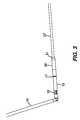
- FIG. 2depicts a close-up view of the distal ends of the instrument and loader in FIG. 1 ;
- FIG. 3depicts an instrument being inserted into an end effector
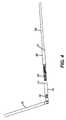
- FIG. 4depicts an instrument attached to an end effector being withdrawn from a loader
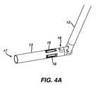
- FIG. 4Adepicts a loader with removable distal end
- FIG. 5depicts an isometric close-up view of the distal end of an instrument in a locked position
- FIG. 6depicts an isometric close-up view of the distal end of an instrument in an unlocked position
- FIG. 7depicts an isometric cross-sectional view of the distal end of an instrument attached to an end effector
- FIG. 8depicts an isometric cross-sectional view of the distal end of an instrument attached to an end effector in a pushed-off configuration
- FIG. 9depicts an instrument handle
- FIG. 10depicts a bi-polar jawed end effector
- FIG. 11depicts a cutting shears end effector
- FIG. 12depicts a Maryland dissector end effector
- FIG. 13depicts an ultrasonic shears end effector
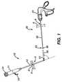
- instrument ( 20 )comprises an elongate shaft ( 22 ) passing through an incision ( 8 ) of a tissue wall ( 6 ).
- a loader ( 10 )comprises an elongate shaft ( 12 ) passing through an incision ( 4 ) of a tissue wall ( 2 ).
- the surgical end effector ( 30 )is selectively attachable in vivo and detachable in vivo to the attachment mechanism ( 40 ) located at the distal end ( 23 ) of the instrument ( 20 ).
- the end effectoris a jawed tissue grasper, but a variety of other end effectors could be also be used.
- the end effector ( 30 )may be loaded ex vivo into the distal end ( 13 ) of the shaft ( 12 ), and then introduced into the surgical field through the incision ( 4 ).
- the loader ( 10 )holds the end effector ( 30 ) during the in vivo attachment to and in vivo detachment from the instrument ( 20 ).
- the loader ( 10 ) and instrument ( 20 )each includes ex vivo handles ( 11 , 21 ) attached to the proximal ends of the shafts ( 12 , 22 ) that enable surgeons to use the devices.
- tissue wall ( 2 , 6 ) anatomieswill vary based on the surgical procedure, but some non-limiting examples include percutaneous incisions into the abdomen, thorax, or pelvis.
- the incisions ( 4 , 8 )may be created with a cutting or puncturing instrument, and will typically be spaced from one another.
- the tissue walls ( 2 , 6 )may be the same or different anatomies. For instance, tissue walls ( 2 , 6 ) may both be the abdominal wall.
- tissue wall ( 2 )could be an organ (e.g., stomach, colon, esophagus, etc.) accessed through a natural orifice, while the incision ( 8 ) in tissue wall ( 6 ) could be percutaneous.
- incision ( 4 )may provide access to the abdomen, while the incision ( 8 ) may provide access to the pelvis.
- the incisionsmay include instrument seals, such as those commonly found in trocars.
- the instrument seal ( 5 )is schematically shown in incision ( 4 ) with the loader ( 10 ) passing through the seal ( 5 ), while the shaft ( 22 ) seals directly with the tissue wall ( 6 ) by virtue of the resilience of the tissue without the aid of a sealing device.
- the loader shaft ( 12 ) in this embodimentis rigid and straight, but the shaft ( 12 ) could be curved or flexible, which would be beneficial for natural orifice transluminal introduction of the distal end ( 13 ) to the surgical field.
- the loader ( 10 )may include an articulating distal end ( 13 ) controlled by the knob ( 14 ).
- the distal end ( 13 )will typically be introduced and removed through the incision ( 4 ) in-line with the shaft ( 12 ), and then articulated in vivo to facilitate alignment between the end effector ( 30 ) and the shaft ( 22 ).
- the arm ( 15 )is rigidly connected the handle ( 11 ) to facilitate grasping of the handle and rotational orientation of the articulated distal end ( 13 ) about the shaft ( 12 ) axis.
- the distal end ( 13 ) of the loader ( 10 )comprises a tube opening at the distal tip ( 17 ).
- the tubeis dimensioned to receive the end effector ( 30 ).
- the tube ( 30 )includes an engagement feature ( 16 ) for holding the end effector ( 30 ). While the engagement feature ( 16 ) may vary, in this embodiment a plurality of leaf springs provide an interference fit with the end effector ( 30 ) to frictionally hold the end effector in the tube.
- the end effector ( 30 )when the end effector ( 30 ) is loaded in the tube, the distal end ( 32 ) is positioned in the tube and the proximal end ( 31 ) extends from the tube opening ( 17 ). This arrangement prevents the jaws of the end effector from opening. After the distal end ( 23 ) of the instrument ( 20 ) is attached to the proximal end ( 31 ) of the end effector ( 30 ), the end effector ( 3 ) can be pulled from the distal end ( 13 ) of the loader ( 10 ).
- FIG. 4Adepicts an alternative embodiment of a loader ( 10 ) where the distal end ( 13 ) is selectively attachable and detachable to the shaft ( 12 ). As shown in this example, this feature is enabled with a bayonet connection ( 18 ), but other connections are also contemplated including snap connections, threaded connections, and the like.
- a bayonet connection18
- other connectionsare also contemplated including snap connections, threaded connections, and the like.
- One advantage of this alternative embodimentis that different distal end ( 13 ) configurations may be used to hold end effectors that may not be accommodated by a single sized tube.
- FIGS. 5 and 6depict a detailed view of one embodiment of an attachment mechanism ( 40 ) located at the distal end ( 23 ) of the shaft ( 22 ).
- the attachment mechanism ( 40 )comprises a mating feature on the shaft ( 22 ), which in this embodiment is a circumferential groove ( 45 ) positioned on the lateral surface of the shaft ( 22 ).
- the attachment mechanism ( 40 )also comprises arms ( 42 A, 42 B) projecting distally from the distal end ( 44 ) of the shaft ( 22 ).
- the armsare axially slideable relative the shaft ( 22 ) and are resiliently deflectable medially into the gap ( 46 ).
- the armseach comprise a mating feature, which in this embodiment comprises a stepped lateral notch ( 43 A, 43 B).
- An elongate pin ( 41 )is positioned medially relative the arms ( 42 ) and is axially slideable relative the arms ( 42 ) between a locked position preventing medial deflection of the arms (an example of which is shown in FIG. 5 ) and an unlocked position allowing medial deflection of the arms (an example of which is shown in FIG. 6 ).
- the pin ( 41 ) and arms ( 42 )may each slide independently relative the shaft ( 22 ).
- the elongate pin ( 41 )may include a pointed obtruator tip.
- the distal end ( 23 )may be used to puncture through the tissue wall ( 6 ).
- the distal ends of the arms ( 42 ) and distal end ( 44 ) of the shaft ( 22 )include tapered surfaces to facilitate passing through the incision ( 8 ).
- FIG. 7shows the attachment mechanism ( 40 ) attached to the end effector ( 30 ).
- the groove ( 45 ) of the shaft ( 22 )mates the rib ( 32 ) of the end effector ( 30 ) preventing relative axial motion.
- the lateral grooves ( 43 ) of the arms ( 42 )mate the ring ( 33 ) of the end effector ( 30 ) preventing relative axial motion.
- the rib ( 32 )is rigidly connected to the outer housing ( 37 ) of the end effector ( 30 ), and the ring ( 33 ) is rigidly connected to the jaw actuator ( 34 ) via the coupling ( 35 ). Accordingly, axial movement of the arms ( 42 ) relative the shaft ( 22 ) will cause axial movement of the jaw actuator ( 34 ) relative the housing ( 37 ), thereby causing the jaws to open and close.
- the distal end ( 23 )is introduced in into the proximal end ( 31 ) of the end effector ( 30 ) with the pin ( 41 ) in the unlocked position.
- the chamfered lead ( 36 ) of the ring ( 33 )medially deflects the arms ( 42 ) until the ring ( 33 ) is seated into the lateral notches ( 43 ).
- the shaft ( 22 )advances axially into the end effector ( 30 ), and the tapered end ( 44 ) aligns the rib ( 32 ) to seat into the groove ( 45 ).
- the surgeonwill feel a tactile “snap” indicating proper engagement.
- the pin ( 41 )may be slid to the locked position thereby attaching the end effector ( 30 ) to the instrument ( 20 ).
- the surgeonmay pull the end effector from the loader ( 10 ), and the loader ( 10 ) may then be removed from the surgical field. The surgeon may then manipulate tissue with the end effector ( 30 ) as needed for the surgical procedure.
- FIG. 9shows and example of the handle ( 21 ) for the instrument ( 20 ).
- the handle ( 21 )includes a base ( 50 ).
- a knob ( 51 )rotates the attachment mechanism ( 40 ) about the axis of the shaft ( 22 ), which will also rotate an attached end effector ( 30 ).
- the trigger ( 54 )pivots relative the base ( 50 ) causing axial movement of the arms ( 42 ) and the pin ( 41 ) relative the shaft ( 22 ). Operation of the trigger ( 54 ) will operate the jaws on an attached end effector ( 30 ).
- the latch ( 55 )pivots relative the base ( 50 ) between a locked position (as shown in figure) to prevent operation of the trigger ( 54 ) and an unlocked position recessed in the base ( 50 ).
- the latch ( 55 )may be locked to maintain the same relative axial spacing of the corresponding the mating features ( 43 , 45 ) as the mating features ( 33 , 32 ), resulting in resulting in a single “snap” feedback.
- the trigger lock ( 56 )can lock/unlock the trigger in/from its depressed position.
- An actuator ( 53 )which in this embodiment is a slider, controls axial movement of the pin ( 51 ) relative the arms ( 42 ). The distal most position of the actuator ( 53 ) relative the base (as shown in the figure) places the pin ( 51 ) in its locked position, and the proximal most position places the pin ( 51 ) in its unlocked position.
- the pin lock ( 52 )includes a pin ( 52 A) which went inserted into the hole ( 53 A) maintains the pin ( 41 ) and arms ( 42 ) in the extended and locked positions as shown in FIG. 5 .
- FIGS. 10-13illustrate some non-limiting examples of alternative end effectors ( 30 A-D) that may attached to the distal end ( 23 ) of the instrument ( 20 ).
- all or a portion of the end effectors ( 30 , 30 A, 30 B, 30 C, 30 D)may be bundled as part of a kit so the surgeon may interchange the attached end effector as needed for a surgical procedure.
- All the end effectors examples shown herehave cooperating jaws; however, non-jawed end effectors could also be employed such as hook knives, snares, and the like.
- the followingdescribes one method for using the devices during a laparoscopic surgical procedure.
- An instrument ( 20 )is obtained and passed through incision ( 8 ).
- the incision ( 8 )may be a precutaneous incision formed at least partially by a puncture formed with the obtruator on the pin ( 41 ) in the configuration shown in FIG. 5 .
- the pin lock ( 52 ) and latch ( 55 )may be secured to the slider ( 53 ) and trigger ( 54 ), respectively. After the puncture, the pin lock ( 52 ) may be removed.
- a loader ( 10 ) and end effector ( 30 )are obtained.
- the end effector ( 30 )may be selected from a plurality of end effectors provided in a kit.
- the end effector ( 30 )is loading ex vivo into the distal end ( 13 ) of the loader ( 10 ).
- the distal end ( 13 ) of the loader ( 10 ) with the loaded end effector ( 30 )is passed through incision ( 4 ).
- the second incision ( 4 )may also be percutaneous incision spaced from the first incision ( 8 ), and may include passing the distal end ( 13 ) with the loaded end effector ( 30 ) through a trocar.
- the distal end ( 13 )may be articulated to facilitate orientation between the proximal end ( 31 ) of the end effector ( 30 ) and the attachment mechanism ( 40 ).
- the actuator ( 53 )is slid proximally to move the pin ( 41 ) to its unlocked position.
- the distal end ( 23 ) of the instrument ( 20 )is advanced into the proximal end ( 31 ) of the end effector ( 30 ) until the respective mating features of the instrument ( 20 ) and end effector ( 30 ) are engaged.
- the actuator ( 53 )may then be slid distally thus advancing the pin ( 41 ) to its locked position.
- the end effector ( 30 )has now been attached in vivo to the instrument ( 20 ).
- the end effector ( 30 )may then be pulled from the loader ( 10 ) and the latch ( 55 ) disengaged from the trigger ( 54 ). Tissue is then manipulating by actuating the trigger ( 54 ) of the handle ( 21 ) to operate the jaws of the end effector ( 30 ).
- the end effector ( 30 )may be detached from the shaft ( 22 ). If previously removed, the loader ( 10 ) may be reintroduced through the incision ( 4 ) into the surgical field. The distal end ( 32 ) of the end effector ( 30 ) is seated into the distal end ( 13 ) of the loader ( 10 ), and the pin ( 41 ) moved to its unlocked position. The arms ( 42 ) are then proximally withdrawn from the ring ( 33 ) and the pin ( 41 ) is returned to the locked position. Accordingly, the device will be in the configuration depicted in FIG. 8 .
- the end effector ( 30 )may have a much larger diameter than the shaft ( 22 ); accordingly, the incision ( 8 ) can be smaller compared to more traditional laparoscopic instruments resulting in less pain and scarring, and quicker recovery. This also facilitates a small diameter shaft ( 22 ) (even less than 3 mm), thus potentially eliminating a trocar in the incision ( 8 ).
- the attachment mechanism ( 40 )provides quick end effector ( 30 ) exchanges with the instrument ( 20 ), thus decreasing surgical time.
- the loader ( 10 )also facilitates quick end effector ( 30 ) exchanges.
- a kit of multiple end effectorsmay reduce instrument costs by consolidating a single shaft ( 22 ) and handle ( 21 ) for all instruments. Many other benefits will be apparent to those skilled in the art.
Landscapes
- Health & Medical Sciences (AREA)
- Surgery (AREA)
- Life Sciences & Earth Sciences (AREA)
- Engineering & Computer Science (AREA)
- Biomedical Technology (AREA)
- Public Health (AREA)
- Nuclear Medicine, Radiotherapy & Molecular Imaging (AREA)
- Veterinary Medicine (AREA)
- General Health & Medical Sciences (AREA)
- Heart & Thoracic Surgery (AREA)
- Medical Informatics (AREA)
- Molecular Biology (AREA)
- Animal Behavior & Ethology (AREA)
- Physics & Mathematics (AREA)
- Otolaryngology (AREA)
- Plasma & Fusion (AREA)
- Ophthalmology & Optometry (AREA)
- Surgical Instruments (AREA)
Abstract
Description
The present invention relates in general to surgical devices and procedures, and more particularly to minimally invasive surgery.
Surgical procedures are often used to treat and cure a wide range of diseases, conditions, and injuries. Surgery often requires access to internal tissue through open surgical procedures or endoscopic surgical procedures. The term “endoscopic” refers to all types of minimally invasive surgical procedures including laparoscopic, arthroscopic, natural orifice intraluminal, and natural orifice transluminal procedures. Endoscopic surgery has numerous advantages compared to traditional open surgical procedures, including reduced trauma, faster recovery, reduced risk of infection, and reduced scarring. Endoscopic surgery is often performed with an insufflatory fluid present within the body cavity, such as carbon dioxide or saline, to provide adequate space to perform the intended surgical procedures. The insufflated cavity is generally under pressure and is sometimes referred to as being in a state of pneumoperitoneum. Surgical access devices are often used to facilitate surgical manipulation of internal tissue while maintaining pneumoperitoneum. For example, trocars are often used to provide a port through which endoscopic surgical instruments are passed. Trocars generally have an instrument seal, which prevents the insufflatory fluid from escaping while an instrument is positioned in the trocar.
While surgical access devices are known, no one has previously made or used the surgical devices and methods in accordance with the present invention.
While the specification concludes with claims which particularly point out and distinctly claim the invention, it is believed the invention will be better understood from the following description taken in conjunction with the accompanying drawings illustrating some non-limiting examples of the invention. Unless otherwise indicated, the figures are not necessarily drawn to scale, but rather to illustrate the principles of the invention.
As shown inFIG. 1 , instrument (20) comprises an elongate shaft (22) passing through an incision (8) of a tissue wall (6). A loader (10) comprises an elongate shaft (12) passing through an incision (4) of a tissue wall (2). The surgical end effector (30) is selectively attachable in vivo and detachable in vivo to the attachment mechanism (40) located at the distal end (23) of the instrument (20). In this example, the end effector is a jawed tissue grasper, but a variety of other end effectors could be also be used. The end effector (30) may be loaded ex vivo into the distal end (13) of the shaft (12), and then introduced into the surgical field through the incision (4). The loader (10) holds the end effector (30) during the in vivo attachment to and in vivo detachment from the instrument (20). The loader (10) and instrument (20) each includes ex vivo handles (11,21) attached to the proximal ends of the shafts (12,22) that enable surgeons to use the devices.
The tissue wall (2,6) anatomies will vary based on the surgical procedure, but some non-limiting examples include percutaneous incisions into the abdomen, thorax, or pelvis. The incisions (4,8) may be created with a cutting or puncturing instrument, and will typically be spaced from one another. The tissue walls (2,6) may be the same or different anatomies. For instance, tissue walls (2,6) may both be the abdominal wall. In another example, tissue wall (2) could be an organ (e.g., stomach, colon, esophagus, etc.) accessed through a natural orifice, while the incision (8) in tissue wall (6) could be percutaneous. In yet another example, incision (4) may provide access to the abdomen, while the incision (8) may provide access to the pelvis. If pneumoperitoneum is desired, the incisions may include instrument seals, such as those commonly found in trocars. In this example, the instrument seal (5) is schematically shown in incision (4) with the loader (10) passing through the seal (5), while the shaft (22) seals directly with the tissue wall (6) by virtue of the resilience of the tissue without the aid of a sealing device.
The loader shaft (12) in this embodiment is rigid and straight, but the shaft (12) could be curved or flexible, which would be beneficial for natural orifice transluminal introduction of the distal end (13) to the surgical field. The loader (10) may include an articulating distal end (13) controlled by the knob (14). The distal end (13) will typically be introduced and removed through the incision (4) in-line with the shaft (12), and then articulated in vivo to facilitate alignment between the end effector (30) and the shaft (22). The arm (15) is rigidly connected the handle (11) to facilitate grasping of the handle and rotational orientation of the articulated distal end (13) about the shaft (12) axis. In this embodiment, the distal end (13) of the loader (10) comprises a tube opening at the distal tip (17). The tube is dimensioned to receive the end effector (30). The tube (30) includes an engagement feature (16) for holding the end effector (30). While the engagement feature (16) may vary, in this embodiment a plurality of leaf springs provide an interference fit with the end effector (30) to frictionally hold the end effector in the tube. In this embodiment, when the end effector (30) is loaded in the tube, the distal end (32) is positioned in the tube and the proximal end (31) extends from the tube opening (17). This arrangement prevents the jaws of the end effector from opening. After the distal end (23) of the instrument (20) is attached to the proximal end (31) of the end effector (30), the end effector (3) can be pulled from the distal end (13) of the loader (10).
As shown in the embodiment ofFIG. 5 , the elongate pin (41) may include a pointed obtruator tip. In this configuration the distal end (23) may be used to puncture through the tissue wall (6). The distal ends of the arms (42) and distal end (44) of the shaft (22) include tapered surfaces to facilitate passing through the incision (8).
The following describes one method for attaching the end effector (30) to the shaft (22). The distal end (23) is introduced in into the proximal end (31) of the end effector (30) with the pin (41) in the unlocked position. As the arms (42) are advanced axially into the end effector (30), the chamfered lead (36) of the ring (33) medially deflects the arms (42) until the ring (33) is seated into the lateral notches (43). Simultaneously the shaft (22) advances axially into the end effector (30), and the tapered end (44) aligns the rib (32) to seat into the groove (45). In both cases, the surgeon will feel a tactile “snap” indicating proper engagement. Once fully seated in the end effector (30), the pin (41) may be slid to the locked position thereby attaching the end effector (30) to the instrument (20). Once attached, the surgeon may pull the end effector from the loader (10), and the loader (10) may then be removed from the surgical field. The surgeon may then manipulate tissue with the end effector (30) as needed for the surgical procedure.
The following describes one method for using the devices during a laparoscopic surgical procedure. An instrument (20) is obtained and passed through incision (8). The incision (8) may be a precutaneous incision formed at least partially by a puncture formed with the obtruator on the pin (41) in the configuration shown inFIG. 5 . The pin lock (52) and latch (55) may be secured to the slider (53) and trigger (54), respectively. After the puncture, the pin lock (52) may be removed.
A loader (10) and end effector (30) are obtained. The end effector (30) may be selected from a plurality of end effectors provided in a kit. The end effector (30) is loading ex vivo into the distal end (13) of the loader (10). The distal end (13) of the loader (10) with the loaded end effector (30) is passed through incision (4). The second incision (4) may also be percutaneous incision spaced from the first incision (8), and may include passing the distal end (13) with the loaded end effector (30) through a trocar. The distal end (13) may be articulated to facilitate orientation between the proximal end (31) of the end effector (30) and the attachment mechanism (40). The actuator (53) is slid proximally to move the pin (41) to its unlocked position. The distal end (23) of the instrument (20) is advanced into the proximal end (31) of the end effector (30) until the respective mating features of the instrument (20) and end effector (30) are engaged. The actuator (53) may then be slid distally thus advancing the pin (41) to its locked position. The end effector (30) has now been attached in vivo to the instrument (20). The end effector (30) may then be pulled from the loader (10) and the latch (55) disengaged from the trigger (54). Tissue is then manipulating by actuating the trigger (54) of the handle (21) to operate the jaws of the end effector (30).
After completing the surgical procedure, the end effector (30) may be detached from the shaft (22). If previously removed, the loader (10) may be reintroduced through the incision (4) into the surgical field. The distal end (32) of the end effector (30) is seated into the distal end (13) of the loader (10), and the pin (41) moved to its unlocked position. The arms (42) are then proximally withdrawn from the ring (33) and the pin (41) is returned to the locked position. Accordingly, the device will be in the configuration depicted inFIG. 8 . Distally advancing the arms (42) will push the ring (33) distally till the rib (32) unseats from the groove (45). This unseating may be facilitated by the jaws of the end effector (30) being held in a closed position by the tube in the loader distal end (13). The distal end (23) may then be withdrawn from the end effector (30) thus detaching the end effector (30) from the instrument (20). The end effector will be held in the loader (10) by virtue of the engagement feature (16). Removal of the loader (10) from the surgical field will remove the end effector (30). A different end effector may then be attached to the instrument (20), or the instrument (20) may be withdrawn from the surgical field.
Without limitation, the following describe some of the benefits and advantages of the foregoing devices and methods over the prior art. The end effector (30) may have a much larger diameter than the shaft (22); accordingly, the incision (8) can be smaller compared to more traditional laparoscopic instruments resulting in less pain and scarring, and quicker recovery. This also facilitates a small diameter shaft (22) (even less than 3 mm), thus potentially eliminating a trocar in the incision (8). The attachment mechanism (40) provides quick end effector (30) exchanges with the instrument (20), thus decreasing surgical time. The loader (10) also facilitates quick end effector (30) exchanges. A kit of multiple end effectors may reduce instrument costs by consolidating a single shaft (22) and handle (21) for all instruments. Many other benefits will be apparent to those skilled in the art.
Having shown and described various embodiments and examples of the present invention, further adaptations of the methods and devices described herein can be accomplished by appropriate modifications by one of ordinary skill in the art without departing from the scope of the present invention. Several of such potential modifications have been mentioned, and others will be apparent to those skilled in the art. For instance, the specific materials, dimensions, and the scale of drawings will be understood to be non-limiting examples. Accordingly, the scope of the present invention should be considered in terms of the following claims and is understood not to be limited to the details of structure, materials, or acts shown and described in the specification and drawings.
Claims (12)
1. A laparoscopic surgical method, comprising:
a) obtaining a first instrument comprising an elongate shaft with a distal end and a proximal end connected to a first handle;
b) passing the distal end of the first instrument directly through a percutaneous incision without a sealing device;
c) obtaining a surgical end effector having a distal end with operable jaws and a proximal end selectively attachable to and detachable from the distal end of the first instrument;
d) obtaining a second instrument comprising a distal end and a proximal end connected to a second handle, the distal end comprising a tubular receiver;
e) loading ex vivo the surgical end effector on the distal end of the second instrument such that the operable jaws are positioned in the tubular receiver;
f) passing the distal end of the second instrument with the loaded surgical end effector through a sealing device in a second incision spaced from the percutaneous incision;
g) bending the distal end of the second instrument;
h) attaching in vivo the proximal end of the surgical end effector to the distal end of the first instrument; and
i) manipulating tissue by actuating the handle of the first instrument to operate the jaws of the surgical end effector.
2. The laparoscopic surgical method ofclaim 1 , wherein (f) comprises passing the distal end of the second instrument with the loaded surgical end effector through a trocar.
3. The laparoscopic surgical method ofclaim 1 , wherein the first percutaneous incision is created at least partially by a puncture formed with an obturator on the distal end of the first instrument.
4. The laparoscopic surgical method ofclaim 1 , wherein (g) further comprises aligning the surgical end effector with the first instrument by bending the distal end of the second instrument.
5. The laparoscopic surgical method ofclaim 1 , wherein (e) comprises loading ex vivo the surgical end effector in the tubular receiver on the distal end of the second instrument such that the jaws are prevented from opening.
6. The laparoscopic surgical method ofclaim 1 , wherein (e) comprises loading ex vivo the surgical end effector on the distal end of the second instrument such that the surgical end effector extends distally from the distal end of the second instrument.
7. The laparoscopic surgical method ofclaim 6 , wherein (e) comprises loading ex vivo the surgical end effector in the tubular receiver on the distal end of the second instrument such that the distal end of the surgical end effector is positioned in the tubular receiver and the proximal end of the surgical end effector extends distally from the tubular receiver.
8. The laparoscopic surgical method ofclaim 1 , wherein (e) comprises obtaining a surgical end effector from a plurality of surgical end effectors each having a distal end with operable jaws and a proximal end selectively attachable to and detachable from the distal end of the first instrument.
9. A laparoscopic surgical method, comprising:
a) obtaining a first instrument comprising an elongate shaft with a distal end and a proximal end connected to a first actuator, the distal end comprising an obturator;
b) creating a first incision with the obturator,
c) passing the distal end of the first instrument directly through the first incision without a sealing device;
d) obtaining a surgical end effector having a distal end with operable jaws and a proximal end selectively attachable to and detachable from the distal end of the first instrument;
e) obtaining a second instrument comprising a distal end having a tubular receiver and a proximal end connected to a second actuator;
f) loading ex vivo the surgical end effector on the distal end of the second instrument such that the jaws are inserted into the tubular receiver;
g) passing the distal end of the second instrument with the loaded surgical end effector through a sealing device in a second incision spaced from the first incision;
h) orienting the surgical end effector relative the first instrument by bending the distal end of the second instrument;
i) attaching in vivo the proximal end of the surgical end effector to the distal end of the first instrument; and
j) manipulating tissue by actuating the first actuator of the first instrument to operate the jaws of the surgical end effector.
10. The laparoscopic surgical method ofclaim 9 , wherein the surgical end effector comprises a distal tissue engaging feature and a proximal mating feature adapted to mate with the first percutaneous instrument, and wherein step (e) further comprises loading ex vivo the surgical end effector on the distal end of the second instrument such that the proximal end of the end effector projects distally from the tubular receiver.
11. The laparoscopic surgical method ofclaim 10 , wherein in step (f) the loaded surgical end effector is passed through the sealing device proximal end first.
12. The laparoscopic surgical method ofclaim 9 , wherein step (f) further comprises loading ex vivo the surgical end effector on the distal end of the second instrument such that the proximal end of the end effector projects distally from the tubular receiver.
Priority Applications (5)
| Application Number | Priority Date | Filing Date | Title |
|---|---|---|---|
| US12/576,578US9186203B2 (en) | 2009-10-09 | 2009-10-09 | Method for exchanging end effectors In Vivo |
| BR112012010813ABR112012010813B8 (en) | 2009-10-09 | 2010-10-07 | surgical device and laparoscopic surgical device |
| JP2012533317AJP5770194B2 (en) | 2009-10-09 | 2010-10-07 | Laparoscopic instrument with attached end effector |
| PCT/US2010/051812WO2011044353A1 (en) | 2009-10-09 | 2010-10-07 | Laparoscopic instrument with attachable end effector |
| EP10771594.8AEP2485632B1 (en) | 2009-10-09 | 2010-10-07 | Laparoscopic instrument with attachable end effector |
Applications Claiming Priority (1)
| Application Number | Priority Date | Filing Date | Title |
|---|---|---|---|
| US12/576,578US9186203B2 (en) | 2009-10-09 | 2009-10-09 | Method for exchanging end effectors In Vivo |
Publications (2)
| Publication Number | Publication Date |
|---|---|
| US20110087267A1 US20110087267A1 (en) | 2011-04-14 |
| US9186203B2true US9186203B2 (en) | 2015-11-17 |
Family
ID=43855439
Family Applications (1)
| Application Number | Title | Priority Date | Filing Date |
|---|---|---|---|
| US12/576,578Active2031-02-17US9186203B2 (en) | 2009-10-09 | 2009-10-09 | Method for exchanging end effectors In Vivo |
Country Status (1)
| Country | Link |
|---|---|
| US (1) | US9186203B2 (en) |
Cited By (15)
| Publication number | Priority date | Publication date | Assignee | Title |
|---|---|---|---|---|
| US9526516B2 (en) | 2012-09-26 | 2016-12-27 | Ethicon Endo-Surgery, Llc | Detachable end effector and loader |
| US10004558B2 (en) | 2009-01-12 | 2018-06-26 | Ethicon Endo-Surgery, Inc. | Electrical ablation devices |
| US10105141B2 (en) | 2008-07-14 | 2018-10-23 | Ethicon Endo-Surgery, Inc. | Tissue apposition clip application methods |
| US10143454B2 (en) | 2009-10-09 | 2018-12-04 | Ethicon Llc | Loader for exchanging end effectors in vivo |
| US10206709B2 (en) | 2012-05-14 | 2019-02-19 | Ethicon Llc | Apparatus for introducing an object into a patient |
| US10258406B2 (en) | 2011-02-28 | 2019-04-16 | Ethicon Llc | Electrical ablation devices and methods |
| US10278761B2 (en) | 2011-02-28 | 2019-05-07 | Ethicon Llc | Electrical ablation devices and methods |
| US10314649B2 (en) | 2012-08-02 | 2019-06-11 | Ethicon Endo-Surgery, Inc. | Flexible expandable electrode and method of intraluminal delivery of pulsed power |
| US10314603B2 (en) | 2008-11-25 | 2019-06-11 | Ethicon Llc | Rotational coupling device for surgical instrument with flexible actuators |
| US10342598B2 (en) | 2012-08-15 | 2019-07-09 | Ethicon Llc | Electrosurgical system for delivering a biphasic waveform |
| US10478248B2 (en) | 2007-02-15 | 2019-11-19 | Ethicon Llc | Electroporation ablation apparatus, system, and method |
| US10492880B2 (en) | 2012-07-30 | 2019-12-03 | Ethicon Llc | Needle probe guide |
| EP3690004A1 (en) | 2007-10-04 | 2020-08-05 | Universal Display Corporation | Complexes with tridentate ligands |
| US10779882B2 (en) | 2009-10-28 | 2020-09-22 | Ethicon Endo-Surgery, Inc. | Electrical ablation devices |
| US11484191B2 (en) | 2013-02-27 | 2022-11-01 | Cilag Gmbh International | System for performing a minimally invasive surgical procedure |
Families Citing this family (25)
| Publication number | Priority date | Publication date | Assignee | Title |
|---|---|---|---|---|
| US20110087265A1 (en)* | 2009-10-09 | 2011-04-14 | Nobis Rudolph H | Laparoscopic instrument with attachable end effector |
| US9186203B2 (en) | 2009-10-09 | 2015-11-17 | Ethicon Endo-Surgery, Inc. | Method for exchanging end effectors In Vivo |
| DE102009045749A1 (en)* | 2009-10-15 | 2011-04-21 | Aesculap Ag | Surgical instrument |
| US9028483B2 (en) | 2009-12-18 | 2015-05-12 | Ethicon Endo-Surgery, Inc. | Surgical instrument comprising an electrode |
| JP5916735B2 (en)* | 2010-09-24 | 2016-05-11 | エシコン・エンド−サージェリィ・インコーポレイテッドEthicon Endo−Surgery,Inc. | Laparoscopic instrument with attached end effector |
| US9049987B2 (en) | 2011-03-17 | 2015-06-09 | Ethicon Endo-Surgery, Inc. | Hand held surgical device for manipulating an internal magnet assembly within a patient |
| US9282879B2 (en) | 2011-03-24 | 2016-03-15 | EON Surgical Ltd. | Laparoscope system |
| US8721529B2 (en) | 2011-09-30 | 2014-05-13 | Ethicon Endo-Surgery, Inc. | Devices and methods for providing suction and/or irrigation in a surgical procedure |
| US20130085341A1 (en) | 2011-09-30 | 2013-04-04 | Rudolph H. Nobis | Methods and devices for manipulating tissue in vivo |
| US9078662B2 (en) | 2012-07-03 | 2015-07-14 | Ethicon Endo-Surgery, Inc. | Endoscopic cap electrode and method for using the same |
| US9398905B2 (en) | 2012-12-13 | 2016-07-26 | Ethicon Endo-Surgery, Llc | Circular needle applier with offset needle and carrier tracks |
| US9451937B2 (en)* | 2013-02-27 | 2016-09-27 | Ethicon Endo-Surgery, Llc | Percutaneous instrument with collet locking mechanisms |
| AU2014250896B2 (en)* | 2013-04-11 | 2018-11-15 | Faculty Physicians And Surgeons Of Loma Linda University School Of Medicine | Minimally invasive surgical devices and methods |
| US10166030B2 (en) | 2014-02-03 | 2019-01-01 | Modular Surgical, Inc. | Surgical tool system having multiple tool tip interfaces |
| US9795449B2 (en) | 2014-02-06 | 2017-10-24 | Faculty Physicians And Surgeons Of Loma Linda University School Of Medicine | Methods and devices for performing abdominal surgery |
| US20170056038A1 (en) | 2015-08-26 | 2017-03-02 | Ethicon Endo-Surgery, Llc | Dissecting surgical jaws |
| MX2018002324A (en) | 2015-08-26 | 2018-04-11 | Ethicon Llc | Articulating surgical devices and loaders having stabilizing features. |
| US10342520B2 (en) | 2015-08-26 | 2019-07-09 | Ethicon Llc | Articulating surgical devices and loaders having stabilizing features |
| US10335196B2 (en) | 2015-08-31 | 2019-07-02 | Ethicon Llc | Surgical instrument having a stop guard |
| US10251636B2 (en) | 2015-09-24 | 2019-04-09 | Ethicon Llc | Devices and methods for cleaning a surgical device |
| US10702257B2 (en) | 2015-09-29 | 2020-07-07 | Ethicon Llc | Positioning device for use with surgical instruments |
| US10675009B2 (en) | 2015-11-03 | 2020-06-09 | Ethicon Llc | Multi-head repository for use with a surgical device |
| US10912543B2 (en) | 2015-11-03 | 2021-02-09 | Ethicon Llc | Surgical end effector loading device and trocar integration |
| US10265130B2 (en) | 2015-12-11 | 2019-04-23 | Ethicon Llc | Systems, devices, and methods for coupling end effectors to surgical devices and loading devices |
| US10722228B2 (en) | 2016-02-12 | 2020-07-28 | Medos International Sarl | Suture anchors having location placement identification features |
Citations (145)
| Publication number | Priority date | Publication date | Assignee | Title |
|---|---|---|---|---|
| US3043309A (en) | 1959-09-29 | 1962-07-10 | Avco Corp | Method of performing intestinal intubation |
| US3358676A (en) | 1962-11-30 | 1967-12-19 | Yeda Res & Dev | Magnetic propulsion of diagnostic or therapeutic elements through the body ducts of animal or human patients |
| US3710399A (en) | 1970-06-23 | 1973-01-16 | H Hurst | Ossicle replacement prosthesis |
| US3893448A (en)* | 1973-11-26 | 1975-07-08 | John W Brantigan | Catheter device for use in detecting gas in body fluids and tissue |
| US3906217A (en) | 1974-03-14 | 1975-09-16 | Ppg Industries Inc | Lamp mounting bracket |
| US3988535A (en) | 1975-11-04 | 1976-10-26 | Western Electric Company, Inc. | Automated positioning |
| US4047136A (en) | 1975-05-13 | 1977-09-06 | Nihon Beru-Haueru Kabushiki Kaisha (Bell & Howell Japan, Ltd.) | Moving magnet type instrument |
| US4063561A (en) | 1975-08-25 | 1977-12-20 | The Signal Companies, Inc. | Direction control device for endotracheal tube |
| US4099192A (en) | 1974-07-12 | 1978-07-04 | Canon Kabushiki Kaisha | Photographic camera with an electromagnetic control system |
| US4278077A (en) | 1978-07-27 | 1981-07-14 | Olympus Optical Co., Ltd. | Medical camera system |
| US4384584A (en) | 1981-10-28 | 1983-05-24 | Chen Allen S | Method and means for esophageal feeding |
| US4585282A (en) | 1983-07-19 | 1986-04-29 | Bosley Robert W | Magnetic levitation system |
| US4597390A (en) | 1984-04-02 | 1986-07-01 | Mulhollan James S | Surgical needle manipulator |
| US4655746A (en) | 1985-12-02 | 1987-04-07 | Target Therapeutics | Catheter device |
| US5052402A (en)* | 1989-01-31 | 1991-10-01 | C.R. Bard, Inc. | Disposable biopsy forceps |
| US5201743A (en) | 1992-05-05 | 1993-04-13 | Habley Medical Technology Corp. | Axially extendable endoscopic surgical instrument |
| US5282806A (en) | 1992-08-21 | 1994-02-01 | Habley Medical Technology Corporation | Endoscopic surgical instrument having a removable, rotatable, end effector assembly |
| US5286255A (en) | 1991-07-29 | 1994-02-15 | Linvatec Corporation | Surgical forceps |
| US5308357A (en) | 1992-08-21 | 1994-05-03 | Microsurge, Inc. | Handle mechanism for manual instruments |
| US5314424A (en) | 1992-04-06 | 1994-05-24 | United States Surgical Corporation | Surgical instrument locking mechanism |
| US5330502A (en) | 1992-10-09 | 1994-07-19 | Ethicon, Inc. | Rotational endoscopic mechanism with jointed drive mechanism |
| US5352219A (en) | 1992-09-30 | 1994-10-04 | Reddy Pratap K | Modular tools for laparoscopic surgery |
| US5392917A (en) | 1993-08-03 | 1995-02-28 | Ethicon, Inc. | Easy open 1-2-3 instrumentation package |
| US5417203A (en) | 1992-04-23 | 1995-05-23 | United States Surgical Corporation | Articulating endoscopic surgical apparatus |
| US5441059A (en) | 1994-02-02 | 1995-08-15 | Dannan; Patrick A. | Method of minimally invasive surgery |
| US5468250A (en) | 1993-04-01 | 1995-11-21 | Ethicon, Inc. | Endoscopic mechanism with friction maintaining handle |
| US5502698A (en) | 1993-09-22 | 1996-03-26 | Victor Company Of Japan, Ltd. | Automatic attitude correcting system for optical disc device |
| US5507297A (en) | 1991-04-04 | 1996-04-16 | Symbiosis Corporation | Endoscopic instruments having detachable proximal handle and distal portions |
| US5540648A (en) | 1992-08-17 | 1996-07-30 | Yoon; Inbae | Medical instrument stabilizer with anchoring system and methods |
| US5562655A (en) | 1994-08-12 | 1996-10-08 | United States Surgical Corporation | Surgical apparatus having a universal handle for actuating various attachments |
| US5578052A (en) | 1992-10-27 | 1996-11-26 | Koros; Tibor | Insulated laparoscopic grasper with removable shaft |
| US5593402A (en) | 1994-11-14 | 1997-01-14 | Biosearch Medical Products Inc. | Laparoscopic device having a detachable distal tip |
| US5613937A (en) | 1993-02-22 | 1997-03-25 | Heartport, Inc. | Method of retracting heart tissue in closed-chest heart surgery using endo-scopic retraction |
| US5618303A (en) | 1992-07-02 | 1997-04-08 | Marlow Surgical Technologies, Inc. | Endoscopic instrument system and method |
| US5716326A (en) | 1995-08-14 | 1998-02-10 | Dannan; Patrick A. | Method for lifting tissue and apparatus for performing same |
| US5762255A (en) | 1996-02-20 | 1998-06-09 | Richard-Allan Medical Industries, Inc. | Surgical instrument with improvement safety lockout mechanisms |
| US5782748A (en) | 1996-07-10 | 1998-07-21 | Symbiosis Corporation | Endoscopic surgical instruments having detachable proximal and distal portions |
| US5792165A (en) | 1993-07-21 | 1998-08-11 | Charles H. Klieman | Endoscopic instrument with detachable end effector |
| US5810877A (en) | 1994-02-14 | 1998-09-22 | Heartport, Inc. | Endoscopic microsurgical instruments and methods |
| US5881615A (en) | 1997-01-15 | 1999-03-16 | Enderes Tool Company, Inc. | Multiple bit screwdrivers and methods |
| US5928263A (en) | 1998-02-02 | 1999-07-27 | Aslan Medical Technologies | Surgical instrument with flexible actuator and rigid actuator cover |
| US5980455A (en) | 1993-02-22 | 1999-11-09 | Heartport, Inc. | Method for manipulating a tissue structure within a thoracic cavity |
| US6024748A (en) | 1996-07-23 | 2000-02-15 | United States Surgical Corporation | Singleshot anastomosis instrument with detachable loading unit and method |
| US6059719A (en) | 1997-08-06 | 2000-05-09 | Olympus Optical Co., Ltd. | Endoscope system |
| US6099537A (en) | 1996-02-26 | 2000-08-08 | Olympus Optical Co., Ltd. | Medical treatment instrument |
| US6159200A (en) | 1996-11-18 | 2000-12-12 | Smith & Nephew | Systems, methods, and instruments for minimally invasive surgery |
| US6309397B1 (en)* | 1999-12-02 | 2001-10-30 | Sri International | Accessories for minimally invasive robotic surgery and methods |
| US6315789B1 (en) | 1999-02-08 | 2001-11-13 | Andrew H. Cragg | Medical device anchoring system and method |
| US20010051766A1 (en) | 1999-03-01 | 2001-12-13 | Gazdzinski Robert F. | Endoscopic smart probe and method |
| US6419688B1 (en) | 1998-12-04 | 2002-07-16 | Karl Storz Gmbh & Co. Kg | Medical tubular-shaft instrument |
| US6471172B1 (en) | 1999-10-19 | 2002-10-29 | Norbert Lemke | Fixing device for at least one operating element suitable for application in sterile areas in surgical operations, such as a surgical instrument |
| DE10149421A1 (en) | 2001-10-06 | 2003-04-24 | Fraunhofer Ges Forschung | Instrument for use in minimal invasive surgery comprises a magazine for simultaneous storage of a number of tools or instruments for use in surgery and a control device that moves tools from a storage to an operating position |
| US20030114731A1 (en) | 2001-12-14 | 2003-06-19 | Cadeddu Jeffrey A. | Magnetic positioning system for trocarless laparoscopic instruments |
| US6589211B1 (en) | 1995-04-28 | 2003-07-08 | Macleod Cathel | Modified trocar and methods of use |
| US6595984B1 (en) | 2000-03-28 | 2003-07-22 | Microline, Inc. | Laparoscopic instrument with a detachable tip |
| US6626824B2 (en) | 2000-05-16 | 2003-09-30 | Storz Endoskop Gmbh | Exchangeable tool assembly for an endoscopic treatment device and such treatment device |
| US6635071B2 (en) | 1997-07-22 | 2003-10-21 | Karl Storz Gmbh & Co. Kg | Surgical grasping and holding forceps |
| US6723043B2 (en) | 2001-09-25 | 2004-04-20 | Sdgi Holdings, Inc. | Methods and devices for inserting and manipulating surgical instruments |
| US20040093039A1 (en) | 2002-10-25 | 2004-05-13 | Raphael Schumert | Gastrointestinal pacemaker |
| US6770081B1 (en) | 2000-01-07 | 2004-08-03 | Intuitive Surgical, Inc. | In vivo accessories for minimally invasive robotic surgery and methods |
| US20040152941A1 (en) | 2003-02-03 | 2004-08-05 | Scimed Life Systems, Inc. | Medical device with changeable tip flexibility |
| US6776165B2 (en) | 2002-09-12 | 2004-08-17 | The Regents Of The University Of California | Magnetic navigation system for diagnosis, biopsy and drug delivery vehicles |
| US6827712B2 (en) | 1997-06-18 | 2004-12-07 | United States Surgical Corporation | Robotic arm DLUs for performing surgical tasks |
| US20050033354A1 (en) | 2001-11-19 | 2005-02-10 | Scimed Life Systems, Inc. | Endoscopic surgical instrument |
| US6860878B2 (en) | 1998-02-24 | 2005-03-01 | Endovia Medical Inc. | Interchangeable instrument |
| US6869395B2 (en) | 2000-05-15 | 2005-03-22 | C. R. Bard, Inc. | Endoscopic accessory attachment mechanism |
| US6884213B2 (en) | 2000-05-23 | 2005-04-26 | Given Imaging Ltd | Device and method for positioning an object in a body lumen |
| US20050119640A1 (en) | 2003-10-03 | 2005-06-02 | The Regents Of The University Of California | Surgical instrument for adhering to tissues |
| US20050131396A1 (en) | 2001-12-19 | 2005-06-16 | George Stanczak | Failsafe reconfigurable surgical apparatus |
| US6936003B2 (en) | 2002-10-29 | 2005-08-30 | Given Imaging Ltd | In-vivo extendable element device and system, and method of use |
| US6942674B2 (en)* | 2000-01-05 | 2005-09-13 | Integrated Vascular Systems, Inc. | Apparatus and methods for delivering a closure device |
| JP2005261734A (en) | 2004-03-19 | 2005-09-29 | Olympus Corp | Treatment tool for endoscope and endoscope treatment system |
| US20050250984A1 (en) | 2004-05-07 | 2005-11-10 | Usgi Medical Inc. | Multiple removable apparatus and methods for manipulating and securing tissue |
| US20050272972A1 (en) | 2004-06-07 | 2005-12-08 | Iddan Gavriel J | Method, system and device for suction biopsy |
| US20050273139A1 (en) | 2004-06-01 | 2005-12-08 | Norbert Krauss | Device for clamping tissue |
| US20050288555A1 (en) | 2004-06-28 | 2005-12-29 | Binmoeller Kenneth E | Methods and devices for illuminating, vievwing and monitoring a body cavity |
| US6986738B2 (en) | 2001-08-06 | 2006-01-17 | Given Imaging Ltd | System and method for maneuvering a device in vivo |
| US6994708B2 (en) | 2001-04-19 | 2006-02-07 | Intuitive Surgical | Robotic tool with monopolar electro-surgical scissors |
| US20060079933A1 (en) | 2004-10-08 | 2006-04-13 | Dylan Hushka | Latching mechanism for forceps |
| US7039453B2 (en) | 2000-02-08 | 2006-05-02 | Tarun Mullick | Miniature ingestible capsule |
| US7042184B2 (en) | 2003-07-08 | 2006-05-09 | Board Of Regents Of The University Of Nebraska | Microrobot for surgical applications |
| US7066879B2 (en) | 2003-07-15 | 2006-06-27 | The Trustees Of Columbia University In The City Of New York | Insertable device and system for minimal access procedure |
| US7083579B2 (en) | 2001-09-27 | 2006-08-01 | Olympus Corporation | Encapsulated medical device and method of examining, curing, and treating internal region of body cavity using encapsulated medical device |
| US20060184161A1 (en) | 2005-02-16 | 2006-08-17 | Usgi Medical Inc. | Flexible shaft system having interchangeable end effectors |
| US20060190035A1 (en) | 2004-10-08 | 2006-08-24 | Sherwood Services Ag | Latching mechanism for forceps |
| US7122028B2 (en) | 2001-12-19 | 2006-10-17 | Allegiance Corporation | Reconfiguration surgical apparatus |
| US7125403B2 (en) | 1998-12-08 | 2006-10-24 | Intuitive Surgical | In vivo accessories for minimally invasive robotic surgery |
| US20060258905A1 (en) | 2004-01-27 | 2006-11-16 | Olympus Corporation | Endoscope treatment system |
| US20070010709A1 (en) | 2005-07-08 | 2007-01-11 | Johannes Reinschke | Endoscopy capsule |
| US7169104B2 (en) | 2002-09-13 | 2007-01-30 | Pentax Corporation | Magnetic anchor remote guidance system |
| US20070049966A1 (en) | 2005-03-22 | 2007-03-01 | Frank Bonadio | Surgical instrument |
| US20070073247A1 (en) | 2005-07-13 | 2007-03-29 | Microline Pentax Inc. | Tip and shaft connection for medical device |
| US7211094B2 (en) | 2002-11-05 | 2007-05-01 | Satiety, Inc. | Magnetic anchoring devices |
| US20070123748A1 (en) | 2005-07-14 | 2007-05-31 | Dwight Meglan | Robot for minimally invasive interventions |
| US7241290B2 (en) | 2004-06-16 | 2007-07-10 | Kinetic Surgical, Llc | Surgical tool kit |
| US7297142B2 (en) | 1998-02-24 | 2007-11-20 | Hansen Medical, Inc. | Interchangeable surgical instrument |
| US20070270651A1 (en) | 2006-05-19 | 2007-11-22 | Zvika Gilad | Device and method for illuminating an in vivo site |
| US20080015413A1 (en) | 2006-02-22 | 2008-01-17 | Olympus Medical Systems Corporation | Capsule endoscope system and medical procedure |
| WO2008015666A2 (en) | 2006-08-01 | 2008-02-07 | Shaul Shohat | System and method for telesurgery |
| US7331967B2 (en) | 2002-09-09 | 2008-02-19 | Hansen Medical, Inc. | Surgical instrument coupling mechanism |
| US20080045003A1 (en) | 2002-10-15 | 2008-02-21 | Megica Corporation | Method of wire bonding over active area of a semiconductor circuit |
| US20080140090A1 (en) | 2006-10-17 | 2008-06-12 | Ernest Aranyi | Apparatus For Applying Surgical Clips |
| US20080142005A1 (en) | 2005-05-10 | 2008-06-19 | Ralf Schnell | Insertion Aid for Percutaneous Tracheostomy |
| US20080154299A1 (en) | 2006-12-08 | 2008-06-26 | Steve Livneh | Forceps for performing endoscopic surgery |
| US7429259B2 (en) | 2003-12-02 | 2008-09-30 | Cadeddu Jeffrey A | Surgical anchor and system |
| US20080242939A1 (en) | 2007-04-02 | 2008-10-02 | William Johnston | Retractor system for internal in-situ assembly during laparoscopic surgery |
| US20080243106A1 (en)* | 2007-03-30 | 2008-10-02 | Ethicon Endo-Surgery, Inc. | Detachable end effectors |
| US7448993B2 (en) | 2003-09-30 | 2008-11-11 | Olympus Corporation | Gastrointestinal tract examining apparatus |
| US20080287926A1 (en) | 2008-08-02 | 2008-11-20 | Tarek Ahmed Nabil Abou El Kheir | Multi-Purpose Minimally Invasive Instrument That Uses a Micro Entry Port |
| US20090005638A1 (en) | 2007-06-28 | 2009-01-01 | Ethicon Endo-Surgery, Inc. | Interchangeable Endoscopic End Effectors |
| US7559887B2 (en) | 2004-12-08 | 2009-07-14 | Patrick Dannan | Tool insertion device for use in minimally invasive surgery |
| US20090209947A1 (en) | 2006-10-03 | 2009-08-20 | Udi Gordin | Interchangeable tips and tool box for assisting surgical procedures |
| US7678043B2 (en) | 2005-12-29 | 2010-03-16 | Given Imaging, Ltd. | Device, system and method for in-vivo sensing of a body lumen |
| US7691126B2 (en) | 2002-09-14 | 2010-04-06 | Karl Storz Gmbh & Co. Kg | Medical instrument |
| US7691103B2 (en) | 2006-04-29 | 2010-04-06 | Board Of Regents, The University Of Texas System | Devices for use in transluminal and endoluminal surgery |
| US7699835B2 (en) | 2001-02-15 | 2010-04-20 | Hansen Medical, Inc. | Robotically controlled surgical instruments |
| US20100249700A1 (en) | 2009-03-27 | 2010-09-30 | Ethicon Endo-Surgery, Inc. | Surgical instruments for in vivo assembly |
| WO2010114634A1 (en) | 2009-04-03 | 2010-10-07 | The Board Of Trustees Of The Leland Stanford Junior University | Surgical device and method |
| US20110040322A1 (en) | 2009-07-27 | 2011-02-17 | Tracey Stribling | Device & method for the positioning of tissue during laparoscopic or endoscopic surgery |
| US20110087266A1 (en)* | 2009-10-09 | 2011-04-14 | Conlon Sean P | Loader for exchanging end effectors in vivo |
| US20110087265A1 (en)* | 2009-10-09 | 2011-04-14 | Nobis Rudolph H | Laparoscopic instrument with attachable end effector |
| WO2011044353A1 (en) | 2009-10-09 | 2011-04-14 | Ethicon Endo-Surgery, Inc. | Laparoscopic instrument with attachable end effector |
| US20110087267A1 (en) | 2009-10-09 | 2011-04-14 | Spivey James T | Method for exchanging end effectors in vivo |
| US20110208007A1 (en) | 2010-01-20 | 2011-08-25 | EON Surgical Ltd. | Rapid Laparoscopy Exchange System And Method Of Use Thereof |
| US20110230869A1 (en) | 2009-01-16 | 2011-09-22 | Wom Industrias Srl | Surgical instrument equipment appropriate for mini-invasive surgery |
| US8038612B2 (en) | 2003-07-02 | 2011-10-18 | Virtual Ports Ltd. | Virtual ports devices and method |
| US8052636B2 (en) | 2004-03-05 | 2011-11-08 | Hansen Medical, Inc. | Robotic catheter system and methods |
| US8057502B2 (en)* | 2006-05-27 | 2011-11-15 | Aesculap Ag | Surgical obturator |
| WO2012035524A2 (en) | 2010-09-19 | 2012-03-22 | EON Surgical Ltd. | Micro laparoscopy devices and deployments thereof |
| WO2012040183A1 (en) | 2010-09-24 | 2012-03-29 | Ethicon Endo-Surgery, Inc. | Laparoscopic instrument with attachable end effector |
| US20120078291A1 (en) | 2010-09-24 | 2012-03-29 | Nobis Rudolph H | Laparoscopic instrument with attachable end effector |
| US8182414B2 (en) | 2007-06-14 | 2012-05-22 | Olympus Medical Systems Corp. | Endoscope system having retaining instrument |
| US8187166B2 (en) | 2001-08-29 | 2012-05-29 | Siemens Aktiengesellschaft | Minimally invasive medical system employing a magnetically controlled endo-robot |
| WO2012126967A2 (en) | 2011-03-24 | 2012-09-27 | EON Surgical Ltd. | Laparoscope system |
| WO2012112622A3 (en) | 2011-02-14 | 2012-11-22 | The Board Of Trustees Of The Leland Stanford Jr. University | Apparatus, systems, and methods for performing laparoscopic surgery |
| US20120316575A1 (en) | 2010-01-20 | 2012-12-13 | EON Surgical Ltd. | System and method of deploying an elongate unit in a body cavity |
| WO2013007764A2 (en) | 2011-07-11 | 2013-01-17 | EON Surgical Ltd. | Laparoscopic graspers |
| US8398544B2 (en) | 2008-10-20 | 2013-03-19 | Wom Industries SRL | Surgical instrument equipment appropriate for mini-invasive surgery |
| US8409076B2 (en) | 2005-11-28 | 2013-04-02 | Mport Pte Ltd | Device for laparoscopic or thoracoscopic surgery |
| WO2013048963A2 (en) | 2011-09-30 | 2013-04-04 | Ethicon Endo-Surgery, Inc. | Laparoscopic instrument with attachable energy end effector |
| US8475361B2 (en) | 2006-01-06 | 2013-07-02 | Olympus Medical Systems Corp. | Percutaneous or natural-orifice medical procedure and system therefor |
| US8518024B2 (en) | 2006-04-24 | 2013-08-27 | Transenterix, Inc. | System and method for multi-instrument surgical access using a single access port |
| US20140088637A1 (en) | 2012-09-26 | 2014-03-27 | Ethicon Endo-Surgery, Inc. | Magnetic Collet for Attaching End Effector |
| WO2014052177A1 (en) | 2012-09-26 | 2014-04-03 | Ethicon Endo-Surgery, Inc. | Detachable end effector and loader |
| US8845661B2 (en) | 2004-11-05 | 2014-09-30 | Ethicon Endo-Surgery, Inc. | Device and method for the therapy of obesity |
- 2009
- 2009-10-09USUS12/576,578patent/US9186203B2/enactiveActive
Patent Citations (171)
| Publication number | Priority date | Publication date | Assignee | Title |
|---|---|---|---|---|
| US3043309A (en) | 1959-09-29 | 1962-07-10 | Avco Corp | Method of performing intestinal intubation |
| US3358676A (en) | 1962-11-30 | 1967-12-19 | Yeda Res & Dev | Magnetic propulsion of diagnostic or therapeutic elements through the body ducts of animal or human patients |
| US3710399A (en) | 1970-06-23 | 1973-01-16 | H Hurst | Ossicle replacement prosthesis |
| US3893448A (en)* | 1973-11-26 | 1975-07-08 | John W Brantigan | Catheter device for use in detecting gas in body fluids and tissue |
| US3906217A (en) | 1974-03-14 | 1975-09-16 | Ppg Industries Inc | Lamp mounting bracket |
| US4099192A (en) | 1974-07-12 | 1978-07-04 | Canon Kabushiki Kaisha | Photographic camera with an electromagnetic control system |
| US4047136A (en) | 1975-05-13 | 1977-09-06 | Nihon Beru-Haueru Kabushiki Kaisha (Bell & Howell Japan, Ltd.) | Moving magnet type instrument |
| US4063561A (en) | 1975-08-25 | 1977-12-20 | The Signal Companies, Inc. | Direction control device for endotracheal tube |
| US3988535A (en) | 1975-11-04 | 1976-10-26 | Western Electric Company, Inc. | Automated positioning |
| US4278077A (en) | 1978-07-27 | 1981-07-14 | Olympus Optical Co., Ltd. | Medical camera system |
| US4384584A (en) | 1981-10-28 | 1983-05-24 | Chen Allen S | Method and means for esophageal feeding |
| US4585282A (en) | 1983-07-19 | 1986-04-29 | Bosley Robert W | Magnetic levitation system |
| US4597390A (en) | 1984-04-02 | 1986-07-01 | Mulhollan James S | Surgical needle manipulator |
| US4655746A (en) | 1985-12-02 | 1987-04-07 | Target Therapeutics | Catheter device |
| US5052402A (en)* | 1989-01-31 | 1991-10-01 | C.R. Bard, Inc. | Disposable biopsy forceps |
| US5507297A (en) | 1991-04-04 | 1996-04-16 | Symbiosis Corporation | Endoscopic instruments having detachable proximal handle and distal portions |
| US5286255A (en) | 1991-07-29 | 1994-02-15 | Linvatec Corporation | Surgical forceps |
| US5314424A (en) | 1992-04-06 | 1994-05-24 | United States Surgical Corporation | Surgical instrument locking mechanism |
| US5417203A (en) | 1992-04-23 | 1995-05-23 | United States Surgical Corporation | Articulating endoscopic surgical apparatus |
| US5201743A (en) | 1992-05-05 | 1993-04-13 | Habley Medical Technology Corp. | Axially extendable endoscopic surgical instrument |
| US5618303A (en) | 1992-07-02 | 1997-04-08 | Marlow Surgical Technologies, Inc. | Endoscopic instrument system and method |
| US5540648A (en) | 1992-08-17 | 1996-07-30 | Yoon; Inbae | Medical instrument stabilizer with anchoring system and methods |
| US5282806A (en) | 1992-08-21 | 1994-02-01 | Habley Medical Technology Corporation | Endoscopic surgical instrument having a removable, rotatable, end effector assembly |
| US5308357A (en) | 1992-08-21 | 1994-05-03 | Microsurge, Inc. | Handle mechanism for manual instruments |
| US5352219A (en) | 1992-09-30 | 1994-10-04 | Reddy Pratap K | Modular tools for laparoscopic surgery |
| US5330502A (en) | 1992-10-09 | 1994-07-19 | Ethicon, Inc. | Rotational endoscopic mechanism with jointed drive mechanism |
| US5578052A (en) | 1992-10-27 | 1996-11-26 | Koros; Tibor | Insulated laparoscopic grasper with removable shaft |
| US5980455A (en) | 1993-02-22 | 1999-11-09 | Heartport, Inc. | Method for manipulating a tissue structure within a thoracic cavity |
| US5613937A (en) | 1993-02-22 | 1997-03-25 | Heartport, Inc. | Method of retracting heart tissue in closed-chest heart surgery using endo-scopic retraction |
| US5468250A (en) | 1993-04-01 | 1995-11-21 | Ethicon, Inc. | Endoscopic mechanism with friction maintaining handle |
| US5792165A (en) | 1993-07-21 | 1998-08-11 | Charles H. Klieman | Endoscopic instrument with detachable end effector |
| US5392917A (en) | 1993-08-03 | 1995-02-28 | Ethicon, Inc. | Easy open 1-2-3 instrumentation package |
| US5502698A (en) | 1993-09-22 | 1996-03-26 | Victor Company Of Japan, Ltd. | Automatic attitude correcting system for optical disc device |
| US5441059A (en) | 1994-02-02 | 1995-08-15 | Dannan; Patrick A. | Method of minimally invasive surgery |
| US5810877A (en) | 1994-02-14 | 1998-09-22 | Heartport, Inc. | Endoscopic microsurgical instruments and methods |
| US5562655A (en) | 1994-08-12 | 1996-10-08 | United States Surgical Corporation | Surgical apparatus having a universal handle for actuating various attachments |
| US5593402A (en) | 1994-11-14 | 1997-01-14 | Biosearch Medical Products Inc. | Laparoscopic device having a detachable distal tip |
| US6589211B1 (en) | 1995-04-28 | 2003-07-08 | Macleod Cathel | Modified trocar and methods of use |
| US5716326A (en) | 1995-08-14 | 1998-02-10 | Dannan; Patrick A. | Method for lifting tissue and apparatus for performing same |
| US5762255A (en) | 1996-02-20 | 1998-06-09 | Richard-Allan Medical Industries, Inc. | Surgical instrument with improvement safety lockout mechanisms |
| US6099537A (en) | 1996-02-26 | 2000-08-08 | Olympus Optical Co., Ltd. | Medical treatment instrument |
| US5782748A (en) | 1996-07-10 | 1998-07-21 | Symbiosis Corporation | Endoscopic surgical instruments having detachable proximal and distal portions |
| US6024748A (en) | 1996-07-23 | 2000-02-15 | United States Surgical Corporation | Singleshot anastomosis instrument with detachable loading unit and method |
| US6159200A (en) | 1996-11-18 | 2000-12-12 | Smith & Nephew | Systems, methods, and instruments for minimally invasive surgery |
| US5881615A (en) | 1997-01-15 | 1999-03-16 | Enderes Tool Company, Inc. | Multiple bit screwdrivers and methods |
| US6827712B2 (en) | 1997-06-18 | 2004-12-07 | United States Surgical Corporation | Robotic arm DLUs for performing surgical tasks |
| US6635071B2 (en) | 1997-07-22 | 2003-10-21 | Karl Storz Gmbh & Co. Kg | Surgical grasping and holding forceps |
| US6059719A (en) | 1997-08-06 | 2000-05-09 | Olympus Optical Co., Ltd. | Endoscope system |
| US5928263A (en) | 1998-02-02 | 1999-07-27 | Aslan Medical Technologies | Surgical instrument with flexible actuator and rigid actuator cover |
| US7604642B2 (en) | 1998-02-24 | 2009-10-20 | Hansen Medical, Inc. | Interchangeable instrument |
| US7297142B2 (en) | 1998-02-24 | 2007-11-20 | Hansen Medical, Inc. | Interchangeable surgical instrument |
| US20050215983A1 (en) | 1998-02-24 | 2005-09-29 | Endo Via Medical, Inc. | Interchangeable instrument |
| US6860878B2 (en) | 1998-02-24 | 2005-03-01 | Endovia Medical Inc. | Interchangeable instrument |
| US6419688B1 (en) | 1998-12-04 | 2002-07-16 | Karl Storz Gmbh & Co. Kg | Medical tubular-shaft instrument |
| US7125403B2 (en) | 1998-12-08 | 2006-10-24 | Intuitive Surgical | In vivo accessories for minimally invasive robotic surgery |
| US7722599B2 (en) | 1998-12-08 | 2010-05-25 | Intuitive Surgical Operations, Inc. | In vivo accessories for minimally invasive robotic surgery |
| US20070093792A1 (en) | 1998-12-08 | 2007-04-26 | Intuitive Surgical Inc. | In vivo accessories for minimally invasive robotic surgery |
| US6315789B1 (en) | 1999-02-08 | 2001-11-13 | Andrew H. Cragg | Medical device anchoring system and method |
| US8636648B2 (en) | 1999-03-01 | 2014-01-28 | West View Research, Llc | Endoscopic smart probe |
| US20010051766A1 (en) | 1999-03-01 | 2001-12-13 | Gazdzinski Robert F. | Endoscopic smart probe and method |
| US6471172B1 (en) | 1999-10-19 | 2002-10-29 | Norbert Lemke | Fixing device for at least one operating element suitable for application in sterile areas in surgical operations, such as a surgical instrument |
| US6309397B1 (en)* | 1999-12-02 | 2001-10-30 | Sri International | Accessories for minimally invasive robotic surgery and methods |
| US6942674B2 (en)* | 2000-01-05 | 2005-09-13 | Integrated Vascular Systems, Inc. | Apparatus and methods for delivering a closure device |
| US6770081B1 (en) | 2000-01-07 | 2004-08-03 | Intuitive Surgical, Inc. | In vivo accessories for minimally invasive robotic surgery and methods |
| US7894882B2 (en) | 2000-02-08 | 2011-02-22 | Tarun Mullick | Miniature ingestible capsule |
| US7039453B2 (en) | 2000-02-08 | 2006-05-02 | Tarun Mullick | Miniature ingestible capsule |
| US6595984B1 (en) | 2000-03-28 | 2003-07-22 | Microline, Inc. | Laparoscopic instrument with a detachable tip |
| US6869395B2 (en) | 2000-05-15 | 2005-03-22 | C. R. Bard, Inc. | Endoscopic accessory attachment mechanism |
| US6626824B2 (en) | 2000-05-16 | 2003-09-30 | Storz Endoskop Gmbh | Exchangeable tool assembly for an endoscopic treatment device and such treatment device |
| US6884213B2 (en) | 2000-05-23 | 2005-04-26 | Given Imaging Ltd | Device and method for positioning an object in a body lumen |
| US7699835B2 (en) | 2001-02-15 | 2010-04-20 | Hansen Medical, Inc. | Robotically controlled surgical instruments |
| US6994708B2 (en) | 2001-04-19 | 2006-02-07 | Intuitive Surgical | Robotic tool with monopolar electro-surgical scissors |
| US6986738B2 (en) | 2001-08-06 | 2006-01-17 | Given Imaging Ltd | System and method for maneuvering a device in vivo |
| US8187166B2 (en) | 2001-08-29 | 2012-05-29 | Siemens Aktiengesellschaft | Minimally invasive medical system employing a magnetically controlled endo-robot |
| US6723043B2 (en) | 2001-09-25 | 2004-04-20 | Sdgi Holdings, Inc. | Methods and devices for inserting and manipulating surgical instruments |
| US7083579B2 (en) | 2001-09-27 | 2006-08-01 | Olympus Corporation | Encapsulated medical device and method of examining, curing, and treating internal region of body cavity using encapsulated medical device |
| US7651471B2 (en) | 2001-09-27 | 2010-01-26 | Olympus Corporation | Encapsulated medical device and method of examining, curing, and treating internal region of body cavity using encapsulated medical device |
| DE10149421A1 (en) | 2001-10-06 | 2003-04-24 | Fraunhofer Ges Forschung | Instrument for use in minimal invasive surgery comprises a magazine for simultaneous storage of a number of tools or instruments for use in surgery and a control device that moves tools from a storage to an operating position |
| US20050033354A1 (en) | 2001-11-19 | 2005-02-10 | Scimed Life Systems, Inc. | Endoscopic surgical instrument |
| US20030114731A1 (en) | 2001-12-14 | 2003-06-19 | Cadeddu Jeffrey A. | Magnetic positioning system for trocarless laparoscopic instruments |
| US20050131396A1 (en) | 2001-12-19 | 2005-06-16 | George Stanczak | Failsafe reconfigurable surgical apparatus |
| US7122028B2 (en) | 2001-12-19 | 2006-10-17 | Allegiance Corporation | Reconfiguration surgical apparatus |
| US7901398B2 (en) | 2001-12-19 | 2011-03-08 | George Stanczak | Failsafe reconfigurable surgical apparatus |
| US7566331B2 (en) | 2001-12-19 | 2009-07-28 | Allegiance Corporation | Reconfigurable surgical apparatus |
| US7331967B2 (en) | 2002-09-09 | 2008-02-19 | Hansen Medical, Inc. | Surgical instrument coupling mechanism |
| US6776165B2 (en) | 2002-09-12 | 2004-08-17 | The Regents Of The University Of California | Magnetic navigation system for diagnosis, biopsy and drug delivery vehicles |
| US7169104B2 (en) | 2002-09-13 | 2007-01-30 | Pentax Corporation | Magnetic anchor remote guidance system |
| US7691126B2 (en) | 2002-09-14 | 2010-04-06 | Karl Storz Gmbh & Co. Kg | Medical instrument |
| US20080045003A1 (en) | 2002-10-15 | 2008-02-21 | Megica Corporation | Method of wire bonding over active area of a semiconductor circuit |
| US20040093039A1 (en) | 2002-10-25 | 2004-05-13 | Raphael Schumert | Gastrointestinal pacemaker |
| US20050272974A1 (en) | 2002-10-29 | 2005-12-08 | Given Imaging Ltd. | In-vivo extendable element device and system, and method of use |
| US6936003B2 (en) | 2002-10-29 | 2005-08-30 | Given Imaging Ltd | In-vivo extendable element device and system, and method of use |
| US7211094B2 (en) | 2002-11-05 | 2007-05-01 | Satiety, Inc. | Magnetic anchoring devices |
| US20040152941A1 (en) | 2003-02-03 | 2004-08-05 | Scimed Life Systems, Inc. | Medical device with changeable tip flexibility |
| US8038612B2 (en) | 2003-07-02 | 2011-10-18 | Virtual Ports Ltd. | Virtual ports devices and method |
| US7042184B2 (en) | 2003-07-08 | 2006-05-09 | Board Of Regents Of The University Of Nebraska | Microrobot for surgical applications |
| US7199545B2 (en) | 2003-07-08 | 2007-04-03 | Board Of Regents Of The University Of Nebraska | Robot for surgical applications |
| US7066879B2 (en) | 2003-07-15 | 2006-06-27 | The Trustees Of Columbia University In The City Of New York | Insertable device and system for minimal access procedure |
| US7448993B2 (en) | 2003-09-30 | 2008-11-11 | Olympus Corporation | Gastrointestinal tract examining apparatus |
| US20050119640A1 (en) | 2003-10-03 | 2005-06-02 | The Regents Of The University Of California | Surgical instrument for adhering to tissues |
| US7429259B2 (en) | 2003-12-02 | 2008-09-30 | Cadeddu Jeffrey A | Surgical anchor and system |
| US20060258905A1 (en) | 2004-01-27 | 2006-11-16 | Olympus Corporation | Endoscope treatment system |
| EP1709900B1 (en) | 2004-01-27 | 2011-03-23 | Olympus Corporation | Endoscope treatment system |
| US8052636B2 (en) | 2004-03-05 | 2011-11-08 | Hansen Medical, Inc. | Robotic catheter system and methods |
| JP2005261734A (en) | 2004-03-19 | 2005-09-29 | Olympus Corp | Treatment tool for endoscope and endoscope treatment system |
| US20050250984A1 (en) | 2004-05-07 | 2005-11-10 | Usgi Medical Inc. | Multiple removable apparatus and methods for manipulating and securing tissue |
| US20050273139A1 (en) | 2004-06-01 | 2005-12-08 | Norbert Krauss | Device for clamping tissue |
| US20050272972A1 (en) | 2004-06-07 | 2005-12-08 | Iddan Gavriel J | Method, system and device for suction biopsy |
| US7241290B2 (en) | 2004-06-16 | 2007-07-10 | Kinetic Surgical, Llc | Surgical tool kit |
| US8021358B2 (en) | 2004-06-16 | 2011-09-20 | Carefusion 2200, Inc. | Surgical tool kit |
| US20080015552A1 (en) | 2004-06-16 | 2008-01-17 | Kinetic Surgical, Llc | Surgical tool kit |
| US20050288555A1 (en) | 2004-06-28 | 2005-12-29 | Binmoeller Kenneth E | Methods and devices for illuminating, vievwing and monitoring a body cavity |
| US20060079933A1 (en) | 2004-10-08 | 2006-04-13 | Dylan Hushka | Latching mechanism for forceps |
| US20060190035A1 (en) | 2004-10-08 | 2006-08-24 | Sherwood Services Ag | Latching mechanism for forceps |
| US8845661B2 (en) | 2004-11-05 | 2014-09-30 | Ethicon Endo-Surgery, Inc. | Device and method for the therapy of obesity |
| US7559887B2 (en) | 2004-12-08 | 2009-07-14 | Patrick Dannan | Tool insertion device for use in minimally invasive surgery |
| US20060184161A1 (en) | 2005-02-16 | 2006-08-17 | Usgi Medical Inc. | Flexible shaft system having interchangeable end effectors |
| US20070049966A1 (en) | 2005-03-22 | 2007-03-01 | Frank Bonadio | Surgical instrument |
| US20080142005A1 (en) | 2005-05-10 | 2008-06-19 | Ralf Schnell | Insertion Aid for Percutaneous Tracheostomy |
| US20070010709A1 (en) | 2005-07-08 | 2007-01-11 | Johannes Reinschke | Endoscopy capsule |
| US7862553B2 (en) | 2005-07-13 | 2011-01-04 | Microline Surgical, Inc. | Tip and shaft connection for medical device |
| US20070073247A1 (en) | 2005-07-13 | 2007-03-29 | Microline Pentax Inc. | Tip and shaft connection for medical device |
| US20070123748A1 (en) | 2005-07-14 | 2007-05-31 | Dwight Meglan | Robot for minimally invasive interventions |
| US8409076B2 (en) | 2005-11-28 | 2013-04-02 | Mport Pte Ltd | Device for laparoscopic or thoracoscopic surgery |
| US7678043B2 (en) | 2005-12-29 | 2010-03-16 | Given Imaging, Ltd. | Device, system and method for in-vivo sensing of a body lumen |
| US8475361B2 (en) | 2006-01-06 | 2013-07-02 | Olympus Medical Systems Corp. | Percutaneous or natural-orifice medical procedure and system therefor |
| US20080015413A1 (en) | 2006-02-22 | 2008-01-17 | Olympus Medical Systems Corporation | Capsule endoscope system and medical procedure |
| US8518024B2 (en) | 2006-04-24 | 2013-08-27 | Transenterix, Inc. | System and method for multi-instrument surgical access using a single access port |
| US7691103B2 (en) | 2006-04-29 | 2010-04-06 | Board Of Regents, The University Of Texas System | Devices for use in transluminal and endoluminal surgery |
| US20070270651A1 (en) | 2006-05-19 | 2007-11-22 | Zvika Gilad | Device and method for illuminating an in vivo site |
| US8057502B2 (en)* | 2006-05-27 | 2011-11-15 | Aesculap Ag | Surgical obturator |
| WO2008015666A2 (en) | 2006-08-01 | 2008-02-07 | Shaul Shohat | System and method for telesurgery |
| US20110288560A1 (en) | 2006-08-01 | 2011-11-24 | Shaul Shohat | System and method for telesurgery |
| US20090209947A1 (en) | 2006-10-03 | 2009-08-20 | Udi Gordin | Interchangeable tips and tool box for assisting surgical procedures |
| US8397335B2 (en) | 2006-10-03 | 2013-03-19 | Virtual Ports Ltd. | Device and method for lens cleaning for surgical procedures |
| US8128643B2 (en) | 2006-10-17 | 2012-03-06 | Tyco Healthcare Group Lp | Apparatus for applying surgical clips |
| US20080140090A1 (en) | 2006-10-17 | 2008-06-12 | Ernest Aranyi | Apparatus For Applying Surgical Clips |
| US20080154299A1 (en) | 2006-12-08 | 2008-06-26 | Steve Livneh | Forceps for performing endoscopic surgery |
| US8764735B2 (en) | 2007-03-30 | 2014-07-01 | Ethicon Endo-Surgery, Inc. | Detachable end effectors |
| US8377044B2 (en) | 2007-03-30 | 2013-02-19 | Ethicon Endo-Surgery, Inc. | Detachable end effectors |
| US20080243106A1 (en)* | 2007-03-30 | 2008-10-02 | Ethicon Endo-Surgery, Inc. | Detachable end effectors |
| US20080242939A1 (en) | 2007-04-02 | 2008-10-02 | William Johnston | Retractor system for internal in-situ assembly during laparoscopic surgery |
| US8182414B2 (en) | 2007-06-14 | 2012-05-22 | Olympus Medical Systems Corp. | Endoscope system having retaining instrument |
| US20090005638A1 (en) | 2007-06-28 | 2009-01-01 | Ethicon Endo-Surgery, Inc. | Interchangeable Endoscopic End Effectors |
| US8088062B2 (en) | 2007-06-28 | 2012-01-03 | Ethicon Endo-Surgery, Inc. | Interchangeable endoscopic end effectors |
| US20080287926A1 (en) | 2008-08-02 | 2008-11-20 | Tarek Ahmed Nabil Abou El Kheir | Multi-Purpose Minimally Invasive Instrument That Uses a Micro Entry Port |
| US7666181B2 (en)* | 2008-08-02 | 2010-02-23 | Tarek Ahmed Nabil Abou El Kheir | Multi-purpose minimally invasive instrument that uses a micro entry port |
| US8398544B2 (en) | 2008-10-20 | 2013-03-19 | Wom Industries SRL | Surgical instrument equipment appropriate for mini-invasive surgery |
| US20110230869A1 (en) | 2009-01-16 | 2011-09-22 | Wom Industrias Srl | Surgical instrument equipment appropriate for mini-invasive surgery |
| US20100249700A1 (en) | 2009-03-27 | 2010-09-30 | Ethicon Endo-Surgery, Inc. | Surgical instruments for in vivo assembly |
| US20120083826A1 (en) | 2009-04-03 | 2012-04-05 | The Board Of Trustees Of The Leland Stanford Junior University | Surgical device and methods |
| WO2010114634A1 (en) | 2009-04-03 | 2010-10-07 | The Board Of Trustees Of The Leland Stanford Junior University | Surgical device and method |
| US20110040322A1 (en) | 2009-07-27 | 2011-02-17 | Tracey Stribling | Device & method for the positioning of tissue during laparoscopic or endoscopic surgery |
| US20110087265A1 (en)* | 2009-10-09 | 2011-04-14 | Nobis Rudolph H | Laparoscopic instrument with attachable end effector |
| US20110087266A1 (en)* | 2009-10-09 | 2011-04-14 | Conlon Sean P | Loader for exchanging end effectors in vivo |
| WO2011044353A1 (en) | 2009-10-09 | 2011-04-14 | Ethicon Endo-Surgery, Inc. | Laparoscopic instrument with attachable end effector |
| US20110087267A1 (en) | 2009-10-09 | 2011-04-14 | Spivey James T | Method for exchanging end effectors in vivo |
| US20110208007A1 (en) | 2010-01-20 | 2011-08-25 | EON Surgical Ltd. | Rapid Laparoscopy Exchange System And Method Of Use Thereof |
| US8721539B2 (en) | 2010-01-20 | 2014-05-13 | EON Surgical Ltd. | Rapid laparoscopy exchange system and method of use thereof |
| US20120316575A1 (en) | 2010-01-20 | 2012-12-13 | EON Surgical Ltd. | System and method of deploying an elongate unit in a body cavity |
| US20140005474A1 (en) | 2010-09-19 | 2014-01-02 | EON Surgical Ltd. | Micro laparoscopy devices and deployments thereof |
| WO2012035524A2 (en) | 2010-09-19 | 2012-03-22 | EON Surgical Ltd. | Micro laparoscopy devices and deployments thereof |
| US20120078291A1 (en) | 2010-09-24 | 2012-03-29 | Nobis Rudolph H | Laparoscopic instrument with attachable end effector |
| WO2012040183A1 (en) | 2010-09-24 | 2012-03-29 | Ethicon Endo-Surgery, Inc. | Laparoscopic instrument with attachable end effector |
| WO2012112622A3 (en) | 2011-02-14 | 2012-11-22 | The Board Of Trustees Of The Leland Stanford Jr. University | Apparatus, systems, and methods for performing laparoscopic surgery |
| US20140066711A1 (en) | 2011-03-24 | 2014-03-06 | EON Surgical Ltd. | Laparoscope system |
| WO2012126967A2 (en) | 2011-03-24 | 2012-09-27 | EON Surgical Ltd. | Laparoscope system |
| WO2013007764A2 (en) | 2011-07-11 | 2013-01-17 | EON Surgical Ltd. | Laparoscopic graspers |
| WO2013048963A2 (en) | 2011-09-30 | 2013-04-04 | Ethicon Endo-Surgery, Inc. | Laparoscopic instrument with attachable energy end effector |
| US20140088637A1 (en) | 2012-09-26 | 2014-03-27 | Ethicon Endo-Surgery, Inc. | Magnetic Collet for Attaching End Effector |
| WO2014052177A1 (en) | 2012-09-26 | 2014-04-03 | Ethicon Endo-Surgery, Inc. | Detachable end effector and loader |
Non-Patent Citations (38)
| Title |
|---|
| Co-Owned U.S. Appl. No. 12/413,479, filed Mar. 27, 2009 (now US20100249700). |
| Co-Owned U.S. Appl. No. 12/576,514, filed Oct. 9, 2009 (now Issued 8,623,011). |
| Co-Owned U.S. Appl. No. 12/576,529, filed Oct. 9, 2009. |
| Co-Owned U.S. Appl. No. 12/617,998, filed Nov. 13, 2009 (now US2011/0115891). |
| Co-Owned U.S. Appl. No. 12/873,845, filed Sep. 1, 2010 (now US2012/0053406). |
| Co-Owned U.S. Appl. No. 12/879,310, filed Sep. 10, 2010 (now US2012/0065627). |
| Co-Owned U.S. Appl. No. 12/900,132, filed Oct. 7, 2010 (now US2012/0089093). |
| Co-Owned U.S. Appl. No. 12/902,531, filed Oct. 12, 2010 (now US2012/0088965). |
| Co-Owned U.S. Appl. No. 12/904,280, filed Oct. 14, 2010 (now US2012/0095298). |
| Co-Owned U.S. Appl. No. 14/302,734, filed Jun. 12, 2014. |
| Co-Owned U.S. Appl. No. 14/557,588, filed Dec. 2, 2014. |
| International Preliminary Report dated Apr. 1, 2014; International Application No. PCT/US2012/056900. |
| International Preliminary Report dated Apr. 11, 2012; International Application No. PCT/US2010/051812. |
| International Preliminary Report dated Mar. 26, 2013; International Application No. PCT/US2011/052327. |
| International Preliminary Report dated Mar. 31, 2015, International Application No. PCT/US2013/060803. |
| International Preliminary Report dated Mar. 5, 2013; International Application No. PCT/US2011/050198. |
| International Search Report dated Apr. 3, 2013; International Application No. PCT/US2012/056900. |
| International Search Report dated Dec. 12, 2011; International Application No. PCT/US2011/052327. |
| International Search Report dated Dec. 20, 2013; International Application No. PCT/US2013/060803. |
| International Search Report dated Mar. 2, 2012; International Application No. PCT/US2011/050198. |
| International Search Report dated Mar. 21, 2011, International Application No. PCT/US2010/051812. |
| International Search Report dated May 28, 2014; International Application No. PCT/US2014/015738. |
| International Search Report, Internationai Application No. PCT/US2011/050198, Mar. 2, 2012. |
| International Search Report, International Application No. PCT/US2011/05237, Dec. 12, 2011. |
| International Search Report, International Application No. PCT/US2012/056900, Apr. 3, 2013. |
| U.S. Appl. No. 12/576,546, filed Oct. 9, 2009. |
| U.S. Appl. No. 12/576,565, filed Oct. 9, 2009. |
| U.S. Appl. No. 12/873,829, filed Sep. 1, 2010. |
| U.S. Appl. No. 12/889,454, filed Sep. 24, 2010. |
| U.S. Appl. No. 12/889,458, filed Sep. 24, 2010. |
| U.S. Appl. No. 13/249,790, filed Sep. 30, 2011. |
| U.S. Appl. No. 13/627,177, filed Sep. 26, 2012. |
| U.S. Appl. No. 13/627,192, filed Sep. 26, 2012. |
| U.S. Appl. No. 13/627,211, filed Sep. 26, 2012. |
| U.S. Appl. No. 13/779,211, filed Feb. 27, 2013. |
| U.S. Appl. No. 13/779,235, filed Feb. 27, 2013. |
| U.S. Appl. No. 13/779,254, filed Feb. 27, 2013. |
| U.S. Appl. No. 13/832,496, filed Mar. 15, 2013. |
Cited By (17)
| Publication number | Priority date | Publication date | Assignee | Title |
|---|---|---|---|---|
| US10478248B2 (en) | 2007-02-15 | 2019-11-19 | Ethicon Llc | Electroporation ablation apparatus, system, and method |
| EP3690004A1 (en) | 2007-10-04 | 2020-08-05 | Universal Display Corporation | Complexes with tridentate ligands |
| US11399834B2 (en) | 2008-07-14 | 2022-08-02 | Cilag Gmbh International | Tissue apposition clip application methods |
| US10105141B2 (en) | 2008-07-14 | 2018-10-23 | Ethicon Endo-Surgery, Inc. | Tissue apposition clip application methods |
| US10314603B2 (en) | 2008-11-25 | 2019-06-11 | Ethicon Llc | Rotational coupling device for surgical instrument with flexible actuators |
| US10004558B2 (en) | 2009-01-12 | 2018-06-26 | Ethicon Endo-Surgery, Inc. | Electrical ablation devices |
| US10143454B2 (en) | 2009-10-09 | 2018-12-04 | Ethicon Llc | Loader for exchanging end effectors in vivo |
| US10779882B2 (en) | 2009-10-28 | 2020-09-22 | Ethicon Endo-Surgery, Inc. | Electrical ablation devices |
| US10258406B2 (en) | 2011-02-28 | 2019-04-16 | Ethicon Llc | Electrical ablation devices and methods |
| US10278761B2 (en) | 2011-02-28 | 2019-05-07 | Ethicon Llc | Electrical ablation devices and methods |
| US10206709B2 (en) | 2012-05-14 | 2019-02-19 | Ethicon Llc | Apparatus for introducing an object into a patient |
| US11284918B2 (en) | 2012-05-14 | 2022-03-29 | Cilag GmbH Inlernational | Apparatus for introducing a steerable camera assembly into a patient |
| US10492880B2 (en) | 2012-07-30 | 2019-12-03 | Ethicon Llc | Needle probe guide |
| US10314649B2 (en) | 2012-08-02 | 2019-06-11 | Ethicon Endo-Surgery, Inc. | Flexible expandable electrode and method of intraluminal delivery of pulsed power |
| US10342598B2 (en) | 2012-08-15 | 2019-07-09 | Ethicon Llc | Electrosurgical system for delivering a biphasic waveform |
| US9526516B2 (en) | 2012-09-26 | 2016-12-27 | Ethicon Endo-Surgery, Llc | Detachable end effector and loader |
| US11484191B2 (en) | 2013-02-27 | 2022-11-01 | Cilag Gmbh International | System for performing a minimally invasive surgical procedure |
Also Published As
| Publication number | Publication date |
|---|---|
| US20110087267A1 (en) | 2011-04-14 |
Similar Documents
| Publication | Publication Date | Title |
|---|---|---|
| US10143454B2 (en) | Loader for exchanging end effectors in vivo | |
| US9186203B2 (en) | Method for exchanging end effectors In Vivo | |
| CA2812133C (en) | Laparoscopic instrument with attachable end effector | |
| US20110087265A1 (en) | Laparoscopic instrument with attachable end effector | |
| EP2485632B1 (en) | Laparoscopic instrument with attachable end effector | |
| US20180028210A1 (en) | Laparoscopic Instrument with Attachable End Effector | |
| US20120078290A1 (en) | Laparoscopic instrument with attachable end effector | |
| EP2763615B1 (en) | Laparoscopic instrument with attachable energy end effector | |
| US20120259325A1 (en) | Laparoscopic Instrument with Attachable Energy End Effector | |
| US20140088637A1 (en) | Magnetic Collet for Attaching End Effector | |
| US9526516B2 (en) | Detachable end effector and loader | |
| US20130085341A1 (en) | Methods and devices for manipulating tissue in vivo | |
| US10182836B2 (en) | Percutaneous instrument with collet locking mechanisms | |
| US10342520B2 (en) | Articulating surgical devices and loaders having stabilizing features | |
| US20140088638A1 (en) | Detachable End Effector with Lateral Lock | |
| US20140277018A1 (en) | Percutaneous Instrument and Method | |
| EP2961335B1 (en) | Percutaneous instrument with collet locking mechanisms |
Legal Events
| Date | Code | Title | Description |
|---|---|---|---|
| AS | Assignment | Owner name:ETHICON ENDO-SURGERY, INC., OHIO Free format text:ASSIGNMENT OF ASSIGNORS INTEREST;ASSIGNORS:SPIVEY, JAMES T.;HUEY, KEVIN M.;NOBIS, RUDOLPH H.;AND OTHERS;SIGNING DATES FROM 20091118 TO 20091130;REEL/FRAME:023629/0728 | |
| STCF | Information on status: patent grant | Free format text:PATENTED CASE | |
| MAFP | Maintenance fee payment | Free format text:PAYMENT OF MAINTENANCE FEE, 4TH YEAR, LARGE ENTITY (ORIGINAL EVENT CODE: M1551); ENTITY STATUS OF PATENT OWNER: LARGE ENTITY Year of fee payment:4 | |
| MAFP | Maintenance fee payment | Free format text:PAYMENT OF MAINTENANCE FEE, 8TH YEAR, LARGE ENTITY (ORIGINAL EVENT CODE: M1552); ENTITY STATUS OF PATENT OWNER: LARGE ENTITY Year of fee payment:8 |