US9186053B2 - Methods of using light to repair hernia defects - Google Patents
Methods of using light to repair hernia defectsDownload PDFInfo
- Publication number
- US9186053B2 US9186053B2US13/860,695US201313860695AUS9186053B2US 9186053 B2US9186053 B2US 9186053B2US 201313860695 AUS201313860695 AUS 201313860695AUS 9186053 B2US9186053 B2US 9186053B2
- Authority
- US
- United States
- Prior art keywords
- light
- light pipe
- hernia
- patient
- defect
- Prior art date
- Legal status (The legal status is an assumption and is not a legal conclusion. Google has not performed a legal analysis and makes no representation as to the accuracy of the status listed.)
- Active, expires
Links
Images


Classifications
- A—HUMAN NECESSITIES
- A61—MEDICAL OR VETERINARY SCIENCE; HYGIENE
- A61B—DIAGNOSIS; SURGERY; IDENTIFICATION
- A61B1/00—Instruments for performing medical examinations of the interior of cavities or tubes of the body by visual or photographical inspection, e.g. endoscopes; Illuminating arrangements therefor
- A61B1/06—Instruments for performing medical examinations of the interior of cavities or tubes of the body by visual or photographical inspection, e.g. endoscopes; Illuminating arrangements therefor with illuminating arrangements
- A—HUMAN NECESSITIES
- A61—MEDICAL OR VETERINARY SCIENCE; HYGIENE
- A61B—DIAGNOSIS; SURGERY; IDENTIFICATION
- A61B17/00—Surgical instruments, devices or methods
- A61B17/00234—Surgical instruments, devices or methods for minimally invasive surgery
- A—HUMAN NECESSITIES
- A61—MEDICAL OR VETERINARY SCIENCE; HYGIENE
- A61B—DIAGNOSIS; SURGERY; IDENTIFICATION
- A61B1/00—Instruments for performing medical examinations of the interior of cavities or tubes of the body by visual or photographical inspection, e.g. endoscopes; Illuminating arrangements therefor
- A61B1/06—Instruments for performing medical examinations of the interior of cavities or tubes of the body by visual or photographical inspection, e.g. endoscopes; Illuminating arrangements therefor with illuminating arrangements
- A61B1/0625—Instruments for performing medical examinations of the interior of cavities or tubes of the body by visual or photographical inspection, e.g. endoscopes; Illuminating arrangements therefor with illuminating arrangements for multiple fixed illumination angles
- A61B19/5202—
- A61B19/5212—
- A—HUMAN NECESSITIES
- A61—MEDICAL OR VETERINARY SCIENCE; HYGIENE
- A61B—DIAGNOSIS; SURGERY; IDENTIFICATION
- A61B90/00—Instruments, implements or accessories specially adapted for surgery or diagnosis and not covered by any of the groups A61B1/00 - A61B50/00, e.g. for luxation treatment or for protecting wound edges
- A61B90/10—Instruments, implements or accessories specially adapted for surgery or diagnosis and not covered by any of the groups A61B1/00 - A61B50/00, e.g. for luxation treatment or for protecting wound edges for stereotaxic surgery, e.g. frame-based stereotaxis
- A61B90/11—Instruments, implements or accessories specially adapted for surgery or diagnosis and not covered by any of the groups A61B1/00 - A61B50/00, e.g. for luxation treatment or for protecting wound edges for stereotaxic surgery, e.g. frame-based stereotaxis with guides for needles or instruments, e.g. arcuate slides or ball joints
- A61B90/13—Instruments, implements or accessories specially adapted for surgery or diagnosis and not covered by any of the groups A61B1/00 - A61B50/00, e.g. for luxation treatment or for protecting wound edges for stereotaxic surgery, e.g. frame-based stereotaxis with guides for needles or instruments, e.g. arcuate slides or ball joints guided by light, e.g. laser pointers
- A—HUMAN NECESSITIES
- A61—MEDICAL OR VETERINARY SCIENCE; HYGIENE
- A61B—DIAGNOSIS; SURGERY; IDENTIFICATION
- A61B90/00—Instruments, implements or accessories specially adapted for surgery or diagnosis and not covered by any of the groups A61B1/00 - A61B50/00, e.g. for luxation treatment or for protecting wound edges
- A61B90/30—Devices for illuminating a surgical field, the devices having an interrelation with other surgical devices or with a surgical procedure
- A—HUMAN NECESSITIES
- A61—MEDICAL OR VETERINARY SCIENCE; HYGIENE
- A61B—DIAGNOSIS; SURGERY; IDENTIFICATION
- A61B90/00—Instruments, implements or accessories specially adapted for surgery or diagnosis and not covered by any of the groups A61B1/00 - A61B50/00, e.g. for luxation treatment or for protecting wound edges
- A61B90/36—Image-producing devices or illumination devices not otherwise provided for
- A61B90/361—Image-producing devices, e.g. surgical cameras
- A—HUMAN NECESSITIES
- A61—MEDICAL OR VETERINARY SCIENCE; HYGIENE
- A61F—FILTERS IMPLANTABLE INTO BLOOD VESSELS; PROSTHESES; DEVICES PROVIDING PATENCY TO, OR PREVENTING COLLAPSING OF, TUBULAR STRUCTURES OF THE BODY, e.g. STENTS; ORTHOPAEDIC, NURSING OR CONTRACEPTIVE DEVICES; FOMENTATION; TREATMENT OR PROTECTION OF EYES OR EARS; BANDAGES, DRESSINGS OR ABSORBENT PADS; FIRST-AID KITS
- A61F2/00—Filters implantable into blood vessels; Prostheses, i.e. artificial substitutes or replacements for parts of the body; Appliances for connecting them with the body; Devices providing patency to, or preventing collapsing of, tubular structures of the body, e.g. stents
- A61F2/0063—Implantable repair or support meshes, e.g. hernia meshes
- A61B2019/202—
- A61B2019/5206—
- A61B2019/5445—
- A—HUMAN NECESSITIES
- A61—MEDICAL OR VETERINARY SCIENCE; HYGIENE
- A61B—DIAGNOSIS; SURGERY; IDENTIFICATION
- A61B90/00—Instruments, implements or accessories specially adapted for surgery or diagnosis and not covered by any of the groups A61B1/00 - A61B50/00, e.g. for luxation treatment or for protecting wound edges
- A61B90/30—Devices for illuminating a surgical field, the devices having an interrelation with other surgical devices or with a surgical procedure
- A61B2090/306—Devices for illuminating a surgical field, the devices having an interrelation with other surgical devices or with a surgical procedure using optical fibres
- A—HUMAN NECESSITIES
- A61—MEDICAL OR VETERINARY SCIENCE; HYGIENE
- A61B—DIAGNOSIS; SURGERY; IDENTIFICATION
- A61B90/00—Instruments, implements or accessories specially adapted for surgery or diagnosis and not covered by any of the groups A61B1/00 - A61B50/00, e.g. for luxation treatment or for protecting wound edges
- A61B90/39—Markers, e.g. radio-opaque or breast lesions markers
- A61B2090/3937—Visible markers
- A61B2090/3945—Active visible markers, e.g. light emitting diodes
- A—HUMAN NECESSITIES
- A61—MEDICAL OR VETERINARY SCIENCE; HYGIENE
- A61F—FILTERS IMPLANTABLE INTO BLOOD VESSELS; PROSTHESES; DEVICES PROVIDING PATENCY TO, OR PREVENTING COLLAPSING OF, TUBULAR STRUCTURES OF THE BODY, e.g. STENTS; ORTHOPAEDIC, NURSING OR CONTRACEPTIVE DEVICES; FOMENTATION; TREATMENT OR PROTECTION OF EYES OR EARS; BANDAGES, DRESSINGS OR ABSORBENT PADS; FIRST-AID KITS
- A61F2/00—Filters implantable into blood vessels; Prostheses, i.e. artificial substitutes or replacements for parts of the body; Appliances for connecting them with the body; Devices providing patency to, or preventing collapsing of, tubular structures of the body, e.g. stents
- A61F2/0063—Implantable repair or support meshes, e.g. hernia meshes
- A61F2002/0072—Delivery tools therefor
Definitions
- the present disclosurerelates to hernia repair methods. More particularly, the present disclosure relates to methods for positioning a surgical patch to a tissue site of a hernia using light.
- a herniais a protrusion of a tissue, structure, or part of an organ through injured muscle tissue or an injured membrane by which the tissue, structure, or organ is normally contained.
- herniasinclude: abdominal hernias, diaphragmatic hernias and hiatal hernias (for example, para-esophageal hernia of the stomach), pelvic hernias, for example, obturator hernia, anal hernias, hernias of the nucleus pulposus of the intervertebral discs, intracranial hernias, and Spigelian hernias.
- Herniasmay be surgically repaired, and are principally repaired by pushing back, or “reducing”, the herniated tissue, and then reinforcing the defect in injured muscle tissue (an operation called herniorrhaphy).
- Modern muscle reinforcement techniquesinvolve placement of a surgical patch, such as a surgical mesh, near the injured tissue or defect to support the defect.
- the surgical patchis either placed over the defect (anterior repair) or under the defect (posterior repair).
- fixation devicesare used to anchor the surgical patch to the tissue.
- a needled suturemay be passed through or around the tissue near the defect to hold the surgical patch in a position which spans the injured tissue.
- staples, tacks, clips and pinsare also known to be passed through or around the tissue near the defect to anchor the surgical patch in a position which spans the injured tissue.
- a hernia repair methodincludes the step of identifying a hernia defect in a patient, the hernia defect having a size, a location, and a shape. The method involves positioning a dispensing instrument laparoscopically into the patient adjacent the hernia defect.
- the methodincludes dispensing one or more light pipes from the dispensing instrument at predetermined locations.
- the light pipesmay be fiber optic.
- the methodmay include the step of positioning the dispensing instrument adjacent a corner or an extreme of the hernia defect prior to dispensing the one or more light pipes.
- the methodalso involves advancing the one or more light pipes through the patient's skin.
- the methodmay further include piercing the patient's skin with that one or more light pipes.
- the methodmay further comprise the step of advancing the one or more light pipes to a position immediately adjacent the hernia defect after advancing the one or more light pipes through the patient's skin.
- the methodmay include the step of bundling a plurality of light pipes. Another step includes coupling a light source to the one or more light pipes. The method also involves generating a pattern of light that indicates one or more of the size, the location, and the shape of the hernia defect. The method further includes the step of forming an outline of the hernia defect, in vivo, with the pattern of light being formed from the positioning of the plurality of light pipes at the predetermined locations.
- One stepincludes positioning a surgical patch adjacent the hernia defect in accordance with the pattern of light.
- the methodinvolves generating a pattern of light that can be visualized, ex vivo, through the surgical patch when the surgical patch is positioned over the hernia defect in vivo.
- the methodinvolves removing the one or more light pipes from the patient after positioning the surgical patch adjacent the hernia defect.
- FIG. 1is a cross-sectional view illustrating a tear in an abdominal wall
- FIG. 2is a cross-sectional view illustrating a ventral hernia
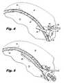
- FIG. 3is a perspective view of a hernia repair system in accordance with the present disclosure.
- FIGS. 4-8are progressive views illustrating a deployment of a light pipe of the hernia repair system of FIG. 3 into tissue;
- FIG. 9is perspective view of a plurality of light pipes disposed in tissue after being deployed from the hernia repair system of FIG. 3 ;
- FIG. 10is a perspective view of another embodiment of a hernia repair system deploying a plurality of light pipes in tissue in accordance with the present disclosure.
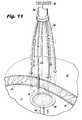
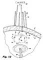
- FIGS. 11-13are progressive views illustrating a surgical patch being positioned adjacent a hernia defect with the aid of a plurality of light pipes in accordance with the present disclosure.
- the present disclosurerelates to methods for surgeries such as transluminal and/or endoluminal placement of a surgical patch at a surgical site.
- surgical patchis used to refer to any type of patch for use in surgical procedures, such as, for example, meshes that can be attached to the abdominal wall.
- the methods of the disclosuremay be used in any surgical repair.
- proximalwill refer to an end of a device that is closer to the user, while the term “distal” will refer to the end of the device that is farther from the user.
- FIG. 1illustrates a hernia that may involve a defect 30 such as a tear in the abdominal wall 40 .
- the abdominal wall 40is defined by an external side 40 a and an internal side 40 b .
- a surface tissue 42which covers the external side 40 a of abdominal wall 40 , may or may not be immediately affected by this defect 30 .
- An internal organ 44 located below the internal side 40 b of the abdominal wall 40may not protrude until some form of exertion or use of the muscle located at the abdominal wall 40 forces the internal organ 44 into the defect 30 .
- exertionmay not be needed to cause the organ to protrude.
- a herniaoccurs when an internal organ 44 protrudes into the defect 30 of abdominal wall 40 . Oftentimes the protrusion creates a bulge 46 in the surface tissue 42 .
- a hernia repair system 100includes an access port 110 , a dispensing instrument 120 , and one or more light pipes 130 .
- the access port 110includes a seal assembly 112 at a proximal end and cannula 114 at a distal end.
- the seal assembly 112accommodates the dispensing instrument 120 in a substantially sealed relationship.
- the seal assembly 112includes an insufflation valve 116 to selectively permit the passage of insufflation fluids therethrough to create a working space in an underlying tissue site.
- the dispensing instrument 120includes an actuation assembly 122 at a proximal end and a shaft 124 at a distal end.
- the shaft 124extends from the actuation assembly 122 .
- the shaft 124defines a lumen 126 therethrough to accommodate the one or more light pipes 130 .
- the shaft 124is movable via the actuation assembly 122 to dispense the one or more light pipes 130 at predetermined locations within a patient.
- the distal end of the shaft 124may be rotatable, pivotable, and/or articulable to orient the distal end of the shaft 124 in a particular orientation relative to the hernia defect 30 .
- Each light pipe 130includes a distal tip 132 , which may be sharpened to pierce tissue, and an elongated member 134 .
- the elongated member 134may define a lumen 136 therethrough to permit the passage of light therethrough when coupled to a light source 150 (see FIG. 11 ).
- the elongated member 134may include any suitable electrical and/or mechanical and/or chemical components configured to emit light from the distal end of the elongated member 134 (e.g., like a flashlight).
- the elongated member 134may be rigid or flexible.
- the light pipes 130may be fiber optic.
- a hernia defect 30is identified in a patient.
- each hernia defect 30has a particular size, location, and shape and therefore proper placement of a surgical patch 160 during minimally invasive surgery is facilitated when a practitioner can ascertain the size, location, and shape from an ex vivo location.
- the practitionerinserts the access port 110 , namely the cannula 114 into tissue adjacent the hernia defect 30 (see FIG. 4 ).
- the underlying tissue site “TS”may be insufflated when the insufflation valve 116 is coupled to an insufflation source 118 to create a working space.
- the practitionermay then laparoscopically advance the dispensing instrument 120 into the patient adjacent the hernia defect 30 to facilitate placement of one or more light pipes 30 in position about the hernia defect 30 , which is best depicted in FIG. 5 .
- any number of access ports 110 and/or dispensing instruments 120may be used to position the one or more light pipes 130 in tissue.
- the practitionermay even directly laparoscopically advance the one or more light pipes 30 through the access port 110 or directly laparoscopically advance the one or more light pipes 30 through an incision (without the access port 110 ) by virtue of the sharpened distal tip 132 , where appropriate.
- one or more light pipes 130may then be dispensed from the dispensing instrument 120 at predetermined locations about the hernia defect 30 to create a pattern about the defect 30 that is commensurate with the size, location, orientation and/or shape of the defect 30 .
- the dispensing instrument 120may be positioned adjacent one or more corners and/or extremes of the hernia defect 30 prior to dispensing the one or more light pipes 130 to generate the pattern.
- the one or more light pipes 130can then be dispensed with sufficient force to pierce and advance through the patient's skin, e.g. surface tissue 42 .
- the one or more light pipes 130may include sharpened tips 132 to further facilitate the penetration of the patient's skin.
- the one or more light pipes 130may be advanced to a position immediately adjacent the hernia defect 30 or the abdominal wall 40 .
- the one or more light pipes 130may be pulled proximally through the pierced skin until they are positioned snug against the hernia defect 30 or the abdominal wall 40 , depending upon the desired position.
- the one or more light pipes 130are most suitably configured in the pattern.
- the patternmay extend along the defect 30 and/or along an area immediately adjacent the defect 30 .
- the patternmay have any suitable geometry, size, etc. for facilitating the placement of a surgical patch 160 adjacent the defect 30 .
- one or more points of lightare formed about the defect 30 corresponding to the pattern to indicate the size, location, orientation and/or shape of the defect 30 .
- the pluralitywhen there is a plurality of light pipes 130 , the plurality may be bundled together via a bundling member 140 .
- the light source 150may then be coupled to the one or more light pipes 130 , either individually, collectively, or by groups of light pipes 130 .
- the bundling member 140may include a light source 150 .
- the light source 150generates the pattern of light “P” via points of light P1, P2, P3, P4, P5, P6, etc. that indicate the size, the location, the orientation and/or the shape of the hernia defect 30 .
- the light emitted from the light source 150will form an outline of the hernia defect 30 , in vivo, so as to be viewable ex vivo so that the surgical patch 160 may be positioned adjacent the hernia defect 30 with any suitable instrument 200 (e.g., a grasper) in accordance with the outline/pattern of light.
- any suitable instrument 200e.g., a grasper
- the generated pattern of lightcan be visualized, ex vivo, through the surgical patch 110 and the tissue.
- the surgical patch 160may be secured to the defect 30 by any suitable means (e.g., glue, tack, staple, suture, etc.) and the one or more light pipes 130 may then be removed from the patient either individually, collectively, or by groups of light pipes 130 (see FIG. 13 ).
- suitable meanse.g., glue, tack, staple, suture, etc.
- the presently disclosed surgical patchmay be any type of patch for use in surgical repair and suitable for use in situ.
- the surgical patchmay be any suitable shape (i.e., circular, noncircular, etc.) and may include one or more layers.
- the surgical patchmay be made of multiple fibers, or may be made of a single fiber.
- the fibersmay be a monofilament or multi-filament.
- the fibers forming the presently disclosed patchmay be made from a natural material or a synthetic material.
- the fibersmay be biodegradable or non-biodegradable. Any combination of natural, synthetic, bioadegradable and non-biodegradable materials may be used to form the fibers.
- biodegradableas used herein is defined to include both bioabsorbable and bioresorbable materials. By biodegradable, it is meant that the materials decompose, or lose structural integrity under body conditions (e.g. enzymatic degradation or hydrolysis) or are broken down (physically or chemically) under physiologic conditions in the body such that the degradation products are excretable or absorbable by the body.
- the surgical patch of the present disclosuremay be formed using any method suitable to forming patch structures, including but not limited to knitting, weaving, non-woven techniques, and the like. Suitable techniques for making the surgical patch are within the purview of those skilled in the art.
- the surgical patchmay be any shape or size suitable for covering the herniated area and securing the patch to surrounding tissue.
- the surgical patchmay be preformed to a certain size, such as, for example, a 9 cm diameter round patch or 50 cm ⁇ 50 cm square patch.
- the surgical patchmay be cut to a particular size and shape as needed.
- the surgical patch of the present disclosuremay be rolled, folded, or otherwise oriented so that the surgical patch forms a shape more suitable for placement adjacent a hernia defect.
Landscapes
- Health & Medical Sciences (AREA)
- Life Sciences & Earth Sciences (AREA)
- Surgery (AREA)
- General Health & Medical Sciences (AREA)
- Biomedical Technology (AREA)
- Veterinary Medicine (AREA)
- Public Health (AREA)
- Animal Behavior & Ethology (AREA)
- Heart & Thoracic Surgery (AREA)
- Engineering & Computer Science (AREA)
- Medical Informatics (AREA)
- Nuclear Medicine, Radiotherapy & Molecular Imaging (AREA)
- Molecular Biology (AREA)
- Oral & Maxillofacial Surgery (AREA)
- Pathology (AREA)
- Physics & Mathematics (AREA)
- Optics & Photonics (AREA)
- Cardiology (AREA)
- Transplantation (AREA)
- Vascular Medicine (AREA)
- Biophysics (AREA)
- Radiology & Medical Imaging (AREA)
- Surgical Instruments (AREA)
Abstract
Description
The present application claims the benefit of and priority to U.S. Provisional Application Ser. No. 61/641,974, filed on May 3, 2012, the entire contents of which are incorporated herein by reference.
1. Technical Field
The present disclosure relates to hernia repair methods. More particularly, the present disclosure relates to methods for positioning a surgical patch to a tissue site of a hernia using light.
2. Description of Related Art
A hernia is a protrusion of a tissue, structure, or part of an organ through injured muscle tissue or an injured membrane by which the tissue, structure, or organ is normally contained. Some examples of hernias include: abdominal hernias, diaphragmatic hernias and hiatal hernias (for example, para-esophageal hernia of the stomach), pelvic hernias, for example, obturator hernia, anal hernias, hernias of the nucleus pulposus of the intervertebral discs, intracranial hernias, and Spigelian hernias.
Hernias may be surgically repaired, and are principally repaired by pushing back, or “reducing”, the herniated tissue, and then reinforcing the defect in injured muscle tissue (an operation called herniorrhaphy). Modern muscle reinforcement techniques involve placement of a surgical patch, such as a surgical mesh, near the injured tissue or defect to support the defect. The surgical patch is either placed over the defect (anterior repair) or under the defect (posterior repair).
A variety of different fixation devices are used to anchor the surgical patch to the tissue. For example, a needled suture may be passed through or around the tissue near the defect to hold the surgical patch in a position which spans the injured tissue. In other examples, staples, tacks, clips and pins are also known to be passed through or around the tissue near the defect to anchor the surgical patch in a position which spans the injured tissue.
When applying a surgical patch during minimally invasive surgery, it is imperative that the surgeon know the precise location, size and shape of the hernia defect in order to properly place the surgical patch. However, since the bounds of the hernia defect are generally internal, visibility is often limited and placement of the surgical patch can be cumbersome. Thus, a continuing need still exits to provide a means for facilitating the effectiveness of the placement of surgical patches used to surgically repair hernias.
Accordingly, a hernia repair method includes the step of identifying a hernia defect in a patient, the hernia defect having a size, a location, and a shape. The method involves positioning a dispensing instrument laparoscopically into the patient adjacent the hernia defect.
According to one step, the method includes dispensing one or more light pipes from the dispensing instrument at predetermined locations. The light pipes may be fiber optic. The method may include the step of positioning the dispensing instrument adjacent a corner or an extreme of the hernia defect prior to dispensing the one or more light pipes.
The method also involves advancing the one or more light pipes through the patient's skin. The method may further include piercing the patient's skin with that one or more light pipes. The method may further comprise the step of advancing the one or more light pipes to a position immediately adjacent the hernia defect after advancing the one or more light pipes through the patient's skin.
The method may include the step of bundling a plurality of light pipes. Another step includes coupling a light source to the one or more light pipes. The method also involves generating a pattern of light that indicates one or more of the size, the location, and the shape of the hernia defect. The method further includes the step of forming an outline of the hernia defect, in vivo, with the pattern of light being formed from the positioning of the plurality of light pipes at the predetermined locations.
One step includes positioning a surgical patch adjacent the hernia defect in accordance with the pattern of light. According to one step, the method involves generating a pattern of light that can be visualized, ex vivo, through the surgical patch when the surgical patch is positioned over the hernia defect in vivo. According to one step, the method involves removing the one or more light pipes from the patient after positioning the surgical patch adjacent the hernia defect.
The above and other aspects, features, and advantages of the present disclosure will become more apparent in light of the following detailed description when taken in conjunction with the accompanying drawings in which:
The present disclosure relates to methods for surgeries such as transluminal and/or endoluminal placement of a surgical patch at a surgical site. As used herein the term “surgical patch” is used to refer to any type of patch for use in surgical procedures, such as, for example, meshes that can be attached to the abdominal wall. Although described herein with reference to a hernia surgical patch, the methods of the disclosure may be used in any surgical repair.
In the drawings and in the description that follows, the term “proximal,” as is traditional, will refer to an end of a device that is closer to the user, while the term “distal” will refer to the end of the device that is farther from the user.
Referring now in specific detail to the drawings, in which like numbers identify similar or identical elements,FIG. 1 illustrates a hernia that may involve adefect 30 such as a tear in theabdominal wall 40. Theabdominal wall 40 is defined by anexternal side 40aand an internal side40b. Asurface tissue 42, which covers theexternal side 40aofabdominal wall 40, may or may not be immediately affected by thisdefect 30. Aninternal organ 44 located below the internal side40bof theabdominal wall 40 may not protrude until some form of exertion or use of the muscle located at theabdominal wall 40 forces theinternal organ 44 into thedefect 30. Depending on the size and location of thedefect 30, exertion may not be needed to cause the organ to protrude. As shown inFIG. 2 , a hernia occurs when aninternal organ 44 protrudes into thedefect 30 ofabdominal wall 40. Oftentimes the protrusion creates abulge 46 in thesurface tissue 42.
As depicted inFIG. 3 , a hernia repair system100 includes anaccess port 110, a dispensinginstrument 120, and one ormore light pipes 130.
Theaccess port 110 includes aseal assembly 112 at a proximal end andcannula 114 at a distal end. Theseal assembly 112 accommodates the dispensinginstrument 120 in a substantially sealed relationship. Theseal assembly 112 includes aninsufflation valve 116 to selectively permit the passage of insufflation fluids therethrough to create a working space in an underlying tissue site.
Thedispensing instrument 120 includes anactuation assembly 122 at a proximal end and ashaft 124 at a distal end. Theshaft 124 extends from theactuation assembly 122. Theshaft 124 defines alumen 126 therethrough to accommodate the one ormore light pipes 130. Theshaft 124 is movable via theactuation assembly 122 to dispense the one or morelight pipes 130 at predetermined locations within a patient. In particular, as illustrated inFIG. 3 , the distal end of theshaft 124 may be rotatable, pivotable, and/or articulable to orient the distal end of theshaft 124 in a particular orientation relative to thehernia defect 30.
Eachlight pipe 130 includes adistal tip 132, which may be sharpened to pierce tissue, and anelongated member 134. Theelongated member 134 may define alumen 136 therethrough to permit the passage of light therethrough when coupled to a light source150 (seeFIG. 11 ). Alternatively, theelongated member 134 may include any suitable electrical and/or mechanical and/or chemical components configured to emit light from the distal end of the elongated member134 (e.g., like a flashlight). Theelongated member 134 may be rigid or flexible. Thelight pipes 130 may be fiber optic.
In use, ahernia defect 30 is identified in a patient. As can be appreciated, eachhernia defect 30 has a particular size, location, and shape and therefore proper placement of asurgical patch 160 during minimally invasive surgery is facilitated when a practitioner can ascertain the size, location, and shape from an ex vivo location. Thus, in order to be able to perceive thehernia defect 30 from an ex vivo location, the practitioner inserts theaccess port 110, namely thecannula 114 into tissue adjacent the hernia defect30 (seeFIG. 4 ). With continued reference toFIGS. 4-5 , the underlying tissue site “TS” may be insufflated when theinsufflation valve 116 is coupled to aninsufflation source 118 to create a working space. In this respect, the practitioner may then laparoscopically advance the dispensinginstrument 120 into the patient adjacent thehernia defect 30 to facilitate placement of one or morelight pipes 30 in position about thehernia defect 30, which is best depicted inFIG. 5 . As depicted inFIG. 10 , any number ofaccess ports 110 and/or dispensinginstruments 120 may be used to position the one or morelight pipes 130 in tissue. In some modes of operation, the practitioner may even directly laparoscopically advance the one or morelight pipes 30 through theaccess port 110 or directly laparoscopically advance the one or morelight pipes 30 through an incision (without the access port110) by virtue of the sharpeneddistal tip 132, where appropriate.
Turning now toFIGS. 6-9 , one or morelight pipes 130 may then be dispensed from the dispensinginstrument 120 at predetermined locations about thehernia defect 30 to create a pattern about thedefect 30 that is commensurate with the size, location, orientation and/or shape of thedefect 30. For example, the dispensinginstrument 120 may be positioned adjacent one or more corners and/or extremes of thehernia defect 30 prior to dispensing the one or morelight pipes 130 to generate the pattern. The one or morelight pipes 130 can then be dispensed with sufficient force to pierce and advance through the patient's skin,e.g. surface tissue 42. The one or morelight pipes 130 may include sharpenedtips 132 to further facilitate the penetration of the patient's skin. After the ends, e.g., the sharpenedtips 132 are positioned so that they extend externally, the one or morelight pipes 130 may be advanced to a position immediately adjacent thehernia defect 30 or theabdominal wall 40. In this respect, the one or morelight pipes 130 may be pulled proximally through the pierced skin until they are positioned snug against thehernia defect 30 or theabdominal wall 40, depending upon the desired position.
Once in the snug position, the one or morelight pipes 130 are most suitably configured in the pattern. The pattern may extend along thedefect 30 and/or along an area immediately adjacent thedefect 30. The pattern may have any suitable geometry, size, etc. for facilitating the placement of asurgical patch 160 adjacent thedefect 30.
As illustrated inFIGS. 11-13 , when the one or morelight pipes 130 are coupled to alight source 150 or are otherwise adapted to emit light (e.g., selectively via a switch coupled to the one or more light pipes or autonomously by fluorescent chemicals or the like), one or more points of light are formed about thedefect 30 corresponding to the pattern to indicate the size, location, orientation and/or shape of thedefect 30.
With reference toFIG. 11 , when there is a plurality oflight pipes 130, the plurality may be bundled together via a bundlingmember 140. Thelight source 150 may then be coupled to the one or morelight pipes 130, either individually, collectively, or by groups oflight pipes 130. The bundlingmember 140 may include alight source 150. As depicted inFIG. 12 , thelight source 150 generates the pattern of light “P” via points of light P1, P2, P3, P4, P5, P6, etc. that indicate the size, the location, the orientation and/or the shape of thehernia defect 30. In this regard, the light emitted from thelight source 150 will form an outline of thehernia defect 30, in vivo, so as to be viewable ex vivo so that thesurgical patch 160 may be positioned adjacent thehernia defect 30 with any suitable instrument200 (e.g., a grasper) in accordance with the outline/pattern of light. As can be appreciated, when thesurgical patch 160 is positioned in vivo over thehernia defect 30, the generated pattern of light can be visualized, ex vivo, through thesurgical patch 110 and the tissue. After thesurgical patch 160 is placed in a desired position adjacent thehernia defect 30, thesurgical patch 160 may be secured to thedefect 30 by any suitable means (e.g., glue, tack, staple, suture, etc.) and the one or morelight pipes 130 may then be removed from the patient either individually, collectively, or by groups of light pipes130 (seeFIG. 13 ).
The presently disclosed surgical patch may be any type of patch for use in surgical repair and suitable for use in situ. The surgical patch may be any suitable shape (i.e., circular, noncircular, etc.) and may include one or more layers. The surgical patch may be made of multiple fibers, or may be made of a single fiber. The fibers may be a monofilament or multi-filament.
The fibers forming the presently disclosed patch may be made from a natural material or a synthetic material. The fibers may be biodegradable or non-biodegradable. Any combination of natural, synthetic, bioadegradable and non-biodegradable materials may be used to form the fibers. The term “biodegradable” as used herein is defined to include both bioabsorbable and bioresorbable materials. By biodegradable, it is meant that the materials decompose, or lose structural integrity under body conditions (e.g. enzymatic degradation or hydrolysis) or are broken down (physically or chemically) under physiologic conditions in the body such that the degradation products are excretable or absorbable by the body.
The surgical patch of the present disclosure may be formed using any method suitable to forming patch structures, including but not limited to knitting, weaving, non-woven techniques, and the like. Suitable techniques for making the surgical patch are within the purview of those skilled in the art.
The surgical patch may be any shape or size suitable for covering the herniated area and securing the patch to surrounding tissue. The surgical patch may be preformed to a certain size, such as, for example, a 9 cm diameter round patch or 50 cm×50 cm square patch. In embodiments, the surgical patch may be cut to a particular size and shape as needed.
In addition, the surgical patch of the present disclosure may be rolled, folded, or otherwise oriented so that the surgical patch forms a shape more suitable for placement adjacent a hernia defect.
While several embodiments of the disclosure have been shown in the drawings, it is not intended that the disclosure be limited thereto, as it is intended that the disclosure be as broad in scope as the art will allow and that the specification be read likewise. Therefore, the above description should not be construed as limiting, but merely as exemplifications of particular embodiments. Those skilled in the art will envision other modifications within the scope and spirit of the claims appended hereto.
Claims (20)
1. A hernia repair method, comprising:
identifying a hernia defect in a patient, the hernia defect having a size, a location, and a shape;
positioning a dispensing instrument laparoscopically into the patient adjacent the hernia defect;
dispensing leading and trailing ends of a first light pipe from a leading end of the dispensing instrument at a predetermined location within the patient to separate the first light pipe from the dispensing instrument;
advancing the leading end of the first light pipe through the patient's skin;
coupling a light source to the leading end of the first light pipe;
generating a pattern of light with the first light pipe that indicates at least one of: the size, the location, or the shape of the hernia defect; and
positioning a surgical patch adjacent the hernia defect in accordance with the pattern of light.
2. The hernia repair method ofclaim 1 , further comprising positioning the dispensing instrument adjacent a corner or a periphery of the hernia defect prior to dispensing the first light pipe.
3. The hernia repair method ofclaim 1 , further comprising advancing the trailing end of the first light pipe to a position immediately adjacent the hernia defect after advancing the leading end of the first light pipe through the patient's skin.
4. The hernia repair method ofclaim 1 , further comprising dispensing a second light pipe from the dispensing instrument.
5. The hernia repair method ofclaim 4 , further comprising bundling the first and second light pipes.
6. The hernia repair method ofclaim 1 , further comprising generating the pattern of light so that the pattern of light can be visualized, ex vivo, through the surgical patch when the surgical patch is positioned over the hernia defect in vivo.
7. The hernia repair method ofclaim 1 , wherein the first light pipe is a fiber optic light pipe.
8. The hernia repair method ofclaim 1 , further comprising removing the first light pipe from the patient after positioning the surgical patch adjacent the hernia defect.
9. The hernia repair method ofclaim 1 , wherein coupling the light source to the leading end of the first light pipe includes transmitting light from the light source from the leading end of the first light pipe to the trailing end of the first light pipe so that light can exit from the trailing end of the first light pipe into the patient.
10. A hernia repair method, comprising:
identifying a hernia defect in a patient, the hernia defect having a size, a location, and a shape;
positioning a dispensing instrument laparoscopically into the patient adjacent the hernia defect;
dispensing leading and trailing ends of a first light pipe from a leading end of the dispensing instrument at a predetermined location within the patient to separate the first light pipe from the dispensing instrument;
piercing the patient's skin with the leading end of the first light pipe to cut through the patient's skin from an internal surface of the patient's skin to an external surface of the patient's skin, the leading end of the first light pipe being sharpened;
advancing the leading end of the first light pipe through the patient's skin;
coupling a light source to the leading end of the first light pipe;
generating a pattern of light with the first light pipe that indicates at least one of: the size, the location, or the shape of the hernia defect; and
positioning a surgical patch adjacent the hernia defect in accordance with the pattern of light.
11. The hernia repair method ofclaim 10 , further comprising maintaining the trailing end of the first light pipe positioned within the patient after the leading end of the first light pipe is pierced through the internal and external surfaces of the patient's skin and positioned externally of the patient.
12. The hernia repair method ofclaim 10 , further comprising positioning the dispensing instrument adjacent a corner or a periphery of the hernia defect prior to dispensing the first light pipe.
13. The hernia repair method ofclaim 10 , further comprising advancing the trailing end of the first light pipe to a position immediately adjacent the hernia defect after advancing the leading end of the first light pipe through the patient's skin.
14. The hernia repair method ofclaim 10 , further comprising dispensing a second light pipe from the dispensing instrument.
15. The hernia repair method ofclaim 14 , further comprising generating the pattern of light with the first and second light pipes and positioning the first and second light pipes at predetermined locations within the patient to form an outline of the hernia defect in vivo.
16. The hernia repair method ofclaim 14 , further comprising bundling the first and second light pipes.
17. The hernia repair method ofclaim 10 , further comprising generating the pattern of light so that the pattern of light can be visualized, ex vivo, through the surgical patch when the surgical patch is positioned over the hernia defect in vivo.
18. The hernia repair method ofclaim 10 , wherein the first light pipe is a fiber optic light pipe.
19. The hernia repair method ofclaim 10 , further comprising removing the first light pipe from the patient after positioning the surgical patch adjacent the hernia defect.
20. The hernia repair method ofclaim 10 , wherein coupling the light source to the leading end of the first light pipe includes transmitting light from the light source from the leading end of the first light pipe to the trailing end of the first light pipe so that light can exit from the trailing end of the first light pipe into the patient.
Priority Applications (3)
| Application Number | Priority Date | Filing Date | Title |
|---|---|---|---|
| US13/860,695US9186053B2 (en) | 2012-05-03 | 2013-04-11 | Methods of using light to repair hernia defects |
| AU2013205092AAU2013205092B2 (en) | 2012-05-03 | 2013-04-13 | Methods of using light to repair hernia defects |
| CA2814117ACA2814117A1 (en) | 2012-05-03 | 2013-04-26 | Methods of using light to repair hernia defects |
Applications Claiming Priority (2)
| Application Number | Priority Date | Filing Date | Title |
|---|---|---|---|
| US201261641974P | 2012-05-03 | 2012-05-03 | |
| US13/860,695US9186053B2 (en) | 2012-05-03 | 2013-04-11 | Methods of using light to repair hernia defects |
Publications (2)
| Publication Number | Publication Date |
|---|---|
| US20130296657A1 US20130296657A1 (en) | 2013-11-07 |
| US9186053B2true US9186053B2 (en) | 2015-11-17 |
Family
ID=49513070
Family Applications (1)
| Application Number | Title | Priority Date | Filing Date |
|---|---|---|---|
| US13/860,695Active2034-01-29US9186053B2 (en) | 2012-05-03 | 2013-04-11 | Methods of using light to repair hernia defects |
Country Status (3)
| Country | Link |
|---|---|
| US (1) | US9186053B2 (en) |
| AU (1) | AU2013205092B2 (en) |
| CA (1) | CA2814117A1 (en) |
Cited By (10)
| Publication number | Priority date | Publication date | Assignee | Title |
|---|---|---|---|---|
| US9955910B2 (en) | 2005-10-14 | 2018-05-01 | Aranz Healthcare Limited | Method of monitoring a surface feature and apparatus therefor |
| US10013527B2 (en) | 2016-05-02 | 2018-07-03 | Aranz Healthcare Limited | Automatically assessing an anatomical surface feature and securely managing information related to the same |
| US10874302B2 (en) | 2011-11-28 | 2020-12-29 | Aranz Healthcare Limited | Handheld skin measuring or monitoring device |
| US11116407B2 (en) | 2016-11-17 | 2021-09-14 | Aranz Healthcare Limited | Anatomical surface assessment methods, devices and systems |
| US11903723B2 (en) | 2017-04-04 | 2024-02-20 | Aranz Healthcare Limited | Anatomical surface assessment methods, devices and systems |
| US11963676B2 (en) | 2018-10-16 | 2024-04-23 | Activ Surgical, Inc. | Autonomous methods and systems for tying surgical knots |
| US12039726B2 (en) | 2019-05-20 | 2024-07-16 | Aranz Healthcare Limited | Automated or partially automated anatomical surface assessment methods, devices and systems |
| US12201387B2 (en) | 2019-04-19 | 2025-01-21 | Activ Surgical, Inc. | Systems and methods for trocar kinematics |
| US12262952B2 (en) | 2018-12-28 | 2025-04-01 | Activ Surgical, Inc. | Systems and methods to optimize reachability, workspace, and dexterity in minimally invasive surgery |
| US12400340B2 (en) | 2018-12-28 | 2025-08-26 | Activ Surgical, Inc. | User interface elements for orientation of remote camera during surgery |
Families Citing this family (1)
| Publication number | Priority date | Publication date | Assignee | Title |
|---|---|---|---|---|
| US9186053B2 (en)* | 2012-05-03 | 2015-11-17 | Covidien Lp | Methods of using light to repair hernia defects |
Citations (178)
| Publication number | Priority date | Publication date | Assignee | Title |
|---|---|---|---|---|
| US3272204A (en)* | 1965-09-22 | 1966-09-13 | Ethicon Inc | Absorbable collagen prosthetic implant with non-absorbable reinforcing strands |
| US3376869A (en)* | 1964-02-21 | 1968-04-09 | Ethicon Inc | Surgical collagen film formed from random lengths of collagen tapes or strands |
| US4562832A (en)* | 1984-01-21 | 1986-01-07 | Wilder Joseph R | Medical instrument and light pipe illumination assembly |
| US4619249A (en)* | 1985-07-24 | 1986-10-28 | Kim Landry | Transcutaneous intravenous illuminator |
| US4688554A (en)* | 1986-04-10 | 1987-08-25 | American Hospital Supply Corp. | Directing cannula for an optical diagnostic system |
| US4898175A (en) | 1986-12-26 | 1990-02-06 | Olympus Optical Co., Ltd. | Out-body observing apparatus |
| US5143076A (en) | 1988-12-23 | 1992-09-01 | Tyrone L. Hardy | Three-dimensional beam localization microscope apparatus for stereotactic diagnoses or surgery |
| US5242456A (en)* | 1991-11-21 | 1993-09-07 | Kensey Nash Corporation | Apparatus and methods for clamping tissue and reflecting the same |
| US5269753A (en)* | 1992-07-14 | 1993-12-14 | Wilk Peter J | Method for use in laparoscopic hernia repair |
| US5275166A (en)* | 1992-11-16 | 1994-01-04 | Ethicon, Inc. | Method and apparatus for performing ultrasonic assisted surgical procedures |
| US5280788A (en)* | 1991-02-26 | 1994-01-25 | Massachusetts Institute Of Technology | Devices and methods for optical diagnosis of tissue |
| US5290217A (en)* | 1991-10-10 | 1994-03-01 | Earl K. Sipes | Method and apparatus for hernia repair |
| US5337736A (en)* | 1992-09-30 | 1994-08-16 | Reddy Pratap K | Method of using a laparoscopic retractor |
| US5353786A (en)* | 1992-01-24 | 1994-10-11 | Wilk Peter J | Surgical lighting method |
| US5383477A (en)* | 1991-08-02 | 1995-01-24 | Dematteis; Ralph A. | Method and apparatus for laparoscopic repair of hernias |
| US5433721A (en)* | 1992-01-17 | 1995-07-18 | Ethicon, Inc. | Endoscopic instrument having a torsionally stiff drive shaft for applying fasteners to tissue |
| US5456720A (en)* | 1991-02-08 | 1995-10-10 | Schultz; Leonard S. | Prosthesis for repair of direct space and indirect space inguinal hernias |
| US5517997A (en) | 1994-09-15 | 1996-05-21 | Gabriel Medical, Inc. | Transillumination of body members for protection during body invasive procedures |
| US5540711A (en)* | 1992-06-02 | 1996-07-30 | General Surgical Innovations, Inc. | Apparatus and method for developing an anatomic space for laparoscopic procedures with laparoscopic visualization |
| US5607443A (en)* | 1992-06-02 | 1997-03-04 | General Surgical Innovations, Inc. | Expansible tunneling apparatus for creating an anatomic working space with laparoscopic observation |
| US5622170A (en) | 1990-10-19 | 1997-04-22 | Image Guided Technologies, Inc. | Apparatus for determining the position and orientation of an invasive portion of a probe inside a three-dimensional body |
| US5695525A (en)* | 1992-05-20 | 1997-12-09 | C.R. Bard, Incorporated | Implantable prosthesis and method and apparatus for loading and delivering an implantable prosthesis |
| US5713869A (en)* | 1995-03-08 | 1998-02-03 | Morejon; Orlando | Trocar assembly |
| US5730756A (en)* | 1992-06-02 | 1998-03-24 | General Surgical Innovations, Inc. | Method for developing an anatomic space for laparoscopic procedures with laparoscopic visualization |
| US5772680A (en)* | 1992-06-02 | 1998-06-30 | General Surgical Innovations, Inc. | Apparatus and method for developing an anatomic space for laparoscopic procedures with laparoscopic visualization |
| US5807387A (en) | 1993-09-07 | 1998-09-15 | Deemed International, S.A. | Optical designation device especially for microsurgical operations |
| US5891158A (en) | 1997-10-23 | 1999-04-06 | Manwaring; Kim H. | Method and system for directing an instrument to a target |
| US5907395A (en) | 1997-06-06 | 1999-05-25 | Image Guided Technologies, Inc. | Optical fiber probe for position measurement |
| US5972007A (en)* | 1997-10-31 | 1999-10-26 | Ethicon Endo-Surgery, Inc. | Energy-base method applied to prosthetics for repairing tissue defects |
| US5989269A (en)* | 1996-08-30 | 1999-11-23 | Vts Holdings L.L.C. | Method, instruments and kit for autologous transplantation |
| US6041249A (en) | 1997-03-13 | 2000-03-21 | Siemens Aktiengesellschaft | Device for making a guide path for an instrument on a patient |
| US6096049A (en) | 1998-07-27 | 2000-08-01 | Minrad Inc. | Light guiding device and method |
| US6138681A (en) | 1997-10-13 | 2000-10-31 | Light Sciences Limited Partnership | Alignment of external medical device relative to implanted medical device |
| US6193653B1 (en)* | 1998-02-06 | 2001-02-27 | Ethicon Endo-Surgery, Inc. | Methods and devices for visualizing, dissecting and harvesting vessels and the like |
| US6251110B1 (en)* | 1999-03-31 | 2001-06-26 | Ethicon Endo-Surgery, Inc. | Combined radio frequency and ultrasonic surgical device |
| US6257241B1 (en)* | 1999-03-31 | 2001-07-10 | Ethicon Endo-Surgery, Inc. | Method for repairing tissue defects using ultrasonic radio frequency energy |
| US6287344B1 (en)* | 1999-03-31 | 2001-09-11 | Ethicon Endo-Surgery, Inc. | Method for repairing tissue defects using an ultrasonic device |
| US6286514B1 (en) | 1996-11-05 | 2001-09-11 | Jerome Lemelson | System and method for treating select tissue in a living being |
| US6293282B1 (en) | 1996-11-05 | 2001-09-25 | Jerome Lemelson | System and method for treating select tissue in living being |
| US6317616B1 (en) | 1999-09-15 | 2001-11-13 | Neil David Glossop | Method and system to facilitate image guided surgery |
| US6354297B1 (en)* | 1998-04-16 | 2002-03-12 | The Uniformed Services University Of The Health Sciences | Method and device for destroying fat cells by induction of programmed cell death |
| US6416486B1 (en)* | 1999-03-31 | 2002-07-09 | Ethicon Endo-Surgery, Inc. | Ultrasonic surgical device having an embedding surface and a coagulating surface |
| US20020099279A1 (en)* | 2000-11-28 | 2002-07-25 | Pfeiffer Ulrich J. | Device for the determination of tissue perfusion and operative use thereof |
| US6442409B1 (en) | 2000-03-22 | 2002-08-27 | Gholam A. Peyman | Diagnostic system and method using radiant energy and implants |
| US6447527B1 (en)* | 1998-04-23 | 2002-09-10 | Ronald J. Thompson | Apparatus and methods for the penetration of tissue |
| US6540764B1 (en)* | 1992-06-02 | 2003-04-01 | General Surgical Innovations, Inc. | Apparatus and method for dissecting tissue layers |
| US6569172B2 (en)* | 1996-08-30 | 2003-05-27 | Verigen Transplantation Service International (Vtsi) | Method, instruments, and kit for autologous transplantation |
| US6587702B1 (en)* | 1999-01-22 | 2003-07-01 | Instrumentation Metrics, Inc | Classification and characterization of tissue through features related to adipose tissue |
| US6597941B2 (en) | 1994-09-15 | 2003-07-22 | Stryker Corporation | Transillumination of body members for protection during body invasive procedures |
| US20040049251A1 (en)* | 2002-07-14 | 2004-03-11 | Knowlton Edward W. | Method and apparatus for surgical dissection |
| US6711423B2 (en) | 1998-08-26 | 2004-03-23 | Sensors For Medicine And Science, Inc. | Optical-based sensing devices |
| US20040064019A1 (en)* | 1996-08-26 | 2004-04-01 | Chang Huei Liang | Endoscope assembly useful with a scope-sensing light cable |
| US6750311B1 (en)* | 1996-11-21 | 2004-06-15 | Minimed Inc. | Detection of biological molecules using boronate-based chemical amplification and optical sensors |
| US20040204734A1 (en)* | 2003-04-11 | 2004-10-14 | Wagner Darrell Orvin | Tunneling tool with subcutaneous transdermal illumination |
| US20040220516A1 (en)* | 2002-11-04 | 2004-11-04 | Stephen Solomon | Food extraction apparatus and method |
| US20040236231A1 (en)* | 2003-05-23 | 2004-11-25 | Embro Corporation | Light catheter for illuminating tissue structures |
| US20050004576A1 (en)* | 1998-11-23 | 2005-01-06 | Benderev Theodore V. | System for securing sutures, grafts and soft tissue to bone and periosteum |
| US6873867B2 (en) | 2000-04-05 | 2005-03-29 | Brainlab Ag | Referencing or registering a patient or a patient body part in a medical navigation system by means of irradiation of light points |
| US20050113798A1 (en)* | 2000-07-21 | 2005-05-26 | Slater Charles R. | Methods and apparatus for treating the interior of a blood vessel |
| US20050216040A1 (en)* | 2004-03-23 | 2005-09-29 | Michael Gertner | Devices and methods to treat a patient |
| US20050216041A1 (en)* | 2004-03-26 | 2005-09-29 | Olympus Corporation | Treatment method |
| US20050277900A1 (en)* | 2002-11-04 | 2005-12-15 | Samuel Klein | Apparatus for treating obesity by extracting food |
| US6978166B2 (en) | 1994-10-07 | 2005-12-20 | Saint Louis University | System for use in displaying images of a body part |
| US20050288691A1 (en) | 2004-06-28 | 2005-12-29 | Leiboff Arnold R | Hernia patch |
| US20050288706A1 (en)* | 2004-05-07 | 2005-12-29 | Nmt Medical, Inc. | Inflatable occluder |
| US6991637B2 (en)* | 2003-06-18 | 2006-01-31 | Gore Enterprise Holdings, Inc. | Soft tissue defect repair device |
| US20060069313A1 (en)* | 2004-09-30 | 2006-03-30 | Couvillon Lucien A Jr | Medical devices with light emitting regions |
| US7040807B2 (en) | 2003-04-29 | 2006-05-09 | Siemens Aktiengesellschaft | Radiographic image acquisition apparatus with pulsed laser light marker |
| US20060270908A1 (en)* | 2005-05-18 | 2006-11-30 | Luloh K P | Self Inserting Intraolcular Light |
| US20060282105A1 (en)* | 2002-08-02 | 2006-12-14 | C.R. Bard, Inc. | Implantable prosthesis |
| US20070073160A1 (en)* | 2005-09-13 | 2007-03-29 | Children's Medical Center Corporation | Light-guided transluminal catheter |
| US20070088391A1 (en)* | 2005-10-18 | 2007-04-19 | Cook Biotech Inc. | Medical device with affixation means |
| US7224472B2 (en) | 2002-03-01 | 2007-05-29 | Brainlab Ag | Operation lamp with camera system for 3D referencing |
| US20070185506A1 (en)* | 2003-08-04 | 2007-08-09 | Kelly Jackson | Medical instruments and methods for using the same |
| US20070260179A1 (en)* | 2004-10-14 | 2007-11-08 | Mordehai Sholev | Hernia Repair Device |
| US7297153B2 (en)* | 1992-06-02 | 2007-11-20 | General Surgical Innovations, Inc. | Apparatus and method for developing an anatomic space for laparoscopic hernia repair and patch for use therewith |
| US20070270890A1 (en)* | 2006-03-16 | 2007-11-22 | Dennis Miller | System and Method for Treating Tissue Wall Prolapse |
| US20080033263A1 (en)* | 2006-08-03 | 2008-02-07 | Marcinek David J | Method and system for determining the contribution of hemoglobin and myoglobin to in vivo optical spectra |
| US20080058836A1 (en) | 2006-08-03 | 2008-03-06 | Hansen Medical, Inc. | Systems and methods for performing minimally invasive procedures |
| US20080065229A1 (en)* | 2006-09-12 | 2008-03-13 | Adams Jason P | Inflatable hernia patch |
| US20080081945A1 (en)* | 2006-10-03 | 2008-04-03 | Boston Scientific Scimed, Inc. | Pelvic floor repair implants and methods |
| US20080082078A1 (en)* | 2001-05-21 | 2008-04-03 | Eyelight, Inc. | Glaucoma surgery methods and systems |
| US20080109015A1 (en)* | 2006-11-07 | 2008-05-08 | Chu Michael S | Delivering Sutures |
| US20080132753A1 (en)* | 2006-10-03 | 2008-06-05 | Boston Scientific Scimed, Inc. | Coaxial device for delivering an implant to a patient's pelvic region |
| US20080132917A1 (en)* | 2006-12-04 | 2008-06-05 | Gregory Paul Mueller | Surgical instrument docking device |
| US20080167546A1 (en)* | 2007-01-09 | 2008-07-10 | Ethicon Endo-Surgery, Inc. | Methods for imaging the anatomy with an anatomically secured scanner assembly |
| US7404819B1 (en)* | 2000-09-14 | 2008-07-29 | C.R. Bard, Inc. | Implantable prosthesis |
| US7450783B2 (en)* | 2003-09-12 | 2008-11-11 | Biopticon Corporation | Methods and systems for measuring the size and volume of features on live tissues |
| US20090105728A1 (en)* | 2007-10-23 | 2009-04-23 | Minos Medical | Devices and methods for securing tissue |
| US20090171142A1 (en)* | 2007-12-28 | 2009-07-02 | Chu Michael S H | Devices and methods for treating pelvic floor dysfunctions |
| US20090187181A1 (en) | 1998-05-20 | 2009-07-23 | Shadduck John H | Surgical instruments and techniques for treating gastro-esophageal reflux disease |
| US20090192528A1 (en)* | 2008-01-29 | 2009-07-30 | Biomet Biologics, Inc. | Method and device for hernia repair |
| US20090192530A1 (en)* | 2008-01-29 | 2009-07-30 | Insightra Medical, Inc. | Fortified mesh for tissue repair |
| US20090210006A1 (en)* | 2008-02-20 | 2009-08-20 | Tyco Healthcare Group Lp | Compound barb medical device and method |
| US20090216253A1 (en)* | 2007-12-13 | 2009-08-27 | Bell Stephen G | Methods and Apparatus for Treating Ventral Wall Hernia |
| US20090234370A1 (en) | 2008-03-11 | 2009-09-17 | Gabriel Haras | Method and marking out apparatus for marking out a guide line for a penetration instrument, control device and recording system |
| US20090234379A1 (en)* | 2008-03-14 | 2009-09-17 | Rehnke Robert D | Apparatuses for the performance of a minimally invasive ventral hernia repair |
| US20100036393A1 (en) | 2007-03-01 | 2010-02-11 | Titan Medical Inc. | Methods, systems and devices for threedimensional input, and control methods and systems based thereon |
| US20100056928A1 (en)* | 2008-08-10 | 2010-03-04 | Karel Zuzak | Digital light processing hyperspectral imaging apparatus |
| US20100104608A1 (en)* | 2008-09-26 | 2010-04-29 | Tyco Healthcare Group Lp | Reactive surgical implant |
| US7728868B2 (en) | 2006-08-02 | 2010-06-01 | Inneroptic Technology, Inc. | System and method of providing real-time dynamic imagery of a medical procedure site using multiple modalities |
| US20100145415A1 (en)* | 2006-10-11 | 2010-06-10 | Dahm Jonathan S | Light delivery system |
| US20100168763A1 (en) | 2008-12-31 | 2010-07-01 | Intuitive Surgical, Inc. | Configuration marker design and detection for instrument tracking |
| US7751865B2 (en) | 2003-10-17 | 2010-07-06 | Medtronic Navigation, Inc. | Method and apparatus for surgical navigation |
| US20100274282A1 (en)* | 2009-04-27 | 2010-10-28 | Teleflex Medical Incorporated | Colored Suture Construction |
| US20100280538A1 (en)* | 1999-07-02 | 2010-11-04 | Pulmonx Corporation | Methods, systems, and kits for lung volume reduction |
| US20100286473A1 (en)* | 2007-08-10 | 2010-11-11 | Yale University | Suspension/retraction device for surgical manipulation |
| US20100292724A1 (en)* | 2006-03-13 | 2010-11-18 | Sundaram Ravikumar | Minimally Invasive Surgical Clamps, Assemblies and Methods |
| US20100298953A1 (en) | 2007-10-31 | 2010-11-25 | Vanderbilt University | Device and method for positioning a surgical prosthesis |
| US20110009894A1 (en) | 2008-01-29 | 2011-01-13 | Peter Forsell | Apparatus for treating reflux disease (gerd) and obesity |
| US7881770B2 (en) | 2000-03-01 | 2011-02-01 | Medtronic Navigation, Inc. | Multiple cannula image guided tool for image guided procedures |
| US20110032715A1 (en)* | 2008-04-07 | 2011-02-10 | Faurecia Interieur Industrie | Piece of motor vehicle interior equipment and associated method of manufacture |
| US20110040311A1 (en) | 2009-08-17 | 2011-02-17 | PolyTouch Medical, Inc. | Articulating patch deployment device and method of use |
| US20110077668A1 (en)* | 2009-09-25 | 2011-03-31 | Lindsay Gordon | Devices for approximating tissue and related methods of use |
| US20110077456A1 (en)* | 2009-09-30 | 2011-03-31 | Bruce Drummond | Prolapse repair device and methods of use |
| US20110082442A1 (en)* | 2006-07-05 | 2011-04-07 | Solovay Kenneth S | Externally reinforced percutaneous gastrostomy tube with customizable smooth tube length |
| US20110098700A1 (en)* | 2009-10-28 | 2011-04-28 | Olympus Corporation | Biological-tissue joining apparatus |
| US20110106113A1 (en)* | 2007-07-13 | 2011-05-05 | The Brigham And Women's Hospital, Inc. | System and method for hernia mesh fixation |
| US20110112573A1 (en)* | 2008-06-18 | 2011-05-12 | Orahn Preiss Bloom | Methods and devices for use with sealants |
| US20110130774A1 (en)* | 2009-11-30 | 2011-06-02 | Tyco Healthcare Group Lp | Ventral Hernia Repair With Barbed Suture |
| US20110208215A1 (en)* | 2009-09-22 | 2011-08-25 | Modesitt D Bruce | Devices, methods, and kits for forming tracts in tissue |
| US20110224704A1 (en)* | 2008-09-18 | 2011-09-15 | Sofradim Production | Surgical instrument for deploying a prosthesis |
| US20110276090A1 (en)* | 2008-11-06 | 2011-11-10 | Itv Denkendorf Produktservice Gmbh | Medical product , a surgical kit, and a method for producing the medical product |
| US20110297161A1 (en)* | 2010-06-04 | 2011-12-08 | Coloplast A/S | Sacrocolpopexy support and method of implantation |
| US20110319915A1 (en)* | 2010-06-29 | 2011-12-29 | Frank Viola | Surgical Mesh Maker |
| US20120004501A1 (en)* | 2009-03-12 | 2012-01-05 | Ams Research Corporation | Method and Apparatus for Prolapse Repair |
| US20120041263A1 (en)* | 2009-04-23 | 2012-02-16 | M.S.T. Medical Surgery Technologies Ltd. | Two-part endoscope surgical device |
| US20120071727A1 (en)* | 2009-06-05 | 2012-03-22 | Hanson Anthony J | Method and articles for treating the sinus system |
| US20120116423A1 (en)* | 2010-11-08 | 2012-05-10 | Seth Gleiman | Expandable Mesh System And Method Of Use Therefor |
| US20120203273A1 (en)* | 2004-08-31 | 2012-08-09 | Wadsworth Medical Technologies, Inc. | Systems and Methods for Closing a Tissue Opening |
| US20120209301A1 (en)* | 2008-02-21 | 2012-08-16 | Insightra Medical, Inc. | Implant for hernia repair |
| US20120209319A1 (en)* | 2009-06-15 | 2012-08-16 | Technion-Research And Development Foundation Ltd. | Reinforced surgical adhesives and sealants and their in-situ application |
| US20120215073A1 (en)* | 2009-08-21 | 2012-08-23 | Sherman Audrey A | Methods and products for illuminating tissue |
| US20120232334A1 (en)* | 2008-07-31 | 2012-09-13 | Insightra Medical, Inc. | Implant for hernia repair |
| US20120253339A1 (en)* | 2011-03-31 | 2012-10-04 | Tyco Healthcare Group Lp | Radio frequency-based surgical implant fixation apparatus |
| US20120289811A1 (en)* | 2011-05-13 | 2012-11-15 | Tyco Healthcare Group Lp | Mask on monitor hernia locator |
| US20120303011A1 (en)* | 2010-11-27 | 2012-11-29 | Cook Medical Technologies Llc | Catheters and Methods for Identification and Treatment of Bodily Passages |
| US20130041266A1 (en)* | 2011-08-12 | 2013-02-14 | Tyco Healthcare Group Lp, | System and Method for Indicating Positioning of an Internal Anatomical Feature |
| US20130085337A1 (en)* | 2011-09-30 | 2013-04-04 | Ethicon Endo-Surgery, Inc. | Devices and methods for providing suction and/or irrigation in a surgical procedure |
| US20130096583A1 (en)* | 2006-12-04 | 2013-04-18 | Implicitcare, Llc | Surgical threading device with removable suture |
| USRE44380E1 (en)* | 1995-05-19 | 2013-07-16 | Roger A. de la Torre | Screw-type skin seal with inflatable membrane |
| US20130190598A1 (en)* | 2012-01-24 | 2013-07-25 | Covidien Lp | Magnetic field device for mapping and navigation in laparoscopic surgery |
| US20130190558A1 (en)* | 2010-09-29 | 2013-07-25 | Ams Research Corporation | Systems, tools, and methods for treatments of pelvic conditions |
| US20130218075A1 (en)* | 2009-11-03 | 2013-08-22 | Yissum Research Development Company Of The Hebrew University Of Jerusalem, Ltd. | Device for irradiating an internal body surface |
| US20130226037A1 (en)* | 2012-02-27 | 2013-08-29 | Covidien Lp | Ultra-wide angle zoom projection system for real time in-situ surgical metrology |
| US20130226156A1 (en)* | 2012-02-27 | 2013-08-29 | Covidien Lp | Device and method for optical image correction in metrology systems |
| US20130225936A1 (en)* | 2010-09-29 | 2013-08-29 | Ams Research Corporation | Systems, tools, and methods for treatments of pelvic conditions |
| US20130253387A1 (en)* | 2012-03-08 | 2013-09-26 | Sonitec, LLC | Vibratory energy systems and methods for occluded body cavities |
| US20130296657A1 (en)* | 2012-05-03 | 2013-11-07 | Covidien Lp | Methods of using light to repair hernia defects |
| US20130303845A1 (en)* | 2012-05-11 | 2013-11-14 | Ethicon, Inc. | Applicator instruments with imaging systems for dispensing surgical fasteners during open repair procedures |
| US8617157B2 (en)* | 2010-01-26 | 2013-12-31 | Covidien Lp | Hernia repair system |
| US20140031665A1 (en)* | 2012-07-25 | 2014-01-30 | Covidien Lp | Telecentric Scale Projection System for Real-Time In-Situ Surgical Metrology |
| US20140100431A1 (en)* | 2012-10-05 | 2014-04-10 | Fox Chase Cancer Center | Method for inserting medical instrument |
| US20140100604A1 (en)* | 2010-06-11 | 2014-04-10 | Entourage Medical Technologies, Inc. | System and method for transapical access and closure |
| US20140155711A1 (en)* | 2011-07-01 | 2014-06-05 | Andrew David Mitchell | Medical Device Comprising Illumination Arrangement |
| US20140187661A1 (en)* | 2012-12-27 | 2014-07-03 | Zachodniopomorski Uniwersytet Technologiczny W Szczecinie | Application of composition containing telechelic macromer and photoinitiator for producing implant for hernia repair |
| US20140194806A1 (en)* | 2009-07-10 | 2014-07-10 | Metamodix, Inc. | External anchoring configurations for modular gastrointestinal prostheses |
| US20140213968A1 (en)* | 2013-01-31 | 2014-07-31 | Medtronic Xomed, Inc. | Inflation device for balloon sinus dilation |
| US20140236222A1 (en)* | 2013-02-19 | 2014-08-21 | St. Jude Medical Puerto Rico Llc | Vascular closure devices comprising force management features and related methods |
| US8814788B2 (en)* | 2010-05-13 | 2014-08-26 | Livac Pty Ltd | Suction retractor |
| US20140243872A1 (en)* | 2013-12-13 | 2014-08-28 | Scott Cordray | Reusable sinus dilation instrument and method of use |
| US20140257027A1 (en)* | 2013-03-11 | 2014-09-11 | Boston Scientific Scimed, Inc. | Implantable medical device and methods of delivering the implantable medical device |
| US20140275775A1 (en)* | 2013-03-14 | 2014-09-18 | Acclarent, Inc. | Narrowband Illumination of Paranasal Cavities for Visual Localization of Target Tissue |
| US20140277043A1 (en)* | 2013-03-15 | 2014-09-18 | Acclarent, Inc. | Apparatus and method for treatment of ethmoid sinusitis |
| US20140276996A1 (en)* | 2013-03-12 | 2014-09-18 | Covidien Lp | Hernia mesh placement system and method for in-situ surgical applications |
| US20140276627A1 (en)* | 2013-03-15 | 2014-09-18 | Acclarent, Inc. | Nasal fluid management device |
| US20140309626A1 (en)* | 2013-04-12 | 2014-10-16 | Covidien Lp | System and method having an electromagnetic manipulator with a uv tacking mechanism |
| US20150005805A1 (en)* | 2013-03-15 | 2015-01-01 | Acclarent, Inc. | Uncinate process support for ethmoid infundibulum illumination |
| US20150018851A1 (en)* | 2012-03-22 | 2015-01-15 | Sofradim Production | Guide for selecting and positioning a prosthesis |
| US20150031990A1 (en)* | 2012-03-09 | 2015-01-29 | The Johns Hopkins University | Photoacoustic tracking and registration in interventional ultrasound |
| US20150032135A1 (en)* | 2012-02-28 | 2015-01-29 | Incorpracyl Technologies Ltd. | Tissue support structure |
| US20150039027A1 (en)* | 2012-11-19 | 2015-02-05 | Covidien Lp | Tissue Fixation Device |
| US20150045818A1 (en)* | 2011-12-16 | 2015-02-12 | Samyang Biopharmaceuticals Corporation | Composition for anti-adhesion, surgical mesh composite with anti-adhesion property comprising the same and method for producing thereof |
| US20150051642A1 (en)* | 2012-08-22 | 2015-02-19 | Covidien Lp | Tissue Fixation Device |
| US20150055094A1 (en)* | 2010-06-25 | 2015-02-26 | Annidis Health Systems Corp | Method and apparatus for imaging the choroid |
| US20150066078A1 (en)* | 2012-11-30 | 2015-03-05 | Covidien Lp | Looped Tissue Fixation Device |
| US20150073473A1 (en)* | 2012-08-23 | 2015-03-12 | Covidien Lp | Tissue Fixation Device |
| US20150073445A1 (en)* | 2012-08-13 | 2015-03-12 | Covidien Lp | Implantable Porous Device Including a Film |
Family Cites Families (1)
| Publication number | Priority date | Publication date | Assignee | Title |
|---|---|---|---|---|
| US7231243B2 (en)* | 2000-10-30 | 2007-06-12 | The General Hospital Corporation | Optical methods for tissue analysis |
- 2013
- 2013-04-11USUS13/860,695patent/US9186053B2/enactiveActive
- 2013-04-13AUAU2013205092Apatent/AU2013205092B2/ennot_activeCeased
- 2013-04-26CACA2814117Apatent/CA2814117A1/ennot_activeAbandoned
Patent Citations (186)
| Publication number | Priority date | Publication date | Assignee | Title |
|---|---|---|---|---|
| US3376869A (en)* | 1964-02-21 | 1968-04-09 | Ethicon Inc | Surgical collagen film formed from random lengths of collagen tapes or strands |
| US3272204A (en)* | 1965-09-22 | 1966-09-13 | Ethicon Inc | Absorbable collagen prosthetic implant with non-absorbable reinforcing strands |
| US4562832A (en)* | 1984-01-21 | 1986-01-07 | Wilder Joseph R | Medical instrument and light pipe illumination assembly |
| US4619249A (en)* | 1985-07-24 | 1986-10-28 | Kim Landry | Transcutaneous intravenous illuminator |
| US4688554A (en)* | 1986-04-10 | 1987-08-25 | American Hospital Supply Corp. | Directing cannula for an optical diagnostic system |
| US4898175A (en) | 1986-12-26 | 1990-02-06 | Olympus Optical Co., Ltd. | Out-body observing apparatus |
| US5143076A (en) | 1988-12-23 | 1992-09-01 | Tyrone L. Hardy | Three-dimensional beam localization microscope apparatus for stereotactic diagnoses or surgery |
| US5622170A (en) | 1990-10-19 | 1997-04-22 | Image Guided Technologies, Inc. | Apparatus for determining the position and orientation of an invasive portion of a probe inside a three-dimensional body |
| US5987349A (en) | 1990-10-19 | 1999-11-16 | Image Guided Technologies, Inc. | Method for determining the position and orientation of two moveable objects in three-dimensional space |
| US5456720A (en)* | 1991-02-08 | 1995-10-10 | Schultz; Leonard S. | Prosthesis for repair of direct space and indirect space inguinal hernias |
| US5280788A (en)* | 1991-02-26 | 1994-01-25 | Massachusetts Institute Of Technology | Devices and methods for optical diagnosis of tissue |
| US5383477A (en)* | 1991-08-02 | 1995-01-24 | Dematteis; Ralph A. | Method and apparatus for laparoscopic repair of hernias |
| US5290217A (en)* | 1991-10-10 | 1994-03-01 | Earl K. Sipes | Method and apparatus for hernia repair |
| US5242456A (en)* | 1991-11-21 | 1993-09-07 | Kensey Nash Corporation | Apparatus and methods for clamping tissue and reflecting the same |
| US5433721A (en)* | 1992-01-17 | 1995-07-18 | Ethicon, Inc. | Endoscopic instrument having a torsionally stiff drive shaft for applying fasteners to tissue |
| US5353786A (en)* | 1992-01-24 | 1994-10-11 | Wilk Peter J | Surgical lighting method |
| US5695525A (en)* | 1992-05-20 | 1997-12-09 | C.R. Bard, Incorporated | Implantable prosthesis and method and apparatus for loading and delivering an implantable prosthesis |
| US5540711A (en)* | 1992-06-02 | 1996-07-30 | General Surgical Innovations, Inc. | Apparatus and method for developing an anatomic space for laparoscopic procedures with laparoscopic visualization |
| US7297153B2 (en)* | 1992-06-02 | 2007-11-20 | General Surgical Innovations, Inc. | Apparatus and method for developing an anatomic space for laparoscopic hernia repair and patch for use therewith |
| US5607443A (en)* | 1992-06-02 | 1997-03-04 | General Surgical Innovations, Inc. | Expansible tunneling apparatus for creating an anatomic working space with laparoscopic observation |
| US6540764B1 (en)* | 1992-06-02 | 2003-04-01 | General Surgical Innovations, Inc. | Apparatus and method for dissecting tissue layers |
| US5730756A (en)* | 1992-06-02 | 1998-03-24 | General Surgical Innovations, Inc. | Method for developing an anatomic space for laparoscopic procedures with laparoscopic visualization |
| US5772680A (en)* | 1992-06-02 | 1998-06-30 | General Surgical Innovations, Inc. | Apparatus and method for developing an anatomic space for laparoscopic procedures with laparoscopic visualization |
| US6004337A (en)* | 1992-06-02 | 1999-12-21 | General Surgical Innovations, Inc. | Apparatus for developing an anatomic space for laparoscopic procedures with laparoscopic visualization |
| US5269753A (en)* | 1992-07-14 | 1993-12-14 | Wilk Peter J | Method for use in laparoscopic hernia repair |
| US5337736A (en)* | 1992-09-30 | 1994-08-16 | Reddy Pratap K | Method of using a laparoscopic retractor |
| US5275166A (en)* | 1992-11-16 | 1994-01-04 | Ethicon, Inc. | Method and apparatus for performing ultrasonic assisted surgical procedures |
| US6442416B1 (en) | 1993-04-22 | 2002-08-27 | Image Guided Technologies, Inc. | Determination of the position and orientation of at least one object in space |
| US5807387A (en) | 1993-09-07 | 1998-09-15 | Deemed International, S.A. | Optical designation device especially for microsurgical operations |
| US6597941B2 (en) | 1994-09-15 | 2003-07-22 | Stryker Corporation | Transillumination of body members for protection during body invasive procedures |
| US5517997A (en) | 1994-09-15 | 1996-05-21 | Gabriel Medical, Inc. | Transillumination of body members for protection during body invasive procedures |
| US6978166B2 (en) | 1994-10-07 | 2005-12-20 | Saint Louis University | System for use in displaying images of a body part |
| US5713869A (en)* | 1995-03-08 | 1998-02-03 | Morejon; Orlando | Trocar assembly |
| USRE44790E1 (en)* | 1995-05-19 | 2014-03-04 | Roger A. de la Torre | Skin seal |
| USRE44380E1 (en)* | 1995-05-19 | 2013-07-16 | Roger A. de la Torre | Screw-type skin seal with inflatable membrane |
| US20040064019A1 (en)* | 1996-08-26 | 2004-04-01 | Chang Huei Liang | Endoscope assembly useful with a scope-sensing light cable |
| US6569172B2 (en)* | 1996-08-30 | 2003-05-27 | Verigen Transplantation Service International (Vtsi) | Method, instruments, and kit for autologous transplantation |
| US5989269A (en)* | 1996-08-30 | 1999-11-23 | Vts Holdings L.L.C. | Method, instruments and kit for autologous transplantation |
| US6293282B1 (en) | 1996-11-05 | 2001-09-25 | Jerome Lemelson | System and method for treating select tissue in living being |
| US6286514B1 (en) | 1996-11-05 | 2001-09-11 | Jerome Lemelson | System and method for treating select tissue in a living being |
| US6750311B1 (en)* | 1996-11-21 | 2004-06-15 | Minimed Inc. | Detection of biological molecules using boronate-based chemical amplification and optical sensors |
| US6041249A (en) | 1997-03-13 | 2000-03-21 | Siemens Aktiengesellschaft | Device for making a guide path for an instrument on a patient |
| US5907395A (en) | 1997-06-06 | 1999-05-25 | Image Guided Technologies, Inc. | Optical fiber probe for position measurement |
| US6138681A (en) | 1997-10-13 | 2000-10-31 | Light Sciences Limited Partnership | Alignment of external medical device relative to implanted medical device |
| US5891158A (en) | 1997-10-23 | 1999-04-06 | Manwaring; Kim H. | Method and system for directing an instrument to a target |
| US5972007A (en)* | 1997-10-31 | 1999-10-26 | Ethicon Endo-Surgery, Inc. | Energy-base method applied to prosthetics for repairing tissue defects |
| US6193653B1 (en)* | 1998-02-06 | 2001-02-27 | Ethicon Endo-Surgery, Inc. | Methods and devices for visualizing, dissecting and harvesting vessels and the like |
| US6354297B1 (en)* | 1998-04-16 | 2002-03-12 | The Uniformed Services University Of The Health Sciences | Method and device for destroying fat cells by induction of programmed cell death |
| US6447527B1 (en)* | 1998-04-23 | 2002-09-10 | Ronald J. Thompson | Apparatus and methods for the penetration of tissue |
| US20100042091A1 (en) | 1998-05-20 | 2010-02-18 | Mederi Therapeutics, Inc. | Surgical instruments and techniques for treating gastro-esophageal reflux disease |
| US20090187181A1 (en) | 1998-05-20 | 2009-07-23 | Shadduck John H | Surgical instruments and techniques for treating gastro-esophageal reflux disease |
| US20100191237A1 (en) | 1998-05-20 | 2010-07-29 | Shadduck John H | Surgical instruments and techniques for treating gastro-esophageal reflux disease |
| US6096049A (en) | 1998-07-27 | 2000-08-01 | Minrad Inc. | Light guiding device and method |
| US6711423B2 (en) | 1998-08-26 | 2004-03-23 | Sensors For Medicine And Science, Inc. | Optical-based sensing devices |
| US20050004576A1 (en)* | 1998-11-23 | 2005-01-06 | Benderev Theodore V. | System for securing sutures, grafts and soft tissue to bone and periosteum |
| US6587702B1 (en)* | 1999-01-22 | 2003-07-01 | Instrumentation Metrics, Inc | Classification and characterization of tissue through features related to adipose tissue |
| US6416486B1 (en)* | 1999-03-31 | 2002-07-09 | Ethicon Endo-Surgery, Inc. | Ultrasonic surgical device having an embedding surface and a coagulating surface |
| US6287344B1 (en)* | 1999-03-31 | 2001-09-11 | Ethicon Endo-Surgery, Inc. | Method for repairing tissue defects using an ultrasonic device |
| US6257241B1 (en)* | 1999-03-31 | 2001-07-10 | Ethicon Endo-Surgery, Inc. | Method for repairing tissue defects using ultrasonic radio frequency energy |
| US6251110B1 (en)* | 1999-03-31 | 2001-06-26 | Ethicon Endo-Surgery, Inc. | Combined radio frequency and ultrasonic surgical device |
| US20100280538A1 (en)* | 1999-07-02 | 2010-11-04 | Pulmonx Corporation | Methods, systems, and kits for lung volume reduction |
| US6694164B2 (en) | 1999-09-15 | 2004-02-17 | Neil David Glossop | Method and system to improve projected images during image guided surgery |
| US6317616B1 (en) | 1999-09-15 | 2001-11-13 | Neil David Glossop | Method and system to facilitate image guided surgery |
| US7881770B2 (en) | 2000-03-01 | 2011-02-01 | Medtronic Navigation, Inc. | Multiple cannula image guided tool for image guided procedures |
| US6442409B1 (en) | 2000-03-22 | 2002-08-27 | Gholam A. Peyman | Diagnostic system and method using radiant energy and implants |
| US6873867B2 (en) | 2000-04-05 | 2005-03-29 | Brainlab Ag | Referencing or registering a patient or a patient body part in a medical navigation system by means of irradiation of light points |
| US20050113798A1 (en)* | 2000-07-21 | 2005-05-26 | Slater Charles R. | Methods and apparatus for treating the interior of a blood vessel |
| US7404819B1 (en)* | 2000-09-14 | 2008-07-29 | C.R. Bard, Inc. | Implantable prosthesis |
| US20020099279A1 (en)* | 2000-11-28 | 2002-07-25 | Pfeiffer Ulrich J. | Device for the determination of tissue perfusion and operative use thereof |
| US20080082078A1 (en)* | 2001-05-21 | 2008-04-03 | Eyelight, Inc. | Glaucoma surgery methods and systems |
| US7224472B2 (en) | 2002-03-01 | 2007-05-29 | Brainlab Ag | Operation lamp with camera system for 3D referencing |
| US20040049251A1 (en)* | 2002-07-14 | 2004-03-11 | Knowlton Edward W. | Method and apparatus for surgical dissection |
| US20060282105A1 (en)* | 2002-08-02 | 2006-12-14 | C.R. Bard, Inc. | Implantable prosthesis |
| US20050277900A1 (en)* | 2002-11-04 | 2005-12-15 | Samuel Klein | Apparatus for treating obesity by extracting food |
| US20040220516A1 (en)* | 2002-11-04 | 2004-11-04 | Stephen Solomon | Food extraction apparatus and method |
| US20040204734A1 (en)* | 2003-04-11 | 2004-10-14 | Wagner Darrell Orvin | Tunneling tool with subcutaneous transdermal illumination |
| US7040807B2 (en) | 2003-04-29 | 2006-05-09 | Siemens Aktiengesellschaft | Radiographic image acquisition apparatus with pulsed laser light marker |
| US20040236231A1 (en)* | 2003-05-23 | 2004-11-25 | Embro Corporation | Light catheter for illuminating tissue structures |
| US6991637B2 (en)* | 2003-06-18 | 2006-01-31 | Gore Enterprise Holdings, Inc. | Soft tissue defect repair device |
| US20070185506A1 (en)* | 2003-08-04 | 2007-08-09 | Kelly Jackson | Medical instruments and methods for using the same |
| US7450783B2 (en)* | 2003-09-12 | 2008-11-11 | Biopticon Corporation | Methods and systems for measuring the size and volume of features on live tissues |
| US7751865B2 (en) | 2003-10-17 | 2010-07-06 | Medtronic Navigation, Inc. | Method and apparatus for surgical navigation |
| US20050216040A1 (en)* | 2004-03-23 | 2005-09-29 | Michael Gertner | Devices and methods to treat a patient |
| US20050216041A1 (en)* | 2004-03-26 | 2005-09-29 | Olympus Corporation | Treatment method |
| US20050288706A1 (en)* | 2004-05-07 | 2005-12-29 | Nmt Medical, Inc. | Inflatable occluder |
| US20050288691A1 (en) | 2004-06-28 | 2005-12-29 | Leiboff Arnold R | Hernia patch |
| US20120203273A1 (en)* | 2004-08-31 | 2012-08-09 | Wadsworth Medical Technologies, Inc. | Systems and Methods for Closing a Tissue Opening |
| US20060069313A1 (en)* | 2004-09-30 | 2006-03-30 | Couvillon Lucien A Jr | Medical devices with light emitting regions |
| US20070260179A1 (en)* | 2004-10-14 | 2007-11-08 | Mordehai Sholev | Hernia Repair Device |
| US20060270908A1 (en)* | 2005-05-18 | 2006-11-30 | Luloh K P | Self Inserting Intraolcular Light |
| US20070073160A1 (en)* | 2005-09-13 | 2007-03-29 | Children's Medical Center Corporation | Light-guided transluminal catheter |
| US20070088391A1 (en)* | 2005-10-18 | 2007-04-19 | Cook Biotech Inc. | Medical device with affixation means |
| US20100292724A1 (en)* | 2006-03-13 | 2010-11-18 | Sundaram Ravikumar | Minimally Invasive Surgical Clamps, Assemblies and Methods |
| US20070270890A1 (en)* | 2006-03-16 | 2007-11-22 | Dennis Miller | System and Method for Treating Tissue Wall Prolapse |
| US20110082442A1 (en)* | 2006-07-05 | 2011-04-07 | Solovay Kenneth S | Externally reinforced percutaneous gastrostomy tube with customizable smooth tube length |
| US7728868B2 (en) | 2006-08-02 | 2010-06-01 | Inneroptic Technology, Inc. | System and method of providing real-time dynamic imagery of a medical procedure site using multiple modalities |
| US20080033263A1 (en)* | 2006-08-03 | 2008-02-07 | Marcinek David J | Method and system for determining the contribution of hemoglobin and myoglobin to in vivo optical spectra |
| US20080058836A1 (en) | 2006-08-03 | 2008-03-06 | Hansen Medical, Inc. | Systems and methods for performing minimally invasive procedures |
| US20080065229A1 (en)* | 2006-09-12 | 2008-03-13 | Adams Jason P | Inflatable hernia patch |
| US20080081945A1 (en)* | 2006-10-03 | 2008-04-03 | Boston Scientific Scimed, Inc. | Pelvic floor repair implants and methods |
| US20080132753A1 (en)* | 2006-10-03 | 2008-06-05 | Boston Scientific Scimed, Inc. | Coaxial device for delivering an implant to a patient's pelvic region |
| US20100145415A1 (en)* | 2006-10-11 | 2010-06-10 | Dahm Jonathan S | Light delivery system |
| US20080109015A1 (en)* | 2006-11-07 | 2008-05-08 | Chu Michael S | Delivering Sutures |
| US20080132917A1 (en)* | 2006-12-04 | 2008-06-05 | Gregory Paul Mueller | Surgical instrument docking device |
| US20130096583A1 (en)* | 2006-12-04 | 2013-04-18 | Implicitcare, Llc | Surgical threading device with removable suture |
| US20080167546A1 (en)* | 2007-01-09 | 2008-07-10 | Ethicon Endo-Surgery, Inc. | Methods for imaging the anatomy with an anatomically secured scanner assembly |
| US20100036393A1 (en) | 2007-03-01 | 2010-02-11 | Titan Medical Inc. | Methods, systems and devices for threedimensional input, and control methods and systems based thereon |
| US20110106113A1 (en)* | 2007-07-13 | 2011-05-05 | The Brigham And Women's Hospital, Inc. | System and method for hernia mesh fixation |
| US20100286473A1 (en)* | 2007-08-10 | 2010-11-11 | Yale University | Suspension/retraction device for surgical manipulation |
| US20090105728A1 (en)* | 2007-10-23 | 2009-04-23 | Minos Medical | Devices and methods for securing tissue |
| US20100298953A1 (en) | 2007-10-31 | 2010-11-25 | Vanderbilt University | Device and method for positioning a surgical prosthesis |
| US20090216253A1 (en)* | 2007-12-13 | 2009-08-27 | Bell Stephen G | Methods and Apparatus for Treating Ventral Wall Hernia |
| US20090171142A1 (en)* | 2007-12-28 | 2009-07-02 | Chu Michael S H | Devices and methods for treating pelvic floor dysfunctions |
| US20110009897A1 (en) | 2008-01-29 | 2011-01-13 | Peter Forsell | Apparatus for treating obesity and reflux disease |
| US20110009894A1 (en) | 2008-01-29 | 2011-01-13 | Peter Forsell | Apparatus for treating reflux disease (gerd) and obesity |
| US20090192530A1 (en)* | 2008-01-29 | 2009-07-30 | Insightra Medical, Inc. | Fortified mesh for tissue repair |
| US20090192528A1 (en)* | 2008-01-29 | 2009-07-30 | Biomet Biologics, Inc. | Method and device for hernia repair |
| US20090210006A1 (en)* | 2008-02-20 | 2009-08-20 | Tyco Healthcare Group Lp | Compound barb medical device and method |
| US20120209301A1 (en)* | 2008-02-21 | 2012-08-16 | Insightra Medical, Inc. | Implant for hernia repair |
| US20090234370A1 (en) | 2008-03-11 | 2009-09-17 | Gabriel Haras | Method and marking out apparatus for marking out a guide line for a penetration instrument, control device and recording system |
| US20090234379A1 (en)* | 2008-03-14 | 2009-09-17 | Rehnke Robert D | Apparatuses for the performance of a minimally invasive ventral hernia repair |
| US20110032715A1 (en)* | 2008-04-07 | 2011-02-10 | Faurecia Interieur Industrie | Piece of motor vehicle interior equipment and associated method of manufacture |
| US20110112573A1 (en)* | 2008-06-18 | 2011-05-12 | Orahn Preiss Bloom | Methods and devices for use with sealants |
| US20120232334A1 (en)* | 2008-07-31 | 2012-09-13 | Insightra Medical, Inc. | Implant for hernia repair |
| US20100056928A1 (en)* | 2008-08-10 | 2010-03-04 | Karel Zuzak | Digital light processing hyperspectral imaging apparatus |
| US20110224704A1 (en)* | 2008-09-18 | 2011-09-15 | Sofradim Production | Surgical instrument for deploying a prosthesis |
| US20100104608A1 (en)* | 2008-09-26 | 2010-04-29 | Tyco Healthcare Group Lp | Reactive surgical implant |
| US20110276090A1 (en)* | 2008-11-06 | 2011-11-10 | Itv Denkendorf Produktservice Gmbh | Medical product , a surgical kit, and a method for producing the medical product |
| US20100168763A1 (en) | 2008-12-31 | 2010-07-01 | Intuitive Surgical, Inc. | Configuration marker design and detection for instrument tracking |
| US20120004501A1 (en)* | 2009-03-12 | 2012-01-05 | Ams Research Corporation | Method and Apparatus for Prolapse Repair |
| US20120041263A1 (en)* | 2009-04-23 | 2012-02-16 | M.S.T. Medical Surgery Technologies Ltd. | Two-part endoscope surgical device |
| US20100274282A1 (en)* | 2009-04-27 | 2010-10-28 | Teleflex Medical Incorporated | Colored Suture Construction |
| US20120071727A1 (en)* | 2009-06-05 | 2012-03-22 | Hanson Anthony J | Method and articles for treating the sinus system |
| US20120209319A1 (en)* | 2009-06-15 | 2012-08-16 | Technion-Research And Development Foundation Ltd. | Reinforced surgical adhesives and sealants and their in-situ application |
| US20140194806A1 (en)* | 2009-07-10 | 2014-07-10 | Metamodix, Inc. | External anchoring configurations for modular gastrointestinal prostheses |
| US20110040311A1 (en) | 2009-08-17 | 2011-02-17 | PolyTouch Medical, Inc. | Articulating patch deployment device and method of use |
| US20120215073A1 (en)* | 2009-08-21 | 2012-08-23 | Sherman Audrey A | Methods and products for illuminating tissue |
| US20110208215A1 (en)* | 2009-09-22 | 2011-08-25 | Modesitt D Bruce | Devices, methods, and kits for forming tracts in tissue |
| US20110077668A1 (en)* | 2009-09-25 | 2011-03-31 | Lindsay Gordon | Devices for approximating tissue and related methods of use |
| US20110077456A1 (en)* | 2009-09-30 | 2011-03-31 | Bruce Drummond | Prolapse repair device and methods of use |
| US20110098700A1 (en)* | 2009-10-28 | 2011-04-28 | Olympus Corporation | Biological-tissue joining apparatus |
| US20130218075A1 (en)* | 2009-11-03 | 2013-08-22 | Yissum Research Development Company Of The Hebrew University Of Jerusalem, Ltd. | Device for irradiating an internal body surface |
| US20110130774A1 (en)* | 2009-11-30 | 2011-06-02 | Tyco Healthcare Group Lp | Ventral Hernia Repair With Barbed Suture |
| US8617157B2 (en)* | 2010-01-26 | 2013-12-31 | Covidien Lp | Hernia repair system |
| US8814788B2 (en)* | 2010-05-13 | 2014-08-26 | Livac Pty Ltd | Suction retractor |
| US20110297161A1 (en)* | 2010-06-04 | 2011-12-08 | Coloplast A/S | Sacrocolpopexy support and method of implantation |
| US20140100604A1 (en)* | 2010-06-11 | 2014-04-10 | Entourage Medical Technologies, Inc. | System and method for transapical access and closure |
| US20150055094A1 (en)* | 2010-06-25 | 2015-02-26 | Annidis Health Systems Corp | Method and apparatus for imaging the choroid |
| US20110319915A1 (en)* | 2010-06-29 | 2011-12-29 | Frank Viola | Surgical Mesh Maker |
| US20130190558A1 (en)* | 2010-09-29 | 2013-07-25 | Ams Research Corporation | Systems, tools, and methods for treatments of pelvic conditions |
| US20130225936A1 (en)* | 2010-09-29 | 2013-08-29 | Ams Research Corporation | Systems, tools, and methods for treatments of pelvic conditions |
| US20120116423A1 (en)* | 2010-11-08 | 2012-05-10 | Seth Gleiman | Expandable Mesh System And Method Of Use Therefor |
| US20120303011A1 (en)* | 2010-11-27 | 2012-11-29 | Cook Medical Technologies Llc | Catheters and Methods for Identification and Treatment of Bodily Passages |
| US20120253339A1 (en)* | 2011-03-31 | 2012-10-04 | Tyco Healthcare Group Lp | Radio frequency-based surgical implant fixation apparatus |
| US20120289811A1 (en)* | 2011-05-13 | 2012-11-15 | Tyco Healthcare Group Lp | Mask on monitor hernia locator |
| US20140155711A1 (en)* | 2011-07-01 | 2014-06-05 | Andrew David Mitchell | Medical Device Comprising Illumination Arrangement |
| US20130041266A1 (en)* | 2011-08-12 | 2013-02-14 | Tyco Healthcare Group Lp, | System and Method for Indicating Positioning of an Internal Anatomical Feature |
| US20130085337A1 (en)* | 2011-09-30 | 2013-04-04 | Ethicon Endo-Surgery, Inc. | Devices and methods for providing suction and/or irrigation in a surgical procedure |
| US20150045818A1 (en)* | 2011-12-16 | 2015-02-12 | Samyang Biopharmaceuticals Corporation | Composition for anti-adhesion, surgical mesh composite with anti-adhesion property comprising the same and method for producing thereof |
| US20130190598A1 (en)* | 2012-01-24 | 2013-07-25 | Covidien Lp | Magnetic field device for mapping and navigation in laparoscopic surgery |
| US20130226156A1 (en)* | 2012-02-27 | 2013-08-29 | Covidien Lp | Device and method for optical image correction in metrology systems |
| US20130226037A1 (en)* | 2012-02-27 | 2013-08-29 | Covidien Lp | Ultra-wide angle zoom projection system for real time in-situ surgical metrology |
| US20150032135A1 (en)* | 2012-02-28 | 2015-01-29 | Incorpracyl Technologies Ltd. | Tissue support structure |
| US20130253387A1 (en)* | 2012-03-08 | 2013-09-26 | Sonitec, LLC | Vibratory energy systems and methods for occluded body cavities |
| US20150031990A1 (en)* | 2012-03-09 | 2015-01-29 | The Johns Hopkins University | Photoacoustic tracking and registration in interventional ultrasound |
| US20150018851A1 (en)* | 2012-03-22 | 2015-01-15 | Sofradim Production | Guide for selecting and positioning a prosthesis |
| US20130296657A1 (en)* | 2012-05-03 | 2013-11-07 | Covidien Lp | Methods of using light to repair hernia defects |
| US20130303845A1 (en)* | 2012-05-11 | 2013-11-14 | Ethicon, Inc. | Applicator instruments with imaging systems for dispensing surgical fasteners during open repair procedures |
| US20140031665A1 (en)* | 2012-07-25 | 2014-01-30 | Covidien Lp | Telecentric Scale Projection System for Real-Time In-Situ Surgical Metrology |
| US20150073445A1 (en)* | 2012-08-13 | 2015-03-12 | Covidien Lp | Implantable Porous Device Including a Film |
| US20150051642A1 (en)* | 2012-08-22 | 2015-02-19 | Covidien Lp | Tissue Fixation Device |
| US20150073473A1 (en)* | 2012-08-23 | 2015-03-12 | Covidien Lp | Tissue Fixation Device |
| US20140100431A1 (en)* | 2012-10-05 | 2014-04-10 | Fox Chase Cancer Center | Method for inserting medical instrument |
| US20150039027A1 (en)* | 2012-11-19 | 2015-02-05 | Covidien Lp | Tissue Fixation Device |
| US20150066078A1 (en)* | 2012-11-30 | 2015-03-05 | Covidien Lp | Looped Tissue Fixation Device |
| US20140187661A1 (en)* | 2012-12-27 | 2014-07-03 | Zachodniopomorski Uniwersytet Technologiczny W Szczecinie | Application of composition containing telechelic macromer and photoinitiator for producing implant for hernia repair |
| US20140213968A1 (en)* | 2013-01-31 | 2014-07-31 | Medtronic Xomed, Inc. | Inflation device for balloon sinus dilation |
| US20140236222A1 (en)* | 2013-02-19 | 2014-08-21 | St. Jude Medical Puerto Rico Llc | Vascular closure devices comprising force management features and related methods |
| US20140257027A1 (en)* | 2013-03-11 | 2014-09-11 | Boston Scientific Scimed, Inc. | Implantable medical device and methods of delivering the implantable medical device |
| US20140276996A1 (en)* | 2013-03-12 | 2014-09-18 | Covidien Lp | Hernia mesh placement system and method for in-situ surgical applications |
| US20140275775A1 (en)* | 2013-03-14 | 2014-09-18 | Acclarent, Inc. | Narrowband Illumination of Paranasal Cavities for Visual Localization of Target Tissue |
| US20150005805A1 (en)* | 2013-03-15 | 2015-01-01 | Acclarent, Inc. | Uncinate process support for ethmoid infundibulum illumination |
| US20140276627A1 (en)* | 2013-03-15 | 2014-09-18 | Acclarent, Inc. | Nasal fluid management device |
| US20140277043A1 (en)* | 2013-03-15 | 2014-09-18 | Acclarent, Inc. | Apparatus and method for treatment of ethmoid sinusitis |
| US20140309626A1 (en)* | 2013-04-12 | 2014-10-16 | Covidien Lp | System and method having an electromagnetic manipulator with a uv tacking mechanism |
| US20140243872A1 (en)* | 2013-12-13 | 2014-08-28 | Scott Cordray | Reusable sinus dilation instrument and method of use |
Cited By (17)
| Publication number | Priority date | Publication date | Assignee | Title |
|---|---|---|---|---|
| US10827970B2 (en) | 2005-10-14 | 2020-11-10 | Aranz Healthcare Limited | Method of monitoring a surface feature and apparatus therefor |
| US9955910B2 (en) | 2005-10-14 | 2018-05-01 | Aranz Healthcare Limited | Method of monitoring a surface feature and apparatus therefor |
| US10874302B2 (en) | 2011-11-28 | 2020-12-29 | Aranz Healthcare Limited | Handheld skin measuring or monitoring device |
| US11850025B2 (en) | 2011-11-28 | 2023-12-26 | Aranz Healthcare Limited | Handheld skin measuring or monitoring device |
| US10013527B2 (en) | 2016-05-02 | 2018-07-03 | Aranz Healthcare Limited | Automatically assessing an anatomical surface feature and securely managing information related to the same |
| US10777317B2 (en) | 2016-05-02 | 2020-09-15 | Aranz Healthcare Limited | Automatically assessing an anatomical surface feature and securely managing information related to the same |
| US11250945B2 (en) | 2016-05-02 | 2022-02-15 | Aranz Healthcare Limited | Automatically assessing an anatomical surface feature and securely managing information related to the same |
| US11923073B2 (en) | 2016-05-02 | 2024-03-05 | Aranz Healthcare Limited | Automatically assessing an anatomical surface feature and securely managing information related to the same |
| US12268472B2 (en) | 2016-11-17 | 2025-04-08 | ARANZ Medical Limited | Anatomical surface assessment methods, devices and systems |
| US11116407B2 (en) | 2016-11-17 | 2021-09-14 | Aranz Healthcare Limited | Anatomical surface assessment methods, devices and systems |
| US11903723B2 (en) | 2017-04-04 | 2024-02-20 | Aranz Healthcare Limited | Anatomical surface assessment methods, devices and systems |
| US12279883B2 (en) | 2017-04-04 | 2025-04-22 | ARANZ Medical Limited | Anatomical surface assessment methods, devices and systems |
| US11963676B2 (en) | 2018-10-16 | 2024-04-23 | Activ Surgical, Inc. | Autonomous methods and systems for tying surgical knots |
| US12262952B2 (en) | 2018-12-28 | 2025-04-01 | Activ Surgical, Inc. | Systems and methods to optimize reachability, workspace, and dexterity in minimally invasive surgery |
| US12400340B2 (en) | 2018-12-28 | 2025-08-26 | Activ Surgical, Inc. | User interface elements for orientation of remote camera during surgery |
| US12201387B2 (en) | 2019-04-19 | 2025-01-21 | Activ Surgical, Inc. | Systems and methods for trocar kinematics |
| US12039726B2 (en) | 2019-05-20 | 2024-07-16 | Aranz Healthcare Limited | Automated or partially automated anatomical surface assessment methods, devices and systems |
Also Published As
| Publication number | Publication date |
|---|---|
| AU2013205092B2 (en) | 2015-04-02 |
| CA2814117A1 (en) | 2013-11-03 |
| AU2013205092A1 (en) | 2013-11-21 |
| US20130296657A1 (en) | 2013-11-07 |
Similar Documents
| Publication | Publication Date | Title |
|---|---|---|
| US9186053B2 (en) | Methods of using light to repair hernia defects | |
| US6966916B2 (en) | Device and method for surgical repair of abdominal wall hernias | |
| JP5524243B2 (en) | Loadable surgical stapler | |
| JP3526487B2 (en) | Medical sutures | |
| US7959640B2 (en) | Method of performing transgastric ventral hernia repair and tissue anchors and deployment devices therefor | |
| US20070021760A1 (en) | Methods and apparatus for securing an anchor to soft tissue | |
| US20080177288A1 (en) | Remote Suturing Device | |
| US8795383B2 (en) | Laparoscopic inguinal hernia prosthesis | |
| US20150038782A1 (en) | Implant for hernia repair | |
| US10893854B2 (en) | Biomedical device for watertight sealing of an opening | |
| WO2004080348A1 (en) | Device for deploying and placing a surgical prosthesis mesh | |
| EP3197390A1 (en) | Method and means to attach anchor sutures onto mesh implants | |
| CN107582123B (en) | Anastomotic stoma reinforcing and repairing assembly | |
| US10993795B2 (en) | Mesh positioning system for laparoscopic ventral hernia repair | |
| WO2016100866A1 (en) | Port closure device and methods of use | |
| JP2014530683A (en) | Implantable prosthesis for repairing or reinforcing anatomical defects | |
| US20200297339A1 (en) | Interventional radiology suture device | |
| US9592109B2 (en) | Hernia mesh placement system and method for in-situ surgical applications | |
| US20150297334A1 (en) | Implantable Hernia Patch | |
| US20160249904A1 (en) | Apparatus and method for fixating a device to a body tissue | |
| JP2006015160A (en) | Medical suturing material |
Legal Events
| Date | Code | Title | Description |
|---|---|---|---|
| AS | Assignment | Owner name:COVIDIEN LP, MASSACHUSETTS Free format text:ASSIGNMENT OF ASSIGNORS INTEREST;ASSIGNOR:VIOLA, FRANK J.;REEL/FRAME:030195/0371 Effective date:20130409 | |
| STCF | Information on status: patent grant | Free format text:PATENTED CASE | |
| MAFP | Maintenance fee payment | Free format text:PAYMENT OF MAINTENANCE FEE, 4TH YEAR, LARGE ENTITY (ORIGINAL EVENT CODE: M1551); ENTITY STATUS OF PATENT OWNER: LARGE ENTITY Year of fee payment:4 | |
| MAFP | Maintenance fee payment | Free format text:PAYMENT OF MAINTENANCE FEE, 8TH YEAR, LARGE ENTITY (ORIGINAL EVENT CODE: M1552); ENTITY STATUS OF PATENT OWNER: LARGE ENTITY Year of fee payment:8 |