US9167955B2 - Protective cap for arthroscopic instruments - Google Patents
Protective cap for arthroscopic instrumentsDownload PDFInfo
- Publication number
- US9167955B2 US9167955B2US13/545,886US201213545886AUS9167955B2US 9167955 B2US9167955 B2US 9167955B2US 201213545886 AUS201213545886 AUS 201213545886AUS 9167955 B2US9167955 B2US 9167955B2
- Authority
- US
- United States
- Prior art keywords
- cap
- sheath
- arthroscope
- disposed
- distal end
- Prior art date
- Legal status (The legal status is an assumption and is not a legal conclusion. Google has not performed a legal analysis and makes no representation as to the accuracy of the status listed.)
- Active, expires
Links
- 230000001681protective effectEffects0.000titleabstractdescription16
- 238000001356surgical procedureMethods0.000abstractdescription19
- 230000006378damageEffects0.000abstractdescription11
- 239000012530fluidSubstances0.000description18
- 239000000463materialSubstances0.000description10
- 238000000034methodMethods0.000description9
- 210000001519tissueAnatomy0.000description8
- 238000009966trimmingMethods0.000description7
- 230000002262irrigationEffects0.000description4
- 238000003973irrigationMethods0.000description4
- 208000014674injuryDiseases0.000description3
- 238000004519manufacturing processMethods0.000description3
- 230000003287optical effectEffects0.000description3
- 229920001634CopolyesterPolymers0.000description2
- PPBRXRYQALVLMV-UHFFFAOYSA-NStyreneChemical compoundC=CC1=CC=CC=C1PPBRXRYQALVLMV-UHFFFAOYSA-N0.000description2
- 210000000988bone and boneAnatomy0.000description2
- 210000000845cartilageAnatomy0.000description2
- 239000003814drugSubstances0.000description2
- 229920001971elastomerPolymers0.000description2
- 239000000806elastomerSubstances0.000description2
- 210000003127kneeAnatomy0.000description2
- 229920001296polysiloxanePolymers0.000description2
- 229920006132styrene block copolymerPolymers0.000description2
- 229940124597therapeutic agentDrugs0.000description2
- 229920002397thermoplastic olefinPolymers0.000description2
- 229920002803thermoplastic polyurethanePolymers0.000description2
- 230000008733traumaEffects0.000description2
- 108700028490CAP protocol 2Proteins0.000description1
- JOYRKODLDBILNP-UHFFFAOYSA-NEthyl urethaneChemical compoundCCOC(N)=OJOYRKODLDBILNP-UHFFFAOYSA-N0.000description1
- FAPWRFPIFSIZLT-UHFFFAOYSA-MSodium chlorideChemical compound[Na+].[Cl-]FAPWRFPIFSIZLT-UHFFFAOYSA-M0.000description1
- 229910000831SteelInorganic materials0.000description1
- 239000004433Thermoplastic polyurethaneSubstances0.000description1
- 229920004482WACKER®Polymers0.000description1
- 208000027418Wounds and injuryDiseases0.000description1
- NIXOWILDQLNWCW-UHFFFAOYSA-Nacrylic acid groupChemical groupC(C=C)(=O)ONIXOWILDQLNWCW-UHFFFAOYSA-N0.000description1
- 239000000853adhesiveSubstances0.000description1
- 230000001070adhesive effectEffects0.000description1
- 229910052782aluminiumInorganic materials0.000description1
- XAGFODPZIPBFFR-UHFFFAOYSA-NaluminiumChemical compound[Al]XAGFODPZIPBFFR-UHFFFAOYSA-N0.000description1
- 210000003484anatomyAnatomy0.000description1
- 210000001264anterior cruciate ligamentAnatomy0.000description1
- 239000008280bloodSubstances0.000description1
- 210000004369bloodAnatomy0.000description1
- 239000003086colorantSubstances0.000description1
- 238000005520cutting processMethods0.000description1
- 230000001934delayEffects0.000description1
- 210000002082fibulaAnatomy0.000description1
- 238000001914filtrationMethods0.000description1
- 238000002347injectionMethods0.000description1
- 239000007924injectionSubstances0.000description1
- 239000004816latexSubstances0.000description1
- 229920000126latexPolymers0.000description1
- 230000005499meniscusEffects0.000description1
- 229910052751metalInorganic materials0.000description1
- 239000002184metalSubstances0.000description1
- HLXZNVUGXRDIFK-UHFFFAOYSA-Nnickel titaniumChemical compound[Ti].[Ti].[Ti].[Ti].[Ti].[Ti].[Ti].[Ti].[Ti].[Ti].[Ti].[Ni].[Ni].[Ni].[Ni].[Ni].[Ni].[Ni].[Ni].[Ni].[Ni].[Ni].[Ni].[Ni].[Ni]HLXZNVUGXRDIFK-UHFFFAOYSA-N0.000description1
- 229910001000nickel titaniumInorganic materials0.000description1
- 210000004417patellaAnatomy0.000description1
- 230000007170pathologyEffects0.000description1
- 238000002428photodynamic therapyMethods0.000description1
- 229920000515polycarbonatePolymers0.000description1
- 239000004417polycarbonateSubstances0.000description1
- 229920000098polyolefinPolymers0.000description1
- 210000002967posterior cruciate ligamentAnatomy0.000description1
- 239000012781shape memory materialSubstances0.000description1
- 238000004904shorteningMethods0.000description1
- 229910052710siliconInorganic materials0.000description1
- 239000010703siliconSubstances0.000description1
- 239000011780sodium chlorideSubstances0.000description1
- 210000004872soft tissueAnatomy0.000description1
- 239000010959steelSubstances0.000description1
- 230000001225therapeutic effectEffects0.000description1
- 229920001169thermoplasticPolymers0.000description1
- 229920002725thermoplastic elastomerPolymers0.000description1
- 229920001187thermosetting polymerPolymers0.000description1
- 239000004416thermosoftening plasticSubstances0.000description1
- 210000002303tibiaAnatomy0.000description1
- 210000000689upper legAnatomy0.000description1
Images


Classifications
- A—HUMAN NECESSITIES
- A61—MEDICAL OR VETERINARY SCIENCE; HYGIENE
- A61B—DIAGNOSIS; SURGERY; IDENTIFICATION
- A61B1/00—Instruments for performing medical examinations of the interior of cavities or tubes of the body by visual or photographical inspection, e.g. endoscopes; Illuminating arrangements therefor
- A61B1/313—Instruments for performing medical examinations of the interior of cavities or tubes of the body by visual or photographical inspection, e.g. endoscopes; Illuminating arrangements therefor for introducing through surgical openings, e.g. laparoscopes
- A61B1/317—Instruments for performing medical examinations of the interior of cavities or tubes of the body by visual or photographical inspection, e.g. endoscopes; Illuminating arrangements therefor for introducing through surgical openings, e.g. laparoscopes for bones or joints, e.g. osteoscopes, arthroscopes
- A—HUMAN NECESSITIES
- A61—MEDICAL OR VETERINARY SCIENCE; HYGIENE
- A61B—DIAGNOSIS; SURGERY; IDENTIFICATION
- A61B1/00—Instruments for performing medical examinations of the interior of cavities or tubes of the body by visual or photographical inspection, e.g. endoscopes; Illuminating arrangements therefor
- A61B1/00064—Constructional details of the endoscope body
- A61B1/00071—Insertion part of the endoscope body
- A61B1/0008—Insertion part of the endoscope body characterised by distal tip features
- A61B1/00096—Optical elements
- A—HUMAN NECESSITIES
- A61—MEDICAL OR VETERINARY SCIENCE; HYGIENE
- A61B—DIAGNOSIS; SURGERY; IDENTIFICATION
- A61B1/00—Instruments for performing medical examinations of the interior of cavities or tubes of the body by visual or photographical inspection, e.g. endoscopes; Illuminating arrangements therefor
- A61B1/00064—Constructional details of the endoscope body
- A61B1/00071—Insertion part of the endoscope body
- A61B1/0008—Insertion part of the endoscope body characterised by distal tip features
- A61B1/00101—Insertion part of the endoscope body characterised by distal tip features the distal tip features being detachable
- A—HUMAN NECESSITIES
- A61—MEDICAL OR VETERINARY SCIENCE; HYGIENE
- A61B—DIAGNOSIS; SURGERY; IDENTIFICATION
- A61B1/00—Instruments for performing medical examinations of the interior of cavities or tubes of the body by visual or photographical inspection, e.g. endoscopes; Illuminating arrangements therefor
- A61B1/00131—Accessories for endoscopes
- A61B1/00135—Oversleeves mounted on the endoscope prior to insertion
- A—HUMAN NECESSITIES
- A61—MEDICAL OR VETERINARY SCIENCE; HYGIENE
- A61B—DIAGNOSIS; SURGERY; IDENTIFICATION
- A61B1/00—Instruments for performing medical examinations of the interior of cavities or tubes of the body by visual or photographical inspection, e.g. endoscopes; Illuminating arrangements therefor
- A61B1/00131—Accessories for endoscopes
- A61B1/00137—End pieces at either end of the endoscope, e.g. caps, seals or forceps plugs
- A—HUMAN NECESSITIES
- A61—MEDICAL OR VETERINARY SCIENCE; HYGIENE
- A61B—DIAGNOSIS; SURGERY; IDENTIFICATION
- A61B1/00—Instruments for performing medical examinations of the interior of cavities or tubes of the body by visual or photographical inspection, e.g. endoscopes; Illuminating arrangements therefor
- A61B1/00142—Instruments for performing medical examinations of the interior of cavities or tubes of the body by visual or photographical inspection, e.g. endoscopes; Illuminating arrangements therefor with means for preventing contamination, e.g. by using a sanitary sheath
- A—HUMAN NECESSITIES
- A61—MEDICAL OR VETERINARY SCIENCE; HYGIENE
- A61B—DIAGNOSIS; SURGERY; IDENTIFICATION
- A61B1/00—Instruments for performing medical examinations of the interior of cavities or tubes of the body by visual or photographical inspection, e.g. endoscopes; Illuminating arrangements therefor
- A61B1/012—Instruments for performing medical examinations of the interior of cavities or tubes of the body by visual or photographical inspection, e.g. endoscopes; Illuminating arrangements therefor characterised by internal passages or accessories therefor
- A—HUMAN NECESSITIES
- A61—MEDICAL OR VETERINARY SCIENCE; HYGIENE
- A61B—DIAGNOSIS; SURGERY; IDENTIFICATION
- A61B1/00—Instruments for performing medical examinations of the interior of cavities or tubes of the body by visual or photographical inspection, e.g. endoscopes; Illuminating arrangements therefor
- A61B1/00064—Constructional details of the endoscope body
- A61B1/00071—Insertion part of the endoscope body
- A61B1/0008—Insertion part of the endoscope body characterised by distal tip features
- A61B1/00089—Hoods
- A—HUMAN NECESSITIES
- A61—MEDICAL OR VETERINARY SCIENCE; HYGIENE
- A61B—DIAGNOSIS; SURGERY; IDENTIFICATION
- A61B1/00—Instruments for performing medical examinations of the interior of cavities or tubes of the body by visual or photographical inspection, e.g. endoscopes; Illuminating arrangements therefor
- A61B1/00163—Optical arrangements
- A61B1/00174—Optical arrangements characterised by the viewing angles
- A61B1/00177—Optical arrangements characterised by the viewing angles for 90 degrees side-viewing
- A—HUMAN NECESSITIES
- A61—MEDICAL OR VETERINARY SCIENCE; HYGIENE
- A61B—DIAGNOSIS; SURGERY; IDENTIFICATION
- A61B1/00—Instruments for performing medical examinations of the interior of cavities or tubes of the body by visual or photographical inspection, e.g. endoscopes; Illuminating arrangements therefor
- A61B1/00163—Optical arrangements
- A61B1/00174—Optical arrangements characterised by the viewing angles
- A61B1/00183—Optical arrangements characterised by the viewing angles for variable viewing angles
Definitions
- the inventions described belowrelate the field of arthroscopic surgical instruments.
- Arthroscopic surgeryinvolves using optical instruments, such as an arthroscope, to visualize an operating field inside or near a joint of a patient.
- the same instrument or other instrumentsmay be used to perform a surgical procedure in the operating field.
- Common instruments used in addition to the arthroscopeinclude a trimming instrument for cutting tissue and an irrigation instrument for irrigating the surgical field.
- Each of the instrumentsrequires its own incision to be introduced into the surgical field; thus, many surgeons prefer to use only a trimming instrument and an arthroscope during arthroscopic surgical procedures.
- Arthroscopesare fragile in relation to the forces applied during arthroscopic surgery, so a rigid cannula is placed over the arthroscope to reinforce it.
- the distal end of the rigid cannulais pointed, usually sharp, and so the rigid cannula can scratch or gouge soft tissue within the operating field.
- the rigid cannulacan also become stuck between bones or cartilage during a procedure.
- a rigid cannulacan also damage metal prosthetics used to replace joints, resulting in a shortening of the useful life of the prosthetic and forcing the patient to undergo additional, painful surgeries to correct the problem.
- An additional problem associated with arthroscopic surgeryis maintaining a clear surgical field during surgery. Blood and debris can cloud the field, impairing a surgeon's ability to visualize tissue.
- One method of solving this problemis to use the irrigation instrument to clear the surgical field with saline; however, many surgeons strongly prefer to avoid the additional trauma caused by inserting a third instrument. These surgeons will perform arthroscopic surgeries despite problems with visualizing the surgical field.
- a further problem associated with arthroscopic surgeryis accidental damage to the arthroscope.
- the arthroscopeis often damaged if the working end of a trimming instrument accidentally strikes the sensitive optical components on the distal portion of the arthroscope.
- the arthroscopemay also be damaged if the arthroscope becomes stuck between bones, cartilage or other tissue and excessive force must be used to free the arthroscope.
- Arthroscopesare expensive, costing thousands of dollars, so accidental damage to arthroscopes is a significant cost problem.
- a damaged arthroscopecould cause delays during surgery and broken pieces of the arthroscope could be deposited in the surgical field. Both situations are harmful to the patient. Thus, devices and methods are needed to prevent accidental damage to arthroscopes during surgery.
- the methods and devices shown belowprovide for a protective cap that is placed over the distal portion of an arthroscope.
- the capis made of a transparent, yet durable material that prevents accidental damage to the arthroscope caused by trimming instruments or impacts with hard tissue within the surgical field. Holes may be placed in the cap to provide for the inflow and outflow of fluids from the cap.
- One or more lenses or filtersmay be provided within the cap to adjust the field of view as seen through the arthroscope.
- FIG. 1shows a method of performing arthroscopic surgery on a patient.
- FIG. 2shows a protective cap disposed over the distal portion of an arthroscopic instrument.
- FIG. 3shows a cross section of a protective cap disposed over the distal portion of an arthroscopic instrument.
- FIG. 4shows a cross section of a multi-lumen protective cap.
- FIG. 5shows a protective cap having a concave lens disposed at the distal end of the cap.
- FIG. 6shows a protective cap having a convex lens disposed at the distal end of the cap.
- FIG. 7shows cap with a reticule for use with an arthroscopic instrument.
- FIG. 8shows a reticule for use with an arthroscopic instrument.
- FIG. 9illustrate an atraumatic sheath for use over an endoscope provided with a mirror having a hinge.
- FIG. 10illustrates an atraumatic sheath devices that allows viewing around an obstruction.
- FIG. 11shows a protective cap over an arthroscopic sheath.
- FIG. 12shows a cross-section of protective cap over an arthroscopic sheath.
- FIG. 1shows a method of performing arthroscopic surgery on a patient using an arthroscopic instrument 2 sheathed in an atraumatic introducer sheath 3 .
- the various parts of the arthroscopeare shown in phantom to indicate their positions inside the sheath.
- Various anatomical landmarks in a patient's knee 4are shown for reference, including the femur 5 , patella 6 , posterior cruciate ligament 7 , anterior cruciate ligament 8 , meniscus 9 , tibia 10 and fibula 11 .
- the surgeonintroduces the arthroscope 2 into the knee via a first incision 12 in order to visualize the surgical field.
- a trimming instrument 13is introduced through a second incision 14 to remove or trim tissue that the surgeon determines should be removed or trimmed.
- an irrigating instrument 15may be introduced through a third incision 16 in order to irrigate the surgical field and thereby maintain a clear view.
- a combined arthroscope and inflow/outflow atraumatic sheathmay replace the irrigating instrument.
- the arthroscope 2is an optical instrument 17 surrounded by a rigid cannula 18 having a distal edge that typically is cut at an angle.
- the arthroscopehas been inserted into a resilient outer introducer sheath or atraumatic sheath 3 that extends over the rigid cannula.
- the distal tip 19 of the atraumatic sheathextends distally just past the distal end of the arthroscope and rigid cannula to further protect the patient.
- FIGS. 2 and 3show an atraumatic protective cap 30 having a cap body 28 and a lens 29 and disposed over the distal portion 31 of an arthroscopic instrument.
- the cap body 28is sized and dimensioned and of such a profile as to allow passage into restricted joint anatomy.
- the capis disposed on the end of the sheath 3 and slides over the distal end of the arthroscope as the sheath is pulled over the arthroscope as shown in FIG. 2 .
- the sheathis also shown in our co-pending application Ser. No. 10/769,629, filed Jan.
- the sheathcan be a tube of polymeric material sized and dimensioned to slip fit over the outer surface of a cannula or endoscope.
- the sheathmay further be provided with longitudinal ribs characterizing fluid flow lumens between the outer surface of the cannula and the inner surface of the sheath.
- the capmay also be provided as a separate device from the sheath as illustrated in FIG. 3 .
- the capis preferably provided with a rounded, or bulbous, shape that reduces the chances of injuring tissue during the surgical procedure.
- the capis optically transparent to wavelengths of light used during surgery, though only that portion of the cap covering the view port of the arthroscope need be transparent.
- the view portis that portion of the arthroscope through which the surgeon visualizes the surgical field.
- the material of the capis sufficiently durable that the cap will prevent accidental damage to the arthroscope caused by unintended contact with the working end of a trimming instrument, by unintended contact with burrs or other hard tissue within the surgical field or because of excessive force applied to the arthroscope.
- the body of the capmay be manufactured from thermoplastic elastomers.
- styrenic block copolymerssuch as silicone, urethane or latex may also be used to manufacture the cap body.
- Sterilizable elastomersare typically used to make the body of the cap while optically clear polycarbonate materials comprise the viewing lens 29 in the cap 30 .
- Other materials suitable for the viewing lensinclude molded acrylic, styrene, polyolefin or silicon.
- the body of the cap and lenscan also be manufactured from the same optically clear material reducing manufacturing and assembly costs.
- suitable materialincludes optically clear silicone available from Wacker SiliconesTM and having a hardness ranging from approximately 30 Shore A to approximately 40 Shore D.
- One or more holes 32may be placed in the cap to provide for suction, irrigation or the injection of therapeutic agents. Similar holes 33 may also be placed in the sheath. The holes are in fluid communication with one or more of the lumens disposed in the sheath or arthroscope or disposed between the sheath and arthroscope.
- a fluid source 34 in fluid communication with one or more lumensis provided to irrigate the surgical field or to inject therapeutic agents into the surgical field.
- a vacuum source 35 in fluid communication with one or more of the lumensprovides suction.
- a manifold 36 disposed on the sheath or arthroscopedistributes the flow of fluids within the sheath or arthroscope.
- the sheath and protective capare pulled over the arthroscope.
- the surgeonthen inserts the arthroscope into the surgical field and subsequently performs a surgical procedure on the patient. If, during the procedure, the working end of a trimming instrument accidentally strikes the cap, the cap will prevent damage to the arthroscope. Likewise, the cap will prevent damage to the arthroscope if the arthroscope strikes a burr or other hard piece of tissue.
- the capis releasably attached to the sheath so that the cap may be easily replaced if damaged by these events.
- FIG. 4shows a cross section of a multi-lumen protective cap 30 and an arthroscope 2 disposed within the cap.
- One or more holes 37extend from the outer diameter of the cap to the outer lumen 38 .
- one or more additional holes 39extend from the outer diameter of the cap, through the outer lumen and to the inner lumen 40 of the cap.
- the holes 39 communicating with the inner lumendo not communicate with the outer lumen, thereby isolating the inner and outer lumens.
- suctionmay be provided through one lumen and simultaneous irrigation provided through the other lumen.
- the capmay be provided as part of a system providing fluid inflow and outflow to a surgical site where fluid inflow and outflow is accomplished by devices other than an arthroscope or arthroscope sheath.
- the cap 30 or cap body 28 shown in FIGS. 2 through 4may be integrally formed with a sheath 3 , attached to a sheath 3 , or fit over a sheath 3 .
- the capmay also be provided without the sheath.
- the capmay be placed directly over the distal portion of the arthroscope without an atraumatic sheath.
- the capcan be held to the arthroscope by friction fit between the scope and the cap, by a shrink tube, by an adhesive, by detents or by any other suitable mechanism.
- the capis removably attached so that the cap may be easily replaced.
- holes 32may be provided to provide for the inflow and outflow of fluids.
- FIG. 5shows a protective cap 30 having a concave lens 53 disposed at the distal end of the cap.
- the lensis provided just inside and proximal the distal end of the cap.
- the concave lensprovides for a wide-angle view of the surgical field.
- FIG. 6shows a protective cap 30 having a convex lens 54 disposed at the distal end of the cap.
- the lensis provided just inside and proximal the distal end of the cap.
- the convex lensprovides for a magnified view of an object within the surgical field.
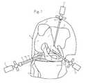
- FIG. 7 and FIG. 8show a reticule 55 for use with an arthroscopic instrument.
- the reticulemay be etched into a lens 29 disposed within the cap or may be otherwise suitably placed on the cap or even on arthroscope itself.
- the reticuleis marked with a scale 56 with which the surgeon can measure the size of objects seen through the arthroscope. The surgeon may also use the reticule to align the arthroscope within the surgical field.
- filtersmay be provided within the cap to reduce light reflected into the arthroscope or to block certain wavelengths of light.
- the filtered lensmay comprise a polarizing filter, a bandpass filter, a color filter, or an interference filter. These filters can be used in conjunction with specialized light sources (e.g. Ultraviolet or Infrared) and video processing for therapeutic and diagnostic purposes.
- the capmay be part of a complete system to diagnose pathology using different wavelengths of light and/or colors of light and filtering the light. Further, the cap may also be provided as part of system that delivers photonic energy to a surgical site to control and visualize photodynamic therapy.
- the sheath, the cap or the sheath with the cap combinationmay be configured to allow viewing around an object or obstruction. This may be accomplished through the use of a right angle prism, pentaprism, roof prism, retro-reflector or mirror disposed within the cap.
- the mirrormay be flat, concave or convex to produce a normal, reduced, or magnified image.
- an atraumatic sheath 3 for use over an endoscopeis provided with a mirror 67 having a hinge 68 .
- a pull-wire 69is coupled to the mirror 67 and is disposed within the sheath 3 .
- the pull-wire 69is further coupled to an articulating knob 70 . When the knob is manipulated, the pull-wire moves the mirror and changes the viewing angle 71 .
- FIG. 10illustrates an atraumatic sheath 3 device that allows viewing around an object or obstruction through the use of a deflectable material or flexible mount 72 having a mirror 67 .
- the mountmay be manufactured from formable materials such as aluminum or steel having spring characteristics.
- a pull-wire 69 or other manipulation devicemay also be coupled to the mount to change viewing angle 71 when the wire is manipulated manually using an articulating knob 70 .
- the mountmay also be manufactured from shape memory materials such as Nitinol® that may be manipulated using electrical current to change the angle of the mirror.
- the cap and the sheath with the capmay also be modified for use with other types of endoscopes or for use with other delicate instruments that are subject to damage during a surgical procedure.
- FIGS. 11 and 12illustrate a protective cap 30 over an arthroscopic sheath 3 .
- the sheath 3comprises an inner lumen 61 in fluid communication with a vacuum source and a hole dispose 33 in the sheath in fluid communication with the vacuum source and a surgical site within a patient 1 .
- a backstop or flange 63is disposed within an inner diameter of a bore within the cap. The bore is sized and dimensioned to friction fit over the sheath 3 .
- the flange 63prevents the sheath 3 from being further pushed into the cap and extends inwardly to come in contact with an outer diameter of the rigid cannula 18 disposed within the sheath 3 . This contact forms a seal between the flange 63 and the outer diameter of the rigid cannula 18 .
- the rigid cannula 18is provided with a lumen in fluid communication with a fluid source the cap. Holes 32 disposed in the cap are in fluid communication with the surgical site and the lumen within the rigid cannula 18 as well as the fluid source allowing fluid to flow from the fluid source to the surgical site.
- the capfurther comprises a concave lens 53 coupled to the distal portion 64 of the cap 3 .
Landscapes
- Health & Medical Sciences (AREA)
- Life Sciences & Earth Sciences (AREA)
- Surgery (AREA)
- Biomedical Technology (AREA)
- Medical Informatics (AREA)
- Optics & Photonics (AREA)
- Pathology (AREA)
- Radiology & Medical Imaging (AREA)
- Biophysics (AREA)
- Engineering & Computer Science (AREA)
- Physics & Mathematics (AREA)
- Heart & Thoracic Surgery (AREA)
- Nuclear Medicine, Radiotherapy & Molecular Imaging (AREA)
- Molecular Biology (AREA)
- Animal Behavior & Ethology (AREA)
- General Health & Medical Sciences (AREA)
- Public Health (AREA)
- Veterinary Medicine (AREA)
- Orthopedic Medicine & Surgery (AREA)
- Physical Education & Sports Medicine (AREA)
- Endoscopes (AREA)
- Surgical Instruments (AREA)
Abstract
Description
Claims (2)
Priority Applications (2)
| Application Number | Priority Date | Filing Date | Title |
|---|---|---|---|
| US13/545,886US9167955B2 (en) | 2005-06-01 | 2012-07-10 | Protective cap for arthroscopic instruments |
| US14/924,586US9833134B2 (en) | 2005-06-01 | 2015-10-27 | Protective cap for arthroscopic instruments |
Applications Claiming Priority (3)
| Application Number | Priority Date | Filing Date | Title |
|---|---|---|---|
| US11/142,990US7553278B2 (en) | 2005-06-01 | 2005-06-01 | Protective cap for arthroscopic instruments |
| US12/491,566US8216131B2 (en) | 2005-06-01 | 2009-06-25 | Protective cap for arthroscopic instruments |
| US13/545,886US9167955B2 (en) | 2005-06-01 | 2012-07-10 | Protective cap for arthroscopic instruments |
Related Parent Applications (1)
| Application Number | Title | Priority Date | Filing Date |
|---|---|---|---|
| US12/491,566ContinuationUS8216131B2 (en) | 2005-06-01 | 2009-06-25 | Protective cap for arthroscopic instruments |
Related Child Applications (1)
| Application Number | Title | Priority Date | Filing Date |
|---|---|---|---|
| US14/924,586ContinuationUS9833134B2 (en) | 2005-06-01 | 2015-10-27 | Protective cap for arthroscopic instruments |
Publications (2)
| Publication Number | Publication Date |
|---|---|
| US20120277533A1 US20120277533A1 (en) | 2012-11-01 |
| US9167955B2true US9167955B2 (en) | 2015-10-27 |
Family
ID=37482300
Family Applications (4)
| Application Number | Title | Priority Date | Filing Date |
|---|---|---|---|
| US11/142,990Active2027-07-08US7553278B2 (en) | 2005-06-01 | 2005-06-01 | Protective cap for arthroscopic instruments |
| US12/491,566Active2025-10-22US8216131B2 (en) | 2005-06-01 | 2009-06-25 | Protective cap for arthroscopic instruments |
| US13/545,886Active2026-06-05US9167955B2 (en) | 2005-06-01 | 2012-07-10 | Protective cap for arthroscopic instruments |
| US14/924,586Active2026-01-05US9833134B2 (en) | 2005-06-01 | 2015-10-27 | Protective cap for arthroscopic instruments |
Family Applications Before (2)
| Application Number | Title | Priority Date | Filing Date |
|---|---|---|---|
| US11/142,990Active2027-07-08US7553278B2 (en) | 2005-06-01 | 2005-06-01 | Protective cap for arthroscopic instruments |
| US12/491,566Active2025-10-22US8216131B2 (en) | 2005-06-01 | 2009-06-25 | Protective cap for arthroscopic instruments |
Family Applications After (1)
| Application Number | Title | Priority Date | Filing Date |
|---|---|---|---|
| US14/924,586Active2026-01-05US9833134B2 (en) | 2005-06-01 | 2015-10-27 | Protective cap for arthroscopic instruments |
Country Status (4)
| Country | Link |
|---|---|
| US (4) | US7553278B2 (en) |
| EP (1) | EP1890587B1 (en) |
| JP (1) | JP5256027B2 (en) |
| WO (1) | WO2006130730A2 (en) |
Cited By (1)
| Publication number | Priority date | Publication date | Assignee | Title |
|---|---|---|---|---|
| US11219355B2 (en) | 2017-08-14 | 2022-01-11 | Medos International Sarl | Surgical instruments with reflective mirror-like surfaces |
Families Citing this family (101)
| Publication number | Priority date | Publication date | Assignee | Title |
|---|---|---|---|---|
| WO2007093994A2 (en)* | 2006-02-16 | 2007-08-23 | Vision - Sciences Inc. | Endoscope with imaging capsule |
| US7655004B2 (en) | 2007-02-15 | 2010-02-02 | Ethicon Endo-Surgery, Inc. | Electroporation ablation apparatus, system, and method |
| WO2008119118A1 (en)* | 2007-03-30 | 2008-10-09 | Polartechnics Limited | Sheath system for tissue probe |
| US8075572B2 (en) | 2007-04-26 | 2011-12-13 | Ethicon Endo-Surgery, Inc. | Surgical suturing apparatus |
| US8100922B2 (en) | 2007-04-27 | 2012-01-24 | Ethicon Endo-Surgery, Inc. | Curved needle suturing tool |
| US8226548B2 (en) | 2007-07-07 | 2012-07-24 | Cannuflow, Inc. | Rigid arthroscope system |
| US8579897B2 (en) | 2007-11-21 | 2013-11-12 | Ethicon Endo-Surgery, Inc. | Bipolar forceps |
| US8262655B2 (en) | 2007-11-21 | 2012-09-11 | Ethicon Endo-Surgery, Inc. | Bipolar forceps |
| US8568410B2 (en) | 2007-08-31 | 2013-10-29 | Ethicon Endo-Surgery, Inc. | Electrical ablation surgical instruments |
| US20090112059A1 (en) | 2007-10-31 | 2009-04-30 | Nobis Rudolph H | Apparatus and methods for closing a gastrotomy |
| US8480657B2 (en) | 2007-10-31 | 2013-07-09 | Ethicon Endo-Surgery, Inc. | Detachable distal overtube section and methods for forming a sealable opening in the wall of an organ |
| US8262680B2 (en) | 2008-03-10 | 2012-09-11 | Ethicon Endo-Surgery, Inc. | Anastomotic device |
| US8317806B2 (en) | 2008-05-30 | 2012-11-27 | Ethicon Endo-Surgery, Inc. | Endoscopic suturing tension controlling and indication devices |
| US8771260B2 (en) | 2008-05-30 | 2014-07-08 | Ethicon Endo-Surgery, Inc. | Actuating and articulating surgical device |
| US8070759B2 (en) | 2008-05-30 | 2011-12-06 | Ethicon Endo-Surgery, Inc. | Surgical fastening device |
| US8114072B2 (en) | 2008-05-30 | 2012-02-14 | Ethicon Endo-Surgery, Inc. | Electrical ablation device |
| US8652150B2 (en) | 2008-05-30 | 2014-02-18 | Ethicon Endo-Surgery, Inc. | Multifunction surgical device |
| US8679003B2 (en) | 2008-05-30 | 2014-03-25 | Ethicon Endo-Surgery, Inc. | Surgical device and endoscope including same |
| US8906035B2 (en)* | 2008-06-04 | 2014-12-09 | Ethicon Endo-Surgery, Inc. | Endoscopic drop off bag |
| US8403926B2 (en) | 2008-06-05 | 2013-03-26 | Ethicon Endo-Surgery, Inc. | Manually articulating devices |
| US8361112B2 (en) | 2008-06-27 | 2013-01-29 | Ethicon Endo-Surgery, Inc. | Surgical suture arrangement |
| US8262563B2 (en) | 2008-07-14 | 2012-09-11 | Ethicon Endo-Surgery, Inc. | Endoscopic translumenal articulatable steerable overtube |
| US8888792B2 (en) | 2008-07-14 | 2014-11-18 | Ethicon Endo-Surgery, Inc. | Tissue apposition clip application devices and methods |
| US8211125B2 (en) | 2008-08-15 | 2012-07-03 | Ethicon Endo-Surgery, Inc. | Sterile appliance delivery device for endoscopic procedures |
| US8529563B2 (en) | 2008-08-25 | 2013-09-10 | Ethicon Endo-Surgery, Inc. | Electrical ablation devices |
| US8241204B2 (en) | 2008-08-29 | 2012-08-14 | Ethicon Endo-Surgery, Inc. | Articulating end cap |
| US8480689B2 (en) | 2008-09-02 | 2013-07-09 | Ethicon Endo-Surgery, Inc. | Suturing device |
| US8409200B2 (en) | 2008-09-03 | 2013-04-02 | Ethicon Endo-Surgery, Inc. | Surgical grasping device |
| US8114119B2 (en) | 2008-09-09 | 2012-02-14 | Ethicon Endo-Surgery, Inc. | Surgical grasping device |
| US8337394B2 (en) | 2008-10-01 | 2012-12-25 | Ethicon Endo-Surgery, Inc. | Overtube with expandable tip |
| US8157834B2 (en) | 2008-11-25 | 2012-04-17 | Ethicon Endo-Surgery, Inc. | Rotational coupling device for surgical instrument with flexible actuators |
| US8172772B2 (en) | 2008-12-11 | 2012-05-08 | Ethicon Endo-Surgery, Inc. | Specimen retrieval device |
| US8361066B2 (en) | 2009-01-12 | 2013-01-29 | Ethicon Endo-Surgery, Inc. | Electrical ablation devices |
| US8828031B2 (en) | 2009-01-12 | 2014-09-09 | Ethicon Endo-Surgery, Inc. | Apparatus for forming an anastomosis |
| US8252057B2 (en) | 2009-01-30 | 2012-08-28 | Ethicon Endo-Surgery, Inc. | Surgical access device |
| US9226772B2 (en) | 2009-01-30 | 2016-01-05 | Ethicon Endo-Surgery, Inc. | Surgical device |
| US8037591B2 (en) | 2009-02-02 | 2011-10-18 | Ethicon Endo-Surgery, Inc. | Surgical scissors |
| US20110098704A1 (en) | 2009-10-28 | 2011-04-28 | Ethicon Endo-Surgery, Inc. | Electrical ablation devices |
| US8608652B2 (en) | 2009-11-05 | 2013-12-17 | Ethicon Endo-Surgery, Inc. | Vaginal entry surgical devices, kit, system, and method |
| US8353487B2 (en) | 2009-12-17 | 2013-01-15 | Ethicon Endo-Surgery, Inc. | User interface support devices for endoscopic surgical instruments |
| US8496574B2 (en) | 2009-12-17 | 2013-07-30 | Ethicon Endo-Surgery, Inc. | Selectively positionable camera for surgical guide tube assembly |
| US8506564B2 (en) | 2009-12-18 | 2013-08-13 | Ethicon Endo-Surgery, Inc. | Surgical instrument comprising an electrode |
| US9028483B2 (en) | 2009-12-18 | 2015-05-12 | Ethicon Endo-Surgery, Inc. | Surgical instrument comprising an electrode |
| US9005198B2 (en) | 2010-01-29 | 2015-04-14 | Ethicon Endo-Surgery, Inc. | Surgical instrument comprising an electrode |
| GB201007920D0 (en)* | 2010-05-12 | 2010-06-30 | Park Medical Ltd Q | Sheath for protecting endoscope probe |
| US9375139B2 (en)* | 2010-07-29 | 2016-06-28 | Cannuflow, Inc. | Arthroscopic system |
| US20120029289A1 (en)* | 2010-07-29 | 2012-02-02 | Cannuflow, Inc. | Optical Cap for Use With Arthroscopic System |
| US20140316199A1 (en)* | 2010-07-29 | 2014-10-23 | Cannuflow, Inc. | Arthroscopic system |
| US10092291B2 (en) | 2011-01-25 | 2018-10-09 | Ethicon Endo-Surgery, Inc. | Surgical instrument with selectively rigidizable features |
| EP4000497A1 (en) | 2011-02-16 | 2022-05-25 | The General Hospital Corporation | Optical coupler for an endoscope |
| US9233241B2 (en) | 2011-02-28 | 2016-01-12 | Ethicon Endo-Surgery, Inc. | Electrical ablation devices and methods |
| US9314620B2 (en) | 2011-02-28 | 2016-04-19 | Ethicon Endo-Surgery, Inc. | Electrical ablation devices and methods |
| US9254169B2 (en) | 2011-02-28 | 2016-02-09 | Ethicon Endo-Surgery, Inc. | Electrical ablation devices and methods |
| US9049987B2 (en) | 2011-03-17 | 2015-06-09 | Ethicon Endo-Surgery, Inc. | Hand held surgical device for manipulating an internal magnet assembly within a patient |
| KR101226683B1 (en) | 2011-10-10 | 2013-01-25 | 김재환 | Cover for arthroscopic surgical instrument |
| US8986199B2 (en) | 2012-02-17 | 2015-03-24 | Ethicon Endo-Surgery, Inc. | Apparatus and methods for cleaning the lens of an endoscope |
| ES2951058T3 (en) | 2012-03-09 | 2023-10-17 | 3Shape As | 3D scanner with steam autoclavable tip containing a heated optical element |
| US20130253266A1 (en) | 2012-03-22 | 2013-09-26 | Codman & Shurtleff, Inc. | Fluid management catheter and methods of using same |
| KR101371927B1 (en)* | 2012-04-27 | 2014-03-26 | 고려대학교 산학협력단 | Bead for stitching, needle for stitching and side suction cap and apparatus for stitching internal organ using the same |
| US9427255B2 (en) | 2012-05-14 | 2016-08-30 | Ethicon Endo-Surgery, Inc. | Apparatus for introducing a steerable camera assembly into a patient |
| DE102012105370A1 (en)* | 2012-06-20 | 2013-12-24 | Karl Storz Gmbh & Co. Kg | Endoscopic sleeve, endoscope assembly and method for providing an endoscope assembly |
| US9078662B2 (en) | 2012-07-03 | 2015-07-14 | Ethicon Endo-Surgery, Inc. | Endoscopic cap electrode and method for using the same |
| US9545290B2 (en) | 2012-07-30 | 2017-01-17 | Ethicon Endo-Surgery, Inc. | Needle probe guide |
| US10314649B2 (en) | 2012-08-02 | 2019-06-11 | Ethicon Endo-Surgery, Inc. | Flexible expandable electrode and method of intraluminal delivery of pulsed power |
| US9572623B2 (en) | 2012-08-02 | 2017-02-21 | Ethicon Endo-Surgery, Inc. | Reusable electrode and disposable sheath |
| US9277957B2 (en) | 2012-08-15 | 2016-03-08 | Ethicon Endo-Surgery, Inc. | Electrosurgical devices and methods |
| US9451875B2 (en) | 2012-12-07 | 2016-09-27 | Cook Medical Technologies Llc | Flexible lens |
| DE102013102024A1 (en)* | 2013-02-01 | 2014-08-21 | Firma Trokamed Gmbh | arthroscopy shaft |
| US10098527B2 (en) | 2013-02-27 | 2018-10-16 | Ethidcon Endo-Surgery, Inc. | System for performing a minimally invasive surgical procedure |
| US20140275768A1 (en)* | 2013-03-13 | 2014-09-18 | Covidien Lp | Thoracic Scope With Skirt And Gap |
| JP2014212835A (en)* | 2013-04-23 | 2014-11-17 | ショーダテクトロン株式会社 | Hood for endoscope, and endoscope with same hood for endoscope |
| US20160199072A1 (en)* | 2013-08-19 | 2016-07-14 | Smith & Nephew, Inc. | Bone removal under direct visualization |
| GB2520332A (en)* | 2013-11-18 | 2015-05-20 | Meditech Endoscopy Ltd | Gripping Device |
| US9459442B2 (en) | 2014-09-23 | 2016-10-04 | Scott Miller | Optical coupler for optical imaging visualization device |
| CN106604687B (en)* | 2014-09-30 | 2019-08-16 | 莎·卡西玛尔·瓦拉巴达斯 | Sheath assembly and porous catheter for endoscopic surgery in different fields including suction, irrigation and material removal |
| GB201418173D0 (en)* | 2014-10-14 | 2014-11-26 | Meditech Endoscopy Ltd | Instrument tip protector |
| US10034742B2 (en) | 2014-10-23 | 2018-07-31 | Medos International Sarl | Biceps tenodesis implants and delivery tools |
| US10856966B2 (en) | 2014-10-23 | 2020-12-08 | Medos International Sarl | Biceps tenodesis implants and delivery tools |
| US10751161B2 (en) | 2014-10-23 | 2020-08-25 | Medos International Sárl | Biceps tenodesis anchor implants |
| US10076374B2 (en) | 2014-10-23 | 2018-09-18 | Medos International Sárl | Biceps tenodesis delivery tools |
| US10729419B2 (en) | 2014-10-23 | 2020-08-04 | Medos International Sarl | Biceps tenodesis implants and delivery tools |
| US9693856B2 (en) | 2015-04-22 | 2017-07-04 | DePuy Synthes Products, LLC | Biceps repair device |
| US10548467B2 (en) | 2015-06-02 | 2020-02-04 | GI Scientific, LLC | Conductive optical element |
| WO2017015480A1 (en) | 2015-07-21 | 2017-01-26 | GI Scientific, LLC | Endoscope accessory with angularly adjustable exit portal |
| US11701174B2 (en)* | 2016-01-29 | 2023-07-18 | Boston Scientific Scimed, Inc. | Medical device having a plurality of lumens and a port |
| EP3410973B1 (en)* | 2016-02-02 | 2023-09-13 | Boston Scientific Scimed, Inc. | Laser lithotripsy medical device |
| US10231823B2 (en) | 2016-04-08 | 2019-03-19 | Medos International Sarl | Tenodesis implants and tools |
| US10231824B2 (en) | 2016-04-08 | 2019-03-19 | Medos International Sárl | Tenodesis anchoring systems and tools |
| WO2018207594A1 (en)* | 2017-05-10 | 2018-11-15 | オリンパス株式会社 | Hood for endscope, and endoscope system |
| US11382662B2 (en) | 2017-08-04 | 2022-07-12 | The Brigham And Women's Hospital, Inc. | Trocars and veress-type needles with illuminated guidance and safety features |
| WO2019028458A1 (en)* | 2017-08-04 | 2019-02-07 | Brigham And Women's Hospital, Inc. | Veress-type needles with illuminated guidance and safety features |
| CN111295126B (en)* | 2017-09-11 | 2023-11-17 | 艾拉姆有限公司 | Disposable miniature endoscopy system |
| US12102295B2 (en)* | 2017-09-18 | 2024-10-01 | Periwinkle Technologies Pvt. Ltd. | Digital device facilitating body cavity screening and diagnosis |
| KR102056153B1 (en)* | 2017-11-14 | 2019-12-16 | 주식회사 엔도비전 | Bidirectional vertebral endoscopic device for surgery |
| CN118177700A (en) | 2018-01-05 | 2024-06-14 | 波士顿科学国际有限公司 | Fluorophore imaging device, system and method for endoscopic surgery |
| US20190231177A1 (en)* | 2018-01-31 | 2019-08-01 | UVision360, Inc. | Flexible imaging window |
| DE102018110082A1 (en)* | 2018-04-26 | 2019-10-31 | avateramedical GmBH | Sterile endoscope cover |
| US11903557B2 (en) | 2019-04-30 | 2024-02-20 | Psip2 Llc | Endoscope for imaging in nonvisible light |
| US20220133138A1 (en)* | 2020-10-29 | 2022-05-05 | Clearmind Biomedical, Inc. | Dilator-less and obturator-less introducer for viewing and acting on internal passageways or tissue |
| WO2022249116A2 (en)* | 2021-05-26 | 2022-12-01 | Psip2 Llc | Endoscope |
| US20220378279A1 (en)* | 2021-05-26 | 2022-12-01 | Psip2 Llc | Endoscope |
Citations (42)
| Publication number | Priority date | Publication date | Assignee | Title |
|---|---|---|---|---|
| US3051176A (en) | 1959-12-11 | 1962-08-28 | Alberti Franz | Rectoscopic devices |
| EP0058020A1 (en) | 1981-02-03 | 1982-08-18 | Olympus Optical Co., Ltd. | Endoscopes |
| US4727416A (en)* | 1987-03-05 | 1988-02-23 | Fuji Optical Systems, Inc. | Electronic video dental camera |
| US4782819A (en)* | 1987-02-25 | 1988-11-08 | Adair Edwin Lloyd | Optical catheter |
| US4809678A (en) | 1987-08-14 | 1989-03-07 | Klein Richard S | Endoscope for preventing patient contamination |
| US4809679A (en) | 1986-11-19 | 1989-03-07 | Olympus Optical Co., Ltd. | Forceps plug for endoscopes |
| US4856495A (en) | 1986-09-25 | 1989-08-15 | Olympus Optical Co., Ltd. | Endoscope apparatus |
| US4867546A (en) | 1985-01-11 | 1989-09-19 | Olympus Optical Co., Ltd. | Objective lens system for an endoscope |
| US4886049A (en) | 1988-05-17 | 1989-12-12 | Darras Robert L | Medical instrument cover |
| US5029574A (en) | 1988-04-14 | 1991-07-09 | Okamoto Industries, Inc. | Endoscopic balloon with a protective film thereon |
| DE9215725U1 (en) | 1992-11-19 | 1993-01-14 | Schölly Fiberoptic GmbH, 7819 Denzlingen | Device for lighting and inspection of cavities and gaps |
| US5191878A (en) | 1990-04-12 | 1993-03-09 | Olympus Optical Co., Ltd. | Endoscope device |
| US5215077A (en)* | 1989-11-09 | 1993-06-01 | Machida Endoscope Co., Ltd. | Direct vision/side vision exchangeable endoscope |
| US5329935A (en) | 1989-12-25 | 1994-07-19 | Asahi Kogaku Kabushiki Kaisha | Sheathed endoscope and sheath therefor |
| US5370649A (en)* | 1991-08-16 | 1994-12-06 | Myriadlase, Inc. | Laterally reflecting tip for laser transmitting fiber |
| EP0647425A1 (en) | 1993-10-08 | 1995-04-12 | United States Surgical Corporation | Endoscope attachment for changing angle of view |
| US5413092A (en) | 1991-06-24 | 1995-05-09 | Xomed-Treace, Inc. | Sheath for endoscope |
| US5518501A (en) | 1993-07-08 | 1996-05-21 | Vision-Sciences, Inc. | Endoscopic contamination protection system to facilitate cleaning of endoscopes |
| US5536236A (en) | 1993-02-12 | 1996-07-16 | Olympus Optical Co., Ltd. | Covered endoscope system |
| US5735792A (en) | 1992-11-25 | 1998-04-07 | Clarus Medical Systems, Inc. | Surgical instrument including viewing optics and an atraumatic probe |
| US5807237A (en)* | 1997-03-31 | 1998-09-15 | Tindel; Nathaniel L. | Endoscopic device |
| JPH11249014A (en) | 1998-03-03 | 1999-09-17 | Olympus Optical Co Ltd | Image pickup optical system and image pickup device using it |
| US5961445A (en)* | 1995-05-31 | 1999-10-05 | Machida Endoscope Co., Ltd. | Endoscope having replaceable objective unit |
| US6095970A (en) | 1997-02-19 | 2000-08-01 | Asahi Kogaku Kogyo Kabushiki Kaisha | Endoscope |
| US6184923B1 (en)* | 1994-11-25 | 2001-02-06 | Olympus Optical Co., Ltd. | Endoscope with an interchangeable distal end optical adapter |
| US6293909B1 (en) | 1998-08-07 | 2001-09-25 | Scimed Life Systems, Inc. | Device and method of using a surgical assembly with mesh sheath |
| US20020035311A1 (en) | 2000-09-18 | 2002-03-21 | Asahi Kogaku Kogyo Kabushiki Kaisha | Tip portion of an endoscope |
| US20020040179A1 (en) | 2000-09-29 | 2002-04-04 | Fuji Photo Optical Co., Ltd. | Endoscope tip part with no swell at outer skin fixing part |
| JP2002136472A (en) | 2000-11-02 | 2002-05-14 | Olympus Optical Co Ltd | Endoscope |
| JP2002233491A (en) | 2001-02-08 | 2002-08-20 | Asahi Optical Co Ltd | Endoscope endoscope with tip cap |
| US6447444B1 (en) | 1997-11-04 | 2002-09-10 | Sightline Technologies Ltd. | Video rectoscope |
| US20030018340A1 (en) | 2001-06-29 | 2003-01-23 | Branch Thomas P. | Method and apparatus for installing cannula |
| US6537209B1 (en)* | 2000-09-14 | 2003-03-25 | Itconcepts, Inc. | Optical system of lateral observation endoscope |
| US20030191369A1 (en)* | 2002-03-25 | 2003-10-09 | Minoru Arai | Omnidirectional endoscope apparatus |
| US6695775B2 (en) | 2001-06-07 | 2004-02-24 | Fuji Photo Optical Co., Ltd. | Lens assembly for endoscopic lens system |
| US6761684B1 (en) | 2000-08-10 | 2004-07-13 | Linvatec Corporation | Endoscope tip protection system |
| US20040143162A1 (en) | 2001-04-27 | 2004-07-22 | Beat Krattiger | Optical instrument, in particular an endoscope, having an interchangeable head |
| US20040147807A1 (en) | 2002-11-22 | 2004-07-29 | Thomas Viebach | Endoscope head |
| US20050197530A1 (en) | 2003-09-25 | 2005-09-08 | Wallace Daniel T. | Balloon visualization for traversing a tissue wall |
| US20060084839A1 (en) | 2002-05-30 | 2006-04-20 | Mourlas Nicholas J | Apparatus and methods for coronary sinus access |
| US7033317B2 (en) | 2003-06-05 | 2006-04-25 | Hydrocision, Inc. | Disposable endoscope and method of making a disposable endoscope |
| US7217241B2 (en)* | 2000-12-20 | 2007-05-15 | Faro Fabbrica Apparecchiature Razionali Odontoiatriche S.P.A | Device for orthodontic interventions |
Family Cites Families (8)
| Publication number | Priority date | Publication date | Assignee | Title |
|---|---|---|---|---|
| US4470407A (en)* | 1982-03-11 | 1984-09-11 | Laserscope, Inc. | Endoscopic device |
| JP2802244B2 (en)* | 1994-08-29 | 1998-09-24 | オリンパス光学工業株式会社 | Endoscope sheath |
| US5607441A (en)* | 1995-03-24 | 1997-03-04 | Ethicon Endo-Surgery, Inc. | Surgical dissector |
| JPH0998938A (en)* | 1995-10-04 | 1997-04-15 | Fuji Photo Optical Co Ltd | Protector of insertion part of endoscope |
| JPH09140659A (en)* | 1995-11-24 | 1997-06-03 | Fuji Photo Optical Co Ltd | Guiding cap for insertion of endoscope |
| JPH10192297A (en)* | 1996-05-09 | 1998-07-28 | Olympus Optical Co Ltd | Securing device of cavity for bone operation |
| JP3748994B2 (en)* | 1997-08-22 | 2006-02-22 | オリンパス株式会社 | Endoscope fitting |
| US6010450A (en)* | 1998-06-29 | 2000-01-04 | Welch Allyn, Inc. | Measuring adapter for viewing instrument |
- 2005
- 2005-06-01USUS11/142,990patent/US7553278B2/enactiveActive
- 2006
- 2006-05-31JPJP2008514825Apatent/JP5256027B2/enactiveActive
- 2006-05-31EPEP06760606.1Apatent/EP1890587B1/enactiveActive
- 2006-05-31WOPCT/US2006/021188patent/WO2006130730A2/enactiveApplication Filing
- 2009
- 2009-06-25USUS12/491,566patent/US8216131B2/enactiveActive
- 2012
- 2012-07-10USUS13/545,886patent/US9167955B2/enactiveActive
- 2015
- 2015-10-27USUS14/924,586patent/US9833134B2/enactiveActive
Patent Citations (44)
| Publication number | Priority date | Publication date | Assignee | Title |
|---|---|---|---|---|
| US3051176A (en) | 1959-12-11 | 1962-08-28 | Alberti Franz | Rectoscopic devices |
| EP0058020A1 (en) | 1981-02-03 | 1982-08-18 | Olympus Optical Co., Ltd. | Endoscopes |
| US4867546A (en) | 1985-01-11 | 1989-09-19 | Olympus Optical Co., Ltd. | Objective lens system for an endoscope |
| US4856495A (en) | 1986-09-25 | 1989-08-15 | Olympus Optical Co., Ltd. | Endoscope apparatus |
| US4809679A (en) | 1986-11-19 | 1989-03-07 | Olympus Optical Co., Ltd. | Forceps plug for endoscopes |
| US4782819A (en)* | 1987-02-25 | 1988-11-08 | Adair Edwin Lloyd | Optical catheter |
| US4727416B1 (en)* | 1987-03-05 | 1993-04-06 | Fuji Optical System Inc | |
| US4727416A (en)* | 1987-03-05 | 1988-02-23 | Fuji Optical Systems, Inc. | Electronic video dental camera |
| US4809678A (en) | 1987-08-14 | 1989-03-07 | Klein Richard S | Endoscope for preventing patient contamination |
| US5029574A (en) | 1988-04-14 | 1991-07-09 | Okamoto Industries, Inc. | Endoscopic balloon with a protective film thereon |
| US4886049A (en) | 1988-05-17 | 1989-12-12 | Darras Robert L | Medical instrument cover |
| US5215077A (en)* | 1989-11-09 | 1993-06-01 | Machida Endoscope Co., Ltd. | Direct vision/side vision exchangeable endoscope |
| US5329935A (en) | 1989-12-25 | 1994-07-19 | Asahi Kogaku Kabushiki Kaisha | Sheathed endoscope and sheath therefor |
| US5191878A (en) | 1990-04-12 | 1993-03-09 | Olympus Optical Co., Ltd. | Endoscope device |
| US5413092A (en) | 1991-06-24 | 1995-05-09 | Xomed-Treace, Inc. | Sheath for endoscope |
| US5370649A (en)* | 1991-08-16 | 1994-12-06 | Myriadlase, Inc. | Laterally reflecting tip for laser transmitting fiber |
| DE9215725U1 (en) | 1992-11-19 | 1993-01-14 | Schölly Fiberoptic GmbH, 7819 Denzlingen | Device for lighting and inspection of cavities and gaps |
| US5735792A (en) | 1992-11-25 | 1998-04-07 | Clarus Medical Systems, Inc. | Surgical instrument including viewing optics and an atraumatic probe |
| US5536236A (en) | 1993-02-12 | 1996-07-16 | Olympus Optical Co., Ltd. | Covered endoscope system |
| US5518501A (en) | 1993-07-08 | 1996-05-21 | Vision-Sciences, Inc. | Endoscopic contamination protection system to facilitate cleaning of endoscopes |
| EP0647425A1 (en) | 1993-10-08 | 1995-04-12 | United States Surgical Corporation | Endoscope attachment for changing angle of view |
| US6184923B1 (en)* | 1994-11-25 | 2001-02-06 | Olympus Optical Co., Ltd. | Endoscope with an interchangeable distal end optical adapter |
| US5961445A (en)* | 1995-05-31 | 1999-10-05 | Machida Endoscope Co., Ltd. | Endoscope having replaceable objective unit |
| US6095970A (en) | 1997-02-19 | 2000-08-01 | Asahi Kogaku Kogyo Kabushiki Kaisha | Endoscope |
| US5807237A (en)* | 1997-03-31 | 1998-09-15 | Tindel; Nathaniel L. | Endoscopic device |
| US6447444B1 (en) | 1997-11-04 | 2002-09-10 | Sightline Technologies Ltd. | Video rectoscope |
| JPH11249014A (en) | 1998-03-03 | 1999-09-17 | Olympus Optical Co Ltd | Image pickup optical system and image pickup device using it |
| US6293909B1 (en) | 1998-08-07 | 2001-09-25 | Scimed Life Systems, Inc. | Device and method of using a surgical assembly with mesh sheath |
| US6761684B1 (en) | 2000-08-10 | 2004-07-13 | Linvatec Corporation | Endoscope tip protection system |
| US6537209B1 (en)* | 2000-09-14 | 2003-03-25 | Itconcepts, Inc. | Optical system of lateral observation endoscope |
| US20020035311A1 (en) | 2000-09-18 | 2002-03-21 | Asahi Kogaku Kogyo Kabushiki Kaisha | Tip portion of an endoscope |
| US20020040179A1 (en) | 2000-09-29 | 2002-04-04 | Fuji Photo Optical Co., Ltd. | Endoscope tip part with no swell at outer skin fixing part |
| JP2002136472A (en) | 2000-11-02 | 2002-05-14 | Olympus Optical Co Ltd | Endoscope |
| US7217241B2 (en)* | 2000-12-20 | 2007-05-15 | Faro Fabbrica Apparecchiature Razionali Odontoiatriche S.P.A | Device for orthodontic interventions |
| JP2002233491A (en) | 2001-02-08 | 2002-08-20 | Asahi Optical Co Ltd | Endoscope endoscope with tip cap |
| US20040143162A1 (en) | 2001-04-27 | 2004-07-22 | Beat Krattiger | Optical instrument, in particular an endoscope, having an interchangeable head |
| US6695775B2 (en) | 2001-06-07 | 2004-02-24 | Fuji Photo Optical Co., Ltd. | Lens assembly for endoscopic lens system |
| US20030018340A1 (en) | 2001-06-29 | 2003-01-23 | Branch Thomas P. | Method and apparatus for installing cannula |
| US20030191369A1 (en)* | 2002-03-25 | 2003-10-09 | Minoru Arai | Omnidirectional endoscope apparatus |
| US20060084839A1 (en) | 2002-05-30 | 2006-04-20 | Mourlas Nicholas J | Apparatus and methods for coronary sinus access |
| US20040147807A1 (en) | 2002-11-22 | 2004-07-29 | Thomas Viebach | Endoscope head |
| US7371209B2 (en) | 2002-11-22 | 2008-05-13 | Stm Medizintechnik Starnberg Gmbh | Endoscope head |
| US7033317B2 (en) | 2003-06-05 | 2006-04-25 | Hydrocision, Inc. | Disposable endoscope and method of making a disposable endoscope |
| US20050197530A1 (en) | 2003-09-25 | 2005-09-08 | Wallace Daniel T. | Balloon visualization for traversing a tissue wall |
Cited By (2)
| Publication number | Priority date | Publication date | Assignee | Title |
|---|---|---|---|---|
| US11219355B2 (en) | 2017-08-14 | 2022-01-11 | Medos International Sarl | Surgical instruments with reflective mirror-like surfaces |
| US11896200B2 (en) | 2017-08-14 | 2024-02-13 | Medos International Sarl | Surgical instruments with reflective mirror-like surfaces |
Also Published As
| Publication number | Publication date |
|---|---|
| US8216131B2 (en) | 2012-07-10 |
| JP5256027B2 (en) | 2013-08-07 |
| US20160045105A1 (en) | 2016-02-18 |
| EP1890587A2 (en) | 2008-02-27 |
| WO2006130730A3 (en) | 2007-04-19 |
| US20120277533A1 (en) | 2012-11-01 |
| EP1890587B1 (en) | 2017-07-12 |
| JP2008541947A (en) | 2008-11-27 |
| EP1890587A4 (en) | 2012-06-13 |
| WO2006130730A2 (en) | 2006-12-07 |
| US7553278B2 (en) | 2009-06-30 |
| US9833134B2 (en) | 2017-12-05 |
| US20090326328A1 (en) | 2009-12-31 |
| US20060276692A1 (en) | 2006-12-07 |
Similar Documents
| Publication | Publication Date | Title |
|---|---|---|
| US9833134B2 (en) | Protective cap for arthroscopic instruments | |
| US12226076B2 (en) | Rigid endoscope system | |
| US9872604B2 (en) | Atraumatic arthroscopic instrument sheath and method | |
| US9375207B2 (en) | Atraumatic arthroscopic instrument sheath | |
| US8545462B2 (en) | Patch for irrigation/aspiration tip | |
| JP2007522837A (en) | Noninvasive arthroscopy instrument sheath |
Legal Events
| Date | Code | Title | Description |
|---|---|---|---|
| AS | Assignment | Owner name:CANNUFLOW, INC., CALIFORNIA Free format text:ASSIGNMENT OF ASSIGNORS INTEREST;ASSIGNOR:KUCKLICK, THEODORE R.;REEL/FRAME:028570/0340 Effective date:20050725 | |
| STCF | Information on status: patent grant | Free format text:PATENTED CASE | |
| MAFP | Maintenance fee payment | Free format text:PAYMENT OF MAINTENANCE FEE, 4TH YR, SMALL ENTITY (ORIGINAL EVENT CODE: M2551); ENTITY STATUS OF PATENT OWNER: SMALL ENTITY Year of fee payment:4 | |
| AS | Assignment | Owner name:PRISTINE SURGICAL LLC, NEW HAMPSHIRE Free format text:ASSIGNMENT OF ASSIGNORS INTEREST;ASSIGNOR:CANNUFLOW, INC.;REEL/FRAME:053594/0011 Effective date:20200611 | |
| AS | Assignment | Owner name:PINEY LAKE OPPORTUNITIES ECI MASTER FUND LP, AS AGENT, CONNECTICUT Free format text:SECURITY INTEREST;ASSIGNOR:PRISTINE SURGICAL LLC;REEL/FRAME:056765/0708 Effective date:20210702 | |
| AS | Assignment | Owner name:PSIP2 LLC, NEW HAMPSHIRE Free format text:ASSIGNMENT OF ASSIGNORS INTEREST;ASSIGNOR:PRISTINE SURGICAL LLC;REEL/FRAME:060254/0452 Effective date:20220601 | |
| AS | Assignment | Owner name:PINEY LAKE OPPORTUNITIES ECI MASTER FUND LP, AS COLLATERAL AGENT, CONNECTICUT Free format text:SECURITY INTEREST;ASSIGNOR:PSIP2 LLC;REEL/FRAME:061006/0252 Effective date:20220728 | |
| MAFP | Maintenance fee payment | Free format text:PAYMENT OF MAINTENANCE FEE, 8TH YR, SMALL ENTITY (ORIGINAL EVENT CODE: M2552); ENTITY STATUS OF PATENT OWNER: SMALL ENTITY Year of fee payment:8 |