US9155532B2 - Medical devices, systems and methods for closing perforations - Google Patents
Medical devices, systems and methods for closing perforationsDownload PDFInfo
- Publication number
- US9155532B2 US9155532B2US12/125,475US12547508AUS9155532B2US 9155532 B2US9155532 B2US 9155532B2US 12547508 AUS12547508 AUS 12547508AUS 9155532 B2US9155532 B2US 9155532B2
- Authority
- US
- United States
- Prior art keywords
- medical device
- delivery devices
- tubes
- guide surface
- distal
- Prior art date
- Legal status (The legal status is an assumption and is not a legal conclusion. Google has not performed a legal analysis and makes no representation as to the accuracy of the status listed.)
- Active, expires
Links
- 238000000034methodMethods0.000titleabstractdescription17
- 239000000853adhesiveSubstances0.000claimsdescription4
- 230000001070adhesive effectEffects0.000claimsdescription4
- 238000003466weldingMethods0.000claimsdescription3
- 238000005476solderingMethods0.000claimsdescription2
- 238000012986modificationMethods0.000description3
- 230000004048modificationEffects0.000description3
- 230000009278visceral effectEffects0.000description3
- 241000894006BacteriaSpecies0.000description1
- 238000005452bendingMethods0.000description1
- 230000002498deadly effectEffects0.000description1
- 230000003247decreasing effectEffects0.000description1
- 230000001419dependent effectEffects0.000description1
- 210000003238esophagusAnatomy0.000description1
- 239000012530fluidSubstances0.000description1
- 230000002496gastric effectEffects0.000description1
- 210000001035gastrointestinal tractAnatomy0.000description1
- 208000015181infectious diseaseDiseases0.000description1
- 230000001681protective effectEffects0.000description1
- 210000001835visceraAnatomy0.000description1
Images


Classifications
- A—HUMAN NECESSITIES
- A61—MEDICAL OR VETERINARY SCIENCE; HYGIENE
- A61B—DIAGNOSIS; SURGERY; IDENTIFICATION
- A61B17/00—Surgical instruments, devices or methods
- A61B17/04—Surgical instruments, devices or methods for suturing wounds; Holders or packages for needles or suture materials
- A61B17/0469—Suturing instruments for use in minimally invasive surgery, e.g. endoscopic surgery
- A—HUMAN NECESSITIES
- A61—MEDICAL OR VETERINARY SCIENCE; HYGIENE
- A61B—DIAGNOSIS; SURGERY; IDENTIFICATION
- A61B17/00—Surgical instruments, devices or methods
- A61B17/0057—Implements for plugging an opening in the wall of a hollow or tubular organ, e.g. for sealing a vessel puncture or closing a cardiac septal defect
- A—HUMAN NECESSITIES
- A61—MEDICAL OR VETERINARY SCIENCE; HYGIENE
- A61B—DIAGNOSIS; SURGERY; IDENTIFICATION
- A61B17/00—Surgical instruments, devices or methods
- A61B17/04—Surgical instruments, devices or methods for suturing wounds; Holders or packages for needles or suture materials
- A61B17/0401—Suture anchors, buttons or pledgets, i.e. means for attaching sutures to bone, cartilage or soft tissue; Instruments for applying or removing suture anchors
- A—HUMAN NECESSITIES
- A61—MEDICAL OR VETERINARY SCIENCE; HYGIENE
- A61B—DIAGNOSIS; SURGERY; IDENTIFICATION
- A61B17/00—Surgical instruments, devices or methods
- A61B17/04—Surgical instruments, devices or methods for suturing wounds; Holders or packages for needles or suture materials
- A61B17/0482—Needle or suture guides
- A—HUMAN NECESSITIES
- A61—MEDICAL OR VETERINARY SCIENCE; HYGIENE
- A61B—DIAGNOSIS; SURGERY; IDENTIFICATION
- A61B17/00—Surgical instruments, devices or methods
- A61B17/0057—Implements for plugging an opening in the wall of a hollow or tubular organ, e.g. for sealing a vessel puncture or closing a cardiac septal defect
- A61B2017/00637—Implements for plugging an opening in the wall of a hollow or tubular organ, e.g. for sealing a vessel puncture or closing a cardiac septal defect for sealing trocar wounds through abdominal wall
- A—HUMAN NECESSITIES
- A61—MEDICAL OR VETERINARY SCIENCE; HYGIENE
- A61B—DIAGNOSIS; SURGERY; IDENTIFICATION
- A61B17/00—Surgical instruments, devices or methods
- A61B17/0057—Implements for plugging an opening in the wall of a hollow or tubular organ, e.g. for sealing a vessel puncture or closing a cardiac septal defect
- A61B2017/00646—Type of implements
- A61B2017/00663—Type of implements the implement being a suture
- A—HUMAN NECESSITIES
- A61—MEDICAL OR VETERINARY SCIENCE; HYGIENE
- A61B—DIAGNOSIS; SURGERY; IDENTIFICATION
- A61B17/00—Surgical instruments, devices or methods
- A61B17/04—Surgical instruments, devices or methods for suturing wounds; Holders or packages for needles or suture materials
- A61B17/0469—Suturing instruments for use in minimally invasive surgery, e.g. endoscopic surgery
- A61B2017/0472—Multiple-needled, e.g. double-needled, instruments
Definitions
- the present inventionrelates generally to medical devices, systems, and methods for closing perforations in tissue.
- T-anchorsalso known as tissue anchors or visceral anchors.
- An exemplary tissue anchoris disclosed in U.S. Pat. No. 5,123,914, the entire contents of which are incorporated by reference herein. Such tissue anchors have been very successful in medical procedures requiring visceral wall mobilization or wall apposition.
- Tissue anchorshave also been successfully used in closing perforations, but are not without their drawbacks. For example, when a series of anchors are placed around a perforation, each individual anchor is manually placed in sequence. This can be time consuming, and can result in uneven spacing of the anchors around the perforation. It can therefore be difficult to ensure proper approximation of the tissue around the perforation and complete closure thereof. This is especially critical within the gastrointestinal tract, where the travel of bacteria laden fluids outside of the tract may cause unwanted and sometimes deadly infection.
- the present inventionprovides medical devices, systems and methods for placing tissue fixation devices that are easy to employ, reduce procedure time, and improve spacing of the tissue fixation devices.
- a medical deviceconstructed in accordance with the teachings of the present invention, includes a plurality of elongate delivery devices, a plurality of elongate tubes, and a distal tip.
- the delivery devicesare needles, although other devices may be used for delivering tissue fixation devices.
- Each delivery devicedefines a delivery lumen sized to receive a tissue fixation device.
- the plurality of elongate tubesextend generally parallel to a longitudinal axis, and each tube defines a tube lumen sized to receive one of the delivery devices.
- Each tube lumenhas a distal port.
- the distal tipis connected to the plurality of elongate tubes and defines a guide surface positioned distally of the distal ports.
- the guide surfaceis structured to redirect the plurality of delivery devices radially outwardly as the plurality of delivery devices are distally translated through the tube lumens and the distal ports of the plurality of elongate tubes.
- the guide surfaceslopes radially outwardly, and preferably is curved.
- the guide surfaceredirects the plurality of delivery devices in a direction angled relative to the longitudinal axis, preferably angled in the range of 10 to 60 degrees, and most preferably about 30 degrees.
- the distal tipdefines a hub having a plurality of pockets sized to receive the plurality of elongate tubes. An end surface of the distal tip is atraumatically shaped.
- the delivery devicesare puncture needles
- the tissue fixation devicesare tissue anchors.
- a medical system for placing tissue fixation devices in bodily tissuesincludes a plurality of elongate delivery devices, a plurality of elongate tubes, a distal tip, and an endoscope.
- the plurality of delivery deviceseach define a delivery lumen sized to receive a tissue fixation device.
- the plurality of elongate tubesextend generally parallel to a longitudinal axis and each tube defines a tube lumen sized to receive one of the plurality of delivery devices.
- Each tube lumendefines a distal port.
- a distal tipis connected to the plurality of elongate tubes and defines a guide surface positioned distally of the distal ports and extending radially outwardly.
- the distal tipdefines a passageway.
- the endoscopeis selectively attachable to the distal tip, and the passageway is sized to receive a distal end of the endoscope therein.
- the endoscopeis loosely press fit within the passageway of the distal tip.
- the passagewaydefines an inner surface, and the inner surface is positioned to frictionally engage the endoscope.
- the plurality of tubesextend along an outer surface of the endoscope.
- the guide surfaceis structured to redirect the plurality of delivery devices radially outwardly as the plurality of delivery devices are distally translated through the tube lumens and the distal ports of the plurality of elongate tubes.
- a method of placing tissue fixation devices in bodily tissue of a patientis also provided in accordance with the teachings of the present invention.
- One embodiment of the methodincludes providing a medical system comprising a medical device and an endoscope, such as those previously described.
- the endoscopeis attached to the medical device, and the medical system is introduced through a bodily lumen of the patient to a position proximate the bodily tissue.
- the plurality of delivery devicesare translated distally through the plurality of tubes such that the plurality of delivery devices engage the guide surface and deflect radially outwardly.
- the plurality of delivery devicesare positioned proximate the bodily tissue.
- the plurality of tissue fixation devicesare delivered through the plurality of delivery devices to a position engaged with the bodily tissue.
- the plurality of delivery devicesare retracted into the plurality of tubes.
- a cutting instrumentmay be passed through an accessory channel of the endoscope and used to form an opening in the bodily tissue.
- the step of forming the openingis preferably performed after the step of positioning the plurality of delivery devices proximate the bodily tissue, and most preferably after the step of delivering the plurality of tissue fixation devices.
- the methodalso includes adjusting the position of the medical system relative to the bodily tissue prior to the step of passing the plurality of delivery devices through the bodily tissue. In this manner, the spacing of the tissue fixation devices may be easily controlled.
- the endoscope and medical devicemay be retracted, and the endoscope or other medical instrument may be reintroduced through the bodily lumen and through the opening in the bodily tissue.
- the plurality of tissue fixation devicesare connected together, such as by tying one or more sutures which are attached to the tissue fixation devices.
- FIG. 1is a plan view, partially cut-away, showing a medical device constructed in accordance with the teachings of the present invention
- FIG. 2is a front view, partially in cross-section, showing a medical system including the medical device depicted in FIG. 1 , constructed in accordance with the teachings of the present invention
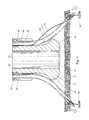
- FIG. 3is an enlarged cross-sectional view, partially cut away, of the distal end of the medical device depicted in FIG. 1 ;
- FIG. 4is an enlarged cross-sectional view, partially cut away, of the distal end of the medical system depicted in FIG. 2 ;
- FIGS. 5 , 6 and 7are enlarged cross-sectional views similar to FIG. 4 showing operation of the medical system.
- FIG. 1depicts a medical device 20 constructed in accordance with the teachings of the present invention.
- the medical device 20also forms part of a medical system 22 , which includes the medical device 20 and an endoscope 24 .
- the medical device 20is selectively attachable to the endoscope 24 , and the medical system 22 may be traversed through a bodily lumen of a patient to a desired location for performing procedures within the body, such as at a particular bodily wall or tissue.
- the bodily lumenmay be the esophagus 10 while the bodily tissue may be the gastric wall 12 , although the medical system 22 may be used with any bodily lumen and tissue, as will be understood by those skilled in the art.
- the medical device 20generally includes a plurality of puncture needles 30 and a plurality of tubes 32 .
- the plurality of needles 30have at their proximal end a plurality of needle housings 34 , while the proximal end of the plurality of tubes 32 includes a plurality of tube housings 36 .
- the relative positions of the needle housings 34 and the tube housings 36control the relative positions of the needles 30 within the protective tubes 32 .
- the needles 30may be operated to project from a distal end of the tubes 32 , or operated to be retracted within the tubes 32 , as will be described in more detail hereinbelow.
- the proximal end of the medical device 20also includes a plurality of plungers 38 which are connected to stylets 40 ( FIG. 3 ) that extend through the plurality of needles 30 .
- the stylets 40(operated via their plungers 38 ) are used to push tissue fixation devices such as tissue anchors 54 ( FIG. 3 ) out from the distal ends of the needles 30 .
- the plurality of tubes 32are connected to a distal tip 42 at the distal end of the tubes 32 .
- the distal tip 42generally includes a plurality of hubs 44 defining a plurality of pockets 46 for receiving the plurality of tubes 32 , as best seen in FIG. 3 .
- the hubs 44could be a single hub having a plurality of pockets 46 , or as shown in FIG. 2 , individual and discreet hubs 44 may be designed to define the plurality of pockets 46 .
- Each hub 44defines a shoulder 48 at the bottom of the pocket 46 for abutting a distal end of each tube 32 .
- the distal tip 42 and plurality of tubes 32may be interconnected through frictional engagement, adhesives, welding, soldering or any other well known means for connecting two structures.
- the hubs 44define distal ports 55 through which the plurality of needles 30 extend and are exposed for placement through the bodily tissue 12 .
- the distal tip 42may simply have the plurality of tubes 32 attached to its exterior surface, thereby eliminating the hub or hubs 44 and their pockets 46 .
- the plurality of tubes 32themselves would define the distal ports 55 though which the needles 30 can exit.
- the distal ports 55are circumferentially spaced about the distal tip 42 .
- the distal ports 55are equidistantly spaced to provide for delivery of the tissue anchors 54 in a generally equidistantly spaced configuration, although different spacings may be designed, such as to match the shape of the perforation 16 ( FIG. 7 ) in the tissue 12 , which can be elongated or otherwise uneven, rather than circular.
- the plurality of tubes 32each define a tube lumen 58 which slideably receives the needles 30 therein.
- the needles 30each define a needle lumen 52 which is sized to receive the stylet 40 .
- a distal end 31 of each needle 30contains one or more tissue anchors 54 positioned within the needle lumen 52 .
- the distal end 31also includes a slot 33 through which the suture 56 passes.
- the suture 56is connected to the tissue anchor 54 and is used to draw the perforation 16 ( FIG. 7 ) closed, as is known in the art. It can be seen in FIG. 3 that the suture 56 extends proximally through the tube lumen 58 between the needle 30 and tube 32 .
- suture 56could alternatively pass through the interior of the needle 30 , i.e., through the needle lumen 52 .
- the tissue anchors 54may be pre-loaded within the needles 30 , and likewise the needles may be pre-loaded within the plurality of tubes 32 , although this is not necessary.
- tissue anchors 54may be employed in conjunction with the present invention, exemplary tissue anchors being disclosed in U.S. Pat. No. 5,123,914; and U.S. patent application Ser. No. 11/946,565.
- the medical device 20 and medical system 22 of the present inventionmay also be used in conjunction with other tissue fixation devices such as staples, an exemplary tissue staple and system being disclosed in U.S. Patent Application No. 60/956,580).
- the disclosures of all of the above-identified patents/applicationsare hereby incorporated by reference in their entireties. Accordingly, it will be seen that the plurality of puncture needles 30 can be replaced with other elongate delivery devices, such as delivery catheters for tissue staples, dependent upon the particular tissue fixation device.
- the distal tip 42includes an interior passageway 60 defined by an interior wall 62 .
- the passageway 60 and interior wall 62are sized to receive an endoscope therein for forming the medical system 22 of the present invention.
- the passageway 60 and wall 62are sized to be loosely press-fit with the endoscope 24 such that they are selectively attachable, although it will be recognized by those skilled in the art that many different structures and means for selectively attaching the endoscope 24 to the medical device 20 may be employed.
- An end surface 66 of the distal tipis atraumatically shaped, i.e. rounded, such that the medical system 22 may be safely navigated through the patient's bodily lumens.
- the plurality of tubes 32 and needles 30generally extend along a longitudinal axis 14 that is also shared by the distal tip 42 .
- the plurality of tubes 32are elongated and structured to extend longitudinally along the outer surface of the endoscope 24 , as shown in FIG. 2 .
- a band 23 or other structuremay be used to connect the plurality of tubes 32 to the endoscope 24 at points along the length of the medical system 22 , although such bands 23 or the like are not necessary to traverse the medical system 22 through the bodily lumen 10 .
- the endoscope 24can be integrally formed with the medical device 20 , namely with the plurality of tubes 32 and distal tip 42 .
- the distal tip 42 and/or plurality of tubes 32can be bonded to the endoscope 24 , such as with an adhesive or using welding techniques.
- An outer surface of the distal tip 42defines a guide surface 64 .
- the guide surface 64extends radially outwardly and is positioned distally of the distal port 55 .
- the guide surface 64extends radially outwardly and deflects the needles 30 radially outwardly, as indicated by arrow 70 .
- the needles 30upon further translation in the distal direction, the needles 30 continue to flex and are directed radially outwardly.
- the hubs 44define slots 50 that open radially, and the slots 50 provide increased range of movement to the needles 30 as they flex.
- the plurality of tubes 32are simply attached to the exterior surface of the distal tip 42 (i.e. without the hub 44 and pockets 46 ) these slots 50 may be formed in the tubes 32 themselves. As such, the needles 30 enter the slots 50 defined by the hub 44 and pockets 46 , thereby permitting greater range of movement and preventing excessive flexing or bending of the needles 30 .
- the distal tip 42 and its guiding surface 64move each of the needles 30 radially outwardly away from each other and away from the longitudinal axis 14 .
- the guide surface 64may take many different curvatures, or can even be flat, such as in a conical or pyramidal shape (i.e. flat in the longitudinal direction).
- the outer diameter of the distal tip 42 at its peak 65may also be increased or decreased to increase or decrease the amount of deflection of the plurality of needles 30 .
- the peaks 65are preferably spaced radially outwardly of the distal ports 50 .
- the distal tip 42 and its guiding surface 64are structured to cause the needles 30 to be passed through the tissue 12 at an angle of about 10 to 60 degrees relative to the longitudinal axis 14 .
- the needles 30are at an angle of about 30 degrees relative to the longitudinal axis 14 .
- the needles 30are in paths that are circumferentially spaced apart, and preferably equidistantly spaced apart, thereby greatly improving the spacing of the tissue anchors 54 around a perforation 16 ( FIG. 7 ) and improving perforation closure.
- a method for placing tissue fixation devices in bodily tissue 12 of a patientwill now be described with reference to FIGS. 2 and 5 - 7 .
- a medical system 20is provided, such as the medical device 20 and endoscope 24 as previously discussed.
- the endoscope 24is attached to the medical device 20 , namely by placing the distal end of the endoscope 24 into the interior passageway 60 of the distal tip 42 .
- the medical system 20is introduced through a bodily lumen 10 of the patient to a position proximate the bodily tissue 12 , as shown in FIG. 2 .
- the plurality of puncture needles 30are translated distally through the plurality of tubes 32 such that the plurality of needles 30 engage the guide surface 64 and deflect radially outwardly, as shown in FIG.
- the plurality of puncture needles 30are passed through the bodily tissue 12 . This may occur sequentially or simultaneously. It will be recognized by those skilled in the art that because the distance between each of the needles 30 increases in the longitudinal direction (i.e. the more distal the greater the spacing), the medical system 22 and its distal tip 42 may be placed closer to the tissue 12 to bring the placement sites closer together, or the medical system 22 may be moved further away from the tissue 12 to increase the distance between the placement sites. In embodiments where other types of tissue fixation devices and delivery devices are employed (e.g. a visceral staple and delivery catheter, respectively) the delivery device need only be positioned proximate the tissue 12 , and need not pass through the tissue 12 .
- tissue fixation devices and delivery devicese.g. a visceral staple and delivery catheter, respectively
- the plurality of tissue anchors 54are delivered to a position on the distal side of the bodily tissue 12 .
- the tissue anchors 54are connected to suture 56 , and the anchors 54 are ejected from the distal end of the needles 30 through translation of the stylets 40 via depression of the plungers 38 ( FIG. 1 ).
- the plurality of puncture needles 30are then retracted into the plurality of tubes 30 , leaving the tissue anchors 54 in place.
- a cutting instrument 68may then be delivered through the accessory channel 25 of the endoscope 24 , and used to form an opening 16 in the tissue 12 .
- the cutting tool 68is an electrosurgical cutting instrument, although it will be recognized by those skilled in the art that any type of cutting instrument or device may be employed.
- the step of forming the opening 16is performed after the step of translating the puncture needles 30 and most preferably after delivering the plurality of tissue anchors 54 , thereby reducing the potential for the tissue 12 to deflect or move when placing the tissue anchors 54 .
- the plurality of tissue anchors 54 or other tissue fixation devicesmay be delivered after the opening 16 has already been formed in the tissue 12 . In either case, the tissue 12 (and sometimes the opening 16 ) is preferably visualized using the endoscope 24 prior to placing the plurality of puncture needles 30 through the bodily tissue 12 .
- the medical system 22may be retracted through the bodily lumen 10 , and the endoscope 24 may be detached from the medical device 20 . If desired, the endoscope 24 may then be reintroduced through the bodily lumen 10 and through the opening 16 in the bodily tissue 12 . As such, various procedures may be employed with, or in conjunction with, the endoscope 24 on the distal side of the bodily tissue 12 . It will also be recognized that various other medical devices may be passed through the bodily lumen 10 and through the opening 16 formed in the bodily tissue 12 . When the procedure(s) on the distal side of the opening 16 are complete, the one or more sutures 56 attached to the tissue anchors 54 are connected together, such as by using a suture lock.
- Exemplary suture locksare disclosed in U.S. Patent Application Nos. 60/941,086 and 60/956,575, the disclosures of which are incorporated herein by reference in their entirety.
- fixation of the sutures 56the perforation 16 can be easily and securely closed.
Landscapes
- Health & Medical Sciences (AREA)
- Life Sciences & Earth Sciences (AREA)
- Surgery (AREA)
- Molecular Biology (AREA)
- General Health & Medical Sciences (AREA)
- Biomedical Technology (AREA)
- Heart & Thoracic Surgery (AREA)
- Medical Informatics (AREA)
- Nuclear Medicine, Radiotherapy & Molecular Imaging (AREA)
- Animal Behavior & Ethology (AREA)
- Engineering & Computer Science (AREA)
- Public Health (AREA)
- Veterinary Medicine (AREA)
- Cardiology (AREA)
- Rheumatology (AREA)
- Surgical Instruments (AREA)
- Endoscopes (AREA)
Abstract
Description
Claims (19)
Priority Applications (1)
| Application Number | Priority Date | Filing Date | Title |
|---|---|---|---|
| US12/125,475US9155532B2 (en) | 2007-05-25 | 2008-05-22 | Medical devices, systems and methods for closing perforations |
Applications Claiming Priority (2)
| Application Number | Priority Date | Filing Date | Title |
|---|---|---|---|
| US94024607P | 2007-05-25 | 2007-05-25 | |
| US12/125,475US9155532B2 (en) | 2007-05-25 | 2008-05-22 | Medical devices, systems and methods for closing perforations |
Publications (2)
| Publication Number | Publication Date |
|---|---|
| US20080294001A1 US20080294001A1 (en) | 2008-11-27 |
| US9155532B2true US9155532B2 (en) | 2015-10-13 |
Family
ID=39689461
Family Applications (1)
| Application Number | Title | Priority Date | Filing Date |
|---|---|---|---|
| US12/125,475Active2033-09-14US9155532B2 (en) | 2007-05-25 | 2008-05-22 | Medical devices, systems and methods for closing perforations |
Country Status (6)
| Country | Link |
|---|---|
| US (1) | US9155532B2 (en) |
| EP (1) | EP2155071B1 (en) |
| JP (1) | JP5443341B2 (en) |
| AU (1) | AU2008256823B2 (en) |
| CA (1) | CA2688261C (en) |
| WO (1) | WO2008147871A1 (en) |
Cited By (2)
| Publication number | Priority date | Publication date | Assignee | Title |
|---|---|---|---|---|
| US20160278763A1 (en)* | 2015-03-24 | 2016-09-29 | Richard B. BEAVEN MD | Laparoscopic wound closure device |
| US12383246B2 (en) | 2020-10-12 | 2025-08-12 | Abbott Cardiovascular Systems, Inc. | Vessel closure device with improved safety and tract hemostasis |
Families Citing this family (55)
| Publication number | Priority date | Publication date | Assignee | Title |
|---|---|---|---|---|
| US9579091B2 (en) | 2000-01-05 | 2017-02-28 | Integrated Vascular Systems, Inc. | Closure system and methods of use |
| US6391048B1 (en) | 2000-01-05 | 2002-05-21 | Integrated Vascular Systems, Inc. | Integrated vascular device with puncture site closure component and sealant and methods of use |
| US8758400B2 (en) | 2000-01-05 | 2014-06-24 | Integrated Vascular Systems, Inc. | Closure system and methods of use |
| DE60144328D1 (en) | 2000-09-08 | 2011-05-12 | Abbott Vascular Inc | Surgical clamp |
| US6626918B1 (en) | 2000-10-06 | 2003-09-30 | Medical Technology Group | Apparatus and methods for positioning a vascular sheath |
| US6623510B2 (en) | 2000-12-07 | 2003-09-23 | Integrated Vascular Systems, Inc. | Closure device and methods for making and using them |
| US6695867B2 (en) | 2002-02-21 | 2004-02-24 | Integrated Vascular Systems, Inc. | Plunger apparatus and methods for delivering a closure device |
| US8690910B2 (en) | 2000-12-07 | 2014-04-08 | Integrated Vascular Systems, Inc. | Closure device and methods for making and using them |
| US7905900B2 (en)* | 2003-01-30 | 2011-03-15 | Integrated Vascular Systems, Inc. | Clip applier and methods of use |
| IES20030424A2 (en) | 2002-06-04 | 2003-12-10 | Robert Stevenson | Blood vessel closure clip and delivery device |
| US8202293B2 (en) | 2003-01-30 | 2012-06-19 | Integrated Vascular Systems, Inc. | Clip applier and methods of use |
| US8905937B2 (en) | 2009-02-26 | 2014-12-09 | Integrated Vascular Systems, Inc. | Methods and apparatus for locating a surface of a body lumen |
| US8398656B2 (en) | 2003-01-30 | 2013-03-19 | Integrated Vascular Systems, Inc. | Clip applier and methods of use |
| US9713465B1 (en)* | 2004-04-19 | 2017-07-25 | Granit Medical Innovation Llc | Surgical closure device and associated method |
| US8926633B2 (en) | 2005-06-24 | 2015-01-06 | Abbott Laboratories | Apparatus and method for delivering a closure element |
| US8313497B2 (en) | 2005-07-01 | 2012-11-20 | Abbott Laboratories | Clip applier and methods of use |
| US8556930B2 (en) | 2006-06-28 | 2013-10-15 | Abbott Laboratories | Vessel closure device |
| US8893947B2 (en) | 2007-12-17 | 2014-11-25 | Abbott Laboratories | Clip applier and methods of use |
| US9282965B2 (en) | 2008-05-16 | 2016-03-15 | Abbott Laboratories | Apparatus and methods for engaging tissue |
| WO2010033189A1 (en)* | 2008-09-16 | 2010-03-25 | VentralFix, Inc. | Method and apparatus for minimally invasive delivery, tensioned deployment and fixation of secondary material prosthetic devices in patient body tissue, including hernia repair within the patient's herniation site |
| WO2010042402A1 (en)* | 2008-10-06 | 2010-04-15 | Wilson-Cook Medical, Inc. | Endcap for safely deploying tissue anchors |
| US8398676B2 (en) | 2008-10-30 | 2013-03-19 | Abbott Vascular Inc. | Closure device |
| US9414820B2 (en)* | 2009-01-09 | 2016-08-16 | Abbott Vascular Inc. | Closure devices, systems, and methods |
| US9089311B2 (en) | 2009-01-09 | 2015-07-28 | Abbott Vascular Inc. | Vessel closure devices and methods |
| US20100179589A1 (en) | 2009-01-09 | 2010-07-15 | Abbott Vascular Inc. | Rapidly eroding anchor |
| US9486191B2 (en) | 2009-01-09 | 2016-11-08 | Abbott Vascular, Inc. | Closure devices |
| US9173644B2 (en) | 2009-01-09 | 2015-11-03 | Abbott Vascular Inc. | Closure devices, systems, and methods |
| US20100185234A1 (en) | 2009-01-16 | 2010-07-22 | Abbott Vascular Inc. | Closure devices, systems, and methods |
| EP2413809B1 (en)* | 2009-04-03 | 2014-10-08 | Cook Medical Technologies LLC | Medical devices for rapid deployment and fixation of tissue anchors |
| US20110054492A1 (en) | 2009-08-26 | 2011-03-03 | Abbott Laboratories | Medical device for repairing a fistula |
| US20150335320A1 (en)* | 2010-04-13 | 2015-11-26 | neoSurgical Ltd. | Suture delivery system |
| US9855031B2 (en) | 2010-04-13 | 2018-01-02 | Neosurgical Limited | Suture delivery system |
| GB2486497B (en)* | 2010-12-17 | 2013-06-19 | Neosurgical Ltd | Laparoscopic trocar system |
| EP3305209B1 (en) | 2010-06-09 | 2025-01-08 | C.R. Bard Inc. | Instruments for delivering transfascial sutures and transfascial suture assemblies. |
| ITPI20100099A1 (en)* | 2010-08-18 | 2012-02-19 | Enrico Gervasi | INNOVATIVE DEVICE FOR SURGICAL USE TO BE USED IN MUSCLE / TENDINE TRANSPLANTATION CARRIED OUT IN ARTHROSCOPY |
| JP5615112B2 (en)* | 2010-09-21 | 2014-10-29 | 日本コヴィディエン株式会社 | Organ fixing device and organ fixing device |
| GB2495534B (en)* | 2011-10-13 | 2014-04-23 | Neosurgical Ltd | Laparoscopic system |
| WO2013062933A1 (en) | 2011-10-24 | 2013-05-02 | C.R. Bard, Inc. | Instruments for delivering transfascial sutures, transfascial suture assemblies and methods of transfascial suturing |
| US9078648B2 (en) | 2011-11-07 | 2015-07-14 | C.R. Bard, Inc. | Instruments for delivering transfascial sutures and methods of transfascial suturing |
| US9039721B2 (en) | 2011-11-07 | 2015-05-26 | C.R. Bard, Inc. | Instruments for delivering transfascial sutures and methods of transfascial suturing |
| US9924938B2 (en) | 2011-11-07 | 2018-03-27 | C.R. Bard, Inc. | Instruments for delivering transfascial sutures and methods of transfascial suturing |
| US9332976B2 (en) | 2011-11-30 | 2016-05-10 | Abbott Cardiovascular Systems, Inc. | Tissue closure device |
| WO2013119630A1 (en)* | 2012-02-06 | 2013-08-15 | Cook Medical Technologies Llc | Artificial device deployment apparatus |
| US9364209B2 (en) | 2012-12-21 | 2016-06-14 | Abbott Cardiovascular Systems, Inc. | Articulating suturing device |
| JP6117160B2 (en)* | 2014-09-18 | 2017-04-19 | 本田技研工業株式会社 | Vehicle braking control device |
| US10639020B2 (en) | 2015-09-28 | 2020-05-05 | M-V Arterica AB | Vascular closure device |
| US20190142403A1 (en) | 2017-11-16 | 2019-05-16 | M-V Arterica AB | Tissue closure device |
| US11179145B2 (en) | 2017-11-16 | 2021-11-23 | M-V Arterica AB | Collapsible tube for hemostasis |
| US11344292B2 (en)* | 2018-06-14 | 2022-05-31 | Covidien Lp | Trans-vaginal cuff anchor and method of deploying same |
| EP3870076A4 (en)* | 2018-10-24 | 2022-08-10 | Arterica Inc. | SELF-EXPANDING HEMOSTATIC DEVICES AND METHODS FOR FASCIAL AND VASCULAR PASSAGES |
| US11375994B2 (en)* | 2019-07-12 | 2022-07-05 | Abbot Cardiovascular Systems, Inc. | Methods, systems, and devices for positioning sutures for closing an opening in tissue |
| CN119423871A (en)* | 2019-09-06 | 2025-02-14 | 波士顿科学国际有限公司 | Suture device |
| EP4061244A4 (en) | 2019-11-19 | 2024-06-19 | Arterica Inc. | VASCULAR CLOSURE DEVICES AND METHODS |
| WO2023114840A1 (en)* | 2021-12-16 | 2023-06-22 | Hologic, Inc. | Colpotomy cup for faciliting suturing of vaginal cuff |
| CN120241153B (en)* | 2025-06-04 | 2025-09-30 | 浙江省肿瘤医院 | Suturing device for duodenal endoscopic treatment postoperative perforation |
Citations (108)
| Publication number | Priority date | Publication date | Assignee | Title |
|---|---|---|---|---|
| US2199025A (en) | 1936-06-08 | 1940-04-30 | Carl E Conn | Means and method of closing surgical incisions |
| US3556079A (en) | 1967-05-16 | 1971-01-19 | Haruo Omizo | Method of puncturing a medical instrument under guidance of ultrasound |
| US4235238A (en) | 1978-05-11 | 1980-11-25 | Olympus Optical Co., Ltd. | Apparatus for suturing coeliac tissues |
| US5123914A (en) | 1986-05-19 | 1992-06-23 | Cook Incorporated | Visceral anchor for visceral wall mobilization |
| US5203787A (en) | 1990-11-19 | 1993-04-20 | Biomet, Inc. | Suture retaining arrangement |
| US5333624A (en) | 1992-02-24 | 1994-08-02 | United States Surgical Corporation | Surgical attaching apparatus |
| US5354279A (en)* | 1992-10-21 | 1994-10-11 | Bavaria Medizin Technologie Gmbh | Plural needle injection catheter |
| US5366480A (en) | 1990-12-24 | 1994-11-22 | American Cyanamid Company | Synthetic elastomeric buttressing pledget |
| US5417691A (en) | 1982-05-20 | 1995-05-23 | Hayhurst; John O. | Apparatus and method for manipulating and anchoring tissue |
| US5520700A (en) | 1992-11-13 | 1996-05-28 | Technion Research & Development Foundation, Ltd. | Stapler device particularly useful in medical suturing |
| US5527343A (en) | 1993-05-14 | 1996-06-18 | Bonutti; Peter M. | Suture anchor |
| US5554183A (en) | 1994-01-19 | 1996-09-10 | Nazari; Stefano | Vascular prosthesis for the substitution or internal lining of blood vessels of medium or large diameter and device for its application |
| US5690656A (en) | 1995-06-27 | 1997-11-25 | Cook Incorporated | Method and apparatus for creating abdominal visceral anastomoses |
| US5728124A (en) | 1995-02-22 | 1998-03-17 | Cockburn; John Francis | Medical needle for use in ultrasound imaging and method of enhancing the visiblity of such a needle to ultrasound |
| US5807304A (en) | 1995-03-09 | 1998-09-15 | Cockburn; John F. | Medical needle for use in ultrasound imaging |
| US5810848A (en) | 1996-08-21 | 1998-09-22 | Hayhurst; John O. | Suturing system |
| US5865791A (en) | 1995-06-07 | 1999-02-02 | E.P. Technologies Inc. | Atrial appendage stasis reduction procedure and devices |
| WO1999012480A1 (en) | 1997-09-10 | 1999-03-18 | Applied Medical Resources Corporation | Suturing apparatus and method |
| US5891159A (en) | 1997-05-02 | 1999-04-06 | Cardiothoratic Systems, Inc. | Automatic purse string suture device |
| US5908428A (en) | 1997-05-27 | 1999-06-01 | United States Surgical Corporation | Stitching devices for heart valve replacement surgery |
| US6053871A (en) | 1997-01-21 | 2000-04-25 | William Cook Australia Pty. Ltd | Calibrated hollow probe for use with ultrasound imaging |
| US6110183A (en) | 1998-12-22 | 2000-08-29 | Cook Incorporated | Suture anchor device |
| US6251084B1 (en)* | 1989-08-09 | 2001-06-26 | Medtronic Ave, Inc. | Guide catheter and guidewires for effecting rapid catheter exchange |
| US20010021855A1 (en) | 1999-07-13 | 2001-09-13 | Scion Cardio-Vascular | Suture with toggle and delivery system |
| US6290674B1 (en) | 1999-09-20 | 2001-09-18 | Appriva Medical, Inc. | Method and apparatus for closing intracardiac septal defects |
| US6358197B1 (en) | 1999-08-13 | 2002-03-19 | Enteric Medical Technologies, Inc. | Apparatus for forming implants in gastrointestinal tract and kit for use therewith |
| US6423087B1 (en) | 1999-08-04 | 2002-07-23 | Olympus Optical Co., Ltd. | Internal organ walls joining instrument for an endoscope |
| US6491707B2 (en) | 1997-06-28 | 2002-12-10 | Transvascular, Inc. | Transluminal methods and devices for closing, forming attachments to, and/or forming anastomotic junctions in, luminal anatomical structures |
| US20020188189A1 (en)* | 1997-09-29 | 2002-12-12 | Scimed Life Systems, Inc. | Intravascular imaging guidewire |
| US6572629B2 (en) | 2000-08-17 | 2003-06-03 | Johns Hopkins University | Gastric reduction endoscopy |
| US6592559B1 (en) | 1998-12-09 | 2003-07-15 | Cook Incorporated | Hollow, curved, superlastic medical needle |
| US6638275B1 (en)* | 2000-10-05 | 2003-10-28 | Medironic, Inc. | Bipolar ablation apparatus and method |
| US20030208209A1 (en) | 2000-03-03 | 2003-11-06 | Gambale Richard A. | Endoscopic tissue apposition device with multiple suction ports |
| US6699263B2 (en) | 2002-04-05 | 2004-03-02 | Cook Incorporated | Sliding suture anchor |
| US20040153074A1 (en) | 2003-02-05 | 2004-08-05 | Bojarski Raymond A. | Tissue anchor and insertion tool |
| US20040186514A1 (en) | 2001-05-18 | 2004-09-23 | Swain Christopher Paul | Flexible device for transfixing and joining tissue |
| US20050113851A1 (en) | 2002-05-17 | 2005-05-26 | Swain Christopher P. | Device for transfixing and joining tissue |
| US20050154401A1 (en) | 2004-01-08 | 2005-07-14 | Scimed Life Systems, Inc. | Suturing device for implantable device |
| JP2005261857A (en)* | 2004-03-22 | 2005-09-29 | Yamamoto Hironori | Ultrasonic endoscope apparatus |
| US20050234297A1 (en)* | 2004-04-15 | 2005-10-20 | Wilson-Cook Medical, Inc. | Endoscopic surgical access devices and methods of articulating an external accessory channel |
| US20050240201A1 (en) | 2001-02-13 | 2005-10-27 | Yeung Jeffrey E | Disc shunt delivery devices |
| US20050251177A1 (en) | 2004-05-07 | 2005-11-10 | Usgi Medical Inc. | Apparatus and methods for rapid deployment of tissue anchors |
| US20050251166A1 (en) | 2004-05-07 | 2005-11-10 | Usgi Medical Inc. | Tissue manipulation and securement system |
| US20050251165A1 (en) | 2004-05-07 | 2005-11-10 | Usgi Medical Inc. | Tissue manipulation and securement system |
| US20050277945A1 (en) | 2004-06-14 | 2005-12-15 | Usgi Medical Inc. | Apparatus and methods for performing transluminal gastrointestinal procedures |
| US20050277981A1 (en) | 2004-06-09 | 2005-12-15 | Usgi Medical Inc. | Apparatus and methods for optimizing anchoring force |
| US20060004409A1 (en) | 2004-05-14 | 2006-01-05 | Nobis Rudolph H | Devices for locking and/or cutting a suture |
| US20060004410A1 (en) | 2004-05-14 | 2006-01-05 | Nobis Rudolph H | Suture locking and cutting devices and methods |
| US20060015125A1 (en) | 2004-05-07 | 2006-01-19 | Paul Swain | Devices and methods for gastric surgery |
| US20060015006A1 (en) | 2004-06-01 | 2006-01-19 | Laurence Bernard H | System and method for accessing a body cavity |
| US20060020274A1 (en) | 2004-07-23 | 2006-01-26 | Usgi Medical Inc. | Manipulatable grasping needle |
| US20060025654A1 (en) | 2002-03-18 | 2006-02-02 | Olympus Corporation | Endoscopic system for treating inside of body cavity |
| US7087073B2 (en) | 2000-05-03 | 2006-08-08 | Marctec, Llc | Method of securing body tissue |
| US20060190016A1 (en) | 2002-07-11 | 2006-08-24 | Olympus Corporation | Endoscopic suture apparatus |
| JP2006223358A (en)* | 2005-02-15 | 2006-08-31 | Pentax Corp | Object internal treatment device and object internal treatment system |
| US20060206063A1 (en) | 2002-11-01 | 2006-09-14 | Jonathan Kagan | Attachment system for transmural attachment at the gastroesophageal junction |
| US20060207608A1 (en) | 2005-02-08 | 2006-09-21 | Mark Hirotsuka | System and method for percutaneous glossoplasty |
| US20060217762A1 (en) | 2004-06-09 | 2006-09-28 | Usgi Medical, Inc. | Compressible tissue anchor assemblies |
| WO2006109377A1 (en) | 2005-04-11 | 2006-10-19 | Olympus Medical Systems Corp. | Medical treatment device |
| US20060237022A1 (en) | 2005-04-26 | 2006-10-26 | Usgi Medical Inc. | Transgastric abdominal access |
| US20060237023A1 (en) | 2005-04-26 | 2006-10-26 | Usgi Medical Inc. | Transgastric tubal ligation |
| US20060253144A1 (en) | 2004-01-08 | 2006-11-09 | Olympus Corporation | Anastomosis instrument and method of excising wall portion of hollow organ within a living body |
| US20060271073A1 (en) | 2005-05-26 | 2006-11-30 | Usgi Medical Inc. | Methods and apparatus for securing and deploying tissue anchors |
| US20060271101A1 (en) | 2005-05-26 | 2006-11-30 | Usgi Medical Inc. | Methods and apparatus for securing and deploying tissue anchors |
| US20060270906A1 (en) | 2003-11-26 | 2006-11-30 | Olympus Corporation | Cap for endoscope |
| US20070032820A1 (en) | 2005-06-02 | 2007-02-08 | Chen Chao-Chin | Patent foramen ovale closure device |
| US20070038232A1 (en) | 2005-08-12 | 2007-02-15 | Kraemer Stefan J M | Apparatus and method for securing the stomach to the diaphragm for use, for example, in treating hiatal hernias and gastroesophageal reflux disease |
| US20070073320A1 (en)* | 2005-09-28 | 2007-03-29 | Olympus Medical Systems Corp. | Method for suturing perforation |
| US20070100375A1 (en) | 2003-06-06 | 2007-05-03 | Olympus Corporation | Suturing instrument |
| US7217279B2 (en) | 2003-11-14 | 2007-05-15 | Ethicon, Inc. | Suture loop anchor |
| US20070112362A1 (en) | 2005-11-14 | 2007-05-17 | Olympus Medical Systems Corp. | Perforation suturing method |
| US20070123840A1 (en) | 2005-10-18 | 2007-05-31 | Usgi Medical, Inc. | Instrument assisted abdominal access |
| US20070191886A1 (en) | 2006-01-13 | 2007-08-16 | Olympus Medical Systems Corporation | Needle for endoscopic treatment and operative procedure via body orifice |
| US20070219411A1 (en) | 2006-01-13 | 2007-09-20 | Olympus Medical Systems Corp. | Overtube and endoscopic treatment system |
| US20070265647A1 (en) | 2006-05-09 | 2007-11-15 | Possis Medical, Inc. | Atherectomy system having a variably exposed cutter |
| US20070270752A1 (en) | 2006-05-18 | 2007-11-22 | Labombard Denis | Multifunctional instrument introducer |
| US20070270889A1 (en) | 2006-05-19 | 2007-11-22 | Conlon Sean P | Combination knotting element and suture anchor applicator |
| US20080009888A1 (en) | 2006-07-07 | 2008-01-10 | Usgi Medical, Inc. | Low profile tissue anchors, tissue anchor systems, and methods for their delivery and use |
| US7326221B2 (en) | 2004-04-07 | 2008-02-05 | Olympus Corporation | Ligature and suture device for medical application, and ligaturing and suturing method for medical application |
| US20080097152A1 (en) | 2006-07-20 | 2008-04-24 | David Stefanchik | Braided endoscope accessories |
| US7390329B2 (en) | 2004-05-07 | 2008-06-24 | Usgi Medical, Inc. | Methods for grasping and cinching tissue anchors |
| US20080183035A1 (en) | 2007-01-26 | 2008-07-31 | Ethicon Endo-Surgery, Inc. | Endoscopic Accessory Control Mechanism |
| US20080185752A1 (en) | 2006-07-28 | 2008-08-07 | Ethicon, Inc. | Apparatus and method for making suture packages |
| US20080200930A1 (en) | 2004-05-07 | 2008-08-21 | Usgi Medical, Inc. | Apparatus for manipulating and securing tissue |
| US7416554B2 (en) | 2002-12-11 | 2008-08-26 | Usgi Medical Inc | Apparatus and methods for forming and securing gastrointestinal tissue folds |
| US20080208219A1 (en) | 2007-02-27 | 2008-08-28 | Olympus Medical Systems Corporation | Endoscopic treatment instrument |
| US20080208218A1 (en) | 2007-02-27 | 2008-08-28 | Olympus Medical Systems Corp. | Suture tool |
| US20080208214A1 (en) | 2007-02-26 | 2008-08-28 | Olympus Medical Systems Corp. | Applicator and tissue fastening method through natural orifice |
| US20080208161A1 (en) | 2007-02-26 | 2008-08-28 | Olympus Medical Systems Corp. | Application of procedure through natural orifice |
| US20080208220A1 (en) | 2007-02-27 | 2008-08-28 | Olympus Medical Systems Corporation | Suture instrument |
| US20080221619A1 (en) | 2007-03-08 | 2008-09-11 | Spivey James T | Surgical suture anchors and deployment device |
| US20080228203A1 (en) | 2007-03-15 | 2008-09-18 | Minos Medical | System and method for translumenal closure in natural orifice surgery |
| US7431694B2 (en) | 2003-05-16 | 2008-10-07 | Ethicon Endo-Surgery, Inc. | Method of guiding medical devices |
| US20080255423A1 (en) | 2006-01-13 | 2008-10-16 | Olympus Medical Systems Corp. | Medical device |
| US20080255422A1 (en) | 2006-01-13 | 2008-10-16 | Olympus Medical Systems Corp. | Medical device |
| US20080262525A1 (en) | 2007-04-17 | 2008-10-23 | Usgi Medical, Inc. | Tissue penetration and grasping apparatus |
| US20080275297A1 (en) | 2007-05-01 | 2008-11-06 | Ethicon Endo-Surgery, Inc. | Endoscopic guide device |
| US20080300547A1 (en) | 2007-06-01 | 2008-12-04 | Bakos Gregory J | Integrated securement and closure apparatus |
| EP1762185B1 (en) | 2005-09-13 | 2008-12-10 | Olympus Medical Systems Corp. | Treating implement cartridge for living body tissue |
| US20080319257A1 (en) | 2006-07-05 | 2008-12-25 | Olympus Medical Systems Corp. | Living body wall fixing tool used in endoscope |
| US20090005800A1 (en) | 2007-06-29 | 2009-01-01 | Ethicon Endo-Surgery, Inc. | Insertion device and method of use |
| US20090005638A1 (en) | 2007-06-28 | 2009-01-01 | Ethicon Endo-Surgery, Inc. | Interchangeable Endoscopic End Effectors |
| US20090018602A1 (en) | 2007-07-11 | 2009-01-15 | Vladimir Mitelberg | Methods And Systems For Performing Submucosal Medical Procedures |
| US7481826B2 (en) | 2003-09-30 | 2009-01-27 | Ethicon, Inc. | Fluid emitting suture needle |
| US20090088797A1 (en) | 2007-09-28 | 2009-04-02 | Ethicon, Inc. | Surgical anchor device |
| US20090088780A1 (en) | 2007-09-28 | 2009-04-02 | Olympus Medical Systems Corp. | Suturing device |
| US20090149714A1 (en) | 2007-12-05 | 2009-06-11 | Frank Bonadio | Surgical devices and methods |
| US20100036198A1 (en)* | 2006-03-13 | 2010-02-11 | Roberto Tacchino | Device for the manipulation of body tissue |
- 2008
- 2008-05-22USUS12/125,475patent/US9155532B2/enactiveActive
- 2008-05-22AUAU2008256823Apatent/AU2008256823B2/ennot_activeCeased
- 2008-05-22WOPCT/US2008/064508patent/WO2008147871A1/enactiveApplication Filing
- 2008-05-22EPEP08756124.7Apatent/EP2155071B1/enactiveActive
- 2008-05-22JPJP2010510426Apatent/JP5443341B2/enactiveActive
- 2008-05-22CACA2688261Apatent/CA2688261C/ennot_activeExpired - Fee Related
Patent Citations (127)
| Publication number | Priority date | Publication date | Assignee | Title |
|---|---|---|---|---|
| US2199025A (en) | 1936-06-08 | 1940-04-30 | Carl E Conn | Means and method of closing surgical incisions |
| US3556079A (en) | 1967-05-16 | 1971-01-19 | Haruo Omizo | Method of puncturing a medical instrument under guidance of ultrasound |
| US4235238A (en) | 1978-05-11 | 1980-11-25 | Olympus Optical Co., Ltd. | Apparatus for suturing coeliac tissues |
| US5417691A (en) | 1982-05-20 | 1995-05-23 | Hayhurst; John O. | Apparatus and method for manipulating and anchoring tissue |
| US5123914A (en) | 1986-05-19 | 1992-06-23 | Cook Incorporated | Visceral anchor for visceral wall mobilization |
| US6251084B1 (en)* | 1989-08-09 | 2001-06-26 | Medtronic Ave, Inc. | Guide catheter and guidewires for effecting rapid catheter exchange |
| US5203787A (en) | 1990-11-19 | 1993-04-20 | Biomet, Inc. | Suture retaining arrangement |
| US5366480A (en) | 1990-12-24 | 1994-11-22 | American Cyanamid Company | Synthetic elastomeric buttressing pledget |
| US5333624A (en) | 1992-02-24 | 1994-08-02 | United States Surgical Corporation | Surgical attaching apparatus |
| US5354279A (en)* | 1992-10-21 | 1994-10-11 | Bavaria Medizin Technologie Gmbh | Plural needle injection catheter |
| US5520700A (en) | 1992-11-13 | 1996-05-28 | Technion Research & Development Foundation, Ltd. | Stapler device particularly useful in medical suturing |
| US5527343A (en) | 1993-05-14 | 1996-06-18 | Bonutti; Peter M. | Suture anchor |
| USRE36974E (en) | 1993-05-14 | 2000-11-28 | Bonutti; Peter M. | Suture anchor |
| US5554183A (en) | 1994-01-19 | 1996-09-10 | Nazari; Stefano | Vascular prosthesis for the substitution or internal lining of blood vessels of medium or large diameter and device for its application |
| US5728124A (en) | 1995-02-22 | 1998-03-17 | Cockburn; John Francis | Medical needle for use in ultrasound imaging and method of enhancing the visiblity of such a needle to ultrasound |
| US5807304A (en) | 1995-03-09 | 1998-09-15 | Cockburn; John F. | Medical needle for use in ultrasound imaging |
| US5865791A (en) | 1995-06-07 | 1999-02-02 | E.P. Technologies Inc. | Atrial appendage stasis reduction procedure and devices |
| US5690656A (en) | 1995-06-27 | 1997-11-25 | Cook Incorporated | Method and apparatus for creating abdominal visceral anastomoses |
| US5810848A (en) | 1996-08-21 | 1998-09-22 | Hayhurst; John O. | Suturing system |
| US6053871A (en) | 1997-01-21 | 2000-04-25 | William Cook Australia Pty. Ltd | Calibrated hollow probe for use with ultrasound imaging |
| US5891159A (en) | 1997-05-02 | 1999-04-06 | Cardiothoratic Systems, Inc. | Automatic purse string suture device |
| US5908428A (en) | 1997-05-27 | 1999-06-01 | United States Surgical Corporation | Stitching devices for heart valve replacement surgery |
| US6491707B2 (en) | 1997-06-28 | 2002-12-10 | Transvascular, Inc. | Transluminal methods and devices for closing, forming attachments to, and/or forming anastomotic junctions in, luminal anatomical structures |
| US7056325B1 (en) | 1997-06-28 | 2006-06-06 | Medtronic Vascular, Inc. | Transluminal methods and devices for closing, forming attachments to, and/or forming anastomotic junctions in, luminal anatomical structures |
| WO1999012480A1 (en) | 1997-09-10 | 1999-03-18 | Applied Medical Resources Corporation | Suturing apparatus and method |
| US20020188189A1 (en)* | 1997-09-29 | 2002-12-12 | Scimed Life Systems, Inc. | Intravascular imaging guidewire |
| US6592559B1 (en) | 1998-12-09 | 2003-07-15 | Cook Incorporated | Hollow, curved, superlastic medical needle |
| US6110183A (en) | 1998-12-22 | 2000-08-29 | Cook Incorporated | Suture anchor device |
| US20010021855A1 (en) | 1999-07-13 | 2001-09-13 | Scion Cardio-Vascular | Suture with toggle and delivery system |
| US6423087B1 (en) | 1999-08-04 | 2002-07-23 | Olympus Optical Co., Ltd. | Internal organ walls joining instrument for an endoscope |
| US6358197B1 (en) | 1999-08-13 | 2002-03-19 | Enteric Medical Technologies, Inc. | Apparatus for forming implants in gastrointestinal tract and kit for use therewith |
| US7025756B2 (en) | 1999-09-20 | 2006-04-11 | Ev 3 Sunnyvale, Inc. | Method of securing tissue |
| US6419669B1 (en) | 1999-09-20 | 2002-07-16 | Appriva Medical, Inc. | Method and apparatus for patching a tissue opening |
| US7115110B2 (en) | 1999-09-20 | 2006-10-03 | Ev3 Sunnyvale, Inc. | Method and apparatus for closing a body lumen |
| US6328727B1 (en) | 1999-09-20 | 2001-12-11 | Appriva Medical, Inc. | Transluminal anastomosis method and apparatus |
| US20010039436A1 (en) | 1999-09-20 | 2001-11-08 | Frazier Andrew G.C. | Endoluminal anchor |
| US6641557B1 (en) | 1999-09-20 | 2003-11-04 | Ev3 Sunnyvale, Inc. | Method and apparatus for closing a body lumen |
| US6290674B1 (en) | 1999-09-20 | 2001-09-18 | Appriva Medical, Inc. | Method and apparatus for closing intracardiac septal defects |
| US6712804B2 (en) | 1999-09-20 | 2004-03-30 | Ev3 Sunnyvale, Inc. | Method of closing an opening in a wall of the heart |
| US6746472B2 (en) | 1999-09-20 | 2004-06-08 | Ev3 Sunnyvale, Inc. | Endoluminal anchor |
| US20030208209A1 (en) | 2000-03-03 | 2003-11-06 | Gambale Richard A. | Endoscopic tissue apposition device with multiple suction ports |
| US7087073B2 (en) | 2000-05-03 | 2006-08-08 | Marctec, Llc | Method of securing body tissue |
| US6572629B2 (en) | 2000-08-17 | 2003-06-03 | Johns Hopkins University | Gastric reduction endoscopy |
| US6638275B1 (en)* | 2000-10-05 | 2003-10-28 | Medironic, Inc. | Bipolar ablation apparatus and method |
| US20050240201A1 (en) | 2001-02-13 | 2005-10-27 | Yeung Jeffrey E | Disc shunt delivery devices |
| US20040186514A1 (en) | 2001-05-18 | 2004-09-23 | Swain Christopher Paul | Flexible device for transfixing and joining tissue |
| US20060025654A1 (en) | 2002-03-18 | 2006-02-02 | Olympus Corporation | Endoscopic system for treating inside of body cavity |
| US6699263B2 (en) | 2002-04-05 | 2004-03-02 | Cook Incorporated | Sliding suture anchor |
| US20050113851A1 (en) | 2002-05-17 | 2005-05-26 | Swain Christopher P. | Device for transfixing and joining tissue |
| US7494496B2 (en) | 2002-05-17 | 2009-02-24 | Ucl Biomedica Plc | Device for transfixing and joining tissue |
| US20060190016A1 (en) | 2002-07-11 | 2006-08-24 | Olympus Corporation | Endoscopic suture apparatus |
| US20060206063A1 (en) | 2002-11-01 | 2006-09-14 | Jonathan Kagan | Attachment system for transmural attachment at the gastroesophageal junction |
| US20090018552A1 (en) | 2002-12-11 | 2009-01-15 | Usgi Medical, Inc. | Apparatus and methods for forming and securing gastrointestinal tissue folds |
| US7416554B2 (en) | 2002-12-11 | 2008-08-26 | Usgi Medical Inc | Apparatus and methods for forming and securing gastrointestinal tissue folds |
| US20040153074A1 (en) | 2003-02-05 | 2004-08-05 | Bojarski Raymond A. | Tissue anchor and insertion tool |
| US7431694B2 (en) | 2003-05-16 | 2008-10-07 | Ethicon Endo-Surgery, Inc. | Method of guiding medical devices |
| US20070276424A1 (en) | 2003-06-06 | 2007-11-29 | Olympus Corporation | Suturing instrument |
| US20070100376A1 (en) | 2003-06-06 | 2007-05-03 | Olympus Corporation | Suturing instrument |
| US20070100375A1 (en) | 2003-06-06 | 2007-05-03 | Olympus Corporation | Suturing instrument |
| US7481826B2 (en) | 2003-09-30 | 2009-01-27 | Ethicon, Inc. | Fluid emitting suture needle |
| US7217279B2 (en) | 2003-11-14 | 2007-05-15 | Ethicon, Inc. | Suture loop anchor |
| US20060270906A1 (en) | 2003-11-26 | 2006-11-30 | Olympus Corporation | Cap for endoscope |
| US20050154401A1 (en) | 2004-01-08 | 2005-07-14 | Scimed Life Systems, Inc. | Suturing device for implantable device |
| US20060253144A1 (en) | 2004-01-08 | 2006-11-09 | Olympus Corporation | Anastomosis instrument and method of excising wall portion of hollow organ within a living body |
| JP2005261857A (en)* | 2004-03-22 | 2005-09-29 | Yamamoto Hironori | Ultrasonic endoscope apparatus |
| US7326221B2 (en) | 2004-04-07 | 2008-02-05 | Olympus Corporation | Ligature and suture device for medical application, and ligaturing and suturing method for medical application |
| US20080086153A1 (en) | 2004-04-07 | 2008-04-10 | Olympus Corporation | Ligature and suture device for medical application, and ligaturing and suturing method for medical application |
| US20050234297A1 (en)* | 2004-04-15 | 2005-10-20 | Wilson-Cook Medical, Inc. | Endoscopic surgical access devices and methods of articulating an external accessory channel |
| US7390329B2 (en) | 2004-05-07 | 2008-06-24 | Usgi Medical, Inc. | Methods for grasping and cinching tissue anchors |
| US20060015125A1 (en) | 2004-05-07 | 2006-01-19 | Paul Swain | Devices and methods for gastric surgery |
| US20050251165A1 (en) | 2004-05-07 | 2005-11-10 | Usgi Medical Inc. | Tissue manipulation and securement system |
| US20080200930A1 (en) | 2004-05-07 | 2008-08-21 | Usgi Medical, Inc. | Apparatus for manipulating and securing tissue |
| US20050251166A1 (en) | 2004-05-07 | 2005-11-10 | Usgi Medical Inc. | Tissue manipulation and securement system |
| US20050251177A1 (en) | 2004-05-07 | 2005-11-10 | Usgi Medical Inc. | Apparatus and methods for rapid deployment of tissue anchors |
| US20060004409A1 (en) | 2004-05-14 | 2006-01-05 | Nobis Rudolph H | Devices for locking and/or cutting a suture |
| US20060025819A1 (en) | 2004-05-14 | 2006-02-02 | Nobis Rudolph H | T-type suture anchoring devices and methods of using same |
| US20060004410A1 (en) | 2004-05-14 | 2006-01-05 | Nobis Rudolph H | Suture locking and cutting devices and methods |
| US20060015006A1 (en) | 2004-06-01 | 2006-01-19 | Laurence Bernard H | System and method for accessing a body cavity |
| US20060217762A1 (en) | 2004-06-09 | 2006-09-28 | Usgi Medical, Inc. | Compressible tissue anchor assemblies |
| US20050277981A1 (en) | 2004-06-09 | 2005-12-15 | Usgi Medical Inc. | Apparatus and methods for optimizing anchoring force |
| US20050277945A1 (en) | 2004-06-14 | 2005-12-15 | Usgi Medical Inc. | Apparatus and methods for performing transluminal gastrointestinal procedures |
| US20060020274A1 (en) | 2004-07-23 | 2006-01-26 | Usgi Medical Inc. | Manipulatable grasping needle |
| US20080177304A1 (en) | 2004-09-29 | 2008-07-24 | Usgi Medical, Inc. | Apparatus for grasping and cinching tissue anchors |
| US20060207608A1 (en) | 2005-02-08 | 2006-09-21 | Mark Hirotsuka | System and method for percutaneous glossoplasty |
| JP2006223358A (en)* | 2005-02-15 | 2006-08-31 | Pentax Corp | Object internal treatment device and object internal treatment system |
| WO2006109377A1 (en) | 2005-04-11 | 2006-10-19 | Olympus Medical Systems Corp. | Medical treatment device |
| US20060237022A1 (en) | 2005-04-26 | 2006-10-26 | Usgi Medical Inc. | Transgastric abdominal access |
| US20060237023A1 (en) | 2005-04-26 | 2006-10-26 | Usgi Medical Inc. | Transgastric tubal ligation |
| US20060271101A1 (en) | 2005-05-26 | 2006-11-30 | Usgi Medical Inc. | Methods and apparatus for securing and deploying tissue anchors |
| US20060271073A1 (en) | 2005-05-26 | 2006-11-30 | Usgi Medical Inc. | Methods and apparatus for securing and deploying tissue anchors |
| US20070032820A1 (en) | 2005-06-02 | 2007-02-08 | Chen Chao-Chin | Patent foramen ovale closure device |
| US20070038232A1 (en) | 2005-08-12 | 2007-02-15 | Kraemer Stefan J M | Apparatus and method for securing the stomach to the diaphragm for use, for example, in treating hiatal hernias and gastroesophageal reflux disease |
| EP1762185B1 (en) | 2005-09-13 | 2008-12-10 | Olympus Medical Systems Corp. | Treating implement cartridge for living body tissue |
| US20090125039A1 (en) | 2005-09-28 | 2009-05-14 | Olympus Medical Systems Corp. | Suture instrument |
| US20070073320A1 (en)* | 2005-09-28 | 2007-03-29 | Olympus Medical Systems Corp. | Method for suturing perforation |
| US20080243148A1 (en) | 2005-09-28 | 2008-10-02 | Olympus Medical Systems Corp. | Suture instrument |
| US20070123840A1 (en) | 2005-10-18 | 2007-05-31 | Usgi Medical, Inc. | Instrument assisted abdominal access |
| US20070112362A1 (en) | 2005-11-14 | 2007-05-17 | Olympus Medical Systems Corp. | Perforation suturing method |
| US20070191886A1 (en) | 2006-01-13 | 2007-08-16 | Olympus Medical Systems Corporation | Needle for endoscopic treatment and operative procedure via body orifice |
| US20080255422A1 (en) | 2006-01-13 | 2008-10-16 | Olympus Medical Systems Corp. | Medical device |
| US20070219411A1 (en) | 2006-01-13 | 2007-09-20 | Olympus Medical Systems Corp. | Overtube and endoscopic treatment system |
| US20080255423A1 (en) | 2006-01-13 | 2008-10-16 | Olympus Medical Systems Corp. | Medical device |
| US20100036198A1 (en)* | 2006-03-13 | 2010-02-11 | Roberto Tacchino | Device for the manipulation of body tissue |
| US20070265647A1 (en) | 2006-05-09 | 2007-11-15 | Possis Medical, Inc. | Atherectomy system having a variably exposed cutter |
| US20070270752A1 (en) | 2006-05-18 | 2007-11-22 | Labombard Denis | Multifunctional instrument introducer |
| US20070270889A1 (en) | 2006-05-19 | 2007-11-22 | Conlon Sean P | Combination knotting element and suture anchor applicator |
| US20080319257A1 (en) | 2006-07-05 | 2008-12-25 | Olympus Medical Systems Corp. | Living body wall fixing tool used in endoscope |
| US20080009888A1 (en) | 2006-07-07 | 2008-01-10 | Usgi Medical, Inc. | Low profile tissue anchors, tissue anchor systems, and methods for their delivery and use |
| US20080097152A1 (en) | 2006-07-20 | 2008-04-24 | David Stefanchik | Braided endoscope accessories |
| US20080185752A1 (en) | 2006-07-28 | 2008-08-07 | Ethicon, Inc. | Apparatus and method for making suture packages |
| US20080183035A1 (en) | 2007-01-26 | 2008-07-31 | Ethicon Endo-Surgery, Inc. | Endoscopic Accessory Control Mechanism |
| US20080208161A1 (en) | 2007-02-26 | 2008-08-28 | Olympus Medical Systems Corp. | Application of procedure through natural orifice |
| US20080208214A1 (en) | 2007-02-26 | 2008-08-28 | Olympus Medical Systems Corp. | Applicator and tissue fastening method through natural orifice |
| US20080208220A1 (en) | 2007-02-27 | 2008-08-28 | Olympus Medical Systems Corporation | Suture instrument |
| US20080208218A1 (en) | 2007-02-27 | 2008-08-28 | Olympus Medical Systems Corp. | Suture tool |
| US20080208219A1 (en) | 2007-02-27 | 2008-08-28 | Olympus Medical Systems Corporation | Endoscopic treatment instrument |
| US20080221619A1 (en) | 2007-03-08 | 2008-09-11 | Spivey James T | Surgical suture anchors and deployment device |
| US20080228203A1 (en) | 2007-03-15 | 2008-09-18 | Minos Medical | System and method for translumenal closure in natural orifice surgery |
| US20080262525A1 (en) | 2007-04-17 | 2008-10-23 | Usgi Medical, Inc. | Tissue penetration and grasping apparatus |
| US20080275297A1 (en) | 2007-05-01 | 2008-11-06 | Ethicon Endo-Surgery, Inc. | Endoscopic guide device |
| US20080300547A1 (en) | 2007-06-01 | 2008-12-04 | Bakos Gregory J | Integrated securement and closure apparatus |
| US20090005638A1 (en) | 2007-06-28 | 2009-01-01 | Ethicon Endo-Surgery, Inc. | Interchangeable Endoscopic End Effectors |
| US20090005800A1 (en) | 2007-06-29 | 2009-01-01 | Ethicon Endo-Surgery, Inc. | Insertion device and method of use |
| US20090018602A1 (en) | 2007-07-11 | 2009-01-15 | Vladimir Mitelberg | Methods And Systems For Performing Submucosal Medical Procedures |
| US20090088797A1 (en) | 2007-09-28 | 2009-04-02 | Ethicon, Inc. | Surgical anchor device |
| US20090088780A1 (en) | 2007-09-28 | 2009-04-02 | Olympus Medical Systems Corp. | Suturing device |
| US20090149714A1 (en) | 2007-12-05 | 2009-06-11 | Frank Bonadio | Surgical devices and methods |
Non-Patent Citations (3)
| Title |
|---|
| International Search Report; PCT/US2008/064508 (Aug. 29, 2008). |
| International Search Report; PCT/US2008/077382 (Dec. 18, 2008). |
| International Search Report; PCT/US2008/085157 (May 12, 2009). |
Cited By (4)
| Publication number | Priority date | Publication date | Assignee | Title |
|---|---|---|---|---|
| US20160278763A1 (en)* | 2015-03-24 | 2016-09-29 | Richard B. BEAVEN MD | Laparoscopic wound closure device |
| US9668725B2 (en)* | 2015-03-24 | 2017-06-06 | Richard B. Beaven | Laparoscopic wound closure device |
| US10349926B2 (en)* | 2015-03-24 | 2019-07-16 | Modern Surgical Solutions Llc | Laparoscopic wound closure device |
| US12383246B2 (en) | 2020-10-12 | 2025-08-12 | Abbott Cardiovascular Systems, Inc. | Vessel closure device with improved safety and tract hemostasis |
Also Published As
| Publication number | Publication date |
|---|---|
| EP2155071B1 (en) | 2014-07-16 |
| JP2010527744A (en) | 2010-08-19 |
| CA2688261C (en) | 2013-04-30 |
| CA2688261A1 (en) | 2008-12-04 |
| EP2155071A1 (en) | 2010-02-24 |
| JP5443341B2 (en) | 2014-03-19 |
| AU2008256823A1 (en) | 2008-12-04 |
| AU2008256823B2 (en) | 2013-09-12 |
| WO2008147871A1 (en) | 2008-12-04 |
| US20080294001A1 (en) | 2008-11-27 |
Similar Documents
| Publication | Publication Date | Title |
|---|---|---|
| US9155532B2 (en) | Medical devices, systems and methods for closing perforations | |
| US12376846B2 (en) | Suturing devices and methods for suturing an anatomic structure | |
| US8142448B2 (en) | Endoscopic instruments for suturing tissues in a body cavity | |
| EP2230987B1 (en) | Medical systems for endoscopically suturing perforations | |
| US5997555A (en) | Device and method for suturing blood vessels | |
| US7758598B2 (en) | Combination knotting element and suture anchor applicator | |
| AU2007202249B2 (en) | Combination knotting element and suture anchor applicator | |
| US6451031B1 (en) | Blood vessel suturing device with single guide-wire/needle receiving lumen | |
| US5855585A (en) | Device and method for suturing blood vessels and the like | |
| US8317679B2 (en) | Endcap for safely deploying tissue anchors | |
| US9386980B2 (en) | Wound closure device including direct-driven needle | |
| US8376932B2 (en) | Endoscope endcap for suturing tissue | |
| US20050043720A1 (en) | Anastomosis system for performing anastomosis in body | |
| US8764768B2 (en) | Stapling device for closing perforations | |
| JP2013514866A (en) | Medical instrument and method for suturing tissue | |
| JP2009233438A (en) | Device and method for suturing blood vessels and the like | |
| US8435252B2 (en) | Wound closure device | |
| CN116897021A (en) | Devices, systems and methods for anchoring tissue | |
| AU2013201924B2 (en) | Stapling device for closing perforations |
Legal Events
| Date | Code | Title | Description |
|---|---|---|---|
| AS | Assignment | Owner name:WILSON-COOK MEDICAL INC., NORTH CAROLINA Free format text:ASSIGNMENT OF ASSIGNORS INTEREST;ASSIGNOR:SURTI, VIHAR C.;REEL/FRAME:021322/0948 Effective date:20080530 | |
| AS | Assignment | Owner name:COOK MEDICAL TECHNOLOGIES LLC, INDIANA Free format text:ASSIGNMENT OF ASSIGNORS INTEREST;ASSIGNOR:WILSON-COOK MEDICAL INC.;REEL/FRAME:029655/0598 Effective date:20130111 | |
| STCF | Information on status: patent grant | Free format text:PATENTED CASE | |
| MAFP | Maintenance fee payment | Free format text:PAYMENT OF MAINTENANCE FEE, 4TH YEAR, LARGE ENTITY (ORIGINAL EVENT CODE: M1551); ENTITY STATUS OF PATENT OWNER: LARGE ENTITY Year of fee payment:4 | |
| MAFP | Maintenance fee payment | Free format text:PAYMENT OF MAINTENANCE FEE, 8TH YEAR, LARGE ENTITY (ORIGINAL EVENT CODE: M1552); ENTITY STATUS OF PATENT OWNER: LARGE ENTITY Year of fee payment:8 | |
| AS | Assignment | Owner name:WILMINGTON TRUST, NATIONAL ASSOCIATION, AS COLLATERAL AGENT, DELAWARE Free format text:SECURITY INTEREST;ASSIGNOR:COOK MEDICAL TECHNOLOGIES LLC;REEL/FRAME:066700/0277 Effective date:20240227 |