US9146313B2 - Point source transmission and speed-of-sound correction using multi-aperature ultrasound imaging - Google Patents
Point source transmission and speed-of-sound correction using multi-aperature ultrasound imagingDownload PDFInfo
- Publication number
- US9146313B2 US9146313B2US13/029,907US201113029907AUS9146313B2US 9146313 B2US9146313 B2US 9146313B2US 201113029907 AUS201113029907 AUS 201113029907AUS 9146313 B2US9146313 B2US 9146313B2
- Authority
- US
- United States
- Prior art keywords
- aperture
- ultrasound
- receiving element
- time
- point source
- Prior art date
- Legal status (The legal status is an assumption and is not a legal conclusion. Google has not performed a legal analysis and makes no representation as to the accuracy of the status listed.)
- Active, expires
Links
Images


Classifications
- A—HUMAN NECESSITIES
- A61—MEDICAL OR VETERINARY SCIENCE; HYGIENE
- A61B—DIAGNOSIS; SURGERY; IDENTIFICATION
- A61B8/00—Diagnosis using ultrasonic, sonic or infrasonic waves
- A61B8/52—Devices using data or image processing specially adapted for diagnosis using ultrasonic, sonic or infrasonic waves
- A61B8/5215—Devices using data or image processing specially adapted for diagnosis using ultrasonic, sonic or infrasonic waves involving processing of medical diagnostic data
- A61B8/5238—Devices using data or image processing specially adapted for diagnosis using ultrasonic, sonic or infrasonic waves involving processing of medical diagnostic data for combining image data of patient, e.g. merging several images from different acquisition modes into one image
- A61B8/5246—Devices using data or image processing specially adapted for diagnosis using ultrasonic, sonic or infrasonic waves involving processing of medical diagnostic data for combining image data of patient, e.g. merging several images from different acquisition modes into one image combining images from the same or different imaging techniques, e.g. color Doppler and B-mode
- A—HUMAN NECESSITIES
- A61—MEDICAL OR VETERINARY SCIENCE; HYGIENE
- A61B—DIAGNOSIS; SURGERY; IDENTIFICATION
- A61B8/00—Diagnosis using ultrasonic, sonic or infrasonic waves
- A61B8/13—Tomography
- A61B8/14—Echo-tomography
- A—HUMAN NECESSITIES
- A61—MEDICAL OR VETERINARY SCIENCE; HYGIENE
- A61B—DIAGNOSIS; SURGERY; IDENTIFICATION
- A61B8/00—Diagnosis using ultrasonic, sonic or infrasonic waves
- A61B8/13—Tomography
- A61B8/14—Echo-tomography
- A61B8/145—Echo-tomography characterised by scanning multiple planes
- A—HUMAN NECESSITIES
- A61—MEDICAL OR VETERINARY SCIENCE; HYGIENE
- A61B—DIAGNOSIS; SURGERY; IDENTIFICATION
- A61B8/00—Diagnosis using ultrasonic, sonic or infrasonic waves
- A61B8/44—Constructional features of the ultrasonic, sonic or infrasonic diagnostic device
- A61B8/4444—Constructional features of the ultrasonic, sonic or infrasonic diagnostic device related to the probe
- A61B8/4455—Features of the external shape of the probe, e.g. ergonomic aspects
- A—HUMAN NECESSITIES
- A61—MEDICAL OR VETERINARY SCIENCE; HYGIENE
- A61B—DIAGNOSIS; SURGERY; IDENTIFICATION
- A61B8/00—Diagnosis using ultrasonic, sonic or infrasonic waves
- A61B8/44—Constructional features of the ultrasonic, sonic or infrasonic diagnostic device
- A61B8/4477—Constructional features of the ultrasonic, sonic or infrasonic diagnostic device using several separate ultrasound transducers or probes
- A—HUMAN NECESSITIES
- A61—MEDICAL OR VETERINARY SCIENCE; HYGIENE
- A61B—DIAGNOSIS; SURGERY; IDENTIFICATION
- A61B8/00—Diagnosis using ultrasonic, sonic or infrasonic waves
- A61B8/44—Constructional features of the ultrasonic, sonic or infrasonic diagnostic device
- A61B8/4483—Constructional features of the ultrasonic, sonic or infrasonic diagnostic device characterised by features of the ultrasound transducer
- A—HUMAN NECESSITIES
- A61—MEDICAL OR VETERINARY SCIENCE; HYGIENE
- A61B—DIAGNOSIS; SURGERY; IDENTIFICATION
- A61B8/00—Diagnosis using ultrasonic, sonic or infrasonic waves
- A61B8/46—Ultrasonic, sonic or infrasonic diagnostic devices with special arrangements for interfacing with the operator or the patient
- A61B8/461—Displaying means of special interest
- A61B8/463—Displaying means of special interest characterised by displaying multiple images or images and diagnostic data on one display
- A—HUMAN NECESSITIES
- A61—MEDICAL OR VETERINARY SCIENCE; HYGIENE
- A61B—DIAGNOSIS; SURGERY; IDENTIFICATION
- A61B8/00—Diagnosis using ultrasonic, sonic or infrasonic waves
- A61B8/52—Devices using data or image processing specially adapted for diagnosis using ultrasonic, sonic or infrasonic waves
- A61B8/5207—Devices using data or image processing specially adapted for diagnosis using ultrasonic, sonic or infrasonic waves involving processing of raw data to produce diagnostic data, e.g. for generating an image
- A—HUMAN NECESSITIES
- A61—MEDICAL OR VETERINARY SCIENCE; HYGIENE
- A61B—DIAGNOSIS; SURGERY; IDENTIFICATION
- A61B8/00—Diagnosis using ultrasonic, sonic or infrasonic waves
- A61B8/52—Devices using data or image processing specially adapted for diagnosis using ultrasonic, sonic or infrasonic waves
- A61B8/5269—Devices using data or image processing specially adapted for diagnosis using ultrasonic, sonic or infrasonic waves involving detection or reduction of artifacts
- G—PHYSICS
- G01—MEASURING; TESTING
- G01S—RADIO DIRECTION-FINDING; RADIO NAVIGATION; DETERMINING DISTANCE OR VELOCITY BY USE OF RADIO WAVES; LOCATING OR PRESENCE-DETECTING BY USE OF THE REFLECTION OR RERADIATION OF RADIO WAVES; ANALOGOUS ARRANGEMENTS USING OTHER WAVES
- G01S15/00—Systems using the reflection or reradiation of acoustic waves, e.g. sonar systems
- G01S15/88—Sonar systems specially adapted for specific applications
- G01S15/89—Sonar systems specially adapted for specific applications for mapping or imaging
- G01S15/8906—Short-range imaging systems; Acoustic microscope systems using pulse-echo techniques
- G01S15/8909—Short-range imaging systems; Acoustic microscope systems using pulse-echo techniques using a static transducer configuration
- G01S15/8913—Short-range imaging systems; Acoustic microscope systems using pulse-echo techniques using a static transducer configuration using separate transducers for transmission and reception
- G—PHYSICS
- G01—MEASURING; TESTING
- G01S—RADIO DIRECTION-FINDING; RADIO NAVIGATION; DETERMINING DISTANCE OR VELOCITY BY USE OF RADIO WAVES; LOCATING OR PRESENCE-DETECTING BY USE OF THE REFLECTION OR RERADIATION OF RADIO WAVES; ANALOGOUS ARRANGEMENTS USING OTHER WAVES
- G01S15/00—Systems using the reflection or reradiation of acoustic waves, e.g. sonar systems
- G01S15/88—Sonar systems specially adapted for specific applications
- G01S15/89—Sonar systems specially adapted for specific applications for mapping or imaging
- G01S15/8906—Short-range imaging systems; Acoustic microscope systems using pulse-echo techniques
- G01S15/8909—Short-range imaging systems; Acoustic microscope systems using pulse-echo techniques using a static transducer configuration
- G01S15/8915—Short-range imaging systems; Acoustic microscope systems using pulse-echo techniques using a static transducer configuration using a transducer array
- G01S15/8927—Short-range imaging systems; Acoustic microscope systems using pulse-echo techniques using a static transducer configuration using a transducer array using simultaneously or sequentially two or more subarrays or subapertures
- G—PHYSICS
- G01—MEASURING; TESTING
- G01S—RADIO DIRECTION-FINDING; RADIO NAVIGATION; DETERMINING DISTANCE OR VELOCITY BY USE OF RADIO WAVES; LOCATING OR PRESENCE-DETECTING BY USE OF THE REFLECTION OR RERADIATION OF RADIO WAVES; ANALOGOUS ARRANGEMENTS USING OTHER WAVES
- G01S15/00—Systems using the reflection or reradiation of acoustic waves, e.g. sonar systems
- G01S15/88—Sonar systems specially adapted for specific applications
- G01S15/89—Sonar systems specially adapted for specific applications for mapping or imaging
- G01S15/8906—Short-range imaging systems; Acoustic microscope systems using pulse-echo techniques
- G01S15/8959—Short-range imaging systems; Acoustic microscope systems using pulse-echo techniques using coded signals for correlation purposes
- G01S15/8961—Short-range imaging systems; Acoustic microscope systems using pulse-echo techniques using coded signals for correlation purposes using pulse compression
- G—PHYSICS
- G01—MEASURING; TESTING
- G01S—RADIO DIRECTION-FINDING; RADIO NAVIGATION; DETERMINING DISTANCE OR VELOCITY BY USE OF RADIO WAVES; LOCATING OR PRESENCE-DETECTING BY USE OF THE REFLECTION OR RERADIATION OF RADIO WAVES; ANALOGOUS ARRANGEMENTS USING OTHER WAVES
- G01S15/00—Systems using the reflection or reradiation of acoustic waves, e.g. sonar systems
- G01S15/88—Sonar systems specially adapted for specific applications
- G01S15/89—Sonar systems specially adapted for specific applications for mapping or imaging
- G01S15/8906—Short-range imaging systems; Acoustic microscope systems using pulse-echo techniques
- G01S15/8977—Short-range imaging systems; Acoustic microscope systems using pulse-echo techniques using special techniques for image reconstruction, e.g. FFT, geometrical transformations, spatial deconvolution, time deconvolution
- G—PHYSICS
- G01—MEASURING; TESTING
- G01S—RADIO DIRECTION-FINDING; RADIO NAVIGATION; DETERMINING DISTANCE OR VELOCITY BY USE OF RADIO WAVES; LOCATING OR PRESENCE-DETECTING BY USE OF THE REFLECTION OR RERADIATION OF RADIO WAVES; ANALOGOUS ARRANGEMENTS USING OTHER WAVES
- G01S15/00—Systems using the reflection or reradiation of acoustic waves, e.g. sonar systems
- G01S15/88—Sonar systems specially adapted for specific applications
- G01S15/89—Sonar systems specially adapted for specific applications for mapping or imaging
- G01S15/8906—Short-range imaging systems; Acoustic microscope systems using pulse-echo techniques
- G01S15/8993—Three dimensional imaging systems
- G—PHYSICS
- G01—MEASURING; TESTING
- G01S—RADIO DIRECTION-FINDING; RADIO NAVIGATION; DETERMINING DISTANCE OR VELOCITY BY USE OF RADIO WAVES; LOCATING OR PRESENCE-DETECTING BY USE OF THE REFLECTION OR RERADIATION OF RADIO WAVES; ANALOGOUS ARRANGEMENTS USING OTHER WAVES
- G01S7/00—Details of systems according to groups G01S13/00, G01S15/00, G01S17/00
- G01S7/52—Details of systems according to groups G01S13/00, G01S15/00, G01S17/00 of systems according to group G01S15/00
- G01S7/52017—Details of systems according to groups G01S13/00, G01S15/00, G01S17/00 of systems according to group G01S15/00 particularly adapted to short-range imaging
- G01S7/52046—Techniques for image enhancement involving transmitter or receiver
- G01S7/52049—Techniques for image enhancement involving transmitter or receiver using correction of medium-induced phase aberration
- G—PHYSICS
- G01—MEASURING; TESTING
- G01S—RADIO DIRECTION-FINDING; RADIO NAVIGATION; DETERMINING DISTANCE OR VELOCITY BY USE OF RADIO WAVES; LOCATING OR PRESENCE-DETECTING BY USE OF THE REFLECTION OR RERADIATION OF RADIO WAVES; ANALOGOUS ARRANGEMENTS USING OTHER WAVES
- G01S15/00—Systems using the reflection or reradiation of acoustic waves, e.g. sonar systems
- G01S15/88—Sonar systems specially adapted for specific applications
- G01S15/89—Sonar systems specially adapted for specific applications for mapping or imaging
- G01S15/8906—Short-range imaging systems; Acoustic microscope systems using pulse-echo techniques
- G01S15/8997—Short-range imaging systems; Acoustic microscope systems using pulse-echo techniques using synthetic aperture techniques
Definitions
- an ultrasound beamis typically formed and focused either by a phased array or a shaped transducer.
- Phased array ultrasoundis a commonly used method of steering and focusing a narrow ultrasound beam for forming images in medical ultrasonography.
- a phased array probehas many small ultrasonic transducer elements, each of which can be pulsed individually.
- a pattern of constructive interferenceis set up that results in a beam directed at a chosen angle. This is known as beam steering.
- Such a steered ultrasound beammay then be swept through the tissue or object being examined. Data from multiple beams are then combined to make a visual image showing a slice through the object.
- the limitation on single aperture sizeis dictated by the space between the ribs (the intercostal spaces). Such intercostal apertures are typically limited to no more than about one to two centimeters.
- the limitation on aperture sizeis less a matter of physical constraints, and more a matter of difficulties in image processing.
- the problemis that it is difficult to keep the elements of a large aperture array in phase because the speed of ultrasound transmission varies with the type of tissue between the probe and the area of interest. According to the book by Wells (cited above), the speed varies up to plus or minus 10% within the soft tissues.
- the apertureis kept small (e.g.
- the intervening tissueis, to a first order of approximation, all the same and any variation is ignored.
- the additional elements of a phased arraymay be out of phase and may actually degrade the image rather than improving it.
- One embodiment of a methoddescribes a method of constructing an ultrasound image, comprising transmitting an omni-directional unfocused ultrasound waveform approximating a first point source within a transmit aperture on a first array through a target region, receiving ultrasound echoes from the target region with first and second receiving elements disposed on a first receive aperture on a second array, the first array being physically separated from the second array, determining a first time for the waveform to propagate from the first point source to a first pixel location in the target region to the first receiving element, and determining a second time for the waveform to propagate from the first point source to the first pixel location in the target region to the second receiving element, and forming a first ultrasound image of the first pixel by combining the echo received by the first receiving element at the first time with the echo received by the second receiving element at the second time.
- the methodfurther comprises repeating the determining and forming steps for additional pixel locations in the target region.
- additional pixel locationsare located on a grid without scan-conversion.
- determining the first time and the second timecomprises assuming a uniform speed of sound.
- the methodfurther comprises transmitting a second omni-directional unfocused ultrasound waveform approximating a second point source within the transmit aperture through the target region, receiving ultrasound echoes from the target region with first and second receiving elements disposed on the first receive aperture, determining a third time for the second waveform to propagate from the second point source to the first pixel location in the target region to the first receiving element, and determining a fourth time for the second waveform to propagate from the second point source to the first pixel location in the target region to the second receiving element, and forming a second ultrasound image of the first pixel by combining the echo received by the first receiving element at the third time with the echo received by the second receiving element at the fourth time.
- the methodfurther comprises combining the first ultrasound image with the second ultrasound image.
- the combining stepcan comprise coherent addition.
- the combining stepcan comprise incoherent addition.
- the combining stepcan comprise a combination of coherent addition and incoherent addition.
- the methodcan further comprise receiving ultrasound echoes from the target region with third and fourth receiving elements disposed on a second receive aperture on a third array, the third array being physically separated from the first and second arrays, determining a third time for the waveform to propagate from the first point source to the first pixel location in the target region to the third receiving element, and determining a fourth time for the waveform to propagate from the first point source to the first pixel location in the target region to the fourth receiving element, and forming a second ultrasound image of the first pixel by combining the echo received by the third receiving element at the third time with the echo received by the fourth receiving element at the fourth time.
- the methodfurther comprises repeating the determining and forming steps for additional pixel locations in the target region.
- the additional pixel locationsare located on a grid without scan-conversion.
- the methodfurther comprises transmitting a second omni-directional unfocused ultrasound waveform approximating a second point source within the transmit aperture through the target region, receiving ultrasound echoes from the target region with first and second receiving elements disposed on the first receive aperture and with the third and fourth receiving elements disposed on the second receive aperture, determining a fifth time for the second waveform to propagate from the second point source to the first pixel location in the target region to the first receiving element, determining a sixth time for the second waveform to propagate from the second point source to the first pixel location in the target region to the second receiving element, determining a seventh time for the second waveform to propagate from the second point source to the first pixel location in the target region to the third receiving element, determining an eighth time for the second waveform to propagate from the second point source to the first pixel location in the target region to the fourth receiving element, and forming a third ultrasound image of the first pixel by combining the echo received by the first receiving element at the fifth time with the echo received by the second receiving element at
- the methodfurther comprises combining the first, second, third, and fourth ultrasound images.
- the combining stepcomprises coherent addition.
- the combining stepcomprises incoherent addition.
- the combining stepcomprises a combination of coherent addition and incoherent addition.
- the methodcomprises combining the first ultrasound image with the second ultrasound image.
- the combining stepcan comprise coherent addition.
- the combining stepcan comprise incoherent addition.
- the combining stepcan comprise a combination of coherent addition and incoherent addition.
- the methodfurther comprises comparing the first ultrasound image to the second, third, and fourth ultrasound images to determine displacements of the second, third, and fourth ultrasound images relative to the first ultrasound image.
- the methodfurther comprises correcting the displacements of the second, third, and fourth ultrasound images relative to the first ultrasound image and then combining the first, second, third and fourth ultrasound images.
- the methodcomprises adjusting the third, fourth, fifth, sixth, seventh, and eighth times to correct the displacements of the second, third, and fourth ultrasound images relative to the first ultrasound image.
- the methodfurther comprises comparing the first ultrasound image to the second ultrasound image to determine a displacement of the second ultrasound image relative to the first ultrasound image.
- the methodcan further comprise correcting the displacement of the second ultrasound image relative to the first ultrasound image and then combining the first and second ultrasound images.
- the methodcomprises adjusting the third time and the fourth time to correct the displacement of the second ultrasound image relative to the first ultrasound image.
- the first pixelis disposed outside a plane defined by the point source, the first receiving element, and the second receiving element. In other embodiments, the first pixel is disposed inside a plane defined by the point source, the first receiving element, and the second receiving element.
- a multi-aperture ultrasound imaging systemcomprising a transmit aperture on a first array configured to transmit an omni-directional unfocused ultrasound waveform approximating a first point source through a target region, a first receive aperture on a second array having first and second receiving elements, the second array being physically separated from the first array, wherein the first and second receiving elements are configured to receive ultrasound echoes from the target region, and a control system coupled to the transmit aperture and the first receive aperture, the control system configured to determine a first time for the waveform to propagate from the first point source to a first pixel location in the target region to the first receiving element, and is configured to determine a second time for the waveform to propagate from the first point source to the first pixel location in the target region to the second receiving element, the control system also being configured to form a first ultrasound image of the first pixel by combining the echo received by the first receiving element at the first time with the echo received by the second receiving element at the second time.
- transducer elementsthere are no transducer elements disposed between the physical separation of the transmit aperture and the first receive aperture.
- the transmit aperture and the first receive apertureare separated by at least twice a minimum wavelength of transmission from the transmit aperture.
- the transmit aperture and the receive aperturecomprise a total aperture ranging from 2 cm to 10 cm.
- the ultrasound systemfurther comprises a second receive aperture on a third array having third and fourth receiving elements, the third array being physically separated from the first and second arrays, wherein the third and fourth receiving elements are configured to receive ultrasound echoes from the target region.
- control systemcan be coupled to the transmit aperture and the first and second receive apertures, wherein the control system is configured to determine a third time for the waveform to propagate from the first point source to a first pixel location in the target region to the third receiving element, and is configured to determine a fourth time for the waveform to propagate from the first point source to the first pixel location in the target region to the fourth receiving element, the control system also being configured to form a second ultrasound image of the first pixel by combining the echo received by the third receiving element at the third time with the echo received by the fourth receiving element at the fourth time.
- control systemis configured to correct a displacement of the second ultrasound image relative to the first ultrasound image due to speed of sound variation.
- the transmit aperture, the first receive aperture, and the second receive apertureare not all in a single scan plane.
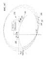
- FIG. 1AA two-aperture system.
- FIG. 1BEquidistant time delay points forming an ellipse around a transmit transducer element and receive transducer element.
- FIG. 1CLoci of points relative to equidistant time delays for different receive transducer elements.
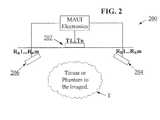
- FIG. 2A three-aperture system.
- FIG. 3Grid for display and coordinate system.
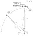
- FIG. 4Fat layer model with a three-aperture system.
- FIG. 5Construction for estimation of point spread function.
- Systems and methods hereinmay provide for both transmit functions from point sources and for compensation for variations in the speed-of-sound of ultrasound pulses traveling through potentially diverse tissue types along a path between a transmit aperture and one or more receive apertures.
- Such speed-of-sound compensationmay be performed by a combination of image comparison techniques (e.g., cross-correlation), and the coherent and/or incoherent averaging of a plurality of received image frames.
- an ultrasound transducermay comprise a piezoelectric device.
- ultrasound transducersmay comprise capacitive micromachined ultrasound transducers (CMUT).
- CMUTcapacitive micromachined ultrasound transducers
- Transducersare often configured in arrays of multiple elements. An element of a transducer array may be the smallest discrete component of an array. For example, in the case of an array of piezoelectric transducer elements, each element may be a single piezoelectric crystal.
- the terms “transmit element” and “receive element”may carry their ordinary meanings as understood by those skilled in the art of ultrasound imaging technologies.
- the term “transmit element”may refer without limitation to an ultrasound transducer element which at least momentarily performs a transmit function in which an electrical signal is converted into an ultrasound signal.
- the term “receive element”may refer without limitation to an ultrasound transducer element which at least momentarily performs a receive function in which an ultrasound signal impinging on the element is converted into an electrical signal. Transmission of ultrasound into a medium may also be referred to herein as “insonifying.” An object or structure which reflects ultrasound waves may be referred to as a “reflector” or a “scatterer.”
- aperturerefers without limitation to one or more ultrasound transducer elements collectively performing a common function at a given instant of time.
- the term aperturemay refer to a group of transducer elements performing a transmit function.
- the term aperturemay refer to a plurality of transducer elements performing a receive function.
- group of transducer elements forming an aperturemay be redefined at different points in time.
- FIG. 3demonstrates multiple apertures used in a multiple aperture ultrasound probe. An aperture of the probe has up to three distinct features. First, it is often physically separated from other transducers located in other apertures. In FIG. 3 , a distance ‘d’ physically separates aperture 302 from aperture 304 .
- Distance ‘d’can be the minimum distance between transducer elements on aperture 302 and transducer elements on aperture 304 . In some embodiments, no transducer elements are disposed along the distance ‘d’ between the physical separation of apertures 302 and 304 . In some embodiments, the distance can be equal to at least twice the minimum wavelength of transmission from the transmit aperture. Second, the transducer elements of an aperture need not be in the same rectangular or horizontal plane. In FIG. 3 , all the elements of aperture 304 have a different vertical position ‘j’ from any element of aperture 302 . Third, apertures do not share a common line of sight to the region of interest. In FIG.
- aperture 302has a line of sight ‘a’ for point (i,j), while aperture 304 has a line of sight ‘b’.
- An aperturemay include any number of individual ultrasound elements. Ultrasound elements defining an aperture are often, but not necessarily adjacent to one another within an array. During operation of a multi-aperture ultrasound imaging system, the size of an aperture (e.g. the number and/or size and/or position of ultrasound elements) may be dynamically changed by re-assigning elements.
- point source transmissionmay refer to an introduction of transmitted ultrasound energy into a medium from single spatial location. This may be accomplished using a single ultrasound transducer element or combination of adjacent transducer elements transmitting together. A single transmission from said element(s) approximates a uniform spherical wave front, or in the case of imaging a 2D slice it creates a uniform circular wave front within the 2D slice.
- This point source transmissiondiffers in its spatial characteristics from a “phased array transmission” which focuses energy in a particular direction from the transducer element array. Phased array transmission manipulates the phase of a group of transducer elements in sequence so as to strengthen or steer an insonifying wave to a specific region of interest.
- a short duration point source transmissionis referred to herein as a “point source pulse.”
- a short duration phased array transmissionis referred to herein as a “phased array pulse.”
- the terms “receive aperture,” “insonifying aperture,” and/or “transmit aperture”can carry their ordinary meanings as understood by those skilled in the art of ultrasound imaging, and may refer to an individual element, a group of elements within an array, or even entire arrays within a common housing, that perform the desired transmit or receive function from a desired physical viewpoint or aperture at a given time.
- these various aperturesmay be created as physically separate components with dedicated functionality.
- the functionalitymay be electronically designated and changed as needed.
- aperture functionalitymay involve a combination of both fixed and variable elements.
- an apertureis an array of ultrasound transducers which is separated from other transducer arrays.
- Such multiple aperture ultrasound imaging systemsprovide greatly increased lateral resolution.
- a multi-aperture imaging methodcomprises the steps of insonifying a target object with an ultrasound pulse from a first aperture, detecting returned echoes with a second aperture positioned at a distance from the first aperture, determining the relative positions of the second aperture with respect to the first aperture, and processing returned echo data to combine images white correcting for variations in speed-of-sound through the target object.
- a distance and orientation between adjacent aperturesmay be fixed relative to one another, such as by use of a rigid housing.
- distances and orientations of apertures relative to one anothermay be variable, such as with a movable linkage.
- aperturesmay be defined as groups of elements on a single large transducer array where the groups are separated by at least a specified distance. For example, some embodiments of such a system are shown and described in U.S. Provisional Patent Application No. 61/392,896, filed Oct. 13, 2010, titled “Multiple Aperture Medical Ultrasound Transducers”.
- a distance between adjacent aperturesmay be at least a width of one transducer element.
- a distance between aperturesmay be as large as possible within the constraints of a particular application and probe design.
- a multi-aperture ultrasound imaging system with a large effective aperture(the total aperture of the several sub apertures) can be made viable by compensation for the variation of speed-of-sound in the target tissue. This may be accomplished in one of several ways to enable the increased aperture to be effective rather than destructive, as described below.
- FIG. 1Aillustrates one embodiment of a simplified multi-aperture ultrasound imaging system 100 comprising two apertures, aperture 102 and aperture 104 .
- Each of apertures 102 and 104can comprise a plurality of transducer elements.
- aperture 102can comprise transmit elements T 1 . . . Tn to be used entirely for transmit functions
- aperture 104can comprise receive elements R 1 . . . . Rm to be used entirely for receive functions.
- transmit elementsmay be interspersed with receive elements, or some elements may be used both for transmit and receive functions.
- FIG. 1Acan be configured to be placed on a skin surface of a patient to image target object or internal tissue T with ultrasound energy. As shown in FIG. 1A , aperture 102 is positioned a distance “a” from tissue T, and aperture 104 is positioned a distance “b” from tissue T. Also shown in FIG. 1A , MAUI electronics may be coupled to the transmit and receive apertures 102 and 104 . In some embodiments, the MAUI electronics can comprise a processor, control system, or computing system, including hardware and software configured to control the multi-aperture imaging system 100 .
- the MAUI electronicscan be configured to control the system to transmit an omni-directional unfocused ultrasound waveform from an aperture, receive echoes on an aperture, and form images from the transmitted waveform and the received echoes.
- the MAUI electronicscan be configured to control and achieve any of the methods described herein.
- Ultrasound elements and arrays described hereinmay also be multi-function. That is, the designation of transducer elements or arrays as transmitters in one instance does not preclude their immediate re-designation as receivers in the next instance. Moreover, embodiments of the control system described herein include the capabilities for making such designations electronically based on user inputs or pre-set scan or resolution criteria.
- FIG. 2Another embodiment of a multi-aperture ultrasound imaging system 200 is shown in FIG. 2 and includes transducer elements arranged to form three apertures 202 , 204 , and 206 .
- transmit elements T 1 . . . Tn in aperture 202may be used for transmit
- receive elements R R 1 . . . R R m in apertures 204 and 206may be used for receive.
- elements in all the aperturesmay be used for both transmit and receive.
- the multi-aperture ultrasound imaging system 200 of FIG. 2can be configured to image tissue T with ultrasound energy.
- MAUI electronicsmay be coupled to the transmit and receive apertures 202 and 204 .
- the MAUI electronicscan comprise a processor, control system, or computing system, including hardware and software configured to control the multi-aperture imaging system 200 .
- the MAUI electronicscan be configured to control the system to transmit an omni-directional unfocused ultrasound waveform from an aperture, receive echoes on an aperture, and form images from the transmitted waveform and the received echoes.
- the MAUI electronicscan be configured to control and achieve any of the methods described herein.
- Multi-aperture ultrasound imaging systems described hereinmay be configured to utilize transducers of any desired construction.
- 1D, 1.5D, 2D, CMUT or any other transducer arraysmay be utilized in multi-aperture configurations to improve overall resolution and field of view.
- acoustic energymay be transmitted to as wide a two-dimensional slice as possible by using point source transmission.
- a transmit aperturesuch as transmit apertures 102 or 202 in FIGS. 1A and 2 , respectively, may transmit acoustic energy in the form of a point source pulse from a single substantially omni-directional transducer element in an array.
- a plurality of transducer elementsmay be provisioned to transmit a point source pulse that is relatively wide in three dimensions to insonify objects in a three dimensional space. In such embodiments, all of the beam formation may be achieved by the software or firmware associated with the transducer arrays acting as receivers.
- Each echo detected at a receive aperturesuch as receive apertures 104 or 204 / 206 in FIGS. 1A and 2 , respectively, may be stored separately. If the echoes detected with elements in a receive aperture are stored separately for every point source pulse from an insonifying or transmit aperture, an entire two-dimensional image can be formed from the information received by as few as just one element. Additional copies of the image may be formed by additional receive apertures collecting data from the same set of insonifying point source pulses. Ultimately, multiple images can be created simultaneously from one or more apertures and combined to achieve a comprehensive 2D or 3D image.
- point source pulsesAlthough several point source pulses are typically used in order to produce a high-quality image, fewer point source pulses are required than if each pulse were focused on a particular scan line. Since the number of pulses that can be transmitted in a given time is strictly limited by the speed of ultrasound in tissue, this yields the practical advantage that more frames can be produced per second by utilizing a point source pulse. This is very important when imaging moving organs, and in particular, the heart.
- a spread spectrum waveformmay be imposed on a transmit aperture made up of one or more ultrasound transducer elements.
- a spread spectrum waveformmay be a sequence of frequencies such as a chirp (e.g., frequencies progressing from low to high, or vice versa), random frequency sequence (also referred to as frequency hop), or a signal generated by a pseudo random waveform (PN sequence).
- PN sequencepseudo random waveform
- FIG. 1Aillustrates one embodiment of a multi-aperture ultrasound imaging system 100 containing a first aperture 102 with ultrasound transmitting elements T 1 , T 2 , . . . Tn and a second aperture 104 with ultrasound receive elements R 1 , R 2 , . . . Rm.
- This multi-aperture ultrasound imaging system 100is configured to be placed on the surface of an object or body to be examined (such as a human body).
- both aperturesmay be sensitive to the same plane of scan.
- one of the aperturesmay be in a different plane of scan.
- the mechanical and acoustic position of each transducer element of each aperturemust be known precisely relative to a common reference point or to each other.
- an ultrasound imagemay be produced by insonifying the entire region to be imaged, such as internal tissue or target object T, (e.g., a plane through the heart, organ, tumor, or other portion of the body) with a transmitting element (e.g., transmit element T 1 of aperture 102 ), and then receiving echoes from the entire imaged plane on a receive element (e.g. receive element R 1 of aperture 104 ).
- receive functionsmay be performed by all elements in the receive probe (e.g., R 1 through Rm).
- echoesare received on only one or a select few elements of the receive aperture. The method proceeds by using each of the elements on the transmitting aperture 102 (e.g., T 2 , . .
- Transmit elementsmay be operated in any desired sequential order, and need not follow a prescribed pattern. Individually, the images obtained after insonification by each transmitting element may not be sufficient to provide a high resolution image, but the combination of all the images may provide a high resolution image of the entire region to be imaged.
- a scanning point represented by coordinates (i,j) as shown in FIG. 1Ait is a simple matter to calculate the total distance “a” from a particular transmit element Tx to an element of internal tissue or target object at (i,j), and the distance “b” from that point to a particular receive element. These calculations may be performed using basic trigonometry. The sum of these distances is the total distance traveled by one ultrasound wave.
- FIG. 1Bdemonstrates that points (g,h), (i,j), (k,m), (n,p) (q,r), (s,t) all have the same time delay for transmit element T 1 and receive element R 1 .
- FIG. 1Balso illustrates MAUI electronics, which can comprise the MAUI electronics described above with reference to FIGS. 1A and 2 .
- FIG. 1Cshows that with a transmit pulse from element T 1 , echoes from a single scatterer (n,p) are received by different receive elements such as R 1 , R 2 , and R 3 at different times.
- the loci of the same scatterercan be represented by ellipses 180 , 185 and 190 of FIG. 1C .
- the location at which these ellipses intersect (point n,p)represents the true location of the scatterer.
- Beam forming hardware, firmware, or softwarecan combine the echoes from each receive element to generate an image, effectively reinforcing the image at the intersection of the ellipses.
- many more receiver elements than the three shownmay be used in order to obtain a desirable signal-to-noise ratio for the image.
- FIG. 1Calso illustrates MAUI electronics, which can comprise the MAUI electronics described above with reference to FIGS. 1A and 2 .
- FIG. 3illustrates a grid of points to be imaged by apertures 302 and 304 .
- a point on the gridis given the rectangular coordinates (i,j).
- the complete imagewill be a two dimensional array called “echo.”
- mhis the maximum horizontal dimension of the array
- mvis the maximum vertical dimension.
- FIG. 3also illustrates MAUI electronics, which can comprise the MAUI electronics described above with reference to FIGS. 1A and 2 .
- the following pseudo codemay be used to accumulate all of the information to be gathered from a transmit pulse from one transmit element (e.g., one element of T 1 . . . Tn from aperture 302 ), and the consequent echoes received by one receive element (e.g., one element of R 1 . . . Rm from aperture 304 ) in the arrangement of FIG. 3 .
- one transmit elemente.g., one element of T 1 . . . Tn from aperture 302
- the consequent echoes received by one receive elemente.g., one element of R 1 . . . Rm from aperture 304
- the fixed delayis primarily the time from the transmit pulse until the first echoes are received. As will be discussed later, an increment can be added or subtracted to compensate for varying fat layers.
- a complete two dimensional imagemay be formed by repeating this process for every receive element in aperture 304 (e.g., R 1 . . . Rm).
- receive element in aperture 304e.g., R 1 . . . Rm.
- this codeit is possible to implement this code in parallel hardware resulting in real time image formation.
- the combination of imagesmay be performed by a simple summation of the single point source pulse images (e.g., coherent addition).
- the combinationmay involve taking the absolute value of each element of the single point source pulse images first before summation (e.g., incoherent addition).
- the first technique (coherent addition)may be best used for improving lateral resolution
- the second technique (incoherent addition)may be best applied for the reduction of speckle noise.
- the incoherent techniquemay be used with less precision required in the measurement of the relative positions of the transmit and receive apertures.
- a combination of both techniquesmay be used to provide an optimum balance of improved lateral resolution and reduced speckle noise.
- the final sumshould be replaced by the absolute value of each element, and in both cases, some form of compression of the dynamic range may be used so that both prominent features and more-subtle features appear on the same display.
- additional pixel locationsare located on a grid without scan-conversion.
- compression schemesmay include taking the logarithm (e.g., 20 log 10 or “dB”) of each element before display, or taking the nth root (e.g., 4 th root) of each element before display. Other compression schemes may also be employed.
- any number of receive probes and transmit probesmay be combined to enhance the image of scatterer (i,j) as long as the relative positions of the transducer elements are known to a designed degree of precision, and all of the elements are in the same scan plane and are focused to either transmit energy into the scan plane or receive energy propagated in the scan plane.
- Any element in any probemay be used for either transmit or receive or both.
- the speed-of-sound in various soft tissues throughout the bodycan vary by +/ ⁇ 10%.
- the speed-of-soundis constant in the path between the transducer and the organ of interest. This assumption is valid for narrow transducer arrays in systems using one transducer array for both transmit and receive.
- the constant speed-of-sound assumptionbreaks down as the transducer's aperture becomes wider because the ultrasound pulses pass through more tissue and possibly diverse types of tissue, such as fat, muscle, blood vessels, etc. Tissue diversity under the width of the transducer array affects both the transmit and the receive functions.
- a scattererWhen a scatterer is insonified by a point source pulse from a single transmit element, it reflects back an echo to all of the elements of the receiver group. Coherent addition of images collected by elements in this receive aperture can be effective if the speed-of-sound variations in the paths from scatterer (i,j) to each of the receiver elements do not exceed + ⁇ 180 degrees phase shift relative to one path chosen as reference.
- the maximum size of the receive aperture for which coherent addition can be effectiveis dependent on tissue variation within the patient and cannot be computed in advance. However, a practical maximum for a particular transmit frequency can be determined from experience.
- the aperture size of the transmit groupis not highly critical since variation in the path time from transmitter elements to a scatterer such as scatterer (i,j) will change only the displayed position of the point. For example, a variation resulting in a phase shift of 180 degrees in the receive paths results in complete phase cancellation when using coherent addition, whereas the same variation on the transmit paths results in a displayed position error of only a half wavelength (typically about 0.2 mm), a distortion that would not be noticed.
- Substantial improvement in lateral resolutionis achieved with a receive aperture of the same width as a conventional single array 1D, 1.5D or 2D ultrasound probe used for both transmit and receive, because received energy when imaging adjacent cells (i.e., regions of the target object) to that which represents a scatterer is dependent on the time difference between when an echo is expected to arrive and the time that it actually arrives.
- the transmit pulseoriginates from the same array used for receive, the time difference is small.
- the transmit pulseoriginates from a second array at some distance from the receive array, the time difference is larger and therefore more out of phase with the signal for the correct cell. The result is that fewer adjacent cells will have signals sufficiently in phase to falsely represent the true scatterer.
- FIG. 4also illustrates MAUI electronics, which can comprise the MAUI electronics described above.
- a single imagemay be formed by coherent averaging of all of the signals arriving at the receiver elements as a result of a single point source pulse for insonification. Summation of these images resulting from multiple point source pulses can be accomplished either by coherent addition, incoherent addition, or a combination of coherent addition by groups and incoherent addition of the images from the groups. Coherent addition (retaining the phase information before addition) maximizes resolution whereas incoherent addition (using the magnitude of the signals and not the phase) minimizes the effects of registration errors and averages out speckle noise. Some combination of the two modes may be preferred. Coherent addition can be used to average point source pulse images resulting from transmit elements that are close together and therefore producing pulses transmitted through very similar tissue layers. Incoherent addition can then be used where phase cancellation would be a problem. In the extreme case of transmission time variation due to speed-of-sound variations, 2D image correlation can be used to align images prior to addition.
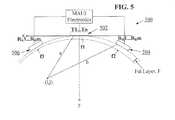
- tissue layer model for the three-aperture ultrasound imaging system 500as shown in FIG. 5 , which illustrates the effects of varying thicknesses of different types of tissue, such as fat or muscle.
- a fat layer “F”is shown in FIG. 5 , and the thickness of the tissue layers f 1 , f 2 , and f 3 under each aperture 502 , 504 , and 506 , respectively, is different and unknown. It is not reasonable to assume that the tissue layer at aperture 506 will be the same as at aperture 504 , and so coherent addition of the signals from all of the receive elements together is not usually possible.
- tissue layer at aperture 504were as much as 3 cm larger than that at aperture 506 , this corresponds to about 3 wavelengths (at 3.5 MHz) displacement of the signals, but this is only 1.3 mm displacement of the representation of the deep tissues. For such small displacements, only a tiny amount of geometric distortion of the image would be observed. Therefore, although coherent addition is not possible, incoherent addition with displacement of one image relative to the other is possible.
- Image comparison techniquesmay be used to determine the amount of displacement needed to align image frames from left and right apertures (e.g., apertures 506 and 504 , respectively).
- the image comparison techniquecan be cross-correlation.
- Cross-correlationinvolves evaluating the similarity of images or image sections to identify areas with a high degree of similarity. Areas with at least a threshold value of similarity may be assumed to be the same. Thus, by identifying areas within images with high degrees of similarity, one image (or a section thereof) may be shifted such that areas with substantial similarity overlap and enhance overall image quality.
- FIG. 5also illustrates MAUI electronics, which can comprise the MAUI electronics described above.
- these image comparison techniquescan also be used by applying sub-image analysis, which can be used to determine displacement of sub-images and accommodate for localized variation in speed-of-sound in the underlying tissue.
- sub-image analysiscan be used to determine displacement of sub-images and accommodate for localized variation in speed-of-sound in the underlying tissue.
- by breaking down the images into smaller segmentse.g. halves, thirds, quarters, etc
- small portions of a first imagemay be compared to the corresponding small portion of a second image.
- the two imagesmay then be combined by warping to assure alignment. Warping is a technique understood by those skilled in the art, and is described, for example in U.S. Pat. No. 7,269,299 to Schroeder.
- the same technique of incoherent addition of images from multiple receive transducer arraysmay be applied to any number of apertures.
- the same ideamay be applied even to a single element array which is too wide to be used for coherent addition all at once.
- An ultrasound imaging system with a single wide array of elementsmay be divided into sections (apertures) each of which is small enough for coherent addition, and then the images resulting from these sections may be combined incoherently (with displacement if necessary).
- a faster techniqueincludes calculating the cross correlation network for the uncorrected pair of images, and feeding this into a neural network trained to pick the correction delay.
- some embodiments of the multi-aperture ultrasound systems described hereincan have apertures located 10 cm apart from one another. Since resolution is proportional to 2 ⁇ /D, this larger aperture leads to higher resolution of tissues located well below the surface of the skin. For instance, the renal arteries are frequently located 10 cm to 15 cm below the skin and are 4 mm to 6 mm in size near the abdominal aorta. Phased array, linear array and synthetic aperture ultrasound systems usually cannot detect this physiology in most patients; specifically because the aperture size is not large enough to have adequate lateral resolution. Typically, phased array systems have aperture sizes of approximately 2 cm. Increasing the aperture size from larger than 2 cm to approximately 10 cm in a multi-aperture ultrasound system can increase the resolution by up to 5 ⁇ .
- three-dimensional informationmay be obtained by moving a two-dimensional imaging system and acquiring 2D slices at a number of positions or angles. From this information and using interpolation techniques, a 3D image at any position or angle may be reconstructed. Alternatively, a 2D projection of all of the data in the 3D volume may be produced. A third alternative is to use the information in a direct 3D display.
- multi-aperture ultrasound imaging systemsmay result in wider probe devices, the easiest way to use them to obtain 3D data is to not move them on the patient's skin but merely rock them so that the 2D slices span the 3D volume to be imaged.
- a mechanical rotator mechanismwhich records position data may be used to assist in the collection the 2D slices.
- a freely operated ultrasound probe with precision position sensorssuch as gyroscopic sensors located in the head of the probe may be used instead.
- Such an apparatusallows for complete freedom of movement while collecting 2D slices.
- intravenous and intracavity probesmay also be manufactured to accommodate wide apertures. Such probes may be manipulated in similar ways in order to collect 2D slices.
- a multi-aperture imaging systemis ideal in this case since the intervening rib would render a flat probe useless, while a probe with at least two widely spaced apertures can be positioned such that a send aperture and a receive aperture align with separate intercostal spaces. Once a probe with multiple apertures is in place, it cannot be rotated, but it can be rocked to obtain the 3D information, A multi-aperture probe may also be used in the same intercostal space but across the sternum.
- 3D informationmay also be obtained directly with multi-aperture imaging systems having apertures that are not all in the same scan plane.
- the elements making up the transmit aperturepreferably propagate spherical waveforms (rather than circular waveforms confined to one plane of scan).
- the elements making up the receive aperturesmay likewise be sensitive to energy arriving from all directions (rather than being sensitive only to ultrasonic energy in a single plane of scan).
- the reconstruction pseudo code provided abovemay then be extended to three dimensions.
Landscapes
- Engineering & Computer Science (AREA)
- Physics & Mathematics (AREA)
- Health & Medical Sciences (AREA)
- Life Sciences & Earth Sciences (AREA)
- Remote Sensing (AREA)
- Radar, Positioning & Navigation (AREA)
- Acoustics & Sound (AREA)
- Surgery (AREA)
- Veterinary Medicine (AREA)
- Heart & Thoracic Surgery (AREA)
- Medical Informatics (AREA)
- Molecular Biology (AREA)
- Radiology & Medical Imaging (AREA)
- Animal Behavior & Ethology (AREA)
- General Health & Medical Sciences (AREA)
- Public Health (AREA)
- Biomedical Technology (AREA)
- Biophysics (AREA)
- Nuclear Medicine, Radiotherapy & Molecular Imaging (AREA)
- Pathology (AREA)
- Computer Networks & Wireless Communication (AREA)
- General Physics & Mathematics (AREA)
- Computer Vision & Pattern Recognition (AREA)
- Gynecology & Obstetrics (AREA)
- Ultra Sonic Daignosis Equipment (AREA)
Abstract
Description
This application claims the benefit of U.S. Provisional Patent Application No. 61/305,784, filed on Feb. 18, 2010, titled “Alternative Method for Medical Multi-Aperture Ultrasound Imaging”.
This application is also related to U.S. patent application Ser. No. 11/865,501, filed Oct. 1, 2007, titled “Method and Apparatus to Produce Ultrasonic Images Using Multiple Apertures”, and to U.S. patent application Ser. No. 11/532,013, filed Sep. 14, 2006, titled “Method and Apparatus to Visualize the Coronary Arteries Using Ultrasound”; all of which are herein incorporated by reference in their entirety.
All publications, including patents and patent applications, mentioned in this specification are herein incorporated by reference in their entirety to the same extent as if each individual publication was specifically and individually indicated to be incorporated by reference.
In conventional ultrasonic imaging, a focused beam of ultrasound energy is transmitted into body tissues to be examined and the returned echoes are detected and plotted to form an image. The basic principles of conventional ultrasonic imaging are well described in the first chapter of “Echocardiography,” by Harvey Feigenbaum (Lippincott Williams & Wilkins, 5th ed., Philadelphia, 1993).
In order to insonify body tissues, an ultrasound beam is typically formed and focused either by a phased array or a shaped transducer. Phased array ultrasound is a commonly used method of steering and focusing a narrow ultrasound beam for forming images in medical ultrasonography. A phased array probe has many small ultrasonic transducer elements, each of which can be pulsed individually. By varying the timing of ultrasound pulses (e.g. by pulsing elements one by one in sequence along a row), a pattern of constructive interference is set up that results in a beam directed at a chosen angle. This is known as beam steering. Such a steered ultrasound beam may then be swept through the tissue or object being examined. Data from multiple beams are then combined to make a visual image showing a slice through the object.
Traditionally, the same transducer or array used for transmitting an ultrasound beam is used to detect the returning echoes. This design configuration lies at the heart of one of the most significant limitations in the use of ultrasonic imaging for medical purposes: poor lateral resolution. Theoretically, the lateral resolution could be improved by increasing the width of the aperture of an ultrasonic probe, but practical problems involved with aperture size increase have kept apertures small. Unquestionably, ultrasonic imaging has been very useful even with this limitation, but it could be more effective with better resolution.
In the practice of cardiology, for example, the limitation on single aperture size is dictated by the space between the ribs (the intercostal spaces). Such intercostal apertures are typically limited to no more than about one to two centimeters. For scanners intended for abdominal and other use, the limitation on aperture size is less a matter of physical constraints, and more a matter of difficulties in image processing. The problem is that it is difficult to keep the elements of a large aperture array in phase because the speed of ultrasound transmission varies with the type of tissue between the probe and the area of interest. According to the book by Wells (cited above), the speed varies up to plus or minus 10% within the soft tissues. When the aperture is kept small (e.g. less than about 2 cm), the intervening tissue is, to a first order of approximation, all the same and any variation is ignored. When the size of the aperture is increased to improve the lateral resolution, the additional elements of a phased array may be out of phase and may actually degrade the image rather than improving it.
US Patent Application Publication 2008/0103393 to Specht teaches embodiments of ultrasound imaging systems utilizing multiple apertures which may be separated by greater distances, thereby producing significant improvements in lateral resolution of ultrasound images.
One embodiment of a method describes a method of constructing an ultrasound image, comprising transmitting an omni-directional unfocused ultrasound waveform approximating a first point source within a transmit aperture on a first array through a target region, receiving ultrasound echoes from the target region with first and second receiving elements disposed on a first receive aperture on a second array, the first array being physically separated from the second array, determining a first time for the waveform to propagate from the first point source to a first pixel location in the target region to the first receiving element, and determining a second time for the waveform to propagate from the first point source to the first pixel location in the target region to the second receiving element, and forming a first ultrasound image of the first pixel by combining the echo received by the first receiving element at the first time with the echo received by the second receiving element at the second time.
In some embodiments, the method further comprises repeating the determining and forming steps for additional pixel locations in the target region. In one embodiment, additional pixel locations are located on a grid without scan-conversion.
In one embodiment, determining the first time and the second time comprises assuming a uniform speed of sound.
In another embodiment, the method further comprises transmitting a second omni-directional unfocused ultrasound waveform approximating a second point source within the transmit aperture through the target region, receiving ultrasound echoes from the target region with first and second receiving elements disposed on the first receive aperture, determining a third time for the second waveform to propagate from the second point source to the first pixel location in the target region to the first receiving element, and determining a fourth time for the second waveform to propagate from the second point source to the first pixel location in the target region to the second receiving element, and forming a second ultrasound image of the first pixel by combining the echo received by the first receiving element at the third time with the echo received by the second receiving element at the fourth time.
In some embodiments, the method further comprises combining the first ultrasound image with the second ultrasound image. The combining step can comprise coherent addition. In another embodiment, the combining step can comprise incoherent addition. In yet another embodiment, the combining step can comprise a combination of coherent addition and incoherent addition.
In some embodiments, the method can further comprise receiving ultrasound echoes from the target region with third and fourth receiving elements disposed on a second receive aperture on a third array, the third array being physically separated from the first and second arrays, determining a third time for the waveform to propagate from the first point source to the first pixel location in the target region to the third receiving element, and determining a fourth time for the waveform to propagate from the first point source to the first pixel location in the target region to the fourth receiving element, and forming a second ultrasound image of the first pixel by combining the echo received by the third receiving element at the third time with the echo received by the fourth receiving element at the fourth time.
In some embodiments, the method further comprises repeating the determining and forming steps for additional pixel locations in the target region. In some embodiments, the additional pixel locations are located on a grid without scan-conversion.
In one embodiment, the method further comprises transmitting a second omni-directional unfocused ultrasound waveform approximating a second point source within the transmit aperture through the target region, receiving ultrasound echoes from the target region with first and second receiving elements disposed on the first receive aperture and with the third and fourth receiving elements disposed on the second receive aperture, determining a fifth time for the second waveform to propagate from the second point source to the first pixel location in the target region to the first receiving element, determining a sixth time for the second waveform to propagate from the second point source to the first pixel location in the target region to the second receiving element, determining a seventh time for the second waveform to propagate from the second point source to the first pixel location in the target region to the third receiving element, determining an eighth time for the second waveform to propagate from the second point source to the first pixel location in the target region to the fourth receiving element, and forming a third ultrasound image of the first pixel by combining the echo received by the first receiving element at the fifth time with the echo received by the second receiving element at the sixth time, and forming a fourth ultrasound image of the first pixel by combining the echo received by the third receiving element at the seventh time with the echo received by the fourth receiving element at the eighth time.
In some embodiments, the method further comprises combining the first, second, third, and fourth ultrasound images. In some embodiments, the combining step comprises coherent addition. In other embodiments, the combining step comprises incoherent addition. In additional embodiments, the combining step comprises a combination of coherent addition and incoherent addition.
In some embodiments, the method comprises combining the first ultrasound image with the second ultrasound image. The combining step can comprise coherent addition. In another embodiment, the combining step can comprise incoherent addition. In yet another embodiment, the combining step can comprise a combination of coherent addition and incoherent addition.
In some embodiments, the method further comprises comparing the first ultrasound image to the second, third, and fourth ultrasound images to determine displacements of the second, third, and fourth ultrasound images relative to the first ultrasound image.
In another embodiment, the method further comprises correcting the displacements of the second, third, and fourth ultrasound images relative to the first ultrasound image and then combining the first, second, third and fourth ultrasound images.
In an additional embodiment, the method comprises adjusting the third, fourth, fifth, sixth, seventh, and eighth times to correct the displacements of the second, third, and fourth ultrasound images relative to the first ultrasound image.
In some embodiments, the method further comprises comparing the first ultrasound image to the second ultrasound image to determine a displacement of the second ultrasound image relative to the first ultrasound image.
The method can further comprise correcting the displacement of the second ultrasound image relative to the first ultrasound image and then combining the first and second ultrasound images.
In another embodiment, the method comprises adjusting the third time and the fourth time to correct the displacement of the second ultrasound image relative to the first ultrasound image.
In some embodiments, the first pixel is disposed outside a plane defined by the point source, the first receiving element, and the second receiving element. In other embodiments, the first pixel is disposed inside a plane defined by the point source, the first receiving element, and the second receiving element.
Various embodiments of a multi-aperture ultrasound imaging system are also provided, comprising a transmit aperture on a first array configured to transmit an omni-directional unfocused ultrasound waveform approximating a first point source through a target region, a first receive aperture on a second array having first and second receiving elements, the second array being physically separated from the first array, wherein the first and second receiving elements are configured to receive ultrasound echoes from the target region, and a control system coupled to the transmit aperture and the first receive aperture, the control system configured to determine a first time for the waveform to propagate from the first point source to a first pixel location in the target region to the first receiving element, and is configured to determine a second time for the waveform to propagate from the first point source to the first pixel location in the target region to the second receiving element, the control system also being configured to form a first ultrasound image of the first pixel by combining the echo received by the first receiving element at the first time with the echo received by the second receiving element at the second time.
In some embodiments of the system, there are no transducer elements disposed between the physical separation of the transmit aperture and the first receive aperture.
In one embodiment of the system, the transmit aperture and the first receive aperture are separated by at least twice a minimum wavelength of transmission from the transmit aperture. In another embodiment, the transmit aperture and the receive aperture comprise a total aperture ranging from 2 cm to 10 cm.
In some embodiments, the ultrasound system further comprises a second receive aperture on a third array having third and fourth receiving elements, the third array being physically separated from the first and second arrays, wherein the third and fourth receiving elements are configured to receive ultrasound echoes from the target region.
In another embodiment of the multi-aperture ultrasound imaging system, the control system can be coupled to the transmit aperture and the first and second receive apertures, wherein the control system is configured to determine a third time for the waveform to propagate from the first point source to a first pixel location in the target region to the third receiving element, and is configured to determine a fourth time for the waveform to propagate from the first point source to the first pixel location in the target region to the fourth receiving element, the control system also being configured to form a second ultrasound image of the first pixel by combining the echo received by the third receiving element at the third time with the echo received by the fourth receiving element at the fourth time.
In some embodiments, the control system is configured to correct a displacement of the second ultrasound image relative to the first ultrasound image due to speed of sound variation.
In other embodiments of the multi-aperture ultrasound imaging system, the transmit aperture, the first receive aperture, and the second receive aperture are not all in a single scan plane.
Greatly improved lateral resolution in ultrasound imaging can be achieved by using multiple separate apertures for transmit and receive functions. Systems and methods herein may provide for both transmit functions from point sources and for compensation for variations in the speed-of-sound of ultrasound pulses traveling through potentially diverse tissue types along a path between a transmit aperture and one or more receive apertures. Such speed-of-sound compensation may be performed by a combination of image comparison techniques (e.g., cross-correlation), and the coherent and/or incoherent averaging of a plurality of received image frames.
As used herein the terms “ultrasound transducer” and “transducer” may carry their ordinary meanings as understood by those skilled in the art of ultrasound imaging technologies, and may refer without limitation to any single component capable of converting an electrical signal into an ultrasonic signal and/or vice versa. For example, in some embodiments, an ultrasound transducer may comprise a piezoelectric device. In some alternative embodiments, ultrasound transducers may comprise capacitive micromachined ultrasound transducers (CMUT). Transducers are often configured in arrays of multiple elements. An element of a transducer array may be the smallest discrete component of an array. For example, in the case of an array of piezoelectric transducer elements, each element may be a single piezoelectric crystal.
As used herein, the terms “transmit element” and “receive element” may carry their ordinary meanings as understood by those skilled in the art of ultrasound imaging technologies. The term “transmit element” may refer without limitation to an ultrasound transducer element which at least momentarily performs a transmit function in which an electrical signal is converted into an ultrasound signal. Similarly, the term “receive element” may refer without limitation to an ultrasound transducer element which at least momentarily performs a receive function in which an ultrasound signal impinging on the element is converted into an electrical signal. Transmission of ultrasound into a medium may also be referred to herein as “insonifying.” An object or structure which reflects ultrasound waves may be referred to as a “reflector” or a “scatterer.”
As used herein the term “aperture” refers without limitation to one or more ultrasound transducer elements collectively performing a common function at a given instant of time. For example, in some embodiments, the term aperture may refer to a group of transducer elements performing a transmit function. In alternative embodiments, the term aperture may refer to a plurality of transducer elements performing a receive function. In some embodiments, group of transducer elements forming an aperture may be redefined at different points in time.FIG. 3 demonstrates multiple apertures used in a multiple aperture ultrasound probe. An aperture of the probe has up to three distinct features. First, it is often physically separated from other transducers located in other apertures. InFIG. 3 , a distance ‘d’ physically separatesaperture 302 fromaperture 304. Distance ‘d’ can be the minimum distance between transducer elements onaperture 302 and transducer elements onaperture 304. In some embodiments, no transducer elements are disposed along the distance ‘d’ between the physical separation ofapertures FIG. 3 , all the elements ofaperture 304 have a different vertical position ‘j’ from any element ofaperture 302. Third, apertures do not share a common line of sight to the region of interest. InFIG. 3 ,aperture 302 has a line of sight ‘a’ for point (i,j), whileaperture 304 has a line of sight ‘b’. An aperture may include any number of individual ultrasound elements. Ultrasound elements defining an aperture are often, but not necessarily adjacent to one another within an array. During operation of a multi-aperture ultrasound imaging system, the size of an aperture (e.g. the number and/or size and/or position of ultrasound elements) may be dynamically changed by re-assigning elements.
As used herein the term “point source transmission” may refer to an introduction of transmitted ultrasound energy into a medium from single spatial location. This may be accomplished using a single ultrasound transducer element or combination of adjacent transducer elements transmitting together. A single transmission from said element(s) approximates a uniform spherical wave front, or in the case of imaging a 2D slice it creates a uniform circular wave front within the 2D slice. This point source transmission differs in its spatial characteristics from a “phased array transmission” which focuses energy in a particular direction from the transducer element array. Phased array transmission manipulates the phase of a group of transducer elements in sequence so as to strengthen or steer an insonifying wave to a specific region of interest. A short duration point source transmission is referred to herein as a “point source pulse.” Likewise, a short duration phased array transmission is referred to herein as a “phased array pulse.”
As used herein, the terms “receive aperture,” “insonifying aperture,” and/or “transmit aperture” can carry their ordinary meanings as understood by those skilled in the art of ultrasound imaging, and may refer to an individual element, a group of elements within an array, or even entire arrays within a common housing, that perform the desired transmit or receive function from a desired physical viewpoint or aperture at a given time. In some embodiments, these various apertures may be created as physically separate components with dedicated functionality. In alternative embodiments, the functionality may be electronically designated and changed as needed. In still further embodiments, aperture functionality may involve a combination of both fixed and variable elements.
In some embodiments, an aperture is an array of ultrasound transducers which is separated from other transducer arrays. Such multiple aperture ultrasound imaging systems provide greatly increased lateral resolution. According to some embodiments, a multi-aperture imaging method comprises the steps of insonifying a target object with an ultrasound pulse from a first aperture, detecting returned echoes with a second aperture positioned at a distance from the first aperture, determining the relative positions of the second aperture with respect to the first aperture, and processing returned echo data to combine images white correcting for variations in speed-of-sound through the target object.
In some embodiments, a distance and orientation between adjacent apertures may be fixed relative to one another, such as by use of a rigid housing. In alternative embodiments, distances and orientations of apertures relative to one another may be variable, such as with a movable linkage. In further alternative embodiments, apertures may be defined as groups of elements on a single large transducer array where the groups are separated by at least a specified distance. For example, some embodiments of such a system are shown and described in U.S. Provisional Patent Application No. 61/392,896, filed Oct. 13, 2010, titled “Multiple Aperture Medical Ultrasound Transducers”. In some embodiments of a multi-aperture ultrasound imaging system, a distance between adjacent apertures may be at least a width of one transducer element. In alternative embodiments, a distance between apertures may be as large as possible within the constraints of a particular application and probe design.
A multi-aperture ultrasound imaging system with a large effective aperture (the total aperture of the several sub apertures) can be made viable by compensation for the variation of speed-of-sound in the target tissue. This may be accomplished in one of several ways to enable the increased aperture to be effective rather than destructive, as described below.
Ultrasound elements and arrays described herein may also be multi-function. That is, the designation of transducer elements or arrays as transmitters in one instance does not preclude their immediate re-designation as receivers in the next instance. Moreover, embodiments of the control system described herein include the capabilities for making such designations electronically based on user inputs or pre-set scan or resolution criteria.
Another embodiment of a multi-apertureultrasound imaging system 200 is shown inFIG. 2 and includes transducer elements arranged to form threeapertures aperture 202 may be used for transmit, and receive elements RR1 . . . RRm inapertures ultrasound imaging system 200 ofFIG. 2 can be configured to image tissue T with ultrasound energy. Also shown inFIG. 2 , MAUI electronics may be coupled to the transmit and receiveapertures multi-aperture imaging system 200. In some embodiments, the MAUI electronics can be configured to control the system to transmit an omni-directional unfocused ultrasound waveform from an aperture, receive echoes on an aperture, and form images from the transmitted waveform and the received echoes. As will be described in further detail below, the MAUI electronics can be configured to control and achieve any of the methods described herein.
Multi-aperture ultrasound imaging systems described herein may be configured to utilize transducers of any desired construction. For example, 1D, 1.5D, 2D, CMUT or any other transducer arrays may be utilized in multi-aperture configurations to improve overall resolution and field of view.
Point Source Transmission
In some embodiments, acoustic energy may be transmitted to as wide a two-dimensional slice as possible by using point source transmission. For example, in some embodiments, a transmit aperture, such as transmitapertures FIGS. 1A and 2 , respectively, may transmit acoustic energy in the form of a point source pulse from a single substantially omni-directional transducer element in an array. In alternative embodiments, a plurality of transducer elements may be provisioned to transmit a point source pulse that is relatively wide in three dimensions to insonify objects in a three dimensional space. In such embodiments, all of the beam formation may be achieved by the software or firmware associated with the transducer arrays acting as receivers. There are several advantages to using a multi-aperture ultrasound imaging technique by transmitting with a point source pulse rather than a phased array pulse. For example when using a phased array pulse, focusing tightly on transmit is problematic because the transmit pulse would have to be focused at a particular depth and would be somewhat out of focus at all other depths. Whereas, with a point source transmission an entire two-dimensional slice or three-dimensional volume can be insonified with a single point source transmit pulse.
Each echo detected at a receive aperture, such as receiveapertures FIGS. 1A and 2 , respectively, may be stored separately. If the echoes detected with elements in a receive aperture are stored separately for every point source pulse from an insonifying or transmit aperture, an entire two-dimensional image can be formed from the information received by as few as just one element. Additional copies of the image may be formed by additional receive apertures collecting data from the same set of insonifying point source pulses. Ultimately, multiple images can be created simultaneously from one or more apertures and combined to achieve a comprehensive 2D or 3D image.
Although several point source pulses are typically used in order to produce a high-quality image, fewer point source pulses are required than if each pulse were focused on a particular scan line. Since the number of pulses that can be transmitted in a given time is strictly limited by the speed of ultrasound in tissue, this yields the practical advantage that more frames can be produced per second by utilizing a point source pulse. This is very important when imaging moving organs, and in particular, the heart.
In some embodiments, a spread spectrum waveform may be imposed on a transmit aperture made up of one or more ultrasound transducer elements. A spread spectrum waveform may be a sequence of frequencies such as a chirp (e.g., frequencies progressing from low to high, or vice versa), random frequency sequence (also referred to as frequency hop), or a signal generated by a pseudo random waveform (PN sequence). These techniques can be collectively referred to as pulse compression. Pulse compression provides longer pulses for greater depth penetration without loss of depth resolution. In fact, the depth resolution may be greatly improved in the process. Spread spectrum processing typically involves much more signal processing in the form of matched filtering of each of the received signals before the delay and summation steps. The above examples of transmit pulse forms are provided for illustration only. The techniques taught herein may apply regardless of the form of the transmit pulse.
Basic Image Rendering
In one embodiment, an ultrasound image may be produced by insonifying the entire region to be imaged, such as internal tissue or target object T, (e.g., a plane through the heart, organ, tumor, or other portion of the body) with a transmitting element (e.g., transmit element T1 of aperture102), and then receiving echoes from the entire imaged plane on a receive element (e.g. receive element R1 of aperture104). In some embodiments, receive functions may be performed by all elements in the receive probe (e.g., R1 through Rm). In alternative embodiments, echoes are received on only one or a select few elements of the receive aperture. The method proceeds by using each of the elements on the transmitting aperture102 (e.g., T2, . . . Tn) and insonifying the entire region to be imaged with each of the transmitting elements in turn, and receiving echoes on the receive aperture after each insonifying pulse. Transmit elements may be operated in any desired sequential order, and need not follow a prescribed pattern. Individually, the images obtained after insonification by each transmitting element may not be sufficient to provide a high resolution image, but the combination of all the images may provide a high resolution image of the entire region to be imaged. For a scanning point represented by coordinates (i,j) as shown inFIG. 1A , it is a simple matter to calculate the total distance “a” from a particular transmit element Tx to an element of internal tissue or target object at (i,j), and the distance “b” from that point to a particular receive element. These calculations may be performed using basic trigonometry. The sum of these distances is the total distance traveled by one ultrasound wave.
When the speed of ultrasound in tissue is assumed to be uniform throughout the tissue, it is possible to calculate the time delay from the onset of the transmit pulse to the time that an echo is received at the receive element. (Non uniform speed-of-sound in tissue is discussed below.) This one fact means that a scatterer (i.e., a reflective point within the target object) is a point in the medium for which a+b=the given time delay. The same method can be used to calculate delays for any point in the desired tissue to be imaged, creating a locus of points.FIG. 1B demonstrates that points (g,h), (i,j), (k,m), (n,p) (q,r), (s,t) all have the same time delay for transmit element T1and receive element R1. A map of scatter positions and amplitudes can be rendered by tracing the echo amplitude to all of the points for the locus of equal-time-delay points. This locus takes the form of anellipse 180 with foci at the transmit and receive elements.FIG. 1B also illustrates MAUI electronics, which can comprise the MAUI electronics described above with reference toFIGS. 1A and 2 .
The fact that all points on theellipse 180 are returned with the same time delay presents a display challenge, since distinguishing points along the ellipse from one another within a single image is not possible. However, by combining images obtained from multiple receive points, the points may be more easily distinguished, since the equal-time-delay ellipses defined by the multiple receive apertures will be slightly different.
A method of rendering the location of all of the scatterers in the target object, and thus forming a two dimensional cross section of the target object, will now be described with reference to multi-apertureultrasound imaging system 300 ofFIG. 3 .FIG. 3 illustrates a grid of points to be imaged byapertures FIG. 3 , mh is the maximum horizontal dimension of the array and mv is the maximum vertical dimension.FIG. 3 also illustrates MAUI electronics, which can comprise the MAUI electronics described above with reference toFIGS. 1A and 2 .
In one embodiment, the following pseudo code may be used to accumulate all of the information to be gathered from a transmit pulse from one transmit element (e.g., one element of T1 . . . Tn from aperture302), and the consequent echoes received by one receive element (e.g., one element of R1 . . . Rm from aperture304) in the arrangement ofFIG. 3 .
| for (i = 0; i < mh; i++){ |
| for (j = 0;j < mv; j++){ |
| compute distance a | |
| compute distance b | |
| compute time equivalent of a+b | |
| echo[ i ][ j ] = echo[i ][ j]+stored received echo at the computed | |
| time delay. |
| } |
| } |
The fixed delay is primarily the time from the transmit pulse until the first echoes are received. As will be discussed later, an increment can be added or subtracted to compensate for varying fat layers.
A complete two dimensional image may be formed by repeating this process for every receive element in aperture304 (e.g., R1 . . . Rm). In some embodiments, it is possible to implement this code in parallel hardware resulting in real time image formation.
Combining similar images resulting from pulses from other transmit elements will improve the quality (e.g., in terms of signal-to-noise ratio) of the image. In some embodiments, the combination of images may be performed by a simple summation of the single point source pulse images (e.g., coherent addition). Alternatively, the combination may involve taking the absolute value of each element of the single point source pulse images first before summation (e.g., incoherent addition). In some embodiments, the first technique (coherent addition) may be best used for improving lateral resolution, and the second technique (incoherent addition) may be best applied for the reduction of speckle noise. In addition, the incoherent technique may be used with less precision required in the measurement of the relative positions of the transmit and receive apertures. A combination of both techniques may be used to provide an optimum balance of improved lateral resolution and reduced speckle noise. Finally, in the case of coherent addition, the final sum should be replaced by the absolute value of each element, and in both cases, some form of compression of the dynamic range may be used so that both prominent features and more-subtle features appear on the same display. In some embodiments, additional pixel locations are located on a grid without scan-conversion.
In some embodiments, compression schemes may include taking the logarithm (e.g., 20 log10or “dB”) of each element before display, or taking the nth root (e.g., 4throot) of each element before display. Other compression schemes may also be employed.
Referring still toFIG. 3 , any number of receive probes and transmit probes may be combined to enhance the image of scatterer (i,j) as long as the relative positions of the transducer elements are known to a designed degree of precision, and all of the elements are in the same scan plane and are focused to either transmit energy into the scan plane or receive energy propagated in the scan plane. Any element in any probe may be used for either transmit or receive or both.
The speed-of-sound in various soft tissues throughout the body can vary by +/−10%. Using typical ultrasound techniques, it is commonly assumed that the speed-of-sound is constant in the path between the transducer and the organ of interest. This assumption is valid for narrow transducer arrays in systems using one transducer array for both transmit and receive. However, the constant speed-of-sound assumption breaks down as the transducer's aperture becomes wider because the ultrasound pulses pass through more tissue and possibly diverse types of tissue, such as fat, muscle, blood vessels, etc. Tissue diversity under the width of the transducer array affects both the transmit and the receive functions.
When a scatterer is insonified by a point source pulse from a single transmit element, it reflects back an echo to all of the elements of the receiver group. Coherent addition of images collected by elements in this receive aperture can be effective if the speed-of-sound variations in the paths from scatterer (i,j) to each of the receiver elements do not exceed +−180 degrees phase shift relative to one path chosen as reference. Referring toFIG. 3 , the maximum size of the receive aperture for which coherent addition can be effective is dependent on tissue variation within the patient and cannot be computed in advance. However, a practical maximum for a particular transmit frequency can be determined from experience.
When insonifying with unfocused point source pulses, the aperture size of the transmit group is not highly critical since variation in the path time from transmitter elements to a scatterer such as scatterer (i,j) will change only the displayed position of the point. For example, a variation resulting in a phase shift of 180 degrees in the receive paths results in complete phase cancellation when using coherent addition, whereas the same variation on the transmit paths results in a displayed position error of only a half wavelength (typically about 0.2 mm), a distortion that would not be noticed.
Thus, in a multi-aperture imaging system with one aperture used only for transmit and the other used only for receive during a single transmit/receive cycle, as is illustrated inFIG. 1A , very little additional compensation for the speed-of-sound variation is needed. Although the aperture has been increased from element T1 to Rm which can be many times the width of a conventional sector scanner probe, the concern of destructive interference of the signals from scatterer (i,j) is independent of the width of the transmit aperture or the separation of the apertures, and is dependent only on the width of the receive aperture (element R1 to Rm). The standard width for which speed-of-sound variation presents a minimal problem in practice is about 16-20 mm for 3.5 MHz systems (and smaller for higher frequencies). Therefore, no explicit compensation for speed-of-sound variation is necessary if the receive aperture has the same or smaller width than standard apertures.
Substantial improvement in lateral resolution is achieved with a receive aperture of the same width as a conventional single array 1D, 1.5D or 2D ultrasound probe used for both transmit and receive, because received energy when imaging adjacent cells (i.e., regions of the target object) to that which represents a scatterer is dependent on the time difference between when an echo is expected to arrive and the time that it actually arrives. When the transmit pulse originates from the same array used for receive, the time difference is small. However, when the transmit pulse originates from a second array at some distance from the receive array, the time difference is larger and therefore more out of phase with the signal for the correct cell. The result is that fewer adjacent cells will have signals sufficiently in phase to falsely represent the true scatterer.
Referring toFIG. 4 , consider the signal received at a single element (e.g., one of receive elements R1 . . . Rm) of a receiveaperture 404 from a scatterer at “S”. If both the transmit and receive functions are performed on the same element, the time for the ultrasound to propagate to “S” and be returned would be 2a/C (where C is the speed-of-sound in tissue). When the reconstruction algorithm is evaluating the signal received for a possible scatterer in an adjacent cell “S” separated “c” distance from the true scatterer, “S”, the expected time of arrival is 2(sqrt(a2+c2)/C). When “c” is small, this time is almost the same and so the signal from “S” will be degraded only slightly when estimating the magnitude of the scatterer “S′” in the adjacent cell.FIG. 4 also illustrates MAUI electronics, which can comprise the MAUI electronics described above.
Now consider moving the transmittingaperture 402 away from the receiveaperture 404 by an angle theta (“θ”). For convenience in comparison, let the distance “b” fromaperture 402 to scatterer “S” be equal to the distance “a” fromaperture 404 to scatterer “S”. The time for the ultrasound to propagate from the transmitaperture 402 to “S” and be returned to the receiveaperture 404 would still be (a+b)/C=2a/C (with a=b), but the expected time for the signal to propagate to the adjacent cell “S” would be (d+sqrt(a2+c2)/C=(sqrt((a sin θ−c)2+(a cos θ)2)+sqrt(a2+c2))/C. The difference between the expected time of arrival and actual would then be Diff=(sqrt((a sin θ−c)2+(a cos θ)2)+sqrt(a2+c2)−2a)/C.
To put some numbers in this equation, suppose that the separation ofaperture 402 andaperture 404 is only 5 degrees, distance a=400 cells, and distance c=1 cell. Then the ratio of the difference in time-of-arrival for θ=5 degrees to that for θ=0 degrees is 33.8. That is, the drop off of display amplitude to adjacent cells is 33 times faster with θ=5 degrees. The larger difference in time-of-arrival greatly simplifies the ability to uniquely distinguish echo information from adjacent cells. Therefore, with high theta angles, the display of a point will be less visible as noise in adjacent cells and the result will be higher resolution of the real image. With multiple aperture transmitters and receivers, we can make the angle as high as needed to improve resolution.
Simulation for a realistic ultrasound system with multiple reflectors in multiple cells shows that the effect is still significant, but not as dramatic as above. For a system comprising a receive aperture of 63 elements, a θ of 10 degrees, and a transmit pulse from a point-source transmit aperture that extends for 5 cycles with cosine modulation, the lateral spread of the point spread function was improved by a factor of 2.3.
Explicit Compensation for Speed-of-Sound Variation
A single image may be formed by coherent averaging of all of the signals arriving at the receiver elements as a result of a single point source pulse for insonification. Summation of these images resulting from multiple point source pulses can be accomplished either by coherent addition, incoherent addition, or a combination of coherent addition by groups and incoherent addition of the images from the groups. Coherent addition (retaining the phase information before addition) maximizes resolution whereas incoherent addition (using the magnitude of the signals and not the phase) minimizes the effects of registration errors and averages out speckle noise. Some combination of the two modes may be preferred. Coherent addition can be used to average point source pulse images resulting from transmit elements that are close together and therefore producing pulses transmitted through very similar tissue layers. Incoherent addition can then be used where phase cancellation would be a problem. In the extreme case of transmission time variation due to speed-of-sound variations, 2D image correlation can be used to align images prior to addition.
When an ultrasound imaging system in a second aperture, using the second aperture for receiving as well as transmitting produces much better resolution. In combining the images from two or more receive arrays; it is possible and beneficial to use explicit compensation for the speed-of-sound variation.
Consider the tissue layer model for the three-apertureultrasound imaging system 500 as shown inFIG. 5 , which illustrates the effects of varying thicknesses of different types of tissue, such as fat or muscle. A fat layer “F” is shown inFIG. 5 , and the thickness of the tissue layers f1, f2, and f3 under eachaperture aperture 506 will be the same as ataperture 504, and so coherent addition of the signals from all of the receive elements together is not usually possible. In one example, if the tissue layer ataperture 504 were as much as 3 cm larger than that ataperture 506, this corresponds to about 3 wavelengths (at 3.5 MHz) displacement of the signals, but this is only 1.3 mm displacement of the representation of the deep tissues. For such small displacements, only a tiny amount of geometric distortion of the image would be observed. Therefore, although coherent addition is not possible, incoherent addition with displacement of one image relative to the other is possible.
Image comparison techniques may be used to determine the amount of displacement needed to align image frames from left and right apertures (e.g.,apertures FIG. 5 also illustrates MAUI electronics, which can comprise the MAUI electronics described above.
Further, these image comparison techniques can also be used by applying sub-image analysis, which can be used to determine displacement of sub-images and accommodate for localized variation in speed-of-sound in the underlying tissue. In other words, by breaking down the images into smaller segments (e.g. halves, thirds, quarters, etc), small portions of a first image may be compared to the corresponding small portion of a second image. The two images may then be combined by warping to assure alignment. Warping is a technique understood by those skilled in the art, and is described, for example in U.S. Pat. No. 7,269,299 to Schroeder.
The same technique of incoherent addition of images from multiple receive transducer arrays may be applied to any number of apertures. The same idea may be applied even to a single element array which is too wide to be used for coherent addition all at once. An ultrasound imaging system with a single wide array of elements may be divided into sections (apertures) each of which is small enough for coherent addition, and then the images resulting from these sections may be combined incoherently (with displacement if necessary).
Even a slight distortion of the image may be compensated for with sufficient computational power. Image renderings may be computed for one receive array using varying amounts of delay in the rendering algorithm (echo[i][j]=echo[i][j]+stored receive echo at the computed time+delay). Then the best matched of these (by cross-correlation or some other measure of acuity) may be incoherently added to the image from the other receive array(s). A faster technique includes calculating the cross correlation network for the uncorrected pair of images, and feeding this into a neural network trained to pick the correction delay.
Because multiple aperture ultrasound systems that can correct for speed of sound incongruences allow for significantly larger apertures, some embodiments of the multi-aperture ultrasound systems described herein can have apertures located 10 cm apart from one another. Since resolution is proportional to 2λ/D, this larger aperture leads to higher resolution of tissues located well below the surface of the skin. For instance, the renal arteries are frequently located 10 cm to 15 cm below the skin and are 4 mm to 6 mm in size near the abdominal aorta. Phased array, linear array and synthetic aperture ultrasound systems usually cannot detect this physiology in most patients; specifically because the aperture size is not large enough to have adequate lateral resolution. Typically, phased array systems have aperture sizes of approximately 2 cm. Increasing the aperture size from larger than 2 cm to approximately 10 cm in a multi-aperture ultrasound system can increase the resolution by up to 5×.
3D Imaging
In some embodiments, three-dimensional information may be obtained by moving a two-dimensional imaging system and acquiring 2D slices at a number of positions or angles. From this information and using interpolation techniques, a 3D image at any position or angle may be reconstructed. Alternatively, a 2D projection of all of the data in the 3D volume may be produced. A third alternative is to use the information in a direct 3D display.
Because multi-aperture ultrasound imaging systems may result in wider probe devices, the easiest way to use them to obtain 3D data is to not move them on the patient's skin but merely rock them so that the 2D slices span the 3D volume to be imaged. In some embodiments, a mechanical rotator mechanism which records position data may be used to assist in the collection the 2D slices. In other embodiments, a freely operated ultrasound probe with precision position sensors (such as gyroscopic sensors) located in the head of the probe may be used instead. Such an apparatus allows for complete freedom of movement while collecting 2D slices. Finally, intravenous and intracavity probes may also be manufactured to accommodate wide apertures. Such probes may be manipulated in similar ways in order to collect 2D slices.
This combination is particularly desirable for 3D cardiac imaging using a multi-aperture ultrasound imaging system. Most patients have good acoustic windows in two intercostal spaces next to the sternum. A multi-aperture imaging system is ideal in this case since the intervening rib would render a flat probe useless, while a probe with at least two widely spaced apertures can be positioned such that a send aperture and a receive aperture align with separate intercostal spaces. Once a probe with multiple apertures is in place, it cannot be rotated, but it can be rocked to obtain the 3D information, A multi-aperture probe may also be used in the same intercostal space but across the sternum.
3D information may also be obtained directly with multi-aperture imaging systems having apertures that are not all in the same scan plane. In this case the elements making up the transmit aperture preferably propagate spherical waveforms (rather than circular waveforms confined to one plane of scan). The elements making up the receive apertures may likewise be sensitive to energy arriving from all directions (rather than being sensitive only to ultrasonic energy in a single plane of scan). The reconstruction pseudo code provided above may then be extended to three dimensions.
As for additional details pertinent to the present invention, materials and manufacturing techniques may be employed as within the level of those with skill in the relevant art. The same may hold true with respect to method-based aspects of the invention in terms of additional acts commonly or logically employed. Also, it is contemplated that any optional feature of the inventive variations described may be set forth and claimed independently, or in combination with any one or more of the features described herein. Likewise, reference to a singular item, includes the possibility that there are plural of the same items present. More specifically, as used herein and in the appended claims, the singular forms “a,” “and,” “said,” and the include plural referents unless the context clearly dictates otherwise. It is further noted that the claims may be drafted to exclude any optional element. As such, this statement is intended to serve as antecedent basis for use of such exclusive terminology as “solely,” “only” and the like in connection with the recitation of claim elements, or use of a “negative” limitation. Unless defined otherwise herein, all technical and scientific terms used herein have the same meaning as commonly understood by one of ordinary skill in the art to which this invention belongs. The breadth of the present invention is not to be limited by the subject specification, but rather only by the plain meaning of the claim terms employed.
Claims (41)
1. A method of constructing an ultrasound image, comprising:
transmitting an omni-directional unfocused ultrasound waveform from a transmit aperture comprising at least one transducer element and approximating a first point source within the transmit aperture on a first array through a target region;
receiving ultrasound echoes from the target region with first and second receiving elements disposed on a first receive aperture on a second array, the first array being physically separated from the second array;
retrieving position data describing a mechanical and acoustic position of each of the first receiving element, the second receiving element, and the at least one transducer element of the transmit aperture relative to a common reference point;
determining, using the position data, a first time for the waveform to propagate from the first point source to a first pixel location in the target region to the first receiving element, and determining a second time for the waveform to propagate from the first point source to the first pixel location in the target region to the second receiving element; and
forming a first ultrasound image of the first pixel location by combining a first echo received by the first receiving element at the first time with a second echo received by the second receiving element at the second time.
2. The method ofclaim 1 further comprising repeating the determining and forming steps for additional pixel locations in the target region.
3. The method ofclaim 2 further comprising transmitting a second omni-directional unfocused ultrasound waveform approximating a second point source within the transmit aperture through the target region;
receiving ultrasound echoes from the target region with first and second receiving elements disposed on the first receive aperture;
determining a third time for the second waveform to propagate from the second point source to the first pixel location in the target region to the first receiving element, and determining a fourth time for the second waveform to propagate from the second point source to the first pixel location in the target region to the second receiving element; and
forming a second ultrasound image of the first pixel location by combining a third echo received by the first receiving element at the third time with a fourth echo received by the second receiving element at the fourth time.
4. The method ofclaim 3 further comprising combining the first ultrasound image with the second ultrasound image.
5. The method ofclaim 4 wherein the combining step comprises coherent addition.
6. The method ofclaim 4 wherein the combining step comprises incoherent addition.
7. The method ofclaim 4 wherein the combining step comprises a combination of coherent addition and incoherent addition.
8. The method ofclaim 1 wherein the additional pixel locations are located on a grid without scan-conversion.
9. The method ofclaim 1 wherein determining the first time and the second time comprises assuming a uniform speed of sound.
10. The method ofclaim 1 further comprising:
receiving ultrasound echoes from the target region with third and fourth receiving elements disposed on a second receive aperture on a third array, the third array being physically separated from the first and second arrays;
determining a third time for the waveform to propagate from the first point source to the first pixel location in the target region to the third receiving element, and determining a fourth time for the waveform to propagate from the first point source to the first pixel location in the target region to the fourth receiving element; and
forming a second ultrasound image of the first pixel location by combining a third echo received by the third receiving element at the third time with a fourth echo received by the fourth receiving element at the fourth time.
11. The method ofclaim 10 further comprising repeating the determining and forming steps for additional pixel locations in the target region.
12. The method ofclaim 10 wherein the additional pixel locations are located on a grid without scan-conversion.
13. The method ofclaim 11 further comprising transmitting a second omni-directional unfocused ultrasound waveform approximating a second point source within the transmit aperture through the target region;
receiving ultrasound echoes from the target region with first and second receiving elements disposed on the first receive aperture and with the third and fourth receiving elements disposed on the second receive aperture;
determining a fifth time for the second waveform to propagate from the second point source to the first pixel location in the target region to the first receiving element, determining a sixth time for the second waveform to propagate from the second point source to the first pixel location in the target region to the second receiving element, determining a seventh time for the second waveform to propagate from the second point source to the first pixel location in the target region to the third receiving element, determining an eighth time for the second waveform to propagate from the second point source to the first pixel location in the target region to the fourth receiving element; and
forming a third ultrasound image of the first pixel location by combining a fifth echo received by the first receiving element at the fifth time with a sixth echo received by the second receiving element at the sixth time, and forming a fourth ultrasound image of the first pixel location by combining a seventh echo received by the third receiving element at the seventh time with an eighth echo received by the fourth receiving element at the eighth time.
14. The method ofclaim 13 further comprising combining the first, second, third, and fourth ultrasound images.
15. The method ofclaim 14 wherein the combining step comprises coherent addition.
16. The method ofclaim 14 wherein the combining step comprises incoherent addition.
17. The method ofclaim 14 wherein the combining step comprises a combination of coherent addition and incoherent addition.
18. The method ofclaim 10 further comprising combining the first ultrasound image with the second ultrasound image.
19. The method ofclaim 18 wherein the combining step comprises coherent addition.
20. The method ofclaim 18 wherein the combining step comprises incoherent addition.
21. The method ofclaim 18 wherein the combining step comprises a combination of coherent addition and incoherent addition.
22. The method ofclaim 13 further comprising comparing the first ultrasound image to the second, third, and fourth ultrasound images to determine displacements of the second, third, and fourth ultrasound images relative to the first ultrasound image.
23. The method ofclaim 22 further comprising correcting the displacements of the second, third, and fourth ultrasound images relative to the first ultrasound image and then combining the first, second, third and fourth ultrasound images.
24. The method ofclaim 22 further comprising adjusting the third, fourth, fifth, sixth, seventh, and eighth times to correct the displacements of the second, third, and fourth ultrasound images relative to the first ultrasound image.
25. The method ofclaim 10 further comprising comparing the first ultrasound image to the second ultrasound image to determine a displacement of the second ultrasound image relative to the first ultrasound image.
26. The method ofclaim 25 further comprising correcting the displacement of the second ultrasound image relative to the first ultrasound image and then combining the first and second ultrasound images.
27. The method ofclaim 25 further comprising adjusting the third time and the fourth time to correct the displacement of the second ultrasound image relative to the first ultrasound image.
28. The method ofclaim 10 wherein the first pixel location is disposed outside a plane defined by the point source, the first receiving element, and the second receiving element.
29. The method ofclaim 10 wherein the first pixel location is disposed inside a plane defined by the point source, the first receiving element, and the second receiving element.
30. The method ofclaim 10 wherein the first pixel location is disposed outside a plane defined by the point source, the third receiving element, and the fourth receiving element.
31. The method ofclaim 10 wherein the first pixel location is disposed inside a plane defined by the point source, the third receiving element, and the fourth receiving element.
32. The method ofclaim 1 wherein the first pixel location is disposed outside a plane defined by the point source, the first receiving element, and the second receiving element.
33. The method ofclaim 1 wherein the first pixel location is disposed inside a plane defined by the point source, the first receiving element, and the second receiving element.
34. A multi-aperture ultrasound imaging system, comprising:
a transmit aperture comprising at least one transducer element on a first array configured to transmit an omni-directional unfocused ultrasound waveform approximating a first point source through a target region;
a first receive aperture on a second array having first and second receiving elements, the second array being physically separated from the first array, wherein the first and second receiving elements are configured to receive ultrasound echoes from the target region;
a position memory containing position data describing a mechanical and acoustic position of each of the first receiving element, the second receiving element, and the at least one transducer element of the transmit aperture relative to a common reference point;
a control system coupled to the transmit aperture and the first receive aperture, the control system configured to retrieve the position data from the position memory, determine, using the position data, a first time for the waveform to propagate from the first point source to a first pixel location in the target region to the first receiving element, and is configured to determine a second time for the waveform to propagate from the first point source to the first pixel location in the target region to the second receiving element, the control system also being configured to form a first ultrasound image of the first pixel location by combining a first echo received by the first receiving element at the first time with a second echo received by the second receiving element at the second time.
35. The multi-aperture ultrasound imaging system ofclaim 34 wherein there are no transducer elements disposed between the physical separation of the transmit aperture and the first receive aperture.
36. The multi-aperture ultrasound imaging system ofclaim 34 wherein the transmit aperture and the first receive aperture are separated by at least twice a minimum wavelength of transmission from the transmit aperture.
37. The multi-aperture ultrasound imaging system ofclaim 34 wherein the transmit aperture and the receive aperture comprise a total aperture ranging from 2 cm to 10 cm.
38. The multi-aperture ultrasound imaging system ofclaim 34 further comprising a second receive aperture on a third array having third and fourth receiving elements, the third array being physically separated from the first and second arrays, wherein the third and fourth receiving elements are configured to receive ultrasound echoes from the target region.
39. The multi-aperture ultrasound imaging system ofclaim 38 wherein the control system is coupled to the transmit aperture and the first and second receive apertures, wherein the control system is configured to determine a third time for the waveform to propagate from the first point source to a first pixel location in the target region to the third receiving element, and is configured to determine a fourth time for the waveform to propagate from the first point source to the first pixel location in the target region to the fourth receiving element, the control system also being configured to form a second ultrasound image of the first pixel location by combining a third echo received by the third receiving element at the third time with a fourth echo received by the fourth receiving element at the fourth time.
40. The multi-aperture ultrasound imaging system ofclaim 39 wherein the control system is configured to correct a displacement of the second ultrasound image relative to the first ultrasound image due to speed of sound variation.
41. The multi-aperture ultrasound imaging system ofclaim 38 wherein the transmit aperture, the first receive aperture, and the second receive aperture are not all in a single scan plane.
Priority Applications (12)
| Application Number | Priority Date | Filing Date | Title |
|---|---|---|---|
| US13/029,907US9146313B2 (en) | 2006-09-14 | 2011-02-17 | Point source transmission and speed-of-sound correction using multi-aperature ultrasound imaging |
| US13/272,105US9247926B2 (en) | 2010-04-14 | 2011-10-12 | Concave ultrasound transducers and 3D arrays |
| US13/773,340US9339256B2 (en) | 2007-10-01 | 2013-02-21 | Determining material stiffness using multiple aperture ultrasound |
| US14/595,083US9220478B2 (en) | 2010-04-14 | 2015-01-12 | Concave ultrasound transducers and 3D arrays |
| US14/846,374US9526475B2 (en) | 2006-09-14 | 2015-09-04 | Point source transmission and speed-of-sound correction using multi-aperture ultrasound imaging |
| US14/965,704US10835208B2 (en) | 2010-04-14 | 2015-12-10 | Concave ultrasound transducers and 3D arrays |
| US15/155,908US10675000B2 (en) | 2007-10-01 | 2016-05-16 | Determining material stiffness using multiple aperture ultrasound |
| US15/364,075US9986975B2 (en) | 2006-09-14 | 2016-11-29 | Point source transmission and speed-of-sound correction using multi-aperture ultrasound imaging |
| US16/000,507US10342518B2 (en) | 2010-02-18 | 2018-06-05 | Point source transmission and speed-of-sound correction using multi-aperture ultrasound imaging |
| US16/506,570US11464492B2 (en) | 2010-02-18 | 2019-07-09 | Point source transmission and speed-of-sound correction using multi-aperture ultrasound imaging |
| US17/936,420US11998395B2 (en) | 2010-02-18 | 2022-09-29 | Point source transmission and speed-of-sound correction using multi-aperture ultrasound imaging |
| US18/646,623US20250114076A1 (en) | 2010-02-18 | 2024-04-25 | Point source transmission and speed-of-sound correction using multi-aperture ultrasound imaging |
Applications Claiming Priority (4)
| Application Number | Priority Date | Filing Date | Title |
|---|---|---|---|
| US11/532,013US8105239B2 (en) | 2006-02-06 | 2006-09-14 | Method and apparatus to visualize the coronary arteries using ultrasound |
| US11/865,501US8007439B2 (en) | 2006-10-25 | 2007-10-01 | Method and apparatus to produce ultrasonic images using multiple apertures |
| US30578410P | 2010-02-18 | 2010-02-18 | |
| US13/029,907US9146313B2 (en) | 2006-09-14 | 2011-02-17 | Point source transmission and speed-of-sound correction using multi-aperature ultrasound imaging |
Related Child Applications (1)
| Application Number | Title | Priority Date | Filing Date |
|---|---|---|---|
| US14/846,374ContinuationUS9526475B2 (en) | 2006-09-14 | 2015-09-04 | Point source transmission and speed-of-sound correction using multi-aperture ultrasound imaging |
Publications (2)
| Publication Number | Publication Date |
|---|---|
| US20110201933A1 US20110201933A1 (en) | 2011-08-18 |
| US9146313B2true US9146313B2 (en) | 2015-09-29 |
Family
ID=44370131
Family Applications (7)
| Application Number | Title | Priority Date | Filing Date |
|---|---|---|---|
| US13/029,907Active2034-06-04US9146313B2 (en) | 2006-09-14 | 2011-02-17 | Point source transmission and speed-of-sound correction using multi-aperature ultrasound imaging |
| US14/846,374ActiveUS9526475B2 (en) | 2006-09-14 | 2015-09-04 | Point source transmission and speed-of-sound correction using multi-aperture ultrasound imaging |
| US15/364,075ActiveUS9986975B2 (en) | 2006-09-14 | 2016-11-29 | Point source transmission and speed-of-sound correction using multi-aperture ultrasound imaging |
| US16/000,507ActiveUS10342518B2 (en) | 2010-02-18 | 2018-06-05 | Point source transmission and speed-of-sound correction using multi-aperture ultrasound imaging |
| US16/506,570Active2032-07-11US11464492B2 (en) | 2010-02-18 | 2019-07-09 | Point source transmission and speed-of-sound correction using multi-aperture ultrasound imaging |
| US17/936,420ActiveUS11998395B2 (en) | 2010-02-18 | 2022-09-29 | Point source transmission and speed-of-sound correction using multi-aperture ultrasound imaging |
| US18/646,623PendingUS20250114076A1 (en) | 2010-02-18 | 2024-04-25 | Point source transmission and speed-of-sound correction using multi-aperture ultrasound imaging |
Family Applications After (6)
| Application Number | Title | Priority Date | Filing Date |
|---|---|---|---|
| US14/846,374ActiveUS9526475B2 (en) | 2006-09-14 | 2015-09-04 | Point source transmission and speed-of-sound correction using multi-aperture ultrasound imaging |
| US15/364,075ActiveUS9986975B2 (en) | 2006-09-14 | 2016-11-29 | Point source transmission and speed-of-sound correction using multi-aperture ultrasound imaging |
| US16/000,507ActiveUS10342518B2 (en) | 2010-02-18 | 2018-06-05 | Point source transmission and speed-of-sound correction using multi-aperture ultrasound imaging |
| US16/506,570Active2032-07-11US11464492B2 (en) | 2010-02-18 | 2019-07-09 | Point source transmission and speed-of-sound correction using multi-aperture ultrasound imaging |
| US17/936,420ActiveUS11998395B2 (en) | 2010-02-18 | 2022-09-29 | Point source transmission and speed-of-sound correction using multi-aperture ultrasound imaging |
| US18/646,623PendingUS20250114076A1 (en) | 2010-02-18 | 2024-04-25 | Point source transmission and speed-of-sound correction using multi-aperture ultrasound imaging |
Country Status (6)
| Country | Link |
|---|---|
| US (7) | US9146313B2 (en) |
| EP (2) | EP2536339B1 (en) |
| JP (3) | JP6274724B2 (en) |
| KR (2) | KR102322776B1 (en) |
| TW (1) | TWI547267B (en) |
| WO (1) | WO2011103303A2 (en) |
Cited By (16)
| Publication number | Priority date | Publication date | Assignee | Title |
|---|---|---|---|---|
| US20130204137A1 (en)* | 2012-02-03 | 2013-08-08 | Delphinus Medical Technologies, Inc. | Method and System for Denoising Acoustic Travel Times and Imaging a Volume of Tissue |
| US9420994B2 (en) | 2006-10-25 | 2016-08-23 | Maui Imaging, Inc. | Method and apparatus to produce ultrasonic images using multiple apertures |
| US9526475B2 (en) | 2006-09-14 | 2016-12-27 | Maui Imaging, Inc. | Point source transmission and speed-of-sound correction using multi-aperture ultrasound imaging |
| US10064605B2 (en) | 2012-08-10 | 2018-09-04 | Maui Imaging, Inc. | Calibration of multiple aperture ultrasound probes |
| US10206662B2 (en) | 2009-04-14 | 2019-02-19 | Maui Imaging, Inc. | Calibration of ultrasound probes |
| US10267913B2 (en) | 2013-03-13 | 2019-04-23 | Maui Imaging, Inc. | Alignment of ultrasound transducer arrays and multiple aperture probe assembly |
| US10401493B2 (en) | 2014-08-18 | 2019-09-03 | Maui Imaging, Inc. | Network-based ultrasound imaging system |
| US10617384B2 (en) | 2011-12-29 | 2020-04-14 | Maui Imaging, Inc. | M-mode ultrasound imaging of arbitrary paths |
| US10653392B2 (en) | 2013-09-13 | 2020-05-19 | Maui Imaging, Inc. | Ultrasound imaging using apparent point-source transmit transducer |
| US10675000B2 (en) | 2007-10-01 | 2020-06-09 | Maui Imaging, Inc. | Determining material stiffness using multiple aperture ultrasound |
| US10835208B2 (en) | 2010-04-14 | 2020-11-17 | Maui Imaging, Inc. | Concave ultrasound transducers and 3D arrays |
| US10856846B2 (en) | 2016-01-27 | 2020-12-08 | Maui Imaging, Inc. | Ultrasound imaging with sparse array probes |
| US11172911B2 (en) | 2010-04-14 | 2021-11-16 | Maui Imaging, Inc. | Systems and methods for improving ultrasound image quality by applying weighting factors |
| WO2022094465A1 (en) | 2020-11-02 | 2022-05-05 | Maui Imaging, Inc. | Systems and methods for improving ultrasound image quality |
| US12167209B2 (en) | 2012-09-06 | 2024-12-10 | Maui Imaging, Inc. | Ultrasound imaging system memory architecture |
| US12190627B2 (en) | 2015-03-30 | 2025-01-07 | Maui Imaging, Inc. | Ultrasound imaging systems and methods for detecting object motion |
Families Citing this family (40)
| Publication number | Priority date | Publication date | Assignee | Title |
|---|---|---|---|---|
| WO2007092054A2 (en) | 2006-02-06 | 2007-08-16 | Specht Donald F | Method and apparatus to visualize the coronary arteries using ultrasound |
| US9788813B2 (en) | 2010-10-13 | 2017-10-17 | Maui Imaging, Inc. | Multiple aperture probe internal apparatus and cable assemblies |
| US10226234B2 (en) | 2011-12-01 | 2019-03-12 | Maui Imaging, Inc. | Motion detection using ping-based and multiple aperture doppler ultrasound |
| KR101659723B1 (en) | 2009-04-14 | 2016-09-26 | 마우이 이미징, 인코포레이티드 | Multiple aperture ultrasound array alignment fixture |
| SG11201401833UA (en) | 2011-10-28 | 2014-05-29 | Decision Sciences Int Corp | Spread spectrum coded waveforms in ultrasound imaging |
| WO2013116783A1 (en) | 2012-02-03 | 2013-08-08 | Los Alamos National Security, Llc | Windowed time-reversal music technique for super-resolution ultrasound imaging |
| WO2013116809A1 (en)* | 2012-02-03 | 2013-08-08 | Los Alamos National Security, Llc | Ultrasound waveform tomography with tv regularization |
| JP6071260B2 (en)* | 2012-06-13 | 2017-02-01 | キヤノン株式会社 | Subject information acquisition apparatus and information processing method |
| WO2014031642A1 (en) | 2012-08-21 | 2014-02-27 | Maui Imaging, Inc. | Ultrasound imaging system memory architecture |
| US20160007960A1 (en)* | 2013-02-28 | 2016-01-14 | Alpinion Medical Systems Co., Ltd. | Method for confirming location of focal point, and ultrasonic medical apparatus therefor |
| KR20220097541A (en) | 2013-03-15 | 2022-07-07 | 버터플라이 네트워크, 인크. | Monolithic ultrasonic imaging devices, systems and methods |
| EP3024594A2 (en) | 2013-07-23 | 2016-06-01 | Butterfly Network Inc. | Interconnectable ultrasound transducer probes and related methods and apparatus |
| US9844359B2 (en) | 2013-09-13 | 2017-12-19 | Decision Sciences Medical Company, LLC | Coherent spread-spectrum coded waveforms in synthetic aperture image formation |
| TWI643601B (en) | 2014-04-18 | 2018-12-11 | 美商蝴蝶網路公司 | Ultrasonic imaging compression method and device |
| WO2015161157A1 (en) | 2014-04-18 | 2015-10-22 | Butterfly Network, Inc. | Architecture of single substrate ultrasonic imaging devices, related apparatuses, and methods |
| EP3000401B1 (en)* | 2014-09-23 | 2022-11-09 | Samsung Medison Co., Ltd. | Method and apparatus for generating ultrasound image |
| EP3261548B1 (en) | 2015-02-25 | 2021-09-08 | Decision Sciences Medical Company, LLC | Acoustic signal transmission couplants and coupling mediums |
| EP3359048B1 (en) | 2015-10-08 | 2023-07-19 | Decision Sciences Medical Company, LLC | Acoustic orthopedic tracking system and methods |
| DE102016204368B4 (en)* | 2016-03-16 | 2024-10-02 | Siemens Healthineers Ag | Ultrasound device and method for supporting an ultrasound examination |
| CN106361375B (en)* | 2016-09-14 | 2019-03-19 | 飞依诺科技(苏州)有限公司 | Automatic aperture adjusting method and system for ultrasonic pulse Doppler imaging |
| ES2812649T3 (en)* | 2017-08-23 | 2021-03-17 | Ecole Polytechnique Fed Lausanne Epfl | Model-based image reconstruction method |
| US11860273B2 (en) | 2017-10-27 | 2024-01-02 | Decision Sciences Medical Company, LLC | Spatial and temporal encoding of transmission for full synthetic transmit aperture imaging |
| CN108478233A (en)* | 2018-03-02 | 2018-09-04 | 广州丰谱信息技术有限公司 | Ultrasonic wave chromatography method and device based on space-time array super-resolution inversion imaging |
| TWI694439B (en)* | 2019-02-18 | 2020-05-21 | 大陸商寧波酷旺智能科技有限公司 | Sound input device having one-pressed sound compensation function and method of sound compensation |
| JP2020130597A (en)* | 2019-02-19 | 2020-08-31 | 株式会社Cesデカルト | Method and measurement apparatus for acquiring three-dimensional reflection image inside measurement object |
| US20200268345A1 (en)* | 2019-02-22 | 2020-08-27 | Koninklijke Philips N.V. | Ultrasound pulse sequences for controllable frame rates and associated devices, systems, and methods |
| JP7370903B2 (en)* | 2020-02-28 | 2023-10-30 | キヤノン株式会社 | Ultrasonic diagnostic equipment, learning equipment, image processing methods and programs |
| WO2022032283A2 (en) | 2020-08-07 | 2022-02-10 | Alpheus Medical, Inc. | Ultrasound arrays for enhanced sonodynamic therapy for treating cancer |
| US11774406B2 (en)* | 2020-09-28 | 2023-10-03 | Plasser American Corporation | Advanced highspeed system to identify and classify areas of rail anomalies |
| US11998393B2 (en) | 2020-10-20 | 2024-06-04 | GE Precision Healthcare LLC | System and method of signal processing for ultrasound arrays with mechanically adjustable transducer shapes |
| KR20230124893A (en) | 2020-10-21 | 2023-08-28 | 마우이 이미징, 인코포레이티드 | Systems and methods for tissue characterization using multi-aperture ultrasound |
| CA3202517A1 (en) | 2020-11-13 | 2022-05-19 | Decision Sciences Medical Company, LLC | Systems and methods for synthetic aperture ultrasound imaging of an object |
| CN115835822A (en)* | 2021-06-07 | 2023-03-21 | 京东方科技集团股份有限公司 | Ultrasonic imaging apparatus and ultrasonic imaging method |
| CN113534099B (en)* | 2021-07-13 | 2024-04-09 | 西安电子科技大学 | OPA scanning dynamic imaging method and imaging system |
| US12372648B2 (en) | 2021-08-09 | 2025-07-29 | Decision Sciences Medical Company, LLC | Sparse synthetic aperture ultrasound methods and systems |
| US12196713B2 (en) | 2021-08-24 | 2025-01-14 | Saudi Arabian Oil Company | Convex ultrasonic sensor for weld inspection |
| JP7188717B1 (en) | 2022-03-14 | 2022-12-13 | 株式会社人材開発支援機構 | Sludge thickness measuring method and sludge thickness measuring device |
| CN114355290B (en)* | 2022-03-22 | 2022-06-24 | 杭州兆华电子股份有限公司 | Sound source three-dimensional imaging method and system based on stereo array |
| WO2025183738A1 (en)* | 2023-09-15 | 2025-09-04 | Maui Imaging, Inc. | Imaging skull, bone, and lung imaging using multiple aperture ultrasound imaging |
| CN117100317B (en)* | 2023-10-25 | 2024-01-16 | 杭州励影光电成像有限责任公司 | Large-aperture ultra-fast ultrasonic imaging system and method |
Citations (472)
| Publication number | Priority date | Publication date | Assignee | Title |
|---|---|---|---|---|
| US3174286A (en) | 1963-06-27 | 1965-03-23 | Gen Motors Corp | Master cylinder |
| JPS4911189A (en) | 1972-05-29 | 1974-01-31 | ||
| US3895381A (en) | 1973-02-21 | 1975-07-15 | Winston E Kock | Synthetic aperture imaging systems |
| US4055988A (en) | 1976-12-09 | 1977-11-01 | J. B. Engineering And Sales Company, Inc. | Alignment control apparatus for a turntable used in an ultrasonic testing system |
| US4072922A (en) | 1975-11-13 | 1978-02-07 | Seiscom Delta Inc. | Method for seismic exploration for enhanced results in simulated cylindrical or plane waves |
| US4097835A (en) | 1976-09-20 | 1978-06-27 | Sri International | Dual transducer arrangement for ultrasonic imaging system |
| US4105018A (en) | 1976-02-02 | 1978-08-08 | University Of Utah | Acoustic examination, material characterization and imaging of the internal structure of a body by measurement of the time-of-flight of acoustic energy therethrough |
| JPS5444375A (en) | 1977-09-14 | 1979-04-07 | Oki Electric Ind Co Ltd | Ultrasonic wave reflection system |
| JPS55103839A (en) | 1979-02-03 | 1980-08-08 | Fujitsu Ltd | Ultrasonic diagnosis apparatus |
| US4259733A (en) | 1979-05-14 | 1981-03-31 | Seiscom Delta, Inc. | Multi-dimensional seismic imaging |
| US4271842A (en) | 1978-03-03 | 1981-06-09 | Smith Kline Instruments, Inc. | Apparatus and method for providing multiple ultrasonic sector image displays |
| US4325257A (en) | 1980-02-20 | 1982-04-20 | Kino Gordon S | Real-time digital, synthetic-focus, acoustic imaging system |
| US4333474A (en) | 1978-02-06 | 1982-06-08 | New York Institute Of Technology | Ultrasonic imaging system |
| US4339952A (en) | 1979-04-26 | 1982-07-20 | Ontario Cancer Institute | Cylindrical transducer ultrasonic scanner |
| US4452084A (en) | 1982-10-25 | 1984-06-05 | Sri International | Inherent delay line ultrasonic transducer and systems |
| JPS59174151A (en) | 1983-03-25 | 1984-10-02 | 横河メディカルシステム株式会社 | Ultrasonic image apparatus |
| JPS6013109U (en) | 1983-07-07 | 1985-01-29 | アロカ株式会社 | Ultrasound diagnostic equipment |
| US4501279A (en) | 1981-09-18 | 1985-02-26 | Tokyo Shibaura Denki Kabushiki Kaisha | Ultrasonic blood flow sensing apparatus |
| JPS6068836A (en) | 1983-09-24 | 1985-04-19 | 株式会社島津製作所 | Ultrasonic diagnostic apparatus |
| US4539847A (en) | 1984-01-03 | 1985-09-10 | Texaco Inc. | Acoustic method and apparatus for measuring thickness of a coating layer on a substrate |
| US4566459A (en) | 1983-02-14 | 1986-01-28 | Hitachi, Ltd. | Ultrasonic diagnosis system |
| US4567768A (en) | 1984-01-18 | 1986-02-04 | Kabushiki Kaisha Toshiba | Ultrasonic image output apparatus |
| US4662222A (en) | 1984-12-21 | 1987-05-05 | Johnson Steven A | Apparatus and method for acoustic imaging using inverse scattering techniques |
| US4669482A (en) | 1985-10-28 | 1987-06-02 | Board Of Regents, The University Of Texas System | Pulse echo method and apparatus for sound velocity estimation in vivo |
| US4682497A (en) | 1985-03-04 | 1987-07-28 | Kabushiki Kaisha Toshiba | Ultrasonic imaging apparatus |
| US4781199A (en) | 1985-01-07 | 1988-11-01 | Kabushiki Kaisha Toshiba | System and method for measuring sound velocity of internal tissue in an object being investigated |
| US4817434A (en) | 1985-11-19 | 1989-04-04 | Forrest Anderson | Device for imaging three dimensions using simultaneous multiple beam formation |
| US4831601A (en) | 1986-10-31 | 1989-05-16 | Siemens Aktiengesellschaft | Apparatus for transmitting and receiving ultrasonic signals |
| US4893284A (en) | 1988-05-27 | 1990-01-09 | General Electric Company | Calibration of phased array ultrasound probe |
| US4893628A (en) | 1988-04-04 | 1990-01-16 | Bjorn Angelsen | Dual element ultrasonic transducer probe for combined imaging of tissue structures and blood flow in real time |
| US5050588A (en) | 1990-02-08 | 1991-09-24 | Richard Grey | High energy ultrasonic lens assembly with mounting facets |
| US5141738A (en) | 1983-04-15 | 1992-08-25 | Schering Aktiengesellschaft | Ultrasonic contrast medium comprising gas bubbles and solid lipophilic surfactant-containing microparticles and use thereof |
| WO1992018054A1 (en) | 1991-04-22 | 1992-10-29 | Interspec, Inc. | Ultrasonic probe assembly |
| JPH0467856B2 (en) | 1987-03-05 | 1992-10-29 | Terumo Corp | |
| US5161536A (en) | 1991-03-22 | 1992-11-10 | Catheter Technology | Ultrasonic position indicating apparatus and methods |
| JPH0542138A (en) | 1991-08-14 | 1993-02-23 | Toshiba Corp | Ultrasonic diagnostic device and its calibrating method |
| US5197475A (en) | 1988-08-10 | 1993-03-30 | The Board Of Regents, The University Of Texas System | Method and apparatus for analyzing material properties using ultrasound |
| US5226019A (en) | 1992-01-10 | 1993-07-06 | Amoco Corporation | Method of geophysical exploration |
| US5230339A (en) | 1991-06-13 | 1993-07-27 | Array Tech, Inc. | Performance evaluation of ultrasonic examination equipment |
| US5269309A (en) | 1991-12-11 | 1993-12-14 | Fort J Robert | Synthetic aperture ultrasound imaging system |
| US5278757A (en) | 1991-11-15 | 1994-01-11 | The Trustees Of The University Of Pennsylvania | Synthetic aperture ultrasonic imaging system using a minimum or reduced redundancy phased array |
| US5293871A (en) | 1993-05-05 | 1994-03-15 | Cornell Research Foundation Inc. | System for ultrasonically determining corneal layer thicknesses and shape |
| US5299576A (en) | 1991-11-29 | 1994-04-05 | Fujitsu Limited | Ultrasonic synthetic aperture diagnostic apparatus |
| US5301674A (en) | 1992-03-27 | 1994-04-12 | Diasonics, Inc. | Method and apparatus for focusing transmission and reception of ultrasonic beams |
| US5305756A (en) | 1993-04-05 | 1994-04-26 | Advanced Technology Laboratories, Inc. | Volumetric ultrasonic imaging with diverging elevational ultrasound beams |
| US5339282A (en) | 1992-10-02 | 1994-08-16 | University Of Utah Research Foundation | Resolution enhancement for ultrasonic reflection mode imaging |
| US5340510A (en) | 1993-04-05 | 1994-08-23 | Materials Systems Incorporated | Method for making piezoelectric ceramic/polymer composite transducers |
| US5345426A (en) | 1993-05-12 | 1994-09-06 | Hewlett-Packard Company | Delay interpolator for digital phased array ultrasound beamformers |
| US5355888A (en) | 1992-11-12 | 1994-10-18 | Massachusetts Institute Of Technology | High resolution phased array echo imager |
| US5398216A (en) | 1993-08-30 | 1995-03-14 | General Electric Company | Method for detecting two-dimensional flow for ultrasound color flow imaging |
| US5409010A (en)* | 1992-05-19 | 1995-04-25 | Board Of Regents Of The University Of Washington | Vector doppler medical devices for blood velocity studies |
| US5442462A (en) | 1992-06-10 | 1995-08-15 | D.V.P. Technologies Ltd. | Apparatus and method for smoothing images |
| US5503152A (en) | 1994-09-28 | 1996-04-02 | Tetrad Corporation | Ultrasonic transducer assembly and method for three-dimensional imaging |
| US5515856A (en) | 1994-08-30 | 1996-05-14 | Vingmed Sound A/S | Method for generating anatomical M-mode displays |
| US5515853A (en) | 1995-03-28 | 1996-05-14 | Sonometrics Corporation | Three-dimensional digital ultrasound tracking system |
| US5522393A (en) | 1994-05-24 | 1996-06-04 | Duke University | Multi-dimensional real-time ultrasonic blood flow imaging apparatus and method |
| US5526815A (en) | 1993-01-29 | 1996-06-18 | Siemens Aktiengesellschat | Therapy apparatus for locating and treating a zone located in the body of a life form with acoustic waves |
| US5544659A (en) | 1994-12-29 | 1996-08-13 | Siemens Medical Systems, Inc. | Ultrasonic doppler imager having a reduced hardware adaptive tissue rejection filter arrangement |
| US5558092A (en) | 1995-06-06 | 1996-09-24 | Imarx Pharmaceutical Corp. | Methods and apparatus for performing diagnostic and therapeutic ultrasound simultaneously |
| JPH08252253A (en) | 1995-03-17 | 1996-10-01 | Hitachi Medical Corp | Ultrasonic diagnostic system |
| US5564423A (en) | 1994-01-14 | 1996-10-15 | Igea S.R.L. | Ultrasonic measurement system for the determination of bone density and structure |
| US5568812A (en) | 1994-01-25 | 1996-10-29 | Aloka Co., Ltd. | Diagnostic ultrasound apparatus |
| US5570691A (en) | 1994-08-05 | 1996-11-05 | Acuson Corporation | Method and apparatus for real-time, concurrent adaptive focusing in an ultrasound beamformer imaging system |
| US5581517A (en) | 1994-08-05 | 1996-12-03 | Acuson Corporation | Method and apparatus for focus control of transmit and receive beamformer systems |
| JPH09103429A (en) | 1995-10-13 | 1997-04-22 | Hitachi Medical Corp | Ultrasonic diagnostic device |
| US5625149A (en) | 1994-07-27 | 1997-04-29 | Hewlett-Packard Company | Ultrasonic transductor |
| US5628320A (en) | 1996-03-29 | 1997-05-13 | Siemens Medical Systems, Inc. | Ultrasound image reconstruction using back-propagation |
| JPH09201361A (en) | 1995-11-21 | 1997-08-05 | Toshiba Corp | Ultrasound diagnostic equipment |
| US5675550A (en) | 1995-06-08 | 1997-10-07 | Ekhaus; Ira B. | Reduced wavenumber synthetic aperture |
| US5673697A (en) | 1996-04-24 | 1997-10-07 | Raytheon Company | High-resolution three, dimensional ultrasound imaging device |
| WO1998000719A2 (en) | 1996-07-02 | 1998-01-08 | B-K Medical A/S | Apparatus and method for determining movements and velocities of moving objects |
| US5720708A (en) | 1997-01-02 | 1998-02-24 | Mayo Foundation For Medical Education And Research | High frame rate imaging with limited diffraction beams |
| US5720291A (en) | 1996-03-22 | 1998-02-24 | Advanced Technology Laboratories, Inc. | Three dimensional medical ultrasonic diagnostic image of tissue texture and vasculature |
| US5744898A (en) | 1992-05-14 | 1998-04-28 | Duke University | Ultrasound transducer array with transmitter/receiver integrated circuitry |
| US5769079A (en) | 1996-10-22 | 1998-06-23 | Acuson Corporation | Method and apparatus for determining quantitative measures of flow parameters |
| US5784334A (en) | 1996-03-13 | 1998-07-21 | Atlantic Richfield Company | Method and system for detecting hydrocarbon reservoirs using amplitude versus offset analysis of seismic signals |
| US5795297A (en) | 1996-09-12 | 1998-08-18 | Atlantis Diagnostics International, L.L.C. | Ultrasonic diagnostic imaging system with personal computer architecture |
| JPH10216128A (en) | 1997-02-05 | 1998-08-18 | Olympus Optical Co Ltd | Ultrasonic diagnosing apparatus |
| US5797845A (en) | 1996-11-04 | 1998-08-25 | Barabash; Leonid S. | Ultrasound apparatus for three dimensional image reconstruction |
| US5798459A (en) | 1995-05-12 | 1998-08-25 | Japan As Represented By President Of Hokkaido University | Subject identification method, apparatus and system |
| US5820561A (en) | 1996-07-30 | 1998-10-13 | Vingmed Sound A/S | Analysis and measurement of temporal tissue velocity information |
| US5838564A (en) | 1994-12-12 | 1998-11-17 | Amoco Corporation | Apparatus for seismic signal processing and exploration |
| US5850622A (en) | 1996-11-08 | 1998-12-15 | Amoco Corporation | Time-frequency processing and analysis of seismic data using very short-time fourier transforms |
| US5862100A (en) | 1996-05-28 | 1999-01-19 | Atlantic Richfield Company | Method and system for detecting hydrocarbon reservoirs using statistical normalization of amplitude-versus-offset indicators based upon seismic signals |
| US5870691A (en) | 1996-12-06 | 1999-02-09 | Amoco Corporation | Spectral decomposition for seismic interpretation |
| US5876342A (en) | 1997-06-30 | 1999-03-02 | Siemens Medical Systems, Inc. | System and method for 3-D ultrasound imaging and motion estimation |
| JPH1189833A (en) | 1997-09-16 | 1999-04-06 | Aloka Co Ltd | Ultrasonic bone measuring instrument |
| US5892732A (en) | 1996-04-12 | 1999-04-06 | Amoco Corporation | Method and apparatus for seismic signal processing and exploration |
| US5891038A (en) | 1996-12-30 | 1999-04-06 | General Electric Company | Method, apparatus and applications for combining transmit wave functions to obtain synthetic waveform in ultrasonic imaging system |
| US5916169A (en) | 1995-06-07 | 1999-06-29 | Acuson Corporation | Phased array transducer design and method for manufacture thereof |
| US5919139A (en) | 1997-12-19 | 1999-07-06 | Diasonics Ultrasound | Vibrational doppler ultrasonic imaging |
| US5920285A (en) | 1996-06-06 | 1999-07-06 | University Of Bristol | Post-reception focusing in remote detection systems |
| US5930730A (en) | 1994-12-12 | 1999-07-27 | Amoco Corporation | Method and apparatus for seismic signal processing and exploration |
| US5940778A (en) | 1997-07-31 | 1999-08-17 | Bp Amoco Corporation | Method of seismic attribute generation and seismic exploration |
| US5964707A (en) | 1993-11-29 | 1999-10-12 | Life Imaging Systems Inc. | Three-dimensional imaging system |
| US5969661A (en) | 1996-06-06 | 1999-10-19 | University Of Bristol | Apparatus for and method of detecting a reflector within a medium |
| US5999836A (en) | 1995-06-06 | 1999-12-07 | Nelson; Robert S. | Enhanced high resolution breast imaging device and method utilizing non-ionizing radiation of narrow spectral bandwidth |
| US6007499A (en) | 1997-10-31 | 1999-12-28 | University Of Washington | Method and apparatus for medical procedures using high-intensity focused ultrasound |
| US6013032A (en) | 1998-03-13 | 2000-01-11 | Hewlett-Packard Company | Beamforming methods and apparatus for three-dimensional ultrasound imaging using two-dimensional transducer array |
| US6014473A (en) | 1996-02-29 | 2000-01-11 | Acuson Corporation | Multiple ultrasound image registration system, method and transducer |
| US6048315A (en) | 1998-09-28 | 2000-04-11 | General Electric Company | Method and apparatus for ultrasonic synthetic transmit aperture imaging using orthogonal complementary codes |
| US6049509A (en) | 1995-04-27 | 2000-04-11 | Geco A.S. | Method of processing seismic data to obtain the travel time curve |
| US6050943A (en) | 1997-10-14 | 2000-04-18 | Guided Therapy Systems, Inc. | Imaging, therapy, and temperature monitoring ultrasonic system |
| US6058074A (en) | 1998-07-31 | 2000-05-02 | Atlantic Richfield Company | Method and system for detecting hydrocarbon reservoirs using amplitude-versus-offset analysis with improved measurement of background statistics |
| US6056693A (en) | 1999-08-16 | 2000-05-02 | General Electric Company | Ultrasound imaging with synthetic transmit focusing |
| US6077224A (en) | 1998-03-23 | 2000-06-20 | Lang; Philipp | Methods and device for improving broadband ultrasonic attenuation and speed of sound measurements using anatomical landmarks |
| US6092026A (en) | 1998-01-22 | 2000-07-18 | Bp Amoco Corporation | Seismic signal processing and exploration |
| US6122538A (en) | 1997-01-16 | 2000-09-19 | Acuson Corporation | Motion--Monitoring method and system for medical devices |
| US6123670A (en) | 1998-12-15 | 2000-09-26 | General Electric Company | Ultrasound imaging with optimal image quality in region of interest |
| US6129672A (en) | 1991-11-08 | 2000-10-10 | Mayo Foundation For Medical Education And Research | Volumetric image ultrasound transducer underfluid catheter system |
| US6138075A (en) | 1998-08-05 | 2000-10-24 | Landmark Graphics Corporation | Methods and apparatus for analyzing seismic data |
| US6135960A (en) | 1998-08-31 | 2000-10-24 | Holmberg; Linda Jean | High-resolution, three-dimensional whole body ultrasound imaging system |
| US6148095A (en) | 1997-09-08 | 2000-11-14 | University Of Iowa Research Foundation | Apparatus and method for determining three-dimensional representations of tortuous vessels |
| US6162175A (en) | 1997-09-29 | 2000-12-19 | Acuson Corporation | Multi-array pencil-sized untrasound transducer and method of imaging and manufacture |
| US6166384A (en) | 1998-11-06 | 2000-12-26 | General Electric Company | Method and apparatus for minimizing blurring and generating a high resolution image in a radiation imaging system |
| US6166853A (en) | 1997-01-09 | 2000-12-26 | The University Of Connecticut | Method and apparatus for three-dimensional deconvolution of optical microscope images |
| US6193665B1 (en) | 1998-12-31 | 2001-02-27 | General Electric Company | Doppler angle unfolding in ultrasound color flow and Doppler |
| US6196739B1 (en) | 1997-07-15 | 2001-03-06 | Silverbrook Research Pty Ltd | Paper guide system in a print on demand digital camera system |
| US6200266B1 (en) | 1998-03-31 | 2001-03-13 | Case Western Reserve University | Method and apparatus for ultrasound imaging using acoustic impedance reconstruction |
| US6210335B1 (en) | 1999-12-08 | 2001-04-03 | General Electric Company | Acoustic flash to increase penetration |
| US6213958B1 (en) | 1996-08-29 | 2001-04-10 | Alan A. Winder | Method and apparatus for the acoustic emission monitoring detection, localization, and classification of metabolic bone disease |
| US6221019B1 (en)* | 1995-10-04 | 2001-04-24 | Sunlight Ultrasound Technologies Limited | Ultrasonic device for determining bone characteristics |
| US6231511B1 (en) | 1997-11-10 | 2001-05-15 | Medison Co., Ltd. | Ultrasonic signal focusing method and apparatus for ultrasonic imaging system |
| US6238342B1 (en) | 1998-05-26 | 2001-05-29 | Riverside Research Institute | Ultrasonic tissue-type classification and imaging methods and apparatus |
| US6246901B1 (en) | 1999-05-05 | 2001-06-12 | David A. Benaron | Detecting, localizing, and targeting internal sites in vivo using optical contrast agents |
| US6251073B1 (en) | 1999-08-20 | 2001-06-26 | Novasonics, Inc. | Miniaturized ultrasound apparatus and method |
| US6264609B1 (en) | 1999-09-15 | 2001-07-24 | Wake Forest University | Ultrasound apparatus and method for tissue characterization |
| US6266551B1 (en) | 1996-02-15 | 2001-07-24 | Biosense, Inc. | Catheter calibration and usage monitoring system |
| US6278949B1 (en) | 1998-11-25 | 2001-08-21 | M. Aftab Alam | Method for multi-attribute identification of structure and stratigraphy in a volume of seismic data |
| JP2001245884A (en) | 1999-12-27 | 2001-09-11 | Ge Yokogawa Medical Systems Ltd | Ultrasonic imaging apparatus |
| US6289230B1 (en) | 1998-07-07 | 2001-09-11 | Lightouch Medical, Inc. | Tissue modulation process for quantitative noninvasive in vivo spectroscopic analysis of tissues |
| US6304684B1 (en) | 2000-02-15 | 2001-10-16 | Cyberecord, Inc. | Information processing system and method of using same |
| US6309356B1 (en) | 2000-03-06 | 2001-10-30 | Acuson Corporation | Method and apparatus for forming medical ultrasound images |
| US6324453B1 (en) | 1998-12-31 | 2001-11-27 | Automotive Technologies International, Inc. | Methods for determining the identification and position of and monitoring objects in a vehicle |
| US6345539B1 (en) | 1996-10-19 | 2002-02-12 | Cranfield University | Relating to flow measurement |
| US6363033B1 (en) | 1994-08-05 | 2002-03-26 | Acuson Corporation | Method and apparatus for transmit beamformer system |
| US6361500B1 (en) | 2000-02-07 | 2002-03-26 | Scimed Life Systems, Inc. | Three transducer catheter |
| US20020035864A1 (en) | 1997-12-31 | 2002-03-28 | Yoav Paltieli | Calibration method and apparatus for calibrating position sensors on scanning transducers |
| US6374185B1 (en) | 2000-02-18 | 2002-04-16 | Rdsp I, L.P. | Method for generating an estimate of lithological characteristics of a region of the earth's subsurface |
| US6394955B1 (en) | 1999-02-01 | 2002-05-28 | Siemens Aktiengesellschaft | Device attachable to a therapeutic head for adjustably holding an ultrasound transducer, and therapeutic head in combination with such a device |
| US20020087071A1 (en) | 2000-09-15 | 2002-07-04 | Institut Fur Diagnostikforschung Gmbh | Process for graphic visualization and diagnosis of thrombi by means of nuclear spin tomography with use of particulate contrast media |
| US6423002B1 (en) | 1999-06-24 | 2002-07-23 | Acuson Corporation | Intra-operative diagnostic ultrasound multiple-array transducer probe and optional surgical tool |
| JP2002209894A (en) | 2001-01-19 | 2002-07-30 | Fuji Photo Film Co Ltd | Ultrasonic probe |
| US20020111568A1 (en) | 2001-02-12 | 2002-08-15 | Shmuel Bukshpan | Method for phased array ultrasonic transmission |
| US6436046B1 (en) | 1997-12-18 | 2002-08-20 | Acuson Corporation | Diagnostic ultrasound imaging method and system with improved frame rate |
| JP2002253549A (en) | 2001-03-02 | 2002-09-10 | Fuji Photo Film Co Ltd | Ultrasonic image pickup device and method, and probe |
| US6449821B1 (en) | 1997-09-23 | 2002-09-17 | Koninklijke Philips Electronics, N.V. | Method of constructing segmented connections for multiple elevation transducers |
| US6450965B2 (en) | 1998-01-26 | 2002-09-17 | Scimed Life Systems, Inc. | Imaging catheter assembly with distal end inductive coupler and embedded transmission line |
| US6468216B1 (en) | 2000-08-24 | 2002-10-22 | Kininklijke Philips Electronics N.V. | Ultrasonic diagnostic imaging of the coronary arteries |
| WO2002084594A2 (en) | 2001-04-18 | 2002-10-24 | Voxar Limited | Correction of boundary artefacts in image data processing |
| US20020161299A1 (en) | 2001-04-30 | 2002-10-31 | Prater David M. | Real-time arbitrary mmode for ultrasonic imaging system |
| US6475150B2 (en) | 2000-12-01 | 2002-11-05 | The Regents Of The University Of California | System and method for ultrasonic tomography |
| US6480790B1 (en) | 1999-10-29 | 2002-11-12 | Exxonmobil Upstream Research Company | Process for constructing three-dimensional geologic models having adjustable geologic interfaces |
| US6487502B1 (en) | 2000-12-01 | 2002-11-26 | Rdsp I, L.P. | System for estimating the locations of shaley subsurface formations |
| US6499536B1 (en) | 1997-12-22 | 2002-12-31 | Eureka Oil Asa | Method to increase the oil production from an oil reservoir |
| US6508768B1 (en) | 2000-11-22 | 2003-01-21 | University Of Kansas Medical Center | Ultrasonic elasticity imaging |
| US20030028111A1 (en) | 1998-09-18 | 2003-02-06 | The University Of Washington | Noise-free real time ultrasonic imaging of a treatment site undergoing high intensity focused ultrasound therapy |
| US6517484B1 (en) | 2000-02-28 | 2003-02-11 | Wilk Patent Development Corporation | Ultrasonic imaging system and associated method |
| US6526163B1 (en) | 1998-11-23 | 2003-02-25 | G.E. Diasonics Ltd. | Ultrasound system with parallel processing architecture |
| US20030040669A1 (en) | 2001-01-09 | 2003-02-27 | Michael Grass | Method of imaging the blood flow in a vascular tree |
| US6543272B1 (en) | 2000-04-21 | 2003-04-08 | Insightec-Txsonics Ltd. | Systems and methods for testing and calibrating a focused ultrasound transducer array |
| US6547732B2 (en) | 1998-10-01 | 2003-04-15 | Koninklijke Philips Electronics N.V. | Adaptive image processing for spatial compounding |
| US6551246B1 (en) | 2000-03-06 | 2003-04-22 | Acuson Corporation | Method and apparatus for forming medical ultrasound images |
| US6565510B1 (en) | 2000-03-22 | 2003-05-20 | General Electric Company | Method and apparatus for servicing remote ultrasound beamformer from central service facility |
| US6585647B1 (en) | 1998-07-21 | 2003-07-01 | Alan A. Winder | Method and means for synthetic structural imaging and volume estimation of biological tissue organs |
| US6604421B1 (en) | 1998-10-23 | 2003-08-12 | Gang Li | Method, transducer wheel and flaw detection system for ultrasonic detecting railroad rails |
| US6614560B1 (en) | 1997-07-15 | 2003-09-02 | Silverbrook Research Pty Ltd | Integrated camera circuit including image sensor, image processing, and printer drive circuits |
| US6620101B2 (en) | 2001-07-26 | 2003-09-16 | Dentosonic Ltd. | Bone measurement device |
| US6652461B1 (en) | 1999-04-15 | 2003-11-25 | F.R.A.Y Project Ltd. | Ultrasound device for three-dimensional imaging of internal structure of a body part |
| US20030228053A1 (en) | 2002-05-03 | 2003-12-11 | Creatv Microtech, Inc. | Apparatus and method for three-dimensional image reconstruction |
| US6668654B2 (en) | 2001-08-15 | 2003-12-30 | Lockheed Martin Corporation | Method and apparatus for generating specific frequency response for ultrasound testing |
| US6672165B2 (en) | 2000-08-29 | 2004-01-06 | Barbara Ann Karmanos Cancer Center | Real-time three dimensional acoustoelectronic imaging and characterization of objects |
| US6681185B1 (en) | 2002-07-26 | 2004-01-20 | Eseis | Method of seismic signal processing |
| US6690816B2 (en) | 2000-04-07 | 2004-02-10 | The University Of North Carolina At Chapel Hill | Systems and methods for tubular object processing |
| US6692450B1 (en) | 2000-01-19 | 2004-02-17 | Medtronic Xomed, Inc. | Focused ultrasound ablation devices having selectively actuatable ultrasound emitting elements and methods of using the same |
| US6695778B2 (en) | 2002-07-03 | 2004-02-24 | Aitech, Inc. | Methods and systems for construction of ultrasound images |
| US6702745B1 (en) | 1999-01-21 | 2004-03-09 | David Smythe | 3D/4D ultrasound imaging system |
| US20040054283A1 (en) | 2002-08-28 | 2004-03-18 | Scott Corey | Apparatus for hematocrit determination and method for its use |
| US20040068184A1 (en) | 2002-10-07 | 2004-04-08 | Trahey Gregg E. | Methods, systems, and computer program products for imaging using virtual extended shear wave sources |
| US6719693B2 (en) | 2002-03-29 | 2004-04-13 | Becs Technology, Inc. | Apparatus and system for real-time synthetic focus ultrasonic imaging |
| US6728567B2 (en) | 1998-03-20 | 2004-04-27 | Barbara Ann Karmanos Cancer Institute | Method and apparatus for high-resolution detection and characterization of medical pathologies |
| US20040100163A1 (en) | 2002-11-22 | 2004-05-27 | Baumgartner Charles E. | Method for making electrical connection to ultrasonic transducer through acoustic backing material |
| US20040111028A1 (en) | 2002-08-12 | 2004-06-10 | Yasuhiko Abe | Ultrasound diagnosis apparatus and ultrasound image display method and apparatus |
| US6752762B1 (en) | 1999-01-21 | 2004-06-22 | Acuson Corporation | Method and apparatus for ultrasound contrast imaging |
| US20040122322A1 (en) | 2002-12-18 | 2004-06-24 | Barbara Ann Karmanos Cancer Institute | Electret acoustic transducer array for computerized ultrasound risk evaluation system |
| US20040122313A1 (en) | 2002-12-18 | 2004-06-24 | Barbara Ann Karmanos Cancer Institute | Methods and systems for using reference images in acoustic image processing |
| US6755787B2 (en) | 1998-06-02 | 2004-06-29 | Acuson Corporation | Medical diagnostic ultrasound system and method for versatile processing |
| US20040127793A1 (en) | 1998-03-09 | 2004-07-01 | Mendlein John D. | Methods and devices for improving ultrasonic measurements using multiple angle interrogation |
| US20040138565A1 (en) | 2002-11-28 | 2004-07-15 | Andrea Trucco | Method and apparatus for ultrasonic imaging using acoustic beamforming |
| US20040144176A1 (en) | 2003-01-16 | 2004-07-29 | Sadato Yoden | Ultrasonic diagnostic apparatus and ultrasonic diagnostic method |
| US6780152B2 (en) | 2002-06-26 | 2004-08-24 | Acuson Corporation | Method and apparatus for ultrasound imaging of the heart |
| FR2851662A1 (en) | 2003-02-24 | 2004-08-27 | Socomate Internat | Medium e.g. linear probe, discontinuity detecting method for e.g. portable medical apparatus, involves applying coherence criteria to selected maxima for regrouping maxima to same discontinuity, where maxima has value higher than threshold |
| US6790182B2 (en) | 2000-09-05 | 2004-09-14 | Koninklijke Philips Electronics N.V. | Ultrasound system and ultrasound diagnostic apparatus for imaging scatterers in a medium |
| US20040236217A1 (en) | 2003-05-22 | 2004-11-25 | Cerwin Stephen Anthony | Electromagnetic-acoustic Imaging |
| US20040236223A1 (en) | 2003-05-22 | 2004-11-25 | Siemens Medical Solutions Usa, Inc.. | Transducer arrays with an integrated sensor and methods of use |
| JP2004337457A (en) | 2003-05-16 | 2004-12-02 | Olympus Corp | Ultrasonic image processor |
| US6837853B2 (en) | 2002-06-27 | 2005-01-04 | Acuson Corporation | System and method for using an ultrasound transducer with an integrated transducer information system |
| US20050004449A1 (en) | 2003-05-20 | 2005-01-06 | Matthias Mitschke | Method for marker-less navigation in preoperative 3D images using an intraoperatively acquired 3D C-arm image |
| US6843770B2 (en) | 2002-06-26 | 2005-01-18 | Acuson Corporation | Compound tuning method and system |
| US6847737B1 (en) | 1998-03-13 | 2005-01-25 | University Of Houston System | Methods for performing DAF data filtering and padding |
| WO2005009245A1 (en) | 2003-07-15 | 2005-02-03 | Board Of Regents The University Of Texas System | Rapid and accurate detection of bone quality using ultrasound critical angle reflectometry |
| US6854332B2 (en) | 2001-01-26 | 2005-02-15 | Guided Ultrasonics (Rail) Ltd. | Inspection of non axi-symmetric elongate bodies |
| US20050053305A1 (en) | 2003-09-10 | 2005-03-10 | Yadong Li | Systems and methods for implementing a speckle reduction filter |
| US20050054910A1 (en) | 2003-07-14 | 2005-03-10 | Sunnybrook And Women's College Health Sciences Centre | Optical image-based position tracking for magnetic resonance imaging applications |
| US20050090743A1 (en) | 2003-10-14 | 2005-04-28 | Olympus Corporation | Ultrasonic diagnostic apparatus |
| US20050090745A1 (en) | 2003-10-28 | 2005-04-28 | Steen Erik N. | Methods and systems for medical imaging |
| US20050111846A1 (en) | 2003-11-21 | 2005-05-26 | Franz Steinbacher | Method and apparatus for transmitting multiple beams |
| US20050113694A1 (en) | 2003-11-21 | 2005-05-26 | Haugen Geir U. | Ultrasound probe distributed beamformer |
| US20050124883A1 (en) | 2003-11-20 | 2005-06-09 | Hunt Thomas J. | Adaptive parallel artifact mitigation |
| US20050131300A1 (en) | 2003-12-15 | 2005-06-16 | Siemens Medical Solutions Usa, Inc. | Automatic optimization for ultrasound medical imaging |
| US20050147297A1 (en) | 2002-03-04 | 2005-07-07 | Mclaughlin Robert A. | Unsupervised data segmentation |
| US20050165312A1 (en) | 2004-01-26 | 2005-07-28 | Knowles Heather B. | Acoustic window for ultrasound probes |
| JP2005523792A (en) | 2002-04-30 | 2005-08-11 | コーニンクレッカ フィリップス エレクトロニクス エヌ ヴィ | Synthetic focused ultrasound diagnostic imaging system for aging tissue and flow |
| US6932767B2 (en) | 2003-03-20 | 2005-08-23 | Siemens Medical Solutions Usa, Inc. | Diagnostic medical ultrasound system having a pipes and filters architecture |
| JP2005526539A (en) | 2002-01-07 | 2005-09-08 | ジーイー・メディカル・システムズ・イスラエル・リミテッド | System and method for mapping irregularities in hard tissue |
| US20050203404A1 (en) | 2004-02-26 | 2005-09-15 | Siemens Medical Solutions Usa, Inc. | Steered continuous wave doppler methods and systems for two-dimensional ultrasound transducer arrays |
| US20050215883A1 (en) | 2004-02-06 | 2005-09-29 | Hundley William G | Non-invasive imaging for determination of global tissue characteristics |
| US20050240125A1 (en) | 2004-04-16 | 2005-10-27 | Makin Inder Raj S | Medical system having multiple ultrasound transducers or an ultrasound transducer and an RF electrode |
| US20050281447A1 (en) | 2004-03-02 | 2005-12-22 | Romain Moreau-Gobard | System and method for detecting the aortic valve using a model-based segmentation technique |
| US20050288588A1 (en) | 2004-06-25 | 2005-12-29 | Peter Weber | Real-time 3D ultrasonic imaging apparatus and method |
| US20060062447A1 (en) | 2004-09-09 | 2006-03-23 | Daniel Rinck | Method for simple geometric visualization of tubular anatomical structures |
| US20060074313A1 (en) | 2004-10-06 | 2006-04-06 | Guided Therapy Systems, L.L.C. | Method and system for treating cellulite |
| US20060074320A1 (en) | 2004-08-27 | 2006-04-06 | Yoo Yang M | Home ultrasound system |
| US20060074315A1 (en) | 2004-10-04 | 2006-04-06 | Jianming Liang | Medical diagnostic ultrasound characterization of cardiac motion |
| US20060079759A1 (en) | 2004-10-13 | 2006-04-13 | Regis Vaillant | Method and apparatus for registering 3D models of anatomical regions of a heart and a tracking system with projection images of an interventional fluoroscopic system |
| US20060079778A1 (en) | 2004-10-07 | 2006-04-13 | Zonare Medical Systems, Inc. | Ultrasound imaging system parameter optimization via fuzzy logic |
| US20060079782A1 (en) | 2004-08-31 | 2006-04-13 | University Of Washington | Ultrasonic technique for assessing wall vibrations in stenosed blood vessels |
| US7033320B2 (en) | 2003-08-05 | 2006-04-25 | Siemens Medical Solutions Usa, Inc. | Extended volume ultrasound data acquisition |
| US20060094962A1 (en) | 2004-10-29 | 2006-05-04 | Clark David W | Aperture shading estimation techniques for reducing ultrasound multi-line image distortion |
| US20060111634A1 (en) | 2004-10-30 | 2006-05-25 | Sonowise, Inc. | User interface for medical imaging including improved pan-zoom control |
| US20060122506A1 (en) | 2004-09-20 | 2006-06-08 | Davies Timothy J | Sub-Nyquist sampling of acoustic signals in ultrasound imaging |
| US20060173327A1 (en) | 2005-01-05 | 2006-08-03 | Medison Co., Ltd. | Ultrasound diagnostic system and method of forming arbitrary M-mode images |
| US7087023B2 (en) | 2003-02-14 | 2006-08-08 | Sensant Corporation | Microfabricated ultrasonic transducers with bias polarity beam profile control and method of operating the same |
| US7104956B1 (en) | 1996-11-08 | 2006-09-12 | Research Corporation Technologies, Inc. | Finite amplitude distortion-based inhomogeneous pulse echo ultrasonic imaging |
| WO2006114735A1 (en) | 2005-04-25 | 2006-11-02 | Koninklijke Philips Electronics, N.V. | Method and apparatus for continuous imaging by ultrasound transducer system |
| US20060262961A1 (en) | 2000-06-14 | 2006-11-23 | Troy Holsing Et Al. | System and method for image based sensor calibration |
| US20060262291A1 (en) | 2005-05-17 | 2006-11-23 | Hess Harald F | Dynamic reference plane compensation |
| US20070016022A1 (en) | 2003-01-14 | 2007-01-18 | University Of Virginia Patent Foundation | Ultrasound imaging beam-former apparatus and method |
| US20070016044A1 (en) | 2003-01-14 | 2007-01-18 | University Of Virginia Patent Foundation | Ultrasonic transducer drive |
| US20070036414A1 (en) | 2005-08-15 | 2007-02-15 | Siemens Corporate Research Inc | Method for database guided simultaneous multi slice object detection in three dimensional volumetric data |
| US20070055155A1 (en) | 2005-08-17 | 2007-03-08 | Neil Owen | Method and system to synchronize acoustic therapy with ultrasound imaging |
| US20070078345A1 (en) | 2005-09-30 | 2007-04-05 | Siemens Medical Solutions Usa, Inc. | Flexible ultrasound transducer array |
| US20070088213A1 (en) | 2003-11-20 | 2007-04-19 | Koninklijke Philips Electronics N.V. | Ultrasonic diagnostic imaging with automatic adjustment of beamforming parameters |
| US7217243B2 (en) | 2003-06-25 | 2007-05-15 | Aloka Co., Ltd. | Ultrasound diagnosis apparatus |
| US7221867B2 (en) | 1997-07-15 | 2007-05-22 | Silverbrook Research Pty Ltd | Device for image capture and processing |
| US7231072B2 (en) | 2002-02-27 | 2007-06-12 | Konica Corporation | Image processing method and image converting apparatus |
| US20070138157A1 (en) | 2005-12-20 | 2007-06-21 | Metal Improvement Company Llc | Laser Shock Processing With Momentum Trap |
| US20070161898A1 (en) | 2006-01-10 | 2007-07-12 | Siemens Medical Solutions Usa, Inc. | Raw data reprocessing in ultrasound diagnostic imaging |
| US20070161904A1 (en) | 2006-11-10 | 2007-07-12 | Penrith Corporation | Transducer array imaging system |
| US20070167824A1 (en) | 2005-11-30 | 2007-07-19 | Warren Lee | Method of manufacture of catheter tips, including mechanically scanning ultrasound probe catheter tip, and apparatus made by the method |
| US20070167752A1 (en) | 2005-12-07 | 2007-07-19 | Siemens Medical Solutions Usa, Inc. | Ultrasound imaging transducer array for synthetic aperture |
| US7269299B2 (en) | 2003-10-10 | 2007-09-11 | Orbimage Si Opco, Inc. | Image warp |
| US20070232914A1 (en) | 2005-12-07 | 2007-10-04 | Siemens Corporate Research, Inc. | System and Method For Adaptive Spatial Compounding For Ultrasound Imaging |
| US20070238985A1 (en) | 2006-02-16 | 2007-10-11 | Catholic Healthcare West (D/B/A St. Joseph's Hospital And Medical Center) | System utilizing radio frequency signals for tracking and improving navigation of slender instruments during insertion in the body |
| US20070238999A1 (en) | 2006-02-06 | 2007-10-11 | Specht Donald F | Method and apparatus to visualize the coronary arteries using ultrasound |
| US7283652B2 (en) | 2002-11-27 | 2007-10-16 | General Electric Company | Method and system for measuring disease relevant tissue changes |
| US20070242567A1 (en) | 2005-12-07 | 2007-10-18 | Daft Christopher M | Multi-dimensional CMUT array with integrated beamformation |
| US7285094B2 (en) | 2002-01-30 | 2007-10-23 | Nohara Timothy J | 3D ultrasonic imaging apparatus and method |
| US7313053B2 (en) | 2003-03-06 | 2007-12-25 | General Electric Company | Method and apparatus for controlling scanning of mosaic sensor array |
| US7366704B2 (en) | 2001-06-28 | 2008-04-29 | Waters Investments, Limited | System and method for deconvoluting the effect of topography on scanning probe microscopy measurements |
| US20080103393A1 (en) | 2006-10-25 | 2008-05-01 | Specht Donald F | Method and apparatus to produce ultrasonic images using multiple apertures |
| JP2008513763A (en) | 2004-09-20 | 2008-05-01 | インナー ビジョン メディカル テクノロジーズ インコーポレイテッド | System and method for improving imaging performance |
| US20080114247A1 (en)* | 2006-11-10 | 2008-05-15 | Penrith Corporation | Transducer array imaging system |
| US20080114245A1 (en) | 2006-11-10 | 2008-05-15 | Randall Kevin S | Transducer array imaging system |
| US20080114253A1 (en) | 2006-11-10 | 2008-05-15 | Penrith Corporation | Transducer array imaging system |
| US20080110261A1 (en) | 2006-11-10 | 2008-05-15 | Penrith Corporation | Transducer array imaging system |
| US20080112265A1 (en) | 2006-11-10 | 2008-05-15 | Penrith Corporation | Transducer array imaging system |
| US20080114241A1 (en) | 2006-11-10 | 2008-05-15 | Penrith Corporation | Transducer array imaging system |
| US20080114246A1 (en) | 2006-11-10 | 2008-05-15 | Penrith Corporation | Transducer array imaging system |
| US20080114249A1 (en) | 2006-11-10 | 2008-05-15 | Penrith Corporation | Transducer array imaging system |
| US20080114250A1 (en) | 2006-11-10 | 2008-05-15 | Penrith Corporation | Transducer array imaging system |
| US20080110263A1 (en) | 2006-11-10 | 2008-05-15 | Penrith Corporation | Transducer array imaging system |
| US20080114255A1 (en) | 2006-11-10 | 2008-05-15 | Penrith Corporation | Transducer array imaging system |
| US20080114248A1 (en) | 2006-11-10 | 2008-05-15 | Penrith Corporation | Transducer array imaging system |
| US20080125659A1 (en) | 2006-11-28 | 2008-05-29 | Siemens Medical Solutions Usa, Inc. | Helical acoustic array for medical ultrasound |
| JP2008122209A (en) | 2006-11-10 | 2008-05-29 | Mitsubishi Heavy Ind Ltd | Ultrasonic flaw inspection device and method |
| US7402136B2 (en) | 2003-01-15 | 2008-07-22 | University Of Virginia Patent Foundation | Efficient ultrasound system for two-dimensional C-scan imaging and related method thereof |
| EP1949856A1 (en) | 2005-11-11 | 2008-07-30 | Hitachi Medical Corporation | Ultrasonic probe and ultrasonographic device |
| US20080181479A1 (en) | 2002-06-07 | 2008-07-31 | Fuxing Yang | System and method for cardiac imaging |
| US20080183075A1 (en) | 2007-01-31 | 2008-07-31 | Assaf Govari | Ultrasound catheter calibration with enhanced accuracy |
| US20080188750A1 (en) | 2007-02-05 | 2008-08-07 | Penrith Corporation | Automated movement detection with audio and visual information |
| US7410469B1 (en) | 1999-05-21 | 2008-08-12 | Exogen, Inc. | Apparatus and method for ultrasonically and electromagnetically treating tissue |
| US20080194958A1 (en) | 2005-08-11 | 2008-08-14 | Medison, Co., Ltd | Method of Compounding and Ultrasound Image |
| US20080194957A1 (en) | 2007-02-14 | 2008-08-14 | Ralph Thomas Hoctor | Method and Apparatus for Generating an Ultrasound Image of Moving Objects Using Deformable Models |
| US20080194959A1 (en) | 2004-06-04 | 2008-08-14 | Shih-Ping Wang | Breast Ultrasound Scanning Promoting Patient Comfort and Improved Imaging Near Chest Wall |
| US7415880B2 (en) | 2002-07-17 | 2008-08-26 | Agfa Ntd Gmbh | Method for determining the sound velocity in a basic material, particularly for measuring the thickness of a wall |
| US20080208061A1 (en) | 2007-02-23 | 2008-08-28 | General Electric Company | Methods and systems for spatial compounding in a handheld ultrasound device |
| US20080242996A1 (en)* | 2007-03-30 | 2008-10-02 | General Electric Company | Method and apparatus for measuring flow in multi-dimensional ultrasound |
| US20080249408A1 (en) | 2007-02-09 | 2008-10-09 | Palmeri Mark L | Methods, Systems and Computer Program Products for Ultrasound Shear Wave Velocity Estimation and Shear Modulus Reconstruction |
| US20080255452A1 (en) | 2004-09-29 | 2008-10-16 | Koninklijke Philips Electronics, N.V. | Methods and Apparatus For Performing Enhanced Ultrasound Diagnostic Breast Imaging |
| US7443765B2 (en) | 2003-03-06 | 2008-10-28 | General Electric Company | Reconfigurable linear sensor arrays for reduced channel count |
| JP2008259541A (en) | 2007-04-10 | 2008-10-30 | Hitachi Medical Corp | Ultrasonic probe and ultrasonic diagnostic apparatus |
| US20080269604A1 (en) | 2004-04-15 | 2008-10-30 | John Hopkins University | Ultrasound Calibration and Real-Time Quality Assurance Based on Closed Form Formulation |
| US20080269613A1 (en) | 2004-04-26 | 2008-10-30 | Summers Douglas G | Versatile Breast Ultrasound Scanning |
| US7447535B2 (en) | 2003-08-04 | 2008-11-04 | Koninklijke Philips Electronics N.V. | Mapping the coronary arteries on a sphere |
| US7444875B1 (en) | 2005-02-18 | 2008-11-04 | University Of Rochester | Real time visualization of shear wave propagation in soft materials with sonoelastography |
| US20080275344A1 (en) | 2007-05-04 | 2008-11-06 | Barbara Ann Karmanos Cancer Institute | Method and Apparatus for Categorizing Breast Density and Assessing Cancer Risk Utilizing Acoustic Parameters |
| US20080287787A1 (en) | 2006-02-02 | 2008-11-20 | Frank Sauer | Line-based calibration of ultrasound transducer integrated with a pose sensor |
| US20080285819A1 (en) | 2006-08-30 | 2008-11-20 | The Trustees Of Columbia University In The City Of New York | Systems and method for composite elastography and wave imaging |
| US20080294052A1 (en) | 2007-05-21 | 2008-11-27 | Siemens Medical Solutions Usa, Inc. | Biplane ultrasound imaging and corresponding transducer |
| US20080294045A1 (en) | 2003-11-21 | 2008-11-27 | Becky Ellington | Three Dimensional Ultrasonic Imaging Using Mechanical Probes with Beam Scanning Reversal |
| US20080294050A1 (en) | 2004-07-08 | 2008-11-27 | Ryuichi Shinomura | Ultrasonic Imaging Apparatus |
| US20080306386A1 (en) | 2007-06-05 | 2008-12-11 | Tatsuro Baba | Ultrasonic diagnostic apparatus and method of measuring velocity with ultrasonic waves |
| US20080306382A1 (en) | 2007-06-05 | 2008-12-11 | Siemens Medical Solutions Usa, Inc. | Adaptive clinical marker preservation in spatial compound ultrasound imaging |
| US7466848B2 (en) | 2002-12-13 | 2008-12-16 | Rutgers, The State University Of New Jersey | Method and apparatus for automatically detecting breast lesions and tumors in images |
| US7469096B2 (en) | 1997-07-15 | 2008-12-23 | Silverbrook Research Pty Ltd | Platen unit for a disposable camera having a printer |
| US20080319317A1 (en) | 2006-03-31 | 2008-12-25 | Naohisa Kamiyama | Ultrasonic diagnostic apparatus and method of controlling the same |
| US7474778B2 (en) | 2002-03-12 | 2009-01-06 | Hitachi Medical Corporation | Ultrasonograph |
| US20090012393A1 (en) | 2007-03-23 | 2009-01-08 | Medison Co. Ltd. | Ultrasound system and method for forming ultrasound images |
| US20090010459A1 (en) | 2006-11-28 | 2009-01-08 | Garbini Lex J | Multi-twisted acoustic array for medical ultrasound |
| US20090018445A1 (en) | 2007-06-20 | 2009-01-15 | Perception Raisonnement Action En Medecine | Ultrasonic bone motion tracking system |
| US20090016163A1 (en) | 2006-03-01 | 2009-01-15 | Koninklijke Philips Electronics, N.V. | Linear array ultrasound transducer with microbeamformer |
| US20090024039A1 (en) | 2006-05-02 | 2009-01-22 | U-Systems, Inc. | Handheld volumetric ultrasound scanning device |
| US7481577B2 (en) | 2003-06-23 | 2009-01-27 | Siemens Medical Solutions Usa, Inc. | Ultrasound transducer fault measurement method and system |
| US20090036780A1 (en) | 2007-08-03 | 2009-02-05 | Innoscion, Llc | Wired and Wireless Remotely Controlled Ultrasonic Transducer and Imaging Apparatus |
| US20090043206A1 (en) | 2007-08-06 | 2009-02-12 | Farhad Towfiq | System and method for three-dimensional ultrasound imaging |
| US7491171B2 (en) | 2004-10-06 | 2009-02-17 | Guided Therapy Systems, L.L.C. | Method and system for treating acne and sebaceous glands |
| US20090048519A1 (en) | 2007-08-16 | 2009-02-19 | The University Of Virginia Patent Foundation | Hybrid Dual Layer Diagnostic Ultrasound Transducer Array |
| US7497830B2 (en) | 2003-11-21 | 2009-03-03 | Koninklijke Philips Electronics N.V. | Three dimensional ultrasonic imaging using mechanical probes with beam scanning reversal |
| US7497828B1 (en) | 1992-01-10 | 2009-03-03 | Wilk Ultrasound Of Canada, Inc. | Ultrasonic medical device and associated method |
| US20090069686A1 (en) | 2007-09-11 | 2009-03-12 | Siemens Medical Solutions Usa, Inc. | Piezoelectric and CMUT layered ultrasound transducer array |
| US20090069681A1 (en) | 2007-09-12 | 2009-03-12 | Sonosite, Inc. | System and method for spatial compounding using phased arrays |
| US20090069692A1 (en) | 2006-05-12 | 2009-03-12 | Koninklijke Philips Electronics, N.V. | Ultrasonic Synthetic Transmit Focusing With a Multiline Beamformer |
| US7510529B2 (en) | 2002-09-18 | 2009-03-31 | Zonare Medical Systems, Inc. | Ultrasound reconstruction unit |
| US7514851B2 (en) | 2005-07-13 | 2009-04-07 | Siemens Medical Solutions Usa, Inc. | Curved capacitive membrane ultrasound transducer array |
| US20090099483A1 (en) | 2007-10-11 | 2009-04-16 | Andrey Rybyanets | Apparatus and method for ultrasound treatment |
| US20090112095A1 (en) | 2005-04-14 | 2009-04-30 | Verasonics, Inc. | Ultrasound imaging system with pixel oriented processing |
| EP2058796A2 (en) | 2007-11-07 | 2009-05-13 | Aloka Co., Ltd. | Ultrasound receive beamformer |
| US20090131797A1 (en) | 2007-11-14 | 2009-05-21 | Mok Kun Jeong | Ultrasound Diagnostic Device Having Transducers Facing Each Other |
| US20090143680A1 (en) | 2007-11-29 | 2009-06-04 | Shenzhen Mindray Bio-Medical Electronics Co., Ltd. | Method and apparatus for scan conversion and interpolation of ultrasonic linear array steering imaging |
| US20090150094A1 (en) | 2007-11-14 | 2009-06-11 | Fbs, Inc. | Guided waves for nondestructive testing of pipes |
| US20090148012A1 (en) | 2007-12-05 | 2009-06-11 | Andres Claudio Altmann | Anatomical modeling from a 3-d image and a surface mapping |
| US7549962B2 (en) | 1998-11-19 | 2009-06-23 | Siemens Medical Solutions Usa, Inc. | Diagnostic medical ultrasound systems and transducers utilizing micro-mechanical components |
| US20090182237A1 (en) | 2005-07-26 | 2009-07-16 | Angelsen Bjorn A J | Dual Frequency Band Ultrasound Transducer Arrays |
| US20090198134A1 (en) | 2008-01-31 | 2009-08-06 | Shinichi Hashimoto | Ultrasonic diagnostic apparatus, diagnostic imaging apparatus, and program |
| US7574026B2 (en) | 2003-02-12 | 2009-08-11 | Koninklijke Philips Electronics N.V. | Method for the 3d modeling of a tubular structure |
| US20090203997A1 (en) | 2008-02-07 | 2009-08-13 | Kutay Ustuner | Ultrasound displacement imaging with spatial compounding |
| US20090208080A1 (en) | 2005-07-18 | 2009-08-20 | Vicente Grau | Method and computer program for spatial compounding of images |
| EP2101191A2 (en) | 2008-03-13 | 2009-09-16 | SuperSonic Imagine | Method and apparatus for ultrasound synthetic imaging |
| KR20090103408A (en) | 2008-03-28 | 2009-10-01 | 연세대학교 산학협력단 | Elastic measurement system of biological tissue using ultrasonic wave |
| US20090259128A1 (en) | 2008-04-14 | 2009-10-15 | Stribling Mark L | Moveable ultrasound elements for use in medical diagnostic equipment |
| US20090264760A1 (en) | 2008-04-21 | 2009-10-22 | Siemens Medical Solutions Usa, Inc. | Compounding in medical diagnostic ultrasound for infant or adaptive imaging |
| US7625343B2 (en) | 2005-07-01 | 2009-12-01 | Scimed Life Systems, Inc. | Concave phased array imaging catheter |
| US20090306510A1 (en) | 2005-06-17 | 2009-12-10 | Kunio Hashiba | Ultrasound Imaging Apparatus |
| US7637869B2 (en) | 2003-06-30 | 2009-12-29 | Koninklijke Philips Electronics N.V. | Two-dimensional transducer arrays for improved field of view |
| US20090326379A1 (en) | 2008-06-26 | 2009-12-31 | Ronald Elvin Daigle | High frame rate quantitative doppler flow imaging using unfocused transmit beams |
| US20100010354A1 (en) | 2008-07-09 | 2010-01-14 | Olaf Skerl | Implantable ultrasonic measurement arrangement |
| US20100016725A1 (en) | 2006-12-20 | 2010-01-21 | Koninklijke Philips Electronics N.V. | Multi-beam transmit isolation |
| WO2010017445A2 (en) | 2008-08-08 | 2010-02-11 | Maui Imaging, Inc. | Imaging with multiple aperture medical ultrasound and synchronization of add-on systems |
| US7668583B2 (en) | 2002-06-10 | 2010-02-23 | Rayonex Schwingungstechnik Gmbh | Method and apparatus for control and location of an instrument or appliance |
| US7674228B2 (en) | 2004-03-01 | 2010-03-09 | Sunnybrook And Women's College Health Sciences Centre | System and method for ECG-triggered retrospective color flow ultrasound imaging |
| US20100063397A1 (en) | 2008-09-09 | 2010-03-11 | Paul Wagner | Multi-dimensional transducer array and beamforming for ultrasound imaging |
| US20100063399A1 (en) | 2008-08-18 | 2010-03-11 | Walker William F | Front end circuitry for imaging systems and methods of use |
| US20100069756A1 (en) | 2008-09-17 | 2010-03-18 | Kabushiki Kaisha Toshiba | Ultrasonic diagnostic apparatus and computer program product |
| US7682311B2 (en) | 2005-09-22 | 2010-03-23 | Siemens Medical Solutions Usa, Inc. | Phase unwrapped velocity display for ultrasound medical imaging |
| US7699776B2 (en) | 2002-03-08 | 2010-04-20 | University Of Virginia Patent Foundation | Intuitive ultrasonic imaging system and related method thereof |
| US20100106431A1 (en) | 2008-10-29 | 2010-04-29 | Hitachi, Ltd. | Apparatus and method for ultrasonic testing |
| US20100109481A1 (en) | 2008-10-30 | 2010-05-06 | Avago Technologies, Ltd. | Multi-aperture acoustic horn |
| US20100121193A1 (en) | 2007-04-24 | 2010-05-13 | Panasonic Corporation | Ultrasonsographic device |
| US20100121196A1 (en) | 1996-06-28 | 2010-05-13 | Sonosite, Inc. | Ultrasonic Signal Processor for a Hand Held Ultrasonic Diagnostic Instrument |
| US7722541B2 (en) | 2004-12-10 | 2010-05-25 | Siemens Medical Solutions Usa, Inc. | Multiple receive beams for rapid acquisition |
| US20100130855A1 (en) | 2008-11-24 | 2010-05-27 | Sonosite, Inc. | Systems and methods for active optimized spatio-temporal sampling |
| EP2198785A1 (en) | 2004-06-04 | 2010-06-23 | U-Systems, Inc. | Processing and displaying breast ultrasound information |
| US7744532B2 (en) | 2004-03-31 | 2010-06-29 | Siemens Medical Solutions Usa, Inc. | Coherence factor adaptive ultrasound imaging methods and systems |
| US20100168578A1 (en) | 2007-06-12 | 2010-07-01 | University Of Virginia Patent Foundation | System and Method for Combined ECG-Echo for Cardiac Diagnosis |
| US7750311B2 (en) | 2005-02-25 | 2010-07-06 | Intramedical Imaging, Llc | Positron emission detectors and configurations |
| US20100174194A1 (en) | 2008-09-15 | 2010-07-08 | Teratech Corporation | Ultrasound 3d imaging system |
| US20100217124A1 (en) | 2006-06-27 | 2010-08-26 | Koninklijke Philips Electronics, N.V. | Ultrasound imaging system and method using multiline acquisition with high frame rate |
| WO2010095094A1 (en) | 2009-02-20 | 2010-08-26 | Koninklijke Philips Electronics N.V. | Ultrasonic imaging with a variable refractive lens |
| US7787680B2 (en) | 2006-03-10 | 2010-08-31 | Medison Co., Ltd. | System and method for processing an image |
| US7785260B2 (en) | 2001-12-12 | 2010-08-31 | Hitachi Medical Corporation | Ultrasonograph |
| US20100240994A1 (en) | 2009-03-23 | 2010-09-23 | The Hong Kong Polytechnic University | Method and apparatus for ultrasound imaging and elasticity measurement |
| US20100249596A1 (en) | 2009-03-24 | 2010-09-30 | Texas Instruments Incorporated | Receive beamformer for ultrasound having delay value sorting |
| US20100249570A1 (en) | 2007-12-12 | 2010-09-30 | Carson Jeffrey J L | Three-dimensional photoacoustic imager and methods for calibrating an imager |
| US7806828B2 (en) | 2002-02-05 | 2010-10-05 | Inceptio Medical Technologies, Lc | Multiplanar ultrasonic vascular sensor assembly and apparatus for movably affixing a sensor assembly to a body |
| US20100256488A1 (en) | 2007-09-27 | 2010-10-07 | University Of Southern California | High frequency ultrasonic convex array transducers and tissue imaging |
| US20100262013A1 (en) | 2009-04-14 | 2010-10-14 | Smith David M | Universal Multiple Aperture Medical Ultrasound Probe |
| US20100266176A1 (en) | 2009-04-16 | 2010-10-21 | Fujifilm Corporation | Diagnosis assisting apparatus, diagnosis assisting method, and storage medium having a diagnosis assisting program recorded therein |
| US20100268503A1 (en) | 2009-04-14 | 2010-10-21 | Specht Donald F | Multiple Aperture Ultrasound Array Alignment Fixture |
| US7822250B2 (en) | 2005-08-29 | 2010-10-26 | Shenzhen Mindray Bio-Medical Electronics Co., Ltd. | Method and apparatus for ultrasonic imaging in mmode |
| US7819810B2 (en) | 2002-02-05 | 2010-10-26 | Inceptio Medical Technologies, Lc | Multiplanar ultrasonic vascular sensor assembly, system and methods employing same, apparatus for movably affixing a sensor assembly to a body and associated methods |
| US7824337B2 (en) | 2005-12-27 | 2010-11-02 | Kabushiki Kaisha Toshiba | Ultrasonic image processing apparatus and control program for ultrasonic image processing apparatus |
| US20100286525A1 (en) | 2009-05-08 | 2010-11-11 | Kabushiki Kaisha Toshiba | Ultrasound diagnosis apparatus |
| US20100286527A1 (en) | 2009-05-08 | 2010-11-11 | Penrith Corporation | Ultrasound system with multi-head wireless probe |
| US7833163B2 (en) | 2003-12-10 | 2010-11-16 | Siemens Medical Solutions Usa, Inc. | Steering angle varied pattern for ultrasound imaging with a two-dimensional array |
| EP1757955B1 (en) | 2005-08-24 | 2010-11-17 | Medison Co., Ltd. | Apparatus and method for processing an ultrasound image |
| US7837624B1 (en) | 1998-11-20 | 2010-11-23 | Siemens Medical Solutions Usa, Inc. | Medical diagnostic ultrasound imaging methods for extended field of view |
| US7846097B2 (en) | 2003-02-20 | 2010-12-07 | Siemens Medical Solutions Usa, Inc. | Measuring transducer movement methods and systems for multi-dimensional ultrasound imaging |
| US20100310143A1 (en) | 2009-06-05 | 2010-12-09 | Rao Bimba S | Filter Bank for Ultrasound Image Enhancement |
| US7850613B2 (en) | 2003-05-30 | 2010-12-14 | Orison Corporation | Apparatus and method for three dimensional ultrasound breast imaging |
| US20100324423A1 (en) | 2009-06-23 | 2010-12-23 | Essa El-Aklouk | Ultrasound transducer device and method of operation |
| US20100329521A1 (en) | 2009-06-26 | 2010-12-30 | Beymer David James | Systems and methods for cardiac view recognition and disease recognition |
| US7862508B2 (en)* | 2004-09-20 | 2011-01-04 | Innervision Medical Technologies Inc. | Systems and methods for ultrasound imaging |
| US20110005322A1 (en) | 2009-07-08 | 2011-01-13 | Ustuner Kutay F | Pulse Period Jitter for Artifact Detection or Reduction in Ultrasound Imaging |
| US7876945B2 (en) | 2003-11-12 | 2011-01-25 | Valtion Teknillinen Tutkimuskeskus | Method for processing slice images |
| US20110021920A1 (en) | 2009-07-22 | 2011-01-27 | Siemens Medical Solutions Usa, Inc | Switch for Aperture Control in Medical Diagnostic Ultrasound Imaging |
| US20110033098A1 (en) | 2009-08-07 | 2011-02-10 | Medinol Ltd. | Method and system for stabilizing a series of intravascular ultrasound images and extracting vessel lumen from the images |
| US7887486B2 (en) | 2003-07-25 | 2011-02-15 | Siemens Medical Solutions Usa, Inc. | Adaptive grating lobe suppression in ultrasound imaging |
| US20110044133A1 (en) | 2008-06-23 | 2011-02-24 | Canon Kabushiki Kaisha | Ultrasound apparatus |
| US7901358B2 (en) | 2005-11-02 | 2011-03-08 | Visualsonics Inc. | High frequency array ultrasound system |
| US20110066030A1 (en) | 2009-09-11 | 2011-03-17 | Mr Holdings (Hk) Ltd. | Systems and methods for shear wave field formation |
| US7914451B2 (en) | 2005-09-15 | 2011-03-29 | Innervision Medical Technologies Inc. | Determining attributes using ultrasound |
| US7919906B2 (en) | 2005-01-18 | 2011-04-05 | Esaote, S.P.A. | Ultrasound probe, particularly for diagnostic imaging |
| US7926350B2 (en) | 2005-10-28 | 2011-04-19 | Fraunhofer-Gesellschaft zur Förderung der angewandten Forschung e.V. | Method for the non-destructive inspection of a test body using ultrasound |
| US7927280B2 (en) | 2005-08-05 | 2011-04-19 | Koninklijke Philips Electronics N.V. | Curved 2-D array ultrasound transducer and method for volumetric imaging |
| US20110098565A1 (en) | 2007-08-27 | 2011-04-28 | Hiroshi Masuzawa | Ultrasound imaging device |
| WO2011057252A1 (en) | 2009-11-09 | 2011-05-12 | Sonosite, Inc. | Systems and methods for beam enhancement |
| US20110112404A1 (en) | 2006-10-24 | 2011-05-12 | Alla Gourevitch | 3-d quantitative-imaging ultrasonic method for bone inspections and device for its implementation |
| US20110112400A1 (en) | 2009-11-06 | 2011-05-12 | Ardian, Inc. | High intensity focused ultrasound catheter apparatuses, systems, and methods for renal neuromodulation |
| EP2325672A1 (en) | 2009-11-19 | 2011-05-25 | Medison Co., Ltd. | Spatial compound imaging in an ultrasound system |
| US20110125017A1 (en) | 2004-09-20 | 2011-05-26 | Innervision Medical Technologies Inc. | Systems and Methods for Ultrasound Imaging |
| US7972271B2 (en) | 2003-10-28 | 2011-07-05 | The Board Of Trustees Of The Leland Stanford Junior University | Apparatus and method for phased subarray imaging |
| EP1462819B1 (en) | 2003-03-24 | 2011-07-20 | FUJIFILM Corporation | Ultrasonic imaging apparatus with multiple transmit beams and reduced sidelobes |
| US7984637B2 (en) | 2007-07-06 | 2011-07-26 | General Electric Company | System and method for field calibration of flow meters |
| US7984651B2 (en) | 2006-11-10 | 2011-07-26 | Penrith Corporation | Transducer array imaging system |
| EP2356941A1 (en) | 2008-11-10 | 2011-08-17 | Kyoto University | Ultrasonographic system and ultrasonographic device |
| WO2011100697A1 (en) | 2010-02-12 | 2011-08-18 | Delphinus Medical Technologies, Inc. | Method of characterizing tissue of a patient |
| US8002705B1 (en) | 2005-07-22 | 2011-08-23 | Zonaire Medical Systems, Inc. | Continuous transmit focusing method and apparatus for ultrasound imaging system |
| WO2011123529A1 (en) | 2010-04-02 | 2011-10-06 | Duke University | Methods, systems and apparatuses for van-cittert zernike imaging |
| EP1979739B1 (en) | 2006-01-27 | 2011-10-12 | Fraunhofer-Gesellschaft zur Förderung der angewandten Forschung e.V. | Method for the non-destructive examination of a test body having at least one acoustically anisotropic material area |
| EP2385391A2 (en) | 2010-05-04 | 2011-11-09 | Sony Corporation | Active imaging device and method for speckle noise reduction |
| US8057393B2 (en) | 2006-11-28 | 2011-11-15 | Shenzhen Mindray Bio-Medical Electronics Co., Ltd. | Method and device for transmission of a wide-beam in an ultrasonic diagnostic system |
| US8057392B2 (en) | 2004-10-05 | 2011-11-15 | University Of Virgina Patent Foundation | Efficient architecture for 3D and planar ultrasonic imaging—synthetic axial acquisition and method thereof |
| US20110301470A1 (en) | 2010-06-04 | 2011-12-08 | Takeshi Sato | Ultrasonic diagnostic apparatus |
| US20110306886A1 (en) | 2010-06-10 | 2011-12-15 | Siemens Medical Solutions Usa, Inc. | Volume Mechanical Transducer for Medical Diagnostic Ultrasound |
| US8079263B2 (en) | 2006-11-10 | 2011-12-20 | Penrith Corporation | Transducer array imaging system |
| US8079956B2 (en) | 2003-08-06 | 2011-12-20 | Hitachi Medical Corporation | Ultrasonographic device and ultrasonographic method |
| US20110319764A1 (en) | 2010-06-23 | 2011-12-29 | Toshiba Medical Systems Corporation | Ultrasonic diagnosis apparatus |
| US8088068B2 (en) | 2006-11-28 | 2012-01-03 | Shenzhen Mindray Bio-Medical Electronics Co., Ltd. | Multiple ultrasound beams transmitting and receiving method and apparatus |
| US8088067B2 (en) | 2002-12-23 | 2012-01-03 | Insightec Ltd. | Tissue aberration corrections in ultrasound therapy |
| US8088071B2 (en) | 2002-03-29 | 2012-01-03 | Sonosite, Inc. | Modular apparatus for diagnostic ultrasound |
| US20120004545A1 (en) | 2010-06-30 | 2012-01-05 | Morris Ziv-Ari | Method and system for ultrasound data processing |
| US20120035482A1 (en) | 2010-08-05 | 2012-02-09 | Samsung Electro-Mechanics Co., Ltd. | Method for estimating acoustic velocity of ultrasonic image and ultrasonic diagnosis apparatus using the same |
| US20120036934A1 (en) | 2004-12-11 | 2012-02-16 | Kroening Michael | Method using ultrasound for the non-destructive examination of a test body |
| EP2294400B1 (en) | 2008-05-28 | 2012-02-29 | GE Sensing & Inspection Technologies GmbH | Device and method for the non-destructive testing of objects using ultrasound and the use of matrix phased array probes |
| WO2012028896A1 (en) | 2010-08-31 | 2012-03-08 | B-K Medical Aps | 3d view of 2d ultrasound images |
| US20120057428A1 (en) | 2009-04-14 | 2012-03-08 | Specht Donald F | Calibration of ultrasound probes |
| US8135190B2 (en) | 2007-10-25 | 2012-03-13 | Medison Co., Ltd. | Method of removing an effect of side lobes in forming an ultrasound synthetic image |
| US20120085173A1 (en) | 2010-09-02 | 2012-04-12 | Alliant Techsystems Inc. | Non-intrusive sensor for in-situ measurement of recession rate of ablative and eroding materials |
| US8157737B2 (en) | 2006-12-15 | 2012-04-17 | Shenzhen Mindray Bio-Medical Electronics Co., Ltd. | Method and apparatus for real time spatial compound imaging |
| US20120095347A1 (en) | 2010-10-13 | 2012-04-19 | Adam Sharon L | Multiple Aperture Probe Internal Apparatus and Cable Assemblies |
| US20120095343A1 (en) | 2010-04-14 | 2012-04-19 | Smith David M | Concave Ultrasound Transducers and 3D Arrays |
| WO2012049612A2 (en) | 2010-10-14 | 2012-04-19 | Koninklijke Philips Electronics N.V. | High intensity focused ultrasound system, computer-implemented method, and computer program product |
| WO2012049124A2 (en) | 2010-10-11 | 2012-04-19 | B-K Medical Aps | Methods and systems for producing compounded ultrasound images |
| US20120101378A1 (en) | 2010-10-20 | 2012-04-26 | Samsung Medison Co., Ltd. | Providing an ultrasound spatial compound image based on a phased array probe in an ultrasound system |
| US20120114210A1 (en) | 2010-11-10 | 2012-05-10 | Medison Co., Ltd. | Enhancing quality of ultrasound image in ultrasound system |
| EP2453256A2 (en) | 2010-11-10 | 2012-05-16 | Samsung Medison Co., Ltd. | Enhancing the quality of an ultrasound spatial compound image based on beam profiles in an ultrasound system |
| US20120121150A1 (en) | 2010-11-16 | 2012-05-17 | Hitachi Aloka Medical, Ltd. | Ultrasonic image processing apparatus |
| US8182427B2 (en) | 2007-11-22 | 2012-05-22 | Ge Medical Systems Global Technology Company, Llc | Ultrasonic imaging apparatus and ultrasonic imaging method |
| EP1840594B1 (en) | 2006-03-28 | 2012-06-06 | FUJIFILM Corporation | Ultrasonic diagnostic apparatus and data analysis and measurement apparatus |
| US20120141002A1 (en) | 2010-10-07 | 2012-06-07 | Penrith Corporation | Ultrasound Image Performance Determination |
| US20120137778A1 (en) | 2008-05-30 | 2012-06-07 | Hitachi, Ltd. | Ultrasonic inspection equipment and ultrasonic inspection method |
| WO2012078639A1 (en) | 2010-12-06 | 2012-06-14 | Salzman Aram T | Flexible ultrasound transducer device |
| US8202219B2 (en) | 2004-02-23 | 2012-06-19 | Cyberlogic, Inc. | Ultrasonic bone assessment apparatus and method |
| US20120165670A1 (en) | 2009-09-03 | 2012-06-28 | Koninklijke Philips Electronics N.V. | Contralateral array based correction of transcranial ultrasound aberration |
| WO2012091280A1 (en) | 2010-12-27 | 2012-07-05 | 서강대학교산학협력단 | Method for generating synthetic image and ultrasonic imaging apparatus using same |
| US20120179044A1 (en) | 2009-09-30 | 2012-07-12 | Alice Chiang | Ultrasound 3d imaging system |
| WO2012112540A2 (en) | 2011-02-15 | 2012-08-23 | Fujifilm Dimatix, Inc. | Piezoelectric transducers using micro-dome arrays |
| US20120226201A1 (en) | 2009-11-09 | 2012-09-06 | Koninklijke Philips Electronics N.V. | Curved ultrasonic hifu transducer with pre-formed spherical matching layer |
| US20120243763A1 (en) | 2009-12-03 | 2012-09-27 | Han Wen | Signal-to-noise enhancement in imaging applications using a time-series of images |
| US8279705B2 (en) | 2010-10-20 | 2012-10-02 | Kabushiki Kaisha Toshiba | Ultrasonic diagnostic apparatus and ultrasonic transmission/reception method |
| EP1850743B1 (en) | 2005-02-09 | 2012-12-05 | Micrima Limited | Methods and apparatus for measuring the internal structure of an object |
| US8412307B2 (en) | 1997-08-01 | 2013-04-02 | Boston Scientific Scimed, Inc. | System and method for marking an anatomical structure in three-dimensional coordinate system |
| US8419642B2 (en) | 2007-03-21 | 2013-04-16 | Echosens | Device for measuring the viscoelastic properties of biological tissues and method using said device |
| US20130131516A1 (en) | 2008-09-25 | 2013-05-23 | Fujifilm Corporation | Ultrasound diagnostic apparatus and ultrasound diagnostic method |
| US20130144165A1 (en) | 2010-06-09 | 2013-06-06 | Emad S. Ebbini | Dual mode ultrasound transducer (dmut) system and method for controlling delivery of ultrasound therapy |
| US8478382B2 (en) | 2008-02-11 | 2013-07-02 | C. R. Bard, Inc. | Systems and methods for positioning a catheter |
| US8532951B2 (en) | 2009-12-22 | 2013-09-10 | Delphinus Medical Technologies, Inc. | Method for calibrating a transducer array |
| EP1594404B1 (en) | 2003-01-23 | 2013-09-11 | 3G Ultrasound, Inc. | Ultrasonic imaging device and system |
| EP2026280B1 (en) | 2007-07-23 | 2013-10-23 | Esaote S.p.A. | Method and corresponding apparatus for quantitative measurements on sequences of images, particularly ultrasonic images |
| US8582848B2 (en) | 2010-07-30 | 2013-11-12 | Siemens Medical Solutions Usa, Inc. | System and method for detection of acoustic shadows and automatic assessment of image usability in 3D ultrasound images |
| US8634615B2 (en) | 2008-10-08 | 2014-01-21 | Tomtec Imaging Systems Gmbh | Method of filtering an image dataset |
Family Cites Families (176)
| Publication number | Priority date | Publication date | Assignee | Title |
|---|---|---|---|---|
| AT343265B (en) | 1973-08-16 | 1978-05-26 | Siemens Ag | DEVICE FOR MEASURING THE SPEED OF MEDIA IN PARTICULAR IN CABLES |
| US4229798A (en) | 1978-01-30 | 1980-10-21 | Alistair Francis McDermott | Liquid storage tank contents gauge |
| US4180792A (en) | 1978-03-09 | 1979-12-25 | General Electric Company | Transmit-receive transducer array and ultrasonic imaging system |
| US4205394A (en) | 1978-11-03 | 1980-05-27 | The United States Of America As Represented By The Secretary Of The Navy | Sealed cavity hydrophone array calibration |
| US4265126A (en) | 1979-06-15 | 1981-05-05 | General Electric Company | Measurement of true blood velocity by an ultrasound system |
| US4327738A (en) | 1979-10-19 | 1982-05-04 | Green Philip S | Endoscopic method & apparatus including ultrasonic B-scan imaging |
| US4328569A (en) | 1979-11-14 | 1982-05-04 | The United States Of America As Represented By The Secretary Of The Navy | Array shading for a broadband constant directivity transducer |
| JPS56103327A (en) | 1980-01-21 | 1981-08-18 | Hitachi Ltd | Ultrasonic image pickup apparatus |
| JPS5731848A (en) | 1980-08-01 | 1982-02-20 | Fujitsu Ltd | Ultrasonic diagnostic device |
| JPS58223059A (en) | 1982-06-21 | 1983-12-24 | Toshiba Corp | Ultrasonic flaw detector |
| JPS59101143A (en) | 1982-12-02 | 1984-06-11 | 富士通株式会社 | Ultrasonic measurement or processing device |
| US4604697A (en) | 1983-08-05 | 1986-08-05 | Interspec, Inc. | Body imaging using vectorial addition of acoustic reflection to achieve effect of scanning beam continuously focused in range |
| US4694434A (en) | 1984-06-12 | 1987-09-15 | Von Ramm Olaf T | Three-dimensional imaging system |
| US5027658A (en) | 1986-05-02 | 1991-07-02 | Anderson Forrest L | Three dimensional imaging method and device using ellipsoidal backprojection with arbitrarily shaped pulses |
| US5477858A (en) | 1986-07-30 | 1995-12-26 | Siemens Medical Systems, Inc. | Ultrasound blood flow/tissue imaging system |
| FR2607631B1 (en) | 1986-11-28 | 1989-02-17 | Thomson Cgr | PROBE FOR ULTRASONIC APPARATUS HAVING A CONCEIVED ARRANGEMENT OF PIEZOELECTRIC ELEMENTS |
| JP2619446B2 (en) | 1987-12-21 | 1997-06-11 | 株式会社日立製作所 | Ultrasound diagnostic equipment |
| JPH02206446A (en)* | 1989-02-03 | 1990-08-16 | Toshiba Corp | Ultrasound diagnostic equipment |
| US4990462A (en) | 1989-04-12 | 1991-02-05 | Advanced Micro Devices, Inc. | Method for coplanar integration of semiconductor ic devices |
| JP2777197B2 (en)* | 1989-06-13 | 1998-07-16 | 株式会社東芝 | Ultrasound diagnostic equipment |
| JPH03126443A (en) | 1989-10-11 | 1991-05-29 | Matsushita Electric Ind Co Ltd | Ultrasonic probe |
| JP2849159B2 (en) | 1990-05-11 | 1999-01-20 | 淑 中山 | Ultrasound diagnostic equipment |
| US5060205A (en) | 1990-05-29 | 1991-10-22 | Deere & Company | Ultrasonic distance measuring system |
| JPH0467856A (en)* | 1990-07-09 | 1992-03-03 | Yokogawa Medical Syst Ltd | Ultrasonic imager by bistatic transmission/reception |
| US5062295A (en) | 1990-12-24 | 1991-11-05 | Sparktech | Dual tube sonic level gage |
| US5349960A (en) | 1991-10-01 | 1994-09-27 | Olympus Optical Co., Ltd. | Ultrasonic diagnosis apparatus |
| US5871446A (en) | 1992-01-10 | 1999-02-16 | Wilk; Peter J. | Ultrasonic medical system and associated method |
| US5666953A (en) | 1993-01-10 | 1997-09-16 | Wilk; Peter J. | System and associated method for providing information for use in forming medical diagnosis |
| JPH06125908A (en)* | 1992-10-19 | 1994-05-10 | Toshiba Corp | Ultrasonic diagnostic equipment |
| US5381794A (en) | 1993-01-21 | 1995-01-17 | Aloka Co., Ltd. | Ultrasonic probe apparatus |
| JPH06254092A (en) | 1993-03-05 | 1994-09-13 | Hitachi Ltd | Ultrasonic signal processor |
| JPH07204202A (en) | 1994-01-26 | 1995-08-08 | Toshiba Corp | Ultrasonic therapy equipment |
| US5454372A (en) | 1994-06-17 | 1995-10-03 | Siemens Medical Systems, Inc. | Angle independent doppler in ultrasound imaging |
| JP2006130313A (en) | 1994-09-17 | 2006-05-25 | Toshiba Corp | Ultrasonic therapy device |
| JP3555699B2 (en)* | 1994-12-08 | 2004-08-18 | 株式会社日立メディコ | Ultrasonic device |
| CA2225622A1 (en) | 1995-06-29 | 1997-01-16 | Steven R. Broadstone | Portable ultrasound imaging system |
| US5904652A (en) | 1995-06-29 | 1999-05-18 | Teratech Corporation | Ultrasound scan conversion with spatial dithering |
| JP3862793B2 (en) | 1996-11-19 | 2006-12-27 | 株式会社日立メディコ | Ultrasonic probe and ultrasonic diagnostic apparatus using the same |
| IL119701A0 (en) | 1996-11-26 | 1997-02-18 | Novadent Ltd | Device and method for the ultrasonic detection of dental caries |
| US5938612A (en) | 1997-05-05 | 1999-08-17 | Creare Inc. | Multilayer ultrasonic transducer array including very thin layer of transducer elements |
| US5817023A (en) | 1997-05-12 | 1998-10-06 | General Electrical Company | Ultrasound imaging system with dynamic window function generator |
| JPH11239578A (en) | 1997-12-17 | 1999-09-07 | Nippon Koden Corp | 3D position calibrator and calibration method |
| US6431175B1 (en) | 1997-12-30 | 2002-08-13 | Remon Medical Technologies Ltd. | System and method for directing and monitoring radiation |
| DE19819928A1 (en) | 1998-05-05 | 1999-11-11 | Philips Patentverwaltung | Medical imaging system e.g. for image-assisted surgery |
| US6279399B1 (en) | 1998-08-03 | 2001-08-28 | Vingmed Sound A/S | Multi-dimensional transducer array apparatus |
| US5951479A (en) | 1998-09-29 | 1999-09-14 | General Electric Company | Method and apparatus for synthetic transmit aperture imaging |
| US6113547A (en) | 1998-11-20 | 2000-09-05 | Atl Ultrasound, Inc. | Ultrasonic diagnostic imaging with cordless scanhead transmission system |
| US6645147B1 (en) | 1998-11-25 | 2003-11-11 | Acuson Corporation | Diagnostic medical ultrasound image and system for contrast agent imaging |
| US6224556B1 (en) | 1998-11-25 | 2001-05-01 | Acuson Corporation | Diagnostic medical ultrasound system and method for using a sparse array |
| KR100330855B1 (en) | 1999-02-09 | 2002-04-03 | 이민화 | Digital-ultrasound imaging system of memorizing the pre-received signals and again using the memorized signals |
| US6373984B1 (en) | 1999-03-16 | 2002-04-16 | Indigo Medical Incorporated | System and method for detecting patterns or objects in a digital image |
| US6370480B1 (en) | 1999-05-21 | 2002-04-09 | General Electric Company | Quantitative analysis system and method for certifying ultrasound medical imaging equipment |
| US20040015079A1 (en) | 1999-06-22 | 2004-01-22 | Teratech Corporation | Ultrasound probe with integrated electronics |
| US6835178B1 (en) | 1999-06-23 | 2004-12-28 | Hologic, Inc. | Ultrasonic bone testing with copolymer transducers |
| US6704692B1 (en) | 1999-10-25 | 2004-03-09 | The Boeing Company | Method and system for tracking multiple objects |
| JP3725522B2 (en) | 2000-03-17 | 2005-12-14 | 株式会社東芝 | Ultrasonic diagnostic apparatus, maintenance management method thereof, and maintenance management system thereof |
| US6640634B2 (en) | 2000-03-31 | 2003-11-04 | Kabushiki Kaisha Toshiba | Ultrasonic probe, method of manufacturing the same and ultrasonic diagnosis apparatus |
| US6503204B1 (en) | 2000-03-31 | 2003-01-07 | Acuson Corporation | Two-dimensional ultrasonic transducer array having transducer elements in a non-rectangular or hexagonal grid for medical diagnostic ultrasonic imaging and ultrasound imaging system using same |
| US6464637B1 (en) | 2000-06-23 | 2002-10-15 | Koninklijke Philips Electronics N.V. | Automatic flow angle correction by ultrasonic vector |
| ATE449352T1 (en) | 2000-07-21 | 2009-12-15 | Schlumberger Technology Bv | METHOD AND DEVICE FOR ANALYZING NUCLEAR MAGNETIC RESONANCE DATA |
| ES2259330T3 (en) | 2000-07-21 | 2006-10-01 | Compagnie Gervais Danone | PROCEDURE FOR THE DETECTION OF MICROORGANISMS. |
| US6582367B1 (en) | 2000-09-15 | 2003-06-24 | Koninklijke Philips Electronics N.V. | 2D ultrasonic transducer array for two dimensional and three dimensional imaging |
| US7556602B2 (en) | 2000-11-24 | 2009-07-07 | U-Systems, Inc. | Breast cancer screening with adjunctive ultrasound mammography |
| AU2002239360A1 (en) | 2000-11-28 | 2002-06-11 | Allez Physionix Limited | Systems and methods for making non-invasive physiological assessments |
| US6514203B2 (en) | 2001-02-12 | 2003-02-04 | Sonata Technologies Ltd. | Method for ultrasonic coronary thrombolysis |
| JP2002253548A (en) | 2001-03-02 | 2002-09-10 | Fuji Photo Film Co Ltd | Ultrasonic examination device |
| US6508770B1 (en) | 2001-03-08 | 2003-01-21 | Acuson Corporation | Aperture compounding for medical imaging |
| US6579240B2 (en) | 2001-06-12 | 2003-06-17 | Ge Medical Systems Global Technology Company, Llc | Ultrasound display of selected movement parameter values |
| US7135809B2 (en) | 2001-06-27 | 2006-11-14 | Koninklijke Philips Electronics, N.V. | Ultrasound transducer |
| US6585653B2 (en) | 2001-07-31 | 2003-07-01 | Koninklijke Philips Electronics N.V. | Micro-machined ultrasonic transducer (MUT) array |
| JP2003235839A (en) | 2002-02-18 | 2003-08-26 | Matsushita Electric Ind Co Ltd | Ultrasound diagnostic equipment |
| US7006594B2 (en) | 2002-02-25 | 2006-02-28 | Ge Medical Systems Global Technology Company, Llc | Method and apparatus for reconstruction calibration of detector position and source motion based on a multi-pin phantom |
| JP2003305035A (en) | 2002-02-25 | 2003-10-28 | Ge Medical Systems Global Technology Co Llc | Method and apparatus for reconstruction calibration of detector position and source motion based on multi-pin phantom |
| US7128711B2 (en) | 2002-03-25 | 2006-10-31 | Insightec, Ltd. | Positioning systems and methods for guided ultrasound therapy systems |
| US6783497B2 (en) | 2002-05-23 | 2004-08-31 | Volumetrics Medical Imaging, Inc. | Two-dimensional ultrasonic array with asymmetric apertures |
| FR2844058B1 (en) | 2002-09-02 | 2004-11-12 | Centre Nat Rech Scient | IMAGING METHOD AND DEVICE USING SHEAR WAVES |
| US6629929B1 (en) | 2002-11-08 | 2003-10-07 | Koninklijke Philips Electronics N.V. | Method and apparatus for automatically setting the transmit aperture and apodization of an ultrasound transducer array |
| JP2004167092A (en) | 2002-11-21 | 2004-06-17 | Aloka Co Ltd | Ultrasonic diagnostic apparatus |
| US6865140B2 (en) | 2003-03-06 | 2005-03-08 | General Electric Company | Mosaic arrays using micromachined ultrasound transducers |
| JP2006523115A (en) | 2003-03-27 | 2006-10-12 | コーニンクレッカ フィリップス エレクトロニクス エヌ ヴィ | Method for guiding an invasive medical device using a combined three-dimensional ultrasound imaging system |
| US7601122B2 (en) | 2003-04-22 | 2009-10-13 | Wisconsin Alumni Research Foundation | Ultrasonic elastography with angular compounding |
| JP2004340809A (en) | 2003-05-16 | 2004-12-02 | Mitsubishi Heavy Ind Ltd | Phased array probe and ultrasonic test equipment using it |
| US20050061536A1 (en) | 2003-09-19 | 2005-03-24 | Siemens Medical Solutions Usa, Inc. | Reduced crosstalk ultrasound cable |
| US7090639B2 (en) | 2003-05-29 | 2006-08-15 | Biosense, Inc. | Ultrasound catheter calibration system |
| US7066895B2 (en) | 2003-06-30 | 2006-06-27 | Ethicon, Inc. | Ultrasonic radial focused transducer for pulmonary vein ablation |
| EP1646317A1 (en) | 2003-07-10 | 2006-04-19 | Koninklijke Philips Electronics N.V. | Apparatus and method for navigating an instrument through an anatomical structure |
| JP2005046193A (en) | 2003-07-29 | 2005-02-24 | Toshiba Corp | Ultrasonic diagnostic apparatus and ultrasonic image data generation method |
| JP2005046192A (en) | 2003-07-29 | 2005-02-24 | Toshiba Medical System Co Ltd | Medical information system, medical database system, ultrasonic diagnostic apparatus, and medical data supply method |
| US20050113689A1 (en) | 2003-11-21 | 2005-05-26 | Arthur Gritzky | Method and apparatus for performing multi-mode imaging |
| JP2005152187A (en) | 2003-11-25 | 2005-06-16 | Mitsubishi Electric Corp | 3D ultrasonic phantom |
| JP4542813B2 (en) | 2004-04-26 | 2010-09-15 | 株式会社東芝 | 3D ultrasonic inspection equipment |
| EP1774312B1 (en) | 2004-07-20 | 2017-04-12 | Elekta Ltd. | Calibrating ultrasound imaging devices |
| US7632229B2 (en) | 2004-08-09 | 2009-12-15 | General Electric Company | Range dependent weighting for spatial compound imaging |
| JP4532209B2 (en) | 2004-08-24 | 2010-08-25 | アロカ株式会社 | Ultrasonic diagnostic equipment |
| US7824348B2 (en) | 2004-09-16 | 2010-11-02 | Guided Therapy Systems, L.L.C. | System and method for variable depth ultrasound treatment |
| JP4787569B2 (en) | 2004-09-29 | 2011-10-05 | 日立アロカメディカル株式会社 | Ultrasonic diagnostic equipment |
| JP2008522642A (en) | 2004-10-06 | 2008-07-03 | ガイデッド セラピー システムズ, エル.エル.シー. | Method and system for beauty enhancement |
| EP2409731B1 (en) | 2004-10-06 | 2017-09-13 | Guided Therapy Systems, L.L.C. | System for controlled thermal treatment of human superficial tissue |
| US7293462B2 (en) | 2005-01-04 | 2007-11-13 | General Electric Company | Isolation of short-circuited sensor cells for high-reliability operation of sensor array |
| US20060173313A1 (en) | 2005-01-27 | 2006-08-03 | Siemens Medical Solutions Usa, Inc. | Coherence factor adaptive ultrasound imaging |
| JP2008539908A (en) | 2005-05-12 | 2008-11-20 | コンピュメディクス メディカル イノベーション ピーティーワイ リミテッド | Ultrasound diagnostic and treatment equipment |
| JP4193812B2 (en) | 2005-05-13 | 2008-12-10 | カシオ計算機株式会社 | Imaging apparatus, imaging method, and program thereof |
| US7880154B2 (en) | 2005-07-25 | 2011-02-01 | Karl Otto | Methods and apparatus for the planning and delivery of radiation treatments |
| US20070043290A1 (en) | 2005-08-03 | 2007-02-22 | Goepp Julius G | Method and apparatus for the detection of a bone fracture |
| US7464114B2 (en) | 2005-09-27 | 2008-12-09 | International Business Machines Corporation | Method and apparatus to capture and transmit dense diagnostic data of a file system |
| US20070083109A1 (en) | 2005-09-28 | 2007-04-12 | Ustuner Kutay F | Adaptive line synthesis for ultrasound |
| FR2899336B1 (en) | 2006-03-29 | 2008-07-04 | Super Sonic Imagine | METHOD AND DEVICE FOR IMAGING A VISCOELASTIC MEDIUM |
| JP4253334B2 (en) | 2006-07-12 | 2009-04-08 | 株式会社東芝 | Two-dimensional array type ultrasonic probe |
| CN101116622B (en) | 2006-08-02 | 2010-12-01 | 深圳迈瑞生物医疗电子股份有限公司 | Beam unitized real-time computing technique for receiving apodized parameters and the device thereof |
| US20080044572A1 (en) | 2006-08-15 | 2008-02-21 | Loeffler Stephen D | Right-sized vacuum precursor (rsvp) distillation system |
| KR20080044737A (en) | 2006-11-16 | 2008-05-21 | 주식회사 메디슨 | Ultrasound Image Processing Method |
| US8961421B2 (en) | 2007-04-19 | 2015-02-24 | General Electric Company | Transmit/receive circuitry for ultrasound systems |
| WO2008137030A1 (en) | 2007-05-01 | 2008-11-13 | The Regents Of The University Of California | A flexible conformal ultrasonic imaging transducer and system |
| WO2008143901A2 (en) | 2007-05-15 | 2008-11-27 | Techniscan, Inc. | Improved imaging system |
| CA2685886C (en) | 2007-05-16 | 2016-02-23 | Super Sonic Imagine | Method and device for measuring a mean value of visco-elasticity of a region of interest |
| CN101677810B (en) | 2007-06-04 | 2012-04-04 | 松下电器产业株式会社 | Ultrasonic diagnostic device |
| US8265175B2 (en) | 2007-06-05 | 2012-09-11 | Constellation Designs, Inc. | Methods and apparatuses for signaling with geometric constellations |
| JP2008307087A (en) | 2007-06-12 | 2008-12-25 | Toshiba Corp | Ultrasonic diagnostic equipment |
| US8057394B2 (en) | 2007-06-30 | 2011-11-15 | St. Jude Medical, Atrial Fibrillation Division, Inc. | Ultrasound image processing to render three-dimensional images from two-dimensional images |
| US8483804B2 (en) | 2007-07-10 | 2013-07-09 | General Electric Company | Method and apparatus for reconstructing images of moving structures based on motion cycle temporal data |
| US20090015665A1 (en) | 2007-07-13 | 2009-01-15 | Willsie Todd D | Medical diagnostic ultrasound video timing control |
| US8824743B2 (en) | 2007-08-23 | 2014-09-02 | Verasonics, Inc. | Adaptive ultrasound image reconstruction based on sensing of local media motion |
| US8641628B2 (en) | 2007-09-26 | 2014-02-04 | Siemens Medical Solutions Usa, Inc. | Aperture synthesis using cMUTs |
| US10226234B2 (en) | 2011-12-01 | 2019-03-12 | Maui Imaging, Inc. | Motion detection using ping-based and multiple aperture doppler ultrasound |
| GB0721694D0 (en) | 2007-11-05 | 2007-12-12 | Univ Bristol | Methods and apparatus for measuring the contents of a search volume |
| US20090182233A1 (en) | 2008-01-10 | 2009-07-16 | Robert Gideon Wodnicki | Ultrasound System With Integrated Control Switches |
| JP4688893B2 (en) | 2008-01-15 | 2011-05-25 | アロカ株式会社 | Ultrasonic diagnostic equipment |
| JP5373308B2 (en)* | 2008-03-31 | 2013-12-18 | 富士フイルム株式会社 | Ultrasonic imaging apparatus and ultrasonic imaging method |
| JP5495607B2 (en)* | 2008-05-27 | 2014-05-21 | キヤノン株式会社 | Ultrasonic diagnostic equipment |
| EP2280652A4 (en) | 2008-05-30 | 2012-12-19 | Gore Enterprise Holdings Inc | Real time ultrasound catheter probe |
| RU2507535C2 (en)* | 2008-06-05 | 2014-02-20 | Конинклейке Филипс Электроникс Н.В. | Extended field of view ultrasonic imaging with two dimensional array probe |
| US9606206B2 (en) | 2008-06-25 | 2017-03-28 | Koninklijke Philips Electronics N.V. | Radiation therapy system with real time magnetic resonance monitoring |
| ES2736276T3 (en) | 2008-07-14 | 2019-12-27 | Arizona Board Of Regents For And On Behalf Of Arizona State Univ | Cellular activity modulation devices using ultrasound |
| FR2934695B1 (en) | 2008-07-31 | 2011-07-15 | Intelligence In Medical Technologies | METHOD AND SYSTEM FOR CENTRALIZING IMAGE CONSTRUCTION |
| US8485976B2 (en) | 2008-08-29 | 2013-07-16 | Hitachi Medical Corporation | Ultrasonic diagnostic apparatus |
| US9364194B2 (en) | 2008-09-18 | 2016-06-14 | General Electric Company | Systems and methods for detecting regions of altered stiffness |
| US8194102B2 (en) | 2008-10-06 | 2012-06-05 | Microsoft Corporation | Rendering annotations for images |
| EP2189112A1 (en) | 2008-11-24 | 2010-05-26 | Bracco Research S.A. | Real-time perfusion imaging and quantification |
| JP5420884B2 (en) | 2008-11-25 | 2014-02-19 | 株式会社東芝 | Ultrasonic diagnostic equipment |
| US8398550B2 (en) | 2008-12-01 | 2013-03-19 | The Board Of Trustees Of The University Of Illinois | Techniques to evaluate mechanical properties of a biologic material |
| KR101313218B1 (en) | 2008-12-08 | 2013-09-30 | 삼성메디슨 주식회사 | Handheld ultrasound system |
| US8444558B2 (en) | 2009-01-07 | 2013-05-21 | Bam Labs, Inc. | Apparatus for monitoring vital signs having fluid bladder beneath padding |
| US20100228126A1 (en) | 2009-03-06 | 2010-09-09 | Mirabilis Medica Inc. | Ultrasound treatment and imaging applicator |
| JP4776707B2 (en) | 2009-03-30 | 2011-09-21 | 株式会社東芝 | Ultrasonic imaging device |
| US8992426B2 (en) | 2009-05-04 | 2015-03-31 | Siemens Medical Solutions Usa, Inc. | Feedback in medical ultrasound imaging for high intensity focused ultrasound |
| US10039527B2 (en) | 2009-05-20 | 2018-08-07 | Analogic Canada Corporation | Ultrasound systems incorporating spatial position sensors and associated methods |
| JP5653057B2 (en) | 2009-05-27 | 2015-01-14 | キヤノン株式会社 | measuring device |
| US9168021B2 (en) | 2009-06-04 | 2015-10-27 | Super Sonic Imagine | Method and apparatus for measuring heart contractility |
| CN101609150B (en) | 2009-07-07 | 2011-09-14 | 哈尔滨工程大学 | Quick beamforming method capable of improving array resolution and gain |
| WO2011004661A1 (en) | 2009-07-07 | 2011-01-13 | 株式会社 日立メディコ | Ultrasonic diagnosis apparatus and ultrasonic measurement method |
| US9025849B2 (en) | 2009-09-16 | 2015-05-05 | Monash University | Partical image velocimetry suitable for X-ray projection imaging |
| WO2011032210A1 (en) | 2009-09-16 | 2011-03-24 | Monash University | Particle image velocimetry suitable for x-ray projection imaging |
| CN102667522B (en) | 2009-11-25 | 2014-10-08 | 皇家飞利浦电子股份有限公司 | Ultrasonic shear wave imaging with focused scanline beamforming |
| CN101785684B (en) | 2010-01-27 | 2012-10-10 | 天津大学 | Ultrasonic endoscopic synthetic aperture imaging system and synthetic method of larger aperture |
| EP2528509B1 (en) | 2010-01-29 | 2021-10-13 | University Of Virginia Patent Foundation | Ultrasound for locating anatomy or probe guidance |
| JP6274724B2 (en) | 2010-02-18 | 2018-02-07 | マウイ イマギング,インコーポレーテッド | Point source transmission and sound velocity correction using multi-aperture ultrasound imaging |
| US8414564B2 (en) | 2010-02-18 | 2013-04-09 | Alcon Lensx, Inc. | Optical coherence tomographic system for ophthalmic surgery |
| CN101912278A (en) | 2010-08-12 | 2010-12-15 | 陈庆武 | Ultrasound dynamic elastic imaging probe and method |
| WO2012051216A1 (en) | 2010-10-11 | 2012-04-19 | The Regents Of The University Of Colorado, A Body Corporate | Direct echo particle image velocimetry flow vector mapping on ultrasound dicom images |
| WO2012063929A1 (en) | 2010-11-11 | 2012-05-18 | オリンパスメディカルシステムズ株式会社 | Ultrasound observation device, operating method for ultrasound observation device, and operating program for ultrasound observation device |
| JP5574936B2 (en) | 2010-12-07 | 2014-08-20 | ジーイー・メディカル・システムズ・グローバル・テクノロジー・カンパニー・エルエルシー | Ultrasonic probe and ultrasonic diagnostic apparatus |
| US8968205B2 (en) | 2011-02-10 | 2015-03-03 | Siemens Medical Solutions Usa, Inc. | Sub-aperture control in high intensity focused ultrasound |
| CN103582455B (en) | 2011-02-14 | 2016-12-28 | 罗切斯特大学 | Computer aided detection based on cone beam breast CT image and the method and apparatus of diagnosis |
| JP2015503404A (en) | 2011-12-29 | 2015-02-02 | マウイ イマギング,インコーポレーテッド | Arbitrary path M-mode ultrasound imaging |
| JP6438769B2 (en) | 2012-02-21 | 2018-12-19 | マウイ イマギング,インコーポレーテッド | Determination of material hardness using multiple aperture ultrasound. |
| WO2013148673A1 (en) | 2012-03-26 | 2013-10-03 | Maui Imaging, Inc. | Systems and methods for improving ultrasound image quality by applying weighting factors |
| JP6270843B2 (en) | 2012-08-10 | 2018-01-31 | マウイ イマギング,インコーポレーテッド | Calibration of multiple aperture ultrasonic probes |
| WO2014031642A1 (en) | 2012-08-21 | 2014-02-27 | Maui Imaging, Inc. | Ultrasound imaging system memory architecture |
| US20140064513A1 (en) | 2012-09-06 | 2014-03-06 | MUSIC Group IP Ltd. | System and method for remotely controlling audio equipment |
| WO2014160291A1 (en) | 2013-03-13 | 2014-10-02 | Maui Imaging, Inc. | Alignment of ultrasound transducer arrays and multiple aperture probe assembly |
| US9883848B2 (en) | 2013-09-13 | 2018-02-06 | Maui Imaging, Inc. | Ultrasound imaging using apparent point-source transmit transducer |
| US9526485B2 (en)* | 2013-10-07 | 2016-12-27 | National Taiwan University Hospital | Device of automatic mechanical wound opener for head and neck surgery |
| CN106794007B (en) | 2014-08-18 | 2021-03-09 | 毛伊图像公司 | Network-based ultrasound imaging system |
| KR102681141B1 (en) | 2015-03-30 | 2024-07-02 | 마우이 이미징, 인코포레이티드 | Ultrasonic imaging systems and methods for detecting object motion |
| CN108778530B (en) | 2016-01-27 | 2021-07-27 | 毛伊图像公司 | Ultrasound imaging with sparse array detectors |
| KR20230124893A (en) | 2020-10-21 | 2023-08-28 | 마우이 이미징, 인코포레이티드 | Systems and methods for tissue characterization using multi-aperture ultrasound |
- 2011
- 2011-02-17JPJP2012554027Apatent/JP6274724B2/enactiveActive
- 2011-02-17USUS13/029,907patent/US9146313B2/enactiveActive
- 2011-02-17EPEP11745231.8Apatent/EP2536339B1/enactiveActive
- 2011-02-17EPEP23177941.4Apatent/EP4235215A3/enactivePending
- 2011-02-17KRKR1020207015973Apatent/KR102322776B1/enactiveActive
- 2011-02-17WOPCT/US2011/025263patent/WO2011103303A2/enactiveApplication Filing
- 2011-02-17KRKR1020127024410Apatent/KR102121040B1/enactiveActive
- 2011-02-18TWTW100105433Apatent/TWI547267B/enactive
- 2015
- 2015-09-04USUS14/846,374patent/US9526475B2/enactiveActive
- 2016
- 2016-08-24JPJP2016163183Apatent/JP6643961B2/enactiveActive
- 2016-11-29USUS15/364,075patent/US9986975B2/enactiveActive
- 2018
- 2018-06-05USUS16/000,507patent/US10342518B2/enactiveActive
- 2019
- 2019-07-09USUS16/506,570patent/US11464492B2/enactiveActive
- 2019-08-26JPJP2019153336Apatent/JP6934497B2/enactiveActive
- 2022
- 2022-09-29USUS17/936,420patent/US11998395B2/enactiveActive
- 2024
- 2024-04-25USUS18/646,623patent/US20250114076A1/enactivePending
Patent Citations (496)
| Publication number | Priority date | Publication date | Assignee | Title |
|---|---|---|---|---|
| US3174286A (en) | 1963-06-27 | 1965-03-23 | Gen Motors Corp | Master cylinder |
| JPS4911189A (en) | 1972-05-29 | 1974-01-31 | ||
| US3895381A (en) | 1973-02-21 | 1975-07-15 | Winston E Kock | Synthetic aperture imaging systems |
| US4072922A (en) | 1975-11-13 | 1978-02-07 | Seiscom Delta Inc. | Method for seismic exploration for enhanced results in simulated cylindrical or plane waves |
| US4105018A (en) | 1976-02-02 | 1978-08-08 | University Of Utah | Acoustic examination, material characterization and imaging of the internal structure of a body by measurement of the time-of-flight of acoustic energy therethrough |
| US4097835A (en) | 1976-09-20 | 1978-06-27 | Sri International | Dual transducer arrangement for ultrasonic imaging system |
| US4055988A (en) | 1976-12-09 | 1977-11-01 | J. B. Engineering And Sales Company, Inc. | Alignment control apparatus for a turntable used in an ultrasonic testing system |
| JPS5444375A (en) | 1977-09-14 | 1979-04-07 | Oki Electric Ind Co Ltd | Ultrasonic wave reflection system |
| US4333474A (en) | 1978-02-06 | 1982-06-08 | New York Institute Of Technology | Ultrasonic imaging system |
| US4271842A (en) | 1978-03-03 | 1981-06-09 | Smith Kline Instruments, Inc. | Apparatus and method for providing multiple ultrasonic sector image displays |
| JPS55103839A (en) | 1979-02-03 | 1980-08-08 | Fujitsu Ltd | Ultrasonic diagnosis apparatus |
| US4339952A (en) | 1979-04-26 | 1982-07-20 | Ontario Cancer Institute | Cylindrical transducer ultrasonic scanner |
| US4259733A (en) | 1979-05-14 | 1981-03-31 | Seiscom Delta, Inc. | Multi-dimensional seismic imaging |
| US4325257A (en) | 1980-02-20 | 1982-04-20 | Kino Gordon S | Real-time digital, synthetic-focus, acoustic imaging system |
| US4501279A (en) | 1981-09-18 | 1985-02-26 | Tokyo Shibaura Denki Kabushiki Kaisha | Ultrasonic blood flow sensing apparatus |
| US4452084A (en) | 1982-10-25 | 1984-06-05 | Sri International | Inherent delay line ultrasonic transducer and systems |
| US4566459A (en) | 1983-02-14 | 1986-01-28 | Hitachi, Ltd. | Ultrasonic diagnosis system |
| JPS59174151A (en) | 1983-03-25 | 1984-10-02 | 横河メディカルシステム株式会社 | Ultrasonic image apparatus |
| US5141738A (en) | 1983-04-15 | 1992-08-25 | Schering Aktiengesellschaft | Ultrasonic contrast medium comprising gas bubbles and solid lipophilic surfactant-containing microparticles and use thereof |
| JPS6013109U (en) | 1983-07-07 | 1985-01-29 | アロカ株式会社 | Ultrasound diagnostic equipment |
| JPS6068836A (en) | 1983-09-24 | 1985-04-19 | 株式会社島津製作所 | Ultrasonic diagnostic apparatus |
| US4539847A (en) | 1984-01-03 | 1985-09-10 | Texaco Inc. | Acoustic method and apparatus for measuring thickness of a coating layer on a substrate |
| US4567768A (en) | 1984-01-18 | 1986-02-04 | Kabushiki Kaisha Toshiba | Ultrasonic image output apparatus |
| US4662222A (en) | 1984-12-21 | 1987-05-05 | Johnson Steven A | Apparatus and method for acoustic imaging using inverse scattering techniques |
| US4781199A (en) | 1985-01-07 | 1988-11-01 | Kabushiki Kaisha Toshiba | System and method for measuring sound velocity of internal tissue in an object being investigated |
| US4682497A (en) | 1985-03-04 | 1987-07-28 | Kabushiki Kaisha Toshiba | Ultrasonic imaging apparatus |
| US4669482A (en) | 1985-10-28 | 1987-06-02 | Board Of Regents, The University Of Texas System | Pulse echo method and apparatus for sound velocity estimation in vivo |
| US4817434A (en) | 1985-11-19 | 1989-04-04 | Forrest Anderson | Device for imaging three dimensions using simultaneous multiple beam formation |
| US4831601A (en) | 1986-10-31 | 1989-05-16 | Siemens Aktiengesellschaft | Apparatus for transmitting and receiving ultrasonic signals |
| JPH0467856B2 (en) | 1987-03-05 | 1992-10-29 | Terumo Corp | |
| US4893628A (en) | 1988-04-04 | 1990-01-16 | Bjorn Angelsen | Dual element ultrasonic transducer probe for combined imaging of tissue structures and blood flow in real time |
| US4893284A (en) | 1988-05-27 | 1990-01-09 | General Electric Company | Calibration of phased array ultrasound probe |
| US5197475A (en) | 1988-08-10 | 1993-03-30 | The Board Of Regents, The University Of Texas System | Method and apparatus for analyzing material properties using ultrasound |
| US5050588A (en) | 1990-02-08 | 1991-09-24 | Richard Grey | High energy ultrasonic lens assembly with mounting facets |
| US5161536A (en) | 1991-03-22 | 1992-11-10 | Catheter Technology | Ultrasonic position indicating apparatus and methods |
| WO1992018054A1 (en) | 1991-04-22 | 1992-10-29 | Interspec, Inc. | Ultrasonic probe assembly |
| US5230339A (en) | 1991-06-13 | 1993-07-27 | Array Tech, Inc. | Performance evaluation of ultrasonic examination equipment |
| JPH0542138A (en) | 1991-08-14 | 1993-02-23 | Toshiba Corp | Ultrasonic diagnostic device and its calibrating method |
| US6129672A (en) | 1991-11-08 | 2000-10-10 | Mayo Foundation For Medical Education And Research | Volumetric image ultrasound transducer underfluid catheter system |
| US5278757A (en) | 1991-11-15 | 1994-01-11 | The Trustees Of The University Of Pennsylvania | Synthetic aperture ultrasonic imaging system using a minimum or reduced redundancy phased array |
| US5299576A (en) | 1991-11-29 | 1994-04-05 | Fujitsu Limited | Ultrasonic synthetic aperture diagnostic apparatus |
| US5269309A (en) | 1991-12-11 | 1993-12-14 | Fort J Robert | Synthetic aperture ultrasound imaging system |
| US5226019A (en) | 1992-01-10 | 1993-07-06 | Amoco Corporation | Method of geophysical exploration |
| US7497828B1 (en) | 1992-01-10 | 2009-03-03 | Wilk Ultrasound Of Canada, Inc. | Ultrasonic medical device and associated method |
| US5301674A (en) | 1992-03-27 | 1994-04-12 | Diasonics, Inc. | Method and apparatus for focusing transmission and reception of ultrasonic beams |
| US5744898A (en) | 1992-05-14 | 1998-04-28 | Duke University | Ultrasound transducer array with transmitter/receiver integrated circuitry |
| US5409010A (en)* | 1992-05-19 | 1995-04-25 | Board Of Regents Of The University Of Washington | Vector doppler medical devices for blood velocity studies |
| US5442462A (en) | 1992-06-10 | 1995-08-15 | D.V.P. Technologies Ltd. | Apparatus and method for smoothing images |
| US5339282A (en) | 1992-10-02 | 1994-08-16 | University Of Utah Research Foundation | Resolution enhancement for ultrasonic reflection mode imaging |
| US5355888A (en) | 1992-11-12 | 1994-10-18 | Massachusetts Institute Of Technology | High resolution phased array echo imager |
| US5526815A (en) | 1993-01-29 | 1996-06-18 | Siemens Aktiengesellschat | Therapy apparatus for locating and treating a zone located in the body of a life form with acoustic waves |
| US5340510A (en) | 1993-04-05 | 1994-08-23 | Materials Systems Incorporated | Method for making piezoelectric ceramic/polymer composite transducers |
| US5305756A (en) | 1993-04-05 | 1994-04-26 | Advanced Technology Laboratories, Inc. | Volumetric ultrasonic imaging with diverging elevational ultrasound beams |
| US5293871A (en) | 1993-05-05 | 1994-03-15 | Cornell Research Foundation Inc. | System for ultrasonically determining corneal layer thicknesses and shape |
| JPH0751266A (en) | 1993-05-12 | 1995-02-28 | Hewlett Packard Co <Hp> | Delay interpolation circuit for digital phased array type ultrasonic beam forming device |
| US5345426A (en) | 1993-05-12 | 1994-09-06 | Hewlett-Packard Company | Delay interpolator for digital phased array ultrasound beamformers |
| US5398216A (en) | 1993-08-30 | 1995-03-14 | General Electric Company | Method for detecting two-dimensional flow for ultrasound color flow imaging |
| US5964707A (en) | 1993-11-29 | 1999-10-12 | Life Imaging Systems Inc. | Three-dimensional imaging system |
| US5564423A (en) | 1994-01-14 | 1996-10-15 | Igea S.R.L. | Ultrasonic measurement system for the determination of bone density and structure |
| US5568812A (en) | 1994-01-25 | 1996-10-29 | Aloka Co., Ltd. | Diagnostic ultrasound apparatus |
| US5522393A (en) | 1994-05-24 | 1996-06-04 | Duke University | Multi-dimensional real-time ultrasonic blood flow imaging apparatus and method |
| US5625149A (en) | 1994-07-27 | 1997-04-29 | Hewlett-Packard Company | Ultrasonic transductor |
| US6363033B1 (en) | 1994-08-05 | 2002-03-26 | Acuson Corporation | Method and apparatus for transmit beamformer system |
| US5570691A (en) | 1994-08-05 | 1996-11-05 | Acuson Corporation | Method and apparatus for real-time, concurrent adaptive focusing in an ultrasound beamformer imaging system |
| US5581517A (en) | 1994-08-05 | 1996-12-03 | Acuson Corporation | Method and apparatus for focus control of transmit and receive beamformer systems |
| US5515856A (en) | 1994-08-30 | 1996-05-14 | Vingmed Sound A/S | Method for generating anatomical M-mode displays |
| US5503152A (en) | 1994-09-28 | 1996-04-02 | Tetrad Corporation | Ultrasonic transducer assembly and method for three-dimensional imaging |
| US5930730A (en) | 1994-12-12 | 1999-07-27 | Amoco Corporation | Method and apparatus for seismic signal processing and exploration |
| US5838564A (en) | 1994-12-12 | 1998-11-17 | Amoco Corporation | Apparatus for seismic signal processing and exploration |
| US5544659A (en) | 1994-12-29 | 1996-08-13 | Siemens Medical Systems, Inc. | Ultrasonic doppler imager having a reduced hardware adaptive tissue rejection filter arrangement |
| JPH08252253A (en) | 1995-03-17 | 1996-10-01 | Hitachi Medical Corp | Ultrasonic diagnostic system |
| US5515853A (en) | 1995-03-28 | 1996-05-14 | Sonometrics Corporation | Three-dimensional digital ultrasound tracking system |
| US6049509A (en) | 1995-04-27 | 2000-04-11 | Geco A.S. | Method of processing seismic data to obtain the travel time curve |
| US5798459A (en) | 1995-05-12 | 1998-08-25 | Japan As Represented By President Of Hokkaido University | Subject identification method, apparatus and system |
| US5558092A (en) | 1995-06-06 | 1996-09-24 | Imarx Pharmaceutical Corp. | Methods and apparatus for performing diagnostic and therapeutic ultrasound simultaneously |
| US5999836A (en) | 1995-06-06 | 1999-12-07 | Nelson; Robert S. | Enhanced high resolution breast imaging device and method utilizing non-ionizing radiation of narrow spectral bandwidth |
| US5916169A (en) | 1995-06-07 | 1999-06-29 | Acuson Corporation | Phased array transducer design and method for manufacture thereof |
| US5675550A (en) | 1995-06-08 | 1997-10-07 | Ekhaus; Ira B. | Reduced wavenumber synthetic aperture |
| US6221019B1 (en)* | 1995-10-04 | 2001-04-24 | Sunlight Ultrasound Technologies Limited | Ultrasonic device for determining bone characteristics |
| JPH09103429A (en) | 1995-10-13 | 1997-04-22 | Hitachi Medical Corp | Ultrasonic diagnostic device |
| JPH09201361A (en) | 1995-11-21 | 1997-08-05 | Toshiba Corp | Ultrasound diagnostic equipment |
| US5785654A (en) | 1995-11-21 | 1998-07-28 | Kabushiki Kaisha Toshiba | Ultrasound diagnostic apparatus |
| US6266551B1 (en) | 1996-02-15 | 2001-07-24 | Biosense, Inc. | Catheter calibration and usage monitoring system |
| US6014473A (en) | 1996-02-29 | 2000-01-11 | Acuson Corporation | Multiple ultrasound image registration system, method and transducer |
| US5784334A (en) | 1996-03-13 | 1998-07-21 | Atlantic Richfield Company | Method and system for detecting hydrocarbon reservoirs using amplitude versus offset analysis of seismic signals |
| US5720291A (en) | 1996-03-22 | 1998-02-24 | Advanced Technology Laboratories, Inc. | Three dimensional medical ultrasonic diagnostic image of tissue texture and vasculature |
| US5628320A (en) | 1996-03-29 | 1997-05-13 | Siemens Medical Systems, Inc. | Ultrasound image reconstruction using back-propagation |
| US5892732A (en) | 1996-04-12 | 1999-04-06 | Amoco Corporation | Method and apparatus for seismic signal processing and exploration |
| US5673697A (en) | 1996-04-24 | 1997-10-07 | Raytheon Company | High-resolution three, dimensional ultrasound imaging device |
| US5862100A (en) | 1996-05-28 | 1999-01-19 | Atlantic Richfield Company | Method and system for detecting hydrocarbon reservoirs using statistical normalization of amplitude-versus-offset indicators based upon seismic signals |
| US5969661A (en) | 1996-06-06 | 1999-10-19 | University Of Bristol | Apparatus for and method of detecting a reflector within a medium |
| US5920285A (en) | 1996-06-06 | 1999-07-06 | University Of Bristol | Post-reception focusing in remote detection systems |
| US20100121196A1 (en) | 1996-06-28 | 2010-05-13 | Sonosite, Inc. | Ultrasonic Signal Processor for a Hand Held Ultrasonic Diagnostic Instrument |
| WO1998000719A2 (en) | 1996-07-02 | 1998-01-08 | B-K Medical A/S | Apparatus and method for determining movements and velocities of moving objects |
| US5820561A (en) | 1996-07-30 | 1998-10-13 | Vingmed Sound A/S | Analysis and measurement of temporal tissue velocity information |
| US6213958B1 (en) | 1996-08-29 | 2001-04-10 | Alan A. Winder | Method and apparatus for the acoustic emission monitoring detection, localization, and classification of metabolic bone disease |
| US5795297A (en) | 1996-09-12 | 1998-08-18 | Atlantis Diagnostics International, L.L.C. | Ultrasonic diagnostic imaging system with personal computer architecture |
| US6345539B1 (en) | 1996-10-19 | 2002-02-12 | Cranfield University | Relating to flow measurement |
| US5769079A (en) | 1996-10-22 | 1998-06-23 | Acuson Corporation | Method and apparatus for determining quantitative measures of flow parameters |
| US5797845A (en) | 1996-11-04 | 1998-08-25 | Barabash; Leonid S. | Ultrasound apparatus for three dimensional image reconstruction |
| US5850622A (en) | 1996-11-08 | 1998-12-15 | Amoco Corporation | Time-frequency processing and analysis of seismic data using very short-time fourier transforms |
| US7104956B1 (en) | 1996-11-08 | 2006-09-12 | Research Corporation Technologies, Inc. | Finite amplitude distortion-based inhomogeneous pulse echo ultrasonic imaging |
| US5870691A (en) | 1996-12-06 | 1999-02-09 | Amoco Corporation | Spectral decomposition for seismic interpretation |
| US5891038A (en) | 1996-12-30 | 1999-04-06 | General Electric Company | Method, apparatus and applications for combining transmit wave functions to obtain synthetic waveform in ultrasonic imaging system |
| US5720708A (en) | 1997-01-02 | 1998-02-24 | Mayo Foundation For Medical Education And Research | High frame rate imaging with limited diffraction beams |
| US6166853A (en) | 1997-01-09 | 2000-12-26 | The University Of Connecticut | Method and apparatus for three-dimensional deconvolution of optical microscope images |
| US6122538A (en) | 1997-01-16 | 2000-09-19 | Acuson Corporation | Motion--Monitoring method and system for medical devices |
| JPH10216128A (en) | 1997-02-05 | 1998-08-18 | Olympus Optical Co Ltd | Ultrasonic diagnosing apparatus |
| US5876342A (en) | 1997-06-30 | 1999-03-02 | Siemens Medical Systems, Inc. | System and method for 3-D ultrasound imaging and motion estimation |
| US6196739B1 (en) | 1997-07-15 | 2001-03-06 | Silverbrook Research Pty Ltd | Paper guide system in a print on demand digital camera system |
| US7469096B2 (en) | 1997-07-15 | 2008-12-23 | Silverbrook Research Pty Ltd | Platen unit for a disposable camera having a printer |
| US6614560B1 (en) | 1997-07-15 | 2003-09-02 | Silverbrook Research Pty Ltd | Integrated camera circuit including image sensor, image processing, and printer drive circuits |
| US7221867B2 (en) | 1997-07-15 | 2007-05-22 | Silverbrook Research Pty Ltd | Device for image capture and processing |
| US5940778A (en) | 1997-07-31 | 1999-08-17 | Bp Amoco Corporation | Method of seismic attribute generation and seismic exploration |
| US8412307B2 (en) | 1997-08-01 | 2013-04-02 | Boston Scientific Scimed, Inc. | System and method for marking an anatomical structure in three-dimensional coordinate system |
| US6148095A (en) | 1997-09-08 | 2000-11-14 | University Of Iowa Research Foundation | Apparatus and method for determining three-dimensional representations of tortuous vessels |
| JPH1189833A (en) | 1997-09-16 | 1999-04-06 | Aloka Co Ltd | Ultrasonic bone measuring instrument |
| US6449821B1 (en) | 1997-09-23 | 2002-09-17 | Koninklijke Philips Electronics, N.V. | Method of constructing segmented connections for multiple elevation transducers |
| US6162175A (en) | 1997-09-29 | 2000-12-19 | Acuson Corporation | Multi-array pencil-sized untrasound transducer and method of imaging and manufacture |
| US6050943A (en) | 1997-10-14 | 2000-04-18 | Guided Therapy Systems, Inc. | Imaging, therapy, and temperature monitoring ultrasonic system |
| US6007499A (en) | 1997-10-31 | 1999-12-28 | University Of Washington | Method and apparatus for medical procedures using high-intensity focused ultrasound |
| US6231511B1 (en) | 1997-11-10 | 2001-05-15 | Medison Co., Ltd. | Ultrasonic signal focusing method and apparatus for ultrasonic imaging system |
| US6436046B1 (en) | 1997-12-18 | 2002-08-20 | Acuson Corporation | Diagnostic ultrasound imaging method and system with improved frame rate |
| US5919139A (en) | 1997-12-19 | 1999-07-06 | Diasonics Ultrasound | Vibrational doppler ultrasonic imaging |
| US6499536B1 (en) | 1997-12-22 | 2002-12-31 | Eureka Oil Asa | Method to increase the oil production from an oil reservoir |
| US20020035864A1 (en) | 1997-12-31 | 2002-03-28 | Yoav Paltieli | Calibration method and apparatus for calibrating position sensors on scanning transducers |
| US6092026A (en) | 1998-01-22 | 2000-07-18 | Bp Amoco Corporation | Seismic signal processing and exploration |
| US6450965B2 (en) | 1998-01-26 | 2002-09-17 | Scimed Life Systems, Inc. | Imaging catheter assembly with distal end inductive coupler and embedded transmission line |
| US20040127793A1 (en) | 1998-03-09 | 2004-07-01 | Mendlein John D. | Methods and devices for improving ultrasonic measurements using multiple angle interrogation |
| US6847737B1 (en) | 1998-03-13 | 2005-01-25 | University Of Houston System | Methods for performing DAF data filtering and padding |
| US6013032A (en) | 1998-03-13 | 2000-01-11 | Hewlett-Packard Company | Beamforming methods and apparatus for three-dimensional ultrasound imaging using two-dimensional transducer array |
| US6728567B2 (en) | 1998-03-20 | 2004-04-27 | Barbara Ann Karmanos Cancer Institute | Method and apparatus for high-resolution detection and characterization of medical pathologies |
| US6077224A (en) | 1998-03-23 | 2000-06-20 | Lang; Philipp | Methods and device for improving broadband ultrasonic attenuation and speed of sound measurements using anatomical landmarks |
| US6200266B1 (en) | 1998-03-31 | 2001-03-13 | Case Western Reserve University | Method and apparatus for ultrasound imaging using acoustic impedance reconstruction |
| US6238342B1 (en) | 1998-05-26 | 2001-05-29 | Riverside Research Institute | Ultrasonic tissue-type classification and imaging methods and apparatus |
| US6755787B2 (en) | 1998-06-02 | 2004-06-29 | Acuson Corporation | Medical diagnostic ultrasound system and method for versatile processing |
| US6289230B1 (en) | 1998-07-07 | 2001-09-11 | Lightouch Medical, Inc. | Tissue modulation process for quantitative noninvasive in vivo spectroscopic analysis of tissues |
| US6585647B1 (en) | 1998-07-21 | 2003-07-01 | Alan A. Winder | Method and means for synthetic structural imaging and volume estimation of biological tissue organs |
| US6058074A (en) | 1998-07-31 | 2000-05-02 | Atlantic Richfield Company | Method and system for detecting hydrocarbon reservoirs using amplitude-versus-offset analysis with improved measurement of background statistics |
| US6138075A (en) | 1998-08-05 | 2000-10-24 | Landmark Graphics Corporation | Methods and apparatus for analyzing seismic data |
| US6135960A (en) | 1998-08-31 | 2000-10-24 | Holmberg; Linda Jean | High-resolution, three-dimensional whole body ultrasound imaging system |
| US20030028111A1 (en) | 1998-09-18 | 2003-02-06 | The University Of Washington | Noise-free real time ultrasonic imaging of a treatment site undergoing high intensity focused ultrasound therapy |
| US6048315A (en) | 1998-09-28 | 2000-04-11 | General Electric Company | Method and apparatus for ultrasonic synthetic transmit aperture imaging using orthogonal complementary codes |
| US6547732B2 (en) | 1998-10-01 | 2003-04-15 | Koninklijke Philips Electronics N.V. | Adaptive image processing for spatial compounding |
| US6604421B1 (en) | 1998-10-23 | 2003-08-12 | Gang Li | Method, transducer wheel and flaw detection system for ultrasonic detecting railroad rails |
| US6166384A (en) | 1998-11-06 | 2000-12-26 | General Electric Company | Method and apparatus for minimizing blurring and generating a high resolution image in a radiation imaging system |
| US7549962B2 (en) | 1998-11-19 | 2009-06-23 | Siemens Medical Solutions Usa, Inc. | Diagnostic medical ultrasound systems and transducers utilizing micro-mechanical components |
| US7837624B1 (en) | 1998-11-20 | 2010-11-23 | Siemens Medical Solutions Usa, Inc. | Medical diagnostic ultrasound imaging methods for extended field of view |
| US6526163B1 (en) | 1998-11-23 | 2003-02-25 | G.E. Diasonics Ltd. | Ultrasound system with parallel processing architecture |
| US6278949B1 (en) | 1998-11-25 | 2001-08-21 | M. Aftab Alam | Method for multi-attribute identification of structure and stratigraphy in a volume of seismic data |
| US6123670A (en) | 1998-12-15 | 2000-09-26 | General Electric Company | Ultrasound imaging with optimal image quality in region of interest |
| US6324453B1 (en) | 1998-12-31 | 2001-11-27 | Automotive Technologies International, Inc. | Methods for determining the identification and position of and monitoring objects in a vehicle |
| US6193665B1 (en) | 1998-12-31 | 2001-02-27 | General Electric Company | Doppler angle unfolding in ultrasound color flow and Doppler |
| US6752762B1 (en) | 1999-01-21 | 2004-06-22 | Acuson Corporation | Method and apparatus for ultrasound contrast imaging |
| US6702745B1 (en) | 1999-01-21 | 2004-03-09 | David Smythe | 3D/4D ultrasound imaging system |
| US6394955B1 (en) | 1999-02-01 | 2002-05-28 | Siemens Aktiengesellschaft | Device attachable to a therapeutic head for adjustably holding an ultrasound transducer, and therapeutic head in combination with such a device |
| US6652461B1 (en) | 1999-04-15 | 2003-11-25 | F.R.A.Y Project Ltd. | Ultrasound device for three-dimensional imaging of internal structure of a body part |
| US6246901B1 (en) | 1999-05-05 | 2001-06-12 | David A. Benaron | Detecting, localizing, and targeting internal sites in vivo using optical contrast agents |
| US7410469B1 (en) | 1999-05-21 | 2008-08-12 | Exogen, Inc. | Apparatus and method for ultrasonically and electromagnetically treating tissue |
| US6423002B1 (en) | 1999-06-24 | 2002-07-23 | Acuson Corporation | Intra-operative diagnostic ultrasound multiple-array transducer probe and optional surgical tool |
| US6056693A (en) | 1999-08-16 | 2000-05-02 | General Electric Company | Ultrasound imaging with synthetic transmit focusing |
| US6251073B1 (en) | 1999-08-20 | 2001-06-26 | Novasonics, Inc. | Miniaturized ultrasound apparatus and method |
| US6264609B1 (en) | 1999-09-15 | 2001-07-24 | Wake Forest University | Ultrasound apparatus and method for tissue characterization |
| US6480790B1 (en) | 1999-10-29 | 2002-11-12 | Exxonmobil Upstream Research Company | Process for constructing three-dimensional geologic models having adjustable geologic interfaces |
| US6210335B1 (en) | 1999-12-08 | 2001-04-03 | General Electric Company | Acoustic flash to increase penetration |
| JP2001245884A (en) | 1999-12-27 | 2001-09-11 | Ge Yokogawa Medical Systems Ltd | Ultrasonic imaging apparatus |
| US6692450B1 (en) | 2000-01-19 | 2004-02-17 | Medtronic Xomed, Inc. | Focused ultrasound ablation devices having selectively actuatable ultrasound emitting elements and methods of using the same |
| US6361500B1 (en) | 2000-02-07 | 2002-03-26 | Scimed Life Systems, Inc. | Three transducer catheter |
| US6304684B1 (en) | 2000-02-15 | 2001-10-16 | Cyberecord, Inc. | Information processing system and method of using same |
| US6374185B1 (en) | 2000-02-18 | 2002-04-16 | Rdsp I, L.P. | Method for generating an estimate of lithological characteristics of a region of the earth's subsurface |
| US6517484B1 (en) | 2000-02-28 | 2003-02-11 | Wilk Patent Development Corporation | Ultrasonic imaging system and associated method |
| US6551246B1 (en) | 2000-03-06 | 2003-04-22 | Acuson Corporation | Method and apparatus for forming medical ultrasound images |
| US6309356B1 (en) | 2000-03-06 | 2001-10-30 | Acuson Corporation | Method and apparatus for forming medical ultrasound images |
| US6565510B1 (en) | 2000-03-22 | 2003-05-20 | General Electric Company | Method and apparatus for servicing remote ultrasound beamformer from central service facility |
| US6690816B2 (en) | 2000-04-07 | 2004-02-10 | The University Of North Carolina At Chapel Hill | Systems and methods for tubular object processing |
| US6543272B1 (en) | 2000-04-21 | 2003-04-08 | Insightec-Txsonics Ltd. | Systems and methods for testing and calibrating a focused ultrasound transducer array |
| US20060262961A1 (en) | 2000-06-14 | 2006-11-23 | Troy Holsing Et Al. | System and method for image based sensor calibration |
| US6468216B1 (en) | 2000-08-24 | 2002-10-22 | Kininklijke Philips Electronics N.V. | Ultrasonic diagnostic imaging of the coronary arteries |
| US6471650B2 (en) | 2000-08-24 | 2002-10-29 | Koninklijke Philips Electronics N.V. | Three dimensional ultrasonic diagnostic imaging with high density hexagonal acquisition |
| US6672165B2 (en) | 2000-08-29 | 2004-01-06 | Barbara Ann Karmanos Cancer Center | Real-time three dimensional acoustoelectronic imaging and characterization of objects |
| US6790182B2 (en) | 2000-09-05 | 2004-09-14 | Koninklijke Philips Electronics N.V. | Ultrasound system and ultrasound diagnostic apparatus for imaging scatterers in a medium |
| US20020087071A1 (en) | 2000-09-15 | 2002-07-04 | Institut Fur Diagnostikforschung Gmbh | Process for graphic visualization and diagnosis of thrombi by means of nuclear spin tomography with use of particulate contrast media |
| US6508768B1 (en) | 2000-11-22 | 2003-01-21 | University Of Kansas Medical Center | Ultrasonic elasticity imaging |
| US6475150B2 (en) | 2000-12-01 | 2002-11-05 | The Regents Of The University Of California | System and method for ultrasonic tomography |
| US6487502B1 (en) | 2000-12-01 | 2002-11-26 | Rdsp I, L.P. | System for estimating the locations of shaley subsurface formations |
| US20030040669A1 (en) | 2001-01-09 | 2003-02-27 | Michael Grass | Method of imaging the blood flow in a vascular tree |
| JP2002209894A (en) | 2001-01-19 | 2002-07-30 | Fuji Photo Film Co Ltd | Ultrasonic probe |
| US6854332B2 (en) | 2001-01-26 | 2005-02-15 | Guided Ultrasonics (Rail) Ltd. | Inspection of non axi-symmetric elongate bodies |
| US20020111568A1 (en) | 2001-02-12 | 2002-08-15 | Shmuel Bukshpan | Method for phased array ultrasonic transmission |
| JP2002253549A (en) | 2001-03-02 | 2002-09-10 | Fuji Photo Film Co Ltd | Ultrasonic image pickup device and method, and probe |
| WO2002084594A2 (en) | 2001-04-18 | 2002-10-24 | Voxar Limited | Correction of boundary artefacts in image data processing |
| US20020161299A1 (en) | 2001-04-30 | 2002-10-31 | Prater David M. | Real-time arbitrary mmode for ultrasonic imaging system |
| US7366704B2 (en) | 2001-06-28 | 2008-04-29 | Waters Investments, Limited | System and method for deconvoluting the effect of topography on scanning probe microscopy measurements |
| US6620101B2 (en) | 2001-07-26 | 2003-09-16 | Dentosonic Ltd. | Bone measurement device |
| US6668654B2 (en) | 2001-08-15 | 2003-12-30 | Lockheed Martin Corporation | Method and apparatus for generating specific frequency response for ultrasound testing |
| US7785260B2 (en) | 2001-12-12 | 2010-08-31 | Hitachi Medical Corporation | Ultrasonograph |
| JP2005526539A (en) | 2002-01-07 | 2005-09-08 | ジーイー・メディカル・システムズ・イスラエル・リミテッド | System and method for mapping irregularities in hard tissue |
| US7285094B2 (en) | 2002-01-30 | 2007-10-23 | Nohara Timothy J | 3D ultrasonic imaging apparatus and method |
| US7806828B2 (en) | 2002-02-05 | 2010-10-05 | Inceptio Medical Technologies, Lc | Multiplanar ultrasonic vascular sensor assembly and apparatus for movably affixing a sensor assembly to a body |
| US7819810B2 (en) | 2002-02-05 | 2010-10-26 | Inceptio Medical Technologies, Lc | Multiplanar ultrasonic vascular sensor assembly, system and methods employing same, apparatus for movably affixing a sensor assembly to a body and associated methods |
| US7231072B2 (en) | 2002-02-27 | 2007-06-12 | Konica Corporation | Image processing method and image converting apparatus |
| US20050147297A1 (en) | 2002-03-04 | 2005-07-07 | Mclaughlin Robert A. | Unsupervised data segmentation |
| US7699776B2 (en) | 2002-03-08 | 2010-04-20 | University Of Virginia Patent Foundation | Intuitive ultrasonic imaging system and related method thereof |
| US7474778B2 (en) | 2002-03-12 | 2009-01-06 | Hitachi Medical Corporation | Ultrasonograph |
| US6719693B2 (en) | 2002-03-29 | 2004-04-13 | Becs Technology, Inc. | Apparatus and system for real-time synthetic focus ultrasonic imaging |
| US8088071B2 (en) | 2002-03-29 | 2012-01-03 | Sonosite, Inc. | Modular apparatus for diagnostic ultrasound |
| US7448998B2 (en) | 2002-04-30 | 2008-11-11 | Koninklijke Philips Electronics, N.V. | Synthetically focused ultrasonic diagnostic imaging system for tissue and flow imaging |
| JP2005523792A (en) | 2002-04-30 | 2005-08-11 | コーニンクレッカ フィリップス エレクトロニクス エヌ ヴィ | Synthetic focused ultrasound diagnostic imaging system for aging tissue and flow |
| US20030228053A1 (en) | 2002-05-03 | 2003-12-11 | Creatv Microtech, Inc. | Apparatus and method for three-dimensional image reconstruction |
| US20080181479A1 (en) | 2002-06-07 | 2008-07-31 | Fuxing Yang | System and method for cardiac imaging |
| US7668583B2 (en) | 2002-06-10 | 2010-02-23 | Rayonex Schwingungstechnik Gmbh | Method and apparatus for control and location of an instrument or appliance |
| US6780152B2 (en) | 2002-06-26 | 2004-08-24 | Acuson Corporation | Method and apparatus for ultrasound imaging of the heart |
| US6843770B2 (en) | 2002-06-26 | 2005-01-18 | Acuson Corporation | Compound tuning method and system |
| US6837853B2 (en) | 2002-06-27 | 2005-01-04 | Acuson Corporation | System and method for using an ultrasound transducer with an integrated transducer information system |
| US6695778B2 (en) | 2002-07-03 | 2004-02-24 | Aitech, Inc. | Methods and systems for construction of ultrasound images |
| US7415880B2 (en) | 2002-07-17 | 2008-08-26 | Agfa Ntd Gmbh | Method for determining the sound velocity in a basic material, particularly for measuring the thickness of a wall |
| US6681185B1 (en) | 2002-07-26 | 2004-01-20 | Eseis | Method of seismic signal processing |
| US20040111028A1 (en) | 2002-08-12 | 2004-06-10 | Yasuhiko Abe | Ultrasound diagnosis apparatus and ultrasound image display method and apparatus |
| US20040054283A1 (en) | 2002-08-28 | 2004-03-18 | Scott Corey | Apparatus for hematocrit determination and method for its use |
| US7510529B2 (en) | 2002-09-18 | 2009-03-31 | Zonare Medical Systems, Inc. | Ultrasound reconstruction unit |
| US20040068184A1 (en) | 2002-10-07 | 2004-04-08 | Trahey Gregg E. | Methods, systems, and computer program products for imaging using virtual extended shear wave sources |
| US20040100163A1 (en) | 2002-11-22 | 2004-05-27 | Baumgartner Charles E. | Method for making electrical connection to ultrasonic transducer through acoustic backing material |
| US7283652B2 (en) | 2002-11-27 | 2007-10-16 | General Electric Company | Method and system for measuring disease relevant tissue changes |
| US20040138565A1 (en) | 2002-11-28 | 2004-07-15 | Andrea Trucco | Method and apparatus for ultrasonic imaging using acoustic beamforming |
| US6866633B2 (en)* | 2002-11-28 | 2005-03-15 | Esaote, S.P.A. | Method and apparatus for ultrasonic imaging using acoustic beamforming |
| US7466848B2 (en) | 2002-12-13 | 2008-12-16 | Rutgers, The State University Of New Jersey | Method and apparatus for automatically detecting breast lesions and tumors in images |
| US20040122313A1 (en) | 2002-12-18 | 2004-06-24 | Barbara Ann Karmanos Cancer Institute | Methods and systems for using reference images in acoustic image processing |
| US20040122322A1 (en) | 2002-12-18 | 2004-06-24 | Barbara Ann Karmanos Cancer Institute | Electret acoustic transducer array for computerized ultrasound risk evaluation system |
| US8088067B2 (en) | 2002-12-23 | 2012-01-03 | Insightec Ltd. | Tissue aberration corrections in ultrasound therapy |
| US20070016022A1 (en) | 2003-01-14 | 2007-01-18 | University Of Virginia Patent Foundation | Ultrasound imaging beam-former apparatus and method |
| US20070016044A1 (en) | 2003-01-14 | 2007-01-18 | University Of Virginia Patent Foundation | Ultrasonic transducer drive |
| US7402136B2 (en) | 2003-01-15 | 2008-07-22 | University Of Virginia Patent Foundation | Efficient ultrasound system for two-dimensional C-scan imaging and related method thereof |
| JP2004215987A (en) | 2003-01-16 | 2004-08-05 | Matsushita Electric Ind Co Ltd | Ultrasonic diagnostic apparatus and ultrasonic diagnostic method |
| US20040144176A1 (en) | 2003-01-16 | 2004-07-29 | Sadato Yoden | Ultrasonic diagnostic apparatus and ultrasonic diagnostic method |
| EP1594404B1 (en) | 2003-01-23 | 2013-09-11 | 3G Ultrasound, Inc. | Ultrasonic imaging device and system |
| US7574026B2 (en) | 2003-02-12 | 2009-08-11 | Koninklijke Philips Electronics N.V. | Method for the 3d modeling of a tubular structure |
| US7087023B2 (en) | 2003-02-14 | 2006-08-08 | Sensant Corporation | Microfabricated ultrasonic transducers with bias polarity beam profile control and method of operating the same |
| US7846097B2 (en) | 2003-02-20 | 2010-12-07 | Siemens Medical Solutions Usa, Inc. | Measuring transducer movement methods and systems for multi-dimensional ultrasound imaging |
| FR2851662A1 (en) | 2003-02-24 | 2004-08-27 | Socomate Internat | Medium e.g. linear probe, discontinuity detecting method for e.g. portable medical apparatus, involves applying coherence criteria to selected maxima for regrouping maxima to same discontinuity, where maxima has value higher than threshold |
| US7443765B2 (en) | 2003-03-06 | 2008-10-28 | General Electric Company | Reconfigurable linear sensor arrays for reduced channel count |
| US7313053B2 (en) | 2003-03-06 | 2007-12-25 | General Electric Company | Method and apparatus for controlling scanning of mosaic sensor array |
| US6932767B2 (en) | 2003-03-20 | 2005-08-23 | Siemens Medical Solutions Usa, Inc. | Diagnostic medical ultrasound system having a pipes and filters architecture |
| EP1462819B1 (en) | 2003-03-24 | 2011-07-20 | FUJIFILM Corporation | Ultrasonic imaging apparatus with multiple transmit beams and reduced sidelobes |
| JP2004337457A (en) | 2003-05-16 | 2004-12-02 | Olympus Corp | Ultrasonic image processor |
| US20050004449A1 (en) | 2003-05-20 | 2005-01-06 | Matthias Mitschke | Method for marker-less navigation in preoperative 3D images using an intraoperatively acquired 3D C-arm image |
| US20040236217A1 (en) | 2003-05-22 | 2004-11-25 | Cerwin Stephen Anthony | Electromagnetic-acoustic Imaging |
| US20040236223A1 (en) | 2003-05-22 | 2004-11-25 | Siemens Medical Solutions Usa, Inc.. | Transducer arrays with an integrated sensor and methods of use |
| US7850613B2 (en) | 2003-05-30 | 2010-12-14 | Orison Corporation | Apparatus and method for three dimensional ultrasound breast imaging |
| US7481577B2 (en) | 2003-06-23 | 2009-01-27 | Siemens Medical Solutions Usa, Inc. | Ultrasound transducer fault measurement method and system |
| US7217243B2 (en) | 2003-06-25 | 2007-05-15 | Aloka Co., Ltd. | Ultrasound diagnosis apparatus |
| US7637869B2 (en) | 2003-06-30 | 2009-12-29 | Koninklijke Philips Electronics N.V. | Two-dimensional transducer arrays for improved field of view |
| US20050054910A1 (en) | 2003-07-14 | 2005-03-10 | Sunnybrook And Women's College Health Sciences Centre | Optical image-based position tracking for magnetic resonance imaging applications |
| WO2005009245A1 (en) | 2003-07-15 | 2005-02-03 | Board Of Regents The University Of Texas System | Rapid and accurate detection of bone quality using ultrasound critical angle reflectometry |
| US7887486B2 (en) | 2003-07-25 | 2011-02-15 | Siemens Medical Solutions Usa, Inc. | Adaptive grating lobe suppression in ultrasound imaging |
| US7447535B2 (en) | 2003-08-04 | 2008-11-04 | Koninklijke Philips Electronics N.V. | Mapping the coronary arteries on a sphere |
| US7033320B2 (en) | 2003-08-05 | 2006-04-25 | Siemens Medical Solutions Usa, Inc. | Extended volume ultrasound data acquisition |
| US8079956B2 (en) | 2003-08-06 | 2011-12-20 | Hitachi Medical Corporation | Ultrasonographic device and ultrasonographic method |
| US20050053305A1 (en) | 2003-09-10 | 2005-03-10 | Yadong Li | Systems and methods for implementing a speckle reduction filter |
| US7269299B2 (en) | 2003-10-10 | 2007-09-11 | Orbimage Si Opco, Inc. | Image warp |
| US20050090743A1 (en) | 2003-10-14 | 2005-04-28 | Olympus Corporation | Ultrasonic diagnostic apparatus |
| US20050090745A1 (en) | 2003-10-28 | 2005-04-28 | Steen Erik N. | Methods and systems for medical imaging |
| US7972271B2 (en) | 2003-10-28 | 2011-07-05 | The Board Of Trustees Of The Leland Stanford Junior University | Apparatus and method for phased subarray imaging |
| US7876945B2 (en) | 2003-11-12 | 2011-01-25 | Valtion Teknillinen Tutkimuskeskus | Method for processing slice images |
| US20050124883A1 (en) | 2003-11-20 | 2005-06-09 | Hunt Thomas J. | Adaptive parallel artifact mitigation |
| US20070088213A1 (en) | 2003-11-20 | 2007-04-19 | Koninklijke Philips Electronics N.V. | Ultrasonic diagnostic imaging with automatic adjustment of beamforming parameters |
| US20080294045A1 (en) | 2003-11-21 | 2008-11-27 | Becky Ellington | Three Dimensional Ultrasonic Imaging Using Mechanical Probes with Beam Scanning Reversal |
| US20050113694A1 (en) | 2003-11-21 | 2005-05-26 | Haugen Geir U. | Ultrasound probe distributed beamformer |
| US20050111846A1 (en) | 2003-11-21 | 2005-05-26 | Franz Steinbacher | Method and apparatus for transmitting multiple beams |
| US7497830B2 (en) | 2003-11-21 | 2009-03-03 | Koninklijke Philips Electronics N.V. | Three dimensional ultrasonic imaging using mechanical probes with beam scanning reversal |
| US7833163B2 (en) | 2003-12-10 | 2010-11-16 | Siemens Medical Solutions Usa, Inc. | Steering angle varied pattern for ultrasound imaging with a two-dimensional array |
| US20050131300A1 (en) | 2003-12-15 | 2005-06-16 | Siemens Medical Solutions Usa, Inc. | Automatic optimization for ultrasound medical imaging |
| US20050165312A1 (en) | 2004-01-26 | 2005-07-28 | Knowles Heather B. | Acoustic window for ultrasound probes |
| US20050215883A1 (en) | 2004-02-06 | 2005-09-29 | Hundley William G | Non-invasive imaging for determination of global tissue characteristics |
| US8202219B2 (en) | 2004-02-23 | 2012-06-19 | Cyberlogic, Inc. | Ultrasonic bone assessment apparatus and method |
| US20050203404A1 (en) | 2004-02-26 | 2005-09-15 | Siemens Medical Solutions Usa, Inc. | Steered continuous wave doppler methods and systems for two-dimensional ultrasound transducer arrays |
| US7674228B2 (en) | 2004-03-01 | 2010-03-09 | Sunnybrook And Women's College Health Sciences Centre | System and method for ECG-triggered retrospective color flow ultrasound imaging |
| US20050281447A1 (en) | 2004-03-02 | 2005-12-22 | Romain Moreau-Gobard | System and method for detecting the aortic valve using a model-based segmentation technique |
| US7744532B2 (en) | 2004-03-31 | 2010-06-29 | Siemens Medical Solutions Usa, Inc. | Coherence factor adaptive ultrasound imaging methods and systems |
| US20080269604A1 (en) | 2004-04-15 | 2008-10-30 | John Hopkins University | Ultrasound Calibration and Real-Time Quality Assurance Based on Closed Form Formulation |
| US20050240125A1 (en) | 2004-04-16 | 2005-10-27 | Makin Inder Raj S | Medical system having multiple ultrasound transducers or an ultrasound transducer and an RF electrode |
| US20080269613A1 (en) | 2004-04-26 | 2008-10-30 | Summers Douglas G | Versatile Breast Ultrasound Scanning |
| US20080194959A1 (en) | 2004-06-04 | 2008-08-14 | Shih-Ping Wang | Breast Ultrasound Scanning Promoting Patient Comfort and Improved Imaging Near Chest Wall |
| EP2198785A1 (en) | 2004-06-04 | 2010-06-23 | U-Systems, Inc. | Processing and displaying breast ultrasound information |
| US20050288588A1 (en) | 2004-06-25 | 2005-12-29 | Peter Weber | Real-time 3D ultrasonic imaging apparatus and method |
| US20080294050A1 (en) | 2004-07-08 | 2008-11-27 | Ryuichi Shinomura | Ultrasonic Imaging Apparatus |
| US20060074320A1 (en) | 2004-08-27 | 2006-04-06 | Yoo Yang M | Home ultrasound system |
| US20060079782A1 (en) | 2004-08-31 | 2006-04-13 | University Of Washington | Ultrasonic technique for assessing wall vibrations in stenosed blood vessels |
| US20060062447A1 (en) | 2004-09-09 | 2006-03-23 | Daniel Rinck | Method for simple geometric visualization of tubular anatomical structures |
| US7862508B2 (en)* | 2004-09-20 | 2011-01-04 | Innervision Medical Technologies Inc. | Systems and methods for ultrasound imaging |
| US20060122506A1 (en) | 2004-09-20 | 2006-06-08 | Davies Timothy J | Sub-Nyquist sampling of acoustic signals in ultrasound imaging |
| JP2008513763A (en) | 2004-09-20 | 2008-05-01 | インナー ビジョン メディカル テクノロジーズ インコーポレイテッド | System and method for improving imaging performance |
| US20110125017A1 (en) | 2004-09-20 | 2011-05-26 | Innervision Medical Technologies Inc. | Systems and Methods for Ultrasound Imaging |
| US20080255452A1 (en) | 2004-09-29 | 2008-10-16 | Koninklijke Philips Electronics, N.V. | Methods and Apparatus For Performing Enhanced Ultrasound Diagnostic Breast Imaging |
| US20060074315A1 (en) | 2004-10-04 | 2006-04-06 | Jianming Liang | Medical diagnostic ultrasound characterization of cardiac motion |
| US8057392B2 (en) | 2004-10-05 | 2011-11-15 | University Of Virgina Patent Foundation | Efficient architecture for 3D and planar ultrasonic imaging—synthetic axial acquisition and method thereof |
| US7491171B2 (en) | 2004-10-06 | 2009-02-17 | Guided Therapy Systems, L.L.C. | Method and system for treating acne and sebaceous glands |
| US20060074313A1 (en) | 2004-10-06 | 2006-04-06 | Guided Therapy Systems, L.L.C. | Method and system for treating cellulite |
| US20060079778A1 (en) | 2004-10-07 | 2006-04-13 | Zonare Medical Systems, Inc. | Ultrasound imaging system parameter optimization via fuzzy logic |
| US20060079759A1 (en) | 2004-10-13 | 2006-04-13 | Regis Vaillant | Method and apparatus for registering 3D models of anatomical regions of a heart and a tracking system with projection images of an interventional fluoroscopic system |
| US20060094962A1 (en) | 2004-10-29 | 2006-05-04 | Clark David W | Aperture shading estimation techniques for reducing ultrasound multi-line image distortion |
| US20060111634A1 (en) | 2004-10-30 | 2006-05-25 | Sonowise, Inc. | User interface for medical imaging including improved pan-zoom control |
| US7722541B2 (en) | 2004-12-10 | 2010-05-25 | Siemens Medical Solutions Usa, Inc. | Multiple receive beams for rapid acquisition |
| US20120036934A1 (en) | 2004-12-11 | 2012-02-16 | Kroening Michael | Method using ultrasound for the non-destructive examination of a test body |
| US20060173327A1 (en) | 2005-01-05 | 2006-08-03 | Medison Co., Ltd. | Ultrasound diagnostic system and method of forming arbitrary M-mode images |
| US7919906B2 (en) | 2005-01-18 | 2011-04-05 | Esaote, S.P.A. | Ultrasound probe, particularly for diagnostic imaging |
| EP1850743B1 (en) | 2005-02-09 | 2012-12-05 | Micrima Limited | Methods and apparatus for measuring the internal structure of an object |
| US7444875B1 (en) | 2005-02-18 | 2008-11-04 | University Of Rochester | Real time visualization of shear wave propagation in soft materials with sonoelastography |
| US7750311B2 (en) | 2005-02-25 | 2010-07-06 | Intramedical Imaging, Llc | Positron emission detectors and configurations |
| US20090112095A1 (en) | 2005-04-14 | 2009-04-30 | Verasonics, Inc. | Ultrasound imaging system with pixel oriented processing |
| WO2006114735A1 (en) | 2005-04-25 | 2006-11-02 | Koninklijke Philips Electronics, N.V. | Method and apparatus for continuous imaging by ultrasound transducer system |
| US20060262291A1 (en) | 2005-05-17 | 2006-11-23 | Hess Harald F | Dynamic reference plane compensation |
| US20090306510A1 (en) | 2005-06-17 | 2009-12-10 | Kunio Hashiba | Ultrasound Imaging Apparatus |
| US7625343B2 (en) | 2005-07-01 | 2009-12-01 | Scimed Life Systems, Inc. | Concave phased array imaging catheter |
| US7514851B2 (en) | 2005-07-13 | 2009-04-07 | Siemens Medical Solutions Usa, Inc. | Curved capacitive membrane ultrasound transducer array |
| US20090208080A1 (en) | 2005-07-18 | 2009-08-20 | Vicente Grau | Method and computer program for spatial compounding of images |
| US8002705B1 (en) | 2005-07-22 | 2011-08-23 | Zonaire Medical Systems, Inc. | Continuous transmit focusing method and apparatus for ultrasound imaging system |
| US8672846B2 (en) | 2005-07-22 | 2014-03-18 | Zonare Medical Systems, Inc. | Continuous transmit focusing method and apparatus for ultrasound imaging system |
| US20090182237A1 (en) | 2005-07-26 | 2009-07-16 | Angelsen Bjorn A J | Dual Frequency Band Ultrasound Transducer Arrays |
| US7927280B2 (en) | 2005-08-05 | 2011-04-19 | Koninklijke Philips Electronics N.V. | Curved 2-D array ultrasound transducer and method for volumetric imaging |
| US20080194958A1 (en) | 2005-08-11 | 2008-08-14 | Medison, Co., Ltd | Method of Compounding and Ultrasound Image |
| US20070036414A1 (en) | 2005-08-15 | 2007-02-15 | Siemens Corporate Research Inc | Method for database guided simultaneous multi slice object detection in three dimensional volumetric data |
| US20070055155A1 (en) | 2005-08-17 | 2007-03-08 | Neil Owen | Method and system to synchronize acoustic therapy with ultrasound imaging |
| EP1757955B1 (en) | 2005-08-24 | 2010-11-17 | Medison Co., Ltd. | Apparatus and method for processing an ultrasound image |
| US7822250B2 (en) | 2005-08-29 | 2010-10-26 | Shenzhen Mindray Bio-Medical Electronics Co., Ltd. | Method and apparatus for ultrasonic imaging in mmode |
| US7914451B2 (en) | 2005-09-15 | 2011-03-29 | Innervision Medical Technologies Inc. | Determining attributes using ultrasound |
| US7682311B2 (en) | 2005-09-22 | 2010-03-23 | Siemens Medical Solutions Usa, Inc. | Phase unwrapped velocity display for ultrasound medical imaging |
| US20070078345A1 (en) | 2005-09-30 | 2007-04-05 | Siemens Medical Solutions Usa, Inc. | Flexible ultrasound transducer array |
| US7926350B2 (en) | 2005-10-28 | 2011-04-19 | Fraunhofer-Gesellschaft zur Förderung der angewandten Forschung e.V. | Method for the non-destructive inspection of a test body using ultrasound |
| US7901358B2 (en) | 2005-11-02 | 2011-03-08 | Visualsonics Inc. | High frequency array ultrasound system |
| EP1949856A1 (en) | 2005-11-11 | 2008-07-30 | Hitachi Medical Corporation | Ultrasonic probe and ultrasonographic device |
| US20070167824A1 (en) | 2005-11-30 | 2007-07-19 | Warren Lee | Method of manufacture of catheter tips, including mechanically scanning ultrasound probe catheter tip, and apparatus made by the method |
| US20070232914A1 (en) | 2005-12-07 | 2007-10-04 | Siemens Corporate Research, Inc. | System and Method For Adaptive Spatial Compounding For Ultrasound Imaging |
| US20070167752A1 (en) | 2005-12-07 | 2007-07-19 | Siemens Medical Solutions Usa, Inc. | Ultrasound imaging transducer array for synthetic aperture |
| US20070242567A1 (en) | 2005-12-07 | 2007-10-18 | Daft Christopher M | Multi-dimensional CMUT array with integrated beamformation |
| US20070138157A1 (en) | 2005-12-20 | 2007-06-21 | Metal Improvement Company Llc | Laser Shock Processing With Momentum Trap |
| US7824337B2 (en) | 2005-12-27 | 2010-11-02 | Kabushiki Kaisha Toshiba | Ultrasonic image processing apparatus and control program for ultrasonic image processing apparatus |
| US20070161898A1 (en) | 2006-01-10 | 2007-07-12 | Siemens Medical Solutions Usa, Inc. | Raw data reprocessing in ultrasound diagnostic imaging |
| EP1979739B1 (en) | 2006-01-27 | 2011-10-12 | Fraunhofer-Gesellschaft zur Förderung der angewandten Forschung e.V. | Method for the non-destructive examination of a test body having at least one acoustically anisotropic material area |
| US20080287787A1 (en) | 2006-02-02 | 2008-11-20 | Frank Sauer | Line-based calibration of ultrasound transducer integrated with a pose sensor |
| US20120116226A1 (en) | 2006-02-06 | 2012-05-10 | Specht Donald F | Method and Apparatus to Visualize the Coronary Arteries Using Ultrasound |
| US20130247350A1 (en) | 2006-02-06 | 2013-09-26 | Donald F. Specht | Multiple aperture ultrasound array alignment fixture |
| US20070238999A1 (en) | 2006-02-06 | 2007-10-11 | Specht Donald F | Method and apparatus to visualize the coronary arteries using ultrasound |
| US20070238985A1 (en) | 2006-02-16 | 2007-10-11 | Catholic Healthcare West (D/B/A St. Joseph's Hospital And Medical Center) | System utilizing radio frequency signals for tracking and improving navigation of slender instruments during insertion in the body |
| US20090016163A1 (en) | 2006-03-01 | 2009-01-15 | Koninklijke Philips Electronics, N.V. | Linear array ultrasound transducer with microbeamformer |
| US7787680B2 (en) | 2006-03-10 | 2010-08-31 | Medison Co., Ltd. | System and method for processing an image |
| EP1840594B1 (en) | 2006-03-28 | 2012-06-06 | FUJIFILM Corporation | Ultrasonic diagnostic apparatus and data analysis and measurement apparatus |
| US20080319317A1 (en) | 2006-03-31 | 2008-12-25 | Naohisa Kamiyama | Ultrasonic diagnostic apparatus and method of controlling the same |
| WO2007127147A2 (en) | 2006-04-26 | 2007-11-08 | Siemens Medical Solutions Usa, Inc. | Multi-dimensional cmut array with integrated beamformation |
| US20090024039A1 (en) | 2006-05-02 | 2009-01-22 | U-Systems, Inc. | Handheld volumetric ultrasound scanning device |
| US20090069692A1 (en) | 2006-05-12 | 2009-03-12 | Koninklijke Philips Electronics, N.V. | Ultrasonic Synthetic Transmit Focusing With a Multiline Beamformer |
| US20100217124A1 (en) | 2006-06-27 | 2010-08-26 | Koninklijke Philips Electronics, N.V. | Ultrasound imaging system and method using multiline acquisition with high frame rate |
| US20080285819A1 (en) | 2006-08-30 | 2008-11-20 | The Trustees Of Columbia University In The City Of New York | Systems and method for composite elastography and wave imaging |
| US20110112404A1 (en) | 2006-10-24 | 2011-05-12 | Alla Gourevitch | 3-d quantitative-imaging ultrasonic method for bone inspections and device for its implementation |
| US20130035595A1 (en) | 2006-10-25 | 2013-02-07 | Specht Donald F | Method and Apparatus to Produce Ultrasonic Images Using Multiple Apertures |
| US20080103393A1 (en) | 2006-10-25 | 2008-05-01 | Specht Donald F | Method and apparatus to produce ultrasonic images using multiple apertures |
| US20080112265A1 (en) | 2006-11-10 | 2008-05-15 | Penrith Corporation | Transducer array imaging system |
| US20080114250A1 (en) | 2006-11-10 | 2008-05-15 | Penrith Corporation | Transducer array imaging system |
| JP2008122209A (en) | 2006-11-10 | 2008-05-29 | Mitsubishi Heavy Ind Ltd | Ultrasonic flaw inspection device and method |
| US20080114248A1 (en) | 2006-11-10 | 2008-05-15 | Penrith Corporation | Transducer array imaging system |
| US20080114247A1 (en)* | 2006-11-10 | 2008-05-15 | Penrith Corporation | Transducer array imaging system |
| US20080114245A1 (en) | 2006-11-10 | 2008-05-15 | Randall Kevin S | Transducer array imaging system |
| US20080114253A1 (en) | 2006-11-10 | 2008-05-15 | Penrith Corporation | Transducer array imaging system |
| US8079263B2 (en) | 2006-11-10 | 2011-12-20 | Penrith Corporation | Transducer array imaging system |
| US20080110261A1 (en) | 2006-11-10 | 2008-05-15 | Penrith Corporation | Transducer array imaging system |
| US20080114241A1 (en) | 2006-11-10 | 2008-05-15 | Penrith Corporation | Transducer array imaging system |
| US20080114246A1 (en) | 2006-11-10 | 2008-05-15 | Penrith Corporation | Transducer array imaging system |
| US20080114251A1 (en) | 2006-11-10 | 2008-05-15 | Penrith Corporation | Transducer array imaging system |
| US20080114249A1 (en) | 2006-11-10 | 2008-05-15 | Penrith Corporation | Transducer array imaging system |
| US20070161904A1 (en) | 2006-11-10 | 2007-07-12 | Penrith Corporation | Transducer array imaging system |
| US20080110263A1 (en) | 2006-11-10 | 2008-05-15 | Penrith Corporation | Transducer array imaging system |
| US20080114255A1 (en) | 2006-11-10 | 2008-05-15 | Penrith Corporation | Transducer array imaging system |
| US7984651B2 (en) | 2006-11-10 | 2011-07-26 | Penrith Corporation | Transducer array imaging system |
| US20080114252A1 (en) | 2006-11-10 | 2008-05-15 | Penrith Corporation | Transducer array imaging system |
| US20090010459A1 (en) | 2006-11-28 | 2009-01-08 | Garbini Lex J | Multi-twisted acoustic array for medical ultrasound |
| US8057393B2 (en) | 2006-11-28 | 2011-11-15 | Shenzhen Mindray Bio-Medical Electronics Co., Ltd. | Method and device for transmission of a wide-beam in an ultrasonic diagnostic system |
| US8088068B2 (en) | 2006-11-28 | 2012-01-03 | Shenzhen Mindray Bio-Medical Electronics Co., Ltd. | Multiple ultrasound beams transmitting and receiving method and apparatus |
| US20080125659A1 (en) | 2006-11-28 | 2008-05-29 | Siemens Medical Solutions Usa, Inc. | Helical acoustic array for medical ultrasound |
| US8157737B2 (en) | 2006-12-15 | 2012-04-17 | Shenzhen Mindray Bio-Medical Electronics Co., Ltd. | Method and apparatus for real time spatial compound imaging |
| US20100016725A1 (en) | 2006-12-20 | 2010-01-21 | Koninklijke Philips Electronics N.V. | Multi-beam transmit isolation |
| US20080183075A1 (en) | 2007-01-31 | 2008-07-31 | Assaf Govari | Ultrasound catheter calibration with enhanced accuracy |
| US20080188750A1 (en) | 2007-02-05 | 2008-08-07 | Penrith Corporation | Automated movement detection with audio and visual information |
| US20080188747A1 (en) | 2007-02-05 | 2008-08-07 | Penrith Corporation | Automated movement detection with audio and visual information |
| US20080249408A1 (en) | 2007-02-09 | 2008-10-09 | Palmeri Mark L | Methods, Systems and Computer Program Products for Ultrasound Shear Wave Velocity Estimation and Shear Modulus Reconstruction |
| US20080194957A1 (en) | 2007-02-14 | 2008-08-14 | Ralph Thomas Hoctor | Method and Apparatus for Generating an Ultrasound Image of Moving Objects Using Deformable Models |
| US20080208061A1 (en) | 2007-02-23 | 2008-08-28 | General Electric Company | Methods and systems for spatial compounding in a handheld ultrasound device |
| US8419642B2 (en) | 2007-03-21 | 2013-04-16 | Echosens | Device for measuring the viscoelastic properties of biological tissues and method using said device |
| US20090012393A1 (en) | 2007-03-23 | 2009-01-08 | Medison Co. Ltd. | Ultrasound system and method for forming ultrasound images |
| US20080242996A1 (en)* | 2007-03-30 | 2008-10-02 | General Electric Company | Method and apparatus for measuring flow in multi-dimensional ultrasound |
| JP2008259541A (en) | 2007-04-10 | 2008-10-30 | Hitachi Medical Corp | Ultrasonic probe and ultrasonic diagnostic apparatus |
| US20100121193A1 (en) | 2007-04-24 | 2010-05-13 | Panasonic Corporation | Ultrasonsographic device |
| US20080275344A1 (en) | 2007-05-04 | 2008-11-06 | Barbara Ann Karmanos Cancer Institute | Method and Apparatus for Categorizing Breast Density and Assessing Cancer Risk Utilizing Acoustic Parameters |
| US20080294052A1 (en) | 2007-05-21 | 2008-11-27 | Siemens Medical Solutions Usa, Inc. | Biplane ultrasound imaging and corresponding transducer |
| US20080306382A1 (en) | 2007-06-05 | 2008-12-11 | Siemens Medical Solutions Usa, Inc. | Adaptive clinical marker preservation in spatial compound ultrasound imaging |
| US20080306386A1 (en) | 2007-06-05 | 2008-12-11 | Tatsuro Baba | Ultrasonic diagnostic apparatus and method of measuring velocity with ultrasonic waves |
| US20100168578A1 (en) | 2007-06-12 | 2010-07-01 | University Of Virginia Patent Foundation | System and Method for Combined ECG-Echo for Cardiac Diagnosis |
| US20090018445A1 (en) | 2007-06-20 | 2009-01-15 | Perception Raisonnement Action En Medecine | Ultrasonic bone motion tracking system |
| US7984637B2 (en) | 2007-07-06 | 2011-07-26 | General Electric Company | System and method for field calibration of flow meters |
| EP2026280B1 (en) | 2007-07-23 | 2013-10-23 | Esaote S.p.A. | Method and corresponding apparatus for quantitative measurements on sequences of images, particularly ultrasonic images |
| US20090036780A1 (en) | 2007-08-03 | 2009-02-05 | Innoscion, Llc | Wired and Wireless Remotely Controlled Ultrasonic Transducer and Imaging Apparatus |
| US20090043206A1 (en) | 2007-08-06 | 2009-02-12 | Farhad Towfiq | System and method for three-dimensional ultrasound imaging |
| US20090048519A1 (en) | 2007-08-16 | 2009-02-19 | The University Of Virginia Patent Foundation | Hybrid Dual Layer Diagnostic Ultrasound Transducer Array |
| US20110098565A1 (en) | 2007-08-27 | 2011-04-28 | Hiroshi Masuzawa | Ultrasound imaging device |
| US20090069686A1 (en) | 2007-09-11 | 2009-03-12 | Siemens Medical Solutions Usa, Inc. | Piezoelectric and CMUT layered ultrasound transducer array |
| EP2187813A1 (en) | 2007-09-12 | 2010-05-26 | SonoSite, Inc. | System and method for spatial compounding using phased arrays |
| US20090069681A1 (en) | 2007-09-12 | 2009-03-12 | Sonosite, Inc. | System and method for spatial compounding using phased arrays |
| US20100256488A1 (en) | 2007-09-27 | 2010-10-07 | University Of Southern California | High frequency ultrasonic convex array transducers and tissue imaging |
| US20090099483A1 (en) | 2007-10-11 | 2009-04-16 | Andrey Rybyanets | Apparatus and method for ultrasound treatment |
| US8135190B2 (en) | 2007-10-25 | 2012-03-13 | Medison Co., Ltd. | Method of removing an effect of side lobes in forming an ultrasound synthetic image |
| EP2058796A2 (en) | 2007-11-07 | 2009-05-13 | Aloka Co., Ltd. | Ultrasound receive beamformer |
| US20090131797A1 (en) | 2007-11-14 | 2009-05-21 | Mok Kun Jeong | Ultrasound Diagnostic Device Having Transducers Facing Each Other |
| US20090150094A1 (en) | 2007-11-14 | 2009-06-11 | Fbs, Inc. | Guided waves for nondestructive testing of pipes |
| US8182427B2 (en) | 2007-11-22 | 2012-05-22 | Ge Medical Systems Global Technology Company, Llc | Ultrasonic imaging apparatus and ultrasonic imaging method |
| US20090143680A1 (en) | 2007-11-29 | 2009-06-04 | Shenzhen Mindray Bio-Medical Electronics Co., Ltd. | Method and apparatus for scan conversion and interpolation of ultrasonic linear array steering imaging |
| US20090148012A1 (en) | 2007-12-05 | 2009-06-11 | Andres Claudio Altmann | Anatomical modeling from a 3-d image and a surface mapping |
| US20100249570A1 (en) | 2007-12-12 | 2010-09-30 | Carson Jeffrey J L | Three-dimensional photoacoustic imager and methods for calibrating an imager |
| US20090198134A1 (en) | 2008-01-31 | 2009-08-06 | Shinichi Hashimoto | Ultrasonic diagnostic apparatus, diagnostic imaging apparatus, and program |
| US20090203997A1 (en) | 2008-02-07 | 2009-08-13 | Kutay Ustuner | Ultrasound displacement imaging with spatial compounding |
| US8478382B2 (en) | 2008-02-11 | 2013-07-02 | C. R. Bard, Inc. | Systems and methods for positioning a catheter |
| EP2101191A2 (en) | 2008-03-13 | 2009-09-16 | SuperSonic Imagine | Method and apparatus for ultrasound synthetic imaging |
| KR20090103408A (en) | 2008-03-28 | 2009-10-01 | 연세대학교 산학협력단 | Elastic measurement system of biological tissue using ultrasonic wave |
| US20090259128A1 (en) | 2008-04-14 | 2009-10-15 | Stribling Mark L | Moveable ultrasound elements for use in medical diagnostic equipment |
| US20090264760A1 (en) | 2008-04-21 | 2009-10-22 | Siemens Medical Solutions Usa, Inc. | Compounding in medical diagnostic ultrasound for infant or adaptive imaging |
| EP2294400B1 (en) | 2008-05-28 | 2012-02-29 | GE Sensing & Inspection Technologies GmbH | Device and method for the non-destructive testing of objects using ultrasound and the use of matrix phased array probes |
| US20120137778A1 (en) | 2008-05-30 | 2012-06-07 | Hitachi, Ltd. | Ultrasonic inspection equipment and ultrasonic inspection method |
| US20110044133A1 (en) | 2008-06-23 | 2011-02-24 | Canon Kabushiki Kaisha | Ultrasound apparatus |
| US20090326379A1 (en) | 2008-06-26 | 2009-12-31 | Ronald Elvin Daigle | High frame rate quantitative doppler flow imaging using unfocused transmit beams |
| US20100010354A1 (en) | 2008-07-09 | 2010-01-14 | Olaf Skerl | Implantable ultrasonic measurement arrangement |
| WO2010017445A2 (en) | 2008-08-08 | 2010-02-11 | Maui Imaging, Inc. | Imaging with multiple aperture medical ultrasound and synchronization of add-on systems |
| US20110178400A1 (en)* | 2008-08-08 | 2011-07-21 | Maui Imaging, Inc. | Imaging with multiple aperture medical ultrasound and synchronization of add-on systems |
| US20100063399A1 (en) | 2008-08-18 | 2010-03-11 | Walker William F | Front end circuitry for imaging systems and methods of use |
| US20100063397A1 (en) | 2008-09-09 | 2010-03-11 | Paul Wagner | Multi-dimensional transducer array and beamforming for ultrasound imaging |
| US20100174194A1 (en) | 2008-09-15 | 2010-07-08 | Teratech Corporation | Ultrasound 3d imaging system |
| US20130261463A1 (en) | 2008-09-15 | 2013-10-03 | Teratech Corp. | Ultrasound 3d imaging system |
| US20100069756A1 (en) | 2008-09-17 | 2010-03-18 | Kabushiki Kaisha Toshiba | Ultrasonic diagnostic apparatus and computer program product |
| US20130131516A1 (en) | 2008-09-25 | 2013-05-23 | Fujifilm Corporation | Ultrasound diagnostic apparatus and ultrasound diagnostic method |
| US8634615B2 (en) | 2008-10-08 | 2014-01-21 | Tomtec Imaging Systems Gmbh | Method of filtering an image dataset |
| EP2182352A2 (en) | 2008-10-29 | 2010-05-05 | Hitachi Ltd. | Apparatus and method for ultrasonic testing |
| US20100106431A1 (en) | 2008-10-29 | 2010-04-29 | Hitachi, Ltd. | Apparatus and method for ultrasonic testing |
| US20100109481A1 (en) | 2008-10-30 | 2010-05-06 | Avago Technologies, Ltd. | Multi-aperture acoustic horn |
| EP2356941A1 (en) | 2008-11-10 | 2011-08-17 | Kyoto University | Ultrasonographic system and ultrasonographic device |
| US20110270088A1 (en) | 2008-11-10 | 2011-11-03 | Kyoto University | Ultrasonographic diagnostic system and ultrasonic diagnostic device |
| US20100130855A1 (en) | 2008-11-24 | 2010-05-27 | Sonosite, Inc. | Systems and methods for active optimized spatio-temporal sampling |
| WO2010095094A1 (en) | 2009-02-20 | 2010-08-26 | Koninklijke Philips Electronics N.V. | Ultrasonic imaging with a variable refractive lens |
| US20100240994A1 (en) | 2009-03-23 | 2010-09-23 | The Hong Kong Polytechnic University | Method and apparatus for ultrasound imaging and elasticity measurement |
| US20100249596A1 (en) | 2009-03-24 | 2010-09-30 | Texas Instruments Incorporated | Receive beamformer for ultrasound having delay value sorting |
| US20100268503A1 (en) | 2009-04-14 | 2010-10-21 | Specht Donald F | Multiple Aperture Ultrasound Array Alignment Fixture |
| US20100262013A1 (en) | 2009-04-14 | 2010-10-14 | Smith David M | Universal Multiple Aperture Medical Ultrasound Probe |
| US20120057428A1 (en) | 2009-04-14 | 2012-03-08 | Specht Donald F | Calibration of ultrasound probes |
| US20100266176A1 (en) | 2009-04-16 | 2010-10-21 | Fujifilm Corporation | Diagnosis assisting apparatus, diagnosis assisting method, and storage medium having a diagnosis assisting program recorded therein |
| US20100286525A1 (en) | 2009-05-08 | 2010-11-11 | Kabushiki Kaisha Toshiba | Ultrasound diagnosis apparatus |
| US20100286527A1 (en) | 2009-05-08 | 2010-11-11 | Penrith Corporation | Ultrasound system with multi-head wireless probe |
| US20100310143A1 (en) | 2009-06-05 | 2010-12-09 | Rao Bimba S | Filter Bank for Ultrasound Image Enhancement |
| US20100324423A1 (en) | 2009-06-23 | 2010-12-23 | Essa El-Aklouk | Ultrasound transducer device and method of operation |
| US20100324418A1 (en) | 2009-06-23 | 2010-12-23 | Essa El-Aklouk | Ultrasound transducer |
| US20100329521A1 (en) | 2009-06-26 | 2010-12-30 | Beymer David James | Systems and methods for cardiac view recognition and disease recognition |
| US20110005322A1 (en) | 2009-07-08 | 2011-01-13 | Ustuner Kutay F | Pulse Period Jitter for Artifact Detection or Reduction in Ultrasound Imaging |
| US20110021920A1 (en) | 2009-07-22 | 2011-01-27 | Siemens Medical Solutions Usa, Inc | Switch for Aperture Control in Medical Diagnostic Ultrasound Imaging |
| US20110016977A1 (en) | 2009-07-22 | 2011-01-27 | Siemens Medical Solutions Usa, Inc. | Scan Patterns for Electronically Positioned Apertures on an Array |
| US20110021923A1 (en) | 2009-07-22 | 2011-01-27 | Siemens Medical Solutions Usa, Inc. | Redistribution Layer in an Ultrasound Diagnostic Imaging Transducer |
| US20110033098A1 (en) | 2009-08-07 | 2011-02-10 | Medinol Ltd. | Method and system for stabilizing a series of intravascular ultrasound images and extracting vessel lumen from the images |
| US20120165670A1 (en) | 2009-09-03 | 2012-06-28 | Koninklijke Philips Electronics N.V. | Contralateral array based correction of transcranial ultrasound aberration |
| US20110066030A1 (en) | 2009-09-11 | 2011-03-17 | Mr Holdings (Hk) Ltd. | Systems and methods for shear wave field formation |
| US20120179044A1 (en) | 2009-09-30 | 2012-07-12 | Alice Chiang | Ultrasound 3d imaging system |
| US20110112400A1 (en) | 2009-11-06 | 2011-05-12 | Ardian, Inc. | High intensity focused ultrasound catheter apparatuses, systems, and methods for renal neuromodulation |
| US20120226201A1 (en) | 2009-11-09 | 2012-09-06 | Koninklijke Philips Electronics N.V. | Curved ultrasonic hifu transducer with pre-formed spherical matching layer |
| WO2011057252A1 (en) | 2009-11-09 | 2011-05-12 | Sonosite, Inc. | Systems and methods for beam enhancement |
| EP2325672A1 (en) | 2009-11-19 | 2011-05-25 | Medison Co., Ltd. | Spatial compound imaging in an ultrasound system |
| US20120243763A1 (en) | 2009-12-03 | 2012-09-27 | Han Wen | Signal-to-noise enhancement in imaging applications using a time-series of images |
| US8532951B2 (en) | 2009-12-22 | 2013-09-10 | Delphinus Medical Technologies, Inc. | Method for calibrating a transducer array |
| WO2011100697A1 (en) | 2010-02-12 | 2011-08-18 | Delphinus Medical Technologies, Inc. | Method of characterizing tissue of a patient |
| WO2011123529A1 (en) | 2010-04-02 | 2011-10-06 | Duke University | Methods, systems and apparatuses for van-cittert zernike imaging |
| US20120095343A1 (en) | 2010-04-14 | 2012-04-19 | Smith David M | Concave Ultrasound Transducers and 3D Arrays |
| EP2385391A2 (en) | 2010-05-04 | 2011-11-09 | Sony Corporation | Active imaging device and method for speckle noise reduction |
| US20110301470A1 (en) | 2010-06-04 | 2011-12-08 | Takeshi Sato | Ultrasonic diagnostic apparatus |
| US20130144165A1 (en) | 2010-06-09 | 2013-06-06 | Emad S. Ebbini | Dual mode ultrasound transducer (dmut) system and method for controlling delivery of ultrasound therapy |
| US20110306886A1 (en) | 2010-06-10 | 2011-12-15 | Siemens Medical Solutions Usa, Inc. | Volume Mechanical Transducer for Medical Diagnostic Ultrasound |
| US20110319764A1 (en) | 2010-06-23 | 2011-12-29 | Toshiba Medical Systems Corporation | Ultrasonic diagnosis apparatus |
| US20120004545A1 (en) | 2010-06-30 | 2012-01-05 | Morris Ziv-Ari | Method and system for ultrasound data processing |
| US8582848B2 (en) | 2010-07-30 | 2013-11-12 | Siemens Medical Solutions Usa, Inc. | System and method for detection of acoustic shadows and automatic assessment of image usability in 3D ultrasound images |
| US20120035482A1 (en) | 2010-08-05 | 2012-02-09 | Samsung Electro-Mechanics Co., Ltd. | Method for estimating acoustic velocity of ultrasonic image and ultrasonic diagnosis apparatus using the same |
| WO2012028896A1 (en) | 2010-08-31 | 2012-03-08 | B-K Medical Aps | 3d view of 2d ultrasound images |
| US20120085173A1 (en) | 2010-09-02 | 2012-04-12 | Alliant Techsystems Inc. | Non-intrusive sensor for in-situ measurement of recession rate of ablative and eroding materials |
| US8627724B2 (en) | 2010-09-02 | 2014-01-14 | Alliant Techsystems Inc. | Non-intrusive sensor for in-situ measurement of recession rate of ablative and eroding materials |
| US20120141002A1 (en) | 2010-10-07 | 2012-06-07 | Penrith Corporation | Ultrasound Image Performance Determination |
| US20130258805A1 (en) | 2010-10-11 | 2013-10-03 | B-K Medical Aps | Methods and systems for producing compounded ultrasound images |
| WO2012049124A2 (en) | 2010-10-11 | 2012-04-19 | B-K Medical Aps | Methods and systems for producing compounded ultrasound images |
| US20120095347A1 (en) | 2010-10-13 | 2012-04-19 | Adam Sharon L | Multiple Aperture Probe Internal Apparatus and Cable Assemblies |
| WO2012049612A2 (en) | 2010-10-14 | 2012-04-19 | Koninklijke Philips Electronics N.V. | High intensity focused ultrasound system, computer-implemented method, and computer program product |
| US8279705B2 (en) | 2010-10-20 | 2012-10-02 | Kabushiki Kaisha Toshiba | Ultrasonic diagnostic apparatus and ultrasonic transmission/reception method |
| US20120101378A1 (en) | 2010-10-20 | 2012-04-26 | Samsung Medison Co., Ltd. | Providing an ultrasound spatial compound image based on a phased array probe in an ultrasound system |
| EP2453256A2 (en) | 2010-11-10 | 2012-05-16 | Samsung Medison Co., Ltd. | Enhancing the quality of an ultrasound spatial compound image based on beam profiles in an ultrasound system |
| US20120114210A1 (en) | 2010-11-10 | 2012-05-10 | Medison Co., Ltd. | Enhancing quality of ultrasound image in ultrasound system |
| US20120121150A1 (en) | 2010-11-16 | 2012-05-17 | Hitachi Aloka Medical, Ltd. | Ultrasonic image processing apparatus |
| WO2012078639A1 (en) | 2010-12-06 | 2012-06-14 | Salzman Aram T | Flexible ultrasound transducer device |
| WO2012091280A1 (en) | 2010-12-27 | 2012-07-05 | 서강대학교산학협력단 | Method for generating synthetic image and ultrasonic imaging apparatus using same |
| WO2012112540A2 (en) | 2011-02-15 | 2012-08-23 | Fujifilm Dimatix, Inc. | Piezoelectric transducers using micro-dome arrays |
Non-Patent Citations (48)
| Title |
|---|
| Belevich et al.; U.S. Appl. No. 13/964,701 entitled "Calibration of Multiple Aperture Ultrasound Probes," filed Aug. 12, 2013. |
| Brewer et al.; U.S. Appl. No. 13/730,346 entitled "M-Mode Ultrasound Imaging of Arbitrary Paths," filed Dec. 28, 2012. |
| Cai et al.; Off-axis directional acoustic wave beaming control by an asymmetric rubber heterostructures film deposited on steel plate in water; IEEE Intl.; 2009 Ultrasonics Symposium (IUS); pp. 1552-1554; Rome; Sep. 2009. |
| Call et al.; U.S. Appl. No. 13/850,823 entitled "Systems and methods for improving ultrasound image quality by applying weighting factors," filed Mar. 26, 2013. |
| Call et al.; U.S. Appl. No. 13/971,689 entitled "Ultrasound Imaging System Memory Architecture," filed Aug. 20, 2013. |
| Cristianini et al.; An Introduction to Support Vector Machines; Cambridge University Press; pp. 93-111; 2000. |
| Du et al.; User parameter free approaches to multistatic adaptive ultrasound imaging; 5th IEEE International Symposium; pp. 1287-1290, May 2008. |
| Feigenbaum, Harvey, M.D.; Echocardiography; Lippincott Williams & Wilkins; Philadelphia; 5th Ed.; pp. 428, 484; 1993. |
| Haun et al.; Efficient three-dimensional imaging from a small cylindrical aperture; IEEE Trans. on Ultrasonics, Ferroelectrics, and Frequency Control; 49(7); pp. 861-870; Jul. 2002. |
| Haykin, Simon; Neural Networks: A Comprehensive Foundation (2nd Ed.); Prentice Hall; pp. 156-187; 1999. |
| Hendee et al.; Medical Imaging Physics; Wiley-Liss, Inc. 4th Edition; Chap. 19-22; pp. 303-353; © 2002 (year of pub. sufficiently earlier than effective US filing date and any foreign priority date). |
| Kramb et al,.; Considerations for using phased array ultrasonics in a fully automated inspection system. Review of Quantitative Nondestructive Evaluation, vol. 23, ed. D. O. Thompson and D. E. Chimenti, pp. 817-825, (month unavailable) 2004. |
| Ledesma-Carbayo et al.; Spatio-temporal nonrigid registration for ultrasound cardiac motion estimation; IEEE Trans. on Medical Imaging; vol. 24; No. 9; Sep. 2005. |
| Leotta et al.; Quantitative three-dimensional echocardiography by rapid imaging . . . ; J American Society of Echocardiography; vol. 10; No. 8; ppl 830-839; Oct. 1997. |
| Li et al.; An efficient speckle tracking algorithm for ultrasonic imaging; 24; pp. 215-228; Oct. 1, 2002. |
| Montaldo et al.; Building three-diminsional images using a time-reversal chaotic cavity; IEEE Trans. on Ultrasonics, Ferroelectrics, and Frequency Control; 52(9); pp. 1489-1497; Sep. 2005. |
| Morrison et al.; A probabilistic neural network based image segmentation network for magnetic resonance images; Proc. Conf. Neural Networks; Baltimore, MD; vol. 3; pp. 60-65; 1992. |
| Nadkarni et al.; Cardiac motion synchronization for 3D cardiac ultrasound imaging; Ph.D. Dissertation, University of Western Ontario; 2002. |
| Press et al.; Cubic spline interpolation; §3.3 in "Numerical Recipes in FORTRAN: The Art of Scientific Computing", 2nd Ed.; Cambridge, England; Cambridge University Press; pp. 107-110; 1992. |
| Sakas et al.; Preprocessing and volume rendering of 3D ultrasonic data; IEEE Computer Graphics and Applications; pp. 47-54, Jul. 1995. |
| Sapia et al.; Deconvolution of ultrasonic waveforms using an adaptive wiener filter; Review of Progress in Quantitative Nondestructive Evaluation; vol. 13A; Plenum Press; pp. 855-862; (year of publication is sufficiently earlier than the effective U.S. filing date and any foreign priority date) 1994. |
| Sapia et al.; Ultrasound image deconvolution using adaptive inverse filtering; 12 IEEE Symposium on Computer-Based Medical Systems, CBMS, pp. 248-253; 1999. |
| Sapia, Mark Angelo; Multi-dimensional deconvolution of optical microscope and ultrasound imaging using adaptive least-mean-square (LMS) inverse filtering; Ph.D. Dissertation; University of Connecticut; 2000. |
| Smith et al.; High-speed ultrasound volumetric imaging system. 1. Transducer design and beam steering; IEEE Trans. Ultrason., Ferroelect., Freq. Contr.; vol. 38; pp. 100-108; 1991. |
| Smith et al.; U.S. Appl. No. 14/210,015 entitled "Alignment of ultrasound transducer arrays and multiple aperture probe assembly," filed Mar. 13, 2014. |
| Smith et al.; U.S. Appl. No. 14/526,186 entitled "Universal multiple aperture medical ultrasound probe," filed Oct. 28, 2014. |
| Smith et al.; U.S. Appl. No. 14/595,083 entitled "Concave ultrasound transducers and 3D arrays," filed Jan. 12, 2015. |
| Specht et al.; Deconvolution techniques for digital longitudinal tomography; SPIE; vol. 454; presented at Application of Optical Instrumentation in Medicine XII; pp. 319-325; 1984. |
| Specht et al.; Experience with adaptive PNN and adaptive GRNN; Proc. IEEE International Joint Conf. on Neural Networks; vol. 2; pp. 1203-1208; Orlando, FL; Jun. 1994. |
| Specht et al.; U.S. Appl. No. 13/002,778 entitled "Imaging With Multiple Aperture Medical Ultrasound and Synchronization of Add-On Systems," filed Apr. 6, 2011. |
| Specht et al.; U.S. Appl. No. 13/690,989 entitled "Motion Detection Using Ping-Based and Multiple Aperture Doppler Ultrasound," filed Nov. 30, 2013. |
| Specht et al.; U.S. Appl. No. 13/773,340 entitled "Determining Material Stiffness Using Multiple Aperture Ultrasound," filed Feb. 21, 2013. |
| Specht et al.; U.S. Appl. No. 14/078,311 entitled "Imaging with Multiple Aperture Medical Ultrasound and Synchronization of Add-On Systems," filed Nov. 12, 2013. |
| Specht et al.; U.S. Appl. No. 14/279,052 entitled "Ultrasound imaging using apparent point-source transmit transducer," filed May 15, 2014. |
| Specht, D. F.; U.S. Appl. No. 14/157,257 entitled "Method and Apparatus to Produce Ultrasonic Images Using Multiple Apertures," filed Jan. 16, 2014. |
| Specht, D.F.; A general regression neural network; IEEE Trans. on Neural Networks; vol. 2.; No. 6; Nov. 1991. |
| Specht, D.F.; Blind deconvolution of motion blur using LMS inverse filtering; Lockheed Independent Research (unpublished); 1976. |
| Specht, D.F.; Enhancements to probabilistic neural networks; Proc. IEEE International Joint Conf. on Neural Networks; Baltimore, MD; Jun. 1992. |
| Specht, D.F.; GRNN with double clustering; Proc. IEEE International Joint Conf. Neural Networks; Vancouver, Canada; Jul. 16-21, 2006. |
| Specht, D.F.; Probabilistic neural networks; Pergamon Press; Neural Networks; vol. 3; pp. 109-118; 1990. |
| Specht, Donald F.; U.S. Appl. No. 13/215,966 entitled "Method and apparatus to produce ultrasonic images using multiple apertures ," filed Aug. 23, 2011. |
| UCLA Academic Technology; SPSS learning module: How can I analyze a subset of my data; 6 pages; retrieved from the internet (http://www.ats.ucla.edu/stat/spss/modules/subset-analyze.htm) Nov. 26, 2001. |
| Von Ramm et al.; High-speed ultrasound volumetric imaging-System. 2. Parallel processing and image display; IEEE Trans. Ultrason., Ferroelect., Freq. Contr.; vol. 38; pp. 109-115; 1991. |
| Wells, P.N.T.; Biomedical ultrasonics; Academic Press; London, New York, San Francisco; pp. 124-125; 1977. |
| Widrow et al.; Adaptive signal processing; Prentice-Hall; Englewood Cliffs, NJ; pp. 99-116; 1985. |
| Wikipedia; Curve fitting; 5 pages; retrieved from the internet (http:en.wikipedia.org/wiki/Curve-fitting) Dec. 19, 2010. |
| Wikipedia; Point cloud; 2 pages; retrieved Nov. 24, 2014 from the internet (https://en.wikipedia.org/w/index.php?title=Point-cloud&oldid=472583138). |
| Wikipedia; Speed of sound; 17 pages; retrieved from the Internet (http:en.wikipedia.org/wiki/Speed-of-sound) Feb. 15, 2011. |
Cited By (29)
| Publication number | Priority date | Publication date | Assignee | Title |
|---|---|---|---|---|
| US9526475B2 (en) | 2006-09-14 | 2016-12-27 | Maui Imaging, Inc. | Point source transmission and speed-of-sound correction using multi-aperture ultrasound imaging |
| US9986975B2 (en) | 2006-09-14 | 2018-06-05 | Maui Imaging, Inc. | Point source transmission and speed-of-sound correction using multi-aperture ultrasound imaging |
| US10130333B2 (en) | 2006-10-25 | 2018-11-20 | Maui Imaging, Inc. | Method and apparatus to produce ultrasonic images using multiple apertures |
| US9420994B2 (en) | 2006-10-25 | 2016-08-23 | Maui Imaging, Inc. | Method and apparatus to produce ultrasonic images using multiple apertures |
| US10675000B2 (en) | 2007-10-01 | 2020-06-09 | Maui Imaging, Inc. | Determining material stiffness using multiple aperture ultrasound |
| US11051791B2 (en)* | 2009-04-14 | 2021-07-06 | Maui Imaging, Inc. | Calibration of ultrasound probes |
| US10206662B2 (en) | 2009-04-14 | 2019-02-19 | Maui Imaging, Inc. | Calibration of ultrasound probes |
| US10342518B2 (en)* | 2010-02-18 | 2019-07-09 | Maui Imaging, Inc. | Point source transmission and speed-of-sound correction using multi-aperture ultrasound imaging |
| US11998395B2 (en) | 2010-02-18 | 2024-06-04 | Maui Imaging, Inc. | Point source transmission and speed-of-sound correction using multi-aperture ultrasound imaging |
| US11172911B2 (en) | 2010-04-14 | 2021-11-16 | Maui Imaging, Inc. | Systems and methods for improving ultrasound image quality by applying weighting factors |
| US10835208B2 (en) | 2010-04-14 | 2020-11-17 | Maui Imaging, Inc. | Concave ultrasound transducers and 3D arrays |
| US12350101B2 (en) | 2010-10-13 | 2025-07-08 | Maui Imaging, Inc. | Concave ultrasound transducers and 3D arrays |
| US10617384B2 (en) | 2011-12-29 | 2020-04-14 | Maui Imaging, Inc. | M-mode ultrasound imaging of arbitrary paths |
| US20130204137A1 (en)* | 2012-02-03 | 2013-08-08 | Delphinus Medical Technologies, Inc. | Method and System for Denoising Acoustic Travel Times and Imaging a Volume of Tissue |
| US12343210B2 (en) | 2012-02-21 | 2025-07-01 | Maui Imaging, Inc. | Determining material stiffness using multiple aperture ultrasound |
| US12186133B2 (en) | 2012-03-26 | 2025-01-07 | Maui Imaging, Inc. | Systems and methods for improving ultrasound image quality by applying weighting factors |
| US10064605B2 (en) | 2012-08-10 | 2018-09-04 | Maui Imaging, Inc. | Calibration of multiple aperture ultrasound probes |
| US11253233B2 (en) | 2012-08-10 | 2022-02-22 | Maui Imaging, Inc. | Calibration of multiple aperture ultrasound probes |
| US12171621B2 (en) | 2012-08-10 | 2024-12-24 | Maui Imaging, Inc. | Calibration of multiple aperture ultrasound probes |
| US12167209B2 (en) | 2012-09-06 | 2024-12-10 | Maui Imaging, Inc. | Ultrasound imaging system memory architecture |
| US10267913B2 (en) | 2013-03-13 | 2019-04-23 | Maui Imaging, Inc. | Alignment of ultrasound transducer arrays and multiple aperture probe assembly |
| US10653392B2 (en) | 2013-09-13 | 2020-05-19 | Maui Imaging, Inc. | Ultrasound imaging using apparent point-source transmit transducer |
| US12426855B2 (en) | 2013-09-13 | 2025-09-30 | Maui Imaging, Inc. | Ultrasound imaging using apparent point-source transmit transducer |
| US12204023B2 (en) | 2014-08-18 | 2025-01-21 | Maui Imaging, Inc. | Network-based ultrasound imaging system |
| US10401493B2 (en) | 2014-08-18 | 2019-09-03 | Maui Imaging, Inc. | Network-based ultrasound imaging system |
| US12190627B2 (en) | 2015-03-30 | 2025-01-07 | Maui Imaging, Inc. | Ultrasound imaging systems and methods for detecting object motion |
| US12048587B2 (en) | 2016-01-27 | 2024-07-30 | Maui Imaging, Inc. | Ultrasound imaging with sparse array probes |
| US10856846B2 (en) | 2016-01-27 | 2020-12-08 | Maui Imaging, Inc. | Ultrasound imaging with sparse array probes |
| WO2022094465A1 (en) | 2020-11-02 | 2022-05-05 | Maui Imaging, Inc. | Systems and methods for improving ultrasound image quality |
Also Published As
| Publication number | Publication date |
|---|---|
| US20170367686A9 (en) | 2017-12-28 |
| JP2020014857A (en) | 2020-01-30 |
| US20230270416A1 (en) | 2023-08-31 |
| KR20130010892A (en) | 2013-01-29 |
| EP2536339B1 (en) | 2024-05-15 |
| JP6643961B2 (en) | 2020-02-12 |
| JP2013520235A (en) | 2013-06-06 |
| EP4235215A3 (en) | 2024-02-28 |
| KR102121040B1 (en) | 2020-06-09 |
| JP2017018610A (en) | 2017-01-26 |
| EP2536339A4 (en) | 2014-08-06 |
| JP6934497B2 (en) | 2021-09-15 |
| KR20200067932A (en) | 2020-06-12 |
| US11464492B2 (en) | 2022-10-11 |
| EP2536339A2 (en) | 2012-12-26 |
| JP6274724B2 (en) | 2018-02-07 |
| US20110201933A1 (en) | 2011-08-18 |
| EP4235215A2 (en) | 2023-08-30 |
| WO2011103303A8 (en) | 2012-03-01 |
| TWI547267B (en) | 2016-09-01 |
| US20150374345A1 (en) | 2015-12-31 |
| US20190328367A1 (en) | 2019-10-31 |
| TW201204326A (en) | 2012-02-01 |
| KR102322776B1 (en) | 2021-11-04 |
| US20250114076A1 (en) | 2025-04-10 |
| WO2011103303A3 (en) | 2011-11-17 |
| US11998395B2 (en) | 2024-06-04 |
| US20170079621A1 (en) | 2017-03-23 |
| US9986975B2 (en) | 2018-06-05 |
| US20180279998A1 (en) | 2018-10-04 |
| WO2011103303A2 (en) | 2011-08-25 |
| US9526475B2 (en) | 2016-12-27 |
| US10342518B2 (en) | 2019-07-09 |
Similar Documents
| Publication | Publication Date | Title |
|---|---|---|
| US11998395B2 (en) | Point source transmission and speed-of-sound correction using multi-aperture ultrasound imaging | |
| US20210378633A1 (en) | Method and apparatus to produce ultrasonic images using multiple apertures | |
| US8235906B2 (en) | System and method for accelerated focused ultrasound imaging | |
| HK1179843A (en) | Point source transmission and speed-of-sound correction using multi-aperture ultrasound imaging | |
| HK1179843B (en) | Point source transmission and speed-of-sound correction using multi-aperture ultrasound imaging |
Legal Events
| Date | Code | Title | Description |
|---|---|---|---|
| AS | Assignment | Owner name:MAUI IMAGING, INC., CALIFORNIA Free format text:ASSIGNMENT OF ASSIGNORS INTEREST;ASSIGNORS:SPECHT, DONALD F.;BREWER, KENNETH D.;REEL/FRAME:027119/0085 Effective date:20110331 | |
| STCF | Information on status: patent grant | Free format text:PATENTED CASE | |
| MAFP | Maintenance fee payment | Free format text:PAYMENT OF MAINTENANCE FEE, 4TH YR, SMALL ENTITY (ORIGINAL EVENT CODE: M2551); ENTITY STATUS OF PATENT OWNER: SMALL ENTITY Year of fee payment:4 | |
| MAFP | Maintenance fee payment | Free format text:PAYMENT OF MAINTENANCE FEE, 8TH YR, SMALL ENTITY (ORIGINAL EVENT CODE: M2552); ENTITY STATUS OF PATENT OWNER: SMALL ENTITY Year of fee payment:8 |