US9084767B2 - Placental tissue grafts and methods of preparing and using the same - Google Patents
Placental tissue grafts and methods of preparing and using the sameDownload PDFInfo
- Publication number
- US9084767B2 US9084767B2US14/323,964US201414323964AUS9084767B2US 9084767 B2US9084767 B2US 9084767B2US 201414323964 AUS201414323964 AUS 201414323964AUS 9084767 B2US9084767 B2US 9084767B2
- Authority
- US
- United States
- Prior art keywords
- tissue
- amnion
- graft
- membrane
- layer
- Prior art date
- Legal status (The legal status is an assumption and is not a legal conclusion. Google has not performed a legal analysis and makes no representation as to the accuracy of the status listed.)
- Active
Links
- 238000000034methodMethods0.000titleabstractdescription55
- 210000005059placental tissueAnatomy0.000titledescription12
- 210000001519tissueAnatomy0.000claimsabstractdescription139
- 210000001691amnionAnatomy0.000claimsabstractdescription93
- 210000002469basement membraneAnatomy0.000claimsabstractdescription22
- 210000004379membraneAnatomy0.000claimsdescription55
- 239000012528membraneSubstances0.000claimsdescription55
- 210000001136chorionAnatomy0.000claimsdescription23
- 210000002950fibroblastAnatomy0.000claimsdescription8
- 239000003242anti bacterial agentSubstances0.000claimsdescription7
- 239000012867bioactive agentSubstances0.000claimsdescription6
- 210000000130stem cellAnatomy0.000claimsdescription5
- 210000004207dermisAnatomy0.000claimsdescription3
- 239000003634thrombocyte concentrateSubstances0.000claimsdescription3
- 210000001185bone marrowAnatomy0.000claimsdescription2
- 239000003102growth factorSubstances0.000claimsdescription2
- 235000015110jelliesNutrition0.000claimsdescription2
- 239000008274jellySubstances0.000claimsdescription2
- 210000003516pericardiumAnatomy0.000claimsdescription2
- 230000003115biocidal effectEffects0.000claims1
- 230000000149penetrating effectEffects0.000claims1
- 210000000981epitheliumAnatomy0.000abstractdescription23
- 210000002826placentaAnatomy0.000abstractdescription9
- 230000000975bioactive effectEffects0.000abstractdescription7
- 230000021164cell adhesionEffects0.000abstractdescription7
- 210000002200mouth mucosaAnatomy0.000abstractdescription5
- 108010028309kalininProteins0.000abstractdescription3
- 239000010410layerSubstances0.000description75
- 238000001035dryingMethods0.000description43
- 238000012545processingMethods0.000description21
- 239000000463materialSubstances0.000description20
- 210000004027cellAnatomy0.000description19
- 210000005081epithelial layerAnatomy0.000description14
- FAPWRFPIFSIZLT-UHFFFAOYSA-MSodium chlorideChemical compound[Na+].[Cl-]FAPWRFPIFSIZLT-UHFFFAOYSA-M0.000description13
- 238000001356surgical procedureMethods0.000description13
- 230000008569processEffects0.000description12
- 210000002919epithelial cellAnatomy0.000description10
- 238000012360testing methodMethods0.000description10
- 230000018044dehydrationEffects0.000description9
- 238000006297dehydration reactionMethods0.000description9
- 238000011156evaluationMethods0.000description9
- 210000004872soft tissueAnatomy0.000description9
- 229920002274NalgenePolymers0.000description8
- 210000004369bloodAnatomy0.000description8
- 239000008280bloodSubstances0.000description8
- 229920000690TyvekPolymers0.000description7
- 206010052428WoundDiseases0.000description7
- 208000027418Wounds and injuryDiseases0.000description7
- 238000013461designMethods0.000description7
- 229940088710antibiotic agentDrugs0.000description6
- 150000001875compoundsChemical class0.000description6
- 238000005520cutting processMethods0.000description6
- 239000000203mixtureSubstances0.000description6
- 238000012216screeningMethods0.000description6
- 239000000243solutionSubstances0.000description6
- XLYOFNOQVPJJNP-UHFFFAOYSA-NwaterChemical compoundOXLYOFNOQVPJJNP-UHFFFAOYSA-N0.000description6
- 239000012984antibiotic solutionSubstances0.000description5
- 238000004140cleaningMethods0.000description5
- 230000007547defectEffects0.000description5
- 238000004519manufacturing processMethods0.000description5
- 230000002980postoperative effectEffects0.000description5
- 238000007710freezingMethods0.000description4
- 230000008014freezingEffects0.000description4
- 210000004877mucosaAnatomy0.000description4
- 238000000926separation methodMethods0.000description4
- 230000000007visual effectEffects0.000description4
- 238000013019agitationMethods0.000description3
- 210000004381amniotic fluidAnatomy0.000description3
- 230000001413cellular effectEffects0.000description3
- 239000000356contaminantSubstances0.000description3
- 238000002845discolorationMethods0.000description3
- SEACYXSIPDVVMV-UHFFFAOYSA-Leosin YChemical compound[Na+].[Na+].[O-]C(=O)C1=CC=CC=C1C1=C2C=C(Br)C(=O)C(Br)=C2OC2=C(Br)C([O-])=C(Br)C=C21SEACYXSIPDVVMV-UHFFFAOYSA-L0.000description3
- 238000004806packaging method and processMethods0.000description3
- 230000001172regenerating effectEffects0.000description3
- 239000002356single layerSubstances0.000description3
- 239000011780sodium chlorideSubstances0.000description3
- 239000008223sterile waterSubstances0.000description3
- 238000003860storageMethods0.000description3
- 241000894006BacteriaSpecies0.000description2
- 108010080379Fibrin Tissue AdhesiveProteins0.000description2
- 230000004888barrier functionEffects0.000description2
- 238000005452bendingMethods0.000description2
- 238000006243chemical reactionMethods0.000description2
- 239000002131composite materialSubstances0.000description2
- 229920001577copolymerPolymers0.000description2
- 230000006378damageEffects0.000description2
- 239000003599detergentSubstances0.000description2
- 238000007689inspectionMethods0.000description2
- 230000003993interactionEffects0.000description2
- 239000011159matrix materialSubstances0.000description2
- 230000005012migrationEffects0.000description2
- 238000013508migrationMethods0.000description2
- 210000001331noseAnatomy0.000description2
- 230000003239periodontal effectEffects0.000description2
- 210000003800pharynxAnatomy0.000description2
- 230000003169placental effectEffects0.000description2
- 238000004321preservationMethods0.000description2
- 230000005855radiationEffects0.000description2
- 238000011084recoveryMethods0.000description2
- 210000000664rectumAnatomy0.000description2
- 238000005057refrigerationMethods0.000description2
- 238000011069regeneration methodMethods0.000description2
- 230000008439repair processEffects0.000description2
- 238000004513sizingMethods0.000description2
- 238000010186stainingMethods0.000description2
- 230000001954sterilising effectEffects0.000description2
- 238000004659sterilization and disinfectionMethods0.000description2
- 230000017423tissue regenerationEffects0.000description2
- RDEIXVOBVLKYNT-VQBXQJRRSA-N(2r,3r,4r,5r)-2-[(1s,2s,3r,4s,6r)-4,6-diamino-3-[(2r,3r,6s)-3-amino-6-(1-aminoethyl)oxan-2-yl]oxy-2-hydroxycyclohexyl]oxy-5-methyl-4-(methylamino)oxane-3,5-diol;(2r,3r,4r,5r)-2-[(1s,2s,3r,4s,6r)-4,6-diamino-3-[(2r,3r,6s)-3-amino-6-(aminomethyl)oxan-2-yl]oChemical compoundOS(O)(=O)=O.O1C[C@@](O)(C)[C@H](NC)[C@@H](O)[C@H]1O[C@@H]1[C@@H](O)[C@H](O[C@@H]2[C@@H](CC[C@@H](CN)O2)N)[C@@H](N)C[C@H]1N.O1C[C@@](O)(C)[C@H](NC)[C@@H](O)[C@H]1O[C@@H]1[C@@H](O)[C@H](O[C@@H]2[C@@H](CC[C@H](O2)C(C)N)N)[C@@H](N)C[C@H]1N.O1[C@H](C(C)NC)CC[C@@H](N)[C@H]1O[C@H]1[C@H](O)[C@@H](O[C@@H]2[C@@H]([C@@H](NC)[C@@](C)(O)CO2)O)[C@H](N)C[C@@H]1NRDEIXVOBVLKYNT-VQBXQJRRSA-N0.000description1
- DHKHKXVYLBGOIT-UHFFFAOYSA-N1,1-DiethoxyethaneChemical compoundCCOC(C)OCCDHKHKXVYLBGOIT-UHFFFAOYSA-N0.000description1
- WEEMDRWIKYCTQM-UHFFFAOYSA-N2,6-dimethoxybenzenecarbothioamideChemical compoundCOC1=CC=CC(OC)=C1C(N)=SWEEMDRWIKYCTQM-UHFFFAOYSA-N0.000description1
- 108010001478BacitracinProteins0.000description1
- 241000193403ClostridiumSpecies0.000description1
- 102000008186CollagenHuman genes0.000description1
- 108010035532CollagenProteins0.000description1
- 102000012422Collagen Type IHuman genes0.000description1
- 108010022452Collagen Type IProteins0.000description1
- 229920000742CottonPolymers0.000description1
- 229920004943Delrin®Polymers0.000description1
- KCXVZYZYPLLWCC-UHFFFAOYSA-NEDTAChemical compoundOC(=O)CN(CC(O)=O)CCN(CC(O)=O)CC(O)=OKCXVZYZYPLLWCC-UHFFFAOYSA-N0.000description1
- 102000016359FibronectinsHuman genes0.000description1
- 108010067306FibronectinsProteins0.000description1
- 108010010803GelatinProteins0.000description1
- 208000031886HIV InfectionsDiseases0.000description1
- 241000711549Hepacivirus CSpecies0.000description1
- 241000598436Human T-cell lymphotropic virusSpecies0.000description1
- 206010020460Human T-cell lymphotropic virus type I infectionDiseases0.000description1
- 241000713772Human immunodeficiency virus 1Species0.000description1
- 241000713340Human immunodeficiency virus 2Species0.000description1
- 229920001340Microbial cellulosePolymers0.000description1
- SBKRTALNRRAOJP-BWSIXKJUSA-NN-[(2S)-4-amino-1-[[(2S,3R)-1-[[(2S)-4-amino-1-oxo-1-[[(3S,6S,9S,12S,15R,18R,21S)-6,9,18-tris(2-aminoethyl)-15-benzyl-3-[(1R)-1-hydroxyethyl]-12-(2-methylpropyl)-2,5,8,11,14,17,20-heptaoxo-1,4,7,10,13,16,19-heptazacyclotricos-21-yl]amino]butan-2-yl]amino]-3-hydroxy-1-oxobutan-2-yl]amino]-1-oxobutan-2-yl]-6-methylheptanamide (6S)-N-[(2S)-4-amino-1-[[(2S,3R)-1-[[(2S)-4-amino-1-oxo-1-[[(3S,6S,9S,12S,15R,18R,21S)-6,9,18-tris(2-aminoethyl)-15-benzyl-3-[(1R)-1-hydroxyethyl]-12-(2-methylpropyl)-2,5,8,11,14,17,20-heptaoxo-1,4,7,10,13,16,19-heptazacyclotricos-21-yl]amino]butan-2-yl]amino]-3-hydroxy-1-oxobutan-2-yl]amino]-1-oxobutan-2-yl]-6-methyloctanamide sulfuric acidPolymersOS(O)(=O)=O.CC(C)CCCCC(=O)N[C@@H](CCN)C(=O)N[C@@H]([C@@H](C)O)C(=O)N[C@@H](CCN)C(=O)N[C@H]1CCNC(=O)[C@@H](NC(=O)[C@H](CCN)NC(=O)[C@H](CCN)NC(=O)[C@H](CC(C)C)NC(=O)[C@@H](Cc2ccccc2)NC(=O)[C@@H](CCN)NC1=O)[C@@H](C)O.CC[C@H](C)CCCCC(=O)N[C@@H](CCN)C(=O)N[C@@H]([C@@H](C)O)C(=O)N[C@@H](CCN)C(=O)N[C@H]1CCNC(=O)[C@@H](NC(=O)[C@H](CCN)NC(=O)[C@H](CCN)NC(=O)[C@H](CC(C)C)NC(=O)[C@@H](Cc2ccccc2)NC(=O)[C@@H](CCN)NC1=O)[C@@H](C)OSBKRTALNRRAOJP-BWSIXKJUSA-N0.000description1
- 101710163270NucleaseProteins0.000description1
- 208000025157Oral diseaseDiseases0.000description1
- 108010081750ReticulinProteins0.000description1
- 241000194017StreptococcusSpecies0.000description1
- 239000004809TeflonSubstances0.000description1
- 229920006362Teflon®Polymers0.000description1
- 208000007536ThrombosisDiseases0.000description1
- 239000011354acetal resinSubstances0.000description1
- 239000000853adhesiveSubstances0.000description1
- 230000001070adhesive effectEffects0.000description1
- 210000003484anatomyAnatomy0.000description1
- 239000000427antigenSubstances0.000description1
- 102000036639antigensHuman genes0.000description1
- 108091007433antigensProteins0.000description1
- 210000000436anusAnatomy0.000description1
- QVGXLLKOCUKJST-UHFFFAOYSA-Natomic oxygenChemical compound[O]QVGXLLKOCUKJST-UHFFFAOYSA-N0.000description1
- 229960003071bacitracinDrugs0.000description1
- 229930184125bacitracinNatural products0.000description1
- CLKOFPXJLQSYAH-ABRJDSQDSA-Nbacitracin AChemical compoundC1SC([C@@H](N)[C@@H](C)CC)=N[C@@H]1C(=O)N[C@@H](CC(C)C)C(=O)N[C@H](CCC(O)=O)C(=O)N[C@@H]([C@@H](C)CC)C(=O)N[C@@H]1C(=O)N[C@H](CCCN)C(=O)N[C@@H]([C@@H](C)CC)C(=O)N[C@H](CC=2C=CC=CC=2)C(=O)N[C@@H](CC=2N=CNC=2)C(=O)N[C@H](CC(O)=O)C(=O)N[C@@H](CC(N)=O)C(=O)NCCCC1CLKOFPXJLQSYAH-ABRJDSQDSA-N0.000description1
- 210000000988bone and boneAnatomy0.000description1
- 230000010478bone regenerationEffects0.000description1
- 238000013130cardiovascular surgeryMethods0.000description1
- 230000012292cell migrationEffects0.000description1
- 230000008859changeEffects0.000description1
- 238000009390chemical decontaminationMethods0.000description1
- 239000003795chemical substances by applicationSubstances0.000description1
- 230000001684chronic effectEffects0.000description1
- 229920001436collagenPolymers0.000description1
- 230000001010compromised effectEffects0.000description1
- 238000011109contaminationMethods0.000description1
- 239000002826coolantSubstances0.000description1
- 239000006071creamSubstances0.000description1
- 238000012864cross contaminationMethods0.000description1
- 238000005202decontaminationMethods0.000description1
- 230000003588decontaminative effectEffects0.000description1
- 239000008367deionised waterSubstances0.000description1
- 229910021641deionized waterInorganic materials0.000description1
- 239000004053dental implantSubstances0.000description1
- 230000001419dependent effectEffects0.000description1
- 201000010099diseaseDiseases0.000description1
- 208000037265diseases, disorders, signs and symptomsDiseases0.000description1
- 238000009826distributionMethods0.000description1
- 238000010894electron beam technologyMethods0.000description1
- 230000008030eliminationEffects0.000description1
- 238000003379elimination reactionMethods0.000description1
- 229920006351engineering plasticPolymers0.000description1
- 210000003195fasciaAnatomy0.000description1
- 210000004700fetal bloodAnatomy0.000description1
- 230000001605fetal effectEffects0.000description1
- 239000008273gelatinSubstances0.000description1
- 229920000159gelatinPolymers0.000description1
- 235000019322gelatineNutrition0.000description1
- 235000011852gelatine dessertsNutrition0.000description1
- 201000005562gingival recessionDiseases0.000description1
- 238000003306harvestingMethods0.000description1
- 230000035876healingEffects0.000description1
- 238000010438heat treatmentMethods0.000description1
- 208000002672hepatitis BDiseases0.000description1
- 239000000819hypertonic solutionSubstances0.000description1
- 229940021223hypertonic solutionDrugs0.000description1
- 230000002163immunogenEffects0.000description1
- 238000002513implantationMethods0.000description1
- 208000014674injuryDiseases0.000description1
- 230000000968intestinal effectEffects0.000description1
- 238000002372labellingMethods0.000description1
- 230000033001locomotionEffects0.000description1
- 238000005259measurementMethods0.000description1
- 238000007431microscopic evaluationMethods0.000description1
- 230000004048modificationEffects0.000description1
- 238000012986modificationMethods0.000description1
- 210000000214mouthAnatomy0.000description1
- 208000018962mouth soreDiseases0.000description1
- 210000002850nasal mucosaAnatomy0.000description1
- 229920002113octoxynolPolymers0.000description1
- 229910052760oxygenInorganic materials0.000description1
- 239000001301oxygenSubstances0.000description1
- 210000004417patellaAnatomy0.000description1
- 210000005152placental membraneAnatomy0.000description1
- 229920000642polymerPolymers0.000description1
- 229920006324polyoxymethylenePolymers0.000description1
- 238000002360preparation methodMethods0.000description1
- 230000035755proliferationEffects0.000description1
- 238000000275quality assuranceMethods0.000description1
- 238000012079reconstructive surgical procedureMethods0.000description1
- 230000008929regenerationEffects0.000description1
- 238000012552reviewMethods0.000description1
- 238000005096rolling processMethods0.000description1
- 238000009589serological testMethods0.000description1
- 239000002904solventSubstances0.000description1
- 239000011877solvent mixtureSubstances0.000description1
- 229960002385streptomycin sulfateDrugs0.000description1
- 239000000758substrateSubstances0.000description1
- 238000010189synthetic methodMethods0.000description1
- 229920001059synthetic polymerPolymers0.000description1
- 208000006379syphilisDiseases0.000description1
- 210000004876tela submucosaAnatomy0.000description1
- 238000002054transplantationMethods0.000description1
- 230000008736traumatic injuryEffects0.000description1
- 238000009966trimmingMethods0.000description1
- 210000003954umbilical cordAnatomy0.000description1
- 210000001215vaginaAnatomy0.000description1
- 238000011179visual inspectionMethods0.000description1
- 230000029663wound healingEffects0.000description1
- 239000003357wound healing promoting agentSubstances0.000description1
Images


Classifications
- A—HUMAN NECESSITIES
- A61—MEDICAL OR VETERINARY SCIENCE; HYGIENE
- A61K—PREPARATIONS FOR MEDICAL, DENTAL OR TOILETRY PURPOSES
- A61K35/00—Medicinal preparations containing materials or reaction products thereof with undetermined constitution
- A61K35/12—Materials from mammals; Compositions comprising non-specified tissues or cells; Compositions comprising non-embryonic stem cells; Genetically modified cells
- A61K35/28—Bone marrow; Haematopoietic stem cells; Mesenchymal stem cells of any origin, e.g. adipose-derived stem cells
- A—HUMAN NECESSITIES
- A61—MEDICAL OR VETERINARY SCIENCE; HYGIENE
- A61F—FILTERS IMPLANTABLE INTO BLOOD VESSELS; PROSTHESES; DEVICES PROVIDING PATENCY TO, OR PREVENTING COLLAPSING OF, TUBULAR STRUCTURES OF THE BODY, e.g. STENTS; ORTHOPAEDIC, NURSING OR CONTRACEPTIVE DEVICES; FOMENTATION; TREATMENT OR PROTECTION OF EYES OR EARS; BANDAGES, DRESSINGS OR ABSORBENT PADS; FIRST-AID KITS
- A61F2/00—Filters implantable into blood vessels; Prostheses, i.e. artificial substitutes or replacements for parts of the body; Appliances for connecting them with the body; Devices providing patency to, or preventing collapsing of, tubular structures of the body, e.g. stents
- A61F2/02—Prostheses implantable into the body
- A—HUMAN NECESSITIES
- A61—MEDICAL OR VETERINARY SCIENCE; HYGIENE
- A61K—PREPARATIONS FOR MEDICAL, DENTAL OR TOILETRY PURPOSES
- A61K35/00—Medicinal preparations containing materials or reaction products thereof with undetermined constitution
- A61K35/12—Materials from mammals; Compositions comprising non-specified tissues or cells; Compositions comprising non-embryonic stem cells; Genetically modified cells
- A61K35/14—Blood; Artificial blood
- A61K35/19—Platelets; Megacaryocytes
- A—HUMAN NECESSITIES
- A61—MEDICAL OR VETERINARY SCIENCE; HYGIENE
- A61K—PREPARATIONS FOR MEDICAL, DENTAL OR TOILETRY PURPOSES
- A61K35/00—Medicinal preparations containing materials or reaction products thereof with undetermined constitution
- A61K35/12—Materials from mammals; Compositions comprising non-specified tissues or cells; Compositions comprising non-embryonic stem cells; Genetically modified cells
- A61K35/33—Fibroblasts
- A—HUMAN NECESSITIES
- A61—MEDICAL OR VETERINARY SCIENCE; HYGIENE
- A61K—PREPARATIONS FOR MEDICAL, DENTAL OR TOILETRY PURPOSES
- A61K35/00—Medicinal preparations containing materials or reaction products thereof with undetermined constitution
- A61K35/12—Materials from mammals; Compositions comprising non-specified tissues or cells; Compositions comprising non-embryonic stem cells; Genetically modified cells
- A61K35/48—Reproductive organs
- A61K35/50—Placenta; Placental stem cells; Amniotic fluid; Amnion; Amniotic stem cells
- A—HUMAN NECESSITIES
- A61—MEDICAL OR VETERINARY SCIENCE; HYGIENE
- A61K—PREPARATIONS FOR MEDICAL, DENTAL OR TOILETRY PURPOSES
- A61K9/00—Medicinal preparations characterised by special physical form
- A61K9/70—Web, sheet or filament bases ; Films; Fibres of the matrix type containing drug
- A61K9/7007—Drug-containing films, membranes or sheets
- A—HUMAN NECESSITIES
- A61—MEDICAL OR VETERINARY SCIENCE; HYGIENE
- A61L—METHODS OR APPARATUS FOR STERILISING MATERIALS OR OBJECTS IN GENERAL; DISINFECTION, STERILISATION OR DEODORISATION OF AIR; CHEMICAL ASPECTS OF BANDAGES, DRESSINGS, ABSORBENT PADS OR SURGICAL ARTICLES; MATERIALS FOR BANDAGES, DRESSINGS, ABSORBENT PADS OR SURGICAL ARTICLES
- A61L27/00—Materials for grafts or prostheses or for coating grafts or prostheses
- A61L27/36—Materials for grafts or prostheses or for coating grafts or prostheses containing ingredients of undetermined constitution or reaction products thereof, e.g. transplant tissue, natural bone, extracellular matrix
- A61L27/3604—Materials for grafts or prostheses or for coating grafts or prostheses containing ingredients of undetermined constitution or reaction products thereof, e.g. transplant tissue, natural bone, extracellular matrix characterised by the human or animal origin of the biological material, e.g. hair, fascia, fish scales, silk, shellac, pericardium, pleura, renal tissue, amniotic membrane, parenchymal tissue, fetal tissue, muscle tissue, fat tissue, enamel
- A—HUMAN NECESSITIES
- A61—MEDICAL OR VETERINARY SCIENCE; HYGIENE
- A61L—METHODS OR APPARATUS FOR STERILISING MATERIALS OR OBJECTS IN GENERAL; DISINFECTION, STERILISATION OR DEODORISATION OF AIR; CHEMICAL ASPECTS OF BANDAGES, DRESSINGS, ABSORBENT PADS OR SURGICAL ARTICLES; MATERIALS FOR BANDAGES, DRESSINGS, ABSORBENT PADS OR SURGICAL ARTICLES
- A61L27/00—Materials for grafts or prostheses or for coating grafts or prostheses
- A61L27/36—Materials for grafts or prostheses or for coating grafts or prostheses containing ingredients of undetermined constitution or reaction products thereof, e.g. transplant tissue, natural bone, extracellular matrix
- A61L27/3683—Materials for grafts or prostheses or for coating grafts or prostheses containing ingredients of undetermined constitution or reaction products thereof, e.g. transplant tissue, natural bone, extracellular matrix subjected to a specific treatment prior to implantation, e.g. decellularising, demineralising, grinding, cellular disruption/non-collagenous protein removal, anti-calcification, crosslinking, supercritical fluid extraction, enzyme treatment
- A—HUMAN NECESSITIES
- A61—MEDICAL OR VETERINARY SCIENCE; HYGIENE
- A61L—METHODS OR APPARATUS FOR STERILISING MATERIALS OR OBJECTS IN GENERAL; DISINFECTION, STERILISATION OR DEODORISATION OF AIR; CHEMICAL ASPECTS OF BANDAGES, DRESSINGS, ABSORBENT PADS OR SURGICAL ARTICLES; MATERIALS FOR BANDAGES, DRESSINGS, ABSORBENT PADS OR SURGICAL ARTICLES
- A61L27/00—Materials for grafts or prostheses or for coating grafts or prostheses
- A61L27/36—Materials for grafts or prostheses or for coating grafts or prostheses containing ingredients of undetermined constitution or reaction products thereof, e.g. transplant tissue, natural bone, extracellular matrix
- A61L27/38—Materials for grafts or prostheses or for coating grafts or prostheses containing ingredients of undetermined constitution or reaction products thereof, e.g. transplant tissue, natural bone, extracellular matrix containing added animal cells
- A61L27/3804—Materials for grafts or prostheses or for coating grafts or prostheses containing ingredients of undetermined constitution or reaction products thereof, e.g. transplant tissue, natural bone, extracellular matrix containing added animal cells characterised by specific cells or progenitors thereof, e.g. fibroblasts, connective tissue cells, kidney cells
- A61L27/3834—Cells able to produce different cell types, e.g. hematopoietic stem cells, mesenchymal stem cells, marrow stromal cells, embryonic stem cells
- A—HUMAN NECESSITIES
- A61—MEDICAL OR VETERINARY SCIENCE; HYGIENE
- A61L—METHODS OR APPARATUS FOR STERILISING MATERIALS OR OBJECTS IN GENERAL; DISINFECTION, STERILISATION OR DEODORISATION OF AIR; CHEMICAL ASPECTS OF BANDAGES, DRESSINGS, ABSORBENT PADS OR SURGICAL ARTICLES; MATERIALS FOR BANDAGES, DRESSINGS, ABSORBENT PADS OR SURGICAL ARTICLES
- A61L27/00—Materials for grafts or prostheses or for coating grafts or prostheses
- A61L27/50—Materials characterised by their function or physical properties, e.g. injectable or lubricating compositions, shape-memory materials, surface modified materials
- A61L27/56—Porous materials, e.g. foams or sponges
- A—HUMAN NECESSITIES
- A61—MEDICAL OR VETERINARY SCIENCE; HYGIENE
- A61P—SPECIFIC THERAPEUTIC ACTIVITY OF CHEMICAL COMPOUNDS OR MEDICINAL PREPARATIONS
- A61P41/00—Drugs used in surgical methods, e.g. surgery adjuvants for preventing adhesion or for vitreum substitution
- A—HUMAN NECESSITIES
- A61—MEDICAL OR VETERINARY SCIENCE; HYGIENE
- A61F—FILTERS IMPLANTABLE INTO BLOOD VESSELS; PROSTHESES; DEVICES PROVIDING PATENCY TO, OR PREVENTING COLLAPSING OF, TUBULAR STRUCTURES OF THE BODY, e.g. STENTS; ORTHOPAEDIC, NURSING OR CONTRACEPTIVE DEVICES; FOMENTATION; TREATMENT OR PROTECTION OF EYES OR EARS; BANDAGES, DRESSINGS OR ABSORBENT PADS; FIRST-AID KITS
- A61F2210/00—Particular material properties of prostheses classified in groups A61F2/00 - A61F2/26 or A61F2/82 or A61F9/00 or A61F11/00 or subgroups thereof
- A—HUMAN NECESSITIES
- A61—MEDICAL OR VETERINARY SCIENCE; HYGIENE
- A61F—FILTERS IMPLANTABLE INTO BLOOD VESSELS; PROSTHESES; DEVICES PROVIDING PATENCY TO, OR PREVENTING COLLAPSING OF, TUBULAR STRUCTURES OF THE BODY, e.g. STENTS; ORTHOPAEDIC, NURSING OR CONTRACEPTIVE DEVICES; FOMENTATION; TREATMENT OR PROTECTION OF EYES OR EARS; BANDAGES, DRESSINGS OR ABSORBENT PADS; FIRST-AID KITS
- A61F2230/00—Geometry of prostheses classified in groups A61F2/00 - A61F2/26 or A61F2/82 or A61F9/00 or A61F11/00 or subgroups thereof
- A—HUMAN NECESSITIES
- A61—MEDICAL OR VETERINARY SCIENCE; HYGIENE
- A61L—METHODS OR APPARATUS FOR STERILISING MATERIALS OR OBJECTS IN GENERAL; DISINFECTION, STERILISATION OR DEODORISATION OF AIR; CHEMICAL ASPECTS OF BANDAGES, DRESSINGS, ABSORBENT PADS OR SURGICAL ARTICLES; MATERIALS FOR BANDAGES, DRESSINGS, ABSORBENT PADS OR SURGICAL ARTICLES
- A61L2430/00—Materials or treatment for tissue regeneration
- A61L2430/34—Materials or treatment for tissue regeneration for soft tissue reconstruction
- A—HUMAN NECESSITIES
- A61—MEDICAL OR VETERINARY SCIENCE; HYGIENE
- A61L—METHODS OR APPARATUS FOR STERILISING MATERIALS OR OBJECTS IN GENERAL; DISINFECTION, STERILISATION OR DEODORISATION OF AIR; CHEMICAL ASPECTS OF BANDAGES, DRESSINGS, ABSORBENT PADS OR SURGICAL ARTICLES; MATERIALS FOR BANDAGES, DRESSINGS, ABSORBENT PADS OR SURGICAL ARTICLES
- A61L2430/00—Materials or treatment for tissue regeneration
- A61L2430/40—Preparation and treatment of biological tissue for implantation, e.g. decellularisation, cross-linking
- Y—GENERAL TAGGING OF NEW TECHNOLOGICAL DEVELOPMENTS; GENERAL TAGGING OF CROSS-SECTIONAL TECHNOLOGIES SPANNING OVER SEVERAL SECTIONS OF THE IPC; TECHNICAL SUBJECTS COVERED BY FORMER USPC CROSS-REFERENCE ART COLLECTIONS [XRACs] AND DIGESTS
- Y10—TECHNICAL SUBJECTS COVERED BY FORMER USPC
- Y10T—TECHNICAL SUBJECTS COVERED BY FORMER US CLASSIFICATION
- Y10T156/00—Adhesive bonding and miscellaneous chemical manufacture
- Y10T156/10—Methods of surface bonding and/or assembly therefor
- Y10T156/1002—Methods of surface bonding and/or assembly therefor with permanent bending or reshaping or surface deformation of self sustaining lamina
- Y10T156/1039—Surface deformation only of sandwich or lamina [e.g., embossed panels]
Definitions
- Human placental membranee.g. amniotic membrane or tissue
- the membraneserves as a substrate material, more commonly referred to as a biological dressing or patch graft.
- a biological dressing or patch graftSuch a membrane has also been used widely for ophthalmic procedures.
- Typically, such membraneis either frozen or dried for preservation and storage until needed for surgery.
- the placentais composed of the umbilical cord and amniotic sac.
- the amniotic saccommonly referred to as the amniotic membrane, has two primary layers of tissue, amnion and chorion.
- Amnion tissueis innermost layer of the amniotic sac and in direct contact with the amniotic fluid.
- the amniotic saccontains the amniotic fluid and protects the fetal environment. Histological evaluation indicates that the membrane layers of the amnion consist of single layer of epithelium cells, thin reticular fibers (basement membrane), a thick compact layer, and fibroblast layer.
- the fibrous layer of amnioni.e., the basement membrane contains collagen types IV, V, and VII, and cell-adhesion bio-active factors including fibronectin and laminins.
- Amnion tissueprovides unique grafting characteristics when used for surgical procedures, including providing a matrix for cellular migration/proliferation, providing a natural biological barrier, are non-immunogenic, and contains numerous bio-active molecules, which can be used as a membrane to assist in tissue regeneration and improved healing outcomes in numerous applications.
- the membranehas the capability to self-adhere or, in the alternative, is susceptible of being fixed in place using different techniques including fibrin glue or suturing.
- Such graftswhen properly prepared, can be stored at room temperature for extended periods of time, without need for refrigeration or freezing, until needed for a surgical procedure.
- amnion graftsinclude ocular reconstruction, burns, anti-adhesion applications, barrier membranes, and general wound care. Described herein are membranes utilizing amnion tissue as building blocks to build membranes well suited for use in a variety of application such as, for example, perioplastic surgery and other surgical applications involving human mucosa tissue. The grafts and methods described herein utilize these biological molecules to enhance the performance of the amniotic membrane.
- tissue graftsderived from the placenta.
- the graftsare composed of at least one layer of amnion tissue where the epithelium layer has been substantially removed in order to expose the basement layer to host cells. By removing the epithelium layer, cells from the host can more readily interact with the cell-adhesion bio-active factors located onto top and within of the basement membrane.
- methods for making and using the tissue graftsare also described herein.
- the laminin structure of amnion tissueis nearly identical to that of native human tissue such as, for example, oral mucosa tissue.
- FIG. 1is an overview flow chart of the process for making the tissue grafts described herein.
- FIG. 2is an exemplary tissue check-in form used with the tissue grafts described herein.
- FIG. 3is an exemplary raw tissue assessment form used with the tissue grafts described herein.
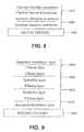
- FIG. 4is an exemplary dehydration process form used with the tissue grafts described herein.
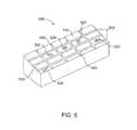
- FIG. 5is a perspective view of an exemplary drying fixture for making the tissue grafts described herein.
- FIGS. 6-9are representative side views of different tissue grafts described herein.
- FIG. 10is a top view of a tissue graft with perforations.
- tissue graftsand methods of making and using thereof.
- the tissue graftsare multilayered systems composed of one or more membranes laminated to a base amnion, where the epithelium layer of the base amnion has been substantially removed.
- the process for preparing the amnion and removing the epithelium layerinvolves
- FIG. 1depicts an overview ( 100 ) of the steps to harvest, process, and prepare placental material for later use as a tissue graft is disclosed. More detailed descriptions and discussion regarding each individual step will follow.
- the placenta tissueis collected from a consenting patient following an elective Cesarean surgery (step 110 ).
- the materialis preserved and transported in conventional tissue preservation manner to a suitable processing location or facility for check-in and evaluation (step 120 ).
- Gross processing, handling, and separation of the tissue layersthen takes place (step 130 ).
- step 135After the epithelium layer is substantially removed from the amnion to expose the base membrane (step 135 ), acceptable tissue is then decontaminated (step 140 ), dehydrated (step 150 ), cut and packaged (step 160 ), tissue is radiological terminal sterilized using gamma radiation (step 165 ), and released (step 170 ) to the market for use by surgeons and other medical professionals in appropriate surgical procedures and for wound care. Each step is described in detail below.
- the recovery of placenta tissueoriginates in a hospital, where it is collected during a Cesarean section birth.
- the donorreferring to the mother who is about to give birth, voluntarily submits to a comprehensive screening process designed to provide the safest tissue possible for transplantation.
- the screening processpreferably tests for antibodies to the human immunodeficiency virus type 1 and type 2 (anti-HIV-1 and anti-HIV-2), hepatitis B surface antigens (HBsAg), antibodies to the hepatitis C virus (anti-HCV), antibodies to the human T-lymphotropic virus type I and type II (anti-HTLV-I and anti-HTLV-II), CMV, and syphilis, using conventional serological tests.
- the above list of testsis exemplary only, as more, fewer, or different tests may be desired or necessary over time or based upon the intended use of the grafts, as will be appreciated by those skilled in the art.
- the donorwill either be deemed acceptable or not.
- culturesare taken to determine the presence of bacteria, for example, Clostridium or Streptococcus. If the donor's information, screening tests, and the delivery cultures are all satisfactory (i.e., do not indicate any risks or indicate acceptable level of risk), the donor is approved by a medical director and the tissue specimen is designated as initially eligible for further processing and evaluation.
- Human placentas that meet the above selection criteriaare preferably bagged in a saline solution in a sterile shipment bag and stored in a container of wet ice for shipment to a processing location or laboratory for further processing.
- tissueis collected prior to the completion or obtaining of results from the screening tests and delivery cultures, such tissue is labeled and kept in quarantine.
- the tissueis approved for further processing only after the required screening assessments and delivery cultures, which declare the tissue safe for handling and use, are satisfied and obtains final approval from a medical director.
- the shipmentUpon arrival at the processing center or laboratory, the shipment is opened and verified that the sterile shipment bag/container is still sealed and in the coolant, that the appropriate donor paperwork is present, and that the donor number on the paperwork matches the number on the sterile shipment bag containing the tissue.
- the sterile shipment bag containing the tissueis then stored in a refrigerator until ready for further processing. All appropriate forms, including a tissue check-in form, such as that shown in FIG. 2 , are completed and chain of custody and handling logs (not shown) are also completed.
- the sterile supplies necessary for processing the placenta tissuefurther are assembled in a staging area in a controlled environment and are prepared for introduction into a controlled environment. If the controlled environment is a manufacturing hood, the sterile supplies are opened and placed into the hood using conventional sterile technique. If the controlled environment is a clean room, the sterile supplies are opened and placed on a cart covered by a sterile drape. All the work surfaces are covered by a piece of sterile drape using conventional sterile techniques, and the sterile supplies and the processing equipments are placed on to the sterile drape, again using conventional sterile techniques.
- Processing equipmentis decontaminated according to conventional and industry-approved decontamination procedures and then introduced into the controlled environment.
- the equipmentis strategically placed within the controlled environment to minimize the chance for the equipment to come in proximity to or is inadvertently contaminated by the tissue specimen.
- the placentais removed from the sterile shipment bag and transferred aseptically to a sterile processing basin within the controlled environment.
- the sterile basincontains hyperisotonic saline solution (e.g., 18% NaCl) that is at room or near room temperature.
- the placentais gently massaged to help separate blood clots and to allow the placenta tissue to reach room temperature, which will make the separation of the amnion and chorion layers from each other, as discussed hereinafter, easier.
- the placentais then removed from the sterile processing basin and laid flat on a processing tray with the amniotic membrane layer facing down for inspection.
- the placenta tissueis examined and the results of the examination are documented on a “Raw Tissue Assessment Form” similar to that shown in FIG. 3 .
- the placenta tissueis examined for discoloration, debris or other contamination, odor, and signs of damage.
- the size of the tissueis also noted. A determination is made, at this point, as to whether the tissue is acceptable for further processing.
- the amnion and chorion layers of the placenta tissueare then carefully separated.
- the materials and equipment used in this procedureinclude a processing tray, 18% saline solution, sterile 4 ⁇ 4 sponges, and two sterile Nalgene jars.
- the placenta tissueis then closely examined to find an area (typically a corner) in which the amnion layer can be separated from the chorion layer.
- the amnionappears as a thin, opaque layer on the chorion.
- the fibroblast layerPrior to removal of the epithelium, the fibroblast layer is identified by gently contacting each side of the membrane with a piece of sterile gauze or a cotton tipped applicator. The fibroblast layer will stick to the test material. The amnion is placed into processing tray fibroblast layer down. Using a blunt instrument, a cell scraper or sterile gauze, any residual blood is also removed. This step must be done with adequate care, again, so as not to tear the amnion or chorion tissues. The cleaning of the amnion is complete once the amnion tissue is smooth and opaque-white in appearance. If the amnion tissue is cleaned too much, the jelly-like fibroblast layer can be removed. Any areas of the amnion cleaned too aggressively and appear clear will be unacceptable and will ultimately be discarded.
- the epithelium layer present on the amnionis substantially removed in order to expose the basement layer of the amnion.
- the significance of removing the epithelium layeris described below.
- the term “substantially removed” with respect to the amount of epithelium removedis defined herein as removing greater than 90%, greater than 95%, or greater than 99% of the epithelial cells from the amnion.
- the presence or absence of epithelial cells remaining on the amnion layercan be evaluated using techniques known in the art. For example, after removal of the epithelial cell layer, a representative tissue sample from the processing lot is placed onto a standard microscope examination slide. The tissue sample is then stained using Eosin Y Stain and evaluated as described below. The sample is then covered and allowed to stand. Once an adequate amount of time has passed to allow for staining, visual observation is done under magnification.
- the epithelium layercan be removed by techniques known in the art. For example, the epithelium layer can be scraped off of the amnion using a cell scraper. Other techniques include, but are not limited to, freezing the membrane, physical removal using a cell scraper, or exposing the epithelial cells to nonionic detergents, anionic detergents, and nucleases.
- the de-epithelialized tissueis then evaluated to determine that the basement membrane has not been compromised and remains intact. This step is performed after completion of the processing step and the tissue has been dehydrated as described in the next section. For example, a representative sample graft is removed for microscopic analysis.
- tissue sampleis place onto a standard slide and 100 ⁇ l of Eosin Y stain is applied to the sample and allowed to set.
- the tissue sampleis then examined under magnification. Cellular material will stain dark indicating the presence of cells. If no stained cells are present, de-epithelization has been achieved.
- the amnionis then placed into a sterile Nalgene jar for the next step for additional cleaning. If the chorion is to be recovered and processed further, it too is placed in its own sterile Nalgene jar for additional cleaning. If the chorion is not to be kept or used further, it can be discarded in an appropriate biohazard container. In one aspect, the following procedure can be used to clean the amnion.
- Each Nalgene jaris aseptically filled with 18% saline hypertonic solution and sealed (or sealed with a top). The jar is then placed on a rocker platform and agitated for between 30 and 90 minutes, which further cleans the tissue of contaminants. If the rocker platform was not in the critical environment (e.g., the manufacturing hood), the Nalgene jar is returned to the controlled/sterile environment and opened. Using sterile forceps or by aseptically decanting the contents, the tissue is gently removed from the Nalgene jar containing the 18% hyperisotonic saline solution and placed into an empty Nalgene jar.
- the premixed antibiotic solutionis comprised of a cocktail of antibiotics, such as Streptomycin Sulfate and Gentamicin Sulfate. Other antibiotics, such as Polymixin B Sulfate and Bacitracin, or similar antibiotics now available or available in the future, are also suitable. Additionally, it is preferred that the antibiotic solution be at room temperature when added so that it does not change the temperature of or otherwise damage the tissue.
- This jar or container containing the tissue and antibioticsis then sealed or closed and placed on a rocker platform and agitated for, preferably, between 60 and 90 minutes. Such rocking or agitation of the tissue within the antibiotic solution further cleans the tissue of contaminants and bacteria.
- the jar or container containing the tissue and antibioticsis then returned to the critical/sterile environment and opened.
- the tissueis gently removed from the jar or container and placed in a sterile basin containing sterile water or normal saline (0.9% saline solution).
- the tissueis allowed to soak in place in the sterile water/normal saline solution for at least 10 to 15 minutes.
- the tissuemay be slightly agitated to facilitate removal of the antibiotic solution and any other contaminants from the tissue. After at least 10 to 15 minutes, the tissue is ready to be dehydrated and processed further.
- the following exemplary procedurecan be used.
- the chorionis rinsed in 18% saline solution for 30 min.
- 18% salineis heated in a sterile container using laboratory heating plate such that the solution temperature is approximately 48° C.
- the solutionis decanted, the chorion tissue is placed into the sterile container, and decanted saline solution is poured into the container.
- the containeris sealed and placed on rocker plate and agitated for 1 hour. After 1 hour agitation bath, remove the tissue and place the tissue into second heated agitation bath for an additional 1 hour rinse cycle.
- the chorion tissueis placed into 200 ml of 0.5% Triton-X wash solution.
- the containeris sealed and agitated without heat for 2 hours.
- the tissueis next washed with deionized water (250 ml of DI water ⁇ 4) with vigorous motion for each rinse.
- the tissueis removed and placed into a container of 1 ⁇ PBS w/EDTA solution.
- the containeris sealed and agitated for 1 hour at controlled temperature for 8 hours.
- the tissueis removed and rinsed using sterile water. A visual inspection was performed to remove any remaining discolored fibrous blood material from the membrane.
- Membraneshould have a cream white visual appearance with no evidence of brownish discoloration.
- the amnionis ready to be used to produce the tissue graft.
- the amnion layeris laid on a suitable drying fixture, where the exposed basement membrane is adjacent to the surface of the drying fixture.
- the amnioncan be placed on the surface of the drying fixture such that the exposed basement membrane is facing up.
- the drying fixtureis preferably sized to be large enough to receive the amnion tissue, fully, in laid out, flat fashion.
- the drying fixtureis made of Teflon or of Delrin, which is the brand name for an acetal resin engineering plastic invented and sold by DuPont and which is also available commercially from Werner Machine, Inc. in Marietta, Ga.
- any other suitable material that is heat and cut resistant, capable of being formed into an appropriate shape to receive wet tissue and to hold and maintain textured designs, logos, or textcan also be used for the drying fixture. It is desirable that the amnion tissue be placed on the drying fixture so that it completely covers as many “product spaces” (as explained hereinafter) as possible.
- the receiving surface of the drying fixture 500has grooves 505 that define the product spaces 510 , which are the desired outer contours of the tissue after it is cut and of a size and shape that is desired for the applicable surgical procedure in which the tissue will be used.
- the drying fixturecan be laid out so that the grooves are in a grid arrangement.
- the grids on a single drying fixturemay be the same uniform size or may include multiple sizes that are designed for different surgical applications. Nevertheless, any size and shape arrangement can be used for the drying fixture, as will be appreciated by those skilled in the art.
- the drying fixtureinstead of having grooves to define the product spaces, the drying fixture has raised ridges or blades.
- the drying fixturecan include a slightly raised or indented texture in the form of text, logo, name, or similar design 520 .
- This textured text, logo, name, or designcan be customized.
- the tissueWhen dried, the tissue will mold itself around the raised texture or into the indented texture—essentially providing a label within the tissue itself.
- texture/labelcan be read or viewed on the tissue in only one orientation so that, after drying and cutting, an end user (typically, a clinician) of the dried tissue will be able to tell the stromal side from the basement side of the dried tissue.
- FIG. 5illustrates a variety of marks, logos, and text 520 that can be included within the empty spaces 510 of the drying fixture 500 .
- a single drying fixturewill include the same design or text within all of the empty spaces; however, FIG. 5 shows, for illustrative purposes, a wide variety of designs that can be included on such drying fixtures to emboss each graft.
- FIGS. 6-9depict numerous examples of multi-laminated tissue grafts produced by the methods described herein.
- amnion layer 610is applied to the surface of the drying fixture 600 , where the basement membrane 605 is adjacent to the surface of the drying fixture 600 .
- Two additional amnion layers 620 and 630are applied to amnion 610 .
- the fibrous layer of each amnion layeracts as an adhesive for the next layer.
- the fibrous layer of amnion 610adheres to the epithelial layer of amnion 620 . It is important to note that when multiple amnion layers are used, it is not necessary to remove the epithelial cells from the basement membrane for those layers that are not in direct contact with host cells. Although in this aspect, the fibroblast layer is used to adhere the membranes together, other techniques and materials such as, for example, fibrin glue, gelatin, photochemical techniques, and suturing can be used to produce the multi-laminated tissue graft.
- FIGS. 7 and 8depict additional features of the multi-layered tissue graft.
- amnion layer 700 minus the epithelial layeris applied to drying fixture 600 .
- Four additional amnion layers ( 710 - 740 )are then applied to the amnion layer 700 .
- amnion layer 800 minus the epithelial layeris applied to drying fixture 600 , and chorion layer 810 is applied to the amnion layer 800 .
- the multi-layered tissue graftcan be composed of one or more membranes sandwiched between two amnion membranes with the amnion tissue with its epithelial layer removed exposing the basement membrane of the amnion tissue to host cells. This aspect is depicted in FIG. 9 , where amnion layers 920 and 930 are sandwiched between amnion layers 910 and 940 (no epithelial layer).
- FIGS. 6-9depict the use of amnion and chorion to produce the multi-layered tissue graft
- other biodegradable biologically compatible materialscan be used in place of chorion to form a multi-layered amnion composite membrane.
- examples of such materialsinclude, but are not limited to, allograft pericardium, allograft acellular dermis, amniotic membrane (i.e., both amnion and chorion), Wharton's jelly, purified xenograft Type-1 collagen, biocellulose polymers or copolymers, biocompatible synthetic polymer or copolymer films, purified small intestinal submucosa, bladder acellular matrix, cadaveric fascia, or any combination thereof.
- the actual number of layerswill depend upon the surgical need and procedure with which the tissue graft is designed to be used for. For example, in periodontal applications such as root coverage procedures, a single membrane that is between 20-50 ⁇ m in thickness to multilayer tissue grafts having a thickness up to 2 mm can be used. In one aspect, the number of membranes laminated to the base amnion tissue with substantially all of the epithelial cells removed can be one, two, five and or ten, with the ultimate number of layers dependent of the type of membrane used, and the expected indication for use.
- the multi-layered tissue grafts described hereinare thicker and stronger than a single layer of base amnion.
- the base amnionmay have handling limitations. In general, tissue grafts are cut to size to match the morphology of the wound, placed on or within the wound, and if desired, can be held in place with sutures. If left unsupported, the base amnion tissue may fall over itself, and may bunch together. Moreover, the native amnion tissue may be unable to hold sutures. Sutures may tear the tissue during placement, and may be knocked free by the patient. These issues make trimming, placement, and securing a single layer of base amnion tissue less than ideal for the medical professional.
- the drying fixtureis placed in a sterile Tyvex (or similar, breathable, heat-resistant, and sealable material) dehydration bag and sealed.
- a breathable dehydration bagprevents the tissue from drying too quickly and prevents cross-contamination, when multiple donors are dehydrated in the oven at the same time.
- each drying fixtureis either placed in its own Tyvex bag or, alternatively, placed into a suitable mounting frame that is designed to hold multiple drying frames thereon and the entire frame is then placed into a larger, single sterile Tyvex dehydration bag and sealed.
- the Tyvex dehydration bag containing the one or more drying fixturesis then placed into an oven or incubator (vacuum or non-vacuum) that has been preheated to approximately 35 to 50° C.
- the Tyvex bagremains in the oven for between 30 and 120 minutes, although approximately 45 minutes at a temperature of approximately 45° C. appears to be ideal to dry the tissue sufficiently but without over-drying the tissue.
- the specific temperature and time for any specific ovenwill need to be calibrated and adjusted based on other factors including altitude, size of the oven, accuracy of the oven temperature, material used for the drying fixture, number of drying fixtures being dried simultaneously, whether a single or multiple frames of drying fixtures are dried simultaneously, and the like.
- the dehydration recordation formsimilar to that shown in FIG. 4 , can be completed at the end of the dehydration process.
- the membraneis not physically altered except for final cutting and packaging (step 160 ).
- the processed tissue grafthas a semi-transparent appearance with a whitish coloration.
- the tissueis pliable to withstand bending and sizing in its dry, non-hydrated state.
- the tissue grafts described hereincan be stored at room temperature for extended periods of time.
- the tissue graftis perforated using a small gauge needle, or punch to produce a mesh-type configuration, as shown in FIG. 10 , where a plurality of holes 1010 are present in graft 1000 .
- the size and spacing of the holescan vary depending upon the application.
- the holeshave a diameter from (0.2 mm-2.0 mm or 0.2 mm to 0.5 mm) and are spaced from 1 mm to 5 mm or 2 mm to 4 mm apart.
- the holesmay facilitate cell migration and provide for the passage of blood and other factors to the opposite side of the graft, which ultimately enhances wound healing.
- the membraneis placed directly onto the exposed root of the tooth.
- the tooth surfacedoes not supply a source of blood and the holes provide a source of blood and other factors to the side of the membrane that is in direct contact with the tooth surface.
- the holesmay allow for a more rapid and even distribution of wound healing agents which may be applied and or adsorbed onto prior to implantation to further improve the regenerative potential of the treatment.
- the processed tissue grafthas a semi-transparent appearance with a whitish coloration.
- the tissueis pliable to withstand bending and sizing in its dry, non-hydrated state.
- the tissue grafts described hereincan be stored at room temperature for extended periods of time.
- the tissue graftis then ready to be cut into specific product sizes and appropriately packages for storage, terminal sterilization, and later surgical use.
- the Tyvex bag containing the dehydrated tissueis placed back into the sterile/controlled environment.
- the number of grafts to be producedis estimated based on the size and shape of the tissue on the drying fixture(s).
- An appropriate number of pouches, one for each tissue graft,are then also introduced into the sterile/controlled environment.
- the drying fixture(s)are then removed from the Tyvex bag.
- drying fixturehas grooves
- the following exemplary procedureis followed for cutting the tissue into product sizes. If the drying fixture is configured in a grid pattern, a #20 or similar straight or rolling blade is used to cut along each groove line in parallel. Then, all lines in the perpendicular direction are cut.
- the drying fixturehas raised edges or blades, then the following procedure is followed for cutting the tissue into product sizes. A sterile roller is used to roll across the drying fixture. Sufficient pressure must be applied so that the dehydrated tissue is cut along all of the raised blades or edges of the drying fixture.
- each tissue graftis placed in a respective “inner” pouch.
- the inner pouchwhich preferably has a clear side and an opaque side, should be oriented clear side facing up.
- the tissue graftis placed in the “inner” pouch so that the texture in the form of text, logo, name, or similar design is facing out through the clear side of the inner pouch and is visible outside of the inner pouch. This process is repeated for each separate graft.
- Each tissue graftis then given a final inspection to confirm that there are no tears or holes, that the product size (as cut) is within approximately 1 millimeter (plus or minus) of the specified size for that particular graft, that there are no noticeable blemishes or discoloration of the tissue, and that the textured logo or wording is readable and viewable through the “inner” pouch.
- the inner pouchcan be sealed in any suitable manner; however, a heat seal has shown to be effective.
- the productis terminally sterilized by radiation, using gamma or electron beam sterilization with a target dose of 17.5 kGy.
- each inner pouchis separately packaged in an “outer” pouch for further protection, storage, and shipment.
- tissue grafts described hereinare designed to be stored and shipped at room or ambient temperature without need for refrigeration or freezing.
- These membranesare composed of at least one layer of amnion tissue where the epithelium layer has been substantially removed in order to expose the basement layer to host cells. By removing the epithelium layer, cells from the host can more readily interact with the cell-adhesion bio-active molecules located onto top and throughout the basement membrane.
- These graftscould be composed of one or several layers of amnion tissue, combined with chorion or other biocompatible membranes to form tissue grafts.
- the laminin structure of amnion tissueis nearly identical to that of native human oral mucosa tissue. They both include high level of laminin-5, a cell adhesion factor show to bind gingival epithelia-cells, throughout upper portions of the basement membrane.
- the membranes described hereinwill be highly suited for use in regenerative procedures involving the oral mucosa. Due to the similarity of mucosa tissue found in oral cavity, nose, throat, vagina, and rectum, it should also be highly effective in regenerative procedures in those anatomical regions.
- the laminins found in the basement membranedo not adhere to dermis epithelia cells, and it is unknown if cells specific to another anatomical location my adhere to the laminins found it on the basement membrane of amnion tissue, the amnion based membranes could be effective in broad range of applications including the treatment of burns, chronic wounds, and as grafts in cardiovascular surgery
- membranescan be soaked with a bioactive agent such as a solution composed of naturally occurring growth factors sourced from platelet concentrates, either using autologous blood collection and separation products, or platelet concentrates sourced from expired banked blood; bone marrow aspirate; stem cells derived from concentrated human placental cord blood stem cells, concentrated amniotic fluid stem cells or stem cells grown in a bioreactor; or antibiotics.
- a bioactive agentsuch as a solution composed of naturally occurring growth factors sourced from platelet concentrates, either using autologous blood collection and separation products, or platelet concentrates sourced from expired banked blood; bone marrow aspirate; stem cells derived from concentrated human placental cord blood stem cells, concentrated amniotic fluid stem cells or stem cells grown in a bioreactor; or antibiotics.
- a bioactive agentsuch as a solution composed of naturally occurring growth factors sourced from platelet concentrates, either using autologous blood collection and separation products, or platelet concentrates sourced from expired banked blood; bone marrow
- tissue graftsare easy to handle and do not readily break. Additionally, the tissue may composed in such an manner to hold sutures, provide specific rates of resorption based on the requirements of the indication in which it is being used.
- tissue grafts described hereinhave numerous applications and can be used in a variety of procedures.
- the tissue graftscan be used in the following perioplastic procedures including the treatment of recession defects, increasing gingival tissue height and width, increase the amount of attached gingival tissue at the gingival margin, and increase the zone of attached gingival tissue, elimination of a frenum pull, regeneration of lost patella tissue, repair of the Schneiderian membrane in the sinus cavity, soft tissue around dental implants, vestibuloplasty, and guided tissue regeneration.
- oral surgerythey could be used to improve soft tissue outcomes and grow new bone in guided bone regeneration procedures.
- oral mucosa tissuetreating mouth sores and oral lesions, and larger replace larger amounts of mucosa tissue lost through disease or traumatic injury.
- These same membranescould also be used in reconstructive procedures where the tissue is composed of mucosa including in ear, nose, and throat, urogynecology, and surgical procedures involving the rectum and anus.
- reaction conditionse.g., component concentrations, desired solvents, solvent mixtures, temperatures, pressures and other reaction ranges and conditions that can be used to optimize the product purity and yield obtained from the described process. Only reasonable and routine experimentation will be required to optimize such process conditions.
- Tissue graftscomposed of (5) layers of amniotic membrane with (1) each layer having intact epithelium and (2) the epithelial layer removed from the first amnion layer to expose the basement layer were evaluated.
- the epithelial layerwas removed using a specialized instrument (custom cell scraper) according to SOP#220.125, “Removal of the epithelial cell layer for de-epithelialized products”. This procedure would allow for complete removal of the epithelial cell layer and exposure of the basement membrane.
- a representative tissue sample from the processing lotis placed onto a standard microscope examination slide. The tissue sample is then stained using Eosin Y Stain. The sample is then covered and allowed to stand. Once an adequate amount of time has passed to allow for staining, visual observation is done under magnification. The presence of cells and cellular material will appear darker than the areas which have been de-epithelialized.
- a total of (10) consenting patientswere implanted with the tissue grafts under initial design specifications.
- Patient #1 of the case study evaluationwas diagnosed with a Miller's Class I recession defect resulting in surgical decision.
- the patientdisplayed a pre-operative recession defect measuring 3 mm at tooth site #5 in the maxillary region.
- Standard surgical techniqueswere employed with tissue graft placement (with epithelium) and closure technique.
- the exposed basement membranewas applied to the gingival, with the fibroblast layer attached to the tooth.
- Post-operative observationsrevealed soft tissue closure (flap) recession and soft tissue non-attachment at the (1) week post-operative interval.
Landscapes
- Health & Medical Sciences (AREA)
- Life Sciences & Earth Sciences (AREA)
- Chemical & Material Sciences (AREA)
- Engineering & Computer Science (AREA)
- Biomedical Technology (AREA)
- Animal Behavior & Ethology (AREA)
- General Health & Medical Sciences (AREA)
- Public Health (AREA)
- Veterinary Medicine (AREA)
- Medicinal Chemistry (AREA)
- Epidemiology (AREA)
- Cell Biology (AREA)
- Developmental Biology & Embryology (AREA)
- Zoology (AREA)
- Oral & Maxillofacial Surgery (AREA)
- Transplantation (AREA)
- Dermatology (AREA)
- Chemical Kinetics & Catalysis (AREA)
- Botany (AREA)
- Immunology (AREA)
- Pharmacology & Pharmacy (AREA)
- Hematology (AREA)
- Virology (AREA)
- Biotechnology (AREA)
- Molecular Biology (AREA)
- Urology & Nephrology (AREA)
- Reproductive Health (AREA)
- Pregnancy & Childbirth (AREA)
- Bioinformatics & Cheminformatics (AREA)
- Dispersion Chemistry (AREA)
- Surgery (AREA)
- General Chemical & Material Sciences (AREA)
- Nuclear Medicine, Radiotherapy & Molecular Imaging (AREA)
- Organic Chemistry (AREA)
- Materials For Medical Uses (AREA)
- Cardiology (AREA)
- Heart & Thoracic Surgery (AREA)
- Vascular Medicine (AREA)
- Medicines Containing Material From Animals Or Micro-Organisms (AREA)
Abstract
Description
- (a) obtaining a placenta from a subject, wherein the placenta comprises an amniotic membrane layer and a chorion tissue layer;
- (b) separating the chorion tissue layer from the amnion layer, wherein the amnion comprises epithelium cells adjacent to a basement membrane;
- (d) removing substantially all of the epithelium cells to expose the basement membrane of the amnion;
- (e) mounting the first membrane onto a surface of a drying fixture;
- (f) mounting one or more additional membranes on the first membrane to produce a layered tissue graft; and
- (g) dehydrating the layered tissue graft on the drying fixture.
Claims (10)
Priority Applications (6)
| Application Number | Priority Date | Filing Date | Title |
|---|---|---|---|
| US14/323,964US9084767B2 (en) | 2007-09-07 | 2014-07-03 | Placental tissue grafts and methods of preparing and using the same |
| US14/804,156US9789137B2 (en) | 2007-09-07 | 2015-07-20 | Placental tissue grafts and improved methods of preparing and using the same |
| US14/919,692US9415074B2 (en) | 2007-09-07 | 2015-10-21 | Placental tissue grafts |
| US15/716,135US10874697B2 (en) | 2007-09-07 | 2017-09-26 | Placental tissue grafts and improved methods of preparing and using the same |
| US17/120,626US11752174B2 (en) | 2007-09-07 | 2020-12-14 | Placental tissue grafts and improved methods of preparing and using the same |
| US18/464,343US20230414670A1 (en) | 2007-09-07 | 2023-09-11 | Placental tissue grafts and improved methods of preparing and using the same |
Applications Claiming Priority (11)
| Application Number | Priority Date | Filing Date | Title |
|---|---|---|---|
| US97078007P | 2007-09-07 | 2007-09-07 | |
| US98666507P | 2007-11-09 | 2007-11-09 | |
| US98929907P | 2007-11-20 | 2007-11-20 | |
| US12/206,508US8357403B2 (en) | 2007-09-07 | 2008-09-08 | Placental tissue grafts |
| US12/428,908US8323701B2 (en) | 2007-09-07 | 2009-04-23 | Placental tissue grafts |
| US13/569,134US8409626B2 (en) | 2007-09-07 | 2012-08-07 | Placental tissue grafts |
| US13/765,640US8709493B2 (en) | 2007-09-07 | 2013-02-12 | Placental tissue grafts |
| US13/861,305US8642092B2 (en) | 2007-09-07 | 2013-04-11 | Placental tissue grafts |
| US14/171,511US9533011B2 (en) | 2007-09-07 | 2014-02-03 | Placental tissue grafts and methods of preparing and using the same |
| US14/262,590US9272003B2 (en) | 2007-09-07 | 2014-04-25 | Placental tissue grafts |
| US14/323,964US9084767B2 (en) | 2007-09-07 | 2014-07-03 | Placental tissue grafts and methods of preparing and using the same |
Related Parent Applications (1)
| Application Number | Title | Priority Date | Filing Date |
|---|---|---|---|
| US14/262,590ContinuationUS9272003B2 (en) | 2007-09-07 | 2014-04-25 | Placental tissue grafts |
Related Child Applications (1)
| Application Number | Title | Priority Date | Filing Date |
|---|---|---|---|
| US14/804,156ContinuationUS9789137B2 (en) | 2007-09-07 | 2015-07-20 | Placental tissue grafts and improved methods of preparing and using the same |
Publications (2)
| Publication Number | Publication Date |
|---|---|
| US20140322289A1 US20140322289A1 (en) | 2014-10-30 |
| US9084767B2true US9084767B2 (en) | 2015-07-21 |
Family
ID=40429413
Family Applications (18)
| Application Number | Title | Priority Date | Filing Date |
|---|---|---|---|
| US12/206,508Active2028-09-23US8357403B2 (en) | 2007-09-07 | 2008-09-08 | Placental tissue grafts |
| US12/428,908Active2029-10-07US8323701B2 (en) | 2007-09-07 | 2009-04-23 | Placental tissue grafts |
| US13/569,134ActiveUS8409626B2 (en) | 2007-09-07 | 2012-08-07 | Placental tissue grafts |
| US13/569,140ActiveUS8372439B2 (en) | 2007-09-07 | 2012-08-07 | Method for treating a wound using improved placental tissue graft |
| US13/569,138ActiveUS8372438B2 (en) | 2007-09-07 | 2012-08-07 | Method for inhibiting adhesion formation using an improved placental tissue graft |
| US13/742,241ActiveUS8703206B2 (en) | 2007-09-07 | 2013-01-15 | Placental tissue grafts |
| US13/765,640ActiveUS8709493B2 (en) | 2007-09-07 | 2013-02-12 | Placental tissue grafts |
| US13/861,305ActiveUS8642092B2 (en) | 2007-09-07 | 2013-04-11 | Placental tissue grafts |
| US13/958,499ActiveUS8703207B2 (en) | 2007-09-07 | 2013-08-02 | Placental tissue grafts |
| US14/171,511Active2028-12-15US9533011B2 (en) | 2007-09-07 | 2014-02-03 | Placental tissue grafts and methods of preparing and using the same |
| US14/231,553ActiveUS8932643B2 (en) | 2007-09-07 | 2014-03-31 | Placental tissue grafts |
| US14/262,590ActiveUS9272003B2 (en) | 2007-09-07 | 2014-04-25 | Placental tissue grafts |
| US14/323,964ActiveUS9084767B2 (en) | 2007-09-07 | 2014-07-03 | Placental tissue grafts and methods of preparing and using the same |
| US14/804,156ActiveUS9789137B2 (en) | 2007-09-07 | 2015-07-20 | Placental tissue grafts and improved methods of preparing and using the same |
| US14/919,692ActiveUS9415074B2 (en) | 2007-09-07 | 2015-10-21 | Placental tissue grafts |
| US15/716,135Active2029-06-04US10874697B2 (en) | 2007-09-07 | 2017-09-26 | Placental tissue grafts and improved methods of preparing and using the same |
| US17/120,626Active2029-06-04US11752174B2 (en) | 2007-09-07 | 2020-12-14 | Placental tissue grafts and improved methods of preparing and using the same |
| US18/464,343PendingUS20230414670A1 (en) | 2007-09-07 | 2023-09-11 | Placental tissue grafts and improved methods of preparing and using the same |
Family Applications Before (12)
| Application Number | Title | Priority Date | Filing Date |
|---|---|---|---|
| US12/206,508Active2028-09-23US8357403B2 (en) | 2007-09-07 | 2008-09-08 | Placental tissue grafts |
| US12/428,908Active2029-10-07US8323701B2 (en) | 2007-09-07 | 2009-04-23 | Placental tissue grafts |
| US13/569,134ActiveUS8409626B2 (en) | 2007-09-07 | 2012-08-07 | Placental tissue grafts |
| US13/569,140ActiveUS8372439B2 (en) | 2007-09-07 | 2012-08-07 | Method for treating a wound using improved placental tissue graft |
| US13/569,138ActiveUS8372438B2 (en) | 2007-09-07 | 2012-08-07 | Method for inhibiting adhesion formation using an improved placental tissue graft |
| US13/742,241ActiveUS8703206B2 (en) | 2007-09-07 | 2013-01-15 | Placental tissue grafts |
| US13/765,640ActiveUS8709493B2 (en) | 2007-09-07 | 2013-02-12 | Placental tissue grafts |
| US13/861,305ActiveUS8642092B2 (en) | 2007-09-07 | 2013-04-11 | Placental tissue grafts |
| US13/958,499ActiveUS8703207B2 (en) | 2007-09-07 | 2013-08-02 | Placental tissue grafts |
| US14/171,511Active2028-12-15US9533011B2 (en) | 2007-09-07 | 2014-02-03 | Placental tissue grafts and methods of preparing and using the same |
| US14/231,553ActiveUS8932643B2 (en) | 2007-09-07 | 2014-03-31 | Placental tissue grafts |
| US14/262,590ActiveUS9272003B2 (en) | 2007-09-07 | 2014-04-25 | Placental tissue grafts |
Family Applications After (5)
| Application Number | Title | Priority Date | Filing Date |
|---|---|---|---|
| US14/804,156ActiveUS9789137B2 (en) | 2007-09-07 | 2015-07-20 | Placental tissue grafts and improved methods of preparing and using the same |
| US14/919,692ActiveUS9415074B2 (en) | 2007-09-07 | 2015-10-21 | Placental tissue grafts |
| US15/716,135Active2029-06-04US10874697B2 (en) | 2007-09-07 | 2017-09-26 | Placental tissue grafts and improved methods of preparing and using the same |
| US17/120,626Active2029-06-04US11752174B2 (en) | 2007-09-07 | 2020-12-14 | Placental tissue grafts and improved methods of preparing and using the same |
| US18/464,343PendingUS20230414670A1 (en) | 2007-09-07 | 2023-09-11 | Placental tissue grafts and improved methods of preparing and using the same |
Country Status (4)
| Country | Link |
|---|---|
| US (18) | US8357403B2 (en) |
| EP (4) | EP2197270B8 (en) |
| CA (1) | CA2736663C (en) |
| WO (1) | WO2009033160A1 (en) |
Cited By (17)
| Publication number | Priority date | Publication date | Assignee | Title |
|---|---|---|---|---|
| US20160136328A1 (en)* | 2014-11-17 | 2016-05-19 | Mimedx Group, Inc. | Fenestration kits for making fenestrated placental tissue allografts and methods of using the same |
| US9415074B2 (en) | 2007-09-07 | 2016-08-16 | Mimedx Group, Inc. | Placental tissue grafts |
| US9616152B2 (en) | 2008-04-25 | 2017-04-11 | Allosource | Multi-layer tissue systems and methods |
| US9744266B2 (en) | 2011-12-19 | 2017-08-29 | Allosource | Flowable matrix compositions and methods |
| US9795707B2 (en) | 2013-12-06 | 2017-10-24 | Allosource | Methods of drying sheets of donor-provided human birth tissue |
| US9808492B2 (en) | 2012-09-10 | 2017-11-07 | Terry W. Broussard | Methods of preparing lyophilized human tissues |
| US9956253B2 (en) | 2006-08-17 | 2018-05-01 | Mimedx Group, Inc. | Placental tissue grafts |
| US10251917B1 (en) | 2017-09-19 | 2019-04-09 | Gary M. Petrucci | Methods and materials for treating tumors |
| US10342830B2 (en) | 2015-01-05 | 2019-07-09 | Gary M. Petrucci | Methods and materials for treating lung disorders |
| US10478531B2 (en) | 2017-06-22 | 2019-11-19 | Gary M. Petrucci | Methods and materials for treating blood vessels |
| US10568990B2 (en) | 2013-03-15 | 2020-02-25 | Allosource | Cell repopulated collagen matrix for soft tissue repair and regeneration |
| US10632155B2 (en) | 2005-09-27 | 2020-04-28 | Tissuetech, Inc. | Amniotic membrane preparations and purified compositions and therapy for scar reversal and inhibition |
| US10993969B2 (en) | 2016-02-05 | 2021-05-04 | Gary M. Petrucci | Methods and materials for treating nerve injuries and neurological disorders |
| US11116871B2 (en) | 2015-09-17 | 2021-09-14 | Stimlabs Llc | Compositions derived from placenta and methods of producing the same |
| US11154641B2 (en) | 2017-12-22 | 2021-10-26 | Stimlabs Llc | Translucent, dehydrated placental tissue and methods of producing and using the same |
| US11413372B2 (en) | 2015-09-17 | 2022-08-16 | Stimlabs Llc | Compositions derived from placenta and methods of producing the same |
| US12397086B2 (en) | 2011-04-28 | 2025-08-26 | Biotissue Holdings Inc. | Methods of modulating bone remodeling |
Families Citing this family (122)
| Publication number | Priority date | Publication date | Assignee | Title |
|---|---|---|---|---|
| US8187639B2 (en) | 2005-09-27 | 2012-05-29 | Tissue Tech, Inc. | Amniotic membrane preparations and purified compositions and anti-angiogenesis treatment |
| US9480549B2 (en) | 2008-04-25 | 2016-11-01 | Allosource | Multi-layer tissue patches |
| KR20110013419A (en)* | 2008-04-25 | 2011-02-09 | 알로소스 | Anti-adhesion barrier wound dressing comprising processed amnion tissue and methods of using the same |
| US9205177B2 (en)* | 2009-03-04 | 2015-12-08 | Peytant Solutions, Inc. | Stents modified with material comprising amnion tissue and corresponding processes |
| US8298586B2 (en)* | 2009-07-22 | 2012-10-30 | Acell Inc | Variable density tissue graft composition |
| US8883210B1 (en) | 2010-05-14 | 2014-11-11 | Musculoskeletal Transplant Foundation | Tissue-derived tissuegenic implants, and methods of fabricating and using same |
| US9352003B1 (en) | 2010-05-14 | 2016-05-31 | Musculoskeletal Transplant Foundation | Tissue-derived tissuegenic implants, and methods of fabricating and using same |
| US10130736B1 (en) | 2010-05-14 | 2018-11-20 | Musculoskeletal Transplant Foundation | Tissue-derived tissuegenic implants, and methods of fabricating and using same |
| US8840665B2 (en) | 2010-06-11 | 2014-09-23 | Liventa Bioscience, Inc. | Method of tendon repair with amnion and chorion constructs |
| WO2012003377A2 (en)* | 2010-06-30 | 2012-01-05 | Tissuetech, Inc. | Methods of preparing chorion tissue and products derived therefrom |
| WO2012087348A1 (en)* | 2010-12-20 | 2012-06-28 | Stemnion, Inc. | Methods for treating dental diseases, disorders and injuries |
| KR20210146436A (en) | 2011-02-14 | 2021-12-03 | 미메딕스 그룹 인크. | Micronized placental tissue compositions and methods for making and using the same |
| JP6019040B2 (en)* | 2011-02-14 | 2016-11-02 | ミメドックス グループ インコーポレーティッドMimedx Group Inc. | Tissue grafts modified with a cross-linking agent and methods of making and using the same |
| KR20210104178A (en) | 2011-02-14 | 2021-08-24 | 미메딕스 그룹 인크. | Laminated tissue grafts composed of wharton's jelly and methods of making and using the same |
| AU2012217975B2 (en)* | 2011-02-14 | 2015-11-19 | Mimedx Group Inc. | Micronized placental tissue compositions and methods for making and using the same |
| US9682044B2 (en) | 2011-06-10 | 2017-06-20 | Tissuetech, Inc. | Methods of processing fetal support tissues, fetal support tissue powder products, and uses thereof |
| EP2748308A4 (en) | 2011-08-26 | 2015-04-29 | Tissuetech Inc | METHODS OF STERILIZING TISSUE SUPPORT F RATES |
| US20130136773A1 (en)* | 2011-09-30 | 2013-05-30 | NuTech Spine, Inc. | Expandable Placental Membrane and Methods of Making and Storing Same |
| US10517902B2 (en)* | 2011-09-30 | 2019-12-31 | Prime Merger Sub, Llc | Expandable amnion membrane for treating non-healing wounds |
| US12290613B2 (en) | 2011-10-06 | 2025-05-06 | Mimedx Group, Inc. | Micronized compositions composed of bone grafts and methods of making and using the same |
| US9585983B1 (en) | 2011-10-12 | 2017-03-07 | BioDlogics, LLC | Wound covering and method of preparation |
| PL2775928T3 (en) | 2011-11-08 | 2019-09-30 | Auxocell Laboratories Inc. | Systems and methods for processing cells |
| EP2785360B1 (en) | 2011-12-02 | 2019-06-19 | MIMEDX Group Inc. | Placental tissue grafts produced by chemical dehydration/freeze-drying and methods for making and using same |
| WO2013082158A1 (en)* | 2011-12-02 | 2013-06-06 | Mimedx Group Inc. | Shredded tissue grafts and methods for making and using the same |
| EP2793745B1 (en)* | 2011-12-22 | 2019-07-31 | MIMEDX Group Inc. | Cross-linked dehydrated placental tissue grafts and methods for making and using the same |
| US9416410B2 (en)* | 2012-02-14 | 2016-08-16 | Genetics Development Corporation | Cutoff point delta Ct. method for HER2 PCR testing in breast cancer |
| US20130211504A1 (en)* | 2012-02-14 | 2013-08-15 | AFcell Medical | Method of using amnion allograft in heart valve repair surgery |
| US8961617B2 (en) | 2012-03-08 | 2015-02-24 | Liventa Bioscience, Inc. | Amnion and chorion constructs and uses thereof in abdominal surgery |
| US20130236506A1 (en)* | 2012-03-08 | 2013-09-12 | AFcell Medical | Amnion and chorion constructs and uses thereof in ob-gyn surgery |
| US10576037B2 (en) | 2012-03-14 | 2020-03-03 | MAM Holdings of West Florida, L.L.C. | Compositions comprising placental collagen for use in wound healing |
| US20130289724A1 (en)* | 2012-04-25 | 2013-10-31 | AFcell Medical | Amnion and chorion wound dressing and uses thereof |
| RU2519357C2 (en)* | 2012-04-26 | 2014-06-10 | Государственное бюджетное образовательное учреждение высшего профессионального образования "Амурская государственная медицинская академия" Минздравсоцразвития Российской Федерации | Method for amniotic membrane dissection for intrastromal implantation into corneal stroma on ice-cold surface |
| US9943551B2 (en) | 2012-08-15 | 2018-04-17 | Mimedx Group, Inc. | Tissue grafts composed of micronized placental tissue and methods of making and using the same |
| EP2884944B1 (en) | 2012-08-15 | 2020-10-07 | MiMedx Group, Inc. | Reinforced placental tissue grafts and methods of making and using the same |
| US8904664B2 (en)* | 2012-08-15 | 2014-12-09 | Mimedx Group, Inc. | Dehydration device and methods for drying biological materials |
| US11338063B2 (en) | 2012-08-15 | 2022-05-24 | Mimedx Group, Inc. | Placental tissue grafts modified with a cross-linking agent and methods of making and using the same |
| ES2756332T3 (en) | 2012-09-10 | 2020-04-27 | Univ Wake Forest Health Sciences | Amniotic membrane and its use in wound healing and tissue engineering constructions |
| CN102836171B (en)* | 2012-09-17 | 2015-04-22 | 北京金康驰医药投资有限公司 | Solution for surgery and endoscope washing and preparation method thereof |
| US9180145B2 (en) | 2012-10-12 | 2015-11-10 | Mimedx Group, Inc. | Compositions and methods for recruiting and localizing stem cells |
| US20140106447A1 (en) | 2012-10-12 | 2014-04-17 | Mimedx Group, Inc. | Compositions and methods for recruiting stem cells |
| US8946163B2 (en) | 2012-11-19 | 2015-02-03 | Mimedx Group, Inc. | Cross-linked collagen comprising metallic anticancer agents |
| US8940684B2 (en) | 2012-11-19 | 2015-01-27 | Mimedx Group, Inc. | Cross-linked collagen comprising an antifungal agent |
| US9155799B2 (en) | 2012-11-19 | 2015-10-13 | Mimedx Group, Inc. | Cross-linked collagen with at least one bound antimicrobial agent for in vivo release of the agent |
| JP2016501245A (en)* | 2012-12-07 | 2016-01-18 | アントフロゲネシス コーポレーション | Treatment of oral lesions using placental extracellular matrix |
| CA2897662C (en) | 2013-01-09 | 2022-07-26 | Ryanne EARLY | Decellularized biomaterial from non-mammalian tissue |
| US10517931B2 (en) | 2013-01-17 | 2019-12-31 | Mimedx Group, Inc. | Non-surgical, localized delivery of compositions for placental growth factors |
| US9655948B1 (en) | 2013-01-17 | 2017-05-23 | Mimedx Group, Inc. | Non-surgical, localized delivery of compositions for placental growth factors |
| US9827293B2 (en) | 2013-01-17 | 2017-11-28 | Mimedx Group, Inc. | Non-surgical, localized delivery of compositions for placental growth factors |
| WO2014113733A1 (en) | 2013-01-18 | 2014-07-24 | Mimedx Group, Inc. | Methods for treating cardiac conditions |
| US10206977B1 (en) | 2013-01-18 | 2019-02-19 | Mimedx Group, Inc. | Isolated placental stem cell recruiting factors |
| US10905800B1 (en) | 2013-01-29 | 2021-02-02 | BioDlogics, LLC | Ocular covering and method of use |
| EP2951289B1 (en)* | 2013-01-30 | 2019-10-02 | Nutech Medical, Inc. | Placental membrane preparation and methods of making and using same |
| US9498327B1 (en) | 2013-03-05 | 2016-11-22 | Biodlogics Llc | Repair of tympanic membrane using human birth tissue material |
| US9789138B1 (en) | 2013-03-06 | 2017-10-17 | BioDlogics, LLC | Neural repair construct and method of use |
| US9770472B1 (en) | 2013-03-08 | 2017-09-26 | Brahm Holdings, Llc | Organ jacket and methods of use |
| US9855301B1 (en) | 2013-03-13 | 2018-01-02 | Biodlogics Llc | Human birth tissue laminate and methods of use |
| AU2014248873B2 (en) | 2013-03-13 | 2017-03-23 | Allosource | Fascia fibrous compositions and methods for their use and manufacture |
| US10029030B2 (en) | 2013-03-15 | 2018-07-24 | Mimedx Group, Inc. | Molded placental tissue compositions and methods of making and using the same |
| US9795638B1 (en)* | 2013-03-16 | 2017-10-24 | BioDlogics, LLC | Cardiothoracic construct and methods of use |
| US10335433B2 (en) | 2013-04-10 | 2019-07-02 | Mimedx Group, Inc. | NDGA polymers and metal complexes thereof |
| US20140336600A1 (en) | 2013-05-10 | 2014-11-13 | Carl Randall Harrell | Method for Obtaining Sterile Human Amniotic Fluid and Uses Thereof |
| US10485521B2 (en) | 2013-05-10 | 2019-11-26 | MAM Holdings of West Florida, L.L.C. | Method for obtaining sterile human amniotic fluid and uses thereof |
| US9446142B2 (en) | 2013-05-28 | 2016-09-20 | Mimedx Group, Inc. | Polymer chelator conjugates |
| JP6756612B2 (en) | 2013-08-30 | 2020-09-16 | ミメディクス グループ インコーポレイテッド | Finely divided placental composition containing a chelator |
| JP2016529070A (en)* | 2013-09-10 | 2016-09-23 | ミメディクス グループ インコーポレイテッド | Cosmetic use of molded placenta compositions |
| JP7464348B2 (en) | 2014-01-17 | 2024-04-09 | ミメディクス グループ インコーポレイテッド | Methods for inducing angiogenesis |
| WO2015134936A1 (en)* | 2014-03-06 | 2015-09-11 | Amnio Technology Llc | Amnion derived therapeutic compositions and methods of use |
| WO2015134951A1 (en)* | 2014-03-06 | 2015-09-11 | Amnio Technology Llc | Amnion derived therapeutic compositions and methods of use |
| US9801910B2 (en) | 2014-03-17 | 2017-10-31 | Ethicon, Inc. | Decellularized pleural matrix |
| CN103893823B (en)* | 2014-04-04 | 2016-08-31 | 江苏期佰医疗技术有限公司 | A kind of high-activity biological derived material and preparation method and application |
| TW201603818A (en)* | 2014-06-03 | 2016-02-01 | 組織科技股份有限公司 | Compositions and methods |
| WO2016007554A1 (en) | 2014-07-08 | 2016-01-14 | Mimedx Group, Inc. | Micronized wharton's jelly |
| US9993748B2 (en) | 2014-08-11 | 2018-06-12 | Auxocell Laboratories, Inc. | Centrifuge clip and method |
| USD748462S1 (en) | 2014-08-11 | 2016-02-02 | Auxocell Laboratories, Inc. | Centrifuge clip |
| WO2016033385A1 (en) | 2014-08-28 | 2016-03-03 | Mimedx Group, Inc. | Collagen reinforced tissue grafts |
| CA2963273C (en) | 2014-10-02 | 2023-10-17 | Wake Forest University Health Sciences | Amniotic membrane powder and its use in wound healing and tissue engineering constructs |
| US10265438B1 (en) | 2014-11-03 | 2019-04-23 | BioDlogics, LLC | Methods and compositions for the repair and replacement of connective tissue |
| US10765705B2 (en) | 2014-11-24 | 2020-09-08 | Prime Merger Sub, Llc | Visco-supplement compositions, and methods of use thereof |
| US11207355B2 (en)* | 2015-01-09 | 2021-12-28 | Amnio Technology Llc | Enriched multilayer amnion derived tissue graft |
| US10736990B2 (en) | 2015-02-11 | 2020-08-11 | Mimedx Group, Inc. | Collagen and micronized placental tissue compositions and methods of making and using the same |
| TW201642914A (en) | 2015-02-23 | 2016-12-16 | 組織科技股份有限公司 | Apparatuses and methods for treating ophthalmic diseases and disorders |
| US11129856B2 (en)* | 2015-03-03 | 2021-09-28 | Prime Merger Sub, Llc | Methods of treating chronic wounds with amniotic fluid having elevated levels of tissue inhibitors of matrix metalloproteinases |
| CA2980316A1 (en) | 2015-03-24 | 2016-09-29 | Osiris Therapeutics, Inc. | Compositions comprising meniscal tissues and uses thereof |
| WO2016168752A1 (en) | 2015-04-17 | 2016-10-20 | The Regents Of The University Of California | Decellularized human amniotic membrane for cell delivery, cell culture and inflammation prevention |
| CA2984443A1 (en) | 2015-05-20 | 2016-11-24 | Tissuetech, Inc. | Compositions and methods for preventing the proliferation and epithelial-mesenchymal transition of epithelial cells |
| CA2986702C (en) | 2015-05-21 | 2023-04-04 | David Wang | Modified demineralized cortical bone fibers |
| WO2017041089A1 (en) | 2015-09-03 | 2017-03-09 | Mimedx Group, Inc. | Non-surgical, localized delivery of compositions for placental growth factors |
| CA2998136C (en) | 2015-09-11 | 2023-10-10 | Lifecell Corporation | Perforated tissue matrix |
| US10517903B2 (en)* | 2015-09-14 | 2019-12-31 | Amnio Technology Llc | Amnion derived therapeutic composition and process of making same |
| US11273183B2 (en) | 2015-10-09 | 2022-03-15 | MAM Holdings of West Florida, L.L.C. | Amniotic fluid formulation for treatment of joint pain or disorders |
| WO2017062937A1 (en) | 2015-10-09 | 2017-04-13 | Mam Holdings Of West Florida, Llc | Amniotic fluid formulation for treatment of joint pain or disorder |
| US20170100440A1 (en) | 2015-10-09 | 2017-04-13 | MAM Holdings of West Florida, L.L.C. | Amniotic fluid topical formulation |
| DE102015013989A1 (en)* | 2015-10-30 | 2017-05-04 | Johnson & Johnson Medical Gmbh | Surgical implant |
| US20180325961A1 (en)* | 2015-11-18 | 2018-11-15 | Lucina BioSciences, LLC | Acellular regenerative products and methods of their manufacture |
| CA3009178A1 (en) | 2015-12-23 | 2017-06-29 | Lifenet Health | Decellularized placental membrane and methods of preparing and use thereof |
| US10583219B1 (en)* | 2015-12-29 | 2020-03-10 | Brahm Holdings, Llc | Multilayer bioabsorbable construct and methods of use |
| TW201733600A (en) | 2016-01-29 | 2017-10-01 | 帝聖工業公司 | Fetal support tissue products and methods of use |
| US20170354692A1 (en) | 2016-06-13 | 2017-12-14 | MAM Holdings of West Florida, L.L.C. | Amniotic fluid formulation for treatment of lung disorders |
| WO2018067644A1 (en) | 2016-10-06 | 2018-04-12 | Lifecell Corporation | Tissue matrix with preformed openings or pilot openings |
| IL266381B2 (en)* | 2016-11-02 | 2024-04-01 | Axogen Corp | Amnion tissue grafts and methods of preparing and using same |
| CA3041719C (en)* | 2016-11-07 | 2022-08-30 | Ise Professional Testing & Consulting Services, Inc. | Methods for isolating equine decellularized tissue |
| US10772986B2 (en) | 2017-01-26 | 2020-09-15 | Allosource | Fascia fibrous compositions and methods for their use and manufacture |
| US20190365948A1 (en)* | 2017-01-27 | 2019-12-05 | Aziyo Biologics, Inc. | Lyophilized placental composite sheet and uses thereof |
| EP3592397A4 (en) | 2017-03-06 | 2020-12-30 | Tei Biosciences, Inc. | PERFORATED TISSUE TRANSPLANT |
| USD962484S1 (en)* | 2017-10-03 | 2022-08-30 | Alexander Lorenz | Concrete slab |
| USD896994S1 (en)* | 2017-10-03 | 2020-09-22 | Alexander Lorenz | Concrete slab |
| CN107583110A (en)* | 2017-10-24 | 2018-01-16 | 杭州恩格生物医疗科技有限公司 | A kind of preparation method of the artificial esophagus with biological structure and function |
| US20190134268A1 (en)* | 2017-11-06 | 2019-05-09 | Theodore I. Malinin | Membranous tissue with evenly spaced elevated projections on one side and concave depressions on the other side method and use |
| CZ307603B6 (en) | 2017-12-27 | 2019-01-02 | BioHealing k.s. | A method for preparing amniotic membrane-based material and a material obtainable by this method |
| US11944650B2 (en)* | 2019-03-28 | 2024-04-02 | The Board Of Supervisors Of Louisiana State University And Agricultural And Mechanical College | Method of treating or preventing hernia formation |
| WO2021173650A1 (en) | 2020-02-28 | 2021-09-02 | Aziyo Biologics Inc. | Placental tissue compositions and methods |
| US20210346437A1 (en)* | 2020-05-07 | 2021-11-11 | Board Of Trustees Of Southern Illinois University | Novel treatment of dysphagia |
| AU2021376324A1 (en)* | 2020-11-05 | 2023-06-08 | Mimedx Group, Inc. | Meshed placental membrane tissue grafts |
| CN112641999B (en)* | 2020-12-04 | 2022-10-04 | 陕西佰傲再生医学有限公司 | Film paving equipment and application thereof |
| US20230166006A1 (en)* | 2021-12-01 | 2023-06-01 | Khalid Alhezaimi | Methods and compositions for periapical tissue regeneration |
| US11925532B2 (en) | 2021-12-10 | 2024-03-12 | Vivex Biologics Group, Inc. | Vented wound dressing barrier |
| US11944718B1 (en)* | 2022-05-11 | 2024-04-02 | Healthtech Solutions, Inc. | Compositions and manufacture of allograft tissue |
| EP4395798A4 (en) | 2022-06-29 | 2025-09-17 | Biostem Tech Inc | STERILE HUMAN PLACENTAL ALLOGRANS WITH A MULTIPLE NUMBER OF SLITTS, OPENINGS AND/OR FENESTRATIONS THEREON |
| JP2025524577A (en) | 2022-06-29 | 2025-07-30 | バイオステム テクノロジーズ,インコーポレイテッド | Micronized wound healing compositions prepared from intact human amniotic-chorionic tissue with an intact intermediate spongiosa layer therebetween |
| EP4358981B1 (en) | 2022-06-29 | 2024-10-30 | Biostem Technologies, Inc. | Sterile human placental allografts and methods of making thereof |
| USD1085424S1 (en) | 2022-11-18 | 2025-07-22 | Biostem Technologies, Inc. | Fenestrated human placental allograft |
| USD1085425S1 (en) | 2022-11-18 | 2025-07-22 | Biostem Technologies, Inc. | Fenestrated human placental allograft |
Citations (75)
| Publication number | Priority date | Publication date | Assignee | Title |
|---|---|---|---|---|
| US4361552A (en) | 1980-09-26 | 1982-11-30 | Board Of Regents, The University Of Texas System | Wound dressing |
| US4446234A (en) | 1981-10-23 | 1984-05-01 | The United States Of America As Represented By The Department Of Health And Human Services | Vitro cellular interaction with amnion membrane substrate |
| US4846165A (en) | 1986-11-26 | 1989-07-11 | Dentsply Research & Development Corp. | Wound dressing membrane |
| US5197976A (en) | 1991-09-16 | 1993-03-30 | Atrium Medical Corporation | Manually separable multi-lumen vascular graft |
| WO1993010722A2 (en) | 1991-11-26 | 1993-06-10 | Research Development Foundation | Fetal membrane tubes for nerve and vessel grafts |
| US5350583A (en) | 1988-03-09 | 1994-09-27 | Terumo Kabushiki Kaisha | Cell-penetrable medical material and artificial skin |
| JPH08266613A (en) | 1995-03-31 | 1996-10-15 | Toyobo Co Ltd | Medical material and manufacture thereof |
| EP0773033A1 (en) | 1995-10-31 | 1997-05-14 | Bio-Engineering Laboratories, Ltd. | A raw membranous material for medical materials and manufacturing methods thereof |
| US5711969A (en) | 1995-04-07 | 1998-01-27 | Purdue Research Foundation | Large area submucosal tissue graft constructs |
| US5882929A (en) | 1998-04-07 | 1999-03-16 | Tissue Engineering, Inc. | Methods and apparatus for the conditioning of cartilage replacement tissue |
| US5997575A (en) | 1996-04-05 | 1999-12-07 | Purdue Research Foundation | Perforated submucosal tissue graft constructs |
| US6143315A (en) | 1997-07-24 | 2000-11-07 | Wang; Ming X. | Biochemical contact lens for treating injured corneal tissue |
| US6146414A (en) | 1997-12-19 | 2000-11-14 | Gelman; Martin L. | Medical graft and construction of the same |
| US6152142A (en) | 1997-02-28 | 2000-11-28 | Tseng; Scheffer C. G. | Grafts made from amniotic membrane; methods of separating, preserving, and using such grafts in surgeries |
| JP2001161353A (en) | 1999-12-09 | 2001-06-19 | Japan Ophthalmic Consultants:Kk | Cell fragment for transplantation and method for producing the same |
| US20020034757A1 (en) | 1998-05-20 | 2002-03-21 | Cubicciotti Roger S. | Single-molecule selection methods and compositions therefrom |
| US20020039788A1 (en) | 2000-02-29 | 2002-04-04 | Isseroff Roslyn R. | Corneal epithelial graft composites |
| US6379710B1 (en) | 1996-12-10 | 2002-04-30 | Purdue Research Foundation | Biomaterial derived from vertebrate liver tissue |
| US6398797B2 (en) | 1999-07-28 | 2002-06-04 | Cardica, Inc. | Tissue bonding system and method for controlling a tissue site during anastomosis |
| US20030032179A1 (en) | 2000-12-06 | 2003-02-13 | Hariri Robert J. | Post-partum mammalian placenta, its use and placental stem cells therefrom |
| US6544289B2 (en) | 1998-06-30 | 2003-04-08 | Lifenet | Plasticized bone grafts, and methods of making and using same |
| US20030135284A1 (en) | 1998-06-30 | 2003-07-17 | Katrina Crouch | Plasticized bone grafts and methods of making and using same |
| US20030187515A1 (en) | 2002-03-26 | 2003-10-02 | Hariri Robert J. | Collagen biofabric and methods of preparing and using the collagen biofabric |
| US20030235909A1 (en) | 2002-04-12 | 2003-12-25 | Hariri Robert J. | Modulation of stem and progenitor cell differentiation, assays, and uses thereof |
| US20040028660A1 (en) | 2002-05-30 | 2004-02-12 | Anthrogenesis Corporation | Methods of using JNK or MKK inhibitors to modulate cell differentiation and to treat myeloproliferative disorders and myelodysplastic syndromes |
| US20040039453A1 (en) | 2001-07-27 | 2004-02-26 | Anderson Kimberly A. | Pelvic health implants and methods |
| US20040057938A1 (en) | 2002-09-18 | 2004-03-25 | Emiliano Ghinelli | Use of a human amniotic membrane composition for prophylaxis and treatment of diseases and conditions of the eye and skin |
| US20040126878A1 (en) | 2001-03-30 | 2004-07-01 | Maria Ramos | Method for the preparation of immunologically inert amniotic membranes |
| US20040171147A1 (en) | 2002-11-26 | 2004-09-02 | Hariri Robert J. | Cytotherapeutics, cytotherapeutic units and methods for treatments using them |
| US20040219136A1 (en) | 2003-02-13 | 2004-11-04 | Hariri Robert J | Use of umbilical cord blood to treat individuals having a disease, disorder or condition |
| US20040265293A1 (en) | 2000-02-10 | 2004-12-30 | Crowe John H. | Therapeutic platelets and method |
| WO2005007835A1 (en) | 2003-06-16 | 2005-01-27 | Zephan Biopharmaceuticals, Inc. | Methods of preparing a transplantable product for treatment of skin defects |
| US20050026279A1 (en) | 2003-04-28 | 2005-02-03 | Tseng Scheffer C.G. | Surgical grafts and methods of preparation |
| US20050107876A1 (en) | 2002-09-30 | 2005-05-19 | Kim Ki-Ho | Dermal substitute consisting of amnion and biodegradable polymer, the preparation method and the use thereof |
| US20050118715A1 (en) | 2002-04-12 | 2005-06-02 | Hariri Robert J. | Modulation of stem and progenitor cell differentiation, assays, and uses thereof |
| US20050148034A1 (en) | 2002-04-12 | 2005-07-07 | Hariri Robert J. | Methods for identification of modulators of angiogenesis, compounds discovered thereby, and methods of treatment using the compounds |
| WO2005075002A1 (en) | 2004-02-04 | 2005-08-18 | National Institute For Materials Science | Medical material and process for producing the same |
| US20060004189A1 (en) | 2004-07-02 | 2006-01-05 | James Gandy | Compositions for treating wounds and processes for their preparation |
| CN1757717A (en) | 2005-07-12 | 2006-04-12 | 何伟 | Preparation method of dried active amnion |
| US7045148B2 (en) | 2000-12-06 | 2006-05-16 | Anthrogenesis Corporation | Method of collecting placental stem cells |
| US7049139B2 (en) | 2002-03-29 | 2006-05-23 | Singapore Eye Research Institute | Method for growth of human conjunctival tissue equivalents for research, clinical ocular surface transplantation and tissue engineering |
| US20060147492A1 (en) | 2003-11-10 | 2006-07-06 | Angiotech International Ag | Medical implants and anti-scarring agents |
| US20060153928A1 (en) | 2003-02-26 | 2006-07-13 | Shigeru Kinoshita | Aminion-origin medical material and method of preparing the same |
| US20060222634A1 (en) | 2005-03-31 | 2006-10-05 | Clarke Diana L | Amnion-derived cell compositions, methods of making and uses thereof |
| WO2006129673A1 (en) | 2005-05-30 | 2006-12-07 | Arblast Co., Ltd. | Material for tissue reconstruction and utilization of the same |
| US20070020225A1 (en) | 2005-06-10 | 2007-01-25 | Sascha Abramson | Human placental collagen compositions, processes for their preparation, methods of their use and kits comprising the compositions |
| US20070021704A1 (en) | 2005-07-13 | 2007-01-25 | Hariri Robert J | Treatment of leg ulcers using placenta derived collagen biofabric |
| US20070021762A1 (en) | 2005-07-13 | 2007-01-25 | Qing Liu | Ocular plug formed from placenta derived collagen biofabric |
| WO2007010305A2 (en) | 2005-07-15 | 2007-01-25 | The University Of Nottingham | Surgical membrane |
| WO2007013331A1 (en) | 2005-07-25 | 2007-02-01 | Arblast Co., Ltd. | Sheet-like composition |
| US20070031471A1 (en) | 2004-06-23 | 2007-02-08 | Minu, L.L.C. | Uses of Amniotic Membranes as Biocompatible Devices |
| US20070038298A1 (en) | 2005-06-30 | 2007-02-15 | Sulner Joseph W | Repair of tympanic membrane using placenta derived collagen biofabric |
| US20070071740A1 (en) | 2005-09-27 | 2007-03-29 | Bio-Tissue, Inc. | Purified amniotic membrane compositions and methods of use |
| FR2892311A1 (en) | 2005-10-24 | 2007-04-27 | Sang Etablissement Public A Ca | Healing dressing for e.g. mucosa burn, has inert component and serum covering amniotic membrane to be applied on wound, where component and serum have healing properties by presence of growth factors and intrinsic characteristics |
| US20070134210A1 (en) | 2005-10-13 | 2007-06-14 | Mohammad Heidaran | Production of oligodendrocytes from placenta-derived stem cells |
| US20070154515A1 (en) | 2005-12-29 | 2007-07-05 | Johnson Chad E | Implantable graft material |
| US20070190042A1 (en) | 2005-12-29 | 2007-08-16 | Edinger James W | Composition for collecting and preserving placental stem cells and methods of using the composition |
| US20070218039A1 (en) | 2006-02-24 | 2007-09-20 | Reliance Life Sciences Pvt. Ltd. | Conjunctival tissue system |
| US20070254005A1 (en) | 2004-08-26 | 2007-11-01 | Pathak Chandraskekhar P | Implantable Tissue Compositions and Method |
| US20070269791A1 (en) | 2003-12-25 | 2007-11-22 | Yoshisiro Takami | Method of Preparing Isolated Cell-Free Skin, Cell-Free Dermal Matrix, Method of Producing the Same and Composite Cultured Skin with The Use of the Cell-Free Dermal Matrix |
| CN101088479A (en) | 2006-08-31 | 2007-12-19 | 中山大学中山眼科中心 | Biological membrane fixing device for eye surface |
| US20070292401A1 (en) | 2006-06-20 | 2007-12-20 | Harmon Alexander M | Soft tissue repair and regeneration using stem cell products |
| US7311905B2 (en) | 2002-02-13 | 2007-12-25 | Anthrogenesis Corporation | Embryonic-like stem cells derived from post-partum mammalian placenta, and uses and methods of treatment using said cells |
| US7311904B2 (en) | 2001-02-14 | 2007-12-25 | Anthrogenesis Corporation | Tissue matrices comprising placental stem cells, and methods of making the same |
| US20080032401A1 (en) | 2005-12-29 | 2008-02-07 | James Edinger | Placental stem cell populations |
| US20080039940A1 (en) | 2004-03-11 | 2008-02-14 | Kouji Hashimoto | Biological Tissue Sheet, Method Of Forming The Same And Transplantation Method By Using The Sheet |
| US20080044848A1 (en) | 2006-06-09 | 2008-02-21 | Heidaran Mohammad A | Placental niche and use thereof to culture stem cells |
| US20080046095A1 (en) | 2006-08-17 | 2008-02-21 | Surgical Biologics, Inc. | Placental Tissue Grafts and Improved Methods of Preparing and Using the Same |
| US20080069895A1 (en) | 2006-08-15 | 2008-03-20 | Qing Liu | Umbilical cord biomaterial for medical use |
| US20080181935A1 (en) | 2006-10-06 | 2008-07-31 | Mohit Bhatia | Human placental collagen compositions, and methods of making and using the same |
| US7413734B2 (en) | 2003-06-27 | 2008-08-19 | Ethicon, Incorporated | Treatment of retinitis pigmentosa with human umbilical cord cells |
| US20080213389A1 (en) | 2005-06-17 | 2008-09-04 | Drexel University | Three-Dimensional Scaffolds for Tissue Engineering Made by Processing Complex Extracts of Natural Extracellular Matrices |
| US20080248005A1 (en) | 2005-10-21 | 2008-10-09 | Cellresearch Corporation Pte Ltd | Isolation and Cultivation of Stem/Progenitor Cells From the Amniotic Membrane of Umbilical Cord and Uses of Cells Differentiated Therefrom |
| WO2009033160A1 (en) | 2007-09-07 | 2009-03-12 | Surgical Biologics | Placental tissue grafts and improved methods of preparing and using the same |
| WO2009132186A1 (en) | 2008-04-25 | 2009-10-29 | Allosource | Anti-adhesion barrier wound dressing comprising processed amniotic tissue and method of use |
Family Cites Families (32)
| Publication number | Priority date | Publication date | Assignee | Title |
|---|---|---|---|---|
| US1938318A (en) | 1930-08-07 | 1933-12-05 | Baker & Co Inc | Method of and apparatus for making pertused sheets |
| GB8803697D0 (en) | 1988-02-17 | 1988-03-16 | Deltanine Research Ltd | Clinical developments using amniotic membrane cells |
| US4964992A (en) | 1989-03-21 | 1990-10-23 | Goldsmith Susan H | Method of making membrane-type filter and product thereof |
| US5163955A (en) | 1991-01-24 | 1992-11-17 | Autogenics | Rapid assembly, concentric mating stent, tissue heart valve with enhanced clamping and tissue alignment |
| US5255587A (en) | 1992-05-08 | 1993-10-26 | Eichenberg Robert J | Sheet cutting die press |
| US5425741A (en) | 1993-12-17 | 1995-06-20 | Autogenics | Tissue cutting die |
| WO2000078259A1 (en) | 1999-06-22 | 2000-12-28 | Research Development Foundation | Enhanced wound coverage to enhance wound healing |
| US6626965B2 (en) | 2001-06-29 | 2003-09-30 | Provo Craft & Novelty, Inc. | Apparatus for forming die cuts and method of manufacturing same |
| WO2003035125A2 (en)* | 2001-10-26 | 2003-05-01 | Cook Biotech Incorporated | Medical graft device with meshed structure |
| AU2003241362A1 (en) | 2002-05-06 | 2003-11-11 | General Binding Corporation | Rotary punch and punch pin holder |
| EP1729677B1 (en) | 2004-03-31 | 2010-04-21 | Cook Incorporated | Graft material and stent graft comprising extracellular collagen matrix and method of preparation |
| KR100739767B1 (en)* | 2005-12-05 | 2007-07-13 | 삼성전자주식회사 | Method and device for providing photo printing service in digital TV |
| US20070142745A1 (en) | 2005-12-21 | 2007-06-21 | Brahm Timothy R | Method of procuring birth tissue |
| EP2133105A4 (en) | 2007-02-23 | 2012-12-19 | Nat Univ Corp Univ Toyama | MEDICAL REPLACEMENT MEMBRANE, USE THEREOF, AND METHOD FOR REPAIRING MEMBRANE TISSUE IN LIVING BODY |
| US9358320B2 (en) | 2008-04-25 | 2016-06-07 | Allosource | Multi-layer tissue patches |
| US20110212065A1 (en) | 2010-02-18 | 2011-09-01 | Timothy Jansen | Methods of manufacture of therapeutic products comprising vitalized placental dispersions |
| US8460691B2 (en) | 2010-04-23 | 2013-06-11 | Warsaw Orthopedic, Inc. | Fenestrated wound repair scaffold |
| KR20210104178A (en) | 2011-02-14 | 2021-08-24 | 미메딕스 그룹 인크. | Laminated tissue grafts composed of wharton's jelly and methods of making and using the same |
| JP6019040B2 (en) | 2011-02-14 | 2016-11-02 | ミメドックス グループ インコーポレーティッドMimedx Group Inc. | Tissue grafts modified with a cross-linking agent and methods of making and using the same |
| KR20210146436A (en) | 2011-02-14 | 2021-12-03 | 미메딕스 그룹 인크. | Micronized placental tissue compositions and methods for making and using the same |
| US20130136773A1 (en) | 2011-09-30 | 2013-05-30 | NuTech Spine, Inc. | Expandable Placental Membrane and Methods of Making and Storing Same |
| US20130085570A1 (en) | 2011-09-30 | 2013-04-04 | Karce Daniel Rose | Fenestrator device and related methods |
| US9486316B2 (en) | 2011-09-30 | 2016-11-08 | NuTech Spine, Inc. | Osteoconductive implants and methods of using same |
| EP2785360B1 (en) | 2011-12-02 | 2019-06-19 | MIMEDX Group Inc. | Placental tissue grafts produced by chemical dehydration/freeze-drying and methods for making and using same |
| EP2793745B1 (en) | 2011-12-22 | 2019-07-31 | MIMEDX Group Inc. | Cross-linked dehydrated placental tissue grafts and methods for making and using the same |
| US9943551B2 (en) | 2012-08-15 | 2018-04-17 | Mimedx Group, Inc. | Tissue grafts composed of micronized placental tissue and methods of making and using the same |
| US8904664B2 (en) | 2012-08-15 | 2014-12-09 | Mimedx Group, Inc. | Dehydration device and methods for drying biological materials |
| US11338063B2 (en) | 2012-08-15 | 2022-05-24 | Mimedx Group, Inc. | Placental tissue grafts modified with a cross-linking agent and methods of making and using the same |
| EP2884944B1 (en) | 2012-08-15 | 2020-10-07 | MiMedx Group, Inc. | Reinforced placental tissue grafts and methods of making and using the same |
| US9155799B2 (en) | 2012-11-19 | 2015-10-13 | Mimedx Group, Inc. | Cross-linked collagen with at least one bound antimicrobial agent for in vivo release of the agent |
| EP3220970B1 (en) | 2014-11-17 | 2021-04-28 | MiMedx Group, Inc. | Fenestration kits for making fenestrated placental tissue allografts and methods of using the same |
| US9463647B1 (en) | 2015-07-15 | 2016-10-11 | Toshiba Tec Kabushiki Kaisha | Printer and method for operating the same |
- 2008
- 2008-09-08EPEP08829350.1Apatent/EP2197270B8/enactiveActive
- 2008-09-08EPEP16203661.0Apatent/EP3189731B1/enactiveActive
- 2008-09-08USUS12/206,508patent/US8357403B2/enactiveActive
- 2008-09-08WOPCT/US2008/075608patent/WO2009033160A1/enactiveApplication Filing
- 2008-09-08EPEP20153610.9Apatent/EP3689421B1/enactiveActive
- 2008-09-08EPEP25157472.9Apatent/EP4559488A3/enactivePending
- 2008-09-08CACA2736663Apatent/CA2736663C/enactiveActive
- 2009
- 2009-04-23USUS12/428,908patent/US8323701B2/enactiveActive
- 2012
- 2012-08-07USUS13/569,134patent/US8409626B2/enactiveActive
- 2012-08-07USUS13/569,140patent/US8372439B2/enactiveActive
- 2012-08-07USUS13/569,138patent/US8372438B2/enactiveActive
- 2013
- 2013-01-15USUS13/742,241patent/US8703206B2/enactiveActive
- 2013-02-12USUS13/765,640patent/US8709493B2/enactiveActive
- 2013-04-11USUS13/861,305patent/US8642092B2/enactiveActive
- 2013-08-02USUS13/958,499patent/US8703207B2/enactiveActive
- 2014
- 2014-02-03USUS14/171,511patent/US9533011B2/enactiveActive
- 2014-03-31USUS14/231,553patent/US8932643B2/enactiveActive
- 2014-04-25USUS14/262,590patent/US9272003B2/enactiveActive
- 2014-07-03USUS14/323,964patent/US9084767B2/enactiveActive
- 2015
- 2015-07-20USUS14/804,156patent/US9789137B2/enactiveActive
- 2015-10-21USUS14/919,692patent/US9415074B2/enactiveActive
- 2017
- 2017-09-26USUS15/716,135patent/US10874697B2/enactiveActive
- 2020
- 2020-12-14USUS17/120,626patent/US11752174B2/enactiveActive
- 2023
- 2023-09-11USUS18/464,343patent/US20230414670A1/enactivePending
Patent Citations (98)
| Publication number | Priority date | Publication date | Assignee | Title |
|---|---|---|---|---|
| US4361552A (en) | 1980-09-26 | 1982-11-30 | Board Of Regents, The University Of Texas System | Wound dressing |
| US4446234A (en) | 1981-10-23 | 1984-05-01 | The United States Of America As Represented By The Department Of Health And Human Services | Vitro cellular interaction with amnion membrane substrate |
| US4846165A (en) | 1986-11-26 | 1989-07-11 | Dentsply Research & Development Corp. | Wound dressing membrane |
| US5350583A (en) | 1988-03-09 | 1994-09-27 | Terumo Kabushiki Kaisha | Cell-penetrable medical material and artificial skin |
| US5197976A (en) | 1991-09-16 | 1993-03-30 | Atrium Medical Corporation | Manually separable multi-lumen vascular graft |
| WO1993010722A2 (en) | 1991-11-26 | 1993-06-10 | Research Development Foundation | Fetal membrane tubes for nerve and vessel grafts |
| JPH08266613A (en) | 1995-03-31 | 1996-10-15 | Toyobo Co Ltd | Medical material and manufacture thereof |
| US5711969A (en) | 1995-04-07 | 1998-01-27 | Purdue Research Foundation | Large area submucosal tissue graft constructs |
| US5885619A (en) | 1995-04-07 | 1999-03-23 | Purdue Research Foundation | Large area submucosal tissue graft constructs and method for making the same |
| US5955110A (en) | 1995-04-07 | 1999-09-21 | Purdue Research Foundation, Inc. | Multilayered submucosal graft constructs and method for making the same |
| EP0773033A1 (en) | 1995-10-31 | 1997-05-14 | Bio-Engineering Laboratories, Ltd. | A raw membranous material for medical materials and manufacturing methods thereof |
| US5997575A (en) | 1996-04-05 | 1999-12-07 | Purdue Research Foundation | Perforated submucosal tissue graft constructs |
| US6379710B1 (en) | 1996-12-10 | 2002-04-30 | Purdue Research Foundation | Biomaterial derived from vertebrate liver tissue |
| US6152142A (en) | 1997-02-28 | 2000-11-28 | Tseng; Scheffer C. G. | Grafts made from amniotic membrane; methods of separating, preserving, and using such grafts in surgeries |
| US6326019B1 (en) | 1997-02-28 | 2001-12-04 | Scheffer C. G. Tseng | Grafts made from amniotic membrane; methods of separating, preserving, and using such grafts in surgeries |
| US6143315A (en) | 1997-07-24 | 2000-11-07 | Wang; Ming X. | Biochemical contact lens for treating injured corneal tissue |
| US6146414A (en) | 1997-12-19 | 2000-11-14 | Gelman; Martin L. | Medical graft and construction of the same |
| US5882929A (en) | 1998-04-07 | 1999-03-16 | Tissue Engineering, Inc. | Methods and apparatus for the conditioning of cartilage replacement tissue |
| US20020034757A1 (en) | 1998-05-20 | 2002-03-21 | Cubicciotti Roger S. | Single-molecule selection methods and compositions therefrom |
| US20030135284A1 (en) | 1998-06-30 | 2003-07-17 | Katrina Crouch | Plasticized bone grafts and methods of making and using same |
| US6544289B2 (en) | 1998-06-30 | 2003-04-08 | Lifenet | Plasticized bone grafts, and methods of making and using same |
| US6398797B2 (en) | 1999-07-28 | 2002-06-04 | Cardica, Inc. | Tissue bonding system and method for controlling a tissue site during anastomosis |
| JP2001161353A (en) | 1999-12-09 | 2001-06-19 | Japan Ophthalmic Consultants:Kk | Cell fragment for transplantation and method for producing the same |
| US20040265293A1 (en) | 2000-02-10 | 2004-12-30 | Crowe John H. | Therapeutic platelets and method |
| US20020039788A1 (en) | 2000-02-29 | 2002-04-04 | Isseroff Roslyn R. | Corneal epithelial graft composites |
| US20030032179A1 (en) | 2000-12-06 | 2003-02-13 | Hariri Robert J. | Post-partum mammalian placenta, its use and placental stem cells therefrom |
| US7045148B2 (en) | 2000-12-06 | 2006-05-16 | Anthrogenesis Corporation | Method of collecting placental stem cells |
| US7311904B2 (en) | 2001-02-14 | 2007-12-25 | Anthrogenesis Corporation | Tissue matrices comprising placental stem cells, and methods of making the same |
| US20040126878A1 (en) | 2001-03-30 | 2004-07-01 | Maria Ramos | Method for the preparation of immunologically inert amniotic membranes |
| US20040039453A1 (en) | 2001-07-27 | 2004-02-26 | Anderson Kimberly A. | Pelvic health implants and methods |
| US7311905B2 (en) | 2002-02-13 | 2007-12-25 | Anthrogenesis Corporation | Embryonic-like stem cells derived from post-partum mammalian placenta, and uses and methods of treatment using said cells |
| US20040048796A1 (en) | 2002-03-26 | 2004-03-11 | Hariri Robert J. | Collagen biofabric and methods of preparation and use therefor |
| US20030187515A1 (en) | 2002-03-26 | 2003-10-02 | Hariri Robert J. | Collagen biofabric and methods of preparing and using the collagen biofabric |
| US7049139B2 (en) | 2002-03-29 | 2006-05-23 | Singapore Eye Research Institute | Method for growth of human conjunctival tissue equivalents for research, clinical ocular surface transplantation and tissue engineering |
| US20030235909A1 (en) | 2002-04-12 | 2003-12-25 | Hariri Robert J. | Modulation of stem and progenitor cell differentiation, assays, and uses thereof |
| US20050118715A1 (en) | 2002-04-12 | 2005-06-02 | Hariri Robert J. | Modulation of stem and progenitor cell differentiation, assays, and uses thereof |
| US20050148034A1 (en) | 2002-04-12 | 2005-07-07 | Hariri Robert J. | Methods for identification of modulators of angiogenesis, compounds discovered thereby, and methods of treatment using the compounds |
| US20040028660A1 (en) | 2002-05-30 | 2004-02-12 | Anthrogenesis Corporation | Methods of using JNK or MKK inhibitors to modulate cell differentiation and to treat myeloproliferative disorders and myelodysplastic syndromes |
| US20040057938A1 (en) | 2002-09-18 | 2004-03-25 | Emiliano Ghinelli | Use of a human amniotic membrane composition for prophylaxis and treatment of diseases and conditions of the eye and skin |
| US20050107876A1 (en) | 2002-09-30 | 2005-05-19 | Kim Ki-Ho | Dermal substitute consisting of amnion and biodegradable polymer, the preparation method and the use thereof |
| US20040171147A1 (en) | 2002-11-26 | 2004-09-02 | Hariri Robert J. | Cytotherapeutics, cytotherapeutic units and methods for treatments using them |
| US20040219136A1 (en) | 2003-02-13 | 2004-11-04 | Hariri Robert J | Use of umbilical cord blood to treat individuals having a disease, disorder or condition |
| US20060153928A1 (en) | 2003-02-26 | 2006-07-13 | Shigeru Kinoshita | Aminion-origin medical material and method of preparing the same |
| US20050026279A1 (en) | 2003-04-28 | 2005-02-03 | Tseng Scheffer C.G. | Surgical grafts and methods of preparation |
| US20060228339A1 (en) | 2003-06-16 | 2006-10-12 | Zheng-Pin Wang | Methods of preparing transplantable product for treatment of skin defects |
| WO2005007835A1 (en) | 2003-06-16 | 2005-01-27 | Zephan Biopharmaceuticals, Inc. | Methods of preparing a transplantable product for treatment of skin defects |
| US7413734B2 (en) | 2003-06-27 | 2008-08-19 | Ethicon, Incorporated | Treatment of retinitis pigmentosa with human umbilical cord cells |
| US20060147492A1 (en) | 2003-11-10 | 2006-07-06 | Angiotech International Ag | Medical implants and anti-scarring agents |
| US20070269791A1 (en) | 2003-12-25 | 2007-11-22 | Yoshisiro Takami | Method of Preparing Isolated Cell-Free Skin, Cell-Free Dermal Matrix, Method of Producing the Same and Composite Cultured Skin with The Use of the Cell-Free Dermal Matrix |
| WO2005075002A1 (en) | 2004-02-04 | 2005-08-18 | National Institute For Materials Science | Medical material and process for producing the same |
| US20070254013A1 (en) | 2004-02-04 | 2007-11-01 | Nat'l Institute For Materials Science | Medical Material and Process for Producing the Same |
| US20080039940A1 (en) | 2004-03-11 | 2008-02-14 | Kouji Hashimoto | Biological Tissue Sheet, Method Of Forming The Same And Transplantation Method By Using The Sheet |
| US20070031471A1 (en) | 2004-06-23 | 2007-02-08 | Minu, L.L.C. | Uses of Amniotic Membranes as Biocompatible Devices |
| US20060004189A1 (en) | 2004-07-02 | 2006-01-05 | James Gandy | Compositions for treating wounds and processes for their preparation |
| US20070254005A1 (en) | 2004-08-26 | 2007-11-01 | Pathak Chandraskekhar P | Implantable Tissue Compositions and Method |
| US20060222634A1 (en) | 2005-03-31 | 2006-10-05 | Clarke Diana L | Amnion-derived cell compositions, methods of making and uses thereof |
| WO2006129673A1 (en) | 2005-05-30 | 2006-12-07 | Arblast Co., Ltd. | Material for tissue reconstruction and utilization of the same |
| US20070020225A1 (en) | 2005-06-10 | 2007-01-25 | Sascha Abramson | Human placental collagen compositions, processes for their preparation, methods of their use and kits comprising the compositions |
| US20080213389A1 (en) | 2005-06-17 | 2008-09-04 | Drexel University | Three-Dimensional Scaffolds for Tissue Engineering Made by Processing Complex Extracts of Natural Extracellular Matrices |
| US20070038298A1 (en) | 2005-06-30 | 2007-02-15 | Sulner Joseph W | Repair of tympanic membrane using placenta derived collagen biofabric |
| CN1757717A (en) | 2005-07-12 | 2006-04-12 | 何伟 | Preparation method of dried active amnion |
| US20070021704A1 (en) | 2005-07-13 | 2007-01-25 | Hariri Robert J | Treatment of leg ulcers using placenta derived collagen biofabric |
| US20070021762A1 (en) | 2005-07-13 | 2007-01-25 | Qing Liu | Ocular plug formed from placenta derived collagen biofabric |
| WO2007010305A2 (en) | 2005-07-15 | 2007-01-25 | The University Of Nottingham | Surgical membrane |
| WO2007013331A1 (en) | 2005-07-25 | 2007-02-01 | Arblast Co., Ltd. | Sheet-like composition |
| US20070231401A1 (en) | 2005-09-27 | 2007-10-04 | Bio-Tissue, Inc. | Amniotic membrane preparations and purified compositions and anti-inflammation methods |
| US20070071740A1 (en) | 2005-09-27 | 2007-03-29 | Bio-Tissue, Inc. | Purified amniotic membrane compositions and methods of use |
| US20070134210A1 (en) | 2005-10-13 | 2007-06-14 | Mohammad Heidaran | Production of oligodendrocytes from placenta-derived stem cells |
| US20080248005A1 (en) | 2005-10-21 | 2008-10-09 | Cellresearch Corporation Pte Ltd | Isolation and Cultivation of Stem/Progenitor Cells From the Amniotic Membrane of Umbilical Cord and Uses of Cells Differentiated Therefrom |
| FR2892311A1 (en) | 2005-10-24 | 2007-04-27 | Sang Etablissement Public A Ca | Healing dressing for e.g. mucosa burn, has inert component and serum covering amniotic membrane to be applied on wound, where component and serum have healing properties by presence of growth factors and intrinsic characteristics |
| US20070190042A1 (en) | 2005-12-29 | 2007-08-16 | Edinger James W | Composition for collecting and preserving placental stem cells and methods of using the composition |
| US20080032401A1 (en) | 2005-12-29 | 2008-02-07 | James Edinger | Placental stem cell populations |
| US20070154515A1 (en) | 2005-12-29 | 2007-07-05 | Johnson Chad E | Implantable graft material |
| US20070218039A1 (en) | 2006-02-24 | 2007-09-20 | Reliance Life Sciences Pvt. Ltd. | Conjunctival tissue system |
| US20080044848A1 (en) | 2006-06-09 | 2008-02-21 | Heidaran Mohammad A | Placental niche and use thereof to culture stem cells |
| US20070292401A1 (en) | 2006-06-20 | 2007-12-20 | Harmon Alexander M | Soft tissue repair and regeneration using stem cell products |
| US20080069895A1 (en) | 2006-08-15 | 2008-03-20 | Qing Liu | Umbilical cord biomaterial for medical use |
| US20080046095A1 (en) | 2006-08-17 | 2008-02-21 | Surgical Biologics, Inc. | Placental Tissue Grafts and Improved Methods of Preparing and Using the Same |
| CN101088479A (en) | 2006-08-31 | 2007-12-19 | 中山大学中山眼科中心 | Biological membrane fixing device for eye surface |
| US20080181935A1 (en) | 2006-10-06 | 2008-07-31 | Mohit Bhatia | Human placental collagen compositions, and methods of making and using the same |
| US8372438B2 (en) | 2007-09-07 | 2013-02-12 | Mimedx Group, Inc. | Method for inhibiting adhesion formation using an improved placental tissue graft |
| US8409626B2 (en) | 2007-09-07 | 2013-04-02 | Mimedx Group, Inc. | Placental tissue grafts |
| US20120078378A1 (en) | 2007-09-07 | 2012-03-29 | John Daniel | Placental tissue grafts and improved methods of preparing and using the same |
| US20120294908A1 (en) | 2007-09-07 | 2012-11-22 | Mimedx Group Inc. | Placental tissue grafts and improved methods of preparing and using the same |
| US20120294910A1 (en) | 2007-09-07 | 2012-11-22 | Mimedx Group Inc. | Method for treating a wound |
| US20120294909A1 (en) | 2007-09-07 | 2012-11-22 | Mimedx Group Inc. | Method for inhibiting adhesion formation |
| US8323701B2 (en)* | 2007-09-07 | 2012-12-04 | Mimedx Group, Inc. | Placental tissue grafts |
| US8357403B2 (en) | 2007-09-07 | 2013-01-22 | Mimedx Group, Inc. | Placental tissue grafts |
| US8709493B2 (en)* | 2007-09-07 | 2014-04-29 | Mimedx Group, Inc. | Placental tissue grafts |
| WO2009033160A1 (en) | 2007-09-07 | 2009-03-12 | Surgical Biologics | Placental tissue grafts and improved methods of preparing and using the same |
| US8372439B2 (en) | 2007-09-07 | 2013-02-12 | Mimedx Group, Inc. | Method for treating a wound using improved placental tissue graft |
| US20130129836A1 (en) | 2007-09-07 | 2013-05-23 | Mimedx Group, Inc. | Placental tissue grafts and improved methods of preparing and using the same |
| US20130197665A1 (en) | 2007-09-07 | 2013-08-01 | John Daniel | Placental tissue grafts and improved methods of preparing and using the same |
| US20130224159A1 (en) | 2007-09-07 | 2013-08-29 | Mimedx Group, Inc. | Placental tissue grafts and improved methods of preparing and using the same |
| US20130317624A1 (en) | 2007-09-07 | 2013-11-28 | Mimedx Group, Inc. | Placental tissue grafts and improved methods of preparing and using the same |
| US8642092B2 (en)* | 2007-09-07 | 2014-02-04 | Mimedx Group, Inc. | Placental tissue grafts |
| US8703207B2 (en) | 2007-09-07 | 2014-04-22 | Mimedx Group, Inc. | Placental tissue grafts |
| WO2009132186A1 (en) | 2008-04-25 | 2009-10-29 | Allosource | Anti-adhesion barrier wound dressing comprising processed amniotic tissue and method of use |
Non-Patent Citations (35)
| Title |
|---|
| Bhatia, et al., "The Mechanism of Cell Interaction and Response on Decellularized Human Amniotic Membrane: Implications in Wound Healing," http://www.woundsresearch.com/article/7614, (2007), 19(8):1-24. |
| Burgos, H., "Angiogenic and growth factors in human amnio-chorion and placenta", Eur. J. of Clin. Invest., vol. 13, pp. 289-296 (Dec. 1982). |
| Dino et al., "Human Amnion: The Establishment of an Amnion Bank and its Practical Applications in Surgery", The Journal of the Philippine Medical Association, vol. 42, No. 7, pp. 357-366 (Jul. 1966). |
| Douglas, "Homografts of Fetal Membranes as a Covering for Large Wounds-Especially Those from Burns", Journal of Tennessee State Medical Association, 45:6, 230-235 (Jun. 1952). |
| Dua et al., "Amniotic membrane transplantation", Br. J. Ophthalmol., 83:748-752 (1999). |
| Ebihara, et al., "The Functions of Exogenous and Endogenous Laminin-5 on Corneal Epithelial Cells," Exp. Eye Res., (2000), 71:69-79. |
| Expert Declaration of Dr. Helen N. Jones. |
| Expert Declaration of Rebecca N. Baergen. |
| Extended European Search Report dated Nov. 27, 2012 for European Application Serial No. 08829350.1. |
| Fukuda, et al., "Differential Distribution of Subchains of the Basement Membrane Components Type IV Collagen and Laminin Among the Amniotic Membrane, Cornea, and Conjunctiva," Cornea, (1999), 18:73-79. |
| Hanada et al., "Multilayered Amniotic Membrane Transplantation for Severe Ulceration of the Cornea and Sclera", American J. of Ophthalmol., vol. 131, No. 3, pp. 324-331 (Mar. 2001). |
| Hao, et al., "Identification of Antiangiogenic and Antiinflammatory Proteins in Human Amniotic Membrane," Cornea, (2000), 19(3):348-352. |
| International Search Report and Written Opinion dated Nov. 13, 2008 for PCT Application Serial No. PCT/US08/75608. |
| Ishino et al., "Amniotic Membrane as a Carrier for Cultivated Human Corneal Endothelial Cell Transplantation", IOVS, vol. 45, No. 3, pp. 800-806 (Mar. 2004). |
| Klen, "Preparation of chorion and/or amnion grafts used in burns", Transactions of the Third International Congress on Research in Burns, held in Prague, Sep. 20-25, 1970, Matter et al., eds., pp. 289-292 (1971). |
| Koizumi, et al., "Growth Factor mRNA and protein in preserved human amniotic membrane," Current Eye Research, (2000), 20(3):173-177. |
| Lee et al., "Amniotic Membrane Transplantation for Persistent Epithelial Defects With Ulceration", Amniotic Membrane Transplantation, vol. 123, No. 3, pp. 303-312 (Mar. 1997). |
| Niknejad et al., "Properties of the Amniotic Membrane for Potential Use in Tissue Engineering," European Cells and Materials, (2008), 15:88-99. |
| Ohno-Matsui, "In vitro and in vivo characterization of iris pigment epithelial cells cultured on amniotic membranes", Molecular Vision, vol. 12, pp. 1022-1032 (Aug. 2006). |
| Oxlund et al., "Biomechanical analysis of human chorioamniotic membranes", European Journal of Obstetrics and Gynecology and Reproductive Biology, vol. 34, No. 3, pp. 247-255 (Mar. 1990). |
| Pakkala, et al., "Function of Laminins and Laminin-Binding Integrins in Gingival Epithelial Cell Adhesion," J. Periodontol., (2002), 73:709-719. |
| Park, et al., "Healing of a porcine burn wound dressed with human and bovine amniotic membranes," Wound Rep. Reg., (2008), 16:520-528. |
| Parry et al., "Premature Rupture of the Fetal Membranes", 1998, New England J. Med., 338(10) 663-670. |
| Petition for Inter Partes Review of U.S. Pat. No. 8323701 from Petitioner, Musculoskeletal Transplant Foundation dated Feb. 2, 2015. |
| Rinastiti, et al., "Histological evaluation of rabbit gingival wound healing transplanted with human amniotic membrane," Int. J. Oral Maxillofac. Surg., (2006), 35:247-251. |
| Robson et al., "Amniotic Membranes as a Temporary Wound Dressing", Surgery, Gynecology & Obstetrics, vol. 136, pp. 904-906 (Jun. 1973). |
| Rodriquez-Ares, et al., "Effects of lyophilization on human amniotic membrane," Acta Ophthalmol., (2009), 87:396-403. |
| Takashima, et al., Characterization of laminin isoforms in human amnion, Tissue and Cell, (2008), 40:75-81. |
| Talmi, et al., "Antibacterial Properties of Human Amniotic Membranes," Placenta, (1991), 12:285-288. |
| Toda, et al., "The Potential of Amniotic Membrane/Amnion-Derived Cells for Regeneration of Various Tissues," J. Pharmacol. Sci., (2007), 105:215-228. |
| U.S. Appl. No. 14/171,511, filed Feb. 3, 2014, Daniel, et al. |
| U.S. Appl. No. 14/231,553, to Daniel et al; filed Mar. 31, 2014.* |
| U.S. Appl. No. 14/262,590, to Daniel et al; filed Apr. 25, 2014.* |
| Vishwakarma et al., "Amniotic Arthroplasty for Tuberculosis of the Hip", J. Bone & Joint Surgery 68-B(1), 68-74 (1986). |
| Ward et al., "The healing of chronic venous leg ulcers with prepared human amnion", British Journal of Plastic Surgery, 42, 463-467 (1989). |
Cited By (32)
| Publication number | Priority date | Publication date | Assignee | Title |
|---|---|---|---|---|
| US10632155B2 (en) | 2005-09-27 | 2020-04-28 | Tissuetech, Inc. | Amniotic membrane preparations and purified compositions and therapy for scar reversal and inhibition |
| US9956253B2 (en) | 2006-08-17 | 2018-05-01 | Mimedx Group, Inc. | Placental tissue grafts |
| US9415074B2 (en) | 2007-09-07 | 2016-08-16 | Mimedx Group, Inc. | Placental tissue grafts |
| US9789137B2 (en) | 2007-09-07 | 2017-10-17 | Mimedx Group, Inc. | Placental tissue grafts and improved methods of preparing and using the same |
| US11752174B2 (en) | 2007-09-07 | 2023-09-12 | Mimedx Group, Inc. | Placental tissue grafts and improved methods of preparing and using the same |
| US10874697B2 (en) | 2007-09-07 | 2020-12-29 | Mimedx Group, Inc. | Placental tissue grafts and improved methods of preparing and using the same |
| US9616152B2 (en) | 2008-04-25 | 2017-04-11 | Allosource | Multi-layer tissue systems and methods |
| US12397086B2 (en) | 2011-04-28 | 2025-08-26 | Biotissue Holdings Inc. | Methods of modulating bone remodeling |
| US9744266B2 (en) | 2011-12-19 | 2017-08-29 | Allosource | Flowable matrix compositions and methods |
| US9801976B2 (en) | 2011-12-19 | 2017-10-31 | Allosource | Flowable matrix compositions and methods |
| US9801975B2 (en) | 2011-12-19 | 2017-10-31 | Allosource | Flowable matrix compositions and methods |
| US9808492B2 (en) | 2012-09-10 | 2017-11-07 | Terry W. Broussard | Methods of preparing lyophilized human tissues |
| US11229725B2 (en) | 2013-03-15 | 2022-01-25 | Allosource | Cell repopulated collagen matrix for soft tissue repair and regeneration |
| US10568990B2 (en) | 2013-03-15 | 2020-02-25 | Allosource | Cell repopulated collagen matrix for soft tissue repair and regeneration |
| US10842910B2 (en) | 2013-12-06 | 2020-11-24 | Allosource | Systems for drying sheets of donor-provided human tissue |
| US9795707B2 (en) | 2013-12-06 | 2017-10-24 | Allosource | Methods of drying sheets of donor-provided human birth tissue |
| US10143777B2 (en)* | 2014-11-17 | 2018-12-04 | Mimedx Group, Inc. | Fenestration kits for making fenestrated placental tissue allografts and methods of using the same |
| US20160136328A1 (en)* | 2014-11-17 | 2016-05-19 | Mimedx Group, Inc. | Fenestration kits for making fenestrated placental tissue allografts and methods of using the same |
| US11110131B2 (en) | 2015-01-05 | 2021-09-07 | Gary M. Petrucci | Methods and materials for treating lung disorders |
| US12171788B2 (en) | 2015-01-05 | 2024-12-24 | Gary M. Petrucci | Methods and materials for treating lung disorders |
| US10342830B2 (en) | 2015-01-05 | 2019-07-09 | Gary M. Petrucci | Methods and materials for treating lung disorders |
| US11116871B2 (en) | 2015-09-17 | 2021-09-14 | Stimlabs Llc | Compositions derived from placenta and methods of producing the same |
| US11413372B2 (en) | 2015-09-17 | 2022-08-16 | Stimlabs Llc | Compositions derived from placenta and methods of producing the same |
| US12064530B2 (en) | 2015-09-17 | 2024-08-20 | Stimlabs Llc | Compositions derived from placenta and methods of producing the same |
| US10993969B2 (en) | 2016-02-05 | 2021-05-04 | Gary M. Petrucci | Methods and materials for treating nerve injuries and neurological disorders |
| US10478531B2 (en) | 2017-06-22 | 2019-11-19 | Gary M. Petrucci | Methods and materials for treating blood vessels |
| US11207449B2 (en) | 2017-06-22 | 2021-12-28 | Gary M. Petrucci | Methods and materials for treating blood vessels |
| US10251917B1 (en) | 2017-09-19 | 2019-04-09 | Gary M. Petrucci | Methods and materials for treating tumors |
| US11154577B2 (en) | 2017-09-19 | 2021-10-26 | Gary M. Petrucci | Amnion coated embolic agents for treating tumors |
| US11744918B2 (en) | 2017-12-22 | 2023-09-05 | Stimlabs Llc | Translucent, dehydrated placental tissue and methods of producing and using the same |
| US11738115B2 (en) | 2017-12-22 | 2023-08-29 | Stimlabs Llc | Translucent, dehydrated placental tissue and methods of producing and using the same |
| US11154641B2 (en) | 2017-12-22 | 2021-10-26 | Stimlabs Llc | Translucent, dehydrated placental tissue and methods of producing and using the same |
Also Published As
Similar Documents
| Publication | Publication Date | Title |
|---|---|---|
| US11752174B2 (en) | Placental tissue grafts and improved methods of preparing and using the same | |
| US9265801B2 (en) | Placental tissue grafts | |
| US20160000968A1 (en) | Placental tissue grafts and improved methods of preparing and using the same |
Legal Events
| Date | Code | Title | Description |
|---|---|---|---|
| AS | Assignment | Owner name:SURGICAL BIOLOGICS, LLC, GEORGIA Free format text:ASSIGNMENT OF ASSIGNORS INTEREST;ASSIGNOR:SNOASIS MEDICAL, INC.;REEL/FRAME:033933/0133 Effective date:20131004 Owner name:SNOASIS MEDICAL, INC., COLORADO Free format text:ASSIGNMENT OF ASSIGNORS INTEREST;ASSIGNOR:TOFE, ROBERT;REEL/FRAME:033933/0116 Effective date:20131008 | |
| AS | Assignment | Owner name:SURGICAL BIOLOGICS, LLC, GEORGIA Free format text:ASSIGNMENT OF ASSIGNORS INTEREST;ASSIGNORS:DANIEL, JOHN;SPENCER, RANDALL;RUSSO, JOHN;AND OTHERS;SIGNING DATES FROM 20120927 TO 20121018;REEL/FRAME:034201/0920 Owner name:MIMEDX GROUP, INC., GEORGIA Free format text:ASSIGNMENT OF ASSIGNORS INTEREST;ASSIGNOR:SURGICAL BIOLOGICS, LLC;REEL/FRAME:034201/0930 Effective date:20121022 | |
| STCF | Information on status: patent grant | Free format text:PATENTED CASE | |
| AS | Assignment | Owner name:BANK OF AMERICA, N.A., AS ADMINISTRATIVE AGENT, NORTH CAROLINA Free format text:NOTICE OF GRANT OF SECURITY INTEREST IN PATENTS;ASSIGNOR:MIMEDX GROUP, INC.;REEL/FRAME:036872/0498 Effective date:20151012 Owner name:BANK OF AMERICA, N.A., AS ADMINISTRATIVE AGENT, NO Free format text:NOTICE OF GRANT OF SECURITY INTEREST IN PATENTS;ASSIGNOR:MIMEDX GROUP, INC.;REEL/FRAME:036872/0498 Effective date:20151012 | |
| FEPP | Fee payment procedure | Free format text:PAT HOLDER NO LONGER CLAIMS SMALL ENTITY STATUS, ENTITY STATUS SET TO UNDISCOUNTED (ORIGINAL EVENT CODE: STOL); ENTITY STATUS OF PATENT OWNER: LARGE ENTITY | |
| MAFP | Maintenance fee payment | Free format text:PAYMENT OF MAINTENANCE FEE, 4TH YEAR, LARGE ENTITY (ORIGINAL EVENT CODE: M1551); ENTITY STATUS OF PATENT OWNER: LARGE ENTITY Year of fee payment:4 | |
| AS | Assignment | Owner name:MIMEDX GROUP, INC., GEORGIA Free format text:TERMINATION AND RELEASE OF SECURITY INTEREST IN PATENTS;ASSIGNOR:BANK OF AMERICA, N.A., AS ADMINISTRATIVE AGENT;REEL/FRAME:048121/0835 Effective date:20190123 | |
| AS | Assignment | Owner name:BLUE TORCH FINANCE LLC, AS COLLATERAL AGENT, NEW Y Free format text:SECURITY INTEREST;ASSIGNOR:MIMEDX GROUP, INC.;REEL/FRAME:049426/0863 Effective date:20190610 Owner name:BLUE TORCH FINANCE LLC, AS COLLATERAL AGENT, NEW YORK Free format text:SECURITY INTEREST;ASSIGNOR:MIMEDX GROUP, INC.;REEL/FRAME:049426/0863 Effective date:20190610 | |
| AS | Assignment | Owner name:HAYFIN SERVICES LLP, AS COLLATERAL AGENT, ENGLAND Free format text:PATENT SECURITY AGREEMENT;ASSIGNORS:MIMEDX GROUP, INC.;MIMEDX PROCESSING SERVICES, LLC;REEL/FRAME:053121/0833 Effective date:20200702 Owner name:MIMEDX PROCESSING SERVICES, LLC, GEORGIA Free format text:RELEASE BY SECURED PARTY;ASSIGNOR:BLUE TORCH FINANCE LLC, AS COLLATERAL AGENT;REEL/FRAME:054075/0687 Effective date:20200702 Owner name:MIMEDX GROUP, INC., GEORGIA Free format text:RELEASE BY SECURED PARTY;ASSIGNOR:BLUE TORCH FINANCE LLC, AS COLLATERAL AGENT;REEL/FRAME:054075/0687 Effective date:20200702 | |
| MAFP | Maintenance fee payment | Free format text:PAYMENT OF MAINTENANCE FEE, 8TH YEAR, LARGE ENTITY (ORIGINAL EVENT CODE: M1552); ENTITY STATUS OF PATENT OWNER: LARGE ENTITY Year of fee payment:8 | |
| AS | Assignment | Owner name:CITIZENS BANK, N.A., RHODE ISLAND Free format text:SECURITY INTEREST;ASSIGNOR:MIMEDX GROUP, INC.;REEL/FRAME:066371/0136 Effective date:20240119 | |
| AS | Assignment | Owner name:MIMEDX PROCESSING SERVICES, LLC, GEORGIA Free format text:RELEASE BY SECURED PARTY;ASSIGNOR:HAYFIN SERVICES LLP;REEL/FRAME:066493/0322 Effective date:20240119 Owner name:MIMEDX GROUP, INC., GEORGIA Free format text:RELEASE BY SECURED PARTY;ASSIGNOR:HAYFIN SERVICES LLP;REEL/FRAME:066493/0322 Effective date:20240119 |