US9078775B2 - Methods of making collagen fiber medical constructs and related medical constructs, including nerve guides and patches - Google Patents
Methods of making collagen fiber medical constructs and related medical constructs, including nerve guides and patchesDownload PDFInfo
- Publication number
- US9078775B2 US9078775B2US12/576,435US57643509AUS9078775B2US 9078775 B2US9078775 B2US 9078775B2US 57643509 AUS57643509 AUS 57643509AUS 9078775 B2US9078775 B2US 9078775B2
- Authority
- US
- United States
- Prior art keywords
- collagen
- fiber
- tube
- layer
- film
- Prior art date
- Legal status (The legal status is an assumption and is not a legal conclusion. Google has not performed a legal analysis and makes no representation as to the accuracy of the status listed.)
- Active, expires
Links
Images


Classifications
- A—HUMAN NECESSITIES
- A61—MEDICAL OR VETERINARY SCIENCE; HYGIENE
- A61L—METHODS OR APPARATUS FOR STERILISING MATERIALS OR OBJECTS IN GENERAL; DISINFECTION, STERILISATION OR DEODORISATION OF AIR; CHEMICAL ASPECTS OF BANDAGES, DRESSINGS, ABSORBENT PADS OR SURGICAL ARTICLES; MATERIALS FOR BANDAGES, DRESSINGS, ABSORBENT PADS OR SURGICAL ARTICLES
- A61L27/00—Materials for grafts or prostheses or for coating grafts or prostheses
- A61L27/40—Composite materials, i.e. containing one material dispersed in a matrix of the same or different material
- A61L27/44—Composite materials, i.e. containing one material dispersed in a matrix of the same or different material having a macromolecular matrix
- A61L27/46—Composite materials, i.e. containing one material dispersed in a matrix of the same or different material having a macromolecular matrix with phosphorus-containing inorganic fillers
- A—HUMAN NECESSITIES
- A61—MEDICAL OR VETERINARY SCIENCE; HYGIENE
- A61B—DIAGNOSIS; SURGERY; IDENTIFICATION
- A61B17/00—Surgical instruments, devices or methods
- A61B17/11—Surgical instruments, devices or methods for performing anastomosis; Buttons for anastomosis
- A61B17/1128—Surgical instruments, devices or methods for performing anastomosis; Buttons for anastomosis of nerves
- A—HUMAN NECESSITIES
- A61—MEDICAL OR VETERINARY SCIENCE; HYGIENE
- A61B—DIAGNOSIS; SURGERY; IDENTIFICATION
- A61B17/00—Surgical instruments, devices or methods
- A61B17/11—Surgical instruments, devices or methods for performing anastomosis; Buttons for anastomosis
- A61B17/1146—Surgical instruments, devices or methods for performing anastomosis; Buttons for anastomosis of tendons
- A61B19/026—
- A—HUMAN NECESSITIES
- A61—MEDICAL OR VETERINARY SCIENCE; HYGIENE
- A61B—DIAGNOSIS; SURGERY; IDENTIFICATION
- A61B50/00—Containers, covers, furniture or holders specially adapted for surgical or diagnostic appliances or instruments, e.g. sterile covers
- A61B50/30—Containers specially adapted for packaging, protecting, dispensing, collecting or disposing of surgical or diagnostic appliances or instruments
- A—HUMAN NECESSITIES
- A61—MEDICAL OR VETERINARY SCIENCE; HYGIENE
- A61F—FILTERS IMPLANTABLE INTO BLOOD VESSELS; PROSTHESES; DEVICES PROVIDING PATENCY TO, OR PREVENTING COLLAPSING OF, TUBULAR STRUCTURES OF THE BODY, e.g. STENTS; ORTHOPAEDIC, NURSING OR CONTRACEPTIVE DEVICES; FOMENTATION; TREATMENT OR PROTECTION OF EYES OR EARS; BANDAGES, DRESSINGS OR ABSORBENT PADS; FIRST-AID KITS
- A61F2/00—Filters implantable into blood vessels; Prostheses, i.e. artificial substitutes or replacements for parts of the body; Appliances for connecting them with the body; Devices providing patency to, or preventing collapsing of, tubular structures of the body, e.g. stents
- A61F2/02—Prostheses implantable into the body
- A61F2/04—Hollow or tubular parts of organs, e.g. bladders, tracheae, bronchi or bile ducts
- A61F2/06—Blood vessels
- A—HUMAN NECESSITIES
- A61—MEDICAL OR VETERINARY SCIENCE; HYGIENE
- A61F—FILTERS IMPLANTABLE INTO BLOOD VESSELS; PROSTHESES; DEVICES PROVIDING PATENCY TO, OR PREVENTING COLLAPSING OF, TUBULAR STRUCTURES OF THE BODY, e.g. STENTS; ORTHOPAEDIC, NURSING OR CONTRACEPTIVE DEVICES; FOMENTATION; TREATMENT OR PROTECTION OF EYES OR EARS; BANDAGES, DRESSINGS OR ABSORBENT PADS; FIRST-AID KITS
- A61F2/00—Filters implantable into blood vessels; Prostheses, i.e. artificial substitutes or replacements for parts of the body; Appliances for connecting them with the body; Devices providing patency to, or preventing collapsing of, tubular structures of the body, e.g. stents
- A61F2/82—Devices providing patency to, or preventing collapsing of, tubular structures of the body, e.g. stents
- A—HUMAN NECESSITIES
- A61—MEDICAL OR VETERINARY SCIENCE; HYGIENE
- A61L—METHODS OR APPARATUS FOR STERILISING MATERIALS OR OBJECTS IN GENERAL; DISINFECTION, STERILISATION OR DEODORISATION OF AIR; CHEMICAL ASPECTS OF BANDAGES, DRESSINGS, ABSORBENT PADS OR SURGICAL ARTICLES; MATERIALS FOR BANDAGES, DRESSINGS, ABSORBENT PADS OR SURGICAL ARTICLES
- A61L15/00—Chemical aspects of, or use of materials for, bandages, dressings or absorbent pads
- A61L15/16—Bandages, dressings or absorbent pads for physiological fluids such as urine or blood, e.g. sanitary towels, tampons
- A61L15/22—Bandages, dressings or absorbent pads for physiological fluids such as urine or blood, e.g. sanitary towels, tampons containing macromolecular materials
- A61L15/32—Proteins, polypeptides; Degradation products or derivatives thereof, e.g. albumin, collagen, fibrin, gelatin
- A61L15/325—Collagen
- A—HUMAN NECESSITIES
- A61—MEDICAL OR VETERINARY SCIENCE; HYGIENE
- A61L—METHODS OR APPARATUS FOR STERILISING MATERIALS OR OBJECTS IN GENERAL; DISINFECTION, STERILISATION OR DEODORISATION OF AIR; CHEMICAL ASPECTS OF BANDAGES, DRESSINGS, ABSORBENT PADS OR SURGICAL ARTICLES; MATERIALS FOR BANDAGES, DRESSINGS, ABSORBENT PADS OR SURGICAL ARTICLES
- A61L27/00—Materials for grafts or prostheses or for coating grafts or prostheses
- A61L27/14—Macromolecular materials
- A61L27/22—Polypeptides or derivatives thereof, e.g. degradation products
- A61L27/24—Collagen
- A—HUMAN NECESSITIES
- A61—MEDICAL OR VETERINARY SCIENCE; HYGIENE
- A61L—METHODS OR APPARATUS FOR STERILISING MATERIALS OR OBJECTS IN GENERAL; DISINFECTION, STERILISATION OR DEODORISATION OF AIR; CHEMICAL ASPECTS OF BANDAGES, DRESSINGS, ABSORBENT PADS OR SURGICAL ARTICLES; MATERIALS FOR BANDAGES, DRESSINGS, ABSORBENT PADS OR SURGICAL ARTICLES
- A61L27/00—Materials for grafts or prostheses or for coating grafts or prostheses
- A61L27/14—Macromolecular materials
- A61L27/26—Mixtures of macromolecular compounds
- A—HUMAN NECESSITIES
- A61—MEDICAL OR VETERINARY SCIENCE; HYGIENE
- A61L—METHODS OR APPARATUS FOR STERILISING MATERIALS OR OBJECTS IN GENERAL; DISINFECTION, STERILISATION OR DEODORISATION OF AIR; CHEMICAL ASPECTS OF BANDAGES, DRESSINGS, ABSORBENT PADS OR SURGICAL ARTICLES; MATERIALS FOR BANDAGES, DRESSINGS, ABSORBENT PADS OR SURGICAL ARTICLES
- A61L27/00—Materials for grafts or prostheses or for coating grafts or prostheses
- A61L27/28—Materials for coating prostheses
- A61L27/34—Macromolecular materials
- A—HUMAN NECESSITIES
- A61—MEDICAL OR VETERINARY SCIENCE; HYGIENE
- A61L—METHODS OR APPARATUS FOR STERILISING MATERIALS OR OBJECTS IN GENERAL; DISINFECTION, STERILISATION OR DEODORISATION OF AIR; CHEMICAL ASPECTS OF BANDAGES, DRESSINGS, ABSORBENT PADS OR SURGICAL ARTICLES; MATERIALS FOR BANDAGES, DRESSINGS, ABSORBENT PADS OR SURGICAL ARTICLES
- A61L27/00—Materials for grafts or prostheses or for coating grafts or prostheses
- A61L27/50—Materials characterised by their function or physical properties, e.g. injectable or lubricating compositions, shape-memory materials, surface modified materials
- A61L27/507—Materials characterised by their function or physical properties, e.g. injectable or lubricating compositions, shape-memory materials, surface modified materials for artificial blood vessels
- A—HUMAN NECESSITIES
- A61—MEDICAL OR VETERINARY SCIENCE; HYGIENE
- A61L—METHODS OR APPARATUS FOR STERILISING MATERIALS OR OBJECTS IN GENERAL; DISINFECTION, STERILISATION OR DEODORISATION OF AIR; CHEMICAL ASPECTS OF BANDAGES, DRESSINGS, ABSORBENT PADS OR SURGICAL ARTICLES; MATERIALS FOR BANDAGES, DRESSINGS, ABSORBENT PADS OR SURGICAL ARTICLES
- A61L27/00—Materials for grafts or prostheses or for coating grafts or prostheses
- A61L27/50—Materials characterised by their function or physical properties, e.g. injectable or lubricating compositions, shape-memory materials, surface modified materials
- A61L27/54—Biologically active materials, e.g. therapeutic substances
- A—HUMAN NECESSITIES
- A61—MEDICAL OR VETERINARY SCIENCE; HYGIENE
- A61L—METHODS OR APPARATUS FOR STERILISING MATERIALS OR OBJECTS IN GENERAL; DISINFECTION, STERILISATION OR DEODORISATION OF AIR; CHEMICAL ASPECTS OF BANDAGES, DRESSINGS, ABSORBENT PADS OR SURGICAL ARTICLES; MATERIALS FOR BANDAGES, DRESSINGS, ABSORBENT PADS OR SURGICAL ARTICLES
- A61L27/00—Materials for grafts or prostheses or for coating grafts or prostheses
- A61L27/50—Materials characterised by their function or physical properties, e.g. injectable or lubricating compositions, shape-memory materials, surface modified materials
- A61L27/56—Porous materials, e.g. foams or sponges
- A—HUMAN NECESSITIES
- A61—MEDICAL OR VETERINARY SCIENCE; HYGIENE
- A61L—METHODS OR APPARATUS FOR STERILISING MATERIALS OR OBJECTS IN GENERAL; DISINFECTION, STERILISATION OR DEODORISATION OF AIR; CHEMICAL ASPECTS OF BANDAGES, DRESSINGS, ABSORBENT PADS OR SURGICAL ARTICLES; MATERIALS FOR BANDAGES, DRESSINGS, ABSORBENT PADS OR SURGICAL ARTICLES
- A61L27/00—Materials for grafts or prostheses or for coating grafts or prostheses
- A61L27/50—Materials characterised by their function or physical properties, e.g. injectable or lubricating compositions, shape-memory materials, surface modified materials
- A61L27/58—Materials at least partially resorbable by the body
- A—HUMAN NECESSITIES
- A61—MEDICAL OR VETERINARY SCIENCE; HYGIENE
- A61L—METHODS OR APPARATUS FOR STERILISING MATERIALS OR OBJECTS IN GENERAL; DISINFECTION, STERILISATION OR DEODORISATION OF AIR; CHEMICAL ASPECTS OF BANDAGES, DRESSINGS, ABSORBENT PADS OR SURGICAL ARTICLES; MATERIALS FOR BANDAGES, DRESSINGS, ABSORBENT PADS OR SURGICAL ARTICLES
- A61L31/00—Materials for other surgical articles, e.g. stents, stent-grafts, shunts, surgical drapes, guide wires, materials for adhesion prevention, occluding devices, surgical gloves, tissue fixation devices
- A61L31/04—Macromolecular materials
- A61L31/043—Proteins; Polypeptides; Degradation products thereof
- A61L31/044—Collagen
- B—PERFORMING OPERATIONS; TRANSPORTING
- B32—LAYERED PRODUCTS
- B32B—LAYERED PRODUCTS, i.e. PRODUCTS BUILT-UP OF STRATA OF FLAT OR NON-FLAT, e.g. CELLULAR OR HONEYCOMB, FORM
- B32B37/00—Methods or apparatus for laminating, e.g. by curing or by ultrasonic bonding
- B32B37/14—Methods or apparatus for laminating, e.g. by curing or by ultrasonic bonding characterised by the properties of the layers
- B32B37/142—Laminating of sheets, panels or inserts, e.g. stiffeners, by wrapping in at least one outer layer, or inserting into a preformed pocket
- B—PERFORMING OPERATIONS; TRANSPORTING
- B32—LAYERED PRODUCTS
- B32B—LAYERED PRODUCTS, i.e. PRODUCTS BUILT-UP OF STRATA OF FLAT OR NON-FLAT, e.g. CELLULAR OR HONEYCOMB, FORM
- B32B37/00—Methods or apparatus for laminating, e.g. by curing or by ultrasonic bonding
- B32B37/14—Methods or apparatus for laminating, e.g. by curing or by ultrasonic bonding characterised by the properties of the layers
- B32B37/16—Methods or apparatus for laminating, e.g. by curing or by ultrasonic bonding characterised by the properties of the layers with all layers existing as coherent layers before laminating
- B32B37/20—Methods or apparatus for laminating, e.g. by curing or by ultrasonic bonding characterised by the properties of the layers with all layers existing as coherent layers before laminating involving the assembly of continuous webs only
- B—PERFORMING OPERATIONS; TRANSPORTING
- B32—LAYERED PRODUCTS
- B32B—LAYERED PRODUCTS, i.e. PRODUCTS BUILT-UP OF STRATA OF FLAT OR NON-FLAT, e.g. CELLULAR OR HONEYCOMB, FORM
- B32B38/00—Ancillary operations in connection with laminating processes
- B32B38/08—Impregnating
- A—HUMAN NECESSITIES
- A61—MEDICAL OR VETERINARY SCIENCE; HYGIENE
- A61B—DIAGNOSIS; SURGERY; IDENTIFICATION
- A61B17/00—Surgical instruments, devices or methods
- A61B17/00491—Surgical glue applicators
- A—HUMAN NECESSITIES
- A61—MEDICAL OR VETERINARY SCIENCE; HYGIENE
- A61B—DIAGNOSIS; SURGERY; IDENTIFICATION
- A61B17/00—Surgical instruments, devices or methods
- A61B17/04—Surgical instruments, devices or methods for suturing wounds; Holders or packages for needles or suture materials
- A61B17/0401—Suture anchors, buttons or pledgets, i.e. means for attaching sutures to bone, cartilage or soft tissue; Instruments for applying or removing suture anchors
- A—HUMAN NECESSITIES
- A61—MEDICAL OR VETERINARY SCIENCE; HYGIENE
- A61B—DIAGNOSIS; SURGERY; IDENTIFICATION
- A61B17/00—Surgical instruments, devices or methods
- A61B17/04—Surgical instruments, devices or methods for suturing wounds; Holders or packages for needles or suture materials
- A61B17/06—Needles ; Sutures; Needle-suture combinations; Holders or packages for needles or suture materials
- A61B17/06166—Sutures
- A—HUMAN NECESSITIES
- A61—MEDICAL OR VETERINARY SCIENCE; HYGIENE
- A61B—DIAGNOSIS; SURGERY; IDENTIFICATION
- A61B17/00—Surgical instruments, devices or methods
- A61B2017/00526—Methods of manufacturing
- A—HUMAN NECESSITIES
- A61—MEDICAL OR VETERINARY SCIENCE; HYGIENE
- A61B—DIAGNOSIS; SURGERY; IDENTIFICATION
- A61B17/00—Surgical instruments, devices or methods
- A61B17/04—Surgical instruments, devices or methods for suturing wounds; Holders or packages for needles or suture materials
- A61B17/06—Needles ; Sutures; Needle-suture combinations; Holders or packages for needles or suture materials
- A61B2017/06052—Needle-suture combinations in which a suture is extending inside a hollow tubular needle, e.g. over the entire length of the needle
- A—HUMAN NECESSITIES
- A61—MEDICAL OR VETERINARY SCIENCE; HYGIENE
- A61B—DIAGNOSIS; SURGERY; IDENTIFICATION
- A61B17/00—Surgical instruments, devices or methods
- A61B17/04—Surgical instruments, devices or methods for suturing wounds; Holders or packages for needles or suture materials
- A61B17/06—Needles ; Sutures; Needle-suture combinations; Holders or packages for needles or suture materials
- A61B17/06066—Needles, e.g. needle tip configurations
- A61B2017/06071—Needles, e.g. needle tip configurations with an abrupt angle formed between two adjacent sections
- A—HUMAN NECESSITIES
- A61—MEDICAL OR VETERINARY SCIENCE; HYGIENE
- A61B—DIAGNOSIS; SURGERY; IDENTIFICATION
- A61B17/00—Surgical instruments, devices or methods
- A61B17/04—Surgical instruments, devices or methods for suturing wounds; Holders or packages for needles or suture materials
- A61B17/06—Needles ; Sutures; Needle-suture combinations; Holders or packages for needles or suture materials
- A61B17/06066—Needles, e.g. needle tip configurations
- A61B2017/0608—J-shaped
- A61B2019/0267—
- A—HUMAN NECESSITIES
- A61—MEDICAL OR VETERINARY SCIENCE; HYGIENE
- A61B—DIAGNOSIS; SURGERY; IDENTIFICATION
- A61B50/00—Containers, covers, furniture or holders specially adapted for surgical or diagnostic appliances or instruments, e.g. sterile covers
- A61B50/30—Containers specially adapted for packaging, protecting, dispensing, collecting or disposing of surgical or diagnostic appliances or instruments
- A61B2050/314—Flexible bags or pouches
- A—HUMAN NECESSITIES
- A61—MEDICAL OR VETERINARY SCIENCE; HYGIENE
- A61F—FILTERS IMPLANTABLE INTO BLOOD VESSELS; PROSTHESES; DEVICES PROVIDING PATENCY TO, OR PREVENTING COLLAPSING OF, TUBULAR STRUCTURES OF THE BODY, e.g. STENTS; ORTHOPAEDIC, NURSING OR CONTRACEPTIVE DEVICES; FOMENTATION; TREATMENT OR PROTECTION OF EYES OR EARS; BANDAGES, DRESSINGS OR ABSORBENT PADS; FIRST-AID KITS
- A61F2/00—Filters implantable into blood vessels; Prostheses, i.e. artificial substitutes or replacements for parts of the body; Appliances for connecting them with the body; Devices providing patency to, or preventing collapsing of, tubular structures of the body, e.g. stents
- A61F2/02—Prostheses implantable into the body
- A61F2/08—Muscles; Tendons; Ligaments
- A—HUMAN NECESSITIES
- A61—MEDICAL OR VETERINARY SCIENCE; HYGIENE
- A61F—FILTERS IMPLANTABLE INTO BLOOD VESSELS; PROSTHESES; DEVICES PROVIDING PATENCY TO, OR PREVENTING COLLAPSING OF, TUBULAR STRUCTURES OF THE BODY, e.g. STENTS; ORTHOPAEDIC, NURSING OR CONTRACEPTIVE DEVICES; FOMENTATION; TREATMENT OR PROTECTION OF EYES OR EARS; BANDAGES, DRESSINGS OR ABSORBENT PADS; FIRST-AID KITS
- A61F2210/00—Particular material properties of prostheses classified in groups A61F2/00 - A61F2/26 or A61F2/82 or A61F9/00 or A61F11/00 or subgroups thereof
- A61F2210/0004—Particular material properties of prostheses classified in groups A61F2/00 - A61F2/26 or A61F2/82 or A61F9/00 or A61F11/00 or subgroups thereof bioabsorbable
- A—HUMAN NECESSITIES
- A61—MEDICAL OR VETERINARY SCIENCE; HYGIENE
- A61F—FILTERS IMPLANTABLE INTO BLOOD VESSELS; PROSTHESES; DEVICES PROVIDING PATENCY TO, OR PREVENTING COLLAPSING OF, TUBULAR STRUCTURES OF THE BODY, e.g. STENTS; ORTHOPAEDIC, NURSING OR CONTRACEPTIVE DEVICES; FOMENTATION; TREATMENT OR PROTECTION OF EYES OR EARS; BANDAGES, DRESSINGS OR ABSORBENT PADS; FIRST-AID KITS
- A61F2210/00—Particular material properties of prostheses classified in groups A61F2/00 - A61F2/26 or A61F2/82 or A61F9/00 or A61F11/00 or subgroups thereof
- A61F2210/0076—Particular material properties of prostheses classified in groups A61F2/00 - A61F2/26 or A61F2/82 or A61F9/00 or A61F11/00 or subgroups thereof multilayered, e.g. laminated structures
- A—HUMAN NECESSITIES
- A61—MEDICAL OR VETERINARY SCIENCE; HYGIENE
- A61F—FILTERS IMPLANTABLE INTO BLOOD VESSELS; PROSTHESES; DEVICES PROVIDING PATENCY TO, OR PREVENTING COLLAPSING OF, TUBULAR STRUCTURES OF THE BODY, e.g. STENTS; ORTHOPAEDIC, NURSING OR CONTRACEPTIVE DEVICES; FOMENTATION; TREATMENT OR PROTECTION OF EYES OR EARS; BANDAGES, DRESSINGS OR ABSORBENT PADS; FIRST-AID KITS
- A61F2230/00—Geometry of prostheses classified in groups A61F2/00 - A61F2/26 or A61F2/82 or A61F9/00 or A61F11/00 or subgroups thereof
- A61F2230/0063—Three-dimensional shapes
- A61F2230/0067—Three-dimensional shapes conical
- A—HUMAN NECESSITIES
- A61—MEDICAL OR VETERINARY SCIENCE; HYGIENE
- A61F—FILTERS IMPLANTABLE INTO BLOOD VESSELS; PROSTHESES; DEVICES PROVIDING PATENCY TO, OR PREVENTING COLLAPSING OF, TUBULAR STRUCTURES OF THE BODY, e.g. STENTS; ORTHOPAEDIC, NURSING OR CONTRACEPTIVE DEVICES; FOMENTATION; TREATMENT OR PROTECTION OF EYES OR EARS; BANDAGES, DRESSINGS OR ABSORBENT PADS; FIRST-AID KITS
- A61F2230/00—Geometry of prostheses classified in groups A61F2/00 - A61F2/26 or A61F2/82 or A61F9/00 or A61F11/00 or subgroups thereof
- A61F2230/0063—Three-dimensional shapes
- A61F2230/0069—Three-dimensional shapes cylindrical
- A—HUMAN NECESSITIES
- A61—MEDICAL OR VETERINARY SCIENCE; HYGIENE
- A61F—FILTERS IMPLANTABLE INTO BLOOD VESSELS; PROSTHESES; DEVICES PROVIDING PATENCY TO, OR PREVENTING COLLAPSING OF, TUBULAR STRUCTURES OF THE BODY, e.g. STENTS; ORTHOPAEDIC, NURSING OR CONTRACEPTIVE DEVICES; FOMENTATION; TREATMENT OR PROTECTION OF EYES OR EARS; BANDAGES, DRESSINGS OR ABSORBENT PADS; FIRST-AID KITS
- A61F2240/00—Manufacturing or designing of prostheses classified in groups A61F2/00 - A61F2/26 or A61F2/82 or A61F9/00 or A61F11/00 or subgroups thereof
- A61F2240/001—Designing or manufacturing processes
- A—HUMAN NECESSITIES
- A61—MEDICAL OR VETERINARY SCIENCE; HYGIENE
- A61L—METHODS OR APPARATUS FOR STERILISING MATERIALS OR OBJECTS IN GENERAL; DISINFECTION, STERILISATION OR DEODORISATION OF AIR; CHEMICAL ASPECTS OF BANDAGES, DRESSINGS, ABSORBENT PADS OR SURGICAL ARTICLES; MATERIALS FOR BANDAGES, DRESSINGS, ABSORBENT PADS OR SURGICAL ARTICLES
- A61L2420/00—Materials or methods for coatings medical devices
- A61L2420/04—Coatings containing a composite material such as inorganic/organic, i.e. material comprising different phases
- A—HUMAN NECESSITIES
- A61—MEDICAL OR VETERINARY SCIENCE; HYGIENE
- A61L—METHODS OR APPARATUS FOR STERILISING MATERIALS OR OBJECTS IN GENERAL; DISINFECTION, STERILISATION OR DEODORISATION OF AIR; CHEMICAL ASPECTS OF BANDAGES, DRESSINGS, ABSORBENT PADS OR SURGICAL ARTICLES; MATERIALS FOR BANDAGES, DRESSINGS, ABSORBENT PADS OR SURGICAL ARTICLES
- A61L2420/00—Materials or methods for coatings medical devices
- A61L2420/08—Coatings comprising two or more layers
- A—HUMAN NECESSITIES
- A61—MEDICAL OR VETERINARY SCIENCE; HYGIENE
- A61L—METHODS OR APPARATUS FOR STERILISING MATERIALS OR OBJECTS IN GENERAL; DISINFECTION, STERILISATION OR DEODORISATION OF AIR; CHEMICAL ASPECTS OF BANDAGES, DRESSINGS, ABSORBENT PADS OR SURGICAL ARTICLES; MATERIALS FOR BANDAGES, DRESSINGS, ABSORBENT PADS OR SURGICAL ARTICLES
- A61L2430/00—Materials or treatment for tissue regeneration
- A61L2430/10—Materials or treatment for tissue regeneration for reconstruction of tendons or ligaments
- A—HUMAN NECESSITIES
- A61—MEDICAL OR VETERINARY SCIENCE; HYGIENE
- A61L—METHODS OR APPARATUS FOR STERILISING MATERIALS OR OBJECTS IN GENERAL; DISINFECTION, STERILISATION OR DEODORISATION OF AIR; CHEMICAL ASPECTS OF BANDAGES, DRESSINGS, ABSORBENT PADS OR SURGICAL ARTICLES; MATERIALS FOR BANDAGES, DRESSINGS, ABSORBENT PADS OR SURGICAL ARTICLES
- A61L2430/00—Materials or treatment for tissue regeneration
- A61L2430/32—Materials or treatment for tissue regeneration for nerve reconstruction
- A—HUMAN NECESSITIES
- A61—MEDICAL OR VETERINARY SCIENCE; HYGIENE
- A61L—METHODS OR APPARATUS FOR STERILISING MATERIALS OR OBJECTS IN GENERAL; DISINFECTION, STERILISATION OR DEODORISATION OF AIR; CHEMICAL ASPECTS OF BANDAGES, DRESSINGS, ABSORBENT PADS OR SURGICAL ARTICLES; MATERIALS FOR BANDAGES, DRESSINGS, ABSORBENT PADS OR SURGICAL ARTICLES
- A61L27/00—Materials for grafts or prostheses or for coating grafts or prostheses
- A61L27/50—Materials characterised by their function or physical properties, e.g. injectable or lubricating compositions, shape-memory materials, surface modified materials
- B—PERFORMING OPERATIONS; TRANSPORTING
- B29—WORKING OF PLASTICS; WORKING OF SUBSTANCES IN A PLASTIC STATE IN GENERAL
- B29C—SHAPING OR JOINING OF PLASTICS; SHAPING OF MATERIAL IN A PLASTIC STATE, NOT OTHERWISE PROVIDED FOR; AFTER-TREATMENT OF THE SHAPED PRODUCTS, e.g. REPAIRING
- B29C53/00—Shaping by bending, folding, twisting, straightening or flattening; Apparatus therefor
- B29C53/56—Winding and joining, e.g. winding spirally
- B29C53/58—Winding and joining, e.g. winding spirally helically
- B29C53/60—Winding and joining, e.g. winding spirally helically using internal forming surfaces, e.g. mandrels
- B29C53/62—Winding and joining, e.g. winding spirally helically using internal forming surfaces, e.g. mandrels rotatable about the winding axis
- B29C53/66—Winding and joining, e.g. winding spirally helically using internal forming surfaces, e.g. mandrels rotatable about the winding axis with axially movable winding feed member, e.g. lathe type winding
- B—PERFORMING OPERATIONS; TRANSPORTING
- B32—LAYERED PRODUCTS
- B32B—LAYERED PRODUCTS, i.e. PRODUCTS BUILT-UP OF STRATA OF FLAT OR NON-FLAT, e.g. CELLULAR OR HONEYCOMB, FORM
- B32B2535/00—Medical equipment, e.g. bandage, prostheses or catheter
- Y—GENERAL TAGGING OF NEW TECHNOLOGICAL DEVELOPMENTS; GENERAL TAGGING OF CROSS-SECTIONAL TECHNOLOGIES SPANNING OVER SEVERAL SECTIONS OF THE IPC; TECHNICAL SUBJECTS COVERED BY FORMER USPC CROSS-REFERENCE ART COLLECTIONS [XRACs] AND DIGESTS
- Y10—TECHNICAL SUBJECTS COVERED BY FORMER USPC
- Y10T—TECHNICAL SUBJECTS COVERED BY FORMER US CLASSIFICATION
- Y10T428/00—Stock material or miscellaneous articles
- Y10T428/249921—Web or sheet containing structurally defined element or component
- Y—GENERAL TAGGING OF NEW TECHNOLOGICAL DEVELOPMENTS; GENERAL TAGGING OF CROSS-SECTIONAL TECHNOLOGIES SPANNING OVER SEVERAL SECTIONS OF THE IPC; TECHNICAL SUBJECTS COVERED BY FORMER USPC CROSS-REFERENCE ART COLLECTIONS [XRACs] AND DIGESTS
- Y10—TECHNICAL SUBJECTS COVERED BY FORMER USPC
- Y10T—TECHNICAL SUBJECTS COVERED BY FORMER US CLASSIFICATION
- Y10T428/00—Stock material or miscellaneous articles
- Y10T428/29—Coated or structually defined flake, particle, cell, strand, strand portion, rod, filament, macroscopic fiber or mass thereof
- Y10T428/2913—Rod, strand, filament or fiber
- Y10T428/2933—Coated or with bond, impregnation or core
- Y10T428/2938—Coating on discrete and individual rods, strands or filaments
- Y—GENERAL TAGGING OF NEW TECHNOLOGICAL DEVELOPMENTS; GENERAL TAGGING OF CROSS-SECTIONAL TECHNOLOGIES SPANNING OVER SEVERAL SECTIONS OF THE IPC; TECHNICAL SUBJECTS COVERED BY FORMER USPC CROSS-REFERENCE ART COLLECTIONS [XRACs] AND DIGESTS
- Y10—TECHNICAL SUBJECTS COVERED BY FORMER USPC
- Y10T—TECHNICAL SUBJECTS COVERED BY FORMER US CLASSIFICATION
- Y10T442/00—Fabric [woven, knitted, or nonwoven textile or cloth, etc.]
- Y10T442/30—Woven fabric [i.e., woven strand or strip material]
- Y10T442/3065—Including strand which is of specific structural definition
Definitions
- the inventionrelates to biomedical materials and products.
- NDGAnordihydroguaiaretic acid
- Embodiments of the present inventionare directed to methods of making collagen constructs for medical use and related constructs.
- Particular embodimentsare directed to nerve guides having a tube with a wall having at least three laminated layers, including an intermediate layer of at least one collagen fiber arranged in a repeating pattern sandwiched by a collagen film outer surface and a collagen film inner surface.
- the collagen filmcan be applied as a collagen gel to and the tube can be cross-linked with nordihydroguaiaretic acid (NDGA) to create a polymerized collagen tube.
- NDGAnordihydroguaiaretic acid
- Some embodimentsare directed to methods of manufacturing a medical construct.
- the methodscan include winding at least one collagen fiber a number of revolutions about a length of a support member having a long axis, the winding can have at least one defined pitch and/or fiber angle relative to the long axis of the support member to form the construct.
- the methodmay include placing a liquid or gel comprising soluble collagen onto the at least one wound collagen fiber during or after the winding step so that the elongate construct is wetted and/or so that the outer surface is covered in a collagen film, when the soluble collagen is dry.
- the methodmay also optionally include providing a spooled supply of at least one collagen fiber for the winding step.
- the at least one collagen fibermay optionally be introduced to the support member from the spooled supply in a substantially dry state.
- the winding stepmay be carried out to create multiple adjacent overlying layers of the at least one fiber, the adjacent layers being coextensive for at least a major portion of a length of the construct.
- the pitches on the different layers on some portion of each layermay differ.
- the winding stepcan be carried out to have a first pitch for the winding of the at least one collagen fiber on the first layer and a second smaller or greater pitch for the winding of the at least one collagen fiber on the second layer.
- the at least one fiber on the second layerresides between gaps defined by the at least one fiber wound with the defined pitch on the first layer.
- the methodmay include placing collagen gel about an outer surface of the support member before the winding step and allowing the collagen gel to dry to form a film on the support member. Then, the collagen fiber winding can be carried out while applying soluble collagen to a surface of the at least one fiber on the support member.
- the wound collagen fiber with the soluble collagenis actively or passively dried, then a collagen gel can be applied over the dried collagen fiber with the soluble collagen and, again allowed to dry, to form an outer layer of film.
- the devicesmay be a material, an implant themselves or on or in implants.
- the devicescan include a tube with a wall surrounding an axially extending cavity.
- the wallhas at least one collagen fiber (typically of a continuous length of fiber) having a number of revolutions over at least a major length of the tube with a pattern of intersecting segments.
- the tubemay also optionally have an inner layer of a collagen film and/or an outer layer of collagen film, each integrally attached to the at least one collagen fiber.
- the at least one collagen fibercan be derived from soluble dermal collagen, and wherein the collagen film comprises soluble dermal collagen having a collagen concentration of between about 0.1-4% weight per volume.
- the nerve guidesinclude a tube with a wall surrounding an axially extending cavity, the wall having at least one wound collagen fiber arranged with a number of revolutions over at least a major length of the tube on at least one layer.
- the nerve guidecan include an inner layer of a collagen film and/or an outer layer of collagen film which may be integrally attached to the collagen fiber.
- the medical nerve guide or cuffcan include an elastic tube with a wall surrounding an axially extending cavity.
- the wallcan have at least one collagen fiber of a continuous length arranged in a fiber mesh pattern of intersecting segments over at least a major length of the tube.
- the at least one collagen fibercan be embedded in a collagen film that extends over interstitial spaces defined by the fiber mesh pattern.
- the patcheshave at least one collagen fiber having a length arranged in an angular pattern with adjacent layers defining fiber orientations that intersect.
- the patchesmay optionally have an inner layer of a collagen film and/or an outer layer of collagen film.
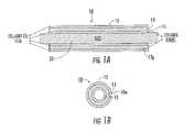
- FIG. 1Ais a schematic cross-section (in an axial direction) of an exemplary collagen fiber construct on an exemplary support member according to embodiments of the present invention.
- FIG. 1Bis an end view of the device shown in FIG. 1A (shown without the support member) according to embodiments of the present invention.
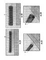
- FIGS. 2A-2Dare digital photographs of a prototype of a collagen fiber construct that may be particularly suitable for a nerve guide according to embodiments of the present invention.
- FIG. 3Ais a top perspective view of a lathe that can be used to wind collagen fiber(s) onto a tubular support member according to embodiments of the present invention.
- FIG. 3Bis a side perspective view of the device shown in FIG. 3A .
- FIG. 3Cis a side perspective view of the lathe with a substantially planar elongate support member according to embodiments of the present invention.
- FIG. 3Dis a side perspective view of a planar support member with a wound collagen fiber(s) according to other embodiments of the present invention.
- FIG. 3Eis a side perspective view of a tubular support member with an insert according to embodiments of the present invention.
- FIG. 4is a schematic illustration of different collagen fiber configurations that may be used for winding a construct according to embodiments of the present invention.
- FIG. 5Ais a schematic illustration of a tubular construct with segments having increased fiber density according to embodiments of the present invention.
- FIG. 5Bis a schematic illustration showing that the tubular structure of FIG. 5A can be separated or cut into multiple different components (shown as two) according to embodiments of the present invention.
- FIG. 6Ais a schematic illustration of a substantially planar construct with segments having increased fiber density according to embodiments of the present invention.
- FIG. 6Bis a schematic illustration of the construct shown in FIG. 6A illustrating that the construct can be separated into multiple components (shown as four) according to embodiments of the present invention.
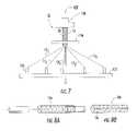
- FIG. 7is a front view of a winding apparatus that can be used to wind (braid) collagen fiber according to embodiments of the present invention.
- FIG. 8Ais a schematic illustration of a collagen nerve guide according to embodiments of the present invention.
- FIG. 8Bis a schematic illustration of a collagen cuff according to embodiments of the present invention.
- FIG. 9is a schematic illustration of a medical kit according to embodiments of the present invention.
- FIG. 10is a flow chart of operations that can be used to fabricate a construct according to embodiments of the present invention.
- FIG. 11is a flow chart of an exemplary winding protocol according to particular embodiments of the present invention.
- phrases such as “between X and Y” and “between about X and Y”should be interpreted to include X and Y.
- phrases such as “between about X and Y”mean “between about X and about Y.”
- phrases such as “from about X to Y”mean “from about X to about Y.”
- first, second, etc.may be used herein to describe various elements, components, regions, layers and/or sections, these elements, components, regions, layers and/or sections should not be limited by these terms. These terms are only used to distinguish one element, component, region, layer or section from another region, layer or section. Thus, a first element, component, region, layer or section discussed below could be termed a second element, component, region, layer or section without departing from the teachings of the present invention.
- the sequence of operations (or steps)is not limited to the order presented in the claims or figures unless specifically indicated otherwise.
- spatially relative termssuch as “under”, “below”, “lower”, “over”, “upper” and the like, may be used herein for ease of description to describe one element or feature's relationship to another element(s) or feature(s) as illustrated in the figures. It will be understood that the spatially relative terms are intended to encompass different orientations of the device in use or operation in addition to the orientation depicted in the figures. For example, if a device in the figures is inverted, elements described as “under” or “beneath” other elements or features would then be oriented “over” the other elements or features. Thus, the exemplary term “under” can encompass both an orientation of over and under.
- the devicemay be otherwise oriented (rotated 90 degrees or at other orientations) and the spatially relative descriptors used herein interpreted accordingly.
- the terms “upwardly”, “downwardly”, “vertical”, “horizontal” and the likeare used herein for the purpose of explanation only unless specifically indicated otherwise.
- the term “patch”refers to a piece or segment of biomaterial that can be placed on and/or affixed to target anatomical structure, typically soft tissue, to treat, protect, repair and/or reinforce a target site.
- the patchcan be any geometric shape but is typically substantially planar and may, in position, conform to the shape of underlying or overlying tissue.
- implantableand derivatives thereof means the device can be inserted, embedded, grafted or otherwise acutely or chronically attached or placed in or on a patient.
- constructrefers to a device and/or material in a final form for use or in a pre-final form.
- pitchmeans winding or wound at an angle relative to a first plane normal to the longitudinal axis of a core or cavity.
- windingand “wound” and derivatives thereof means to wrap about an object or center at least once, typically repeatedly, e.g., to turn in a series of circular motions.
- at least one collagen fibermultiple fibers, one or more fiber bundles
- the windingmay define a coil (e.g., a series of connected typically substantially concentric rings or spirals), woven and/or braided fiber arrangement with a number of revolutions or turns about a core and/or tube, typically in a regular pattern (but an irregular pattern may also be used) about a length of at least one layer of a tube or cylindrical shape.
- Embodiments of the present inventioncomprise collagen, typically dermal collagen.
- the collagencan be of any form and from any origin.
- the collagencan be any of the identified collagen genotypes, for example, the interstitial fiber forming collagen types I, II and III, as well as any other substantially fiber forming types of collagen, for example collagen VI.
- the collagencan be acid soluble collagen or pepsin solubilized or soluble collagen.
- the collagencan be from mammalian cells synthesized in vitro.
- the collagencan be from molecularly engineered constructs and synthesized by bacterial, yeast or any other molecularly manipulated cell type.
- the collagencan be sea cucumber dermis collagen, bovine, caprine, porcine, ovine or other suitable donor mammal, marine animal collagen such as chinoderms, molecularly engineered collagen, or gelatin (e.g., in any suitable form including solid, gel, hydrogels, liquids, or foams).
- the collagencan be digested with a protease before, where used, oxidizing and polymerizing steps.
- the collagencan be in the form of microfibrils, fibrils, natural fibers, or synthetic fibers.
- the collagencan be solubilized, dissolved or otherwise transferred into an acid solution, for example, acetic acid (e.g., about 0.01M to about 1.0M, typically about 0.5M), hydrochloric acid (between about pH 1 to about pH 3, typically about pH 2.0), or any other suitable acid at appropriate concentration (e.g., about pH 1.0 to about pH 3.0, typically about pH 2.0). Dialysis may optionally be used to neutralize a soluble collagen solution.
- the collagencan also or alternatively be dissolved in a neutral buffered solution either with or without salts, e.g., phosphate buffer at about pH 7.0, or phosphate buffered saline at about pH 7.0.
- the phosphate buffercan be at any concentration of sodium phosphate between about 0.01 and 0.5, but more typically between about 0.02 and about 0.1M.
- the buffercan also be any buffer, including, but not limited to, for example, sodium acetate, HEPES, or MOPS.
- the collagencan be present in a quantity that is at least about 0.1% to about 10%, typically between 0,1% to about 5% (e.g., about 0.1, 0.2, 0.3, 0.4, 1.0, 2.0, 4.0%) by weight per volume, or by weight per volume in the neutral buffer solution before fibrillogenesis and fiber formation.
- collagencan be present in an amount of weight by volume of between about 50-100% (e.g., at least about 75%, 90%, 95% or 100%) before crosslinking (where crosslinking is used).
- Collagen “microfibrils,” “fibrils,” “fibers,” and “natural fibers”refer to naturally-occurring structures found in a tendon. Microfibrils are about 3.5 to 50 nm in diameter. Fibrils are about 50 nm to 50 ⁇ m in diameter. Natural fibers are above 50 ⁇ m in diameter.
- a “synthetic fiber”refers to any fiber-like material that has been formed and/or chemically or physically created or altered from its naturally-occurring state. For example, an extruded fiber of fibrils formed from a digested tendon is a synthetic fiber but a tendon fiber newly harvested from a mammal is a natural fiber.
- synthetic collagen fiberscan include non-collagenous components or biocompatible materials, such as particulates, hydroxyapatite and other mineral phases, or drugs that facilitate tissue growth or other desired effects. See, U.S. Pat. No. 6,821,530, incorporated herein by reference above.
- the fibers and/or constructs formed from samecan include compositions that can contain carbon nano-tubes, zinc nano-wires, nano-crystalline diamond, or other nano-scale particulates; and larger crystalline and non-crystalline particulates such as calcium phosphate, calcium sulfate, apatite minerals.
- compositionscan also or alternatively contain therapeutic agents such as bisphosphonates, anti-inflammatory steroids, growth factors such as basic fibroblast growth factor, tumor growth factor beta, bone morphogenic proteins, platelet-derived growth factor, and insulin-like growth factors; chemotactic factors such fibronectin and hyaluronan; and extracellular matrix molecules such as aggrecan, biglycan, decorin, fibromodulin, COMP, elastin, and fibrillin.
- the fibers and/or fiber-derived constructscan contain cells, engineered cells, stem cells, and the like. Combinations of the above or other materials can be embedded, coated and/or otherwise directly or indirectly attached to the collagen fibers and/or construct formed of same.
- collagen gelmeans a semi-solid (e.g., gelatinous density) material that includes collagen fiber, fibrils and/or microfibrils, typically dermal collagen, that has been acid or pepsin solubilized (e.g., soluble collagen) and processed to maintain the collagen in its molecular form.
- the collagen concentration of the soluble collagen and/or resulting soluble collagen gelcan be between about 0.1% to about 4% weight per volume.
- the soluble collagen gelmay be formed to be in a cylindrical shape of a defined length and diameter, typically with a diameter of between about 0.1 to 1 cm, and a length of between about 5 cm to about 100 m, more typically between about 1 m to about 50 m.
- the collagen fibers and collagen gelcan be produced in batch or continuous-type systems, including wet gel collagen extrusion systems, which produce cylindrical lengths of gel that can be allowed to substantially dry (actively or passively) to obtain a suitable length of fiber.
- wet gel collagen extrusion systemswhich produce cylindrical lengths of gel that can be allowed to substantially dry (actively or passively) to obtain a suitable length of fiber.
- Examples of some collagen fiber production processes that can generate soluble collagen in suitable lengthsare described in U.S. Pat. No. 6,565,960, and pending U.S. Patent Application Publication No. US-2008-0188933-A1, the contents of which are hereby incorporated by reference.
- the collagen fiberscan be spooled for supplying to an automated or semi-automated winder to form the biomedical construct.
- the collagen fibersmay be formed with a relatively thin diameter, such as, for example between about 0.05 mm to about 0.2 mm (average), such as about 0.08 mm dry diameter (average) and about a 0.13 mm wet diameter (average).
- filmrefers to a thin layer of collagen gel that has dried.
- the filmis typically present in a thickness that is between about 5 and 200 microns.
- the filmmay be permeable and flexible and optically transmissive, e.g., translucent or transparent, or may be opaque.
- Several layers of the gelcan be applied to generate the desired film thickness or coverage.
- the color or transmissve characteristicsmay change when hydrated.
- the filmcan infuse into, migrate and/or bond to a coiled or wound (dry) collagen fiber to form a collagen fiber laminate.
- the gel/filmis not required, but where used can provide a smooth (and typically a substantially constant diameter) surface over or under the fiber.
- FIG. 1Aan exemplary elongate construct 10 is shown on a support member 20 .
- the construct 10includes an inner layer of collagen film 11 , an intermediate layer of at least one wound collagen Fiber 13 , and an outer layer of collagen film 15 .
- the construct 10can be formed without the inner and/or outer layer of film 11 and/or may optionally include other materials or constituents and/or layers.
- hydroxyapatitecan be placed into the collagen fiber and/or collagen gel material. This configuration can be particularly suitable to augment interference screw fixation of autograft tendons.
- the construct 10can have a wall 10 w with a suitable thickness defined by the at least one collagen fiber 13 and the film layers (where used) and/or other coatings and/or materials placed thereon.
- the construct 10can have an open through cavity or may be filled or partially filled with a nerve-growth media or other therapeutic material (e.g., an anti-inflammatory, antibiotic and/or the like).
- the at least one collagen fiber 13has an angular fiber pattern 13 p of repeating intersecting collagen fiber segments along its length.
- the angular pattern 13 pcan be defined by a number of revolutions of the at least one fiber 13 about the support member 20 at a given pitch or pitches for at least one layer (typically more than one layer).
- the support member 20is used to wrap the at least one collagen fiber around its exterior surface to form a desired shape.
- the support member 20can include a lubricious and/or smooth surface, or an embossed surface with lower contact surface area, typically of a polymer material.
- the support member 20can include an anti-slip surface with ridges or a sleeve can be placed over the support member (not shown) to contact the next layer (e.g., inner film 11 or fiber 13 ).
- the support member 20comprises Teflon® or other suitable low friction and/or anti-stick material.
- the support member 20can be tubular, e.g., cylindrical, as shown in FIGS. 1A , 3 A, 3 B and 3 E or may be substantially flat and rectangular 20 ′ as shown in FIGS. 3C and 3D . Other geometries may also be used, such as, for example, a frustoconical or funnel shape.
- the support member 20is elongate and has a substantially circular, oval, polygonal or other cross-sectional shape.
- the at least one collagen fiber 13can be organized into various arrays including braids, weaves, knits, parallel arrays, and various patterns.
- the orientation of one or more of the fibers 13 within the resulting material 10can be targeted to meet the specific mechanical requirements of the medical application.
- Fiber densitycan vary from dense to loose geometries and the numbers and size of the one or more collagen fibers used can vary as well as the thickness of the film to provide specific mechanical properties.
- the fiber(s) 13can be continuous length fibers or may be formed by attaching a series of collagen fibers in an end-to-end orientation 13 j ( FIG. 4 ).
- FIGS. 2A-21are digital photographs of a prototype of a construct 10 .
- This construct 10may be particularly suitable as a nerve tube or guide 10 n ( FIG. 8A ).
- the construct 10is tubular 10 t with an open cavity and has a flexible elastic configuration.
- the construct 10may be configured as a nerve guide 10 n .
- the nerve guide 10 ncan be formed using a single fiber 13 formed in wound multiple layers, the fiber 13 can have a length between about 1-6 m, typically about 5 m.
- the nerve guide 10 ncan be formed using a single fiber 13 of a continuous length that is wrapped in several layers about the support member 20 . Use of a single fiber 13 can reduce the likelihood of any fraying associated with multiple fibers (such as those wound in one lengthwise direction).
- the nerve guide 10 ncan have a length between about 1 cm to about 6 cm (or more), and the inner diameter can be between about 1-10 m with the wall thickness being about 0.1 mm to about 3 mm.
- the construct 10can have reversible elasticity with sufficient rigidity or strength to prevent undue nerve compression, while allowing flexibility sufficient to allow the construct 10 to spring back into its original shape after being exposed to a strain or tension caused by normal body movement that deforms the shape.
- the nerve guide 10 ncan be used for any nerve location, e.g., peripheral nerves (such as in a hand or finger), spinal cord nerves, and the like.
- the construct 10can be used for other repairs or treatments as will be discussed further below.
- the construct 10is biocompatible (or at least non-cytotoxic) and can provide a desired half-life suitable for its intended function.
- the construct 10 and/or the fiber 13can be cross-linked with a suitable polymerizing material, such as, but not limited to, NDGA, or may be used in a non-cross-linked state.
- NDGA cross-linkingcan increase the strength of the device 10 but may decrease the resiliency, elasticity or flexibility.
- the collagen fiber 13is not cross-linked during the winding process, but may optionally be cross-linked after the winding process (typically after the collagen film has been applied to the outer surface and dried).
- the support member 20can be configured to facilitate removal of the construct 10 .
- the construct 10may be wound tightly against the outer surface of the support member 20 and allowed to dry.
- the support member 20can be configured to reduce in cross-sectional size or disassemble with the construct 10 held thereon to allow easy removal of the elongate construct.
- the support member 20can be a multi-piece device that provides this size change.
- the support member 20may be cooled while the construct is heated to provide a size difference.
- the support member 20can cooperate with an insert 20 I ( FIG. 3D ) that provides the desired size adjustability.
- the construct 10can be removed from the support member without such a size adjustment (e.g., its inner surface may be sufficiently lubricous or a suitable liquid or other material can be used to slide the construct off the support member.
- the construct 10can be cut in a lengthwise (e.g., “X”) direction and taken off the support member 20 .
- the construct 10may be cut or otherwise separated in a long axis direction with a longitudinal slit 10 s and used for a cuff 10 e ( FIG.
- the cuff 10 cmay be configured to provide a snug or alternatively, a non-constricting, encasement for injured peripheral nerves for protection of the neural environment.
- the wall of the cuff with the longitudinal slit 10 scan be spread open for easy placement over the injured nerve or other target tissue. The resilience of the collagen conduit allows the cuff to recover and maintain closure once the device is placed around the nerve.
- the construct 10can be made by winding at least one collagen fiber 13 around a support member 20 using a computer-guided and/or controlled lathe system 100 .
- the lathe systemcan be configured to rotate the support member 20 and to move the support member back and forth in a length direction to alter the location of the fiber on the support member 20 relative to the introduction point of the fiber (e.g., the fiber introduction point may be stationary).
- the fiber(s) 13can be supplied through a head that moves relative to the support member 20 (e.g., the support member can be stationary) or both the fiber introduction head and the support member may move relative to teach other.
- transverse small cross-section support memberse.g., diameter rods
- larger transverse cross-section support memberse.g. diameter rods
- An example of a small lathe 100typically a micro or miniature lathe, suitable for fabricating embodiments of the constructs is the Model 4410 lathe available from Sherline Products, Inc., having a place of business in Vista, Calif.
- Two user-selectable inputscan operate the lathe system: one controls the speed that the support member that spins and the other controls the pattern (fiber angle) in which the at least one fiber 13 is laid onto the support member.
- the operationcan be configured so that the fiber is self-pulling from a spool in communication with a channel in the feeder head based on the speed of the spinning support member 20 .
- the lathe 100can co-wind a plurality of fibers or fiber bundles substantially concurrently about the support member 20 .
- the at least one collagen fiber 13can be coated with one or more layers of collagen gel 11 , 15 and/or other suitable bio-compatible material during and/or after winding the at least one collagen fiber 13 to seal the fiber(s) 13 within the biocomposite material and/or to form a smooth inner and/or outer surface of the construct 10 .
- FIG. 3Billustrates that collagen gel can be applied to the fiber 13 on the support member during the winding.
- FIG. 3Billustrates that a brush 111 can be used to apply the gel.
- Other application techniquesmay be used, such as spray, pour, drop, and the like.
- the application of the soluble collage gelmay be manual or automated and applied by electro-mechanical devices.
- the windingcan be performed so that at least one layer of the at least one collagen fiber has a substantially constant pitch for at least a major portion of a length thereof or so that at least one layer of the at least one collagen fiber has a variable pitch for at least a major portion of a length thereof.
- FIG. 4illustrates that different configurations of fibers 13 may be used.
- fiber bundles 13 b , 13 ttwo or more fibers 13 can be grouped together to form the fiber bundle 13 b , 13 t and that bundle 13 b , 13 t applied or wrapped about the support member 20 , similar to a single fiber.
- One or more fiber bundles 13 b , 13 tmay be used to form the construct 10 .
- Combinations of the different fiber typesmay also be used for some constructs 10 . That is, for example, a twisted fiber 13 t can be co-wound with a single fiber 13 1 and/or a single fiber 13 1 may be used to form one layer and a twisted 13 t to form a different layer, and the like.
- the collagen fiber 13can be wound using various fiber angles (e.g., pitch angles), such as, angles between about 2-70 degrees, typically between about 5-60 degrees, such as, for example, 5, 10, 15, 20, 25, 30, 35, 40, 45, 50, 54 and 55 degrees, or other odd or even numbers between 5-70. Where constructs of multiple layers are used, one layer may have a first pitch and another layer may have a different pitch.
- pitch anglese.g., angles between about 2-70 degrees, typically between about 5-60 degrees, such as, for example, 5, 10, 15, 20, 25, 30, 35, 40, 45, 50, 54 and 55 degrees, or other odd or even numbers between 5-70.
- FIG. 5Aillustrates that a construct 10 can be wound with increased fiber density 52 along certain segments, typically forming end rings 52 r .
- This increased fiber density 52can provide sufficient rigidity to allow a suture to attach thereto.
- the construct 10is tubular 10 t and may optionally include an increased density segment 52 at an intermediate location.
- FIG. 5Billustrates that the construct 10 can be used as formed, or may be cut or separated along a Y-axis into two components 10 ta , 10 tb .
- the intermediate increased density ring 52can form end rings for the separated construct 10 ta , 10 tb.
- FIG. 6Aillustrates a construct 10 that is relatively flat 10 f and/or rectangular.
- the construct 10 fcan optionally include increased fiber density segments 52 that may be suitable for end rings 52 r .
- FIG. 6illustrates that the construct 10 f can be cut along the X-axis and separated into at least two components that form biocompatible patches.
- the intermediate increased density ring(s) 52where used, can optionally form end rings 52 for the separated construct 10 fa , 10 fb , etc.
- FIG. 7illustrates an example of another automated winding system 100 ′ that can be used to form the construct 10 .
- This embodimentuses several fibers 13 , each independently wound and/or wrapped to weave or braid the fibers about the support member 20 to form the construct 10 .
- the system 100 ′includes a plate 122 supporting spindles 124 , a forming plate 126 , a support member (shown as a cylindrical mandrel) 20 that extends through an aperture in the fainting plate 126 , and braid puller 128 .
- An exemplary microbraideris believed to be available from Kokubun Ltd of Japan. See also, FIG. 2 and col. 2 of U.S. Pat. No. 7,135,040, the content of which is hereby incorporated by reference.
- the fibers 13can be wound before or after cross-linking (or not cross-linked at all). If wound before, the fibers can, where desired, be polymerized with any suitable cross-linking materials, to promote collagen organization, such as, for example, NDGA, but other cross-linking materials may be used, including, for example, glutaraldehyde.
- the (dried) collagen fibercan also be treated with other methods to improve the tensile properties of the fiber.
- the (dried) collagen fibers 13can be cross-linked with agents such as glutaraldehyde, formaldehyde, epoxy resins, tannic acid, or any other chemical agent that produces covalent cross-links between collagen molecules within fibrils or between fibrils.
- the fiber 13can be treated to induce cross-linking between collagen molecules such as, but not limited to, one or more of a carbodiimide treatment, ultraviolet irradiation either with or without carbohydrates to initiate glycation adducts, and dehydrothermal treatment coupled with any of the aforementioned methods.
- a carbodiimide treatmentultraviolet irradiation either with or without carbohydrates to initiate glycation adducts
- dehydrothermal treatmentcoupled with any of the aforementioned methods.
- FIG. 9illustrates a medical kit 250 that includes a medical device or implant 10 or 10 ′.
- the kit 250may optionally include other components, such as, for example, a container of surgical adhesive, sutures 210 , suture anchors, and the like.
- the device or implant 10 , 10 ′may be held hydrated in a flexible sealed package of sterile liquid 230 .
- the kit 250may include a temperature warning so that the construct 10 , 10 ′ is not exposed to unduly hot temperatures that may degrade the implant.
- a temperature sensor 252may optionally be included on the package of the kit to alert the clinician as to any excessive or undue temperature exposure prior to implantation.
- kit 250it may be desirable to hold or store the kit 250 (and implant or device 10 , 10 ′) at a temperature that is less than about 37° C. and/or 100° F. prior to implantation.
- the kit 250may be packaged in a housing with a temperature controlled or insulated chamber 250 e to facilitate an appropriate temperature range.
- FIG. 10is a flow chart of operations that can be used to carry out embodiments of the present invention.
- the at least one collagen fiberis wound a number of revolutions about a length of a support member having a long axis.
- the windingcan have a defined pitch and/or fiber angle relative to the long axis of the support member to form an elongate construct with at least one wound collagen fiber (block 150 ).
- the winding stepcan form multiple overlying layers of the at least one collagen fiber in one or more fiber angles so that the at least one fiber intersects itself at different locations along a length of the construct.
- a collagen gelcan be placed onto the support member and the gel can dry to form a film on the outer surface of the support member before the winding step (block 155 ).
- the collagen filmcan be dried or allowed to dry on the support member (e.g., rod).
- a soluble collagencan be applied (e.g., wrapped, painted, sprayed, dripped and the like) onto the fiber(s) and/or support member so that the fiber(s) become wet while one or more layers are wound on the lathe.
- the at least one collagen fibercan be supplied to the winder/support member in a substantially dry state and may be provided as a spooled (dry) quantity of the at least one collagen fiber (block 152 ).
- the fiber(s)can be supplied and wound in a non-cross-linked state.
- the winding stepcan be carried out to create multiple adjacent overlying layers of the at least one fiber, the adjacent layers being coextensive for at least a major portion of a length of the construct (block 153 ).
- a liquid or gel comprising soluble collagencan be placed onto the at least one wound collagen fiber to cover at least the outer surface in a collagen film (block 165 ).
- the placing of the collagen gel or liquidis carried out by placing collagen gel having a cylindrical shape around the at least one wound collagen fiber and the support member (block 158 ).
- the collagencan be polymerized while the elongate construct is held on the support member using a suitable cross-linker, such as, for example, NDGA, then removing the construct from the support member (block 166 ).
- a suitable cross-linkersuch as, for example, NDGA
- the windingcan be carried out so that the at least one fiber turns about the support member in one of a clockwise or counterclockwise direction along a first lengthwise direction for a first layer, then reverses to travel in an opposing lengthwise direction and continues to turn about the support member in the same clockwise or counterclockwise direction for a second adjacent layer (block 180 , FIG. 11 ).
- the windingmay be carried out so that the at least one collagen fiber turns (is wrapped) about the support member in one of a clockwise or counterclockwise direction along a first lengthwise direction for a first layer, then reverses to travel in an opposing lengthwise direction and turns about the support member in the other clockwise or counterclockwise direction a second adjacent layer.
- the winding stephas a first pitch for the winding of the at least one collagen fiber on the first layer and a second smaller or greater pitch for the winding of the at least one collagen fiber on the second layer.
- the at least one fiber on the second layerresides between gaps defined by the at least one fiber wound with the defined pitch on the first layer.
- the methodcan include cutting the construct in an axial direction to form a flat collagen fiber patch.
- the methodcan include winding the collagen fibers in a plurality of axially spaced apart segments with increased collagen fiber density, at least some of which are provided as reinforced segments for suturing.
- the reinforced segmentscan be formed at end portions of the tube and optionally at one or more intermediate locations therebetween.
- the methodscan produce a nerve guide having sufficient strength and elasticity to withstand buckling and to be able to bend and to elastically return to its original shape after bending to inhibit occlusive pressures or restrictions on nerves.
- Embodiments of the inventioncan be used for a number of different medical applications, including, but not limited to, nerve guides, wound bed patches, muscle or organ patches, cardiac patches, valve replacements or repairs, hernia patches, skin patches, burn treatment patches, skin/tissue repair patches or cuffs, blood vessel (artery, vein, and the like) repairs, sleeves that can reside about repairing tendon to prevent or inhibit adhesions, indwelling tubes for delivery of therapeutic agents, ducts such as lymphatic, hepatic, pancreatic and cystic ducts, tubes such as ureter and urethra tubes and the like.
- FIGS. 2A-2Dillustrate exemplary sleeves or tubes of wound NDGA-collagen fibers that may be particularly suitable for nerve guides.
- the inner diameter of the tubecan vary between about 1 and 10 mm.
- the thickness of the wallcan vary between about 0.1 and 3 mm.
- the length of the tubecan vary from between about 1 to 6 cm or more.
- the tubecan be made of dermal collagen that is acid or pepsin soluble.
- the soluble collagencan be made by neutralizing acid soluble collagen and keeping the soluble collagen at a desired low temperature to maintain the collagen in molecular form, (e.g., about 4° C.).
- Collagen gelscan be produced from acid soluble collagen by neutralization, injection molding in a Teflon® tube of diameter between 0.1 cm to 1.0 cm and incubation for at least about 4 hours at 37° C. The resulting gel can be extruded into deionized water to form a gel cylinder with a diameter between about 0.1 cm to 1.0 cm (and can have a length between about 1-100 m.
- Collagen concentration of the soluble collagen and collagen gelcan be from about 0.1-4% weight per volume.
- the gel cylindercan be used in the gel form or allowed to dry, actively or passively (suspended in air), to form a collagen fiber having a diameter between about 0.05 mm (average) to about 0.2 mm (average).
- the first step to make this prototype tubeis to wrap the collagen gel of specified collagen concentration and diameter onto a Teflon® rod of selected diameter.
- the collagen gel layerwas allowed to dry on the rod at room temperature to form a thin layer of collagen film.
- the thickness of this collagen filmcan be varied by applying more or less layers of collagen gel, either is a single application of in several applications.
- the second stepis to wind dry collagen fibers on to the collagen film coated Teflon® rod.
- the pitch of the fiber relative to the long axis of the tubecan be specified.
- the thickness of the collagen windingcan be adjusted, for example, corresponding to the number of layers of fibers that are laid on (and/or the number of fibers bundled together for the winding).
- soluble collagenis applied (e.g., painted) onto the surface of the laid-on fibers.
- the thickness of the final soluble collagen layercan be varied to achieve specific thickness.
- the soluble collagen coated fiber wound cylinderis allowed to dry.
- the third step in making the tubeis the same as the first step, e.g., to wrap a collagen gel on to the collagen fiber would Teflon® rod and the gel layer is allowed to dry to form a collagen film enwrapping the collagen fiber tube.
- the thickness of the penultimate collagen filmcan be varied by the number of layers of wrapped gel.
- the dried tubecan be used “as-is” (used in a non-cross-linked state and hydrated when in the body or prior to placement in the body), or it can be cross-linked with any agent or action that cross-links the collagen.
- the (nerve) tubeis then taken off the Teflon® rod.
- the tubeis cross-linked with nor-dihydroguaiaretic acid (NDGA), see, e.g., U.S. Pat. No. 6,565,960, and U.S. Patent Application Publication No. US-2008-0161917-A1, the contents of which are hereby incorporated by reference as if recited in full herein.
- NDGAnor-dihydroguaiaretic acid
Landscapes
- Health & Medical Sciences (AREA)
- Life Sciences & Earth Sciences (AREA)
- Veterinary Medicine (AREA)
- Public Health (AREA)
- Animal Behavior & Ethology (AREA)
- General Health & Medical Sciences (AREA)
- Chemical & Material Sciences (AREA)
- Oral & Maxillofacial Surgery (AREA)
- Transplantation (AREA)
- Epidemiology (AREA)
- Medicinal Chemistry (AREA)
- Engineering & Computer Science (AREA)
- Dermatology (AREA)
- Biomedical Technology (AREA)
- Surgery (AREA)
- Heart & Thoracic Surgery (AREA)
- Vascular Medicine (AREA)
- Molecular Biology (AREA)
- Nuclear Medicine, Radiotherapy & Molecular Imaging (AREA)
- Medical Informatics (AREA)
- Cardiology (AREA)
- Materials Engineering (AREA)
- Pulmonology (AREA)
- Gastroenterology & Hepatology (AREA)
- Composite Materials (AREA)
- Inorganic Chemistry (AREA)
- Hematology (AREA)
- Biophysics (AREA)
- Orthopedic Medicine & Surgery (AREA)
- Rheumatology (AREA)
- Neurology (AREA)
- Dispersion Chemistry (AREA)
- Materials For Medical Uses (AREA)
- Prostheses (AREA)
- Chemical Kinetics & Catalysis (AREA)
- Polymers & Plastics (AREA)
- Organic Chemistry (AREA)
- Rehabilitation Therapy (AREA)
Abstract
Description
Claims (34)
Priority Applications (5)
| Application Number | Priority Date | Filing Date | Title |
|---|---|---|---|
| US12/576,435US9078775B2 (en) | 2008-10-09 | 2009-10-09 | Methods of making collagen fiber medical constructs and related medical constructs, including nerve guides and patches |
| US13/153,665US9179976B2 (en) | 2008-10-09 | 2011-06-06 | Methods of making collagen fiber medical constructs and related medical constructs, including tubes |
| US14/734,548US10238773B2 (en) | 2008-10-09 | 2015-06-09 | Methods of making collagen fiber medical constructs and related medical constructs, including nerve guides and patches |
| US14/875,202US9801978B2 (en) | 2008-10-09 | 2015-10-05 | Medical constructs including tubes and collagen fibers |
| US15/795,881US20180177921A1 (en) | 2008-10-09 | 2017-10-27 | Medical constructs including tubes and collagen fibers |
Applications Claiming Priority (3)
| Application Number | Priority Date | Filing Date | Title |
|---|---|---|---|
| US10399508P | 2008-10-09 | 2008-10-09 | |
| US13816508P | 2008-12-17 | 2008-12-17 | |
| US12/576,435US9078775B2 (en) | 2008-10-09 | 2009-10-09 | Methods of making collagen fiber medical constructs and related medical constructs, including nerve guides and patches |
Related Child Applications (2)
| Application Number | Title | Priority Date | Filing Date |
|---|---|---|---|
| US13/153,665Continuation-In-PartUS9179976B2 (en) | 2008-10-09 | 2011-06-06 | Methods of making collagen fiber medical constructs and related medical constructs, including tubes |
| US14/734,548DivisionUS10238773B2 (en) | 2008-10-09 | 2015-06-09 | Methods of making collagen fiber medical constructs and related medical constructs, including nerve guides and patches |
Publications (2)
| Publication Number | Publication Date |
|---|---|
| US20100094318A1 US20100094318A1 (en) | 2010-04-15 |
| US9078775B2true US9078775B2 (en) | 2015-07-14 |
Family
ID=42099574
Family Applications (7)
| Application Number | Title | Priority Date | Filing Date |
|---|---|---|---|
| US12/576,423Active2030-12-07US8367148B2 (en) | 2008-10-09 | 2009-10-09 | Methods of making biocomposite medical constructs and related constructs including artificial tissues, vessels and patches |
| US12/576,435Active2032-02-03US9078775B2 (en) | 2008-10-09 | 2009-10-09 | Methods of making collagen fiber medical constructs and related medical constructs, including nerve guides and patches |
| US13/153,665Active2031-01-30US9179976B2 (en) | 2008-10-09 | 2011-06-06 | Methods of making collagen fiber medical constructs and related medical constructs, including tubes |
| US13/729,668ActiveUS9125759B2 (en) | 2008-10-09 | 2012-12-28 | Biocomposite medical constructs including artificial tissues, vessels and patches |
| US14/734,548Active2031-09-21US10238773B2 (en) | 2008-10-09 | 2015-06-09 | Methods of making collagen fiber medical constructs and related medical constructs, including nerve guides and patches |
| US14/875,202ActiveUS9801978B2 (en) | 2008-10-09 | 2015-10-05 | Medical constructs including tubes and collagen fibers |
| US15/795,881AbandonedUS20180177921A1 (en) | 2008-10-09 | 2017-10-27 | Medical constructs including tubes and collagen fibers |
Family Applications Before (1)
| Application Number | Title | Priority Date | Filing Date |
|---|---|---|---|
| US12/576,423Active2030-12-07US8367148B2 (en) | 2008-10-09 | 2009-10-09 | Methods of making biocomposite medical constructs and related constructs including artificial tissues, vessels and patches |
Family Applications After (5)
| Application Number | Title | Priority Date | Filing Date |
|---|---|---|---|
| US13/153,665Active2031-01-30US9179976B2 (en) | 2008-10-09 | 2011-06-06 | Methods of making collagen fiber medical constructs and related medical constructs, including tubes |
| US13/729,668ActiveUS9125759B2 (en) | 2008-10-09 | 2012-12-28 | Biocomposite medical constructs including artificial tissues, vessels and patches |
| US14/734,548Active2031-09-21US10238773B2 (en) | 2008-10-09 | 2015-06-09 | Methods of making collagen fiber medical constructs and related medical constructs, including nerve guides and patches |
| US14/875,202ActiveUS9801978B2 (en) | 2008-10-09 | 2015-10-05 | Medical constructs including tubes and collagen fibers |
| US15/795,881AbandonedUS20180177921A1 (en) | 2008-10-09 | 2017-10-27 | Medical constructs including tubes and collagen fibers |
Country Status (4)
| Country | Link |
|---|---|
| US (7) | US8367148B2 (en) |
| EP (3) | EP2349363B1 (en) |
| CA (4) | CA2740008C (en) |
| WO (3) | WO2010042205A2 (en) |
Cited By (4)
| Publication number | Priority date | Publication date | Assignee | Title |
|---|---|---|---|---|
| US9808492B2 (en) | 2012-09-10 | 2017-11-07 | Terry W. Broussard | Methods of preparing lyophilized human tissues |
| US10441664B2 (en) | 2012-11-19 | 2019-10-15 | Mimedx Group, Inc. | Cross-linked collagen with at least one bound antimicrobial agent for in vivo release of the agent |
| US11583609B2 (en) | 2018-08-08 | 2023-02-21 | Mimedx Group, Inc. | Collagen constructs and methods of making the same |
| US11589973B2 (en) | 2018-05-16 | 2023-02-28 | Mimedx Group, Inc. | Methods of making collagen fiber medical constructs and related medical constructs, including patches |
Families Citing this family (99)
| Publication number | Priority date | Publication date | Assignee | Title |
|---|---|---|---|---|
| JP4596335B2 (en)* | 2007-12-07 | 2010-12-08 | 東洋紡績株式会社 | Method for manufacturing nerve regeneration induction tube |
| CA2740008C (en) | 2008-10-09 | 2017-01-31 | Mimedx, Inc. | Methods of making biocomposite medical constructs and related constructs including artificial tissues, vessels and patches |
| CA2750605C (en) | 2009-01-23 | 2019-01-22 | Royal College Of Surgeons In Ireland | Layered scaffold suitable for osteochondral repair |
| US8858577B2 (en) | 2010-05-19 | 2014-10-14 | University Of Utah Research Foundation | Tissue stabilization system |
| US20120012771A1 (en)* | 2010-07-16 | 2012-01-19 | Lale Korkmaz | Ball seat having collapsible helical seat |
| FR2962646B1 (en) | 2010-07-16 | 2012-06-22 | Sofradim Production | PROSTHETIC WITH RADIO OPAQUE ELEMENT |
| US8758374B2 (en) | 2010-09-15 | 2014-06-24 | University Of Utah Research Foundation | Method for connecting nerves via a side-to-side epineurial window using artificial conduits |
| US8852214B2 (en) | 2011-02-04 | 2014-10-07 | University Of Utah Research Foundation | System for tissue fixation to bone |
| WO2012121986A2 (en) | 2011-03-08 | 2012-09-13 | Mimedx, Inc.. | Collagen fiber ribbons with integrated fixation sutures and methods of making the same |
| FR2977790B1 (en) | 2011-07-13 | 2013-07-19 | Sofradim Production | PROSTHETIC FOR UMBILIC HERNIA |
| US20130178874A1 (en)* | 2011-07-19 | 2013-07-11 | Hilton Becker | Composite implant |
| EP2747796B8 (en)* | 2011-08-25 | 2018-12-26 | Universität Zürich | A device for repair surgery of cylindrical organs, particularly ruptured tendons |
| WO2013066619A1 (en) | 2011-10-17 | 2013-05-10 | University Of Utah Research Foundation | Methods and devices for connecting nerves |
| US10842494B2 (en) | 2011-10-17 | 2020-11-24 | University Of Utah Research Foundation | Methods and devices for connecting nerves |
| US9179994B2 (en) | 2011-10-25 | 2015-11-10 | Covidien Lp | Implantable film/mesh composite |
| US9005308B2 (en)* | 2011-10-25 | 2015-04-14 | Covidien Lp | Implantable film/mesh composite for passage of tissue therebetween |
| US8986378B2 (en) | 2011-11-02 | 2015-03-24 | Mimedx Group, Inc. | Implantable collagen devices and related methods and systems of making same |
| CA2854885C (en) | 2011-11-10 | 2020-08-25 | Lifecell Corporation | Device for tendon and ligament treatment |
| US20130218125A1 (en)* | 2012-02-16 | 2013-08-22 | Covidien Lp | Implantable Devices Including A Mesh And A Perforated Film |
| US20130331802A1 (en)* | 2012-06-06 | 2013-12-12 | Cory Collinge | Wound Treatment Medium and Method of Packaging and Use |
| FR2992662B1 (en) | 2012-06-28 | 2014-08-08 | Sofradim Production | KNIT WITH PICOTS |
| WO2014003844A1 (en)* | 2012-06-29 | 2014-01-03 | University Of South Florida | Biocompatible polyacrylate compositions and methods of use |
| FR2992547B1 (en) | 2012-06-29 | 2015-04-24 | Sofradim Production | PROSTHETIC FOR HERNIA |
| US9644177B2 (en)* | 2012-07-12 | 2017-05-09 | Board Of Regents, The University Of Texas System | Extracellular matrix films and methods of making and using same |
| EP2687188A1 (en)* | 2012-07-20 | 2014-01-22 | Le Centre National De La Recherche Scientifique | Artificial tendon or ligament with varying stiffness along its length |
| US10130346B2 (en) | 2012-07-24 | 2018-11-20 | Omrix Biopharmaceuticals Ltd. | Device and method for the application of a curable fluid composition to a bodily organ |
| US10390935B2 (en) | 2012-07-30 | 2019-08-27 | Conextions, Inc. | Soft tissue to bone repair devices, systems, and methods |
| US11944531B2 (en) | 2012-07-30 | 2024-04-02 | Conextions, Inc. | Devices, systems, and methods for repairing soft tissue and attaching soft tissue to bone |
| US11957334B2 (en) | 2012-07-30 | 2024-04-16 | Conextions, Inc. | Devices, systems, and methods for repairing soft tissue and attaching soft tissue to bone |
| US10219804B2 (en) | 2012-07-30 | 2019-03-05 | Conextions, Inc. | Devices, systems, and methods for repairing soft tissue and attaching soft tissue to bone |
| US11253252B2 (en) | 2012-07-30 | 2022-02-22 | Conextions, Inc. | Devices, systems, and methods for repairing soft tissue and attaching soft tissue to bone |
| US9427309B2 (en)* | 2012-07-30 | 2016-08-30 | Conextions, Inc. | Soft tissue repair devices, systems, and methods |
| US10835241B2 (en) | 2012-07-30 | 2020-11-17 | Conextions, Inc. | Devices, systems, and methods for repairing soft tissue and attaching soft tissue to bone |
| US9943551B2 (en) | 2012-08-15 | 2018-04-17 | Mimedx Group, Inc. | Tissue grafts composed of micronized placental tissue and methods of making and using the same |
| US9180145B2 (en) | 2012-10-12 | 2015-11-10 | Mimedx Group, Inc. | Compositions and methods for recruiting and localizing stem cells |
| US8946163B2 (en) | 2012-11-19 | 2015-02-03 | Mimedx Group, Inc. | Cross-linked collagen comprising metallic anticancer agents |
| US8940684B2 (en) | 2012-11-19 | 2015-01-27 | Mimedx Group, Inc. | Cross-linked collagen comprising an antifungal agent |
| WO2014113733A1 (en) | 2013-01-18 | 2014-07-24 | Mimedx Group, Inc. | Methods for treating cardiac conditions |
| US10206977B1 (en) | 2013-01-18 | 2019-02-19 | Mimedx Group, Inc. | Isolated placental stem cell recruiting factors |
| WO2014143925A1 (en)* | 2013-03-15 | 2014-09-18 | New York Society For The Ruptured And Crippled Maintaining The Hospital For Special Surgery | Carbon nanotubes and graphene patches and implants for biological tissue |
| US10029030B2 (en) | 2013-03-15 | 2018-07-24 | Mimedx Group, Inc. | Molded placental tissue compositions and methods of making and using the same |
| US10335433B2 (en) | 2013-04-10 | 2019-07-02 | Mimedx Group, Inc. | NDGA polymers and metal complexes thereof |
| US9446142B2 (en)* | 2013-05-28 | 2016-09-20 | Mimedx Group, Inc. | Polymer chelator conjugates |
| US20160135946A1 (en)* | 2013-06-28 | 2016-05-19 | Saga University | Patch-type artificial skin preparation |
| JP6756612B2 (en) | 2013-08-30 | 2020-09-16 | ミメディクス グループ インコーポレイテッド | Finely divided placental composition containing a chelator |
| WO2015093018A1 (en)* | 2013-12-16 | 2015-06-25 | エーザイ・アール・アンド・ディー・マネジメント株式会社 | Revascularization graft material |
| JP7464348B2 (en) | 2014-01-17 | 2024-04-09 | ミメディクス グループ インコーポレイテッド | Methods for inducing angiogenesis |
| EP3104809B1 (en)* | 2014-02-13 | 2017-12-27 | Antonio Sambusseti | Non-absorbable tissue reconstruction device, in particular for tissues such as ligaments |
| KR20160131030A (en)* | 2014-03-11 | 2016-11-15 | 안토니오 삼부세티 | Absorbable tissue reconstruction device, in particular for tissues such as ligaments |
| WO2015138760A1 (en) | 2014-03-12 | 2015-09-17 | Conextions, Inc. | Soft tissue repair devices, systems, and methods |
| US11583384B2 (en) | 2014-03-12 | 2023-02-21 | Conextions, Inc. | Devices, systems, and methods for repairing soft tissue and attaching soft tissue to bone |
| US9713510B2 (en) | 2014-03-17 | 2017-07-25 | Implant Direct Sybron International Llc | Drill limit system and method of using same |
| EP3139972B1 (en)* | 2014-05-08 | 2020-04-29 | The Secant Group, LLC | Composite lumen with reinforcing textile and matrix |
| US20170246351A1 (en)* | 2014-09-02 | 2017-08-31 | Rutgers, The State University Of New Jersey | Biocompatible Textile Sleeves to Support and Guide Muscle Regeneration and Methods of Use Thereof |
| CN104921841B (en)* | 2015-04-10 | 2017-03-29 | 南开大学 | A kind of preparation method of double-decker artificial blood vessel |
| US10413635B2 (en)* | 2015-08-17 | 2019-09-17 | Vivex Biomedical, Inc. | Umbilical cord transplant product |
| CN106480607A (en)* | 2015-08-24 | 2017-03-08 | 上海微创医疗器械(集团)有限公司 | Static spinning membrane, the preparation method of static spinning membrane and composite membrane |
| EP3337923B2 (en) | 2015-09-21 | 2023-01-04 | Modern Meadow, Inc. | Fiber reinforced tissue composites |
| AU2016344019B2 (en) | 2015-10-30 | 2021-07-15 | New York Society For The Relief Of The Ruptured And Crippled, Maintaining The Hospital For Special Surgery | Suture sleeve patch and methods of delivery within an existing arthroscopic workflow |
| US11523812B2 (en) | 2016-02-01 | 2022-12-13 | Medos International Sarl | Soft tissue fixation repair methods using tissue augmentation constructs |
| US20170273680A1 (en) | 2016-02-01 | 2017-09-28 | DePuy Synthes Products, Inc. | Tissue augmentation tacks for use with soft tissue fixation repair systems and methods |
| US11484401B2 (en) | 2016-02-01 | 2022-11-01 | Medos International Sarl | Tissue augmentation scaffolds for use in soft tissue fixation repair |
| KR20170096093A (en) | 2016-02-15 | 2017-08-23 | 브렌던 패트릭 퍼셀 | Composite biofabricated material |
| US11110197B2 (en)* | 2016-02-24 | 2021-09-07 | Georgia Tech Research Corporation | Surgical suture materials with porous sheaths for drug delivery |
| EP3420020B1 (en) | 2016-02-25 | 2020-07-22 | The Secant Group, LLC | Composite containing poly(glycerol sebacate) filler |
| US11696822B2 (en) | 2016-09-28 | 2023-07-11 | Conextions, Inc. | Devices, systems, and methods for repairing soft tissue and attaching soft tissue to bone |
| EP3600465A1 (en) | 2017-03-31 | 2020-02-05 | The Secant Group, LLC | Cured biodegradable microparticles and scaffolds and methods of making and using the same |
| CN107385577B (en)* | 2017-08-22 | 2019-06-25 | 安徽佳元工业纤维有限公司 | The method of multi cord first twist differentiation is realized on a kind of two-for-one twister |
| US11065099B2 (en) | 2017-10-06 | 2021-07-20 | The Secant Group, Llc | Flexible hollow lumen composite |
| AU2018253595A1 (en) | 2017-11-13 | 2019-05-30 | Modern Meadow, Inc. | Biofabricated leather articles having zonal properties |
| US12102317B2 (en) | 2017-12-20 | 2024-10-01 | Conextions, Inc. | Devices, systems, and methods for repairing soft tissue and attaching soft tissue to bone |
| US10973509B2 (en) | 2017-12-20 | 2021-04-13 | Conextions, Inc. | Devices, systems, and methods for repairing soft tissue and attaching soft tissue to bone |
| US11547397B2 (en) | 2017-12-20 | 2023-01-10 | Conextions, Inc. | Devices, systems, and methods for repairing soft tissue and attaching soft tissue to bone |
| GB201808106D0 (en) | 2018-05-18 | 2018-07-04 | Cambridge Entpr Ltd | Collagen biomaterials and methods for manufacturing collagen biomaterials |
| EP3595589B1 (en) | 2018-05-21 | 2024-10-09 | Aran Biomedical Teoranta | Insertable medical devices with low profile composite coverings |
| EP3879019B1 (en)* | 2018-09-27 | 2024-03-27 | Sofradim Production | Bioresorbable knit for hernia repair |
| US20200129435A1 (en)* | 2018-10-24 | 2020-04-30 | Uvic Industry Partnerships Inc. | Composition for delivering a therapeutic agent and methods for making and using |
| WO2020147733A1 (en)* | 2019-01-16 | 2020-07-23 | 武汉杨森生物技术有限公司 | Method for preparing material for artificial blood vessel, artificial blood vessel prepared thereby and application thereof |
| CA3121853A1 (en) | 2019-01-17 | 2020-07-23 | Modern Meadow, Inc. | Layered collagen materials and methods of making the same |
| WO2021035679A1 (en)* | 2019-08-30 | 2021-03-04 | 江南大学 | Tissue engineered nerve graft and preparation method therefor |
| US20220338910A1 (en)* | 2019-09-25 | 2022-10-27 | Cannuflow, Inc. | Method and devices for implantation of biologic constructs |
| JP7732983B2 (en)* | 2019-12-09 | 2025-09-02 | モダン メドウ,インコーポレイテッド | Leather-like material comprising melt-blended collagen and thermoplastic polymer |
| WO2021140366A1 (en)* | 2020-01-07 | 2021-07-15 | Kci Licensing, Inc. | Means to improve usability of a wound insert for application to deep wounds |
| EP3854348A1 (en)* | 2020-01-23 | 2021-07-28 | Kardiozis SAS | Endoprosthesis and method of attaching a fiber to a surface |
| EP3854349A1 (en)* | 2020-01-23 | 2021-07-28 | Kardiozis SAS | Endoprosthesis and method of manufacturing an endoprosthesis |
| WO2021167898A1 (en)* | 2020-02-18 | 2021-08-26 | Smith & Nephew, Inc. | Bioactive scaffold augmentation for acl reconstruction |
| US11471264B2 (en)* | 2020-04-01 | 2022-10-18 | Medtronic Vascular, Inc. | Branching stent graft with mechanical interlock |
| WO2022081856A1 (en)* | 2020-10-16 | 2022-04-21 | The Johns Hopkins University | Biodegradable nanofiber conical conduits for nerve repair |
| US20220193302A1 (en)* | 2020-11-30 | 2022-06-23 | Embody, Inc. | Micropost array apparatus and composite biological scaffold |
| CN114836894A (en)* | 2021-02-01 | 2022-08-02 | 金湖三木机械制造实业有限公司 | Fiber braided tube production line |
| CN113045645B (en)* | 2021-03-15 | 2024-03-22 | 张梦蝶 | Raw pigskin collagen fiber extraction element of medical treatment usefulness |
| EP4082448A1 (en)* | 2021-04-26 | 2022-11-02 | Tissium SA | Nerve conduit |
| US12076437B2 (en) | 2021-07-12 | 2024-09-03 | Brown University | Proangiogenic protein cocktails delivered in custom biomaterials to revascularize ischemic tissue |
| US11959054B2 (en)* | 2021-11-04 | 2024-04-16 | Upside Foods, Inc. | Substrate apparatus with multi-layer substrate for cell-based meat cultivators |
| CN116135181A (en)* | 2021-11-16 | 2023-05-19 | 浙江脉通智造科技(集团)有限公司 | Artificial blood vessel and artificial patch and preparation method thereof |
| US12121440B2 (en) | 2022-06-24 | 2024-10-22 | Secada Medical Llc | Bone graft system |
| US12232944B2 (en) | 2022-08-26 | 2025-02-25 | Checkpoint Surgical, Inc. | Chitosan tubular member and method of producing |
| WO2024078716A1 (en)* | 2022-10-12 | 2024-04-18 | Tissium S.A. | Nerve conduit |
| EP4577394A2 (en)* | 2022-10-14 | 2025-07-02 | Michael Francis | Microfiber implant made by winding filament |
Citations (92)
| Publication number | Priority date | Publication date | Assignee | Title |
|---|---|---|---|---|
| US3316699A (en) | 1963-10-16 | 1967-05-02 | Klinger Mfg Co Ltd | Apparatus for twisting yarn |
| US3700489A (en) | 1970-07-30 | 1972-10-24 | Ethicon Inc | Process for applying a thin coating of polytetrafluoroethylene |
| WO1982001647A1 (en) | 1980-11-17 | 1982-05-27 | Robert L Kaster | Vascular graft |
| US4590928A (en) | 1980-09-25 | 1986-05-27 | South African Invention Development Corporation | Surgical implant |
| US4778467A (en)* | 1984-04-25 | 1988-10-18 | The University Of Utah | Prostheses and methods for promoting nerve regeneration and for inhibiting the formation of neuromas |
| US4792336A (en) | 1986-03-03 | 1988-12-20 | American Cyanamid Company | Flat braided ligament or tendon implant device having texturized yarns |
| US4841962A (en) | 1984-03-27 | 1989-06-27 | Berg Richard A | Collagen matrix/polymer film composite dressing |
| US4883486A (en) | 1988-05-31 | 1989-11-28 | Indu Kapadia | Prosthetic ligament |
| US4923380A (en)* | 1984-02-21 | 1990-05-08 | Bioetica, S.A. | Apparatus for the extrusion of collogen tubes |
| US4979956A (en) | 1987-10-30 | 1990-12-25 | Pfizer Hospital Products Group, Inc. | Device and method for tendon and ligament repair |
| US5028695A (en)* | 1988-03-11 | 1991-07-02 | Chemokol Gesellschaft Zur Entwicklung Von Kollagenprodukten | Process for the manufacture of collagen membranes used for hemostasis, the dressing of wounds and for implants |
| US5078744A (en) | 1987-09-04 | 1992-01-07 | Bio-Products, Inc. | Method of using tendon/ligament substitutes composed of long, parallel, non-antigenic tendon/ligament fibers |
| US5106949A (en) | 1989-09-15 | 1992-04-21 | Organogenesis, Inc. | Collagen compositions and methods for preparation thereof |
| US5162430A (en) | 1988-11-21 | 1992-11-10 | Collagen Corporation | Collagen-polymer conjugates |
| US5256418A (en) | 1990-04-06 | 1993-10-26 | Organogenesis, Inc. | Collagen constructs |
| US5263984A (en) | 1987-07-20 | 1993-11-23 | Regen Biologics, Inc. | Prosthetic ligaments |
| US5378469A (en) | 1990-04-06 | 1995-01-03 | Organogenesis, Inc. | Collagen threads |
| WO1996014095A1 (en) | 1994-11-07 | 1996-05-17 | The University Of Medicine And Dentistry Of New Jersey | Synthetic collagen orthopaedic structures such as grafts, tendons and other structures |
| US5569302A (en) | 1990-12-13 | 1996-10-29 | United States Surgical Corporation | Tipped multifilament surgical suture |
| US5656605A (en) | 1994-01-26 | 1997-08-12 | Institute Of Molecular Biology, Inc. | Device to promote drug-induced nerve regeneration |
| US5713374A (en) | 1995-02-10 | 1998-02-03 | The Hospital For Joint Diseases Orthopaedic Institute | Fixation method for the attachment of wound repair materials to cartilage defects |
| US5718717A (en) | 1996-08-19 | 1998-02-17 | Bonutti; Peter M. | Suture anchor |
| US5718012A (en) | 1996-05-28 | 1998-02-17 | Organogenesis, Inc. | Method of strength enhancement of collagen constructs |
| US6090117A (en) | 1996-11-20 | 2000-07-18 | Yasuhiko Shimizu | Artificial neural canal |
| WO2001015754A1 (en) | 1999-08-31 | 2001-03-08 | Virginia Commonwealth University Intellectual Property Foundation | Engineered muscle |
| CA2285161A1 (en) | 1999-10-06 | 2001-04-06 | 1218122 Ontario Inc. | In vitro cultured ligament tissue and method of making same |
| US6224630B1 (en) | 1998-05-29 | 2001-05-01 | Advanced Bio Surfaces, Inc. | Implantable tissue repair device |
| US6277397B1 (en) | 1996-11-20 | 2001-08-21 | Yasuhiko Shimizu | Collagen material and process for producing the same |
| US6280474B1 (en) | 1997-01-09 | 2001-08-28 | Neucoll, Inc. | Devices for tissue repair and methods for preparation and use thereof |
| US20010018619A1 (en) | 1998-12-22 | 2001-08-30 | Robert-Jan Enzerink | Graft material convenience package |
| US6292697B1 (en) | 2000-02-15 | 2001-09-18 | Medtronic, Inc. | Testing sterile packaged components of an implantable medical device prior to chronic implantation |
| WO2001072241A1 (en) | 2000-03-24 | 2001-10-04 | Drexel University | Ligament replacement constructs and methods for production and use thereof |
| US6335007B1 (en) | 1997-05-28 | 2002-01-01 | Yasuhiko Shimizu | Collagen gel |
| US20020037940A1 (en) | 2000-06-01 | 2002-03-28 | Koob Thomas J. | Polymer composite compositions |
| US6420625B1 (en) | 1997-09-12 | 2002-07-16 | Kimberly-Clark Worldwide, Inc. | Breathable, liquid-impermeable, apertured film/nonwoven laminate and process for making same |
| US20020123805A1 (en) | 1999-06-22 | 2002-09-05 | Murray Martha M. | Biologic replacement for fibrin clot |
| US20030003157A1 (en)* | 2001-06-06 | 2003-01-02 | University Of Medicine & Dentistry Of New Jersey | Collagen compositions and methods for making and using the same |
| US6514286B1 (en) | 1996-12-03 | 2003-02-04 | Osteobiologics, Inc. | Biodegradable polymeric film |
| US6531147B2 (en) | 1996-03-22 | 2003-03-11 | Focal, Inc. | Compliant tissue sealants |
| US20030100108A1 (en) | 2001-11-16 | 2003-05-29 | Altman Gregory H. | Matrix for the production of tissue engineered ligaments, tendons and other tissue |
| EP1319415A1 (en) | 2001-12-13 | 2003-06-18 | Nipro Corporation | Adhesion preventive membrane, method of producing a collagen single strand, collagen nonwoven fabric and method and apparatus for producing the same |
| US6589257B1 (en) | 1998-06-10 | 2003-07-08 | Tapic International Co., Ltd. | Artificial neural tube |
| US6592623B1 (en) | 1999-08-31 | 2003-07-15 | Virginia Commonwealth University Intellectual Property Foundation | Engineered muscle |
| US6596296B1 (en) | 1999-08-06 | 2003-07-22 | Board Of Regents, The University Of Texas System | Drug releasing biodegradable fiber implant |
| US6645247B2 (en) | 1999-10-08 | 2003-11-11 | Bret A. Ferree | Supplementing engineered annulus tissues with autograft of allograft tendons |
| US20030211130A1 (en) | 2002-02-22 | 2003-11-13 | Sanders Joan E. | Bioengineered tissue substitutes |
| US20030230316A1 (en) | 2002-06-18 | 2003-12-18 | Glucksman Dov Z. | Battery operated hair braider |
| US6692528B2 (en) | 2000-11-09 | 2004-02-17 | The Polymer Technology Group Incorporated | Devices that change size/shape via osmotic pressure |
| US6713537B1 (en) | 1999-07-14 | 2004-03-30 | Kaneka Corporation | Regenerated collagen fiber with excellent heat resistance |
| US6730124B2 (en) | 2002-03-08 | 2004-05-04 | Musculoskeletal Transplant Foundation | Bone-tendon-bone assembly with cancellous allograft bone block |
| US20040110439A1 (en) | 2001-04-20 | 2004-06-10 | Chaikof Elliot L | Native protein mimetic fibers, fiber networks and fabrics for medical use |
| US6752831B2 (en) | 2000-12-08 | 2004-06-22 | Osteotech, Inc. | Biocompatible osteogenic band for repair of spinal disorders |
| US20040131562A1 (en) | 2002-04-18 | 2004-07-08 | Gower Laurie B. | Biomimetic organic/inorganic composites, processes for their production, and methods of use |
| US20040193241A1 (en) | 1997-08-01 | 2004-09-30 | Stinson Jonathan S. | Bioabsorbable self-expanding stent |
| US20040267362A1 (en) | 2003-06-30 | 2004-12-30 | Julia Hwang | Scaffold for connective tissue repair |
| US20050009178A1 (en) | 2003-06-04 | 2005-01-13 | University Of South Carolina | Tissue scaffold having aligned fibrils, apparatus and method for producing the same, and artificial tissue and methods of use thereof |
| US6866681B2 (en) | 1996-11-20 | 2005-03-15 | Jacques-Phillippe Laboureau | Pre-adjusted prosthetic ligament and method of manufacture |
| US6936072B2 (en) | 1999-08-18 | 2005-08-30 | Intrinsic Therapeutics, Inc. | Encapsulated intervertebral disc prosthesis and methods of manufacture |
| US6953482B2 (en)* | 2001-04-26 | 2005-10-11 | Nipro Corporation | Instrument for regenerating living organism tissue or organ |
| US6977231B1 (en) | 1999-01-21 | 2005-12-20 | Nipro Corporation | Suturable adhesion-preventing membrane |
| US20060095134A1 (en) | 2004-10-28 | 2006-05-04 | Sdgi Holdings, Inc. | Materials, devices and methods for implantation of transformable implants |
| US20060100647A1 (en) | 2002-12-27 | 2006-05-11 | Nobutoshi Doi | Nerve regeneration-inducing tube comprising |
| US7048963B2 (en)* | 2001-11-30 | 2006-05-23 | Cambridge Polymers Group, Inc. | Layered aligned polymer structures and methods of making same |
| US7084082B1 (en) | 1998-06-11 | 2006-08-01 | Tapic International Co., Ltd. | Collagen material and its production process |
| US7090690B2 (en) | 2002-11-19 | 2006-08-15 | Arthrocare Corporation | Devices and methods for repairing soft tissue |
| US20060200250A1 (en) | 2005-03-04 | 2006-09-07 | Ku David N | Biocompatible implant device |
| US7105021B2 (en) | 2002-04-25 | 2006-09-12 | Scimed Life Systems, Inc. | Implantable textile prostheses having PTFE cold drawn yarns |
| US7115146B2 (en) | 2000-03-22 | 2006-10-03 | Boyer Ii Michael L | Multipiece implants formed of bone material |
| US7135040B2 (en) | 2002-12-23 | 2006-11-14 | Agency For Science, Technology And Research | Medical guide tubes |
| US20060257377A1 (en) | 2005-03-11 | 2006-11-16 | Wake Forest University Health Services | Production of tissue engineered digits and limbs |
| US20060263417A1 (en) | 2005-05-10 | 2006-11-23 | Lelkes Peter I | Electrospun blends of natural and synthetic polymer fibers as tissue engineering scaffolds |
| US20070038290A1 (en) | 2005-08-15 | 2007-02-15 | Bin Huang | Fiber reinforced composite stents |
| US20070118217A1 (en) | 2004-01-23 | 2007-05-24 | Lars-Laboratoire D' Application Et De Recherche Scientifique | Method for fixing traction threads to ends of a prosthetic ligament |
| US7264859B2 (en) | 2002-12-19 | 2007-09-04 | Kimberly-Clark Worldwide, Inc. | Lubricious coating for medical devices |
| US20070248643A1 (en) | 2003-10-29 | 2007-10-25 | Gentis, Inc. | Polymerizable emulsions for tissue engineering |
| US20070269481A1 (en) | 2006-01-27 | 2007-11-22 | The Regents Of The University Of California | Biomimetic Scaffolds |
| US7309359B2 (en) | 2003-08-21 | 2007-12-18 | Warsaw Orthopedic, Inc. | Allogenic/xenogenic implants and methods for augmenting or repairing intervertebral discs |
| US20080020012A1 (en) | 2006-06-22 | 2008-01-24 | Ju Young M | Collagen scaffolds, medical implants with same and methods of use |
| US20080038352A1 (en) | 1999-02-25 | 2008-02-14 | Virginia Commonwealth University Intellectual Property Foundation | Electroprocessed Collagen and Tissue Engineering |
| WO2008033505A1 (en) | 2006-09-13 | 2008-03-20 | University Of South Florida | Biocomposite for artificial tissue design |
| US7354627B2 (en) | 2004-12-22 | 2008-04-08 | Depuy Products, Inc. | Method for organizing the assembly of collagen fibers and compositions formed therefrom |
| WO2008041183A2 (en) | 2006-10-05 | 2008-04-10 | Technion Research & Development Foundation Ltd. | Microtubes and methods of producing same |
| US20080161917A1 (en) | 2006-12-27 | 2008-07-03 | Shriners Hospitals For Children | Methods of making high-strength ndga polymerized collagen fibers and related collagen-prep methods, medical devices and constructs |
| WO2008093342A2 (en) | 2007-02-01 | 2008-08-07 | Technion Research & Development Foundation Ltd. | Albumin fibers and fabrics and methods of generating and using same |
| US20080199506A1 (en) | 2005-05-05 | 2008-08-21 | Roland Horres | Coating of the Entire Surface of Endoprostheses |
| US20080200992A1 (en) | 2007-02-20 | 2008-08-21 | Shriners Hospitals For Children | In vivo hydraulic fixation including bio-rivets using biocompatible expandable fibers |
| US20080286332A1 (en) | 2007-05-14 | 2008-11-20 | Pacetti Stephen D | Implantable medical devices with a topcoat layer of phosphoryl choline acrylate polymer for reduced thrombosis, and improved mechanical properties |
| US20080300683A1 (en) | 2007-03-20 | 2008-12-04 | Altman Gregory H | Prosthetic device and method of manufacturing the same |
| US20090216233A1 (en) | 2008-02-22 | 2009-08-27 | Mimedx, Inc. | Biostaples suitable for wrist, hand and other ligament replacements or repairs |
| US20090287308A1 (en) | 2008-05-16 | 2009-11-19 | Tian Davis | Medical constructs of twisted lengths of collagen fibers and methods of making same |
| US7650742B2 (en) | 2004-10-19 | 2010-01-26 | Tokyo Rope Manufacturing Co., Ltd. | Cable made of high strength fiber composite material |
| US20100076462A1 (en) | 2008-09-25 | 2010-03-25 | Ethicon Endo-Surgery, Inc. | Methods and devices for delivering and applying suture anchors |
Family Cites Families (10)
| Publication number | Priority date | Publication date | Assignee | Title |
|---|---|---|---|---|
| US3791388A (en)* | 1971-09-22 | 1974-02-12 | Ethicon Inc | Covered suture |
| US6391333B1 (en)* | 1999-04-14 | 2002-05-21 | Collagen Matrix, Inc. | Oriented biopolymeric membrane |
| US20030035786A1 (en)* | 1999-11-04 | 2003-02-20 | Medtronic, Inc. | Biological tissue adhesives, articles, and methods |
| US6445247B1 (en)* | 2001-06-01 | 2002-09-03 | Qualcomm Incorporated | Self-controlled high efficiency power amplifier |
| US6596096B2 (en)* | 2001-08-14 | 2003-07-22 | General Electric Company | Permanent magnet for electromagnetic device and method of making |
| US6899981B1 (en) | 2001-11-21 | 2005-05-31 | Dupont Photomasks, Inc. | Photomask and method for detecting violations in a mask pattern file using a manufacturing rule |
| WO2005093729A1 (en)* | 2004-03-29 | 2005-10-06 | Pioneer Corporation | Information recording medium, information reproducing device and method, information recording device and method, and computer program |
| US7595282B2 (en)* | 2004-05-20 | 2009-09-29 | Halliburton Energy Services, Inc. | Methods and compositions of controlling the rheology of a diutan-containing well treatment fluid at high temperatures |
| WO2007138603A2 (en)* | 2006-05-31 | 2007-12-06 | Storwize Ltd. | Method and system for transformation of logical data objects for storage |
| CA2740008C (en)* | 2008-10-09 | 2017-01-31 | Mimedx, Inc. | Methods of making biocomposite medical constructs and related constructs including artificial tissues, vessels and patches |
- 2009
- 2009-10-09CACA2740008Apatent/CA2740008C/enactiveActive
- 2009-10-09CACA2740009Apatent/CA2740009C/enactiveActive
- 2009-10-09WOPCT/US2009/005540patent/WO2010042205A2/enactiveApplication Filing
- 2009-10-09EPEP09819570.4Apatent/EP2349363B1/enactiveActive
- 2009-10-09USUS12/576,423patent/US8367148B2/enactiveActive
- 2009-10-09WOPCT/US2009/005542patent/WO2010042207A2/enactiveApplication Filing
- 2009-10-09USUS12/576,435patent/US9078775B2/enactiveActive
- 2009-10-09CACA2976265Apatent/CA2976265C/enactiveActive
- 2009-10-09EPEP09819572.0Apatent/EP2349364B1/enactiveActive
- 2011
- 2011-06-06USUS13/153,665patent/US9179976B2/enactiveActive
- 2011-06-07EPEP11792986.9Apatent/EP2575911B1/enactiveActive
- 2011-06-07WOPCT/US2011/039375patent/WO2011156319A2/enactiveApplication Filing
- 2011-06-07CACA2806396Apatent/CA2806396C/enactiveActive
- 2012
- 2012-12-28USUS13/729,668patent/US9125759B2/enactiveActive
- 2015
- 2015-06-09USUS14/734,548patent/US10238773B2/enactiveActive
- 2015-10-05USUS14/875,202patent/US9801978B2/enactiveActive
- 2017
- 2017-10-27USUS15/795,881patent/US20180177921A1/ennot_activeAbandoned
Patent Citations (100)
| Publication number | Priority date | Publication date | Assignee | Title |
|---|---|---|---|---|
| US3316699A (en) | 1963-10-16 | 1967-05-02 | Klinger Mfg Co Ltd | Apparatus for twisting yarn |
| US3700489A (en) | 1970-07-30 | 1972-10-24 | Ethicon Inc | Process for applying a thin coating of polytetrafluoroethylene |
| US4590928A (en) | 1980-09-25 | 1986-05-27 | South African Invention Development Corporation | Surgical implant |
| WO1982001647A1 (en) | 1980-11-17 | 1982-05-27 | Robert L Kaster | Vascular graft |
| US4923380A (en)* | 1984-02-21 | 1990-05-08 | Bioetica, S.A. | Apparatus for the extrusion of collogen tubes |
| US4841962A (en) | 1984-03-27 | 1989-06-27 | Berg Richard A | Collagen matrix/polymer film composite dressing |
| US4778467A (en)* | 1984-04-25 | 1988-10-18 | The University Of Utah | Prostheses and methods for promoting nerve regeneration and for inhibiting the formation of neuromas |
| US4792336A (en) | 1986-03-03 | 1988-12-20 | American Cyanamid Company | Flat braided ligament or tendon implant device having texturized yarns |
| US5263984A (en) | 1987-07-20 | 1993-11-23 | Regen Biologics, Inc. | Prosthetic ligaments |
| US5078744A (en) | 1987-09-04 | 1992-01-07 | Bio-Products, Inc. | Method of using tendon/ligament substitutes composed of long, parallel, non-antigenic tendon/ligament fibers |
| US4979956A (en) | 1987-10-30 | 1990-12-25 | Pfizer Hospital Products Group, Inc. | Device and method for tendon and ligament repair |
| US5028695A (en)* | 1988-03-11 | 1991-07-02 | Chemokol Gesellschaft Zur Entwicklung Von Kollagenprodukten | Process for the manufacture of collagen membranes used for hemostasis, the dressing of wounds and for implants |
| US4883486A (en) | 1988-05-31 | 1989-11-28 | Indu Kapadia | Prosthetic ligament |
| US5162430A (en) | 1988-11-21 | 1992-11-10 | Collagen Corporation | Collagen-polymer conjugates |
| US5106949A (en) | 1989-09-15 | 1992-04-21 | Organogenesis, Inc. | Collagen compositions and methods for preparation thereof |
| US5378469A (en) | 1990-04-06 | 1995-01-03 | Organogenesis, Inc. | Collagen threads |
| US5256418A (en) | 1990-04-06 | 1993-10-26 | Organogenesis, Inc. | Collagen constructs |
| US5569302A (en) | 1990-12-13 | 1996-10-29 | United States Surgical Corporation | Tipped multifilament surgical suture |
| US5656605A (en) | 1994-01-26 | 1997-08-12 | Institute Of Molecular Biology, Inc. | Device to promote drug-induced nerve regeneration |
| WO1996014095A1 (en) | 1994-11-07 | 1996-05-17 | The University Of Medicine And Dentistry Of New Jersey | Synthetic collagen orthopaedic structures such as grafts, tendons and other structures |
| US5713374A (en) | 1995-02-10 | 1998-02-03 | The Hospital For Joint Diseases Orthopaedic Institute | Fixation method for the attachment of wound repair materials to cartilage defects |
| US6531147B2 (en) | 1996-03-22 | 2003-03-11 | Focal, Inc. | Compliant tissue sealants |
| US5718012A (en) | 1996-05-28 | 1998-02-17 | Organogenesis, Inc. | Method of strength enhancement of collagen constructs |
| US6955683B2 (en) | 1996-08-19 | 2005-10-18 | Bonutti Ip, Llc | Tissue fixation device |
| US5718717A (en) | 1996-08-19 | 1998-02-17 | Bonutti; Peter M. | Suture anchor |
| US6090117A (en) | 1996-11-20 | 2000-07-18 | Yasuhiko Shimizu | Artificial neural canal |
| US6866681B2 (en) | 1996-11-20 | 2005-03-15 | Jacques-Phillippe Laboureau | Pre-adjusted prosthetic ligament and method of manufacture |
| US6277397B1 (en) | 1996-11-20 | 2001-08-21 | Yasuhiko Shimizu | Collagen material and process for producing the same |
| US6514286B1 (en) | 1996-12-03 | 2003-02-04 | Osteobiologics, Inc. | Biodegradable polymeric film |
| US6280474B1 (en) | 1997-01-09 | 2001-08-28 | Neucoll, Inc. | Devices for tissue repair and methods for preparation and use thereof |
| US6335007B1 (en) | 1997-05-28 | 2002-01-01 | Yasuhiko Shimizu | Collagen gel |
| US20040193241A1 (en) | 1997-08-01 | 2004-09-30 | Stinson Jonathan S. | Bioabsorbable self-expanding stent |
| US6420625B1 (en) | 1997-09-12 | 2002-07-16 | Kimberly-Clark Worldwide, Inc. | Breathable, liquid-impermeable, apertured film/nonwoven laminate and process for making same |
| US6224630B1 (en) | 1998-05-29 | 2001-05-01 | Advanced Bio Surfaces, Inc. | Implantable tissue repair device |
| US6589257B1 (en) | 1998-06-10 | 2003-07-08 | Tapic International Co., Ltd. | Artificial neural tube |
| US7084082B1 (en) | 1998-06-11 | 2006-08-01 | Tapic International Co., Ltd. | Collagen material and its production process |
| US20010018619A1 (en) | 1998-12-22 | 2001-08-30 | Robert-Jan Enzerink | Graft material convenience package |
| US6977231B1 (en) | 1999-01-21 | 2005-12-20 | Nipro Corporation | Suturable adhesion-preventing membrane |
| US20080038352A1 (en) | 1999-02-25 | 2008-02-14 | Virginia Commonwealth University Intellectual Property Foundation | Electroprocessed Collagen and Tissue Engineering |
| US20020123805A1 (en) | 1999-06-22 | 2002-09-05 | Murray Martha M. | Biologic replacement for fibrin clot |
| US6713537B1 (en) | 1999-07-14 | 2004-03-30 | Kaneka Corporation | Regenerated collagen fiber with excellent heat resistance |
| US6596296B1 (en) | 1999-08-06 | 2003-07-22 | Board Of Regents, The University Of Texas System | Drug releasing biodegradable fiber implant |
| US6936072B2 (en) | 1999-08-18 | 2005-08-30 | Intrinsic Therapeutics, Inc. | Encapsulated intervertebral disc prosthesis and methods of manufacture |
| US6592623B1 (en) | 1999-08-31 | 2003-07-15 | Virginia Commonwealth University Intellectual Property Foundation | Engineered muscle |
| WO2001015754A1 (en) | 1999-08-31 | 2001-03-08 | Virginia Commonwealth University Intellectual Property Foundation | Engineered muscle |
| CA2285161A1 (en) | 1999-10-06 | 2001-04-06 | 1218122 Ontario Inc. | In vitro cultured ligament tissue and method of making same |
| US6645247B2 (en) | 1999-10-08 | 2003-11-11 | Bret A. Ferree | Supplementing engineered annulus tissues with autograft of allograft tendons |
| US6292697B1 (en) | 2000-02-15 | 2001-09-18 | Medtronic, Inc. | Testing sterile packaged components of an implantable medical device prior to chronic implantation |
| US7115146B2 (en) | 2000-03-22 | 2006-10-03 | Boyer Ii Michael L | Multipiece implants formed of bone material |
| WO2001072241A1 (en) | 2000-03-24 | 2001-10-04 | Drexel University | Ligament replacement constructs and methods for production and use thereof |
| US6821530B2 (en) | 2000-06-01 | 2004-11-23 | Shriners Hospitals For Children | Polymer composite compositions |
| US6565960B2 (en) | 2000-06-01 | 2003-05-20 | Shriners Hospital Of Children | Polymer composite compositions |
| US20020037940A1 (en) | 2000-06-01 | 2002-03-28 | Koob Thomas J. | Polymer composite compositions |
| US6692528B2 (en) | 2000-11-09 | 2004-02-17 | The Polymer Technology Group Incorporated | Devices that change size/shape via osmotic pressure |
| US6752831B2 (en) | 2000-12-08 | 2004-06-22 | Osteotech, Inc. | Biocompatible osteogenic band for repair of spinal disorders |
| US20040110439A1 (en) | 2001-04-20 | 2004-06-10 | Chaikof Elliot L | Native protein mimetic fibers, fiber networks and fabrics for medical use |
| US6953482B2 (en)* | 2001-04-26 | 2005-10-11 | Nipro Corporation | Instrument for regenerating living organism tissue or organ |
| US20030003157A1 (en)* | 2001-06-06 | 2003-01-02 | University Of Medicine & Dentistry Of New Jersey | Collagen compositions and methods for making and using the same |
| US20040224406A1 (en) | 2001-11-16 | 2004-11-11 | Tissue Regeneration, Inc. | Immunoneutral silk-fiber-based medical devices |
| US20030100108A1 (en) | 2001-11-16 | 2003-05-29 | Altman Gregory H. | Matrix for the production of tissue engineered ligaments, tendons and other tissue |
| US7048963B2 (en)* | 2001-11-30 | 2006-05-23 | Cambridge Polymers Group, Inc. | Layered aligned polymer structures and methods of making same |
| EP1319415A1 (en) | 2001-12-13 | 2003-06-18 | Nipro Corporation | Adhesion preventive membrane, method of producing a collagen single strand, collagen nonwoven fabric and method and apparatus for producing the same |
| US20030211130A1 (en) | 2002-02-22 | 2003-11-13 | Sanders Joan E. | Bioengineered tissue substitutes |
| US6730124B2 (en) | 2002-03-08 | 2004-05-04 | Musculoskeletal Transplant Foundation | Bone-tendon-bone assembly with cancellous allograft bone block |
| US20040131562A1 (en) | 2002-04-18 | 2004-07-08 | Gower Laurie B. | Biomimetic organic/inorganic composites, processes for their production, and methods of use |
| US7105021B2 (en) | 2002-04-25 | 2006-09-12 | Scimed Life Systems, Inc. | Implantable textile prostheses having PTFE cold drawn yarns |
| US20030230316A1 (en) | 2002-06-18 | 2003-12-18 | Glucksman Dov Z. | Battery operated hair braider |
| US7090690B2 (en) | 2002-11-19 | 2006-08-15 | Arthrocare Corporation | Devices and methods for repairing soft tissue |
| US7264859B2 (en) | 2002-12-19 | 2007-09-04 | Kimberly-Clark Worldwide, Inc. | Lubricious coating for medical devices |
| US7135040B2 (en) | 2002-12-23 | 2006-11-14 | Agency For Science, Technology And Research | Medical guide tubes |
| US20060100647A1 (en) | 2002-12-27 | 2006-05-11 | Nobutoshi Doi | Nerve regeneration-inducing tube comprising |
| US20050009178A1 (en) | 2003-06-04 | 2005-01-13 | University Of South Carolina | Tissue scaffold having aligned fibrils, apparatus and method for producing the same, and artificial tissue and methods of use thereof |
| EP1493404A1 (en) | 2003-06-30 | 2005-01-05 | DePuy Mitek, Inc. | Scaffold for connective tissue repair |
| US20040267362A1 (en) | 2003-06-30 | 2004-12-30 | Julia Hwang | Scaffold for connective tissue repair |
| US7309359B2 (en) | 2003-08-21 | 2007-12-18 | Warsaw Orthopedic, Inc. | Allogenic/xenogenic implants and methods for augmenting or repairing intervertebral discs |
| US20070248643A1 (en) | 2003-10-29 | 2007-10-25 | Gentis, Inc. | Polymerizable emulsions for tissue engineering |
| US20070118217A1 (en) | 2004-01-23 | 2007-05-24 | Lars-Laboratoire D' Application Et De Recherche Scientifique | Method for fixing traction threads to ends of a prosthetic ligament |
| US7650742B2 (en) | 2004-10-19 | 2010-01-26 | Tokyo Rope Manufacturing Co., Ltd. | Cable made of high strength fiber composite material |
| US20060095134A1 (en) | 2004-10-28 | 2006-05-04 | Sdgi Holdings, Inc. | Materials, devices and methods for implantation of transformable implants |
| US7354627B2 (en) | 2004-12-22 | 2008-04-08 | Depuy Products, Inc. | Method for organizing the assembly of collagen fibers and compositions formed therefrom |
| US20060200250A1 (en) | 2005-03-04 | 2006-09-07 | Ku David N | Biocompatible implant device |
| US20060257377A1 (en) | 2005-03-11 | 2006-11-16 | Wake Forest University Health Services | Production of tissue engineered digits and limbs |
| US20080199506A1 (en) | 2005-05-05 | 2008-08-21 | Roland Horres | Coating of the Entire Surface of Endoprostheses |
| US20060263417A1 (en) | 2005-05-10 | 2006-11-23 | Lelkes Peter I | Electrospun blends of natural and synthetic polymer fibers as tissue engineering scaffolds |
| US20070038290A1 (en) | 2005-08-15 | 2007-02-15 | Bin Huang | Fiber reinforced composite stents |
| US20070269481A1 (en) | 2006-01-27 | 2007-11-22 | The Regents Of The University Of California | Biomimetic Scaffolds |
| US20080020012A1 (en) | 2006-06-22 | 2008-01-24 | Ju Young M | Collagen scaffolds, medical implants with same and methods of use |
| US20080124371A1 (en) | 2006-09-13 | 2008-05-29 | University Of South Florida | Biocomposite for artificial tissue design |
| WO2008033505A1 (en) | 2006-09-13 | 2008-03-20 | University Of South Florida | Biocomposite for artificial tissue design |
| WO2008041183A2 (en) | 2006-10-05 | 2008-04-10 | Technion Research & Development Foundation Ltd. | Microtubes and methods of producing same |
| US20080161917A1 (en) | 2006-12-27 | 2008-07-03 | Shriners Hospitals For Children | Methods of making high-strength ndga polymerized collagen fibers and related collagen-prep methods, medical devices and constructs |
| US20080188933A1 (en) | 2006-12-27 | 2008-08-07 | Shriners Hospitals For Children | Woven and/or braided fiber implants and methods of making same |
| US20080215150A1 (en) | 2006-12-27 | 2008-09-04 | Shriners Hospitals For Children | Tendon or ligament bioprostheses and methods of making same |
| WO2008093342A2 (en) | 2007-02-01 | 2008-08-07 | Technion Research & Development Foundation Ltd. | Albumin fibers and fabrics and methods of generating and using same |
| US20080200992A1 (en) | 2007-02-20 | 2008-08-21 | Shriners Hospitals For Children | In vivo hydraulic fixation including bio-rivets using biocompatible expandable fibers |
| US20080300683A1 (en) | 2007-03-20 | 2008-12-04 | Altman Gregory H | Prosthetic device and method of manufacturing the same |
| US20080286332A1 (en) | 2007-05-14 | 2008-11-20 | Pacetti Stephen D | Implantable medical devices with a topcoat layer of phosphoryl choline acrylate polymer for reduced thrombosis, and improved mechanical properties |
| US20090216233A1 (en) | 2008-02-22 | 2009-08-27 | Mimedx, Inc. | Biostaples suitable for wrist, hand and other ligament replacements or repairs |
| US20090287308A1 (en) | 2008-05-16 | 2009-11-19 | Tian Davis | Medical constructs of twisted lengths of collagen fibers and methods of making same |
| US20100076462A1 (en) | 2008-09-25 | 2010-03-25 | Ethicon Endo-Surgery, Inc. | Methods and devices for delivering and applying suture anchors |
Non-Patent Citations (29)
| Title |
|---|
| Becker et al., Early active motion following a beveled technique of flexor tendon repair: Report on fifty cases, Journal of Hand Surgery, 1979, vol. 4 No. 5, pp. 454-460. |
| Biosingularity, Advances in biological systems, Google Ad, MIT Technology Review, 2006, 1 Page. |
| Brunelli et al., Slip-knot flexor tendon suture in zone II allowing immediate mobilisation, The Hand, 1983, vol. 15, pp. 352-358. |
| Extended European Search Report corresponding to European Application No. 09819570.4 dated Oct. 4, 2013. |
| Extended European Search Report corresponding to European Application No. 09819572.0 dated Sep. 26, 2013. |
| Greis et al, The influence of tendon length and fit on the strength of the tendon-bone tunnel complex, Am. J. Sports Med., 2001, 29:493-497. |
| Grog, The Reef (Square) Knot, Animated Knots by Grog, downloaded at http://www.animatedknots.com/reef/index.php, on May 28, 2009 using WayBack Machine on www.archive.org for publication date of Dec. 26, 2005. |
| Integra(TM) NeuraGen(TM) Nerve Guide, Product Brochure, 4 pages 2005. |
| Integra(TM) NeuraGen(TM) Nerve Guide, Product Webpage, http://www.integra-ls.com/products/?product=88, Date unknown but believed to be prior to the filing date of the present application, 2 pages. |
| Integra(TM) NeuraWrap(TM) Nerve Protector, Product Webpage, http://www.integra-ls.com/products/?product=198, Date unknown but believed to be prior to the filing date of the present application, 2 pages. |
| Integra™ NeuraGen™ Nerve Guide, Product Brochure, 4 pages 2005. |
| Integra™ NeuraGen™ Nerve Guide, Product Webpage, http://www.integra-ls.com/products/?product=88, Date unknown but believed to be prior to the filing date of the present application, 2 pages. |
| Integra™ NeuraWrap™ Nerve Protector, Product Webpage, http://www.integra-ls.com/products/?product=198, Date unknown but believed to be prior to the filing date of the present application, 2 pages. |
| International Search Report and Written Opinion for corresponding PCT application No. PCT/US2009/005542, date of mailing May 17, 2010. |
| Kakisis, J., et al., Artificial blood vessel: The Holy Grail of peripheral vascular surgery, Journal of Vascular Surgery, vol. 41, Issue 2, 2003, pp. 349-354 (abstract only). |
| Koob et al., Biocompatibility of NDGA-polymerized collagen fibers. I. Evaluation of cytotoxicity with tendon fibroblasts in vitro, © 2001John Wiley & Sons, Inc. |
| Koob et al., Biocompatibility of NDGA-polymerized collagen fibers. II. Attachment, proliferation, and migration of tendon fibroblasts in vitro, © 2001John Wiley & Sons, Inc. |
| Koob et al., Biomimetic approaches to tendon repair, Comp. Biochem. Physiol. A Mol. Integr. Phys., 2002, 133: 1171-1192. |
| Koob et al., Material properties of NDGA-collagen composite fibers: development of biologically based tendon constructs, Biomaterials, 2002, 23:202-212. |
| Koob et al., Mechanical and thermal properties of novel polymerized NDGA-gelatin hydrogels, Biomaterials, 2002, 24:1285-1292. |
| Martin et al., Anterior Cruciate Ligament Graft Preparation: A New and Quick Alternative to the Whipstitch, Arthroscopy: The Journal of Arthroscopic & Related Surgery, Online Publication Date of Nov. 29, 2006. |
| Messina, The double armed suture: Tendon repair with immediate mobilization of the fingers, Journal of Hand Surgery, 1992, 17A:137-142. |
| Nottage et al., Arthoscopic Knot Tying Techniques, Arthroscopy: The Journal of Arthroscopic & Related Surgery 15(1999): 515-521. |
| Powell et al., Forces transmitted along human flexor tendons during passive and active movements of the fingers, J. Hand Surg., 2004, 29:4:386-389. |
| Product advertisement, Conair QB3ECS Quick Braid Styling Kit, © 2007 (1 page). |
| Rodeo et al., Tendon healing in a bone tunnel. A biomechanical and histological study in a dog, J. Bone Joint Surg., 1993, 75:1795-1803. |
| Savage et al., Flexor tendon repair using a "six strand" method of repair and early active mobilisation, Journal of Hand Surgery, (British Volume, 1989), 14B:396-399. |
| Silva et al., The insertion site of the canine flexor digitorum profundus tendon heals slowly following injury and suture repair, J. Orthop. Res., 2002, 20:447-453. |
| Trotter et al., Molecular structure and functional morphology of echinoderm collagen fibrils, Cell Tiss. Res., 1994, 275: 451-458. |
Cited By (4)
| Publication number | Priority date | Publication date | Assignee | Title |
|---|---|---|---|---|
| US9808492B2 (en) | 2012-09-10 | 2017-11-07 | Terry W. Broussard | Methods of preparing lyophilized human tissues |
| US10441664B2 (en) | 2012-11-19 | 2019-10-15 | Mimedx Group, Inc. | Cross-linked collagen with at least one bound antimicrobial agent for in vivo release of the agent |
| US11589973B2 (en) | 2018-05-16 | 2023-02-28 | Mimedx Group, Inc. | Methods of making collagen fiber medical constructs and related medical constructs, including patches |
| US11583609B2 (en) | 2018-08-08 | 2023-02-21 | Mimedx Group, Inc. | Collagen constructs and methods of making the same |
Also Published As
| Publication number | Publication date |
|---|---|
| EP2349363A2 (en) | 2011-08-03 |
| US9125759B2 (en) | 2015-09-08 |
| EP2349364A2 (en) | 2011-08-03 |
| US20160022874A1 (en) | 2016-01-28 |
| WO2011156319A3 (en) | 2012-04-12 |
| CA2740009C (en) | 2017-09-26 |
| CA2806396C (en) | 2019-01-08 |
| US20130131781A1 (en) | 2013-05-23 |
| CA2740008A1 (en) | 2010-04-15 |
| US9179976B2 (en) | 2015-11-10 |
| CA2976265C (en) | 2019-03-19 |
| EP2349363A4 (en) | 2013-11-06 |
| US8367148B2 (en) | 2013-02-05 |
| US20100094318A1 (en) | 2010-04-15 |
| EP2575911B1 (en) | 2019-03-06 |
| US10238773B2 (en) | 2019-03-26 |
| EP2575911A4 (en) | 2016-02-10 |
| WO2011156319A2 (en) | 2011-12-15 |
| US20100094404A1 (en) | 2010-04-15 |
| CA2740008C (en) | 2017-01-31 |
| US20110282448A1 (en) | 2011-11-17 |
| WO2010042205A3 (en) | 2010-07-22 |
| US20180177921A1 (en) | 2018-06-28 |
| CA2740009A1 (en) | 2010-04-15 |
| EP2575911A2 (en) | 2013-04-10 |
| EP2349364B1 (en) | 2017-04-12 |
| WO2010042207A2 (en) | 2010-04-15 |
| EP2349363B1 (en) | 2018-12-05 |
| EP2349364A4 (en) | 2013-10-30 |
| WO2010042207A3 (en) | 2010-07-08 |
| CA2976265A1 (en) | 2010-04-15 |
| CA2806396A1 (en) | 2011-12-15 |
| US9801978B2 (en) | 2017-10-31 |
| WO2010042205A2 (en) | 2010-04-15 |
| US20150283305A1 (en) | 2015-10-08 |
Similar Documents
| Publication | Publication Date | Title |
|---|---|---|
| US10238773B2 (en) | Methods of making collagen fiber medical constructs and related medical constructs, including nerve guides and patches | |
| CA2674456C (en) | Tendon or ligament bioprostheses and methods of making same | |
| US11589973B2 (en) | Methods of making collagen fiber medical constructs and related medical constructs, including patches | |
| US9610151B2 (en) | Methods of using compressible tubes for placing implants | |
| US11583609B2 (en) | Collagen constructs and methods of making the same |
Legal Events
| Date | Code | Title | Description |
|---|---|---|---|
| AS | Assignment | Owner name:MIMEDX, INC.,GEORGIA Free format text:ASSIGNMENT OF ASSIGNORS INTEREST;ASSIGNORS:LI, MENGYAN;KOOB, THOMAS J.;REEL/FRAME:023473/0037 Effective date:20091021 Owner name:MIMEDX, INC., GEORGIA Free format text:ASSIGNMENT OF ASSIGNORS INTEREST;ASSIGNORS:LI, MENGYAN;KOOB, THOMAS J.;REEL/FRAME:023473/0037 Effective date:20091021 | |
| AS | Assignment | Owner name:MIMEDX GROUP, INC., GEORGIA Free format text:ASSIGNMENT OF ASSIGNORS INTEREST;ASSIGNOR:MIMEDX, INC.;REEL/FRAME:035803/0138 Effective date:20150604 | |
| STCF | Information on status: patent grant | Free format text:PATENTED CASE | |
| AS | Assignment | Owner name:BANK OF AMERICA, N.A., AS ADMINISTRATIVE AGENT, NORTH CAROLINA Free format text:NOTICE OF GRANT OF SECURITY INTEREST IN PATENTS;ASSIGNOR:MIMEDX GROUP, INC.;REEL/FRAME:036872/0498 Effective date:20151012 Owner name:BANK OF AMERICA, N.A., AS ADMINISTRATIVE AGENT, NO Free format text:NOTICE OF GRANT OF SECURITY INTEREST IN PATENTS;ASSIGNOR:MIMEDX GROUP, INC.;REEL/FRAME:036872/0498 Effective date:20151012 | |
| FEPP | Fee payment procedure | Free format text:ENTITY STATUS SET TO UNDISCOUNTED (ORIGINAL EVENT CODE: BIG.) | |
| MAFP | Maintenance fee payment | Free format text:PAYMENT OF MAINTENANCE FEE, 4TH YEAR, LARGE ENTITY (ORIGINAL EVENT CODE: M1551); ENTITY STATUS OF PATENT OWNER: LARGE ENTITY Year of fee payment:4 | |
| AS | Assignment | Owner name:MIMEDX GROUP, INC., GEORGIA Free format text:TERMINATION AND RELEASE OF SECURITY INTEREST IN PATENTS;ASSIGNOR:BANK OF AMERICA, N.A., AS ADMINISTRATIVE AGENT;REEL/FRAME:048121/0835 Effective date:20190123 | |
| AS | Assignment | Owner name:BLUE TORCH FINANCE LLC, AS COLLATERAL AGENT, NEW Y Free format text:SECURITY INTEREST;ASSIGNOR:MIMEDX GROUP, INC.;REEL/FRAME:049426/0863 Effective date:20190610 Owner name:BLUE TORCH FINANCE LLC, AS COLLATERAL AGENT, NEW YORK Free format text:SECURITY INTEREST;ASSIGNOR:MIMEDX GROUP, INC.;REEL/FRAME:049426/0863 Effective date:20190610 | |
| AS | Assignment | Owner name:HAYFIN SERVICES LLP, AS COLLATERAL AGENT, ENGLAND Free format text:PATENT SECURITY AGREEMENT;ASSIGNORS:MIMEDX GROUP, INC.;MIMEDX PROCESSING SERVICES, LLC;REEL/FRAME:053121/0833 Effective date:20200702 Owner name:MIMEDX PROCESSING SERVICES, LLC, GEORGIA Free format text:RELEASE BY SECURED PARTY;ASSIGNOR:BLUE TORCH FINANCE LLC, AS COLLATERAL AGENT;REEL/FRAME:054075/0687 Effective date:20200702 Owner name:MIMEDX GROUP, INC., GEORGIA Free format text:RELEASE BY SECURED PARTY;ASSIGNOR:BLUE TORCH FINANCE LLC, AS COLLATERAL AGENT;REEL/FRAME:054075/0687 Effective date:20200702 | |
| MAFP | Maintenance fee payment | Free format text:PAYMENT OF MAINTENANCE FEE, 8TH YEAR, LARGE ENTITY (ORIGINAL EVENT CODE: M1552); ENTITY STATUS OF PATENT OWNER: LARGE ENTITY Year of fee payment:8 | |
| AS | Assignment | Owner name:MIMEDX PROCESSING SERVICES, LLC, GEORGIA Free format text:RELEASE BY SECURED PARTY;ASSIGNOR:HAYFIN SERVICES LLP;REEL/FRAME:066493/0322 Effective date:20240119 Owner name:MIMEDX GROUP, INC., GEORGIA Free format text:RELEASE BY SECURED PARTY;ASSIGNOR:HAYFIN SERVICES LLP;REEL/FRAME:066493/0322 Effective date:20240119 |