US9075899B1 - Automated display settings for categories of items - Google Patents
Automated display settings for categories of itemsDownload PDFInfo
- Publication number
- US9075899B1 US9075899B1US13/572,547US201213572547AUS9075899B1US 9075899 B1US9075899 B1US 9075899B1US 201213572547 AUS201213572547 AUS 201213572547AUS 9075899 B1US9075899 B1US 9075899B1
- Authority
- US
- United States
- Prior art keywords
- display
- items
- size
- information
- exam
- Prior art date
- Legal status (The legal status is an assumption and is not a legal conclusion. Google has not performed a legal analysis and makes no representation as to the accuracy of the status listed.)
- Active, expires
Links
Images


Classifications
- G—PHYSICS
- G16—INFORMATION AND COMMUNICATION TECHNOLOGY [ICT] SPECIALLY ADAPTED FOR SPECIFIC APPLICATION FIELDS
- G16H—HEALTHCARE INFORMATICS, i.e. INFORMATION AND COMMUNICATION TECHNOLOGY [ICT] SPECIALLY ADAPTED FOR THE HANDLING OR PROCESSING OF MEDICAL OR HEALTHCARE DATA
- G16H30/00—ICT specially adapted for the handling or processing of medical images
- G16H30/20—ICT specially adapted for the handling or processing of medical images for handling medical images, e.g. DICOM, HL7 or PACS
- G06F19/321—
- G—PHYSICS
- G06—COMPUTING OR CALCULATING; COUNTING
- G06F—ELECTRIC DIGITAL DATA PROCESSING
- G06F18/00—Pattern recognition
- G06F18/20—Analysing
- G06F18/21—Design or setup of recognition systems or techniques; Extraction of features in feature space; Blind source separation
- G—PHYSICS
- G06—COMPUTING OR CALCULATING; COUNTING
- G06F—ELECTRIC DIGITAL DATA PROCESSING
- G06F3/00—Input arrangements for transferring data to be processed into a form capable of being handled by the computer; Output arrangements for transferring data from processing unit to output unit, e.g. interface arrangements
- G06F3/01—Input arrangements or combined input and output arrangements for interaction between user and computer
- G06F3/048—Interaction techniques based on graphical user interfaces [GUI]
- G06F3/0484—Interaction techniques based on graphical user interfaces [GUI] for the control of specific functions or operations, e.g. selecting or manipulating an object, an image or a displayed text element, setting a parameter value or selecting a range
- G—PHYSICS
- G06—COMPUTING OR CALCULATING; COUNTING
- G06N—COMPUTING ARRANGEMENTS BASED ON SPECIFIC COMPUTATIONAL MODELS
- G06N5/00—Computing arrangements using knowledge-based models
- G06N5/02—Knowledge representation; Symbolic representation
- G—PHYSICS
- G06—COMPUTING OR CALCULATING; COUNTING
- G06T—IMAGE DATA PROCESSING OR GENERATION, IN GENERAL
- G06T11/00—2D [Two Dimensional] image generation
- G06T11/003—Reconstruction from projections, e.g. tomography
- G—PHYSICS
- G16—INFORMATION AND COMMUNICATION TECHNOLOGY [ICT] SPECIALLY ADAPTED FOR SPECIFIC APPLICATION FIELDS
- G16H—HEALTHCARE INFORMATICS, i.e. INFORMATION AND COMMUNICATION TECHNOLOGY [ICT] SPECIALLY ADAPTED FOR THE HANDLING OR PROCESSING OF MEDICAL OR HEALTHCARE DATA
- G16H40/00—ICT specially adapted for the management or administration of healthcare resources or facilities; ICT specially adapted for the management or operation of medical equipment or devices
- G16H40/60—ICT specially adapted for the management or administration of healthcare resources or facilities; ICT specially adapted for the management or operation of medical equipment or devices for the operation of medical equipment or devices
- G16H40/63—ICT specially adapted for the management or administration of healthcare resources or facilities; ICT specially adapted for the management or operation of medical equipment or devices for the operation of medical equipment or devices for local operation
Definitions
- a method of customizing display of objectscomprises monitoring selection of items for display on one or more display devices of a computing system, wherein the items include one or more documents, images, forms, and/or dialogs associated with a patient, monitoring changes in one or more of a size or position of items displayed on the one or more display devices, and, in response to detecting a change in one or more of a size or position of a particular item of the items displayed on the computing device, creating or updating item information associated with the item.
- the item informationincludes a category associated with the item, a user of the computing system, one or more display characteristics of the one or more display devices on which the particular item is displayed, associations between a size and/or position of the particular item after the detected change, the category, the user, and the one or more display characteristics, wherein the associations are usable by the computing system and/or other computer systems in order to automatically adjust a size and/or position of other items selected for display that are also within the category, such that a size and position of the other items match the size and position of the particular item.
- the item informationis further associated with a medical exam such that subsequent selection of the medical exam results in display of the particular item at the size and position stored in the item information.
- the item informationfurther comprises associations between one or more other items and respective sizes and/or positions, categories, users, and display characteristics.
- the item informationfurther comprises an arrangement of multiple items of an exam such that subsequent selection of the exam results in display of the multiple items at the respective sizes and positions stored in the associations information.
- the item informationis stored with and/or associated with the exam.
- the item informationis stored on a network accessible device so that the item information is accessible to determine the arrangement and/or size and position of items of the exam selected on other computing systems.
- the categoriescomprise documents, images, forms, and/or dialogs.
- the categoriescomprise types of documents, types of images, types of forms, and/or types of dialogs.
- the one or more display characteristicsinclude one or more of a resolution of the one or more displays, an aspect ratio of the one or more displays, and whether the one or more displays are color or black and white.
- the item informationfurther comprises associations between a second size and/or position, the same determined category, the same determined user, and a second one or more display characteristics.
- the methodfurther comprises, in response to selection of a second particular item for display on a display device having about the second one or more display characteristics, adjusting a size and/or position of second particular item to the size and position associated with the second one or more characteristics.
- the display deviceis determined to have about the second one or more display characteristics if a resolution of the display device is less than 5% different than a resolution included in the second one or more display characteristics.
- the methodfurther comprises providing a user interface on a first of the one or more display devices for selection of the particular item for display on a second of the one or more display devices having the determined one or more display characteristics such that the particular item is selected by the user interface on the first of the one or more display devices and displayed on the second of the one or more display devices.
- the methodfurther comprises determining whether a second display device is within a range of acceptable characteristics for displaying the particular item according to the item information, and in response to user selection of the particular item for display on the second display device, only use the size and/or position of the particular item if the second display device is within the range of acceptable characteristics.
- the item informationfurther comprises information indicating an arrangement of multiple categories of items displayed on the one or more display devices.
- the methodfurther comprises, in response to selection of a plurality of items for display, determining whether the multiple categories of items are included in the plurality of items and, in response to determining that the multiple categories of items are included in the plurality of items, determining a size and/or position of the plurality of items according to the item information associated with the respective categories.
- the item informationindicates respective display devices on which items of respective categories are displayed.
- the computing system and/or other computer systemsautomatically adjust a size and/or position of other items selected for display that are also within the determined category in response to determining that a resolution of the computing system and/or other computer system is about the determined resolution.
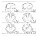
- FIG. 1is a sample montage that may be displayed on a computing device of a user, such as a radiologist or doctor.
- FIG. 2is another example of a montage with a different number of images, including images that are formatted differently and include annotations (e.g., arrows pointing to areas of specific interest).
- FIG. 3is a sample screen shot of information displayed on a monitor by image viewing/manipulation software, such as DR Systems Unity RIS/PACS.
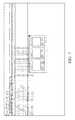
- FIG. 4is another sample screen shot of information displayed on a monitor including, in this specific example, an Advanced Beneficiary Notice and a clinical report template.
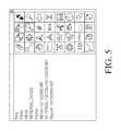
- FIG. 5illustrates a sample toolbox that may be used to select items for display on multiple monitors.
- FIG. 6is a sample user interface that may be displayed to a user when a non-matching exam type is ordered, received, or otherwise accessed.
- FIG. 7illustrates a sample screenshot of a user interface that may be used to link a form with one or more of an exam type, insurance plan, acquisition site, or other link.
- FIG. 8illustrates a sample screenshot of a user interface that allows selection and/or viewing of an attribute indicating whether or not a particular form needs to be returned to the medical facility (e.g., after completion by a patient).
- FIG. 9is a system diagram which shows the various components of a system for performing the system and methods described above.
- the terms “viewer” and “user”are used interchangeably to describe an individual (or group of individuals) that interfaces with a computing device.
- Usersmay include, for example, doctors, radiologists, hospital staff, or other individuals involved in acquisition, analysis, storage, management, or other tasks related to medical images.
- usersmay include any individuals or groups of individuals that generate, transmit, view, and/or otherwise work with images of any type.
- Any discussion herein of user preferencesshould be construed to also, or alternatively, include user group preferences, site preferences, system preferences, and/or default software preferences.
- the methods described with reference to the flowcharts, as well as any other methods discussed hereinmay include fewer or additional blocks and/or the blocks may be performed in a different order than is illustrated.
- Software code configured for execution on a computing device in order to perform the methodsmay be provided on a tangible computer readable medium, such as a compact disc, digital video disc, flash drive, hard drive, memory device or any other tangible medium.
- Such software codemay be stored, partially or fully, on a memory of a computing device (e.g., RAM, ROM, etc.), such as the computing system 150 (see discussion of FIG. 1 , below), and/or other computing devices illustrated in the figures, in order to perform the respective methods.
- a computing devicee.g., RAM, ROM, etc.
- the methodswill be described herein as performed by the computing system 150 , but the methods are not limited to performance by the computing system 150 and should be interpreted to include performance by any one or more of the computing devices noted herein and/or any other suitable computing device.
- FIG. 1is a sample montage that may be displayed on a computing device of a user, such as a radiologist or doctor.
- the images of the montageare those selected by the reading physician as key images, such as at the time of review of an MRI of the Brain.
- the exammay include several image series, and hundreds or thousands of images.
- the radiologistcomposes the montage by selecting one or more key images from one or more image series, and by adjusting various view settings of the images, such as window/level settings, centering, cropping, magnification, annotations, insets, etc.
- FIG. 2is another example of a montage with a different number of images, including images that are formatted differently and include annotations (e.g., arrows pointing to areas of specific interest).
- montagesare saved as separate files, such as separate image files that are essentially a screenshot of a montage (e.g., a snapshot of the montages of FIG. 1 or 2 ).
- a montagee.g., a snapshot of the montages of FIG. 1 or 2
- the montage image filemay be notated as a key image, such as according to the DICOM (Digital Imaging and Communications in Medicine) specification.
- the montagemight include images from multiple examinations, or might include reference images such as illustrations or medical images exemplifying pathological or normal conditions.
- a montage having 1 or more imagescan be stored in one or multiple ways, including (1) storage of the complete composite montage image and/or (2) storage of sufficient information regarding each image so that the entire montage can be recreated upon future display or the individually stored images can be displayed, depending on the user's preferences, depending on the display environment (such as aspect ratio of the display window, monitor resolution, a combination of user preferences and display environment, or other factors.) For example, information regarding the arrangement of images in the montage, as well as information regarding display settings of respective images (e.g., magnification, brightness, centering, cropping, filters, annotations, insets, etc.) may be stored.
- display settings of respective imagese.g., magnification, brightness, centering, cropping, filters, annotations, insets, etc.
- montage characteristicsmay then be recalled at a future time and used to re-build the montage.
- storage of an image of the entire montagemay not be necessary, while in other embodiments the montage image (e.g., a snapshot of the montage) may be stored and used in certain circumstances, such as when the montage is to be displayed on a display having essentially the same resolution and aspect ratio as the display on which the montage was originally created.
- the arrangement information included in montage characteristicsmay include a number of rows and columns of medical images in the montage, indications of specific locations of each medical image in the montage, indications of an order that medical images of the montage are displayed, and/or any other information that may be usable to read construct a montage on the same or another computing device based on the layout of the montage images.
- the systeme.g., the device that will display the images and/or a device that is serving the images
- the format of the montagesuch as 4 ⁇ 2 vs. 2 ⁇ 4 based on the aspect ratio of the images compared to the aspect ratio/orientation of the monitor.
- the montagemay be displayed in a manually sizable window, and the format of the montage may be automatically and optionally dynamically adjusted based on the aspect ratio of the window.
- the display formatis automatically adjusted based on the aspect ratio of the montage window, the aspect ratio of the added images, and/or the number of images added.
- orientation of the display devicee.g., portrait or landscape
- matrix informatione.g., the number of rows and columns of images, e.g., 4 images ⁇ 2 images or 6 images ⁇ 4 images
- montage characteristicse.g., a file that is associated with an exam or added to header information to one or more exam files.
- multiple sets of montage characteristics that include the same (or some of the same) imagesmay be stored and selected based on characteristics of the computing device that later displays the montage, such as the display size.
- a first set of montage characteristicsmay be automatically selected for viewing of images of a particular exam on a tablet computer while a second set of montage characteristics may be automatically selected for viewing of images of the same particular exam on a desktop computer with a monitor having a much higher resolution.
- the computing device that displays the montagemay automatically select the appropriate set of montage characteristics, without any input from the user.
- montage characteristicsmay be stored with a particular exam, such that when that exam is later recalled by any computing system, montage characteristics may be accessed in order to reconstruct part or all of the montage.
- montage characteristicsare used by a viewing computing system to rebuild all (or parts of the montage) according to rules for doing so, such as user or system rules.
- the montage characteristicsessentially provide information that is usable by a viewing computing system to view portions or all of the montage in accordance with a viewing environment and/or user preferences.
- the multiple images of a montageare simultaneously saved as key images (e.g., key DICOM objects) so that the images may be easily identified for inclusion in a montage that is generated based on stored montage characteristics.
- key imagese.g., key DICOM objects
- montage characteristicsmay be automatically selected based on the user, the display monitor (such as its resolution, aspect ratio, Smartphone vs PC monitor, etc), and/or other characteristics of an image viewing environment.
- Montage characteristicsmay be used to display the entire montage in the arrangement originally used by the viewer.
- the usermay cycle through other images of images series to which key images belong, keeping the same display characteristics as the key image as a default.
- an imagemay be one of many images in an image series, such as one axial image of a number of stacked axial images of the patient.
- the systemmay retain information related to the entire series of images so that a user may manipulate the montage image to also access other images in the same series.
- FIG. 3is a sample screen shot of information displayed on a monitor by image viewing/manipulation software, such as DR Systems Unity RIS/PACS.
- the large dialog that fills most of the screenmay be referred to as the Requisition, and it contains several tabs, some shown in red (the last three tabs) and others in blue (the first five tabs).
- Each tabrepresents a document or dialog associated with one or more imaging exams that are currently displayed on other monitors of a multi-monitor system.
- FIG. 4is a sample screen shot of one such arrangement, where the clinical report template associated with the current exam is displayed on the right, and a scanned document (Advanced Beneficiary Notice) is shown on the left.
- a reading physicianmay want to display the Requisition and the various available documents/dialogs/webforms according to a preferred layout (e.g., a user-preferred layout), including size and position of various available elements.
- a preferred layoute.g., a user-preferred layout
- the usermay have displayed the Advanced Beneficiary Notice by selecting one of the tabs from the upper left of the requisition, via another mouse action, keyboard shortcut or audio command, and also displayed the clinical report template through another means, such as a button click, mouse click, hotkey, or audio command.
- the usercan adjust the size and position of these objects. Repeating these actions for other patients/exams is repetitive and not efficient.
- the systemwhen a user sizes and positions a window, the system remembers that size and position for that category of item (e.g., any object) and for the user (or user group), such as by storing an association between the size and position of the item with the determined category in “item information.”
- the systemmay then automatically recreate that sizing and layout in the future for that user when a document of the category is displayed, suing the item information. Accordingly, each user can size and position these various documents/dialogs, and the system will remember the layout for any workstation across the network that uses a monitor of the same matrix size, while defaulting to a standard configuration for monitors of a different matrix size.
- a single usermay have multiple arrangements of documents/dialogs that are associated with different monitor sizes that are used by the user.
- display settingsmay be stored for specific documents/dialogs as well, or as an alternative to the category settings discussed above.
- the display characteristicsare associated with a display size (e.g., the matrix size and/or orientation of a monitor on which the document/dialog was viewed), such that the sizing and layout of documents of that category are displayed in that manner only when requested for viewing on the same or similar display configuration.
- the systemmay select a default display layout for other monitor formats. If the user then sets the sizing and layout for another monitor format, the system will remember both set-ups for the user and make those layouts available to other devices throughout a network, such as a WAN or LAN. Any number of set-ups can be remembered.
- the usercan open a toolbox, such as the sample toolbox of FIG. 5 , from any of multiple monitors by clicking a keyboard shortcut or mouse button, or via an audio command.
- This toolboxincludes a list of all of the display objects available on the Requisition or other objects. The user can select any item on the list to immediately display the desired object, and the object will appear in the location associated with the item information for a category of the object based on the users stored display layout preference or the default layout preference.
- the userdoes not need to drag a mouse to the monitor on which he wants to display the object, and one can control the display of an object on one monitor or in one display window, while working from another monitor or another display window.
- a single monitormay be large enough to display many windows that might have previously required many monitors.
- the systems and methods discussed hereinallow automatic positioning on the single monitor at a location associated with the user account.
- a reading physician who is viewing images on another monitorcan thus control the display of objects on a first monitor without first dragging a mouse or distracting his vision to the first monitor.
- the object appearsit follows the stored set-up instructions.
- RIS, PACS, and other healthcare information systemstypically contain a table of Exam Types, which is a list of various medical imaging exams available for selection.
- the Exam Type table(or “Exam type master file”) may include exam descriptions, modality descriptions, alphanumeric codes, as well as other information about each Exam Type—such as the default title of a clinical report based on that Exam Type, forms that should appear to the user (clerk, patient, technologist, and/or doctor) when that Exam Type is performed or viewed, linked clinical report templates, linked information about required supplies, clinical protocols, payment policies, charges, relative value/productivity units, safety policies and more.
- various user or site preferencescan be based on the Exam Type or modality, such as which exams should be automatically restored from archive for comparison when a particular Exam Type is scheduled or performed, which and how many exams are displayed for comparison when an exam is viewed, how a particular user prefers images to be displayed, and more.
- system automationmay depend on the Exam Type table, such as rules automating the pre-fetching of prior comparison exams, reading physician protocols for exam display, automated creation of virtual series, keyboard shortcuts, reading sequences, routing of exams, and more. Therefore, in a sense, the Exam Type master file is a sort of DNA of some healthcare information system.
- the Exam Type informationmay or may not match up with information already present in the Exam Type table.
- exams of the same typemay be named differently by different acquisition/viewing system. This information might be exchanged via information in a DR RIS, CVIS, DICOM metafile, in an HL-7 message, an order message, billing message, etc.
- the systemmay offer configuration options that specify how the system should respond to a non-matching Exam Type, such as by either holding the processing of the message or exam import, or automatically adding the non-matching Exam Type to the Exam Type master file.
- an Exam Type mapping functionis defined so that when a non-matching Exam Type is encountered by the system via the variety of different possible messages described above, the system prompts the user (in one or more of many possible manners—such as either a pop-up message, generation of a worklist, text-message of other means) that a non-matching Exam Type was encountered. The user can then map the non-matching Exam Type to the proper Exam Type from the master file, so that if the non-matching Exam Type is again encountered, it can be automatically processed (e.g., without any notification to the user).
- the systemmight create a new scheduled exam with the internally mapped Exam Type, or import a DICOM imaging exam with the proper internally mapped Exam Type. All of the system automation that depends on the internally mapped Exam Type may then properly occur.
- FIG. 6is a sample user interface that may be displayed to a user when a non-matching Exam Type is ordered or received.
- the Exam Descriptionmay be mapped to an Exam Type already stored in the Exam Type master file so that future exams having the same Exam Description are automatically mapped to the selected Exam Type and its corresponding actions.
- the systemcould apply rules that map Exam Types based on relative matching of character strings or other best match rules related to Exam Codes or other message characteristics. Based on a confidence level of a match, the automated mapping may be applied without further input from the user. For example, if a confidence level of a match is lower (e.g., below 80%) the user may be provided with the most likely matches and provided an opportunity to select from the short list of possible matches, rather than navigating through a list of all Exam Types in the master file.
- a confidence level of a matchis lower (e.g., below 80%) the user may be provided with the most likely matches and provided an opportunity to select from the short list of possible matches, rather than navigating through a list of all Exam Types in the master file.
- this mappingmay be applied not only to inbound messages/exams, but also to outbound messages returning to external information systems, so that any edits, changes, and/or updates could be communicated back to the original system.
- Patient formsmay be created and stored as form templates that are referred to in a data structure that links respective form templates with various links.
- the linkscan indicate when the forms are automatically presented and where they are automatically filed.
- a particular formmight be linked to a particular insurance, patient sex, patient language, age group, exam type, modality, or other stored information.
- the form templatescan be linked to a specific naming convention and storage location.
- CT Consent Form templateand an MRI Consent Form template, and store information such that when either of these templates is used to create a CT Consent Form or MRI Consent Form, these forms are stored with the patient record such that they are labeled as Consent Forms, whereas there might be other form templates that would be stored as Insurance Forms, or Release Forms, etc.
- the proper forms based on the automated linksare posted to an internet-accessible location where the proper patient can view, print, or complete the forms.
- the formsmay be automatically labeled with an indentifying barcode, so that if the patient prints and completes the forms on paper, the paper can later be scanned, the bar code identified, and the form thus automatically filed with the proper patient, proper exam, and proper label.
- Each instance of a formmay be provided with a specific identifier so that the information provided in the form (e.g., electronically or manually) may be associated with the proper patient's record and/or exam, and labeled properly.
- FIG. 7illustrates a sample screenshot of a user interface that may be used to link a form with one or more of an Exam Type, Insurance Plan, Acquisition Site, or other link.
- FIG. 8is a screenshot of a sample user interface that may be accessible by a “Set Series” or similar button.
- the user interfaceallows the user to specify the category or series name that will be used to store instances of the form that are created using one of the templates stored in this list.
- the templatesmight be one of many types of document formats, including MSWord, HTML, XML, CDA, CCR, etc.
- the systemcan automatically store the instance of the form with the proper patient, proper exam, and in the proper series.
- the series informationmay also further specify if, how, and when the form is presented for any particular user or by system default.
- formsmay be associated with an attribute that indicates whether the form must be returned to system (e.g., to the medical facility that originally provided the form).
- some quantity of forms provided to a patientmay be for use of the patient (and/or a party other than the medical facility that provides the forms) and, thus, are not required to be returned to the medical facility.
- a medical facilitymay not want forms that provide informational content to the patient returned to the medical facility.
- many forms provided to the patientmay need to be returned to the medical facility and/or required to be returned prior to performance of an exam or procedure, for example.
- an attribute indicating whether or not a particular form needs to be returned to the medical facilitymay be indicated using a user interface similar to that shown in FIG. 8 .
- a patient's filemay then be automatically reviewed in order to determine if any forms that are required to be returned have not yet been returned (possibly a certain number of days after the forms are provided or a certain number of days before a scheduled exam).
- FIG. 9is a system diagram which shows the various components of a system 100 for performing the system and methods described above, wherein the configuration of the system 100 may include fewer or additional features than are illustrated and individual components, such as the computing device 150 , may also include fewer or additional components.
- the methods discussed above as being performed by “a system”are performed by the computing device 150 . In other embodiments, the methods may be performed by any other suitable computing device.
- the Computing Device 150may take various forms.
- the Computing Device 150may be a computer workstation having software modules 151 .
- software modules 151may reside on another computing device, such as a web server, and the user directly interacts with a second computing device that is connected to the web server via a computer network.
- the Computing Device 150comprises a server, a desktop computer, a workstation, a laptop computer, a mobile computer, a Smartphone, a tablet computer, a cell phone, a personal digital assistant, a gaming system, a kiosk, an audio player, any other device that utilizes a graphical user interface, including office equipment, automobiles, airplane cockpits, household appliances, automated teller machines, self-service checkouts at stores, information and other kiosks, ticketing kiosks, vending machines, industrial equipment, and/or a television, for example.
- a servera desktop computer, a workstation, a laptop computer, a mobile computer, a Smartphone, a tablet computer, a cell phone, a personal digital assistant, a gaming system, a kiosk, an audio player, any other device that utilizes a graphical user interface, including office equipment, automobiles, airplane cockpits, household appliances, automated teller machines, self-service checkouts at stores, information and other kiosks, ticketing kiosks, vending machines, industrial equipment, and/or a television, for
- the Computing Device 150runs an operating system 154 , such as an off-the-shelf operating system, for example, Windows, Linux, MacOS, Android, or iOS operation system.
- the Computing Device 150may also run a more specialized operating system which may be designed for the specific tasks performed by the computing device 150 .
- the Computing Device 150may include one or more computing processors 152 .
- the computer processors 152may include central processing units (CPUs), and may further include dedicated processors such as graphics processor chips, or other specialized processors.
- the processorsgenerally are used to execute computer instructions based on the software modules 151 to cause the computing device to perform operations as specified by the modules 151 .
- the modules 151may include, by way of example, components, such as software components, object-oriented software components, class components and task components, processes, functions, attributes, procedures, subroutines, segments of program code, drivers, firmware, microcode, circuitry, data, data structures, data structures, tables, arrays, and variables.
- modulesmay include software code written in a programming language, such as, for example, Java, JavaScript, ActionScript, Visual Basic, HTML, C, C++, or C#. While “modules” are generally discussed herein with reference to software, any modules may alternatively be represented in hardware or firmware. Generally, the modules described herein refer to logical modules that may be combined with other modules or divided into sub-modules despite their physical organization or storage.
- the Computing Device 150may also include memory 153 .
- the memory 153may include volatile data storage such as RAM or SDRAM.
- the memory 153may also include more permanent forms of storage such as a hard disk drive, a flash disk, flash memory, a solid state drive, or some other type of non-volatile storage.
- the Computing Device 150may also include or be interfaced to one or more display devices 155 that provide information to the users.
- Display devices 155may include a video display, such as one or more high-resolution computer monitors, or a display device integrated into or attached to a laptop computer, handheld computer, Smartphone, computer tablet device, or medical scanner.
- the display device 155may include an LCD, OLED, or other thin screen display surface, a monitor, television, projector, a display integrated into wearable glasses, or any other device that visually depicts user interfaces and data to viewers.
- the Display Computing Device 150may also include or be interfaced to one or more input devices 156 which receive input from users, such as a keyboard, trackball, mouse, 3D mouse, drawing tablet, joystick, game controller, touch screen (e.g., capacitive or resistive touch screen), touchpad, accelerometer, video camera and/or microphone.
- input devices 156which receive input from users, such as a keyboard, trackball, mouse, 3D mouse, drawing tablet, joystick, game controller, touch screen (e.g., capacitive or resistive touch screen), touchpad, accelerometer, video camera and/or microphone.
- the Computing Device 150may also include one or more interfaces 157 which allow information exchange between Computing Device 150 and other computers and input/output devices using systems such as Ethernet, Wi-Fi, Bluetooth, as well as other wired and wireless data communications techniques.
- the modules of Computing Device 150may be connected using a standard based bus system.
- the standard based bus systemcould be Peripheral Component Interconnect (“PCI”), PCI Express, Accelerated Graphics Port (“AGP”), Micro channel, Small Computer System Interface (“SCSI”), Industrial Standard Architecture (“ISA”) and Extended ISA (“EISA”) architectures, for example.
- PCIPeripheral Component Interconnect
- AGPAccelerated Graphics Port
- SCSISmall Computer System Interface
- ISAIndustrial Standard Architecture
- EISAExtended ISA
- Computing Device 150may communicate and/or interface with other systems and/or devices.
- the computing device 150may be connected to a computer network 110 .
- the computer network 110may take various forms. It may be a wired network or a wireless network, or it may be some combination of both.
- the computer network 110may be a single computer network, or it may be a combination or collection of different networks and network protocols.
- the computer network 110may include one or more local area networks (LAN), wide area networks (WAN), personal area networks (PAN), cellular or data networks, and/or the Internet.
- LANlocal area networks
- WANwide area networks
- PANpersonal area networks
- cellular or data networksand/or the Internet.
- Various devices and subsystemsmay be connected to the network 110 .
- one or more medical scannersmay be connected, such as MRI scanners 120 .
- the MRI scanners 120may be used to acquire MRI images from patients, and may share the acquired images with other devices on the network 110 .
- the network 110may also be coupled to one or more CT scanners 122 .
- the CT scanners 122may also be used to acquire images and, like the MRI scanner 120 , may then store those images and/or share those images with other devices via the network 110 .
- Any other scanner or device capable of inputting or generating informationcould be included, including ultrasound, angiography, nuclear medicine, radiography, endoscopy, pathology, dermatology, etc.
- the PACS 136is typically used for the storage, retrieval, distribution and presentation of images (such as those created and/or generated by the MRI scanner 120 and CT Scanner 122 ).
- the medical imagesmay be stored in an independent format, an open source format, or some other proprietary format.
- a common format for image storage in the PACS systemis the Digital Imaging and Communications in Medicine (DICOM) format.
- DICOMDigital Imaging and Communications in Medicine
- the network 110may also be connected to a Radiology Information System (RIS) 140 .
- the radiology information system 140is typically a computerized data storage system that is used by radiology departments to store, manipulate and distribute patient radiological information such as Radiology Reports.
- EMRElectronic Medical Record
- the EMR system 142may be configured to store and make accessible to a plurality of medical practitioners computerized medical records.
- Laboratory Information System 144is typically a system which stores information created or generated by clinical laboratories.
- Digital Pathology System 146used to digitally manage and store information related to medical pathology.
- CADComputer Aided Diagnosis System
- the CAD 148 functionalitymay reside in a computing device separate from Information Display Computing Device 150 while in another embodiment the CAD 148 functionality may reside within Information Display Computing Device 150 .
- 3D Processing System 149used to perform computations on imaging information to create new views of the information, e.g., 3D volumetric display, Multiplanar Reconstruction (MPR) and Maximum Intensity Projection reconstruction (MIP).
- MPRMultiplanar Reconstruction
- MIPMaximum Intensity Projection reconstruction
- the 3D Processing functionalitymay reside in a computing device separate from Information Display Computing Device 150 while in another embodiment the 3D Processing functionality may reside within Information Display Computing Device 150 .
- computing devicesthat store, provide, acquire, and/or otherwise manipulate medical data may also be coupled to the network 110 and may be in communication with one or more of the devices illustrated in FIG. 9 , such as with the Information Display Computing Device 150 .
- Computing Device 150may be configured to interface with various networked computing devices in order to communicate medical information that is stored among the various systems present in the network.
- Information Display Computing Device 150may be used to display non-medical information.
- the other devices illustrated in FIG. 9may include some or all of the same components discussed above with reference to the Information Display Computer Device 150 .
- Conditional languagesuch as, among others, “can,” “could,” “might,” or “may,” unless specifically stated otherwise, or otherwise understood within the context as used, is generally intended to convey that certain embodiments include, while other embodiments do not include, certain features, elements and/or steps. Thus, such conditional language is not generally intended to imply that features, elements and/or steps are in any way required for one or more embodiments or that one or more embodiments necessarily include logic for deciding, with or without user input or prompting, whether these features, elements and/or steps are included or are to be performed in any particular embodiment.
- All of the methods and processes described abovemay be embodied in, and partially or fully automated via, software code modules executed by one or more general purpose computers.
- the methods described hereinmay be performed by an Information Display Computing Device and/or any other suitable computing device.
- the methodsmay be executed on the computing devices in response to execution of software instructions or other executable code read from a tangible computer readable medium.
- a tangible computer readable mediumis a data storage device that can store data that is readable by a computer system. Examples of computer readable mediums include read-only memory, random-access memory, other volatile or non-volatile memory devices, CD-ROMs, magnetic tape, flash drives, and optical data storage devices.
Landscapes
- Engineering & Computer Science (AREA)
- Theoretical Computer Science (AREA)
- Health & Medical Sciences (AREA)
- General Engineering & Computer Science (AREA)
- General Physics & Mathematics (AREA)
- Physics & Mathematics (AREA)
- Medical Informatics (AREA)
- General Health & Medical Sciences (AREA)
- Public Health (AREA)
- Primary Health Care (AREA)
- Epidemiology (AREA)
- Biomedical Technology (AREA)
- Data Mining & Analysis (AREA)
- Human Computer Interaction (AREA)
- Evolutionary Computation (AREA)
- Artificial Intelligence (AREA)
- Business, Economics & Management (AREA)
- General Business, Economics & Management (AREA)
- Radiology & Medical Imaging (AREA)
- Nuclear Medicine, Radiotherapy & Molecular Imaging (AREA)
- Computing Systems (AREA)
- Computational Linguistics (AREA)
- Mathematical Physics (AREA)
- Software Systems (AREA)
- Evolutionary Biology (AREA)
- Computer Vision & Pattern Recognition (AREA)
- Bioinformatics & Computational Biology (AREA)
- Bioinformatics & Cheminformatics (AREA)
- Life Sciences & Earth Sciences (AREA)
- Medical Treatment And Welfare Office Work (AREA)
- User Interface Of Digital Computer (AREA)
Abstract
Description
Claims (18)
Priority Applications (2)
| Application Number | Priority Date | Filing Date | Title |
|---|---|---|---|
| US13/572,547US9075899B1 (en) | 2011-08-11 | 2012-08-10 | Automated display settings for categories of items |
| US14/792,210US10579903B1 (en) | 2011-08-11 | 2015-07-06 | Dynamic montage reconstruction |
Applications Claiming Priority (2)
| Application Number | Priority Date | Filing Date | Title |
|---|---|---|---|
| US201161522633P | 2011-08-11 | 2011-08-11 | |
| US13/572,547US9075899B1 (en) | 2011-08-11 | 2012-08-10 | Automated display settings for categories of items |
Related Parent Applications (1)
| Application Number | Title | Priority Date | Filing Date |
|---|---|---|---|
| US13/572,552ContinuationUS9092727B1 (en) | 2011-08-11 | 2012-08-10 | Exam type mapping |
Related Child Applications (1)
| Application Number | Title | Priority Date | Filing Date |
|---|---|---|---|
| US14/792,210ContinuationUS10579903B1 (en) | 2011-08-11 | 2015-07-06 | Dynamic montage reconstruction |
Publications (1)
| Publication Number | Publication Date |
|---|---|
| US9075899B1true US9075899B1 (en) | 2015-07-07 |
Family
ID=53492006
Family Applications (4)
| Application Number | Title | Priority Date | Filing Date |
|---|---|---|---|
| US13/572,547Active2033-05-19US9075899B1 (en) | 2011-08-11 | 2012-08-10 | Automated display settings for categories of items |
| US13/572,397Active2033-04-11US9092551B1 (en) | 2011-08-11 | 2012-08-10 | Dynamic montage reconstruction |
| US13/572,552Expired - Fee RelatedUS9092727B1 (en) | 2011-08-11 | 2012-08-10 | Exam type mapping |
| US14/792,210Active2035-04-07US10579903B1 (en) | 2011-08-11 | 2015-07-06 | Dynamic montage reconstruction |
Family Applications After (3)
| Application Number | Title | Priority Date | Filing Date |
|---|---|---|---|
| US13/572,397Active2033-04-11US9092551B1 (en) | 2011-08-11 | 2012-08-10 | Dynamic montage reconstruction |
| US13/572,552Expired - Fee RelatedUS9092727B1 (en) | 2011-08-11 | 2012-08-10 | Exam type mapping |
| US14/792,210Active2035-04-07US10579903B1 (en) | 2011-08-11 | 2015-07-06 | Dynamic montage reconstruction |
Country Status (1)
| Country | Link |
|---|---|
| US (4) | US9075899B1 (en) |
Cited By (18)
| Publication number | Priority date | Publication date | Assignee | Title |
|---|---|---|---|---|
| US9471210B1 (en) | 2004-11-04 | 2016-10-18 | D.R. Systems, Inc. | Systems and methods for interleaving series of medical images |
| US9501617B1 (en) | 2009-09-28 | 2016-11-22 | D.R. Systems, Inc. | Selective display of medical images |
| US9501863B1 (en) | 2004-11-04 | 2016-11-22 | D.R. Systems, Inc. | Systems and methods for viewing medical 3D imaging volumes |
| US9542082B1 (en) | 2004-11-04 | 2017-01-10 | D.R. Systems, Inc. | Systems and methods for matching, naming, and displaying medical images |
| US9672477B1 (en) | 2006-11-22 | 2017-06-06 | D.R. Systems, Inc. | Exam scheduling with customer configured notifications |
| US9727938B1 (en) | 2004-11-04 | 2017-08-08 | D.R. Systems, Inc. | Systems and methods for retrieval of medical data |
| US9836202B1 (en) | 2004-11-04 | 2017-12-05 | D.R. Systems, Inc. | Systems and methods for viewing medical images |
| US20180060491A1 (en)* | 2016-08-29 | 2018-03-01 | Canon Kabushiki Kaisha | Medical information processing apparatus, medical information processing system, medical information processing method, and storage medium |
| US10007652B1 (en)* | 2013-08-15 | 2018-06-26 | Allscripts Software, Llc | Method for reusing and dynamically filtering content for structured document construction |
| US10579903B1 (en) | 2011-08-11 | 2020-03-03 | Merge Healthcare Solutions Inc. | Dynamic montage reconstruction |
| US10592688B2 (en) | 2008-11-19 | 2020-03-17 | Merge Healthcare Solutions Inc. | System and method of providing dynamic and customizable medical examination forms |
| US10665342B2 (en) | 2013-01-09 | 2020-05-26 | Merge Healthcare Solutions Inc. | Intelligent management of computerized advanced processing |
| US10902201B2 (en)* | 2018-08-02 | 2021-01-26 | International Business Machines Corporation | Dynamic configuration of document portions via machine learning |
| US10909168B2 (en) | 2015-04-30 | 2021-02-02 | Merge Healthcare Solutions Inc. | Database systems and interactive user interfaces for dynamic interaction with, and review of, digital medical image data |
| US11158410B2 (en) | 2016-09-27 | 2021-10-26 | Canon Kabushiki Kaisha | Medical information processing apparatus, medical information processing system, medical information processing method, and storage medium |
| US20220207896A1 (en)* | 2020-12-30 | 2022-06-30 | Stryker Corporation | Systems and methods for classifying and annotating images taken during a medical procedure |
| US20230245753A1 (en)* | 2020-04-13 | 2023-08-03 | Kaliber Labs Inc. | Systems and methods for ai-assisted surgery |
| US12396869B2 (en) | 2021-04-12 | 2025-08-26 | Kaliber Labs Inc. | Systems and methods for using image analysis to guide a surgical procedure |
Families Citing this family (7)
| Publication number | Priority date | Publication date | Assignee | Title |
|---|---|---|---|---|
| US8843852B2 (en)* | 2010-12-17 | 2014-09-23 | Orca Health, Inc. | Medical interface, annotation and communication systems |
| US9449380B2 (en)* | 2012-03-20 | 2016-09-20 | Siemens Medical Solutions Usa, Inc. | Medical image quality monitoring and improvement system |
| CA2869632C (en) | 2012-04-16 | 2021-05-25 | Airstrip Ip Holdings, Llc | Systems and methods for displaying patient data |
| US10169863B2 (en) | 2015-06-12 | 2019-01-01 | International Business Machines Corporation | Methods and systems for automatically determining a clinical image or portion thereof for display to a diagnosing physician |
| US10366128B2 (en)* | 2015-11-12 | 2019-07-30 | Flipboard, Inc. | Curating a digital magazine with a user's own content |
| US20190180864A1 (en)* | 2017-12-13 | 2019-06-13 | International Business Machines Corporation | Method and system for selecting and arranging images with a montage template |
| US10832808B2 (en)* | 2017-12-13 | 2020-11-10 | International Business Machines Corporation | Automated selection, arrangement, and processing of key images |
Citations (16)
| Publication number | Priority date | Publication date | Assignee | Title |
|---|---|---|---|---|
| US6734880B2 (en)* | 1999-11-24 | 2004-05-11 | Stentor, Inc. | User interface for a medical informatics systems |
| US20070106633A1 (en)* | 2005-10-26 | 2007-05-10 | Bruce Reiner | System and method for capturing user actions within electronic workflow templates |
| US20070159962A1 (en) | 2006-01-10 | 2007-07-12 | Shivaprasad Mathavu | Hanging protocol software simulator |
| US20080016111A1 (en)* | 2005-11-28 | 2008-01-17 | Ronald Keen | Transactional storage and workflow routing for medical image objects |
| US20080126982A1 (en)* | 2006-09-18 | 2008-05-29 | Agfa Inc. | Imaging history display system and method |
| US20080136838A1 (en)* | 2002-05-31 | 2008-06-12 | Patricia Anne Goede | System and method for visual annotation and knowledge representation |
| US20080275913A1 (en) | 2007-05-01 | 2008-11-06 | Van Arragon Paul | Dynamic assignment of statements for selected exams using clinical concepts |
| US20080279439A1 (en) | 2002-11-29 | 2008-11-13 | Minyard Thomas J | Distributed Architecture for Mammographic Image Acquisition and Processing |
| US20090182577A1 (en)* | 2008-01-15 | 2009-07-16 | Carestream Health, Inc. | Automated information management process |
| US20090213034A1 (en)* | 2006-06-14 | 2009-08-27 | Koninklijke Philips Electronics N. V. | Multi-modality medical image layout editor |
| US20090248442A1 (en) | 2008-03-28 | 2009-10-01 | Medicalis Corp | Processing of clinical data for validation of selected clinical procedures |
| US20090268986A1 (en) | 2006-11-11 | 2009-10-29 | Joerg Holstein | System for the playback of medical images |
| US20100138239A1 (en)* | 2008-11-19 | 2010-06-03 | Dr Systems, Inc. | System and method of providing dynamic and customizable medical examination forms |
| US20100211409A1 (en) | 2009-02-17 | 2010-08-19 | Kotula Jeffrey J | Organizing medical images for display |
| US20100246981A1 (en) | 2009-03-30 | 2010-09-30 | Xiao Hu | PACS Optimization Techniques |
| US20120130730A1 (en) | 2010-11-24 | 2012-05-24 | General Electric Company | Multi-department healthcare real-time dashboard |
Family Cites Families (357)
| Publication number | Priority date | Publication date | Assignee | Title |
|---|---|---|---|---|
| US1015768A (en) | 1911-03-13 | 1912-01-30 | Randall Telephone Mfg Company | Automatic telephone circuit and repeater. |
| JPS60196856A (en) | 1984-03-20 | 1985-10-05 | Olympus Optical Co Ltd | Picture retrieval registering system |
| US5179651A (en) | 1988-11-08 | 1993-01-12 | Massachusetts General Hospital | Apparatus for retrieval and processing of selected archived images for display at workstation terminals |
| US5123056A (en) | 1990-02-02 | 1992-06-16 | Siemens Medical Systems, Inc. | Whole-leg x-ray image processing and display techniques |
| US5172419A (en) | 1991-03-05 | 1992-12-15 | Lumisys, Inc. | Medical image processing system |
| US5779634A (en) | 1991-05-10 | 1998-07-14 | Kabushiki Kaisha Toshiba | Medical information processing system for supporting diagnosis |
| US5384862A (en) | 1992-05-29 | 1995-01-24 | Cimpiter Corporation | Radiographic image evaluation apparatus and method |
| US5734915A (en) | 1992-11-25 | 1998-03-31 | Eastman Kodak Company | Method and apparatus for composing digital medical imagery |
| US5452416A (en) | 1992-12-30 | 1995-09-19 | Dominator Radiology, Inc. | Automated system and a method for organizing, presenting, and manipulating medical images |
| EP0973116A1 (en) | 1993-03-01 | 2000-01-19 | Kabushiki Kaisha Toshiba | Medical information processing system for supporting diagnosis |
| US5431161A (en) | 1993-04-15 | 1995-07-11 | Adac Laboratories | Method and apparatus for information acquistion, processing, and display within a medical camera system |
| US5515375A (en) | 1993-07-30 | 1996-05-07 | Motorola, Inc. | Method and apparatus for multiplexing fixed length message data and variably coded speech |
| US5542003A (en) | 1993-09-13 | 1996-07-30 | Eastman Kodak | Method for maximizing fidelity and dynamic range for a region of interest within digitized medical image display |
| JP3277677B2 (en) | 1994-04-01 | 2002-04-22 | ソニー株式会社 | Signal encoding method and apparatus, signal recording medium, signal transmission method, and signal decoding method and apparatus |
| JP3544557B2 (en) | 1994-04-08 | 2004-07-21 | オリンパス株式会社 | Image file device |
| JPH11501538A (en) | 1995-03-03 | 1999-02-09 | アーチ ディヴェロプメント コーポレイション | Method and system for detecting lesions in medical images |
| US5857030A (en) | 1995-08-18 | 1999-01-05 | Eastman Kodak Company | Automated method and system for digital image processing of radiologic images utilizing artificial neural networks |
| DE19620371A1 (en) | 1996-05-21 | 1997-12-04 | Philips Patentverwaltung | X-ray procedure |
| US6222939B1 (en) | 1996-06-25 | 2001-04-24 | Eyematic Interfaces, Inc. | Labeled bunch graphs for image analysis |
| JP3363735B2 (en) | 1996-06-26 | 2003-01-08 | 松下電器産業株式会社 | X-ray imaging device |
| US6128002A (en) | 1996-07-08 | 2000-10-03 | Leiper; Thomas | System for manipulation and display of medical images |
| US6820093B2 (en) | 1996-07-30 | 2004-11-16 | Hyperphrase Technologies, Llc | Method for verifying record code prior to an action based on the code |
| US6272235B1 (en) | 1997-03-03 | 2001-08-07 | Bacus Research Laboratories, Inc. | Method and apparatus for creating a virtual microscope slide |
| US5924074A (en) | 1996-09-27 | 1999-07-13 | Azron Incorporated | Electronic medical records system |
| US5986662A (en) | 1996-10-16 | 1999-11-16 | Vital Images, Inc. | Advanced diagnostic viewer employing automated protocol selection for volume-rendered imaging |
| US6115486A (en) | 1996-11-06 | 2000-09-05 | Quinton Instrument Company | Teleradiology system for the storage and transmission of angiographic and related image sequences |
| JP3878259B2 (en) | 1996-11-13 | 2007-02-07 | 東芝医用システムエンジニアリング株式会社 | Medical image processing device |
| US5987345A (en)* | 1996-11-29 | 1999-11-16 | Arch Development Corporation | Method and system for displaying medical images |
| US6243095B1 (en) | 1996-12-05 | 2001-06-05 | Peter E. Shile | Navigation and display system for digital radiographs |
| US6151581A (en) | 1996-12-17 | 2000-11-21 | Pulsegroup Inc. | System for and method of collecting and populating a database with physician/patient data for processing to improve practice quality and healthcare delivery |
| JPH10276461A (en) | 1997-03-28 | 1998-10-13 | Sharp Corp | Receiving machine |
| US5995644A (en) | 1997-06-30 | 1999-11-30 | Siemens Corporate Research, Inc. | Robust and automatic adjustment of display window width and center for MR images |
| US5926568A (en) | 1997-06-30 | 1999-07-20 | The University Of North Carolina At Chapel Hill | Image object matching using core analysis and deformable shape loci |
| US6008813A (en) | 1997-08-01 | 1999-12-28 | Mitsubishi Electric Information Technology Center America, Inc. (Ita) | Real-time PC based volume rendering system |
| US5999639A (en) | 1997-09-04 | 1999-12-07 | Qualia Computing, Inc. | Method and system for automated detection of clustered microcalcifications from digital mammograms |
| US6606171B1 (en) | 1997-10-09 | 2003-08-12 | Howtek, Inc. | Digitizing scanner |
| US6630937B2 (en) | 1997-10-30 | 2003-10-07 | University Of South Florida | Workstation interface for use in digital mammography and associated methods |
| US6130671A (en) | 1997-11-26 | 2000-10-10 | Vital Images, Inc. | Volume rendering lighting using dot product methodology |
| US6175643B1 (en) | 1997-12-18 | 2001-01-16 | Siemens Corporate Research, Inc. | Neural network based auto-windowing system for MR images |
| US6775670B2 (en) | 1998-05-29 | 2004-08-10 | Luc Bessette | Method and apparatus for the management of data files |
| JPH11282937A (en) | 1998-03-31 | 1999-10-15 | Fuji Photo Film Co Ltd | Medical network system |
| US6313841B1 (en) | 1998-04-13 | 2001-11-06 | Terarecon, Inc. | Parallel volume rendering system with a resampling module for parallel and perspective projections |
| US6056691A (en) | 1998-06-24 | 2000-05-02 | Ecton, Inc. | System for collecting ultrasound imaging data at an adjustable collection image frame rate |
| US6438533B1 (en) | 1998-10-30 | 2002-08-20 | College Of American Pathologists | System for retrieval of information from data structure of medical records |
| US6427022B1 (en) | 1998-11-10 | 2002-07-30 | Western Research Company, Inc. | Image comparator system and method for detecting changes in skin lesions |
| US6342885B1 (en) | 1998-11-12 | 2002-01-29 | Tera Recon Inc. | Method and apparatus for illuminating volume data in a rendering pipeline |
| US6297799B1 (en) | 1998-11-12 | 2001-10-02 | James Knittel | Three-dimensional cursor for a real-time volume rendering system |
| US6426749B1 (en) | 1998-11-12 | 2002-07-30 | Terarecon, Inc. | Method and apparatus for mapping reflectance while illuminating volume data in a rendering pipeline |
| US6356265B1 (en) | 1998-11-12 | 2002-03-12 | Terarecon, Inc. | Method and apparatus for modulating lighting with gradient magnitudes of volume data in a rendering pipeline |
| US6512517B1 (en) | 1998-11-12 | 2003-01-28 | Terarecon, Inc. | Volume rendering integrated circuit |
| US6211884B1 (en) | 1998-11-12 | 2001-04-03 | Mitsubishi Electric Research Laboratories, Inc | Incrementally calculated cut-plane region for viewing a portion of a volume data set in real-time |
| US6404429B1 (en) | 1998-11-12 | 2002-06-11 | Terarecon, Inc. | Method for modulating volume samples with gradient magnitude vectors and step functions |
| US6266733B1 (en) | 1998-11-12 | 2001-07-24 | Terarecon, Inc | Two-level mini-block storage system for volume data sets |
| US6369816B1 (en) | 1998-11-12 | 2002-04-09 | Terarecon, Inc. | Method for modulating volume samples using gradient magnitudes and complex functions over a range of values |
| US6411296B1 (en) | 1998-11-12 | 2002-06-25 | Trrarecon, Inc. | Method and apparatus for applying modulated lighting to volume data in a rendering pipeline |
| US6081267A (en) | 1998-11-19 | 2000-06-27 | Columbia Scientific Incorporated | Computerized apparatus and method for displaying X-rays and the like for radiological analysis and manipulation and transmission of data |
| US6424996B1 (en) | 1998-11-25 | 2002-07-23 | Nexsys Electronics, Inc. | Medical network system and method for transfer of information |
| US6674449B1 (en) | 1998-11-25 | 2004-01-06 | Ge Medical Systems Global Technology Company, Llc | Multiple modality interface for imaging systems |
| US6603494B1 (en) | 1998-11-25 | 2003-08-05 | Ge Medical Systems Global Technology Company, Llc | Multiple modality interface for imaging systems including remote services over a network |
| US6310620B1 (en) | 1998-12-22 | 2001-10-30 | Terarecon, Inc. | Method and apparatus for volume rendering with multiple depth buffers |
| US6574629B1 (en) | 1998-12-23 | 2003-06-03 | Agfa Corporation | Picture archiving and communication system |
| US6556695B1 (en) | 1999-02-05 | 2003-04-29 | Mayo Foundation For Medical Education And Research | Method for producing high resolution real-time images, of structure and function during medical procedures |
| US6532311B1 (en) | 1999-02-26 | 2003-03-11 | Lockheed Martin Corporation | Image browser |
| US6532299B1 (en) | 2000-04-28 | 2003-03-11 | Orametrix, Inc. | System and method for mapping a surface |
| US6697506B1 (en) | 1999-03-17 | 2004-02-24 | Siemens Corporate Research, Inc. | Mark-free computer-assisted diagnosis method and system for assisting diagnosis of abnormalities in digital medical images using diagnosis based image enhancement |
| US7107253B1 (en) | 1999-04-05 | 2006-09-12 | American Board Of Family Practice, Inc. | Computer architecture and process of patient generation, evolution and simulation for computer based testing system using bayesian networks as a scripting language |
| US6351547B1 (en) | 1999-04-28 | 2002-02-26 | General Electric Company | Method and apparatus for formatting digital images to conform to communications standard |
| US6388687B1 (en) | 1999-04-28 | 2002-05-14 | General Electric Company | Operator-interactive display menu showing status of image transfer to remotely located devices |
| US6407737B1 (en) | 1999-05-20 | 2002-06-18 | Terarecon, Inc. | Rendering a shear-warped partitioned volume data set |
| US6476810B1 (en) | 1999-07-15 | 2002-11-05 | Terarecon, Inc. | Method and apparatus for generating a histogram of a volume data set |
| US6424346B1 (en) | 1999-07-15 | 2002-07-23 | Tera Recon, Inc. | Method and apparatus for mapping samples in a rendering pipeline |
| US6421057B1 (en) | 1999-07-15 | 2002-07-16 | Terarecon, Inc. | Configurable volume rendering pipeline |
| US6785410B2 (en) | 1999-08-09 | 2004-08-31 | Wake Forest University Health Sciences | Image reporting method and system |
| US6697067B1 (en) | 1999-09-28 | 2004-02-24 | Cedera Software Corp. | Method and system for storing information regarding a selected view of a three dimensional image generated from a multi-frame object |
| US6654012B1 (en) | 1999-10-01 | 2003-11-25 | Terarecon, Inc. | Early ray termination in a parallel pipelined volume rendering system |
| US20020190984A1 (en) | 1999-10-01 | 2002-12-19 | Larry D. Seiler | Voxel and sample pruning in a parallel pipelined volume rendering system |
| US6909436B1 (en) | 1999-10-27 | 2005-06-21 | The Board Of Supervisors Of Louisiana State University And Agrigultural & Mechanical College | Radiologist workstation |
| US6621918B1 (en) | 1999-11-05 | 2003-09-16 | H Innovation, Inc. | Teleradiology systems for rendering and visualizing remotely-located volume data sets |
| US20020044696A1 (en) | 1999-11-24 | 2002-04-18 | Sirohey Saad A. | Region of interest high resolution reconstruction for display purposes and a novel bookmarking capability |
| US6556724B1 (en) | 1999-11-24 | 2003-04-29 | Stentor Inc. | Methods and apparatus for resolution independent image collaboration |
| US7263710B1 (en) | 1999-12-31 | 2007-08-28 | General Electric Company | Medical diagnostic system with on-line real-time video training |
| US6383135B1 (en) | 2000-02-16 | 2002-05-07 | Oleg K. Chikovani | System and method for providing self-screening of patient symptoms |
| JP2002165787A (en) | 2000-02-22 | 2002-06-11 | Nemoto Kyorindo:Kk | Medical tomogram display device |
| US6988075B1 (en) | 2000-03-15 | 2006-01-17 | Hacker L Leonard | Patient-controlled medical information system and method |
| US6665709B1 (en) | 2000-03-27 | 2003-12-16 | Securit-E-Doc, Inc. | Method, apparatus, and system for secure data transport |
| US6778689B1 (en) | 2000-03-29 | 2004-08-17 | General Electric Company | System and method of real-time multiple field-of-view imaging |
| FR2807543B1 (en) | 2000-04-06 | 2004-11-05 | Imstar S A | IMAGING APPARATUS ASSOCIATED WITH AN IMAGE DATABASE |
| CA2304978C (en) | 2000-04-13 | 2008-02-12 | Giuceppe Milioto | Limb extremity positioning device and measurement method |
| US6618060B1 (en) | 2000-04-24 | 2003-09-09 | Ge Medical Systems Global Technology Company, Llc | Method and apparatus for formatting digital images in accordance with user-selected communications standard |
| US6711283B1 (en) | 2000-05-03 | 2004-03-23 | Aperio Technologies, Inc. | Fully automatic rapid microscope slide scanner |
| JP2001346031A (en) | 2000-06-05 | 2001-12-14 | Fuji Photo Film Co Ltd | Method and device for compositing image |
| US6304667B1 (en) | 2000-06-21 | 2001-10-16 | Carmen T. Reitano | System and method for incorporating dyslexia detection in handwriting pattern recognition systems |
| US20020016718A1 (en) | 2000-06-22 | 2002-02-07 | Rothschild Peter A. | Medical image management system and method |
| US8538770B2 (en) | 2000-08-01 | 2013-09-17 | Logical Images, Inc. | System and method to aid diagnoses using cross-referenced knowledge and image databases |
| US20050027570A1 (en) | 2000-08-11 | 2005-02-03 | Maier Frith Ann | Digital image collection and library system |
| DE10041165B4 (en) | 2000-08-21 | 2005-07-07 | Leica Microsystems Heidelberg Gmbh | Method and arrangement for controlling the analysis and adjustment processes of a microscope |
| US20020091659A1 (en) | 2000-09-12 | 2002-07-11 | Beaulieu Christopher F. | Portable viewing of medical images using handheld computers |
| AU2001291175A1 (en) | 2000-09-21 | 2002-04-02 | Md Online Inc. | Medical image processing systems |
| US6760755B1 (en) | 2000-09-22 | 2004-07-06 | Ge Medical Systems Global Technology Company, Llc | Imaging system with user-selectable prestored files for configuring communication with remote devices |
| US7031504B1 (en) | 2000-09-26 | 2006-04-18 | Vital Images, Inc. | Image data based retrospective temporal selection of medical images |
| JP2002111987A (en) | 2000-09-29 | 2002-04-12 | Fuji Photo Film Co Ltd | Image managing system and method for managing image |
| JP4176299B2 (en) | 2000-09-29 | 2008-11-05 | 富士フイルム株式会社 | Medical image display system |
| US6614447B1 (en) | 2000-10-04 | 2003-09-02 | Terarecon, Inc. | Method and apparatus for correcting opacity values in a rendering pipeline |
| US6680735B1 (en) | 2000-10-04 | 2004-01-20 | Terarecon, Inc. | Method for correcting gradients of irregular spaced graphic data |
| US7106479B2 (en) | 2000-10-10 | 2006-09-12 | Stryker Corporation | Systems and methods for enhancing the viewing of medical images |
| US7257832B2 (en) | 2000-10-16 | 2007-08-14 | Heartlab, Inc. | Medical image capture system and method |
| US6678764B2 (en) | 2000-10-20 | 2004-01-13 | Sony Corporation | Medical image processing system |
| US6925200B2 (en) | 2000-11-22 | 2005-08-02 | R2 Technology, Inc. | Graphical user interface for display of anatomical information |
| US7103205B2 (en) | 2000-11-24 | 2006-09-05 | U-Systems, Inc. | Breast cancer screening with ultrasound image overlays |
| WO2002043008A1 (en) | 2000-11-25 | 2002-05-30 | Infinitt Co., Ltd. | 3 dimensional slab rendering system, method and computer-readable medium |
| US7027633B2 (en) | 2000-11-30 | 2006-04-11 | Foran David J | Collaborative diagnostic systems |
| FR2818781B1 (en) | 2000-12-22 | 2003-03-21 | Ge Med Sys Global Tech Co Llc | METHOD OF SIMULTANEOUSLY DISPLAYING ORGAN IMAGES |
| US20020090119A1 (en) | 2001-01-08 | 2002-07-11 | Motoaki Saito | Displaying multiple slice images |
| US20020103827A1 (en) | 2001-01-26 | 2002-08-01 | Robert Sesek | System and method for filling out forms |
| US20020103673A1 (en) | 2001-02-01 | 2002-08-01 | Atwood Lindsay T. | Methods and apparatus for facilitating the provision of services |
| TW514781B (en) | 2001-02-15 | 2002-12-21 | Way Tech Dev Inc | Image management system and method providing real-time internet driving function |
| DE10112307A1 (en) | 2001-03-14 | 2002-10-02 | Siemens Ag | Method and device for evaluating medical examination images |
| US6625310B2 (en) | 2001-03-23 | 2003-09-23 | Diamondback Vision, Inc. | Video segmentation using statistical pixel modeling |
| US7050620B2 (en) | 2001-03-30 | 2006-05-23 | Heckman Carol A | Method of assaying shape and structural features in cells |
| US6537219B2 (en) | 2001-04-04 | 2003-03-25 | Koninklijke Philips Electronics N.V. | Static focus ultrasound apparatus and method |
| JP3715249B2 (en) | 2001-04-27 | 2005-11-09 | シャープ株式会社 | Image processing circuit, image display device, and image processing method |
| US20030005464A1 (en) | 2001-05-01 | 2003-01-02 | Amicas, Inc. | System and method for repository storage of private data on a network for direct client access |
| JP3766608B2 (en) | 2001-05-02 | 2006-04-12 | テラリコン・インコーポレイテッド | 3D image display device in network environment |
| US20020172409A1 (en) | 2001-05-18 | 2002-11-21 | Motoaki Saito | Displaying three-dimensional medical images |
| US6826297B2 (en) | 2001-05-18 | 2004-11-30 | Terarecon, Inc. | Displaying three-dimensional medical images |
| ATE417333T1 (en) | 2001-05-23 | 2008-12-15 | Vital Images Inc | COVER MASKING FOR VOLUME DISPLAYING AN OBJECT ORDER |
| US7350231B2 (en) | 2001-06-06 | 2008-03-25 | Yahoo ! Inc. | System and method for controlling access to digital content, including streaming media |
| US6886133B2 (en) | 2001-06-07 | 2005-04-26 | Microsoft Corporation | Interactive formatting interface |
| US7130457B2 (en) | 2001-07-17 | 2006-10-31 | Accuimage Diagnostics Corp. | Systems and graphical user interface for analyzing body images |
| US7031846B2 (en) | 2001-08-16 | 2006-04-18 | Affymetrix, Inc. | Method, system, and computer software for the presentation and storage of analysis results |
| US7747453B2 (en) | 2001-08-06 | 2010-06-29 | Ulrich Medical Concepts, Inc. | System and method for managing patient encounters |
| US20030037054A1 (en) | 2001-08-09 | 2003-02-20 | International Business Machines Corporation | Method for controlling access to medical information |
| DE10141186A1 (en) | 2001-08-22 | 2003-03-20 | Siemens Ag | Device for processing images, in particular medical images |
| US7039723B2 (en) | 2001-08-31 | 2006-05-02 | Hinnovation, Inc. | On-line image processing and communication system |
| JP3704492B2 (en) | 2001-09-11 | 2005-10-12 | テラリコン・インコーポレイテッド | Reporting system in network environment |
| US20030065613A1 (en) | 2001-09-28 | 2003-04-03 | Smith Diane K. | Software for financial institution monitoring and management and for assessing risk for a financial institution |
| DE10149634B4 (en) | 2001-10-09 | 2006-09-07 | Mevis Breastcare Gmbh & Co. Kg | Method for displaying images |
| US20030083903A1 (en) | 2001-10-30 | 2003-05-01 | Myers Gene E. | Method and apparatus for contemporaneous billing and documenting with rendered services |
| US7272610B2 (en) | 2001-11-02 | 2007-09-18 | Medrecon, Ltd. | Knowledge management system |
| US20030115083A1 (en) | 2001-11-07 | 2003-06-19 | Masarie Fred E. | HTML-based clinical content |
| US20030120516A1 (en) | 2001-11-19 | 2003-06-26 | Perednia Douglas A. | Interactive record-keeping system and method |
| US7054473B1 (en) | 2001-11-21 | 2006-05-30 | R2 Technology, Inc. | Method and apparatus for an improved computer aided diagnosis system |
| US7992105B2 (en) | 2001-11-21 | 2011-08-02 | Jose Rodriguez | System process and logic element for providing and managing record keeping applications |
| US7155043B2 (en) | 2001-11-21 | 2006-12-26 | Confirma, Incorporated | User interface having analysis status indicators |
| US20030101291A1 (en) | 2001-11-23 | 2003-05-29 | Mussack Christopher Joseph | Application programming interface for provision of DICOM services |
| US7647320B2 (en) | 2002-01-18 | 2010-01-12 | Peoplechart Corporation | Patient directed system and method for managing medical information |
| US7016952B2 (en) | 2002-01-24 | 2006-03-21 | Ge Medical Technology Services, Inc. | System and method for universal remote access and display of diagnostic images for service delivery |
| US20030204420A1 (en) | 2002-04-30 | 2003-10-30 | Wilkes Gordon J. | Healthcare database management offline backup and synchronization system and method |
| WO2003071779A1 (en) | 2002-02-19 | 2003-08-28 | Siemens Corporate Research, Inc. | System and method for generating movie loop display from medical image data |
| US6772943B2 (en) | 2002-02-22 | 2004-08-10 | Softmed Systems, Inc. | System and method for document storage management |
| US7139416B2 (en) | 2002-02-22 | 2006-11-21 | Agfa-Gevaert N.V. | Method for enhancing the contrast of an image |
| US7296239B2 (en) | 2002-03-04 | 2007-11-13 | Siemens Corporate Research, Inc. | System GUI for identification and synchronized display of object-correspondence in CT volume image sets |
| JP3889650B2 (en) | 2002-03-28 | 2007-03-07 | 三洋電機株式会社 | Image processing method, image processing apparatus, computer program, and recording medium |
| US20030187689A1 (en) | 2002-03-28 | 2003-10-02 | Barnes Robert D. | Method and apparatus for a single database engine driven, configurable RIS-PACS functionality |
| US7092572B2 (en) | 2002-03-29 | 2006-08-15 | Sun Microsystems, Inc. | Method and apparatus for global image quantification verification |
| JP4070493B2 (en) | 2002-04-03 | 2008-04-02 | 株式会社東芝 | X-ray diagnostic apparatus and medical image analysis apparatus |
| JP3838141B2 (en) | 2002-04-09 | 2006-10-25 | オムロンヘルスケア株式会社 | Blood pressure measuring device and exercise equipment |
| EP1500054A1 (en) | 2002-04-12 | 2005-01-26 | Koninklijke Philips Electronics N.V. | Graphical apparatus and method for tracking image volume review |
| US7043474B2 (en) | 2002-04-15 | 2006-05-09 | International Business Machines Corporation | System and method for measuring image similarity based on semantic meaning |
| US7636413B2 (en) | 2002-04-16 | 2009-12-22 | General Electric Company | Method and apparatus of multi-energy imaging |
| US8050938B1 (en) | 2002-04-19 | 2011-11-01 | Greenway Medical Technologies, Inc. | Integrated medical software system with enhanced portability |
| US7295691B2 (en) | 2002-05-15 | 2007-11-13 | Ge Medical Systems Global Technology Company, Llc | Computer aided diagnosis of an image set |
| US6785674B2 (en) | 2003-01-17 | 2004-08-31 | Intelitrac, Inc. | System and method for structuring data in a computer system |
| US7450747B2 (en) | 2002-07-12 | 2008-11-11 | Ge Medical Systems Global Technology Company, Llc | System and method for efficiently customizing an imaging system |
| JP2004072398A (en) | 2002-08-06 | 2004-03-04 | Sony Corp | Information processing system, information processing apparatus and method therefor, program storing medium, and program |
| US7260249B2 (en) | 2002-09-27 | 2007-08-21 | Confirma Incorporated | Rules-based approach for processing medical images |
| US20040061889A1 (en) | 2002-09-27 | 2004-04-01 | Confirma, Inc. | System and method for distributing centrally located pre-processed medical image data to remote terminals |
| US7058901B1 (en) | 2002-10-29 | 2006-06-06 | Koninklijke Philips Electronics N.V. | Methods and apparatus for controlling the display of medical images |
| US20040086163A1 (en) | 2002-10-31 | 2004-05-06 | Konica Minolta Holdings, Inc. | Medical image radiographing system, medical image management method and portable terminal |
| US20040088192A1 (en) | 2002-11-04 | 2004-05-06 | Schmidt Tim W. | Medical office electronic management system |
| AU2003298655A1 (en) | 2002-11-15 | 2004-06-15 | Bioarray Solutions, Ltd. | Analysis, secure access to, and transmission of array images |
| JP2004171361A (en) | 2002-11-21 | 2004-06-17 | Canon Inc | Image display method, image display device, program, and storage medium |
| US7123684B2 (en) | 2002-11-27 | 2006-10-17 | Hologic, Inc. | Full field mammography with tissue exposure control, tomosynthesis, and dynamic field of view processing |
| US7760924B2 (en) | 2002-11-27 | 2010-07-20 | Hologic, Inc. | System and method for generating a 2D image from a tomosynthesis data set |
| US7616801B2 (en)* | 2002-11-27 | 2009-11-10 | Hologic, Inc. | Image handling and display in x-ray mammography and tomosynthesis |
| US7831296B2 (en) | 2002-11-27 | 2010-11-09 | Hologic, Inc. | X-ray mammography with tomosynthesis |
| US7583861B2 (en) | 2002-11-27 | 2009-09-01 | Teramedica, Inc. | Intelligent medical image management system |
| US6891920B1 (en) | 2002-11-29 | 2005-05-10 | Fischer Imaging Corporation | Automated background processing mammographic image data |
| SE524847C2 (en) | 2002-11-29 | 2004-10-12 | Sectra Imtec Ab | Method for interpreting images |
| US7346199B2 (en) | 2002-11-30 | 2008-03-18 | Intuitive Software, Inc. | Anatomic triangulation |
| US20040172306A1 (en) | 2002-12-02 | 2004-09-02 | Recare, Inc. | Medical data entry interface |
| US20040122705A1 (en) | 2002-12-18 | 2004-06-24 | Sabol John M. | Multilevel integrated medical knowledge base system and method |
| US20040122787A1 (en) | 2002-12-18 | 2004-06-24 | Avinash Gopal B. | Enhanced computer-assisted medical data processing system and method |
| DE10300545B4 (en) | 2003-01-09 | 2010-10-07 | Siemens Ag | Device, method, storage medium and data structure for the identification and storage of data |
| US7778490B2 (en) | 2003-01-13 | 2010-08-17 | Koninklijke Philips Electronics N.V. | Method of image registration and medical image data processing apparatus |
| US7613335B2 (en) | 2003-02-12 | 2009-11-03 | The University Of Iowa Research Foundation | Methods and devices useful for analyzing color medical images |
| US7212661B2 (en) | 2003-02-14 | 2007-05-01 | Ge Medical Systems Information Technologies. Inc. | Image data navigation method and apparatus |
| US7796839B2 (en) | 2003-02-19 | 2010-09-14 | Agfa Healthcare, N.V. | Method of detecting the orientation of an object in an image |
| US20040165791A1 (en) | 2003-02-21 | 2004-08-26 | Ted Kaltanji | Dental image storage and retrieval apparatus |
| US7218763B2 (en) | 2003-02-27 | 2007-05-15 | Eastman Kodak Company | Method for automated window-level settings for magnetic resonance images |
| KR101038489B1 (en) | 2003-03-05 | 2011-06-01 | 삼성테크윈 주식회사 | Image transmitting apparatus and method using CDMA communication network |
| US8292811B2 (en) | 2003-03-20 | 2012-10-23 | Siemens Medical Solutions Usa, Inc. | Advanced application framework system and method for use with a diagnostic medical ultrasound streaming application |
| US7944478B2 (en) | 2003-03-28 | 2011-05-17 | Konica Minolta Holdings, Inc. | Medical image photographing system and medical image managing method |
| US7022073B2 (en) | 2003-04-02 | 2006-04-04 | Siemens Medical Solutions Usa, Inc. | Border detection for medical imaging |
| WO2004104921A2 (en) | 2003-05-16 | 2004-12-02 | Andrew Odlivak | System and method for automatic processing of endoscopic images |
| US20040243435A1 (en) | 2003-05-29 | 2004-12-02 | Med-Sched, Inc. | Medical information management system |
| US20050065424A1 (en) | 2003-06-06 | 2005-03-24 | Ge Medical Systems Information Technologies, Inc. | Method and system for volumemetric navigation supporting radiological reading in medical imaging systems |
| US6909795B2 (en) | 2003-06-16 | 2005-06-21 | R2 Technology, Inc. | Communicating computer-aided detection results in a standards-based medical imaging environment |
| US6996205B2 (en) | 2003-06-24 | 2006-02-07 | Ge Medical Ssytems Global Technology Company, Llc | Methods and apparatus to facilitate review of CT colonography exams |
| EP1643370A4 (en) | 2003-07-02 | 2009-04-01 | Satoshi Yamatake | Image database system |
| US20050010531A1 (en) | 2003-07-09 | 2005-01-13 | Kushalnagar Nandakishore R. | System and method for distributing digital rights management digital content in a controlled network ensuring digital rights |
| US20050027569A1 (en) | 2003-07-31 | 2005-02-03 | Sohrab Gollogly | Systems and methods for documentation of encounters and communications regarding same |
| AU2004266022B2 (en) | 2003-08-13 | 2009-03-05 | Siemens Healthcare Gmbh | Computer-aided decision support systems and methods |
| US20050043970A1 (en) | 2003-08-21 | 2005-02-24 | Kent Hsieh | Electronic imaging dental record management system |
| US7366992B2 (en) | 2003-09-19 | 2008-04-29 | Siemens Medical Solutions Usa, Inc. | Method and system for displaying and/or manipulating medical image data |
| US20050063575A1 (en) | 2003-09-22 | 2005-03-24 | Ge Medical Systems Global Technology, Llc | System and method for enabling a software developer to introduce informational attributes for selective inclusion within image headers for medical imaging apparatus applications |
| US7174054B2 (en) | 2003-09-23 | 2007-02-06 | Amazon Technologies, Inc. | Method and system for access to electronic images of text based on user ownership of corresponding physical text |
| GB0322877D0 (en) | 2003-09-30 | 2003-10-29 | British Telecomm | Search system and method |
| US20050074150A1 (en) | 2003-10-03 | 2005-04-07 | Andrew Bruss | Systems and methods for emulating an angiogram using three-dimensional image data |
| US20050108058A1 (en) | 2003-10-10 | 2005-05-19 | Eastman Kodak Company | Management of dental records and digital images |
| US7840067B2 (en) | 2003-10-24 | 2010-11-23 | Arcsoft, Inc. | Color matching and color correction for images forming a panoramic image |
| AU2004288358A1 (en) | 2003-11-06 | 2005-05-19 | Atsushi Matsunaga | Medical information computerized system, program and medium |
| US7545965B2 (en) | 2003-11-10 | 2009-06-09 | The University Of Chicago | Image modification and detection using massive training artificial neural networks (MTANN) |
| JP2005149107A (en) | 2003-11-14 | 2005-06-09 | Konica Minolta Medical & Graphic Inc | Medical image management system |
| US7574030B2 (en) | 2003-11-26 | 2009-08-11 | Ge Medical Systems Information Technologies, Inc. | Automated digitized film slicing and registration tool |
| US20050114178A1 (en) | 2003-11-26 | 2005-05-26 | Anand Krishnamurthy | Method for processing a workflow for automated patient scheduling in a hospital information system |
| US20050110791A1 (en) | 2003-11-26 | 2005-05-26 | Prabhu Krishnamoorthy | Systems and methods for segmenting and displaying tubular vessels in volumetric imaging data |
| US20050114179A1 (en) | 2003-11-26 | 2005-05-26 | Brackett Charles C. | Method and apparatus for constructing and viewing a multi-media patient summary |
| WO2005055148A1 (en) | 2003-11-29 | 2005-06-16 | Vital Images, Inc. | Segmented volume rendering using a programmable graphics pipeline |
| JP2006014928A (en) | 2004-07-01 | 2006-01-19 | Fuji Photo Film Co Ltd | Method, device and program for displaying image |
| US20050171818A1 (en) | 2004-01-23 | 2005-08-04 | Mclaughlin Barbara K. | Patient communication device and method |
| US20050197860A1 (en) | 2004-02-23 | 2005-09-08 | Rademr, Inc. | Data management system |
| US7672491B2 (en) | 2004-03-23 | 2010-03-02 | Siemens Medical Solutions Usa, Inc. | Systems and methods providing automated decision support and medical imaging |
| US7492970B2 (en) | 2004-05-12 | 2009-02-17 | Terarecon, Inc. | Reporting system in a networked environment |
| US7639879B2 (en) | 2004-05-31 | 2009-12-29 | Kabushiki Kaisha Toshiba | Group information generating system and group information generating method |
| US20050273009A1 (en) | 2004-06-02 | 2005-12-08 | Harald Deischinger | Method and apparatus for co-display of inverse mode ultrasound images and histogram information |
| EP1617344A1 (en) | 2004-07-07 | 2006-01-18 | Canon Kabushiki Kaisha | Image management device and method for managing a plurality of images |
| US20060031097A1 (en) | 2004-08-09 | 2006-02-09 | Catalis, Inc. | Practice management system |
| US7310651B2 (en) | 2004-08-18 | 2007-12-18 | Ashok Dave | Medical media file management system and method |
| WO2006026468A2 (en) | 2004-08-25 | 2006-03-09 | Washington University | Method and apparatus for acquiring overlapped medical image slices |
| US7375745B2 (en) | 2004-09-03 | 2008-05-20 | Seiko Epson Corporation | Method for digital image stitching and apparatus for performing the same |
| GB2418094B (en) | 2004-09-10 | 2010-05-12 | Medicsight Plc | User interface for CT scan analysis |
| US7734119B2 (en) | 2004-09-21 | 2010-06-08 | General Electric Company | Method and system for progressive multi-resolution three-dimensional image reconstruction using region of interest information |
| JP4160548B2 (en) | 2004-09-29 | 2008-10-01 | 株式会社東芝 | Document summary creation system, method, and program |
| US7920152B2 (en) | 2004-11-04 | 2011-04-05 | Dr Systems, Inc. | Systems and methods for viewing medical 3D imaging volumes |
| US7660488B2 (en) | 2004-11-04 | 2010-02-09 | Dr Systems, Inc. | Systems and methods for viewing medical images |
| US7787672B2 (en) | 2004-11-04 | 2010-08-31 | Dr Systems, Inc. | Systems and methods for matching, naming, and displaying medical images |
| US7970625B2 (en) | 2004-11-04 | 2011-06-28 | Dr Systems, Inc. | Systems and methods for retrieval of medical data |
| US7885440B2 (en) | 2004-11-04 | 2011-02-08 | Dr Systems, Inc. | Systems and methods for interleaving series of medical images |
| US7656543B2 (en) | 2004-11-12 | 2010-02-02 | Hewlett-Packard Development Company, L.P. | Albuming images |
| US20060171574A1 (en) | 2004-11-12 | 2006-08-03 | Delmonego Brian | Graphical healthcare order processing system and method |
| US7412111B2 (en) | 2004-11-19 | 2008-08-12 | General Electric Company | Enhanced image processing method for the presentation of digitally-combined medical images |
| US20060122482A1 (en) | 2004-11-22 | 2006-06-08 | Foresight Imaging Inc. | Medical image acquisition system for receiving and transmitting medical images instantaneously and method of using the same |
| US7834891B2 (en) | 2004-11-23 | 2010-11-16 | General Electric Company | System and method for perspective-based procedure analysis |
| US8000979B2 (en) | 2004-11-24 | 2011-08-16 | Blom Michael G | Automated patient management system |
| US7516417B2 (en) | 2004-11-29 | 2009-04-07 | Canon U.S.A. | Display parameter adjustment |
| US7525554B2 (en) | 2005-01-03 | 2009-04-28 | General Electric Company | Content based hanging protocols facilitated by rules based system |
| US7698152B2 (en) | 2005-01-07 | 2010-04-13 | Siemens Medical Solutions Health Services Corporation | Medical image viewing management and status system |
| US8145503B2 (en) | 2005-02-25 | 2012-03-27 | Virtual Radiologic Corporation | Medical image metadata processing |
| US7634121B2 (en) | 2005-03-01 | 2009-12-15 | General Electric Company | Method and system for rule-based comparison study matching to customize a hanging protocol |
| US7660413B2 (en) | 2005-04-08 | 2010-02-09 | Shahram Partovi | Secure digital couriering system and method |
| KR100777503B1 (en) | 2005-04-26 | 2007-11-20 | 가부시끼가이샤 도시바 | Medical picture filing apparatus and medical picture filing method |
| JP2006338630A (en) | 2005-05-31 | 2006-12-14 | Terarikon Inc | Three-dimensional image display device for creating three-dimensional image while sequentially and partially decompressing compressed image data |
| US8249687B2 (en) | 2005-06-02 | 2012-08-21 | Vital Images, Inc. | Systems and methods for virtual identification of polyps |
| US7613620B2 (en) | 2005-06-07 | 2009-11-03 | Angadbir Singh Salwan | Physician to patient network system for real-time electronic communications and transfer of patient health information |
| EP1736907A3 (en) | 2005-06-10 | 2016-07-06 | Siemens Healthcare GmbH | Improvement of data acquisition and image reconstruction for MR images |
| WO2007002685A2 (en) | 2005-06-24 | 2007-01-04 | Volcano Corporation | Co-registration of graphical image data representing three-dimensional vascular features |
| US7236558B2 (en) | 2005-07-07 | 2007-06-26 | Terarecon, Inc. | Three-dimensional image display device creating three-dimensional image directly from projection data |
| US20070021977A1 (en) | 2005-07-19 | 2007-01-25 | Witt Biomedical Corporation | Automated system for capturing and archiving information to verify medical necessity of performing medical procedure |
| EP1915875B1 (en) | 2005-08-01 | 2012-11-21 | Barco NV | Method and device for improved display standard conformance |
| US20070192140A1 (en) | 2005-08-17 | 2007-08-16 | Medcommons, Inc. | Systems and methods for extending an information standard through compatible online access |
| US20070050701A1 (en) | 2005-08-31 | 2007-03-01 | Khaled El Emam | Method, system and computer program product for medical form creation |
| US20070055550A1 (en) | 2005-09-06 | 2007-03-08 | Patient Practitioners Llc | Personal transportable healthcare data base |
| US20070064984A1 (en)* | 2005-09-19 | 2007-03-22 | General Electric Company | System and method for dynamic configuration of PACS workstation displays |
| US20070073556A1 (en) | 2005-09-23 | 2007-03-29 | General Electric Co. | System and method for coordinating examination scheduling |
| US7706330B2 (en) | 2005-10-03 | 2010-04-27 | Motorola, Inc. | Method and system for link adaptation in a wireless multi-carrier communication system |
| US8019622B2 (en) | 2005-10-24 | 2011-09-13 | CellTrak Technologies, Inc. | Home health point-of-care and administration system |
| US8384729B2 (en) | 2005-11-01 | 2013-02-26 | Kabushiki Kaisha Toshiba | Medical image display system, medical image display method, and medical image display program |
| US20070109299A1 (en) | 2005-11-15 | 2007-05-17 | Vital Images, Inc. | Surface-based characteristic path generation |
| US7660481B2 (en) | 2005-11-17 | 2010-02-09 | Vital Images, Inc. | Image enhancement using anisotropic noise filtering |
| US7590272B2 (en) | 2005-11-23 | 2009-09-15 | Vital Images, Inc. | Colon characteristic path registration |
| US7574029B2 (en) | 2005-11-23 | 2009-08-11 | Vital Images, Inc. | Characteristic path-based colon segmentation |
| US7991210B2 (en) | 2005-11-23 | 2011-08-02 | Vital Images, Inc. | Automatic aortic detection and segmentation in three-dimensional image data |
| US20070165917A1 (en) | 2005-11-26 | 2007-07-19 | Zhujiang Cao | Fully automatic vessel tree segmentation |
| US7725658B2 (en) | 2005-11-29 | 2010-05-25 | Siemens Aktiengesellschaft | Self-optimizing caching system and method for data records |
| US20070174079A1 (en) | 2005-12-01 | 2007-07-26 | Kraus Steven J | Apparatus and method for digital imaging, education, and internal marketing software and system |
| US20070140536A1 (en) | 2005-12-19 | 2007-06-21 | Eastman Kodak Company | Medical image processing method and apparatus |
| US20070245308A1 (en) | 2005-12-31 | 2007-10-18 | Hill John E | Flexible XML tagging |
| US20070162433A1 (en) | 2006-01-11 | 2007-07-12 | Peters James D | System and method for a secure process to perform distributed transactions |
| US7899514B1 (en) | 2006-01-26 | 2011-03-01 | The United States Of America As Represented By The Secretary Of The Army | Medical image processing methodology for detection and discrimination of objects in tissue |
| US20070192138A1 (en) | 2006-02-16 | 2007-08-16 | Motoaki Saito | Medical record system in a wide-area network environment |
| JP2007275312A (en) | 2006-04-06 | 2007-10-25 | Terarikon Inc | Three-dimensional image display device with preprocessor based on analysis protocol |
| US8015024B2 (en) | 2006-04-07 | 2011-09-06 | Depuy Products, Inc. | System and method for managing patient-related data |
| WO2007131157A2 (en) | 2006-05-04 | 2007-11-15 | Xoran Technologies, Inc. | Medical imaging exchange network |
| US8280483B2 (en)* | 2006-06-14 | 2012-10-02 | Koninklijke Philips Electronics N.V. | Multi-modality medical image viewing |
| US20080021877A1 (en) | 2006-07-20 | 2008-01-24 | Terarecon, Inc. | Medical image processing system in a wide-area network environment |
| JP4573818B2 (en) | 2006-08-30 | 2010-11-04 | 富士フイルム株式会社 | MEDICAL IMAGE MANAGEMENT METHOD, MEDICAL IMAGE MANAGEMENT DEVICE, AND MEDICAL NETWORK SYSTEM |
| EP1913868A1 (en) | 2006-10-19 | 2008-04-23 | Esaote S.p.A. | System for determining diagnostic indications |
| US8223143B2 (en) | 2006-10-27 | 2012-07-17 | Carl Zeiss Meditec, Inc. | User interface for efficiently displaying relevant OCT imaging data |
| US20080103828A1 (en) | 2006-11-01 | 2008-05-01 | Squilla John R | Automated custom report generation system for medical information |
| US7787679B2 (en) | 2006-11-22 | 2010-08-31 | Agfa Healthcare Inc. | Study navigation system and method |
| US7970188B2 (en) | 2006-11-22 | 2011-06-28 | General Electric Company | Systems and methods for automatic routing and prioritization of exams based on image classification |
| US7953614B1 (en) | 2006-11-22 | 2011-05-31 | Dr Systems, Inc. | Smart placement rules |
| US20080125846A1 (en) | 2006-11-29 | 2008-05-29 | General Electric Company | Method and system for stent placement |
| US9665597B2 (en) | 2006-12-05 | 2017-05-30 | Qualcomm Incorporated | Method and system for processing images using time and location filters |
| US7885828B2 (en) | 2007-01-17 | 2011-02-08 | Siemens Aktiengesellschaft | Knowledge-based ordering systeming for radiological procedures |
| DE102007009183A1 (en) | 2007-02-26 | 2008-08-28 | Siemens Ag | Image representing method for object i.e. human, involves simultaneously planning measurements of imaging methods based on overview picture, and simultaneously executing planned measurements of imaging methods |
| US20080300484A1 (en) | 2007-04-13 | 2008-12-04 | Jing Wang | Delay insensitive SVD algorithm for perfusion analysis |
| GB0708600D0 (en) | 2007-05-03 | 2007-06-13 | Ucl Business Plc | Registration method |
| US9427201B2 (en) | 2007-06-30 | 2016-08-30 | Accuray Incorporated | Non-invasive method for using 2D angiographic images for radiosurgical target definition |
| US8335359B2 (en) | 2007-07-20 | 2012-12-18 | General Electric Company | Systems, apparatus and processes for automated medical image segmentation |
| JP2009022626A (en) | 2007-07-23 | 2009-02-05 | Ge Medical Systems Global Technology Co Llc | Ultrasonic imager and image diagnostic system |
| US8379051B2 (en) | 2007-08-22 | 2013-02-19 | The Boeing Company | Data set conversion systems and methods |
| US7936910B2 (en) | 2007-09-20 | 2011-05-03 | James Hamilton Watt | Method, system and software for displaying medical images |
| US20090091566A1 (en) | 2007-10-05 | 2009-04-09 | Turney Stephen G | System and methods for thick specimen imaging using a microscope based tissue sectioning device |
| US8065166B2 (en) | 2007-10-30 | 2011-11-22 | Onemednet Corporation | Methods, systems, and devices for managing medical images and records |
| US8520978B2 (en)* | 2007-10-31 | 2013-08-27 | Mckesson Technologies Inc. | Methods, computer program products, apparatuses, and systems for facilitating viewing and manipulation of an image on a client device |
| US8111810B2 (en) | 2007-11-13 | 2012-02-07 | Wisconsin Alumni Research Foundation | Method for producing highly constrained ultrasound images |
| US10438694B2 (en) | 2007-11-19 | 2019-10-08 | Medicalis Corporation | Management of medical workflow |
| US8150175B2 (en) | 2007-11-20 | 2012-04-03 | General Electric Company | Systems and methods for image handling and presentation |
| US20090150481A1 (en) | 2007-12-08 | 2009-06-11 | David Garcia | Organizing And Publishing Assets In UPnP Networks |
| US20090164247A1 (en) | 2007-12-21 | 2009-06-25 | Siemens Aktiengesellschaft | Data and Display Protocols |
| DE102008005923B4 (en) | 2008-01-24 | 2022-07-07 | Siemens Healthcare Gmbh | Method and device for automatic contrast agent phase classification of image data |
| US8160899B2 (en) | 2008-01-31 | 2012-04-17 | Paul Rhodes | Knowledge based electronic clinical record for dentistry |
| US8406491B2 (en) | 2008-05-08 | 2013-03-26 | Ut-Battelle, Llc | Image registration method for medical image sequences |
| DE102008030244A1 (en) | 2008-06-25 | 2009-12-31 | Siemens Aktiengesellschaft | Method for supporting percutaneous interventions |
| US8370293B2 (en) | 2008-08-21 | 2013-02-05 | Terarecon Inc. | Workflow template management for medical image data processing |
| US8072504B2 (en) | 2008-08-27 | 2011-12-06 | Micron Technology, Inc. | Method and system for aiding user alignment for capturing partially overlapping digital images |
| US7941462B2 (en) | 2008-09-24 | 2011-05-10 | Toshiba Medical Visualization Systems Europe, Limited | Method and apparatus for classification of coronary artery image data |
| JP5491405B2 (en) | 2008-10-03 | 2014-05-14 | 株式会社日立メディコ | Ultrasonic diagnostic apparatus and image processing apparatus for ultrasonic diagnosis |
| US8270695B2 (en) | 2008-10-07 | 2012-09-18 | Carestream Health, Inc. | Diagnostic image processing with automatic self image quality validation |
| US9357974B2 (en) | 2008-10-27 | 2016-06-07 | Carestream Health, Inc. | Integrated portable digital X-ray imaging system |
| US8577696B2 (en) | 2008-11-19 | 2013-11-05 | Dr Systems, Inc. | System and method for communication of medical information |
| US8214756B2 (en) | 2008-11-25 | 2012-07-03 | Vital Images, Inc. | User interface for iterative image modification |
| CN102460444B (en) | 2009-06-09 | 2016-01-27 | 皇家飞利浦电子股份有限公司 | For the apparatus and method sorted to the image stored |
| JP5258694B2 (en) | 2009-07-27 | 2013-08-07 | 富士フイルム株式会社 | Medical image processing apparatus and method, and program |
| US8712120B1 (en) | 2009-09-28 | 2014-04-29 | Dr Systems, Inc. | Rules-based approach to transferring and/or viewing medical images |
| JP5670079B2 (en) | 2009-09-30 | 2015-02-18 | 富士フイルム株式会社 | MEDICAL IMAGE DISPLAY DEVICE AND METHOD, AND PROGRAM |
| US9292654B2 (en) | 2009-10-15 | 2016-03-22 | Esaote Europe N.V. | Apparatus and method for performing diagnostic imaging examinations with tutorial means for the user, both in the preparatory step and in the operative step |
| US8819172B2 (en) | 2010-11-04 | 2014-08-26 | Digimarc Corporation | Smartphone-based methods and systems |
| US8452063B2 (en)* | 2009-11-03 | 2013-05-28 | Mela Sciences, Inc. | Showing skin lesion information |
| US8520920B2 (en) | 2009-11-11 | 2013-08-27 | Siemens Corporation | System for dynamically improving medical image acquisition quality |
| US9037988B2 (en) | 2009-11-25 | 2015-05-19 | Vital Images, Inc. | User interface for providing clinical applications and associated data sets based on image data |
| JP5846755B2 (en) | 2010-05-14 | 2016-01-20 | 株式会社東芝 | Image diagnostic apparatus and medical image display apparatus |
| US8526694B2 (en) | 2010-05-25 | 2013-09-03 | Siemens Medical Solutions Usa, Inc. | Medical image processing and registration system |
| US8724908B2 (en) | 2010-09-03 | 2014-05-13 | Adobe Systems Incorporated | System and method for labeling a collection of images |
| JP5725797B2 (en) | 2010-10-29 | 2015-05-27 | 株式会社東芝 | Medical image processing device |
| US8797350B2 (en) | 2010-12-20 | 2014-08-05 | Dr Systems, Inc. | Dynamic customizable human-computer interaction behavior |
| US20120324400A1 (en)* | 2011-06-15 | 2012-12-20 | Caliendo Jr Neal Robert | Rotation Of Multi-Workspace Environment Containing Tiles |
| US9075899B1 (en) | 2011-08-11 | 2015-07-07 | D.R. Systems, Inc. | Automated display settings for categories of items |
| US9182935B2 (en) | 2011-09-27 | 2015-11-10 | Z124 | Secondary single screen mode activation through menu option |
| US9152760B2 (en) | 2011-11-23 | 2015-10-06 | General Electric Company | Smart 3D PACS workflow by learning |
| DE102012201169A1 (en) | 2012-01-27 | 2013-08-01 | Siemens Aktiengesellschaft | Automatic registration of image pairs of medical image data sets |
| US9552147B2 (en) | 2012-02-01 | 2017-01-24 | Facebook, Inc. | Hierarchical user interface |
| US9324188B1 (en) | 2012-04-30 | 2016-04-26 | Dr Systems, Inc. | Manipulation of 3D medical objects |
| US20130297331A1 (en) | 2012-05-01 | 2013-11-07 | Siemens Aktiengesellschaft | Medical Imaging Guideline Compliance System |
| JP6188288B2 (en) | 2012-07-20 | 2017-08-30 | キヤノン株式会社 | Information processing apparatus and control method thereof |
| US9098183B2 (en) | 2012-09-28 | 2015-08-04 | Qualcomm Incorporated | Drag and drop application launches of user interface objects |
| JP6143433B2 (en) | 2012-10-31 | 2017-06-07 | キヤノン株式会社 | Medical image photographing apparatus and medical image display method |
| US9495604B1 (en) | 2013-01-09 | 2016-11-15 | D.R. Systems, Inc. | Intelligent management of computerized advanced processing |
| US8976190B1 (en) | 2013-03-15 | 2015-03-10 | Pme Ip Australia Pty Ltd | Method and system for rule based display of sets of images |
| US20140378810A1 (en) | 2013-04-18 | 2014-12-25 | Digimarc Corporation | Physiologic data acquisition and analysis |
| US20150046349A1 (en) | 2013-08-12 | 2015-02-12 | Thomas J. Michael, JR. | Mobile Platform for Legal Process |
| US9536106B2 (en) | 2013-10-08 | 2017-01-03 | D.R. Systems, Inc. | System and method for the display of restricted information on private displays |
| US8954884B1 (en) | 2013-11-18 | 2015-02-10 | Maestro Devices, LLC | Navigation system for viewing an image data-stack in less time with less effort and less repetitive motions |
| US9898156B2 (en) | 2014-07-30 | 2018-02-20 | Change Healthcare Llc | Method and computing device for window width and window level adjustment utilizing a multitouch user interface |
| US20170046483A1 (en) | 2015-04-30 | 2017-02-16 | D.R. Systems, Inc. | Database systems and interactive user interfaces for dynamic interaction with, and comparison of, digital medical image data |
- 2012
- 2012-08-10USUS13/572,547patent/US9075899B1/enactiveActive
- 2012-08-10USUS13/572,397patent/US9092551B1/enactiveActive
- 2012-08-10USUS13/572,552patent/US9092727B1/ennot_activeExpired - Fee Related
- 2015
- 2015-07-06USUS14/792,210patent/US10579903B1/enactiveActive
Patent Citations (16)
| Publication number | Priority date | Publication date | Assignee | Title |
|---|---|---|---|---|
| US6734880B2 (en)* | 1999-11-24 | 2004-05-11 | Stentor, Inc. | User interface for a medical informatics systems |
| US20080136838A1 (en)* | 2002-05-31 | 2008-06-12 | Patricia Anne Goede | System and method for visual annotation and knowledge representation |
| US20080279439A1 (en) | 2002-11-29 | 2008-11-13 | Minyard Thomas J | Distributed Architecture for Mammographic Image Acquisition and Processing |
| US20070106633A1 (en)* | 2005-10-26 | 2007-05-10 | Bruce Reiner | System and method for capturing user actions within electronic workflow templates |
| US20080016111A1 (en)* | 2005-11-28 | 2008-01-17 | Ronald Keen | Transactional storage and workflow routing for medical image objects |
| US20070159962A1 (en) | 2006-01-10 | 2007-07-12 | Shivaprasad Mathavu | Hanging protocol software simulator |
| US20090213034A1 (en)* | 2006-06-14 | 2009-08-27 | Koninklijke Philips Electronics N. V. | Multi-modality medical image layout editor |
| US20080126982A1 (en)* | 2006-09-18 | 2008-05-29 | Agfa Inc. | Imaging history display system and method |
| US20090268986A1 (en) | 2006-11-11 | 2009-10-29 | Joerg Holstein | System for the playback of medical images |
| US20080275913A1 (en) | 2007-05-01 | 2008-11-06 | Van Arragon Paul | Dynamic assignment of statements for selected exams using clinical concepts |
| US20090182577A1 (en)* | 2008-01-15 | 2009-07-16 | Carestream Health, Inc. | Automated information management process |
| US20090248442A1 (en) | 2008-03-28 | 2009-10-01 | Medicalis Corp | Processing of clinical data for validation of selected clinical procedures |
| US20100138239A1 (en)* | 2008-11-19 | 2010-06-03 | Dr Systems, Inc. | System and method of providing dynamic and customizable medical examination forms |
| US20100211409A1 (en) | 2009-02-17 | 2010-08-19 | Kotula Jeffrey J | Organizing medical images for display |
| US20100246981A1 (en) | 2009-03-30 | 2010-09-30 | Xiao Hu | PACS Optimization Techniques |
| US20120130730A1 (en) | 2010-11-24 | 2012-05-24 | General Electric Company | Multi-department healthcare real-time dashboard |
Non-Patent Citations (70)
| Title |
|---|
| AGFA HealthCare, color brochure "IMPAX 6: Digital Image and Information Management," ©2012 Agfa HealthCare N.V. Downloaded from http://www.agfahealthcare.com/global/en/he/library/libraryopen?ID=32882925. Accessed on Feb. 9, 2015. |
| AGFA HealthCare, IMPAX 6.5 Datasheet (US)2012. ©2012 Agfa HealthCare N.V. Downloaded from http://www.agfahealthcare.com/global/en/he/library/libraryopen?ID=37459801. Accessed on Feb. 9, 2015. |
| AMD Technologies, Inc., Catella PACS 5.0 Viewer User Manual (112 pgs), © 2010, AMD Technologies, Inc. (Doc. 340-3-503 Rev. 01). Downloaded from http://www.amdtechnologies.com/lit/cat5viewer.pdf. Accessed on Feb. 9, 2015. |
| ASPYRA's Imaging Solutions, 3 page color print out. Accessed at http://www.aspyra.com/imaging-solutions. Accessed on Feb. 9, 2015. |
| AVREO, interWorks-RIS/PACS package, 2 page color brochure, © 2014, Avreo, Inc. (Document MR-5032 Rev. 4). Downloaded from http://www.avreo.com/ProductBrochures/MR5032Rev.°/0204interWORKS%2ORISPACSPackage.pdf. Accessed on Feb. 9, 2015. |
| BRIT Systems, BRIT PACS View Viewer, 2 page color brochure, (BPB-BPV-0001). Downloaded from http://www.brit.com/pdfs/britpacsview.pdf. Accessed on Feb. 9, 2015. |
| BRIT Systems, Roentgen Works-100% Browers-based VNA (Vendor Neutral Archive/PACS), © 2010 BRIT Systems, 1 page color sheet. Accessed at http://www.roentgenworks.com/PACS. Accessed on Feb. 9, 2015. |
| BRIT Systems, Vision Multi-modality Viewer-with 3D, 2 page color brochure, (BPB-BVV0001 REVC). Downloaded from http://www.brit.com/pdfs/BPB-BVV0001REVC-BRIT-Vision-Viewer.pdf. Accessed on Feb. 9, 2015. |
| CANDELiS, ImageGrid(TM): Image Management Appliance, 6 page color brochure. (AD-012 Rev. F Nov. 2012), © 2012 Candelis, Inc. Downloaded from http://www.candelis.com/images/pdf/Candelis-ImageGrid-Appliance-20111121.pdf. Accessed on Feb. 9, 2015. |
| CANDELiS, ImageGrid™: Image Management Appliance, 6 page color brochure. (AD-012 Rev. F Nov. 2012), © 2012 Candelis, Inc. Downloaded from http://www.candelis.com/images/pdf/Candelis-ImageGrid-Appliance-20111121.pdf. Accessed on Feb. 9, 2015. |
| Carestream, Cardiology PACS, 8 page color brochure. (CAT 866 6075 06/12). © Carestream Health, Inc., 2012. Downloaded from http://www.carestream.com/cardioPACS-brochure-M1-877.pdf. Accessed on Feb. 9, 2015. |
| Carestream, Vue PACS, 8 page color brochure. (CAT 300 1035 05/14). © Carestream Health, Inc., 2014. Downloaded from http://www.carestream.com/csPACS-brochure-M1-876.pdf. Accessed on Feb. 9, 2015. |
| Cerner, Radiology-Streamline image management, 2 page color brochure, (fl03-332-10-v3). Downloaded from http://www.cerner.com/uploadedFiles/Clinical-Imaging.pdf. Accessed on Feb. 9, 2015. |
| CoActiv, EXAM-PACS, 2 page color brochure, © 2014 CoActiv, LLC. Downloaded from http://coactiv.com/wp-content/uploads/2013/08/EXAM-PACS-BROCHURE-final-web.pdf. Accessed on Feb. 9, 2015. |
| Crowley, Rebecca et al., Development of Visual Diagnostic Expertise in Pathology: an Information-processing Study, Jan. 2003, Journal of the American medical Informatics Association, vol. 10, No. 1, pp. 39-51. |
| DR Systems, Dominator(TM) Guide for Reading Physicians, Release 8.2, 546 pages, (TCP-000260-A), © 1997-2009, DR Systems, Inc. Downloaded from https://resources.dominator.com/assets/004/6999.pdf. Document accessed Feb. 9, 2015. |
| DR Systems, Dominator™ Guide for Reading Physicians, Release 8.2, 546 pages, (TCP-000260-A), © 1997-2009, DR Systems, Inc. Downloaded from https://resources.dominator.com/assets/004/6999.pdf. Document accessed Feb. 9, 2015. |
| DR Systems, DR Scheduler User Guide, Release 8.2, 410 pages, (TCP-000115-A), © 19972009, DR Systems, Inc. Downloaded from https://resources.dominator.com/assets/003/6850.pdf. Document accessed Feb. 9, 2015. |
| Erickson, et al.: "Effect of Automated Image Registration on Radiologist Interpretation," Journal of Digital Imaging, vol. 20, No. 2 (Jun.), 2007; pp. 105-113. |
| Erickson, et al.: "Image Registration Improves Confidence and Accuracy of Image Interpretation," Special Issue-Imaging Informatics, Cancer Informatics 2007: 1 19-24. |
| Final Office Action dated Jan. 13, 2015 in U.S. Appl. No. 13/572,397. |
| Final Office Action dated Jan. 28, 2105 in U.S. Appl. No. 13/572,552. |
| FUJIFILM Medical Systems, SYNAPSE@ Product Data, Synapse Release Version 3.2.1, Foundation Technologies, 4 page color brochure, (XBUSSY084) Aug. 2008. Downloaded from http://www.fujifilmusa.com/shared/bin/foundation.pdf. Accessed on Feb. 9, 2015. |
| FUJIFILM Medical Systems, SYNAPSE® Product Data, Synapse Release Version 3.2.1, Server Modules and Interfaces, 4 page color brochure, (XBUSSY085) Aug. 2008. Downloaded from http://www.fujifilmusa.com/shared/bin/server-interface.pdf. Accessed on Feb. 9, 2015. |
| FUJIFILM Medical Systems, SYNAPSE® Product Data, Synapse Release Version 3.2.1, Workstation Software, 4 p. color brochure, (XBUSSY082) Aug., 2008. Downloaded from http://www.fujifilmusa.com/shared/bin/workstation.pdf. Accessed on Feb. 9, 2015. |
| GE Healthcare, Centricity PACS, in 8 page printout. Accessed at http://www3.gehealthcare.com/en/products/categories/healthcare-it/medical-imaging-inform atics---ris-pacs-cvis/centricity-pacs. Accessed on Feb. 9, 2015. |
| Handylife.com-Features of Handy Patients Enterprise, in 4 page printout. Accessed from http://www.handylife.com/en/software/features.html. Accessed on Feb. 18, 2015. |
| Handylife.com-Overview of Handy Patients Enterprise, in 2 page printout. Accessed from http://www.handylife.com/en/software/overview.html. Accessed on Feb. 18, 2015. |
| Handylife.com-Screenshots of Handy Patients Enterprise, in 2 page printout. Accessed from http://www.handylife.com/en/software/screenshots.html. Accessed on Feb. 18, 2015. |
| iCRco, I See The Future, in 12 pages, color brochure, (BR080809AUS), © 2009 iCRco.ClarityPACS. Downloaded from http://www.claritypacs.com/pdfs/ISeeFuture-26-Web.pdf. Accessed on Feb. 9, 2015. |
| imageanalysis, dynamika, 2 page color brochure. Downloaded from http://www.imageanalysis.org.uk/what-we-do. Accessed on Feb. 9, 2015. |
| imageanalysis, MRI Software, in 5 page printout. Accessed at http://www.imageanalysis.org.uk/mri-software. Accessed on Feb. 9, 2015. |
| IMSI, Integrated Modular Systems, Inc., Hosted / Cloud PACS in one page printout. Accessed at http://www.imsimed.com/#!products-services/ctnu. Accessed on Feb. 9, 2015. |
| Intelerad, IntelePACS, 2 page color brochure, © 2014 Intelerad Medical Systems Incoprorated. Downloaded http://www.intelerad.com/wpcontent/uploads/sites/Feb. 2014/08/IntelePACS-brochure.pdf. Accessed on Feb. 9, 2015. |
| Intelerad, InteleViewer, 2 page color brochure, © 2014 Intelerad Medical Systems Incoprorated. Downloaded from http://www.intelerad.com/wp-content/uploads/sites/2/2014/09/InteleViewer-brochure.pdf. Accessed on Feb. 9, 2015. |
| Interview Summary dated Sep. 3, 2014 in U.S. Appl. No. 13/572,552. |
| Intuitive Imaging Informatics, ImageQube, 1 page in color. Downloaded from http://www.intuitiveimaging.com/2013/pdf/ImageQube%20one-sheet.pdf. Accessed on Feb. 9, 2015. |
| Ivetic, D., and Dragan, D., Medical Image on the Go!, 2009, J Med Syst, vol. 35, pp. 499-516.* |
| Kuhl, Helen: Comparison Chart/PACS, Customers Are Happy, But Looking for More, (color) Imaging Techology News, itnonline.com, May 2012, pp. 24-27. Downloaded from http://www.merge.com/MergeHealthcare/media/company/In%20The%20News/merge-pacs-comparison.pdf. Accessed on Feb. 9, 2015. |
| lnfinitt, PACS, RIS, Mammo PACS, Cardiology Suite and 3D/Advanced Visualization I lnfinittna, 2 page printout. Accessed at http://www.infinittna.com/products/radiology/radiology-pacs. Accessed on Feb. 9, 2015. |
| LUMEDX CardioPACS 5.0 Web Viewer, Cardiopacs Module, 2 page color brochure, (506-10011 Rev A). Downloaded from http://cdn.medicexchange.com/images/whitepaper/cardiopacs-web-viewer.pdf?1295436926. Accessed on Feb. 9, 2015. |
| LUMEDX Cardiovascular Information System, CardioPACS, one page in color printout. Accessed at http://www.lumedx..com/pacs.aspx. Accessed on Feb. 9, 2015. |
| McKesson Enterprise Medical Imagining and PACS | McKesson, 1 page (color) printout. Accessed at http://www.mckesson.com/providers/health-systems/diagnostic-imaging/enterprise-medical-imaging. Accessed on Feb. 9, 2015. |
| Medweb Radiology Workflow Solutions, Radiology Workflow Solutions, Complete Workflow & Flexible Turnkey Solutions, Web RIS/PACS with Advanced Viewer, 3 page color brochure, © 2006-2014 Medweb. Downloaded from http://www.medweb.com/docs/rispacs-brochure-2014.pdf. Accessed on Feb. 9, 2015. |
| Mendelson, et al., "Informatics in Radiology-Image Exchange: IHE and the Evolution of Image Sharing," RadioGraphics, Nov.-Dec. 2008, vol. 28, No. 7. |
| Merge Radiology Solutions, Merge PACS, A real-time picture archiving communication system, (PAX-21990 rev 2.0), 2 page color brochure. Downloaded from http://www.merge.com/MergeHealthcare/media/documents/brochures/Merge-PACS-web.pdf . Accessed on Feb. 9, 2015. |
| Notice of Allowance dated Mar. 19, 2015 in U.S. Appl. No. 13/572,397. |
| NOVARAD Enterprise Imaging Solutions, NOVAPACS, 2 page (color) printout. Accessed at http://ww1.novarad.net/novapacs. Accessed on Feb. 9, 2015. |
| Office Action dated Jul. 30, 2014 in U.S. Appl. No. 13/572,552. |
| Office Action dated Jun. 27, 2014 in U.S. Appl. No. 13/572,397. |
| PACSPLUS, PACSPLUS Server, 1 page (color) printout. Accessed at http://www.pacsplus.com/01-products/products-01.html. Accessed on Feb. 9, 2015. |
| PACSPLUS, PACSPLUS Workstation, 3 p. (color) printout. Accessed at http://www.pacsplus.com/01-products/products-01.html. Accessed on Feb. 9, 2015. |
| PHILIPS IntelliSpace PACS, in 2 color page printout. Accessed at https://www.healthcare.philips.com/main/products/healthcare-informatics/products/enterprise -imaging-informatics/isite-pacs. Accessed on Feb. 9, 2015. |
| RamSoft, RIS PACS Teleradiology, PowerServer PACS, Lite PACS, XU PACS Compare RamSoft PACS Products, 2 color page printout. Accessed at http://www.ramsoft.com/products/powerserver-pacs-overview. Accessed on Feb. 9, 2015. |
| Sage Intergy PACS | Product Summary. Enhancing Your Workflow by Delivering Web-based Diagnostic Images When and Where You Need Them, in 2 color pages. (IRV-SSINTPACS-PSS-031309). © 2009 Sage Software Healcare, Inc. Downloaded from http://www.greenwayhealth.com/solutions/intergy/. Accessed on Feb. 9, 2015. |
| Schellingerhout, Dawid, MD, et al.: "Coregistration of Head CT Comparison Studies: Assessment of Clinical Utility," Acad Radiol 2003; 10:242-248. |
| Sclmage, Cardiology PACS, in 8 color page printout. Accessed at http://www.scimage.com/solutions/clinical-solutions/cardiology. Accessed on Feb. 9 2015. |
| Sectra RIS PACS, in 2 color page printout. Accessed at https://www.sectra.com/medical/diagnostic-imaging/solutions/ris-pacs/. Accessed on Feb. 9, 2015. |
| Siemens syngo.plaza, Features and Benefits, in 2 color page printout. Accessed at http://www.healthcare.siemens.com/medical-imaging-it/imaging-it-radiology-imagemanagement-pacs/syngoplaza/features. Accessed on Feb. 9, 2015. |
| Simms | RIS and PACS Medical Imaging Software, in 2 color page printout. http://www.mysimms.com/ris-pacs.php. Accessed on Feb. 9, 2015. |
| Stryker, Imaging-OfficePACS Power Digital Imaging, in one color page printout. Accessed from http://www.stryker.com/emea/Solutions/Imaging/OfficePACSPowerDigitallmaging/index.htm. Accessed on Feb. 9, 2015. |
| Stryker, OfficePACS Power-Digital Imaging, 8 page color brochure, (MPP-022 Rev 4 BC/MP 300 1/07). © 2007 Stryker. Downloaded from http://www.stryker.com/emea/Solutions/Imaging/OfficePACSPowerDigitallmaging/ssLINK/emea/1557/022268. Accessed on Feb. 9, 2015. |
| Tahmoush, D. and Samet, H., A New Database for Medical Images and Information, 2007, Medical Imaging 2007: PACS and Imaging Informatics, vol. 6516, pp. 1-9.* |
| Tahmoush, D. and Samet, H., A New Database for Medical Images and Information, 2007, Medical Imaging 2007; PACS and Imaging Informatics, vol. 6516. Pages 1-9. |
| UltraRAD-ultra VISION, 1 page (color). Downloaded from http://www.ultraradcorp.com/pdf/UltraVISION.pdf. Accessed on Feb. 9, 2015. |
| VioStream for VitreaView, 2 color pages printout. Accessed at http://www.vitalimages.com/solutions/universal-viewing/viostream-for-vitreaview. Accessed on Feb. 9, 2015. |
| Visage Imaging Visage 7, 3 color page printout. Accessed at http://www.visageimaging.com/visage-7. Accessed on Feb. 9, 2015. |
| VIZTEK Radiology PACS Software Vixtek Opal-RAD, 4 color page printout. Accessed at http://viztek.net/products/opal-rad. Accessed on Feb. 9, 2015. |
| Voyager Imaging-Voyager PACS Radiologist Workstation, 2 page color brochure. Downloaded from http://www.intellirad.com.au/assets/Uploads/Voyager-PacsWorkstations.pdf?. Accessed on Feb. 9, 2015. |
| Voyager Imaging-Voyager PACS, 3 page color brochure. Downloaded from http://www.intellirad.com.au/index.php/assets/Uploads/Voyager-Pacs3.pdf. Accessed on Feb. 9, 2015. |
Cited By (36)
| Publication number | Priority date | Publication date | Assignee | Title |
|---|---|---|---|---|
| US10096111B2 (en) | 2004-11-04 | 2018-10-09 | D.R. Systems, Inc. | Systems and methods for interleaving series of medical images |
| US11177035B2 (en) | 2004-11-04 | 2021-11-16 | International Business Machines Corporation | Systems and methods for matching, naming, and displaying medical images |
| US9501863B1 (en) | 2004-11-04 | 2016-11-22 | D.R. Systems, Inc. | Systems and methods for viewing medical 3D imaging volumes |
| US9542082B1 (en) | 2004-11-04 | 2017-01-10 | D.R. Systems, Inc. | Systems and methods for matching, naming, and displaying medical images |
| US10540763B2 (en) | 2004-11-04 | 2020-01-21 | Merge Healthcare Solutions Inc. | Systems and methods for matching, naming, and displaying medical images |
| US10437444B2 (en) | 2004-11-04 | 2019-10-08 | Merge Healthcare Soltuions Inc. | Systems and methods for viewing medical images |
| US9727938B1 (en) | 2004-11-04 | 2017-08-08 | D.R. Systems, Inc. | Systems and methods for retrieval of medical data |
| US9734576B2 (en) | 2004-11-04 | 2017-08-15 | D.R. Systems, Inc. | Systems and methods for interleaving series of medical images |
| US9836202B1 (en) | 2004-11-04 | 2017-12-05 | D.R. Systems, Inc. | Systems and methods for viewing medical images |
| US9471210B1 (en) | 2004-11-04 | 2016-10-18 | D.R. Systems, Inc. | Systems and methods for interleaving series of medical images |
| US10790057B2 (en) | 2004-11-04 | 2020-09-29 | Merge Healthcare Solutions Inc. | Systems and methods for retrieval of medical data |
| US10614615B2 (en) | 2004-11-04 | 2020-04-07 | Merge Healthcare Solutions Inc. | Systems and methods for viewing medical 3D imaging volumes |
| US9672477B1 (en) | 2006-11-22 | 2017-06-06 | D.R. Systems, Inc. | Exam scheduling with customer configured notifications |
| US10157686B1 (en) | 2006-11-22 | 2018-12-18 | D.R. Systems, Inc. | Automated document filing |
| US10896745B2 (en) | 2006-11-22 | 2021-01-19 | Merge Healthcare Solutions Inc. | Smart placement rules |
| US10592688B2 (en) | 2008-11-19 | 2020-03-17 | Merge Healthcare Solutions Inc. | System and method of providing dynamic and customizable medical examination forms |
| US9892341B2 (en) | 2009-09-28 | 2018-02-13 | D.R. Systems, Inc. | Rendering of medical images using user-defined rules |
| US9501617B1 (en) | 2009-09-28 | 2016-11-22 | D.R. Systems, Inc. | Selective display of medical images |
| US10607341B2 (en) | 2009-09-28 | 2020-03-31 | Merge Healthcare Solutions Inc. | Rules-based processing and presentation of medical images based on image plane |
| US9934568B2 (en) | 2009-09-28 | 2018-04-03 | D.R. Systems, Inc. | Computer-aided analysis and rendering of medical images using user-defined rules |
| US9684762B2 (en) | 2009-09-28 | 2017-06-20 | D.R. Systems, Inc. | Rules-based approach to rendering medical imaging data |
| US10579903B1 (en) | 2011-08-11 | 2020-03-03 | Merge Healthcare Solutions Inc. | Dynamic montage reconstruction |
| US10665342B2 (en) | 2013-01-09 | 2020-05-26 | Merge Healthcare Solutions Inc. | Intelligent management of computerized advanced processing |
| US10672512B2 (en) | 2013-01-09 | 2020-06-02 | Merge Healthcare Solutions Inc. | Intelligent management of computerized advanced processing |
| US11094416B2 (en) | 2013-01-09 | 2021-08-17 | International Business Machines Corporation | Intelligent management of computerized advanced processing |
| US10007652B1 (en)* | 2013-08-15 | 2018-06-26 | Allscripts Software, Llc | Method for reusing and dynamically filtering content for structured document construction |
| US10909168B2 (en) | 2015-04-30 | 2021-02-02 | Merge Healthcare Solutions Inc. | Database systems and interactive user interfaces for dynamic interaction with, and review of, digital medical image data |
| US10929508B2 (en) | 2015-04-30 | 2021-02-23 | Merge Healthcare Solutions Inc. | Database systems and interactive user interfaces for dynamic interaction with, and indications of, digital medical image data |
| US11315664B2 (en)* | 2016-08-29 | 2022-04-26 | Canon Kabushiki Kaisha | Medical information processing apparatus, medical information processing system, medical information processing method, and storage medium |
| US20180060491A1 (en)* | 2016-08-29 | 2018-03-01 | Canon Kabushiki Kaisha | Medical information processing apparatus, medical information processing system, medical information processing method, and storage medium |
| US11158410B2 (en) | 2016-09-27 | 2021-10-26 | Canon Kabushiki Kaisha | Medical information processing apparatus, medical information processing system, medical information processing method, and storage medium |
| US10902201B2 (en)* | 2018-08-02 | 2021-01-26 | International Business Machines Corporation | Dynamic configuration of document portions via machine learning |
| US20230245753A1 (en)* | 2020-04-13 | 2023-08-03 | Kaliber Labs Inc. | Systems and methods for ai-assisted surgery |
| US20220207896A1 (en)* | 2020-12-30 | 2022-06-30 | Stryker Corporation | Systems and methods for classifying and annotating images taken during a medical procedure |
| US12412407B2 (en)* | 2020-12-30 | 2025-09-09 | Stryker Corporation | Systems and methods for classifying and annotating images taken during a medical procedure |
| US12396869B2 (en) | 2021-04-12 | 2025-08-26 | Kaliber Labs Inc. | Systems and methods for using image analysis to guide a surgical procedure |
Also Published As
| Publication number | Publication date |
|---|---|
| US9092551B1 (en) | 2015-07-28 |
| US9092727B1 (en) | 2015-07-28 |
| US10579903B1 (en) | 2020-03-03 |
Similar Documents
| Publication | Publication Date | Title |
|---|---|---|
| US9075899B1 (en) | Automated display settings for categories of items | |
| US10269449B2 (en) | Automated report generation | |
| US9933930B2 (en) | Systems and methods for applying series level operations and comparing images using a thumbnail navigator | |
| US11094416B2 (en) | Intelligent management of computerized advanced processing | |
| US10372802B2 (en) | Generating a report based on image data | |
| US11900266B2 (en) | Database systems and interactive user interfaces for dynamic conversational interactions | |
| US20140006926A1 (en) | Systems and methods for natural language processing to provide smart links in radiology reports | |
| US20080117230A1 (en) | Hanging Protocol Display System and Method | |
| US10943677B2 (en) | Report links | |
| US8601385B2 (en) | Zero pixel travel systems and methods of use | |
| US20150234557A1 (en) | System for managing and processing data from a medical facility | |
| AU2017201809A1 (en) | Characterizing states of subject | |
| US20190348156A1 (en) | Customized presentation of data | |
| Wadali et al. | Evaluation of free, open-source, web-based DICOM viewers for the Indian national telemedicine service (eSanjeevani) | |
| US20080167902A1 (en) | Systems and methods for patient and study data export from a pacs database | |
| US20240021318A1 (en) | System and method for medical imaging using virtual reality | |
| CN102609175B (en) | Sketch map omniselector is used to apply sequence-level operation and the system and method for movement images |
Legal Events
| Date | Code | Title | Description |
|---|---|---|---|
| AS | Assignment | Owner name:DR SYSTEMS, INC., CALIFORNIA Free format text:ASSIGNMENT OF ASSIGNORS INTEREST;ASSIGNOR:REICHER, MURRAY A.;REEL/FRAME:029135/0884 Effective date:20121010 | |
| AS | Assignment | Owner name:GUGGENHEIM CORPORATE FUNDING, LLC, AS COLLATERAL A Free format text:SECURITY INTEREST;ASSIGNOR:D.R. SYSTEMS, INC.;REEL/FRAME:035626/0423 Effective date:20150511 | |
| STCF | Information on status: patent grant | Free format text:PATENTED CASE | |
| AS | Assignment | Owner name:D.R. SYSTEMS, INC., CALIFORNIA Free format text:RELEASE BY SECURED PARTY;ASSIGNOR:GUGGENHEIM CORPORATE FUNDING, LLC;REEL/FRAME:036874/0785 Effective date:20151013 | |
| FEPP | Fee payment procedure | Free format text:PAYOR NUMBER ASSIGNED (ORIGINAL EVENT CODE: ASPN); ENTITY STATUS OF PATENT OWNER: LARGE ENTITY | |
| MAFP | Maintenance fee payment | Free format text:PAYMENT OF MAINTENANCE FEE, 4TH YEAR, LARGE ENTITY (ORIGINAL EVENT CODE: M1551); ENTITY STATUS OF PATENT OWNER: LARGE ENTITY Year of fee payment:4 | |
| AS | Assignment | Owner name:D.R. SYSTEMS, INC., CALIFORNIA Free format text:CORRECTIVE ASSIGNMENT TO CORRECT THE RECEIVIN PREVIOUSLY RECORDED AT REEL: FRAME: . ASSIGNOR(S) HEREBY CONFIRMS THE;ASSIGNOR:DR SYSTEMS, INC.;REEL/FRAME:049384/0575 Effective date:20180426 | |
| AS | Assignment | Owner name:MERGE HEALTHCARE SOLUTIONS INC., WISCONSIN Free format text:AFFIDAVIT;ASSIGNOR:D.R. SYSTEMS, INC.;REEL/FRAME:049854/0340 Effective date:20190218 | |
| AS | Assignment | Owner name:INTERNATIONAL BUSINESS MACHINES CORPORATION, NEW YORK Free format text:ASSIGNMENT OF ASSIGNORS INTEREST;ASSIGNOR:MERGE HEALTHCARE SOLUTIONS INC.;REEL/FRAME:055617/0985 Effective date:20210315 | |
| AS | Assignment | Owner name:MERATIVE US L.P., MICHIGAN Free format text:ASSIGNMENT OF ASSIGNORS INTEREST;ASSIGNOR:INTERNATIONAL BUSINESS MACHINES CORPORATION;REEL/FRAME:061496/0752 Effective date:20220630 | |
| MAFP | Maintenance fee payment | Free format text:PAYMENT OF MAINTENANCE FEE, 8TH YEAR, LARGE ENTITY (ORIGINAL EVENT CODE: M1552); ENTITY STATUS OF PATENT OWNER: LARGE ENTITY Year of fee payment:8 |