US9044185B2 - Method and device for examining or imaging an interior surface of a cavity - Google Patents
Method and device for examining or imaging an interior surface of a cavityDownload PDFInfo
- Publication number
- US9044185B2 US9044185B2US13/584,647US201213584647AUS9044185B2US 9044185 B2US9044185 B2US 9044185B2US 201213584647 AUS201213584647 AUS 201213584647AUS 9044185 B2US9044185 B2US 9044185B2
- Authority
- US
- United States
- Prior art keywords
- colon
- images
- image
- interior surface
- processing device
- Prior art date
- Legal status (The legal status is an assumption and is not a legal conclusion. Google has not performed a legal analysis and makes no representation as to the accuracy of the status listed.)
- Active
Links
Images


Classifications
- G—PHYSICS
- G06—COMPUTING OR CALCULATING; COUNTING
- G06T—IMAGE DATA PROCESSING OR GENERATION, IN GENERAL
- G06T7/00—Image analysis
- G06T7/0002—Inspection of images, e.g. flaw detection
- G06T7/0012—Biomedical image inspection
- G06T7/0014—Biomedical image inspection using an image reference approach
- A—HUMAN NECESSITIES
- A61—MEDICAL OR VETERINARY SCIENCE; HYGIENE
- A61B—DIAGNOSIS; SURGERY; IDENTIFICATION
- A61B1/00—Instruments for performing medical examinations of the interior of cavities or tubes of the body by visual or photographical inspection, e.g. endoscopes; Illuminating arrangements therefor
- A61B1/00002—Operational features of endoscopes
- A61B1/00043—Operational features of endoscopes provided with output arrangements
- A61B1/00045—Display arrangement
- A61B1/0005—Display arrangement combining images e.g. side-by-side, superimposed or tiled
- A—HUMAN NECESSITIES
- A61—MEDICAL OR VETERINARY SCIENCE; HYGIENE
- A61B—DIAGNOSIS; SURGERY; IDENTIFICATION
- A61B1/00—Instruments for performing medical examinations of the interior of cavities or tubes of the body by visual or photographical inspection, e.g. endoscopes; Illuminating arrangements therefor
- A61B1/04—Instruments for performing medical examinations of the interior of cavities or tubes of the body by visual or photographical inspection, e.g. endoscopes; Illuminating arrangements therefor combined with photographic or television appliances
- A61B1/05—Instruments for performing medical examinations of the interior of cavities or tubes of the body by visual or photographical inspection, e.g. endoscopes; Illuminating arrangements therefor combined with photographic or television appliances characterised by the image sensor, e.g. camera, being in the distal end portion
- A—HUMAN NECESSITIES
- A61—MEDICAL OR VETERINARY SCIENCE; HYGIENE
- A61B—DIAGNOSIS; SURGERY; IDENTIFICATION
- A61B1/00—Instruments for performing medical examinations of the interior of cavities or tubes of the body by visual or photographical inspection, e.g. endoscopes; Illuminating arrangements therefor
- A61B1/31—Instruments for performing medical examinations of the interior of cavities or tubes of the body by visual or photographical inspection, e.g. endoscopes; Illuminating arrangements therefor for the rectum, e.g. proctoscopes, sigmoidoscopes, colonoscopes
- G—PHYSICS
- G06—COMPUTING OR CALCULATING; COUNTING
- G06T—IMAGE DATA PROCESSING OR GENERATION, IN GENERAL
- G06T7/00—Image analysis
- G06T7/0002—Inspection of images, e.g. flaw detection
- G06T7/0012—Biomedical image inspection
- G06T7/004—
- G—PHYSICS
- G06—COMPUTING OR CALCULATING; COUNTING
- G06T—IMAGE DATA PROCESSING OR GENERATION, IN GENERAL
- G06T7/00—Image analysis
- G06T7/30—Determination of transform parameters for the alignment of images, i.e. image registration
- G—PHYSICS
- G06—COMPUTING OR CALCULATING; COUNTING
- G06T—IMAGE DATA PROCESSING OR GENERATION, IN GENERAL
- G06T7/00—Image analysis
- G06T7/70—Determining position or orientation of objects or cameras
- H—ELECTRICITY
- H04—ELECTRIC COMMUNICATION TECHNIQUE
- H04N—PICTORIAL COMMUNICATION, e.g. TELEVISION
- H04N23/00—Cameras or camera modules comprising electronic image sensors; Control thereof
- H04N23/80—Camera processing pipelines; Components thereof
- H—ELECTRICITY
- H04—ELECTRIC COMMUNICATION TECHNIQUE
- H04N—PICTORIAL COMMUNICATION, e.g. TELEVISION
- H04N5/00—Details of television systems
- H04N5/222—Studio circuitry; Studio devices; Studio equipment
- H04N5/262—Studio circuits, e.g. for mixing, switching-over, change of character of image, other special effects ; Cameras specially adapted for the electronic generation of special effects
- H04N5/265—Mixing
- A—HUMAN NECESSITIES
- A61—MEDICAL OR VETERINARY SCIENCE; HYGIENE
- A61B—DIAGNOSIS; SURGERY; IDENTIFICATION
- A61B1/00—Instruments for performing medical examinations of the interior of cavities or tubes of the body by visual or photographical inspection, e.g. endoscopes; Illuminating arrangements therefor
- A61B1/04—Instruments for performing medical examinations of the interior of cavities or tubes of the body by visual or photographical inspection, e.g. endoscopes; Illuminating arrangements therefor combined with photographic or television appliances
- A61B1/042—Instruments for performing medical examinations of the interior of cavities or tubes of the body by visual or photographical inspection, e.g. endoscopes; Illuminating arrangements therefor combined with photographic or television appliances characterised by a proximal camera, e.g. a CCD camera
- G—PHYSICS
- G06—COMPUTING OR CALCULATING; COUNTING
- G06T—IMAGE DATA PROCESSING OR GENERATION, IN GENERAL
- G06T2207/00—Indexing scheme for image analysis or image enhancement
- G06T2207/10—Image acquisition modality
- G06T2207/10068—Endoscopic image
- G—PHYSICS
- G06—COMPUTING OR CALCULATING; COUNTING
- G06T—IMAGE DATA PROCESSING OR GENERATION, IN GENERAL
- G06T2207/00—Indexing scheme for image analysis or image enhancement
- G06T2207/20—Special algorithmic details
- G06T2207/20212—Image combination
- G—PHYSICS
- G06—COMPUTING OR CALCULATING; COUNTING
- G06T—IMAGE DATA PROCESSING OR GENERATION, IN GENERAL
- G06T2207/00—Indexing scheme for image analysis or image enhancement
- G06T2207/20—Special algorithmic details
- G06T2207/20212—Image combination
- G06T2207/20221—Image fusion; Image merging
- G—PHYSICS
- G06—COMPUTING OR CALCULATING; COUNTING
- G06T—IMAGE DATA PROCESSING OR GENERATION, IN GENERAL
- G06T2207/00—Indexing scheme for image analysis or image enhancement
- G06T2207/30—Subject of image; Context of image processing
- G06T2207/30004—Biomedical image processing
- G06T2207/30028—Colon; Small intestine
Definitions
- the present inventionrelates to a method and device for examining or imaging an interior surface of a cavity such as a colon.
- An endoscopeis a medical device comprising a flexible tube, which is insertable into an internal body cavity through a body orifice to examine the body cavity and tissues for diagnosis.
- An endoscopemay include a camera and a light source mounted on the distal end of its flexible tube to allow visualization of the internal environment of the body cavity.
- the tube of the endoscopehas one or more longitudinal channels, through which an instrument can reach the body cavity to take samples of suspicious tissues or to perform other surgical procedures such as polypectomy.
- a physicianadvances the endoscope's flexible tube into the body cavity with the distal end of the flexible tube at the front.
- the physicianmay steer the flexible tube to follow the cavity's contour by controlling a bendable distal end portion of the flexible tube.
- the physicianbegins to retract the endoscope and visually scans the colon for abnormalities as the endoscope is retracted.
- an endoscopemay be used to examine or image an interior surface of a cavity such as a colon.
- an operatorsuch as a physician may first advance the endoscope to the end of the colon or to a point beyond an area of the colon to be examined. Then the operator may retract the endoscope and start examining the colon by viewing the partial images of the colon captured by the imaging device of the endoscope.
- the partial images captured by the imaging deviceare relayed to a video processing device that joins the partial images to generate a two dimensional image of the colon's interior surface. If the video processing device cannot generate a single complete view of the colon's interior surface (i.e.
- an area of the colonis missing from the single view
- itemits a warning signal, which communicates to the physician that an area of the colon's interior surface has been missed.
- the physiciancan then move the imaging device to the missing area and capture one or more additional images.
- the video processing devicecan then integrate the additional images into the single image of the colon's interior surface.
- the processing devicehas created a complete two-dimensional image of the colon's interior surface.
- the video processing devicecan calculate the scanning speed and/of the total amount of time that the imaging device spends in a segment of the colon such as the ascending or transverse portion of the colon. This information can also be used to warn the physician of potential hasty examination.
- a method for examining or imaging an interior surface of a cavityincludes the steps of capturing partial images of an interior surface of a cavity; joining the captured partial images to form a complete image of said interior surface of the cavity; and providing an warning if the joined partial images does not form a complete image of said interior surface of the cavity.
- the step of capturing partial imagesincludes the steps of storing the captured partial images; and recording a sequence in which the partial images were captured.
- the cavityis a tubular cavity and each partial image is a partial image of said interior surface of the tubular cavity.
- the step of joining the captured partial imagesincludes flattening the partial images of the interior surface of the tubular cavity; and joining the flattened partial images to form a complete flat image of said interior surface of the tubular cavity.
- the step of flattening each partial imageincludes outlining the lumen of the tubular cavity in said partial image by analyzing said partial image for the difference in contrast between the lumen of the tubular cavity and said interior surface of the tubular cavity; and excising the lumen from said partial image.
- the step of flattening each partial imageincludes excising an outer edge of the tubular cavity in said partial image.
- the excised outer edge of said interior surface of the tubular cavityis larger than, but similar in shape to, the excised lumen.
- the tubular cavityis a colon
- the excised outer edge of the interior surface of the colonis an outline of a haustral fold of the colon.
- the step of flattening each partial imageincludes flattening the excised partial image to create a rectangular image.
- the step of flattening the excised partial image to create a rectangular imageincludes straightening each of the inner and outer edges of said interior surface of the tubular cavity into a substantially straight line.
- the step of joining the captured partial imagesincludes identifying similar regions or corresponding key points between any two images.
- the step of joining the captured partial imagesincludes calculating a suitable transformation matrix which brings the any two images together such that the key points or similar regions overlap.
- the step of joining the captured partial imagesincludes joining the two images by meshing or overlapping the images as dictated by the transformation matrix.
- the methodfurther includes capturing one or more additional partial images of a missing area in the image of the interior surface of the cavity if the joined partial images does not form a complete image of said interior surface of the cavity; joining the one or more additional partial images with the incomplete image of said interior surface of the cavity to form a complete image of said interior surface of the cavity; and providing an warning if the joined partial images still does not form a complete image of said interior surface of the cavity.
- the methodfurther includes providing direction to an operator to reach the missing area.
- the step of providing directionincludes using an on-screen navigation cue to direct an operator to the missing area.
- the on-screen navigation cueincludes an arrow and the missing area, both of which are displayed on a screen.
- the methodfurther includes calculating a scanning speed.
- the step of calculating the scanning speedincludes identifying similar regions or corresponding key points between any two images; calculating a distance by which a key point or corresponding area has moved from the earlier one of the two images to the later of the two images; and calculating the scanning speed by dividing the distance by the time lapsed between the two images.
- the step of calculating the distanceincludes counting the number of image pixels by which the key point or corresponding area has moved.
- the methodfurther includes providing a warning if the scanning speed is greater than a given value.
- the methodfurther includes calculating an amount of time spent on examining a region of said interior surface of the cavity.
- the methodfurther includes recognizing known features of said interior surface of the cavity to determine the region being examined.
- the methodfurther includes providing a warning if the amount of time spent on examining the region is less than a given value.
- a method for examining or imaging an interior surface of a cavityincludes capturing partial images of an interior surface of a cavity; joining the captured partial images to form a complete image of said interior surface of the cavity; capturing one or more additional partial images of a missing area in the image of said interior surface of the cavity if the joined partial images does not form a complete image of said interior surface of the cavity; and joining the one or more additional partial images with the incomplete image of said interior surface of the cavity to form a complete image of said interior surface of the cavity.
- the methodfurther includes providing direction to an operator to reach the missing area.
- the step of providing directionincludes using an on-screen navigation cue to direct an operator to the missing area.
- the on-screen navigation cueincludes an arrow and the missing area, both of which are displayed on a screen.
- a method for examining or imaging an interior surface of a colonincludes capturing partial images of an interior surface of a colon; and joining the captured partial images to form a complete image of said interior surface of the colon.
- each partial imageis a partial image of said interior surface of the colon
- the step of joining the captured partial imagesincludes flattening the partial images of the interior surface of the colon; and joining the flattened partial images to form a complete flat image of said interior surface of the colon.
- the step of flattening each partial imageincludes outlining the lumen of the colon in said partial image by analyzing said partial image for the difference in contrast between the lumen of the colon and said interior surface of the colon; and excising the lumen from said partial image.
- the step of flattening each partial imageincludes excising an outer edge of the colon in said partial image.
- the excised outer edge of said interior surface of the colonis larger than, but similar in shape to, the excised lumen.
- the excised outer edge of the interior surface of the colonis an outline of a haustral fold of the colon.
- the step of flattening each partial imageincludes flattening the excised partial image to create a rectangular image.
- the step of flattening the excised partial image to create a rectangular imageincludes straightening each of the inner and outer edges of said interior surface of the colon into a substantially straight line.
- the step of joining the captured partial imagesincludes identifying similar regions or corresponding key points between any two images.
- the step of joining the captured partial imagesincludes calculating a suitable transformation matrix which brings the two images together such that the key points or similar regions overlap.
- step of joining the captured partial imagesincludes joining the two images by meshing or overlapping the images as dictated by the transformation matrix.
- the methodfurther includes providing an warning if the joined partial images does not form a complete image of said interior surface of the colon.
- the methodfurther includes capturing one or more additional partial images of a missing area in the image of the interior surface of the colon if the joined partial images does not form a complete image of said interior surface of the colon; joining the one or more additional partial images with the incomplete image of said interior surface of the colon to form a complete image of said interior surface of the colon; and providing an warning if the joined partial images still does not form a complete image of said interior surface of the colon.
- a method for examining or imaging an interior surface of a colonincludes visually scanning an interior surface of a colon; calculating a scanning speed; and providing a warning if the scanning speed is greater than a given value.
- the step of calculating the scanning speedincludes capturing partial images of said interior surface of a colon; identifying similar regions or corresponding key points between any two images; calculating a distance by which a key point or corresponding area has moved from the earlier one of the two images to the later of the two images; and calculating the scanning speed by dividing the distance by the time lapsed between the two images.
- the step of calculating the distanceincludes counting the number of image pixels by which the key point or corresponding area has moved.
- a method for examining an interior surface of a colonincludes visually scanning an interior surface of a colon; and calculating an amount of time spent on examining a region of said interior surface of the colon.
- the methodfurther includes recognizing known features of said interior surface of the colon to determine the region being examined.
- the methodfurther includes providing a warning if the amount of time spent on examining the region is less than a given value.
- a device for examining or imaging an interior surface of a cavityincludes an element for capturing partial images of an interior surface of a cavity; an element for joining the captured partial images to form a complete image of said interior surface of the cavity; and an element for providing an warning if the joined partial images does not form a complete image of said interior surface of the cavity.
- a device for examining or imaging an interior surface of a cavityincludes an element capturing partial images of an interior surface of a cavity; an element joining the captured partial images to form a complete image of said interior surface of the cavity; an element capturing one or more additional partial images of a missing area in the image of said interior surface of the cavity if the joined partial images does not form a complete image of said interior surface of the cavity; and an element joining the one or more additional partial images with the incomplete image of said interior surface of the cavity to form a complete image of said interior surface of the cavity.
- a device for examining or imaging an interior surface of a colonincludes an element capturing partial images of an interior surface of a colon; and an element joining the captured partial images to form a complete image of said interior surface of the colon.
- FIG. 1shows a perspective view of an endoscope that can be used with the present invention.
- FIG. 2shows a perspective view of the distal end of an insertion tube of the endoscope of FIG. 1 .
- FIGS. 3 a to 3 gshow an example of image transformation.
- FIGS. 4 a to 4 gshow another example of image transformation.
- FIG. 5shows a diagram illustrating the joining of images.
- FIG. 6shows an on-screen cue for directing an operator to a missing area of a joined image.
- an endoscopemay be used to examine or image an interior surface of a cavity such as a colon.
- an operatorsuch as a physician may first advance the endoscope to the end of the colon or to a point beyond an area of the colon to be examined. Then the operator may retract the endoscope and start examining the colon by viewing the partial images of the colon captured by the imaging device of the endoscope.
- the partial images captured by the imaging deviceare relayed to a video processing device that joins, either in real time or subsequent to a colon examination, the partial images to generate a complete two-dimensional image of the colon's interior surface.
- the video processing devicecannot generate a complete view of the colon's interior surface, it emits a warning signal, which communicates to the physician that an area of the colon's interior surface has been missed.
- the physiciancan then move the imaging device to the missing area and capture one or more additional images.
- the video processing devicecan then integrate the additional images into the two dimensional image of the colon's interior surface.
- the processing devicehas created a complete two-dimensional image of the colon's interior surface.
- the complete image of the colon's interior surfacemay be used for various purposes. For example, a series of complete images of the colon's interior surface may be obtained and stored over a period of time. A newer image may be compared an older image to determine whether there have been any new polyps or whether there has been any enlargement of a polyps. Additionally, stored images may be used to prove in a malpractice lawsuit that the physician did not miss a polyps during a colon examination.
- FIG. 1illustrates an exemplary endoscope 10 that can be used with one or more embodiments of the present invention.
- this endoscope 10can be used in the examining or imaging of the interior surface of a cavity.
- the endoscope 10can be used in a variety of medical procedures in which examining or imaging of a body tissue, organ, cavity or lumen is required.
- the types of proceduresinclude, for example, anoscopy, arthroscopy, bronchoscopy, colonoscopy, cystoscopy, EGD, laparoscopy, and sigmoidoscopy.
- the endoscope 10includes an insertion tube 14 that, as shown in FIG. 2 , has two longitudinal channels 16 .
- the insertion tube 14may have any number of longitudinal channels.
- Each longitudinal channel 16allows an instrument to reach the body cavity to perform any desired procedures such as to take samples of suspicious tissues or to perform other surgical procedures such as polypectomy.
- the instrumentsmay be, for example, a retractable needle for drug injection, hydraulically actuated scissors, clamps, grasping tools, electrocoagulation systems, ultrasound transducers, electrical sensors, heating elements, laser mechanisms and other ablation means.
- one of the channelscan be used to supply a washing liquid such as water for washing.
- Another or the same channelmay be used to supply a gas, such as CO 2 or air into the organ.
- the channels 16may also be used to extract liquids or inject liquids, such as a drug in a liquid carrier, into the body.
- the insertion tube 14preferably is steerable or has a steerable distal end region 18 as shown in FIG. 1 .
- the length of the distal end region 18may be any suitable fraction of the length of the insertion tube 14 , such as one half, one third, one fourth, one sixth, one tenth, or one twentieth.
- the insertion tube 14may have control cables (not shown) for the manipulation of the insertion tube 14 .
- the control cablesare symmetrically positioned within the insertion tube 14 and extend along the length of the insertion tube 14 .
- the control cablesmay be anchored at or near the distal end 19 of the insertion tube 14 .
- Each of the control cablesmay be a Bowden cable, which includes a wire contained in a flexible overlying hollow tube.
- the wires of the Bowden cablesare attached to controls 20 in the handle 22 ( FIG. 1 ). Using the controls 20 , the wires can be pulled to bend the distal end region 18 of the insertion tube 14 in a given direction.
- the endoscope 10may also include a control handle 22 connected to the proximal end 24 of the insertion tube 14 .
- the control handle 22has one or more ports and/or valves (not shown) for controlling access to the channels 16 of the insertion tube 14 .
- the ports and/or valvescan be air or water valves, suction valves, instrumentation ports, and suction/instrumentation ports.
- the control handle 22may additionally include buttons 26 for taking pictures with an imaging device on the insertion tube 14 .
- the proximal end 28 of the control handle 22may include an accessory outlet 30 ( FIG. 1 ) that provides fluid communication between the air, water and suction channels and the pumps and related accessories.
- the same outlet 30 or a different outletcan be used for electrical lines to light and imaging components at the distal end of the endoscope 10 .
- the endoscope 10also includes an imaging device 32 and light sources 34 , both of which are disposed at the distal end 19 of the insertion tube 14 .
- the imaging device 32 and light source 34may be positioned on the cylindrical sidewall of the insertion tube 14 .
- the imaging device 32may include, for example, a lens, single chip sensor, multiple chip sensor or fiber optic implemented devices.
- the imaging device 32in electrical communication with a processor and/or monitor, may provide still images or recorded or live video images.
- the light sources 34may be light emitting diodes (LEDs) or fiber optical delivery of light from an external light source.
- the light sources 34preferably are equidistant from the imaging device 32 to provide even illumination. The intensity of each light source 34 can be adjusted to achieve optimum imaging.
- the circuits for the imaging device 32 and light sources 34may be incorporated into a printed circuit board (PCB).
- PCBprinted circuit board
- this endoscope 10may be used to examine or image an interior surface of a cavity such as a colon.
- an operatorsuch as a physician may insert the endoscope 10 into the patient's rectum and then advance it to the end of the colon or to a point beyond an area of the colon to be examined. Then the operator may retract the endoscope 10 and start examining the colon by viewing the images captured by the imaging device 32 of the endoscope 10 .
- the operatormay examine the colon by advancing the endoscope 10 (as opposed to retracting the endoscope 10 ).
- the operatormay move or position the endoscope 10 in any suitable manner during the examination of the colon.
- the still imagesmay be captured from the video signal generated by the imaging device 32 .
- a still cameramay be used to capture the images.
- the still imagesmay be captured either automatically or manually.
- Manual operationhas the advantage that an image is captured only when the view is sufficient clear and when there is no fluid or excrement in the view that prevents an unobstructed view of the colon's interior surface. If there is fluid or secretion in the view, the operator may wash the colon or extract the fluid or secretion from the colon before an image is captured.
- the images captured by the imaging device 32are then relayed to a processing device, which stores the images in memory. Preferably the order in which the images are captured is also stored.
- an image that is blurry or difficult to joinmay be discarded and the next image may be stored and used.
- a blurry imagemay be caused by fluid or excrement in the colon.
- duplicate or similar imagescan be discarded such that unnecessary images are not stored and used to form the final joined image. For example, if a procedure such as a biopsy or polypectomy needs to be performed using the endoscope, the physician can pause the image capture such that the final joined image is not adversely affected. Furthermore, the operator can decide whether images are being collected merely for display, for creating the final joined image, or both.
- the imaging device 32faces the longitudinal direction of the colon, and the image of the colon captured by the imaging device 32 will likely show a view of the colon's interior surface along the longitudinal direction.
- the captured view of the colon 40will likely show the colon's lumen 42 surrounded by the colon's interior surface 44 , with the colon's interior surface 44 farther away from the imaging device 32 being at the center of the image 40 and surrounding the colon's lumen 42 and with the colon's interior surface 44 closer to the imaging device 32 being at the outer edge of the image 40 .
- the video processing devicemanipulates and scales the image from showing a longitudinal view of the colon's interior surface to showing a “flattened” rectangular view of the colon's interior surface.
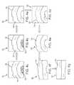
- This procedureillustrated in FIGS. 3 a to 3 g , may be carried out by excising the image so that it shows only the interior surface of a given length of the colon.
- FIG. 3 aillustrates an image 40 captured by the imaging device 32 and relayed to the video processing device.
- the processing deviceoutlines the lumen 42 by analyzing the image 40 for the difference in contrast between the colon's lumen 42 and its interior surface 44 .
- the processing deviceexcises the lumen 42 from the image 40 , as shown in FIG. 3 c .
- both the lumen 42 and the area surrounding the lumen 42may be excised from the image 40 .
- the outer edge 46 of the image 40may be excised to produce a ring-shaped image of the colon, as shown in FIGS. 3 d and 3 e .
- the outer edge of the excised imageis similar in shape to its inner edge of the excised image.
- the outer edge of the excised imagecan be equally spaced from the inner edge in the radial direction. This allows the flattened image to have a substantially rectangular configuration.
- the outer edge of the imagecan follow the outline of a haustral fold of the colon as shown in FIG. 3 d .
- a haustral foldcan be identified by the unique pattern of shading and contrast exhibited in the image.
- This excised imageis then “cut” radially and longitudinally along a side of the image ( FIG. 3 f ), and it is manipulated and flattened to show a rectangular view of the colon's interior surface ( FIG. 3 g ).
- the excised imagemay be “cut” with or without overlap.
- the processing devicemay convert the inner and outer edges of the image into substantially straight lines such that the ring-shaped view is converted into a rectangular view as shown in FIGS. 3 f to 3 g . This conversion causes certain areas of the image to undergo compression and others expansion.
- the image of the colonshows a longitudinal view of the colon.
- the endoscope 10lies parallel with the longitudinal axis of the colon, and the imaging device 32 of the endoscope 10 is disposed at the distal end 19 of the endoscope 10 and faces the longitudinal direction of the colon.
- the image 50may not show the entire lumen 52 , and the image may need to be reconstructed in a slightly different manner ( FIGS. 4 a to 4 g ).
- the lumen 52 or part of the lumen 52is identified by the difference in contrast.
- the lumen 52is excised from the image 50 as shown in FIG. 4 c , and a corresponding arc is also excised from the outer edge of the image 50 as shown in FIG. 4 d .
- the image 50is then converted to a substantially rectangle view as shown in FIGS. 4 f and 4 g.
- the processing devicemay locate the image spatially based on the positions of the previous images, such as the positions of the preceding images. For example, if the imaging device 32 faces a direction that is perpendicular to the longitudinal direction of the colon, the processing device can locate the image based on the positions of the previous images that overlap with this particular image. Images captured from this viewpoint may not need to be converted because of their substantially rectangular and flat shape.
- SIFTScale Invariant Feature Transform
- Interest pointswhich are invariant to scale and rotation, are identified in each image by constructing a multi-scale pyramid of Difference of Gaussian (DoG) images.
- DoGDifference of Gaussian
- Key pointsare identified by localizing maxima or minima in the Gaussian pyramid across levels.
- each interest pointis oriented by computing a gradient orientation histogram.
- a set of orientation histograms in a neighborhood such as 4 ⁇ 4 pixel neighborhoodmay be used to create the key point descriptor.
- the feature descriptorsare normalized in order to account for differences in illumination.
- corresponding key pointsare identified.
- a suitable transformation matrixwhich brings the images together such that key points or similar regions overlap, is calculated.
- An index or numbermay be used to measure the degree of similarity between two regions of two images. If this index or number is above a given value the two regions of the two images are considered to be overlapping.
- the processing devicechecks to ensure that no areas are missing as it continuously joins images together.
- the processing devicesends a signal, such as an audio and/or visual signal, that alerts the physician to the missed area.
- the physiciancan then return to the missing area and capture one or more additional images.
- the processing devicealso alerts the physician so that one or more additional images may be acquired to take the place of the unsuitable image.
- a missing area in the joined imagecan be detected in various manners. For example, there is likely a missing area if an excised inner or outer edge of a partial image is not joined to another partial image or if a region bordering on an excised inner or outer edge of a partial image does not have a corresponding region in another partial image and therefore cannot be joined to another partial image. This, however, does not always apply to the cut edge of a partial image ( FIG. 3 f ), which is made so that the partial image can be flattened ( FIGS. 3 a - 3 g ). This may also not apply to the first and last images because these two images each have an edge not joined to another image. Another way to detect a missing area is to see whether the cut edges of each partial image can be rejoined after the partial images have been joined to form a single image. If the cut edges of each partial image cannot be rejoined, the single image will likely have a missing area.
- the processing deviceuses an on-screen navigation cue to direct the operator to the location of the missing area or unsuitable image.
- the on-screen navigation cuemay include an arrow 72 on the screen 70 indicating the desired direction of movement for the imaging device 32 .
- the on-screen navigation cuemay include a highlighted area 74 on the screen 70 representing the location of the missing area or unsuitable image.
- the processing devicemay implement this feature by comparing the images joined previously with the current image being captured by the imaging device 32 . By comparing corresponding key points between these images, the processing device can direct the operator to move the imaging device 32 to the desired location.
- the processing devicecan calculate the scanning speed and/of the total amount of time that the imaging device spends in a segment of the colon such as the ascending or transverse portion of the colon.
- the processing devicemay rely on an algorithm that determines tracking time or speed in a manner similar to an optical computer mouse.
- the processing devicecan perform the same or similar steps of analyzing captured images. Before joining the images, the processing device analyzes the distance by which key points or corresponding areas have moved from one image to a subsequent image.
- the processing devicecan calculate the scanning speed by dividing the distance traveled by the time lapsed between the two images. The distance by which a given point or feature travels can be denoted by the number of image pixels. Each pixel can then be standardized to a measurement of actual distance such that the calculation can be performed.
- the distance traveledcan also be calculated by measuring the size change of a geometric feature from one image to another image. For example, if the geometric feature appears larger in an earlier image and smaller in a later image, it can be concluded that the imaging device is moving away from the geometric feature. The distance traveled by the imaging device can be calculated from the change in size.
- the processing deviceneeds to recognize when the imaging device is in the segment of the colon.
- the processing devicerecognizes a segment of the colon by its distinctive features, which can be, for example, the various flexures (e.g., splenic, hepatic, etc).
- the processing devicerecognizes a feature by comparing the captured image of the feature with a stored standard or typical image of the feature. For example, as the endoscope is withdrawn from the end of the colon, the hepatic flexure is expected first, and images are filtered for properties that would suggest an image of the hepatic flexure.
- the location of areas of shading/contrast and the location of the lumen in the imagewould suggest to the processing device that the image is of a flexure.
- the processing devicecan alert the operator about whether she is scanning the colon too fast and provide data on how much time was spent in each segment of the colon.
- Another feature that can be used to recognize the segment of the colonis the geometric shape of the colon.
- the lumen of the transverse colonhas a particularly triangular shape.
- An image of the colon's geometric shapecan be compared with a database of images the colon's geometric shape to determine which segment of the colon the imaging device is in.
- the endoscopemay have a sensor or transducer for communicating the position (such as the location and/or orientation) of the imaging device to the processing device.
- a positioning sensorinclude magnetic positioning sensors such as the Aurora System manufactured by NDI International of Waterloo, Canada, RF positioning sensors, or optical positioning sensors.
- the processing devicecan integrate the positional information of the imaging device with the image capturing and joining algorithm to better determine how to join the images. Joining of the images can be improved based on the imaging device's position, or based on information about the particular geometry of the colon the imaging device is in.
Landscapes
- Engineering & Computer Science (AREA)
- Health & Medical Sciences (AREA)
- Life Sciences & Earth Sciences (AREA)
- Physics & Mathematics (AREA)
- Surgery (AREA)
- General Health & Medical Sciences (AREA)
- Medical Informatics (AREA)
- Nuclear Medicine, Radiotherapy & Molecular Imaging (AREA)
- Radiology & Medical Imaging (AREA)
- Computer Vision & Pattern Recognition (AREA)
- General Physics & Mathematics (AREA)
- Theoretical Computer Science (AREA)
- Biomedical Technology (AREA)
- Heart & Thoracic Surgery (AREA)
- Biophysics (AREA)
- Optics & Photonics (AREA)
- Pathology (AREA)
- Veterinary Medicine (AREA)
- Public Health (AREA)
- Animal Behavior & Ethology (AREA)
- Molecular Biology (AREA)
- Multimedia (AREA)
- Signal Processing (AREA)
- Quality & Reliability (AREA)
- Endoscopes (AREA)
- Image Processing (AREA)
Abstract
Description
Claims (20)
Priority Applications (4)
| Application Number | Priority Date | Filing Date | Title |
|---|---|---|---|
| US13/584,647US9044185B2 (en) | 2007-04-10 | 2012-08-13 | Method and device for examining or imaging an interior surface of a cavity |
| US14/701,372US9613418B2 (en) | 2007-04-10 | 2015-04-30 | Method and device for examining or imaging an interior surface of a cavity |
| US15/467,759US10354382B2 (en) | 2007-04-10 | 2017-03-23 | Method and device for examining or imaging an interior surface of a cavity |
| US16/511,368US20190340761A1 (en) | 2007-04-10 | 2019-07-15 | Examining or Imaging an Interior Surface of a Cavity |
Applications Claiming Priority (4)
| Application Number | Priority Date | Filing Date | Title |
|---|---|---|---|
| US91105407P | 2007-04-10 | 2007-04-10 | |
| US12/101,050US8064666B2 (en) | 2007-04-10 | 2008-04-10 | Method and device for examining or imaging an interior surface of a cavity |
| US13/275,206US20120033062A1 (en) | 2007-04-10 | 2011-10-17 | Method and device for examining or imaging an interior surface of a cavity |
| US13/584,647US9044185B2 (en) | 2007-04-10 | 2012-08-13 | Method and device for examining or imaging an interior surface of a cavity |
Related Parent Applications (1)
| Application Number | Title | Priority Date | Filing Date |
|---|---|---|---|
| US13/275,206ContinuationUS20120033062A1 (en) | 2007-04-10 | 2011-10-17 | Method and device for examining or imaging an interior surface of a cavity |
Related Child Applications (1)
| Application Number | Title | Priority Date | Filing Date |
|---|---|---|---|
| US14/701,372ContinuationUS9613418B2 (en) | 2007-04-10 | 2015-04-30 | Method and device for examining or imaging an interior surface of a cavity |
Publications (2)
| Publication Number | Publication Date |
|---|---|
| US20120300999A1 US20120300999A1 (en) | 2012-11-29 |
| US9044185B2true US9044185B2 (en) | 2015-06-02 |
Family
ID=39853782
Family Applications (6)
| Application Number | Title | Priority Date | Filing Date |
|---|---|---|---|
| US12/101,050Active2030-08-23US8064666B2 (en) | 2007-04-10 | 2008-04-10 | Method and device for examining or imaging an interior surface of a cavity |
| US13/275,206AbandonedUS20120033062A1 (en) | 2007-04-10 | 2011-10-17 | Method and device for examining or imaging an interior surface of a cavity |
| US13/584,647ActiveUS9044185B2 (en) | 2007-04-10 | 2012-08-13 | Method and device for examining or imaging an interior surface of a cavity |
| US14/701,372Active2028-04-26US9613418B2 (en) | 2007-04-10 | 2015-04-30 | Method and device for examining or imaging an interior surface of a cavity |
| US15/467,759ActiveUS10354382B2 (en) | 2007-04-10 | 2017-03-23 | Method and device for examining or imaging an interior surface of a cavity |
| US16/511,368PendingUS20190340761A1 (en) | 2007-04-10 | 2019-07-15 | Examining or Imaging an Interior Surface of a Cavity |
Family Applications Before (2)
| Application Number | Title | Priority Date | Filing Date |
|---|---|---|---|
| US12/101,050Active2030-08-23US8064666B2 (en) | 2007-04-10 | 2008-04-10 | Method and device for examining or imaging an interior surface of a cavity |
| US13/275,206AbandonedUS20120033062A1 (en) | 2007-04-10 | 2011-10-17 | Method and device for examining or imaging an interior surface of a cavity |
Family Applications After (3)
| Application Number | Title | Priority Date | Filing Date |
|---|---|---|---|
| US14/701,372Active2028-04-26US9613418B2 (en) | 2007-04-10 | 2015-04-30 | Method and device for examining or imaging an interior surface of a cavity |
| US15/467,759ActiveUS10354382B2 (en) | 2007-04-10 | 2017-03-23 | Method and device for examining or imaging an interior surface of a cavity |
| US16/511,368PendingUS20190340761A1 (en) | 2007-04-10 | 2019-07-15 | Examining or Imaging an Interior Surface of a Cavity |
Country Status (1)
| Country | Link |
|---|---|
| US (6) | US8064666B2 (en) |
Cited By (32)
| Publication number | Priority date | Publication date | Assignee | Title |
|---|---|---|---|---|
| US9474440B2 (en) | 2009-06-18 | 2016-10-25 | Endochoice, Inc. | Endoscope tip position visual indicator and heat management system |
| US9667935B2 (en) | 2013-05-07 | 2017-05-30 | Endochoice, Inc. | White balance enclosure for use with a multi-viewing elements endoscope |
| US9706908B2 (en) | 2010-10-28 | 2017-07-18 | Endochoice, Inc. | Image capture and video processing systems and methods for multiple viewing element endoscopes |
| US9943218B2 (en) | 2013-10-01 | 2018-04-17 | Endochoice, Inc. | Endoscope having a supply cable attached thereto |
| US9949623B2 (en) | 2013-05-17 | 2018-04-24 | Endochoice, Inc. | Endoscope control unit with braking system |
| US9968242B2 (en) | 2013-12-18 | 2018-05-15 | Endochoice, Inc. | Suction control unit for an endoscope having two working channels |
| US10045685B2 (en) | 2006-01-23 | 2018-08-14 | Avantis Medical Systems, Inc. | Endoscope |
| US10064541B2 (en) | 2013-08-12 | 2018-09-04 | Endochoice, Inc. | Endoscope connector cover detection and warning system |
| US10078207B2 (en) | 2015-03-18 | 2018-09-18 | Endochoice, Inc. | Systems and methods for image magnification using relative movement between an image sensor and a lens assembly |
| US10105039B2 (en) | 2013-06-28 | 2018-10-23 | Endochoice, Inc. | Multi-jet distributor for an endoscope |
| US10123684B2 (en) | 2014-12-18 | 2018-11-13 | Endochoice, Inc. | System and method for processing video images generated by a multiple viewing elements endoscope |
| US10130246B2 (en) | 2009-06-18 | 2018-11-20 | Endochoice, Inc. | Systems and methods for regulating temperature and illumination intensity at the distal tip of an endoscope |
| US10258222B2 (en) | 2014-07-21 | 2019-04-16 | Endochoice, Inc. | Multi-focal, multi-camera endoscope systems |
| US10271713B2 (en) | 2015-01-05 | 2019-04-30 | Endochoice, Inc. | Tubed manifold of a multiple viewing elements endoscope |
| US10292570B2 (en) | 2016-03-14 | 2019-05-21 | Endochoice, Inc. | System and method for guiding and tracking a region of interest using an endoscope |
| US10354382B2 (en) | 2007-04-10 | 2019-07-16 | Avantis Medical Systems, Inc. | Method and device for examining or imaging an interior surface of a cavity |
| US10376181B2 (en) | 2015-02-17 | 2019-08-13 | Endochoice, Inc. | System for detecting the location of an endoscopic device during a medical procedure |
| US20190268581A1 (en)* | 2016-11-10 | 2019-08-29 | Kyocera Corporation | Body cavity observation system, laparoscope system, trocar apparatus, and operation method of body cavity observation system |
| US10401611B2 (en) | 2015-04-27 | 2019-09-03 | Endochoice, Inc. | Endoscope with integrated measurement of distance to objects of interest |
| US10488648B2 (en) | 2016-02-24 | 2019-11-26 | Endochoice, Inc. | Circuit board assembly for a multiple viewing element endoscope using CMOS sensors |
| US10516865B2 (en) | 2015-05-17 | 2019-12-24 | Endochoice, Inc. | Endoscopic image enhancement using contrast limited adaptive histogram equalization (CLAHE) implemented in a processor |
| US10517464B2 (en) | 2011-02-07 | 2019-12-31 | Endochoice, Inc. | Multi-element cover for a multi-camera endoscope |
| US10524645B2 (en) | 2009-06-18 | 2020-01-07 | Endochoice, Inc. | Method and system for eliminating image motion blur in a multiple viewing elements endoscope |
| US10542877B2 (en) | 2014-08-29 | 2020-01-28 | Endochoice, Inc. | Systems and methods for varying stiffness of an endoscopic insertion tube |
| US10595714B2 (en) | 2013-03-28 | 2020-03-24 | Endochoice, Inc. | Multi-jet controller for an endoscope |
| US10663714B2 (en) | 2010-10-28 | 2020-05-26 | Endochoice, Inc. | Optical system for an endoscope |
| US10898062B2 (en) | 2015-11-24 | 2021-01-26 | Endochoice, Inc. | Disposable air/water and suction valves for an endoscope |
| US10993605B2 (en) | 2016-06-21 | 2021-05-04 | Endochoice, Inc. | Endoscope system with multiple connection interfaces to interface with different video data signal sources |
| US11082598B2 (en) | 2014-01-22 | 2021-08-03 | Endochoice, Inc. | Image capture and video processing systems and methods for multiple viewing element endoscopes |
| US11234581B2 (en) | 2014-05-02 | 2022-02-01 | Endochoice, Inc. | Elevator for directing medical tool |
| US11529197B2 (en) | 2015-10-28 | 2022-12-20 | Endochoice, Inc. | Device and method for tracking the position of an endoscope within a patient's body |
| US12207796B2 (en) | 2013-03-28 | 2025-01-28 | Endochoice Inc. | Multi-jet controller for an endoscope |
Families Citing this family (39)
| Publication number | Priority date | Publication date | Assignee | Title |
|---|---|---|---|---|
| US20060149129A1 (en)* | 2005-01-05 | 2006-07-06 | Watts H D | Catheter with multiple visual elements |
| US8797392B2 (en) | 2005-01-05 | 2014-08-05 | Avantis Medical Sytems, Inc. | Endoscope assembly with a polarizing filter |
| US8872906B2 (en) | 2005-01-05 | 2014-10-28 | Avantis Medical Systems, Inc. | Endoscope assembly with a polarizing filter |
| US8289381B2 (en) | 2005-01-05 | 2012-10-16 | Avantis Medical Systems, Inc. | Endoscope with an imaging catheter assembly and method of configuring an endoscope |
| US8182422B2 (en) | 2005-12-13 | 2012-05-22 | Avantis Medical Systems, Inc. | Endoscope having detachable imaging device and method of using |
| US8287446B2 (en) | 2006-04-18 | 2012-10-16 | Avantis Medical Systems, Inc. | Vibratory device, endoscope having such a device, method for configuring an endoscope, and method of reducing looping of an endoscope |
| EP2023795A2 (en) | 2006-05-19 | 2009-02-18 | Avantis Medical Systems, Inc. | Device and method for reducing effects of video artifacts |
| US9375164B2 (en) | 2007-03-08 | 2016-06-28 | Sync-Rx, Ltd. | Co-use of endoluminal data and extraluminal imaging |
| WO2008107905A2 (en) | 2007-03-08 | 2008-09-12 | Sync-Rx, Ltd. | Imaging and tools for use with moving organs |
| US10716528B2 (en) | 2007-03-08 | 2020-07-21 | Sync-Rx, Ltd. | Automatic display of previously-acquired endoluminal images |
| US11197651B2 (en) | 2007-03-08 | 2021-12-14 | Sync-Rx, Ltd. | Identification and presentation of device-to-vessel relative motion |
| US9629571B2 (en) | 2007-03-08 | 2017-04-25 | Sync-Rx, Ltd. | Co-use of endoluminal data and extraluminal imaging |
| US8781193B2 (en) | 2007-03-08 | 2014-07-15 | Sync-Rx, Ltd. | Automatic quantitative vessel analysis |
| US11064964B2 (en) | 2007-03-08 | 2021-07-20 | Sync-Rx, Ltd | Determining a characteristic of a lumen by measuring velocity of a contrast agent |
| US9968256B2 (en) | 2007-03-08 | 2018-05-15 | Sync-Rx Ltd. | Automatic identification of a tool |
| US9305334B2 (en) | 2007-03-08 | 2016-04-05 | Sync-Rx, Ltd. | Luminal background cleaning |
| US9144394B2 (en) | 2008-11-18 | 2015-09-29 | Sync-Rx, Ltd. | Apparatus and methods for determining a plurality of local calibration factors for an image |
| US10362962B2 (en)* | 2008-11-18 | 2019-07-30 | Synx-Rx, Ltd. | Accounting for skipped imaging locations during movement of an endoluminal imaging probe |
| US11064903B2 (en) | 2008-11-18 | 2021-07-20 | Sync-Rx, Ltd | Apparatus and methods for mapping a sequence of images to a roadmap image |
| US8855744B2 (en) | 2008-11-18 | 2014-10-07 | Sync-Rx, Ltd. | Displaying a device within an endoluminal image stack |
| US9974509B2 (en) | 2008-11-18 | 2018-05-22 | Sync-Rx Ltd. | Image super enhancement |
| US9101286B2 (en) | 2008-11-18 | 2015-08-11 | Sync-Rx, Ltd. | Apparatus and methods for determining a dimension of a portion of a stack of endoluminal data points |
| US9095313B2 (en) | 2008-11-18 | 2015-08-04 | Sync-Rx, Ltd. | Accounting for non-uniform longitudinal motion during movement of an endoluminal imaging probe |
| TWI432168B (en)* | 2009-12-31 | 2014-04-01 | Univ Nat Yunlin Sci & Tech | Endoscope navigation method and endoscopy navigation system |
| CA2835495A1 (en) | 2010-05-10 | 2011-11-17 | Nanamed,Llc | Method and device for imaging an interior surface of an intracorporeal cavity |
| US20140148734A1 (en)* | 2010-09-03 | 2014-05-29 | Lyndon V. Hernandez | Colonoscopy systems and methods |
| EP2863802B1 (en) | 2012-06-26 | 2020-11-04 | Sync-RX, Ltd. | Flow-related image processing in luminal organs |
| WO2015011594A1 (en)* | 2013-07-24 | 2015-01-29 | Koninklijke Philips N.V. | Non-imaging two dimensional array probe and system for automated screening of carotid stenosis |
| US10130243B2 (en) | 2014-01-30 | 2018-11-20 | Qatar University Al Tarfa | Image-based feedback endoscopy system |
| US20150313445A1 (en)* | 2014-05-01 | 2015-11-05 | Endochoice, Inc. | System and Method of Scanning a Body Cavity Using a Multiple Viewing Elements Endoscope |
| WO2020160567A1 (en) | 2019-04-05 | 2020-08-06 | Carnegie Mellon University | Real-time measurement of visible surface area from colonoscopy video |
| JP7464060B2 (en)* | 2019-10-01 | 2024-04-09 | 日本電気株式会社 | Image processing device, control method and program |
| US20220071711A1 (en)* | 2020-09-04 | 2022-03-10 | Karl Storz Se & Co. Kg | Devices, systems, and methods for identifying unexamined regions during a medical procedure |
| US20220160220A1 (en)* | 2020-11-23 | 2022-05-26 | Medos International Sárl | Arthroscopic medical implements and assemblies |
| US12070196B2 (en) | 2020-11-23 | 2024-08-27 | Medos International Sarl | Arthroscopic medical implements and assemblies |
| US12061590B2 (en) | 2021-11-18 | 2024-08-13 | Olympus Corporation | Information processing system and processing method |
| US20250139769A1 (en)* | 2021-11-18 | 2025-05-01 | Nec Corporation | Information display device, information display method, and recording medium |
| GB2617408A (en)* | 2022-04-08 | 2023-10-11 | Aker Medhat | A colonoscope device |
| US20230410336A1 (en) | 2022-06-15 | 2023-12-21 | CapsoVision, Inc. | Method and Apparatus for Identifying Capsule Camera Location inside Gastrointestinal Tract |
Citations (371)
| Publication number | Priority date | Publication date | Assignee | Title |
|---|---|---|---|---|
| FR711949A (en) | 1930-05-31 | 1931-09-21 | Gentile Et Cie Successeurs P | Cystoscope |
| US3437747A (en) | 1964-03-24 | 1969-04-08 | Sheldon Edward E | Devices for inspection using fiberoptic members |
| US3610231A (en) | 1967-07-21 | 1971-10-05 | Olympus Optical Co | Endoscope |
| US3643653A (en) | 1968-12-24 | 1972-02-22 | Olympus Optical Co | Endoscopic apparatus |
| US3739770A (en) | 1970-10-09 | 1973-06-19 | Olympus Optical Co | Bendable tube of an endoscope |
| JPS49130235A (en) | 1973-04-16 | 1974-12-13 | ||
| US3889662A (en) | 1973-05-31 | 1975-06-17 | Olympus Optical Co | Endoscope |
| US3897775A (en) | 1973-09-07 | 1975-08-05 | Olympus Optical Co | Endoscope with facile bending operation |
| US4066071A (en) | 1975-08-15 | 1978-01-03 | Nagel John G | Extension pull through device to allow for easier passage of flexible fiber endoscope |
| JPS569712A (en) | 1979-07-06 | 1981-01-31 | Olympus Optical Co Ltd | Visual field direction changing optical system for slender image transmission system |
| US4261344A (en) | 1979-09-24 | 1981-04-14 | Welch Allyn, Inc. | Color endoscope |
| JPS5656486A (en) | 1979-10-15 | 1981-05-18 | Takumi Sakamoto | Oillpressure elevator device |
| US4351587A (en) | 1979-03-05 | 1982-09-28 | Olympus Optical Company, Ltd. | Apparatus for positioning eyepiece of endoscope |
| JPS57170707A (en) | 1981-04-16 | 1982-10-21 | Nippon Soken Inc | Manufacture for honeycomb forming die |
| US4494549A (en) | 1981-05-21 | 1985-01-22 | Olympus Optical Co., Ltd. | Device for diagnosing body cavity interiors with supersonic waves |
| JPS6076714A (en) | 1983-10-03 | 1985-05-01 | Olympus Optical Co Ltd | Endoscope using polarizing filter |
| JPS6083636A (en) | 1983-10-12 | 1985-05-11 | オリンパス光学工業株式会社 | Endoscope apparatus |
| JPS60111217A (en) | 1983-11-18 | 1985-06-17 | Olympus Optical Co Ltd | Endoscope device equipped with control means for quantity of incident light |
| US4573450A (en) | 1983-11-11 | 1986-03-04 | Fuji Photo Optical Co., Ltd. | Endoscope |
| US4586491A (en) | 1984-12-14 | 1986-05-06 | Warner-Lambert Technologies, Inc. | Bronchoscope with small gauge viewing attachment |
| US4602281A (en) | 1983-09-05 | 1986-07-22 | Olympus Optical Co., Ltd. | Automatic means for controlling dosage of illuminating light for picking-up image by endoscope assembly |
| US4625236A (en) | 1984-07-31 | 1986-11-25 | Olympus Optical Co., Ltd. | Light source means for endoscope employing solid state imaging device |
| US4646722A (en) | 1984-12-10 | 1987-03-03 | Opielab, Inc. | Protective endoscope sheath and method of installing same |
| US4699463A (en) | 1985-11-01 | 1987-10-13 | Circon Corporation | Multidirectional viewing borescope |
| US4721097A (en) | 1986-10-31 | 1988-01-26 | Circon Corporation | Endoscope sheaths and method and apparatus for installation and removal |
| US4727859A (en) | 1986-12-29 | 1988-03-01 | Welch Allyn, Inc. | Right angle detachable prism assembly for borescope |
| US4741326A (en) | 1986-10-01 | 1988-05-03 | Fujinon, Inc. | Endoscope disposable sheath |
| US4742817A (en) | 1985-05-15 | 1988-05-10 | Olympus Optical Co., Ltd. | Endoscopic apparatus having a bendable insertion section |
| US4790295A (en) | 1986-12-16 | 1988-12-13 | Olympus Optical Co., Ltd. | Endoscope having transparent resin sealing layer |
| JPS63309912A (en) | 1987-06-11 | 1988-12-19 | Olympus Optical Co Ltd | Light source device for endoscope |
| US4800870A (en) | 1988-03-11 | 1989-01-31 | Reid Jr Ben A | Method and apparatus for bile duct exploration |
| US4825850A (en) | 1988-05-13 | 1989-05-02 | Opielab, Inc. | Contamination protection system for endoscope control handles |
| US4836211A (en) | 1986-09-17 | 1989-06-06 | Naomi Sekino | Ultrasonic treatment apparatus for performing medical treatment by use of ultrasonic vibrations |
| US4846154A (en) | 1988-06-13 | 1989-07-11 | Macanally Richard B | Dual view endoscope |
| US4853773A (en) | 1987-01-31 | 1989-08-01 | Olympus Optical, Co., Ltd. | Endoscope signal processing apparatus using sequential and synchronous imaging devices |
| US4852551A (en) | 1988-04-22 | 1989-08-01 | Opielab, Inc. | Contamination-free endoscope valves for use with a disposable endoscope sheath |
| US4862873A (en) | 1987-05-27 | 1989-09-05 | Olympus Optical Co., Ltd. | Stereo endoscope |
| US4867138A (en) | 1987-05-13 | 1989-09-19 | Olympus Optical Co., Ltd. | Rigid electronic endoscope |
| US4869238A (en) | 1988-04-22 | 1989-09-26 | Opielab, Inc. | Endoscope for use with a disposable sheath |
| US4870488A (en) | 1987-02-10 | 1989-09-26 | Olympus Optical Co., Ltd. | Endoscope imaging system used with an electronic scope and an optical endoscope |
| US4873572A (en) | 1987-02-27 | 1989-10-10 | Olympus Optical Co., Ltd. | Electronic endoscope apparatus |
| US4873965A (en) | 1987-07-31 | 1989-10-17 | Guido Danieli | Flexible endoscope |
| JPH01267514A (en) | 1988-04-19 | 1989-10-25 | Fuji Photo Film Co Ltd | Endoscope |
| JPH01172847U (en) | 1988-05-26 | 1989-12-07 | ||
| US4899732A (en) | 1988-09-02 | 1990-02-13 | Baxter International, Inc. | Miniscope |
| US4905667A (en) | 1987-05-12 | 1990-03-06 | Ernst Foerster | Apparatus for endoscopic-transpapillary exploration of biliary tract |
| US4907395A (en) | 1988-05-13 | 1990-03-13 | Opielab, Inc. | Packaging system for disposable endoscope sheaths |
| US4911148A (en) | 1989-03-14 | 1990-03-27 | Intramed Laboratories, Inc. | Deflectable-end endoscope with detachable flexible shaft assembly |
| US4911564A (en) | 1988-03-16 | 1990-03-27 | Baker Herbert R | Protective bearing guard |
| US4926258A (en) | 1987-10-20 | 1990-05-15 | Olympus Optical Co., Ltd. | Electronic endoscope apparatus capable of driving solid state imaging devices having different characteristics |
| US4947828A (en) | 1989-04-17 | 1990-08-14 | Schott Fiber Optics | Endoscope connector |
| US4947827A (en) | 1988-12-30 | 1990-08-14 | Opielab, Inc. | Flexible endoscope |
| JPH02295530A (en) | 1989-05-10 | 1990-12-06 | Sumitomo Bakelite Co Ltd | Endoscope catheter and method for using the same |
| US4979496A (en) | 1988-04-05 | 1990-12-25 | Fuji Photo Optical Co., Ltd. | Endoscope for bile duct and pancreatic duct |
| US4991565A (en) | 1989-06-26 | 1991-02-12 | Asahi Kogaku Kogyo Kabushiki Kaisha | Sheath device for endoscope and fluid conduit connecting structure therefor |
| US5019040A (en) | 1989-08-31 | 1991-05-28 | Koshin Sangyo Kabushiki Kaisha | Catheter |
| US5026377A (en) | 1989-07-13 | 1991-06-25 | American Medical Systems, Inc. | Stent placement instrument and method |
| US5025778A (en) | 1990-03-26 | 1991-06-25 | Opielab, Inc. | Endoscope with potential channels and method of using the same |
| JPH03159629A (en) | 1989-11-20 | 1991-07-09 | Hiroaki Kumagai | Apparatus for diagnosing digestive tract |
| US5050585A (en) | 1988-03-28 | 1991-09-24 | Asahi Kogaku Kogyo Kabushiki Kaisha | Sheathed endoscope |
| JPH04500768A (en) | 1989-06-28 | 1992-02-13 | カール シュトルツ ゲーエムベーハー アンド カンパニー | Endoscope with a video device placed at the distal end |
| US5159446A (en) | 1991-06-21 | 1992-10-27 | Olympus Optical Co., Ltd. | Electronic endoscope system provided with a separate camera controlling unit and motor controlling unit |
| USRE34110E (en) | 1988-04-22 | 1992-10-27 | Opielab, Inc. | Endoscope for use with a disposable sheath |
| JPH04341232A (en) | 1991-03-11 | 1992-11-27 | Olympus Optical Co Ltd | Electronic endoscope system |
| US5178130A (en) | 1990-04-04 | 1993-01-12 | Olympus Optical Co., Ltd. | Parent-and-son type endoscope system for making a synchronized field sequential system illumination |
| US5187572A (en) | 1990-10-31 | 1993-02-16 | Olympus Optical Co., Ltd. | Endoscope system with a plurality of synchronized light source apparatuses |
| US5193525A (en) | 1990-11-30 | 1993-03-16 | Vision Sciences | Antiglare tip in a sheath for an endoscope |
| US5196928A (en) | 1991-04-02 | 1993-03-23 | Olympus Optical Co., Ltd. | Endoscope system for simultaneously displaying two endoscopic images on a shared monitor |
| WO1993015648A1 (en) | 1992-02-07 | 1993-08-19 | Wilk Peter J | Endoscope with disposable insertion member |
| US5253638A (en) | 1992-03-25 | 1993-10-19 | Welch Allyn, Inc. | Right-angle detachable variable-position reflector assembly |
| JPH05285091A (en) | 1992-04-10 | 1993-11-02 | Olympus Optical Co Ltd | Bending apparatus |
| US5260780A (en) | 1991-11-15 | 1993-11-09 | Threadmasters, Inc. | Visual inspection device and process |
| JPH05307144A (en) | 1992-04-28 | 1993-11-19 | Bikoudou:Kk | Halation removing device for endoscope and electronic endoscope |
| US5271381A (en) | 1991-11-18 | 1993-12-21 | Vision Sciences, Inc. | Vertebrae for a bending section of an endoscope |
| JPH05341210A (en) | 1992-06-09 | 1993-12-24 | Olympus Optical Co Ltd | Stereoscopic endoscope device |
| JPH069228B2 (en) | 1986-02-27 | 1994-02-02 | 株式会社町田製作所 | Endoscope with built-in solid-state image sensor |
| EP0586162A1 (en) | 1992-08-24 | 1994-03-09 | Ethicon Inc. | Glare elimination device |
| US5305121A (en) | 1992-06-08 | 1994-04-19 | Origin Medsystems, Inc. | Stereoscopic endoscope system |
| JPH06130308A (en) | 1992-10-16 | 1994-05-13 | Olympus Optical Co Ltd | Optical adapter of endoscope |
| US5318031A (en) | 1990-07-23 | 1994-06-07 | Bruker Analytische Messtechnik Gmbh | Method and apparatus for determining chemical states of living animal or human tissue using nuclear magnetic resonance |
| US5329887A (en) | 1992-04-03 | 1994-07-19 | Vision Sciences, Incorporated | Endoscope control assembly with removable control knob/brake assembly |
| US5337734A (en) | 1992-10-29 | 1994-08-16 | Advanced Polymers, Incorporated | Disposable sheath with optically transparent window formed continuously integral therewith |
| JPH07354A (en) | 1993-06-15 | 1995-01-06 | Hiroaki Kumagai | Large intestine endoscope |
| JPH07352A (en) | 1993-06-15 | 1995-01-06 | Olympus Optical Co Ltd | Insertion auxiliary tool for endoscope |
| US5381784A (en) | 1992-09-30 | 1995-01-17 | Adair; Edwin L. | Stereoscopic endoscope |
| US5398685A (en)* | 1992-01-10 | 1995-03-21 | Wilk; Peter J. | Endoscopic diagnostic system and associated method |
| JPH0721001U (en) | 1993-09-17 | 1995-04-18 | 旭光学工業株式会社 | Endoscope insertion part |
| JPH07136108A (en) | 1990-12-20 | 1995-05-30 | Hiroaki Kumagai | Set intestinal endoscope |
| US5434669A (en) | 1990-10-23 | 1995-07-18 | Olympus Optical Co., Ltd. | Measuring interferometric endoscope having a laser radiation source |
| US5447148A (en) | 1993-07-08 | 1995-09-05 | Vision Sciences, Inc. | Endoscopic contamination protection system to facilitate cleaning of endoscopes |
| JPH07275197A (en) | 1994-04-12 | 1995-10-24 | Fuji Photo Optical Co Ltd | Electronic endoscope apparatus for separating image pickup part |
| US5483951A (en) | 1994-02-25 | 1996-01-16 | Vision-Sciences, Inc. | Working channels for a disposable sheath for an endoscope |
| US5520607A (en) | 1994-03-04 | 1996-05-28 | Vision Sciences, Inc. | Holding tray and clamp assembly for an endoscopic sheath |
| US5530238A (en) | 1993-09-03 | 1996-06-25 | U.S. Philips Corporation | Image detection device having correction circuit for removing artifacts due to delayed charge transfer |
| US5533496A (en) | 1994-02-15 | 1996-07-09 | Very Inventive Physicians, Inc. | Endoscopic technique particularly suited for exploratory surgery |
| US5536236A (en) | 1993-02-12 | 1996-07-16 | Olympus Optical Co., Ltd. | Covered endoscope system |
| JPH08206061A (en) | 1995-02-03 | 1996-08-13 | Olympus Optical Co Ltd | Curving device |
| US5556367A (en) | 1993-03-05 | 1996-09-17 | Olympus Optical Co., Ltd. | Cover type endoscope apparatus |
| JPH0956662A (en) | 1995-08-21 | 1997-03-04 | Hitachi Ltd | Side-viewing endoscope |
| US5613936A (en) | 1995-02-22 | 1997-03-25 | Concurrent Technologies Corp. | Stereo laparoscope apparatus |
| US5614943A (en) | 1991-12-19 | 1997-03-25 | Olympus Optical Co., Ltd. | Dissimilar endoscopes usable with a common control unit |
| US5626553A (en) | 1995-06-05 | 1997-05-06 | Vision-Sciences, Inc. | Endoscope articulation system to reduce effort during articulation of an endoscope |
| US5634466A (en) | 1993-11-19 | 1997-06-03 | Advanced Technology Laboratories, Inc. | Ultrasonic transesophageal probe with detachable transducer tip |
| US5653677A (en) | 1994-04-12 | 1997-08-05 | Fuji Photo Optical Co. Ltd | Electronic endoscope apparatus with imaging unit separable therefrom |
| US5679216A (en) | 1992-03-12 | 1997-10-21 | Olympus Optical Co., Ltd. | Method of manufacturing a multi-degree-of-freedom manipulator |
| US5682199A (en) | 1996-03-28 | 1997-10-28 | Jedmed Instrument Company | Video endoscope with interchangeable endoscope heads |
| US5681260A (en) | 1989-09-22 | 1997-10-28 | Olympus Optical Co., Ltd. | Guiding apparatus for guiding an insertable body within an inspected object |
| US5685822A (en) | 1996-08-08 | 1997-11-11 | Vision-Sciences, Inc. | Endoscope with sheath retaining device |
| US5692729A (en) | 1996-02-16 | 1997-12-02 | Vision-Sciences, Inc. | Pressure equalized flow control apparatus and method for endoscope channels |
| US5696850A (en) | 1995-12-21 | 1997-12-09 | Eastman Kodak Company | Automatic image sharpening in an electronic imaging system |
| US5702348A (en) | 1996-07-24 | 1997-12-30 | Vision-Sciences, Inc. | Disposable endoscopic sheath support and positioning assembly |
| US5706128A (en) | 1995-09-11 | 1998-01-06 | Edge Scientific Instrument Company Llc | Stereo microscope condenser |
| DE19626433A1 (en) | 1996-06-19 | 1998-01-15 | Jan Henrik Dr Wilkens | Endoscope head arrangement with integrated image production arrangement |
| US5711299A (en)* | 1996-01-26 | 1998-01-27 | Manwaring; Kim H. | Surgical guidance method and system for approaching a target within a body |
| US5722933A (en) | 1993-01-27 | 1998-03-03 | Olympus Optical Co., Ltd. | Channeled endoscope cover fitted type endoscope |
| US5752912A (en) | 1995-06-26 | 1998-05-19 | Asahi Kogaku Kogyo Kabushiki Kaisha | Manipulator for flexible portion of an endoscope |
| US5762603A (en) | 1995-09-15 | 1998-06-09 | Pinotage, Llc | Endoscope having elevation and azimuth control of camera assembly |
| US5817061A (en) | 1997-05-16 | 1998-10-06 | Ethicon Endo-Surgery, Inc. | Trocar assembly |
| US5827177A (en) | 1997-02-18 | 1998-10-27 | Vision-Sciences, Inc. | Endoscope sheath assembly with isolating fabric sleeve |
| US5833603A (en) | 1996-03-13 | 1998-11-10 | Lipomatrix, Inc. | Implantable biosensing transponder |
| US5843460A (en) | 1993-05-19 | 1998-12-01 | Institut Pasteur | Immunogenic compositions against helicobacter infection, polypeptides for use in the compositions, and nucleic acid sequences encoding said polypeptides |
| US5843103A (en) | 1997-03-06 | 1998-12-01 | Scimed Life Systems, Inc. | Shaped wire rotational atherectomy device |
| US5854859A (en) | 1996-12-27 | 1998-12-29 | Hewlett-Packard Company | Image sharpening filter providing variable sharpening dependent on pixel intensity |
| US5860914A (en) | 1993-10-05 | 1999-01-19 | Asahi Kogaku Kogyo Kabushiki Kaisha | Bendable portion of endoscope |
| JPH1176150A (en) | 1997-09-03 | 1999-03-23 | Asahi Optical Co Ltd | Endoscope |
| WO1999017542A1 (en) | 1997-09-26 | 1999-04-08 | The Secretary Of State For Defence | Sensor system |
| WO1999030506A1 (en) | 1997-12-05 | 1999-06-17 | Intel Corporation | Dark frame cancellation for cmos sensor-based tethered video peripherals |
| US5916147A (en) | 1997-09-22 | 1999-06-29 | Boury; Harb N. | Selectively manipulable catheter |
| US5924977A (en) | 1993-02-26 | 1999-07-20 | Olympus Optical Co., Ltd. | Endoscope system including endoscope and disposable protection cover |
| US5938587A (en) | 1996-04-25 | 1999-08-17 | Modified Polymer Components, Inc. | Flexible inner liner for the working channel of an endoscope |
| JPH11253401A (en) | 1998-03-10 | 1999-09-21 | Olympus Optical Co Ltd | Endoscope |
| US5982932A (en) | 1992-09-16 | 1999-11-09 | Mikos, Ltd. | Method and apparatus for flash correlation |
| US5989224A (en) | 1998-02-23 | 1999-11-23 | Dexide Corporation | Universal seal for use with endoscopic cannula |
| US5989182A (en) | 1997-12-19 | 1999-11-23 | Vista Medical Technologies, Inc. | Device-steering shaft assembly and endoscope |
| JPH11332821A (en) | 1998-05-22 | 1999-12-07 | Olympus Optical Co Ltd | Endoscope |
| US6017358A (en) | 1997-05-01 | 2000-01-25 | Inbae Yoon | Surgical instrument with multiple rotatably mounted offset end effectors |
| US6026323A (en) | 1997-03-20 | 2000-02-15 | Polartechnics Limited | Tissue diagnostic system |
| US6066090A (en) | 1997-06-19 | 2000-05-23 | Yoon; Inbae | Branched endoscope system |
| US6099466A (en) | 1994-09-21 | 2000-08-08 | Asahi Kogaku Kogyo Kabushiki Kaisha | Fluorescence diagnosis endoscope system |
| US6099464A (en) | 1995-04-10 | 2000-08-08 | Olympus Optical Co., Ltd. | Bending sheath for probe |
| US6099485A (en) | 1996-08-27 | 2000-08-08 | C. R. Bard, Inc. | Torquable, low mass medical guidewire |
| US6106463A (en) | 1998-04-20 | 2000-08-22 | Wilk; Peter J. | Medical imaging device and associated method including flexible display |
| US6174280B1 (en) | 1998-11-19 | 2001-01-16 | Vision Sciences, Inc. | Sheath for protecting and altering the bending characteristics of a flexible endoscope |
| US6190330B1 (en) | 1999-08-09 | 2001-02-20 | Vision-Sciences, Inc. | Endoscopic location and vacuum assembly and method |
| US20010007468A1 (en) | 2000-01-11 | 2001-07-12 | Asahi Kogaku Kogyo Kabushiki Kaisha | Electronic endoscope selector and electronic endoscope system |
| US6261307B1 (en) | 1997-05-01 | 2001-07-17 | Inbae Yoon | Method of using surgical instrument with rotatably mounted offset end effector |
| US6261226B1 (en) | 1994-03-30 | 2001-07-17 | Medical Media Systems | Electronically Steerable Endoscope |
| US6277064B1 (en) | 1997-12-30 | 2001-08-21 | Inbae Yoon | Surgical instrument with rotatably mounted offset endoscope |
| US6296608B1 (en) | 1996-07-08 | 2001-10-02 | Boston Scientific Corporation | Diagnosing and performing interventional procedures on tissue in vivo |
| US6301047B1 (en) | 1998-11-17 | 2001-10-09 | Nhk Spring Co., Ltd. | System for optically identifying an object |
| US20010031912A1 (en) | 2000-04-10 | 2001-10-18 | Cbeyond Inc. | Image sensor and an endoscope using the same |
| US20010037052A1 (en) | 1998-03-25 | 2001-11-01 | Fuji Photo Optical Co., Ltd. | Electronic-endoscope light source unit for setting shading period |
| US20010051766A1 (en) | 1999-03-01 | 2001-12-13 | Gazdzinski Robert F. | Endoscopic smart probe and method |
| US20010056238A1 (en) | 2000-06-26 | 2001-12-27 | Fuji Photo Film Co., Ltd. | Fluorescent image obtaining apparatus |
| US6350231B1 (en) | 1999-01-21 | 2002-02-26 | Vision Sciences, Inc. | Apparatus and method for forming thin-walled elastic components from an elastomeric material |
| US20020026188A1 (en) | 2000-03-31 | 2002-02-28 | Balbierz Daniel J. | Tissue biopsy and treatment apparatus and method |
| US20020039400A1 (en) | 1996-09-16 | 2002-04-04 | Arie E. Kaufman | System and method for performing a three-dimensional examination with collapse correction |
| US6369855B1 (en) | 1996-11-01 | 2002-04-09 | Texas Instruments Incorporated | Audio and video decoder circuit and system |
| US6375653B1 (en) | 2000-01-28 | 2002-04-23 | Allegiance Corporation | Surgical apparatus providing tool access and replaceable irrigation pump cartridge |
| US6387043B1 (en) | 1998-05-13 | 2002-05-14 | Inbae Yoon | Penetrating endoscope and endoscopic surgical instrument with CMOS image sensor and display |
| US20020089584A1 (en) | 2001-01-09 | 2002-07-11 | Fuji Photo Optical Co., Ltd. | Electronic endoscope apparatus adaptable to endoscopes equipped with imaging device with different pixel density |
| US20020095168A1 (en) | 2001-01-18 | 2002-07-18 | Griego John A. | Steerable sphincterotome and methods for cannulation, papillotomy and sphincterotomy |
| US20020099267A1 (en) | 2001-01-25 | 2002-07-25 | Scimed Life Systems, Inc. | Endoscopic vision system |
| US20020103420A1 (en) | 2001-01-26 | 2002-08-01 | George Coleman | Endoscope with alterable viewing angle |
| US20020101546A1 (en) | 1995-05-23 | 2002-08-01 | Colorlink, Inc. | Color filters, sequencers and displays using color selective light modulators |
| US6433492B1 (en) | 2000-09-18 | 2002-08-13 | Northrop Grumman Corporation | Magnetically shielded electrodeless light source |
| US20020110282A1 (en) | 2001-02-09 | 2002-08-15 | Walter Kraft | Local change of an image sharpness of photographic images with masks |
| US20020115908A1 (en) | 2000-06-30 | 2002-08-22 | Inner Vision Imaging, L.L.C. | Endoscope |
| US6456684B1 (en) | 1999-07-23 | 2002-09-24 | Inki Mun | Surgical scanning system and process for use thereof |
| US6454702B1 (en) | 1999-10-14 | 2002-09-24 | Scimed Life Systems, Inc. | Endoscope and endoscopic instrument system having reduced backlash when moving the endoscopic instrument within a working channel of the endoscope |
| US6461294B1 (en) | 2000-10-30 | 2002-10-08 | Vision Sciences, Inc. | Inflatable member for an endoscope sheath |
| US6464633B1 (en) | 1999-08-23 | 2002-10-15 | Olympus Optical Co., Ltd. | Light source device for endoscope using DMD |
| US20020156347A1 (en) | 2001-04-24 | 2002-10-24 | Byungkyu Kim | Micro-robot for colonoscope with motor locomotion and system for colonoscope using the same |
| WO2002085194A1 (en) | 2001-04-20 | 2002-10-31 | Power Medical Interventions, Inc. | Imaging device |
| US6482149B1 (en) | 1999-05-12 | 2002-11-19 | Fuji Photo Optical Co., Ltd. | Curved part of endoscope |
| WO2002094105A2 (en) | 2001-05-23 | 2002-11-28 | Boston Scientific Limited | Method for performing endoluminal fundoplication and apparatus for use in the method |
| US20020193662A1 (en) | 2000-04-03 | 2002-12-19 | Amir Belson | Steerable endoscope and improved method of insertion |
| US20030001951A1 (en) | 2001-06-27 | 2003-01-02 | Fuji Photo Film Co., Ltd. | Image obtaining method and apparatus for an endoscope |
| US6503195B1 (en) | 1999-05-24 | 2003-01-07 | University Of North Carolina At Chapel Hill | Methods and systems for real-time structured light depth extraction and endoscope using real-time structured light depth extraction |
| US20030011768A1 (en) | 1998-06-30 | 2003-01-16 | Jung Wayne D. | Apparatus and method for measuring optical characteristics of an object |
| US20030032863A1 (en) | 2001-08-09 | 2003-02-13 | Yuri Kazakevich | Endoscope with imaging probe |
| US20030040668A1 (en) | 2001-08-03 | 2003-02-27 | Olympus Optical Co., Ltd. | Endoscope apparatus |
| US6527704B1 (en) | 1999-03-10 | 2003-03-04 | Stryker Corporation | Endoscopic camera system integrated with a trocar sleeve |
| US20030045778A1 (en) | 2000-04-03 | 2003-03-06 | Ohline Robert M. | Tendon-driven endoscope and methods of insertion |
| US20030065250A1 (en) | 2001-09-17 | 2003-04-03 | Case Western Reserve University | Peristaltically Self-propelled endoscopic device |
| US6547724B1 (en) | 1999-05-26 | 2003-04-15 | Scimed Life Systems, Inc. | Flexible sleeve slidingly transformable into a large suction sleeve |
| US6554767B2 (en) | 2001-04-04 | 2003-04-29 | Olympus Optical Co. Ltd. | Endoscopic optical adapter freely attachable to and detachable from endoscope |
| US20030088152A1 (en) | 2001-11-06 | 2003-05-08 | Masazumi Takada | Self-propelled colonoscope |
| US6564088B1 (en) | 2000-01-21 | 2003-05-13 | University Of Massachusetts | Probe for localized tissue spectroscopy |
| JP2003135388A (en) | 2001-10-30 | 2003-05-13 | Olympus Optical Co Ltd | Endoscope apparatus |
| US20030093088A1 (en) | 2001-11-09 | 2003-05-15 | Long Gary L. | Self-propelled, intraluminal device with working channel and method of use |
| US20030093031A1 (en) | 2001-11-09 | 2003-05-15 | Long Gary L. | Self-propelled, intraluminal device with medical agent applicator and method of use |
| US20030103199A1 (en) | 1997-07-01 | 2003-06-05 | Jung Wayne D. | Apparatus and method for measuring optical characteristics of an object |
| US20030105386A1 (en) | 1999-01-29 | 2003-06-05 | Sightline Technologies Ltd. | Propulsion of a probe in the colon using a flexible sleeve |
| US20030120130A1 (en) | 2001-08-06 | 2003-06-26 | Arkady Glukhovsky | System and method for maneuvering a device in vivo |
| US20030125630A1 (en) | 2001-12-31 | 2003-07-03 | Furnish Simon M. | Catheter probe arrangement for tissue analysis by radiant energy delivery and radiant energy collection |
| US20030128892A1 (en) | 2001-11-01 | 2003-07-10 | Ge Medical Systems Technology Company Llc | Method for contrast matching of multiple images of the same object or scene to a common reference image |
| US20030130711A1 (en) | 2001-09-28 | 2003-07-10 | Pearson Robert M. | Impedance controlled tissue ablation apparatus and method |
| US20030153866A1 (en) | 2001-11-09 | 2003-08-14 | Long Gary L. | Self-propelled, intraluminal device with hollow, cylindrical head and method of use |
| US20030161545A1 (en) | 2002-02-27 | 2003-08-28 | Eastman Kodak Company | Method for sharpening a digital image with signal to noise estimation |
| US20030167007A1 (en)* | 2002-01-09 | 2003-09-04 | Amir Belson | Apparatus and method for spectroscopic examination of the colon |
| US20030171650A1 (en) | 2000-04-03 | 2003-09-11 | Tartaglia Joseph M. | Endoscope with adjacently positioned guiding apparatus |
| US20030176767A1 (en) | 2002-03-15 | 2003-09-18 | Long Gary L. | Method for controlling position of medical instruments |
| US20030179302A1 (en) | 2002-02-20 | 2003-09-25 | Osamu Harada | Image data correction processing |
| US20030187326A1 (en) | 2002-03-15 | 2003-10-02 | Chang Stanley F. | Locking mechanism for an endoscopic overtube |
| US20030195545A1 (en) | 1995-05-22 | 2003-10-16 | Hermann George D. | Balloon dissecting instruments |
| US20030197781A1 (en) | 2000-01-11 | 2003-10-23 | Asahi Kogaku Kogyo Kabushiki Kaisha | Electronic endoscope selector and electronic endoscope system |
| US20030197793A1 (en) | 2001-01-09 | 2003-10-23 | Tomoo Mitsunaga | Imaging device |
| US6640017B1 (en) | 1999-05-26 | 2003-10-28 | Intel Corporation | Method and apparatus for adaptively sharpening an image |
| US6648816B2 (en) | 2000-02-01 | 2003-11-18 | Karl Storz Gmbh & Co. Kg | Device for intracorporal, minimal-invasive treatment of a patient |
| US20030215788A1 (en) | 1989-06-02 | 2003-11-20 | James Blake | Cysteine thiol-protected peptides for use in immunoassays |
| US20030225433A1 (en) | 2001-10-16 | 2003-12-04 | Granit Medical Innovation, Inc. | Endoscopic retractor instrument and associated method |
| US20030233115A1 (en) | 2002-04-25 | 2003-12-18 | Eversull Christian Scott | Expandable guide sheath and apparatus and methods using such sheaths |
| US20040015049A1 (en) | 2002-02-05 | 2004-01-22 | Kersten Zaar | Endoscope with sideview optics |
| US6683716B1 (en) | 1999-07-16 | 2004-01-27 | Sl3D, Inc. | Stereoscopic video/film adapter |
| US6687010B1 (en) | 1999-09-09 | 2004-02-03 | Olympus Corporation | Rapid depth scanning optical imaging device |
| US20040023397A1 (en) | 2002-08-05 | 2004-02-05 | Rakesh Vig | Tamper-resistant authentication mark for use in product or product packaging authentication |
| US20040034278A1 (en) | 2001-08-24 | 2004-02-19 | Adams Ronald D. | Endoscopic resection devices and related methods of use |
| US6697536B1 (en) | 1999-04-16 | 2004-02-24 | Nec Corporation | Document image scanning apparatus and method thereof |
| US6699180B2 (en) | 2000-10-11 | 2004-03-02 | Olympus Corporation | Endoscopic hood |
| US20040049096A1 (en) | 1998-06-19 | 2004-03-11 | Ronald Adams | Non-circular resection device and endoscope |
| US20040059191A1 (en) | 2002-06-17 | 2004-03-25 | Robert Krupa | Mechanical steering mechanism for borescopes, endoscopes, catheters, guide tubes, and working tools |
| US20040080613A1 (en) | 2002-10-25 | 2004-04-29 | Olympus Optical Co., Ltd. | Endoscope system |
| US20040085443A1 (en) | 2000-12-13 | 2004-05-06 | Kallioniemi Olli P | Method and system for processing regions of interest for objects comprising biological material |
| US20040111019A1 (en) | 2002-12-05 | 2004-06-10 | Long Gary L. | Locally-propelled, intraluminal device with cable loop track and method of use |
| US20040109319A1 (en) | 2001-10-26 | 2004-06-10 | Olympus Optical Co., Ltd | Illumination control system and illumination control method for endoscope systems |
| US6748975B2 (en) | 2001-12-26 | 2004-06-15 | Micralyne Inc. | Microfluidic valve and method of manufacturing same |
| US20040122291A1 (en) | 2001-10-30 | 2004-06-24 | Olympus Optical Co., Ltd. | Adjusting method for endoscope systems |
| JP2004202252A (en) | 2002-12-25 | 2004-07-22 | Tokuyo Den | Endoscope device |
| US20040141054A1 (en) | 1999-06-09 | 2004-07-22 | Olympus Corporation | Image processing unit whose ability to process endoscopic image signal can be expanded, and endoscopic imaging system |
| US20040158124A1 (en) | 2003-01-31 | 2004-08-12 | Olympus Corporation | Diathermic snare, medical instrument system using the snare, and method of assembling the medical instrument system |
| US6796939B1 (en) | 1999-08-26 | 2004-09-28 | Olympus Corporation | Electronic endoscope |
| US20040207618A1 (en) | 2003-04-17 | 2004-10-21 | Nvidia Corporation | Method for synchronizing graphics processing units |
| US20040228544A1 (en) | 2002-11-29 | 2004-11-18 | Canon Kabushiki Kaisha | Image processing method and apparatus for generating panoramic image |
| US20040242987A1 (en) | 2002-09-16 | 2004-12-02 | Imaging Therapeutics, Inc. | Methods of predicting musculoskeletal disease |
| US6833871B1 (en) | 1998-02-26 | 2004-12-21 | Foveon, Inc. | Exposure control in electronic cameras by detecting overflow from active pixels |
| US20050010084A1 (en) | 1998-11-25 | 2005-01-13 | Jory Tsai | Medical inspection device |
| US6845190B1 (en) | 2000-11-27 | 2005-01-18 | University Of Washington | Control of an optical fiber scanner |
| US20050014996A1 (en) | 2003-04-11 | 2005-01-20 | Yutaka Konomura | Optical adaptor and endoscope device |
| US20050020918A1 (en) | 2000-02-28 | 2005-01-27 | Wilk Ultrasound Of Canada, Inc. | Ultrasonic medical device and associated method |
| US20050020926A1 (en) | 2003-06-23 | 2005-01-27 | Wiklof Christopher A. | Scanning endoscope |
| US20050038317A1 (en) | 2004-10-11 | 2005-02-17 | Nitesh Ratnakar | Dual View Endoscope |
| US20050038319A1 (en) | 2003-08-13 | 2005-02-17 | Benad Goldwasser | Gastrointestinal tool over guidewire |
| US20050068431A1 (en) | 2003-09-17 | 2005-03-31 | Keiichi Mori | Image pickup apparatus having function of suppressing fixed pattern noise |
| US20050085693A1 (en) | 2000-04-03 | 2005-04-21 | Amir Belson | Activated polymer articulated instruments and methods of insertion |
| US20050085790A1 (en) | 2003-09-15 | 2005-04-21 | James Guest | Method and system for cellular transplantation |
| US20050096502A1 (en) | 2003-10-29 | 2005-05-05 | Khalili Theodore M. | Robotic surgical device |
| US6891977B2 (en) | 2002-02-27 | 2005-05-10 | Eastman Kodak Company | Method for sharpening a digital image without amplifying noise |
| CN1628603A (en) | 2003-12-19 | 2005-06-22 | 田德扬 | endoscope device |
| US20050154278A1 (en) | 2003-12-30 | 2005-07-14 | Oz Cabiri | Gastrointestinal system with traction member |
| US20050165279A1 (en) | 2001-12-11 | 2005-07-28 | Doron Adler | Apparatus, method and system for intravascular photographic imaging |
| US20050165272A1 (en) | 2003-12-01 | 2005-07-28 | Yuta Okada | Endoscope system |
| US6928314B1 (en)* | 1998-01-23 | 2005-08-09 | Mayo Foundation For Medical Education And Research | System for two-dimensional and three-dimensional imaging of tubular structures in the human body |
| US20050177024A1 (en) | 2004-02-10 | 2005-08-11 | Mackin Robert A. | Endotracheal camera |
| US6929636B1 (en) | 2000-11-08 | 2005-08-16 | Hewlett-Packard Development Company, L.P. | Internal drug dispenser capsule medical device |
| EP1570778A1 (en) | 2004-03-01 | 2005-09-07 | Fujinon Corporation | Endoscope system |
| US20050203420A1 (en) | 2003-12-08 | 2005-09-15 | Martin Kleen | Method for merging medical images |
| US6947784B2 (en)* | 2000-04-07 | 2005-09-20 | The General Hospital Corporation | System for digital bowel subtraction and polyp detection and related techniques |
| US20050215911A1 (en) | 2004-01-16 | 2005-09-29 | The City College Of The University Of New York | Micro-scale compact device for in vivo medical diagnosis combining optical imaging and point fluorescence spectroscopy |
| US6951536B2 (en) | 2001-07-30 | 2005-10-04 | Olympus Corporation | Capsule-type medical device and medical system |
| US20050228224A1 (en) | 2004-04-13 | 2005-10-13 | Olympus Corporation | Endoscope therapeutic device |
| US6966906B2 (en) | 2001-06-08 | 2005-11-22 | Joe Denton Brown | Deflection mechanism for a surgical instrument, such as a laser delivery device and/or endoscope, and method of use |
| US20050267361A1 (en) | 2004-05-25 | 2005-12-01 | Younker Marlin E | Delivery device |
| US20050272977A1 (en) | 2004-04-14 | 2005-12-08 | Usgi Medical Inc. | Methods and apparatus for performing endoluminal procedures |
| US20050272975A1 (en) | 2004-03-23 | 2005-12-08 | Mcweeney John O | In-vivo visualization system |
| US6974411B2 (en) | 2000-04-03 | 2005-12-13 | Neoguide Systems, Inc. | Endoscope with single step guiding apparatus |
| US6997871B2 (en) | 2000-09-21 | 2006-02-14 | Medigus Ltd. | Multiple view endoscopes |
| US20060044267A1 (en) | 2004-09-01 | 2006-03-02 | Tong Xie | Apparatus for controlling the position of a screen pointer with low sensitivity to fixed pattern noise |
| US20060052709A1 (en) | 1995-08-01 | 2006-03-09 | Medispectra, Inc. | Analysis of volume elements for tissue characterization |
| US20060058584A1 (en) | 2004-03-25 | 2006-03-16 | Yasuo Hirata | Endoscope |
| US7029435B2 (en) | 2003-10-16 | 2006-04-18 | Granit Medical Innovation, Llc | Endoscope having multiple working segments |
| US7041050B1 (en) | 2004-07-19 | 2006-05-09 | Ronald Medical Ltd. | System for performing a surgical procedure inside a body |
| US20060114986A1 (en) | 2004-09-30 | 2006-06-01 | Knapp Keith N Ii | Adapter for use with digital imaging medical device |
| US20060149129A1 (en) | 2005-01-05 | 2006-07-06 | Watts H D | Catheter with multiple visual elements |
| US20060149127A1 (en) | 2004-12-30 | 2006-07-06 | Seddiqui Fred R | Disposable multi-lumen catheter with reusable stylet |
| US20060184037A1 (en) | 2004-11-30 | 2006-08-17 | Can Ince | Pulsed lighting imaging systems and methods |
| US7095548B1 (en) | 2005-07-28 | 2006-08-22 | Angstrom Inc. | Micromirror array lens with free surface |
| WO2006087981A1 (en) | 2005-02-15 | 2006-08-24 | Olympus Corporation | Medical image processing device, lumen image processing device, lumen image processing method, and programs for them |
| US20060217594A1 (en) | 2005-03-24 | 2006-09-28 | Ferguson Gary W | Endoscopy device with removable tip |
| US7116352B2 (en) | 1999-02-25 | 2006-10-03 | Visionsense Ltd. | Capsule |
| WO2006110275A2 (en) | 2005-04-11 | 2006-10-19 | Usgi Medical Inc. | Methods and apparatus for off-axis visualization |
| US20060238614A1 (en) | 2005-04-26 | 2006-10-26 | Olympus Medical Systems Corp. | Capsule endoscope |
| US20060252994A1 (en) | 2005-05-06 | 2006-11-09 | Nitesh Ratnakar | Next Generation Colonoscope |
| US20060250503A1 (en) | 2002-05-16 | 2006-11-09 | Crutchfield Corporation | System and method of image capture simulation |
| US20060279632A1 (en) | 2005-05-26 | 2006-12-14 | Anderson Shane M | Method for fixed pattern noise reduction in infrared imaging cameras |
| US20060280360A1 (en) | 1996-02-26 | 2006-12-14 | Holub Richard A | Color calibration of color image rendering devices |
| US20060285766A1 (en) | 2005-06-20 | 2006-12-21 | Ali Walid S I | Selective local transient improvement and peaking for video sharpness enhancement |
| US20060293562A1 (en) | 2004-02-26 | 2006-12-28 | Olympus Corporation | Endoscope and endoscopic system |
| US20070015989A1 (en) | 2005-07-01 | 2007-01-18 | Avantis Medical Systems, Inc. | Endoscope Image Recognition System and Method |
| US20070015967A1 (en)* | 2003-04-01 | 2007-01-18 | Boston Scientific Scimed, Inc. | Autosteering vision endoscope |
| US7173656B1 (en) | 1997-12-03 | 2007-02-06 | Intel Corporation | Method and apparatus for processing digital pixel output signals |
| WO2007015241A2 (en) | 2005-08-01 | 2007-02-08 | G.I. View Ltd. | Tools for use in esophagus |
| DE202006017173U1 (en) | 2006-11-09 | 2007-02-22 | POLYDIAGNOST Entwicklungs-, Produktions-, Vertriebs- und Servicegesellschaft für medizinelektronische Diagnostik- und Therapiegeräte mbH | Urethro-renoscope for removal of kidney stones, comprises rigid hollow shaft accommodating catheter with elastic tip |
| US20070066868A1 (en) | 2005-09-21 | 2007-03-22 | Fuji Photo Film Co., Ltd. | Capsule endoscope |
| EP1769720A1 (en) | 2004-07-08 | 2007-04-04 | Olympus Corporation | Device introduced in subject and in-subject introduction system |
| US20070083081A1 (en) | 2005-10-11 | 2007-04-12 | Richard Wolf Gmbh | Endoscope |
| US20070097206A1 (en) | 2005-11-02 | 2007-05-03 | Houvener Robert C | Multi-user stereoscopic 3-D panoramic vision system and method |
| US20070103460A1 (en)* | 2005-11-09 | 2007-05-10 | Tong Zhang | Determining camera motion |
| US20070118017A1 (en) | 2005-11-10 | 2007-05-24 | Olympus Medical Systems Corp. | In-vivo image acquiring apparatus, receiving apparatus, and in-vivo information acquiring system |
| US7228004B2 (en) | 2002-09-05 | 2007-06-05 | Eastman Kodak Company | Method for sharpening a digital image |
| JP2007143580A (en) | 2005-11-24 | 2007-06-14 | Olympus Medical Systems Corp | Endoscope device |
| US20070142711A1 (en) | 2005-12-13 | 2007-06-21 | Lex Bayer | Detachable Imaging Device, Endoscope Having A Detachable Imaging Device, And Method of Configuring Such An Endoscope |
| US20070173686A1 (en) | 2006-01-24 | 2007-07-26 | Lin Chun M | Capsular endoscope device with an orientation/release mechanism |
| US20070177008A1 (en) | 2005-01-05 | 2007-08-02 | Avantis Medical, Inc. | Endoscope with an imaging catheter assembly and method of configuring an endoscope |
| WO2007087421A2 (en) | 2006-01-23 | 2007-08-02 | Avantis Medical Systems, Inc. | Endoscope |
| US20070177009A1 (en) | 2005-01-05 | 2007-08-02 | Avantis Medical Systems, Inc. | Endoscope assembly with a polarizing filter |
| US20070183685A1 (en)* | 2006-02-06 | 2007-08-09 | Toshiaki Wada | Image combining apparatus, image combining method and storage medium |
| WO2007092533A2 (en) | 2006-02-06 | 2007-08-16 | Avantis Medical Systems, Inc. | Endoscope with an imaging catheter assembly and method of configuring an endoscope |
| WO2007092636A2 (en) | 2006-02-09 | 2007-08-16 | Avantis Medical Systems, Inc. | Endoscope assembly with a polarizing filter |
| US7258663B2 (en) | 1999-05-18 | 2007-08-21 | Olympus Corporation | Endoscope system with irradiated light switching feature |
| US20070225552A1 (en) | 2004-12-28 | 2007-09-27 | Olympus Corporation | Introduction-assisting apparatus for capsule medical device |
| US20070225554A1 (en) | 2006-03-22 | 2007-09-27 | Boston Scientific Scimed, Inc. | Endoscope working channel with multiple functionality |
| US20070225734A1 (en) | 2006-03-22 | 2007-09-27 | Minos Medical | Systems and methods for less invasive resolution of maladies of tissue including the appendix, gall bladder, and hemorrhoids |
| US7280141B1 (en) | 2003-09-23 | 2007-10-09 | Pixim Inc | Fixed pattern noise subtraction in a digital image sensor |
| US20070238927A1 (en) | 2004-12-03 | 2007-10-11 | Olympus Corporation | Power driven bending endoscope with detachable insertion portion |
| US20070244354A1 (en) | 2006-04-18 | 2007-10-18 | Avantis Medical Systems, Inc. | Vibratory Device, Endoscope Having Such A Device, Method For Configuring An Endoscope, And Method Of Reducing Looping Of An Endoscope. |
| US20070270642A1 (en) | 2006-05-19 | 2007-11-22 | Avantis Medical Systems, Inc. | System and method for producing and improving images |
| US20070280669A1 (en) | 2006-05-31 | 2007-12-06 | Technologies4All, Inc. | Camera glare reduction system and method |
| US20070293720A1 (en) | 2005-01-05 | 2007-12-20 | Avantis Medical Systems, Inc. | Endoscope assembly and method of viewing an area inside a cavity |
| US7317458B2 (en) | 2003-09-03 | 2008-01-08 | Olympus Corporation | Image display apparatus, image display program, image display method, and recording medium for recording the image display program |
| US20080021274A1 (en) | 2005-01-05 | 2008-01-24 | Avantis Medical Systems, Inc. | Endoscopic medical device with locking mechanism and method |
| US20080021269A1 (en) | 2006-07-24 | 2008-01-24 | Brian Tinkham | Positioning System for Manipulating a Treatment Instrument at the End of a Medical Device |
| US7322934B2 (en) | 2003-06-24 | 2008-01-29 | Olympus Corporation | Endoscope |
| US20080033450A1 (en) | 2006-08-04 | 2008-02-07 | Lex Bayer | Surgical Port With Embedded Imaging Device |
| US20080039693A1 (en) | 2006-08-14 | 2008-02-14 | University Of Washington | Endoscope tip unit and endoscope with scanning optical fiber |
| US7341555B2 (en) | 2000-04-17 | 2008-03-11 | Olympus Corporation | Method of using a guide wire, therapeutic instrument and endoscope |
| US20080064931A1 (en) | 2006-06-13 | 2008-03-13 | Intuitive Surgical, Inc. | Minimally invasive surgical illumination |
| US20080079827A1 (en) | 2006-09-28 | 2008-04-03 | Sony Corporation | Method of processing noise in image data, noise reduction unit, and imaging apparatus |
| US20080084478A1 (en) | 2006-09-28 | 2008-04-10 | Zvika Gilad | System and method for an in-vivo imaging device with an angled field of view |
| US7362911B1 (en) | 2003-11-13 | 2008-04-22 | Pixim, Inc. | Removal of stationary noise pattern from digital images |
| US20080097292A1 (en) | 2004-01-09 | 2008-04-24 | Gi Veiw Ltd. | Pressure-Propelled System For Body Lumen |
| US20080114288A1 (en) | 2006-11-09 | 2008-05-15 | Ncontact Surgical, Inc. | Diaphragm entry for posterior surgical access |
| US20080130108A1 (en) | 2005-01-05 | 2008-06-05 | Avantis Medical Systems, Inc. | Endoscope assembly with a polarizing filter |
| US7389892B2 (en) | 2003-04-11 | 2008-06-24 | Park Way Co., Ltd. | Dye cup |
| US20080154288A1 (en) | 2002-01-09 | 2008-06-26 | Neoguide Systems, Inc. | Apparatus and method for endoscopic colectomy |
| US7405877B1 (en) | 2003-02-10 | 2008-07-29 | Visionsense Ltd. | Stereoscopic endoscope |
| US20080200763A1 (en) | 2005-11-22 | 2008-08-21 | Haruhiko Ueno | Endoscope |
| US20080199829A1 (en)* | 2006-01-20 | 2008-08-21 | Paley Eric B | Real time display of acquired 3d dental data |
| US7436562B2 (en) | 2005-03-23 | 2008-10-14 | Olympus Corporation | Scanning examination apparatus, lens unit, and objective-lens adaptor |
| US7435218B2 (en) | 2001-04-27 | 2008-10-14 | Storz Endoskop Gmbh | Optical instrument, in particular an endoscope, having an interchangeable head |
| US20080275298A1 (en) | 2004-10-11 | 2008-11-06 | Novation Science, Llc | Dual View Endoscope |
| US20090015842A1 (en) | 2005-03-21 | 2009-01-15 | Rainer Leitgeb | Phase Sensitive Fourier Domain Optical Coherence Tomography |
| US20090023998A1 (en) | 2007-07-17 | 2009-01-22 | Nitesh Ratnakar | Rear view endoscope sheath |
| US20090028407A1 (en) | 2005-11-23 | 2009-01-29 | University Of Washington | Scanning beam with variable sequential framing using interrupted scanning resonance |
| WO2009015396A2 (en) | 2007-07-26 | 2009-01-29 | Avantis Medical Systems, Inc. | Endoscope system |
| US20090036739A1 (en) | 2004-12-01 | 2009-02-05 | Vision-Sciences Inc. | Endospoic Sheath with Illumination Systems |
| US20090049627A1 (en) | 2005-06-30 | 2009-02-26 | Novapharm Research (Australia) Pty Ltd. | Device for use in cleaning endoscopes |
| US20090082629A1 (en) | 2004-05-14 | 2009-03-26 | G.I. View Ltd. | Omnidirectional and forward-looking imaging device |
| WO2009049322A2 (en) | 2007-10-11 | 2009-04-16 | Avantis Medical Systems, Inc. | Endoscope assembly comprising retrograde viewing imaging device and instrument channel |
| WO2009049324A1 (en) | 2007-10-11 | 2009-04-16 | Avantis Medical Systems, Inc. | Method and device for reducing the fixed pattern noise of a digital image |
| US20090137867A1 (en) | 2007-11-28 | 2009-05-28 | Hiroaki Goto | Endoscope treatment system |
| US7551196B2 (en) | 1999-02-09 | 2009-06-23 | Olympus Corporation | Endoscope apparatus |
| US7556599B2 (en) | 2004-04-02 | 2009-07-07 | Tokendo | Device for metrology by laser mapping for a videoendoscopic probe |
| US7561190B2 (en) | 2006-04-19 | 2009-07-14 | Nethra Imaging Inc. | Method and system to reduce flicker artifacts in captured images |
| US20090231419A1 (en) | 2007-02-06 | 2009-09-17 | Avantis Medical Systems, Inc. | Endoscope Assembly and Method of Performing a Medical Procedure |
| US7646520B2 (en) | 2006-02-17 | 2010-01-12 | Kyocera Mita Corporation | Optical element holder, light scanning unit, and image forming apparatus with accommodation for heat-related dimensional changes of optical element |
| US7678043B2 (en) | 2005-12-29 | 2010-03-16 | Given Imaging, Ltd. | Device, system and method for in-vivo sensing of a body lumen |
| US7683926B2 (en) | 1999-02-25 | 2010-03-23 | Visionsense Ltd. | Optical device |
| US7749156B2 (en) | 2004-03-24 | 2010-07-06 | Hoya Corporation | Retractable treatment instrument for endoscope |
| US7864215B2 (en) | 2003-07-14 | 2011-01-04 | Cogeye Ab | Method and device for generating wide image sequences |
| US7910295B2 (en) | 2002-11-14 | 2011-03-22 | John Wayne Cancer Institute | Detection of micro metastasis of melanoma and breast cancer in paraffin-embedded tumor draining lymph nodes by multimarker quantitative RT-PCR |
| US8009167B2 (en)* | 2004-06-23 | 2011-08-30 | Koninklijke Philips Electronics N.V. | Virtual endoscopy |
| US20110213206A1 (en) | 2004-08-09 | 2011-09-01 | Boston Scientific Scimed, Inc. | Fiber optic imaging catheter |
| US8064666B2 (en) | 2007-04-10 | 2011-11-22 | Avantis Medical Systems, Inc. | Method and device for examining or imaging an interior surface of a cavity |
| US8070743B2 (en) | 2002-11-01 | 2011-12-06 | Valentx, Inc. | Devices and methods for attaching an endolumenal gastrointestinal implant |
| US20120053407A1 (en) | 2009-06-18 | 2012-03-01 | Peer Medical Ltd. | Multi-camera endoscope |
| US20120065468A1 (en) | 2009-06-18 | 2012-03-15 | Peer Medical Ltd. | Multi-viewing element endoscope |
| US20120232343A1 (en) | 2011-03-07 | 2012-09-13 | Peer Medical Ltd. | Multi camera endoscope assembly having multiple working channels |
| US20120232340A1 (en) | 2011-03-07 | 2012-09-13 | Peer Medical Ltd. | Multi camera endoscope having a side service channel |
| US20120229615A1 (en) | 2011-03-07 | 2012-09-13 | Peer Medical Ltd. | Endoscope circuit board assembly |
| US20120232345A1 (en) | 2011-03-07 | 2012-09-13 | Peer Medical Ltd. | Camera assembly for medical probes |
Family Cites Families (51)
| Publication number | Priority date | Publication date | Assignee | Title |
|---|---|---|---|---|
| US1509041A (en) | 1922-12-05 | 1924-09-16 | Harry T Hyams | Dental appliance |
| US1500798A (en) | 1924-04-12 | 1924-07-08 | Campodonico Alcibiades | Dental instrument and mirror |
| JPS5850881Y2 (en) | 1978-07-27 | 1983-11-19 | オリンパス光学工業株式会社 | Endoscope |
| JPS607277Y2 (en) | 1979-10-09 | 1985-03-11 | 株式会社シマノ | Bicycle front derailleur |
| JPS606652B2 (en) | 1979-11-16 | 1985-02-19 | オリンパス光学工業株式会社 | Flexible tube for endoscope |
| US4759347A (en) | 1981-09-12 | 1988-07-26 | Fuji Photo Optical Company, Ltd. | Endoscope apparatus using solid state image pickup device |
| JPS6294312U (en) | 1985-12-02 | 1987-06-16 | ||
| US5010876A (en) | 1986-06-02 | 1991-04-30 | Smith & Nephew Dyonics, Inc. | Arthroscopic surgical practice |
| USRE34100E (en) | 1987-01-12 | 1992-10-13 | Seagate Technology, Inc. | Data error correction system |
| US4915626A (en) | 1989-01-18 | 1990-04-10 | Lemmey Edgar S | Dental inspection and display apparatus |
| US5122122A (en) | 1989-11-22 | 1992-06-16 | Dexide, Incorporated | Locking trocar sleeve |
| US6450950B2 (en) | 1992-11-12 | 2002-09-17 | Karl Storz Gmbh & Co. Kg | Endoscope having stereo-lateral-view optics |
| US5782751A (en) | 1994-05-26 | 1998-07-21 | Asahi Kogaku Kogyo Kabushiki Kaisha | Side view endoscope |
| JP3356355B2 (en) | 1994-07-14 | 2002-12-16 | オリンパス光学工業株式会社 | Combination endoscope |
| US5618787A (en)* | 1995-05-26 | 1997-04-08 | Eli Lilly And Company | Cyclic peptide antifungal agents |
| ES2241037T3 (en) | 1996-02-15 | 2005-10-16 | Biosense Webster, Inc. | PRECISE DETERMINATION OF THE POSITION OF ENDOSCOPES. |
| AU2299797A (en) | 1996-04-03 | 1997-10-29 | Graham Stewart Brandon Street | Apparatus and method for stereoscopic endoscopy |
| US7030904B2 (en) | 1997-10-06 | 2006-04-18 | Micro-Medical Devices, Inc. | Reduced area imaging device incorporated within wireless endoscopic devices |
| US5947905A (en) | 1997-10-15 | 1999-09-07 | Advanced Coronary Intervention, Inc. | Ultrasound transducer array probe for intraluminal imaging catheter |
| JP4121615B2 (en) | 1997-10-31 | 2008-07-23 | オリンパス株式会社 | Endoscope |
| US6364829B1 (en) | 1999-01-26 | 2002-04-02 | Newton Laboratories, Inc. | Autofluorescence imaging system for endoscopy |
| US6216226B1 (en) | 1998-10-02 | 2001-04-10 | International Business Machines Corporation | Method and system for dynamically selecting a boot process within a data processing system |
| JP3945133B2 (en) | 2000-08-02 | 2007-07-18 | フジノン株式会社 | Endoscope observation window cleaning device |
| US7553276B2 (en) | 2001-01-16 | 2009-06-30 | Given Imaging Ltd. | Method and device for imaging body lumens |
| US20030125788A1 (en) | 2001-11-09 | 2003-07-03 | Long Gary L. | Self-propelled, intraluminal device with electrode configuration and method of use |
| US6966697B2 (en) | 2002-02-22 | 2005-11-22 | Pactiv Corporation | Trash bags with narrowing seals to facilitate gripping |
| AU2003267114A1 (en) | 2002-05-06 | 2003-11-11 | Nanomuscle, Inc. | High stroke, highly integrated sma actuators |
| WO2004096008A2 (en) | 2003-05-01 | 2004-11-11 | Given Imaging Ltd. | Panoramic field of view imaging device |
| JP2005013708A (en) | 2003-05-30 | 2005-01-20 | Olympus Corp | Endoscope and method for assembling it |
| JP4533695B2 (en) | 2003-09-23 | 2010-09-01 | オリンパス株式会社 | Treatment endoscope |
| US8165358B2 (en)* | 2004-01-12 | 2012-04-24 | Ge Medical Systems Global Technology Co., Llc | System and method for overlaying color cues on a virtual representation of an anatomical structure |
| JP4779120B2 (en) | 2004-07-02 | 2011-09-28 | 国立大学法人大阪大学 | Endoscope attachment and endoscope |
| US7149145B2 (en)* | 2004-07-19 | 2006-12-12 | Micron Technology, Inc. | Delay stage-interweaved analog DLL/PLL |
| US7777199B2 (en) | 2004-09-17 | 2010-08-17 | Wichita State University | System and method for capturing image sequences at ultra-high framing rates |
| CN2776947Y (en) | 2004-12-22 | 2006-05-03 | 蔡建芳 | Hoisting device of pipeline |
| EP1984559A1 (en) | 2006-02-17 | 2008-10-29 | Huntsman Advanced Materials (Switzerland) GmbH | Process for dyeing polyester |
| WO2007100846A2 (en) | 2006-02-28 | 2007-09-07 | Emphasys Medical, Inc. | Endoscopic tool |
| US8715281B2 (en) | 2006-03-09 | 2014-05-06 | Olympus Medical Systems Corp. | Treatment device for endoscope |
| JP4574596B2 (en) | 2006-07-06 | 2010-11-04 | 富士フイルム株式会社 | Capsule endoscope |
| CN102368910B (en) | 2009-02-03 | 2014-07-30 | 艾戈泰克创新股份有限公司 | Micronutrient fertilizer and its preparation method and use |
| WO2010146587A1 (en) | 2009-06-18 | 2010-12-23 | Peer Medical Ltd. | Multi-camera endoscope |
| US9642513B2 (en) | 2009-06-18 | 2017-05-09 | Endochoice Inc. | Compact multi-viewing element endoscope system |
| US9872609B2 (en) | 2009-06-18 | 2018-01-23 | Endochoice Innovation Center Ltd. | Multi-camera endoscope |
| US20140296866A1 (en) | 2009-06-18 | 2014-10-02 | Endochoice, Inc. | Multiple Viewing Elements Endoscope Having Two Front Service Channels |
| US9713417B2 (en) | 2009-06-18 | 2017-07-25 | Endochoice, Inc. | Image capture assembly for use in a multi-viewing elements endoscope |
| CN102469930B (en) | 2009-11-06 | 2014-09-10 | 奥林巴斯医疗株式会社 | Endoscope system |
| EP2618718B1 (en) | 2010-09-20 | 2020-04-15 | EndoChoice Innovation Center Ltd. | Multi-camera endoscope having fluid channels |
| CN103403605A (en) | 2010-10-28 | 2013-11-20 | 恩多巧爱思创新中心有限公司 | Optical systems for multi-sensor endoscopes |
| US9320419B2 (en) | 2010-12-09 | 2016-04-26 | Endochoice Innovation Center Ltd. | Fluid channeling component of a multi-camera endoscope |
| EP2604172B1 (en) | 2011-12-13 | 2015-08-12 | EndoChoice Innovation Center Ltd. | Rotatable connector for an endoscope |
| CA2798716A1 (en) | 2011-12-13 | 2013-06-13 | Peermedical Ltd. | Removable tip endoscope |
- 2008
- 2008-04-10USUS12/101,050patent/US8064666B2/enactiveActive
- 2011
- 2011-10-17USUS13/275,206patent/US20120033062A1/ennot_activeAbandoned
- 2012
- 2012-08-13USUS13/584,647patent/US9044185B2/enactiveActive
- 2015
- 2015-04-30USUS14/701,372patent/US9613418B2/enactiveActive
- 2017
- 2017-03-23USUS15/467,759patent/US10354382B2/enactiveActive
- 2019
- 2019-07-15USUS16/511,368patent/US20190340761A1/enactivePending
Patent Citations (450)
| Publication number | Priority date | Publication date | Assignee | Title |
|---|---|---|---|---|
| FR711949A (en) | 1930-05-31 | 1931-09-21 | Gentile Et Cie Successeurs P | Cystoscope |
| US3437747A (en) | 1964-03-24 | 1969-04-08 | Sheldon Edward E | Devices for inspection using fiberoptic members |
| US3610231A (en) | 1967-07-21 | 1971-10-05 | Olympus Optical Co | Endoscope |
| US3643653A (en) | 1968-12-24 | 1972-02-22 | Olympus Optical Co | Endoscopic apparatus |
| US3739770A (en) | 1970-10-09 | 1973-06-19 | Olympus Optical Co | Bendable tube of an endoscope |
| US3918438A (en) | 1973-04-16 | 1975-11-11 | Olympus Optical Co | Endoscope capable of changing over the directions of visual field |
| JPS49130235A (en) | 1973-04-16 | 1974-12-13 | ||
| US3889662A (en) | 1973-05-31 | 1975-06-17 | Olympus Optical Co | Endoscope |
| US3897775A (en) | 1973-09-07 | 1975-08-05 | Olympus Optical Co | Endoscope with facile bending operation |
| US4066071A (en) | 1975-08-15 | 1978-01-03 | Nagel John G | Extension pull through device to allow for easier passage of flexible fiber endoscope |
| US4351587A (en) | 1979-03-05 | 1982-09-28 | Olympus Optical Company, Ltd. | Apparatus for positioning eyepiece of endoscope |
| JPS569712A (en) | 1979-07-06 | 1981-01-31 | Olympus Optical Co Ltd | Visual field direction changing optical system for slender image transmission system |
| US4398811A (en) | 1979-07-06 | 1983-08-16 | Olympus Optical Co., Ltd. | Viewing-direction changing optical system |
| US4261344A (en) | 1979-09-24 | 1981-04-14 | Welch Allyn, Inc. | Color endoscope |
| JPS5656486A (en) | 1979-10-15 | 1981-05-18 | Takumi Sakamoto | Oillpressure elevator device |
| JPS57170707A (en) | 1981-04-16 | 1982-10-21 | Nippon Soken Inc | Manufacture for honeycomb forming die |
| US4494549A (en) | 1981-05-21 | 1985-01-22 | Olympus Optical Co., Ltd. | Device for diagnosing body cavity interiors with supersonic waves |
| US4602281A (en) | 1983-09-05 | 1986-07-22 | Olympus Optical Co., Ltd. | Automatic means for controlling dosage of illuminating light for picking-up image by endoscope assembly |
| JPS6076714A (en) | 1983-10-03 | 1985-05-01 | Olympus Optical Co Ltd | Endoscope using polarizing filter |
| JPS6083636A (en) | 1983-10-12 | 1985-05-11 | オリンパス光学工業株式会社 | Endoscope apparatus |
| US4573450A (en) | 1983-11-11 | 1986-03-04 | Fuji Photo Optical Co., Ltd. | Endoscope |
| JPS60111217A (en) | 1983-11-18 | 1985-06-17 | Olympus Optical Co Ltd | Endoscope device equipped with control means for quantity of incident light |
| US4625236A (en) | 1984-07-31 | 1986-11-25 | Olympus Optical Co., Ltd. | Light source means for endoscope employing solid state imaging device |
| US4646722A (en) | 1984-12-10 | 1987-03-03 | Opielab, Inc. | Protective endoscope sheath and method of installing same |
| US4586491A (en) | 1984-12-14 | 1986-05-06 | Warner-Lambert Technologies, Inc. | Bronchoscope with small gauge viewing attachment |
| US4742817A (en) | 1985-05-15 | 1988-05-10 | Olympus Optical Co., Ltd. | Endoscopic apparatus having a bendable insertion section |
| US4699463A (en) | 1985-11-01 | 1987-10-13 | Circon Corporation | Multidirectional viewing borescope |
| JPH069228B2 (en) | 1986-02-27 | 1994-02-02 | 株式会社町田製作所 | Endoscope with built-in solid-state image sensor |
| US4836211A (en) | 1986-09-17 | 1989-06-06 | Naomi Sekino | Ultrasonic treatment apparatus for performing medical treatment by use of ultrasonic vibrations |
| US4741326A (en) | 1986-10-01 | 1988-05-03 | Fujinon, Inc. | Endoscope disposable sheath |
| US4721097A (en) | 1986-10-31 | 1988-01-26 | Circon Corporation | Endoscope sheaths and method and apparatus for installation and removal |
| US4790295A (en) | 1986-12-16 | 1988-12-13 | Olympus Optical Co., Ltd. | Endoscope having transparent resin sealing layer |
| US4727859A (en) | 1986-12-29 | 1988-03-01 | Welch Allyn, Inc. | Right angle detachable prism assembly for borescope |
| US4853773A (en) | 1987-01-31 | 1989-08-01 | Olympus Optical, Co., Ltd. | Endoscope signal processing apparatus using sequential and synchronous imaging devices |
| US4870488A (en) | 1987-02-10 | 1989-09-26 | Olympus Optical Co., Ltd. | Endoscope imaging system used with an electronic scope and an optical endoscope |
| US4873572A (en) | 1987-02-27 | 1989-10-10 | Olympus Optical Co., Ltd. | Electronic endoscope apparatus |
| US4905667A (en) | 1987-05-12 | 1990-03-06 | Ernst Foerster | Apparatus for endoscopic-transpapillary exploration of biliary tract |
| US4867138A (en) | 1987-05-13 | 1989-09-19 | Olympus Optical Co., Ltd. | Rigid electronic endoscope |
| US4862873A (en) | 1987-05-27 | 1989-09-05 | Olympus Optical Co., Ltd. | Stereo endoscope |
| JPS63309912A (en) | 1987-06-11 | 1988-12-19 | Olympus Optical Co Ltd | Light source device for endoscope |
| US4884133A (en) | 1987-06-11 | 1989-11-28 | Olympus Optical Co., Ltd. | Endoscope light source apparatus |
| US4873965A (en) | 1987-07-31 | 1989-10-17 | Guido Danieli | Flexible endoscope |
| US4926258A (en) | 1987-10-20 | 1990-05-15 | Olympus Optical Co., Ltd. | Electronic endoscope apparatus capable of driving solid state imaging devices having different characteristics |
| US4800870A (en) | 1988-03-11 | 1989-01-31 | Reid Jr Ben A | Method and apparatus for bile duct exploration |
| US4911564A (en) | 1988-03-16 | 1990-03-27 | Baker Herbert R | Protective bearing guard |
| US5050585A (en) | 1988-03-28 | 1991-09-24 | Asahi Kogaku Kogyo Kabushiki Kaisha | Sheathed endoscope |
| US4979496A (en) | 1988-04-05 | 1990-12-25 | Fuji Photo Optical Co., Ltd. | Endoscope for bile duct and pancreatic duct |
| JPH01267514A (en) | 1988-04-19 | 1989-10-25 | Fuji Photo Film Co Ltd | Endoscope |
| US4869238A (en) | 1988-04-22 | 1989-09-26 | Opielab, Inc. | Endoscope for use with a disposable sheath |
| US4852551A (en) | 1988-04-22 | 1989-08-01 | Opielab, Inc. | Contamination-free endoscope valves for use with a disposable endoscope sheath |
| USRE34110E (en) | 1988-04-22 | 1992-10-27 | Opielab, Inc. | Endoscope for use with a disposable sheath |
| US4907395A (en) | 1988-05-13 | 1990-03-13 | Opielab, Inc. | Packaging system for disposable endoscope sheaths |
| US4825850A (en) | 1988-05-13 | 1989-05-02 | Opielab, Inc. | Contamination protection system for endoscope control handles |
| JPH01172847U (en) | 1988-05-26 | 1989-12-07 | ||
| US4846154A (en) | 1988-06-13 | 1989-07-11 | Macanally Richard B | Dual view endoscope |
| US4899732A (en) | 1988-09-02 | 1990-02-13 | Baxter International, Inc. | Miniscope |
| US4947827A (en) | 1988-12-30 | 1990-08-14 | Opielab, Inc. | Flexible endoscope |
| US4911148A (en) | 1989-03-14 | 1990-03-27 | Intramed Laboratories, Inc. | Deflectable-end endoscope with detachable flexible shaft assembly |
| US4947828A (en) | 1989-04-17 | 1990-08-14 | Schott Fiber Optics | Endoscope connector |
| JPH02295530A (en) | 1989-05-10 | 1990-12-06 | Sumitomo Bakelite Co Ltd | Endoscope catheter and method for using the same |
| US20030215788A1 (en) | 1989-06-02 | 2003-11-20 | James Blake | Cysteine thiol-protected peptides for use in immunoassays |
| US4991565A (en) | 1989-06-26 | 1991-02-12 | Asahi Kogaku Kogyo Kabushiki Kaisha | Sheath device for endoscope and fluid conduit connecting structure therefor |
| US5166787A (en) | 1989-06-28 | 1992-11-24 | Karl Storz Gmbh & Co. | Endoscope having provision for repositioning a video sensor to a location which does not provide the same cross-sectionally viewed relationship with the distal end |
| JPH04500768A (en) | 1989-06-28 | 1992-02-13 | カール シュトルツ ゲーエムベーハー アンド カンパニー | Endoscope with a video device placed at the distal end |
| US5026377A (en) | 1989-07-13 | 1991-06-25 | American Medical Systems, Inc. | Stent placement instrument and method |
| US5019040A (en) | 1989-08-31 | 1991-05-28 | Koshin Sangyo Kabushiki Kaisha | Catheter |
| US5681260A (en) | 1989-09-22 | 1997-10-28 | Olympus Optical Co., Ltd. | Guiding apparatus for guiding an insertable body within an inspected object |
| JPH03159629A (en) | 1989-11-20 | 1991-07-09 | Hiroaki Kumagai | Apparatus for diagnosing digestive tract |
| US5025778A (en) | 1990-03-26 | 1991-06-25 | Opielab, Inc. | Endoscope with potential channels and method of using the same |
| US5178130A (en) | 1990-04-04 | 1993-01-12 | Olympus Optical Co., Ltd. | Parent-and-son type endoscope system for making a synchronized field sequential system illumination |
| US5318031A (en) | 1990-07-23 | 1994-06-07 | Bruker Analytische Messtechnik Gmbh | Method and apparatus for determining chemical states of living animal or human tissue using nuclear magnetic resonance |
| US5434669A (en) | 1990-10-23 | 1995-07-18 | Olympus Optical Co., Ltd. | Measuring interferometric endoscope having a laser radiation source |
| US5187572A (en) | 1990-10-31 | 1993-02-16 | Olympus Optical Co., Ltd. | Endoscope system with a plurality of synchronized light source apparatuses |
| US5193525A (en) | 1990-11-30 | 1993-03-16 | Vision Sciences | Antiglare tip in a sheath for an endoscope |
| JPH07136108A (en) | 1990-12-20 | 1995-05-30 | Hiroaki Kumagai | Set intestinal endoscope |
| JPH04341232A (en) | 1991-03-11 | 1992-11-27 | Olympus Optical Co Ltd | Electronic endoscope system |
| US5196928A (en) | 1991-04-02 | 1993-03-23 | Olympus Optical Co., Ltd. | Endoscope system for simultaneously displaying two endoscopic images on a shared monitor |
| US5159446A (en) | 1991-06-21 | 1992-10-27 | Olympus Optical Co., Ltd. | Electronic endoscope system provided with a separate camera controlling unit and motor controlling unit |
| US5260780A (en) | 1991-11-15 | 1993-11-09 | Threadmasters, Inc. | Visual inspection device and process |
| US5271381A (en) | 1991-11-18 | 1993-12-21 | Vision Sciences, Inc. | Vertebrae for a bending section of an endoscope |
| US5614943A (en) | 1991-12-19 | 1997-03-25 | Olympus Optical Co., Ltd. | Dissimilar endoscopes usable with a common control unit |
| US5398685A (en)* | 1992-01-10 | 1995-03-21 | Wilk; Peter J. | Endoscopic diagnostic system and associated method |
| WO1993015648A1 (en) | 1992-02-07 | 1993-08-19 | Wilk Peter J | Endoscope with disposable insertion member |
| US5679216A (en) | 1992-03-12 | 1997-10-21 | Olympus Optical Co., Ltd. | Method of manufacturing a multi-degree-of-freedom manipulator |
| US5253638A (en) | 1992-03-25 | 1993-10-19 | Welch Allyn, Inc. | Right-angle detachable variable-position reflector assembly |
| US5329887A (en) | 1992-04-03 | 1994-07-19 | Vision Sciences, Incorporated | Endoscope control assembly with removable control knob/brake assembly |
| JPH05285091A (en) | 1992-04-10 | 1993-11-02 | Olympus Optical Co Ltd | Bending apparatus |
| JPH05307144A (en) | 1992-04-28 | 1993-11-19 | Bikoudou:Kk | Halation removing device for endoscope and electronic endoscope |
| US5305121A (en) | 1992-06-08 | 1994-04-19 | Origin Medsystems, Inc. | Stereoscopic endoscope system |
| JPH05341210A (en) | 1992-06-09 | 1993-12-24 | Olympus Optical Co Ltd | Stereoscopic endoscope device |
| JPH06169880A (en) | 1992-08-24 | 1994-06-21 | Ethicon Inc | Glare removing apparatus |
| EP0586162A1 (en) | 1992-08-24 | 1994-03-09 | Ethicon Inc. | Glare elimination device |
| US5406938A (en) | 1992-08-24 | 1995-04-18 | Ethicon, Inc. | Glare elimination device |
| US5982932A (en) | 1992-09-16 | 1999-11-09 | Mikos, Ltd. | Method and apparatus for flash correlation |
| US5494483A (en) | 1992-09-30 | 1996-02-27 | Adair; Edwin L. | Stereoscopic endoscope with miniaturized electronic imaging chip |
| US5381784A (en) | 1992-09-30 | 1995-01-17 | Adair; Edwin L. | Stereoscopic endoscope |
| JPH06130308A (en) | 1992-10-16 | 1994-05-13 | Olympus Optical Co Ltd | Optical adapter of endoscope |
| US5337734A (en) | 1992-10-29 | 1994-08-16 | Advanced Polymers, Incorporated | Disposable sheath with optically transparent window formed continuously integral therewith |
| US5443781A (en) | 1992-10-29 | 1995-08-22 | Saab; Mark A. | Method of preparing disposable sheath with optically transparent windows formed continuously integral therewith |
| US5722933A (en) | 1993-01-27 | 1998-03-03 | Olympus Optical Co., Ltd. | Channeled endoscope cover fitted type endoscope |
| US5536236A (en) | 1993-02-12 | 1996-07-16 | Olympus Optical Co., Ltd. | Covered endoscope system |
| US5924977A (en) | 1993-02-26 | 1999-07-20 | Olympus Optical Co., Ltd. | Endoscope system including endoscope and disposable protection cover |
| US5556367A (en) | 1993-03-05 | 1996-09-17 | Olympus Optical Co., Ltd. | Cover type endoscope apparatus |
| US5843460A (en) | 1993-05-19 | 1998-12-01 | Institut Pasteur | Immunogenic compositions against helicobacter infection, polypeptides for use in the compositions, and nucleic acid sequences encoding said polypeptides |
| JPH07354A (en) | 1993-06-15 | 1995-01-06 | Hiroaki Kumagai | Large intestine endoscope |
| JPH07352A (en) | 1993-06-15 | 1995-01-06 | Olympus Optical Co Ltd | Insertion auxiliary tool for endoscope |
| US5518501A (en) | 1993-07-08 | 1996-05-21 | Vision-Sciences, Inc. | Endoscopic contamination protection system to facilitate cleaning of endoscopes |
| US5447148A (en) | 1993-07-08 | 1995-09-05 | Vision Sciences, Inc. | Endoscopic contamination protection system to facilitate cleaning of endoscopes |
| US5530238A (en) | 1993-09-03 | 1996-06-25 | U.S. Philips Corporation | Image detection device having correction circuit for removing artifacts due to delayed charge transfer |
| JPH0721001U (en) | 1993-09-17 | 1995-04-18 | 旭光学工業株式会社 | Endoscope insertion part |
| US5860914A (en) | 1993-10-05 | 1999-01-19 | Asahi Kogaku Kogyo Kabushiki Kaisha | Bendable portion of endoscope |
| US5634466A (en) | 1993-11-19 | 1997-06-03 | Advanced Technology Laboratories, Inc. | Ultrasonic transesophageal probe with detachable transducer tip |
| US5533496A (en) | 1994-02-15 | 1996-07-09 | Very Inventive Physicians, Inc. | Endoscopic technique particularly suited for exploratory surgery |
| US5483951A (en) | 1994-02-25 | 1996-01-16 | Vision-Sciences, Inc. | Working channels for a disposable sheath for an endoscope |
| US5520607A (en) | 1994-03-04 | 1996-05-28 | Vision Sciences, Inc. | Holding tray and clamp assembly for an endoscopic sheath |
| US6261226B1 (en) | 1994-03-30 | 2001-07-17 | Medical Media Systems | Electronically Steerable Endoscope |
| US5653677A (en) | 1994-04-12 | 1997-08-05 | Fuji Photo Optical Co. Ltd | Electronic endoscope apparatus with imaging unit separable therefrom |
| JPH07275197A (en) | 1994-04-12 | 1995-10-24 | Fuji Photo Optical Co Ltd | Electronic endoscope apparatus for separating image pickup part |
| US6099466A (en) | 1994-09-21 | 2000-08-08 | Asahi Kogaku Kogyo Kabushiki Kaisha | Fluorescence diagnosis endoscope system |
| JPH08206061A (en) | 1995-02-03 | 1996-08-13 | Olympus Optical Co Ltd | Curving device |
| US5613936A (en) | 1995-02-22 | 1997-03-25 | Concurrent Technologies Corp. | Stereo laparoscope apparatus |
| US6099464A (en) | 1995-04-10 | 2000-08-08 | Olympus Optical Co., Ltd. | Bending sheath for probe |
| US20030195545A1 (en) | 1995-05-22 | 2003-10-16 | Hermann George D. | Balloon dissecting instruments |
| US20020101546A1 (en) | 1995-05-23 | 2002-08-01 | Colorlink, Inc. | Color filters, sequencers and displays using color selective light modulators |
| US5626553A (en) | 1995-06-05 | 1997-05-06 | Vision-Sciences, Inc. | Endoscope articulation system to reduce effort during articulation of an endoscope |
| US5667476A (en) | 1995-06-05 | 1997-09-16 | Vision-Sciences, Inc. | Endoscope articulation system to reduce effort during articulation of an endoscope |
| US5752912A (en) | 1995-06-26 | 1998-05-19 | Asahi Kogaku Kogyo Kabushiki Kaisha | Manipulator for flexible portion of an endoscope |
| US20060052709A1 (en) | 1995-08-01 | 2006-03-09 | Medispectra, Inc. | Analysis of volume elements for tissue characterization |
| JPH0956662A (en) | 1995-08-21 | 1997-03-04 | Hitachi Ltd | Side-viewing endoscope |
| US5706128A (en) | 1995-09-11 | 1998-01-06 | Edge Scientific Instrument Company Llc | Stereo microscope condenser |
| US5762603A (en) | 1995-09-15 | 1998-06-09 | Pinotage, Llc | Endoscope having elevation and azimuth control of camera assembly |
| US5696850A (en) | 1995-12-21 | 1997-12-09 | Eastman Kodak Company | Automatic image sharpening in an electronic imaging system |
| US5711299A (en)* | 1996-01-26 | 1998-01-27 | Manwaring; Kim H. | Surgical guidance method and system for approaching a target within a body |
| US5692729A (en) | 1996-02-16 | 1997-12-02 | Vision-Sciences, Inc. | Pressure equalized flow control apparatus and method for endoscope channels |
| US20060280360A1 (en) | 1996-02-26 | 2006-12-14 | Holub Richard A | Color calibration of color image rendering devices |
| US5833603A (en) | 1996-03-13 | 1998-11-10 | Lipomatrix, Inc. | Implantable biosensing transponder |
| US5682199A (en) | 1996-03-28 | 1997-10-28 | Jedmed Instrument Company | Video endoscope with interchangeable endoscope heads |
| US5938587A (en) | 1996-04-25 | 1999-08-17 | Modified Polymer Components, Inc. | Flexible inner liner for the working channel of an endoscope |
| DE19626433A1 (en) | 1996-06-19 | 1998-01-15 | Jan Henrik Dr Wilkens | Endoscope head arrangement with integrated image production arrangement |
| US6296608B1 (en) | 1996-07-08 | 2001-10-02 | Boston Scientific Corporation | Diagnosing and performing interventional procedures on tissue in vivo |
| US5702348A (en) | 1996-07-24 | 1997-12-30 | Vision-Sciences, Inc. | Disposable endoscopic sheath support and positioning assembly |
| US5685822A (en) | 1996-08-08 | 1997-11-11 | Vision-Sciences, Inc. | Endoscope with sheath retaining device |
| US5876329A (en) | 1996-08-08 | 1999-03-02 | Vision-Sciences, Inc. | Endoscope with sheath retaining device |
| US6099485A (en) | 1996-08-27 | 2000-08-08 | C. R. Bard, Inc. | Torquable, low mass medical guidewire |
| US20020039400A1 (en) | 1996-09-16 | 2002-04-04 | Arie E. Kaufman | System and method for performing a three-dimensional examination with collapse correction |
| US6369855B1 (en) | 1996-11-01 | 2002-04-09 | Texas Instruments Incorporated | Audio and video decoder circuit and system |
| US5854859A (en) | 1996-12-27 | 1998-12-29 | Hewlett-Packard Company | Image sharpening filter providing variable sharpening dependent on pixel intensity |
| US5827177A (en) | 1997-02-18 | 1998-10-27 | Vision-Sciences, Inc. | Endoscope sheath assembly with isolating fabric sleeve |
| US5843103A (en) | 1997-03-06 | 1998-12-01 | Scimed Life Systems, Inc. | Shaped wire rotational atherectomy device |
| US6026323A (en) | 1997-03-20 | 2000-02-15 | Polartechnics Limited | Tissue diagnostic system |
| US6017358A (en) | 1997-05-01 | 2000-01-25 | Inbae Yoon | Surgical instrument with multiple rotatably mounted offset end effectors |
| US6261307B1 (en) | 1997-05-01 | 2001-07-17 | Inbae Yoon | Method of using surgical instrument with rotatably mounted offset end effector |
| US6214028B1 (en) | 1997-05-01 | 2001-04-10 | Inbae Yoon | Surgical instrument with multiple rotatably mounted offset end effectors and method of using the same |
| US5817061A (en) | 1997-05-16 | 1998-10-06 | Ethicon Endo-Surgery, Inc. | Trocar assembly |
| US6066090A (en) | 1997-06-19 | 2000-05-23 | Yoon; Inbae | Branched endoscope system |
| US20030103199A1 (en) | 1997-07-01 | 2003-06-05 | Jung Wayne D. | Apparatus and method for measuring optical characteristics of an object |
| JPH1176150A (en) | 1997-09-03 | 1999-03-23 | Asahi Optical Co Ltd | Endoscope |
| US5916147A (en) | 1997-09-22 | 1999-06-29 | Boury; Harb N. | Selectively manipulable catheter |
| WO1999017542A1 (en) | 1997-09-26 | 1999-04-08 | The Secretary Of State For Defence | Sensor system |
| US7173656B1 (en) | 1997-12-03 | 2007-02-06 | Intel Corporation | Method and apparatus for processing digital pixel output signals |
| WO1999030506A1 (en) | 1997-12-05 | 1999-06-17 | Intel Corporation | Dark frame cancellation for cmos sensor-based tethered video peripherals |
| US5989182A (en) | 1997-12-19 | 1999-11-23 | Vista Medical Technologies, Inc. | Device-steering shaft assembly and endoscope |
| US6277064B1 (en) | 1997-12-30 | 2001-08-21 | Inbae Yoon | Surgical instrument with rotatably mounted offset endoscope |
| US6928314B1 (en)* | 1998-01-23 | 2005-08-09 | Mayo Foundation For Medical Education And Research | System for two-dimensional and three-dimensional imaging of tubular structures in the human body |
| US5989224A (en) | 1998-02-23 | 1999-11-23 | Dexide Corporation | Universal seal for use with endoscopic cannula |
| US6833871B1 (en) | 1998-02-26 | 2004-12-21 | Foveon, Inc. | Exposure control in electronic cameras by detecting overflow from active pixels |
| JPH11253401A (en) | 1998-03-10 | 1999-09-21 | Olympus Optical Co Ltd | Endoscope |
| US20010037052A1 (en) | 1998-03-25 | 2001-11-01 | Fuji Photo Optical Co., Ltd. | Electronic-endoscope light source unit for setting shading period |
| US6106463A (en) | 1998-04-20 | 2000-08-22 | Wilk; Peter J. | Medical imaging device and associated method including flexible display |
| US6387043B1 (en) | 1998-05-13 | 2002-05-14 | Inbae Yoon | Penetrating endoscope and endoscopic surgical instrument with CMOS image sensor and display |
| JPH11332821A (en) | 1998-05-22 | 1999-12-07 | Olympus Optical Co Ltd | Endoscope |
| US20040049096A1 (en) | 1998-06-19 | 2004-03-11 | Ronald Adams | Non-circular resection device and endoscope |
| US20030011768A1 (en) | 1998-06-30 | 2003-01-16 | Jung Wayne D. | Apparatus and method for measuring optical characteristics of an object |
| US6301047B1 (en) | 1998-11-17 | 2001-10-09 | Nhk Spring Co., Ltd. | System for optically identifying an object |
| US6174280B1 (en) | 1998-11-19 | 2001-01-16 | Vision Sciences, Inc. | Sheath for protecting and altering the bending characteristics of a flexible endoscope |
| US20050010084A1 (en) | 1998-11-25 | 2005-01-13 | Jory Tsai | Medical inspection device |
| US6350231B1 (en) | 1999-01-21 | 2002-02-26 | Vision Sciences, Inc. | Apparatus and method for forming thin-walled elastic components from an elastomeric material |
| US20030105386A1 (en) | 1999-01-29 | 2003-06-05 | Sightline Technologies Ltd. | Propulsion of a probe in the colon using a flexible sleeve |
| US7551196B2 (en) | 1999-02-09 | 2009-06-23 | Olympus Corporation | Endoscope apparatus |
| US7683926B2 (en) | 1999-02-25 | 2010-03-23 | Visionsense Ltd. | Optical device |
| US7116352B2 (en) | 1999-02-25 | 2006-10-03 | Visionsense Ltd. | Capsule |
| US20010051766A1 (en) | 1999-03-01 | 2001-12-13 | Gazdzinski Robert F. | Endoscopic smart probe and method |
| US6527704B1 (en) | 1999-03-10 | 2003-03-04 | Stryker Corporation | Endoscopic camera system integrated with a trocar sleeve |
| US6697536B1 (en) | 1999-04-16 | 2004-02-24 | Nec Corporation | Document image scanning apparatus and method thereof |
| US6482149B1 (en) | 1999-05-12 | 2002-11-19 | Fuji Photo Optical Co., Ltd. | Curved part of endoscope |
| US7258663B2 (en) | 1999-05-18 | 2007-08-21 | Olympus Corporation | Endoscope system with irradiated light switching feature |
| US6503195B1 (en) | 1999-05-24 | 2003-01-07 | University Of North Carolina At Chapel Hill | Methods and systems for real-time structured light depth extraction and endoscope using real-time structured light depth extraction |
| US6640017B1 (en) | 1999-05-26 | 2003-10-28 | Intel Corporation | Method and apparatus for adaptively sharpening an image |
| US6547724B1 (en) | 1999-05-26 | 2003-04-15 | Scimed Life Systems, Inc. | Flexible sleeve slidingly transformable into a large suction sleeve |
| US20040141054A1 (en) | 1999-06-09 | 2004-07-22 | Olympus Corporation | Image processing unit whose ability to process endoscopic image signal can be expanded, and endoscopic imaging system |
| US6683716B1 (en) | 1999-07-16 | 2004-01-27 | Sl3D, Inc. | Stereoscopic video/film adapter |
| US6456684B1 (en) | 1999-07-23 | 2002-09-24 | Inki Mun | Surgical scanning system and process for use thereof |
| US6190330B1 (en) | 1999-08-09 | 2001-02-20 | Vision-Sciences, Inc. | Endoscopic location and vacuum assembly and method |
| US6464633B1 (en) | 1999-08-23 | 2002-10-15 | Olympus Optical Co., Ltd. | Light source device for endoscope using DMD |
| US6796939B1 (en) | 1999-08-26 | 2004-09-28 | Olympus Corporation | Electronic endoscope |
| US6687010B1 (en) | 1999-09-09 | 2004-02-03 | Olympus Corporation | Rapid depth scanning optical imaging device |
| US20040109164A1 (en) | 1999-09-09 | 2004-06-10 | Olympus Optical Co., Ltd. | Rapid depth scanning optical imaging device |
| US6454702B1 (en) | 1999-10-14 | 2002-09-24 | Scimed Life Systems, Inc. | Endoscope and endoscopic instrument system having reduced backlash when moving the endoscopic instrument within a working channel of the endoscope |
| US20030197781A1 (en) | 2000-01-11 | 2003-10-23 | Asahi Kogaku Kogyo Kabushiki Kaisha | Electronic endoscope selector and electronic endoscope system |
| US20010007468A1 (en) | 2000-01-11 | 2001-07-12 | Asahi Kogaku Kogyo Kabushiki Kaisha | Electronic endoscope selector and electronic endoscope system |
| US6564088B1 (en) | 2000-01-21 | 2003-05-13 | University Of Massachusetts | Probe for localized tissue spectroscopy |
| US6375653B1 (en) | 2000-01-28 | 2002-04-23 | Allegiance Corporation | Surgical apparatus providing tool access and replaceable irrigation pump cartridge |
| US6648816B2 (en) | 2000-02-01 | 2003-11-18 | Karl Storz Gmbh & Co. Kg | Device for intracorporal, minimal-invasive treatment of a patient |
| US20050020918A1 (en) | 2000-02-28 | 2005-01-27 | Wilk Ultrasound Of Canada, Inc. | Ultrasonic medical device and associated method |
| US20020026188A1 (en) | 2000-03-31 | 2002-02-28 | Balbierz Daniel J. | Tissue biopsy and treatment apparatus and method |
| US20030171650A1 (en) | 2000-04-03 | 2003-09-11 | Tartaglia Joseph M. | Endoscope with adjacently positioned guiding apparatus |
| US20030045778A1 (en) | 2000-04-03 | 2003-03-06 | Ohline Robert M. | Tendon-driven endoscope and methods of insertion |
| US20030004399A1 (en) | 2000-04-03 | 2003-01-02 | Amir Belson | Steerable endoscope and improved method of insertion |
| US6974411B2 (en) | 2000-04-03 | 2005-12-13 | Neoguide Systems, Inc. | Endoscope with single step guiding apparatus |
| US20050085693A1 (en) | 2000-04-03 | 2005-04-21 | Amir Belson | Activated polymer articulated instruments and methods of insertion |
| US20020193662A1 (en) | 2000-04-03 | 2002-12-19 | Amir Belson | Steerable endoscope and improved method of insertion |
| US6947784B2 (en)* | 2000-04-07 | 2005-09-20 | The General Hospital Corporation | System for digital bowel subtraction and polyp detection and related techniques |
| US20010031912A1 (en) | 2000-04-10 | 2001-10-18 | Cbeyond Inc. | Image sensor and an endoscope using the same |
| US7341555B2 (en) | 2000-04-17 | 2008-03-11 | Olympus Corporation | Method of using a guide wire, therapeutic instrument and endoscope |
| US20010056238A1 (en) | 2000-06-26 | 2001-12-27 | Fuji Photo Film Co., Ltd. | Fluorescent image obtaining apparatus |
| US20020115908A1 (en) | 2000-06-30 | 2002-08-22 | Inner Vision Imaging, L.L.C. | Endoscope |
| US20040097790A1 (en) | 2000-06-30 | 2004-05-20 | Inner Vision Imaging, L.L.C. | Endoscope |
| US6433492B1 (en) | 2000-09-18 | 2002-08-13 | Northrop Grumman Corporation | Magnetically shielded electrodeless light source |
| US6997871B2 (en) | 2000-09-21 | 2006-02-14 | Medigus Ltd. | Multiple view endoscopes |
| US6699180B2 (en) | 2000-10-11 | 2004-03-02 | Olympus Corporation | Endoscopic hood |
| US6461294B1 (en) | 2000-10-30 | 2002-10-08 | Vision Sciences, Inc. | Inflatable member for an endoscope sheath |
| US6929636B1 (en) | 2000-11-08 | 2005-08-16 | Hewlett-Packard Development Company, L.P. | Internal drug dispenser capsule medical device |
| US6845190B1 (en) | 2000-11-27 | 2005-01-18 | University Of Washington | Control of an optical fiber scanner |
| US20040085443A1 (en) | 2000-12-13 | 2004-05-06 | Kallioniemi Olli P | Method and system for processing regions of interest for objects comprising biological material |
| US20030197793A1 (en) | 2001-01-09 | 2003-10-23 | Tomoo Mitsunaga | Imaging device |
| US6992694B2 (en) | 2001-01-09 | 2006-01-31 | Fujinon Corporation | Electronic endoscope apparatus adaptable to endoscopes equipped with imaging device with different pixel density |
| US20020089584A1 (en) | 2001-01-09 | 2002-07-11 | Fuji Photo Optical Co., Ltd. | Electronic endoscope apparatus adaptable to endoscopes equipped with imaging device with different pixel density |
| US20020095168A1 (en) | 2001-01-18 | 2002-07-18 | Griego John A. | Steerable sphincterotome and methods for cannulation, papillotomy and sphincterotomy |
| US7004900B2 (en) | 2001-01-25 | 2006-02-28 | Boston Scientific Scimed, Inc. | Endoscopic vision system |
| US20020099267A1 (en) | 2001-01-25 | 2002-07-25 | Scimed Life Systems, Inc. | Endoscopic vision system |
| US20060106286A1 (en) | 2001-01-25 | 2006-05-18 | Boston Scientific Scimed, Inc. | Endoscopic vision system |
| US6736773B2 (en) | 2001-01-25 | 2004-05-18 | Scimed Life Systems, Inc. | Endoscopic vision system |
| US20020103420A1 (en) | 2001-01-26 | 2002-08-01 | George Coleman | Endoscope with alterable viewing angle |
| US7103228B2 (en) | 2001-02-09 | 2006-09-05 | Imaging Solutions Ag | Local change of an image sharpness of photographic images with masks |
| US20020110282A1 (en) | 2001-02-09 | 2002-08-15 | Walter Kraft | Local change of an image sharpness of photographic images with masks |
| US6554767B2 (en) | 2001-04-04 | 2003-04-29 | Olympus Optical Co. Ltd. | Endoscopic optical adapter freely attachable to and detachable from endoscope |
| US20020165444A1 (en) | 2001-04-20 | 2002-11-07 | Whitman Michael P. | Imaging device |
| WO2002085194A1 (en) | 2001-04-20 | 2002-10-31 | Power Medical Interventions, Inc. | Imaging device |
| JP2004525717A (en) | 2001-04-20 | 2004-08-26 | パワー メディカル インターベンションズ,インコーポレイテッド | Imaging equipment |
| US20020156347A1 (en) | 2001-04-24 | 2002-10-24 | Byungkyu Kim | Micro-robot for colonoscope with motor locomotion and system for colonoscope using the same |
| US7435218B2 (en) | 2001-04-27 | 2008-10-14 | Storz Endoskop Gmbh | Optical instrument, in particular an endoscope, having an interchangeable head |
| WO2002094105A2 (en) | 2001-05-23 | 2002-11-28 | Boston Scientific Limited | Method for performing endoluminal fundoplication and apparatus for use in the method |
| WO2002094105A3 (en) | 2001-05-23 | 2003-02-20 | Scimed Life Systems Inc | Method for performing endoluminal fundoplication and apparatus for use in the method |
| US6966906B2 (en) | 2001-06-08 | 2005-11-22 | Joe Denton Brown | Deflection mechanism for a surgical instrument, such as a laser delivery device and/or endoscope, and method of use |
| US20030001951A1 (en) | 2001-06-27 | 2003-01-02 | Fuji Photo Film Co., Ltd. | Image obtaining method and apparatus for an endoscope |
| US6951536B2 (en) | 2001-07-30 | 2005-10-04 | Olympus Corporation | Capsule-type medical device and medical system |
| US20030040668A1 (en) | 2001-08-03 | 2003-02-27 | Olympus Optical Co., Ltd. | Endoscope apparatus |
| US20030120130A1 (en) | 2001-08-06 | 2003-06-26 | Arkady Glukhovsky | System and method for maneuvering a device in vivo |
| JP2003220023A (en) | 2001-08-06 | 2003-08-05 | Given Imaging Ltd | System and method for maneuvering device in vivo |
| US6916286B2 (en) | 2001-08-09 | 2005-07-12 | Smith & Nephew, Inc. | Endoscope with imaging probe |
| US20030032863A1 (en) | 2001-08-09 | 2003-02-13 | Yuri Kazakevich | Endoscope with imaging probe |
| WO2003013349A2 (en) | 2001-08-09 | 2003-02-20 | Smith & Nephew, Inc. | Endoscope with imaging probe |
| JP2004537362A (en) | 2001-08-09 | 2004-12-16 | スミス アンド ネフュー インコーポレーテッド | Endoscope with imaging probe |
| US20040034278A1 (en) | 2001-08-24 | 2004-02-19 | Adams Ronald D. | Endoscopic resection devices and related methods of use |
| US20030065250A1 (en) | 2001-09-17 | 2003-04-03 | Case Western Reserve University | Peristaltically Self-propelled endoscopic device |
| US20030130711A1 (en) | 2001-09-28 | 2003-07-10 | Pearson Robert M. | Impedance controlled tissue ablation apparatus and method |
| US20030225433A1 (en) | 2001-10-16 | 2003-12-04 | Granit Medical Innovation, Inc. | Endoscopic retractor instrument and associated method |
| US20040109319A1 (en) | 2001-10-26 | 2004-06-10 | Olympus Optical Co., Ltd | Illumination control system and illumination control method for endoscope systems |
| US6974240B2 (en) | 2001-10-26 | 2005-12-13 | Olympus Corporation | Illumination control system and illumination control method for endoscope systems |
| JP2003135388A (en) | 2001-10-30 | 2003-05-13 | Olympus Optical Co Ltd | Endoscope apparatus |
| US20040122291A1 (en) | 2001-10-30 | 2004-06-24 | Olympus Optical Co., Ltd. | Adjusting method for endoscope systems |
| US20030128892A1 (en) | 2001-11-01 | 2003-07-10 | Ge Medical Systems Technology Company Llc | Method for contrast matching of multiple images of the same object or scene to a common reference image |
| US20030088152A1 (en) | 2001-11-06 | 2003-05-08 | Masazumi Takada | Self-propelled colonoscope |
| US20030093088A1 (en) | 2001-11-09 | 2003-05-15 | Long Gary L. | Self-propelled, intraluminal device with working channel and method of use |
| US20030093031A1 (en) | 2001-11-09 | 2003-05-15 | Long Gary L. | Self-propelled, intraluminal device with medical agent applicator and method of use |
| US20030153866A1 (en) | 2001-11-09 | 2003-08-14 | Long Gary L. | Self-propelled, intraluminal device with hollow, cylindrical head and method of use |
| US20050165279A1 (en) | 2001-12-11 | 2005-07-28 | Doron Adler | Apparatus, method and system for intravascular photographic imaging |
| US6748975B2 (en) | 2001-12-26 | 2004-06-15 | Micralyne Inc. | Microfluidic valve and method of manufacturing same |
| US20030125630A1 (en) | 2001-12-31 | 2003-07-03 | Furnish Simon M. | Catheter probe arrangement for tissue analysis by radiant energy delivery and radiant energy collection |
| US20080154288A1 (en) | 2002-01-09 | 2008-06-26 | Neoguide Systems, Inc. | Apparatus and method for endoscopic colectomy |
| US20030167007A1 (en)* | 2002-01-09 | 2003-09-04 | Amir Belson | Apparatus and method for spectroscopic examination of the colon |
| US20040015049A1 (en) | 2002-02-05 | 2004-01-22 | Kersten Zaar | Endoscope with sideview optics |
| US20030179302A1 (en) | 2002-02-20 | 2003-09-25 | Osamu Harada | Image data correction processing |
| US6965702B2 (en) | 2002-02-27 | 2005-11-15 | Eastman Kodak Company | Method for sharpening a digital image with signal to noise estimation |
| US6891977B2 (en) | 2002-02-27 | 2005-05-10 | Eastman Kodak Company | Method for sharpening a digital image without amplifying noise |
| US20030161545A1 (en) | 2002-02-27 | 2003-08-28 | Eastman Kodak Company | Method for sharpening a digital image with signal to noise estimation |
| US20030187326A1 (en) | 2002-03-15 | 2003-10-02 | Chang Stanley F. | Locking mechanism for an endoscopic overtube |
| US20030176767A1 (en) | 2002-03-15 | 2003-09-18 | Long Gary L. | Method for controlling position of medical instruments |
| US20030233115A1 (en) | 2002-04-25 | 2003-12-18 | Eversull Christian Scott | Expandable guide sheath and apparatus and methods using such sheaths |
| US20060250503A1 (en) | 2002-05-16 | 2006-11-09 | Crutchfield Corporation | System and method of image capture simulation |
| US20040059191A1 (en) | 2002-06-17 | 2004-03-25 | Robert Krupa | Mechanical steering mechanism for borescopes, endoscopes, catheters, guide tubes, and working tools |
| US20040023397A1 (en) | 2002-08-05 | 2004-02-05 | Rakesh Vig | Tamper-resistant authentication mark for use in product or product packaging authentication |
| US7228004B2 (en) | 2002-09-05 | 2007-06-05 | Eastman Kodak Company | Method for sharpening a digital image |
| US20040242987A1 (en) | 2002-09-16 | 2004-12-02 | Imaging Therapeutics, Inc. | Methods of predicting musculoskeletal disease |
| US20040080613A1 (en) | 2002-10-25 | 2004-04-29 | Olympus Optical Co., Ltd. | Endoscope system |
| US8070743B2 (en) | 2002-11-01 | 2011-12-06 | Valentx, Inc. | Devices and methods for attaching an endolumenal gastrointestinal implant |
| US7910295B2 (en) | 2002-11-14 | 2011-03-22 | John Wayne Cancer Institute | Detection of micro metastasis of melanoma and breast cancer in paraffin-embedded tumor draining lymph nodes by multimarker quantitative RT-PCR |
| US20040228544A1 (en) | 2002-11-29 | 2004-11-18 | Canon Kabushiki Kaisha | Image processing method and apparatus for generating panoramic image |
| US20040111019A1 (en) | 2002-12-05 | 2004-06-10 | Long Gary L. | Locally-propelled, intraluminal device with cable loop track and method of use |
| JP2004202252A (en) | 2002-12-25 | 2004-07-22 | Tokuyo Den | Endoscope device |
| US7507200B2 (en) | 2003-01-31 | 2009-03-24 | Olympus Corporation | Diathermic snare, medical instrument system using the snare, and method of assembling the medical instrument system |
| US20040158124A1 (en) | 2003-01-31 | 2004-08-12 | Olympus Corporation | Diathermic snare, medical instrument system using the snare, and method of assembling the medical instrument system |
| US7405877B1 (en) | 2003-02-10 | 2008-07-29 | Visionsense Ltd. | Stereoscopic endoscope |
| US20070015967A1 (en)* | 2003-04-01 | 2007-01-18 | Boston Scientific Scimed, Inc. | Autosteering vision endoscope |
| US20050014996A1 (en) | 2003-04-11 | 2005-01-20 | Yutaka Konomura | Optical adaptor and endoscope device |
| US7389892B2 (en) | 2003-04-11 | 2008-06-24 | Park Way Co., Ltd. | Dye cup |
| US20040207618A1 (en) | 2003-04-17 | 2004-10-21 | Nvidia Corporation | Method for synchronizing graphics processing units |
| US20050020926A1 (en) | 2003-06-23 | 2005-01-27 | Wiklof Christopher A. | Scanning endoscope |
| US7322934B2 (en) | 2003-06-24 | 2008-01-29 | Olympus Corporation | Endoscope |
| US7864215B2 (en) | 2003-07-14 | 2011-01-04 | Cogeye Ab | Method and device for generating wide image sequences |
| US20050038319A1 (en) | 2003-08-13 | 2005-02-17 | Benad Goldwasser | Gastrointestinal tool over guidewire |
| US7317458B2 (en) | 2003-09-03 | 2008-01-08 | Olympus Corporation | Image display apparatus, image display program, image display method, and recording medium for recording the image display program |
| US20050085790A1 (en) | 2003-09-15 | 2005-04-21 | James Guest | Method and system for cellular transplantation |
| US20050068431A1 (en) | 2003-09-17 | 2005-03-31 | Keiichi Mori | Image pickup apparatus having function of suppressing fixed pattern noise |
| US7280141B1 (en) | 2003-09-23 | 2007-10-09 | Pixim Inc | Fixed pattern noise subtraction in a digital image sensor |
| US7029435B2 (en) | 2003-10-16 | 2006-04-18 | Granit Medical Innovation, Llc | Endoscope having multiple working segments |
| US20050096502A1 (en) | 2003-10-29 | 2005-05-05 | Khalili Theodore M. | Robotic surgical device |
| US7362911B1 (en) | 2003-11-13 | 2008-04-22 | Pixim, Inc. | Removal of stationary noise pattern from digital images |
| US20050165272A1 (en) | 2003-12-01 | 2005-07-28 | Yuta Okada | Endoscope system |
| US20050203420A1 (en) | 2003-12-08 | 2005-09-15 | Martin Kleen | Method for merging medical images |
| CN1628603A (en) | 2003-12-19 | 2005-06-22 | 田德扬 | endoscope device |
| US20050154278A1 (en) | 2003-12-30 | 2005-07-14 | Oz Cabiri | Gastrointestinal system with traction member |
| US20080097292A1 (en) | 2004-01-09 | 2008-04-24 | Gi Veiw Ltd. | Pressure-Propelled System For Body Lumen |
| US20050215911A1 (en) | 2004-01-16 | 2005-09-29 | The City College Of The University Of New York | Micro-scale compact device for in vivo medical diagnosis combining optical imaging and point fluorescence spectroscopy |
| US20050177024A1 (en) | 2004-02-10 | 2005-08-11 | Mackin Robert A. | Endotracheal camera |
| US20060293562A1 (en) | 2004-02-26 | 2006-12-28 | Olympus Corporation | Endoscope and endoscopic system |
| EP1570778A1 (en) | 2004-03-01 | 2005-09-07 | Fujinon Corporation | Endoscope system |
| US20050222500A1 (en) | 2004-03-01 | 2005-10-06 | Fujinon Corporation | Endoscope system and operation method for endoscope |
| US20050272975A1 (en) | 2004-03-23 | 2005-12-08 | Mcweeney John O | In-vivo visualization system |
| US7749156B2 (en) | 2004-03-24 | 2010-07-06 | Hoya Corporation | Retractable treatment instrument for endoscope |
| US20060058584A1 (en) | 2004-03-25 | 2006-03-16 | Yasuo Hirata | Endoscope |
| US7556599B2 (en) | 2004-04-02 | 2009-07-07 | Tokendo | Device for metrology by laser mapping for a videoendoscopic probe |
| US20050228224A1 (en) | 2004-04-13 | 2005-10-13 | Olympus Corporation | Endoscope therapeutic device |
| US20050272977A1 (en) | 2004-04-14 | 2005-12-08 | Usgi Medical Inc. | Methods and apparatus for performing endoluminal procedures |
| US20060183975A1 (en) | 2004-04-14 | 2006-08-17 | Usgi Medical, Inc. | Methods and apparatus for performing endoluminal procedures |
| US20090082629A1 (en) | 2004-05-14 | 2009-03-26 | G.I. View Ltd. | Omnidirectional and forward-looking imaging device |
| US20050267361A1 (en) | 2004-05-25 | 2005-12-01 | Younker Marlin E | Delivery device |
| US8009167B2 (en)* | 2004-06-23 | 2011-08-30 | Koninklijke Philips Electronics N.V. | Virtual endoscopy |
| EP1769720A1 (en) | 2004-07-08 | 2007-04-04 | Olympus Corporation | Device introduced in subject and in-subject introduction system |
| US7041050B1 (en) | 2004-07-19 | 2006-05-09 | Ronald Medical Ltd. | System for performing a surgical procedure inside a body |
| US20110213206A1 (en) | 2004-08-09 | 2011-09-01 | Boston Scientific Scimed, Inc. | Fiber optic imaging catheter |
| US20060044267A1 (en) | 2004-09-01 | 2006-03-02 | Tong Xie | Apparatus for controlling the position of a screen pointer with low sensitivity to fixed pattern noise |
| US20060114986A1 (en) | 2004-09-30 | 2006-06-01 | Knapp Keith N Ii | Adapter for use with digital imaging medical device |
| US20080275298A1 (en) | 2004-10-11 | 2008-11-06 | Novation Science, Llc | Dual View Endoscope |
| US20100217076A1 (en) | 2004-10-11 | 2010-08-26 | Nitesh Ratnakar | Dual view endoscope |
| US20050038317A1 (en) | 2004-10-11 | 2005-02-17 | Nitesh Ratnakar | Dual View Endoscope |
| US20060184037A1 (en) | 2004-11-30 | 2006-08-17 | Can Ince | Pulsed lighting imaging systems and methods |
| US20090036739A1 (en) | 2004-12-01 | 2009-02-05 | Vision-Sciences Inc. | Endospoic Sheath with Illumination Systems |
| US20070238927A1 (en) | 2004-12-03 | 2007-10-11 | Olympus Corporation | Power driven bending endoscope with detachable insertion portion |
| US20070225552A1 (en) | 2004-12-28 | 2007-09-27 | Olympus Corporation | Introduction-assisting apparatus for capsule medical device |
| WO2006073676A1 (en) | 2004-12-30 | 2006-07-13 | Avantis Medical Systems, Inc. | Disposable multi-lumen catheter/sheath with reusable stylet/endoscope |
| US20060149127A1 (en) | 2004-12-30 | 2006-07-06 | Seddiqui Fred R | Disposable multi-lumen catheter with reusable stylet |
| US20070293720A1 (en) | 2005-01-05 | 2007-12-20 | Avantis Medical Systems, Inc. | Endoscope assembly and method of viewing an area inside a cavity |
| US20070177009A1 (en) | 2005-01-05 | 2007-08-02 | Avantis Medical Systems, Inc. | Endoscope assembly with a polarizing filter |
| US20060149129A1 (en) | 2005-01-05 | 2006-07-06 | Watts H D | Catheter with multiple visual elements |
| US20140336459A1 (en) | 2005-01-05 | 2014-11-13 | Avantis Medical Systems, Inc. | Endoscope assembly with a polarizing filter |
| US20130012778A1 (en) | 2005-01-05 | 2013-01-10 | Lex Bayer | Endoscope with an imaging catheter assembly and method of configuring an endoscope |
| WO2006073725A1 (en) | 2005-01-05 | 2006-07-13 | Avantis Medical Systems, Inc. | Catheter with multiple visual elements |
| US20080021274A1 (en) | 2005-01-05 | 2008-01-24 | Avantis Medical Systems, Inc. | Endoscopic medical device with locking mechanism and method |
| US20080130108A1 (en) | 2005-01-05 | 2008-06-05 | Avantis Medical Systems, Inc. | Endoscope assembly with a polarizing filter |
| US20070177008A1 (en) | 2005-01-05 | 2007-08-02 | Avantis Medical, Inc. | Endoscope with an imaging catheter assembly and method of configuring an endoscope |
| US8797392B2 (en) | 2005-01-05 | 2014-08-05 | Avantis Medical Sytems, Inc. | Endoscope assembly with a polarizing filter |
| US8872906B2 (en) | 2005-01-05 | 2014-10-28 | Avantis Medical Systems, Inc. | Endoscope assembly with a polarizing filter |
| US8289381B2 (en) | 2005-01-05 | 2012-10-16 | Avantis Medical Systems, Inc. | Endoscope with an imaging catheter assembly and method of configuring an endoscope |
| US20090208071A1 (en) | 2005-02-15 | 2009-08-20 | Olympus Corporation | Medical Image Processing Apparatus, Luminal Image Processing Apparatus, Luminal Image Processing Method, and Programs for the Same |
| WO2006087981A1 (en) | 2005-02-15 | 2006-08-24 | Olympus Corporation | Medical image processing device, lumen image processing device, lumen image processing method, and programs for them |
| US20090015842A1 (en) | 2005-03-21 | 2009-01-15 | Rainer Leitgeb | Phase Sensitive Fourier Domain Optical Coherence Tomography |
| US7436562B2 (en) | 2005-03-23 | 2008-10-14 | Olympus Corporation | Scanning examination apparatus, lens unit, and objective-lens adaptor |
| US20060217594A1 (en) | 2005-03-24 | 2006-09-28 | Ferguson Gary W | Endoscopy device with removable tip |
| WO2006110275A2 (en) | 2005-04-11 | 2006-10-19 | Usgi Medical Inc. | Methods and apparatus for off-axis visualization |
| WO2006110275A3 (en) | 2005-04-11 | 2007-09-07 | Usgi Medical Inc | Methods and apparatus for off-axis visualization |
| US20060238614A1 (en) | 2005-04-26 | 2006-10-26 | Olympus Medical Systems Corp. | Capsule endoscope |
| US20060252994A1 (en) | 2005-05-06 | 2006-11-09 | Nitesh Ratnakar | Next Generation Colonoscope |
| US7621869B2 (en) | 2005-05-06 | 2009-11-24 | Nitesh Ratnakar | Next generation colonoscope |
| US20060279632A1 (en) | 2005-05-26 | 2006-12-14 | Anderson Shane M | Method for fixed pattern noise reduction in infrared imaging cameras |
| US20060285766A1 (en) | 2005-06-20 | 2006-12-21 | Ali Walid S I | Selective local transient improvement and peaking for video sharpness enhancement |
| US20090049627A1 (en) | 2005-06-30 | 2009-02-26 | Novapharm Research (Australia) Pty Ltd. | Device for use in cleaning endoscopes |
| US20070015989A1 (en) | 2005-07-01 | 2007-01-18 | Avantis Medical Systems, Inc. | Endoscope Image Recognition System and Method |
| US7095548B1 (en) | 2005-07-28 | 2006-08-22 | Angstrom Inc. | Micromirror array lens with free surface |
| WO2007015241A3 (en) | 2005-08-01 | 2007-06-07 | G I View Ltd | Tools for use in esophagus |
| WO2007015241A2 (en) | 2005-08-01 | 2007-02-08 | G.I. View Ltd. | Tools for use in esophagus |
| US20070066868A1 (en) | 2005-09-21 | 2007-03-22 | Fuji Photo Film Co., Ltd. | Capsule endoscope |
| US20070083081A1 (en) | 2005-10-11 | 2007-04-12 | Richard Wolf Gmbh | Endoscope |
| US20070097206A1 (en) | 2005-11-02 | 2007-05-03 | Houvener Robert C | Multi-user stereoscopic 3-D panoramic vision system and method |
| US20070103460A1 (en)* | 2005-11-09 | 2007-05-10 | Tong Zhang | Determining camera motion |
| US20070118017A1 (en) | 2005-11-10 | 2007-05-24 | Olympus Medical Systems Corp. | In-vivo image acquiring apparatus, receiving apparatus, and in-vivo information acquiring system |
| US20080200763A1 (en) | 2005-11-22 | 2008-08-21 | Haruhiko Ueno | Endoscope |
| US20090028407A1 (en) | 2005-11-23 | 2009-01-29 | University Of Washington | Scanning beam with variable sequential framing using interrupted scanning resonance |
| JP2007143580A (en) | 2005-11-24 | 2007-06-14 | Olympus Medical Systems Corp | Endoscope device |
| US20140018624A1 (en) | 2005-12-13 | 2014-01-16 | Avantis Medical Systems, Inc. | Detachable imaging device, endoscope having a detachable imaging device, and method of configuring such an endoscope |
| US20120209071A1 (en) | 2005-12-13 | 2012-08-16 | Avantis Medical Sytems, Inc. | Detachable imaging device, endoscope having a detachable imaging device, and method of configuring such an endoscope |
| WO2007070644A2 (en) | 2005-12-13 | 2007-06-21 | Avantis Medical Systems, Inc. | Detachable imaging device, endoscope having a detachable imaging device, and method of configuring such an endoscope |
| WO2007070644A3 (en) | 2005-12-13 | 2007-11-15 | Avantis Medical Systems Inc | Detachable imaging device, endoscope having a detachable imaging device, and method of configuring such an endoscope |
| US8182422B2 (en) | 2005-12-13 | 2012-05-22 | Avantis Medical Systems, Inc. | Endoscope having detachable imaging device and method of using |
| US20070142711A1 (en) | 2005-12-13 | 2007-06-21 | Lex Bayer | Detachable Imaging Device, Endoscope Having A Detachable Imaging Device, And Method of Configuring Such An Endoscope |
| US7678043B2 (en) | 2005-12-29 | 2010-03-16 | Given Imaging, Ltd. | Device, system and method for in-vivo sensing of a body lumen |
| US20080199829A1 (en)* | 2006-01-20 | 2008-08-21 | Paley Eric B | Real time display of acquired 3d dental data |
| WO2007087421A3 (en) | 2006-01-23 | 2007-11-22 | Avantis Medical Systems Inc | Endoscope |
| US20070185384A1 (en) | 2006-01-23 | 2007-08-09 | Avantis Medical Systems, Inc. | Endoscope |
| US20130116506A1 (en) | 2006-01-23 | 2013-05-09 | Avantis Medical Systems, Inc. | Endoscope |
| US8235887B2 (en) | 2006-01-23 | 2012-08-07 | Avantis Medical Systems, Inc. | Endoscope assembly with retroscope |
| WO2007087421A2 (en) | 2006-01-23 | 2007-08-02 | Avantis Medical Systems, Inc. | Endoscope |
| US20070173686A1 (en) | 2006-01-24 | 2007-07-26 | Lin Chun M | Capsular endoscope device with an orientation/release mechanism |
| WO2007092533A3 (en) | 2006-02-06 | 2007-12-21 | Avantis Medical Systems Inc | Endoscope with an imaging catheter assembly and method of configuring an endoscope |
| WO2007092533A2 (en) | 2006-02-06 | 2007-08-16 | Avantis Medical Systems, Inc. | Endoscope with an imaging catheter assembly and method of configuring an endoscope |
| US20070183685A1 (en)* | 2006-02-06 | 2007-08-09 | Toshiaki Wada | Image combining apparatus, image combining method and storage medium |
| WO2007092636A3 (en) | 2006-02-09 | 2007-12-21 | Avantis Medical Systems Inc | Endoscope assembly with a polarizing filter |
| WO2007092636A2 (en) | 2006-02-09 | 2007-08-16 | Avantis Medical Systems, Inc. | Endoscope assembly with a polarizing filter |
| US7646520B2 (en) | 2006-02-17 | 2010-01-12 | Kyocera Mita Corporation | Optical element holder, light scanning unit, and image forming apparatus with accommodation for heat-related dimensional changes of optical element |
| US20070225734A1 (en) | 2006-03-22 | 2007-09-27 | Minos Medical | Systems and methods for less invasive resolution of maladies of tissue including the appendix, gall bladder, and hemorrhoids |
| US20070225554A1 (en) | 2006-03-22 | 2007-09-27 | Boston Scientific Scimed, Inc. | Endoscope working channel with multiple functionality |
| US20070244354A1 (en) | 2006-04-18 | 2007-10-18 | Avantis Medical Systems, Inc. | Vibratory Device, Endoscope Having Such A Device, Method For Configuring An Endoscope, And Method Of Reducing Looping Of An Endoscope. |
| US8287446B2 (en) | 2006-04-18 | 2012-10-16 | Avantis Medical Systems, Inc. | Vibratory device, endoscope having such a device, method for configuring an endoscope, and method of reducing looping of an endoscope |
| US7561190B2 (en) | 2006-04-19 | 2009-07-14 | Nethra Imaging Inc. | Method and system to reduce flicker artifacts in captured images |
| WO2007136879B1 (en) | 2006-05-19 | 2008-05-22 | Avantis Medical Systems Inc | System and method for producing and improving images |
| WO2007136859A3 (en) | 2006-05-19 | 2008-03-13 | Avantis Medical Systems Inc | Device and method for reducing effects of video artifacts |
| US20070270642A1 (en) | 2006-05-19 | 2007-11-22 | Avantis Medical Systems, Inc. | System and method for producing and improving images |
| WO2007136859A2 (en) | 2006-05-19 | 2007-11-29 | Avantis Medical Systems, Inc. | Device and method for reducing effects of video artifacts |
| US20140046136A1 (en) | 2006-05-19 | 2014-02-13 | Avantis Medical Systems, Inc. | Device and method for reducing effects of video artifacts |
| WO2007136879A2 (en) | 2006-05-19 | 2007-11-29 | Avantis Medical Systems, Inc. | System and method for producing and improving images |
| US8587645B2 (en) | 2006-05-19 | 2013-11-19 | Avantis Medical Systems, Inc. | Device and method for reducing effects of video artifacts |
| US20120224026A1 (en) | 2006-05-19 | 2012-09-06 | Avantis Medical Systems, Inc. | System and method for producing and improving images |
| US20070279486A1 (en) | 2006-05-19 | 2007-12-06 | Avantis Medical Systems, Inc. | Device and method for reducing effects of video artifacts |
| US8310530B2 (en) | 2006-05-19 | 2012-11-13 | Avantis Medical Systems, Inc. | Device and method for reducing effects of video artifacts |
| WO2007136879A3 (en) | 2006-05-19 | 2008-01-31 | Avantis Medical Systems Inc | System and method for producing and improving images |
| US8197399B2 (en) | 2006-05-19 | 2012-06-12 | Avantis Medical Systems, Inc. | System and method for producing and improving images |
| US20070280669A1 (en) | 2006-05-31 | 2007-12-06 | Technologies4All, Inc. | Camera glare reduction system and method |
| US20080071291A1 (en) | 2006-06-13 | 2008-03-20 | Intuitive Surgical, Inc. | Minimally invasive surgical system |
| US20080064931A1 (en) | 2006-06-13 | 2008-03-13 | Intuitive Surgical, Inc. | Minimally invasive surgical illumination |
| US20080065110A1 (en) | 2006-06-13 | 2008-03-13 | Intuitive Surgical Inc. | Retrograde instrument |
| US20080021269A1 (en) | 2006-07-24 | 2008-01-24 | Brian Tinkham | Positioning System for Manipulating a Treatment Instrument at the End of a Medical Device |
| US20110160535A1 (en) | 2006-08-04 | 2011-06-30 | Avantis Medical Systems, Inc. | Surgical access port with embedded imaging device |
| US7927272B2 (en) | 2006-08-04 | 2011-04-19 | Avantis Medical Systems, Inc. | Surgical port with embedded imaging device |
| US20080033450A1 (en) | 2006-08-04 | 2008-02-07 | Lex Bayer | Surgical Port With Embedded Imaging Device |
| US20080039693A1 (en) | 2006-08-14 | 2008-02-14 | University Of Washington | Endoscope tip unit and endoscope with scanning optical fiber |
| US20080084478A1 (en) | 2006-09-28 | 2008-04-10 | Zvika Gilad | System and method for an in-vivo imaging device with an angled field of view |
| US20080079827A1 (en) | 2006-09-28 | 2008-04-03 | Sony Corporation | Method of processing noise in image data, noise reduction unit, and imaging apparatus |
| US7825964B2 (en) | 2006-09-28 | 2010-11-02 | Sony Corporation | Method of processing noise in image data, noise reduction unit, and imaging apparatus |
| US20080114288A1 (en) | 2006-11-09 | 2008-05-15 | Ncontact Surgical, Inc. | Diaphragm entry for posterior surgical access |
| DE202006017173U1 (en) | 2006-11-09 | 2007-02-22 | POLYDIAGNOST Entwicklungs-, Produktions-, Vertriebs- und Servicegesellschaft für medizinelektronische Diagnostik- und Therapiegeräte mbH | Urethro-renoscope for removal of kidney stones, comprises rigid hollow shaft accommodating catheter with elastic tip |
| US20090231419A1 (en) | 2007-02-06 | 2009-09-17 | Avantis Medical Systems, Inc. | Endoscope Assembly and Method of Performing a Medical Procedure |
| US20120033062A1 (en) | 2007-04-10 | 2012-02-09 | Lex Bayer | Method and device for examining or imaging an interior surface of a cavity |
| US8064666B2 (en) | 2007-04-10 | 2011-11-22 | Avantis Medical Systems, Inc. | Method and device for examining or imaging an interior surface of a cavity |
| US20090023998A1 (en) | 2007-07-17 | 2009-01-22 | Nitesh Ratnakar | Rear view endoscope sheath |
| WO2009015396A3 (en) | 2007-07-26 | 2009-04-23 | Avantis Medical Systems Inc | Endoscope system |
| WO2009015396A2 (en) | 2007-07-26 | 2009-01-29 | Avantis Medical Systems, Inc. | Endoscope system |
| US20090105538A1 (en) | 2007-07-26 | 2009-04-23 | Jacques Van Dam | Endoscope System |
| WO2009014895A1 (en) | 2007-07-26 | 2009-01-29 | Avantis Medical Systems, Inc. | Endoscope assembly and method of viewing an area inside a cavity |
| US20090213211A1 (en) | 2007-10-11 | 2009-08-27 | Avantis Medical Systems, Inc. | Method and Device for Reducing the Fixed Pattern Noise of a Digital Image |
| WO2009049322A2 (en) | 2007-10-11 | 2009-04-16 | Avantis Medical Systems, Inc. | Endoscope assembly comprising retrograde viewing imaging device and instrument channel |
| WO2009049322A3 (en) | 2007-10-11 | 2009-05-28 | Avantis Medical Systems Inc | Endoscope assembly comprising retrograde viewing imaging device and instrument channel |
| WO2009049324A1 (en) | 2007-10-11 | 2009-04-16 | Avantis Medical Systems, Inc. | Method and device for reducing the fixed pattern noise of a digital image |
| WO2009062179A1 (en) | 2007-11-10 | 2009-05-14 | Avantis Medical Systems, Inc. | Endoscope assembly with a polarizing filter |
| US20090137867A1 (en) | 2007-11-28 | 2009-05-28 | Hiroaki Goto | Endoscope treatment system |
| US20120053407A1 (en) | 2009-06-18 | 2012-03-01 | Peer Medical Ltd. | Multi-camera endoscope |
| US20120065468A1 (en) | 2009-06-18 | 2012-03-15 | Peer Medical Ltd. | Multi-viewing element endoscope |
| US20120232343A1 (en) | 2011-03-07 | 2012-09-13 | Peer Medical Ltd. | Multi camera endoscope assembly having multiple working channels |
| US20120232345A1 (en) | 2011-03-07 | 2012-09-13 | Peer Medical Ltd. | Camera assembly for medical probes |
| US20120229615A1 (en) | 2011-03-07 | 2012-09-13 | Peer Medical Ltd. | Endoscope circuit board assembly |
| US20120232340A1 (en) | 2011-03-07 | 2012-09-13 | Peer Medical Ltd. | Multi camera endoscope having a side service channel |
Non-Patent Citations (168)
| Title |
|---|
| Advisory Action mailed on May 23, 2011, for U.S. Appl. No. 11/751,605, filed May 21, 2007, 3 pages. |
| Advisory Action mailed on Nov. 2, 2010, for U.S. Appl. No. 11/609,838, filed Dec. 12, 2006, 3 pages. |
| Amendment in Response to Final Office Action filed on Dec. 7, 2011, for U.S. Appl. No. 11/736,438, filed Apr. 17, 2007, 10 pages. |
| Amendment in Response to Final Office Action filed on Feb. 28, 2011, for U.S. Appl. No. 11/215,660, filed Aug. 29, 2005, 11 pages. |
| Amendment in response to Final Office Action filed on Jun. 7, 2011, for U.S. Appl. No. 11/751,605, filed May 21, 2007, 11 pages. |
| Amendment in Response to Final Office Action filed on Mar. 8, 2010, for U.S. Appl. No. 11/215,660, filed Aug. 29, 2005, 11 pages. |
| Amendment in Response to Final Office Action filed on May 17, 2011, for U.S. Appl. No. 11/751,605, filed May 21, 2007, 10 pages. |
| Amendment in Response to Final Office Action filed on May 24, 2011, for U.S. Appl. No. 11/828,835, filed Jul. 26, 2007, 13 pages. |
| Amendment in Response to Final Office Action filed on Oct. 22, 2010, for U.S. Appl. No. 11/609,838, filed Dec. 12, 2006, 15 pages. |
| Amendment in Response to Final Office Action filed on Sep. 4, 2014, for U.S. Appl. No. 13/454,974, filed Apr. 24, 2012, 15 pages. |
| Amendment in Response to Non-Final Office Action filed on Apr. 12, 2011, for U.S. Appl. No. 11/626,189, filed Jan. 23, 2007, 18 pages. |
| Amendment in Response to Non-Final Office Action filed on Aug. 30, 2010, for U.S. Appl. No. 11/215,660, filed Aug. 29, 2005, 17 pages. |
| Amendment in Response to Non-Final Office Action filed on Dec. 16, 2011, for U.S. Appl. No. 11/938,256, filed Nov. 10, 2007, 10 pages. |
| Amendment in Response to Non-Final Office Action filed on Feb. 15, 2012, for U.S. Appl. No. 11/626,189, filed Jan. 23, 2007, 13 pages. |
| Amendment in Response to Non-Final Office Action filed on Feb. 17, 2012, for U.S. Appl. No. 11/751,597, filed May 21, 2007, 18 pages. |
| Amendment in Response to Non-Final Office Action filed on Feb. 25, 2011, for U.S. Appl. No. 11/751,605, filed May 21, 2007, 15 pages. |
| Amendment in Response to Non-Final Office Action filed on Feb. 9, 2011, for U.S. Appl. No. 11/828,835, filed Jul. 26, 2007, 10 pages. |
| Amendment in Response to Non-Final Office Action filed on Jan. 9, 2012, for U.S. Appl. No. 11/751,596, filed May 21, 2007, 9 pages. |
| Amendment in Response to Non-Final Office Action filed on Jun. 25, 2010, for U.S. Appl. No. 11/609,838, filed Dec. 12, 2006, 14 pages. |
| Amendment in Response to Non-Final Office Action filed on Jun. 29, 2009, for U.S. Appl. No. 11/215,660, filed Aug. 29, 2005, 9 pages. |
| Amendment in Response to Non-Final Office Action filed on Jun. 6, 2011, for U.S. Appl. No. 12/101,050, filed Apr. 10, 2008, 17 pages. |
| Amendment in Response to Non-Final Office Action filed on Mar. 23, 2012, for U.S. Appl. No. 11/672,020, filed Feb. 6, 2007, 12 pages. |
| Amendment in Response to Non-Final Office Action filed on Mar. 5, 2014, for U.S. Appl. No. 13/454,974, filed Apr. 24, 2012, 11 pages. |
| Amendment in Response to Non-Final Office Action filed on May 17, 2011, for U.S. Appl. No. 11/609,838, filed Dec. 12, 2006,18 pages. |
| Amendment in Response to Non-Final Office Action filed on May 23, 2011, for U.S. Appl. No. 11/736,438, filed Apr. 17, 2007, 11 pages. |
| Amendment in Response to Non-Final Office Action filed on Oct. 22, 2010, for U.S. Appl. No. 11/834,540, filed Aug. 6, 2007, 13 pages. |
| Amendment in Response to Non-Final Office Action mailed on Dec. 10, 2014, for U.S. Appl. No. 13/463,690, filed May 3, 2012, 12 pages. |
| European Communication mailed on Jan. 22, 2009, for European Application No. 07777255.6, filed May 21, 2007, 2 pages. |
| European Office Action mailed on Apr. 1, 2010, for European Patent Application No. 07717235.1, filed on Feb. 9, 2007, 2 pages. |
| European Office Action mailed on Feb. 5, 2010, for European Patent Application No. 06845440.4, filed on Dec. 13, 2006, 4 pages. |
| European Office Action mailed on Jun. 14, 2011, for European Patent Application No. 07795177.0, filed on May 21, 2007, 6 pages. |
| European Office Action mailed on May 21, 2012, for European Patent Application No. 06845440.4, filed on Jul. 10, 2008, 6 pages. |
| European Office Action mailed on May 21, 2012, for European Patent Application No. 07717024.9, filed on Aug. 21, 2008, 5 pages. |
| European Office Action mailed on May 21, 2012, for European Patent Application No. 07717235.1, filed on Sep. 5, 2008, 5 pages. |
| European Office Action mailed on May 5, 2009, for European Patent Application No. 07763368.3, filed on Feb. 6, 2007, 3 pages. |
| European Office Action mailed on Nov. 8, 2010, for European Patent Application No. 05854262.2, filed on Dec. 8, 2005, 5 pages. |
| European Office Action mailed on Sep. 3, 2013, for European Patent Application No. 05854262.2, filed on Dec. 8, 2005, 6 pages. |
| Extended European Search Report mailed Apr. 26, 2012, for European Patent Application No. 12153946.4, filed Feb. 3, 2012, 6 pages. |
| Extended European Search Report mailed Oct. 5, 2012, for European Patent Application No. 12162806.9, filed Apr. 2, 2012, 5 pages. |
| Final Office Action mailed on Apr. 23, 2012, for U.S. Appl. No. 11/938,256, filed Nov. 10, 2007, 13 pages. |
| Final Office Action mailed on Apr. 25, 2012, for U.S. Appl. No. 12/251,406, filed Oct. 14, 2008, 10 pages. |
| Final Office Action mailed on Apr. 29, 2011, for U.S. Appl. No. 11/751,605, filed May 21, 2007, 9 pages. |
| Final Office Action mailed on Aug. 1, 2012, for U.S. Appl. No. 11/673,470, filed Feb. 9, 2007, 33 pages. |
| Final Office Action mailed on Aug. 23, 2010, for U.S. Appl. No. 11/609,838, filed Dec. 12, 2006, 20 pages. |
| Final Office Action mailed on Aug. 3, 2011, for U.S. Appl. No. 11/736,438, filed Apr. 17, 2007, 11 pages. |
| Final Office Action mailed on Dec. 24, 2014, for U.S. Appl. No. 13/463,690, filed May 3, 2012, 11 pages. |
| Final Office Action mailed on Jul. 7, 2014, for U.S. Appl. No. 13/454,974, filed Apr. 24, 2012, 11 pages. |
| Final Office Action mailed on Jun. 10, 2014, for U.S. Appl. No. 11/215,660, filed Aug. 29, 2005, 14 pages. |
| Final Office Action mailed on Mar. 22, 2011, for U.S. Appl. No. 11/828,835, filed Jul. 26, 2007, 11 pages. |
| Final Office Action mailed on Mar. 6, 2014, for U.S. Appl. No. 11/751,605, filed May 21, 2007, 8 pages. |
| Final Office Action mailed on Nov. 1, 2010, for U.S. Appl. No. 11/215,660, filed Aug. 29, 2005, 12 pages. |
| Final Office Action mailed on Oct. 8, 2009, for U.S. Appl. No. 11/215,660, filed Aug. 29, 2005, 12 pages. |
| International Search Report mailed on Apr. 6, 2009, for PCT Patent Application No. PCT/US2008/079878, filed on Oct. 14, 2008, 3 pages. |
| International Search Report mailed on Dec. 11, 2007, for PCT Patent Application No. PCT/US2007/012358, filed on May 21, 2007, 3 pages. |
| International Search Report mailed on Feb. 25, 2009, for PCT Patent Application No. PCT/US2008/071390, filed on Jul. 28, 2008, 2 pages. |
| International Search Report mailed on Jan. 28, 2008, for PCT Patent Application No. PCT/US2007/012189, filed on May 21, 2007, 2 pages. |
| International Search Report mailed on Jun. 20, 2007, for PCT Patent Application No. PCT/US2006/047748, filed on Dec. 13, 2006, 3 pages. |
| International Search Report mailed on Mar. 13, 2009, for PCT Patent Application No. PCT/US2008/079891, filed on Nov. 10, 2008, 2 pages. |
| International Search Report mailed on Mar. 13, 2009, for PCT Patent Application No. PCT/US2008/083034, filed on Nov. 10, 2008, 2 pages. |
| International Search Report mailed on May 18, 2006, for PCT Patent Application No. PCT/US2005/045499, filed on Dec. 8, 2005, 4 pages. |
| International Search Report mailed on May 19, 2006, for PCT Patent Application No. PCT/US2005/044624, filed on Dec. 8, 2005, 4 pages. |
| International Search Report mailed on Oct. 23, 2008, for PCT Patent Application No. PCT/US2008/069435, filed on Jul. 8, 2008, 4 pages. |
| International Search Report mailed on Oct. 25, 2007, for PCT Patent Application No. PCT/US2007/003322, filed on Feb. 6, 2007, 5 pages. |
| International Search Report mailed on Oct. 26, 2007, for PCT Patent Application No. PCT/US2007/003631, filed on Feb. 9, 2007, 5 pages. |
| International Search Report mailed on Sep. 28, 2007, for PCT Patent Application No. PCT/US2007/002096 filed on Jan. 23, 2007, 4 pages. |
| Invitation to Pay Additional Fees mailed on Aug. 7, 2007, for PCT Patent Application No. PCT/US2007/003322, filed on Feb. 6, 2007, 5 pages. |
| Invitation to Pay Additional Fees mailed on Aug. 7, 2007, for PCT Patent Application No. PCT/US2007/003631, filed on Feb. 9, 2007, 5 pages. |
| Invitation to Pay Additional Fees mailed on Dec. 29, 2008, for PCT Patent Application No. PCT/US2008/079891, filed on Oct. 14, 2008, 7 pages. |
| Invitation to Pay Additional Fees mailed on Jul. 6, 2007, for PCT Patent Application No. PCT/US2007/002096, filed on Jan. 23, 2007, 4 pages. |
| Invitation to Pay Additional Fees mailed on Nov. 11, 2008, for PCT Patent Application No. PCT/US2008/071390, filed on Jul. 28, 2008, 5 pages. |
| Japanese Office Action mailed Feb. 28, 2012, for Japanese Patent Application No. 2008-545817, filed on Dec. 13, 2006, with English Translation, 6 pages. |
| Japanese Office Action mailed Jan. 8, 2013 for Japanese Patent Application No. 2008-545817, filed on Dec. 13, 2006, with English Translation, 6 pages. |
| Japanese Office Action mailed on Feb. 28, 2012, for Japanese Patent Application No. 2008-551487, filed on Jan. 23, 2007, with English Translation, 9 pages. |
| Japanese Office Action mailed on Feb. 28, 2012, for Japanese Patent Application No. 2008-554410, filed on Feb. 9, 2007, with English Translation, 6 pages. |
| Japanese Office Action mailed on Jan. 15, 2013, for Japanese Patent Application No. 2010-518438, filed on Jan. 25, 2010, with English Translation, 7 pages. |
| Japanese Office Action mailed on Jul. 19, 2011, for Japanese Patent Application No. 2007-550378, filed on Dec. 8, 2005, with English Translation, 11 pages. |
| Japanese Office Action mailed on Mar. 5, 2013, for Japanese Patent Application No. 2008-553430, filed on Feb. 6, 2007, with English Translation, 4 pages. |
| Japanese Office Action mailed on Mar. 6, 2012, for Japanese Patent Application No. 2008-553430, filed on Feb. 6, 2007, with English Translation, 6 pages. |
| Japanese Office Action mailed on May 13, 2014, for Japanese Patent Application No. 2013-138949, filed on Dec. 13, 2006, 6 pages. |
| Japanese Office Action mailed on May 27, 2014, for Japanese Patent Application No. 2013-111118, filed on Jan. 23, 2007, 13 pages. |
| Non-Final Office Action mailed on Apr. 6, 2010, for U.S. Appl. No. 11/609,838, filed Dec. 12, 2006, 25 pages. |
| Non-Final Office Action mailed on Aug. 15, 2011, for U.S. Appl. No. 11/626,189, filed Jan. 23, 2007, 13 pages. |
| Non-Final Office Action mailed on Aug. 18, 2011, for U.S. Appl. No. 11/751,597, filed May 21, 2007, 25 pages. |
| Non-Final Office Action mailed on Aug. 24, 2010, for U.S. Appl. No. 11/834,540, filed Aug. 6, 2007, 11 pages. |
| Non-Final Office Action mailed on Aug. 30, 2013, for U.S. Appl. No. 11/673,470, filed Feb. 9, 2007, 37 pages. |
| Non-Final Office Action mailed on Aug. 4, 2011, for U.S. Appl. No. 11/609,838, filed Dec. 12, 2006, 16 pages. |
| Non-Final Office Action mailed on Dec. 22, 2010, for U.S. Appl. No. 11/751,605, filed May 21, 2007, 10 pages. |
| Non-Final Office Action mailed on Dec. 22, 2014, for U.S. Appl. No. 14/025,539, filed Sep. 12, 2013, 12 pages. |
| Non-Final Office Action mailed on Feb. 13, 2012, for U.S. Appl. No. 13/275,206, filed Oct. 17, 2011, 13 pages. |
| Non-Final Office Action mailed on Feb. 14, 2012, for U.S Appl. No. 12/251,383, filed Oct. 14, 2008, 9 pages. |
| Non-Final Office Action mailed on Feb. 17, 2011, for U.S. Appl. No. 11/609,838, filed Dec. 12, 2006, 24 pages. |
| Non-Final Office Action mailed on Jan. 10, 2008, for U.S. Appl. No. 11/160,646, filed Jul. 1, 2005, 6 pages. |
| Non-Final Office Action mailed on Jan. 26, 2015, for U.S. Appl. No. 13/454,974, filed Apr. 24, 2012, 14 pages. |
| Non-Final Office Action mailed on Jul. 19, 2013, for U.S. Appl. No. 11/938,256, filed Nov. 10, 2007, 12 pages. |
| Non-Final Office Action mailed on Jun. 11, 2014, for U.S. Appl. No. 13/463,690, filed May 3, 2012, 13 pages. |
| Non-Final Office Action mailed on Jun. 22, 2012, for U.S. Appl. No. 12/181,280, filed Jul. 28, 2008, 12 pages. |
| Non-Final Office Action mailed on Jun. 28, 2011, for U.S. Appl. No. 11/938,256, filed Nov. 10, 2007, 23 pages. |
| Non-Final Office Action mailed on Mar. 12, 2008, for U.S. Appl. No. 11/153,007, filed Jun. 14, 2005, 11 pages. |
| Non-Final Office Action mailed on Mar. 2, 2011, for U.S. Appl. No. 11/736,438, filed Apr. 17, 2007, 10 pages. |
| Non-Final Office Action mailed on Mar. 25, 2009, for U.S. Appl. No. 11/215,660, filed Aug. 29, 2005, 11 pages. |
| Non-Final Office Action mailed on Mar. 29, 2010, for U.S. Appl. No. 11/215,660, filed Aug. 29, 2005, 16 pages. |
| Non-Final Office Action mailed on May 14, 2013, for U.S. Appl. No. 11/751,605, filed May 21, 2007, 11 pages. |
| Non-Final Office Action mailed on May 23, 2011, for U.S. Appl. No. 12/101,050, filed Apr. 10, 2008, 11 pages. |
| Non-Final Office Action mailed on Nov. 14, 2013, for U.S. Appl. No. 11/215,660, filed Aug. 29, 2005, 10 pages. |
| Non-Final Office Action mailed on Nov. 23, 2011, for U.S. Appl. No. 11/672,020, filed Feb. 6, 2007, 12 pages. |
| Non-Final Office Action mailed on Oct. 18, 2010, for U.S. Appl. No. 11/626,189, filed Jan. 23, 2007, 11 pages. |
| Non-Final Office Action mailed on Oct. 21, 2011, for PCT U.S. Appl. No. 12/251,406, filed Oct. 14, 2008, 8 pages. |
| Non-Final Office Action mailed on Oct. 22, 2014, for U.S. Appl. No. 13/467,909, filed May 9, 2012, 8 pages. |
| Non-Final Office Action mailed on Oct. 26, 2011, for U.S. Appl. No. 11/673,470, filed Feb. 9, 2007, 40 pages. |
| Non-Final Office Action mailed on Oct. 28, 2010, for U.S. Appl. No. 11/828,835, filed Jul. 26, 2007, 11 pages. |
| Non-Final Office Action mailed on Sep. 5, 2013, for U.S. Appl. No. 13/454,974, filed Apr. 24, 2012, 11 pages. |
| Non-Final Office Action mailed on Sep. 9, 2011, for U.S. Appl. No. 11/751,596, filed May 21, 2007, 6 pages. |
| Notice of Allowance mailed on Dec. 13, 2010, for U.S. Appl. No. 11/834,540, filed Aug. 6, 2007, 4 pages. |
| Notice of Allowance mailed on Feb. 29, 2012, for U.S. Appl. No. 11/751,596, filed May 21, 2007, 10 pages. |
| Notice of Allowance mailed on Feb. 8, 2012, for U.S. Appl. No. 11/609,838, filed Dec. 12, 2006, 8 pages. |
| Notice of Allowance mailed on Jul. 19, 2013, for U.S. Appl. No. 13/606,465, filed Sep. 7, 2012, 14 pages. |
| Notice of Allowance mailed on Jul. 22, 2011, for U.S. Appl. No. 12/101,050, filed Apr. 10, 2008, 7 pages. |
| Notice of Allowance mailed on Jun. 7, 2012, for U.S. Appl. No. 11/751,597, filed May 21, 2007, 17 pages. |
| Notice of Allowance mailed on Mar. 14, 2012, for U.S. Appl. No. 11/626,189, filed Jan. 23, 2007, 13 pages. |
| Preliminary Amendment filed Jan. 26, 2009, for U.S. Appl. No. 11/672,020, filed Feb. 6, 2007, 11 pages. |
| Response to European Communication filed Feb. 6, 2009, for European Patent Application No. 07777255.6, filed on May 21, 2007, 5 pages. |
| Response to European Office Action filed on Aug. 18, 2010, for European Patent Application No. 07717235.1, filed on Feb. 9, 2007, 7 pages. |
| Response to European Office Action filed on Dec. 13, 2011, for European Patent Application No. 07795177.0, filed on May 21, 2007, 9 pages. |
| Response to European Office Action filed on Feb. 20, 2014, for European Patent Application No. 05854262.2, filed on Dec. 8, 2005, 5 pages. |
| Response to European Office Action filed on Jul. 7, 2010, for European Patent Application No. 06845440.4, filed on Dec. 13, 2006, 13 pages. |
| Response to European Office Action filed on Mar. 8, 2011, for European Patent Application No. 05854262.2, filed on Dec. 8, 2005, 11 pages. |
| Response to European Office Action filed on Nov. 11, 2009, for European Patent Application No. 07783368.3, filed on Feb. 6, 2007, 12 pages. |
| Response to Japanese Office Action mailed on Nov. 17, 2014, for Japanese Patent Application No. 2013-111118, filed on Jan. 23, 2007, 4 pages. |
| Response to Japanese Office Action mailed on Nov. 6, 2014, for Japanese Patent Application No. 2013-138949, filed on Dec. 13, 2006, 7 pages. |
| Response to Restriction Requirement filed on Apr. 27, 2011, for U.S. Appl. No. 12/101,050, filed Apr. 10, 2008, 13 pages. |
| Response to Restriction Requirement filed on Aug. 4, 2010, for U.S. Appl. No. 11/834,540, filed Aug. 6, 2007, 5 pages. |
| Response to Restriction Requirement filed on Feb. 8, 2011, for U.S. Appl. No. 11/736,438, filed Apr. 17, 2007, 8 pages. |
| Response to Restriction Requirement filed on Jan. 26, 2009, for U.S. Appl. No. 11/215,660, filed Aug. 29, 2005, 2 pages. |
| Response to Restriction Requirement filed on Jul. 19, 2013, for U.S. Appl. No. 13/454,974, filed Apr. 24, 2012, 7 pages. |
| Response to Restriction Requirement filed on Jul. 23, 2010, for U.S. Appl. No. 11/751,605, filed May 21, 2007, 9 pages. |
| Response to Restriction Requirement filed on Jun. 16, 2011, for U.S. Appl. No. 11/751,596, filed May 21, 2007, 8 pages. |
| Response to Restriction Requirement filed on Oct. 21, 2010, for U.S. Appl. No. 11/828,835, filed Jul. 26, 2007, 7 pages. |
| Response to Restriction Requirement filed on Oct. 31, 2011, for U.S. Appl. No. 11/672,020, filed Feb. 6, 2007, 3 pages. |
| Response to Restriction Requirement filed on Sep. 2, 2014, for U.S. Appl. No. 13/467,909, filed May 9, 2012, 4 pages. |
| Response to Restriction Requirement filed on Sep. 9, 2010, for U.S. Appl. No. 11/626,189, filed Jan. 23, 2007, 8 pages. |
| Response to Restriction Requirement mailed on Apr. 21, 2014, for U.S. Appl. No. 13/463,690, filed May 3, 2012, 9 pages. |
| Restriction Requirement mailed on Aug. 10, 2010, for U.S. Appl. No. 11/626,189, filed Jan. 23, 2007, 5 pages. |
| Restriction Requirement mailed on Dec. 10, 2010, for U.S. Appl. No. 11/736,438, filed Apr. 17, 2007, 16 pages. |
| Restriction Requirement mailed on Feb. 20, 2014, for U.S. Appl. No. 13/463,690, filed May 3, 2012, 6 pages. |
| Restriction Requirement mailed on Jul. 1, 2014, for U.S. Appl. No. 13/467,909, filed May 9, 2012, 9 pages. |
| Restriction Requirement mailed on Jul. 13, 2010, for U.S. Appl. No. 11/834,540, filed Aug. 6, 2007, 8 pages. |
| Restriction Requirement mailed on Jun. 21, 2013, for U.S. Appl. No. 13/454,974, filed Apr. 24, 2012, 11 pages. |
| Restriction Requirement mailed on Jun. 25, 2010, for U.S. Appl. No. 11/751,605, filed May 21, 2007, 9 pages. |
| Restriction Requirement mailed on Jun. 6, 2011, for U.S. Appl. No. 11/751,596, filed May 21, 2007, 6 pages. |
| Restriction Requirement mailed on Mar. 11, 2011, for U.S. Appl. No. 12/101,050, filed Apr. 10, 2008, 6 pages. |
| Restriction Requirement mailed on Nov. 28, 2011, for U.S. Appl. No. 12/251,383, filed Oct. 14, 2008, 6 pages. |
| Restriction Requirement mailed on Oct. 30, 2008, for U.S. Appl. No. 11/215,660, filed Aug. 29, 2005, 7 pages. |
| Restriction Requirement mailed on Sep. 21, 2010, for U.S. Appl. No. 11/828,835, filed Jul. 26, 2007, 6 pages. |
| Restriction Requirement mailed on Sep. 29, 2011, for U.S. Appl. No. 11/672,020, filed Feb. 6, 2007, 6 pages. |
| Substitute Preliminary Amendment filed Mar. 8, 2010, for U.S. Appl. No. 11/672,020, filed Feb. 6, 2007, 2 pages. |
| Written Opinion of International Searching Authority mailed on Apr. 6, 2009, for PCT Patent Application No. PCT/US2008/079878, filed on Oct. 14, 2008, 13 pages. |
| Written Opinion of International Searching Authority mailed on Mar. 13, 2009, for PCT Patent Application No. PCT/US2008/079891, filed on Nov. 10, 2008, 5 pages. |
| Written Opinion of the International Searching Authority mailed on Dec. 11, 2007, for PCT Patent Application No. PCT/US2007/012358, filed on May 21, 2007, 6 pages. |
| Written Opinion of the International Searching Authority mailed on Feb. 25, 2009, for PCT Patent Application No. PCT/US2008/071390, filed on Jul. 28, 2008, 7 pages. |
| Written Opinion of the International Searching Authority mailed on Jan. 28, 2008, for PCT Patent Application No. PCT/US2007/012189, filed on May 21, 2007, 7 pages. |
| Written Opinion of the International Searching Authority mailed on Jun. 20, 2007, for PCT Patent Application No. PCT/US2006/047748, filed on Dec. 13, 2006, 7 pages. |
| Written Opinion of the International Searching Authority mailed on Mar. 13, 2009, for PCT Patent Application No. PCT/US2008/083034, filed on Nov. 10, 2008, 4 pages. |
| Written Opinion of the International Searching Authority mailed on May 18, 2006, for PCT Patent Application No. PCT/US2005/045499, filed on Dec. 8, 2005, 9 pages. |
| Written Opinion of the International Searching Authority mailed on May 19, 2006, for PCT Patent Application No. PCT/US2005/044624, filed on Dec. 8, 2005, 8 pages. |
| Written Opinion of the International Searching Authority mailed on Oct. 23, 2008, for PCT Patent Application No. PCT/US2008/069435, filed on Jul. 8, 2008, 6 pages. |
| Written Opinion of the International Searching Authority mailed on Oct. 25, 2007, for PCT Patent Application No. PCT/US2007/003322, filed on Feb. 6, 2007, 9 pages. |
| Written Opinion of the International Searching Authority mailed on Oct. 26, 2007, for PCT Patent Application No. PCT/US2007/003631, filed on Feb. 9, 2007, 7 pages. |
| Written Opinion of the International Searching Authority mailed on Sep. 28, 2007, for PCT Patent Application No. PCT/US2007/002096 filed on Jan. 23, 2007, 8 pages. |
Cited By (66)
| Publication number | Priority date | Publication date | Assignee | Title |
|---|---|---|---|---|
| US10045685B2 (en) | 2006-01-23 | 2018-08-14 | Avantis Medical Systems, Inc. | Endoscope |
| US10354382B2 (en) | 2007-04-10 | 2019-07-16 | Avantis Medical Systems, Inc. | Method and device for examining or imaging an interior surface of a cavity |
| US10130246B2 (en) | 2009-06-18 | 2018-11-20 | Endochoice, Inc. | Systems and methods for regulating temperature and illumination intensity at the distal tip of an endoscope |
| US9907462B2 (en) | 2009-06-18 | 2018-03-06 | Endochoice, Inc. | Endoscope tip position visual indicator and heat management system |
| US9474440B2 (en) | 2009-06-18 | 2016-10-25 | Endochoice, Inc. | Endoscope tip position visual indicator and heat management system |
| US10561308B2 (en) | 2009-06-18 | 2020-02-18 | Endochoice, Inc. | Systems and methods for regulating temperature and illumination intensity at the distal tip of an endoscope |
| US10524645B2 (en) | 2009-06-18 | 2020-01-07 | Endochoice, Inc. | Method and system for eliminating image motion blur in a multiple viewing elements endoscope |
| US10912454B2 (en) | 2009-06-18 | 2021-02-09 | Endochoice, Inc. | Systems and methods for regulating temperature and illumination intensity at the distal tip of an endoscope |
| US10412290B2 (en) | 2010-10-28 | 2019-09-10 | Endochoice, Inc. | Image capture and video processing systems and methods for multiple viewing element endoscopes |
| US9706908B2 (en) | 2010-10-28 | 2017-07-18 | Endochoice, Inc. | Image capture and video processing systems and methods for multiple viewing element endoscopes |
| US11966040B2 (en) | 2010-10-28 | 2024-04-23 | Endochoice, Inc. | Optical system for an endoscope |
| US10663714B2 (en) | 2010-10-28 | 2020-05-26 | Endochoice, Inc. | Optical system for an endoscope |
| US10779707B2 (en) | 2011-02-07 | 2020-09-22 | Endochoice, Inc. | Multi-element cover for a multi-camera endoscope |
| US10517464B2 (en) | 2011-02-07 | 2019-12-31 | Endochoice, Inc. | Multi-element cover for a multi-camera endoscope |
| US10595714B2 (en) | 2013-03-28 | 2020-03-24 | Endochoice, Inc. | Multi-jet controller for an endoscope |
| US12207796B2 (en) | 2013-03-28 | 2025-01-28 | Endochoice Inc. | Multi-jet controller for an endoscope |
| US11375885B2 (en) | 2013-03-28 | 2022-07-05 | Endochoice Inc. | Multi-jet controller for an endoscope |
| US10205925B2 (en) | 2013-05-07 | 2019-02-12 | Endochoice, Inc. | White balance enclosure for use with a multi-viewing elements endoscope |
| US9667935B2 (en) | 2013-05-07 | 2017-05-30 | Endochoice, Inc. | White balance enclosure for use with a multi-viewing elements endoscope |
| US11229351B2 (en) | 2013-05-17 | 2022-01-25 | Endochoice, Inc. | Endoscope control unit with braking system |
| US9949623B2 (en) | 2013-05-17 | 2018-04-24 | Endochoice, Inc. | Endoscope control unit with braking system |
| US10433715B2 (en) | 2013-05-17 | 2019-10-08 | Endochoice, Inc. | Endoscope control unit with braking system |
| US11957311B2 (en) | 2013-05-17 | 2024-04-16 | Endochoice, Inc. | Endoscope control unit with braking system |
| US10105039B2 (en) | 2013-06-28 | 2018-10-23 | Endochoice, Inc. | Multi-jet distributor for an endoscope |
| US10064541B2 (en) | 2013-08-12 | 2018-09-04 | Endochoice, Inc. | Endoscope connector cover detection and warning system |
| US9943218B2 (en) | 2013-10-01 | 2018-04-17 | Endochoice, Inc. | Endoscope having a supply cable attached thereto |
| US9968242B2 (en) | 2013-12-18 | 2018-05-15 | Endochoice, Inc. | Suction control unit for an endoscope having two working channels |
| US11082598B2 (en) | 2014-01-22 | 2021-08-03 | Endochoice, Inc. | Image capture and video processing systems and methods for multiple viewing element endoscopes |
| US11234581B2 (en) | 2014-05-02 | 2022-02-01 | Endochoice, Inc. | Elevator for directing medical tool |
| US12053155B2 (en) | 2014-05-02 | 2024-08-06 | Endochoice, Inc. | Elevator for directing medical tool |
| US10258222B2 (en) | 2014-07-21 | 2019-04-16 | Endochoice, Inc. | Multi-focal, multi-camera endoscope systems |
| US11229348B2 (en) | 2014-07-21 | 2022-01-25 | Endochoice, Inc. | Multi-focal, multi-camera endoscope systems |
| US11883004B2 (en) | 2014-07-21 | 2024-01-30 | Endochoice, Inc. | Multi-focal, multi-camera endoscope systems |
| US12402783B2 (en) | 2014-08-29 | 2025-09-02 | Endochoice, Inc. | Systems and methods for varying stiffness of an endoscopic insertion tube |
| US10542877B2 (en) | 2014-08-29 | 2020-01-28 | Endochoice, Inc. | Systems and methods for varying stiffness of an endoscopic insertion tube |
| US11771310B2 (en) | 2014-08-29 | 2023-10-03 | Endochoice, Inc. | Systems and methods for varying stiffness of an endoscopic insertion tube |
| US10123684B2 (en) | 2014-12-18 | 2018-11-13 | Endochoice, Inc. | System and method for processing video images generated by a multiple viewing elements endoscope |
| US10271713B2 (en) | 2015-01-05 | 2019-04-30 | Endochoice, Inc. | Tubed manifold of a multiple viewing elements endoscope |
| US11147469B2 (en) | 2015-02-17 | 2021-10-19 | Endochoice, Inc. | System for detecting the location of an endoscopic device during a medical procedure |
| US10376181B2 (en) | 2015-02-17 | 2019-08-13 | Endochoice, Inc. | System for detecting the location of an endoscopic device during a medical procedure |
| US10634900B2 (en) | 2015-03-18 | 2020-04-28 | Endochoice, Inc. | Systems and methods for image magnification using relative movement between an image sensor and a lens assembly |
| US11194151B2 (en) | 2015-03-18 | 2021-12-07 | Endochoice, Inc. | Systems and methods for image magnification using relative movement between an image sensor and a lens assembly |
| US10078207B2 (en) | 2015-03-18 | 2018-09-18 | Endochoice, Inc. | Systems and methods for image magnification using relative movement between an image sensor and a lens assembly |
| US12038572B2 (en) | 2015-03-18 | 2024-07-16 | Endochoice, Inc. | Systems and methods for image magnification using relative movement between an image sensor and a lens assembly |
| US10401611B2 (en) | 2015-04-27 | 2019-09-03 | Endochoice, Inc. | Endoscope with integrated measurement of distance to objects of interest |
| US11555997B2 (en) | 2015-04-27 | 2023-01-17 | Endochoice, Inc. | Endoscope with integrated measurement of distance to objects of interest |
| US12231829B2 (en) | 2015-05-17 | 2025-02-18 | Endochoice, Inc. | Endoscopic image enhancement using contrast limited adaptive histogram equalization (CLAHE) implemented in a processor |
| US10791308B2 (en) | 2015-05-17 | 2020-09-29 | Endochoice, Inc. | Endoscopic image enhancement using contrast limited adaptive histogram equalization (CLAHE) implemented in a processor |
| US11330238B2 (en) | 2015-05-17 | 2022-05-10 | Endochoice, Inc. | Endoscopic image enhancement using contrast limited adaptive histogram equalization (CLAHE) implemented in a processor |
| US11750782B2 (en) | 2015-05-17 | 2023-09-05 | Endochoice, Inc. | Endoscopic image enhancement using contrast limited adaptive histogram equalization (CLAHE) implemented in a processor |
| US10516865B2 (en) | 2015-05-17 | 2019-12-24 | Endochoice, Inc. | Endoscopic image enhancement using contrast limited adaptive histogram equalization (CLAHE) implemented in a processor |
| US12349985B2 (en) | 2015-10-28 | 2025-07-08 | Endochoice, Inc. | Device and method for tracking the position of an endoscope within a patient's body |
| US11529197B2 (en) | 2015-10-28 | 2022-12-20 | Endochoice, Inc. | Device and method for tracking the position of an endoscope within a patient's body |
| US11311181B2 (en) | 2015-11-24 | 2022-04-26 | Endochoice, Inc. | Disposable air/water and suction valves for an endoscope |
| US10898062B2 (en) | 2015-11-24 | 2021-01-26 | Endochoice, Inc. | Disposable air/water and suction valves for an endoscope |
| US12279753B2 (en) | 2015-11-24 | 2025-04-22 | Endochoice, Inc. | Disposable air/water and suction valves for an endoscope |
| US12342988B2 (en) | 2015-11-24 | 2025-07-01 | Endochoice, Inc. | Disposable air/water and suction valves for an endoscope |
| US10488648B2 (en) | 2016-02-24 | 2019-11-26 | Endochoice, Inc. | Circuit board assembly for a multiple viewing element endoscope using CMOS sensors |
| US11782259B2 (en) | 2016-02-24 | 2023-10-10 | Endochoice, Inc. | Circuit board assembly for a multiple viewing elements endoscope using CMOS sensors |
| US10908407B2 (en) | 2016-02-24 | 2021-02-02 | Endochoice, Inc. | Circuit board assembly for a multiple viewing elements endoscope using CMOS sensors |
| US10292570B2 (en) | 2016-03-14 | 2019-05-21 | Endochoice, Inc. | System and method for guiding and tracking a region of interest using an endoscope |
| US11672407B2 (en) | 2016-06-21 | 2023-06-13 | Endochoice, Inc. | Endoscope system with multiple connection interfaces to interface with different video data signal sources |
| US10993605B2 (en) | 2016-06-21 | 2021-05-04 | Endochoice, Inc. | Endoscope system with multiple connection interfaces to interface with different video data signal sources |
| US12310555B2 (en) | 2016-06-21 | 2025-05-27 | Endochoice, Inc. | Endoscope system with multiple connection interfaces to interface with different video data signal sources |
| US20190268581A1 (en)* | 2016-11-10 | 2019-08-29 | Kyocera Corporation | Body cavity observation system, laparoscope system, trocar apparatus, and operation method of body cavity observation system |
| US10944949B2 (en)* | 2016-11-10 | 2021-03-09 | Kyocera Corporation | Body cavity observation system, laparoscope system, trocar apparatus, and operation method of body cavity observation system |
Also Published As
| Publication number | Publication date |
|---|---|
| US9613418B2 (en) | 2017-04-04 |
| US20190340761A1 (en) | 2019-11-07 |
| US20120300999A1 (en) | 2012-11-29 |
| US10354382B2 (en) | 2019-07-16 |
| US20080253686A1 (en) | 2008-10-16 |
| US20160086331A1 (en) | 2016-03-24 |
| US8064666B2 (en) | 2011-11-22 |
| US20180040126A1 (en) | 2018-02-08 |
| US20120033062A1 (en) | 2012-02-09 |
Similar Documents
| Publication | Publication Date | Title |
|---|---|---|
| US10354382B2 (en) | Method and device for examining or imaging an interior surface of a cavity | |
| US20150313445A1 (en) | System and Method of Scanning a Body Cavity Using a Multiple Viewing Elements Endoscope | |
| US11529197B2 (en) | Device and method for tracking the position of an endoscope within a patient's body | |
| JP7154832B2 (en) | Improving registration by orbital information with shape estimation | |
| US7744528B2 (en) | Methods and devices for endoscopic imaging | |
| US20070015967A1 (en) | Autosteering vision endoscope | |
| US7824328B2 (en) | Method and apparatus for tracking a surgical instrument during surgery | |
| JP4009639B2 (en) | Endoscope device, endoscope device navigation method, endoscope image display method, and endoscope image display program | |
| EP1681011A1 (en) | Insertion support system | |
| US12082882B2 (en) | Medical systems and related methods | |
| JP7125479B2 (en) | MEDICAL IMAGE PROCESSING APPARATUS, METHOD OF OPERATION OF MEDICAL IMAGE PROCESSING APPARATUS, AND ENDOSCOPE SYSTEM | |
| US20160310043A1 (en) | Endoscopic Polyp Measurement Tool and Method for Using the Same | |
| US20190231167A1 (en) | System and method for guiding and tracking a region of interest using an endoscope | |
| US20100262000A1 (en) | Methods and devices for endoscopic imaging | |
| JP4022114B2 (en) | Endoscope device | |
| WO2020090731A1 (en) | Medical image processing device, medical image processing method and program, and diagnosis assistance device | |
| WO2019087969A1 (en) | Endoscope system, reporting method, and program | |
| JP4022192B2 (en) | Insertion support system | |
| EP4302681A1 (en) | Medical image processing device, medical image processing method, and program | |
| US20240112407A1 (en) | System, methods, and storage mediums for reliable ureteroscopes and/or for imaging | |
| US12419492B2 (en) | Information processing device, display method, and non-transitory computer-readable medium for storing a program for lesion detection processing supporting decision making using machine learning | |
| US20240188798A1 (en) | Endoscope system, medical information processing apparatus, medical information processing method, medical information processing program, and recording medium | |
| WO2024176780A1 (en) | Medical assistance device, endoscope, medical assistance method, and program | |
| WO2024081745A2 (en) | Localization and targeting of small pulmonary lesions |
Legal Events
| Date | Code | Title | Description |
|---|---|---|---|
| AS | Assignment | Owner name:AVANTIS MEDICAL SYSTEMS, INC., CALIFORNIA Free format text:ASSIGNMENT OF ASSIGNORS INTEREST;ASSIGNOR:BAYER, LEX;REEL/FRAME:028815/0701 Effective date:20080423 | |
| FEPP | Fee payment procedure | Free format text:PAYOR NUMBER ASSIGNED (ORIGINAL EVENT CODE: ASPN); ENTITY STATUS OF PATENT OWNER: SMALL ENTITY | |
| STCF | Information on status: patent grant | Free format text:PATENTED CASE | |
| MAFP | Maintenance fee payment | Free format text:PAYMENT OF MAINTENANCE FEE, 4TH YR, SMALL ENTITY (ORIGINAL EVENT CODE: M2551); ENTITY STATUS OF PATENT OWNER: SMALL ENTITY Year of fee payment:4 | |
| AS | Assignment | Owner name:PSIP LLC, DELAWARE Free format text:ASSIGNMENT OF ASSIGNORS INTEREST;ASSIGNOR:AVANTIS MEDICAL SYSTEMS, INC.;REEL/FRAME:049719/0873 Effective date:20190709 | |
| AS | Assignment | Owner name:PINEY LAKE OPPORTUNITIES ECI MASTER FUND LP, AS AGENT, CONNECTICUT Free format text:SECURITY INTEREST;ASSIGNOR:PSIP LLC;REEL/FRAME:056765/0749 Effective date:20210702 | |
| AS | Assignment | Owner name:PINEY LAKE OPPORTUNITIES ECI MASTER FUND LP, AS COLLATERAL AGENT, CONNECTICUT Free format text:SECURITY INTEREST;ASSIGNOR:PSIP2 LLC;REEL/FRAME:061006/0252 Effective date:20220728 | |
| MAFP | Maintenance fee payment | Free format text:PAYMENT OF MAINTENANCE FEE, 8TH YR, SMALL ENTITY (ORIGINAL EVENT CODE: M2552); ENTITY STATUS OF PATENT OWNER: SMALL ENTITY Year of fee payment:8 |