US9044140B2 - Photodynamic therapy with spatially resolved dual spectroscopic monitoring - Google Patents
Photodynamic therapy with spatially resolved dual spectroscopic monitoringDownload PDFInfo
- Publication number
- US9044140B2 US9044140B2US11/631,121US63112105AUS9044140B2US 9044140 B2US9044140 B2US 9044140B2US 63112105 AUS63112105 AUS 63112105AUS 9044140 B2US9044140 B2US 9044140B2
- Authority
- US
- United States
- Prior art keywords
- light
- fluorescence
- instrument
- treatment
- optical
- Prior art date
- Legal status (The legal status is an assumption and is not a legal conclusion. Google has not performed a legal analysis and makes no representation as to the accuracy of the status listed.)
- Active, expires
Links
- 238000002428photodynamic therapyMethods0.000titleclaimsabstractdescription13
- 238000012544monitoring processMethods0.000titledescription3
- 230000009977dual effectEffects0.000title1
- 230000003287optical effectEffects0.000claimsabstractdescription36
- 230000005284excitationEffects0.000claimsabstractdescription13
- 230000003902lesionEffects0.000claimsabstractdescription11
- 239000000835fiberSubstances0.000claimsdescription57
- 239000013307optical fiberSubstances0.000claimsdescription28
- 238000001228spectrumMethods0.000claimsdescription13
- 239000003814drugSubstances0.000claimsdescription2
- 229940079593drugDrugs0.000claimsdescription2
- 230000003213activating effectEffects0.000claims1
- 230000000903blocking effectEffects0.000claims1
- 238000005457optimizationMethods0.000claims1
- 230000003595spectral effectEffects0.000abstractdescription5
- 238000005259measurementMethods0.000description10
- 238000001514detection methodMethods0.000description7
- 238000010168coupling processMethods0.000description6
- 238000005859coupling reactionMethods0.000description6
- 239000000523sampleSubstances0.000description6
- 239000008280bloodSubstances0.000description5
- 210000004369bloodAnatomy0.000description5
- 230000008878couplingEffects0.000description5
- 230000015572biosynthetic processEffects0.000description3
- 230000000694effectsEffects0.000description3
- 238000002189fluorescence spectrumMethods0.000description3
- 238000001727in vivoMethods0.000description3
- 238000000034methodMethods0.000description3
- 239000003504photosensitizing agentSubstances0.000description3
- 230000008901benefitEffects0.000description2
- 238000010586diagramMethods0.000description2
- 238000004980dosimetryMethods0.000description2
- 238000000295emission spectrumMethods0.000description2
- 238000006213oxygenation reactionMethods0.000description2
- 150000004032porphyrinsChemical class0.000description2
- 238000000985reflectance spectrumMethods0.000description2
- 238000011160researchMethods0.000description2
- 238000011287therapeutic doseMethods0.000description2
- 230000000287tissue oxygenationEffects0.000description2
- 102000001554HemoglobinsHuman genes0.000description1
- 108010054147HemoglobinsProteins0.000description1
- 238000009825accumulationMethods0.000description1
- 238000004458analytical methodMethods0.000description1
- QVGXLLKOCUKJST-UHFFFAOYSA-Natomic oxygenChemical compound[O]QVGXLLKOCUKJST-UHFFFAOYSA-N0.000description1
- 230000005540biological transmissionEffects0.000description1
- 230000005779cell damageEffects0.000description1
- 238000010205computational analysisMethods0.000description1
- 230000008021depositionEffects0.000description1
- 238000013461designMethods0.000description1
- 229910052736halogenInorganic materials0.000description1
- 230000001404mediated effectEffects0.000description1
- 238000012986modificationMethods0.000description1
- 230000004048modificationEffects0.000description1
- 229910052760oxygenInorganic materials0.000description1
- 239000001301oxygenSubstances0.000description1
- 230000001766physiological effectEffects0.000description1
- 230000010287polarizationEffects0.000description1
- 230000000135prohibitive effectEffects0.000description1
- 238000001055reflectance spectroscopyMethods0.000description1
- 238000002310reflectometryMethods0.000description1
- 229920006395saturated elastomerPolymers0.000description1
- 239000000126substanceSubstances0.000description1
- 229910052721tungstenInorganic materials0.000description1
- 239000010937tungstenSubstances0.000description1
- -1tungsten halogenChemical class0.000description1
- 238000011144upstream manufacturingMethods0.000description1
- 229910052724xenonInorganic materials0.000description1
- FHNFHKCVQCLJFQ-UHFFFAOYSA-Nxenon atomChemical compound[Xe]FHNFHKCVQCLJFQ-UHFFFAOYSA-N0.000description1
Images


Classifications
- A—HUMAN NECESSITIES
- A61—MEDICAL OR VETERINARY SCIENCE; HYGIENE
- A61B—DIAGNOSIS; SURGERY; IDENTIFICATION
- A61B5/00—Measuring for diagnostic purposes; Identification of persons
- A61B5/0059—Measuring for diagnostic purposes; Identification of persons using light, e.g. diagnosis by transillumination, diascopy, fluorescence
- A—HUMAN NECESSITIES
- A61—MEDICAL OR VETERINARY SCIENCE; HYGIENE
- A61N—ELECTROTHERAPY; MAGNETOTHERAPY; RADIATION THERAPY; ULTRASOUND THERAPY
- A61N5/00—Radiation therapy
- A61N5/06—Radiation therapy using light
- A61N5/0601—Apparatus for use inside the body
- A—HUMAN NECESSITIES
- A61—MEDICAL OR VETERINARY SCIENCE; HYGIENE
- A61N—ELECTROTHERAPY; MAGNETOTHERAPY; RADIATION THERAPY; ULTRASOUND THERAPY
- A61N5/00—Radiation therapy
- A61N5/06—Radiation therapy using light
- A61N5/0613—Apparatus adapted for a specific treatment
- A61N5/062—Photodynamic therapy, i.e. excitation of an agent
Definitions
- the present inventionis directed to photodynamic therapy (PDT) and more particularly to a method and apparatus for accurate, real-time determination of a therapeutic dose delivered by photodynamic therapy.
- PDTphotodynamic therapy
- PDTphotodynamic therapy
- the present inventionis directed to an apparatus for real-time determination of photodynamic therapy dosimetry in vivo, employing measurements of fluorescence emission spectra corrected for the effects of dynamic tissue optical properties using white light diffuse reflectance.
- This systemaccurately measures photosensitizer photobleaching, photoproduct formation, and tissue oxygenation, all of which are useful as dose metrics.
- the instrumentationprovides 405 nm fluorescence excitation light to two spatially-resolved points on the skin, delivered through fiber-pigtailed LEDs terminated with GRIN microlenses. One point is located inside the PDT target lesion and the other in the perilesion margin.
- the fluorescence spectra generated from sensitizer, photoproducts, autofluorescence, and various endogenous porphyrinsare measured from both points, concurrently with excitation. Emission spectra from these points are corrected for the effects of tissue optical properties with division by white light reflectance spectra delivered through the treatment fiber. Spectral fitting reports fluorophore concentrations and blood oxygenation.
- This instrumentationemploys multimode fiber switches and time multiplexing to deliver the treatment beam at 635 nm, fluorescence excitation beam at 405 nm, and white light interrogation beam while monitoring the aforementioned dose metrics with a pair of thermoelectrically cooled spectrometers.
- the present inventioncan provide real-time determination of photodynamic therapy dosimetry in vivo during PDT treatment.
- the fluorescence spectra and white light reflectanceare measured from each point during brief interruption of the treatment beam.
- Emission spectraare corrected for the effects of tissue optical properties with division by white light reflectance spectra, and spectral fitting is used to accurately characterize photosensitizer photobleaching, photoproduct formation, blood volume, and tissue oxygenation, all of which are useful as dose metrics.
- FIG. 1is a schematic diagram showing an instrument according to a first preferred embodiment of the invention
- FIG. 2is a timing chart showing a timing of operation of the instrument of FIG. 1 ;
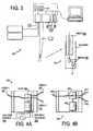
- FIG. 3is a schematic diagram showing an instrument according to a second preferred embodiment of the invention.
- FIGS. 4A and 4Bshow a 1 ⁇ 2 fiber optic switch usable in the embodiment of FIG. 1 or that of FIG. 3 ;
- FIGS. 5A-5Cshow variations of the fiber terminations in the switch of FIGS. 4A and 4B ;
- FIG. 6shows a 2 ⁇ 2 fiber optic switch based on the fiber optic switch of FIGS. 4A and 4B ;
- FIGS. 7A and 7Bshow a variation of the switch of FIGS. 4A and 4B with a filter which can be moved into or out of the light beam;
- FIGS. 8A and 8Bshow a further modification of the switch of FIGS. 4A and 4B ;
- FIG. 9is a perspective view showing a bulkhead used in the switch of FIGS. 8A and 8B ;
- FIG. 10shows an instrument according to a third preferred embodiment of the present invention.
- FIG. 11shows an alternative fiber coupler usable with the embodiment of FIG. 10 .
- FIG. 1An instrument according to a first preferred embodiment of the present invention is shown in FIG. 1 as 100 .
- the instrumentincludes a treatment laser (such as a dye laser) 102 , a white light source (such as a tungsten halogen or xenon lamp) 104 , fiber optic switches 106 , 108 , a shutter 110 , two thermoelectrically cooled spectrometers 112 , 114 , an optical probe 116 , an optical filter 118 , circuitry 120 , housings (not shown), and a computer 122 .
- the computer 122 and the spectrometers 112 , 114communicate through a USB cable connection 124 .
- Each of the spectrometers 112 , 114has reserved auxiliary (AUX) pins on its on-board circuitry.
- the AUX pinsare connected to the LEDs 126 , shutter 110 , and fiber optic switches 106 , 108 via external circuitry 120 .
- Transistor-transistor logic (TTL) pulsestransmit from the AUX pins to the circuitry 120 .
- the white light source 104 and treatment laser 102are coupled via fibers 128 into the 2 ⁇ 1 fiber optic switch 106 .
- the output of this switch 106is coupled via a fiber 130 to part of the optical probe 116 and is terminated with a microlens 132 .
- two more optical fibers 134 terminated with microlenses 136are in the probe.
- Each of the two fibers 134is directed to a housing (not shown) with a dichroic optical filter 138 .
- the reflection path of the filter housingcontains an LED 126
- the transmission pathcontains a secondary optical fiber 140 .
- the secondary optical fibers 140are connected to the 2 ⁇ 2 optical switch 108 .
- One of the two outputs of the 2 ⁇ 2 switch 108is directed to the first spectrometer 112
- the second outputis directed to the optical filter 118 and then to the second spectrometer 114 .
- the treatment area Aincludes two regions, a target lesion region L and a perilesion margin region M.
- the computer 122determines which of two states the first fiber optic switch 106 is in and the state of the white light shutter 110 . Depending on the states of the switch 106 and shutter 110 , the light is blocked, white light is transmitted through the treatment fiber 130 , or laser light from the treatment laser 102 is transmitted though the treatment fiber 130 . Light transmitted through the treatment fiber 130 is directed onto the treatment area A, comprising both the lesion region L and the perilesion region M.
- the treatment areasmay have different optical, chemical or physiological properties.
- Light emitted or reflected from the area Ais collected by the two optical fibers 134 terminated with microlenses 136 .
- One of the optical fibers 134collects primarily from the target lesion region L, and the other fiber 134 collects primarily from the perilesion margin region M.
- Lightmay also be generated by the LEDs 126 in the detection arm. Light generated by the LEDs 126 will be reflected off the dichroic mirror 138 and transmitted through the optical fibers 134 and directed through the microlenses 136 onto the corresponding treatment regions L, M. Light omitted or reflected from these regions will be collected by the fibers 134 and directed onto the dichroic mirror 138 . Light that is at a different wavelength from the LED sources 126 will be transmitted through the filter 138 .
- Light transmitted through the secondary fibers 140is directed into the 2 ⁇ 2 optical switch 108 .
- light from either of the two secondary detection fibers 140can be directed either through another optical filter 118 followed by a spectrometer 114 or directly into a spectrometer 112 .
- the laser sourceis used as a treatment beam. Light from this source is directed into the treatment area and activates photoactive drugs within that area.
- Treatment beam excited fluorescencecan be measured. Some of the absorbed laser light may be emitted as fluorescence.
- the fluorescent signalcan be evaluated without the spectrometer being optically saturated by the treatment laser. Therefore, with the first switch transmitting the laser and the second switch directing light collected from the treatment area region of interest, spatially resolved fluorescence from that region can be measured.
- the fluorescent signals of interestare highly excited by light emitted by the LED sources.
- the 405 nm lightcan be directed onto either treatment region of interest, and excited fluorescence can be collected through that same path and directed by the 2 ⁇ 2 fiber switch directly to the non-filtered spectrometer path. Therefore, a spatially resolved measurement of fluorescence can be made.
- the reflected spectrum of a white light sourceprovides information about tissue optical properties, blood volume, and blood oxygenation.
- White lightcan be directed through the first optical switch on the treatment area, and the reflected signal can be collected by the detection arm. Either of the two fibers in the detection path can be directed into the non-filtered spectrometer, and the spatially resolved white light reflectance can be measured.
- the computer 122receives detection signals for all types of reflected light and uses the reflected white light to correct the detection signals for the dynamic optical properties of the tissue, particularly the spectral reflectivity.
- dataare transmitted from the spectrometers into the computer, where characteristics about the treatment regions are stored and analyzed. Analysis of the data from these measurements can be fed back into the system to control the timing of the measurements and the treatment.
- timingis shown in FIG. 2 .
- the timing of the light sourcealternates among the laser, the white light source, and the LED (an ultraviolet source).
- the corresponding time periods for measurementare fluorescence from the laser, reflectance from the white light, and fluorescence from the LED. With those measurements, it is possible to correct for dynamic tissue optical characteristics.
- FIG. 3An instrument according to the second preferred embodiment is shown in FIG. 3 as 300 . Except as noted below, the instrument 300 can be constructed and used like the instrument 100 .
- the optical probe 302is capable of two-point spatial resolution.
- a single or plurality of optical fibers 304can be used in concert with either a single or plurality of MEMS (micro-electro-mechanical systems) scanning mirrors 306 in a housing 308 to scan the treatment area.
- MEMSmicro-electro-mechanical systems
- the delivery of the laser, white light, and LED sourcesmay be delivered through the same optical probe that is doing the collection depending on switching configuration.
- Either of the preferred embodimentscan use a variety of switches, such as the following.
- switches 106 and 108 of FIGS. 1 and 3are provided, as illustrated in FIGS. 4A through 9 .
- the purpose of the switchesis to control the flow of light through the system, as previously disclosed.
- some embodimentscontain in-line filters which can be moved into or out of the path in order to control the light transmitted through the system.
- FIG. 4Ashows a top view of a 1 ⁇ 2 fiber optic switch 400 in which an optical fiber 402 is mounted on a translating sled 404 that can be translated by a linear actuator (bistable solenoid) 406 by applying a voltage from a voltage source 408 across electrical leads.
- a linear actuatorbistable solenoid 406
- a voltage pulse across one set of leadstranslates the sled 404 into a first position. Permanent magnets within the solenoid 406 latch the device in place so that it is stable and requires no additional electrical source to hold it in place.
- the fiber 402is in close proximity to the fiber 412 and provides optical throughput from the fiber 402 to the fiber 412 (or vice versa).
- a voltage pulse across a second set of leads(not shown) translates the sled 404 to a second position, as shown in FIG. 4B , where a second set of permanent magnets holds the sled 404 in place and throughput between the fiber 402 and a fiber 410 is obtained.
- a precision linear slide(not shown) holds the sled 404 very precisely in two dimensions to reduce fiber alignment errors otherwise imparted by the slop in the axle of the bistable solenoid 406 .
- FIGS. 5A-5Cshow a magnified view of the sled 404 and the alignment of the optical fibers 402 , 410 , 412 .
- the fibers usedhave polished ends 502 and are coupled by placing them in close proximity.
- the fibersare terminated with lenses (GRIN lenses) 504 which collimate the beam and allow efficient coupling at spacing prohibitive for polished fiber coupling.
- An alternative embodiment of that shown in FIG. 5Bwould include traditional lenses or ball lenses.
- FIG. 5Cshows a perspective view of the sled holding an optical fiber.
- FIG. 6shows a 2 ⁇ 2 switch 600 using the same base components as used for the 1 ⁇ 2 switches of FIGS. 4A and 4B .
- an interconnecting fiber 602allows either of the two input fibers 604 , 606 to be connected to either of the two output fibers 608 , 610 by controlling the positions of the sleds 404 with two bistable solenoids 406 .
- the basic 1 ⁇ 2 switchcan be multiplexed together as in the 2 ⁇ 2 switch to increase number of channels that can be switched.
- FIGS. 7A and 7Bshow a removable in-line filter 702 incorporated into a 1 ⁇ 2 switch 700 based on the previous design.
- a fiber 704 from the 1 ⁇ 2 switchis coupled to a second fiber 704 with lenses 706 .
- a filter 702 mounted onto a sled 404(as previously described) can then be translated into or out of the beam path using a solenoid 406 .
- FIG. 7Ashows this device with the filter not in the path of the light
- FIG. 7Bshows this device with the filter in the path of the light.
- a precision linear guidemay be used to improve system repeatability and robustness.
- FIGS. 8A and 8Bshow an alternative 1 ⁇ 2 switch embodiment 800 in which two lens-terminated fibers 802 , 804 are mounted in the switch 800 and aligned to the two input ends of a y-coupled fiber 806 . Switching between throughput of fibers is accomplished by translating a bulkhead 808 with through holes 810 .
- the bulkhead 808is in a first position, and light transmitted through a first fiber 802 travels through a through hole 810 in the bulkhead and is coupled into the output by the y-coupler 806 . Light transmitted through the second fiber 804 is blocked by the bulkhead 808 .
- FIG. 8Athe bulkhead 808 is in a first position, and light transmitted through a first fiber 802 travels through a through hole 810 in the bulkhead and is coupled into the output by the y-coupler 806 . Light transmitted through the second fiber 804 is blocked by the bulkhead 808 .
- FIG. 8Bshows the device 800 after the bistable solenoid 406 is used to switch the position of the bulkhead 808 .
- light transmitted through the first fiber 802is blocked by the bulkhead 808
- light transmitted through the second fiber 804is transmitted through a through hole 810 and is coupled into the output fiber by the y-coupler 806 .
- a perspective view of the bulkhead 808 with through holes 810is illustrated in FIG. 9 .
- this basic switch unitcan be multiplexed to achieve a higher number of channels and can also have in-line filters incorporated for additional functionality.
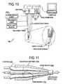
- FIG. 10shows a third preferred embodiment of the invention.
- the instrument 1000can be constructed and used like the instrument 100 of FIG. 1 , except that additional light sources, such as a fluorescence laser 1002 , are coupled into the system and transmitted through the treatment fiber. Such additional sources may be coupled with multiplexed switches, as described above.
- sourcesmay be coupled into the system by butt-coupling multiple fiber sources into the treatment source.
- FIG. 11A known type of coupling is shown in FIG. 11 as 1100 , in which input fibers 1102 are formed into a fiber bundle 1104 in a housing 1106 and tapered to form a taper region 1108 leading to the output fiber 1110 .
- the source fibers usedwould have smaller diameters than the treatment fiber, and the sources would be switched on and off, or shuttered on and off upstream of the coupling. In this way, multiple sources can be used to interrogate the target tissue without much added complexity. It is an advantage of this embodiment that the sources are transmitted to the tissue surface in the same geometry which simplifies computational analysis and interpretation of resulting measurements.
Landscapes
- Health & Medical Sciences (AREA)
- Life Sciences & Earth Sciences (AREA)
- Engineering & Computer Science (AREA)
- Biomedical Technology (AREA)
- Animal Behavior & Ethology (AREA)
- Veterinary Medicine (AREA)
- Pathology (AREA)
- Public Health (AREA)
- General Health & Medical Sciences (AREA)
- Nuclear Medicine, Radiotherapy & Molecular Imaging (AREA)
- Biophysics (AREA)
- Radiology & Medical Imaging (AREA)
- Surgery (AREA)
- Molecular Biology (AREA)
- Medical Informatics (AREA)
- Heart & Thoracic Surgery (AREA)
- Physics & Mathematics (AREA)
- Investigating Or Analysing Materials By Optical Means (AREA)
- Radiation-Therapy Devices (AREA)
- Laser Surgery Devices (AREA)
Abstract
Description
Claims (18)
Priority Applications (1)
| Application Number | Priority Date | Filing Date | Title |
|---|---|---|---|
| US11/631,121US9044140B2 (en) | 2004-06-30 | 2005-06-30 | Photodynamic therapy with spatially resolved dual spectroscopic monitoring |
Applications Claiming Priority (3)
| Application Number | Priority Date | Filing Date | Title |
|---|---|---|---|
| US58378604P | 2004-06-30 | 2004-06-30 | |
| US11/631,121US9044140B2 (en) | 2004-06-30 | 2005-06-30 | Photodynamic therapy with spatially resolved dual spectroscopic monitoring |
| PCT/US2005/023573WO2006025940A2 (en) | 2004-06-30 | 2005-06-30 | Photodynamic therapy with spatially resolved dual spectroscopic monitoring |
Publications (2)
| Publication Number | Publication Date |
|---|---|
| US20090043296A1 US20090043296A1 (en) | 2009-02-12 |
| US9044140B2true US9044140B2 (en) | 2015-06-02 |
Family
ID=36000481
Family Applications (1)
| Application Number | Title | Priority Date | Filing Date |
|---|---|---|---|
| US11/631,121Active2030-07-12US9044140B2 (en) | 2004-06-30 | 2005-06-30 | Photodynamic therapy with spatially resolved dual spectroscopic monitoring |
Country Status (7)
| Country | Link |
|---|---|
| US (1) | US9044140B2 (en) |
| EP (2) | EP1778076B1 (en) |
| AT (1) | ATE441459T1 (en) |
| AU (1) | AU2005280762A1 (en) |
| CA (1) | CA2576264A1 (en) |
| DE (1) | DE602005016424D1 (en) |
| WO (1) | WO2006025940A2 (en) |
Families Citing this family (19)
| Publication number | Priority date | Publication date | Assignee | Title |
|---|---|---|---|---|
| EP2142254A4 (en) | 2007-05-02 | 2011-01-26 | Univ Rochester | FEEDBACK CONTROL METHOD FOR ADMINISTRATION OF PHOTODYNAMIC THERAPY AND RELATED INSTRUMENTATION |
| EP2200697A4 (en)* | 2007-10-18 | 2012-04-25 | Univ Rochester | METHOD FOR CONTROLLING IRRADIATION OF PHOTODYNAMIC THERAPY AND CORRESPONDING INSTRUMENTATION |
| GB2454652A (en)* | 2007-11-01 | 2009-05-20 | Mohamed Abdelhafez El-Far | Photodynamic therapy device including a plurality of light sources |
| CA3194784A1 (en) | 2008-05-20 | 2009-11-26 | University Health Network | Device and method for fluorescence-based imaging and monitoring |
| GB0921477D0 (en)* | 2009-12-08 | 2010-01-20 | Moor Instr Ltd | Apparatus for measuring blood parameters |
| CN102655906B (en)* | 2009-12-15 | 2015-03-25 | 爱尔康研究有限公司 | Multi-spot laser probe |
| JP2013515266A (en)* | 2009-12-21 | 2013-05-02 | テルモ株式会社 | Excitation / detection / projection system to visualize target cancer tissue |
| US8818733B2 (en) | 2010-04-20 | 2014-08-26 | Mayo Foundation For Medical Education And Research | Determination of photodynamic therapy (PDT) treatment parameters |
| US9034023B2 (en)* | 2011-01-24 | 2015-05-19 | Biolitec Pharma Marketing Ltd | Dynamic colorectal PDT application |
| JP5426620B2 (en)* | 2011-07-25 | 2014-02-26 | 富士フイルム株式会社 | Endoscope system and method for operating endoscope system |
| US10245181B2 (en) | 2012-12-21 | 2019-04-02 | Alcon Research, Ltd. | Grin fiber multi-spot laser probe |
| US10589120B1 (en) | 2012-12-31 | 2020-03-17 | Gary John Bellinger | High-intensity laser therapy method and apparatus |
| JP6769949B2 (en) | 2014-07-24 | 2020-10-14 | ユニバーシティー ヘルス ネットワーク | Data collection and analysis for diagnostic purposes |
| US11040217B2 (en) | 2015-07-23 | 2021-06-22 | Health Research, Inc. | System and method for delivering dose light to tissue |
| US10687895B2 (en)* | 2015-11-06 | 2020-06-23 | The University Of Akron | Integrated fiber optic probe for performing image-guided laser induced thermal therapy |
| FR3044414B1 (en) | 2015-11-30 | 2020-10-16 | Univ De Lorraine | FIBER BIMODAL OPTICAL SPECTROSCOPY MEDICAL DEVICE |
| CN105597223A (en)* | 2016-01-20 | 2016-05-25 | 侯荣 | Noninvasive gynaecological nursing drug use device |
| CN111407467A (en)* | 2020-01-08 | 2020-07-14 | 北京航空航天大学 | High-quality laser bone processing method based on spectrum online monitoring |
| CN113633261A (en)* | 2021-08-11 | 2021-11-12 | 南京航空航天大学 | Multichannel in-vivo pharmacokinetic analysis system based on fluorescence monitoring |
Citations (66)
| Publication number | Priority date | Publication date | Assignee | Title |
|---|---|---|---|---|
| DE2521659A1 (en) | 1975-05-13 | 1976-12-02 | Siemens Ag | Optical waveguide where core is covered by a winding - of separate fibres with a lower refractive index than the core |
| GB2157842A (en) | 1984-01-11 | 1985-10-30 | Plessey Co Plc | Optical fibre sensing apparatus |
| US4768513A (en)* | 1986-04-21 | 1988-09-06 | Agency Of Industrial Science And Technology | Method and device for measuring and processing light |
| EP0323920A2 (en) | 1988-01-07 | 1989-07-12 | The Furukawa Electric Co., Ltd. | Optical switch system |
| US4913142A (en) | 1985-03-22 | 1990-04-03 | Massachusetts Institute Of Technology | Catheter for laser angiosurgery |
| US5035482A (en)* | 1989-04-06 | 1991-07-30 | Amp Incorporated | Optical switch |
| US5098804A (en)* | 1989-01-13 | 1992-03-24 | E. I. Du Pont De Nemours And Company | Multiplexer-demultiplexer for integrated optic circuit |
| EP0521797A1 (en) | 1991-07-05 | 1993-01-07 | Alcatel N.V. | Electrically controlled optical switch |
| WO1993021842A1 (en) | 1992-04-30 | 1993-11-11 | Quadra Logic Technologies, Inc. | High-power light-emitting diodes for photodynamic therapy |
| US5278692A (en) | 1991-09-17 | 1994-01-11 | Commissariat A L'energie Atomique | Optical switch and process for the production of said switch |
| DE4240769A1 (en) | 1992-12-03 | 1994-06-09 | Invent Entwicklung Neuer Techn | Measuring velocity of particles suspended in fluid - projecting two pairs of measuring beams in perpendicular planes for measuring coordinate velocity components |
| US5413197A (en) | 1994-03-14 | 1995-05-09 | Baer; Larry G. | Parking brake valve |
| US5433204A (en) | 1993-11-16 | 1995-07-18 | Camilla Olson | Method of assessing placentation |
| US5483958A (en) | 1994-01-25 | 1996-01-16 | United States Of America As Represented By The Secretary Of The Department Of Health And Human Services | Fluorescent-tipped dosimeter probe |
| US5533508A (en) | 1991-10-31 | 1996-07-09 | Pdt Systems, Inc. | Vivo dosimeter for photodynamic therapy |
| US5534997A (en) | 1994-07-15 | 1996-07-09 | Bruker Analytische Messtechnik Gmbh | Raman spectrometer using a remote probe with enhanced efficiency |
| WO1996020683A1 (en) | 1994-12-29 | 1996-07-11 | Mcneil-Ppc, Inc. | Optional inserters for digital tampons |
| US5537499A (en) | 1994-08-18 | 1996-07-16 | Laser Peripherals, Inc. | Side-firing laser optical fiber probe and method of making same |
| WO1999017668A1 (en) | 1997-10-08 | 1999-04-15 | The General Hospital Corporation | Phototherapy methods and systems |
| US5907395A (en) | 1997-06-06 | 1999-05-25 | Image Guided Technologies, Inc. | Optical fiber probe for position measurement |
| US6070093A (en)* | 1997-12-02 | 2000-05-30 | Abbott Laboratories | Multiplex sensor and method of use |
| US6102857A (en) | 1996-10-04 | 2000-08-15 | Optosonics, Inc. | Photoacoustic breast scanner |
| US6130071A (en) | 1997-02-05 | 2000-10-10 | Helsinki University Licensing, Ltd. | Vascular endothelial growth factor C (VEGF-C) ΔCys156 protein and gene, and uses thereof |
| US6219566B1 (en)* | 1999-07-13 | 2001-04-17 | Photonics Research Ontario | Method of measuring concentration of luminescent materials in turbid media |
| US6238348B1 (en) | 1997-07-22 | 2001-05-29 | Scimed Life Systems, Inc. | Miniature spectrometer system and method |
| US20010047136A1 (en) | 2000-01-21 | 2001-11-29 | Domanik Richard A. | In-vivo tissue inspection and sampling |
| US20020035361A1 (en) | 1999-06-25 | 2002-03-21 | Houser Russell A. | Apparatus and methods for treating tissue |
| US20020045811A1 (en) | 1985-03-22 | 2002-04-18 | Carter Kittrell | Laser ablation process and apparatus |
| US6377841B1 (en)* | 2000-03-31 | 2002-04-23 | Vanderbilt University | Tumor demarcation using optical spectroscopy |
| US6384951B1 (en) | 1997-11-19 | 2002-05-07 | University Of Washington | High throughput optical scanner |
| US20020118870A1 (en) | 1997-06-05 | 2002-08-29 | Youvan Dougalas C. | Calibration of fluorescence resonance energy transfer in microscopy |
| US20020123023A1 (en) | 1993-09-27 | 2002-09-05 | Sicurelli Robert J. | Flexible post in a dental post and core system |
| US20020138073A1 (en) | 2001-03-23 | 2002-09-26 | Intintoli Alfred J. | Light-dispersive probe |
| US20020141625A1 (en) | 2001-03-28 | 2002-10-03 | Nelson Alan C. | Apparatus and method for imaging small objects in a flow stream using optical tomography |
| US6477289B1 (en)* | 2000-02-23 | 2002-11-05 | Optical Coating Laboratory, Inc. | Optical wedge switch |
| US20030009205A1 (en) | 1997-08-25 | 2003-01-09 | Biel Merrill A. | Treatment device for topical photodynamic therapy and method of using same |
| US20030013973A1 (en) | 2001-01-19 | 2003-01-16 | Massachusetts Institute Of Technology | System and methods of fluorescence, reflectance and light scattering spectroscopy for measuring tissue characteristics |
| US20030018324A1 (en) | 2000-12-15 | 2003-01-23 | Scott Davenport | Methods for laser treatment of soft tissue |
| US6571118B1 (en)* | 1998-05-04 | 2003-05-27 | Board Of Regents, The University Of Texas System | Combined fluorescence and reflectance spectroscopy |
| US20030099166A1 (en) | 2001-11-23 | 2003-05-29 | Via Technologies, Inc. | Method and apparatus for long seeking control of pickup head |
| US6572609B1 (en)* | 1999-07-14 | 2003-06-03 | Cardiofocus, Inc. | Phototherapeutic waveguide apparatus |
| US6593101B2 (en) | 2000-03-28 | 2003-07-15 | Board Of Regents, The University Of Texas System | Enhancing contrast in biological imaging |
| US6615063B1 (en)* | 2000-11-27 | 2003-09-02 | The General Hospital Corporation | Fluorescence-mediated molecular tomography |
| US20030199860A1 (en) | 2002-04-22 | 2003-10-23 | Loeb Marvin P. | Devices and methods for directed, interstitial ablation of tissue |
| US20030228566A1 (en)* | 2002-06-11 | 2003-12-11 | Biotechplex Corporation | Method of and apparatus for screening for drug candidates |
| US20030232445A1 (en)* | 2002-01-18 | 2003-12-18 | Newton Laboratories, Inc. | Spectroscopic diagnostic methods and system |
| US20040044287A1 (en) | 2000-03-31 | 2004-03-04 | Wei-Chiang Lin | Identification of human tissue using optical spectroscopy |
| US20040095855A1 (en) | 2002-11-19 | 2004-05-20 | Minoru Minase | Recording medium driving apparatus |
| US20040155049A1 (en) | 2003-02-10 | 2004-08-12 | Artromick International, Inc. | Pill sorting device and method of use thereof |
| US20040172163A1 (en) | 2000-08-28 | 2004-09-02 | Reijo Varis | Cartridge for dispensing pill -or capsule - form medications in desired doses |
| WO2004100761A2 (en) | 2003-05-14 | 2004-11-25 | Spectracure Ab | System and method for therapy and diagnosis comprising in combination non-mechanical and mechanical distributors for distribution of radiation |
| US20040243123A1 (en) | 1999-02-19 | 2004-12-02 | Scimed Life Systems, Inc. | Laser lithotripsy device with suction |
| US20050182392A1 (en) | 2003-10-30 | 2005-08-18 | Medical Cv, Inc. | Apparatus and method for guided ablation treatment |
| US20050203353A1 (en) | 2004-03-10 | 2005-09-15 | Jie Ma | Multiple purpose, portable apparatus for measurement, analysis and diagnosis |
| US7037325B2 (en) | 2001-11-14 | 2006-05-02 | Spectracure Ab | Therapy and diagnosis system and method with distributor for distribution of radiation |
| US20070299341A1 (en) | 2006-01-20 | 2007-12-27 | Lihong Wang | Photoacoustic and thermoacoustic tomography for breast imaging |
| US20080123083A1 (en) | 2006-11-29 | 2008-05-29 | The Regents Of The University Of Michigan | System and Method for Photoacoustic Guided Diffuse Optical Imaging |
| US7399278B1 (en) | 2003-05-05 | 2008-07-15 | Los Angeles Biomedical Research Institute At Harbor-Ucla Medical Center | Method and system for measuring amniotic fluid volume and/or assessing fetal weight |
| US20080192897A1 (en) | 2007-02-12 | 2008-08-14 | Stanislaw Piorek | Small spot x-ray fluorescence (xrf) analyzer |
| US7443491B2 (en)* | 2002-12-03 | 2008-10-28 | Bay Bioscience Kabushiki Kaisha | System for collecting information on biological particles |
| US20090023168A1 (en) | 2007-07-16 | 2009-01-22 | Won Sun Park | Method of screening placental proteins responsible for pathophysiology of preeclampsia, and marker for early diagnosis and prediction of preeclampsia |
| US20090024043A1 (en) | 2007-07-17 | 2009-01-22 | Macleod Ainslie | Methods, Devices and Systems for the Prevention of Sudden Infant Death Syndrome (SIDS) and the Diagnosis and Treatment of Infants Predisposed to SIDS |
| US20090252392A1 (en) | 2008-04-08 | 2009-10-08 | Goyaike S.A.A.C.I.Y.F | System and method for analyzing medical images |
| US7606394B2 (en) | 2006-04-03 | 2009-10-20 | Jbs Swift & Company | Methods and systems for administering a drug program related to livestock |
| US7613330B2 (en) | 2006-04-03 | 2009-11-03 | Jbs Swift & Company | Methods and systems for tracking and managing livestock through the production process |
| US7697145B2 (en)* | 2003-05-28 | 2010-04-13 | Duke University | System for fourier domain optical coherence tomography |
- 2005
- 2005-06-30EPEP05806200Apatent/EP1778076B1/ennot_activeExpired - Lifetime
- 2005-06-30ATAT05806200Tpatent/ATE441459T1/ennot_activeIP Right Cessation
- 2005-06-30EPEP09160156Apatent/EP2085116A1/ennot_activeWithdrawn
- 2005-06-30AUAU2005280762Apatent/AU2005280762A1/ennot_activeAbandoned
- 2005-06-30CACA002576264Apatent/CA2576264A1/ennot_activeAbandoned
- 2005-06-30WOPCT/US2005/023573patent/WO2006025940A2/enactiveApplication Filing
- 2005-06-30DEDE602005016424Tpatent/DE602005016424D1/ennot_activeExpired - Lifetime
- 2005-06-30USUS11/631,121patent/US9044140B2/enactiveActive
Patent Citations (66)
| Publication number | Priority date | Publication date | Assignee | Title |
|---|---|---|---|---|
| DE2521659A1 (en) | 1975-05-13 | 1976-12-02 | Siemens Ag | Optical waveguide where core is covered by a winding - of separate fibres with a lower refractive index than the core |
| GB2157842A (en) | 1984-01-11 | 1985-10-30 | Plessey Co Plc | Optical fibre sensing apparatus |
| US4913142A (en) | 1985-03-22 | 1990-04-03 | Massachusetts Institute Of Technology | Catheter for laser angiosurgery |
| US20020045811A1 (en) | 1985-03-22 | 2002-04-18 | Carter Kittrell | Laser ablation process and apparatus |
| US4768513A (en)* | 1986-04-21 | 1988-09-06 | Agency Of Industrial Science And Technology | Method and device for measuring and processing light |
| EP0323920A2 (en) | 1988-01-07 | 1989-07-12 | The Furukawa Electric Co., Ltd. | Optical switch system |
| US5098804A (en)* | 1989-01-13 | 1992-03-24 | E. I. Du Pont De Nemours And Company | Multiplexer-demultiplexer for integrated optic circuit |
| US5035482A (en)* | 1989-04-06 | 1991-07-30 | Amp Incorporated | Optical switch |
| EP0521797A1 (en) | 1991-07-05 | 1993-01-07 | Alcatel N.V. | Electrically controlled optical switch |
| US5278692A (en) | 1991-09-17 | 1994-01-11 | Commissariat A L'energie Atomique | Optical switch and process for the production of said switch |
| US5533508A (en) | 1991-10-31 | 1996-07-09 | Pdt Systems, Inc. | Vivo dosimeter for photodynamic therapy |
| WO1993021842A1 (en) | 1992-04-30 | 1993-11-11 | Quadra Logic Technologies, Inc. | High-power light-emitting diodes for photodynamic therapy |
| DE4240769A1 (en) | 1992-12-03 | 1994-06-09 | Invent Entwicklung Neuer Techn | Measuring velocity of particles suspended in fluid - projecting two pairs of measuring beams in perpendicular planes for measuring coordinate velocity components |
| US20020123023A1 (en) | 1993-09-27 | 2002-09-05 | Sicurelli Robert J. | Flexible post in a dental post and core system |
| US5433204A (en) | 1993-11-16 | 1995-07-18 | Camilla Olson | Method of assessing placentation |
| US5483958A (en) | 1994-01-25 | 1996-01-16 | United States Of America As Represented By The Secretary Of The Department Of Health And Human Services | Fluorescent-tipped dosimeter probe |
| US5413197A (en) | 1994-03-14 | 1995-05-09 | Baer; Larry G. | Parking brake valve |
| US5534997A (en) | 1994-07-15 | 1996-07-09 | Bruker Analytische Messtechnik Gmbh | Raman spectrometer using a remote probe with enhanced efficiency |
| US5537499A (en) | 1994-08-18 | 1996-07-16 | Laser Peripherals, Inc. | Side-firing laser optical fiber probe and method of making same |
| WO1996020683A1 (en) | 1994-12-29 | 1996-07-11 | Mcneil-Ppc, Inc. | Optional inserters for digital tampons |
| US6102857A (en) | 1996-10-04 | 2000-08-15 | Optosonics, Inc. | Photoacoustic breast scanner |
| US6130071A (en) | 1997-02-05 | 2000-10-10 | Helsinki University Licensing, Ltd. | Vascular endothelial growth factor C (VEGF-C) ΔCys156 protein and gene, and uses thereof |
| US20020118870A1 (en) | 1997-06-05 | 2002-08-29 | Youvan Dougalas C. | Calibration of fluorescence resonance energy transfer in microscopy |
| US5907395A (en) | 1997-06-06 | 1999-05-25 | Image Guided Technologies, Inc. | Optical fiber probe for position measurement |
| US6238348B1 (en) | 1997-07-22 | 2001-05-29 | Scimed Life Systems, Inc. | Miniature spectrometer system and method |
| US20030009205A1 (en) | 1997-08-25 | 2003-01-09 | Biel Merrill A. | Treatment device for topical photodynamic therapy and method of using same |
| WO1999017668A1 (en) | 1997-10-08 | 1999-04-15 | The General Hospital Corporation | Phototherapy methods and systems |
| US6384951B1 (en) | 1997-11-19 | 2002-05-07 | University Of Washington | High throughput optical scanner |
| US6070093A (en)* | 1997-12-02 | 2000-05-30 | Abbott Laboratories | Multiplex sensor and method of use |
| US6571118B1 (en)* | 1998-05-04 | 2003-05-27 | Board Of Regents, The University Of Texas System | Combined fluorescence and reflectance spectroscopy |
| US20040243123A1 (en) | 1999-02-19 | 2004-12-02 | Scimed Life Systems, Inc. | Laser lithotripsy device with suction |
| US20020035361A1 (en) | 1999-06-25 | 2002-03-21 | Houser Russell A. | Apparatus and methods for treating tissue |
| US6219566B1 (en)* | 1999-07-13 | 2001-04-17 | Photonics Research Ontario | Method of measuring concentration of luminescent materials in turbid media |
| US6572609B1 (en)* | 1999-07-14 | 2003-06-03 | Cardiofocus, Inc. | Phototherapeutic waveguide apparatus |
| US20010047136A1 (en) | 2000-01-21 | 2001-11-29 | Domanik Richard A. | In-vivo tissue inspection and sampling |
| US6477289B1 (en)* | 2000-02-23 | 2002-11-05 | Optical Coating Laboratory, Inc. | Optical wedge switch |
| US6593101B2 (en) | 2000-03-28 | 2003-07-15 | Board Of Regents, The University Of Texas System | Enhancing contrast in biological imaging |
| US20040044287A1 (en) | 2000-03-31 | 2004-03-04 | Wei-Chiang Lin | Identification of human tissue using optical spectroscopy |
| US6377841B1 (en)* | 2000-03-31 | 2002-04-23 | Vanderbilt University | Tumor demarcation using optical spectroscopy |
| US20040172163A1 (en) | 2000-08-28 | 2004-09-02 | Reijo Varis | Cartridge for dispensing pill -or capsule - form medications in desired doses |
| US6615063B1 (en)* | 2000-11-27 | 2003-09-02 | The General Hospital Corporation | Fluorescence-mediated molecular tomography |
| US20030018324A1 (en) | 2000-12-15 | 2003-01-23 | Scott Davenport | Methods for laser treatment of soft tissue |
| US20030013973A1 (en) | 2001-01-19 | 2003-01-16 | Massachusetts Institute Of Technology | System and methods of fluorescence, reflectance and light scattering spectroscopy for measuring tissue characteristics |
| US20020138073A1 (en) | 2001-03-23 | 2002-09-26 | Intintoli Alfred J. | Light-dispersive probe |
| US20020141625A1 (en) | 2001-03-28 | 2002-10-03 | Nelson Alan C. | Apparatus and method for imaging small objects in a flow stream using optical tomography |
| US7037325B2 (en) | 2001-11-14 | 2006-05-02 | Spectracure Ab | Therapy and diagnosis system and method with distributor for distribution of radiation |
| US20030099166A1 (en) | 2001-11-23 | 2003-05-29 | Via Technologies, Inc. | Method and apparatus for long seeking control of pickup head |
| US20030232445A1 (en)* | 2002-01-18 | 2003-12-18 | Newton Laboratories, Inc. | Spectroscopic diagnostic methods and system |
| US20030199860A1 (en) | 2002-04-22 | 2003-10-23 | Loeb Marvin P. | Devices and methods for directed, interstitial ablation of tissue |
| US20030228566A1 (en)* | 2002-06-11 | 2003-12-11 | Biotechplex Corporation | Method of and apparatus for screening for drug candidates |
| US20040095855A1 (en) | 2002-11-19 | 2004-05-20 | Minoru Minase | Recording medium driving apparatus |
| US7443491B2 (en)* | 2002-12-03 | 2008-10-28 | Bay Bioscience Kabushiki Kaisha | System for collecting information on biological particles |
| US20040155049A1 (en) | 2003-02-10 | 2004-08-12 | Artromick International, Inc. | Pill sorting device and method of use thereof |
| US7399278B1 (en) | 2003-05-05 | 2008-07-15 | Los Angeles Biomedical Research Institute At Harbor-Ucla Medical Center | Method and system for measuring amniotic fluid volume and/or assessing fetal weight |
| WO2004100761A2 (en) | 2003-05-14 | 2004-11-25 | Spectracure Ab | System and method for therapy and diagnosis comprising in combination non-mechanical and mechanical distributors for distribution of radiation |
| US7697145B2 (en)* | 2003-05-28 | 2010-04-13 | Duke University | System for fourier domain optical coherence tomography |
| US20050182392A1 (en) | 2003-10-30 | 2005-08-18 | Medical Cv, Inc. | Apparatus and method for guided ablation treatment |
| US20050203353A1 (en) | 2004-03-10 | 2005-09-15 | Jie Ma | Multiple purpose, portable apparatus for measurement, analysis and diagnosis |
| US20070299341A1 (en) | 2006-01-20 | 2007-12-27 | Lihong Wang | Photoacoustic and thermoacoustic tomography for breast imaging |
| US7606394B2 (en) | 2006-04-03 | 2009-10-20 | Jbs Swift & Company | Methods and systems for administering a drug program related to livestock |
| US7613330B2 (en) | 2006-04-03 | 2009-11-03 | Jbs Swift & Company | Methods and systems for tracking and managing livestock through the production process |
| US20080123083A1 (en) | 2006-11-29 | 2008-05-29 | The Regents Of The University Of Michigan | System and Method for Photoacoustic Guided Diffuse Optical Imaging |
| US20080192897A1 (en) | 2007-02-12 | 2008-08-14 | Stanislaw Piorek | Small spot x-ray fluorescence (xrf) analyzer |
| US20090023168A1 (en) | 2007-07-16 | 2009-01-22 | Won Sun Park | Method of screening placental proteins responsible for pathophysiology of preeclampsia, and marker for early diagnosis and prediction of preeclampsia |
| US20090024043A1 (en) | 2007-07-17 | 2009-01-22 | Macleod Ainslie | Methods, Devices and Systems for the Prevention of Sudden Infant Death Syndrome (SIDS) and the Diagnosis and Treatment of Infants Predisposed to SIDS |
| US20090252392A1 (en) | 2008-04-08 | 2009-10-08 | Goyaike S.A.A.C.I.Y.F | System and method for analyzing medical images |
Non-Patent Citations (128)
| Title |
|---|
| Adair, et al., "Developmental determinants of blood pressure in adults," Annual Review of Nutrition, 2005, pp. 407-434, vol. 25, Palo Alto, CA, USA. |
| Amit, et al., "Structural image restoration through deformable templates,". Journal of the American Statistical Association, 1991, pp. 376-387, vol. 86, No. 414, American Statistical Association, Alexandria, VA, USA. |
| Armitage, et al. "Experimental models of developmental programming: consequences of exposure to an energy rich diet during development," Journal of Physiology, 2005, pp. 3-8, vol. 565, The Physiological Society, Cambridge, UK. |
| Asbury, et al., "Birthweight-discordance and differences in early parenting relate to monozygotic twin differences in behaviour problems and academic achievement at age 7," Developmental Science, 2006, pp. F22-F31, vol. 9, No. 2, Wiley, Hoboken, NJ, USA. |
| Baik, I., et al., Association of fetal hormone levels with stem cell potential: evidence for early life roots of human cancer. Cancer Res, 2005. 65(1): p. 358-63. |
| Barker, "The fetal origins of type 2 diabetes mellitus," Annals of Internal Medicine, 1999, pp. 322-324, vol. 130, No. 4, pt. 1, Philadelphia, PA, USA. |
| Barker, et al., "The developmental origins of insulin resistance," Hormone Research, 2005, pp. 2-7, vol. 64, suppl 3, Karger AG, Basel, Switzerland. |
| Barker, et al., "The intrauterine and early postnatal origins of cardiovascular disease and chronic bronchitis," Journal of Epidemiology and Community Health, 1989, pp. 237-240, vol. 43, BMJ, London, UK. |
| Barker, et al., "The maternal and fetal origins of cardiovascular disease," Journal of Epidemiology and Community Health, 1992, pp. 8-11, vol. 46, BMJ, London, UK. |
| Baschat, et al., "Fetal growth restriction due to placental disease," Seminars in Perinatology, 2004, pp. 67-80, vol. 28, No. 1, Elsevier, Maryland Heights, MO, USA. |
| Bellingham-Young, et al., "Prematurity and adult minor illness," Neuroendocrinology Letters, 2004, pp. 117-126, vol. 25, suppl. 1, Society of Integrated Sciences. |
| Benirschke K, K.P. "Architecture of Normal Villous Trees," Chapter 7, Pathology of the Human Placenta, 2002, Springer Verlag, New York, NY, pp. 116-154. |
| Benirschke K, K.P., Normative Values and Tables (Chapter 28), in Pathology of the Human Placenta. 2002, Springer-Verlag: New York. p. 920-927. |
| Benirschke, "Angioarchitecture of Villi," in The Pathology of the Placenta, 2002, pp. 134-140, Springer-Verlag, New York, USA. |
| Benirschke, "Basic Structure of the Villous Trees," Chapter 6 in the Pathology of the Placenta, 2002, pp. 50-115, Springer-Verlag, New York, USA. |
| Benirschke, "Classification of Villous Maldevelopment," Chapter 15, in Pathology of the Human Placenta, 2002, pp. 437-460, Springer, New York, NY, USA. |
| Benirschke, "Placental Shape Aberrations," Chapter 13, in Pathology of the Human Placenta, 2002, pp. 401-404, Springer, New York, NY, USA. |
| Bentler, et al., "Structural equation models in medical research," Statistical Methods in Medical Research, 1992, pp. 159-181, vol. 1, SAGE Publications, Newbury Park, CA, USA. |
| Bertram, et al., "Prenatal programming of postnatal endocrine responses by glucocorticoids," Reproduction, 2002, pp. 459-467, vol. 124, BioScientifica Ltd, Bradley Stoke, UK. |
| Buss, et al., "The EAS Temperament Scale," in Temperament: Early Developing Personality Traits, 1984, Hillsdale, NJ, USA. |
| Byrne, "Factor analytic models: viewing the structure of an assessment instrument from three perspectives," Journal of Personality Assessment, 2005, pp. 17-32, vol. 85, Informa PLC, St. Helier, Jersey. |
| Carey, "Infant Temperament Questionnaire (4-8 months)," Philadelphia: Dept. Educational Psychology, Temple University, 1977. |
| Charnock-Jones, et al., "Aspects of human fetoplacental vasculogenesis and angiogenesis: I. Molecular regulation," Placenta, 2004, pp. 103-113, vol. 25, Elsevier, Maryland Heights, MO, USA. |
| Cooper, et al., "Review: developmental origins of osteoporotic fracture," Osteoporosis International, 2005, pp. 337-347, vol. 17, Springer, New York, NY, USA. |
| Coste, et al., "Methodological issues in determining the dimensionality of composite health measures using principal component analysis: case illustration and suggestions for practice," Quality of Life Research, 2005, pp. 641-654, vol. 14, Springer, New York, NY, USA. |
| De Boo, et al., "The developmental origins of adult disease (Barker) hypothesis," Australian and New Zealand Journal of Obstetrics and Gynaecology, 2006, pp. 4-14, vol. 46, Wiley, Hoboken, NJ, USA. |
| Demir, et al., "Classification of human placental stem villi: review of structural and functional aspects," Microscopy Research and Technique, 1997, pp. 29-41, vol. 38, Wiley, Hoboken, NJ, USA. |
| Demir, et al., "Fetal vasculogenesis and angiogenesis in human placental villi," Acta Anat, 1989, pp. 190-203, vol. 136, Karger AG, Basel, Switzerland. |
| Dorosty, et al., "Factors associated with early adiposity rebound," Pediatrics, 2000, pp. 1115-1118, vol. 105, American Academy of Pediatrics, Elk Grove Village, IL, USA. |
| Dryden, I.L.M., KV. , Statistical Shape Analysis. 1998, New York: Wiley Press. |
| Farrow, et al., "Birthweight of term infants and maternal occupation in a prospective cohort of pregnant women," Occupational and Environmental Medicine, 1998, pp. 18-23, vol. 55, BMJ Group, London, UK. |
| Fenson, et al., "Technical Manual for the MacArthur Communicative Development Inventories," 1991, San Diego, CA: Development Psychology Laboratory. |
| Fergusson, et al., "Maternal use of cannabis and pregnancy outcome," BJOG: An International Journal of Obstetrics and Gynaecology, 2002, pp. 21-27, vol. 109, Wiley, Hoboken, NJ, USA. |
| Frankenburg, et al., "The Denver Developmental Screening Test," Journal of Pediatrics, 1967, pp. 181-191, vol. 71, No. 2, Elsevier, Maryland Heights, MO, USA. |
| Fullard, et al., "Toddler Tempermant Scale (1-3 year old children . . . ," Philadelphia, PA: Dept. Educational Psychology, Temple University, 1978. |
| Giles, "Benoit Mandelbrot: father of fractals," Nature, 2004, pp. 266-267, vol. 432, Nature Publishing Group, London, UK. |
| Gluckman, et al., "Life-long echoes-a critical analysis of the developmental origins of adult disease model," Biology of the Neonate, 2005, pp. 127-139, vol. 87, Karger AG, Basel, Switzerland. |
| Golding, "Children of the nineties-a longitudinal study of pregnancy and childhood based on the population of Avon (ALSPAC)," West of England Medical Journal, 1990, pp. 80-82, vol. 105. |
| Golding, "Outcome of pregnancy in diabetic women-more investigation is needed into whether control of diabetes is really poorer in England than Norway," BMJ, 2001, pp. 614-615, vol. 322, BMJ Group, London, UK. |
| Goodman, "The Strengths and Difficulties Questionnaire: a Research Note . . . ," Journal of Child Psychology and Psychiatry, 1997, pp. 581-586, vol. 38, No. 5, Wiley, Hoboken, NJ, USA. |
| Green, "Programming of endocrine mechanisms of cardiovascular control and growth," Journal of the Society for Gynecologic Investigation, 2001, pp. 57-68, vol. 8, No. 2, SAGE Publications, Newbury Park, CA, USA. |
| Grenander, "General Pattern Theory-a Mathematical Study of Regular Structures," 1993, pp. 539-544 and 740-784, Oxford University Press, Oxford, UK. |
| Grether, et al., "Reliability of placental histology using archived specimens," Paediatric Perinatal Epidemiology, 1999, pp. 489-495, vol. 13, Wiley, Hoboken, NJ, USA. |
| Griffiths, "Administering the Scale," in The Abilities of Babies-A Study in Mental Measurement, 1954, pp. 117-182, McGraw-Hill, New York, NY, USA. |
| Grizzi, et al., "Estimate of Neovascular Tree Complexity by Microscopy Analysis," Current Issues on Multidisciplinary Microscopy Research and Education, 2005, pp. 140-149, Formatex, Badajoz, Spain. |
| Gunnell, et al., "The association of fetal and childhood growth with risk of schizophrenia. Cohort study of 720,000 Swedish men and women," Schizophrenia Research, 2005, pp. 315-322, vol. 79, Elsevier, Maryland Heights, MO, USA. |
| Hagberg, H., D. Peebles, and C. Mallard, Models of white matter injury: comparison of infectious, hypoxic-ischemic, and excitotoxic insults. Ment Retard Dev Disabil Res Rev, 2002. 8(1): p. 30-8. |
| Hastie, et al., "Elements of Statistical Learning: Data Mining, Inference, and Prediction," 2001, pp. 1-40, Springer, New York, NY, USA. |
| Headley, et al., "Medication use during pregnancy: data from the Avon Longitudinal Study of Parents and Children," European Journal of Clinical Pharmacology, 2004, pp. 355-361, vol. 60, Springer, New York, NY, USA. |
| Horton, "Fetal origins of developmental plasticity: animal models of induced life history variation," Am J Hum Biol, pp. 34-43, 2005, vol. 17. |
| Hu, et al., "Genetic regulation of branching morphogenesis: lessons learned from loss-of-function phenotypes," Pediatric Research, 2003, pp. 433-438, vol. 54, Lippincott Williams & Wilkins, Hagerstown MD, USA. |
| Ingelfinger, et al., "Perinatal programming, renal development, and adult renal function," American Journal of Hypertension, 2002, pp. 46S-49S vol. 15, No. 2, pt. 2, Elsevier, Maryland Heights, MO, USA. |
| Jaddoe, et al., "Hypotheses on the fetal origins of adult diseases: contributions of epidemiological studies," European Journal of Epidemiology, 2006, pp. 91-102, vol. 21, Springer, New York, NY, USA. |
| Jasienska, et al., "High ponderal index at birth predicts high estradiol levels in adult women," American Journal of Human Biology, 2006, pp. 133-140, vol. 18, Wiley, Hoboken, NJ, USA. |
| Johansson, et al., "System for Integrated Interstitial Photodynamic Therapy and Dosimetric Monitoring", Proceedings of the SPIE-The International Society for Optical Engineering, Jan. 2005, pp. 130-140, vol. 5689, No. 1. |
| K. B., Examination of the Placenta, prepared for the Collaborative Study on Cerebral Palsy, Mental retardation and other Neurological and Sensory Disorders of Infancy and Childhood, N.I.o.N.D.a. Blindness, Editor. 1961, US Department of Health, Education and Welfare. |
| Kaufmann, et al., "Aspects of human fetoplacental vasculogenesis and angiogenesis: II. Changes during normal pregnancy," Placenta, 2004, pp. 114-126, vol. 25, Elsevier, Maryland Heights, MO, USA. Ltd., Maryland Heights, MO, USA. |
| Kaufmann, et al., "Classification of human placental villi: I. Histology," Cell and Tissue Research, 1979, pp. 409-423, vol. 200, Springer, New York, NY, USA. |
| Kaufmann, et al., "Cross-sectional features and three-dimensional structure of human placental villi," Placenta, 1987, pp. 235-247, vol. 8, Elsevier, Maryland Heights, MO, USA. Ltd., Maryland Heights, MO, USA. |
| Keipes, et al., "Of the British coastline and the interest of fractals in medicine," Biomedicine & Pharmacotherapy, 1993, pp. 409-415, vol. 47, Elsevier, Maryland Heights, MO, USA. |
| Khong, "Placental vascular development and neonatal outcome," Seminars in Neonatology, 2004, pp. 255-263, vol. 9, Elsevier, Maryland Heights, MO, USA. |
| Khong, et al., "Observer reliability in assessing placental maturity by histology," Journal of Clinical Pathology, 1995, pp. 420-423, vol. 48, BMJ, London, UK. |
| Kingdom, et al., "Development of the placental villous tree and its consequences for fetal growth," European Journal of Obstetrics & Gynecology and Reproductive Biology, 2000, pp. 35-43, vol. 92, Elsevier, Maryland Heights, MO, USA. |
| Kosanke, et al., "Branching patterns of human placental villous trees: perspectives of topological analysis," Placenta, 1993, pp. 591-604, vol. 14, Elsevier, Maryland Heights, MO, USA. |
| Kuh, D. and R. Hardy, A life course approach to women's health. Life course approach to adult health ; No. 1. 2002, Oxford ; New York: Oxford University Press. xvi, 419 p. |
| Lagiou, et al., "Diet during pregnancy and levels of maternal pregnancy hormones in relation to the risk of breast cancer in the offspring," European Journal of Cancer Prevention, 2006, pp. 20-26, vol. 15, Lippincott Williams & Wilkins, Hagerstown, MD, USA. |
| Lagiou, et al., "Maternal height, pregnancy estriol and birth weight in reference to breast cancer risk in Boston and Shanghai," International Journal of Cancer, 2005, pp. 494-498, vol. 117, Wiley, Hoboken, NJ, USA. |
| Lagiou, P., et al., Birthweight differences between USA and China and their relevance to breast cancer aetiology. Int J Epidemiol, 2003. 32(2): p. 193-8. |
| Larsen, et al., "Stereologic examination of placentas from mothers who smoke during pregnancy," American Journal of Obstetrics & Gynecology, 2002, pp. 531-537, vol. 186, Elsevier, Maryland Heights, MO, USA. |
| Lawlor, et al., "Birth weight is inversely associated with incident coronary heart disease and stroke among individuals born in the 1950s: findings from the Aberdeen Children of the 1950s prospective cohort study," Circulation, 2005, pp. 1414-1418, vol. 112, Lippincott Williams & Wilkins, Hagerstown, MD, USA. |
| Le Noble, et al., "Control of arterial branching morphogenesis in embryogenesis: go with the flow," Cardiovascular Research, 2005, vol. 65, pp. 619-628, Elsevier, Maryland Heights, MO, USA. |
| Lele, S.R., JT., An Invariant Approach to Statistical Analysis of Shapes. 2000, London, UK.: Chapman and Hall/CRC Press. |
| Levitt, et al., "Adult BMI and fat distribution but not height amplify the effect of low birthweight on insulin resistance and increased blood pressure in 20-year-old South Africans," Diabetologia, 2005, pp. 1118-1125, vol. 48, Springer, New York, NY, USA. |
| Levitt, et al., "An inverse relation between blood pressure and birth weight among 5 year old children from Soweto, South Africa," Journal of Epidemiology and Community Health, 1999, pp. 264-268. vol. 53, BMJ, London, UK. |
| Levitt, et al., "The foetal origins of the metabolic syndrome-a South African perspective," Cardiovascular Journal of South Africa, 2002, pp. 179-180, vol. 13, No. 4, Durbanville, South Africa. |
| Lockwood, "The diagnosis of preterm labor and the prediction of preterm delivery," Clinical Obstetrics and Gynecology, 1995, pp. 675-687, vol. 38, No. 4, Lippincott Williams & Wilkins, Hagerstown, MD, USA. |
| Longo, "Fetal origins of adult vascular dysfunction in mice lacking endothelial nitric oxide synthase," American Journal of Physiology-Regulatory, Integrative and Comparative Physiology, 2005, pp. R1114-R1121, vol. 288, The American Physiological Society, Bethesda, MD, USA. |
| Maitra, et al., "Mode of delivery is not associated with asthma or atopy in childhood," Clinical and Experimental Allergy, 2004, pp. 1349-1355, vol. 34, Wiley, Hoboken, NJ, USA. |
| Mayhew, "Changes in fetal capillaries during preplacental hypoxia: growth, shape remodelling and villous capillarization in placentae from high-altitude pregnancies," Placenta, 2003, pp. 191-198, vol. 24, Elsevier, Maryland Heights, MO, USA. |
| Mayhew, et al., "Aspects of human fetoplacental vasculogenesis and angiogenesis: III. Changes in complicated pregnancies," Placenta, 2004, pp. 127-139, vol. 25, Elsevier, Maryland Heights, MO, USA. |
| Mayhew, et al., "Stereological investigation of placental morphology in pregnancies complicated by pre-eclampsia with and without intrauterine growth restriction," Placenta, 2003, pp. 219-226, vol. 24, Elsevier, Maryland Heights, MO, USA. |
| McKeague, I., A Statistical Model for Signature Verification. . Journal of the American Statistical Association, 2005. 100: p. 231-241. |
| McMillen, et al. "Developmental origins of the metabolic syndrome: prediction, plasticity, and programming," Physiological Review, 2005, pp. 571-633. vol. 85, The American Physiological Society, Bethesda, MD, USA. |
| McMillen, et al., "Early origins of obesity: programming the appetite regulatory system," Journal of Physiology, 2005, pp. 9-17, vol. 565, The Physiological Society, Cambridge, UK. |
| Meisel, "Generalized Mandelbrot rule for fractal sections," Physical Review A, 1992, pp. 654-656, vol. 45, No. 2, American Physical Society, College Park, MD, USA. |
| Metzger, et al., "Genetic control of branching morphogenesis," Science Magazine, 1999, pp. 1635-1639, vol. 284, Washington, DC, USA. |
| Miettinen, "Epidermal growth factor receptor in mice and men-any applications to clinical practice?" Annals of Medicine, 1997, pp. 531-534, vol. 29, Informa PLC, St. Helier, Jersey. |
| Miettinen, et al., "Epithelial immaturity and multiorgan failure in mice lacking epidermal growth factor receptor," Nature, 1995, pp. 337-341, vol. 376, Nature Publishing Group, London, UK. |
| Miller, et al., "A Mathematical textbook of deformable neuro-anatomies . . . ," Proceedings of the National Academy of Sciences, 1993, pp. 11944-11948, National Academy of Sciences, Washington, DC, USA. |
| Morley, "Fetal origins of adult disease," Seminars in Fetal & Neonatal Medicine, 2006, pp. 73-78, vol. 11, Elsevier, Maryland Heights, MO, USA. |
| Myrianthopoulos, N.C. and K.S. French, An application of the U.S. Bureau of the Census socioeconomic index to a large, diversified patient population. Soc Sci Med, 1968. 2(3): p. 283-99. |
| Naeye, "Disorders of the Placenta and Decidua," in Disorders of the Placenta, Fetus and Neonata, 1992, pp. 129-130, Mosby Year Book: Philadelphia, PA, USA. |
| Naeye, "Disorders of the Placenta and Decidua," in Disorders of the Placenta, Fetus and Neonata, 1992, pp. 129-134, Mosby Year Book: Philadelphia, PA, USA. |
| Nilsen, et al., "Birth size and subsequent risk for prostate cancer: a prospective population-based study in Norway," International Journal of Cancer, 2005, pp. 1002-1004, vol. 113, Wiley, Hoboken, NJ, USA. |
| Nilsson, et al., "Fetal growth restriction and schizophrenia: a Swedish twin study," Twin Research and Human Genetics, 2005, pp. 402-408, vol. 8, No. 4. Australian Academic Press, Bowen Hills, Australia. |
| Niswander, K. and M. Gordon, The Collaborative Perinatal Study of the National Institute of Neurological Diseases and Stroke: The Women and Their Pregnancies. 1972, Philadelphia, PA: W.B. Saunders. |
| Patel, et al., "Prenatal risk factors for Caesarean section. Analyses of the ALSPAC cohort of 12,944 women in England," International Journal of Epidemiology, 2005, pp. 353-367, vol. 34, Oxford University Press, Oxford, UK. |
| Pembrey, "The Avon Longitudinal Study of Parents and Children (ALSPAC): a resource for genetic epidemiology," European Journal of Endocrinology, 2004, pp. U125-U129, vol. 151, BioScientifica Ltd, Bristol, UK. |
| Penev, et al., "Local feature analysis: a general statistical theory for object representation . . . ," Network: Computation in Neural Systems, 1996, pp. 477-500, vol. 7, Informa PLC, St. Helier, Jersey. |
| Pham, et al., "Uteroplacental insufficiency increases apoptosis and alters p53 gene methylation in the full-term IUGR rat kidney," American Journal of Physiology-Regulatory, Integrative and Comparative Physiology, 2003, pp. R962-R970, vol. 285, The American Physiological Society, Bethesda, MD, USA. |
| Porter, et al., "A fractal analysis of pyramidal neurons in mammalian motor cortex," Neuroscience Letters, 1991, pp. 112-116, vol. 130, Elsevier, Maryland Heights, MO, USA. |
| Randhawa, et al., "The role of the insulin-like growth factor system in prenatal growth," Molecular Genetics and Metabolism, 2005, pp. 84-90, vol. 86, Elsevier, Maryland Heights, MO, USA. |
| Reshetnikova, et al., "Placental histomorphometry and morphometric diffusing capacity of the villous membrane in pregnancies complicated by maternal iron-deficiency anemia," American Journal of Obstetrics & Gynecology, 1995, pp. 724-727, vol. 173, Elsevier, Maryland Heights, MO, USA. |
| Resnik, "Intrauterine growth restriction," Obstetrics & Gynecology, 2002, pp. 490-496, vol. 99, Elsevier, Maryland Heights, MO, USA. |
| Rich-Edwards, et al., "Longitudinal study of birth weight and adult body mass index in predicting risk of coronary heart disease and stroke in women," BMJ Online First, 2005, 300 p. 1115, BMJ, London, UK. |
| Rogers, et al., "Financial difficulties, smoking habits, composition of the diet and birthweight in a population of pregnant women in the South West of England," European Journal of Clinical Nutrition, 1998, pp. 251-260, vol. 52, Nature Publishing Group, London, UK. |
| Salafia, C.M., et al., Clinical correlations of patterns of placental pathology in preterm pre-eclampsia. Placenta, 1998. 19(1): p. 67-72. |
| Salafia, C.M., et al., Intrauterine growth restriction in infants of less than thirty-two weeks' gestation: associated placental pathologic features. Am J Obstet Gynecol, 1995. 173(4): p. 1049-57. |
| Salafia, C.M., et al., Maternal, placental, and neonatal associations with early germinal matrix/intraventricular hemorrhage in infants born before 32 weeks' gestation. Am J Perinatol, 1995. 12(6): p. 429-36. 117. Salafia, C.M., et al., Clinical correlations of patterns of placental pathology in preterm pre-eclampsia. Placenta, 1998. 19(1): p. 67-72. |
| Salafia, C.M., et al., Placental pathologic features of preterm preeclampsia. Am J Obstet Gynecol, 1995. 173(4): p. 1097-105. |
| Salafia, C.M., et al., Placental pathology of absent and reversed end-diastolic flow in growth-restricted fetuses. Obstet Gynecol, 1997. 90(5): p. 830-6. |
| Salafia, C.M., et al., Relationship between placental histologic features and umbilical cord blood gases in preterm gestations. Am J Obstet Gynecol, 1995. 173(4): p. 1058-64. |
| Schweikhart, et al., "Morphology of placental villi after premature delivery and its clinical relevance," Archives of Gynecology, 1986, pp. 101-114, vol. 239, Springer, New York, NY, USA. |
| Seckl, "Glucocorticoids, feto-placental 11 beta-hydroxysteroid dehydrogenase type 2, and the early life origins of adult disease,". Steroids, 1997, pp. 89-94, vol. 62, Elsevier, Maryland Heights, MO, USA. |
| Sibley, et al., "Placental phenotypes of intrauterine growth," Pediatric Research, 2005, vol. 58, pp. 827-832, International Pediatric Research Foundation, Inc., Lippincott Williams & Wilkins, Hagerstown MD, USA. |
| Small, C., The Statistical Theory of Shape. 1996, New York: Springer. |
| Stocker, et al., "Fetal origins of insulin resistance and obesity," Proceedings of the Nutrition Society, 2005, pp. 143-151, vol. 64, Cambridge University Press, New York, NY, USA. |
| Talbert, "Uterine flow velocity waveform shape as an indicator of maternal and placental development failure mechanisms: a model-based synthesizing approach," Ultrasound in Obstetrics and Gynecology, 1995, pp. 261-271, vol. 6, Wiley, Hoboken, NJ, USA. |
| Tanis, et al., "Dutch women with a low birth weight have an increased risk of myocardial infarction later in life: a case control study," Reproductive Health, 2005, p. 1, vol. 2, No. 1. |
| Utzinger et al., "Fiber Optic Probes for Biomedical Optical Spectroscopy", Journal of Biomedical Optics, Jan. 2003, p. 3. |
| Vickers, et al., "Fetal origins of hyperphagia, obesity, and hypertension and postnatal amplification by hypercaloric nutrition," American Journal of Physiology-Endocrinology and Metabolism, 2000, pp. E83-E87, vol. 279, The American Physiological Society, Bethesda, MD, USA. |
| Viscardi, R.M. and C.C. Sun, Placental lesion multiplicity: risk factor for IUGR and neonatal cranial ultrasound abnormalities. Early Hum Dev, 2001. 62(1): p. 1-10. |
| Wallace, et al., "Nutritionally mediated placental growth restriction in the growing adolescent: consequences for the fetus," Biology of Reproduction, 2004, pp. 1055-1062, vol. 71, The Society for the Study of Reproduction, Inc., Madison, WI, USA. |
| Warburton, et al., "Molecular mechanisms of early lung specification and branching morphogenesis," Pediatric Research, 2005, pp. 26R-37R, vol. 57, No. 5, pt. 2, Lippincott Williams & Wilkins, Hagerstown MD, USA. |
| Warren, Paul. "From Ubiquitous Computing to Ubiquitous Intelligence," Journal BT Technology, Springer Netherlands, Issue vol. 22, No. 2/Apr. 2004, pp. 28-38 [retireved on Jul. 2, 2007]. Retrieved from the Internet: URL:http://www.tcn-uk.org/siteassets/documents/TCN/1/B/1BB94F36-032A-47D4-BC1E-336FABCC40C8/1/2004%20vol3prt1%20Jan%20(5).pdf>. |
| Willinger, et al., "Neurodevelopmental schizophrenia: obstetric complications, birth weight, premorbid social withdrawal and learning disabilities," Neuropsychobiology, 2001, pp. 163-169, vol. 43, Karger AG, Basel, Switzerland. |
| Wu, et al., "Maternal nutrition and fetal development," The Journal of Nutrition, 2004, pp. 2169-2172, vol. 134, American Society for Nutrition, Bethesda, MD, USA. |
| Yevtodiyenko, et al., "Dlk1 expression marks developing endothelium and sites of branching morphogenesis in the mouse embryo and placenta," Developmental Dynamics, 2006, pp. 1115-1123, vol. 235, Wiley, Hoboken, NJ, USA. |
Also Published As
| Publication number | Publication date |
|---|---|
| AU2005280762A1 (en) | 2006-03-09 |
| EP1778076B1 (en) | 2009-09-02 |
| EP1778076A2 (en) | 2007-05-02 |
| ATE441459T1 (en) | 2009-09-15 |
| DE602005016424D1 (en) | 2009-10-15 |
| WO2006025940A9 (en) | 2006-04-13 |
| WO2006025940A2 (en) | 2006-03-09 |
| CA2576264A1 (en) | 2006-03-09 |
| US20090043296A1 (en) | 2009-02-12 |
| EP2085116A1 (en) | 2009-08-05 |
| EP1778076A4 (en) | 2008-03-26 |
| WO2006025940B1 (en) | 2006-11-16 |
| WO2006025940A3 (en) | 2006-07-27 |
Similar Documents
| Publication | Publication Date | Title |
|---|---|---|
| US9044140B2 (en) | Photodynamic therapy with spatially resolved dual spectroscopic monitoring | |
| US10610087B2 (en) | Apparatus, systems, and methods for biomedical imaging and stimulation | |
| CN101744611B (en) | Apparatus for photodynamic therapy and photo detection | |
| JP4406091B2 (en) | Photodynamic processing apparatus for living organisms and living organs | |
| JP7230052B2 (en) | Spectral reflectance interferometry system and method with pointer mode for combined imaging and spectroscopic analysis | |
| US20020049386A1 (en) | Multi-spectral fluorescence imaging and spectroscopy device | |
| EP3177955B1 (en) | Miniature multi-target optical imaging apparatus | |
| CN216251601U (en) | Semiconductor laser system for medical treatment | |
| CA3007551C (en) | Optical fiber-based spectroreflectrometric system | |
| KR100536882B1 (en) | System for photodynamic therapy of living organisms and their organs and/or tissues | |
| Brennan et al. | Portable laser spectrofluorimeter system for in vivo human tissue fluorescence studies | |
| CN211723130U (en) | Image detection device | |
| US20230152228A1 (en) | System, method and optical module for multi-fiber photometry with patterned sample stimulation | |
| KR102170174B1 (en) | Beam combiner of multi-wavelength laser diode, device and method for controlling the beam combiner | |
| KR102826240B1 (en) | Multispectral fluorescence imaging apparatus | |
| EP4245212A1 (en) | System and method for fiber photometry and optical manipulation | |
| EP3773163B1 (en) | Fiber endoscope | |
| Ozbay et al. | Miniature Multiphoton Microscopes for Recording Neural Activity in Freely Moving Animals | |
| CN114024195A (en) | A medical semiconductor laser system | |
| WO2024231279A1 (en) | Functional phantom system | |
| Mak et al. | Implantable silicon neural probes with nanophotonic phased arrays for single-lobe beam steering | |
| Marcu | FLUORESCENCE SPECTROSCOPY/BIOMEDICAL IMAGING: Fluorescence'lifetime'moves toward clinical application Time-resolved (" lifetime") fluorescence spectroscopy and imaging provide label-free optical molecular contrast of diseased tissues and outperform steady-state fluorescence. Now proven for in vivo applications, including noninvasive diagnostics and endoscopy, fluorescence lifetime is promising for clinical work—but depends on advancement of new, more affordable optics and photonics components. | |
| ITPD20000288A1 (en) | ENDOSCOPIC EQUIPMENT FOR FLUORESCENT DIAGNOSIS OF INITIAL NEOPLASTIC INJURIES AND FOR PHOTODYNAMIC THERAPY. |
Legal Events
| Date | Code | Title | Description |
|---|---|---|---|
| AS | Assignment | Owner name:ROSWELL PARK CANCER INSTITUTE, NEW YORK Free format text:ASSIGNMENT OF ASSIGNORS INTEREST;ASSIGNORS:FOSTER, THOMAS H.;COTTRELL, WILLIAM J.;BIGELOW, CHAD;AND OTHERS;SIGNING DATES FROM 20080321 TO 20080428;REEL/FRAME:020992/0335 Owner name:UNIVERSITY OF ROCHESTER, NEW YORK Free format text:ASSIGNMENT OF ASSIGNORS INTEREST;ASSIGNORS:FOSTER, THOMAS H.;COTTRELL, WILLIAM J.;BIGELOW, CHAD;AND OTHERS;SIGNING DATES FROM 20080321 TO 20080428;REEL/FRAME:020992/0335 | |
| AS | Assignment | Owner name:UNIVERSITY OF ROCHESTER, NEW YORK Free format text:ASSIGNMENT OF ASSIGNORS INTEREST;ASSIGNORS:FOSTER, THOMAS H.;COTTRELL, WILLIAM J.;BIGELOW, CHAD;AND OTHERS;REEL/FRAME:021093/0267;SIGNING DATES FROM 20080321 TO 20080428 Owner name:ROSWELL PARK CANCER INSTITUTE, NEW YORK Free format text:ASSIGNMENT OF ASSIGNORS INTEREST;ASSIGNORS:FOSTER, THOMAS H.;COTTRELL, WILLIAM J.;BIGELOW, CHAD;AND OTHERS;REEL/FRAME:021093/0267;SIGNING DATES FROM 20080321 TO 20080428 Owner name:UNIVERSITY OF ROCHESTER, NEW YORK Free format text:ASSIGNMENT OF ASSIGNORS INTEREST;ASSIGNORS:FOSTER, THOMAS H.;COTTRELL, WILLIAM J.;BIGELOW, CHAD;AND OTHERS;SIGNING DATES FROM 20080321 TO 20080428;REEL/FRAME:021093/0267 Owner name:ROSWELL PARK CANCER INSTITUTE, NEW YORK Free format text:ASSIGNMENT OF ASSIGNORS INTEREST;ASSIGNORS:FOSTER, THOMAS H.;COTTRELL, WILLIAM J.;BIGELOW, CHAD;AND OTHERS;SIGNING DATES FROM 20080321 TO 20080428;REEL/FRAME:021093/0267 | |
| AS | Assignment | Owner name:NATIONAL INSTITUTES OF HEALTH (NIH), U.S. DEPT. OF Free format text:EXECUTIVE ORDER 9424, CONFIRMATORY LICENSE;ASSIGNOR:UNIVERSITY OF ROCHESTER;REEL/FRAME:021563/0008 Effective date:20070921 | |
| FEPP | Fee payment procedure | Free format text:PAYOR NUMBER ASSIGNED (ORIGINAL EVENT CODE: ASPN); ENTITY STATUS OF PATENT OWNER: SMALL ENTITY | |
| STCF | Information on status: patent grant | Free format text:PATENTED CASE | |
| MAFP | Maintenance fee payment | Free format text:PAYMENT OF MAINTENANCE FEE, 4TH YR, SMALL ENTITY (ORIGINAL EVENT CODE: M2551); ENTITY STATUS OF PATENT OWNER: SMALL ENTITY Year of fee payment:4 | |
| MAFP | Maintenance fee payment | Free format text:PAYMENT OF MAINTENANCE FEE, 8TH YR, SMALL ENTITY (ORIGINAL EVENT CODE: M2552); ENTITY STATUS OF PATENT OWNER: SMALL ENTITY Year of fee payment:8 | |
| AS | Assignment | Owner name:ROSWELL PARK CANCER INSTITUTE, NEW YORK Free format text:CORRECTIVE ASSIGNMENT TO CORRECT THE APPLICATION NO. FROM 11/631,131 TO 11/631,121 PREVIOUSLY RECORDED ON REEL 20992 FRAME 335. ASSIGNOR(S) HEREBY CONFIRMS THE ASSIGNMENT;ASSIGNORS:FOSTER, THOMAS H.;COTTRELL, WILLIAM J.;BIGELOW, CHAD;AND OTHERS;SIGNING DATES FROM 20080321 TO 20080428;REEL/FRAME:068259/0382 Owner name:UNIVERSITY OF ROCHESTER, NEW YORK Free format text:CORRECTIVE ASSIGNMENT TO CORRECT THE APPLICATION NO. FROM 11/631,131 TO 11/631,121 PREVIOUSLY RECORDED ON REEL 20992 FRAME 335. ASSIGNOR(S) HEREBY CONFIRMS THE ASSIGNMENT;ASSIGNORS:FOSTER, THOMAS H.;COTTRELL, WILLIAM J.;BIGELOW, CHAD;AND OTHERS;SIGNING DATES FROM 20080321 TO 20080428;REEL/FRAME:068259/0382 |