US9020222B2 - Method and arrangement for the computer-assisted structuring of medical examination data - Google Patents
Method and arrangement for the computer-assisted structuring of medical examination dataDownload PDFInfo
- Publication number
- US9020222B2 US9020222B2US13/682,910US201213682910AUS9020222B2US 9020222 B2US9020222 B2US 9020222B2US 201213682910 AUS201213682910 AUS 201213682910AUS 9020222 B2US9020222 B2US 9020222B2
- Authority
- US
- United States
- Prior art keywords
- image data
- data record
- medical
- video data
- time stamp
- Prior art date
- Legal status (The legal status is an assumption and is not a legal conclusion. Google has not performed a legal analysis and makes no representation as to the accuracy of the status listed.)
- Active, expires
Links
Images

Classifications
- G—PHYSICS
- G16—INFORMATION AND COMMUNICATION TECHNOLOGY [ICT] SPECIALLY ADAPTED FOR SPECIFIC APPLICATION FIELDS
- G16H—HEALTHCARE INFORMATICS, i.e. INFORMATION AND COMMUNICATION TECHNOLOGY [ICT] SPECIALLY ADAPTED FOR THE HANDLING OR PROCESSING OF MEDICAL OR HEALTHCARE DATA
- G16H30/00—ICT specially adapted for the handling or processing of medical images
- G16H30/20—ICT specially adapted for the handling or processing of medical images for handling medical images, e.g. DICOM, HL7 or PACS
- G06Q50/24—
Definitions
- the applicationlies in the fields of medical technology and medical information technology and relates to the computer-assisted structuring of medical examination data and/or one or several examination data records.
- the main field of applicationlies inter alia in the field of radiology, in which computer-assisted RIS (Radiology Information System), HIS (Hospital Information System) and PACS (Picture Archiving and Communication System) systems are usually used.
- the findingsare based on a medical imaging examination on different modalities, such as for instance computed tomography (CT), magnetic resonance tomography (MRT), positron emission tomography (PET), an x-ray device (x-ray) or an ultrasound device (US)
- CTcomputed tomography
- MRTmagnetic resonance tomography
- PETpositron emission tomography
- x-rayx-ray
- USultrasound device
- the cited radiological examination devicesrepresent the image data record.
- the image data recordis generally an image volume data record, which contains a volume image, or an image series data record, which contains a series of images.
- the image informationmay however not be stored in the same archive, or only with difficulty, since x-ray images in DICOM format (Digital Imaging and Communications in Medicine) are stored. Image information from external sources nevertheless exist in most instances in jpg-, bmp-, tif- or avi-format. An archiving in a PACS/HIS/KIS/RIS is rarely possible.
- DICOM formatDigital Imaging and Communications in Medicine
- the object of the present applicationconsists in improving the afore-cited information representation, structuring and storage of the afore-cited examination and/or findings data and video data.
- the image information from external video sourcesis to be read in directly, e.g. via a frame grabber, into the computer of an x-ray system. In this way, it is to be converted into a format used as standard in medical technology, such as into the DICOM format. Moreover, it is provided with the time stamp of the x-ray system and is extended with the patient data likewise stored in the x-ray system. The data is to be stored in the same archive as the x-ray images.
- the different connection possibilities of the video sourcessuch as for instance SVideo, HDMI (DVI), HD-SID etc. and the different image formats such as for instance jpg, bmp, tif, avi, etc. are taken into consideration here.
- the applicationprovides for an integration of the data in different data formats, wherein the process flow is accelerated and facilitated, by no longer having to type the patient information twice.
- the shared archivefacilitates access and if applicable the mechanical sorting in the correct sequence of image acquisition.
- the advantage for the physicianis that he/she is not able to arrange the two separate archives correctly or only with difficulty.
- the disclosed introduction of the shared time stampallows radiological and external recordings to be assigned relative to one another.
- the applicationis also economical by only one computing system still being used. The risk of malpractice and/or unnecessary radiation load is also minimized.
- a further aspect of the applicationis an arrangement, such as one or several servers and/or computers, for the computer-assisted structuring of medical examination data comprising device and/or modules for implementing the afore-cited method, which can be characterized in each instance in a hardware or software manner and/or as a computer program product.
- the patient examination data and/or findings data and video datacan be shown here on a display apparatus.
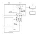
- FIGUREshows a characteristic of the disclosed arrangement.
- FIGUREshows an embodiment of the application in the form of an architecture of a software and/or hardware implementation.
- a PC or computer which includes a shared archiving system CAis connected to an imaging x-ray system XRS.
- the computeris connected to the PACS/HIS/KIS/RIS system IS and can display data on a monitor M.
- Video or image data or signals of different formats, e.g. S-Video, HD, jpeg, tif, etc. from one or several external video sources, identified in the FIGURE e.g. as V 1 and V 2are transmitted via a frame grabber FG connected to the archiving system CA, i.e. a video or image capture circuit, into the shared archiving system.
- a findings reportcan be manually and/or mechanically created in the computer PC from the data stored in the shared archiving system and/or saved in the PACS/HIS/KIS/RIS system. In this way, inconsistencies or redundancies in the creation of the findings report can be disclosed, if necessary made known and if necessary eliminated.
- One aspect of the applicationlies in the use of a computer for the storage of the shared image information, both that of the x-ray system and also that of all external video sources.
- the aspect hereis the correct time stamp or time stamp synchronized between the two recording devices, and the shared archive system to be used. It is as a result possible to temporally correctly arrange the image information from the x-ray system and the external sources.
- This image informationcan then be introduced correctly in a manually and/or mechanically created findings report.
- Multiple examinationse.g. by x-rays
- an incorrect representation of the object of intereste.g. kidneys
- the same patient informationis used so that a faulty examination is prevented.
- the workflow or process flowis simplified within the examination.
- the storage into a PACS or suchlikeis also enabled and/or significantly simplified on account of the integration and the DICOM format.
Landscapes
- Health & Medical Sciences (AREA)
- Engineering & Computer Science (AREA)
- Medical Informatics (AREA)
- Nuclear Medicine, Radiotherapy & Molecular Imaging (AREA)
- Radiology & Medical Imaging (AREA)
- Epidemiology (AREA)
- General Health & Medical Sciences (AREA)
- Primary Health Care (AREA)
- Public Health (AREA)
- Measuring And Recording Apparatus For Diagnosis (AREA)
- Medical Treatment And Welfare Office Work (AREA)
- Apparatus For Radiation Diagnosis (AREA)
Abstract
Description
- CA shared archiving system
- FG frame grabber (video- and/or image capture circuit)
- IS PACS/HIS/KIS/RIS
- M display apparatus and/or monitor and/or display
- PC PC and/or computer
- V1, V2 external video sources1 and2
- XRS imaging x-ray system
Claims (11)
Applications Claiming Priority (3)
| Application Number | Priority Date | Filing Date | Title |
|---|---|---|---|
| DE102011086724.4 | 2011-11-21 | ||
| DE102011086724 | 2011-11-21 | ||
| DE102011086724ADE102011086724A1 (en) | 2011-11-21 | 2011-11-21 | Method and arrangement for the computer-aided structuring of medical examination data |
Publications (2)
| Publication Number | Publication Date |
|---|---|
| US20130129167A1 US20130129167A1 (en) | 2013-05-23 |
| US9020222B2true US9020222B2 (en) | 2015-04-28 |
Family
ID=48221864
Family Applications (1)
| Application Number | Title | Priority Date | Filing Date |
|---|---|---|---|
| US13/682,910Active2033-07-24US9020222B2 (en) | 2011-11-21 | 2012-11-21 | Method and arrangement for the computer-assisted structuring of medical examination data |
Country Status (2)
| Country | Link |
|---|---|
| US (1) | US9020222B2 (en) |
| DE (1) | DE102011086724A1 (en) |
Cited By (2)
| Publication number | Priority date | Publication date | Assignee | Title |
|---|---|---|---|---|
| US10311045B2 (en)* | 2015-01-26 | 2019-06-04 | Microsoft Technology Licensing, Llc | Aggregation/evaluation of heterogenic time series data |
| US11823376B2 (en) | 2018-05-16 | 2023-11-21 | Benevis Informatics, Llc | Systems and methods for review of computer-aided detection of pathology in images |
Families Citing this family (1)
| Publication number | Priority date | Publication date | Assignee | Title |
|---|---|---|---|---|
| US20240187485A1 (en)* | 2021-04-26 | 2024-06-06 | Drägerwerk AG & Co. KGaA | Self-describing protocol translation device |
Citations (5)
| Publication number | Priority date | Publication date | Assignee | Title |
|---|---|---|---|---|
| US6381029B1 (en)* | 1998-12-23 | 2002-04-30 | Etrauma, Llc | Systems and methods for remote viewing of patient images |
| US20040068423A1 (en) | 2002-10-03 | 2004-04-08 | Shaw Grant D. | Graphical user interfaces for sets of medical image data files |
| US20040141661A1 (en)* | 2002-11-27 | 2004-07-22 | Hanna Christopher J. | Intelligent medical image management system |
| US20060031236A1 (en)* | 2004-08-04 | 2006-02-09 | Kabushiki Kaisha Toshiba | Data structure of metadata and reproduction method of the same |
| US20120143625A1 (en)* | 2010-08-31 | 2012-06-07 | Eaves Christopher B | Diagnostic medical information broker system and method |
- 2011
- 2011-11-21DEDE102011086724Apatent/DE102011086724A1/ennot_activeCeased
- 2012
- 2012-11-21USUS13/682,910patent/US9020222B2/enactiveActive
Patent Citations (5)
| Publication number | Priority date | Publication date | Assignee | Title |
|---|---|---|---|---|
| US6381029B1 (en)* | 1998-12-23 | 2002-04-30 | Etrauma, Llc | Systems and methods for remote viewing of patient images |
| US20040068423A1 (en) | 2002-10-03 | 2004-04-08 | Shaw Grant D. | Graphical user interfaces for sets of medical image data files |
| US20040141661A1 (en)* | 2002-11-27 | 2004-07-22 | Hanna Christopher J. | Intelligent medical image management system |
| US20060031236A1 (en)* | 2004-08-04 | 2006-02-09 | Kabushiki Kaisha Toshiba | Data structure of metadata and reproduction method of the same |
| US20120143625A1 (en)* | 2010-08-31 | 2012-06-07 | Eaves Christopher B | Diagnostic medical information broker system and method |
Non-Patent Citations (6)
| Title |
|---|
| Digital Imaging and Communications in Medicine (DICOM), Part 5: Data Structures and Encoding, PS 3.5-2006 "Encapsulated RE compressed Images". Copyright 2006 by the National Electrical Manufacturers Association; Copyright 2006 by the National Electrical Manufacturers Association; Digital Imaging and Communications in Medicine (DICOM), Part 5: Data Structures and Encoding, PS 3.5-2006 "Encapsulated RE compressed Images". Copyright 2006 by the National Electrical Manufacturers Association; Digital Imaging and Communications in Medicine (DICOM), Part 5: Data Structures and Encoding, PS 3.5-2006 "Encapsulated RE compressed Images". Copyright 2006 by the National Electrical Manufacturers Association; Virginia 200209; National Electrical Manufacturers Association; 2006; US. |
| Digital Imaging and Communications in Medicine (DICOM), Part 5: Data Structures and Encoding, PS 3.5—2006 "Encapsulated RE compressed Images". Copyright 2006 by the National Electrical Manufacturers Association; Copyright 2006 by the National Electrical Manufacturers Association; Digital Imaging and Communications in Medicine (DICOM), Part 5: Data Structures and Encoding, PS 3.5—2006 "Encapsulated RE compressed Images". Copyright 2006 by the National Electrical Manufacturers Association; Digital Imaging and Communications in Medicine (DICOM), Part 5: Data Structures and Encoding, PS 3.5—2006 "Encapsulated RE compressed Images". Copyright 2006 by the National Electrical Manufacturers Association; Virginia 200209; National Electrical Manufacturers Association; 2006; US. |
| J. An et al.: Integrated Visualization of Multi-Modal Electronic Health Record Data. Proceedings of the 2nd Internat. Conf. on Bioinformatics and Biomedical Engineering, ICBEE 2008, p. 640-643; 2008. |
| Online-Enzyklopädie "Wikipedia", Artikel v. 26.10.2011 zum Begriff "Server", (Nov. 6, 2012); 2011; Oct. 26, 2011. |
| S. Furuie et al.: Integrating Medical Images and Clinical Information. In: Fourth International Conference on Information and Communication Technology (ICICT 2006), Cairo, Proceedings of Fourth Intenational Conference on Info. and Com. Techn., p. 555-563, 2006; URL: http://www.icict.gov.eg/ICICT-2006/Papers/Electronic%20Health%20Records&20%28EHR%29/ICICT-pep-sfuruie2006-06-13-final-jpeg-sem-autores.pdf (Nov. 6, 2012); 2006. |
| S. Furuie et al.: Integrating Medical Images and Clinical Information. In: Fourth International Conference on Information and Communication Technology (ICICT 2006), Cairo, Proceedings of Fourth Intenational Conference on Info. and Com. Techn., p. 555-563, 2006; URL: http://www.icict.gov.eg/ICICT-2006/Papers/Electronic%20Health%20Records&20%28EHR%29/ICICT-pep-sfuruie2006—06—13-final-jpeg-sem—autores.pdf (Nov. 6, 2012); 2006. |
Cited By (4)
| Publication number | Priority date | Publication date | Assignee | Title |
|---|---|---|---|---|
| US10311045B2 (en)* | 2015-01-26 | 2019-06-04 | Microsoft Technology Licensing, Llc | Aggregation/evaluation of heterogenic time series data |
| US11823376B2 (en) | 2018-05-16 | 2023-11-21 | Benevis Informatics, Llc | Systems and methods for review of computer-aided detection of pathology in images |
| US12254628B2 (en) | 2018-05-16 | 2025-03-18 | Benevis Informatics, Llc | Systems and methods for review of computer-aided detection of pathology in images |
| US12272062B2 (en) | 2018-05-16 | 2025-04-08 | Benevis Informatics, Llc | Systems and methods for review of computer-aided detection of pathology in images |
Also Published As
| Publication number | Publication date |
|---|---|
| DE102011086724A1 (en) | 2013-05-23 |
| US20130129167A1 (en) | 2013-05-23 |
Similar Documents
| Publication | Publication Date | Title |
|---|---|---|
| Huang | PACS and imaging informatics: basic principles and applications | |
| US9355309B2 (en) | Generation of medical image series including a patient photograph | |
| JP2007050254A (en) | Real-time integration and recording of surgical image data | |
| JPWO2007119615A1 (en) | Medical image display apparatus and program | |
| US10037405B2 (en) | Medical image generation apparatus, medical image storage apparatus, medical image display apparatus, and medical image display system | |
| US10460488B2 (en) | Spine labeling automation | |
| KR101352999B1 (en) | Appratus and method for image bookmarking using attribute information | |
| US10095904B2 (en) | Image visualization | |
| US20120123239A1 (en) | Medical Image Processing System and Processing Method | |
| US9020222B2 (en) | Method and arrangement for the computer-assisted structuring of medical examination data | |
| KR101176448B1 (en) | Apparatus for managing medical image data using reference coordinates and method thereof | |
| KR101919908B1 (en) | Method for facilitating labeling of medical image and apparatus using the same | |
| US8923582B2 (en) | Systems and methods for computer aided detection using pixel intensity values | |
| CN101266634A (en) | Method for data exchange between medical devices | |
| US10176569B2 (en) | Multiple algorithm lesion segmentation | |
| US11081228B2 (en) | Automatic retrospective review of electronic medical records | |
| Siegel | Current state of the art and future trends | |
| JP5303192B2 (en) | Medical image system and image compression method for medical image system | |
| KR101597135B1 (en) | Medical image storage and transmission system tagging simultaneously with recording | |
| US10157292B2 (en) | Viewing session takeover in medical imaging applications | |
| KR20190138106A (en) | Medical image information starage system | |
| KR101582267B1 (en) | Generating device for medical key image | |
| JP2009254690A (en) | Medical image storage apparatus and medical image observation apparatus | |
| WO2005119443A2 (en) | System and method of a converting medical imaging movie format data to dicom compatible images | |
| KR20150066130A (en) | Method for medical imaging information communication between hospitals and system for it |
Legal Events
| Date | Code | Title | Description |
|---|---|---|---|
| AS | Assignment | Owner name:SIEMENS AKTIENGESELLSCHAFT, GERMANY Free format text:ASSIGNMENT OF ASSIGNORS INTEREST;ASSIGNOR:WIETS, MICHAEL;REEL/FRAME:029454/0684 Effective date:20121129 | |
| STCF | Information on status: patent grant | Free format text:PATENTED CASE | |
| AS | Assignment | Owner name:SIEMENS HEALTHCARE GMBH, GERMANY Free format text:ASSIGNMENT OF ASSIGNORS INTEREST;ASSIGNOR:SIEMENS AKTIENGESELLSCHAFT;REEL/FRAME:039271/0561 Effective date:20160610 | |
| MAFP | Maintenance fee payment | Free format text:PAYMENT OF MAINTENANCE FEE, 4TH YEAR, LARGE ENTITY (ORIGINAL EVENT CODE: M1551); ENTITY STATUS OF PATENT OWNER: LARGE ENTITY Year of fee payment:4 | |
| MAFP | Maintenance fee payment | Free format text:PAYMENT OF MAINTENANCE FEE, 8TH YEAR, LARGE ENTITY (ORIGINAL EVENT CODE: M1552); ENTITY STATUS OF PATENT OWNER: LARGE ENTITY Year of fee payment:8 | |
| AS | Assignment | Owner name:SIEMENS HEALTHINEERS AG, GERMANY Free format text:ASSIGNMENT OF ASSIGNORS INTEREST;ASSIGNOR:SIEMENS HEALTHCARE GMBH;REEL/FRAME:066088/0256 Effective date:20231219 | |
| AS | Assignment | Owner name:SIEMENS HEALTHINEERS AG, GERMANY Free format text:CORRECTIVE ASSIGNMENT TO CORRECT THE ASSIGNEE PREVIOUSLY RECORDED AT REEL: 066088 FRAME: 0256. ASSIGNOR(S) HEREBY CONFIRMS THE ASSIGNMENT;ASSIGNOR:SIEMENS HEALTHCARE GMBH;REEL/FRAME:071178/0246 Effective date:20231219 |