US8992567B1 - Compressible, deformable, or deflectable tissue closure devices and method of manufacture - Google Patents
Compressible, deformable, or deflectable tissue closure devices and method of manufactureDownload PDFInfo
- Publication number
- US8992567B1 US8992567B1US12/563,297US56329709AUS8992567B1US 8992567 B1US8992567 B1US 8992567B1US 56329709 AUS56329709 AUS 56329709AUS 8992567 B1US8992567 B1US 8992567B1
- Authority
- US
- United States
- Prior art keywords
- closure device
- tissue closure
- tissue
- radially extending
- acd
- Prior art date
- Legal status (The legal status is an assumption and is not a legal conclusion. Google has not performed a legal analysis and makes no representation as to the accuracy of the status listed.)
- Expired - Fee Related, expires
Links
Images


Classifications
- A—HUMAN NECESSITIES
- A61—MEDICAL OR VETERINARY SCIENCE; HYGIENE
- A61B—DIAGNOSIS; SURGERY; IDENTIFICATION
- A61B17/00—Surgical instruments, devices or methods
- A61B17/08—Wound clamps or clips, i.e. not or only partly penetrating the tissue ; Devices for bringing together the edges of a wound
- A—HUMAN NECESSITIES
- A61—MEDICAL OR VETERINARY SCIENCE; HYGIENE
- A61B—DIAGNOSIS; SURGERY; IDENTIFICATION
- A61B17/00—Surgical instruments, devices or methods
- A61B17/00491—Surgical glue applicators
- A—HUMAN NECESSITIES
- A61—MEDICAL OR VETERINARY SCIENCE; HYGIENE
- A61B—DIAGNOSIS; SURGERY; IDENTIFICATION
- A61B17/00—Surgical instruments, devices or methods
- A61B17/0057—Implements for plugging an opening in the wall of a hollow or tubular organ, e.g. for sealing a vessel puncture or closing a cardiac septal defect
- A61B2017/00575—Implements for plugging an opening in the wall of a hollow or tubular organ, e.g. for sealing a vessel puncture or closing a cardiac septal defect for closure at remote site, e.g. closing atrial septum defects
- A61B2017/00588—Rigid or stiff implements, e.g. made of several rigid parts linked by hinges
- A—HUMAN NECESSITIES
- A61—MEDICAL OR VETERINARY SCIENCE; HYGIENE
- A61B—DIAGNOSIS; SURGERY; IDENTIFICATION
- A61B17/00—Surgical instruments, devices or methods
- A61B17/0057—Implements for plugging an opening in the wall of a hollow or tubular organ, e.g. for sealing a vessel puncture or closing a cardiac septal defect
- A61B2017/00575—Implements for plugging an opening in the wall of a hollow or tubular organ, e.g. for sealing a vessel puncture or closing a cardiac septal defect for closure at remote site, e.g. closing atrial septum defects
- A61B2017/00592—Elastic or resilient implements
- A—HUMAN NECESSITIES
- A61—MEDICAL OR VETERINARY SCIENCE; HYGIENE
- A61B—DIAGNOSIS; SURGERY; IDENTIFICATION
- A61B17/00—Surgical instruments, devices or methods
- A61B17/0057—Implements for plugging an opening in the wall of a hollow or tubular organ, e.g. for sealing a vessel puncture or closing a cardiac septal defect
- A61B2017/00575—Implements for plugging an opening in the wall of a hollow or tubular organ, e.g. for sealing a vessel puncture or closing a cardiac septal defect for closure at remote site, e.g. closing atrial septum defects
- A61B2017/00606—Implements H-shaped in cross-section, i.e. with occluders on both sides of the opening
- A—HUMAN NECESSITIES
- A61—MEDICAL OR VETERINARY SCIENCE; HYGIENE
- A61B—DIAGNOSIS; SURGERY; IDENTIFICATION
- A61B17/00—Surgical instruments, devices or methods
- A61B17/0057—Implements for plugging an opening in the wall of a hollow or tubular organ, e.g. for sealing a vessel puncture or closing a cardiac septal defect
- A61B2017/00575—Implements for plugging an opening in the wall of a hollow or tubular organ, e.g. for sealing a vessel puncture or closing a cardiac septal defect for closure at remote site, e.g. closing atrial septum defects
- A61B2017/00615—Implements with an occluder on one side of the opening and holding means therefor on the other
- A—HUMAN NECESSITIES
- A61—MEDICAL OR VETERINARY SCIENCE; HYGIENE
- A61B—DIAGNOSIS; SURGERY; IDENTIFICATION
- A61B17/00—Surgical instruments, devices or methods
- A61B17/0057—Implements for plugging an opening in the wall of a hollow or tubular organ, e.g. for sealing a vessel puncture or closing a cardiac septal defect
- A61B2017/00575—Implements for plugging an opening in the wall of a hollow or tubular organ, e.g. for sealing a vessel puncture or closing a cardiac septal defect for closure at remote site, e.g. closing atrial septum defects
- A61B2017/00623—Introducing or retrieving devices therefor
- A—HUMAN NECESSITIES
- A61—MEDICAL OR VETERINARY SCIENCE; HYGIENE
- A61B—DIAGNOSIS; SURGERY; IDENTIFICATION
- A61B17/00—Surgical instruments, devices or methods
- A61B17/0057—Implements for plugging an opening in the wall of a hollow or tubular organ, e.g. for sealing a vessel puncture or closing a cardiac septal defect
- A61B2017/00575—Implements for plugging an opening in the wall of a hollow or tubular organ, e.g. for sealing a vessel puncture or closing a cardiac septal defect for closure at remote site, e.g. closing atrial septum defects
- A61B2017/00628—T-shaped occluders
Definitions
- the field of the inventionsgenerally relates to cardiovascular and arterial closure devices, and, more particularly, to arterial closure devices and techniques.
- a catheterIn most cardiology and radiology procedures, a catheter is inserted into an artery, such as the femoral artery, through a vascular introducer. When the procedure is complete, the physician removes the catheter from the introducer and then removes the introducer from the arteriotomy into the vessel. The physician then must prevent or limit the amount of blood that leaks through the arteriotomy so that the patient can be discharged. Physicians currently use a number of methods to close the arteriotomy, such as localized compression, sutures, collagen plugs, and adhesives, gels, foams, and similar materials. To use localized compression, the physician presses down against the vessel to allow the arteriotomy to naturally clot.
- localized compressionthe physician presses down against the vessel to allow the arteriotomy to naturally clot.
- This methodcan take half an hour or more, and requires the patient to remain immobilized for at least that period of time and be kept in the hospital for observation. There are potentials for clots at puncture site to be dislodged. Moreover, the amount of time necessary for the compression can be significantly increased depending upon how much heparin, glycoprotein IIb/IIA antagonists, or other anti-clotting agents were used during the procedure. Sutures and collagen plugs may have procedure variability, may require time to close the vessel, may have negative cost factors, and may necessitate a separate deployment device. Adhesives, gels, and foams may have negative cost factors, may necessitate a possibly complicated deployment process, and may have procedure variability.
- an arterial closure deviceis deliverable over a tube for placement within and against an arteriotomy.
- the arterial closure deviceincludes a first member forming an enlargement around the circumference of the arterial closure device and being configured to be received against an outer surface of a vessel; a connecting member having a smaller outer diameter than the first member, extending from the first member, and being configured to be positioned within an arteriotomy of a vessel; and a longitudinal channel configured to receive a tube and passing between the first member and the connecting member.
- Embodiments of the arterial closure devicemay include one or more of the following features.
- the connecting membermay include slits.
- the connecting membermay be expandable from a first narrow diameter to a second expanded diameter.
- the arterial closure devicemay further include a second member extending from the connecting member, forming an enlargement around the circumference of the arterial closure device and being configured to be received against an inner surface of the vessel when the first member is received against the outer surface of the vessel.
- the arterial closure devicemay still further include an adhesive layer on at least one of the first member, the second member, and the connecting member.
- the first membermay extend at an angle from the arterial closure device, the second member may extend at an angle from the arterial closure device, and the first member may be generally oriented in the direction of the second member.
- the first membermay include at least one superelastic/shape memory element configured to move between a first extended position and a second extended position and the second member may include at least one superelastic/shape memory element configured to move between a first extended position and a second extended position.
- the arterial closure devicemay further include an adhesive layer on at least one of the first member and the connecting member.
- the arterial closure devicemay further include an adhesive within the longitudinal channel.
- the arterial closure devicemay further include longitudinal slots along the longitudinal channel.
- the arterial closure devicemay further include an extending member extending from the first member in a generally opposite direction away from the connecting member and the longitudinal channel continues from the first member through the extending member.
- the extending membermay include a closable opening of the longitudinal channel.
- the arterial closure devicemay further include a slot along at least a part of the length of the arterial closure device.
- the arterial closure devicemay further include a deployment tool, the deployment tool including a handle, a contacting section, and an extension that extends between the handle and the contacting section.
- the contacting sectionis configured to mate with the arterial closure device to advance the arterial closure device over the tube and deploy the arterial closure device within the vessel.
- an arterial closure systemin another general aspect, includes an arterial closure device and a deployment tool.
- the arterial closure deviceincludes a first member forming an enlargement around the circumference of the arterial closure device and being configured to be received against an outer surface of a vessel, a connecting member having a smaller outer diameter than the first member, extending from the first member, and being configured to be positioned within an arteriotomy of a vessel, and a longitudinal channel configured to receive a tube and passing between the first member and the connecting member.
- the deployment toolincludes a handle, a contacting section, and an extension that extends between the handle and the contacting section, the contacting section being configured to mate with the arterial closure device to advance the arterial closure device over the tube and deploy the arterial closure device within the vessel.
- Embodiments of the arterial closure systemmay include any of the features described above or herein.
- the arterial closure systemmay further include a second member extending from the connecting member, forming an enlargement around the circumference of the arterial closure device and being configured to be received against an inner surface of the vessel when the first member is received against the outer surface of the vessel.
- the first membermay include at least one superelastic/shape memory element configured to move between a first extended position and a second extended position; and the second member may include at least one superelastic/shape memory element configured to move between a first extended position and a second extended position.
- the arterial closure devicemay include a slot along at least a portion of the length of the arterial closure device and the contacting section of the deployment tool may include a longitudinal slot.
- a method of closing an opening in a vesselincludes providing an arterial closure device that includes a first member forming an enlargement around the circumference of the arterial closure device and being configured to be received against an outer surface of a vessel, a connecting member having a smaller outer diameter than the first member, extending from the first member, and being configured to be positioned within an arteriotomy of a vessel, and a longitudinal channel configured to receive a tube and passing between the first member and the connecting member.
- the methodfurther includes providing a deployment tool comprising a handle, a contacting section, and an extension that extends between the handle and the contacting section, the contacting section being configured to mate with the arterial closure device to advance the arterial closure device over the tube and deploy the arterial closure device within the vessel.
- the methodstill further includes slidably mounting the arterial closure device to a tube; inserting the tube through an opening into the vessel; using the deployment tool to advance and deploy the arterial closure device by advancing the arterial closure device along the tube until the connecting member is deployed within the vessel and the first member is received against the outer surface of the vessel; and removing the tube from the vessel and from the arterial closure device.
- Embodiments of the method of closing an opening in a vesselmay include any of the features described above or herein.
- the arterial closure devicemay further include a second member extending from the connecting member, forming an enlargement around the circumference of the arterial closure device and being configured to be received against an inner surface of the vessel when the first member is received against the outer surface of the vessel and an adhesive layer is positioned on at least one of the first member, the second member, and the connecting member, and deploying the arterial closure device further comprises positioning the second member against the inner surface of the vessel.
- the arterial closure device, the arterial closure system, and the arterial closure methodprovides considerable advantages, as described herein.
- the ACDs and methods described hereincan provide: (1) the ability to deploy an ACD without the removal and re-insertion of a second device; (2) the ability to be used on most commercial vascular introducers, catheters, tubes, etc.; (3) the ability to use tactile feedback to correctly and properly deploy an ACD without direct or indirect visual assistance; (4) the ability to use adhesives to secure the device to the vessel; (5) the ability to use adhesives to close off the device to prevent blood leaking or seepage; and (6) the ability to provide eluting therapeutic agents incorporated within or on the device.
- the device, system and methodare advantageously simple to use, inexpensive, and effective as a percutaneous vessel access closure device and method.
- FIG. 1is a side view of a arterial closure device positioned around a tubular section of a vascular introducer.
- FIG. 2is a side view of the arterial closure device of FIG. 1 advanced through a percutaneous opening by a deployment instrument.
- FIG. 3is a side view of the arterial closure device of FIG. 1 deployed through a vessel wall.
- FIG. 4is a cross-sectional side view of the arterial closure device of FIG. 1 deployed through a vessel wall.
- FIG. 5is a top view of the arterial closure device of FIG. 1 .
- FIGS. 6 and 7are side and cross-sectional side views, respectively, of a second implementation of an arterial closure device deployed within an arteriotomy of a vessel wall.
- FIG. 8is a bottom end view of the arterial closure device of FIG. 6 showing the flared end opened.
- FIG. 9is a bottom end view of the arterial closure device of FIG. 6 showing the flared end closed.
- FIG. 10is a top end view of the arterial closure device of FIG. 6 showing the flared end partially closed.
- FIG. 11is a side view of the arterial closure device of FIG. 6 showing the flared end.
- FIGS. 12 and 13are a cross-sectional side view and a top view, respectively, of the arterial closure device of FIG. 1 having an adhesive on the inner diameter and tissue engagement areas.
- FIGS. 14 and 15are a cross-sectional side view and a top view, respectively, of the arterial closure device of FIG. 6 having an adhesive on the inner diameter and tissue engagement areas.
- FIGS. 16 and 17are a cross-sectional side view and a top view, respectively, of the arterial closure device of FIG. 1 having grooves on the inner diameter to form a thinned or weakened wall.
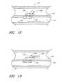
- FIG. 18is a side view of an angled arterial closure device.
- FIG. 19is a side view of an angled arterial closure device having a foldable extending member.
- FIG. 20is a side view of a deployment tool.
- FIG. 21is a side view of the deployment tool of FIG. 20 used to deploy a arterial closure device.
- FIG. 22is an end view of the deployment tool FIG. 20 .
- FIG. 23is a side view of the deployment tool of FIG. 20 having an extended contacting member.
- FIG. 24is a side view of the deployment tool of FIG. 23 used to deploy a arterial closure device.
- FIG. 25is an end view of the deployment tool FIG. 23 .
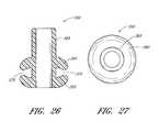
- FIG. 26is a cross-sectional side view of a arterial closure device having angled closure edges for compressing a vessel wall.
- FIG. 27is a top view of the arterial closure device of FIG. 26 .
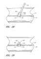
- FIG. 28is a side view of the arterial closure device of FIG. 26 being advanced through the skin into a vessel with the closure edges deflected.
- FIG. 29is a side view of the arterial closure device of FIG. 26 deployed and secured onto vessel wall with the closure edges occluding the arteriotomy.
- FIG. 30is a side view of a arterial closure device.
- FIG. 31is an end view of the arterial closure device of FIG. 30 .
- FIG. 32is a perspective side view of a vascular connector having a closable end.
- FIG. 33is an end view of the arterial closure device of FIG. 32 .
- FIG. 34is a side view of a liner having a longitudinal slot for a arterial closure device.
- FIG. 35is an end view of the liner of FIG. 34 .
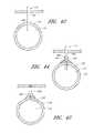
- FIG. 36is a side view of a liner having a radial slot.
- FIG. 37is an end view of the liner of FIG. 36 .
- FIG. 38is a side view of a plug-style arterial closure device that includes an adhesive layer on the vessel contact areas.
- FIG. 39is a side view of a plug style arterial closure device that has limited vessel protrusion and includes an adhesive on the vessel contacting areas.
- FIGS. 40 and 41are side views of the plug style arterial closure device of FIG. 39 being deployed and deployed within a vessel.
- FIG. 42is an end view showing the distal end of the introducer inside a vessel.
- FIGS. 43 and 44are end views showing a flared arterial closure device deployed along the introducer.
- FIG. 45is an end view showing the flared arterial closure device of FIG. 43 deployed against the vessel to close the arteriotomy.
- FIGS. 46 and 47are electrical schematics for a direct resistive element heating circuit and an ohmic tissue heating circuit.
- FIG. 48is a perspective view of a tube used to fabricate a arterial closure device.
- FIG. 49is a perspective view of the tube of FIG. 48 showing material being removed.
- FIG. 50is a side view of the tube of FIG. 48 with the material removed.
- FIG. 51is a top view of the curved configuration.
- FIG. 52is a side view of the configuration of FIG. 51 .
- FIG. 53is a perspective view of a fabric covering.
- FIGS. 54-58are side views showing the fabric covering of FIG. 53 being mounted within the curved configuration of FIG. 51 to form a arterial closure device.
- FIG. 59is a side view of a configuration having side arms that fold over each other.
- FIGS. 60 and 61are front and cross-sectional side views of a deployment tool for deploying the arterial closure device.
- FIG. 62is a cross-sectional side view of the deployment tool of FIG. 60 having the arterial closure device within.
- a vascular closure system 100generally includes two components: an arterial closure device (“ACD”) 105 and a deployment instrument 110 .
- the ACD 105is slidably mounted to a vascular introducer 115 or other tubular device, such as a catheter, advanced over a tube section 120 of the introducer 115 using the deployment instrument 110 , passed through a percutaneous opening 125 , and placed through an arteriotomy 130 in a vessel wall 133 into a blood vessel 135 .
- the deployment tool 110 and the introducer 115then are removed from the blood vessel 135 and out of the percutaneous opening 125 .
- the ACD 105is generally compliant, tubular, and includes a first member 140 , a second member 145 , a connecting member 150 between the first member and the second member, and an optional extending member 155 that extends from the second member.
- a longitudinal channel 160passes between a first opening 165 in the extending member (or second member if the extending member is not present) and a second opening 170 in the first member 140 .
- the ACD 105is formed of a tubular structure of sufficient length and thickness (e.g., a single wall thickness of between 0.005′′ and 0.05′′, and more particularly between 0.01′′ and 0.02′′) that can be advanced over the introducer 115 , and through the puncture site 125 .
- the ACD 105has sufficient rigidity to be advanced through the puncture site 125 yet is compliant enough to be compressed onto itself by the natural elasticity of the vessel wall 133 after the introducer 115 is removed.
- the connecting member 150can be configured to have a natural elasticity such that when it is no longer mounted over the introducer tube 120 , it will return to its original smaller diameter state.
- the ACD 105may include, for example, longitudinal sections of the tube where the wall thickness is thinner (e.g., connecting member 150 ) thereby creating creases or weakened areas that receive the vessel wall 133 .
- the creaseswould reduce the amount of compressive force required to collapse the tube onto itself.
- a design allowing tactile feedbackmay be used to determine the proper insertion position (depth). The tactile feedback could be accomplished by the ACD 105 having one or more rings of increased wall thickness, an “hour glass” geometry, a thin, narrow, then wide geometry, combination, or other means to provide an abrupt change in the advancing force resistance during deployment.
- the ACD 105may be manufactured in many different French sizes, to match the outer diameter of any commercial vascular introducers 115 .
- the ACD 105is placed around the outside of any commercially available introducer 115 , or other device that is inserted into the cardiovascular system (e.g., catheter, etc.), and positioned adjacent to the proximal end of the introducer (i.e., near the valve or luer fitting of the introducer).
- the introducer 115then is inserted into the vasculature using standard techniques.
- the tubular ACD 115Prior to removing the introducer 115 , the tubular ACD 115 is advanced to the skin, for example, by the physician manually advancing the ACD along the tube 120 .
- the deployment instrument 110then is positioned against or clipped onto the tube 120 , advanced to be in contact with the proximal end (i.e., second member 145 ) of the ACD 105 , and advanced through the skin such that at least the distal most portion (e.g., first member 140 ) of the ACD is inside the vessel 135 .
- the ACD 105is prevented from deforming or collapsing during insertion by the rigidity of the tube 120 .
- the tube 120also acts as a guide to position the ACD 105 through the puncture site 125 during its advancement and deployment.
- the deployment instrument 110is held in position and still in contact with the ACD 105 preventing the ACD from coming out of the vessel 135 along with the introducer. Once the introducer 115 is completely removed, the ACD 105 is compressed together due to the elastic recovery of the vessel wall 133 , achieving hemostasis and effectively sealing the arteriotomy 150 and puncture site 125 .
- the ACD 105can be partially or completely fabricated from a biocompatible material, such as expanded polytetrafluoroethylene (ePTFE), polyester, polyurethane, silicone, Dacron, urethane, and/or a composite or combination of these or other suitable materials.
- a biocompatible materialsuch as expanded polytetrafluoroethylene (ePTFE), polyester, polyurethane, silicone, Dacron, urethane, and/or a composite or combination of these or other suitable materials.
- the ACD 105also can be partially or completely fabricated from a biodegradable/bioabsorbable material, including modified cellulose, collagen, fibrin, fibrinogen, elastin or other connective proteins or natural materials, polymers or copolymers such as polylactide [poly-L-lactide (PLLA), poly-D-lactide (PDLA)], polyglycolide, polydioxanone, polycaprolactone, polygluconate, polylactic acid (PLA), polylactic acid-polyethylene oxide copolymers, poly(hydroxybutyrate), polyanhydride, polyphosphoester, poly(amino acids), poly(alpha-hydroxy acid) or related copolymers of these materials as well as composites and combinations thereof and combinations of other biodegradable/bioabsorbable materials.
- PLLApoly-L-lactide
- PDLApoly-D-lactide
- polyglycolidepolydioxanone
- polycaprolactonepolygluconate
- the ACD 105also can be partially or completely fabricated from materials that swell, or expand when they are exposed to a fluid, such as blood, or another fluid, for example, that can be added by the physician to cause the material to swell.
- materialsinclude hydrophilic gels (hydrogels), regenerated cellulose, polyethylene vinyl acetate (PEVA), as well as composites and combinations thereof and combinations of other biocompatible swellable or expandable materials.
- the ACD 105can be made using several methods and processes including extrusion, molding (i.e., injection molding or other known molding techniques), casting, dip coating, spraying, adhesive bonding, ultra-sonic welding, composite fabrication techniques, and combinations of these and/or other similar methods and processes.
- the ACD 105also can have a biocompatible contact adhesive or other material within the longitudinal channel 160 so that when the longitudinal channel is compressed within the arteriotomy 130 , the adhesive bonds the inside surfaces of the longitudinal channel together. This assists or expedites the sealing of the arteriotomy.
- bonding materialscan be used on the outside of the ACD 105 , for example, on the outer surface of the first member 140 , the second member 145 , the connecting member 150 , and or the optional extending member 155 . In particular, the bonding material is especially useful where the ACD contacts the vessel wall 133 defining the arteriotomy 130 .
- the biocompatible contact adhesive adhesive/bonding compounds/solutionscould be added during the manufacturing process, just prior to deployment, or after the device has been deployed.
- the bonding materialscould be in the form of a liquid, semi solid, or solid. Suitable bonding materials include gels, foams and microporous mesh. Suitable adhesives include acrylates, cyanoacrylates, epoxies, fibrin-based adhesives, other biological based adhesives, UV light and/or heat activated or other specialized adhesives.
- the adhesivecould bond on initial contact, or longer, to allow repositioning if desired.
- the preferred adhesivemay be a crystalline polymer that changes from a non-tacky crystalline state to an adhesive gel state when the temperature is raised from room temperature to body temperature.
- Such materialis available under the trade name IntillemerTM adhesive, available from Landec Corp. as well as composites and combinations thereof and combinations of other materials.
- Suppliers of biocompatible adhesivesinclude, but are not limited to, Plasto (Dijon, France), Haemacure (Montreal, Canada), Cohesion (Palo Alto, Calif.), Cryolife (Kennesaw, Ga.), TissueLink (Dover, N.H.), and others.
- the adhesivecan be blended with a material, such as a starch or other material, that retards or delays bonding to allow repositioning of the coupler after it has been deployed.
- a degradable coatingcan be placed over the adhesive coating so that it degrades and exposes the adhesive.
- Other adhesivesare understood to include composites-based adherents and combinations of the above materials and other suitable materials as are known in the art.
- the ACD 105can be fabricated from materials that include one or more radiopaque materials, such as barium sulfate, bismuth trioxide, or other any other radiopaque material.
- the radiopaque materialis added to the materials from which the ACD 105 is fabricated or to the bonding materials that are placed in, on, or around the ACD.
- ACD 200a second implementation of a arterial closure device is shown as a arterial closure device (“ACD”) 200 .
- the ACD 200includes a first member 205 , a second member 210 , and an optional extending member 215 that extends from the second member.
- a longitudinal channel 220passes between a first opening 225 in the extending member (or second member if the extending member is not present) and a second opening 230 in the first member 205 .
- the ACD 200is implanted within an arteriotomy 130 in a manner similar to the implantation of the ACD 105 . However, the ACD 200 does not include a member that is substantially in contact with the inner wall of the vessel 135 .
- the ACDhas a flare, or two or more short slits 235 in the side wall of the first member 205 .
- the flare or slits 235are designed to open or flare around the catheter or introducer 120 when advanced to the top of the vessel puncture site ( FIG. 8 ).
- the materials from which the ACD 200 or the second member 205 are fabricatedmay be a very elastic material such that when around the introducer it expands and when advanced beyond the end of the introducer, it contracts such that the individual flares pinch or otherwise catch the edges of the arteriotomy or punctured vessel and pull them together while contracting ( FIG. 9 ). This action is intended to close the arteriotomy 130 and create hemostasis.
- the inside of the flared section 235 of the ACD 200may have a biocompatible contact adhesive or other bonding material, as described above, that further secures the ACD within the arteriotomy and to the vessel 135 , and, in particular the second member 210 to the top or outer surface of the vessel.
- the adhesive or bonding materialscan be implemented on any of the above ACDs.
- the ACD 105has an adhesive or bonding material 270 on the inner diameter and tissue engagement areas.
- the ACD 200has the adhesive or bonding material 270 on the inner diameter and tissue engagement areas.
- adhesive 270will close the respective longitudinal channel 160 , 200 of the ACD 105 , 200 to reduce or eliminate seepage blood.
- the adhesive 270 around the tissue contacting areaswill bond the ACD to the vessel wall to reduce or eliminate seepage of blood through those regions.
- the ACD 105can have the inner diameter of the longitudinal channel 160 modified to include ridges 280 and channels 285 that weaken or thin the wall section of the ACD.
- the inner diameter of the longitudinal channel 160can be expanded or reduced depending upon the circumferential pressure exerted against the ACD. For example, when passing the introducer through the longitudinal channel the inner diameter will be expanded. When the introducer is subsequently removed, the inner diameter is reduced because of the natural elastic recoil properties of the ACD. In this manner, the seepage of blood through the longitudinal channel is reduced or eliminated.
- the surfaces of the inner diameter of the longitudinal channelcan be coated with an adhesive, as described above, to further ensure that the inner diameter is closed.
- the ACDs described hereinalso can include one or more therapeutic agents that affect healing at the site where the device is deployed.
- the agent(s)can be incorporated into the structure forming the device and/or incorporated into a coating.
- therapeutic agentsmay include, but are not limited to, antithrombotics (such as anticoagulants), antimitogens, antimitotoxins, antisense oligonucleotides, gene therapy solutions, nitric oxide, and growth factors and inhibitors.
- Direct thrombin inhibitorsthat may be beneficial include Hirudin, Hirugen, Hirulog, PPACK (D-phenylalanyl-L-propyl-Larginine chloromethyl ketone), Argatreban, and D-FPRCH.sub.2 Cl (D-phenylalanyl-Lpropyl-L-arginyl chloromethyl ketone); indirect thrombin inhibitors include Heparin and Warfarin (coumadin). Alternatively, a clot promoter may be used, such as protamine sulphate or calcium hydroxide.
- Additional therapeutic materialsinclude, aspirin, dexamethasone, dexamethasone phosphate, streptokinase, tocopherol, TPA, urokinase, paclitaxel (Taxol), actinomycin, rapamyacin, or other. Sirolimus, or other antibiotics may also be used.
- the therapeutic compounds/solutionsmay be blended with the device base materials during fabrication, applied just prior to deployment, or after the device has been deployed. Additionally, the therapeutic materials may be located on, through, inside, or combination of the device in holes, grooves, slots or other indentation to allow elution of the therapeutic compound(s).
- Post device fabrication coating methodsinclude, but are not limited to, dipping, spraying, brushing, submerging the devices into a beaker containing a therapeutic solution while inside a vacuum chamber to permeate the device material, etc.
- the geometry of the ACDs described hereinis shown for illustration purposes as being generally round. However, they can be of any other geometry, such as oval, elliptical, rectangular, square, ridged, or a combination of shapes.
- the ACDhas been illustrated as forming a generally perpendicular angle with the vessel wall once deployed. Nonetheless, the inventors intend the configuration to be at any suitable angle, such as between 30° and 60°, or, for example, 45° or as otherwise desired.
- a range of angles of the ACDcan be available and the physician can choose the appropriate ACD based on the angle at which the introducer is introduced into the vessel. For example, referring to FIG.
- an ACD 290is formed to have the extending member 155 extending at an angle of approximately 45° from the second member 145 .
- the first member 140 and the second member 145are longitudinally offset. This configuration is designed to cause the extending member 155 to follow the path created by the introducer.
- the ACDhas a second member 292 , a foldable extending member 294 , and a groove 296 positioned between the second member 292 and the folding extending member 294 . In this manner, the extending member 294 can be folded or bent over to be less obtrusive and to close off the flow of blood through the ACD.
- a deployment tool 300is designed to engage or otherwise contact the proximal edge, or other edge, of the ACD.
- the tool 300is generally handheld and includes a handle 305 , an extension 310 , and a contacting section 315 that clips onto, or otherwise contacts the outside of the introducer and mates with the ACD.
- the contacting section 315has sufficient length to advance the ACD through the tissue to the desired position on the vessel.
- the handle 305 or grasping sectioncan be, for example, round, rectangular, elliptical, or a combination of shapes or other shape that fit comfortably in the hand.
- the contacting section 315can have a cross-sectional geometry of a partially open tube having more than 50% diameter coverage, so that it can clip onto, and slide over the outer diameter of the introducer.
- the deployment toolcan include an additional extension 320 that is configured to fit around the extending member 155 and mate with the second member 145 .
- the extension 320can be attached to the introducer after the introducer is positioned within the artery.
- the deployment tool 300can be made partially or completely from several different polymer materials including polycarbonate, nylon, polyethylene, polytetrafluoroethylene (PTFE), fluoroethylene-propylene (FEP) or polyfluoroacrylate (PFA), polyester ether ketone (PEEK), polyamide, polyimide, polyethyleneteraphthalate (PET), combination or other material able to withstand sterilization processing.
- the toolcan also be made partially or completely from several different types of metals including stainless steel; spring metal alloys such as ElgiloyTM, InconelTM; superelastic/shape memory alloys such as Nitinol (NiTi) as well as composites and combinations thereof and combinations of other materials.
- the deployment tool 300can be made using several methods and processes including extrusion, molding (injection and other), casting, adhesive bonding, ultrasonic welding as well as combinations thereof and combinations of other methods and processes.
- the proximal edge of the ACDi.e., of the extending member 155 or the second member 145
- the distal edge or other portion of the advancement tool 300may have interlocking geometries to aid and/or control the position of the ACD during advancement along the introducer.
- the engagement/contact section 315 , 320 of the tool 300may have a cross-sectional geometry of a complete circle that is designed to split away from the introducer once the ACD has been advanced and deployed. Splitting can be accomplished by having weakened areas in the wall of the tubing, such as linear perforations, or linear scores. This version would require that the deployment tool be back loaded onto the introducer before the ACD is placed onto the introducer and prior to insertion into the vessel.
- the inside, concave section of the contact section 315 , 320may be coated with a hydrophilic or other lubricious material to reduce the friction during advancement and deployment of the ACD.
- the contacting section 315 , 320 of the toolcan be lengthened and designed to further attach to and compress the distal edge of the ACD, thereby providing additional support during insertion and deployment into the vessel.
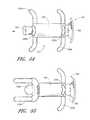
- a ACD 350includes a first angled closure edge 355 , a second angled closure edge 360 , an extending member 365 , and a connection member 370 between the first and second angled closure members.
- the first angled closure edge 355 and the second angled closure edgeare generally directed at each other such that they define a narrow opening 375 through which the vessel wall 133 is received.
- the ACD 350is deployed over the introducer tube section 120 using, for example, the deployment tool 300 .
- the second angled closure edge 360is deflected away from the first angled closure edge 355 . The deflection can be caused, for example, by the contacting section 320 surrounding the second angled closure edge 360 .
- the angled closure edges 355 and 360are formed, for example, from a flexible member, such as a polymer, superelastic/shape memory material, or a combination of the two.
- the superelastic/shape memory membercan be coated with a polymer.
- a ACD 400includes a threaded section 405 and an extending section 410 .
- the threaded section 405includes threads 415 mounted on and between a first member 420 and a second member 425 .
- the extending section 410includes a longitudinal channel 430 that includes a distal shaped channel 435 .
- a deployment tool having a mating shaped distal endis inserted into the longitudinal channel 430 such that it mates with the distal shaped channel 435 . By rotating the deployment tool, the ACD can be threadably inserted into the arteriotomy.
- the distal edge of the ACD 400is designed to engage the opening of the arteriotomy or puncture site and protrude to a specific depth based on how many times the ACD was advanced, twisted or turned.
- the ACD 400may have a stop 437 to limit how far the device protrudes into the vessel.
- the same “screw” type distal edgecould be used on a hemostatic plug, made from a solid piece of material, rather than a tube structure.
- a deployment toolwould be needed that has, for example, a grasping distal end for insertion into the vessel.
- the ACD 400can be modified to include a longitudinal channel that pass through the entire length of the device and deployed over a introducer.
- the deployment tool and the proximal edge of the ACDwould have a mating geometry such that the deployment tool is rotated to threadably insert the ACD through the arteriotomy.
- a ACD 450includes a tissue contacting member 455 and an extending member 460 .
- a longitudinal channel 463passes through the ACD.
- the extending member 460includes a longitudinal slot 465 and a circumferential channel 470 in which a contracting member 475 is received.
- the contracting member 475tends to close the longitudinal channel 463 unless kept open, for example, by an introducer 115 within the channel. In this manner, when the ACD 450 is deployed within the arteriotomy and the introducer is removed, the longitudinal channel is closed, which prevents or limits blood flow or seepage through the channel.
- the ACDcan be formed from any of the materials described above.
- the ACDcan be formed from a polymer and the extending member can be formed from a flexible material such as a polyurethane/Dacron composite that easily collapses as a consequence of contraction property of the contracting member 475 .
- a ACD inner liner 480is formed as a simple slotted tube 485 that includes a slot 490 along its length that functions a means for side access onto the introducer, after the introducer has been inserted into the vessel.
- the slot 490can be formed as a longitudinal or radial slit, illustrated below.
- the ACD inner linercan be opened sufficiently to attach onto the introducer from the side. Any configuration of the ACDs described herein is built around the ACD liner 480 with a slot formed within the ACD.
- the tube 485optionally can extend from the ACD and then be clamped at the proximal end once the ACD liner 480 and ACD are deployed.
- a ACD liner 500includes a tube 505 that includes a radial slot 510 along an extending member 515 and through a first member 520 and a second member 525 .
- the ACD inner liner 500is sufficiently openable to be threaded onto the introducer from its side. Any configuration of the ACDs described herein can be built around the ACD liner 500 with a slot formed within the ACD.
- the tube 505optionally can extend from the ACD and then be clamped at the proximal end once the ACD liner 500 and ACD are deployed.
- a plug style ACD 550that is similar to ACD 105 includes a channel 555 into which a deployment tool 552 is inserted to deploy the ACD through an arteriotomy to close the arteriotomy.
- the ACDincludes an adhesive layer 560 for bonding to the tissue.
- the ACD 550differs from the ACD 105 in that the channel 555 does not extend the entire length of the ACD.
- An ACD 570( FIG. 39 ) is similar to the ACD 550 except that it has limited vessel protrusion, similar to the ACD 200 above.
- the ACD 550 , 570is placed into the arteriotomy and held briefly for an adhesive bond to form.
- the deployment device 552then can be removed.
- the distal end of the deployment tool 552also can have a grasping feature to grasp the proximal end of the plug ACD during deployment and to release after the plug ACD has been seated in or is on the vessel, and able to release when the tool is being withdrawn.
- an ACDcan have a distal end geometry, which once positioned at the puncture site, is designed to compress the vessel wall for increased securement and sealing.
- a ACD 600may have a flare 605 , or two or more longitudinal slits in the side of the tube, that are designed to open, or flare apart when advanced and in contact with the top of the vessel puncture site (i.e., arteriotomy).
- the ACD 600can be made from a very elastic material and/or a superelastic/shape memory material such that when the introducer is removed, the flares or slits will pinch, or otherwise bring the edges of the punctured vessel together, effectively creating hemostasis.
- the inside of the flared section of the closure devicecould have biocompatible contact adhesive, other bonding material, and/or small barbs or protrusions that may assist in securing the device to the top of the vessel wall.
- heatcan be used to assist with, or as an adjunct to, the process by recovering the ACD, activating (e.g., causing to flow, etc.) a hemostatic material to the puncture site that assists in sealing (e.g., through vessel contraction including the denaturing and reformation of collagen at the site) or accelerate healing, or a combination of these or other beneficial effects.
- Direct resistive element heatingFIG. 46
- ohmic tissue heatingFIG. 47
- Biocompatible electrode materialscan be mixed with the base material of the ACD as a powder during manufacturing, or as a wire, strip, or other geometry, added onto any surface of the device, and connected to a suitable (i.e., electrical and biocompatible) conductor.
- a suitable conductori.e., electrical and biocompatible
- one conductor 620is connected to an RF power source.
- Another conductoris connected to a ground pad 630 placed on the patient's body, and also connected to the power source.
- both conductors from the power source 625are connected to an electrode 635 .
- a twisting, cutting, or other manipulative actionremoves the conductor previously attached to the closure device.
- a special tipis placed over a standard electro surgical tool (e.g., Bovie) to insert through the skin and make contact with the closure device, tissue or both.
- Bovieelectro surgical tool
- Alternative versions of the closure devicemay utilize an electrode that is formed by ion deposition, sputter coating, spraying, dip coating, adhesive, combination or other method or design.
- a superelastic/shape memory ACD 700is made from a superelastic/shape memory sheet or tube 705 .
- the sheet or tube 705is etched, cut, or otherwise machined to remove material 710 ( FIG. 49 ) to leave a starting configuration 715 ( FIG. 50 ).
- the method of removing the materialmay be, for example, photo-etching and/or laser or chemical cutting.
- the starting configurationincludes first extending members 720 , second extending members 725 , and a connecting member 730 between the first and second extending members.
- the first and second extending members 720 and 725then are bent and curved ( FIGS. 51 and 52 ).
- the first and second extending membersare curved to mate with the inner and outer surface, respectively, of a vessel.
- longer first and second extending members 720 a and 725 aare bent to be generally perpendicular to the connecting member 730 and have a curvature that is similar to that of the length dimension of a vessel wall.
- the shorter first and second extending members 720 b and 725 bare bent to have a radius of curvature that is similar to that of the radius of curvature of the circumference of a vessel wall.
- the shapes of the first and second extending members 720 , 725are set using known techniques of imparting shapes in superelastic/shape memory materials, as described in further detail below.
- a fabric covering 740( FIG. 53 ), such as Dacron, then is mounted to the curved configuration 715 .
- the covering 740includes distal side openings 745 and proximal side openings 750 .
- a longitudinal channel 755passes between a distal opening 760 and a proximal opening 765 .
- the covering 740is pulled distal end through the curved configuration 715 and the extending members 720 are straightened from their retracted state and passed through the distal side openings 745 ( FIG. 54 ).
- the covering 740then is pulled back such that the distal side openings 745 are tight against the first extending members 720 ( FIG. 55 ).
- the first extending members 720then are allowed to expand back to their retracted state.
- the second extending members 725then are straightened from their retracted state and passed through the proximal side openings 750 ( FIG. 56 ).
- the second extending members 725then are allowed to expand back to their retracted state, thereby trapping a proximal end 760 of the covering against the connecting member 730 between the first and second extending members 720 , 725 ( FIGS. 57 and 58 ).
- the longitudinal channel 755passes through the covering 740 and the shaped configuration 715 .
- the second extending members 725 bcan be configured to curve back over and under the opposite second extending member 725 b .
- the second extending members 725 binstead of curving against the outer circumference of the vessel in which the device is implanted, the second extending members 725 b function to close the longitudinal channel 755 when they are in their retracted position.
- the covering 740is mounted to the curved configuration 715 as described above.
- the second extending members 725 bare kept in a straightened position because of the introducer or catheter that passes through the longitudinal channel 755 .
- the second extending members 725 breturn to their refracted position, thereby closing or partially closing the longitudinal channel 755 .
- the covering 740also contributes to the closure of the longitudinal channel 755 and reduction or elimination of blood leakage or seepage through the longitudinal channel.
- the deployment tubeincludes a handle 780 , an extension 783 , a guide 786 , and a pusher tuber 789 .
- the guide 786extends from the extension 783 and includes a first longitudinal channel 791 and a longitudinal ridge 792 that passes along the inner surface of the first longitudinal channel 791 .
- the pusher tube 789is slidably mounted within the first longitudinal channel 791 and includes a second longitudinal channel 793 , a pusher surface 794 , and a groove 796 that is configured to slide over the longitudinal ridge 792 .
- the guide 786 and the pusher tube 789include longitudinal slots 797 , 798 so that the deployment tool 775 can be placed around the catheter or introducer.
- the physicianpushes the ACD 700 along the introducer 120 into the vessel using the pusher tube 789 .
- the ACDcan be placed within an arteriotomy using other deployment tools or even by hand.
- the ACDs hereinmay contain a metallic braid, coil, sheet, strip, wire, rod, or other configuration on the inner diameter, outer diameter, within, and/or a combination of these.
- the metallic materialcould be made from superelastic/shape memory alloys such as Nitinol.
- the metallic braid or coilcould be annealed in one configuration during manufacture and processed and packaged in another configuration. When the material is exposed to normal body temperature (i.e., 37° C.), it will be set to either expand apart or contract inward depending on the design and annealed geometry (diameter). This characteristic may assist with the closure of the ACD.
- Elasticityis the ability of the metal, under a bending load, for example, to deflect (i.e., strain) and not take a permanent “set” when the load (i.e., stress) is removed.
- Common elastic metalscan strain to about two percent before they set.
- Superelastic metalsare unique in that they can withstand up to about ten percent strain before taking a set. This is attributed to a “stress-induced” phase change within the metal to allow it to withstand such dramatic levels of strain. Depending on the composition of the metal, this temperature that allows such a phase change can vary.
- the metalcan return to an “unset” shape. Then, upon returning to the previous “set” temperature, the shape changes back. This is a “shape-memory” effect due to the change in temperature changing the phase within the metal.
- Elasticityis a key feature of superelastic materials. When a metal is loaded (i.e., stressed) and undergoes, for example, bending, it may deflect (i.e., strain) in a “springy” fashion and tend to return to its original shape when the load is removed, or it may tend to “set” and stay in a bent condition. This ability to return to the original shape is a measure of the elasticity or “resilience” of the metal. This ability for a metal to be resilient is desirable for such things as springs, shock absorbing devices, and even wire for orthodontic braces where the ability to deflect, but not deform (i.e., set) is important to maintain an applied force.
- the metaltakes a set, it is said to have plastically (versus elastically) deformed. This is because the imposed stress, produced by the bending load, has exceeded the “yield strength” (stress) of the metal.
- yield strengthstress
- this level of stress that produces a setis referred to as the “elastic limit”, but is about the same as the yield strength. If the applied load increases past the yield strength of the metal, it will produce more plasticity and can eventually break. The higher the yield strength of the metal, the more elastic it is. “Good” elastic metals can accommodate up to about two percent strain prior to taking a set. But this is not the only factor governing “elasticity”.
- the modulus of the metalis an inherent property. Steels, for example, have a relatively high modulus (30 msi) while the more flexible aluminum has a lower modulus of about 10 msi.
- the modulus for titanium alloysis generally between 12 and 15 msi.
- Resilienceis the overall measure of elasticity or “spring-back ability” of a metal.
- the ratio of the yield strength divided by the modulus of the metalis the resilience. Although it is one thing for a metal to be resilient, it must also have sufficient strength for the intended service conditions.
- each increment of loadproduces a given increment of deflection (strain) within the metal. And the metal remains elastic if the applied is below the yield stress.
- the metal alloysthat behave in an even more elastic manner. These are the “superelastic” metals, where, for a given applied stress (load) increment, the strain in the metal can reach 5 or 6 percent or more without taking a set. In these types of metals, the overall strain required to produce a set can reach an impressive 10 percent. This phenomenon is related to a phase change within the metal, and which is induced by the applied stress. This “stress-induced” phase change can also allow the metal to be set at one temperature and return to another shape at another temperature. This is a “shape-memory” effect, discussed below.
- the most common superelastic metalused in many commercial applications, is an alloy comprised of about equal parts of nickel (Ni) and titanium (Ti), and has a trade name of “Nitinol”. It is also referred to as “NiTi”.
- NiTinickel
- the stability of the internal phases in the metalcan be changed. Basically, there are two phases: (1) an “austenite” phase and (2) a lower temperature, “martensite” phase. When the metal is in an austenitic phase condition and is stressed, then a stress-induced martensite forms, resulting in the super-elasticity. This is reversible, and the original shape returns upon release of the applied stress.
- the Ni-to-Ti ratio in the Nitinolis selected so that the stress-induced martensite forms at ambient temperatures for the case of super-elastic brace and support devices, which are used in ambient conditions.
- the specific compositioncan be selected to result in the desired temperature for the formation of the martensite phase (Ms) and the lower temperature (Mf) at which this transformation finishes. Both the Ms and Mf temperatures are below the temperature at which the austenite phase is stable (As and Af).
- the performance of an ACDcan be further enhanced with the use of superelastic materials such as Nitinol.
- the superelasticityallows for greatly improved collapsibility, which will return to its intended original shape when the introducer (or catheter) is removed from the inside of the ACD.
- the high degree of flexibilityis also more compatible with the stiffness of the engaged vessel.
- NitinolBy manipulating the composition of Nitinol, a variety of stress-induced superelastic properties can result, and over a desired, predetermined service temperature range. This allows the metal to behave in a “shape-memory” or “shape recovery” fashion.
- the metalis “set” to a predetermined, desired shape at one temperature when in a martensitic condition, and which returns to the original shape when the temperature is returned to the austenitic temperature.
- the shape memory phenomenonoccurs from a reversible crystalline phase change between austenite and the lower-temperature martensite.
- This transformationoccurs from an induced stress as described previously, it can, of course, also change with temperature variations.
- This transformationis reversible, but the temperatures at which these phase changes start and finish differs depending on whether it is heated or cooled. This difference is referred to as a hysteresis cycle.
- This cycleis characterized by the four temperatures mentioned previously, As, Af, Ms, and Mf.
- the transformation to austenitebegins at the As, and will be fully austenite at Af.
- austenitewill begin to transform back to martensite at the Ms temperature, and become fully martensitic at the Mf.
- the specific composition of the alloycan result in a desired combination of these four transformation temperatures.
- the alloyIn the malleable martensitic state, the alloy can be easily deformed (set). Then upon heating back to the austenitic temperature, the alloy will freely recover back to its original shape. Then if cooled back to the martensitic state, the deformed shape reforms.
- the typical sequence of utilizing this shape memory propertyis to set the shape of, for example, a stent or anastomosis connector, while in the higher-temperature austenitic state. Then, when cooled, deform the martensite material, and then heat to recover the original shape.
- Nitinol materialrequires an exceptionally tight bend, and one that would normally exceed the elastic limit of the material and thus permanently deform it
- a bendcan be placed in the device and the device annealed to relieve bending stresses within the device.
- the devicecan be bent further to produce an even sharper bend, and then re-annealed to alleviate the stress from this additional bending.
- This processcan be repeated to attain the desired, sharp bend or radii that would otherwise permanently deform the device if the bend were attempted in a single bending event.
- the process for recovery from the position of the most recent bendis then performed as described above.
- Nitinolis, by far the most popular of the superelastic metals, there are other alloys that can also exhibit superelastic or shape memory behavior. These include the following:
- Nitinolbecause of the large amount of titanium in the composition, has been the only FDA approved superelastic/shape memory alloy for medical implant devices.
- the corrosion resistance of Nitinolis superior to that of commonly used 3161 stainless steel, and, if surface oxidized or passivated carefully, can reach corrosion resistance comparable to the most popular titanium implant alloy, Ti6Al4V.
- the metal piececan be electropolished to improve its biocompatibility and blood compatibility. Biocompatibility studies have routinely showed Nitinol as a metal with suitable biocompatibility for medical device applications.
- Nitinolis also biocompatible and corrosion resistant. This unique combination of properties allows a device made of Nitinol, such as an arterial closure device, to be fully collapsed within a deployment tool and be subsequently released, at a particular site within the vessel, to form its intended service shape.
- Materials other than superelastic/shape memory alloysmay be used as reinforcements provided they can be elastically deformed within the temperature, stress, and strain parameters required to maximize the elastic restoring force thereby enabling the tubular closure device to recover to a specific diameter and/or geometry once deployed inside, over, or on top of the vessel or other location.
- Such materialsinclude other shape memory alloys, spring stainless steel 17-7, other spring metal alloys such as ElgiloyTM, InconelTM, superelastic polymers, etc.
- the superelastic/shape memory material(s), previously cut into the desired pattern and/or length,are stressed into the desired resting configuration over a mandrel or other forming fixture having the desired resting shape of the tubular plug, depending on the vessel size or other location where the ACD or plug is intended to be used, and the material is heated to between 300 and 650° Celsius for a period of time, typically between 30 seconds and 30 minutes.
- the superelastic materialis quenched by inserting into chilled water or other fluid, or otherwise allowed to return to ambient temperature.
- the superelastic reinforcementsare fabricated into their resting configuration.
- the superelastic/shape memory reinforcementsmay be full or partial length or width of the ACD or tubular plug.
- Electropolishingmay reduce platelet adhesion causing thrombosis, and encourage endothelization of the exposed metallic areas. Electropolishing also beneficially removes or reduces flash and other artifacts from the fabrication of the device.
- Superelastic/shape memory materialssuch as tubular, rectangular, wire, braid, flat, round, combination or other structures also can be used in the design of the closure device, to assist with grasping, contacting, bringing tissue together, sealing, or other desired function.
- the superelastic/shape memory materialscould be used to resist compressive closure and act as a flexible reinforcing strain relief to prevent kinking and to prevent the conduit from closing.
- the engagement/contact section of the deployment toolcan have a cross sectional geometry of a complete circle that may be designed to split away from the introducer once the closure device has been advanced/deployed. Splitting could be accomplished by having thinned or weakened areas in the wall of the deployment device tubing, such as linear perforations, or linear scores, combination, or other perforation configuration. This version would require that the deployment tool be back loaded onto the introducer before the closure device is placed onto the introducer and prior to insertion into the vessel.
- the deployment toolcan be a clip-on tool, can compress the device to reduce the cross sectional profile prior to insertion and/or may include a constraining sheath to reduce a section, or sections of the device during insertion to the target site.
- This versionwould be particularly useful for bringing two tissue walls together while yet providing a conduit between the tissues.
- the proximal end of the ACDs described hereinmay be closed using hemostats, or other tools, by pinching the end together until the inner diameter bonds, or compresses together. Adhesive may be used to assist in the closure of the device.
- the proximal edge of the closure device and the distal (or other) edge of the advancement/deployment toolcan have interlocking geometries to aid control during advancement (particularly when inserting by twisting or turning while advancing into the vessel).
- the proximal edge or end of the closure devicemay have a collar made of a superelastic/shape memory material, an elastic combination of materials or suitable elastic materials that would compress the end of the device together once the introducer is removed from the inner diameter of the closure device.
- the closing and sealing of the devicemay be enhanced with an adhesive, swellable material, or other coating or layer.
- the closure meanscan be other than a tubular structure, such as a plug.
- a special introducerhaving multi-lumens, one for the catheter or other device, and at least one for the hemostatic plug material.
- the hemostatic material, and matching geometry plungerwould be inserted into the proximal end of the special introducer. As the plunger is advanced, the hemostatic material is advanced into position and the introducer is withdrawn from the vessel.
- the basic device, system and methodcan be sized and configured for medical devices other than vascular introducers, such as guide wires, catheters, laparoscope, endoscope, trocar, cannula, electrode wire, or other.
- medical devicesother than vascular introducers, such as guide wires, catheters, laparoscope, endoscope, trocar, cannula, electrode wire, or other.
- tubular closure deviceespecially when made from swellable material, as a reversible sterilization method for women by occluding the fallopian tubes, and men by occluding the vas ducts or tubes.
- a modified version of the device and systemcan be used for the closure of septal defects in the heart, as well as anywhere else in the body.
- the clip on section of the deployment toolwould be modified to fit onto the catheter, and be long enough (such as, e.g., full catheter length) to be remotely advanced from the proximal end of the catheter.
- the deployment toolalso may be modified and used to compress the device during insertion into the body to thereby reduce the cross-sectional profile during insertion.
- the deployment methodmay be enabled by longitudinal movement, manipulation, or retraction of the deployment tool away from the closure device, which removes the compression of the device and allows the device to expand and fill in the opening, such as a septal defect.
- the ACDs described hereincan be used for cardiovascular applications where hemostasis (temporary or permanent) is desired. Additionally, the ACDs can be used with simple modifications for any tubular, duct, organ, hollow body cavity, or other structures or tissues, where temporary or permanent sealing or plugging is needed, or alternatively, where a conduit or conduit reinforcement is desired. For conduit or conduit reinforcement applications, the material and design used thereby would be sufficiently resistant to compressive closure while still remaining flexible, e.g., longitudinally and/or radially flexible.
- the ACDs described hereinalso can be used for gastric bypass procedures, general tissue bunching or bringing tissues together, and on or in other vessels, organs, tissues, bones, and/or other body tissues than those specifically described.
Landscapes
- Health & Medical Sciences (AREA)
- Life Sciences & Earth Sciences (AREA)
- Surgery (AREA)
- Heart & Thoracic Surgery (AREA)
- Engineering & Computer Science (AREA)
- Biomedical Technology (AREA)
- Nuclear Medicine, Radiotherapy & Molecular Imaging (AREA)
- Medical Informatics (AREA)
- Molecular Biology (AREA)
- Animal Behavior & Ethology (AREA)
- General Health & Medical Sciences (AREA)
- Public Health (AREA)
- Veterinary Medicine (AREA)
- Cardiology (AREA)
- Surgical Instruments (AREA)
Abstract
Description
Claims (31)
Priority Applications (1)
| Application Number | Priority Date | Filing Date | Title |
|---|---|---|---|
| US12/563,297US8992567B1 (en) | 2001-04-24 | 2009-09-21 | Compressible, deformable, or deflectable tissue closure devices and method of manufacture |
Applications Claiming Priority (6)
| Application Number | Priority Date | Filing Date | Title |
|---|---|---|---|
| US28626901P | 2001-04-24 | 2001-04-24 | |
| US30225501P | 2001-06-28 | 2001-06-28 | |
| US12771402A | 2002-04-23 | 2002-04-23 | |
| US10/224,659US7025776B1 (en) | 2001-04-24 | 2002-08-21 | Arteriotomy closure devices and techniques |
| US27924206A | 2006-04-10 | 2006-04-10 | |
| US12/563,297US8992567B1 (en) | 2001-04-24 | 2009-09-21 | Compressible, deformable, or deflectable tissue closure devices and method of manufacture |
Related Parent Applications (1)
| Application Number | Title | Priority Date | Filing Date |
|---|---|---|---|
| US27924206AContinuation | 2001-04-24 | 2006-04-10 |
Publications (1)
| Publication Number | Publication Date |
|---|---|
| US8992567B1true US8992567B1 (en) | 2015-03-31 |
Family
ID=52707781
Family Applications (1)
| Application Number | Title | Priority Date | Filing Date |
|---|---|---|---|
| US12/563,297Expired - Fee RelatedUS8992567B1 (en) | 2001-04-24 | 2009-09-21 | Compressible, deformable, or deflectable tissue closure devices and method of manufacture |
Country Status (1)
| Country | Link |
|---|---|
| US (1) | US8992567B1 (en) |
Cited By (11)
| Publication number | Priority date | Publication date | Assignee | Title |
|---|---|---|---|---|
| US20130245680A1 (en)* | 2012-03-16 | 2013-09-19 | Covidien Lp | Closure Tape Dispenser |
| US20140155934A1 (en)* | 2012-12-03 | 2014-06-05 | Cook Medical Technologies Llc | Degradable expanding closure plug |
| US20140194925A1 (en)* | 2011-05-18 | 2014-07-10 | St. Jude Medical Puerto Rico Llc | Method and system for closing a vascular hole |
| US9345460B2 (en) | 2001-04-24 | 2016-05-24 | Cardiovascular Technologies, Inc. | Tissue closure devices, device and systems for delivery, kits and methods therefor |
| US20160220357A1 (en)* | 2015-02-04 | 2016-08-04 | Boston Scientific Scimed, Inc. | Novel concept to reduce left atrial pressure in systolic and diastolic hf patients to treat pulmonary edema and reduce hospitalization rates |
| US10219797B2 (en)* | 2015-02-27 | 2019-03-05 | Surgical Innovations Llc | Wound closure apparatus and method |
| US20190231327A1 (en)* | 2015-02-27 | 2019-08-01 | Surgical Innovations Llc | Wound closure apparatus and method |
| US10595840B2 (en)* | 2015-02-27 | 2020-03-24 | Surgical Innovations Llc | Wound closure apparatus and method |
| CN113069153A (en)* | 2021-05-19 | 2021-07-06 | 资阳市第一人民医院 | Plugging device for lacerated port on arterial blood vessel |
| US11759189B2 (en)* | 2018-12-12 | 2023-09-19 | Lap Iq, Inc. | Implantable tissue scaffold |
| US12383246B2 (en) | 2020-10-12 | 2025-08-12 | Abbott Cardiovascular Systems, Inc. | Vessel closure device with improved safety and tract hemostasis |
Citations (466)
| Publication number | Priority date | Publication date | Assignee | Title |
|---|---|---|---|---|
| US3270745A (en) | 1963-06-11 | 1966-09-06 | Rene G Le Vaux | Hemostatic clip constructions |
| US3459189A (en) | 1965-07-28 | 1969-08-05 | Brunswick Corp | Trocar catheter |
| US3675639A (en) | 1970-05-11 | 1972-07-11 | Hugo S Cimber | Device for and method of temporary sterilizing a female |
| US3702611A (en) | 1971-06-23 | 1972-11-14 | Meyer Fishbein | Surgical expansive reamer for hip socket |
| US3859998A (en) | 1972-06-05 | 1975-01-14 | Johnson & Johnson | Intravenous needle assembly |
| US3874388A (en) | 1973-02-12 | 1975-04-01 | Ochsner Med Found Alton | Shunt defect closure system |
| US3937733A (en) | 1971-04-02 | 1976-02-10 | Schering Aktiengesellschaft | Salts of iodomethanesulfonic acid with organic bases |
| US4007743A (en) | 1975-10-20 | 1977-02-15 | American Hospital Supply Corporation | Opening mechanism for umbrella-like intravascular shunt defect closure device |
| US4008720A (en) | 1974-06-08 | 1977-02-22 | Paul Brinckmann | Blade with irrigation tubes |
| US4052989A (en) | 1975-10-30 | 1977-10-11 | Medical Evaluation Devices And Instruments Corporation | Needle catheter |
| US4108175A (en) | 1977-01-28 | 1978-08-22 | Orton Dale W | Catheter insertion apparatus |
| US4154226A (en) | 1977-04-20 | 1979-05-15 | Coloplast International A/S | Magnetically operated closure for an intestinal orifice |
| US4207870A (en) | 1978-06-15 | 1980-06-17 | Becton, Dickinson And Company | Blood sampling assembly having porous vent means vein entry indicator |
| SU782814A1 (en) | 1977-01-18 | 1980-11-30 | За витель | Prosthesis for closing defect in heart tissues |
| US4314555A (en) | 1979-02-20 | 1982-02-09 | Terumo Corporation | Intravascular catheter assembly |
| US4317445A (en) | 1980-03-31 | 1982-03-02 | Baxter Travenol Laboratories, Inc. | Catheter insertion unit with separate flashback indication for the cannula |
| EP0047618A1 (en) | 1980-09-03 | 1982-03-17 | Crosfield Electronics Limited | A rotogravure printing press |
| US4357846A (en) | 1979-09-03 | 1982-11-09 | Ansaldo Societa Per Azioni | Reamer for hollow shafts |
| US4394373A (en) | 1981-04-06 | 1983-07-19 | Malette William Graham | Method of achieving hemostasis |
| SU1088709A1 (en) | 1981-02-10 | 1984-04-30 | Институт Клинической И Экспериментальной Хирургии | Method of treatment of stomach fistula |
| US4447915A (en)* | 1981-02-18 | 1984-05-15 | Sulzer Brothers Limited | Expandable medullary canal plug |
| US4468224A (en) | 1982-01-28 | 1984-08-28 | Advanced Cardiovascular Systems, Inc. | System and method for catheter placement in blood vessels of a human patient |
| US4487605A (en) | 1982-11-17 | 1984-12-11 | Critikon, Inc. | Flashback device for catheters |
| EP0139091A1 (en) | 1983-07-18 | 1985-05-02 | Abbott Laboratories | Catheter flashback indicator |
| US4525157A (en) | 1983-07-28 | 1985-06-25 | Manresa, Inc. | Closed system catheter with guide wire |
| US4543086A (en) | 1984-06-01 | 1985-09-24 | Kimberly-Clark Corporation | Compact tampon applicator |
| US4598711A (en) | 1984-08-09 | 1986-07-08 | American Cyanamid Company | Surgical instrument |
| US4610671A (en) | 1985-03-28 | 1986-09-09 | Luther Medical Products, Inc. | Assembly of stylet and catheter |
| US4610659A (en) | 1983-01-28 | 1986-09-09 | Johnson & Johnson G.M.B.H. | Tampon pack |
| US4645492A (en) | 1983-10-11 | 1987-02-24 | Medical Engineering Corporation | Catheter anchoring device |
| US4650488A (en) | 1984-05-16 | 1987-03-17 | Richards Medical Company | Biodegradable prosthetic device |
| US4652256A (en) | 1985-10-29 | 1987-03-24 | Manresa, Inc. | Closed system catheter with guide wire |
| US4654031A (en) | 1985-04-15 | 1987-03-31 | Warner-Lambert Company | Flash chamber |
| US4655750A (en) | 1985-11-22 | 1987-04-07 | Manresa, Inc. | Closed system catheter with guide wire |
| US4669473A (en) | 1985-09-06 | 1987-06-02 | Acufex Microsurgical, Inc. | Surgical fastener |
| US4710173A (en) | 1986-03-24 | 1987-12-01 | Mcfarlane Richard H | Flashback structure |
| US4738658A (en) | 1986-09-19 | 1988-04-19 | Aries Medical Incorporated | Tapered hemostatic device for use in conjunction with a catheter for alleviating blood leakage and method for using same |
| US4744364A (en) | 1987-02-17 | 1988-05-17 | Intravascular Surgical Instruments, Inc. | Device for sealing percutaneous puncture in a vessel |
| US4770183A (en) | 1986-07-03 | 1988-09-13 | Advanced Magnetics Incorporated | Biologically degradable superparamagnetic particles for use as nuclear magnetic resonance imaging agents |
| US4772264A (en) | 1986-06-23 | 1988-09-20 | Regents Of The University Of Minnesota | Catheter introduction set |
| US4774091A (en) | 1983-10-14 | 1988-09-27 | Sumitomo Pharmaceuticals Company, Ltd. | Long-term sustained-release preparation |
| US4787391A (en) | 1985-06-17 | 1988-11-29 | Elefteriades John A | Anastomotic marking device and related method |
| US4792326A (en) | 1987-03-30 | 1988-12-20 | Kimberly-Clark Corporation | Rapidly disintegrating paper tubes |
| US4832045A (en) | 1988-03-18 | 1989-05-23 | Goldberger Robert E | Biopsy instrument |
| US4836204A (en) | 1987-07-06 | 1989-06-06 | Landymore Roderick W | Method for effecting closure of a perforation in the septum of the heart |
| US4852568A (en) | 1987-02-17 | 1989-08-01 | Kensey Nash Corporation | Method and apparatus for sealing an opening in tissue of a living being |
| US4863431A (en) | 1988-03-03 | 1989-09-05 | Vaillancourt Vincent L | Catheter assembly |
| US4890612A (en) | 1987-02-17 | 1990-01-02 | Kensey Nash Corporation | Device for sealing percutaneous puncture in a vessel |
| US4894052A (en) | 1988-08-22 | 1990-01-16 | Becton, Dickinson And Company | Flash detection in an over the needle catheter with a restricted needle bore |
| WO1990001497A1 (en) | 1988-08-03 | 1990-02-22 | The Children's Hospital, Incorporated | IMMUNOGENIC GLYCOPROTEINS OF HUMAN CYTOMEGALOVIRUS gCII |
| US4904240A (en) | 1988-06-09 | 1990-02-27 | Hoover Rocklin L | Method and apparatus for starting intravenous solutions |
| US4955890A (en) | 1986-01-16 | 1990-09-11 | Vitaphore Corporation | Surgical skin incision device, percutaneous infection control kit and methods of use |
| US4957105A (en) | 1988-10-04 | 1990-09-18 | Kurth Paul A | Femoral compression device for post-catheterization hemostasis |
| US4959048A (en)* | 1989-01-17 | 1990-09-25 | Helix Medical, Inc. | Lacrimal duct occluder |
| US4961729A (en) | 1988-12-13 | 1990-10-09 | Vaillancourt Vincent L | Catheter insertion assembly |
| EP0401525A1 (en) | 1989-06-06 | 1990-12-12 | B. Braun Melsungen AG | Set of instruments with a catheter for continuous spinal anaesthesia |
| US5021059A (en) | 1990-05-07 | 1991-06-04 | Kensey Nash Corporation | Plug device with pulley for sealing punctures in tissue and methods of use |
| US5030207A (en) | 1990-11-02 | 1991-07-09 | Becton, Dickinson And Company | Instantaneous vein entry indicator for intravenous needle |
| US5035701A (en) | 1988-12-26 | 1991-07-30 | Jamil Kabbara | Device and method to check the tension of a suture during a surgical operation |
| US5053046A (en) | 1988-08-22 | 1991-10-01 | Woodrow W. Janese | Dural sealing needle and method of use |
| US5053008A (en) | 1990-11-21 | 1991-10-01 | Sandeep Bajaj | Intracardiac catheter |
| WO1991015155A1 (en) | 1990-04-02 | 1991-10-17 | Kanji Inoue | Device for closing shunt opening by nonoperative method |
| US5059210A (en) | 1989-12-12 | 1991-10-22 | Ultracision Inc. | Apparatus and methods for attaching and detaching an ultrasonic actuated blade/coupler and an acoustical mount therefor |
| US5061274A (en) | 1989-12-04 | 1991-10-29 | Kensey Nash Corporation | Plug device for sealing openings and method of use |
| US5074840A (en) | 1990-07-24 | 1991-12-24 | Inbae Yoon | Packing device and method of packing for endoscopic procedures |
| US5092841A (en) | 1990-05-17 | 1992-03-03 | Wayne State University | Method for treating an arterial wall injured during angioplasty |
| US5100416A (en) | 1989-10-17 | 1992-03-31 | Edward Weck Incorporated | Ligating clip applying instrument |
| US5108421A (en) | 1990-10-01 | 1992-04-28 | Quinton Instrument Company | Insertion assembly and method of inserting a vessel plug into the body of a patient |
| US5108420A (en) | 1991-02-01 | 1992-04-28 | Temple University | Aperture occlusion device |
| US5116357A (en) | 1990-10-11 | 1992-05-26 | Eberbach Mark A | Hernia plug and introducer apparatus |
| US5120527A (en) | 1989-10-19 | 1992-06-09 | King Chuen Peter Li | Paramagnetic oil emulsions as mri contrast agents |
| US5120319A (en) | 1991-06-26 | 1992-06-09 | Critikon, Inc. | Flash tube for intravenous catheter |
| US5128121A (en) | 1988-04-08 | 1992-07-07 | Nycomed As | Mixture of a positive and negative contrast agent for magnetic resonance imaging |
| US5127626A (en) | 1989-10-31 | 1992-07-07 | Applied Vascular Devices, Inc. | Apparatus for sealing around members extending therethrough |
| US5160339A (en) | 1991-06-18 | 1992-11-03 | Ethicon, Inc. | Endoscopic suture clip |
| US5163959A (en) | 1990-03-29 | 1992-11-17 | Herrick Robert S | Method for treating an eye with a canalicular implant having a collapsible flared section |
| US5171270A (en)* | 1990-03-29 | 1992-12-15 | Herrick Robert S | Canalicular implant having a collapsible flared section and method |
| US5176653A (en) | 1990-02-15 | 1993-01-05 | Joel Metals | Improvements to implantable vascular access devices |
| US5192301A (en)* | 1989-01-17 | 1993-03-09 | Nippon Zeon Co., Ltd. | Closing plug of a defect for medical use and a closing plug device utilizing it |
| US5201900A (en) | 1992-02-27 | 1993-04-13 | Medical Scientific, Inc. | Bipolar surgical clip |
| US5207691A (en) | 1991-11-01 | 1993-05-04 | Medical Scientific, Inc. | Electrosurgical clip applicator |
| US5209776A (en) | 1990-07-27 | 1993-05-11 | The Trustees Of Columbia University In The City Of New York | Tissue bonding and sealing composition and method of using the same |
| US5219353A (en) | 1992-02-28 | 1993-06-15 | Garvey Iii Thomas Q | Surgical endoclip |
| US5222974A (en) | 1991-11-08 | 1993-06-29 | Kensey Nash Corporation | Hemostatic puncture closure system and method of use |
| US5254133A (en) | 1991-04-24 | 1993-10-19 | Seid Arnold S | Surgical implantation device and related method of use |
| US5269809A (en) | 1990-07-02 | 1993-12-14 | American Cyanamid Company | Locking mechanism for use with a slotted suture anchor |
| US5283063A (en) | 1992-01-31 | 1994-02-01 | Eagle Vision | Punctum plug method and apparatus |
| US5290310A (en) | 1991-10-30 | 1994-03-01 | Howmedica, Inc. | Hemostatic implant introducer |
| US5292332A (en) | 1992-07-27 | 1994-03-08 | Lee Benjamin I | Methods and device for percutanceous sealing of arterial puncture sites |
| US5292362A (en) | 1990-07-27 | 1994-03-08 | The Trustees Of Columbia University In The City Of New York | Tissue bonding and sealing composition and method of using the same |
| US5306234A (en) | 1993-03-23 | 1994-04-26 | Johnson W Dudley | Method for closing an atrial appendage |
| US5314410A (en) | 1992-02-10 | 1994-05-24 | Marks Ronald L | Entry indicator device for arterial or intravenous needle |
| US5330445A (en) | 1988-05-26 | 1994-07-19 | Haaga John R | Sheath for wound closure caused by a medical tubular device |
| US5334137A (en) | 1992-02-21 | 1994-08-02 | Eagle Vision, Inc. | Lacrimal fluid control device |
| US5334216A (en) | 1992-12-10 | 1994-08-02 | Howmedica Inc. | Hemostatic plug |
| US5333624A (en) | 1992-02-24 | 1994-08-02 | United States Surgical Corporation | Surgical attaching apparatus |
| US5342393A (en) | 1992-08-27 | 1994-08-30 | Duke University | Method and device for vascular repair |
| US5350399A (en) | 1991-09-23 | 1994-09-27 | Jay Erlebacher | Percutaneous arterial puncture seal device and insertion tool therefore |
| US5350404A (en) | 1991-04-12 | 1994-09-27 | Incontrol, Inc. | Lead system for use with an atrial defibrillator and method |
| US5366459A (en) | 1987-05-14 | 1994-11-22 | Inbae Yoon | Surgical clip and clip application procedures |
| US5370650A (en) | 1992-02-24 | 1994-12-06 | United States Surgical Corporation | Articulating mesh deployment apparatus |
| US5376376A (en) | 1992-01-13 | 1994-12-27 | Li; Shu-Tung | Resorbable vascular wound dressings |
| US5382899A (en) | 1991-11-22 | 1995-01-17 | Kabushiki Kaisha Sg | Position detecting and time measuring device providing measurement finer than a clock signal period |
| US5383899A (en) | 1993-09-28 | 1995-01-24 | Hammerslag; Julius G. | Method of using a surface opening adhesive sealer |
| US5383896A (en) | 1993-05-25 | 1995-01-24 | Gershony; Gary | Vascular sealing device |
| US5391183A (en) | 1990-09-21 | 1995-02-21 | Datascope Investment Corp | Device and method sealing puncture wounds |
| USRE34866E (en) | 1987-02-17 | 1995-02-21 | Kensey Nash Corporation | Device for sealing percutaneous puncture in a vessel |
| US5405360A (en) | 1992-02-24 | 1995-04-11 | United States Surgical Corporation | Resilient arm mesh deployer |
| US5409499A (en) | 1993-06-18 | 1995-04-25 | Ethicon, Inc. | Biocompatible suture knot clip |
| US5417651A (en) | 1993-07-01 | 1995-05-23 | Guena; Nicolas | Punctum plug and monocanalicular probe for lacrimal pathology |
| US5419765A (en) | 1990-12-27 | 1995-05-30 | Novoste Corporation | Wound treating device and method for treating wounds |
| US5419760A (en) | 1993-01-08 | 1995-05-30 | Pdt Systems, Inc. | Medicament dispensing stent for prevention of restenosis of a blood vessel |
| US5425757A (en)* | 1993-05-21 | 1995-06-20 | Tiefenbrun; Jonathan | Aortic surgical procedure |
| US5431639A (en) | 1993-08-12 | 1995-07-11 | Boston Scientific Corporation | Treating wounds caused by medical procedures |
| US5443500A (en) | 1989-01-26 | 1995-08-22 | Advanced Cardiovascular Systems, Inc. | Intravascular stent |
| US5443463A (en) | 1992-05-01 | 1995-08-22 | Vesta Medical, Inc. | Coagulating forceps |
| US5443481A (en) | 1992-07-27 | 1995-08-22 | Lee; Benjamin I. | Methods and device for percutaneous sealing of arterial puncture sites |
| US5454834A (en) | 1992-03-12 | 1995-10-03 | Richard Wolf Gmbh | Surgical suture material |
| US5480388A (en) | 1991-09-13 | 1996-01-02 | Zadini; Filiberto P. | Semi-automatic cannulation device |
| US5486195A (en) | 1993-07-26 | 1996-01-23 | Myers; Gene | Method and apparatus for arteriotomy closure |
| US5531759A (en) | 1994-04-29 | 1996-07-02 | Kensey Nash Corporation | System for closing a percutaneous puncture formed by a trocar to prevent tissue at the puncture from herniating |
| US5536251A (en) | 1993-02-22 | 1996-07-16 | Heartport, Inc. | Thoracoscopic devices and methods for arresting the heart |
| US5556385A (en) | 1994-12-06 | 1996-09-17 | Corpak, Inc. | Improved percutaneous access device |
| US5562617A (en) | 1994-01-18 | 1996-10-08 | Finch, Jr.; Charles D. | Implantable vascular device |
| US5571181A (en) | 1992-05-11 | 1996-11-05 | Li; Shu-Tung | Soft tissue closure systems |
| US5584801A (en) | 1991-05-07 | 1996-12-17 | Kotec Limited | Wound covering |
| US5591189A (en) | 1993-06-24 | 1997-01-07 | Yoon; Inbae | Safety penetrating instrument with safety member moving during penetration and triggered safety member protrusion |
| US5593422A (en) | 1989-05-29 | 1997-01-14 | Muijs Van De Moer; Wouter M. | Occlusion assembly for sealing openings in blood vessels and a method for sealing openings in blood vessels |
| US5609604A (en) | 1995-10-16 | 1997-03-11 | Ethicon Endo-Surgery, Inc. | Trocar with improved blade attachment |
| US5613974A (en) | 1992-12-10 | 1997-03-25 | Perclose, Inc. | Apparatus and method for vascular closure |
| US5618297A (en) | 1994-10-13 | 1997-04-08 | Applied Medical Resources | Obturator with internal tip protector |
| US5620461A (en) | 1989-05-29 | 1997-04-15 | Muijs Van De Moer; Wouter M. | Sealing device |
| US5620456A (en) | 1995-10-20 | 1997-04-15 | Lasersurge, Inc. | Trocar assembly |
| US5624454A (en) | 1994-07-31 | 1997-04-29 | Palti; Yoram | Padded vascular clamp |
| US5626601A (en) | 1995-10-27 | 1997-05-06 | Gary Gershony | Vascular sealing apparatus and method |
| US5630833A (en) | 1992-07-16 | 1997-05-20 | Sherwood Medical Company | Device for sealing hemostatic incisions |
| US5634936A (en)* | 1995-02-06 | 1997-06-03 | Scimed Life Systems, Inc. | Device for closing a septal defect |
| US5643596A (en) | 1993-11-03 | 1997-07-01 | Clarion Pharmaceuticals, Inc. | Hemostatic patch |
| US5643318A (en) | 1994-03-31 | 1997-07-01 | Boston Scientific Corporation | Vascular plug with vessel locator |
| US5645553A (en) | 1994-08-29 | 1997-07-08 | United States Surgical Corporation | Suture clip applier |
| US5645568A (en) | 1995-11-20 | 1997-07-08 | Medicinelodge, Inc. | Expandable body suture |
| US5645566A (en) | 1995-09-15 | 1997-07-08 | Sub Q Inc. | Apparatus and method for percutaneous sealing of blood vessel punctures |
| US5649959A (en) | 1995-02-10 | 1997-07-22 | Sherwood Medical Company | Assembly for sealing a puncture in a vessel |
| US5653730A (en) | 1993-09-28 | 1997-08-05 | Hemodynamics, Inc. | Surface opening adhesive sealer |
| US5690674A (en) | 1996-07-02 | 1997-11-25 | Cordis Corporation | Wound closure with plug |
| US5697942A (en) | 1994-07-31 | 1997-12-16 | Palti; Yoram | Internal vascular clamp |
| US5702421A (en) | 1995-01-11 | 1997-12-30 | Schneidt; Bernhard | Closure device for closing a vascular opening, such as patent ductus arteriosus |
| US5718725A (en) | 1992-12-03 | 1998-02-17 | Heartport, Inc. | Devices and methods for intracardiac procedures |
| US5723005A (en) | 1995-06-07 | 1998-03-03 | Herrick Family Limited Partnership | Punctum plug having a collapsible flared section and method |
| US5725551A (en) | 1993-07-26 | 1998-03-10 | Myers; Gene | Method and apparatus for arteriotomy closure |
| US5733294A (en) | 1996-02-28 | 1998-03-31 | B. Braun Medical, Inc. | Self expanding cardiovascular occlusion device, method of using and method of making the same |
| US5759194A (en) | 1993-09-28 | 1998-06-02 | Hemodynamics, Inc. | Vascular patch applicator |
| US5766183A (en) | 1996-10-21 | 1998-06-16 | Lasersurge, Inc. | Vascular hole closure |
| US5782860A (en) | 1997-02-11 | 1998-07-21 | Biointerventional Corporation | Closure device for percutaneous occlusion of puncture sites and tracts in the human body and method |
| US5782861A (en) | 1996-12-23 | 1998-07-21 | Sub Q Inc. | Percutaneous hemostasis device |
| US5792173A (en) | 1995-07-10 | 1998-08-11 | Stuart D. Edwards | Wound closure hemostasis device |
| US5797944A (en) | 1992-11-12 | 1998-08-25 | Ethicon Endo-Surgery, Inc. | Visualization trocar |
| US5797960A (en) | 1993-02-22 | 1998-08-25 | Stevens; John H. | Method and apparatus for thoracoscopic intracardiac procedures |
| US5810810A (en) | 1992-04-23 | 1998-09-22 | Scimed Life Systems, Inc. | Apparatus and method for sealing vascular punctures |
| US5810884A (en) | 1996-09-09 | 1998-09-22 | Beth Israel Deaconess Medical Center | Apparatus and method for closing a vascular perforation after percutaneous puncture of a blood vessel in a living subject |
| US5814097A (en) | 1992-12-03 | 1998-09-29 | Heartport, Inc. | Devices and methods for intracardiac procedures |
| US5814066A (en) | 1994-12-23 | 1998-09-29 | The University Of Virginia Patent Foundation | Reduction of femoral arterial bleeding post catheterization using percutaneous application of fibrin sealant |
| US5830222A (en) | 1995-10-13 | 1998-11-03 | Transvascular, Inc. | Device, system and method for intersititial transvascular intervention |
| US5830171A (en) | 1997-08-12 | 1998-11-03 | Odyssey Medical, Inc. | Punctal occluder |
| US5843108A (en) | 1997-10-23 | 1998-12-01 | Samuels; Shaun Laurence Wilkie | Over the wire scapel |
| US5843124A (en) | 1993-09-28 | 1998-12-01 | Hemodynamics, Inc. | Surface opening adhesive sealer |
| US5861004A (en) | 1991-11-08 | 1999-01-19 | Kensey Nash Corporation | Hemostatic puncture closure system including closure locking means and method of use |
| US5861003A (en) | 1996-10-23 | 1999-01-19 | The Cleveland Clinic Foundation | Apparatus and method for occluding a defect or aperture within body surface |
| US5865791A (en) | 1995-06-07 | 1999-02-02 | E.P. Technologies Inc. | Atrial appendage stasis reduction procedure and devices |
| US5868773A (en) | 1993-03-29 | 1999-02-09 | Endoscopic Concepts, Inc. | Shielded trocar with safety locking mechanism |
| US5868778A (en) | 1995-10-27 | 1999-02-09 | Vascular Solutions, Inc. | Vascular sealing apparatus and method |
| US5879403A (en) | 1997-03-27 | 1999-03-09 | Johnson & Johnson Professional, Inc. | Bistable cement restrictor |
| US5885258A (en) | 1996-02-23 | 1999-03-23 | Memory Medical Systems, Inc. | Medical instrument with slotted memory metal tube |
| US5904703A (en) | 1996-05-08 | 1999-05-18 | Bard Connaught | Occluder device formed from an open cell foam material |
| US5906631A (en) | 1997-12-05 | 1999-05-25 | Surface Genesis, Inc. | Method and device for sealing vascular puncture wounds |
| US5941897A (en) | 1997-05-09 | 1999-08-24 | Myers; Gene E. | Energy activated fibrin plug |
| US5964782A (en) | 1997-09-18 | 1999-10-12 | Scimed Life Systems, Inc. | Closure device and method |
| US5972034A (en) | 1997-07-29 | 1999-10-26 | Joint Enterprises, L.C. A Limited Corporation | Self-venting intramedullary cement restrictor |
| US5978705A (en) | 1997-03-14 | 1999-11-02 | Uab Research Foundation | Method and apparatus for treating cardiac arrhythmia using auxiliary pulse |
| US5976174A (en) | 1997-12-15 | 1999-11-02 | Ruiz; Carlos E. | Medical hole closure device and methods of use |
| US5978704A (en) | 1997-06-03 | 1999-11-02 | Uab Research Foundation | Method and apparatus for treating cardiac arrhythmia |
| US5979446A (en) | 1998-10-22 | 1999-11-09 | Synergyn Technologies, Inc. | Removable fallopian tube plug and associated methods |
| US5997580A (en) | 1997-03-27 | 1999-12-07 | Johnson & Johnson Professional, Inc. | Cement restrictor including shape memory material |
| US6001110A (en) | 1997-06-20 | 1999-12-14 | Boston Scientific Corporation | Hemostatic clips |
| US6004347A (en) | 1993-04-22 | 1999-12-21 | C. R. Bard, Inc. | Non-migrating vascular prosthesis and minimally invasive placement system therefor |
| US6017359A (en) | 1993-05-25 | 2000-01-25 | Vascular Solutions, Inc. | Vascular sealing apparatus |
| US6024755A (en) | 1998-12-11 | 2000-02-15 | Embol-X, Inc. | Suture-free clamp and sealing port and methods of use |
| US6027470A (en) | 1998-06-10 | 2000-02-22 | Eagle Vision, Inc. | Punctum plug and method for inserting the same into the punctual opening |
| US6042592A (en) | 1997-08-04 | 2000-03-28 | Meadox Medicals, Inc. | Thin soft tissue support mesh |
| US6042601A (en) | 1998-03-18 | 2000-03-28 | United States Surgical Corporation | Apparatus for vascular hole closure |
| US6045570A (en) | 1997-02-11 | 2000-04-04 | Biointerventional Corporation | Biological sealant mixture and system for use in percutaneous occlusion of puncture sites and tracts in the human body and method |
| US6056770A (en) | 1997-02-11 | 2000-05-02 | Biointerventional Corporation | Expansile device for use in blood vessels and tracts in the body and method |
| US6056769A (en) | 1997-02-11 | 2000-05-02 | Biointerventional Corporation | Expansile device for use in blood vessels and tracts in the body and tension application device for use therewith and method |
| US6063099A (en) | 1994-05-06 | 2000-05-16 | Endoscopic Concepts, Inc. | Dilating trocar shield with blade tip |
| US6071300A (en) | 1995-09-15 | 2000-06-06 | Sub-Q Inc. | Apparatus and method for percutaneous sealing of blood vessel punctures |
| US6071281A (en) | 1998-05-05 | 2000-06-06 | Ep Technologies, Inc. | Surgical method and apparatus for positioning a diagnostic or therapeutic element within the body and remote power control unit for use with same |
| US6085119A (en) | 1998-07-22 | 2000-07-04 | Cardiac Pacemakers, Inc. | Single pass endocardial lead for multi-site atrial pacing |
| US6090135A (en) | 1993-06-07 | 2000-07-18 | Endovascular Instruments, Inc. | Anti-stenotic method and product for occluded and partially occluded arteries |
| US6099506A (en) | 1997-09-26 | 2000-08-08 | Macoviak; John A. | Introducer and perfusion cannula |
| US6110184A (en) | 1999-08-04 | 2000-08-29 | Weadock; Kevin S. | Introducer with vascular sealing mechanism |
| US6113608A (en) | 1998-11-20 | 2000-09-05 | Scimed Life Systems, Inc. | Stent delivery device |
| US6126675A (en)* | 1999-01-11 | 2000-10-03 | Ethicon, Inc. | Bioabsorbable device and method for sealing vascular punctures |
| US6149684A (en) | 1995-06-07 | 2000-11-21 | Herrick; Robert S. | Punctum plug having a thin elongated lip and a distal starting tip and method of using |
| US6152144A (en) | 1998-11-06 | 2000-11-28 | Appriva Medical, Inc. | Method and device for left atrial appendage occlusion |
| US6156016A (en) | 1998-01-06 | 2000-12-05 | Maginot Vascular Systems | Catheter systems and associated methods utilizing removable inner catheter or catheters |
| US6183496B1 (en) | 1998-11-02 | 2001-02-06 | Datascope Investment Corp. | Collapsible hemostatic plug |
| US6197042B1 (en) | 2000-01-05 | 2001-03-06 | Medical Technology Group, Inc. | Vascular sheath with puncture site closure apparatus and methods of use |
| US6206907B1 (en) | 1999-05-07 | 2001-03-27 | Cardia, Inc. | Occlusion device with stranded wire support arms |
| US6206893B1 (en) | 1993-11-08 | 2001-03-27 | Perclose, Inc. | Device and method for suturing of internal puncture sites |
| US6210366B1 (en) | 1996-10-10 | 2001-04-03 | Sanfilippo, Ii Dominic Joseph | Vascular access kit |
| US6224618B1 (en) | 1999-10-20 | 2001-05-01 | Dennis P. Gordon | Device for treatment of spider veins |
| US6224630B1 (en) | 1998-05-29 | 2001-05-01 | Advanced Bio Surfaces, Inc. | Implantable tissue repair device |
| US6231561B1 (en) | 1999-09-20 | 2001-05-15 | Appriva Medical, Inc. | Method and apparatus for closing a body lumen |
| US6237605B1 (en) | 1996-10-22 | 2001-05-29 | Epicor, Inc. | Methods of epicardial ablation |
| US6238363B1 (en) | 1998-08-27 | 2001-05-29 | Mlc Limited Company | Apparatus for intubation of lacrimal drainage pathway |
| US6241728B1 (en) | 1997-12-18 | 2001-06-05 | Medtronic, Inc. | Left atrium ablation catheter and method |
| US20010003158A1 (en) | 1991-11-08 | 2001-06-07 | Kensey Nash Corporation | Hemostatic puncture closure system and method of use |
| US20010003986A1 (en) | 1996-02-19 | 2001-06-21 | Cosgrove Delos M. | Minimally invasive cardiac surgery procedure |
| US6254562B1 (en) | 1997-02-04 | 2001-07-03 | Alain Fouere | Meatus plug for lachrymal canal capable of being screwed |
| US6258113B1 (en) | 1998-10-20 | 2001-07-10 | Boston Scientific Corporation | Needle herniorrhaphy devices |
| US6264673B1 (en) | 1997-05-23 | 2001-07-24 | Radi Medical Systems Ab | Device for performing hemostasis |
| US6296657B1 (en)* | 1998-10-07 | 2001-10-02 | Gregory G. Brucker | Vascular sealing device and method |
| US6306114B1 (en) | 1998-06-16 | 2001-10-23 | Eagle Vision, Inc. | Valved canalicular plug for lacrimal duct occlusion |
| US20010034509A1 (en) | 1998-05-01 | 2001-10-25 | Cragg Andrew H. | Method of hydrating a sponge material for delivery to a body |
| WO2001097696A1 (en) | 2000-06-19 | 2001-12-27 | Image-Guided Neurologics, Inc. | System and method of minimally-invasive exovascular aneurysm treatment |
| US6337995B1 (en) | 1996-08-19 | 2002-01-08 | Mower Chf Treatment Irrevocable Trust | Atrial sensing and multiple site stimulation as intervention for atrial fibrillation |
| US20020019648A1 (en) | 2000-04-19 | 2002-02-14 | Dan Akerfeldt | Intra-arterial occluder |
| US20020022860A1 (en) | 2000-08-18 | 2002-02-21 | Borillo Thomas E. | Expandable implant devices for filtering blood flow from atrial appendages |
| US20020026208A1 (en) | 2000-01-05 | 2002-02-28 | Medical Technology Group, Inc. | Apparatus and methods for delivering a closure device |
| US20020032454A1 (en) | 1999-11-18 | 2002-03-14 | Durgin Russell F. | Apparatus and method for compressing body tissue |
| US6375671B1 (en) | 1999-04-19 | 2002-04-23 | Nipro Corporation | Closure device for transcatheter operations |
| US20020049457A1 (en) | 1999-05-20 | 2002-04-25 | Kaplan Aaron V. | Methods and apparatus for transpericardial left atrial appendage closure |
| US6383204B1 (en) | 1998-12-15 | 2002-05-07 | Micrus Corporation | Variable stiffness coil for vasoocclusive devices |
| US20020058925A1 (en) | 1999-09-16 | 2002-05-16 | Kaplan Aaron V. | Methods and apparatus for pericardial access |
| US6391048B1 (en) | 2000-01-05 | 2002-05-21 | Integrated Vascular Systems, Inc. | Integrated vascular device with puncture site closure component and sealant and methods of use |
| US20020065512A1 (en) | 2000-07-13 | 2002-05-30 | Todd Fjield | Thermal treatment methods and apparatus with focused energy application |
| US6402770B1 (en) | 1998-06-01 | 2002-06-11 | Avatar Design & Development, Inc. | Method and apparatus for placing and maintaining a percutaneous tube into a body cavity |
| US20020072768A1 (en) | 2000-12-07 | 2002-06-13 | Ginn Richard S. | Apparatus and methods for providing tactile feedback while delivering a closure device |
| US20020077657A1 (en) | 2000-12-14 | 2002-06-20 | Integrated Vascular Systems, Inc. | Apparatus and methods for sealing vascular punctures |
| US20020077658A1 (en) | 2000-12-14 | 2002-06-20 | Ginn Richard S. | Apparatus and methods for sealing vascular punctures |
| US20020082641A1 (en) | 2000-12-07 | 2002-06-27 | Ginn Richard S. | Closure device and methods for making and using them |
| US20020087151A1 (en) | 2000-12-29 | 2002-07-04 | Afx, Inc. | Tissue ablation apparatus with a sliding ablation instrument and method |
| US6425911B1 (en) | 2001-05-09 | 2002-07-30 | Radi Medical Systems Ab | Positioning device and incision closure device |
| US20020111647A1 (en) | 1999-11-08 | 2002-08-15 | Khairkhahan Alexander K. | Adjustable left atrial appendage occlusion device |
| US6436119B1 (en) | 1999-09-30 | 2002-08-20 | Raymedica, Inc. | Adjustable surgical dilator |
| US20020128640A1 (en) | 2001-03-07 | 2002-09-12 | Scimed Life Systems, Inc. | Internal indifferent electrode device for use with lesion creation apparatus and method of forming lesions using the same |
| US20020133227A1 (en) | 2001-02-28 | 2002-09-19 | Gregory Murphy | Ventricular restoration patch apparatus and method of use |
| US6461364B1 (en) | 2000-01-05 | 2002-10-08 | Integrated Vascular Systems, Inc. | Vascular sheath with bioabsorbable puncture site closure apparatus and methods of use |
| US6463317B1 (en) | 1998-05-19 | 2002-10-08 | Regents Of The University Of Minnesota | Device and method for the endovascular treatment of aneurysms |
| US20020147456A1 (en) | 2001-02-26 | 2002-10-10 | Diduch David R. | Superelastic suture passing devices and methods |
| US20020147461A1 (en) | 2001-04-06 | 2002-10-10 | Aldrich William N. | Apparatus and methods for closing openings in spinal discs |
| US6464712B1 (en) | 1997-02-11 | 2002-10-15 | Biointerventional Corporation | Expansile device for use in blood vessels and tracts in the body and method |
| US6474340B1 (en) | 1996-10-22 | 2002-11-05 | Epicor, Inc. | Apparatus and method for diagnosis and therapy of electrophysiological disease |
| US6482175B1 (en) | 1997-04-21 | 2002-11-19 | Btg International Limited | Two-part surgical incision implement for forming an opening in the skin |
| US6485407B2 (en) | 2000-03-23 | 2002-11-26 | Spiration, Inc. | Tissue resection device, system and method |
| US20020183823A1 (en) | 2001-06-04 | 2002-12-05 | Ramesh Pappu | Cardiac stimulating apparatus having a blood clot filter and atrial pacer |
| US20020188318A1 (en) | 2000-12-07 | 2002-12-12 | Carley Michael T. | Closure device and methods for making and using them |
| US6494848B1 (en) | 1996-12-19 | 2002-12-17 | St. Jude Medical Puerto Rico B.V. | Measuring device for use with a hemostatic puncture closure device |
| US6497651B1 (en) | 1999-03-18 | 2002-12-24 | Hitachi, Ltd. | Operating instrument insertion apparatus and operation supporting apparatus |
| US6502575B1 (en) | 1996-05-03 | 2003-01-07 | Clemens J. Jacobs | Instrument for interrupting conduction paths within the heart |
| US6508828B1 (en) | 2000-11-03 | 2003-01-21 | Radi Medical Systems Ab | Sealing device and wound closure device |
| US6511508B1 (en) | 2000-08-04 | 2003-01-28 | Environmental Robots, Inc. | Surgical correction of human eye refractive errors by active composite artificial muscle implants |
| US20030023141A1 (en) | 1997-08-21 | 2003-01-30 | Paul Stelzer | Minimally invasive surgery device |
| US20030023267A1 (en) | 2000-12-14 | 2003-01-30 | Core Medical, Inc. | Guide wire element for positioning vascular closure devices and methods for use |
| US20030023262A1 (en) | 2001-07-18 | 2003-01-30 | Jeffrey Welch | Cardiac implant device tether system and method |
| US20030023266A1 (en) | 2001-07-19 | 2003-01-30 | Borillo Thomas E. | Individually customized atrial appendage implant device |
| US6517575B1 (en) | 1996-12-31 | 2003-02-11 | Scimed Life Systems, Inc. | Multilayer liquid absorption and deformation devices |
| US20030032858A1 (en) | 2000-12-14 | 2003-02-13 | Core Medical, Inc. | Plug with collet and apparatus and method for delivering such plugs |
| US20030045896A1 (en) | 2001-02-28 | 2003-03-06 | Chase Medical, Lp | Method of using a ventricular restoration shaping apparatus |
| US6537299B1 (en) | 1999-04-05 | 2003-03-25 | Ethicon, Inc. | Intravascular hemostasis device and method |
| US20030057156A1 (en) | 2001-03-08 | 2003-03-27 | Dean Peterson | Atrial filter implants |
| US20030065339A1 (en) | 2001-10-02 | 2003-04-03 | Spiration, Inc. | Constriction device including reinforced suture holes |
| US6546270B1 (en) | 2000-07-07 | 2003-04-08 | Biosense, Inc. | Multi-electrode catheter, system and method |
| US6547806B1 (en) | 2000-02-04 | 2003-04-15 | Ni Ding | Vascular sealing device and method of use |
| US6549812B1 (en) | 1999-11-29 | 2003-04-15 | Medtronic, Inc. | Medical electrical lead having bending stiffness which increase in the distal direction |
| US6551303B1 (en) | 1999-10-27 | 2003-04-22 | Atritech, Inc. | Barrier device for ostium of left atrial appendage |
| US20030078616A1 (en) | 2000-12-14 | 2003-04-24 | Core Medical, Inc. | Plug with detachable guidewire element and methods for use |
| US20030125604A1 (en) | 1998-03-17 | 2003-07-03 | Estech, Inc. (Endoscopic Technologies, Inc.) | Tissue stabilization and ablation devices and methods |
| US6589269B2 (en) | 2000-02-24 | 2003-07-08 | Loma Linda University Medical Center | Patch and glue delivery system for closing tissue openings during surgery |
| US20030158464A1 (en) | 2001-12-04 | 2003-08-21 | Estech, Inc. (Endoscopic Technologies, Inc.) | Methods & devices for minimally invasive cardiac surgery for atrial fibrillation |
| US6612069B2 (en) | 1999-07-02 | 2003-09-02 | Randal D. A. Locke | Retrofitting aerating root pruner |
| US6613059B2 (en) | 1999-03-01 | 2003-09-02 | Coalescent Surgical, Inc. | Tissue connector apparatus and methods |
| US20030181942A1 (en) | 2002-01-25 | 2003-09-25 | Sutton Gregg S. | Atrial appendage blood filtration systems |
| US20030181901A1 (en) | 2000-06-13 | 2003-09-25 | Maguire Mark A. | Surgical ablation probe for forming a circumferential lesion |
| US20030181945A1 (en) | 2002-03-15 | 2003-09-25 | Nmt Medical, Inc. | Coupling system useful in placement of implants |
| US20030181940A1 (en) | 2001-02-28 | 2003-09-25 | Gregory Murphy | Ventricular restoration shaping apparatus and method of use |
| US6626918B1 (en) | 2000-10-06 | 2003-09-30 | Medical Technology Group | Apparatus and methods for positioning a vascular sheath |
| US20030187362A1 (en) | 2001-04-30 | 2003-10-02 | Gregory Murphy | System and method for facilitating cardiac intervention |
| US6629533B1 (en) | 2000-06-30 | 2003-10-07 | Eagle Vision, Inc. | Punctum plug with at least one anchoring arm |
| US6645205B2 (en) | 2001-08-15 | 2003-11-11 | Core Medical, Inc. | Apparatus and methods for reducing lung volume |
| US6645225B1 (en)* | 2000-11-01 | 2003-11-11 | Alvan W. Atkinson | Method and apparatus for plugging a patent foramen ovale formed in the heart |
| US6652556B1 (en) | 1999-10-27 | 2003-11-25 | Atritech, Inc. | Filter apparatus for ostium of left atrial appendage |
| US6652555B1 (en) | 1999-10-27 | 2003-11-25 | Atritech, Inc. | Barrier device for covering the ostium of left atrial appendage |
| WO2003096881A2 (en) | 2002-05-14 | 2003-11-27 | University Of Pittsburgh | Device and method of use for functional isolation of animal or human tissues |
| US20030220667A1 (en) | 1998-11-06 | 2003-11-27 | Van Der Burg Erik J. | Method of containing embolic material in the left atrial appendage |
| US20030225443A1 (en) | 2002-03-13 | 2003-12-04 | Kanthi Kiran | Methods and devices for modulating atrial configuration |
| US6666861B1 (en) | 2000-10-05 | 2003-12-23 | James R. Grabek | Atrial appendage remodeling device and method |
| US6669687B1 (en) | 1999-06-25 | 2003-12-30 | Vahid Saadat | Apparatus and methods for treating tissue |
| US6669714B2 (en) | 2000-09-08 | 2003-12-30 | James Coleman | Device for locating a puncture hole in a liquid-carrying vessel |
| US6679904B2 (en) | 1996-10-17 | 2004-01-20 | Malachy Gleeson | Device for closure of puncture wound |
| US6682489B2 (en) | 2001-01-12 | 2004-01-27 | Radi Medical Systems Ab | Technique to confirm correct positioning of arterial wall sealing device |
| US20040034366A1 (en) | 1999-11-08 | 2004-02-19 | Ev3 Sunnyvale, Inc., A California Corporation | Device for containing embolic material in the LAA having a plurality of tissue retention structures |
| US20040039414A1 (en) | 2000-12-07 | 2004-02-26 | Integrated Vascular Systems, Inc. | Methods for manufacturing a clip and clip |
| US6699240B2 (en) | 2001-04-26 | 2004-03-02 | Medtronic, Inc. | Method and apparatus for tissue ablation |
| US20040044361A1 (en) | 1998-11-06 | 2004-03-04 | Frazier Andrew G.C. | Detachable atrial appendage occlusion balloon |
| US6702835B2 (en) | 2001-09-07 | 2004-03-09 | Core Medical, Inc. | Needle apparatus for closing septal defects and methods for using such apparatus |
| US6712837B2 (en) | 2001-05-03 | 2004-03-30 | Radi Medical Systems Ab | Guiding tool for wound closure element |
| US20040064014A1 (en) | 2001-05-31 | 2004-04-01 | Melvin David B. | Devices and methods for assisting natural heart function |
| US6716234B2 (en) | 2001-09-13 | 2004-04-06 | Arthrex, Inc. | High strength suture material |
| US20040073241A1 (en) | 2002-10-11 | 2004-04-15 | Spiration, Inc. | Implantable tissue constriction device and method for suppressing leakage of fluid from resectioned body tissue |
| US6726696B1 (en) | 2001-04-24 | 2004-04-27 | Advanced Catheter Engineering, Inc. | Patches and collars for medical applications and methods of use |
| US6730020B2 (en) | 1999-09-07 | 2004-05-04 | Origin Medsystems Inc. | Locking arm having ball joints for use in an organ manipulator apparatus |
| US20040093025A1 (en) | 2002-10-25 | 2004-05-13 | Radi Medical Systems Ab | Absorbable medical sealing device |
| US6736815B2 (en) | 2001-09-06 | 2004-05-18 | Core Medical, Inc. | Apparatus and methods for treating spinal discs |
| US20040098042A1 (en) | 2002-06-03 | 2004-05-20 | Devellian Carol A. | Device with biological tissue scaffold for percutaneous closure of an intracardiac defect and methods thereof |
| US6743259B2 (en) | 2001-08-03 | 2004-06-01 | Core Medical, Inc. | Lung assist apparatus and methods for use |
| US6746461B2 (en) | 2000-08-15 | 2004-06-08 | William R. Fry | Low-profile, shape-memory surgical occluder |
| US20040147969A1 (en) | 2000-01-11 | 2004-07-29 | Brian Mann | System for detecting, diagnosing, and treating cardiovascular disease |
| US20040148019A1 (en) | 2002-11-12 | 2004-07-29 | Vidlund Robert M. | Devices and methods for heart valve treatment |
| US20040158287A1 (en) | 2000-07-14 | 2004-08-12 | Cragg Andrew H. | Sheath-mounted arterial plug delivery device |
| US6776784B2 (en) | 2001-09-06 | 2004-08-17 | Core Medical, Inc. | Clip apparatus for closing septal defects and methods of use |
| US6786905B2 (en) | 1994-10-07 | 2004-09-07 | Ep Technologies, Inc. | Surgical method and apparatus for positioning a diagnostic or therapeutic element within the body |
| US6786915B2 (en) | 2000-04-19 | 2004-09-07 | Radi Medical Systems Ab | Reinforced absorbable medical sealing device |
| US20040175522A1 (en) | 2003-03-07 | 2004-09-09 | Niccon Kohsan Co., Ltd. | Method for producing medical container and medical container |
| US6790218B2 (en) | 1999-12-23 | 2004-09-14 | Swaminathan Jayaraman | Occlusive coil manufacture and delivery |
| US20040215230A1 (en) | 2003-04-28 | 2004-10-28 | Frazier Andrew G. C. | Left atrial appendage occlusion device with active expansion |
| US20040220560A1 (en) | 2003-04-29 | 2004-11-04 | Briscoe Roderick E. | Endocardial dispersive electrode for use with a monopolar RF ablation pen |
| US20040225212A1 (en) | 2003-05-07 | 2004-11-11 | Ge Medical Systems Global Technology Company, Llc | Cardiac CT system and method for planning left atrial appendage isolation |
| US20040232597A1 (en) | 2003-05-22 | 2004-11-25 | St. Jude Medical Ab | Method for sealing an access channel of an implantable medical device |
| US20040249342A1 (en) | 2003-06-04 | 2004-12-09 | Access Closure, Inc. | Apparatus and methods for sealing a vascular puncture |
| US20040249412A1 (en) | 2003-06-04 | 2004-12-09 | Snow Brent W. | Apparatus and methods for puncture site closure |
| US20040267308A1 (en) | 2003-06-04 | 2004-12-30 | Accessclosure, Inc. | Auto-retraction apparatus and methods for sealing a vascular puncture |
| US20050010252A1 (en) | 2003-07-08 | 2005-01-13 | Ideker Raymond E. | Methods, systems and computer program products for defibrillation by applying stimulus to a fastest activating region of a heart |
| US20050021016A1 (en) | 2003-03-27 | 2005-01-27 | Cierra, Inc. | Energy based devices and methods for treatment of anatomic tissue defects |
| US20050033287A1 (en) | 2003-07-01 | 2005-02-10 | Sra Jasbir S. | Method and apparatus for epicardial left atrial appendage isolation in patients with atrial fibrillation |
| US20050038470A1 (en) | 2003-08-15 | 2005-02-17 | Van Der Burg Erik J. | System and method for delivering a left atrial appendage containment device |
| US6860895B1 (en) | 1999-06-18 | 2005-03-01 | Radi Medical Systems Ab | Tool, a sealing device, a system and a method for closing a wound |
| US20050049681A1 (en) | 2003-05-19 | 2005-03-03 | Secant Medical, Llc | Tissue distention device and related methods for therapeutic intervention |
| US20050070952A1 (en) | 2003-09-12 | 2005-03-31 | Nmt Medical, Inc. | Device and methods for preventing formation of thrombi in the left atrial appendage |
| US20050085854A1 (en) | 2003-10-17 | 2005-04-21 | Ensure Medical, Inc. | Locator and closure device and method of use |
| US20050085856A1 (en) | 2003-10-17 | 2005-04-21 | Ensure Medical, Inc. | Locator and delivery device and method of use |
| US20050113861A1 (en) | 2003-11-25 | 2005-05-26 | Corcoran Michael P. | Left atrial appendage closure device |
| US20050113868A1 (en) | 2003-11-20 | 2005-05-26 | Devellian Carol A. | Device, with electrospun fabric, for a percutaneous transluminal procedure, and methods thereof |
| US20050131503A1 (en) | 2003-11-17 | 2005-06-16 | Synergio Ag | Device, a kit and a method for treatment of disorders in the heart rhythm regulation system |
| US20050149068A1 (en) | 2003-12-17 | 2005-07-07 | Mathew Williams | Left atrial appendage exclusion device |
| US20050149117A1 (en) | 2003-12-24 | 2005-07-07 | Farhad Khosravi | Apparatus and methods for delivering sealing materials during a percutaneous procedure to facilitate hemostasis |
| US20050149069A1 (en) | 2001-12-04 | 2005-07-07 | Bertolero Arthur A. | Left atrial appendage devices and methods |
| US20050154404A1 (en) | 2003-10-09 | 2005-07-14 | Liddicoat John R. | Apparatus and method for the ligation of tissue |
| US20050169974A1 (en) | 2002-05-08 | 2005-08-04 | Radi Medical Systems Ab | Dissolvable medical sealing device |
| US20050177183A1 (en) | 2004-02-09 | 2005-08-11 | Thorne Gale H. | Guide-wire steered variable incision width safety scalpel |
| US20050177182A1 (en) | 2003-12-04 | 2005-08-11 | Van Der Burg Erik J. | System and method for delivering a left atrial appendage containment device |
| US20050177180A1 (en) | 2001-11-28 | 2005-08-11 | Aptus Endosystems, Inc. | Devices, systems, and methods for supporting tissue and/or structures within a hollow body organ |
| US20050197716A1 (en) | 2004-03-03 | 2005-09-08 | Cardiokinetix, Inc. | Inflatable ventricular partitioning device |
| US20050203410A1 (en) | 2004-02-27 | 2005-09-15 | Ep Medsystems, Inc. | Methods and systems for ultrasound imaging of the heart from the pericardium |
| US20050222533A1 (en) | 2004-03-30 | 2005-10-06 | Nmt Medical, Inc. | Restoration of flow in LAA via tubular conduit |
| US20050234543A1 (en) | 2004-03-30 | 2005-10-20 | Nmt Medical, Inc. | Plug for use in left atrial appendage |
| US20050234540A1 (en) | 2004-03-12 | 2005-10-20 | Nmt Medical, Inc. | Dilatation systems and methods for left atrial appendage |
| US20050245876A1 (en) | 2003-12-24 | 2005-11-03 | Accessclosure, Inc. | Apparatus and methods for facilitating access through a puncture including sealing compound therein |
| US20050245963A1 (en) | 2004-04-28 | 2005-11-03 | Toshiaki Kida | Introducer sheath stabilizer |
| US20050267521A1 (en) | 2004-05-13 | 2005-12-01 | St. Jude Medical Puerto Rico B.V. | Collagen sponge for arterial sealing |
| US20050267528A1 (en) | 2000-12-14 | 2005-12-01 | Ensure Medical, Inc. | Vascular plug having composite construction |
| US20050273129A1 (en) | 2002-10-31 | 2005-12-08 | Medtronic, Inc. | Anatomical space access tools and methods |
| US20050277959A1 (en) | 2004-05-26 | 2005-12-15 | Idx Medical, Ltd. | Apparatus and methods for occluding a hollow anatomical structure |
| US20050283187A1 (en) | 2004-06-22 | 2005-12-22 | Longson Matthew S | Vascular occlusion device |
| US20060004388A1 (en) | 2004-06-18 | 2006-01-05 | Ablatrics, Inc. | System for tissue cavity closure |
| US20060009715A1 (en) | 2000-04-13 | 2006-01-12 | Khairkhahan Alexander K | Method and apparatus for accessing the left atrial appendage |
| US20060020271A1 (en) | 2004-06-18 | 2006-01-26 | Stewart Mark T | Methods and devices for occlusion of an atrial appendage |
| US20060034930A1 (en) | 1998-08-14 | 2006-02-16 | Accessclosure, Inc. | Apparatus and methods for sealing a vascular puncture |
| US20060047313A1 (en) | 2004-08-27 | 2006-03-02 | Accessclosure, Inc. | Apparatus and methods for facilitating hemostasis within a vascular puncture |
| US7008439B1 (en) | 1990-09-21 | 2006-03-07 | Datascope Investments Corp. | Device and method for sealing puncture wounds |
| US20060058844A1 (en) | 2004-09-13 | 2006-03-16 | St. Jude Medical Puerto Rico B.V. | Vascular sealing device with locking system |
| US20060074397A1 (en) | 2004-10-05 | 2006-04-06 | Jin Shimada | Devices and methods for access through a tissue wall |
| US20060074410A1 (en) | 2004-06-21 | 2006-04-06 | Cierra, Inc. | Energy based devices and methods for treatment of anatomic tissue defects |
| US7025776B1 (en) | 2001-04-24 | 2006-04-11 | Advanced Catheter Engineering, Inc. | Arteriotomy closure devices and techniques |
| US7025773B2 (en) | 1999-01-15 | 2006-04-11 | Medtronic, Inc. | Methods and devices for placing a conduit in fluid communication with a target vessel |
| US7029838B2 (en) | 2001-03-30 | 2006-04-18 | Arizona Board Of Regents On Behalf Of The University Of Arizona | Prevascularized contructs for implantation to provide blood perfusion |
| US20060100664A1 (en) | 2004-11-05 | 2006-05-11 | Accessclosure, Inc. | Apparatus and methods for sealing a vascular puncture |
| US20060106442A1 (en) | 2004-05-19 | 2006-05-18 | The Board Of Trustees Of The Leland Stanford Junior University | Devices and methods for treating cardiac pathologies |
| US7052829B2 (en) | 2001-03-30 | 2006-05-30 | The Arizona Board Of Regents On Behalf Of The University Of Arizona | Prevascularized constructs for implantation to provide blood perfusion |
| US20060136051A1 (en) | 1998-07-27 | 2006-06-22 | Icon Interventional Systems, Inc. | Coated medical device |
| US20060135989A1 (en) | 2000-12-07 | 2006-06-22 | Carley Michael T | Closure device |
| US20060206109A1 (en) | 2005-03-08 | 2006-09-14 | Boston Scientific Scimed, Inc. | Finger mountable lesion formation devices and methods |
| US20060206121A1 (en) | 2001-12-26 | 2006-09-14 | Chin Albert K | Temporary seal and method for facilitating anastomosis |
| US7115095B2 (en) | 2000-01-11 | 2006-10-03 | Cedars-Sinai Medical Center | Systems and methods for detecting, diagnosing and treating congestive heart failure |
| US20060229674A1 (en) | 2005-04-12 | 2006-10-12 | St. Jude Medical Puerto Rico B.V. | Tissue puncture closure device with scroll gear transmission tamping system |
| US20060229672A1 (en) | 2005-04-11 | 2006-10-12 | St. Jude Medical Puerto Rico B.V. | Tissue puncture closure device with automatic torque sensing tamping system |
| US20060247680A1 (en) | 2004-04-08 | 2006-11-02 | Aga Medical Corporation | Flanged occlusion devices and methods |
| US20060247672A1 (en) | 2005-04-27 | 2006-11-02 | Vidlund Robert M | Devices and methods for pericardial access |
| US20060253129A1 (en) | 2005-04-07 | 2006-11-09 | Liddicoat John R | Apparatus and method for the ligation of tissue |
| US20060253072A1 (en) | 2005-04-22 | 2006-11-09 | Accessclosure, Inc. | Apparatus and methods for sealing a puncture in tissue |
| US20060253037A1 (en) | 2005-05-04 | 2006-11-09 | Ensure Medical, Inc. | Locator and closure device and method of use |
| US20060265007A1 (en) | 2005-05-17 | 2006-11-23 | St. Jude Medical Puerto Rico B.V. | Tissue puncture closure system with retractable sheath |
| US20060263418A1 (en) | 2005-05-17 | 2006-11-23 | Boston Scientific Scimed, Inc. | Self-adhering lesion formation apparatus and methods |
| US20060271089A1 (en) | 2005-04-11 | 2006-11-30 | Cierra, Inc. | Methods and apparatus to achieve a closure of a layered tissue defect |
| US20060271032A1 (en) | 2005-05-26 | 2006-11-30 | Chin Albert K | Ablation instruments and methods for performing abalation |
| US20060287674A1 (en) | 2000-01-05 | 2006-12-21 | Ginn Richard S | Closure system and methods of use |
| US20070004984A1 (en) | 1997-10-31 | 2007-01-04 | University Of Washington | Method and apparatus for preparing organs and tissues for laparoscopic surgery |
| US20070016228A1 (en) | 2005-07-13 | 2007-01-18 | Boston Scientific Scimed, Inc. | Surgical clip applicator and apparatus including the same |
| US7169164B2 (en) | 2000-09-21 | 2007-01-30 | Atritech, Inc. | Apparatus for implanting devices in atrial appendages |
| US20070032824A1 (en) | 2005-08-04 | 2007-02-08 | St. Jude Medical Puerto Rico B.V. | Tissue puncture closure device with track plug |
| US20070032823A1 (en) | 2005-08-04 | 2007-02-08 | St. Jude Medical Puerto Rico B.V. | Tissue puncture closure device with coiled automatic tamping system |
| US20070032734A1 (en) | 2002-10-03 | 2007-02-08 | Integrated Sensing Systems, Inc. | Method for monitoring a physiologic parameter of patients with congestive heart failure |
| US20070043344A1 (en) | 2005-08-19 | 2007-02-22 | Boston Scientific Scimed, Inc. | Occlusion apparatus |
| US7182771B1 (en) | 2001-12-20 | 2007-02-27 | Russell A. Houser | Vascular couplers, techniques, methods, and accessories |
| US7189199B2 (en) | 1997-01-02 | 2007-03-13 | Myocor, Inc. | Methods and devices for improving cardiac function in hearts |
| US20070060950A1 (en) | 2003-12-24 | 2007-03-15 | Farhad Khosravi | Apparatus and methods for delivering sealing materials during a percutaneous procedure to facilitate hemostasis |
| US20070060951A1 (en) | 2005-09-15 | 2007-03-15 | Shannon Francis L | Atrial tissue fixation device |
| US20070066993A1 (en) | 2005-09-16 | 2007-03-22 | Kreidler Marc S | Intracardiac cage and method of delivering same |
| US20070073345A1 (en) | 2003-12-03 | 2007-03-29 | St. Jude Medical Puerto Rico B.V. | Vascular sealing device with high surface area sealing plug |
| US20070083230A1 (en) | 2005-10-07 | 2007-04-12 | Alex Javois | Left atrial appendage occlusion device |
| US7211048B1 (en) | 2002-10-07 | 2007-05-01 | Integrated Sensing Systems, Inc. | System for monitoring conduit obstruction |
| US7217284B2 (en) | 1999-06-25 | 2007-05-15 | Houser Russell A | Apparatus and methods for treating tissue |
| JP2007516731A (en) | 2003-12-26 | 2007-06-28 | テルモ株式会社 | Tissue closure device and tissue closure device |
| US20070149988A1 (en) | 2005-12-22 | 2007-06-28 | Michler Robert E | Exclusion of the left atrial appendage |
| US20070156010A1 (en) | 1997-07-11 | 2007-07-05 | Aboul-Hosn Walid N | Single port cardiac support apparatus related applications |
| US20070156084A1 (en) | 2006-01-04 | 2007-07-05 | St. Jude Medical Puerto Rico B.V. | Balloon insertion apparatus and method of sealing a tissue puncture |
| US20070156123A1 (en) | 2005-12-09 | 2007-07-05 | Hansen Medical, Inc | Robotic catheter system and methods |
| US20070162048A1 (en) | 2005-12-01 | 2007-07-12 | Chris Quinn | Method and apparatus for retrieving an embolized implant |
| US20070179345A1 (en) | 2006-01-27 | 2007-08-02 | Santilli Albert N | Retraction of the Left Atrial Appendage |
| US20070185530A1 (en) | 2005-09-01 | 2007-08-09 | Chao Chin-Chen | Patent foramen ovale closure method |
| US20070208329A1 (en) | 2004-12-10 | 2007-09-06 | Jim Ward | Ablative treatment of atrial fibrillation via the coronary sinus |
| US20070219546A1 (en) | 2006-03-17 | 2007-09-20 | Mody Dinesh I | Devices and methods for creating continuous lesions |
| US20070244476A1 (en) | 1998-03-17 | 2007-10-18 | Kochamba Gary S | Tissue stabilization and ablation device |
| US20070255314A1 (en) | 2005-04-11 | 2007-11-01 | St. Jude Medical Puerto Rico B.V. | Tissue puncture closure device with automatic torque sensing tamping system |
| US20070260278A1 (en) | 2006-05-03 | 2007-11-08 | Raptor Ridge, Llc | Systems and methods of tissue closure |
| US20070270891A1 (en) | 2005-04-22 | 2007-11-22 | Mcguckin James F Jr | Closure device for left atrial appendage |
| US20070276433A1 (en) | 2006-05-23 | 2007-11-29 | St. Jude Medical Puerto Rico B.V. | Puncture closure apparatuses, sealing plugs, and related methods |
| US20080009843A1 (en) | 2006-06-14 | 2008-01-10 | De La Torre Ralph | Surgical ablation system with chest wall platform |
| US20080015569A1 (en) | 2005-02-02 | 2008-01-17 | Voyage Medical, Inc. | Methods and apparatus for treatment of atrial fibrillation |
| US7327862B2 (en) | 2001-04-30 | 2008-02-05 | Chase Medical, L.P. | System and method for facilitating cardiac intervention |
| US20080033457A1 (en) | 2004-06-18 | 2008-02-07 | Francischelli David E | Methods and devices for occlusion of an atrial appendage |
| US20080033241A1 (en) | 2006-08-01 | 2008-02-07 | Ruey-Feng Peh | Left atrial appendage closure |
| US20080039879A1 (en) | 2006-08-09 | 2008-02-14 | Chin Albert K | Devices and methods for atrial appendage exclusion |
| US20080045946A1 (en) | 1997-10-15 | 2008-02-21 | Matthias Vaska | Devices and methods for ablating cardiac tissue |
| US7335219B1 (en) | 2002-11-04 | 2008-02-26 | Sub-Q, Inc. | Hemostatic device including a capsule |
| US20080071311A1 (en) | 2006-09-18 | 2008-03-20 | St. Jude Medical Puerto Rico B.V. | Flexible tamping device |
| US20080082122A1 (en) | 2006-09-13 | 2008-04-03 | Farhad Khosravi | Apparatus and methods for sealing a vascular puncture |
| US20080109030A1 (en) | 2001-04-24 | 2008-05-08 | Houser Russell A | Arteriotomy closure devices and techniques |
| US20080125848A1 (en) | 2006-06-30 | 2008-05-29 | Kusleika Richard S | Medical devices with amorphous metals, and methods therefor |
| US20080132602A1 (en) | 2006-12-01 | 2008-06-05 | Tepha, Inc. | Medical devices containing oriented films of poly-4-hydroxybutyrate and copolymers |
| US20080167750A1 (en) | 2007-01-10 | 2008-07-10 | Stahler Gregory J | Robotic catheter system and methods |
| US20080167713A1 (en) | 2007-01-08 | 2008-07-10 | Bolling Steven F | Reconfiguring Heart Features |
| US7429264B2 (en) | 2004-06-15 | 2008-09-30 | Warsaw Orthopedic, Inc. | Minimally invasive deployable cutting instrument |
| US20080243183A1 (en) | 2007-03-30 | 2008-10-02 | Miller Gary H | Devices, systems, and methods for closing the left atrial appendage |
| US20080243111A1 (en) | 2005-04-29 | 2008-10-02 | James Gammie | Surgical method and apparatus for treating atrial fibrillation |
| US20080249397A1 (en) | 2006-10-06 | 2008-10-09 | The Cleveland Clinic Foundation | Apparatus and method for targeting a body tissue |
| US20080294175A1 (en) | 2007-05-21 | 2008-11-27 | Epitek, Inc. | Left atrial appendage closure |
| US20080294088A1 (en) | 2001-07-06 | 2008-11-27 | Jan Otto Solem | Biodegradable Tissue Cutting Device, A Kit And A Method For Treatment Of Disorders In The Heart Rhythm Regulation System |
| US20080312676A1 (en) | 2005-05-17 | 2008-12-18 | Jan Otto Solem | Device And Kit For Treatment Of Disorders In The Heart Rhythm Regulation System |
| US20080312664A1 (en) | 2007-05-21 | 2008-12-18 | Epitek, Inc. | Left atrial appendage closure |
| US20090005674A1 (en) | 2007-04-25 | 2009-01-01 | Nidus Medical, Llc | Shape-sensing expandable member |
| US20090012545A1 (en) | 2005-07-14 | 2009-01-08 | Idx Medical, Ltd. | Apparatus and Methods for Occluding a Hallow Anatomical Structure |
| US20090012354A1 (en) | 2007-07-06 | 2009-01-08 | The General Hospital Corporation D/B/A Massachusetts General Hospital | System and method for intraventricular treatment |
| US20090088793A1 (en) | 2007-09-28 | 2009-04-02 | Accessclosure, Inc. | Apparatus and methods for sealing a vascular puncture |
| US20090112257A1 (en) | 2007-10-31 | 2009-04-30 | Radi Medical Systems Ab | Method and device for sealing a puncture hole in a bodily organ |
| US20090143789A1 (en) | 2007-12-03 | 2009-06-04 | Houser Russell A | Vascular closure devices, systems, and methods of use |
| US20090143808A1 (en) | 2001-04-24 | 2009-06-04 | Houser Russell A | Guided Tissue Cutting Device, Method of Use and Kits Therefor |
| US7575586B2 (en) | 1998-01-30 | 2009-08-18 | St. Jude Medical Atg, Inc. | Medical graft connector or plug structures, and methods of making and installing same |
| US20090254121A1 (en) | 2008-04-02 | 2009-10-08 | Cardica, Inc. | Vascular Closure with Multi-Pronged Clip |
| US20100312259A1 (en) | 2001-04-24 | 2010-12-09 | Houser Russell A | Tissue closure devices, device and systems for delivery, kits and methods therefor |
| US7931671B2 (en) | 2005-03-11 | 2011-04-26 | Radi Medical Systems Ab | Medical sealing device |
| US20110144661A1 (en) | 2001-04-24 | 2011-06-16 | Houser Russell A | Tissue closure devices, device and systems for delivery, kits and methods therefor |
| US8105352B2 (en) | 2004-12-16 | 2012-01-31 | Radi Medical Systems Ab | Medical sealing device |
| US8118831B2 (en) | 2003-01-14 | 2012-02-21 | Radi Medical Systems Ab | Closure device and method for sealing a puncture in a blood vessel |
| US8157836B2 (en)* | 1998-05-26 | 2012-04-17 | Boston Scientific Scimed, Inc. | Implantable tissue fastener and system for treating gastroesophageal reflux disease |
| US20140081318A1 (en) | 2009-04-09 | 2014-03-20 | CardioVascular Technologies, Inc | Tissue closure devices, device and systems for delivery, kits and methods therefor |
- 2009
- 2009-09-21USUS12/563,297patent/US8992567B1/ennot_activeExpired - Fee Related
Patent Citations (643)
| Publication number | Priority date | Publication date | Assignee | Title |
|---|---|---|---|---|
| US3270745A (en) | 1963-06-11 | 1966-09-06 | Rene G Le Vaux | Hemostatic clip constructions |
| US3459189A (en) | 1965-07-28 | 1969-08-05 | Brunswick Corp | Trocar catheter |
| US3675639A (en) | 1970-05-11 | 1972-07-11 | Hugo S Cimber | Device for and method of temporary sterilizing a female |
| US3937733A (en) | 1971-04-02 | 1976-02-10 | Schering Aktiengesellschaft | Salts of iodomethanesulfonic acid with organic bases |
| US3702611A (en) | 1971-06-23 | 1972-11-14 | Meyer Fishbein | Surgical expansive reamer for hip socket |
| US3859998A (en) | 1972-06-05 | 1975-01-14 | Johnson & Johnson | Intravenous needle assembly |
| US3874388A (en) | 1973-02-12 | 1975-04-01 | Ochsner Med Found Alton | Shunt defect closure system |
| US4008720A (en) | 1974-06-08 | 1977-02-22 | Paul Brinckmann | Blade with irrigation tubes |
| US4007743A (en) | 1975-10-20 | 1977-02-15 | American Hospital Supply Corporation | Opening mechanism for umbrella-like intravascular shunt defect closure device |
| US4052989A (en) | 1975-10-30 | 1977-10-11 | Medical Evaluation Devices And Instruments Corporation | Needle catheter |
| SU782814A1 (en) | 1977-01-18 | 1980-11-30 | За витель | Prosthesis for closing defect in heart tissues |
| US4108175A (en) | 1977-01-28 | 1978-08-22 | Orton Dale W | Catheter insertion apparatus |
| US4154226A (en) | 1977-04-20 | 1979-05-15 | Coloplast International A/S | Magnetically operated closure for an intestinal orifice |
| US4207870A (en) | 1978-06-15 | 1980-06-17 | Becton, Dickinson And Company | Blood sampling assembly having porous vent means vein entry indicator |
| US4314555A (en) | 1979-02-20 | 1982-02-09 | Terumo Corporation | Intravascular catheter assembly |
| US4357846A (en) | 1979-09-03 | 1982-11-09 | Ansaldo Societa Per Azioni | Reamer for hollow shafts |
| US4317445A (en) | 1980-03-31 | 1982-03-02 | Baxter Travenol Laboratories, Inc. | Catheter insertion unit with separate flashback indication for the cannula |
| EP0047618A1 (en) | 1980-09-03 | 1982-03-17 | Crosfield Electronics Limited | A rotogravure printing press |
| SU1088709A1 (en) | 1981-02-10 | 1984-04-30 | Институт Клинической И Экспериментальной Хирургии | Method of treatment of stomach fistula |
| US4447915A (en)* | 1981-02-18 | 1984-05-15 | Sulzer Brothers Limited | Expandable medullary canal plug |
| US4394373A (en) | 1981-04-06 | 1983-07-19 | Malette William Graham | Method of achieving hemostasis |
| US4468224A (en) | 1982-01-28 | 1984-08-28 | Advanced Cardiovascular Systems, Inc. | System and method for catheter placement in blood vessels of a human patient |
| US4487605A (en) | 1982-11-17 | 1984-12-11 | Critikon, Inc. | Flashback device for catheters |
| US4610659A (en) | 1983-01-28 | 1986-09-09 | Johnson & Johnson G.M.B.H. | Tampon pack |
| EP0139091A1 (en) | 1983-07-18 | 1985-05-02 | Abbott Laboratories | Catheter flashback indicator |
| US4525157A (en) | 1983-07-28 | 1985-06-25 | Manresa, Inc. | Closed system catheter with guide wire |
| US4645492A (en) | 1983-10-11 | 1987-02-24 | Medical Engineering Corporation | Catheter anchoring device |
| US4774091A (en) | 1983-10-14 | 1988-09-27 | Sumitomo Pharmaceuticals Company, Ltd. | Long-term sustained-release preparation |
| US4650488A (en) | 1984-05-16 | 1987-03-17 | Richards Medical Company | Biodegradable prosthetic device |
| US4543086A (en) | 1984-06-01 | 1985-09-24 | Kimberly-Clark Corporation | Compact tampon applicator |
| US4598711A (en) | 1984-08-09 | 1986-07-08 | American Cyanamid Company | Surgical instrument |
| US4610671A (en) | 1985-03-28 | 1986-09-09 | Luther Medical Products, Inc. | Assembly of stylet and catheter |
| US4654031A (en) | 1985-04-15 | 1987-03-31 | Warner-Lambert Company | Flash chamber |
| US4787391A (en) | 1985-06-17 | 1988-11-29 | Elefteriades John A | Anastomotic marking device and related method |
| US4669473A (en) | 1985-09-06 | 1987-06-02 | Acufex Microsurgical, Inc. | Surgical fastener |
| US4652256A (en) | 1985-10-29 | 1987-03-24 | Manresa, Inc. | Closed system catheter with guide wire |
| US4655750A (en) | 1985-11-22 | 1987-04-07 | Manresa, Inc. | Closed system catheter with guide wire |
| US4955890A (en) | 1986-01-16 | 1990-09-11 | Vitaphore Corporation | Surgical skin incision device, percutaneous infection control kit and methods of use |
| US4710173A (en) | 1986-03-24 | 1987-12-01 | Mcfarlane Richard H | Flashback structure |
| US4772264A (en) | 1986-06-23 | 1988-09-20 | Regents Of The University Of Minnesota | Catheter introduction set |
| US4770183A (en) | 1986-07-03 | 1988-09-13 | Advanced Magnetics Incorporated | Biologically degradable superparamagnetic particles for use as nuclear magnetic resonance imaging agents |
| US4738658A (en) | 1986-09-19 | 1988-04-19 | Aries Medical Incorporated | Tapered hemostatic device for use in conjunction with a catheter for alleviating blood leakage and method for using same |
| US4744364A (en) | 1987-02-17 | 1988-05-17 | Intravascular Surgical Instruments, Inc. | Device for sealing percutaneous puncture in a vessel |
| US4852568A (en) | 1987-02-17 | 1989-08-01 | Kensey Nash Corporation | Method and apparatus for sealing an opening in tissue of a living being |
| US4890612A (en) | 1987-02-17 | 1990-01-02 | Kensey Nash Corporation | Device for sealing percutaneous puncture in a vessel |
| USRE34866E (en) | 1987-02-17 | 1995-02-21 | Kensey Nash Corporation | Device for sealing percutaneous puncture in a vessel |
| US4792326A (en) | 1987-03-30 | 1988-12-20 | Kimberly-Clark Corporation | Rapidly disintegrating paper tubes |
| US5366459A (en) | 1987-05-14 | 1994-11-22 | Inbae Yoon | Surgical clip and clip application procedures |
| US4836204A (en) | 1987-07-06 | 1989-06-06 | Landymore Roderick W | Method for effecting closure of a perforation in the septum of the heart |
| US4863431A (en) | 1988-03-03 | 1989-09-05 | Vaillancourt Vincent L | Catheter assembly |
| US4832045A (en) | 1988-03-18 | 1989-05-23 | Goldberger Robert E | Biopsy instrument |
| US5128121A (en) | 1988-04-08 | 1992-07-07 | Nycomed As | Mixture of a positive and negative contrast agent for magnetic resonance imaging |
| US5447502A (en) | 1988-05-26 | 1995-09-05 | Haaga; John R. | Sheath for wound closure caused by a medical tubular device |
| US5330445A (en) | 1988-05-26 | 1994-07-19 | Haaga John R | Sheath for wound closure caused by a medical tubular device |
| US5573518A (en) | 1988-05-26 | 1996-11-12 | Haaga; John R. | Sheath for wound closure caused by a medical tubular device |
| US4904240A (en) | 1988-06-09 | 1990-02-27 | Hoover Rocklin L | Method and apparatus for starting intravenous solutions |
| WO1990001497A1 (en) | 1988-08-03 | 1990-02-22 | The Children's Hospital, Incorporated | IMMUNOGENIC GLYCOPROTEINS OF HUMAN CYTOMEGALOVIRUS gCII |
| US4894052A (en) | 1988-08-22 | 1990-01-16 | Becton, Dickinson And Company | Flash detection in an over the needle catheter with a restricted needle bore |
| US5053046A (en) | 1988-08-22 | 1991-10-01 | Woodrow W. Janese | Dural sealing needle and method of use |
| US4957105A (en) | 1988-10-04 | 1990-09-18 | Kurth Paul A | Femoral compression device for post-catheterization hemostasis |
| US4961729A (en) | 1988-12-13 | 1990-10-09 | Vaillancourt Vincent L | Catheter insertion assembly |
| US5035701A (en) | 1988-12-26 | 1991-07-30 | Jamil Kabbara | Device and method to check the tension of a suture during a surgical operation |
| US5192301A (en)* | 1989-01-17 | 1993-03-09 | Nippon Zeon Co., Ltd. | Closing plug of a defect for medical use and a closing plug device utilizing it |
| US4959048A (en)* | 1989-01-17 | 1990-09-25 | Helix Medical, Inc. | Lacrimal duct occluder |
| US5443500A (en) | 1989-01-26 | 1995-08-22 | Advanced Cardiovascular Systems, Inc. | Intravascular stent |
| US5593422A (en) | 1989-05-29 | 1997-01-14 | Muijs Van De Moer; Wouter M. | Occlusion assembly for sealing openings in blood vessels and a method for sealing openings in blood vessels |
| US5620461A (en) | 1989-05-29 | 1997-04-15 | Muijs Van De Moer; Wouter M. | Sealing device |
| EP0401525A1 (en) | 1989-06-06 | 1990-12-12 | B. Braun Melsungen AG | Set of instruments with a catheter for continuous spinal anaesthesia |
| US5100416A (en) | 1989-10-17 | 1992-03-31 | Edward Weck Incorporated | Ligating clip applying instrument |
| US5120527A (en) | 1989-10-19 | 1992-06-09 | King Chuen Peter Li | Paramagnetic oil emulsions as mri contrast agents |
| US5127626A (en) | 1989-10-31 | 1992-07-07 | Applied Vascular Devices, Inc. | Apparatus for sealing around members extending therethrough |
| US5061274A (en) | 1989-12-04 | 1991-10-29 | Kensey Nash Corporation | Plug device for sealing openings and method of use |
| US5059210A (en) | 1989-12-12 | 1991-10-22 | Ultracision Inc. | Apparatus and methods for attaching and detaching an ultrasonic actuated blade/coupler and an acoustical mount therefor |
| US5176653A (en) | 1990-02-15 | 1993-01-05 | Joel Metals | Improvements to implantable vascular access devices |
| US5171270A (en)* | 1990-03-29 | 1992-12-15 | Herrick Robert S | Canalicular implant having a collapsible flared section and method |
| US5163959A (en) | 1990-03-29 | 1992-11-17 | Herrick Robert S | Method for treating an eye with a canalicular implant having a collapsible flared section |
| WO1991015155A1 (en) | 1990-04-02 | 1991-10-17 | Kanji Inoue | Device for closing shunt opening by nonoperative method |
| US5021059A (en) | 1990-05-07 | 1991-06-04 | Kensey Nash Corporation | Plug device with pulley for sealing punctures in tissue and methods of use |
| US5092841A (en) | 1990-05-17 | 1992-03-03 | Wayne State University | Method for treating an arterial wall injured during angioplasty |
| US5269809A (en) | 1990-07-02 | 1993-12-14 | American Cyanamid Company | Locking mechanism for use with a slotted suture anchor |
| US5074840A (en) | 1990-07-24 | 1991-12-24 | Inbae Yoon | Packing device and method of packing for endoscopic procedures |
| US5292362A (en) | 1990-07-27 | 1994-03-08 | The Trustees Of Columbia University In The City Of New York | Tissue bonding and sealing composition and method of using the same |
| US5209776A (en) | 1990-07-27 | 1993-05-11 | The Trustees Of Columbia University In The City Of New York | Tissue bonding and sealing composition and method of using the same |
| US5830130A (en) | 1990-09-21 | 1998-11-03 | Datascope Investment Corp. | Device and method for sealing puncture wounds |
| US5725498A (en) | 1990-09-21 | 1998-03-10 | Datascope Investment Corp. | Device and method for sealing puncture wounds |
| US5741223A (en) | 1990-09-21 | 1998-04-21 | Datascope Investment Corp. | Device and method for sealing puncture wounds |
| US7008439B1 (en) | 1990-09-21 | 2006-03-07 | Datascope Investments Corp. | Device and method for sealing puncture wounds |
| US5948425A (en) | 1990-09-21 | 1999-09-07 | Datascope Investment Corp. | Device and method for sealing puncture wounds |
| US5591204A (en) | 1990-09-21 | 1997-01-07 | Datascope Investment Corp. | Device and method for sealing puncture wounds |
| US5391183A (en) | 1990-09-21 | 1995-02-21 | Datascope Investment Corp | Device and method sealing puncture wounds |
| US5108421A (en) | 1990-10-01 | 1992-04-28 | Quinton Instrument Company | Insertion assembly and method of inserting a vessel plug into the body of a patient |
| US5275616A (en) | 1990-10-01 | 1994-01-04 | Quinton Instrument Company | Insertion assembly and method of inserting a vessel plug into the body of a patient |
| US5601602A (en) | 1990-10-01 | 1997-02-11 | Quinton Instrument Company | Insertion assembly and method of inserting a vessel plug into the body of a patient |
| US5716375A (en) | 1990-10-01 | 1998-02-10 | Quinton Instrument Company | Insertion assembly and method of inserting a vessel plug into the body of a patient |
| US5275616B1 (en) | 1990-10-01 | 1996-01-23 | Quinton Instr | Insertion assembly and method of inserting a vessel plug into the body of a patient |
| US5116357A (en) | 1990-10-11 | 1992-05-26 | Eberbach Mark A | Hernia plug and introducer apparatus |
| US5030207A (en) | 1990-11-02 | 1991-07-09 | Becton, Dickinson And Company | Instantaneous vein entry indicator for intravenous needle |
| US5053008A (en) | 1990-11-21 | 1991-10-01 | Sandeep Bajaj | Intracardiac catheter |
| US5419765A (en) | 1990-12-27 | 1995-05-30 | Novoste Corporation | Wound treating device and method for treating wounds |
| US5108420A (en) | 1991-02-01 | 1992-04-28 | Temple University | Aperture occlusion device |
| US5350404A (en) | 1991-04-12 | 1994-09-27 | Incontrol, Inc. | Lead system for use with an atrial defibrillator and method |
| US5433729A (en) | 1991-04-12 | 1995-07-18 | Incontrol, Inc. | Atrial defibrillator, lead systems, and method |
| US5254133A (en) | 1991-04-24 | 1993-10-19 | Seid Arnold S | Surgical implantation device and related method of use |
| US5584801A (en) | 1991-05-07 | 1996-12-17 | Kotec Limited | Wound covering |
| US5160339A (en) | 1991-06-18 | 1992-11-03 | Ethicon, Inc. | Endoscopic suture clip |
| US5120319A (en) | 1991-06-26 | 1992-06-09 | Critikon, Inc. | Flash tube for intravenous catheter |
| US5480388A (en) | 1991-09-13 | 1996-01-02 | Zadini; Filiberto P. | Semi-automatic cannulation device |
| US5350399A (en) | 1991-09-23 | 1994-09-27 | Jay Erlebacher | Percutaneous arterial puncture seal device and insertion tool therefore |
| US5324306A (en) | 1991-10-30 | 1994-06-28 | Howmedica, Inc. | Hemostatic implant introducer |
| US5290310A (en) | 1991-10-30 | 1994-03-01 | Howmedica, Inc. | Hemostatic implant introducer |
| US5207691A (en) | 1991-11-01 | 1993-05-04 | Medical Scientific, Inc. | Electrosurgical clip applicator |
| US20010003158A1 (en) | 1991-11-08 | 2001-06-07 | Kensey Nash Corporation | Hemostatic puncture closure system and method of use |
| US6045569A (en) | 1991-11-08 | 2000-04-04 | Kensey Nash Corporation | Hemostatic puncture closure system including closure locking means and methods of use |
| US5861004A (en) | 1991-11-08 | 1999-01-19 | Kensey Nash Corporation | Hemostatic puncture closure system including closure locking means and method of use |
| US6090130A (en) | 1991-11-08 | 2000-07-18 | Kensey Nash Corporation | Hemostatic puncture closure system including blood vessel locator and method of use |
| US5222974A (en) | 1991-11-08 | 1993-06-29 | Kensey Nash Corporation | Hemostatic puncture closure system and method of use |
| US5382899A (en) | 1991-11-22 | 1995-01-17 | Kabushiki Kaisha Sg | Position detecting and time measuring device providing measurement finer than a clock signal period |
| US5376376A (en) | 1992-01-13 | 1994-12-27 | Li; Shu-Tung | Resorbable vascular wound dressings |
| US5283063A (en) | 1992-01-31 | 1994-02-01 | Eagle Vision | Punctum plug method and apparatus |
| US5314410A (en) | 1992-02-10 | 1994-05-24 | Marks Ronald L | Entry indicator device for arterial or intravenous needle |
| US5334137A (en) | 1992-02-21 | 1994-08-02 | Eagle Vision, Inc. | Lacrimal fluid control device |
| US5333624A (en) | 1992-02-24 | 1994-08-02 | United States Surgical Corporation | Surgical attaching apparatus |
| US5405360A (en) | 1992-02-24 | 1995-04-11 | United States Surgical Corporation | Resilient arm mesh deployer |
| US5370650A (en) | 1992-02-24 | 1994-12-06 | United States Surgical Corporation | Articulating mesh deployment apparatus |
| US5201900A (en) | 1992-02-27 | 1993-04-13 | Medical Scientific, Inc. | Bipolar surgical clip |
| US5219353A (en) | 1992-02-28 | 1993-06-15 | Garvey Iii Thomas Q | Surgical endoclip |
| US5454834A (en) | 1992-03-12 | 1995-10-03 | Richard Wolf Gmbh | Surgical suture material |
| US5810810A (en) | 1992-04-23 | 1998-09-22 | Scimed Life Systems, Inc. | Apparatus and method for sealing vascular punctures |
| US5443463A (en) | 1992-05-01 | 1995-08-22 | Vesta Medical, Inc. | Coagulating forceps |
| US5571181A (en) | 1992-05-11 | 1996-11-05 | Li; Shu-Tung | Soft tissue closure systems |
| US5630833A (en) | 1992-07-16 | 1997-05-20 | Sherwood Medical Company | Device for sealing hemostatic incisions |
| US5443481A (en) | 1992-07-27 | 1995-08-22 | Lee; Benjamin I. | Methods and device for percutaneous sealing of arterial puncture sites |
| US5292332A (en) | 1992-07-27 | 1994-03-08 | Lee Benjamin I | Methods and device for percutanceous sealing of arterial puncture sites |
| US5342393A (en) | 1992-08-27 | 1994-08-30 | Duke University | Method and device for vascular repair |
| US5797944A (en) | 1992-11-12 | 1998-08-25 | Ethicon Endo-Surgery, Inc. | Visualization trocar |
| US5814097A (en) | 1992-12-03 | 1998-09-29 | Heartport, Inc. | Devices and methods for intracardiac procedures |
| US5718725A (en) | 1992-12-03 | 1998-02-17 | Heartport, Inc. | Devices and methods for intracardiac procedures |
| US5449375A (en) | 1992-12-10 | 1995-09-12 | Howmedica Inc. | Method of making a hemostatic plug |
| US5334216A (en) | 1992-12-10 | 1994-08-02 | Howmedica Inc. | Hemostatic plug |
| US5613974A (en) | 1992-12-10 | 1997-03-25 | Perclose, Inc. | Apparatus and method for vascular closure |
| US5419760A (en) | 1993-01-08 | 1995-05-30 | Pdt Systems, Inc. | Medicament dispensing stent for prevention of restenosis of a blood vessel |
| US6079414A (en) | 1993-02-22 | 2000-06-27 | Heartport, Inc. | Method for thoracoscopic intracardiac procedures including septal defect |
| US5536251A (en) | 1993-02-22 | 1996-07-16 | Heartport, Inc. | Thoracoscopic devices and methods for arresting the heart |
| US5797960A (en) | 1993-02-22 | 1998-08-25 | Stevens; John H. | Method and apparatus for thoracoscopic intracardiac procedures |
| US5306234A (en) | 1993-03-23 | 1994-04-26 | Johnson W Dudley | Method for closing an atrial appendage |
| US5868773A (en) | 1993-03-29 | 1999-02-09 | Endoscopic Concepts, Inc. | Shielded trocar with safety locking mechanism |
| US6004347A (en) | 1993-04-22 | 1999-12-21 | C. R. Bard, Inc. | Non-migrating vascular prosthesis and minimally invasive placement system therefor |
| US5425757A (en)* | 1993-05-21 | 1995-06-20 | Tiefenbrun; Jonathan | Aortic surgical procedure |
| US5957952A (en) | 1993-05-25 | 1999-09-28 | Vascular Solutions, Inc. | Vascular sealing device |
| US6017359A (en) | 1993-05-25 | 2000-01-25 | Vascular Solutions, Inc. | Vascular sealing apparatus |
| US5383896A (en) | 1993-05-25 | 1995-01-24 | Gershony; Gary | Vascular sealing device |
| US6090135A (en) | 1993-06-07 | 2000-07-18 | Endovascular Instruments, Inc. | Anti-stenotic method and product for occluded and partially occluded arteries |
| US5409499A (en) | 1993-06-18 | 1995-04-25 | Ethicon, Inc. | Biocompatible suture knot clip |
| US5591189A (en) | 1993-06-24 | 1997-01-07 | Yoon; Inbae | Safety penetrating instrument with safety member moving during penetration and triggered safety member protrusion |
| US5417651A (en) | 1993-07-01 | 1995-05-23 | Guena; Nicolas | Punctum plug and monocanalicular probe for lacrimal pathology |
| US5725551A (en) | 1993-07-26 | 1998-03-10 | Myers; Gene | Method and apparatus for arteriotomy closure |
| US5486195A (en) | 1993-07-26 | 1996-01-23 | Myers; Gene | Method and apparatus for arteriotomy closure |
| US5431639A (en) | 1993-08-12 | 1995-07-11 | Boston Scientific Corporation | Treating wounds caused by medical procedures |
| US5529577A (en) | 1993-09-28 | 1996-06-25 | Hemodynamics, Inc. | Surface opening adhesive sealer |
| US6287323B1 (en) | 1993-09-28 | 2001-09-11 | Hemodynamics, Inc. | Method of catheterization and inhibition of arterial bleeding |
| US5843124A (en) | 1993-09-28 | 1998-12-01 | Hemodynamics, Inc. | Surface opening adhesive sealer |
| US5383899A (en) | 1993-09-28 | 1995-01-24 | Hammerslag; Julius G. | Method of using a surface opening adhesive sealer |
| US5665107A (en) | 1993-09-28 | 1997-09-09 | Hemodynamics, Inc. | Surface opening adhesive sealer |
| US5665106A (en) | 1993-09-28 | 1997-09-09 | Hemodynamics, Inc. | Vascular patch applicator |
| US5653730A (en) | 1993-09-28 | 1997-08-05 | Hemodynamics, Inc. | Surface opening adhesive sealer |
| US5759194A (en) | 1993-09-28 | 1998-06-02 | Hemodynamics, Inc. | Vascular patch applicator |
| US5645849A (en) | 1993-11-03 | 1997-07-08 | Clarion Pharmaceuticals, Inc. | Hemostatic patch |
| US5643596A (en) | 1993-11-03 | 1997-07-01 | Clarion Pharmaceuticals, Inc. | Hemostatic patch |
| US6206893B1 (en) | 1993-11-08 | 2001-03-27 | Perclose, Inc. | Device and method for suturing of internal puncture sites |
| US5713859A (en) | 1994-01-18 | 1998-02-03 | Vasca, Inc. | Implantable vascular device |
| US5562617A (en) | 1994-01-18 | 1996-10-08 | Finch, Jr.; Charles D. | Implantable vascular device |
| US5643318A (en) | 1994-03-31 | 1997-07-01 | Boston Scientific Corporation | Vascular plug with vessel locator |
| US5531759A (en) | 1994-04-29 | 1996-07-02 | Kensey Nash Corporation | System for closing a percutaneous puncture formed by a trocar to prevent tissue at the puncture from herniating |
| US6063099A (en) | 1994-05-06 | 2000-05-16 | Endoscopic Concepts, Inc. | Dilating trocar shield with blade tip |
| US5624454A (en) | 1994-07-31 | 1997-04-29 | Palti; Yoram | Padded vascular clamp |
| US5697942A (en) | 1994-07-31 | 1997-12-16 | Palti; Yoram | Internal vascular clamp |
| US5645553A (en) | 1994-08-29 | 1997-07-08 | United States Surgical Corporation | Suture clip applier |
| US7052492B2 (en) | 1994-10-07 | 2006-05-30 | Ep Technologies, Inc. | Surgical method and apparatus for positioning a diagnostic or therapeutic element within the body |
| US6786905B2 (en) | 1994-10-07 | 2004-09-07 | Ep Technologies, Inc. | Surgical method and apparatus for positioning a diagnostic or therapeutic element within the body |
| US20050033285A1 (en) | 1994-10-07 | 2005-02-10 | Swanson David K. | Surgical method and apparatus for positioning a diagnostic or therapeutic element within the body |
| US5618297A (en) | 1994-10-13 | 1997-04-08 | Applied Medical Resources | Obturator with internal tip protector |
| US5556385A (en) | 1994-12-06 | 1996-09-17 | Corpak, Inc. | Improved percutaneous access device |
| US5814066A (en) | 1994-12-23 | 1998-09-29 | The University Of Virginia Patent Foundation | Reduction of femoral arterial bleeding post catheterization using percutaneous application of fibrin sealant |
| US5702421A (en) | 1995-01-11 | 1997-12-30 | Schneidt; Bernhard | Closure device for closing a vascular opening, such as patent ductus arteriosus |
| US5634936A (en)* | 1995-02-06 | 1997-06-03 | Scimed Life Systems, Inc. | Device for closing a septal defect |
| US5649959A (en) | 1995-02-10 | 1997-07-22 | Sherwood Medical Company | Assembly for sealing a puncture in a vessel |
| US7297144B2 (en) | 1995-06-07 | 2007-11-20 | Ep Technologies, Inc. | Methods for electrically isolating a portion of the atria |
| US5865791A (en) | 1995-06-07 | 1999-02-02 | E.P. Technologies Inc. | Atrial appendage stasis reduction procedure and devices |
| US6379366B1 (en) | 1995-06-07 | 2002-04-30 | Ep Technologies, Inc. | Devices for installing stasis reducing means in body tissue |
| US20080097488A1 (en) | 1995-06-07 | 2008-04-24 | Fleischman Sidney D | Device For Installing Stasis Reducing Means In Body Tissue |
| US5723005A (en) | 1995-06-07 | 1998-03-03 | Herrick Family Limited Partnership | Punctum plug having a collapsible flared section and method |
| US6149684A (en) | 1995-06-07 | 2000-11-21 | Herrick; Robert S. | Punctum plug having a thin elongated lip and a distal starting tip and method of using |
| US6132438A (en) | 1995-06-07 | 2000-10-17 | Ep Technologies, Inc. | Devices for installing stasis reducing means in body tissue |
| US5984917A (en) | 1995-06-07 | 1999-11-16 | Ep Technologies, Inc. | Device and method for remote insertion of a closed loop |
| US20020111636A1 (en) | 1995-06-07 | 2002-08-15 | Fleischman Sidney D. | Devices for installing stasis reducing means in body tissue |
| US20050033321A1 (en) | 1995-06-07 | 2005-02-10 | Fleischman Sidney D. | Methods for electrically isolating a portion of the atria |
| US6830576B2 (en) | 1995-06-07 | 2004-12-14 | Ep Technologies, Inc. | Apparatus for electrically isolating a portion of the atria |
| US5792173A (en) | 1995-07-10 | 1998-08-11 | Stuart D. Edwards | Wound closure hemostasis device |
| US5645566A (en) | 1995-09-15 | 1997-07-08 | Sub Q Inc. | Apparatus and method for percutaneous sealing of blood vessel punctures |
| US6071300A (en) | 1995-09-15 | 2000-06-06 | Sub-Q Inc. | Apparatus and method for percutaneous sealing of blood vessel punctures |
| US5830222A (en) | 1995-10-13 | 1998-11-03 | Transvascular, Inc. | Device, system and method for intersititial transvascular intervention |
| US5609604A (en) | 1995-10-16 | 1997-03-11 | Ethicon Endo-Surgery, Inc. | Trocar with improved blade attachment |
| US5620456A (en) | 1995-10-20 | 1997-04-15 | Lasersurge, Inc. | Trocar assembly |
| US5868778A (en) | 1995-10-27 | 1999-02-09 | Vascular Solutions, Inc. | Vascular sealing apparatus and method |
| US5626601A (en) | 1995-10-27 | 1997-05-06 | Gary Gershony | Vascular sealing apparatus and method |
| US5645568A (en) | 1995-11-20 | 1997-07-08 | Medicinelodge, Inc. | Expandable body suture |
| US20010003986A1 (en) | 1996-02-19 | 2001-06-21 | Cosgrove Delos M. | Minimally invasive cardiac surgery procedure |
| US6732739B2 (en) | 1996-02-19 | 2004-05-11 | The Cleveland Clinic Foundation | Minimally invasive cardiac surgery procedure |
| US20030178033A1 (en) | 1996-02-19 | 2003-09-25 | Cosgrove Delos M. | Minimally invasive cardiac surgery procedure |
| US5885258A (en) | 1996-02-23 | 1999-03-23 | Memory Medical Systems, Inc. | Medical instrument with slotted memory metal tube |
| US5733294A (en) | 1996-02-28 | 1998-03-31 | B. Braun Medical, Inc. | Self expanding cardiovascular occlusion device, method of using and method of making the same |
| US6502575B1 (en) | 1996-05-03 | 2003-01-07 | Clemens J. Jacobs | Instrument for interrupting conduction paths within the heart |
| US7128740B2 (en) | 1996-05-03 | 2006-10-31 | Jacobs Clemens J | Method for interrupting conduction paths within the heart |
| US20030191462A1 (en) | 1996-05-03 | 2003-10-09 | Jacobs Clemens J. | Method for interrupting conduction paths within the heart |
| US5904703A (en) | 1996-05-08 | 1999-05-18 | Bard Connaught | Occluder device formed from an open cell foam material |
| US5690674A (en) | 1996-07-02 | 1997-11-25 | Cordis Corporation | Wound closure with plug |
| US6337995B1 (en) | 1996-08-19 | 2002-01-08 | Mower Chf Treatment Irrevocable Trust | Atrial sensing and multiple site stimulation as intervention for atrial fibrillation |
| US5810884A (en) | 1996-09-09 | 1998-09-22 | Beth Israel Deaconess Medical Center | Apparatus and method for closing a vascular perforation after percutaneous puncture of a blood vessel in a living subject |
| US6210366B1 (en) | 1996-10-10 | 2001-04-03 | Sanfilippo, Ii Dominic Joseph | Vascular access kit |
| US6679904B2 (en) | 1996-10-17 | 2004-01-20 | Malachy Gleeson | Device for closure of puncture wound |
| US5766183A (en) | 1996-10-21 | 1998-06-16 | Lasersurge, Inc. | Vascular hole closure |
| US20030079753A1 (en) | 1996-10-22 | 2003-05-01 | Epicor, Inc. | Apparatus and method for diagnosis and therapy of electrophysiological disease |
| US20060004352A1 (en) | 1996-10-22 | 2006-01-05 | Matthias Vaska | Apparatus and method for diagnosis and therapy of electrophysiological disease |
| US6237605B1 (en) | 1996-10-22 | 2001-05-29 | Epicor, Inc. | Methods of epicardial ablation |
| US6474340B1 (en) | 1996-10-22 | 2002-11-05 | Epicor, Inc. | Apparatus and method for diagnosis and therapy of electrophysiological disease |
| US5861003A (en) | 1996-10-23 | 1999-01-19 | The Cleveland Clinic Foundation | Apparatus and method for occluding a defect or aperture within body surface |
| US6494848B1 (en) | 1996-12-19 | 2002-12-17 | St. Jude Medical Puerto Rico B.V. | Measuring device for use with a hemostatic puncture closure device |
| US5984950A (en) | 1996-12-23 | 1999-11-16 | Sub-Q, Inc. | Percutaneous hemostasis device |
| US5782861A (en) | 1996-12-23 | 1998-07-21 | Sub Q Inc. | Percutaneous hemostasis device |
| US6517575B1 (en) | 1996-12-31 | 2003-02-11 | Scimed Life Systems, Inc. | Multilayer liquid absorption and deformation devices |
| US7189199B2 (en) | 1997-01-02 | 2007-03-13 | Myocor, Inc. | Methods and devices for improving cardiac function in hearts |
| US6254562B1 (en) | 1997-02-04 | 2001-07-03 | Alain Fouere | Meatus plug for lachrymal canal capable of being screwed |
| US6464712B1 (en) | 1997-02-11 | 2002-10-15 | Biointerventional Corporation | Expansile device for use in blood vessels and tracts in the body and method |
| US6056769A (en) | 1997-02-11 | 2000-05-02 | Biointerventional Corporation | Expansile device for use in blood vessels and tracts in the body and tension application device for use therewith and method |
| US5782860A (en) | 1997-02-11 | 1998-07-21 | Biointerventional Corporation | Closure device for percutaneous occlusion of puncture sites and tracts in the human body and method |
| US6656207B2 (en) | 1997-02-11 | 2003-12-02 | Biointerventional Corporation | Expansile device for use in blood vessels and tracts in the body and method |
| US6056770A (en) | 1997-02-11 | 2000-05-02 | Biointerventional Corporation | Expansile device for use in blood vessels and tracts in the body and method |
| US6045570A (en) | 1997-02-11 | 2000-04-04 | Biointerventional Corporation | Biological sealant mixture and system for use in percutaneous occlusion of puncture sites and tracts in the human body and method |
| US20030163146A1 (en) | 1997-02-11 | 2003-08-28 | Epstein Gordon H. | Expansile device for use in blood vessels and tracts in the body and method |
| US5978705A (en) | 1997-03-14 | 1999-11-02 | Uab Research Foundation | Method and apparatus for treating cardiac arrhythmia using auxiliary pulse |
| US5997580A (en) | 1997-03-27 | 1999-12-07 | Johnson & Johnson Professional, Inc. | Cement restrictor including shape memory material |
| US5879403A (en) | 1997-03-27 | 1999-03-09 | Johnson & Johnson Professional, Inc. | Bistable cement restrictor |
| US6280477B1 (en) | 1997-03-27 | 2001-08-28 | Depuy Orthopaedics, Inc. | Cement restrictor |
| US6482175B1 (en) | 1997-04-21 | 2002-11-19 | Btg International Limited | Two-part surgical incision implement for forming an opening in the skin |
| US5941897A (en) | 1997-05-09 | 1999-08-24 | Myers; Gene E. | Energy activated fibrin plug |
| US6264673B1 (en) | 1997-05-23 | 2001-07-24 | Radi Medical Systems Ab | Device for performing hemostasis |
| US5978704A (en) | 1997-06-03 | 1999-11-02 | Uab Research Foundation | Method and apparatus for treating cardiac arrhythmia |
| US6193733B1 (en) | 1997-06-20 | 2001-02-27 | Boston Scientific Corporation | Hemostatic clips |
| US6001110A (en) | 1997-06-20 | 1999-12-14 | Boston Scientific Corporation | Hemostatic clips |
| US20070156010A1 (en) | 1997-07-11 | 2007-07-05 | Aboul-Hosn Walid N | Single port cardiac support apparatus related applications |
| US5972034A (en) | 1997-07-29 | 1999-10-26 | Joint Enterprises, L.C. A Limited Corporation | Self-venting intramedullary cement restrictor |
| US6375662B1 (en) | 1997-08-04 | 2002-04-23 | Scimed Life Systems, Inc. | Thin soft tissue surgical support mesh |
| US6042592A (en) | 1997-08-04 | 2000-03-28 | Meadox Medicals, Inc. | Thin soft tissue support mesh |
| US5830171A (en) | 1997-08-12 | 1998-11-03 | Odyssey Medical, Inc. | Punctal occluder |
| US20030023141A1 (en) | 1997-08-21 | 2003-01-30 | Paul Stelzer | Minimally invasive surgery device |
| US5964782A (en) | 1997-09-18 | 1999-10-12 | Scimed Life Systems, Inc. | Closure device and method |
| US6099506A (en) | 1997-09-26 | 2000-08-08 | Macoviak; John A. | Introducer and perfusion cannula |
| US20080045946A1 (en) | 1997-10-15 | 2008-02-21 | Matthias Vaska | Devices and methods for ablating cardiac tissue |
| US5843108A (en) | 1997-10-23 | 1998-12-01 | Samuels; Shaun Laurence Wilkie | Over the wire scapel |
| US20070004984A1 (en) | 1997-10-31 | 2007-01-04 | University Of Washington | Method and apparatus for preparing organs and tissues for laparoscopic surgery |
| US5906631A (en) | 1997-12-05 | 1999-05-25 | Surface Genesis, Inc. | Method and device for sealing vascular puncture wounds |
| US5976174A (en) | 1997-12-15 | 1999-11-02 | Ruiz; Carlos E. | Medical hole closure device and methods of use |
| US6241728B1 (en) | 1997-12-18 | 2001-06-05 | Medtronic, Inc. | Left atrium ablation catheter and method |
| US6156016A (en) | 1998-01-06 | 2000-12-05 | Maginot Vascular Systems | Catheter systems and associated methods utilizing removable inner catheter or catheters |
| US7575586B2 (en) | 1998-01-30 | 2009-08-18 | St. Jude Medical Atg, Inc. | Medical graft connector or plug structures, and methods of making and installing same |
| US20070233226A1 (en) | 1998-03-17 | 2007-10-04 | Estech | Tissue stabilization and ablation devices and methods |
| US20070244476A1 (en) | 1998-03-17 | 2007-10-18 | Kochamba Gary S | Tissue stabilization and ablation device |
| US20030125604A1 (en) | 1998-03-17 | 2003-07-03 | Estech, Inc. (Endoscopic Technologies, Inc.) | Tissue stabilization and ablation devices and methods |
| US6042601A (en) | 1998-03-18 | 2000-03-28 | United States Surgical Corporation | Apparatus for vascular hole closure |
| US20010034509A1 (en) | 1998-05-01 | 2001-10-25 | Cragg Andrew H. | Method of hydrating a sponge material for delivery to a body |
| US6610026B2 (en) | 1998-05-01 | 2003-08-26 | Sub-Q, Inc. | Method of hydrating a sponge material for delivery to a body |
| US6071281A (en) | 1998-05-05 | 2000-06-06 | Ep Technologies, Inc. | Surgical method and apparatus for positioning a diagnostic or therapeutic element within the body and remote power control unit for use with same |
| US6463317B1 (en) | 1998-05-19 | 2002-10-08 | Regents Of The University Of Minnesota | Device and method for the endovascular treatment of aneurysms |
| US8157836B2 (en)* | 1998-05-26 | 2012-04-17 | Boston Scientific Scimed, Inc. | Implantable tissue fastener and system for treating gastroesophageal reflux disease |
| US6224630B1 (en) | 1998-05-29 | 2001-05-01 | Advanced Bio Surfaces, Inc. | Implantable tissue repair device |
| US6402770B1 (en) | 1998-06-01 | 2002-06-11 | Avatar Design & Development, Inc. | Method and apparatus for placing and maintaining a percutaneous tube into a body cavity |
| US6027470A (en) | 1998-06-10 | 2000-02-22 | Eagle Vision, Inc. | Punctum plug and method for inserting the same into the punctual opening |
| US6306114B1 (en) | 1998-06-16 | 2001-10-23 | Eagle Vision, Inc. | Valved canalicular plug for lacrimal duct occlusion |
| US6085119A (en) | 1998-07-22 | 2000-07-04 | Cardiac Pacemakers, Inc. | Single pass endocardial lead for multi-site atrial pacing |
| US20060136051A1 (en) | 1998-07-27 | 2006-06-22 | Icon Interventional Systems, Inc. | Coated medical device |
| US20060034930A1 (en) | 1998-08-14 | 2006-02-16 | Accessclosure, Inc. | Apparatus and methods for sealing a vascular puncture |
| US6238363B1 (en) | 1998-08-27 | 2001-05-29 | Mlc Limited Company | Apparatus for intubation of lacrimal drainage pathway |
| US6296657B1 (en)* | 1998-10-07 | 2001-10-02 | Gregory G. Brucker | Vascular sealing device and method |
| US6258113B1 (en) | 1998-10-20 | 2001-07-10 | Boston Scientific Corporation | Needle herniorrhaphy devices |
| US5979446A (en) | 1998-10-22 | 1999-11-09 | Synergyn Technologies, Inc. | Removable fallopian tube plug and associated methods |
| US6357443B1 (en) | 1998-10-22 | 2002-03-19 | Synergyn Technologies, Inc. | Removable fallopian tube plug and associated methods |
| US6261309B1 (en) | 1998-11-02 | 2001-07-17 | Datascope Investment Corp. | Collapsible hemostatic plug |
| US6183496B1 (en) | 1998-11-02 | 2001-02-06 | Datascope Investment Corp. | Collapsible hemostatic plug |
| US20070083227A1 (en) | 1998-11-06 | 2007-04-12 | Van Der Burg Erik J | Method for left atrial appendage occlusion |
| US20040098031A1 (en) | 1998-11-06 | 2004-05-20 | Van Der Burg Erik J. | Method and device for left atrial appendage occlusion |
| US20050004652A1 (en) | 1998-11-06 | 2005-01-06 | Van Der Burg Eric J. | Method for left atrial appendage occlusion |
| US20050203568A1 (en) | 1998-11-06 | 2005-09-15 | Burg Erik J.V. | Filter mesh for preventing passage of embolic material form an atrial appendage |
| US20030220667A1 (en) | 1998-11-06 | 2003-11-27 | Van Der Burg Erik J. | Method of containing embolic material in the left atrial appendage |
| US7128073B1 (en) | 1998-11-06 | 2006-10-31 | Ev3 Endovascular, Inc. | Method and device for left atrial appendage occlusion |
| US7152605B2 (en) | 1998-11-06 | 2006-12-26 | Ev3 Endovascular, Inc. | Adjustable left atrial appendage implant deployment system |
| US6152144A (en) | 1998-11-06 | 2000-11-28 | Appriva Medical, Inc. | Method and device for left atrial appendage occlusion |
| US20040044361A1 (en) | 1998-11-06 | 2004-03-04 | Frazier Andrew G.C. | Detachable atrial appendage occlusion balloon |
| US6113608A (en) | 1998-11-20 | 2000-09-05 | Scimed Life Systems, Inc. | Stent delivery device |
| US6024755A (en) | 1998-12-11 | 2000-02-15 | Embol-X, Inc. | Suture-free clamp and sealing port and methods of use |
| US6383204B1 (en) | 1998-12-15 | 2002-05-07 | Micrus Corporation | Variable stiffness coil for vasoocclusive devices |
| US6126675A (en)* | 1999-01-11 | 2000-10-03 | Ethicon, Inc. | Bioabsorbable device and method for sealing vascular punctures |
| US7025773B2 (en) | 1999-01-15 | 2006-04-11 | Medtronic, Inc. | Methods and devices for placing a conduit in fluid communication with a target vessel |
| US6613059B2 (en) | 1999-03-01 | 2003-09-02 | Coalescent Surgical, Inc. | Tissue connector apparatus and methods |
| US6770026B2 (en) | 1999-03-18 | 2004-08-03 | Hitachi, Ltd. | Operating instrument insertion apparatus and operation supporting apparatus |
| US6497651B1 (en) | 1999-03-18 | 2002-12-24 | Hitachi, Ltd. | Operating instrument insertion apparatus and operation supporting apparatus |
| US6537299B1 (en) | 1999-04-05 | 2003-03-25 | Ethicon, Inc. | Intravascular hemostasis device and method |
| US6375671B1 (en) | 1999-04-19 | 2002-04-23 | Nipro Corporation | Closure device for transcatheter operations |
| US6206907B1 (en) | 1999-05-07 | 2001-03-27 | Cardia, Inc. | Occlusion device with stranded wire support arms |
| US20020099390A1 (en) | 1999-05-20 | 2002-07-25 | Kaplan Aaron V. | Methods and apparatus for transpericardial left atrial appendage closure |
| US20020103492A1 (en) | 1999-05-20 | 2002-08-01 | Kaplan Aaron V. | Methods and apparatus for transpericardial left atrial appendage closure |
| US7226458B2 (en) | 1999-05-20 | 2007-06-05 | Aaron V. Kaplan | Methods and apparatus for transpericardial left atrial appendage closure |
| US7318829B2 (en) | 1999-05-20 | 2008-01-15 | Aaron V. Kaplan | Methods and apparatus for transpericardial left atrial appendage closure |
| US20020111637A1 (en) | 1999-05-20 | 2002-08-15 | Kaplan Aaron V. | Methods and apparatus for transpericardial left atrial appendage closure |
| US6488689B1 (en) | 1999-05-20 | 2002-12-03 | Aaron V. Kaplan | Methods and apparatus for transpericardial left atrial appendage closure |
| US20080125795A1 (en) | 1999-05-20 | 2008-05-29 | Aaron V. Kaplan | Methods and apparatus for transpericardial left atrial appendage closure |
| US20020049457A1 (en) | 1999-05-20 | 2002-04-25 | Kaplan Aaron V. | Methods and apparatus for transpericardial left atrial appendage closure |
| US6860895B1 (en) | 1999-06-18 | 2005-03-01 | Radi Medical Systems Ab | Tool, a sealing device, a system and a method for closing a wound |
| US7217284B2 (en) | 1999-06-25 | 2007-05-15 | Houser Russell A | Apparatus and methods for treating tissue |
| US6669687B1 (en) | 1999-06-25 | 2003-12-30 | Vahid Saadat | Apparatus and methods for treating tissue |
| US20070129758A1 (en) | 1999-06-25 | 2007-06-07 | Vahid Saadat | Apparatus and methods for treating tissue |
| US6612069B2 (en) | 1999-07-02 | 2003-09-02 | Randal D. A. Locke | Retrofitting aerating root pruner |
| US6110184A (en) | 1999-08-04 | 2000-08-29 | Weadock; Kevin S. | Introducer with vascular sealing mechanism |
| US6730020B2 (en) | 1999-09-07 | 2004-05-04 | Origin Medsystems Inc. | Locking arm having ball joints for use in an organ manipulator apparatus |
| US6423051B1 (en) | 1999-09-16 | 2002-07-23 | Aaron V. Kaplan | Methods and apparatus for pericardial access |
| US20020058925A1 (en) | 1999-09-16 | 2002-05-16 | Kaplan Aaron V. | Methods and apparatus for pericardial access |
| US7427279B2 (en) | 1999-09-20 | 2008-09-23 | Atritech, Inc. | Method of closing an opening in a wall of a heart |
| US20070265641A1 (en) | 1999-09-20 | 2007-11-15 | Roue Chad C | Method of closing an opening in a wall of the heart |
| US6561969B2 (en) | 1999-09-20 | 2003-05-13 | Ev3 Inc. | Method of reducing the volume of the heart |
| US20050149115A1 (en) | 1999-09-20 | 2005-07-07 | Roue Chad C. | Method of closing an opening in a wall of the heart |
| US20040186486A1 (en) | 1999-09-20 | 2004-09-23 | Ev3 Sunnyvale, Inc. | Method of closing an opening in a wall of the heart |
| US6436088B2 (en) | 1999-09-20 | 2002-08-20 | Appriva Medical, Inc. | Method and apparatus for closing a subcutaneous tissue opening |
| US6231561B1 (en) | 1999-09-20 | 2001-05-15 | Appriva Medical, Inc. | Method and apparatus for closing a body lumen |
| US6436119B1 (en) | 1999-09-30 | 2002-08-20 | Raymedica, Inc. | Adjustable surgical dilator |
| US6224618B1 (en) | 1999-10-20 | 2001-05-01 | Dennis P. Gordon | Device for treatment of spider veins |
| US6689150B1 (en) | 1999-10-27 | 2004-02-10 | Atritech, Inc. | Filter apparatus for ostium of left atrial appendage |
| US6652556B1 (en) | 1999-10-27 | 2003-11-25 | Atritech, Inc. | Filter apparatus for ostium of left atrial appendage |
| US6730108B2 (en) | 1999-10-27 | 2004-05-04 | Atritech, Inc. | Barrier device for ostium of left atrial appendage |
| US6652555B1 (en) | 1999-10-27 | 2003-11-25 | Atritech, Inc. | Barrier device for covering the ostium of left atrial appendage |
| US20040049210A1 (en) | 1999-10-27 | 2004-03-11 | Vantassel Robert A. | Filter apparatus for ostium of left atrial appendage |
| US6551303B1 (en) | 1999-10-27 | 2003-04-22 | Atritech, Inc. | Barrier device for ostium of left atrial appendage |
| US20030191526A1 (en) | 1999-10-27 | 2003-10-09 | Atritech, Inc. | Barrier device for ostium of left atrial appendage |
| US6949113B2 (en) | 1999-10-27 | 2005-09-27 | Atritech, Inc. | Barrier device for ostium of left atrial appendage |
| US20040122467A1 (en) | 1999-10-27 | 2004-06-24 | Atritech, Inc. | Filter apparatus for ostium of left atrial appendage |
| US20040127935A1 (en) | 1999-10-27 | 2004-07-01 | Atritech, Inc. | Filter apparatus for ostium of left atrial appendage |
| US20030120337A1 (en) | 1999-10-27 | 2003-06-26 | Atritech, Inc. | Barrier device for ostium of left atrial appendage |
| US20050049573A1 (en) | 1999-10-27 | 2005-03-03 | Atritech, Inc. | Barrier device for ostium of left atrial appendage |
| US7044134B2 (en) | 1999-11-08 | 2006-05-16 | Ev3 Sunnyvale, Inc | Method of implanting a device in the left atrial appendage |
| US6994092B2 (en) | 1999-11-08 | 2006-02-07 | Ev3 Sunnyvale, Inc. | Device for containing embolic material in the LAA having a plurality of tissue retention structures |
| US20040034366A1 (en) | 1999-11-08 | 2004-02-19 | Ev3 Sunnyvale, Inc., A California Corporation | Device for containing embolic material in the LAA having a plurality of tissue retention structures |
| US20020111647A1 (en) | 1999-11-08 | 2002-08-15 | Khairkhahan Alexander K. | Adjustable left atrial appendage occlusion device |
| US7192439B2 (en) | 1999-11-08 | 2007-03-20 | Ev3 Endovascular, Inc. | Method of removing an implanted device |
| US20060206148A1 (en) | 1999-11-08 | 2006-09-14 | Khairkhahan Alexander K | Method of implanting an adjustable occlusion device |
| US20020032454A1 (en) | 1999-11-18 | 2002-03-14 | Durgin Russell F. | Apparatus and method for compressing body tissue |
| US6549812B1 (en) | 1999-11-29 | 2003-04-15 | Medtronic, Inc. | Medical electrical lead having bending stiffness which increase in the distal direction |
| US6790218B2 (en) | 1999-12-23 | 2004-09-14 | Swaminathan Jayaraman | Occlusive coil manufacture and delivery |
| US6391048B1 (en) | 2000-01-05 | 2002-05-21 | Integrated Vascular Systems, Inc. | Integrated vascular device with puncture site closure component and sealant and methods of use |
| US20010007077A1 (en) | 2000-01-05 | 2001-07-05 | Medical Technology Group, Inc. | Vascular sheath with puncture site closure apparatus and methods of use |
| US20020026208A1 (en) | 2000-01-05 | 2002-02-28 | Medical Technology Group, Inc. | Apparatus and methods for delivering a closure device |
| US20020133193A1 (en) | 2000-01-05 | 2002-09-19 | Ginn Richard S. | Integrated vascular device with puncture site closure component and sealant and methods of use |
| US20060190037A1 (en) | 2000-01-05 | 2006-08-24 | Ginn Richard S | Integrated vascular device with puncture site closure component and sealant and methods of use |
| US20060190014A1 (en) | 2000-01-05 | 2006-08-24 | Ginn Richard S | Integrated vascular device with puncture site closure component and sealant and methods of use |
| US6461364B1 (en) | 2000-01-05 | 2002-10-08 | Integrated Vascular Systems, Inc. | Vascular sheath with bioabsorbable puncture site closure apparatus and methods of use |
| US6780197B2 (en) | 2000-01-05 | 2004-08-24 | Integrated Vascular Systems, Inc. | Apparatus and methods for delivering a vascular closure device to a body lumen |
| US20060195124A1 (en) | 2000-01-05 | 2006-08-31 | Ginn Richard S | Vascular sheath with bioabsorbable puncture site closure apparatus and methods of use |
| US20060195123A1 (en) | 2000-01-05 | 2006-08-31 | Ginn Richard S | Vascular sheath with bioabsorbable puncture site closure apparatus and methods of use |
| US6197042B1 (en) | 2000-01-05 | 2001-03-06 | Medical Technology Group, Inc. | Vascular sheath with puncture site closure apparatus and methods of use |
| US6277140B2 (en) | 2000-01-05 | 2001-08-21 | Integrated Vascular Systems, Inc. | Vascular sheath with puncture site closure apparatus and methods of use |
| US6632238B2 (en) | 2000-01-05 | 2003-10-14 | Integrated Vascular Systems, Inc. | Vascular sheath with puncture site closure apparatus and methods of use |
| US20030078598A1 (en) | 2000-01-05 | 2003-04-24 | Integrated Vascular Systems, Inc. | Vascular sheath with bioabsorbable puncture site closure apparatus and methods of use |
| US20060287674A1 (en) | 2000-01-05 | 2006-12-21 | Ginn Richard S | Closure system and methods of use |
| US7137953B2 (en) | 2000-01-11 | 2006-11-21 | Cedars-Sinai Medical Center | Implantable system and method for measuring left atrial pressure to detect, diagnose and treating congestive heart failure |
| US6970742B2 (en) | 2000-01-11 | 2005-11-29 | Savacor, Inc. | Method for detecting, diagnosing, and treating cardiovascular disease |
| US7115095B2 (en) | 2000-01-11 | 2006-10-03 | Cedars-Sinai Medical Center | Systems and methods for detecting, diagnosing and treating congestive heart failure |
| US20040147969A1 (en) | 2000-01-11 | 2004-07-29 | Brian Mann | System for detecting, diagnosing, and treating cardiovascular disease |
| US20040167580A1 (en) | 2000-01-11 | 2004-08-26 | Brian Mann | Method for detecting, diagnosing, and treating cardiovascular disease |
| US7483743B2 (en) | 2000-01-11 | 2009-01-27 | Cedars-Sinai Medical Center | System for detecting, diagnosing, and treating cardiovascular disease |
| US6547806B1 (en) | 2000-02-04 | 2003-04-15 | Ni Ding | Vascular sealing device and method of use |
| US6589269B2 (en) | 2000-02-24 | 2003-07-08 | Loma Linda University Medical Center | Patch and glue delivery system for closing tissue openings during surgery |
| US6485407B2 (en) | 2000-03-23 | 2002-11-26 | Spiration, Inc. | Tissue resection device, system and method |
| US20030083542A1 (en) | 2000-03-23 | 2003-05-01 | Spiration, Inc. | Tissue resection device, system and method |
| US7056294B2 (en) | 2000-04-13 | 2006-06-06 | Ev3 Sunnyvale, Inc | Method and apparatus for accessing the left atrial appendage |
| US20060009715A1 (en) | 2000-04-13 | 2006-01-12 | Khairkhahan Alexander K | Method and apparatus for accessing the left atrial appendage |
| US20020019648A1 (en) | 2000-04-19 | 2002-02-14 | Dan Akerfeldt | Intra-arterial occluder |
| US6596012B2 (en) | 2000-04-19 | 2003-07-22 | Radi Medical Systems Ab | Intra-arterial occluder |
| US6786915B2 (en) | 2000-04-19 | 2004-09-07 | Radi Medical Systems Ab | Reinforced absorbable medical sealing device |
| US20030181901A1 (en) | 2000-06-13 | 2003-09-25 | Maguire Mark A. | Surgical ablation probe for forming a circumferential lesion |
| US6793664B2 (en) | 2000-06-19 | 2004-09-21 | Image-Guided Neurologics | System and method of minimally-invasive exovascular aneurysm treatment |
| WO2001097696A1 (en) | 2000-06-19 | 2001-12-27 | Image-Guided Neurologics, Inc. | System and method of minimally-invasive exovascular aneurysm treatment |
| US6629533B1 (en) | 2000-06-30 | 2003-10-07 | Eagle Vision, Inc. | Punctum plug with at least one anchoring arm |
| US6546270B1 (en) | 2000-07-07 | 2003-04-08 | Biosense, Inc. | Multi-electrode catheter, system and method |
| US7326201B2 (en) | 2000-07-13 | 2008-02-05 | Prorhythm, Inc. | Thermal treatment methods and apparatus with focused energy application |
| US20030050632A1 (en) | 2000-07-13 | 2003-03-13 | Transurgical, Inc. | Thermal treatment methods and apparatus with focused energy application |
| US7083614B2 (en) | 2000-07-13 | 2006-08-01 | Prorhythm, Inc. | Thermal treatment methods and apparatus with focused energy application |
| US20060009753A1 (en) | 2000-07-13 | 2006-01-12 | Prorhythm, Inc. | Thermal treatment methods and apparatus with focused energy application |
| US20020065512A1 (en) | 2000-07-13 | 2002-05-30 | Todd Fjield | Thermal treatment methods and apparatus with focused energy application |
| US20040158287A1 (en) | 2000-07-14 | 2004-08-12 | Cragg Andrew H. | Sheath-mounted arterial plug delivery device |
| US6511508B1 (en) | 2000-08-04 | 2003-01-28 | Environmental Robots, Inc. | Surgical correction of human eye refractive errors by active composite artificial muscle implants |
| US6746461B2 (en) | 2000-08-15 | 2004-06-08 | William R. Fry | Low-profile, shape-memory surgical occluder |
| US20020022860A1 (en) | 2000-08-18 | 2002-02-21 | Borillo Thomas E. | Expandable implant devices for filtering blood flow from atrial appendages |
| US20060149314A1 (en) | 2000-08-18 | 2006-07-06 | Atritech, Inc. | Expandable implant devices for filtering blood flow from atrial appendages |
| US20080269801A1 (en) | 2000-09-08 | 2008-10-30 | Abbott Vascular Inc. | Surgical stapler |
| US20080272173A1 (en) | 2000-09-08 | 2008-11-06 | Abbott Vascular Inc. | Surgical stapler |
| US6669714B2 (en) | 2000-09-08 | 2003-12-30 | James Coleman | Device for locating a puncture hole in a liquid-carrying vessel |
| US20080269802A1 (en) | 2000-09-08 | 2008-10-30 | Abbott Vascular Inc. | Surgical stapler |
| US7169164B2 (en) | 2000-09-21 | 2007-01-30 | Atritech, Inc. | Apparatus for implanting devices in atrial appendages |
| US20040064138A1 (en) | 2000-10-05 | 2004-04-01 | Grabek James R. | Atrial appendage remodeling device and method |
| US6666861B1 (en) | 2000-10-05 | 2003-12-23 | James R. Grabek | Atrial appendage remodeling device and method |
| US7144411B2 (en) | 2000-10-06 | 2006-12-05 | Integrated Vascular Systems, Inc. | Apparatus and methods for positioning a vascular sheath |
| US6626918B1 (en) | 2000-10-06 | 2003-09-30 | Medical Technology Group | Apparatus and methods for positioning a vascular sheath |
| US6645225B1 (en)* | 2000-11-01 | 2003-11-11 | Alvan W. Atkinson | Method and apparatus for plugging a patent foramen ovale formed in the heart |
| US6508828B1 (en) | 2000-11-03 | 2003-01-21 | Radi Medical Systems Ab | Sealing device and wound closure device |
| US20040010285A1 (en) | 2000-12-07 | 2004-01-15 | Carley Michael T. | Closure device and methods for making and using them |
| US20060190038A1 (en) | 2000-12-07 | 2006-08-24 | Carley Michael T | Closure device and methods for making and using them |
| US20020082641A1 (en) | 2000-12-07 | 2002-06-27 | Ginn Richard S. | Closure device and methods for making and using them |
| US20060167484A1 (en) | 2000-12-07 | 2006-07-27 | Carley Michael T | Closure device and methods for making and using them |
| US6719777B2 (en) | 2000-12-07 | 2004-04-13 | Integrated Vascular Systems, Inc. | Closure device and methods for making and using them |
| US20050119695A1 (en) | 2000-12-07 | 2005-06-02 | Carley Michael T. | Closure device and methods for making and using them |
| US20020072768A1 (en) | 2000-12-07 | 2002-06-13 | Ginn Richard S. | Apparatus and methods for providing tactile feedback while delivering a closure device |
| US20020188318A1 (en) | 2000-12-07 | 2002-12-12 | Carley Michael T. | Closure device and methods for making and using them |
| US20040009289A1 (en) | 2000-12-07 | 2004-01-15 | Carley Michael T. | Closure device and methods for making and using them |
| US20040039414A1 (en) | 2000-12-07 | 2004-02-26 | Integrated Vascular Systems, Inc. | Methods for manufacturing a clip and clip |
| US6623510B2 (en) | 2000-12-07 | 2003-09-23 | Integrated Vascular Systems, Inc. | Closure device and methods for making and using them |
| US7001398B2 (en) | 2000-12-07 | 2006-02-21 | Integrated Vascular Systems, Inc. | Closure device and methods for making and using them |
| US20030195561A1 (en) | 2000-12-07 | 2003-10-16 | Carley Michael T. | Closure device and methods for making and using them |
| US20060135989A1 (en) | 2000-12-07 | 2006-06-22 | Carley Michael T | Closure device |
| US6623509B2 (en) | 2000-12-14 | 2003-09-23 | Core Medical, Inc. | Apparatus and methods for sealing vascular punctures |
| US20050273137A1 (en) | 2000-12-14 | 2005-12-08 | Core Medical, Inc. | Apparatus and methods for sealing vascular punctures |
| US20040127940A1 (en) | 2000-12-14 | 2004-07-01 | Ginn Richard S. | Apparatus and methods for sealing vascular punctures |
| US20030195560A1 (en) | 2000-12-14 | 2003-10-16 | Core Medical, Inc. | Apparatus and methods for sealing vascular puncturess |
| US6846319B2 (en)* | 2000-12-14 | 2005-01-25 | Core Medical, Inc. | Devices for sealing openings through tissue and apparatus and methods for delivering them |
| US20030032858A1 (en) | 2000-12-14 | 2003-02-13 | Core Medical, Inc. | Plug with collet and apparatus and method for delivering such plugs |
| US6663655B2 (en) | 2000-12-14 | 2003-12-16 | Core Medical, Inc. | Apparatus and methods for sealing vascular punctures |
| US20030023267A1 (en) | 2000-12-14 | 2003-01-30 | Core Medical, Inc. | Guide wire element for positioning vascular closure devices and methods for use |
| US20050267528A1 (en) | 2000-12-14 | 2005-12-01 | Ensure Medical, Inc. | Vascular plug having composite construction |
| US6969397B2 (en) | 2000-12-14 | 2005-11-29 | Ensure Medical, Inc. | Guide wire element for positioning vascular closure devices and methods for use |
| US20030078616A1 (en) | 2000-12-14 | 2003-04-24 | Core Medical, Inc. | Plug with detachable guidewire element and methods for use |
| US20050222614A1 (en) | 2000-12-14 | 2005-10-06 | Ginn Richard S | Plug with collet and apparatus and method for delivering such plugs |
| US7083635B2 (en) | 2000-12-14 | 2006-08-01 | Ensure Medical | Apparatus and methods for sealing vascular punctures |
| US20050177189A1 (en) | 2000-12-14 | 2005-08-11 | Ginn Richard S. | Plug with detachable guidewire element and methods for use |
| US6890343B2 (en) | 2000-12-14 | 2005-05-10 | Ensure Medical, Inc. | Plug with detachable guidewire element and methods for use |
| US6896692B2 (en) | 2000-12-14 | 2005-05-24 | Ensure Medical, Inc. | Plug with collet and apparatus and method for delivering such plugs |
| US20020077657A1 (en) | 2000-12-14 | 2002-06-20 | Integrated Vascular Systems, Inc. | Apparatus and methods for sealing vascular punctures |
| US20020077658A1 (en) | 2000-12-14 | 2002-06-20 | Ginn Richard S. | Apparatus and methods for sealing vascular punctures |
| US20020077656A1 (en) | 2000-12-14 | 2002-06-20 | Integrated Vascular Systems, Inc. | Devices for sealing openings through tissue and apparatus and methods for delivering them |
| US20030050630A1 (en) | 2000-12-29 | 2003-03-13 | Afx, Inc. | Tissue ablation apparatus with a sliding ablation instrument and method |
| US20020087151A1 (en) | 2000-12-29 | 2002-07-04 | Afx, Inc. | Tissue ablation apparatus with a sliding ablation instrument and method |
| US6682489B2 (en) | 2001-01-12 | 2004-01-27 | Radi Medical Systems Ab | Technique to confirm correct positioning of arterial wall sealing device |
| US20040010273A1 (en) | 2001-02-26 | 2004-01-15 | Diduch David R. | Superelastic suture passing devices and methods |
| US20020147456A1 (en) | 2001-02-26 | 2002-10-10 | Diduch David R. | Superelastic suture passing devices and methods |
| US20030045896A1 (en) | 2001-02-28 | 2003-03-06 | Chase Medical, Lp | Method of using a ventricular restoration shaping apparatus |
| US6994093B2 (en) | 2001-02-28 | 2006-02-07 | Chase Medical, L.P. | Ventricular restoration shaping apparatus and method of use |
| US20030181940A1 (en) | 2001-02-28 | 2003-09-25 | Gregory Murphy | Ventricular restoration shaping apparatus and method of use |
| US20020133227A1 (en) | 2001-02-28 | 2002-09-19 | Gregory Murphy | Ventricular restoration patch apparatus and method of use |
| US6702763B2 (en) | 2001-02-28 | 2004-03-09 | Chase Medical, L.P. | Sizing apparatus and method for use during ventricular restoration |
| US6959711B2 (en) | 2001-02-28 | 2005-11-01 | Chase Medical, L.P. | Kit and method for use during ventricular restoration |
| US6681773B2 (en) | 2001-02-28 | 2004-01-27 | Chase Medical, Inc. | Kit and method for use during ventricular restoration |
| US20030050659A1 (en) | 2001-02-28 | 2003-03-13 | Chase Medical, Lp | Ventricular restoration shaping apparatus |
| US20050085805A1 (en) | 2001-03-07 | 2005-04-21 | Swanson David K. | Internal indifferent electrode device for use with lesion creation apparatus and method of forming lesions using the same |
| US7288090B2 (en) | 2001-03-07 | 2007-10-30 | Boston Scientific Scimed, Inc. | Internal indifferent electrode device for use with lesion creation apparatus and method of forming lesions using the same |
| US20020128640A1 (en) | 2001-03-07 | 2002-09-12 | Scimed Life Systems, Inc. | Internal indifferent electrode device for use with lesion creation apparatus and method of forming lesions using the same |
| US20030057156A1 (en) | 2001-03-08 | 2003-03-27 | Dean Peterson | Atrial filter implants |
| US7052829B2 (en) | 2001-03-30 | 2006-05-30 | The Arizona Board Of Regents On Behalf Of The University Of Arizona | Prevascularized constructs for implantation to provide blood perfusion |
| US7029838B2 (en) | 2001-03-30 | 2006-04-18 | Arizona Board Of Regents On Behalf Of The University Of Arizona | Prevascularized contructs for implantation to provide blood perfusion |
| US20020147461A1 (en) | 2001-04-06 | 2002-10-10 | Aldrich William N. | Apparatus and methods for closing openings in spinal discs |
| US20090005777A1 (en) | 2001-04-24 | 2009-01-01 | Vascular Closure Systems, Inc. | Arteriotomy closure devices and techniques |
| US20090143808A1 (en) | 2001-04-24 | 2009-06-04 | Houser Russell A | Guided Tissue Cutting Device, Method of Use and Kits Therefor |
| US8518063B2 (en) | 2001-04-24 | 2013-08-27 | Russell A. Houser | Arteriotomy closure devices and techniques |
| US20100312259A1 (en) | 2001-04-24 | 2010-12-09 | Houser Russell A | Tissue closure devices, device and systems for delivery, kits and methods therefor |
| US20110144661A1 (en) | 2001-04-24 | 2011-06-16 | Houser Russell A | Tissue closure devices, device and systems for delivery, kits and methods therefor |
| US20080114394A1 (en) | 2001-04-24 | 2008-05-15 | Houser Russell A | Arteriotomy Closure Devices and Techniques |
| US7025776B1 (en) | 2001-04-24 | 2006-04-11 | Advanced Catheter Engineering, Inc. | Arteriotomy closure devices and techniques |
| US20080109030A1 (en) | 2001-04-24 | 2008-05-08 | Houser Russell A | Arteriotomy closure devices and techniques |
| US6726696B1 (en) | 2001-04-24 | 2004-04-27 | Advanced Catheter Engineering, Inc. | Patches and collars for medical applications and methods of use |
| US6699240B2 (en) | 2001-04-26 | 2004-03-02 | Medtronic, Inc. | Method and apparatus for tissue ablation |
| US20030187362A1 (en) | 2001-04-30 | 2003-10-02 | Gregory Murphy | System and method for facilitating cardiac intervention |
| US7327862B2 (en) | 2001-04-30 | 2008-02-05 | Chase Medical, L.P. | System and method for facilitating cardiac intervention |
| US6712837B2 (en) | 2001-05-03 | 2004-03-30 | Radi Medical Systems Ab | Guiding tool for wound closure element |
| US6425911B1 (en) | 2001-05-09 | 2002-07-30 | Radi Medical Systems Ab | Positioning device and incision closure device |
| US20040064014A1 (en) | 2001-05-31 | 2004-04-01 | Melvin David B. | Devices and methods for assisting natural heart function |
| US6941169B2 (en) | 2001-06-04 | 2005-09-06 | Albert Einstein Healthcare Network | Cardiac stimulating apparatus having a blood clot filter and atrial pacer |
| US20050004641A1 (en) | 2001-06-04 | 2005-01-06 | Ramesh Pappu | Cardiac stimulating apparatus having a blood clot filter and atrial pacer |
| US20020183823A1 (en) | 2001-06-04 | 2002-12-05 | Ramesh Pappu | Cardiac stimulating apparatus having a blood clot filter and atrial pacer |
| US20080294088A1 (en) | 2001-07-06 | 2008-11-27 | Jan Otto Solem | Biodegradable Tissue Cutting Device, A Kit And A Method For Treatment Of Disorders In The Heart Rhythm Regulation System |
| US20030023262A1 (en) | 2001-07-18 | 2003-01-30 | Jeffrey Welch | Cardiac implant device tether system and method |
| US7011671B2 (en) | 2001-07-18 | 2006-03-14 | Atritech, Inc. | Cardiac implant device tether system and method |
| US20030023266A1 (en) | 2001-07-19 | 2003-01-30 | Borillo Thomas E. | Individually customized atrial appendage implant device |
| US6743259B2 (en) | 2001-08-03 | 2004-06-01 | Core Medical, Inc. | Lung assist apparatus and methods for use |
| US6645205B2 (en) | 2001-08-15 | 2003-11-11 | Core Medical, Inc. | Apparatus and methods for reducing lung volume |
| US6736815B2 (en) | 2001-09-06 | 2004-05-18 | Core Medical, Inc. | Apparatus and methods for treating spinal discs |
| US6776784B2 (en) | 2001-09-06 | 2004-08-17 | Core Medical, Inc. | Clip apparatus for closing septal defects and methods of use |
| US6702835B2 (en) | 2001-09-07 | 2004-03-09 | Core Medical, Inc. | Needle apparatus for closing septal defects and methods for using such apparatus |
| US6716234B2 (en) | 2001-09-13 | 2004-04-06 | Arthrex, Inc. | High strength suture material |
| US20030065339A1 (en) | 2001-10-02 | 2003-04-03 | Spiration, Inc. | Constriction device including reinforced suture holes |
| US6632239B2 (en) | 2001-10-02 | 2003-10-14 | Spiration, Inc. | Constriction device including reinforced suture holes |
| US20050177180A1 (en) | 2001-11-28 | 2005-08-11 | Aptus Endosystems, Inc. | Devices, systems, and methods for supporting tissue and/or structures within a hollow body organ |
| US20060287661A1 (en) | 2001-11-28 | 2006-12-21 | Aptus Endosystems, Inc. | Devices, systems, and methods for supporting tissue and/or structures within a hollow body organ |
| US20050149069A1 (en) | 2001-12-04 | 2005-07-07 | Bertolero Arthur A. | Left atrial appendage devices and methods |
| US20030158464A1 (en) | 2001-12-04 | 2003-08-21 | Estech, Inc. (Endoscopic Technologies, Inc.) | Methods & devices for minimally invasive cardiac surgery for atrial fibrillation |
| US7182771B1 (en) | 2001-12-20 | 2007-02-27 | Russell A. Houser | Vascular couplers, techniques, methods, and accessories |
| US20060206121A1 (en) | 2001-12-26 | 2006-09-14 | Chin Albert K | Temporary seal and method for facilitating anastomosis |
| US20030181942A1 (en) | 2002-01-25 | 2003-09-25 | Sutton Gregg S. | Atrial appendage blood filtration systems |
| US20030225443A1 (en) | 2002-03-13 | 2003-12-04 | Kanthi Kiran | Methods and devices for modulating atrial configuration |
| US20030181945A1 (en) | 2002-03-15 | 2003-09-25 | Nmt Medical, Inc. | Coupling system useful in placement of implants |
| US7344553B2 (en) | 2002-03-15 | 2008-03-18 | Nmt Medical, Inc. | Coupling system useful in placement of implants |
| US20050169974A1 (en) | 2002-05-08 | 2005-08-04 | Radi Medical Systems Ab | Dissolvable medical sealing device |
| US20040030335A1 (en) | 2002-05-14 | 2004-02-12 | University Of Pittsburgh | Device and method of use for functional isolation of animal or human tissues |
| WO2003096881A3 (en) | 2002-05-14 | 2004-04-08 | Univ Pittsburgh | Device and method of use for functional isolation of animal or human tissues |
| WO2003096881A2 (en) | 2002-05-14 | 2003-11-27 | University Of Pittsburgh | Device and method of use for functional isolation of animal or human tissues |
| US20070198060A1 (en) | 2002-06-03 | 2007-08-23 | Nmt Medical, Inc. | Device with biological tissue scaffold for percutaneous closure of an intracardiac defect and methods thereof |
| US20040098042A1 (en) | 2002-06-03 | 2004-05-20 | Devellian Carol A. | Device with biological tissue scaffold for percutaneous closure of an intracardiac defect and methods thereof |
| US20070032734A1 (en) | 2002-10-03 | 2007-02-08 | Integrated Sensing Systems, Inc. | Method for monitoring a physiologic parameter of patients with congestive heart failure |
| US7211048B1 (en) | 2002-10-07 | 2007-05-01 | Integrated Sensing Systems, Inc. | System for monitoring conduit obstruction |
| US20040073241A1 (en) | 2002-10-11 | 2004-04-15 | Spiration, Inc. | Implantable tissue constriction device and method for suppressing leakage of fluid from resectioned body tissue |
| US20040093025A1 (en) | 2002-10-25 | 2004-05-13 | Radi Medical Systems Ab | Absorbable medical sealing device |
| US20050273129A1 (en) | 2002-10-31 | 2005-12-08 | Medtronic, Inc. | Anatomical space access tools and methods |
| US7335219B1 (en) | 2002-11-04 | 2008-02-26 | Sub-Q, Inc. | Hemostatic device including a capsule |
| US7112219B2 (en) | 2002-11-12 | 2006-09-26 | Myocor, Inc. | Devices and methods for heart valve treatment |
| US20040148019A1 (en) | 2002-11-12 | 2004-07-29 | Vidlund Robert M. | Devices and methods for heart valve treatment |
| US8118831B2 (en) | 2003-01-14 | 2012-02-21 | Radi Medical Systems Ab | Closure device and method for sealing a puncture in a blood vessel |
| US20040175522A1 (en) | 2003-03-07 | 2004-09-09 | Niccon Kohsan Co., Ltd. | Method for producing medical container and medical container |
| US7293562B2 (en) | 2003-03-27 | 2007-11-13 | Cierra, Inc. | Energy based devices and methods for treatment of anatomic tissue defects |
| US20080004658A1 (en) | 2003-03-27 | 2008-01-03 | Cierra, Inc. | Energy based devices and methods for treatment of anatomic tissue defects |
| US20050021016A1 (en) | 2003-03-27 | 2005-01-27 | Cierra, Inc. | Energy based devices and methods for treatment of anatomic tissue defects |
| US20040215230A1 (en) | 2003-04-28 | 2004-10-28 | Frazier Andrew G. C. | Left atrial appendage occlusion device with active expansion |
| US20040220560A1 (en) | 2003-04-29 | 2004-11-04 | Briscoe Roderick E. | Endocardial dispersive electrode for use with a monopolar RF ablation pen |
| US20040225212A1 (en) | 2003-05-07 | 2004-11-11 | Ge Medical Systems Global Technology Company, Llc | Cardiac CT system and method for planning left atrial appendage isolation |
| US20060178694A1 (en) | 2003-05-19 | 2006-08-10 | Secant Medical, Llc | Tissue distention device and related methods for therapeutic intervention |
| US20050049681A1 (en) | 2003-05-19 | 2005-03-03 | Secant Medical, Llc | Tissue distention device and related methods for therapeutic intervention |
| US20040232597A1 (en) | 2003-05-22 | 2004-11-25 | St. Jude Medical Ab | Method for sealing an access channel of an implantable medical device |
| US20040249342A1 (en) | 2003-06-04 | 2004-12-09 | Access Closure, Inc. | Apparatus and methods for sealing a vascular puncture |
| US20040249412A1 (en) | 2003-06-04 | 2004-12-09 | Snow Brent W. | Apparatus and methods for puncture site closure |
| US20040267193A1 (en) | 2003-06-04 | 2004-12-30 | Accessclosure, Inc. | Auto-injector apparatus and methods for sealing a vascular puncture |
| US20040267308A1 (en) | 2003-06-04 | 2004-12-30 | Accessclosure, Inc. | Auto-retraction apparatus and methods for sealing a vascular puncture |
| US20040267307A1 (en) | 2003-06-04 | 2004-12-30 | Access Closure, Inc. | Occlusion member and tensioner apparatus and methods of their use for sealing a vascular puncture |
| US20080058864A1 (en) | 2003-06-04 | 2008-03-06 | Accessclosure, Inc. | Occlusion member and tensioner apparatus and methods of their use for sealing a vascular puncture |
| US20080058862A1 (en) | 2003-06-04 | 2008-03-06 | Accessclosure, Inc. | Apparatus and methods for sealing a vascular puncture |
| US20050033287A1 (en) | 2003-07-01 | 2005-02-10 | Sra Jasbir S. | Method and apparatus for epicardial left atrial appendage isolation in patients with atrial fibrillation |
| US7344543B2 (en) | 2003-07-01 | 2008-03-18 | Medtronic, Inc. | Method and apparatus for epicardial left atrial appendage isolation in patients with atrial fibrillation |
| US20050010252A1 (en) | 2003-07-08 | 2005-01-13 | Ideker Raymond E. | Methods, systems and computer program products for defibrillation by applying stimulus to a fastest activating region of a heart |
| US20050038470A1 (en) | 2003-08-15 | 2005-02-17 | Van Der Burg Erik J. | System and method for delivering a left atrial appendage containment device |
| US20050070952A1 (en) | 2003-09-12 | 2005-03-31 | Nmt Medical, Inc. | Device and methods for preventing formation of thrombi in the left atrial appendage |
| US20070073313A1 (en) | 2003-10-09 | 2007-03-29 | Sentreheart, Inc. | Apparatus and method for the ligation of tissue |
| US20080147097A1 (en) | 2003-10-09 | 2008-06-19 | Sentreheart, Inc. | Apparatus and method for the ligation of tissue |
| US20080221593A1 (en) | 2003-10-09 | 2008-09-11 | Sentreheart, Inc. | Apparatus and method for the ligation of tissue |
| US20050154404A1 (en) | 2003-10-09 | 2005-07-14 | Liddicoat John R. | Apparatus and method for the ligation of tissue |
| US20050085854A1 (en) | 2003-10-17 | 2005-04-21 | Ensure Medical, Inc. | Locator and closure device and method of use |
| US20050085856A1 (en) | 2003-10-17 | 2005-04-21 | Ensure Medical, Inc. | Locator and delivery device and method of use |
| US7361183B2 (en) | 2003-10-17 | 2008-04-22 | Ensure Medical, Inc. | Locator and delivery device and method of use |
| US20050131503A1 (en) | 2003-11-17 | 2005-06-16 | Synergio Ag | Device, a kit and a method for treatment of disorders in the heart rhythm regulation system |
| US20050113868A1 (en) | 2003-11-20 | 2005-05-26 | Devellian Carol A. | Device, with electrospun fabric, for a percutaneous transluminal procedure, and methods thereof |
| US20050113861A1 (en) | 2003-11-25 | 2005-05-26 | Corcoran Michael P. | Left atrial appendage closure device |
| US20070073345A1 (en) | 2003-12-03 | 2007-03-29 | St. Jude Medical Puerto Rico B.V. | Vascular sealing device with high surface area sealing plug |
| US20050177182A1 (en) | 2003-12-04 | 2005-08-11 | Van Der Burg Erik J. | System and method for delivering a left atrial appendage containment device |
| US20050149068A1 (en) | 2003-12-17 | 2005-07-07 | Mathew Williams | Left atrial appendage exclusion device |
| US20070060950A1 (en) | 2003-12-24 | 2007-03-15 | Farhad Khosravi | Apparatus and methods for delivering sealing materials during a percutaneous procedure to facilitate hemostasis |
| US20050149117A1 (en) | 2003-12-24 | 2005-07-07 | Farhad Khosravi | Apparatus and methods for delivering sealing materials during a percutaneous procedure to facilitate hemostasis |
| US20050245876A1 (en) | 2003-12-24 | 2005-11-03 | Accessclosure, Inc. | Apparatus and methods for facilitating access through a puncture including sealing compound therein |
| JP2007516731A (en) | 2003-12-26 | 2007-06-28 | テルモ株式会社 | Tissue closure device and tissue closure device |
| US20050177183A1 (en) | 2004-02-09 | 2005-08-11 | Thorne Gale H. | Guide-wire steered variable incision width safety scalpel |
| US20050203410A1 (en) | 2004-02-27 | 2005-09-15 | Ep Medsystems, Inc. | Methods and systems for ultrasound imaging of the heart from the pericardium |
| US20050197716A1 (en) | 2004-03-03 | 2005-09-08 | Cardiokinetix, Inc. | Inflatable ventricular partitioning device |
| US20050234540A1 (en) | 2004-03-12 | 2005-10-20 | Nmt Medical, Inc. | Dilatation systems and methods for left atrial appendage |
| US20050234543A1 (en) | 2004-03-30 | 2005-10-20 | Nmt Medical, Inc. | Plug for use in left atrial appendage |
| US20050222533A1 (en) | 2004-03-30 | 2005-10-06 | Nmt Medical, Inc. | Restoration of flow in LAA via tubular conduit |
| US20060247680A1 (en) | 2004-04-08 | 2006-11-02 | Aga Medical Corporation | Flanged occlusion devices and methods |
| US20050245963A1 (en) | 2004-04-28 | 2005-11-03 | Toshiaki Kida | Introducer sheath stabilizer |
| US20050267521A1 (en) | 2004-05-13 | 2005-12-01 | St. Jude Medical Puerto Rico B.V. | Collagen sponge for arterial sealing |
| US20060106442A1 (en) | 2004-05-19 | 2006-05-18 | The Board Of Trustees Of The Leland Stanford Junior University | Devices and methods for treating cardiac pathologies |
| US20050277959A1 (en) | 2004-05-26 | 2005-12-15 | Idx Medical, Ltd. | Apparatus and methods for occluding a hollow anatomical structure |
| US7429264B2 (en) | 2004-06-15 | 2008-09-30 | Warsaw Orthopedic, Inc. | Minimally invasive deployable cutting instrument |
| US20060020271A1 (en) | 2004-06-18 | 2006-01-26 | Stewart Mark T | Methods and devices for occlusion of an atrial appendage |
| US20060020162A1 (en) | 2004-06-18 | 2006-01-26 | Ablatrics, Inc. | Methods and system for tissue cavity closure |
| US20060004388A1 (en) | 2004-06-18 | 2006-01-05 | Ablatrics, Inc. | System for tissue cavity closure |
| US20080033457A1 (en) | 2004-06-18 | 2008-02-07 | Francischelli David E | Methods and devices for occlusion of an atrial appendage |
| US20070287999A1 (en) | 2004-06-21 | 2007-12-13 | Cierra, Inc. | Energy based devices and methods for treatment of anatomic tissue defects |
| US20060074410A1 (en) | 2004-06-21 | 2006-04-06 | Cierra, Inc. | Energy based devices and methods for treatment of anatomic tissue defects |
| US20050283187A1 (en) | 2004-06-22 | 2005-12-22 | Longson Matthew S | Vascular occlusion device |
| US20060047313A1 (en) | 2004-08-27 | 2006-03-02 | Accessclosure, Inc. | Apparatus and methods for facilitating hemostasis within a vascular puncture |
| US20060058844A1 (en) | 2004-09-13 | 2006-03-16 | St. Jude Medical Puerto Rico B.V. | Vascular sealing device with locking system |
| US20060074397A1 (en) | 2004-10-05 | 2006-04-06 | Jin Shimada | Devices and methods for access through a tissue wall |
| US20060100664A1 (en) | 2004-11-05 | 2006-05-11 | Accessclosure, Inc. | Apparatus and methods for sealing a vascular puncture |
| US20080097521A1 (en) | 2004-11-05 | 2008-04-24 | Accessclosure, Inc. | Apparatus and methods for sealing a vascular puncture |
| US7335220B2 (en) | 2004-11-05 | 2008-02-26 | Access Closure, Inc. | Apparatus and methods for sealing a vascular puncture |
| US20070123817A1 (en) | 2004-11-05 | 2007-05-31 | Accessclosure, Inc. | Apparatus and Methods for Sealing a Vascular Puncture |
| US20060099238A1 (en) | 2004-11-05 | 2006-05-11 | Accessclosure, Inc. | Apparatus and methods for sealing a vascular puncture |
| US20070208329A1 (en) | 2004-12-10 | 2007-09-06 | Jim Ward | Ablative treatment of atrial fibrillation via the coronary sinus |
| US8105352B2 (en) | 2004-12-16 | 2012-01-31 | Radi Medical Systems Ab | Medical sealing device |
| US20080015569A1 (en) | 2005-02-02 | 2008-01-17 | Voyage Medical, Inc. | Methods and apparatus for treatment of atrial fibrillation |
| US20060206109A1 (en) | 2005-03-08 | 2006-09-14 | Boston Scientific Scimed, Inc. | Finger mountable lesion formation devices and methods |
| US7455669B2 (en) | 2005-03-08 | 2008-11-25 | Boston Scientific Scimed, Inc. | Finger mountable lesion formation devices and methods |
| US7931671B2 (en) | 2005-03-11 | 2011-04-26 | Radi Medical Systems Ab | Medical sealing device |
| US20060253129A1 (en) | 2005-04-07 | 2006-11-09 | Liddicoat John R | Apparatus and method for the ligation of tissue |
| US7250057B2 (en) | 2005-04-11 | 2007-07-31 | St. Jude Medical Puerto Rico B.V. | Tissue puncture closure device with automatic torque sensing tamping system |
| US20060271089A1 (en) | 2005-04-11 | 2006-11-30 | Cierra, Inc. | Methods and apparatus to achieve a closure of a layered tissue defect |
| US20070255314A1 (en) | 2005-04-11 | 2007-11-01 | St. Jude Medical Puerto Rico B.V. | Tissue puncture closure device with automatic torque sensing tamping system |
| US20060229672A1 (en) | 2005-04-11 | 2006-10-12 | St. Jude Medical Puerto Rico B.V. | Tissue puncture closure device with automatic torque sensing tamping system |
| US20060229674A1 (en) | 2005-04-12 | 2006-10-12 | St. Jude Medical Puerto Rico B.V. | Tissue puncture closure device with scroll gear transmission tamping system |
| US20060253072A1 (en) | 2005-04-22 | 2006-11-09 | Accessclosure, Inc. | Apparatus and methods for sealing a puncture in tissue |
| US20070270891A1 (en) | 2005-04-22 | 2007-11-22 | Mcguckin James F Jr | Closure device for left atrial appendage |
| US20060247672A1 (en) | 2005-04-27 | 2006-11-02 | Vidlund Robert M | Devices and methods for pericardial access |
| US20080243111A1 (en) | 2005-04-29 | 2008-10-02 | James Gammie | Surgical method and apparatus for treating atrial fibrillation |
| US20060253037A1 (en) | 2005-05-04 | 2006-11-09 | Ensure Medical, Inc. | Locator and closure device and method of use |
| US20060265007A1 (en) | 2005-05-17 | 2006-11-23 | St. Jude Medical Puerto Rico B.V. | Tissue puncture closure system with retractable sheath |
| US20060263418A1 (en) | 2005-05-17 | 2006-11-23 | Boston Scientific Scimed, Inc. | Self-adhering lesion formation apparatus and methods |
| US20080312676A1 (en) | 2005-05-17 | 2008-12-18 | Jan Otto Solem | Device And Kit For Treatment Of Disorders In The Heart Rhythm Regulation System |
| US20060271032A1 (en) | 2005-05-26 | 2006-11-30 | Chin Albert K | Ablation instruments and methods for performing abalation |
| US20070016228A1 (en) | 2005-07-13 | 2007-01-18 | Boston Scientific Scimed, Inc. | Surgical clip applicator and apparatus including the same |
| US20090012545A1 (en) | 2005-07-14 | 2009-01-08 | Idx Medical, Ltd. | Apparatus and Methods for Occluding a Hallow Anatomical Structure |
| US20070032823A1 (en) | 2005-08-04 | 2007-02-08 | St. Jude Medical Puerto Rico B.V. | Tissue puncture closure device with coiled automatic tamping system |
| US20070032824A1 (en) | 2005-08-04 | 2007-02-08 | St. Jude Medical Puerto Rico B.V. | Tissue puncture closure device with track plug |
| US20070043344A1 (en) | 2005-08-19 | 2007-02-22 | Boston Scientific Scimed, Inc. | Occlusion apparatus |
| US20070185530A1 (en) | 2005-09-01 | 2007-08-09 | Chao Chin-Chen | Patent foramen ovale closure method |
| US20070060951A1 (en) | 2005-09-15 | 2007-03-15 | Shannon Francis L | Atrial tissue fixation device |
| US20070066993A1 (en) | 2005-09-16 | 2007-03-22 | Kreidler Marc S | Intracardiac cage and method of delivering same |
| US20070083230A1 (en) | 2005-10-07 | 2007-04-12 | Alex Javois | Left atrial appendage occlusion device |
| US20070162048A1 (en) | 2005-12-01 | 2007-07-12 | Chris Quinn | Method and apparatus for retrieving an embolized implant |
| US20070156123A1 (en) | 2005-12-09 | 2007-07-05 | Hansen Medical, Inc | Robotic catheter system and methods |
| US20070149988A1 (en) | 2005-12-22 | 2007-06-28 | Michler Robert E | Exclusion of the left atrial appendage |
| US20070156084A1 (en) | 2006-01-04 | 2007-07-05 | St. Jude Medical Puerto Rico B.V. | Balloon insertion apparatus and method of sealing a tissue puncture |
| US20070179345A1 (en) | 2006-01-27 | 2007-08-02 | Santilli Albert N | Retraction of the Left Atrial Appendage |
| US20070219546A1 (en) | 2006-03-17 | 2007-09-20 | Mody Dinesh I | Devices and methods for creating continuous lesions |
| US20070260278A1 (en) | 2006-05-03 | 2007-11-08 | Raptor Ridge, Llc | Systems and methods of tissue closure |
| US20070276433A1 (en) | 2006-05-23 | 2007-11-29 | St. Jude Medical Puerto Rico B.V. | Puncture closure apparatuses, sealing plugs, and related methods |
| US20080009843A1 (en) | 2006-06-14 | 2008-01-10 | De La Torre Ralph | Surgical ablation system with chest wall platform |
| US20080125848A1 (en) | 2006-06-30 | 2008-05-29 | Kusleika Richard S | Medical devices with amorphous metals, and methods therefor |
| US20080033241A1 (en) | 2006-08-01 | 2008-02-07 | Ruey-Feng Peh | Left atrial appendage closure |
| US20080039879A1 (en) | 2006-08-09 | 2008-02-14 | Chin Albert K | Devices and methods for atrial appendage exclusion |
| US20080082122A1 (en) | 2006-09-13 | 2008-04-03 | Farhad Khosravi | Apparatus and methods for sealing a vascular puncture |
| US20080071311A1 (en) | 2006-09-18 | 2008-03-20 | St. Jude Medical Puerto Rico B.V. | Flexible tamping device |
| US20080249397A1 (en) | 2006-10-06 | 2008-10-09 | The Cleveland Clinic Foundation | Apparatus and method for targeting a body tissue |
| US20080132602A1 (en) | 2006-12-01 | 2008-06-05 | Tepha, Inc. | Medical devices containing oriented films of poly-4-hydroxybutyrate and copolymers |
| US20080167713A1 (en) | 2007-01-08 | 2008-07-10 | Bolling Steven F | Reconfiguring Heart Features |
| US20080167750A1 (en) | 2007-01-10 | 2008-07-10 | Stahler Gregory J | Robotic catheter system and methods |
| US20080243183A1 (en) | 2007-03-30 | 2008-10-02 | Miller Gary H | Devices, systems, and methods for closing the left atrial appendage |
| US20090005674A1 (en) | 2007-04-25 | 2009-01-01 | Nidus Medical, Llc | Shape-sensing expandable member |
| US20080294175A1 (en) | 2007-05-21 | 2008-11-27 | Epitek, Inc. | Left atrial appendage closure |
| US20080312664A1 (en) | 2007-05-21 | 2008-12-18 | Epitek, Inc. | Left atrial appendage closure |
| US20090012354A1 (en) | 2007-07-06 | 2009-01-08 | The General Hospital Corporation D/B/A Massachusetts General Hospital | System and method for intraventricular treatment |
| US20090088793A1 (en) | 2007-09-28 | 2009-04-02 | Accessclosure, Inc. | Apparatus and methods for sealing a vascular puncture |
| US20090112257A1 (en) | 2007-10-31 | 2009-04-30 | Radi Medical Systems Ab | Method and device for sealing a puncture hole in a bodily organ |
| US20090143789A1 (en) | 2007-12-03 | 2009-06-04 | Houser Russell A | Vascular closure devices, systems, and methods of use |
| US20090254121A1 (en) | 2008-04-02 | 2009-10-08 | Cardica, Inc. | Vascular Closure with Multi-Pronged Clip |
| US20140081318A1 (en) | 2009-04-09 | 2014-03-20 | CardioVascular Technologies, Inc | Tissue closure devices, device and systems for delivery, kits and methods therefor |
Non-Patent Citations (35)
| Title |
|---|
| International search report and written opinion dated Jul. 27, 2011 for PCT Application No. US2010/056059. |
| International search report and written opinion dated Mar. 8, 2011 for PCT Application No. US2010/030531. |
| International Search Report dated Jul. 12, 2004 for PCT/US2004/040933. |
| Notice of allowance dated May 23, 2013 for U.S. Appl. No. 12/167,212. |
| Notice of allowance dated Nov. 4, 2005 for U.S. Appl. No. 10/224,659. |
| Notice of allowance dated Oct. 16, 2014 for U.S. Appl. No. 12/263,322. |
| Office action dated Apr. 13, 2011 for U.S. Appl. No. 12/263,322. |
| Office action dated Apr. 14, 2011 for U.S. Appl. No. 10/831,850. |
| Office action dated Apr. 19, 2012 for U.S. Appl. No. 12/563,371. |
| Office action dated Apr. 6, 2009 for U.S. Appl. No. 11/279,242. |
| Office action dated Aug. 14, 2013 for U.S. Appl. No. 11/279,242. |
| Office action dated Dec. 12, 2007 for U.S. Appl. No. 10/785,486. |
| Office action dated Feb. 7, 2005 for U.S. Appl. No. 10/224,659. |
| Office action dated Jan. 10, 2014 for U.S. Appl. No. 12/263,322. |
| Office action dated Jan. 22, 2013 for U.S. Appl. No. 12/563,320. |
| Office action dated Jul. 22, 2005 for U.S. Appl. No. 10/224,659. |
| Office action dated Jul. 26, 2012 for U.S. Appl. No. 12/167,212. |
| Office action dated Jun. 18, 2012 for U.S. Appl. No. 12/563,320. |
| Office action dated Jun. 19, 2012 for U.S. Appl. No. 11/930,111. |
| Office action dated Jun. 24, 2013 for U.S. Appl. No. 12/757,275. |
| Office action dated Mar. 15, 2012 for U.S. Appl. No. 12/561,104. |
| Office action dated Mar. 17, 2008 for U.S. Appl. No. 11/279,242. |
| Office action dated Mar. 28, 2013 for U.S. Appl. No. 12/757,275. |
| Office action dated Mar. 29, 2013 for U.S. Appl. No. 12/942,914. |
| Office action dated May 15, 2007 for U.S. Appl. No. 10/785,486. |
| Office action dated May 2, 2007 for U.S. Appl. No. 11/279,242. |
| Office action dated Nov. 13, 2008 for U.S. Appl. No. 11/279,242. |
| Office action dated Nov. 19, 2013 for U.S. Appl. No. 12/942,914. |
| Office action dated Nov. 23, 2011 for U.S. Appl. No. 12/263,322. |
| Office action dated Nov. 29, 2011 for U.S. Appl. No. 12/327,655. |
| Office action dated Oct. 15, 2008 for U.S. Appl. No. 10/785,486. |
| Office action dated Oct. 22, 2013 for U.S. Appl. No. 12/563,338. |
| Office action dated Sep. 13, 2006 for U.S. Appl. No. 10/785,486. |
| Office action dated Sep. 23, 2011 for U.S. Appl. No. 11/933,129. |
| U.S. Appl. No. 13/786,375, filed Mar. 5, 2013, Houser et al. |
Cited By (15)
| Publication number | Priority date | Publication date | Assignee | Title |
|---|---|---|---|---|
| US9345460B2 (en) | 2001-04-24 | 2016-05-24 | Cardiovascular Technologies, Inc. | Tissue closure devices, device and systems for delivery, kits and methods therefor |
| US20140194925A1 (en)* | 2011-05-18 | 2014-07-10 | St. Jude Medical Puerto Rico Llc | Method and system for closing a vascular hole |
| US20130245680A1 (en)* | 2012-03-16 | 2013-09-19 | Covidien Lp | Closure Tape Dispenser |
| US9161756B2 (en)* | 2012-03-16 | 2015-10-20 | Covidien Lp | Closure tape dispenser |
| US9757106B2 (en)* | 2012-12-03 | 2017-09-12 | Cook Medical Technologies Llc | Degradable expanding closure plug |
| US20140155934A1 (en)* | 2012-12-03 | 2014-06-05 | Cook Medical Technologies Llc | Degradable expanding closure plug |
| US20160220357A1 (en)* | 2015-02-04 | 2016-08-04 | Boston Scientific Scimed, Inc. | Novel concept to reduce left atrial pressure in systolic and diastolic hf patients to treat pulmonary edema and reduce hospitalization rates |
| US10219797B2 (en)* | 2015-02-27 | 2019-03-05 | Surgical Innovations Llc | Wound closure apparatus and method |
| US20190231327A1 (en)* | 2015-02-27 | 2019-08-01 | Surgical Innovations Llc | Wound closure apparatus and method |
| US10441259B2 (en)* | 2015-02-27 | 2019-10-15 | Surgical Innovations Llc | Wound closure apparatus and method |
| US10595840B2 (en)* | 2015-02-27 | 2020-03-24 | Surgical Innovations Llc | Wound closure apparatus and method |
| US11759189B2 (en)* | 2018-12-12 | 2023-09-19 | Lap Iq, Inc. | Implantable tissue scaffold |
| US12295557B2 (en) | 2018-12-12 | 2025-05-13 | Lap Iq, Inc. | Implantable tissue scaffold |
| US12383246B2 (en) | 2020-10-12 | 2025-08-12 | Abbott Cardiovascular Systems, Inc. | Vessel closure device with improved safety and tract hemostasis |
| CN113069153A (en)* | 2021-05-19 | 2021-07-06 | 资阳市第一人民医院 | Plugging device for lacerated port on arterial blood vessel |
Similar Documents
| Publication | Publication Date | Title |
|---|---|---|
| US7025776B1 (en) | Arteriotomy closure devices and techniques | |
| US8518063B2 (en) | Arteriotomy closure devices and techniques | |
| US6726696B1 (en) | Patches and collars for medical applications and methods of use | |
| US8992567B1 (en) | Compressible, deformable, or deflectable tissue closure devices and method of manufacture | |
| US7182771B1 (en) | Vascular couplers, techniques, methods, and accessories | |
| AU2005216893B2 (en) | Closure device | |
| US7326230B2 (en) | Vascular sealing device and method of use | |
| JP5807970B2 (en) | Tissue suturing device, transfer device and system, kit and method therefor | |
| JP5626738B2 (en) | Tissue suturing device, transfer device and system, kit and method therefor | |
| US9320522B2 (en) | Closure device and methods for making and using them | |
| US20170135684A1 (en) | Closure device and methods for making and using them | |
| US9427217B2 (en) | Apparatus and method for closing an opening in a blood vessel using memory metal and collagen | |
| US20110144661A1 (en) | Tissue closure devices, device and systems for delivery, kits and methods therefor | |
| JP2013255826A (en) | Access and closure device and method | |
| WO2007025014A2 (en) | Vascular closure methods and apparatuses | |
| JP2017140434A (en) | Instrument and method for securing a medical instrument within an anatomical structure | |
| WO2008157283A1 (en) | Improved vessel anastomosis clips and related methods of use | |
| HK1097720B (en) | Closure device |
Legal Events
| Date | Code | Title | Description |
|---|---|---|---|
| STCF | Information on status: patent grant | Free format text:PATENTED CASE | |
| AS | Assignment | Owner name:CARDIOVASCULAR TECHNOLOGIES, INC., CALIFORNIA Free format text:ASSIGNMENT OF ASSIGNORS INTEREST;ASSIGNOR:ADVANCED CATHETER ENGINEERING, INC.;REEL/FRAME:036218/0039 Effective date:20060728 Owner name:ADVANCED CATHETER ENGINEERING, INC., CALIFORNIA Free format text:ASSIGNMENT OF ASSIGNORS INTEREST;ASSIGNOR:HOUSER, RUSSELL A.;REEL/FRAME:036217/0755 Effective date:20030624 | |
| FEPP | Fee payment procedure | Free format text:MAINTENANCE FEE REMINDER MAILED (ORIGINAL EVENT CODE: REM.); ENTITY STATUS OF PATENT OWNER: SMALL ENTITY | |
| LAPS | Lapse for failure to pay maintenance fees | Free format text:PATENT EXPIRED FOR FAILURE TO PAY MAINTENANCE FEES (ORIGINAL EVENT CODE: EXP.); ENTITY STATUS OF PATENT OWNER: SMALL ENTITY | |
| STCH | Information on status: patent discontinuation | Free format text:PATENT EXPIRED DUE TO NONPAYMENT OF MAINTENANCE FEES UNDER 37 CFR 1.362 | |
| FP | Lapsed due to failure to pay maintenance fee | Effective date:20190331 |