US8965088B2 - Methods for determining meniscal size and shape and for devising treatment - Google Patents
Methods for determining meniscal size and shape and for devising treatmentDownload PDFInfo
- Publication number
- US8965088B2 US8965088B2US14/157,707US201414157707AUS8965088B2US 8965088 B2US8965088 B2US 8965088B2US 201414157707 AUS201414157707 AUS 201414157707AUS 8965088 B2US8965088 B2US 8965088B2
- Authority
- US
- United States
- Prior art keywords
- joint
- cartilage
- patient
- tibial
- shape
- Prior art date
- Legal status (The legal status is an assumption and is not a legal conclusion. Google has not performed a legal analysis and makes no representation as to the accuracy of the status listed.)
- Expired - Lifetime
Links
Images


Classifications
- G—PHYSICS
- G06—COMPUTING OR CALCULATING; COUNTING
- G06T—IMAGE DATA PROCESSING OR GENERATION, IN GENERAL
- G06T7/00—Image analysis
- G06T7/0002—Inspection of images, e.g. flaw detection
- G06T7/0012—Biomedical image inspection
- A—HUMAN NECESSITIES
- A61—MEDICAL OR VETERINARY SCIENCE; HYGIENE
- A61B—DIAGNOSIS; SURGERY; IDENTIFICATION
- A61B34/00—Computer-aided surgery; Manipulators or robots specially adapted for use in surgery
- A61B34/10—Computer-aided planning, simulation or modelling of surgical operations
- A—HUMAN NECESSITIES
- A61—MEDICAL OR VETERINARY SCIENCE; HYGIENE
- A61F—FILTERS IMPLANTABLE INTO BLOOD VESSELS; PROSTHESES; DEVICES PROVIDING PATENCY TO, OR PREVENTING COLLAPSING OF, TUBULAR STRUCTURES OF THE BODY, e.g. STENTS; ORTHOPAEDIC, NURSING OR CONTRACEPTIVE DEVICES; FOMENTATION; TREATMENT OR PROTECTION OF EYES OR EARS; BANDAGES, DRESSINGS OR ABSORBENT PADS; FIRST-AID KITS
- A61F2/00—Filters implantable into blood vessels; Prostheses, i.e. artificial substitutes or replacements for parts of the body; Appliances for connecting them with the body; Devices providing patency to, or preventing collapsing of, tubular structures of the body, e.g. stents
- A61F2/02—Prostheses implantable into the body
- A61F2/30—Joints
- A61F2/30756—Cartilage endoprostheses
- A—HUMAN NECESSITIES
- A61—MEDICAL OR VETERINARY SCIENCE; HYGIENE
- A61F—FILTERS IMPLANTABLE INTO BLOOD VESSELS; PROSTHESES; DEVICES PROVIDING PATENCY TO, OR PREVENTING COLLAPSING OF, TUBULAR STRUCTURES OF THE BODY, e.g. STENTS; ORTHOPAEDIC, NURSING OR CONTRACEPTIVE DEVICES; FOMENTATION; TREATMENT OR PROTECTION OF EYES OR EARS; BANDAGES, DRESSINGS OR ABSORBENT PADS; FIRST-AID KITS
- A61F2/00—Filters implantable into blood vessels; Prostheses, i.e. artificial substitutes or replacements for parts of the body; Appliances for connecting them with the body; Devices providing patency to, or preventing collapsing of, tubular structures of the body, e.g. stents
- A61F2/02—Prostheses implantable into the body
- A61F2/30—Joints
- A61F2/3094—Designing or manufacturing processes
- A61F2/30942—Designing or manufacturing processes for designing or making customized prostheses, e.g. using templates, CT or NMR scans, finite-element analysis or CAD-CAM techniques
- A—HUMAN NECESSITIES
- A61—MEDICAL OR VETERINARY SCIENCE; HYGIENE
- A61B—DIAGNOSIS; SURGERY; IDENTIFICATION
- A61B34/00—Computer-aided surgery; Manipulators or robots specially adapted for use in surgery
- A61B34/10—Computer-aided planning, simulation or modelling of surgical operations
- A61B2034/108—Computer aided selection or customisation of medical implants or cutting guides
- A—HUMAN NECESSITIES
- A61—MEDICAL OR VETERINARY SCIENCE; HYGIENE
- A61B—DIAGNOSIS; SURGERY; IDENTIFICATION
- A61B5/00—Measuring for diagnostic purposes; Identification of persons
- A61B5/103—Measuring devices for testing the shape, pattern, colour, size or movement of the body or parts thereof, for diagnostic purposes
- A61B5/107—Measuring physical dimensions, e.g. size of the entire body or parts thereof
- A61B5/1075—Measuring physical dimensions, e.g. size of the entire body or parts thereof for measuring dimensions by non-invasive methods, e.g. for determining thickness of tissue layer
- A—HUMAN NECESSITIES
- A61—MEDICAL OR VETERINARY SCIENCE; HYGIENE
- A61B—DIAGNOSIS; SURGERY; IDENTIFICATION
- A61B5/00—Measuring for diagnostic purposes; Identification of persons
- A61B5/45—For evaluating or diagnosing the musculoskeletal system or teeth
- A61B5/4514—Cartilage
- A—HUMAN NECESSITIES
- A61—MEDICAL OR VETERINARY SCIENCE; HYGIENE
- A61B—DIAGNOSIS; SURGERY; IDENTIFICATION
- A61B5/00—Measuring for diagnostic purposes; Identification of persons
- A61B5/45—For evaluating or diagnosing the musculoskeletal system or teeth
- A61B5/4528—Joints
- A—HUMAN NECESSITIES
- A61—MEDICAL OR VETERINARY SCIENCE; HYGIENE
- A61F—FILTERS IMPLANTABLE INTO BLOOD VESSELS; PROSTHESES; DEVICES PROVIDING PATENCY TO, OR PREVENTING COLLAPSING OF, TUBULAR STRUCTURES OF THE BODY, e.g. STENTS; ORTHOPAEDIC, NURSING OR CONTRACEPTIVE DEVICES; FOMENTATION; TREATMENT OR PROTECTION OF EYES OR EARS; BANDAGES, DRESSINGS OR ABSORBENT PADS; FIRST-AID KITS
- A61F2/00—Filters implantable into blood vessels; Prostheses, i.e. artificial substitutes or replacements for parts of the body; Appliances for connecting them with the body; Devices providing patency to, or preventing collapsing of, tubular structures of the body, e.g. stents
- A61F2/02—Prostheses implantable into the body
- A61F2/30—Joints
- A61F2/32—Joints for the hip
- A61F2/34—Acetabular cups
- A—HUMAN NECESSITIES
- A61—MEDICAL OR VETERINARY SCIENCE; HYGIENE
- A61F—FILTERS IMPLANTABLE INTO BLOOD VESSELS; PROSTHESES; DEVICES PROVIDING PATENCY TO, OR PREVENTING COLLAPSING OF, TUBULAR STRUCTURES OF THE BODY, e.g. STENTS; ORTHOPAEDIC, NURSING OR CONTRACEPTIVE DEVICES; FOMENTATION; TREATMENT OR PROTECTION OF EYES OR EARS; BANDAGES, DRESSINGS OR ABSORBENT PADS; FIRST-AID KITS
- A61F2/00—Filters implantable into blood vessels; Prostheses, i.e. artificial substitutes or replacements for parts of the body; Appliances for connecting them with the body; Devices providing patency to, or preventing collapsing of, tubular structures of the body, e.g. stents
- A61F2/02—Prostheses implantable into the body
- A61F2/30—Joints
- A61F2/32—Joints for the hip
- A61F2/36—Femoral heads ; Femoral endoprostheses
- A—HUMAN NECESSITIES
- A61—MEDICAL OR VETERINARY SCIENCE; HYGIENE
- A61F—FILTERS IMPLANTABLE INTO BLOOD VESSELS; PROSTHESES; DEVICES PROVIDING PATENCY TO, OR PREVENTING COLLAPSING OF, TUBULAR STRUCTURES OF THE BODY, e.g. STENTS; ORTHOPAEDIC, NURSING OR CONTRACEPTIVE DEVICES; FOMENTATION; TREATMENT OR PROTECTION OF EYES OR EARS; BANDAGES, DRESSINGS OR ABSORBENT PADS; FIRST-AID KITS
- A61F2/00—Filters implantable into blood vessels; Prostheses, i.e. artificial substitutes or replacements for parts of the body; Appliances for connecting them with the body; Devices providing patency to, or preventing collapsing of, tubular structures of the body, e.g. stents
- A61F2/02—Prostheses implantable into the body
- A61F2/30—Joints
- A61F2/38—Joints for elbows or knees
- A61F2/3804—Joints for elbows or knees for elbows
- A—HUMAN NECESSITIES
- A61—MEDICAL OR VETERINARY SCIENCE; HYGIENE
- A61F—FILTERS IMPLANTABLE INTO BLOOD VESSELS; PROSTHESES; DEVICES PROVIDING PATENCY TO, OR PREVENTING COLLAPSING OF, TUBULAR STRUCTURES OF THE BODY, e.g. STENTS; ORTHOPAEDIC, NURSING OR CONTRACEPTIVE DEVICES; FOMENTATION; TREATMENT OR PROTECTION OF EYES OR EARS; BANDAGES, DRESSINGS OR ABSORBENT PADS; FIRST-AID KITS
- A61F2/00—Filters implantable into blood vessels; Prostheses, i.e. artificial substitutes or replacements for parts of the body; Appliances for connecting them with the body; Devices providing patency to, or preventing collapsing of, tubular structures of the body, e.g. stents
- A61F2/02—Prostheses implantable into the body
- A61F2/30—Joints
- A61F2/40—Joints for shoulders
- A—HUMAN NECESSITIES
- A61—MEDICAL OR VETERINARY SCIENCE; HYGIENE
- A61F—FILTERS IMPLANTABLE INTO BLOOD VESSELS; PROSTHESES; DEVICES PROVIDING PATENCY TO, OR PREVENTING COLLAPSING OF, TUBULAR STRUCTURES OF THE BODY, e.g. STENTS; ORTHOPAEDIC, NURSING OR CONTRACEPTIVE DEVICES; FOMENTATION; TREATMENT OR PROTECTION OF EYES OR EARS; BANDAGES, DRESSINGS OR ABSORBENT PADS; FIRST-AID KITS
- A61F2/00—Filters implantable into blood vessels; Prostheses, i.e. artificial substitutes or replacements for parts of the body; Appliances for connecting them with the body; Devices providing patency to, or preventing collapsing of, tubular structures of the body, e.g. stents
- A61F2/02—Prostheses implantable into the body
- A61F2/30—Joints
- A61F2/42—Joints for wrists or ankles; for hands, e.g. fingers; for feet, e.g. toes
- A61F2/4202—Joints for wrists or ankles; for hands, e.g. fingers; for feet, e.g. toes for ankles
- A—HUMAN NECESSITIES
- A61—MEDICAL OR VETERINARY SCIENCE; HYGIENE
- A61F—FILTERS IMPLANTABLE INTO BLOOD VESSELS; PROSTHESES; DEVICES PROVIDING PATENCY TO, OR PREVENTING COLLAPSING OF, TUBULAR STRUCTURES OF THE BODY, e.g. STENTS; ORTHOPAEDIC, NURSING OR CONTRACEPTIVE DEVICES; FOMENTATION; TREATMENT OR PROTECTION OF EYES OR EARS; BANDAGES, DRESSINGS OR ABSORBENT PADS; FIRST-AID KITS
- A61F2/00—Filters implantable into blood vessels; Prostheses, i.e. artificial substitutes or replacements for parts of the body; Appliances for connecting them with the body; Devices providing patency to, or preventing collapsing of, tubular structures of the body, e.g. stents
- A61F2/02—Prostheses implantable into the body
- A61F2/30—Joints
- A61F2/42—Joints for wrists or ankles; for hands, e.g. fingers; for feet, e.g. toes
- A61F2/4241—Joints for wrists or ankles; for hands, e.g. fingers; for feet, e.g. toes for hands, e.g. fingers
- A—HUMAN NECESSITIES
- A61—MEDICAL OR VETERINARY SCIENCE; HYGIENE
- A61F—FILTERS IMPLANTABLE INTO BLOOD VESSELS; PROSTHESES; DEVICES PROVIDING PATENCY TO, OR PREVENTING COLLAPSING OF, TUBULAR STRUCTURES OF THE BODY, e.g. STENTS; ORTHOPAEDIC, NURSING OR CONTRACEPTIVE DEVICES; FOMENTATION; TREATMENT OR PROTECTION OF EYES OR EARS; BANDAGES, DRESSINGS OR ABSORBENT PADS; FIRST-AID KITS
- A61F2/00—Filters implantable into blood vessels; Prostheses, i.e. artificial substitutes or replacements for parts of the body; Appliances for connecting them with the body; Devices providing patency to, or preventing collapsing of, tubular structures of the body, e.g. stents
- A61F2/02—Prostheses implantable into the body
- A61F2/30—Joints
- A61F2/42—Joints for wrists or ankles; for hands, e.g. fingers; for feet, e.g. toes
- A61F2/4261—Joints for wrists or ankles; for hands, e.g. fingers; for feet, e.g. toes for wrists
- A—HUMAN NECESSITIES
- A61—MEDICAL OR VETERINARY SCIENCE; HYGIENE
- A61F—FILTERS IMPLANTABLE INTO BLOOD VESSELS; PROSTHESES; DEVICES PROVIDING PATENCY TO, OR PREVENTING COLLAPSING OF, TUBULAR STRUCTURES OF THE BODY, e.g. STENTS; ORTHOPAEDIC, NURSING OR CONTRACEPTIVE DEVICES; FOMENTATION; TREATMENT OR PROTECTION OF EYES OR EARS; BANDAGES, DRESSINGS OR ABSORBENT PADS; FIRST-AID KITS
- A61F2/00—Filters implantable into blood vessels; Prostheses, i.e. artificial substitutes or replacements for parts of the body; Appliances for connecting them with the body; Devices providing patency to, or preventing collapsing of, tubular structures of the body, e.g. stents
- A61F2/02—Prostheses implantable into the body
- A61F2/30—Joints
- A61F2/44—Joints for the spine, e.g. vertebrae, spinal discs
- A—HUMAN NECESSITIES
- A61—MEDICAL OR VETERINARY SCIENCE; HYGIENE
- A61F—FILTERS IMPLANTABLE INTO BLOOD VESSELS; PROSTHESES; DEVICES PROVIDING PATENCY TO, OR PREVENTING COLLAPSING OF, TUBULAR STRUCTURES OF THE BODY, e.g. STENTS; ORTHOPAEDIC, NURSING OR CONTRACEPTIVE DEVICES; FOMENTATION; TREATMENT OR PROTECTION OF EYES OR EARS; BANDAGES, DRESSINGS OR ABSORBENT PADS; FIRST-AID KITS
- A61F2/00—Filters implantable into blood vessels; Prostheses, i.e. artificial substitutes or replacements for parts of the body; Appliances for connecting them with the body; Devices providing patency to, or preventing collapsing of, tubular structures of the body, e.g. stents
- A61F2/02—Prostheses implantable into the body
- A61F2/30—Joints
- A61F2002/30001—Additional features of subject-matter classified in A61F2/28, A61F2/30 and subgroups thereof
- A61F2002/30108—Shapes
- A61F2002/3011—Cross-sections or two-dimensional shapes
- A61F2002/30112—Rounded shapes, e.g. with rounded corners
- A61F2002/30133—Rounded shapes, e.g. with rounded corners kidney-shaped or bean-shaped
- A—HUMAN NECESSITIES
- A61—MEDICAL OR VETERINARY SCIENCE; HYGIENE
- A61F—FILTERS IMPLANTABLE INTO BLOOD VESSELS; PROSTHESES; DEVICES PROVIDING PATENCY TO, OR PREVENTING COLLAPSING OF, TUBULAR STRUCTURES OF THE BODY, e.g. STENTS; ORTHOPAEDIC, NURSING OR CONTRACEPTIVE DEVICES; FOMENTATION; TREATMENT OR PROTECTION OF EYES OR EARS; BANDAGES, DRESSINGS OR ABSORBENT PADS; FIRST-AID KITS
- A61F2/00—Filters implantable into blood vessels; Prostheses, i.e. artificial substitutes or replacements for parts of the body; Appliances for connecting them with the body; Devices providing patency to, or preventing collapsing of, tubular structures of the body, e.g. stents
- A61F2/02—Prostheses implantable into the body
- A61F2/30—Joints
- A61F2/30767—Special external or bone-contacting surface, e.g. coating for improving bone ingrowth
- A61F2/30771—Special external or bone-contacting surface, e.g. coating for improving bone ingrowth applied in original prostheses, e.g. holes or grooves
- A61F2002/30878—Special external or bone-contacting surface, e.g. coating for improving bone ingrowth applied in original prostheses, e.g. holes or grooves with non-sharp protrusions, for instance contacting the bone for anchoring, e.g. keels, pegs, pins, posts, shanks, stems, struts
- A61F2002/30891—Plurality of protrusions
- A61F2002/30894—Plurality of protrusions inclined obliquely with respect to each other
- A—HUMAN NECESSITIES
- A61—MEDICAL OR VETERINARY SCIENCE; HYGIENE
- A61F—FILTERS IMPLANTABLE INTO BLOOD VESSELS; PROSTHESES; DEVICES PROVIDING PATENCY TO, OR PREVENTING COLLAPSING OF, TUBULAR STRUCTURES OF THE BODY, e.g. STENTS; ORTHOPAEDIC, NURSING OR CONTRACEPTIVE DEVICES; FOMENTATION; TREATMENT OR PROTECTION OF EYES OR EARS; BANDAGES, DRESSINGS OR ABSORBENT PADS; FIRST-AID KITS
- A61F2/00—Filters implantable into blood vessels; Prostheses, i.e. artificial substitutes or replacements for parts of the body; Appliances for connecting them with the body; Devices providing patency to, or preventing collapsing of, tubular structures of the body, e.g. stents
- A61F2/02—Prostheses implantable into the body
- A61F2/30—Joints
- A61F2/3094—Designing or manufacturing processes
- A61F2/30942—Designing or manufacturing processes for designing or making customized prostheses, e.g. using templates, CT or NMR scans, finite-element analysis or CAD-CAM techniques
- A61F2002/30943—Designing or manufacturing processes for designing or making customized prostheses, e.g. using templates, CT or NMR scans, finite-element analysis or CAD-CAM techniques using mathematical models
- A—HUMAN NECESSITIES
- A61—MEDICAL OR VETERINARY SCIENCE; HYGIENE
- A61F—FILTERS IMPLANTABLE INTO BLOOD VESSELS; PROSTHESES; DEVICES PROVIDING PATENCY TO, OR PREVENTING COLLAPSING OF, TUBULAR STRUCTURES OF THE BODY, e.g. STENTS; ORTHOPAEDIC, NURSING OR CONTRACEPTIVE DEVICES; FOMENTATION; TREATMENT OR PROTECTION OF EYES OR EARS; BANDAGES, DRESSINGS OR ABSORBENT PADS; FIRST-AID KITS
- A61F2/00—Filters implantable into blood vessels; Prostheses, i.e. artificial substitutes or replacements for parts of the body; Appliances for connecting them with the body; Devices providing patency to, or preventing collapsing of, tubular structures of the body, e.g. stents
- A61F2/02—Prostheses implantable into the body
- A61F2/30—Joints
- A61F2/3094—Designing or manufacturing processes
- A61F2/30942—Designing or manufacturing processes for designing or making customized prostheses, e.g. using templates, CT or NMR scans, finite-element analysis or CAD-CAM techniques
- A61F2002/30948—Designing or manufacturing processes for designing or making customized prostheses, e.g. using templates, CT or NMR scans, finite-element analysis or CAD-CAM techniques using computerized tomography, i.e. CT scans
- A—HUMAN NECESSITIES
- A61—MEDICAL OR VETERINARY SCIENCE; HYGIENE
- A61F—FILTERS IMPLANTABLE INTO BLOOD VESSELS; PROSTHESES; DEVICES PROVIDING PATENCY TO, OR PREVENTING COLLAPSING OF, TUBULAR STRUCTURES OF THE BODY, e.g. STENTS; ORTHOPAEDIC, NURSING OR CONTRACEPTIVE DEVICES; FOMENTATION; TREATMENT OR PROTECTION OF EYES OR EARS; BANDAGES, DRESSINGS OR ABSORBENT PADS; FIRST-AID KITS
- A61F2/00—Filters implantable into blood vessels; Prostheses, i.e. artificial substitutes or replacements for parts of the body; Appliances for connecting them with the body; Devices providing patency to, or preventing collapsing of, tubular structures of the body, e.g. stents
- A61F2/02—Prostheses implantable into the body
- A61F2/30—Joints
- A61F2/3094—Designing or manufacturing processes
- A61F2/30942—Designing or manufacturing processes for designing or making customized prostheses, e.g. using templates, CT or NMR scans, finite-element analysis or CAD-CAM techniques
- A61F2002/30952—Designing or manufacturing processes for designing or making customized prostheses, e.g. using templates, CT or NMR scans, finite-element analysis or CAD-CAM techniques using CAD-CAM techniques or NC-techniques
- A—HUMAN NECESSITIES
- A61—MEDICAL OR VETERINARY SCIENCE; HYGIENE
- A61F—FILTERS IMPLANTABLE INTO BLOOD VESSELS; PROSTHESES; DEVICES PROVIDING PATENCY TO, OR PREVENTING COLLAPSING OF, TUBULAR STRUCTURES OF THE BODY, e.g. STENTS; ORTHOPAEDIC, NURSING OR CONTRACEPTIVE DEVICES; FOMENTATION; TREATMENT OR PROTECTION OF EYES OR EARS; BANDAGES, DRESSINGS OR ABSORBENT PADS; FIRST-AID KITS
- A61F2230/00—Geometry of prostheses classified in groups A61F2/00 - A61F2/26 or A61F2/82 or A61F9/00 or A61F11/00 or subgroups thereof
- A61F2230/0002—Two-dimensional shapes, e.g. cross-sections
- A61F2230/0004—Rounded shapes, e.g. with rounded corners
- A61F2230/0015—Kidney-shaped, e.g. bean-shaped
- Y—GENERAL TAGGING OF NEW TECHNOLOGICAL DEVELOPMENTS; GENERAL TAGGING OF CROSS-SECTIONAL TECHNOLOGIES SPANNING OVER SEVERAL SECTIONS OF THE IPC; TECHNICAL SUBJECTS COVERED BY FORMER USPC CROSS-REFERENCE ART COLLECTIONS [XRACs] AND DIGESTS
- Y10—TECHNICAL SUBJECTS COVERED BY FORMER USPC
- Y10S—TECHNICAL SUBJECTS COVERED BY FORMER USPC CROSS-REFERENCE ART COLLECTIONS [XRACs] AND DIGESTS
- Y10S623/00—Prosthesis, i.e. artificial body members, parts thereof, or aids and accessories therefor
- Y10S623/909—Method or apparatus for assembling prosthetic
- Y10S623/911—Bone
Definitions
- the present inventionrelates to methods for determining meniscal size and shape for use in designing therapies for the treatment of various joint diseases. This method is then used to design an implant or articular repair system for use in a joint.
- Hyaline cartilageis found at the articular surfaces of bones, e.g., in the joints, and is responsible for providing the smooth gliding motion characteristic of moveable joints.
- Articular cartilageis firmly attached to the underlying bones and measures typically less than 5 mm in thickness in human joints, with considerable variation depending on the joint and more particularly the site within the joint.
- articular cartilageis aneural, avascular, and alymphatic
- the superficial zone of the knee articular cartilageexhibits an increase in tensile strength up to the third decade of life, after which it decreases markedly with age as detectable damage to type II collagen occurs at the articular surface.
- the deep zone cartilagealso exhibits a progressive decrease in tensile strength with increasing age, although collagen content does not appear to decrease.
- Implantation of these prosthetic devicesis usually associated with loss of underlying tissue and bone without recovery of the full function allowed by the original cartilage and, with some devices, serious long-term complications associated with the loss of significant amounts of tissue and bone can include infection, osteolysis and also loosening of the implant.
- joint arthroplastiesare highly invasive and require surgical resection of the entire, or a majority of the, articular surface of one or more bones involved in the repair.
- the marrow spaceis fairly extensively reamed in order to fit the stem of the prosthesis within the bone. Reaming results in a loss of the patient's bone stock and over time subsequent osteolysis will frequently lead to loosening of the prosthesis. Further, the area where the implant and the bone mate degrades over time requiring the prosthesis to eventually be replaced. Since the patient's bone stock is limited, the number of possible replacement surgeries is also limited for joint arthroplasty. In short, over the course of 15 to 20 years, and in some cases even shorter time periods, the patient can run out of therapeutic options ultimately resulting in a painful, non-functional joint.
- U.S. Pat. No. 4,502,161 to Wall issued Mar. 5, 1985describes a prosthetic meniscus constructed from materials such as silicone rubber or Teflon with reinforcing materials of stainless steel or nylon strands.
- U.S. Pat. No. 4,085,466 to Goodfellow et al. issued Mar. 25, 1978describes a meniscal component made from plastic materials. Reconstruction of meniscal lesions has also been attempted with carbon-fiber-polyurethane-poly (L-lactide). Leeslag, et al., Biological and Biomechanical Performance of Biomaterials (Christel et al., eds.) Elsevier Science Publishers B.V., Amsterdam. 1986. pp. 347-352. Reconstruction of meniscal lesions is also possible with bioresorbable materials and tissue scaffolds.
- the inventionwhen the meniscus is present in the subject, the invention includes measuring the dimensions and/or shape parameters of the meniscus.
- dimensions and parametersinclude, for example, but are not limited to, the maximum anterior-posterior distance of the meniscus, the maximum medial-lateral distance of the meniscus, the size or area of the meniscal attachment(s), the maximum length of the anterior horn, the maximum and minimum height of the anterior horn, the maximum and minimum height of the body, the maximum and minimum height of the posterior horn, the maximum height and minimum height of the meniscus, the maximum and minimum width of the anterior horn, the maximum and minimum width of the body, the maximum and minimum width of the posterior horn, meniscal radii and angles at various locations.
- These measurementscan then be used to design therapies for the treatment of joint diseases.
- These treatmentscan include, for example, meniscal repair systems, cartilage repair systems, articular repair systems and arthroplasty systems and they can consist of, for example, biologic materials, tissue scaffolds, plastic, metal or metal alloys, or combinations thereof.
- Therapiescan be custom-made, typically utilizing at least one or more of these measurements.
- a pre-made, “off-the-shelf” component closely matching at least one or more of these measurementscan be selected.
- the inventionincludes measuring the dimensions and/or shape parameters of the contralateral meniscus.
- dimensions and parametersinclude, for example, but are not limited to, the maximum anterior-posterior distance of the meniscus, the maximum medial-lateral distance of the meniscus, the size or area of the meniscal attachment(s), the maximum length of the anterior horn, the maximum length of the body, the maximum length of the posterior horn, the maximum and minimum height of the anterior horn, the maximum and minimum height of the body, the maximum and minimum height of the posterior horn, the maximum height and minimum height of the meniscus, the maximum and minimum width of the anterior horn, the maximum and minimum width of the body, the maximum and minimum width of the posterior horn, meniscal radii, and angles at various locations.
- the meniscus of the opposite compartmentcan be used to create a mirror image of the meniscus on the diseased side.
- These measurementscan then be used to determine meniscal size and/or shape in designing treatments for the diseased joint.
- These treatmentscan include, for example, meniscal repair systems, cartilage repair systems, articular repair systems and arthroplasty systems and they can consist of, for example, biologic materials, tissue scaffolds, plastic, metal or metal alloys or combinations thereof.
- Therapiescan be custom-made, typically utilizing at least one or more of these measurements.
- a pre-made, “off-the-shelf” component matching or closely matching at least one or more of these measurementscan be selected.
- the 3D geometry of the meniscus on the affected sitecan be derived from measurements from neighboring articular surfaces and structures to recreate the shape and size of the diseased meniscus.
- measurementsinclude, for example, but are not limited to, tibial bone dimensions, such as maximum anterior-posterior distance, maximum medial-lateral distance, maximum distance from the tibial spine to the edge, width of the tibial spines, height of the tibial spines, area of tibial plateau occupied by tibial spines, depth of tibial plateau, 2D and 3D shape of tibial plateau; femoral condyle bone dimensions, such as maximum anterior-posterior distance, maximum superior-inferior distance, maximum medial-lateral distance, maximum distance from the trochlea to the medial or lateral edge; width and depth of intercondylar notch, curvature at select regions along the femoral condyle, 2D and 3D shape.
- the inventionwhen applied to the knee joint the invention includes one or more of the following measurements: (1) tibial bone dimensions, for example, maximum anterior-posterior distance, maximum medial-lateral distance, maximum distance from the tibial spine to the edge, width of the tibial spines, height of the tibial spines, area of tibial plateau occupied by tibial spines, depth of tibial plateau, 2D and 3D shape of tibial plateau; (2) tibial cartilage dimensions, including thickness and shape; (3) femoral condyle bone dimensions, for example, maximum anterior-posterior distance, maximum superior-inferior distance, maximum medial-lateral distance, maximum distance from the trochlea to the medial or lateral edge; width and depth of intercondylar notch, curvature at select regions along the femoral condyle, 2D and 3D shape; and (4) femoral cartilage measurements including thickness and shape.
- tibial bone dimensionsfor example, maximum anterior-pos
- These measurementscan then be used to estimate meniscal size and/or shape for the treatment of joint diseases.
- treatmentscan include, for example, meniscal repair systems, cartilage repair systems, articular repair systems and arthroplasty systems and it can consist of, for example, biologic materials, tissue scaffolds, plastic, metal or metal alloys, or combinations thereof.
- Therapiescan be custom-made, typically utilizing at least one or more of these measurements. Alternatively, a pre-made, “off-the-shelf” component closely matching at least one or more of these measurements can be selected.
- meniscal measurementsare taken from a reference population possessing normal or near normal menisci.
- Meniscal measurementscan include, but are not limited to, for example, the maximum anterior-posterior distance of the meniscus, the maximum medial-lateral distance of the meniscus, the size or area of the meniscal attachment(s), the maximum length of the anterior horn, the maximum length of the body, the maximum length of the posterior horn, the maximum and minimum height of the anterior horn, the maximum and minimum height of the body, the maximum and minimum height of the posterior horn, the maximum height and minimum height of the meniscus, the maximum and minimum width of the anterior horn, the maximum and minimum width of the body, the maximum and minimum width of the posterior horn, meniscal radii and angles at various locations.
- Additional non-meniscal measurementscan also be taken using the same reference population and may include one or more of the following:
- tibial bone dimensionsfor example, maximum anterior-posterior distance, maximum medial-lateral distance, maximum distance from the tibial spine to the edge, width of the tibial spines, height of the tibial spines, area of tibial plateau occupied by tibial spines, depth of tibial plateau, 2D and 3D shape of tibial plateau;
- tibial cartilage dimensionsincluding thickness and shape;
- femoral condyle bone dimensionsfor example, maximum anterior-posterior distance, maximum superior-inferior distance, maximum medial-lateral distance, maximum distance from the trochlea to the medial or lateral edge, width and depth of the intercondylar notch, curvature at select regions along the femoral condyle, 2D and 3D shape,
- femoral cartilage measurementsincluding thickness and shape; (5) measuring the patellar bone dimensions; (6) measuring the patellar cartilage dimensions including thickness and shape; and/or (7) measuring the size, length or
- the size and/or shape of the menisci in the reference populationcan then be correlated to one or more of the additional non-meniscal measurements. Once a correlation is established, the bone and/or cartilage and/or ligamentous dimensions with the highest correlation to meniscal size and/or shape can be used to predict meniscal size and/or shape in designing therapies for persons suffering from joint disease.
- the data from the reference populationis typically stored in a database which can be periodically or continuously updated.
- therapiescan be devices which include, for example, meniscal repair systems, cartilage repair systems, articular repair systems and arthroplasty systems and they can consist of, for example, biologic materials, tissue scaffolds, plastic, metal or metal alloys, or combinations thereof.
- Therapiescan be custom-made, typically utilizing at least one or more of these measurements.
- a pre-made, “off-the-shelf” component closely matching at least one or more of these measurementscan be selected.
- a meniscal repair systemcan be selected utilizing this information.
- this informationcan be utilized in shaping an interpositional arthroplasty system.
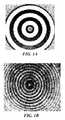
- FIG. 1Aillustrates an example of a Placido disk of concentrically arranged circles of light.
- FIG. 1Billustrates an example of a projected Placido disk on a surface of fixed curvature.
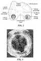
- FIG. 2shows a reflection resulting from a projection of concentric circles of light (Placido Disk) on each femoral condyle, demonstrating the effect of variation in surface contour on the reflected circles.
- FIG. 3illustrates an example of a 2D topographical map of an irregularly curved surface.
- FIG. 4illustrates an example of a 3D topographical map of an irregularly curved surface.
- FIG. 5illustrates surface registration of MRI surface and a digitized surface using a laser scanner.
- the illustration to the leftshows the surface before registration and the illustration to the right shows the surface after registration.
- FIG. 6is a reproduction of a three-dimensional thickness map of the articular cartilage of the distal femur.
- Three-dimensional thickness mapscan be generated, for example, from ultrasound, CT or MRI data. Dark holes within the substances of the cartilage indicate areas of full thickness cartilage loss.
- FIG. 7illustrates the cartilage surface of a medial femoral condyle from a sagittal scan (blue) and a coronal scan (red).
- FIG. 8Aillustrates an axial view of a meniscus
- FIG. 8Billustrates a sagittal view of the meniscus
- FIG. 8Cillustrates a coronal view of the meniscus.
- FIG. 9Aillustrates a sagittal view of the tibia
- FIG. 9Billustrates a coronal view of the tibia.
- FIG. 10Aillustrates a sagittal view of the femur
- FIG. 10Billustrates a coronal view of the femur.
- FIGS. 11A-Cillustrate a chart showing the tibial cartilage surface and superior meniscal surface combined after extraction from a coronal FSE, and a meniscal surface scaled to account for compression under loading conditions. From the information is derived the cross-section of the implant, FIG. 11C .
- FIG. 12illustrates a point cloud of an implant surface (yellow) that approximates smooth surface patch (brown).
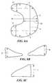
- FIGS. 13A and Bare views of an implant suitable for use on a condyle of the femur shown from the inferior and superior surface viewpoints, respectively.
- FIG. 14is a view of an implant suitable for a portion of the tibial plateau in the knee.
- FIGS. 15A-Dare views of an implant suitable for the hip.
- the practice of the present inventionemploys, unless otherwise indicated, conventional methods of x-ray imaging and processing, x-ray tomosynthesis, ultrasound including A-scan, B-scan and C-scan, computed tomography (CT scan), magnetic resonance imaging (MRI), optical coherence tomography, single photon emission tomography (SPECT) and positron emission tomography (PET) within the skill of the art.
- CT scancomputed tomography
- MRImagnetic resonance imaging
- SPECTsingle photon emission tomography
- PETpositron emission tomography
- the present inventionsolves the need for methods to recreate natural or near natural relationships between two articular surfaces by providing methods for determining meniscal size and shape.
- Meniscal size and shapecan be useful in designing therapies for the treatment of joint diseases including, for example, meniscal repair, meniscal regeneration, and articular repair therapies.
- the methods and compositions described hereincan be used to treat defects resulting from disease of the cartilage (e.g., osteoarthritis), bone damage, cartilage damage, trauma, and/or degeneration due to overuse or age.
- the inventionallows, among other things, a health practitioner to evaluate and treat such defects.
- size, curvature and/or thickness measurementscan be obtained using any suitable technique.
- one dimensional, two dimensional, and/or three dimensional measurementscan be obtained using suitable mechanical means, laser devices, electromagnetic or optical tracking systems, molds, materials applied to the articular surface that harden and “memorize the surface contour,” and/or one or more imaging techniques known in the art. Measurements can be obtained non-invasively and/or intraoperatively (e.g., using a probe or other surgical device).
- the thickness of the repair devicecan vary at any given point depending upon the depth of the damage to the cartilage and/or bone to be corrected at any particular location on an articular surface.
- imaging techniques suitable for measuring thickness and/or curvature (e.g., of cartilage and/or bone) or size of areas of diseased cartilage or cartilage lossinclude the use of x-rays, magnetic resonance imaging (MRI), computed tomography scanning (CT, also known as computerized axial tomography or CAT), optical coherence tomography, SPECT, PET, ultrasound imaging techniques, and optical imaging techniques.
- MRImagnetic resonance imaging
- CTcomputed tomography scanning
- SPECTalso known as computerized axial tomography or CAT
- SPECTcomputerized axial tomography
- PETcomputerized axial tomography
- ultrasound imaging techniquesand optical imaging techniques.
- Contrast or other enhancing agentscan be employed using any route of administration, e.g. intravenous, intra-articular, etc.
- CT or MRIis used to assess tissue, bone, cartilage and any defects therein, for example cartilage lesions or areas of diseased cartilage, to obtain information on subchondral bone or cartilage degeneration and to provide morphologic or biochemical or biomechanical information about the area of damage.
- changessuch as fissuring, partial or full thickness cartilage loss, and signal changes within residual cartilage can be detected using one or more of these methods.
- MRIincluding conventional T1 and T2-weighted spin-echo imaging, gradient recalled echo (GRE) imaging, magnetization transfer contrast (MTC) imaging, fast spin-echo (FSE) imaging, contrast enhanced imaging, rapid acquisition relaxation enhancement (RARE) imaging, gradient echo acquisition in the steady state (GRASS), and driven equilibrium Fourier transform (DEFT) imaging, to obtain information on cartilage, see Alexander, et al., WO 02/22014.
- GREgradient recalled echo
- MTCmagnetization transfer contrast
- FSEfast spin-echo
- RARErapid acquisition relaxation enhancement
- GASSgradient echo acquisition in the steady state
- DEFTdriven equilibrium Fourier transform
- Two-dimensional and three-dimensional images, or maps, of the cartilage alone or in combination with a movement pattern of the joint, e.g. flexion-extension, translation and/or rotation,can be obtained.
- Three-dimensional imagescan include information on movement patterns, contact points, contact zone of two or more opposing articular surfaces, and movement of the contact point or zone during joint motion.
- Two and three-dimensional imagescan include information on biochemical composition of the articular cartilage.
- imaging techniquescan be compared over time, for example to provide up-to-date information on the shape and type of repair material needed.
- imaging devices described hereincan also be used intra-operatively (see, also below), for example using a hand-held ultrasound and/or optical probe to image the articular surface intra-operatively.
- measurements of the size of an area of diseased cartilage or an area of cartilage loss, measurements of cartilage thickness and/or curvature of cartilage or bonecan be obtained intraoperatively during arthroscopy or open arthrotomy. Intraoperative measurements may or may not involve actual contact with one or more areas of the articular surfaces.
- Devices suitable for obtaining intraoperative measurements of cartilage or bone or other articular structures, and to generate a topographical map of the surfaceinclude but are not limited to, Placido disks and laser interferometers, and/or deformable materials or devices.
- Placido disks and laser interferometersSee, for example, U.S. Pat. No. 6,382,028 to Wooh et al., issued May 7, 2002; U.S. Pat. No. 6,057,927 to Levesque et al., issued May 2, 2000; U.S. Pat. No. 5,523,843 to Yamane et al. issued Jun. 4, 1996; U.S. Pat. No. 5,847,804 to Sarver et al. issued Dec. 8, 1998; and U.S. Pat. No. 5,684,562 to Fujieda, issued Nov. 4, 1997).
- FIG. 1Aillustrates a Placido disk of concentrically arranged circles of light.
- the concentric arrays of the Placido diskproject well-defined circles of light of varying radii, generated either with laser or white light transported via optical fiber.
- the Placido diskcan be attached to the end of an endoscopic device (or to any probe, for example a hand-held probe) so that the circles of light are projected onto the cartilage surface.
- FIG. 1Billustrates an example of a Placido disk projected onto the surface of a fixed curvature.
- One or more imaging camerascan be used (e.g., attached to the device) to capture the reflection of the circles.
- Mathematical analysisis used to determine the surface curvature.
- the curvaturecan then, for example, be visualized on a monitor as a color-coded, topographical map of the cartilage surface. Additionally, a mathematical model of the topographical map can be used to determine the ideal surface topography to replace any cartilage defects in the area analyzed.
- FIG. 2shows a reflection resulting from the projection of concentric circles of light (Placido disk) on each femoral condyle, demonstrating the effect of variation in surface contour on reflected circles.
- a laser interferometercan also be attached to the end of an endoscopic device.
- a small sensorcan be attached to the device in order to determine the cartilage surface or bone curvature using phase shift interferometry, producing a fringe pattern analysis phase map (wave front) visualization of the cartilage surface.
- the curvaturecan then be visualized on a monitor as a color coded, topographical map of the cartilage surface.
- a mathematical model of the topographical mapcan be used to determine the ideal surface topography to replace any cartilage or bone defects in the area analyzed. This computed, ideal surface, or surfaces, can then be visualized on the monitor, and can be used to select the curvature, or curvatures, of the replacement cartilage.
- Mechanical devicescan also be used for intraoperative measurements, for example, deformable materials such as gels, molds, any hardening materials (e.g., materials that remain deformable until they are heated, cooled, or otherwise manipulated).
- deformable materialssuch as gels, molds, any hardening materials (e.g., materials that remain deformable until they are heated, cooled, or otherwise manipulated).
- a deformable gelcan be applied to a femoral condyle. The side of the gel pointing towards the condyle can yield a negative impression of the surface contour of the condyle. The negative impression can then be used to determine the size of a defect, the depth of a defect and the curvature of the articular surface in and adjacent to a defect.
- This informationcan be used to select a therapy, e.g. an articular surface repair system.
- a hardening materialcan be applied to an articular surface, e.g. a femoral condyle or a tibial plateau. The hardening material can remain on the articular surface until hardening has occurred. The hardening material can then be removed from the articular surface. The side of the hardening material pointing towards the articular surface can yield a negative impression of the articular surface. The negative impression can then be used to determine the size of a defect, the depth of a defect and the curvature of the articular surface in and adjacent to a defect.
- This informationcan then be used to select a therapy, e.g. an articular surface repair system.
- the hardening systemcan remain in place and form the actual articular surface repair system.
- the deformable materialcomprises a plurality of individually moveable mechanical elements.
- each elementWhen pressed against the surface of interest, each element can be pushed in the opposing direction and the extent to which it is pushed (deformed) can correspond to the curvature of the surface of interest.
- the devicecan include a brake mechanism so that the elements are maintained in the position that conforms to the surface of the cartilage and/or bone. The device can then be removed from the patient and analyzed for curvature.
- each individual moveable elementcan include markers indicating the amount and/or degree it is deformed at a given spot.
- a cameracan be used to intra-operatively image the device and the image can be saved and analyzed for curvature information. Suitable markers include, but are not limited to, actual linear measurements (metric or empirical), different colors corresponding to different amounts of deformation and/or different shades or hues of the same color(s). Displacement of the moveable elements can also be measured using electronic means.
- Other devices to measure cartilage and subchondral bone intraoperativelyinclude, for example, ultrasound probes.
- An ultrasound probepreferably handheld, can be applied to the cartilage and the curvature of the cartilage and/or the subchondral bone can be measured. Moreover, the size of a cartilage defect can be assessed and the thickness of the articular cartilage can be determined.
- Such ultrasound measurementscan be obtained in A-mode, B-mode, or C-mode. If A-mode measurements are obtained, an operator can typically repeat the measurements with several different probe orientations, e.g. mediolateral and anteroposterior, in order to derive a three-dimensional assessment of size, curvature and thickness.
- the probesare preferably handheld.
- the probes or at least a portion of the probe, typically the portion that is in contact with the tissuecan be sterile. Sterility can be achieved with use of sterile covers, for example similar to those disclosed in WO 99/08598A1 to Lang, published Feb. 25, 1999.
- Analysis on the curvature of the articular cartilage or subchondral bone using imaging tests and/or intraoperative measurementscan be used to determine the size of an area of diseased cartilage or cartilage loss.
- the curvaturecan change abruptly in areas of cartilage loss.
- Such abrupt or sudden changes in curvaturecan be used to detect the boundaries of diseased cartilage or cartilage defects.
- a semi-automated segmentation approachhas been implemented based on the live wire algorithm, which provides a high degree of flexibility and therefore holds the potential to improve segmentation of osteoarthritic cartilage considerably.
- Imagesare optionally pre-processed using a non-linear diffusion filter.
- the live wire algorithmassigns a list of features to each oriented edge between two pixels (boundary element-bel) in an image.
- the feature valuesare converted into cost values.
- the costs for each featureare added up by means of a predetermined weighting scheme, resulting in a single joint cost value between 0 and 1 for each bel b that expresses the likelihood of b being part of the cartilage boundary.
- the operatorchooses a starting pixel P.
- the systemcalculates the least cost bel path from each image pixel to P with a dynamic programming scheme.
- the systemdisplays the calculated path from the current mouse position to P in real time. This current path can be frozen as part of the cartilage contour by the operator. This way, the operator has to assemble the desired contour in each slice from a number of pieces (“strokes”).
- the features of a bel b used with this segmentation techniqueare the gray values left and right of b and the magnitude of the gray level gradient across b.
- segmentation processes describedcan be automated as desired.
- segmentation techniquesincluding but not limited to thresholding, grey level gradient techniques, snakes, model based segmentation, watershed, clustering, statistical segmentation, filtering including linear diffusion filtering can be employed.
- the cartilage surface extracted from MRI scanscan be compared with results obtained from segmentation of the joint surface data which is acquired, for example, using a laser scanner after specimen dissection.
- the resulting two surfaces from MRI and laser scancan be registered using the iterative closest point method, and the distance between each point on the MRI surface to the registered laser scan surface can be used to determine the accuracy of the MRI segmentation results.
- FIG. 5shows the MRI and digitized surfaces before and after registration. The distance measurements for the two specimens are shown in TABLE 1.
- the dataillustrate that the average error between the segmented MRI surface and the laser scan surface is within the range of the resolution of the MRI scan.
- the segmentation approachyields an accuracy within the given MRI scan parameters.
- a suitable approach for calculating the cartilage thicknessis based on a 3D Euclidean distance transform (EDT).
- EDTEuclidean distance transform
- An algorithm by Saito and Toriwakican be used to achieve computationally very fast (less than 10 sec for a 256 ⁇ 256 ⁇ 60 data set on a SGI O2) data processing.
- the algorithmfunctions by decomposing the calculation into a series of 3 one-dimensional transformations and uses the square of the actual distances. This process accelerates the analysis by avoiding the determination of square roots.
- voxels on the inner cartilage surface (ICS)are given a value of 0, whereas all other voxels, including the ones on the outer cartilage surface (OCS) are set to 1.
- each pointis assigned the square of the distance to the closest feature point in the same row in i-direction.
- h ijkmin y ⁇ g iyk ( ⁇ ( j ⁇ y )) 2 ;1 ⁇ y ⁇ M ⁇ [Eq. 2]
- the algorithmsearches each column in j-direction.
- the sum of the square distance between a point (i,j,k) and a point (i,y,k) in the same column, ( ⁇ (j ⁇ y)) 2 , and the square distance between (i,y,k) and a particular feature point, g iykequals the square distance between the point (i,j,k) and that feature point.
- the minimum of these sumsis the square distance between (i,j,k) and the closest feature point in the two-dimensional i-j-plane.
- Equation 3The third dimension is added by equation 3, which is the same transformation as described in equation 2 for the k-direction.
- s ijkmin z ⁇ h ijz +( ⁇ ( k ⁇ z )) 2 ;1 ⁇ z ⁇ N ⁇ [Eq. 3]
- the thickness of the cartilage for a given point (a,b,c) on the OCSequals the square root of s abc . This results in a truly three-dimensional distance value determined normal to the ICS.
- the x, y, and z position of each pixel located along the bone-cartilage interfaceis registered on a 3D map and thickness values are translated into color values. In this fashion, the anatomic location of each pixel at the bone-cartilage interface can be displayed simultaneously with the thickness of the cartilage for that given location ( FIG. 6 ).
- curvatureAnother relevant parameter for the analysis of articular cartilage surfaces is curvature.
- a set of curvature mapscan be derived from the cartilage surface data that is extracted from the MRI.
- a local bi-cubic surface patchis fitted to the cartilage surface based on a sub-sampling scheme in which every other surface point is used to generate a mesh of 5 ⁇ 5 point elements.
- every other surface pointis used to generate a mesh of 5 ⁇ 5 point elements.
- FIG. 6shows an example of the maximum principal curvature maps (value and direction), estimated using the bi-cubic surface patch fitting approach.
- one or more first scans S1are taken in a first plane. Each of the first scans are parallel to each other. Thereafter, one or more second scans S2 are taken with an imaging plane oriented to the first scan S1 so that the planes intersect.
- scans S1can be in a first plane while scans S2 are in a plane perpendicular to the first plane. Additional scans in other planes or directions, e.g., S3, S4. Sn, can also be obtained in addition to the perpendicular scans or instead of the perpendicular scans.
- S2, and any other scanscan have the same in-plane resolution as S1. Any or all of the scans can also contain a sufficient number of slices to cover the entire field of view of S1. In this scenario, two data volumes with information from the same 3D space or overlapping 3D spaces can be generated.
- Datacan be merged from these two scans to extract the objects of interest in each scan independently. Further, a subsequent analysis can combine these two segmented data sets in one coordinate system, as is shown in FIG. 6 . This technique is helpful in outlining the boundaries of objects that are oriented parallel to the imaging plane of S1, but therefore will be perpendicular to the imaging plane of S2.
- This third data volumeis typically isotropic or near-isotropic with a resolution corresponding to the in-plane resolution of S1 and S2, thus reducing partial volume effects between slices ( FIG. 7 ).
- S1 and S2can first be registered into the same coordinate system. If both scans are acquired during the same session (without moving the patient between scans), the image header information is used to obtain the transformation matrix. Otherwise, a mutual information-based rigid registration is applied.
- the gray value for each voxel V of the third data volumeis calculated as follows:
- datacan be obtained with isotropic or near isotropic resolution.
- Thisis possible, for example, with spiral CT acquisition technique or novel MRI pulse sequence such as 3D acquisition techniques.
- 3D acquisition techniquesinclude 3D Driven Equilibrium Transfer (DEFT), 3D Fast Spin-Echo (FSE), 3D SSFP (Steady State Free Precession), 3D Gradient Echo (GRE), 3D Spoiled Gradient Echo (SPGR), and 3D Flexible Equilibrium MR (FEMR) techniques.
- Imagescan be obtained using fat saturation or using water selective excitation.
- an isotropic resolution of 0.5 ⁇ 0.5 ⁇ 0.5 mm or lessis desirable, although in select circumstances 1.0 ⁇ 1.0 ⁇ 1.0 and even larger can yield adequate results.
- near isotropic resolutionthe variation in voxel dimensions in one or more planes does not usually exceed 50%.
- the dimensions and shape of a personalized interpositional arthroplasty systemcan be determined by measuring a patient's meniscal shape and size and by evaluating the 3D geometry of the articular cartilage. Many osteoarthritis patients, however, have torn menisci, often times with only small or no meniscal remnants. In these patients, the shape of a personalized interpositional arthroplasty system can be determined by acquiring measurements of surrounding articular surfaces and structures.
- a few measurementscan be made on the femoral and tibial bone in MR images of the diseased knee.
- the shape of the superior surface of the implantshould resemble that of the superior surface of the respective meniscus. Measurements of the bones can help determine how well meniscal dimensions can be predicted.
- FIG. 8Aillustrates an axial view of a meniscus 100 .
- the meniscushas a maximum anterior-posterior distance 1, and a maximum medial lateral distance 2. In the knee, the meniscus compensates for an anterior horn and a posterior which each have a maximum length 3, 5 and width 9, 11.
- the body itselfhas a maximum length 4 and width 10 which are a function of the patient's anatomy.
- FIG. 8Billustrates a sagittal view of the meniscus in FIG. 8A .
- the meniscus 100has a maximum height 6, 8 which correlates to the maximum height of the anterior horn and the posterior horn.
- FIG. 8Cillustrates a coronal view of the meniscus 100 . From the coronal view it is apparent that the body has a maximum and minimum height.
- FIG. 9Aa sagittal view of a tibia 110 is shown.
- the tibiahas a maximum anterior-posterior distance 12.
- FIG. 9Billustrates the coronal view of the tibia 110 shown in FIG. 9A . From the sagittal view it is apparent that the tibia has a maximum medial-lateral distance 13, a maximum distance from the tibial spine to the edge 14, and a width 15.
- the tibiamates with the femur 120 , which is shown in a sagittal view in FIG. 10A .
- the femurhas a maximum anterior-posterior distance 16 and a maximum superior-interior distance 17. From the coronal view shown in FIG. 10B the maximum medial-lateral distance 18, the distance from the trochlea to the edge 19, and the width of the intercondylar notch 20 is apparent.
- a Pearson's correlation coefficient rcan be obtained for a variety of measurements to assess how well one variable is expressed by another variable. Suitable measurements include, for example, the following measurements:
- the Pearsons' coefficientdetermines the relationship between two sizes that are measured. The higher the correlation, the better the relationship between two measurements. From the data in TABLE 2, it becomes evident that, in the knee, the AP length of both medial and lateral menisci can be predicted well by measuring the length of the respective femoral condyle. For the medial meniscus, the length of the medial tibial plateau can also be used. The ML width of the medial femoral condyle is a good predictor for the width of the medial meniscus. The height of the medial and lateral tibial spines correlates well with the height of the respective menisci.
- meniscal dimensionscan be predicted in a reliable fashion by measuring bony landmarks in MR images.
- the Pearson's coefficientis high (e.g., close to 1)
- the two measurementscan, in effect, be used interchangeably to represent the measurement desired.
- the Pearson's coefficientis low (e.g., 0.34)
- a correction factormay be applied to the measurement.
- the measurement as correctedmay then equal or approximate the corresponding measurement.
- use of a correction factormay not be feasible or desired.
- other approachessuch as logistic regression and multivariate analysis, can be used as an alternative without departing from the scope of the invention.
- a library of measurementscan be created, for example for generating one or more correlation factors that can be used for a particular joint. For example, a single correlation factor can be generated using a plurality of measurements on different subjects.
- a plurality of correlation factorscan be generated based on, for example, joint assessed, size, weight, body mass index, age, sex of a patient, ethnic background.
- a patient seeking treatmentcan be assessed. Measurements can be taken of, for example, the medial femoral condyle.
- the correlation factor for the medial femoral condyle in the patientcan then be compared to a correlation factor calculated based on samples wherein the sample patients had the same, or were within a defined range for factors, including for example: size, weight, age and sex.
- Digitized surface data from menisci of cadaveric specimens for generation of a generic meniscal modelcan be acquired using a Titanium FaroArm® coordinate measurement machine (CMM) (FARO Technologies Inc., Lake Mary, Fla.).
- CCMTitanium FaroArm® coordinate measurement machine
- the design workflow for each implantcan consist of a combination of one or more of the following steps:
- the meniscusis, to a great extent, depleted, and therefore cannot serve directly as a template from which the superior implant surface can be derived.
- dimensions of the remaining joint bonecan be used to adjust the size of a generic meniscal model, which can then serve as a template for the implant.
- the superior surface of an implantcan be modeled based on the superior meniscal surface and the joint cartilage surface in those areas that are not covered by the meniscus. Therefore, after the slice-by-slice segmentation of the superior meniscal surface from the SE or FSE or other MRI images and the tibial cartilage surface from the 3D SPGR or FSE or other MRI images, both data sets will be combined ( FIGS. 11A-C ). To determine the composite surface for the prosthesis, the intersection between the two surfaces is located. In the event that the two surfaces do not intersect in a particular slice, the intersection between the tangential line through the
- the meniscusis, to a great extent, depleted, and therefore cannot serve directly as a template from which the superior implant surface can be derived.
- dimensions of the remaining joint bonecan be used to adjust the size of a generic meniscal model, which can then serve as a template for the implant.
- the superior surface of an implantcan be modeled based on the superior meniscal surface and the joint cartilage surface in those areas that are not covered by the meniscus. Therefore, after the slice-by-slice segmentation of the superior meniscal surface from the SE or FSE or other MRI images and the tibial cartilage surface from the 3D SPGR or FSE or other MRI images, both data sets will be combined ( FIGS. 11A-C ). To determine the composite surface for the prosthesis, the intersection between the two surfaces is located. In the event that the two surfaces do not intersect in a particular slice, the intersection between the tangential line through the central end of the meniscal surface with the tibial surface will be calculated ( FIG. 11A ).
- each point on the meniscal surfaceis connected to the closest point on the cartilage surface.
- the new point for the adjusted meniscal surfaceis chosen at 60% of the distance from the tibial cartilage surface.
- Suitable adjustment ratioswill vary depending on patient physiology and desired degree of correction and include, for example, ratios that range from 0.2 to 1.5.
- the amount of height adjustment of the implant relative to the natural meniscuswill vary depending upon the material that the implant is manufactured from. For example, where the implant is manufactured from a material having a high degree of elasticity, it may be desirable to use an adjustment greater than 1. Where the material has a low degree of elasticity, the adjustment is likely to approach 50%. The appropriate adjustment will also depend upon the joint for which the implant is manufactured.
- an implant manufactured for the knee using a material with a low degree of elasticitycan have an adjustment of between 50-70%, while an implant manufactured for the shoulder also using a material with a low degree of elasticity may have a desired adjustment of 60-80%.
- the correction factor for an implantwill vary depending upon the target joint and the properties of the material from which the implant is manufactured.
- the adjustment ratiocan also vary depending on the location within a joint with a plurality of ratios possible for any given design. For example, in a knee joint, an adjustment ratio close to 0.8 can be used anteriorly, while an adjustment ratio close to 0.5 can be used posteriorly. Additionally, more adjustment ratios can be selected such that the adjustment ratio gradually changes, for example, anteriorly, depending on the anticipated biomechanics of the joint. Changes can also be made to the adjustment ratio as a result of patient specific parameters such as age, sex, weight, ethnicity, and activity level. The adjustment ratio can be selected in order to achieve an optimal biomechanical or functional result. In vitro cadaveric testing, constraint testing, testing of contact surface, fatigue testing and robotic testing can, for example, be used for determining the optimal adjustment ratio(s) for an implant.
- the compressed meniscal surfacecan be combined with the portion of the tibial cartilage surface that is not covered by the meniscus.
- the shape of, for example, an inferior surface of the implantcan be derived from the entire cartilage surface ( FIG. 11C ) or the subchondral bone surface. The latter can be used, for example, if there is significant eburnation of the joint and most of the cartilage has been lost.
- meniscal surfacecannot be used as a template for an implant surface as described above.
- a generic meniscal modelcan be used to design the desired implant surface.
- the generic meniscal modelcan be generated from data that is, for example, collected from cadaveric femoral specimens using a Titanium FaroArm as described above. Alternatively, a laser scanning device or an optical device can be used. In this instance, meniscal surface data can be digitized, for example, from ten frozen cadaveric tibial specimens. All surface data sets obtained can then be matched for size differences using, for example, an affine surface registration scheme. The matched surface points after registration can then be merged into a single point cloud.
- a generic meniscal surface, S gcan be fitted through a point cloud using a least-squares optimization, resulting in a “mean” surface of the ten specimens.
- the antero-posterior length Lwill be calculated from the length of the femoral condyle.
- medio-lateral meniscal width Wwe can use the position of the medial margin of the tibia for the medial meniscus and the lateral tibial margin for the lateral meniscus.
- the height Hcan be derived from the highest point of the tibial spine.
- L g , W g , and H gare the respective dimensions of S g .
- the transformed points P′can form the meniscal surface S that will be used as a template for designing the superior implant surface as described in the previous section.
- the first and second implant surfaces derived from an MR image, as described above,consist of point clouds.
- the point cloudscan be converted into a data format that then can be manipulated in, for example, a CAD system.
- the Surface Patch function in the surface modeling program Rhinoceroscan be used to approximate a smooth surface patch to the point cloud data ( FIG. 12 ).
- This surfacecan then be exported in the IGES format to be read by the CAD software.
- Other software programscan be used without departing from the scope of the invention. For example, Pro/Engineer, Solid Edge, Alibre and IronCAD are also suitable programs.
- the superior and inferior surfacescan be combined into one design model. Both surfaces can be clipped using the outer meniscal edge as a margin ( FIG. 11 ).
- FIGS. 13A and Bare views of a joint implant suitable for use on a condyle of the femur. These views are shown from the inferior and superior surface viewpoints. The surfaces, edges and height of the implant can be adjusted to account for the measurements taken to achieve an implant with an optimal patient fit.
- FIG. 14is a view of an implant suitable for placement in a joint knee and placed on a portion of the tibial plateau.
- FIGS. 15A-Dare views of an implant suitable for the hip. These implants can also be designed so that the surfaces, edges and height of the implants can be adjusted to account for the measurements taken as well as the patient specific criteria, as appropriate or desirable.
- Suitable spiral CTalso with intravenous or intra-articular contrast enhancement, or MRI images can be acquired, from which medial and lateral menisci can then be extracted using live wire segmentation, or other suitable mechanisms.
- the generic models for the medial and lateral meniscuscan be fitted as described above. For each subject, the medial and lateral meniscus that was segmented from the MRI can be compared to the fitted models as follows:
- nis the normal to the plane and (•,•) denotes the dot product.
- the total distance measure Ddepends on the relative position of the segmented MRI data and the fitted model in the coordinate system. This relative position can be optimized to minimize D by adjusting the rigid body transformation T that positions the model in an iterative registration process based on the iterative closest point algorithm, using D(7) as a cost function.
Landscapes
- Health & Medical Sciences (AREA)
- Engineering & Computer Science (AREA)
- Life Sciences & Earth Sciences (AREA)
- General Health & Medical Sciences (AREA)
- Veterinary Medicine (AREA)
- Animal Behavior & Ethology (AREA)
- Public Health (AREA)
- Biomedical Technology (AREA)
- Heart & Thoracic Surgery (AREA)
- Vascular Medicine (AREA)
- Cardiology (AREA)
- Orthopedic Medicine & Surgery (AREA)
- Oral & Maxillofacial Surgery (AREA)
- Transplantation (AREA)
- Surgery (AREA)
- Physics & Mathematics (AREA)
- Nuclear Medicine, Radiotherapy & Molecular Imaging (AREA)
- Medical Informatics (AREA)
- Manufacturing & Machinery (AREA)
- Geometry (AREA)
- Rheumatology (AREA)
- Robotics (AREA)
- Molecular Biology (AREA)
- Radiology & Medical Imaging (AREA)
- Quality & Reliability (AREA)
- Computer Vision & Pattern Recognition (AREA)
- General Physics & Mathematics (AREA)
- Theoretical Computer Science (AREA)
- Prostheses (AREA)
- Image Processing (AREA)
Abstract
Description
| TABLE 1 |
| DISTANCE CALCULATIONS BETWEEN SEGMENTED |
| MRI AND LASER DIGITIZED SURFACES (IN MM) |
| Minimum | Maximum | Mean | Standard | ||
| Specimen | Distance | Distance | Distance | Deviation σ | |
| 1 | 3.60447e−05 | 2.10894 | 0.325663 | 0.312803 |
| 2 | 2.79092e−06 | 1.616828 | 0.262131 | 0.234424 |
gijk=minx{(α(i−x))2;fxjk=0;1≦x≦L} [Eq. 1]
hijk=miny{giyk(β(j−y))2;1≦y≦M} [Eq. 2]
sijk=minz{hijz+(γ(k−z))2;1≦z≦N} [Eq. 3]
Ki=arccos(n0·ni)/dsi=dθ/dsi,
- (1) determine the position in 3D space for V;
- (2) determine the gray values in S1 and S2 at this position;
- (3) interpolate the two gray values into a single gray value G; and
- (4) assign G to V.
- antero-posterior (AP) length of medial (lateral) meniscus with AP length of medial (lateral) femoral condyle;
- AP length of medial (lateral) meniscus with AP length of medial (lateral) tibial plateau;
- medio-lateral (ML) width of medial (lateral) meniscus with ML width of medial (lateral) femoral condyle;
- ML width of medial (lateral) meniscus with ML width of medial (lateral) tibial plateau;
- Y coordinate of highest point of medial (lateral) meniscus with y coordinate of highest point of medial (lateral) tibial spine;
- X coordinate of medial (lateral) margin of medial (lateral) meniscus with x coordinate of medial (lateral) margin of medial (lateral) femoral condyle; and
- X coordinate of medial (lateral) margin of medial (lateral) meniscus with x coordinate of medial (lateral) margin of medial (lateral) tibial plateau.
| TABLE 2 |
| CORRELATION BETWEEN MENISCAL DIMENSIONS |
| AND DIMENSIONS OF FEMORAL AND TIBIAL BONE |
| Measurement | Imaging Plane | N | Pearson's r |
| AP Length: medial meniscus-medial | Sagittal | 23 | 0.74 |
| femoral condyle | |||
| AP Length: lateral meniscus-lateral | Sagittal | 24 | 0.73 |
| femoral condyle | |||
| AP Length: medial meniscus-medial | Sagittal | 23 | 0.79 |
| tibial plateau | |||
| AP Length: lateral meniscus-lateral | Sagittal | 24 | 0.27 |
| tibial plateau | |||
| ML Width: menisci-femur | Coronal | 12 | 0.91 |
| ML Width: menisci-tibia | Coronal | 12 | 0.92 |
| ML Width: menisci-medial femoral | Coronal | 12 | 0.81 |
| condyle | |||
| ML Width: menisci-lateral femoral | Coronal | 12 | 0.65 |
| condyle | |||
| ML Width: menisci-medial tibial | Coronal | 12 | 0.86 |
| plateau | |||
| ML Width: menisci-lateral tibial | Coronal | 12 | 0.48 |
| plateau | |||
| ML Width: medial meniscus-medial | Coronal | 12 | 0.95 |
| femoral condyle | |||
| ML Width: lateral meniscus-lateral | Coronal | 12 | 0.45 |
| femoral condyle | |||
| ML Width: medial meniscus-medial | Coronal | 12 | 0.69 |
| tibial plateau | |||
| ML Width: lateral meniscus-lateral | Coronal | 12 | 0.34 |
| tibial plateau | |||
| ML Length: medial meniscus-lateral | Coronal | 12 | 0.12 |
| meniscus | |||
| Meniscal Height: medial meniscus- | Coronal | 12 | 0.01 |
| lateral meniscus | |||
| Meniscal Height: Medial meniscal | Coronal | 12 | 0.22 |
| height-medial femoral height | |||
| Meniscal Height: Lateral meniscal | Coronal | 12 | 0.22 |
| height-lateral femoral height | |||
| Meniscal Height: Medial meniscal | Coronal | 12 | 0.55 |
| height-medial tibial height | |||
| Meniscal Height: Lateral meniscal | Coronal | 12 | 0.17 |
| height-lateral tibial height | |||
| Highest Point (y coordinate): medial | Coronal | 12 | 0.99 |
| meniscus-medial tibial spine | |||
| Highest Point (y coordinate): lateral | Coronal | 12 | 0.90 |
| meniscus-lateral tibial spine | |||
| Medial margin (x-coordinate): medial | Coronal | 12 | 1.00 |
| meniscus-femoral condyle | |||
| Lateral margin (x-coordinate): lateral | Coronal | 12 | 1.00 |
| meniscus-lateral femoral condyle | |||
| Medial Margin (x-coordinate): medial | Coronal | 12 | 1.00 |
| meniscus-medial tibial plateau | |||
| Lateral Margin (x-coordinate): lateral | Coronal | 12 | 1.00 |
| meniscus-lateral tibial plateau | |||
- a. Fusion of the sagittal and coronal 3D SPGR or 2D or 3D FSE data or other sequences for a joint;
- b. Segmentation of point data from the cartilage surface of a joint;
- c. Fusion of the sagittal and coronal 2D or 3D FSE or 2D SE data or other sequences of a joint;
- d. Segmentation point data of the superior meniscal surface;
- e. Combination of cartilage surface data and meniscal surface data to serve as model for a surface of an implant;
- f. Compression of a meniscal surface by factor ranging from 0.2 to 0.99;
- g. Conversion of point cloud data for a superior and an inferior implant surface into parametric surface data; and
- h. Cutting of parametric surface data sets to determine exact shape of implant.
P′=(x′,y′,z′)=((L/Lg)·x,(W/Wg)·y,(H/Hg)·z) [Eq. 4]
- 1. For each point P=(x,y,z) in the segmented data set choose the closest point P1=(x1,y1,z1) from the fitted model with z1≧z and the two closest points P2=(x2,y2,z2) and P3=(x3,y3,z3) with Z2,Z3≦Z.
- 2. The point P is projected orthogonally onto the plane defined by P1, P2and P3. The projected point P′ is given by:
P□=P−((P−P1,n)/(n,n))
- 3. Calculate the distance d1between P and the plane, given by
d1=∥P′−P∥. - 4. Repeat 1-3 with P1=(x1,x1,z1) such that z1≦z and P2=(x2,y2,z2) and P3=(x3,y3,z3) such that z2,z3≧z, resulting in d2.
- 5. Calculate the mean distance for P: d(P)=(d1+d2)/2.
- 6. Calculate the total distance measure D over all points in the segmented data set:
D=Σpd(P).
- 3. Calculate the distance d1between P and the plane, given by
Claims (19)
Priority Applications (1)
| Application Number | Priority Date | Filing Date | Title |
|---|---|---|---|
| US14/157,707US8965088B2 (en) | 2002-11-07 | 2014-01-17 | Methods for determining meniscal size and shape and for devising treatment |
Applications Claiming Priority (5)
| Application Number | Priority Date | Filing Date | Title |
|---|---|---|---|
| US42496402P | 2002-11-07 | 2002-11-07 | |
| US10/704,325US7796791B2 (en) | 2002-11-07 | 2003-11-07 | Methods for determining meniscal size and shape and for devising treatment |
| US12/853,599US8077950B2 (en) | 2002-11-07 | 2010-08-10 | Methods for determining meniscal size and shape and for devising treatment |
| US13/312,339US8634617B2 (en) | 2002-11-07 | 2011-12-06 | Methods for determining meniscal size and shape and for devising treatment |
| US14/157,707US8965088B2 (en) | 2002-11-07 | 2014-01-17 | Methods for determining meniscal size and shape and for devising treatment |
Related Parent Applications (1)
| Application Number | Title | Priority Date | Filing Date |
|---|---|---|---|
| US13/312,339ContinuationUS8634617B2 (en) | 2002-11-07 | 2011-12-06 | Methods for determining meniscal size and shape and for devising treatment |
Publications (2)
| Publication Number | Publication Date |
|---|---|
| US20140153798A1 US20140153798A1 (en) | 2014-06-05 |
| US8965088B2true US8965088B2 (en) | 2015-02-24 |
Family
ID=32312904
Family Applications (6)
| Application Number | Title | Priority Date | Filing Date |
|---|---|---|---|
| US10/704,325Active2028-11-10US7796791B2 (en) | 2002-11-07 | 2003-11-07 | Methods for determining meniscal size and shape and for devising treatment |
| US10/704,208Active2026-05-27US8932363B2 (en) | 2002-11-07 | 2003-11-07 | Methods for determining meniscal size and shape and for devising treatment |
| US12/853,599Expired - LifetimeUS8077950B2 (en) | 2002-11-07 | 2010-08-10 | Methods for determining meniscal size and shape and for devising treatment |
| US13/312,339Expired - LifetimeUS8634617B2 (en) | 2002-11-07 | 2011-12-06 | Methods for determining meniscal size and shape and for devising treatment |
| US14/157,707Expired - LifetimeUS8965088B2 (en) | 2002-11-07 | 2014-01-17 | Methods for determining meniscal size and shape and for devising treatment |
| US14/594,492AbandonedUS20150216615A1 (en) | 2002-11-07 | 2015-01-12 | Methods for Determining Meniscal Size and Shape and for Devising Treatment |
Family Applications Before (4)
| Application Number | Title | Priority Date | Filing Date |
|---|---|---|---|
| US10/704,325Active2028-11-10US7796791B2 (en) | 2002-11-07 | 2003-11-07 | Methods for determining meniscal size and shape and for devising treatment |
| US10/704,208Active2026-05-27US8932363B2 (en) | 2002-11-07 | 2003-11-07 | Methods for determining meniscal size and shape and for devising treatment |
| US12/853,599Expired - LifetimeUS8077950B2 (en) | 2002-11-07 | 2010-08-10 | Methods for determining meniscal size and shape and for devising treatment |
| US13/312,339Expired - LifetimeUS8634617B2 (en) | 2002-11-07 | 2011-12-06 | Methods for determining meniscal size and shape and for devising treatment |
Family Applications After (1)
| Application Number | Title | Priority Date | Filing Date |
|---|---|---|---|
| US14/594,492AbandonedUS20150216615A1 (en) | 2002-11-07 | 2015-01-12 | Methods for Determining Meniscal Size and Shape and for Devising Treatment |
Country Status (7)
| Country | Link |
|---|---|
| US (6) | US7796791B2 (en) |
| EP (2) | EP3075356B1 (en) |
| JP (1) | JP2006505366A (en) |
| CN (1) | CN1780594A (en) |
| AU (1) | AU2003290757A1 (en) |
| CA (1) | CA2505371A1 (en) |
| WO (1) | WO2004043305A1 (en) |
Cited By (39)
| Publication number | Priority date | Publication date | Assignee | Title |
|---|---|---|---|---|
| US9180015B2 (en) | 2008-03-05 | 2015-11-10 | Conformis, Inc. | Implants for altering wear patterns of articular surfaces |
| US9308091B2 (en) | 2001-05-25 | 2016-04-12 | Conformis, Inc. | Devices and methods for treatment of facet and other joints |
| US9333085B2 (en) | 2001-05-25 | 2016-05-10 | Conformis, Inc. | Patient selectable knee arthroplasty devices |
| US9387079B2 (en) | 2001-05-25 | 2016-07-12 | Conformis, Inc. | Patient-adapted and improved articular implants, designs and related guide tools |
| US9408686B1 (en) | 2012-01-20 | 2016-08-09 | Conformis, Inc. | Devices, systems and methods for manufacturing orthopedic implants |
| US9495483B2 (en) | 2001-05-25 | 2016-11-15 | Conformis, Inc. | Automated Systems for manufacturing patient-specific orthopedic implants and instrumentation |
| US9579106B2 (en) | 2010-03-31 | 2017-02-28 | New York Society For The Relief Of The Ruptured And Crippled, Maintaining The Hospital For Special Surgery | Shoulder arthroplasty instrumentation |
| US9687945B2 (en) | 2002-12-04 | 2017-06-27 | Conformis, Inc. | Fusion of multiple imaging planes for isotropic imaging in MRI and quantitative image analysis using isotropic or near-isotropic imaging |
| US9737406B2 (en) | 2013-08-21 | 2017-08-22 | Laboratories Bodycad Inc. | Anatomically adapted orthopedic implant and method of manufacturing same |
| US9775680B2 (en) | 2001-05-25 | 2017-10-03 | Conformis, Inc. | Patient-adapted and improved articular implants, designs and related guide tools |
| USD808524S1 (en) | 2016-11-29 | 2018-01-23 | Laboratoires Bodycad Inc. | Femoral implant |
| US10405993B2 (en) | 2013-11-13 | 2019-09-10 | Tornier Sas | Shoulder patient specific instrument |
| US10667829B2 (en) | 2013-08-21 | 2020-06-02 | Laboratoires Bodycad Inc. | Bone resection guide and method |
| US10716676B2 (en) | 2008-06-20 | 2020-07-21 | Tornier Sas | Method for modeling a glenoid surface of a scapula, apparatus for implanting a glenoid component of a shoulder prosthesis, and method for producing such a component |
| US10828046B2 (en) | 2007-09-30 | 2020-11-10 | DePuy Synthes Products, Inc. | Apparatus and method for fabricating a customized patient-specific orthopaedic instrument |
| US10959742B2 (en) | 2017-07-11 | 2021-03-30 | Tornier, Inc. | Patient specific humeral cutting guides |
| US10987176B2 (en) | 2018-06-19 | 2021-04-27 | Tornier, Inc. | Virtual guidance for orthopedic surgical procedures |
| US11051829B2 (en) | 2018-06-26 | 2021-07-06 | DePuy Synthes Products, Inc. | Customized patient-specific orthopaedic surgical instrument |
| US11065016B2 (en) | 2015-12-16 | 2021-07-20 | Howmedica Osteonics Corp. | Patient specific instruments and methods for joint prosthesis |
| US11141174B1 (en) | 2018-12-13 | 2021-10-12 | Paragon 28, Inc. | Patient specific instruments and methods of use |
| US11166733B2 (en) | 2017-07-11 | 2021-11-09 | Howmedica Osteonics Corp. | Guides and instruments for improving accuracy of glenoid implant placement |
| US11324525B1 (en) | 2021-06-30 | 2022-05-10 | Kinos Medical Inc. | Surgical alignment guide assembly for total ankle replacement and method of using the same |
| US20220167999A1 (en) | 2018-12-13 | 2022-06-02 | Paragon 28, Inc. | Alignment instruments and methods for use in total ankle replacement |
| US11389253B2 (en) | 2018-04-30 | 2022-07-19 | Paragon 28, Inc. | Laser-based implant alignment and resection guide systems and related methods |
| US20220292779A1 (en)* | 2019-12-11 | 2022-09-15 | Fujifilm Corporation | Meniscus projection plane setting apparatus, meniscus projection plane setting method, and meniscus projection plane setting program |
| US11806028B1 (en) | 2022-10-04 | 2023-11-07 | Restor3D, Inc. | Surgical guides and processes for producing and using the same |
| USD1013876S1 (en) | 2020-01-08 | 2024-02-06 | Restor3D, Inc. | Osteotomy wedge |
| USD1013875S1 (en) | 2020-01-08 | 2024-02-06 | Restor3D, Inc. | Spinal implant |
| US11931106B2 (en) | 2019-09-13 | 2024-03-19 | Treace Medical Concepts, Inc. | Patient-specific surgical methods and instrumentation |
| US11960266B1 (en) | 2023-08-23 | 2024-04-16 | Restor3D, Inc. | Patient-specific medical devices and additive manufacturing processes for producing the same |
| US11986251B2 (en) | 2019-09-13 | 2024-05-21 | Treace Medical Concepts, Inc. | Patient-specific osteotomy instrumentation |
| US12133804B2 (en) | 2018-12-13 | 2024-11-05 | Paragon 28, Inc. | Total ankle replacement surgical method |
| USD1051384S1 (en) | 2023-03-24 | 2024-11-12 | Restor3D, Inc. | Bone fixation pin |
| USD1052732S1 (en) | 2023-05-25 | 2024-11-26 | Restor3D, Inc. | Subtalar wedge |
| USD1053353S1 (en) | 2023-03-24 | 2024-12-03 | Restor3D, Inc. | Orthopedic screw |
| US12156664B2 (en) | 2018-12-13 | 2024-12-03 | Paragon 28, Inc. | Resection guides, sweeping reamers, and methods for use in total ankle replacement |
| US12193939B2 (en) | 2017-12-29 | 2025-01-14 | Howmedica Osteonics Corp. | Patient specific humeral implant components |
| USD1071220S1 (en) | 2020-01-08 | 2025-04-15 | Restor3D, Inc. | Osteotomy wedge |
| EP4600903A1 (en)* | 2024-02-06 | 2025-08-13 | Seoul Women's University Industry-University Cooperation Foundation | Device and method for predicting tertiary structure of patient-tailored implants |
Families Citing this family (303)
| Publication number | Priority date | Publication date | Assignee | Title |
|---|---|---|---|---|
| US9603711B2 (en) | 2001-05-25 | 2017-03-28 | Conformis, Inc. | Patient-adapted and improved articular implants, designs and related guide tools |
| US7468075B2 (en) | 2001-05-25 | 2008-12-23 | Conformis, Inc. | Methods and compositions for articular repair |
| US8617242B2 (en) | 2001-05-25 | 2013-12-31 | Conformis, Inc. | Implant device and method for manufacture |
| US7618451B2 (en) | 2001-05-25 | 2009-11-17 | Conformis, Inc. | Patient selectable joint arthroplasty devices and surgical tools facilitating increased accuracy, speed and simplicity in performing total and partial joint arthroplasty |
| US20030055502A1 (en) | 2001-05-25 | 2003-03-20 | Philipp Lang | Methods and compositions for articular resurfacing |
| US8882847B2 (en) | 2001-05-25 | 2014-11-11 | Conformis, Inc. | Patient selectable knee joint arthroplasty devices |
| US8735773B2 (en) | 2007-02-14 | 2014-05-27 | Conformis, Inc. | Implant device and method for manufacture |
| US8083745B2 (en) | 2001-05-25 | 2011-12-27 | Conformis, Inc. | Surgical tools for arthroplasty |
| US7534263B2 (en) | 2001-05-25 | 2009-05-19 | Conformis, Inc. | Surgical tools facilitating increased accuracy, speed and simplicity in performing joint arthroplasty |
| US20110071802A1 (en)* | 2009-02-25 | 2011-03-24 | Ray Bojarski | Patient-adapted and improved articular implants, designs and related guide tools |
| US20070233269A1 (en)* | 2001-05-25 | 2007-10-04 | Conformis, Inc. | Interpositional Joint Implant |
| US20090222103A1 (en)* | 2001-05-25 | 2009-09-03 | Conformis, Inc. | Articular Implants Providing Lower Adjacent Cartilage Wear |
| US20070100462A1 (en)* | 2001-05-25 | 2007-05-03 | Conformis, Inc | Joint Arthroplasty Devices |
| US8771365B2 (en) | 2009-02-25 | 2014-07-08 | Conformis, Inc. | Patient-adapted and improved orthopedic implants, designs, and related tools |
| WO2000035346A2 (en) | 1998-09-14 | 2000-06-22 | Stanford University | Assessing the condition of a joint and preventing damage |
| US9289153B2 (en) | 1998-09-14 | 2016-03-22 | The Board Of Trustees Of The Leland Stanford Junior University | Joint and cartilage diagnosis, assessment and modeling |
| US7239908B1 (en) | 1998-09-14 | 2007-07-03 | The Board Of Trustees Of The Leland Stanford Junior University | Assessing the condition of a joint and devising treatment |
| DE60136474D1 (en) | 2000-09-14 | 2008-12-18 | Univ R | ASSESSMENT OF THE CONDITION OF A JOINT AND LOSS OF CARTEL TISSUE |
| ATE426357T1 (en) | 2000-09-14 | 2009-04-15 | Univ Leland Stanford Junior | ASSESSING THE CONDITION OF A JOINT AND PLANNING TREATMENT |
| DE60232315D1 (en)* | 2001-02-27 | 2009-06-25 | Smith & Nephew Inc | SURGICAL NAVIGATION SYSTEM FOR PARTIAL KNEE JOINT CONSTRUCTION |
| US8439926B2 (en) | 2001-05-25 | 2013-05-14 | Conformis, Inc. | Patient selectable joint arthroplasty devices and surgical tools |
| US8801720B2 (en) | 2002-05-15 | 2014-08-12 | Otismed Corporation | Total joint arthroplasty system |
| JP2006501977A (en) | 2002-10-07 | 2006-01-19 | コンフォーミス・インコーポレイテッド | Minimally invasive joint implant with a three-dimensional profile that conforms to the joint surface |
| JP2006505366A (en) | 2002-11-07 | 2006-02-16 | コンフォーミス・インコーポレイテッド | Method of determining meniscus size and shape and devised treatment |
| JP5202841B2 (en)* | 2003-03-27 | 2013-06-05 | コーニンクレッカ フィリップス エレクトロニクス エヌ ヴィ | Medical imaging system and method of segmenting an object of interest |
| US7559931B2 (en) | 2003-06-09 | 2009-07-14 | OrthAlign, Inc. | Surgical orientation system and method |
| WO2004112610A2 (en) | 2003-06-09 | 2004-12-29 | Vitruvian Orthopaedics, Llc | Surgical orientation device and method |
| US20070027547A1 (en)* | 2003-06-27 | 2007-02-01 | Advanced Bio Surfaces, Inc. | System and method for ankle arthroplasty |
| US7862570B2 (en) | 2003-10-03 | 2011-01-04 | Smith & Nephew, Inc. | Surgical positioners |
| US20090088846A1 (en) | 2007-04-17 | 2009-04-02 | David Myung | Hydrogel arthroplasty device |
| US8007448B2 (en)* | 2004-10-08 | 2011-08-30 | Stryker Leibinger Gmbh & Co. Kg. | System and method for performing arthroplasty of a joint and tracking a plumb line plane |
| DE102005014623A1 (en)* | 2005-03-23 | 2006-09-28 | Aesculap Ag & Co. Kg | Surgical system for preparing an implant and method for preparing an implant |
| WO2006106335A1 (en)* | 2005-04-06 | 2006-10-12 | Depuy International Ltd | Registration system and method |
| US9301845B2 (en) | 2005-06-15 | 2016-04-05 | P Tech, Llc | Implant for knee replacement |
| US20070179626A1 (en)* | 2005-11-30 | 2007-08-02 | De La Barrera Jose L M | Functional joint arthroplasty method |
| DE102005058760A1 (en)* | 2005-12-05 | 2007-06-06 | Aesculap Ag & Co. Kg | Method and device for preparing an implant from an implant material |
| CN105030296A (en)* | 2006-02-06 | 2015-11-11 | 康复米斯公司 | Patient selectable joint arthroplasty devices and surgical tools |
| US8623026B2 (en) | 2006-02-06 | 2014-01-07 | Conformis, Inc. | Patient selectable joint arthroplasty devices and surgical tools incorporating anatomical relief |
| CA2642615A1 (en) | 2006-02-15 | 2007-08-30 | Otismed Corp | Arthroplasty jigs and related methods |
| US9808262B2 (en) | 2006-02-15 | 2017-11-07 | Howmedica Osteonics Corporation | Arthroplasty devices and related methods |
| US8858561B2 (en) | 2006-06-09 | 2014-10-14 | Blomet Manufacturing, LLC | Patient-specific alignment guide |
| US8535387B2 (en) | 2006-02-27 | 2013-09-17 | Biomet Manufacturing, Llc | Patient-specific tools and implants |
| US8282646B2 (en) | 2006-02-27 | 2012-10-09 | Biomet Manufacturing Corp. | Patient specific knee alignment guide and associated method |
| US8864769B2 (en) | 2006-02-27 | 2014-10-21 | Biomet Manufacturing, Llc | Alignment guides with patient-specific anchoring elements |
| US9345548B2 (en) | 2006-02-27 | 2016-05-24 | Biomet Manufacturing, Llc | Patient-specific pre-operative planning |
| US8568487B2 (en) | 2006-02-27 | 2013-10-29 | Biomet Manufacturing, Llc | Patient-specific hip joint devices |
| US8591516B2 (en) | 2006-02-27 | 2013-11-26 | Biomet Manufacturing, Llc | Patient-specific orthopedic instruments |
| US8377066B2 (en) | 2006-02-27 | 2013-02-19 | Biomet Manufacturing Corp. | Patient-specific elbow guides and associated methods |
| US8133234B2 (en) | 2006-02-27 | 2012-03-13 | Biomet Manufacturing Corp. | Patient specific acetabular guide and method |
| US9918740B2 (en) | 2006-02-27 | 2018-03-20 | Biomet Manufacturing, Llc | Backup surgical instrument system and method |
| US20150335438A1 (en) | 2006-02-27 | 2015-11-26 | Biomet Manufacturing, Llc. | Patient-specific augments |
| US8608748B2 (en) | 2006-02-27 | 2013-12-17 | Biomet Manufacturing, Llc | Patient specific guides |
| US8298237B2 (en) | 2006-06-09 | 2012-10-30 | Biomet Manufacturing Corp. | Patient-specific alignment guide for multiple incisions |
| US7967868B2 (en) | 2007-04-17 | 2011-06-28 | Biomet Manufacturing Corp. | Patient-modified implant and associated method |
| US8473305B2 (en) | 2007-04-17 | 2013-06-25 | Biomet Manufacturing Corp. | Method and apparatus for manufacturing an implant |
| US8608749B2 (en) | 2006-02-27 | 2013-12-17 | Biomet Manufacturing, Llc | Patient-specific acetabular guides and associated instruments |
| US10278711B2 (en) | 2006-02-27 | 2019-05-07 | Biomet Manufacturing, Llc | Patient-specific femoral guide |
| US9339278B2 (en) | 2006-02-27 | 2016-05-17 | Biomet Manufacturing, Llc | Patient-specific acetabular guides and associated instruments |
| US8070752B2 (en) | 2006-02-27 | 2011-12-06 | Biomet Manufacturing Corp. | Patient specific alignment guide and inter-operative adjustment |
| US9173661B2 (en) | 2006-02-27 | 2015-11-03 | Biomet Manufacturing, Llc | Patient specific alignment guide with cutting surface and laser indicator |
| US9113971B2 (en) | 2006-02-27 | 2015-08-25 | Biomet Manufacturing, Llc | Femoral acetabular impingement guide |
| US8407067B2 (en) | 2007-04-17 | 2013-03-26 | Biomet Manufacturing Corp. | Method and apparatus for manufacturing an implant |
| US9289253B2 (en) | 2006-02-27 | 2016-03-22 | Biomet Manufacturing, Llc | Patient-specific shoulder guide |
| US8241293B2 (en) | 2006-02-27 | 2012-08-14 | Biomet Manufacturing Corp. | Patient specific high tibia osteotomy |
| US8092465B2 (en) | 2006-06-09 | 2012-01-10 | Biomet Manufacturing Corp. | Patient specific knee alignment guide and associated method |
| US8603180B2 (en) | 2006-02-27 | 2013-12-10 | Biomet Manufacturing, Llc | Patient-specific acetabular alignment guides |
| US9907659B2 (en) | 2007-04-17 | 2018-03-06 | Biomet Manufacturing, Llc | Method and apparatus for manufacturing an implant |
| US8246680B2 (en)* | 2006-05-25 | 2012-08-21 | Spinemedica, Llc | Patient-specific spinal implants and related systems and methods |
| US9795399B2 (en) | 2006-06-09 | 2017-10-24 | Biomet Manufacturing, Llc | Patient-specific knee alignment guide and associated method |
| US8192491B2 (en) | 2006-10-09 | 2012-06-05 | Active Implants Corporation | Meniscus prosthetic device |
| EP2076218B1 (en) | 2006-10-09 | 2016-03-09 | Active Implants Corporation | Meniscus prosthetic device |
| US20080097606A1 (en)* | 2006-10-19 | 2008-04-24 | Cragg Andrew H | Knee joint prosthesis and hyaluronate compositions for treatment of osteoarthritis |
| US8460302B2 (en) | 2006-12-18 | 2013-06-11 | Otismed Corporation | Arthroplasty devices and related methods |
| US20100153081A1 (en)* | 2008-12-11 | 2010-06-17 | Mako Surgical Corp. | Implant planning for multiple implant components using constraints |
| WO2008157412A2 (en) | 2007-06-13 | 2008-12-24 | Conformis, Inc. | Surgical cutting guide |
| US8831302B2 (en) | 2007-08-17 | 2014-09-09 | Mohamed Rashwan Mahfouz | Implant design analysis suite |
| WO2009042644A2 (en)* | 2007-09-25 | 2009-04-02 | Perception Raisonnement Action En Medecine | Methods and apparatus for assisting cartilage diagnostic and therapeutic procedures |
| US8265949B2 (en) | 2007-09-27 | 2012-09-11 | Depuy Products, Inc. | Customized patient surgical plan |
| US8979855B2 (en) | 2007-09-30 | 2015-03-17 | DePuy Synthes Products, Inc. | Customized patient-specific bone cutting blocks |
| US8357111B2 (en) | 2007-09-30 | 2013-01-22 | Depuy Products, Inc. | Method and system for designing patient-specific orthopaedic surgical instruments |
| WO2011106400A1 (en) | 2010-02-25 | 2011-09-01 | Depuy Products, Inc. | Customized patient-specific tibial cutting blocks |
| US9173662B2 (en) | 2007-09-30 | 2015-11-03 | DePuy Synthes Products, Inc. | Customized patient-specific tibial cutting blocks |
| WO2011106430A1 (en) | 2010-02-25 | 2011-09-01 | Depuy Products, Inc | Customized patient-specific bone cutting blocks |
| USD642263S1 (en) | 2007-10-25 | 2011-07-26 | Otismed Corporation | Arthroplasty jig blank |
| US8460303B2 (en) | 2007-10-25 | 2013-06-11 | Otismed Corporation | Arthroplasty systems and devices, and related methods |
| US10582934B2 (en) | 2007-11-27 | 2020-03-10 | Howmedica Osteonics Corporation | Generating MRI images usable for the creation of 3D bone models employed to make customized arthroplasty jigs |
| AU2008335328B2 (en) | 2007-12-06 | 2014-11-27 | Smith & Nephew, Inc. | Systems and methods for determining the mechanical axis of a femur |
| US8480679B2 (en)* | 2008-04-29 | 2013-07-09 | Otismed Corporation | Generation of a computerized bone model representative of a pre-degenerated state and useable in the design and manufacture of arthroplasty devices |
| US8737700B2 (en) | 2007-12-18 | 2014-05-27 | Otismed Corporation | Preoperatively planning an arthroplasty procedure and generating a corresponding patient specific arthroplasty resection guide |
| US8221430B2 (en) | 2007-12-18 | 2012-07-17 | Otismed Corporation | System and method for manufacturing arthroplasty jigs |
| US8545509B2 (en) | 2007-12-18 | 2013-10-01 | Otismed Corporation | Arthroplasty system and related methods |
| US8311306B2 (en) | 2008-04-30 | 2012-11-13 | Otismed Corporation | System and method for image segmentation in generating computer models of a joint to undergo arthroplasty |
| US8160345B2 (en) | 2008-04-30 | 2012-04-17 | Otismed Corporation | System and method for image segmentation in generating computer models of a joint to undergo arthroplasty |
| US8777875B2 (en) | 2008-07-23 | 2014-07-15 | Otismed Corporation | System and method for manufacturing arthroplasty jigs having improved mating accuracy |
| US8617171B2 (en) | 2007-12-18 | 2013-12-31 | Otismed Corporation | Preoperatively planning an arthroplasty procedure and generating a corresponding patient specific arthroplasty resection guide |
| US8715291B2 (en) | 2007-12-18 | 2014-05-06 | Otismed Corporation | Arthroplasty system and related methods |
| US7690864B2 (en)* | 2007-12-27 | 2010-04-06 | Allen Engineering Corporation | Hydraulic riding trowel with automatic load sensing system |
| CA2706356C (en) | 2008-02-20 | 2017-03-28 | Mako Surgical Corp. | Implant planning using corrected captured joint motion information |
| US8734455B2 (en) | 2008-02-29 | 2014-05-27 | Otismed Corporation | Hip resurfacing surgical guide tool |
| AU2009221773B2 (en)* | 2008-03-05 | 2015-03-05 | Conformis, Inc. | Edge-matched articular implant |
| WO2009114829A2 (en)* | 2008-03-13 | 2009-09-17 | Thornberry Robert L | Computer-guided system for orienting the acetabular cup in the pelvis during total hip replacement surgery |
| US8549888B2 (en) | 2008-04-04 | 2013-10-08 | Nuvasive, Inc. | System and device for designing and forming a surgical implant |
| WO2009125755A1 (en)* | 2008-04-08 | 2009-10-15 | 国立大学法人山口大学 | X-ray image processing method using fourier descriptors of open curves and a system thereof |
| US7991599B2 (en) | 2008-04-09 | 2011-08-02 | Active Implants Corporation | Meniscus prosthetic device selection and implantation methods |
| US8361147B2 (en)* | 2008-04-09 | 2013-01-29 | Active Implants Corporation | Meniscus prosthetic devices with anti-migration features |
| US8016884B2 (en)* | 2008-04-09 | 2011-09-13 | Active Implants Corporation | Tensioned meniscus prosthetic devices and associated methods |
| US7611653B1 (en)* | 2008-04-09 | 2009-11-03 | Active Implants Corporation | Manufacturing and material processing for prosthetic devices |
| ITMI20080967A1 (en) | 2008-05-23 | 2009-11-24 | Salvatore Longoni | METHOD OF DESIGN AND / OR SELECTION OF A PLANT AND / OR IMPLANTABLE MATERIAL IN FABRICS OF THE HUMAN OR ANIMAL BODY AND DEVICE AND / OR MATERIAL SO IT IS OBTAINED |
| US20120209396A1 (en) | 2008-07-07 | 2012-08-16 | David Myung | Orthopedic implants having gradient polymer alloys |
| US8617175B2 (en) | 2008-12-16 | 2013-12-31 | Otismed Corporation | Unicompartmental customized arthroplasty cutting jigs and methods of making the same |
| WO2010010193A1 (en)* | 2008-07-24 | 2010-01-28 | Universiteit Gent | A scoring system to monitor natural or drug-modified disease progression in “erosive osteoarthritis" of the interphalangeal finger joints |
| AU2009273863B2 (en) | 2008-07-24 | 2014-12-18 | OrthAlign, Inc. | Systems and methods for joint replacement |
| WO2010017282A1 (en) | 2008-08-05 | 2010-02-11 | Biomimedica, Inc. | Polyurethane-grafted hydrogels |
| AU2009291743B2 (en) | 2008-09-10 | 2015-02-05 | Orthalign, Inc | Hip surgery systems and methods |
| US8078440B2 (en) | 2008-09-19 | 2011-12-13 | Smith & Nephew, Inc. | Operatively tuning implants for increased performance |
| US8992538B2 (en) | 2008-09-30 | 2015-03-31 | DePuy Synthes Products, Inc. | Customized patient-specific acetabular orthopaedic surgical instrument and method of use and fabrication |
| EP2345005A2 (en)* | 2008-10-07 | 2011-07-20 | Koninklijke Philips Electronics N.V. | Brain ventricle analysis |
| US8538102B2 (en)* | 2008-12-17 | 2013-09-17 | Synarc Inc | Optimised region of interest selection |
| US8170641B2 (en) | 2009-02-20 | 2012-05-01 | Biomet Manufacturing Corp. | Method of imaging an extremity of a patient |
| US12383287B2 (en) | 2009-02-24 | 2025-08-12 | Microport Orthopedics Holdings, Inc. | Systems and methods for installing an orthopedic implant |
| US8808303B2 (en) | 2009-02-24 | 2014-08-19 | Microport Orthopedics Holdings Inc. | Orthopedic surgical guide |
| US8808297B2 (en) | 2009-02-24 | 2014-08-19 | Microport Orthopedics Holdings Inc. | Orthopedic surgical guide |
| US9017334B2 (en) | 2009-02-24 | 2015-04-28 | Microport Orthopedics Holdings Inc. | Patient specific surgical guide locator and mount |
| CA2753488C (en) | 2009-02-25 | 2014-04-29 | Mohamed Rashwan Mahfouz | Customized orthopaedic implants and related methods |
| US9078755B2 (en) | 2009-02-25 | 2015-07-14 | Zimmer, Inc. | Ethnic-specific orthopaedic implants and custom cutting jigs |
| US8308814B2 (en)* | 2009-03-27 | 2012-11-13 | Depuy Mitek, Inc. | Methods and devices for preparing and implanting tissue scaffolds |
| US8241298B2 (en) | 2009-03-27 | 2012-08-14 | Depuy Mitek, Inc. | Methods and devices for delivering and affixing tissue scaffolds |
| SG10201401326SA (en) | 2009-04-16 | 2014-10-30 | Conformis Inc | Patient-specific joint arthroplasty devices for ligament repair |
| WO2010124282A1 (en)* | 2009-04-24 | 2010-10-28 | Invivolink, Llc | Medical implant tracking and order management |
| US8794977B2 (en)* | 2009-04-29 | 2014-08-05 | Lifemodeler, Inc. | Implant training system |
| CN101579240B (en)* | 2009-06-19 | 2012-09-19 | 唐佩福 | Measurement method of human acetabular bone |
| US10869771B2 (en) | 2009-07-24 | 2020-12-22 | OrthAlign, Inc. | Systems and methods for joint replacement |
| US8118815B2 (en) | 2009-07-24 | 2012-02-21 | OrthAlign, Inc. | Systems and methods for joint replacement |
| DE102009028503B4 (en) | 2009-08-13 | 2013-11-14 | Biomet Manufacturing Corp. | Resection template for the resection of bones, method for producing such a resection template and operation set for performing knee joint surgery |
| CA2778057C (en) | 2009-10-29 | 2019-02-19 | Zimmer, Inc. | Patient-specific mill guide |
| JP5820383B2 (en)* | 2009-10-30 | 2015-11-24 | コーニンクレッカ フィリップス エヌ ヴェKoninklijke Philips N.V. | Three-dimensional analysis of lesions represented by image data |
| CA2782137A1 (en) | 2009-12-11 | 2011-06-16 | Conformis, Inc. | Patient-specific and patient-engineered orthopedic implants |
| AU2011341678B2 (en)* | 2010-01-21 | 2014-12-11 | OrthAlign, Inc. | Systems and methods for joint replacement |
| US8834568B2 (en) | 2010-02-04 | 2014-09-16 | Paul S. Shapiro | Surgical technique using a contoured allograft cartilage as a spacer of the carpo-metacarpal joint of the thumb or tarso-metatarsal joint of the toe |
| WO2011106407A1 (en) | 2010-02-25 | 2011-09-01 | Depuy Products, Inc. | Method of fabricating customized patient-specific bone cutting blocks |
| US8632547B2 (en) | 2010-02-26 | 2014-01-21 | Biomet Sports Medicine, Llc | Patient-specific osteotomy devices and methods |
| US9066727B2 (en) | 2010-03-04 | 2015-06-30 | Materialise Nv | Patient-specific computed tomography guides |
| US9386994B2 (en) | 2010-06-11 | 2016-07-12 | Smith & Nephew, Inc. | Patient-matched instruments |
| US8908937B2 (en) | 2010-07-08 | 2014-12-09 | Biomet Manufacturing, Llc | Method and device for digital image templating |
| US8764840B2 (en) | 2010-07-24 | 2014-07-01 | Zimmer, Inc. | Tibial prosthesis |
| ES2632995T3 (en) | 2010-07-24 | 2017-09-18 | Zimmer, Inc. | Asymmetric tibia components for a knee prosthesis |
| US8808302B2 (en) | 2010-08-12 | 2014-08-19 | DePuy Synthes Products, LLC | Customized patient-specific acetabular orthopaedic surgical instrument and method of use and fabrication |
| EP2609154B1 (en) | 2010-08-27 | 2020-04-22 | Hyalex Orthopaedics, Inc. | Hydrophobic and hydrophilic interpenetrating polymer networks derived from hydrophobic polymers and methods of preparing the same |
| GB201014824D0 (en)* | 2010-09-07 | 2010-10-20 | Goodfellow John | Unicondylar meniscal bearing knee replacement |
| US8591594B2 (en) | 2010-09-10 | 2013-11-26 | Zimmer, Inc. | Motion facilitating tibial components for a knee prosthesis |
| US9271744B2 (en) | 2010-09-29 | 2016-03-01 | Biomet Manufacturing, Llc | Patient-specific guide for partial acetabular socket replacement |
| CA3054709C (en) | 2010-10-29 | 2022-04-12 | The Cleveland Clinic Foundation | System and method for association of a guiding aid with a patient tissue |
| US9254155B2 (en) | 2010-10-29 | 2016-02-09 | The Cleveland Clinic Foundation | System and method for assisting with arrangement of a stock instrument with respect to a patient tissue |
| US9717508B2 (en) | 2010-10-29 | 2017-08-01 | The Cleveland Clinic Foundation | System of preoperative planning and provision of patient-specific surgical aids |
| EP2632349B1 (en) | 2010-10-29 | 2018-03-07 | The Cleveland Clinic Foundation | System for assisting with attachment of a stock implant to a patient tissue |
| US9968376B2 (en) | 2010-11-29 | 2018-05-15 | Biomet Manufacturing, Llc | Patient-specific orthopedic instruments |
| US8603101B2 (en) | 2010-12-17 | 2013-12-10 | Zimmer, Inc. | Provisional tibial prosthesis system |
| US8917290B2 (en) | 2011-01-31 | 2014-12-23 | Biomet Manufacturing, Llc | Digital image templating |
| WO2012112694A2 (en) | 2011-02-15 | 2012-08-23 | Conformis, Inc. | Medeling, analyzing and using anatomical data for patient-adapted implants. designs, tools and surgical procedures |
| US9241745B2 (en) | 2011-03-07 | 2016-01-26 | Biomet Manufacturing, Llc | Patient-specific femoral version guide |
| US8715289B2 (en) | 2011-04-15 | 2014-05-06 | Biomet Manufacturing, Llc | Patient-specific numerically controlled instrument |
| US9675400B2 (en) | 2011-04-19 | 2017-06-13 | Biomet Manufacturing, Llc | Patient-specific fracture fixation instrumentation and method |
| US8668700B2 (en) | 2011-04-29 | 2014-03-11 | Biomet Manufacturing, Llc | Patient-specific convertible guides |
| US8956364B2 (en) | 2011-04-29 | 2015-02-17 | Biomet Manufacturing, Llc | Patient-specific partial knee guides and other instruments |
| US10130378B2 (en) | 2011-05-11 | 2018-11-20 | The Cleveland Clinic Foundation | Generating patient specific instruments for use as surgical aids |
| US8780362B2 (en) | 2011-05-19 | 2014-07-15 | Covidien Lp | Methods utilizing triangulation in metrology systems for in-situ surgical applications |
| CA2836535C (en) | 2011-05-19 | 2019-09-24 | The Cleveland Clinic Foundation | Apparatus and method for providing a reference indication to a patient tissue |
| US8532807B2 (en) | 2011-06-06 | 2013-09-10 | Biomet Manufacturing, Llc | Pre-operative planning and manufacturing method for orthopedic procedure |
| US9084618B2 (en) | 2011-06-13 | 2015-07-21 | Biomet Manufacturing, Llc | Drill guides for confirming alignment of patient-specific alignment guides |
| WO2012173890A2 (en) | 2011-06-16 | 2012-12-20 | Smith & Nephew, Inc. | Surgical alignment using references |
| US8641721B2 (en) | 2011-06-30 | 2014-02-04 | DePuy Synthes Products, LLC | Customized patient-specific orthopaedic pin guides |
| US20130001121A1 (en) | 2011-07-01 | 2013-01-03 | Biomet Manufacturing Corp. | Backup kit for a patient-specific arthroplasty kit assembly |
| US8764760B2 (en) | 2011-07-01 | 2014-07-01 | Biomet Manufacturing, Llc | Patient-specific bone-cutting guidance instruments and methods |
| US8597365B2 (en) | 2011-08-04 | 2013-12-03 | Biomet Manufacturing, Llc | Patient-specific pelvic implants for acetabular reconstruction |
| US9295497B2 (en) | 2011-08-31 | 2016-03-29 | Biomet Manufacturing, Llc | Patient-specific sacroiliac and pedicle guides |
| US9066734B2 (en) | 2011-08-31 | 2015-06-30 | Biomet Manufacturing, Llc | Patient-specific sacroiliac guides and associated methods |
| US11000387B2 (en) | 2011-09-02 | 2021-05-11 | Episurf Ip-Management Ab | Implant for cartilage repair |
| US10603049B2 (en) | 2011-09-02 | 2020-03-31 | Episurf Ip-Management Ab | Implant specific drill bit in surgical kit for cartilage repair |
| US9386993B2 (en) | 2011-09-29 | 2016-07-12 | Biomet Manufacturing, Llc | Patient-specific femoroacetabular impingement instruments and methods |
| KR20140115294A (en) | 2011-10-03 | 2014-09-30 | 바이오미메디카, 인코포레이티드 | Polymeric adhesive for anchoring compliant materials to another surface |
| HK1199808A1 (en)* | 2011-10-14 | 2015-07-24 | Conformis, Inc. | Methods and systems for identification, assessment, modeling, and repair of anatomical disparities in joint replacement |
| US9301812B2 (en) | 2011-10-27 | 2016-04-05 | Biomet Manufacturing, Llc | Methods for patient-specific shoulder arthroplasty |
| WO2013062848A1 (en) | 2011-10-27 | 2013-05-02 | Biomet Manufacturing Corporation | Patient-specific glenoid guides |
| KR20130046337A (en) | 2011-10-27 | 2013-05-07 | 삼성전자주식회사 | Multi-view device and contol method thereof, display apparatus and contol method thereof, and display system |
| US9451973B2 (en) | 2011-10-27 | 2016-09-27 | Biomet Manufacturing, Llc | Patient specific glenoid guide |
| US9554910B2 (en) | 2011-10-27 | 2017-01-31 | Biomet Manufacturing, Llc | Patient-specific glenoid guide and implants |
| US9788975B2 (en)* | 2011-10-28 | 2017-10-17 | Zimmer, Inc. | Method of estimating soft tissue balance for knee arthroplasty |
| EP3175824B1 (en) | 2011-11-18 | 2019-01-02 | Zimmer, Inc. | Tibial bearing component for a knee prosthesis with improved articular characteristics |
| ES2585838T3 (en) | 2011-11-21 | 2016-10-10 | Zimmer, Inc. | Tibial base plate with asymmetric placement of fixing structures |
| KR20140113655A (en) | 2011-11-21 | 2014-09-24 | 바이오미메디카, 인코포레이티드 | Systems, devices, and methods for anchoring orthopaedic implants to bone |
| EP2806810B1 (en) | 2012-01-24 | 2018-03-28 | Zimmer, Inc. | Method and system for creating patient-specific instrumentation for chondral graft transfer |
| IN2014DN07145A (en) | 2012-01-30 | 2015-04-24 | Zimmer Inc | |
| US9237950B2 (en) | 2012-02-02 | 2016-01-19 | Biomet Manufacturing, Llc | Implant with patient-specific porous structure |
| JP5945865B2 (en)* | 2012-02-22 | 2016-07-05 | アルスロデザイン株式会社 | Guide for artificial joint replacement |
| US11207132B2 (en) | 2012-03-12 | 2021-12-28 | Nuvasive, Inc. | Systems and methods for performing spinal surgery |
| US9498334B2 (en)* | 2012-03-27 | 2016-11-22 | DePuy Synthes Products, Inc. | Glenoid defect-filling component |
| JP6166775B2 (en) | 2012-03-28 | 2017-07-19 | オーソソフト インコーポレイティド | Glenoid implants using patient-specific instruments |
| US9539112B2 (en) | 2012-03-28 | 2017-01-10 | Robert L. Thornberry | Computer-guided system for orienting a prosthetic acetabular cup in the acetabulum during total hip replacement surgery |
| US9486226B2 (en) | 2012-04-18 | 2016-11-08 | Conformis, Inc. | Tibial guides, tools, and techniques for resecting the tibial plateau |
| US9549742B2 (en) | 2012-05-18 | 2017-01-24 | OrthAlign, Inc. | Devices and methods for knee arthroplasty |
| US10327786B2 (en) | 2012-05-24 | 2019-06-25 | Zimmer, Inc. | Patient-specific instrumentation and method for articular joint repair |
| US9675471B2 (en) | 2012-06-11 | 2017-06-13 | Conformis, Inc. | Devices, techniques and methods for assessing joint spacing, balancing soft tissues and obtaining desired kinematics for joint implant components |
| RU2498772C1 (en)* | 2012-06-13 | 2013-11-20 | Федеральное государственное бюджетное учреждение "Российский научный центр "Восстановительная травматология и ортопедия" имени академика Г.А. Илизарова" Министерства здравоохранения и социального развития Российской Федерации | Method for differential assessment of gonycampsis and selection of surgical management in gonarthrosis |
| CA2873074C (en) | 2012-07-23 | 2020-09-22 | Orthosoft Inc. | Patient-specific instrumentation for implant revision surgery |
| AU2013296108B2 (en) | 2012-07-24 | 2017-08-31 | Orthosoft Ulc | Patient specific instrumentation with mems in surgery |
| US9649160B2 (en) | 2012-08-14 | 2017-05-16 | OrthAlign, Inc. | Hip replacement navigation system and method |
| US9275302B1 (en)* | 2012-08-24 | 2016-03-01 | Amazon Technologies, Inc. | Object detection and identification |
| US9402637B2 (en) | 2012-10-11 | 2016-08-02 | Howmedica Osteonics Corporation | Customized arthroplasty cutting guides and surgical methods using the same |
| US9129435B2 (en)* | 2012-12-04 | 2015-09-08 | Fuji Xerox Co., Ltd. | Method for creating 3-D models by stitching multiple partial 3-D models |
| US9060788B2 (en) | 2012-12-11 | 2015-06-23 | Biomet Manufacturing, Llc | Patient-specific acetabular guide for anterior approach |
| US9204977B2 (en) | 2012-12-11 | 2015-12-08 | Biomet Manufacturing, Llc | Patient-specific acetabular guide for anterior approach |
| US9387083B2 (en) | 2013-01-30 | 2016-07-12 | Conformis, Inc. | Acquiring and utilizing kinematic information for patient-adapted implants, tools and surgical procedures |
| CN103099680B (en)* | 2013-03-06 | 2015-04-22 | 吴志宏 | Production method for guide template |
| US9839438B2 (en) | 2013-03-11 | 2017-12-12 | Biomet Manufacturing, Llc | Patient-specific glenoid guide with a reusable guide holder |
| US9579107B2 (en) | 2013-03-12 | 2017-02-28 | Biomet Manufacturing, Llc | Multi-point fit for patient specific guide |
| US9498233B2 (en) | 2013-03-13 | 2016-11-22 | Biomet Manufacturing, Llc. | Universal acetabular guide and associated hardware |
| US9826981B2 (en) | 2013-03-13 | 2017-11-28 | Biomet Manufacturing, Llc | Tangential fit of patient-specific guides |
| US9968408B1 (en) | 2013-03-15 | 2018-05-15 | Nuvasive, Inc. | Spinal balance assessment |
| US9517145B2 (en) | 2013-03-15 | 2016-12-13 | Biomet Manufacturing, Llc | Guide alignment system and method |
| USD702349S1 (en) | 2013-05-14 | 2014-04-08 | Laboratories Bodycad Inc. | Tibial prosthesis |
| USD752222S1 (en) | 2013-05-14 | 2016-03-22 | Laboratoires Bodycad Inc. | Femoral prosthesis |
| CN105246433B (en) | 2013-06-11 | 2018-06-05 | 奥尔索夫特公司 | Acetabular cup prosthesis positioning apparatus and method |
| US10124124B2 (en) | 2013-06-11 | 2018-11-13 | Zimmer, Inc. | Computer assisted subchondral injection |
| KR101549184B1 (en) | 2013-08-09 | 2015-09-03 | 국립대학법인 울산과학기술대학교 산학협력단 | Highly accelerated 3D spin-echo MR image acquisition method using a compressed sensing-assisted MMME sequence and apparatus thereof |
| US9925052B2 (en) | 2013-08-30 | 2018-03-27 | Zimmer, Inc. | Method for optimizing implant designs |
| FR3010628B1 (en) | 2013-09-18 | 2015-10-16 | Medicrea International | METHOD FOR REALIZING THE IDEAL CURVATURE OF A ROD OF A VERTEBRAL OSTEOSYNTHESIS EQUIPMENT FOR STRENGTHENING THE VERTEBRAL COLUMN OF A PATIENT |
| US9924950B2 (en) | 2013-09-25 | 2018-03-27 | Zimmer, Inc. | Patient specific instrumentation (PSI) for orthopedic surgery and systems and methods for using X-rays to produce same |
| US9848922B2 (en) | 2013-10-09 | 2017-12-26 | Nuvasive, Inc. | Systems and methods for performing spine surgery |
| EP3057524B1 (en) | 2013-10-10 | 2019-11-20 | Imascap | Method for designing and producing a shoulder surgery guide |
| FR3012030B1 (en) | 2013-10-18 | 2015-12-25 | Medicrea International | METHOD FOR REALIZING THE IDEAL CURVATURE OF A ROD OF A VERTEBRAL OSTEOSYNTHESIS EQUIPMENT FOR STRENGTHENING THE VERTEBRAL COLUMN OF A PATIENT |
| US20150112349A1 (en) | 2013-10-21 | 2015-04-23 | Biomet Manufacturing, Llc | Ligament Guide Registration |
| EP3925574A1 (en) | 2013-11-08 | 2021-12-22 | Imascap | Pre-operatively planned adaptive glenoid implants and method for planning its design |
| DK3102154T3 (en)* | 2014-02-07 | 2018-09-03 | Episurf Ip Man Ab | Method of Preparing a Surgical Kit for Cartilage Repair |
| DE102015204957A1 (en)* | 2014-03-27 | 2015-10-01 | Siemens Aktiengesellschaft | Imaging tomosynthesis system, in particular mammography system |
| US10282488B2 (en) | 2014-04-25 | 2019-05-07 | Biomet Manufacturing, Llc | HTO guide with optional guided ACL/PCL tunnels |
| US10350022B2 (en) | 2014-04-30 | 2019-07-16 | Zimmer, Inc. | Acetabular cup impacting using patient-specific instrumentation |
| US9408616B2 (en) | 2014-05-12 | 2016-08-09 | Biomet Manufacturing, Llc | Humeral cut guide |
| US9839436B2 (en) | 2014-06-03 | 2017-12-12 | Biomet Manufacturing, Llc | Patient-specific glenoid depth control |
| US9561040B2 (en) | 2014-06-03 | 2017-02-07 | Biomet Manufacturing, Llc | Patient-specific glenoid depth control |
| US10217530B2 (en) | 2014-06-03 | 2019-02-26 | Zimmer, Inc. | Patient-specific cutting block and method of manufacturing same |
| US10709509B2 (en) | 2014-06-17 | 2020-07-14 | Nuvasive, Inc. | Systems and methods for planning, performing, and assessing spinal correction during surgery |
| WO2016004991A1 (en) | 2014-07-09 | 2016-01-14 | Episurf Ip-Management Ab | Customized implant for cartilage repair and corresponding method of design |
| US20170172744A1 (en)* | 2014-07-09 | 2017-06-22 | Episurf Ip-Management Ab | Surgical joint implant and a bone-mountable rig |
| JP5918325B2 (en)* | 2014-09-11 | 2016-05-18 | 日立アロカメディカル株式会社 | Ultrasonic diagnostic equipment |
| US9833245B2 (en) | 2014-09-29 | 2017-12-05 | Biomet Sports Medicine, Llc | Tibial tubercule osteotomy |
| US9826994B2 (en) | 2014-09-29 | 2017-11-28 | Biomet Manufacturing, Llc | Adjustable glenoid pin insertion guide |
| US10433893B1 (en) | 2014-10-17 | 2019-10-08 | Nuvasive, Inc. | Systems and methods for performing spine surgery |
| EP3217913A4 (en)* | 2014-11-12 | 2018-06-13 | Materialise N.V. | System and method of improving surgical devices using captured images for efficient surgical plan development |
| CN107205783B (en) | 2015-02-02 | 2020-04-14 | 奥尔索夫特无限责任公司 | Acetabular rim digitization device and method |
| US10695099B2 (en) | 2015-02-13 | 2020-06-30 | Nuvasive, Inc. | Systems and methods for planning, performing, and assessing spinal correction during surgery |
| US10363149B2 (en) | 2015-02-20 | 2019-07-30 | OrthAlign, Inc. | Hip replacement navigation system and method |
| WO2016149824A1 (en) | 2015-03-25 | 2016-09-29 | Orthosoft Inc. | Method and system for assisting implant placement in thin bones such as scapula |
| US9820868B2 (en) | 2015-03-30 | 2017-11-21 | Biomet Manufacturing, Llc | Method and apparatus for a pin apparatus |
| US10271858B2 (en) | 2015-05-28 | 2019-04-30 | Zimmer, Inc. | Patient-specific bone grafting system and method |
| US10226262B2 (en) | 2015-06-25 | 2019-03-12 | Biomet Manufacturing, Llc | Patient-specific humeral guide designs |
| US10568647B2 (en) | 2015-06-25 | 2020-02-25 | Biomet Manufacturing, Llc | Patient-specific humeral guide designs |
| AU2016290962B2 (en) | 2015-07-08 | 2021-04-08 | Zimmer, Inc. | Patient-specific instrumentation for implant revision surgery |
| US11077228B2 (en) | 2015-08-10 | 2021-08-03 | Hyalex Orthopaedics, Inc. | Interpenetrating polymer networks |
| US9855146B2 (en) | 2015-08-24 | 2018-01-02 | Arthrex, Inc. | Arthroscopic resurfacing techniques |
| WO2017053196A1 (en) | 2015-09-21 | 2017-03-30 | Zimmer, Inc. | Prosthesis system including tibial bearing component |
| EP3355831A4 (en) | 2015-09-30 | 2020-05-13 | Zimmer, Inc. | Patient-specific instrumentation for patellar resurfacing surgery and method |
| US10034753B2 (en) | 2015-10-22 | 2018-07-31 | DePuy Synthes Products, Inc. | Customized patient-specific orthopaedic instruments for component placement in a total hip arthroplasty |
| US10456211B2 (en) | 2015-11-04 | 2019-10-29 | Medicrea International | Methods and apparatus for spinal reconstructive surgery and measuring spinal length and intervertebral spacing, tension and rotation |
| US10624764B2 (en) | 2015-11-26 | 2020-04-21 | Orthosoft Ulc | System and method for the registration of an anatomical feature |
| DK3181050T3 (en) | 2015-12-18 | 2020-05-11 | Episurf Ip Man Ab | System and method for forming a decision support material indicating damage to an anatomical joint |
| US11526988B2 (en) | 2015-12-18 | 2022-12-13 | Episurf Ip-Management Ab | System and method for creating a decision support material indicating damage to an anatomical joint |
| US10004564B1 (en) | 2016-01-06 | 2018-06-26 | Paul Beck | Accurate radiographic calibration using multiple images |
| US10010372B1 (en) | 2016-01-06 | 2018-07-03 | Paul Beck | Marker Positioning Apparatus |
| WO2017151949A1 (en) | 2016-03-02 | 2017-09-08 | Nuvasive, Inc. | Systems and methods for spinal correction surgical planning |
| WO2018109556A1 (en) | 2016-12-12 | 2018-06-21 | Medicrea International | Systems and methods for patient-specific spinal implants |
| US10675153B2 (en) | 2017-03-10 | 2020-06-09 | Zimmer, Inc. | Tibial prosthesis with tibial bearing component securing feature |
| US10722310B2 (en) | 2017-03-13 | 2020-07-28 | Zimmer Biomet CMF and Thoracic, LLC | Virtual surgery planning system and method |
| EP3595554A4 (en) | 2017-03-14 | 2021-01-06 | OrthAlign, Inc. | Hip replacement navigation systems and methods |
| CA3056495A1 (en) | 2017-03-14 | 2018-09-20 | OrthAlign, Inc. | Soft tissue measurement & balancing systems and methods |
| EP3612122B1 (en) | 2017-04-21 | 2023-12-20 | Medicrea International | A system for developing one or more patient-specific spinal implants |
| WO2018208612A1 (en) | 2017-05-12 | 2018-11-15 | Zimmer, Inc. | Femoral prostheses with upsizing and downsizing capabilities |
| US11250561B2 (en) | 2017-06-16 | 2022-02-15 | Episurf Ip-Management Ab | Determination and visualization of damage to an anatomical joint |
| CN107374663B (en)* | 2017-08-27 | 2020-07-10 | 上海道衡科贸有限公司 | Instantaneous central shaft and instantaneous central curved surface positioning method for human knee joint stretching and bending movement |
| US11426282B2 (en) | 2017-11-16 | 2022-08-30 | Zimmer, Inc. | Implants for adding joint inclination to a knee arthroplasty |
| US10918422B2 (en) | 2017-12-01 | 2021-02-16 | Medicrea International | Method and apparatus for inhibiting proximal junctional failure |
| EP3498197A3 (en) | 2017-12-12 | 2019-10-16 | Orthosoft, Inc. | Patient-specific instrumentation for implant revision surgery |
| RU2692997C1 (en)* | 2018-04-02 | 2019-06-28 | Федеральное Государственное Бюджетное Образовательное Учреждение Высшего Образования "Красноярский Государственный Медицинский Университет Имени Профессора В.Ф. Войно-Ясенецкого" Министерства Здравоохранения Российской Федерации | Method for determining body and horn boundaries of meniscus knees of a human |
| US10835380B2 (en) | 2018-04-30 | 2020-11-17 | Zimmer, Inc. | Posterior stabilized prosthesis system |
| DE102018113580A1 (en)* | 2018-06-07 | 2019-12-12 | Christoph Karl | METHOD AND DEVICE FOR PRODUCING AN IMPLANT |
| US10869950B2 (en) | 2018-07-17 | 2020-12-22 | Hyalex Orthopaedics, Inc. | Ionic polymer compositions |
| US11645749B2 (en) | 2018-12-14 | 2023-05-09 | Episurf Ip-Management Ab | Determination and visualization of damage to an anatomical joint |
| CN109662716B (en)* | 2018-12-19 | 2021-10-22 | 上海联影医疗科技股份有限公司 | Cartilage thickness measurement method, device, computer equipment and storage medium |
| US11944385B2 (en) | 2019-04-02 | 2024-04-02 | Medicrea International | Systems and methods for medical image analysis |
| US11877801B2 (en) | 2019-04-02 | 2024-01-23 | Medicrea International | Systems, methods, and devices for developing patient-specific spinal implants, treatments, operations, and/or procedures |
| US11925417B2 (en) | 2019-04-02 | 2024-03-12 | Medicrea International | Systems, methods, and devices for developing patient-specific spinal implants, treatments, operations, and/or procedures |
| CN109993700B (en)* | 2019-04-03 | 2023-07-04 | 百度在线网络技术(北京)有限公司 | Data processing method, device, electronic equipment and computer readable storage medium |
| EP3975939B1 (en) | 2019-05-29 | 2024-11-13 | Wright Medical Technology, Inc. | Device for preparing a tibia for receiving tibial implant component of a replacement ankle |
| US20220233336A1 (en)* | 2019-09-10 | 2022-07-28 | Ashesh Shah | Pre-operative Planning and Instrument Optimization Method and System for Primary Knee Replacement Procedures |
| US11324601B2 (en)* | 2019-09-30 | 2022-05-10 | Paragon Advanced Technologies, Inc. | Talus implant |
| US11832893B2 (en)* | 2019-10-01 | 2023-12-05 | Smith & Nephew, Inc. | Methods of accessing joints for arthroscopic procedures |
| US11769251B2 (en) | 2019-12-26 | 2023-09-26 | Medicrea International | Systems and methods for medical image analysis |
| WO2021146015A1 (en) | 2020-01-17 | 2021-07-22 | Wright Medical Technology, Inc. | Guidance tools, systems, and methods |
| EP3858279A1 (en)* | 2020-01-29 | 2021-08-04 | Ottokar-Kernsto Leibnitz | Determining a size of an implant |
| CN112950595B (en)* | 2021-03-10 | 2021-10-22 | 西北民族大学 | A method and system for segmentation of human body parts based on SPECT imaging |
| CN113288346B (en)* | 2021-05-20 | 2023-12-29 | 博思研生物技术(苏州)有限公司 | Positioning and cutting device for treating liver cancer |
| CN113040980B (en)* | 2021-05-28 | 2021-08-24 | 北京大学第三医院(北京大学第三临床医学院) | A kind of solid polyurethane meniscus graft substitute and its making method |
| US12318144B2 (en) | 2021-06-23 | 2025-06-03 | Medicrea International SA | Systems and methods for planning a patient-specific spinal correction |
| US12419689B2 (en) | 2021-06-29 | 2025-09-23 | DePuy Synthes Products, Inc. | Patient-specific registration jig and associated method for registering an orthopaedic surgical instrument to a patient |
Citations (526)
| Publication number | Priority date | Publication date | Assignee | Title |
|---|---|---|---|---|
| US3314420A (en) | 1961-10-23 | 1967-04-18 | Haeger Potteries Inc | Prosthetic parts and methods of making the same |
| US3605123A (en) | 1969-04-29 | 1971-09-20 | Melpar Inc | Bone implant |
| US3694820A (en) | 1969-08-25 | 1972-10-03 | Nat Res Dev | Prosthetic shoulder joint |
| US3798679A (en) | 1971-07-09 | 1974-03-26 | Ewald Frederick | Joint prostheses |
| US3808606A (en) | 1972-02-22 | 1974-05-07 | R Tronzo | Bone implant with porous exterior surface |
| US3816855A (en) | 1971-06-01 | 1974-06-18 | Nat Res Dev | Knee joint prosthesis |
| US3843975A (en) | 1973-04-09 | 1974-10-29 | R Tronzo | Prosthesis for femoral shaft |
| US3852830A (en) | 1973-02-15 | 1974-12-10 | Richards Mfg Co | Knee prosthesis |
| US3855638A (en) | 1970-06-04 | 1974-12-24 | Ontario Research Foundation | Surgical prosthetic device with porous metal coating |
| DE2306552B2 (en) | 1973-02-10 | 1975-07-03 | Friedrichsfeld Gmbh Steinzeug- Und Kunststoffwerke, 6800 Mannheim | Joint endoprosthesis |
| US3938198A (en) | 1970-08-04 | 1976-02-17 | Cutter Laboratories, Inc. | Hip joint prosthesis |
| US3987499A (en) | 1973-08-10 | 1976-10-26 | Sybron Corporation | Surgical implant and method for its production |
| US3991425A (en) | 1975-11-20 | 1976-11-16 | Minnesota Mining And Manufacturing Company | Prosthetic bone joint devices |
| US4052753A (en) | 1976-08-02 | 1977-10-11 | Dedo Richard G | Knee spacer and method of reforming sliding body surfaces |
| US4055862A (en) | 1976-01-23 | 1977-11-01 | Zimmer Usa, Inc. | Human body implant of graphitic carbon fiber reinforced ultra-high molecular weight polyethylene |
| US4085466A (en) | 1974-11-18 | 1978-04-25 | National Research Development Corporation | Prosthetic joint device |
| US4098626A (en) | 1976-11-15 | 1978-07-04 | Thiokol Corporation | Hydroxy terminated polybutadiene based polyurethane bound propellant grains |
| US4164793A (en) | 1978-04-26 | 1979-08-21 | Swanson Alfred B | Lunate implant |
| US4178641A (en) | 1977-01-26 | 1979-12-18 | Schutt and Grundei O.H.G. | Knee-joint-endoprothese |
| US4203444A (en) | 1977-11-07 | 1980-05-20 | Dyonics, Inc. | Surgical instrument suitable for closed surgery such as of the knee |
| US4207627A (en) | 1979-01-18 | 1980-06-17 | Cloutier Jean Marie | Knee prosthesis |
| US4211228A (en) | 1979-01-24 | 1980-07-08 | Cloutier Jean Marie | Multipurpose tibial template |
| US4213816A (en) | 1978-06-12 | 1980-07-22 | Glasrock Products, Inc. | Method for bonding porous coating to rigid structural member |
| US4219893A (en) | 1977-09-01 | 1980-09-02 | United States Surgical Corporation | Prosthetic knee joint |
| JPS5683343U (en) | 1979-11-30 | 1981-07-04 | ||
| US4280231A (en) | 1979-06-14 | 1981-07-28 | Swanson Alfred B | Elbow prosthesis |
| US4309778A (en) | 1979-07-02 | 1982-01-12 | Biomedical Engineering Corp. | New Jersey meniscal bearing knee replacement |
| US4340978A (en) | 1979-07-02 | 1982-07-27 | Biomedical Engineering Corp. | New Jersey meniscal bearing knee replacement |
| US4344193A (en) | 1980-11-28 | 1982-08-17 | Kenny Charles H | Meniscus prosthesis |
| US4368040A (en) | 1981-06-01 | 1983-01-11 | Ipco Corporation | Dental impression tray for forming a dental prosthesis in situ |
| US4436684A (en) | 1982-06-03 | 1984-03-13 | Contour Med Partners, Ltd. | Method of forming implantable prostheses for reconstructive surgery |
| US4459985A (en) | 1983-03-04 | 1984-07-17 | Howmedica Inc. | Tibial prosthesis extractor and method for extracting a tibial implant |
| US4502161A (en) | 1981-09-21 | 1985-03-05 | Wall W H | Prosthetic meniscus for the repair of joints |
| US4575805A (en) | 1980-12-24 | 1986-03-11 | Moermann Werner H | Method and apparatus for the fabrication of custom-shaped implants |
| US4586496A (en) | 1984-03-05 | 1986-05-06 | Waldemar Link Gmbh & Co. | Surgical chisel |
| US4594380A (en) | 1985-05-01 | 1986-06-10 | At&T Bell Laboratories | Elastomeric controlled release formulation and article comprising same |
| US4601290A (en) | 1983-10-11 | 1986-07-22 | Cabot Medical Corporation | Surgical instrument for cutting body tissue from a body area having a restricted space |
| US4609551A (en) | 1984-03-20 | 1986-09-02 | Arnold Caplan | Process of and material for stimulating growth of cartilage and bony tissue at anatomical sites |
| JPS61247448A (en) | 1985-04-25 | 1986-11-04 | 日石三菱株式会社 | Production of artificial joint |
| DE3516743A1 (en) | 1985-05-09 | 1986-11-13 | orthoplant Endoprothetik GmbH, 2800 Bremen | Endoprosthesis for a femoral head |
| US4627853A (en) | 1985-05-29 | 1986-12-09 | American Hospital Supply Corporation | Method of producing prostheses for replacement of articular cartilage and prostheses so produced |
| US4655227A (en) | 1985-06-06 | 1987-04-07 | Diagnospine Research Inc. | Equipment for the detection of mechanical injuries in the lumbar spine of a patient, using a mathematical model |
| US4662889A (en) | 1983-04-28 | 1987-05-05 | Feldmuhle Aktiengesellschaft | Knee joint prosthesis |
| FR2589720A1 (en) | 1985-11-14 | 1987-05-15 | Aubaniac Jean | KNEE JOINT PROSTHETIC ASSEMBLY |
| US4699156A (en) | 1985-06-06 | 1987-10-13 | Diagnospine Research Inc. | Non invasive method and equipment for the detection of torsional injuries in the lumar spine of a patient |
| CN86209787U (en) | 1986-11-29 | 1987-11-18 | 于也宽 | Sleeve-shaped artificial elbow joint |
| US4714474A (en) | 1986-05-12 | 1987-12-22 | Dow Corning Wright Corporation | Tibial knee joint prosthesis with removable articulating surface insert |
| US4714472A (en) | 1987-01-20 | 1987-12-22 | Osteonics Corp. | Knee prosthesis with accommodation for angular misalignment |
| US4769040A (en) | 1986-11-18 | 1988-09-06 | Queen's University At Kingston | Tibial prosthesis |
| US4813436A (en) | 1987-07-30 | 1989-03-21 | Human Performance Technologies, Inc. | Motion analysis system employing various operating modes |
| US4822365A (en) | 1986-05-30 | 1989-04-18 | Walker Peter S | Method of design of human joint prosthesis |
| US4823807A (en) | 1988-02-11 | 1989-04-25 | Board Of Regents, Univ. Of Texas System | Device for non-invasive diagnosis and monitoring of articular and periarticular pathology |
| US4846835A (en) | 1987-06-15 | 1989-07-11 | Grande Daniel A | Technique for healing lesions in cartilage |
| US4865607A (en) | 1985-10-02 | 1989-09-12 | Ulrich Witzel | Tibial plate for a knee-joint endoprosthesis |
| DE8909091U1 (en) | 1988-07-27 | 1989-09-21 | Howmedica International Inc., Shannon, Clare | Tibial component for a knee replacement prosthesis |
| JPH01249049A (en) | 1988-03-30 | 1989-10-04 | Kyocera Corp | artificial knee joint |
| US4872452A (en) | 1989-01-09 | 1989-10-10 | Minnesota Mining And Manufacturing Company | Bone rasp |
| US4880429A (en) | 1987-07-20 | 1989-11-14 | Stone Kevin R | Prosthetic meniscus |
| US4883488A (en) | 1988-06-13 | 1989-11-28 | Harrington Arthritis Research Center | Tibial component for a knee prosthesis |
| US4888021A (en) | 1988-02-02 | 1989-12-19 | Joint Medical Products Corporation | Knee and patellar prosthesis |
| US4936862A (en) | 1986-05-30 | 1990-06-26 | Walker Peter S | Method of designing and manufacturing a human joint prosthesis |
| US4936853A (en) | 1989-01-11 | 1990-06-26 | Kirschner Medical Corporation | Modular knee prosthesis |
| US4944757A (en) | 1988-11-07 | 1990-07-31 | Martinez David M | Modulator knee prosthesis system |
| WO1990009769A1 (en) | 1989-03-02 | 1990-09-07 | Regen Corporation | Prosthetic meniscus |
| US5019103A (en) | 1990-02-05 | 1991-05-28 | Boehringer Mannheim Corporation | Tibial wedge system |
| US5021061A (en) | 1990-09-26 | 1991-06-04 | Queen's University At Kingston | Prosthetic patello-femoral joint |
| US5041138A (en) | 1986-11-20 | 1991-08-20 | Massachusetts Institute Of Technology | Neomorphogenesis of cartilage in vivo from cell culture |
| US5059216A (en) | 1989-09-29 | 1991-10-22 | Winters Thomas F | Knee joint replacement apparatus |
| US5067964A (en) | 1989-12-13 | 1991-11-26 | Stryker Corporation | Articular surface repair |
| WO1992003108A1 (en) | 1990-08-28 | 1992-03-05 | British Technology Group Ltd | Prosthetic femoral components |
| US5099859A (en) | 1988-12-06 | 1992-03-31 | Bell Gene D | Method and apparatus for comparative analysis of videofluoroscopic joint motion |
| US5108452A (en) | 1989-02-08 | 1992-04-28 | Smith & Nephew Richards Inc. | Modular hip prosthesis |
| US5123927A (en) | 1990-12-05 | 1992-06-23 | University Of British Columbia | Method and apparatus for antibiotic knee prothesis |
| US5129908A (en) | 1990-01-23 | 1992-07-14 | Petersen Thomas D | Method and instruments for resection of the patella |
| US5133759A (en) | 1991-05-24 | 1992-07-28 | Turner Richard H | Asymmetrical femoral condye total knee arthroplasty prosthesis |
| US5150304A (en) | 1989-10-28 | 1992-09-22 | Metalpraecis Berchem+Schaberg Gesellschaft Fur Metallformgebung Mbh | Method of making an implantable joint prosthesis |
| US5152797A (en) | 1991-10-25 | 1992-10-06 | Johnson & Johnson Orthopaedics, Inc. | Modular prosthesis |
| US5154178A (en) | 1990-10-09 | 1992-10-13 | Sri International | Method and apparatus for obtaining in-vivo nmr data from a moving subject |
| US5162430A (en) | 1988-11-21 | 1992-11-10 | Collagen Corporation | Collagen-polymer conjugates |
| US5171322A (en) | 1990-02-13 | 1992-12-15 | Kenny Charles H | Stabilized meniscus prosthesis |
| US5171244A (en) | 1990-01-08 | 1992-12-15 | Caspari Richard B | Methods and apparatus for arthroscopic prosthetic knee replacement |
| EP0528080A1 (en) | 1989-12-13 | 1993-02-24 | Stryker Corporation | Articular cartilage repair piece |
| US5197985A (en) | 1990-11-16 | 1993-03-30 | Caplan Arnold I | Method for enhancing the implantation and differentiation of marrow-derived mesenchymal cells |
| WO1993004710A3 (en) | 1991-09-06 | 1993-04-15 | Shaw Robert F | Methods and compositions for the treatment and repair of defects or lesions in cartilage or bone |
| US5206023A (en) | 1991-01-31 | 1993-04-27 | Robert F. Shaw | Method and compositions for the treatment and repair of defects or lesions in cartilage |
| WO1993009819A1 (en) | 1991-11-18 | 1993-05-27 | Michael Braden | The use of biomaterials for tissue repair |
| US5226914A (en) | 1990-11-16 | 1993-07-13 | Caplan Arnold I | Method for treating connective tissue disorders |
| JPH05184612A (en) | 1991-06-17 | 1993-07-27 | Andre Bahler | Artificial joint with which natural joint is replaced, particularly artificial knee joint |
| US5234433A (en) | 1989-09-26 | 1993-08-10 | Kirschner Medical Corporation | Method and instrumentation for unicompartmental total knee arthroplasty |
| US5245282A (en) | 1991-06-28 | 1993-09-14 | University Of Virginia Alumni Patents Foundation | Three-dimensional magnetic resonance imaging |
| US5246013A (en) | 1989-12-22 | 1993-09-21 | Massachusetts Institute Of Technology | Probe, system and method for detecting cartilage degeneration |
| US5246530A (en) | 1990-04-20 | 1993-09-21 | Dynamet Incorporated | Method of producing porous metal surface |
| WO1993025157A1 (en) | 1992-06-18 | 1993-12-23 | Klaus Radermacher | Template for treatment tools and method for the treatment of osseous structures |
| US5274565A (en) | 1990-10-03 | 1993-12-28 | Board Of Regents, The University Of Texas System | Process for making custom joint replacements |
| US5288797A (en) | 1991-04-30 | 1994-02-22 | Tremco Ltd. | Moisture curable polyurethane composition |
| US5303148A (en) | 1987-11-27 | 1994-04-12 | Picker International, Inc. | Voice actuated volume image controller and display controller |
| US5306311A (en) | 1987-07-20 | 1994-04-26 | Regen Corporation | Prosthetic articular cartilage |
| US5306307A (en) | 1991-07-22 | 1994-04-26 | Calcitek, Inc. | Spinal disk implant |
| US5314478A (en) | 1991-03-29 | 1994-05-24 | Kyocera Corporation | Artificial bone connection prosthesis |
| US5314482A (en) | 1988-02-05 | 1994-05-24 | British Technology Group Ltd. | Femoral component, tool and method |
| US5320102A (en) | 1992-11-18 | 1994-06-14 | Ciba-Geigy Corporation | Method for diagnosing proteoglycan deficiency in cartilage based on magnetic resonance image (MRI) |
| US5326363A (en) | 1992-09-14 | 1994-07-05 | Zimmer, Inc. | Provisional implant |
| US5326365A (en) | 1992-04-10 | 1994-07-05 | Alvine Franklin G | Ankle implant |
| US5344459A (en) | 1991-12-03 | 1994-09-06 | Swartz Stephen J | Arthroscopically implantable prosthesis |
| US5360446A (en) | 1992-12-18 | 1994-11-01 | Zimmer, Inc. | Interactive prosthesis design system for implantable prosthesis |
| US5365996A (en) | 1992-06-10 | 1994-11-22 | Amei Technologies Inc. | Method and apparatus for making customized fixation devices |
| US5403319A (en) | 1988-04-26 | 1995-04-04 | Board Of Regents Of The University Of Washington | Bone imobilization device |
| US5405395A (en) | 1993-05-03 | 1995-04-11 | Wright Medical Technology, Inc. | Modular femoral implant |
| US5413116A (en) | 1993-06-24 | 1995-05-09 | Bioresearch | Method and apparatus for diagnosing joints |
| US5423828A (en) | 1992-05-14 | 1995-06-13 | Bentwood Place, Inc. | Method and apparatus for simplifying prosthetic joint replacements |
| US5433215A (en) | 1992-04-21 | 1995-07-18 | Board Of Regents, University Of Texas System | Arthroscopic indenter |
| US5445152A (en) | 1992-11-23 | 1995-08-29 | Resonex Holding Company | Kinematic device for producing precise incremental flexing of the knee |
| JPH07236648A (en) | 1994-03-01 | 1995-09-12 | Sankyo Seiki Mfg Co Ltd | Artificial bone material and its processing method |
| WO1995027450A1 (en) | 1994-04-11 | 1995-10-19 | Corin Medical Limited | Unicompartmental knee prosthesis________________________________ |
| WO1995028688A1 (en) | 1994-04-19 | 1995-10-26 | Materialise, Naamloze Vennootschap | Method for making a perfected medical model on the basis of digital image information of a part of the body |
| WO1995030390A1 (en) | 1994-05-04 | 1995-11-16 | Ysebaert S.A. | Knee prosthesis with a movable meniscus |
| WO1995032623A1 (en) | 1994-05-27 | 1995-12-07 | Regen Biologics, Inc. | Meniscal augmentation device |
| US5478739A (en) | 1992-10-23 | 1995-12-26 | Advanced Tissue Sciences, Inc. | Three-dimensional stromal cell and tissue culture system |
| US5489309A (en) | 1993-01-06 | 1996-02-06 | Smith & Nephew Richards Inc. | Modular humeral component system |
| US5501687A (en) | 1992-11-20 | 1996-03-26 | Sulzer Medizinaltechnik Ag | Body for distributing bone cement for the anchoring of implants |
| US5503162A (en) | 1992-04-21 | 1996-04-02 | Board Of Regents, University Of Texas System | Arthroscopic cartilage evaluator and method for using the same |
| EP0704193A1 (en) | 1994-09-27 | 1996-04-03 | Schuster, Luis, Dr.med. | Process for making an endoprosthesis suited for knee joint replacement |
| US5507820A (en) | 1992-12-14 | 1996-04-16 | Biomedical Engineering Trust I | Fixed bearing joint endoprosthesis |
| US5510121A (en) | 1988-11-21 | 1996-04-23 | Rhee; Woonza M. | Glycosaminoglycan-synthetic polymer conjugates |
| US5522900A (en) | 1993-12-17 | 1996-06-04 | Avanta Orthopaedics | Prosthetic joint and method of manufacture |
| US5523843A (en) | 1990-07-09 | 1996-06-04 | Canon Kabushiki Kaisha | Position detecting system |
| JPH08173465A (en) | 1994-12-26 | 1996-07-09 | Kyocera Corp | Artificial knee joint |
| US5541515A (en) | 1994-01-11 | 1996-07-30 | Kabushiki Kaisha Toshiba | MRI joint imaging system |
| WO1996024302A1 (en) | 1995-02-07 | 1996-08-15 | Matrix Biotechnologies, Inc. | Surgical implantation of cartilage repair unit |
| US5554190A (en) | 1992-04-24 | 1996-09-10 | Draenert; Klaus | Prosthesis component and a method of producing it |
| US5556432A (en) | 1991-12-11 | 1996-09-17 | Joachim Theusner | Artificial joint |
| US5560096A (en) | 1995-01-23 | 1996-10-01 | Smith & Nephew Richards Inc. | Method of manufacturing femoral knee implant |
| GB2291355B (en) | 1994-07-06 | 1996-10-02 | British Tech Group | Endoprosthetic knee joint device |
| US5564437A (en) | 1992-12-15 | 1996-10-15 | Universite Joseph Fourier | Method and system for determining the fixation point on the femur of a crossed ligament of the knee |
| US5571191A (en) | 1995-03-16 | 1996-11-05 | Fitz; William R. | Artificial facet joint |
| US5571205A (en) | 1991-12-05 | 1996-11-05 | James; Kelvin B. | System for controlling artificial knee joint action in an above knee prosthesis |
| US5609640A (en) | 1991-07-05 | 1997-03-11 | Johnson; David P. | Patella prostheses |
| US5611802A (en) | 1995-02-14 | 1997-03-18 | Samuelson; Kent M. | Method and apparatus for resecting bone |
| US5616146A (en) | 1994-05-16 | 1997-04-01 | Murray; William M. | Method and apparatus for machining bone to fit an orthopedic surgical implant |
| EP0600806B1 (en) | 1992-12-01 | 1997-06-04 | Medinov Sa | Tricompartimental knee prosthesis |
| EP0626156B1 (en) | 1991-02-04 | 1997-07-23 | WALKER, Peter Stanley, The Institute of Orthopaedics Department of Biomedical Engineering | Prosthesis for knee replacement |
| WO1997027885A1 (en) | 1996-01-29 | 1997-08-07 | John Michael Lee | Preparation of biological material for implants |
| JPH09206322A (en) | 1996-02-02 | 1997-08-12 | Naohide Tomita | Artificial knee articulation |
| WO1997029703A1 (en) | 1996-02-20 | 1997-08-21 | Hudson Surgical Design, Inc. | Prosthetic implant |
| US5671741A (en) | 1995-08-04 | 1997-09-30 | The Regents Of The University Of California | Magnetic resonance imaging technique for tissue characterization |
| WO1997038676A1 (en) | 1996-04-16 | 1997-10-23 | Depuy Orthopaedics, Inc. | Bioerodable polymeric adhesives for tissue repair |
| US5681354A (en) | 1996-02-20 | 1997-10-28 | Board Of Regents, University Of Colorado | Asymmetrical femoral component for knee prosthesis |
| US5683468A (en) | 1995-03-13 | 1997-11-04 | Pappas; Michael J. | Mobile bearing total joint replacement |
| US5683466A (en) | 1996-03-26 | 1997-11-04 | Vitale; Glenn C. | Joint surface replacement system |
| US5682886A (en) | 1995-12-26 | 1997-11-04 | Musculographics Inc | Computer-assisted surgical system |
| US5684562A (en) | 1994-12-12 | 1997-11-04 | Nidek Company, Ltd. | Ophthalmic apparatus |
| US5687210A (en) | 1993-05-28 | 1997-11-11 | Europ Scan | Method and device for determining the attenuation function of an object with respect to the transmission of a reference material thickness |
| WO1997046665A1 (en) | 1996-06-04 | 1997-12-11 | Sulzer Orthopedics Ltd. | Method for making cartilage and implants |
| US5702463A (en) | 1996-02-20 | 1997-12-30 | Smith & Nephew Inc. | Tibial prosthesis with polymeric liner and liner insertion/removal instrument |
| FR2740326B1 (en) | 1995-10-31 | 1998-02-20 | Osteal Medical Lab | FEMORO-PATELLAR PROSTHESIS OF THE KNEE |
| US5723331A (en) | 1994-05-05 | 1998-03-03 | Genzyme Corporation | Methods and compositions for the repair of articular cartilage defects in mammals |
| US5728162A (en) | 1993-01-28 | 1998-03-17 | Board Of Regents Of University Of Colorado | Asymmetric condylar and trochlear femoral knee component |
| WO1998012994A1 (en) | 1996-09-27 | 1998-04-02 | Ingvar Eriksson | Orthopedic device |
| WO1998008469A3 (en) | 1996-08-30 | 1998-04-23 | Vts Holdings Ltd | Method, instruments and kit for autologous transplantation |
| US5749362A (en) | 1992-05-27 | 1998-05-12 | International Business Machines Corporation | Method of creating an image of an anatomical feature where the feature is within a patient's body |
| US5749876A (en) | 1994-07-12 | 1998-05-12 | Biomicron | Apparatus for resection of the condyles of the knee for putting a prosthesis into place, and method for putting such an apparatus into position |
| US5749874A (en) | 1995-02-07 | 1998-05-12 | Matrix Biotechnologies, Inc. | Cartilage repair unit and method of assembling same |
| WO1998020816A1 (en) | 1996-11-13 | 1998-05-22 | Theusner, Joachim | Artificial joint, in particular endoprosthesis for replacing natural joints |
| US5759205A (en) | 1994-01-21 | 1998-06-02 | Brown University Research Foundation | Negatively charged polymeric electret implant |
| US5769899A (en) | 1994-08-12 | 1998-06-23 | Matrix Biotechnologies, Inc. | Cartilage repair unit |
| US5772595A (en) | 1993-04-06 | 1998-06-30 | Fonar Corporation | Multipositional MRI for kinematic studies of movable joints |
| US5779651A (en) | 1997-02-07 | 1998-07-14 | Bio Syntech | Medical apparatus for the diagnosis of cartilage degeneration via spatial mapping of compression-induced electrical potentials |
| WO1998030617A1 (en) | 1997-01-11 | 1998-07-16 | Smith & Nephew Plc | Hydrogels |
| US5824085A (en) | 1996-09-30 | 1998-10-20 | Integrated Surgical Systems, Inc. | System and method for cavity generation for surgical planning and initial placement of a bone prosthesis |
| US5824102A (en) | 1992-06-19 | 1998-10-20 | Buscayret; Christian | Total knee prosthesis |
| US5827289A (en) | 1994-01-26 | 1998-10-27 | Reiley; Mark A. | Inflatable device for use in surgical protocols relating to treatment of fractured or diseased bones |
| US5832422A (en) | 1995-04-11 | 1998-11-03 | Wiedenhoefer; Curt | Measuring device |
| US5835619A (en) | 1996-03-29 | 1998-11-10 | Teijin Limited | Method of processing a sectional image of a sample bone including a cortical bone portion and a cancellous bone portion |
| WO1998052498A1 (en) | 1997-05-23 | 1998-11-26 | Volkmar Jansson | Replacement structures for bones and cartilage |
| US5842477A (en) | 1996-02-21 | 1998-12-01 | Advanced Tissue Sciences, Inc. | Method for repairing cartilage |
| US5847804A (en) | 1994-10-28 | 1998-12-08 | Eyesys Technologies, Inc. | Multi-camera corneal analysis system |
| US5853746A (en) | 1991-01-31 | 1998-12-29 | Robert Francis Shaw | Methods and compositions for the treatment and repair of defects or lesions in cartilage or bone using functional barrier |
| WO1999002654A1 (en) | 1997-07-14 | 1999-01-21 | Depuy Orthopaedics, Inc. | Isolation of bone and cartilage precursor cells |
| JPH1119104A (en) | 1997-06-30 | 1999-01-26 | Kazumasa Itokazu | Artificial bone replacement material for knee tibial condylar fracture |
| GB2304051B (en) | 1995-08-09 | 1999-01-27 | Corin Medical Ltd | A knee prosthesis |
| CN2305966Y (en) | 1997-06-09 | 1999-02-03 | 山东省文登整骨医院 | Bone and joint generating body |
| US5871540A (en) | 1996-07-30 | 1999-02-16 | Osteonics Corp. | Patellar implant component and method |
| US5871546A (en) | 1995-09-29 | 1999-02-16 | Johnson & Johnson Professional, Inc. | Femoral component condyle design for knee prosthesis |
| WO1999008598A1 (en) | 1997-08-19 | 1999-02-25 | Mendlein John D | Ultrasonic transmission films and devices, particularly for hygienic transducer surfaces |
| WO1999008728A1 (en) | 1997-08-14 | 1999-02-25 | Sulzer Innotec Ag | Composition and device for in vivo cartilage repair |
| US5880976A (en) | 1997-02-21 | 1999-03-09 | Carnegie Mellon University | Apparatus and method for facilitating the implantation of artificial components in joints |
| US5885296A (en) | 1995-11-02 | 1999-03-23 | Medidea, Llc | Bone cutting guides with removable housings for use in the implantation of prosthetic joint components |
| US5885298A (en) | 1994-02-23 | 1999-03-23 | Biomet, Inc. | Patellar clamp and reamer with adjustable stop |
| US5900245A (en) | 1996-03-22 | 1999-05-04 | Focal, Inc. | Compliant tissue sealants |
| US5906934A (en) | 1995-03-14 | 1999-05-25 | Morphogen Pharmaceuticals, Inc. | Mesenchymal stem cells for cartilage repair |
| US5906643A (en) | 1994-07-28 | 1999-05-25 | Walker; Peter Stanley | Stabilised mobile bearing knee |
| US5913821A (en) | 1997-10-14 | 1999-06-22 | Cornell Research Foundation, Inc. | Diagnostic method and apparatus for assessing canine hip dysplasia |
| US5916220A (en) | 1998-02-02 | 1999-06-29 | Medidea, Llc | Bone cutting guide and method to accommodate different-sized implants |
| US5928945A (en) | 1996-11-20 | 1999-07-27 | Advanced Tissue Sciences, Inc. | Application of shear flow stress to chondrocytes or chondrocyte stem cells to produce cartilage |
| DE19803673A1 (en) | 1998-01-30 | 1999-08-05 | Norbert M Dr Meenen | Biohybrid joint replacement |
| US5939323A (en) | 1996-05-28 | 1999-08-17 | Brown University | Hyaluronan based biodegradable scaffolds for tissue repair |
| WO1999042061A1 (en) | 1998-02-17 | 1999-08-26 | Krehl Frieder W | Method for guiding external air from the outside through a window in a forced ventilated space and window for the same |
| WO1999047186A1 (en) | 1998-03-18 | 1999-09-23 | University Of Pittsburgh | Chitosan-based composite materials containing glycosaminoglycan for cartilage repair |
| JPH11276510A (en) | 1998-03-31 | 1999-10-12 | Kyocera Corp | Surface modified bone prosthesis member and method of manufacturing the same |
| WO1999051719A1 (en) | 1998-04-07 | 1999-10-14 | Tissue Engineering, Inc. | Methods and apparatus for the conditioning of cartilage replacement tissue |
| US5968051A (en) | 1995-07-27 | 1999-10-19 | Johnson & Johnson Professional, Inc. | Patella clamping device |
| US5968099A (en) | 1997-01-17 | 1999-10-19 | Ceramtec Ag Innovative Ceramic Engineering | Fixation of a tibial part on a tibial plate of a knee-joint endoprosthesis |
| US5972385A (en) | 1997-01-15 | 1999-10-26 | Orquest, Inc. | Collagen-polysaccharide matrix for bone and cartilage repair |
| US6013103A (en) | 1996-07-11 | 2000-01-11 | Wright Medical Technology, Inc. | Medial pivot knee prosthesis |
| WO2000015153A1 (en) | 1998-09-10 | 2000-03-23 | Mansmann Kevin A | Arthroscopic replacement of cartilage using flexible inflatable envelopes |
| US6046379A (en) | 1995-06-07 | 2000-04-04 | Stone; Kevin R. | Meniscal xenografts |
| US6057927A (en) | 1998-02-25 | 2000-05-02 | American Iron And Steel Institute | Laser-ultrasound spectroscopy apparatus and method with detection of shear resonances for measuring anisotropy, thickness, and other properties |
| WO2000009179A3 (en) | 1998-08-14 | 2000-06-02 | Verigen Transplantation Serv | Methods, instruments and materials for chondrocyte cell transplantation |
| EP0672397B1 (en) | 1994-03-15 | 2000-06-07 | Sulzer Orthopädie AG | Tibial plate for an artificial knee joint |
| US6078680A (en) | 1997-07-25 | 2000-06-20 | Arch Development Corporation | Method, apparatus, and storage medium for detection of nodules in biological tissue using wavelet snakes to characterize features in radiographic images |
| US6081577A (en) | 1998-07-24 | 2000-06-27 | Wake Forest University | Method and system for creating task-dependent three-dimensional images |
| US6082364A (en) | 1997-12-15 | 2000-07-04 | Musculoskeletal Development Enterprises, Llc | Pluripotential bone marrow cell line and methods of using the same |
| WO2000019911A3 (en) | 1998-10-02 | 2000-07-13 | Synthes Ag | Spinal disc space distractor |
| US6090144A (en) | 1998-05-12 | 2000-07-18 | Letot; Patrick | Synthetic knee system |
| US6093204A (en) | 1995-06-07 | 2000-07-25 | Crosscart, Inc. | Meniscal heterografts |
| US6102955A (en) | 1995-01-19 | 2000-08-15 | Mendes; David | Surgical method, surgical tool and artificial implants for repairing knee joints |
| US6110209A (en) | 1997-08-07 | 2000-08-29 | Stone; Kevin R. | Method and paste for articular cartilage transplantation |
| US6112109A (en) | 1993-09-10 | 2000-08-29 | The University Of Queensland | Constructive modelling of articles |
| US6120541A (en) | 1999-03-23 | 2000-09-19 | Johnson; Lanny L. | Apparatus for use in grafting articular cartilage |
| US6120543A (en) | 1995-06-14 | 2000-09-19 | Joachim Theusner | Artificial joint, in particular endoprosthesis for replacing natural joints |
| US6126690A (en) | 1996-07-03 | 2000-10-03 | The Trustees Of Columbia University In The City Of New York | Anatomically correct prosthesis and method and apparatus for manufacturing prosthesis |
| WO2000059411A1 (en) | 1999-04-02 | 2000-10-12 | Fell Barry M | Surgically implantable knee prosthesis |
| WO2000035346A3 (en) | 1998-09-14 | 2000-10-26 | Univ Stanford | Assessing the condition of a joint and preventing damage |
| US6139578A (en) | 1995-05-19 | 2000-10-31 | Etex Corporation | Preparation of cell seeded ceramic compositions |
| US6146422A (en) | 1999-01-25 | 2000-11-14 | Lawson; Kevin Jon | Prosthetic nucleus replacement for surgical reconstruction of intervertebral discs and treatment method |
| WO2000068749A1 (en) | 1999-05-11 | 2000-11-16 | 3Di Gmbh | Method for generating patient-specific implants |
| US6151521A (en) | 1997-11-19 | 2000-11-21 | Mitsubishi Denki Kabushiki Kaisha | Medical support system |
| US6152960A (en) | 1998-10-13 | 2000-11-28 | Biomedical Engineering Trust I | Femoral component for knee endoprosthesis |
| US6156069A (en) | 1999-02-04 | 2000-12-05 | Amstutz; Harlan C. | Precision hip joint replacement method |
| US6161080A (en) | 1997-11-17 | 2000-12-12 | The Trustees Of Columbia University In The City Of New York | Three dimensional multibody modeling of anatomical joints |
| DE19926083A1 (en) | 1999-06-08 | 2000-12-14 | Universitaetsklinikum Freiburg | Biological joint construct |
| WO2000048550A3 (en) | 1999-02-16 | 2000-12-14 | Sulzer Biolog Inc | Device and method for regeneration and repair of cartilage lesions |
| US6162208A (en) | 1997-09-11 | 2000-12-19 | Genzyme Corporation | Articulating endoscopic implant rotator surgical apparatus and method for using same |
| WO2000076428A1 (en) | 1999-06-16 | 2000-12-21 | Btg International Limited | Tibial component of a knee prosthesis |
| US6165221A (en) | 1996-11-14 | 2000-12-26 | Plus Endoprothetik Ag | Implant |
| US6171340B1 (en) | 1998-02-27 | 2001-01-09 | Mcdowell Charles L. | Method and device for regenerating cartilage in articulating joints |
| US6175655B1 (en) | 1996-09-19 | 2001-01-16 | Integrated Medical Systems, Inc. | Medical imaging system for displaying, manipulating and analyzing three-dimensional images |
| US6178225B1 (en) | 1999-06-04 | 2001-01-23 | Edge Medical Devices Ltd. | System and method for management of X-ray imaging facilities |
| US6197325B1 (en) | 1990-11-27 | 2001-03-06 | The American National Red Cross | Supplemented and unsupplemented tissue sealants, methods of their production and use |
| GB2348373B (en) | 1999-03-09 | 2001-03-14 | Corin Medical Ltd | A knee prosthesis |
| WO2001017463A1 (en) | 1999-09-10 | 2001-03-15 | Mansmann Kevin A | Improved resorbable scaffolds to promote cartilage regeneration |
| US6203576B1 (en) | 1996-12-09 | 2001-03-20 | Groupe Controle Dedienne Gcd Societe De Droit Francais | Complete knee joint prosthesis |
| US6205411B1 (en) | 1997-02-21 | 2001-03-20 | Carnegie Mellon University | Computer-assisted surgery planner and intra-operative guidance system |
| US6206927B1 (en) | 1999-04-02 | 2001-03-27 | Barry M. Fell | Surgically implantable knee prothesis |
| US6219571B1 (en) | 1998-04-06 | 2001-04-17 | Board Of Trustees Of The Leland Stanford Junior University | Magnetic resonance imaging using driven equilibrium fourier transform |
| US6224632B1 (en) | 1994-10-27 | 2001-05-01 | Biomedical Engineering Trust I | Prosthesis fixturing device |
| WO2001035968A1 (en) | 1999-11-19 | 2001-05-25 | Children's Medical Center Corporation | Methods for inducing chondrogenesis and producing de novo cartilage in vitro |
| US6249692B1 (en) | 2000-08-17 | 2001-06-19 | The Research Foundation Of City University Of New York | Method for diagnosis and management of osteoporosis |
| US6251143B1 (en) | 1999-06-04 | 2001-06-26 | Depuy Orthopaedics, Inc. | Cartilage repair unit |
| WO2001045764A1 (en) | 1999-12-20 | 2001-06-28 | Verigen Transplantation Service International Ag | Cellular matrix |
| US6254639B1 (en) | 1996-09-25 | 2001-07-03 | Ninian Peckitt | Prosthetic implants |
| WO2001010356A3 (en) | 1999-08-10 | 2001-08-23 | Nicolaas Jacobus J Verdonschot | Method and apparatus for delivering cement to bones and/or positioning components |
| US6281195B1 (en) | 1997-02-07 | 2001-08-28 | Stryker Corporation | Matrix-free osteogenic devices, implants and methods of use thereof |
| EP1129675A2 (en) | 2000-03-02 | 2001-09-05 | Ethicon, Inc. | Scaffold fixation device for use in articular cartilage repair |
| US6289115B1 (en) | 1998-02-20 | 2001-09-11 | Fuji Photo Film Co., Ltd. | Medical network system |
| US6289753B1 (en) | 1997-02-14 | 2001-09-18 | The United States Of America As Represented By The Department Of Health And Human Services | Method for measuring mechanical properties of the collagen network in cartilage |
| WO2001068800A1 (en) | 2000-03-11 | 2001-09-20 | The Trustees Of Columbia University In The City Of New York | Bioreactor for generating functional cartilaginous tissue |
| WO2001070142A1 (en) | 2000-03-17 | 2001-09-27 | Kinamed, Inc. | A custom replacement device for resurfacing a femur and method of making the same |
| US6299645B1 (en) | 1999-07-23 | 2001-10-09 | William S. Ogden | Dove tail total knee replacement unicompartmental |
| US6302582B1 (en) | 1998-12-22 | 2001-10-16 | Bio-Imaging Technologies, Inc. | Spine phantom simulating cortical and trabecular bone for calibration of dual energy x-ray bone densitometers |
| WO2001019254A3 (en) | 1999-09-17 | 2001-10-25 | Genzyme Corp | Surgical fastener and device and method for applying it |
| US6310477B1 (en) | 1999-05-10 | 2001-10-30 | General Electric Company | MR imaging of lesions and detection of malignant tumors |
| US6310619B1 (en) | 1998-11-10 | 2001-10-30 | Robert W. Rice | Virtual reality, tissue-specific body model having user-variable tissue-specific attributes and a system and method for implementing the same |
| US20010039455A1 (en) | 2000-03-14 | 2001-11-08 | Timothy Simon | Cartilage repair plug |
| US6316153B1 (en) | 1998-04-21 | 2001-11-13 | The University Of Connecticut | Free-form fabricaton using multi-photon excitation |
| US6322588B1 (en) | 1999-08-17 | 2001-11-27 | St. Jude Medical, Inc. | Medical devices with metal/polymer composites |
| US6328765B1 (en) | 1998-12-03 | 2001-12-11 | Gore Enterprise Holdings, Inc. | Methods and articles for regenerating living tissue |
| US6334006B1 (en) | 1998-06-29 | 2001-12-25 | Mitsubishi Denki Kabushiki Kaisha | Loading device and electronic equipment system |
| US6334066B1 (en) | 1999-12-21 | 2001-12-25 | Siemens Aktiengesellschaft | Method for monitoring growth disorder therapy |
| US6342075B1 (en) | 2000-02-18 | 2002-01-29 | Macarthur A. Creig | Prosthesis and methods for total knee arthroplasty |
| US20020013626A1 (en) | 2000-07-19 | 2002-01-31 | Peter Geistlich | Bone material and collagen combination for repair of injured joints |
| US6344059B1 (en) | 1996-02-26 | 2002-02-05 | Gabor Krakovits | Knee surface replacement prosthesis |
| US6344043B1 (en) | 1997-11-18 | 2002-02-05 | Michael J. Pappas | Anterior-posterior femoral resection guide with set of detachable collets |
| US20020016543A1 (en) | 2000-04-06 | 2002-02-07 | Tyler Jenny A. | Method for diagnosis of and prognosis for damaged tissue |
| WO2002009623A1 (en) | 2000-08-01 | 2002-02-07 | Aubaniac Jean Manuel | Unicompartmental knee prosthesis |
| US20020022884A1 (en) | 2000-03-27 | 2002-02-21 | Mansmann Kevin A. | Meniscus-type implant with hydrogel surface reinforced by three-dimensional mesh |
| US6352558B1 (en) | 1996-02-22 | 2002-03-05 | Ed. Geistlich Soehne Ag Fuer Chemische Industrie | Method for promoting regeneration of surface cartilage in a damage joint |
| US6358253B1 (en) | 1997-02-11 | 2002-03-19 | Smith & Newhew Inc | Repairing cartilage |
| WO2002022014A1 (en) | 2000-09-14 | 2002-03-21 | The Board Of Trustees Of The Leland Stanford Junior University | Assessing the condition of a joint and devising treatment |
| WO2002022013A1 (en) | 2000-09-14 | 2002-03-21 | The Board Of Trustees Of The Leland Stanford Junior University | Assessing condition of a joint and cartilage loss |
| US6365405B1 (en) | 1999-09-16 | 2002-04-02 | Universite Pierre Et Marie Curie (Paris Iv) | Compositions of chondrocytes, preparation and utilization |
| WO2001082677A3 (en) | 2000-05-01 | 2002-04-11 | Std Mfg Inc | System and method for joint resurface repair |
| US6373250B1 (en) | 2000-05-19 | 2002-04-16 | Ramot University Authority For Applied Research And Industrial Development Ltd. | Method of magnetic resonance imaging |
| US6375658B1 (en) | 2000-04-28 | 2002-04-23 | Smith & Nephew, Inc. | Cartilage grafting |
| US6379388B1 (en) | 1999-12-08 | 2002-04-30 | Ortho Development Corporation | Tibial prosthesis locking system and method of repairing knee joint |
| US20020052606A1 (en) | 2000-01-14 | 2002-05-02 | Bonutti Peter M. | Method of performing surgery |
| US6383228B1 (en) | 1998-02-11 | 2002-05-07 | Plus Endoprothetik Ag | Femoral hip-joint prosthesis with a logarithmically extending curvature |
| US6382028B1 (en) | 2000-02-23 | 2002-05-07 | Massachusetts Institute Of Technology | Ultrasonic defect detection system |
| WO2002036147A1 (en) | 2000-10-31 | 2002-05-10 | Orquest, Inc. | Mineralized collagen-polysaccharide matrix for bone and cartilage repair |
| US6387131B1 (en) | 1997-10-21 | 2002-05-14 | Aesculap | Knee prosthesis comprising a tibial wedge |
| US20020059049A1 (en) | 2000-04-05 | 2002-05-16 | Therics, Inc | System and method for rapidly customizing design, manufacture and/or selection of biomedical devices |
| US20020067798A1 (en) | 2000-08-29 | 2002-06-06 | Philipp Lang | Methods and devices for quantitative analysis of x-ray images |
| US20020068979A1 (en) | 2000-12-05 | 2002-06-06 | Brown David Ray | Unicondylar femoral prosthesis and instruments |
| US6402786B1 (en) | 1998-03-10 | 2002-06-11 | Zimmer, Inc. | Four compartment knee |
| US20020072821A1 (en) | 2000-12-11 | 2002-06-13 | Baker Gregg S. | Parametric input to a design and production system |
| US20020082703A1 (en) | 2000-12-26 | 2002-06-27 | Repicci John A. | Prosthetic knee |
| WO2002002021A3 (en) | 2000-06-29 | 2002-07-18 | Richard V Williamson | Instruments and methods for use in performing knee surgery |
| US6429013B1 (en) | 1999-08-19 | 2002-08-06 | Artecel Science, Inc. | Use of adipose tissue-derived stromal cells for chondrocyte differentiation and cartilage repair |
| US20020106625A1 (en) | 2002-02-07 | 2002-08-08 | Hung Clark T. | Bioreactor for generating functional cartilaginous tissue |
| US20020111694A1 (en) | 2000-12-06 | 2002-08-15 | Bioti As | Medical prosthetic devices and implants having improved biocompatibility |
| US20020120274A1 (en) | 2001-02-26 | 2002-08-29 | Ethicon, Inc. | Tissue scaffold anchor for cartilage repair |
| US20020120281A1 (en) | 2001-02-26 | 2002-08-29 | Ethicon, Inc. | Scaffold fixation device for use in articular cartilage repair |
| US6443988B2 (en) | 1994-05-06 | 2002-09-03 | Disc Dynamics, Inc. | Mold apparatus and kit for in situ tissue repair |
| US6443991B1 (en) | 1998-09-21 | 2002-09-03 | Depuy Orthopaedics, Inc. | Posterior stabilized mobile bearing knee |
| US6444222B1 (en) | 2001-05-08 | 2002-09-03 | Verigen Transplantation Services International Ag | Reinforced matrices |
| WO2002037423A3 (en) | 2000-10-31 | 2002-09-06 | Ecole Technologie Superieure | High precision modeling of a body part using a 3d imaging system |
| US20020127264A1 (en) | 2000-08-28 | 2002-09-12 | Felt Jeffrey C. | Method and system for mammalian joint resurfacing |
| US6450978B1 (en) | 1998-05-28 | 2002-09-17 | Orthosoft, Inc. | Interactive computer-assisted surgical system and method thereof |
| EP0809987B1 (en) | 1996-05-28 | 2002-10-09 | Howmedica International S. De R.L. | Tibial element for a replacement knee prosthesis |
| EP0833620B1 (en) | 1995-06-12 | 2002-10-09 | Yeda Research And Development Co. Ltd. | Fgfr3 as a marker for mesenchymal skeletal progenitor cells |
| US20020147392A1 (en) | 2000-09-14 | 2002-10-10 | Daniel Steines | Technique for manipulating medical images |
| US20020151986A1 (en) | 1996-08-30 | 2002-10-17 | Asculai Samuel S. | Method, Instruments,and kit for autologous tansplantation |
| US20020156150A1 (en) | 1999-03-25 | 2002-10-24 | Metabolix, Inc. | Medical devices and applications of polyhydroxyalkanoate polymers |
| US6479996B1 (en) | 2000-07-10 | 2002-11-12 | Koninklijke Philips Electronics | Magnetic resonance imaging of several volumes |
| US6482209B1 (en) | 2001-06-14 | 2002-11-19 | Gerard A. Engh | Apparatus and method for sculpting the surface of a joint |
| WO2002061688A8 (en) | 2001-01-29 | 2002-11-28 | Acrobot Company Ltd | Modelling for surgery |
| US6510334B1 (en) | 2000-11-14 | 2003-01-21 | Luis Schuster | Method of producing an endoprosthesis as a joint substitute for a knee joint |
| US20030015208A1 (en) | 2001-05-25 | 2003-01-23 | Philipp Lang | Methods to diagnose treat and prevent bone loss |
| US20030031292A1 (en) | 2000-08-29 | 2003-02-13 | Philipp Lang | Methods and devices for quantitative analysis of x-ray images |
| US6520964B2 (en) | 2000-05-01 | 2003-02-18 | Std Manufacturing, Inc. | System and method for joint resurface repair |
| WO2003013373A1 (en) | 2001-08-10 | 2003-02-20 | Depuy International Limited | Guide for locating femur resection plane |
| WO2002034310B1 (en) | 2000-10-25 | 2003-02-20 | Sdgi Holdings Inc | Self-forming orthopedic implants |
| US20030035773A1 (en) | 2001-07-27 | 2003-02-20 | Virtualscopics Llc | System and method for quantitative assessment of joint diseases and the change over time of joint diseases |
| WO2001077988A3 (en) | 2000-04-05 | 2003-03-06 | Therics Inc | System and method for rapidly customizing a design and remotely manufacturing biomedical devices using a computer system |
| US20030045935A1 (en) | 2001-02-28 | 2003-03-06 | Angelucci Christopher M. | Laminoplasty implants and methods of use |
| US20030055500A1 (en) | 1999-05-10 | 2003-03-20 | Fell Barry M. | Surgically implantable knee prosthesis having two-piece keyed components |
| US20030055502A1 (en) | 2001-05-25 | 2003-03-20 | Philipp Lang | Methods and compositions for articular resurfacing |
| US20030055501A1 (en) | 1999-05-10 | 2003-03-20 | Fell Barry M. | Surgically implantable knee prosthesis having different tibial and femoral surface profiles |
| US20030060882A1 (en) | 1999-05-10 | 2003-03-27 | Fell Barry M. | Surgically implantable knee prosthesis having medially shifted tibial surface |
| US20030060883A1 (en) | 1999-05-10 | 2003-03-27 | Fell Barry M. | Surgically implantable knee prosthesis having attachment apertures |
| US20030060884A1 (en) | 1999-05-10 | 2003-03-27 | Fell Barry M. | Surgically implantable knee prosthesis having keels |
| US20030060885A1 (en) | 1999-05-10 | 2003-03-27 | Fell Barry M. | Surgically implantable knee prosthesis having enlarged femoral surface |
| US20030063704A1 (en) | 2000-08-29 | 2003-04-03 | Philipp Lang | Methods and devices for quantitative analysis of x-ray images |
| US20030069591A1 (en) | 2001-02-27 | 2003-04-10 | Carson Christopher Patrick | Computer assisted knee arthroplasty instrumentation, systems, and processes |
| US6556855B2 (en) | 2000-07-25 | 2003-04-29 | Siemens Aktiengesellschaft | Method for the implementation of a perfusion measurement with magnetic resonance imaging |
| US6560476B1 (en) | 1999-11-01 | 2003-05-06 | Arthrovision, Inc. | Evaluating disease progression using magnetic resonance imaging |
| US6558421B1 (en) | 2000-09-19 | 2003-05-06 | Barry M. Fell | Surgically implantable knee prosthesis |
| WO2003037192A1 (en) | 2001-11-02 | 2003-05-08 | Eugene Sherry | Apparatus and methods for bone surgery |
| WO2003007788A3 (en) | 2001-07-16 | 2003-05-22 | Depuy Products Inc | Unitary surgical device and method |
| US20030100953A1 (en) | 2001-11-28 | 2003-05-29 | Rosa Richard A. | Knee joint prostheses |
| US6575980B1 (en) | 1997-01-28 | 2003-06-10 | New York Society For The Ruptured And Crippled Maintaining The Hospital For Special Surgery | Method and apparatus for femoral resection |
| WO2003047470A2 (en) | 2001-12-04 | 2003-06-12 | Discure Ltd. | Cushion bearing implants for load bearing applications |
| US6592624B1 (en) | 1999-11-24 | 2003-07-15 | Depuy Acromed, Inc. | Prosthetic implant element |
| US6591581B2 (en) | 2000-03-08 | 2003-07-15 | Arthrex, Inc. | Method for preparing and inserting round, size specific osteochondral cores in the knee |
| US20030158606A1 (en) | 2002-02-20 | 2003-08-21 | Coon Thomas M. | Knee arthroplasty prosthesis and method |
| US6623526B1 (en) | 1999-01-08 | 2003-09-23 | Corin Limited | Knee prosthesis |
| US6632235B2 (en) | 2001-04-19 | 2003-10-14 | Synthes (U.S.A.) | Inflatable device and method for reducing fractures in bone and in treating the spine |
| US20030216669A1 (en) | 2001-05-25 | 2003-11-20 | Imaging Therapeutics, Inc. | Methods and compositions for articular repair |
| US20030225457A1 (en) | 2002-05-24 | 2003-12-04 | Justin Daniel F. | Femoral components for knee arthroplasty |
| US20040006393A1 (en) | 2002-07-03 | 2004-01-08 | Brian Burkinshaw | Implantable prosthetic knee for lateral compartment |
| US6679917B2 (en) | 2000-05-01 | 2004-01-20 | Arthrosurface, Incorporated | System and method for joint resurface repair |
| US6690816B2 (en) | 2000-04-07 | 2004-02-10 | The University Of North Carolina At Chapel Hill | Systems and methods for tubular object processing |
| US6692448B2 (en) | 2000-09-18 | 2004-02-17 | Fuji Photo Film Co., Ltd. | Artificial bone template selection system, artificial bone template display system, artificial bone template storage system and artificial bone template recording medium |
| US6702821B2 (en) | 2000-01-14 | 2004-03-09 | The Bonutti 2003 Trust A | Instrumentation for minimally invasive joint replacement and methods for using same |
| WO2003099106A3 (en) | 2002-05-24 | 2004-03-18 | Medicinelodge Inc | Modular femoral components for knee arthroplasty |
| US20040062358A1 (en) | 2000-10-11 | 2004-04-01 | Imaging Therapeutics, Inc. | Methods and devices for analysis of X-ray images |
| US6719794B2 (en) | 2001-05-03 | 2004-04-13 | Synthes (U.S.A.) | Intervertebral implant for transforaminal posterior lumbar interbody fusion procedure |
| US20040098132A1 (en) | 2002-11-19 | 2004-05-20 | Thomas Andriacchi | Femoral prosthesis |
| WO2004006811A3 (en) | 2002-07-11 | 2004-05-21 | Advanced Bio Surfaces Inc | Method and kit for interpositional arthroplasty |
| US20040102852A1 (en) | 2002-11-22 | 2004-05-27 | Johnson Erin M. | Modular knee prosthesis |
| US20040102851A1 (en) | 2002-11-22 | 2004-05-27 | Joseph Saladino | Modular knee prosthesis |
| WO2004043305A1 (en) | 2002-11-07 | 2004-05-27 | Conformis, Inc. | Methods for determing meniscal size and shape and for devising treatment |
| WO2004032806A9 (en) | 2002-10-07 | 2004-05-27 | Imaging Therapeutics Inc | Minimally invasive joint implant with 3-dimensional geometry matching the articular surfaces |
| US20040117023A1 (en) | 2002-12-13 | 2004-06-17 | Gerbec Daniel E. | Modular implant for joint reconstruction and method of use |
| US20040117015A1 (en) | 2000-12-04 | 2004-06-17 | Spineco | Molded surgical implant and method |
| US20040122521A1 (en) | 2002-12-20 | 2004-06-24 | Lee Chelynne Nicole | Prosthetic knee implant with modular augment |
| US20040153162A1 (en) | 2003-02-04 | 2004-08-05 | Sanford Adam H. | Provisional orthopedic prosthesis for partially resected bone |
| US20040153164A1 (en) | 2003-02-03 | 2004-08-05 | Adam Sanford | Mobile bearing unicompartmental knee |
| US20040167630A1 (en) | 2003-02-20 | 2004-08-26 | Rolston Lindsey R. | Device and method for bicompartmental arthroplasty |
| US20040167390A1 (en) | 1998-09-14 | 2004-08-26 | Alexander Eugene J. | Assessing the condition of a joint and devising treatment |
| US20040193280A1 (en) | 2003-02-03 | 2004-09-30 | Webster Vincent A. | Mobile bearing unicondylar tibial knee prosthesis |
| US20040204766A1 (en) | 2003-04-08 | 2004-10-14 | Thomas Siebel | Anatomical knee prosthesis |
| US20040204760A1 (en) | 2001-05-25 | 2004-10-14 | Imaging Therapeutics, Inc. | Patient selectable knee arthroplasty devices |
| US20040204644A1 (en) | 2002-12-04 | 2004-10-14 | Imaging Therapeutics, Inc. | Fusion of multiple imaging planes for isotropic imaging in MRI and quantitative image analysis using isotropic or near-isotropic imaging |
| US6816607B2 (en) | 2001-05-16 | 2004-11-09 | Siemens Corporate Research, Inc. | System for modeling static and dynamic three dimensional anatomical structures by 3-D models |
| US20040236424A1 (en) | 2001-05-25 | 2004-11-25 | Imaging Therapeutics, Inc. | Patient selectable joint arthroplasty devices and surgical tools facilitating increased accuracy, speed and simplicity in performing total and partial joint arthroplasty |
| EP1437101A3 (en) | 2002-12-31 | 2004-12-22 | DePuy Spine, Inc. | Prosthetic facet joint ligament |
| US6835377B2 (en) | 1998-05-13 | 2004-12-28 | Osiris Therapeutics, Inc. | Osteoarthritis cartilage regeneration |
| US20050010106A1 (en) | 2003-03-25 | 2005-01-13 | Imaging Therapeutics, Inc. | Methods for the compensation of imaging technique in the processing of radiographic images |
| WO2005002473A1 (en) | 2003-07-02 | 2005-01-13 | Biomet Merck Limited | Prosthesis with artificial ligament |
| US20050015153A1 (en) | 2002-05-24 | 2005-01-20 | Medicine Lodge, Inc. | Implants and related methods and apparatus for securing an implant on an articulating surface of an orthopedic joint |
| US20050021042A1 (en) | 2003-07-21 | 2005-01-27 | Theirry Marnay | Instruments and method for inserting an intervertebral implant |
| US20050033424A1 (en) | 1999-05-10 | 2005-02-10 | Fell Barry M. | Surgically implantable knee prosthesis |
| US20050043807A1 (en) | 2003-08-18 | 2005-02-24 | Wood David John | Two-thirds prosthetic arthroplasty |
| WO2004073550A3 (en) | 2003-02-20 | 2005-03-03 | Ian P Murray | Knee spacer |
| US20050055028A1 (en) | 1994-09-02 | 2005-03-10 | Hudson Surgical Design, Inc. | Methods and apparatus for femoral and tibial resection |
| US6873741B2 (en) | 2002-01-10 | 2005-03-29 | Sharp Laboratories Of America | Nonlinear edge-enhancement filter |
| US20050078802A1 (en) | 2000-08-29 | 2005-04-14 | Philipp Lang | Calibration devices and methods of use thereof |
| WO2003061522A9 (en) | 2002-01-22 | 2005-04-28 | Advanced Bio Surfaces Inc | Interpositional arthroplasty system and method |
| WO2005016175A3 (en) | 2003-06-27 | 2005-05-12 | Advanced Bio Surfaces Inc | Meniscus preserving implant method and apparatus |
| US6893467B1 (en) | 1999-07-26 | 2005-05-17 | Michel Bercovy | Knee prosthesis |
| US20050107883A1 (en) | 2003-11-18 | 2005-05-19 | Goodfried Gary P. | Modular implant system with fully porous coated sleeve |
| US6902582B2 (en) | 2002-07-11 | 2005-06-07 | Hjs Gelenk System Gmbh | Artificial joint particularly suitable for use as an endoprosthesis for a human knee joint |
| US20050125029A1 (en) | 1999-07-09 | 2005-06-09 | Bernard Pierre M. | Anatomical interbody implant and gripper for same |
| US20050148843A1 (en) | 2003-12-30 | 2005-07-07 | Roose Jeffrey R. | System and method of designing and manufacturing customized instrumentation for accurate implantation of prosthesis by utilizing computed tomography data |
| US20050154471A1 (en) | 2004-01-12 | 2005-07-14 | Luke Aram | Systems and methods for compartmental replacement in a knee |
| EP1070487B1 (en) | 1999-07-23 | 2005-09-14 | Ethicon, Inc. | Graft fixation device |
| US20050203384A1 (en) | 2002-06-21 | 2005-09-15 | Marwan Sati | Computer assisted system and method for minimal invasive hip, uni knee and total knee replacement |
| US20050216305A1 (en) | 2002-02-08 | 2005-09-29 | Are Funderud | System for preparation and transmission of specification for customized prostheses |
| WO2005020850B1 (en) | 2003-08-29 | 2005-09-29 | Gerraspine A G | Facet implant |
| EP1074229B1 (en) | 1999-08-04 | 2005-10-05 | Schuster, Luis, Dr.med. | Method of manufacturing a knee joint replacing endoprosthesis |
| US20050234461A1 (en) | 2001-05-25 | 2005-10-20 | Burdulis Albert G Jr | Surgical tools facilitating increased accuracy, speed and simplicity in performing joint arthroplasty |
| US20050267584A1 (en) | 2001-05-25 | 2005-12-01 | Burdulis Albert G Jr | Patient selectable knee joint arthroplasty devices |
| US6978188B1 (en) | 2002-09-30 | 2005-12-20 | Medical Modeling, Llc | Method for contouring bone reconstruction plates |
| US6984981B2 (en) | 2000-03-31 | 2006-01-10 | Virtualscopics, Llc | Magnetic resonance method and system forming an isotropic, high resolution, three-dimensional diagnostic image of a subject from two-dimensional image data scans |
| US20060009853A1 (en) | 2003-12-30 | 2006-01-12 | Medicinelodge, Inc. | Tethered joint bearing implants and systems |
| DE10135771B4 (en) | 2001-07-23 | 2006-02-16 | Aesculap Ag & Co. Kg | Facet joint implant |
| US7020314B1 (en) | 2001-11-13 | 2006-03-28 | Koninklijke Philips Electronics N.V. | Black blood angiography method and apparatus |
| EP1327423B1 (en) | 2002-01-11 | 2006-03-29 | Zimmer GmbH | A surgically implantable knee prosthesis having keels |
| US20060069318A1 (en) | 2004-09-30 | 2006-03-30 | The Regents Of The University Of California | Method for assessment of the structure-function characteristics of structures in a human or animal body |
| US20060111722A1 (en) | 2004-11-19 | 2006-05-25 | Hacene Bouadi | Surgical cutting tool |
| US7058209B2 (en) | 2001-09-20 | 2006-06-06 | Eastman Kodak Company | Method and computer program product for locating facial features |
| US20060129246A1 (en) | 2004-10-20 | 2006-06-15 | Zimmer Technology, Inc. | Mobile bearing unicondylar knee prosthesis |
| WO2006065774A1 (en) | 2004-12-13 | 2006-06-22 | St. Francis Medical Technologies, Inc. | Inter-facet implant |
| US20060149374A1 (en) | 2004-12-13 | 2006-07-06 | St. Francis Medical Technologies, Inc. | Inter-cervical facet implant and method |
| US20060190086A1 (en) | 2005-02-22 | 2006-08-24 | Mako Surgical Corporation | Knee implant |
| WO2006092600A1 (en) | 2005-03-01 | 2006-09-08 | Kings College London | Surgical planning |
| US20060265078A1 (en) | 2005-05-19 | 2006-11-23 | Mcminn Derek J W | Knee prosthesis |
| US20070015995A1 (en) | 1998-09-14 | 2007-01-18 | Philipp Lang | Joint and cartilage diagnosis, assessment and modeling |
| US7172596B2 (en) | 2002-03-05 | 2007-02-06 | Coon Thomas M | Minimally invasive total knee arthroplasty method and instrumentation |
| US7174282B2 (en) | 2001-06-22 | 2007-02-06 | Scott J Hollister | Design methodology for tissue engineering scaffolds and biomaterial implants |
| WO2005076974A3 (en) | 2004-02-06 | 2007-02-22 | Quantum Orthopedics | Vertebral facet joint prosthesis and method of fixation |
| EP1329205B1 (en) | 2002-01-18 | 2007-02-28 | Finsbury (Development) Limited | Prosthesis |
| US20070047794A1 (en) | 2000-10-11 | 2007-03-01 | Philipp Lang | Methods and devices for analysis of x-ray images |
| US20070083266A1 (en) | 2001-05-25 | 2007-04-12 | Vertegen, Inc. | Devices and methods for treating facet joints, uncovertebral joints, costovertebral joints and other joints |
| WO2007041375A2 (en) | 2005-09-30 | 2007-04-12 | Conformis, Inc. | Joint arthroplasty devices |
| US7204807B2 (en) | 2001-07-24 | 2007-04-17 | Sunlight Medical Ltd. | Joint analysis using ultrasound |
| US20070118055A1 (en) | 2005-11-04 | 2007-05-24 | Smith & Nephew, Inc. | Systems and methods for facilitating surgical procedures involving custom medical implants |
| US20070118243A1 (en) | 2005-10-14 | 2007-05-24 | Vantus Technology Corporation | Personal fit medical implants and orthopedic surgical instruments and methods for making |
| US20070118222A1 (en) | 2005-11-21 | 2007-05-24 | Vertegen, Inc. | Intervertebral devices and methods |
| US7238203B2 (en) | 2001-12-12 | 2007-07-03 | Vita Special Purpose Corporation | Bioactive spinal implants and method of manufacture thereof |
| US20070156171A1 (en) | 2001-05-25 | 2007-07-05 | Conformis, Inc. | Implant Grasper |
| US7244273B2 (en) | 1999-12-17 | 2007-07-17 | Cartificial A/S | Prosthetic device |
| WO2006012370A3 (en) | 2004-07-20 | 2007-08-16 | Alexandria Res Tech Llc | Modular apparatus for sculpting the surface of a joint |
| US20070190108A1 (en) | 2004-05-17 | 2007-08-16 | Arindam Datta | High performance reticulated elastomeric matrix preparation, properties, reinforcement, and use in surgical devices, tissue augmentation and/or tissue repair |
| US20070198022A1 (en) | 2001-05-25 | 2007-08-23 | Conformis, Inc. | Patient Selectable Joint Arthroplasty Devices and Surgical Tools |
| WO2007106172A1 (en) | 2006-03-14 | 2007-09-20 | Mako Surgical Corporation | Prosthetic device and system and method for implanting prosthetic device |
| US20070233269A1 (en) | 2001-05-25 | 2007-10-04 | Conformis, Inc. | Interpositional Joint Implant |
| US20070233156A1 (en) | 2006-02-16 | 2007-10-04 | Robert Metzger | Surgical instrument |
| US20070239165A1 (en) | 2006-03-29 | 2007-10-11 | Farid Amirouche | Device and method of spacer and trial design during joint arthroplasty |
| US20070255288A1 (en) | 2006-03-17 | 2007-11-01 | Zimmer Technology, Inc. | Methods of predetermining the contour of a resected bone surface and assessing the fit of a prosthesis on the bone |
| US20070276501A1 (en) | 2006-05-25 | 2007-11-29 | Spinemedica Corp. | Patient-specific spinal implants and related systems and methods |
| WO2007109641A3 (en) | 2006-03-21 | 2007-12-06 | Conformis Inc | Interpositional joint implant |
| US20070282451A1 (en) | 2006-05-31 | 2007-12-06 | Biomet Manufacturing Corp. | Prosthesis and implementation system |
| US20080009950A1 (en) | 2005-01-12 | 2008-01-10 | Richardson Rodney Lan W | Prosthetic Knee |
| US7326252B2 (en) | 2002-12-20 | 2008-02-05 | Smith & Nephew, Inc. | High performance knee prostheses |
| US20080031412A1 (en) | 2003-09-19 | 2008-02-07 | Imaging Therapeutics, Inc. | Method for Bone Structure Prognosis and Simulated Bone Remodeling |
| WO2008021494A2 (en) | 2006-08-18 | 2008-02-21 | Smith & Nephew, Inc. | Systems and methods for designing, analyzing and using orthopaedic devices |
| US20080058945A1 (en) | 2006-03-13 | 2008-03-06 | Mako Surgical Corp. | Prosthetic device and system and method for implanting prosthetic device |
| US20080058613A1 (en) | 2003-09-19 | 2008-03-06 | Imaging Therapeutics, Inc. | Method and System for Providing Fracture/No Fracture Classification |
| US20080119938A1 (en) | 2006-11-17 | 2008-05-22 | Sang Soo Oh | Knee joint prosthesis for bi-compartmental knee replacement and surgical devices thereof |
| US20080147072A1 (en) | 2006-12-18 | 2008-06-19 | Ilwhan Park | Arthroplasty devices and related methods |
| WO2008055161A3 (en) | 2006-10-30 | 2008-07-17 | St Francis Medical Tech Inc | Facet joint implant sizing tool |
| US20080172125A1 (en) | 2000-05-01 | 2008-07-17 | Arthrosurface Incorporated | System and Method for Joint Resurface Repair |
| US20080195216A1 (en) | 2001-05-25 | 2008-08-14 | Conformis, Inc. | Implant Device and Method for Manufacture |
| US20080195108A1 (en) | 2007-02-12 | 2008-08-14 | Jmea Corporation | Total Knee Arthoplasty System |
| US20080208348A1 (en) | 2005-02-11 | 2008-08-28 | Wolfgang Fitz | Apparatus and Method for Shoulder Arthroplasty |
| US20080243127A1 (en) | 2001-05-25 | 2008-10-02 | Conformis, Inc. | Surgical Tools for Arthroplasty |
| WO2008117028A1 (en) | 2007-03-23 | 2008-10-02 | University Of Leeds | Surgical templates |
| US20080255445A1 (en) | 2006-08-08 | 2008-10-16 | Timo Neubauer | Planning method and system for free-form implant modification |
| US7438685B2 (en) | 2001-11-05 | 2008-10-21 | Computerized Medical Systems, Inc. | Apparatus and method for registration, guidance and targeting of external beam radiation therapy |
| CN101288597A (en) | 2008-06-20 | 2008-10-22 | 周一新 | Knee-joint prosthesis implantation process, osteotomy module thereof and device thereof |
| US20080262624A1 (en) | 2007-04-17 | 2008-10-23 | Biomet Manufacturing Corp. | Patient-Modified Implant And Associated Method |
| US20080275452A1 (en) | 2001-05-25 | 2008-11-06 | Conformis, Inc. | Surgical Cutting Guide |
| EP1120087B1 (en) | 2000-01-28 | 2008-11-12 | Depuy Orthopaedics, Inc. | Apparatus for affixing tissue repair material to bone |
| US20080319448A1 (en) | 2006-12-12 | 2008-12-25 | Perception Raisonnement Action En Medecine | System and method for determining an optimal type and position of an implant |
| US20090076508A1 (en) | 2005-11-07 | 2009-03-19 | Ft Innovations (Fti) B.V. | Implantable prosthesis |
| US7517358B2 (en) | 2004-11-22 | 2009-04-14 | Orthopedic Development Corporation | Implant device used in minimally invasive facet joint hemi-arthroplasty |
| US20090118830A1 (en) | 2004-04-26 | 2009-05-07 | Fell Barry M | Surgically Implantable Knee Prosthesis |
| US20090131941A1 (en) | 2002-05-15 | 2009-05-21 | Ilwhan Park | Total joint arthroplasty system |
| WO2009068892A1 (en) | 2007-11-29 | 2009-06-04 | Ortho-Pro-Teknica Limited | Method for manufacturing orthodontic appliances |
| US7572293B2 (en) | 2005-06-30 | 2009-08-11 | Depuy Products, Inc. | Tibial insert and associated surgical method |
| US20090222103A1 (en) | 2001-05-25 | 2009-09-03 | Conformis, Inc. | Articular Implants Providing Lower Adjacent Cartilage Wear |
| US20090228113A1 (en) | 2008-03-05 | 2009-09-10 | Comformis, Inc. | Edge-Matched Articular Implant |
| US20090228111A1 (en) | 2008-03-04 | 2009-09-10 | Mako Surgical Corp. | Multi-compartmental prosthetic device with patellar component transition |
| US20090226068A1 (en) | 2008-03-05 | 2009-09-10 | Conformis, Inc. | Implants for Altering Wear Patterns of Articular Surfaces |
| US7603192B2 (en) | 2007-02-13 | 2009-10-13 | Orthohelix Surgical Designs, Inc. | Method of making orthopedic implants and the orthopedic implants |
| US7611653B1 (en) | 2008-04-09 | 2009-11-03 | Active Implants Corporation | Manufacturing and material processing for prosthetic devices |
| US7611519B2 (en) | 2001-11-05 | 2009-11-03 | Depuy (Ireland) Limited | Methods for selecting knee prosthesis elements and device therefor |
| US20090276045A1 (en) | 2001-05-25 | 2009-11-05 | Conformis, Inc. | Devices and Methods for Treatment of Facet and Other Joints |
| US7615054B1 (en) | 2000-01-14 | 2009-11-10 | Martec, LLC | Bicompartmental knee implant and method |
| US20090326666A1 (en) | 2008-06-30 | 2009-12-31 | Wyss Joseph G | Posterior stabilized orthopaedic prosthesis |
| US7718109B2 (en) | 2005-02-14 | 2010-05-18 | Mayo Foundation For Medical Education And Research | Tissue support structure |
| US20100217270A1 (en) | 2009-02-25 | 2010-08-26 | Conformis, Inc. | Integrated Production of Patient-Specific Implants and Instrumentation |
| WO2010099231A2 (en) | 2009-02-24 | 2010-09-02 | Conformis, Inc. | Automated systems for manufacturing patient-specific orthopedic implants and instrumentation |
| WO2010099353A1 (en) | 2009-02-25 | 2010-09-02 | Conformis, Inc. | Patient-adapted and improved orthopedic implants, designs and related tools |
| WO2010140036A1 (en) | 2009-06-05 | 2010-12-09 | Stellenbosch University | A method of designing a knee prosthesis |
| WO2010151564A1 (en) | 2009-06-24 | 2010-12-29 | Bojarski Raymond A | Patient-adapted and improved orthopedic implants, designs and related tools |
| US20100331991A1 (en) | 2009-05-29 | 2010-12-30 | Zachary Christopher Wilkinson | Methods and Apparatus for Performing Knee Arthroplasty |
| US20110029091A1 (en) | 2009-02-25 | 2011-02-03 | Conformis, Inc. | Patient-Adapted and Improved Orthopedic Implants, Designs, and Related Tools |
| WO2011028624A1 (en) | 2009-08-26 | 2011-03-10 | Conformis, Inc. | Patient-specific orthopedic implants and models |
| US20110071802A1 (en) | 2009-02-25 | 2011-03-24 | Ray Bojarski | Patient-adapted and improved articular implants, designs and related guide tools |
| US20110071645A1 (en) | 2009-02-25 | 2011-03-24 | Ray Bojarski | Patient-adapted and improved articular implants, designs and related guide tools |
| US20110087332A1 (en) | 2001-05-25 | 2011-04-14 | Ray Bojarski | Patient-adapted and improved articular implants, designs and related guide tools |
| US7935151B2 (en) | 2001-03-05 | 2011-05-03 | Hudson Surgical Design, Inc. | Femoral prosthetic implant |
| WO2011056995A2 (en) | 2009-11-04 | 2011-05-12 | Conformis, Inc. | Patient-adapted and improved orthopedic implants, designs and related tools |
| US20110144760A1 (en) | 2004-01-05 | 2011-06-16 | Conformis, Inc. | Patient-Specific and Patient-Engineered Orthopedic Implants |
| WO2011075697A2 (en) | 2009-12-18 | 2011-06-23 | Conformis, Inc. | Patient-adapted and improved orthopedic implants, designs and related tools |
| US7983777B2 (en) | 2005-08-19 | 2011-07-19 | Mark Melton | System for biomedical implant creation and procurement |
| US20110218635A1 (en) | 2008-07-10 | 2011-09-08 | Andrew Arthur Amis | Modular knee implants |
| US20110264097A1 (en) | 2008-06-27 | 2011-10-27 | Zimmer, Inc. | Acl accommodating tibial design |
| US20110266265A1 (en) | 2007-02-14 | 2011-11-03 | Conformis, Inc. | Implant Device and Method for Manufacture |
| US20110288669A1 (en) | 2008-12-02 | 2011-11-24 | Zimmer, Inc. | Mass production of individualized medical devices |
| US20110295378A1 (en) | 2001-05-25 | 2011-12-01 | Conformis, Inc. | Patient-Adapted and Improved Orthopedic Implants, Designs and Related Tools |
| US8070821B2 (en) | 2005-12-27 | 2011-12-06 | Howmedica Osteonics Corp. | Hybrid femoral implant |
| US8086336B2 (en) | 2002-09-30 | 2011-12-27 | Medical Modeling Inc. | Method for design and production of a custom-fit prosthesis |
| EP2173260B1 (en) | 2007-06-25 | 2012-01-11 | Depuy International Limited | Surgical instrument |
| US20120022659A1 (en) | 2010-07-24 | 2012-01-26 | Zimmer, Inc. | Asymmetric tibial components for a knee prosthesis |
| US8192498B2 (en) | 2008-06-30 | 2012-06-05 | Depuy Products, Inc. | Posterior cructiate-retaining orthopaedic knee prosthesis having controlled condylar curvature |
| US8211181B2 (en) | 2005-12-14 | 2012-07-03 | New York University | Surface guided knee replacement |
| US8236061B2 (en) | 2008-06-30 | 2012-08-07 | Depuy Products, Inc. | Orthopaedic knee prosthesis having controlled condylar curvature |
| US20120209394A1 (en) | 1997-01-08 | 2012-08-16 | Conformis, Inc. | Patient-Adapted and Improved Articular Implants, Designs and Related Guide Tools |
| US20120265496A1 (en) | 2009-02-25 | 2012-10-18 | Mohamed Rashwan Mahfouz | Ethnic-Specific Orthopaedic Implants and Custom Cutting Jigs |
| WO2013020026A1 (en) | 2011-08-03 | 2013-02-07 | Conformis, Inc. | Automated design, selection, manufacturing and implantation of patient-adapted and improved articular implants, designs and related guide tools |
| WO2013025814A1 (en) | 2011-08-15 | 2013-02-21 | Conformis, Inc. | Revision systems, tools and methods for revising joint arthroplasty implants |
| WO2013056036A1 (en) | 2011-10-14 | 2013-04-18 | Conformis, Inc. | Methods and systems for identification, assessment, modeling, and repair of anatomical disparities in joint replacement |
| US8439926B2 (en) | 2001-05-25 | 2013-05-14 | Conformis, Inc. | Patient selectable joint arthroplasty devices and surgical tools |
| US8500740B2 (en) | 2006-02-06 | 2013-08-06 | Conformis, Inc. | Patient-specific joint arthroplasty devices for ligament repair |
| US20130211531A1 (en) | 2001-05-25 | 2013-08-15 | Conformis, Inc. | Patient-adapted and improved articular implants, designs and related guide tools |
| WO2013131066A1 (en) | 2012-03-02 | 2013-09-06 | Conformis, Inc. | Patient-adapted posterior stabilized knee implants, designs and related methods and tools |
| WO2013152341A1 (en) | 2012-04-06 | 2013-10-10 | Conformis, Inc. | Advanced methods, techniques, devices, and systems for cruciate retaining knee implants |
| US20130297031A1 (en) | 2012-05-02 | 2013-11-07 | Conformis, Inc. | Patient specific instruments and related methods for joint replacement |
| US8623026B2 (en) | 2006-02-06 | 2014-01-07 | Conformis, Inc. | Patient selectable joint arthroplasty devices and surgical tools incorporating anatomical relief |
| WO2014035991A1 (en) | 2012-08-27 | 2014-03-06 | Conformis, Inc. | Methods, devices and techniques for improved placement and fixation of shoulder implant components |
| US20140086780A1 (en) | 2012-09-21 | 2014-03-27 | Conformis, Inc. | Methods and systems for optimizing design and manufacture of implant components using solid freeform fabrication |
| US20140172111A1 (en) | 2012-09-20 | 2014-06-19 | Conformis, Inc. | Solid freeform fabrication of implant components |
Family Cites Families (20)
| Publication number | Priority date | Publication date | Assignee | Title |
|---|---|---|---|---|
| US100953A (en)* | 1870-03-15 | Improvement in the manufacture of rosinroil | ||
| US60883A (en)* | 1867-01-01 | George hadfield | ||
| US60885A (en)* | 1867-01-01 | William b | ||
| US60884A (en)* | 1867-01-01 | Improvement in convebting wheel oaemaqes into sleighs | ||
| FR2336666A1 (en)* | 1975-12-24 | 1977-07-22 | Farnier Andre | FLUID METER |
| US4344192A (en) | 1980-07-14 | 1982-08-17 | Imbert Jean C | Full dual element prosthesis designed for the femoro-patellar joint |
| US5306446A (en)* | 1992-07-10 | 1994-04-26 | Howe Robert J | Apparatus with roller and for irradiation of photopolymers |
| SE508404C2 (en) | 1993-11-10 | 1998-10-05 | Hans Mueller | Device for two stage ejectors |
| US5840443A (en) | 1995-07-31 | 1998-11-24 | Midwest Research Institute | Redox polymer electrodes for advanced batteries |
| US5876217A (en)* | 1996-03-14 | 1999-03-02 | Molex Incorporated | Electric connector assembly with improved retention characteristics |
| US6351558B1 (en)* | 1996-11-13 | 2002-02-26 | Seiko Epson Corporation | Image processing system, image processing method, and medium having an image processing control program recorded thereon |
| US6282003B1 (en)* | 1998-02-02 | 2001-08-28 | Uniphase Corporation | Method and apparatus for optimizing SBS performance in an optical communication system using at least two phase modulation tones |
| JP3819214B2 (en)* | 2000-04-27 | 2006-09-06 | 東洋ゴム工業株式会社 | Vibration isolator for vehicle pump |
| PL359411A1 (en) | 2000-07-13 | 2004-08-23 | Sankyo Company, Limited | Amino alcohol derivatives |
| US20040045940A1 (en)* | 2002-09-05 | 2004-03-11 | Angel Jeffrey R. | Pinch weld gun with curvilinear shunt |
| US20060195108A1 (en) | 2005-02-25 | 2006-08-31 | Fox Michael D | Patellar bone tunneling system |
| US9031637B2 (en)* | 2009-04-27 | 2015-05-12 | Smith & Nephew, Inc. | Targeting an orthopaedic implant landmark |
| WO2011049603A1 (en)* | 2009-10-22 | 2011-04-28 | Dana-Farber Cancer Institute, Inc. | Biomarkers to identify hiv-specific t-cell subsets |
| AU2011289154B2 (en) | 2010-08-13 | 2016-06-09 | Smith & Nephew, Inc. | Systems and methods for optimizing parameters of orthopaedic procedures |
| US10960454B2 (en) | 2012-02-07 | 2021-03-30 | Biomet Manufacturing, Llc | Acetabular prosthesis |
- 2003
- 2003-11-07JPJP2004552133Apatent/JP2006505366A/enactivePending
- 2003-11-07USUS10/704,325patent/US7796791B2/enactiveActive
- 2003-11-07EPEP15182214.5Apatent/EP3075356B1/ennot_activeExpired - Lifetime
- 2003-11-07EPEP03783340.7Apatent/EP1558181B1/ennot_activeExpired - Lifetime
- 2003-11-07WOPCT/US2003/036079patent/WO2004043305A1/enactiveApplication Filing
- 2003-11-07CACA002505371Apatent/CA2505371A1/ennot_activeAbandoned
- 2003-11-07USUS10/704,208patent/US8932363B2/enactiveActive
- 2003-11-07CNCNA2003801027957Apatent/CN1780594A/enactivePending
- 2003-11-07AUAU2003290757Apatent/AU2003290757A1/ennot_activeAbandoned
- 2010
- 2010-08-10USUS12/853,599patent/US8077950B2/ennot_activeExpired - Lifetime
- 2011
- 2011-12-06USUS13/312,339patent/US8634617B2/ennot_activeExpired - Lifetime
- 2014
- 2014-01-17USUS14/157,707patent/US8965088B2/ennot_activeExpired - Lifetime
- 2015
- 2015-01-12USUS14/594,492patent/US20150216615A1/ennot_activeAbandoned
Patent Citations (772)
| Publication number | Priority date | Publication date | Assignee | Title |
|---|---|---|---|---|
| US3314420A (en) | 1961-10-23 | 1967-04-18 | Haeger Potteries Inc | Prosthetic parts and methods of making the same |
| US3605123A (en) | 1969-04-29 | 1971-09-20 | Melpar Inc | Bone implant |
| US3694820A (en) | 1969-08-25 | 1972-10-03 | Nat Res Dev | Prosthetic shoulder joint |
| US3855638A (en) | 1970-06-04 | 1974-12-24 | Ontario Research Foundation | Surgical prosthetic device with porous metal coating |
| US3938198A (en) | 1970-08-04 | 1976-02-17 | Cutter Laboratories, Inc. | Hip joint prosthesis |
| US3816855A (en) | 1971-06-01 | 1974-06-18 | Nat Res Dev | Knee joint prosthesis |
| US3798679A (en) | 1971-07-09 | 1974-03-26 | Ewald Frederick | Joint prostheses |
| US3808606A (en) | 1972-02-22 | 1974-05-07 | R Tronzo | Bone implant with porous exterior surface |
| DE2306552B2 (en) | 1973-02-10 | 1975-07-03 | Friedrichsfeld Gmbh Steinzeug- Und Kunststoffwerke, 6800 Mannheim | Joint endoprosthesis |
| GB1451283A (en) | 1973-02-10 | 1976-09-29 | Friedrichsfeld Gmbh | Joint endoprostheses |
| US3852830A (en) | 1973-02-15 | 1974-12-10 | Richards Mfg Co | Knee prosthesis |
| US3843975A (en) | 1973-04-09 | 1974-10-29 | R Tronzo | Prosthesis for femoral shaft |
| US3987499A (en) | 1973-08-10 | 1976-10-26 | Sybron Corporation | Surgical implant and method for its production |
| US4085466A (en) | 1974-11-18 | 1978-04-25 | National Research Development Corporation | Prosthetic joint device |
| US3991425A (en) | 1975-11-20 | 1976-11-16 | Minnesota Mining And Manufacturing Company | Prosthetic bone joint devices |
| US4055862A (en) | 1976-01-23 | 1977-11-01 | Zimmer Usa, Inc. | Human body implant of graphitic carbon fiber reinforced ultra-high molecular weight polyethylene |
| US4052753A (en) | 1976-08-02 | 1977-10-11 | Dedo Richard G | Knee spacer and method of reforming sliding body surfaces |
| US4098626A (en) | 1976-11-15 | 1978-07-04 | Thiokol Corporation | Hydroxy terminated polybutadiene based polyurethane bound propellant grains |
| US4178641A (en) | 1977-01-26 | 1979-12-18 | Schutt and Grundei O.H.G. | Knee-joint-endoprothese |
| US4219893A (en) | 1977-09-01 | 1980-09-02 | United States Surgical Corporation | Prosthetic knee joint |
| US4203444A (en) | 1977-11-07 | 1980-05-20 | Dyonics, Inc. | Surgical instrument suitable for closed surgery such as of the knee |
| US4203444B1 (en) | 1977-11-07 | 1987-07-21 | ||
| US4164793A (en) | 1978-04-26 | 1979-08-21 | Swanson Alfred B | Lunate implant |
| US4213816A (en) | 1978-06-12 | 1980-07-22 | Glasrock Products, Inc. | Method for bonding porous coating to rigid structural member |
| US4207627A (en) | 1979-01-18 | 1980-06-17 | Cloutier Jean Marie | Knee prosthesis |
| US4211228A (en) | 1979-01-24 | 1980-07-08 | Cloutier Jean Marie | Multipurpose tibial template |
| US4280231A (en) | 1979-06-14 | 1981-07-28 | Swanson Alfred B | Elbow prosthesis |
| US4309778A (en) | 1979-07-02 | 1982-01-12 | Biomedical Engineering Corp. | New Jersey meniscal bearing knee replacement |
| US4340978A (en) | 1979-07-02 | 1982-07-27 | Biomedical Engineering Corp. | New Jersey meniscal bearing knee replacement |
| JPS5683343U (en) | 1979-11-30 | 1981-07-04 | ||
| US4344193A (en) | 1980-11-28 | 1982-08-17 | Kenny Charles H | Meniscus prosthesis |
| US4575805A (en) | 1980-12-24 | 1986-03-11 | Moermann Werner H | Method and apparatus for the fabrication of custom-shaped implants |
| US4368040A (en) | 1981-06-01 | 1983-01-11 | Ipco Corporation | Dental impression tray for forming a dental prosthesis in situ |
| US4502161A (en) | 1981-09-21 | 1985-03-05 | Wall W H | Prosthetic meniscus for the repair of joints |
| US4502161B1 (en) | 1981-09-21 | 1989-07-25 | ||
| US4436684A (en) | 1982-06-03 | 1984-03-13 | Contour Med Partners, Ltd. | Method of forming implantable prostheses for reconstructive surgery |
| US4436684B1 (en) | 1982-06-03 | 1988-05-31 | ||
| US4459985A (en) | 1983-03-04 | 1984-07-17 | Howmedica Inc. | Tibial prosthesis extractor and method for extracting a tibial implant |
| US4662889A (en) | 1983-04-28 | 1987-05-05 | Feldmuhle Aktiengesellschaft | Knee joint prosthesis |
| US4601290A (en) | 1983-10-11 | 1986-07-22 | Cabot Medical Corporation | Surgical instrument for cutting body tissue from a body area having a restricted space |
| US4586496A (en) | 1984-03-05 | 1986-05-06 | Waldemar Link Gmbh & Co. | Surgical chisel |
| US4609551A (en) | 1984-03-20 | 1986-09-02 | Arnold Caplan | Process of and material for stimulating growth of cartilage and bony tissue at anatomical sites |
| JPS61247448A (en) | 1985-04-25 | 1986-11-04 | 日石三菱株式会社 | Production of artificial joint |
| US4594380A (en) | 1985-05-01 | 1986-06-10 | At&T Bell Laboratories | Elastomeric controlled release formulation and article comprising same |
| DE3516743A1 (en) | 1985-05-09 | 1986-11-13 | orthoplant Endoprothetik GmbH, 2800 Bremen | Endoprosthesis for a femoral head |
| US4627853A (en) | 1985-05-29 | 1986-12-09 | American Hospital Supply Corporation | Method of producing prostheses for replacement of articular cartilage and prostheses so produced |
| US4655227A (en) | 1985-06-06 | 1987-04-07 | Diagnospine Research Inc. | Equipment for the detection of mechanical injuries in the lumbar spine of a patient, using a mathematical model |
| US4699156A (en) | 1985-06-06 | 1987-10-13 | Diagnospine Research Inc. | Non invasive method and equipment for the detection of torsional injuries in the lumar spine of a patient |
| US4865607A (en) | 1985-10-02 | 1989-09-12 | Ulrich Witzel | Tibial plate for a knee-joint endoprosthesis |
| FR2589720A1 (en) | 1985-11-14 | 1987-05-15 | Aubaniac Jean | KNEE JOINT PROSTHETIC ASSEMBLY |
| WO1987002882A1 (en) | 1985-11-14 | 1987-05-21 | Jean Manuel Aubaniac | Prosthetic assembly for the knee joint |
| US4714474A (en) | 1986-05-12 | 1987-12-22 | Dow Corning Wright Corporation | Tibial knee joint prosthesis with removable articulating surface insert |
| US4822365A (en) | 1986-05-30 | 1989-04-18 | Walker Peter S | Method of design of human joint prosthesis |
| US4936862A (en) | 1986-05-30 | 1990-06-26 | Walker Peter S | Method of designing and manufacturing a human joint prosthesis |
| US4769040A (en) | 1986-11-18 | 1988-09-06 | Queen's University At Kingston | Tibial prosthesis |
| US5041138A (en) | 1986-11-20 | 1991-08-20 | Massachusetts Institute Of Technology | Neomorphogenesis of cartilage in vivo from cell culture |
| CN86209787U (en) | 1986-11-29 | 1987-11-18 | 于也宽 | Sleeve-shaped artificial elbow joint |
| US4714472A (en) | 1987-01-20 | 1987-12-22 | Osteonics Corp. | Knee prosthesis with accommodation for angular misalignment |
| US4846835A (en) | 1987-06-15 | 1989-07-11 | Grande Daniel A | Technique for healing lesions in cartilage |
| US4880429A (en) | 1987-07-20 | 1989-11-14 | Stone Kevin R | Prosthetic meniscus |
| US5306311A (en) | 1987-07-20 | 1994-04-26 | Regen Corporation | Prosthetic articular cartilage |
| US4813436A (en) | 1987-07-30 | 1989-03-21 | Human Performance Technologies, Inc. | Motion analysis system employing various operating modes |
| US5303148A (en) | 1987-11-27 | 1994-04-12 | Picker International, Inc. | Voice actuated volume image controller and display controller |
| US4888021A (en) | 1988-02-02 | 1989-12-19 | Joint Medical Products Corporation | Knee and patellar prosthesis |
| US5314482A (en) | 1988-02-05 | 1994-05-24 | British Technology Group Ltd. | Femoral component, tool and method |
| US4823807A (en) | 1988-02-11 | 1989-04-25 | Board Of Regents, Univ. Of Texas System | Device for non-invasive diagnosis and monitoring of articular and periarticular pathology |
| JPH01249049A (en) | 1988-03-30 | 1989-10-04 | Kyocera Corp | artificial knee joint |
| US5690635A (en) | 1988-04-26 | 1997-11-25 | The Board Of Regents Of The University Of Washington | Prosthesis template for use in a robot-aided system for surgery |
| US5403319A (en) | 1988-04-26 | 1995-04-04 | Board Of Regents Of The University Of Washington | Bone imobilization device |
| US4883488A (en) | 1988-06-13 | 1989-11-28 | Harrington Arthritis Research Center | Tibial component for a knee prosthesis |
| DE8909091U1 (en) | 1988-07-27 | 1989-09-21 | Howmedica International Inc., Shannon, Clare | Tibial component for a knee replacement prosthesis |
| US5047057A (en) | 1988-07-27 | 1991-09-10 | Peter Lawes | Tibial component for a replacement knee prosthesis |
| US4944757A (en) | 1988-11-07 | 1990-07-31 | Martinez David M | Modulator knee prosthesis system |
| US5162430A (en) | 1988-11-21 | 1992-11-10 | Collagen Corporation | Collagen-polymer conjugates |
| US5510121A (en) | 1988-11-21 | 1996-04-23 | Rhee; Woonza M. | Glycosaminoglycan-synthetic polymer conjugates |
| US5099859A (en) | 1988-12-06 | 1992-03-31 | Bell Gene D | Method and apparatus for comparative analysis of videofluoroscopic joint motion |
| US4872452A (en) | 1989-01-09 | 1989-10-10 | Minnesota Mining And Manufacturing Company | Bone rasp |
| US4936853A (en) | 1989-01-11 | 1990-06-26 | Kirschner Medical Corporation | Modular knee prosthesis |
| US5108452A (en) | 1989-02-08 | 1992-04-28 | Smith & Nephew Richards Inc. | Modular hip prosthesis |
| WO1990009769A1 (en) | 1989-03-02 | 1990-09-07 | Regen Corporation | Prosthetic meniscus |
| US5234433A (en) | 1989-09-26 | 1993-08-10 | Kirschner Medical Corporation | Method and instrumentation for unicompartmental total knee arthroplasty |
| US5059216A (en) | 1989-09-29 | 1991-10-22 | Winters Thomas F | Knee joint replacement apparatus |
| US5150304A (en) | 1989-10-28 | 1992-09-22 | Metalpraecis Berchem+Schaberg Gesellschaft Fur Metallformgebung Mbh | Method of making an implantable joint prosthesis |
| US5067964A (en) | 1989-12-13 | 1991-11-26 | Stryker Corporation | Articular surface repair |
| EP0528080A1 (en) | 1989-12-13 | 1993-02-24 | Stryker Corporation | Articular cartilage repair piece |
| US5246013A (en) | 1989-12-22 | 1993-09-21 | Massachusetts Institute Of Technology | Probe, system and method for detecting cartilage degeneration |
| US5171244A (en) | 1990-01-08 | 1992-12-15 | Caspari Richard B | Methods and apparatus for arthroscopic prosthetic knee replacement |
| US5129908A (en) | 1990-01-23 | 1992-07-14 | Petersen Thomas D | Method and instruments for resection of the patella |
| US5019103A (en) | 1990-02-05 | 1991-05-28 | Boehringer Mannheim Corporation | Tibial wedge system |
| US5171322A (en) | 1990-02-13 | 1992-12-15 | Kenny Charles H | Stabilized meniscus prosthesis |
| US5246530A (en) | 1990-04-20 | 1993-09-21 | Dynamet Incorporated | Method of producing porous metal surface |
| US5523843A (en) | 1990-07-09 | 1996-06-04 | Canon Kabushiki Kaisha | Position detecting system |
| WO1992003108A1 (en) | 1990-08-28 | 1992-03-05 | British Technology Group Ltd | Prosthetic femoral components |
| US5021061A (en) | 1990-09-26 | 1991-06-04 | Queen's University At Kingston | Prosthetic patello-femoral joint |
| US5274565A (en) | 1990-10-03 | 1993-12-28 | Board Of Regents, The University Of Texas System | Process for making custom joint replacements |
| US5448489A (en) | 1990-10-03 | 1995-09-05 | Board Of Regents, The University Of Texas System | Process for making custom joint replacements |
| US5154178A (en) | 1990-10-09 | 1992-10-13 | Sri International | Method and apparatus for obtaining in-vivo nmr data from a moving subject |
| US5226914A (en) | 1990-11-16 | 1993-07-13 | Caplan Arnold I | Method for treating connective tissue disorders |
| US5197985A (en) | 1990-11-16 | 1993-03-30 | Caplan Arnold I | Method for enhancing the implantation and differentiation of marrow-derived mesenchymal cells |
| US6197325B1 (en) | 1990-11-27 | 2001-03-06 | The American National Red Cross | Supplemented and unsupplemented tissue sealants, methods of their production and use |
| US5123927A (en) | 1990-12-05 | 1992-06-23 | University Of British Columbia | Method and apparatus for antibiotic knee prothesis |
| US5368858A (en) | 1991-01-31 | 1994-11-29 | Robert F. Shaw | Methods and compositions for the treatment and repair of defects or lesions in cartilage |
| US5206023A (en) | 1991-01-31 | 1993-04-27 | Robert F. Shaw | Method and compositions for the treatment and repair of defects or lesions in cartilage |
| US5853746A (en) | 1991-01-31 | 1998-12-29 | Robert Francis Shaw | Methods and compositions for the treatment and repair of defects or lesions in cartilage or bone using functional barrier |
| EP0626156B1 (en) | 1991-02-04 | 1997-07-23 | WALKER, Peter Stanley, The Institute of Orthopaedics Department of Biomedical Engineering | Prosthesis for knee replacement |
| US5314478A (en) | 1991-03-29 | 1994-05-24 | Kyocera Corporation | Artificial bone connection prosthesis |
| US5288797A (en) | 1991-04-30 | 1994-02-22 | Tremco Ltd. | Moisture curable polyurethane composition |
| US5133759A (en) | 1991-05-24 | 1992-07-28 | Turner Richard H | Asymmetrical femoral condye total knee arthroplasty prosthesis |
| US5282868A (en) | 1991-06-17 | 1994-02-01 | Andre Bahler | Prosthetic arrangement for a complex joint, especially knee joint |
| JPH05184612A (en) | 1991-06-17 | 1993-07-27 | Andre Bahler | Artificial joint with which natural joint is replaced, particularly artificial knee joint |
| US5245282A (en) | 1991-06-28 | 1993-09-14 | University Of Virginia Alumni Patents Foundation | Three-dimensional magnetic resonance imaging |
| US5609640A (en) | 1991-07-05 | 1997-03-11 | Johnson; David P. | Patella prostheses |
| US5306307A (en) | 1991-07-22 | 1994-04-26 | Calcitek, Inc. | Spinal disk implant |
| WO1993004710A3 (en) | 1991-09-06 | 1993-04-15 | Shaw Robert F | Methods and compositions for the treatment and repair of defects or lesions in cartilage or bone |
| EP0530804B1 (en) | 1991-09-06 | 2004-06-02 | Shaw, Robert Francis | Kits and compositions for the treatment and repair of defects or lesions in cartilage or bone |
| US5270300A (en) | 1991-09-06 | 1993-12-14 | Robert Francis Shaw | Methods and compositions for the treatment and repair of defects or lesions in cartilage or bone |
| US5152797A (en) | 1991-10-25 | 1992-10-06 | Johnson & Johnson Orthopaedics, Inc. | Modular prosthesis |
| WO1993009819A1 (en) | 1991-11-18 | 1993-05-27 | Michael Braden | The use of biomaterials for tissue repair |
| US5468787A (en) | 1991-11-18 | 1995-11-21 | Braden; Michael | Biomaterials for tissue repair |
| EP0613380B1 (en) | 1991-11-18 | 1999-12-29 | BRADEN, Michael | The use of monomeric/polymeric materials in the preparation of a curable composition for cartilage repair |
| US5344459A (en) | 1991-12-03 | 1994-09-06 | Swartz Stephen J | Arthroscopically implantable prosthesis |
| US5571205A (en) | 1991-12-05 | 1996-11-05 | James; Kelvin B. | System for controlling artificial knee joint action in an above knee prosthesis |
| US5879390A (en) | 1991-12-11 | 1999-03-09 | Joachim Theusner | Artificial joint |
| US5556432A (en) | 1991-12-11 | 1996-09-17 | Joachim Theusner | Artificial joint |
| US5326365A (en) | 1992-04-10 | 1994-07-05 | Alvine Franklin G | Ankle implant |
| US5433215A (en) | 1992-04-21 | 1995-07-18 | Board Of Regents, University Of Texas System | Arthroscopic indenter |
| US5503162A (en) | 1992-04-21 | 1996-04-02 | Board Of Regents, University Of Texas System | Arthroscopic cartilage evaluator and method for using the same |
| US5554190A (en) | 1992-04-24 | 1996-09-10 | Draenert; Klaus | Prosthesis component and a method of producing it |
| US5423828A (en) | 1992-05-14 | 1995-06-13 | Bentwood Place, Inc. | Method and apparatus for simplifying prosthetic joint replacements |
| US5749362A (en) | 1992-05-27 | 1998-05-12 | International Business Machines Corporation | Method of creating an image of an anatomical feature where the feature is within a patient's body |
| US5365996A (en) | 1992-06-10 | 1994-11-22 | Amei Technologies Inc. | Method and apparatus for making customized fixation devices |
| WO1993025157A1 (en) | 1992-06-18 | 1993-12-23 | Klaus Radermacher | Template for treatment tools and method for the treatment of osseous structures |
| US5824102A (en) | 1992-06-19 | 1998-10-20 | Buscayret; Christian | Total knee prosthesis |
| US5326363A (en) | 1992-09-14 | 1994-07-05 | Zimmer, Inc. | Provisional implant |
| US5478739A (en) | 1992-10-23 | 1995-12-26 | Advanced Tissue Sciences, Inc. | Three-dimensional stromal cell and tissue culture system |
| US5320102A (en) | 1992-11-18 | 1994-06-14 | Ciba-Geigy Corporation | Method for diagnosing proteoglycan deficiency in cartilage based on magnetic resonance image (MRI) |
| US5501687A (en) | 1992-11-20 | 1996-03-26 | Sulzer Medizinaltechnik Ag | Body for distributing bone cement for the anchoring of implants |
| US5445152A (en) | 1992-11-23 | 1995-08-29 | Resonex Holding Company | Kinematic device for producing precise incremental flexing of the knee |
| EP0600806B1 (en) | 1992-12-01 | 1997-06-04 | Medinov Sa | Tricompartimental knee prosthesis |
| US5507820A (en) | 1992-12-14 | 1996-04-16 | Biomedical Engineering Trust I | Fixed bearing joint endoprosthesis |
| US5564437A (en) | 1992-12-15 | 1996-10-15 | Universite Joseph Fourier | Method and system for determining the fixation point on the femur of a crossed ligament of the knee |
| US5360446A (en) | 1992-12-18 | 1994-11-01 | Zimmer, Inc. | Interactive prosthesis design system for implantable prosthesis |
| US5489309A (en) | 1993-01-06 | 1996-02-06 | Smith & Nephew Richards Inc. | Modular humeral component system |
| US5728162A (en) | 1993-01-28 | 1998-03-17 | Board Of Regents Of University Of Colorado | Asymmetric condylar and trochlear femoral knee component |
| US5810006A (en) | 1993-04-06 | 1998-09-22 | Fonar Corporation | Multipositional MRI for kinematic studies of movable joints |
| US5772595A (en) | 1993-04-06 | 1998-06-30 | Fonar Corporation | Multipositional MRI for kinematic studies of movable joints |
| US5899859A (en) | 1993-04-06 | 1999-05-04 | Fonar Corporation | Multipositional MRI for kinematic studies of movable joints |
| US5405395A (en) | 1993-05-03 | 1995-04-11 | Wright Medical Technology, Inc. | Modular femoral implant |
| US5687210A (en) | 1993-05-28 | 1997-11-11 | Europ Scan | Method and device for determining the attenuation function of an object with respect to the transmission of a reference material thickness |
| US5413116A (en) | 1993-06-24 | 1995-05-09 | Bioresearch | Method and apparatus for diagnosing joints |
| US6112109A (en) | 1993-09-10 | 2000-08-29 | The University Of Queensland | Constructive modelling of articles |
| US5549690A (en) | 1993-12-17 | 1996-08-27 | Avanta Orthopaedics | Prosthetic thumb joint and method of manufacture |
| US5522900A (en) | 1993-12-17 | 1996-06-04 | Avanta Orthopaedics | Prosthetic joint and method of manufacture |
| US5541515A (en) | 1994-01-11 | 1996-07-30 | Kabushiki Kaisha Toshiba | MRI joint imaging system |
| US5759205A (en) | 1994-01-21 | 1998-06-02 | Brown University Research Foundation | Negatively charged polymeric electret implant |
| US5827289A (en) | 1994-01-26 | 1998-10-27 | Reiley; Mark A. | Inflatable device for use in surgical protocols relating to treatment of fractured or diseased bones |
| US5885298A (en) | 1994-02-23 | 1999-03-23 | Biomet, Inc. | Patellar clamp and reamer with adjustable stop |
| JPH07236648A (en) | 1994-03-01 | 1995-09-12 | Sankyo Seiki Mfg Co Ltd | Artificial bone material and its processing method |
| EP0672397B1 (en) | 1994-03-15 | 2000-06-07 | Sulzer Orthopädie AG | Tibial plate for an artificial knee joint |
| WO1995027450A1 (en) | 1994-04-11 | 1995-10-19 | Corin Medical Limited | Unicompartmental knee prosthesis________________________________ |
| WO1995028688A1 (en) | 1994-04-19 | 1995-10-26 | Materialise, Naamloze Vennootschap | Method for making a perfected medical model on the basis of digital image information of a part of the body |
| US5768134A (en) | 1994-04-19 | 1998-06-16 | Materialise, Naamloze Vennootschap | Method for making a perfected medical model on the basis of digital image information of a part of the body |
| WO1995030390A1 (en) | 1994-05-04 | 1995-11-16 | Ysebaert S.A. | Knee prosthesis with a movable meniscus |
| US5786217A (en) | 1994-05-05 | 1998-07-28 | Genzyme Corporation | Methods and compositions for the repair of articular cartilage defects in mammals |
| US5723331A (en) | 1994-05-05 | 1998-03-03 | Genzyme Corporation | Methods and compositions for the repair of articular cartilage defects in mammals |
| US6443988B2 (en) | 1994-05-06 | 2002-09-03 | Disc Dynamics, Inc. | Mold apparatus and kit for in situ tissue repair |
| US5616146A (en) | 1994-05-16 | 1997-04-01 | Murray; William M. | Method and apparatus for machining bone to fit an orthopedic surgical implant |
| WO1995032623A1 (en) | 1994-05-27 | 1995-12-07 | Regen Biologics, Inc. | Meniscal augmentation device |
| GB2291355B (en) | 1994-07-06 | 1996-10-02 | British Tech Group | Endoprosthetic knee joint device |
| US5871542A (en) | 1994-07-06 | 1999-02-16 | British Technology Group Limited | Endoprosthetic knee joint device |
| US5749876A (en) | 1994-07-12 | 1998-05-12 | Biomicron | Apparatus for resection of the condyles of the knee for putting a prosthesis into place, and method for putting such an apparatus into position |
| US5906643A (en) | 1994-07-28 | 1999-05-25 | Walker; Peter Stanley | Stabilised mobile bearing knee |
| US5769899A (en) | 1994-08-12 | 1998-06-23 | Matrix Biotechnologies, Inc. | Cartilage repair unit |
| US20050055028A1 (en) | 1994-09-02 | 2005-03-10 | Hudson Surgical Design, Inc. | Methods and apparatus for femoral and tibial resection |
| US6197064B1 (en) | 1994-09-02 | 2001-03-06 | Hudson Surgical Design, Inc. | Prosthetic implant |
| US5735277A (en) | 1994-09-27 | 1998-04-07 | Schuster; Luis | Method of producing an endoprosthesis as a joint substitute for knee-joints |
| EP0704193A1 (en) | 1994-09-27 | 1996-04-03 | Schuster, Luis, Dr.med. | Process for making an endoprosthesis suited for knee joint replacement |
| DE4434539C2 (en) | 1994-09-27 | 1998-06-04 | Luis Dr Med Schuster | Process for the production of an endoprosthesis as a joint replacement for knee joints |
| US6224632B1 (en) | 1994-10-27 | 2001-05-01 | Biomedical Engineering Trust I | Prosthesis fixturing device |
| US5847804A (en) | 1994-10-28 | 1998-12-08 | Eyesys Technologies, Inc. | Multi-camera corneal analysis system |
| US5684562A (en) | 1994-12-12 | 1997-11-04 | Nidek Company, Ltd. | Ophthalmic apparatus |
| JPH08173465A (en) | 1994-12-26 | 1996-07-09 | Kyocera Corp | Artificial knee joint |
| US6102955A (en) | 1995-01-19 | 2000-08-15 | Mendes; David | Surgical method, surgical tool and artificial implants for repairing knee joints |
| US5560096B1 (en) | 1995-01-23 | 1998-03-10 | Smith & Nephew Richards Inc | Method of manufacturing femoral knee implant |
| US5560096A (en) | 1995-01-23 | 1996-10-01 | Smith & Nephew Richards Inc. | Method of manufacturing femoral knee implant |
| US5632745A (en) | 1995-02-07 | 1997-05-27 | R&D Biologicals, Inc. | Surgical implantation of cartilage repair unit |
| US5749874A (en) | 1995-02-07 | 1998-05-12 | Matrix Biotechnologies, Inc. | Cartilage repair unit and method of assembling same |
| WO1996024302A1 (en) | 1995-02-07 | 1996-08-15 | Matrix Biotechnologies, Inc. | Surgical implantation of cartilage repair unit |
| WO1997025942A1 (en) | 1995-02-07 | 1997-07-24 | Matrix Biotechnologies, Inc. | Cartilage repair unit |
| EP0814731B1 (en) | 1995-02-07 | 2002-08-21 | Matrix Biotechnologies, Inc. | Cartilage repair unit |
| US5611802A (en) | 1995-02-14 | 1997-03-18 | Samuelson; Kent M. | Method and apparatus for resecting bone |
| EP0732091B1 (en) | 1995-03-13 | 2001-12-05 | Michael J. Pappas | Mobile bearing total joint replacement |
| US5683468A (en) | 1995-03-13 | 1997-11-04 | Pappas; Michael J. | Mobile bearing total joint replacement |
| US6214369B1 (en) | 1995-03-14 | 2001-04-10 | Morphogen Pharmaceuticals, Inc. | Mesenchymal stem cells for cartilage repair |
| US5906934A (en) | 1995-03-14 | 1999-05-25 | Morphogen Pharmaceuticals, Inc. | Mesenchymal stem cells for cartilage repair |
| US5571191A (en) | 1995-03-16 | 1996-11-05 | Fitz; William R. | Artificial facet joint |
| US5832422A (en) | 1995-04-11 | 1998-11-03 | Wiedenhoefer; Curt | Measuring device |
| US6277151B1 (en) | 1995-05-19 | 2001-08-21 | Etex Corporation | Cartilage growth from cell seeded ceramic compositions |
| US6139578A (en) | 1995-05-19 | 2000-10-31 | Etex Corporation | Preparation of cell seeded ceramic compositions |
| US6046379A (en) | 1995-06-07 | 2000-04-04 | Stone; Kevin R. | Meniscal xenografts |
| US6093204A (en) | 1995-06-07 | 2000-07-25 | Crosscart, Inc. | Meniscal heterografts |
| EP0833620B1 (en) | 1995-06-12 | 2002-10-09 | Yeda Research And Development Co. Ltd. | Fgfr3 as a marker for mesenchymal skeletal progenitor cells |
| US6120543A (en) | 1995-06-14 | 2000-09-19 | Joachim Theusner | Artificial joint, in particular endoprosthesis for replacing natural joints |
| US5968051A (en) | 1995-07-27 | 1999-10-19 | Johnson & Johnson Professional, Inc. | Patella clamping device |
| US5671741A (en) | 1995-08-04 | 1997-09-30 | The Regents Of The University Of California | Magnetic resonance imaging technique for tissue characterization |
| GB2304051B (en) | 1995-08-09 | 1999-01-27 | Corin Medical Ltd | A knee prosthesis |
| US5871546A (en) | 1995-09-29 | 1999-02-16 | Johnson & Johnson Professional, Inc. | Femoral component condyle design for knee prosthesis |
| FR2740326B1 (en) | 1995-10-31 | 1998-02-20 | Osteal Medical Lab | FEMORO-PATELLAR PROSTHESIS OF THE KNEE |
| US20010001120A1 (en) | 1995-11-02 | 2001-05-10 | Medldea, Llc | Apparatus and method for preparing box cuts in a distal femur with a cutting guide attached to an intramedullary stem |
| US5885296A (en) | 1995-11-02 | 1999-03-23 | Medidea, Llc | Bone cutting guides with removable housings for use in the implantation of prosthetic joint components |
| US6102916A (en) | 1995-11-02 | 2000-08-15 | Medidea, Llc | Bone cutting guides with removable housings for use in the implantation of prosthetic joint components |
| US5897559A (en) | 1995-11-02 | 1999-04-27 | Medidea, Llc | Bone cutting guides for use in the implantation of prosthetic joint components |
| US5961523A (en) | 1995-11-02 | 1999-10-05 | Medidea, Llc | Bone cutting guides to accommodate variable thicknesses of augmentation for use in the implantation of prosthetic joint components |
| US6187010B1 (en) | 1995-11-02 | 2001-02-13 | Medidea, Llc | Bone cutting guides for use in the implantation of prosthetic joint components |
| US5871018A (en) | 1995-12-26 | 1999-02-16 | Delp; Scott L. | Computer-assisted surgical method |
| US5682886A (en) | 1995-12-26 | 1997-11-04 | Musculographics Inc | Computer-assisted surgical system |
| US6200606B1 (en) | 1996-01-16 | 2001-03-13 | Depuy Orthopaedics, Inc. | Isolation of precursor cells from hematopoietic and nonhematopoietic tissues and their use in vivo bone and cartilage regeneration |
| WO1997027885A1 (en) | 1996-01-29 | 1997-08-07 | John Michael Lee | Preparation of biological material for implants |
| JPH09206322A (en) | 1996-02-02 | 1997-08-12 | Naohide Tomita | Artificial knee articulation |
| WO1997029703A1 (en) | 1996-02-20 | 1997-08-21 | Hudson Surgical Design, Inc. | Prosthetic implant |
| US5681354A (en) | 1996-02-20 | 1997-10-28 | Board Of Regents, University Of Colorado | Asymmetrical femoral component for knee prosthesis |
| US5702463A (en) | 1996-02-20 | 1997-12-30 | Smith & Nephew Inc. | Tibial prosthesis with polymeric liner and liner insertion/removal instrument |
| US5842477A (en) | 1996-02-21 | 1998-12-01 | Advanced Tissue Sciences, Inc. | Method for repairing cartilage |
| US6352558B1 (en) | 1996-02-22 | 2002-03-05 | Ed. Geistlich Soehne Ag Fuer Chemische Industrie | Method for promoting regeneration of surface cartilage in a damage joint |
| US6344059B1 (en) | 1996-02-26 | 2002-02-05 | Gabor Krakovits | Knee surface replacement prosthesis |
| US5900245A (en) | 1996-03-22 | 1999-05-04 | Focal, Inc. | Compliant tissue sealants |
| US6217894B1 (en) | 1996-03-22 | 2001-04-17 | Focal, Inc. | Compliant tissue sealants |
| US5683466A (en) | 1996-03-26 | 1997-11-04 | Vitale; Glenn C. | Joint surface replacement system |
| US5835619A (en) | 1996-03-29 | 1998-11-10 | Teijin Limited | Method of processing a sectional image of a sample bone including a cortical bone portion and a cancellous bone portion |
| WO1997038676A1 (en) | 1996-04-16 | 1997-10-23 | Depuy Orthopaedics, Inc. | Bioerodable polymeric adhesives for tissue repair |
| US6299905B1 (en) | 1996-04-16 | 2001-10-09 | Depuy Orthopaedics, Inc. | Bioerodable polymeric adhesives for tissue repair |
| EP0809987B1 (en) | 1996-05-28 | 2002-10-09 | Howmedica International S. De R.L. | Tibial element for a replacement knee prosthesis |
| US5939323A (en) | 1996-05-28 | 1999-08-17 | Brown University | Hyaluronan based biodegradable scaffolds for tissue repair |
| WO1997046665A1 (en) | 1996-06-04 | 1997-12-11 | Sulzer Orthopedics Ltd. | Method for making cartilage and implants |
| US6126690A (en) | 1996-07-03 | 2000-10-03 | The Trustees Of Columbia University In The City Of New York | Anatomically correct prosthesis and method and apparatus for manufacturing prosthesis |
| US6459948B1 (en) | 1996-07-03 | 2002-10-01 | The Trustees Of Columbia University In The City Of New York | Anatomically correct prosthesis and method and apparatus for manufacturing prosthesis |
| US6013103A (en) | 1996-07-11 | 2000-01-11 | Wright Medical Technology, Inc. | Medial pivot knee prosthesis |
| US5871540A (en) | 1996-07-30 | 1999-02-16 | Osteonics Corp. | Patellar implant component and method |
| US20020151986A1 (en) | 1996-08-30 | 2002-10-17 | Asculai Samuel S. | Method, Instruments,and kit for autologous tansplantation |
| US6283980B1 (en) | 1996-08-30 | 2001-09-04 | Verigen Transplantation Services Internt'l | Method, instruments, and kit for autologous transplantation |
| US6379367B1 (en) | 1996-08-30 | 2002-04-30 | Verigen Transplantation Service International (Vtsi) Ag | Method instruments and kit for autologous transplantation |
| WO1998008469A3 (en) | 1996-08-30 | 1998-04-23 | Vts Holdings Ltd | Method, instruments and kit for autologous transplantation |
| US6175655B1 (en) | 1996-09-19 | 2001-01-16 | Integrated Medical Systems, Inc. | Medical imaging system for displaying, manipulating and analyzing three-dimensional images |
| US6254639B1 (en) | 1996-09-25 | 2001-07-03 | Ninian Peckitt | Prosthetic implants |
| WO1998012994A1 (en) | 1996-09-27 | 1998-04-02 | Ingvar Eriksson | Orthopedic device |
| US5824085A (en) | 1996-09-30 | 1998-10-20 | Integrated Surgical Systems, Inc. | System and method for cavity generation for surgical planning and initial placement of a bone prosthesis |
| WO1998020816A1 (en) | 1996-11-13 | 1998-05-22 | Theusner, Joachim | Artificial joint, in particular endoprosthesis for replacing natural joints |
| US6235060B1 (en) | 1996-11-13 | 2001-05-22 | Hjs Gelenk System Gmbh | Artificial joint, in particular endoprosthesis for replacing natural joints |
| US6165221A (en) | 1996-11-14 | 2000-12-26 | Plus Endoprothetik Ag | Implant |
| US5928945A (en) | 1996-11-20 | 1999-07-27 | Advanced Tissue Sciences, Inc. | Application of shear flow stress to chondrocytes or chondrocyte stem cells to produce cartilage |
| US6203576B1 (en) | 1996-12-09 | 2001-03-20 | Groupe Controle Dedienne Gcd Societe De Droit Francais | Complete knee joint prosthesis |
| US20120209394A1 (en) | 1997-01-08 | 2012-08-16 | Conformis, Inc. | Patient-Adapted and Improved Articular Implants, Designs and Related Guide Tools |
| WO1998030617A1 (en) | 1997-01-11 | 1998-07-16 | Smith & Nephew Plc | Hydrogels |
| US5972385A (en) | 1997-01-15 | 1999-10-26 | Orquest, Inc. | Collagen-polysaccharide matrix for bone and cartilage repair |
| US5968099A (en) | 1997-01-17 | 1999-10-19 | Ceramtec Ag Innovative Ceramic Engineering | Fixation of a tibial part on a tibial plate of a knee-joint endoprosthesis |
| US6575980B1 (en) | 1997-01-28 | 2003-06-10 | New York Society For The Ruptured And Crippled Maintaining The Hospital For Special Surgery | Method and apparatus for femoral resection |
| US6281195B1 (en) | 1997-02-07 | 2001-08-28 | Stryker Corporation | Matrix-free osteogenic devices, implants and methods of use thereof |
| US5779651A (en) | 1997-02-07 | 1998-07-14 | Bio Syntech | Medical apparatus for the diagnosis of cartilage degeneration via spatial mapping of compression-induced electrical potentials |
| US6358253B1 (en) | 1997-02-11 | 2002-03-19 | Smith & Newhew Inc | Repairing cartilage |
| US6289753B1 (en) | 1997-02-14 | 2001-09-18 | The United States Of America As Represented By The Department Of Health And Human Services | Method for measuring mechanical properties of the collagen network in cartilage |
| US6002859A (en) | 1997-02-21 | 1999-12-14 | Carnegie Mellon University | Apparatus and method facilitating the implantation of artificial components in joints |
| US5995738A (en) | 1997-02-21 | 1999-11-30 | Carnegie Mellon University | Apparatus and method for facilitating the implantation of artificial components in joints |
| US5880976A (en) | 1997-02-21 | 1999-03-09 | Carnegie Mellon University | Apparatus and method for facilitating the implantation of artificial components in joints |
| US6205411B1 (en) | 1997-02-21 | 2001-03-20 | Carnegie Mellon University | Computer-assisted surgery planner and intra-operative guidance system |
| WO1998052498A1 (en) | 1997-05-23 | 1998-11-26 | Volkmar Jansson | Replacement structures for bones and cartilage |
| CN2305966Y (en) | 1997-06-09 | 1999-02-03 | 山东省文登整骨医院 | Bone and joint generating body |
| JPH1119104A (en) | 1997-06-30 | 1999-01-26 | Kazumasa Itokazu | Artificial bone replacement material for knee tibial condylar fracture |
| WO1999002654A1 (en) | 1997-07-14 | 1999-01-21 | Depuy Orthopaedics, Inc. | Isolation of bone and cartilage precursor cells |
| US6078680A (en) | 1997-07-25 | 2000-06-20 | Arch Development Corporation | Method, apparatus, and storage medium for detection of nodules in biological tissue using wavelet snakes to characterize features in radiographic images |
| US6110209A (en) | 1997-08-07 | 2000-08-29 | Stone; Kevin R. | Method and paste for articular cartilage transplantation |
| EP0896825B1 (en) | 1997-08-14 | 2002-07-17 | Sulzer Innotec Ag | Composition and device for in vivo cartilage repair comprising nanocapsules with osteoinductive and/or chondroinductive factors |
| WO1999008728A1 (en) | 1997-08-14 | 1999-02-25 | Sulzer Innotec Ag | Composition and device for in vivo cartilage repair |
| US6514514B1 (en)* | 1997-08-14 | 2003-02-04 | Sùlzer Biologics Inc. | Device and method for regeneration and repair of cartilage lesions |
| WO1999008598A1 (en) | 1997-08-19 | 1999-02-25 | Mendlein John D | Ultrasonic transmission films and devices, particularly for hygienic transducer surfaces |
| US6162208A (en) | 1997-09-11 | 2000-12-19 | Genzyme Corporation | Articulating endoscopic implant rotator surgical apparatus and method for using same |
| US5913821A (en) | 1997-10-14 | 1999-06-22 | Cornell Research Foundation, Inc. | Diagnostic method and apparatus for assessing canine hip dysplasia |
| US6387131B1 (en) | 1997-10-21 | 2002-05-14 | Aesculap | Knee prosthesis comprising a tibial wedge |
| US6161080A (en) | 1997-11-17 | 2000-12-12 | The Trustees Of Columbia University In The City Of New York | Three dimensional multibody modeling of anatomical joints |
| US6344043B1 (en) | 1997-11-18 | 2002-02-05 | Michael J. Pappas | Anterior-posterior femoral resection guide with set of detachable collets |
| US6151521A (en) | 1997-11-19 | 2000-11-21 | Mitsubishi Denki Kabushiki Kaisha | Medical support system |
| US6082364A (en) | 1997-12-15 | 2000-07-04 | Musculoskeletal Development Enterprises, Llc | Pluripotential bone marrow cell line and methods of using the same |
| US6319712B1 (en) | 1998-01-30 | 2001-11-20 | Deutsche Institute Fur Textil-Und Faserforschung Stuttgart | Biohybrid articular surface replacement |
| DE19803673A1 (en) | 1998-01-30 | 1999-08-05 | Norbert M Dr Meenen | Biohybrid joint replacement |
| US5916220A (en) | 1998-02-02 | 1999-06-29 | Medidea, Llc | Bone cutting guide and method to accommodate different-sized implants |
| US6383228B1 (en) | 1998-02-11 | 2002-05-07 | Plus Endoprothetik Ag | Femoral hip-joint prosthesis with a logarithmically extending curvature |
| WO1999042061A1 (en) | 1998-02-17 | 1999-08-26 | Krehl Frieder W | Method for guiding external air from the outside through a window in a forced ventilated space and window for the same |
| US6289115B1 (en) | 1998-02-20 | 2001-09-11 | Fuji Photo Film Co., Ltd. | Medical network system |
| US6057927A (en) | 1998-02-25 | 2000-05-02 | American Iron And Steel Institute | Laser-ultrasound spectroscopy apparatus and method with detection of shear resonances for measuring anisotropy, thickness, and other properties |
| US6171340B1 (en) | 1998-02-27 | 2001-01-09 | Mcdowell Charles L. | Method and device for regenerating cartilage in articulating joints |
| US6402786B1 (en) | 1998-03-10 | 2002-06-11 | Zimmer, Inc. | Four compartment knee |
| WO1999047186A1 (en) | 1998-03-18 | 1999-09-23 | University Of Pittsburgh | Chitosan-based composite materials containing glycosaminoglycan for cartilage repair |
| JPH11276510A (en) | 1998-03-31 | 1999-10-12 | Kyocera Corp | Surface modified bone prosthesis member and method of manufacturing the same |
| US6219571B1 (en) | 1998-04-06 | 2001-04-17 | Board Of Trustees Of The Leland Stanford Junior University | Magnetic resonance imaging using driven equilibrium fourier transform |
| WO1999051719A1 (en) | 1998-04-07 | 1999-10-14 | Tissue Engineering, Inc. | Methods and apparatus for the conditioning of cartilage replacement tissue |
| US6316153B1 (en) | 1998-04-21 | 2001-11-13 | The University Of Connecticut | Free-form fabricaton using multi-photon excitation |
| US6090144A (en) | 1998-05-12 | 2000-07-18 | Letot; Patrick | Synthetic knee system |
| US6835377B2 (en) | 1998-05-13 | 2004-12-28 | Osiris Therapeutics, Inc. | Osteoarthritis cartilage regeneration |
| US6533737B1 (en) | 1998-05-28 | 2003-03-18 | Orthosoft, Inc. | Interactive computer-assisted surgical system and method thereof |
| US6450978B1 (en) | 1998-05-28 | 2002-09-17 | Orthosoft, Inc. | Interactive computer-assisted surgical system and method thereof |
| US6334006B1 (en) | 1998-06-29 | 2001-12-25 | Mitsubishi Denki Kabushiki Kaisha | Loading device and electronic equipment system |
| US6081577A (en) | 1998-07-24 | 2000-06-27 | Wake Forest University | Method and system for creating task-dependent three-dimensional images |
| WO2000009179A3 (en) | 1998-08-14 | 2000-06-02 | Verigen Transplantation Serv | Methods, instruments and materials for chondrocyte cell transplantation |
| US20020045940A1 (en) | 1998-08-14 | 2002-04-18 | Bruno Giannetti | Methods, instruments and materials for chondrocyte cell transplantation |
| WO2000015153A1 (en) | 1998-09-10 | 2000-03-23 | Mansmann Kevin A | Arthroscopic replacement of cartilage using flexible inflatable envelopes |
| US8369926B2 (en) | 1998-09-14 | 2013-02-05 | The Board Of Trustees Of The Leland Stanford Junior University | Assessing the condition of a joint and devising treatment |
| US20090076371A1 (en) | 1998-09-14 | 2009-03-19 | The Board Of Trustees Of The Leland Stanford Junior University | Joint and Cartilage Diagnosis, Assessment and Modeling |
| US20070015995A1 (en) | 1998-09-14 | 2007-01-18 | Philipp Lang | Joint and cartilage diagnosis, assessment and modeling |
| US7184814B2 (en) | 1998-09-14 | 2007-02-27 | The Board Of Trustees Of The Leland Stanford Junior University | Assessing the condition of a joint and assessing cartilage loss |
| US7239908B1 (en) | 1998-09-14 | 2007-07-03 | The Board Of Trustees Of The Leland Stanford Junior University | Assessing the condition of a joint and devising treatment |
| US20020087274A1 (en) | 1998-09-14 | 2002-07-04 | Alexander Eugene J. | Assessing the condition of a joint and preventing damage |
| US20070203430A1 (en) | 1998-09-14 | 2007-08-30 | The Board Of Trustees Of The Leland Stanford Junior University | Assessing the Condition of a Joint and Assessing Cartilage Loss |
| US20070276224A1 (en) | 1998-09-14 | 2007-11-29 | The Board Of Trustees Of The Leland Stanford Junior University | Assessing the Condition of a Joint and Devising Treatment |
| US20130006598A1 (en) | 1998-09-14 | 2013-01-03 | The Board Of Trustees Of The Leland Stanford Junior University | Assessing the Condition of a Joint and Preventing Damage |
| US20080015433A1 (en) | 1998-09-14 | 2008-01-17 | The Board Of Trustees Of The Leland Stanford Junior University | Assessing the Condition of a Joint and Devising Treatment |
| US20040167390A1 (en) | 1998-09-14 | 2004-08-26 | Alexander Eugene J. | Assessing the condition of a joint and devising treatment |
| US8306601B2 (en) | 1998-09-14 | 2012-11-06 | The Board Of Trustees Of The Leland Stanford Junior University | Assessing the condition of a joint and devising treatment |
| WO2000035346A3 (en) | 1998-09-14 | 2000-10-26 | Univ Stanford | Assessing the condition of a joint and preventing damage |
| US7881768B2 (en) | 1998-09-14 | 2011-02-01 | The Board Of Trustees Of The Leland Stanford Junior University | Assessing the condition of a joint and devising treatment |
| US20110125009A1 (en) | 1998-09-14 | 2011-05-26 | The Board Of Trustees Of The Leland Stanford Junior University | Assessing the Condition of a Joint and Devising Treatment |
| US20130071828A1 (en) | 1998-09-14 | 2013-03-21 | The Board Of Trustees Of The Leland Stanford Junior University | Assessing the Condition of a Joint and Devising Treatment |
| US8036729B2 (en) | 1998-09-14 | 2011-10-11 | The Board Of Trustees Of The Leland Stanford Junior University | Assessing the condition of a joint and devising treatment |
| US8112142B2 (en) | 1998-09-14 | 2012-02-07 | The Board Of Trustees Of The Leland Stanford Junior University | Assessing the condition of a joint and devising treatment |
| USRE43282E1 (en) | 1998-09-14 | 2012-03-27 | The Board Of Trustees Of The Leland Stanford Junior University | Assessing the condition of a joint and devising treatment |
| US20020177770A1 (en) | 1998-09-14 | 2002-11-28 | Philipp Lang | Assessing the condition of a joint and assessing cartilage loss |
| US8265730B2 (en)* | 1998-09-14 | 2012-09-11 | The Board Of Trustees Of The Leland Stanford Junior University | Assessing the condition of a joint and preventing damage |
| US6443991B1 (en) | 1998-09-21 | 2002-09-03 | Depuy Orthopaedics, Inc. | Posterior stabilized mobile bearing knee |
| US6261296B1 (en) | 1998-10-02 | 2001-07-17 | Synthes U.S.A. | Spinal disc space distractor |
| WO2000019911A3 (en) | 1998-10-02 | 2000-07-13 | Synthes Ag | Spinal disc space distractor |
| US6152960A (en) | 1998-10-13 | 2000-11-28 | Biomedical Engineering Trust I | Femoral component for knee endoprosthesis |
| US6310619B1 (en) | 1998-11-10 | 2001-10-30 | Robert W. Rice | Virtual reality, tissue-specific body model having user-variable tissue-specific attributes and a system and method for implementing the same |
| US6328765B1 (en) | 1998-12-03 | 2001-12-11 | Gore Enterprise Holdings, Inc. | Methods and articles for regenerating living tissue |
| US6302582B1 (en) | 1998-12-22 | 2001-10-16 | Bio-Imaging Technologies, Inc. | Spine phantom simulating cortical and trabecular bone for calibration of dual energy x-ray bone densitometers |
| US6623526B1 (en) | 1999-01-08 | 2003-09-23 | Corin Limited | Knee prosthesis |
| US6146422A (en) | 1999-01-25 | 2000-11-14 | Lawson; Kevin Jon | Prosthetic nucleus replacement for surgical reconstruction of intervertebral discs and treatment method |
| US6156069A (en) | 1999-02-04 | 2000-12-05 | Amstutz; Harlan C. | Precision hip joint replacement method |
| WO2000048550A3 (en) | 1999-02-16 | 2000-12-14 | Sulzer Biolog Inc | Device and method for regeneration and repair of cartilage lesions |
| GB2348373B (en) | 1999-03-09 | 2001-03-14 | Corin Medical Ltd | A knee prosthesis |
| US6120541A (en) | 1999-03-23 | 2000-09-19 | Johnson; Lanny L. | Apparatus for use in grafting articular cartilage |
| US20020156150A1 (en) | 1999-03-25 | 2002-10-24 | Metabolix, Inc. | Medical devices and applications of polyhydroxyalkanoate polymers |
| US6206927B1 (en) | 1999-04-02 | 2001-03-27 | Barry M. Fell | Surgically implantable knee prothesis |
| WO2000059411A1 (en) | 1999-04-02 | 2000-10-12 | Fell Barry M | Surgically implantable knee prosthesis |
| US20030055500A1 (en) | 1999-05-10 | 2003-03-20 | Fell Barry M. | Surgically implantable knee prosthesis having two-piece keyed components |
| US6855165B2 (en) | 1999-05-10 | 2005-02-15 | Barry M. Fell | Surgically implantable knee prosthesis having enlarged femoral surface |
| US6966928B2 (en) | 1999-05-10 | 2005-11-22 | Fell Barry M | Surgically implantable knee prosthesis having keels |
| US20030060884A1 (en) | 1999-05-10 | 2003-03-27 | Fell Barry M. | Surgically implantable knee prosthesis having keels |
| US20030060882A1 (en) | 1999-05-10 | 2003-03-27 | Fell Barry M. | Surgically implantable knee prosthesis having medially shifted tibial surface |
| US6923831B2 (en) | 1999-05-10 | 2005-08-02 | Barry M. Fell | Surgically implantable knee prosthesis having attachment apertures |
| US6911044B2 (en) | 1999-05-10 | 2005-06-28 | Barry M. Fell | Surgically implantable knee prosthesis having medially shifted tibial surface |
| US6893463B2 (en) | 1999-05-10 | 2005-05-17 | Barry M. Fell | Surgically implantable knee prosthesis having two-piece keyed components |
| US6310477B1 (en) | 1999-05-10 | 2001-10-30 | General Electric Company | MR imaging of lesions and detection of malignant tumors |
| US20030055501A1 (en) | 1999-05-10 | 2003-03-20 | Fell Barry M. | Surgically implantable knee prosthesis having different tibial and femoral surface profiles |
| US20030060883A1 (en) | 1999-05-10 | 2003-03-27 | Fell Barry M. | Surgically implantable knee prosthesis having attachment apertures |
| US20030060885A1 (en) | 1999-05-10 | 2003-03-27 | Fell Barry M. | Surgically implantable knee prosthesis having enlarged femoral surface |
| US20050033424A1 (en) | 1999-05-10 | 2005-02-10 | Fell Barry M. | Surgically implantable knee prosthesis |
| US6932842B1 (en) | 1999-05-11 | 2005-08-23 | 3Di Gmbh | Method for generating patient-specific implants |
| WO2000068749A1 (en) | 1999-05-11 | 2000-11-16 | 3Di Gmbh | Method for generating patient-specific implants |
| WO2000074554A3 (en) | 1999-06-04 | 2003-11-06 | Depuy Orthopaedics Inc | Cartilage repair unit |
| US6251143B1 (en) | 1999-06-04 | 2001-06-26 | Depuy Orthopaedics, Inc. | Cartilage repair unit |
| US6468314B2 (en) | 1999-06-04 | 2002-10-22 | Depuy Orthopaedics, Inc. | Cartilage repair unit |
| US20010010023A1 (en) | 1999-06-04 | 2001-07-26 | Schwartz Robert E. | Cartilage repair unit |
| US6178225B1 (en) | 1999-06-04 | 2001-01-23 | Edge Medical Devices Ltd. | System and method for management of X-ray imaging facilities |
| WO2000074741A3 (en) | 1999-06-08 | 2002-07-18 | Universitaetsklinikum Freiburg | Biological joint construct |
| DE19926083A1 (en) | 1999-06-08 | 2000-12-14 | Universitaetsklinikum Freiburg | Biological joint construct |
| WO2000076428A1 (en) | 1999-06-16 | 2000-12-21 | Btg International Limited | Tibial component of a knee prosthesis |
| US20040138755A1 (en) | 1999-06-16 | 2004-07-15 | Btg International Limited | Tibial component |
| US7060101B2 (en) | 1999-06-16 | 2006-06-13 | Btg International Limited | Tibial component |
| US6964687B1 (en) | 1999-07-09 | 2005-11-15 | Scient'x | Anatomical interbody implant and gripper for same |
| US20050125029A1 (en) | 1999-07-09 | 2005-06-09 | Bernard Pierre M. | Anatomical interbody implant and gripper for same |
| US6299645B1 (en) | 1999-07-23 | 2001-10-09 | William S. Ogden | Dove tail total knee replacement unicompartmental |
| EP1070487B1 (en) | 1999-07-23 | 2005-09-14 | Ethicon, Inc. | Graft fixation device |
| US6893467B1 (en) | 1999-07-26 | 2005-05-17 | Michel Bercovy | Knee prosthesis |
| EP1074229B1 (en) | 1999-08-04 | 2005-10-05 | Schuster, Luis, Dr.med. | Method of manufacturing a knee joint replacing endoprosthesis |
| WO2001010356A3 (en) | 1999-08-10 | 2001-08-23 | Nicolaas Jacobus J Verdonschot | Method and apparatus for delivering cement to bones and/or positioning components |
| US6322588B1 (en) | 1999-08-17 | 2001-11-27 | St. Jude Medical, Inc. | Medical devices with metal/polymer composites |
| US6429013B1 (en) | 1999-08-19 | 2002-08-06 | Artecel Science, Inc. | Use of adipose tissue-derived stromal cells for chondrocyte differentiation and cartilage repair |
| EP1077253B1 (en) | 1999-08-19 | 2006-06-14 | Zen-Bio, Inc. | Use of adipose tissue-derived stromal cells for chondrocyte differentiation and cartilage repair |
| US20020115647A1 (en) | 1999-08-19 | 2002-08-22 | Halvorsen Yuan-Di C. | Use of adipose tissue-derived stromal cells for chondrocyte differentiation and cartilage repair |
| WO2001017463A1 (en) | 1999-09-10 | 2001-03-15 | Mansmann Kevin A | Improved resorbable scaffolds to promote cartilage regeneration |
| US6365405B1 (en) | 1999-09-16 | 2002-04-02 | Universite Pierre Et Marie Curie (Paris Iv) | Compositions of chondrocytes, preparation and utilization |
| WO2001019254A3 (en) | 1999-09-17 | 2001-10-25 | Genzyme Corp | Surgical fastener and device and method for applying it |
| US6560476B1 (en) | 1999-11-01 | 2003-05-06 | Arthrovision, Inc. | Evaluating disease progression using magnetic resonance imaging |
| WO2001035968A1 (en) | 1999-11-19 | 2001-05-25 | Children's Medical Center Corporation | Methods for inducing chondrogenesis and producing de novo cartilage in vitro |
| US6592624B1 (en) | 1999-11-24 | 2003-07-15 | Depuy Acromed, Inc. | Prosthetic implant element |
| US6379388B1 (en) | 1999-12-08 | 2002-04-30 | Ortho Development Corporation | Tibial prosthesis locking system and method of repairing knee joint |
| US7244273B2 (en) | 1999-12-17 | 2007-07-17 | Cartificial A/S | Prosthetic device |
| WO2001045764A1 (en) | 1999-12-20 | 2001-06-28 | Verigen Transplantation Service International Ag | Cellular matrix |
| US6334066B1 (en) | 1999-12-21 | 2001-12-25 | Siemens Aktiengesellschaft | Method for monitoring growth disorder therapy |
| US7806896B1 (en) | 2000-01-14 | 2010-10-05 | Marctec, Llc | Knee arthroplasty method |
| US7615054B1 (en) | 2000-01-14 | 2009-11-10 | Martec, LLC | Bicompartmental knee implant and method |
| US6702821B2 (en) | 2000-01-14 | 2004-03-09 | The Bonutti 2003 Trust A | Instrumentation for minimally invasive joint replacement and methods for using same |
| US20020052606A1 (en) | 2000-01-14 | 2002-05-02 | Bonutti Peter M. | Method of performing surgery |
| US6770078B2 (en) | 2000-01-14 | 2004-08-03 | Peter M. Bonutti | Movable knee implant and methods therefor |
| EP1120087B1 (en) | 2000-01-28 | 2008-11-12 | Depuy Orthopaedics, Inc. | Apparatus for affixing tissue repair material to bone |
| US6342075B1 (en) | 2000-02-18 | 2002-01-29 | Macarthur A. Creig | Prosthesis and methods for total knee arthroplasty |
| US6382028B1 (en) | 2000-02-23 | 2002-05-07 | Massachusetts Institute Of Technology | Ultrasonic defect detection system |
| EP1129675A2 (en) | 2000-03-02 | 2001-09-05 | Ethicon, Inc. | Scaffold fixation device for use in articular cartilage repair |
| US6371958B1 (en) | 2000-03-02 | 2002-04-16 | Ethicon, Inc. | Scaffold fixation device for use in articular cartilage repair |
| US6591581B2 (en) | 2000-03-08 | 2003-07-15 | Arthrex, Inc. | Method for preparing and inserting round, size specific osteochondral cores in the knee |
| WO2001068800A1 (en) | 2000-03-11 | 2001-09-20 | The Trustees Of Columbia University In The City Of New York | Bioreactor for generating functional cartilaginous tissue |
| US6626945B2 (en) | 2000-03-14 | 2003-09-30 | Chondrosite, Llc | Cartilage repair plug |
| US20010039455A1 (en) | 2000-03-14 | 2001-11-08 | Timothy Simon | Cartilage repair plug |
| WO2001091672A1 (en) | 2000-03-14 | 2001-12-06 | Timothy Simon | Cartilage repair plug |
| US6712856B1 (en) | 2000-03-17 | 2004-03-30 | Kinamed, Inc. | Custom replacement device for resurfacing a femur and method of making the same |
| US20050119664A1 (en) | 2000-03-17 | 2005-06-02 | Kinamed, Inc. | Marking template for installing a custom replacement device for resurfacing a femur and associated installation method |
| US20040098133A1 (en) | 2000-03-17 | 2004-05-20 | Roger Carignan | Custom replacement device for resurfacing a femur and method of making the same |
| US20080215059A1 (en) | 2000-03-17 | 2008-09-04 | Kinamed, Inc. | Marking template for installing a custom replacement device for resurfacing a femur and associated installation method |
| US6905514B2 (en) | 2000-03-17 | 2005-06-14 | Kinamed, Inc. | Custom replacement device for resurfacing a femur and method of making the same |
| WO2001070142A1 (en) | 2000-03-17 | 2001-09-27 | Kinamed, Inc. | A custom replacement device for resurfacing a femur and method of making the same |
| US20020022884A1 (en) | 2000-03-27 | 2002-02-21 | Mansmann Kevin A. | Meniscus-type implant with hydrogel surface reinforced by three-dimensional mesh |
| US6984981B2 (en) | 2000-03-31 | 2006-01-10 | Virtualscopics, Llc | Magnetic resonance method and system forming an isotropic, high resolution, three-dimensional diagnostic image of a subject from two-dimensional image data scans |
| US6998841B1 (en) | 2000-03-31 | 2006-02-14 | Virtualscopics, Llc | Method and system which forms an isotropic, high-resolution, three-dimensional diagnostic image of a subject from two-dimensional image data scans |
| WO2001077988A3 (en) | 2000-04-05 | 2003-03-06 | Therics Inc | System and method for rapidly customizing a design and remotely manufacturing biomedical devices using a computer system |
| US20020059049A1 (en) | 2000-04-05 | 2002-05-16 | Therics, Inc | System and method for rapidly customizing design, manufacture and/or selection of biomedical devices |
| US6772026B2 (en) | 2000-04-05 | 2004-08-03 | Therics, Inc. | System and method for rapidly customizing design, manufacture and/or selection of biomedical devices |
| US20020016543A1 (en) | 2000-04-06 | 2002-02-07 | Tyler Jenny A. | Method for diagnosis of and prognosis for damaged tissue |
| US6690816B2 (en) | 2000-04-07 | 2004-02-10 | The University Of North Carolina At Chapel Hill | Systems and methods for tubular object processing |
| US6375658B1 (en) | 2000-04-28 | 2002-04-23 | Smith & Nephew, Inc. | Cartilage grafting |
| US6520964B2 (en) | 2000-05-01 | 2003-02-18 | Std Manufacturing, Inc. | System and method for joint resurface repair |
| WO2003051210A2 (en) | 2000-05-01 | 2003-06-26 | Arthrosurface, Incorporated | System and method for joint resurface repair |
| US6679917B2 (en) | 2000-05-01 | 2004-01-20 | Arthrosurface, Incorporated | System and method for joint resurface repair |
| WO2001082677A3 (en) | 2000-05-01 | 2002-04-11 | Std Mfg Inc | System and method for joint resurface repair |
| US20080172125A1 (en) | 2000-05-01 | 2008-07-17 | Arthrosurface Incorporated | System and Method for Joint Resurface Repair |
| US6373250B1 (en) | 2000-05-19 | 2002-04-16 | Ramot University Authority For Applied Research And Industrial Development Ltd. | Method of magnetic resonance imaging |
| WO2002002021A3 (en) | 2000-06-29 | 2002-07-18 | Richard V Williamson | Instruments and methods for use in performing knee surgery |
| US6479996B1 (en) | 2000-07-10 | 2002-11-12 | Koninklijke Philips Electronics | Magnetic resonance imaging of several volumes |
| US20020013626A1 (en) | 2000-07-19 | 2002-01-31 | Peter Geistlich | Bone material and collagen combination for repair of injured joints |
| US6556855B2 (en) | 2000-07-25 | 2003-04-29 | Siemens Aktiengesellschaft | Method for the implementation of a perfusion measurement with magnetic resonance imaging |
| WO2002009623A1 (en) | 2000-08-01 | 2002-02-07 | Aubaniac Jean Manuel | Unicompartmental knee prosthesis |
| US6249692B1 (en) | 2000-08-17 | 2001-06-19 | The Research Foundation Of City University Of New York | Method for diagnosis and management of osteoporosis |
| US6652587B2 (en) | 2000-08-28 | 2003-11-25 | Advanced Bio Surfaces, Inc. | Method and system for mammalian joint resurfacing |
| US20020127264A1 (en) | 2000-08-28 | 2002-09-12 | Felt Jeffrey C. | Method and system for mammalian joint resurfacing |
| US7914582B2 (en) | 2000-08-28 | 2011-03-29 | Vertebral Technologies, Inc. | Method and system for mammalian joint resurfacing |
| US20020173852A1 (en) | 2000-08-28 | 2002-11-21 | Felt Jeffrey C. | Method and system for mammalian joint resurfacing |
| US20020183850A1 (en) | 2000-08-28 | 2002-12-05 | Felt Jeffrey C. | Method and system for mammalian joint resurfacing |
| US20070274444A1 (en) | 2000-08-29 | 2007-11-29 | Imaging Therapeutics, Inc. | Methods and Devices for Quantitative Analysis of X-Ray Images |
| US7467892B2 (en) | 2000-08-29 | 2008-12-23 | Imaging Therapeutics, Inc. | Calibration devices and methods of use thereof |
| US20050226374A1 (en) | 2000-08-29 | 2005-10-13 | Philipp Lang | Methods and devices for quantitative analysis of x-ray images |
| US7292674B2 (en) | 2000-08-29 | 2007-11-06 | Imaging Therapeutics, Inc. | Methods and devices for quantitative analysis of x-ray images |
| US7050534B2 (en) | 2000-08-29 | 2006-05-23 | Imaging Therapeutics, Inc. | Methods and devices for quantitative analysis of x-ray images |
| US20020067798A1 (en) | 2000-08-29 | 2002-06-06 | Philipp Lang | Methods and devices for quantitative analysis of x-ray images |
| US7058159B2 (en) | 2000-08-29 | 2006-06-06 | Imaging Therapeutics, Inc. | Methods and devices for quantitative analysis of x-ray images |
| US20030063704A1 (en) | 2000-08-29 | 2003-04-03 | Philipp Lang | Methods and devices for quantitative analysis of x-ray images |
| US7379529B2 (en) | 2000-08-29 | 2008-05-27 | Imaging Therapeutics, Inc. | Methods and devices for quantitative analysis of x-ray images |
| US20060210017A1 (en) | 2000-08-29 | 2006-09-21 | Imaging Therapeutics, Inc. | Methods and devices for quantitative analysis of x-ray images |
| US20060210018A1 (en) | 2000-08-29 | 2006-09-21 | Imaging Therapeutics, Inc. | Methods and devices for quantitative analysis of x-ray images |
| US20080025463A1 (en) | 2000-08-29 | 2008-01-31 | Imaging Therapeutics, Inc. | Methods and Devices for Quantitative Analysis of X-Ray Images |
| US20080170659A1 (en) | 2000-08-29 | 2008-07-17 | Imaging Therapeutics, Inc. | Calibration Devices and Methods of use Thereof |
| US20050078802A1 (en) | 2000-08-29 | 2005-04-14 | Philipp Lang | Calibration devices and methods of use thereof |
| US7245697B2 (en) | 2000-08-29 | 2007-07-17 | Imaging Therapeutics, Inc. | Methods and devices for quantitative analysis of x-ray images |
| US20080219412A1 (en) | 2000-08-29 | 2008-09-11 | Imaging Therapeutics, Inc. | Methods and Devices for Quantitative Analysis of X-Ray Images |
| US20030031292A1 (en) | 2000-08-29 | 2003-02-13 | Philipp Lang | Methods and devices for quantitative analysis of x-ray images |
| US6799066B2 (en) | 2000-09-14 | 2004-09-28 | The Board Of Trustees Of The Leland Stanford Junior University | Technique for manipulating medical images |
| WO2002022014A1 (en) | 2000-09-14 | 2002-03-21 | The Board Of Trustees Of The Leland Stanford Junior University | Assessing the condition of a joint and devising treatment |
| US20020147392A1 (en) | 2000-09-14 | 2002-10-10 | Daniel Steines | Technique for manipulating medical images |
| WO2002023483A3 (en) | 2000-09-14 | 2003-03-27 | Univ Leland Stanford Junior | Technique for manipulating medical images |
| WO2002022013A1 (en) | 2000-09-14 | 2002-03-21 | The Board Of Trustees Of The Leland Stanford Junior University | Assessing condition of a joint and cartilage loss |
| US6692448B2 (en) | 2000-09-18 | 2004-02-17 | Fuji Photo Film Co., Ltd. | Artificial bone template selection system, artificial bone template display system, artificial bone template storage system and artificial bone template recording medium |
| US6558421B1 (en) | 2000-09-19 | 2003-05-06 | Barry M. Fell | Surgically implantable knee prosthesis |
| US20070047794A1 (en) | 2000-10-11 | 2007-03-01 | Philipp Lang | Methods and devices for analysis of x-ray images |
| US20040062358A1 (en) | 2000-10-11 | 2004-04-01 | Imaging Therapeutics, Inc. | Methods and devices for analysis of X-ray images |
| US20040081287A1 (en) | 2000-10-11 | 2004-04-29 | Imaging Therapeutics, Inc. | Methods and devices for analysis of x-ray images |
| WO2002034310B1 (en) | 2000-10-25 | 2003-02-20 | Sdgi Holdings Inc | Self-forming orthopedic implants |
| US20030236473A1 (en) | 2000-10-31 | 2003-12-25 | Sylvie Dore | High precision modeling of a body part using a 3D imaging system |
| WO2002037423A3 (en) | 2000-10-31 | 2002-09-06 | Ecole Technologie Superieure | High precision modeling of a body part using a 3d imaging system |
| WO2002036147A1 (en) | 2000-10-31 | 2002-05-10 | Orquest, Inc. | Mineralized collagen-polysaccharide matrix for bone and cartilage repair |
| US6510334B1 (en) | 2000-11-14 | 2003-01-21 | Luis Schuster | Method of producing an endoprosthesis as a joint substitute for a knee joint |
| US20040117015A1 (en) | 2000-12-04 | 2004-06-17 | Spineco | Molded surgical implant and method |
| US20020068979A1 (en) | 2000-12-05 | 2002-06-06 | Brown David Ray | Unicondylar femoral prosthesis and instruments |
| US20020111694A1 (en) | 2000-12-06 | 2002-08-15 | Bioti As | Medical prosthetic devices and implants having improved biocompatibility |
| US20020072821A1 (en) | 2000-12-11 | 2002-06-13 | Baker Gregg S. | Parametric input to a design and production system |
| US20020133230A1 (en) | 2000-12-26 | 2002-09-19 | Repicci John A. | Prosthetic knee |
| US20020082703A1 (en) | 2000-12-26 | 2002-06-27 | Repicci John A. | Prosthetic knee |
| WO2002061688A8 (en) | 2001-01-29 | 2002-11-28 | Acrobot Company Ltd | Modelling for surgery |
| US20040102866A1 (en) | 2001-01-29 | 2004-05-27 | Harris Simon James | Modelling for surgery |
| EP1234552B1 (en) | 2001-02-26 | 2006-08-16 | Ethicon, Inc. | Scaffold fixation device for use in articular cartilage repair |
| US20020120274A1 (en) | 2001-02-26 | 2002-08-29 | Ethicon, Inc. | Tissue scaffold anchor for cartilage repair |
| US20020120281A1 (en) | 2001-02-26 | 2002-08-29 | Ethicon, Inc. | Scaffold fixation device for use in articular cartilage repair |
| EP1234555B1 (en) | 2001-02-26 | 2007-02-07 | Ethicon, Inc. | Tissue scaffold anchor for cartilage repair |
| US20030069591A1 (en) | 2001-02-27 | 2003-04-10 | Carson Christopher Patrick | Computer assisted knee arthroplasty instrumentation, systems, and processes |
| US20030045935A1 (en) | 2001-02-28 | 2003-03-06 | Angelucci Christopher M. | Laminoplasty implants and methods of use |
| US7935151B2 (en) | 2001-03-05 | 2011-05-03 | Hudson Surgical Design, Inc. | Femoral prosthetic implant |
| US6632235B2 (en) | 2001-04-19 | 2003-10-14 | Synthes (U.S.A.) | Inflatable device and method for reducing fractures in bone and in treating the spine |
| US6719794B2 (en) | 2001-05-03 | 2004-04-13 | Synthes (U.S.A.) | Intervertebral implant for transforaminal posterior lumbar interbody fusion procedure |
| US6444222B1 (en) | 2001-05-08 | 2002-09-03 | Verigen Transplantation Services International Ag | Reinforced matrices |
| US6816607B2 (en) | 2001-05-16 | 2004-11-09 | Siemens Corporate Research, Inc. | System for modeling static and dynamic three dimensional anatomical structures by 3-D models |
| US20030055502A1 (en) | 2001-05-25 | 2003-03-20 | Philipp Lang | Methods and compositions for articular resurfacing |
| US8343218B2 (en) | 2001-05-25 | 2013-01-01 | Conformis, Inc. | Methods and compositions for articular repair |
| US8617242B2 (en) | 2001-05-25 | 2013-12-31 | Conformis, Inc. | Implant device and method for manufacture |
| US8568480B2 (en) | 2001-05-25 | 2013-10-29 | Conformis, Inc. | Joint arthroplasty devices and surgical tools |
| US20030216669A1 (en) | 2001-05-25 | 2003-11-20 | Imaging Therapeutics, Inc. | Methods and compositions for articular repair |
| US8568479B2 (en) | 2001-05-25 | 2013-10-29 | Conformis, Inc. | Joint arthroplasty devices and surgical tools |
| US20100274534A1 (en) | 2001-05-25 | 2010-10-28 | Conformis, Inc. | Automated Systems for Manufacturing Patient-Specific Orthopedic Implants and Instrumentation |
| US20100303313A1 (en) | 2001-05-25 | 2010-12-02 | Conformis, Inc. | Methods and Compositions for Articular Repair |
| US20100303324A1 (en) | 2001-05-25 | 2010-12-02 | Conformis, Inc. | Methods and Compositions for Articular Repair |
| US20100305907A1 (en) | 2001-05-25 | 2010-12-02 | Conformis, Inc. | Patient Selectable Knee Arthroplasty Devices |
| US8561278B2 (en) | 2001-05-25 | 2013-10-22 | Conformis, Inc. | Joint arthroplasty devices and surgical tools |
| US20100305708A1 (en) | 2001-05-25 | 2010-12-02 | Conformis, Inc. | Patient Selectable Knee Joint Arthroplasty Devices |
| US20100329530A1 (en) | 2001-05-25 | 2010-12-30 | Conformis, Inc. | Patient Selectable Knee Joint Arthroplasty Devices |
| US20050234461A1 (en) | 2001-05-25 | 2005-10-20 | Burdulis Albert G Jr | Surgical tools facilitating increased accuracy, speed and simplicity in performing joint arthroplasty |
| US20090312805A1 (en) | 2001-05-25 | 2009-12-17 | Conformis, Inc. | Methods and compositions for articular repair |
| US20090306676A1 (en) | 2001-05-25 | 2009-12-10 | Conformis, Inc. | Methods and compositions for articular repair |
| US20050267584A1 (en) | 2001-05-25 | 2005-12-01 | Burdulis Albert G Jr | Patient selectable knee joint arthroplasty devices |
| US20110029093A1 (en) | 2001-05-25 | 2011-02-03 | Ray Bojarski | Patient-adapted and improved articular implants, designs and related guide tools |
| US7618451B2 (en) | 2001-05-25 | 2009-11-17 | Conformis, Inc. | Patient selectable joint arthroplasty devices and surgical tools facilitating increased accuracy, speed and simplicity in performing total and partial joint arthroplasty |
| US20140005792A1 (en) | 2001-05-25 | 2014-01-02 | Conformis, Inc. | Methods and compositions for articular resurfacing |
| US8562611B2 (en) | 2001-05-25 | 2013-10-22 | Conformis, Inc. | Joint arthroplasty devices and surgical tools |
| WO2002096268A9 (en) | 2001-05-25 | 2003-04-10 | Imaging Therapeutics | Methods and compositions for articular resurfacing |
| US20090276045A1 (en) | 2001-05-25 | 2009-11-05 | Conformis, Inc. | Devices and Methods for Treatment of Facet and Other Joints |
| US20110087332A1 (en) | 2001-05-25 | 2011-04-14 | Ray Bojarski | Patient-adapted and improved articular implants, designs and related guide tools |
| US20090222103A1 (en) | 2001-05-25 | 2009-09-03 | Conformis, Inc. | Articular Implants Providing Lower Adjacent Cartilage Wear |
| US8562618B2 (en) | 2001-05-25 | 2013-10-22 | Conformis, Inc. | Joint arthroplasty devices and surgical tools |
| US7981158B2 (en) | 2001-05-25 | 2011-07-19 | Conformis, Inc. | Patient selectable joint arthroplasty devices and surgical tools |
| US7534263B2 (en) | 2001-05-25 | 2009-05-19 | Conformis, Inc. | Surgical tools facilitating increased accuracy, speed and simplicity in performing joint arthroplasty |
| US8556906B2 (en) | 2001-05-25 | 2013-10-15 | Conformis, Inc. | Joint arthroplasty devices and surgical tools |
| US8062302B2 (en)* | 2001-05-25 | 2011-11-22 | Conformis, Inc. | Surgical tools for arthroplasty |
| US8066708B2 (en) | 2001-05-25 | 2011-11-29 | Conformis, Inc. | Patient selectable joint arthroplasty devices and surgical tools |
| US8556907B2 (en) | 2001-05-25 | 2013-10-15 | Conformis, Inc. | Joint arthroplasty devices and surgical tools |
| US20110295378A1 (en) | 2001-05-25 | 2011-12-01 | Conformis, Inc. | Patient-Adapted and Improved Orthopedic Implants, Designs and Related Tools |
| US8083745B2 (en) | 2001-05-25 | 2011-12-27 | Conformis, Inc. | Surgical tools for arthroplasty |
| US8556983B2 (en) | 2001-05-25 | 2013-10-15 | Conformis, Inc. | Patient-adapted and improved orthopedic implants, designs and related tools |
| US8556971B2 (en) | 2001-05-25 | 2013-10-15 | Conformis, Inc. | Joint arthroplasty devices formed in situ |
| US8551102B2 (en) | 2001-05-25 | 2013-10-08 | Conformis, Inc. | Joint arthroplasty devices and surgical tools |
| US8551103B2 (en) | 2001-05-25 | 2013-10-08 | Conformis, Inc. | Joint arthroplasty devices and surgical tools |
| US8551169B2 (en) | 2001-05-25 | 2013-10-08 | Conformis, Inc. | Joint arthroplasty devices and surgical tools |
| US7468075B2 (en) | 2001-05-25 | 2008-12-23 | Conformis, Inc. | Methods and compositions for articular repair |
| US8551099B2 (en) | 2001-05-25 | 2013-10-08 | Conformis, Inc. | Surgical tools for arthroplasty |
| US8545569B2 (en) | 2001-05-25 | 2013-10-01 | Conformis, Inc. | Patient selectable knee arthroplasty devices |
| US20080281329A1 (en) | 2001-05-25 | 2008-11-13 | Conformis, Inc. | Patient Selectable Joint Arthroplasty Devices and Surgical Tools |
| US20080281426A1 (en) | 2001-05-25 | 2008-11-13 | Conformis, Inc. | Patient Selectable Joint Arthroplasty Devices and Surgical Tools |
| US20140029814A1 (en) | 2001-05-25 | 2014-01-30 | Conformis, Inc. | Patient Selectable Knee Arthroplasty Devices |
| US20080281328A1 (en) | 2001-05-25 | 2008-11-13 | Conformis, Inc. | Surgical Tools for Arthroplasty |
| US20130245803A1 (en) | 2001-05-25 | 2013-09-19 | Conformis, Inc. | Implant device and method for manufacture |
| US20140194996A1 (en) | 2001-05-25 | 2014-07-10 | Conformis, Inc. | Patient-adapted and improved articular implants, designs and related guide tools |
| US20080275452A1 (en) | 2001-05-25 | 2008-11-06 | Conformis, Inc. | Surgical Cutting Guide |
| US8105330B2 (en) | 2001-05-25 | 2012-01-31 | Conformis, Inc. | Patient selectable joint arthroplasty devices and surgical tools |
| US20080243127A1 (en) | 2001-05-25 | 2008-10-02 | Conformis, Inc. | Surgical Tools for Arthroplasty |
| US8529630B2 (en) | 2001-05-25 | 2013-09-10 | Conformis, Inc. | Patient selectable joint arthroplasty devices and surgical tools |
| US8768028B2 (en) | 2001-05-25 | 2014-07-01 | Conformis, Inc. | Methods and compositions for articular repair |
| US20120191420A1 (en) | 2001-05-25 | 2012-07-26 | Bojarski Raymond A | Patient-adapted and improved articular implants, designs and related guide tools |
| US20080195216A1 (en) | 2001-05-25 | 2008-08-14 | Conformis, Inc. | Implant Device and Method for Manufacture |
| US20130211531A1 (en) | 2001-05-25 | 2013-08-15 | Conformis, Inc. | Patient-adapted and improved articular implants, designs and related guide tools |
| US20070083266A1 (en) | 2001-05-25 | 2007-04-12 | Vertegen, Inc. | Devices and methods for treating facet joints, uncovertebral joints, costovertebral joints and other joints |
| US20130197870A1 (en) | 2001-05-25 | 2013-08-01 | Conformis, Inc. | Automated Systems for Manufacturing Patient-Specific Orthopedic Implants and Instrumentation |
| US20120191205A1 (en) | 2001-05-25 | 2012-07-26 | Bojarski Raymond A | Patient-adapted and improved articular implants, designs and related guide tools |
| US20070100462A1 (en) | 2001-05-25 | 2007-05-03 | Conformis, Inc | Joint Arthroplasty Devices |
| US8480754B2 (en) | 2001-05-25 | 2013-07-09 | Conformis, Inc. | Patient-adapted and improved articular implants, designs and related guide tools |
| US8460304B2 (en)* | 2001-05-25 | 2013-06-11 | Conformis, Inc. | Joint arthroplasty devices and surgical tools |
| US8439926B2 (en) | 2001-05-25 | 2013-05-14 | Conformis, Inc. | Patient selectable joint arthroplasty devices and surgical tools |
| US20040204760A1 (en) | 2001-05-25 | 2004-10-14 | Imaging Therapeutics, Inc. | Patient selectable knee arthroplasty devices |
| US8641716B2 (en) | 2001-05-25 | 2014-02-04 | Conformis, Inc. | Joint arthroplasty devices and surgical tools |
| US20070156171A1 (en) | 2001-05-25 | 2007-07-05 | Conformis, Inc. | Implant Grasper |
| US8234097B2 (en) | 2001-05-25 | 2012-07-31 | Conformis, Inc. | Automated systems for manufacturing patient-specific orthopedic implants and instrumentation |
| US20140039631A1 (en) | 2001-05-25 | 2014-02-06 | Conformis, Inc. | Patient-adapted and improved articular implants, designs and related guide tools |
| US20130110471A1 (en) | 2001-05-25 | 2013-05-02 | Conformis, Inc. | Methods and Compositions for Articular Repair |
| US20130103363A1 (en) | 2001-05-25 | 2013-04-25 | Conformis, Inc. | Methods and Compositions for Articular Repair |
| US8657827B2 (en) | 2001-05-25 | 2014-02-25 | Conformis, Inc. | Surgical tools for arthroplasty |
| US20070198022A1 (en) | 2001-05-25 | 2007-08-23 | Conformis, Inc. | Patient Selectable Joint Arthroplasty Devices and Surgical Tools |
| US20120197408A1 (en) | 2001-05-25 | 2012-08-02 | Conformis, Inc. | Joint Arthroplasty Devices |
| US8377129B2 (en) | 2001-05-25 | 2013-02-19 | Conformis, Inc. | Joint arthroplasty devices and surgical tools |
| US8366771B2 (en) | 2001-05-25 | 2013-02-05 | Conformis, Inc. | Surgical tools facilitating increased accuracy, speed and simplicity in performing joint arthroplasty |
| US20070233269A1 (en) | 2001-05-25 | 2007-10-04 | Conformis, Inc. | Interpositional Joint Implant |
| US20040236424A1 (en) | 2001-05-25 | 2004-11-25 | Imaging Therapeutics, Inc. | Patient selectable joint arthroplasty devices and surgical tools facilitating increased accuracy, speed and simplicity in performing total and partial joint arthroplasty |
| US8690945B2 (en) | 2001-05-25 | 2014-04-08 | Conformis, Inc. | Patient selectable knee arthroplasty devices |
| US20070250169A1 (en) | 2001-05-25 | 2007-10-25 | Philipp Lang | Joint arthroplasty devices formed in situ |
| US8617172B2 (en) | 2001-05-25 | 2013-12-31 | Conformis, Inc. | Joint arthroplasty devices and surgical tools |
| US20140109384A1 (en) | 2001-05-25 | 2014-04-24 | Conformis, Inc. | Implant device and method for manufacture |
| US8337501B2 (en) | 2001-05-25 | 2012-12-25 | Conformis, Inc. | Patient selectable joint arthroplasty devices and surgical tools |
| US20120232671A1 (en) | 2001-05-25 | 2012-09-13 | Bojarski Raymond A | Patient-adapted and improved articular implants, designs and related guide tools |
| US20030015208A1 (en) | 2001-05-25 | 2003-01-23 | Philipp Lang | Methods to diagnose treat and prevent bone loss |
| US8337507B2 (en) | 2001-05-25 | 2012-12-25 | Conformis, Inc. | Methods and compositions for articular repair |
| US20120232669A1 (en) | 2001-05-25 | 2012-09-13 | Bojarski Raymond A | Patient-adapted and improved articular implants, designs and related guide tools |
| US20120232670A1 (en) | 2001-05-25 | 2012-09-13 | Bojarski Raymond A | Patient-adapted and improved orthopedic implants, designs and related tools |
| US7520901B2 (en) | 2001-06-14 | 2009-04-21 | Alexandria Research Technologies, Inc. | Bicompartmental implants and method of use |
| US7115131B2 (en) | 2001-06-14 | 2006-10-03 | Alexandria Research Technologies, Llc | Apparatus and method for sculpting the surface of a joint |
| US6482209B1 (en) | 2001-06-14 | 2002-11-19 | Gerard A. Engh | Apparatus and method for sculpting the surface of a joint |
| US7174282B2 (en) | 2001-06-22 | 2007-02-06 | Scott J Hollister | Design methodology for tissue engineering scaffolds and biomaterial implants |
| WO2003007788A3 (en) | 2001-07-16 | 2003-05-22 | Depuy Products Inc | Unitary surgical device and method |
| DE10135771B4 (en) | 2001-07-23 | 2006-02-16 | Aesculap Ag & Co. Kg | Facet joint implant |
| US7204807B2 (en) | 2001-07-24 | 2007-04-17 | Sunlight Medical Ltd. | Joint analysis using ultrasound |
| US20030035773A1 (en) | 2001-07-27 | 2003-02-20 | Virtualscopics Llc | System and method for quantitative assessment of joint diseases and the change over time of joint diseases |
| WO2003013373A1 (en) | 2001-08-10 | 2003-02-20 | Depuy International Limited | Guide for locating femur resection plane |
| US7058209B2 (en) | 2001-09-20 | 2006-06-06 | Eastman Kodak Company | Method and computer program product for locating facial features |
| WO2003037192A1 (en) | 2001-11-02 | 2003-05-08 | Eugene Sherry | Apparatus and methods for bone surgery |
| US7611519B2 (en) | 2001-11-05 | 2009-11-03 | Depuy (Ireland) Limited | Methods for selecting knee prosthesis elements and device therefor |
| US7438685B2 (en) | 2001-11-05 | 2008-10-21 | Computerized Medical Systems, Inc. | Apparatus and method for registration, guidance and targeting of external beam radiation therapy |
| US7020314B1 (en) | 2001-11-13 | 2006-03-28 | Koninklijke Philips Electronics N.V. | Black blood angiography method and apparatus |
| US20030100953A1 (en) | 2001-11-28 | 2003-05-29 | Rosa Richard A. | Knee joint prostheses |
| WO2003047470A2 (en) | 2001-12-04 | 2003-06-12 | Discure Ltd. | Cushion bearing implants for load bearing applications |
| US7238203B2 (en) | 2001-12-12 | 2007-07-03 | Vita Special Purpose Corporation | Bioactive spinal implants and method of manufacture thereof |
| US6873741B2 (en) | 2002-01-10 | 2005-03-29 | Sharp Laboratories Of America | Nonlinear edge-enhancement filter |
| EP1327423B1 (en) | 2002-01-11 | 2006-03-29 | Zimmer GmbH | A surgically implantable knee prosthesis having keels |
| EP1329205B1 (en) | 2002-01-18 | 2007-02-28 | Finsbury (Development) Limited | Prosthesis |
| WO2003061522A9 (en) | 2002-01-22 | 2005-04-28 | Advanced Bio Surfaces Inc | Interpositional arthroplasty system and method |
| US20020106625A1 (en) | 2002-02-07 | 2002-08-08 | Hung Clark T. | Bioreactor for generating functional cartilaginous tissue |
| US20050216305A1 (en) | 2002-02-08 | 2005-09-29 | Are Funderud | System for preparation and transmission of specification for customized prostheses |
| US20030158606A1 (en) | 2002-02-20 | 2003-08-21 | Coon Thomas M. | Knee arthroplasty prosthesis and method |
| US7172596B2 (en) | 2002-03-05 | 2007-02-06 | Coon Thomas M | Minimally invasive total knee arthroplasty method and instrumentation |
| US20090131941A1 (en) | 2002-05-15 | 2009-05-21 | Ilwhan Park | Total joint arthroplasty system |
| WO2003099106A3 (en) | 2002-05-24 | 2004-03-18 | Medicinelodge Inc | Modular femoral components for knee arthroplasty |
| US20030225457A1 (en) | 2002-05-24 | 2003-12-04 | Justin Daniel F. | Femoral components for knee arthroplasty |
| US20050015153A1 (en) | 2002-05-24 | 2005-01-20 | Medicine Lodge, Inc. | Implants and related methods and apparatus for securing an implant on an articulating surface of an orthopedic joint |
| US20050203384A1 (en) | 2002-06-21 | 2005-09-15 | Marwan Sati | Computer assisted system and method for minimal invasive hip, uni knee and total knee replacement |
| US20040006393A1 (en) | 2002-07-03 | 2004-01-08 | Brian Burkinshaw | Implantable prosthetic knee for lateral compartment |
| WO2004006811A3 (en) | 2002-07-11 | 2004-05-21 | Advanced Bio Surfaces Inc | Method and kit for interpositional arthroplasty |
| US6902582B2 (en) | 2002-07-11 | 2005-06-07 | Hjs Gelenk System Gmbh | Artificial joint particularly suitable for use as an endoprosthesis for a human knee joint |
| US20060111726A1 (en) | 2002-07-11 | 2006-05-25 | Advanced Bio Surfaces, Inc. | Method and kit for interpositional arthroplasty |
| US8086336B2 (en) | 2002-09-30 | 2011-12-27 | Medical Modeling Inc. | Method for design and production of a custom-fit prosthesis |
| US6978188B1 (en) | 2002-09-30 | 2005-12-20 | Medical Modeling, Llc | Method for contouring bone reconstruction plates |
| US20110066245A1 (en) | 2002-10-07 | 2011-03-17 | Conformis, Inc. | Minimally Invasive Joint Implant with 3-Dimensional Geometry Matching the Articular Surfaces |
| US8709089B2 (en) | 2002-10-07 | 2014-04-29 | Conformis, Inc. | Minimally invasive joint implant with 3-dimensional geometry matching the articular surfaces |
| US20040133276A1 (en) | 2002-10-07 | 2004-07-08 | Imaging Therapeutics, Inc. | Minimally invasive joint implant with 3-Dimensional geometry matching the articular surfaces |
| US20040138754A1 (en) | 2002-10-07 | 2004-07-15 | Imaging Therapeutics, Inc. | Minimally invasive joint implant with 3-Dimensional geometry matching the articular surfaces |
| WO2004032806A9 (en) | 2002-10-07 | 2004-05-27 | Imaging Therapeutics Inc | Minimally invasive joint implant with 3-dimensional geometry matching the articular surfaces |
| US7799077B2 (en) | 2002-10-07 | 2010-09-21 | Conformis, Inc. | Minimally invasive joint implant with 3-dimensional geometry matching the articular surfaces |
| US20120093377A1 (en) | 2002-11-07 | 2012-04-19 | Conformis, Inc. | Methods for determining meniscal size and shape and for devising treatment |
| US20040153079A1 (en) | 2002-11-07 | 2004-08-05 | Imaging Therapeutics, Inc. | Methods for determining meniscal size and shape and for devising treatment |
| US8634617B2 (en)* | 2002-11-07 | 2014-01-21 | Conformis, Inc. | Methods for determining meniscal size and shape and for devising treatment |
| WO2004043305A1 (en) | 2002-11-07 | 2004-05-27 | Conformis, Inc. | Methods for determing meniscal size and shape and for devising treatment |
| US8077950B2 (en) | 2002-11-07 | 2011-12-13 | Conformis, Inc. | Methods for determining meniscal size and shape and for devising treatment |
| US7796791B2 (en) | 2002-11-07 | 2010-09-14 | Conformis, Inc. | Methods for determining meniscal size and shape and for devising treatment |
| US20100303317A1 (en) | 2002-11-07 | 2010-12-02 | Conformis, Inc. | Methods for Determining Meniscal Size and Shape and for Devising Treatment |
| US20040147927A1 (en) | 2002-11-07 | 2004-07-29 | Imaging Therapeutics, Inc. | Methods for determining meniscal size and shape and for devising treatment |
| US20040098132A1 (en) | 2002-11-19 | 2004-05-20 | Thomas Andriacchi | Femoral prosthesis |
| US20050278034A1 (en) | 2002-11-22 | 2005-12-15 | Johnson Erin M | Modular knee prosthesis |
| US20040102852A1 (en) | 2002-11-22 | 2004-05-27 | Johnson Erin M. | Modular knee prosthesis |
| US20040102851A1 (en) | 2002-11-22 | 2004-05-27 | Joseph Saladino | Modular knee prosthesis |
| US7105026B2 (en) | 2002-11-22 | 2006-09-12 | Zimmer Technology, Inc. | Modular knee prosthesis |
| US20050107884A1 (en) | 2002-11-22 | 2005-05-19 | Johnson Erin M. | Modular knee prosthesis |
| WO2004049981A3 (en) | 2002-11-27 | 2005-04-14 | Conformis Inc | Patient selectable joint arthroplasty devices and surgical tools facilitating increased accuracy, speed and simplicity in performing total and partial joint arthroplasty |
| US8638998B2 (en) | 2002-12-04 | 2014-01-28 | Conformis, Inc. | Fusion of multiple imaging planes for isotropic imaging in MRI and quantitative image analysis using isotropic or near-isotropic imaging |
| US7634119B2 (en) | 2002-12-04 | 2009-12-15 | Conformis, Inc. | Fusion of multiple imaging planes for isotropic imaging in MRI and quantitative image analysis using isotropic or near-isotropic imaging |
| US20140115872A1 (en) | 2002-12-04 | 2014-05-01 | Conformis, Inc. | Fusion of Multiple Imaging Planes for Isotropic Imaging in MRI and Quantitative Image Analysis using Isotropic or Near-isotropic Imaging |
| US20040204644A1 (en) | 2002-12-04 | 2004-10-14 | Imaging Therapeutics, Inc. | Fusion of multiple imaging planes for isotropic imaging in MRI and quantitative image analysis using isotropic or near-isotropic imaging |
| US20100054572A1 (en) | 2002-12-04 | 2010-03-04 | Conformis, Inc. | Fusion of Multiple Imaging Planes for Isotropic Imaging in MRI and Quantitative Image Analysis using Isotropic or Near-isotropic Imaging |
| US20120201440A1 (en) | 2002-12-04 | 2012-08-09 | Conformis, Inc. | Fusion of Multiple Imaging Planes for Isotropic Imaging in MRI and Quantitative Image Analysis using Isotropic or Near-isotropic Imaging |
| US8094900B2 (en) | 2002-12-04 | 2012-01-10 | Conformis, Inc. | Fusion of multiple imaging planes for isotropic imaging in MRI and quantitative image analysis using isotropic or near-isotropic imaging |
| WO2004051301A3 (en) | 2002-12-04 | 2004-12-02 | Conformis Inc | Fusion of multiple imaging planes for isotropic imaging in mri and quantitative image analysis using isotropic or near-isotropic imaging |
| US20040117023A1 (en) | 2002-12-13 | 2004-06-17 | Gerbec Daniel E. | Modular implant for joint reconstruction and method of use |
| US20080119940A1 (en) | 2002-12-20 | 2008-05-22 | Otto Jason K | High performance knee prostheses |
| US7326252B2 (en) | 2002-12-20 | 2008-02-05 | Smith & Nephew, Inc. | High performance knee prostheses |
| US20040122521A1 (en) | 2002-12-20 | 2004-06-24 | Lee Chelynne Nicole | Prosthetic knee implant with modular augment |
| EP1437101A3 (en) | 2002-12-31 | 2004-12-22 | DePuy Spine, Inc. | Prosthetic facet joint ligament |
| US20040153164A1 (en) | 2003-02-03 | 2004-08-05 | Adam Sanford | Mobile bearing unicompartmental knee |
| US20040193280A1 (en) | 2003-02-03 | 2004-09-30 | Webster Vincent A. | Mobile bearing unicondylar tibial knee prosthesis |
| US20040153162A1 (en) | 2003-02-04 | 2004-08-05 | Sanford Adam H. | Provisional orthopedic prosthesis for partially resected bone |
| WO2004073550A3 (en) | 2003-02-20 | 2005-03-03 | Ian P Murray | Knee spacer |
| US20040167630A1 (en) | 2003-02-20 | 2004-08-26 | Rolston Lindsey R. | Device and method for bicompartmental arthroplasty |
| US20050171612A1 (en) | 2003-02-20 | 2005-08-04 | Rolston Lindsey R. | Device and method for bicompartmental arthroplasty |
| US6916341B2 (en) | 2003-02-20 | 2005-07-12 | Lindsey R. Rolston | Device and method for bicompartmental arthroplasty |
| US20050010106A1 (en) | 2003-03-25 | 2005-01-13 | Imaging Therapeutics, Inc. | Methods for the compensation of imaging technique in the processing of radiographic images |
| US20040204766A1 (en) | 2003-04-08 | 2004-10-14 | Thomas Siebel | Anatomical knee prosthesis |
| US20070067032A1 (en) | 2003-06-27 | 2007-03-22 | Felt Jeffrey C | Meniscus preserving implant method and apparatus |
| WO2005016175A3 (en) | 2003-06-27 | 2005-05-12 | Advanced Bio Surfaces Inc | Meniscus preserving implant method and apparatus |
| WO2005002473A1 (en) | 2003-07-02 | 2005-01-13 | Biomet Merck Limited | Prosthesis with artificial ligament |
| US20050021042A1 (en) | 2003-07-21 | 2005-01-27 | Theirry Marnay | Instruments and method for inserting an intervertebral implant |
| US20050043807A1 (en) | 2003-08-18 | 2005-02-24 | Wood David John | Two-thirds prosthetic arthroplasty |
| WO2005020850B1 (en) | 2003-08-29 | 2005-09-29 | Gerraspine A G | Facet implant |
| US20080031412A1 (en) | 2003-09-19 | 2008-02-07 | Imaging Therapeutics, Inc. | Method for Bone Structure Prognosis and Simulated Bone Remodeling |
| US20080058613A1 (en) | 2003-09-19 | 2008-03-06 | Imaging Therapeutics, Inc. | Method and System for Providing Fracture/No Fracture Classification |
| US20050107883A1 (en) | 2003-11-18 | 2005-05-19 | Goodfried Gary P. | Modular implant system with fully porous coated sleeve |
| WO2005051239A1 (en) | 2003-11-25 | 2005-06-09 | Conformis, Inc. | Joint arthroplasty devices formed in situ |
| WO2005051240A1 (en) | 2003-11-25 | 2005-06-09 | Conformis, Inc. | Patient selectable knee joint arthroplasty devices |
| US20050148843A1 (en) | 2003-12-30 | 2005-07-07 | Roose Jeffrey R. | System and method of designing and manufacturing customized instrumentation for accurate implantation of prosthesis by utilizing computed tomography data |
| US20060009853A1 (en) | 2003-12-30 | 2006-01-12 | Medicinelodge, Inc. | Tethered joint bearing implants and systems |
| US20110144760A1 (en) | 2004-01-05 | 2011-06-16 | Conformis, Inc. | Patient-Specific and Patient-Engineered Orthopedic Implants |
| US20050154471A1 (en) | 2004-01-12 | 2005-07-14 | Luke Aram | Systems and methods for compartmental replacement in a knee |
| WO2005067521A3 (en) | 2004-01-12 | 2006-07-27 | Depuy Products Inc | Systems and methods for compartmental replacement in a knee |
| JP2007521881A (en) | 2004-02-06 | 2007-08-09 | スパイナル・エレメンツ・インコーポレーテッド | Vertebral facet prosthesis and its fixation method |
| WO2005076974A3 (en) | 2004-02-06 | 2007-02-22 | Quantum Orthopedics | Vertebral facet joint prosthesis and method of fixation |
| US20090118830A1 (en) | 2004-04-26 | 2009-05-07 | Fell Barry M | Surgically Implantable Knee Prosthesis |
| US20070190108A1 (en) | 2004-05-17 | 2007-08-16 | Arindam Datta | High performance reticulated elastomeric matrix preparation, properties, reinforcement, and use in surgical devices, tissue augmentation and/or tissue repair |
| WO2006012370A3 (en) | 2004-07-20 | 2007-08-16 | Alexandria Res Tech Llc | Modular apparatus for sculpting the surface of a joint |
| US20060069318A1 (en) | 2004-09-30 | 2006-03-30 | The Regents Of The University Of California | Method for assessment of the structure-function characteristics of structures in a human or animal body |
| US20060129246A1 (en) | 2004-10-20 | 2006-06-15 | Zimmer Technology, Inc. | Mobile bearing unicondylar knee prosthesis |
| US20060111722A1 (en) | 2004-11-19 | 2006-05-25 | Hacene Bouadi | Surgical cutting tool |
| US8529568B2 (en) | 2004-11-19 | 2013-09-10 | Conformis, Inc. | Surgical cutting tool |
| US7517358B2 (en) | 2004-11-22 | 2009-04-14 | Orthopedic Development Corporation | Implant device used in minimally invasive facet joint hemi-arthroplasty |
| EP2324799A2 (en) | 2004-11-24 | 2011-05-25 | Conformis, Inc. | Patient selectable knee joint arthroplasty devices |
| WO2006058057A8 (en) | 2004-11-24 | 2006-07-13 | Conformis Inc | Patient selectable knee joint arthroplasty devices |
| WO2006060795A1 (en) | 2004-12-02 | 2006-06-08 | Conformis, Inc. | Surgical tools facilitating increased accuracy, speed and simplicity in performing joint arthroplasty |
| US20060149374A1 (en) | 2004-12-13 | 2006-07-06 | St. Francis Medical Technologies, Inc. | Inter-cervical facet implant and method |
| WO2006065774A1 (en) | 2004-12-13 | 2006-06-22 | St. Francis Medical Technologies, Inc. | Inter-facet implant |
| US20080009950A1 (en) | 2005-01-12 | 2008-01-10 | Richardson Rodney Lan W | Prosthetic Knee |
| US20080208348A1 (en) | 2005-02-11 | 2008-08-28 | Wolfgang Fitz | Apparatus and Method for Shoulder Arthroplasty |
| US7718109B2 (en) | 2005-02-14 | 2010-05-18 | Mayo Foundation For Medical Education And Research | Tissue support structure |
| US20060190086A1 (en) | 2005-02-22 | 2006-08-24 | Mako Surgical Corporation | Knee implant |
| WO2006092600A1 (en) | 2005-03-01 | 2006-09-08 | Kings College London | Surgical planning |
| US20060265078A1 (en) | 2005-05-19 | 2006-11-23 | Mcminn Derek J W | Knee prosthesis |
| US7572293B2 (en) | 2005-06-30 | 2009-08-11 | Depuy Products, Inc. | Tibial insert and associated surgical method |
| US7983777B2 (en) | 2005-08-19 | 2011-07-19 | Mark Melton | System for biomedical implant creation and procurement |
| WO2007041375A2 (en) | 2005-09-30 | 2007-04-12 | Conformis, Inc. | Joint arthroplasty devices |
| US20070118243A1 (en) | 2005-10-14 | 2007-05-24 | Vantus Technology Corporation | Personal fit medical implants and orthopedic surgical instruments and methods for making |
| US20070118055A1 (en) | 2005-11-04 | 2007-05-24 | Smith & Nephew, Inc. | Systems and methods for facilitating surgical procedures involving custom medical implants |
| US20090076508A1 (en) | 2005-11-07 | 2009-03-19 | Ft Innovations (Fti) B.V. | Implantable prosthesis |
| US20070118222A1 (en) | 2005-11-21 | 2007-05-24 | Vertegen, Inc. | Intervertebral devices and methods |
| WO2007062079A3 (en) | 2005-11-21 | 2007-09-07 | Philipp Lang | Devices and methods for treating facet joints, uncovertebral joints, costovertebral joints and other joints |
| US8211181B2 (en) | 2005-12-14 | 2012-07-03 | New York University | Surface guided knee replacement |
| US8070821B2 (en) | 2005-12-27 | 2011-12-06 | Howmedica Osteonics Corp. | Hybrid femoral implant |
| WO2007092841A3 (en) | 2006-02-06 | 2007-11-08 | Conformis Inc | Patient selectable joint arthroplasty devices and surgical tools |
| US8623026B2 (en) | 2006-02-06 | 2014-01-07 | Conformis, Inc. | Patient selectable joint arthroplasty devices and surgical tools incorporating anatomical relief |
| US8500740B2 (en) | 2006-02-06 | 2013-08-06 | Conformis, Inc. | Patient-specific joint arthroplasty devices for ligament repair |
| US20070233156A1 (en) | 2006-02-16 | 2007-10-04 | Robert Metzger | Surgical instrument |
| US20080058945A1 (en) | 2006-03-13 | 2008-03-06 | Mako Surgical Corp. | Prosthetic device and system and method for implanting prosthetic device |
| US7842092B2 (en) | 2006-03-14 | 2010-11-30 | Mako Surgical Corp. | Prosthetic device and system and method for implanting prosthetic device |
| WO2007106172A1 (en) | 2006-03-14 | 2007-09-20 | Mako Surgical Corporation | Prosthetic device and system and method for implanting prosthetic device |
| US20070255288A1 (en) | 2006-03-17 | 2007-11-01 | Zimmer Technology, Inc. | Methods of predetermining the contour of a resected bone surface and assessing the fit of a prosthesis on the bone |
| WO2007109641A3 (en) | 2006-03-21 | 2007-12-06 | Conformis Inc | Interpositional joint implant |
| US20070239165A1 (en) | 2006-03-29 | 2007-10-11 | Farid Amirouche | Device and method of spacer and trial design during joint arthroplasty |
| US20070276501A1 (en) | 2006-05-25 | 2007-11-29 | Spinemedica Corp. | Patient-specific spinal implants and related systems and methods |
| US20070282451A1 (en) | 2006-05-31 | 2007-12-06 | Biomet Manufacturing Corp. | Prosthesis and implementation system |
| US20080255445A1 (en) | 2006-08-08 | 2008-10-16 | Timo Neubauer | Planning method and system for free-form implant modification |
| EP1886640B1 (en) | 2006-08-08 | 2009-11-18 | BrainLAB AG | Planning method and system for adjusting a free-shaped bone implant |
| WO2008021494A2 (en) | 2006-08-18 | 2008-02-21 | Smith & Nephew, Inc. | Systems and methods for designing, analyzing and using orthopaedic devices |
| WO2008055161A3 (en) | 2006-10-30 | 2008-07-17 | St Francis Medical Tech Inc | Facet joint implant sizing tool |
| US20080119938A1 (en) | 2006-11-17 | 2008-05-22 | Sang Soo Oh | Knee joint prosthesis for bi-compartmental knee replacement and surgical devices thereof |
| US20080319448A1 (en) | 2006-12-12 | 2008-12-25 | Perception Raisonnement Action En Medecine | System and method for determining an optimal type and position of an implant |
| US20080147072A1 (en) | 2006-12-18 | 2008-06-19 | Ilwhan Park | Arthroplasty devices and related methods |
| US20080195108A1 (en) | 2007-02-12 | 2008-08-14 | Jmea Corporation | Total Knee Arthoplasty System |
| US7603192B2 (en) | 2007-02-13 | 2009-10-13 | Orthohelix Surgical Designs, Inc. | Method of making orthopedic implants and the orthopedic implants |
| US8735773B2 (en) | 2007-02-14 | 2014-05-27 | Conformis, Inc. | Implant device and method for manufacture |
| WO2008101090A2 (en) | 2007-02-14 | 2008-08-21 | Conformis, Inc. | Implant device and method for manufacture |
| US20110266265A1 (en) | 2007-02-14 | 2011-11-03 | Conformis, Inc. | Implant Device and Method for Manufacture |
| WO2008117028A1 (en) | 2007-03-23 | 2008-10-02 | University Of Leeds | Surgical templates |
| US20080262624A1 (en) | 2007-04-17 | 2008-10-23 | Biomet Manufacturing Corp. | Patient-Modified Implant And Associated Method |
| WO2008157412A2 (en) | 2007-06-13 | 2008-12-24 | Conformis, Inc. | Surgical cutting guide |
| EP2173260B1 (en) | 2007-06-25 | 2012-01-11 | Depuy International Limited | Surgical instrument |
| WO2009068892A1 (en) | 2007-11-29 | 2009-06-04 | Ortho-Pro-Teknica Limited | Method for manufacturing orthodontic appliances |
| US20090228111A1 (en) | 2008-03-04 | 2009-09-10 | Mako Surgical Corp. | Multi-compartmental prosthetic device with patellar component transition |
| US20090226068A1 (en) | 2008-03-05 | 2009-09-10 | Conformis, Inc. | Implants for Altering Wear Patterns of Articular Surfaces |
| US20140207243A1 (en) | 2008-03-05 | 2014-07-24 | Conformis, Inc. | Implants for Altering Wear Patterns of Articular Surfaces |
| US20090228113A1 (en) | 2008-03-05 | 2009-09-10 | Comformis, Inc. | Edge-Matched Articular Implant |
| US8682052B2 (en) | 2008-03-05 | 2014-03-25 | Conformis, Inc. | Implants for altering wear patterns of articular surfaces |
| US7611653B1 (en) | 2008-04-09 | 2009-11-03 | Active Implants Corporation | Manufacturing and material processing for prosthetic devices |
| WO2009140294A1 (en) | 2008-05-12 | 2009-11-19 | Conformis, Inc. | Devices and methods for treatment of facet and other joints |
| CN101288597A (en) | 2008-06-20 | 2008-10-22 | 周一新 | Knee-joint prosthesis implantation process, osteotomy module thereof and device thereof |
| US20110264097A1 (en) | 2008-06-27 | 2011-10-27 | Zimmer, Inc. | Acl accommodating tibial design |
| US20090326666A1 (en) | 2008-06-30 | 2009-12-31 | Wyss Joseph G | Posterior stabilized orthopaedic prosthesis |
| US8192498B2 (en) | 2008-06-30 | 2012-06-05 | Depuy Products, Inc. | Posterior cructiate-retaining orthopaedic knee prosthesis having controlled condylar curvature |
| US8236061B2 (en) | 2008-06-30 | 2012-08-07 | Depuy Products, Inc. | Orthopaedic knee prosthesis having controlled condylar curvature |
| US20110218635A1 (en) | 2008-07-10 | 2011-09-08 | Andrew Arthur Amis | Modular knee implants |
| US20110288669A1 (en) | 2008-12-02 | 2011-11-24 | Zimmer, Inc. | Mass production of individualized medical devices |
| US20140136154A1 (en) | 2009-02-24 | 2014-05-15 | Conformis, Inc. | Patient-adapted and improved articular implants, designs and related guide tools |
| WO2010099231A2 (en) | 2009-02-24 | 2010-09-02 | Conformis, Inc. | Automated systems for manufacturing patient-specific orthopedic implants and instrumentation |
| US20110071802A1 (en) | 2009-02-25 | 2011-03-24 | Ray Bojarski | Patient-adapted and improved articular implants, designs and related guide tools |
| US20100217270A1 (en) | 2009-02-25 | 2010-08-26 | Conformis, Inc. | Integrated Production of Patient-Specific Implants and Instrumentation |
| WO2010099353A1 (en) | 2009-02-25 | 2010-09-02 | Conformis, Inc. | Patient-adapted and improved orthopedic implants, designs and related tools |
| US8771365B2 (en) | 2009-02-25 | 2014-07-08 | Conformis, Inc. | Patient-adapted and improved orthopedic implants, designs, and related tools |
| US20110071645A1 (en) | 2009-02-25 | 2011-03-24 | Ray Bojarski | Patient-adapted and improved articular implants, designs and related guide tools |
| US20120265496A1 (en) | 2009-02-25 | 2012-10-18 | Mohamed Rashwan Mahfouz | Ethnic-Specific Orthopaedic Implants and Custom Cutting Jigs |
| US20110029091A1 (en) | 2009-02-25 | 2011-02-03 | Conformis, Inc. | Patient-Adapted and Improved Orthopedic Implants, Designs, and Related Tools |
| US20100331991A1 (en) | 2009-05-29 | 2010-12-30 | Zachary Christopher Wilkinson | Methods and Apparatus for Performing Knee Arthroplasty |
| WO2010140036A1 (en) | 2009-06-05 | 2010-12-09 | Stellenbosch University | A method of designing a knee prosthesis |
| WO2010151564A1 (en) | 2009-06-24 | 2010-12-29 | Bojarski Raymond A | Patient-adapted and improved orthopedic implants, designs and related tools |
| WO2011028624A1 (en) | 2009-08-26 | 2011-03-10 | Conformis, Inc. | Patient-specific orthopedic implants and models |
| WO2011056995A2 (en) | 2009-11-04 | 2011-05-12 | Conformis, Inc. | Patient-adapted and improved orthopedic implants, designs and related tools |
| WO2011072235A3 (en) | 2009-12-11 | 2011-12-15 | Conformis, Inc. | Patient-specific and patient-engineered orthopedic implants |
| WO2011075697A2 (en) | 2009-12-18 | 2011-06-23 | Conformis, Inc. | Patient-adapted and improved orthopedic implants, designs and related tools |
| US20120022659A1 (en) | 2010-07-24 | 2012-01-26 | Zimmer, Inc. | Asymmetric tibial components for a knee prosthesis |
| WO2012112698A2 (en) | 2011-02-15 | 2012-08-23 | Conformis, Inc. | Patient-adapted and improved articular implants, procedures and tools to address, assess, correct, modify and/or accommodate anatomical variation and/or asymmetry |
| WO2012112694A2 (en) | 2011-02-15 | 2012-08-23 | Conformis, Inc. | Medeling, analyzing and using anatomical data for patient-adapted implants. designs, tools and surgical procedures |
| WO2012112702A3 (en) | 2011-02-15 | 2013-01-17 | Conformis, Inc. | Patient-adapted and improved articular implants, designs and related guide tools |
| WO2012112701A2 (en) | 2011-02-15 | 2012-08-23 | Conformis, Inc. | Patent-adapted and improved articular implants, designs, surgical procedures and related guide tools |
| WO2013020026A1 (en) | 2011-08-03 | 2013-02-07 | Conformis, Inc. | Automated design, selection, manufacturing and implantation of patient-adapted and improved articular implants, designs and related guide tools |
| WO2013025814A1 (en) | 2011-08-15 | 2013-02-21 | Conformis, Inc. | Revision systems, tools and methods for revising joint arthroplasty implants |
| US20140208578A1 (en) | 2011-08-15 | 2014-07-31 | Conformis, Inc. | Revision Systems, Tools and Methods for Revising Joint Arthroplasty Implants |
| WO2013056036A1 (en) | 2011-10-14 | 2013-04-18 | Conformis, Inc. | Methods and systems for identification, assessment, modeling, and repair of anatomical disparities in joint replacement |
| WO2013131066A1 (en) | 2012-03-02 | 2013-09-06 | Conformis, Inc. | Patient-adapted posterior stabilized knee implants, designs and related methods and tools |
| WO2013152341A1 (en) | 2012-04-06 | 2013-10-10 | Conformis, Inc. | Advanced methods, techniques, devices, and systems for cruciate retaining knee implants |
| US20130297031A1 (en) | 2012-05-02 | 2013-11-07 | Conformis, Inc. | Patient specific instruments and related methods for joint replacement |
| WO2014035991A1 (en) | 2012-08-27 | 2014-03-06 | Conformis, Inc. | Methods, devices and techniques for improved placement and fixation of shoulder implant components |
| US20140172111A1 (en) | 2012-09-20 | 2014-06-19 | Conformis, Inc. | Solid freeform fabrication of implant components |
| US20140086780A1 (en) | 2012-09-21 | 2014-03-27 | Conformis, Inc. | Methods and systems for optimizing design and manufacture of implant components using solid freeform fabrication |
Non-Patent Citations (425)
| Title |
|---|
| Adam et al., "MR Imaging of the Knee: Three-Dimensional Volume Imaging Combined with Fast Processing," J. Compt. Asst. Tomogr., 13(6): 984-988 (1989). |
| Adam et al., "NMR tomography of the cartilage structures of the knee joint with 3-D volume image combined with a rapid optical-imaging computer," ROFO Fortschr. Geb. Rontgenstr. Nuklearmed., 150(1): 44-48 (1989) Abstract Only. |
| Adams et al., "Quantitative Imaging of Osteoarthritis," Semin Arthritis Rheum, 20(6) Suppl. 2: 26-39 (Jun. 1991). |
| Ahmad et al., "Biomechanical and Topographic Considerations for Autologous Osteochondral Grafting in the Knee," Am J Sports Med, 29(2): 201-206 (Mar.-Apr. 2001). |
| Alexander et al., "Correcting for deformation in skin-based marker systems," Proceedings of the 3rd Annual Gait and Clinical Movement Analysis Meeting, San Diego, CA (1998). |
| Alexander et al., "Dynamic Functional Imaging of the Musculoskeletal System," ASME Winter International Congress and Exposition, Nashville, TN (1999). |
| Alexander et al., "Internal to external correspondence in the analysis of lower limb bone motion," Proceedings of the 1999 ASME Summer Bioengineering Conference, Big Sky, Montana (1999). |
| Alexander et al., "Optimization techniques for skin deformation correction," International Symposium on 3-D Human Movement Conference, Chattanooga, TN, (1998). |
| Alexander et al., "State estimation theory in human movement analysis," Proceedings of the ASME International Mechanical Engineering Congress (1998). |
| Alexander, "Estimating the motion of bones from markers on the skin," University of Illinois at Chicago (Doctoral Dissertation) (1998). |
| Allen et al., "Late degenerative changes after meniscectomy 5 factors affecting the knee after operations," J Bone Joint Surg 66B: 666-671 (1984). |
| Alley et al., "Ultrafast contrast-enhanced three dimensional MR Aagiography: State of the art," Radiographics 18: 273-285 (1998). |
| Andersson et al., "MacIntosh Arthroplasty in Rheumatoid Arthritis," Acta. Orthrop. Scand. 45(2):245-259 (1974). |
| Andriacchi et al., "Gait analysis as a tool to assess joint kinetics biomechanics of normal and pathological human articulating joints," Nijhoff, Series E 93: 83-102 (1985). |
| Andriacchi et al., "In vivo measurement of six-degrees-of-freedom knee movement during functional testing," Transactions of the Orthopedic Research Society 698 (1995). |
| Andriacchi, "Dynamics of knee Malalignment," Orthop Clin North Am 25: 395-403 (1994). |
| Andriacchi, et al., "A point cluster method for in vivo motion analysis: Applied to a study of knee kinematics," J. Biomech Eng 120(12): 743-749 (1998). |
| Andriacchi, et al., "Methods for evaluating the progression of Osterarthiritis," Journal of Rehabilitation Research and Development 37(2): 163-170 (2000). |
| Argenson et al., "Is There a Place for Patellofemoral Arthroplasty?," Clinical Orthopaedics and Related Research No. 321, pp. 162-167 (1995). |
| Aro et al., "Clinical Use of Bone Allografts," Ann Med 25:403-412 (1993). |
| Bashir, "Validation of Gadolinium-Enhanced MRI of FAF Measurement in Human Cartilage," Intl. Soc. Mag. Resonance Med. (1998). |
| Beaulieu et al., "Dynamic imaging of glenohumeral instability with open MRI," Int. Society for Magnetic Resonance in Medicine Sydney, Australia (1998). |
| Beaulieu et al., "Glenohumeral relationships during physiological shoulder motion and stress testing: Initial experience with open MRI and active Scan-25 plane registration," Radiology (1999). |
| Beckmann et al., "Noninvasive 3D MR Microscopy as Tool in Pharmacological Research: Application to a Model of Rheumatoid Arthritis," Magn Reson Imaging 13(7): 1013-1017 (1995). |
| Billet, Philippe, French Version-"Gliding Knee Prostheses-Analysis of Mechanical Failures", Thesis, Medical School of Marseilles, 1982, 64 pages. |
| Billet, Philippe, Translated Version-"Gliding Knee Prostheses-Analysis of Mechanical Failures", Thesis, Medical School of Marseilles, 1982, 93 pages. |
| Blazina et al., "Patellofemoral replacement: Utilizing a customized femoral groove replacement," 5(1)53-55 (1990). |
| Blum et al., "Knee Arthroplasty in Patients with Rheumatoid Arthritis," Ann. Rheum. Dis. 33 (1): 1-11 (1974). |
| Bobic, "Arthroscopic osteochondral autogaft transplantation in anterior cruciate ligament reconstruction: a preliminary clinical study," Knee Surg Sports Traumatol Arthrosc 3(4): 262-264 (1996). |
| Boe et al., "Arthroscopic partial meniscectomy in patients aged over 50," J. Bone Joint Surg 68B: 707 (1986). |
| Bogoch, et al., "Supracondylar Fractures of the Femur Adjacent to Resurfacing and MacIntosh Arthroplasties of the Knee in Patients with Rheumatoid Arthritis," Clin. Orthop. (229):213-220 (Apr. 1988). |
| Borthakur et al., "In Vivo Triple Quantum Filtered Sodium MRI of Human Articular Cartilage," Proc. Intl. Soc. Mag. Resonance Med., 7:549 (1999). |
| Brandt et al., English Translation with Certification: "CRIGOS-Development of a Compact Robot System for Image-Guided Orthopedic Surgery," Der Orthopäde, Springer-Verlag, vol. 29, No. 7, pp. 645-649 (Jul. 2000). |
| Brandt et al., In German: "CRIGOS-Development of a Compact Robot System for Image-Guided Orthopedic Surgery," Der Orthopäde, Springer-Verlag, vol. 29, No. 7, pp. 645-649 (Jul. 2000). |
| Bregler et al., "Recovering non-rigid 3D shape from image streams," Proc. IEEE Conference on Computer Vision and Pattern Recognition (Jun. 2000). |
| Brett et al., "Quantitative Analysis of Biomedical Images," Univ. of Manchester, Zeneca Pharmaceuticals, IBM UK, http://www.wiau.man.ac.uk/~ads/imv (1998). |
| Brett et al., "Quantitative Analysis of Biomedical Images," Univ. of Manchester, Zeneca Pharmaceuticals, IBM UK, http://www.wiau.man.ac.uk/˜ads/imv (1998). |
| Brittberg et al., "A critical analysis of cartilage repair," Acta Orthop Scand 68(2): 186-191 (1997). |
| Brittberg et al., "Treatment of deep cartilage defects in the knee with autologous chrondrocyte transplantation," N Engl J Med 331(14): 889-895 (1994). |
| Broderick et al., "Severity of articular cartilage abnormality in patients with osteoarthritis: evaluation with fast spin-echo MR vs. arthroscopy," AJR 162: 99-103 (1994). |
| Bromberg & Sunstein LLP, Amendment After Final Rejection dated Aug. 26, 2008, pertaining to U.S. Appl. No. 10/305,652, 17 pages. |
| Bromberg & Sunstein LLP, Amendment dated Jun. 30, 2009, pertaining to U.S. Appl. No. 11/410,515, 18 pages. |
| Bromberg & Sunstein LLP, Amendment dated Oct. 7, 2008, pertaining to U.S. Appl. No. 10/728,731, 25 pages. |
| Bromberg & Sunstein LLP, Preliminary Amendment dated Aug. 22, 2006, pertaining to U.S. Appl. No. 11/410,515, 10 pages. |
| Bromberg & Sunstein LLP, Request for Continued Examination dated May 24, 2007, pertaining to U.S. Appl. No. 10/305,652, 21 pages. |
| Bromberg & Sunstein LLP, Response to Office Action dated Aug. 13, 2007, pertaining to U.S. Appl. No. 10/305,652, 10 pages. |
| Bromberg & Sunstein LLP, Response to Office Action dated Dec. 19, 2007, pertaining to U.S. Appl. No. 10/305,652, 17 pages. |
| Bromberg & Sunstein LLP, Response to Office Action dated Jan. 9, 2009, pertaining to U.S. Appl. No. 10/764,010 (US Patent Publication No. US 2004/0167390), 25 pages. |
| Bromberg & Sunstein LLP, Supplemental Response dated May 2, 2008, pertaining to U.S. Appl. No. 10/305,652, 12 pages. |
| Brown, Ph.D., et al., "MRI Basic Principles and Applications", Second Ed., Mark A. Brown and Richard C. Semelka, 1999, Wiley-Liss Inc., Title page and Table of Contents Pages Only (ISBN 0471330620). |
| Burgkart et al., "Magnetic Resonance Imaging-Based Assessment of Cartilage Loss in Severe Osteoarthritis," Arth Rheum; 44(9): 2072-2077 (Sep. 2001). |
| Butterworth et al., "A T1O2 Dielectric-Filled Toroidal Resonator," Depts of Biomedical Engineering, Medicine, Neurology, & Center for Nuclear Imaging Research, U. of Alabama at Birmingham, USA, 1 Page (1999). |
| Butts et al., "Real-Time MR imaging of joint motion on an open MR imaging scanner," Radiological Society of North America, 83rd Scientific Assembly and Annual Meeting, Chicago, IL (1997). |
| Cameron, et al., "Review of a Failed Knee Replacement and Some Observations on the Design of a Knee Resurfacing Prosthesis," Arch. Orthop Trauma Surg. 97(2):87-89 (1980). |
| CAOS, "MIS meets CAOS Spring 2005 Symposium Schedule", CAOS Spring 2005 Symposium, pp. 1-9, May 19, 2005. |
| Carano et al., "Estimation of Erosive Changes in Rheumatoid Arthritis by Temporal Multispectral Analysis," Proc. Intl. Soc. Mag. Resonance Med., 7:408 (1999). |
| Carr et al., "Surface Interpolation with Radial Basis Functions for Medical Imaging," IEEE Transactions on Medical Imaging, IEEE, Inc. New York, vol. 16, pp. 96-107 (Feb. 1997). |
| Castriota-Scanderbeg et al., "Precision of Sonographic Measurement of Articular Cartilage: Inter- and Intraobserver Analysis," Skeletal Radiol 25: 545-549 (1996). |
| Chan et al., "Osteoarthritis of the Knee: Comparison of Radiography, CT and MR Imaging to Asses Extent and Severity," AJR Am J Roentgenol 157(4): 799-806 (1991). |
| Chelule et al., "Patient-Specific Template to Preserve Bone Stock in Total Knee Replacement: Preliminary Results", 15th Annual ISTA Symposium, Sep. 2002, 1 page. |
| Clarke et al., "Human Hip Joint Geometry and Hemiarthroplasty Selection," The Hip. C.V. Mosby, St. Louis 63-89 (1975). |
| Clary et al., "Experience with the MacIntosh Knee Prosthesis," South Med. J. 65(3):265-272 (1972). |
| Cohen et al., "Computer-Aided Planning of Patellofemoral Joint OA Surgery: Developing Physical Models from Patient MRI", MICCAI, Oct. 11-13, 1998, 13 pages. |
| Cohen et al., "Knee cartilage topography, thickness, and contact areas from MRI: in-vitro calibration and in-vivo measurements," Osteoarthritis and Cartilage 7: 95-109 (1999). |
| Conaty, et al., "Surgery of the Hip and Knee in Patients with Rheumatoid Arthritis," J. Bone Joint Surg. Am. 55(2):301-314 (1973). |
| Creamer et al., "Quantitative Magnetic Resonance Imaging of the Knee: A Method of Measuring Response to Intra-Articular Treatments," Ann Rheum Dis. 378-381 (1997). |
| Daniel et al., "Breast cancer-gadolinium-enhanced MR imaging with a 0.5T open imager and three-point Dixon technique," Radiology 207(1): 183-190 (1998). |
| Dardzinski et al., "Entropy Mapping of Articular Cartilage", ISMRM Seventh Scientific Meeting, Philadelphia, PA (1999) T. 41, V. II. |
| Dardzinski et al., "T1-T2 Comparison in Adult Articular Cartilage," ISMRM Seventh Scientific Meeting, Philadelphia, PA (May 22-28, 1999). |
| De Winter et al., "The Richards Type II Patellofemoral Arthroplasty", Acta Orthop Scand 2001; 72 (5): 487-490. |
| Delp et al., "A Graphics-Based Software System to Develop and Analyze Models of Musculoskeletal Structures," Comput. Biol. Med., vol. 25, No. 1, pp. 21-34, 1995. |
| Disler et al., "Detection of knee hyaline cartilage defects using fat-suppressed three-dimensional spoiled gradient-echo MR imaging: comparison with standard MR imaging and correlation with arthroscopy," AJR 165: 377-382 (1995). |
| Disler et al., "Fat-suppressed three-dimensional spoiled gradient-echo MR imaging of hyaline cartilage defects in the knee: comparison with standard MR imaging and arthroscopy," AJR 167: 127-132 (1996). |
| Disler, "Fat-suppressed three-dimensional spoiled gradient-recalled MR imaging: assessment of articular and physeal hyaline cartilage," AJR 169: 1117-1123 (1997). |
| Doherty et al., Osteoarthritis, Oxford Textbook of Rheumatology, Oxford University Press 959-983 (1993). |
| Dougados et al., "Longitudinal radiologic evaluation of osteoarthritis of the knee," J Rheumatol 19: 378-384 (1992). |
| Du et al., "Reduction of partial-volume artifacts with zero filled interpolation in three-dimensional MR Angiography," J Magn Res Imaging 4: 733-741 (1994). |
| Du et al., "Vessel enhancement filtering in three-dimensional MR angiography," J. Magn Res Imaging 5: 151-157 (1995). |
| Dufour et al., "A Technique for the Dynamical Evaluation of the Acromiohumeral Distance of the Shoulder in the Seated Position under Open-field MRI," Proc. Intl. Soc. Mag. Resonance Med., 7:406 (1999). |
| Dumoulin et al., "Real-time position monitoring of invasive devises using magnetic resonance," Magn Reson Med 29: 411-5 (1993). |
| Dupuy et al., "Quantification of Articular Cartilage in the Knee with Three-Dimensional MR Imaging," Acad Radiol 3: 919-924 (1996). |
| Eckstein et al., "Determination of Knee Joint Cartilage Thickness Using Three-Dimensional Magnetic Resonance Chondro-Crassometry (3D MR-CCM)," Magn. Reson. Med. 36(2):256-265, (1996). |
| Eckstein et al., "Effect of Gradient and Section Orientation on Quantitative Analyses of Knee Joint Cartilage," Journal of Magnetic Resonance Imaging 11: 161-167 (2000). |
| Eckstein et al., "Effect of Physical Exercise on Cartilage Volume and Thickness In Vivo: An MR Imaging Study," Radiology 207: 243-248 (1998). |
| Eckstein et al., "Functional Analysis of Articular Cartilage Deformation, Recovery, and Fluid Flow Following Dynamic Exercise In Vivo," Anatomy and Embryology 200: 419-424 (1999). |
| Eckstein et al., "In Vivo Reproducibility of Three-Dimensional Cartilage Volume and Thickness Measurements With MR Imaging", AJR 170(3): 593-597 (1998). |
| Eckstein et al., "Magnetic Resonance Chondro-Crassometry (MR CCM): A Method for Accurate Determination of Articular Cartilage Thickness?" Magn. Reson. Med. 35: 89-96 (1996). |
| Eckstein et al., "New Quantitative Approaches With 3-D MRI: Cartilage Morphology, Function and Degeneration", Medical Imaging International, Nov.-Dec. 1998. |
| Eckstein et al., "Side Differences of Knee Joint Cartilage Volume, Thickness, and Surface Area, and Correlation With Lower Limb Dominance-An MRI-Based Study," Osteoarthritis and Cartilage 10: 914-921 (2002). |
| Eckstein et al., "The Influence of Geometry on the Stress Distribution in Joints-A Finite Element Analysis," Anat Embryol, 189: 545-552 (1994). |
| Eckstein et al., "The Morphology of Articular Cartilage Assessed by Magnetic Resonance Imaging: Reproducibility and Anatomical Correlation," Sur. Radiol Anat 16: 429-438 (1994). |
| Eckstein et al., Accuracy of Cartilage Volume and Thickness Measurements with Magnetic Resonance Imaging, Clin. Orthop. 1998; 352: 137-148 T. 60 V. II. |
| Elting et al., "Unilateral frame distraction: proximal tibial valgus osteotomy for medial gonarthritis," Contemp Orthrop 27(6): 522-524 (1993). |
| European Patent Office, European Search Report—Application No. 10192339.9-1257 dated Jan. 23, 2013, 5 pages. |
| European Patent Office, European Search Report—Application No. 10829105.5-1654 dated Nov. 5, 2013, 3 pages. |
| European Patent Office, European Search Report—Application No. 12170854.9-1526 dated Oct. 9, 2012, 6 pages. |
| European Patent Office, European Search Report—Application No. EP 03790194, dated Jul. 13, 2006, 7 pages. |
| European Patent Office, European Search Report—International Application No. PCT/US2006/045131 dated Mar. 3, 2010, 6 pages. |
| European Patent Office, Extended European Search Report—Application No. 10746859.7-1654 dated Mar. 4, 2013, 7 pages. |
| European Patent Office, Extended European Search Report—Application No. 10792589.3-2310 dated Feb. 7, 2013, 9 pages. |
| European Patent Office, Extended European Search Report-Application No. 10836760.8-1654 dated Apr. 11, 2014, 6 pages. |
| European Patent Office, Extended European Search Report—Application No. 10838327.4-1654 dated Nov. 14, 2013, 6 pages. |
| European Patent Office, Extended European Search Report—Application No. 12192903.8-1654 dated Apr. 17, 2013, 8 pages. |
| European Patent Office, Extended European Search Report—European Application No. 06815884.9-2310, dated Sep. 14, 2010, 7 pages. |
| European Patent Office, Extended European Search Report—European Application No. 10012404.9-2310, dated Apr. 1, 2011, 7 pages. |
| European Patent Office, Supplementary European Search Report—Application No. 03713907.8, dated Dec. 6, 2006, 3 pages. |
| European Patent Office, Supplementary European Search Report—Application No. 04812273.3, dated Oct. 8, 2007, 6 pages. |
| European Patent Office, Supplementary European Search Report—Application No. 04812273.3-2310, dated Dec. 10, 2007, 7 pages. |
| European Patent Office, Supplementary Partial European Search Report—Application No. 02737254.9, dated Mar. 2, 2007, 5 pages. |
| Faber et al., "Gender Differences in Knee Joint Cartilage Thickness, Volume and Articular Surface Areas: Assessment With Quantitative Three-Dimensional MR Imaging," Skeletal Radiology 30 (3): 144-150 (2001). |
| Faber et al., "Quantitative Changes of Articular Cartilage Microstructure During Compression of an Intact Joint," Proc. Intl. Soc. Mag. Resonance Med., 7:547 (1999). |
| Falcao et al., "User-steered image segmentation paradigms. Live wire and live lane," Graphical Models and Image Processing 60: 233-260 (1998). |
| Felson et al., "Weight Loss Reduces the risk for symptomatic knee osteoarthritis in women: the Framingham study," Ann Intern Med 116: 535-539 (1992). |
| Gandy et al., "One-Year Longitudinal Study of Femoral Cartilage Lesions in Knee Arthritis," Proc. Intl. Soc. Mag. Resonance Med., 7:1032 (1999). |
| Garrett, "Osteochondral allografts for reconstruction of articular defects of the knee," Instr Course Lect 47: 517-522 (1998). |
| Gerscovich, "A Radiologist's Guide to the Imaging in the Diagnosis and Treatment of Developmental Dysplasia of the Hip," Skeletal Radiol 26: 447-456 (1997). |
| Ghelman et al., "Kinematics of the Knee After Prosthetic Replacements", Clin. Orthop. May 1975: (108): 149-157. |
| Ghosh et al., "Watershed Segmentation of High Resolution Articular Cartilage Images for Assessment of Osteoarthritis," International Society for Magnetic Resonance in Medicine, Philadelphia, (1999). |
| Glaser et al., "Optimization and Validation of a Rapid Highresolution T1-W 3-D Flash Waterexcitation MR Sequence for the Quantitative Assessment of Articular Cartilage Volume and Thickness," Magnetic Resonance Imaging 19: 177-185 (2001). |
| Goodwin et al., "MR Imaging of Articular Cartilage: Striations in the Radial Layer Reflect the Fibrous Structure of Cartilage," Proc. Intl. Soc. Mag. Resonance Med., 7:546 (1999). |
| Gouraud, "Continuous shading of curved surfaces," IEEE Trans on Computers C-20(6) (1971). |
| Graichen et al., "Three-Dimensional Analysis of the Width of the Subacromial Space in Healthy Subjects and Patients With Impingement Syndrome," American Journal of Roentgenology 172: 1081-1086 (1999). |
| Hafez et al., "Computer Assisted Total Knee Replacement: Could a Two-Piece Custom Template Replace the Complex Conventional Instrumentations?" Session 6: Novel Instruments; Computer Aided Surgery, Session 6, vol. 9, No. 3, pp. 93-94 (Jun. 2004). |
| Hafez et al., "Computer-assisted Total Knee Arthroplasty Using Patient-specific Templating," Clinical Orthopaedics and Related Research, No. 444, pp. 184-192 (Mar. 2006). |
| Hall et al., "Quantitative MRI for Clinical Drug Trials of Joint Diseases; Virtual Biopsy of Articular Cartilage" NIH-FDA Conf. on Biomarkers and Surrogate Endpoints: Advancing Clinical Research and Applications (1998). |
| Hardy et al., "Measuring the Thickness of Articular Cartilage From MR Images," J. Magnetic Resonance Imaging 13: 120-126 (2001). |
| Hardy et al., "The Influence of the Resolution and Contrast on Measuring the Articular Cartilage Volume in Magnetic Resonance Images" Magn Reson Imaging. 18(8): 965-972 (Oct. 2000). |
| Hargreaves et al., "Imaging of articular cartilage using driven equilibrium," International Society for Magnetic Resonance in Medicine, Sydney, Australia (Apr. 17-24, 1998). |
| Hargreaves et al., "MR Imaging of Articular Cartilage Using Driven Equilibrium," Magnetic Resonance in Medicine 42(4): 695-703 (Oct. 1999). |
| Hargreaves et al., "Technical considerations for DEFT imaging," International Society for Magnetic Resonance in Medicine, Sydney, Australia (Apr. 17-24, 1998). |
| Harryson et al., "Custom-Designed Orthopedic Implants Evaluated Using Finite Element Analysis of Patient-Specific Computed Tomoraphy Data: Femoral-Component Case Study", BMC Musculoskeletal Disorders, vol. 8(91), Sep. 2007, 10 pages. |
| Hastings et al., "Double Hemiarthroplasty of the Knee in Rheumatoid Arthritis, A Survey of Fifty Consecutive Cases," J. Bone Joint Surg. Br. 55(1):112-118 (1973). |
| Haubner M, et al., "A Non-Invasive Technique for 3-Dimensional Assessment of Articular Cartilage Thickness Based on MRI Part @: Validation Using CT Arthrography," Magn Reson Imaging; 15(7): 805-813 (1997). |
| Haut et al., "A High Accuracy Three-Dimensional Coordinate Digitizing System for Reconstructing the Geometry of Diarthrodial Joints," J. Biomechanics 31: 571-577 (1998). |
| Hayes et al., "Evaluation of Articular Cartilage: Radiographic and Cross-Sectional Imaging Techniques," Radiographics 12: 409-428 (1992). |
| Henderson et al., "Experience with the Use of the Macintosh Prosthesis in Knees of Patients with Pheumatoid Arthritis," South. Med. J. 62(11):1311-1315 (1969). |
| Henkelman, "Anisotropy of NMR Properties of Tissues", Magn Res Med. 32: 592-601 (1994). |
| Herberhold et al., "An MR-Based Technique for Quantifying the Deformation of Articular Cartilage During Mechanical Loading in an Intact Cadaver Joint," Magnetic Resonance in Medicine 39(5): 843-850 (1998). |
| Herberhold, "In Situ Measurement of Articular Cartilage Deformation in Intact Femorapatellar Joints Under Static Loading", Journal of biomechanics 32: 1287-1295 (1999). |
| Herrmann et al., "High Resolution Imaging of Normal and Osteoarthritic Cartilage with Optical Coherence Tomogrqaphy," J. Rheumatoil 26: 627-635 (1999). |
| High et al., "Early Macromolecular Collagen Changes in Articular Cartilage of Osteoarthritis (OA): An In Vivo MT-MRI and Histopathologic Study," Proc. Intl. Soc. Mag. Resonance Med., 7:550 (1999). |
| Hohe, "Surface Size, Curvature Analysis, and Assessment of Knee Joint Incongruity With MR Imaging In Vivo", Magnetic Resonance in Medicine, 47: 554-561 (2002). |
| Holdsworth et al., "Benefits of Articular Cartilage Imaging at 4 Tesla: An In Vivo Study of Normal Volunteers," Proc. Intl. Soc. Mag. Resonance Med., 7:1028 (1999). |
| Hughes et al., "Technical Note: A Technique for Measuring the Surface Area of Articular Cartilage in Acetabular Fractures," Br. J. Radiol; 67: 584-588 (1994). |
| Husmann et al., "Three-Dimensional Morphology of the Proximal Femur," J. Arthroplasty; 12(4): 444-450 (Jun. 1997). |
| Hyhlik-Durr et al., "Precision of Tibial Cartilage Morphometry with a coronal water-excitation MR sequence," European Radiology 10(2): 297-303 (2000). |
| Ihara H., "Double-Contrast CT Arthrography of the Cartilage of the Patellofemoral Joint," Clin. Orthop.; 198: 50-55 (Sep. 1985). |
| Iida et al., "Socket Location in Total Hip Replacement: Preoperative Computed Tomography and Computer Simulation" Acta Orthop Scand; 59(1): 1-5 (1998). |
| International Searching Authority, Great Britain Search and Examination Report—Application No. GB1201112.8 dated Feb. 3, 2014, 4 pages. |
| International Searching Authority, International Preliminary Report on Patentability—International Application No. PCT/US2006/045131, dated Jun. 5, 2008, together with the Written Opinion of the International Searching Authority, 6 pages. |
| International Searching Authority, International Search Report—International Application No. PCT/US02/16945, dated Mar. 26, 2003, 6 pages. |
| International Searching Authority, International Search Report—International Application No. PCT/US03/32123, dated Mar. 17, 2004, 7 pages. |
| International Searching Authority, International Search Report—International Application No. PCT/US03/36079, dated Apr. 15, 2004, 7 pages. |
| International Searching Authority, International Search Report—International Application No. PCT/US03/38158, dated Feb. 23, 2005, 7 pages. |
| International Searching Authority, International Search Report—International Application No. PCT/US03/39682, dated Oct. 21, 2004, 3 pages. |
| International Searching Authority, International Search Report—International Application No. PCT/US04/39714, dated May 13, 2005, together with the Written Opinion of the International Searching Authority, 8 pages. |
| International Searching Authority, International Search Report—International Application No. PCT/US06/38212, dated Apr. 22, 2008, together with the Written Opinion of the International Searching Authority, 7 pages. |
| International Searching Authority, International Search Report—International Application No. PCT/US06/45131, dated Jul. 11, 2007, together with the Written Opinion of the International Searching Authority, 6 pages. |
| International Searching Authority, International Search Report—International Application No. PCT/US12/59936 dated Jan. 9, 2013, together with the Written Opinion of the International Searching Authority, 11 pages. |
| International Searching Authority, International Search Report—International Application No. PCT/US13/56841 dated Feb. 12, 2014, together with the Written Opinion of the International Searching Authority, 9 pages. |
| International Searching Authority, International Search Report—International Application No. PCT/US2005/042421, dated May 18, 2006, together with the Written Opinion of the International Searching Authority, 7 pages. |
| International Searching Authority, International Search Report—International Application No. PCT/US2007/064349, dated Oct. 12, 2007, together with the Written Opinion of the International Searching Authority, 20 pages. |
| International Searching Authority, International Search Report—International Application No. PCT/US2009/036165, dated May 7, 2009, together with the Written Opinion of the International Searching Authority, 9 pages. |
| International Searching Authority, International Search Report—International Application No. PCT/US2009/043656, dated Jul. 9, 2009, together with the Written Opinion of the International Searching Authority, 8 pages. |
| International Searching Authority, International Search Report—International Application No. PCT/US2010/025274, dated Sep. 20, 2010, together with the Written Opinion of the International Searching Authority, 18 pages. |
| International Searching Authority, International Search Report—International Application No. PCT/US2010/025459, dated Apr. 20, 2010, together with the Written Opinion of the International Searching Authority, 15 pages. |
| International Searching Authority, International Search Report—International Application No. PCT/US2010/039587, dated Aug. 19, 2010, together with the Written Opinion of the International Searching Authority, 15 pages. |
| International Searching Authority, International Search Report—International Application No. PCT/US2010/046868, dated Jan. 7, 2011, together with the Written Opinion of the International Searching Authority, 11 pages. |
| International Searching Authority, International Search Report—International Application No. PCT/US2010/055483, dated Jul. 28, 2011, together with the Written Opinion of the International Searching Authority, 9 pages. |
| International Searching Authority, International Search Report—International Application No. PCT/US2010/059910 dated Oct. 25, 2011, together with the Written Opinion of the International Searching Authority, 9 pages. |
| International Searching Authority, International Search Report-International Application No. PCT/US2010/061141, dated Aug. 31, 2011, together with the Written Opinion of the International Searching Authority, 8 pages. |
| International Searching Authority, International Search Report—International Application No. PCT/US2012/025269 dated Aug. 31, 2012, together with the Written Opinion of the International Searching Authority, 14 pages. |
| International Searching Authority, International Search Report—International Application No. PCT/US2012/025274, dated Oct. 25, 2012, together with the Written Opinion of the International Searching Authority, 12 pages. |
| International Searching Authority, International Search Report—International Application No. PCT/US2012/025277, dated Oct. 25, 2012, together with the Written Opinion of the International Searching Authority, 12 pages. |
| International Searching Authority, International Search Report—International Application No. PCT/US2012/025280, dated Oct. 25, 2012, together with the Written Opinion of the International Searching Authority, 11 pages. |
| International Searching Authority, International Search Report—International Application No. PCT/US2012/049472 dated Oct. 16, 2012, together with the Written Opinion of the International Searching Authority, 12 pages. |
| International Searching Authority, International Search Report—International Application No. PCT/US2012/050964 dated Oct. 22, 2012, together with the Written Opinion of the International Searching Authority, 13 pages. |
| International Searching Authority, International Search Report—International Application No. PCT/US2013/028762 dated Jun. 21, 2013, 9 pages. |
| International Searching Authority, International Search Report—International Application No. PCT/US2013/035536 dated Jul. 18, 2013, 3 pages. |
| International Searching Authority, International Search Report—International Application No. PCT/US2013/061042 dated Jan. 10, 2014, together with the Written Opinion of the International Searching Authority, 12 pages. |
| International Searching Authority, Invitation to Pay Additional Fees—International Application No. PCT/US2007/064349 dated Aug. 7, 2007, 8 pages. |
| Irarrazabal et al., "Fast three-dimensional magnetic resonance imaging," Mag Res. Med. 33: 656-662 (1995). |
| Jessop et al., "Follow-up of the MacIntosh Arthroplasty of the Knee Joint," Rheumatol Phys. Med. 11(5):217-224 (1972). |
| Johnson et al., "Development of a knee wear method based on prosthetic in vivo slip velocity," Transaction of the Orthopedic Research Society, 46th Annual Meeting (Mar. 2000). |
| Johnson et al., "The distribution of load across the knee. A comparison of static and dynamic measurements," J. Bone Joint Surg 62B: 346-349 (1980). |
| Johnson, "In vivo contact kinematics of the knee joint: Advancing the point cluster technique," Ph.D. Thesis, University of Minnesota (1999). |
| Jonsson et al., "Precision of Hyaline Cartilage Thickness Measurements," Acta Radiol 1992; 33(3): 234-239 (1992). |
| Kaneuji et al., "Three Dimensional Morphological Analysis of the Proximal Femoral Canal, Using Computer-Aided Design System, in Japanese Patients with Osteoarthrosis of the Hip," J. Orthop Sci; 5(4): 361-368 (2000). |
| Karvonen et al., "Articular Cartilage Defects of the Knee: Correlation Between Magnetic Resonance Imaging and Gross Pathology," Ann Rheum Dis. 49: 672-675 (1990). |
| Kass et al., "Snakes: Active contour models.," Int J Comput Vision 1: 321-331 (1988). |
| Kates, et al., "Experiences of Arthroplasty of the Rheumatoid Knee Using MacIntosh Prostheses," Ann. Rheum. Dis. 28(3):328 (1969). |
| Kaufman et al., "Articular Cartilage Sodium content as a function of compression" Seventh Scientific Meeting of ISMRM, p. 1022, 1999 T. 105, V. III. |
| Kay et al., The MacIntosh Tibial Plateau Hemiprosthesis for the Rheumatoid Knee, J. Bone Joint Surg. Br. 54(2):256-262 (1972). |
| Kidder et al., "3D Model Acquisition, Design, Planning and Manufacturing of Orthopaedic Devices: A Framework," Proceedings of the SPIE—Advanced Sensor and Control-System Interface, Boston, MA, vol. 2911, pp. 9-22, 21 (Nov. 1996). |
| Klosterman et al., "T2 Measurements in Adult Patellar Cartilage at 1.5 and 3.0 Tesla," ISMRM Seventh Scientific Meeting, Philadelphia, PA, (May 22-28, 1999). |
| Knauss et al., "Self-Diffusion of Water in Cartilage and Cartilage Components as Studied by Pulsed Field Gradient NMR," Magnetic Resonance in Medicine 41:285-292 (1999). |
| Koh et al., "Visualization by Magnetic Resonance Imaging of Focal Cartilage Lesions in the Excised Mini-Pig Knee," J. Orthop. Res; 14(4): 554-561 (Jul. 1996). |
| Korhonen et al., "Importance of the Superficial Tissue Layer for the Indentation Stiffness of Articular Cartilage," Med. Eng. Phys; 24(2): 99-108 (Mar. 2002). |
| Korkala et al., "Autogenous Osteoperiosteal Grafts in the Reconstruction of Full-Thickness Joint Surface Defects," Int. Orthop.; 15(3): 233-237 (1991). |
| Kshirsagar et al., "Measurement of Localized Cartilage Volume and Thickness of Human Knee Joints by Computer Analysis of Three-Dimensional Magnetic Resonance Images," Invest Radiol. 33(5): 289-299 (May 1998). |
| Kwak et al., "Anatomy of Human Patellofemoral Joint Articular Cartilage: Surface Curvature Analysis," J. Orthop. Res.; 15: 468-472 (1997). |
| LaFortune et al., "Three dimensional kinematics of the human knee during walking," J. Biomechanics 25: 347-357 (1992). |
| Lam et al., "X-Ray Diagnosis: A Physician's Approach", Editor Lam, 1998, Springer-Verlag publishers, Title page and Index Only (ISBN 9813083247). |
| Lang et al., "Functional joint imaging: a new technique integrating MRI and biomotion studies," International Society for Magnetic Resonance in Medicine, Denver (Apr. 18-24, 2000). |
| Lang et al., Cartilage imaging: comparison of driven equilibrium with gradient-echo, SPAR, and fast spin-echo sequences. International Society for Magnetic Resonance in Medicine, Sydney, Australia, (Apr. 17-24, 1998). |
| Lang et al., Risk factors for progression of cartilage loss: a longitudinal MRI study. European Society of Musculoskeletal Radiology, 6th Annual Meeting, Edinburgh, Scotland (1999). |
| Ledingham et al., "Factors affecting radiographic progression of knee osteoarthritis," Ann Rheum Dis 54: 53-58 (1995). |
| Leenslag et al., "A Porous Composite for Reconstruction of Meniscus Lesions," Biological and Biomechanical Perform. of Biomaterials, Elsevier Science Publishers Amsterdam pp. 147-152 (1986). |
| Lefebvre et al., "Automatic Three-Dimensional Reconstruction and Characterization of Articular Cartilage from High-Resolution Ultrasound Acquisitions," Ultrasound Med. Biol.; 24(9): 1369-1381 (Nov. 1998). |
| Li et al., A Boundary Optimization Algorithm for Delineating Brain Objects from CT Scans: Nuclear Science Symposium and Medical Imaging Conference 1993 IEEE Conference Record, San Francisco, CA (1993). |
| Lin et al., "Three-Dimensional Characteristics of Cartilagenous and Bony Components of Dysplastic Hips in Children: Three-Dimensional Computed Tomography Quantitative Analysis," J. Pediatr. Orthop.; 17: 152-157 (1997). |
| Lombardi, Jr. et al., "Patient-Specific Approach in Total Knee Arthroplasty", Orthopedics, vol. 31, Issue 9, Sep. 2008, 8 pages. |
| Lorensen et al., "Marching cubes: a high resolution 3d surface construction algorithm," Comput Graph 21: 163-169 (1987). |
| Losch et al., "A non-invasive technique for 3-dimensional assessment of articular cartilage thickness based on MRI part 1: development of a computational method," Magn Res Imaging 15(7): 795-804 (1997). |
| Lu et al., "Bone position estimation from skin marker co-ordinates using globals optimization with joint constraints," J Biomechanics 32: 129-134 (1999). |
| Lu et al., "In vitro degradation of porous poly(L-lactic acid) foams", Biomaterials, 21(15):1595-1605, Aug. 2000. |
| Lucchetti et al., "Skin movement artefact assessment and compensation in the estimation of knee-joint kinematics," J Biomechanics 31: 977-984 (1998). |
| Lusse et al., "Measurement of Distribution of Water Content of Human Articular Cartilage Based on Transverse Relaxation Times: An In Vitro Study," Seventh Scientific Meeting of ISMRM, p. 1020 (1999). |
| Lynch et al., "Cartilage segmentation of 3D MRI scans of the osteoarthritic knee combining user knowledge and active contours," Proc. SPIE 3979 Medical Imaging, San Diego pp. 925-935 (Feb. 2000). |
| MacIntosh et al., "The Use of the Hemiarthroplasty Prosthesis for Advanced Osteoarthritis and Rheumatoid Arthritis of the Knee," J. of Bone & Joint Surg., vol. 54B, No. 2, pp. 244-255 (1972). |
| MacIntosh, "Arthroplasty of the Knee in Rheumatoid Arthritis Using the Hemiarthroplasty Prosthesis," Synovectomy and Arthroplasty in Rheumatoid Arthritis pp. 79-80, Second Int'l. Symposium, Jan. 27-29, 1967 (Basle, Switzerland). |
| MacIntosh, "Arthroplasty of the Knee in Rheumatoid Arthritis," Proceedings and Reports of Councils and Assotions, J. Bone & Joint Surg., vol. 48B No. (1): 179 (Feb. 1966). |
| MacIntosh, "Hemiarthroplasty of the Knee Using a Space Occupying Prosthesis for Painful Varus and Valgus Deformities," J. Bone Joint Surg. Am. Dec. 1958:40-A:1431. |
| Maki et al., "SNR improvement in NMR microscopy using DEFT," J Mag Res; pp. 482-492 (1988). |
| Marler et al., "Soft-Tissue Augmentation with Injectable Alginate and Syngeneic Fibroblasts", Plastic & Reconstructive Surgery, 105(6):2049-2058, May 2000. |
| Marshall et al., "Quantitation of Articular Cartilage Using Magnetic Resonance Imaging and Three-Dimensional Reconstruction," J. Orthop. Res.; 13: 814-823 (1995). |
| Matsen, III et al., "Robotic Assistance in Orthopaedic Surgery: A Proof of Principle Using Distal Femoral Arthroplasty", Clinical Ortho. and Related Research, 296:178-186 (1993). |
| Mattila et al., "Massive Osteoarticular Knee Allografts: Structural Changes Evaluated with CT," Radiology; 196: 657-660 (1995). |
| McCollum et al., "Tibial Plateau Prosthesis in Arthroplasty of the Knee," J. Bone Joint Surg. Am. 1970 52(4):827-8 (Feb. 1996). |
| McKeever, "The Classic Tibial Plateau Prosthesis," Clin. Orthop. Relat. Res. (192):3-12 (1985). |
| Merkle et al., "A Transceiver Coil Assembly for Hetero-Nuclear Investigations of Human Breast at 4T," Proc. Intl. Soc. Mag. Resonance Med., 7:170 (1999). |
| Meyer et al., "Simultaneous spatial and spectral selective excitation," Magn Res Med 15: 287-304 (1990). |
| Mills et al., "Magnetic Resonance Imaging of the Knee: Evaluation of Meniscal Disease," Curr. Opin. Radiol. 4(6): 77-82 (1992). |
| Milz et al., "The Thickness of the Subchondral Plate and Its Correlation with the thickness of the Uncalcified Articular Cartilage in the Human Patella," Anat. Embryol.; 192: 437-444 (1995). |
| Minas, "Chondrocyte Implantation in the Repair of Chondral Lesions of the Knee: Economics and Quality of Life", Am. J. Orthop. Nov. 1998; 27: 739-744. |
| Modest et al., "Optical Verification of a Technique for In Situ Ultrasonic Measurement of Articular Cartilage Thickness," J. Biomechanics 22(2): 171-176 (1989). |
| Mollica et al., "Surgical treatment of arthritic varus knee by tibial corticotomy and angular distraction with an external fixator," Ital J Orthrop Traumatol 18(1): 17-23 (1992). |
| Moor et al., "Derivation of Final Implant Volume for Breast Reconstruction Using Three Dimensional CT Scan", Eur. J. Plast. Surg., vol. 23, pp. 138-141, 2000. |
| Moussa, "Rotational Malalignment and Femoral Torsion in Osteoarthritic Knees with Patellofemoral Joint Imvolvement: A CT Scan Study," Clin. Orthop.; 304: 176-183 (Jul. 1994). |
| Mundinger et al., "Magnetic Resonance Tomography in the Diagnosis of Peripheral Joints," Schweiz Med. Wochenschr. 121(15): 517-527 (1991) (Abstract Only). |
| Myers et al., "Experimental Assessment by High Frequency Ultrasound of Articular Cartilage Thickness and Osteoarthritic Changes," J. Rheumatol; 22: 109-116 (1995). |
| Nelson et al., "Arthroplasty and Arthrodesis of the Knee Joint," Orthop. Clin. North Am. 2 (1): 245-64 (1971). |
| Nieminen et al., "T2 Indicates Incompletely the Biomechanical Status of Enzymatically Degraded Articular Cartilage of 9.4T," Proc. Intl. Soc. Mag. Resonance Med., 7:551 (1999). |
| Nishii et al., "Three Dimensional Evaluation of the Acetabular and Femoral Articular Cartilage in the Osteoarthritis of the Hip Joint," Proc. Intl. Soc. Mag. Resonance Med., 7:1030 (1999). |
| Nizard, "Role of tibial osteotomy in the treatment of medical femorotibial osteoarthritis," Rev Rhum Engl Ed 65(7-9): 443-446 (1998). |
| Noll et al., "Homodyne detection in magnetic resonance imaging," IEEE Trans Med Imag 10(2): 154-163 (1991). |
| Ogilvie-Harris et al., "Arthroscopic management of the degenerative knee," Arthroscopy 7: 151-157 (1991). |
| Overhoff et al., "Total Knee Arthroplasty: Coordinate System Definition and Planning Based on 3-D Ultrasound Image Volumes", CARS 2001, pp. 283-288. |
| Parkkinen et al., "A Mechanical Apparatus With Microprocessor Controlled Stress Profile for Cyclic Compression of Cultured Articular Cartilage Explants," J. Biomech.; 22 (11-12): 1285-1290 (1989). |
| Pearle et al., "Use of an external MR-tracking coil for active scan plane registration during dynamic Musculoskeletal MR imaging in a vertically open MR unit," American Roentgen Ray Society, San Francisco, CA (1998). |
| Peterfy et al., "Emerging Applications of Magnetic Resonance Imaging in the Evaluation of Articular Cartilage," Radiol Clin North Am.; 34(2): 195-213 (Mar. 1996). |
| Peterfy et al., "MR Imaging of the arthritic knee: improved discrimination of cartilage, synovium, and effusion with pulsed saturation transfer and fat-suppressed TI-weighted sequences," Radiology 191(2): 413-419 (1994). |
| Peterfy et al., "Quantification of articular cartilage in the knee with pulsed saturation transfer subtraction and fat-suppressed MR imaging: optimization and validation," Radiology 192(2): 485-491 (1994). |
| Peterfy et al., "Quantification of the volume of articular cartilage in the metacarpophalangeal joints of the hand: accuracy and precision of three-dimensional MR imaging," AJR 165: 371-375 (1995). |
| Pilch et al., "Assessment of Cartilage Volume in the Femorotibial Joint With Magnetic Resonance Imaging and 3D Computer Reconstruction," J. Rheumatol. 21(12): 2307-2319 (1994). |
| Piplani et al., "Articular cartilage volume in the knee: semi-automated determination from three-dimensional reformations of MR images," Radiology 198: 855-859 (1996). |
| Platt et al., "Mould Arthroplasty of the Knee: A Ten-Yr Follow-up Study," Oxford Regional Rheumatic Diseases Resch. Ctre, J. of Bone & Joint Surg., vol. 51B, pp. 76-87 (1969). |
| Porter et al., "MacIntosh Arthroplasty: A Long-Term Review," J. R. Coll. Surg. Edin. (192):199-201 (1988). |
| Portheine et al., "CT-Based Planning and Individual Template Navigation in TKA", Navigation and Robotics in Total Joint and Spine Surgery, Springer, 48:336-342 (2004). |
| Portheine et al., "Development of a Clinical Demonstrator for Computer Assisted Orthopedic Surgery with CT Image Based Individual Templates." In Lemke HU, Vannier MW, Inamura K (eds). Computer Assisted Radiology and Surgery. Amsterdam, Elsevier 944-949, 1997. |
| Potter et al., "Arthroplasty of the Knee in Rheumatoid Arthritis and Osteoarthritis: A Follow-up Study After Implantation of the McKeever and MacIntosh Prostheses," J. Bone Joint Surg. Am. 54(1):1-24 (1972). |
| Potter et al., "Magnetic resonance imaging of articular cartilage in the knee: an evaluation with use of fast-spin-echo imaging," J Bone Joint Surg 80-A(9): 1276-1284 (1998). |
| Potter et al., "Sensitivity of Quantitative NMR Imaging to Matrix Composition in Engineered Cartilage Tissue" Proc. Intl. Soc. Mag. Resonance Med., 7:552 (1999). |
| Potter, "Arthroplasty of the Knee With Tibial Metallic Implants of the McKeever and MacIntosh Design," Sug. Clin. North Am. 49(4):903-915 (1969). |
| Probst et al., "Technique for Measuring the Area of Canine Articular Surfaces," Am. J. Vet. Res. 48(4): 608-609 (1987). |
| Prodromos et al., "A relationship between gait and clinical changes following high tibial osteotomy," J Bone Joint Surg 67A: 1188-1194 (1985). |
| Radermacher et al., "Computer Assisted Orthopedic Surgery by Means of Individual Templates •Aspects and Analysis of Potential Applications •" Proceedings of the First International Symposium on Medical Robotics and Computer Assisted Surgery, vol. 1: Sessions I-III, MRCAS '94, Pittsburgh, PA, pp. 42-48 (Sep. 22-24, 1994). |
| Radermacher et al., "Computer Integrated Orthopedic Surgery—Connection of Planning and Execution in Surgical Inventions." In Taylor, R., Lavallee, S., Burdea G. Mosges, R. (eds). Computer Integrated Surgery. Cambridge, MIT press 451-463, 1996. |
| Radermacher et al., "CT Image Based Planning and Execution of Interventions in Orthopedic Surgery Using Individual Templates—Experimental Results and Aspects of Clinical Applications." In Nolte LP, Ganz, R. (eds). CAOS—Computer Assisted Orthopaedic Surgery. Bern, Hans Huber (In Press) 1998. |
| Radermacher et al., "Image Guided Orthopedic Surgery Using Individual Templates—Experimental Results and Aspects of the Development of a Demonstrator for Pelvis Surgery." In Troccaz J. Grimson E., Mosges R (eds). Computer Vision, Virtual Reality and Robotics in Medicine and Medical Robotics and Computer Assisted Surgery, Lecture Notes in Computer Science. Berlin, Springer-Verlag 606-615, 1997. |
| Radermacher et al., "Technique for Better Execution of CT Scan Planned Orthopedic Surgery on Bone Structures." In Lemke HW, Inamura, K., Jaffe, CC, Vannier, MW (eds). Computer Assisted Radiology, Berlin, Springer 933-938, 1995. |
| Radermacher, "Computer Assisted Orthopaedic Surgery With Image Based Individual Templates" Clinical Orthopaedics, Sep. 1998, vol. 354, pp. 28-38. |
| Radermacher, English Translation: Helmholtz Institute of Biomedical Technology, "Computer-Assisted Planning and Execution of Orthopedic Surgery Using Individual Surgical Templates", May 18, 1999. |
| Radermacher, German Version: Helmholtz Institute of Biomedical Technology, "Computer-Assisted Planning and Execution of Orthopedic Surgery Using Individual Surgical Templates", May 18, 1999. |
| Radin et al., "Mechanical Determination of Osteoarthrosis," Sem Arthr Rheum 21(3): 12-21 (1991). |
| Radin et al., Characteristics of Joint Loading as it Applies to Osteoarthrosis in: Mow VC, Woo S.Y., Ratcliffe T., eds. Symposium on Biomechanics of Diathrodial Joints, vol. 2, New York, NY: Springer-Verlag, pp. 437-451 (1990). |
| Ranawat et al., "MacIntosh Hemiarthroplasty in Rheumatoid Knee," Acta Orthop Belg., 39 (1): 1-11 (1973). |
| Recht et al., "Accuracy of fat-suppressed three-dimensional spoiled gradient-echo Flash MR imaging in the detection of patellofemoral articular cartilage abnormalities," Radiology 198: 209-212 (1996). |
| Recht et al., "MR imaging of articular cartilage: current status and future directions," AJR 163: 283-290 (1994). |
| Reiser et al., "Magnetic Resonance in Cartilaginous Lesions of the Knee Joint With Three-Dimensional Gradient-Echo Imaging," Skeletal Radiol. 17(7): 465-471, (1988). |
| Ritter et al., "Postoperative alignment of total knee replacement," Clin Orthop 299: 153-156 (1994). |
| Robarts Research Institute, Abstract #1028 (1999). |
| Robinson et al., "The Early Innovators of Today's Resurfacing Condylar Knees", The Journal of Arthroplasty, vol. 20, No. 1, Suppl. 1, 2005. |
| Robson et al., "A Combined Analysis and Magnetic Resonance Imaging Technique for Computerized Automatic Measurement of Cartilage Thickness in Distal Interphalangeal Joint," Magnetic Resonance Imaging 13(5): 709-718 (1995). |
| Rushfeldt et al., "Improved Techniques for Measuring In Vitro the Geometry and Pressure Distribution in the Human Acetabulum—1. Ultrasonic Measurement of Acetabular Surfaces, Sphericity and Cartilage Thickness," J. Biomech; 14(4): 253-260 (1981). |
| Saied, "Assessment of Articular Cartilage and Subchondral Bone: Subtle and Progressive Changes in Experimental Osteoarthritis Using 50 MHz Echography In Vitro", J. Bone Miner Res. 1997; 12(9): 1378-1386. |
| Saito et al., "New algorithms for Euclidean distance transformation of an -dimensional digitized picture with applications," Pattern Recognition 27(11): 1551-1565 (1994). |
| Schiffers et al., English Translation with Certification: "Planning and execution of orthopedic surgery using individualized templates," Der Orthopäde, Springer-Verlag, vol. 29, No. 7, pp. 636-640, (Jul. 2000). |
| Schiffers et al., In German: "Planning and execution of orthopedic surgery using individualized templates," Der Orthopäde, Springer-Verlag, vol. 29, No. 7, pp. 636-640, (Jul. 2000). |
| Schipplein et al., "Interaction between active and passive knee stabilizers during level walking," J Orthop Res 9: 113-119 (1991). |
| Schorn et al., "MacIntosh Arthroplasty in Rheumatoid Arthritis," Rheumatol Rehabil. Aug. 1978:17(3):155-163. |
| Schouten et al., "A 12 year follow up study in the general population on prognostic factors of cartilage loss in osteoarthritis of the knee," Ann Rheum Dis 51: 932-937 (1992). |
| Shapiro et al., "In-Vivo Evaluation of Human Cartilage Compression and Recovery using 1H and 23Na MRI," Proc. Intl. Soc. Mag. Resonance Med., 7:548 (1999). |
| Sharif et al., "Serum hyaluronic acid level as a predictor of disease progression in osteoarthritis of the knee," Arthritis Rheum 38: 760-767 (1995). |
| Sharma et al., "Knee adduction moment, serum hyaluronic acid level, and disease severity in medial tibiofemoral osteoarthritis," Arthritis and Rheumatism 41(7): 1233-40 (1998). |
| Shoup et al., "The driven equilibrium Fourier transform NMR technique: an experimental study," J Mag Res p. 298-310 (1972). |
| Sittek et al., "Assessment of Normal Patellar Cartilage Volume and Thickness Using MRI: An Analysis of Currently Available Pulse Sequences", Skeletal Radiol 1996; 25: 55-61. |
| Slemenda et al., "Lower extremity lean tissue mass strength predict increases in pain and in functional impairment in knee osteoarthritis," Arthritis Rheum 39(suppl): S212 (1996). |
| Slemenda et al., "Lower extremity strength, lean tissue mass and bone density in progression of knee osteoarthritis," Arthritis Rheum 39(suppl): S169 (1996). |
| Slone et al., "Body CT: A Practical Approach", Editor Slone, 1999 McGraw-Hill publishers, Title page and Table of Contents pgs. Only (ISBN 007058219). |
| Solloway et al., "The use of active shape models for making thickness measurements of articular cartilage from MR images," Mag Res Med 37: 943-952 (1997). |
| Soslowsky et al., "Articular Geometry of the Glenohumeral Joint," Clin. Orthop.; 285: 181-190 (Dec. 1992). |
| Spoor et al., "Rigid body motion calculated from spatial coordinates of markers," J. Biomechanics 13: 391-393 (1980). |
| Stammberger et al., "A Method for Quantifying Time Dependent Changes in MR Signal Intensity of Articular Cartilage as a Function of Tissue Deformation in Intact Joints" Medical Engineering & Physics 20: 741-749 (1998). |
| Stammberger et al., "A New Method for 3D Cartilage Thickness Measurement with MRI, Based on Euclidean Distance Transformation, and its Reproducibility in the Living," Proc. Intl. Soc. Mag. Resonance Med., 6:562 (1998). |
| Stammberger et al., "Determination of 3D cartilage thickness data from MR imaging: computational method and reproducibility in the living," Mag Res Med 41: 529-536 (1999). |
| Stammberger et al., "Elastic Registration of 3D Cartilage Surfaces From MR Image Data for Detecting Local Changes of the Cartilage Thickness," Magnetic Resonance in Medicine 44: 592-601 (2000). |
| Stammberger et al., "Interobserver to reproducibility of quantitative cartilage measurements: Comparison of B-spline snakes and manual segmentation," Mag Res Imaging 17: 1033-1042 (1999). |
| Stauffer et al., "The MacIntosh Prosthesis. Prospective Clinical and Gait Evaluation," Arch. Surg. 110(6):717-720 (1975). |
| Steines et al., "Measuring volume of articular cartilage defects in osteoarthritis using MRI," ACR 64th Annual Scientific Meeting, Philadelphia, (Oct. 2000). |
| Steines et al., "Segmentation of osteoarthritis femoral cartilage from MR images," CARS—Computer-Assisted Radiology and Surgery, pp. 578-583, San Francisco (2000). |
| Steines et al., Segmentation of osteoarthritic femoral cartilage using live wire, Proc. Intl. Soc. Mag. Resonance Med., 8:220 (2000). |
| Stevenson et al., "The fate of articular cartilage after transplantation of fresh and cryopreserved tissue-antigen-matched and mismatched osteochondral allografts in dogs," J. Bone Joint Surg 71(9): 1297-1307 (1989). |
| Stout et al., "X-Ray Structure Determination: A Practical Guide", 2nd Ed. Editors Stout and Jensen, 1989, John Wiley & Sons, Title page and Table of Contents pgs. Only (ISBN 0471607118). |
| Sunstein Kann Murphy & Timbers LLP, Amendment dated Apr. 11, 2013, pertaining to U.S. Appl. No. 13/312,339, 11 pages. |
| Sunstein Kann Murphy & Timbers LLP, Amendment dated Apr. 6, 2011 pertaining to U.S. Appl. No. 11/410,515, 12 pages. |
| Sunstein Kann Murphy & Timbers LLP, Amendment dated Feb. 2, 2011 pertaining to U.S. Appl. No. 11/769,434, 44 pages. |
| Sunstein Kann Murphy & Timbers LLP, Amendment dated Jan. 11, 2010, pertaining to U.S. Appl. No. 10/160,667, 12 pages. |
| Sunstein Kann Murphy & Timbers LLP, Amendment dated Jun. 28, 2010 pertaining to U.S. Appl. No. 11/410,515, 16 pages. |
| Sunstein Kann Murphy & Timbers LLP, Amendment dated May 7, 2013 pertaining to U.S. Appl. No. 13/345,843, 31 pages. |
| Sunstein Kann Murphy & Timbers LLP, Preliminary Amendment dated Aug. 12, 2011, pertaining to U.S. Appl. No. 13/017,886, 13 pages. |
| Sunstein Kann Murphy & Timbers LLP, Preliminary Amendment dated Jul. 31, 2009 pertaining to U.S. Appl. No. 11/769,434, 44 pages. |
| Sunstein Kann Murphy & Timbers LLP, Preliminary Amendment dated Jul. 31, 2009, pertaining to U.S. Appl. No. 11/739,326, 19 pages. |
| Sunstein Kann Murphy & Timbers LLP, Request for Continued Examination and Response dated Aug. 27, 2009 pertaining to U.S. Appl. No. 10/752,438, 22 pages. |
| Sunstein Kann Murphy & Timbers LLP, Request for Continued Examination and Response dated Jul. 27, 2009 pertaining to U.S. Appl. No. 10/997,407, 26 pages. |
| Sunstein Kann Murphy & Timbers LLP, Request for Continued Examination and Response dated Oct. 26, 2009, pertaining to U.S. Appl. No. 10/704,208, 17 pages. |
| Sunstein Kann Murphy & Timbers LLP, Response to Office Action dated Apr. 20, 2010, pertaining to U.S. Appl. No. 11/739,326, 22 pages. |
| Sunstein Kann Murphy & Timbers LLP, Response to Office Action dated Aug. 2, 2010, pertaining to U.S. Appl. No. 12/317,472, 15 pages. |
| Sunstein Kann Murphy & Timbers LLP, Response to Office Action dated Aug. 4, 2009, pertaining to U.S. Appl. No. 10/704,325, 15 pages. |
| Sunstein Kann Murphy & Timbers LLP, Response to Office Action dated Aug. 6, 2009, pertaining to U.S. Appl. No. 10/681,749, 18 pages. |
| Sunstein Kann Murphy & Timbers LLP, Response to Office Action dated Dec. 6, 2010, pertaining to U.S. Appl. No. 12/853,599, 16 pages. |
| Sunstein Kann Murphy & Timbers LLP, Response to Office Action dated Feb. 3, 2011, pertaining to U.S. Appl. No. 12/614,946, 25 pages. |
| Sunstein Kann Murphy & Timbers LLP, Response to Office Action dated Jan. 22, 2009, pertaining to U.S. Appl. No. 10/728,731, 25 pages. |
| Sunstein Kann Murphy & Timbers LLP, Response to Office Action dated Jul. 30, 2009, pertaining to U.S. Appl. No. 11/537,318, 9 pages. |
| Sunstein Kann Murphy & Timbers LLP, Response to Office Action dated May 17, 2010, pertaining to U.S. Appl. No. 10,764,010, 21 pages. |
| Sunstein Kann Murphy & Timbers LLP, Response to Office Action dated Nov. 25, 2008, pertaining to U.S. Appl. No. 10/681,750, 17 pages. |
| Sunstein Kann Murphy & Timbers LLP, Supplemental Amendment dated Aug. 26, 2009, pertaining to U.S. Appl. No. 11/410,515, 11 pages. |
| Sunstein Kann Murphy & Timbers LLP, Supplemental Amendment dated Sep. 21, 2009, pertaining to U.S. Appl. No. 11/410,515, 11 pages. |
| Taha et al., "Modeling and Design of a Custom Made Cranium Implant for Large Skull Reconstruction Before a Tumor Removal", Phidias Newsletter No. 6, pp. 3, 6, Jun. 2001. Retrieved from the Internet: URL:http://www.materialise.com/medical/files/pdf. |
| Tamez-Pena et al., MRI Isotropic Resolution Reconstruction from two Orthogonal Scans:, Proceedings of the SPIE—The International Society for Optical Engineering SOIE-OMT. vol. 4322, pp. 87-97, 2001. |
| Tebben et al., "Three-Dimensional Computerized Reconstruction. Illustration of Incremental Articular Cartilage Thinning," Invest. Radiol. 32(8): 475-484 (1997). |
| Thoma et al., English Translation with Certification: "Custom-made knee endoprosthetics using subtraction data of three-dimensional CT scans—A new approach," Der Orthopäde, Springer-Verlag, vol. 29, No. 7, pp. 641-644, (Jul. 2000). |
| Thoma et al., English Translation with Certification: "Use of a New Subtraction Procedure Based on Three-Dimensional CT Scans for the Individual Treatment of Bone Defects in the Hip and Knee," Journal DPGW, No. 17, pp. 27-28 (May 1999). |
| Thoma et al., In German: "Custom-made knee endoprosthetics using subtraction data of three-dimensional CT scans—A new approach," Der Orthopäde, Springer-Verlag, vol. 29, No. 7, pp. 641-644, (Jul. 2000). |
| Thoma et al., In German: "Use of a New Subtraction Procedure Based on Three-Dimensional CT Scans for the Individual Treatment of Bone Defects in the Hip and Knee," Journal DGPW, No. 17, pp. 27-28 (May 1999). |
| Tieschky et al., "Repeatability of patellar cartilage thickness patterns in the living, using a fat-suppressed magnetic resonance imaging sequence with short acquisition time and three-dimensional data processing," J. Orthop Res 15(6): 808-813 (1997). |
| Tomasi et al., "Shape and motion from image streams under orthography—a factorization method," Proc. Nat. Acad. Sci. 90(21): 9795-9802 (1993). |
| Tsai et al., "Accurate Surface Voxelization for Manipulating Volumetric Surfaces and Solids with Application in Simulating Musculoskeletal Surgery", Inst. of Information and Computer Engineering, pp. 234-243, 2001. |
| Tsai et al., "Application of a flexible loop-gap resonator for MR imaging of articular cartilage at 3.TO," International Society for Magnetic Resonance in Medicine, Denver (Apr. 24-28, 2000) 8:2127. |
| Tyler et al., "Detection and Monitoring of Progressive Degeneration of Osteoarthritic Cartilage by MRI," Acta Orthop Scand 1995; 66 Suppl. 266: 130-138 (1995). |
| United States Patent and Trademark Office, Notice of Allowance dated Aug. 5, 2011, pertaining to U.S. Appl. No. 10/764,010, 14 pages. |
| United States Patent and Trademark Office, Notice of Allowance dated Dec. 16, 2010, pertaining to U.S. Appl. No. 10/764,010, 11 pages. |
| United States Patent and Trademark Office, Notice of Allowance dated May 17, 2010, pertaining to U.S. Appl. No. 10/704,325, 20 pages. |
| United States Patent and Trademark Office, Notice of Allowance dated May 30, 2013, pertaining to U.S. Appl. No. 13/345,843, 36 pages. |
| United States Patent and Trademark Office, Notice of Allowance dated May 31, 2013, pertaining to U.S. Appl. No. 13/312,339, 53 pages. |
| United States Patent and Trademark Office, Notice of Allowance dated Nov. 24, 2010, pertaining to U.S. Appl. No. 11/739,326, 8 pages. |
| United States Patent and Trademark Office, Notice of Allowance dated Sep. 14, 2011, pertaining to U.S. Appl. No. 12/853,599, 9 pages. |
| United States Patent and Trademark Office, Notice of Allowance dated Sep. 21, 2009, pertaining to U.S. Appl. No. 10/728,731, 11 pages. |
| United States Patent and Trademark Office, Notice of Allowance dated Sep. 29, 2011, pertaining to U.S. Appl. No. 12/614,946, 28 pages. |
| United States Patent and Trademark Office, Office Action dated Apr. 10, 2008, pertaining to U.S. Appl. No. 10/728,731, 17 pages. |
| United States Patent and Trademark Office, Office Action dated Apr. 18, 2011, pertaining to U.S. Appl. No. 12/464,763, 13 pages. |
| United States Patent and Trademark Office, Office Action dated Apr. 20, 2010, pertaining to U.S. Appl. No. 11/739,326, 13 pages. |
| United States Patent and Trademark Office, Office Action dated Apr. 24, 2009, pertaining to U.S. Appl. No. 10/704,208, 23 pages. |
| United States Patent and Trademark Office, Office Action dated Apr. 26, 2010, pertaining to U.S. Appl. No. 10/160,667, 11 pages. |
| United States Patent and Trademark Office, Office Action dated Aug. 13, 2007, pertaining to U.S. Appl. No. 10/305,652, 6 pages. |
| United States Patent and Trademark Office, Office Action dated Aug. 2, 2010 pertaining to U.S. Appl. No. 11/769,434, 83 pages. |
| United States Patent and Trademark Office, Office Action dated Aug. 2, 2010, pertaining to U.S. Appl. No. 12/317,472, 7 pages. |
| United States Patent and Trademark Office, Office Action dated Aug. 4, 2009, pertaining to U.S. Appl. No. 10/704,325, 11 pages. |
| United States Patent and Trademark Office, Office Action dated Aug. 5, 2010, pertaining to U.S. Appl. No. 10/997,407, 12 pages. |
| United States Patent and Trademark Office, Office Action dated Aug. 6, 2009, pertaining to U.S. Appl. No. 10/681,749, 6 pages. |
| United States Patent and Trademark Office, Office Action dated Dec. 19, 2007, pertaining to U.S. Appl. No. 10/305,652, 6 pages. |
| United States Patent and Trademark Office, Office Action dated Dec. 28, 2009, pertaining to U.S. Appl. No. 11/410,515, 43 pages. |
| United States Patent and Trademark Office, Office Action dated Dec. 30, 2008, pertaining to U.S. Appl. No. 11/410,515, 32 pages. |
| United States Patent and Trademark Office, Office Action dated Dec. 30, 2009, pertaining to U.S. Appl. No. 10/704,208, 10 pages. |
| United States Patent and Trademark Office, Office Action dated Dec. 6, 2010, pertaining to U.S. Appl. No. 12/853,599, 11 pages. |
| United States Patent and Trademark Office, Office Action dated Feb. 10, 2011, pertaining to U.S. Appl. No. 12/317,416, 10 pages. |
| United States Patent and Trademark Office, Office Action dated Feb. 22, 2011, pertaining to U.S. Appl. No. 11/602,713, 10 pages. |
| United States Patent and Trademark Office, Office Action dated Feb. 24, 2011, pertaining to U.S. Appl. No. 12/317,472, 12 pages. |
| United States Patent and Trademark Office, Office Action dated Feb. 3, 2011, pertaining to U.S. Appl. No. 12/614,946, 15 pages. |
| United States Patent and Trademark Office, Office Action dated Jan. 22, 2009, pertaining to U.S. Appl. No. 10/728,731, 6 pages. |
| United States Patent and Trademark Office, Office Action dated Jan. 9, 2009, pertaining to U.S. Appl. No. 10/764,010 (US Patent Publication No. US 2004/0167390), 11 pages. |
| United States Patent and Trademark Office, Office Action dated Jul. 23, 2010, pertaining to U.S. Appl. No. 12/317,416, 7 pages. |
| United States Patent and Trademark Office, Office Action dated Jul. 29, 2008, pertaining to U.S. Appl. No. 10/305,652, 10 pages. |
| United States Patent and Trademark Office, Office Action dated Jul. 30, 2009, pertaining to U.S. Appl. No. 11/537,318, 56 pages. |
| United States Patent and Trademark Office, Office Action dated Jul. 9, 2009, pertaining to U.S. Appl. No. 10/160,667, 5 pages. |
| United States Patent and Trademark Office, Office Action dated Jun. 23, 2011 pertaining to U.S. Appl. No. 11/410,515, 13 pages. |
| United States Patent and Trademark Office, Office Action dated Jun. 28, 2010, pertaining to U.S. Appl. No. 10/752,438, 9 pages. |
| United States Patent and Trademark Office, Office Action dated Jun. 3, 2010, pertaining to U.S. Appl. No. 11/537,318, 10 pages. |
| United States Patent and Trademark Office, Office Action dated Mar. 2, 2011, pertaining to U.S. Appl. No. 10/752,438, 8 pages. |
| United States Patent and Trademark Office, Office Action dated Mar. 4, 2010, pertaining to U.S. Appl. No. 11/688,340, 15 pages. |
| United States Patent and Trademark Office, Office Action dated May 17, 2010, pertaining to U.S. Appl. No. 10/764,010, 12 pages. |
| United States Patent and Trademark Office, Office Action dated May 26, 2010, pertaining to U.S. Appl. No. 11/602,713, 10 pages. |
| United States Patent and Trademark Office, Office Action dated Nov. 10, 2009 pertaining to U.S. Appl. No. 10/752,438, 8 pages. |
| United States Patent and Trademark Office, Office Action dated Nov. 24, 2009 pertaining to U.S. Appl. No. 10/997,407, 14 pages. |
| United States Patent and Trademark Office, Office Action dated Nov. 25, 2008, pertaining to U.S. Appl. No. 10/681,750, 21 pages. |
| United States Patent and Trademark Office, Office Action dated Nov. 7, 2012, pertaining to U.S. Appl. No. 13/345,843, 18 pages. |
| United States Patent and Trademark Office, Office Action dated Oct. 11, 2012, pertaining to U.S. Appl. No. 13/312,339, 32 pages. |
| United States Patent and Trademark Office, Office Action dated Oct. 23, 2009, pertaining to U.S. Appl. No. 10/764,010 (US Patent Publication No. US 2004/0167390), 13 pages. |
| United States Patent and Trademark Office, Office Action dated Oct. 6, 2010 pertaining to U.S. Appl. No. 11/410,515, 20 pages. |
| United States Patent and Trademark Office, Office Action dated Sep. 15, 2010, pertaining to U.S. Appl. No. 10/704,208, 13 pages. |
| United States Patent and Trademark Office, Office Action dated Sep. 15, 2011, pertaining to U.S. Appl. No. 10/997,407, 13 pages. |
| United States Patent and Trademark Office, Office Action dated Sep. 22, 2009, pertaining to U.S. Appl. No. 10/681,750, 21 pages. |
| Van der Linden et al., "MR Imaging of Hyaline Cartilage at 0.5 T: A Quantitative and Qualitative in vitro Evaluation of Three Types of Sequences" pp. 297-305 (Jun. 1998). |
| Van Leersum et al., "Thickness of Patellofemoral Articular Cartilage as Measured on MR Imaging: Sequence Comparison of accuracy, reproducibility, and interobserver variation," Skeletal Radiol 1995; 24: 431-435 (1995). |
| Vandeberg et al., "Assessment of Knee Cartilage in Cadavers with Dual-Detector Sprial CT ARthrography and MR Imaging", Radiology, Feb. 2002: 222(2): 430-435 T. 195, V.V. |
| Velyvis et al., "Evaluation of Articular Cartilage with Delayed Gd(DTPA)2-Enhanced MRI: Promise and Pitfalls," Proc. Intl. Soc. Mag. Resonance Med., 7:554 (1999). |
| Wang et al., "The influence of walking mechanics and time on the results of proximal tibial osteotomy," J. Bone Joint Surg 72A: 905-909 (1990). |
| Warfield et al., "Adaptive Template Moderated Spatially Varying Statistical Classification," Proc. First International Conference on Medical Image Computing and Computer Assisted, MICCAI, pp. 231-238 (1998). |
| Warfield et al., "Adaptive, Template Moderated Spatially Varying Statistical Classification," Medical Image Analysis 4(1): 43-55 (2000). |
| Warfield et al., "Automatic Segmentation of MRI of the Knee," ISMRM Sixth Scientific Meeting and Exhibition p. 563, Sydney, Australia (Apr. 17-24, 1998). |
| Waterton et al., "Diurnal variation in the femoral articular cartilage of the knee in young adult humans," Mag Res Med 43: 126-132 (2000). |
| Waterton et al., "Magnetic Resonance Methods for Measurement of Disease Progression in Rheumatoid Arthritis," Mag. Res. Imaging; 11: 1033-1038 (1993). |
| Watson et al., "MR Protocols for Imaging the Guinea Pig Knee," Mag. Res. Imaging 15(8): 957-970 (1997). |
| Wayne et al., "Finite Element Analyses of Repaired Articular Surfaces," Proc. Instn. Mech. Eng.; 205(3): 155-162 (1991). |
| Wayne et al., "Measurement of Articular Cartilage Thickness in the Articulated Knee," Ann Biomed Eng.; 26(1): 96-102 (1998). |
| Wiese et al., "Biomaterial properties and biocompatibility in cell culture of a novel self-inflating hydrogel tissue expander", J. Biomedical Materials Research Part A, 54(2):179-188, Nov. 2000. |
| Wolff et al., "Magnetization transfer contrast: MR imaging of the knee," Radiology 179: 623-628 (1991). |
| Wordsworth et al., "MacIntosh Arthroplasty for the Rheumatoid Knee: A 10-year Follow Up," Ann. Rheum. Dis. 44(11):738-741 (1985). |
| Worring et al., "Digital curvature estimation. CVGIP," Image Understanding 58(3): 366-382 (1993). |
| Yan, "Measuring changes in local volumetric bone density," new approaches to quantitative computed tomography, Ph.D. thesis, Dept. of Electrical Engineering, Stanford University (1998). |
| Yao et al., "Incidental magnetization transfer contrast in fast spin-echo imaging of cartilage," J. Magn Reson Imaging 6(1): 180-184 (1996). |
| Yao et al., "MR imaging of joints: analytic optimization of GRE techniques at 1.5T," AJR 158(2): 339-345 (1992). |
| Yasuda et al., "A 10 to 15 year follow up observation of high tibial osteotomy in medial compartment osteoarthritis," Clin Orthop 282: 186-195 (1992). |
| Yusof et al., "Preparation and characterization of chitin beads as a wound dressing precursor", J. Biomedical Materials Research Part A, 54(1):59-68, Oct. 2000. |
| Zimmer, Inc. "There's a New Addition to the Flex Family! The Zimmer® Unicompartmental Knee System", pp. 1-8 (2004). |
Cited By (85)
| Publication number | Priority date | Publication date | Assignee | Title |
|---|---|---|---|---|
| US9308091B2 (en) | 2001-05-25 | 2016-04-12 | Conformis, Inc. | Devices and methods for treatment of facet and other joints |
| US9333085B2 (en) | 2001-05-25 | 2016-05-10 | Conformis, Inc. | Patient selectable knee arthroplasty devices |
| US9387079B2 (en) | 2001-05-25 | 2016-07-12 | Conformis, Inc. | Patient-adapted and improved articular implants, designs and related guide tools |
| US9439767B2 (en) | 2001-05-25 | 2016-09-13 | Conformis, Inc. | Patient-adapted and improved articular implants, designs and related guide tools |
| US9495483B2 (en) | 2001-05-25 | 2016-11-15 | Conformis, Inc. | Automated Systems for manufacturing patient-specific orthopedic implants and instrumentation |
| US9775680B2 (en) | 2001-05-25 | 2017-10-03 | Conformis, Inc. | Patient-adapted and improved articular implants, designs and related guide tools |
| US9687945B2 (en) | 2002-12-04 | 2017-06-27 | Conformis, Inc. | Fusion of multiple imaging planes for isotropic imaging in MRI and quantitative image analysis using isotropic or near-isotropic imaging |
| US10828046B2 (en) | 2007-09-30 | 2020-11-10 | DePuy Synthes Products, Inc. | Apparatus and method for fabricating a customized patient-specific orthopaedic instrument |
| US11696768B2 (en) | 2007-09-30 | 2023-07-11 | DePuy Synthes Products, Inc. | Apparatus and method for fabricating a customized patient-specific orthopaedic instrument |
| US11931049B2 (en) | 2007-09-30 | 2024-03-19 | DePuy Synthes Products, Inc. | Apparatus and method for fabricating a customized patient-specific orthopaedic instrument |
| US9180015B2 (en) | 2008-03-05 | 2015-11-10 | Conformis, Inc. | Implants for altering wear patterns of articular surfaces |
| US9700420B2 (en) | 2008-03-05 | 2017-07-11 | Conformis, Inc. | Implants for altering wear patterns of articular surfaces |
| US12156815B2 (en) | 2008-06-20 | 2024-12-03 | Tornier Sas | Method for modeling a glenoid surface of a scapula, apparatus for implanting a glenoid component of a shoulder prosthesis, and method for producing such a component |
| US10716676B2 (en) | 2008-06-20 | 2020-07-21 | Tornier Sas | Method for modeling a glenoid surface of a scapula, apparatus for implanting a glenoid component of a shoulder prosthesis, and method for producing such a component |
| US11432930B2 (en) | 2008-06-20 | 2022-09-06 | Tornier Sas | Method for modeling a glenoid surface of a scapula, apparatus for implanting a glenoid component of a shoulder prosthesis, and method for producing such a component |
| US12268608B2 (en) | 2008-06-20 | 2025-04-08 | Tornier Sas | Method for modeling a glenoid surface of a scapula, apparatus for implanting a glenoid component of a shoulder prosthesis, and method for producing such a component |
| US9579106B2 (en) | 2010-03-31 | 2017-02-28 | New York Society For The Relief Of The Ruptured And Crippled, Maintaining The Hospital For Special Surgery | Shoulder arthroplasty instrumentation |
| US9408686B1 (en) | 2012-01-20 | 2016-08-09 | Conformis, Inc. | Devices, systems and methods for manufacturing orthopedic implants |
| US10667829B2 (en) | 2013-08-21 | 2020-06-02 | Laboratoires Bodycad Inc. | Bone resection guide and method |
| US11583298B2 (en) | 2013-08-21 | 2023-02-21 | Laboratoires Bodycad Inc. | Bone resection guide and method |
| US9737406B2 (en) | 2013-08-21 | 2017-08-22 | Laboratories Bodycad Inc. | Anatomically adapted orthopedic implant and method of manufacturing same |
| US11179249B2 (en) | 2013-11-13 | 2021-11-23 | Tornier Sas | Shoulder patient specific instrument |
| US12097129B2 (en) | 2013-11-13 | 2024-09-24 | Tornier Sas | Shoulder patient specific instrument |
| US10405993B2 (en) | 2013-11-13 | 2019-09-10 | Tornier Sas | Shoulder patient specific instrument |
| US11980377B2 (en) | 2015-12-16 | 2024-05-14 | Howmedica Osteonics Corp. | Patient specific instruments and methods for joint prosthesis |
| US11065016B2 (en) | 2015-12-16 | 2021-07-20 | Howmedica Osteonics Corp. | Patient specific instruments and methods for joint prosthesis |
| USD808524S1 (en) | 2016-11-29 | 2018-01-23 | Laboratoires Bodycad Inc. | Femoral implant |
| US11399851B2 (en) | 2017-07-11 | 2022-08-02 | Howmedica Osteonics Corp. | Guides and instruments for improving accuracy of glenoid implant placement |
| US11076873B2 (en) | 2017-07-11 | 2021-08-03 | Howmedica Osteonics Corp. | Patient specific humeral cutting guides |
| US11166733B2 (en) | 2017-07-11 | 2021-11-09 | Howmedica Osteonics Corp. | Guides and instruments for improving accuracy of glenoid implant placement |
| US10959742B2 (en) | 2017-07-11 | 2021-03-30 | Tornier, Inc. | Patient specific humeral cutting guides |
| US12035929B2 (en) | 2017-07-11 | 2024-07-16 | Howmedica Osteonics Corp. | Patient specific humeral cutting guides |
| US12178456B2 (en) | 2017-07-11 | 2024-12-31 | Howmedica Osteonics Corp. | Guides and instruments for improving accuracy of glenoid implant placement |
| US12324598B2 (en) | 2017-07-11 | 2025-06-10 | Howmedica Osteonics Corp. | Guides and instruments for improving accuracy of glenoid implant placement |
| US12251118B2 (en) | 2017-07-11 | 2025-03-18 | Howmedica Osteonics Corp. | Guides and instruments for improving accuracy of glenoid implant placement |
| US12178455B2 (en) | 2017-07-11 | 2024-12-31 | Howmedica Osteonics Corp. | Guides and instruments for improving accuracy of glenoid implant placement |
| US11278299B2 (en) | 2017-07-11 | 2022-03-22 | Howmedica Osteonics Corp | Guides and instruments for improving accuracy of glenoid implant placement |
| US11918239B2 (en) | 2017-07-11 | 2024-03-05 | Howmedica Osteonics Corp. | Guides and instruments for improving accuracy of glenoid implant placement |
| US11234721B2 (en) | 2017-07-11 | 2022-02-01 | Howmedica Osteonics Corp. | Guides and instruments for improving accuracy of glenoid implant placement |
| US12193939B2 (en) | 2017-12-29 | 2025-01-14 | Howmedica Osteonics Corp. | Patient specific humeral implant components |
| US11389253B2 (en) | 2018-04-30 | 2022-07-19 | Paragon 28, Inc. | Laser-based implant alignment and resection guide systems and related methods |
| US12237066B2 (en) | 2018-06-19 | 2025-02-25 | Howmedica Osteonics Corp. | Multi-user collaboration and workflow techniques for orthopedic surgical procedures using mixed reality |
| US12170139B2 (en) | 2018-06-19 | 2024-12-17 | Howmedica Osteonics Corp. | Virtual checklists for orthopedic surgery |
| US12380986B2 (en) | 2018-06-19 | 2025-08-05 | Howmedica Osteonics Corp. | Virtual guidance for orthopedic surgical procedures |
| US12347545B2 (en) | 2018-06-19 | 2025-07-01 | Howmedica Osteonics Corp. | Automated instrument or component assistance using externally controlled light sources in orthopedic surgical procedures |
| US11657287B2 (en) | 2018-06-19 | 2023-05-23 | Howmedica Osteonics Corp. | Virtual guidance for ankle surgery procedures |
| US11645531B2 (en) | 2018-06-19 | 2023-05-09 | Howmedica Osteonics Corp. | Mixed-reality surgical system with physical markers for registration of virtual models |
| US11571263B2 (en) | 2018-06-19 | 2023-02-07 | Howmedica Osteonics Corp. | Mixed-reality surgical system with physical markers for registration of virtual models |
| US11478310B2 (en) | 2018-06-19 | 2022-10-25 | Howmedica Osteonics Corp. | Virtual guidance for ankle surgery procedures |
| US12125577B2 (en) | 2018-06-19 | 2024-10-22 | Howmedica Osteonics Corp. | Mixed reality-aided education using virtual models or virtual representations for orthopedic surgical procedures |
| US11439469B2 (en) | 2018-06-19 | 2022-09-13 | Howmedica Osteonics Corp. | Virtual guidance for orthopedic surgical procedures |
| US12112269B2 (en) | 2018-06-19 | 2024-10-08 | Howmedica Osteonics Corp. | Mixed reality-aided surgical assistance in orthopedic surgical procedures |
| US12266440B2 (en) | 2018-06-19 | 2025-04-01 | Howmedica Osteonics Corp. | Automated instrument or component assistance using mixed reality in orthopedic surgical procedures |
| US12020801B2 (en) | 2018-06-19 | 2024-06-25 | Howmedica Osteonics Corp. | Virtual guidance for orthopedic surgical procedures |
| US12362057B2 (en) | 2018-06-19 | 2025-07-15 | Howmedica Osteonics Corp. | Virtual guidance for orthopedic surgical procedures |
| US12046349B2 (en) | 2018-06-19 | 2024-07-23 | Howmedica Osteonics Corp. | Visualization of intraoperatively modified surgical plans |
| US10987176B2 (en) | 2018-06-19 | 2021-04-27 | Tornier, Inc. | Virtual guidance for orthopedic surgical procedures |
| US12050999B2 (en) | 2018-06-19 | 2024-07-30 | Howmedica Osteonics Corp. | Virtual guidance for orthopedic surgical procedures |
| US12148518B2 (en) | 2018-06-19 | 2024-11-19 | Howmedica Osteonics Corp. | Neural network for recommendation of shoulder surgery type |
| US12112843B2 (en) | 2018-06-19 | 2024-10-08 | Howmedica Osteonics Corp. | Mixed reality-aided education related to orthopedic surgical procedures |
| US11051829B2 (en) | 2018-06-26 | 2021-07-06 | DePuy Synthes Products, Inc. | Customized patient-specific orthopaedic surgical instrument |
| US11950786B2 (en) | 2018-06-26 | 2024-04-09 | DePuy Synthes Products, Inc. | Customized patient-specific orthopaedic surgical instrument |
| US11871943B2 (en) | 2018-12-13 | 2024-01-16 | Paragon 28, Inc. | Alignment instruments and methods for use in total ankle replacement |
| US12133804B2 (en) | 2018-12-13 | 2024-11-05 | Paragon 28, Inc. | Total ankle replacement surgical method |
| US11141174B1 (en) | 2018-12-13 | 2021-10-12 | Paragon 28, Inc. | Patient specific instruments and methods of use |
| US11819224B2 (en) | 2018-12-13 | 2023-11-21 | Paragon 28, Inc. | Patient specific instruments and methods of use |
| US12156664B2 (en) | 2018-12-13 | 2024-12-03 | Paragon 28, Inc. | Resection guides, sweeping reamers, and methods for use in total ankle replacement |
| US20220167999A1 (en) | 2018-12-13 | 2022-06-02 | Paragon 28, Inc. | Alignment instruments and methods for use in total ankle replacement |
| US11986251B2 (en) | 2019-09-13 | 2024-05-21 | Treace Medical Concepts, Inc. | Patient-specific osteotomy instrumentation |
| US11931106B2 (en) | 2019-09-13 | 2024-03-19 | Treace Medical Concepts, Inc. | Patient-specific surgical methods and instrumentation |
| US12236531B2 (en)* | 2019-12-11 | 2025-02-25 | Fujifilm Corporation | Meniscus projection plane setting apparatus, meniscus projection plane setting method, and meniscus projection plane setting program |
| US20220292779A1 (en)* | 2019-12-11 | 2022-09-15 | Fujifilm Corporation | Meniscus projection plane setting apparatus, meniscus projection plane setting method, and meniscus projection plane setting program |
| USD1013875S1 (en) | 2020-01-08 | 2024-02-06 | Restor3D, Inc. | Spinal implant |
| USD1013876S1 (en) | 2020-01-08 | 2024-02-06 | Restor3D, Inc. | Osteotomy wedge |
| USD1071220S1 (en) | 2020-01-08 | 2025-04-15 | Restor3D, Inc. | Osteotomy wedge |
| US11839388B2 (en) | 2021-06-30 | 2023-12-12 | Restor3D, Inc. | Surgical alignment guide assembly for total ankle replacement and method of using the same |
| US11324525B1 (en) | 2021-06-30 | 2022-05-10 | Kinos Medical Inc. | Surgical alignment guide assembly for total ankle replacement and method of using the same |
| US12042159B1 (en) | 2022-10-04 | 2024-07-23 | Restor3D, Inc. | Surgical guides and processes for producing and using the same |
| US11806028B1 (en) | 2022-10-04 | 2023-11-07 | Restor3D, Inc. | Surgical guides and processes for producing and using the same |
| USD1051384S1 (en) | 2023-03-24 | 2024-11-12 | Restor3D, Inc. | Bone fixation pin |
| USD1053353S1 (en) | 2023-03-24 | 2024-12-03 | Restor3D, Inc. | Orthopedic screw |
| USD1052732S1 (en) | 2023-05-25 | 2024-11-26 | Restor3D, Inc. | Subtalar wedge |
| US12265373B1 (en) | 2023-08-23 | 2025-04-01 | Restor3D, Inc. | Patient-specific medical devices and additive manufacturing processes for producing the same |
| US11960266B1 (en) | 2023-08-23 | 2024-04-16 | Restor3D, Inc. | Patient-specific medical devices and additive manufacturing processes for producing the same |
| EP4600903A1 (en)* | 2024-02-06 | 2025-08-13 | Seoul Women's University Industry-University Cooperation Foundation | Device and method for predicting tertiary structure of patient-tailored implants |
Also Published As
| Publication number | Publication date |
|---|---|
| US20140153798A1 (en) | 2014-06-05 |
| US20120093377A1 (en) | 2012-04-19 |
| EP1558181A1 (en) | 2005-08-03 |
| CN1780594A (en) | 2006-05-31 |
| US20150216615A1 (en) | 2015-08-06 |
| US8634617B2 (en) | 2014-01-21 |
| US8932363B2 (en) | 2015-01-13 |
| HK1072888A1 (en) | 2005-09-16 |
| US20100303317A1 (en) | 2010-12-02 |
| US20040153079A1 (en) | 2004-08-05 |
| US8077950B2 (en) | 2011-12-13 |
| AU2003290757A1 (en) | 2004-06-03 |
| EP3075356A1 (en) | 2016-10-05 |
| WO2004043305A1 (en) | 2004-05-27 |
| JP2006505366A (en) | 2006-02-16 |
| US20040147927A1 (en) | 2004-07-29 |
| CA2505371A1 (en) | 2004-05-27 |
| EP1558181B1 (en) | 2015-08-26 |
| US7796791B2 (en) | 2010-09-14 |
| EP3075356B1 (en) | 2023-07-05 |
Similar Documents
| Publication | Publication Date | Title |
|---|---|---|
| US8965088B2 (en) | Methods for determining meniscal size and shape and for devising treatment | |
| US20210186704A1 (en) | Implants for Altering Wear Patterns of Articular Surfaces | |
| US9681956B2 (en) | Acquiring and utilizing kinematic information for patient-adapted implants, tools and surgical procedures | |
| US8709089B2 (en) | Minimally invasive joint implant with 3-dimensional geometry matching the articular surfaces | |
| US8882847B2 (en) | Patient selectable knee joint arthroplasty devices | |
| US20170258598A1 (en) | Advanced Methods of Modeling Knee Joint Kinematics and Designing Surgical Repair Systems | |
| US20040204760A1 (en) | Patient selectable knee arthroplasty devices | |
| US20150250552A1 (en) | Advanced methods of modeling knee joint kinematics and designing surgical repair systems | |
| HK1072888B (en) | Methods for determining meniscal size and shape and for devising treatment | |
| HK1158923A (en) | Patient selectable knee joint arthoplasty devices |
Legal Events
| Date | Code | Title | Description |
|---|---|---|---|
| AS | Assignment | Owner name:IMAGING THERAPEUTICS, INC., CALIFORNIA Free format text:ASSIGNMENT OF ASSIGNORS INTEREST;ASSIGNORS:TSOUGARAKIS, KONSTANTINOS;STEINES, DANIEL;VISSA, BHASKAR RAO;AND OTHERS;SIGNING DATES FROM 20040420 TO 20040716;REEL/FRAME:034476/0258 Owner name:CONFORMIS, INC., MASSACHUSETTS Free format text:ASSIGNMENT OF ASSIGNORS INTEREST;ASSIGNOR:IMAGING THERAPEUTICS, INC.;REEL/FRAME:034476/0304 Effective date:20040616 | |
| STCF | Information on status: patent grant | Free format text:PATENTED CASE | |
| CC | Certificate of correction | ||
| MAFP | Maintenance fee payment | Free format text:PAYMENT OF MAINTENANCE FEE, 4TH YR, SMALL ENTITY (ORIGINAL EVENT CODE: M2551) Year of fee payment:4 | |
| AS | Assignment | Owner name:OXFORD FINANCE LLC, AS COLLATERAL AGENT, VIRGINIA Free format text:SECURITY INTEREST;ASSIGNOR:CONFORMIS, INC.;REEL/FRAME:046676/0152 Effective date:20180731 | |
| AS | Assignment | Owner name:INNOVATUS LIFE SCIENCES LENDING FUND I, LP, AS COL Free format text:SECURITY INTEREST;ASSIGNORS:CONFORMIS, INC.;IMATX, INC.;CONFORMIS CARES LLC;REEL/FRAME:049588/0288 Effective date:20190625 Owner name:INNOVATUS LIFE SCIENCES LENDING FUND I, LP, AS COLLATERAL AGENT, NEW YORK Free format text:SECURITY INTEREST;ASSIGNORS:CONFORMIS, INC.;IMATX, INC.;CONFORMIS CARES LLC;REEL/FRAME:049588/0288 Effective date:20190625 | |
| AS | Assignment | Owner name:CONFORMIS, INC., MASSACHUSETTS Free format text:RELEASE BY SECURED PARTY;ASSIGNOR:OXFORD FINANCE LLC, AS COLLATERAL AGENT;REEL/FRAME:049599/0084 Effective date:20190625 | |
| AS | Assignment | Owner name:MIDCAP FINANCIAL TRUST, AS AGENT, MARYLAND Free format text:SECURITY INTEREST;ASSIGNORS:CONFORMIS, INC.;IMATX, INC.;REEL/FRAME:058219/0346 Effective date:20211122 | |
| AS | Assignment | Owner name:INNOVATUS LIFE SCIENCES LENDING FUND I, LP, NEW YORK Free format text:RELEASE BY SECURED PARTY;ASSIGNORS:CONFORMIS, INC.;IMATX, INC.;CONFORMIS CARES LLC;REEL/FRAME:058234/0292 Effective date:20211122 | |
| MAFP | Maintenance fee payment | Free format text:PAYMENT OF MAINTENANCE FEE, 8TH YR, SMALL ENTITY (ORIGINAL EVENT CODE: M2552); ENTITY STATUS OF PATENT OWNER: SMALL ENTITY Year of fee payment:8 | |
| AS | Assignment | Owner name:IMATX, INC., NORTH CAROLINA Free format text:RELEASE BY SECURED PARTY;ASSIGNOR:MIDCAP FINANCIAL TRUST, AS AGENT FOR LENDERS;REEL/FRAME:064818/0713 Effective date:20230905 Owner name:CONFORMIS, INC., NORTH CAROLINA Free format text:RELEASE BY SECURED PARTY;ASSIGNOR:MIDCAP FINANCIAL TRUST, AS AGENT FOR LENDERS;REEL/FRAME:064818/0713 Effective date:20230905 |