US8945178B2 - Apparatus and methods for sealing a vascular puncture - Google Patents
Apparatus and methods for sealing a vascular punctureDownload PDFInfo
- Publication number
- US8945178B2 US8945178B2US13/610,849US201213610849AUS8945178B2US 8945178 B2US8945178 B2US 8945178B2US 201213610849 AUS201213610849 AUS 201213610849AUS 8945178 B2US8945178 B2US 8945178B2
- Authority
- US
- United States
- Prior art keywords
- plug
- puncture
- anchoring element
- positioning
- positioning member
- Prior art date
- Legal status (The legal status is an assumption and is not a legal conclusion. Google has not performed a legal analysis and makes no representation as to the accuracy of the status listed.)
- Expired - Lifetime
Links
- 238000007789sealingMethods0.000titleclaimsabstractdescription37
- 238000000034methodMethods0.000titleclaimsdescription55
- 230000002792vascularEffects0.000titledescription4
- 238000004873anchoringMethods0.000claimsabstractdescription97
- 239000000017hydrogelSubstances0.000claimsabstractdescription58
- 230000000887hydrating effectEffects0.000claimsabstractdescription4
- 239000000463materialSubstances0.000claimsdescription41
- 230000001464adherent effectEffects0.000claimsdescription12
- 239000000203mixtureSubstances0.000claimsdescription9
- 150000001875compoundsChemical class0.000claimsdescription8
- 210000001124body fluidAnatomy0.000claimsdescription7
- 230000003331prothrombotic effectEffects0.000claimsdescription5
- 150000004677hydratesChemical class0.000claimsdescription3
- 230000000916dilatatory effectEffects0.000claims1
- 210000004204blood vesselAnatomy0.000abstractdescription11
- 238000003780insertionMethods0.000abstractdescription5
- 230000037431insertionEffects0.000abstractdescription5
- ORQBXQOJMQIAOY-UHFFFAOYSA-NnobeliumChemical compound[No]ORQBXQOJMQIAOY-UHFFFAOYSA-N0.000description49
- 239000002243precursorSubstances0.000description32
- 239000011162core materialSubstances0.000description31
- 239000003795chemical substances by applicationSubstances0.000description13
- 230000003213activating effectEffects0.000description12
- 239000007787solidSubstances0.000description9
- 239000012530fluidSubstances0.000description8
- 230000023597hemostasisEffects0.000description8
- 238000006243chemical reactionMethods0.000description7
- 238000000576coating methodMethods0.000description7
- 239000007788liquidSubstances0.000description7
- 239000011248coating agentSubstances0.000description6
- 229920001223polyethylene glycolPolymers0.000description6
- 229920000954PolyglycolidePolymers0.000description5
- 239000008280bloodSubstances0.000description5
- 210000004369bloodAnatomy0.000description5
- 229920000642polymerPolymers0.000description5
- PVVTWNMXEHROIA-UHFFFAOYSA-N2-(3-hydroxypropyl)-1h-quinazolin-4-oneChemical compoundC1=CC=C2NC(CCCO)=NC(=O)C2=C1PVVTWNMXEHROIA-UHFFFAOYSA-N0.000description4
- 102000008186CollagenHuman genes0.000description4
- 108010035532CollagenProteins0.000description4
- 239000000853adhesiveSubstances0.000description4
- 230000001070adhesive effectEffects0.000description4
- 229920001436collagenPolymers0.000description4
- 230000000977initiatory effectEffects0.000description4
- 239000003002pH adjusting agentSubstances0.000description4
- MPMKMQHJHDHPBE-RUZDIDTESA-N4-[[(2r)-1-(1-benzothiophene-3-carbonyl)-2-methylazetidine-2-carbonyl]-[(3-chlorophenyl)methyl]amino]butanoic acidChemical compoundO=C([C@@]1(N(CC1)C(=O)C=1C2=CC=CC=C2SC=1)C)N(CCCC(O)=O)CC1=CC=CC(Cl)=C1MPMKMQHJHDHPBE-RUZDIDTESA-N0.000description3
- BTBUEUYNUDRHOZ-UHFFFAOYSA-NBorateChemical compound[O-]B([O-])[O-]BTBUEUYNUDRHOZ-UHFFFAOYSA-N0.000description3
- 208000032843HemorrhageDiseases0.000description3
- 230000000740bleeding effectEffects0.000description3
- 230000008859changeEffects0.000description3
- 239000013078crystalSubstances0.000description3
- 230000005012migrationEffects0.000description3
- 238000013508migrationMethods0.000description3
- 229920000747poly(lactic acid)Polymers0.000description3
- 239000004626polylactic acidSubstances0.000description3
- 239000000843powderSubstances0.000description3
- 229920002994synthetic fiberPolymers0.000description3
- 229920002134Carboxymethyl cellulosePolymers0.000description2
- 102000009123FibrinHuman genes0.000description2
- 108010073385FibrinProteins0.000description2
- BWGVNKXGVNDBDI-UHFFFAOYSA-NFibrin monomerChemical compoundCNC(=O)CNC(=O)CNBWGVNKXGVNDBDI-UHFFFAOYSA-N0.000description2
- 108010010803GelatinProteins0.000description2
- 206010018852HaematomaDiseases0.000description2
- KDXKERNSBIXSRK-YFKPBYRVSA-NL-lysineChemical compoundNCCCC[C@H](N)C(O)=OKDXKERNSBIXSRK-YFKPBYRVSA-N0.000description2
- NVGBPTNZLWRQSY-UWVGGRQHSA-NLys-LysChemical compoundNCCCC[C@H](N)C(=O)N[C@H](C(O)=O)CCCCNNVGBPTNZLWRQSY-UWVGGRQHSA-N0.000description2
- WBSCNDJQPKSPII-KKUMJFAQSA-NLys-Lys-LysChemical compoundNCCCC[C@H](N)C(=O)N[C@@H](CCCCN)C(=O)N[C@@H](CCCCN)C(O)=OWBSCNDJQPKSPII-KKUMJFAQSA-N0.000description2
- KDXKERNSBIXSRK-UHFFFAOYSA-NLysineNatural productsNCCCCC(N)C(O)=OKDXKERNSBIXSRK-UHFFFAOYSA-N0.000description2
- 239000004472LysineSubstances0.000description2
- 229920002201Oxidized cellulosePolymers0.000description2
- 239000004372Polyvinyl alcoholSubstances0.000description2
- 229920000615alginic acidPolymers0.000description2
- 235000010443alginic acidNutrition0.000description2
- 150000001413amino acidsChemical class0.000description2
- 239000001768carboxy methyl celluloseSubstances0.000description2
- 235000010948carboxy methyl celluloseNutrition0.000description2
- 239000008112carboxymethyl-celluloseSubstances0.000description2
- 229940105329carboxymethylcelluloseDrugs0.000description2
- 229960005188collagenDrugs0.000description2
- 230000000295complement effectEffects0.000description2
- 238000004132cross linkingMethods0.000description2
- 150000002148estersChemical group0.000description2
- 229950003499fibrinDrugs0.000description2
- 229920000159gelatinPolymers0.000description2
- 239000008273gelatinSubstances0.000description2
- 235000019322gelatineNutrition0.000description2
- 235000011852gelatine dessertsNutrition0.000description2
- 230000002439hemostatic effectEffects0.000description2
- 230000036571hydrationEffects0.000description2
- 238000006703hydration reactionMethods0.000description2
- 108010054155lysyllysineProteins0.000description2
- 238000012986modificationMethods0.000description2
- 230000004048modificationEffects0.000description2
- 229940107304oxidized celluloseDrugs0.000description2
- 239000002245particleSubstances0.000description2
- 229920002451polyvinyl alcoholPolymers0.000description2
- 108090000623proteins and genesProteins0.000description2
- 102000004169proteins and genesHuman genes0.000description2
- 230000008961swellingEffects0.000description2
- 230000001225therapeutic effectEffects0.000description2
- 210000005166vasculatureAnatomy0.000description2
- 208000005189EmbolismDiseases0.000description1
- 229910004844Na2B4O7.10H2OInorganic materials0.000description1
- 239000002202Polyethylene glycolSubstances0.000description1
- UIIMBOGNXHQVGW-DEQYMQKBSA-MSodium bicarbonate-14CChemical compound[Na+].O[14C]([O-])=OUIIMBOGNXHQVGW-DEQYMQKBSA-M0.000description1
- 206010052428WoundDiseases0.000description1
- 230000009471actionEffects0.000description1
- 230000002411adverseEffects0.000description1
- 238000012387aerosolizationMethods0.000description1
- 238000000889atomisationMethods0.000description1
- 230000003190augmentative effectEffects0.000description1
- 238000001574biopsyMethods0.000description1
- 230000015572biosynthetic processEffects0.000description1
- 210000001715carotid arteryAnatomy0.000description1
- 125000003636chemical groupChemical group0.000description1
- 239000000515collagen spongeSubstances0.000description1
- 238000004891communicationMethods0.000description1
- 238000005056compactionMethods0.000description1
- 238000002405diagnostic procedureMethods0.000description1
- 239000003814drugSubstances0.000description1
- 238000012377drug deliveryMethods0.000description1
- -1e.g.Substances0.000description1
- 210000001105femoral arteryAnatomy0.000description1
- 210000003811fingerAnatomy0.000description1
- 230000009969flowable effectEffects0.000description1
- 239000006260foamSubstances0.000description1
- 230000035876healingEffects0.000description1
- 208000015181infectious diseaseDiseases0.000description1
- 208000014674injuryDiseases0.000description1
- 238000013152interventional procedureMethods0.000description1
- 238000003698laser cuttingMethods0.000description1
- 238000003754machiningMethods0.000description1
- 238000002844meltingMethods0.000description1
- 230000008018meltingEffects0.000description1
- 229910052751metalInorganic materials0.000description1
- 239000002184metalSubstances0.000description1
- 238000000465mouldingMethods0.000description1
- 239000008177pharmaceutical agentSubstances0.000description1
- 229920003023plasticPolymers0.000description1
- 239000004033plasticSubstances0.000description1
- 239000004633polyglycolic acidSubstances0.000description1
- 238000003825pressingMethods0.000description1
- 230000008569processEffects0.000description1
- 150000003839saltsChemical class0.000description1
- 239000003566sealing materialSubstances0.000description1
- 239000011343solid materialSubstances0.000description1
- 239000002904solventSubstances0.000description1
- 239000010935stainless steelSubstances0.000description1
- 229910001220stainless steelInorganic materials0.000description1
- 210000003813thumbAnatomy0.000description1
- 230000008733traumaEffects0.000description1
- 239000011800void materialSubstances0.000description1
Images


Classifications
- A—HUMAN NECESSITIES
- A61—MEDICAL OR VETERINARY SCIENCE; HYGIENE
- A61B—DIAGNOSIS; SURGERY; IDENTIFICATION
- A61B17/00—Surgical instruments, devices or methods
- A61B17/0057—Implements for plugging an opening in the wall of a hollow or tubular organ, e.g. for sealing a vessel puncture or closing a cardiac septal defect
- A—HUMAN NECESSITIES
- A61—MEDICAL OR VETERINARY SCIENCE; HYGIENE
- A61B—DIAGNOSIS; SURGERY; IDENTIFICATION
- A61B17/00—Surgical instruments, devices or methods
- A61B17/00491—Surgical glue applicators
- A—HUMAN NECESSITIES
- A61—MEDICAL OR VETERINARY SCIENCE; HYGIENE
- A61B—DIAGNOSIS; SURGERY; IDENTIFICATION
- A61B17/00—Surgical instruments, devices or methods
- A61B17/00491—Surgical glue applicators
- A61B2017/00495—Surgical glue applicators for two-component glue
- A—HUMAN NECESSITIES
- A61—MEDICAL OR VETERINARY SCIENCE; HYGIENE
- A61B—DIAGNOSIS; SURGERY; IDENTIFICATION
- A61B17/00—Surgical instruments, devices or methods
- A61B17/0057—Implements for plugging an opening in the wall of a hollow or tubular organ, e.g. for sealing a vessel puncture or closing a cardiac septal defect
- A61B2017/00637—Implements for plugging an opening in the wall of a hollow or tubular organ, e.g. for sealing a vessel puncture or closing a cardiac septal defect for sealing trocar wounds through abdominal wall
- A—HUMAN NECESSITIES
- A61—MEDICAL OR VETERINARY SCIENCE; HYGIENE
- A61B—DIAGNOSIS; SURGERY; IDENTIFICATION
- A61B17/00—Surgical instruments, devices or methods
- A61B17/0057—Implements for plugging an opening in the wall of a hollow or tubular organ, e.g. for sealing a vessel puncture or closing a cardiac septal defect
- A61B2017/00646—Type of implements
- A61B2017/0065—Type of implements the implement being an adhesive
- A—HUMAN NECESSITIES
- A61—MEDICAL OR VETERINARY SCIENCE; HYGIENE
- A61B—DIAGNOSIS; SURGERY; IDENTIFICATION
- A61B17/00—Surgical instruments, devices or methods
- A61B17/0057—Implements for plugging an opening in the wall of a hollow or tubular organ, e.g. for sealing a vessel puncture or closing a cardiac septal defect
- A61B2017/00672—Locating means therefor, e.g. bleed back lumen
Definitions
- the present inventionrelates generally to apparatus and methods for sealing punctures in a body, and more particularly, to apparatus and methods for sealing a vascular puncture extending through tissue into a blood vessel, and to apparatus and methods for delivering a plug into a percutaneous puncture extending from a patient's skin to a blood vessel or other body lumen to seal the puncture.
- Apparatus and methodsare known for accessing a patient's vasculature percutaneously, e.g., to perform a procedure within the vasculature, and for sealing the puncture that results after completing the procedure.
- a hollow needlemay be inserted through a patient's skin and overlying tissue into a blood vessel.
- a guide wiremay be passed through the needle lumen into the blood vessel, whereupon the needle may be removed.
- An introducer sheathmay then be advanced over the guide wire into the vessel, e.g., in conjunction with or subsequent to one or more dilators.
- a catheter or other devicemay be advanced through the introducer sheath and over the guide wire into a position for performing a medical procedure.
- the introducer sheathmay facilitate accessing and/or introducing various devices into the vessel, while minimizing trauma to the vessel wall and/or minimizing blood loss.
- the device(s) and introducer sheathmay be removed, leaving a puncture extending between the skin and the vessel wall.
- U.S. Pat. No. 5,108,421 to Fowlerdiscloses a plug that may be delivered into a puncture through tissue.
- the plugis a cylindrical rod-shaped member which is constructed of a porous, bioabsorbable and expandable hemostatic collagen sponge or a polymerized polylactic acid or polyglycolic acid.
- a catheteris inserted through the puncture into the blood vessel.
- a balloon on the catheteris expanded and retracted until the balloon is disposed adjacent the puncture at the wall of the vessel.
- the plugmay be advanced into the puncture until the plug contacts the balloon.
- the balloonOnce the plug is positioned within the puncture, the balloon may be deflated and withdrawn, leaving the plug within the puncture to expand and seal the puncture and/or to promote hemostasis.
- U.S. Pat. Nos. 5,192,302 and 5,222,974 issued to Kensey et al.describe a bioabsorbable collagen plug that may be delivered through an introducer sheath into a puncture site.
- the disclosed plugmay be difficult to position properly with respect to the vessel, which may be significant since it is generally undesirable to expose the collagen material within the bloodstream where it may float downstream and cause an embolism.
- U.S. Pat. No. 6,605,295describes rods, plugs, crushed or irregularly shaped pieces of substantially dehydrated hydrogel that may be introduced into a lumen or void in a patient's body to seal or plug a biopsy needle track, reinforce weak tissue, or deliver a therapeutic compound.
- a plug of dehydrated hydrogelmay be deployed into the site of an arteriotomy and allowed to hydrate in the presence of the tissue fluids and blood, to fill the track of the catheter sheath and prevent further bleeding. By swelling to equilibrium hydration, the plug may lock itself firmly in place and thus reduce the risk of formation of a large hematoma at the site of the puncture.
- U.S. Pat. No. 6,703,047discloses dehydrated hydrogel precursor-based, tissue adherent compositions.
- the hydrogelsmay be used, for example, for sealing fluid leaks from tissue, as adherent drug delivery depots, and as means for augmenting and/or supporting tissue.
- the hydrogelsmay be administered directly to an open wound site or may be dispensed, e.g., using a non-adhesive backing material, an absorbable backing material, a syringe applicator, a powder atomization or aerosolization system, or a needle-less injector.
- the present inventionis directed to apparatus and methods for sealing a puncture in a body, and, more particularly, to apparatus and methods for providing temporary or permanent hemostasis within a vascular puncture extending into a blood vessel, and/or to apparatus and methods for delivering a sealing plug into a percutaneous puncture extending from a patient's skin to a blood vessel or other body lumen.
- an apparatusfor sealing a puncture extending through tissue that includes a tubular member including a proximal end, a distal end sized for insertion through the puncture, a lumen extending between the proximal and distal ends, and a distal opening in communication with the lumen.
- a bioabsorbable plugis disposed within the lumen, e.g., adjacent the distal opening, and a bioabsorbable anchor element is disposed within the lumen proximal to the plug.
- a pusher memberis slidable within the lumen of the tubular member for deploying the plug and anchor element through the lumen and out the distal opening of the tubular member.
- the plug and anchoring elementmay include material, hydrogel material, that hydrates when exposed to an aqueous physiological environment, the plug hydrating at a more rapid rate than the anchoring element.
- the plugmay be porous and the anchoring element may be the anchoring element may be less porous than the plug.
- the anchoring elementmay include a substantially rigid body, e.g., formed from air-dried hydrogel, and may include one or more protrusions for securing the anchoring element to surrounding tissue within a puncture.
- the plugmay include a core, e.g., of lyophilized hydrogel, and a coating on at least a portion of the core, e.g., including first and second precursors, that remains in an unreactive state prior to exposure to an aqueous physiological environment in the tissue whereupon the first and second precursors react to form an adherent coating on the core.
- an activating agente.g., a pH adjusting material
- the activating agentmay be disposed on at least a portion of the core, the activating agent facilitating or initiating reaction of the first and second precursors when exposed to an aqueous physiological environment.
- an apparatusfor sealing a puncture extending through tissue and communicating with a body lumen that includes a tubular member including a proximal end, a distal end sized for insertion through the puncture and into the body lumen, and a lumen extending between the proximal and distal ends.
- a bioabsorbable plugmay be disposed within the lumen of the tubular member adjacent the distal end and a bioabsorbable anchoring element may be disposed within the lumen of the tubular member proximal to the plug.
- a pusher membermay be movable within the tubular member lumen for deploying the plug and anchoring element out the distal end of the tubular member.
- the pusher member, plug, and anchoring elementmay include a lumen extending therethrough, and the apparatus may include an elongate positioning member including a proximal end slidable through the plug lumen, the anchoring element lumen, and the pusher member lumen.
- the positioning membermay include a positioning element on a distal end thereof for preventing the positioning element from being removed from the body lumen into the puncture after being deployed within the body lumen and/or for sealing the body lumen from the puncture.
- a methodfor sealing a puncture extending through tissue to a body lumen.
- a positioning membermay be introduced into the puncture until a positioning element thereon is disposed within the body lumen, and the positioning member may be retracted until the positioning element contacts a wall of the body lumen.
- a bioabsorbable plugmay be delivered into the puncture over the positioning member until the plug is disposed proximate the positioning element, and an anchoring element may be delivered into the puncture above the plug to prevent the plug from moving proximally within the puncture.
- the plug and anchoring elementare carried within a tubular member, and the plug and anchoring element may be delivered into the puncture simultaneously by advancing the tubular member into the puncture.
- the tubular membermay be retracted while maintaining the plug and the anchoring element within the puncture to expose the plug and the anchoring element within the puncture, e.g., adjacent the body lumen.
- the plugmay be cinched or otherwise compressed against a wall of the body lumen.
- the plug and/or anchoring elementWhen the plug and/or anchoring element are exposed to bodily fluid when the tubular member is retracted, the plug and/or anchoring element may hydrate to enhance sealing the puncture.
- the plug and the anchoring elementmay include hydrogel material, the anchoring element hydrating more slowly than the plug when exposed within the puncture.
- a sealing compounde.g., a liquid hydrogel, may be delivered into the puncture after delivering the plug into the puncture, e.g., to enhance sealing the puncture.
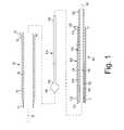
- FIG. 1is an exploded side view of an apparatus for delivering a plug into a puncture through tissue.
- FIGS. 2A and 2Bare cross-sectional views of the apparatus of FIG. 1 , with a cartridge carrying a plug and anchor in proximal and distal positions, respectively.
- FIG. 3Ais a perspective view of an exemplary embodiment of a plug that may be delivered using the apparatus of FIG. 1 .
- FIG. 3Bis a cross-sectional detail of the plug of FIG. 3A .
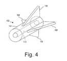
- FIG. 4is a perspective view of an exemplary embodiment of an anchoring element that may be delivered using the apparatus of FIG. 1 .
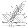
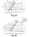
- FIGS. 5A-5Fare cross-sectional views of a patient's body, showing a method for sealing a puncture extending from the patient's skin to a blood vessel using the apparatus of FIG. 1 .
- FIGS. 1 , 2 A, and 2 Bshow an exemplary embodiment of an apparatus 101 for sealing a puncture through tissue.
- the apparatus 101includes a cartridge or other tubular member 120 and a plunger, cincher, or other pusher member 130 .
- the cartridge 120generally carries a plug 2 and an anchoring element 102 , such as those described further below.
- the apparatus 101may include a positioning member 140 , a delivery, access, or introducer sheath 20 , and/or other components, e.g., a needle and/or guidewire for creating a puncture (not shown), and/or a source of sealing compound (also not shown).
- the introducer sheath 20may be a substantially rigid, semi-rigid, and/or flexible tubular body, including a proximal end 22 , a distal end 24 sized for insertion into a puncture through tissue, and a lumen 26 extending between the proximal and distal ends 22 , 24 .
- the distal end 24may be tapered and/or may include a substantially atraumatic distal tip 25 for facilitating advancement through a puncture.
- the introducer sheath 20may include a handle or hub 23 on the proximal end 22 , and/or one or more seals on the proximal end 22 , e.g., a hemostatic seal (not shown) that prevents proximal flow of blood or other fluids, yet accommodates inserting one or more instruments (also not shown) into the lumen 26 of the introducer sheath 20 .
- a hemostatic sealnot shown
- the cartridge 120may be an elongate tubular body including a proximal end 122 , a distal end 124 , and a lumen 126 extending between the proximal and distal ends 122 , 124 .
- the cartridge 120may include a tapered distal tip 125 and/or an enlarged handle or hub 123 on the proximal end 122 .
- the cartridge 120may be substantially rigid, semi-rigid, or flexible, e.g., such that the cartridge 120 may be advanced through the introducer sheath 20 or otherwise into a puncture through tissue.
- the pusher member 130may also be an elongate tubular body, e.g., a plunger or catheter, including a proximal end 132 , a distal end 134 , and a lumen 136 extending between the proximal and distal ends 132 , 134 .
- the pusher member 130may have a size for slidable insertion into the lumen 126 of the cartridge 120 .
- the distal end 134 of the pusher member 130may terminate in a substantially blunt distal tip 135 , e.g., to facilitate contacting, pushing, and/or “cinching” the plug 2 within the puncture, as described further below.
- the pusher member 130may be substantially rigid, semi-rigid, and/or substantially flexible, having sufficient column strength to allow proximal movement of the cartridge 120 relative to the plug 2 without buckling the pusher member 130 .
- the pusher member 130may also include a lumen 136 extending between the proximal end 132 and the distal end 134 , e.g., to accommodate the positioning member 140 , a guidewire (not shown), a flowable sealing compound, and/or other fluid.
- the plug 2may be disposed within the lumen 126 of the cartridge 120 proximate to the distal end 124 , e.g., immediately adjacent the distal tip 125 .
- the lumen 126may be sized such that the plug 2 is slidable therein, e.g., able to traverse distally from the cartridge 120 during delivery, as described further below.
- the anchoring element 102may also be disposed within the lumen 126 of the cartridge 120 proximal to the plug 2 , e.g., immediately adjacent the plug 2 .
- the positioning member 140may be a guidewire and/or other solid or hollow elongate body, including a proximal end 142 , a distal end 144 , and a positioning element 146 on the distal end 144 .
- the positioning element 146may be an expandable element, such as a wire mesh structure, an expandable frame, and/or a balloon member (not shown).
- the positioning element 146may include a skin or other covering (not shown) on at least a proximal portion thereof, thereby making the positioning element 146 substantially nonporous.
- the positioning element 146may be selectively expandable, e.g., using a pull wire, a source of inflation media (e.g., coupled to a lumen, not shown, extending through the positioning member 140 to a balloon or other inflatable positioning element, also not shown), or other actuator (also not shown) operable from the proximal end 142 of the positioning member 140 .
- the positioning element 146may be biased to an enlarged condition, but may be compressed to a contracted condition, e.g., by an overlying sleeve or other constraint (not shown). The constraint may be removed to expose the expandable element, allowing the expandable element to automatically expand to the enlarged condition.
- the delivery apparatus 101may be used to position and deliver the plug 2 within a puncture, e.g., extra-vascularly just above or otherwise adjacent to an arteriotomy in a blood vessel or other body lumen communicating with the puncture, as explained further below.
- the cartridge 120(along with the pusher member 130 , plug 2 , and anchoring element 102 ) may be initially provided on a proximal end 142 of the positioning member 140 .
- the cartridge 120may be slidable distally along the positioning member 140 , e.g., until the distal end 124 of the cartridge 120 is disposed adjacent the positioning element 146 , as shown in FIG. 2B .
- the positioning member 140 and/or pusher member 130may include one or more detents that engage when the cartridge 120 reaches a predetermined location along the positioning member 140 , e.g., to limit subsequent movement of the pusher member 130 relative to the positioning member 140 .
- the positioning member 140may include a ring, tab, or other raised element 145
- the pusher member 130may include a living hinge, tab, or other latch element 137 , e.g., on proximal end 132 .
- the latch element 137may simply be an annular notch in the proximal end 132 of the pusher member 130 to bias the proximal end 132 inwardly.
- the ring 145may be provided at a predetermined location on the positioning member 140 , e.g., a predetermined distance from the positioning element 146 that corresponds to a length of the pusher member 130 .
- the latch element 137may pass freely over the raised element 145 . Thereafter, the latch element 137 may prevent the pusher member 130 from being retracted again past the ring 145 , due to the blunt edge of the latch element 137 abutting the ring 145 on the positioning member 140 .
- the cartridge member 120 and pusher member 130may be provided initially adjacent the distal end 144 of the positioning member 140 , as shown in FIG. 2B .
- the pusher member 130 and positioning member 140may include the cooperating detents 133 , 145 to prevent proximal movement of the pusher member 130 relative to the positioning member 140 .
- the pusher member 130may be otherwise fixed relative to the positioning member 140 , for example, mechanically bonded, chemically bonded, and the like.
- the distal end 134 of the pusher member 130may be fixed a predetermined distance proximal to the positioning element 146 , e.g., to provide the plug 2 immediately adjacent the positioning element 146 , as shown in FIG. 2B .
- the plug 2may include a carrier or core 4 , having first and second hydrogel precursors disposed thereon in an unreactive state, thereby providing an adherent coating 6 .
- the plug 2may have a solid or hollow cylindrical shape, a disk shape, or other shapes or cross-sections, such as elliptical, triangular, square, conical, disk, polygonic shapes, and the like.
- the plug 2may include a lumen 10 extending between proximal and distal ends 14 , 16 thereof, thereby defining a longitudinal axis 18 .
- the lumen 10may be created when the core 4 is formed, e.g., if the core 4 is rolled from one or more sheets or layers of material or formed by molding. Alternatively, the lumen 10 may formed by boring into or otherwise removing material from an already formed solid core 4 .
- the lumen 10may be dimensioned such that the positioning member 140 , a guidewire or other instrument (not shown) may slide or otherwise pass through the plug 2 .
- the core 4may be formed from a biocompatible and/or bioabsorbable material, for example, a porous, bioabsorbable foam or other solid material.
- the core 4may be formed from a biocompatible and/or bioabsorbable hydrogel, e.g., polyethylene glycol (“PEG”), or other synthetic material.
- the core 4may be formed from a lyophilized (i.e., freeze-dried) PEG polymer that contains hydrolytically degradable chemical groups.
- the lyophilized PEG polymere.g., including a macroporous polymer network, may uptake fluid and expand when exposed to an aqueous environment.
- the magnitude of expansion or swelling (pre to post hydration)may be significant, e.g., between about two and ten times (2 ⁇ -10 ⁇ ) its lyophilized size based on volume.
- the core 4may include pro-thrombotic material, e.g., including one or more biological pro-thrombotics, such as collagen, fibrin, carboxymethylcellulose, oxidized cellulose, alginates, gelatin, or other protein-based material, and/or synthetic materials, such as polyglycolic acids (PGA's), polyactides (PLA's), polyvinyl alcohol, and the like.
- the material of the core 4may be at least partially absorbed by the body over time, e.g., over a period of days, weeks, or months.
- the core 4may include therapeutic and/or pharmaceutical agents, e.g., to promote healing, prevent infection and/or other adverse medical events, and the like. Such agents may be embedded in the core material and/or applied as one or more coatings or layers.
- the material of the core 4may have a substantially uniform composition or the composition may be varied, e.g., along its length and/or within underlying layers within the core 4 .
- the first and second hydrogel precursors 6may remain in the unreactive state, e.g., before or until exposure to an aqueous physiological environment.
- An aqueous physiological environmentmay exist, for example, inside a puncture track extending through tissue. Blood or other bodily fluids that contact the precursor-laden carrier may initiate a hydrogel forming reaction between the two precursors.
- the reaction of the hydrogel precursorsmay form a cross-linked adhesive or tacky coating that may aid in retaining the plug 2 within a puncture after deployment and/or in facilitating hemostasis within the puncture.
- the first and second hydrogel precursors 6may be loaded onto the core 4 , e.g., by wicking a mixture of the liquid hydrogel precursors onto the core 4 .
- the hydrogel precursorsmay initially be a solid dehydrated material, e.g., a powder, that may be heated above its melting point to form a liquid suitable for wicking.
- the first and second hydrogel precursorsmay be sufficiently mixed before being loaded onto the core 4 .
- the first and second precursorsmay be provided in a liquid form into which the core 4 may be dipped, that may be poured onto the core 4 , and/or otherwise applied to the core 4 together or successively.
- the first and second precursorsmay be dissolved in a solvent that may then be applied to the core 4 .
- the first and second hydrogel precursorsmay be in a solid or semi-solid state.
- the first hydrogel precursormay include any number of hydrogel precursor materials, such as those disclosed in U.S. Pat. Nos. 6,152,943, 6,165,201, 6,179,862, 6,514,534, 6,379,373, 6,703,047, and in co-pending application Ser. No. 10/010,715 filed Nov. 9, 2001, Ser. No. 10/068,807 filed Feb. 5, 2002, and Ser. No. 10/454,362, filed Jun. 4, 2003.
- the disclosures of these references and any others cited thereinare expressly incorporated by reference herein.
- the first hydrogel precursormay include a four arm, 10 kDalton PEG with reactive ester end groups or an eight arm, 20 kDalton PEG amine.
- the first hydrogel precursormay include a bioabsorbable star polymer having a complementary cross-linking species such as, for example, an amino acid with reactive end groups, e.g., lysine, dilysine, trilysine, etc.
- a bioabsorbable star polymer having a complementary cross-linking speciessuch as, for example, an amino acid with reactive end groups, e.g., lysine, dilysine, trilysine, etc.
- the second hydrogel precursormay include any number of hydrogel precursor materials, e.g., a material reactive with the first precursor material once exposed within a hydrous or aqueous environment, such as those materials disclosed above and in the references incorporated by reference above.
- the second precursormay be the other of an eight arm, 20 kDalton PEG amine or a four arm, 10 kDalton PEG ester.
- the second precursormay be the complementary cross-linking species of a bioabsorbable star polymer, such as an amino acid with reactive end groups, e.g., lysine, dilysine, trilysine, etc.
- an activating agent 8e.g., a pH adjusting or activating agent, may also be disposed on the core 4 , e.g., to initiate, accelerate, or otherwise enhance the reaction of the precursors 6 .
- the pH activating agent 8may create a localized change in pH after exposure to a hydrous or aqueous environment.
- the pH activating agent 8may include solid borate crystals, such as Na 2 B 4 O 7 .10H 2 O, although different salt-based or other materials that alter the localized pH value may be employed.
- other pH adjusting agents 8may be used, such as sodium bicarbonate, and the like.
- the pH activating agent 8may be loaded onto the core 4 by physically contacting solid borate crystals, powder, or other particles onto the precursor-laden (first and second hydrogel precursors) core.
- the core 4may simply be rolled over a pH activating agent 8 with sufficient force to embed the pH activating agent 8 into the exterior surface of the core 4 .
- the pH activating agent 8may be adhered to the exterior surface of the core 4 , e.g., by pressing particles of the pH activating agent 8 into the exterior surface, by using an adhesive (e.g., that is substantially inert or unreactive with the first or second precursors), and the like. Additional information on plugs that may be provided are disclosed in co-pending application Ser. No. 10/982,387, filed Nov. 5, 2004, entitled “Apparatus and Methods for Sealing a Vascular Puncture,” the entire disclosure of which is expressly incorporated herein by reference.
- laminate structuresmay be used for the plug 2 , e.g., a sheet including multiple layers of different components, such as one or more of the components described above, may be formed, and the sheet may be rolled into a tubular or solid cylindrical structure.
- An exemplary embodiment of such a sheetmay include three layers, e.g., a first layer of lyophilized hydrogel, a second layer of two-part hydrogel adherent material, and a third layer of lyophilized hydrogel.
- the layersmay be substantially uniform, or one or more of the layers may vary in thickness, e.g., along their lengths.
- the layer(s)may become progressively thicker from one edge corresponding to the proximal end 14 of the plug 2 to the opposite edge corresponding to the distal end 16 of the plug 2 .
- the plug 2may have a frustoconical shape (not shown), rather than a substantially uniform cylindrical shape.
- a layer of lyophilized hydrogelmay be provided, and an adherent layer, e.g., including two hydrogel precursors in an initially unreactive state, may be applied to one surface of the layer of lyophilized hydrogel.
- a pH adjusting agente.g., borate crystals, may be embedded or otherwise applied to the opposite surface of the layer of lyophilized hydrogel.
- the pH adjusting agentmay be substantially segregated from the adherent layer. This may be desirable to prevent the pH adjusting agent from initiating reaction of the materials of the adherent layer prematurely, which may otherwise occur to some degree, even absent an aqueous environment.
- the composition of the plug 2may be varied along its length.
- material on or adjacent the distal end 16 of the plug 2may more rapidly rehydrate and/or otherwise expand than material on or adjacent the proximal end 14 of the plug 2 .
- a composition of hydrogelmay be provided adjacent the distal end 16 that is more porous than the hydrogel provided adjacent the proximal end 14 , which may accelerate expansion of the distal end 16 compared to the proximal end 14 .
- the plug 2may have a frustoconical shape (not shown), or other shape in which the distal end 16 is substantially larger than the proximal end 14 . This configuration may facilitate and/or enhance compaction against an arteriotomy or otherwise enhance sealing.
- the material of the plug 2may be compacted before or after being formed into the plug shape, e.g., to change its shape from a substantially uniform cylindrical shape and/or to change the density of the plug 2 along its length.
- the anchoring element 102includes a body 104 including a lumen 110 and one or more protrusions 108 .
- the protrusions 108include a plurality of barbs extending radially outwardly from the body 104 .
- the protrusions 108may extend transversely from the body 104 , e.g., laterally and proximally. Thus, when the protrusions 108 are embedded in or otherwise contact surrounding tissue, the protrusions 108 may limit proximal movement of the anchoring element 102 .
- the anchoring element 102may be formed from a substantially rigid bioabsorbable material.
- the anchoring element 102may be formed from dehydrated hydrogel material, e.g., air-dried hydrogel.
- a hydrogel materialWhen a hydrogel material is air-dried (as opposed to freeze-dried), the material may collapse in upon itself, e.g., resulting in a substantially nonporous structure that is substantially rigid.
- the resulting structuremay be relatively nonporous, e.g., compared to lyophilized hydrogel, such as those described above for the plug 2 .
- the anchoring element 102may hydrate at a slower rate than the plug 2 , e.g., on the order of several minutes.
- the materialmay then be formed into a desired shape, e.g., by laser cutting, sawing, machining, grinding, and the like.
- a desired volume of hydrogelmay be air-dried, and then the resulting piece of hydrogel may be formed, e.g., to create the body 104 , lumen 110 , and protrusions 108 .
- the anchoring element 102may be formed from other bioabsorbable materials that are sufficiently rigid to engage tissue surrounding a puncture and anchor the anchoring element 102 and plug 2 within the puncture.
- synthetic materialsmay be used, such as polyglycolic acids (PGA's), polyactides (PLA's), and polyvinyl alcohol.
- the anchoring element 102may include pro-thrombotic material, e.g., including one or more biological pro-thrombotics, such as collagen, fibrin, carboxymethylcellulose, oxidized cellulose, alginates, gelatin, or other protein-based material.
- the anchoring element 102may be formed from biocompatible, but not bioabsorbable material.
- the anchoring element 102may be formed from metal, such as stainless steel, or plastics, that may remain within a patient's body indefinitely.
- the anchoring element 102may be of sufficient size to engage tissue surrounding a puncture, yet be small enough to remain unobtrusively within the patient's body after the procedure, i.e., after the plug 2 has been absorbed and/or the puncture has healed.
- FIGS. 5A-5Fan exemplary method is shown for sealing a puncture 90 , e.g., using the apparatus 101 described above to deliver a plug 2 and extra-vascular anchoring element 102 , such as any of the embodiments described above.
- the puncture 90extends from a patient's skin 92 through intervening tissue 96 , e.g., to a body lumen 94 .
- the puncture 90may be a percutaneous puncture communicating with a blood vessel 94 , such as a femoral artery, carotid artery, and the like.
- the puncture 90may be created using known procedures, e.g., using a needle, guidewire, one or more dilators, and the like (not shown).
- An introducer sheath 20may be advanced through the puncture 90 into the vessel 94 , e.g., to provide access into the vessel 90 for one or more instruments, and/or allow one or more diagnostic and/or interventional procedures to be performed via the vessel 90 , as is known in the art.
- any instruments and/or the introducer sheathmay be removed from the puncture 90 .
- a positioning member 140may be introduced into and/or through the lumen 26 of the introducer sheath 20 , e.g., with the expandable frame or other positioning element 146 thereon in a collapsed condition.
- the cartridge 120(along with the plug device 102 and pusher member 130 ) may be provided initially on the proximal end 142 of the positioning member 140 (not shown in FIG. 5A for clarity, see FIG. 2A ).
- the cartridge 120may initially be located outside the puncture 90 as the positioning member 140 is advanced into the puncture 90 .
- the cartridge 120may be carried on the distal end 144 of the positioning member 140 (as shown in FIG. 2B ), e.g., such that the cartridge 120 (along with the plug device 102 and pusher member 130 ) are introduced simultaneously with the positioning member 140 (not shown in FIG. 5A ).
- the cartridge 120may be provided separate from the positioning member 140 (not shown).
- the shaft of the positioning member 140may extend proximally from the proximal end 22 of the introducer sheath 20 out of the puncture 90 .
- the proximal end 142 of the positioning member 140may be back-loaded into the cartridge 120 , e.g., through the lumens 10 , 110 , 136 of the plug 2 , anchoring element 102 , and pusher member 130 .
- the distal end 144 of the positioning member 140may be inserted through the puncture 90 (via the introducer sheath 20 ) and into the vessel 94 (shown in phantom).
- the positioning element 146 on the distal end 144 of the positioning member 140may be expanded or otherwise deployed to an enlarged condition.
- the positioning member 140may be at least partially withdrawn until the positioning element 146 contacts the wall of the vessel 94 , e.g., to substantially seal the vessel 94 from the puncture 90 .
- thismay involve a two-step process (although it may be completed in a single continuous action).
- the positioning member 140is withdrawn until it contacts the distal end 24 of the introducer sheath 20 , which may provide a first tactile feedback to the user (that the positioning element 146 has contacted the introducer sheath 20 based upon the increased weight and/or resistance to proximal movement).
- the positioning member 140may be withdrawn further until the positioning element 146 contacts the wall of the vessel 94 , thereby providing a second tactile feedback.
- the introducer sheath 20may be pulled proximally by the positioning element 146 as the positioning member 120 is withdrawn, e.g., until the distal end 24 of the introducer sheath 20 is withdrawn from the vessel 94 into the puncture 90 , as shown in FIG. 5B .
- Proximal tensionmay be applied and/or maintained on the positioning member 140 to hold the positioning element 146 against the wall of the vessel 94 , e.g., to seal the puncture 90 from the vessel 94 .
- the proximal tensionmay be maintained manually or using a tensioner device (not shown) to provide temporary hemostasis, e.g., during the subsequent steps.
- Exemplary tension devicesare disclosed in co-pending application Ser. No. 10/806,952, filed Mar. 22, 2004, the entire disclosure of which is expressly incorporated herein by reference.
- the cartridge 120(carrying the plug 2 and anchoring element 102 ) may be advanced distally over the positioning member 140 into the puncture 90 .
- the cartridge 120may be advanced through the introducer sheath 20 until a hub 123 of the cartridge 120 abuts a hub 23 on the introducer sheath 20 , as shown in FIG. 5C .
- the pusher member 130may slide over the positioning member 140 until the latch element 137 on the pusher member 130 passes over the ring 145 on the positioning member 140 . This may prevent subsequent proximal movement of the pusher member 130 relative to the positioning member 140 .
- the pusher member 130is maintained with the distal end 134 immediately adjacent the anchoring element 102 , and the introducer sheath 20 and cartridge 120 are retracted proximally to expose or otherwise deploy the plug 2 and anchoring element 102 within the puncture 90 .
- the pusher member 130may serve as a stop that prevents the plug 2 and anchoring element 102 from moving proximally while the introducer sheath 20 and cartridge 120 are withdrawn.
- the pusher member 130may be advanced distally into the lumen 126 of the cartridge 120 , e.g., until the distal end 134 of the pusher member 130 is proximally adjacent the anchoring element 102 .
- the cartridge 120 (and introducer sheath 20 )may then be withdrawn, while maintaining the pusher member 130 in position to deploy the plug 2 and anchoring element 102 successively within the puncture 90 .
- the user of the delivery apparatus 101may position his or her thumb on hub 133 of the pusher member 130 to maintain its position while the introducer sheath 20 and cartridge 120 are retracted by pulling on hub 23 , e.g., using his or her index and middle fingers.
- the hub 23 of the introducer sheath 20may be held and withdrawn, thereby causing the cartridge 120 to be withdrawn simultaneously.
- the introducer sheath 20may be removed first, and then the cartridge 120 may be removed.
- the cartridge 120 and introducer sheath 20may be removed entirely from the puncture 90 or only sufficiently to expose the plug 2 and anchoring element 102 within the puncture 90 .
- the plug 2may be cinched or otherwise compressed within the puncture 90 , e.g., by advancing the pusher member 130 distally to press the anchoring element 102 against the plug 2 and the plug 2 against the wall of the vessel 94 and/or against the positioning element 146 . This may cause the plug 2 to expand radially outwardly and/or seal against the arteriotomy, e.g., to enhance sealing the puncture 90 from the vessel 94 .
- the proximal tension on the positioning member 140may be released and/or the positioning element 146 may be collapsed to its collapsed state.
- the positioning element 146may be mechanically collapsed or deflated.
- the positioning member 140(and consequently the positioning element 146 ) may be slowly withdrawn through the lumens 10 , 110 , 136 of the plug 2 , the anchoring element 102 , and the pusher member 130 , respectively.
- the pusher member 130may be maintained to serve as a stop and prevent proximal migration of the plug 2 and/or anchoring element 102 within the puncture 90 .
- the plug 2includes an adherent layer (not shown in FIG. 5D )
- the “sticky” adherent layermay also aid in securing the plug to the tissue surrounding the puncture 90 to prevent migration.
- the protrusions 108 on the anchoring element 102may engage the surrounding tissue to prevent migration of the plug 2 .
- the pusher member 130may be withdrawn, leaving the plug 2 and anchoring element 102 in place. If desired, e.g., if bleeding occurs proximally through the lumen 136 of the pusher member 130 , liquid hydrogel or other sealing compound may be delivered into the puncture 90 above and/or around the plug device 102 , to assist in achieving permanent hemostasis.
- a source of sealing compounde.g., a syringe assembly 50 carrying liquid sealing compound components
- sealing compound 99may be delivered into the puncture 90 above and/or around the plug 2 and/or anchoring element 102 .
- the pusher member 130may be retracted proximally as the sealing compound 99 is delivered to at least partially fill the puncture 90 with the sealing compound 99 .
- blood and/or other fluid within the vessel 94may enter the puncture 90 , thereby exposing the plug 2 and anchoring element 102 to an aqueous physiological environment.
- the aqueous physiological environmentmay wet the plug 2 and anchoring element 102 , thereby initiating rehydration of the materials of the plug 2 and/or anchoring element 102 and/or initiating a reaction between the first and second precursors (or other adherent coating) on the plug 2 .
- the fluidmay dissolve the activating agent 8 , changing the pH of the fluid to initiate the first and second hydrogel precursors 6 reacting with one another.
- the reaction of the first and second hydrogel precursors 6may form an adhesive or “sticky” hydrogel coating that may bond or otherwise attach to tissue surrounding the puncture 90 , which may facilitate retaining the plug 2 in place within the puncture 90 .
- the plug 2includes lyophilized hydrogel, the hydrogel may expand or swell as it hydrates to further aid in retaining the plug 2 within the puncture 90 and/or enhance sealing the puncture 90 .
- the plug 2 and anchoring element 102both include dehydrated hydrogel, the plug 2 including lyophilized hydrogel, and the anchoring element 102 including air-dried hydrogel.
- the anchoring element 102because the anchoring element 102 is less porous, it does not hydrate as rapidly as the plug 2 . This may be desirable to ensure that the anchoring element 102 retains its rigidity and shape initially, e.g., for the several minutes or hours it takes for the anchoring element 102 to hydrate.
- the plug 2may hydrate and expand more rapidly than the anchoring element 102 , e.g., within seconds or minutes to enhance sealing of the puncture 90 .
- the anchoring element 102may retain its shape and rigidity for several minutes or even hours after being delivered, the protrusions 108 of the anchoring element 102 may engage the surrounding tissue (particularly as the tissue recoils inwardly into the puncture 90 ), thereby preventing the anchoring element 102 from moving proximally (and optionally distally) within the puncture 90 .
- the plug 2With the anchoring element 102 secured within the puncture 90 , the plug 2 may be unable to move proximally within the puncture 90 , but instead may contact the anchoring element 102 .
- the anchoring element 102may prevent the plug 2 from moving away from the arteriotomy, e.g., if the patient becomes ambulatory or is otherwise moved in a manner that may otherwise disturb the plug 2 .
- the material of the plug 2 and anchoring element 102may be at least partially absorbed by the body over time, e.g., over a period of days, weeks, or months, as is known in the art. Additional methods for delivering the plug 2 and anchoring element 102 are disclosed in co-pending application Ser. No. 10/982,387, incorporated by reference above. Although this application does not disclose an extra-vascular anchoring element, it will be appreciated that similar methods may be used to deliver both the plug 2 and the anchoring element 102 as those used to deliver the plug 2 alone.
Landscapes
- Health & Medical Sciences (AREA)
- Life Sciences & Earth Sciences (AREA)
- Surgery (AREA)
- Heart & Thoracic Surgery (AREA)
- Engineering & Computer Science (AREA)
- Biomedical Technology (AREA)
- Nuclear Medicine, Radiotherapy & Molecular Imaging (AREA)
- Medical Informatics (AREA)
- Molecular Biology (AREA)
- Animal Behavior & Ethology (AREA)
- General Health & Medical Sciences (AREA)
- Public Health (AREA)
- Veterinary Medicine (AREA)
- Cardiology (AREA)
- Surgical Instruments (AREA)
Abstract
Description
Claims (25)
Priority Applications (1)
| Application Number | Priority Date | Filing Date | Title |
|---|---|---|---|
| US13/610,849US8945178B2 (en) | 2004-11-05 | 2012-09-11 | Apparatus and methods for sealing a vascular puncture |
Applications Claiming Priority (2)
| Application Number | Priority Date | Filing Date | Title |
|---|---|---|---|
| US10/982,385US8262693B2 (en) | 2004-11-05 | 2004-11-05 | Apparatus and methods for sealing a vascular puncture |
| US13/610,849US8945178B2 (en) | 2004-11-05 | 2012-09-11 | Apparatus and methods for sealing a vascular puncture |
Related Parent Applications (1)
| Application Number | Title | Priority Date | Filing Date |
|---|---|---|---|
| US10/982,385ContinuationUS8262693B2 (en) | 2004-11-05 | 2004-11-05 | Apparatus and methods for sealing a vascular puncture |
Publications (2)
| Publication Number | Publication Date |
|---|---|
| US20130066361A1 US20130066361A1 (en) | 2013-03-14 |
| US8945178B2true US8945178B2 (en) | 2015-02-03 |
Family
ID=35945164
Family Applications (2)
| Application Number | Title | Priority Date | Filing Date |
|---|---|---|---|
| US10/982,385Expired - Fee RelatedUS8262693B2 (en) | 2004-11-05 | 2004-11-05 | Apparatus and methods for sealing a vascular puncture |
| US13/610,849Expired - LifetimeUS8945178B2 (en) | 2004-11-05 | 2012-09-11 | Apparatus and methods for sealing a vascular puncture |
Family Applications Before (1)
| Application Number | Title | Priority Date | Filing Date |
|---|---|---|---|
| US10/982,385Expired - Fee RelatedUS8262693B2 (en) | 2004-11-05 | 2004-11-05 | Apparatus and methods for sealing a vascular puncture |
Country Status (5)
| Country | Link |
|---|---|
| US (2) | US8262693B2 (en) |
| EP (1) | EP1807131B1 (en) |
| JP (1) | JP2008518743A (en) |
| CA (1) | CA2583238C (en) |
| WO (1) | WO2006052612A1 (en) |
Cited By (2)
| Publication number | Priority date | Publication date | Assignee | Title |
|---|---|---|---|---|
| US20130238018A1 (en)* | 2010-02-11 | 2013-09-12 | Boston Scientific Scimed, Inc. | Automatic vascular closure deployment devices and methods |
| US9107646B2 (en)* | 2013-03-11 | 2015-08-18 | St. Jude Medical Puerto Rico Llc | Active securement detachable sealing tip for extra-vascular closure device and methods |
Families Citing this family (141)
| Publication number | Priority date | Publication date | Assignee | Title |
|---|---|---|---|---|
| US20020095164A1 (en)* | 1997-06-26 | 2002-07-18 | Andreas Bernard H. | Device and method for suturing tissue |
| US7335220B2 (en)* | 2004-11-05 | 2008-02-26 | Access Closure, Inc. | Apparatus and methods for sealing a vascular puncture |
| US20040092964A1 (en) | 1999-03-04 | 2004-05-13 | Modesitt D. Bruce | Articulating suturing device and method |
| US6964668B2 (en) | 1999-03-04 | 2005-11-15 | Abbott Laboratories | Articulating suturing device and method |
| US7842048B2 (en) | 2006-08-18 | 2010-11-30 | Abbott Laboratories | Articulating suture device and method |
| US8137364B2 (en) | 2003-09-11 | 2012-03-20 | Abbott Laboratories | Articulating suturing device and method |
| US7001400B1 (en) | 1999-03-04 | 2006-02-21 | Abbott Laboratories | Articulating suturing device and method |
| US7235087B2 (en) | 1999-03-04 | 2007-06-26 | Abbott Park | Articulating suturing device and method |
| US8758400B2 (en) | 2000-01-05 | 2014-06-24 | Integrated Vascular Systems, Inc. | Closure system and methods of use |
| US6391048B1 (en) | 2000-01-05 | 2002-05-21 | Integrated Vascular Systems, Inc. | Integrated vascular device with puncture site closure component and sealant and methods of use |
| US9579091B2 (en) | 2000-01-05 | 2017-02-28 | Integrated Vascular Systems, Inc. | Closure system and methods of use |
| US7842068B2 (en) | 2000-12-07 | 2010-11-30 | Integrated Vascular Systems, Inc. | Apparatus and methods for providing tactile feedback while delivering a closure device |
| US6461364B1 (en) | 2000-01-05 | 2002-10-08 | Integrated Vascular Systems, Inc. | Vascular sheath with bioabsorbable puncture site closure apparatus and methods of use |
| EP1259168B1 (en)* | 2000-02-24 | 2010-09-08 | Loma Linda University Medical Center | Patch and glue delivery system for closing tissue openings during surgery |
| DE60144328D1 (en) | 2000-09-08 | 2011-05-12 | Abbott Vascular Inc | Surgical clamp |
| US6626918B1 (en) | 2000-10-06 | 2003-09-30 | Medical Technology Group | Apparatus and methods for positioning a vascular sheath |
| US7211101B2 (en) | 2000-12-07 | 2007-05-01 | Abbott Vascular Devices | Methods for manufacturing a clip and clip |
| US6623510B2 (en) | 2000-12-07 | 2003-09-23 | Integrated Vascular Systems, Inc. | Closure device and methods for making and using them |
| US7806904B2 (en) | 2000-12-07 | 2010-10-05 | Integrated Vascular Systems, Inc. | Closure device |
| US8690910B2 (en) | 2000-12-07 | 2014-04-08 | Integrated Vascular Systems, Inc. | Closure device and methods for making and using them |
| US7905900B2 (en) | 2003-01-30 | 2011-03-15 | Integrated Vascular Systems, Inc. | Clip applier and methods of use |
| US6695867B2 (en) | 2002-02-21 | 2004-02-24 | Integrated Vascular Systems, Inc. | Plunger apparatus and methods for delivering a closure device |
| US7029480B2 (en)* | 2001-01-24 | 2006-04-18 | Abott Laboratories | Device and method for suturing of internal puncture sites |
| US20080109030A1 (en) | 2001-04-24 | 2008-05-08 | Houser Russell A | Arteriotomy closure devices and techniques |
| US8992567B1 (en) | 2001-04-24 | 2015-03-31 | Cardiovascular Technologies Inc. | Compressible, deformable, or deflectable tissue closure devices and method of manufacture |
| US8961541B2 (en) | 2007-12-03 | 2015-02-24 | Cardio Vascular Technologies Inc. | Vascular closure devices, systems, and methods of use |
| IES20010547A2 (en) | 2001-06-07 | 2002-12-11 | Christy Cummins | Surgical Staple |
| US7329414B2 (en)* | 2002-05-03 | 2008-02-12 | Biopsy Sciences, Llc | Biodegradable polymer for marking tissue and sealing tracts |
| IES20030424A2 (en) | 2002-06-04 | 2003-12-10 | Robert Stevenson | Blood vessel closure clip and delivery device |
| AU2003297665A1 (en)* | 2002-12-06 | 2004-06-30 | Fast Country, Inc. | Systems and methods for providing interactive guest resources |
| US7160309B2 (en) | 2002-12-31 | 2007-01-09 | Laveille Kao Voss | Systems for anchoring a medical device in a body lumen |
| US8758398B2 (en) | 2006-09-08 | 2014-06-24 | Integrated Vascular Systems, Inc. | Apparatus and method for delivering a closure element |
| US7857828B2 (en) | 2003-01-30 | 2010-12-28 | Integrated Vascular Systems, Inc. | Clip applier and methods of use |
| US8202293B2 (en) | 2003-01-30 | 2012-06-19 | Integrated Vascular Systems, Inc. | Clip applier and methods of use |
| US8398656B2 (en) | 2003-01-30 | 2013-03-19 | Integrated Vascular Systems, Inc. | Clip applier and methods of use |
| US8821534B2 (en) | 2010-12-06 | 2014-09-02 | Integrated Vascular Systems, Inc. | Clip applier having improved hemostasis and methods of use |
| US8905937B2 (en) | 2009-02-26 | 2014-12-09 | Integrated Vascular Systems, Inc. | Methods and apparatus for locating a surface of a body lumen |
| US9289195B2 (en) | 2003-06-04 | 2016-03-22 | Access Closure, Inc. | Auto-retraction apparatus and methods for sealing a vascular puncture |
| US7462188B2 (en) | 2003-09-26 | 2008-12-09 | Abbott Laboratories | Device and method for suturing intracardiac defects |
| US7449024B2 (en) | 2003-12-23 | 2008-11-11 | Abbott Laboratories | Suturing device with split arm and method of suturing tissue |
| IES20040368A2 (en) | 2004-05-25 | 2005-11-30 | James E Coleman | Surgical stapler |
| US7806856B2 (en) | 2005-04-22 | 2010-10-05 | Accessclosure, Inc. | Apparatus and method for temporary hemostasis |
| US8926633B2 (en) | 2005-06-24 | 2015-01-06 | Abbott Laboratories | Apparatus and method for delivering a closure element |
| US8313497B2 (en) | 2005-07-01 | 2012-11-20 | Abbott Laboratories | Clip applier and methods of use |
| US7883517B2 (en) | 2005-08-08 | 2011-02-08 | Abbott Laboratories | Vascular suturing device |
| US8083754B2 (en) | 2005-08-08 | 2011-12-27 | Abbott Laboratories | Vascular suturing device with needle capture |
| US8267947B2 (en) | 2005-08-08 | 2012-09-18 | Abbott Laboratories | Vascular suturing device |
| US8920442B2 (en) | 2005-08-24 | 2014-12-30 | Abbott Vascular Inc. | Vascular opening edge eversion methods and apparatuses |
| US9456811B2 (en) | 2005-08-24 | 2016-10-04 | Abbott Vascular Inc. | Vascular closure methods and apparatuses |
| US8758397B2 (en)* | 2005-08-24 | 2014-06-24 | Abbott Vascular Inc. | Vascular closure methods and apparatuses |
| US20070060895A1 (en)* | 2005-08-24 | 2007-03-15 | Sibbitt Wilmer L Jr | Vascular closure methods and apparatuses |
| JP5253170B2 (en)* | 2005-10-05 | 2013-07-31 | ローマ リンダ ユニヴァーシティ メディカル センター | Vascular wound closure device and method |
| US20100168767A1 (en) | 2008-06-30 | 2010-07-01 | Cardiva Medical, Inc. | Apparatus and methods for delivering hemostatic materials for blood vessel closure |
| US8911472B2 (en) | 2005-12-13 | 2014-12-16 | Cardiva Medical, Inc. | Apparatus and methods for delivering hemostatic materials for blood vessel closure |
| US8939910B2 (en) | 2006-03-28 | 2015-01-27 | Devicor Medical Products, Inc. | Method for enhancing ultrasound visibility of hyperechoic materials |
| US20170066162A9 (en)* | 2006-03-28 | 2017-03-09 | Devicor Medical Products, Inc. | Method of Enhancing Ultrasound Visibility of Hyperechoic Materials |
| US8808310B2 (en) | 2006-04-20 | 2014-08-19 | Integrated Vascular Systems, Inc. | Resettable clip applier and reset tools |
| US8556930B2 (en) | 2006-06-28 | 2013-10-15 | Abbott Laboratories | Vessel closure device |
| US7789893B2 (en)* | 2006-09-12 | 2010-09-07 | Boston Scientific Scimed, Inc. | Method and apparatus for promoting hemostasis of a blood vessel puncture |
| WO2008033964A2 (en) | 2006-09-13 | 2008-03-20 | Accessclosure, Inc. | Apparatus for sealing a vascular puncture |
| US8226681B2 (en) | 2007-06-25 | 2012-07-24 | Abbott Laboratories | Methods, devices, and apparatus for managing access through tissue |
| US8574244B2 (en) | 2007-06-25 | 2013-11-05 | Abbott Laboratories | System for closing a puncture in a vessel wall |
| US8568445B2 (en) | 2007-08-21 | 2013-10-29 | St. Jude Medical Puerto Rico Llc | Extra-vascular sealing device and method |
| US8333787B2 (en) | 2007-12-31 | 2012-12-18 | St. Jude Medical Puerto Rico Llc | Vascular closure device having a flowable sealing material |
| US20090069843A1 (en)* | 2007-09-10 | 2009-03-12 | Agnew Charles W | Fistula plugs including a hydration resistant component |
| EP2209426A4 (en)* | 2007-11-02 | 2015-04-22 | Incept Llc | Apparatus and methods for sealing a vascular puncture |
| US20090157101A1 (en) | 2007-12-17 | 2009-06-18 | Abbott Laboratories | Tissue closure system and methods of use |
| US8893947B2 (en) | 2007-12-17 | 2014-11-25 | Abbott Laboratories | Clip applier and methods of use |
| US7841502B2 (en) | 2007-12-18 | 2010-11-30 | Abbott Laboratories | Modular clip applier |
| US8840640B2 (en)* | 2007-12-31 | 2014-09-23 | St. Jude Medical Puerto Rico Llc | Vascular closure device having an improved plug |
| US20090227938A1 (en)* | 2008-03-05 | 2009-09-10 | Insitu Therapeutics, Inc. | Wound Closure Devices, Methods of Use, and Kits |
| US9364206B2 (en) | 2008-04-04 | 2016-06-14 | Access Closure, Inc. | Apparatus and methods for sealing a vascular puncture |
| US8029533B2 (en)* | 2008-04-04 | 2011-10-04 | Accessclosure, Inc. | Apparatus and methods for sealing a vascular puncture |
| US9282965B2 (en) | 2008-05-16 | 2016-03-15 | Abbott Laboratories | Apparatus and methods for engaging tissue |
| EP2330981B1 (en) | 2008-08-26 | 2013-11-27 | St. Jude Medical, Inc. | System for sealing percutaneous punctures |
| US8398676B2 (en) | 2008-10-30 | 2013-03-19 | Abbott Vascular Inc. | Closure device |
| WO2010056915A1 (en) | 2008-11-12 | 2010-05-20 | Accessclosure, Inc. | Apparatus and methods for sealing a vascular puncture |
| US8323312B2 (en) | 2008-12-22 | 2012-12-04 | Abbott Laboratories | Closure device |
| US8858594B2 (en) | 2008-12-22 | 2014-10-14 | Abbott Laboratories | Curved closure device |
| US9089311B2 (en) | 2009-01-09 | 2015-07-28 | Abbott Vascular Inc. | Vessel closure devices and methods |
| US9486191B2 (en) | 2009-01-09 | 2016-11-08 | Abbott Vascular, Inc. | Closure devices |
| US9414820B2 (en) | 2009-01-09 | 2016-08-16 | Abbott Vascular Inc. | Closure devices, systems, and methods |
| US20100179589A1 (en) | 2009-01-09 | 2010-07-15 | Abbott Vascular Inc. | Rapidly eroding anchor |
| US9173644B2 (en) | 2009-01-09 | 2015-11-03 | Abbott Vascular Inc. | Closure devices, systems, and methods |
| US20100185234A1 (en) | 2009-01-16 | 2010-07-22 | Abbott Vascular Inc. | Closure devices, systems, and methods |
| US20100217309A1 (en)* | 2009-02-20 | 2010-08-26 | Boston Scientific Scimed, Inc. | Plug for arteriotomy closure and method of use |
| US8529598B2 (en) | 2009-02-20 | 2013-09-10 | Boston Scientific Scimed, Inc. | Tissue puncture closure device |
| US8052914B2 (en) | 2009-02-20 | 2011-11-08 | Boston Scientific Scimed, Inc. | Modified plug for arteriotomy closure |
| US8317824B2 (en)* | 2009-02-20 | 2012-11-27 | Boston Scientific Scimed, Inc. | Tissue puncture closure device |
| US8375553B2 (en) | 2009-02-20 | 2013-02-19 | Boston Scientific Scimed, Inc. | Locking element for vascular closure device |
| US8292918B2 (en) | 2009-02-20 | 2012-10-23 | Boston Scientific Scimed, Inc. | Composite plug for arteriotomy closure and method of use |
| US9913634B2 (en)* | 2009-02-20 | 2018-03-13 | Boston Scientific Scimed, Inc. | Locking element for vascular closure device |
| EP2416711A4 (en) | 2009-04-09 | 2017-06-07 | Cardiovascular Technologies, Inc. | Tissue closure devices, device and systems for delivery, kits and methods therefor |
| WO2010129510A2 (en)* | 2009-05-04 | 2010-11-11 | Incept. Llc | Biomaterials for track and puncture closure |
| US20110054492A1 (en) | 2009-08-26 | 2011-03-03 | Abbott Laboratories | Medical device for repairing a fistula |
| US9179900B2 (en)* | 2009-12-08 | 2015-11-10 | Phillips Medical Llc | Hemostatic device and its methods of use |
| US9993236B2 (en) | 2009-12-08 | 2018-06-12 | Phillips Medical, LLC | Hemostatic device and its methods of use |
| US8870937B2 (en)* | 2010-02-26 | 2014-10-28 | ProMed, Inc. | Method for vessel access closure |
| US8303624B2 (en) | 2010-03-15 | 2012-11-06 | Abbott Cardiovascular Systems, Inc. | Bioabsorbable plug |
| EP2560556B1 (en) | 2010-04-19 | 2016-10-12 | Phillips Medical LLC | A hemostatic device |
| JP5650215B2 (en)* | 2010-06-21 | 2015-01-07 | 株式会社グツドマン | Hemostatic device |
| US10751206B2 (en) | 2010-06-26 | 2020-08-25 | Scott M. Epstein | Catheter or stent delivery system |
| US20110319902A1 (en)* | 2010-06-26 | 2011-12-29 | Scott Epstein | Catheter delivery system |
| US8758399B2 (en) | 2010-08-02 | 2014-06-24 | Abbott Cardiovascular Systems, Inc. | Expandable bioabsorbable plug apparatus and method |
| US8603116B2 (en) | 2010-08-04 | 2013-12-10 | Abbott Cardiovascular Systems, Inc. | Closure device with long tines |
| US9370353B2 (en) | 2010-09-01 | 2016-06-21 | Abbott Cardiovascular Systems, Inc. | Suturing devices and methods |
| US8663252B2 (en) | 2010-09-01 | 2014-03-04 | Abbott Cardiovascular Systems, Inc. | Suturing devices and methods |
| US8597340B2 (en) | 2010-09-17 | 2013-12-03 | Boston Scientific Scimed, Inc. | Torque mechanism actuated bioabsorbable vascular closure device |
| EP2462874B1 (en)* | 2010-12-10 | 2020-02-05 | Carl Freudenberg KG | Medical instrument for micro-invasive applications |
| US8758402B2 (en) | 2010-12-17 | 2014-06-24 | Boston Scientific Scimed, Inc. | Tissue puncture closure device |
| EP3461422B1 (en) | 2011-01-19 | 2020-12-30 | Access Closure, Inc. | Methods for sealing a vascular puncture |
| US9820728B2 (en) | 2011-01-19 | 2017-11-21 | Access Closure, Inc. | Apparatus and methods for sealing a vascular puncture |
| US9149276B2 (en) | 2011-03-21 | 2015-10-06 | Abbott Cardiovascular Systems, Inc. | Clip and deployment apparatus for tissue closure |
| US9386968B2 (en) | 2011-05-11 | 2016-07-12 | Access Closure, Inc. | Apparatus and methods for sealing a vascular puncture |
| US9414822B2 (en) | 2011-05-19 | 2016-08-16 | Abbott Cardiovascular Systems, Inc. | Tissue eversion apparatus and tissue closure device and methods for use thereof |
| US9050067B2 (en) | 2011-09-26 | 2015-06-09 | Cook Medical Technologies, LLC | Percutaneous nephrostomy plug delivery device |
| US9332976B2 (en) | 2011-11-30 | 2016-05-10 | Abbott Cardiovascular Systems, Inc. | Tissue closure device |
| EP2811913B1 (en)* | 2012-01-24 | 2017-11-01 | St. Jude Medical Puerto Rico LLC | Bioresorbable tip with low force release |
| US9161756B2 (en)* | 2012-03-16 | 2015-10-20 | Covidien Lp | Closure tape dispenser |
| US9757105B2 (en) | 2012-03-23 | 2017-09-12 | Accessclosure, Inc. | Apparatus and methods for sealing a vascular puncture |
| US8721680B2 (en) | 2012-03-23 | 2014-05-13 | Accessclosure, Inc. | Apparatus and methods for sealing a vascular puncture |
| NZ722910A (en)* | 2012-03-23 | 2018-03-23 | Accessclosure Inc | Apparatus and methods for sealing a vascular puncture |
| US8858573B2 (en) | 2012-04-10 | 2014-10-14 | Abbott Cardiovascular Systems, Inc. | Apparatus and method for suturing body lumens |
| US8864778B2 (en) | 2012-04-10 | 2014-10-21 | Abbott Cardiovascular Systems, Inc. | Apparatus and method for suturing body lumens |
| US9642604B2 (en) | 2012-04-12 | 2017-05-09 | Phillips Medical Llc | Hemostatic system and its methods of use |
| US9241707B2 (en) | 2012-05-31 | 2016-01-26 | Abbott Cardiovascular Systems, Inc. | Systems, methods, and devices for closing holes in body lumens |
| US9468428B2 (en) | 2012-06-13 | 2016-10-18 | Phillips Medical Llc | Hemostatic device and its methods of use |
| US9364209B2 (en) | 2012-12-21 | 2016-06-14 | Abbott Cardiovascular Systems, Inc. | Articulating suturing device |
| US9724081B2 (en) | 2013-06-04 | 2017-08-08 | Phillips Medical Llc | Hemostatic system and its methods of use |
| US10085730B2 (en) | 2013-07-12 | 2018-10-02 | Phillips Medical, LLC | Hemostatic device and its methods of use |
| US9839416B2 (en) | 2013-07-12 | 2017-12-12 | Phillips Medical, LLC | Hemostatic device and its methods of use |
| WO2016004283A1 (en)* | 2014-07-02 | 2016-01-07 | The Cleveland Clinic Foundation | Anastomosis devices and methods of using same |
| US10183132B2 (en)* | 2014-09-11 | 2019-01-22 | Ethicon Llc | Methods and devices for co-delivery of liquid and powdered hemostats and sealants |
| MA40946A (en) | 2014-11-14 | 2017-09-19 | Access Closure Inc | APPARATUS AND METHODS FOR MAKING A VASCULAR PUNCTURE WATERTIGHT |
| USD843573S1 (en) | 2015-11-13 | 2019-03-19 | Access Closure, Inc. | Vascular closure apparatus |
| USD865166S1 (en) | 2015-11-13 | 2019-10-29 | Access Closure, Inc. | Sheath adapter |
| USD847988S1 (en) | 2015-11-13 | 2019-05-07 | Access Closure, Inc. | Handle grip |
| US10849615B2 (en)* | 2016-12-15 | 2020-12-01 | Heartstitch, Inc. | Balloon component for locating a suturing device |
| US10426449B2 (en) | 2017-02-16 | 2019-10-01 | Abbott Cardiovascular Systems, Inc. | Articulating suturing device with improved actuation and alignment mechanisms |
| WO2018183185A1 (en)* | 2017-03-28 | 2018-10-04 | C.R. Bard, Inc. | Implantable prosthetic device |
| CN109700525A (en)* | 2018-12-28 | 2019-05-03 | 先健科技(深圳)有限公司 | Stoma instrument |
Citations (26)
| Publication number | Priority date | Publication date | Assignee | Title |
|---|---|---|---|---|
| US4852568A (en)* | 1987-02-17 | 1989-08-01 | Kensey Nash Corporation | Method and apparatus for sealing an opening in tissue of a living being |
| US4890612A (en)* | 1987-02-17 | 1990-01-02 | Kensey Nash Corporation | Device for sealing percutaneous puncture in a vessel |
| US5108421A (en)* | 1990-10-01 | 1992-04-28 | Quinton Instrument Company | Insertion assembly and method of inserting a vessel plug into the body of a patient |
| US5192302A (en)* | 1989-12-04 | 1993-03-09 | Kensey Nash Corporation | Plug devices for sealing punctures and methods of use |
| US5312435A (en)* | 1993-05-17 | 1994-05-17 | Kensey Nash Corporation | Fail predictable, reinforced anchor for hemostatic puncture closure |
| US5334216A (en)* | 1992-12-10 | 1994-08-02 | Howmedica Inc. | Hemostatic plug |
| US5370660A (en)* | 1993-11-01 | 1994-12-06 | Cordis Corporation | Apparatus and method for delivering a vessel plug into the body of a patient |
| US5383896A (en)* | 1993-05-25 | 1995-01-24 | Gershony; Gary | Vascular sealing device |
| US5514158A (en)* | 1992-12-28 | 1996-05-07 | Kanesaka; Nozomu | Sealing device for a percutaneous puncture |
| US5531759A (en)* | 1994-04-29 | 1996-07-02 | Kensey Nash Corporation | System for closing a percutaneous puncture formed by a trocar to prevent tissue at the puncture from herniating |
| US5662681A (en)* | 1996-04-23 | 1997-09-02 | Kensey Nash Corporation | Self locking closure for sealing percutaneous punctures |
| US5700277A (en)* | 1993-06-04 | 1997-12-23 | Kensey Nash Corporation | Hemostatic vessel puncture closure with filament lock |
| US5827325A (en)* | 1993-03-01 | 1998-10-27 | Ethicon, Inc. | Implant in particular for the trocar puncture points |
| US6056768A (en)* | 1992-01-07 | 2000-05-02 | Cates; Christopher U. | Blood vessel sealing system |
| US6090996A (en)* | 1997-08-04 | 2000-07-18 | Collagen Matrix, Inc. | Implant matrix |
| US6159232A (en)* | 1997-12-16 | 2000-12-12 | Closys Corporation | Clotting cascade initiating apparatus and methods of use and methods of closing wounds |
| US20010046518A1 (en)* | 1998-08-14 | 2001-11-29 | Amarpreet S. Sawhney | Methods of using in situ hydration of hydrogel articles for sealing or augmentation of tissue or vessels |
| US20020019648A1 (en)* | 2000-04-19 | 2002-02-14 | Dan Akerfeldt | Intra-arterial occluder |
| US20020026215A1 (en)* | 1998-08-04 | 2002-02-28 | Redmond Russell J. | Percutaneous tissue track closure assembly and method |
| US20020072767A1 (en)* | 2000-08-02 | 2002-06-13 | Zhu Yong Hua | Method and apparatus for closing vascular puncture using hemostatic material |
| US20020106409A1 (en)* | 2001-02-02 | 2002-08-08 | Sawhney Amarpreet S. | Dehydrated hydrogel precursor-based, tissue adherent compositions and methods of use |
| US6524327B1 (en)* | 2000-09-29 | 2003-02-25 | Praxis, Llc | In-situ bonds |
| US6613070B2 (en)* | 1998-08-04 | 2003-09-02 | Baxter International Inc. | System and method for sealing vascular penetrations with hemostatic gels |
| US6699261B1 (en)* | 1992-01-07 | 2004-03-02 | Cch Associates, Inc. | Blood vessel sealing system |
| US20040254636A1 (en)* | 2003-05-28 | 2004-12-16 | Flagle Jacob A. | Prosthetic valve with vessel engaging member |
| US20050119737A1 (en)* | 2000-01-12 | 2005-06-02 | Bene Eric A. | Ocular implant and methods for making and using same |
Family Cites Families (128)
| Publication number | Priority date | Publication date | Assignee | Title |
|---|---|---|---|---|
| US34866A (en)* | 1862-04-01 | Improvement in files | ||
| US2115492A (en) | 1936-04-27 | 1938-04-26 | Searle & Co | Pharmaceutical products adapted for injection into the human body |
| US3765419A (en) | 1971-05-14 | 1973-10-16 | Int Paper Co | Amylose acetate |
| US4002173A (en) | 1974-07-23 | 1977-01-11 | International Paper Company | Diester crosslinked polyglucan hydrogels and reticulated sponges thereof |
| US4327709A (en) | 1978-03-06 | 1982-05-04 | Datascope Corp. | Apparatus and method for the percutaneous introduction of intra-aortic balloons into the human body |
| US4260077A (en) | 1979-10-04 | 1981-04-07 | Aelco Corporation | Dual separable dispenser |
| US4362150A (en) | 1980-09-10 | 1982-12-07 | Kontron Cardiovascular Inc. | Percutaneous intra-aortic balloon apparatus |
| CA1181915A (en) | 1981-02-05 | 1985-02-05 | Nippon Oil Co., Ltd. | Process for preparing a hydrogel |
| US4734097A (en) | 1981-09-25 | 1988-03-29 | Nippon Oil Company, Ltd. | Medical material of polyvinyl alcohol and process of making |
| JPS5930881A (en) | 1982-08-13 | 1984-02-18 | Nippon Oil Co Ltd | Manufacture of low-temperature insulating gel |
| US4540404A (en) | 1983-01-19 | 1985-09-10 | Datascope Corp. | Balloon catheter with intrinsic introducer for percutaneous insertion into a blood vessel over a guide wire, and method of use |
| JPS6144825A (en) | 1984-08-09 | 1986-03-04 | Unitika Ltd | Hemostatic agent |
| US4738658A (en) | 1986-09-19 | 1988-04-19 | Aries Medical Incorporated | Tapered hemostatic device for use in conjunction with a catheter for alleviating blood leakage and method for using same |
| USRE34866E (en) | 1987-02-17 | 1995-02-21 | Kensey Nash Corporation | Device for sealing percutaneous puncture in a vessel |
| US4838280A (en) | 1988-05-26 | 1989-06-13 | Haaga John R | Hemostatic sheath for a biopsy needle and method of use |
| US5643464A (en) | 1988-11-21 | 1997-07-01 | Collagen Corporation | Process for preparing a sterile, dry crosslinking agent |
| US5550187A (en) | 1988-11-21 | 1996-08-27 | Collagen Corporation | Method of preparing crosslinked biomaterial compositions for use in tissue augmentation |
| NL8901350A (en) | 1989-05-29 | 1990-12-17 | Wouter Matthijs Muijs Van De M | CLOSURE ASSEMBLY. |
| US5487897A (en) | 1989-07-24 | 1996-01-30 | Atrix Laboratories, Inc. | Biodegradable implant precursor |
| US5104375A (en) | 1989-10-16 | 1992-04-14 | Johnson & Johnson Medical, Inc. | Locking holder for a pair of syringes and method of use |
| US5061274A (en) | 1989-12-04 | 1991-10-29 | Kensey Nash Corporation | Plug device for sealing openings and method of use |
| ES2030639T1 (en) | 1990-09-21 | 1992-11-16 | Bioplex Medical B.V. | DEVICE FOR PLACING STYPTIC MATERIAL, IN CONCRETE IN A FIBROUS OR SPONJIFORM FORM ON PERFORATED BLOOD VESSELS, IN CONCRETE ARTERIES. |
| US5391183A (en) | 1990-09-21 | 1995-02-21 | Datascope Investment Corp | Device and method sealing puncture wounds |
| US5192300A (en) | 1990-10-01 | 1993-03-09 | Quinton Instrument Company | Insertion assembly and method of inserting a vessel plug into the body of a patient |
| JPH04146763A (en)* | 1990-10-11 | 1992-05-20 | Terumo Corp | Gelatinous material sticking method to medical substrate |
| US5419765A (en) | 1990-12-27 | 1995-05-30 | Novoste Corporation | Wound treating device and method for treating wounds |
| US5221259A (en) | 1990-12-27 | 1993-06-22 | Novoste Corporation | Wound treating device and method of using same |
| US5104389A (en) | 1991-06-27 | 1992-04-14 | Cordis Corporation | Medical instrument valve with foam partition member having vapor permeable skin |
| US5259835A (en) | 1991-08-29 | 1993-11-09 | Tri-Point Medical L.P. | Wound closure means and method using flowable adhesive |
| US5290310A (en) | 1991-10-30 | 1994-03-01 | Howmedica, Inc. | Hemostatic implant introducer |
| US5282827A (en) | 1991-11-08 | 1994-02-01 | Kensey Nash Corporation | Hemostatic puncture closure system and method of use |
| US5222974A (en) | 1991-11-08 | 1993-06-29 | Kensey Nash Corporation | Hemostatic puncture closure system and method of use |
| US5258042A (en) | 1991-12-16 | 1993-11-02 | Henry Ford Health System | Intravascular hydrogel implant |
| US6818008B1 (en) | 1992-01-07 | 2004-11-16 | Cch Associates, Inc. | Percutaneous puncture sealing method |
| GB9206504D0 (en) | 1992-03-25 | 1992-05-06 | Jevco Ltd | Heteromorphic sponges as wound implants |
| US6350274B1 (en) | 1992-05-11 | 2002-02-26 | Regen Biologics, Inc. | Soft tissue closure systems |
| US5326350A (en) | 1992-05-11 | 1994-07-05 | Li Shu Tung | Soft tissue closure systems |
| US5413571A (en) | 1992-07-16 | 1995-05-09 | Sherwood Medical Company | Device for sealing hemostatic incisions |
| US5292332A (en) | 1992-07-27 | 1994-03-08 | Lee Benjamin I | Methods and device for percutanceous sealing of arterial puncture sites |
| US5443481A (en) | 1992-07-27 | 1995-08-22 | Lee; Benjamin I. | Methods and device for percutaneous sealing of arterial puncture sites |
| CA2103846A1 (en) | 1992-08-13 | 1994-02-14 | Patricia-Ann Truter | Hydrogel composition and methods of making it |
| US5306254A (en) | 1992-10-01 | 1994-04-26 | Kensey Nash Corporation | Vessel position locating device and method of use |
| US5320639A (en) | 1993-03-12 | 1994-06-14 | Meadox Medicals, Inc. | Vascular plug delivery system |
| US6017359A (en) | 1993-05-25 | 2000-01-25 | Vascular Solutions, Inc. | Vascular sealing apparatus |
| US5951583A (en) | 1993-05-25 | 1999-09-14 | Vascular Solutions, Inc. | Thrombin and collagen procoagulant and process for making the same |
| US5626601A (en) | 1995-10-27 | 1997-05-06 | Gary Gershony | Vascular sealing apparatus and method |
| US5409703A (en) | 1993-06-24 | 1995-04-25 | Carrington Laboratories, Inc. | Dried hydrogel from hydrophilic-hygroscopic polymer |
| US5725551A (en) | 1993-07-26 | 1998-03-10 | Myers; Gene | Method and apparatus for arteriotomy closure |
| US5486195A (en) | 1993-07-26 | 1996-01-23 | Myers; Gene | Method and apparatus for arteriotomy closure |
| US5403291A (en) | 1993-08-02 | 1995-04-04 | Quinton Instrument Company | Catheter with elongated side holes |
| US5431639A (en) | 1993-08-12 | 1995-07-11 | Boston Scientific Corporation | Treating wounds caused by medical procedures |
| WO1995008289A2 (en) | 1993-09-16 | 1995-03-30 | Scimed Life Systems, Inc. | Percutaneous repair of cardiovascular anomalies and repair compositions |
| US5843124A (en) | 1993-09-28 | 1998-12-01 | Hemodynamics, Inc. | Surface opening adhesive sealer |
| US5383899A (en) | 1993-09-28 | 1995-01-24 | Hammerslag; Julius G. | Method of using a surface opening adhesive sealer |
| AT400675B (en) | 1993-10-18 | 1996-02-26 | Immuno Ag | SYRINGE SET FOR STORAGE AND APPLICATION OF A MULTI-COMPONENT MATERIAL, SYRINGE DEVICE AND ACTUATING DEVICE THEREFOR, AND METHOD FOR PRODUCING A FILLED, STERILE SYRINGE DEVICE |
| US5437292A (en) | 1993-11-19 | 1995-08-01 | Bioseal, Llc | Method for sealing blood vessel puncture sites |
| US5728122A (en) | 1994-01-18 | 1998-03-17 | Datascope Investment Corp. | Guide wire with releaseable barb anchor |
| US5795331A (en) | 1994-01-24 | 1998-08-18 | Micro Therapeutics, Inc. | Balloon catheter for occluding aneurysms of branch vessels |
| PT754032E (en) | 1994-04-08 | 2002-05-31 | Atrix Lab Inc | LIQUID COMPOSITIONS FOR DIFFUSE |
| WO1995029729A1 (en) | 1994-04-29 | 1995-11-09 | Boston Scientific Corporation | Novel micro occlusion balloon catheter |
| US6033401A (en) | 1997-03-12 | 2000-03-07 | Advanced Closure Systems, Inc. | Vascular sealing device with microwave antenna |
| US6302898B1 (en) | 1994-06-24 | 2001-10-16 | Advanced Closure Systems, Inc. | Devices for sealing punctures in body vessels |
| US5709934A (en) | 1994-11-22 | 1998-01-20 | Tissue Engineering, Inc. | Bipolymer foams having extracellular matrix particulates |
| WO1996022123A1 (en) | 1995-01-18 | 1996-07-25 | Medchem Products, Inc. | Apparatus and method for applying a hemostatic agent onto a tissue |
| US5580923A (en) | 1995-03-14 | 1996-12-03 | Collagen Corporation | Anti-adhesion films and compositions for medical use |
| US5900245A (en) | 1996-03-22 | 1999-05-04 | Focal, Inc. | Compliant tissue sealants |
| US5785679A (en) | 1995-07-19 | 1998-07-28 | Endotex Interventional Systems, Inc. | Methods and apparatus for treating aneurysms and arterio-venous fistulas |
| US5731368A (en) | 1995-09-01 | 1998-03-24 | Union Carbide Chemicals & Plastics Technology Corporation | Aoueous vinyl polymer dispersions |
| US5645566A (en)* | 1995-09-15 | 1997-07-08 | Sub Q Inc. | Apparatus and method for percutaneous sealing of blood vessel punctures |
| US5752974A (en) | 1995-12-18 | 1998-05-19 | Collagen Corporation | Injectable or implantable biomaterials for filling or blocking lumens and voids of the body |
| DE19612628A1 (en) | 1996-03-29 | 1997-10-02 | Hoechst Ag | Process for the production of porous, hydrophilic, highly swellable hydrogels |
| US5728133A (en) | 1996-07-09 | 1998-03-17 | Cardiologics, L.L.C. | Anchoring device and method for sealing percutaneous punctures in vessels |
| US5836970A (en) | 1996-08-02 | 1998-11-17 | The Kendall Company | Hemostatic wound dressing |
| US6063061A (en) | 1996-08-27 | 2000-05-16 | Fusion Medical Technologies, Inc. | Fragmented polymeric compositions and methods for their use |
| US7009034B2 (en) | 1996-09-23 | 2006-03-07 | Incept, Llc | Biocompatible crosslinked polymers |
| AU4648697A (en) | 1996-09-23 | 1998-04-14 | Chandrashekar Pathak | Methods and devices for preparing protein concentrates |
| US6083522A (en) | 1997-01-09 | 2000-07-04 | Neucoll, Inc. | Devices for tissue repair and methods for preparation and use thereof |
| US5718916A (en) | 1997-02-03 | 1998-02-17 | Scherr; George H. | Alginate foam products |
| US6056769A (en) | 1997-02-11 | 2000-05-02 | Biointerventional Corporation | Expansile device for use in blood vessels and tracts in the body and tension application device for use therewith and method |
| US6045570A (en) | 1997-02-11 | 2000-04-04 | Biointerventional Corporation | Biological sealant mixture and system for use in percutaneous occlusion of puncture sites and tracts in the human body and method |
| US5951589A (en) | 1997-02-11 | 1999-09-14 | Biointerventional Corporation | Expansile device for use in blood vessels and tracts in the body and tension application device for use therewith and method |
| US5782860A (en) | 1997-02-11 | 1998-07-21 | Biointerventional Corporation | Closure device for percutaneous occlusion of puncture sites and tracts in the human body and method |
| US6371975B2 (en) | 1998-11-06 | 2002-04-16 | Neomend, Inc. | Compositions, systems, and methods for creating in situ, chemically cross-linked, mechanical barriers |
| US6271278B1 (en) | 1997-05-13 | 2001-08-07 | Purdue Research Foundation | Hydrogel composites and superporous hydrogel composites having fast swelling, high mechanical strength, and superabsorbent properties |
| DE19729879C2 (en) | 1997-07-11 | 1999-07-08 | Mann Gerhard Chem Pharm Fab | Storage stable ophthalmic compositions comprising diclofenac and ofloxacin |
| US6162241A (en) | 1997-08-06 | 2000-12-19 | Focal, Inc. | Hemostatic tissue sealants |
| DE69839888D1 (en) | 1997-11-12 | 2008-09-25 | Genesis Technologies Llc | DEVICE FOR REMOVING OCCLUSIONS IN BIOLOGICAL PASSES |
| US5948829A (en) | 1997-11-25 | 1999-09-07 | Kimberly-Clark Worldwide, Inc. | Process for preparing an absorbent foam |
| US5941847A (en) | 1998-02-06 | 1999-08-24 | Medela Holding Ag | Breast shield with vacuum isolation element |
| JP2003522550A (en) | 1998-02-10 | 2003-07-29 | アーテミス・メディカル・インコーポレイテッド | Occlusion, fixation, tensioning, and diverting devices and methods of use |
| US6315753B1 (en) | 1998-05-01 | 2001-11-13 | Sub-Q, Inc. | System and method for facilitating hemostasis of blood vessel punctures with absorbable sponge |
| US6048358A (en) | 1998-07-13 | 2000-04-11 | Barak; Shlomo | Method and apparatus for hemostasis following arterial catheterization |
| US20020015724A1 (en) | 1998-08-10 | 2002-02-07 | Chunlin Yang | Collagen type i and type iii hemostatic compositions for use as a vascular sealant and wound dressing |
| US6152943A (en) | 1998-08-14 | 2000-11-28 | Incept Llc | Methods and apparatus for intraluminal deposition of hydrogels |
| CA2340381C (en) | 1998-08-14 | 2009-01-13 | Incept Llc | Methods and apparatus for in situ formation of hydrogels |
| US6179862B1 (en) | 1998-08-14 | 2001-01-30 | Incept Llc | Methods and apparatus for in situ formation of hydrogels |
| US6818018B1 (en) | 1998-08-14 | 2004-11-16 | Incept Llc | In situ polymerizable hydrogels |
| US6514534B1 (en) | 1998-08-14 | 2003-02-04 | Incept Llc | Methods for forming regional tissue adherent barriers and drug delivery systems |
| US6994686B2 (en) | 1998-08-26 | 2006-02-07 | Neomend, Inc. | Systems for applying cross-linked mechanical barriers |
| US6458147B1 (en) | 1998-11-06 | 2002-10-01 | Neomend, Inc. | Compositions, systems, and methods for arresting or controlling bleeding or fluid leakage in body tissue |
| AUPP633798A0 (en)* | 1998-10-02 | 1998-10-29 | White, Geoffrey H. | Device for the occlusion of a puncture in a bodily duct |
| US6022361A (en) | 1998-10-09 | 2000-02-08 | Biointerventional Corporation | Device for introducing and polymerizing polymeric biomaterials in the human body and method |
| US6830756B2 (en) | 1998-11-06 | 2004-12-14 | Neomend, Inc. | Systems, methods, and compositions for achieving closure of vascular puncture sites |
| US6949114B2 (en) | 1998-11-06 | 2005-09-27 | Neomend, Inc. | Systems, methods, and compositions for achieving closure of vascular puncture sites |
| WO2000030553A1 (en) | 1998-11-20 | 2000-06-02 | Medical Industries Corp. | Hemostatic agent inserting device |
| AU2707500A (en) | 1998-12-04 | 2000-06-26 | Incept Llc | Biocompatible crosslinked polymers |
| SE518736C2 (en) | 1999-08-30 | 2002-11-12 | Sca Hygiene Prod Ab | Absorbent, open-celled foam material with good liquid storage capacity and absorbent structure in an absorbent article |
| US6984219B2 (en) | 1999-09-23 | 2006-01-10 | Mark Ashby | Depth and puncture control for blood vessel hemostasis system |
| US6690281B2 (en)* | 2000-12-18 | 2004-02-10 | Joseph A. Palmer | Water detector and alarm |
| US6502436B2 (en)* | 2001-02-09 | 2003-01-07 | Schlage Lock Company | Cylindrical shell for a deadbolt |
| US6537569B2 (en) | 2001-02-14 | 2003-03-25 | Microvention, Inc. | Radiation cross-linked hydrogels |
| US6569185B2 (en) | 2001-02-15 | 2003-05-27 | Scimed Life Systems Inc | Continuous infusion technique for arterial sealing |
| US6608117B1 (en) | 2001-05-11 | 2003-08-19 | Nanosystems Research Inc. | Methods for the preparation of cellular hydrogels |
| US6863680B2 (en) | 2001-11-08 | 2005-03-08 | Sub-Q, Inc. | System and method for delivering hemostasis promoting material to a blood vessel puncture site by fluid pressure |
| CA2448900C (en) | 2001-06-08 | 2011-05-17 | Morris Innovative Research, Inc. | Method and apparatus for sealing access |
| US9861517B2 (en) | 2001-07-26 | 2018-01-09 | Cook Medical Technologies Llc | Vessel closure member, delivery apparatus, and method of inserting the member |
| US6592608B2 (en) | 2001-12-07 | 2003-07-15 | Biopsy Sciences, Llc | Bioabsorbable sealant |
| KR20050044725A (en)* | 2001-12-04 | 2005-05-12 | 크리스토퍼 제이 울버튼 | Storage-stable fibrin sealant |
| FR2838748B1 (en)* | 2002-04-17 | 2004-07-09 | Urgo Laboratoires | NEW HYDROPHILIC THERMAL MELT ADHESIVE COMPOSITIONS |
| WO2003089506A1 (en) | 2002-04-22 | 2003-10-30 | Purdue Research Foundation | Hydrogels having enhanced elasticity and mechanical strength properties |
| DE60200515T2 (en) | 2002-06-12 | 2004-09-30 | Radi Medical Systems Ab | closure device |
| US7303575B2 (en)* | 2002-08-01 | 2007-12-04 | Lumen Biomedical, Inc. | Embolism protection devices |
| US20040147016A1 (en) | 2002-09-30 | 2004-07-29 | Rowley Jonathan A. | Programmable scaffold and methods for making and using the same |
| JP2004161649A (en)* | 2002-11-12 | 2004-06-10 | Asahi Medical Co Ltd | Biological tissue-repairing agent and method for producing the same |
| US8709038B2 (en) | 2002-12-20 | 2014-04-29 | Boston Scientific Scimed, Inc. | Puncture hole sealing device |
| US6863924B2 (en) | 2002-12-23 | 2005-03-08 | Kimberly-Clark Worldwide, Inc. | Method of making an absorbent composite |
| JP2004261218A (en)* | 2003-02-07 | 2004-09-24 | Yasuhiko Tabata | Blood vessel blocking composition |
| US7331979B2 (en) | 2003-06-04 | 2008-02-19 | Access Closure, Inc. | Apparatus and methods for sealing a vascular puncture |
- 2004
- 2004-11-05USUS10/982,385patent/US8262693B2/ennot_activeExpired - Fee Related
- 2005
- 2005-11-01WOPCT/US2005/039693patent/WO2006052612A1/enactiveApplication Filing
- 2005-11-01JPJP2007540405Apatent/JP2008518743A/enactivePending
- 2005-11-01CACA2583238Apatent/CA2583238C/enactiveActive
- 2005-11-01EPEP05820934.7Apatent/EP1807131B1/enactiveActive
- 2012
- 2012-09-11USUS13/610,849patent/US8945178B2/ennot_activeExpired - Lifetime
Patent Citations (29)
| Publication number | Priority date | Publication date | Assignee | Title |
|---|---|---|---|---|
| US4890612A (en)* | 1987-02-17 | 1990-01-02 | Kensey Nash Corporation | Device for sealing percutaneous puncture in a vessel |
| US4852568A (en)* | 1987-02-17 | 1989-08-01 | Kensey Nash Corporation | Method and apparatus for sealing an opening in tissue of a living being |
| US5192302A (en)* | 1989-12-04 | 1993-03-09 | Kensey Nash Corporation | Plug devices for sealing punctures and methods of use |
| US5108421A (en)* | 1990-10-01 | 1992-04-28 | Quinton Instrument Company | Insertion assembly and method of inserting a vessel plug into the body of a patient |
| US6162240A (en)* | 1992-01-07 | 2000-12-19 | Cch Associates, Llc | Blood vessel sealing system |
| US6699261B1 (en)* | 1992-01-07 | 2004-03-02 | Cch Associates, Inc. | Blood vessel sealing system |
| US6056768A (en)* | 1992-01-07 | 2000-05-02 | Cates; Christopher U. | Blood vessel sealing system |
| US5334216A (en)* | 1992-12-10 | 1994-08-02 | Howmedica Inc. | Hemostatic plug |
| US5514158A (en)* | 1992-12-28 | 1996-05-07 | Kanesaka; Nozomu | Sealing device for a percutaneous puncture |
| US5827325A (en)* | 1993-03-01 | 1998-10-27 | Ethicon, Inc. | Implant in particular for the trocar puncture points |
| US5312435A (en)* | 1993-05-17 | 1994-05-17 | Kensey Nash Corporation | Fail predictable, reinforced anchor for hemostatic puncture closure |
| US5383896A (en)* | 1993-05-25 | 1995-01-24 | Gershony; Gary | Vascular sealing device |
| US5700277A (en)* | 1993-06-04 | 1997-12-23 | Kensey Nash Corporation | Hemostatic vessel puncture closure with filament lock |
| US5370660A (en)* | 1993-11-01 | 1994-12-06 | Cordis Corporation | Apparatus and method for delivering a vessel plug into the body of a patient |
| US5531759A (en)* | 1994-04-29 | 1996-07-02 | Kensey Nash Corporation | System for closing a percutaneous puncture formed by a trocar to prevent tissue at the puncture from herniating |
| US5662681A (en)* | 1996-04-23 | 1997-09-02 | Kensey Nash Corporation | Self locking closure for sealing percutaneous punctures |
| US6090996A (en)* | 1997-08-04 | 2000-07-18 | Collagen Matrix, Inc. | Implant matrix |
| US6159232A (en)* | 1997-12-16 | 2000-12-12 | Closys Corporation | Clotting cascade initiating apparatus and methods of use and methods of closing wounds |
| US20020026215A1 (en)* | 1998-08-04 | 2002-02-28 | Redmond Russell J. | Percutaneous tissue track closure assembly and method |
| US6613070B2 (en)* | 1998-08-04 | 2003-09-02 | Baxter International Inc. | System and method for sealing vascular penetrations with hemostatic gels |
| US20010046518A1 (en)* | 1998-08-14 | 2001-11-29 | Amarpreet S. Sawhney | Methods of using in situ hydration of hydrogel articles for sealing or augmentation of tissue or vessels |
| US6605294B2 (en)* | 1998-08-14 | 2003-08-12 | Incept Llc | Methods of using in situ hydration of hydrogel articles for sealing or augmentation of tissue or vessels |
| US20050119737A1 (en)* | 2000-01-12 | 2005-06-02 | Bene Eric A. | Ocular implant and methods for making and using same |
| US20020019648A1 (en)* | 2000-04-19 | 2002-02-14 | Dan Akerfeldt | Intra-arterial occluder |
| US20020072767A1 (en)* | 2000-08-02 | 2002-06-13 | Zhu Yong Hua | Method and apparatus for closing vascular puncture using hemostatic material |
| US6890342B2 (en)* | 2000-08-02 | 2005-05-10 | Loma Linda University | Method and apparatus for closing vascular puncture using hemostatic material |
| US6524327B1 (en)* | 2000-09-29 | 2003-02-25 | Praxis, Llc | In-situ bonds |
| US20020106409A1 (en)* | 2001-02-02 | 2002-08-08 | Sawhney Amarpreet S. | Dehydrated hydrogel precursor-based, tissue adherent compositions and methods of use |
| US20040254636A1 (en)* | 2003-05-28 | 2004-12-16 | Flagle Jacob A. | Prosthetic valve with vessel engaging member |
Cited By (3)
| Publication number | Priority date | Publication date | Assignee | Title |
|---|---|---|---|---|
| US20130238018A1 (en)* | 2010-02-11 | 2013-09-12 | Boston Scientific Scimed, Inc. | Automatic vascular closure deployment devices and methods |
| US9301740B2 (en)* | 2010-02-11 | 2016-04-05 | Boston Scientific Scimed, Inc. | Automatic vascular closure deployment devices and methods |
| US9107646B2 (en)* | 2013-03-11 | 2015-08-18 | St. Jude Medical Puerto Rico Llc | Active securement detachable sealing tip for extra-vascular closure device and methods |
Also Published As
| Publication number | Publication date |
|---|---|
| US20060100664A1 (en) | 2006-05-11 |
| EP1807131B1 (en) | 2018-09-05 |
| EP1807131A1 (en) | 2007-07-18 |
| WO2006052612A1 (en) | 2006-05-18 |
| US20130066361A1 (en) | 2013-03-14 |
| CA2583238A1 (en) | 2006-05-18 |
| CA2583238C (en) | 2015-12-29 |
| JP2008518743A (en) | 2008-06-05 |
| US8262693B2 (en) | 2012-09-11 |
Similar Documents
| Publication | Publication Date | Title |
|---|---|---|
| US8945178B2 (en) | Apparatus and methods for sealing a vascular puncture | |
| US10149670B2 (en) | Apparatus and methods for sealing a vascular puncture |
Legal Events
| Date | Code | Title | Description |
|---|---|---|---|
| AS | Assignment | Owner name:ACCESSCLOSURE, INC., CALIFORNIA Free format text:ASSIGNMENT OF ASSIGNORS INTEREST;ASSIGNORS:PAI, SURESH S.;BAGAOISAN, CELSO J.;KHOSRAVI, FARHAD;REEL/FRAME:029349/0821 Effective date:20050124 | |
| AS | Assignment | Owner name:SILICON VALLEY BANK, CALIFORNIA Free format text:SECURITY AGREEMENT;ASSIGNOR:ACCESS CLOSURE, INC.;REEL/FRAME:029625/0796 Effective date:20121109 | |
| AS | Assignment | Owner name:ACCESS CLOSURE, INC., CALIFORNIA Free format text:RELEASE BY SECURED PARTY;ASSIGNOR:SILICON VALLEY BANK;REEL/FRAME:032891/0961 Effective date:20140509 | |
| STCF | Information on status: patent grant | Free format text:PATENTED CASE | |
| MAFP | Maintenance fee payment | Free format text:PAYMENT OF MAINTENANCE FEE, 4TH YEAR, LARGE ENTITY (ORIGINAL EVENT CODE: M1551) Year of fee payment:4 | |
| AS | Assignment | Owner name:DEUTSCHE BANK AG NEW YORK BRANCH, AS COLLATERAL AGENT, NEW YORK Free format text:SECURITY INTEREST;ASSIGNORS:ACCESS CLOSURE, INC.;CORDIS US CORP.;REEL/FRAME:057292/0001 Effective date:20210802 | |
| MAFP | Maintenance fee payment | Free format text:PAYMENT OF MAINTENANCE FEE, 8TH YEAR, LARGE ENTITY (ORIGINAL EVENT CODE: M1552); ENTITY STATUS OF PATENT OWNER: LARGE ENTITY Year of fee payment:8 |