US8928889B2 - Arrangements and methods for providing multimodality microscopic imaging of one or more biological structures - Google Patents
Arrangements and methods for providing multimodality microscopic imaging of one or more biological structuresDownload PDFInfo
- Publication number
- US8928889B2 US8928889B2US13/646,834US201213646834AUS8928889B2US 8928889 B2US8928889 B2US 8928889B2US 201213646834 AUS201213646834 AUS 201213646834AUS 8928889 B2US8928889 B2US 8928889B2
- Authority
- US
- United States
- Prior art keywords
- arrangement
- modality
- source
- data
- sample
- Prior art date
- Legal status (The legal status is an assumption and is not a legal conclusion. Google has not performed a legal analysis and makes no representation as to the accuracy of the status listed.)
- Active
Links
- UJVYZEHGYLTNBH-UHFFFAOYSA-N[No].[No]Chemical compound[No].[No]UJVYZEHGYLTNBH-UHFFFAOYSA-N0.000description1
Images


Classifications
- G—PHYSICS
- G01—MEASURING; TESTING
- G01N—INVESTIGATING OR ANALYSING MATERIALS BY DETERMINING THEIR CHEMICAL OR PHYSICAL PROPERTIES
- G01N33/00—Investigating or analysing materials by specific methods not covered by groups G01N1/00 - G01N31/00
- G01N33/48—Biological material, e.g. blood, urine; Haemocytometers
- G01N33/483—Physical analysis of biological material
- G01N33/4833—Physical analysis of biological material of solid biological material, e.g. tissue samples, cell cultures
- A—HUMAN NECESSITIES
- A61—MEDICAL OR VETERINARY SCIENCE; HYGIENE
- A61B—DIAGNOSIS; SURGERY; IDENTIFICATION
- A61B5/00—Measuring for diagnostic purposes; Identification of persons
- A61B5/0059—Measuring for diagnostic purposes; Identification of persons using light, e.g. diagnosis by transillumination, diascopy, fluorescence
- A61B5/0062—Arrangements for scanning
- A61B5/0066—Optical coherence imaging
- A—HUMAN NECESSITIES
- A61—MEDICAL OR VETERINARY SCIENCE; HYGIENE
- A61B—DIAGNOSIS; SURGERY; IDENTIFICATION
- A61B5/00—Measuring for diagnostic purposes; Identification of persons
- A61B5/0059—Measuring for diagnostic purposes; Identification of persons using light, e.g. diagnosis by transillumination, diascopy, fluorescence
- A61B5/0062—Arrangements for scanning
- A61B5/0068—Confocal scanning
- A—HUMAN NECESSITIES
- A61—MEDICAL OR VETERINARY SCIENCE; HYGIENE
- A61B—DIAGNOSIS; SURGERY; IDENTIFICATION
- A61B5/00—Measuring for diagnostic purposes; Identification of persons
- A61B5/0059—Measuring for diagnostic purposes; Identification of persons using light, e.g. diagnosis by transillumination, diascopy, fluorescence
- A61B5/0073—Measuring for diagnostic purposes; Identification of persons using light, e.g. diagnosis by transillumination, diascopy, fluorescence by tomography, i.e. reconstruction of 3D images from 2D projections
- A—HUMAN NECESSITIES
- A61—MEDICAL OR VETERINARY SCIENCE; HYGIENE
- A61B—DIAGNOSIS; SURGERY; IDENTIFICATION
- A61B5/00—Measuring for diagnostic purposes; Identification of persons
- A61B5/0059—Measuring for diagnostic purposes; Identification of persons using light, e.g. diagnosis by transillumination, diascopy, fluorescence
- A61B5/0082—Measuring for diagnostic purposes; Identification of persons using light, e.g. diagnosis by transillumination, diascopy, fluorescence adapted for particular medical purposes
- A61B5/0084—Measuring for diagnostic purposes; Identification of persons using light, e.g. diagnosis by transillumination, diascopy, fluorescence adapted for particular medical purposes for introduction into the body, e.g. by catheters
- A—HUMAN NECESSITIES
- A61—MEDICAL OR VETERINARY SCIENCE; HYGIENE
- A61B—DIAGNOSIS; SURGERY; IDENTIFICATION
- A61B5/00—Measuring for diagnostic purposes; Identification of persons
- A61B5/68—Arrangements of detecting, measuring or recording means, e.g. sensors, in relation to patient
- A61B5/6846—Arrangements of detecting, measuring or recording means, e.g. sensors, in relation to patient specially adapted to be brought in contact with an internal body part, i.e. invasive
- A61B5/6847—Arrangements of detecting, measuring or recording means, e.g. sensors, in relation to patient specially adapted to be brought in contact with an internal body part, i.e. invasive mounted on an invasive device
- A61B5/6852—Catheters
- G—PHYSICS
- G01—MEASURING; TESTING
- G01B—MEASURING LENGTH, THICKNESS OR SIMILAR LINEAR DIMENSIONS; MEASURING ANGLES; MEASURING AREAS; MEASURING IRREGULARITIES OF SURFACES OR CONTOURS
- G01B9/00—Measuring instruments characterised by the use of optical techniques
- G01B9/02—Interferometers
- G—PHYSICS
- G01—MEASURING; TESTING
- G01B—MEASURING LENGTH, THICKNESS OR SIMILAR LINEAR DIMENSIONS; MEASURING ANGLES; MEASURING AREAS; MEASURING IRREGULARITIES OF SURFACES OR CONTOURS
- G01B9/00—Measuring instruments characterised by the use of optical techniques
- G01B9/02—Interferometers
- G01B9/02015—Interferometers characterised by the beam path configuration
- G01B9/02027—Two or more interferometric channels or interferometers
- G—PHYSICS
- G01—MEASURING; TESTING
- G01B—MEASURING LENGTH, THICKNESS OR SIMILAR LINEAR DIMENSIONS; MEASURING ANGLES; MEASURING AREAS; MEASURING IRREGULARITIES OF SURFACES OR CONTOURS
- G01B9/00—Measuring instruments characterised by the use of optical techniques
- G01B9/02—Interferometers
- G01B9/02049—Interferometers characterised by particular mechanical design details
- G—PHYSICS
- G01—MEASURING; TESTING
- G01B—MEASURING LENGTH, THICKNESS OR SIMILAR LINEAR DIMENSIONS; MEASURING ANGLES; MEASURING AREAS; MEASURING IRREGULARITIES OF SURFACES OR CONTOURS
- G01B9/00—Measuring instruments characterised by the use of optical techniques
- G01B9/02—Interferometers
- G01B9/02055—Reduction or prevention of errors; Testing; Calibration
- G01B9/02062—Active error reduction, i.e. varying with time
- G01B9/02064—Active error reduction, i.e. varying with time by particular adjustment of coherence gate, i.e. adjusting position of zero path difference in low coherence interferometry
- G—PHYSICS
- G01—MEASURING; TESTING
- G01B—MEASURING LENGTH, THICKNESS OR SIMILAR LINEAR DIMENSIONS; MEASURING ANGLES; MEASURING AREAS; MEASURING IRREGULARITIES OF SURFACES OR CONTOURS
- G01B9/00—Measuring instruments characterised by the use of optical techniques
- G01B9/02—Interferometers
- G01B9/02083—Interferometers characterised by particular signal processing and presentation
- G01B9/02087—Combining two or more images of the same region
- G—PHYSICS
- G01—MEASURING; TESTING
- G01B—MEASURING LENGTH, THICKNESS OR SIMILAR LINEAR DIMENSIONS; MEASURING ANGLES; MEASURING AREAS; MEASURING IRREGULARITIES OF SURFACES OR CONTOURS
- G01B9/00—Measuring instruments characterised by the use of optical techniques
- G01B9/02—Interferometers
- G01B9/0209—Low-coherence interferometers
- G01B9/02091—Tomographic interferometers, e.g. based on optical coherence
- G—PHYSICS
- G01—MEASURING; TESTING
- G01B—MEASURING LENGTH, THICKNESS OR SIMILAR LINEAR DIMENSIONS; MEASURING ANGLES; MEASURING AREAS; MEASURING IRREGULARITIES OF SURFACES OR CONTOURS
- G01B9/00—Measuring instruments characterised by the use of optical techniques
- G01B9/04—Measuring microscopes
- G—PHYSICS
- G01—MEASURING; TESTING
- G01N—INVESTIGATING OR ANALYSING MATERIALS BY DETERMINING THEIR CHEMICAL OR PHYSICAL PROPERTIES
- G01N21/00—Investigating or analysing materials by the use of optical means, i.e. using sub-millimetre waves, infrared, visible or ultraviolet light
- G01N21/17—Systems in which incident light is modified in accordance with the properties of the material investigated
- G01N21/25—Colour; Spectral properties, i.e. comparison of effect of material on the light at two or more different wavelengths or wavelength bands
- G01N21/27—Colour; Spectral properties, i.e. comparison of effect of material on the light at two or more different wavelengths or wavelength bands using photo-electric detection ; circuits for computing concentration
- G—PHYSICS
- G01—MEASURING; TESTING
- G01N—INVESTIGATING OR ANALYSING MATERIALS BY DETERMINING THEIR CHEMICAL OR PHYSICAL PROPERTIES
- G01N21/00—Investigating or analysing materials by the use of optical means, i.e. using sub-millimetre waves, infrared, visible or ultraviolet light
- G01N21/17—Systems in which incident light is modified in accordance with the properties of the material investigated
- G01N21/47—Scattering, i.e. diffuse reflection
- G01N21/4795—Scattering, i.e. diffuse reflection spatially resolved investigating of object in scattering medium
- G—PHYSICS
- G01—MEASURING; TESTING
- G01N—INVESTIGATING OR ANALYSING MATERIALS BY DETERMINING THEIR CHEMICAL OR PHYSICAL PROPERTIES
- G01N21/00—Investigating or analysing materials by the use of optical means, i.e. using sub-millimetre waves, infrared, visible or ultraviolet light
- G01N21/62—Systems in which the material investigated is excited whereby it emits light or causes a change in wavelength of the incident light
- G01N21/63—Systems in which the material investigated is excited whereby it emits light or causes a change in wavelength of the incident light optically excited
- G01N21/64—Fluorescence; Phosphorescence
- G01N21/645—Specially adapted constructive features of fluorimeters
- G01N21/6456—Spatial resolved fluorescence measurements; Imaging
- G01N21/6458—Fluorescence microscopy
- G—PHYSICS
- G01—MEASURING; TESTING
- G01N—INVESTIGATING OR ANALYSING MATERIALS BY DETERMINING THEIR CHEMICAL OR PHYSICAL PROPERTIES
- G01N21/00—Investigating or analysing materials by the use of optical means, i.e. using sub-millimetre waves, infrared, visible or ultraviolet light
- G01N21/62—Systems in which the material investigated is excited whereby it emits light or causes a change in wavelength of the incident light
- G01N21/63—Systems in which the material investigated is excited whereby it emits light or causes a change in wavelength of the incident light optically excited
- G01N21/64—Fluorescence; Phosphorescence
- G01N21/6486—Measuring fluorescence of biological material, e.g. DNA, RNA, cells
- G—PHYSICS
- G01—MEASURING; TESTING
- G01N—INVESTIGATING OR ANALYSING MATERIALS BY DETERMINING THEIR CHEMICAL OR PHYSICAL PROPERTIES
- G01N23/00—Investigating or analysing materials by the use of wave or particle radiation, e.g. X-rays or neutrons, not covered by groups G01N3/00 – G01N17/00, G01N21/00 or G01N22/00
- G01N23/02—Investigating or analysing materials by the use of wave or particle radiation, e.g. X-rays or neutrons, not covered by groups G01N3/00 – G01N17/00, G01N21/00 or G01N22/00 by transmitting the radiation through the material
- G01N23/04—Investigating or analysing materials by the use of wave or particle radiation, e.g. X-rays or neutrons, not covered by groups G01N3/00 – G01N17/00, G01N21/00 or G01N22/00 by transmitting the radiation through the material and forming images of the material
- G01N23/046—Investigating or analysing materials by the use of wave or particle radiation, e.g. X-rays or neutrons, not covered by groups G01N3/00 – G01N17/00, G01N21/00 or G01N22/00 by transmitting the radiation through the material and forming images of the material using tomography, e.g. computed tomography [CT]
- G—PHYSICS
- G02—OPTICS
- G02B—OPTICAL ELEMENTS, SYSTEMS OR APPARATUS
- G02B23/00—Telescopes, e.g. binoculars; Periscopes; Instruments for viewing the inside of hollow bodies; Viewfinders; Optical aiming or sighting devices
- G02B23/24—Instruments or systems for viewing the inside of hollow bodies, e.g. fibrescopes
- G02B23/2407—Optical details
- G02B23/2423—Optical details of the distal end
- G02B23/243—Objectives for endoscopes
- G—PHYSICS
- G02—OPTICS
- G02B—OPTICAL ELEMENTS, SYSTEMS OR APPARATUS
- G02B23/00—Telescopes, e.g. binoculars; Periscopes; Instruments for viewing the inside of hollow bodies; Viewfinders; Optical aiming or sighting devices
- G02B23/24—Instruments or systems for viewing the inside of hollow bodies, e.g. fibrescopes
- G02B23/2407—Optical details
- G02B23/2461—Illumination
- G—PHYSICS
- G02—OPTICS
- G02B—OPTICAL ELEMENTS, SYSTEMS OR APPARATUS
- G02B23/00—Telescopes, e.g. binoculars; Periscopes; Instruments for viewing the inside of hollow bodies; Viewfinders; Optical aiming or sighting devices
- G02B23/24—Instruments or systems for viewing the inside of hollow bodies, e.g. fibrescopes
- G02B23/2476—Non-optical details, e.g. housings, mountings, supports
- A—HUMAN NECESSITIES
- A61—MEDICAL OR VETERINARY SCIENCE; HYGIENE
- A61B—DIAGNOSIS; SURGERY; IDENTIFICATION
- A61B5/00—Measuring for diagnostic purposes; Identification of persons
- A61B5/0059—Measuring for diagnostic purposes; Identification of persons using light, e.g. diagnosis by transillumination, diascopy, fluorescence
- A61B5/0075—Measuring for diagnostic purposes; Identification of persons using light, e.g. diagnosis by transillumination, diascopy, fluorescence by spectroscopy, i.e. measuring spectra, e.g. Raman spectroscopy, infrared absorption spectroscopy
- G—PHYSICS
- G01—MEASURING; TESTING
- G01N—INVESTIGATING OR ANALYSING MATERIALS BY DETERMINING THEIR CHEMICAL OR PHYSICAL PROPERTIES
- G01N21/00—Investigating or analysing materials by the use of optical means, i.e. using sub-millimetre waves, infrared, visible or ultraviolet light
- G01N21/17—Systems in which incident light is modified in accordance with the properties of the material investigated
- G01N2021/1765—Method using an image detector and processing of image signal
- G—PHYSICS
- G01—MEASURING; TESTING
- G01N—INVESTIGATING OR ANALYSING MATERIALS BY DETERMINING THEIR CHEMICAL OR PHYSICAL PROPERTIES
- G01N2223/00—Investigating materials by wave or particle radiation
- G01N2223/40—Imaging
- G01N2223/419—Imaging computed tomograph
- G—PHYSICS
- G02—OPTICS
- G02B—OPTICAL ELEMENTS, SYSTEMS OR APPARATUS
- G02B21/00—Microscopes
- G02B21/0004—Microscopes specially adapted for specific applications
- G02B21/002—Scanning microscopes
- G02B21/0024—Confocal scanning microscopes (CSOMs) or confocal "macroscopes"; Accessories which are not restricted to use with CSOMs, e.g. sample holders
- G02B21/0028—Confocal scanning microscopes (CSOMs) or confocal "macroscopes"; Accessories which are not restricted to use with CSOMs, e.g. sample holders specially adapted for specific applications, e.g. for endoscopes, ophthalmoscopes, attachments to conventional microscopes
Definitions
- the present inventiongenerally relates to arrangements and methods for providing multimodality microscopic imaging of one or more biological structures, and particularly to, e.g., conducting reflectance and/or fluorescence microscopy of biological specimens using spectrally encoded confocal microscopy (“SECM”), fluorescence SECM, optical coherence tomography (“OCT”), spectral domain (“SD”)-OCT, optical frequency domain interferometry (“OFDI”), and optical coherence microscopy (“OCM”) procedures.
- SECMspectrally encoded confocal microscopy
- OCToptical coherence tomography
- SDspectral domain
- OFDIoptical frequency domain interferometry
- OCMoptical coherence microscopy
- a determination of the relationship between the molecular basis of genetic alterations and phenotypegenerally utilizes accurate two- and three-dimensional characterization of microstructure of biological specimens. However, motion and small dimensions make many living biological specimens can be more difficult to evaluate.
- Optical techniquesoffer the potential to image the biological specimens at a high resolution.
- optical imaging based on endogenous contrastcan be advantageous over techniques that require exogenous agents, since such beneficial procedures can allow the analysis of the specimen in its native state and at multiple time points, with a small amount of preparation.
- endogenous-contrast imaging modalitiesare described herein for visualizing embryonic heart microstructure: two exemplary forms of optical coherence tomography (“OCT”) as described in D. Huang et al., “Optical coherence tomography,” Science 254, pp. 1178-1181 (1991), time-domain optical coherence tomography (“TD-OCT”) as described in S. A.
- Boppart et al.“Investigation of developing embryonic morphology using optical coherence tomography,” Dev Biol 177, pp. 54-63 (1996), and optical frequency domain imaging (“OFDI”) as described in M. A. Choma et al., “Sensitivity advantage of swept source and Fourier domain optical coherence tomography,” Optics Express 11, pp. 2183-2189 (2003); and S. H. Yun et al., “High-speed optical frequency-domain imaging,” Optics Express 11, pp 2953-2963 (2003).
- FOCMfull-field optical coherence microscopy
- the TDOCT techniquescan use low-coherence interferometry to obtain cross-sectional images with ⁇ 10 ⁇ m resolution and at depths of up to 2 mm.
- S. A. Boppart et al.“Noninvasive assessment of the developing Xenopus cardiovascular system using optical coherence tomography,” Proc Natl Acad Sci USA 94, pp. 4256-4261 (1997); S. Yazdanfar et al., “High resolution imaging of in vivo cardiac dynamics using color Doppler optical coherence tomography,” Optics Express 1, pp. 424-431 (1997); T. M.
- the exemplary OFDI techniquecan be considered as a derivative of the TDOCT techniques that may enable an acquisition of images at significantly higher frame rates as described in R. Huber et al., “Three-dimensional and C-mode OCT imaging with a compact, frequency swept laser source at 1300 nm,” Optics Express 13, pp. 10523-10538 (2005).
- the high speed of the OFDI techniquescan enable an implementation of a true four-dimensional (4D) microscopy (e.g., three-dimensional microscopy as a function of time).
- Full-field optical coherence microscopy (“FFOCM”) techniquescan utilize low-coherence interferometry and higher numerical aperture objective lenses to attain resolution at the subcellular level in all three dimensions.
- the exemplary SECM techniquescan have a form of the reflectance confocal microscopy using which it may be possible to obtain two-dimensional images with micron-level resolution, at significantly higher speeds than possibly obtained using the FFOCM techniques.
- One of the objects of the present inventionis to overcome certain deficiencies and shortcomings of the prior art systems (including those described herein above), and provide exemplary embodiments of providing multimodality microscopic imaging of one or more biological structures.
- Such exemplary embodimentscan conduct reflectance and/or fluorescence microscopy of biological specimens using spectrally encoded confocal microscopy (“SECM”), fluorescence SECM, optical coherence tomography (“OCT”), spectral domain (“SD”)-OCT, optical frequency domain interferometry (“OFDI”), and optical coherence microscopy (“OCM”) procedures.
- SECMspectrally encoded confocal microscopy
- OCToptical coherence tomography
- SDspectral domain
- OFDIoptical frequency domain interferometry
- OCMoptical coherence microscopy
- an analysis of biological specimensgenerally employs a visualization of its microstructure and functions, preferably with small alterations to the specimen.
- a combination of multiple different imaging modalitiescan be provided in a single microscope device.
- Each exemplary technique according to certain exemplary embodiments of the present inventioncan provide distinct and complementary imaging capabilities, including high-speed (e.g., 1000 frames per second) and high axial resolution (4-16 ⁇ m) cross-sectional imaging in vivo, true four-dimensional imaging in vivo, three-dimensional microscopy with isotropic cellular (e.g., 1-2 ⁇ m) resolution in vitro, and two-dimensional subcellular imaging in vivo.
- these exemplary imaging modalitiescan effectuate a more complete picture of the morphologic and dynamics of biological specimens.
- the exemplary embodiments of the present inventioninclude arrangements and methods for acquiring multimodality microscopic data.
- Datacan be acquired simultaneously and/or serially, e.g., without moving the specimen.
- data obtained from different modalitiescan be co-registered so that it can be displayed side-by-side and/or overlaid on top of each other.
- Quantitative informationcan be obtained from all of the datasets in a complementary manner.
- first data associated with a first signal received from at least one region of at least one samplecan be provided based on a first modality
- second data associated with a second signal received from the at least one samplecan be provided based on a second modality which is different from the first modality.
- Third data associated with a referencecan be received. Further data can be generated based on the first, second and third data.
- third data associated with a second signal received from the at least one samplecan be obtained.
- Each of the third datacan be based on a further modality which is different from the first modality and the second modality, and the further data can be further determined based on the third data.
- the first modalitycan be a spectral-encoded modality
- the second modalitycan be a non-spectral-encoding modality.
- the first modalitycan be florescence imaging.
- a microscope arrangement and/or a beam-scanning arrangementcan be provided.
- the beam-scanning arrangementmay be configured to forward electro-magnetic radiation to the at least region.
- a two-dimensional image and/or a three-dimensional imagecan be produced as a function of the further data.
- the first and second datamay be obtained substantially simultaneously.
- the first and second datamay be associated with approximately the same location on the sample, and/or can be obtained using another one of the first and second data.
- the apparatuscan be provided in a probe and/or a single enclosure. It is also possible to obtain spectral encoding microscopy information using such exemplary apparatus and method, as well as bright field, dark field, phase contrast, polarization, epireflectance and/or reflectance microscopy information. It is further possible to use such exemplary apparatus and method change from the first modality to the second modality.
- Optical coherence tomography information associated with a signal provided by a source arrangement having a plurality of wavelengthscan be obtained.
- a plurality of detectorscan be provided to detect a spectral interference between the second and third signals as a function of the wavelengths.
- Optical coherence tomography information associated with a signal provided by a source arrangementcan be obtained whose wavelength varies over time. At least one image can be generated based on the first and second data. In addition, a first image can be generated based on the first data and a second image can be generated based on the second data. The first and second images may be associated with one another as a function of the first and second data. It is possible to obtain optical coherence tomography information and/or optical frequency domain interferometry information.
- FIG. 1is a schematic diagram of an exemplary SECM system that utilizes a broad bandwidth source
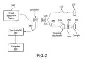
- FIG. 2is a schematic diagram of an exemplary SD-OCT system
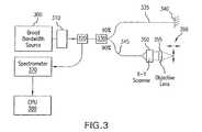
- FIG. 3is a schematic diagram of an exemplary OCM system that utilizes a broad bandwidth source
- FIG. 4 a - 4 bare schematic diagrams of an exemplary FFOCM system that utilizes a broad bandwidth source
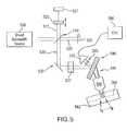
- FIG. 5is a schematic diagram of an exemplary fluorescence SECM system that utilizes a broad bandwidth source
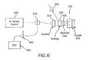
- FIG. 6is a schematic diagram of an exemplary SECM system that utilizes a wavelength tuning source
- FIG. 7is a schematic diagram of an exemplary OFDI system that utilizes a wavelength tuning/modulated source
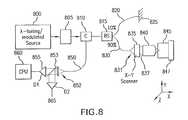
- FIG. 8is a schematic diagram of an exemplary OCM system that utilizes a wavelength modulated/tuning source
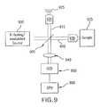
- FIG. 9is a schematic diagram of an exemplary FFOCM system that utilizes a wavelength modulated/tuning source
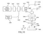
- FIG. 10is a schematic diagram of an exemplary combined SECM/SD-OCT/OCM system that utilizes a broad bandwidth source according to a first exemplary embodiment of the present invention
- FIG. 11is a schematic diagram of an exemplary combined SECM/SD-OCT/FFOCM system that utilizes a broad bandwidth source according to a second exemplary embodiment of the present invention
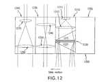
- FIG. 12is a schematic diagram of exemplary multimodality microscope sliders according to a particular exemplary embodiment of the present invention.
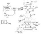
- FIG. 13is a schematic diagram of an exemplary combined SECM/OFDI/OCM system that utilizes a wavelength tuning source according to a third exemplary embodiment of the present invention
- FIG. 14is a schematic diagram of an exemplary combined SECM/OFDI/FFOCM system that utilizes a wavelength tuning source according to a third exemplary embodiment of the present invention.
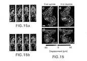
- FIGS. 15 a - 15 mare various exemplary images of Xenopus laevis hearts (stage 49) in vivo using exemplary embodiments of the TDOCT and OFDI procedures.
- FIGS. 16 a - 16 mare various exemplary three-dimensional images of Xenopus heart in vitro using exemplary embodiments of the FFOCM procedure.
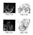
- FIGS. 17 a - 17 hare exemplary high-resolution confocal images obtained in vivo using the exemplary embodiments of the SECM procedure;
- FIGS. 18 a - 18 eare exemplary images of an aneurismal dilatation in the Xenopus heart obtained using the exemplary embodiments of the method and arrangements according to the present invention.
- FIGS. 19 a - 19 xare exemplary images of abnormal heart formation due to ethanol exposure using the exemplary embodiments of the method and arrangements according to the present invention.
- FIG. 1depicts a schematic diagram of an exemplary SECM arrangement which utilizes a broadband source.
- a quasimonochromatic or broadband light 100illuminates a circulator 110 , which alternatively may be a beam splitter.
- this circulator or beam splitteris fiber-optic coupled.
- the core of the optical fibercan serve as the pinhole for the confocal microscope system.
- the fibermay alternatively have multiple claddings that transmit light such that for example the light exciting the sample is single mode and the collected light is multimode.
- Light from this elementmay be incident on a scanning mechanism 115 that scans the angle of the beam so as to produce one or more transverse scans on the sample.
- the scanning mechanismmay alternatively be one of a resonant scanner, galvanometer scanner, polygon scanning mirror, acousto-optic scanner or the like.
- a telescope apparatusmay be used to image the scan axis to the back focal plane of the objective lens 130 .
- Light from the scanning mechanismcan then be directed towards a wavelength dispersing element 120 such as a transmission diffraction grating, prism, grating prism, dual prism grating prism (DP-GRISM) or the like.
- This exemplary elementmay disperse the different wavelengths in the broad bandwidth source so that it is incident on the objective lens 130 with varying angles that depend on wavelength.
- the lenscan have a numerical aperture that may produce a small focused spot or alternatively the lens has a high NA>0.2.
- the objective lens 130focuses each wavelength region onto the sample where each wavelength region on the sample 160 that can be located at a different spatial location.
- these exemplary elementsmay form a wavelength encoded line 140 on the sample where each position on the line is encoded by a different wavelength region. Light from the sample 160 can be reflected back through the exemplary system of FIG. 1 .
- Out-of-focus lightmay be rejected by the cladding of the optical fiber and in focus (e.g., confocal) light is transmitted back through the circulator/beam splitter 110 to a spectrometer that measures the spectral content of the returned light 145 .
- Confocal remittance as a function of spatial locationis decoded by measuring this spectrum, forming one line on an image. Successive lines are formed for each angular position of the scanning mechanism 115 , forming a spectrally-encoded confocal microscopy image.
- FIG. 2depicts a schematic diagram of an exemplary spectral-domain OCT system.
- the exemplary SD-OCTcan provide cross-sectional images of a biological specimen by using coherence gating in the Fourier domain.
- SD-OCT imagescan typically have a lower resolution ( ⁇ 3-10 ⁇ m), and may have a larger field of view (several mm's).
- a broad bandwidth or quasimonochromatic source 200can be input into an interferometer, which may be fiber optic-based.
- the fiber-coupled lightcan be transmitted to a circulator 210 and a beam splitter 220 .
- the lightcan preferably be subsequently split by a beam splitter 220 so that a portion thereof can be transmitted to a reference arm 225 and a portion is transmitted to a sample arm 235 .
- Light from the reference arm 225can be reflected off a mirror 230 (e.g., a reference) to the beam splitter 220 or alternatively transmitted back to the beam splitter 220 .
- the splitter 220can be configured so that the majority of light is transmitted to the sample arm 235 .
- Light from the sample arm fibercan then be directed towards a lens and a scanning mechanism 240 .
- the scanning mechanismcan scan the light of the sample arm 235 in arbitrary one- or two-dimensional patterns.
- Lightcan be transmitted from the scanning mechanism to a lens 250 which, in one exemplary embodiment, can have a NA so that the confocal parameter is sufficiently large to allow cross-sectional imaging in the biological specimen or sample 260 .
- Light remitted from the samplemay be transmitted back through the apparatus to the circulator/beam splitter 210 , and directed to a spectrometer 280 .
- the reflectance as a function of depth (A-line) within the tissuemay be reconstructed by, e.g., a background subtraction, remapping ⁇ -space to k-space, and inverse Fourier transformation of the spectral interference signal in a central processing unit or computer 290 . Successive A-lines are obtained for each scanning mechanism position, thereby reconstructing a cross-sectional image of the sample.
- STFTshort-time-Fourier transformation
- Doppler-sensitive SD-OCT and polarization-sensitive SD-OCTmay be also utilized to extract additional information from the biological specimen, such as absorption, flow, and birefringence.
- FIG. 3depicts a schematic diagram of an exemplary optical coherence microscopy (“OCM”) system.
- OCMoptical coherence microscopy
- the exemplary OCM systemcan utilize a combination of confocal microscopy and OCT techniques that may be advantageous, as the axial point spread functions of both such exemplary techniques may be multiplied so as to provide a greater degree of optical sectioning.
- light from a broad bandwidth sourcecan be input into a modulating element 310 so that the modulation frequency approximates that of the spectral interference in the interferometer.
- This exemplary modulation elementmay be one of a Michelson interferometer, pulse shaping apparatus, spectral filter, etc.
- the modulationmay also shift the spectral phase by some amount over time so that successive spectra may be subtracted to extract only the spectral interference term.
- the lightcan be transmitted to a circulator/beam splitter 320 and then, if a circulator is used, to a beam splitter 330 .
- Lightcan again be transmitted to a reference arm 335 and a sample arm 345 .
- Light from the reference arm 335is reflected by a mirror 340 .
- Light from the sample arm 345can be transmitted to an x-y scanner 350 , which can alternatively be one of a or a combination of a resonant scanner, galvanometer scanner, polygon scanning mirror, acousto-optic scanner or the like.
- Light from the scanner 350can be directed to an objective lens 355 so that a tightly focused spot can be scanned within the sample.
- the objective lens or sample 360may be alternatively scanned in any of three dimensions to facilitate data collection from different portions within the sample.
- Lightis transmitted back from the sample 360 to the circulator/beam splitter 320 and subsequently to a detection apparatus.
- the detectoris a spectrometer and OCM data is obtained by obtaining A-lines from the sample in a similar manner as performed by the exemplary SD-OCT.
- the detectorcan alternatively be a photodiode or other single detector that is synchronized to the source modulation element 310 . Exemplary lock-in or subtraction techniques can be utilized to extract the OCM signal.
- FIG. 4Adepicts a schematic diagram of an exemplary FFOCM system, where broad bandwidth light 400 is transmitted to a beam splitter 410 .
- Lightis split into the sample arm 423 and the reference arm 422 .
- Light in the reference arm 422may be directed 415 towards a reference objective lens 420 and to a mirror 425 , which is capable of an axial motion.
- Light in the sample arm 423may be directed towards a sample objective lens 430 and to the sample 440 .
- the reference and sample objectives 420 , 430have the similar characteristics.
- the objective lenses 420 , 430may be optimized for use with immersion fluid that has a refractive index that is similar to the sample.
- the samplecan be coupled to a stage 443 that provides motion in any of three-dimensions.
- Light from the reference arm 422 and the sample arm 423can be imaged using a lens 445 onto a CCD camera 450 . Fringes are detected by the CCD camera 445 resulting from the interference of the sample arm 422 and the reference arm 423 .
- Multiple imagescan be typically detected for different positions of the reference arm mirror 425 .
- the exemplary imagesmay be arithmetically combined to extract the information from an optical section within the sample.
- a broad bandwidth light source 451can be coupled into a modulating element, such as a Michelson interferometer or other interferometer (e.g. Mach-Zehnder, Sagnac) or spectrum altering unit.
- a modulating elementsuch as a Michelson interferometer or other interferometer (e.g. Mach-Zehnder, Sagnac) or spectrum altering unit.
- a beam splitter 452For the Michelson interferometer case, light from the source 451 can be transmitted to a beam splitter 452 . Light may then be split into two arms for exemplary arm A 453 and arm B 455 . Light from arm A 453 is transmitted to a mirror and backreflected back to a beam splitter. Light from arm B 455 is likewise transmitted to a mirror 456 , and backreflected back to a beam splitter 452 .
- can be set to be substantially equal to the path length difference between the reference and sample arms in the second interferometer.
- At least one of the arms A or Bcan be configured to change path lengths or produce a phase shift in the light therein.
- the path lengthmay be changed by a motion of one of the mirrors or a rapidly scanning optical delay line. The motion may be actuated by a piezoelectric transducer, galvanometer, linear motor or the like. Alternatively, path length changes may be generated by one of an acousto-optic modulator or electro optic modulator.
- Both reference and sample arm lightmay be combined at the beam splitter, and transmitted to another static interferometer with beam splitter 459 , separating light into a reference arm 458 and a sample arm 457 , respectively.
- Light from both arms 457 , 458can illuminate objective lenses 460 , 470 , respectively, which are substantially similar.
- the reference objective lens 460can be brought to a focus on a reflector 465 , which is typically not moving, whereas in the sample arm the sample objective lens 470 focuses the sample arm light on or within the sample 480 .
- the sample 480 or the sample objective lens 470may be mounted to a stage 481 , capable of moving the sample 480 in any of three-dimensions, under manual control or computer control.
- the path length difference between the path lengths of the reference arm 458 and the sample arm 457may be substantially equal to
- Light from reference and sample arms 458 , 457 , respectively,can be combined at a beam splitter 459 , and imaged onto a CCD array 490 or array of detectors via a lens 485 .
- a FFOCM image or datacan be generated by a linear combination of images acquired by CCD 490 and while moving or at different positions of mirror 456 . Processing, display and storage of FFOCM images is provided by a CPU 495 . Accumulations or averages are utilized to increase signal to noise ratio.
- FIG. 5depicts an exemplary embodiment of a SECM system configured for a fluorescence detection and using a broad bandwidth source.
- light from the source 500can be transmitted to a beam splitter 510 , which splits light into two paths 515 and 520 . Both arms/paths terminate on mirrors 520 and 525 , with at least one arm having a path length or phase that changes over time.
- Light returned from both arms 530can be coupled to the beam splitter 510 and directed 535 towards a SECM probe containing a grating or dispersive element 540 , an objective lens 550 .
- the arrangement of the grating and the objective lens 550focuses a spectrally encoded line 560 on or within the specimen 562 which may be mounted to a three-dimensional stage.
- Fluorescent light within the samplecan be excited by the illuminating light, transmitted back through the objective lens 550 , imaged by another lens 565 onto a detector 570 .
- Detected lightcan be digitized and converted to a line in an image by a processing arrangement (e.g., CPU) 580 . Additional lines in the image may be generated at different positions of the beam scanning mechanism 537 .
- Nonlinearities in the moving mirrorcan be corrected by an exemplary interferometer 521 that has a narrow bandwidth source that illuminates the same moving mirror 520 .
- FIG. 6depicts a schematic diagram of an exemplary embodiment of an SECM system that uses a wavelength tuning source 600 .
- the source 600can be coupled into a circulator/beam splitter 610 .
- light from the splitter 610is transmitted via an optical fiber to a scanner, which alternatively may also contain a telescope lens imaging system that projects the scan axis to the back focal plane of the objective lens 625 .
- Light from the scanning mechanismis transmitted to a dispersive element 620 (such as a diffraction grating, prism, GRISM, or DP-GRISM, etc.).
- Light from 620is transmitted to an objective lens 625 , with preferably a high NA, which can focus the beam within the sample 635 .
- one wavelength from the wavelength swept source 600can illuminate a distinct portion of the sample.
- the beamcan be scanned along a line 630 within the sample 635 .
- Remitted light from the sample 635can be transmitted back through the elements 625 , 620 , and 615 , respectively, spatially filtered by the optical fiber or a pinhole and transmitted back to the circulator beam splitter 610 .
- Light from the splitter 610can be directed to a detector 640 , and digitized by a processing arrangement (e.g., CPU) 650 , displayed and digitally stored.
- a single line in the imageis obtained following one full sweep of the wavelength-tuning source. Lines may be acquired at different positions of the scanning mechanism to form the image.
- Fluorescent light excited by the wavelength-tuning source 600 remitted from the samplecan be alternatively detected by a detector 660 to form a fluorescent image.
- FIG. 7depicts a schematic diagram of an exemplary OFDI system.
- a wavelength tuning sourcemay be coupled to an optical fiber-based circulator 705 and a beam splitter 705 .
- Light from the circulator 705can be transmitted to the beam splitter 705 , configured to send a majority of light, in the preferred embodiment, to the sample arm 725 .
- Such split light forwarded to the reference arm 715can be terminated by a reflector 720 , and sent back to the beam splitter 710 and the circulator 705 .
- Light in the sample arm 725is transmitted to a scanning mechanism 730 and an imaging lens 735 that has a NA sufficiently low to allow cross-sectional imaging of the biological specimen 740 .
- Lightis reflected from the reference mirror 720 and the sample 740 , recombined at the circulator 705 , and directed by an optical fiber 750 to a detector apparatus 755 , which in an exemplary embodiment can contain dual-balanced detectors.
- Lightis digitized by the detector apparatus 755 and the digital signal is transmitted to a CPU 760 .
- Spectral interferenceis processed in a manner similar to the processing using the exemplary SD-OCT system/procedure, e.g., the background is subtracted, ⁇ -space is converted to k-space, and an inverse Fourier transform is performed to produce an A-line.
- A-linescan be acquired as a function of scanning mechanism position, creating a cross-sectional OFDI image.
- FIG. 8depicts a schematic diagram of an exemplary embodiment of an OCM system which utilizes a wavelength tuning/modulated source.
- a wavelength modulation arrangement 805may produce a spectral pattern on the source, for example, a sinusoidal modulation of the spectrum, which may be altered over time to correspond to spectral interference modulation produced by interference between the sample and reference arms.
- Light from the source 800 and/or the modulation arrangement 805can be coupled into a fiber-optic circulator/beam splitter 810 , and subsequently transmitted to a beam splitter 815 which preferably directs a majority of light to the sample arm 830 .
- Light in the reference arm 820is directed towards a reference reflector 825 or a transmission element.
- Light in the sample arm 830can be transmitted to an x-y scanner, which may comprise one or more of galvanometers, resonant scanners, polygon scanners, acousto-optic scanners, electro optic scanners, etc.
- Light from the scannercan be alternatively transmitted to a telescope 837 and an objective lens 840 with preferably a high NA.
- the objective lens 840focuses the light within the sample 845 , which is alternatively affixed to a three-dimensional stage 847 .
- Lightis returned from the sample back through the elements 840 , 837 and 835 and coupled back into preferable the core of an optical fiber or pinhole in the sample arm 831 to reject out-of-focus light.
- Lightis directed to the circulator 810 and transmitted to a detector 855 , digitized and transmitted to a CPU 860 .
- OCM datacan be obtained by obtaining A-lines from the sample in a similar manner to the way it is performed using the exemplary OFDI system and procedure.
- the detectorcan be synchronized to the source modulation element 805 .
- Lock-in or subtraction techniquescan be utilized to extract the OCM signal in this case.
- An exemplary imagecan be generated by acquiring data for each position of the x-y scanning mechanism 835 . Fluorescent light remitted from the sample can be further detected by use of a dichroic mirror or filter 853 and a second detector 865 .
- FIG. 9depicts an exemplary embodiment of an FFOCM system that utilizes a wavelength-tuning/modulated source 900 .
- the light sourcemay be tuned over its bandwidth or alternatively be modulated to contain a spectral modulation frequency substantially similar the frequency provided by spectral interference modulation of the interferometer.
- Light from the source 900may be coupled into a beam splitter 905 , and directed to a sample arm 910 and a reference arm 915 , respectively, which are terminated by respective objectives 920 , 930 .
- the reference arm objective lens 920focuses reference arm light onto a reflector, which is subsequently returned to the beam splitter 905 .
- Sample arm lightis focused by 930 onto or within the specimen 935 .
- Light remitted from the sampleis combined with the reference arm light at 905 , and imaged by a lens 940 onto a CCD array 950 . Images can be obtained for each wavelength of the wavelength swept source or different modulation patterns of the source and arithmetically combined by a CPU 960 to reconstruct an exemplary FFOCM optical section.
- the exemplary systems described above and alternative exemplary embodiments thereofmay be combined to form a multimodality imaging system.
- This exemplary combination of systems and/or devicescan be provided by creating separate systems, and configuring their optics so that they can obtain images from the same portions of the biological specimen. Different wavelength, scanning, and detection mechanisms may be provided in such combined modality system.
- the different devicescan be implemented using many common components, which they share to provide a more efficient, cost-effective apparatus.
- FIG. 10depicts a schematic diagram of a multimodality system according to an exemplary embodiment of the present invention that utilizes a broad bandwidth source 1000 and spectrometer 1080 to provide simultaneous and co-registered SD-OCT, OCM, SECM, and fluorescence SECM data and/or images.
- light from the broad bandwidth source 1000can be coupled alternatively to a spectral modulation unit 1005 .
- Light from the spectral modulation unit 1005is coupled into a circulator 1010 and a beam splitter 1015 . If a circulator is utilized, light from the circulator 1010 is transmitted to the beam splitter 1015 that preferably directs a majority of light to the sample.
- Reference arm 1020Light in the reference arm 1020 is transmitted to a reference reflector 1025 that may move or otherwise change the path length of 1020 , and/or which can be non-movable. If the reference arm is allowed to move, conventional time-domain OCT (e.g., TD-OCT) arrangement and/or procedures may be implemented or complex spectral domain may be obtained using the exemplary SD-OCT arrangement and/or procedures using processes that are known in the art.
- TD-OCTtime-domain OCT
- Light in the sample arm 1030is transmitted to a filter/dichroic/WDM apparatus 1035 that transmits the sample arm light in the direction from the beam splitter to the sample.
- Light from 1035is directed to a beam scanning mechanism 1040 that is capable of scanning the beam in two directions at high or slow speeds.
- the beam scanning mechanism 1040may also contain a telescope for imaging the scanners onto the back focal plane of the lens 1055 .
- Light from the scanning mechanism 1040can be transmitted to a slider 1045 that contains multiple optical elements. For example, when the slider 1045 is positioned at a distinct position, either one or more or a combination of SD-OCT, OCM, SECM and/or fluorescence OCM arrangements/procedures can be implemented.
- Light from the slider 1045can be transmitted to an objective lens 1055 mounted to a lens turret in one embodiment that is capable of changing objective lenses.
- the slider 1045 and/or turret 1050may be under computer control for automatic selection of imaging modality.
- Lightis focused by objective lens 1055 onto or within the sample 1060 , which may be mounted to a computer-controlled three-dimensional translation stage 1065 .
- Reflected lightis transmitted back through the apparatus to 1010 , which redirects the light to a spectrometer. Detected reflected light is processed to form exemplary SD-OCT, OCM, SECM images using the arrangements and/or procedures described herein.
- fluorescent lightmay be redirected to a second detector via the filter/dichroic mirror/WDM apparatus 1035 to a second detector 1075 .
- Fluorescent light from 1075is utilized to reconstruct a fluorescent confocal image of the biological sample 1060 .
- a visible aiming beammay be coupled into the exemplary system, coincident with the near-infrared light, to allow visualization of the locations of imaging.
- a white light image of the specimen under investigationmay be provided by use of an alternative imaging port on the microscope.
- FIG. 11An alternative exemplary multimodality embodiment configured to provide SD-OCT, OCM, SECM, and FFOCM images and data according to the present invention at a different wavelength from the other three modalities is depicted in FIG. 11 .
- a broad bandwidth source 1100is coupled alternatively to a spectral modulation unit 1105 .
- Light from the spectral modulation unit 1105is coupled into a circulator 1110 and a beam splitter 1115 . If the circulator 1110 is utilized, light from the circulator 1110 can be transmitted to the beam splitter 1115 that preferably directs a majority of light to the sample.
- TD-OCTtime-domain OCT
- exemplary conventional time-domain OCT (TD-OCT) procedures or complex spectral domainmay be utilized for SD-OCT by methods known in the art.
- Light in the sample arm 1130is transmitted a beam scanning mechanism 1135 that is capable of scanning the beam in two directions at high or slow speeds.
- the beam scanning mechanism 1135may also include a telescope for imaging the scanners onto the back focal plane of the lens 1160 .
- Light from the scanning mechanism 1135is transmitted to a dichroic splitter/WDM 1140 that transmits the excitation light for SD-OCT, OCM, and SECM modalities, and can reflect FFOCM light.
- an exemplary FFOCM system similar to that shown in FIG. 3can be coupled into the beam path via 1140 .
- Light from 1140is directed to a slider 1150 that contains multiple optical elements; when the slider may be positioned at a distinct position, either one or a combination of SD-OCT, OCM, SECM or FFOCM is provided.
- Light from the slider 1150is transmitted to an objective lens 1160 mounted to a lens turret 1155 in one embodiment that is capable of changing objective lenses.
- the slider 1150 and/or turret 1155may be under computer control for automatic selection of imaging modality.
- Lightis focused by the objective lens 1160 onto or within the sample 1165 , which may be mounted to a computer-controlled three-dimensional translation stage 1170 .
- Reflected lightis transmitted back through the apparatus to the circulator 1110 , which redirects the light to a spectrometer.
- Detected reflected lightmay be processed to form exemplary SD-OCT, OCM, SECM images by methods described herein.
- FFOCM lightmay be redirected to the FFOCM system 1175 via the filter/dichroic mirror/WDM apparatus 1140 .
- a visible aiming beammay be coupled into the exemplary system shown in FIG. 11 , coincident with the near-infrared light, to allow visualization of the locations of imaging.
- a white light image of the specimen under investigationmay be provided by use of an alternative imaging port on the microscope.
- Alternative exemplary embodiments known in the artincluding the capability to obtain spectral information from the sample by short-time-Fourier transformation (STFT) of the spectral interference, Doppler-sensitive SD-OCT and polarization-sensitive SD-OCT, may be also utilized to extract additional information from the biological specimen, such as, e.g., absorption, flow, and birefringence.
- STFTshort-time-Fourier transformation
- Doppler-sensitive SD-OCT and polarization-sensitive SD-OCTmay be also utilized to extract additional information from the biological specimen, such as, e.g., absorption, flow, and birefringence.
- FIG. 12depicts an exemplary embodiment of an arrangement of sliders that may be utilized for the multimodality imaging according to the present invention.
- optical elementscan be contained in a housing 1200 that may be translated manually, or under computer or automatic control.
- Each slider positioncan terminate in different slider positions 1205 , 1210 , 1230 , 1260 that provide one or more imaging modalities.
- the slider position 1205 , 1210 , 1230 , 1260may be coupled to the objective lens turret.
- the slider position 1205contains no optical elements (air) or optical element windows.
- the microscopeis configured to perform FFOCM.
- a lens apparatus 1212 and 1213can be configured to expand the beam and illuminate a DP-GRISM containing two prisms 1215 and 1225 that surround a transmission grating 1220 .
- This exemplary configurationprovides an ability to perform the SECM imaging. Exemplary OCM procedures can also be conducted in this position using a scanning mechanism that scans the spectrally-encoded line across the sample.
- a lens apparatus 1240 , 1250can be configured to image beam angle, with or without beam magnification. This slider position 1230 can provide imaging using exemplary SDOCT procedures.
- a lens apparatus 1270 , 1280is configured to expand the scanned beam to allow imaging using the exemplary OCM procedures.
- exemplary embodiments of combined systemscan also include wavelength tuning/modulated sources and single or multiple detector configurations, and such exemplary embodiment is shown in FIG. 13 .
- a wavelength tuning/modulated source 1300is coupled into a circulator 1305 and a beam splitter 1310 . If a circulator is utilized, light from the circulator 1305 is transmitted to a beam splitter 1310 that preferably directs a majority of light to the sample.
- Light in the reference arm 1315is transmitted to a reference reflector 1320 that may be stationary, and may or otherwise change the path length of 1315 .
- TD-OCTtime-domain OCT
- a filter/dichroic/WDM apparatus 1330that transmits the sample arm light in the direction from the beam splitter to the sample.
- Light from 1330is directed to a x-y beam scanning mechanism 1335 that is capable of scanning the beam in two directions at high or slow speeds.
- the beam scanning mechanism 1335may also include a telescope for imaging the scanners onto the back focal plane of the lens 1353 .
- Light from the scanning mechanism 1335is transmitted to a slider 1340 that contains multiple optical elements; when the slider is positioned at a distinct position, either one or a combination of OFDI, OCM, SECM or fluorescence OCM modalities can be provided.
- Light from the slider 1340is transmitted to an objective lens 1353 mounted to a lens turret 1350 in one embodiment that is capable of changing objective lenses.
- the slider 1340 and/or turret 1350may be manual, under computer control for automatic selection of imaging modality.
- Lightis focused by objective lens 1353 onto or within the sample 1355 , which may be mounted to a computer-controlled three-dimensional translation stage 1360 .
- Reflected lightis transmitted back through the apparatus to 1305 , which redirects the light to a detector apparatus 1380 suitable for detecting OFDI, wavelength tuning OCM or SECM signals, images and/or data.
- Detected reflected lightis processed by a CPU 1385 to form exemplary OFDI, OCM, SECM images by methods described above.
- Fluorescent lightmay be redirected to a second detector via the filter/dichroic mirror/WDM apparatus 1330 to a second detector 1370 .
- Fluorescent light from 1370is utilized to reconstruct a fluorescent confocal image of the biological sample 1355 .
- a visible aiming beammay be coupled into the system, coincident with the near-infrared light, to allow visualization of the locations of imaging.
- a white light image of the specimen under investigationmay be provided by use of an alternative imaging port on the microscope.
- FIG. 14Another exemplary multimodality embodiment of a system according to the present invention which is configured to provide OFDI, OCM, SECM, and FFOCM images, data and other information, where FFOCM signal is provided at a different wavelength from the other three modalities, is depicted in FIG. 14 .
- a wavelength tuning source 1400is coupled alternatively to a spectral modulation unit 1405 .
- Light from the modulation unit 1405is coupled into a circulator 1410 and a beam splitter 1415 . If the circulator 1410 is utilized, light from the circulator 1410 is transmitted to the beam splitter 1415 that preferably directs a majority of light to the sample.
- TD-OCTtime-domain OCT
- a beam scanning mechanism 1435that is capable of scanning the beam in two directions at high or slow speeds.
- the beam scanning mechanism 1435may also contain a telescope for imaging the scanners onto the back focal plane of the lens 1465 .
- Light from the scanning mechanism 1435is transmitted to a dichroic splitter/WDM 1445 that transmits the excitation light for OFDI, OCM, and SECM, but reflects FFOCM light.
- An exemplary FFOCM system similar to the system(s) of FIG. 3 and/or FIG. 4can be coupled into the beam path via the dichroic splitter/WDM 1445 .
- Light from the dichroic splitter/WDM 1445is directed to a slider 1455 that contains multiple optical elements; when the slider 1455 is positioned at a distinct position, either one or a combination of OFDI, OCM, SECM or FFOCM data and/or images is provided.
- Light from the slider 1455is transmitted to an objective lens 1465 mounted to a lens turret 1460 in one exemplary embodiment that is capable of changing the objective lenses.
- the slider 1455 and/or a turret 1460may be under computer control for an automatic selection of imaging modality.
- Lightcan be focused by objective lens 1465 onto or within the sample 1470 , which may be mounted to a computer-controlled three-dimensional translation stage 1475 .
- Reflected lightis transmitted back through the apparatus to 1410 , which redirects the light to a spectrometer.
- Detected reflected lightis processed to form OFDI, OCM, SECM images by methods described herein.
- FFOCM lightmay be redirected to the FFOCM system 1450 via the filter/dichroic mirror/WDM apparatus 1445 .
- a visible aiming beammay be coupled into the exemplary system, coincident with the near-infrared light, to allow visualization of the locations of imaging.
- a white light image of the specimen under investigationmay be provided by use of an alternative imaging port on the microscope.
- Doppler-sensitive OFDI and polarization-sensitive OFDImay be also utilized to extract additional information from the biological specimen, such as absorption, flow, and birefringence.
- the microscopecan be configured to allow imaging from both sides of the sample.
- SDOCT, SECM and OCM procedurescan be performed from above the sample, and FFOCM procedures may be performed with the imaging lens illuminates the sample from below.
- the samplecan be mounted between a microscope slide and a thin cover glass, to allow imaging from both sides.
- the exemplary systems described hereincan provide a multimodality imaging of biological specimens in a variety of different formats, speeds, resolutions, fields of view, and contrast mechanisms.
- Each image data setmay be two- or three-dimensional, and may be co-registered to the data sets of the other respective imaging modalities.
- Computer processing methods known in the artmay be utilized to display the different data sets in a variety of different imaging formats including three-dimensional volume visualization, four-dimensional representations, and processed two-, three- and four-dimensional data sets, where the processing apparatus is configured to highlight important areas of interest. Any one or more datasets may be displayed with respect to the other and a comprehensive, all-inclusive dataset may be derived from a combination of the individual data sets.
- Quantitative informationmay be derived from the data sets in their two-, three-, and four-dimensional contexts.
- Image datamay also be combined with conventional fluorescent or brightfield images of the biological specimen.
- axial rangingis performed by use of low coherence reflectometry where the individual depth points are probed sequentially in time.
- the frame ratewas 20 per second (2 kHz A-line rate, 100 ⁇ 500 pixels).
- Exemplary OFDI procedures and systemscan use a frequency domain reflectometry in which all depth points are acquired simultaneously. This technique provides a several-hundred-fold improvement in signal-to-noise ratio (SNR) as described in M. A. Choma et al. “Sensitivity advantage of swept source and Fourier domain optical coherence tomography,” Optics Express 11, pp 2183-2189 (2003); and S. H. Yun et al., “High-speed optical frequency-domain imaging,” Optics Express 11, pp. 2953-2963 (2003).
- the exemplary OFDI systems and procedurescan use a rapidly swept, wavelength tunable laser as a light source.
- the laserfeatured a sweep repetition rate of up to 64 kHz, a wide tuning range of 111 nm centered at 1320 nm, and a high average output power of 30 mW (7 mW on the tissue).
- the axial resolutionwas 10 ⁇ m in tissue.
- the systemfurther comprised an acousto-optic frequency shifter (25 MHz) to remove the depth degeneracy inherent in the frequency-domain reflectometry, as described in S. H. Yun et al., “Removing the depth-degeneracy in optical frequency domain imaging with frequency shifting,” Optics Express 12, pp. 4822-4828 (2004).
- Polarization-diversity detectionwas implemented to eliminate polarization artifacts in the fiber-based OFDI system.
- Dual-balanced photoreceiverswere used to improve imaging sensitivity through the reduction of laser intensity noise.
- the photoreceiver outputswere digitized with a 2-channel analog-to-digital converter at a sampling rate of 100 MHz with 14-bit resolution.
- Exemplary TDOCT and high-speed OFDI configurationwere incorporated into a dissecting light microscope.
- the scanning systemwas comprised of a collimating lens (5 mm beam diameter), two synchronized galvanometric scanners for transverse scanning, a focusing lens (50 mm focal length), and a small mirror that deflected the beam downward toward the sample.
- the transverse resolutionwas 16 ⁇ m with a confocal parameter of 330 ⁇ m.
- Displacements associated with local cardiac motionwere determined directly from the volumetric data by subtracting the heart surface locations at end diastole from those at end systole on a frame-by-frame basis. Displacement was displayed using a color look up table. Volumetric rendering and three-dimensional visualization was accomplished by using OsiriX software.
- High-resolution OFDI procedurewas performed using a laser source with 200 nm tuning range, centered at 1250 nm, in which two semiconductor optical amplifiers were utilized as the gain media, as described in W. Y. Oh et al., “Wide tuning range wavelength-swept laser with two semiconductor optical amplifiers,” IEEE Photonics Technology Letters 17, pp. 678-680 (2005).
- An axial resolution of 4 ⁇ m in tissuewas achieved.
- the imaging ratewas 40 frames per second with an A-line rate of 20 kHz (500 A-lines per frame).
- Polarization-diversity and dual-balanced detectionwas performed and the photoreceiver outputs were digitized with a 2-channel analog-to-digital converter at a sampling rate of 10 MHz with 12-bit resolution.
- FFOCMis an interferometric technique that utilizes two-dimensional parallel detection to provide subcellular resolution images of reflected light within biological specimens, as described in A. Dubois et al., “Ultrahigh-resolution full-field optical coherence tomography,” Appl Opt 43, pp. 2874-2883 (2004), and A. Dubois et al., “Three-dimensional cellular-level imaging using full-field optical coherence tomography,” Phys Med Biol 49, pp. 1227-1234 (2004).
- Interference imageswere captured with a CMOS area scan camera with spectral response centered at 650 nm.
- the transverse resolutionswere 2 ⁇ m and axial resolution, 1.1 ⁇ m.
- Acquisition timewas 2 seconds per frame for a transverse field of view of approximately 700 ⁇ m ⁇ 700 ⁇ m.
- Three-dimensional datawas obtained by moving the sample through the focus at 1 ⁇ m increments. Volumetric rendering and visualization was accomplished by using OsiriX software.
- SECMis a reflectance confocal microscopy technique, which uses near-infrared light that allows deeper penetration into tissue, as described in R. R. Anderson et al., “The optics of human skin,” J Invest Dermatol 77, pp. 13-19 (1981), compared with confocal microscopes that utilize visible light.
- Exemplary SECM techniquediffers from conventional laser scanning confocal microscopy in that it projects different wavelengths onto distinct locations on the sample, as described in G. J. Tearney et al., “Spectrally encoded confocal microscopy,” Optics Letters 23, pp. 1152-1154 (1998). Rapid acquisition of spectra returned from the sample enables high-speed reconstruction of the image.
- a multimode fiberwas used for signal collection, resulting in 0.9 ⁇ m transverse and 2.5 ⁇ m axial resolutions. Images comprised of 500 ⁇ 500 pixels were acquired at 10 frames per second. The maximum imaging depth was limited to the 280 ⁇ m working distance of the objective lens.
- Xenopus laevis frogswere purchased from Nasco (Fort Atkinson, Wis.). Animal procedures were performed according to the approved protocols of Massachusetts General Hospital Subcommittee on Research Animal Care. Embryos were obtained by in vitro fertilization, incubated in 0.1 ⁇ Marc's modified Ringer's medium (MMR)(as described in J. Newport et al., “A major development transition in early Xenopus embryos: 1. Characterization and timing of cellular changes at the midblastula stage,” Cell 30, pp. 675-686, 1982), and staged according to Nieuwkoop and Faber tables. (see P. D. Nieuwkoop and J. Faber, Normal table of Xenopus laevis, Daudin , North-Holland Publishing Company, Amsterdam, 1967).
- MMRMarc's modified Ringer's medium
- Ethanol treatmentswere performed in 0.1 ⁇ MMR (vol/vol), soon after Mid Blastula Transition (stage 8.5) (as described in R. Yelin et al., “Ethanol exposure affects gene expression in the embryonic organizer and reduces retinoic acid levels,” Dev Biol 279, pp. 193-204 (2005).) until imaging.
- stage 8.5Mid Blastula Transition
- embryosPrior to in vivo imaging, embryos were anesthetized using 0.02% 3-aminobenzoic acid ethyl ester (A-5040, Sigma).
- A-50403-aminobenzoic acid ethyl ester
- Plastic Histology sections(as described in A. M. Glauert, Fixation, Dehydration and Embedding of Biological Specimens , North-Holland Publishing Company Amsterdam, 1986) were obtained after additional fixation in Karnovsky's Fixative (KII) and embedding in tEpon-812 (Tousimis). Sections of 1 ⁇ m thick were cut on a Reichert Ultracut Microtome and stained with methylene blue/toluidine blue in borate buffer (Tousimis). Paraffin sections (5 ⁇ m thickness) were stained with Hematoxylin & Eosin.
- Rapid volumetric imaging of the beating heartenables the evaluation of three-dimensional morphology and function during the cardiac cycle.
- TDOCTwhich provides cross-sectional imaging in vivo (as shown in FIGS. 15 a and 15 b )
- the exemplary OFDI system and procedurecan image at much higher frame rates, making four-dimensional imaging of the beating heart without cardiac gating possible.
- Volumetric OFDI images of the Xenopus heart (stage 49)were acquired at a rate of 20 three-dimensional data sets per second (as shown in FIGS. 15 c - 15 g ).
- OFDI cross-sections of a stage 49 Xenopus heartwere obtained in vivo (as shown in FIGS. 15 i - 15 m ) using a broadband (e.g., 200 nm) wavelength-swept source, as described in W. Y. Oh et al., “Wide tuning range wavelength-swept laser with two semiconductor optical amplifiers,” IEEE Photonics Technology Letters 17, pp.
- a broadband wavelength-swept sourcee.g. 200 nm
- the transverse and axial resolutions of high resolution OFDI resultswere 2 ⁇ m and 4 ⁇ m, respectively.
- Details within the three-chamber Xenopus heartcan be clearly resolved with the high-resolution OFDI procedures and systems, including atrioventricular valve dynamics (as shown in FIGS. 15 i - 15 k ), ventricular contractions, and trabecular dynamics ( FIG. 15 m ).
- Individual blood cellscan also be seen, flowing from the atrium to the ventricle through the atrioventricular valve (as shown in FIG. 15 k ).
- Exemplary FFOCM procedures and systemsoffer the capability to image microstructure of the embryonic heat with nearly isotropic cellular level resolution.
- Volumetric FFOCM imagesspanned a field of view of 700 ⁇ 700 ⁇ 1000 ⁇ m (axial). The transverse and axial resolutions were 2 ⁇ m and 1.1 ⁇ m, respectively. Acquisition time was 2 seconds for a single en face section, and 33 minutes for the entire volume.
- Exemplary FFOCM sections of the Xenopus heart (stage 49)allow visualization of ventricular trabeculae (as shown in FIGS. 16 a and 16 c ), the spiral valve (as shown in FIGS. 16 b and 16 d , see arrows), and the partial atrial septum (as shown in FIG.
- FIG. 16 i and 16 jA magnified view of the atrioventricular valve shown (as shown in FIG. 16 l ) next to a corresponding histology section of the same embryo (as shown in FIG. 16 m ), demonstrates its bicuspid morphology.
- Exemplary SECM procedures and systemsprovide a transverse resolution comparable to those associated with FFOCM, but at higher frame rates, enabling microscopy of the heart in vivo.
- the Xenopus myocardium (stage 49)was imaged in vivo using the exemplary SECM procedures and systems at a frame rate of 10/s, a field of view of 220 ⁇ 220 ⁇ m, and transverse and axial resolutions of 1.2 and 6 ⁇ m, respectively.
- the maximum penetration depthwas 280 ⁇ m.
- FIGS. 17 a - 16 mprocedures and systems, show the thin cusps of the atrioventricular valve (as shown in FIG. 17 a ), approximately 280 ⁇ m below the ventral surface, and parts of the ventricle and TA (as shown in FIG. 17 c ), containing individual blood cells within the intratrabecular spaces.
- SECM imagescorrelated well with corresponding histology sections (as shown in FIGS. 17 b and 17 d ).
- a series of frames from a different tadpole (stage 47)demonstrates the spiral valve as it closes (as shown in FIG. 17 e ) and opens (as shown in FIGS.
- stage 47a protrusion emanating from the TA wall has been identified.
- SECM sections obtained in vivo at two different depthsreveal its saccular shape, its location with respect to spiral valve, as well the flow of individual blood cells through the defect. This abnormality was also observed using the exemplary TDOCT procedures and systems in vivo (as shown in FIG. 18 a , see inset).
- the embryowas then fixed and imaged with the exemplary FFOCM procedures and systems.
- An FFOCM section(as shown in FIG. 18 c ) and a three-dimensional rendering of the FFOCM volumetric data set (as shown in FIG.
- Cardiovascular malformationcan be caused by genetic (as described in K. L. Clark et al., “Transcription factors and congenital heart defects,” Annu Rev Physiol 68, pp. 97-121 (2006)) and teratogenic factors (as described in S. M. Mone et al., “Effects of environmental exposures on the cardiovascular system: prenatal period through adolescence,” Pediatrics 113, pp. 1058-1069 (2004)).
- Ethanolis a well-known teratogen; exposure of human embryo during pregnancy to alcohol (ethanol) is associated with Fetal Alcohol Syndrome (FAS).
- FASFetal Alcohol Syndrome
- a tadpole(stage 48) has been selected from each of the control, 0.5%, 1.5%, and 2.0% ethanol treated groups to demonstrate typical phenotypes (as shown in FIGS. 19 a - 19 d ). It was determined that the four tadpoles' hearts were in advanced developmental stages by identifying the existence of a partial atrial septum (as shown in FIGS. 19 a - 19 d , see right images, septa marked by arrows) and an atrioventricular valve. The TDOCT images provided the first indication of damaged looping in the 1.5% and 2.0% groups.
- FIGS. 19 e - 19 hPhotographs of the tadpoles, taken in vivo from the ventral aspect, are shown in FIGS. 19 e - 19 h.
- FIGS. 19 i - 19 lThree-dimensional rendering of data acquired with the exemplary FFOCM systems and procedures in vitro allowed evaluation of myocardial structure at high-resolution, revealing the similarity between the control and the 0.5% tadpoles and clearly showing defective heart tube looping in the tadpoles from the 1.5% and 2.0% groups (as shown in FIGS. 19 i - 19 l ).
- Sections through the FFOCM volumetric data setsdemonstrated smaller, distorted TA's and spiral valves (marked by arrows) in the 1.5% (as shown in FIG. 19 o ) and 2.0% embryos (as shown in FIG. 19 p ) compared with the control (as shown in FIG. 19 m ) and the 0.5% (as shown in FIG.
- a common paradigm in developmental biology researchis to manipulate the genotype and monitor the phenotype. Morphology is an important aspect of the phenotype. In the heart, even slight morphological and dynamical abnormalities may be critical for proper myocardial function. An ability to identify subtle morphological and dynamical variations in two and three dimensions can significantly improve the sensitivity of this paradigm.
- heart structuressuch as the myocardium wall, septum and valves may only be a few cells thick. Evaluating the morphological phenotype not only requires resolving such fine structures, but also the capability to visualize these microscopic features within the beating heart, where typical displacement velocities are on the order of 1 mm/sec. If the imaging speed is sufficiently high, three-dimensional images of the embryo heart can be obtained at different times within the cardiac cycle. This exemplary four-dimensional imaging could allow reliable measurements of dynamic physiological parameters, such as stroke volume and ejection fraction, as well as valve opposition, stiffness and modularity, which have close analogs in human pathophysiology. High resolution and high speed are not the only requirements for effective imaging of the heart. In the Xenopus embryo, the heart extends from between 200 ⁇ m and 800 ⁇ m beneath the ventral surface. An effective imaging method should therefore also be capable of imaging at these depths without substantial loss of signal and resolution.
- the morphology of the developing Xenopus laevis hearthas been studied in vitro and described in detail, using three-dimensional rendering of histology sections. (See T. J. Mohun et al., “The morphology of heart development in Xenopus laevis,” Dev Biol 218, 74-88 (2000)). For histologic studies, however, sample preparation and sectioning make preserving structural fidelity difficult. As a result, imaging of intact embryos in their natural environment is preferred. Structural imaging of the heart in vivo has been demonstrated using a variety of non-invasive imaging modalities such as micro-MRI (see D. L.
- Optical techniquesenable imaging of the embryonic heart at higher resolution. Confocal microscopy has been used to image early Xenopus heart development, in vitro (as described in S. J. Kolker et al., “Confocal imaging of early heart development in Xenopus laevis,” Dev Biol 218, pp. 64-73 (2000)), and to study the role of intracardiac fluid forces in zebrafish embryonic cardiogenesis, in vivo (as described in J. R. Hove et al., “Intracardiac fluid forces are an essential epigenetic factor for embryonic cardiogenesis,” Nature 421, pp. 172-177 (2003)).
- the exemplary OFDI procedures and systemsprovided real-time, true four-dimensional imaging of a beating heart without requiring cardiac gating and was found to be useful for assessing myocardial wall displacement during the cardiac cycle (as shown in FIGS. 15 c - 15 f ).
- contrastwas generated by endogenous scattering.
- molecular imagingmay be important for relating gene and protein expression to phenotype.
- the exemplary systems and methods described hereincan be used for imaging fluorescent labels and molecular species. It has been described that fluorescence imaging can be conducted via spectral encoding by modification of the source and detection electronics. (See J. T. Motz et al., “Spectral- and frequency-encoded fluorescence imaging,” Opt Lett 30, pp. 2760-2762 (2005)). The same principles used in fluorescence SECM procedures and systems can likewise be utilized for endoscopic two-photon and second harmonic imaging.
- the natural contrast optical imaging modalities presented in this workallow evaluation of the embryonic heart from different vantage points.
- Combining OFDI, SECM, and FFOCM modalitiescan leverage their strengths (see Table 1), and provide a ability for obtaining a more comprehensive morphological and functional myocardial phenotype.
- This multi-modality paradigmcan be extended to other systems and animal models as well. Since these non-invasive imaging techniques do not alter the specimen, they can be used sequentially or in parallel.
- there is no fundamental barrier preventing their combination into one imaging system that uses a single wavelength swept source.See S. H. Yun et al., “High-speed optical frequency-domain imaging,” Optics Express 11, pp.
Landscapes
- Health & Medical Sciences (AREA)
- Physics & Mathematics (AREA)
- Life Sciences & Earth Sciences (AREA)
- General Physics & Mathematics (AREA)
- General Health & Medical Sciences (AREA)
- Engineering & Computer Science (AREA)
- Pathology (AREA)
- Biomedical Technology (AREA)
- Nuclear Medicine, Radiotherapy & Molecular Imaging (AREA)
- Molecular Biology (AREA)
- Chemical & Material Sciences (AREA)
- Biophysics (AREA)
- Radiology & Medical Imaging (AREA)
- Animal Behavior & Ethology (AREA)
- Veterinary Medicine (AREA)
- Public Health (AREA)
- Surgery (AREA)
- Medical Informatics (AREA)
- Heart & Thoracic Surgery (AREA)
- Analytical Chemistry (AREA)
- Biochemistry (AREA)
- Immunology (AREA)
- Optics & Photonics (AREA)
- Astronomy & Astrophysics (AREA)
- Theoretical Computer Science (AREA)
- Spectroscopy & Molecular Physics (AREA)
- Medicinal Chemistry (AREA)
- Urology & Nephrology (AREA)
- Hematology (AREA)
- Signal Processing (AREA)
- Pulmonology (AREA)
- Food Science & Technology (AREA)
- Mathematical Physics (AREA)
- Investigating Or Analysing Materials By Optical Means (AREA)
- Investigating, Analyzing Materials By Fluorescence Or Luminescence (AREA)
- Endoscopes (AREA)
- Microscoopes, Condenser (AREA)
- Instruments For Viewing The Inside Of Hollow Bodies (AREA)
- Automatic Focus Adjustment (AREA)
- Transforming Light Signals Into Electric Signals (AREA)
Abstract
Description
This application is a continuation of, and therefore claims priority from U.S. patent application Ser. No. 12/943,762 filed on Nov. 10, 2010, which issued as U.S. Pat. No. 8,289,522 on Oct. 16, 2012, which is a divisional application of U.S. patent application Ser. No. 11/537,343, filed on Sep. 29, 2006, which issued as U.S. Pat. No. 7,872,759 on Jan. 18, 2011, is based upon and claims the benefit of priority from U.S. Patent Application Ser. No. 60/721,802, filed Sep. 29, 2005. The entire disclosures of the applications are incorporated herein by reference.
The present invention generally relates to arrangements and methods for providing multimodality microscopic imaging of one or more biological structures, and particularly to, e.g., conducting reflectance and/or fluorescence microscopy of biological specimens using spectrally encoded confocal microscopy (“SECM”), fluorescence SECM, optical coherence tomography (“OCT”), spectral domain (“SD”)-OCT, optical frequency domain interferometry (“OFDI”), and optical coherence microscopy (“OCM”) procedures.
A determination of the relationship between the molecular basis of genetic alterations and phenotype generally utilizes accurate two- and three-dimensional characterization of microstructure of biological specimens. However, motion and small dimensions make many living biological specimens can be more difficult to evaluate.
Optical techniques offer the potential to image the biological specimens at a high resolution. For certain applications, optical imaging based on endogenous contrast can be advantageous over techniques that require exogenous agents, since such beneficial procedures can allow the analysis of the specimen in its native state and at multiple time points, with a small amount of preparation. As an example, several endogenous-contrast imaging modalities are described herein for visualizing embryonic heart microstructure: two exemplary forms of optical coherence tomography (“OCT”) as described in D. Huang et al., “Optical coherence tomography,”Science254, pp. 1178-1181 (1991), time-domain optical coherence tomography (“TD-OCT”) as described in S. A. Boppart et al., “Investigation of developing embryonic morphology using optical coherence tomography,”Dev Biol177, pp. 54-63 (1996), and optical frequency domain imaging (“OFDI”) as described in M. A. Choma et al., “Sensitivity advantage of swept source and Fourier domain optical coherence tomography,”Optics Express11, pp. 2183-2189 (2003); and S. H. Yun et al., “High-speed optical frequency-domain imaging,”Optics Express11, pp 2953-2963 (2003).
Additional examples can be provided and utilized including two reflectance microscopy techniques, e.g., full-field optical coherence microscopy (“FFOCM”) as described in E. Beaurepaire et al., “Full-field optical coherence microscopy,”Optics Letters23, pp. 244-246 (1998); A. Dubois et al., “Ultrahigh-resolution full-field optical coherence tomography,”Appl Opt43, pp. 2874-2883 (2004); and G. Moneron et al., “Stroboscopic ultrahigh-resolution full-field optical coherence tomography,”Opt Lett30, pp. 1351-1353 (2005), and spectrally encoded confocal microscopy (“SECM”) as described in G. J. Tearney et al., “Spectrally encoded confocal microscopy,”Optics Letters23, pp. 1152-1154 (1998); and C. Boudoux et al., “Rapid wavelength-swept spectrally encoded confocal microscopy,”Optics Express13, pp. 8214-8221 (2005).
For example, the TDOCT techniques can use low-coherence interferometry to obtain cross-sectional images with ˜10 μm resolution and at depths of up to 2 mm. (See S. A. Boppart et al., “Noninvasive assessment of the developingXenopuscardiovascular system using optical coherence tomography,”Proc Natl Acad Sci USA94, pp. 4256-4261 (1997); S. Yazdanfar et al., “High resolution imaging of in vivo cardiac dynamics using color Doppler optical coherence tomography,”Optics Express1, pp. 424-431 (1997); T. M. Yelbuz et al., “Optical coherence tomography: a new high-resolution imaging technology to study cardiac development in chick embryos,”Circulation106, pp. 2771-2774 (2002); V. X. D. Yang et al., “High speed, wide velocity dynamic range Doppler optical coherence tomography (Part II): Imaging in vivo cardiac dynamics ofXenopus laevis,” Optics Express11, pp. 1650-1658 (2003); and W. Luo et al., “Three-dimensional optical coherence tomography of the embryonic murine cardiovascular system”Journal of biomedical optics11, 021014 (2006).
The exemplary OFDI technique can be considered as a derivative of the TDOCT techniques that may enable an acquisition of images at significantly higher frame rates as described in R. Huber et al., “Three-dimensional and C-mode OCT imaging with a compact, frequency swept laser source at 1300 nm,”Optics Express13, pp. 10523-10538 (2005). The high speed of the OFDI techniques can enable an implementation of a true four-dimensional (4D) microscopy (e.g., three-dimensional microscopy as a function of time). Full-field optical coherence microscopy (“FFOCM”) techniques can utilize low-coherence interferometry and higher numerical aperture objective lenses to attain resolution at the subcellular level in all three dimensions. Such FFOCM techniques are likely considerably slower than the OFDI techniques. The exemplary SECM techniques can have a form of the reflectance confocal microscopy using which it may be possible to obtain two-dimensional images with micron-level resolution, at significantly higher speeds than possibly obtained using the FFOCM techniques.
While each of these natural-contrast procedures can individually be used for imaging a microstructure of the embryonic heart, when combined, these procedures can provide a powerful set of tools for two-, three-, and four-dimensional characterization of early myocardial morphology and dynamics. A combination of these different modalities into one single microscopy device may have additional advantages such as, e.g., (a) a comparison of images in different formats, different resolutions, and fields of view, (b) a simultaneous acquisition of both structural and function information, and/or (c) these tasks can be accomplished using one instrument without requiring moving or altering the specimen.
One of the objects of the present invention is to overcome certain deficiencies and shortcomings of the prior art systems (including those described herein above), and provide exemplary embodiments of providing multimodality microscopic imaging of one or more biological structures. Such exemplary embodiments can conduct reflectance and/or fluorescence microscopy of biological specimens using spectrally encoded confocal microscopy (“SECM”), fluorescence SECM, optical coherence tomography (“OCT”), spectral domain (“SD”)-OCT, optical frequency domain interferometry (“OFDI”), and optical coherence microscopy (“OCM”) procedures.
For example, an analysis of biological specimens generally employs a visualization of its microstructure and functions, preferably with small alterations to the specimen. According to one exemplary embodiment of the present invention, a combination of multiple different imaging modalities can be provided in a single microscope device. Each exemplary technique according to certain exemplary embodiments of the present invention can provide distinct and complementary imaging capabilities, including high-speed (e.g., 1000 frames per second) and high axial resolution (4-16 μm) cross-sectional imaging in vivo, true four-dimensional imaging in vivo, three-dimensional microscopy with isotropic cellular (e.g., 1-2 μm) resolution in vitro, and two-dimensional subcellular imaging in vivo. When combined, these exemplary imaging modalities can effectuate a more complete picture of the morphologic and dynamics of biological specimens.
Thus, the exemplary embodiments of the present invention include arrangements and methods for acquiring multimodality microscopic data. For example, according to one exemplary embodiment, it is possible to use a combination of unique broad bandwidth or rapid wavelength swept sources and optics interposed between a scanning mechanism and an imaging lens. Data can be acquired simultaneously and/or serially, e.g., without moving the specimen. For example, data obtained from different modalities can be co-registered so that it can be displayed side-by-side and/or overlaid on top of each other. Quantitative information can be obtained from all of the datasets in a complementary manner.
Thus, in accordance with the exemplary embodiments of the present invention, method and apparatus can be provided. For example, first data associated with a first signal received from at least one region of at least one sample can be provided based on a first modality, and second data associated with a second signal received from the at least one sample can be provided based on a second modality which is different from the first modality. Third data associated with a reference can be received. Further data can be generated based on the first, second and third data. In addition, third data associated with a second signal received from the at least one sample can be obtained. Each of the third data can be based on a further modality which is different from the first modality and the second modality, and the further data can be further determined based on the third data. Further, the first modality can be a spectral-encoded modality, and the second modality can be a non-spectral-encoding modality.
In another exemplary embodiment of the present invention, the first modality can be florescence imaging. A microscope arrangement and/or a beam-scanning arrangement can be provided. The beam-scanning arrangement may be configured to forward electro-magnetic radiation to the at least region. Further, a two-dimensional image and/or a three-dimensional image can be produced as a function of the further data. The first and second data may be obtained substantially simultaneously. In addition, the first and second data may be associated with approximately the same location on the sample, and/or can be obtained using another one of the first and second data.
According to a further exemplary embodiment of the present invention, the apparatus can be provided in a probe and/or a single enclosure. It is also possible to obtain spectral encoding microscopy information using such exemplary apparatus and method, as well as bright field, dark field, phase contrast, polarization, epireflectance and/or reflectance microscopy information. It is further possible to use such exemplary apparatus and method change from the first modality to the second modality. Optical coherence tomography information associated with a signal provided by a source arrangement having a plurality of wavelengths can be obtained. A plurality of detectors can be provided to detect a spectral interference between the second and third signals as a function of the wavelengths.
Optical coherence tomography information associated with a signal provided by a source arrangement can be obtained whose wavelength varies over time. At least one image can be generated based on the first and second data. In addition, a first image can be generated based on the first data and a second image can be generated based on the second data. The first and second images may be associated with one another as a function of the first and second data. It is possible to obtain optical coherence tomography information and/or optical frequency domain interferometry information.
Other features and advantages of the present invention will become apparent upon reading the following detailed description of embodiments of the invention, when taken in conjunction with the appended claims.
Further objects, features and advantages of the present invention will become apparent from the following detailed description taken in conjunction with the accompanying figures showing illustrative embodiments of the present invention, in which:
Throughout the figures, the same reference numerals and characters, unless otherwise stated, are used to denote like features, elements, components or portions of the illustrated embodiments. Moreover, while the subject invention will now be described in detail with reference to the figures, it is done so in connection with the illustrative embodiments. It is intended that changes and modifications can be made to the described embodiments without departing from the true scope and spirit of the subject invention as defined by the appended claims.
Exemplary SECM techniques are capable of providing subcellular level resolution images in tissue or biological specimens. SECM images can alternatively represent fluorescence from the sample or reflectance from the sample.FIG. 1 depicts a schematic diagram of an exemplary SECM arrangement which utilizes a broadband source. In this exemplary configuration, a quasimonochromatic orbroadband light 100 illuminates acirculator 110, which alternatively may be a beam splitter. In one embodiment this circulator or beam splitter is fiber-optic coupled. The core of the optical fiber can serve as the pinhole for the confocal microscope system. The fiber may alternatively have multiple claddings that transmit light such that for example the light exciting the sample is single mode and the collected light is multimode. Light from this element may be incident on ascanning mechanism 115 that scans the angle of the beam so as to produce one or more transverse scans on the sample. The scanning mechanism may alternatively be one of a resonant scanner, galvanometer scanner, polygon scanning mirror, acousto-optic scanner or the like. A telescope apparatus may be used to image the scan axis to the back focal plane of theobjective lens 130. Light from the scanning mechanism can then be directed towards awavelength dispersing element 120 such as a transmission diffraction grating, prism, grating prism, dual prism grating prism (DP-GRISM) or the like. This exemplary element may disperse the different wavelengths in the broad bandwidth source so that it is incident on theobjective lens 130 with varying angles that depend on wavelength.
In one exemplary embodiment, the lens can have a numerical aperture that may produce a small focused spot or alternatively the lens has a high NA>0.2. Theobjective lens 130 focuses each wavelength region onto the sample where each wavelength region on thesample 160 that can be located at a different spatial location. For a diffraction grating and an objective lens, these exemplary elements may form a wavelength encodedline 140 on the sample where each position on the line is encoded by a different wavelength region. Light from thesample 160 can be reflected back through the exemplary system ofFIG. 1 . Out-of-focus light may be rejected by the cladding of the optical fiber and in focus (e.g., confocal) light is transmitted back through the circulator/beam splitter 110 to a spectrometer that measures the spectral content of the returned light145. Confocal remittance as a function of spatial location is decoded by measuring this spectrum, forming one line on an image. Successive lines are formed for each angular position of thescanning mechanism 115, forming a spectrally-encoded confocal microscopy image.
Light remitted from the sample may be transmitted back through the apparatus to the circulator/beam splitter 210, and directed to aspectrometer 280. The reflectance as a function of depth (A-line) within the tissue may be reconstructed by, e.g., a background subtraction, remapping λ-space to k-space, and inverse Fourier transformation of the spectral interference signal in a central processing unit orcomputer 290. Successive A-lines are obtained for each scanning mechanism position, thereby reconstructing a cross-sectional image of the sample. Alternative exemplary embodiments known in the art, including the capability to obtain spectral information from the sample by short-time-Fourier transformation (“STFT”) of the spectral interference, Doppler-sensitive SD-OCT and polarization-sensitive SD-OCT, may be also utilized to extract additional information from the biological specimen, such as absorption, flow, and birefringence.
Light from thescanner 350 can be directed to anobjective lens 355 so that a tightly focused spot can be scanned within the sample. The objective lens orsample 360 may be alternatively scanned in any of three dimensions to facilitate data collection from different portions within the sample. Light is transmitted back from thesample 360 to the circulator/beam splitter 320 and subsequently to a detection apparatus. In one embodiment, the detector is a spectrometer and OCM data is obtained by obtaining A-lines from the sample in a similar manner as performed by the exemplary SD-OCT. In the spectral modulation embodiment the detector can alternatively be a photodiode or other single detector that is synchronized to thesource modulation element 310. Exemplary lock-in or subtraction techniques can be utilized to extract the OCM signal.
Full-field optical coherence microscopy is typically a free-space interferometric technique that utilizes a broad bandwidth source to obtain transverse, high-resolution optical sections of biological specimens.FIG. 4A depicts a schematic diagram of an exemplary FFOCM system, wherebroad bandwidth light 400 is transmitted to abeam splitter 410. Light is split into thesample arm 423 and thereference arm 422. Light in thereference arm 422, according to one exemplary embodiment, may be directed415 towards areference objective lens 420 and to amirror 425, which is capable of an axial motion. Light in thesample arm 423 may be directed towards a sampleobjective lens 430 and to thesample 440. In one exemplary embodiment, the reference andsample objectives
In another exemplary embodiment, theobjective lenses stage 443 that provides motion in any of three-dimensions. Light from thereference arm 422 and thesample arm 423 can be imaged using alens 445 onto aCCD camera 450. Fringes are detected by theCCD camera 445 resulting from the interference of thesample arm 422 and thereference arm 423. Multiple images can be typically detected for different positions of thereference arm mirror 425. The exemplary images may be arithmetically combined to extract the information from an optical section within the sample.
In another exemplary embodiment of the FFOCM system as depicted inFIG. 4B , a broad bandwidthlight source 451 can be coupled into a modulating element, such as a Michelson interferometer or other interferometer (e.g. Mach-Zehnder, Sagnac) or spectrum altering unit. For the Michelson interferometer case, light from thesource 451 can be transmitted to abeam splitter 452. Light may then be split into two arms forexemplary arm A 453 andarm B 455. Light fromarm A 453 is transmitted to a mirror and backreflected back to a beam splitter. Light fromarm B 455 is likewise transmitted to amirror 456, and backreflected back to abeam splitter 452. The difference between the path length Lain arm A and the path length Lbin arm B, |La-Lb|, can be set to be substantially equal to the path length difference between the reference and sample arms in the second interferometer. At least one of the arms A or B can be configured to change path lengths or produce a phase shift in the light therein. In one exemplary embodiment, the path length may be changed by a motion of one of the mirrors or a rapidly scanning optical delay line. The motion may be actuated by a piezoelectric transducer, galvanometer, linear motor or the like. Alternatively, path length changes may be generated by one of an acousto-optic modulator or electro optic modulator.
Both reference and sample arm light may be combined at the beam splitter, and transmitted to another static interferometer withbeam splitter 459, separating light into areference arm 458 and asample arm 457, respectively. Light from botharms objective lenses reference arm 458, thereference objective lens 460 can be brought to a focus on areflector 465, which is typically not moving, whereas in the sample arm the sampleobjective lens 470 focuses the sample arm light on or within thesample 480. Thesample 480 or the sampleobjective lens 470 may be mounted to astage 481, capable of moving thesample 480 in any of three-dimensions, under manual control or computer control.
The path length difference between the path lengths of thereference arm 458 and thesample arm 457 may be substantially equal to |La-Lb| of the first interferometer. Light from reference and samplearms beam splitter 459, and imaged onto aCCD array 490 or array of detectors via alens 485. A FFOCM image or data can be generated by a linear combination of images acquired byCCD 490 and while moving or at different positions ofmirror 456. Processing, display and storage of FFOCM images is provided by aCPU 495. Accumulations or averages are utilized to increase signal to noise ratio.
Light is digitized by thedetector apparatus 755 and the digital signal is transmitted to aCPU 760. Spectral interference is processed in a manner similar to the processing using the exemplary SD-OCT system/procedure, e.g., the background is subtracted, λ-space is converted to k-space, and an inverse Fourier transform is performed to produce an A-line. A-lines can be acquired as a function of scanning mechanism position, creating a cross-sectional OFDI image. Alternative exemplary embodiments known in the art, including the capability to obtain spectral information from the sample by short-time-Fourier transformation (STFT) of the spectral interference, complex spectral domain processing, Doppler-sensitive OFDI and polarization-sensitive OFDI, may be also utilized to extract additional information from the biological specimen, such as absorption, flow, and birefringence.
Light in thereference arm 820 is directed towards areference reflector 825 or a transmission element. Light in thesample arm 830 can be transmitted to an x-y scanner, which may comprise one or more of galvanometers, resonant scanners, polygon scanners, acousto-optic scanners, electro optic scanners, etc. Light from the scanner can be alternatively transmitted to atelescope 837 and anobjective lens 840 with preferably a high NA. Theobjective lens 840 focuses the light within thesample 845, which is alternatively affixed to a three-dimensional stage 847. Light is returned from the sample back through theelements sample arm 831 to reject out-of-focus light. Light is directed to thecirculator 810 and transmitted to adetector 855, digitized and transmitted to aCPU 860.
In one exemplary embodiment, OCM data can be obtained by obtaining A-lines from the sample in a similar manner to the way it is performed using the exemplary OFDI system and procedure. For example, in the exemplary spectral modulation system and procedure, the detector can be synchronized to thesource modulation element 805. Lock-in or subtraction techniques can be utilized to extract the OCM signal in this case. An exemplary image can be generated by acquiring data for each position of thex-y scanning mechanism 835. Fluorescent light remitted from the sample can be further detected by use of a dichroic mirror or filter853 and asecond detector 865.
According to one exemplary embodiment of the present invention, the exemplary systems described above and alternative exemplary embodiments thereof may be combined to form a multimodality imaging system. This exemplary combination of systems and/or devices can be provided by creating separate systems, and configuring their optics so that they can obtain images from the same portions of the biological specimen. Different wavelength, scanning, and detection mechanisms may be provided in such combined modality system. Alternatively, the different devices can be implemented using many common components, which they share to provide a more efficient, cost-effective apparatus.)
Light in thesample arm 1030 is transmitted to a filter/dichroic/WDM apparatus 1035 that transmits the sample arm light in the direction from the beam splitter to the sample. Light from1035 is directed to abeam scanning mechanism 1040 that is capable of scanning the beam in two directions at high or slow speeds. Thebeam scanning mechanism 1040 may also contain a telescope for imaging the scanners onto the back focal plane of thelens 1055. Light from thescanning mechanism 1040 can be transmitted to aslider 1045 that contains multiple optical elements. For example, when theslider 1045 is positioned at a distinct position, either one or more or a combination of SD-OCT, OCM, SECM and/or fluorescence OCM arrangements/procedures can be implemented. Light from theslider 1045 can be transmitted to anobjective lens 1055 mounted to a lens turret in one embodiment that is capable of changing objective lenses. Theslider 1045 and/orturret 1050 may be under computer control for automatic selection of imaging modality. Light is focused byobjective lens 1055 onto or within thesample 1060, which may be mounted to a computer-controlled three-dimensional translation stage 1065. Reflected light is transmitted back through the apparatus to1010, which redirects the light to a spectrometer. Detected reflected light is processed to form exemplary SD-OCT, OCM, SECM images using the arrangements and/or procedures described herein.
As shown inFIG. 10 , fluorescent light may be redirected to a second detector via the filter/dichroic mirror/WDM apparatus 1035 to asecond detector 1075. Fluorescent light from1075 is utilized to reconstruct a fluorescent confocal image of thebiological sample 1060. In the case where invisible near-infrared light is utilized, a visible aiming beam may be coupled into the exemplary system, coincident with the near-infrared light, to allow visualization of the locations of imaging. Alternatively or in addition, a white light image of the specimen under investigation may be provided by use of an alternative imaging port on the microscope. Alternative exemplary embodiments known in the art, including the capability to obtain spectral information from the sample by short-time-Fourier transformation (STFT) of the spectral interference, Doppler-sensitive SD-OCT and polarization-sensitive SD-OCT, may be also utilized to obtain additional information from the biological specimen, such as, e.g., absorption, flow, and birefringence.)
An alternative exemplary multimodality embodiment configured to provide SD-OCT, OCM, SECM, and FFOCM images and data according to the present invention at a different wavelength from the other three modalities is depicted inFIG. 11 . In this exemplary embodiment abroad bandwidth source 1100 is coupled alternatively to aspectral modulation unit 1105. Light from thespectral modulation unit 1105 is coupled into acirculator 1110 and abeam splitter 1115. If thecirculator 1110 is utilized, light from thecirculator 1110 can be transmitted to thebeam splitter 1115 that preferably directs a majority of light to the sample. Light in thereference arm 1120 is transmitted to areference reflector 1125 that can be stationary and/or may or otherwise change the path length of thereference arm 1120. In case thereference arm 1120 is allowed to move, exemplary conventional time-domain OCT (TD-OCT) procedures or complex spectral domain may be utilized for SD-OCT by methods known in the art. Light in the sample arm1130 is transmitted abeam scanning mechanism 1135 that is capable of scanning the beam in two directions at high or slow speeds. Thebeam scanning mechanism 1135 may also include a telescope for imaging the scanners onto the back focal plane of thelens 1160. Light from thescanning mechanism 1135 is transmitted to a dichroic splitter/WDM 1140 that transmits the excitation light for SD-OCT, OCM, and SECM modalities, and can reflect FFOCM light.
For example, an exemplary FFOCM system similar to that shown inFIG. 3 can be coupled into the beam path via1140. Light from1140 is directed to aslider 1150 that contains multiple optical elements; when the slider may be positioned at a distinct position, either one or a combination of SD-OCT, OCM, SECM or FFOCM is provided. Light from theslider 1150 is transmitted to anobjective lens 1160 mounted to alens turret 1155 in one embodiment that is capable of changing objective lenses. Theslider 1150 and/orturret 1155 may be under computer control for automatic selection of imaging modality. Light is focused by theobjective lens 1160 onto or within thesample 1165, which may be mounted to a computer-controlled three-dimensional translation stage 1170. Reflected light is transmitted back through the apparatus to thecirculator 1110, which redirects the light to a spectrometer. Detected reflected light may be processed to form exemplary SD-OCT, OCM, SECM images by methods described herein. FFOCM light may be redirected to theFFOCM system 1175 via the filter/dichroic mirror/WDM apparatus 1140.
In the case where invisible near-infrared light is utilized, a visible aiming beam may be coupled into the exemplary system shown inFIG. 11 , coincident with the near-infrared light, to allow visualization of the locations of imaging. Alternatively or in addition, a white light image of the specimen under investigation may be provided by use of an alternative imaging port on the microscope. Alternative exemplary embodiments known in the art, including the capability to obtain spectral information from the sample by short-time-Fourier transformation (STFT) of the spectral interference, Doppler-sensitive SD-OCT and polarization-sensitive SD-OCT, may be also utilized to extract additional information from the biological specimen, such as, e.g., absorption, flow, and birefringence.
While certain embodiments of the multimodality imaging systems have utilized a broad bandwidth source, exemplary embodiments of combined systems can also include wavelength tuning/modulated sources and single or multiple detector configurations, and such exemplary embodiment is shown inFIG. 13 . For example, inFIG. 13 , a wavelength tuning/modulatedsource 1300 is coupled into acirculator 1305 and abeam splitter 1310. If a circulator is utilized, light from thecirculator 1305 is transmitted to abeam splitter 1310 that preferably directs a majority of light to the sample. Light in thereference arm 1315 is transmitted to areference reflector 1320 that may be stationary, and may or otherwise change the path length of1315. In case the reference arm is allowed to move, conventional exemplary time-domain OCT (TD-OCT) procedures may be provided or complex spectral domain may be utilized for implementing OFDI modalities by methods known in the art. Light in thesample arm 1325 is transmitted to a filter/dichroic/WDM apparatus 1330 that transmits the sample arm light in the direction from the beam splitter to the sample. Light from1330 is directed to a x-ybeam scanning mechanism 1335 that is capable of scanning the beam in two directions at high or slow speeds.
Thebeam scanning mechanism 1335 may also include a telescope for imaging the scanners onto the back focal plane of thelens 1353. Light from thescanning mechanism 1335 is transmitted to aslider 1340 that contains multiple optical elements; when the slider is positioned at a distinct position, either one or a combination of OFDI, OCM, SECM or fluorescence OCM modalities can be provided. Light from theslider 1340 is transmitted to anobjective lens 1353 mounted to alens turret 1350 in one embodiment that is capable of changing objective lenses. Theslider 1340 and/orturret 1350 may be manual, under computer control for automatic selection of imaging modality. Light is focused byobjective lens 1353 onto or within thesample 1355, which may be mounted to a computer-controlled three-dimensional translation stage 1360. Reflected light is transmitted back through the apparatus to1305, which redirects the light to adetector apparatus 1380 suitable for detecting OFDI, wavelength tuning OCM or SECM signals, images and/or data. Detected reflected light is processed by aCPU 1385 to form exemplary OFDI, OCM, SECM images by methods described above.
Fluorescent light may be redirected to a second detector via the filter/dichroic mirror/WDM apparatus 1330 to asecond detector 1370. Fluorescent light from1370 is utilized to reconstruct a fluorescent confocal image of thebiological sample 1355. In the case where invisible near-infrared light is utilized, a visible aiming beam may be coupled into the system, coincident with the near-infrared light, to allow visualization of the locations of imaging. Alternatively or in addition, a white light image of the specimen under investigation may be provided by use of an alternative imaging port on the microscope. Alternative embodiments known in the art, including the capability to obtain spectral information from the sample by short-time-Fourier transformation (STFT) of the spectral interference, Doppler-sensitive SD-OCT and polarization-sensitive SD-OCT, may be also utilized to extract additional information from the biological specimen, such as absorption, flow, and birefringence.
Another exemplary multimodality embodiment of a system according to the present invention which is configured to provide OFDI, OCM, SECM, and FFOCM images, data and other information, where FFOCM signal is provided at a different wavelength from the other three modalities, is depicted inFIG. 14 . In this exemplary embodiment, awavelength tuning source 1400 is coupled alternatively to aspectral modulation unit 1405. Light from themodulation unit 1405 is coupled into acirculator 1410 and abeam splitter 1415. If thecirculator 1410 is utilized, light from thecirculator 1410 is transmitted to thebeam splitter 1415 that preferably directs a majority of light to the sample. Light in thereference arm 1420 is transmitted to a reference reflector1425 that may be stationary, or can move or otherwise change the path length of1420. In case thereference arm 1420 is allowed to move, conventional time-domain OCT (TD-OCT) procedures and modalities may be provided or complex spectral domain may be obtained for the OFDI data by methods known in the art. Light in thesample arm 1430 is transmitted abeam scanning mechanism 1435 that is capable of scanning the beam in two directions at high or slow speeds. Thebeam scanning mechanism 1435 may also contain a telescope for imaging the scanners onto the back focal plane of thelens 1465. Light from thescanning mechanism 1435 is transmitted to a dichroic splitter/WDM 1445 that transmits the excitation light for OFDI, OCM, and SECM, but reflects FFOCM light.
An exemplary FFOCM system similar to the system(s) ofFIG. 3 and/orFIG. 4 can be coupled into the beam path via the dichroic splitter/WDM 1445. Light from the dichroic splitter/WDM 1445 is directed to aslider 1455 that contains multiple optical elements; when theslider 1455 is positioned at a distinct position, either one or a combination of OFDI, OCM, SECM or FFOCM data and/or images is provided. Light from theslider 1455 is transmitted to anobjective lens 1465 mounted to alens turret 1460 in one exemplary embodiment that is capable of changing the objective lenses. Theslider 1455 and/or aturret 1460 may be under computer control for an automatic selection of imaging modality. Light can be focused byobjective lens 1465 onto or within thesample 1470, which may be mounted to a computer-controlled three-dimensional translation stage 1475.
Reflected light is transmitted back through the apparatus to1410, which redirects the light to a spectrometer. Detected reflected light is processed to form OFDI, OCM, SECM images by methods described herein. FFOCM light may be redirected to theFFOCM system 1450 via the filter/dichroic mirror/WDM apparatus 1445. In the case where invisible near-infrared light is utilized, a visible aiming beam may be coupled into the exemplary system, coincident with the near-infrared light, to allow visualization of the locations of imaging. Alternatively or in addition, a white light image of the specimen under investigation may be provided by use of an alternative imaging port on the microscope. Alternative exemplary embodiments known in the art, including the capability to obtain spectral information from the sample by short-time-Fourier transformation (STFT) of the spectral interference, Doppler-sensitive OFDI and polarization-sensitive OFDI may be also utilized to extract additional information from the biological specimen, such as absorption, flow, and birefringence.
In another exemplary embodiment of the present invention, the microscope can be configured to allow imaging from both sides of the sample. For example, SDOCT, SECM and OCM procedures can be performed from above the sample, and FFOCM procedures may be performed with the imaging lens illuminates the sample from below. In such exemplary configuration, the sample can be mounted between a microscope slide and a thin cover glass, to allow imaging from both sides.
The exemplary systems described herein can provide a multimodality imaging of biological specimens in a variety of different formats, speeds, resolutions, fields of view, and contrast mechanisms. Each image data set may be two- or three-dimensional, and may be co-registered to the data sets of the other respective imaging modalities. Computer processing methods known in the art may be utilized to display the different data sets in a variety of different imaging formats including three-dimensional volume visualization, four-dimensional representations, and processed two-, three- and four-dimensional data sets, where the processing apparatus is configured to highlight important areas of interest. Any one or more datasets may be displayed with respect to the other and a comprehensive, all-inclusive dataset may be derived from a combination of the individual data sets. Quantitative information may be derived from the data sets in their two-, three-, and four-dimensional contexts. Image data may also be combined with conventional fluorescent or brightfield images of the biological specimen.
Provided below are examples conducted to investigate using exemplary multiple imaging modalities according to the present invention to image the developingXenopus laevisheart.
Exemplary Methods
Bench-Top Exemplary OCT and OFDI Systems
In the exemplary TDOCT configuration, axial ranging is performed by use of low coherence reflectometry where the individual depth points are probed sequentially in time. A broad bandwidth (50 nm) source centered at 1.3 μm was used, providing an axial resolution of ˜10 μm in tissue (n=1.4). The frame rate was 20 per second (2 kHz A-line rate, 100×500 pixels).
Exemplary OFDI procedures and systems can use a frequency domain reflectometry in which all depth points are acquired simultaneously. This technique provides a several-hundred-fold improvement in signal-to-noise ratio (SNR) as described in M. A. Choma et al. “Sensitivity advantage of swept source and Fourier domain optical coherence tomography,”Optics Express11, pp 2183-2189 (2003); and S. H. Yun et al., “High-speed optical frequency-domain imaging,”Optics Express11, pp. 2953-2963 (2003). The exemplary OFDI systems and procedures can use a rapidly swept, wavelength tunable laser as a light source. An extended-cavity semiconductor laser employing an intracavity spectral filter, as described in M. A. Choma et al. “Sensitivity advantage of swept source and Fourier domain optical coherence tomography,”Optics Express11, pp 2183-2189 (2003), C. Boudoux et al., “Rapid wavelength-swept spectrally encoded confocal microscopy,”Optics Express13, pp. 8214-8221 (2005).
The laser featured a sweep repetition rate of up to 64 kHz, a wide tuning range of 111 nm centered at 1320 nm, and a high average output power of 30 mW (7 mW on the tissue). The axial resolution was 10 μm in tissue. The system further comprised an acousto-optic frequency shifter (25 MHz) to remove the depth degeneracy inherent in the frequency-domain reflectometry, as described in S. H. Yun et al., “Removing the depth-degeneracy in optical frequency domain imaging with frequency shifting,”Optics Express12, pp. 4822-4828 (2004). Polarization-diversity detection was implemented to eliminate polarization artifacts in the fiber-based OFDI system. Dual-balanced photoreceivers were used to improve imaging sensitivity through the reduction of laser intensity noise. The photoreceiver outputs were digitized with a 2-channel analog-to-digital converter at a sampling rate of 100 MHz with 14-bit resolution.
Exemplary TDOCT and high-speed OFDI configuration were incorporated into a dissecting light microscope. The scanning system was comprised of a collimating lens (5 mm beam diameter), two synchronized galvanometric scanners for transverse scanning, a focusing lens (50 mm focal length), and a small mirror that deflected the beam downward toward the sample. For exemplary TDOCT and OFDI configuration, the transverse resolution was 16 μm with a confocal parameter of 330 μm.
Displacements associated with local cardiac motion were determined directly from the volumetric data by subtracting the heart surface locations at end diastole from those at end systole on a frame-by-frame basis. Displacement was displayed using a color look up table. Volumetric rendering and three-dimensional visualization was accomplished by using OsiriX software.
High-resolution OFDI procedure was performed using a laser source with 200 nm tuning range, centered at 1250 nm, in which two semiconductor optical amplifiers were utilized as the gain media, as described in W. Y. Oh et al., “Wide tuning range wavelength-swept laser with two semiconductor optical amplifiers,”IEEE Photonics Technology Letters17, pp. 678-680 (2005). An axial resolution of 4 μm in tissue was achieved. The transverse resolution was 2 μm with NA=0.2 objective lens. The imaging rate was 40 frames per second with an A-line rate of 20 kHz (500 A-lines per frame). Polarization-diversity and dual-balanced detection was performed and the photoreceiver outputs were digitized with a 2-channel analog-to-digital converter at a sampling rate of 10 MHz with 12-bit resolution.
Exemplary FFOCM System
For example, FFOCM is an interferometric technique that utilizes two-dimensional parallel detection to provide subcellular resolution images of reflected light within biological specimens, as described in A. Dubois et al., “Ultrahigh-resolution full-field optical coherence tomography,”Appl Opt43, pp. 2874-2883 (2004), and A. Dubois et al., “Three-dimensional cellular-level imaging using full-field optical coherence tomography,”Phys Med Biol49, pp. 1227-1234 (2004). The exemplary FFOCM system used spatially incoherent broadband light from a xenon arc lamp to illuminate the sample and the reference mirror of a Linnik interference microscope using two identical NA=0.3 water-immersion microscope objective lenses. Interference images were captured with a CMOS area scan camera with spectral response centered at 650 nm. The transverse resolutions were 2 μm and axial resolution, 1.1 μm. Acquisition time was 2 seconds per frame for a transverse field of view of approximately 700 μm×700 μm. Three-dimensional data was obtained by moving the sample through the focus at 1 μm increments. Volumetric rendering and visualization was accomplished by using OsiriX software.
Exemplary SECM System
For example, SECM is a reflectance confocal microscopy technique, which uses near-infrared light that allows deeper penetration into tissue, as described in R. R. Anderson et al., “The optics of human skin,”J Invest Dermatol77, pp. 13-19 (1981), compared with confocal microscopes that utilize visible light. Exemplary SECM technique differs from conventional laser scanning confocal microscopy in that it projects different wavelengths onto distinct locations on the sample, as described in G. J. Tearney et al., “Spectrally encoded confocal microscopy,”Optics Letters23, pp. 1152-1154 (1998). Rapid acquisition of spectra returned from the sample enables high-speed reconstruction of the image. In the SECM system, as described in C. Boudoux et al., “Rapid wavelength-swept spectrally encoded confocal microscopy,”Optics Express13, pp. 8214-8221 (2005), light from a rapid wavelength tuning source in the near-infrared (center wavelength=1.32 μm, instantaneous line width=0.1 nm, total bandwidth=70 nm, repetition rate up to 15.7 kHz), was collimated onto a diffraction grating (1100 lines per mm) and focused using a 1.2 NA, 60× objective (Olympus UPlanApo/IR 60X/1.20W). A multimode fiber was used for signal collection, resulting in 0.9 μm transverse and 2.5 μm axial resolutions. Images comprised of 500×500 pixels were acquired at 10 frames per second. The maximum imaging depth was limited to the 280 μm working distance of the objective lens.
Specimen Preparation, Ethanol Treatment and Histology
Xenopus laevisfrogs were purchased from Nasco (Fort Atkinson, Wis.). Animal procedures were performed according to the approved protocols of Massachusetts General Hospital Subcommittee on Research Animal Care. Embryos were obtained by in vitro fertilization, incubated in 0.1× Marc's modified Ringer's medium (MMR)(as described in J. Newport et al., “A major development transition in earlyXenopusembryos: 1. Characterization and timing of cellular changes at the midblastula stage,”Cell30, pp. 675-686, 1982), and staged according to Nieuwkoop and Faber tables. (see P. D. Nieuwkoop and J. Faber,Normal table of Xenopus laevis, Daudin, North-Holland Publishing Company, Amsterdam, 1967).
Ethanol treatments were performed in 0.1×MMR (vol/vol), soon after Mid Blastula Transition (stage 8.5) (as described in R. Yelin et al., “Ethanol exposure affects gene expression in the embryonic organizer and reduces retinoic acid levels,”Dev Biol279, pp. 193-204 (2005).) until imaging. Prior to in vivo imaging, embryos were anesthetized using 0.02% 3-aminobenzoic acid ethyl ester (A-5040, Sigma). For TDOCT and OFDI imaging techniques and systems, embryos were positioned on a 1.5% agarose gel plate with their ventral side facing up, covered by the anesthesia working solution. For imaging with the exemplary SECM system, embryos were placed on a cover slip, lying on their ventral side in an anesthesia buffer, and imaged from below. In vitro imaging by the exemplary FFOCM procedures and/or systems commenced following fixation in MEMFA (0.1M MOPS [pH7.4], 2 mM EGTA, 1 mM MgSO4 and 3.7% formaldehyde) for greater than one hour. Prior to imaging, the fixed embryos were transferred into a Petri dish with 1×PBS (8 gr NaCl, 0.2 gr KCl, 1.44 gr Na2HPO4, 0.24 gr KH2PO4), with its ventral side facing up, supported by clay.
Plastic Histology sections (as described in A. M. Glauert,Fixation, Dehydration and Embedding of Biological Specimens, North-Holland Publishing Company Amsterdam, 1986) were obtained after additional fixation in Karnovsky's Fixative (KII) and embedding in tEpon-812 (Tousimis). Sections of 1 μm thick were cut on a Reichert Ultracut Microtome and stained with methylene blue/toluidine blue in borate buffer (Tousimis). Paraffin sections (5 μm thickness) were stained with Hematoxylin & Eosin.
Exemplary Results
Four-Dimensional Imaging of Embryonic Heart with OFDI Techniques In Vivo
Rapid volumetric imaging of the beating heart enables the evaluation of three-dimensional morphology and function during the cardiac cycle. Compared with TDOCT, which provides cross-sectional imaging in vivo (as shown inFIGS. 15 aand15b), the exemplary OFDI system and procedure can image at much higher frame rates, making four-dimensional imaging of the beating heart without cardiac gating possible. Volumetric OFDI images of theXenopusheart (stage 49) were acquired at a rate of 20 three-dimensional data sets per second (as shown inFIGS. 15 c-15g). At end systole, the use of the OFDI procedure demonstrated that the ventricle was at its smallest volume; the volumes of the atrium and truncus arteriosus (TA) were conversely at their maxima (as shown inFIGS. 15 cand15d). At end diastole, the ventricle was dilated to its greatest volume, whereas the volumes of the atrium and TA were at their minima (as shown inFIGS. 15 eand15f). A three-dimensional rendering of the heart (as shown inFIG. 15 g), taken from the four-dimensional data set, corresponds to a brightfield photograph of the same heart following its dissection (as shown inFIG. 15 h).
High-Resolution OFDI Procedure on Embryonic Heart In Vivo
While the exemplary OFDI system was capable of four-dimensional imaging, there are cases where higher resolution is required to identify subtle morphological and functional abnormalities. In order to increase resolution, OFDI cross-sections of a stage 49Xenopusheart were obtained in vivo (as shown inFIGS. 15 i-15m) using a broadband (e.g., 200 nm) wavelength-swept source, as described in W. Y. Oh et al., “Wide tuning range wavelength-swept laser with two semiconductor optical amplifiers,”IEEE Photonics Technology Letters17, pp. 678-680 (2005) Compared to the 16 μm transverse and 10 μm axial resolutions of the previously described TDOCT and OFDI procedures and systems, the transverse and axial resolutions of high resolution OFDI results were 2 μm and 4 μm, respectively. Details within the three-chamberXenopusheart can be clearly resolved with the high-resolution OFDI procedures and systems, including atrioventricular valve dynamics (as shown inFIGS. 15 i-15k), ventricular contractions, and trabecular dynamics (FIG. 15 m). Individual blood cells can also be seen, flowing from the atrium to the ventricle through the atrioventricular valve (as shown inFIG. 15 k).
High-Resolution Three-Dimensional Imaging of Embryonic Heart Using FFOCM Procedures In Vitro
Exemplary FFOCM procedures and systems offer the capability to image microstructure of the embryonic heat with nearly isotropic cellular level resolution. Volumetric FFOCM images spanned a field of view of 700×700×1000 μm (axial). The transverse and axial resolutions were 2 μm and 1.1 μm, respectively. Acquisition time was 2 seconds for a single en face section, and 33 minutes for the entire volume. Exemplary FFOCM sections of theXenopusheart (stage 49) allow visualization of ventricular trabeculae (as shown inFIGS. 16 aand16c), the spiral valve (as shown inFIGS. 16 band16d, see arrows), and the partial atrial septum (as shown inFIG. 16 d, see arrow head) with greater detail than generated using the exemplary TDOCT or OFDI procedures or systems. Partially transparent volumetric rendering of the heart (as shown inFIGS. 16 e-16h), reveals the looping-compression structure with the angled TA (as shown inFIG. 16 e), the aortic arches (as shown inFIGS. 16 fand16g), and the thin wall of the atrium (as shown inFIGS. 16 gand16h), in their three-dimensional context. Cut-away views of (as shown inFIG. 16 e) show fine three-dimensional internal structures, including the trabeculae (as shown inFIGS. 16 iand16j) and the atrioventricular valve (as shown inFIG. 16 k). A magnified view of the atrioventricular valve shown (as shown inFIG. 16 l) next to a corresponding histology section of the same embryo (as shown inFIG. 16 m), demonstrates its bicuspid morphology.
High-Speed Imagining of Embryonic Heart with SECM Procedures In Vivo
Exemplary SECM procedures and systems provide a transverse resolution comparable to those associated with FFOCM, but at higher frame rates, enabling microscopy of the heart in vivo. TheXenopus myocardium(stage 49) was imaged in vivo using the exemplary SECM procedures and systems at a frame rate of 10/s, a field of view of 220×220 μm, and transverse and axial resolutions of 1.2 and 6 μm, respectively. The maximum penetration depth was 280 μm. Exemplary of the same tadpole (stage 49) visualized by TD-OCT (as shown inFIGS. 15 aand15b) and FFOCM (as shown inFIGS. 16 a-16m) procedures and systems, show the thin cusps of the atrioventricular valve (as shown inFIG. 17 a), approximately 280 μm below the ventral surface, and parts of the ventricle and TA (as shown inFIG. 17 c), containing individual blood cells within the intratrabecular spaces. SECM images correlated well with corresponding histology sections (as shown inFIGS. 17 band17d). A series of frames from a different tadpole (stage 47), demonstrates the spiral valve as it closes (as shown inFIG. 17 e) and opens (as shown inFIGS. 17 fand17g), regulating blood flow, seen at the single-cell level, from the TA to the aortic bifurcation. Blood cells are also apparent within the trabeculae (as shown in as shown inFIG. 17 h). Intracellular features within individual myocytes that may represent nuclei and organelles can be observed.
Aneurismal Dilatation in theXenopusEmbryo
In one of the embryos (stage 47), a protrusion emanating from the TA wall has been identified. SECM sections obtained in vivo at two different depths (as shown inFIGS. 18 aand18b), reveal its saccular shape, its location with respect to spiral valve, as well the flow of individual blood cells through the defect. This abnormality was also observed using the exemplary TDOCT procedures and systems in vivo (as shown inFIG. 18 a, see inset). The embryo was then fixed and imaged with the exemplary FFOCM procedures and systems. An FFOCM section (as shown inFIG. 18 c) and a three-dimensional rendering of the FFOCM volumetric data set (as shown inFIG. 18 d) show the dilatation in the context of the entire heart. Difficult to see under conventional brightfield microscopy (as shown inFIG. 18 e), but clearly visualized using exemplary TDOCT, FFOCM and SECM procedures and systems, this protrusion may represent a saccular aneurismal dilatation of the TA, in a heart that otherwise appeared to have a normal phenotype.
Heart Abnormalities Due to Ethanol Exposure
Cardiovascular malformation can be caused by genetic (as described in K. L. Clark et al., “Transcription factors and congenital heart defects,”Annu Rev Physiol68, pp. 97-121 (2006)) and teratogenic factors (as described in S. M. Mone et al., “Effects of environmental exposures on the cardiovascular system: prenatal period through adolescence,”Pediatrics113, pp. 1058-1069 (2004)). Ethanol is a well-known teratogen; exposure of human embryo during pregnancy to alcohol (ethanol) is associated with Fetal Alcohol Syndrome (FAS). (See K. L. Jones et al., “Recognition of the fetal alcohol syndrome in early infancy,”Lancet 2, pp. 999-1001 (1973), and J. D. Chaudhuri, “Alcohol and the developing fetus—a review,”Med Sci Monit6, pp. 1031-1041 (2000)). One estimate indicates that 54% of the children with FAS have heart defects. (See E. L. Abel,Fetal Alcohol Syndrome, Medical Economics Books, Oradell, N.J., 1990).
In order to study the teratogenic effect of ethanol onXenopusheart development, embryos were exposed to different concentrations of ethanol (0.5%-2.5%) from the mid blastula transition (stage 8.5). (See R. Yelin et al., “Ethanol exposure affects gene expression in the embryonic organizer and reduces retinoic acid levels,”Dev Biol279, pp. 193-204 (2005)). Siblings developing under the same conditions, but not exposed to ethanol were used as controls. During the developmental process we screened the heart area of the embryos using the exemplary TDOCT procedures and systems to identify and qualitatively evaluate the extent of the teratogenic effect. We did not observe morphologic differences between the 0.5% ethanol treated group (n=16) and the control group (n=42). Moderate teratogenic effects, defined as complete maturation with a substantial change in morphology compared to the controls, was found in a minority (25%) of embryos that were exposed to 1% ethanol (n=28), and in a majority (74%) of embryos that were exposed to 1.5% ethanol (n=27). Severe effect, defined as grossly abnormal rotation of the heart tube and/or incomplete maturation, was found in all the embryos in the 2.0% and 2.5% groups (n=17, n=7, respectively). Cardiac motion was evident in all embryos, even those with the most severe malformations.
Using the exemplary TDOCT procedures and systems, a tadpole (stage 48) has been selected from each of the control, 0.5%, 1.5%, and 2.0% ethanol treated groups to demonstrate typical phenotypes (as shown inFIGS. 19 a-19d). It was determined that the four tadpoles' hearts were in advanced developmental stages by identifying the existence of a partial atrial septum (as shown inFIGS. 19 a-19d, see right images, septa marked by arrows) and an atrioventricular valve. The TDOCT images provided the first indication of damaged looping in the 1.5% and 2.0% groups. Further observed were lower TDOCT signal from within the ventricle in the 1.5% and 2.0% groups, which may be attributed to diminished blood flow in these embryos. Photographs of the tadpoles, taken in vivo from the ventral aspect, are shown inFIGS. 19 e-19h.
Three-dimensional rendering of data acquired with the exemplary FFOCM systems and procedures in vitro allowed evaluation of myocardial structure at high-resolution, revealing the similarity between the control and the 0.5% tadpoles and clearly showing defective heart tube looping in the tadpoles from the 1.5% and 2.0% groups (as shown inFIGS. 19 i-19l). Sections through the FFOCM volumetric data sets demonstrated smaller, distorted TA's and spiral valves (marked by arrows) in the 1.5% (as shown inFIG. 19 o) and 2.0% embryos (as shown inFIG. 19 p) compared with the control (as shown inFIG. 19 m) and the 0.5% (as shown inFIG. 19 n) embryos. Pericardial edema was present in the 1.5% and 2.0% groups (as shown inFIGS. 19 o,19p,19sand19t), compared with control and 0.5% groups. Ethanol also affected the ventricle; the developed trabeculae in the control (as shown inFIG. 19 q) and 0.5% (as shown inFIG. 19 r) hearts contrast the less developed trabeculae in the 1.5% group (as shown inFIG. 19 s) and the large ventricular cavity with sparse, stunted trabeculae in embryos exposed to 2.0% ethanol (as shown inFIG. 19 t). Corresponding histological sections confirmed some of our findings, including the less developed trabeculae (as shown inFIGS. 19 u-19x) in embryos with the greater ethanol exposure.
Discussion of Exemplary Results
A common paradigm in developmental biology research is to manipulate the genotype and monitor the phenotype. Morphology is an important aspect of the phenotype. In the heart, even slight morphological and dynamical abnormalities may be critical for proper myocardial function. An ability to identify subtle morphological and dynamical variations in two and three dimensions can significantly improve the sensitivity of this paradigm.
In theXenopustadpole, heart structures such as the myocardium wall, septum and valves may only be a few cells thick. Evaluating the morphological phenotype not only requires resolving such fine structures, but also the capability to visualize these microscopic features within the beating heart, where typical displacement velocities are on the order of 1 mm/sec. If the imaging speed is sufficiently high, three-dimensional images of the embryo heart can be obtained at different times within the cardiac cycle. This exemplary four-dimensional imaging could allow reliable measurements of dynamic physiological parameters, such as stroke volume and ejection fraction, as well as valve opposition, stiffness and modularity, which have close analogs in human pathophysiology. High resolution and high speed are not the only requirements for effective imaging of the heart. In theXenopusembryo, the heart extends from between 200 μm and 800 μm beneath the ventral surface. An effective imaging method should therefore also be capable of imaging at these depths without substantial loss of signal and resolution.
The morphology of the developingXenopus laevisheart has been studied in vitro and described in detail, using three-dimensional rendering of histology sections. (See T. J. Mohun et al., “The morphology of heart development inXenopus laevis,” Dev Biol218, 74-88 (2000)). For histologic studies, however, sample preparation and sectioning make preserving structural fidelity difficult. As a result, imaging of intact embryos in their natural environment is preferred. Structural imaging of the heart in vivo has been demonstrated using a variety of non-invasive imaging modalities such as micro-MRI (see D. L. Kraitchman et al., “In vivo magnetic resonance imaging of mesenchymal stem cells in myocardial infarction,”Circulation107, pp. 2290-2293 (2003), and F. Wiesmann et al., “Developmental changes of cardiac function and mass assessed with MRI in neonatal, juvenile, and adult mice,”Am J Physiol Heart Circ Physiol278, pp. H652-657 (2000)), micro-CT (see M. Malyar et al., “Relationship between arterial diameter and perfused tissue volume in myocardial microcirculation: a micro-CT-based analysis,”Am J Physiol Heart Circ Physiol286, pp. H2386-2392 (2004), and C. T. Badea et al., “4-D micro-CT of the mouse heart,”Mol Imaging4, pp. 110-116 (2005)), ultrasound (see S. Srinivasan et al., “Noninvasive, in utero imaging of mouse embryonic heart development with 40-MHz echocardiography,”Circulation98, pp. 912-918 (1998)), and PET (see L. W. Dobrucki et al., “Molecular cardiovascular imaging,”Curr Cardiol Rep7, pp. 130-135 (2005), and L. Stegger et al., “Monitoring left ventricular dilation in mice with PET,”J Nucl Med46, pp. 1516-1521 (2005)).
Optical techniques enable imaging of the embryonic heart at higher resolution. Confocal microscopy has been used to image earlyXenopusheart development, in vitro (as described in S. J. Kolker et al., “Confocal imaging of early heart development inXenopus laevis,” Dev Biol218, pp. 64-73 (2000)), and to study the role of intracardiac fluid forces in zebrafish embryonic cardiogenesis, in vivo (as described in J. R. Hove et al., “Intracardiac fluid forces are an essential epigenetic factor for embryonic cardiogenesis,”Nature421, pp. 172-177 (2003)). Doppler TDOCT procedures and systems were used to study blood flow in theXenopustadpole, allowing quantitative velocity measurements under the tissue surface. (See J. R. Hove et al., “Intracardiac fluid forces are an essential epigenetic factor for embryonic cardiogenesis,”Nature421, pp. 172-177 (2003), and V. X. D. Yang, M. L. Gordon, E. Seng-Yue et al., “High speed, wide velocity dynamic range Doppler optical coherence tomography (Part II): Imaging in vivo cardiac dynamics ofXenopus laevis,” Optics Express11, pp. 1650-1658 (2003)). Due to its limited imaging speed, three-dimensional heart imaging using TDOCT has primarily only been previously demonstrated in vitro. (See S. A. Boppart et al., “Noninvasive assessment of the developingXenopuscardiovascular system using optical coherence tomography,”Proc Natl Acad Sci USA94, pp. 4256-4261 (1997), T. M. Yelbuz et al., “Optical coherence tomography: a new high-resolution imaging technology to study cardiac development in chick embryos,”Circulation106, pp. 2771-2774 (2002), and W. Luo et al., “Three-dimensional optical coherence tomography of the embryonic murine cardiovascular system”Journal of biomedical optics11, 021014 (2006).
Gating or post-acquisition synchronization techniques have been employed to circumvent the limited speed of conventional imaging methods, enabling the reconstruction of three-dimensional images of embryo hearts at different stages in the cardiac cycle. (See M. W. Jenkins et al., “4D embryonic cardiography using gated optical coherence tomography,”Optics Express14, pp. 736-748 (2006). M. Liebling et al., “Four-dimensional cardiac imaging in living embryos via postacquisition synchronization of nongated slice sequences,”J Biomed Opt 10, 054001 (2005). For some of the experiments, we utilized TDOCT as it was more readily available in our laboratory, however the exemplary OFDI procedures and systems were capable of providing all of the functionality of the exemplary TDOCT procedures and systems at much higher speeds. The exemplary OFDI procedures and systems provided real-time, true four-dimensional imaging of a beating heart without requiring cardiac gating and was found to be useful for assessing myocardial wall displacement during the cardiac cycle (as shown inFIGS. 15 c-15f).
By modifying the OFDI light source, we were also able to conduct real-time cross-sectional imaging with higher axial resolution (4 μm), enabling visualization of valve dynamics (as shown inFIGS. 15 i-15k) and single-cell blood flow. For subcellular-level resolution imaging of the embryonic heart, we investigated the use of the exemplary FFOCM and SECM procedures and systems. The FFOCM modality was found to be capable of providing high quality three-dimensional imaging with isotropic cellular (1-2 μm) resolution. The SECM modality demonstrated comparable resolution to the FFOCM modality, but was capable of imaging at higher speeds, enabling visualization of myocyte, blood, and valve motion in vivo at the subcellular level. Table 1 summarizes the different capabilities of each procedure, highlighting their complementary nature.

The large penetration depth of the exemplary TDOCT and FFOCM procedures and systems allowed imaging of the heart through pericardial edema that developed as part of the ethanol teratogenic phenotype. Our preliminary results suggest that ethanol interferes with the process of heart looping (FIGS. 19 i-l), in agreement with a study in quail. (See W. O. Twal et al., “Retinoic acid reverses ethanol-induced cardiovascular abnormalities in quail embryos,”Alcohol Clin Exp Res21, pp. 1137-1143 (1997)). The reduction in TA size that is reported in this work was predicted by Cavierres and Smith, (see M. F. Cavieres et al., “Genetic and developmental modulation of cardiac deficits in prenatal alcohol exposure,”Alcohol Clin Exp Res24, pp. 102-109 (2000)), but not observed. It is believed that the less developed ventricular trabeculae described here (as shown inFIGS. 19 q-t) have not been previously developed. Since inXenopusand zebrafish (Danio rerio), the ventricular trabeculae serve as a functional equivalent of the His-Purkinje system (see D. Sedmera et al., “Functional and morphological evidence for a ventricular conduction system in zebrafish andXenopushearts,”Am J Physiol Heart Circ Physiol284, pp. H1152-1160 (2003)), a determination of less developed trabeculae could be associated with the slower heart rate that has been reported in ethanol treated quail (see W. O. Twal et al., “Retinoic acid reverses ethanol-induced cardiovascular abnormalities in quail embryos,”Alcohol Clin Exp Res21, pp. 1137-1143 (1997)), and zebrafish embryos (see J. Bilotta et al., “Ethanol exposure alters zebrafish development: a novel model of fetal alcohol syndrome,” Neurotoxicol Teratol 26, pp. 737-743 (2004)). Interruption of active blood circulation due to ethanol treatment (W. O. Twal et al., “Retinoic acid reverses ethanol-induced cardiovascular abnormalities in quail embryos,”Alcohol Clin Exp Res21, pp. 1137-1143 (1997), and X. Wang et al., “Japanese medaka (Oryzias latipes): developmental model for the study of alcohol teratology,”Birth Defects Res B Dev Reprod Toxicol77, pp. 29-39 (2006)) may explain the loss of signal from within the heart cavities, which is also consistent with the determinations.
Despite their relatively high penetration depth, none of the conventional optical imaging procedures could image the heart at the onset of cardiac organogensis (heart tube formation, stage 29), due to high scattering at these earlier stages. The initiation of cardiac movements (stage 35), however, was observed and detailed structural images at the onset of chamber formation (around stage 40) were obtained as the embryo became optically transparent. Especially for the FFOCM and SECM modalities, it was difficult to match histology to the microscopy data sets. The embryos were quite fragile when processed and embedded, making preservation of morphology challenging. Furthermore, images should be registered to histology with a precision on the order of 10 μm, which is difficult to achieve with conventional sectioning techniques.
For the imaging procedures according to exemplary embodiments of the present invention, contrast was generated by endogenous scattering. Still, molecular imaging may be important for relating gene and protein expression to phenotype. Thus, the exemplary systems and methods described herein can be used for imaging fluorescent labels and molecular species. It has been described that fluorescence imaging can be conducted via spectral encoding by modification of the source and detection electronics. (See J. T. Motz et al., “Spectral- and frequency-encoded fluorescence imaging,”Opt Lett30, pp. 2760-2762 (2005)). The same principles used in fluorescence SECM procedures and systems can likewise be utilized for endoscopic two-photon and second harmonic imaging. With the coherent detection used in the exemplary TDOCT, OFDI, and FFOCM procedures and systems, it may be difficult to directly detect fluorescence. However, several molecular contrast methods have already been described for the OCT modality. (See C. Yang, “Molecular contrast optical coherence tomography: a review,”Photochem Photobiol81, pp. 215-237 (2005) and S. A. Boppart, et al., “Optical probes and techniques for molecular contrast enhancement in coherence imaging,”J Biomed Opt 10, 41208 (2005)).
The natural contrast optical imaging modalities presented in this work allow evaluation of the embryonic heart from different vantage points. Combining OFDI, SECM, and FFOCM modalities can leverage their strengths (see Table 1), and provide a ability for obtaining a more comprehensive morphological and functional myocardial phenotype. This multi-modality paradigm can be extended to other systems and animal models as well. Since these non-invasive imaging techniques do not alter the specimen, they can be used sequentially or in parallel. Furthermore, while we have used separate imaging systems in this work, there is no fundamental barrier preventing their combination into one imaging system that uses a single wavelength swept source. (See S. H. Yun et al., “High-speed optical frequency-domain imaging,”Optics Express11, pp. 2953-2963 (2003); C. Boudoux et al., “Rapid wavelength-swept spectrally encoded confocal microscopy,”Optics Express13, pp. 8214-8221 (2005); and W. Y. Oh et al., “Wide tuning range wavelength-swept laser with two semiconductor optical amplifiers,”IEEE Photonics Technology Letters17, pp. 678-680 (2005)).
The foregoing merely illustrates the principles of the invention. Various modifications and alterations to the described embodiments will be apparent to those skilled in the art in view of the teachings herein. Indeed, the arrangements, systems and methods according to the exemplary embodiments of the present invention can be used with any OCT system, OFDI system, SD-OCT system or other imaging systems, and for example with those described in International Patent Application PCT/US2004/029148, filed Sep. 8, 2004, U.S. patent application Ser. No. 11/266,779, filed Nov. 2, 2005, and U.S. patent application Ser. No. 10/501,276, filed Jul. 9, 2004, the disclosures of which are incorporated by reference herein in their entireties. It will thus be appreciated that those skilled in the art will be able to devise numerous systems, arrangements and methods which, although not explicitly shown or described herein, embody the principles of the invention and are thus within the spirit and scope of the present invention. In addition, to the extent that the prior art knowledge has not been explicitly incorporated by reference herein above, it is explicitly being incorporated herein in its entirety. All publications referenced herein above are incorporated herein by reference in their entireties.
Claims (24)
1. An apparatus comprising:
at least one source first arrangement configured to provide an electro-magnetic light radiation; and
at least one second arrangement configured to provide (i) first data associated with a first signal received from at least one region of at least one sample based on a first modality, (ii) second data associated with a second signal received from the at least one sample based on a second modality which is different from the first modality, and (iii) at least one third data associated with a third signal received from the at least one sample, wherein each of the at least one third data is based on a third modality which is different from at least one of the first modality or the second modality,
wherein at least two of the first, second and third signals are caused by the electro-magnetic light radiation provided by the at least one source first arrangement.
2. The apparatus according toclaim 1 , further comprising at least one third arrangement which is configured to obtain optical coherence tomography information based on at least one of the first data, the second data or the third data.
3. The apparatus according toclaim 1 , wherein the at least one second arrangement is configured to obtain optical coherence microscopy information.
4. The apparatus according toclaim 1 , wherein the at least one second arrangement is configured to obtain full field optical coherence microscopy information.
5. The apparatus according toclaim 1 , wherein the first and second data are provided substantially simultaneously by the at least one second arrangement.
6. The apparatus according toclaim 1 , wherein the at least one source first arrangement and the at least one second arrangement are provided in at least one of a probe or a single enclosure.
7. The apparatus according toclaim 1 , wherein the at least one source first arrangement and the at least one second arrangement include common components which are provided in a wavelength swept-source arrangement.
8. The apparatus according toclaim 1 , wherein the at least one second arrangement is further configured to obtain optical coherence tomography information associated with a signal provided by the at least one first arrangement whose wavelength varies over time.
9. The apparatus according toclaim 1 , further comprising:
at least one third arrangement configured to control at least one of the at least one first arrangement or the at least one second arrangement based on at least one of the previously-obtained first data or the second data.
10. The apparatus according toclaim 1 , wherein the at least one second arrangement is provided in at least one of a probe or a single enclosure, and further comprising:
at least one positioning fourth arrangement configured to position the at least one of the probe or the single enclosure at a particular location relative to the sample based on at least one of the first data or the second data.
11. The apparatus according toclaim 1 , wherein the at least one source first arrangement is a sole source of a generated radiation in the apparatus.
12. The apparatus according toclaim 1 , wherein one of the first, second and third modalities is an optical coherence tomography modality, and another one of the first, second and third modalities is a spectral encoding modality.
13. An apparatus comprising:
at least one source first arrangement which is configured to provide an electro-magnetic radiation; and
at least one second arrangement configured to provide (i) first data associated with a first signal received from at least one region of at least one sample based on a spectral-encoding first modality, and (ii) second data associated with a second signal received from the at least one sample based on a non-spectral-encoding second modality,
wherein at least one of the first signal or the second signal is cause by the electro-magnetic radiation provided by the at least one first arrangement.
14. The apparatus according toclaim 13 , wherein the first and second data are provided substantially simultaneously by the at least one source first arrangement.
15. The apparatus according toclaim 13 , wherein the at least one source first arrangement is provided in at least one of a probe or a single enclosure.
16. The apparatus according toclaim 13 , wherein the at least one source first arrangement and the at least one second arrangement include common components which are provided in a wavelength swept-source arrangement.
17. The apparatus according toclaim 13 , wherein the at least one second arrangement is further configured to obtain optical coherence tomography information associated with a signal provided by the at least one first arrangement whose wavelength varies over time.
18. The apparatus according toclaim 13 , further comprising:
at least one third arrangement configured to control at least one of the at least first arrangement or the at least one second arrangement based on at least one of the previously-obtained first data or the second data.
19. The apparatus according toclaim 13 , wherein the at least one source first arrangement is provided in at least one of a probe or a single enclosure, and further comprising:
at least one positioning third arrangement configured to position the at least one of the probe or the single enclosure at a particular location relative to the sample based on at least one of the first data or the second data.
20. The apparatus according toclaim 13 , wherein the at least one source first arrangement is a sole source of a generated radiation in the apparatus.
21. A method comprising:
providing an electro-magnetic light radiation using at least one source arrangement; and
providing (i) first data associated with a first signal received from at least one region of at least one sample based on a first modality, (ii) second data associated with a second signal received from the at least one sample based on a second modality which is different from the first modality, and (iii) at least one third data associated with a third signal received from the at least one sample, wherein each of the at least one third data is based on a third modality which is different from at least one of the first modality or the second modality,
wherein the first and second signals are caused by the electro-magnetic light radiation provided by the at least one source arrangement.
22. A method comprising:
providing an electro-magnetic light radiation using at least one source arrangement; and
providing (i) first data associated with a first signal received from at least one region of at least one sample based on a spectral-encoding first modality, and (ii) second data associated with a second signal received from the at least one sample based on a non-spectral-encoding second modality,
wherein at least one of the first signal or the second signal is caused by the electro-magnetic light radiation provided by the at least one source arrangement.
23. An apparatus, comprising:
at least one source first arrangement configured to provide a first light radiation to at least one region of at least one sample and a second light radiation to the at least one region of the at least one sample; and
a detection arrangement configured to provide:
(i) first data associated with a first signal received from the at least one region of at least one sample based on optical coherence tomography (OCT) that is a first modality, wherein the first signal is caused by the first light radiation,
(ii) second data associated with a second signal received from the at least one sample based on a fluorescence that is a second modality, wherein the second signal is caused by the second light radiation, and
(iii) at least one third data associated with a third signal provided from the at least one sample, wherein the at least one third data is based on a third modality that is different from at least one of the first modality or the second modality.
24. The apparatus ofclaim 23 , wherein the at least one source first arrangement includes a wavelength swept-source providing the first light radiation.
Priority Applications (1)
| Application Number | Priority Date | Filing Date | Title |
|---|---|---|---|
| US13/646,834US8928889B2 (en) | 2005-09-29 | 2012-10-08 | Arrangements and methods for providing multimodality microscopic imaging of one or more biological structures |
Applications Claiming Priority (4)
| Application Number | Priority Date | Filing Date | Title |
|---|---|---|---|
| US72180205P | 2005-09-29 | 2005-09-29 | |
| US11/537,343US7872759B2 (en) | 2005-09-29 | 2006-09-29 | Arrangements and methods for providing multimodality microscopic imaging of one or more biological structures |
| US12/943,762US8289522B2 (en) | 2005-09-29 | 2010-11-10 | Arrangements and methods for providing multimodality microscopic imaging of one or more biological structures |
| US13/646,834US8928889B2 (en) | 2005-09-29 | 2012-10-08 | Arrangements and methods for providing multimodality microscopic imaging of one or more biological structures |
Related Parent Applications (2)
| Application Number | Title | Priority Date | Filing Date |
|---|---|---|---|
| US11/537,343DivisionUS7872759B2 (en) | 2005-09-29 | 2006-09-29 | Arrangements and methods for providing multimodality microscopic imaging of one or more biological structures |
| US12/943,762ContinuationUS8289522B2 (en) | 2005-09-29 | 2010-11-10 | Arrangements and methods for providing multimodality microscopic imaging of one or more biological structures |
Publications (2)
| Publication Number | Publication Date |
|---|---|
| US20130148106A1 US20130148106A1 (en) | 2013-06-13 |
| US8928889B2true US8928889B2 (en) | 2015-01-06 |
Family
ID=37533302
Family Applications (11)
| Application Number | Title | Priority Date | Filing Date |
|---|---|---|---|
| US11/537,170Expired - Fee RelatedUS7843572B2 (en) | 2005-09-29 | 2006-09-29 | Method and apparatus for optical imaging via spectral encoding |
| US11/537,048Active2027-05-27US7847949B2 (en) | 2005-09-29 | 2006-09-29 | Method and apparatus for optical imaging via spectral encoding |
| US11/537,343Active2029-01-01US7872759B2 (en) | 2005-09-29 | 2006-09-29 | Arrangements and methods for providing multimodality microscopic imaging of one or more biological structures |
| US11/537,123AbandonedUS20070121196A1 (en) | 2005-09-29 | 2006-09-29 | Method and apparatus for method for viewing and analyzing of one or more biological samples with progressively increasing resolutions |
| US12/910,533Expired - Fee RelatedUS8149418B2 (en) | 2005-09-29 | 2010-10-22 | Method and apparatus for optical imaging via spectral encoding |
| US12/943,762ActiveUS8289522B2 (en) | 2005-09-29 | 2010-11-10 | Arrangements and methods for providing multimodality microscopic imaging of one or more biological structures |
| US12/946,635ActiveUS8384907B2 (en) | 2005-09-29 | 2010-11-15 | Method and apparatus for optical imaging via spectral encoding |
| US13/437,697Expired - Fee RelatedUS8760663B2 (en) | 2005-09-29 | 2012-04-02 | Method and apparatus for optical imaging via spectral encoding |
| US13/646,834ActiveUS8928889B2 (en) | 2005-09-29 | 2012-10-08 | Arrangements and methods for providing multimodality microscopic imaging of one or more biological structures |
| US13/774,715ActiveUS9304121B2 (en) | 2005-09-29 | 2013-02-22 | Method and apparatus for optical imaging via spectral encoding |
| US14/311,916Active2027-03-18US9513276B2 (en) | 2005-09-29 | 2014-06-23 | Method and apparatus for optical imaging via spectral encoding |
Family Applications Before (8)
| Application Number | Title | Priority Date | Filing Date |
|---|---|---|---|
| US11/537,170Expired - Fee RelatedUS7843572B2 (en) | 2005-09-29 | 2006-09-29 | Method and apparatus for optical imaging via spectral encoding |
| US11/537,048Active2027-05-27US7847949B2 (en) | 2005-09-29 | 2006-09-29 | Method and apparatus for optical imaging via spectral encoding |
| US11/537,343Active2029-01-01US7872759B2 (en) | 2005-09-29 | 2006-09-29 | Arrangements and methods for providing multimodality microscopic imaging of one or more biological structures |
| US11/537,123AbandonedUS20070121196A1 (en) | 2005-09-29 | 2006-09-29 | Method and apparatus for method for viewing and analyzing of one or more biological samples with progressively increasing resolutions |
| US12/910,533Expired - Fee RelatedUS8149418B2 (en) | 2005-09-29 | 2010-10-22 | Method and apparatus for optical imaging via spectral encoding |
| US12/943,762ActiveUS8289522B2 (en) | 2005-09-29 | 2010-11-10 | Arrangements and methods for providing multimodality microscopic imaging of one or more biological structures |
| US12/946,635ActiveUS8384907B2 (en) | 2005-09-29 | 2010-11-15 | Method and apparatus for optical imaging via spectral encoding |
| US13/437,697Expired - Fee RelatedUS8760663B2 (en) | 2005-09-29 | 2012-04-02 | Method and apparatus for optical imaging via spectral encoding |
Family Applications After (2)
| Application Number | Title | Priority Date | Filing Date |
|---|---|---|---|
| US13/774,715ActiveUS9304121B2 (en) | 2005-09-29 | 2013-02-22 | Method and apparatus for optical imaging via spectral encoding |
| US14/311,916Active2027-03-18US9513276B2 (en) | 2005-09-29 | 2014-06-23 | Method and apparatus for optical imaging via spectral encoding |
Country Status (11)
| Country | Link |
|---|---|
| US (11) | US7843572B2 (en) |
| EP (6) | EP1928306B1 (en) |
| JP (8) | JP5275804B2 (en) |
| KR (1) | KR20080066705A (en) |
| CN (5) | CN103479331A (en) |
| AU (1) | AU2006299659A1 (en) |
| CA (1) | CA2624109A1 (en) |
| DK (1) | DK1937137T3 (en) |
| ES (1) | ES2925725T3 (en) |
| PL (1) | PL1937137T3 (en) |
| WO (4) | WO2007041376A1 (en) |
Cited By (43)
| Publication number | Priority date | Publication date | Assignee | Title |
|---|---|---|---|---|
| WO2018031462A1 (en) | 2016-08-12 | 2018-02-15 | Canon U.S.A. Inc. | Coherence range imaging using common path interference |
| WO2018057924A1 (en) | 2016-09-23 | 2018-03-29 | Canon U.S.A. Inc. | Spectrally encoded endoscopy apparatus and methods |
| EP3418785A2 (en) | 2017-06-01 | 2018-12-26 | Canon U.S.A. Inc. | Fiber optic rotary joints and methods of using and manufacturing same |
| WO2018236464A1 (en) | 2017-06-21 | 2018-12-27 | Canon U.S.A., Inc. | ELIMINATION OR MITIGATION OF CROSSTALK IN OPTICAL COHERENCE TOMOGRAPHY |
| US10222607B2 (en) | 2016-12-14 | 2019-03-05 | Canon U.S.A., Inc. | Three-dimensional endoscope |
| EP3494870A2 (en) | 2017-11-17 | 2019-06-12 | Canon U.S.A. Inc. | Rotational extender and/or repeater for rotating imaging systems |
| EP3563757A1 (en) | 2018-05-03 | 2019-11-06 | Canon U.S.A. Inc. | Devices, systems, and methods to emphasize regions of interest across multiple imaging modalities |
| US10578422B2 (en) | 2016-06-08 | 2020-03-03 | Canon U.S.A., Inc. | Devices, systems, methods and storage mediums using full range optical coherence tomography |
| US10602989B2 (en) | 2016-09-02 | 2020-03-31 | Canon U.S.A., Inc. | Capacitive sensing and encoding for imaging probes |
| US10631718B2 (en) | 2015-08-31 | 2020-04-28 | Gentuity, Llc | Imaging system includes imaging probe and delivery devices |
| US10678044B2 (en) | 2017-08-23 | 2020-06-09 | Canon U.S.A., Inc. | Beam-steering devices employing electrowetting prisms |
| US10743749B2 (en) | 2018-09-14 | 2020-08-18 | Canon U.S.A., Inc. | System and method for detecting optical probe connection |
| US10794732B2 (en) | 2018-11-08 | 2020-10-06 | Canon U.S.A., Inc. | Apparatus, system and method for correcting nonuniform rotational distortion in an image comprising at least two stationary light transmitted fibers with predetermined position relative to an axis of rotation of at least one rotating fiber |
| US10791923B2 (en) | 2018-09-24 | 2020-10-06 | Canon U.S.A., Inc. | Ball lens for optical probe and methods therefor |
| EP3722742A1 (en) | 2019-04-08 | 2020-10-14 | Canon U.S.A. Inc. | Automated polarization control |
| US10952616B2 (en) | 2018-03-30 | 2021-03-23 | Canon U.S.A., Inc. | Fluorescence imaging apparatus |
| US10952702B2 (en) | 2016-06-21 | 2021-03-23 | Canon U.S.A., Inc. | Non-uniform rotational distortion detection catheter system |
| EP3796259A1 (en) | 2019-09-17 | 2021-03-24 | Canon U.S.A. Inc. | Constructing or reconstructing 3d structure(s) |
| WO2021055837A1 (en) | 2019-09-20 | 2021-03-25 | Canon U.S.A., Inc. | Artificial intelligence coregistration and marker detection, including machine learning and using results thereof |
| US10996402B2 (en) | 2016-03-24 | 2021-05-04 | Canon U.S.A., Inc. | Multi-channel optical fiber rotary junction |
| US11010877B2 (en) | 2017-01-27 | 2021-05-18 | Canon U.S.A., Inc. | Apparatus, system and method for dynamic in-line spectrum compensation of an image |
| EP3831279A1 (en) | 2019-12-05 | 2021-06-09 | Canon U.S.A. Inc. | Lumen, stent, and/or artifact detection in one or more images, such as in optical coherence tomography images |
| US11147453B2 (en) | 2017-10-03 | 2021-10-19 | Canon U.S.A., Inc. | Calibration for OCT-NIRAF multimodality probe |
| EP3949836A2 (en) | 2020-08-06 | 2022-02-09 | Canon U.S.A. Inc. | Methods and systems for image synchronization |
| EP3949835A2 (en) | 2020-08-06 | 2022-02-09 | Canon U.S.A. Inc. | Auto-pullback triggering method for intracoronary imaging apparatuses or systems using blood clearing |
| US11278206B2 (en) | 2015-04-16 | 2022-03-22 | Gentuity, Llc | Micro-optic probes for neurology |
| US11313760B2 (en)* | 2019-08-28 | 2022-04-26 | Hefei Institute of Physical Science, Chinese Academy of Sciences | Device and method for measuring transmittance curve of Fabry-Parot using whispering gallery mode laser source |
| US11382516B2 (en) | 2018-06-08 | 2022-07-12 | Canon U.S.A., Inc. | Apparatuses, methods, and storage mediums for lumen and artifacts detection in one or more images, such as in optical coherence tomography images |
| US11406327B2 (en) | 2018-04-17 | 2022-08-09 | Canon U.S.A., Inc. | Imaging catheter assembly |
| US11684242B2 (en) | 2017-11-28 | 2023-06-27 | Gentuity, Llc | Imaging system |
| US11707186B2 (en) | 2019-06-14 | 2023-07-25 | Canon U.S.A., Inc. | Fluorescence or auto-fluorescence trigger or triggers |
| US11922633B2 (en) | 2020-06-30 | 2024-03-05 | Canon U.S.A., Inc. | Real-time lumen distance calculation based on three-dimensional (3D) A-line signal data |
| US11920929B2 (en) | 2020-08-06 | 2024-03-05 | Canon U.S.A., Inc. | Detecting and guiding optical connection(s) for one or more imaging modalities, such as in optical coherence tomography |
| US11944778B2 (en) | 2020-08-06 | 2024-04-02 | Canon U.S.A., Inc. | Methods and systems for automatic pullback trigger |
| US12064183B2 (en) | 2017-03-21 | 2024-08-20 | Canon U.S.A., Inc. | Methods, apparatuses and storage mediums for ablation planning and performance |
| US12076118B2 (en) | 2021-10-01 | 2024-09-03 | Canon U.S.A., Inc. | Devices, systems, and methods for detecting external elastic lamina (EEL) from intravascular OCT images |
| US12076177B2 (en) | 2019-01-30 | 2024-09-03 | Canon U.S.A., Inc. | Apparatuses, systems, methods and storage mediums for performance of co-registration |
| US12239412B2 (en) | 2019-05-21 | 2025-03-04 | Spryte Medical, Inc. | Systems and methods for OCT-guided treatment of a patient |
| US12262872B2 (en) | 2018-09-17 | 2025-04-01 | Gentuity, Llc | Imaging system with optical pathway |
| US12277731B2 (en) | 2022-07-21 | 2025-04-15 | Canon U.S.A., Inc. | Methods and systems for system self-diagnosis |
| US12366441B2 (en) | 2020-08-06 | 2025-07-22 | Canon U.S.A., Inc. | Detector or photomultiplier tube (PMT) gain control over time |
| US12364385B2 (en) | 2019-04-30 | 2025-07-22 | Gentuity, Llc | Imaging probe with fluid pressurization element |
| EP4624899A1 (en) | 2024-03-27 | 2025-10-01 | Canon U.S.A., Inc. | Photobleached imaging apparatus or catheter, and methods for using same or performing photo-bleaching for same |
Families Citing this family (450)
| Publication number | Priority date | Publication date | Assignee | Title |
|---|---|---|---|---|
| US7231243B2 (en) | 2000-10-30 | 2007-06-12 | The General Hospital Corporation | Optical methods for tissue analysis |
| US9295391B1 (en) | 2000-11-10 | 2016-03-29 | The General Hospital Corporation | Spectrally encoded miniature endoscopic imaging probe |
| AT503309B1 (en) | 2001-05-01 | 2011-08-15 | Gen Hospital Corp | DEVICE FOR DETERMINING ATHEROSCLEROTIC BEARING BY MEASURING OPTICAL TISSUE PROPERTIES |
| US7355716B2 (en) | 2002-01-24 | 2008-04-08 | The General Hospital Corporation | Apparatus and method for ranging and noise reduction of low coherence interferometry LCI and optical coherence tomography OCT signals by parallel detection of spectral bands |
| US8054468B2 (en) | 2003-01-24 | 2011-11-08 | The General Hospital Corporation | Apparatus and method for ranging and noise reduction of low coherence interferometry LCI and optical coherence tomography OCT signals by parallel detection of spectral bands |
| EP2436307B1 (en) | 2003-03-31 | 2015-10-21 | The General Hospital Corporation | Speckle reduction in optical coherence tomography by path length encoded angular compounding |
| KR101386971B1 (en) | 2003-06-06 | 2014-04-18 | 더 제너럴 하스피탈 코포레이션 | Process and apparatus for a wavelength tunning source |
| EP2280256B1 (en) | 2003-10-27 | 2016-11-16 | The General Hospital Corporation | Method and apparatus for performing optical imaging using frequency-domain interferometry |
| JP5214883B2 (en) | 2003-11-28 | 2013-06-19 | ザ ジェネラル ホスピタル コーポレイション | Method and apparatus for three-dimensional spectrally encoded imaging |
| KR101239250B1 (en) | 2004-05-29 | 2013-03-05 | 더 제너럴 하스피탈 코포레이션 | Process, system and software arrangement for a chromatic dispersion compensation using reflective layers in optical coherence tomography (oct) imaging |
| AU2005270037B2 (en) | 2004-07-02 | 2012-02-09 | The General Hospital Corporation | Endoscopic imaging probe comprising dual clad fibre |
| EP1782020B1 (en) | 2004-08-06 | 2012-10-03 | The General Hospital Corporation | Process, system and software arrangement for determining at least one location in a sample using an optical coherence tomography |
| EP2272421A1 (en) | 2004-08-24 | 2011-01-12 | The General Hospital Corporation | Method and apparatus for imaging of vessel segments |
| WO2006024014A2 (en) | 2004-08-24 | 2006-03-02 | The General Hospital Corporation | Process, system and software arrangement for measuring a mechanical strain and elastic properties of a sample |
| KR101257100B1 (en) | 2004-09-29 | 2013-04-22 | 더 제너럴 하스피탈 코포레이션 | System and Method for Optical Coherence Imaging |
| WO2006058049A1 (en) | 2004-11-24 | 2006-06-01 | The General Hospital Corporation | Common-path interferometer for endoscopic oct |
| WO2006058346A1 (en) | 2004-11-29 | 2006-06-01 | The General Hospital Corporation | Arrangements, devices, endoscopes, catheters and methods for performing optical imaging by simultaneously illuminating and detecting multiple points on a sample |
| US7336366B2 (en)* | 2005-01-20 | 2008-02-26 | Duke University | Methods and systems for reducing complex conjugate ambiguity in interferometric data |
| ES2337497T3 (en) | 2005-04-28 | 2010-04-26 | The General Hospital Corporation | EVALUATION OF CHARACTERISTICS OF THE IMAGE OF AN ANATOMICAL STRUCTURE IN IMAGES OF TOMOGRAPHY OF OPTICAL COHERENCE. |
| US7859679B2 (en) | 2005-05-31 | 2010-12-28 | The General Hospital Corporation | System, method and arrangement which can use spectral encoding heterodyne interferometry techniques for imaging |
| US9060689B2 (en) | 2005-06-01 | 2015-06-23 | The General Hospital Corporation | Apparatus, method and system for performing phase-resolved optical frequency domain imaging |
| EP2267404B1 (en) | 2005-08-09 | 2016-10-05 | The General Hospital Corporation | Apparatus and method for performing polarization-based quadrature demodulation in optical coherence tomography |
| US7843572B2 (en) | 2005-09-29 | 2010-11-30 | The General Hospital Corporation | Method and apparatus for optical imaging via spectral encoding |
| US7889348B2 (en) | 2005-10-14 | 2011-02-15 | The General Hospital Corporation | Arrangements and methods for facilitating photoluminescence imaging |
| FI20051329A0 (en)* | 2005-12-27 | 2005-12-27 | Wallac Oy | Apparatus and method for optical measurement of samples |
| EP1971848B1 (en) | 2006-01-10 | 2019-12-04 | The General Hospital Corporation | Systems and methods for generating data based on one or more spectrally-encoded endoscopy techniques |
| DK1973466T3 (en)* | 2006-01-19 | 2021-02-01 | Massachusetts Gen Hospital | BALLOON IMAGING CATHETER |
| US8145018B2 (en) | 2006-01-19 | 2012-03-27 | The General Hospital Corporation | Apparatus for obtaining information for a structure using spectrally-encoded endoscopy techniques and methods for producing one or more optical arrangements |
| JP2009537024A (en) | 2006-02-01 | 2009-10-22 | ザ ジェネラル ホスピタル コーポレイション | Apparatus for controlling at least one of at least two sites of at least one fiber |
| JP5680829B2 (en) | 2006-02-01 | 2015-03-04 | ザ ジェネラル ホスピタル コーポレイション | A device that irradiates a sample with multiple electromagnetic radiations |
| WO2007149602A2 (en) | 2006-02-01 | 2007-12-27 | The General Hospital Corporation | Methods and systems for providing electromagnetic radiation to at least one portion of a sample using conformal laser therapy procedures |
| US9777053B2 (en) | 2006-02-08 | 2017-10-03 | The General Hospital Corporation | Methods, arrangements and systems for obtaining information associated with an anatomical sample using optical microscopy |
| EP2982929A1 (en) | 2006-02-24 | 2016-02-10 | The General Hospital Corporation | Methods and systems for performing angle-resolved fourier-domain optical coherence tomography |
| JP5135324B2 (en) | 2006-04-05 | 2013-02-06 | ザ ジェネラル ホスピタル コーポレイション | Method, arrangement and system for polarization sensitive optical frequency domain imaging of samples |
| US7439483B2 (en)* | 2006-04-17 | 2008-10-21 | Korea Advanced Institute Of Science And Technology | Real-time confocal microscope using the dispersion optics |
| WO2007133961A2 (en) | 2006-05-10 | 2007-11-22 | The General Hospital Corporation | Processes, arrangements and systems for providing frequency domain imaging of a sample |
| US7782464B2 (en) | 2006-05-12 | 2010-08-24 | The General Hospital Corporation | Processes, arrangements and systems for providing a fiber layer thickness map based on optical coherence tomography images |
| US9867530B2 (en) | 2006-08-14 | 2018-01-16 | Volcano Corporation | Telescopic side port catheter device with imaging system and method for accessing side branch occlusions |
| US7920271B2 (en) | 2006-08-25 | 2011-04-05 | The General Hospital Corporation | Apparatus and methods for enhancing optical coherence tomography imaging using volumetric filtering techniques |
| DE102006046925A1 (en)* | 2006-09-28 | 2008-04-03 | Jenlab Gmbh | Method for laser endoscopy e.g. for medical work and for semiconductor processing, requires laser pulse for producing multi-photon processes as target ionization |
| US8838213B2 (en)* | 2006-10-19 | 2014-09-16 | The General Hospital Corporation | Apparatus and method for obtaining and providing imaging information associated with at least one portion of a sample, and effecting such portion(s) |
| US7935060B2 (en) | 2006-11-08 | 2011-05-03 | Lightlab Imaging, Inc. | Opto-acoustic imaging devices and methods |
| EP2104968A1 (en) | 2007-01-19 | 2009-09-30 | The General Hospital Corporation | Rotating disk reflection for fast wavelength scanning of dispersed broadband light |
| US8190244B2 (en)* | 2007-01-23 | 2012-05-29 | Case Western Reserve University | Gated optical coherence tomography (OCT) environmental chamber |
| US9176319B2 (en) | 2007-03-23 | 2015-11-03 | The General Hospital Corporation | Methods, arrangements and apparatus for utilizing a wavelength-swept laser using angular scanning and dispersion procedures |
| US10534129B2 (en) | 2007-03-30 | 2020-01-14 | The General Hospital Corporation | System and method providing intracoronary laser speckle imaging for the detection of vulnerable plaque |
| US8045177B2 (en) | 2007-04-17 | 2011-10-25 | The General Hospital Corporation | Apparatus and methods for measuring vibrations using spectrally-encoded endoscopy |
| US7571707B2 (en)* | 2007-04-19 | 2009-08-11 | Ford Global Technologies, Llc | Engine mode transition utilizing dynamic torque control |
| JP5037215B2 (en)* | 2007-05-02 | 2012-09-26 | 富士フイルム株式会社 | Compensation table generation method, apparatus, program, and tomographic image processing apparatus using the same |
| WO2008137637A2 (en) | 2007-05-04 | 2008-11-13 | The General Hospital Corporation | Methods, arrangements and systems for obtaining information associated with a sample using brillouin microscopy |
| WO2008144766A1 (en)* | 2007-05-21 | 2008-11-27 | Board Of Regents, The University Of Texas System | Porcine biliary tract imaging |
| JP5291304B2 (en)* | 2007-06-29 | 2013-09-18 | テルモ株式会社 | Optical cable and optical interference diagnostic imaging apparatus using the same |
| WO2009009802A1 (en) | 2007-07-12 | 2009-01-15 | Volcano Corporation | Oct-ivus catheter for concurrent luminal imaging |
| EP2178442B1 (en) | 2007-07-12 | 2017-09-06 | Volcano Corporation | Catheter for in vivo imaging |
| US9596993B2 (en) | 2007-07-12 | 2017-03-21 | Volcano Corporation | Automatic calibration systems and methods of use |
| US9375158B2 (en) | 2007-07-31 | 2016-06-28 | The General Hospital Corporation | Systems and methods for providing beam scan patterns for high speed doppler optical frequency domain imaging |
| US7995828B2 (en)* | 2007-12-19 | 2011-08-09 | Carestream Health, Inc. | Speckle reporting in digital radiographic imaging |
| WO2009023635A1 (en)* | 2007-08-10 | 2009-02-19 | Board Of Regents, The University Of Texas System | Forward-imaging optical coherence tomography (oct) systems and probe |
| WO2009029843A1 (en) | 2007-08-31 | 2009-03-05 | The General Hospital Corporation | System and method for self-interference fluoresence microscopy, and computer-accessible medium associated therewith |
| US20090076396A1 (en)* | 2007-09-17 | 2009-03-19 | The General Hospital Corporation | Optical wavelength range for high contrast imaging of cancer |
| WO2009039303A1 (en)* | 2007-09-19 | 2009-03-26 | State University Of New York At Stony Brook | Optical coherence tomography systems and methods |
| US9347765B2 (en)* | 2007-10-05 | 2016-05-24 | Volcano Corporation | Real time SD-OCT with distributed acquisition and processing |
| US8184365B2 (en)* | 2007-10-19 | 2012-05-22 | University Of Central Florida Research Foundation, Inc. | Optical instruments having dynamic focus |
| US7933021B2 (en) | 2007-10-30 | 2011-04-26 | The General Hospital Corporation | System and method for cladding mode detection |
| WO2009079460A1 (en) | 2007-12-14 | 2009-06-25 | University Of Florida Research Foundation, Inc. | Electrothermal microactuator for large vertical displacement without tilt or lateral shift |
| EP2070468A1 (en)* | 2007-12-14 | 2009-06-17 | Koninklijke Philips Electronics N.V. | An optical image probe |
| EP2240068A1 (en)* | 2008-01-04 | 2010-10-20 | Koninklijke Philips Electronics N.V. | An optical probe |
| WO2009087522A1 (en)* | 2008-01-04 | 2009-07-16 | Koninklijke Philips Electronics N.V. | An optical probe |
| US9332942B2 (en) | 2008-01-28 | 2016-05-10 | The General Hospital Corporation | Systems, processes and computer-accessible medium for providing hybrid flourescence and optical coherence tomography imaging |
| US11123047B2 (en) | 2008-01-28 | 2021-09-21 | The General Hospital Corporation | Hybrid systems and methods for multi-modal acquisition of intravascular imaging data and counteracting the effects of signal absorption in blood |
| US20090227875A1 (en)* | 2008-03-04 | 2009-09-10 | Cao Group, Inc. | Three-dimensional Imaging System |
| US8718743B2 (en)* | 2008-04-24 | 2014-05-06 | Duke University | Methods for single-pass volumetric bidirectional blood flow imaging spectral domain optical coherence tomography using a modified hilbert transform |
| EP2274572A4 (en) | 2008-05-07 | 2013-08-28 | Gen Hospital Corp | SYSTEM, METHOD AND COMPUTER MEDIUM FOR MONITORING THE MOVEMENT OF VESSELS DURING A THREE-DIMENSIONAL MICROSCOPY EXAMINATION OF CORONARY ARTERIES |
| JP2009276327A (en)* | 2008-05-19 | 2009-11-26 | Topcon Corp | Optical image measuring device |
| CA3194784A1 (en) | 2008-05-20 | 2009-11-26 | University Health Network | Device and method for fluorescence-based imaging and monitoring |
| CN102046071B (en) | 2008-06-02 | 2013-11-06 | 光学实验室成像公司 | Quantitative methods for obtaining tissue characteristics from optical coherence tomography images |
| US8861910B2 (en) | 2008-06-20 | 2014-10-14 | The General Hospital Corporation | Fused fiber optic coupler arrangement and method for use thereof |
| WO2010009136A2 (en) | 2008-07-14 | 2010-01-21 | The General Hospital Corporation | Apparatus and methods for color endoscopy |
| US8133127B1 (en)* | 2008-07-21 | 2012-03-13 | Synder Terrance W | Sports training device and methods of use |
| ES2537333T3 (en)* | 2008-07-24 | 2015-06-05 | The Regents Of The University Of California | Apparatus and procedure for imaging using the dispersive Fourier transform |
| WO2010045400A2 (en)* | 2008-10-14 | 2010-04-22 | Tissuevision, Inc. | Devices and methods for direct sampling analog time-resolved detection |
| JPWO2010044322A1 (en)* | 2008-10-17 | 2012-03-15 | コニカミノルタオプト株式会社 | Optical tomograph |
| US8386015B2 (en)* | 2008-10-27 | 2013-02-26 | Siemens Aktiengesellschaft | Integration of micro and macro information for biomedical imaging |
| US8440952B2 (en) | 2008-11-18 | 2013-05-14 | The Regents Of The University Of California | Methods for optical amplified imaging using a two-dimensional spectral brush |
| FR2938917B1 (en)* | 2008-11-26 | 2018-05-18 | Formulaction | DEVICE FOR ANALYZING A POLYPHASIC MIXTURE VIA A LIGHT BEAM RETRODIFFUSED THEREBY |
| JP2010127902A (en)* | 2008-12-01 | 2010-06-10 | Fujifilm Corp | Optical three-dimensional structure image device and optical signal processing method therefor |
| US8275226B2 (en)* | 2008-12-09 | 2012-09-25 | Spectral Applied Research Ltd. | Multi-mode fiber optically coupling a radiation source module to a multi-focal confocal microscope |
| JP5731394B2 (en) | 2008-12-10 | 2015-06-10 | ザ ジェネラル ホスピタル コーポレイション | System, apparatus and method for extending imaging depth range of optical coherence tomography through optical subsampling |
| TWI414818B (en)* | 2008-12-22 | 2013-11-11 | Academia Sinica | Wide-field super-resolution optical sectioning microscopy using a spatial light modulator |
| US20100188739A1 (en)* | 2009-01-19 | 2010-07-29 | Visiongate, Inc. | Tomographic Light Field Microscope |
| JP2012515576A (en)* | 2009-01-20 | 2012-07-12 | ザ ジェネラル ホスピタル コーポレイション | Endoscopic biopsy device, system, and method |
| US8097864B2 (en) | 2009-01-26 | 2012-01-17 | The General Hospital Corporation | System, method and computer-accessible medium for providing wide-field superresolution microscopy |
| JP2010175448A (en)* | 2009-01-30 | 2010-08-12 | Kowa Co | Optical imaging device |
| JP5404078B2 (en)* | 2009-02-03 | 2014-01-29 | 株式会社トプコン | Optical image measuring device |
| CA2749670A1 (en) | 2009-02-04 | 2010-08-12 | The General Hospital Corporation | Apparatus and method for utilization of a high-speed optical wavelength tuning source |
| US10485422B2 (en)* | 2009-02-19 | 2019-11-26 | Manish Dinkarrao Kulkarni | System and method for imaging subsurface of specimen |
| JP5321145B2 (en)* | 2009-03-04 | 2013-10-23 | 日本電気株式会社 | Image diagnosis support apparatus, image diagnosis support method, image diagnosis support program, and storage medium thereof |
| WO2010105197A2 (en) | 2009-03-12 | 2010-09-16 | The General Hospital Corporation | Non-contact optical system, computer-accessible medium and method for measuring at least one mechanical property of tissue using coherent speckle techniques(s) |
| US8693745B2 (en)* | 2009-05-04 | 2014-04-08 | Duke University | Methods and computer program products for quantitative three-dimensional image correction and clinical parameter computation in optical coherence tomography |
| WO2010141500A1 (en) | 2009-06-01 | 2010-12-09 | Theranova, Llc | Methods and apparatus for treatment of a body cavity or lumen |
| JP5498728B2 (en)* | 2009-06-09 | 2014-05-21 | Hoya株式会社 | Medical observation system |
| US8812087B2 (en)* | 2009-06-16 | 2014-08-19 | Technion Research & Development Foundation Limited | Method and system of spectrally encoded imaging |
| DE102009029831A1 (en) | 2009-06-17 | 2011-01-13 | W.O.M. World Of Medicine Ag | Apparatus and method for multi-photon fluorescence microscopy for obtaining information from biological tissue |
| JP5819823B2 (en)* | 2009-07-14 | 2015-11-24 | ザ ジェネラル ホスピタル コーポレイション | Device for measuring the flow and pressure inside a blood vessel and method of operating the device |
| JP2011045525A (en)* | 2009-08-27 | 2011-03-10 | Fujifilm Corp | Endoscope |
| JP5298352B2 (en)* | 2009-09-16 | 2013-09-25 | 株式会社トプコン | Optical structure image observation device and endoscope device |
| US8348427B2 (en) | 2009-09-22 | 2013-01-08 | Bioptigen, Inc. | Systems for extended depth fourier domain optical coherence tomography (FDOCT) and related methods |
| US12426789B2 (en) | 2009-09-23 | 2025-09-30 | Lightlab Imaging, Inc. | Blood vessel lumen morphology and minimum lumen area measurements data collection by intravascular imaging systems for stenosis or stent planning |
| EP2742858B1 (en) | 2009-09-23 | 2024-06-05 | Light-Lab Imaging Inc. | Lumen morphology and vascular resistance measurements data collection systems, apparatus and methods |
| WO2011044301A2 (en)* | 2009-10-06 | 2011-04-14 | The General Hospital Corporation | Apparatus and methods for imaging particular cells including eosinophils |
| US8625104B2 (en)* | 2009-10-23 | 2014-01-07 | Bioptigen, Inc. | Systems for comprehensive fourier domain optical coherence tomography (FDOCT) and related methods |
| US9210288B2 (en) | 2009-11-20 | 2015-12-08 | Faro Technologies, Inc. | Three-dimensional scanner with dichroic beam splitters to capture a variety of signals |
| US9113023B2 (en) | 2009-11-20 | 2015-08-18 | Faro Technologies, Inc. | Three-dimensional scanner with spectroscopic energy detector |
| US9529083B2 (en) | 2009-11-20 | 2016-12-27 | Faro Technologies, Inc. | Three-dimensional scanner with enhanced spectroscopic energy detector |
| DE102009057101A1 (en) | 2009-11-20 | 2011-05-26 | Faro Technologies, Inc., Lake Mary | Device for optically scanning and measuring an environment |
| US9879976B2 (en) | 2010-01-20 | 2018-01-30 | Faro Technologies, Inc. | Articulated arm coordinate measurement machine that uses a 2D camera to determine 3D coordinates of smoothly continuous edge features |
| US9607239B2 (en) | 2010-01-20 | 2017-03-28 | Faro Technologies, Inc. | Articulated arm coordinate measurement machine having a 2D camera and method of obtaining 3D representations |
| US9628775B2 (en) | 2010-01-20 | 2017-04-18 | Faro Technologies, Inc. | Articulated arm coordinate measurement machine having a 2D camera and method of obtaining 3D representations |
| EP2538833A4 (en)* | 2010-02-08 | 2014-03-12 | Univ Oregon Health & Science | METHOD AND APPARATUS FOR ULTRASENSITIVE OPTICAL MICROANGIOGRAPHY |
| KR20130004326A (en) | 2010-03-05 | 2013-01-09 | 더 제너럴 하스피탈 코포레이션 | Systems, methods and computer-accessible medium which provide microscopic images of at least one anatomical structure at a particular resolution |
| US9044142B2 (en)* | 2010-03-12 | 2015-06-02 | Carl Zeiss Meditec Ag | Surgical optical systems for detecting brain tumors |
| CN101852982B (en)* | 2010-04-15 | 2012-05-23 | 万事利集团有限公司 | Manufacturing method of four-dimensional vivid photograph |
| US9069130B2 (en) | 2010-05-03 | 2015-06-30 | The General Hospital Corporation | Apparatus, method and system for generating optical radiation from biological gain media |
| DE102010020925B4 (en) | 2010-05-10 | 2014-02-27 | Faro Technologies, Inc. | Method for optically scanning and measuring an environment |
| US11105686B2 (en) | 2010-05-10 | 2021-08-31 | University of Pittshurgh-Of the Commonwealth System of Higher Education | Spatial-domain low-coherence quantitative phase microscopy |
| EP2575598A2 (en) | 2010-05-25 | 2013-04-10 | The General Hospital Corporation | Apparatus, systems, methods and computer-accessible medium for spectral analysis of optical coherence tomography images |
| EP2575597B1 (en) | 2010-05-25 | 2022-05-04 | The General Hospital Corporation | Apparatus for providing optical imaging of structures and compositions |
| WO2011152523A1 (en)* | 2010-06-03 | 2011-12-08 | 株式会社ニコン | Microscope device |
| JP6066901B2 (en) | 2010-06-03 | 2017-01-25 | ザ ジェネラル ホスピタル コーポレイション | Method for apparatus and device for imaging structures in or in one or more luminal organs |
| US20110301414A1 (en)* | 2010-06-04 | 2011-12-08 | Robert Hotto | Intelligent endoscopy systems and methods |
| JP5541972B2 (en)* | 2010-06-09 | 2014-07-09 | オリンパス株式会社 | Scanning confocal microscope |
| FR2962531B1 (en)* | 2010-07-08 | 2014-01-17 | Lltech Inc | METHOD AND DEVICE FOR THREE-DIMENSIONAL IMAGING BY FULL FIELD INTERFERENTIAL MICROSCOPY |
| DE102010032724A1 (en)* | 2010-07-26 | 2012-01-26 | Faro Technologies, Inc. | Device for optically scanning and measuring an environment |
| US9046419B2 (en)* | 2010-07-28 | 2015-06-02 | Technion Research & Development Foundation Limited | Systems and methods for spectrally encoded imaging |
| WO2012037058A1 (en)* | 2010-09-14 | 2012-03-22 | Albert Einstein College Of Medicine Of Yeshiva University | Methods and apparatus for imaging molecules in living systems |
| WO2012035148A1 (en)* | 2010-09-17 | 2012-03-22 | Lltech Management | Full-field optical coherence tomography system for imaging an object |
| CN101975769A (en)* | 2010-09-17 | 2011-02-16 | 福建师范大学 | Human tissue autofluorescence detection system based on excitation of light sources with different wavelength |
| EP2615967B1 (en)* | 2010-09-17 | 2017-08-16 | LLtech Management | Optical tissue sectioning using full field optical coherence tomography |
| US9482659B2 (en)* | 2010-09-27 | 2016-11-01 | Progyny, Inc. | Apparatus, method, and system for the automated imaging and evaluation of embryos, oocytes and stem cells |
| US9329308B2 (en)* | 2010-10-26 | 2016-05-03 | The Board Of Trustees Of The Leland Stanford Junior University | Longitudinally graded index lens |
| WO2012058381A2 (en) | 2010-10-27 | 2012-05-03 | The General Hospital Corporation | Apparatus, systems and methods for measuring blood pressure within at least one vessel |
| US8792102B2 (en)* | 2010-10-28 | 2014-07-29 | General Electric Company | Interferometric spectral imaging of a two-dimensional array of samples using surface plasmon resonance |
| WO2012055047A1 (en)* | 2010-10-29 | 2012-05-03 | Nir Science Corporation | Method and apparatus for analyte detection |
| EP2641079A2 (en) | 2010-11-15 | 2013-09-25 | Tissuevision, Inc. | Systems and methods for imaging and processing tissue |
| US9168654B2 (en) | 2010-11-16 | 2015-10-27 | Faro Technologies, Inc. | Coordinate measuring machines with dual layer arm |
| US11141063B2 (en) | 2010-12-23 | 2021-10-12 | Philips Image Guided Therapy Corporation | Integrated system architectures and methods of use |
| US11040140B2 (en) | 2010-12-31 | 2021-06-22 | Philips Image Guided Therapy Corporation | Deep vein thrombosis therapeutic methods |
| US9279659B2 (en) | 2011-01-21 | 2016-03-08 | Duke University | Systems and methods for complex conjugate artifact resolved optical coherence tomography |
| US8851675B2 (en)* | 2011-01-26 | 2014-10-07 | Josh N. Hogan | Hybrid OCT scanning device |
| EP2670328B1 (en) | 2011-02-01 | 2019-10-16 | Channel Medsystems, Inc. | Apparatus for cyrogenic treatment of a body cavity or lumen |
| US9446189B2 (en)* | 2011-02-11 | 2016-09-20 | Lifecell Corporation | Tissue transfer systems |
| US9864190B2 (en)* | 2011-02-24 | 2018-01-09 | The Board Of Trustees Of The Leland Stanford Junior University | Confocal microscope, system and method therefor |
| US9057706B2 (en) | 2011-02-25 | 2015-06-16 | University Of Western Australia | Optical cantilever based analyte detection |
| US9835591B2 (en) | 2011-02-25 | 2017-12-05 | Panorama Synergy Ltd | Optical cantilever based analysis |
| US20130205455A1 (en)* | 2012-02-07 | 2013-08-08 | University Of Western Australia | System and method of performing atomic force measurements |
| WO2014099527A1 (en) | 2012-12-17 | 2014-06-26 | Theranova, Llc | Wearable apparatus for the treatment or prevention of osteopenia and osteoporosis, stimulating bone growth, preserving or improving bone mineral density, and inhibiting adipogenesis |
| US9662058B2 (en) | 2011-03-07 | 2017-05-30 | Potrero Medical, Inc. | Sensing Foley catheter |
| WO2012128367A1 (en)* | 2011-03-24 | 2012-09-27 | 株式会社ニコン | Optical coherence tomography observation device, method for determining relative position between images, and program for determining relative position between images |
| ES2527004T3 (en)* | 2011-04-06 | 2015-01-19 | Agfa Healthcare N.V. | System and procedure for optical coherence tomography |
| CA2832162C (en)* | 2011-04-08 | 2019-05-14 | British Columbia Cancer Agency Branch | Apparatus and methods for multiphoton microscopy |
| JP6240064B2 (en) | 2011-04-29 | 2017-11-29 | ザ ジェネラル ホスピタル コーポレイション | Method for determining depth-resolved physical and / or optical properties of a scattering medium |
| US9330092B2 (en) | 2011-07-19 | 2016-05-03 | The General Hospital Corporation | Systems, methods, apparatus and computer-accessible-medium for providing polarization-mode dispersion compensation in optical coherence tomography |
| WO2013066447A1 (en)* | 2011-08-01 | 2013-05-10 | The Trustees Of Columbia University In The City Of New York | Lens-free planar imager and wireless transmitter |
| EP3835718B1 (en) | 2011-08-25 | 2023-07-26 | The General Hospital Corporation | Apparatus for providing micro-optical coherence tomography inside a respiratory system |
| US9360630B2 (en) | 2011-08-31 | 2016-06-07 | Volcano Corporation | Optical-electrical rotary joint and methods of use |
| KR101849974B1 (en)* | 2011-09-16 | 2018-04-19 | 삼성전자주식회사 | Numerical aperture controlling unit, variable optic probe and depth scanning method using the same |
| US9678007B2 (en)* | 2011-10-14 | 2017-06-13 | Northwestern University | Biological tissue analysis by inverse spectroscopic optical coherence tomography |
| JP2015502562A (en) | 2011-10-18 | 2015-01-22 | ザ ジェネラル ホスピタル コーポレイション | Apparatus and method for generating and / or providing recirculating optical delay |
| WO2013059665A1 (en) | 2011-10-19 | 2013-04-25 | The Trustees Of Columbia University In The City Of New York | Ultracompact fabry-perot array for ultracompact hyperspectral imaging |
| US8953911B1 (en) | 2011-10-28 | 2015-02-10 | Lightlab Imaging, Inc. | Spectroscopic imaging probes, devices, and methods |
| GB201118773D0 (en) | 2011-10-31 | 2011-12-14 | Univ Bristol | Probe |
| US8831321B1 (en) | 2011-11-07 | 2014-09-09 | Lightlab Imaging, Inc. | Side branch detection methods, systems and devices |
| EP3597100B1 (en) | 2011-12-05 | 2024-10-23 | Leica Microsystems NC, Inc. | Optical imaging systems having input beam shape control and path length control |
| CN104011508B (en)* | 2011-12-20 | 2016-11-02 | 洛桑联邦理工学院 | Optical Fiber Sensing System Based on Bragg Grating and Optical Time Domain Reflectometer |
| DE102012100609A1 (en) | 2012-01-25 | 2013-07-25 | Faro Technologies, Inc. | Device for optically scanning and measuring an environment |
| ES2878548T3 (en) | 2012-02-26 | 2021-11-19 | Caliber Imaging & Diagnostics Inc | Tissue specimen stage for an optical sectioning microscope |
| US20130250088A1 (en)* | 2012-03-22 | 2013-09-26 | Molecular Devices, Llc | Multi-color confocal microscope and imaging methods |
| WO2013148349A1 (en) | 2012-03-30 | 2013-10-03 | The Trustees Of Columbia University In The City Of New York | Graphene photonics for resonator-enhanced electro-optic devices and all-optical interactions |
| WO2013148306A1 (en) | 2012-03-30 | 2013-10-03 | The General Hospital Corporation | Imaging system, method and distal attachment for multidirectional field of view endoscopy |
| WO2013155537A1 (en)* | 2012-04-13 | 2013-10-17 | Tessonics Corp. | A method to obtain 3d images of a flowing region beneath an object using speckle reflections |
| US10835127B2 (en) | 2012-04-13 | 2020-11-17 | Baker Idi Heart & Diabetes Institute Holdings Limited | Atherosclerotic plaque detection |
| US8896840B2 (en)* | 2012-04-25 | 2014-11-25 | Canon Kabushiki Kaisha | Interferometric method and digital holographic microscope |
| JP2015517387A (en)* | 2012-05-21 | 2015-06-22 | ザ ジェネラル ホスピタル コーポレイション | Apparatus, device and method for capsule microscopy |
| DE102012104874B4 (en)* | 2012-06-05 | 2016-05-19 | Technische Universität München | Optical measuring system with polarization compensation and corresponding method |
| WO2013188520A2 (en)* | 2012-06-12 | 2013-12-19 | Yale University | Multimode optical fiber spectrometer |
| US9541375B2 (en) | 2012-07-20 | 2017-01-10 | Samsung Electronics Co., Ltd. | Method and apparatus for generating tomography images |
| EP2690396A1 (en)* | 2012-07-24 | 2014-01-29 | Hexagon Technology Center GmbH | Interferometric distance measuring assembly and method |
| CN102818768A (en)* | 2012-07-31 | 2012-12-12 | 苏州微清医疗器械有限公司 | Multifunctional biomedical microscope |
| JP6227652B2 (en) | 2012-08-22 | 2017-11-08 | ザ ジェネラル ホスピタル コーポレイション | System, method, and computer-accessible medium for fabricating a miniature endoscope using soft lithography |
| US9307926B2 (en) | 2012-10-05 | 2016-04-12 | Volcano Corporation | Automatic stent detection |
| US11272845B2 (en) | 2012-10-05 | 2022-03-15 | Philips Image Guided Therapy Corporation | System and method for instant and automatic border detection |
| US9513107B2 (en) | 2012-10-05 | 2016-12-06 | Faro Technologies, Inc. | Registration calculation between three-dimensional (3D) scans based on two-dimensional (2D) scan data from a 3D scanner |
| US10070827B2 (en) | 2012-10-05 | 2018-09-11 | Volcano Corporation | Automatic image playback |
| US9292918B2 (en) | 2012-10-05 | 2016-03-22 | Volcano Corporation | Methods and systems for transforming luminal images |
| DE102012109481A1 (en) | 2012-10-05 | 2014-04-10 | Faro Technologies, Inc. | Device for optically scanning and measuring an environment |
| US10067231B2 (en) | 2012-10-05 | 2018-09-04 | Faro Technologies, Inc. | Registration calculation of three-dimensional scanner data performed between scans based on measurements by two-dimensional scanner |
| CA2887421A1 (en) | 2012-10-05 | 2014-04-10 | David Welford | Systems and methods for amplifying light |
| US9286673B2 (en) | 2012-10-05 | 2016-03-15 | Volcano Corporation | Systems for correcting distortions in a medical image and methods of use thereof |
| US9367965B2 (en) | 2012-10-05 | 2016-06-14 | Volcano Corporation | Systems and methods for generating images of tissue |
| US9858668B2 (en) | 2012-10-05 | 2018-01-02 | Volcano Corporation | Guidewire artifact removal in images |
| US10568586B2 (en) | 2012-10-05 | 2020-02-25 | Volcano Corporation | Systems for indicating parameters in an imaging data set and methods of use |
| US20140100454A1 (en) | 2012-10-05 | 2014-04-10 | Volcano Corporation | Methods and systems for establishing parameters for three-dimensional imaging |
| US9324141B2 (en) | 2012-10-05 | 2016-04-26 | Volcano Corporation | Removal of A-scan streaking artifact |
| US9840734B2 (en) | 2012-10-22 | 2017-12-12 | Raindance Technologies, Inc. | Methods for analyzing DNA |
| US9417127B2 (en)* | 2012-10-23 | 2016-08-16 | Eric Udd | Fiber grating sensor system for measuring key parameters during high speed |
| US9212948B2 (en) | 2012-11-07 | 2015-12-15 | The Trustees Of Columbia University In The City Of New York | Lossless hyperspectral imaging |
| DE102012111008B4 (en) | 2012-11-15 | 2014-05-22 | Precitec Optronik Gmbh | Optical measuring method and optical measuring device for detecting a surface topography |
| US10829816B2 (en) | 2012-11-19 | 2020-11-10 | Apton Biosystems, Inc. | Methods of analyte detection |
| EP2919658B1 (en) | 2012-11-19 | 2024-03-20 | Lightlab Imaging, Inc. | Interface devices, systems and methods for multimodal probes |
| CN105264532B (en) | 2012-11-19 | 2019-02-26 | 雅普顿生物系统公司 | Methods and kits for detection of multiple single-molecule target analytes |
| EP2929327B1 (en) | 2012-12-05 | 2019-08-14 | Perimeter Medical Imaging, Inc. | System and method for wide field oct imaging |
| EP2931115B1 (en) | 2012-12-12 | 2017-07-26 | Lightlab Imaging, Inc. | Apparatus for automated determination of a lumen contour of a blood vessel |
| EP2931132B1 (en) | 2012-12-13 | 2023-07-05 | Philips Image Guided Therapy Corporation | System for targeted cannulation |
| US10942022B2 (en) | 2012-12-20 | 2021-03-09 | Philips Image Guided Therapy Corporation | Manual calibration of imaging system |
| EP2934310A4 (en) | 2012-12-20 | 2016-10-12 | Nathaniel J Kemp | Optical coherence tomography system that is reconfigurable between different imaging modes |
| US10939826B2 (en) | 2012-12-20 | 2021-03-09 | Philips Image Guided Therapy Corporation | Aspirating and removing biological material |
| EP2934311B1 (en) | 2012-12-20 | 2020-04-15 | Volcano Corporation | Smooth transition catheters |
| US11406498B2 (en) | 2012-12-20 | 2022-08-09 | Philips Image Guided Therapy Corporation | Implant delivery system and implants |
| WO2014113188A2 (en) | 2012-12-20 | 2014-07-24 | Jeremy Stigall | Locating intravascular images |
| US9486143B2 (en) | 2012-12-21 | 2016-11-08 | Volcano Corporation | Intravascular forward imaging device |
| EP2936241B1 (en) | 2012-12-21 | 2020-10-21 | Nathaniel J. Kemp | Power-efficient optical buffering using a polarisation-maintaining active optical switch |
| CA2895769A1 (en) | 2012-12-21 | 2014-06-26 | Douglas Meyer | Rotational ultrasound imaging catheter with extended catheter body telescope |
| US10413317B2 (en) | 2012-12-21 | 2019-09-17 | Volcano Corporation | System and method for catheter steering and operation |
| US10332228B2 (en) | 2012-12-21 | 2019-06-25 | Volcano Corporation | System and method for graphical processing of medical data |
| JP2016501625A (en) | 2012-12-21 | 2016-01-21 | ジェローム マイ, | Ultrasound imaging with variable line density |
| US10058284B2 (en) | 2012-12-21 | 2018-08-28 | Volcano Corporation | Simultaneous imaging, monitoring, and therapy |
| EP2934323A4 (en) | 2012-12-21 | 2016-08-17 | Andrew Hancock | SYSTEM AND METHOD FOR MULTIPLE PROCESSING OF IMAGE SIGNALS |
| JP2016507892A (en) | 2012-12-21 | 2016-03-10 | デイビッド ウェルフォード, | System and method for narrowing the wavelength emission of light |
| US9612105B2 (en) | 2012-12-21 | 2017-04-04 | Volcano Corporation | Polarization sensitive optical coherence tomography system |
| JP6338597B2 (en)* | 2013-01-09 | 2018-06-06 | ザ リージェンツ オブ ザ ユニバーシティ オブ カリフォルニアThe Regents Of The University Of California | Apparatus and method for fluorescence imaging using radio frequency multiplex excitation |
| US10580128B2 (en)* | 2013-01-10 | 2020-03-03 | Akoya Biosciences, Inc. | Whole slide multispectral imaging systems and methods |
| US9968261B2 (en) | 2013-01-28 | 2018-05-15 | The General Hospital Corporation | Apparatus and method for providing diffuse spectroscopy co-registered with optical frequency domain imaging |
| WO2014120791A1 (en)* | 2013-01-29 | 2014-08-07 | The General Hospital Corporation | Apparatus, systems and methods for providing information regarding the aortic valve |
| TW201447282A (en)* | 2013-01-31 | 2014-12-16 | Univ California | Inspecting method and inspecting apparatus |
| CN105073186B (en) | 2013-01-31 | 2018-07-10 | 迪格玛医疗有限公司 | Methods and systems for reducing neural activity in an organ of a subject |
| US9655521B2 (en)* | 2013-01-31 | 2017-05-23 | Physical Sciences, Inc. | Combined reflectance confocal microscopy-optical coherence tomography system for imaging of biological tissue |
| US11179028B2 (en) | 2013-02-01 | 2021-11-23 | The General Hospital Corporation | Objective lens arrangement for confocal endomicroscopy |
| CN103134784B (en)* | 2013-02-05 | 2015-04-29 | 华中科技大学 | Optical fiber living body fluorescence excitation spectral imaging device |
| WO2014138555A1 (en) | 2013-03-07 | 2014-09-12 | Bernhard Sturm | Multimodal segmentation in intravascular images |
| SG10201707238QA (en)* | 2013-03-07 | 2017-10-30 | Univ Nanyang Tech | Optical imaging device and method for imaging a sample |
| US10226597B2 (en) | 2013-03-07 | 2019-03-12 | Volcano Corporation | Guidewire with centering mechanism |
| US20140276923A1 (en) | 2013-03-12 | 2014-09-18 | Volcano Corporation | Vibrating catheter and methods of use |
| EP2967391A4 (en) | 2013-03-12 | 2016-11-02 | Donna Collins | SYSTEMS AND METHODS FOR DIAGNOSING CORONARY MICROVASCULAR DISEASE |
| US9351698B2 (en) | 2013-03-12 | 2016-05-31 | Lightlab Imaging, Inc. | Vascular data processing and image registration systems, methods, and apparatuses |
| US11026591B2 (en) | 2013-03-13 | 2021-06-08 | Philips Image Guided Therapy Corporation | Intravascular pressure sensor calibration |
| WO2014159819A1 (en) | 2013-03-13 | 2014-10-02 | Jinhyoung Park | System and methods for producing an image from a rotational intravascular ultrasound device |
| US9301687B2 (en) | 2013-03-13 | 2016-04-05 | Volcano Corporation | System and method for OCT depth calibration |
| US20160030151A1 (en) | 2013-03-14 | 2016-02-04 | Volcano Corporation | Filters with echogenic characteristics |
| US10292677B2 (en) | 2013-03-14 | 2019-05-21 | Volcano Corporation | Endoluminal filter having enhanced echogenic properties |
| US10219887B2 (en) | 2013-03-14 | 2019-03-05 | Volcano Corporation | Filters with echogenic characteristics |
| US12343198B2 (en) | 2013-03-14 | 2025-07-01 | Philips Image Guided Therapy Corporation | Delivery catheter having imaging capabilities |
| US10478072B2 (en) | 2013-03-15 | 2019-11-19 | The General Hospital Corporation | Methods and system for characterizing an object |
| US9684076B1 (en) | 2013-03-15 | 2017-06-20 | Daniel Feldkhun | Frequency multiplexed ranging |
| US9833221B2 (en) | 2013-03-15 | 2017-12-05 | Lightlab Imaging, Inc. | Apparatus and method of image registration |
| JP6120624B2 (en)* | 2013-03-18 | 2017-04-26 | オリンパス株式会社 | Optical fiber scanner, illumination device and observation device |
| CN105050475B (en)* | 2013-03-29 | 2017-10-13 | 索尼公司 | Device and Laser Scanning are observed in laser scanning |
| GB201307116D0 (en)* | 2013-04-18 | 2013-05-29 | Mbda Uk Ltd | Imaging apparatus and method |
| EP2997354A4 (en) | 2013-05-13 | 2017-01-18 | The General Hospital Corporation | Detecting self-interefering fluorescence phase and amplitude |
| US10092189B2 (en) | 2013-05-17 | 2018-10-09 | Ninepoint Medical, Inc. | Determining angular orientation for imaging |
| US9464883B2 (en) | 2013-06-23 | 2016-10-11 | Eric Swanson | Integrated optical coherence tomography systems and methods |
| US9683928B2 (en) | 2013-06-23 | 2017-06-20 | Eric Swanson | Integrated optical system and components utilizing tunable optical sources and coherent detection and phased array for imaging, ranging, sensing, communications and other applications |
| JP6217185B2 (en)* | 2013-07-02 | 2017-10-25 | 株式会社ニデック | Ophthalmic photographing apparatus and ophthalmic image processing program |
| EP3021735A4 (en) | 2013-07-19 | 2017-04-19 | The General Hospital Corporation | Determining eye motion by imaging retina. with feedback |
| WO2015009932A1 (en) | 2013-07-19 | 2015-01-22 | The General Hospital Corporation | Imaging apparatus and method which utilizes multidirectional field of view endoscopy |
| WO2015013651A2 (en) | 2013-07-26 | 2015-01-29 | The General Hospital Corporation | System, apparatus and method utilizing optical dispersion for fourier-domain optical coherence tomography |
| EP4306650A3 (en) | 2013-08-22 | 2024-03-27 | Pacific Biosciences of California, Inc. | Digital analysis of molecular analytes using electrical methods |
| WO2015089308A1 (en)* | 2013-12-11 | 2015-06-18 | The General Hospital Corporation | Apparatus and method for high-speed full field optical coherence microscopy |
| WO2015094411A1 (en) | 2013-12-18 | 2015-06-25 | Massachusetts Institute Of Technology | Methods and apparatus for spectrometry |
| WO2015105870A1 (en)* | 2014-01-08 | 2015-07-16 | The General Hospital Corporation | Method and apparatus for microscopic imaging |
| CN103776474A (en)* | 2014-01-10 | 2014-05-07 | 江苏昂德光电科技有限公司 | 3D matrix-type multi-channel optical fiber sensing demodulation system |
| WO2015116951A2 (en) | 2014-01-31 | 2015-08-06 | Canon U.S.A., Inc. | Optical probe, light intensity detection, imaging method and system |
| US10261223B2 (en) | 2014-01-31 | 2019-04-16 | Canon Usa, Inc. | System and method for fabrication of miniature endoscope using nanoimprint lithography |
| US10095020B2 (en) | 2014-01-31 | 2018-10-09 | Canon U.S.A., Inc. | Apparatus and methods for color endoscopy |
| US10736494B2 (en) | 2014-01-31 | 2020-08-11 | The General Hospital Corporation | System and method for facilitating manual and/or automatic volumetric imaging with real-time tension or force feedback using a tethered imaging device |
| US9482796B2 (en)* | 2014-02-04 | 2016-11-01 | California Institute Of Technology | Controllable planar optical focusing system |
| EP3120130B1 (en) | 2014-03-18 | 2023-07-26 | The Regents of the University of California | Parallel flow cytometer using radiofrequency mulitplexing, and method |
| WO2015153982A1 (en) | 2014-04-04 | 2015-10-08 | The General Hospital Corporation | Apparatus and method for controlling propagation and/or transmission of electromagnetic radiation in flexible waveguide(s) |
| EP3131490B1 (en)* | 2014-04-17 | 2019-08-07 | Digma Medical Ltd. | Systems for blocking neural activity in the duodenum |
| KR101609365B1 (en)* | 2014-05-27 | 2016-04-21 | 주식회사 고영테크놀러지 | Removable optical coherence tomography apparatus |
| JP6512756B2 (en)* | 2014-06-06 | 2019-05-15 | キヤノン株式会社 | Light source device and information acquisition device using the same |
| US9636011B2 (en) | 2014-07-03 | 2017-05-02 | Carl Zeiss Meditec, Inc. | Systems and methods for spectrally dispersed illumination optical coherence tomography |
| US9907507B2 (en)* | 2014-07-11 | 2018-03-06 | Lancaster University Business Enterprises Limited | Diagnostic methods and device |
| JP6769949B2 (en) | 2014-07-24 | 2020-10-14 | ユニバーシティー ヘルス ネットワーク | Data collection and analysis for diagnostic purposes |
| US10912462B2 (en) | 2014-07-25 | 2021-02-09 | The General Hospital Corporation | Apparatus, devices and methods for in vivo imaging and diagnosis |
| CN104224117B (en)* | 2014-09-10 | 2017-06-30 | 南京航空航天大学 | It is a kind of optical spectrum encoded confocal imaging method and system to be cooperateed with optical coherence tomography |
| US10499813B2 (en) | 2014-09-12 | 2019-12-10 | Lightlab Imaging, Inc. | Methods, systems and apparatus for temporal calibration of an intravascular imaging system |
| US10664705B2 (en) | 2014-09-26 | 2020-05-26 | Nec Corporation | Object tracking apparatus, object tracking system, object tracking method, display control device, object detection device, and computer-readable medium |
| JP6378602B2 (en)* | 2014-10-03 | 2018-08-22 | 日本電信電話株式会社 | Optical measuring device |
| US10184892B2 (en)* | 2014-10-29 | 2019-01-22 | Horiba Instruments Incorporated | Determination of water treatment parameters based on absorbance and fluorescence |
| TWI553294B (en)* | 2014-11-05 | 2016-10-11 | Univ Nat Taiwan | Optical interference imaging apparatus, system and method of the application of the same |
| US20160143589A1 (en)* | 2014-11-26 | 2016-05-26 | Panasonic Intellectual Property Management Co., Ltd. | Biological measurement device and biological measurement method |
| EP3229663B1 (en) | 2014-12-12 | 2021-02-24 | Lightlab Imaging, Inc. | Method to detect and display endovascular features |
| AU2015367283B2 (en)* | 2014-12-14 | 2021-05-20 | Alcon Inc. | Multichannel optical receivers |
| US10105107B2 (en) | 2015-01-08 | 2018-10-23 | St. Jude Medical International Holding S.À R.L. | Medical system having combined and synergized data output from multiple independent inputs |
| CN104568777A (en)* | 2015-01-12 | 2015-04-29 | 南京理工大学 | Spectrum-coding-based confocal microscopy imaging device and method |
| US10788403B2 (en) | 2015-03-11 | 2020-09-29 | Tissuevision, Inc. | Systems and methods for serial staining and imaging |
| US10070055B2 (en) | 2015-03-25 | 2018-09-04 | Massachusetts Institute Of Technology | Devices and methods for optically multiplexed imaging |
| FR3034858B1 (en)* | 2015-04-10 | 2017-05-26 | Lltech Man | METHOD AND SYSTEM FOR FULL FIELD INTERFERENTIAL MICROSCOPY IMAGING |
| WO2016167204A1 (en)* | 2015-04-16 | 2016-10-20 | 住友電気工業株式会社 | Optical probe |
| JP2016202866A (en)* | 2015-04-16 | 2016-12-08 | 住友電気工業株式会社 | Optical probe |
| DE102015207328A1 (en)* | 2015-04-22 | 2016-10-27 | Siemens Aktiengesellschaft | Method for depth determination |
| US20160357007A1 (en) | 2015-05-05 | 2016-12-08 | Eric Swanson | Fixed distal optics endoscope employing multicore fiber |
| US9996921B2 (en) | 2015-05-17 | 2018-06-12 | LIGHTLAB IMAGING, lNC. | Detection of metal stent struts |
| US10109058B2 (en) | 2015-05-17 | 2018-10-23 | Lightlab Imaging, Inc. | Intravascular imaging system interfaces and stent detection methods |
| US10222956B2 (en) | 2015-05-17 | 2019-03-05 | Lightlab Imaging, Inc. | Intravascular imaging user interface systems and methods |
| US10646198B2 (en) | 2015-05-17 | 2020-05-12 | Lightlab Imaging, Inc. | Intravascular imaging and guide catheter detection methods and systems |
| WO2016187456A1 (en) | 2015-05-20 | 2016-11-24 | Gravitas Medical, Inc. | Methods and apparatus for guiding medical care based on sensor data from the gastrointestinal tract |
| WO2016191367A1 (en)* | 2015-05-22 | 2016-12-01 | Massachusetts Institute Of Technology | Rapid and precise optically multiplexed imaging |
| IL239113A (en)* | 2015-06-01 | 2016-12-29 | Elbit Systems Land & C4I Ltd | System and method for determining audio characteristics from within a body |
| US20180217051A1 (en)* | 2015-06-02 | 2018-08-02 | Centre National De La Recherche Scientifique - Cnrs | Acoustic-optical imaging methods and systems |
| DE102015108759A1 (en)* | 2015-06-03 | 2016-12-08 | Valeo Schalter Und Sensoren Gmbh | Method for classifying a longitudinally extended stationary object in a lateral surrounding area of a motor vehicle, driver assistance system and motor vehicle |
| US10542961B2 (en) | 2015-06-15 | 2020-01-28 | The Research Foundation For The State University Of New York | System and method for infrasonic cardiac monitoring |
| DE102015111426B3 (en)* | 2015-07-14 | 2016-10-20 | Fraunhofer-Gesellschaft zur Förderung der angewandten Forschung e.V. | Method for phase contrast microscopy and phase contrast microscope |
| EP3324830B1 (en) | 2015-07-25 | 2023-01-04 | Lightlab Imaging, Inc. | Intravascular data visualization method and device |
| KR101898220B1 (en)* | 2015-08-04 | 2018-09-12 | 한국과학기술원 | Confocal microscopy and method of processing image using the same |
| WO2017024234A1 (en) | 2015-08-05 | 2017-02-09 | Canon U.S.A., Inc. | Endoscope probes and systems, and methods for use therewith |
| WO2017024145A1 (en) | 2015-08-05 | 2017-02-09 | Canon U.S.A., Inc. | Forward and angle view endoscope |
| KR102711298B1 (en) | 2015-10-13 | 2024-09-30 | 벡톤 디킨슨 앤드 컴퍼니 | Multi-modal fluorescence imaging flow cytometry system |
| WO2020206362A1 (en)* | 2019-04-04 | 2020-10-08 | Inscopix, Inc. | Multi-modal microscopic imaging |
| US9772282B2 (en)* | 2015-11-12 | 2017-09-26 | Massachusetts Institute Of Technology | System for wide field-of-view, highly oblique illumination microscopy for scatter-based discrimination of cells |
| JP6894896B2 (en) | 2015-11-18 | 2021-06-30 | ライトラボ・イメージング・インコーポレーテッド | X-ray image feature detection and alignment systems and methods |
| ES2851548T3 (en) | 2015-11-23 | 2021-09-07 | Lightlab Imaging Inc | Shadow detection and validation in intravascular images |
| US9869820B2 (en) | 2015-12-09 | 2018-01-16 | Canon U.S.A, Inc. | Optical probe, light intensity detection, imaging method and system |
| US9869854B2 (en) | 2015-12-16 | 2018-01-16 | Canon U.S.A, Inc. | Endoscopic system |
| WO2017108129A1 (en)* | 2015-12-23 | 2017-06-29 | Siemens Healthcare Gmbh | Flow cell for analyzing particles in a liquid to be examined |
| WO2017117203A1 (en) | 2015-12-28 | 2017-07-06 | Canon U.S.A., Inc. | Optical probe, light intensity detection, imaging method and system |
| CN105534606B (en)* | 2016-02-04 | 2018-11-09 | 清华大学 | Intelligent imaging system for surgical operation |
| US10551245B2 (en)* | 2016-02-12 | 2020-02-04 | Canon U.S.A., Inc. | Simple monolithic optical element for forward-viewing spectrally encoded endoscopy |
| CN105675615B (en)* | 2016-02-26 | 2018-04-06 | 苏州塞罗尔医学影像科技有限公司 | A kind of high speed large range high resolution rate imaging system |
| BR112018068866B1 (en) | 2016-03-17 | 2022-11-29 | Bd Biosciences | CELL SORTING USING A HIGH-THROUGH FLUORESCENCE FLOW CYTOMETER |
| CN105699401A (en)* | 2016-03-22 | 2016-06-22 | 杭州惠威无损探伤设备有限公司 | X-ray flaw detection machine with bidirectional circuit switch |
| EP3222964B1 (en)* | 2016-03-25 | 2020-01-15 | Fogale Nanotech | Chromatic confocal device and method for 2d/3d inspection of an object such as a wafer |
| JP7027331B2 (en) | 2016-04-14 | 2022-03-01 | ライトラボ・イメージング・インコーポレーテッド | Identification of blood vessel branches |
| EP3455608A1 (en) | 2016-05-12 | 2019-03-20 | BD Biosciences | Fluorescence imaging flow cytometry with enhanced image resolution |
| US10631754B2 (en) | 2016-05-16 | 2020-04-28 | Lightlab Imaging, Inc. | Intravascular absorbable stent detection and diagnostic methods and systems |
| WO2017197527A1 (en) | 2016-05-20 | 2017-11-23 | Perimeter Medical Imaging, Inc. | Method and system for combining microscopic imaging with x-ray |
| WO2017203369A2 (en)* | 2016-05-25 | 2017-11-30 | Carson Benjamin | Method, system, software, and device for remote, miniaturized, and three-dimensional imaging and analysis of human lesions. research and clinical applications thereof |
| EP3252446B1 (en)* | 2016-05-30 | 2019-07-24 | Hitachi High-Tech Analytical Science Limited | A detector assembly for analysis of elemental composition of a sample using optical emission spectroscopy |
| US10969571B2 (en)* | 2016-05-30 | 2021-04-06 | Eric Swanson | Few-mode fiber endoscope |
| US10321810B2 (en) | 2016-06-13 | 2019-06-18 | Canon U.S.A., Inc. | Spectrally encoded endoscopic probe having a fixed fiber |
| JP6895463B6 (en)* | 2016-06-20 | 2021-09-08 | プレアー ソシエテ・アノニム | Devices and methods for detecting and / or characterization suspended particles in fluids |
| US10234694B2 (en) | 2016-07-15 | 2019-03-19 | Canon U.S.A., Inc. | Spectrally encoded probes |
| US10401610B2 (en) | 2016-07-15 | 2019-09-03 | Canon Usa, Inc. | Spectrally encoded probe with multiple diffraction orders |
| JP7048606B2 (en) | 2016-08-14 | 2022-04-05 | ジグマ メディカル リミテッド | Devices and methods for nerve ablation in the walls of the gastrointestinal tract |
| US10575904B1 (en) | 2016-08-14 | 2020-03-03 | Digma Medical Ltd. | Apparatus and method for selective submucosal ablation |
| KR101903074B1 (en) | 2016-08-24 | 2018-10-01 | 울산과학기술원 | Photoacoustic and ultrasonic endoscopy system including a coaxially-configured optical and electromagnetic rotary waveguide assembly and embodiment method thereof |
| CN109477785B (en) | 2016-09-13 | 2022-09-27 | 贝克顿·迪金森公司 | Flow cytometer with optical equalization |
| US10898068B2 (en) | 2016-11-01 | 2021-01-26 | Canon U.S.A., Inc. | Multi-bandwidth spectrally encoded endoscope |
| JP2018094395A (en) | 2016-11-03 | 2018-06-21 | キヤノン ユーエスエイ, インコーポレイテッドCanon U.S.A., Inc | Diagnostic spectrally encoded endoscopy apparatuses and systems, and methods for use with the same |
| EP3538941B1 (en) | 2016-11-10 | 2025-04-23 | The Trustees of Columbia University in the City of New York | Rapid high-resolution imaging methods for large samples |
| CN110192094A (en) | 2016-11-18 | 2019-08-30 | 迪术斐迅公司 | Automated tissue section capture, indexing and storage system and method |
| CN106419853B (en)* | 2016-11-30 | 2018-03-30 | 苏州阿格斯医疗技术有限公司 | Closed loop OCT conduit automatic returning methods and its device |
| US10234265B2 (en) | 2016-12-12 | 2019-03-19 | Precitec Optronik Gmbh | Distance measuring device and method for measuring distances |
| JP7019700B2 (en) | 2016-12-21 | 2022-02-15 | アキュセラ インコーポレイテッド | Optical coherence tomography (OCT) system for measuring retina thickness |
| US10682044B2 (en) | 2017-01-12 | 2020-06-16 | Canon U.S.A., Inc. | Spectrally encoded forward view and spectrally encoded multi-view endoscope using back-reflected light between reflective surfaces |
| DE102017104617A1 (en)* | 2017-03-06 | 2018-09-06 | Grintech Gmbh | Optical probe and method for operating the optical probe |
| CA3056765C (en) | 2017-03-17 | 2024-04-02 | Apton Biosystems, Inc. | Sequencing and high resolution imaging |
| WO2018204674A1 (en) | 2017-05-04 | 2018-11-08 | Massachusetts Institute Of Technology | Scanning optical imaging device |
| US10274312B2 (en)* | 2017-05-11 | 2019-04-30 | Nanovea, Inc. | 3d surface scanning white light axial chromatism device |
| US10337987B2 (en)* | 2017-06-16 | 2019-07-02 | Canon U.S.A. , Inc. | Radial-line scanning spectrometer with two-dimensional sensor |
| JP2020525055A (en)* | 2017-06-29 | 2020-08-27 | ソニー株式会社 | Medical imaging system, method and computer program |
| DE102017115922C5 (en)* | 2017-07-14 | 2023-03-23 | Precitec Gmbh & Co. Kg | Method and device for measuring and setting a distance between a machining head and a workpiece and associated method for regulation |
| WO2019014767A1 (en) | 2017-07-18 | 2019-01-24 | Perimeter Medical Imaging, Inc. | Sample container for stabilizing and aligning excised biological tissue samples for ex vivo analysis |
| US10825152B2 (en) | 2017-09-14 | 2020-11-03 | Canon U.S.A., Inc. | Distortion measurement and correction for spectrally encoded endoscopy |
| JP2019054223A (en)* | 2017-09-14 | 2019-04-04 | 日本電信電話株式会社 | Wavelength swept light source |
| CN107561059A (en)* | 2017-09-28 | 2018-01-09 | 中国科学院苏州生物医学工程技术研究所 | Multi-wavelength line scans Raman spectrometer |
| CN107462336B (en)* | 2017-09-30 | 2019-01-22 | 飞秒激光研究中心(广州)有限公司 | A kind of multi-modal molecule image system of femtosecond laser |
| US10621748B2 (en) | 2017-10-03 | 2020-04-14 | Canon U.S.A., Inc. | Detecting and displaying stent expansion |
| US11571129B2 (en) | 2017-10-03 | 2023-02-07 | Canon U.S.A., Inc. | Detecting and displaying stent expansion |
| US10357160B2 (en) | 2017-10-05 | 2019-07-23 | Canon U.S.A., Inc. | Image acquiring apparatus, systems, and methods |
| WO2019089531A1 (en) | 2017-10-30 | 2019-05-09 | University Of Maryland, College Park | Brillouin imaging devices, and systems and methods employing such devices |
| US10502546B2 (en) | 2017-11-07 | 2019-12-10 | Santec Corporation | Systems and methods for variable-range fourier domain imaging |
| DE102017126310A1 (en) | 2017-11-09 | 2019-05-09 | Precitec Optronik Gmbh | Distance measuring device |
| US10809538B2 (en) | 2017-11-27 | 2020-10-20 | Canon U.S.A., Inc. | Image acquisition apparatus, spectral apparatus, methods, and storage medium for use with same |
| US10401883B2 (en) | 2018-01-11 | 2019-09-03 | Eric Swanson | Optical probe using multimode optical waveguide and proximal processing |
| US10561303B2 (en) | 2018-01-24 | 2020-02-18 | Canon U.S.A., Inc. | Optical probes with correction components for astigmatism correction |
| US10234676B1 (en) | 2018-01-24 | 2019-03-19 | Canon U.S.A., Inc. | Optical probes with reflecting components for astigmatism correction |
| US10606064B2 (en) | 2018-01-24 | 2020-03-31 | Canon U.S.A., Inc. | Optical probes with astigmatism correction |
| US10816789B2 (en) | 2018-01-24 | 2020-10-27 | Canon U.S.A., Inc. | Optical probes that include optical-correction components for astigmatism correction |
| US10806329B2 (en) | 2018-01-24 | 2020-10-20 | Canon U.S.A., Inc. | Optical probes with optical-correction components |
| JP7381087B2 (en)* | 2018-01-26 | 2023-11-15 | コーリー、ジェッド | Micro broadband spectrometer |
| US11650296B2 (en)* | 2018-02-16 | 2023-05-16 | Xiaotian Steve Yao | Optical sensing based on wavelength division multiplexed (WDM) light at different wavelengths in light detection and ranging LiDAR systems |
| JP6969459B2 (en)* | 2018-03-15 | 2021-11-24 | オムロン株式会社 | The sensor head |
| EP3542710A1 (en)* | 2018-03-23 | 2019-09-25 | JenLab GmbH | Multimodal imaging system and method for non-invasive examination of an object |
| CN108519329B (en)* | 2018-03-26 | 2021-01-15 | 华中科技大学 | Multi-channel scanning and detecting line confocal imaging device |
| US10506922B2 (en) | 2018-04-06 | 2019-12-17 | Canon U.S.A., Inc. | Spectrometer for color spectrally-encoded endoscopy |
| CN108732133B (en)* | 2018-04-12 | 2020-10-16 | 杭州电子科技大学 | Plant disease in-vivo nondestructive testing system based on optical imaging technology |
| US10838047B2 (en)* | 2018-04-17 | 2020-11-17 | Santec Corporation | Systems and methods for LIDAR scanning of an environment over a sweep of wavelengths |
| US11067671B2 (en) | 2018-04-17 | 2021-07-20 | Santec Corporation | LIDAR sensing arrangements |
| US10314469B1 (en) | 2018-05-02 | 2019-06-11 | Canon U.S.A., Inc. | Spectrally encoded probes |
| TWI818020B (en) | 2018-05-15 | 2023-10-11 | 美商萊特美特股份有限公司 | Photonic processing systems and methods |
| TW202032187A (en)* | 2018-06-04 | 2020-09-01 | 美商萊特美特股份有限公司 | Real-number photonic encoding |
| CN112638233B (en) | 2018-06-20 | 2024-06-14 | 奥克塞拉有限公司 | Miniature mobile low cost optical coherence tomography system for home-based ophthalmic applications |
| US12144491B2 (en)* | 2018-07-31 | 2024-11-19 | The Penn State Research Foundation | Spectrally encoded imaging using band-shifting imaging probes |
| JP7239285B2 (en)* | 2018-08-27 | 2023-03-14 | セメス株式会社 | X-ray inspection device |
| CN109171650A (en)* | 2018-08-31 | 2019-01-11 | 秦少平 | A kind of biological tissue's three-dimensional image forming apparatus |
| CN109061863B (en)* | 2018-09-11 | 2021-02-26 | 深圳立仪科技有限公司 | A side-illuminated spectral confocal lens |
| KR20210047364A (en) | 2018-09-19 | 2021-04-29 | 앱톤 바이오시스템즈, 인코포레이티드 | Densely packed analyte layer and detection method |
| KR102670423B1 (en)* | 2018-10-22 | 2024-05-28 | 캘리포니아 인스티튜트 오브 테크놀로지 | Color and multi-spectral image sensors based on 3D engineered materials |
| RU2722066C2 (en)* | 2018-11-19 | 2020-05-26 | Самсунг Электроникс Ко., Лтд. | Multichannel dust sensor |
| WO2020106036A1 (en)* | 2018-11-19 | 2020-05-28 | Samsung Electronics Co., Ltd. | Multimodal dust sensor |
| KR102135593B1 (en)* | 2018-11-29 | 2020-07-20 | 재단법인대구경북과학기술원 | Multimode microscope |
| DE102018130901A1 (en) | 2018-12-04 | 2020-06-04 | Precitec Optronik Gmbh | Optical measuring device |
| CN109758114A (en)* | 2019-01-21 | 2019-05-17 | 中国科学院宁波工业技术研究院慈溪生物医学工程研究所 | Light Sheet Microscopy for Corneal Neuroimaging |
| US20220125633A1 (en)* | 2019-02-06 | 2022-04-28 | The Johns Hopkins University | External and internal intraoperative optical coherence tomography imaging for subretinal material delivery |
| WO2020176393A1 (en) | 2019-02-25 | 2020-09-03 | Lightmatter, Inc. | Path-number-balanced universal photonic network |
| US10803259B2 (en) | 2019-02-26 | 2020-10-13 | Lightmatter, Inc. | Hybrid analog-digital matrix processors |
| US11284786B2 (en)* | 2019-03-07 | 2022-03-29 | ZSquare Ltd. | Spatial encoding system decoding system imaging system and methods thereof |
| WO2020223157A1 (en)* | 2019-04-29 | 2020-11-05 | Animated Dynamics, Inc. | Interferometric method and apparatus for non-invasive assessment of oocyte maturity and competency |
| US11661845B2 (en) | 2019-05-10 | 2023-05-30 | Baker Hughes Oilfield Operations Llc | Attenuated total internal reflection optical sensor for obtaining downhole fluid properties |
| US11408282B2 (en) | 2019-05-10 | 2022-08-09 | Baker Hughes Oilfield Operations Llc | Bi-conical optical sensor for obtaining downhole fluid properties |
| US11704918B2 (en) | 2019-07-10 | 2023-07-18 | Becton, Dickinson And Company | Reconfigurable integrated circuits for adjusting cell sorting classification |
| US11950881B2 (en) | 2019-07-28 | 2024-04-09 | Holovsions LLC | Smart bra for optical scanning of breast tissue to detect abnormal tissue with selectively-expandable components to reduce air gaps |
| US11304456B2 (en) | 2019-07-28 | 2022-04-19 | Holovisions LLC | Smart bra with optical sensors to detect abnormal breast tissue |
| US12201402B2 (en) | 2019-07-28 | 2025-01-21 | Holovisions LLC | Wearable device (smart bra) with compressive chambers and optical sensors for analyzing breast tissue |
| CN114514490A (en) | 2019-07-29 | 2022-05-17 | 轻物质公司 | System and method for analog computation using linear optical processors |
| US12032181B2 (en) | 2019-08-27 | 2024-07-09 | The General Hospital Corporation | System and method for high-resolution, high-speed capsule endomicroscopy |
| US12320988B2 (en) | 2019-10-18 | 2025-06-03 | California Institute Of Technology | Broadband polarization splitting based on volumetric meta-optics |
| US11239276B2 (en) | 2019-10-18 | 2022-02-01 | California Institute Of Technology | CMOS color image sensors with metamaterial color splitting |
| WO2021102349A1 (en) | 2019-11-22 | 2021-05-27 | Lightmatter Inc. | Linear photonic processors and related methods |
| US12121211B2 (en)* | 2019-11-22 | 2024-10-22 | Lake Region Manufacturing, Inc. | Guidewire and catheter system for in-vivo forward viewing of the vasculature |
| EP4081096A4 (en) | 2019-12-26 | 2024-01-10 | Acucela Inc. | Optical coherence tomography patient alignment system for home based ophthalmic applications |
| US11513228B2 (en) | 2020-03-05 | 2022-11-29 | Santec Corporation | Lidar sensing arrangements |
| US12334224B2 (en)* | 2020-04-14 | 2025-06-17 | Siemens Healthineers Ag | Cardiovascular assessment of patients suspected of having COVID-19 |
| US11681093B2 (en) | 2020-05-04 | 2023-06-20 | Eric Swanson | Multicore fiber with distal motor |
| US11802759B2 (en) | 2020-05-13 | 2023-10-31 | Eric Swanson | Integrated photonic chip with coherent receiver and variable optical delay for imaging, sensing, and ranging applications |
| JP2023527306A (en) | 2020-05-19 | 2023-06-28 | ベクトン・ディキンソン・アンド・カンパニー | Method and system for modulating the intensity profile of a laser beam |
| JP2023532895A (en) | 2020-06-26 | 2023-08-01 | ベクトン・ディキンソン・アンド・カンパニー | Dual excitation beam for irradiating a sample in a flowstream, and method for using same |
| US12038777B2 (en) | 2020-06-29 | 2024-07-16 | Lightmatter, Inc. | Fast prediction processor |
| JP2023536703A (en) | 2020-07-24 | 2023-08-29 | ライトマター インコーポレイテッド | Systems and methods for exploiting photon degrees of freedom in photonic processors |
| US10959613B1 (en) | 2020-08-04 | 2021-03-30 | Acucela Inc. | Scan pattern and signal processing for optical coherence tomography |
| JP2023538542A (en) | 2020-08-14 | 2023-09-08 | アキュセラ インコーポレイテッド | Systems and methods for optical coherence tomography A-scan decarving |
| KR102488298B1 (en)* | 2020-08-21 | 2023-01-12 | 동아대학교 산학협력단 | Wavelength selection filter |
| US11393094B2 (en) | 2020-09-11 | 2022-07-19 | Acucela Inc. | Artificial intelligence for evaluation of optical coherence tomography images |
| JP2023544704A (en) | 2020-09-30 | 2023-10-25 | アキュセラ インコーポレイテッド | Myopia prediction, diagnosis, planning, and monitoring devices |
| WO2022079721A1 (en)* | 2020-10-18 | 2022-04-21 | Technion Research And Development Foundation Ltd. | System for in vivo measurements of tympanic membrane vibration |
| US12429746B2 (en) | 2020-10-20 | 2025-09-30 | Xiaotian Steve Yao | Optical beam scanning based on waveguide switching and position-to-angle conversion of a lens and applications |
| US12373687B2 (en) | 2020-11-30 | 2025-07-29 | Lightmatter, Inc. | Machine learning model training using an analog processor |
| WO2022132667A1 (en)* | 2020-12-14 | 2022-06-23 | Life Technologies Corporation | Systems and methods for improved imaging and fluorescence in flow cytometry and other applications |
| JP2024511975A (en)* | 2021-03-18 | 2024-03-18 | ワシントン・ユニバーシティ | hemodilution detector |
| US11497396B2 (en) | 2021-03-24 | 2022-11-15 | Acucela Inc. | Axial length measurement monitor |
| WO2023034300A1 (en) | 2021-08-31 | 2023-03-09 | Lightmatter, Inc. | Fiber-coupled laser light source |
| USD1083949S1 (en) | 2021-10-15 | 2025-07-15 | Lightlab Imaging, Inc. | Display screen or portion thereof with graphical user interface |
| TWI800046B (en) | 2021-10-21 | 2023-04-21 | 財團法人工業技術研究院 | Optical probe |
| US12320694B2 (en)* | 2021-12-30 | 2025-06-03 | Technion Research Development Foundation Limited | Homodyne time-domain acousto-optic imaging for low-gain photodetectors |
| CN115077375B (en)* | 2022-05-23 | 2025-09-05 | 广州大学 | A self-witnessed coherent imaging system for artifact removal and noise filtering |
| US12313883B2 (en) | 2023-03-03 | 2025-05-27 | santec Holdings Corporation | Photonic beam steering device with wavelength sweep |
| WO2025048720A1 (en)* | 2023-08-29 | 2025-03-06 | Agency For Science, Technology And Research | Parallel scanning confocal microscope and parallel scanning confocal microscopy method |
| US12085387B1 (en) | 2023-09-23 | 2024-09-10 | Hamamatsu Photonics K.K. | Optical coherence tomography system for subsurface inspection |
| WO2025147567A1 (en)* | 2024-01-03 | 2025-07-10 | The General Hospital Corporation | Bubble-encapsulated exosomes and their uses |
Citations (607)
| Publication number | Priority date | Publication date | Assignee | Title |
|---|---|---|---|---|
| US2339754A (en) | 1941-03-04 | 1944-01-25 | Westinghouse Electric & Mfg Co | Supervisory apparatus |
| US3090753A (en) | 1960-08-02 | 1963-05-21 | Exxon Research Engineering Co | Ester oil compositions containing acid anhydride |
| US3601480A (en) | 1968-07-10 | 1971-08-24 | Physics Int Co | Optical tunnel high-speed camera system |
| GB1257778A (en) | 1967-12-07 | 1971-12-22 | ||
| US3856000A (en) | 1972-06-19 | 1974-12-24 | Machido Seisakusho Kk | Endoscope |
| US3872407A (en) | 1972-09-01 | 1975-03-18 | Us Navy | Rapidly tunable laser |
| US3941121A (en) | 1974-12-20 | 1976-03-02 | The University Of Cincinnati | Focusing fiber-optic needle endoscope |
| US3973219A (en) | 1975-04-24 | 1976-08-03 | Cornell Research Foundation, Inc. | Very rapidly tuned cw dye laser |
| US3983507A (en) | 1975-01-06 | 1976-09-28 | Research Corporation | Tunable laser systems and method |
| US4030831A (en) | 1976-03-22 | 1977-06-21 | The United States Of America As Represented By The Secretary Of The Navy | Phase detector for optical figure sensing |
| US4030827A (en) | 1973-12-03 | 1977-06-21 | Institut National De La Sante Et De La Recherche Medicale (Inserm) | Apparatus for the non-destructive examination of heterogeneous samples |
| US4140364A (en) | 1973-06-23 | 1979-02-20 | Olympus Optical Co., Ltd. | Variable field optical system for endoscopes |
| US4141362A (en) | 1977-05-23 | 1979-02-27 | Richard Wolf Gmbh | Laser endoscope |
| WO1979000841A1 (en) | 1978-03-09 | 1979-10-18 | Nat Res Dev | Speckle interferometric measurement of small oscillatory movements |
| GB2030313A (en) | 1978-06-29 | 1980-04-02 | Wolf Gmbh Richard | Endoscopes |
| US4224929A (en) | 1977-11-08 | 1980-09-30 | Olympus Optical Co., Ltd. | Endoscope with expansible cuff member and operation section |
| US4295738A (en) | 1979-08-30 | 1981-10-20 | United Technologies Corporation | Fiber optic strain sensor |
| US4300816A (en) | 1979-08-30 | 1981-11-17 | United Technologies Corporation | Wide band multicore optical fiber |
| US4303300A (en) | 1979-02-07 | 1981-12-01 | Thomson-Csf | Rotary-joint device providing for an optical waveguide transmission |
| US4428643A (en) | 1981-04-08 | 1984-01-31 | Xerox Corporation | Optical scanning system with wavelength shift correction |
| US4479499A (en) | 1982-01-29 | 1984-10-30 | Alfano Robert R | Method and apparatus for detecting the presence of caries in teeth using visible light |
| EP0110201A3 (en) | 1982-11-25 | 1985-01-23 | Központi Elelmiszeripari Kutato Intezet | Apparatus for providing radiation of controlled spectral composition |
| US4533247A (en) | 1981-09-03 | 1985-08-06 | International Standard Electric Corporation | Optical transmission system |
| US4585349A (en) | 1983-09-12 | 1986-04-29 | Battelle Memorial Institute | Method of and apparatus for determining the position of a device relative to a reference |
| US4601036A (en) | 1982-09-30 | 1986-07-15 | Honeywell Inc. | Rapidly tunable laser |
| US4607622A (en) | 1985-04-11 | 1986-08-26 | Charles D. Fritch | Fiber optic ocular endoscope |
| US4631498A (en) | 1985-04-26 | 1986-12-23 | Hewlett-Packard Company | CW Laser wavemeter/frequency locking technique |
| US4639999A (en) | 1984-11-02 | 1987-02-03 | Xerox Corporation | High resolution, high efficiency I.R. LED printing array fabrication method |
| US4650327A (en) | 1985-10-28 | 1987-03-17 | Oximetrix, Inc. | Optical catheter calibrating assembly |
| JPS62188001U (en) | 1986-05-20 | 1987-11-30 | ||
| US4734578A (en) | 1985-03-27 | 1988-03-29 | Olympus Optical Co., Ltd. | Two-dimensional scanning photo-electric microscope |
| US4744656A (en) | 1986-12-08 | 1988-05-17 | Spectramed, Inc. | Disposable calibration boot for optical-type cardiovascular catheter |
| US4751706A (en) | 1986-12-31 | 1988-06-14 | The United States Of America As Represented By The Secretary Of The Army | Laser for providing rapid sequence of different wavelengths |
| US4763977A (en) | 1985-01-09 | 1988-08-16 | Canadian Patents And Development Limited-Societe | Optical fiber coupler with tunable coupling ratio and method of making |
| US4770492A (en) | 1986-10-28 | 1988-09-13 | Spectran Corporation | Pressure or strain sensitive optical fiber |
| US4827907A (en) | 1986-11-28 | 1989-05-09 | Teac Optical Co., Ltd. | Intra-observation apparatus |
| US4834111A (en) | 1987-01-12 | 1989-05-30 | The Trustees Of Columbia University In The City Of New York | Heterodyne interferometer |
| US4868834A (en) | 1988-09-14 | 1989-09-19 | The United States Of America As Represented By The Secretary Of The Army | System for rapidly tuning a low pressure pulsed laser |
| US4890901A (en) | 1987-12-22 | 1990-01-02 | Hughes Aircraft Company | Color corrector for embedded prisms |
| US4892406A (en) | 1988-01-11 | 1990-01-09 | United Technologies Corporation | Method of and arrangement for measuring vibrations |
| US4905169A (en) | 1988-06-02 | 1990-02-27 | The United States Of America As Represented By The United States Department Of Energy | Method and apparatus for simultaneously measuring a plurality of spectral wavelengths present in electromagnetic radiation |
| US4909631A (en) | 1987-12-18 | 1990-03-20 | Tan Raul Y | Method for film thickness and refractive index determination |
| US4925302A (en) | 1988-04-13 | 1990-05-15 | Hewlett-Packard Company | Frequency locking device |
| US4928005A (en) | 1988-01-25 | 1990-05-22 | Thomson-Csf | Multiple-point temperature sensor using optic fibers |
| US4940328A (en) | 1988-11-04 | 1990-07-10 | Georgia Tech Research Corporation | Optical sensing apparatus and method |
| US4965599A (en) | 1989-11-13 | 1990-10-23 | Eastman Kodak Company | Scanning apparatus for halftone image screen writing |
| US4965441A (en) | 1988-01-27 | 1990-10-23 | Commissariat A L'energie Atomique | Method for the scanning confocal light-optical microscopic and indepth examination of an extended field and devices for implementing said method |
| US4966589A (en) | 1988-11-14 | 1990-10-30 | Hemedix International, Inc. | Intravenous catheter placement device |
| US4984888A (en) | 1989-12-13 | 1991-01-15 | Imo Industries, Inc. | Two-dimensional spectrometer |
| US4993834A (en) | 1988-10-03 | 1991-02-19 | Fried. Krupp Gmbh | Spectrometer for the simultaneous measurement of intensity in various spectral regions |
| US4998972A (en) | 1988-04-28 | 1991-03-12 | Thomas J. Fogarty | Real time angioscopy imaging system |
| US5039193A (en) | 1990-04-03 | 1991-08-13 | Focal Technologies Incorporated | Fibre optic single mode rotary joint |
| US5040889A (en) | 1986-05-30 | 1991-08-20 | Pacific Scientific Company | Spectrometer with combined visible and ultraviolet sample illumination |
| US5045936A (en) | 1988-07-25 | 1991-09-03 | Keymed (Medical And Industrial Equipment) Limited | Laser scanning imaging apparatus and method of ranging |
| US5046501A (en) | 1989-01-18 | 1991-09-10 | Wayne State University | Atherosclerotic identification |
| DE4105221A1 (en) | 1990-03-05 | 1991-09-12 | Jenoptik Jena Gmbh | LIGHT GUIDE FOR A MEDICAL OBSERVATION DEVICE |
| GB2209221B (en) | 1987-09-01 | 1991-10-23 | Litton Systems Inc | Hydrophone demodulator circuit and method |
| US5065331A (en) | 1981-05-18 | 1991-11-12 | Vachon Reginald I | Apparatus and method for determining the stress and strain in pipes, pressure vessels, structural members and other deformable bodies |
| US5085496A (en) | 1989-03-31 | 1992-02-04 | Sharp Kabushiki Kaisha | Optical element and optical pickup device comprising it |
| WO1992001966A1 (en) | 1990-07-18 | 1992-02-06 | Medical Research Council | Confocal scanning optical microscope |
| US5120953A (en) | 1988-07-13 | 1992-06-09 | Harris Martin R | Scanning confocal microscope including a single fibre for transmitting light to and receiving light from an object |
| US5121983A (en) | 1989-12-14 | 1992-06-16 | Goldstar Co., Ltd. | Stereoscopic projector |
| US5127730A (en) | 1990-08-10 | 1992-07-07 | Regents Of The University Of Minnesota | Multi-color laser scanning confocal imaging system |
| JPH0456907B2 (en) | 1986-12-22 | 1992-09-09 | Daikin Mfg Co Ltd | |
| WO1992016865A1 (en) | 1991-03-22 | 1992-10-01 | Hicks John W | Multifiber endoscope with multiple scanning modes to produce an image free of fixed pattern noise |
| WO1992019930A1 (en) | 1991-04-29 | 1992-11-12 | Massachusetts Institute Of Technology | Method and apparatus for optical imaging and measurement |
| US5177488A (en) | 1991-10-08 | 1993-01-05 | Hughes Aircraft Company | Programmable fiber optic delay line, and radar target simulation system incorporating the same |
| WO1993003672A1 (en) | 1991-08-20 | 1993-03-04 | Redd Douglas C B | Optical histochemical analysis, in vivo detection and real-time guidance for ablation of abnormal tissues using a raman spectroscopic detection system |
| US5197470A (en) | 1990-07-16 | 1993-03-30 | Eastman Kodak Company | Near infrared diagnostic method and instrument |
| US5202745A (en) | 1990-11-07 | 1993-04-13 | Hewlett-Packard Company | Polarization independent optical coherence-domain reflectometry |
| US5202931A (en) | 1987-10-06 | 1993-04-13 | Cell Analysis Systems, Inc. | Methods and apparatus for the quantitation of nuclear protein |
| US5208651A (en) | 1991-07-16 | 1993-05-04 | The Regents Of The University Of California | Apparatus and method for measuring fluorescence intensities at a plurality of wavelengths and lifetimes |
| US5212667A (en) | 1992-02-03 | 1993-05-18 | General Electric Company | Light imaging in a scattering medium, using ultrasonic probing and speckle image differencing |
| US5214538A (en) | 1988-07-25 | 1993-05-25 | Keymed (Medical And Industrial Equipment) Limited | Optical apparatus |
| US5217456A (en) | 1992-02-24 | 1993-06-08 | Pdt Cardiovascular, Inc. | Device and method for intra-vascular optical radial imaging |
| US5228001A (en) | 1991-01-23 | 1993-07-13 | Syracuse University | Optical random access memory |
| US5241364A (en) | 1990-10-19 | 1993-08-31 | Fuji Photo Film Co., Ltd. | Confocal scanning type of phase contrast microscope and scanning microscope |
| US5248876A (en) | 1992-04-21 | 1993-09-28 | International Business Machines Corporation | Tandem linear scanning confocal imaging system with focal volumes at different heights |
| US5251009A (en) | 1990-01-22 | 1993-10-05 | Ciba-Geigy Corporation | Interferometric measuring arrangement for refractive index measurements in capillary tubes |
| US5250186A (en) | 1990-10-23 | 1993-10-05 | Cetus Corporation | HPLC light scattering detector for biopolymers |
| US5262644A (en) | 1990-06-29 | 1993-11-16 | Southwest Research Institute | Remote spectroscopy for raman and brillouin scattering |
| EP0251062B1 (en) | 1986-06-20 | 1993-12-22 | Fujitsu Limited | Dual balanced optical signal receiver |
| US5275594A (en) | 1990-11-09 | 1994-01-04 | C. R. Bard, Inc. | Angioplasty system having means for identification of atherosclerotic plaque |
| US5281811A (en) | 1991-06-17 | 1994-01-25 | Litton Systems, Inc. | Digital wavelength division multiplex optical transducer having an improved decoder |
| US5283795A (en) | 1992-04-21 | 1994-02-01 | Hughes Aircraft Company | Diffraction grating driven linear frequency chirped laser |
| US5291885A (en) | 1990-11-27 | 1994-03-08 | Kowa Company Ltd. | Apparatus for measuring blood flow |
| US5293872A (en) | 1991-04-03 | 1994-03-15 | Alfano Robert R | Method for distinguishing between calcified atherosclerotic tissue and fibrous atherosclerotic tissue or normal cardiovascular tissue using Raman spectroscopy |
| US5293873A (en) | 1991-08-29 | 1994-03-15 | Siemens Aktiengesellschaft | Measuring arrangement for tissue-optical examination of a subject with visible, NIR or IR light |
| US5302025A (en) | 1982-08-06 | 1994-04-12 | Kleinerman Marcos Y | Optical systems for sensing temperature and other physical parameters |
| US5304173A (en) | 1985-03-22 | 1994-04-19 | Massachusetts Institute Of Technology | Spectral diagonostic and treatment system |
| US5305759A (en) | 1990-09-26 | 1994-04-26 | Olympus Optical Co., Ltd. | Examined body interior information observing apparatus by using photo-pulses controlling gains for depths |
| US5317389A (en) | 1989-06-12 | 1994-05-31 | California Institute Of Technology | Method and apparatus for white-light dispersed-fringe interferometric measurement of corneal topography |
| US5333144A (en) | 1991-12-30 | 1994-07-26 | U.S. Philips Corporation | Diode laser device having a reflecting feedback element, and apparatus using the device |
| US5348003A (en) | 1992-09-03 | 1994-09-20 | Sirraya, Inc. | Method and apparatus for chemical analysis |
| US5353790A (en) | 1992-01-17 | 1994-10-11 | Board Of Regents, The University Of Texas System | Method and apparatus for optical measurement of bilirubin in tissue |
| US5383467A (en) | 1992-11-18 | 1995-01-24 | Spectrascience, Inc. | Guidewire catheter and apparatus for diagnostic imaging |
| US5394235A (en) | 1993-03-17 | 1995-02-28 | Ando Electric Co., Ltd. | Apparatus for measuring distortion position of optical fiber |
| US5400771A (en) | 1993-01-21 | 1995-03-28 | Pirak; Leon | Endotracheal intubation assembly and related method |
| US5404415A (en) | 1993-01-27 | 1995-04-04 | Shin-Etsu Chemical Co., Ltd. | Optical fiber coupler and method for preparing same |
| US5411016A (en) | 1994-02-22 | 1995-05-02 | Scimed Life Systems, Inc. | Intravascular balloon catheter for use in combination with an angioscope |
| US5414509A (en) | 1993-03-08 | 1995-05-09 | Associated Universities, Inc. | Optical pressure/density measuring means |
| US5419323A (en) | 1988-12-21 | 1995-05-30 | Massachusetts Institute Of Technology | Method for laser induced fluorescence of tissue |
| US5424827A (en) | 1993-04-30 | 1995-06-13 | Litton Systems, Inc. | Optical system and method for eliminating overlap of diffraction spectra |
| US5439000A (en) | 1992-11-18 | 1995-08-08 | Spectrascience, Inc. | Method of diagnosing tissue with guidewire |
| US5441053A (en) | 1991-05-03 | 1995-08-15 | University Of Kentucky Research Foundation | Apparatus and method for multiple wavelength of tissue |
| US5450203A (en) | 1993-12-22 | 1995-09-12 | Electroglas, Inc. | Method and apparatus for determining an objects position, topography and for imaging |
| US5454807A (en) | 1993-05-14 | 1995-10-03 | Boston Scientific Corporation | Medical treatment of deeply seated tissue using optical radiation |
| US5459325A (en) | 1994-07-19 | 1995-10-17 | Molecular Dynamics, Inc. | High-speed fluorescence scanner |
| US5465147A (en) | 1991-04-29 | 1995-11-07 | Massachusetts Institute Of Technology | Method and apparatus for acquiring images using a ccd detector array and no transverse scanner |
| US5486701A (en) | 1992-06-16 | 1996-01-23 | Prometrix Corporation | Method and apparatus for measuring reflectance in two wavelength bands to enable determination of thin film thickness |
| WO1996002184A1 (en) | 1994-07-14 | 1996-02-01 | Washington Research Foundation | Method and apparatus for detecting barrett's metaplasia of the esophagus |
| US5491524A (en) | 1994-10-05 | 1996-02-13 | Carl Zeiss, Inc. | Optical coherence tomography corneal mapping apparatus |
| US5491552A (en) | 1993-03-29 | 1996-02-13 | Bruker Medizintechnik | Optical interferometer employing mutually coherent light source and an array detector for imaging in strongly scattered media |
| WO1996004839A1 (en) | 1994-08-08 | 1996-02-22 | Computed Anatomy, Incorporated | Processing of keratoscopic images using local spatial phase |
| US5522004A (en) | 1993-04-30 | 1996-05-28 | Telefonaktiebolaget Lm Ericsson | Device and method for dispersion compensation in a fiber optic transmission system |
| JPH08136345A (en) | 1994-11-10 | 1996-05-31 | Anritsu Corp | Double monochromator |
| US5526338A (en) | 1995-03-10 | 1996-06-11 | Yeda Research & Development Co. Ltd. | Method and apparatus for storage and retrieval with multilayer optical disks |
| JPH08160129A (en) | 1994-12-05 | 1996-06-21 | Uniden Corp | Speed detector |
| US5555087A (en) | 1993-06-15 | 1996-09-10 | Fuji Photo Film Co., Ltd. | Method and apparatus for employing a light source and heterodyne interferometer for obtaining information representing the microstructure of a medium at various depths therein |
| WO1996028212A1 (en) | 1995-03-09 | 1996-09-19 | Innotech Usa, Inc. | Laser surgical device and method of its use |
| US5565983A (en) | 1995-05-26 | 1996-10-15 | The Perkin-Elmer Corporation | Optical spectrometer for detecting spectra in separate ranges |
| US5566267A (en) | 1994-12-15 | 1996-10-15 | Ceram Optec Industries Inc. | Flat surfaced optical fibers and diode laser medical delivery devices |
| US5565986A (en) | 1994-03-30 | 1996-10-15 | Kn+E,Uml U+Ee Ttel; Alexander | Stationary optical spectroscopic imaging in turbid objects by special light focusing and signal detection of light with various optical wavelengths |
| US5583342A (en) | 1993-06-03 | 1996-12-10 | Hamamatsu Photonics K.K. | Laser scanning optical system and laser scanning optical apparatus |
| US5590660A (en) | 1994-03-28 | 1997-01-07 | Xillix Technologies Corp. | Apparatus and method for imaging diseased tissue using integrated autofluorescence |
| US5600486A (en) | 1995-01-30 | 1997-02-04 | Lockheed Missiles And Space Company, Inc. | Color separation microlens |
| US5621830A (en) | 1995-06-07 | 1997-04-15 | Smith & Nephew Dyonics Inc. | Rotatable fiber optic joint |
| GB2298054B (en) | 1995-02-01 | 1997-04-16 | Nikon Precision Inc | Apparatus and method for delivering orthogonally polarised light beam(s) for interferometery |
| US5623336A (en) | 1993-04-30 | 1997-04-22 | Raab; Michael | Method and apparatus for analyzing optical fibers by inducing Brillouin spectroscopy |
| US5635830A (en) | 1993-03-29 | 1997-06-03 | Matsushita Electric Industrial Co., Ltd. | Optical magnetic field sensor employing differently sized transmission lines |
| US5649924A (en) | 1988-06-10 | 1997-07-22 | Trimedyne, Inc. | Medical device for irradiation of tissue |
| WO1997032182A1 (en) | 1996-02-27 | 1997-09-04 | Massachusetts Institute Of Technology | Method and apparatus for performing optical measurements using a fiber optic imaging guidewire, catheter or endoscope |
| JPH09230248A (en) | 1995-11-07 | 1997-09-05 | Univ Leland Stanford Jr | Small scanning confocal microscope |
| FR2738343B1 (en) | 1995-08-30 | 1997-10-24 | Cohen Sabban Joseph | OPTICAL MICROSTRATIGRAPHY DEVICE |
| US5697373A (en) | 1995-03-14 | 1997-12-16 | Board Of Regents, The University Of Texas System | Optical method and apparatus for the diagnosis of cervical precancers using raman and fluorescence spectroscopies |
| US5698397A (en) | 1995-06-07 | 1997-12-16 | Sri International | Up-converting reporters for biological and other assays using laser excitation techniques |
| US5701155A (en) | 1992-09-11 | 1997-12-23 | Welch Allyn, Inc. | Processor module for video inspection probe |
| WO1998000057A1 (en) | 1996-06-28 | 1998-01-08 | Board Of Regents, The University Of Texas System | Spectroscopic probe for in vivo measurement of raman signals |
| WO1998001074A1 (en) | 1996-07-08 | 1998-01-15 | Boston Scientific Corporation | Diagnosing and performing interventional procedures on tissue in vivo |
| US5710630A (en) | 1994-05-05 | 1998-01-20 | Boehringer Mannheim Gmbh | Method and apparatus for determining glucose concentration in a biological sample |
| US5716324A (en) | 1992-08-25 | 1998-02-10 | Fuji Photo Film Co., Ltd. | Endoscope with surface and deep portion imaging systems |
| US5719399A (en) | 1995-12-18 | 1998-02-17 | The Research Foundation Of City College Of New York | Imaging and characterization of tissue based upon the preservation of polarized light transmitted therethrough |
| US5730731A (en) | 1988-04-28 | 1998-03-24 | Thomas J. Fogarty | Pressure-based irrigation accumulator |
| US5735276A (en) | 1995-03-21 | 1998-04-07 | Lemelson; Jerome | Method and apparatus for scanning and evaluating matter |
| WO1998014132A1 (en) | 1996-10-01 | 1998-04-09 | Leica Lasertechnik Gmbh | Confocal surface-measuring device |
| US5740808A (en) | 1996-10-28 | 1998-04-21 | Ep Technologies, Inc | Systems and methods for guilding diagnostic or therapeutic devices in interior tissue regions |
| US5748598A (en) | 1995-12-22 | 1998-05-05 | Massachusetts Institute Of Technology | Apparatus and methods for reading multilayer storage media using short coherence length sources |
| US5748318A (en) | 1996-01-23 | 1998-05-05 | Brown University Research Foundation | Optical stress generator and detector |
| US5752518A (en) | 1996-10-28 | 1998-05-19 | Ep Technologies, Inc. | Systems and methods for visualizing interior regions of the body |
| EP0590268B1 (en) | 1985-03-22 | 1998-07-01 | Massachusetts Institute Of Technology | Fiber Optic Probe System for Spectrally Diagnosing Tissue |
| US5784352A (en) | 1995-07-21 | 1998-07-21 | Massachusetts Institute Of Technology | Apparatus and method for accessing data on multilayered optical media |
| US5785651A (en) | 1995-06-07 | 1998-07-28 | Keravision, Inc. | Distance measuring confocal microscope |
| JPH10213485A (en) | 1997-01-29 | 1998-08-11 | Seitai Hikarijoho Kenkyusho:Kk | Light measuring apparatus |
| US5795295A (en) | 1996-06-25 | 1998-08-18 | Carl Zeiss, Inc. | OCT-assisted surgical microscope with multi-coordinate manipulator |
| US5801831A (en) | 1996-09-20 | 1998-09-01 | Institute For Space And Terrestrial Science | Fabry-Perot spectrometer for detecting a spatially varying spectral signature of an extended source |
| US5801826A (en) | 1997-02-18 | 1998-09-01 | Williams Family Trust B | Spectrometric device and method for recognizing atomic and molecular signatures |
| US5803082A (en) | 1993-11-09 | 1998-09-08 | Staplevision Inc. | Omnispectramammography |
| WO1998038907A1 (en) | 1997-03-06 | 1998-09-11 | Massachusetts Institute Of Technology | Instrument for optically scanning of living tissue |
| US5807261A (en) | 1992-09-14 | 1998-09-15 | Sextant Medical Corporation | Noninvasive system for characterizing tissue in vivo |
| US5817144A (en) | 1994-10-25 | 1998-10-06 | Latis, Inc. | Method for contemporaneous application OF laser energy and localized pharmacologic therapy |
| JPH10267631A (en) | 1997-03-26 | 1998-10-09 | Kowa Co | Optical measuring device |
| JPH10267830A (en) | 1997-03-26 | 1998-10-09 | Kowa Co | Optical measuring device |
| WO1998046123A1 (en) | 1997-04-11 | 1998-10-22 | Imperial College Of Science, Technology And Medicine | Anatomical probe |
| WO1998035203A3 (en) | 1997-02-07 | 1998-10-29 | Massachusetts Inst Technology | Method and apparatus for performing optical measurements using a rapidly frequency-tuned laser |
| WO1998048846A1 (en) | 1997-04-29 | 1998-11-05 | Nycomed Imaging As | Light imaging contrast agents |
| US5836877A (en) | 1997-02-24 | 1998-11-17 | Lucid Inc | System for facilitating pathological examination of a lesion in tissue |
| US5840075A (en) | 1996-08-23 | 1998-11-24 | Eclipse Surgical Technologies, Inc. | Dual laser device for transmyocardial revascularization procedures |
| US5840023A (en) | 1996-01-31 | 1998-11-24 | Oraevsky; Alexander A. | Optoacoustic imaging for medical diagnosis |
| US5843000A (en) | 1996-05-07 | 1998-12-01 | The General Hospital Corporation | Optical biopsy forceps and method of diagnosing tissue |
| US5843052A (en) | 1996-10-04 | 1998-12-01 | Benja-Athon; Anuthep | Irrigation kit for application of fluids and chemicals for cleansing and sterilizing wounds |
| US5847827A (en) | 1995-06-23 | 1998-12-08 | Carl Zeiss Jena Gmbh | Coherence biometry and coherence tomography with dynamic coherent |
| US5862273A (en) | 1996-02-23 | 1999-01-19 | Kaiser Optical Systems, Inc. | Fiber optic probe with integral optical filtering |
| US5865754A (en) | 1995-08-24 | 1999-02-02 | Purdue Research Foundation Office Of Technology Transfer | Fluorescence imaging system and method |
| US5867268A (en) | 1995-03-01 | 1999-02-02 | Optical Coherence Technologies, Inc. | Optical fiber interferometer with PZT scanning of interferometer arm optical length |
| WO1999005487A1 (en) | 1997-07-25 | 1999-02-04 | Physical Optics Corporation | Accurate tissue injury assessment using hybrid neural network analysis |
| US5872879A (en) | 1996-11-25 | 1999-02-16 | Boston Scientific Corporation | Rotatable connecting optical fibers |
| US5871449A (en) | 1996-12-27 | 1999-02-16 | Brown; David Lloyd | Device and method for locating inflamed plaque in an artery |
| DE19542955C2 (en) | 1995-11-17 | 1999-02-18 | Schwind Gmbh & Co Kg Herbert | endoscope |
| US5877856A (en) | 1996-05-14 | 1999-03-02 | Carl Zeiss Jena Gmbh | Methods and arrangement for increasing contrast in optical coherence tomography by means of scanning an object with a dual beam |
| US5887009A (en) | 1997-05-22 | 1999-03-23 | Optical Biopsy Technologies, Inc. | Confocal optical scanning system employing a fiber laser |
| US5892583A (en) | 1997-08-21 | 1999-04-06 | Li; Ming-Chiang | High speed inspection of a sample using superbroad radiation coherent interferometer |
| US5910839A (en) | 1996-02-05 | 1999-06-08 | The Regents Of The University Of California | White light velocity interferometer |
| WO1999028856A1 (en) | 1997-12-04 | 1999-06-10 | Applied Spectral Imaging Ltd. | Method of cancer cell detection |
| US5912764A (en) | 1996-09-18 | 1999-06-15 | Olympus Optical Co., Ltd. | Endoscope optical system and image pickup apparatus |
| US5920373A (en) | 1997-09-24 | 1999-07-06 | Heidelberg Engineering Optische Messysteme Gmbh | Method and apparatus for determining optical characteristics of a cornea |
| US5920390A (en) | 1997-06-26 | 1999-07-06 | University Of North Carolina | Fiberoptic interferometer and associated method for analyzing tissue |
| US5921926A (en) | 1997-07-28 | 1999-07-13 | University Of Central Florida | Three dimensional optical imaging colposcopy |
| US5926592A (en) | 1995-03-24 | 1999-07-20 | Optiscan Pty Ltd | Optical fibre confocal imager with variable near-confocal control |
| WO1999044089A1 (en) | 1998-02-26 | 1999-09-02 | The General Hospital Corporation | Confocal microscopy with multi-spectral encoding |
| WO1999045338A1 (en) | 1998-03-06 | 1999-09-10 | Optical Coherence Technologies, Inc. | Optical coherent tomography apparatus, fiberoptic lateral scanner and method for studying biological tissues in vivo |
| US5951482A (en) | 1997-10-03 | 1999-09-14 | Intraluminal Therapeutics, Inc. | Assemblies and methods for advancing a guide wire through body tissue |
| US5955737A (en) | 1997-10-27 | 1999-09-21 | Systems & Processes Engineering Corporation | Chemometric analysis for extraction of individual fluorescence spectrum and lifetimes from a target mixture |
| US5968064A (en) | 1997-02-28 | 1999-10-19 | Lumend, Inc. | Catheter system for treating a vascular occlusion |
| US5975697A (en) | 1998-11-25 | 1999-11-02 | Oti Ophthalmic Technologies, Inc. | Optical mapping apparatus with adjustable depth resolution |
| US5983125A (en) | 1993-12-13 | 1999-11-09 | The Research Foundation Of City College Of New York | Method and apparatus for in vivo examination of subcutaneous tissues inside an organ of a body using optical spectroscopy |
| WO1999057507A1 (en) | 1998-05-01 | 1999-11-11 | Board Of Regents, The University Of Texas System | Method and apparatus for subsurface imaging |
| US5987346A (en) | 1993-02-26 | 1999-11-16 | Benaron; David A. | Device and method for classification of tissue |
| US5991697A (en) | 1996-12-31 | 1999-11-23 | The Regents Of The University Of California | Method and apparatus for optical Doppler tomographic imaging of fluid flow velocity in highly scattering media |
| US5994690A (en) | 1997-03-17 | 1999-11-30 | Kulkarni; Manish D. | Image enhancement in optical coherence tomography using deconvolution |
| US5995223A (en) | 1998-06-01 | 1999-11-30 | Power; Joan Fleurette | Apparatus for rapid phase imaging interferometry and method therefor |
| US6002480A (en) | 1997-06-02 | 1999-12-14 | Izatt; Joseph A. | Depth-resolved spectroscopic optical coherence tomography |
| US6004314A (en) | 1994-08-18 | 1999-12-21 | Carl Zeiss, Inc. | Optical coherence tomography assisted surgical apparatus |
| US6006128A (en) | 1997-06-02 | 1999-12-21 | Izatt; Joseph A. | Doppler flow imaging using optical coherence tomography |
| EP0617286B1 (en) | 1993-03-18 | 1999-12-22 | Wallac Oy | Biospecific solid phase carrier |
| US6007996A (en) | 1995-12-12 | 1999-12-28 | Applied Spectral Imaging Ltd. | In situ method of analyzing cells |
| US6010449A (en) | 1997-02-28 | 2000-01-04 | Lumend, Inc. | Intravascular catheter system for treating a vascular occlusion |
| US6014214A (en) | 1997-08-21 | 2000-01-11 | Li; Ming-Chiang | High speed inspection of a sample using coherence processing of scattered superbroad radiation |
| US6016197A (en) | 1995-08-25 | 2000-01-18 | Ceramoptec Industries Inc. | Compact, all-optical spectrum analyzer for chemical and biological fiber optic sensors |
| JP2000023978A (en) | 1998-05-19 | 2000-01-25 | Koninkl Philips Electron Nv | Elastic deformation detecting method and ultrasonograph |
| US6020963A (en) | 1996-06-04 | 2000-02-01 | Northeastern University | Optical quadrature Interferometer |
| US6025956A (en) | 1995-12-26 | 2000-02-15 | Olympus Optical Co., Ltd. | Incident-light fluorescence microscope |
| JP2000046729A (en) | 1998-07-31 | 2000-02-18 | Takahisa Mitsui | Apparatus and method for high-speed measurement of optical topographic image by using wavelength dispersion |
| US6033721A (en) | 1994-10-26 | 2000-03-07 | Revise, Inc. | Image-based three-axis positioner for laser direct write microchemical reaction |
| US6037579A (en) | 1997-11-13 | 2000-03-14 | Biophotonics Information Laboratories, Ltd. | Optical interferometer employing multiple detectors to detect spatially distorted wavefront in imaging of scattering media |
| US6044288A (en) | 1996-11-08 | 2000-03-28 | Imaging Diagnostics Systems, Inc. | Apparatus and method for determining the perimeter of the surface of an object being scanned |
| US6045511A (en) | 1995-02-24 | 2000-04-04 | Dipl-Ing. Lutz Ott | Device and evaluation procedure for the depth-selective, noninvasive detection of the blood flow and/or intra and/or extra-corporeally flowing liquids in biological tissue |
| JP2000504234A (en) | 1996-03-04 | 2000-04-11 | イノテック ユーエスエイ インコーポレイテッド | Laser surgical device and method of using the same |
| US6048742A (en) | 1998-02-26 | 2000-04-11 | The United States Of America As Represented By The Secretary Of The Air Force | Process for measuring the thickness and composition of thin semiconductor films deposited on semiconductor wafers |
| US6052186A (en) | 1997-11-05 | 2000-04-18 | Excel Precision, Inc. | Dual laser system for extended heterodyne interferometry |
| US6053613A (en) | 1998-05-15 | 2000-04-25 | Carl Zeiss, Inc. | Optical coherence tomography with new interferometer |
| WO1998048838A8 (en) | 1997-04-29 | 2000-04-27 | Nycomed Imaging As | Compounds |
| JP2000121961A (en) | 1998-10-13 | 2000-04-28 | Olympus Optical Co Ltd | Confocal optical scanning probe system |
| JP2000126116A (en) | 1998-10-28 | 2000-05-09 | Olympus Optical Co Ltd | Photo-diagnosis system |
| RU2149464C1 (en) | 1999-01-19 | 2000-05-20 | Таганрогский государственный радиотехнический университет | Dynamic memory unit for storage of radio signals |
| US6069698A (en) | 1997-08-28 | 2000-05-30 | Olympus Optical Co., Ltd. | Optical imaging apparatus which radiates a low coherence light beam onto a test object, receives optical information from light scattered by the object, and constructs therefrom a cross-sectional image of the object |
| US6078047A (en) | 1997-03-14 | 2000-06-20 | Lucent Technologies Inc. | Method and apparatus for terahertz tomographic imaging |
| US6091984A (en) | 1997-10-10 | 2000-07-18 | Massachusetts Institute Of Technology | Measuring tissue morphology |
| US6091496A (en) | 1997-01-28 | 2000-07-18 | Zetetic Institute | Multiple layer, multiple track optical disk access by confocal interference microscopy using wavenumber domain reflectometry and background amplitude reduction and compensation |
| US6094274A (en) | 1998-06-05 | 2000-07-25 | Olympus Optical Co., Ltd. | Fluorescence detecting device |
| WO2000043730A1 (en) | 1999-01-20 | 2000-07-27 | Lightlab Imaging | Methods and apparatus for high-speed longitudinal scanning in imaging systems |
| US6107048A (en) | 1997-11-20 | 2000-08-22 | Medical College Of Georgia Research Institute, Inc. | Method of detecting and grading dysplasia in epithelial tissue |
| US6111645A (en) | 1991-04-29 | 2000-08-29 | Massachusetts Institute Of Technology | Grating based phase control optical delay line |
| US6117128A (en) | 1997-04-30 | 2000-09-12 | Kenton W. Gregory | Energy delivery catheter and method for the use thereof |
| US6120516A (en) | 1997-02-28 | 2000-09-19 | Lumend, Inc. | Method for treating vascular occlusion |
| WO2000058766A1 (en) | 1999-03-29 | 2000-10-05 | Scimed Life Systems, Inc. | Single mode optical fiber coupling systems |
| US6134010A (en) | 1997-11-07 | 2000-10-17 | Lucid, Inc. | Imaging system using polarization effects to enhance image quality |
| US6134033A (en) | 1998-02-26 | 2000-10-17 | Tyco Submarine Systems Ltd. | Method and apparatus for improving spectral efficiency in wavelength division multiplexed transmission systems |
| US6151522A (en) | 1998-03-16 | 2000-11-21 | The Research Foundation Of Cuny | Method and system for examining biological materials using low power CW excitation raman spectroscopy |
| US6159445A (en) | 1994-07-20 | 2000-12-12 | Nycomed Imaging As | Light imaging contrast agents |
| US6161031A (en) | 1990-08-10 | 2000-12-12 | Board Of Regents Of The University Of Washington | Optical imaging methods |
| US6166373A (en) | 1998-07-21 | 2000-12-26 | The Institute For Technology Development | Focal plane scanner with reciprocating spatial window |
| WO2001001111A1 (en) | 1999-06-28 | 2001-01-04 | University College London | Optical fibre probe for photoacoustic material analysis |
| JP2001500026A (en) | 1996-02-27 | 2001-01-09 | アータン ラボラトリーズ | Prostate imaging method and device |
| JP2001004447A (en) | 1999-06-23 | 2001-01-12 | Yokogawa Electric Corp | Spectroscope |
| US6175669B1 (en) | 1998-03-30 | 2001-01-16 | The Regents Of The Universtiy Of California | Optical coherence domain reflectometry guidewire |
| US6174291B1 (en) | 1998-03-09 | 2001-01-16 | Spectrascience, Inc. | Optical biopsy system and methods for tissue diagnosis |
| WO2001004828A1 (en) | 1999-07-13 | 2001-01-18 | Chromavision Medical Systems, Inc. | Automated detection of objects in a biological sample |
| US6185271B1 (en) | 1999-02-16 | 2001-02-06 | Richard Estyn Kinsinger | Helical computed tomography with feedback scan control |
| WO2001008579A1 (en) | 1999-07-30 | 2001-02-08 | Ceramoptec Industries, Inc. | Dual wavelength medical diode laser system |
| US6193676B1 (en) | 1997-10-03 | 2001-02-27 | Intraluminal Therapeutics, Inc. | Guide wire assembly |
| WO2000042906B1 (en) | 1999-01-22 | 2001-03-01 | Massachusetts Inst Technology | Fiber optic needle probes for optical coherence tomography imaging |
| US6198956B1 (en) | 1999-09-30 | 2001-03-06 | Oti Ophthalmic Technologies Inc. | High speed sector scanning apparatus having digital electronic control |
| US6201989B1 (en) | 1997-03-13 | 2001-03-13 | Biomax Technologies Inc. | Methods and apparatus for detecting the rejection of transplanted tissue |
| US6208415B1 (en) | 1997-06-12 | 2001-03-27 | The Regents Of The University Of California | Birefringence imaging in biological tissue using polarization sensitive optical coherent tomography |
| US6208887B1 (en) | 1999-06-24 | 2001-03-27 | Richard H. Clarke | Catheter-delivered low resolution Raman scattering analyzing system for detecting lesions |
| WO2001027679A1 (en) | 1999-10-15 | 2001-04-19 | Cellavision Ab | Microscope and method for manufacturing a composite image with a high resolution |
| WO2001033215A1 (en) | 1999-11-01 | 2001-05-10 | Keren Mechkarim Ichilov, Pnimit D' | System and method for generating a profile of particulate components of a body fluid sample |
| WO2001038820A1 (en) | 1999-11-24 | 2001-05-31 | Haag-Streit Ag | Method and device for measuring the optical properties of at least two regions located at a distance from one another in a transparent and/or diffuse object |
| US6245026B1 (en) | 1996-07-29 | 2001-06-12 | Farallon Medsystems, Inc. | Thermography catheter |
| WO2001042735A1 (en) | 1999-12-09 | 2001-06-14 | Oti Ophthalmic Technologies Inc. | Optical mapping apparatus with adjustable depth resolution |
| US6249630B1 (en) | 1996-12-13 | 2001-06-19 | Imra America, Inc. | Apparatus and method for delivery of dispersion-compensated ultrashort optical pulses with high peak power |
| US6249381B1 (en) | 1998-05-13 | 2001-06-19 | Sony Corporation | Illuminating method and illuminating device |
| US6249349B1 (en) | 1996-09-27 | 2001-06-19 | Vincent Lauer | Microscope generating a three-dimensional representation of an object |
| JP2001508340A (en) | 1997-01-13 | 2001-06-26 | メディスペクトラ インコーポレーテッド | Spatially resolved optical measurements |
| JP2001174404A (en) | 1999-12-15 | 2001-06-29 | Takahisa Mitsui | Apparatus and method for measuring optical tomographic image |
| JP2001174744A (en) | 1999-10-06 | 2001-06-29 | Olympus Optical Co Ltd | Optical scanning probe device |
| US6264610B1 (en) | 1999-05-05 | 2001-07-24 | The University Of Connecticut | Combined ultrasound and near infrared diffused light imaging system |
| JP2001212086A (en) | 2000-02-07 | 2001-08-07 | Japan Science & Technology Corp | Tomographic image forming method and apparatus therefor |
| US6272376B1 (en) | 1999-01-22 | 2001-08-07 | Cedars-Sinai Medical Center | Time-resolved, laser-induced fluorescence for the characterization of organic material |
| US6274871B1 (en) | 1998-10-22 | 2001-08-14 | Vysis, Inc. | Method and system for performing infrared study on a biological sample |
| JP2001264246A (en) | 2000-03-21 | 2001-09-26 | Olympus Optical Co Ltd | Optical imaging device |
| US6297018B1 (en) | 1998-04-17 | 2001-10-02 | Ljl Biosystems, Inc. | Methods and apparatus for detecting nucleic acid polymorphisms |
| US6301048B1 (en) | 2000-05-19 | 2001-10-09 | Avanex Corporation | Tunable chromatic dispersion and dispersion slope compensator utilizing a virtually imaged phased array |
| US6308092B1 (en) | 1999-10-13 | 2001-10-23 | C. R. Bard Inc. | Optical fiber tissue localization device |
| US6324419B1 (en) | 1998-10-27 | 2001-11-27 | Nejat Guzelsu | Apparatus and method for non-invasive measurement of stretch |
| US20010047137A1 (en) | 1998-10-08 | 2001-11-29 | University Of Kentucky Research Foundation, Kentucky Corporation | Methods and apparatus for in vivo identification and characterization of vulnerable atherosclerotic plaques |
| US20010055462A1 (en) | 2000-06-19 | 2001-12-27 | Seibel Eric J. | Medical imaging, diagnosis, and therapy using a scanning single optical fiber system |
| JP2002035005A (en) | 2000-07-21 | 2002-02-05 | Olympus Optical Co Ltd | Therapeutic device |
| US20020016533A1 (en) | 2000-05-03 | 2002-02-07 | Marchitto Kevin S. | Optical imaging of subsurface anatomical structures and biomolecules |
| US20020024015A1 (en) | 2000-08-30 | 2002-02-28 | Juergen Hoffmann | Device and method for the excitation of fluorescent labels and scanning microscope |
| US6353693B1 (en) | 1999-05-31 | 2002-03-05 | Sanyo Electric Co., Ltd. | Optical communication device and slip ring unit for an electronic component-mounting apparatus |
| US6359692B1 (en) | 1999-07-09 | 2002-03-19 | Zygo Corporation | Method and system for profiling objects having multiple reflective surfaces using wavelength-tuning phase-shifting interferometry |
| US20020037252A1 (en) | 2000-09-26 | 2002-03-28 | Fuji Photo Film Co., Ltd. | Method and apparatus for obtaining an optical tomographic image of a sentinel lymph node |
| US6374128B1 (en) | 1998-11-20 | 2002-04-16 | Fuji Photo Film Co., Ltd. | Blood vessel imaging system |
| JP2002113017A (en) | 2000-10-05 | 2002-04-16 | Fuji Photo Film Co Ltd | Laser treatment device |
| US6377349B1 (en) | 1998-03-30 | 2002-04-23 | Carl Zeiss Jena Gmbh | Arrangement for spectral interferometric optical tomography and surface profile measurement |
| US20020048025A1 (en) | 2000-08-12 | 2002-04-25 | Hideyuki Takaoka | Optical system and optical apparatus |
| US20020048026A1 (en) | 2000-03-28 | 2002-04-25 | Fumio Isshiki | Laser interferometer displacement measuring system, exposure apparatus, and elecron beam lithography apparatus |
| US20020052547A1 (en) | 2000-10-31 | 2002-05-02 | Fuji Photo Film Co., Ltd. | Endoscope apparatus |
| US6384915B1 (en) | 1998-03-30 | 2002-05-07 | The Regents Of The University Of California | Catheter guided by optical coherence domain reflectometry |
| WO2002036015A1 (en) | 2000-10-30 | 2002-05-10 | The General Hospital Corporation | Optical methods and systems for tissue analysis |
| US20020057431A1 (en) | 1999-04-09 | 2002-05-16 | Fateley William G. | System and method for encoded spatio-spectral information processing |
| US6393312B1 (en) | 1999-10-13 | 2002-05-21 | C. R. Bard, Inc. | Connector for coupling an optical fiber tissue localization device to a light source |
| JP2002148185A (en) | 2000-11-08 | 2002-05-22 | Fuji Photo Film Co Ltd | Oct apparatus |
| US6394964B1 (en) | 1998-03-09 | 2002-05-28 | Spectrascience, Inc. | Optical forceps system and method of diagnosing and treating tissue |
| US6396941B1 (en) | 1996-08-23 | 2002-05-28 | Bacus Research Laboratories, Inc. | Method and apparatus for internet, intranet, and local viewing of virtual microscope slides |
| US20020064341A1 (en) | 2000-11-27 | 2002-05-30 | Fauver Mark E. | Micro-fabricated optical waveguide for use in scanning fiber displays and scanned fiber image acquisition |
| JP2002516586A (en) | 1997-06-23 | 2002-06-04 | ティーエイチエス インターナショナル,インコーポレイテッド | Method and apparatus for providing acoustic hemostasis |
| US20020076152A1 (en) | 2000-12-14 | 2002-06-20 | Hughes Richard P. | Optical fiber termination |
| US20020085209A1 (en) | 2000-11-14 | 2002-07-04 | Mittleman Daniel M. | Interferometric imaging system and method |
| US20020086347A1 (en) | 1999-06-23 | 2002-07-04 | Johnson Peter C. | Method for quantitative analysis of blood vessel structure |
| US20020091322A1 (en) | 1998-12-03 | 2002-07-11 | Joseph Chaiken | Method and apparatus for noninvasive assessment of skin condition and diagnosis of skin abnormalities |
| WO2002054027A1 (en) | 2000-12-28 | 2002-07-11 | Coretek, Inc. | Wavelength monitoring system |
| WO2002053050A1 (en) | 2000-12-28 | 2002-07-11 | Palomar Medical Technologies, Inc. | Method and apparatus for therapeutic emr treatment of the skin |
| US20020093662A1 (en) | 2000-09-05 | 2002-07-18 | Chen Gang Paul | System and method for fabricating components of precise optical path length |
| JP2002205434A (en) | 2001-01-10 | 2002-07-23 | Seiko Epson Corp | Image output device and printing system |
| JP2002214128A (en) | 2001-01-16 | 2002-07-31 | Japan Science & Technology Corp | Heterodyne beat image synchronous measurement method and apparatus |
| US20020109851A1 (en) | 2000-08-08 | 2002-08-15 | Deck Leslie L. | Frequency transform phase shifting interferometry |
| US6437867B2 (en) | 1996-12-04 | 2002-08-20 | The Research Foundation Of The City University Of New York | Performing selected optical measurements with optical coherence domain reflectometry |
| US20020113965A1 (en) | 2000-02-18 | 2002-08-22 | Roche John W. | High numerical aperture flow cytometer and method of using same |
| US6441892B2 (en) | 1999-11-19 | 2002-08-27 | Jobin Yvon, Inc. | Compact spectrofluorometer |
| US6441959B1 (en) | 2000-05-19 | 2002-08-27 | Avanex Corporation | Method and system for testing a tunable chromatic dispersion, dispersion slope, and polarization mode dispersion compensator utilizing a virtually imaged phased array |
| US6445944B1 (en) | 1999-02-01 | 2002-09-03 | Scimed Life Systems | Medical scanning system and related method of scanning |
| US6445939B1 (en) | 1999-08-09 | 2002-09-03 | Lightlab Imaging, Llc | Ultra-small optical probes, imaging optics, and methods for using same |
| US6445485B1 (en) | 2000-01-21 | 2002-09-03 | At&T Corp. | Micro-machine polarization-state controller |
| US20020122182A1 (en) | 2001-03-01 | 2002-09-05 | Carl Zeiss, Inc. | Optical delay line |
| US20020122246A1 (en) | 1998-02-26 | 2002-09-05 | Tearney Guillermo J. | Confocal microscopy with multi-spectral encoding and system and apparatus for spectroscopically encoded confocal microscopy |
| WO2002068853A1 (en) | 2001-02-22 | 2002-09-06 | Expro North Sea Limited | Improved tubing coupling |
| US20020140942A1 (en) | 2001-02-17 | 2002-10-03 | Fee Michale Sean | Acousto-optic monitoring and imaging in a depth sensitive manner |
| US6463313B1 (en) | 1997-07-09 | 2002-10-08 | Thomas R. Winston | Systems for guiding a medical instrument through a body |
| US6469846B2 (en) | 2000-06-29 | 2002-10-22 | Riken | Grism |
| WO2002084263A1 (en) | 2001-04-17 | 2002-10-24 | Medizinisches Laserzentrum Lübeck GmbH | Interferometric arrangement for determining the transit time of light in a sample |
| WO2002083003A1 (en) | 2001-04-11 | 2002-10-24 | Clarke Dana S | Tissue structure identification in advance of instrument |
| US20020158211A1 (en) | 2001-04-16 | 2002-10-31 | Dakota Technologies, Inc. | Multi-dimensional fluorescence apparatus and method for rapid and highly sensitive quantitative analysis of mixtures |
| US6477403B1 (en) | 1999-08-09 | 2002-11-05 | Asahi Kogaku Kogyo Kabushiki Kaisha | Endoscope system |
| US6475159B1 (en) | 1995-09-20 | 2002-11-05 | S. Ward Casscells | Method of detecting vulnerable atherosclerotic plaque |
| US6475210B1 (en) | 2000-02-11 | 2002-11-05 | Medventure Technology Corp | Light treatment of vulnerable atherosclerosis plaque |
| US20020163622A1 (en) | 2001-04-06 | 2002-11-07 | Paul Magnin | Apparatus and method for selective data collection and signal to noise ratio enhancement using optical coherence tomography |
| US20020166946A1 (en) | 2001-03-12 | 2002-11-14 | Olympus Optical Co., Ltd. | Optical scanning probe device using low coherence light |
| US20020168158A1 (en) | 1999-09-02 | 2002-11-14 | Asahi Kogaku Kogyo Kabushiki Kaisha | Fiber bundle and endoscope apparatus |
| US20020172485A1 (en) | 2001-04-02 | 2002-11-21 | Keaton Gregory L. | Optical wavelength filtering apparatus with depressed-index claddings |
| US6485482B1 (en) | 1999-07-30 | 2002-11-26 | Scimed Life Systems, Inc. | Rotational and translational drive coupling for catheter assembly |
| US6485413B1 (en) | 1991-04-29 | 2002-11-26 | The General Hospital Corporation | Methods and apparatus for forward-directed optical scanning instruments |
| US20020183623A1 (en) | 2001-05-31 | 2002-12-05 | Jing Tang | Multi-path optical catheter |
| US20020188204A1 (en) | 2001-06-07 | 2002-12-12 | Mcnamara Edward I. | Fiber optic endoscopic gastrointestinal probe |
| US20020198457A1 (en) | 2001-04-30 | 2002-12-26 | Tearney Guillermo J. | Method and apparatus for improving image clarity and sensitivity in optical coherence tomography using dynamic feedback to control focal properties and coherence gating |
| US20020196446A1 (en) | 2001-01-22 | 2002-12-26 | Roth Jonathan E. | Method and apparatus for polarization-sensitive optical coherence tomography |
| US6501551B1 (en) | 1991-04-29 | 2002-12-31 | Massachusetts Institute Of Technology | Fiber optic imaging endoscope interferometer with at least one faraday rotator |
| US20030001071A1 (en) | 2000-07-28 | 2003-01-02 | Mandella Michael J. | Fiber-coupled, high-speed, angled-dual-axis optical coherence scanning microscopes |
| US20030013973A1 (en) | 2001-01-19 | 2003-01-16 | Massachusetts Institute Of Technology | System and methods of fluorescence, reflectance and light scattering spectroscopy for measuring tissue characteristics |
| US6516014B1 (en) | 1998-11-13 | 2003-02-04 | The Research And Development Institute, Inc. | Programmable frequency reference for laser frequency stabilization, and arbitrary optical clock generator, using persistent spectral hole burning |
| US20030028114A1 (en) | 1995-09-20 | 2003-02-06 | Texas Heart Institute | Method and apparatus for detecting vulnerable atherosclerotic plaque |
| US20030025917A1 (en) | 2001-07-18 | 2003-02-06 | Avraham Suhami | Method and apparatus for dispersion compensated reflected time-of-flight tomography |
| US20030026735A1 (en) | 2001-06-22 | 2003-02-06 | Nolte David D. | Bio-optical compact disk system |
| JP2003035659A (en) | 2001-06-15 | 2003-02-07 | Carl Zeiss Jena Gmbh | Lateral numerical compensation by the stereo variable correlation kernel of dispersion in PCI measurement signal and OCT A scan signal |
| US6517532B1 (en) | 1997-05-15 | 2003-02-11 | Palomar Medical Technologies, Inc. | Light energy delivery head |
| US20030030816A1 (en) | 2001-08-11 | 2003-02-13 | Eom Tae Bong | Nonlinearity error correcting method and phase angle measuring method for displacement measurement in two-freqency laser interferometer and displacement measurement system using the same |
| WO2002038040A3 (en) | 2000-11-10 | 2003-02-27 | Gen Hospital Corp | Spectrally encoded miniature endoscopic imaging probe |
| US20030053673A1 (en) | 2001-09-18 | 2003-03-20 | Piet Dewaele | Radiographic scoring method |
| US6538817B1 (en) | 1999-10-25 | 2003-03-25 | Aculight Corporation | Method and apparatus for optical coherence tomography with a multispectral laser source |
| US6540391B2 (en) | 2000-04-27 | 2003-04-01 | Iridex Corporation | Method and apparatus for real-time detection, control and recording of sub-clinical therapeutic laser lesions during ocular laser photocoagulation |
| JP2003512085A (en) | 1998-09-11 | 2003-04-02 | スペクトルックス・インコーポレイテッド | Multi-mode optical tissue diagnosis system |
| JP2003102672A (en) | 2001-10-01 | 2003-04-08 | Japan Science & Technology Corp | Method and apparatus for automatically detecting and treating or collecting a target site such as a lesion |
| US20030067607A1 (en) | 2001-10-09 | 2003-04-10 | Ralf Wolleschensky | Method and arrangement for the depth-resolved detection of specimens |
| US6549801B1 (en) | 1998-06-11 | 2003-04-15 | The Regents Of The University Of California | Phase-resolved optical coherence tomography and optical doppler tomography for imaging fluid flow in tissue with fast scanning speed and high velocity sensitivity |
| US6556853B1 (en) | 1995-12-12 | 2003-04-29 | Applied Spectral Imaging Ltd. | Spectral bio-imaging of the eye |
| US6556305B1 (en) | 2000-02-17 | 2003-04-29 | Veeco Instruments, Inc. | Pulsed source scanning interferometer |
| US20030082105A1 (en) | 2001-06-04 | 2003-05-01 | Alan Fischman | Methods and devices for detection and therapy of atheromatous plaque |
| US6560259B1 (en) | 2000-05-31 | 2003-05-06 | Applied Optoelectronics, Inc. | Spatially coherent surface-emitting, grating coupled quantum cascade laser with unstable resonance cavity |
| US6558324B1 (en) | 2000-11-22 | 2003-05-06 | Siemens Medical Solutions, Inc., Usa | System and method for strain image display |
| US6564089B2 (en) | 1999-02-04 | 2003-05-13 | University Hospital Of Cleveland | Optical imaging device |
| US6567585B2 (en) | 2000-04-04 | 2003-05-20 | Optiscan Pty Ltd | Z sharpening for fibre confocal microscopes |
| US20030097048A1 (en) | 2001-05-31 | 2003-05-22 | Ryan S. Eric | Referencing optical catheters |
| WO2003046495A1 (en) | 2001-11-21 | 2003-06-05 | Delta E | Refractive-diffractive spectrometer |
| US20030108911A1 (en) | 2001-08-01 | 2003-06-12 | Chromeon Gmbh | Arrangement and method for multiple-fluorescence measurement |
| US20030120137A1 (en) | 2001-12-21 | 2003-06-26 | Romuald Pawluczyk | Raman spectroscopic system with integrating cavity |
| WO2003052478A1 (en) | 2001-12-18 | 2003-06-26 | Advanced Cardiovascular Systems Inc. | Rotatable ferrules and interfaces for use with an optical guidewire |
| EP1324051A1 (en) | 2001-12-26 | 2003-07-02 | Kevin R. Forrester | Motion measuring device |
| US6593101B2 (en) | 2000-03-28 | 2003-07-15 | Board Of Regents, The University Of Texas System | Enhancing contrast in biological imaging |
| US20030135101A1 (en) | 2001-12-21 | 2003-07-17 | Advanced Cardiovascular Systems, Inc. | System and methods for imaging within a body lumen |
| US20030137669A1 (en) | 2001-08-03 | 2003-07-24 | Rollins Andrew M. | Aspects of basic OCT engine technologies for high speed optical coherence tomography and light source and other improvements in optical coherence tomography |
| WO2002037075A3 (en) | 2000-10-31 | 2003-08-14 | Forskningsct Risoe | Optical amplification in coherent optical frequency modulated continuous wave reflectometry |
| US6611833B1 (en) | 1999-06-23 | 2003-08-26 | Tissueinformatics, Inc. | Methods for profiling and classifying tissue using a database that includes indices representative of a tissue population |
| US6615071B1 (en) | 1995-09-20 | 2003-09-02 | Board Of Regents, The University Of Texas System | Method and apparatus for detecting vulnerable atherosclerotic plaque |
| US20030165263A1 (en) | 2002-02-19 | 2003-09-04 | Hamer Michael J. | Histological assessment |
| US20030164952A1 (en) | 2000-08-25 | 2003-09-04 | Nikolaj Deichmann | Method and apparatus for three-dimensional optical scanning of interior surfaces |
| US20030171691A1 (en) | 1999-06-25 | 2003-09-11 | Casscells S. Ward | Method and apparatus for detecting vulnerable atherosclerotic plaque |
| US20030174339A1 (en) | 2002-02-14 | 2003-09-18 | Feldchtein Felix I. | Method for studying a sample and optical interferometer for doing the same |
| US6622732B2 (en) | 1998-07-15 | 2003-09-23 | Corazon Technologies, Inc. | Methods and devices for reducing the mineral content of vascular calcified lesions |
| RU2213421C1 (en) | 2002-06-21 | 2003-09-27 | Южно-Российский государственный университет экономики и сервиса | Dynamic radio-signal memory device |
| US20030199769A1 (en) | 2002-04-08 | 2003-10-23 | Adrian Podoleanu | Apparatus for high resolution imaging of moving organs |
| WO2003088826A1 (en) | 2001-10-24 | 2003-10-30 | Scimed Life Systems, Inc. | Optical catheter connector |
| US20030216719A1 (en) | 2001-12-12 | 2003-11-20 | Len Debenedictis | Method and apparatus for treating skin using patterns of optical energy |
| US20030220749A1 (en) | 2002-04-09 | 2003-11-27 | Zhongping Chen | Phase-resolved functional optical coherence tomography: simultaneous imaging of the stokes vectors, structure, blood flow velocity, standard deviation and birefringence in biological samples |
| US20030218756A1 (en) | 2002-01-16 | 2003-11-27 | Zhongping Chen | High resolution optical coherence tomography with an improved depth range using an axicon lens |
| US6657730B2 (en) | 2001-01-04 | 2003-12-02 | Tilman Pfau | Interferometer with angled beam entry |
| US6658278B2 (en) | 2001-10-17 | 2003-12-02 | Terumo Cardiovascular Systems Corporation | Steerable infrared imaging catheter having steering fins |
| WO2003062802A3 (en) | 2002-01-24 | 2003-12-04 | Gen Hospital Corp | Apparatus and method for rangings and noise reduction of low coherence interferometry lci and optical coherence tomography (oct) signals by parallel detection of spectral bands |
| WO2003003903A3 (en) | 2001-07-02 | 2003-12-11 | Palomar Medical Tech Inc | Laser device for medical/cosmetic procedures |
| US20030236443A1 (en) | 2002-04-19 | 2003-12-25 | Cespedes Eduardo Ignacio | Methods and apparatus for the identification and stabilization of vulnerable plaque |
| US20040002625A1 (en) | 2002-06-27 | 2004-01-01 | Timothy Dietz | Apparatus and methods for cardiac surgery |
| US20040002650A1 (en) | 2001-06-20 | 2004-01-01 | Evgenia Mandrusov | Agents that stimulate therapeutic angiogenesis and techniques and devices that enable their delivery |
| US6680780B1 (en) | 1999-12-23 | 2004-01-20 | Agere Systems, Inc. | Interferometric probe stabilization relative to subject movement |
| WO2003020119A3 (en) | 2001-09-04 | 2004-01-29 | Bioluminate Inc | Multisensor probe for tissue identification |
| JP2004028970A (en) | 2002-06-21 | 2004-01-29 | Institute Of Tsukuba Liaison Co Ltd | Polarization-sensitive optical spectrum interference coherence tomography apparatus and method for measuring polarization information inside sample using the apparatus |
| US6687007B1 (en) | 2000-12-14 | 2004-02-03 | Kestrel Corporation | Common path interferometer for spectral image generation |
| US6687036B2 (en) | 2000-11-03 | 2004-02-03 | Nuonics, Inc. | Multiplexed optical scanner technology |
| US6687010B1 (en) | 1999-09-09 | 2004-02-03 | Olympus Corporation | Rapid depth scanning optical imaging device |
| JP2004037165A (en) | 2002-07-01 | 2004-02-05 | Fuji Photo Optical Co Ltd | Interferometer device |
| US6692430B2 (en) | 2000-04-10 | 2004-02-17 | C2Cure Inc. | Intra vascular imaging apparatus |
| JP2004056907A (en) | 2002-07-19 | 2004-02-19 | Nippon Steel Corp | Synchronous machine |
| US20040039298A1 (en) | 1996-09-04 | 2004-02-26 | Abreu Marcio Marc | Noninvasive measurement of chemical substances |
| US20040039252A1 (en) | 2002-06-27 | 2004-02-26 | Koch Kenneth Elmon | Self-navigating endotracheal tube |
| JP2004057652A (en) | 2002-07-31 | 2004-02-26 | Takeshi Shiina | Ultrasonographic system, distortion distribution display method, and elastic modulus distribution display method |
| US20040054268A1 (en) | 2000-03-01 | 2004-03-18 | Rinat Esenaliev | Continuous optoacoustic monitoring of hemoglobin concentration and hematocrit |
| WO2003053226A3 (en) | 2001-12-11 | 2004-03-18 | Cbyond Inc | Apparatus, method and system for intravascular photographic imaging |
| WO2003105678A3 (en) | 2002-06-12 | 2004-04-01 | Advanced Res & Tech Inst | Method and apparatus for improving both lateral and axial resolution in ophthalmoscopy |
| US6721094B1 (en) | 2001-03-05 | 2004-04-13 | Sandia Corporation | Long working distance interference microscope |
| US20040072200A1 (en) | 2000-11-13 | 2004-04-15 | Rudolf Rigler | Detection of nucleic acid polymorphisms |
| US20040075841A1 (en) | 2002-10-16 | 2004-04-22 | Fiso Technologies, Inc. | System and method for measuring an optical path difference in a sensing interferometer |
| US20040077949A1 (en) | 2001-01-11 | 2004-04-22 | Blofgett David W. | Assessment of tooth structure using laser based ultrasonics |
| US20040076940A1 (en) | 1998-01-28 | 2004-04-22 | Immersion Medical, Inc. | Interface device and method for interfacing instruments to medical procedure simulation systems |
| US20040085540A1 (en) | 2000-12-28 | 2004-05-06 | Lapotko Dmitri Olegovich | Method and device for photothermal examination of microinhomogeneities |
| WO2002045572A9 (en) | 2000-12-06 | 2004-05-06 | Spentech Inc | Doppler ultrasound method for detecting emboli |
| US20040086245A1 (en) | 2002-03-19 | 2004-05-06 | Farroni Julia A. | Optical fiber |
| US6738144B1 (en) | 1999-12-17 | 2004-05-18 | University Of Central Florida | Non-invasive method and low-coherence apparatus system analysis and process control |
| JP2004514920A (en) | 2000-05-03 | 2004-05-20 | ダーク・ソーンクセン | Fully automatic slide scanner for microscope |
| US20040095464A1 (en) | 2002-11-18 | 2004-05-20 | Kunihiko Miyagi | Vibrating object observing system and vocal cord observing processing apparatus |
| US6741355B2 (en) | 2000-11-20 | 2004-05-25 | Robert Bosch Gmbh | Short coherence fiber probe interferometric measuring device |
| US6741884B1 (en) | 1998-09-03 | 2004-05-25 | Hypermed, Inc. | Infrared endoscopic balloon probes |
| US20040100681A1 (en) | 2000-08-11 | 2004-05-27 | Anders Bjarklev | Optical wavelength converter |
| US20040100631A1 (en) | 2002-11-27 | 2004-05-27 | Mark Bashkansky | Method and apparatus for reducing speckle in optical coherence tomography images |
| WO2004043251A1 (en) | 2002-11-07 | 2004-05-27 | Infraredx, Inc. | Spectroscope with modified field-of-view |
| EP1426799A2 (en) | 2002-11-29 | 2004-06-09 | Matsushita Electric Industrial Co., Ltd. | Optical demultiplexer, optical multi-/demultiplexer, and optical device |
| US20040110206A1 (en) | 2002-09-26 | 2004-06-10 | Bio Techplex Corporation | Waveform modulated light emitting diode (LED) light source for use in a method of and apparatus for screening to identify drug candidates |
| US6757467B1 (en) | 2000-07-25 | 2004-06-29 | Optical Air Data Systems, Lp | Optical fiber system |
| US20040126048A1 (en) | 2001-01-12 | 2004-07-01 | Dave Digant P. | Fiber-based optical low coherence tomography |
| US20040126120A1 (en) | 2001-03-08 | 2004-07-01 | Xtellus, Inc. | Dynamic gain equalizer |
| WO2004034869A3 (en) | 2002-10-18 | 2004-07-08 | Arieh Sher | Atherectomy system with imaging guidewire |
| US20040133191A1 (en) | 2002-12-27 | 2004-07-08 | Masayuki Momiuchi | Laser device for medical treatment system |
| US20040150830A1 (en) | 2003-01-29 | 2004-08-05 | Chan Winston Kong | Interferometer having a scanning mirror |
| US20040152989A1 (en) | 2003-01-03 | 2004-08-05 | Jayanth Puttappa | Speckle pattern analysis method and system |
| US20040165184A1 (en) | 2003-02-24 | 2004-08-26 | Pentax Corporation | Confocal probe |
| US20040166593A1 (en) | 2001-06-22 | 2004-08-26 | Nolte David D. | Adaptive interferometric multi-analyte high-speed biosensor |
| US6790175B1 (en) | 1999-10-28 | 2004-09-14 | Pentax Corporation | Endoscope system |
| US20040189999A1 (en) | 2003-03-06 | 2004-09-30 | De Groot Peter J. | Profiling complex surface structures using scanning interferometry |
| US20040204651A1 (en) | 1998-09-03 | 2004-10-14 | Freeman Jenny E. | Infrared endoscopic balloon probes |
| US20040212808A1 (en) | 2002-09-25 | 2004-10-28 | Olympus Optical Co., Ltd. | Optical probe system |
| JP2004317437A (en) | 2003-04-18 | 2004-11-11 | Olympus Corp | Optical imaging apparatus |
| WO2004100789A1 (en) | 2003-05-14 | 2004-11-25 | Spectracure Ab | System and method for therapy and diagnosis comprising optical components for distribution of radiation |
| CN1550203A (en) | 2003-05-12 | 2004-12-01 | ��ʿд������ʽ���� | Air bag type endoscope |
| WO2004073501A3 (en) | 2003-02-20 | 2004-12-02 | Gutin Mikhail | Optical coherence tomography with 3d coherence scanning |
| US20040239938A1 (en) | 2003-05-28 | 2004-12-02 | Duke University | System for fourier domain optical coherence tomography |
| WO2004105598A1 (en) | 2003-05-22 | 2004-12-09 | Boston Scientific Limited | Systems and methods for dynamic optical imaging |
| US20040246583A1 (en) | 2001-12-14 | 2004-12-09 | Emmerich Mueller | Retro-reflecting device in particular for tunable lasers |
| US20040246490A1 (en) | 2003-06-04 | 2004-12-09 | Feiling Wang | Measurements of substances using two different propagation modes of light through a common optical path |
| US20040254474A1 (en) | 2001-05-07 | 2004-12-16 | Eric Seibel | Optical fiber scanner for performing multimodal optical imaging |
| US20040258106A1 (en) | 2003-06-19 | 2004-12-23 | Araujo Francisco M. | Laser source with configurable output beam characteristics |
| WO2004066824A3 (en) | 2003-01-24 | 2004-12-29 | Gen Hospital Corp | System and method for identifying tissue using low-coherence interferometry |
| US20040263843A1 (en) | 2003-04-18 | 2004-12-30 | Knopp Kevin J. | Raman spectroscopy system and method and specimen holder therefor |
| WO2005000115A1 (en) | 2003-06-23 | 2005-01-06 | Infraredx, Inc. | Intraluminal spectroscope with wall-contacting probe |
| WO2004057266A8 (en) | 2002-12-20 | 2005-01-20 | Zeiss Carl | Interferometer system and measuring device |
| US20050018133A1 (en) | 2003-05-01 | 2005-01-27 | The Cleveland Clinic Foundation | Method and apparatus for measuring a retinal sublayer characteristic |
| US20050018201A1 (en) | 2002-01-24 | 2005-01-27 | De Boer Johannes F | Apparatus and method for ranging and noise reduction of low coherence interferometry lci and optical coherence tomography oct signals by parallel detection of spectral bands |
| US20050036150A1 (en) | 2003-01-24 | 2005-02-17 | Duke University | Method for optical coherence tomography imaging with molecular contrast |
| US20050035295A1 (en) | 2003-06-06 | 2005-02-17 | Brett Bouma | Process and apparatus for a wavelength tuning source |
| US20050046837A1 (en) | 2003-09-03 | 2005-03-03 | Fujitsu Limited | Spectroscopic apparatus |
| US20050057680A1 (en) | 2003-09-16 | 2005-03-17 | Agan Martin J. | Method and apparatus for controlling integration time in imagers |
| US20050059894A1 (en) | 2003-09-16 | 2005-03-17 | Haishan Zeng | Automated endoscopy device, diagnostic method, and uses |
| US20050057756A1 (en) | 2001-12-18 | 2005-03-17 | Massachusetts Institute Of Technology | Systems and methods for phase measurements |
| US20050065421A1 (en) | 2003-09-19 | 2005-03-24 | Siemens Medical Solutions Usa, Inc. | System and method of measuring disease severity of a patient before, during and after treatment |
| US20050075547A1 (en) | 2003-06-04 | 2005-04-07 | Feiling Wang | Coherence-gated optical glucose monitor |
| WO2004088361A3 (en) | 2003-03-31 | 2005-04-07 | Gen Hospital Corp | Speckle reduction in optical coherence tomography by path length encoded angular compounding |
| JP2005110208A (en) | 2003-09-30 | 2005-04-21 | Lucent Technol Inc | High-speed modulation for optical subcarrier |
| US20050083534A1 (en) | 2003-08-28 | 2005-04-21 | Riza Nabeel A. | Agile high sensitivity optical sensor |
| WO2005045362A1 (en) | 2003-10-23 | 2005-05-19 | Carl Zeiss Meditec Ag | Apparatus for interferometric eye length measurement with increased sensitivity |
| WO2005047813A1 (en) | 2003-10-27 | 2005-05-26 | The General Hospital Corporation | Method and apparatus for performing optical imaging using frequency-domain interferometry |
| US6900899B2 (en) | 2001-08-20 | 2005-05-31 | Agilent Technologies, Inc. | Interferometers with coated polarizing beam splitters that are rotated to optimize extinction ratios |
| US20050119567A1 (en) | 2002-04-25 | 2005-06-02 | Cardiac Pacemakers, Inc. | Methods using a dual balloon telescoping guiding catheter |
| US20050128488A1 (en) | 2003-11-28 | 2005-06-16 | Dvir Yelin | Method and apparatus for three-dimensional spectrally encoded imaging |
| JP2005156540A (en) | 2003-09-26 | 2005-06-16 | Nippon Telegr & Teleph Corp <Ntt> | Variable wavelength light generator and optical interference tomography device for optical interference tomography |
| US6909105B1 (en) | 1999-03-02 | 2005-06-21 | Max-Planck-Gesellschaft Zur Forderung Der Wissenschaften E.V. | Method and device for representing an object |
| RU2255426C1 (en) | 2004-02-19 | 2005-06-27 | Южно-Российский государственный университет экономики и сервиса | Radio-signal dynamic memory device having series binary fiber- optic system |
| JP2005195485A (en) | 2004-01-08 | 2005-07-21 | Olympus Corp | Confocal microscopic spectroscope |
| US20050165303A1 (en) | 2003-12-15 | 2005-07-28 | Martin Kleen | Catheter device |
| US20050171438A1 (en) | 2003-12-09 | 2005-08-04 | Zhongping Chen | High speed spectral domain functional optical coherence tomography and optical doppler tomography for in vivo blood flow dynamics and tissue structure |
| US20050190372A1 (en) | 2003-08-14 | 2005-09-01 | Aristide Dogariu | Interferometric sensor for characterizing materials |
| JP2005241872A (en) | 2004-02-25 | 2005-09-08 | Fujitsu Ltd | Microscope image photographing system and method |
| WO2005082225A1 (en) | 2004-02-27 | 2005-09-09 | Optiscan Pty Ltd | Optical element |
| US6949072B2 (en) | 2003-09-22 | 2005-09-27 | Infraredx, Inc. | Devices for vulnerable plaque detection |
| JP2003014585A5 (en) | 2002-06-07 | 2005-10-13 | ||
| DE10351319B4 (en) | 2003-10-31 | 2005-10-20 | Med Laserzentrum Luebeck Gmbh | Interferometer for optical coherence tomography |
| US6961123B1 (en) | 2001-09-28 | 2005-11-01 | The Texas A&M University System | Method and apparatus for obtaining information from polarization-sensitive optical coherence tomography |
| US20050254059A1 (en) | 2004-05-14 | 2005-11-17 | Alphonse Gerard A | Low coherence interferometric system for optical metrology |
| US20050254061A1 (en) | 2004-05-14 | 2005-11-17 | Alphonse Gerard A | Low coherence interferometry for detecting and characterizing plaques |
| US6980299B1 (en) | 2001-10-16 | 2005-12-27 | General Hospital Corporation | Systems and methods for imaging a sample |
| US20060020172A1 (en) | 2004-07-21 | 2006-01-26 | Rowiak Gmbh. | OCT laryngoscope |
| US6996549B2 (en) | 1998-05-01 | 2006-02-07 | Health Discovery Corporation | Computer-aided image analysis |
| WO2006014392A1 (en) | 2004-07-02 | 2006-02-09 | The General Hospital Corporation | Endoscopic imaging probe comprising dual clad fibre |
| US20060033923A1 (en) | 2002-05-17 | 2006-02-16 | Japan Science And Technology Agency | Autonomous ultra-short optical pulse compression, phase compensating and waveform shaping device |
| US7006232B2 (en) | 2002-04-05 | 2006-02-28 | Case Western Reserve University | Phase-referenced doppler optical coherence tomography |
| US7006231B2 (en) | 2001-10-18 | 2006-02-28 | Scimed Life Systems, Inc. | Diffraction grating based interferometric systems and methods |
| US7019838B2 (en) | 2003-05-30 | 2006-03-28 | Duke University | System and method for low coherence broadband quadrature interferometry |
| US7027633B2 (en) | 2000-11-30 | 2006-04-11 | Foran David J | Collaborative diagnostic systems |
| WO2006038876A1 (en) | 2004-10-08 | 2006-04-13 | Trajan Badju | A method and a system for generating three- or two-dimensional images |
| US20060093276A1 (en) | 2004-11-02 | 2006-05-04 | The General Hospital Corporation | Fiber-optic rotational device, optical system and method for imaging a sample |
| WO2006020605A3 (en) | 2004-08-10 | 2006-05-04 | Univ California | Device and method for the delivery and/or elimination of compounds in tissue |
| US20060103850A1 (en) | 2004-11-12 | 2006-05-18 | Alphonse Gerard A | Single trace multi-channel low coherence interferometric sensor |
| DE4309056B4 (en) | 1993-03-20 | 2006-05-24 | Häusler, Gerd, Prof. Dr. | Method and device for determining the distance and scattering intensity of scattering points |
| WO2006059109A1 (en) | 2004-12-03 | 2006-06-08 | Ic Innovations Limited | Method for the analysis of cells |
| US7072047B2 (en) | 2002-07-12 | 2006-07-04 | Case Western Reserve University | Method and system for quantitative image correction for optical coherence tomography |
| US20060146339A1 (en) | 2004-12-06 | 2006-07-06 | Fujinon Corporation | Optical tomographic apparatus |
| US20060155193A1 (en) | 2000-01-27 | 2006-07-13 | National Research Center Of Canada | Visible-near infrared spectroscopy in burn injury assessment |
| US20060164639A1 (en) | 2005-01-21 | 2006-07-27 | Horn Jochen M M | Cross-dispersed spectrometer in a spectral domain optical coherence tomography system |
| WO2006039091A3 (en) | 2004-09-10 | 2006-07-27 | Gen Hospital Corp | System and method for optical coherence imaging |
| US20060171503A1 (en) | 2005-01-21 | 2006-08-03 | O'hara Keith E | Method to suppress artifacts in frequency-domain optical coherence tomography |
| US20060184048A1 (en) | 2005-02-02 | 2006-08-17 | Vahid Saadat | Tissue visualization and manipulation system |
| US20060189928A1 (en) | 2005-02-18 | 2006-08-24 | Siemens Aktiengesellschaft | Catheter device |
| US7099358B1 (en) | 2005-08-05 | 2006-08-29 | Santec Corporation | Tunable laser light source |
| JP2006237359A (en) | 2005-02-25 | 2006-09-07 | Sun Tec Kk | Wavelength scanning fiber laser light source |
| US7113625B2 (en) | 2004-10-01 | 2006-09-26 | U.S. Pathology Labs, Inc. | System and method for image analysis of slides |
| WO2006050320A3 (en) | 2004-10-29 | 2006-10-19 | Gen Hospital Corp | Polarization-sensitive optical coherence tomography |
| US7130320B2 (en) | 2003-11-13 | 2006-10-31 | Mitutoyo Corporation | External cavity laser with rotary tuning element |
| US7139598B2 (en) | 2002-04-04 | 2006-11-21 | Veralight, Inc. | Determination of a measure of a glycation end-product or disease state using tissue fluorescence |
| WO2006124860A1 (en) | 2005-05-13 | 2006-11-23 | The General Hospital Corporation | Arrangements, systems and methods capable of providing spectral-domain optical coherence reflectometry for a sensitive detection of chemical and biological sample |
| US7142835B2 (en) | 2003-09-29 | 2006-11-28 | Silicon Laboratories, Inc. | Apparatus and method for digital image correction in a receiver |
| US20060279742A1 (en) | 2005-06-01 | 2006-12-14 | The General Hospital Corporation | Apparatus, method and system for performing phase-resolved optical frequency domain imaging |
| US20070002435A1 (en) | 2003-05-29 | 2007-01-04 | The Regents Of The University Of Michigan | Double-clad fiber scanning microscope |
| WO2006130797A3 (en) | 2005-05-31 | 2007-01-18 | Gen Hospital Corp | Spectral encoding heterodyne interferometry techniques for imaging |
| US20070019208A1 (en) | 2004-12-10 | 2007-01-25 | Fuji Photo Film Co., Ltd. | Optical tomography apparatus |
| US20070024860A1 (en) | 2005-08-01 | 2007-02-01 | Mitutoyo Corporation | Dual laser high precision interferometer |
| US20070035743A1 (en) | 2005-08-09 | 2007-02-15 | The General Hospital Corporation | Apparatus, methods and storage medium for performing polarization-based quadrature demodulation in optical coherence tomography |
| US20070038040A1 (en) | 2005-04-22 | 2007-02-15 | The General Hospital Corporation | Arrangements, systems and methods capable of providing spectral-domain polarization-sensitive optical coherence tomography |
| DE102005034443A1 (en) | 2005-07-22 | 2007-02-22 | Carl Zeiss Jena Gmbh | Sample e.g. cell particle, luminescence microscopy method, involves prevailing one of sample regions for image of sample, so that image has local resolution which is enhanced in relation to excitation radiation distribution |
| US20070048818A1 (en) | 1999-03-12 | 2007-03-01 | Human Genome Sciences, Inc. | Human secreted proteins |
| WO2007028531A1 (en) | 2005-09-09 | 2007-03-15 | Carl Zeiss Meditec Ag | Method of bioimage data processing for revealing more meaningful anatomic features of diseased tissues |
| WO2006058187A8 (en) | 2004-11-23 | 2007-03-15 | Robert Eric Betzig | Optical lattice microscopy |
| JP2007075403A (en) | 2005-09-15 | 2007-03-29 | Pentax Corp | OCT observation instrument, fixing instrument, and OCT system |
| US20070070496A1 (en) | 2005-09-23 | 2007-03-29 | Gweon Dae G | Confocal self-interference microscopy from which side lobe has been removed |
| US20070076217A1 (en) | 2005-10-05 | 2007-04-05 | Chris Baker | Optical coherence tomography for eye-length measurement |
| WO2007038787A1 (en) | 2005-09-29 | 2007-04-05 | General Hospital Corporation | Method and apparatus for optical imaging via spectral encoding |
| US20070086013A1 (en) | 2005-10-11 | 2007-04-19 | Zygo Corporation | Interferometry method and system including spectral decomposition |
| US20070086017A1 (en) | 2005-10-07 | 2007-04-19 | Bioptigen, Inc. | Imaging Systems Using Unpolarized Light And Related Methods And Controllers |
| US20070091317A1 (en) | 2005-10-26 | 2007-04-26 | Klaus Freischlad | Method and apparatus for optically analyzing a surface |
| WO2006131859A3 (en) | 2005-06-07 | 2007-04-26 | Philips Intellectual Property | Laser optical feedback tomography sensor and method |
| US20070133002A1 (en) | 2005-10-11 | 2007-06-14 | Duke University | Systems and methods for endoscopic angle-resolved low coherence interferometry |
| US7236637B2 (en) | 1999-11-24 | 2007-06-26 | Ge Medical Systems Information Technologies, Inc. | Method and apparatus for transmission and display of a compressed digitized image |
| US7242480B2 (en) | 2004-05-14 | 2007-07-10 | Medeikon Corporation | Low coherence interferometry for detecting and characterizing plaques |
| WO2007083138A1 (en) | 2006-01-20 | 2007-07-26 | Perkinelmer Singapore Pte Ltd. | Improvements in and relating to imaging of biological samples |
| US20070188855A1 (en) | 2006-01-19 | 2007-08-16 | The General Hospital Corporation | Apparatus for obtaining information for a structure using spectrally-encoded endoscopy teachniques and methods for producing one or more optical arrangements |
| US20070203404A1 (en) | 2006-01-31 | 2007-08-30 | Zysk Adam M | Method and apparatus for measurement of optical properties in tissue |
| US20070208225A1 (en) | 2006-02-23 | 2007-09-06 | Atmos Medizintechnik Gmbh & Co. Kg | Process and arrangement for the production of a signal that corresponds to the opening status of the vocal cords of the larynx |
| US7267494B2 (en) | 2005-02-01 | 2007-09-11 | Finisar Corporation | Fiber stub for cladding mode coupling reduction |
| US7272252B2 (en) | 2002-06-12 | 2007-09-18 | Clarient, Inc. | Automated system for combining bright field and fluorescent microscopy |
| US20070223006A1 (en) | 2006-01-19 | 2007-09-27 | The General Hospital Corporation | Systems and methods for performing rapid fluorescence lifetime, excitation and emission spectral measurements |
| US20070233056A1 (en) | 2006-02-08 | 2007-10-04 | The General Hospital Corporation | Methods, arrangements and systems for obtaining information associated with an anatomical sample using optical microscopy |
| US20070236700A1 (en) | 2006-04-05 | 2007-10-11 | The General Hospital Corporation | Methods, arrangements and systems for polarization-sensitive optical frequency domain imaging of a sample |
| JP2007271761A (en) | 2006-03-30 | 2007-10-18 | Fujitsu Ltd | Spectroscopic device and wavelength dispersion control device |
| US20070253901A1 (en) | 2006-04-27 | 2007-11-01 | David Deng | Atherosclerosis genes and related reagents and methods of use thereof |
| US20070258094A1 (en) | 2006-04-28 | 2007-11-08 | Bioptigen, Inc. | Methods, systems and computer program products for optical coherence tomography (OCT) using automatic dispersion compensation |
| US7310150B2 (en) | 2002-01-11 | 2007-12-18 | The General Hospital Corporation | Apparatus and method for low coherence ranging |
| US20070291277A1 (en) | 2006-06-20 | 2007-12-20 | Everett Matthew J | Spectral domain optical coherence tomography system |
| EP0933096B1 (en) | 1998-01-29 | 2008-01-02 | International Business Machines Corporation | Laser for dermal ablation |
| US20080002197A1 (en) | 2006-06-19 | 2008-01-03 | Ke-Xun Sun | Grating angle magnification enhanced angular sensor and scanner |
| WO2007030835A3 (en) | 2005-09-10 | 2008-01-03 | Stephen C Baer | High resolution microscopy using an optically switchable fluorophore |
| US7336366B2 (en) | 2005-01-20 | 2008-02-26 | Duke University | Methods and systems for reducing complex conjugate ambiguity in interferometric data |
| US20080049220A1 (en) | 2006-08-28 | 2008-02-28 | Federico Izzia | Spectroscopic microscopy with image-driven analysis |
| US20080070323A1 (en) | 2005-05-23 | 2008-03-20 | Robert Betzig | Optical microscopy with phototransformable optical labels |
| US7355721B2 (en) | 2003-05-05 | 2008-04-08 | D4D Technologies, Llc | Optical coherence tomography imaging |
| US20080097225A1 (en) | 2006-10-19 | 2008-04-24 | The General Hospital Corporation | Apparatus and method for obtaining and providing imaging information associated with at least one portion of a sample, and effecting such portion(s) |
| US20080094637A1 (en) | 2003-01-24 | 2008-04-24 | The General Hospital Corporation | Apparatus and method for ranging and noise reduction of low coherence interferometry lci and optical coherence tomography oct signals by parallel detection of spectral bands |
| US7366376B2 (en) | 2004-09-29 | 2008-04-29 | The General Hospital Corporation | System and method for optical coherence imaging |
| US20080139906A1 (en) | 2006-11-20 | 2008-06-12 | Karlheinz Bussek | Device and method for non-invasive optical detection of chemical and physical blood values and body content substances |
| US7391520B2 (en) | 2005-07-01 | 2008-06-24 | Carl Zeiss Meditec, Inc. | Fourier domain optical coherence tomography employing a swept multi-wavelength laser and a multi-channel receiver |
| US20080154090A1 (en) | 2005-01-04 | 2008-06-26 | Dune Medical Devices Ltd. | Endoscopic System for In-Vivo Procedures |
| WO2007084995A3 (en) | 2006-01-19 | 2008-07-03 | Gen Hospital Corp | Methods and systems for optical imaging of epithelial luminal organs by beam scanning thereof |
| US20080192236A1 (en) | 2004-11-18 | 2008-08-14 | Michelson Diagnostics Limited | Interference Apparatus And Method And Probe |
| JP4135550B2 (en) | 2003-04-18 | 2008-08-20 | 日立電線株式会社 | Semiconductor light emitting device |
| JP4135551B2 (en) | 2002-05-07 | 2008-08-20 | 松下電工株式会社 | Position sensor |
| JP2008533712A (en) | 2005-03-09 | 2008-08-21 | ビーエーエスエフ ソシエタス・ヨーロピア | Photocell containing a photoactive semiconductor material |
| US20080201081A1 (en) | 2004-10-22 | 2008-08-21 | Fermiscan Australia Pty Ltd | Analytical Method and Apparatus |
| US20080204762A1 (en) | 2007-01-17 | 2008-08-28 | Duke University | Methods, systems, and computer program products for removing undesired artifacts in fourier domain optical coherence tomography (FDOCT) systems using integrating buckets |
| US20080218696A1 (en) | 2005-07-01 | 2008-09-11 | Jose Mir | Non-Invasive Monitoring System |
| US20080228086A1 (en) | 2006-02-15 | 2008-09-18 | Olusegun Johnson Ilegbusi | Systems and methods for evaluating vessels |
| US20080265130A1 (en) | 2005-02-23 | 2008-10-30 | Tristan Colomb | Wave Front Sensing Method and Apparatus |
| US7458683B2 (en) | 2003-06-16 | 2008-12-02 | Amo Manufacturing Usa, Llc | Methods and devices for registering optical measurement datasets of an optical system |
| US20080308730A1 (en) | 2005-01-27 | 2008-12-18 | Vizi Szilveszter E | Real-Time, 3D, Non-Linear Microscope Measuring System and Method for Application of the Same |
| US20090005691A1 (en) | 2007-04-10 | 2009-01-01 | University Of Southern California | Methods and systems for blood flow measurement using doppler optical coherence tomography |
| US20090011948A1 (en) | 2005-04-25 | 2009-01-08 | Unlu M Selim | Structured Substrates for Optical Surface Profiling |
| US20090044799A1 (en) | 2007-08-15 | 2009-02-19 | Chunyuan Qiu | Systems and methods for intubation |
| WO2009033064A2 (en) | 2007-09-05 | 2009-03-12 | The General Hospital Corporation | Systems, methods and computer-accessible medium for providing spectral-domain optical coherence phase microscopy for cell and deep tissue imaging |
| US7530948B2 (en) | 2005-02-28 | 2009-05-12 | University Of Washington | Tethered capsule endoscope for Barrett's Esophagus screening |
| US7539530B2 (en) | 2003-08-22 | 2009-05-26 | Infraredx, Inc. | Method and system for spectral examination of vascular walls through blood during cardiac motion |
| US20090192358A1 (en) | 2008-01-28 | 2009-07-30 | The General Hospital Corporation | Systems, processes and computer-accessible medium for providing hybrid flourescence and optical coherence tomography imaging |
| US20090196477A1 (en) | 2004-05-29 | 2009-08-06 | The General Hospital Corporation | Process, System And Software Arrangement For A Chromatic Dispersion Compensation Using Reflective Layers In Optical Coherence Tomography (OCT) Imaging |
| US20090209834A1 (en) | 2006-10-30 | 2009-08-20 | Elfi-Tech Ltd. | System and Method for In Vivo Measurement of Biological Parameters |
| US20090273777A1 (en) | 2008-04-30 | 2009-11-05 | The General Hospital Corporation | Apparatus and method for cross axis parallel spectroscopy |
| US20090281390A1 (en) | 2006-05-26 | 2009-11-12 | Stichting Voor De Technische Wetenschappen | Optical Triggering System For Stroboscopy, And A Stroboscopic System |
| US20090290156A1 (en) | 2008-05-21 | 2009-11-26 | The Board Of Trustee Of The University Of Illinois | Spatial light interference microscopy and fourier transform light scattering for cell and tissue characterization |
| WO2009153929A1 (en) | 2008-06-19 | 2009-12-23 | 株式会社トプコン | Optical image measuring device |
| US20090323056A1 (en) | 2007-05-04 | 2009-12-31 | The General Hospital Corporation | Methods, arrangements and systems for obtaining information associated with a sample using optical microscopy |
| US20100002241A1 (en) | 2008-07-07 | 2010-01-07 | Canon Kabushiki Kaisha | Optical coherence tomographic imaging apparatus and optical coherence tomographic imaging method |
| US7646905B2 (en) | 2002-12-23 | 2010-01-12 | Qinetiq Limited | Scoring estrogen and progesterone receptors expression based on image analysis |
| US7664300B2 (en) | 2005-02-03 | 2010-02-16 | Sti Medical Systems, Llc | Uterine cervical cancer computer-aided-diagnosis (CAD) |
| US20100086251A1 (en) | 2006-10-26 | 2010-04-08 | Chris Xu | Production of optical pulses at a desired wavelength using solition self-frequency shift in higher-order-mode fiber |
| US20100150467A1 (en) | 2008-07-21 | 2010-06-17 | Mingtao Zhao | Methods, systems, and computer readable media for synthetic wavelength-based phase unwrapping in optical coherence tomography and spectral domain phase microscopy |
| US7782464B2 (en) | 2006-05-12 | 2010-08-24 | The General Hospital Corporation | Processes, arrangements and systems for providing a fiber layer thickness map based on optical coherence tomography images |
| US7799558B1 (en) | 2007-05-22 | 2010-09-21 | Dultz Shane C | Ligand binding assays on microarrays in closed multiwell plates |
| US7805034B2 (en) | 2008-01-29 | 2010-09-28 | Fujifilm Corporation | OCT probe for eliminating ghost images |
| US20100261995A1 (en) | 2009-04-08 | 2010-10-14 | Nellcor Puritan Bennett Llc | Medical device and technique for using the same |
| US20110028967A1 (en) | 2009-07-31 | 2011-02-03 | Case Western Reserve University | Characterizing ablation lesions using optical coherence tomography (oct) |
| US7911621B2 (en) | 2007-01-19 | 2011-03-22 | The General Hospital Corporation | Apparatus and method for controlling ranging depth in optical frequency domain imaging |
| WO2011055376A1 (en) | 2009-11-09 | 2011-05-12 | Tata Institute Of Fundamental Research | Biological laser plasma x-ray point source |
| US20110160681A1 (en) | 2008-12-04 | 2011-06-30 | Searete Llc, A Limited Liability Corporation Of The State Of Delaware | Systems, devices, and methods including catheters having light removable coatings based on a sensed condition |
| US7973936B2 (en) | 2001-01-30 | 2011-07-05 | Board Of Trustees Of Michigan State University | Control system and apparatus for use with ultra-fast laser |
| US20110218403A1 (en) | 2010-03-05 | 2011-09-08 | The General Hospital Corporation | Systems, methods and computer-accessible medium which provide microscopic images of at least one anatomical structure at a particular resolution |
| JP5509417B2 (en) | 2007-03-26 | 2014-06-04 | 国立大学法人東京海洋大学 | Germ cell marker using fish Vasa gene |
Family Cites Families (58)
| Publication number | Priority date | Publication date | Assignee | Title |
|---|---|---|---|---|
| US126048A (en)* | 1872-04-23 | Improvement in printing-telegraphs | ||
| US53673A (en)* | 1866-04-03 | Improved railway-frog | ||
| US53613A (en)* | 1866-04-03 | Improvement in steam-engines | ||
| US86347A (en)* | 1869-02-02 | Improvement in the manufacture of tin-lined lead pipes | ||
| US2002503A (en)* | 1930-11-19 | 1935-05-28 | John Speranza | Direction signal for automobiles |
| US2002309A (en)* | 1932-03-28 | 1935-05-21 | Mccabe Maier Corp | Refrigerating apparatus and method |
| US4077949A (en)* | 1973-12-28 | 1978-03-07 | Sloan-Kettering Institute For Cancer Research | Polypeptide hormones of the thymus |
| DE2601226C3 (en)* | 1976-01-14 | 1982-01-14 | Zahnradfabrik Friedrichshafen Ag, 7990 Friedrichshafen | Control device for the automotive control of the hydraulic variable displacement pump of a hydrostat |
| US4451706A (en)* | 1982-12-20 | 1984-05-29 | Gte Automatic Electric Inc. | Telephone substation transmitter muting circuit |
| US5372138A (en)* | 1988-03-21 | 1994-12-13 | Boston Scientific Corporation | Acousting imaging catheters and the like |
| US5205186A (en)* | 1989-05-23 | 1993-04-27 | Eldra-Kunststofftechnik Gmbh | Steering wheel |
| JP2543516Y2 (en) | 1990-09-25 | 1997-08-06 | 因幡電機産業株式会社 | Connector |
| JPH06209953A (en)* | 1993-01-21 | 1994-08-02 | Olympus Optical Co Ltd | Microscope device for surgery |
| US5840031A (en) | 1993-07-01 | 1998-11-24 | Boston Scientific Corporation | Catheters for imaging, sensing electrical potentials and ablating tissue |
| US5623940A (en) | 1994-08-02 | 1997-04-29 | S.L.T. Japan Co., Ltd. | Catheter apparatus with a sensor |
| JP2738343B2 (en) | 1995-05-29 | 1998-04-08 | 株式会社日立製作所 | Method and apparatus for controlling shape of rolling mill |
| US5745496A (en)* | 1995-06-02 | 1998-04-28 | Dsc Communications Corporation | Apparatus and method of establishing a downlink communication path in a wireless telecommunications system |
| US5638178A (en)* | 1995-09-01 | 1997-06-10 | Phase Metrics | Imaging polarimeter detector for measurement of small spacings |
| JP3517535B2 (en)* | 1996-07-10 | 2004-04-12 | 日本碍子株式会社 | Display device |
| US5904651A (en) | 1996-10-28 | 1999-05-18 | Ep Technologies, Inc. | Systems and methods for visualizing tissue during diagnostic or therapeutic procedures |
| WO1998029768A1 (en) | 1996-12-31 | 1998-07-09 | Corning Incorporated | Optical couplers with multilayer fibers |
| JPH11119106A (en) | 1997-10-16 | 1999-04-30 | Olympus Optical Co Ltd | Laser scanning microscope |
| US6201608B1 (en)* | 1998-03-13 | 2001-03-13 | Optical Biopsy Technologies, Inc. | Method and apparatus for measuring optical reflectivity and imaging through a scattering medium |
| US5975699A (en) | 1998-04-29 | 1999-11-02 | Carl Zeiss, Inc. | Method and apparatus for simultaneously measuring the length and refractive error of an eye |
| JP4163301B2 (en)* | 1998-09-21 | 2008-10-08 | オリンパス株式会社 | Scanning cytometer |
| US6541614B1 (en)* | 1999-06-01 | 2003-04-01 | Kao Corporation | Polysaccharide derivative |
| US6725073B1 (en)* | 1999-08-17 | 2004-04-20 | Board Of Regents, The University Of Texas System | Methods for noninvasive analyte sensing |
| JP4172898B2 (en) | 2000-05-31 | 2008-10-29 | Hoya株式会社 | Electronic endoscope device |
| EP1164582A3 (en)* | 2000-06-13 | 2002-03-20 | Toda Kogyo Corporation | Secondary agglomerates of magnetic metal particles for magnetic recording and process for producing the same |
| US6450949B1 (en) | 2000-06-30 | 2002-09-17 | Inner Vision Imaging, Inc. | Endoscope |
| US7242833B2 (en)* | 2000-07-10 | 2007-07-10 | University Health Network | Method and apparatus for high resolution coherent optical imaging |
| US6989441B2 (en) | 2001-02-15 | 2006-01-24 | Millennium Pharmaceuticals, Inc. | 25466, a human transporter family member and uses therefor |
| US7244232B2 (en) | 2001-03-07 | 2007-07-17 | Biomed Solutions, Llc | Process for identifying cancerous and/or metastatic cells of a living organism |
| EP1191321B1 (en) | 2001-06-07 | 2002-12-11 | Agilent Technologies, Inc. (a Delaware corporation) | Determination of properties of an optical device |
| US6847454B2 (en)* | 2001-07-16 | 2005-01-25 | Scimed Life Systems, Inc. | Systems and methods for processing signals from an interferometer by an ultrasound console |
| JP2003344268A (en)* | 2002-05-29 | 2003-12-03 | Olympus Optical Co Ltd | Optical imaging apparatus |
| US7822470B2 (en) | 2001-10-11 | 2010-10-26 | Osypka Medical Gmbh | Method for determining the left-ventricular ejection time TLVE of a heart of a subject |
| US6856384B1 (en)* | 2001-12-13 | 2005-02-15 | Nanometrics Incorporated | Optical metrology system with combined interferometer and ellipsometer |
| JP3943927B2 (en)* | 2001-12-25 | 2007-07-11 | オリンパス株式会社 | Optical scanning observation system |
| CA2390072C (en)* | 2002-06-28 | 2018-02-27 | Adrian Gh Podoleanu | Optical mapping apparatus with adjustable depth resolution and multiple functionality |
| JP2004145302A (en)* | 2002-10-02 | 2004-05-20 | Olympus Corp | Optical scanning type observation device |
| JP2004159924A (en)* | 2002-11-13 | 2004-06-10 | Olympus Corp | Endoscope |
| JP2004321696A (en) | 2003-04-28 | 2004-11-18 | Olympus Corp | Optical imaging apparatus |
| US7362449B2 (en)* | 2003-05-16 | 2008-04-22 | Universite Libre De Bruxelles | Digital holographic microscope for 3D imaging and process using it |
| JP4677208B2 (en) | 2003-07-29 | 2011-04-27 | オリンパス株式会社 | Confocal microscope |
| US8172747B2 (en) | 2003-09-25 | 2012-05-08 | Hansen Medical, Inc. | Balloon visualization for traversing a tissue wall |
| US7145661B2 (en)* | 2003-12-31 | 2006-12-05 | Carl Zeiss Meditec, Inc. | Efficient optical coherence tomography (OCT) system and method for rapid imaging in three dimensions |
| JP2005230202A (en)* | 2004-02-19 | 2005-09-02 | Fujinon Corp | Tomographic apparatus |
| EP1782020B1 (en)* | 2004-08-06 | 2012-10-03 | The General Hospital Corporation | Process, system and software arrangement for determining at least one location in a sample using an optical coherence tomography |
| JP2006122195A (en)* | 2004-10-27 | 2006-05-18 | Olympus Corp | Endoscope optical system |
| US8617152B2 (en) | 2004-11-15 | 2013-12-31 | Medtronic Ablation Frontiers Llc | Ablation system with feedback |
| GB2420881B (en) | 2004-12-01 | 2008-01-16 | Westerngeco Ltd | Processing seismic data |
| US7301644B2 (en)* | 2004-12-02 | 2007-11-27 | University Of Miami | Enhanced optical coherence tomography for anatomical mapping |
| US20060224053A1 (en) | 2005-03-30 | 2006-10-05 | Skyline Biomedical, Inc. | Apparatus and method for non-invasive and minimally-invasive sensing of venous oxygen saturation and pH levels |
| WO2007035553A2 (en) | 2005-09-15 | 2007-03-29 | The Regents Of The University Of California | Methods and compositions for detecting neoplastic cells |
| US7450241B2 (en)* | 2005-09-30 | 2008-11-11 | Infraredx, Inc. | Detecting vulnerable plaque |
| JP5227525B2 (en) | 2007-03-23 | 2013-07-03 | 株式会社日立製作所 | Biological light measurement device |
| WO2009049296A2 (en) | 2007-10-12 | 2009-04-16 | The General Hospital Corporation | Systems and processes for optical imaging of luminal anatomic structures |
- 2006
- 2006-09-29USUS11/537,170patent/US7843572B2/ennot_activeExpired - Fee Related
- 2006-09-29KRKR1020087009988Apatent/KR20080066705A/ennot_activeWithdrawn
- 2006-09-29CNCN201310343981.5Apatent/CN103479331A/enactivePending
- 2006-09-29PLPL06816077.9Tpatent/PL1937137T3/enunknown
- 2006-09-29CNCN200680040695XApatent/CN101304683B/ennot_activeExpired - Fee Related
- 2006-09-29USUS11/537,048patent/US7847949B2/enactiveActive
- 2006-09-29CNCN2006800405482Apatent/CN101304682B/enactiveActive
- 2006-09-29JPJP2008533714Apatent/JP5275804B2/enactiveActive
- 2006-09-29EPEP06815893.0Apatent/EP1928306B1/enactiveActive
- 2006-09-29EPEP10186060.9Apatent/EP2279691B1/enactiveActive
- 2006-09-29ESES06816077Tpatent/ES2925725T3/enactiveActive
- 2006-09-29EPEP06816077.9Apatent/EP1937137B1/enactiveActive
- 2006-09-29WOPCT/US2006/038213patent/WO2007041376A1/enactiveApplication Filing
- 2006-09-29DKDK06816077.9Tpatent/DK1937137T3/enactive
- 2006-09-29AUAU2006299659Apatent/AU2006299659A1/ennot_activeAbandoned
- 2006-09-29WOPCT/US2006/038277patent/WO2007041412A1/enactiveApplication Filing
- 2006-09-29WOPCT/US2006/038541patent/WO2007038787A1/enactiveApplication Filing
- 2006-09-29CACA002624109Apatent/CA2624109A1/ennot_activeAbandoned
- 2006-09-29JPJP2008533712Apatent/JP5371433B2/ennot_activeExpired - Fee Related
- 2006-09-29JPJP2008533727Apatent/JP6046325B2/ennot_activeExpired - Fee Related
- 2006-09-29EPEP10186044Apatent/EP2275026A1/ennot_activeWithdrawn
- 2006-09-29EPEP06825290Apatent/EP1940286A1/ennot_activeWithdrawn
- 2006-09-29USUS11/537,343patent/US7872759B2/enactiveActive
- 2006-09-29JPJP2008533764Apatent/JP2009509689A/enactivePending
- 2006-09-29CNCN2006800405035Apatent/CN101365375B/enactiveActive
- 2006-09-29WOPCT/US2006/038223patent/WO2007041382A1/enactiveApplication Filing
- 2006-09-29USUS11/537,123patent/US20070121196A1/ennot_activeAbandoned
- 2006-09-29EPEP06815885Apatent/EP1928305A1/ennot_activeWithdrawn
- 2006-09-29CNCN2006800409801Apatent/CN101360447B/ennot_activeExpired - Fee Related
- 2010
- 2010-10-22USUS12/910,533patent/US8149418B2/ennot_activeExpired - Fee Related
- 2010-11-10USUS12/943,762patent/US8289522B2/enactiveActive
- 2010-11-15USUS12/946,635patent/US8384907B2/enactiveActive
- 2012
- 2012-04-02USUS13/437,697patent/US8760663B2/ennot_activeExpired - Fee Related
- 2012-10-08USUS13/646,834patent/US8928889B2/enactiveActive
- 2012-11-08JPJP2012246293Apatent/JP2013064743A/enactivePending
- 2012-11-08JPJP2012246622Apatent/JP5678024B2/ennot_activeExpired - Fee Related
- 2013
- 2013-02-12JPJP2013025021Apatent/JP5988383B2/enactiveActive
- 2013-02-22USUS13/774,715patent/US9304121B2/enactiveActive
- 2014
- 2014-06-23USUS14/311,916patent/US9513276B2/enactiveActive
- 2015
- 2015-01-26JPJP2015012474Apatent/JP6174609B2/enactiveActive
Patent Citations (696)
| Publication number | Priority date | Publication date | Assignee | Title |
|---|---|---|---|---|
| US2339754A (en) | 1941-03-04 | 1944-01-25 | Westinghouse Electric & Mfg Co | Supervisory apparatus |
| US3090753A (en) | 1960-08-02 | 1963-05-21 | Exxon Research Engineering Co | Ester oil compositions containing acid anhydride |
| GB1257778A (en) | 1967-12-07 | 1971-12-22 | ||
| US3601480A (en) | 1968-07-10 | 1971-08-24 | Physics Int Co | Optical tunnel high-speed camera system |
| US3856000A (en) | 1972-06-19 | 1974-12-24 | Machido Seisakusho Kk | Endoscope |
| US3872407A (en) | 1972-09-01 | 1975-03-18 | Us Navy | Rapidly tunable laser |
| US4140364A (en) | 1973-06-23 | 1979-02-20 | Olympus Optical Co., Ltd. | Variable field optical system for endoscopes |
| US4030827A (en) | 1973-12-03 | 1977-06-21 | Institut National De La Sante Et De La Recherche Medicale (Inserm) | Apparatus for the non-destructive examination of heterogeneous samples |
| US3941121A (en) | 1974-12-20 | 1976-03-02 | The University Of Cincinnati | Focusing fiber-optic needle endoscope |
| US3983507A (en) | 1975-01-06 | 1976-09-28 | Research Corporation | Tunable laser systems and method |
| US3973219A (en) | 1975-04-24 | 1976-08-03 | Cornell Research Foundation, Inc. | Very rapidly tuned cw dye laser |
| US4030831A (en) | 1976-03-22 | 1977-06-21 | The United States Of America As Represented By The Secretary Of The Navy | Phase detector for optical figure sensing |
| US4141362A (en) | 1977-05-23 | 1979-02-27 | Richard Wolf Gmbh | Laser endoscope |
| US4224929A (en) | 1977-11-08 | 1980-09-30 | Olympus Optical Co., Ltd. | Endoscope with expansible cuff member and operation section |
| WO1979000841A1 (en) | 1978-03-09 | 1979-10-18 | Nat Res Dev | Speckle interferometric measurement of small oscillatory movements |
| GB2030313A (en) | 1978-06-29 | 1980-04-02 | Wolf Gmbh Richard | Endoscopes |
| US4303300A (en) | 1979-02-07 | 1981-12-01 | Thomson-Csf | Rotary-joint device providing for an optical waveguide transmission |
| US4295738A (en) | 1979-08-30 | 1981-10-20 | United Technologies Corporation | Fiber optic strain sensor |
| US4300816A (en) | 1979-08-30 | 1981-11-17 | United Technologies Corporation | Wide band multicore optical fiber |
| US4428643A (en) | 1981-04-08 | 1984-01-31 | Xerox Corporation | Optical scanning system with wavelength shift correction |
| US5065331A (en) | 1981-05-18 | 1991-11-12 | Vachon Reginald I | Apparatus and method for determining the stress and strain in pipes, pressure vessels, structural members and other deformable bodies |
| US4533247A (en) | 1981-09-03 | 1985-08-06 | International Standard Electric Corporation | Optical transmission system |
| US4479499A (en) | 1982-01-29 | 1984-10-30 | Alfano Robert R | Method and apparatus for detecting the presence of caries in teeth using visible light |
| US5302025A (en) | 1982-08-06 | 1994-04-12 | Kleinerman Marcos Y | Optical systems for sensing temperature and other physical parameters |
| US4601036A (en) | 1982-09-30 | 1986-07-15 | Honeywell Inc. | Rapidly tunable laser |
| EP0110201A3 (en) | 1982-11-25 | 1985-01-23 | Központi Elelmiszeripari Kutato Intezet | Apparatus for providing radiation of controlled spectral composition |
| US4585349A (en) | 1983-09-12 | 1986-04-29 | Battelle Memorial Institute | Method of and apparatus for determining the position of a device relative to a reference |
| US4639999A (en) | 1984-11-02 | 1987-02-03 | Xerox Corporation | High resolution, high efficiency I.R. LED printing array fabrication method |
| US4763977A (en) | 1985-01-09 | 1988-08-16 | Canadian Patents And Development Limited-Societe | Optical fiber coupler with tunable coupling ratio and method of making |
| US5304173A (en) | 1985-03-22 | 1994-04-19 | Massachusetts Institute Of Technology | Spectral diagonostic and treatment system |
| EP0590268B1 (en) | 1985-03-22 | 1998-07-01 | Massachusetts Institute Of Technology | Fiber Optic Probe System for Spectrally Diagnosing Tissue |
| US5318024A (en) | 1985-03-22 | 1994-06-07 | Massachusetts Institute Of Technology | Laser endoscope for spectroscopic imaging |
| US4734578A (en) | 1985-03-27 | 1988-03-29 | Olympus Optical Co., Ltd. | Two-dimensional scanning photo-electric microscope |
| US4607622A (en) | 1985-04-11 | 1986-08-26 | Charles D. Fritch | Fiber optic ocular endoscope |
| US4631498A (en) | 1985-04-26 | 1986-12-23 | Hewlett-Packard Company | CW Laser wavemeter/frequency locking technique |
| US4650327A (en) | 1985-10-28 | 1987-03-17 | Oximetrix, Inc. | Optical catheter calibrating assembly |
| JPS62188001U (en) | 1986-05-20 | 1987-11-30 | ||
| US5040889A (en) | 1986-05-30 | 1991-08-20 | Pacific Scientific Company | Spectrometer with combined visible and ultraviolet sample illumination |
| EP0251062B1 (en) | 1986-06-20 | 1993-12-22 | Fujitsu Limited | Dual balanced optical signal receiver |
| US4770492A (en) | 1986-10-28 | 1988-09-13 | Spectran Corporation | Pressure or strain sensitive optical fiber |
| US4827907A (en) | 1986-11-28 | 1989-05-09 | Teac Optical Co., Ltd. | Intra-observation apparatus |
| US4744656A (en) | 1986-12-08 | 1988-05-17 | Spectramed, Inc. | Disposable calibration boot for optical-type cardiovascular catheter |
| JPH0456907B2 (en) | 1986-12-22 | 1992-09-09 | Daikin Mfg Co Ltd | |
| US4751706A (en) | 1986-12-31 | 1988-06-14 | The United States Of America As Represented By The Secretary Of The Army | Laser for providing rapid sequence of different wavelengths |
| US4834111A (en) | 1987-01-12 | 1989-05-30 | The Trustees Of Columbia University In The City Of New York | Heterodyne interferometer |
| GB2209221B (en) | 1987-09-01 | 1991-10-23 | Litton Systems Inc | Hydrophone demodulator circuit and method |
| US5202931A (en) | 1987-10-06 | 1993-04-13 | Cell Analysis Systems, Inc. | Methods and apparatus for the quantitation of nuclear protein |
| US4909631A (en) | 1987-12-18 | 1990-03-20 | Tan Raul Y | Method for film thickness and refractive index determination |
| US4890901A (en) | 1987-12-22 | 1990-01-02 | Hughes Aircraft Company | Color corrector for embedded prisms |
| US4892406A (en) | 1988-01-11 | 1990-01-09 | United Technologies Corporation | Method of and arrangement for measuring vibrations |
| US4928005A (en) | 1988-01-25 | 1990-05-22 | Thomson-Csf | Multiple-point temperature sensor using optic fibers |
| US4965441A (en) | 1988-01-27 | 1990-10-23 | Commissariat A L'energie Atomique | Method for the scanning confocal light-optical microscopic and indepth examination of an extended field and devices for implementing said method |
| US4925302A (en) | 1988-04-13 | 1990-05-15 | Hewlett-Packard Company | Frequency locking device |
| US4998972A (en) | 1988-04-28 | 1991-03-12 | Thomas J. Fogarty | Real time angioscopy imaging system |
| US5730731A (en) | 1988-04-28 | 1998-03-24 | Thomas J. Fogarty | Pressure-based irrigation accumulator |
| US4905169A (en) | 1988-06-02 | 1990-02-27 | The United States Of America As Represented By The United States Department Of Energy | Method and apparatus for simultaneously measuring a plurality of spectral wavelengths present in electromagnetic radiation |
| US5649924A (en) | 1988-06-10 | 1997-07-22 | Trimedyne, Inc. | Medical device for irradiation of tissue |
| US5120953A (en) | 1988-07-13 | 1992-06-09 | Harris Martin R | Scanning confocal microscope including a single fibre for transmitting light to and receiving light from an object |
| US5045936A (en) | 1988-07-25 | 1991-09-03 | Keymed (Medical And Industrial Equipment) Limited | Laser scanning imaging apparatus and method of ranging |
| US5214538A (en) | 1988-07-25 | 1993-05-25 | Keymed (Medical And Industrial Equipment) Limited | Optical apparatus |
| US4868834A (en) | 1988-09-14 | 1989-09-19 | The United States Of America As Represented By The Secretary Of The Army | System for rapidly tuning a low pressure pulsed laser |
| US4993834A (en) | 1988-10-03 | 1991-02-19 | Fried. Krupp Gmbh | Spectrometer for the simultaneous measurement of intensity in various spectral regions |
| US4940328A (en) | 1988-11-04 | 1990-07-10 | Georgia Tech Research Corporation | Optical sensing apparatus and method |
| US4966589A (en) | 1988-11-14 | 1990-10-30 | Hemedix International, Inc. | Intravenous catheter placement device |
| US5419323A (en) | 1988-12-21 | 1995-05-30 | Massachusetts Institute Of Technology | Method for laser induced fluorescence of tissue |
| US5562100A (en) | 1988-12-21 | 1996-10-08 | Massachusetts Institute Of Technology | Method for laser induced fluorescence of tissue |
| US5046501A (en) | 1989-01-18 | 1991-09-10 | Wayne State University | Atherosclerotic identification |
| US5085496A (en) | 1989-03-31 | 1992-02-04 | Sharp Kabushiki Kaisha | Optical element and optical pickup device comprising it |
| US5317389A (en) | 1989-06-12 | 1994-05-31 | California Institute Of Technology | Method and apparatus for white-light dispersed-fringe interferometric measurement of corneal topography |
| US4965599A (en) | 1989-11-13 | 1990-10-23 | Eastman Kodak Company | Scanning apparatus for halftone image screen writing |
| US4984888A (en) | 1989-12-13 | 1991-01-15 | Imo Industries, Inc. | Two-dimensional spectrometer |
| US5121983A (en) | 1989-12-14 | 1992-06-16 | Goldstar Co., Ltd. | Stereoscopic projector |
| US5251009A (en) | 1990-01-22 | 1993-10-05 | Ciba-Geigy Corporation | Interferometric measuring arrangement for refractive index measurements in capillary tubes |
| DE4105221A1 (en) | 1990-03-05 | 1991-09-12 | Jenoptik Jena Gmbh | LIGHT GUIDE FOR A MEDICAL OBSERVATION DEVICE |
| US5039193A (en) | 1990-04-03 | 1991-08-13 | Focal Technologies Incorporated | Fibre optic single mode rotary joint |
| US5262644A (en) | 1990-06-29 | 1993-11-16 | Southwest Research Institute | Remote spectroscopy for raman and brillouin scattering |
| US5197470A (en) | 1990-07-16 | 1993-03-30 | Eastman Kodak Company | Near infrared diagnostic method and instrument |
| US5304810A (en) | 1990-07-18 | 1994-04-19 | Medical Research Council | Confocal scanning optical microscope |
| WO1992001966A1 (en) | 1990-07-18 | 1992-02-06 | Medical Research Council | Confocal scanning optical microscope |
| US5127730A (en) | 1990-08-10 | 1992-07-07 | Regents Of The University Of Minnesota | Multi-color laser scanning confocal imaging system |
| US6161031A (en) | 1990-08-10 | 2000-12-12 | Board Of Regents Of The University Of Washington | Optical imaging methods |
| US5305759A (en) | 1990-09-26 | 1994-04-26 | Olympus Optical Co., Ltd. | Examined body interior information observing apparatus by using photo-pulses controlling gains for depths |
| US5241364A (en) | 1990-10-19 | 1993-08-31 | Fuji Photo Film Co., Ltd. | Confocal scanning type of phase contrast microscope and scanning microscope |
| US5250186A (en) | 1990-10-23 | 1993-10-05 | Cetus Corporation | HPLC light scattering detector for biopolymers |
| US5202745A (en) | 1990-11-07 | 1993-04-13 | Hewlett-Packard Company | Polarization independent optical coherence-domain reflectometry |
| US5275594A (en) | 1990-11-09 | 1994-01-04 | C. R. Bard, Inc. | Angioplasty system having means for identification of atherosclerotic plaque |
| US5291885A (en) | 1990-11-27 | 1994-03-08 | Kowa Company Ltd. | Apparatus for measuring blood flow |
| US5228001A (en) | 1991-01-23 | 1993-07-13 | Syracuse University | Optical random access memory |
| WO1992016865A1 (en) | 1991-03-22 | 1992-10-01 | Hicks John W | Multifiber endoscope with multiple scanning modes to produce an image free of fixed pattern noise |
| US5293872A (en) | 1991-04-03 | 1994-03-15 | Alfano Robert R | Method for distinguishing between calcified atherosclerotic tissue and fibrous atherosclerotic tissue or normal cardiovascular tissue using Raman spectroscopy |
| US6421164B2 (en) | 1991-04-29 | 2002-07-16 | Massachusetts Institute Of Technology | Interferometeric imaging with a grating based phase control optical delay line |
| US5956355A (en) | 1991-04-29 | 1999-09-21 | Massachusetts Institute Of Technology | Method and apparatus for performing optical measurements using a rapidly frequency-tuned laser |
| US6111645A (en) | 1991-04-29 | 2000-08-29 | Massachusetts Institute Of Technology | Grating based phase control optical delay line |
| US6564087B1 (en) | 1991-04-29 | 2003-05-13 | Massachusetts Institute Of Technology | Fiber optic needle probes for optical coherence tomography imaging |
| US5321501A (en) | 1991-04-29 | 1994-06-14 | Massachusetts Institute Of Technology | Method and apparatus for optical imaging with means for controlling the longitudinal range of the sample |
| US20010036002A1 (en) | 1991-04-29 | 2001-11-01 | Guillermo Tearney | Grating based phase control optical delay line |
| US6501551B1 (en) | 1991-04-29 | 2002-12-31 | Massachusetts Institute Of Technology | Fiber optic imaging endoscope interferometer with at least one faraday rotator |
| WO1992019930A1 (en) | 1991-04-29 | 1992-11-12 | Massachusetts Institute Of Technology | Method and apparatus for optical imaging and measurement |
| US6282011B1 (en) | 1991-04-29 | 2001-08-28 | Massachusetts Institute Of Technology | Grating based phase control optical delay line |
| US6485413B1 (en) | 1991-04-29 | 2002-11-26 | The General Hospital Corporation | Methods and apparatus for forward-directed optical scanning instruments |
| US6160826A (en) | 1991-04-29 | 2000-12-12 | Massachusetts Institute Of Technology | Method and apparatus for performing optical frequency domain reflectometry |
| US6134003A (en) | 1991-04-29 | 2000-10-17 | Massachusetts Institute Of Technology | Method and apparatus for performing optical measurements using a fiber optic imaging guidewire, catheter or endoscope |
| US5465147A (en) | 1991-04-29 | 1995-11-07 | Massachusetts Institute Of Technology | Method and apparatus for acquiring images using a ccd detector array and no transverse scanner |
| US5459570A (en) | 1991-04-29 | 1995-10-17 | Massachusetts Institute Of Technology | Method and apparatus for performing optical measurements |
| US5441053A (en) | 1991-05-03 | 1995-08-15 | University Of Kentucky Research Foundation | Apparatus and method for multiple wavelength of tissue |
| US5281811A (en) | 1991-06-17 | 1994-01-25 | Litton Systems, Inc. | Digital wavelength division multiplex optical transducer having an improved decoder |
| US5208651A (en) | 1991-07-16 | 1993-05-04 | The Regents Of The University Of California | Apparatus and method for measuring fluorescence intensities at a plurality of wavelengths and lifetimes |
| WO1993003672A1 (en) | 1991-08-20 | 1993-03-04 | Redd Douglas C B | Optical histochemical analysis, in vivo detection and real-time guidance for ablation of abnormal tissues using a raman spectroscopic detection system |
| US5293873A (en) | 1991-08-29 | 1994-03-15 | Siemens Aktiengesellschaft | Measuring arrangement for tissue-optical examination of a subject with visible, NIR or IR light |
| US5177488A (en) | 1991-10-08 | 1993-01-05 | Hughes Aircraft Company | Programmable fiber optic delay line, and radar target simulation system incorporating the same |
| US5333144A (en) | 1991-12-30 | 1994-07-26 | U.S. Philips Corporation | Diode laser device having a reflecting feedback element, and apparatus using the device |
| US5353790A (en) | 1992-01-17 | 1994-10-11 | Board Of Regents, The University Of Texas System | Method and apparatus for optical measurement of bilirubin in tissue |
| US5212667A (en) | 1992-02-03 | 1993-05-18 | General Electric Company | Light imaging in a scattering medium, using ultrasonic probing and speckle image differencing |
| US5217456A (en) | 1992-02-24 | 1993-06-08 | Pdt Cardiovascular, Inc. | Device and method for intra-vascular optical radial imaging |
| US5248876A (en) | 1992-04-21 | 1993-09-28 | International Business Machines Corporation | Tandem linear scanning confocal imaging system with focal volumes at different heights |
| US5283795A (en) | 1992-04-21 | 1994-02-01 | Hughes Aircraft Company | Diffraction grating driven linear frequency chirped laser |
| US5486701A (en) | 1992-06-16 | 1996-01-23 | Prometrix Corporation | Method and apparatus for measuring reflectance in two wavelength bands to enable determination of thin film thickness |
| US5810719A (en) | 1992-08-25 | 1998-09-22 | Fuji Photo Film Co., Ltd. | Endoscope |
| US5716324A (en) | 1992-08-25 | 1998-02-10 | Fuji Photo Film Co., Ltd. | Endoscope with surface and deep portion imaging systems |
| US5348003A (en) | 1992-09-03 | 1994-09-20 | Sirraya, Inc. | Method and apparatus for chemical analysis |
| US5701155A (en) | 1992-09-11 | 1997-12-23 | Welch Allyn, Inc. | Processor module for video inspection probe |
| US5807261A (en) | 1992-09-14 | 1998-09-15 | Sextant Medical Corporation | Noninvasive system for characterizing tissue in vivo |
| US5601087A (en) | 1992-11-18 | 1997-02-11 | Spectrascience, Inc. | System for diagnosing tissue with guidewire |
| US5439000A (en) | 1992-11-18 | 1995-08-08 | Spectrascience, Inc. | Method of diagnosing tissue with guidewire |
| US5383467A (en) | 1992-11-18 | 1995-01-24 | Spectrascience, Inc. | Guidewire catheter and apparatus for diagnostic imaging |
| US5400771A (en) | 1993-01-21 | 1995-03-28 | Pirak; Leon | Endotracheal intubation assembly and related method |
| US5404415A (en) | 1993-01-27 | 1995-04-04 | Shin-Etsu Chemical Co., Ltd. | Optical fiber coupler and method for preparing same |
| US5987346A (en) | 1993-02-26 | 1999-11-16 | Benaron; David A. | Device and method for classification of tissue |
| US5414509A (en) | 1993-03-08 | 1995-05-09 | Associated Universities, Inc. | Optical pressure/density measuring means |
| US5394235A (en) | 1993-03-17 | 1995-02-28 | Ando Electric Co., Ltd. | Apparatus for measuring distortion position of optical fiber |
| EP0617286B1 (en) | 1993-03-18 | 1999-12-22 | Wallac Oy | Biospecific solid phase carrier |
| DE4309056B4 (en) | 1993-03-20 | 2006-05-24 | Häusler, Gerd, Prof. Dr. | Method and device for determining the distance and scattering intensity of scattering points |
| US5491552A (en) | 1993-03-29 | 1996-02-13 | Bruker Medizintechnik | Optical interferometer employing mutually coherent light source and an array detector for imaging in strongly scattered media |
| US5635830A (en) | 1993-03-29 | 1997-06-03 | Matsushita Electric Industrial Co., Ltd. | Optical magnetic field sensor employing differently sized transmission lines |
| US5522004A (en) | 1993-04-30 | 1996-05-28 | Telefonaktiebolaget Lm Ericsson | Device and method for dispersion compensation in a fiber optic transmission system |
| US5424827A (en) | 1993-04-30 | 1995-06-13 | Litton Systems, Inc. | Optical system and method for eliminating overlap of diffraction spectra |
| US5623336A (en) | 1993-04-30 | 1997-04-22 | Raab; Michael | Method and apparatus for analyzing optical fibers by inducing Brillouin spectroscopy |
| US5454807A (en) | 1993-05-14 | 1995-10-03 | Boston Scientific Corporation | Medical treatment of deeply seated tissue using optical radiation |
| US5583342A (en) | 1993-06-03 | 1996-12-10 | Hamamatsu Photonics K.K. | Laser scanning optical system and laser scanning optical apparatus |
| US5555087A (en) | 1993-06-15 | 1996-09-10 | Fuji Photo Film Co., Ltd. | Method and apparatus for employing a light source and heterodyne interferometer for obtaining information representing the microstructure of a medium at various depths therein |
| US5803082A (en) | 1993-11-09 | 1998-09-08 | Staplevision Inc. | Omnispectramammography |
| US5983125A (en) | 1993-12-13 | 1999-11-09 | The Research Foundation Of City College Of New York | Method and apparatus for in vivo examination of subcutaneous tissues inside an organ of a body using optical spectroscopy |
| US5450203A (en) | 1993-12-22 | 1995-09-12 | Electroglas, Inc. | Method and apparatus for determining an objects position, topography and for imaging |
| US5411016A (en) | 1994-02-22 | 1995-05-02 | Scimed Life Systems, Inc. | Intravascular balloon catheter for use in combination with an angioscope |
| US5590660A (en) | 1994-03-28 | 1997-01-07 | Xillix Technologies Corp. | Apparatus and method for imaging diseased tissue using integrated autofluorescence |
| US5565986A (en) | 1994-03-30 | 1996-10-15 | Kn+E,Uml U+Ee Ttel; Alexander | Stationary optical spectroscopic imaging in turbid objects by special light focusing and signal detection of light with various optical wavelengths |
| US5710630A (en) | 1994-05-05 | 1998-01-20 | Boehringer Mannheim Gmbh | Method and apparatus for determining glucose concentration in a biological sample |
| WO1995033971A1 (en) | 1994-06-02 | 1995-12-14 | Massachusetts Institute Of Technology | Method and apparatus for acquiring images |
| WO1996002184A1 (en) | 1994-07-14 | 1996-02-01 | Washington Research Foundation | Method and apparatus for detecting barrett's metaplasia of the esophagus |
| US5459325A (en) | 1994-07-19 | 1995-10-17 | Molecular Dynamics, Inc. | High-speed fluorescence scanner |
| US6159445A (en) | 1994-07-20 | 2000-12-12 | Nycomed Imaging As | Light imaging contrast agents |
| WO1996004839A1 (en) | 1994-08-08 | 1996-02-22 | Computed Anatomy, Incorporated | Processing of keratoscopic images using local spatial phase |
| US6004314A (en) | 1994-08-18 | 1999-12-21 | Carl Zeiss, Inc. | Optical coherence tomography assisted surgical apparatus |
| US5491524A (en) | 1994-10-05 | 1996-02-13 | Carl Zeiss, Inc. | Optical coherence tomography corneal mapping apparatus |
| US5817144A (en) | 1994-10-25 | 1998-10-06 | Latis, Inc. | Method for contemporaneous application OF laser energy and localized pharmacologic therapy |
| US6033721A (en) | 1994-10-26 | 2000-03-07 | Revise, Inc. | Image-based three-axis positioner for laser direct write microchemical reaction |
| JPH08136345A (en) | 1994-11-10 | 1996-05-31 | Anritsu Corp | Double monochromator |
| JPH08160129A (en) | 1994-12-05 | 1996-06-21 | Uniden Corp | Speed detector |
| US5566267A (en) | 1994-12-15 | 1996-10-15 | Ceram Optec Industries Inc. | Flat surfaced optical fibers and diode laser medical delivery devices |
| US5600486A (en) | 1995-01-30 | 1997-02-04 | Lockheed Missiles And Space Company, Inc. | Color separation microlens |
| GB2298054B (en) | 1995-02-01 | 1997-04-16 | Nikon Precision Inc | Apparatus and method for delivering orthogonally polarised light beam(s) for interferometery |
| EP0728440B1 (en) | 1995-02-24 | 2001-10-04 | Lutz Ott | Method and device for deep-selective, non-invasive detection of muscle activity |
| US6045511A (en) | 1995-02-24 | 2000-04-04 | Dipl-Ing. Lutz Ott | Device and evaluation procedure for the depth-selective, noninvasive detection of the blood flow and/or intra and/or extra-corporeally flowing liquids in biological tissue |
| US5867268A (en) | 1995-03-01 | 1999-02-02 | Optical Coherence Technologies, Inc. | Optical fiber interferometer with PZT scanning of interferometer arm optical length |
| WO1996028212A1 (en) | 1995-03-09 | 1996-09-19 | Innotech Usa, Inc. | Laser surgical device and method of its use |
| US5526338A (en) | 1995-03-10 | 1996-06-11 | Yeda Research & Development Co. Ltd. | Method and apparatus for storage and retrieval with multilayer optical disks |
| US5697373A (en) | 1995-03-14 | 1997-12-16 | Board Of Regents, The University Of Texas System | Optical method and apparatus for the diagnosis of cervical precancers using raman and fluorescence spectroscopies |
| US5735276A (en) | 1995-03-21 | 1998-04-07 | Lemelson; Jerome | Method and apparatus for scanning and evaluating matter |
| US5926592A (en) | 1995-03-24 | 1999-07-20 | Optiscan Pty Ltd | Optical fibre confocal imager with variable near-confocal control |
| US5565983A (en) | 1995-05-26 | 1996-10-15 | The Perkin-Elmer Corporation | Optical spectrometer for detecting spectra in separate ranges |
| US5621830A (en) | 1995-06-07 | 1997-04-15 | Smith & Nephew Dyonics Inc. | Rotatable fiber optic joint |
| US5785651A (en) | 1995-06-07 | 1998-07-28 | Keravision, Inc. | Distance measuring confocal microscope |
| US5698397A (en) | 1995-06-07 | 1997-12-16 | Sri International | Up-converting reporters for biological and other assays using laser excitation techniques |
| US5847827A (en) | 1995-06-23 | 1998-12-08 | Carl Zeiss Jena Gmbh | Coherence biometry and coherence tomography with dynamic coherent |
| US5784352A (en) | 1995-07-21 | 1998-07-21 | Massachusetts Institute Of Technology | Apparatus and method for accessing data on multilayered optical media |
| US5865754A (en) | 1995-08-24 | 1999-02-02 | Purdue Research Foundation Office Of Technology Transfer | Fluorescence imaging system and method |
| US6016197A (en) | 1995-08-25 | 2000-01-18 | Ceramoptec Industries Inc. | Compact, all-optical spectrum analyzer for chemical and biological fiber optic sensors |
| FR2738343B1 (en) | 1995-08-30 | 1997-10-24 | Cohen Sabban Joseph | OPTICAL MICROSTRATIGRAPHY DEVICE |
| US6615071B1 (en) | 1995-09-20 | 2003-09-02 | Board Of Regents, The University Of Texas System | Method and apparatus for detecting vulnerable atherosclerotic plaque |
| US20030028114A1 (en) | 1995-09-20 | 2003-02-06 | Texas Heart Institute | Method and apparatus for detecting vulnerable atherosclerotic plaque |
| US6475159B1 (en) | 1995-09-20 | 2002-11-05 | S. Ward Casscells | Method of detecting vulnerable atherosclerotic plaque |
| JPH09230248A (en) | 1995-11-07 | 1997-09-05 | Univ Leland Stanford Jr | Small scanning confocal microscope |
| DE19542955C2 (en) | 1995-11-17 | 1999-02-18 | Schwind Gmbh & Co Kg Herbert | endoscope |
| US6007996A (en) | 1995-12-12 | 1999-12-28 | Applied Spectral Imaging Ltd. | In situ method of analyzing cells |
| US6556853B1 (en) | 1995-12-12 | 2003-04-29 | Applied Spectral Imaging Ltd. | Spectral bio-imaging of the eye |
| US5719399A (en) | 1995-12-18 | 1998-02-17 | The Research Foundation Of City College Of New York | Imaging and characterization of tissue based upon the preservation of polarized light transmitted therethrough |
| US5748598A (en) | 1995-12-22 | 1998-05-05 | Massachusetts Institute Of Technology | Apparatus and methods for reading multilayer storage media using short coherence length sources |
| US6025956A (en) | 1995-12-26 | 2000-02-15 | Olympus Optical Co., Ltd. | Incident-light fluorescence microscope |
| US5748318A (en) | 1996-01-23 | 1998-05-05 | Brown University Research Foundation | Optical stress generator and detector |
| US5840023A (en) | 1996-01-31 | 1998-11-24 | Oraevsky; Alexander A. | Optoacoustic imaging for medical diagnosis |
| US5910839A (en) | 1996-02-05 | 1999-06-08 | The Regents Of The University Of California | White light velocity interferometer |
| US5862273A (en) | 1996-02-23 | 1999-01-19 | Kaiser Optical Systems, Inc. | Fiber optic probe with integral optical filtering |
| JP2001500026A (en) | 1996-02-27 | 2001-01-09 | アータン ラボラトリーズ | Prostate imaging method and device |
| WO1997032182A1 (en) | 1996-02-27 | 1997-09-04 | Massachusetts Institute Of Technology | Method and apparatus for performing optical measurements using a fiber optic imaging guidewire, catheter or endoscope |
| JP2002214127A (en) | 1996-02-27 | 2002-07-31 | Massachusetts Inst Of Technol <Mit> | Method and apparatus for making optical measurements using fiber optic imaging guidewires, catheters or endoscopes |
| JP2000504234A (en) | 1996-03-04 | 2000-04-11 | イノテック ユーエスエイ インコーポレイテッド | Laser surgical device and method of using the same |
| US5843000A (en) | 1996-05-07 | 1998-12-01 | The General Hospital Corporation | Optical biopsy forceps and method of diagnosing tissue |
| US5877856A (en) | 1996-05-14 | 1999-03-02 | Carl Zeiss Jena Gmbh | Methods and arrangement for increasing contrast in optical coherence tomography by means of scanning an object with a dual beam |
| US6020963A (en) | 1996-06-04 | 2000-02-01 | Northeastern University | Optical quadrature Interferometer |
| US5795295A (en) | 1996-06-25 | 1998-08-18 | Carl Zeiss, Inc. | OCT-assisted surgical microscope with multi-coordinate manipulator |
| US5842995A (en) | 1996-06-28 | 1998-12-01 | Board Of Regents, The Univerisity Of Texas System | Spectroscopic probe for in vivo measurement of raman signals |
| WO1998000057A1 (en) | 1996-06-28 | 1998-01-08 | Board Of Regents, The University Of Texas System | Spectroscopic probe for in vivo measurement of raman signals |
| WO1998001074A1 (en) | 1996-07-08 | 1998-01-15 | Boston Scientific Corporation | Diagnosing and performing interventional procedures on tissue in vivo |
| US6245026B1 (en) | 1996-07-29 | 2001-06-12 | Farallon Medsystems, Inc. | Thermography catheter |
| US6396941B1 (en) | 1996-08-23 | 2002-05-28 | Bacus Research Laboratories, Inc. | Method and apparatus for internet, intranet, and local viewing of virtual microscope slides |
| US5840075A (en) | 1996-08-23 | 1998-11-24 | Eclipse Surgical Technologies, Inc. | Dual laser device for transmyocardial revascularization procedures |
| US20040039298A1 (en) | 1996-09-04 | 2004-02-26 | Abreu Marcio Marc | Noninvasive measurement of chemical substances |
| US5912764A (en) | 1996-09-18 | 1999-06-15 | Olympus Optical Co., Ltd. | Endoscope optical system and image pickup apparatus |
| US5801831A (en) | 1996-09-20 | 1998-09-01 | Institute For Space And Terrestrial Science | Fabry-Perot spectrometer for detecting a spatially varying spectral signature of an extended source |
| US6249349B1 (en) | 1996-09-27 | 2001-06-19 | Vincent Lauer | Microscope generating a three-dimensional representation of an object |
| US6263234B1 (en) | 1996-10-01 | 2001-07-17 | Leica Microsystems Heidelberg Gmbh | Confocal surface-measuring device |
| WO1998014132A1 (en) | 1996-10-01 | 1998-04-09 | Leica Lasertechnik Gmbh | Confocal surface-measuring device |
| US5843052A (en) | 1996-10-04 | 1998-12-01 | Benja-Athon; Anuthep | Irrigation kit for application of fluids and chemicals for cleansing and sterilizing wounds |
| US5752518A (en) | 1996-10-28 | 1998-05-19 | Ep Technologies, Inc. | Systems and methods for visualizing interior regions of the body |
| US5740808A (en) | 1996-10-28 | 1998-04-21 | Ep Technologies, Inc | Systems and methods for guilding diagnostic or therapeutic devices in interior tissue regions |
| US6044288A (en) | 1996-11-08 | 2000-03-28 | Imaging Diagnostics Systems, Inc. | Apparatus and method for determining the perimeter of the surface of an object being scanned |
| US5949929A (en) | 1996-11-25 | 1999-09-07 | Boston Scientific Corporation | Rotatably connecting optical fibers |
| US5872879A (en) | 1996-11-25 | 1999-02-16 | Boston Scientific Corporation | Rotatable connecting optical fibers |
| US6437867B2 (en) | 1996-12-04 | 2002-08-20 | The Research Foundation Of The City University Of New York | Performing selected optical measurements with optical coherence domain reflectometry |
| US6249630B1 (en) | 1996-12-13 | 2001-06-19 | Imra America, Inc. | Apparatus and method for delivery of dispersion-compensated ultrashort optical pulses with high peak power |
| US5871449A (en) | 1996-12-27 | 1999-02-16 | Brown; David Lloyd | Device and method for locating inflamed plaque in an artery |
| US5991697A (en) | 1996-12-31 | 1999-11-23 | The Regents Of The University Of California | Method and apparatus for optical Doppler tomographic imaging of fluid flow velocity in highly scattering media |
| JP2001508340A (en) | 1997-01-13 | 2001-06-26 | メディスペクトラ インコーポレーテッド | Spatially resolved optical measurements |
| US6091496A (en) | 1997-01-28 | 2000-07-18 | Zetetic Institute | Multiple layer, multiple track optical disk access by confocal interference microscopy using wavenumber domain reflectometry and background amplitude reduction and compensation |
| JPH10213485A (en) | 1997-01-29 | 1998-08-11 | Seitai Hikarijoho Kenkyusho:Kk | Light measuring apparatus |
| WO1998035203A3 (en) | 1997-02-07 | 1998-10-29 | Massachusetts Inst Technology | Method and apparatus for performing optical measurements using a rapidly frequency-tuned laser |
| US5801826A (en) | 1997-02-18 | 1998-09-01 | Williams Family Trust B | Spectrometric device and method for recognizing atomic and molecular signatures |
| US5836877A (en) | 1997-02-24 | 1998-11-17 | Lucid Inc | System for facilitating pathological examination of a lesion in tissue |
| US6010449A (en) | 1997-02-28 | 2000-01-04 | Lumend, Inc. | Intravascular catheter system for treating a vascular occlusion |
| US5968064A (en) | 1997-02-28 | 1999-10-19 | Lumend, Inc. | Catheter system for treating a vascular occlusion |
| US6120516A (en) | 1997-02-28 | 2000-09-19 | Lumend, Inc. | Method for treating vascular occlusion |
| WO1998038907A1 (en) | 1997-03-06 | 1998-09-11 | Massachusetts Institute Of Technology | Instrument for optically scanning of living tissue |
| US6201989B1 (en) | 1997-03-13 | 2001-03-13 | Biomax Technologies Inc. | Methods and apparatus for detecting the rejection of transplanted tissue |
| US6078047A (en) | 1997-03-14 | 2000-06-20 | Lucent Technologies Inc. | Method and apparatus for terahertz tomographic imaging |
| US5994690A (en) | 1997-03-17 | 1999-11-30 | Kulkarni; Manish D. | Image enhancement in optical coherence tomography using deconvolution |
| JPH10267631A (en) | 1997-03-26 | 1998-10-09 | Kowa Co | Optical measuring device |
| JPH10267830A (en) | 1997-03-26 | 1998-10-09 | Kowa Co | Optical measuring device |
| WO1998046123A1 (en) | 1997-04-11 | 1998-10-22 | Imperial College Of Science, Technology And Medicine | Anatomical probe |
| WO1998048846A1 (en) | 1997-04-29 | 1998-11-05 | Nycomed Imaging As | Light imaging contrast agents |
| WO1998048838A8 (en) | 1997-04-29 | 2000-04-27 | Nycomed Imaging As | Compounds |
| US6117128A (en) | 1997-04-30 | 2000-09-12 | Kenton W. Gregory | Energy delivery catheter and method for the use thereof |
| US6517532B1 (en) | 1997-05-15 | 2003-02-11 | Palomar Medical Technologies, Inc. | Light energy delivery head |
| US5887009A (en) | 1997-05-22 | 1999-03-23 | Optical Biopsy Technologies, Inc. | Confocal optical scanning system employing a fiber laser |
| US6006128A (en) | 1997-06-02 | 1999-12-21 | Izatt; Joseph A. | Doppler flow imaging using optical coherence tomography |
| US6002480A (en) | 1997-06-02 | 1999-12-14 | Izatt; Joseph A. | Depth-resolved spectroscopic optical coherence tomography |
| US20030023153A1 (en) | 1997-06-02 | 2003-01-30 | Joseph A. Izatt | Doppler flow imaging using optical coherence tomography |
| US6208415B1 (en) | 1997-06-12 | 2001-03-27 | The Regents Of The University Of California | Birefringence imaging in biological tissue using polarization sensitive optical coherent tomography |
| JP2002516586A (en) | 1997-06-23 | 2002-06-04 | ティーエイチエス インターナショナル,インコーポレイテッド | Method and apparatus for providing acoustic hemostasis |
| US5920390A (en) | 1997-06-26 | 1999-07-06 | University Of North Carolina | Fiberoptic interferometer and associated method for analyzing tissue |
| US6463313B1 (en) | 1997-07-09 | 2002-10-08 | Thomas R. Winston | Systems for guiding a medical instrument through a body |
| WO1999005487A1 (en) | 1997-07-25 | 1999-02-04 | Physical Optics Corporation | Accurate tissue injury assessment using hybrid neural network analysis |
| US6141577A (en) | 1997-07-28 | 2000-10-31 | University Of Central Florida | Three dimensional optical imaging colposcopy |
| US5921926A (en) | 1997-07-28 | 1999-07-13 | University Of Central Florida | Three dimensional optical imaging colposcopy |
| US6014214A (en) | 1997-08-21 | 2000-01-11 | Li; Ming-Chiang | High speed inspection of a sample using coherence processing of scattered superbroad radiation |
| US5892583A (en) | 1997-08-21 | 1999-04-06 | Li; Ming-Chiang | High speed inspection of a sample using superbroad radiation coherent interferometer |
| US6069698A (en) | 1997-08-28 | 2000-05-30 | Olympus Optical Co., Ltd. | Optical imaging apparatus which radiates a low coherence light beam onto a test object, receives optical information from light scattered by the object, and constructs therefrom a cross-sectional image of the object |
| US5920373A (en) | 1997-09-24 | 1999-07-06 | Heidelberg Engineering Optische Messysteme Gmbh | Method and apparatus for determining optical characteristics of a cornea |
| US6193676B1 (en) | 1997-10-03 | 2001-02-27 | Intraluminal Therapeutics, Inc. | Guide wire assembly |
| US5951482A (en) | 1997-10-03 | 1999-09-14 | Intraluminal Therapeutics, Inc. | Assemblies and methods for advancing a guide wire through body tissue |
| US6091984A (en) | 1997-10-10 | 2000-07-18 | Massachusetts Institute Of Technology | Measuring tissue morphology |
| US5955737A (en) | 1997-10-27 | 1999-09-21 | Systems & Processes Engineering Corporation | Chemometric analysis for extraction of individual fluorescence spectrum and lifetimes from a target mixture |
| US6052186A (en) | 1997-11-05 | 2000-04-18 | Excel Precision, Inc. | Dual laser system for extended heterodyne interferometry |
| US6134010A (en) | 1997-11-07 | 2000-10-17 | Lucid, Inc. | Imaging system using polarization effects to enhance image quality |
| US6037579A (en) | 1997-11-13 | 2000-03-14 | Biophotonics Information Laboratories, Ltd. | Optical interferometer employing multiple detectors to detect spatially distorted wavefront in imaging of scattering media |
| US6107048A (en) | 1997-11-20 | 2000-08-22 | Medical College Of Georgia Research Institute, Inc. | Method of detecting and grading dysplasia in epithelial tissue |
| WO1999028856A1 (en) | 1997-12-04 | 1999-06-10 | Applied Spectral Imaging Ltd. | Method of cancer cell detection |
| JP2001525580A (en) | 1997-12-04 | 2001-12-11 | アプライド スペクトラル イメージング リミテッド | Method for detecting cancer cells |
| US20040076940A1 (en) | 1998-01-28 | 2004-04-22 | Immersion Medical, Inc. | Interface device and method for interfacing instruments to medical procedure simulation systems |
| EP0933096B1 (en) | 1998-01-29 | 2008-01-02 | International Business Machines Corporation | Laser for dermal ablation |
| US6134033A (en) | 1998-02-26 | 2000-10-17 | Tyco Submarine Systems Ltd. | Method and apparatus for improving spectral efficiency in wavelength division multiplexed transmission systems |
| US6341036B1 (en) | 1998-02-26 | 2002-01-22 | The General Hospital Corporation | Confocal microscopy with multi-spectral encoding |
| WO1999044089A1 (en) | 1998-02-26 | 1999-09-02 | The General Hospital Corporation | Confocal microscopy with multi-spectral encoding |
| US6048742A (en) | 1998-02-26 | 2000-04-11 | The United States Of America As Represented By The Secretary Of The Air Force | Process for measuring the thickness and composition of thin semiconductor films deposited on semiconductor wafers |
| US20020122246A1 (en) | 1998-02-26 | 2002-09-05 | Tearney Guillermo J. | Confocal microscopy with multi-spectral encoding and system and apparatus for spectroscopically encoded confocal microscopy |
| US6831781B2 (en) | 1998-02-26 | 2004-12-14 | The General Hospital Corporation | Confocal microscopy with multi-spectral encoding and system and apparatus for spectroscopically encoded confocal microscopy |
| WO1999045338A1 (en) | 1998-03-06 | 1999-09-10 | Optical Coherence Technologies, Inc. | Optical coherent tomography apparatus, fiberoptic lateral scanner and method for studying biological tissues in vivo |
| US6174291B1 (en) | 1998-03-09 | 2001-01-16 | Spectrascience, Inc. | Optical biopsy system and methods for tissue diagnosis |
| US6394964B1 (en) | 1998-03-09 | 2002-05-28 | Spectrascience, Inc. | Optical forceps system and method of diagnosing and treating tissue |
| US6151522A (en) | 1998-03-16 | 2000-11-21 | The Research Foundation Of Cuny | Method and system for examining biological materials using low power CW excitation raman spectroscopy |
| US6377349B1 (en) | 1998-03-30 | 2002-04-23 | Carl Zeiss Jena Gmbh | Arrangement for spectral interferometric optical tomography and surface profile measurement |
| US6384915B1 (en) | 1998-03-30 | 2002-05-07 | The Regents Of The University Of California | Catheter guided by optical coherence domain reflectometry |
| US6175669B1 (en) | 1998-03-30 | 2001-01-16 | The Regents Of The Universtiy Of California | Optical coherence domain reflectometry guidewire |
| US6297018B1 (en) | 1998-04-17 | 2001-10-02 | Ljl Biosystems, Inc. | Methods and apparatus for detecting nucleic acid polymorphisms |
| WO1999057507A1 (en) | 1998-05-01 | 1999-11-11 | Board Of Regents, The University Of Texas System | Method and apparatus for subsurface imaging |
| US6996549B2 (en) | 1998-05-01 | 2006-02-07 | Health Discovery Corporation | Computer-aided image analysis |
| US6249381B1 (en) | 1998-05-13 | 2001-06-19 | Sony Corporation | Illuminating method and illuminating device |
| US6053613A (en) | 1998-05-15 | 2000-04-25 | Carl Zeiss, Inc. | Optical coherence tomography with new interferometer |
| JP2000023978A (en) | 1998-05-19 | 2000-01-25 | Koninkl Philips Electron Nv | Elastic deformation detecting method and ultrasonograph |
| US5995223A (en) | 1998-06-01 | 1999-11-30 | Power; Joan Fleurette | Apparatus for rapid phase imaging interferometry and method therefor |
| JP2002503134A5 (en) | 1998-06-02 | 2005-12-22 | ||
| US6094274A (en) | 1998-06-05 | 2000-07-25 | Olympus Optical Co., Ltd. | Fluorescence detecting device |
| US6549801B1 (en) | 1998-06-11 | 2003-04-15 | The Regents Of The University Of California | Phase-resolved optical coherence tomography and optical doppler tomography for imaging fluid flow in tissue with fast scanning speed and high velocity sensitivity |
| US6622732B2 (en) | 1998-07-15 | 2003-09-23 | Corazon Technologies, Inc. | Methods and devices for reducing the mineral content of vascular calcified lesions |
| US6166373A (en) | 1998-07-21 | 2000-12-26 | The Institute For Technology Development | Focal plane scanner with reciprocating spatial window |
| JP2000046729A (en) | 1998-07-31 | 2000-02-18 | Takahisa Mitsui | Apparatus and method for high-speed measurement of optical topographic image by using wavelength dispersion |
| US20040204651A1 (en) | 1998-09-03 | 2004-10-14 | Freeman Jenny E. | Infrared endoscopic balloon probes |
| US6741884B1 (en) | 1998-09-03 | 2004-05-25 | Hypermed, Inc. | Infrared endoscopic balloon probes |
| JP2003512085A (en) | 1998-09-11 | 2003-04-02 | スペクトルックス・インコーポレイテッド | Multi-mode optical tissue diagnosis system |
| US20010047137A1 (en) | 1998-10-08 | 2001-11-29 | University Of Kentucky Research Foundation, Kentucky Corporation | Methods and apparatus for in vivo identification and characterization of vulnerable atherosclerotic plaques |
| US6816743B2 (en) | 1998-10-08 | 2004-11-09 | University Of Kentucky Research Foundation | Methods and apparatus for in vivo identification and characterization of vulnerable atherosclerotic plaques |
| JP2000121961A (en) | 1998-10-13 | 2000-04-28 | Olympus Optical Co Ltd | Confocal optical scanning probe system |
| US6274871B1 (en) | 1998-10-22 | 2001-08-14 | Vysis, Inc. | Method and system for performing infrared study on a biological sample |
| US6324419B1 (en) | 1998-10-27 | 2001-11-27 | Nejat Guzelsu | Apparatus and method for non-invasive measurement of stretch |
| JP2000126116A (en) | 1998-10-28 | 2000-05-09 | Olympus Optical Co Ltd | Photo-diagnosis system |
| US6516014B1 (en) | 1998-11-13 | 2003-02-04 | The Research And Development Institute, Inc. | Programmable frequency reference for laser frequency stabilization, and arbitrary optical clock generator, using persistent spectral hole burning |
| US6374128B1 (en) | 1998-11-20 | 2002-04-16 | Fuji Photo Film Co., Ltd. | Blood vessel imaging system |
| US5975697A (en) | 1998-11-25 | 1999-11-02 | Oti Ophthalmic Technologies, Inc. | Optical mapping apparatus with adjustable depth resolution |
| US20020091322A1 (en) | 1998-12-03 | 2002-07-11 | Joseph Chaiken | Method and apparatus for noninvasive assessment of skin condition and diagnosis of skin abnormalities |
| RU2149464C1 (en) | 1999-01-19 | 2000-05-20 | Таганрогский государственный радиотехнический университет | Dynamic memory unit for storage of radio signals |
| US6191862B1 (en) | 1999-01-20 | 2001-02-20 | Lightlab Imaging, Llc | Methods and apparatus for high speed longitudinal scanning in imaging systems |
| WO2000043730A1 (en) | 1999-01-20 | 2000-07-27 | Lightlab Imaging | Methods and apparatus for high-speed longitudinal scanning in imaging systems |
| WO2000042906B1 (en) | 1999-01-22 | 2001-03-01 | Massachusetts Inst Technology | Fiber optic needle probes for optical coherence tomography imaging |
| US6272376B1 (en) | 1999-01-22 | 2001-08-07 | Cedars-Sinai Medical Center | Time-resolved, laser-induced fluorescence for the characterization of organic material |
| US6445944B1 (en) | 1999-02-01 | 2002-09-03 | Scimed Life Systems | Medical scanning system and related method of scanning |
| US6564089B2 (en) | 1999-02-04 | 2003-05-13 | University Hospital Of Cleveland | Optical imaging device |
| US6185271B1 (en) | 1999-02-16 | 2001-02-06 | Richard Estyn Kinsinger | Helical computed tomography with feedback scan control |
| US6909105B1 (en) | 1999-03-02 | 2005-06-21 | Max-Planck-Gesellschaft Zur Forderung Der Wissenschaften E.V. | Method and device for representing an object |
| US20070048818A1 (en) | 1999-03-12 | 2007-03-01 | Human Genome Sciences, Inc. | Human secreted proteins |
| WO2000058766A1 (en) | 1999-03-29 | 2000-10-05 | Scimed Life Systems, Inc. | Single mode optical fiber coupling systems |
| US20020057431A1 (en) | 1999-04-09 | 2002-05-16 | Fateley William G. | System and method for encoded spatio-spectral information processing |
| US6264610B1 (en) | 1999-05-05 | 2001-07-24 | The University Of Connecticut | Combined ultrasound and near infrared diffused light imaging system |
| US6353693B1 (en) | 1999-05-31 | 2002-03-05 | Sanyo Electric Co., Ltd. | Optical communication device and slip ring unit for an electronic component-mounting apparatus |
| US6611833B1 (en) | 1999-06-23 | 2003-08-26 | Tissueinformatics, Inc. | Methods for profiling and classifying tissue using a database that includes indices representative of a tissue population |
| JP2001004447A (en) | 1999-06-23 | 2001-01-12 | Yokogawa Electric Corp | Spectroscope |
| US20020086347A1 (en) | 1999-06-23 | 2002-07-04 | Johnson Peter C. | Method for quantitative analysis of blood vessel structure |
| US6208887B1 (en) | 1999-06-24 | 2001-03-27 | Richard H. Clarke | Catheter-delivered low resolution Raman scattering analyzing system for detecting lesions |
| US20030171691A1 (en) | 1999-06-25 | 2003-09-11 | Casscells S. Ward | Method and apparatus for detecting vulnerable atherosclerotic plaque |
| WO2001001111A1 (en) | 1999-06-28 | 2001-01-04 | University College London | Optical fibre probe for photoacoustic material analysis |
| US6839496B1 (en) | 1999-06-28 | 2005-01-04 | University College Of London | Optical fibre probe for photoacoustic material analysis |
| US6359692B1 (en) | 1999-07-09 | 2002-03-19 | Zygo Corporation | Method and system for profiling objects having multiple reflective surfaces using wavelength-tuning phase-shifting interferometry |
| JP2003504627A (en) | 1999-07-13 | 2003-02-04 | クロマビジョン メディカル システムズ インコーポレイテッド | Automatic detection of objects in biological samples |
| WO2001004828A1 (en) | 1999-07-13 | 2001-01-18 | Chromavision Medical Systems, Inc. | Automated detection of objects in a biological sample |
| US6485482B1 (en) | 1999-07-30 | 2002-11-26 | Scimed Life Systems, Inc. | Rotational and translational drive coupling for catheter assembly |
| WO2001008579A1 (en) | 1999-07-30 | 2001-02-08 | Ceramoptec Industries, Inc. | Dual wavelength medical diode laser system |
| US6445939B1 (en) | 1999-08-09 | 2002-09-03 | Lightlab Imaging, Llc | Ultra-small optical probes, imaging optics, and methods for using same |
| US6477403B1 (en) | 1999-08-09 | 2002-11-05 | Asahi Kogaku Kogyo Kabushiki Kaisha | Endoscope system |
| US20020168158A1 (en) | 1999-09-02 | 2002-11-14 | Asahi Kogaku Kogyo Kabushiki Kaisha | Fiber bundle and endoscope apparatus |
| US6687010B1 (en) | 1999-09-09 | 2004-02-03 | Olympus Corporation | Rapid depth scanning optical imaging device |
| US6198956B1 (en) | 1999-09-30 | 2001-03-06 | Oti Ophthalmic Technologies Inc. | High speed sector scanning apparatus having digital electronic control |
| JP2001174744A (en) | 1999-10-06 | 2001-06-29 | Olympus Optical Co Ltd | Optical scanning probe device |
| US6393312B1 (en) | 1999-10-13 | 2002-05-21 | C. R. Bard, Inc. | Connector for coupling an optical fiber tissue localization device to a light source |
| US6308092B1 (en) | 1999-10-13 | 2001-10-23 | C. R. Bard Inc. | Optical fiber tissue localization device |
| WO2001027679A1 (en) | 1999-10-15 | 2001-04-19 | Cellavision Ab | Microscope and method for manufacturing a composite image with a high resolution |
| US6538817B1 (en) | 1999-10-25 | 2003-03-25 | Aculight Corporation | Method and apparatus for optical coherence tomography with a multispectral laser source |
| US6790175B1 (en) | 1999-10-28 | 2004-09-14 | Pentax Corporation | Endoscope system |
| JP2003513278A (en) | 1999-11-01 | 2003-04-08 | ケレン メッカリム イチロブ, プニミット デー | System and method for generating a particulate component profile of a body fluid sample |
| WO2001033215A1 (en) | 1999-11-01 | 2001-05-10 | Keren Mechkarim Ichilov, Pnimit D' | System and method for generating a profile of particulate components of a body fluid sample |
| US6441892B2 (en) | 1999-11-19 | 2002-08-27 | Jobin Yvon, Inc. | Compact spectrofluorometer |
| US6806963B1 (en) | 1999-11-24 | 2004-10-19 | Haag-Streit Ag | Method and device for measuring the optical properties of at least two regions located at a distance from one another in a transparent and/or diffuse object |
| US7236637B2 (en) | 1999-11-24 | 2007-06-26 | Ge Medical Systems Information Technologies, Inc. | Method and apparatus for transmission and display of a compressed digitized image |
| WO2001038820A1 (en) | 1999-11-24 | 2001-05-31 | Haag-Streit Ag | Method and device for measuring the optical properties of at least two regions located at a distance from one another in a transparent and/or diffuse object |
| JP2003516531A (en) | 1999-12-09 | 2003-05-13 | オーティーアイ オフサルミック テクノロジーズ インク | Optical mapping device with variable depth resolution |
| WO2001042735A1 (en) | 1999-12-09 | 2001-06-14 | Oti Ophthalmic Technologies Inc. | Optical mapping apparatus with adjustable depth resolution |
| JP2001174404A (en) | 1999-12-15 | 2001-06-29 | Takahisa Mitsui | Apparatus and method for measuring optical tomographic image |
| US6738144B1 (en) | 1999-12-17 | 2004-05-18 | University Of Central Florida | Non-invasive method and low-coherence apparatus system analysis and process control |
| US6680780B1 (en) | 1999-12-23 | 2004-01-20 | Agere Systems, Inc. | Interferometric probe stabilization relative to subject movement |
| US6445485B1 (en) | 2000-01-21 | 2002-09-03 | At&T Corp. | Micro-machine polarization-state controller |
| US20060155193A1 (en) | 2000-01-27 | 2006-07-13 | National Research Center Of Canada | Visible-near infrared spectroscopy in burn injury assessment |
| JP2001212086A (en) | 2000-02-07 | 2001-08-07 | Japan Science & Technology Corp | Tomographic image forming method and apparatus therefor |
| US6475210B1 (en) | 2000-02-11 | 2002-11-05 | Medventure Technology Corp | Light treatment of vulnerable atherosclerosis plaque |
| US6556305B1 (en) | 2000-02-17 | 2003-04-29 | Veeco Instruments, Inc. | Pulsed source scanning interferometer |
| US20020113965A1 (en) | 2000-02-18 | 2002-08-22 | Roche John W. | High numerical aperture flow cytometer and method of using same |
| US20040054268A1 (en) | 2000-03-01 | 2004-03-18 | Rinat Esenaliev | Continuous optoacoustic monitoring of hemoglobin concentration and hematocrit |
| JP2001264246A (en) | 2000-03-21 | 2001-09-26 | Olympus Optical Co Ltd | Optical imaging device |
| US20020048026A1 (en) | 2000-03-28 | 2002-04-25 | Fumio Isshiki | Laser interferometer displacement measuring system, exposure apparatus, and elecron beam lithography apparatus |
| US6593101B2 (en) | 2000-03-28 | 2003-07-15 | Board Of Regents, The University Of Texas System | Enhancing contrast in biological imaging |
| US6567585B2 (en) | 2000-04-04 | 2003-05-20 | Optiscan Pty Ltd | Z sharpening for fibre confocal microscopes |
| US6692430B2 (en) | 2000-04-10 | 2004-02-17 | C2Cure Inc. | Intra vascular imaging apparatus |
| US6540391B2 (en) | 2000-04-27 | 2003-04-01 | Iridex Corporation | Method and apparatus for real-time detection, control and recording of sub-clinical therapeutic laser lesions during ocular laser photocoagulation |
| US20020016533A1 (en) | 2000-05-03 | 2002-02-07 | Marchitto Kevin S. | Optical imaging of subsurface anatomical structures and biomolecules |
| JP2004514920A (en) | 2000-05-03 | 2004-05-20 | ダーク・ソーンクセン | Fully automatic slide scanner for microscope |
| WO2001082786A3 (en) | 2000-05-03 | 2002-05-23 | Stephen T Flock | Optical imaging of subsurface anatomical structures and biomolecules |
| US6441959B1 (en) | 2000-05-19 | 2002-08-27 | Avanex Corporation | Method and system for testing a tunable chromatic dispersion, dispersion slope, and polarization mode dispersion compensator utilizing a virtually imaged phased array |
| US6301048B1 (en) | 2000-05-19 | 2001-10-09 | Avanex Corporation | Tunable chromatic dispersion and dispersion slope compensator utilizing a virtually imaged phased array |
| US6560259B1 (en) | 2000-05-31 | 2003-05-06 | Applied Optoelectronics, Inc. | Spatially coherent surface-emitting, grating coupled quantum cascade laser with unstable resonance cavity |
| US20010055462A1 (en) | 2000-06-19 | 2001-12-27 | Seibel Eric J. | Medical imaging, diagnosis, and therapy using a scanning single optical fiber system |
| US6469846B2 (en) | 2000-06-29 | 2002-10-22 | Riken | Grism |
| JP2002035005A (en) | 2000-07-21 | 2002-02-05 | Olympus Optical Co Ltd | Therapeutic device |
| US6757467B1 (en) | 2000-07-25 | 2004-06-29 | Optical Air Data Systems, Lp | Optical fiber system |
| US20030001071A1 (en) | 2000-07-28 | 2003-01-02 | Mandella Michael J. | Fiber-coupled, high-speed, angled-dual-axis optical coherence scanning microscopes |
| US6882432B2 (en) | 2000-08-08 | 2005-04-19 | Zygo Corporation | Frequency transform phase shifting interferometry |
| US20020109851A1 (en) | 2000-08-08 | 2002-08-15 | Deck Leslie L. | Frequency transform phase shifting interferometry |
| US20040100681A1 (en) | 2000-08-11 | 2004-05-27 | Anders Bjarklev | Optical wavelength converter |
| US20020048025A1 (en) | 2000-08-12 | 2002-04-25 | Hideyuki Takaoka | Optical system and optical apparatus |
| US20030164952A1 (en) | 2000-08-25 | 2003-09-04 | Nikolaj Deichmann | Method and apparatus for three-dimensional optical scanning of interior surfaces |
| US20020024015A1 (en) | 2000-08-30 | 2002-02-28 | Juergen Hoffmann | Device and method for the excitation of fluorescent labels and scanning microscope |
| US20020093662A1 (en) | 2000-09-05 | 2002-07-18 | Chen Gang Paul | System and method for fabricating components of precise optical path length |
| US6459487B1 (en) | 2000-09-05 | 2002-10-01 | Gang Paul Chen | System and method for fabricating components of precise optical path length |
| JP2002095663A (en) | 2000-09-26 | 2002-04-02 | Fuji Photo Film Co Ltd | Method of acquiring optical tomographic image of sentinel lymph node and its device |
| US20020037252A1 (en) | 2000-09-26 | 2002-03-28 | Fuji Photo Film Co., Ltd. | Method and apparatus for obtaining an optical tomographic image of a sentinel lymph node |
| JP2002113017A (en) | 2000-10-05 | 2002-04-16 | Fuji Photo Film Co Ltd | Laser treatment device |
| US7231243B2 (en) | 2000-10-30 | 2007-06-12 | The General Hospital Corporation | Optical methods for tissue analysis |
| WO2002036015A1 (en) | 2000-10-30 | 2002-05-10 | The General Hospital Corporation | Optical methods and systems for tissue analysis |
| WO2002037075A3 (en) | 2000-10-31 | 2003-08-14 | Forskningsct Risoe | Optical amplification in coherent optical frequency modulated continuous wave reflectometry |
| US20020052547A1 (en) | 2000-10-31 | 2002-05-02 | Fuji Photo Film Co., Ltd. | Endoscope apparatus |
| US6687036B2 (en) | 2000-11-03 | 2004-02-03 | Nuonics, Inc. | Multiplexed optical scanner technology |
| JP2002148185A (en) | 2000-11-08 | 2002-05-22 | Fuji Photo Film Co Ltd | Oct apparatus |
| WO2002038040A3 (en) | 2000-11-10 | 2003-02-27 | Gen Hospital Corp | Spectrally encoded miniature endoscopic imaging probe |
| US20040072200A1 (en) | 2000-11-13 | 2004-04-15 | Rudolf Rigler | Detection of nucleic acid polymorphisms |
| US20020085209A1 (en) | 2000-11-14 | 2002-07-04 | Mittleman Daniel M. | Interferometric imaging system and method |
| US6741355B2 (en) | 2000-11-20 | 2004-05-25 | Robert Bosch Gmbh | Short coherence fiber probe interferometric measuring device |
| US6558324B1 (en) | 2000-11-22 | 2003-05-06 | Siemens Medical Solutions, Inc., Usa | System and method for strain image display |
| US20020064341A1 (en) | 2000-11-27 | 2002-05-30 | Fauver Mark E. | Micro-fabricated optical waveguide for use in scanning fiber displays and scanned fiber image acquisition |
| US7027633B2 (en) | 2000-11-30 | 2006-04-11 | Foran David J | Collaborative diagnostic systems |
| WO2002045572A9 (en) | 2000-12-06 | 2004-05-06 | Spentech Inc | Doppler ultrasound method for detecting emboli |
| US20020076152A1 (en) | 2000-12-14 | 2002-06-20 | Hughes Richard P. | Optical fiber termination |
| US6687007B1 (en) | 2000-12-14 | 2004-02-03 | Kestrel Corporation | Common path interferometer for spectral image generation |
| US6501878B2 (en) | 2000-12-14 | 2002-12-31 | Nortel Networks Limited | Optical fiber termination |
| US20020161357A1 (en) | 2000-12-28 | 2002-10-31 | Anderson R. Rox | Method and apparatus for EMR treatment |
| US20040085540A1 (en) | 2000-12-28 | 2004-05-06 | Lapotko Dmitri Olegovich | Method and device for photothermal examination of microinhomogeneities |
| WO2002053050A1 (en) | 2000-12-28 | 2002-07-11 | Palomar Medical Technologies, Inc. | Method and apparatus for therapeutic emr treatment of the skin |
| WO2002054027A1 (en) | 2000-12-28 | 2002-07-11 | Coretek, Inc. | Wavelength monitoring system |
| US7230708B2 (en) | 2000-12-28 | 2007-06-12 | Dmitri Olegovich Lapotko | Method and device for photothermal examination of microinhomogeneities |
| US6657730B2 (en) | 2001-01-04 | 2003-12-02 | Tilman Pfau | Interferometer with angled beam entry |
| JP2002205434A (en) | 2001-01-10 | 2002-07-23 | Seiko Epson Corp | Image output device and printing system |
| US20040077949A1 (en) | 2001-01-11 | 2004-04-22 | Blofgett David W. | Assessment of tooth structure using laser based ultrasonics |
| US20040126048A1 (en) | 2001-01-12 | 2004-07-01 | Dave Digant P. | Fiber-based optical low coherence tomography |
| JP2002214128A (en) | 2001-01-16 | 2002-07-31 | Japan Science & Technology Corp | Heterodyne beat image synchronous measurement method and apparatus |
| US20030013973A1 (en) | 2001-01-19 | 2003-01-16 | Massachusetts Institute Of Technology | System and methods of fluorescence, reflectance and light scattering spectroscopy for measuring tissue characteristics |
| US20020196446A1 (en) | 2001-01-22 | 2002-12-26 | Roth Jonathan E. | Method and apparatus for polarization-sensitive optical coherence tomography |
| US7973936B2 (en) | 2001-01-30 | 2011-07-05 | Board Of Trustees Of Michigan State University | Control system and apparatus for use with ultra-fast laser |
| US20020140942A1 (en) | 2001-02-17 | 2002-10-03 | Fee Michale Sean | Acousto-optic monitoring and imaging in a depth sensitive manner |
| WO2002068853A1 (en) | 2001-02-22 | 2002-09-06 | Expro North Sea Limited | Improved tubing coupling |
| US6654127B2 (en) | 2001-03-01 | 2003-11-25 | Carl Zeiss Ophthalmic Systems, Inc. | Optical delay line |
| US20020122182A1 (en) | 2001-03-01 | 2002-09-05 | Carl Zeiss, Inc. | Optical delay line |
| US6721094B1 (en) | 2001-03-05 | 2004-04-13 | Sandia Corporation | Long working distance interference microscope |
| US20040126120A1 (en) | 2001-03-08 | 2004-07-01 | Xtellus, Inc. | Dynamic gain equalizer |
| US20020166946A1 (en) | 2001-03-12 | 2002-11-14 | Olympus Optical Co., Ltd. | Optical scanning probe device using low coherence light |
| US20020172485A1 (en) | 2001-04-02 | 2002-11-21 | Keaton Gregory L. | Optical wavelength filtering apparatus with depressed-index claddings |
| US20020163622A1 (en) | 2001-04-06 | 2002-11-07 | Paul Magnin | Apparatus and method for selective data collection and signal to noise ratio enhancement using optical coherence tomography |
| US6552796B2 (en) | 2001-04-06 | 2003-04-22 | Lightlab Imaging, Llc | Apparatus and method for selective data collection and signal to noise ratio enhancement using optical coherence tomography |
| WO2002083003A1 (en) | 2001-04-11 | 2002-10-24 | Clarke Dana S | Tissue structure identification in advance of instrument |
| US20020158211A1 (en) | 2001-04-16 | 2002-10-31 | Dakota Technologies, Inc. | Multi-dimensional fluorescence apparatus and method for rapid and highly sensitive quantitative analysis of mixtures |
| WO2002084263A1 (en) | 2001-04-17 | 2002-10-24 | Medizinisches Laserzentrum Lübeck GmbH | Interferometric arrangement for determining the transit time of light in a sample |
| US20040150829A1 (en) | 2001-04-17 | 2004-08-05 | Peter Koch | Interferometric arrangement for determining the transit time of light in a sample |
| US20020198457A1 (en) | 2001-04-30 | 2002-12-26 | Tearney Guillermo J. | Method and apparatus for improving image clarity and sensitivity in optical coherence tomography using dynamic feedback to control focal properties and coherence gating |
| US20040254474A1 (en) | 2001-05-07 | 2004-12-16 | Eric Seibel | Optical fiber scanner for performing multimodal optical imaging |
| US20020183623A1 (en) | 2001-05-31 | 2002-12-05 | Jing Tang | Multi-path optical catheter |
| US6701181B2 (en) | 2001-05-31 | 2004-03-02 | Infraredx, Inc. | Multi-path optical catheter |
| US20030097048A1 (en) | 2001-05-31 | 2003-05-22 | Ryan S. Eric | Referencing optical catheters |
| US20030082105A1 (en) | 2001-06-04 | 2003-05-01 | Alan Fischman | Methods and devices for detection and therapy of atheromatous plaque |
| US20020188204A1 (en) | 2001-06-07 | 2002-12-12 | Mcnamara Edward I. | Fiber optic endoscopic gastrointestinal probe |
| US20030043381A1 (en) | 2001-06-15 | 2003-03-06 | Carl Zeiss Jena Gmbh | Numerical a posteriori dispersion compensation in PCI measurement signals and OCT A-scan signals with spatially variant correlation core |
| US7113288B2 (en) | 2001-06-15 | 2006-09-26 | Carl Zeiss Jena Gmbh | Numerical a posteriori dispersion compensation in PCI measurement signals and OCT A-scan signals with spatially variant correlation core |
| JP2003035659A (en) | 2001-06-15 | 2003-02-07 | Carl Zeiss Jena Gmbh | Lateral numerical compensation by the stereo variable correlation kernel of dispersion in PCI measurement signal and OCT A scan signal |
| US20040002650A1 (en) | 2001-06-20 | 2004-01-01 | Evgenia Mandrusov | Agents that stimulate therapeutic angiogenesis and techniques and devices that enable their delivery |
| US20040166593A1 (en) | 2001-06-22 | 2004-08-26 | Nolte David D. | Adaptive interferometric multi-analyte high-speed biosensor |
| US20030026735A1 (en) | 2001-06-22 | 2003-02-06 | Nolte David D. | Bio-optical compact disk system |
| US6685885B2 (en) | 2001-06-22 | 2004-02-03 | Purdue Research Foundation | Bio-optical compact dist system |
| WO2003003903A3 (en) | 2001-07-02 | 2003-12-11 | Palomar Medical Tech Inc | Laser device for medical/cosmetic procedures |
| US20030025917A1 (en) | 2001-07-18 | 2003-02-06 | Avraham Suhami | Method and apparatus for dispersion compensated reflected time-of-flight tomography |
| US20030108911A1 (en) | 2001-08-01 | 2003-06-12 | Chromeon Gmbh | Arrangement and method for multiple-fluorescence measurement |
| US7061622B2 (en) | 2001-08-03 | 2006-06-13 | Case Western Reserve University | Aspects of basic OCT engine technologies for high speed optical coherence tomography and light source and other improvements in optical coherence tomography |
| WO2003012405A3 (en) | 2001-08-03 | 2004-02-26 | Andrew M Rollins | Aspects of basic oct engine technologies for high speed optical coherence tomography and light source and other improvements in oct |
| US20030137669A1 (en) | 2001-08-03 | 2003-07-24 | Rollins Andrew M. | Aspects of basic OCT engine technologies for high speed optical coherence tomography and light source and other improvements in optical coherence tomography |
| WO2003013624A3 (en) | 2001-08-10 | 2004-08-05 | C2Cure Inc | Intra vascular imaging method and apparatus |
| US20030030816A1 (en) | 2001-08-11 | 2003-02-13 | Eom Tae Bong | Nonlinearity error correcting method and phase angle measuring method for displacement measurement in two-freqency laser interferometer and displacement measurement system using the same |
| US6900899B2 (en) | 2001-08-20 | 2005-05-31 | Agilent Technologies, Inc. | Interferometers with coated polarizing beam splitters that are rotated to optimize extinction ratios |
| WO2003020119A3 (en) | 2001-09-04 | 2004-01-29 | Bioluminate Inc | Multisensor probe for tissue identification |
| US20030053673A1 (en) | 2001-09-18 | 2003-03-20 | Piet Dewaele | Radiographic scoring method |
| US6961123B1 (en) | 2001-09-28 | 2005-11-01 | The Texas A&M University System | Method and apparatus for obtaining information from polarization-sensitive optical coherence tomography |
| JP2003102672A (en) | 2001-10-01 | 2003-04-08 | Japan Science & Technology Corp | Method and apparatus for automatically detecting and treating or collecting a target site such as a lesion |
| US20030067607A1 (en) | 2001-10-09 | 2003-04-10 | Ralf Wolleschensky | Method and arrangement for the depth-resolved detection of specimens |
| US7148970B2 (en) | 2001-10-16 | 2006-12-12 | The General Hospital Corporation | Systems and methods for imaging a sample |
| US6980299B1 (en) | 2001-10-16 | 2005-12-27 | General Hospital Corporation | Systems and methods for imaging a sample |
| US6658278B2 (en) | 2001-10-17 | 2003-12-02 | Terumo Cardiovascular Systems Corporation | Steerable infrared imaging catheter having steering fins |
| US7006231B2 (en) | 2001-10-18 | 2006-02-28 | Scimed Life Systems, Inc. | Diffraction grating based interferometric systems and methods |
| WO2003088826A1 (en) | 2001-10-24 | 2003-10-30 | Scimed Life Systems, Inc. | Optical catheter connector |
| WO2003046495A1 (en) | 2001-11-21 | 2003-06-05 | Delta E | Refractive-diffractive spectrometer |
| WO2003046636A9 (en) | 2001-11-29 | 2004-02-26 | Gen Hospital Corp | Confocal microscopy with multi-spectral encoding and system and apparatus for spectroscopically encoded confocal microscopy |
| WO2003053226A3 (en) | 2001-12-11 | 2004-03-18 | Cbyond Inc | Apparatus, method and system for intravascular photographic imaging |
| US20030216719A1 (en) | 2001-12-12 | 2003-11-20 | Len Debenedictis | Method and apparatus for treating skin using patterns of optical energy |
| US20040246583A1 (en) | 2001-12-14 | 2004-12-09 | Emmerich Mueller | Retro-reflecting device in particular for tunable lasers |
| US7365858B2 (en) | 2001-12-18 | 2008-04-29 | Massachusetts Institute Of Technology | Systems and methods for phase measurements |
| US20050057756A1 (en) | 2001-12-18 | 2005-03-17 | Massachusetts Institute Of Technology | Systems and methods for phase measurements |
| WO2003052478A1 (en) | 2001-12-18 | 2003-06-26 | Advanced Cardiovascular Systems Inc. | Rotatable ferrules and interfaces for use with an optical guidewire |
| US20030135101A1 (en) | 2001-12-21 | 2003-07-17 | Advanced Cardiovascular Systems, Inc. | System and methods for imaging within a body lumen |
| US20030120137A1 (en) | 2001-12-21 | 2003-06-26 | Romuald Pawluczyk | Raman spectroscopic system with integrating cavity |
| EP1324051A1 (en) | 2001-12-26 | 2003-07-02 | Kevin R. Forrester | Motion measuring device |
| US7310150B2 (en) | 2002-01-11 | 2007-12-18 | The General Hospital Corporation | Apparatus and method for low coherence ranging |
| US20030218756A1 (en) | 2002-01-16 | 2003-11-27 | Zhongping Chen | High resolution optical coherence tomography with an improved depth range using an axicon lens |
| US20100094576A1 (en) | 2002-01-24 | 2010-04-15 | The General Hospital Corporation | Apparatus and method for ranging and noise reduction of low coherence interferometry lci and optical coherence tomography oct signals by parallel detection of spectral bands |
| US20080097709A1 (en) | 2002-01-24 | 2008-04-24 | The General Hospital Corporation | Apparatus and method for rangings and noise reduction of low coherence interferometry lci and optical coherence tomography oct signals by parallel detection of spectral bands |
| US7643152B2 (en) | 2002-01-24 | 2010-01-05 | The General Hospital Corporation | Apparatus and method for ranging and noise reduction of low coherence interferometry LCI and optical coherence tomography OCT signals by parallel detection of spectral bands |
| US7355716B2 (en) | 2002-01-24 | 2008-04-08 | The General Hospital Corporation | Apparatus and method for ranging and noise reduction of low coherence interferometry LCI and optical coherence tomography OCT signals by parallel detection of spectral bands |
| WO2003062802A3 (en) | 2002-01-24 | 2003-12-04 | Gen Hospital Corp | Apparatus and method for rangings and noise reduction of low coherence interferometry lci and optical coherence tomography (oct) signals by parallel detection of spectral bands |
| US7630083B2 (en) | 2002-01-24 | 2009-12-08 | The General Hospital Corporation | Apparatus and method for ranging and noise reduction of low coherence interferometry LCI and optical coherence tomography OCT signals by parallel detection of spectral bands |
| US20080100837A1 (en) | 2002-01-24 | 2008-05-01 | The General Hospital Corporation | Apparatus and method for ranging and noise reduction of low coherence interferometry lci and optical coherence tomography oct signals by parallel detection of spectral bands |
| US20050018201A1 (en) | 2002-01-24 | 2005-01-27 | De Boer Johannes F | Apparatus and method for ranging and noise reduction of low coherence interferometry lci and optical coherence tomography oct signals by parallel detection of spectral bands |
| US20080152353A1 (en) | 2002-01-24 | 2008-06-26 | The General Hospital Corporation | Apparatus and method for ranging and noise reduction of low coherence interferometry lci and optical coherence tomography oct signals by parallel detection of spectral bands |
| US20030174339A1 (en) | 2002-02-14 | 2003-09-18 | Feldchtein Felix I. | Method for studying a sample and optical interferometer for doing the same |
| US20030165263A1 (en) | 2002-02-19 | 2003-09-04 | Hamer Michael J. | Histological assessment |
| US20040086245A1 (en) | 2002-03-19 | 2004-05-06 | Farroni Julia A. | Optical fiber |
| US7139598B2 (en) | 2002-04-04 | 2006-11-21 | Veralight, Inc. | Determination of a measure of a glycation end-product or disease state using tissue fluorescence |
| US7006232B2 (en) | 2002-04-05 | 2006-02-28 | Case Western Reserve University | Phase-referenced doppler optical coherence tomography |
| US20030199769A1 (en) | 2002-04-08 | 2003-10-23 | Adrian Podoleanu | Apparatus for high resolution imaging of moving organs |
| US20030220749A1 (en) | 2002-04-09 | 2003-11-27 | Zhongping Chen | Phase-resolved functional optical coherence tomography: simultaneous imaging of the stokes vectors, structure, blood flow velocity, standard deviation and birefringence in biological samples |
| US20030236443A1 (en) | 2002-04-19 | 2003-12-25 | Cespedes Eduardo Ignacio | Methods and apparatus for the identification and stabilization of vulnerable plaque |
| US20050119567A1 (en) | 2002-04-25 | 2005-06-02 | Cardiac Pacemakers, Inc. | Methods using a dual balloon telescoping guiding catheter |
| JP4135551B2 (en) | 2002-05-07 | 2008-08-20 | 松下電工株式会社 | Position sensor |
| US7177027B2 (en) | 2002-05-17 | 2007-02-13 | Japan Science And Technology Agency | Autonomous ultra-short optical pulse compression, phase compensating and waveform shaping device |
| US20060033923A1 (en) | 2002-05-17 | 2006-02-16 | Japan Science And Technology Agency | Autonomous ultra-short optical pulse compression, phase compensating and waveform shaping device |
| JP2003014585A5 (en) | 2002-06-07 | 2005-10-13 | ||
| WO2003105678A3 (en) | 2002-06-12 | 2004-04-01 | Advanced Res & Tech Inst | Method and apparatus for improving both lateral and axial resolution in ophthalmoscopy |
| US7272252B2 (en) | 2002-06-12 | 2007-09-18 | Clarient, Inc. | Automated system for combining bright field and fluorescent microscopy |
| RU2213421C1 (en) | 2002-06-21 | 2003-09-27 | Южно-Российский государственный университет экономики и сервиса | Dynamic radio-signal memory device |
| JP2004028970A (en) | 2002-06-21 | 2004-01-29 | Institute Of Tsukuba Liaison Co Ltd | Polarization-sensitive optical spectrum interference coherence tomography apparatus and method for measuring polarization information inside sample using the apparatus |
| US20040002625A1 (en) | 2002-06-27 | 2004-01-01 | Timothy Dietz | Apparatus and methods for cardiac surgery |
| US20040039252A1 (en) | 2002-06-27 | 2004-02-26 | Koch Kenneth Elmon | Self-navigating endotracheal tube |
| JP2004037165A (en) | 2002-07-01 | 2004-02-05 | Fuji Photo Optical Co Ltd | Interferometer device |
| US7072047B2 (en) | 2002-07-12 | 2006-07-04 | Case Western Reserve University | Method and system for quantitative image correction for optical coherence tomography |
| JP2004056907A (en) | 2002-07-19 | 2004-02-19 | Nippon Steel Corp | Synchronous machine |
| JP2004057652A (en) | 2002-07-31 | 2004-02-26 | Takeshi Shiina | Ultrasonographic system, distortion distribution display method, and elastic modulus distribution display method |
| US20040212808A1 (en) | 2002-09-25 | 2004-10-28 | Olympus Optical Co., Ltd. | Optical probe system |
| US20040110206A1 (en) | 2002-09-26 | 2004-06-10 | Bio Techplex Corporation | Waveform modulated light emitting diode (LED) light source for use in a method of and apparatus for screening to identify drug candidates |
| US20040075841A1 (en) | 2002-10-16 | 2004-04-22 | Fiso Technologies, Inc. | System and method for measuring an optical path difference in a sensing interferometer |
| WO2004034869A3 (en) | 2002-10-18 | 2004-07-08 | Arieh Sher | Atherectomy system with imaging guidewire |
| WO2004037068A3 (en) | 2002-10-22 | 2005-09-01 | Reliant Technologies Inc | Method and apparatus for treating skin using patterns of optical energy |
| WO2004043251A1 (en) | 2002-11-07 | 2004-05-27 | Infraredx, Inc. | Spectroscope with modified field-of-view |
| US20040095464A1 (en) | 2002-11-18 | 2004-05-20 | Kunihiko Miyagi | Vibrating object observing system and vocal cord observing processing apparatus |
| US20040100631A1 (en) | 2002-11-27 | 2004-05-27 | Mark Bashkansky | Method and apparatus for reducing speckle in optical coherence tomography images |
| EP1426799A2 (en) | 2002-11-29 | 2004-06-09 | Matsushita Electric Industrial Co., Ltd. | Optical demultiplexer, optical multi-/demultiplexer, and optical device |
| WO2004057266A8 (en) | 2002-12-20 | 2005-01-20 | Zeiss Carl | Interferometer system and measuring device |
| US7646905B2 (en) | 2002-12-23 | 2010-01-12 | Qinetiq Limited | Scoring estrogen and progesterone receptors expression based on image analysis |
| US20040133191A1 (en) | 2002-12-27 | 2004-07-08 | Masayuki Momiuchi | Laser device for medical treatment system |
| US20040152989A1 (en) | 2003-01-03 | 2004-08-05 | Jayanth Puttappa | Speckle pattern analysis method and system |
| US7643153B2 (en) | 2003-01-24 | 2010-01-05 | The General Hospital Corporation | Apparatus and method for ranging and noise reduction of low coherence interferometry LCI and optical coherence tomography OCT signals by parallel detection of spectral bands |
| US20050004453A1 (en) | 2003-01-24 | 2005-01-06 | Tearney Guillermo J. | System and method for identifying tissue using low-coherence interferometry |
| JP2005516187A5 (en) | 2003-01-24 | 2005-12-22 | ||
| WO2004066824A3 (en) | 2003-01-24 | 2004-12-29 | Gen Hospital Corp | System and method for identifying tissue using low-coherence interferometry |
| US20080094613A1 (en) | 2003-01-24 | 2008-04-24 | The General Hospital Corporation | Apparatus and method for ranging and noise reduction of low coherence interferometry lci and optical coherence tomography oct signals by parallel detection of spectral bands |
| US20050036150A1 (en) | 2003-01-24 | 2005-02-17 | Duke University | Method for optical coherence tomography imaging with molecular contrast |
| US7075658B2 (en) | 2003-01-24 | 2006-07-11 | Duke University | Method for optical coherence tomography imaging with molecular contrast |
| US20080094637A1 (en) | 2003-01-24 | 2008-04-24 | The General Hospital Corporation | Apparatus and method for ranging and noise reduction of low coherence interferometry lci and optical coherence tomography oct signals by parallel detection of spectral bands |
| US20040150830A1 (en) | 2003-01-29 | 2004-08-05 | Chan Winston Kong | Interferometer having a scanning mirror |
| WO2004073501A3 (en) | 2003-02-20 | 2004-12-02 | Gutin Mikhail | Optical coherence tomography with 3d coherence scanning |
| US20040165184A1 (en) | 2003-02-24 | 2004-08-26 | Pentax Corporation | Confocal probe |
| JP2004258144A (en) | 2003-02-24 | 2004-09-16 | Pentax Corp | Confocal probe and confocal microscope |
| US20040189999A1 (en) | 2003-03-06 | 2004-09-30 | De Groot Peter J. | Profiling complex surface structures using scanning interferometry |
| WO2004088361A3 (en) | 2003-03-31 | 2005-04-07 | Gen Hospital Corp | Speckle reduction in optical coherence tomography by path length encoded angular compounding |
| JP2004317437A (en) | 2003-04-18 | 2004-11-11 | Olympus Corp | Optical imaging apparatus |
| US20040247268A1 (en) | 2003-04-18 | 2004-12-09 | Olympus Corporation | Optical imaging system |
| US20040263843A1 (en) | 2003-04-18 | 2004-12-30 | Knopp Kevin J. | Raman spectroscopy system and method and specimen holder therefor |
| JP4135550B2 (en) | 2003-04-18 | 2008-08-20 | 日立電線株式会社 | Semiconductor light emitting device |
| US20050018133A1 (en) | 2003-05-01 | 2005-01-27 | The Cleveland Clinic Foundation | Method and apparatus for measuring a retinal sublayer characteristic |
| US7355721B2 (en) | 2003-05-05 | 2008-04-08 | D4D Technologies, Llc | Optical coherence tomography imaging |
| CN1550203A (en) | 2003-05-12 | 2004-12-01 | ��ʿд������ʽ���� | Air bag type endoscope |
| WO2004100789A1 (en) | 2003-05-14 | 2004-11-25 | Spectracure Ab | System and method for therapy and diagnosis comprising optical components for distribution of radiation |
| WO2004105598A1 (en) | 2003-05-22 | 2004-12-09 | Boston Scientific Limited | Systems and methods for dynamic optical imaging |
| US20040239938A1 (en) | 2003-05-28 | 2004-12-02 | Duke University | System for fourier domain optical coherence tomography |
| US20070002435A1 (en) | 2003-05-29 | 2007-01-04 | The Regents Of The University Of Michigan | Double-clad fiber scanning microscope |
| US7019838B2 (en) | 2003-05-30 | 2006-03-28 | Duke University | System and method for low coherence broadband quadrature interferometry |
| US20050075547A1 (en) | 2003-06-04 | 2005-04-07 | Feiling Wang | Coherence-gated optical glucose monitor |
| US6903820B2 (en) | 2003-06-04 | 2005-06-07 | Tomophase Corporation | Measurements of substances using two different propagation modes of light through a common optical path |
| US20040246490A1 (en) | 2003-06-04 | 2004-12-09 | Feiling Wang | Measurements of substances using two different propagation modes of light through a common optical path |
| US20050035295A1 (en) | 2003-06-06 | 2005-02-17 | Brett Bouma | Process and apparatus for a wavelength tuning source |
| US7458683B2 (en) | 2003-06-16 | 2008-12-02 | Amo Manufacturing Usa, Llc | Methods and devices for registering optical measurement datasets of an optical system |
| US20040258106A1 (en) | 2003-06-19 | 2004-12-23 | Araujo Francisco M. | Laser source with configurable output beam characteristics |
| WO2005000115A1 (en) | 2003-06-23 | 2005-01-06 | Infraredx, Inc. | Intraluminal spectroscope with wall-contacting probe |
| JP2007524455A (en) | 2003-06-23 | 2007-08-30 | インフレアデックス, インク. | Intraluminal spectrometer with wall contact probe |
| US20050190372A1 (en) | 2003-08-14 | 2005-09-01 | Aristide Dogariu | Interferometric sensor for characterizing materials |
| US7539530B2 (en) | 2003-08-22 | 2009-05-26 | Infraredx, Inc. | Method and system for spectral examination of vascular walls through blood during cardiac motion |
| US20050083534A1 (en) | 2003-08-28 | 2005-04-21 | Riza Nabeel A. | Agile high sensitivity optical sensor |
| US20050046837A1 (en) | 2003-09-03 | 2005-03-03 | Fujitsu Limited | Spectroscopic apparatus |
| US7304798B2 (en) | 2003-09-03 | 2007-12-04 | Fujitsu Limited | Spectroscopic apparatus |
| US20050057680A1 (en) | 2003-09-16 | 2005-03-17 | Agan Martin J. | Method and apparatus for controlling integration time in imagers |
| US20050059894A1 (en) | 2003-09-16 | 2005-03-17 | Haishan Zeng | Automated endoscopy device, diagnostic method, and uses |
| US20050065421A1 (en) | 2003-09-19 | 2005-03-24 | Siemens Medical Solutions Usa, Inc. | System and method of measuring disease severity of a patient before, during and after treatment |
| US6949072B2 (en) | 2003-09-22 | 2005-09-27 | Infraredx, Inc. | Devices for vulnerable plaque detection |
| JP2005156540A (en) | 2003-09-26 | 2005-06-16 | Nippon Telegr & Teleph Corp <Ntt> | Variable wavelength light generator and optical interference tomography device for optical interference tomography |
| US7142835B2 (en) | 2003-09-29 | 2006-11-28 | Silicon Laboratories, Inc. | Apparatus and method for digital image correction in a receiver |
| JP2005110208A (en) | 2003-09-30 | 2005-04-21 | Lucent Technol Inc | High-speed modulation for optical subcarrier |
| WO2005045362A1 (en) | 2003-10-23 | 2005-05-19 | Carl Zeiss Meditec Ag | Apparatus for interferometric eye length measurement with increased sensitivity |
| US7733497B2 (en) | 2003-10-27 | 2010-06-08 | The General Hospital Corporation | Method and apparatus for performing optical imaging using frequency-domain interferometry |
| US7969578B2 (en) | 2003-10-27 | 2011-06-28 | The General Hospital Corporation | Method and apparatus for performing optical imaging using frequency-domain interferometry |
| US20060244973A1 (en) | 2003-10-27 | 2006-11-02 | Seok-Hyun Yun | Method and apparatus for performing optical imaging using frequency-domain interferometry |
| WO2005047813A1 (en) | 2003-10-27 | 2005-05-26 | The General Hospital Corporation | Method and apparatus for performing optical imaging using frequency-domain interferometry |
| DE10351319B4 (en) | 2003-10-31 | 2005-10-20 | Med Laserzentrum Luebeck Gmbh | Interferometer for optical coherence tomography |
| US7130320B2 (en) | 2003-11-13 | 2006-10-31 | Mitutoyo Corporation | External cavity laser with rotary tuning element |
| US20050128488A1 (en) | 2003-11-28 | 2005-06-16 | Dvir Yelin | Method and apparatus for three-dimensional spectrally encoded imaging |
| WO2005054780A1 (en) | 2003-11-28 | 2005-06-16 | The General Hospital Corporation | Method and apparatus for three-dimensional spectrally encoded imaging |
| US20050171438A1 (en) | 2003-12-09 | 2005-08-04 | Zhongping Chen | High speed spectral domain functional optical coherence tomography and optical doppler tomography for in vivo blood flow dynamics and tissue structure |
| US7359062B2 (en) | 2003-12-09 | 2008-04-15 | The Regents Of The University Of California | High speed spectral domain functional optical coherence tomography and optical doppler tomography for in vivo blood flow dynamics and tissue structure |
| US20050165303A1 (en) | 2003-12-15 | 2005-07-28 | Martin Kleen | Catheter device |
| JP2005195485A (en) | 2004-01-08 | 2005-07-21 | Olympus Corp | Confocal microscopic spectroscope |
| RU2255426C1 (en) | 2004-02-19 | 2005-06-27 | Южно-Российский государственный университет экономики и сервиса | Radio-signal dynamic memory device having series binary fiber- optic system |
| JP2005241872A (en) | 2004-02-25 | 2005-09-08 | Fujitsu Ltd | Microscope image photographing system and method |
| WO2005082225A1 (en) | 2004-02-27 | 2005-09-09 | Optiscan Pty Ltd | Optical element |
| US7242480B2 (en) | 2004-05-14 | 2007-07-10 | Medeikon Corporation | Low coherence interferometry for detecting and characterizing plaques |
| US7190464B2 (en) | 2004-05-14 | 2007-03-13 | Medeikon Corporation | Low coherence interferometry for detecting and characterizing plaques |
| US20050254059A1 (en) | 2004-05-14 | 2005-11-17 | Alphonse Gerard A | Low coherence interferometric system for optical metrology |
| US20050254061A1 (en) | 2004-05-14 | 2005-11-17 | Alphonse Gerard A | Low coherence interferometry for detecting and characterizing plaques |
| US20090196477A1 (en) | 2004-05-29 | 2009-08-06 | The General Hospital Corporation | Process, System And Software Arrangement For A Chromatic Dispersion Compensation Using Reflective Layers In Optical Coherence Tomography (OCT) Imaging |
| WO2006004743A3 (en) | 2004-06-28 | 2007-04-19 | Univ Washington | Optical fiber scanner for performing multimodal optical imaging |
| WO2006014392A1 (en) | 2004-07-02 | 2006-02-09 | The General Hospital Corporation | Endoscopic imaging probe comprising dual clad fibre |
| US20060020172A1 (en) | 2004-07-21 | 2006-01-26 | Rowiak Gmbh. | OCT laryngoscope |
| JP2005062850A5 (en) | 2004-07-26 | 2007-08-30 | ||
| WO2006020605A3 (en) | 2004-08-10 | 2006-05-04 | Univ California | Device and method for the delivery and/or elimination of compounds in tissue |
| WO2006039091A3 (en) | 2004-09-10 | 2006-07-27 | Gen Hospital Corp | System and method for optical coherence imaging |
| US7366376B2 (en) | 2004-09-29 | 2008-04-29 | The General Hospital Corporation | System and method for optical coherence imaging |
| US7113625B2 (en) | 2004-10-01 | 2006-09-26 | U.S. Pathology Labs, Inc. | System and method for image analysis of slides |
| WO2006038876A1 (en) | 2004-10-08 | 2006-04-13 | Trajan Badju | A method and a system for generating three- or two-dimensional images |
| US20080201081A1 (en) | 2004-10-22 | 2008-08-21 | Fermiscan Australia Pty Ltd | Analytical Method and Apparatus |
| WO2006050320A3 (en) | 2004-10-29 | 2006-10-19 | Gen Hospital Corp | Polarization-sensitive optical coherence tomography |
| US20080007734A1 (en) | 2004-10-29 | 2008-01-10 | The General Hospital Corporation | System and method for providing Jones matrix-based analysis to determine non-depolarizing polarization parameters using polarization-sensitive optical coherence tomography |
| US20060093276A1 (en) | 2004-11-02 | 2006-05-04 | The General Hospital Corporation | Fiber-optic rotational device, optical system and method for imaging a sample |
| US20060103850A1 (en) | 2004-11-12 | 2006-05-18 | Alphonse Gerard A | Single trace multi-channel low coherence interferometric sensor |
| US20080192236A1 (en) | 2004-11-18 | 2008-08-14 | Michelson Diagnostics Limited | Interference Apparatus And Method And Probe |
| US7609391B2 (en) | 2004-11-23 | 2009-10-27 | Robert Eric Betzig | Optical lattice microscopy |
| WO2006058187A8 (en) | 2004-11-23 | 2007-03-15 | Robert Eric Betzig | Optical lattice microscopy |
| WO2006059109A1 (en) | 2004-12-03 | 2006-06-08 | Ic Innovations Limited | Method for the analysis of cells |
| US20060146339A1 (en) | 2004-12-06 | 2006-07-06 | Fujinon Corporation | Optical tomographic apparatus |
| US20070019208A1 (en) | 2004-12-10 | 2007-01-25 | Fuji Photo Film Co., Ltd. | Optical tomography apparatus |
| US20080154090A1 (en) | 2005-01-04 | 2008-06-26 | Dune Medical Devices Ltd. | Endoscopic System for In-Vivo Procedures |
| US7336366B2 (en) | 2005-01-20 | 2008-02-26 | Duke University | Methods and systems for reducing complex conjugate ambiguity in interferometric data |
| US20060164639A1 (en) | 2005-01-21 | 2006-07-27 | Horn Jochen M M | Cross-dispersed spectrometer in a spectral domain optical coherence tomography system |
| US7342659B2 (en) | 2005-01-21 | 2008-03-11 | Carl Zeiss Meditec, Inc. | Cross-dispersed spectrometer in a spectral domain optical coherence tomography system |
| US7330270B2 (en) | 2005-01-21 | 2008-02-12 | Carl Zeiss Meditec, Inc. | Method to suppress artifacts in frequency-domain optical coherence tomography |
| US20060171503A1 (en) | 2005-01-21 | 2006-08-03 | O'hara Keith E | Method to suppress artifacts in frequency-domain optical coherence tomography |
| US20080308730A1 (en) | 2005-01-27 | 2008-12-18 | Vizi Szilveszter E | Real-Time, 3D, Non-Linear Microscope Measuring System and Method for Application of the Same |
| US7267494B2 (en) | 2005-02-01 | 2007-09-11 | Finisar Corporation | Fiber stub for cladding mode coupling reduction |
| US20060184048A1 (en) | 2005-02-02 | 2006-08-17 | Vahid Saadat | Tissue visualization and manipulation system |
| US7664300B2 (en) | 2005-02-03 | 2010-02-16 | Sti Medical Systems, Llc | Uterine cervical cancer computer-aided-diagnosis (CAD) |
| US20060189928A1 (en) | 2005-02-18 | 2006-08-24 | Siemens Aktiengesellschaft | Catheter device |
| US20080265130A1 (en) | 2005-02-23 | 2008-10-30 | Tristan Colomb | Wave Front Sensing Method and Apparatus |
| US7649160B2 (en) | 2005-02-23 | 2010-01-19 | Lyncee Tec S.A. | Wave front sensing method and apparatus |
| US7382809B2 (en) | 2005-02-25 | 2008-06-03 | Santec Corporation | Tunable fiber laser light source |
| JP2006237359A (en) | 2005-02-25 | 2006-09-07 | Sun Tec Kk | Wavelength scanning fiber laser light source |
| US7530948B2 (en) | 2005-02-28 | 2009-05-12 | University Of Washington | Tethered capsule endoscope for Barrett's Esophagus screening |
| JP2008533712A (en) | 2005-03-09 | 2008-08-21 | ビーエーエスエフ ソシエタス・ヨーロピア | Photocell containing a photoactive semiconductor material |
| US20070038040A1 (en) | 2005-04-22 | 2007-02-15 | The General Hospital Corporation | Arrangements, systems and methods capable of providing spectral-domain polarization-sensitive optical coherence tomography |
| US20090011948A1 (en) | 2005-04-25 | 2009-01-08 | Unlu M Selim | Structured Substrates for Optical Surface Profiling |
| WO2006124860A1 (en) | 2005-05-13 | 2006-11-23 | The General Hospital Corporation | Arrangements, systems and methods capable of providing spectral-domain optical coherence reflectometry for a sensitive detection of chemical and biological sample |
| US20080070323A1 (en) | 2005-05-23 | 2008-03-20 | Robert Betzig | Optical microscopy with phototransformable optical labels |
| WO2006130797A3 (en) | 2005-05-31 | 2007-01-18 | Gen Hospital Corp | Spectral encoding heterodyne interferometry techniques for imaging |
| US20060279742A1 (en) | 2005-06-01 | 2006-12-14 | The General Hospital Corporation | Apparatus, method and system for performing phase-resolved optical frequency domain imaging |
| WO2006131859A3 (en) | 2005-06-07 | 2007-04-26 | Philips Intellectual Property | Laser optical feedback tomography sensor and method |
| US7391520B2 (en) | 2005-07-01 | 2008-06-24 | Carl Zeiss Meditec, Inc. | Fourier domain optical coherence tomography employing a swept multi-wavelength laser and a multi-channel receiver |
| US20080218696A1 (en) | 2005-07-01 | 2008-09-11 | Jose Mir | Non-Invasive Monitoring System |
| DE102005034443A1 (en) | 2005-07-22 | 2007-02-22 | Carl Zeiss Jena Gmbh | Sample e.g. cell particle, luminescence microscopy method, involves prevailing one of sample regions for image of sample, so that image has local resolution which is enhanced in relation to excitation radiation distribution |
| US20070024860A1 (en) | 2005-08-01 | 2007-02-01 | Mitutoyo Corporation | Dual laser high precision interferometer |
| US7099358B1 (en) | 2005-08-05 | 2006-08-29 | Santec Corporation | Tunable laser light source |
| US20070035743A1 (en) | 2005-08-09 | 2007-02-15 | The General Hospital Corporation | Apparatus, methods and storage medium for performing polarization-based quadrature demodulation in optical coherence tomography |
| WO2007028531A1 (en) | 2005-09-09 | 2007-03-15 | Carl Zeiss Meditec Ag | Method of bioimage data processing for revealing more meaningful anatomic features of diseased tissues |
| WO2007030835A3 (en) | 2005-09-10 | 2008-01-03 | Stephen C Baer | High resolution microscopy using an optically switchable fluorophore |
| JP2007075403A (en) | 2005-09-15 | 2007-03-29 | Pentax Corp | OCT observation instrument, fixing instrument, and OCT system |
| US20070070496A1 (en) | 2005-09-23 | 2007-03-29 | Gweon Dae G | Confocal self-interference microscopy from which side lobe has been removed |
| US20070233396A1 (en) | 2005-09-29 | 2007-10-04 | The General Hospital Corporation | Method and apparatus for optical imaging via spectral encoding |
| WO2007038787A1 (en) | 2005-09-29 | 2007-04-05 | General Hospital Corporation | Method and apparatus for optical imaging via spectral encoding |
| US8289522B2 (en)* | 2005-09-29 | 2012-10-16 | The General Hospital Corporation | Arrangements and methods for providing multimodality microscopic imaging of one or more biological structures |
| US20070076217A1 (en) | 2005-10-05 | 2007-04-05 | Chris Baker | Optical coherence tomography for eye-length measurement |
| US20070086017A1 (en) | 2005-10-07 | 2007-04-19 | Bioptigen, Inc. | Imaging Systems Using Unpolarized Light And Related Methods And Controllers |
| US20070133002A1 (en) | 2005-10-11 | 2007-06-14 | Duke University | Systems and methods for endoscopic angle-resolved low coherence interferometry |
| US20070086013A1 (en) | 2005-10-11 | 2007-04-19 | Zygo Corporation | Interferometry method and system including spectral decomposition |
| US20070091317A1 (en) | 2005-10-26 | 2007-04-26 | Klaus Freischlad | Method and apparatus for optically analyzing a surface |
| US20070188855A1 (en) | 2006-01-19 | 2007-08-16 | The General Hospital Corporation | Apparatus for obtaining information for a structure using spectrally-encoded endoscopy teachniques and methods for producing one or more optical arrangements |
| WO2007084995A3 (en) | 2006-01-19 | 2008-07-03 | Gen Hospital Corp | Methods and systems for optical imaging of epithelial luminal organs by beam scanning thereof |
| US20070223006A1 (en) | 2006-01-19 | 2007-09-27 | The General Hospital Corporation | Systems and methods for performing rapid fluorescence lifetime, excitation and emission spectral measurements |
| WO2007083138A1 (en) | 2006-01-20 | 2007-07-26 | Perkinelmer Singapore Pte Ltd. | Improvements in and relating to imaging of biological samples |
| US20070203404A1 (en) | 2006-01-31 | 2007-08-30 | Zysk Adam M | Method and apparatus for measurement of optical properties in tissue |
| US20070233056A1 (en) | 2006-02-08 | 2007-10-04 | The General Hospital Corporation | Methods, arrangements and systems for obtaining information associated with an anatomical sample using optical microscopy |
| US20080228086A1 (en) | 2006-02-15 | 2008-09-18 | Olusegun Johnson Ilegbusi | Systems and methods for evaluating vessels |
| US20070208225A1 (en) | 2006-02-23 | 2007-09-06 | Atmos Medizintechnik Gmbh & Co. Kg | Process and arrangement for the production of a signal that corresponds to the opening status of the vocal cords of the larynx |
| JP2007271761A (en) | 2006-03-30 | 2007-10-18 | Fujitsu Ltd | Spectroscopic device and wavelength dispersion control device |
| US20070236700A1 (en) | 2006-04-05 | 2007-10-11 | The General Hospital Corporation | Methods, arrangements and systems for polarization-sensitive optical frequency domain imaging of a sample |
| US20070253901A1 (en) | 2006-04-27 | 2007-11-01 | David Deng | Atherosclerosis genes and related reagents and methods of use thereof |
| US20070258094A1 (en) | 2006-04-28 | 2007-11-08 | Bioptigen, Inc. | Methods, systems and computer program products for optical coherence tomography (OCT) using automatic dispersion compensation |
| US7782464B2 (en) | 2006-05-12 | 2010-08-24 | The General Hospital Corporation | Processes, arrangements and systems for providing a fiber layer thickness map based on optical coherence tomography images |
| US20090281390A1 (en) | 2006-05-26 | 2009-11-12 | Stichting Voor De Technische Wetenschappen | Optical Triggering System For Stroboscopy, And A Stroboscopic System |
| US20080002197A1 (en) | 2006-06-19 | 2008-01-03 | Ke-Xun Sun | Grating angle magnification enhanced angular sensor and scanner |
| US20070291277A1 (en) | 2006-06-20 | 2007-12-20 | Everett Matthew J | Spectral domain optical coherence tomography system |
| US20080049220A1 (en) | 2006-08-28 | 2008-02-28 | Federico Izzia | Spectroscopic microscopy with image-driven analysis |
| US20080097225A1 (en) | 2006-10-19 | 2008-04-24 | The General Hospital Corporation | Apparatus and method for obtaining and providing imaging information associated with at least one portion of a sample, and effecting such portion(s) |
| US20100086251A1 (en) | 2006-10-26 | 2010-04-08 | Chris Xu | Production of optical pulses at a desired wavelength using solition self-frequency shift in higher-order-mode fiber |
| US20090209834A1 (en) | 2006-10-30 | 2009-08-20 | Elfi-Tech Ltd. | System and Method for In Vivo Measurement of Biological Parameters |
| US20080139906A1 (en) | 2006-11-20 | 2008-06-12 | Karlheinz Bussek | Device and method for non-invasive optical detection of chemical and physical blood values and body content substances |
| US20080204762A1 (en) | 2007-01-17 | 2008-08-28 | Duke University | Methods, systems, and computer program products for removing undesired artifacts in fourier domain optical coherence tomography (FDOCT) systems using integrating buckets |
| US7911621B2 (en) | 2007-01-19 | 2011-03-22 | The General Hospital Corporation | Apparatus and method for controlling ranging depth in optical frequency domain imaging |
| JP5509417B2 (en) | 2007-03-26 | 2014-06-04 | 国立大学法人東京海洋大学 | Germ cell marker using fish Vasa gene |
| US20090005691A1 (en) | 2007-04-10 | 2009-01-01 | University Of Southern California | Methods and systems for blood flow measurement using doppler optical coherence tomography |
| US20090323056A1 (en) | 2007-05-04 | 2009-12-31 | The General Hospital Corporation | Methods, arrangements and systems for obtaining information associated with a sample using optical microscopy |
| US7799558B1 (en) | 2007-05-22 | 2010-09-21 | Dultz Shane C | Ligand binding assays on microarrays in closed multiwell plates |
| US20090044799A1 (en) | 2007-08-15 | 2009-02-19 | Chunyuan Qiu | Systems and methods for intubation |
| WO2009033064A2 (en) | 2007-09-05 | 2009-03-12 | The General Hospital Corporation | Systems, methods and computer-accessible medium for providing spectral-domain optical coherence phase microscopy for cell and deep tissue imaging |
| US20090192358A1 (en) | 2008-01-28 | 2009-07-30 | The General Hospital Corporation | Systems, processes and computer-accessible medium for providing hybrid flourescence and optical coherence tomography imaging |
| US7805034B2 (en) | 2008-01-29 | 2010-09-28 | Fujifilm Corporation | OCT probe for eliminating ghost images |
| US20090273777A1 (en) | 2008-04-30 | 2009-11-05 | The General Hospital Corporation | Apparatus and method for cross axis parallel spectroscopy |
| US20090290156A1 (en) | 2008-05-21 | 2009-11-26 | The Board Of Trustee Of The University Of Illinois | Spatial light interference microscopy and fourier transform light scattering for cell and tissue characterization |
| WO2009153929A1 (en) | 2008-06-19 | 2009-12-23 | 株式会社トプコン | Optical image measuring device |
| US20100002241A1 (en) | 2008-07-07 | 2010-01-07 | Canon Kabushiki Kaisha | Optical coherence tomographic imaging apparatus and optical coherence tomographic imaging method |
| US20100150467A1 (en) | 2008-07-21 | 2010-06-17 | Mingtao Zhao | Methods, systems, and computer readable media for synthetic wavelength-based phase unwrapping in optical coherence tomography and spectral domain phase microscopy |
| US20110160681A1 (en) | 2008-12-04 | 2011-06-30 | Searete Llc, A Limited Liability Corporation Of The State Of Delaware | Systems, devices, and methods including catheters having light removable coatings based on a sensed condition |
| US20100261995A1 (en) | 2009-04-08 | 2010-10-14 | Nellcor Puritan Bennett Llc | Medical device and technique for using the same |
| US20110028967A1 (en) | 2009-07-31 | 2011-02-03 | Case Western Reserve University | Characterizing ablation lesions using optical coherence tomography (oct) |
| WO2011055376A1 (en) | 2009-11-09 | 2011-05-12 | Tata Institute Of Fundamental Research | Biological laser plasma x-ray point source |
| US20110218403A1 (en) | 2010-03-05 | 2011-09-08 | The General Hospital Corporation | Systems, methods and computer-accessible medium which provide microscopic images of at least one anatomical structure at a particular resolution |
Non-Patent Citations (996)
| Title |
|---|
| A. Ymeti et al., "Integration of microfluidics with a four-channel integrated optical Young interferometer immunosensor", Biosensors and Bioelectronics, Elsevier Science Publishers, 2005, pp. 1417-1421. |
| A.A. Bednov et al., "Investigation of Statistical Properties of Lymph Flow Dynamics Using Speckle-Microscopy," SPIE, 2981: 181-90 (1997). |
| Abbas, G.L., V.W.S. Chan et al., "Local-Oscillator Excess-Noise Suppression for Homodyne and Heterodyne-Detection," Optics Letters, vol. 8, pp. 419-421, Aug. 1983 issue. |
| Acioli, L. H., M. Ulman, et al. (1991). "Femtosecond Temporal Encoding in Barium-Titanate." Optics Letters 16(24): 1984-1986. |
| Adrain, Alyn L. et al. (1997) "High-Resolution Endoluminal Sonography is a Sensitive Modality for the Identification of Barrett's Meaplasia" Gastrointestinal Endoscopy vol. 46, No. 2, pp. 147-151. |
| Agrawal, G.P., "Population Pulsations and Nondegenerate 4-Wave Mixing in Semiconductor-Lasers and Amplifiers," Journal of the Optical Society of America B-Optical Physics, vol. 5, pp. 147-159, Jan. 1998. |
| Aigouy, L., A. Lahrech, et al. (1999). "Polarization effects in apertureless scanning near-field optical microscopy: an experimental study." Optics Letters 24(4): 187-189. |
| Aizu, Y et al. (1991) "Bio-Speckle Phenomena and Their Application to the Evaluation of Blood Flow" Optics and Laser Technology, vol. 23, No. 4, Aug. 1, 1991. |
| Akiba, M., K. P. Chan, et al. (2003). "Full-field optical coherence tomography by two-dimensional heterodyne detection with a pair of CCD cameras." Optics Letters 28(10): 816-818. |
| Akiba, Masahiro et al. "En-face optical coherence imaging for three-dimensional microscopy", SPIE, 2002, pp. 8-15. |
| Akkin, T., D. P. Dave, et al. (2003). "Imaging tissue response to electrical and photothermal stimulation with nanometer sensitivity." Lasers in Surgery and Medicine 33(4): 219-225. |
| Akkin, T., D. P. Dave, et al. (2003). "Surface analysis using phase sensitive optical low coherence reflectometry." Lasers in Surgery and Medicine: 4-4. |
| Akkin, T., D. P. Dave, et al. (2004). "Detection of neural activity using phase-sensitive optical low-coherence reflectometry." Optics Express 12(11): 2377-2386. |
| Akkin, T., T. E. Milner, et al. (2002). "Phase-sensitive measurement of birefringence change as an indication of neural functionality and diseases." Lasers in Surgery and Medicine: 6-6. |
| Anderson, R. Rox et al. (1983) "Selective Photothermolysis Precise Microsurgery by Selective Absorption of Pulsed Radiation" Science vol. 220, No. 4596, pp. 524-527. |
| Andreas Zumbusch et al. "Three-dimensional vibrational imaging by coherent anti-Stokes Raman scattering", Physical Review Letters 1999, 82 (20): 4142-4145. |
| Andretzky, P. et al., "Optical Coherence Tomography by Spectral Radar: Improvement of Signal-to-Noise Ratio," The International Society for Optical Engineering, USA, vol. 3915, 2000. |
| Andretzky, P., Lindner, M.W., Herrmann, J.M., Schultz, A., Konzog, M., Kiesewetter, F., Haeusler, G. (1999). "Optical coherence tomography by ‘spectral radar’: Dynamic range estimation and in vivo measurements of skin." 9Proceedings of SPIE—The International Society for Optical Engineering 3567: pp. 78-87. |
| Antcliff, R. J., M. R. Stanford, et al. (2000). "Comparison between optical coherence tomography and fundus fluorescein angiography for the detection of cystoid macular edema in patients with uveitis." Ophthalmology 107(3): 593-9. |
| Antcliff, R. J., T. J. ffytche, et al. (2000). "Optical coherence tomography of melanocytoma." American Journal of Ophthalmology 130(6): 845-7. |
| Anvari, B., B. S. Tanenbaum, et al. (1995). "A Theoretical-Study of the Thermal Response of Skin to Cryogen Spray Cooling and Pulsed-Laser Irradiation—Implications for Treatment of Port-Wine Stain Birthmarks." Physics in Medicine and Biology 40(9): 1451-1465. |
| Anvari, B., T. E. Milner, et al. (1995). "Selective Cooling of Biological Tissues—Application for Thermally Mediated Therapeutic Procedures." Physics in Medicine and Biology 40(2): 241-252. |
| Arend, O., M. Ruffer, et al. (2000). "Macular circulation in patients with diabetes mellitus with and without arterial hypertension." British Journal of Ophthalmology 84(12): 1392-1396. |
| Arimoto, H. and Y. Ohtsuka (1997). "Measurements of the complex degree of spectral coherence by use of a wave-front-folded interferometer." Optics Letters 22(13): 958-960. |
| Athey, B.D. et al., "Development and Demonstration of a Networked Telepathology 3-D Imaging, Databasing, and Communication System", 1998 ("C2") , pp. 5-17. |
| Australian Examiner's Report mailed May 27, 2008 for Australian patent application No. 2003210669. |
| Azzolini, C., F. Patelli, et al. (2001). "Correlation between optical coherence tomography data and biomicroscopic interpretation of idiopathic macular hole." American Journal of Ophthalmology 132(3): 348-55. |
| Baba, T., K. Ohno-Matsui, et al. (2002). "Optical coherence tomography of choroidal neovascularization in high myopia." Acta Ophthalmoloqica Scandinavica 80(1): 82-7. |
| Bachmann A.H. et al: "Heterodyne Fourier Domain Optical Coherence Tomography for Full Range Probing with High Axial Resolution", Optics Express, OSA, vol. 14, No. 4, Feb. 20, 2006. |
| Bail, M. A. H., Gerd; Herrmann, Juergen M.; Lindner, Michael W.; Ringler, R. (1996). "Optical coherence tomography with the "spectral radar": fast optical analysis in volume scatterers by short-coherence interferometry." Proc. SPIE , 2925: p. 298-303. |
| Bailey et al. "Enhancement of Axial Resolution in Fluorescence Microscopy by Standing-Wave Excitation," Nature 366:44 (1993). |
| Ballif, J. et al., "Rapid and Scalable Scans at 21 m/s in optical Low-Coherence Reflectometry," Optics Letters, vol. 22, pp. 757-759, Jun. 1997. |
| Baney, D. M. and W. V. Sorin (1993). "Extended-Range Optical Low-Coherence Reflectometry Using a Recirculating Delay Technique." Ieee Photonics Technology Letters 5(9): 1109-1112. |
| Baney, D. M., B. Szafraniec, et al. (2002). "Coherent optical spectrum analyzer." Ieee Photonics Technology Letters 14(3): 355-357. |
| Barakat, R. (1981). "Bilinear Constraints between Elements of the 4by4 Mueller-Jones Transfer-Matrix of Polarization Theory." Optics Communications 38(3): 159-161. |
| Barakat, R. (1993). "Analytic Proofs of the Arago-Fresnel Laws for the Interference of Polarized-Light." Journal of the Optical Society of America a-Optics Image Science and Vision 10(1): 180-185. |
| Barakat, Richard, "Statistics of the Stokes Parameters," J. Opt. Soc. Am. B., vol. 4, No. 7, Jul. 1987, pp. 1256-1263. |
| Barbastathis, G. and D. J. Brady (1999). "Multidimensional tomographic imaging using volume holography." Proceedings of the Ieee 87(12): 2098-2120. |
| Bardal, S., A. Kamal, et al. (1992). "Photoinduced Birefringence in Optical Fibers—a Comparative-Study of Low-Birefringence and High-Birefringence Fibers." Optics Letters 17(6): 411-413. |
| Barfuss et al (1989) "Modified Optical Frequency Domain Reflectometry with High spatial Resolution for Components of integrated optic Systems", Journal of Lightwave Technology, IEEE vol. 7., No. 1. |
| Barfuss H. et al., "Modified Optical Frequency-Domain Reflectometry with High Spatial-Resolution for Components of Integrated Optic Systems," Journal of Lightwave Technology, vol. 7, pp. 3-10, Jan. 1989. |
| Barr, H et al. (2005) "Endoscopic Therapy for Barrett's Oesophaugs" Gut vol. 54:875-884. |
| Barry Cense et al., "Spectral-domain polarization-sensitive optical coherence tomography at 850nm", Coherence Domain Optical Methods and Optical Coherence Tomography in Biomedicine IX, 2005, pp. 159-162. |
| Barsky, S. H., S. Rosen, et al. (1980). "Nature and Evolution of Port Wine Stains—Computer-Assisted Study." Journal of Investigative Dermatology 74(3): 154-157. |
| Barton, J. K., A. J. Welch, et al. (1998). "Investigating pulsed dye laser-blood vessel interaction with color Doppler optical coherence tomography." Optics Express 3. |
| Barton, J. K., A. Rollins, et al. (2001). "Photothermal coagulation of blood vessels: a comparison of high-speed optical coherence tomography and numerical modelling." Physics in Medicine and Biology 46. |
| Barton, J. K., J. A. Izatt, et al. (1999). "Three-dimensional reconstruction of blood vessels from in vivo color Doppler optical coherence tomography images." Dermatology 198(4): 355-361. |
| Bashkansky, et al., "Signal Processing for Improving Field Cross-Correlation Function in Optical Coherence Tomography," Optics & Photonics News, vol. 9, pp. 8137-8138, May 1998. |
| Bashkansky, M. and J. Reintjes (2000). "Statistics and reduction of speckle in optical coherence tomography." Optics Letters 25(8): 545-547. |
| Bashkansky, M., M. D. Duncan, et al. (1997). "Subsurface defect detection in ceramics by high-speed high-resolution optical coherent tomography." Optics Letters 22 (1): 61-63. |
| Baumgartner, A., C. K. Hitzenberger, et al. (1998). "Signal and resolution enhancements in dual beam optical coherence tomography of the human eye." Journal of Biomedical Optics 3(1): 45-54. |
| Baumgartner, A., C. K. Hitzenberger, et al. (2000). "Resolution-improved dual-beam and standard optical coherence tomography: a comparison." Graefes Archive for Clinical and Experimental Ophthalmology 238(5): 385-392. |
| Baumgartner, A., S. Dichtl, et al. (2000). "Polarization-sensitive optical coherence tomography of dental structures." Caries Research 34(1): 59-69. |
| Beaud, P. et al., "Optical Reflectometry with Micrometer Resolution for the Investigation of Integrated Optical-Devices," Leee Journal of Quantum Electronics, vol. 25, pp. 755-759, Apr. 1989. |
| Beaurepaire, E., L. Moreaux, et al. (1999). "Combined scanning optical coherence and two-photon-excited fluorescence microscopy." Optics Letters 24(14): 969-971. |
| Beaurepaire, E., P. Gleyzes, et at. (1998). Optical coherence microscopy for the in-depth study of biological structures: System based on a parallel detection scheme, Proceedings of SPIE—The International Society for Optical Engineering. |
| Bechara, F. G., T. Gambichler, et al. (2004). "Histomorphologic correlation with routine histology and optical coherence tomography." Skin Research and Technology 10 (3): 169-173. |
| Bechmann, M., M. J. Thiel, et al. (2000). "Central corneal thickness determined with optical coherence tomography in various types of glaucoma. [see comments]." British Journal of Ophthalmology 84(11): 1233-7. |
| Beddow et al, (May 2002) "Improved Performance Interferomater Designs for Optical Coherence Tomography", IEEE Optical Fiber Sensors Conference, pp. 527-530. |
| Bek, T. and M. Kandi (2000). "Quantitative anomaloscopy and optical coherence tomography scanning in central serous chorioretinopathy." Acta Ophthalmologica Scandinavica 78(6): 632-7. |
| Benoit, A. M., K. Naoun, et al. (2001). "Linear dichroism of the retinal nerve fiber layer expressed with Mueller matrices." Applied Optics 40(4): 565-569. |
| Bernet, S et al: "Quantitative Imaging of Complex Samples by Spiral Phase Contrast Microscopy", Optics Express, May 9, 2006. |
| Berovic, N. "Observation of Brillion scattering from single muscle fibers", European Biophysics Journal, 1989, vol. 17, pp. 69-74. |
| Bickel, S. William et al., "Stokes Vectors, Mueller Matrices, and Polarized Scattered Light," Am. J. Phys., vol. 53, No. 5, May 1985 pp. 468-478. |
| Bicout, D., C. Brosseau, et al. (1994). "Depolarization of Multiply Scattered Waves by Spherical Diffusers—Influence of the Size Parameter." Physical Review E 49(2): 1767-1770. |
| Bilenca A et al: "The Role of Amplitude and phase in Fluorescence Coherence Imaging: From Wide Filed to Nanometer Depth Profiling", Optics IEEE, May 5, 2007. |
| Bingid U. et al., "Fibre-Optic Laser-Assisted Infrared Tumour Diagnostics (FLAIR); Infrared Tomour Diagnostics" Journal of Physics D. Applied Physics, vol. 38, No. 15, Aug. 7, 2005. |
| Blanchot, L., M. Lebec, et al. (1997). Low-coherence in depth microscopy for biological tissues imaging: Design of a real time control system. Proceedings of SPIE—The International Society for Optical Engineering. |
| Blumenthal, E. Z. and R. N. Weinreb (2001). "Assessment of the retinal nerve fiber layer in clinical trials of glaucoma neuroprotection. [Review] [36 refs]." Survey of Ophthalmology 45(Suppl 3):S305-12; discussion S332-4. |
| Blumenthal, E. Z., J. M. Williams, et al. (2000). "Reproducibility of nerve fiber layer thickness measurements by use of optical coherence tomography." Ophthalmology 107(12): 2278-82. |
| Boas et al., "Diffusing temporal light correlation for burn diagnosis", SPIE, 1999, 2979:468-477. |
| Boogert, Jolanda Van Den et al. (1999) "Endoscopic Ablation Therapy for Barrett's Esophagua with High-Grade Dysplasia: A Review" The American Journal of Gastroenterology vol. 94, No. 5, pp. 1153-1160. |
| Boppart, S. A., B. E. Bouma, et al. (1996). "Imaging developing neural morphology using optical coherence tomography." Journal of Neuroscience Methods 70. |
| Boppart, S. A., B. E. Bouma, et al. (1997). "Forward-imaging instruments for optical coherence tomography." Optics Letters 22. |
| Boppart, S. A., B. E. Bouma, et al. (1998). "Intraoperative assessment of microsurgery with three-dimensional optical coherence tomography." Radiology 208: 81-86. |
| Boppart, S. A., J. Herrmann, et al. (1999). "High-resolution optical coherence tomography-guided laser ablation of surgical tissue." Journal of Surgical Research 82(2): 275-84. |
| Bouma, B et al. (1994) "Hybrid Mode Locking of a Flash-Lamp-Pumped Ti: Al2O3 Laser" Optics Letters vol. 19, No. 22, pp. 1858-1860. |
| Bouma, B et al. (1995) "High Resolution Optical Coherence Tomography Imaging Using a Mode-Locked Ti: Al2O3 Laser Source" Optics Letters vol. 20, No. 13, pp. 1486-1488. |
| Bouma, B. E. and G. J. Tearney (2002). "Clinical imaging with optical coherence tomography." Academic Radiology 9(8): 942-953. |
| Bouma, B. E. and J. G. Fujimoto (1996). "Compact Kerr-lens mode-locked resonators." Optics Letters 21. 134-136. |
| Bouma, B. E., G. J. Tearney, et al. (1996). "Self-phase-modulated Kerr-lens mode-locked Cr:forsterite laser source for optical coherence tomography." Optics Letters 21(22): 1839. |
| Bouma, B. E., G. J. Tearney, et al. (2000). "High-resolution imaging of the human esophagus and stomach in vivo using optical coherence tomography." Gastrointestinal Endoscopy 51(4): 467-474. |
| Bouma, B. E., G. J. Tearney, et al. (2003). "Evaluation of intracoronary stenting by intravascular optical coherence tomography." Heart 89(3): 317-320. |
| Bouma, B. E., L. E. Nelson, et al. (1998). "Optical coherence tomographic imaging of human tissue at 1.55 mu m and 1.81 mu m using Er and Tm-doped fiber sources." Journal of Biomedical Optics 3. 76-79. |
| Bouma, B. E., M. Ramaswamy-Paye, et al. (1997). "Compact resonator designs for mode-locked solid-state lasers." Applied Physics B (Lasers and Optics) B65. 213-220. |
| Bouma, Brett et al., "Power-Efficient Nonreciprocal Interferometer and Linear-Scanning Fiber-Optic Catheter for Optical Coherence Tomography," Optics Letters, vol. 24, pp. 531-533, Apr. 1999. |
| Bourquin, S., P. Seitz, et al. (2001). "Optical coherence topography based on a two-dimensional smart detector array." Optics Letters 26(8): 512-514. |
| Bourquin, S., V. Monterosso, et al. (2000). "Video-rate optical low-coherence reflectometry based on a linear smart detector array." Optics Letters 25(2): 102-104. |
| Bouzid, A., M. A. G. Abushagur, et al. (1995). "Fiber-optic four-detector polarimeter." Optics Communications 118(3-4): 329-334. |
| Bowd, C., L. M. Zangwill, et al. (2001). "Detecting early glaucoma by assessment of retinal nerve fiber layer thickness and visual function." Investigative Ophthalmology & Visual Science 42(9): 1993-2003. |
| Bowd, C., L. M. Zangwill, et al. (2002). "Imaging of the optic disc and retinal nerve fiber layer: the effects of age, optic disc area, refractive error, and gender." Journal of the Optical Society of America, A, Optics, Image Science, & Vision 19(1): 197-207. |
| Bowd, C., R. N. Weinreb, et al. (2000). "The retinal nerve fiber layer thickness in ocular hypertensive, normal, and glaucomatous eyes with optical coherence tomography." Archives of Ophthalmology 118(1): 22-6. |
| Brand, S., J. M. Poneros, et al. (2000). "Optical coherence tomography in the gastrointestinal tract." Endoscopy 32(10): 796-803. |
| Bréhonnet, F. Le Roy et al., "Optical Media and Target Characterization by Mueller Matrix Decomposition," J. Phys. D: Appl. Phys. 29, 1996, pp. 34-38. |
| Brezinski, M. E. and J. G. Fujimoto (1999). "Optical coherence tomography: high-resolution imaging in nontransparent tissue." IEEE Journal of Selected Topics in Quantum Electronics 5(4): 1185-1192. |
| Brezinski, M. E., G. J. Tearney, et al. (1996). "Imaging of coronary artery microstructure (in vitro) with optical coherence tomography." American Journal of Cardiology 77 (1): 92-93. |
| Brezinski, M. E., G. J. Tearney, et al. (1996). "Optical coherence tomography for optical biopsy—Properties and demonstration of vascular pathology." Circulation 93(6): 1206-1213. |
| Brezinski, M. E., G. J. Tearney, et al. (1997). "Assessing atherosclerotic plaque morphology: Comparison of optical coherence tomography and high frequency intravascular ultrasound." Heart 77(5): 397-403. |
| Brink, H. B. K. and G. J. Vanblokland (1988). "Birefringence of the Human Foveal Area Assessed Invivo with Mueller-Matrix Ellipsometry." Journal of the Optical Society of America a-Optics Image Science and Vision 5(1): 49-57. |
| Brinkman, Ralf et al. (1996) "Analysis of Cavitation Dynamics During Pulsed Laser Tissue Ablation by Optical On-Line Monitoring" IEEE Journal of Selected Topics in Quantum Electronics vol. 2, No. 4, pp. 826-835. |
| Brinkmeyer, E. et al., "Efficient Algorithm for Non-Equidistant Interpolation of Sampled Data," Electronics Letters, vol. 28, p. 693, Mar. 1992. |
| Brinkmeyer, E. et al., "High-Resolution OCDR in Dispersive Wave-Guides," Electronics Letters, vol. 26, pp. 413-414, Mar. 1990. |
| Brosseau, C. and D. Bicout (1994). "Entropy Production in Multiple-Scattering of Light by a Spatially Random Medium." Physical Review E 50(6): 4997-5005. |
| Brown, Stanley B. et al. (2004) "The Present and Future Role of Photodynamic Therapy in Cancer Treatment" The Lancet Oncology vol. 5, pp. 497-508. |
| Burgoyne, C. F., D. E. Mercante, et al. (2002). "Change detection in regional and volumetric disc parameters using longitudinal confocal scanning laser tomography." Ophthalmology 109(3): 455-66. |
| C. Joo, et al. "Spectral Domain optical coherence phase and multiphoton microscopy," Optics Letters 32:623 (2007). |
| C.J. Stewart et al., "A comparison of two laser-based methods for determination of burn scar perfusion: Laser Doppler versus laser speckle imaging", Elsevier Ltd., 2005, vol. 31, pp. 744-752. |
| Cameron, Brent D. et al., "Measurement and Calculation of the Two-Dimensional Backscattering Mueller Matrix of a Turbid Medium," Optics Letters, vol. 23, No. 7, Apr. 1, 1998, pp. 485-487. |
| Canadian Office Action dated Oct. 10, 2012 for 2,514,189. |
| Candido, R. and T. J. Allen (2002). "Haemodynamics in microvascular complications in type 1 diabetes." Diabetes-Metabolism Research and Reviews 18(4): 286-304. |
| Canto, Marcia Irene et al (1999) "Vital Staining and Barrett's Esophagus" Gastrointestinal Endoscopy vol. 49, No. 3, part 2, pp. 12-16. |
| Cense, B., N. Nassif, et al. (2004). "Ultrahigh-Resolution High-Speed Retinal Imaging Using Spectral-Domain Optical Coherence Tomography." Optics Express 12(11): 2435-2447. |
| Cense, B., T. C. Chen, et al. (2004). "Thickness and birefringence of healthy retinal nerve fiber layer tissue measured with polarization-sensitive optical coherence tomography." Investigative Ophthalmology & Visual Science 45(8): 2606-2612. |
| Cense, Barry et al., "In Vivo Birefringence and Thickness Measurements of the Human Retinal Nerve Fiber Layer Using Polarization-Sensitive Optical Coherence Tomography," Journal of Biomedical Optics, vol. 9, No. 1, Jan./Feb. 2004, pp. 121-125. |
| Cense, Barry et al., "In Vivo Depth-Resolved Birefringence Measurements of the Human Retinal Nerve Fiber Layer by Polarization-Sensitive Optical Coherence Tomography," Optics Letters, vol. 27, No. 18, Sep. 15, 2002, pp. 1610-1612. |
| Chance, B., J. S. Leigh, et al. (1988). "Comparison of Time-Resolved and Time-Unresolved Measurements of Deoxyhemoglobin in Brain." Proceedings of the National Academy of Sciences of the United States of America 85(14): 4971-4975. |
| Chang, E. P., D. A. Keedy, et al. (1974). "Ultrastructures of Rabbit Corneal Stroma—Mapping of Optical and Morphological Anisotropies." Biochimica Et Biophysica Acta 343(3): 615-626. |
| Chartier, T., A. Hideur, et al. (2001). "Measurement of the elliptical birefringence of single-mode optical fibers." Applied Optics 40(30): 5343-5353. |
| Chauhan, B. C., J. W. Blanchard, et al. (2000). "Technique for Detecting Serial Topographic Changes in the Optic Disc and Peripapillary Retina Using Scanning Laser Tomograph." Invest Ophthalmol Vis Sci 41: 775-782. |
| Chen, Z. P., T. E. Milner, et al. (1997). "Noninvasive imaging of in vivo blood flow velocity using optical Doppler tomography." Optics Letters 22(14): 1119-1121. |
| Chen, Z. P., T. E. Milner, et al. (1997). "Optical Doppler tomographic imaging of fluid flow velocity in highly scattering media." Optics Letters 22(1): 64-66. |
| Chen, Z. P., Y. H. Zhao, et al. (1999). "Optical Doppler tomography." Ieee Journal of Selected Topics in Quantum Electronics 5(4): 1134-1142. |
| Cheong, W. F., S. A. Prahl, et al. (1990). "A Review of the Optical-Properties of Biological Tissues." Ieee Journal of Quantum Electronics 26(12): 2166-2185. |
| Chernikov, S. V., Y. Zhu, et al. (1997). "Supercontinuum self-Q-switched ytterbium fiber laser." Optics Letters 22(5): 298-300. |
| Chinese office action dated Aug. 4, 2010 for CN 200780005949.9. |
| Chinese office action dated Aug. 4, 2010 for CN 200780016266.3. |
| Chinn, S.R. et al., "Optical Coherence Tomography Using a Frequency-Tunable Optical Source," Optics Letters, vol. 22, pp. 340-342, Mar. 1997. |
| Cho, S. H., B. E. Bouma, et al. (1999). "Low-repetition-rate high-peak-power Kerr-lens mode-locked Ti:AI/sub 2/0/sub 3/ laser with a multiple-pass cavity." Optics Letters 24(6): 417-419. |
| Choma, M. A., C. H. Yang, et al. (2003). "Instantaneous quadrature low-coherence interferometry with 3×3 fiber-optic couplers." Optics Letters 28(22): 2162-2164. |
| Choma, M. A., M. V. Sarunic, et al. (2003). "Sensitivity advantage of swept source and Fourier domain optical coherence tomography." Optics Express 11(18): 2183-2189. |
| Choplin, N. T. and D. C. Lundy (2001). "The sensitivity and specificity of scanning laser polarimetry in the detection of glaucoma in a clinical setting." Ophthalmology 108 (5): 899-904. |
| Christens Barry, W. A., W. J. Green, et al. (1996). "Spatial mapping of polarized light transmission in the central rabbit cornea." Experimental Eye Research 62(6): 651-662. |
| Chvapil, M., D. P. Speer, et al. (1984). "Identification of the depth of burn injury by collagen stainability." Plastic & Reconstructive Surgery 73(3): 438-41. |
| Cioffi, G. A. (2001). "Three common assumptions about ocular blood flow and glaucoma." Survey of Ophthalmology 45: S325-S331. |
| Clark et al., "Tracking Speckle Patterns with Optical Correlation", SPIE, 1992, 1772:77-87. |
| Coleman, A. L. (1999). "Glaucoma." Lancet 354(9192): 1803-10. |
| Collaborative Normal-Tension Glaucoma Study Group (1998). "Comparison of Glaucomatous Progression Between Untreated Patients With Normal Tension Glaucoma and Patients with Therapeutically Reduced Intraocular Pressures." Am J Ophthalmol 126: 487-97. |
| Collaborative Normal-Tension Glaucoma Study Group (1998). "The effectiveness of intraocular pressure reduction in the treatment of normal-tension glaucoma." Am J Ophthalmol 126: 498-505. |
| Collaborative Normal-Tension Glaucoma Study Group (2001). "Natural History of Normal-Tension Glaucoma." Ophthalmology 108: 247-253. |
| Colston, B. W., M. J. Everett, et al. (1998). "Imaging of hard- and soft-tissue structure in the oral cavity by optical coherence tomography." Applied Optics 37(16): 3582-3585. |
| Colston, B. W., U. S. Sathyam, et al. (1998). "Dental OCT." Optics Express 3(6): 230-238. |
| Congdon, N. G., D. S. Friedman, et al. (2003). "Important causes of visual impairment in the world today." Jama-Journal of the American Medical Association 290(15): 2057-2060. |
| Cregan, R. F., B. J. Mangan, et al. (1999). "Single-mode photonic band gap guidance of light in air." Science 285(5433): 1537-1539. |
| D. Fu et al., "Non-invasive quantitative reconstruction of tissue elasticity using an iterative forward approach", Phys. Med. Biol. 2000 (45): 1495-1509. |
| D. Huang et al., "Optical Coherence Tomography," Science, vol. 254, pp. 1178-1181, Nov. 1991. |
| D. Yelin et al., "Three-dimensional imaging using spectral encoding heterodyne interferometry", Optics Letters, Jul. 15, 2005, vol. 30, No. 14, pp. 1794-1796. |
| D.J. Bowery et al., (1999) "Patterns of Gastritis in Patients with Gastro-Oesophageal Reflux Disease,", Gut, vol. 45, pp. 798-803. |
| DalMolin, M., A. Galtarossa, et al. (1997). "Experimental investigation of linear polarization in high-birefringence single-mode fibers." Applied Optics 36(12): 2526-2528. |
| D'Amico, A.V., et al., "Optical Coherence Tomography as a Method for Identifying Benign and Maliganat Microscopic Structures in the Prostate Gland", Urology, vol. 55, Isue 5, May 2000 ("C3"), pp. 783-787. |
| Danielson, B. L. and C. D. Whittenberg (1987). "Guided-Wave Reflectometry with Micrometer Resolution." Applied Optics 26(14): 2836-2842. |
| Danielson, B.L. et al., "Absolute Optical Ranging Using Low Coherence Interferometry," Applied Optics, vol. 30, p. 2975, Jul. 1991. |
| Database Biosis Biosciences Information Service, Philadelphia, PA, US; Oct. 2006, Yelin D. et al: "Three-Dimensional Miniature Endoscopy". |
| Database Compendex Engineering Information, Inc., New York, NY, US; Mar. 5, 2007, Yelin, Dvir et al: "Spectral-Domain Spectrally-Encoded Endoscopy". |
| Dave, D. P. and T. E. Milner (2000). "Doppler-angle measurement in highly scattering media." Optics Letters 25(20): 1523-1525. |
| Davé, Digant P. et al., "Polarization-Maintaining Fiber-Based Optical Low-Coherence Reflectometer for Characterization and Ranging of Birefringence," Optics Letters, vol. 28, No. 19, Oct. 1, 2003, pp. 1775-1777. |
| David J. Briers, "Speckle fluctuations and biomedical optics: implications and applications", Optical Engineering, 1993, 32(2):277-283. |
| de Boer, J. F., C. E. Saxer, et al. (2001). "Stable carrier generation and phase-resolved digital data processing in optical coherence tomography." Applied Optics 40(31): 5787-5790. |
| de Boer, J. F., T. E. Milner, et al. (1998). Two dimensional birefringence imaging in biological tissue using phase and polarization sensitive optical coherence tomography. Trends in Optics and Photonics (TOPS): Advances in Optical Imaging and Photon Migration, Orlando, USA, Optical Society of America, Washington, DC 1998. |
| De Boer, Johannes F. et al., "Determination of the Depth-Resolved Stokes Parameters of Light Backscattered from Turbid Media by use of Polarization-Sensitive Optical Coherence Tomography," Optics Letters, vol. 24, No. 5, Mar. 1, 1999, pp. 300-302. |
| De Boer, Johannes F. et al., "Imaging Thermally Damaged Tissue by Polarization Sensitive Optical Coherence Tomography," Optics Express, vol. 3, No. 6, Sep. 14, 1998, pp. 212-218. |
| De Boer, Johannes F. et al., "Improved Signal-to-Noise Ratio in Spectral-Domain Compared with Time-Domain Optical Coherence Tomography," Optics Letters, vol. 28, No. 21, Nov. 1, 2003, pp. 2067-2069. |
| De Boer, Johannes F. et al., "Polarization Effects in Optical Coherence Tomography of Various Viological Tissues," IEEE Journal of Selected Topics in Quantum Electronics, vol. 5, No. 4, Jul./Aug. 1999, pp. 1200-1204. |
| De Boer, Johannes F. et al., "Review of Polarization Sensitive Optical Coherence Tomography and Stokes Vector Determination," Journal of Biomedical Optics, Jul. 2002, vol. 7, No. 3, pp. 359-371. |
| De Boer, Johannes F. et al., "Review of Polarization Sensitive Optical Coherence Tomography and Stokes Vector Determination," Journal of Biomedical Optics, vol. 7, No. 3, Jul. 2002, pp. 359-371. |
| De Boer, Johannes F. et al., "Two-Dimensional Birefringence Imaging in Biological Tissue by Polarization-Sensitive Optical Coherence Tomography," Optics Letters, vol. 22, No. 12, Jun. 15, 1997, pp. 934-936. |
| Deckelbaum, Lawrence I. (1994) "Coronary Laser Angioplasty" Lasers in Surgery and Medicine vol. 14, pp. 101-110. |
| Degroot, P. and L. Deck (1993). "3-Dimensional Imaging by Sub-Nyquist Sampling of White-Light Interferograms." Optics Letters 18(17): 1462-1464. |
| Denk, W., J. H. Strickler, et al. (1990). "2-Photon Laser Scanning Fluorescence Microscopy." Science 248(4951): 73-76. |
| Descour, M. R., A. H. O. Karkkainen, et al. (2002). "Toward the development of miniaturized Imaging systems for detection of pre-cancer." Ieee Journal of Quantum Electronics 38(2): 122-130. |
| Desjardins A.E., et al., "Speckle reduction in OCT using massively-parallel detection and frequency-domain ranging", Optics Express, May 15, 2006, vol. 14, No. 11, pp. 4736-4745. |
| Dettwiller, L. (1997). "Polarization state interference: A general investigation." Pure and Applied Optics 6(1): 41-53. |
| Devesa, Susan S. et al. (1998) "Changing Patterns in the Incidence of Esophegeal and Gastric Carcinoma in the United States." American Cancer Society vol. 83, No. 10 pp. 2049-2053. |
| DiCarlo, C. D., W. P. Roach, et al. (1999). "Comparison of optical coherence tomography imaging of cataracts with histopathology." Journal of Biomedical Optics 4. |
| Ding, Z., Y. Zhao, et al. (2002). "Real-time phase-resolved optical coherence tomography and optical Doppler tomography." Optics Express 10(5): 236-245. |
| Dobrin, P. B. (1996). "Effect of histologic preparation on the cross-sectional area of arterial rings." Journal of Surgical Research 61(2): 413-5. |
| Donohue, D. J., B. J. Stoyanov, et al. (1995). "Numerical Modeling of the Corneas Lamellar Structure and Birefringence Properties." Journal of the Optical Society of America a-Optics Image Science and Vision 12(7): 1425-1438. |
| Doornbos, R. M. P., R. Lang, et al. (1999). "The determination of in vivo human tissue optical properties and absolute chromophore concentrations using spatially resolved steady-state diffuse reflectance spectroscopy." Physics in Medicine and Biology 44(4): 967-981. |
| Dorrer, C. et al., "Spectral Resolution and Sampling Issues in Fourier-Transform Spectral Interferometry," Journal of the Optical Society of America B-Optical Physics, vol. 17, pp. 1795-1802, Oct. 2000. |
| Drexler, W., A. Baumgartner, et al. (1997). "Biometric investigation of changes in the anterior eye segment during accommodation." Vision Research 37(19): 2789-2800. |
| Drexler, W., A. Baumgartner, et al. (1997). "Submicrometer precision biometry of the anterior segment of the human eye." Investigative Ophthalmology & Visual Science 38(7): 1304-1313. |
| Drexler, W., A. Baumgartner, et al. (1998). "Dual beam optical coherence tomography: signal identification for ophthalmologic diagnosis." Journal of Biomedical Optics 3 (1): 55-65. |
| Drexler, W., C. K. Hitzenberger, et al. (1995). "Measurement of the Thickness of Fundus Layers by Partial Coherence Tomography." Optical Engineering 34(3): 701-710. |
| Drexler, W., C. K. Hitzenberger, et al. (1996). "(Sub)micrometer precision biometry of the human eye by optical coherence tomography and topography." Investigative Ophthalmology & Visual Science 37(3): 4374-4374. |
| Drexler, W., C. K. Hitzenberger, et al. (1998). "Investigation of dispersion effects in ocular media by multiple wavelength partial coherence interferometry." Experimental Eye Research 66(1): 25-33. |
| Drexler, W., D. Stamper, et al. (2001). "Correlation of collagen organization with polarization sensitive imaging of in vitro cartilage: implications for osteoarthritis." Journal of Rheumatology 28(6): 1311-8. |
| Drexler, W., H. Sattmann, et al. (2003). "Enhanced visualization of macular pathology with the use of ultrahigh-resolution optical coherence tomography." Archives of Ophthalmology 121(5): 695-706. |
| Drexler, W., O. Findl, et al. (1997). "Clinical feasibility of dual beam optical coherence topography and tomography for ophthalmologic diagnosis." Investigative Ophthalmology & Visual Science 38(4): 1038-1038. |
| Drexler, W., O. Findl, et al. (1998). "Partial coherence interferometry: A novel approach to biometry in cataract surgery." American Journal of Ophthalmology 126(4): 524-534. |
| Drexler, W., U. Morgner, et al. (2001). "Ultrahigh-resolution ophthalmic optical coherence tomography. [erratum appears in Nat Med May 2001;7(5):636.]." Nature Medicine 7(4): 502-7. |
| Drexler, W., U. Morgner, et al. (2001). "Ultrahigh-resolution ophthalmic optical coherence tomography." Nature Medicine 7(4): 502-507. |
| Droog, E. J., W. Steenbergen, et al. (2001). "Measurement of depth of burns by laser Doppler perfusion imaging." Burns 27(6): 561-8. |
| Dubois Arnaud et al., "Ultrahigh-resolution OCT using white-light interference microscopy", Proceedings of SPIE, 2003, vol. 4956, pp. 14-21. |
| Dubois, A., K. Grieve, et al. (2004). "Ultrahigh-resolution full-field optical coherence tomography." Applied Optics 43(14): 2874-2883. |
| Dubois, A., L. Vabre, et al. (2002). "High-resolution full-field optical coherence tomography with a Linnik microscope." Applied Optics 41(4): 805-812. |
| Ducros, M. G., J. D. Marsack, et al. (2001). "Primate retina imaging with polarization-sensitive optical coherence tomography." Journal of the Optical Society of America a-Optics Image Science and Vision 18(12): 2945-2956. |
| Ducros, M., M. Laubscher, et al. (2002). "Parallel optical coherence tomography in scattering samples using a two-dimensional smart-pixel detector array." Optics Communications 202(1-3): 29-35. |
| Ducros, Mathieu G. et al., "Polarization Sensitive Optical Coherence Tomography of the Rabbit Eye," IEEE Journal of Selected Topics in Quantum Electronics, vol. 5, No. 4, Jul./Aug. 1999, pp. 1159-1167. |
| Dudley, J.M. et al., "Cross-Correlation Frequency Resolved Optical Gating Analysis of Broadband Continuum Generation in Photonic Crystal Fiber: Simulations and Experiments," Optics Express, vol. 10, p. 1215, Oct. 2002. |
| Duncan, A., J. H. Meek, et al. (1995). "Optical Pathlength Measurements on Adult Head, Calf and Forearm and the Head of the Newborn-Infant Using Phase-Resolved Optical Spectroscopy." Physics in Medicine and Biology 40(2): 295-304. |
| E. Betzig et al. "Imaging intracellular fluorescent proteins at nanometer resolution," Science 313:1642 (2006), stochastic optical reconstruction microscopy (STORM). |
| E. Montgomery et al., "Reproducibility of the Diagnosis of Dysplasia in Barrett Esophagus: A Reaffirmation," Apr. 2001, Human Pathology, vol. 32, No. 4, pp. 368-378. |
| Eickhoff, W. et al., "Optical Frequency-Domain Reflectometry in Single-Mode Fiber," Applied Physics Letters, vol. 39, pp. 693-695, 1981. |
| Eigensee, A., G. Haeusler, et al. (1996). "New method of short-coherence interferometry in human skin (in vivo) and in solid volume scatterers." Proceedings of SPIE—The International Society for Optical Engineerinq 2925: 169-178. |
| Eisenbeiss, W., J. Marotz, et al. (1999). "Reflection-optical multispectral imaging method for objective determination of burn depth." Burns 25(8): 697-704. |
| Elbaum, M., M. King, et al. (1972). "Wavelength-Diversity Technique for Reduction of Speckle Size." Journal of the Optical Society of America 62(5): 732-&. |
| Elliott, K. H. "The use of commercial CCD cameras as linear detectors in the physics undergraduate teaching laboratory", European Journal of Physics, 1998, pp. 107-117. |
| Erdelyi et al. "Generation of diffraction-free beams for applications in optical microlithography", J. Vac. Sci. Technol. B 15 (12), Mar./Apr. 1997, pp. 287-292. |
| Ervin, J. C., H. G. Lemij, et al. (2002). "Clinician change detection viewing longitudinal stereophotographs compared to confocal scanning laser tomography in the LSU Experimental Glaucoma (LEG) Study." Ophthalmology 109(3): 467-81. |
| Essenpreis, M., C. E. Elwell, et al. (1993). "Spectral Dependence of Temporal Point Spread Functions in Human Tissues." Applied Optics 32(4): 418-425. |
| Eun, H. C. (1995). "Evaluation of skin blood flow by laser Doppler flowmetry. [Review] [151 refs]." Clinics in Dermatology 13(4): 337-47. |
| European communication dated May 15, 2008 for European patent application No. 05819917.5. |
| European Communication Pursuant to EPC Article 94(3) for EP 07845206.7 dated Aug. 30, 2012. |
| European Extended Search Report mailed Mar. 26, 2013 for EP 09825421.1. |
| European Extended Search Report mailed on Feb. 1, 2013 for EP 12171521.3. |
| European Official Action dated Dec. 2, 2008 for EP 07718117.0. |
| European Official Communication dated Aug. 1, 2012 for EP 10193526.0. |
| European Official Communication dated Feb. 6, 2013 for 04822169.1. |
| European Patent Office Search Report dated Nov. 20, 2007 for European Application No. 05791226.3. |
| European Patent Office Search report for Application No. 01991092.6-2305 dated Jan. 12, 2006. |
| European Search Report dated Jun. 25, 2012 for EP 10733985.5. |
| European Search Report for 12194876.4 dated Feb. 1, 2013. |
| European Search Report issued May 5, 2005 for European Application No. 01991471.2. |
| European Search Report mailed on Mar. 11, 2013 doe EP 10739129.4. |
| Evans, J. A., J. M. Poneros, et al. (2004). "Application of a histopathologic scoring system to optical coherence tomography (OCT) images to identify high-grade dysplasia in Barrett's esophagus." Gastroenterology 126(4): A51-A51. |
| Evans, John A. et al. (2006) "Optical Coherence Tomography to Identify Intramucosal Carcinoma and High-Grade Dysplasia in Barrett's Esophagus" Clinical Gastroenterology and Hepatology vol. 4, pp. 38-3. |
| Everett, M.J. et al., "Birefringence Characterization of Biological Tissue by Use of Optical Coherence Tomography," Optics Letters, vol. 23, No. 3, Feb. 1, 1998, pp. 228-230. |
| Extended European Search Report mailed Dec. 14, 2010 for EP 10182301.1. |
| Facchini et al., "An endoscopic system for DSPI", Optik, 1993, 95(1):27-30. |
| Falk, Gary W. et al. (1997) "Surveillance of Patients with Barrett's Esophagus for Dysplasia and Cancer with Ballon Cytology" Gastrorenterology vol. 112, pp. 1787-1797. |
| Feldchtein, F. I., G. V. Gelikonov, et al. (1998). "Endoscopic applications of optical coherence tomography." Optics Express 3(6): 257-270. |
| Feldchtein, F. I., G. V. Gelikonov, et al. (1998). "In vivo OCT imaging of hard and soft tissue of the oral cavity." Optics Express 3(6): 239-250. |
| Feng et al., "Mesocopic Conductors and Correlations in Laser Speckle Patters" Science, New Series, vol. 251, No. 4994, pp. 633-639 (Feb. 8, 1991). |
| Fercher, A. F., C. Hitzenberger, et al. (1991). "Measurement of Intraocular Optical Distances Using Partially Coherent Laser-Light." Journal of Modern Optics 38(7): 1327-1333. |
| Fercher, A. F., C. K. Hitzenberger, et al. (1993). "In-Vivo Optical Coherence Tomography." American Journal of Ophthalmology 116(1): 113-115. |
| Fercher, A. F., C. K. Hitzenberger, et al. (1994). In-vivo dual-beam optical coherence tomography. Proceedings of SPIE—The International Society for Optical Engineering. |
| Fercher, A. F., C. K. Hitzenberger, et al. (1995). "Measurement of Intraocular Distances by Backscattering Spectral Interferometry." Optics Communications 117(1-2): 43-48. |
| Fercher, A. F., C. K. Hitzenberger, et al. (1996). Ocular partial coherence interferometry. Proceedings of SPIE—The International Society for Optical Engineering. |
| Fercher, A. F., C. K. Hitzenberger, et al. (2000). "A thermal light source technique for optical coherence tomography." Optics Communications 185(1-3): 57-64. |
| Fercher, A. F., C. K. Hitzenberger, et al. (2001). "Numerical dispersion compensation for Partial Coherence Interferometry and Optical Coherence Tomography." Optics Express 9(12): 610-615. |
| Fercher, A. F., C. K. Hitzenberger, et al. (2002). "Dispersion compensation for optical coherence tomography depth-scan signals by a numerical technique." Optics Communications 204(1-6): 67-74. |
| Fercher, A. F., H. C. Li, et al. (1993). "Slit Lamp Laser-Doppler Interferometer." Lasers in Surgery and Medicine 13(4): 447-452. |
| Fercher, A. F., K. Mengedoht, et at. (1988). "Eye-Length Measurement by Interferometry with Partially Coherent-Light." Optics Letters 13(3): 186-188. |
| Fercher, A. F., W. Drexler, et al. (1994). Measurement of optical distances by optical spectrum modulation. Proceedings of SPIE—The International Society for Optical Engineering. |
| Fercher, A. F., W. Drexler, et al. (1997). "Optical ocular tomography." Neuro-Ophthalmology 18(2): 39-49. |
| Fercher, A. F., W. Drexler, et al. (2003). "Optical coherence tomography—principles and applications." Reports on Progress in Physics 66(2): 239-303. |
| Fercher, Adolf "Optical Coherence Tomography," Journal of Biomedical Optics, vol. 1, pp. 157-173, Apr. 1996. |
| Fernández, Cabrera Delia et al. "Automated detection of retinal layer structures on optical coherence tomography images", Optics Express vol. 13, No. 25, Oct. 4, 2005, pp. 10200-10216. |
| Fernandez-Suarez, M. et al., "Fluorescent Probes for Super-Resolution Imaging in Living Cells" Nature Reviews Molecular Cell Biology vol. 9, Dec. 2008. |
| Ferreira, L.A. et al., "Polarization-Insensitive Fiberoptic White-Light Interferometry," Optics Communications, vol. 114, pp. 386-392, Feb. 1995. |
| Ferro, P., M. Haelterman, et al. (1991). "All-Optical Polarization Switch with Long Low-Birefringence Fiber." Electronics Letters 27(16): 1407-1408. |
| Fetterman, M. R., D. Goswami, et al. (1998). "Ultrafast pulse shaping: amplification and characterization." Optics Express 3(10): 366-375. |
| Findl, O., W. Drexler, et al. (2001). "Improved prediction of intraocular lens power using partial coherence interferometry." Journal of Cataract and Refractive Surgery 27 (6): 861-867. |
| Fork, R. L., C. H. B. Cruz, et al. (1987). "Compression of Optical Pulses to 6 Femtoseconds by Using Cubic Phase Compensation." Optics Letters 12(7): 483-485. |
| Foschini, G. J. and C. D. Poole (1991). "Statistical-Theory of Polarization Dispersion in Single-Mode Fibers." Journal of Lightwave Technology 9(11): 1439-1456. |
| Fox, J.A. et al; "A New Galvanometric Scanner for Rapid tuning of C02 Lasers" New York, IEEE, US vol. Apr. 7, 1991. |
| Francia, C., F. Bruyere, et al. (1998). "PMD second-order effects on pulse propagation in single-mode optical fibers." Ieee Photonics Technology Letters 10(12): 1739-1741. |
| French, P.M.W. et al. (1993) "Continuous-wave Mode-Locked Cr4+: YAG Laser" Optics Letters vol. 18, No. 1, pp. 39-41. |
| Fried, D., R. E. Glena, et al. (1995). "Nature of Light-Scattering in Dental Enamel and Dentin at Visible and near-Infrared Wavelengths." Applied Optics 34(7): 1278-1285. |
| Fried, Daniel et al., "Imaging Caries Lesions and Lesion Progression with Polarization Sensitive Optical Coherence Tomography," Journal of Biomedical Optics, vol. 7, No. 4, Oct. 2002, pp. 618-627. |
| Froehly, J. et al. (2003) "Multiplexed 3D Imaging Using Wavelength Encoded Spectral Interferometry: A Proof of Principle" Optics Communications vol. 222, pp. 127-136. |
| Fu L e tal: double-Clad Photonic Crystal Fiber Coupler for compact Nonlinear Optical Microscopy Imaging, Optics Letters, OSA, Optical Society of America, vol. 31, No. 10, May 15, 2006, pp. 1471-1473. |
| Fujii, Yohji, "High-Isolation Polarization-Independent Optical Circulator", Journal of Lightwave Technology, vol. 9, pp. 1239-1243, Oct. 1991. |
| Fujimoto et al., "High Resolution in Vivo Intra-Arterial Imaging with Optical Coherence Tomography," Official Journal of the British Cardiac Society, vol. 82, pp. 128-133 Heart, 1999. |
| Fujimoto, J. G., M. E. Brezinski, et al. (1995). "Optical Biopsy and Imaging Using Optical Coherence Tomography." Nature Medicine 1(9): 970-972. |
| Fukasawa, A. and H. Iijima (2002). "Optical coherence tomography of choroidal osteoma." American Journal of Ophthalmology 133(3): 419-21. |
| Fymat, A. L. (1981). "High-Resolution Interferometric Spectrophotopolarimetry." Optical Engineering 20(1): 25-30. |
| G. J. Tearney et al., "Atherosclerotic plaque characterization by spatial and temporal speckle pattern analysis", CLEO 2001, vol. 56, pp. 307-307. |
| Galtarossa, A., L. Palmieri, et al. (2000). "Measurements of beat length and perturbation length in long single-mode fibers." Optics Letters 25(6): 384-386. |
| Galtarossa, A., L. Palmieri, et al. (2000). "Statistical characterization of fiber random birefringence." Optics Letters 25(18): 1322-1324. |
| Gandjbakhche, A. H., P. Mills, et al. (1994). "Light-Scattering Technique for the Study of Orientation and Deformation of Red-Blood-Cells in a Concentrated Suspension." Applied Optics 33(6): 1070-1078. |
| Ganz, Robert A. et al. (2004) "Complete Ablation of Esophageal Epithelium with a Balloon-based Bipolar Electrode: A Phased Evaluation in the Porcine and in the Human Esophagus" Gastrointestinal Endoscopy vol. 60, No. 6, pp. 1002-1010. |
| Garcia, N. and M. Nieto-Vesperinas (2002). "Left-handed materials do not make a perfect lens." Physical Review Letters 88(20). |
| Gelikono, V. M. et al. Oct. 1, 2004 "Two-Wavelength Optical Coherence Tomography" Radio physics and Quantum Electronics, Kluwer Academic Publishers-Consultants. vol. 47, No. 10-1. |
| Gelikono, V. M. et al. Oct. 1, 2004 "Two-Wavelength Optical-Coherence Tomography" Radio physics and Quantum Electronics, Kluwer Academic Publishers-Consultants. vol. 47, No. 10-1. |
| Gelikonov, V. M., G. V. Gelikonov, et al. (1995). "Coherent Optical Tomography of Microscopic Inhomogeneities in Biological Tissues." Jetp Letters 61(2): 158-162. |
| Georgakoudi, Irene et al. (2001) "Fluorescence, Reflectance, and Light-Scattering Spectroscopy for Evaluating Dysplasia in Patients with Barrett's Esophagus" Gastroenterology vol. 120, pp. 1620-1629. |
| George, N. and A. Jain (1973). "Speckle Reduction Using Multiple Tones of Illumination." Applied Optics 12(6): 1202-1212. |
| Gibson, G. N., R. Klank, et al. (1996). "Electro-optically cavity-dumped ultrashort-pulse Ti:sapphire oscillator." Optics Letters 21(14): 1055. |
| Gil, J. J. (2000). "Characteristic properties of Mueller matrices." Journal of the Optical Society of America a-Optics Image Science and Vision 17(2): 328-334. |
| Gil, J. J. and E. Bernabeu (1987). "Obtainment of the Polarizing and Retardation Parameters of a Nondepolarizing Optical-System from the Polar Decomposition of Its Mueller Matrix." Optik 76(2): 67-71. |
| Giuliano, Scarcelli et al., "Confocal Brillouin Microscopy for Three-Dimensional Mechanical Imaging." Nat Photonis, Dec. 9, 2007. |
| Giuliano, Scarcelli et al., "Three-Dimensional Brillouin Confocal Microscopy". Optical Society of American, 2007, CtuV5. |
| Gladkova, N. D., G. A. Petrova, et al. (2000). "In vivo optical coherence tomography imaging of human skin: norm and pathology." Skin Research and Technology 6 (1): 6-16. |
| Glaessl, A., A. G. Schreyer, et al. (2001). "Laser surgical planning with magnetic resonance imaging-based 3-dimensional reconstructions for intralesional Nd : YAG laser therapy of a venous malformation of the neck." Archives of Dermatology 137(10): 1331-1335. |
| Glance, B., "Polarization Independent Coherent Optical Receiver," Journal of Lightwave Technology, vol. LT-5, p. 274, Feb. 1987. |
| Gloesmann, M., B. Hermann, et al. (2003). "Histologic correlation of pig retina radial stratification with ultrahigh-resolution optical coherence tomography." Investigative Ophthalmology & Visual Science 44(4): 1696-1703. |
| Glombitza, U., "Coherent Frequency-Domain Reflectometry for Characterization of Single-Mode Integrated-Optical Wave-Guides," Journal of Lightwave Technology, vol. 11, pp. 1377-1384, Aug. 1993. |
| Goldberg, L. and D. Mehuys (1994). "High-Power Superluminescent Diode Source." Electronics Letters 30(20): 1682-1684. |
| Goldsmith, J. A., Y. Li, et al. (2005). "Anterior chamber width measurement by high speed optical coherence tomography." Ophthalmology 112(2): 238-244. |
| Goldstein, L. E., J. A. Muffat, et al. (2003). "Cytosolic beta-amyloid deposition and supranuclear cataracts in lenses from people with Alzheimer's disease." Lancet 361(9365): 1258-1265. |
| Golubovic, B. et al., "Optical Frequency-Domain Reflectometry Using Rapid Wavelength Tuning of a Cr4+:Forsterite Laser," Optics Letters, vol. 11, pp. 1704-1706, Nov. 1997. |
| Golubovic, B., B. E. Bouma, et al. (1996). "Thin crystal, room-temperature Cr/sup 4 +/:forstefite laser using near-infrared pumping." Optics Letters 21(24): 1993-1995. |
| Gonick, Maria M., et al (2002) "Visualization of Blood Microcirculation Parameters in Human Tissues by Time Integrated Dynamic Speckles Analysis" vol. 972, No. 1, Oct. 1, 2002. |
| Gonzalez, R.C. and Wintz, P., "Digital Image Processing" Addison-Wesley Publishing Company, Reading MA, 1987. |
| Gonzalez, S. and Z. Tannous (2002). "Real-time, in vivo confocal reflectance microscopy of basal cell carcinoma." Journal of the American Academy of Dermatology 47(6): 869-874. |
| Gordon, M. O. and M. A. Kass (1999). "The Ocular Hypertension Treatment Study: design and baseline description of the participants." Archives of Ophthalmology 117(5): 573-83. |
| Götzinger, Erich et al., "Measurement and Imaging of Birefringent Properties of the Human Cornea with Phase-Resolved, Polarization-Sensitive Optical Coherence Tomography," Journal of Biomedical Optics, vol. 9, No. 1, Jan./Feb. 2004, pp. 94-102. |
| Grayson, T. P., J. R. Torgerson, et al. (1994). "Observation of a Nonlocal Pancharatnam Phase-Shift in the Process of Induced Coherence without Induced Emission." Physical Review A 49(1): 626-628. |
| Greaney, M. J., D. C. Hoffman, et al. (2002). "Comparison of optic nerve imaging methods to distinguish normal eyes from those with glaucoma." Investigative Ophthalmology & Visual Science 43(1): 140-5. |
| Greenfield, D. S., H. Bagga, et al. (2003). "Macular thickness changes in glaucomatous optic neuropathy detected using optical coherence tomography." Archives of Ophthalmology 121(1): 41-46. |
| Greenfield, D. S., R. W. Knighton, et al. (2000). "Effect of corneal polarization axis on assessment of retinal nerve fiber layer thickness by scanning laser polarimetry." American Journal of Ophthalmology 129(6): 715-722. |
| Griffin, R. A., D. D. Sampson, et al. (1995). "Coherence Coding for Photonic Code-Division Multiple-Access Networks." Journal of Lightwave Technology 13(9): 1826-1837. |
| Groner, Warren et al., "Orthogonal Polarization Spectral Imaging: A New Method for Study of the Microcirculation," Nature Medicine Inc., vol. 5 No. 10, Oct. 1999, pp. 1209-1213. |
| Guedes, V., J. S. Schuman, et al. (2003). "Optical coherence tomography measurement of macular and nerve fiber layer thickness in normal and glaucomatous human eyes." Ophthalmology 110(1): 177-189. |
| Gueugniaud, P. Y., H. Carsin, et al. (2000). "Current advances in the initial management of major thermal burns. [Review] [76 refs]." Intensive Care Medicine 26(7): 848-56. |
| Guido, S. and R. T. Tranquillo (1993). "A Methodology for the Systematic and Quantitative Study of Cell Contact Guidance in Oriented Collagen Gels—Correlation of Fibroblast Orientation and Gel Birefringence." Journal of Cell Science 105: 317-331. |
| Guo, Bujin et al., "Laser-based mid-infrared reflectance imaging of biological tissues", Optics Express, Jan. 12, 2004, vol. 12, No. 1, pp. 208-219. |
| Guo, Shuguang et al., "Depth-Resolved Birefringence and Differential Optical Axis Orientation Measurements with Finer-based Polarization-Sensitive Optical Coherence Tomography," Optics Letters, vol. 29, No. 17, Sep. 1, 2004, pp. 2025-2027. |
| Gurses-Ozden, R., H. Ishikawa, et al. (1999). "Increasing sampling density improves reproducibility of optical coherence tomography measurements." Journal of Glaucoma 8(4): 238-41. |
| Guzzi, R. (1998). "Scattering Theory from Homogeneous and Coated Spheres." 1-11. |
| H. Geddert et al., "Expression of Cyclin B1 in the Metaplasia-Dysphasia-Carcinoma Sequence of Barrett Esophagus," Jan. 2002, Cancer, vol. 94, No. 1, pp. 212-218. |
| Haberland, U. B., Vladimir; Schmitt, Hans J. (1996). "Optical coherent tomography of scattering media using electrically tunable near-infrared semiconductor laser." Applied Optics Draft Copy. |
| Haberland, U. H. P. et al., "Chirp Optical Coherence Tomography of Layered Scattering Media," Journal of Biomedical Optics, vol. 3, pp. 259-266, Jul. 1998. |
| Haberland, U. R., Walter; Blazek, Vladimir; Schmitt, Hans J. (1995). "Investigation of highly scattering media using near-infrared continuous wave tunable semiconductor laser." Proc. SPIE , 2389: 503-512. |
| Haggitt et al., "Barrett's Esophagus Correlation Between Mucin Histochemistry, Flow Cytometry, and Histological Diagnosis for Predicting Increased Cancer Risk," Apr. 1988, American Journal of Pathology, vol. 131, No. 1, pp. 53-61. |
| Hajime Tanaka et al., "New Method of Superheterodyne Light Beating Spectroscopy for Brillouin-Scattering Using Frequency-Tunable Lasers", Physical Review Letters 1995, 74 (9): 1609-1612. |
| Hale, G. M. and M. R. Querry (1973). "Optical-Constants of Water in 200-Nm to 200-Mum Wavelength Region." Applied Optics 12(3): 555-563. |
| Hammer, D. X., R. D. Ferguson, et al. (2002). "Image stabilization for scanning laser ophthalmoscopy." Optics Express 10(26): 1542. |
| Hammer, Daniel X. et al., "Spectrally Resolved White-Light Interferometry for Measurement of Ocular Dispersion," Journal of the Optical Society of America A-Optics Image Science and Vision, vol. 16, pp. 2092-2102, Sep. 1999. |
| Hara, T., Y. Ooi, et al. (1989). "Transfer Characteristics of the Microchannel Spatial Light-Modulator." Applied Optics 28(22): 4781-4786. |
| Hariri, Lida P. et al. "Endoscopic Optical Coherence Tomography and Laser-Induced Fluorescence Spectroscopy in a Murine Colon Cancer Model", Laser in Surgery and Medicine, vol. 38, 2006, pp. 305-313. |
| Harland, C. C., S. G. Kale, et al. (2000). "Differentiation of common benign pigmented skin lesions from melanoma by high-resolution ultrasound." British Journal of Dermatology 143(2): 281-289. |
| Hartl, I., X. D. Li, et al. (2001). "Ultrahigh-resolution optical coherence tomography using continuum generation in an air-silica microstructure optical fiber." Optics Letters 26(9): 608-610. |
| Harvey, K. C. et al., "External-Cavity Diode-Laser Using a Grazing-Incidence Diffraction Grating," Optics Letters, vol. 16, pp. 910-912, Jun. 1991. |
| Hassenstein, A., A. A. Bialasiewicz, et al. (2000). "Optical coherence tomography in uveitis patients." American Journal of Ophthalmology 130(5): 669-70. |
| Hattenhauer, M. G., D. H. Johnson, et al. (1998). "The probability of blindness from open-angle glaucoma. [see comments]." Ophthalmology 105(11): 2099-104. |
| Hausler, G., J. M. Herrmann, et al. (1996). "Observation of light propagation in volume scatterers with 10(11)-fold slow motion." Optics Letters 21(14): 1087-1089. |
| Hausler, Gerd et al., "‘Coherence Radar’ and ‘Spectral Radar’ New Tools for Dermatological Diagnosis," Journal of Biomedical Optics, vol. 3, pp. 21-31, Jan. 1998. |
| Hazebroek, H. F. and A. A. Holscher (1973). "Interferometric Ellipsometry." Journal of Physics E—Scientific Instruments 6(9): 822-826. |
| Hazebroek, H. F. and W. M. Visser (1983). "Automated Laser Interferometric Ellipsometry and Precision Reflectometry." Journal of Physics E—Scientific Instruments 16(7): 654-661. |
| He, Z. Y., N. Mukohzaka, et al. (1997). "Selective image extraction by synthesis of the coherence function using two-dimensional optical lock-in amplifier with microchannel spatial light modulator." Ieee Photonics Technology Letters 9(4): 514-516. |
| Hee, M. R., C. A. Puliafito, et al. (1995). "Quantitative assessment of macular edema with optical coherence tomography." Archives of Ophthalmology 113(8): 1019-29. |
| Hee, M. R., C. A. Puliafito, et al. (1998). "Topography of diabetic macular edema with optical coherence tomography." Ophthalmology 105(2): 360-70. |
| Hee, M. R., J. A. Izatt, et al. (1993). "Femtosecond Transillumination Optical Coherence Tomography." Optics Letters 18(12): 950-952. |
| Hee, M. R., J. A. Izatt, et al. (1995). "Optical coherence tomography of the human retina." Archives of Ophthalmology 113(3): 325-32. |
| Hee, Michael R. et al., "Polarization-Sensitive Low-Coherence Reflectometer for Birefringence Characterization and Ranging," J. Opt. Soc. Am. B., vol. 9, No. 6, Jun. 1992, pp. 903-908. |
| Hee, Michael R. et al., "Polarization-Sensitive Low-Coherence Reflectometer for Birefringence Characterization and Ranging," Journal of the Optical Society of America B (Optical Physics), vol. 9, p. 903-908, Jun. 1992. |
| Hellmuth, T. and M. Welle (1998). "Simultaneous measurement of dispersion, spectrum, and distance with a fourier transform spectrometer." Journal of Biomedical Optics 3(1): 7-11. |
| Hemenger, R. P. (1989). "Birefringence of a medium of tenuous parallel cylinders." Applied Optics 28(18): 4030-4034. |
| Hendrik Verschueren, "Interference Reflection Microscopy in Cell Biology", J. Cell Sci. 75, 1985, pp. 289-301. |
| Henry, M. (1981). "Fresnel-Arago Laws for Interference in Polarized-Light—Demonstration Experiment." American Journal of Physics 49(7): 690-691. |
| Herz, P. R., Y. Chen, et al. (2004). "Micromotor endoscope catheter for in vivo, ultrahigh-resolution optical coherence tomography." Optics Letters 29(19): 2261-2263. |
| Hess, S.T. et al. "Ultra-high Resolution Imaging by Fluorescence Photoactivation Localization Microscopy" Biophysical Journal vol. 91, Dec. 2006, 4258-4272. |
| Hirakawa, H., H. Iijima, et al. (1999). "Optical coherence tomography of cystoid macular edema associated with retinitis pigmentosa." American Journal of Ophthalmology 128(2): 185-91. |
| Hitzenberger, C. K. and A. F. Fercher (1999). "Differential phase contrast in optical coherence tomography." Optics Letters 24(9): 622-624. |
| Hitzenberger, C. K., A. Baumgartner, et al. (1994). "Interferometric Measurement of Corneal Thickness with Micrometer Precision." American Journal of Ophthalmology 118(4): 468-476. |
| Hitzenberger, C. K., A. Baumgartner, et al. (1998). "Dispersion induced multiple signal peak splitting in partial coherence interferometry." Optics Communications 154 (4): 179-185. |
| Hitzenberger, C. K., A. Baumgartner, et al. (1999). "Dispersion effects in partial coherence interferometry: Implications for intraocular ranging." Journal of Biomedical Optics 4(1): 144-151. |
| Hitzenberger, C. K., M. Danner, et al. (1999). "Measurement of the spatial coherence of superluminescent diodes." Journal of Modern Optics 46(12): 1763-1774. |
| Hitzenberger, C. K., M. Sticker, et al. (2001). "Differential phase measurements in low-coherence interferometry without 2 pi ambiguity." Optics Letters 26(23): 1864-1866. |
| Hitzenberger, Christopher K. et al., "Measurement and Imaging of Birefringence and Optic Axis Orientation by Phase Resolved Polarization Sensitive Optical Coherence Tomography," Optics Express, vol. 9, No. 13, Dec. 17, 2001, pp. 780-790. |
| Ho, W. Y. et al. (2005) "115 KHz Tuning Repetition Rate Ultrahigh-Speed Wavelength-Swept Semiconductor Laser" Optics Letters col. 30, No. 23, pp. 3159-3161. |
| Hoeling, B. M., A. D. Fernandez, et al. (2000). "An optical coherence microscope for 3-dimensional imaging in developmental biology." Optics Express 6(7): 136-146. |
| Hoerauf, H., C. Scholz, et al. (2002). "Transscleral optical coherence tomography: a new imaging method for the anterior segment of the eye." Archives of Ophthalmology 120(6): 816-9. |
| Hoffmann, K., M. Happe, et al. (1998). "Optical coherence tomography (OCT) in dermatology." Journal of Investigative Dermatology 110(4): 583-583. |
| Hoh, S. T., D. S. Greenfield, et al. (2000). "Optical coherence tomography and scanning laser polarimetry in normal, ocular hypertensive, and glaucomatous eyes." American Journal of Ophthalmology 129(2): 129-35. |
| Hohenleutner, U., M. Hilbert, et al. (1995). "Epidermal Damage and Limited Coagulation Depth with the Flashlamp-Pumped Pulsed Dye-Laser—a Histochemical-Study." Journal of Investigative Dermatology 104(5): 798-802. |
| Holland, A. J. A., H. C. O. Martin, et al. (2002). "Laser Doppler imaging prediction of burn wound outcome in children." Burns 28(1): 11-17. |
| Hotate Kazuo et al., "Optical Coherence Domain Reflectometry by Synthesis of Coherence Function," Journal of Lightwave Technology, vol. 11, pp. 1701-1710, Oct. 1993. |
| Hotate, K. and T. Okugawa (1994). "Optical Information-Processing by Synthesis of the Coherence Function." Journal of Lightwave Technology 12(7): 1247-1255. |
| Hourdakis, C. J. and A. Perris (1995). "A Monte-Carlo Estimation of Tissue Optical-Properties for Use in Laser Dosimetry." Physics in Medicine and Biology 40(3): 351-364. |
| Hrabovsky, M., "Theory of speckle dispacement and decorrelation: application in mechanics", SPIE, 1998, 3479:345-354. |
| Hu, Z., F. Li, et al. (2000). "Wavelength-tunable narrow-linewidth semiconductor fiber-ring laser." IEEE Photonics Technology Letters 12(8): 977-979. |
| Huang, F., W. Yang, et al. (2001). "Quadrature spectral interferometric detection and pulse shaping." Optics Letters 26(6): 382-384. |
| Huang, X. R. and R. W. Knighton (2002). "Linear birefringence of the retinal nerve fiber layer measured in vitro with a multispectral imaging micropolarimeter." Journal of Biomedical Optics 7(2): 199-204. |
| Huang, Xiang-Run et al.,"Variation of Peripapillary Retinal Nerve Fiber Layer Birefringence in Normal Human Subjects," Investigative Ophthalmology & Visual Science, vol. 45, No. 9, Sep. 2004, pp. 3073-3080. |
| Huber, R et al: "Fourier Domain Mode Locked Lasers for OCT Imaging at up to 290 kHz Sweep Rates", Proceedings of SPIE, SPIE-International Society for Optical Engineering, US, vol. 5861, No. 1, Jan. 1, 2005. |
| Huber, R., M. Wojtkowski, et al. (2005). "Amplified, frequency swept lasers for frequency domain reflectometry and OCT imaging: design and scaling principles." Optics Express 13(9): 3513-3528. |
| Hunter, D. G., J. C. Sandruck, et al. (1999). "Mathematical modeling of retinal birefringence scanning." Journal of the Optical Society of America a-Optics Image Science and Vision 16(9): 2103-2111. |
| Hurwitz, H. H. and R. C. Jones (1941). "A new calculus for the treatment of optical systems II. Proof of three general equivalence theorems." Journal of the Optical Society of America 31(7): 493-499. |
| Huttner, B., B. Gisin, et al. (1999). "Distributed PMD measurement with a polarization-OTDR in optical fibers." Journal of Lightwave Technology 17(10): 1843-1848. |
| Huttner, B., C. De Barros, et al. (1999). "Polarization-induced pulse spreading in birefringent optical fibers with zero differential group delay." Optics Letters 24(6): 370-372. |
| Huttner, B., J. Reecht, et al. (1998). "Local birefringence measurements in single-mode fibers with coherent optical frequency-domain reflectometry." Ieee Photonics Technology Letters 10(10): 1458-1460. |
| Hyde, S. C. W., N. P. Barry, et al. (1995). "Depth-Resolved Holographic Imaging through Scattering Media by Photorefraction." Optics Letters 20(11): 1331-1333. |
| Hyde, S. C. W., N. P. Barry, et al. (1995). "Sub-100-Mu-M Depth-Resolved Holographic Imaging through Scattering Media in the near-Infrared." Optics Letters 20(22): 2330-2332. |
| Iftimia, N. V., B. E. Bouma, et al. (2004). "Adaptive ranging for optical coherence tomography." Optics Express 12(17): 4025-4034. |
| Iida, T., N. Hagimura, et al. (2000). "Evaluation of central serous chorioretinopathy with optical coherence tomography." American Journal of Ophthalmology 129(1): 16-20. |
| Imai, M., H. Iijima, et al. (2001). "Optical coherence tomography of tractional macular elevations in eyes with proliferative diabetic retinopathy. [republished in Am J Ophthalmol. Sep. 2001;132(3):458-61 ; 11530091.]." American Journal of Ophthalmology 132(1): 81-4. |
| Indebetouw, G. and P. Klysubun (2000). "Imaging through scattering media with depth resolution by use of low-coherence gating in spatiotemporal digital holography." Optics Letters 25(4): 212-214. |
| Inoue, Kyo et al., "Nearly Degenerate 4-Wave-Mixing in a Traveling-Wave Semiconductor-Laser Amplifier," Applied Physics Letters, vol. 51, pp. 1051-1053, 1987. |
| Inoue, Yusuke et al: "Varible Phase-Contrast Fluorescence Spectrometry for Fluorescently Strained Cells", Applied Physics Letters, Sep. 18, 2006. |
| International Preliminary Report on Patentability dated Jun. 7, 2007 for PCT/US2005/042408. |
| International Search Report and Written Opinion dated Aug. 11, 2008 for International Application No. PCT/US2008/058703. |
| International Search Report and Written Opinion dated Dec. 20, 2004 for PCT/US04/10152. |
| International Search Report and Written Opinion dated Feb. 2, 2009 for International Application No. PCT/US2008/071786. |
| International Search Report and Written Opinion dated Feb. 24, 2009 for PCT/US2008/076447. |
| International Search Report and Written Opinion dated Feb. 28, 2007 for International Application No. PCT/US2006/038277. |
| International Search Report and Written Opinion dated Jan. 15, 2009 for International Application No. PCT/US2008/074863. |
| International Search Report and Written Opinion dated Jan. 30, 2009 for International Application No. PCT/US2008/081834. |
| International Search Report and Written Opinion dated Jul. 17, 2008 for International Application No. PCT/US2008/057450. |
| International Search Report and Written Opinion dated Jun. 10, 2009 for PCT/US08/075456. |
| International Search Report and Written Opinion dated Mar. 23, 2006 for PCT/US2005/042408. |
| International Search Report and Written Opinion dated May 26, 2008 for International Application No. PCT/US2008/051404. |
| International Search Report and Written Opinion for PCT/US2007/081982 dated Oct. 19, 2007. |
| International Search Report and Written Opinion mailed Aug. 30, 2012 for PCT/US2012/035234. |
| International Search Report and Written Opinion mailed Feb. 9, 2012 based on PCT/US2011/034810. |
| International Search Report and Written Opinion mailed Jan. 31, 2013 for PCT/US2012/060843. |
| International Search Report and Written Opinion mailed Jul. 18, 2008 for PCT/US2008/057533. |
| International Search Report and Written Opinion mailed Jul. 4, 2008 for PCT/US2008/051432. |
| International Search Report and Written Opinion mailed Jun. 10, 2008 for PCT/US2008/051335. |
| International Search Report and Written Opinion mailed Mar. 14, 2005 for PCT/US2004/018045. |
| International Search Report and Written Opinion mailed Mar. 7, 2006 for PCT/US2005/035711. |
| International Search Report and Written Opinion mailed Oct. 25, 2012 for PCT/US2012/047415. |
| International Search report dated Apr. 29, 2011 for PCT/US2010/051715. |
| International Search Report dated Feb. 23, 2010 for U.S. Appl. No. 11/445,131. |
| International Search Report dated Jan. 27, 2010 for PCT/US2009/047988. |
| International Search Report dated Jan. 27, 2010 for PCT/US2009/050553. |
| International Search Report dated Jul. 28, 2011 for PCT/US2010/059534. |
| International Search Report dated May 27, 2010 for PCT/US2009/063420. |
| International Search report dated Nov. 18, 2011 for PCT/US2011/027437. |
| International Search report dated Nov. 18, 2011 for PCT/US2011/027450. |
| International Search report dated Nov. 22, 2011 for PCT/US2011/027421. |
| International Search report dated Sep. 13, 2010 for PCT/US2010/023215. |
| International Search Report for International Patent application No. PCT/US2001/049704 published Dec. 10, 2002. |
| International Search Report for International Patent application No. PCT/US2004/039454 published May 11, 2005. |
| International Search Report for International Patent application No. PCT/US2005/023664 published Oct. 12, 2005. |
| International Search Report for International Patent application No. PCT/US2005/030294 published Aug. 22, 2006. |
| International Search Report for International Patent application No. PCT/US2005/039740 published Feb. 21, 2006. |
| International Search Report for International Patent application No. PCT/US2005/043951 published Apr. 6, 2006. |
| International Search Report mailed Jan. 31, 2013 for PCT/US2012/061135. |
| International Written Opinion for International Patent application No. PCT/US2004/039454 published May 11, 2005. |
| International Written Opinion for International Patent application No. PCT/US2005/023664 published Oct. 12, 2005. |
| International Written Opinion for International Patent application No. PCT/US2005/039740 published Feb. 21, 2006. |
| International Written Opinion for International Patent application No. PCT/US2005/043951 published Apr. 6, 2006. |
| International Written Opinion for International Patent application No. PCT/US2006/016677 filed Apr. 28, 2006. |
| International Written Opinion for International Patent application No. PCT/US2006/018865 filed May 5, 2006. |
| Invitation of Pay Additional Fees mailed Aug. 7, 2008 for International Application No. PCT/US2008/062354. |
| Invitation of Pay Additional Fees mailed Jul. 20, 2008 for International Application No. PCT/US2007/081982. |
| Ip, M. S., B. J. Baker, et al. (2002). "Anatomical outcomes of surgery for idiopathic macular hole as determined by optical coherence tomography." Archives of Ophthalmology 120(1): 29-35. |
| Ishikawa, Hiroshi et al. "Macular Segmentation with optical coherence tomography", Investigative Ophthalmology & Visual Science, vol. 46, No. 6, Jun. 2005, pp. 2012-2017. |
| Ismail, R., V. Tanner, et al. (2002). "Optical coherence tomography imaging of severe commotio retinae and associated macular hole." British Journal of Ophthalmology 86(4): 473-4. |
| Ivanov, A. P. et al., "Interferometric Study of the Spatial Structure of a Light-Scattering Medium," Journal of Applied Spectroscopy, vol. 28, pp. 518-525, 1978. |
| Ivanov, A. P. et al., "New Method for High-Range Resolution Measurements of Light Scattering in Optically Dense Inhomogeneous Media," Optics Letters, vol. 1, pp. 226-228, Dec. 1977. |
| Izatt, J. A., M. D. Kulkami, et al. (1996). "Optical coherence tomography and microscopy in gastrointestinal tissues." IEEE Journal of Selected Topics in Quantum Electronics 2(4): 1017. |
| Izatt, J. A., M. D. Kulkami, et al. (1997). "In vivo bidirectional color Doppler flow imaging of picoliter blood volumes using optical coherence tomography." Optics Letters 22(18): 1439-1441. |
| Izatt, J. A., M. R. Hee, et al. (1994). "Micrometer-scale resolution imaging of the anterior eye in vivo with optical coherence tomography." Archives of Ophthalmology 112 (12): 1584-9. |
| Izatt, J. A., M. R. Hee, et al. (1994). "Optical Coherence Microscopy in Scattering Media." Optics Letters 19(8): 590-592. |
| J. M. Schmitt et al., (1999) "Speckle in Optical Coherence Tomography: An Overview", SPIE vol. 3726, pp. 450-461. |
| J.R. Turner et al., MN Antigen Expression in Normal Preneoplastic, and Neoplastic Esophagus: A Clinicopathological Study of a New Cancer-Associated Biomarker,: Jun. 1997, Human Pathology, vol. 28, No. 6, pp. 740-744. |
| Jacques, S. L., J. R. Roman, et al. (2000). "Imaging superficial tissues with polarized light." Lasers in Surgery and Medicine 26(2): 119-129. |
| Jacques, S. L., J. S. Nelson, et al. (1993). "Pulsed Photothermal Radiometry of Port-Wine-Stain Lesions." Applied Optics 32(13): 2439-2446. |
| Jacques, Steven L. (1993) "Role of Tissue Optics and Pulse Duration on Tissue Effects During High-Power Laser Irradiation" Applied Optics vol. 32, No. 13, pp. 2447-2454. |
| Jang, I. K., B. D. MacNeill, et al. (2002). "In-vivo characterization of coronary plaques in patients with ST elevation acute myocardial infarction using optical coherence tomography (OCT)." Circulation 106(19): 698-698 3440 Suppl. S,. |
| Jang, I. K., B. E. Bouma, et al. (2002). "Visualization of coronary atherosclerotic plaques in patients using optical coherence tomography: Comparison with intravascular ultrasound." Journal of the American College of Cardiology 39(4): 604-609. |
| Jang, I. K., G. J. Tearney, et al. (2000). "Comparison of optical coherence tomography and intravascular ultrasound for detection of coronary plaques with large lipid-core in living patients." Circulation 102(18): 509-509. |
| Japanese language Appeal Decision dated Jan. 10, 2012 for JP 2006-503161. |
| Japanese Notice of Grounds for Rejection dated Dec. 28, 2011 for JP2008-535793. |
| Japanese Notice of Grounds for Rejection dated Oct. 28, 2011 for JP2009-294737. |
| Japanese Notice of Reasons for Rejection dated Dec. 12, 2011 for JP 2008-533712. |
| Japanese Notice of Reasons for Rejection dated Jul. 14, 2009 for Japanese Patent application No. 2006-503161. |
| Japanese Notice of Reasons for Rejection dated Jun. 20, 2012 for JP 2009-546534. |
| Japanese Notice of Reasons for Rejection dated Mar. 27, 2012 for JP 2003-102672. |
| Japanese Notice of Reasons for Rejection dated May 21, 2012 for JP 2008-551523. |
| Japanese Notice of Reasons for Rejection dated May 8, 2012 for JP 2008-533727. |
| Japanese Notice of Reasons for Rejections dated Nov. 27, 2012 for JP 2009-554772. |
| Japanese Notice of Reasons for Rejections dated Nov. 9, 2012 for JP 2007-530134. |
| Japanese Notice of Reasons for Rejections dated Oct. 10, 2012 for 2008-553511. |
| Japanese Notice of Reasons for Rejections dated Oct. 11, 2012 for JP 2008-533712. |
| Japanese Notice of Reasons for Rejections dated Oct. 2, 2012 for 2007-543626. |
| Japanese Office Action dated Apr. 13, 2010 for Japanese Patent application No. 2007-515029. |
| Jeng, J. C., A. Bridgeman, et al. (2003). "Laser Doppler imaging determines need for excision and grafting in advance of clinical judgment: a prospective blinded trial." Burns 29(7): 665-670. |
| Jerath, Maya R. et al (1992) "Dynamic Optical Property Changes: Implications for Reflectance Feedback Control of Photocoagulation" Journal of Photochemical,.Photobiology. B: Biol vol. 16, pp. 113-126. |
| Jerath, Maya R. et al. (1993) "Calibrated Real-Time Control of Lesion Size Based on Reflectance Images" Applied Optics vol. 32, No. 7, pp. 1200-1209. |
| Jesser, C. A., S. A. Boppart, et al. (1999). "High resolution imaging of transitional cell carcinoma with optical coherence tomography: feasibility for the evaluation of bladder pathology." British Journal of Radiology 72: 1170-1176. |
| Jiao, Shuliang et al., "Contrast Mechanisms in Polarization-Sensitive Mueller-Matrix Optical Coherence Tomography and Application in Burn Imaging," Applied Optics, vol. 42, No. 25, Sep. 1, 2003, pp. 5191-5197. |
| Jiao, Shuliang et al., "Depth-Resolved Two-Dimensional Stokes Vectors of Backscattered Light and Mueller Matrices of Biological Tissue Measured with Optical Coherence Tomography," Applied Optics, vol. 39, No. 34, Dec. 1, 2000, pp. 6318-6324. |
| Jiao, Shuliang et al., "Jones-Matrix Imaging of Biological Tissues with Quadruple-Channel Optical Coherence Tomography," Journal of Biomedical Optics, vol. 7, No. 3, Jul. 2002, pp. 350-358. |
| Jiao, Shuliang et al., "Optical-Fiber-Based Mueller Optical Coherence Tomography," Optics Letters, vol. 28, No. 14, Jul. 15, 2003, pp. 1206-1208. |
| Jiao, Shuliang et al., "Two-Dimensional Depth-Resolved Mueller Matrix of Biological Tissue Measured with Double-Beam Polarization-Sensitive Optical Coherence Tomography," Optics Letters, vol. 27, No. 2, Jan. 15, 2002, pp. 101-103. |
| John M. Poneros, "Diagnosis of Barrett's esophagus using optical coherence tomography", Gastrointestinal Endoscopy clinics of North America, 14 (2004) pp. 573-588. |
| Johnson, C. A., J. L. Keltner, et al. (2002). "Baseline visual field characteristics in the ocular hypertension treatment study." Ophthalmoloqy 109(3): 432-7. |
| Johnston, Mark H.(2005) "Technology Insight: Ablative Techniques for Barrett's Esophagus—Current and Emerging Trends" www.Nature.com/clinicalpractice/gasthep. |
| Jonathan, Enock (2005) "Dual Reference Arm Low-Coherence Interferometer-Based Reflectometer for Optical Coherence Tomography (OCT) Application" Optics Communications vol. 252. |
| Jones, R. C. (1941). "A new calculus for the treatment of optical systems I. Description and discussion of the calculus." Journal of the Optical Society of America 31(7): 488-493. |
| Jones, R. C. (1941). "A new calculus for the treatment of optical systems III. The Sohncke theory of optical activity." Journal of the Optical Society of America 31 (7): 500-503. |
| Jones, R. C. (1942). "A new calculus for the treatment of optical systems. IV." Journal of the Optical Society of America 32(8): 486-493. |
| Jones, R. C. (1947). "A New Calculus for the Treatment of Optical Systems .5. A More General Formulation, and Description of Another Calculus." Journal of the Optical Society of America 37(2): 107-110. |
| Jones, R. C. (1947). "A New Calculus for the Treatment of Optical Systems .6. Experimental Determination of the Matrix." Journal of the Optical Society of America 37(2): 110-112. |
| Jones, R. C. (1948). "A New Calculus for the Treatment of Optical Systems .7. Properties of the N-Matrices." Journal of the Optical Society of America 38(8): 671-685. |
| Jones, R. C. (1956). "New Calculus for the Treatment of Optical Systems .8. Electromagnetic Theory." Journal of the Optical Society of America 46(2): 126-131. |
| Joo, Chulmin et al., Spectral-domain optical coherence phase microscopy for quantitative phase-contrast imaging, Optics Letters, Aug. 15, 2005, vol. 30, No. 16, pp. 2131-2133. |
| Jopson, R. MThe ., L. E. Nelson, et al. (1999). "Measurement of second-order polarization-mode dispersion vectors in optical fibers." Ieee Photonics Technology Letters 11 (9): 1153-1155. |
| Joshua, Fox et al: "Measuring Primate RNFL Thickness with OCT", IEEE Journal of Selected Topics in Quantum Electronics, IEEE Service Center, Piscataway, NJ, US, vol. 7,No. 6, Nov. 1, 2001. |
| Jost, B. M., A. V. Sergienko, et al. (1998). "Spatial correlations of spontaneously down-converted photon pairs detected with a single-photon-sensitive CCD camera." Optics Express 3(2): 81-88. |
| Jun Zhang et al. "Full Range Polarization-Sensitive Fourier Domain Optical Coherence Tomography" Optics Express, vol. 12, No. 24. Nov. 29, 2004. |
| K. Drabe et al. "Localization of Spontaneous Emission in front of a mirror," Optics Communications 73:91 (1989). |
| K.J. Koski et al., "Brillouin imaging" Applied Physics Letters 87, 2005. |
| Kaplan, B., E. Compain, et al. (2000). "Phase-modulated Mueller ellipsometry characterization of scattering by latex sphere suspensions." Applied Optics 39 (4): 629-636. |
| Kass, M. A., D. K. Heuer, et al. (2002). "The Ocular Hypertension Treatment Study: a randomized trial determines that topical ocular hypotensive medication delays or prevents the onset of primary open-angle glaucoma." Archives of Ophthalmology 120(6): 701-13; discussion 829-30. |
| Kasuga, Y., J. Arai, et al. (2000). "Optical coherence tomograghy to confirm early closure of macular holes." American Journal of Ophthalmology 130(5): 675-6. |
| Katrin Kneipp et al., "Single molecule detection using surface-enhanced Raman scattering (SERS)", Physical Review Letters 1997, 78 (9): 1667-1670. |
| Kaufman, T., S. N. Lusthaus, et al. (1990). "Deep Partial Skin Thickness Burns—a Reproducible Animal-Model to Study Burn Wound-Healing." Burns 16(1): 13-16. |
| Kazovsky, L. G. et al., "Heterodyne Detection Through Rain, Snow, and Turbid Media: Effective Receiver Size at Optical Through Millimeter Wavelenghths," Applied Optics, vol. 22, pp. 706-710, Mar. 1983. |
| Kemp, N. J., J. Park, et al. (2005). "High-sensitivity determination of birefringence in turbid media with enhanced polarization-sensitive optical coherence tomography." Journal of the Optical Society of America a-Optics Image Science and Vision 22(3): 552-560. |
| Kerrigan-Baumrind, L. A., H. A. Quigley, et al. (2000). "Number of ganglion cells in glaucoma eyes compared with threshold visual field tests in the same persons." Investigative Ophthalmology & Visual Science 41(3): 741-8. |
| Kersey, A. D. et al., "Adaptive Polarization Diversity Receiver Configuration for Coherent Optical Fiber Communications," Electronics Letters, vol. 25, pp. 275-277, Feb. 1989. |
| Kesen, M. R., G. L. Spaeth, et al. (2002). "The Heidelberg Retina Tomograph vs clinical impression in the diagnosis of glaucoma." American Journal of Ophthalmology 133(5): 613-6. |
| Khan, Misban Huzaira et al. (2005) "Intradermally Focused Infrared Laser Pulses: Thermal Effects at Defined Tissue Depths" Lasers in Surgery and Medicine vol. 36, pp. 270-280. |
| Kienle, A. and R. Hibst (1995). "A New Optimal Wavelength for Treatment of Port-Wine Stains." Physics in Medicine and Biology 40(10): 1559-1576. |
| Kienle, A., L. Lilge, et al. (1996). "Spatially resolved absolute diffuse reflectance measurements for noninvasive determination of the optical scattering and absorption coefficients of biological tissue." Applied Optics 35(13): 2304-2314. |
| Kim, B. Y. and S. S. Choi (1981). "Analysis and Measurement of Birefringence in Single-Mode Fibers Using the Backscattering Method." Optics Letters 6(11): 578-580. |
| Kim, B.M. et al. (1998) "Optical Feedback Signal for Ultrashort Laser Pulse Ablation of Tissue" Applied Surface Science vol. 127-129, pp. 857-862. |
| Kimel, S., L. O. Svaasand, et al. (1994). "Differential Vascular-Response to Laser Photothermolysis." Journal of Investigative Dermatology 103(5): 693-700. |
| Kinoshita, Masaya et al., "Optical Frequency-Domain Imaging Microprofilmetry with a Frequency-Tunable Liquid-Crystal Fbry-Perot Etalon Device" Applied Optics, vol. 38, No. 34, Dec. 1, 1999. |
| Kirkpatrick J. Sean et al. "Optical Assessment of Tissue Mechanical Properties", Proceedings of the SPIE—The International Society for Optical Engineering SPIE—vol. 4001, 2000, pp. 92-101. |
| Kloppenberg, F. W. H., G. Beerthuizen, et al. (2001). "Perfusion of burn wounds assessed by Laser Doppler Imaging is related to burn depth and healing time." Burns 27(4): 359-363. |
| Knighton, R. W. and X. R. Huang (2002). "Analytical methods for scanning laser polarimetry." Optics Express 10(21): 1179-1189. |
| Knighton, R. W., X. R. Huang, et al. (2002). "Analytical model of scanning laser polarimetry for retinal nerve fiber layer assessment." Investigative Ophthalmology & Visual Science 43(2): 383-392. |
| Knuettel, A. R. S., Joseph M.: Shay, M.; Knutson, Jay R. (1994). "Stationary low-coherence light imaging and spectroscopy using a CCD camera." Proc. SPIE , vol. 2135: p. 239-250. |
| Knuttel, A. and J. M. Schmitt (1993). "Stationary Depth-Profiling Reflectometer Based on Low-Coherence Interferometry." Optics Communications 102(3-4): 193-198. |
| Knuttel, A. and M. Boehlau-Godau (2000). "Spatially confined and temporally resolved refractive index and scattering evaluation in human skin performed with optical coherence tomography." Journal of Biomedical Optics 5(1): 83-92. |
| Knuttel, A., J. M. Schmitt, et al. (1994). "Low-Coherence Reflectometry for Stationary Lateral and Depth Profiling with Acoustooptic Deflectors and a Ccd Camera." Optics Letters 19(4): 302-304. |
| Ko T et al., "Ultrahigh resolution in vivo versus ex vivo OCT imaging and tissue preservation", Conference on Lasers and electro-optics, 2001, pp. 252-253. |
| Kobayashi, M., H. Hanafusa, et al. (1991). "Polarization-Independent Interferometric Optical-Time-Domain Reflectometer." Journal of Lightwave Technology 9(5): 623-628. |
| Kohlhaas, Andreas et al., "High-Resolution OCDR for Testing Integrated-Optical Waveguides: Dispersion-Corrupted Experimental Data Corrected by a Numerical Algorithm," Journal of Lightwave Technology, vol. 9, pp. 1493-1502, Nov. 1991. |
| Kolios, M. C., M. D. Sherar, et al. (1995). "Large Blood-Vessel Cooling in Heated Tissues—a Numerical Study." Physics in Medicine and Biology 40(4): 477-494. |
| Koozekanani, D., K. Boyer, et al. (2001). "Retinal thickness measurements from optical coherence tomography using a Markov boundary model." Ieee Transactions on Medical Imaging 20(9): 900-916. |
| Kop, R. H. J. and R. Sprik (1995). "Phase-sensitive interferometry with ultrashort optical pulses." Review of Scientific Instruments 66(12): 5459-5463. |
| Korean Office Action dated May 25, 2012 for KR 10-2007-7008116. |
| Kramer, R. Z., J. Bella, et al. (1999). "Sequence dependent conformational variations of collagen triple-helical structure." Nature Structural Biology 6(5): 454-7. |
| Kubba A.K. et al. (1999) "Role of p53 Assessment in Management of Barrett's Esophagus" Digestive Disease and Sciences vol. 44, No. 4. pp. 659-667. |
| Kuipers E.J et al. (2005) "Diagnostic and Therapeutic Endoscopy" Journal of Surgical Oncology vol. 92, pp. 203-209. |
| Kulkarni, et al., "Image Enhancement in Optical Coherence Tomography Using Deconvolution," Electronics Letters, vol. 33, pp. 1365-1367, Jul. 1997. |
| Kulkarni, M. D., T. G. van Leeuwen, et al. (1998). "Velocity-estimation accuracy and frame-rate limitations in color Doppler optical coherence tomography." Optics Letters 23(13): 1057-1059. |
| Kuranov, R.V. et al., "Complementary Use of Cross-Polarization and Standard OCT for Differential Diagnosis of Pathological Tissues," Optics Express, vol. 10, No. 15, Jul. 29, 2002, pp. 707-713. |
| Kwon, Y. H., C. S. Kim, et al. (2001). "Rate of visual field loss and long-term visual outcome in primary open-angle glaucoma." American Journal of Ophthalmology 132(1): 47-56. |
| Kwong, K. F., D. Yankelevich, et al. (1993). "400-Hz Mechanical Scanning Optical Delay-Line." Optics Letters 18(7): 558-560. |
| Landers, J., I. Goldberg, et al. (2002). "Analysis of risk factors that may be associated with progression from ocular hypertension to primary open angle glaucoma." Clin Experiment Ophthalmogy 30(4): 242-7. |
| Larkin, Kieran G., "Efficient Nonlinear Algorithm for Envelope Detection in White Light Interferometry," Journal of the Optical Society of America A-Optics Image Science and Vision, vol. 13, pp. 832-843, Apr. 1996. |
| Laszlo, A. and A. Venetianer (1998). "Heat resistance in mammalian cells: lessons and challenges. [Review] [52 refs]." Annals of the New York Academy of Sciences 851: 169-78. |
| Laszlo, A. and A. Venetianer (1998). Heat resistance in mammalian cells: Lessons and challenges. Stress of Life. 851: 169-178. |
| Lauer, V. "New approach to optical diffraction tomography yielding a vector equation of diffraction tomography and a novel tomographic microscope", Journal of Microscopy vol. 205, Issue 2, 2002, pp. 165-176. |
| Laufer, J., R. Simpson, et al. (1998). "Effect of temperature on the optical properties of ex vivo human dermis and subdermis." Physics in Medicine and Biology 43(9): 2479-2489. |
| Lederer, D. E., J. S. Schuman, et al. (2003). "Analysis of macular volume in normal and glaucomatous eyes using optical coherence tomography." American Journal of Ophthalmology 135(6): 838-843. |
| Lee et al., "The Unstable Atheroma," Arteriosclerosis, Thrombosis & Vascular Biology, 17:1859-67 (1997). |
| Lee, P. P., Z. W. Feldman, et al. (2003). "Longitudinal prevalence of major eye diseases." Archives of Ophthalmology 121(9): 1303-1310. |
| Lee, Seok-Jeong et al., "Ultrahigh Scanning Speed Optical Coherence Tomography Using Optical Frequency Comb Generators", The Japan Society of Applied Physics, vol. 40 (2001). |
| Lees, S. et al., "Studies of Compact Hard Tissues and Collagen by Means of Brillouin Light Scattering", Connective Tissue Research, 1990, vol. 24, pp. 187-205. |
| Lehrer, M. S., T. T. Sun, et al. (1998). "Strategies of epithelial repair: modulation of stem cell and transit amplifying cell proliferation." Journal of Cell Science 111(Pt 19): 2867-75. |
| Leibowitz, H. M., D. E. Krueger, et al. (1980). "The Framingham Eye Study monograph: An ophthalmological and epidemiological study of cataract, glaucoma, diabetic retinopathy, macular degeneration, and visual acuity in a general population of 2631 adults, 1973-1975." Survey of Ophthalmology 24(Suppl): 335-610. |
| Leitgeb, R. A., C. K. Hitzenberger, et al. (2003). "Phase-shifting algorithm to achieve high-speed long-depth-range probing by frequency-domain optical coherence tomography." Optics Letters 28(22): 2201-2203. |
| Leitgeb, R. A., L. Schmetterer, et al. (2003). "Real-time assessment of retinal blood flow with ultrafast acquisition by color Doppler Fourier domain optical coherence tomography." Optics Express 11(23): 3116-3121. |
| Leitgeb, R. A., L. Schmetterer, et al. (2004). "Real-time measurement of in vitro flow by Fourier-domain color Doppler optical coherence tomography." Optics Letters 29 (2): 171-173. |
| Leitgeb, R. A., W. Drexler, et al. (2004). "Ultrahigh resolution Fourier domain optical coherence tomography." Optics Express 12(10): 2156-2165. |
| Leitgeb, R. et al., "Spectral measurement of Absorption by Spectroscopic Frequency-Domain Optical Coherence Tomography," Optics Letters, vol. 25, pp. 820-822, Jun. 2000. |
| Leitgeb, R., C. K. Hitzenberger, et al. (2003). "Performance of fourier domain vs. time domain optical coherence tomography." Optics Express 11(8): 889-894. |
| Leitgeb, R., L. F. Schmetterer, et al. (2002). "Flow velocity measurements by frequency domain short coherence interferometry." Proc. SPIE 4619: 16-21. |
| LeRoyBrehonnet, F. and B. LeJeune (1997). "Utilization of Mueller matrix formalism to obtain optical targets depolarization and polarization properties." Progress in Quantum Electronics 21(2): 109-151. |
| Leske, M. C., A. Heijl, et al. (1999). "Early Manifest Glaucoma Trial. Design and Baseline Data." Ophthalmology 106(11): 2144-2153. |
| Leske, M. C., A. M. Connell, et al. (1995). "Risk factors for open-angle glaucoma. The Barbados Eye Study. [see comments]." Archives of Ophthalmology 113(7): 918-24. |
| Leske, M. C., A. M. Connell, et al. (2001). "Incidence of open-angle glaucoma: the Barbados Eye Studies. The Barbados Eye Studies Group. [see comments]." Archives of Ophthalmology 119(1): 89-95. |
| Lewis, Neil E. et al., (2006) "Applications of Fourier Transform Infrared Imaging Microscopy in Neurotoxicity", Annals New York Academy of Sciences, Dec. 17, 2006, vol. 820, pp. 234-746. |
| Lewis, S. E., J. R. DeBoer, et al. (2005). "Sensitive, selective, and analytical improvements to a porous silicon gas sensor." Sensors and Actuators B: Chemical 110(1): 54-65. |
| Lexer, F. et al., "Wavelength-Tuning Interferometry of Intraocular Distances," Applied Optics,vol. 36, pp. 6548-6553, Sep. 1997. |
| Lexer, F., C. K. Hitzenberger, et al. (1999). "Dynamic coherent focus OCT with depth-independent transversal resolution." Journal of Modern Optics 46(3): 541-553. |
| Li, X., C. Chudoba, et al. (2000). "Imaging needle for optical coherence tomography." Optics Letters 25: 1520-1522. |
| Li, X., T. H. Ko, et al. (2001). "Intraluminal fiber-optic Doppler imaging catheter for structural and functional optical coherence tomography." Optics Letters 26: 1906-1908. |
| Liddington, M. I. and P. G. Shakespeare (1996). "Timing of the thermographic assessment of burns." Burns 22(1): 26-8. |
| Lin, Stollen et al., (1977) "A CW Tunable Near-infrared (1.085-1.175-um) Raman Oscillator," Optics Letters, vol. 1, 96. |
| Lindmo, T., D. J. Smithies, et al. (1998). "Accuracy and noise in optical Doppler tomography studied by Monte Carlo simulation." Physics in Medicine and Biology 43(10): 3045-3064. |
| Liptak David C. et al., (2007) "On the Development of a Confocal Rayleigh-Brillouin Microscope" American Institute of Physics vol. 78, 016106. |
| Lisauskas B. Jennifer et al., "Investigation of Plaque Biomechanics from Intravascular Ultrasound Images using Finite Element Modeling", Proceedings of the 19th International Conference—IEEE Oct. 30-Nov. 2, 1997, pp. 887-888. |
| Liu, J., X. Chen, et al. (1999). "New thermal wave aspects on burn evaluation of skin subjected to instantaneous heating." IEEE Transactions on Biomedical Engineering 46(4): 420-8. |
| Loree et al., "Mechanical Properties of Model Atherosclerotic Lesion Lipid Pools", Arteriosclerosis and Thrombosis, 1994, 14(2):230-234. |
| Lu, Shih-Yau et al., "Homogeneous and Inhomogeneous Jones Matrices," J. Opt. Soc. Am. A., vol. 11, No. 2, Feb. 1994, pp. 766-773. |
| Luke, D. G., R. McBride, et al. (1995). "Polarization mode dispersion minimization in fiber-wound piezoelectric cylinders." Optics Letters 20(24): 2550-2552. |
| M. Gualini Muddassir et al., "Recent Advancements of Optical Interferometry Applied to Medicine", IEEE Transactions on Medical Imaging, vol. 23, No. 2, Feb. 2004, pp. 205-212. |
| M. Gustafsson "Nonlinear structured illumination microscopy: Wide-field fluorescence imaging with theoretically unlimited resolution," PNAS 102:13081 (2005)). |
| M. Gustafsson "Surpassing the lateral resolution limit by a factor of two using structured illumination microscopy," Journal of Microscopy 198:82 (2000). |
| M. Kourogi et al: "Programmable High Speed (1MHz) Vernier-mode-locked Frequency-Swept Laser for OCT Imaging", Proceedings of SPIE, vol. 6847, Feb. 7, 2008. |
| M. Rust et al. "Sub-diffraction-limited imaging by stochastic optical reconstruction microscopy (STORM)," Nature Methods 3:783 (2006), and structured illumination microscopy (SIM). |
| M. Wussling et al., "Laser diffraction and speckling studies in skeletal and heart muscle", Biomed, Biochim, Acta, 1986, 45(1/2):S 23- S 27. |
| M.I. Canto et al., (1999) "Vital Staining and Barrett's Esophagus," Gastrointestinal Endoscopy, vol. 49, No. 3, Part 2, pp. S12-S16. |
| MacNeill, B. D., I. K. Jang, et al. (2004). "Focal and multi-focal plaque distributions in patients with macrophage acute and stable presentations of coronary artery disease." Journal of the American College of Cardiology 44(5): 972-979. |
| Mahgerefteh, D. and C. R. Menyuk (1999). "Effect of first-order PMD compensation on the statistics of pulse broadening in a fiber with randomly varying birefringence." Ieee Photonics Technology Letters 11(3): 340-342. |
| Maitland, D. J. and J. T. Walsh, Jr. (1997). "Quantitative measurements of linear birefringence during heating of native collagen." Lasers in Surgery & Medicine 20 (3): 310-8. |
| Majaron, B., S. M. Srinivas, et al. (2000). "Deep coagulation of dermal collagen with repetitive Er : YAG laser irradiation." Lasers in Surgery and Medicine 26(2): 215-222. |
| Mansuripur, M. (1991). "Effects of High-Numerical-Aperture Focusing on the State of Polarization in Optical and Magnetooptic Data-Storage Systems." Applied Optics 30(22): 3154-3162. |
| Marc Nikles et al., "Brillouin gain spectrum characterization in single-mode optical fibers", Journal of Lightwave Technology 1997, 15 (10): 1842-1851. |
| Marshall, G. W., S. J. Marshall, et al. (1997). "The dentin substrate: structure and properties related to bonding." Journal of Dentistry 25(6): 441-458. |
| Martin, P. (1997). "Wound healing—Aiming for perfect skin regeneration." Science 276 (5309): 75-81. |
| Martinez, O. E. (1987). "3000 Times Grating Compressor with Positive Group-Velocity Dispersion—Application to Fiber Compensation in 1.3-1.6 Mu-M Region." Ieee Journal of Quantum Electronics 23(1): 59-64. |
| Martinez, O. E., J. P. Gordon, et al. (1984). "Negative Group-Velocity Dispersion Using Refraction." Journal of the Optical Society of America a-Optics Image Science and Vision 1(10): 1003-1006. |
| Masahiro, Yamanari et al: "polarization-Sensitive Swept-Source Optical Coherence Tomography with Continuous Source Polarization Modulation", Optics Express, vol. 16, No. 8, Apr. 14, 2008. |
| Matcher, Stephen J. et al., "The Collagen Structure of Bovine Intervertebral Disc Studied Using Polarization-Sensitive Optical Coherence Tomography," Physics in Medicine and Biology, 2004, pp. 1295-1306. |
| Maurice L. Roch et al. "Noninvasive Vascular Elastography: Theoretical Framework", IEEE Transactions on Medical Imaging, vol. 23, No. 2, Feb. 2004, pp. 164-180. |
| McKenzie, A. L. (1990) "Physics of Thermal Processes in Laser-Tissue Interaction" Phys. Med. Biol vol. 35, No. 9, pp. 1175-1209. |
| McKinney, J. D., M. A. Webster, et al. (2000). "Characterization and imaging in optically scattering media by use of laser speckle and a variable-coherence source." Optics Letters 25(1): 4-6. |
| Miglior, S., M. Casula, et al. (2001). "Clinical ability of Heidelberg retinal tomograph examination to detect glaucomatous visual field changes." Ophthalmology 108 (9): 1621-7. |
| Milner, T. E., D. J. Smithies, et al. (1996). "Depth determination of chromophores in human skin by pulsed photothermal radiometry." Applied Optics 35(19): 3379-3385. |
| Milner, T. E., D. M. Goodman, et al. (1995). "Depth Profiling of Laser-Heated Chromophores in Biological Tissues by Pulsed Photothermal Radiometry." Journal of the Optical Society of America a-Optics Image Science and Vision 12 (7): 1479-1488. |
| Milner, T. E., D. M. Goodman, et al. (1996). "Imaging laser heated subsurface chromophores in biological materials: Determination of lateral physical dimensions." Physics in Medicine and Biology 41(1): 31-44. |
| Mishchenko, M. I. and J. W. Hovenier (1995). "Depolarization of Light Backscattered by Randomly Oriented Nonspherical Particles." Optics Letters 20(12): 1356-&. |
| Mistlberger, A., J. M. Liebmann, et al. (1999). "Heidelberg retina tomography and optical coherence tomography in normal, ocular-hypertensive, and glaucomatous eyes." Ophthalmology 106(10): 2027-32. |
| Mitsui, T. (1999). "High-speed detection of ballistic photons propagating through suspensions using spectral interferometry." Japanese Journal of Applied Physics Part 1-Regular Papers Short Notes & Review Papers 38(5A): 2978-2982. |
| Mitsui, Takahisa, "Dynamic Range of Optical Reflectometry with Spectral Interferometry," Japanese Journal of Applied Physics Part 1-Regular Papers Short Notes & Review Papers, vol. 38, pp. 6133-6137, 1999. |
| Moiseev et al., "Spectral Self-Interfence Fluorescence Microscopy", J. Appl. Phys. 96 (9) 2004, pp. 5311-5315. |
| Molteno, A. C., N. J. Bosma, et al. (1999). "Otago glaucoma surgery outcome study: long-term results of trabeculectomy—1976 to 1995." Ophthalmology 106(9): 1742-50. |
| Montag Ethan D., "Parts of the Eye" online textbook for JIMG 774: Vision & Psycophysics, download on Jun. 23, 2010 from http://www.cis.rit.edu/people/faculty/montag/vandplite/pages/chap—8/ch8p3.html. |
| Moreau, Julien et al., "Full-Field Birefringence Imaging by Thermal-Light Polarization-Sensitive Optical Coherence Tomography. I. Theory," Applied Optics, vol. 42, No. 19, Jul. 1, 2003, pp. 3800-3810. |
| Moreau, Julien et al., "Full-Field Birefringence Imaging by Thermal-Light Polarization-Sensitive Optical Coherence Tomography. II. Instruments and Results," Applied Optics, vol. 42, No. 19, Jul. 1, 2003, pp. 3811-3818. |
| Morelli, J.G., et al (1986) "Tunable Dye Laser (577 nm) Treatment of Port Wine Stains" Lasers in Surgery and Medicine vol. 6, pp. 94-99. |
| Morgan, Stephen P. et al., "Surface-Reflection Elimination in Polarization Imaging of Superficial Tissue," Optics Letters, vol. 28, No. 2, Jan. 15, 2003, pp. 114-116. |
| Morgner, U., F. X. Kanner, et al. (1999). "Sub-two-cycle pulses from a Kerr-lens mode-locked Ti : sapphire laser (vol. 24, p. 411, 1999)." Optics Letters 24(13): 920-920. |
| Morgner, U., W. Drexler, et al. (2000). "Spectroscopic optical coherence tomography." Optics Letters 25(2): 111-113. |
| Motaghian Nezam, S.M. et al: "High-speed Wavelength-Swept Semiconductor laser using a Diffrection Grating and a Polygon Scanner in Littro Configuration" Optical Fiber Communication and the National Fiber Optic Engineers Conference Mar. 29, 2007. |
| Motaghian Nezam, S.M. et al: "High-speed Wavelength-Swept Semiconductor laser using and Diffrection Grating and a Polygon Scanner in Littro Configuration" Optical Fiber Communication and the National Fiber Optic Engineers Conference Mar. 29, 2007. |
| Motaghian Nezam, S.M.R. (2007) "increased Ranging Depth in optical Frequency Domain Imaging by Frequency Encoding" Optics Letters, vol. 32, No. 19, Oct. 1, 2007. |
| Motz, J.T. et al: "Spectral-and Frequency-Encoded Fluorescence Imaging" Optics Letters, OSA, Optical Society of America, Washington, DC, US, vol. 30, No. 20, Oct. 15, 2005, pp. 2760-2762. |
| Mourant, J. R., A. H. Hielscher, et al. (1998). "Evidence of intrinsic differences in the light scattering properties of tumorigenic and nontumorigenic cells." Cancer Cytopathology 84(6): 366-374. |
| Muller, M., J. Squier, et al. (1998). "Dispersion pre-compensation of 15 femtosecond optical pulses for high-numerical-aperture objectives." Journal of Microscopy-Oxford 191: 141-150. |
| Muscat, S., N. McKay, et al. (2002). "Repeatability and reproducibility of corneal thickness measurements by optical coherence tomography." Investigative Ophthalmology & Visual Science 43(6): 1791-5. |
| Musch, D. C., P. R. Lichter, et al. (1999). "The Collaborative Initial Glaucoma Treatment Study. Study Design, MethodsR, and Baseline Characteristics of Enrolled Patients." Ophthalmology 106: 653-662. |
| N.V. Salunke et al., "Biomechanics of Atherosclerotic Plaque" Critical Reviews™ in Biomedical Engineering 1997, 25(3):243-285. |
| Nadkarni, Seemantini K. et al (2005) "Charaterization of Atherosclerotic Plaques by Laser Speckle Imaging" Circulation vol. 112, pp. 885-892. |
| Nadkarni, Seemantini K., et al., "Measurement of fibrous cap thickness in atherosclerotic plaques by spatiotemporal analysis of laser speckle images", Journal of Biomedical Optics, vol. 11 Mar./Apr. 2006, pp. 021006-1-021006-8. |
| Naganuma, Kazunori et al., "Group-Delay Measurement Using the Fourier-Transform of an Interferometric Cross-Correlation Generated by White Light," Optics Letters, vol. 15, pp. 393-395, Apr. 1990. |
| Nahen, Kester et al. (1999) "Investigations on Acosustic On-Line Monitoring of IR Laser Ablation of burned Skin" Lasers in Surgery and Medicine vol. 25, pp. 69-78. |
| Nakamura, Koichiro et al., "A New Technique of Optical Ranging by a Frequency-Shifted Feedback Laser", IEEE Photonics Technology Letters, vol. 10, No. 12, pp. 1041-1135, Dec. 1998. |
| Nassif, N.A. et al., "In Vivo High-Resolution Video-Rate Spectral-Domain Optical Coherence Tomography of the Human Retina and Optic Nerve," Optics Express, vol. 12, No. 3, Feb. 9, 2004, pp. 367-376. |
| Nassif, Nader et al., "In Vivo Human Retinal Imaging by Ultrahigh-Speed Spectral Domain Optical Coherence Tomography," Optics Letters, vol. 29, No. 5, Mar. 1, 2004, pp. 480-482. |
| Neerken, S., Lucassen, G.W., Bisschop, M.A., Lenderink, E., Nuijs, T.A.M. (2004). "Characterization of age-related effects in human skin: A comparative study that applies confocal laser scanning microscopy and optical coherence tomography." Journal of Biomedical Optics 9(2): 274-281. |
| Nelson, J. S., K. M. Kelly, et al. (2001). "Imaging blood flow in human port-wine stain in situ and in real time using optical Doppler tomography." Archives of Dermatology 137(6): 741-744. |
| Neumann, R.A. et al. (1991) "Enzyme Histochemical Analysis of Cell Viability After Argon Laser-Induced Coagulation Necrosis of the Skin" Journal of the American Academy of Dermatology vol. 25, No. 6, pp. 991-998. |
| Newson, T. P., F. Farahi, et al. (1988). "Combined Interferometric and Polarimetric Fiber Optic Temperature Sensor with a Short Coherence Length Source." Optics Communications 68(3): 161-165. |
| Nicusor V. Iftimia et al., (2005) "A Portable, Low Coherence Interferometry Based Instrument for Fine Needle Aspiration Biopsy Guidance," Accepted to Review of Scientific Instruments, published May 23, 2005. |
| Notice of Allowance dated Oct. 3, 2007 for U.S. Appl. No. 11/225,840. |
| Notice of Allowance mailed Jun. 4, 2008 for U.S. Appl. No. 11/174,425. |
| Notice of Reasons for Rejection and English translation for Japanese Patent Application No. 2002-538830 dated May 12, 2008. |
| Notice of Reasons for Rejection dated Feb. 19, 2013 for JP 2008-507983. |
| Notice of Reasons for Rejection dated Feb. 5, 2013 for JP 2008-509233. |
| Notice of Reasons for Rejection mailed Dec. 2, 2008 for Japanese patent application No. 2000-533782. |
| Notice of Reasons for Rejection mailed on Apr. 16, 2013 for JP 2009-510092. |
| Notification Concerning Transmittal of International Preliminary Report on Patentability dated Oct. 13, 2005 for PCT/US04/10152. |
| Notification of the international Preliminary Report on Patentability mailed Oct. 21, 2005. |
| November, L. J. (1993). "Recovery of the Matrix Operators in the Similarity and Congruency Transformations—Applications in Polarimetry." Journal of the Optical Society of America a-Optics Image Science and Vision 10(4): 719-739. |
| Office Action dated Apr. 15, 2009 for U.S. Appl. No. 12/205,775. |
| Office Action dated Apr. 17, 2009 for U.S. Appl. No. 11/537,343. |
| Office Action dated Apr. 8, 2010 of U.S. Appl. No. 11/414,564. |
| Office Action dated Aug. 10, 2007 for U.S. Appl. No. 10/997,789. |
| Office Action dated Aug. 13, 2009 for U.S. Appl. No. 10/136,813. |
| Office Action dated Aug. 18, 2009 for U.S. Appl. No. 12/277,178. |
| Office Action dated Aug. 21, 2008 for U.S. Appl. No. 11/505,700. |
| Office Action dated Aug. 21, 2008 for U.S. Appl. No. 11/956,079. |
| Office Action dated Aug. 24, 2006 for U.S. Appl. No. 10/137,749. |
| Office Action dated Aug. 25, 2008 for U.S. Appl. No. 11/264,655. |
| Office Action dated Aug. 27, 2010 for U.S. Appl. No. 11/569,790. |
| Office Action dated Aug. 31, 2010 for U.S. Appl. No. 11/677,278. |
| Office Action dated Aug. 5, 2010 for U.S. Appl. No. 11/623,852. |
| Office Action dated Aug. 6, 2009 for U.S. Appl. No. 11/624,455. |
| Office Action dated Dec. 15, 2009 for U.S. Appl. No. 11/549,397. |
| Office Action dated Dec. 18, 2006 for U.S. Appl. No. 10/501,276. |
| Office Action dated Dec. 18, 2007 for U.S. Appl. No. 11/288,994. |
| Office Action dated Dec. 21, 2007 for U.S. Appl. No. 11/264,655. |
| Office Action dated Dec. 23, 2008 for U.S. Appl. No. 11/780,261. |
| Office Action dated Dec. 6, 2006 for U.S. Appl. No. 10/997,789. |
| Office Action dated Dec. 9, 2008 for U.S. Appl. No. 09/709,162. |
| Office Action dated Feb. 17, 2009 for U.S. Appl. No. 11/211,483. |
| Office Action dated Feb. 18, 2009 for U.S. Appl. No. 11/285,301. |
| Office Action dated Feb. 18, 2009 for U.S. Appl. No. 11/697,012. |
| Office Action dated Feb. 2, 2007 for U.S. Appl. No. 11/174,425. |
| Office Action dated Feb. 23, 2009 for U.S. Appl. No. 11/956,129. |
| Office Action dated Feb. 4, 2008 for U.S. Appl. No. 10/861,179. |
| Office Action dated Jan. 10, 2008 for U.S. Appl. No. 11/410,937. |
| Office Action dated Jan. 10, 2008 for U.S. Appl. No. 11/435,228. |
| Office Action dated Jan. 11, 2008 for U.S. Appl. No. 11/445,990. |
| Office Action dated Jan. 25, 2010 for U.S. Appl. No. 11/537,048. |
| Office Action dated Jan. 3, 2008 for U.S. Appl. No. 10/997,789. |
| Office Action dated Jan. 9, 2010 for U.S. Appl. No. 11/624,455. |
| Office Action dated Jul. 16, 2010 for U.S. Appl. No. 11/445,990. |
| Office Action dated Jul. 20, 2010 for U.S. Appl. No. 11/625,135. |
| Office Action dated Jul. 7, 2008 for U.S. Appl. No. 10/551,735. |
| Office Action dated Jul. 7, 2010 for U.S. Appl. No. 11/624,277. |
| Office Action dated Jun. 2, 2010 for U.S. Appl. No. 12/112,205. |
| Office Action dated Jun. 30, 2008 for U.S. Appl. No. 11/670,058. |
| Office Action dated Mar. 16, 2009 for U.S. Appl. No. 11/621,694. |
| Office Action dated Mar. 18, 2010 of U.S. Appl. No. 11/844,454. |
| Office Action dated Mar. 28, 2007 for U.S. Appl. No. 11/241,907. |
| Office Action dated May 15, 2009 for U.S. Appl. No. 11/537,123. |
| Office Action dated May 23, 2007 for U.S. Appl. No. 10/406,751. |
| Office Action dated May 23, 2007 for U.S. Appl. No. 10/551,735. |
| Office Action dated May 28, 2010 for U.S. Appl. No. 12/015,642. |
| Office Action dated Nov. 13, 2006 for U.S. Appl. No. 10/501,268. |
| Office Action dated Nov. 20, 2006 for U.S. Appl. No. 09/709,162. |
| Office Action dated Oct. 1, 2009 for U.S. Appl. No. 11/677,278. |
| Office Action dated Oct. 11, 2007 for U.S. Appl. No. 11/534,095. |
| Office Action dated Oct. 30, 2007 for U.S. Appl. No. 11/670,069. |
| Office Action dated Oct. 6, 2009 for U.S. Appl. No. 12/015,642. |
| Office Action dated Oct. 9, 2007 for U.S. Appl. No. 09/709,162. |
| Office Action dated Sep. 11, 2008 for U.S. Appl. No. 11/624,334. |
| Office Action dated Sep. 3, 2010 for U.S. Appl. No. 12/139,314. |
| Office Action mailed Oct. 1, 2008 for U.S. Appl. No. 11/955,986. |
| Oh, Jung-Taek et al., "Polarization-Sensitive Optical Coherence Tomography for Photoelasticity Testing of Glass/Epoxy Composites," Optics Express, vol. 11, No. 14, Jul. 14, 2003, pp. 1669-1676. |
| Oh, W. Y., S. H. Yun, et al. (2005). "Wide tuning range wavelength-swept laser with two semiconductor optical amplifiers." Ieee Photonics Technology Letters 17(3): 678-680. |
| Oh. W.Y. et al (2006) "Ultrahigh-Speed Optical Frequency Domain Imaging and Application to laser Ablation Monitoring" Applied Physics Letters, vol. 88. |
| Oka, K. and T. Kato (1999). "Spectroscopic polarimetry with a channeled spectrum." Optics Letters 24(21): 1475-1477. |
| Okoshi,Takanori, "Polarization-State Control Schemes for Heterodyne or Homodyne Optical Fiber Communications," Journal of Lightwave Technology, vol. LT-3, pp. 1232-1237, Dec. 1995. |
| Okugawa, T. and K. Rotate (1996). "Real-time optical image processing by synthesis of the coherence function using real-time holography." Ieee Photonics Technology Letters 8(2): 257-259. |
| O'Reich et al., (2000) "Expression of Oestrogen and Progesterone Receptors in Low-Grade Endometrial Stromal Sarcomas,", British Journal of Cancer, vol. 82, No. 5, pp. 1030-1034. |
| Oscar Eduardo Martinez, "3000 Times Grating Compress or with Positive Group Velocity Dispersion," IEEE, vol. QE-23, pp. 59-64, Jan. 1987. |
| Oshima, M., R. Torii, et al. (2001). "Finite element simulation of blood flow in the cerebral artery." Computer Methods in Applied Mechanics and Engineering 191 (6-7): 661-671. |
| Overholt, Bergein F. et al. (1999) "Photodynamic Therapy for Barrett's Esophagus: Follow-Up in 100 Patients" Gastrointestinal Endoscopy vol. 49, No. 1, pp. 1-7. |
| P. Pfau et al., (2003) "Criteria for the Diagnosis of Dysphasia by Endoscopic Optical Coherence Tomography," Gastrointestinal Endoscopy, vol. 58, No. 2, pp. 196-2002. |
| P.F. Escobar et al., "Diagnostic efficacy of optical coherence tomography in the management of preinvasive and invasive cancer of uterine cervix and vulva", Int. Journal of Gynecological Cancer 2004, 14, pp. 470-474. |
| Pan, Y. T., H. K. Xie, et al. (2001). "Endoscopic optical coherence tomography based on a microelectromechanical mirror." Optics Letters 26(24): 1966-1968. |
| Parisi, V., G. Manni, et al. (2001). "Correlation between optical coherence tomography, pattern electroretinogram, and visual evoked potentials in open-angle glaucoma patients." Ophthalmology 108(5): 905-12. |
| Park et al., "Diffraction Phase and Fluorescence Microscopy", Opt. Expr. 14 (18) 2006, pp. 8263-8268. |
| Park, B. H., M. C. Pierce, et al. (2005). "Real-time fiber-based multi-functional spectral-domain optical coherence tomography at 1.3 mu m." Optics Express 13(11): 3931-3944. |
| Park, B. Hyle et al., "Comment on Optical-Fiber-Based Mueller Optical Coherence Tomography," Optics Letters, vol. 29, No. 24, Dec. 15, 2004, pp. 2873-2874. |
| Park, B. Hyle et al., "In Vivo Burn Depth Determination by High-Speed Fiber-Based Polarization Sensitive Optical Coherence Tomography," Journal of Biomedical Optics, vol. 6 No. 4, Oct. 2001, pp. 474-479. |
| Park, B. Hyle et al., "Jones Matrix Analysis for a Polarization-Sensitive Optical Coherence Tomography System Using Fiber-Optic Components," Optics Letters, vol. 29, No. 21, Nov. 1, 2004, pp. 2512-2514. |
| Park, B. Hyle et al., "Real-Time Multi-Functional Optical Coherence Tomography," Optics Express, vol. 11, No. 7, Apr. 7, 2003, pp. 782-793. |
| Park, D. H., J. W. Hwang, et al. (1998). "Use of laser Doppler flowmetry for estimation of the depth of burns." Plastic and Reconstructive Surgery 101(6): 1516-1523. |
| Parker K. J. et al., "Techniques for Elastic Imaging: A Review", IEEE Engineering in Medicine and Biology, Nov./Dec. 1996, pp. 52-59. |
| Passy, R. et al., "Experimental and Theoretical Investigations of Coherent OFDR with Semiconductor-Laser Sources," Journal of Lightwave Technology, vol. 12, pp. 1622-1630, Sep. 1994. |
| Paul M. Ripley et al., "A comparison of Artificial Intelligence techniques for spectral classification in the diagnosis of human pathologies based upon optical biopsy", Journal of Optical Society of America, 2000, pp. 217-219. |
| PCT International Preliminary Report on Patentability for Application No. PCT/US2005/043951 dated Jun. 7, 2007. |
| PCT International Preliminary Report on Patentability for International Application No. PCT/US2004/038404 dated Jun. 2, 2006. |
| PCT International Search Report and Written Opinion for Application No. PCT/US2004/023585 filed Jul. 23, 2004. |
| PCT International Search Report and Written Opinion for Application No. PCT/US2006/031905 dated May 3, 2007. |
| PCT International Search Report and Written Opinion for Application No. PCT/US2007/060319 dated Jun. 6, 2007. |
| PCT International Search Report and Written Opinion for Application No. PCT/US2007/060481 dated May 23, 2007. |
| PCT International Search Report and Written Opinion for Application No. PCT/US2007/060657 dated Aug. 13, 2007. |
| PCT International Search Report and Written Opinion for Application No. PCT/US2007/060670 dated Sep. 21, 2007. |
| PCT International Search Report and Written Opinion for Application No. PCT/US2007/060717 dated May 24, 2007. |
| PCT International Search Report and Written Opinion for Application No. PCT/US2007/061463 dated Jan. 23, 2008. |
| PCT International Search Report and Written Opinion for Application No. PCT/US2007/061481 dated Mar. 17, 2008. |
| PCT International Search Report and Written Opinion for Application No. PCT/US2007/061815 dated Aug. 2, 2007. |
| PCT International Search Report and Written Opinion for Application No. PCT/US2007/062465 dated Aug. 8, 2007. |
| PCT International Search Report and Written Opinion for Application No. PCT/US2007/066017 dated Aug. 30, 2007. |
| PCT International Search Report and Written Opinion for Application No. PCT/US2007/078254 dated Mar. 28, 2008. |
| PCT International Search Report for Application No. PCT/US2006/016677 filed Apr. 28, 2006. |
| PCT International Search Report for Application No. PCT/US2006/018865 filed May 5, 2006. |
| PCT International Search Report for Application No. PCT/US2007/060787 dated Mar. 18, 2008. |
| PCT International Search Report for Application No. PCT/US2007/068233 dated Feb. 21, 2008. |
| Pendry, J. B., A. J. Holden, et al. (1999). "Magnetism from conductors and enhanced nonlinear phenomena." Ieee Transactions on Microwave Theory and Techniques 47(11): 2075-2084. |
| Penninckx, D. and V. Morenas (1999). "Jones matrix of polarization mode dispersion." Optics Letters 24(13): 875-877. |
| Pfefer, Jorje at al. (2006) "Performance of the Aer-O-Scope, a Pneumatic, Self Propelling, Self Navigating Colonoscope in Animal Experiments" Gastrointestinal Endoscopy vol. 63, No. 5, pp. AB223. |
| Pierce, M. C., M. Shishkov, et al. (2005). "Effects of sample arm motion in endoscopic polarization-sensitive optical coherence tomography." Optics Express 13(15): 5739-5749. |
| Pierce, Mark C. et al., "Advances in Optical Coherence Tomography Imaging for Dermatology," The Society for Investigative Dermatology, Inc. 2004, pp. 458-463. |
| Pierce, Mark C. et al., "Birefringence Measurements in Human Skin Using Polarization-Sensitive Optical Coherence Tomography," Journal of Biomedical Optics, vol. 9, No. 2, Mar./Apr. 2004, pp. 287-291. |
| Pierce, Mark C. et al., "Collagen Denaturation can be Quantified in Burned Human Skin Using Polarization-Sensitive Optical Coherence Tomography," Elsevier, Burns, 2004, pp. 511-517. |
| Pierce, Mark C. et al., "Simultaneous Intensity, Birefringence, and Flow Measurements with High-Speed Fiber-Based Optical Coherence Tomography," Optics Letters, vol. 27, No. 17, Sep. 1, 2002, pp. 1534-1536. |
| Pircher, M., E. Gotzinger, et al. (2003). "Measurement and imaging of water concentration in human cornea with differential absorption optical coherence tomography." Optics Express 11(18): 2190-2197. |
| Pircher, M., E. Gotzinger, et al. (2003). "Speckle reduction in optical coherence tomography by frequency compounding." Journal of Biomedical Optics 8(3): 565-569. |
| Pircher, Michael et al., "Imaging of Polarization Properties of Human Retina in Vivo with Phase Resolved Transversal PS-OCT," Optics Express, vol. 12, No. 24, Nov. 29, 2004 pp. 5940-5951. |
| Pircher, Michael et al., "Three Dimensional Polarization Sensitive OCT of Human Skin In Vivo," 2004, Optical Society of America. |
| Pircher, Michael et al., "Transversal Phase Resolved Polarization Sensitive Optical Coherence Tomography," Physics in Medicine & Biology, 2004, pp. 1257-1263. |
| Podbielska, H. "Interferometric Methods and Biomedical Research", SPIE, 1999, 2732:134-141. |
| Podoleanu, A. G. and D. A. Jackson (1999). "Noise analysis of a combined optical coherence tomograph and a confocal scanning ophthalmoscope." Applied Optics 38(10): 2116-2127. |
| Podoleanu, A. G., G. M. Dobre, et al. (1998). "En-face coherence imaging using galvanometer scanner modulation." Optics Letters 23(3): 147-149. |
| Podoleanu, A. G., J. A. Rogers, et al. (2000). "Three dimensional OCT images from retina and skin." Optics Express 7(9): 292-298. |
| Podoleanu, A. G., M. Seeger, et al. (1998). "Transversal and longitudinal images from the retina of the living eye using low coherence reflectometry." Journal of Biomedical Optics 3(1): 12-20. |
| Podoleanu, Adrian G., "Unbalanced Versus Balanced Operation in an Optical Coherence Tomography System," Applied Optics, vol. 39, pp. 173-182, Jan. 2000. |
| Poneros er al: "Optical Coherence Tomography of the Biliary Tree During ERCP", Gastrointestinal Endoscopy, Elsevier, NL, vol. 55, No. 1, Jan. 1, 2002, pp. 84-88. |
| Poneros, John M. et al. (2001) "Diagnosis of Specialized Intestinal Metaplasia by Optical Coherence Tomography" Gastroenterology vol. 120, pp. 7-12. |
| Poole, C. D. (1988). "Statistical Treatment of Polarization Dispersion in Single-Mode Fiber." Optics Letters 13(8): 687-689. |
| Povazay, B., K. Bizheva, et al. (2002). "Submicrometer axial resolution optical coherence tomography." Optics Letters 27(20): 1800-1802. |
| Price, J. H. V. et al., "Tunable, Femtosecond Pulse Source Operating in the Range 1.06-1.33 mu m Based on an Yb3+-doped Holey Fiber Amplifier," Journal of the Optical Society of America B-Optical Physics, vol. 19, pp. 1286-1294, Jun. 2002. |
| Pyhtila John W. et al., "Determining nuclear morphology using an improved angle-resolved low coherence interferometry system", Optics Express, Dec. 15, 2003, vol. 11, No. 25, pp. 3473-3484. |
| Pythila John W. et al., "Rapid, depth-resolved light scattering measurements using Fourier domain, angle-resolved low coherence interferometry", Optics Society of America, 2004. |
| Qi, B., A. P. Himmer, et al. (2004). "Dynamic focus control in high-speed optical coherence tomography based on a microelectromechanical mirror." Optics Communications 232(1-6): 123-128. |
| R. Kiesslich et al., (2004) "Confocal Laser Endoscopy for Diagnosing Intraepithelial Neoplasias and Colorectal Cancer in Vivo," Gastroenterology, vol. 127, No. 3, pp. 706-713. |
| R. Thompson et al. "Precise nanometer localization analysis for individual fluorescent probes," Biophysical Journal 82:2775 (2002). |
| R.H. Hardwick et al., (1995) "c-erbB-2 Overexpression in the Dysplasia/Carcinoma Sequence of Barrett's Oesophagus," Journal of Clinical Pathology, vol. 48, No. 2, pp. 129-132. |
| Radhakrishnan, S., A. M. Rollins, et al. (2001). "Real-time optical coherence tomography of the anterior segment at 1310 nm." Archives of Ophthalmology 119(8): 1179-1185. |
| Ramasamy Manoharan et al., "Biochemical analysis and mapping of atherosclerotic human artery using FT-IR microspectroscopy", Atherosclerosis, May 1993, 181-1930. |
| Reid, Brian J. (2001) "p53 and Neoplastic Progression in Barrett's Esophagus" The American Journal of Gastroenterology vol. 96, No. 5, pp. 1321-1323. |
| Ren, Hongwu et al., "Phase-Resolved Functional Optical Coherence Tomography: Simultaneous Imaging of In Situ Tissue Structure, Blood Flow Velocity, Standard Deviation, Birefringence, and Stokes Vectors in Human Skin," Optics Letters, vol. 27, No. 19, Oct. 1, 2002, pp. 1702-1704. |
| Richards G.J. et al. (1997) "Laser Speckle Contrast Analysis (LASCA): A Technique for Measuring Capillary Blood Flow Using the First Order Statistics of Laser Speckle Patterns" Apr. 2, 1997. |
| Richards-Kortum et al., "Spectral diagnosis of atherosclerosis using an optical fiber laser catheter", American Heart Journal, 1989, 118(2):381-391. |
| Rogers, A. J. (1981). "Polarization-Optical Time Domain Reflectometry—a Technique for the Measurement of Field Distributions." Applied Optics 20(6): 1060-1074. |
| Rollins, A. M. and J. A. Izatt (1999). "Optimal interferometer designs for optical coherence tomography." Optics Letters 24(21): 1484-1486. |
| Rollins, A. M., R. Ung-arunyawee, et al. (1999). "Real-time in vivo imaging of human gastrointestinal ultrastructure by use of endoscopic optical coherence tomography with a novel efficient interferometer design." Optics Letters 24(19): 1358-1360. |
| Rollins, A. M., S. Yazdanfar, et al. (2000). "Imaging of human retinal hemodynamics using color Doppler optical coherence tomography." Investigative Ophthalmology & Visual Science 41(4): S548-S548. |
| Rollins, A. M., S. Yazdanfar, et al. (2002). "Real-time in vivo colors Doppler optical coherence tomography." Journal of Biomedical Optics 7(1): 123-129. |
| Rollins, et al., "In Vivo Video Rate Optical Coherence Tomography," Optics Express, vol. 3, pp. 219-229, Sep. 1998. |
| Roth, Jonathan E. et al., "Simplified Method for Polarization-Sensitive Optical Coherence Tomography," Optics Letters, vol. 26, No. 14, Jul. 15, 2001, pp. 1069-1071. |
| Ruth, B. "blood flow determination by the laser speckle method", Int J Microcirc: Clin Exp, 1990, 9:21-45. |
| S. Hell et al. "Ground-State-Depletion fluorescence microscopy—a concept for breaking the diffraction resolution limit," Applied Physics B. 60:780 (1994)) fluorescence microscopy, photo-activated localization microscopy (PALM). |
| S. Hell et al., "Breaking the diffraction resolution limit by stimulated-emission—stimulated-emission-depletion fluorescence microscopy," Optics Letters. 19:495 (1995) and Ground State Depletion (GSD). |
| S. Jackle et al., (2000) "In Vivo Endoscopic Optical Coherence Tomography of the Human Gastrointestinal Tract-Toward Optical Biopsy," Encoscopy, vol. 32, No. 10, pp. 743-749. |
| S.B. Adams Jr. et al., "The use of polarization sensitive optical coherence tomography and elastography to assess connective tissue", Optical Soc. of American Washington 2002, p. 3. |
| Sadhwani, Ajay et al., "Determination of Teflon thickness with laser speckle I. Potential for burn depth diagnosis", Optical Society of America, 1996, vol. 35, No. 28, pp. 5727-5735. |
| Sampliner, Richard E. (2004) "Endoscopic Ablative Therapy for Barrett's Esophagus: Current Status" Gastrointestinal Endoscopy vol. 59, No. 1, pp. 66-69. |
| Sampliner, Richard E. et al. (1996) "Reversal of Barrett's Esophagus with Acid Suppression and Multipolar Electrocoagulation: Preliminary Results" Gastrointestinal Endoscopy vol. 44, No. 5, pp. 532-535. |
| Sanchez et al., "Near-Field Fluorscence Microscopy Based on Two-Photon Excvitation with Metal Tips", Phys. Rev. Lett. 82 (20) 1999, pp. 4014-4017. |
| Sandoz, P. (1997). "Wavelet transform as a processing tool in white-light interferometry." Optics Letters 22(14): 1065-1067. |
| Sankaran, V., J. T. Walsh, et al. (2000). "Polarized light propagation through tissue phanto, ehms containing densely packed scatterers." Optics Letters 25(4): 239-241 |
| Sankaran, V., M. J. Everett, et al. (1999). "Comparison of polarized-light propagation in biological tissue and phantoms." Optics Letters 24(15): 1044-1046. |
| Sarunic, M. V., M. A. Choma, et al. (2005). "Instantaneous complex conjugate resolved spectral domain and swept-source OCT using 3×3 fiber couplers." Optics Express 13(3): 957-967. |
| Sathyam, U. S., B. W. Colston, et al. (1999). "Evaluation of optical coherence quantitation of analytes in turbid media by use of two wavelengths." Applied Optics 38(10): 2097-2104. |
| Saxer, et al., High Speed Fiber-Based Polarization-Sensitive Optical Coherence Tomography of in Vivo Human Skin, Optical Society of America, vol. 25, pp. 1355-1357, Sep. 2000. |
| Schmitt M. Joseph et al. "OCT elastography: imaging microscopic deformation and strain of tissue", Optics Express, vol. 3, No. 6, Sep. 14, 1998, pp. 199-211. |
| Schmitt, J. M. (1997). "Array detection for speckle reduction in optical coherence microscopy." Physics in Medicine and Biology 42(7): 1427-1439. |
| Schmitt, J. M. (1999). "Optical coherence tomography (OCT): A review." Ieee Journal of Selected Topics in Quantum Electronics 5(4): 1205-1215. |
| Schmitt, J. M. and A. Knuttel (1997). "Model of optical coherence tomography of heterogeneous tissue." Journal of the Optical Society of America a-Optics Image Science and Vision 14(6): 1231-1242. |
| Schmitt, J. M. et al, "Measurement of Optical-Properties of Biological Tissues by Low-Coherence Reflectometry," Applied Optics, vol. 32, pp. 6032-6042, Oct. 1993. |
| Schmitt, J. M., M. J. Yadlowsky, et al. (1995). "Subsurface Imaging of Living Skin with Optical Coherence Microscopy." Dermatology 191(2): 93-98. |
| Schmitt, J. M., S. H. Xiang, et al. (1998). "Differential absorption imaging with optical coherence tomography." Journal of the Optical Society of America a-Optics Image Science and Vision 15(9): 2288-2296. |
| Schmitt, J. M., S. H. Xiang, et al. (1999). "Speckle in optical coherence tomography." Journal of Biomedical Optics 4(1): 95-105. |
| Schmitt, J. M., S. L. Lee, et al. (1997). "An optical coherence microscope with enhanced resolving power in thick tissue." Optics Communications 142(4-6): 203-207. |
| Schmitt, J.M. et al., "Cross-Polarized Backscatter in Optical Coherence Tomography of Biological Tissue," Optics Letters, vol. 23, No. 13, Jul. 1, 1998, pp. 1060-1062. |
| Schoenenberger, Klaus et al., "Mapping of Birefringence and Thermal Damage in Tissue by use of Polarization-Sensitive Optical Coherence Tomography," Applied Optics, vol. 37, No. 25, Sep. 1, 1998, pp. 6026-6036. |
| Sean J. Kirkpatrick et al., "Laser speckle microstrain measurements in vascular tissue", SPIE, 1999, 3598:121-129. |
| Sean J. Kirkpatrick et al., "Micromechanical behavior of cortical bone as inferred from laser speckle data", Journal of Biomedical Materials Research, 1998, 39(3):373-379. |
| Second Office Action for Japanese Patent Application No. 2013-025021 mailed on Sep. 24, 2014. |
| Seltzer et al., (1991) "160 nm Continuous Tuning of a MQW Laser in an External Cavity Across the Entire 1.3 μm Communications Window," Electronics Letters, vol. 27, pp. 95-96. |
| Sennaroglu, Alphan at al. (1995) "Efficient Continuous-Wave Chromium-Doped YAG Laser" Journal of Optical Society of America vol. 12, No. 5, pp. 930-937. |
| Sepchler, Stuart Jon. (1997) "Barrett's Esophagus: Should We Brush off this Balloning Problem?" Gastroenterology vol. 112, pp. 2138-2152. |
| Shapo et al., "Intravascular strain imaging: Experiments on an Inhomogeneous Phantom", IEEE Ultrasonics Symposium 1996, 2:1177-1180. |
| Shapo et al., "Ultrasonic displacement and strain imaging of coronary arteries with a catheter array", IEEE Ultrasonics Symposium 1995, 2:1511-1514. |
| Sharma, P. et al.(2003) "Magnification Chromoendoscopy for the Detection of Intestinal Metaplasia and Dysplasia in Barrett's Oesophagus" Gut vol. 52, pp. 24-27 |
| Shi, H., I. Nitta, et al. (1999). "Demonstration of phase correlation in multiwavelength mode-locked semiconductor diode lasers." Optics Letters 24(4): 238-240. |
| Shi, H., J. Finlay, et al. (1997). "Multiwavelength 10-GHz picosecond pulse generation from a single-stripe semiconductor diode laser." Ieee Photonics Technology Letters 9(11): 1439-1441. |
| Shim M.G. et al., "Study of Fiber-Optic Probes for In vivo Medical Raman Spectroscopy" Applied Spectroscopy. vol. 53, No. 6, Jun. 1999. |
| Shribak, Michael et al., "Techniques for Fast and Sensitive Measurements of Two-Dimensional Birefringence Distributions," Applied Optics, vol. 42, No. 16, Jun. 1, 2003, pp. 3009-3017. |
| Siavash et al., "Self-Referenced Doppler Optical Coherence Tomography" Optics Letters, vol. 27, No. 23, Dec. 1, 2002. |
| Siavash Yazdanfar et al., "In Vivo imaging in blood flow in human retinal vessels using color Doppler optical coherence tomography", SPIE, 1999 vol. 3598, pp. 177-184. |
| Silberberg, Y. et al., "Passive-Mode Locking of a Semiconductor Diode-Laser," Optics Letters, vol. 9, pp. 507-509, Nov. 1984 |
| Simon, R. (1982). "The Connection between Mueller and Jones Matrices of Polarization Optics." Optics Communications 42(5): 293-297. |
| Sir Randall, John et al., "Brillouin scattering in systems of biological significance", Phil. Trans. R. Soc. Lond. A 293, 1979, pp. 341-348. |
| Smith, L. Montgomery et al., "Absolute Displacement Measurements Using Modulation of the Spectrum of White-Light in a Michelson Interferometer," Applied Optics, vol. 28, pp. 3339-3342, Aug. 1989. |
| Smith, P. J. M., (2000) "Variable-Focus Microlenses as a Potential Technology for Endoscopy." SPIE (vol. 3919), USA pp. 187-192. |
| Smithies, D. J., T. Lindmo, et al. (1998). "Signal attenuation and localization in optical coherence tomography studied by Monte Carlo simulation." Physics in Medicine and Biology 43(10): 3025-3044. |
| Soetikno, Roy M. et al. (2003) "Endoscopic Mucosal resection" Gastrointestinal Endoscopy vol. 57, No. 4, pp. 567-579. |
| Somervell, A.R.D. et al., "Direct Measurement of Fringe Amplitude and Phase Using a Heterodyne Interferometer Operating in Broadband Light," Elsevier, Optics Communications, Oct. 2003. |
| Sonnenschein, C. M. et al., "Signal-To-Noise Relationships for Coaxial Systems that Heterodyne Backscatter from Atmosphere," Applied Optics, vol. 10, pp. 1600-1604, Jul. 1971. |
| Sorin, W. V. and D. F. Gray (1992). "Simultaneous Thickness and Group Index Measurement Using Optical Low-Coherence Reflectometry." Ieee Photonics Technology Letters 4(1): 105-107. |
| Sorin, W. V. et al., "A Simple Intensity Noise-Reduction Technique for Optical Low-Coherence Reflectometry," IEEE Photonics Technology Letters, vol. 4, pp. 1404-1406, Dec. 1992. |
| Sorin, W. V. et al., "Measurement of Rayleigh Backscattering at 1.55 mu m with 32 mu m Spatial Resolution," IEEE Photonics Technology Letters, vol. 4, pp. 374-376, Apr. 1992. |
| Srinivas, Shyam M. et al., "Determination of Burn Depth by Polarization-Sensitive Optical Coherence Tomography," Journal of Biomedical Optics, vol. 9, No. 1, Jan./Feb. 2004, pp. 207-212. |
| Statement under Article 19 and Reply to PCT Written Opinion for PCT International Application No. PCT/US2005/043951 dated Jun. 6, 2006. |
| Sticker, M., C. K. Hitzenberger, et al. (2001). "Quantitative differential phase measurement and imaging in transparent and turbid media by optical coherence tomography." Optics Letters 26(8): 518-520. |
| Sticker, M., M. Pircher, et al. (2002). "En face imaging of single cell layers by differential phase-contrast optical coherence microscopy." Optics Letters 27(13): 1126-1128. |
| Sticker, Markus (2002) En Face Imaging of Single Cell layers by Differential Phase-Contrast Optical Coherence Microscopy) Optics Letters, col. 27, No. 13, Jul. 1, 2002. |
| Stifter, D. et al., "Polarisation-Sensitive Optical Coherence Tomography for Material Characterisation and Strain-Field Mapping," Applied Physics A 76, Materials Science & Processing, Jan. 2003, pp. 947-951. |
| Stoller, P., B. M. Kim, et al. (2002). "Polarization-dependent optical second-harmonic imaging of a rat-tail tendon." Journal of Biomedical Optics 7(2): 205-214. |
| Strasswimmer, John et al., "Polarization-Sensitive Optical Coherence Tomography of Invasive Basal Cell Carcinoma," Journal of Biomedical Optics, vol. 9, No. 2, Mar./Apr. 2004, pp. 292-298. |
| Summons to attend Oral Proceedings dated Oct. 9, 2009 for European patent application No. 06813365.1. |
| Sun, C. S. (2003). "Multiplexing of fiber-optic acoustic sensors in a Michelson interferometer configuration." Optics Letters 28(12): 1001-1003. |
| Swan et al. "Toward nanometer-scale resolution in fluorescence microscopy using spectral self-interference," IEEE Quantum Electronics 9:294 (2003). |
| Swan et al., "High Resolution Spectral Self-Interference Fluorescence Microscopy", Proc. SPIE 4621, 2002, pp. 77-85. |
| Swan et al., "Toward Nanometer-Scale Resolution in Fluorescence Microscopy using Spectral Self-Inteference" IEEE Journal. Selected Topics in Quantum Electronics 9 (2) 2003, pp. 294-300. |
| Swanson, E. A. et al., "High-Speed Optical Coherence Domain Reflectometry," Optics Letters, vol. 17, pp. 151-153, Jan. 1992. |
| Swanson, E. A., J. A. Izatt, et al. (1993). "In-Vivo Retinal Imaging by Optical Coherence Tomography." Optics Letters 18(21): 1864-1866. |
| T. Yoshimura et al., "Statistical properties of dynamic speckles", J. Opt. Soc. Am A. 1986, 3(7):1032-1054 . |
| Takada, K. et al., "High-Resolution OFDR with Incorporated Fiberoptic Frequency Encoder," IEEE Photonics Technology Letters, vol. 4, pp. 1069-1072, Sep. 1992. |
| Takada, K., A. Himeno, et al. (1991). "Phase-Noise and Shot-Noise Limited Operations of Low Coherence Optical-Time Domain Reflectometry." Applied Physics Letters 59(20): 2483-2485. |
| Takada, Kazumasa et al., "Narrow-Band light Source with Acoustooptic Tunable Filter for Optical Low-Coherence Reflectometry," IEEE Photonics Technology Letters, vol. 8, pp. 658-660, May 1996. |
| Takada, Kazumasa et al., "New Measurement System for Fault Location in Optical Wave-Guide Devices Based on an Interometric-Technique," Applied Optics, vol. 26, pp. 1603-1606, May 1987. |
| Takagi, Yasunari, "Application of a microscope to Brillouin scattering spectroscopy", Review of Scientific Instruments, No. 12, Dec. 1992, pp. 5552-5555. |
| Takenaka, H. (1973). "Unified Formalism for Polarization Optics by Using Group-Theory I (Theory)." Japanese Journal of Applied Physics 12(2): 226-231. |
| Tang C. L. et al., "Transient effects in wavelength-modulated dye lasers", Applied Physics Letters, vol. 26, No. 9, May 1, 1975, pp. 534-537. |
| Tang C. L. et al., "Wide-band electro-optical tuning of semiconductor lasers", Applied Physics Letters, vol. 30, No. 2, Jan. 15, 1977, pp. 113-116. |
| Tan-no, N., T. Ichimura, et al. (1994). "Optical Multimode Frequency-Domain Reflectometer." Optics Letters 19(8): 587-589. |
| Tanno, N., T. Ichimura, et al. (1994). "Optical Multimode Frequency-Domain Reflectometer." Optics Letters 19(8): 587-589. |
| Targowski, P., M. Wojtkowski, et al. (2004). "Complex spectral OCT in human eye imaging in vivo." Optics Communications 229(1-6): 79-84. |
| Tateda, Mitsuhiro et al., "Interferometric Method for Chromatic Dispersion Measurement in a Single-Mode Optical Fiber," IEEE Journal of Quantum Electronics, vol. 17, pp. 404-407, Mar. 1981. |
| Tearney et al., "High-Speed Phase—and Group Delay Scanning with a Grating Based Phase Control Delay Line," Optics Letters, vol. 22, pp. 1811-1813, Dec. 1997. |
| Tearney et al., "Spectrally encoded miniature endoscopy" Optical Society of America; Optical Letters vol. 27, No. 6, Mar. 15, 2002; pp. 412-414. |
| Tearney, et al., "In Vivo Endoscopic Optical Biopsy with Optical Coherence Tomography," Science, vol. 276, Jun. 1997. |
| Tearney, G. J., B. E. Bouma, et al. (1996). "Rapid acquisition of in vivo biological images by use of optical coherence tomography." Optics Letters 21(17): 1408-1410. |
| Tearney, G. J., B. E. Bouma, et al. (1997). "In vivo endoscopic optical biopsy with optical coherence tomography." Science 276(5321): 2037-2039. |
| Tearney, G. J., H. Yabushita, et al. (2003). "Quantification of macrophage content in atherosclerotic plaques by optical coherence tomography." Circulation 107(1): 113-119. |
| Tearney, G. J., I. K. Jang, et al. (2000). "Porcine coronary imaging in vivo by optical coherence tomography." Acta Cardiologica 55(4): 233-237. |
| Tearney, G. J., M. E. Brezinski, et al. (1995). "Determination of the refractive index of highly scattering human tissue by optical coherence tomography." Optics Letters 20(21): 2258-2260. |
| Tearney, G. J., M. E. Brezinski, et al. (1996). "Catheter-based optical imaging of a human coronary artery." Circulation 94(11): 3013-3013. |
| Tearney, G. J., M. E. Brezinski, et al. (1997). "In vivo endoscopic optical biopsy with optical coherence tomography." Science 276(5321): 2037-9. |
| Tearney, G. J., M. E. Brezinski, et al. (1997). "Optical biopsy in human gastrointestinal tissue using optical coherence tomography." American Journal of Gastroenterology 92(10): 1800-1804. |
| Tearney, G. J., R. H. Webb, et al. (1998). "Spectrally encoded confocal microscopy." Optics Letters 23(15): 1152-1154. |
| Tearney, G. J., S. A. Boppart, et al. (1996). "Scanning single-mode fiber optic catheter-endoscope for optical coherence tomography (vol. 21, p. 543, 1996)." Optics Letters 21(12): 912-912. |
| Tearney, G.J. et al., "In Vivo Endoscopic Optical Biopsy with Optical Coherence Tomography", Science, vol. 276, No. 5321, Jun. 27, 1997 ("C6"), pp. 2037-2039. |
| Telle M. John, et al., "New method for electro-optical tuning of tunable lasers", Applied Physics Letters, vol. 24, No. 2, Jan. 15, 1974, pp. 85-87. |
| Telle M. John, et al., "Very rapid tuning of cw dye laser", Applied Physics Letters, vol. 26, No. 10, May 15, 1975, pp. 572-574. |
| Thompson et al., "Diffusive media characterization with laser speckle", Applied Optics, 1997, 36(16):3726-3734. |
| Thompson et al., "Imaging in scattering media by use of laser speckle", Opt. Soc. Am. A., 1997, 14(9):2269-2277. |
| Thomsen, Sharon et al. (1990) "Microscopic Correlates of Macroscopic Optical Property Changes During Thermal Coagulation of Myocardium" SPIE vol. 1202, pp. 2-11. |
| Todorovi{hacek over (c)}, Milo{hacek over (s)} et al., "Determination of Local Polarization Properties of Biological Samples in the Presence of Diattenuation by use of Mueller Optical Coherence Tomography," Optics Letters, vol. 29, No. 20, Oct. 15, 2004, pp. 2402-2404. |
| Toide, M. et al., "Two-Dimensional Coherent Detection Imaging in Multiple Scattering Media Based the Directional Resolution Capability of the Optical Heterodyne Method," Applied Physics B (Photophysics and Laser Chemistry), vol. B52, pp. 391-394, 1991. |
| Tower, T. T. and R. T. Tranquillo (2001). "Alignment maps of tissues: I. Microscopic elliptical polarimetry." Biophysical Journal 81(5): 2954-2963. |
| Tower, T. T. and R. T. Tranquillo (2001). "Alignment maps of tissues: II. Fast harmonic analysis for imaging." Biophysical Journal 81(5): 2964-2971. |
| Tripathi, Renu et al., "Spectral Shaping for Non-Gaussian Source Spectra in Optical Coherence Tomography," Optics Letters, vol. 27, No. 6, Mar. 15, 2002, pp. 406-408. |
| Troy, T. L. and S. N. Thennadil (2001). "Optical properties of human skin in the near infrared wavelength range of 1000 to 2200 nm." Journal of Biomedical Optics 6 (2): 167-176. |
| Trutna, W. R. et al., "Continuously Tuned External-Cavity Semiconductor-Laser," Journal of Lightwave Technology, vol. 11, pp. 1279-1286, Aug. 1993. |
| Tsuyoshi Sonehara et al., "Forced Brillouin Spectroscopy Using Frequency-Tunable Continuous-Wave Lasers", Physical Review Letters 1995, 75 (23): 4234-4237. |
| Tuchin, Valery V., "Coherent Optical Techniques for the Analysis of Tissue Structure and Dynamics," Journal of Biomedical Optics, 1999, 4(1):106-124. |
| US National Library of Medicine (NLM), Bethesda, MD, US; Oct. 2007, "Abstracts of the 19th Annual Symposium of Transcatheter Cardiovascular Therapeutics, Oct. 20-25, 2007, Washington, DC, USA." |
| Uttam, Deepak et al., "Precision Time Domain Reflectometry in Optical Fiber Systems Using a Frequency Modulated Continuous Wave Ranging Technique," Journal of Lightwave Technology, vol. 3, pp. 971-977, Oct. 1985. |
| V. Tuchin et al., "Speckle interferometry in the measurements ofbiotissues vibrations," SPIE, 1647: 125 (1992). |
| Vabre, L., A. Dubois, et al. (2002). "Thermal-light full-field optical coherence tomography." Optics Letters 27(7): 530-532. |
| Vakhtin, A. B., D. J. Kane, et al. (2003). "Common-path interferometer for frequency-domain optical coherence tomography." Applied Optics 42(34): 6953-6958. |
| Vakhtin, A. B., K. A. Peterson, et al. (2003). "Differential spectral interferometry: an imaging technique for biomedical applications." Optics Letters 28(15): 1332-1334. |
| Vakoc, B. J., S. H. Yun, et al. (2005). "Phase-resolved optical frequency domain imaging." Optics Express 13(14): 5483-5493. |
| van Leeuwen, T. G., M. D. Kulkarni, et al. (1999). "High-flow-velocity and shear-rate imaging by use of color Doppler optical coherence tomography." Optics Letters 24(22): 1584-1586. |
| Vansteenkiste, N., P. Vignolo, et al. (1993). "Optical Reversibility Theorems for Polarization—Application to Remote-Control of Polarization." Journal of the Optical Society of America a-Optics Image Science and Vision 10(10): 2240-2245. |
| Vargas, O., E. K. Chan, et al. (1999). "Use of an agent to reduce scattering in skin." Lasers in Surgery and Medicine 24(2): 133-141. |
| Vaughan, J.M. et al., "Brillouin Scattering, Density and Elastic Properties of the Lens and Cornea of the Eye", Nature, vol. 284, Apr. 3, 1980, pp. 489-491. |
| Victor S. Y. Lin et al., "A Porous Silicon-Based Optical Interferometric Biosensor," Science Magazine, vol. 278, pp. 840-843, Oct. 31, 1997. |
| Viliyam K. Pratt. Lazernye SistemySvyazi. Moskva, Izdatelstvo "Svyaz", 1972. p. 68-70. |
| Virmani et al., "Lesions from sudden coronary death: A comprehensive morphological classification scheme for atherosclerotic lesions," Arterioscler. Thromb. Vase. Bio., 20:1262-75 (2000). |
| Vogel, Alfred et al. (2003) "Mechanisms of Pulsed Laser Ablation of Biological Tissues" American Chemical Society vol. 103, pp. 577-644. |
| Von Der Weid, J. P. et al., "On the Characterization of Optical Fiber Network Components with Optical Frequency Domain Reflectometry," Journal of Lightwave Technology, vol. 15, pp. 1131-1141, Jul. 1997. |
| W. Drexler et al., "In Vivo Ultrahigh-Resolution Optical Coherence Tomography," Optics Letters vol. 24, pp. 1221-1223, Sep. 1999. |
| W. Polkowski et al, (1998) Clinical Decision making in Barrett's Oesophagus can be supported by Computerized Immunoquantitation and Morphometry of Features Associated with Proliferation and Differentiation, Journal of pathology, vol. 184, pp. 161-168. |
| W.Y. Oh et al: "High-Speed Polarization Sensitive Optical Frequency Domain Imaging with Frequency Multiplexing", Optics Express, vol. 16, No. 2, Jan. 1, 2008. |
| Wang, R. K. (1999). "Resolution improved optical coherence-gated tomography for imaging through biological tissues." Journal of Modern Optics 46(13): 1905-1912. |
| Wang, X. J., T. E. Milner, et al. (1995). "Characterization of Fluid-Flow Velocity by Optical Doppler Tomography." Optics Letters 20(11): 1337-1339. |
| Wang, X. J., T. E. Milner, et al. (1997). "Measurement of fluid-flow-velocity profile in turbid media by the use of optical Doppler tomography." Applied Optics 36(1): 144-149. |
| Wang, Xiao-Jun et al., "Characterization of Dentin and Enamel by Use of Optical Coherence Tomography," Applied Optics, vol. 38, No. 10, Apr. 1, 1999, pp. 2092-2096. |
| Wang, Xuedong et al., (2001) "Propagation of Polarized Light in Birefringent Turbid Media: Time-Resolved Simulations," Optical Imaging Laboratory, Biomedical Engineering Program, Texas A&M University, Aug. 27, 2001, pp. 254-259. |
| Wang, Y. M., J. S. Nelson, et al. (2003). "Optimal wavelength for ultrahigh-resolution optical coherence tomography." Optics Express 11(12): 1411-1417. |
| Wang, Y. M., Y. H. Zhao, et al. (2003). "Ultrahigh-resolution optical coherence tomography by broadband continuum generation from a photonic crystal fiber." Optics Letters 28(3): 182-184. |
| Watkins, L. R., S. M. Tan, et al. (1999). "Determination of interferometer phase distributions by use of wavelets." Optics Letters 24(13): 905-907. |
| Webb RH et al. "Confocal Scanning Laser Ophthalmoscope", Applied Optics 1987, 26 (8): 1492-1499. |
| Wentworth, R. H. (1989). "Theoretical Noise Performance of Coherence-Multiplexed Interferometric Sensors." Journal of Lightwave Technology 7(6): 941-956. |
| Westphal, V., A. M. Rollins, et al. (2002). "Correction of geometric and refractive image distortions in optical coherence tomography applying Fermat's principle." Optics Express 10(9): 397-404. |
| Westphal, V., S. Yazdanfar, et al. (2002). "Real-time, high velocity-resolution color Doppler optical coherence tomography." Optics Letters 27(1): 34-36. |
| Wetzel, J. (2001). "Optical coherence tomography in dermatology: a review." Skin Research and Technology 7(1): 1-9. |
| Whelan, W.M. et al. (2005) "A novel Strategy for Monitoring Laser Thermal Therapy Based on Changes in Optothermal Properties of Heated Tissues" International Journal of Thermophysics vol. 26., No. 1, pp. 233-241. |
| White, Brian R. et al., "In Vivo Dynamic Human Retinal Blood Flow Imaging Using Ultra-High-Speed Spectral Domain Optical Doppler Tomography," Optics Express, vol. 11, No. 25, Dec. 15, 2003, pp. 3490-3497. |
| Wieser, Wolfgang et al., "Multi-Megahertz OCT: High Quality 3D Imaging at 20 million A-Scans and 4.5 Gvoxels Per Second" Jul. 5, 2010, vol. 18, No. 14, Optics Express. |
| Williams, P. A. (1999). "Rotating-wave-plate Stokes polarimeter for differential group delay measurements of polarization-mode dispersion." Applied Optics 38(31): 6508-6515. |
| Wojtkowski, M., A. Kowalczyk, et al. (2002). "Full range complex spectral optical coherence tomography technique in eye imaging." Optics Letters 27(16): 1415-1417. |
| Wojtkowski, M., R. Leitgeb, et al. (2002). "Fourier domain OCT imaging of the human eye in vivo." Proc. SPIE 4619: 230-236. |
| Wojtkowski, M., R. Leitgeb, et al. (2002). "In vivo human retinal imaging by Fourier domain optical coherence tomography." Journal of Biomedical Optics 7(3): 457-463. |
| Wojtkowski, M., T. Bajraszewski, et al. (2003). "Real-time in vivo imaging by high-speed spectral optical coherence tomography." Optics Letters 28(19): 1745-1747. |
| Wojtkowski, M., V. J. Srinivasan, et al. (2004). "Ultrahigh-resolution, high-speed, Fourier domain optical coherence tomography and methods for dispersion compensation." Optics Express 12(11): 2404-2422. |
| Wojtkowski, Maciej, Ph.D. "Three-Dimensional Retinal Imaging with High-Speed Ultrahigh-Resolution Optical Coherence Tomography" Ophthalmology, Oct. 2005, 112(10): 1734-1746. |
| Wolfgang Drexler et al., "Ultrahigh-resolution optical coherence tomography", Journal of Biomedical Optics Spie USA, 2004, pp. 47-74. |
| Wong, B. J. F., Y. H. Zhao, et al. (2004). "Imaging the internal structure of the rat cochlea using optical coherence tomography at 0.827 mu m and 1.3 mu m." Otolaryngology-Head and Neck Surgery 130(3): 334-338. |
| Wong, Brian J.F. et al., "Optical Coherence Tomography of the Rat Cochlea," Journal of Biomedical Optics, vol. 5, No. 4, Oct. 2000, pp. 367-370. |
| Wysocki, P.F. et al., "Broad-Spectrum, Wavelength-Swept, Erbium-Doped Fiber Laser at 1.55-Mu-M," Optics Letters, vol. 15, pp. 879-881, Aug. 1990. |
| X. Qi et al., (2004) "Computer Aided Diagnosis of Dysphasia in Barrett's Esophagus Using Endoscopic Optical Coherence Tomography," SPIE, Coherence Domain Optical Methods and Optical Coherence Tomography in Biomedicine VIII. Proc. of Conference on., vol. 5316, pp. 33-40. |
| Yabushita, H. B., et al. (2002) "Measurement of Thin Fibrous Caps in Atherosclerotic Plaques by Optical Coherence Tomography." American Heart Association, INC, Circulation 2002;106;1640. |
| Yamanari M. et al., "Polarization sensitive Fourier domain optical coherence tomography with continuous polarization modulation", Proc. of SPIE, vol. 6079, 2006. |
| Yang, C. H., A. Wax, et al. (2000). "Interferometric phase-dispersion microscopy." Optics Letters 25(20): 1526-1528. |
| Yang, C. H., A. Wax, et al. (2001). "Phase-dispersion optical tomography." Optics Letters 26(10): 686-688. |
| Yang, C., A. Wax, et al. (2001). "Phase-dispersion optical tomography." Optics Letters 26(10): 686-688. |
| Yang, C., A. Wax, et al. (2001). "Phase-referenced interferometer with subwavelength and subhertz sensitivity applied to the study of cell membrane dynamics." Optics Letters 26(16): 1271-1273. |
| Yang, V. X. D., B. Qi, et al. (2003). "In vivo feasibility of endoscopic catheter-based Doppler optical coherence tomography." Gastroenterologv 124(4): A49-A50. |
| Yang, V. X. D., M. L. Gordon, et al. (2002). "Improved phase-resolved optical Doppler tomography using the Kasai velocity estimator and histogram segmentation." Optics Communications 208(4-6): 209-214. |
| Yang, V. X. D., M. L. Gordon, et al. (2003). "High speed, wide velocity dynainic range Doppler optical coherence tomography (Part II): Imaging in vivo cardiac dynamics of Xenopus laevis." Optics Express 11(14): 1650-1658. |
| Yang, V. X. D., M. L. Gordon, et al. (2003). "High speed, wide velocity dynamic range Doppler optical coherence tomography (Part I): System design, signal processing, and performance." Optics Express 11(7): 794-809. |
| Yang, V. X. D., M. L. Gordon, et al. (2003). "High speed, wide velocity dynamic range Doppler optical coherence tomography (Part III): in vivo endoscopic imaging of blood flow in the rat and human gastrointestinal tracts." Optics Express 11(19): 2416-2424. |
| Yang, Ying et al., "Observations of Birefringence in Tissues from Optic-Fibre-Based Optical Coherence Tomography," Measurement Science and Technology, Nov. 2002, pp. 41-46. |
| Yao, G. and L. H. V. Wang (2000). "Theoretical and experimental studies of ultrasound-modulated optical tomography in biological tissue." Applied Optics 39(4): 659-664. |
| Yao, Gang et al., "Propagation of Polarized Light in Turbid Media: Simulated Animation Sequences," Optics Express, vol. 7, No. 5, Aug. 28, 2000, pp. 198-203. |
| Yao, Gang et al., "Two-Dimensional Depth-Resolved Mueller Matrix Characterization of Biological Tissue by Optical Coherence Tomography," Optics Letters, Apr. 15, 1999, vol. 24, No. 8, pp. 537-539. |
| Yaqoob et al., (Jun. 2002) "High-Speed Wavelength-Multiplexed Fiber-Optic Sensors for Biomedicine," Sensors Proceedings of the IEEE, pp. 325-330. |
| Yasuno, Y. et al., "Birefringence Imaging of Human Skin by Polarization-Sensitive Spectral Interferometric Optical Coherence Tomography," Optics Letters, vol. 27, No. 20, Oct. 15, 2002 pp. 1803-1805. |
| Yasuno, Yoshiaki et al., "Polarization-Sensitive Complex Fourier Domain Optical Coherence Tomography for Jones Matrix Imaging of Biological Samples," Applied Physics Letters, vol. 85, No. 15, Oct. 11, 2004, pp. 3023-3025. |
| Yazdanfar, S. and J. A. Izatt (2002). "Self-referenced Doppler optical coherence tomography." Optics Letters 27(23): 2085-2087. |
| Yazdanfar, S., A. M. Rollins, et al. (2000). "Imaging and velocimetry of the human retinal circulation with color Doppler optical coherence tomography." Optics Letters 25(19): 1448-1450. |
| Yazdanfar, S., A. M. Rollins, et al. (2000). "Noninvasive imaging and velocimetry of human retinal blood flow using color Doppler optical coherence tomography." Investigative Ophthalmology & Visual Science 41(4): S548-S548. |
| Yazdanfar, S., A. M. Rollins, et al. (2003). "In vivo imaging of human retinal flow dynamics by color Doppler optical coherence tomography." Archives of Ophthalmology 121(2): 235-239. |
| Yazdanfar, S., C. H. Yang, et al. (2005). "Frequency estimation precision in Doppler optical coherence tomography using the Cramer-Rao lower bound." Optics Express 13(2): 410-416. |
| Yazdanfar, S., M. D. Kulkarni, et al. (1997). "High resolution imaging of in vivo cardiac dynamics using color Doppler optical coherence tomography." Optics Express 1 (13) : 424-431. |
| Yelin et al., "Double-clad Fiber for Endoscopy" Optical Society of America; Optical Letters vol. 29, No. 20, Oct. 16, 2005; pp. 2408-2410. |
| Yoden, K. et al. "An Approach to Optical Reflection Tomography Along the Geometrial Thickness," Optical Review, vol. 7, No. 5, Oct. 1, 2000. |
| Yong Zhao et al: "Virtual Data Grid Middleware Services for Data-Intensive Science", Concurrency and Computation: Practice and Experience, Wiley, London, GB, Jan. 1, 2000, pp. 1-7, pp. 1532-0626. |
| Yonghua et al., "Real-Time Phase-Resolved Functional Optical Hilbert Transformation" Optics Letters, vol. 27, No. 2, Jan. 15, 2002. |
| Youngquist, Robert C. et al., "Optical Coherence-Domain Reflectometry—A New Optical Evaluation Technique," Optics Letters, vol. 12, pp. 158-160, Mar. 1987. |
| Yu, P. et al. "Imaging of tumor necroses using full-frame optical coherence imaging", Proceedings of SPIE vol. 4956, 2003, pp. 34-41. |
| Yun et al., (2004) "Removing the Depth-Degeneracy in Optical Frequency Domain Imaging with Frequency Shifting", Optics Express, vol. 12, No. 20. |
| Yun, S. H. et al., "Interrogation of Fiber Grating Sensor Arrays with a Wavelength-Swept Fiber Laser," Optics Letters, vol. 23, pp. 843-845, Jun. 1998. |
| Yun, S. H. et al., "Wavelength-Swept Fiber Laser with Frequency Shifted Feedback and Resonantly Swept Intra-Cavity Acoustooptic Tunable Filter," IEEE Journal of Selected Topics in Quantum Electronics, vol. 3, pp. 1087-1096, Aug. 1997. |
| Yun, S. H., C. Boudoux, et al. (2003). "High-speed wavelength-swept semiconductor laser with a polygon-scanner-based wavelength filter." Optics Letters 28(20): 1981-1983. |
| Yun, S. H., C. Boudoux, et al. (2004). "Extended-cavity semiconductor wavelength-swept laser for biomedical imaging." Ieee Photonics Technology Letters 16(1): 293-295. |
| Yun, S. H., G. J. Tearney, et al. (2004). "Motion artifacts in optical coherence tomography with frequency-domain ranging." Optics Express 12(13): 2977-2998. |
| Yun, S. H., G. J. Tearney, et al. (2004). "Pulsed-source and swept-source spectral-domain optical coherence tomography with reduced motion artifacts." Optics Express 12(23): 5614-5624. |
| Yun, S. H., G. J. Tearney, et al. (2004). "Removing the depth-degeneracy in optical frequency domain imaging with frequency shifting." Optics Express 12(20): 4822-4828. |
| Yun, S.H. et al., "High-Speed Optical Frequency-Domain Imaging," Optics Express, vol. 11, No. 22, Nov. 3, 2003, pp. 2953-2963. |
| Yun, S.H. et al., "High-Speed Spectral-Domain Optical Coherence Tomography at 1.3 μm Wavelength," Optics Express, vol. 11, No. 26, Dec. 29, 2003, pp. 3598-3604. |
| Yung et al., "Phase-Domain Processing of Optical Coherence Tomography Images," Journal of Biomedical Optics, vol. 4, pp. 125-136, Jan. 1999. |
| Yung, K. M., "Phase-Domain Processing of Optical Coherence Tomography Images," Journal of Biomedical Optics, vol. 4, pp. 125-136, Jan. 1999. |
| Zhang et al, (Sep. 2004), "Fourier Domain Functional Optical Coherence Tomography", Saratov Fall Meeting 2004, pp. 8-14. |
| Zhang et al., "Full Range Polarization-Sensitive Fourier Domain Optical Coherence Tomography" Optics Express, Nov. 29, 2004, vol. 12, No. 24. |
| Zhang Jun et al., "Full range polarization-sensitive Fourier domain optical coherence tomography", Optics Express, Nov. 29, 2004, vol. 12, No. 24, pp. 6033-6039. |
| Zhang, J., J. S. Nelson, et al. (2005). "Removal of a mirror image and enhancement of the signal-to-noise ratio in Fourier-domain optical coherence tomography using an electro-optic phase modulator." Optics Letters 30(2): 147-149. |
| Zhang, Jun et al., "Determination of Birefringence and Absolute Optic Axis Orientation Using Polarization-Sensitive Optical Coherence Tomography with PM Fibers," Optics Express, vol. 11, No. 24, Dec. 1, 2003, pp. 3262-3270. |
| Zhang, Y., M. Sato, et al. (2001). "Numerical investigations of optimal synthesis of several low coherence sources for resolution improvement." Optics Communications 192(3-6): 183-192. |
| Zhang, Y., M. Sato, et al. (2001). "Resolution improvement in optical coherence tomography by optimal synthesis of light-emitting diodes." Optics Letters 26(4): 205-207. |
| Zhao, Y. et al. "Three-dimensional reconstruction of in vivo blood vessels in human skin using phase-resolved optical Doppler tomography", IEEE Journal of Selected Topics in Quantum Electronics 7.6 (2001): 931-935. |
| Zhao, Y. H., Z. P. Chen, et al. (2000). "Doppler standard deviation imaging for clinical monitoring of in vivo human skin blood flow." Optics Letters 25(18): 1358-1360. |
| Zhao, Y. H., Z. P. Chen, et al. (2000). "Phase-resolved optical coherence tomography and optical Doppler tomography for imaging blood flow in human skin with fast scanning speed and high velocity sensitivity." Optics Letters 25(2): 114-116. |
| Zhao, Y., Z. Chen, et al. (2002). "Real-time phase-resolved functional optical coherence tomography by use of optical Hilbert transformation." Optics Letters 27(2): 98-100. |
| Zhou, D., P. R. Prucnal, et al. (1998). "A widely tunable narrow linewidth semiconductor fiber ring laser." IEEE Photonics Technology Letters 10(6): 781-783. |
| Zhou, Xiao-Qun et al., "Extended-Range FMCW Reflectometry Using an optical Loop with a Frequency Shifter," IEEE Photonics Technology Letters, vol. 8, pp. 248-250, Feb. 1996. |
| Zimnyakov et al., "A study of statistical properties of partially developed speckle fields as applied to the diagnosis of structural changes in human skin", Optics and Spectroscopy, 1994, 76(5): 747-753. |
| Zimnyakov et al., "Spatial speckle correlometry in applications to tissue structure monitoring", Applied Optics 1997, 36(22): 5594-5607. |
| Zimnyakov et al., "Speckle patterns polarization analysis as an approach to turbid tissue structure monitoring", SPIE 1999, 2981:172-180. |
| Zimnyakov, Dmitry A. et al (2002) "Speckle-Contrast Monitoring of Tissue Thermal Modification" Applied Optics vol. 41, No. 28, pp. 5989-5996. |
| Zorabedian, Paul et al., "Tuning Fidelity of Acoustooptically Controlled External Cavity Semiconductor-Lasers," Journal of Lightwave Technology, vol. 13, pp. 62-66, Jan. 1995. |
| Zuluaga, A. F. and R. Richards-Kortum (1999). "Spatially resolved spectral interferometry for determination of subsurface structure." Optics Letters 24(8): 519-521. |
| Zvyagin, A. V., J. B. FitzGerald, et al. (2000). "Real-time detection technique for Doppler optical coherence tomography." Optics Letters 25(22): 1645-1647. |
Cited By (57)
| Publication number | Priority date | Publication date | Assignee | Title |
|---|---|---|---|---|
| US11278206B2 (en) | 2015-04-16 | 2022-03-22 | Gentuity, Llc | Micro-optic probes for neurology |
| US11064873B2 (en) | 2015-08-31 | 2021-07-20 | Gentuity, Llc | Imaging system includes imaging probe and delivery devices |
| US11583172B2 (en) | 2015-08-31 | 2023-02-21 | Gentuity, Llc | Imaging system includes imaging probe and delivery devices |
| US12232705B2 (en) | 2015-08-31 | 2025-02-25 | Spryte Medical, Inc. | Imaging system includes imaging probe and delivery devices |
| US11937786B2 (en) | 2015-08-31 | 2024-03-26 | Gentuity, Llc | Imaging system includes imaging probe and delivery devices |
| US10631718B2 (en) | 2015-08-31 | 2020-04-28 | Gentuity, Llc | Imaging system includes imaging probe and delivery devices |
| US10996402B2 (en) | 2016-03-24 | 2021-05-04 | Canon U.S.A., Inc. | Multi-channel optical fiber rotary junction |
| US10578422B2 (en) | 2016-06-08 | 2020-03-03 | Canon U.S.A., Inc. | Devices, systems, methods and storage mediums using full range optical coherence tomography |
| US10952702B2 (en) | 2016-06-21 | 2021-03-23 | Canon U.S.A., Inc. | Non-uniform rotational distortion detection catheter system |
| WO2018031462A1 (en) | 2016-08-12 | 2018-02-15 | Canon U.S.A. Inc. | Coherence range imaging using common path interference |
| US10602989B2 (en) | 2016-09-02 | 2020-03-31 | Canon U.S.A., Inc. | Capacitive sensing and encoding for imaging probes |
| WO2018057924A1 (en) | 2016-09-23 | 2018-03-29 | Canon U.S.A. Inc. | Spectrally encoded endoscopy apparatus and methods |
| US10222607B2 (en) | 2016-12-14 | 2019-03-05 | Canon U.S.A., Inc. | Three-dimensional endoscope |
| US11010877B2 (en) | 2017-01-27 | 2021-05-18 | Canon U.S.A., Inc. | Apparatus, system and method for dynamic in-line spectrum compensation of an image |
| US12064183B2 (en) | 2017-03-21 | 2024-08-20 | Canon U.S.A., Inc. | Methods, apparatuses and storage mediums for ablation planning and performance |
| EP3418785A2 (en) | 2017-06-01 | 2018-12-26 | Canon U.S.A. Inc. | Fiber optic rotary joints and methods of using and manufacturing same |
| US10895692B2 (en) | 2017-06-01 | 2021-01-19 | Canon U.S.A., Inc. | Fiber optic rotary joints and methods of using and manufacturing same |
| US10323926B2 (en) | 2017-06-21 | 2019-06-18 | Canon U.S.A., Inc. | Crosstalk elimination or mitigation in optical coherence tomography |
| WO2018236464A1 (en) | 2017-06-21 | 2018-12-27 | Canon U.S.A., Inc. | ELIMINATION OR MITIGATION OF CROSSTALK IN OPTICAL COHERENCE TOMOGRAPHY |
| US10678044B2 (en) | 2017-08-23 | 2020-06-09 | Canon U.S.A., Inc. | Beam-steering devices employing electrowetting prisms |
| US11147453B2 (en) | 2017-10-03 | 2021-10-19 | Canon U.S.A., Inc. | Calibration for OCT-NIRAF multimodality probe |
| EP3494870A2 (en) | 2017-11-17 | 2019-06-12 | Canon U.S.A. Inc. | Rotational extender and/or repeater for rotating imaging systems |
| US11224336B2 (en) | 2017-11-17 | 2022-01-18 | Canon U.S.A., Inc. | Rotational extender and/or repeater for rotating fiber based optical imaging systems, and methods and storage mediums for use therewith |
| US11684242B2 (en) | 2017-11-28 | 2023-06-27 | Gentuity, Llc | Imaging system |
| US10952616B2 (en) | 2018-03-30 | 2021-03-23 | Canon U.S.A., Inc. | Fluorescence imaging apparatus |
| US11406327B2 (en) | 2018-04-17 | 2022-08-09 | Canon U.S.A., Inc. | Imaging catheter assembly |
| EP3563757A1 (en) | 2018-05-03 | 2019-11-06 | Canon U.S.A. Inc. | Devices, systems, and methods to emphasize regions of interest across multiple imaging modalities |
| US12067225B2 (en) | 2018-05-03 | 2024-08-20 | Canon U.S.A., Inc. | Devices, systems, and methods to emphasize regions of interest across multiple imaging modalities |
| US11382516B2 (en) | 2018-06-08 | 2022-07-12 | Canon U.S.A., Inc. | Apparatuses, methods, and storage mediums for lumen and artifacts detection in one or more images, such as in optical coherence tomography images |
| US10743749B2 (en) | 2018-09-14 | 2020-08-18 | Canon U.S.A., Inc. | System and method for detecting optical probe connection |
| US12262872B2 (en) | 2018-09-17 | 2025-04-01 | Gentuity, Llc | Imaging system with optical pathway |
| US10791923B2 (en) | 2018-09-24 | 2020-10-06 | Canon U.S.A., Inc. | Ball lens for optical probe and methods therefor |
| US10794732B2 (en) | 2018-11-08 | 2020-10-06 | Canon U.S.A., Inc. | Apparatus, system and method for correcting nonuniform rotational distortion in an image comprising at least two stationary light transmitted fibers with predetermined position relative to an axis of rotation of at least one rotating fiber |
| US12076177B2 (en) | 2019-01-30 | 2024-09-03 | Canon U.S.A., Inc. | Apparatuses, systems, methods and storage mediums for performance of co-registration |
| US11175126B2 (en) | 2019-04-08 | 2021-11-16 | Canon U.S.A., Inc. | Automated polarization control |
| EP3722742A1 (en) | 2019-04-08 | 2020-10-14 | Canon U.S.A. Inc. | Automated polarization control |
| US12364385B2 (en) | 2019-04-30 | 2025-07-22 | Gentuity, Llc | Imaging probe with fluid pressurization element |
| US12239412B2 (en) | 2019-05-21 | 2025-03-04 | Spryte Medical, Inc. | Systems and methods for OCT-guided treatment of a patient |
| US11707186B2 (en) | 2019-06-14 | 2023-07-25 | Canon U.S.A., Inc. | Fluorescence or auto-fluorescence trigger or triggers |
| US11313760B2 (en)* | 2019-08-28 | 2022-04-26 | Hefei Institute of Physical Science, Chinese Academy of Sciences | Device and method for measuring transmittance curve of Fabry-Parot using whispering gallery mode laser source |
| US12109056B2 (en) | 2019-09-17 | 2024-10-08 | Canon U.S.A., Inc. | Constructing or reconstructing 3D structure(s) |
| EP3796259A1 (en) | 2019-09-17 | 2021-03-24 | Canon U.S.A. Inc. | Constructing or reconstructing 3d structure(s) |
| US12161426B2 (en) | 2019-09-20 | 2024-12-10 | Canon U.S.A., Inc. | Artificial intelligence coregistration and marker detection, including machine learning and using results thereof |
| WO2021055837A1 (en) | 2019-09-20 | 2021-03-25 | Canon U.S.A., Inc. | Artificial intelligence coregistration and marker detection, including machine learning and using results thereof |
| US11963740B2 (en) | 2019-12-05 | 2024-04-23 | Canon U.S.A., Inc. | Lumen, stent, and/or artifact detection in one or more images, such as in optical coherence tomography images |
| EP3831279A1 (en) | 2019-12-05 | 2021-06-09 | Canon U.S.A. Inc. | Lumen, stent, and/or artifact detection in one or more images, such as in optical coherence tomography images |
| US11922633B2 (en) | 2020-06-30 | 2024-03-05 | Canon U.S.A., Inc. | Real-time lumen distance calculation based on three-dimensional (3D) A-line signal data |
| US11972561B2 (en) | 2020-08-06 | 2024-04-30 | Canon U.S.A., Inc. | Auto-pullback triggering method for intracoronary imaging apparatuses or systems using blood clearing |
| US11944778B2 (en) | 2020-08-06 | 2024-04-02 | Canon U.S.A., Inc. | Methods and systems for automatic pullback trigger |
| US12112488B2 (en) | 2020-08-06 | 2024-10-08 | Canon U.S.A., Inc. | Methods and systems for image synchronization |
| US11920929B2 (en) | 2020-08-06 | 2024-03-05 | Canon U.S.A., Inc. | Detecting and guiding optical connection(s) for one or more imaging modalities, such as in optical coherence tomography |
| EP3949835A2 (en) | 2020-08-06 | 2022-02-09 | Canon U.S.A. Inc. | Auto-pullback triggering method for intracoronary imaging apparatuses or systems using blood clearing |
| EP3949836A2 (en) | 2020-08-06 | 2022-02-09 | Canon U.S.A. Inc. | Methods and systems for image synchronization |
| US12366441B2 (en) | 2020-08-06 | 2025-07-22 | Canon U.S.A., Inc. | Detector or photomultiplier tube (PMT) gain control over time |
| US12076118B2 (en) | 2021-10-01 | 2024-09-03 | Canon U.S.A., Inc. | Devices, systems, and methods for detecting external elastic lamina (EEL) from intravascular OCT images |
| US12277731B2 (en) | 2022-07-21 | 2025-04-15 | Canon U.S.A., Inc. | Methods and systems for system self-diagnosis |
| EP4624899A1 (en) | 2024-03-27 | 2025-10-01 | Canon U.S.A., Inc. | Photobleached imaging apparatus or catheter, and methods for using same or performing photo-bleaching for same |
Also Published As
Similar Documents
| Publication | Publication Date | Title |
|---|---|---|
| US8928889B2 (en) | Arrangements and methods for providing multimodality microscopic imaging of one or more biological structures | |
| US10107616B2 (en) | Apparatus and method for space-division multiplexing optical coherence tomography | |
| JP6262762B2 (en) | Space-division multiplexed optical coherence tomography system | |
| Zhao et al. | Optical coherence tomography | |
| US20110261367A1 (en) | Integrated Confocal and Spectral-Domain Optical Coherence Tomography Microscope | |
| WO1999057507A1 (en) | Method and apparatus for subsurface imaging | |
| Yelin et al. | Multimodality optical imaging of embryonic heart microstructure | |
| Bradu et al. | Dual optical coherence tomography/fluorescence microscopy for monitoring of Drosophila melanogaster larval heart | |
| Wang et al. | Ultrahigh-resolution full-field optical coherence tomography for imaging of a developing embryo | |
| Pan et al. | Noninvasive imaging of biological tissue structure, function, and abnormalities with optical coherence tomography | |
| Yuan et al. | Optical coherence tomography for bladder cancer diagnosis: from animal study to clinical diagnosis | |
| Chen et al. | Spectral-domain optical coherence tomography for endoscopic imaging | |
| Wang et al. | Optical coherence tomography for ultrahigh-resolution 3D imaging of cell development and real-time guiding for photodynamic therapy | |
| Tumlinson et al. | Miniature endoscope for a combined OCT-LIF system | |
| Mao et al. | Images of arterial tissues using catheter swept source optical coherence tomography | |
| Xie et al. | Optical imaging diagnostics of bladder tissue with optical coherence tomography | |
| Makhlouf et al. | A dual modality fluorescence confocal and optical coherence tomography microendoscope | |
| Kang et al. | Combined spectrally encoded confocal microscopy and optical frequency domain imaging system | |
| Boppart | Optical coherence tomography with applications in cancer imaging | |
| JBlack | Structural and Functional Biomedical Imaging Using Polarization-Based Optical Coherence Tomography | |
| Yun et al. | Biomedical imaging using optical technologies | |
| Wang | Development of Fourier domain optical coherence tomography | |
| Chen et al. | Combined MPM/OCT System and Second Harmonic OCT | |
| Rowińska et al. | The perspectives of applying OCT technology to the investigations of selected biological materials and processes | |
| Boppart et al. | Optical coherence tomography of developing embryonic morphology |
Legal Events
| Date | Code | Title | Description |
|---|---|---|---|
| AS | Assignment | Owner name:THE GENERAL HOSPITAL CORPORATION, MASSACHUSETTS Free format text:ASSIGNMENT OF ASSIGNORS INTEREST;ASSIGNORS:TEARNEY, GUILLERMO J.;YELIN, DVIR;VAKOC, BENJAMIN J.;AND OTHERS;REEL/FRAME:030277/0969 Effective date:20061030 | |
| STCF | Information on status: patent grant | Free format text:PATENTED CASE | |
| MAFP | Maintenance fee payment | Free format text:PAYMENT OF MAINTENANCE FEE, 4TH YEAR, LARGE ENTITY (ORIGINAL EVENT CODE: M1551) Year of fee payment:4 | |
| MAFP | Maintenance fee payment | Free format text:PAYMENT OF MAINTENANCE FEE, 8TH YEAR, LARGE ENTITY (ORIGINAL EVENT CODE: M1552); ENTITY STATUS OF PATENT OWNER: LARGE ENTITY Year of fee payment:8 |