US8926641B2 - Methods and devices for reconfiguring a body organ - Google Patents
Methods and devices for reconfiguring a body organDownload PDFInfo
- Publication number
- US8926641B2 US8926641B2US11/867,577US86757707AUS8926641B2US 8926641 B2US8926641 B2US 8926641B2US 86757707 AUS86757707 AUS 86757707AUS 8926641 B2US8926641 B2US 8926641B2
- Authority
- US
- United States
- Prior art keywords
- adjustment mechanism
- tissue
- implant
- anchor
- suture
- Prior art date
- Legal status (The legal status is an assumption and is not a legal conclusion. Google has not performed a legal analysis and makes no representation as to the accuracy of the status listed.)
- Active, expires
Links
- 238000000034methodMethods0.000titleabstractdescription34
- 210000000056organAnatomy0.000titledescription17
- 239000007943implantSubstances0.000claimsabstractdescription93
- 230000007246mechanismEffects0.000claimsabstractdescription67
- 230000004044responseEffects0.000claimsabstractdescription15
- 239000000463materialSubstances0.000claimsdescription19
- 230000008878couplingEffects0.000claimsdescription13
- 238000010168coupling processMethods0.000claimsdescription13
- 238000005859coupling reactionMethods0.000claimsdescription13
- 239000006227byproductSubstances0.000claimsdescription11
- 239000000470constituentSubstances0.000claimsdescription10
- 239000012530fluidSubstances0.000claimsdescription10
- 239000003356suture materialSubstances0.000claimsdescription10
- 230000001413cellular effectEffects0.000claimsdescription8
- 239000000126substanceSubstances0.000claimsdescription7
- 230000003834intracellular effectEffects0.000claimsdescription6
- 238000002604ultrasonographyMethods0.000claimsdescription6
- 230000010665Enzyme InteractionsEffects0.000claimsdescription5
- 239000013060biological fluidSubstances0.000claimsdescription5
- 230000008614cellular interactionEffects0.000claimsdescription5
- 230000029087digestionEffects0.000claimsdescription5
- 230000003993interactionEffects0.000claimsdescription5
- 229910052751metalInorganic materials0.000claimsdescription4
- 239000002184metalSubstances0.000claimsdescription4
- 210000005070sphincterAnatomy0.000claimsdescription3
- 239000013013elastic materialSubstances0.000claims1
- 239000000696magnetic materialSubstances0.000claims1
- 238000011282treatmentMethods0.000abstractdescription20
- 238000012377drug deliveryMethods0.000abstractdescription7
- 210000001519tissueAnatomy0.000description120
- 210000002784stomachAnatomy0.000description36
- 230000002496gastric effectEffects0.000description19
- 230000000638stimulationEffects0.000description19
- 210000003236esophagogastric junctionAnatomy0.000description13
- 239000003814drugSubstances0.000description11
- 239000003381stabilizerSubstances0.000description10
- 229940079593drugDrugs0.000description9
- 229920000642polymerPolymers0.000description7
- 238000001356surgical procedureMethods0.000description7
- 210000003238esophagusAnatomy0.000description5
- 208000021302gastroesophageal reflux diseaseDiseases0.000description5
- 238000002513implantationMethods0.000description5
- -1temperatureSubstances0.000description5
- NOESYZHRGYRDHS-UHFFFAOYSA-NinsulinChemical compoundN1C(=O)C(NC(=O)C(CCC(N)=O)NC(=O)C(CCC(O)=O)NC(=O)C(C(C)C)NC(=O)C(NC(=O)CN)C(C)CC)CSSCC(C(NC(CO)C(=O)NC(CC(C)C)C(=O)NC(CC=2C=CC(O)=CC=2)C(=O)NC(CCC(N)=O)C(=O)NC(CC(C)C)C(=O)NC(CCC(O)=O)C(=O)NC(CC(N)=O)C(=O)NC(CC=2C=CC(O)=CC=2)C(=O)NC(CSSCC(NC(=O)C(C(C)C)NC(=O)C(CC(C)C)NC(=O)C(CC=2C=CC(O)=CC=2)NC(=O)C(CC(C)C)NC(=O)C(C)NC(=O)C(CCC(O)=O)NC(=O)C(C(C)C)NC(=O)C(CC(C)C)NC(=O)C(CC=2NC=NC=2)NC(=O)C(CO)NC(=O)CNC2=O)C(=O)NCC(=O)NC(CCC(O)=O)C(=O)NC(CCCNC(N)=N)C(=O)NCC(=O)NC(CC=3C=CC=CC=3)C(=O)NC(CC=3C=CC=CC=3)C(=O)NC(CC=3C=CC(O)=CC=3)C(=O)NC(C(C)O)C(=O)N3C(CCC3)C(=O)NC(CCCCN)C(=O)NC(C)C(O)=O)C(=O)NC(CC(N)=O)C(O)=O)=O)NC(=O)C(C(C)CC)NC(=O)C(CO)NC(=O)C(C(C)O)NC(=O)C1CSSCC2NC(=O)C(CC(C)C)NC(=O)C(NC(=O)C(CCC(N)=O)NC(=O)C(CC(N)=O)NC(=O)C(NC(=O)C(N)CC=1C=CC=CC=1)C(C)C)CC1=CN=CN1NOESYZHRGYRDHS-UHFFFAOYSA-N0.000description4
- 230000009467reductionEffects0.000description4
- 230000004936stimulating effectEffects0.000description4
- 206010016717FistulaDiseases0.000description3
- 230000015572biosynthetic processEffects0.000description3
- 230000003890fistulaEffects0.000description3
- 210000004877mucosaAnatomy0.000description3
- 230000002829reductive effectEffects0.000description3
- 230000006641stabilisationEffects0.000description3
- 238000011105stabilizationMethods0.000description3
- 229910001220stainless steelInorganic materials0.000description3
- 239000010935stainless steelSubstances0.000description3
- 229910000684Cobalt-chromeInorganic materials0.000description2
- WQZGKKKJIJFFOK-GASJEMHNSA-NGlucoseNatural productsOC[C@H]1OC(O)[C@H](O)[C@@H](O)[C@@H]1OWQZGKKKJIJFFOK-GASJEMHNSA-N0.000description2
- 102000004877InsulinHuman genes0.000description2
- 108090001061InsulinProteins0.000description2
- 208000008589ObesityDiseases0.000description2
- RTAQQCXQSZGOHL-UHFFFAOYSA-NTitaniumChemical compound[Ti]RTAQQCXQSZGOHL-UHFFFAOYSA-N0.000description2
- WAIPAZQMEIHHTJ-UHFFFAOYSA-N[Cr].[Co]Chemical compound[Cr].[Co]WAIPAZQMEIHHTJ-UHFFFAOYSA-N0.000description2
- 230000004913activationEffects0.000description2
- 238000004873anchoringMethods0.000description2
- 230000004700cellular uptakeEffects0.000description2
- 239000010952cobalt-chromeSubstances0.000description2
- 238000012277endoscopic treatmentMethods0.000description2
- 230000029142excretionEffects0.000description2
- 239000008103glucoseSubstances0.000description2
- 229940088597hormoneDrugs0.000description2
- 239000005556hormoneSubstances0.000description2
- 208000015181infectious diseaseDiseases0.000description2
- 229940125396insulinDrugs0.000description2
- 238000002357laparoscopic surgeryMethods0.000description2
- 238000012806monitoring deviceMethods0.000description2
- 235000020824obesityNutrition0.000description2
- 235000019627satietyNutrition0.000description2
- 230000036186satietyEffects0.000description2
- 230000001953sensory effectEffects0.000description2
- 229940124597therapeutic agentDrugs0.000description2
- 239000010936titaniumSubstances0.000description2
- 229910052719titaniumInorganic materials0.000description2
- 206010012186Delayed deliveryDiseases0.000description1
- 210000003815abdominal wallAnatomy0.000description1
- 230000003213activating effectEffects0.000description1
- 229910045601alloyInorganic materials0.000description1
- 239000000956alloySubstances0.000description1
- 239000003242anti bacterial agentSubstances0.000description1
- 239000002260anti-inflammatory agentSubstances0.000description1
- 229940121363anti-inflammatory agentDrugs0.000description1
- 229940088710antibiotic agentDrugs0.000description1
- 230000008901benefitEffects0.000description1
- 239000000560biocompatible materialSubstances0.000description1
- 229920000249biocompatible polymerPolymers0.000description1
- 229920002988biodegradable polymerPolymers0.000description1
- 239000004621biodegradable polymerSubstances0.000description1
- 230000004071biological effectEffects0.000description1
- 230000008827biological functionEffects0.000description1
- 239000012620biological materialSubstances0.000description1
- 230000003915cell functionEffects0.000description1
- 230000008859changeEffects0.000description1
- 239000003795chemical substances by applicationSubstances0.000description1
- 238000002512chemotherapyMethods0.000description1
- 239000011248coating agentSubstances0.000description1
- 238000000576coating methodMethods0.000description1
- 238000010276constructionMethods0.000description1
- 230000003111delayed effectEffects0.000description1
- 230000001079digestive effectEffects0.000description1
- 230000010339dilationEffects0.000description1
- 208000037265diseases, disorders, signs and symptomsDiseases0.000description1
- 230000000694effectsEffects0.000description1
- 230000006870functionEffects0.000description1
- 239000003292glueSubstances0.000description1
- 230000036571hydrationEffects0.000description1
- 238000006703hydration reactionMethods0.000description1
- 230000002458infectious effectEffects0.000description1
- 230000002401inhibitory effectEffects0.000description1
- 230000002452interceptive effectEffects0.000description1
- 210000000936intestineAnatomy0.000description1
- 239000007788liquidSubstances0.000description1
- 239000012528membraneSubstances0.000description1
- 150000002739metalsChemical class0.000description1
- 210000003205muscleAnatomy0.000description1
- 230000001338necrotic effectEffects0.000description1
- 239000002858neurotransmitter agentSubstances0.000description1
- 102000004169proteins and genesHuman genes0.000description1
- 108090000623proteins and genesProteins0.000description1
- 239000012857radioactive materialSubstances0.000description1
- 238000011084recoveryMethods0.000description1
- 230000008439repair processEffects0.000description1
- 230000000717retained effectEffects0.000description1
- 230000028327secretionEffects0.000description1
- 230000018448secretion by cellEffects0.000description1
- 230000035807sensationEffects0.000description1
- 235000019615sensationsNutrition0.000description1
- 238000004904shorteningMethods0.000description1
- 230000007480spreadingEffects0.000description1
- 239000003894surgical glueSubstances0.000description1
Images


Classifications
- A—HUMAN NECESSITIES
- A61—MEDICAL OR VETERINARY SCIENCE; HYGIENE
- A61B—DIAGNOSIS; SURGERY; IDENTIFICATION
- A61B17/00—Surgical instruments, devices or methods
- A61B17/04—Surgical instruments, devices or methods for suturing wounds; Holders or packages for needles or suture materials
- A61B17/0401—Suture anchors, buttons or pledgets, i.e. means for attaching sutures to bone, cartilage or soft tissue; Instruments for applying or removing suture anchors
- A—HUMAN NECESSITIES
- A61—MEDICAL OR VETERINARY SCIENCE; HYGIENE
- A61B—DIAGNOSIS; SURGERY; IDENTIFICATION
- A61B17/00—Surgical instruments, devices or methods
- A61B17/00234—Surgical instruments, devices or methods for minimally invasive surgery
- A—HUMAN NECESSITIES
- A61—MEDICAL OR VETERINARY SCIENCE; HYGIENE
- A61B—DIAGNOSIS; SURGERY; IDENTIFICATION
- A61B17/00—Surgical instruments, devices or methods
- A61B17/04—Surgical instruments, devices or methods for suturing wounds; Holders or packages for needles or suture materials
- A61B17/0469—Suturing instruments for use in minimally invasive surgery, e.g. endoscopic surgery
- A—HUMAN NECESSITIES
- A61—MEDICAL OR VETERINARY SCIENCE; HYGIENE
- A61B—DIAGNOSIS; SURGERY; IDENTIFICATION
- A61B17/00—Surgical instruments, devices or methods
- A61B2017/00004—(bio)absorbable, (bio)resorbable or resorptive
- A—HUMAN NECESSITIES
- A61—MEDICAL OR VETERINARY SCIENCE; HYGIENE
- A61B—DIAGNOSIS; SURGERY; IDENTIFICATION
- A61B17/00—Surgical instruments, devices or methods
- A61B2017/00743—Type of operation; Specification of treatment sites
- A61B2017/00818—Treatment of the gastro-intestinal system
- A61B2017/00827—Treatment of gastro-esophageal reflux
- A—HUMAN NECESSITIES
- A61—MEDICAL OR VETERINARY SCIENCE; HYGIENE
- A61B—DIAGNOSIS; SURGERY; IDENTIFICATION
- A61B17/00—Surgical instruments, devices or methods
- A61B2017/00831—Material properties
- A61B2017/00867—Material properties shape memory effect
- A—HUMAN NECESSITIES
- A61—MEDICAL OR VETERINARY SCIENCE; HYGIENE
- A61B—DIAGNOSIS; SURGERY; IDENTIFICATION
- A61B17/00—Surgical instruments, devices or methods
- A61B17/04—Surgical instruments, devices or methods for suturing wounds; Holders or packages for needles or suture materials
- A61B17/0401—Suture anchors, buttons or pledgets, i.e. means for attaching sutures to bone, cartilage or soft tissue; Instruments for applying or removing suture anchors
- A61B2017/0404—Buttons
- A—HUMAN NECESSITIES
- A61—MEDICAL OR VETERINARY SCIENCE; HYGIENE
- A61B—DIAGNOSIS; SURGERY; IDENTIFICATION
- A61B17/00—Surgical instruments, devices or methods
- A61B17/04—Surgical instruments, devices or methods for suturing wounds; Holders or packages for needles or suture materials
- A61B17/0401—Suture anchors, buttons or pledgets, i.e. means for attaching sutures to bone, cartilage or soft tissue; Instruments for applying or removing suture anchors
- A61B2017/0417—T-fasteners
- A—HUMAN NECESSITIES
- A61—MEDICAL OR VETERINARY SCIENCE; HYGIENE
- A61B—DIAGNOSIS; SURGERY; IDENTIFICATION
- A61B17/00—Surgical instruments, devices or methods
- A61B17/04—Surgical instruments, devices or methods for suturing wounds; Holders or packages for needles or suture materials
- A61B17/0469—Suturing instruments for use in minimally invasive surgery, e.g. endoscopic surgery
- A61B2017/0472—Multiple-needled, e.g. double-needled, instruments
- A—HUMAN NECESSITIES
- A61—MEDICAL OR VETERINARY SCIENCE; HYGIENE
- A61B—DIAGNOSIS; SURGERY; IDENTIFICATION
- A61B17/00—Surgical instruments, devices or methods
- A61B17/04—Surgical instruments, devices or methods for suturing wounds; Holders or packages for needles or suture materials
- A61B2017/0496—Surgical instruments, devices or methods for suturing wounds; Holders or packages for needles or suture materials for tensioning sutures
- A—HUMAN NECESSITIES
- A61—MEDICAL OR VETERINARY SCIENCE; HYGIENE
- A61F—FILTERS IMPLANTABLE INTO BLOOD VESSELS; PROSTHESES; DEVICES PROVIDING PATENCY TO, OR PREVENTING COLLAPSING OF, TUBULAR STRUCTURES OF THE BODY, e.g. STENTS; ORTHOPAEDIC, NURSING OR CONTRACEPTIVE DEVICES; FOMENTATION; TREATMENT OR PROTECTION OF EYES OR EARS; BANDAGES, DRESSINGS OR ABSORBENT PADS; FIRST-AID KITS
- A61F5/00—Orthopaedic methods or devices for non-surgical treatment of bones or joints; Nursing devices ; Anti-rape devices
- A61F5/0003—Apparatus for the treatment of obesity; Anti-eating devices
- A61F5/0013—Implantable devices or invasive measures
- A61F5/0083—Reducing the size of the stomach, e.g. gastroplasty
- A—HUMAN NECESSITIES
- A61—MEDICAL OR VETERINARY SCIENCE; HYGIENE
- A61N—ELECTROTHERAPY; MAGNETOTHERAPY; RADIATION THERAPY; ULTRASOUND THERAPY
- A61N5/00—Radiation therapy
- A61N5/10—X-ray therapy; Gamma-ray therapy; Particle-irradiation therapy
- A61N5/1001—X-ray therapy; Gamma-ray therapy; Particle-irradiation therapy using radiation sources introduced into or applied onto the body; brachytherapy
Definitions
- the present inventionis related to endoluminal surgical devices, methods, device, and drug delivery. More particularly, the devices and methods provide for endoluminal gastric restriction, endoluminal gastric tissue reconfiguration, endoluminal drug delivery, endoluminal device delivery, and the like.
- Laparoscopic surgeryhas greatly reduced the size and scope of incisions made in a patient and resulted in reduced morbidity and mortality rates.
- a technique that is developing to further reduce surgical complicationsis to work through an endoluminal access port.
- One endoluminal access portis the mouth and this access port can give a surgeon access to a patient's esophagus and stomach.
- Stomach tissueoften needs surgical treatment to treat fistulas and to close transgastric incisions to stop stomach fluids from leaking from the stomach to surrounding tissue and to stop infectious matter from spreading from or to the stomach tissue.
- Other stomach treatmentsinclude stomach reduction procedures for obese patients.
- physicianshave been placing devices such as the Lap Band® on the external surface of the gastric wall to create a restricted stomach capacity.
- Another traditional procedure for stomach reductionincludes a laparoscopic procedure in which surgeons protrude into the stomach from the exterior of the patient and staple the stomach into a smaller volume. This restriction creates a pouch inside the stomach which fills quickly when food is ingested and assists in generating a sensation of being full.
- these procedureshave drawbacks such as complications from port punctures of the stomach, large incisions, substantial recovery time, expense, lost productive work time, infection, and the like.
- the incision required by the current surgical proceduresinclude a morbidity and mortality rate that can be reduced by reducing or eliminating the need for an incision by approaching the surgical site through endoluminal procedures.
- the present inventionincludes an implant device including a medical implant configured to be implanted into a lumen of a body without creating an incision in the body, wherein the medical implant is configured to maintain a plication in a wall of the lumen and wherein the medical implant includes a medical treatment.
- the medical treatmentcan include a drug delivery, a time released drug delivery, a coating, a biological stimulation device, a monitoring device, a feeding tube, a bioresorbable material, and combinations thereof.
- the biological stimulation devicecan be selected from the group consisting of an electrical stimulation device, mechanical stimulation device, vibratory device, sound device, ultra-sound device, chemical stimulatory device, neuro-transmitter stimulation device, thermal stimulation, sensory stimulatory device, and combinations thereof.
- the implant devicefurther includes a controller configured to control the biological stimulation device.
- the controllercan control the biological stimulation device from external to the body and through radio frequency.
- the monitoring devicecan be selected from the group of a pH sensor, pressure sensor, video sensor, chemical sensor, hormone sensor, dilation sensor, fluid sensor, ion sensor, tissue extension sensor, and combinations thereof.
- the medical treatmentincludes material selected from radioactive material, chemotherapy material, biological material, and combinations thereof.
- the medical treatmentis configured to treat a gastric condition.
- the medical implantis includes bioresorbable material including a drug.
- an endoscopic treatment methodincludes introducing an endoscopic device into a lumen of a patient without creating an incision in the patient, engaging a wall of the lumen with an end effecter of the endoscopic device to form a plication, and activating the end effecter to secure the plication with an anchor object and a treatment device, wherein the treatment device is coupled with the anchor object.
- an endoscopic treatment methodincludes introducing an endoscopic device into a lumen of a patient without creating an incision in the patient, introducing a treatment device into the lumen, engaging a wall of the lumen with an end effecter of the endoscopic device, forming a first plication in the wall of the lumen, forming a second plication in the wall of the lumen adjacent the first plication, and coupling the first plication with the second plication to thereby form a first double plication wherein an open pocket is formed between the first plication and the second plication.
- the methodincludes forming a second double plication adjacent the first double plication, wherein a second open pocket formed by the second double plication and the open pocked formed by the first double plication are axially aligned.
- the methodincludes forming an artificial biological tube by aligning multiple double plications such that open pockets formed by the multiple double plications axially align.
- the methodincludes coupling the first double plication with the second double plication.
- an endoscopic GERD treatment methodincludes introducing an endoscopic device into a lumen of a patient without creating an incision in the patient, engaging a wall of the lumen with an end effecter of the endoscopic device, and forming a plication in the wall of the lumen near the gastro-esophageal-junction such that an artificial biologic tube is formed distal to the gastro-esophageal-junction to block gastric fluid from interfering with tissue of the esophagus.
- the methodincludes forming a plurality of plications extending distally from the gastro-esophageal-junction.
- the artificial biologic tubeis between about 0.5 cm and about 5 cm in length.
- the artificial biologic tubeis between about 0.5 cm and about 3 cm in diameter.
- the plurality of plicationsis coupled together with glue, suture, wire, or tissue re-growth after stimulation.
- an endoscopic obesity treatment methodincludes introducing an endoscopic device into a stomach of a patient without creating an incision in the patient, engaging a wall of the stomach with an end effecter of the endoscopic device, and forming a plication in the wall of the stomach such that an artificial biologic tube is formed within the stomach of the patient and thereby reducing a volume of the stomach to treat obesity.
- a medical implant apparatusincludes an anchor object configured to couple with tissue and an adjustable mechanism configured to interact with the anchor object such as to apply alternative forces upon the anchor object in response to a stimulus.
- the adjustable mechanismis coupled with the anchor object through suture material.
- the adjustable mechanismis coupled with the anchor object through surgical wire.
- the adjustable mechanismshortens in response to a stimuli.
- the adjustable mechanismincludes a mechanism selected from the group consisting of: piezoelectric, magnetic, screw-threads, spring, memory metal, elastic, mechanical mechanism, temperature sensitive material, and combinations thereof.
- the stimuliis selected from the group of a radio frequency, ultra-sound, mechanical force, pressure, direct mechanical manipulation, magnetic force, chemical interaction, enzyme interaction, fluid, temperature, biological fluid, cellular interaction, cellular by-product, inter-cellular constituent, intra-cellular constituent, food, digestion by-product, a lapse of time, and combinations thereof.
- the devicefurther includes a plurality of anchor objects wherein each anchor object is coupled with the adjustable mechanism such that an adjustable substantially sphincter shaped tissue structure is formed.
- a method for temporary tissue restructuringincludes implanting an anchor object onto tissue to be adjustably restructured, associating an adjustable mechanism with the anchor object such that a force generated by the adjustable mechanism can change a relative position of the anchor object with respect to the adjustable mechanism and thereby restructures tissue, and stimulating the adjustable mechanism to generate the force.
- the methodfurther includes a plurality of anchor objects, wherein each anchor object is coupled with the adjustable mechanism. The plurality of anchor objects can be coupled in series with the adjustable mechanism.
- the methodincludes shortening the adjustable mechanism in response to a stimuli and thereby resulting in a distance between the adjustable mechanism and the anchor object reducing such as to tighten tissue between the anchor object and the adjustable mechanism.
- the adjustable mechanismincludes a mechanism selected from the group of: piezoelectric, magnetic, screw-threads, spring, memory metal, elastic, mechanical mechanism, temperature sensitive material, and combinations thereof.
- the stimulatingis selected from a radio frequency, ultra-sound, mechanical force, pressure, direct mechanical manipulation, magnetic force, chemical interaction, enzyme interaction, fluid, temperature, biological fluid, cellular interaction, cellular by-product, inter-cellular constituent, intra-cellular constituent, food, digestion by-product, a lapse of time, and combinations thereof.
- the present inventiondiscloses a surgical tool support system that includes an anchor object configured and dimensioned to be attached to a body tissue and a manipulation line extending from the anchor object and configured and dimensioned to couple with a surgical tool.
- the manipulation lineis fabricated from one of the group of a suture, resorbable suture, stainless steel, surgical wire, braided wire, and combinations thereof.
- the present inventionalso includes a method of manipulating a surgical tool including forming an incision in body tissue, attaching a manipulation line to body tissue near the incision, introducing a surgical device through the incision in the body tissue, attaching a free end of the manipulation line to the surgical device, and manipulating the surgical tool to perform a medical treatment procedure.
- the manipulation linecan provide a cantilever for the surgical tool.
- the manipulation lineat least partially supports the surgical device.
- methodincludes implanting a plurality of manipulation lines between the body tissue and the surgical tool. The plurality of manipulation lines are evenly distributed around the surgical tool.
- FIG. 1shows a schematic view of an endoluminal surgical device according to an embodiment of the present invention
- FIG. 2shows a schematic view of a tissue engaging end effecter of an endoluminal surgical device according to an embodiment of the present invention
- FIG. 3shows an implanted device according to an embodiment of the present invention
- FIG. 4shows a device positioned on an end effecter prior to implantation according to an embodiment of the present invention
- FIG. 5shows a tissue plication including an implanted device according to an embodiment of the present invention
- FIG. 6shows another device positioned on an end effecter prior to implantation according to an embodiment of the present invention
- FIG. 7shows another tissue plication including an implanted device according to an embodiment of the present invention.
- FIG. 8shows yet device positioned on an end effecter prior to implantation according to an embodiment of the present invention
- FIG. 9shows another tissue plication including an implanted device according to an embodiment of the present invention.
- FIG. 10shows an end effecter with a tissue plication formed on a needle of the end effecter according to an embodiment of the present invention
- FIG. 11shows an end effecter with multiple tissue plications formed on a needle of the end effecter according to an embodiment of the present invention
- FIGS. 12A-12Cshow embodiments of a tissue plication and corresponding open tissue pocket according to embodiments of the present invention
- FIGS. 13A and 13Bshow implanted devices according to other embodiments of the present invention.
- FIG. 14shows an implanted device according to another embodiment of the present invention.
- FIGS. 15A and 15Bshow an adjustable implantable device according to an embodiment of the present invention
- FIG. 16shows a surgical device stabilization device according to an embodiment of the present invention.
- FIG. 17shows a guide wire for guiding a tissue retractor according to an embodiment of the present invention.
- An endoluminal device 100such as that shown in FIG. 1 and disclosed in U.S. Pat. Nos. 6,835,200; 6,821,285; 6,773,441; 6,663,639; 6,506,196; and 6,494,888, and U.S. Published Application Nos. 2005/0033328; 2004/0194790; 2004/0193194; 2004/0193193; 2004/0193184; 2004/0193117; 2002/0193816; and U.S. Provisional patent application No. 60/741,510 filed Dec. 1, 2005, the disclosure of each being hereby incorporated by reference in their entirety, is utilized in the present invention to manipulate tissue of a patient.
- the endoluminal device 100is configured such that it can be positioned within a hollow organ by entry through a body cavity opening, such as for example, positioning the device into the stomach via the mouth of a patient. Once situated, the device is capable of manipulating tissue and implanting objects or devices.
- tissue of an organsuch as for example, the stomach
- a suture based implantcan be implanted to maintain the manipulated character of the tissue.
- a drug or devicecan also be introduced into the lumen of the organ and implanted or attached to the organ wall with a suture based implant.
- the foldscan be positioned vertically with respect to each other and the suture based implants can be drawn together such as to form an artificial biological tube.
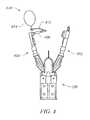
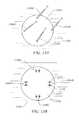
- endoluminal surgical device 100is shown according to an embodiment of the present invention.
- Endoluminal surgical device 100further includes a generally longitudinal and flexible shaft 102 that includes an end effecter 200 ( FIG. 2 ) configured near its distal end.
- End effecter 200includes first arm 202 and second arm 204 .
- First arm 202 and second arm 204are configured to engage tissue and implant anchor object 220 .
- anchor object 220generally includes pledgets 222 and 224 which, according to some embodiments, are connected by suture 226 .
- Endoluminal surgical device 100 , end effecter 200 , and anchor object 220are further disclosed in the reference publications and applications that are incorporated herein by reference.
- anchor objectcan be constructed from any biocompatible material, such as, but not limited to, stainless steel, cobalt chromium, titanium, alloys of such metals, biocompatible polymers, soluble polymers, non-soluble polymers, swellable polymers, absorbable polymers, suture material, bioresorbable suture, bioabsorbable suture, combinations thereof, or the like.
- biocompatible materialsuch as, but not limited to, stainless steel, cobalt chromium, titanium, alloys of such metals, biocompatible polymers, soluble polymers, non-soluble polymers, swellable polymers, absorbable polymers, suture material, bioresorbable suture, bioabsorbable suture, combinations thereof, or the like.
- endoluminal surgical device 100is utilized to manipulate tissue.
- Tissue from the wall of a hollow organ, such as for example the stomach,is formed into plications, such as plication P 1 .
- Plication P 1is formed by gathering or manipulating tissue of the wall of the organ with end effecter 200 into a fold and piercing the folded tissue with needle of end effecter 200 .
- Needleincludes suture 312 and a suture based implant or pledget 314 associated therewith. According to an embodiment, one end of suture 312 is attached to pledget 314 and the other end of suture 312 remains free and outside the patient.
- both ends of suture 312remain free and outside the patient's body, however, pledget 314 is attached to suture at a predetermined location.
- Pledget 314can be loosely attached or securely attached to suture 312 by a knot, a clip, integral with suture 312 , combinations thereof, or the like.
- a second pledget 316is positioned with respect to suture 312 .
- Second pledget 316is preferable positioned with respect to suture 312 outside the body, however, second pledget 316 can be positioned onto suture 12 inside the hollow organ.
- Second pledget 316is then moved into the hollow organ to be manipulated, a second fold of tissue or plication P 2 is formed with the endoluminal or endoscopic surgical device 100 and plication P 2 is pierced with needle of the endoscope 100 .
- plication P 2is pierced, suture 312 is pushed through plication P 2 , thereby, bringing pledget 316 into position adjacent a side of plication P 2 .
- suture 312is tightened such that plication P 1 and plication P 2 are drawn together.
- pledget 314 and pledget 316are positioned on opposing sides of plication P 1 and P 2 , respectively.
- pledget 314 and pledget 316are positioned on the same side of plication P 1 and plication P 2 .
- suture 312is fixed.
- suture 312can be either removably fixed or irremovably fixed to hold plication P 1 and plication P 2 in position.
- Suture 312can be fixed with a knot, a suture clip, fused together, combinations thereof, or the like.
- a predetermined pressurecan be applied through the tightening of suture 312 such as to generate a predetermined treatment condition between the plications. According to some embodiments, this procedure is performed in the stomach of a patient.
- plication P 1 and plication P 2are formed approximately 2 cm from gastro-esophageal (GE) junction 310 . According to alternative embodiments, plication P 1 and plication P 2 are formed between 2 cm and 5 cm from GE junction 310 . Where the stomach condition to be treated requires enhancement or replacement of the gastric valve, such plications are preferably located in cooperative relationship with the GE junction to form an artificial gastric valve.
- GEgastro-esophageal
- pledgets 314 and 316may or may not be used to form plications P 1 and P 2 , respectively. It should be appreciated that depending on a condition to be treated, tissue type being treated, location of treatment, size or area to be treated, combinations thereof, and the like, pledgets 314 and 316 may not be necessary, thereby, using suture 312 to form plications P 1 and P 2 and tighten plications P 1 and P 2 together.
- multiple pledgetscan be implanted adjacent one another such as to treat a large tissue disturbance, such as a large incision, re-incision, necrotic site, fistula, combinations thereof, or the like.
- a patch 318can be introduced with endoluminal device 100 into the organ to be treated. Patch 318 can be associated with a tissue engaging portion of the device and placed on the wall of the organ when the tissue engaging portions grasp the wall to form the plication. In some embodiments, patch 318 may be coupled with a plication, such as plication P 2 , through the securing implant such as pledget 316 . In one exemplary embodiment, patch 318 may be formed as a resilient, clip-like member having at least two arms that can be secured to a plication previously formed by manipulating the tissue with the endoluminal device 100 . As shown, for example, in FIG. 3 , piercing of the tissue is not required for securing such an embodiment.
- patch 318can be attached at a plurality of locations to the wall of the organ. Patch 318 can increase the integrity of the organ.
- patch 318includes therapeutic agents such as, for example, antibiotics, drugs, inhibiting agents, anti-inflammatory agents, combinations thereof, and the like.
- the therapeutic agentscan be coated on patch 318 or they can be dispersed throughout the material that forms patch 318 .
- Patch 318can also be fabricated from non-absorbable or bio-resorbable materials. Other treatment devices as described below also my be employed.
- end effecter 200is shown having first arm 402 and second arm 404 .
- First arm 402 and second arm 404are configured to engage and manipulate tissue.
- second arm 404includes a tissue piercing needle 406 .
- implant device 410includes suture 412 that is attached to implant 414 .
- implant 414can be, but is not limited to, a drug delivery device, such as for example a delayed delivery material, delayed absorbable drug eluding material, selectively drug permeable material or membrane, time release delivery device, combination thereof, or the like.
- implant 414can be, but is not limited to a stimulator device, such as for example, an electrical stimulation device, mechanical stimulation device, vibratory device, sound stimulation device, ultra-sound stimulation device, combinations thereof, or the like.
- the stimulation devicecan be configured to stimulate a sense of a patient.
- the sense stimulated by the stimulation devicecan be a sense of satiety in the patient such that the patient's desire for eating is subsided.
- implant 414can be, but is not limited to, a sensory device, such as for example, a device to monitor pH, pressure, temperature, salinity, hydration, cellular activity, protein levels, glucose levels, insulin levels, hormone levels, biological function, biological secretion, cellular uptake, cellular secretion, combinations thereof, and the like.
- implant 414can be, but is not limited to, a device to control biological activity, such as for example, inter or intra cellular pH, temperature, salinity, cellular function, cellular excretion, cellular uptake, glucose levels, insulin levels, combinations thereof, and the like.
- suture 412can be resorbable suture material or non-resorbable suture material.
- first arm 402 and second arm 404are positioned with respect to tissue that is to be manipulated.
- the armsare manipulated by actuating a control on endoluminal surgical device 100 .
- first arm 402 and second arm 404are actuated, they move toward each other and tissue positioned between the arms is engaged and pierced with needle 406 .
- needle 406is coupled with suture 412 , and suture 412 is attached to implant device 410 .
- suture 412is pierced through the tissue and extends through the tissue with implant device 410 on one side of the pierced tissue and a free end of suture 412 on the other side of the pierced tissue.
- suture 412can be formed as a loop to be secured around a natural or formed anatomical feature, such as a plication, utilizing first and second arms 402 and 404 without piercing the tissue.
- implant device 500is shown implanted with respect to tissue of a patient.
- tissuesuch as stomach wall tissue 504 and stomach mucosa 502 are formed into a plication 520 .
- Plication 520is affixed with an anchor object that includes pledgets 506 and 510 .
- pledgets 506 and 510can be coupled together with a tee bar 508 and suture 512 and in other embodiments pledgets can be coupled together only with suture 512 .
- Implant 514is attached to the anchor object.
- implant 514can be integral with pledget 510 , coupled with pledget 510 , or removable coupled with pledget 514 .
- the coupling between pledget 510 and implant 514can be degradable over a predetermined time such that implant 514 become detached from pledget 510 .
- implant 514can be any of the implant devices, sensors, drugs, drug delivery devices, monitors, control devices, combinations thereof, and the like that are described herein and/or incorporated into this application by reference.
- end effecter 200is shown having first arm 602 and second arm 604 .
- Second armis shown having needle 606 for piercing tissue, however, as will be appreciated first arm 602 and second arm 604 and the components associated therewith can be interchanged such that needle 606 could be associated with first arm 602 , and the like.
- Needle 606is coupled or threaded with suture 612 , which includes a pledget 610 associated therewith.
- first arm 602is configured to couple with implant 614 .
- Implant 614further includes an affixing portion 616 for engaging with needle 606 and coupling thereto to bind tissue therebetween.
- implant 614can be any of the implant devices described herein or incorporated herein by reference.
- suture 612 and pledget 610can be fabricated according to other embodiments described herein or incorporated herein by reference.
- a plication 720can be formed from a tissue wall or lining 704 that may or may not include a mucosa 702 .
- Plication 720is formed from binding tissue 704 with an anchor object that includes pledgets 706 and 710 .
- pledgets 706 and 710can be coupled with suture 712 and tee bar 708 .
- implant 714can be implanted with anchor object by coupling implant 714 between tissue wall 704 or tissue mucosa 702 and pledget 706 .
- implant 814can be implanted on a tether such that implant 814 is relatively affixed to tissue but not rigidly affixed to any particular tissue.
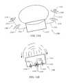
- end effecter 200includes first arm 802 , second arm 804 , and tissue retractor 806 .
- Second arm 804includes tissue piercing needle 808 for piercing tissue and implanting implant 814 .
- Needle 808further includes suture 812 for affixing tissue that is pierced by needed 808 into a plication.
- Suture 812can include pledget 810 for biasing against tissue. In some embodiments, suture 812 extends further beyond pledget 810 and couples with implant 814 .
- second suture 816extends from pledget 810 and couples with implant 814 .
- implant 814is implanted into a patient and affixed into a general location, however, implant 814 is semi-free to move within patient to the extent of tether provided between pledget 810 and implant 814 .
- FIG. 9shows tethered implant 814 in an implanted position with respect to a tissue plication 920 .
- plication 920is formed by anchoring a plication of tissue with an anchor object that includes implant 814 tethered thereto by second suture 816 .
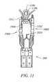
- an end effecter 200has a first arm 1002 , a second arm 1004 , and a tissue retractor 1014 and is used to form a tissue plication 1006 , as described herein.
- Plication 1006is formed by grasping tissue with retractor 1014 and manipulating first arm 1002 and second arm 1004 together such that needle 1008 pierces the tissue.
- Suture 1012is associated with needle 1008 such that suture 1012 is pierced through tissue with needle 1008 . After piercing the tissue with needle 1008 , the tissue is retained on needle 1008 .
- retractor 1014is used a second time to grasp a second portion of tissue such that the tissue can be positioned with respect to first arm 1002 , second arm 1004 , and needle 1008 and be pierced with needle 1008 to form second plication 1102 .
- an open pocket 1104is formed.
- an implant 1106is housed in open pocket 1104 .
- end effecter 200can have multiple retractors 1014 such that the retractor does not have to be removed from the tissue after forming a first plication in order to form a second plication.
- a first retractor 1014can grasp tissue and form a first plication while a second retractor grasps other tissue and forms a second plication.
- the two plicationscan be anchored together such that an open pocket 1104 is formed therebetween.
- tissue graspersare introduced into the surgical site percutaneously for grasping tissue and manipulating it into adjacent plications that form the tube or open pocket 1104 .
- the tissue grasperspull tissue around the shaft 102 of the surgical device and position the tissue for implantation of the anchor object.
- an anchor objectis implanted to fasten the plications around the shaft of the surgical device.
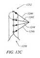
- the end effecter 200is straightened such as to be axially aligned with shaft 102 and the shaft 102 and end effecter 200 are removed through the open pocket 1104 of the tissue plications, leaving behind open pocket 1104 , or as shown in FIGS. 12B and 12C artificial tissue tube 1250 .
- tissue 1220 that forms pocket 1104 between plications 1006 and 1102may be tissue that requires treatment or can be used for locating a treatment.
- implant 1106 positioned in pocket 1104is configured to treat tissue 1220 or otherwise provide treatment to surrounding areas.
- tissue 1220can be removed following the double plication procedure described with respect to FIGS. 10-12A .
- tissue 1220may dissolve or resorb following the double plication procedure.
- multiple double plication proceduressuch as the double plication procedure described with respect to FIGS. 10-12C can be performed on the same tissue, in the same organ, or the like.
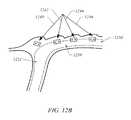
- axially aligned double plications 1240 , 1242 , 1244 , and 1246can be positioned adjacent to each other such that each open pocket 1104 , formed between the plications 1006 and 1102 , is generally aligned and forms an artificial biological tube 1250 .
- artificial biological tube 1250is formed by drawing each double plication together.
- each plication 1240 , 1242 , 1244 , and/or 1246can be drawn together using surgical glue, suture material, surgical wire, staples, scoring the tissue such that the tissue reforms together, combinations thereof, or the like.
- vertically aligned double plications 1240 , 1242 , and 1244are positioned, beginning at or distally near the gastro-esophageal-junction (GEJ) 1252 and extend into the stomach, thereby forming an artificial GEJ or gastric tube 1250 .
- GEJgastro-esophageal-junction
- the artificial gastric tubewhen the artificial gastric tube is flaccid, i.e., empty, the artificial gastric tube acts as a gastric flap or valve for protecting the esophagus from gastric fluids.
- the formation of such an artificial gastric tubecan be a treatment for gastro-esophageal reflux disorder (GERD).
- the formation of an artificial gastric tubecan be a technique for gastric reconstruction or reduction surgery.
- the formation of such an artificial gastric tubeeffectively reduces the volume of the stomach of a patient, and thereby, stimulates the stomach into providing a sense of satiety with consumption of a smaller amount of food.
- between 1 and about 10 plicationsare positioned adjacent each other to form artificial tube 1250 .
- between 1 and about 8 plicationsare positioned adjacent each other to form artificial tube 1250 .
- between 1 and about 5 plicationsare positioned adjacent each other to form artificial tube 1250 .
- between 1 and about 3 plicationsare positioned adjacent each other to form artificial tube 1250 .
- a single plicationis positioned adjacent the GEJ to form artificial tube 1250 or GERD treatment.
- between about 5 and about 8 plicationsare positioned adjacent each other to form artificial tube 1250 .
- artificial tube 1250when artificial tube 1250 is formed with between one and two tissue plications, artificial tube 1250 is about 1 cm in length. In other embodiments, when artificial tube 1250 includes between about 10 to about 20 tissue plications, artificial tube 1250 is about 10 cm in length.
- the diameter of artificial tube 1250is between about 0.2 cm and about 10 cm. In other embodiments, the diameter of artificial tube 1250 is between about 0.5 cm and about 5 cm. In some embodiments, the diameter of artificial tube 1250 is between about 0.5 cm and about 3 cm. In some embodiments, the diameter of artificial tube 1250 is between about 0.5 cm and about 2 cm.
- an implant device 1306can be implanted onto tissue 1302 with multiple attachments, as shown in FIGS. 13A and 13B .
- an implant 1306can be located or held in position by coupling retractors 1304 A- 1304 D to tissue 1302 and/or implant device 1306 .
- implant 1306may be such a shape or dimension that multiple anchor implants are required for proper or secure attachment of implant 1306 to tissue 1302 .
- multiple anchors 1308 A- 1308 Dare implanted through tissue 1302 and implant 1306 to affix implant 1306 as desired to tissue 1302 .
- anchors 1308 A- 1308 Dinclude pledgets 1310 and suture 1312 .
- pledgets 1310 and suture 1312can be fabricated in accord with and from the materials of other pledgets and sutures described herein and incorporated herein by reference.
- anchor implant 1400includes a coupling 1402 and pledgets 506 and 510 coupled together by suture 512 .
- Anchor implant 1400is configured to couple tissue 502 , 504 to itself or to an implanted device.
- coupling 1402is configured to provide a site on anchor implant 1400 for attaching an implantable device, mechanical tool, drug eluding device, sensor, other device described or incorporated herein, combinations thereof, or the like.
- coupling 1402can be, but is not limited to a hook, clip, Velcro, magnet, loop, combinations thereof, or the like.
- coupling 1402can be formed from a non-resorbable polymer, resorbable polymer, biodegradable polymer, drug eluding polymer, stainless steel, titanium, cobalt chromium, surgical material, combinations thereof, or the like.
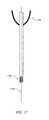
- endoluminal surgical device 100includes guide wire 1702 that includes tissue engaging retractor tip 1704 .
- tissue engaging retractor tip 1704is configured to releasable attach to tissue at a site near where an implant attachment site is targeted.
- tissue engaging retractor tip 1704can be, but is not limited to a cork-screw type design, screw threads, a hook, a loop, jaws, locking jaws, combinations thereof, and the like.
- having a guide wire 1702 that can attach to tissue or target a tissue at or near an implant sitecan help direct where the implant device will pass and increases the efficiency and effectiveness of surgical procedures because the path that the device is require to pass is less rigorous and/or straighter.
- adjustable implantable device 1500includes anchor implants 1506 and 1508 , however, the number of anchor implants used in practice can vary to what is necessary for a particular procedure being performed, corrected, or desired.
- anchor implantsinclude pledgets 1510 connected by suture 1512 .
- anchor implant 1506is coupled with adjustment mechanism 1514 by suture 1516 and anchor implant 1508 is coupled with adjustment mechanism 1514 by suture 1518 .
- Adjustment mechanism 1514is configured to adjust a physical parameter, such as for example, length, width, diameter, rotational axis, combinations thereof, or the like, in response to stimulation.
- adjustment mechanism 1514linearly shortens in response to a predetermined stimulation.
- a predetermined stimulationcan be, but is not limited to, a radio frequency, ultra-sound, mechanical force, pressure, direct mechanical manipulation, magnetic force, chemical interaction, enzyme interaction, fluid, temperature, biological fluid, cellular interaction, cellular by-product, inter-cellular constituent, intra-cellular constituent, food, digestion by-product, time, combinations thereof, or the like.
- adjustable implant device 1500is implanted into the tissue surrounding gastro-esophageal-junction (GEJ) 1502 . Having the adjustable implant device 1500 implanted in or near the GEJ 1502 allows the adjustable implant device 1500 to assist or augment the natural function of the GEJ 1502 . According to an embodiment, in a relaxed state adjustable implant device 1500 leaves the GEJ 1502 in an open state such that food, liquids, or the like can be passed from the esophagus into the stomach of a patient.
- GEJgastro-esophageal-junction
- adjustable mechanism 1514shortens and, thereby, tightens the GEJ 1502 such that gastric fluids are blocked from passing from the stomach into the esophagus and causing or worsening a patient's GERD condition.
- adjustable mechanism 1514can be a threaded mechanism, a coil mechanism, a spring mechanism, a magnetic mechanism, a ratchet mechanism, memory material, piezoelectric material, temperature sensitive material, combinations thereof, or the like.
- adjustable implant device 1500can be implanted in a stomach of a patient. Implanting adjustable implant device 1500 into a stomach allows a patient or physician to reversibly and/or temporarily restrict the volume of the stomach of a patient in response to a predetermined stimulus. According to some embodiments adjustable implant device 1500 , when implanted in the stomach of a patient, can be retracted or adjusted prior to consuming food and thereby act as a temporary gastric restriction device. In some embodiments, adjustable implant device 1500 can be configured to respond to a digestive by-product, a cellular excretion, particular gastric fluids, an external stimulation, combinations thereof, or the like. It will be appreciated that adjustable implant device 1500 can be implanted in many locations in a patient, such as but not limited to, any sphincter, tubular organ such as the intestine, muscle, skeletal system, combinations thereof, or the like.
- devices and mechanisms for improving surgical dexterityincludes attaching stabilizers such as stabilizers 1606 , 1614 , and 1622 to tissue 1602 and shaft of implant device 102 . According to alternative embodiments, more or less stabilizers can be utilized depending the requirements of a particular situation.
- a stabilizersuch as stabilizer 1614 for example, includes a tissue anchor end and a device anchor end.
- the tissue anchor endincludes pledgets 1616 and 1617 connected by suture material 1618 .
- Pledgets 1616 and 1617 and suture material 1618can be implanted according to methods and with devices disclosed herein or incorporated herein by reference.
- Pledget 1617is further connected to line 1620 that extends from pledget 1617 to device 102 .
- Line 1620can be, but is not limited to, suture material, wire, rods, k-wires, t-bars, or other surgical material or devices that can support tension, pressure, and/or torque.
- the device anchoring end of stabilizer 1614can be configured to couple to an attachment on device 102 such that a force applied to stabilizer 1614 can be translated to device 102 and/or manipulation force applied to device 102 can be leveraged over and around stabilizer 1614 .
- stabilization devicescan be implanted as desired to assist in manipulating surgical device 102 .
Landscapes
- Health & Medical Sciences (AREA)
- Surgery (AREA)
- Life Sciences & Earth Sciences (AREA)
- Medical Informatics (AREA)
- Animal Behavior & Ethology (AREA)
- Engineering & Computer Science (AREA)
- Biomedical Technology (AREA)
- Heart & Thoracic Surgery (AREA)
- Rheumatology (AREA)
- Molecular Biology (AREA)
- Nuclear Medicine, Radiotherapy & Molecular Imaging (AREA)
- General Health & Medical Sciences (AREA)
- Public Health (AREA)
- Veterinary Medicine (AREA)
- Surgical Instruments (AREA)
- Prostheses (AREA)
- Infusion, Injection, And Reservoir Apparatuses (AREA)
Abstract
Description
Claims (16)
Priority Applications (1)
| Application Number | Priority Date | Filing Date | Title |
|---|---|---|---|
| US11/867,577US8926641B2 (en) | 2006-10-04 | 2007-10-04 | Methods and devices for reconfiguring a body organ |
Applications Claiming Priority (2)
| Application Number | Priority Date | Filing Date | Title |
|---|---|---|---|
| US84941406P | 2006-10-04 | 2006-10-04 | |
| US11/867,577US8926641B2 (en) | 2006-10-04 | 2007-10-04 | Methods and devices for reconfiguring a body organ |
Publications (2)
| Publication Number | Publication Date |
|---|---|
| US20080312750A1 US20080312750A1 (en) | 2008-12-18 |
| US8926641B2true US8926641B2 (en) | 2015-01-06 |
Family
ID=39269231
Family Applications (6)
| Application Number | Title | Priority Date | Filing Date |
|---|---|---|---|
| US11/867,556Active2028-05-10US8209037B2 (en) | 2006-10-04 | 2007-10-04 | Methods and devices for medical treatment |
| US11/867,582Active2029-12-04US8166978B2 (en) | 2006-10-04 | 2007-10-04 | Methods and systems for manipulating tissue |
| US11/867,567AbandonedUS20080312729A1 (en) | 2006-10-04 | 2007-10-04 | Methods of tissue reconfiguration |
| US11/867,560Active2032-02-22US8469976B2 (en) | 2006-10-04 | 2007-10-04 | Methods of organ reconfiguration |
| US11/867,574Active2029-09-10US8882789B2 (en) | 2006-10-04 | 2007-10-04 | Methods and systems for tissue manipulation |
| US11/867,577Active2029-02-05US8926641B2 (en) | 2006-10-04 | 2007-10-04 | Methods and devices for reconfiguring a body organ |
Family Applications Before (5)
| Application Number | Title | Priority Date | Filing Date |
|---|---|---|---|
| US11/867,556Active2028-05-10US8209037B2 (en) | 2006-10-04 | 2007-10-04 | Methods and devices for medical treatment |
| US11/867,582Active2029-12-04US8166978B2 (en) | 2006-10-04 | 2007-10-04 | Methods and systems for manipulating tissue |
| US11/867,567AbandonedUS20080312729A1 (en) | 2006-10-04 | 2007-10-04 | Methods of tissue reconfiguration |
| US11/867,560Active2032-02-22US8469976B2 (en) | 2006-10-04 | 2007-10-04 | Methods of organ reconfiguration |
| US11/867,574Active2029-09-10US8882789B2 (en) | 2006-10-04 | 2007-10-04 | Methods and systems for tissue manipulation |
Country Status (4)
| Country | Link |
|---|---|
| US (6) | US8209037B2 (en) |
| EP (2) | EP2076183A4 (en) |
| CN (1) | CN101795627A (en) |
| WO (1) | WO2008043044A2 (en) |
Families Citing this family (73)
| Publication number | Priority date | Publication date | Assignee | Title |
|---|---|---|---|---|
| US6387104B1 (en)* | 1999-11-12 | 2002-05-14 | Scimed Life Systems, Inc. | Method and apparatus for endoscopic repair of the lower esophageal sphincter |
| US6592596B1 (en)* | 2000-05-10 | 2003-07-15 | Scimed Life Systems, Inc. | Devices and related methods for securing a tissue fold |
| US7097665B2 (en)* | 2003-01-16 | 2006-08-29 | Synecor, Llc | Positioning tools and methods for implanting medical devices |
| CN101810521B (en) | 2001-08-27 | 2015-05-13 | 辛尼科有限责任公司 | Satiation devices and methods |
| US6675809B2 (en) | 2001-08-27 | 2004-01-13 | Richard S. Stack | Satiation devices and methods |
| US6845776B2 (en)* | 2001-08-27 | 2005-01-25 | Richard S. Stack | Satiation devices and methods |
| US7146984B2 (en)* | 2002-04-08 | 2006-12-12 | Synecor, Llc | Method and apparatus for modifying the exit orifice of a satiation pouch |
| US20040143342A1 (en) | 2003-01-16 | 2004-07-22 | Stack Richard S. | Satiation pouches and methods of use |
| US20050247320A1 (en)* | 2003-10-10 | 2005-11-10 | Stack Richard S | Devices and methods for retaining a gastro-esophageal implant |
| US8206456B2 (en) | 2003-10-10 | 2012-06-26 | Barosense, Inc. | Restrictive and/or obstructive implant system for inducing weight loss |
| WO2005105003A1 (en) | 2004-04-26 | 2005-11-10 | Synecor, Llc | Restrictive and/or obstructive implant for inducing weight loss |
| NZ554495A (en) | 2004-10-15 | 2010-09-30 | Bfkw Llc | Bariatric device and method with lumen exerting force on eosophagus or stomach areas |
| US10925587B2 (en) | 2005-05-20 | 2021-02-23 | Neotract, Inc. | Anchor delivery system |
| US7896891B2 (en) | 2005-05-20 | 2011-03-01 | Neotract, Inc. | Apparatus and method for manipulating or retracting tissue and anatomical structure |
| US8834492B2 (en) | 2005-05-20 | 2014-09-16 | Neotract, Inc. | Continuous indentation lateral lobe apparatus and method |
| US8668705B2 (en) | 2005-05-20 | 2014-03-11 | Neotract, Inc. | Latching anchor device |
| US10195014B2 (en) | 2005-05-20 | 2019-02-05 | Neotract, Inc. | Devices, systems and methods for treating benign prostatic hyperplasia and other conditions |
| US9364212B2 (en) | 2005-05-20 | 2016-06-14 | Neotract, Inc. | Suture anchoring devices and methods for use |
| US9149266B2 (en) | 2005-05-20 | 2015-10-06 | Neotract, Inc. | Deforming anchor device |
| US7758594B2 (en) | 2005-05-20 | 2010-07-20 | Neotract, Inc. | Devices, systems and methods for treating benign prostatic hyperplasia and other conditions |
| US8333776B2 (en) | 2005-05-20 | 2012-12-18 | Neotract, Inc. | Anchor delivery system |
| US9504461B2 (en) | 2005-05-20 | 2016-11-29 | Neotract, Inc. | Anchor delivery system |
| US7645286B2 (en) | 2005-05-20 | 2010-01-12 | Neotract, Inc. | Devices, systems and methods for retracting, lifting, compressing, supporting or repositioning tissues or anatomical structures |
| US8945152B2 (en) | 2005-05-20 | 2015-02-03 | Neotract, Inc. | Multi-actuating trigger anchor delivery system |
| US9549739B2 (en) | 2005-05-20 | 2017-01-24 | Neotract, Inc. | Devices, systems and methods for treating benign prostatic hyperplasia and other conditions |
| US8628542B2 (en) | 2005-05-20 | 2014-01-14 | Neotract, Inc. | Median lobe destruction apparatus and method |
| US8529584B2 (en) | 2005-05-20 | 2013-09-10 | Neotract, Inc. | Median lobe band implant apparatus and method |
| US8425535B2 (en) | 2005-05-20 | 2013-04-23 | Neotract, Inc. | Multi-actuating trigger anchor delivery system |
| US8394113B2 (en) | 2005-05-20 | 2013-03-12 | Neotract, Inc. | Coiled anchor device |
| US8603106B2 (en) | 2005-05-20 | 2013-12-10 | Neotract, Inc. | Integrated handle assembly for anchor delivery system |
| US9055942B2 (en) | 2005-10-03 | 2015-06-16 | Boston Scienctific Scimed, Inc. | Endoscopic plication devices and methods |
| US20080190989A1 (en)* | 2005-10-03 | 2008-08-14 | Crews Samuel T | Endoscopic plication device and method |
| US7976554B2 (en) | 2006-04-19 | 2011-07-12 | Vibrynt, Inc. | Devices, tools and methods for performing minimally invasive abdominal surgical procedures |
| US8585733B2 (en) | 2006-04-19 | 2013-11-19 | Vibrynt, Inc | Devices, tools and methods for performing minimally invasive abdominal surgical procedures |
| US8342183B2 (en) | 2006-04-19 | 2013-01-01 | Vibrynt, Inc. | Devices and methods for treatment of obesity |
| US8556925B2 (en) | 2007-10-11 | 2013-10-15 | Vibrynt, Inc. | Devices and methods for treatment of obesity |
| US9314361B2 (en)* | 2006-09-15 | 2016-04-19 | Boston Scientific Scimed, Inc. | System and method for anchoring stomach implant |
| WO2008043044A2 (en)* | 2006-10-04 | 2008-04-10 | Ndo Surgical, Inc. | Devices and methods for endoluminal gastric restriction tissue manipulation, and drug delivery |
| US8529431B2 (en) | 2007-02-14 | 2013-09-10 | Bfkw, Llc | Bariatric device and method |
| US8020741B2 (en) | 2008-03-18 | 2011-09-20 | Barosense, Inc. | Endoscopic stapling devices and methods |
| WO2010014825A1 (en) | 2008-07-30 | 2010-02-04 | Neotract, Inc. | Slotted anchor device |
| US7934631B2 (en) | 2008-11-10 | 2011-05-03 | Barosense, Inc. | Multi-fire stapling systems and methods for delivering arrays of staples |
| US20100276469A1 (en)* | 2009-05-01 | 2010-11-04 | Barosense, Inc. | Plication tagging device and method |
| US8961539B2 (en)* | 2009-05-04 | 2015-02-24 | Boston Scientific Scimed, Inc. | Endoscopic implant system and method |
| US8617167B2 (en) | 2009-12-04 | 2013-12-31 | Pivot Medical, Inc. | Methods and devices for accessing and retracting a capsule of a joint |
| US8476227B2 (en) | 2010-01-22 | 2013-07-02 | Ethicon Endo-Surgery, Inc. | Methods of activating a melanocortin-4 receptor pathway in obese subjects |
| US9044606B2 (en) | 2010-01-22 | 2015-06-02 | Ethicon Endo-Surgery, Inc. | Methods and devices for activating brown adipose tissue using electrical energy |
| EP2571427B1 (en) | 2010-05-21 | 2017-07-19 | Boston Scientific Scimed, Inc. | Tissue-acquisition and fastening devices |
| WO2012092056A1 (en) | 2010-12-29 | 2012-07-05 | Ethicon Endo-Surgery, Inc. | Methods and devices for activating brown adipose tissue with targeted substance delivery |
| WO2012092057A1 (en) | 2010-12-29 | 2012-07-05 | Ethicon Endo-Surgery, Inc. | Methods and devices for activating brown adipose tissue with light |
| US9161749B2 (en) | 2011-04-14 | 2015-10-20 | Neotract, Inc. | Method and apparatus for treating sexual dysfunction |
| WO2013026473A1 (en) | 2011-08-23 | 2013-02-28 | Ethicon Endo-Surgery, Inc. | Devices and methods for anchoring an endoluminal sleeve in the gi tract |
| US9113868B2 (en) | 2011-12-15 | 2015-08-25 | Ethicon Endo-Surgery, Inc. | Devices and methods for endoluminal plication |
| US9173657B2 (en) | 2011-12-15 | 2015-11-03 | Ethicon Endo-Surgery, Inc. | Devices and methods for endoluminal plication |
| US9314362B2 (en) | 2012-01-08 | 2016-04-19 | Vibrynt, Inc. | Methods, instruments and devices for extragastric reduction of stomach volume |
| US8382775B1 (en) | 2012-01-08 | 2013-02-26 | Vibrynt, Inc. | Methods, instruments and devices for extragastric reduction of stomach volume |
| US9149383B2 (en) | 2012-01-23 | 2015-10-06 | Apollo Endosurgery, Inc. | Endolumenal esophageal restriction device |
| US8992547B2 (en) | 2012-03-21 | 2015-03-31 | Ethicon Endo-Surgery, Inc. | Methods and devices for creating tissue plications |
| US10292801B2 (en) | 2012-03-29 | 2019-05-21 | Neotract, Inc. | System for delivering anchors for treating incontinence |
| US9456916B2 (en) | 2013-03-12 | 2016-10-04 | Medibotics Llc | Device for selectively reducing absorption of unhealthy food |
| US10130353B2 (en) | 2012-06-29 | 2018-11-20 | Neotract, Inc. | Flexible system for delivering an anchor |
| US9067070B2 (en) | 2013-03-12 | 2015-06-30 | Medibotics Llc | Dysgeusia-inducing neurostimulation for modifying consumption of a selected nutrient type |
| US9011365B2 (en) | 2013-03-12 | 2015-04-21 | Medibotics Llc | Adjustable gastrointestinal bifurcation (AGB) for reduced absorption of unhealthy food |
| US10080884B2 (en) | 2014-12-29 | 2018-09-25 | Ethicon Llc | Methods and devices for activating brown adipose tissue using electrical energy |
| US10092738B2 (en) | 2014-12-29 | 2018-10-09 | Ethicon Llc | Methods and devices for inhibiting nerves when activating brown adipose tissue |
| EP3383281B1 (en) | 2015-12-04 | 2024-01-24 | Crossroads Extremity Systems, LLC | Devices for anchoring tissue |
| ES2953556T3 (en) | 2017-12-23 | 2023-11-14 | Teleflex Life Sciences Ltd | Expandable Tissue Docking Apparatus |
| US11097115B2 (en)* | 2018-09-24 | 2021-08-24 | Galvani Bioelectronics Limited | Implantable pulse generator with suture holes and methods for implanting the same |
| WO2020183399A1 (en)* | 2019-03-11 | 2020-09-17 | Bfkw, Llc | Single member intraluminal device and method of fixation |
| US12127958B2 (en) | 2019-03-25 | 2024-10-29 | Bfkw, Llc | Intraluminal device and method with anti-migration |
| CN114286646B (en) | 2020-08-03 | 2024-03-08 | 泰利福生命科学有限公司 | Handle and cassette system for medical intervention |
| JP2024523468A (en)* | 2021-06-22 | 2024-06-28 | ダブリュ.エル.ゴア アンド アソシエイツ,インコーポレイティド | Non-ablative bariatric coupling devices and methods of use |
| WO2023179576A1 (en)* | 2022-03-21 | 2023-09-28 | 南微医学科技股份有限公司 | Anchoring device and anchoring instrument |
Citations (86)
| Publication number | Priority date | Publication date | Assignee | Title |
|---|---|---|---|---|
| US4969892A (en)* | 1989-03-29 | 1990-11-13 | Ams, Inc. | Suturing anchoring device for use in a female suspension procedure |
| US5556428A (en)* | 1993-09-29 | 1996-09-17 | Shah; Mrugesh K. | Apparatus and method for promoting growth and repair of soft tissue |
| US5616131A (en) | 1992-09-23 | 1997-04-01 | Lasersurge, Inc. | Apparatus and method for anchoring surgical instrumentation |
| US5618270A (en) | 1995-05-26 | 1997-04-08 | Orejola; Wilmo C. | Transthoracic aortic sleeve |
| US5649960A (en)* | 1995-02-03 | 1997-07-22 | Pavletic; Michael M. | Apparatus and method for accelerating the stretching of skin |
| US5861003A (en)* | 1996-10-23 | 1999-01-19 | The Cleveland Clinic Foundation | Apparatus and method for occluding a defect or aperture within body surface |
| US5893856A (en)* | 1996-06-12 | 1999-04-13 | Mitek Surgical Products, Inc. | Apparatus and method for binding a first layer of material to a second layer of material |
| US5944738A (en)* | 1998-02-06 | 1999-08-31 | Aga Medical Corporation | Percutaneous catheter directed constricting occlusion device |
| US6120525A (en)* | 1999-07-14 | 2000-09-19 | Westcott; Mitchell S. | Skin tensioning device |
| US6152935A (en)* | 1996-12-11 | 2000-11-28 | Ethicon, Inc. | Meniscal repair device having integral spring member |
| US6352503B1 (en) | 1998-07-17 | 2002-03-05 | Olympus Optical Co., Ltd. | Endoscopic surgery apparatus |
| US20020035361A1 (en) | 1999-06-25 | 2002-03-21 | Houser Russell A. | Apparatus and methods for treating tissue |
| US6375671B1 (en)* | 1999-04-19 | 2002-04-23 | Nipro Corporation | Closure device for transcatheter operations |
| US20020138086A1 (en) | 2000-12-06 | 2002-09-26 | Robert Sixto | Surgical clips particularly useful in the endoluminal treatment of gastroesophageal reflux disease (GERD) |
| US6461293B1 (en)* | 1999-08-12 | 2002-10-08 | Obtech Medical Ag | Controlled food intake restriction |
| US6470892B1 (en)* | 2000-02-10 | 2002-10-29 | Obtech Medical Ag | Mechanical heartburn and reflux treatment |
| US20020183768A1 (en) | 2001-05-30 | 2002-12-05 | Deem Mark E. | Obesity treatment tools and methods |
| US6494888B1 (en) | 1999-06-22 | 2002-12-17 | Ndo Surgical, Inc. | Tissue reconfiguration |
| US6506196B1 (en) | 1999-06-22 | 2003-01-14 | Ndo Surgical, Inc. | Device and method for correction of a painful body defect |
| US6520974B2 (en)* | 1997-06-30 | 2003-02-18 | Eva Corporation | Surgical fastener |
| US20030093117A1 (en)* | 1999-06-25 | 2003-05-15 | Vahid Saadat | Implantable artificial partition and methods of use |
| US20030144695A1 (en)* | 1999-09-13 | 2003-07-31 | Mcguckin James F. | Vascular hole closure device |
| US20030164304A1 (en) | 2001-05-01 | 2003-09-04 | Imran Mir A. | Aendoscopic instrument system@ |
| US20030191495A1 (en)* | 2001-12-19 | 2003-10-09 | Nmt Medical, Inc. | Septal occluder and associated methods |
| WO2003094785A1 (en) | 2002-05-09 | 2003-11-20 | Egan Thomas D | Gastric bypass prosthesis |
| US20030216754A1 (en) | 2002-05-17 | 2003-11-20 | Scout Medical Technologies, Llc | Transoral endoscopic gastroesophageal flap valve restoration device, assembly, system and method |
| US6663639B1 (en) | 1999-06-22 | 2003-12-16 | Ndo Surgical, Inc. | Methods and devices for tissue reconfiguration |
| US20040010245A1 (en) | 1999-06-22 | 2004-01-15 | Cerier Jeffrey C. | Method and devices for tissue reconfiguration |
| US20040034371A1 (en) | 2001-05-18 | 2004-02-19 | Glen Lehman | Method of promoting tissue adhesion |
| US20040044364A1 (en)* | 2002-08-29 | 2004-03-04 | Devries Robert | Tissue fasteners and related deployment systems and methods |
| US20040087976A1 (en) | 2002-08-29 | 2004-05-06 | Devries Robert B. | Devices and methods for fastening tissue layers |
| US20040093091A1 (en) | 2002-08-07 | 2004-05-13 | Jamy Gannoe | Intra-gastric fastening devices |
| US20040092892A1 (en) | 2002-11-01 | 2004-05-13 | Jonathan Kagan | Apparatus and methods for treatment of morbid obesity |
| US20040107004A1 (en) | 2002-12-02 | 2004-06-03 | Seedling Enterprises, Llc | Bariatric sleeve |
| US20040116992A1 (en) | 2002-09-26 | 2004-06-17 | Wardle John L. | Cardiovascular anchoring device and method of deploying same |
| US20040122456A1 (en)* | 2002-12-11 | 2004-06-24 | Saadat Vahid C. | Methods and apparatus for gastric reduction |
| US20040147958A1 (en)* | 2002-12-11 | 2004-07-29 | Usgi Medical | Apparatus and methods for forming and securing gastrointestinal tissue folds |
| US20040148021A1 (en)* | 2002-08-29 | 2004-07-29 | Cartledge Richard G. | Implantable devices for controlling the internal circumference of an anatomic orifice or lumen |
| US20040152947A1 (en)* | 2000-10-06 | 2004-08-05 | Schroeder Richard F. | Methods and devices for improving mitral valve function |
| US20040158125A1 (en) | 2002-09-06 | 2004-08-12 | Aznoian Harold M. | Integrated endoscope and accessory treatment device |
| US20040193194A1 (en) | 1999-06-22 | 2004-09-30 | Ndo Surgical, Inc., A Massachusetts Corporation | Tissue reconfiguration |
| US20040193190A1 (en) | 2002-11-29 | 2004-09-30 | Liddicoat John R. | Apparatus and method for manipulating tissue |
| US20040220637A1 (en)* | 2003-01-24 | 2004-11-04 | Proteus Biomedical, Inc. | Method and apparatus for enhancing cardiac pacing |
| US20040220596A1 (en)* | 2003-02-04 | 2004-11-04 | Frazier Andrew G.C. | Patent foramen ovale closure system |
| US20040225305A1 (en) | 1999-06-25 | 2004-11-11 | Usgi Medical | Apparatus and methods for forming and securing gastrointestinal tissue folds |
| US20040225304A1 (en)* | 2000-10-06 | 2004-11-11 | Myocor | Endovascular splinting devices and methods |
| US20040243211A1 (en) | 2001-05-01 | 2004-12-02 | Olivier Colliou | Endoscopic instrument for engaging a device |
| US6835200B2 (en) | 1999-06-22 | 2004-12-28 | Ndo Surgical. Inc. | Method and devices for tissue reconfiguration |
| WO2005020802A2 (en) | 2003-09-02 | 2005-03-10 | Creighton University | Suturing devices and methods |
| US20050059984A1 (en)* | 2003-09-11 | 2005-03-17 | Andrzej Chanduszko | Devices, systems, and methods for suturing tissue |
| US20050065601A1 (en)* | 2002-04-18 | 2005-03-24 | Coalescent Surgical, Inc. | Annuloplasty apparatus and methods |
| US20050065571A1 (en)* | 2001-05-01 | 2005-03-24 | Imran Mir A. | Responsive gastric stimulator |
| US20050080444A1 (en)* | 2003-10-14 | 2005-04-14 | Kraemer Stefan J.M. | Transesophageal gastric reduction device, system and method |
| US20050090873A1 (en)* | 2003-10-22 | 2005-04-28 | Imran Mir A. | Gastrointestinal stimulation device |
| US20050096673A1 (en) | 2003-10-10 | 2005-05-05 | Stack Richard S. | Devices and methods for retaining a gastro-esophageal implant |
| US20050143760A1 (en)* | 2001-05-01 | 2005-06-30 | Imran Mir A. | Endoscopic gastric constriction device |
| US20050177180A1 (en)* | 2001-11-28 | 2005-08-11 | Aptus Endosystems, Inc. | Devices, systems, and methods for supporting tissue and/or structures within a hollow body organ |
| US20050192601A1 (en)* | 2004-02-27 | 2005-09-01 | Demarais Denise M. | Methods and devices for reducing hollow organ volume |
| US20050192599A1 (en) | 2004-02-13 | 2005-09-01 | Demarais Denise M. | Methods for reducing hollow organ volume |
| US20050203500A1 (en) | 2004-03-09 | 2005-09-15 | Usgi Medical Inc. | Apparatus and methods for mapping out endoluminal gastrointestinal surgery |
| US20050228415A1 (en)* | 2004-03-23 | 2005-10-13 | Michael Gertner | Methods and devices for percutaneous, non-laparoscopic treatment of obesity |
| US20050251208A1 (en) | 2004-05-07 | 2005-11-10 | Usgi Medical Inc. | Linear anchors for anchoring to tissue |
| US20050251166A1 (en) | 2004-05-07 | 2005-11-10 | Usgi Medical Inc. | Tissue manipulation and securement system |
| US20050251157A1 (en) | 2004-05-07 | 2005-11-10 | Usgi Medical Inc. | Apparatus and methods for positioning and securing anchors |
| US20050251160A1 (en) | 2004-05-07 | 2005-11-10 | Usgi Medical Inc. | Apparatus for manipulating and securing tissue |
| US20050250980A1 (en)* | 2004-05-07 | 2005-11-10 | Usgi Medical Corp. | Methods for performing gastroplasty |
| US20050261713A1 (en)* | 2004-04-27 | 2005-11-24 | Hassan Ali H | Methods and devices for producing turbulence in vascular blood flow |
| US20050267533A1 (en)* | 2004-03-23 | 2005-12-01 | Michael Gertner | Methods and devices for the surgical creation of satiety and biofeedback pathways |
| US20050267406A1 (en)* | 2004-05-28 | 2005-12-01 | Ethicon Endo-Surgery, Inc. | Piezo electrically driven bellows infuser for hydraulically controlling an adjustable gastric band |
| US20060020276A1 (en) | 2004-07-23 | 2006-01-26 | Usgi Medical Inc. | Apparatus and methods for achieving prolonged maintenance of gastrointestinal tissue folds |
| US20060020277A1 (en)* | 2004-07-20 | 2006-01-26 | Gostout Christopher J | Gastric reshaping devices and methods |
| US20060074458A1 (en) | 2001-05-01 | 2006-04-06 | Imran Mir A | Digestive organ retention device |
| US20060089571A1 (en)* | 2004-03-23 | 2006-04-27 | Michael Gertner | Obesity treatment systems |
| US7037344B2 (en)* | 2002-11-01 | 2006-05-02 | Valentx, Inc. | Apparatus and methods for treatment of morbid obesity |
| US20060142790A1 (en)* | 2004-03-23 | 2006-06-29 | Michael Gertner | Methods and devices to facilitate connections between body lumens |
| US20060155327A1 (en)* | 1999-09-13 | 2006-07-13 | Briganti Richard T | Vascular hole closure device |
| US20060157067A1 (en) | 2005-01-14 | 2006-07-20 | Usgi Medical Inc. | Attenuation of environmental parameters on a gastric lumen |
| US20060161185A1 (en) | 2005-01-14 | 2006-07-20 | Usgi Medical Inc. | Methods and apparatus for transmitting force to an end effector over an elongate member |
| US20060178550A1 (en)* | 2005-02-04 | 2006-08-10 | Boston Scientific Scimed, Inc. | Ventricular assist and support device |
| US20060183975A1 (en) | 2004-04-14 | 2006-08-17 | Usgi Medical, Inc. | Methods and apparatus for performing endoluminal procedures |
| US7361185B2 (en)* | 2001-05-09 | 2008-04-22 | Canica Design, Inc. | Clinical and surgical system and method for moving and stretching plastic tissue |
| US20080243144A1 (en) | 2006-10-04 | 2008-10-02 | Michael Laufer | Methods and systems for manipulating tissue |
| US7455681B2 (en)* | 2004-09-13 | 2008-11-25 | Wound Care Technologies, Llc | Wound closure product |
| US7566298B2 (en)* | 2006-04-25 | 2009-07-28 | Nicholson Iv William Daniel | Method of and apparatus for prevention of adjustable gastric band slips |
| US7621925B2 (en) | 2004-05-07 | 2009-11-24 | Usgi Medical, Inc. | Needle assembly for tissue manipulation |
| US7686829B2 (en)* | 2004-11-04 | 2010-03-30 | Wound Care Technologies, Inc. | Wound closure product |
Family Cites Families (5)
| Publication number | Priority date | Publication date | Assignee | Title |
|---|---|---|---|---|
| US6532503B1 (en)* | 2000-02-18 | 2003-03-11 | 3Com Corporation | Method and apparatus to detect lost buffers with a descriptor based queue |
| US7035236B2 (en)* | 2001-03-30 | 2006-04-25 | Telcordia Technologies, Inc. | Network-layer and link-layer use of shadow addresses with IP-based base stations |
| EP1560926B2 (en)* | 2002-11-08 | 2013-08-21 | STRATEC Molecular GmbH | Novel buffer formulations for isolating, purifying and recovering long-chain and short-chain nucleic acids |
| US7736374B2 (en)* | 2004-05-07 | 2010-06-15 | Usgi Medical, Inc. | Tissue manipulation and securement system |
| JP4681908B2 (en)* | 2005-02-14 | 2011-05-11 | オリンパス株式会社 | Surgical device controller and surgical system using the same |
- 2007
- 2007-10-04WOPCT/US2007/080485patent/WO2008043044A2/enactiveApplication Filing
- 2007-10-04USUS11/867,556patent/US8209037B2/enactiveActive
- 2007-10-04USUS11/867,582patent/US8166978B2/enactiveActive
- 2007-10-04USUS11/867,567patent/US20080312729A1/ennot_activeAbandoned
- 2007-10-04EPEP07843869Apatent/EP2076183A4/ennot_activeCeased
- 2007-10-04USUS11/867,560patent/US8469976B2/enactiveActive
- 2007-10-04USUS11/867,574patent/US8882789B2/enactiveActive
- 2007-10-04CNCN200780041843.4Apatent/CN101795627A/enactivePending
- 2007-10-04EPEP10180976Apatent/EP2258277A1/ennot_activeWithdrawn
- 2007-10-04USUS11/867,577patent/US8926641B2/enactiveActive
Patent Citations (104)
| Publication number | Priority date | Publication date | Assignee | Title |
|---|---|---|---|---|
| US4969892A (en)* | 1989-03-29 | 1990-11-13 | Ams, Inc. | Suturing anchoring device for use in a female suspension procedure |
| US5616131A (en) | 1992-09-23 | 1997-04-01 | Lasersurge, Inc. | Apparatus and method for anchoring surgical instrumentation |
| US5556428A (en)* | 1993-09-29 | 1996-09-17 | Shah; Mrugesh K. | Apparatus and method for promoting growth and repair of soft tissue |
| US5649960A (en)* | 1995-02-03 | 1997-07-22 | Pavletic; Michael M. | Apparatus and method for accelerating the stretching of skin |
| US5618270A (en) | 1995-05-26 | 1997-04-08 | Orejola; Wilmo C. | Transthoracic aortic sleeve |
| US5893856A (en)* | 1996-06-12 | 1999-04-13 | Mitek Surgical Products, Inc. | Apparatus and method for binding a first layer of material to a second layer of material |
| US5861003A (en)* | 1996-10-23 | 1999-01-19 | The Cleveland Clinic Foundation | Apparatus and method for occluding a defect or aperture within body surface |
| US6152935A (en)* | 1996-12-11 | 2000-11-28 | Ethicon, Inc. | Meniscal repair device having integral spring member |
| US6520974B2 (en)* | 1997-06-30 | 2003-02-18 | Eva Corporation | Surgical fastener |
| US5944738A (en)* | 1998-02-06 | 1999-08-31 | Aga Medical Corporation | Percutaneous catheter directed constricting occlusion device |
| US6352503B1 (en) | 1998-07-17 | 2002-03-05 | Olympus Optical Co., Ltd. | Endoscopic surgery apparatus |
| US6375671B1 (en)* | 1999-04-19 | 2002-04-23 | Nipro Corporation | Closure device for transcatheter operations |
| US20020193816A1 (en) | 1999-06-22 | 2002-12-19 | Ndo Surgical, Inc., A Delaware Corporation | Tissue reconfiguration |
| US20040193184A1 (en) | 1999-06-22 | 2004-09-30 | Ndo Surgical, Inc., A Massachusetts Corporation | Methods and devices for tissue reconfiguration |
| US20050033328A1 (en) | 1999-06-22 | 2005-02-10 | Ndo Surgical, Inc., A Massachusetts Corporation | Methods and devices for tissue reconfiguration |
| US6773441B1 (en) | 1999-06-22 | 2004-08-10 | Ndo Surgical, Inc. | Methods and devices for tissue reconfiguration |
| US20040194790A1 (en) | 1999-06-22 | 2004-10-07 | Ndo Surgical, Inc. | Tissue reconfiguration |
| US6494888B1 (en) | 1999-06-22 | 2002-12-17 | Ndo Surgical, Inc. | Tissue reconfiguration |
| US6835200B2 (en) | 1999-06-22 | 2004-12-28 | Ndo Surgical. Inc. | Method and devices for tissue reconfiguration |
| US6506196B1 (en) | 1999-06-22 | 2003-01-14 | Ndo Surgical, Inc. | Device and method for correction of a painful body defect |
| US6821285B2 (en) | 1999-06-22 | 2004-11-23 | Ndo Surgical, Inc. | Tissue reconfiguration |
| US20040193194A1 (en) | 1999-06-22 | 2004-09-30 | Ndo Surgical, Inc., A Massachusetts Corporation | Tissue reconfiguration |
| US20040193193A1 (en) | 1999-06-22 | 2004-09-30 | Ndo Surgical, Inc., A Massachusetts Corporation | Tissue reconfiguration |
| US20040010245A1 (en) | 1999-06-22 | 2004-01-15 | Cerier Jeffrey C. | Method and devices for tissue reconfiguration |
| US6663639B1 (en) | 1999-06-22 | 2003-12-16 | Ndo Surgical, Inc. | Methods and devices for tissue reconfiguration |
| US20040193117A1 (en) | 1999-06-22 | 2004-09-30 | Ndo Surgical, Inc., A Massachusetts Corporation | Tissue reconfiguration |
| US20020035361A1 (en) | 1999-06-25 | 2002-03-21 | Houser Russell A. | Apparatus and methods for treating tissue |
| US20040225305A1 (en) | 1999-06-25 | 2004-11-11 | Usgi Medical | Apparatus and methods for forming and securing gastrointestinal tissue folds |
| US20030093117A1 (en)* | 1999-06-25 | 2003-05-15 | Vahid Saadat | Implantable artificial partition and methods of use |
| US20030018358A1 (en) | 1999-06-25 | 2003-01-23 | Vahid Saadat | Apparatus and methods for treating tissue |
| US6120525A (en)* | 1999-07-14 | 2000-09-19 | Westcott; Mitchell S. | Skin tensioning device |
| US6461293B1 (en)* | 1999-08-12 | 2002-10-08 | Obtech Medical Ag | Controlled food intake restriction |
| US20030144695A1 (en)* | 1999-09-13 | 2003-07-31 | Mcguckin James F. | Vascular hole closure device |
| US20060155327A1 (en)* | 1999-09-13 | 2006-07-13 | Briganti Richard T | Vascular hole closure device |
| US7267679B2 (en)* | 1999-09-13 | 2007-09-11 | Rex Medical, L.P | Vascular hole closure device |
| US6470892B1 (en)* | 2000-02-10 | 2002-10-29 | Obtech Medical Ag | Mechanical heartburn and reflux treatment |
| US20040225304A1 (en)* | 2000-10-06 | 2004-11-11 | Myocor | Endovascular splinting devices and methods |
| US20040152947A1 (en)* | 2000-10-06 | 2004-08-05 | Schroeder Richard F. | Methods and devices for improving mitral valve function |
| US20020138086A1 (en) | 2000-12-06 | 2002-09-26 | Robert Sixto | Surgical clips particularly useful in the endoluminal treatment of gastroesophageal reflux disease (GERD) |
| US20030164304A1 (en) | 2001-05-01 | 2003-09-04 | Imran Mir A. | Aendoscopic instrument system@ |
| US20040243211A1 (en) | 2001-05-01 | 2004-12-02 | Olivier Colliou | Endoscopic instrument for engaging a device |
| US20050065571A1 (en)* | 2001-05-01 | 2005-03-24 | Imran Mir A. | Responsive gastric stimulator |
| US20050143760A1 (en)* | 2001-05-01 | 2005-06-30 | Imran Mir A. | Endoscopic gastric constriction device |
| US20060074458A1 (en) | 2001-05-01 | 2006-04-06 | Imran Mir A | Digestive organ retention device |
| US7361185B2 (en)* | 2001-05-09 | 2008-04-22 | Canica Design, Inc. | Clinical and surgical system and method for moving and stretching plastic tissue |
| US20040034371A1 (en) | 2001-05-18 | 2004-02-19 | Glen Lehman | Method of promoting tissue adhesion |
| US20020183768A1 (en) | 2001-05-30 | 2002-12-05 | Deem Mark E. | Obesity treatment tools and methods |
| US20050177180A1 (en)* | 2001-11-28 | 2005-08-11 | Aptus Endosystems, Inc. | Devices, systems, and methods for supporting tissue and/or structures within a hollow body organ |
| US20030191495A1 (en)* | 2001-12-19 | 2003-10-09 | Nmt Medical, Inc. | Septal occluder and associated methods |
| US20050065601A1 (en)* | 2002-04-18 | 2005-03-24 | Coalescent Surgical, Inc. | Annuloplasty apparatus and methods |
| WO2003094785A1 (en) | 2002-05-09 | 2003-11-20 | Egan Thomas D | Gastric bypass prosthesis |
| US20030216754A1 (en) | 2002-05-17 | 2003-11-20 | Scout Medical Technologies, Llc | Transoral endoscopic gastroesophageal flap valve restoration device, assembly, system and method |
| US20040093091A1 (en) | 2002-08-07 | 2004-05-13 | Jamy Gannoe | Intra-gastric fastening devices |
| US6746460B2 (en)* | 2002-08-07 | 2004-06-08 | Satiety, Inc. | Intra-gastric fastening devices |
| US20040148021A1 (en)* | 2002-08-29 | 2004-07-29 | Cartledge Richard G. | Implantable devices for controlling the internal circumference of an anatomic orifice or lumen |
| US20040087976A1 (en) | 2002-08-29 | 2004-05-06 | Devries Robert B. | Devices and methods for fastening tissue layers |
| US7083630B2 (en) | 2002-08-29 | 2006-08-01 | Scimed Life Systems, Inc. | Devices and methods for fastening tissue layers |
| US20040044364A1 (en)* | 2002-08-29 | 2004-03-04 | Devries Robert | Tissue fasteners and related deployment systems and methods |
| US20040158125A1 (en) | 2002-09-06 | 2004-08-12 | Aznoian Harold M. | Integrated endoscope and accessory treatment device |
| US20040116992A1 (en) | 2002-09-26 | 2004-06-17 | Wardle John L. | Cardiovascular anchoring device and method of deploying same |
| US7037344B2 (en)* | 2002-11-01 | 2006-05-02 | Valentx, Inc. | Apparatus and methods for treatment of morbid obesity |
| US20040092892A1 (en) | 2002-11-01 | 2004-05-13 | Jonathan Kagan | Apparatus and methods for treatment of morbid obesity |
| US20040193190A1 (en) | 2002-11-29 | 2004-09-30 | Liddicoat John R. | Apparatus and method for manipulating tissue |
| US20040107004A1 (en) | 2002-12-02 | 2004-06-03 | Seedling Enterprises, Llc | Bariatric sleeve |
| US20040147958A1 (en)* | 2002-12-11 | 2004-07-29 | Usgi Medical | Apparatus and methods for forming and securing gastrointestinal tissue folds |
| US20040122456A1 (en)* | 2002-12-11 | 2004-06-24 | Saadat Vahid C. | Methods and apparatus for gastric reduction |
| US20040220637A1 (en)* | 2003-01-24 | 2004-11-04 | Proteus Biomedical, Inc. | Method and apparatus for enhancing cardiac pacing |
| US20040220596A1 (en)* | 2003-02-04 | 2004-11-04 | Frazier Andrew G.C. | Patent foramen ovale closure system |
| WO2005020802A2 (en) | 2003-09-02 | 2005-03-10 | Creighton University | Suturing devices and methods |
| US20050059984A1 (en)* | 2003-09-11 | 2005-03-17 | Andrzej Chanduszko | Devices, systems, and methods for suturing tissue |
| US7691112B2 (en)* | 2003-09-11 | 2010-04-06 | Nmt Medical, Inc. | Devices, systems, and methods for suturing tissue |
| US20050096673A1 (en) | 2003-10-10 | 2005-05-05 | Stack Richard S. | Devices and methods for retaining a gastro-esophageal implant |
| US20050080444A1 (en)* | 2003-10-14 | 2005-04-14 | Kraemer Stefan J.M. | Transesophageal gastric reduction device, system and method |
| US20050090873A1 (en)* | 2003-10-22 | 2005-04-28 | Imran Mir A. | Gastrointestinal stimulation device |
| US20050192599A1 (en) | 2004-02-13 | 2005-09-01 | Demarais Denise M. | Methods for reducing hollow organ volume |
| US20050192601A1 (en)* | 2004-02-27 | 2005-09-01 | Demarais Denise M. | Methods and devices for reducing hollow organ volume |
| US20050203500A1 (en) | 2004-03-09 | 2005-09-15 | Usgi Medical Inc. | Apparatus and methods for mapping out endoluminal gastrointestinal surgery |
| US20050228415A1 (en)* | 2004-03-23 | 2005-10-13 | Michael Gertner | Methods and devices for percutaneous, non-laparoscopic treatment of obesity |
| US20050267533A1 (en)* | 2004-03-23 | 2005-12-01 | Michael Gertner | Methods and devices for the surgical creation of satiety and biofeedback pathways |
| US20060142790A1 (en)* | 2004-03-23 | 2006-06-29 | Michael Gertner | Methods and devices to facilitate connections between body lumens |
| US20060089571A1 (en)* | 2004-03-23 | 2006-04-27 | Michael Gertner | Obesity treatment systems |
| US20060183975A1 (en) | 2004-04-14 | 2006-08-17 | Usgi Medical, Inc. | Methods and apparatus for performing endoluminal procedures |
| US20050261713A1 (en)* | 2004-04-27 | 2005-11-24 | Hassan Ali H | Methods and devices for producing turbulence in vascular blood flow |
| US20050250980A1 (en)* | 2004-05-07 | 2005-11-10 | Usgi Medical Corp. | Methods for performing gastroplasty |
| US20050251208A1 (en) | 2004-05-07 | 2005-11-10 | Usgi Medical Inc. | Linear anchors for anchoring to tissue |
| US7520884B2 (en) | 2004-05-07 | 2009-04-21 | Usgi Medical Inc. | Methods for performing gastroplasty |
| US20050251160A1 (en) | 2004-05-07 | 2005-11-10 | Usgi Medical Inc. | Apparatus for manipulating and securing tissue |
| US20050251157A1 (en) | 2004-05-07 | 2005-11-10 | Usgi Medical Inc. | Apparatus and methods for positioning and securing anchors |
| US20050251166A1 (en) | 2004-05-07 | 2005-11-10 | Usgi Medical Inc. | Tissue manipulation and securement system |
| US7621925B2 (en) | 2004-05-07 | 2009-11-24 | Usgi Medical, Inc. | Needle assembly for tissue manipulation |
| US20050267406A1 (en)* | 2004-05-28 | 2005-12-01 | Ethicon Endo-Surgery, Inc. | Piezo electrically driven bellows infuser for hydraulically controlling an adjustable gastric band |
| US20060020277A1 (en)* | 2004-07-20 | 2006-01-26 | Gostout Christopher J | Gastric reshaping devices and methods |
| US20060020276A1 (en) | 2004-07-23 | 2006-01-26 | Usgi Medical Inc. | Apparatus and methods for achieving prolonged maintenance of gastrointestinal tissue folds |
| US7455681B2 (en)* | 2004-09-13 | 2008-11-25 | Wound Care Technologies, Llc | Wound closure product |
| US7686829B2 (en)* | 2004-11-04 | 2010-03-30 | Wound Care Technologies, Inc. | Wound closure product |
| US20060161185A1 (en) | 2005-01-14 | 2006-07-20 | Usgi Medical Inc. | Methods and apparatus for transmitting force to an end effector over an elongate member |
| US20060157067A1 (en) | 2005-01-14 | 2006-07-20 | Usgi Medical Inc. | Attenuation of environmental parameters on a gastric lumen |
| US20060178550A1 (en)* | 2005-02-04 | 2006-08-10 | Boston Scientific Scimed, Inc. | Ventricular assist and support device |
| US7566298B2 (en)* | 2006-04-25 | 2009-07-28 | Nicholson Iv William Daniel | Method of and apparatus for prevention of adjustable gastric band slips |
| US20090018389A1 (en) | 2006-10-04 | 2009-01-15 | Michael Laufer | Methods and systems for tissue manipulation |
| US20090018558A1 (en) | 2006-10-04 | 2009-01-15 | Michael Laufer | Methods of organ reconfiguration |
| US20090018594A1 (en) | 2006-10-04 | 2009-01-15 | Michael Laufer | Methods and devices for medical treatment |
| US20080312729A1 (en) | 2006-10-04 | 2008-12-18 | Michael Laufer | Methods of tissue reconfiguration |
| US20080243144A1 (en) | 2006-10-04 | 2008-10-02 | Michael Laufer | Methods and systems for manipulating tissue |
Non-Patent Citations (9)
| Title |
|---|
| DeMarco et al. "Outcomes of endoluminal gastric plication for the treatment of gastroesophageal reflux disease" BUMC Proceedings 2003; 16:392-393. |
| European Search Report for EP App. No. 10180976.2 dated Oct. 21, 2010. |
| International Preliminary Report on Patentability (Application No. PCT/US06/046026) dated May 19, 2009. |
| International Preliminary Report on Patentability (Application No. PCT/US07/080485) dated Apr. 23, 2009. |
| International Search Report (Application No. PCT/US07/80485) dated Jun. 5, 2008. |
| Olympus, "Your Vision, Our Future Medical Endoscope & Surgical Products" pp. 1-5, dated Jan. 9, 2006, downloaded Jun. 5, 2008, <http://web.archive.org/web/20060109095625/http://www.olympus.co.jp/en/mesg/endoscope/Gastroenterology/Endo-Therepy/etp/rfb/r113-01.cfm>. |
| Olympus, "Your Vision, Our Future Medical Endoscope & Surgical Products" pp. 1-5, dated Jan. 9, 2006, downloaded Jun. 5, 2008, <http://web.archive.org/web/20060109095625/http://www.olympus.co.jp/en/mesg/endoscope/Gastroenterology/Endo-Therepy/etp/rfb/r113—01.cfm>. |
| Olympus, "Your Vision, Our Future Medical Endoscope & Surgical Products" pp. 1-5, downloaded Jun. 5, 2008, . |
| Olympus, "Your Vision, Our Future Medical Endoscope & Surgical Products" pp. 1-5, downloaded Jun. 5, 2008, <http://olympus.co.jp/en/mesg/endoscope/Gastroenterology/Endo-Therepy/etp/rfb/rfb—01.cfm>. |
Also Published As
| Publication number | Publication date |
|---|---|
| WO2008043044A2 (en) | 2008-04-10 |
| WO2008043044A3 (en) | 2008-06-05 |
| US8882789B2 (en) | 2014-11-11 |
| WO2008043044A8 (en) | 2008-08-07 |
| CN101795627A (en) | 2010-08-04 |
| EP2076183A2 (en) | 2009-07-08 |
| US20080312750A1 (en) | 2008-12-18 |
| US8469976B2 (en) | 2013-06-25 |
| EP2258277A1 (en) | 2010-12-08 |
| EP2076183A4 (en) | 2009-09-16 |
| US8166978B2 (en) | 2012-05-01 |
| US20090018558A1 (en) | 2009-01-15 |
| US8209037B2 (en) | 2012-06-26 |
| US20090018389A1 (en) | 2009-01-15 |
| US20090018594A1 (en) | 2009-01-15 |
| US20080243144A1 (en) | 2008-10-02 |
| US20080312729A1 (en) | 2008-12-18 |
Similar Documents
| Publication | Publication Date | Title |
|---|---|---|
| US8926641B2 (en) | Methods and devices for reconfiguring a body organ | |
| EP2004243A2 (en) | Devices and methods for endoluminal gastric restriction and tissue manipulation | |
| US7988617B2 (en) | Extragastric minimally invasive methods and devices to treat obesity | |
| US20080051824A1 (en) | Methods and Devices For to Treatment of Obesity | |
| US20060264699A1 (en) | Extragastric minimally invasive methods and devices to treat obesity | |
| US20060195139A1 (en) | Extragastric devices and methods for gastroplasty | |
| US20070010866A1 (en) | Attachment cuff for gastrointestinal implant | |
| WO2006049725A2 (en) | Surgical systems and devices to enhance gastric restriction therapies | |
| WO2007137228A9 (en) | Medical staple, system and methods of use | |
| US20060235446A1 (en) | Article, system, and method for securing medical device to tissue or organ | |
| US20110106129A1 (en) | Methods and devices to treat obesity | |
| EP2306942B1 (en) | Gastric volume reduction using anterior to posterior wall junctions | |
| US10646226B2 (en) | Controlling a size of a pyloris | |
| AU2011265332A1 (en) | Devices and methods to treat a patient |
Legal Events
| Date | Code | Title | Description |
|---|---|---|---|
| AS | Assignment | Owner name:NDO SURGICAL, INC., MASSACHUSETTS Free format text:ASSIGNMENT OF ASSIGNORS INTEREST;ASSIGNORS:LAUFER, MICHAEL;CRUZ, AMOS;BROMANDER, THOMAS;SIGNING DATES FROM 20071031 TO 20071211;REEL/FRAME:020236/0786 Owner name:NDO SURGICAL, INC., MASSACHUSETTS Free format text:ASSIGNMENT OF ASSIGNORS INTEREST;ASSIGNORS:LAUFER, MICHAEL;CRUZ, AMOS;BROMANDER, THOMAS;REEL/FRAME:020236/0786;SIGNING DATES FROM 20071031 TO 20071211 | |
| AS | Assignment | Owner name:ETHICON ENDO-SURGERY, INC.,OHIO Free format text:ASSIGNMENT OF ASSIGNORS INTEREST;ASSIGNOR:NDO SURGICAL, INC.;REEL/FRAME:024411/0991 Effective date:20100502 Owner name:ETHICON ENDO-SURGERY, INC., OHIO Free format text:ASSIGNMENT OF ASSIGNORS INTEREST;ASSIGNOR:NDO SURGICAL, INC.;REEL/FRAME:024411/0991 Effective date:20100502 | |
| STCF | Information on status: patent grant | Free format text:PATENTED CASE | |
| MAFP | Maintenance fee payment | Free format text:PAYMENT OF MAINTENANCE FEE, 4TH YEAR, LARGE ENTITY (ORIGINAL EVENT CODE: M1551) Year of fee payment:4 | |
| MAFP | Maintenance fee payment | Free format text:PAYMENT OF MAINTENANCE FEE, 8TH YEAR, LARGE ENTITY (ORIGINAL EVENT CODE: M1552); ENTITY STATUS OF PATENT OWNER: LARGE ENTITY Year of fee payment:8 |