US8911994B2 - Use of adipose tissue-derived stromal cells for chondrocyte differentiation and cartilage repair - Google Patents
Use of adipose tissue-derived stromal cells for chondrocyte differentiation and cartilage repairDownload PDFInfo
- Publication number
- US8911994B2 US8911994B2US13/250,742US201113250742AUS8911994B2US 8911994 B2US8911994 B2US 8911994B2US 201113250742 AUS201113250742 AUS 201113250742AUS 8911994 B2US8911994 B2US 8911994B2
- Authority
- US
- United States
- Prior art keywords
- cells
- medium
- cartilage
- stromal cells
- adipose tissue
- Prior art date
- Legal status (The legal status is an assumption and is not a legal conclusion. Google has not performed a legal analysis and makes no representation as to the accuracy of the status listed.)
- Expired - Fee Related
Links
- 210000002536stromal cellAnatomy0.000titleclaimsabstractdescription61
- 210000001612chondrocyteAnatomy0.000titleclaimsabstractdescription40
- 210000000577adipose tissueAnatomy0.000titleclaimsdescription46
- 210000000845cartilageAnatomy0.000titleabstractdescription64
- 230000008439repair processEffects0.000titleabstractdescription17
- 230000004069differentiationEffects0.000titledescription15
- 210000004027cellAnatomy0.000claimsabstractdescription120
- 238000000034methodMethods0.000claimsabstractdescription40
- 230000022159cartilage developmentEffects0.000claimsabstractdescription11
- 239000011159matrix materialSubstances0.000claimsabstractdescription7
- 235000015097nutrientsNutrition0.000claimsabstract3
- 239000002609mediumSubstances0.000claimsdescription32
- 230000002648chondrogenic effectEffects0.000claimsdescription22
- 102000004887Transforming Growth Factor betaHuman genes0.000claimsdescription21
- 108090001012Transforming Growth Factor betaProteins0.000claimsdescription21
- 210000000988bone and boneAnatomy0.000claimsdescription19
- ZRKFYGHZFMAOKI-QMGMOQQFSA-NtgfbetaChemical compoundC([C@H](NC(=O)[C@H](C(C)C)NC(=O)CNC(=O)[C@H](CCC(O)=O)NC(=O)[C@H](CCCNC(N)=N)NC(=O)[C@H](CC(N)=O)NC(=O)[C@H](CC(C)C)NC(=O)[C@H]([C@@H](C)O)NC(=O)[C@H](CCC(O)=O)NC(=O)[C@H]([C@@H](C)O)NC(=O)[C@H](CC(C)C)NC(=O)CNC(=O)[C@H](C)NC(=O)[C@H](CO)NC(=O)[C@H](CCC(N)=O)NC(=O)[C@@H](NC(=O)[C@H](C)NC(=O)[C@H](C)NC(=O)[C@@H](NC(=O)[C@H](CC(C)C)NC(=O)[C@@H](N)CCSC)C(C)C)[C@@H](C)CC)C(=O)N[C@@H]([C@@H](C)O)C(=O)N[C@@H](C(C)C)C(=O)N[C@@H](CC=1C=CC=CC=1)C(=O)N[C@@H](C)C(=O)N1[C@@H](CCC1)C(=O)N[C@@H]([C@@H](C)O)C(=O)N[C@@H](CC(N)=O)C(=O)N[C@@H](CCC(O)=O)C(=O)N[C@@H](C)C(=O)N[C@@H](CC=1C=CC=CC=1)C(=O)N[C@@H](CCCNC(N)=N)C(=O)N[C@@H](C)C(=O)N[C@@H](CC(C)C)C(=O)N1[C@@H](CCC1)C(=O)N1[C@@H](CCC1)C(=O)N[C@@H](CCCNC(N)=N)C(=O)N[C@@H](CCC(O)=O)C(=O)N[C@@H](CCCNC(N)=N)C(=O)N[C@@H](CO)C(=O)N[C@@H](CCCNC(N)=N)C(=O)N[C@@H](CC(C)C)C(=O)N[C@@H](CC(C)C)C(O)=O)C1=CC=C(O)C=C1ZRKFYGHZFMAOKI-QMGMOQQFSA-N0.000claimsdescription19
- CIWBSHSKHKDKBQ-JLAZNSOCSA-NAscorbic acidChemical compoundOC[C@H](O)[C@H]1OC(=O)C(O)=C1OCIWBSHSKHKDKBQ-JLAZNSOCSA-N0.000claimsdescription18
- 229960003957dexamethasoneDrugs0.000claimsdescription16
- UREBDLICKHMUKA-CXSFZGCWSA-NdexamethasoneChemical compoundC1CC2=CC(=O)C=C[C@]2(C)[C@]2(F)[C@@H]1[C@@H]1C[C@@H](C)[C@@](C(=O)CO)(O)[C@@]1(C)C[C@@H]2OUREBDLICKHMUKA-CXSFZGCWSA-N0.000claimsdescription16
- 108091003079Bovine Serum AlbuminProteins0.000claimsdescription14
- 239000012091fetal bovine serumSubstances0.000claimsdescription14
- 239000003795chemical substances by applicationSubstances0.000claimsdescription12
- 239000006144Dulbecco’s modified Eagle's mediumSubstances0.000claimsdescription11
- 230000001413cellular effectEffects0.000claimsdescription11
- 239000003862glucocorticoidSubstances0.000claimsdescription11
- 210000002966serumAnatomy0.000claimsdescription11
- 235000010410calcium alginateNutrition0.000claimsdescription10
- 229960002681calcium alginateDrugs0.000claimsdescription10
- 239000000648calcium alginateSubstances0.000claimsdescription10
- OKHHGHGGPDJQHR-YMOPUZKJSA-Lcalcium;(2s,3s,4s,5s,6r)-6-[(2r,3s,4r,5s,6r)-2-carboxy-6-[(2r,3s,4r,5s,6r)-2-carboxylato-4,5,6-trihydroxyoxan-3-yl]oxy-4,5-dihydroxyoxan-3-yl]oxy-3,4,5-trihydroxyoxane-2-carboxylateChemical compound[Ca+2].O[C@@H]1[C@H](O)[C@H](O)O[C@@H](C([O-])=O)[C@H]1O[C@H]1[C@@H](O)[C@@H](O)[C@H](O[C@H]2[C@H]([C@@H](O)[C@H](O)[C@H](O2)C([O-])=O)O)[C@H](C(O)=O)O1OKHHGHGGPDJQHR-YMOPUZKJSA-L0.000claimsdescription10
- JYGXADMDTFJGBT-VWUMJDOOSA-NhydrocortisoneChemical compoundO=C1CC[C@]2(C)[C@H]3[C@@H](O)C[C@](C)([C@@](CC4)(O)C(=O)CO)[C@@H]4[C@@H]3CCC2=C1JYGXADMDTFJGBT-VWUMJDOOSA-N0.000claimsdescription10
- 230000003213activating effectEffects0.000claimsdescription9
- 229940072107ascorbateDrugs0.000claimsdescription9
- 235000010323ascorbic acidNutrition0.000claimsdescription9
- 239000011668ascorbic acidSubstances0.000claimsdescription9
- 230000037361pathwayEffects0.000claimsdescription9
- 239000003242anti bacterial agentSubstances0.000claimsdescription8
- 230000003115biocidal effectEffects0.000claimsdescription8
- UCSJYZPVAKXKNQ-HZYVHMACSA-NstreptomycinChemical compoundCN[C@H]1[C@H](O)[C@@H](O)[C@H](CO)O[C@H]1O[C@@H]1[C@](C=O)(O)[C@H](C)O[C@H]1O[C@@H]1[C@@H](NC(N)=N)[C@H](O)[C@@H](NC(N)=N)[C@H](O)[C@H]1OUCSJYZPVAKXKNQ-HZYVHMACSA-N0.000claimsdescription8
- 102000010834Extracellular Matrix ProteinsHuman genes0.000claimsdescription7
- 108010037362Extracellular Matrix ProteinsProteins0.000claimsdescription7
- 210000002744extracellular matrixAnatomy0.000claimsdescription7
- 239000001963growth mediumSubstances0.000claimsdescription7
- 238000010361transductionMethods0.000claimsdescription7
- 230000026683transductionEffects0.000claimsdescription7
- 150000003700vitamin C derivativesChemical class0.000claimsdescription7
- 108090000386Fibroblast Growth Factor 1Proteins0.000claimsdescription6
- 108090000379Fibroblast growth factor 2Proteins0.000claimsdescription6
- 102000003676Glucocorticoid ReceptorsHuman genes0.000claimsdescription6
- 108090000079Glucocorticoid ReceptorsProteins0.000claimsdescription6
- 239000013043chemical agentSubstances0.000claimsdescription6
- 230000008093supporting effectEffects0.000claimsdescription6
- 235000015872dietary supplementNutrition0.000claimsdescription5
- 229960000890hydrocortisoneDrugs0.000claimsdescription5
- 230000004936stimulating effectEffects0.000claimsdescription5
- 102000012422Collagen Type IHuman genes0.000claimsdescription4
- 108010022452Collagen Type IProteins0.000claimsdescription4
- 229930182555PenicillinNatural products0.000claimsdescription4
- JGSARLDLIJGVTE-MBNYWOFBSA-NPenicillin GChemical compoundN([C@H]1[C@H]2SC([C@@H](N2C1=O)C(O)=O)(C)C)C(=O)CC1=CC=CC=C1JGSARLDLIJGVTE-MBNYWOFBSA-N0.000claimsdescription4
- SHGAZHPCJJPHSC-YCNIQYBTSA-Nall-trans-retinoic acidChemical compoundOC(=O)\C=C(/C)\C=C\C=C(/C)\C=C\C1=C(C)CCCC1(C)CSHGAZHPCJJPHSC-YCNIQYBTSA-N0.000claimsdescription4
- 230000000921morphogenic effectEffects0.000claimsdescription4
- 229940049954penicillinDrugs0.000claimsdescription4
- 229930002330retinoic acidNatural products0.000claimsdescription4
- 229960005322streptomycinDrugs0.000claimsdescription4
- 229960001727tretinoinDrugs0.000claimsdescription4
- 239000012980RPMI-1640 mediumSubstances0.000claimsdescription3
- QVGXLLKOCUKJST-UHFFFAOYSA-Natomic oxygenChemical compound[O]QVGXLLKOCUKJST-UHFFFAOYSA-N0.000claimsdescription3
- 229910052760oxygenInorganic materials0.000claimsdescription3
- 239000001301oxygenSubstances0.000claimsdescription3
- 150000002266vitamin A derivativesChemical class0.000claimsdescription3
- 108090000385Fibroblast growth factor 7Proteins0.000claimsdescription2
- 108010067471inhibin AProteins0.000claimsdescription2
- 230000001502supplementing effectEffects0.000claimsdescription2
- 238000007747platingMethods0.000claims2
- 102100031706Fibroblast growth factor 1Human genes0.000claims1
- 102100024785Fibroblast growth factor 2Human genes0.000claims1
- 101000898034Homo sapiens Hepatocyte growth factorProteins0.000claims1
- 101001076408Homo sapiens Interleukin-6Proteins0.000claims1
- 101000868152Homo sapiens Son of sevenless homolog 1Proteins0.000claims1
- 101710098940Pro-epidermal growth factorProteins0.000claims1
- 230000004715cellular signal transductionEffects0.000claims1
- 241000282414Homo sapiensSpecies0.000abstractdescription26
- 238000000338in vitroMethods0.000abstractdescription17
- 239000000203mixtureSubstances0.000abstractdescription11
- 238000001727in vivoMethods0.000abstractdescription5
- 201000010099diseaseDiseases0.000abstractdescription4
- 208000037265diseases, disorders, signs and symptomsDiseases0.000abstractdescription4
- 230000001225therapeutic effectEffects0.000abstractdescription2
- 230000007547defectEffects0.000description26
- 108090000623proteins and genesProteins0.000description19
- 102000004169proteins and genesHuman genes0.000description14
- 102000008186CollagenHuman genes0.000description13
- 108010035532CollagenProteins0.000description13
- 229920001436collagenPolymers0.000description13
- 210000001519tissueAnatomy0.000description13
- 206010061762ChondropathyDiseases0.000description11
- 150000001875compoundsChemical class0.000description11
- 102000000503Collagen Type IIHuman genes0.000description10
- 108010041390Collagen Type IIProteins0.000description10
- 108010067787ProteoglycansProteins0.000description10
- 102000016611ProteoglycansHuman genes0.000description10
- 210000001188articular cartilageAnatomy0.000description10
- 239000003102growth factorSubstances0.000description9
- FHVDTGUDJYJELY-UHFFFAOYSA-N6-{[2-carboxy-4,5-dihydroxy-6-(phosphanyloxy)oxan-3-yl]oxy}-4,5-dihydroxy-3-phosphanyloxane-2-carboxylic acidChemical compoundO1C(C(O)=O)C(P)C(O)C(O)C1OC1C(C(O)=O)OC(OP)C(O)C1OFHVDTGUDJYJELY-UHFFFAOYSA-N0.000description8
- 210000001789adipocyteAnatomy0.000description8
- 229940072056alginateDrugs0.000description8
- 235000010443alginic acidNutrition0.000description8
- 229920000615alginic acidPolymers0.000description8
- 229920000936AgarosePolymers0.000description7
- 210000001185bone marrowAnatomy0.000description7
- 239000003153chemical reaction reagentSubstances0.000description7
- -1linkProteins0.000description7
- 239000000463materialSubstances0.000description7
- 108090000723Insulin-Like Growth Factor IProteins0.000description6
- 102000014429Insulin-like growth factorHuman genes0.000description6
- 241000124008MammaliaSpecies0.000description6
- 210000003035hyaline cartilageAnatomy0.000description6
- 238000004519manufacturing processMethods0.000description6
- 210000002901mesenchymal stem cellAnatomy0.000description6
- 239000008188pelletSubstances0.000description6
- 238000001356surgical procedureMethods0.000description6
- CURLTUGMZLYLDI-UHFFFAOYSA-NCarbon dioxideChemical compoundO=C=OCURLTUGMZLYLDI-UHFFFAOYSA-N0.000description5
- 102000003971Fibroblast Growth Factor 1Human genes0.000description5
- 102000003974Fibroblast growth factor 2Human genes0.000description5
- MIJPAVRNWPDMOR-ZAFYKAAXSA-NL-ascorbic acid 2-phosphateChemical compoundOC[C@H](O)[C@H]1OC(=O)C(OP(O)(O)=O)=C1OMIJPAVRNWPDMOR-ZAFYKAAXSA-N0.000description5
- 102000010780Platelet-Derived Growth FactorHuman genes0.000description5
- 108010038512Platelet-Derived Growth FactorProteins0.000description5
- 210000000968fibrocartilageAnatomy0.000description5
- 210000000130stem cellAnatomy0.000description5
- 239000000725suspensionSubstances0.000description5
- BWGVNKXGVNDBDI-UHFFFAOYSA-NFibrin monomerChemical compoundCNC(=O)CNC(=O)CNBWGVNKXGVNDBDI-UHFFFAOYSA-N0.000description4
- 241000906034OrthopsSpecies0.000description4
- 108700005075Regulator GenesProteins0.000description4
- 108060008539TransglutaminaseProteins0.000description4
- 229910002092carbon dioxideInorganic materials0.000description4
- 238000012258culturingMethods0.000description4
- 230000006378damageEffects0.000description4
- 230000001419dependent effectEffects0.000description4
- 238000010166immunofluorescenceMethods0.000description4
- 210000005065subchondral bone plateAnatomy0.000description4
- 102000003601transglutaminaseHuman genes0.000description4
- 239000013598vectorSubstances0.000description4
- 102000007469ActinsHuman genes0.000description3
- 108010085238ActinsProteins0.000description3
- 108010067219AggrecansProteins0.000description3
- 102000016284AggrecansHuman genes0.000description3
- 108020004414DNAProteins0.000description3
- 239000000227bioadhesiveSubstances0.000description3
- 239000000969carrierSubstances0.000description3
- 230000004956cell adhesive effectEffects0.000description3
- 238000004113cell cultureMethods0.000description3
- 210000002808connective tissueAnatomy0.000description3
- 238000011161developmentMethods0.000description3
- 230000018109developmental processEffects0.000description3
- 230000000694effectsEffects0.000description3
- 210000001162elastic cartilageAnatomy0.000description3
- 150000004676glycansChemical class0.000description3
- 230000006698inductionEffects0.000description3
- 208000014674injuryDiseases0.000description3
- 238000007443liposuctionMethods0.000description3
- 239000003550markerSubstances0.000description3
- 238000004264monolayer cultureMethods0.000description3
- 210000003205muscleAnatomy0.000description3
- 201000008482osteoarthritisDiseases0.000description3
- 210000000963osteoblastAnatomy0.000description3
- 230000002188osteogenic effectEffects0.000description3
- 206010033675panniculitisDiseases0.000description3
- 238000002135phase contrast microscopyMethods0.000description3
- 239000013612plasmidSubstances0.000description3
- 239000004033plasticSubstances0.000description3
- 229920003023plasticPolymers0.000description3
- 229920001282polysaccharidePolymers0.000description3
- 239000005017polysaccharideSubstances0.000description3
- 230000008569processEffects0.000description3
- 239000000047productSubstances0.000description3
- 238000007920subcutaneous administrationMethods0.000description3
- 210000004003subcutaneous fatAnatomy0.000description3
- 238000002054transplantationMethods0.000description3
- VBEQCZHXXJYVRD-GACYYNSASA-NuroantheloneChemical compoundC([C@@H](C(=O)N[C@H](C(=O)N[C@@H](CS)C(=O)N[C@@H](CC(N)=O)C(=O)N[C@@H](CS)C(=O)N[C@H](C(=O)N[C@@H]([C@@H](C)CC)C(=O)NCC(=O)N[C@@H](CC=1C=CC(O)=CC=1)C(=O)N[C@@H](CO)C(=O)NCC(=O)N[C@@H](CC(O)=O)C(=O)N[C@@H](CCCNC(N)=N)C(=O)N[C@@H](CS)C(=O)N[C@@H](CCC(N)=O)C(=O)N[C@@H]([C@@H](C)O)C(=O)N[C@@H](CCCNC(N)=N)C(=O)N[C@@H](CC(O)=O)C(=O)N[C@@H](CC(C)C)C(=O)N[C@@H](CCCNC(N)=N)C(=O)N[C@@H](CC=1C2=CC=CC=C2NC=1)C(=O)N[C@@H](CC=1C2=CC=CC=C2NC=1)C(=O)N[C@@H](CCC(O)=O)C(=O)N[C@@H](CC(C)C)C(=O)N[C@@H](CCCNC(N)=N)C(O)=O)C(C)C)[C@@H](C)O)NC(=O)[C@H](CO)NC(=O)[C@H](CC(O)=O)NC(=O)[C@H](CC(C)C)NC(=O)[C@H](CO)NC(=O)[C@H](CCC(O)=O)NC(=O)[C@@H](NC(=O)[C@H](CC=1NC=NC=1)NC(=O)[C@H](CCSC)NC(=O)[C@H](CS)NC(=O)[C@@H](NC(=O)CNC(=O)CNC(=O)[C@H](CC(N)=O)NC(=O)[C@H](CC(C)C)NC(=O)[C@H](CS)NC(=O)[C@H](CC=1C=CC(O)=CC=1)NC(=O)CNC(=O)[C@H](CC(O)=O)NC(=O)[C@H](CC=1C=CC(O)=CC=1)NC(=O)[C@H](CO)NC(=O)[C@H](CO)NC(=O)[C@H]1N(CCC1)C(=O)[C@H](CS)NC(=O)CNC(=O)[C@H]1N(CCC1)C(=O)[C@H](CC=1C=CC(O)=CC=1)NC(=O)[C@H](CO)NC(=O)[C@@H](N)CC(N)=O)C(C)C)[C@@H](C)CC)C1=CC=C(O)C=C1VBEQCZHXXJYVRD-GACYYNSASA-N0.000description3
- 239000013603viral vectorSubstances0.000description3
- 230000003612virological effectEffects0.000description3
- 108010049931Bone Morphogenetic Protein 2Proteins0.000description2
- 108010049955Bone Morphogenetic Protein 4Proteins0.000description2
- 102000007350Bone Morphogenetic ProteinsHuman genes0.000description2
- 108010007726Bone Morphogenetic ProteinsProteins0.000description2
- 102100024506Bone morphogenetic protein 2Human genes0.000description2
- 102100024505Bone morphogenetic protein 4Human genes0.000description2
- 229920001287Chondroitin sulfatePolymers0.000description2
- 102000002734Collagen Type VIHuman genes0.000description2
- 108010043741Collagen Type VIProteins0.000description2
- 102000004127CytokinesHuman genes0.000description2
- 108090000695CytokinesProteins0.000description2
- 102000009123FibrinHuman genes0.000description2
- 108010073385FibrinProteins0.000description2
- 102000018233Fibroblast Growth FactorHuman genes0.000description2
- 108050007372Fibroblast Growth FactorProteins0.000description2
- 241000282412HomoSpecies0.000description2
- 208000006735PeriostitisDiseases0.000description2
- XUIMIQQOPSSXEZ-UHFFFAOYSA-NSiliconChemical compound[Si]XUIMIQQOPSSXEZ-UHFFFAOYSA-N0.000description2
- 206010068771Soft tissue neoplasmDiseases0.000description2
- 229920006362Teflon®Polymers0.000description2
- 210000000593adipose tissue whiteAnatomy0.000description2
- 239000011543agarose gelSubstances0.000description2
- 238000004458analytical methodMethods0.000description2
- 230000003466anti-cipated effectEffects0.000description2
- 229940088710antibiotic agentDrugs0.000description2
- 230000008901benefitEffects0.000description2
- 239000000560biocompatible materialSubstances0.000description2
- 230000033228biological regulationEffects0.000description2
- 230000015572biosynthetic processEffects0.000description2
- 229940112869bone morphogenetic proteinDrugs0.000description2
- 210000003321cartilage cellAnatomy0.000description2
- 125000002091cationic groupChemical group0.000description2
- 239000006143cell culture mediumSubstances0.000description2
- 239000011248coating agentSubstances0.000description2
- 238000000576coating methodMethods0.000description2
- 238000009833condensationMethods0.000description2
- 230000005494condensationEffects0.000description2
- 238000005520cutting processMethods0.000description2
- 238000001514detection methodMethods0.000description2
- 239000003599detergentSubstances0.000description2
- LIKFHECYJZWXFJ-UHFFFAOYSA-NdimethyldichlorosilaneChemical compoundC[Si](C)(Cl)ClLIKFHECYJZWXFJ-UHFFFAOYSA-N0.000description2
- 239000000835fiberSubstances0.000description2
- 229950003499fibrinDrugs0.000description2
- 210000002950fibroblastAnatomy0.000description2
- 229940126864fibroblast growth factorDrugs0.000description2
- 239000003292glueSubstances0.000description2
- 230000002710gonadal effectEffects0.000description2
- 210000004349growth plateAnatomy0.000description2
- 238000003364immunohistochemistryMethods0.000description2
- 238000002513implantationMethods0.000description2
- 230000001939inductive effectEffects0.000description2
- 238000003780insertionMethods0.000description2
- 230000037431insertionEffects0.000description2
- 238000002955isolationMethods0.000description2
- 210000000107myocyteAnatomy0.000description2
- 108020004707nucleic acidsProteins0.000description2
- 102000039446nucleic acidsHuman genes0.000description2
- 150000007523nucleic acidsChemical class0.000description2
- 230000001575pathological effectEffects0.000description2
- 210000003460periosteumAnatomy0.000description2
- 239000013600plasmid vectorSubstances0.000description2
- 238000003752polymerase chain reactionMethods0.000description2
- 229920001343polytetrafluoroethylenePolymers0.000description2
- 239000004810polytetrafluoroethyleneSubstances0.000description2
- 238000002360preparation methodMethods0.000description2
- 230000004044responseEffects0.000description2
- 229910052710siliconInorganic materials0.000description2
- 239000010703siliconSubstances0.000description2
- 241000894007speciesSpecies0.000description2
- 238000010186stainingMethods0.000description2
- 238000001890transfectionMethods0.000description2
- 230000008736traumatic injuryEffects0.000description2
- IAJILQKETJEXLJ-KKQCNMDGSA-N(2r,3r,4r,5s)-2,3,4,5-tetrahydroxy-6-oxohexanoic acidChemical compoundO=C[C@@H](O)[C@H](O)[C@@H](O)[C@@H](O)C(O)=OIAJILQKETJEXLJ-KKQCNMDGSA-N0.000description1
- WCDDVEOXEIYWFB-VXORFPGASA-N(2s,3s,4r,5r,6r)-3-[(2s,3r,5s,6r)-3-acetamido-5-hydroxy-6-(hydroxymethyl)oxan-2-yl]oxy-4,5,6-trihydroxyoxane-2-carboxylic acidChemical classCC(=O)N[C@@H]1C[C@H](O)[C@@H](CO)O[C@H]1O[C@@H]1[C@@H](C(O)=O)O[C@@H](O)[C@H](O)[C@H]1OWCDDVEOXEIYWFB-VXORFPGASA-N0.000description1
- 108091032973(ribonucleotides)n+mProteins0.000description1
- KSXTUUUQYQYKCR-LQDDAWAPSA-M2,3-bis[[(z)-octadec-9-enoyl]oxy]propyl-trimethylazanium;chlorideChemical compound[Cl-].CCCCCCCC\C=C/CCCCCCCC(=O)OCC(C[N+](C)(C)C)OC(=O)CCCCCCC\C=C/CCCCCCCCKSXTUUUQYQYKCR-LQDDAWAPSA-M0.000description1
- SQDAZGGFXASXDW-UHFFFAOYSA-N5-bromo-2-(trifluoromethoxy)pyridineChemical compoundFC(F)(F)OC1=CC=C(Br)C=N1SQDAZGGFXASXDW-UHFFFAOYSA-N0.000description1
- 108010059616ActivinsProteins0.000description1
- 108010049951Bone Morphogenetic Protein 3Proteins0.000description1
- 108010049976Bone Morphogenetic Protein 5Proteins0.000description1
- 108010049974Bone Morphogenetic Protein 6Proteins0.000description1
- 102100028728Bone morphogenetic protein 1Human genes0.000description1
- 108090000654Bone morphogenetic protein 1Proteins0.000description1
- 102100024504Bone morphogenetic protein 3Human genes0.000description1
- 102100022526Bone morphogenetic protein 5Human genes0.000description1
- 102100022525Bone morphogenetic protein 6Human genes0.000description1
- 206010009269Cleft palateDiseases0.000description1
- 241000702421DependoparvovirusSpecies0.000description1
- 102000016942ElastinHuman genes0.000description1
- 108010014258ElastinProteins0.000description1
- 101800003838Epidermal growth factorProteins0.000description1
- 241000283086EquidaeSpecies0.000description1
- 102000003972Fibroblast growth factor 7Human genes0.000description1
- 206010016654FibrosisDiseases0.000description1
- 229920002683GlycosaminoglycanPolymers0.000description1
- 108010043121Green Fluorescent ProteinsProteins0.000description1
- 102000004144Green Fluorescent ProteinsHuman genes0.000description1
- 102100026818Inhibin beta E chainHuman genes0.000description1
- 229920000288Keratan sulfatePolymers0.000description1
- 102000011782KeratinsHuman genes0.000description1
- 108010076876KeratinsProteins0.000description1
- 206010065433Ligament ruptureDiseases0.000description1
- 102000055008Matrilin ProteinsHuman genes0.000description1
- 108010072582Matrilin ProteinsProteins0.000description1
- 206010028980NeoplasmDiseases0.000description1
- 208000008589ObesityDiseases0.000description1
- 229920002873PolyethyleniminePolymers0.000description1
- 239000004743PolypropyleneSubstances0.000description1
- 102100033237Pro-epidermal growth factorHuman genes0.000description1
- 108010029485Protein IsoformsProteins0.000description1
- 102000001708Protein IsoformsHuman genes0.000description1
- QAOWNCQODCNURD-UHFFFAOYSA-LSulfateChemical compound[O-]S([O-])(=O)=OQAOWNCQODCNURD-UHFFFAOYSA-L0.000description1
- 102000046299Transforming Growth Factor beta1Human genes0.000description1
- 102000011117Transforming Growth Factor beta2Human genes0.000description1
- 101800002279Transforming growth factor beta-1Proteins0.000description1
- 101800000304Transforming growth factor beta-2Proteins0.000description1
- 102000056172Transforming growth factor beta-3Human genes0.000description1
- 108090000097Transforming growth factor beta-3Proteins0.000description1
- 241000700605VirusesSpecies0.000description1
- 239000000488activinSubstances0.000description1
- 210000003486adipose tissue brownAnatomy0.000description1
- 210000003484anatomyAnatomy0.000description1
- 238000013459approachMethods0.000description1
- AEMOLEFTQBMNLQ-UHFFFAOYSA-Nbeta-D-galactopyranuronic acidNatural productsOC1OC(C(O)=O)C(O)C(O)C1OAEMOLEFTQBMNLQ-UHFFFAOYSA-N0.000description1
- 108010005774beta-GalactosidaseProteins0.000description1
- 102000005936beta-GalactosidaseHuman genes0.000description1
- 230000000975bioactive effectEffects0.000description1
- 229920000249biocompatible polymerPolymers0.000description1
- 238000001574biopsyMethods0.000description1
- 210000004369bloodAnatomy0.000description1
- 239000008280bloodSubstances0.000description1
- 210000002798bone marrow cellAnatomy0.000description1
- 210000004271bone marrow stromal cellAnatomy0.000description1
- AIYUHDOJVYHVIT-UHFFFAOYSA-Mcaesium chlorideChemical compound[Cl-].[Cs+]AIYUHDOJVYHVIT-UHFFFAOYSA-M0.000description1
- 239000001506calcium phosphateSubstances0.000description1
- 229910000389calcium phosphateInorganic materials0.000description1
- 235000011010calcium phosphatesNutrition0.000description1
- 201000011510cancerDiseases0.000description1
- 239000001569carbon dioxideSubstances0.000description1
- 238000012512characterization methodMethods0.000description1
- 238000006243chemical reactionMethods0.000description1
- 229940094517chondroitin 4-sulfateDrugs0.000description1
- KXKPYJOVDUMHGS-OSRGNVMNSA-Nchondroitin sulfateChemical compoundCC(=O)N[C@H]1[C@H](O)O[C@H](OS(O)(=O)=O)[C@H](O)[C@@H]1O[C@H]1[C@H](O)[C@@H](O)[C@H](O)[C@@H](C(O)=O)O1KXKPYJOVDUMHGS-OSRGNVMNSA-N0.000description1
- 229940059329chondroitin sulfateDrugs0.000description1
- 229940096422collagen type iDrugs0.000description1
- 210000001608connective tissue cellAnatomy0.000description1
- 239000002537cosmeticSubstances0.000description1
- 238000002316cosmetic surgeryMethods0.000description1
- QTCANKDTWWSCMR-UHFFFAOYSA-Ncostic aldehydeNatural productsC1CCC(=C)C2CC(C(=C)C=O)CCC21CQTCANKDTWWSCMR-UHFFFAOYSA-N0.000description1
- AEMOLEFTQBMNLQ-YBSDWZGDSA-Nd-mannuronic acidChemical compoundO[C@@H]1O[C@@H](C(O)=O)[C@H](O)[C@@H](O)[C@H]1OAEMOLEFTQBMNLQ-YBSDWZGDSA-N0.000description1
- 230000008021depositionEffects0.000description1
- 210000004207dermisAnatomy0.000description1
- 238000009792diffusion processMethods0.000description1
- 238000007876drug discoveryMethods0.000description1
- 210000005069earsAnatomy0.000description1
- 210000004177elastic tissueAnatomy0.000description1
- 229920002549elastinPolymers0.000description1
- 229940116977epidermal growth factorDrugs0.000description1
- 210000002409epiglottisAnatomy0.000description1
- 210000003754fetusAnatomy0.000description1
- 230000004761fibrosisEffects0.000description1
- 238000009472formulationMethods0.000description1
- 239000000499gelSubstances0.000description1
- 238000001415gene therapyMethods0.000description1
- 239000011521glassSubstances0.000description1
- 239000005090green fluorescent proteinSubstances0.000description1
- 230000003394haemopoietic effectEffects0.000description1
- 230000000004hemodynamic effectEffects0.000description1
- 230000001744histochemical effectEffects0.000description1
- 229940088597hormoneDrugs0.000description1
- 239000005556hormoneSubstances0.000description1
- 210000003559hypertrophic chondrocyteAnatomy0.000description1
- 230000002055immunohistochemical effectEffects0.000description1
- 238000011065in-situ storageMethods0.000description1
- 238000010348incorporationMethods0.000description1
- 238000011534incubationMethods0.000description1
- 208000015181infectious diseaseDiseases0.000description1
- 230000030214innervationEffects0.000description1
- 229910052500inorganic mineralInorganic materials0.000description1
- 230000003834intracellular effectEffects0.000description1
- ISTFUJWTQAMRGA-UHFFFAOYSA-Niso-beta-costalNatural productsC1C(C(=C)C=O)CCC2(C)CCCC(C)=C21ISTFUJWTQAMRGA-UHFFFAOYSA-N0.000description1
- 210000000281joint capsuleAnatomy0.000description1
- KXCLCNHUUKTANI-RBIYJLQWSA-NkeratanChemical compoundCC(=O)N[C@@H]1[C@@H](O)C[C@@H](COS(O)(=O)=O)O[C@H]1O[C@@H]1[C@@H](O)[C@H](O[C@@H]2[C@H](O[C@@H](O[C@H]3[C@H]([C@@H](COS(O)(=O)=O)O[C@@H](O)[C@@H]3O)O)[C@H](NC(C)=O)[C@H]2O)COS(O)(=O)=O)O[C@H](COS(O)(=O)=O)[C@@H]1OKXCLCNHUUKTANI-RBIYJLQWSA-N0.000description1
- 210000000867larynxAnatomy0.000description1
- 239000010410layerSubstances0.000description1
- 230000003902lesionEffects0.000description1
- 210000000982limb budAnatomy0.000description1
- 206010024627liposarcomaDiseases0.000description1
- 230000007774longtermEffects0.000description1
- 210000004324lymphatic systemAnatomy0.000description1
- 238000002595magnetic resonance imagingMethods0.000description1
- 238000012423maintenanceMethods0.000description1
- 108020004999messenger RNAProteins0.000description1
- 239000011707mineralSubstances0.000description1
- 239000003226mitogenSubstances0.000description1
- 230000002297mitogenic effectEffects0.000description1
- 238000010369molecular cloningMethods0.000description1
- 230000000877morphologic effectEffects0.000description1
- 230000003387muscularEffects0.000description1
- 210000002184nasal cartilageAnatomy0.000description1
- 238000010899nucleationMethods0.000description1
- 235000020824obesityNutrition0.000description1
- 238000002355open surgical procedureMethods0.000description1
- 210000000056organAnatomy0.000description1
- 230000008520organizationEffects0.000description1
- 230000007170pathologyEffects0.000description1
- 239000004633polyglycolic acidSubstances0.000description1
- 229950008885polyglycolic acidDrugs0.000description1
- 239000004626polylactic acidSubstances0.000description1
- 229920001184polypeptidePolymers0.000description1
- 229920001155polypropylenePolymers0.000description1
- 210000000229preadipocyteAnatomy0.000description1
- 238000001556precipitationMethods0.000description1
- 102000004196processed proteins & peptidesHuman genes0.000description1
- 108090000765processed proteins & peptidesProteins0.000description1
- 230000035755proliferationEffects0.000description1
- 230000001737promoting effectEffects0.000description1
- 210000003689pubic boneAnatomy0.000description1
- 230000008929regenerationEffects0.000description1
- 238000011069regeneration methodMethods0.000description1
- 238000011160researchMethods0.000description1
- 230000028327secretionEffects0.000description1
- 239000002356single layerSubstances0.000description1
- 210000003491skinAnatomy0.000description1
- 210000002460smooth muscleAnatomy0.000description1
- 239000000126substanceSubstances0.000description1
- 210000001179synovial fluidAnatomy0.000description1
- 229940037128systemic glucocorticoidsDrugs0.000description1
- 229960004072thrombinDrugs0.000description1
- 229950003937toloniumDrugs0.000description1
- HNONEKILPDHFOL-UHFFFAOYSA-Mtolonium chlorideChemical compound[Cl-].C1=C(C)C(N)=CC2=[S+]C3=CC(N(C)C)=CC=C3N=C21HNONEKILPDHFOL-UHFFFAOYSA-M0.000description1
- 238000003325tomographyMethods0.000description1
- 230000008733traumaEffects0.000description1
- QORWJWZARLRLPR-UHFFFAOYSA-Htricalcium bis(phosphate)Chemical compound[Ca+2].[Ca+2].[Ca+2].[O-]P([O-])([O-])=O.[O-]P([O-])([O-])=OQORWJWZARLRLPR-UHFFFAOYSA-H0.000description1
- 238000009966trimmingMethods0.000description1
- 241000701161unidentified adenovirusSpecies0.000description1
- 241001430294unidentified retrovirusSpecies0.000description1
- 230000002792vascularEffects0.000description1
- 210000005166vasculatureAnatomy0.000description1
- 230000009278visceral effectEffects0.000description1
- XLYOFNOQVPJJNP-UHFFFAOYSA-NwaterSubstancesOXLYOFNOQVPJJNP-UHFFFAOYSA-N0.000description1
- 238000001262western blotMethods0.000description1
Images


Classifications
- C—CHEMISTRY; METALLURGY
- C12—BIOCHEMISTRY; BEER; SPIRITS; WINE; VINEGAR; MICROBIOLOGY; ENZYMOLOGY; MUTATION OR GENETIC ENGINEERING
- C12N—MICROORGANISMS OR ENZYMES; COMPOSITIONS THEREOF; PROPAGATING, PRESERVING, OR MAINTAINING MICROORGANISMS; MUTATION OR GENETIC ENGINEERING; CULTURE MEDIA
- C12N5/00—Undifferentiated human, animal or plant cells, e.g. cell lines; Tissues; Cultivation or maintenance thereof; Culture media therefor
- C12N5/06—Animal cells or tissues; Human cells or tissues
- C12N5/0602—Vertebrate cells
- C12N5/0652—Cells of skeletal and connective tissues; Mesenchyme
- C12N5/0655—Chondrocytes; Cartilage
- A—HUMAN NECESSITIES
- A61—MEDICAL OR VETERINARY SCIENCE; HYGIENE
- A61L—METHODS OR APPARATUS FOR STERILISING MATERIALS OR OBJECTS IN GENERAL; DISINFECTION, STERILISATION OR DEODORISATION OF AIR; CHEMICAL ASPECTS OF BANDAGES, DRESSINGS, ABSORBENT PADS OR SURGICAL ARTICLES; MATERIALS FOR BANDAGES, DRESSINGS, ABSORBENT PADS OR SURGICAL ARTICLES
- A61L27/00—Materials for grafts or prostheses or for coating grafts or prostheses
- A61L27/36—Materials for grafts or prostheses or for coating grafts or prostheses containing ingredients of undetermined constitution or reaction products thereof, e.g. transplant tissue, natural bone, extracellular matrix
- A61L27/38—Materials for grafts or prostheses or for coating grafts or prostheses containing ingredients of undetermined constitution or reaction products thereof, e.g. transplant tissue, natural bone, extracellular matrix containing added animal cells
- A61L27/3804—Materials for grafts or prostheses or for coating grafts or prostheses containing ingredients of undetermined constitution or reaction products thereof, e.g. transplant tissue, natural bone, extracellular matrix containing added animal cells characterised by specific cells or progenitors thereof, e.g. fibroblasts, connective tissue cells, kidney cells
- A—HUMAN NECESSITIES
- A61—MEDICAL OR VETERINARY SCIENCE; HYGIENE
- A61L—METHODS OR APPARATUS FOR STERILISING MATERIALS OR OBJECTS IN GENERAL; DISINFECTION, STERILISATION OR DEODORISATION OF AIR; CHEMICAL ASPECTS OF BANDAGES, DRESSINGS, ABSORBENT PADS OR SURGICAL ARTICLES; MATERIALS FOR BANDAGES, DRESSINGS, ABSORBENT PADS OR SURGICAL ARTICLES
- A61L27/00—Materials for grafts or prostheses or for coating grafts or prostheses
- A61L27/36—Materials for grafts or prostheses or for coating grafts or prostheses containing ingredients of undetermined constitution or reaction products thereof, e.g. transplant tissue, natural bone, extracellular matrix
- A61L27/38—Materials for grafts or prostheses or for coating grafts or prostheses containing ingredients of undetermined constitution or reaction products thereof, e.g. transplant tissue, natural bone, extracellular matrix containing added animal cells
- A61L27/3839—Materials for grafts or prostheses or for coating grafts or prostheses containing ingredients of undetermined constitution or reaction products thereof, e.g. transplant tissue, natural bone, extracellular matrix containing added animal cells characterised by the site of application in the body
- A61L27/3843—Connective tissue
- A61L27/3852—Cartilage, e.g. meniscus
- A—HUMAN NECESSITIES
- A61—MEDICAL OR VETERINARY SCIENCE; HYGIENE
- A61P—SPECIFIC THERAPEUTIC ACTIVITY OF CHEMICAL COMPOUNDS OR MEDICINAL PREPARATIONS
- A61P19/00—Drugs for skeletal disorders
- A61P19/04—Drugs for skeletal disorders for non-specific disorders of the connective tissue
- A—HUMAN NECESSITIES
- A61—MEDICAL OR VETERINARY SCIENCE; HYGIENE
- A61P—SPECIFIC THERAPEUTIC ACTIVITY OF CHEMICAL COMPOUNDS OR MEDICINAL PREPARATIONS
- A61P19/00—Drugs for skeletal disorders
- A61P19/08—Drugs for skeletal disorders for bone diseases, e.g. rachitism, Paget's disease
- C—CHEMISTRY; METALLURGY
- C12—BIOCHEMISTRY; BEER; SPIRITS; WINE; VINEGAR; MICROBIOLOGY; ENZYMOLOGY; MUTATION OR GENETIC ENGINEERING
- C12N—MICROORGANISMS OR ENZYMES; COMPOSITIONS THEREOF; PROPAGATING, PRESERVING, OR MAINTAINING MICROORGANISMS; MUTATION OR GENETIC ENGINEERING; CULTURE MEDIA
- C12N5/00—Undifferentiated human, animal or plant cells, e.g. cell lines; Tissues; Cultivation or maintenance thereof; Culture media therefor
- C12N5/06—Animal cells or tissues; Human cells or tissues
- C12N5/0602—Vertebrate cells
- C12N5/0652—Cells of skeletal and connective tissues; Mesenchyme
- C12N5/0653—Adipocytes; Adipose tissue
- A—HUMAN NECESSITIES
- A61—MEDICAL OR VETERINARY SCIENCE; HYGIENE
- A61K—PREPARATIONS FOR MEDICAL, DENTAL OR TOILETRY PURPOSES
- A61K35/00—Medicinal preparations containing materials or reaction products thereof with undetermined constitution
- A61K35/12—Materials from mammals; Compositions comprising non-specified tissues or cells; Compositions comprising non-embryonic stem cells; Genetically modified cells
- A—HUMAN NECESSITIES
- A61—MEDICAL OR VETERINARY SCIENCE; HYGIENE
- A61L—METHODS OR APPARATUS FOR STERILISING MATERIALS OR OBJECTS IN GENERAL; DISINFECTION, STERILISATION OR DEODORISATION OF AIR; CHEMICAL ASPECTS OF BANDAGES, DRESSINGS, ABSORBENT PADS OR SURGICAL ARTICLES; MATERIALS FOR BANDAGES, DRESSINGS, ABSORBENT PADS OR SURGICAL ARTICLES
- A61L2430/00—Materials or treatment for tissue regeneration
- A61L2430/06—Materials or treatment for tissue regeneration for cartilage reconstruction, e.g. meniscus
- C—CHEMISTRY; METALLURGY
- C12—BIOCHEMISTRY; BEER; SPIRITS; WINE; VINEGAR; MICROBIOLOGY; ENZYMOLOGY; MUTATION OR GENETIC ENGINEERING
- C12N—MICROORGANISMS OR ENZYMES; COMPOSITIONS THEREOF; PROPAGATING, PRESERVING, OR MAINTAINING MICROORGANISMS; MUTATION OR GENETIC ENGINEERING; CULTURE MEDIA
- C12N2500/00—Specific components of cell culture medium
- C12N2500/30—Organic components
- C12N2500/38—Vitamins
- C—CHEMISTRY; METALLURGY
- C12—BIOCHEMISTRY; BEER; SPIRITS; WINE; VINEGAR; MICROBIOLOGY; ENZYMOLOGY; MUTATION OR GENETIC ENGINEERING
- C12N—MICROORGANISMS OR ENZYMES; COMPOSITIONS THEREOF; PROPAGATING, PRESERVING, OR MAINTAINING MICROORGANISMS; MUTATION OR GENETIC ENGINEERING; CULTURE MEDIA
- C12N2500/00—Specific components of cell culture medium
- C12N2500/98—Xeno-free medium and culture conditions
- C—CHEMISTRY; METALLURGY
- C12—BIOCHEMISTRY; BEER; SPIRITS; WINE; VINEGAR; MICROBIOLOGY; ENZYMOLOGY; MUTATION OR GENETIC ENGINEERING
- C12N—MICROORGANISMS OR ENZYMES; COMPOSITIONS THEREOF; PROPAGATING, PRESERVING, OR MAINTAINING MICROORGANISMS; MUTATION OR GENETIC ENGINEERING; CULTURE MEDIA
- C12N2501/00—Active agents used in cell culture processes, e.g. differentation
- C12N2501/30—Hormones
- C12N2501/38—Hormones with nuclear receptors
- C12N2501/39—Steroid hormones
- C—CHEMISTRY; METALLURGY
- C12—BIOCHEMISTRY; BEER; SPIRITS; WINE; VINEGAR; MICROBIOLOGY; ENZYMOLOGY; MUTATION OR GENETIC ENGINEERING
- C12N—MICROORGANISMS OR ENZYMES; COMPOSITIONS THEREOF; PROPAGATING, PRESERVING, OR MAINTAINING MICROORGANISMS; MUTATION OR GENETIC ENGINEERING; CULTURE MEDIA
- C12N2501/00—Active agents used in cell culture processes, e.g. differentation
- C12N2501/999—Small molecules not provided for elsewhere
- C—CHEMISTRY; METALLURGY
- C12—BIOCHEMISTRY; BEER; SPIRITS; WINE; VINEGAR; MICROBIOLOGY; ENZYMOLOGY; MUTATION OR GENETIC ENGINEERING
- C12N—MICROORGANISMS OR ENZYMES; COMPOSITIONS THEREOF; PROPAGATING, PRESERVING, OR MAINTAINING MICROORGANISMS; MUTATION OR GENETIC ENGINEERING; CULTURE MEDIA
- C12N2502/00—Coculture with; Conditioned medium produced by
- C12N2502/13—Coculture with; Conditioned medium produced by connective tissue cells; generic mesenchyme cells, e.g. so-called "embryonic fibroblasts"
- C12N2502/1305—Adipocytes
- C—CHEMISTRY; METALLURGY
- C12—BIOCHEMISTRY; BEER; SPIRITS; WINE; VINEGAR; MICROBIOLOGY; ENZYMOLOGY; MUTATION OR GENETIC ENGINEERING
- C12N—MICROORGANISMS OR ENZYMES; COMPOSITIONS THEREOF; PROPAGATING, PRESERVING, OR MAINTAINING MICROORGANISMS; MUTATION OR GENETIC ENGINEERING; CULTURE MEDIA
- C12N2506/00—Differentiation of animal cells from one lineage to another; Differentiation of pluripotent cells
- C12N2506/13—Differentiation of animal cells from one lineage to another; Differentiation of pluripotent cells from connective tissue cells, from mesenchymal cells
- C12N2506/1346—Differentiation of animal cells from one lineage to another; Differentiation of pluripotent cells from connective tissue cells, from mesenchymal cells from mesenchymal stem cells
- C12N2506/1384—Differentiation of animal cells from one lineage to another; Differentiation of pluripotent cells from connective tissue cells, from mesenchymal cells from mesenchymal stem cells from adipose-derived stem cells [ADSC], from adipose stromal stem cells
- C—CHEMISTRY; METALLURGY
- C12—BIOCHEMISTRY; BEER; SPIRITS; WINE; VINEGAR; MICROBIOLOGY; ENZYMOLOGY; MUTATION OR GENETIC ENGINEERING
- C12N—MICROORGANISMS OR ENZYMES; COMPOSITIONS THEREOF; PROPAGATING, PRESERVING, OR MAINTAINING MICROORGANISMS; MUTATION OR GENETIC ENGINEERING; CULTURE MEDIA
- C12N2533/00—Supports or coatings for cell culture, characterised by material
- C12N2533/70—Polysaccharides
- C12N2533/74—Alginate
Definitions
- the present inventionrelates to methods and compositions for directing adipose-derived stromal cells cultivated in vitro to differentiate into cells of the chondrocyte lineage and particularly to such directed lineage induction prior to, or at the time of, their implantation into a recipient or host for the therapeutic treatment of pathologic conditions in humans and other species.
- MSCsMesenchymal stem cells
- the specific differentiation pathway which these cells enterdepends upon various influences from mechanical influences and/or endogenous bioactive factors, such as growth factors, cytokines, and/or local microenvironmental conditions established by host tissues.
- a clonal rat fetus calvarial cell linehas also been shown to differentiate into muscle, fat, cartilage, and bone (Goshima et al. (1991) Clin Orthop Rel Res. 269:274-283.
- the existence of MSCs in post-natal organismshas not been widely studied with the objective of showing the differentiation of post-embryonic cells into several mesodermal phenotypes.
- the few studies which have been doneinvolve the formation of bone and cartilage by bone marrow cells following their encasement in diffusion chambers and in vivo transplantation (Ashton et al. (1980) Clin Orthop Rel Res, 151:294-307; Bruder et al.
- the adult bone marrow microenvironmentis a potential source for these hypothetical mesodermal stem cells.
- Cells isolated from adult marroware referred to by a variety of names, including stromal cells, stromal stem cells, mesenchymal stem cells (MSCs), mesenchymal fibroblasts, reticular-endothelial cells, and Westen-Bainton cells (Gimble et al. November 1996) Bone 19(5): 421-8).
- MSCsmesenchymal stem cells
- mesenchymal fibroblastsmesenchymal fibroblasts
- reticular-endothelial cellsreticular-endothelial cells
- Westen-Bainton cellsWesten-Bainton cells
- the bone marrowhas been proposed as a source of stromal stem cells for the regeneration of bone, cartilage, muscle, adipose tissue, and other mesenchymal derived organs.
- the major limitations to the use of these cellsare the difficulty and risk attendant upon bone marrow biopsy procedures and the low yield of stem cells from this source.
- Adipose tissueoffers a potential alternative to the bone marrow as a source of multipotential stromal stem cells.
- Adipose tissueis readily accessible and abundant in many individuals. Obesity is a condition of epidemic proportions in the United States, where over 50% of adults exceed the recommended BMI based on their height.
- Adipocytescan be harvested by liposuction on an outpatient basis. This is a relatively non-invasive procedure with cosmetic effects that are acceptable to the vast majority of patients. It is well documented that adipocytes are a replenishable cell population. Even after surgical removal by liposuction or other procedures, it is common to see a recurrence of adipocytes in an individual over time. This suggests that adipose tissue contains stromal stem cells which are capable of self-renewal.
- adipose-derived stromal cellsare capable of differentiation along multiple mesenchymal lineages.
- the most common soft tissue tumor, liposarcomasdevelop from adipocyte-like cells.
- Soft tissue tumors of mixed originare relatively common. These may include elements of adipose tissue, muscle (smooth or skeletal), cartilage, and/or bone.
- the extramedullary adipocytesare capable of forming osteoblasts (Halvorsen WO 99/28444).
- Cartilageis a hyperhydrated structure with water comprising 70% to 80% of its weight. The remaining 20% to 30% comprises type II collagen and proteoglycan.
- the collagenusually accounts for 70% of the dry weight of cartilage (in “Pathology” (1988) Eds. Rubin & Farber, J. B. Lippincott Company, PA. pp. 1369-1371).
- Proteoglycansare composed of a central protein core from which long chains of polysaccharides extend. These polysaccharides, called glycosaminoglycans, include: chondroitin-4-sulfate, chondroitin-6-sulfate, and keratan sulfate.
- Cartilagehas a characteristic structural organization consisting of chondrogenic cells dispersed within an endogenously produced and secreted extracellular matrix.
- the cavities in the matrix which contain the chondrocytesare called cartilage lacunae.
- cartilageis neither innervated nor penetrated by either the vascular or lymphatic systems (Clemente (1984) in “Gray's Anatomy, 30 th Ed.,” Lea & Febiger).
- Hyaline cartilageconsists of a gristly mass having a firm, elastic consistency, is translucent and is pearly blue in color. Hyaline cartilage is predominantly found on the articulating surfaces of articulating joints. It is found also in epiphyseal plates, costal cartilage, tracheal cartilage, bronchial cartilage and nasal cartilage.
- Fibrocartilageis essentially the same as hyaline cartilage except that it contains fibrils of type I collagen that add tensile strength to the cartilage. The collagenous fibers are arranged in bundles, with the cartilage cells located between the bundles.
- Fibrocartilageis found commonly in the annulus fibrosis of the invertebral disc, tendinous and ligamentous insertions, menisci, the symphysis pubis, and insertions of joint capsules.
- Elastic cartilagealso is similar to hyaline cartilage except that it contains fibers of elastin. It is more opaque than hyaline cartilage and is more flexible and pliant. These characteristics are defined in part by the elastic fibers embedded in the cartilage matrix.
- elastic cartilageis present in the pinna of the ears, the epiglottis, and the larynx.
- articular cartilageThe surfaces of articulating bones in mammalian joints are covered with articular cartilage.
- the articular cartilageprevents direct contact of the opposing bone surfaces and permits the near frictionless movement of the articulating bones relative to one another (Clemente, supra).
- Two types of articular cartilage defectsare commonly observed in mammals and include full-thickness and partial-thickness defects. The two types of defects differ not only in the extent of physical damage but also in the nature of repair response each type of lesion elicits.
- Full-thickness articular cartilage defectsinclude damage to the articular cartilage, the underlying subchondral bone tissue, and the calcified layer of cartilage located between the articular cartilage and the subchondral bone.
- Full-thickness defectstypically arise during severe trauma of the joint or during the late stages of degenerative joint diseases, for example, during osteoarthritis. Since the subchondral bone tissue is both innervated and vascularized, damage to this tissue is often painful. The repair reaction induced by damage to the subchondral bone usually results in the formation of fibrocartilage at the site of the full-thickness defect. Fibrocartilage, however, lacks the biomechanical properties of articular cartilage and fails to persist in the joint on a long term basis.
- Partial-thickness articular cartilage defectsare restricted to the cartilage tissue itself. These defects usually include fissures or clefts in the articulating surface of the cartilage. Partial-thickness defects are caused by mechanical arrangements of the joint which in turn induce wearing of the cartilage tissue within the joint. In the absence of innervation and vasculature, partial-thickness defects do not elicit repair responses and therefore tend not to heal. Although painless, partial-thickness defects often degenerate into full-thickness defects.
- human adipose tissue-derived stromal cellswhen associated in a three-dimensional format they can be induced to commit and differentiate along the chondrogenic pathway when contacted in vitro with certain chondroinductive agents or factors.
- the three dimensional formatis critical to the in vitro chondrogenesis of the invention and the cells are preferably condensed together, for example, as a packed or pelleted cell mass or in an alginate matrix.
- This inventionpresents examples of methods and composition for the isolation, differentiation, and characterization of adult human extramedullary adipose tissue stromal cells along the chondrocyte lineage and outlines their use for the treatment of a number of human conditions and diseases. This in vitro process is believed to recapitulate that which occurs in vivo and can be used to facilitate repair of cartilage in vivo in mammals.
- the present inventionprovides methods and composition for consistent and quantitative induction of stromal cells derived from subcutaneous, mammary, gonadal, or omental adipose tissues into fully functional chondrocytes.
- the methodscomprise incubation of isolated adipose tissue-derived stromal cells, plated at densities of 500 to 20,000 cells/cm 2 in a chemically defined culture medium having or supplemented with (1) a chondroinductive agent that can activate any cellular transduction pathway leading to the mature chondrocyte phenotype; (2) an antibiotic; (3) a nutrient supplement such as fetal bovine serum or horse serum; (4) ascorbate or related vitamin C analogue; and (5) a glucocorticoid or another chemical agent capable of activating the cellular glucocorticoid receptor.
- the present inventionalso provides a method for differentiating adipose tissue derived stromal cells into chondrocytice cells by pelleting stromal cells in medium such as DMEM or alpha-MEM or RPMI 1640 and supplementing the medium with (1) a chondroinductive agent that can activate any cellular transduction pathway leading to the mature chondrocyte phenotype; (2) an antibiotic; (3) a nutrient supplement such as fetal bovine serum or horse serum; (4) ascorbate or related vitamin C analogue; and (5) a glucocorticoid or another chemical agent capable of activating the cellular glucocorticoid receptor.
- mediumsuch as DMEM or alpha-MEM or RPMI 1640
- the present inventionalso provides a method for differentiating adipose tissue derived stromal cells into chondrocytic cells by suspending the cells in a calcium alginate or other biocompatible lattice or matrix capable of supporting chondrogenesis in a three dimensional configuration.
- the present inventionprovides methods for determining the ability of these culture conditions and agents to direct the differentiation and function of the adipose tissue-derived stromal cells, for the transduction of viral vectors carrying regulatory genes into the stromal cells, for the transfection of plasmid vectors carrying regulatory genes into the stromal cells, for the tracking and detection of functional proteins encoded by these genes, and for developing biomechanical carriers for the re-introduction of these cells into a living organism.
- This inventionfurther provides methods for the introduction of these chondrocytes into cartilage defect areas for repair.
- the methods and compositionhave use in drug discovery for compounds and proteins with relevance to the differentiated cell-related disease states and traumatic injuries including but not limited to: anterior crucia ligament tears, full-thickness articular cartilage defects, partial-thickness articular cartilage defects.
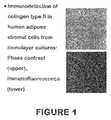
- FIG. 1shows the immunodetection of collagen type II in human adipose stromal cells from monolayer cultures. Phase contrast microscopy is used in the upper panel; immunofluorescence is used in the lower panel.
- FIG. 2shows immunodetection of collagen type II in human adipose stromal cells from pellet cultures. Phase contrast microscopy is used in the upper panel; immunofluorescence is used in the lower panel.
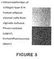
- FIG. 3shows immunodetection of collagen type II in human adipose stromal cells from alginate cultures. Phase contrast microscopy is used in the upper panel; immunofluorescence is used in the lower panel.
- FIG. 4shows Collagen type VI expression when cells were cultured in an alginate matrix at 2 weeks without TGF-beta (control) and with TGF-beta.
- FIG. 5shows a Western blot of results when cells were grown as monolayers or in an alginate suspension for the expression of different proteins including: collagen type VI, link, aggrecan, collagen type I, and actin.
- the present inventionprovides methods and a composition for the differentiation and culture of adipose tissue-derived stromal cells into chondrocytes.
- the cells produced by the methods of inventionare useful in providing a source of fully differentiated and functional cells for research, transplantation, and development of tissue engineering products for the treatment of human disease and traumatic injury repair.
- the inventionprovides a method for differentiating adipose tissue-derived stromal cells into chondrocytes comprising culturing stromal cells in a composition which comprises a medium capable of supporting the growth and differentiation of stromal cells into functional chondrocytes.
- This inventionfurther provides methods for the introduction of these chondrocytes into cartilage defect areas for repair.
- Adipose stromal cellsrefers to stromal cells that originate from adipose tissue.
- adiposeis meant any fat tissue.
- the adipose tissuemay be brown or white adipose tissue, derived from subcutaneous, omental/visceral, mammary, gonadal, or other adipose tissue site.
- the adiposeis subcutaneous white adipose tissue.
- Such cellsmay comprise a primary cell culture or an immortalized cell line.
- the adipose tissuemay be from any organism having fat tissue.
- the adipose tissueis mammalian, most preferably the adipose tissue is human.
- a convenient source of adipose tissueis from liposuction surgery, however, the source of adipose tissue or the method of isolation of adipose tissue is not critical to the invention. If stromal cells are desired for autologous transplantation into a subject, the adipose tissue will be isolated from that subject.
- Chodynamically chondrocytesrefers to cells that are capable of expressing characteristic biochemical markers of chondrocytes, including but not limited to collagen type II, chondroitin sulfate, keratin sulfate and characteristic morphologic markers of smooth muscle, including but not limited to the rounded morphology observed in culture, and able to secrete collagen type II, including but not limited to the generation of tissue or matrices with hemodynamic properties of cartilage in vitro.
- ⁇ MEMalpha modified Minimal Essential Medium
- RPMI Media 1640Roswell Park Memorial Institute Media 1640
- FBSFetal Bovine Serum
- a defined mediumcould be used if the necessary growth factors, cytokines, and hormones in FBS for stromal cells and chondrocytes are identified and provided at appropriate concentrations in the growth medium.
- Media useful in the methods of the inventionmay contain one or more compounds of interest, including, but not limited to antibiotics mitogenic or differentiative compounds for stromal cells.
- the cellswill be grown at temperatures between 31° C. to 37° C. in a humidified incubator.
- the carbon dioxide contentwill be maintained between 2% to 10% and the oxygen content between 1% and 22%. Cells will remain in this environment for periods of up to 4 weeks.
- Antibiotics which can be supplemented into the mediuminclude, but are not limited to penicillin and streptomycin.
- concentration of penicillin in the chemically defined culture mediumis about 10 to about 200 units per ml.
- concentration of streptomycin in the chemically defined culture mediumis about 10 to about 200 ⁇ g/ml.
- Glucocorticoidsthat can be used in the invention include but are not limited to hydrocortisone and dexamethasone.
- the concentration of dexamethasone in the mediumis about 1 to about 100 nM.
- the concentration of hydrocortisone in the mediumis about 1 to about 100 nM.
- chondroinductive agentor “chondroinductive factor” refers to any natural or synthetic, organic or inorganic chemical or biochemical compound or combination or mixture of compounds, or any mechanical or other physical device, container, influence or force that can be applied to human adipose tissue-derived stromal cells so as to effect their in vitro chondrogenic induction or the production of chondrocytes.
- the chondroinductive agentis preferably selected, individually or in combination, from the group consisting of (i) a glucocorticoid such as dexamethasone; (ii) a member of the transforming growth factor- ⁇ superfamily such as a bone morphogenic protein (preferably BMP-2 or BMP-4), TGF- ⁇ 1, TGF- ⁇ 2, TGF- ⁇ 3, insulin-like growth factor (IGF), platelet derived growth factor (PDGF), epidermal growth factor (EGF), acidic fibroblast growth factor (aFBF), basic fibroblast growth factor (bFBF), hepatocytic growth factor (HGF), keratocyte growth factor (KGF), osteogenic proteins (OP-1, OP-2, and OP-3), inhibin A or chondrogenic stimulating activity factor (CSA); (iii) a component of the collagenous extracellular matrix such as collagen I (particularly in the form of a gel); and (iv) a vitamin A analogue such as retinoic acid and; (v)
- the concentration of transforming growth factor-betais about 1 to about 100 ng/ml.
- the concentration of retinoic acidis about 0.1 to about 1 ⁇ g/ml.
- Examples of compounds that are stromal cell mitogensinclude but are not limited to transforming growth factor ⁇ ; fibroblast growth factor, bone morphogenetic protein and stromal cell differentiating factors include but are not limited to dexamethasone, hydrocortisone, transforming growth factor ⁇ , fibroblast growth factor, and bone morphogenetic protein and the like.
- the adipose tissue derived stromal cellsare isolated from the adipose tissue of the subject into which the final differentiated cells are to be introduced.
- the stromal cellsmay also be isolated from any organism of the same or different species as the subject. Any organism with adipose tissue can be a potential candidate.
- the organismis mammalian, most preferably the organism is human.
- the present inventionalso provides a method for differentiating adipose derived stromal cells into chondrocytic cells by suspending the cells in a calcium alginate or another biocompatible lattice or matrix capable of supporting chondrogenesis in a three dimensional configuration.
- lattice materialsinclude (1) calcium alginate, a polysaccharide of cross-linked L-glucuronic and D-mannuronic acid, at concentrations of between 1% to 4%; (2) fibrin; (3) collagen type II; or (4) agarose gel.
- the lattices or matrixes containing the cellsare transferred to culture dishes containing: (1) a chondroinductive agent that can activate any cellular transduction pathway leading to the mature chondrocyte phenotype; (2) an antibiotic; (3) a nutrient supplement such as fetal bovine serum or horse serum; (4) ascorbate or related vitamin C analogue; and (5) a glucocorticoid or another chemical agent capable of activating the cellular glucocorticoid receptor.
- the adipose tissue derived stromal cellsmay be stably or transiently transfected or transduced with a nucleic acid of interest using a plasmid, viral or alternative vector strategy.
- Nucleic acids of interestinclude, but are not limited to, those encoding gene products which enhance the production of extracellular matrix components found in cartilage; examples include transforming growth factor ⁇ , bone morphogentic protein, activin and insulin-like growth factor.
- viral vectors carrying regulatory genes into the stromal cellscan be performed with viral vectors (adenovirus, retrovirus, adeno-associated virus, or other vector) purified by cesium chloride banding or other method at a multiplicity of infection (viral units:cell) of between 10:1 to 2000:1.
- Cellswill be exposed to the virus in serum free or serum-containing medium in the absence or presence of a cationic detergent such as polyethyleneimine or LipofectamineTM for a period of 1 hour to 24 hours (Byk T. et al. (1998) Human Gene Therapy 9:2493-2502; Sommer B. et al. (1999) Calcif. Tissue Int. 64:45-49).
- the transfection of plasmid vectors carrying regulatory genes into the stromal cellscan be introduced into the cells in monolayer cultures by use of calcium phosphate DNA precipitation or cationic detergent methods (LipofectamineTM, DOTAP) or in three-dimensional cultures by incorporation of the plasmid DNA vectors directly into the biocompatible polymer (Bonadio J. et al. (1999) Nat. Med. 5:753-759).
- the viral or plasmid DNA vectorswill contain a readily detectable marker gene, such as the green fluorescent protein or beta-galactosidase enzyme, both of which can be tracked by histochemical means.
- a readily detectable marker genesuch as the green fluorescent protein or beta-galactosidase enzyme
- the carriersinclude but are not limited to calcium alginate, agarose, types I, II, IV or other collagen isoform, fibrin, poly-lactic/poly-glycolic acid, hyaluronate derivatives or other materials (Perka C. et al. (2000) J. Biomed. Mater. Res. 49:305-311; Sechriest V. F. et al. (2000) J. Biomed Mater. Res. 49:534-541; Chu C. R. et al. (1995) J. Biomed Mater. Res. 29:1147-1154; Hendrickson D. A. et al. (1994) Orthop. Res. 12:485-497).
- Another object of the inventionis to provide for the identification and study of compounds that enhance the differentiation of adipose tissue derived stromal cells into chondrocytes.
- Compounds which enhance differentiationmay be of value in the treatment of partial or full cartilage defects, osteoarthritis, traumatized cartilage, cosmetic surgery of inborn defects including cleft palate or deviated septum.
- Methodsinclude but are not limited to the development of three-dimensional in vitro cultures maintaining adipose tissue-derived stromal cells as chondrocytes that can be subsequently exposed to novel compounds of interest.
- Any compoundmay be tested for its ability to affect the differentiation of adipose tissue derived stromal cells into chondrocytes.
- Appropriate vehicles compatible with the compound to be testedare known to those skilled in the art and may be found in the current edition of Remington's Pharmaceutical Sciences (1995, Mack Publishing Co., Easton, Pa.) the contents of which are incorporated herein by reference.
- Stromal cellsare isolated from human subcutaneous adipose tissue according to methods described in “ Methods and Composition of the Differentiation of Human Preadipocytes into Adipocytes” Ser. No. 09/240,029 filed Jan. 29, 1999. These cells are plated at a density of 500 to 20,000 cells per cm 2 .
- the present inventioncontemplates that the creation of a precartilage condensation in vitro promotes chondrogenesis in mesenchymal progenitor cells derived from human adipose tissue. This is accomplished by methods including, but not limited to:
- Human adipose tissue-derived cellsare isolated as described above. For pellet cultures, aliquots of 200,000 cells were centrifuged at 500 g for 10 minutes in sterile 15 ml conical polypropylene tubes in DMEM with 10% fetal bovine serum, 50 ng/ml ascorbate-2-phosphate, 100 nM dexamethasone (DEX) and then incubated at 37° C. in a 5% CO 2 incubator for up to 3 weeks.
- DEXdexamethasone
- cellswere suspended at a density of 1 million cells per ml in 1.2% calcium alginate and maintained in DMEM with 10% fetal bovine serum, 50 ng/ml ascorbate-2-phosphate, 100 nM dexamethasone (DEX) and then incubated at 37° C. in a 5% CO 2 incubator for up to 3 weeks. After 2 or 4 weeks, the cells were isolated, fixed and analyzed for chondrocyte lineage markers by immunohistochemistry with appropriate antibody reagents or by staining with toluidine blue to detect the presence of sulfated proteoglycans in the extracellular matrix.
- DEXdexamethasone
- FIGS. 1-3Results obtained with an antibody detecting a representative chondrocyte marker protein, collagen II, are shown in FIGS. 1-3 .
- FIG. 4Immunohistochemical results with an antibody reagent detecting the chondrocyte marker protein, collagen VI, are shown in FIG. 4 .
- Adipose tissue-derived stromal cellswere maintained in 1.2% calcium alginate and maintained in DMEM with 10% fetal bovine serum, 50 ng/ml ascorbate-2-phosphate, 100 nM dexamethasone (DEX) in the absence or presence of transforming growth factor ⁇ (10 ng/ml) and then incubated at 37° C. in a 5% CO 2 incubator for up to 2 weeks. Immunohistochemistry revealed a dense deposition of the collagen VI protein surrounding those cells maintained in the presence, but not the absence, of transforming growth factor ⁇ .
- DEXdexamethasone
- Adipose tissue-derived stromal cellswere maintained in 1.2% calcium alginate (Alg) or in monolayer (Mono) cultures and maintained in DMEM with 10% fetal bovine serum, 50 ng/ml ascorbate-2-phosphate, 100 nM dexamethasone (DEX) in the absence (TGF ⁇ ) or presence (TGF ⁇ +) of transforming growth factor ⁇ (10 ng/ml) for a period of 4 weeks.
- Total RNAwas isolated from the individual cultures and used in polymerase chain reactions with primers specific for collagens types I or VI, the proteoglycan link (Link) protein, aggrecan, or actin. The collagen markers and actin were detected under all growth conditions. However, the link mRNAs were most abundant under alginate suspension conditions and aggrecan was only present under alginate conditions in the presence of TGF ⁇ .
- the chondrogenic cells still having chondrogenic potentialmay be cultured in an anchorage-independent manner, i.e., in a well having a cell contacting, cell adhesive surface, in order to stimulate the secretion of cartilage-specific extracellular matrix components.
- cartilage patches of predetermined thickness and volumeare dependent not only upon the volume of the well but also upon the number of chondrogenic cells seeded into the well.
- Cartilage of optimal predetermined volumemay be prepared by routine experimentation by altering either or both of the aforementioned parameters.
- the cell contacting surface of the wellmay be coated with a molecule that discourages adhesion of chondrogenic cells to the cell contacting surface.
- Preferred coating reagentsinclude silicon based reagents i.e., dichlorodimethylsilane or polytetrafluoroethylene based reagents, i.e., TEFLON®. Procedures for coating materials with silicon based reagents, specifically dichlorodimethylsilane, are well known in the art. See for example, Sambrook et al. (1989) “Molecular Cloning A Laboratory Manual,” Cold Spring Harbor Laboratory Press, the disclosure of which is incorporated by reference herein. It is appreciated that other biocompatible reagents that prevent the attachment of cells to the surface of the well may be useful in the practice of the instant invention.
- the wellmay be cast from a pliable or moldable biocompatible material that does not permit attachment of cells per se.
- Preferred materials that prevent such cell attachmentinclude, but are not limited to, agarose, glass, untreated cell culture plastic and polytetrafluoroethylene, i.e., TEFLON®.
- Untreated cell culture plasticsi.e., plastics that have not been treated with or made from materials that have an electrostatic charge are commercially available, and may be purchased, for example, from Falcon Labware, Becton-Dickinson, Lincoln Park, N.J.
- the aforementioned materialsare not meant to be limiting. It is appreciated that any other pliable or moldable biocompatible material that inherently discourages the attachment of chondrogenic cells may be useful in the practice of the instant invention.
- the size and shape of the wellmay be determined by the size and shape of the articular cartilage defect to be repaired.
- the wellmay have a cross-sectional surface area of 25 cm 2 . This is the average cross-sectional surface area of an adult, human femoral chondyle. Accordingly, it is anticipated that a single piece of synthetic cartilage may be prepared in accordance with the invention in order to resurface the entire femoral chondyle.
- the depth of the wellis preferably greater than about 0.3 cm and preferably about 0.6 cm in depth.
- the thickness of natural articular cartilage in an adult articulating jointis usually about 0.3 cm. Accordingly, the depth of the well should be large enough to permit a cartilage patch of about 0.3 cm to form. However, the well should also be deep enough to contain growth medium overlaying the cartilage patch.
- a large piece of cartilage prepared in accordance with the inventionmay be “trimmed” to a pre-selected size and shape by a surgeon performing surgical repair of the damaged cartilage. Trimming may be performed with the use of a sharp cutting implement, i.e., a scalpel, a pair of scissors or an arthroscopic device fitted with a cutting edge, using procedures well known in the art.
- a sharp cutting implementi.e., a scalpel, a pair of scissors or an arthroscopic device fitted with a cutting edge
- the pre-shaped wellpreferably is cast in a block of agarose gel under aseptic conditions.
- Agaroseis an economical, biocompatible, pliable and moldable material that can be used to cast pre-shaped wells, quickly and easily.
- the dimensions of the wellmay be dependent upon the size of the resulting cartilage plug that is desired.
- a pre-shaped wellmay be prepared by pouring a hot solution of molten LT agarose (BioRad, Richmond, Calif.) into a tissue culture dish containing a cylinder.
- the cylinderhaving dimensions that mirror the shape of the well to be formed.
- the size and shape of the wellmay be chosen by the artisan and may be dependent upon the shape of the articular cartilage defect to be repaired.
- the cylinderis carefully removed with forceps.
- the surface of the tissue culture dish that is exposed by the removal of the cylinderis covered with molten agarose. This seals the bottom of the well and provides a cell adhesive surface at the base of the well.
- the resulting pre-shaped wellis suitable for culturing, and stimulating the redifferentiation of proliferated chondrogenic cells. It is appreciated, however, that alternative methods may be used to prepare a pre-shaped well useful in the practice of the invention.
- Proliferated chondrogenic cells in suspensionmay be seeded into and cultured in the pre-shaped well.
- the cellsmay be diluted by the addition of cell culture medium to a cell density of about 1 ⁇ 10 5 to 1 ⁇ 10 9 chondrogenic cells per ml.
- a preferred cell culture mediumcomprises DMEM supplemented with 10% fetal bovine serum.
- the cellsmay coalesce to form a cohesive plug of cells. After about 4-10 days, the cells will start to secrete cartilage-specific sulfated proteoglycans and type II collagen. After prolonged periods of time in culture the collagen expressed by the chondrogenic cells in the well will be predominantly type II collagen. It is contemplated, however, that the cohesive plug of cells formed within four hours may be removed from the well and surgically implanted into the cartilage defect. It is anticipated that the undifferentiated chondrogenic cells subsequently may redifferentiate in situ thereby to form synthetic cartilage within the joint.
- chondrocytic differentiation or stimulatory factorsmay be added to the chondrogenic cells in the pre-shaped well to enhance or stimulate the production of articular cartilage specific proteoglycans and/or collagen (Luyten & Reddi (1992) in “Biological Regulation of the Chondrocytes,” CRC Press, Boca Raton, Ann Arbor, London, and Tokyo, pp. 227-236).
- Preferred growth factorsinclude, but are not limited to transforming growth factor- ⁇ (TGF- ⁇ ), insulin-like growth factor (IGF), platelet derived growth factor (PDGF), epidermal growth factor (EGF), acidic fibroblast growth factor (aFBF), basic fibroblast growth factor (bFBF), hepatocytic growth factor; (HGF) keratinocyte growth factor (KGF), the bone morphogenic factors (BMPs) i.e., BMP-1, BMP-2, BMP-3, BMP-4, BMP-5 and BMP-6 and the osteogenic proteins (OPs), i.e., OP-1, OP-2 and OP-3.
- TGF- ⁇transforming growth factor- ⁇
- IGFinsulin-like growth factor
- PDGFplatelet derived growth factor
- EGFepidermal growth factor
- aFBFacidic fibroblast growth factor
- bFBFbasic fibroblast growth factor
- OPsosteogenic proteins
- Preferred concentrations of TGF- ⁇ , IGF, PDGF, EGF, aFBF, bFBF, HGF, and KGFrange from about 1 to 100 ng/ml.
- Preferred concentrations of the BMP's and OP'srange from about 1 to about 500 ng/ml.
- any polypeptide growth factor capable of stimulating or inducing the production of cartilage specific proteoglycans and collagenmay be useful in the practice of the instant invention.
- ascorbatemay be added to the chondrogenic cells in the pre-shaped well to enhance or stimulate the production of cartilage specific proteoglycans and collagen.
- concentrations of ascorbaterange from about 1 to about 1000 ⁇ g/ml.
- Cartilage defects in mammalsare readily identifiable visually during arthroscopic examination or during open surgery of the joint. Cartilage defects may also be identified inferentially by using computer aided tomography (CAT scanning), X-ray examination, magnetic resonance imaging (MRI), analysis of synovial fluid or serum markers or by any other procedures known in the art. Treatment of the defects can be effected during an arthroscopic or open surgical procedure using the methods and compositions disclosed herein.
- CAT scanningcomputer aided tomography
- X-ray examinationX-ray examination
- MRImagnetic resonance imaging
- Treatment of the defectscan be effected during an arthroscopic or open surgical procedure using the methods and compositions disclosed herein.
- the defectmay be treated by the following steps of (1) surgically implanting at the predetermined site, a piece of synthetic articular cartilage prepared by the methodologies described herein, and (2) permitting the synthetic articular cartilage to integrate into predetermined site.
- the synthetic cartilage patchoptimally has a size and shape such that when the patch is implanted into the defect, the edges of the implanted tissue contact directly the edges of the defect.
- the synthetic cartilage patchmay be fixed in place during the surgical procedure. This can be effected by surgically fixing the patch into the defect with biodegradable sutures, i.e., (Ethicon, Johnson & Johnson) and/or by applying a bioadhesive to the region interfacing the patch and the defect.
- Preferred bioadhesivesinclude, but are not limited to: fibrin-thrombin glues similar to those disclosed in Fr. Pat. No. 2 448 900; Fr. Pat. No. 2 448 901 and EP.S.N. 88401961.3 and synthetic bioadhesives similar to those disclosed in U.S. Pat. No. 5,197,973. It is contemplated, however, that alternative types of sutures and biocompatible glues may be useful in the practice of the invention.
- damaged articular cartilagemay be surgically excised prior to the implantation of the patch of synthetic cartilage.
- adhesion of the synthetic cartilage patch to the articular cartilage defectmay be enhanced by treating the defect with transglutaminase (Ichinose et al. (1990) J. Biol. Chem. 265(3):13411-13414; Najjar et al. (1984) in “Transglutaminases,” Boston, Martinuse-Nijhoff).
- transglutaminaseIchinose et al. (1990) J. Biol. Chem. 265(3):13411-13414; Najjar et al. (1984) in “Transglutaminases,” Boston, Martinuse-Nijhoff.
- the cartilage defectis dried, for example, by using cottonoid, and filled with a solution of transglutaminase.
- the solutionis subsequently removed, for example, by aspiration, leaving a film containing transglutaminase upon the carti
- chondrogenic cellsmay be differentiated from human adipose tissue-derived stromal cells, i.e., human subcutaneous adipose tissue.
- the abovedemonstrates a culture system in which human adipose tissue-derived stromal cells differentiate into hypertrophic chondrocytes. Since all components are defined, the system can be used for studies of the effects of growth factors, etc. on the progression of chondrogenesis. In vitro systems have been used by us and others to show that these cell populations have osteogenic and adipocytic potential. We demonstrate here that this population has chondrogenic potential. This has clinical applicability for cartilage repair.
- the inventionalso provides a process for inducing chondrogenesis in human adipose tissue-derived stromal cells by contacting such cells with a chondroinductive agent in vitro where the stromal cells are associated in a three dimensional format.
- the inventionalso provides a process for using in vitro differentiated chondrocytes from adipose-derived stromal cells in the repair of cartilage tissue in mammals, including humans.
- the stromal cellsare preferably isolated, culture expanded human adipose tissue-derived stromal cells in a chemically defined environment and are condensed into close proximity, such as in the form of a three-dimensional cell mass, e.g., packed cells or a centrifugal cell pellet.
- the contactingpreferably comprises culturing a pellet of human adipose tissue-derived stromal cells in a chemically defined medium which comprises DMEM with 10% serum, 50 ng/ml ascorbate-2-phosphate, 10 ⁇ 7 M dexamethasone.
- the differentiated cellsare then introduced into the surgery site to repair cartilage. Since all components of the system are defined, the system can be used as a product for cartilage repair in mammals, including man and horses.
Landscapes
- Health & Medical Sciences (AREA)
- Engineering & Computer Science (AREA)
- Biomedical Technology (AREA)
- Life Sciences & Earth Sciences (AREA)
- Chemical & Material Sciences (AREA)
- Zoology (AREA)
- Organic Chemistry (AREA)
- Bioinformatics & Cheminformatics (AREA)
- General Health & Medical Sciences (AREA)
- Wood Science & Technology (AREA)
- Genetics & Genomics (AREA)
- Biotechnology (AREA)
- Cell Biology (AREA)
- Rheumatology (AREA)
- Veterinary Medicine (AREA)
- Chemical Kinetics & Catalysis (AREA)
- Public Health (AREA)
- Animal Behavior & Ethology (AREA)
- Medicinal Chemistry (AREA)
- General Engineering & Computer Science (AREA)
- Biochemistry (AREA)
- Microbiology (AREA)
- Botany (AREA)
- Oral & Maxillofacial Surgery (AREA)
- Transplantation (AREA)
- Epidemiology (AREA)
- Dermatology (AREA)
- Physical Education & Sports Medicine (AREA)
- Urology & Nephrology (AREA)
- Vascular Medicine (AREA)
- Nuclear Medicine, Radiotherapy & Molecular Imaging (AREA)
- General Chemical & Material Sciences (AREA)
- Pharmacology & Pharmacy (AREA)
- Orthopedic Medicine & Surgery (AREA)
- Micro-Organisms Or Cultivation Processes Thereof (AREA)
- Materials For Medical Uses (AREA)
- Medicines Containing Material From Animals Or Micro-Organisms (AREA)
Abstract
Description
- (1) The pellet culture system, which was developed for use with isolated growth plate cells (Kato et al. (1988)PNAS85:9552-9556; Ballock & Reddi,J. Cell Biol. (1994) 126(5):1311-1318) and has been used to maintain expression of the cartilage phenotype of chondrocytes placed in culture (Solursh (1991)J. Cell Biochem.45:258-260).
- (2) The alginate suspension method, where cells are maintained in a calcium alginate suspension to prevent cell-cell contact and maintain a characteristic rounded morphology promoting the maintenance or acquisition of the chondrocyte phenotype.
Claims (18)
Priority Applications (2)
| Application Number | Priority Date | Filing Date | Title |
|---|---|---|---|
| US13/250,742US8911994B2 (en) | 1999-08-19 | 2011-09-30 | Use of adipose tissue-derived stromal cells for chondrocyte differentiation and cartilage repair |
| US14/570,127US20150099300A1 (en) | 1999-08-19 | 2014-12-15 | Use of adipose tissue-derived stromal cells for chondrocyte differentiation and cartilage repair |
Applications Claiming Priority (6)
| Application Number | Priority Date | Filing Date | Title |
|---|---|---|---|
| US14985099P | 1999-08-19 | 1999-08-19 | |
| US09/573,989US6429013B1 (en) | 1999-08-19 | 2000-05-17 | Use of adipose tissue-derived stromal cells for chondrocyte differentiation and cartilage repair |
| US10/125,106US6841150B2 (en) | 1999-08-19 | 2002-04-18 | Use of adipose tissue-derived stromal cells for chondrocyte differentiation and cartilage repair |
| US10/980,137US7033587B2 (en) | 1999-08-19 | 2004-11-01 | Use of adipose tissue-derived stromal cells for chondrocyte differentiation and cartilage repair |
| US11/408,381US8048673B2 (en) | 1999-08-19 | 2006-04-21 | Use of adipose tissue-derived stromal cells for chondrocyte differentiation and cartilage repair |
| US13/250,742US8911994B2 (en) | 1999-08-19 | 2011-09-30 | Use of adipose tissue-derived stromal cells for chondrocyte differentiation and cartilage repair |
Related Parent Applications (1)
| Application Number | Title | Priority Date | Filing Date |
|---|---|---|---|
| US11/408,381ContinuationUS8048673B2 (en) | 1999-08-19 | 2006-04-21 | Use of adipose tissue-derived stromal cells for chondrocyte differentiation and cartilage repair |
Related Child Applications (1)
| Application Number | Title | Priority Date | Filing Date |
|---|---|---|---|
| US14/570,127DivisionUS20150099300A1 (en) | 1999-08-19 | 2014-12-15 | Use of adipose tissue-derived stromal cells for chondrocyte differentiation and cartilage repair |
Publications (2)
| Publication Number | Publication Date |
|---|---|
| US20130017604A1 US20130017604A1 (en) | 2013-01-17 |
| US8911994B2true US8911994B2 (en) | 2014-12-16 |
Family
ID=26847093
Family Applications (6)
| Application Number | Title | Priority Date | Filing Date |
|---|---|---|---|
| US09/573,989Expired - LifetimeUS6429013B1 (en) | 1999-08-19 | 2000-05-17 | Use of adipose tissue-derived stromal cells for chondrocyte differentiation and cartilage repair |
| US10/125,106Expired - Fee RelatedUS6841150B2 (en) | 1999-08-19 | 2002-04-18 | Use of adipose tissue-derived stromal cells for chondrocyte differentiation and cartilage repair |
| US10/980,137Expired - Fee RelatedUS7033587B2 (en) | 1999-08-19 | 2004-11-01 | Use of adipose tissue-derived stromal cells for chondrocyte differentiation and cartilage repair |
| US11/408,381Expired - Fee RelatedUS8048673B2 (en) | 1999-08-19 | 2006-04-21 | Use of adipose tissue-derived stromal cells for chondrocyte differentiation and cartilage repair |
| US13/250,742Expired - Fee RelatedUS8911994B2 (en) | 1999-08-19 | 2011-09-30 | Use of adipose tissue-derived stromal cells for chondrocyte differentiation and cartilage repair |
| US14/570,127AbandonedUS20150099300A1 (en) | 1999-08-19 | 2014-12-15 | Use of adipose tissue-derived stromal cells for chondrocyte differentiation and cartilage repair |
Family Applications Before (4)
| Application Number | Title | Priority Date | Filing Date |
|---|---|---|---|
| US09/573,989Expired - LifetimeUS6429013B1 (en) | 1999-08-19 | 2000-05-17 | Use of adipose tissue-derived stromal cells for chondrocyte differentiation and cartilage repair |
| US10/125,106Expired - Fee RelatedUS6841150B2 (en) | 1999-08-19 | 2002-04-18 | Use of adipose tissue-derived stromal cells for chondrocyte differentiation and cartilage repair |
| US10/980,137Expired - Fee RelatedUS7033587B2 (en) | 1999-08-19 | 2004-11-01 | Use of adipose tissue-derived stromal cells for chondrocyte differentiation and cartilage repair |
| US11/408,381Expired - Fee RelatedUS8048673B2 (en) | 1999-08-19 | 2006-04-21 | Use of adipose tissue-derived stromal cells for chondrocyte differentiation and cartilage repair |
Family Applications After (1)
| Application Number | Title | Priority Date | Filing Date |
|---|---|---|---|
| US14/570,127AbandonedUS20150099300A1 (en) | 1999-08-19 | 2014-12-15 | Use of adipose tissue-derived stromal cells for chondrocyte differentiation and cartilage repair |
Country Status (9)
| Country | Link |
|---|---|
| US (6) | US6429013B1 (en) |
| EP (1) | EP1077253B1 (en) |
| JP (1) | JP4294207B2 (en) |
| CN (1) | CN1227356C (en) |
| AT (1) | ATE330000T1 (en) |
| CA (1) | CA2316413C (en) |
| DE (1) | DE60028666T2 (en) |
| ES (1) | ES2265873T3 (en) |
| TW (3) | TW200712208A (en) |
Families Citing this family (215)
| Publication number | Priority date | Publication date | Assignee | Title |
|---|---|---|---|---|
| US8480754B2 (en) | 2001-05-25 | 2013-07-09 | Conformis, Inc. | Patient-adapted and improved articular implants, designs and related guide tools |
| US8735773B2 (en) | 2007-02-14 | 2014-05-27 | Conformis, Inc. | Implant device and method for manufacture |
| US8771365B2 (en) | 2009-02-25 | 2014-07-08 | Conformis, Inc. | Patient-adapted and improved orthopedic implants, designs, and related tools |
| US7534263B2 (en) | 2001-05-25 | 2009-05-19 | Conformis, Inc. | Surgical tools facilitating increased accuracy, speed and simplicity in performing joint arthroplasty |
| US8882847B2 (en) | 2001-05-25 | 2014-11-11 | Conformis, Inc. | Patient selectable knee joint arthroplasty devices |
| US7618451B2 (en) | 2001-05-25 | 2009-11-17 | Conformis, Inc. | Patient selectable joint arthroplasty devices and surgical tools facilitating increased accuracy, speed and simplicity in performing total and partial joint arthroplasty |
| US9603711B2 (en) | 2001-05-25 | 2017-03-28 | Conformis, Inc. | Patient-adapted and improved articular implants, designs and related guide tools |
| US20030055502A1 (en) | 2001-05-25 | 2003-03-20 | Philipp Lang | Methods and compositions for articular resurfacing |
| US7468075B2 (en) | 2001-05-25 | 2008-12-23 | Conformis, Inc. | Methods and compositions for articular repair |
| US8556983B2 (en) | 2001-05-25 | 2013-10-15 | Conformis, Inc. | Patient-adapted and improved orthopedic implants, designs and related tools |
| US8545569B2 (en) | 2001-05-25 | 2013-10-01 | Conformis, Inc. | Patient selectable knee arthroplasty devices |
| US8083745B2 (en) | 2001-05-25 | 2011-12-27 | Conformis, Inc. | Surgical tools for arthroplasty |
| US8617242B2 (en) | 2001-05-25 | 2013-12-31 | Conformis, Inc. | Implant device and method for manufacture |
| EP0896825B1 (en) | 1997-08-14 | 2002-07-17 | Sulzer Innotec Ag | Composition and device for in vivo cartilage repair comprising nanocapsules with osteoinductive and/or chondroinductive factors |
| JP4222726B2 (en)* | 1997-12-02 | 2009-02-12 | ゼン‐バイオ,インコーポレイテッド | Generation and use of osteoblasts |
| US7239908B1 (en) | 1998-09-14 | 2007-07-03 | The Board Of Trustees Of The Leland Stanford Junior University | Assessing the condition of a joint and devising treatment |
| US9289153B2 (en)* | 1998-09-14 | 2016-03-22 | The Board Of Trustees Of The Leland Stanford Junior University | Joint and cartilage diagnosis, assessment and modeling |
| WO2000035346A2 (en) | 1998-09-14 | 2000-06-22 | Stanford University | Assessing the condition of a joint and preventing damage |
| US20050153442A1 (en)* | 1999-03-10 | 2005-07-14 | Adam Katz | Adipose-derived stem cells and lattices |
| US6777231B1 (en)* | 1999-03-10 | 2004-08-17 | The Regents Of The University Of California | Adipose-derived stem cells and lattices |
| AU784580B2 (en)* | 1999-03-10 | 2006-05-04 | Regents Of The University Of California, The | Adipose-derived stem cells and lattices |
| US20050076396A1 (en)* | 1999-03-10 | 2005-04-07 | Katz Adam J. | Adipose-derived stem cells and lattices |
| US20030082152A1 (en)* | 1999-03-10 | 2003-05-01 | Hedrick Marc H. | Adipose-derived stem cells and lattices |
| US7078232B2 (en)* | 1999-08-19 | 2006-07-18 | Artecel, Inc. | Adipose tissue-derived adult stem or stromal cells for the repair of articular cartilage fractures and uses thereof |
| US6429013B1 (en) | 1999-08-19 | 2002-08-06 | Artecel Science, Inc. | Use of adipose tissue-derived stromal cells for chondrocyte differentiation and cartilage repair |
| US7935147B2 (en) | 1999-10-20 | 2011-05-03 | Anulex Technologies, Inc. | Method and apparatus for enhanced delivery of treatment device to the intervertebral disc annulus |
| US7004970B2 (en) | 1999-10-20 | 2006-02-28 | Anulex Technologies, Inc. | Methods and devices for spinal disc annulus reconstruction and repair |
| US8632590B2 (en)* | 1999-10-20 | 2014-01-21 | Anulex Technologies, Inc. | Apparatus and methods for the treatment of the intervertebral disc |
| US7052516B2 (en) | 1999-10-20 | 2006-05-30 | Anulex Technologies, Inc. | Spinal disc annulus reconstruction method and deformable spinal disc annulus stent |
| US6592625B2 (en)* | 1999-10-20 | 2003-07-15 | Anulex Technologies, Inc. | Spinal disc annulus reconstruction method and spinal disc annulus stent |
| US20030153976A1 (en)* | 1999-10-20 | 2003-08-14 | Cauthen Joseph C. | Spinal disc annulus reconstruction method and spinal disc annulus stent |
| US7615076B2 (en) | 1999-10-20 | 2009-11-10 | Anulex Technologies, Inc. | Method and apparatus for the treatment of the intervertebral disc annulus |
| US7951201B2 (en)* | 1999-10-20 | 2011-05-31 | Anulex Technologies, Inc. | Method and apparatus for the treatment of the intervertebral disc annulus |
| US8128698B2 (en) | 1999-10-20 | 2012-03-06 | Anulex Technologies, Inc. | Method and apparatus for the treatment of the intervertebral disc annulus |
| US20020123807A1 (en)* | 1999-10-20 | 2002-09-05 | Cauthen Joseph C. | Spinal disc annulus reconstruction method and spinal disc annulus stent |
| CA2427938C (en) | 1999-11-15 | 2010-02-16 | Bio Syntech Canada Inc. | Novel temperature-controlled and ph-dependant self-gelling biopolymeric aqueous solution, composition and preparation thereof |
| US20030158302A1 (en) | 1999-12-09 | 2003-08-21 | Cyric Chaput | Mineral-polymer hybrid composition |
| EP1237585B1 (en) | 1999-12-09 | 2003-06-18 | Biosyntech Canada Inc. | Mineral-polymer hybrid composition |
| AU3869501A (en) | 2000-02-26 | 2001-09-03 | Artecel Sciences Inc | Pleuripotent stem cells generated from adipose tissue-derived stromal cells and uses thereof |
| US7582292B2 (en)* | 2000-02-26 | 2009-09-01 | Artecel, Inc. | Adipose tissue derived stromal cells for the treatment of neurological disorders |
| US6805695B2 (en) | 2000-04-04 | 2004-10-19 | Spinalabs, Llc | Devices and methods for annular repair of intervertebral discs |
| US6402750B1 (en)* | 2000-04-04 | 2002-06-11 | Spinlabs, Llc | Devices and methods for the treatment of spinal disorders |
| SG149679A1 (en) | 2000-06-29 | 2009-02-27 | Biosyntech Canada Inc | Composition and method for the repair and regeneration of cartilage and other tissues |
| ATE426357T1 (en) | 2000-09-14 | 2009-04-15 | Univ Leland Stanford Junior | ASSESSING THE CONDITION OF A JOINT AND PLANNING TREATMENT |
| DE60136474D1 (en) | 2000-09-14 | 2008-12-18 | Univ R | ASSESSMENT OF THE CONDITION OF A JOINT AND LOSS OF CARTEL TISSUE |
| MXPA03004139A (en)* | 2000-11-09 | 2004-09-10 | Luca Kenneth Allan De | Fat extraction. |
| US8439926B2 (en) | 2001-05-25 | 2013-05-14 | Conformis, Inc. | Patient selectable joint arthroplasty devices and surgical tools |
| CA2466880A1 (en)* | 2001-11-09 | 2003-05-15 | Artecel Sciences, Inc. | Methods and compositions for the use of stromal cells to support embryonic and adult stem cells |
| HUP0500699A3 (en)* | 2001-11-09 | 2010-01-28 | Artecel Sciences | Endocrine pancreas differentiation of adipose tissue-derived stromal cells and uses thereof |
| US7595043B2 (en)* | 2001-12-07 | 2009-09-29 | Cytori Therapeutics, Inc. | Method for processing and using adipose-derived stem cells |
| US7651684B2 (en)* | 2001-12-07 | 2010-01-26 | Cytori Therapeutics, Inc. | Methods of using adipose tissue-derived cells in augmenting autologous fat transfer |
| US7585670B2 (en) | 2001-12-07 | 2009-09-08 | Cytori Therapeutics, Inc. | Automated methods for isolating and using clinically safe adipose derived regenerative cells |
| US7771716B2 (en)* | 2001-12-07 | 2010-08-10 | Cytori Therapeutics, Inc. | Methods of using regenerative cells in the treatment of musculoskeletal disorders |
| US20050095228A1 (en) | 2001-12-07 | 2005-05-05 | Fraser John K. | Methods of using regenerative cells in the treatment of peripheral vascular disease and related disorders |
| US8105580B2 (en) | 2001-12-07 | 2012-01-31 | Cytori Therapeutics, Inc. | Methods of using adipose derived stem cells to promote wound healing |
| CA2469370C (en)* | 2001-12-07 | 2014-07-08 | Macropore Biosurgery, Inc. | Adipose-derived cell processing unit |
| US8404229B2 (en)* | 2001-12-07 | 2013-03-26 | Cytori Therapeutics, Inc. | Methods of using adipose derived stem cells to treat acute tubular necrosis |
| US9597395B2 (en)* | 2001-12-07 | 2017-03-21 | Cytori Therapeutics, Inc. | Methods of using adipose tissue-derived cells in the treatment of cardiovascular conditions |
| CA2472239A1 (en)* | 2002-01-22 | 2003-07-31 | Pfizer Inc. | 3-(imidazolyl)-2-aminopropanoic acids for use as tafi-a inhibitors for the treatment of thrombotic diseases |
| CA2473360A1 (en)* | 2002-01-25 | 2003-08-07 | Genzyme Corporation | Serum-free media for chondrocytes and methods of use thereof |
| US20030215426A1 (en)* | 2002-04-02 | 2003-11-20 | William Marsh Rice University | Redifferentiated cells for repairing cartilage defects |
| WO2003084468A2 (en)* | 2002-04-03 | 2003-10-16 | Artecel Sciences, Inc. | Improvements of adipocytic differentiated adipose derived adult stem cells and uses thereof |
| US7622562B2 (en) | 2002-06-26 | 2009-11-24 | Zimmer Orthobiologics, Inc. | Rapid isolation of osteoinductive protein mixtures from mammalian bone tissue |
| JP2006501977A (en) | 2002-10-07 | 2006-01-19 | コンフォーミス・インコーポレイテッド | Minimally invasive joint implant with a three-dimensional profile that conforms to the joint surface |
| JP2006505366A (en) | 2002-11-07 | 2006-02-16 | コンフォーミス・インコーポレイテッド | Method of determining meniscus size and shape and devised treatment |
| BRPI0408787A (en)* | 2003-03-27 | 2006-03-28 | Childrens Hosp Medical Center | use of fgf-18 protein, target proteins and their respective coding nucleotide sequences for induction of cartilage formation |
| US20040213767A1 (en)* | 2003-04-23 | 2004-10-28 | Marc Hendriks | Methods for using adipose-derived cells for healing of aortic aneurysmal tissue |
| US7387645B2 (en)* | 2003-04-25 | 2008-06-17 | Medtronic Vascular, Inc. | Cellular therapy to heal vascular tissue |
| US7067123B2 (en) | 2003-04-29 | 2006-06-27 | Musculoskeletal Transplant Foundation | Glue for cartilage repair |
| US20050222687A1 (en)* | 2004-04-02 | 2005-10-06 | Gordana Vunjak-Novakovic | Cartilage implant assembly and method for implantation |
| US20050064042A1 (en)* | 2003-04-29 | 2005-03-24 | Musculoskeletal Transplant Foundation | Cartilage implant plug with fibrin glue and method for implantation |
| US20090291112A1 (en)* | 2003-05-16 | 2009-11-26 | Truncale Katherine G | Allograft osteochondral plug combined with cartilage particle mixture |
| US7901457B2 (en)* | 2003-05-16 | 2011-03-08 | Musculoskeletal Transplant Foundation | Cartilage allograft plug |
| US7488348B2 (en)* | 2003-05-16 | 2009-02-10 | Musculoskeletal Transplant Foundation | Cartilage allograft plug |
| US7875272B2 (en) | 2003-06-27 | 2011-01-25 | Ethicon, Incorporated | Treatment of stroke and other acute neuraldegenerative disorders using postpartum derived cells |
| US8518390B2 (en) | 2003-06-27 | 2013-08-27 | Advanced Technologies And Regenerative Medicine, Llc | Treatment of stroke and other acute neural degenerative disorders via intranasal administration of umbilical cord-derived cells |
| US9592258B2 (en) | 2003-06-27 | 2017-03-14 | DePuy Synthes Products, Inc. | Treatment of neurological injury by administration of human umbilical cord tissue-derived cells |
| AU2005322060B2 (en) | 2003-06-27 | 2011-11-17 | Ethicon Incorporated | Postpartum cells derived from umbilical cord tissue, and methods of making and using the same |
| US11191789B2 (en) | 2003-06-27 | 2021-12-07 | DePuy Synthes Products, Inc. | Cartilage and bone repair and regeneration using postpartum-derived cells |
| US8491883B2 (en) | 2003-06-27 | 2013-07-23 | Advanced Technologies And Regenerative Medicine, Llc | Treatment of amyotrophic lateral sclerosis using umbilical derived cells |
| US9572840B2 (en) | 2003-06-27 | 2017-02-21 | DePuy Synthes Products, Inc. | Regeneration and repair of neural tissue using postpartum-derived cells |
| US8790637B2 (en) | 2003-06-27 | 2014-07-29 | DePuy Synthes Products, LLC | Repair and regeneration of ocular tissue using postpartum-derived cells |
| WO2005018549A2 (en)* | 2003-08-12 | 2005-03-03 | The Brigham And Women' S Hospital, Inc. | Methods and compositions for tissue repair |
| SG146691A1 (en) | 2003-10-08 | 2008-10-30 | Vet Stem Inc | Methods of preparing and using stem cell compositions and kits comprising the same |
| ES2321001T3 (en)* | 2003-10-10 | 2009-06-01 | Kw2 Implantattechnologie Gmbh | REGENERATION OF CARTILAGO WITH GENERATION OF CONDRONS IN THE PRESENCE OF HIGH CONCENTRATIONS OF MAGNESIUM. |
| AU2004317828B2 (en)* | 2004-03-31 | 2010-06-24 | Reliance Life Sciences Pvt Ltd | Tissue-like organization of cells and macroscopic tissue-like constructs, generated by macromass culture of cells, and the method of macromass culture |
| US8048409B2 (en)* | 2004-05-27 | 2011-11-01 | Medtronic Vascular, Inc. | Cellular therapy to heal vascular tissue |
| EP1778293B1 (en)* | 2004-07-01 | 2015-04-22 | Cytori Therapeutics, Inc. | Methods of using regenerative cells in the treatment of musculoskeletal disorders |
| US20060051865A1 (en)* | 2004-08-31 | 2006-03-09 | Higgins Joel C | Systems and methods for isolating stromal cells from adipose tissue and uses thereof |
| US8697139B2 (en) | 2004-09-21 | 2014-04-15 | Frank M. Phillips | Method of intervertebral disc treatment using articular chondrocyte cells |
| US20080220044A1 (en)* | 2007-03-06 | 2008-09-11 | Semler Eric J | Cancellous construct with support ring for repair of osteochondral defects |
| US7837740B2 (en) | 2007-01-24 | 2010-11-23 | Musculoskeletal Transplant Foundation | Two piece cancellous construct for cartilage repair |
| US7473678B2 (en) | 2004-10-14 | 2009-01-06 | Biomimetic Therapeutics, Inc. | Platelet-derived growth factor compositions and methods of use thereof |
| US20060134781A1 (en)* | 2004-12-07 | 2006-06-22 | Bacterin International, Inc. | Three-dimensional cell culture system |
| WO2006071778A2 (en) | 2004-12-23 | 2006-07-06 | Ethicon Incorporated | Treatment of parkinson's disease and related disorders using postpartum derived cells |
| JP5138384B2 (en)* | 2005-01-10 | 2013-02-06 | サイトリ セラピューティクス インコーポレイテッド | Apparatus and method for monitoring, managing and servicing medical devices |
| US7815926B2 (en)* | 2005-07-11 | 2010-10-19 | Musculoskeletal Transplant Foundation | Implant for articular cartilage repair |
| WO2007009981A1 (en)* | 2005-07-15 | 2007-01-25 | Medizinische Hochschule Hannover | Medium and process for tissue cultivation |
| US7531355B2 (en)* | 2005-07-29 | 2009-05-12 | The Regents Of The University Of California | Methods and compositions for smooth muscle reconstruction |
| US20070027543A1 (en)* | 2005-08-01 | 2007-02-01 | Gimble Jeffrey M | Use of adipose tissue-derived stromal cells in spinal fusion |
| CA2925688C (en)* | 2005-09-09 | 2019-10-22 | Farshid Guilak | Tissue engineering methods and compositions |
| AU2006292224B2 (en) | 2005-09-19 | 2013-08-01 | Histogenics Corporation | Cell-support matrix and a method for preparation thereof |
| US20070104692A1 (en)* | 2005-11-07 | 2007-05-10 | Quijano Rodolfo C | Breast tissue regeneration |
| US20070104693A1 (en)* | 2005-11-07 | 2007-05-10 | Quijano Rodolfo C | Breast augmentation system |
| JP5368102B2 (en)* | 2005-11-17 | 2013-12-18 | バイオミメティック セラピューティクス, インコーポレイテッド | Maxillofacial bone enhancement using rhPDGF-BB and biocompatible matrix |
| EP1971681B1 (en) | 2005-12-16 | 2017-08-23 | DePuy Synthes Products, Inc. | Compositions and methods for inhibiting adverse immune response in histocompatibility-mismatched transplantation |
| EP1976975B1 (en)* | 2005-12-19 | 2012-08-01 | Ethicon, Inc. | In vitro expansion of postpartum derived cells in roller bottles |
| US9125906B2 (en) | 2005-12-28 | 2015-09-08 | DePuy Synthes Products, Inc. | Treatment of peripheral vascular disease using umbilical cord tissue-derived cells |
| CA2641022A1 (en)* | 2006-01-30 | 2007-08-09 | University Of Virginia Patent Foundation | Methods of preparing and characterizing mesenchymal stem cell aggregates and uses thereof |
| US8623026B2 (en) | 2006-02-06 | 2014-01-07 | Conformis, Inc. | Patient selectable joint arthroplasty devices and surgical tools incorporating anatomical relief |
| CN105030296A (en) | 2006-02-06 | 2015-11-11 | 康复米斯公司 | Patient selectable joint arthroplasty devices and surgical tools |
| AU2007212273B2 (en)* | 2006-02-09 | 2013-10-10 | Stryker Corporation | Compositions and methods for treating bone |
| US20070207504A1 (en)* | 2006-03-06 | 2007-09-06 | The Board Of Supervisors Of Louisiana State University And Agricultural And Mechanical College | Proteomic methods for the identification of differentiated adipose cells and adipose derived adult stem cells |
| ITTO20060282A1 (en)* | 2006-04-14 | 2007-10-15 | Univ Degli Studi Torino | MEDIUM OF CULTURE AND PHARMACEUTICAL COMPOSITION FOR THE REGENERATION OF THE RELATIVE PAPER FABRIC PROCEDURE RELATED TO USES AND PRODUCTS |
| EP2019859A4 (en)* | 2006-04-25 | 2009-11-25 | Univ Louisiana State | ADULPTED ADULPTED STEM CELLS DERIVED FOR USE IN HEPATIC REGENERATION |
| US20070258956A1 (en)* | 2006-05-02 | 2007-11-08 | Biomet Manufacturing Corp. | Methods and apparatus for promoting hair growth using adipose cell based therapies |
| US20090304644A1 (en)* | 2006-05-30 | 2009-12-10 | Cytori Therapeutics, Inc. | Systems and methods for manipulation of regenerative cells separated and concentrated from adipose tissue |
| US9161967B2 (en) | 2006-06-30 | 2015-10-20 | Biomimetic Therapeutics, Llc | Compositions and methods for treating the vertebral column |
| EP2049145B1 (en) | 2006-06-30 | 2018-03-14 | BioMimetic Therapeutics, LLC | Pdgf-biomatrix compositions and methods for treating rotator cuff injuries |
| WO2008013863A2 (en)* | 2006-07-26 | 2008-01-31 | Cytori Therapeutics, Inc. | Generation of adipose tissue and adipocytes |
| KR20090086066A (en)* | 2006-10-06 | 2009-08-10 | 유니버시티 오브 버지니아 페이턴트 파운데이션 | Useful methods and compositions for the treatment of diabetic wounds |
| WO2008073628A2 (en) | 2006-11-03 | 2008-06-19 | Biomimetic Therapeutics, Inc. | Compositions and methods for arthrodetic procedures |
| EP2119767B1 (en) | 2007-01-23 | 2016-01-20 | Yokohama City University | Method for preparation of cartilage cell |
| US8034014B2 (en) | 2007-03-06 | 2011-10-11 | Biomet Biologics, Llc | Angiogenesis initation and growth |
| US8435551B2 (en)* | 2007-03-06 | 2013-05-07 | Musculoskeletal Transplant Foundation | Cancellous construct with support ring for repair of osteochondral defects |
| US20080269762A1 (en)* | 2007-04-25 | 2008-10-30 | Biomet Manufacturing Corp. | Method and device for repair of cartilage defects |
| WO2008157412A2 (en) | 2007-06-13 | 2008-12-24 | Conformis, Inc. | Surgical cutting guide |
| US8273091B2 (en)* | 2007-10-04 | 2012-09-25 | Ebi, Llc | Alignment device for locking nail |
| WO2009046335A1 (en) | 2007-10-05 | 2009-04-09 | Ethicon, Incorporated | Repair and regeneration of renal tissue using human umbilical cord tissue-derived cells |
| EP2222234B1 (en)* | 2007-12-04 | 2018-01-24 | Ingeneron, Inc. | Apparatus and methods for cell isolation |
| EP2224884A2 (en)* | 2007-12-05 | 2010-09-08 | Musculoskeletal Transplant Foundation | Cancellous bone implant for cartilage repair |
| US8236538B2 (en) | 2007-12-20 | 2012-08-07 | Advanced Technologies And Regenerative Medicine, Llc | Methods for sterilizing materials containing biologically active agents |
| US20090192528A1 (en)* | 2008-01-29 | 2009-07-30 | Biomet Biologics, Inc. | Method and device for hernia repair |
| ES2422259T3 (en) | 2008-02-07 | 2013-09-10 | Biomimetic Therapeutics Inc | Compositions for distraction osteogenesis |
| WO2009111626A2 (en) | 2008-03-05 | 2009-09-11 | Conformis, Inc. | Implants for altering wear patterns of articular surfaces |
| EP2265220A1 (en)* | 2008-03-05 | 2010-12-29 | Musculoskeletal Transplant Foundation | Cancellous constructs, cartilage particles and combinations of cancellous constructs and cartilage particles |
| AU2009221773B2 (en)* | 2008-03-05 | 2015-03-05 | Conformis, Inc. | Edge-matched articular implant |
| WO2009140294A1 (en) | 2008-05-12 | 2009-11-19 | Conformis, Inc. | Devices and methods for treatment of facet and other joints |
| KR20110051166A (en) | 2008-05-22 | 2011-05-17 | 베스타 테라퓨틱스 인코포레이티드 | How to differentiate mammalian progenitor cells into insulin-producing islet cells |
| WO2010021993A1 (en)* | 2008-08-19 | 2010-02-25 | Cytori Therapeutics, Inc. | Methods of using adipose tissue-derived cells in the treatment of the lymphatic system and malignant disease |
| AU2009201915C1 (en) | 2008-08-22 | 2015-02-26 | Regeneus Ltd | Therapeutic methods |
| AU2011247866C9 (en)* | 2008-08-22 | 2014-02-13 | Regeneus Ltd. | Allogeneic therapeutic methods using adipose tissue-derived cell suspensions |
| CA2735885C (en) | 2008-09-09 | 2018-08-28 | Biomimetic Therapeutics, Inc. | Platelet-derived growth factor compositions and methods for the treatment of tendon and ligament injuries |
| US8163022B2 (en) | 2008-10-14 | 2012-04-24 | Anulex Technologies, Inc. | Method and apparatus for the treatment of the intervertebral disc annulus |
| US8865199B2 (en)* | 2008-11-17 | 2014-10-21 | Ingeneron, Inc. | Biomatrix composition and methods of biomatrix seeding |
| US10179900B2 (en)* | 2008-12-19 | 2019-01-15 | DePuy Synthes Products, Inc. | Conditioned media and methods of making a conditioned media |
| BRPI0923068B1 (en) | 2008-12-19 | 2021-05-25 | Advanced Technologies And Regenerative Medicine, Llc | USE OF CELLS DERIVED FROM UMBILICAL CORD TISSUE TO TREAT A PATIENT HAVING A PULMONARY DISEASE, DISORDER OR INJURY |
| WO2010099231A2 (en) | 2009-02-24 | 2010-09-02 | Conformis, Inc. | Automated systems for manufacturing patient-specific orthopedic implants and instrumentation |
| US9017334B2 (en) | 2009-02-24 | 2015-04-28 | Microport Orthopedics Holdings Inc. | Patient specific surgical guide locator and mount |
| US8808297B2 (en) | 2009-02-24 | 2014-08-19 | Microport Orthopedics Holdings Inc. | Orthopedic surgical guide |
| US8808303B2 (en) | 2009-02-24 | 2014-08-19 | Microport Orthopedics Holdings Inc. | Orthopedic surgical guide |
| US12383287B2 (en) | 2009-02-24 | 2025-08-12 | Microport Orthopedics Holdings, Inc. | Systems and methods for installing an orthopedic implant |
| ES2629003T3 (en) | 2009-03-26 | 2017-08-07 | DePuy Synthes Products, Inc. | Human umbilical cord tissue cells as a treatment for Alzheimer's disease |
| SG10201401326SA (en) | 2009-04-16 | 2014-10-30 | Conformis Inc | Patient-specific joint arthroplasty devices for ligament repair |
| EP2424463B1 (en)* | 2009-05-01 | 2016-12-28 | Bimini Technologies LLC | Systems, methods and compositions for optimizing tissue and cell enriched grafts |
| US9173903B2 (en) | 2009-08-17 | 2015-11-03 | Ashok K. Singh | Fluid associated with adult stem cells for medical, cosmetic, and veterinary use |
| CA2782137A1 (en) | 2009-12-11 | 2011-06-16 | Conformis, Inc. | Patient-specific and patient-engineered orthopedic implants |
| US8652153B2 (en) | 2010-01-11 | 2014-02-18 | Anulex Technologies, Inc. | Intervertebral disc annulus repair system and bone anchor delivery tool |
| CN102811622A (en) | 2010-02-22 | 2012-12-05 | 生物模拟治疗公司 | Platelet-derived growth factor compositions and methods for treating tendinopathy |
| US10130736B1 (en) | 2010-05-14 | 2018-11-20 | Musculoskeletal Transplant Foundation | Tissue-derived tissuegenic implants, and methods of fabricating and using same |
| US8883210B1 (en) | 2010-05-14 | 2014-11-11 | Musculoskeletal Transplant Foundation | Tissue-derived tissuegenic implants, and methods of fabricating and using same |
| US9352003B1 (en) | 2010-05-14 | 2016-05-31 | Musculoskeletal Transplant Foundation | Tissue-derived tissuegenic implants, and methods of fabricating and using same |
| US20120020931A1 (en) | 2010-06-02 | 2012-01-26 | Rutgers, The State University Of New Jersey | Therapeutic encapsulated embryonic stem cells and mesenchymal stromal cells |
| WO2013106655A1 (en) | 2012-01-11 | 2013-07-18 | The Gid Group, Inc. | Method for processing adipose tissue and processing apparatus |
| EP2590695B1 (en) | 2010-07-09 | 2024-11-20 | GID BIO, Inc. | Apparatus and methods relating to collecting and processing human biological material containing adipose |
| US9206387B2 (en) | 2010-07-09 | 2015-12-08 | The Gid Group, Inc. | Method and apparatus for processing adipose tissue |
| US9296984B2 (en) | 2010-07-09 | 2016-03-29 | The Gid Group, Inc. | Tissue processing apparatus and method for processing adipose tissue |
| EP2625263B1 (en) | 2010-10-08 | 2020-03-11 | Terumo BCT, Inc. | Configurable methods and systems of growing and harvesting cells in a hollow fiber bioreactor system |
| US8440440B2 (en) | 2010-12-27 | 2013-05-14 | Intellicell Biosciences Inc. | Ultrasonic cavitation derived stromal or mesenchymal vascular extracts and cells derived therefrom obtained from adipose tissue and use thereof |
| RU2452527C1 (en)* | 2010-12-27 | 2012-06-10 | Александр Сергеевич Тепляшин | Method of cartilage hyaline neogenesis accompanying early destructive diseases |
| WO2012112694A2 (en) | 2011-02-15 | 2012-08-23 | Conformis, Inc. | Medeling, analyzing and using anatomical data for patient-adapted implants. designs, tools and surgical procedures |
| US8834928B1 (en) | 2011-05-16 | 2014-09-16 | Musculoskeletal Transplant Foundation | Tissue-derived tissugenic implants, and methods of fabricating and using same |
| DK3653698T3 (en) | 2011-12-01 | 2025-07-07 | New York Stem Cell Found Inc | AUTOMATED SYSTEM FOR THE PRODUCTION OF INDUCED PLURIPOTENT STEM CELLS OR DIFFERENTIATED CELLS |
| RU2636220C2 (en) | 2011-12-23 | 2017-11-21 | Депуи Синтез Продактс, Инк. | Detection of cells obtained from umbilical cord tissue |
| KR102159844B1 (en)* | 2012-02-24 | 2020-09-24 | 마사노리 사에키 | Cell preparation including fat cell |
| US9486226B2 (en) | 2012-04-18 | 2016-11-08 | Conformis, Inc. | Tibial guides, tools, and techniques for resecting the tibial plateau |
| US9675471B2 (en) | 2012-06-11 | 2017-06-13 | Conformis, Inc. | Devices, techniques and methods for assessing joint spacing, balancing soft tissues and obtaining desired kinematics for joint implant components |
| BR112015004003B1 (en) | 2012-09-06 | 2020-05-19 | The Gid Group Inc | apparatus for processing human biological material containing fibrous tissue and method for processing adipose tissue |
| US10179012B2 (en) | 2013-01-28 | 2019-01-15 | Cartiva, Inc. | Systems and methods for orthopedic repair |
| US9737294B2 (en) | 2013-01-28 | 2017-08-22 | Cartiva, Inc. | Method and system for orthopedic repair |
| EP3011015B1 (en)* | 2013-06-19 | 2021-02-24 | Spinalcyte, LLC | Adipose cells for chondrocyte applications |
| WO2015017500A1 (en) | 2013-07-30 | 2015-02-05 | Musculoskeletal Transplant Foundation | Acellular soft tissue-derived matrices and methods for preparing same |
| US10336980B2 (en) | 2013-09-05 | 2019-07-02 | The Gid Group, Inc. | Tissue processing apparatus and method for processing adipose tissue |
| WO2015073913A1 (en) | 2013-11-16 | 2015-05-21 | Terumo Bct, Inc. | Expanding cells in a bioreactor |
| CN106232800B (en) | 2014-03-25 | 2020-07-03 | 泰尔茂比司特公司 | Passive replacement of media |
| EP2998392A1 (en)* | 2014-09-16 | 2016-03-23 | The Provost, Fellows, Foundation Scholars, & the other members of Board, of the College of the Holy & Undiv. Trinity of Queen Elizabeth near Dublin | A population of cells for use in single-stage cell-based cartilage regeneration |
| JP6830059B2 (en) | 2014-09-26 | 2021-02-17 | テルモ ビーシーティー、インコーポレーテッド | Scheduled cell feeding |
| US10077420B2 (en) | 2014-12-02 | 2018-09-18 | Histogenics Corporation | Cell and tissue culture container |
| KR101706642B1 (en)* | 2015-02-04 | 2017-02-17 | 주식회사 엑소스템텍 | Composition comprising exosomes extracted from stem cells which differentiate into chondrocytes for inducing chondrogenic differentiation or regenerating cartilage tissue |
| CA2986702C (en) | 2015-05-21 | 2023-04-04 | David Wang | Modified demineralized cortical bone fibers |
| WO2017004592A1 (en) | 2015-07-02 | 2017-01-05 | Terumo Bct, Inc. | Cell growth with mechanical stimuli |
| US10912864B2 (en) | 2015-07-24 | 2021-02-09 | Musculoskeletal Transplant Foundation | Acellular soft tissue-derived matrices and methods for preparing same |
| US11052175B2 (en) | 2015-08-19 | 2021-07-06 | Musculoskeletal Transplant Foundation | Cartilage-derived implants and methods of making and using same |
| EP3455345B1 (en) | 2016-05-11 | 2021-03-24 | Animal Cell Therapy - Act | Production of a canine beta cell line from an immature pancreas |
| WO2017205667A1 (en) | 2016-05-25 | 2017-11-30 | Terumo Bct, Inc. | Cell expansion |
| US11685883B2 (en) | 2016-06-07 | 2023-06-27 | Terumo Bct, Inc. | Methods and systems for coating a cell growth surface |
| US11104874B2 (en) | 2016-06-07 | 2021-08-31 | Terumo Bct, Inc. | Coating a bioreactor |
| WO2018044791A1 (en) | 2016-08-30 | 2018-03-08 | Lifecell Corporation | Systems and methods for medical device control |
| CN108342356A (en)* | 2017-01-23 | 2018-07-31 | 曹谊林 | A kind of cartilage graft and its construction method |
| USD851777S1 (en) | 2017-01-30 | 2019-06-18 | Lifecell Corporation | Canister-type device for tissue processing |
| EP3601521A2 (en) | 2017-03-31 | 2020-02-05 | Terumo BCT, Inc. | Cell expansion |
| US12234441B2 (en) | 2017-03-31 | 2025-02-25 | Terumo Bct, Inc. | Cell expansion |
| US11624046B2 (en) | 2017-03-31 | 2023-04-11 | Terumo Bct, Inc. | Cell expansion |
| US11732233B2 (en) | 2017-07-18 | 2023-08-22 | Gid Bio, Inc. | Adipose tissue digestion system and tissue processing method |
| US11696929B2 (en) | 2017-09-20 | 2023-07-11 | The Regents Of The University Of California | Methods and systems for conserving highly expanded cells |
| CA3082242A1 (en) | 2017-11-08 | 2019-05-16 | Animal Cell Therapy - Act | Production of canine pancreatic islets from an immature pancreas |
| US11471562B2 (en) | 2018-04-09 | 2022-10-18 | University of Pittsburgh—of the Commonwealth System of Higher Education | Cartilage material having minimal hypertrophy and robust integration capacity, and uses therefor |
| CA3122024A1 (en) | 2018-12-03 | 2020-06-11 | Ross Ward | Indirect ultrasonic cavitation-derived perivascular cells and methods of use thereof |
| CN109749989A (en)* | 2019-02-18 | 2019-05-14 | 上海交通大学医学院附属第九人民医院 | Utilize the method for adipose tissue direct construction marrow tabernacle model |
| EP3975939B1 (en) | 2019-05-29 | 2024-11-13 | Wright Medical Technology, Inc. | Device for preparing a tibia for receiving tibial implant component of a replacement ankle |
| WO2021146015A1 (en) | 2020-01-17 | 2021-07-22 | Wright Medical Technology, Inc. | Guidance tools, systems, and methods |
| KR102141641B1 (en)* | 2020-01-31 | 2020-08-06 | 주식회사 래디안 | Method for differentiation of dermal papilla cells from human adipose mesenchymal stem cells |
| CN111676190B (en)* | 2020-06-09 | 2022-04-22 | 生物岛实验室 | A kind of inducer for the differentiation of stem cells to chondroblasts and its application |
| EP4314244B1 (en) | 2021-03-23 | 2025-07-23 | Terumo BCT, Inc. | Cell capture and expansion |
| US12152699B2 (en) | 2022-02-28 | 2024-11-26 | Terumo Bct, Inc. | Multiple-tube pinch valve assembly |
Citations (25)
| Publication number | Priority date | Publication date | Assignee | Title |
|---|---|---|---|---|
| FR2448901A1 (en) | 1979-02-15 | 1980-09-12 | Immuno Ag | GLUE FOR JOINING HUMAN TISSUES OR ANIMALS, METHOD FOR MANUFACTURING SAME AND APPLICATIONS OF SUCH GLUE |
| FR2448900A1 (en) | 1979-02-15 | 1980-09-12 | Immuno Ag | GLUE FOR JOINING HUMAN TISSUES OR ANIMALS, METHOD FOR MANUFACTURING SAME AND APPLICATIONS OF SUCH GLUE |
| US5160490A (en) | 1986-04-18 | 1992-11-03 | Marrow-Tech Incorporated | Three-dimensional cell and tissue culture apparatus |
| US5197973A (en) | 1990-12-14 | 1993-03-30 | Creative Biomolecules, Inc. | Synthetic bioadhesive |
| US5446143A (en) | 1993-09-14 | 1995-08-29 | Board Of Regents, The University Of Texas System | Adipose-specific promoter region of human aromatase cytochrome P450 gene |
| WO1995033821A1 (en) | 1994-06-06 | 1995-12-14 | Advanced Tissue Sciences, Inc. | Three-dimensional cartilage cultures |
| US5486359A (en) | 1990-11-16 | 1996-01-23 | Osiris Therapeutics, Inc. | Human mesenchymal stem cells |
| WO1996023059A1 (en) | 1995-01-24 | 1996-08-01 | Case Western Reserve University | Lineage-directed induction of human mesenchymal stem cell differentiation |
| US5567612A (en) | 1986-11-20 | 1996-10-22 | Massachusetts Institute Of Technology | Genitourinary cell-matrix structure for implantation into a human and a method of making |
| WO1997018299A1 (en) | 1995-11-16 | 1997-05-22 | Case Western Reserve University | In vitro chondrogenic induction of human mesenchymal stem cells |
| WO1998004681A2 (en) | 1996-07-25 | 1998-02-05 | Genzyme Corporation | Chondrocyte media formulations and culture procedures |
| US5723331A (en) | 1994-05-05 | 1998-03-03 | Genzyme Corporation | Methods and compositions for the repair of articular cartilage defects in mammals |
| US5759830A (en) | 1986-11-20 | 1998-06-02 | Massachusetts Institute Of Technology | Three-dimensional fibrous scaffold containing attached cells for producing vascularized tissue in vivo |
| US5786207A (en) | 1997-05-28 | 1998-07-28 | University Of Pittsburgh | Tissue dissociating system and method |
| US5842477A (en)* | 1996-02-21 | 1998-12-01 | Advanced Tissue Sciences, Inc. | Method for repairing cartilage |
| US5863531A (en) | 1986-04-18 | 1999-01-26 | Advanced Tissue Sciences, Inc. | In vitro preparation of tubular tissue structures by stromal cell culture on a three-dimensional framework |
| US5906934A (en) | 1995-03-14 | 1999-05-25 | Morphogen Pharmaceuticals, Inc. | Mesenchymal stem cells for cartilage repair |
| WO1999028444A1 (en) | 1997-12-02 | 1999-06-10 | Zen Bio, Inc. | Differentiation of adipose stromal cells into osteoblasts and uses thereof |
| WO2000053795A1 (en) | 1999-03-10 | 2000-09-14 | University Of Pittsburgh Of The Commonwealth System Of Higher Education | Adipose-derived stem cells and lattices |
| WO2000053797A1 (en) | 1999-03-09 | 2000-09-14 | Acordis Industrial Fibers Gmbh | Method for the in vitro testing of active ingredients, corresponding device and their use |
| US6153432A (en) | 1999-01-29 | 2000-11-28 | Zen-Bio, Inc | Methods for the differentiation of human preadipocytes into adipocytes |
| US6194203B1 (en) | 1996-04-19 | 2001-02-27 | Nestec S.A. | Immortalized adult human colon epithelial cell line |
| US6200606B1 (en) | 1996-01-16 | 2001-03-13 | Depuy Orthopaedics, Inc. | Isolation of precursor cells from hematopoietic and nonhematopoietic tissues and their use in vivo bone and cartilage regeneration |
| US6429013B1 (en) | 1999-08-19 | 2002-08-06 | Artecel Science, Inc. | Use of adipose tissue-derived stromal cells for chondrocyte differentiation and cartilage repair |
| US7078232B2 (en) | 1999-08-19 | 2006-07-18 | Artecel, Inc. | Adipose tissue-derived adult stem or stromal cells for the repair of articular cartilage fractures and uses thereof |
Family Cites Families (1)
| Publication number | Priority date | Publication date | Assignee | Title |
|---|---|---|---|---|
| US519773A (en)* | 1894-05-15 | Tile-machine |
- 2000
- 2000-05-17USUS09/573,989patent/US6429013B1/ennot_activeExpired - Lifetime
- 2000-08-08TWTW095132252Apatent/TW200712208A/enunknown
- 2000-08-08TWTW089115958Apatent/TWI243207B/ennot_activeIP Right Cessation
- 2000-08-08TWTW093126260Apatent/TWI283707B/ennot_activeIP Right Cessation
- 2000-08-18CACA2316413Apatent/CA2316413C/ennot_activeExpired - Fee Related
- 2000-08-21DEDE60028666Tpatent/DE60028666T2/ennot_activeExpired - Lifetime
- 2000-08-21JPJP2000250009Apatent/JP4294207B2/ennot_activeExpired - Fee Related
- 2000-08-21EPEP00307155Apatent/EP1077253B1/ennot_activeExpired - Lifetime
- 2000-08-21ATAT00307155Tpatent/ATE330000T1/ennot_activeIP Right Cessation
- 2000-08-21ESES00307155Tpatent/ES2265873T3/ennot_activeExpired - Lifetime
- 2000-08-21CNCNB001289810Apatent/CN1227356C/ennot_activeExpired - Fee Related
- 2002
- 2002-04-18USUS10/125,106patent/US6841150B2/ennot_activeExpired - Fee Related
- 2004
- 2004-11-01USUS10/980,137patent/US7033587B2/ennot_activeExpired - Fee Related
- 2006
- 2006-04-21USUS11/408,381patent/US8048673B2/ennot_activeExpired - Fee Related
- 2011
- 2011-09-30USUS13/250,742patent/US8911994B2/ennot_activeExpired - Fee Related
- 2014
- 2014-12-15USUS14/570,127patent/US20150099300A1/ennot_activeAbandoned
Patent Citations (32)
| Publication number | Priority date | Publication date | Assignee | Title |
|---|---|---|---|---|
| FR2448900A1 (en) | 1979-02-15 | 1980-09-12 | Immuno Ag | GLUE FOR JOINING HUMAN TISSUES OR ANIMALS, METHOD FOR MANUFACTURING SAME AND APPLICATIONS OF SUCH GLUE |
| US4362567A (en) | 1979-02-15 | 1982-12-07 | Immuno Aktiengesellschaft Fur Chemisch-Medizinische Produkte | Tissue adhesive |
| US4377572A (en) | 1979-02-15 | 1983-03-22 | Immuno Aktiengesellschaft Fur Chemisch-Medizinische Produkte | Tissue adhesive |
| FR2448901A1 (en) | 1979-02-15 | 1980-09-12 | Immuno Ag | GLUE FOR JOINING HUMAN TISSUES OR ANIMALS, METHOD FOR MANUFACTURING SAME AND APPLICATIONS OF SUCH GLUE |
| US5160490A (en) | 1986-04-18 | 1992-11-03 | Marrow-Tech Incorporated | Three-dimensional cell and tissue culture apparatus |
| US5902741A (en) | 1986-04-18 | 1999-05-11 | Advanced Tissue Sciences, Inc. | Three-dimensional cartilage cultures |
| US5863531A (en) | 1986-04-18 | 1999-01-26 | Advanced Tissue Sciences, Inc. | In vitro preparation of tubular tissue structures by stromal cell culture on a three-dimensional framework |
| US5567612A (en) | 1986-11-20 | 1996-10-22 | Massachusetts Institute Of Technology | Genitourinary cell-matrix structure for implantation into a human and a method of making |
| US5759830A (en) | 1986-11-20 | 1998-06-02 | Massachusetts Institute Of Technology | Three-dimensional fibrous scaffold containing attached cells for producing vascularized tissue in vivo |
| US5486359A (en) | 1990-11-16 | 1996-01-23 | Osiris Therapeutics, Inc. | Human mesenchymal stem cells |
| US5197973A (en) | 1990-12-14 | 1993-03-30 | Creative Biomolecules, Inc. | Synthetic bioadhesive |
| US5446143A (en) | 1993-09-14 | 1995-08-29 | Board Of Regents, The University Of Texas System | Adipose-specific promoter region of human aromatase cytochrome P450 gene |
| US5723331A (en) | 1994-05-05 | 1998-03-03 | Genzyme Corporation | Methods and compositions for the repair of articular cartilage defects in mammals |
| WO1995033821A1 (en) | 1994-06-06 | 1995-12-14 | Advanced Tissue Sciences, Inc. | Three-dimensional cartilage cultures |
| WO1996023059A1 (en) | 1995-01-24 | 1996-08-01 | Case Western Reserve University | Lineage-directed induction of human mesenchymal stem cell differentiation |
| US5906934A (en) | 1995-03-14 | 1999-05-25 | Morphogen Pharmaceuticals, Inc. | Mesenchymal stem cells for cartilage repair |
| WO1997018299A1 (en) | 1995-11-16 | 1997-05-22 | Case Western Reserve University | In vitro chondrogenic induction of human mesenchymal stem cells |
| US5908784A (en) | 1995-11-16 | 1999-06-01 | Case Western Reserve University | In vitro chondrogenic induction of human mesenchymal stem cells |
| US6200606B1 (en) | 1996-01-16 | 2001-03-13 | Depuy Orthopaedics, Inc. | Isolation of precursor cells from hematopoietic and nonhematopoietic tissues and their use in vivo bone and cartilage regeneration |
| US5842477A (en)* | 1996-02-21 | 1998-12-01 | Advanced Tissue Sciences, Inc. | Method for repairing cartilage |
| US6194203B1 (en) | 1996-04-19 | 2001-02-27 | Nestec S.A. | Immortalized adult human colon epithelial cell line |
| WO1998004681A2 (en) | 1996-07-25 | 1998-02-05 | Genzyme Corporation | Chondrocyte media formulations and culture procedures |
| US5786207A (en) | 1997-05-28 | 1998-07-28 | University Of Pittsburgh | Tissue dissociating system and method |
| WO1999028444A1 (en) | 1997-12-02 | 1999-06-10 | Zen Bio, Inc. | Differentiation of adipose stromal cells into osteoblasts and uses thereof |
| US6153432A (en) | 1999-01-29 | 2000-11-28 | Zen-Bio, Inc | Methods for the differentiation of human preadipocytes into adipocytes |
| WO2000053797A1 (en) | 1999-03-09 | 2000-09-14 | Acordis Industrial Fibers Gmbh | Method for the in vitro testing of active ingredients, corresponding device and their use |
| WO2000053795A1 (en) | 1999-03-10 | 2000-09-14 | University Of Pittsburgh Of The Commonwealth System Of Higher Education | Adipose-derived stem cells and lattices |
| US6429013B1 (en) | 1999-08-19 | 2002-08-06 | Artecel Science, Inc. | Use of adipose tissue-derived stromal cells for chondrocyte differentiation and cartilage repair |
| US6841150B2 (en) | 1999-08-19 | 2005-01-11 | Artecal, Sciences, Inc. | Use of adipose tissue-derived stromal cells for chondrocyte differentiation and cartilage repair |
| US7033587B2 (en) | 1999-08-19 | 2006-04-25 | Artecel Sciences, Inc. | Use of adipose tissue-derived stromal cells for chondrocyte differentiation and cartilage repair |
| US7078232B2 (en) | 1999-08-19 | 2006-07-18 | Artecel, Inc. | Adipose tissue-derived adult stem or stromal cells for the repair of articular cartilage fractures and uses thereof |
| US20060228796A1 (en) | 1999-08-19 | 2006-10-12 | Artecel, Inc. | Adipose tissue-derived adult stem cells for the repair of articular cartilage fractures and uses thereof |
Non-Patent Citations (62)
Also Published As
| Publication number | Publication date |
|---|---|
| DE60028666D1 (en) | 2006-07-27 |
| US20050112761A1 (en) | 2005-05-26 |
| CA2316413A1 (en) | 2001-02-19 |
| CA2316413C (en) | 2013-11-05 |
| DE60028666T2 (en) | 2007-05-24 |
| US20020115647A1 (en) | 2002-08-22 |
| CN1227356C (en) | 2005-11-16 |
| TWI243207B (en) | 2005-11-11 |
| US8048673B2 (en) | 2011-11-01 |
| TW200516149A (en) | 2005-05-16 |
| US20130017604A1 (en) | 2013-01-17 |
| US20150099300A1 (en) | 2015-04-09 |
| TWI283707B (en) | 2007-07-11 |
| EP1077253A1 (en) | 2001-02-21 |
| JP4294207B2 (en) | 2009-07-08 |
| TW200712208A (en) | 2007-04-01 |
| JP2001103965A (en) | 2001-04-17 |
| US6429013B1 (en) | 2002-08-06 |
| US7033587B2 (en) | 2006-04-25 |
| US20060188985A1 (en) | 2006-08-24 |
| ATE330000T1 (en) | 2006-07-15 |
| CN1306085A (en) | 2001-08-01 |
| EP1077253B1 (en) | 2006-06-14 |
| ES2265873T3 (en) | 2007-03-01 |
| US6841150B2 (en) | 2005-01-11 |
Similar Documents
| Publication | Publication Date | Title |
|---|---|---|
| US8911994B2 (en) | Use of adipose tissue-derived stromal cells for chondrocyte differentiation and cartilage repair | |
| US7078232B2 (en) | Adipose tissue-derived adult stem or stromal cells for the repair of articular cartilage fractures and uses thereof | |
| US12359172B2 (en) | Method for preparing 3D cartilage organoid block | |
| US5786217A (en) | Methods and compositions for the repair of articular cartilage defects in mammals | |
| US6761887B1 (en) | Alginate layer system for chondrogenic differentiation of human mesenchymal stem cells | |
| US20090123927A1 (en) | Isolation of precursor cells and their use for tissue repair | |
| KR102479530B1 (en) | Method of Preparing Pellets of Chondrocytes differentiated from human induced pluripotent stem cell and use of the same | |
| WO1995030383A1 (en) | Methods and compositions for the repair of articular cartilage defects in mammals | |
| KR101649375B1 (en) | The method of manufacturing the transplantable spheroids of mixed cellular complexes for cell transplantation and the usage of the same | |
| KR20240041985A (en) | Arthritis therapeutic agent and method for producing arthrosis therapeutic agent | |
| KR20100061605A (en) | Chondrogenic differentiation method from mesenchymal stem cell and composition comprising chondrogenic cell for repairing desease of cartilage damage | |
| van Osch et al. | Cells for Cartilage Regeneration | |
| Martin et al. | Repair of osteochondral lesions | |
| MÄNTYMAA | The Effects of hypoxic conditions on the chondrogenic differentiation of | |
| Wang | Mechanically in vitro induced chondrogenesis under simulation of different clinical chondrocyte transplantation techniques |
Legal Events
| Date | Code | Title | Description |
|---|---|---|---|
| AS | Assignment | Owner name:ARTECEL SCIENCES, INC., NORTH CAROLINA Free format text:ASSIGNMENT OF ASSIGNORS INTEREST;ASSIGNOR:ZEN-BIO, INC.;REEL/FRAME:038248/0157 Effective date:20010306 Owner name:ZEN-BIO, INC., NORTH CAROLINA Free format text:ASSIGNMENT OF ASSIGNORS INTEREST;ASSIGNORS:HALVORSEN, YUAN-DI C.;WILKISON, WILLIAM O.;GIMBLE, JEFFREY M.;REEL/FRAME:038248/0070 Effective date:20000815 | |
| AS | Assignment | Owner name:ARTECEL, INC, MARYLAND Free format text:ASSIGNMENT OF ASSIGNORS INTEREST;ASSIGNOR:ARTECEL SCIENCES, INC.;REEL/FRAME:038397/0681 Effective date:20030306 | |
| AS | Assignment | Owner name:COGNATE BIOSERVICES, INC., MARYLAND Free format text:MERGER;ASSIGNOR:ARTECEL, INC.;REEL/FRAME:039662/0893 Effective date:20141124 | |
| FEPP | Fee payment procedure | Free format text:MAINTENANCE FEE REMINDER MAILED (ORIGINAL EVENT CODE: REM.) | |
| LAPS | Lapse for failure to pay maintenance fees | Free format text:PATENT EXPIRED FOR FAILURE TO PAY MAINTENANCE FEES (ORIGINAL EVENT CODE: EXP.); ENTITY STATUS OF PATENT OWNER: SMALL ENTITY | |
| STCH | Information on status: patent discontinuation | Free format text:PATENT EXPIRED DUE TO NONPAYMENT OF MAINTENANCE FEES UNDER 37 CFR 1.362 | |
| FP | Lapsed due to failure to pay maintenance fee | Effective date:20181216 |