US8858528B2 - Articulating cannula access device - Google Patents
Articulating cannula access deviceDownload PDFInfo
- Publication number
- US8858528B2 US8858528B2US12/108,426US10842608AUS8858528B2US 8858528 B2US8858528 B2US 8858528B2US 10842608 AUS10842608 AUS 10842608AUS 8858528 B2US8858528 B2US 8858528B2
- Authority
- US
- United States
- Prior art keywords
- cannula
- tissue
- inner cannula
- opening
- flexible
- Prior art date
- Legal status (The legal status is an assumption and is not a legal conclusion. Google has not performed a legal analysis and makes no representation as to the accuracy of the status listed.)
- Active, expires
Links
- 238000000034methodMethods0.000claimsabstractdescription56
- 238000001356surgical procedureMethods0.000claimsabstractdescription10
- 238000012800visualizationMethods0.000claimsdescription24
- 210000000115thoracic cavityAnatomy0.000claimsdescription13
- 238000005452bendingMethods0.000claimsdescription3
- 230000008878couplingEffects0.000claimsdescription2
- 238000010168coupling processMethods0.000claimsdescription2
- 238000005859coupling reactionMethods0.000claimsdescription2
- 230000000007visual effectEffects0.000claimsdescription2
- 238000002324minimally invasive surgeryMethods0.000abstractdescription6
- 238000002355open surgical procedureMethods0.000abstractdescription4
- 210000001519tissueAnatomy0.000description32
- 210000000056organAnatomy0.000description20
- 210000000038chestAnatomy0.000description6
- 238000005345coagulationMethods0.000description6
- 230000015271coagulationEffects0.000description6
- 210000004872soft tissueAnatomy0.000description6
- 210000004072lungAnatomy0.000description5
- 230000007246mechanismEffects0.000description4
- 239000000523sampleSubstances0.000description4
- 208000014674injuryDiseases0.000description3
- 210000000779thoracic wallAnatomy0.000description3
- 230000002861ventricularEffects0.000description3
- 238000002679ablationMethods0.000description2
- 239000012530fluidSubstances0.000description2
- 238000010438heat treatmentMethods0.000description2
- 230000002452interceptive effectEffects0.000description2
- 210000003205muscleAnatomy0.000description2
- 230000008569processEffects0.000description2
- 230000008733traumaEffects0.000description2
- 206010003658Atrial FibrillationDiseases0.000description1
- 208000010392Bone FracturesDiseases0.000description1
- 208000006017Cardiac TamponadeDiseases0.000description1
- 102000008186CollagenHuman genes0.000description1
- 108010035532CollagenProteins0.000description1
- 206010011224CoughDiseases0.000description1
- 206010058467Lung neoplasm malignantDiseases0.000description1
- 206010028980NeoplasmDiseases0.000description1
- 208000005228Pericardial EffusionDiseases0.000description1
- 208000027418Wounds and injuryDiseases0.000description1
- 210000001015abdomenAnatomy0.000description1
- 210000003484anatomyAnatomy0.000description1
- 206010003119arrhythmiaDiseases0.000description1
- 230000006793arrhythmiaEffects0.000description1
- 230000004888barrier functionEffects0.000description1
- 238000010009beatingMethods0.000description1
- 230000009286beneficial effectEffects0.000description1
- 239000008280bloodSubstances0.000description1
- 210000004369bloodAnatomy0.000description1
- 210000000988bone and boneAnatomy0.000description1
- 230000002612cardiopulmonary effectEffects0.000description1
- 238000013132cardiothoracic surgeryMethods0.000description1
- 229920001436collagenPolymers0.000description1
- 230000008602contractionEffects0.000description1
- 230000006378damageEffects0.000description1
- 238000002405diagnostic procedureMethods0.000description1
- 239000003814drugSubstances0.000description1
- 230000035876healingEffects0.000description1
- 210000005003heart tissueAnatomy0.000description1
- 230000000004hemodynamic effectEffects0.000description1
- 238000005286illuminationMethods0.000description1
- 238000003780insertionMethods0.000description1
- 230000037431insertionEffects0.000description1
- 230000002427irreversible effectEffects0.000description1
- 201000005202lung cancerDiseases0.000description1
- 208000020816lung neoplasmDiseases0.000description1
- 239000002184metalSubstances0.000description1
- 210000004115mitral valveAnatomy0.000description1
- 208000005907mitral valve insufficiencyDiseases0.000description1
- 210000001562sternumAnatomy0.000description1
- 239000000758substrateSubstances0.000description1
- 206010047302ventricular tachycardiaDiseases0.000description1
Images


Classifications
- A—HUMAN NECESSITIES
- A61—MEDICAL OR VETERINARY SCIENCE; HYGIENE
- A61B—DIAGNOSIS; SURGERY; IDENTIFICATION
- A61B17/00—Surgical instruments, devices or methods
- A61B17/34—Trocars; Puncturing needles
- A61B17/3417—Details of tips or shafts, e.g. grooves, expandable, bendable; Multiple coaxial sliding cannulas, e.g. for dilating
- A61B17/3421—Cannulas
- A61B17/3423—Access ports, e.g. toroid shape introducers for instruments or hands
- A—HUMAN NECESSITIES
- A61—MEDICAL OR VETERINARY SCIENCE; HYGIENE
- A61B—DIAGNOSIS; SURGERY; IDENTIFICATION
- A61B1/00—Instruments for performing medical examinations of the interior of cavities or tubes of the body by visual or photographical inspection, e.g. endoscopes; Illuminating arrangements therefor
- A61B1/00064—Constructional details of the endoscope body
- A61B1/00105—Constructional details of the endoscope body characterised by modular construction
- A—HUMAN NECESSITIES
- A61—MEDICAL OR VETERINARY SCIENCE; HYGIENE
- A61B—DIAGNOSIS; SURGERY; IDENTIFICATION
- A61B1/00—Instruments for performing medical examinations of the interior of cavities or tubes of the body by visual or photographical inspection, e.g. endoscopes; Illuminating arrangements therefor
- A61B1/04—Instruments for performing medical examinations of the interior of cavities or tubes of the body by visual or photographical inspection, e.g. endoscopes; Illuminating arrangements therefor combined with photographic or television appliances
- A61B1/05—Instruments for performing medical examinations of the interior of cavities or tubes of the body by visual or photographical inspection, e.g. endoscopes; Illuminating arrangements therefor combined with photographic or television appliances characterised by the image sensor, e.g. camera, being in the distal end portion
- A—HUMAN NECESSITIES
- A61—MEDICAL OR VETERINARY SCIENCE; HYGIENE
- A61B—DIAGNOSIS; SURGERY; IDENTIFICATION
- A61B1/00—Instruments for performing medical examinations of the interior of cavities or tubes of the body by visual or photographical inspection, e.g. endoscopes; Illuminating arrangements therefor
- A61B1/32—Devices for opening or enlarging the visual field, e.g. of a tube of the body
- A—HUMAN NECESSITIES
- A61—MEDICAL OR VETERINARY SCIENCE; HYGIENE
- A61B—DIAGNOSIS; SURGERY; IDENTIFICATION
- A61B17/00—Surgical instruments, devices or methods
- A61B17/02—Surgical instruments, devices or methods for holding wounds open, e.g. retractors; Tractors
- A61B17/0218—Surgical instruments, devices or methods for holding wounds open, e.g. retractors; Tractors for minimally invasive surgery
- A—HUMAN NECESSITIES
- A61—MEDICAL OR VETERINARY SCIENCE; HYGIENE
- A61B—DIAGNOSIS; SURGERY; IDENTIFICATION
- A61B17/00—Surgical instruments, devices or methods
- A61B17/34—Trocars; Puncturing needles
- A61B17/3417—Details of tips or shafts, e.g. grooves, expandable, bendable; Multiple coaxial sliding cannulas, e.g. for dilating
- A61B17/3421—Cannulas
- A—HUMAN NECESSITIES
- A61—MEDICAL OR VETERINARY SCIENCE; HYGIENE
- A61B—DIAGNOSIS; SURGERY; IDENTIFICATION
- A61B17/00—Surgical instruments, devices or methods
- A61B17/00234—Surgical instruments, devices or methods for minimally invasive surgery
- A61B2017/00292—Surgical instruments, devices or methods for minimally invasive surgery mounted on or guided by flexible, e.g. catheter-like, means
- A61B2017/003—Steerable
- A61B2017/00318—Steering mechanisms
- A61B2017/00331—Steering mechanisms with preformed bends
- A—HUMAN NECESSITIES
- A61—MEDICAL OR VETERINARY SCIENCE; HYGIENE
- A61B—DIAGNOSIS; SURGERY; IDENTIFICATION
- A61B17/00—Surgical instruments, devices or methods
- A61B2017/00982—General structural features
- A61B2017/00991—Telescopic means
- A—HUMAN NECESSITIES
- A61—MEDICAL OR VETERINARY SCIENCE; HYGIENE
- A61B—DIAGNOSIS; SURGERY; IDENTIFICATION
- A61B17/00—Surgical instruments, devices or methods
- A61B17/02—Surgical instruments, devices or methods for holding wounds open, e.g. retractors; Tractors
- A61B17/0218—Surgical instruments, devices or methods for holding wounds open, e.g. retractors; Tractors for minimally invasive surgery
- A61B2017/0225—Surgical instruments, devices or methods for holding wounds open, e.g. retractors; Tractors for minimally invasive surgery flexible, e.g. fabrics, meshes, or membranes
- A—HUMAN NECESSITIES
- A61—MEDICAL OR VETERINARY SCIENCE; HYGIENE
- A61B—DIAGNOSIS; SURGERY; IDENTIFICATION
- A61B17/00—Surgical instruments, devices or methods
- A61B17/34—Trocars; Puncturing needles
- A61B17/3417—Details of tips or shafts, e.g. grooves, expandable, bendable; Multiple coaxial sliding cannulas, e.g. for dilating
- A61B17/3421—Cannulas
- A61B17/3423—Access ports, e.g. toroid shape introducers for instruments or hands
- A61B2017/3425—Access ports, e.g. toroid shape introducers for instruments or hands for internal organs, e.g. heart ports
- A—HUMAN NECESSITIES
- A61—MEDICAL OR VETERINARY SCIENCE; HYGIENE
- A61B—DIAGNOSIS; SURGERY; IDENTIFICATION
- A61B17/00—Surgical instruments, devices or methods
- A61B17/34—Trocars; Puncturing needles
- A61B17/3417—Details of tips or shafts, e.g. grooves, expandable, bendable; Multiple coaxial sliding cannulas, e.g. for dilating
- A61B17/3421—Cannulas
- A61B2017/3443—Cannulas with means for adjusting the length of a cannula
Definitions
- Embodiments of the inventionrelate to methods for minimally invasive surgery and devices useful in such methods. More particularly, methods and devices described herein permit improved access within a body cavity when performing a minimally invasive procedure, typically through a small opening or a surgical port placed in the body to provide access. In such a case, many surgical procedures require treatment of tissue or organs that are not in alignment with an axis of the surgical opening or surgical port. For example, ablation and/or coagulation of tissue during minimally invasive surgical access must often be performed on tissue surfaces that are on a posterior surface of the organ. The ability to advance a rigid device within a body cavity and navigate the device around structures or organs to access a surface that would otherwise be obscured increases the ability of a physician to treat various areas within the body that would otherwise require an open surgical procedure.
- medical procedures involving the thoracic cavityrequire openings made through the chest wall.
- such proceduresinclude median sternotomy, thoracotomy, thoracostomy, or mini-sternotomy.
- these surgical techniquesrequire deflation or retraction of the lungs to access the heart and/or other organs within the thoracic space.
- a median sternotomyprovides the most exposure for the physician.
- the surgeoncreates a midline incision through the sternum that cuts along the bone separating it into two sections.
- the posterior surface of the heart or other organsis still not readily visible unless the heart is significantly rotated or lifted.
- significant rotation or lifting of the heartmay cause undesirable hemodynamic issues during beating heart procedures.
- the surgeoncloses the median sternotomy with large diameter metal wires. The rejoined tissue must be held stable during the healing process, similar to a bone fracture that must remain immobile during rehabilitation. Any coughing or dramatic movement is extremely painful to the patient because the chest moves.
- rehabilitation after the medial sternotomyrequires a significant amount of time.
- Thoracotomy techniquesinvolve creating large (or small with minithoracotomy) incisions between the ribs to gain access to the thoracic cavity. After the incision, the surgeon separates the ribs with a rib spreader to produce space for insertion of various instruments. The muscles that overlay the chest must be cut during the thoracotomy. Much of the pain during the rehabilitation process is due to the cutting of the muscles.
- a thoracotomyprovides limited access and visualization to the heart unless endoscopes are used. Yet, even the use of endoscopes provides limited access to the posterior regions of the heart and lungs because these organs cannot be lifted or rotated easily.
- Thoracostomy techniquesuse ports through the space created during the thoracotomy.
- the surgeonuses trocars (e.g. 6-10 mm) to access the thoracic cavity. Access to the anterior surface of the heart is generally sufficient with this technique. However, this technique does not provide ready access or visualization of posterior regions of the heart or other organs. In other words, there is no access to tissue surfaces or organs that are not in a line-of-sight with an axis of the trocar.
- subxyphoid techniquesthe surgeon creates an incision below the xyphoid process but above the diaphragm. This technique is common for pericardiocentesis where blood is removed from the pericardial cavity during a pericardial effusion or tamponade.
- the diaphragmprovides a barrier and hindrance to manipulating the heart or accessing the posterior heart surface during subxyphoid techniques. Accordingly, subxyphoid techniques are often limited to procedures that target the anterior or apical ventricular regions.
- the conventional surgical techniques discusseddo not provide the medical practitioner with optimal visibility of anatomic structures within the thoracic cavity. For example, these procedures do not provide sufficient visibility for anatomic structures located along or adjacent to the posterior surface of the heart or lungs. In order to obtain such visibility, the patient must be placed on cardiopulmonary bypass support. Then the surgeon must create a large incision in the patient's chest through which the patient's heart and lungs can be lifted and/or rotated. Accordingly, surgical practitioners may be hesitant to treat tissues located along or adjacent to the posterior heart or lungs during less invasive procedures, given the inability to visually observe the target area. As such, minimally invasive cardiothoracic surgery has been limited to those anatomic structures located along the anterior surface of the heart.
- novel methods and devices described hereinoffer improved access to tissue regions within the body when performing minimally invasive procedures through small openings or ports within the body.
- the devices and methodsare not limited to any particular region of the body and can be used in a variety of anatomic regions within the body.
- Methods and devices described hereinprovide for improved manipulation of organs and/or instruments in the body or within body cavities.
- the methods and devicesmay allow for direct visualization if tissue surfaces, organs, other anatomic structures that are not in direct line of sight with the surgical opening.
- the articulating access devicecomprises an outer cannula having a rigid proximal portion and a distal portion having a flexible section and a distal tip, a first control member coupled to the outer cannula and extending from the proximal end of the outer cannula to at least the flexible section, the first control member adapted to articulate and maintain the distal portion at an angle relative to the rigid proximal portion by bending the outer cannula at the flexible section, and a flexible inner cannula comprising at least one lumen and having a straight configuration when un-flexed and having a sufficient column strength to permit axial advancement through the outer cannula when the distal portion of the outer cannula is straight or bent, where advancement of the flexible inner cannula through the outer cannula results in a telescopic movement of the flexible inner cannula in a straight line from the distal tip of the outer cannula.
- the flexible inner cannulacan be chosen so that it advances
- access devices illustrated beloware shown without handles, variations of the devices can incorporate any number of handle designs where such handel portions are coupled to the outer cannula to aid in placement of the device. Such handle portions can be selected based on the type of procedure being performed.
- Variations of the access devicesinclude a second lumen within the flexible inner cannula.
- the second lumencan be isolated from the main lumen to permit advancement of an instrument or visualization device therethrough without disturbing instruments in the main lumen.
- control membersare steerable through the use of control members.
- the control membersare located within the body of the device to prevent interference between the body and the control members.
- the control membersare located within a wall of the outer cannula (e.g., the control member can include wires, curved tubes, or any other steering mechanism or structure that is conventionally used to steer medical devices).
- the outer cannulawill include at least one control member to allow articulation of the distal end in at least one direction.
- any number of control memberscan be used with the device.
- the present inventionalso includes methods for accessing tissue surfaces within a body of a patient where the tissue surface is obscured by a tissue structure.
- Such methodinclude advancing an outer cannula through a first opening in the body of the patient, where the outer cannula has a rigid portion and a distal portion, where the distal portion includes a flexible section, manipulating tissue with the rigid portion of the cannula to place the distal portion adjacent to the tissue structure, articulating the distal portion by bending of the flexible section to position a distal opening around the tissue structure, and positioning a working lumen of a flexible inner cannula at the tissue surface by telescopically advancing the flexible inner cannula through the articulated outer cannula, where the flexible inner cannula bends within the flexible section but remains straight upon advancement from the distal opening.
- the methodcan further include coupling a visualization system to the inner cannula.
- visualization systemsinclude scope-type devices. These devices can be inserted into a second lumen in the inner cannula to provide visual access to the tissue surface while not interfering with other devices located within the main lumen.
- Variations of the methodalso include creation of at least a second incision in the patient to provide additional visualization or access of the surgical procedure.
- FIG. 1shows an example of a plurality of openings created in a patient's chest or abdomen to provide an entry point for the access devices described herein.
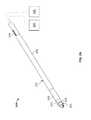
- FIG. 2Aillustrates a variation of an articulating access device
- FIG. 2Bshows a perspective view of the device of FIG. 2A where actuation of the control member causes the cannula to articulate at a flexible section of the distal portion;
- FIG. 2Cshows the device of FIG. 2B after the flexible inner cannula advances out of the outer cannula in a telescopic type motion
- FIGS. 2D and 2Eillustrate variations of an articulating access device having curved or angled rigid proximal sections
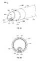
- FIG. 3Ashows a perspective view of a distal portion of an access device
- FIG. 3Bshows a cross sectional view taken along lines 3 B- 3 B of FIG. 3A ;
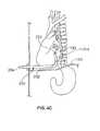
- FIGS. 4A to 4Cillustrates one example of an articulating access device used to access regions of the body not in alignment with an axis of the entry point, port, or incision.
- Methods and devices described hereinprovide for improved access of organs or tissue during minimally invasive surgery. Typically, such surgery involves the creation of small incisions or the use of trocars or ports to access regions of the surgical site.
- the devices and methodscan also improve access during open surgical procedures by eliminating the need to move organs or dissect significant amounts of tissues when the target site is obscured by tissue structures in the surgical field.
- tissue structuresin the surgical field.
- the use of the devices and methods described hereinallow the physician to directly visualize the obscured sides of organs other anatomic structures without the need for excessive manipulation of the organs or tissue structures.
- the methods and devices described hereinare not limited to thoracic applications but can be used in any anatomic region of the body.
- FIG. 1shows examples of placement of trocars or ports 106 into the chest to provide access to the thoracic cavity.
- FIG. 1also show use of an additional access device 182 as described in commonly assigned U.S. application Ser. No. 11/558,417 filed Nov. 9, 2006; Ser. No. 11/408,315 filed Apr. 21, 2006; Ser. No. 11/408,307 filed Apr. 21, 2006; Ser. No. 11/558,417 filed Nov. 9, 2006; Ser. No. 11/558,419 Nov. 9, 2006; and Ser. No. 11/737,493 filed Apr. 19, 2007 (“Diaphragm Entry for Posterior Surgical Access” cases) the entirety of each of which is incorporated by reference.
- ports 106are intended for illustrative purposes only. Any number of ports may be arranged as needed or as conventionally used.
- FIG. 2Aillustrates a variation of an articulating access device 200 according to the principles of the invention.
- the access device 200includes an outer cannula 202 with a flexible inner cannula 204 slidably located therethrough.
- Variations of the device 200can include cannulae that are circular in cross section (as shown).
- the cross sectioncan be oval, rectangular, or any other shape required by the particular application.
- the access device 200can include one or more handles/hubs located on either the inner cannula 204 or outer cannula 202 . Such handles are well known by those familiar with the art. For purposes of illustration, the access device 200 is shown without any handles or hubs.
- the access devicecan simply consist of the telescoping cannula assembly as shown.
- the device 200can also be coupled to a visualization system 300 (e.g., an endoscope, monitor for a camera located at the distal end, or other visualization component).
- a visualization system 300e.g., an endoscope, monitor for a camera located at the distal end, or other visualization component.
- the device 200can be coupled to any number of auxiliary components (such as a vacuum source, fluid source, etc.).
- the outer cannula 202comprises a first rigid section 206 .
- the rigid section 206can optionally comprise a hard outer shell. However, in any case, the rigid section 206 is sufficiently rigid such that a physician can advance and manipulate the cannula remotely (from the proximal end) through a body cavity and through tissue or other organs. In such cases, the physician might need to use the rigid section 206 to move or dissect organs to properly position the opening 210 of the cannula 202
- the distal portion of the cannula 202comprises a flexible section 208 located proximate to the opening 210 .
- the rigid section 206 of the outer cannula 202is straight. However, the rigid section 206 can include curved or angled profiles as required by the particular surgical application or the target anatomy.
- an articulating deviceto have a flexible section 208 distal to the rigid section 206 in a distal portion of the device 200 permits repositioning of the opening 210 around tissue and other structures within the body.
- the rigid nature of the outer cannulapermits the physician to leverage the device into position (typically by manipulating the device from outside of the body through a port or small incision.)
- the relative movement between the flexible inner cannula 204 and the outer cannula 202allows the physician to telescopically advance the opening of the inner cannula 204 to the desired target site and reach organs and surfaces that are not in alignment with the surgical opening.
- the distal portion of the outer cannula 202includes a rigid tip 212 adjacent to the opening 210 and distal to the flexible section 208 .
- the flexible section 208can extend through the entire distal section of the cannula 202 .
- the outer cannula 202also includes one or more control members 214 that permit flexing or articulation of the cannula 202 at the flexible section 208 .
- the control member 214can include any steering mechanism or structure that is conventionally used to steer medical devices. However, in this variation, the control member 214 comprises a pair of wires coupled to the distal tip 212 of the outer cannula 202 .
- the control member 214can include any number of features to assist in positioning of the opening 210 .
- the control memberscan be dial, lever, or trigger actuated with an optional ratcheting mechanism to control the articulation of the cannula 202 .
- the articulating access devicecan be combined with any number of similar features known by those skilled in the art.
- such structuresare described in U.S. Pat. No. 3,470,876 to Barchilon; U.S. Pat. No. 4,690,411 to Buchbinder; U.S. Pat. No. 4,898,577 to Badger et al.; U.S. Pat. No. 4,960,134 to Webster; U.S. Pat. No. 5,325,845 to Adair; and U.S. Pat. No. 6,953,454 to Peterson et al. Each of which is incorporated by reference.
- FIG. 2Bshows a perspective view of the device 200 of FIG. 2A where actuation of the control member 214 (in this variation, in the direction of arrow 216 ) causes the cannula 202 to articulate at a distal portion.
- the cannula 202bends at the flexible section 208 so that the physician can reposition the opening 210 of the access device 202 out of alignment with an axis of the proximal end of the cannula 202 (and thus away from an axis of any entry incision or trocar.)
- the control member 214is encased within the cannula 202 .
- the control member 214can be exterior to the cannula or within a passageway of the cannula as long as operation of the control member 214 does not interfere against tissue or structures when actuating the device 200 .
- FIG. 2Cshows the device of FIG. 2B after the flexible inner cannula 204 advances out of the opening 210 in a telescopic type motion 218 when a physician advances the cannula 204 at a proximal end as shown by arrow 220 .
- the inner cannula 204can be completely flexible such that it can advance through the flexible section 208 of the outer cannula 202 regardless of the direction of articulation of the outer cannula 202 .
- the flexible inner cannula 204comprises a linear shape when un-flexed or when in natural state. Accordingly, as the inner cannula 204 advances from the opening 210 , it advances in a straight line configuration.
- the device 200can also be configured so that rotation of the proximal end of the flexible cannula 204 (as shown by arrow 221 ) causes rotation of the distal portion of the flexible cannula 204 (as shown by arrow 219 ).
- rotation of the proximal end of the flexible cannula 204causes rotation of the distal portion of the flexible cannula 204 (as shown by arrow 219 ).
- the flexible inner cannula 204while the flexible inner cannula 204 is fabricated to advance through the articulated outer cannula 202 , the flexible inner cannula 204 will be fabricated so that rotation of the proximal end translates into a near one-to-one rotation of the distal end.
- FIGS. 2D and 2Eillustrate variations of the articulating access device 200 where the rigid proximal section 206 of the outer cannula 202 is curved ( FIG. 2D ) or has a bend 207 ( FIG. 2E ).
- the flexible inner cannula 204accommodates the curve or bends in the rigid section 206 and further bends to accommodate the articulation of the flexible section 208 .
- the flexible inner cannula 204assumes its straight configuration for telescopic advancement from the opening 210 .
- FIG. 3Ashows a perspective view of a distal portion of an access device 200 .
- a pair of control members 214is coupled to the distal tip 212 of the cannula 202 .
- the control member 214comprises wires on opposite sides of the outer cannula 202 to allow movement in at least two directions.
- any number of control membersis within the scope of this disclosure.
- the flexible inner cannula 204comprises a tapered opening 222 at the end of a passageway or working lumen 224 that extends through the inner cannula 204 .
- a tapered openingallows for an increased open area. This increase in area is beneficial when advancing multiple devices through the cannula 204 .
- the tapered tiphelps to advance the device through the tissue by allowing the physician to gently dilate the tissue as the tip is inserted.
- the tapered tipalso helps with visualization, as it allows structures that are not in line with the opening to be viewed.
- the flexible inner cannula 204can also include a second lumen 226 separate from the main lumen 224 .
- the second lumen 226can be fluidly isolated or may simply be a support frame that permits one or more tools to be placed within the lumen 226 without interfering with other instruments that are within the main lumen 224 .
- the second lumen 226includes a flexible visualization type device 310 (e.g., one having a visualization element 312 and/or an illumination source 314 ).
- the visualization device 310can comprise an endoscope type device or may be a video chip integrated within the channel. In any case, the visualization type device 310 allows the physician to remotely view the area adjacent to the opening of the second lumen 226 .
- the visualization source 310can be advanced with the device 200 or may be advanced once the device 200 is placed within the body. Furthermore, as shown, the second lumen 226 and visualization device 310 are shown to be recessed or offset from the end 222 of the inner cannula 204 . This offset allows for the visualization device 310 to observe other instruments that are advanced through the main lumen for performing various surgical or diagnostic procedures. In addition, recessing the visualization device recessed reduces the chance that tissue will from wiping against the visualization device and obscure the view.)
- FIG. 3Bshows a cross sectional view taken along lines 3 B- 3 B of FIG. 3A .
- the main lumen 224 and second lumen 226extend parallel to each other.
- the main lumen and second lumencan be coaxial.
- the devicecan include various additional lumens (e.g., for suction, fluid delivery, delivery of therapeutics, etc.).
- FIGS. 4A to 4Cillustrates an example of the articulating access device 200 when used to access regions of the body that are not aligned with an axis of the entry point or port 106 .
- the access device 200is shown as accessing the thoracic cavity through the chest wall 10 , the device 200 can be used in any anatomic region of the body. In one such variation, the device can be used with additional techniques to provide multiple points of access to a surgical site. For example, as taught in the “Diaphragm Entry for Posterior Surgical Access” cases cited above, alternate devices are used to access posterior surfaces of organs within the thoracic cavity. Use of the articulating access device 200 in addition provides the physician with an ability to locate additional tissue regions that would otherwise be obscured.
- a physicianadvances the access device 200 through the chest wall 10 (either through a trocar/port 106 or through an incision).
- the physicianis able to manipulate organs due to the rigid portion of the device 200 when placing the distal portion of the device as desired.
- the rigid cannulacan be used to manipulate cardiac tissue or the diaphragm 170 when placing the device.
- the inner cannulatypically contains a visualization device (not shown). In this variation, the visualization permits the physician to locate the opening 222 of the inner cannula at a posterior ventricular surface 190 .
- FIG. 4Billustrates the state of the device 200 when the physician actuates the control members 214 to bend the device at a flexible section 208 of the distal portion of the outer cannula 202 . As shown, this permits the opening 222 of the inner cannula 204 to curve around the heart to access a posterior ventricular surface 190 .
- FIG. 4Cshows advancement of the opening 222 of the inner cannula 204 when the physician advances the inner cannula from outside of the body.
- the opening 222 of the inner cannula 204advances in a straight line towards the desired region. Again, this advancement is typically performed under indirect visualization.
- the opening 222 of the inner cannula 204is now located at a region that would have otherwise been inaccessible due to the presence of organs and tissue structures within the body.
- the access device 200can include a locking mechanism so that the physician can advance additional instruments or devices through the main lumen of the inner cannula 204 to perform various medical diagnostic or procedures.
- the inventioncontemplates use of any surgical device that may be advanced through the access device to perform any procedure that benefits from having improved access as described herein.
- Electrodes 21, 2006are also examples of devices that can be used with the access devices described herein.
- Such probesprovide examples of devices that allow intimate contact specifically between a soft tissue surface and the energy portion of the device.
- the electrode(s) used to transmit energyis capable of heating the soft tissue until achieving irreversible injury making the soft tissue non-viable and unable to propagate electrical impulses, mutate, or reproduce.
- These integrated vacuum coagulation probe embodimentsmay be used with the articulating access device to coagulate soft tissue capable of treating atrial fibrillation, ventricular tachycardia or other arrhythmia substrate, or eliminating cancers (such as lung cancer), or other soft thoracic tissue by destroying target cells.
- these integrated vacuum coagulation devicesmay be used to heat soft tissue along the posterior heart surface resulting in heat-induced contraction of collagen in such tissue thereby resulting shrinking of said soft tissue.
- heating the mitral valve annulus along the posterior atrio-ventricular groovemay induce shrinking of the annulus thereby correcting mitral valve regurgitation.
- the inventionis not limited to the above described vacuum coagulation probes. Instead, any number of coagulation, ablation, or surgical devices may be used as required.
Landscapes
- Health & Medical Sciences (AREA)
- Life Sciences & Earth Sciences (AREA)
- Surgery (AREA)
- Nuclear Medicine, Radiotherapy & Molecular Imaging (AREA)
- Molecular Biology (AREA)
- Veterinary Medicine (AREA)
- Public Health (AREA)
- General Health & Medical Sciences (AREA)
- Animal Behavior & Ethology (AREA)
- Engineering & Computer Science (AREA)
- Biomedical Technology (AREA)
- Heart & Thoracic Surgery (AREA)
- Medical Informatics (AREA)
- Pathology (AREA)
- Physics & Mathematics (AREA)
- Biophysics (AREA)
- Radiology & Medical Imaging (AREA)
- Optics & Photonics (AREA)
- Surgical Instruments (AREA)
Abstract
Description
Claims (12)
Priority Applications (4)
| Application Number | Priority Date | Filing Date | Title |
|---|---|---|---|
| US12/108,426US8858528B2 (en) | 2008-04-23 | 2008-04-23 | Articulating cannula access device |
| EP09734553AEP2274040A4 (en) | 2008-04-23 | 2009-04-17 | Articulating cannula access device |
| PCT/US2009/040981WO2009131912A1 (en) | 2008-04-23 | 2009-04-17 | Articulating cannula access device |
| US14/509,789US9924966B2 (en) | 2008-04-23 | 2014-10-08 | Articulating cannula access device |
Applications Claiming Priority (1)
| Application Number | Priority Date | Filing Date | Title |
|---|---|---|---|
| US12/108,426US8858528B2 (en) | 2008-04-23 | 2008-04-23 | Articulating cannula access device |
Related Child Applications (1)
| Application Number | Title | Priority Date | Filing Date |
|---|---|---|---|
| US14/509,789ContinuationUS9924966B2 (en) | 2008-04-23 | 2014-10-08 | Articulating cannula access device |
Publications (2)
| Publication Number | Publication Date |
|---|---|
| US20090270676A1 US20090270676A1 (en) | 2009-10-29 |
| US8858528B2true US8858528B2 (en) | 2014-10-14 |
Family
ID=41215638
Family Applications (2)
| Application Number | Title | Priority Date | Filing Date |
|---|---|---|---|
| US12/108,426Active2031-03-05US8858528B2 (en) | 2008-04-23 | 2008-04-23 | Articulating cannula access device |
| US14/509,789Active2028-10-15US9924966B2 (en) | 2008-04-23 | 2014-10-08 | Articulating cannula access device |
Family Applications After (1)
| Application Number | Title | Priority Date | Filing Date |
|---|---|---|---|
| US14/509,789Active2028-10-15US9924966B2 (en) | 2008-04-23 | 2014-10-08 | Articulating cannula access device |
Country Status (3)
| Country | Link |
|---|---|
| US (2) | US8858528B2 (en) |
| EP (1) | EP2274040A4 (en) |
| WO (1) | WO2009131912A1 (en) |
Cited By (9)
| Publication number | Priority date | Publication date | Assignee | Title |
|---|---|---|---|---|
| EP3352678A4 (en)* | 2015-09-23 | 2019-05-08 | Corfigo, Inc. | Specialized cannula for trans-xiphoid pericardial procedures |
| US10751511B1 (en) | 2019-08-14 | 2020-08-25 | Vasoinnovations Inc. | Devices, systems, and methods for delivering catheters or other medical devices to locations within a patients body |
| US10751517B1 (en) | 2019-08-14 | 2020-08-25 | Vasoinnovations Inc. | Apparatus and method for advancing catheters or other medical devices through a lumen |
| US10821267B1 (en) | 2019-08-14 | 2020-11-03 | Vasoinnovations Inc. | Apparatus and method for advancing catheters or other medical devices through a lumen |
| US10828470B1 (en) | 2019-08-14 | 2020-11-10 | Vasoinnovations Inc. | Apparatus and method for advancing catheters or other medical devices through a lumen |
| US11419610B2 (en) | 2018-08-17 | 2022-08-23 | Empress Medical, Inc. | Device and method for passing tension member around tissue mass |
| US11419634B2 (en) | 2018-08-17 | 2022-08-23 | Empress Medical, Inc. | Causing ischemia in tumors |
| US11878132B2 (en) | 2019-08-14 | 2024-01-23 | Vasoinnovations Inc. | Apparatus and method for advancing catheters or other medical devices through a lumen |
| US12239322B2 (en) | 2018-08-17 | 2025-03-04 | Empress Medical, Inc. | Device and method for passing tension member around tissue mass |
Families Citing this family (19)
| Publication number | Priority date | Publication date | Assignee | Title |
|---|---|---|---|---|
| AU2007329469A1 (en)* | 2006-12-01 | 2008-06-12 | The Board Of Trustees Of The Leland Stanford Junior University | Devices and methods for accessing the epidural space |
| US8858528B2 (en) | 2008-04-23 | 2014-10-14 | Ncontact Surgical, Inc. | Articulating cannula access device |
| US8267951B2 (en)* | 2008-06-12 | 2012-09-18 | Ncontact Surgical, Inc. | Dissecting cannula and methods of use thereof |
| US20100256483A1 (en)* | 2009-04-03 | 2010-10-07 | Insite Medical Technologies, Inc. | Devices and methods for tissue navigation |
| US9138207B2 (en) | 2009-05-19 | 2015-09-22 | Teleflex Medical Incorporated | Methods and devices for laparoscopic surgery |
| US20110071541A1 (en) | 2009-09-23 | 2011-03-24 | Intuitive Surgical, Inc. | Curved cannula |
| US8465476B2 (en)* | 2009-09-23 | 2013-06-18 | Intuitive Surgical Operations, Inc. | Cannula mounting fixture |
| US8545515B2 (en)* | 2009-09-23 | 2013-10-01 | Intuitive Surgical Operations, Inc. | Curved cannula surgical system |
| US8888789B2 (en) | 2009-09-23 | 2014-11-18 | Intuitive Surgical Operations, Inc. | Curved cannula surgical system control |
| US8623028B2 (en) | 2009-09-23 | 2014-01-07 | Intuitive Surgical Operations, Inc. | Surgical port feature |
| KR20200064161A (en)* | 2009-11-13 | 2020-06-05 | 인튜어티브 서지컬 오퍼레이션즈 인코포레이티드 | Curved cannula surgical system |
| US8721539B2 (en) | 2010-01-20 | 2014-05-13 | EON Surgical Ltd. | Rapid laparoscopy exchange system and method of use thereof |
| EP3251604B1 (en) | 2010-01-20 | 2020-04-22 | EON Surgical Ltd. | System of deploying an elongate unit in a body cavity |
| US8343045B2 (en)* | 2010-04-05 | 2013-01-01 | Intuitive Surgical Operations, Inc. | Curved cannula |
| EP2615980B1 (en)* | 2010-09-19 | 2017-08-16 | EON Surgical Ltd. | Micro laparoscopy devices and deployments thereof |
| US9381010B2 (en) | 2011-06-27 | 2016-07-05 | Covidien Lp | Surgical instrument with adapter for facilitating multi-direction end effector articulation |
| FR2981260B1 (en)* | 2011-10-18 | 2014-12-12 | Univ Grenoble 1 | DEVICE FOR GUIDING A MEDICAL NEEDLE |
| US8419720B1 (en) | 2012-02-07 | 2013-04-16 | National Advanced Endoscopy Devices, Incorporated | Flexible laparoscopic device |
| US11172914B2 (en)* | 2018-04-19 | 2021-11-16 | Fifth Arm Surgical, Llc | Laparoscopic flexible suction device and associated methodology |
Citations (110)
| Publication number | Priority date | Publication date | Assignee | Title |
|---|---|---|---|---|
| US3470876A (en) | 1966-09-28 | 1969-10-07 | John Barchilon | Dirigible catheter |
| US4040413A (en) | 1974-07-18 | 1977-08-09 | Fuji Photo Optical Co. Ltd. | Endoscope |
| US4688554A (en) | 1986-04-10 | 1987-08-25 | American Hospital Supply Corp. | Directing cannula for an optical diagnostic system |
| US4690411A (en) | 1985-12-23 | 1987-09-01 | Winkle Denzal W Van | Bonded mechanically inner connected seal arrangement for a blowout preventer |
| US4719924A (en) | 1986-09-09 | 1988-01-19 | C. R. Bard, Inc. | Small diameter steerable guidewire with adjustable tip |
| US4777951A (en) | 1986-09-19 | 1988-10-18 | Mansfield Scientific, Inc. | Procedure and catheter instrument for treating patients for aortic stenosis |
| EP0135364B1 (en) | 1983-08-24 | 1989-02-08 | Endotherapeutics Corporation | Trocar assembly |
| US4898577A (en) | 1988-09-28 | 1990-02-06 | Advanced Cardiovascular Systems, Inc. | Guiding cathether with controllable distal tip |
| US4921482A (en) | 1989-01-09 | 1990-05-01 | Hammerslag Julius G | Steerable angioplasty device |
| US4920980A (en) | 1987-09-14 | 1990-05-01 | Cordis Corporation | Catheter with controllable tip |
| US4960134A (en) | 1988-11-18 | 1990-10-02 | Webster Wilton W Jr | Steerable catheter |
| US4960411A (en) | 1984-09-18 | 1990-10-02 | Medtronic Versaflex, Inc. | Low profile sterrable soft-tip catheter |
| US4976688A (en)* | 1989-02-03 | 1990-12-11 | Rosenblum Jeffrey L | Position-adjustable thoracic catheter |
| US4998916A (en) | 1989-01-09 | 1991-03-12 | Hammerslag Julius G | Steerable medical device |
| US5025778A (en) | 1990-03-26 | 1991-06-25 | Opielab, Inc. | Endoscope with potential channels and method of using the same |
| US5030204A (en) | 1988-09-28 | 1991-07-09 | Advanced Cardiovascular Systems, Inc. | Guiding catheter with controllable distal tip |
| US5037391A (en) | 1989-01-09 | 1991-08-06 | Pilot Cardiovascular Systems, Inc. | Steerable angioplasty device |
| US5125395A (en) | 1990-09-12 | 1992-06-30 | Adair Edwin Lloyd | Deflectable sheath for optical catheter |
| US5199950A (en) | 1990-12-07 | 1993-04-06 | Willy Rusch Ag | Medical instrument |
| US5203767A (en) | 1991-01-08 | 1993-04-20 | Cloyd David W | Laparoscopic surgical gauze and the like |
| US5205816A (en) | 1992-04-13 | 1993-04-27 | O. R. Concepts, Inc. | Laparoscopic irrigator-aspirator blunt dissector |
| US5209736A (en) | 1991-10-18 | 1993-05-11 | Ethicon, Inc. | Trocar method and apparatus |
| US5235966A (en) | 1991-10-17 | 1993-08-17 | Jay Jamner | Endoscopic retractor |
| US5275608A (en) | 1991-10-16 | 1994-01-04 | Implemed, Inc. | Generic endoscopic instrument |
| US5282826A (en) | 1992-03-05 | 1994-02-01 | Quadtello Corporation | Dissector for endoscopic and laparoscopic use |
| US5308316A (en) | 1992-12-28 | 1994-05-03 | Edward Weck Incorporated | Encoscopic kitner |
| US5322064A (en) | 1991-02-15 | 1994-06-21 | Lundquist Ingemar H | Torquable catheter and method |
| US5325845A (en) | 1992-06-08 | 1994-07-05 | Adair Edwin Lloyd | Steerable sheath for use with selected removable optical catheter |
| US5330502A (en) | 1992-10-09 | 1994-07-19 | Ethicon, Inc. | Rotational endoscopic mechanism with jointed drive mechanism |
| US5374277A (en) | 1992-10-09 | 1994-12-20 | Ethicon, Inc. | Surgical instrument |
| US5378234A (en) | 1993-03-15 | 1995-01-03 | Pilot Cardiovascular Systems, Inc. | Coil polymer composite |
| US5453078A (en) | 1994-03-04 | 1995-09-26 | Merocel Corporation | Endoscopic wedge and organ positioner |
| US5460621A (en) | 1994-03-04 | 1995-10-24 | Merocel Corporation | Composite tissue displacement sponge |
| US5571088A (en) | 1993-07-01 | 1996-11-05 | Boston Scientific Corporation | Ablation catheters |
| US5618294A (en) | 1994-05-24 | 1997-04-08 | Aust & Taylor Medical Corporation | Surgical instrument |
| US5651366A (en) | 1994-09-19 | 1997-07-29 | Board Of Trustees Of The Leland Stanford Junior University | Forward viewing ultrasonic imaging catheter |
| US5658307A (en) | 1990-11-07 | 1997-08-19 | Exconde; Primo D. | Method of using a surgical dissector instrument |
| US5704534A (en) | 1994-12-19 | 1998-01-06 | Ethicon Endo-Surgery, Inc. | Articulation assembly for surgical instruments |
| US5725523A (en) | 1996-03-29 | 1998-03-10 | Mueller; Richard L. | Lateral-and posterior-aspect method and apparatus for laser-assisted transmyocardial revascularization and other surgical applications |
| US5783227A (en) | 1996-01-22 | 1998-07-21 | Cordis Corporation | Catheter balloon folding device |
| US5785706A (en) | 1996-11-18 | 1998-07-28 | Daig Corporation | Nonsurgical mapping and treatment of cardiac arrhythmia using a catheter contained within a guiding introducer containing openings |
| WO1998034550A1 (en) | 1997-02-10 | 1998-08-13 | West Hugh S Jr | Mechanical and electrical endoscopic surgical instrument |
| US5829447A (en)* | 1993-02-22 | 1998-11-03 | Heartport, Inc. | Method and apparatus for thoracoscopic intracardiac procedures |
| US5843017A (en) | 1990-07-24 | 1998-12-01 | Yoon; Inbae | Multifunctional tissue dissecting instrument |
| US5851202A (en) | 1994-08-15 | 1998-12-22 | Gambro Ab | Drip chamber head |
| US5865802A (en) | 1988-07-22 | 1999-02-02 | Yoon; Inbae | Expandable multifunctional instruments for creating spaces at obstructed sites endoscopically |
| WO1999017665A1 (en) | 1997-10-03 | 1999-04-15 | Boston Scientific Corporation | Apparatus and method for percutaneous placement of gastro-intestinal tubes |
| US5899914A (en) | 1997-06-11 | 1999-05-04 | Endius Incorporated | Surgical instrument |
| US5919188A (en) | 1997-02-04 | 1999-07-06 | Medtronic, Inc. | Linear ablation catheter |
| US5967997A (en) | 1998-04-30 | 1999-10-19 | Symbiosis Corporation | Endoscopic surgical instrument with deflectable and rotatable distal end |
| US5980455A (en) | 1993-02-22 | 1999-11-09 | Heartport, Inc. | Method for manipulating a tissue structure within a thoracic cavity |
| US6080151A (en) | 1997-07-21 | 2000-06-27 | Daig Corporation | Ablation catheter |
| US6126649A (en) | 1999-06-10 | 2000-10-03 | Transvascular, Inc. | Steerable catheter with external guidewire as catheter tip deflector |
| US6161543A (en) | 1993-02-22 | 2000-12-19 | Epicor, Inc. | Methods of epicardial ablation for creating a lesion around the pulmonary veins |
| US6176825B1 (en) | 1998-06-22 | 2001-01-23 | Origin Medsystems, Inc. | Cannula-based irrigation system and method |
| US6203559B1 (en) | 1998-10-05 | 2001-03-20 | Origin Medsystems | Method and apparatus for tissue dissection |
| US6234958B1 (en) | 1998-11-30 | 2001-05-22 | Medical Access Systems, Llc | Medical device introduction system including medical introducer having a plurality of access ports and methods of performing medical procedures with same |
| WO2000044286A9 (en) | 1999-01-28 | 2001-09-13 | Minrad Inc | Sampling device and method of retrieving a sample |
| US6314963B1 (en) | 1996-10-22 | 2001-11-13 | Epicor, Inc. | Method of ablating tissue from an epicardial location |
| US6330965B1 (en) | 1997-09-23 | 2001-12-18 | United States Surgical Corporation | Surgical stapling apparatus |
| US6332881B1 (en)* | 1999-09-01 | 2001-12-25 | Cardima, Inc. | Surgical ablation tool |
| US6364876B1 (en) | 1998-10-23 | 2002-04-02 | Afx, Inc. | Vacuum-assisted securing apparatus for a microwave ablation instrument |
| US20020111637A1 (en) | 1999-05-20 | 2002-08-15 | Kaplan Aaron V. | Methods and apparatus for transpericardial left atrial appendage closure |
| US6463332B1 (en) | 1999-09-17 | 2002-10-08 | Core Medical, Inc. | Method and system for pericardial enhancement |
| US6478028B1 (en) | 1999-01-21 | 2002-11-12 | Coroneo, Inc. | Surgical apparatus and method for performing transabdominal cardiac surgery |
| US6506200B1 (en) | 1995-07-13 | 2003-01-14 | Origin Medsystems, Inc. | Tissue separation cannula and method |
| US6514250B1 (en) | 2000-04-27 | 2003-02-04 | Medtronic, Inc. | Suction stabilized epicardial ablation devices |
| US6530914B1 (en) | 2000-10-24 | 2003-03-11 | Scimed Life Systems, Inc. | Deflectable tip guide in guide system |
| US6551337B1 (en) | 1999-10-05 | 2003-04-22 | Omnisonics Medical Technologies, Inc. | Ultrasonic medical device operating in a transverse mode |
| US6558382B2 (en) | 2000-04-27 | 2003-05-06 | Medtronic, Inc. | Suction stabilized epicardial ablation devices |
| US6592604B2 (en) | 2001-09-28 | 2003-07-15 | Ethicon, Inc. | Vessel harvesting retractor with dissection element |
| US6592547B2 (en) | 1998-01-23 | 2003-07-15 | Grimes Kevin V | Methods and devices for occluding the ascending aorta and maintaining circulation of oxygenated blood in the patient when the patient's heart is arrested |
| US20030229296A1 (en) | 2002-03-18 | 2003-12-11 | Olympus Optical Co., Ltd. | Guide tube |
| US6663641B1 (en) | 1997-10-10 | 2003-12-16 | Origin Medsystems, Inc. | Endoscopic surgical instrument for rotational manipulation |
| US6666854B1 (en) | 1999-06-25 | 2003-12-23 | La Precision | Endoscopic surgical instrument |
| US20040039339A1 (en)* | 2002-06-27 | 2004-02-26 | Anders Magnusson | Drainage catheter |
| US6726684B1 (en) | 1996-07-16 | 2004-04-27 | Arthrocare Corporation | Methods for electrosurgical spine surgery |
| US6752756B2 (en) | 1998-06-22 | 2004-06-22 | Origin Medsystems, Inc. | Combined vessel dissection and transection device and method |
| US20040143257A1 (en)* | 1999-08-10 | 2004-07-22 | Biosense Webster, Inc. | Irrigation probe for ablation during open heart surgery |
| US20040216748A1 (en) | 1999-08-10 | 2004-11-04 | Chin Albert K. | Apparatus and method for endoscopic encirclement of pulmonary veins for epicardial ablation |
| US6887249B1 (en) | 1998-06-10 | 2005-05-03 | Converge Medical Inc. | Positioning systems for sutureless anastomosis systems |
| US6893442B2 (en) | 2002-06-14 | 2005-05-17 | Ablatrics, Inc. | Vacuum coagulation probe for atrial fibrillation treatment |
| US20050131403A1 (en) | 2003-12-13 | 2005-06-16 | Chang Stanley F. | Polypectomy snare for specimen retrieval |
| US6917834B2 (en) | 1997-12-03 | 2005-07-12 | Boston Scientific Scimed, Inc. | Devices and methods for creating lesions in endocardial and surrounding tissue to isolate focal arrhythmia substrates |
| US20050203340A1 (en) | 2002-03-06 | 2005-09-15 | John Butler | Steerable colonoscope probe with variable stiffness |
| US6953454B2 (en) | 2001-12-11 | 2005-10-11 | Cardiac Pacemakers, Inc. | Methods of using a deflectable telescoping guide catheter |
| US20050245789A1 (en) | 2003-04-01 | 2005-11-03 | Boston Scientific Scimed, Inc. | Fluid manifold for endoscope system |
| US6989018B2 (en) | 1994-06-29 | 2006-01-24 | General Surgical Innovations, Inc. | Extraluminal balloon dissection |
| US6991627B2 (en) | 1996-05-20 | 2006-01-31 | Intuitive Surgical Inc. | Articulated surgical instrument for performing minimally invasive surgery with enhanced dexterity and sensitivity |
| US7001404B1 (en) | 1995-07-13 | 2006-02-21 | Origin Medsystems, Inc. | Tissue separation cannula and method |
| US7063698B2 (en) | 2002-06-14 | 2006-06-20 | Ncontact Surgical, Inc. | Vacuum coagulation probes |
| US20060183975A1 (en) | 2004-04-14 | 2006-08-17 | Usgi Medical, Inc. | Methods and apparatus for performing endoluminal procedures |
| US7144363B2 (en) | 2001-10-16 | 2006-12-05 | Extensia Medical, Inc. | Systems for heart treatment |
| US20060293646A1 (en) | 2002-06-14 | 2006-12-28 | Whayne James G | Vacuum coagulation & dissection probes |
| US20070083225A1 (en) | 2005-10-12 | 2007-04-12 | Ncontact Surgical, Inc. | Diaphragm entry for posterior surgical access |
| WO2007063904A1 (en) | 2005-12-01 | 2007-06-07 | Olympus Medical Systems Corp. | Guiding long medical member and long medical device |
| US20070219550A1 (en) | 2006-01-27 | 2007-09-20 | Mark Thompson | Device and system for surgical dissection and/or guidance of other medical devices into body |
| US20070249991A1 (en) | 2005-10-12 | 2007-10-25 | Ncontact Surgical, Inc. | Diaphragm entry for posterior surgical access |
| US20070250058A1 (en) | 2002-06-14 | 2007-10-25 | Ncontact Surgical, Inc. | Vacuum coagulation probes |
| US20070255276A1 (en)* | 1996-10-22 | 2007-11-01 | St. Jude Medical, Atrial Fibrillation Division | Methods and devices for ablation |
| US20070265595A1 (en) | 2006-05-09 | 2007-11-15 | Olympus Medical Systems Corp. | Treatment tool inserting/withdrawing auxiliary device and medical procedure through endoscope |
| US7300448B2 (en) | 2002-10-04 | 2007-11-27 | Tyco Healthcare Group Lp | Balloon dissector with cannula |
| US7300488B2 (en) | 2003-03-27 | 2007-11-27 | Höganäs Ab | Powder metal composition and method for producing components thereof |
| US7328071B1 (en) | 2005-10-12 | 2008-02-05 | Pacesetter, Inc. | Lead placement device |
| US20080114342A1 (en) | 2006-11-09 | 2008-05-15 | Ncontact Surgical, Inc. | Diaphragm entry for posterior surgical access |
| US20080114354A1 (en) | 2003-04-29 | 2008-05-15 | Ncontact Surgical, Inc. | Vacuum coagulation probes |
| US20080114355A1 (en) | 2006-11-09 | 2008-05-15 | Ncontact Surgical, Inc. | Vacuum coagulation probes |
| US20080114288A1 (en) | 2006-11-09 | 2008-05-15 | Ncontact Surgical, Inc. | Diaphragm entry for posterior surgical access |
| US20090312783A1 (en) | 2008-06-12 | 2009-12-17 | Ncontact Surgical, Inc. | Dissecting cannula and methods of use thereof |
| EP1935348B1 (en) | 2006-12-22 | 2013-09-04 | The Spectranetics Corporation | Tissue separating systems |
Family Cites Families (16)
| Publication number | Priority date | Publication date | Assignee | Title |
|---|---|---|---|---|
| US589914A (en)* | 1897-09-14 | Cow-tie | ||
| US2005102A (en) | 1931-05-21 | 1935-06-18 | Barrett Co | Distillation of tar |
| US4576162A (en) | 1983-03-30 | 1986-03-18 | Mccorkle Charles E | Apparatus and method for separation of scar tissue in venous pathway |
| US5637097A (en) | 1992-04-15 | 1997-06-10 | Yoon; Inbae | Penetrating instrument having an expandable anchoring portion |
| US6306144B1 (en) | 1996-11-01 | 2001-10-23 | Scimed Life Systems, Inc. | Selective coating of a balloon catheter with lubricious material for stent deployment |
| US5634937A (en) | 1995-05-19 | 1997-06-03 | General Surgical Innovations, Inc. | Skin seal with inflatable membrane |
| US5778367A (en) | 1995-12-14 | 1998-07-07 | Network Engineering Software, Inc. | Automated on-line information service and directory, particularly for the world wide web |
| US7392806B2 (en) | 2003-04-30 | 2008-07-01 | Peter Siltex Yuen | Electronic human breath filtration device |
| WO2004100799A2 (en) | 2003-05-08 | 2004-11-25 | Tyco Healthcare Group Lp | Balloon dissector with balloon anchor cannula |
| JP2008058337A (en) | 2005-01-27 | 2008-03-13 | Sharp Corp | Display device, liquid crystal display device, and manufacturing method of display device |
| GB0623366D0 (en) | 2006-11-23 | 2007-01-03 | Shturman Leonid | Rotational atherectomy device with fluid inflatable support elements and distal protection capability |
| US8142356B2 (en) | 2007-03-30 | 2012-03-27 | Ethicon Endo-Surgery, Inc. | Method of manipulating tissue |
| US8100930B2 (en) | 2007-03-30 | 2012-01-24 | Ethicon Endo-Surgery, Inc. | Tissue moving surgical device |
| US20090082822A1 (en) | 2007-09-20 | 2009-03-26 | Osman Said G | Transpedicular, Extrapedicular and Transcorporeal Approaches to the Intervertebral Discs |
| US8540744B2 (en) | 2008-04-01 | 2013-09-24 | Ethicon Endo-Surgery, Inc. | Tissue penetrating surgical device |
| US8858528B2 (en) | 2008-04-23 | 2014-10-14 | Ncontact Surgical, Inc. | Articulating cannula access device |
- 2008
- 2008-04-23USUS12/108,426patent/US8858528B2/enactiveActive
- 2009
- 2009-04-17WOPCT/US2009/040981patent/WO2009131912A1/enactiveApplication Filing
- 2009-04-17EPEP09734553Apatent/EP2274040A4/ennot_activeWithdrawn
- 2014
- 2014-10-08USUS14/509,789patent/US9924966B2/enactiveActive
Patent Citations (126)
| Publication number | Priority date | Publication date | Assignee | Title |
|---|---|---|---|---|
| US3470876A (en) | 1966-09-28 | 1969-10-07 | John Barchilon | Dirigible catheter |
| US4040413A (en) | 1974-07-18 | 1977-08-09 | Fuji Photo Optical Co. Ltd. | Endoscope |
| EP0135364B1 (en) | 1983-08-24 | 1989-02-08 | Endotherapeutics Corporation | Trocar assembly |
| US4960411A (en) | 1984-09-18 | 1990-10-02 | Medtronic Versaflex, Inc. | Low profile sterrable soft-tip catheter |
| US4690411A (en) | 1985-12-23 | 1987-09-01 | Winkle Denzal W Van | Bonded mechanically inner connected seal arrangement for a blowout preventer |
| US4688554A (en) | 1986-04-10 | 1987-08-25 | American Hospital Supply Corp. | Directing cannula for an optical diagnostic system |
| US4719924A (en) | 1986-09-09 | 1988-01-19 | C. R. Bard, Inc. | Small diameter steerable guidewire with adjustable tip |
| US4777951A (en) | 1986-09-19 | 1988-10-18 | Mansfield Scientific, Inc. | Procedure and catheter instrument for treating patients for aortic stenosis |
| US4920980A (en) | 1987-09-14 | 1990-05-01 | Cordis Corporation | Catheter with controllable tip |
| US5865802A (en) | 1988-07-22 | 1999-02-02 | Yoon; Inbae | Expandable multifunctional instruments for creating spaces at obstructed sites endoscopically |
| US4898577A (en) | 1988-09-28 | 1990-02-06 | Advanced Cardiovascular Systems, Inc. | Guiding cathether with controllable distal tip |
| US5030204A (en) | 1988-09-28 | 1991-07-09 | Advanced Cardiovascular Systems, Inc. | Guiding catheter with controllable distal tip |
| US4960134A (en) | 1988-11-18 | 1990-10-02 | Webster Wilton W Jr | Steerable catheter |
| US4921482A (en) | 1989-01-09 | 1990-05-01 | Hammerslag Julius G | Steerable angioplasty device |
| US5037391A (en) | 1989-01-09 | 1991-08-06 | Pilot Cardiovascular Systems, Inc. | Steerable angioplasty device |
| US4998916A (en) | 1989-01-09 | 1991-03-12 | Hammerslag Julius G | Steerable medical device |
| US4976688A (en)* | 1989-02-03 | 1990-12-11 | Rosenblum Jeffrey L | Position-adjustable thoracic catheter |
| US5025778A (en) | 1990-03-26 | 1991-06-25 | Opielab, Inc. | Endoscope with potential channels and method of using the same |
| US5843017A (en) | 1990-07-24 | 1998-12-01 | Yoon; Inbae | Multifunctional tissue dissecting instrument |
| US5125395A (en) | 1990-09-12 | 1992-06-30 | Adair Edwin Lloyd | Deflectable sheath for optical catheter |
| US5658307A (en) | 1990-11-07 | 1997-08-19 | Exconde; Primo D. | Method of using a surgical dissector instrument |
| US5199950A (en) | 1990-12-07 | 1993-04-06 | Willy Rusch Ag | Medical instrument |
| US5203767A (en) | 1991-01-08 | 1993-04-20 | Cloyd David W | Laparoscopic surgical gauze and the like |
| US5322064A (en) | 1991-02-15 | 1994-06-21 | Lundquist Ingemar H | Torquable catheter and method |
| US5275608A (en) | 1991-10-16 | 1994-01-04 | Implemed, Inc. | Generic endoscopic instrument |
| US5235966A (en) | 1991-10-17 | 1993-08-17 | Jay Jamner | Endoscopic retractor |
| US5209736A (en) | 1991-10-18 | 1993-05-11 | Ethicon, Inc. | Trocar method and apparatus |
| US5282826A (en) | 1992-03-05 | 1994-02-01 | Quadtello Corporation | Dissector for endoscopic and laparoscopic use |
| US5205816A (en) | 1992-04-13 | 1993-04-27 | O. R. Concepts, Inc. | Laparoscopic irrigator-aspirator blunt dissector |
| US5325845A (en) | 1992-06-08 | 1994-07-05 | Adair Edwin Lloyd | Steerable sheath for use with selected removable optical catheter |
| US5330502A (en) | 1992-10-09 | 1994-07-19 | Ethicon, Inc. | Rotational endoscopic mechanism with jointed drive mechanism |
| US5374277A (en) | 1992-10-09 | 1994-12-20 | Ethicon, Inc. | Surgical instrument |
| US5399161A (en) | 1992-12-28 | 1995-03-21 | Linvatec Corporation | Endoscopic kitner |
| US5308316A (en) | 1992-12-28 | 1994-05-03 | Edward Weck Incorporated | Encoscopic kitner |
| US6161543A (en) | 1993-02-22 | 2000-12-19 | Epicor, Inc. | Methods of epicardial ablation for creating a lesion around the pulmonary veins |
| US5980455A (en) | 1993-02-22 | 1999-11-09 | Heartport, Inc. | Method for manipulating a tissue structure within a thoracic cavity |
| US5829447A (en)* | 1993-02-22 | 1998-11-03 | Heartport, Inc. | Method and apparatus for thoracoscopic intracardiac procedures |
| US5378234A (en) | 1993-03-15 | 1995-01-03 | Pilot Cardiovascular Systems, Inc. | Coil polymer composite |
| US5571088A (en) | 1993-07-01 | 1996-11-05 | Boston Scientific Corporation | Ablation catheters |
| US5460621A (en) | 1994-03-04 | 1995-10-24 | Merocel Corporation | Composite tissue displacement sponge |
| US5453078A (en) | 1994-03-04 | 1995-09-26 | Merocel Corporation | Endoscopic wedge and organ positioner |
| US5618294A (en) | 1994-05-24 | 1997-04-08 | Aust & Taylor Medical Corporation | Surgical instrument |
| US6989018B2 (en) | 1994-06-29 | 2006-01-24 | General Surgical Innovations, Inc. | Extraluminal balloon dissection |
| US5851202A (en) | 1994-08-15 | 1998-12-22 | Gambro Ab | Drip chamber head |
| US5651366A (en) | 1994-09-19 | 1997-07-29 | Board Of Trustees Of The Leland Stanford Junior University | Forward viewing ultrasonic imaging catheter |
| US5704534A (en) | 1994-12-19 | 1998-01-06 | Ethicon Endo-Surgery, Inc. | Articulation assembly for surgical instruments |
| US7001404B1 (en) | 1995-07-13 | 2006-02-21 | Origin Medsystems, Inc. | Tissue separation cannula and method |
| US6506200B1 (en) | 1995-07-13 | 2003-01-14 | Origin Medsystems, Inc. | Tissue separation cannula and method |
| US5783227A (en) | 1996-01-22 | 1998-07-21 | Cordis Corporation | Catheter balloon folding device |
| US5725523A (en) | 1996-03-29 | 1998-03-10 | Mueller; Richard L. | Lateral-and posterior-aspect method and apparatus for laser-assisted transmyocardial revascularization and other surgical applications |
| US6991627B2 (en) | 1996-05-20 | 2006-01-31 | Intuitive Surgical Inc. | Articulated surgical instrument for performing minimally invasive surgery with enhanced dexterity and sensitivity |
| US6726684B1 (en) | 1996-07-16 | 2004-04-27 | Arthrocare Corporation | Methods for electrosurgical spine surgery |
| US20070255276A1 (en)* | 1996-10-22 | 2007-11-01 | St. Jude Medical, Atrial Fibrillation Division | Methods and devices for ablation |
| US6314963B1 (en) | 1996-10-22 | 2001-11-13 | Epicor, Inc. | Method of ablating tissue from an epicardial location |
| US8057465B2 (en)* | 1996-10-22 | 2011-11-15 | St. Jude Medical, Atrial Fibrillation Division, Inc. | Methods and devices for ablation |
| US6484727B1 (en) | 1996-10-22 | 2002-11-26 | Epicor, Inc. | Apparatus and method for diagnosis and therapy of electrophysiological disease |
| US5785706A (en) | 1996-11-18 | 1998-07-28 | Daig Corporation | Nonsurgical mapping and treatment of cardiac arrhythmia using a catheter contained within a guiding introducer containing openings |
| US5919188A (en) | 1997-02-04 | 1999-07-06 | Medtronic, Inc. | Linear ablation catheter |
| WO1998034550A1 (en) | 1997-02-10 | 1998-08-13 | West Hugh S Jr | Mechanical and electrical endoscopic surgical instrument |
| US5899914A (en) | 1997-06-11 | 1999-05-04 | Endius Incorporated | Surgical instrument |
| US6077287A (en) | 1997-06-11 | 2000-06-20 | Endius Incorporated | Surgical instrument |
| US6264654B1 (en) | 1997-07-21 | 2001-07-24 | Daig Corporation | Ablation catheter |
| US6080151A (en) | 1997-07-21 | 2000-06-27 | Daig Corporation | Ablation catheter |
| US6330965B1 (en) | 1997-09-23 | 2001-12-18 | United States Surgical Corporation | Surgical stapling apparatus |
| WO1999017665A1 (en) | 1997-10-03 | 1999-04-15 | Boston Scientific Corporation | Apparatus and method for percutaneous placement of gastro-intestinal tubes |
| US6663641B1 (en) | 1997-10-10 | 2003-12-16 | Origin Medsystems, Inc. | Endoscopic surgical instrument for rotational manipulation |
| US6917834B2 (en) | 1997-12-03 | 2005-07-12 | Boston Scientific Scimed, Inc. | Devices and methods for creating lesions in endocardial and surrounding tissue to isolate focal arrhythmia substrates |
| US6592547B2 (en) | 1998-01-23 | 2003-07-15 | Grimes Kevin V | Methods and devices for occluding the ascending aorta and maintaining circulation of oxygenated blood in the patient when the patient's heart is arrested |
| US5967997A (en) | 1998-04-30 | 1999-10-19 | Symbiosis Corporation | Endoscopic surgical instrument with deflectable and rotatable distal end |
| US6887249B1 (en) | 1998-06-10 | 2005-05-03 | Converge Medical Inc. | Positioning systems for sutureless anastomosis systems |
| US6176825B1 (en) | 1998-06-22 | 2001-01-23 | Origin Medsystems, Inc. | Cannula-based irrigation system and method |
| US6752756B2 (en) | 1998-06-22 | 2004-06-22 | Origin Medsystems, Inc. | Combined vessel dissection and transection device and method |
| US6203559B1 (en) | 1998-10-05 | 2001-03-20 | Origin Medsystems | Method and apparatus for tissue dissection |
| US6364876B1 (en) | 1998-10-23 | 2002-04-02 | Afx, Inc. | Vacuum-assisted securing apparatus for a microwave ablation instrument |
| US6234958B1 (en) | 1998-11-30 | 2001-05-22 | Medical Access Systems, Llc | Medical device introduction system including medical introducer having a plurality of access ports and methods of performing medical procedures with same |
| US6478028B1 (en) | 1999-01-21 | 2002-11-12 | Coroneo, Inc. | Surgical apparatus and method for performing transabdominal cardiac surgery |
| WO2000044286A9 (en) | 1999-01-28 | 2001-09-13 | Minrad Inc | Sampling device and method of retrieving a sample |
| US20020111637A1 (en) | 1999-05-20 | 2002-08-15 | Kaplan Aaron V. | Methods and apparatus for transpericardial left atrial appendage closure |
| US6126649A (en) | 1999-06-10 | 2000-10-03 | Transvascular, Inc. | Steerable catheter with external guidewire as catheter tip deflector |
| US6666854B1 (en) | 1999-06-25 | 2003-12-23 | La Precision | Endoscopic surgical instrument |
| US20040216748A1 (en) | 1999-08-10 | 2004-11-04 | Chin Albert K. | Apparatus and method for endoscopic encirclement of pulmonary veins for epicardial ablation |
| US20040143257A1 (en)* | 1999-08-10 | 2004-07-22 | Biosense Webster, Inc. | Irrigation probe for ablation during open heart surgery |
| US7925341B2 (en)* | 1999-08-10 | 2011-04-12 | Biosense Webster, Inc. | Irrigation probe for ablation during open heart surgery |
| US6332881B1 (en)* | 1999-09-01 | 2001-12-25 | Cardima, Inc. | Surgical ablation tool |
| US6463332B1 (en) | 1999-09-17 | 2002-10-08 | Core Medical, Inc. | Method and system for pericardial enhancement |
| US6551337B1 (en) | 1999-10-05 | 2003-04-22 | Omnisonics Medical Technologies, Inc. | Ultrasonic medical device operating in a transverse mode |
| US6514250B1 (en) | 2000-04-27 | 2003-02-04 | Medtronic, Inc. | Suction stabilized epicardial ablation devices |
| US6558382B2 (en) | 2000-04-27 | 2003-05-06 | Medtronic, Inc. | Suction stabilized epicardial ablation devices |
| US6530914B1 (en) | 2000-10-24 | 2003-03-11 | Scimed Life Systems, Inc. | Deflectable tip guide in guide system |
| US6592604B2 (en) | 2001-09-28 | 2003-07-15 | Ethicon, Inc. | Vessel harvesting retractor with dissection element |
| US7144363B2 (en) | 2001-10-16 | 2006-12-05 | Extensia Medical, Inc. | Systems for heart treatment |
| US6953454B2 (en) | 2001-12-11 | 2005-10-11 | Cardiac Pacemakers, Inc. | Methods of using a deflectable telescoping guide catheter |
| US20060129132A1 (en) | 2001-12-11 | 2006-06-15 | Cardiac Pacemakers, Inc. | Deflectable telescoping guide catheter |
| US20050203340A1 (en) | 2002-03-06 | 2005-09-15 | John Butler | Steerable colonoscope probe with variable stiffness |
| US20030229296A1 (en) | 2002-03-18 | 2003-12-11 | Olympus Optical Co., Ltd. | Guide tube |
| US6893442B2 (en) | 2002-06-14 | 2005-05-17 | Ablatrics, Inc. | Vacuum coagulation probe for atrial fibrillation treatment |
| US20070250058A1 (en) | 2002-06-14 | 2007-10-25 | Ncontact Surgical, Inc. | Vacuum coagulation probes |
| US20060200124A1 (en) | 2002-06-14 | 2006-09-07 | Ncontact Surgical, Inc. | Vacuum coagulation probes |
| US20060206113A1 (en) | 2002-06-14 | 2006-09-14 | Ncontact Surgical, Inc. | Methods of coagulating tissue |
| US20060235381A1 (en) | 2002-06-14 | 2006-10-19 | Ncontact Surgical, Inc. | Vacuum coagulation probes |
| US20060009762A1 (en) | 2002-06-14 | 2006-01-12 | Ablatrics, Inc. | Vacuum coagulation probe for atrial fibrillation treatment |
| US20060293646A1 (en) | 2002-06-14 | 2006-12-28 | Whayne James G | Vacuum coagulation & dissection probes |
| US7063698B2 (en) | 2002-06-14 | 2006-06-20 | Ncontact Surgical, Inc. | Vacuum coagulation probes |
| US20040039339A1 (en)* | 2002-06-27 | 2004-02-26 | Anders Magnusson | Drainage catheter |
| US7300448B2 (en) | 2002-10-04 | 2007-11-27 | Tyco Healthcare Group Lp | Balloon dissector with cannula |
| US7300488B2 (en) | 2003-03-27 | 2007-11-27 | Höganäs Ab | Powder metal composition and method for producing components thereof |
| US20050245789A1 (en) | 2003-04-01 | 2005-11-03 | Boston Scientific Scimed, Inc. | Fluid manifold for endoscope system |
| US20080114354A1 (en) | 2003-04-29 | 2008-05-15 | Ncontact Surgical, Inc. | Vacuum coagulation probes |
| US20070043351A1 (en) | 2003-04-29 | 2007-02-22 | Ncontact Surgical, Inc. | Vacuum coagulation probes |
| US20050131403A1 (en) | 2003-12-13 | 2005-06-16 | Chang Stanley F. | Polypectomy snare for specimen retrieval |
| US20060183975A1 (en) | 2004-04-14 | 2006-08-17 | Usgi Medical, Inc. | Methods and apparatus for performing endoluminal procedures |
| WO2007037785A1 (en) | 2005-08-18 | 2007-04-05 | Ncontact Surgical, Inc. | Vacuum coagulation probes |
| US20070249991A1 (en) | 2005-10-12 | 2007-10-25 | Ncontact Surgical, Inc. | Diaphragm entry for posterior surgical access |
| US20070083082A1 (en) | 2005-10-12 | 2007-04-12 | Ncontact Surgical, Inc. | Diaphragm entry for posterior surgical access |
| US20070083225A1 (en) | 2005-10-12 | 2007-04-12 | Ncontact Surgical, Inc. | Diaphragm entry for posterior surgical access |
| US7328071B1 (en) | 2005-10-12 | 2008-02-05 | Pacesetter, Inc. | Lead placement device |
| WO2007063904A1 (en) | 2005-12-01 | 2007-06-07 | Olympus Medical Systems Corp. | Guiding long medical member and long medical device |
| US20070219550A1 (en) | 2006-01-27 | 2007-09-20 | Mark Thompson | Device and system for surgical dissection and/or guidance of other medical devices into body |
| US20070244473A1 (en) | 2006-01-27 | 2007-10-18 | Mark Thompson | Method of surgical dissection and/or guidance of other medical devices into body |
| US20070265595A1 (en) | 2006-05-09 | 2007-11-15 | Olympus Medical Systems Corp. | Treatment tool inserting/withdrawing auxiliary device and medical procedure through endoscope |
| US20080114342A1 (en) | 2006-11-09 | 2008-05-15 | Ncontact Surgical, Inc. | Diaphragm entry for posterior surgical access |
| US20080114355A1 (en) | 2006-11-09 | 2008-05-15 | Ncontact Surgical, Inc. | Vacuum coagulation probes |
| US20080114288A1 (en) | 2006-11-09 | 2008-05-15 | Ncontact Surgical, Inc. | Diaphragm entry for posterior surgical access |
| EP1935348B1 (en) | 2006-12-22 | 2013-09-04 | The Spectranetics Corporation | Tissue separating systems |
| US20090312783A1 (en) | 2008-06-12 | 2009-12-17 | Ncontact Surgical, Inc. | Dissecting cannula and methods of use thereof |
| US20120310268A1 (en) | 2008-06-12 | 2012-12-06 | Ncontact Surgical, Inc. | Dissecting cannula and methods of use thereof |
Non-Patent Citations (5)
| Title |
|---|
| Cragg, et al., "Endovascular diathermic vessel occlusion," Radiology, 144:303-308, 1982. |
| Gorisch et al., "Heat-induced contraction of blood vessels," Lasers in Surgery and Medicine, 2:1-13, 1982. |
| International Patent Application No. PCT/U82009/040981 in the name of Sicvol filed Apr. 17, 2009, International Search Report and Written Opinion mailed Jun. 11, 2009. |
| International Patent Application No. PCT/US2009/046780 in the name of Whayne et al. filed Jun. 9, 2009, International Search Report and Written Opinion mailed Jul. 28, 2009. |
| Nath, et al., "Cellular electrophysiologic effects of hyperthermia on isolated guinea pig papillary muscle: implications for catheter ablation," Circulation, 88:1826-1831, 1993. |
Cited By (18)
| Publication number | Priority date | Publication date | Assignee | Title |
|---|---|---|---|---|
| EP3352678A4 (en)* | 2015-09-23 | 2019-05-08 | Corfigo, Inc. | Specialized cannula for trans-xiphoid pericardial procedures |
| US12239322B2 (en) | 2018-08-17 | 2025-03-04 | Empress Medical, Inc. | Device and method for passing tension member around tissue mass |
| US11419634B2 (en) | 2018-08-17 | 2022-08-23 | Empress Medical, Inc. | Causing ischemia in tumors |
| US11419610B2 (en) | 2018-08-17 | 2022-08-23 | Empress Medical, Inc. | Device and method for passing tension member around tissue mass |
| US10773058B1 (en) | 2019-08-14 | 2020-09-15 | Vasoinnovations Inc. | Apparatus and method for advancing catheters or other medical devices through a lumen |
| US10994099B2 (en) | 2019-08-14 | 2021-05-04 | Vasoinnovations Inc. | Devices, systems, and methods for delivering catheters or other medical devices to locations within a patients body |
| US10799678B1 (en) | 2019-08-14 | 2020-10-13 | Vasoinnovations Inc. | Devices, systems, and methods for delivering catheters or other medical devices to locations within a patients body |
| US10821267B1 (en) | 2019-08-14 | 2020-11-03 | Vasoinnovations Inc. | Apparatus and method for advancing catheters or other medical devices through a lumen |
| US10821273B1 (en) | 2019-08-14 | 2020-11-03 | Vasoinnovations Inc | Apparatus and method for advancing catheters or other medical devices through a lumen |
| US10828470B1 (en) | 2019-08-14 | 2020-11-10 | Vasoinnovations Inc. | Apparatus and method for advancing catheters or other medical devices through a lumen |
| US10994105B2 (en) | 2019-08-14 | 2021-05-04 | Vasoinnovations Inc. | Apparatus and method for advancing catheters or other medical devices through a lumen |
| US10792469B1 (en) | 2019-08-14 | 2020-10-06 | Vasoinnovations Inc. | Devices, systems, and methods for delivering catheters or other medical devices to locations within a patients body |
| US10773059B1 (en) | 2019-08-14 | 2020-09-15 | Vasoinnovations, Inc. | Apparatus and method for advancing catheters or other medical devices through a lumen |
| US10751517B1 (en) | 2019-08-14 | 2020-08-25 | Vasoinnovations Inc. | Apparatus and method for advancing catheters or other medical devices through a lumen |
| US11878132B2 (en) | 2019-08-14 | 2024-01-23 | Vasoinnovations Inc. | Apparatus and method for advancing catheters or other medical devices through a lumen |
| US11925775B2 (en) | 2019-08-14 | 2024-03-12 | Vasoinnovations Inc. | Devices, systems, and methods for delivering catheters or other medical devices to locations within a patients body |
| US12048820B2 (en) | 2019-08-14 | 2024-07-30 | Vasoinnovations Inc. | Apparatus and method for advancing catheters or other medical devices through a lumen |
| US10751511B1 (en) | 2019-08-14 | 2020-08-25 | Vasoinnovations Inc. | Devices, systems, and methods for delivering catheters or other medical devices to locations within a patients body |
Also Published As
| Publication number | Publication date |
|---|---|
| US9924966B2 (en) | 2018-03-27 |
| US20090270676A1 (en) | 2009-10-29 |
| US20150119642A1 (en) | 2015-04-30 |
| WO2009131912A1 (en) | 2009-10-29 |
| EP2274040A1 (en) | 2011-01-19 |
| EP2274040A4 (en) | 2011-11-16 |
Similar Documents
| Publication | Publication Date | Title |
|---|---|---|
| US9924966B2 (en) | Articulating cannula access device | |
| US11497482B2 (en) | Subxyphoid epicardial ablation | |
| US8267951B2 (en) | Dissecting cannula and methods of use thereof | |
| CN101472534B (en) | direction guide | |
| US20050165276A1 (en) | Methods and apparatus for accessing and treating regions of the body | |
| US20080004597A1 (en) | Methods and devices for endocardiac access | |
| CN112584737B (en) | Apparatus and accessory for percutaneous endoscopic access and ablation system | |
| US12023010B2 (en) | Access visualization systems | |
| US20100016784A1 (en) | Positionable medical system for positioning medical components on or within a body | |
| US20250303114A1 (en) | Interventional device with adjustable configuration | |
| US20090163906A1 (en) | Flexible endoscope for endo luminal access radio frequency tumor ablation |
Legal Events
| Date | Code | Title | Description |
|---|---|---|---|
| AS | Assignment | Owner name:NCONTACT SURGICAL, INC., NORTH CAROLINA Free format text:ASSIGNMENT OF ASSIGNORS INTEREST;ASSIGNOR:SICVOL, CHRISTOPHER;REEL/FRAME:021288/0107 Effective date:20080626 | |
| AS | Assignment | Owner name:DAUGHERTY, DENNIS,NORTH CAROLINA Free format text:SECURITY AGREEMENT;ASSIGNOR:NCONTACT SURGICAL, INC.;REEL/FRAME:023928/0240 Effective date:20100210 Owner name:DAUGHERTY, DENNIS, NORTH CAROLINA Free format text:SECURITY AGREEMENT;ASSIGNOR:NCONTACT SURGICAL, INC.;REEL/FRAME:023928/0240 Effective date:20100210 | |
| AS | Assignment | Owner name:INTERSOUTH PARTNERS VI, L.P.,NORTH CAROLINA Free format text:SECURITY AGREEMENT;ASSIGNOR:NCONTACT SURGICAL, INC.;REEL/FRAME:024483/0767 Effective date:20100604 Owner name:HARBERT VENTURE PARTNERS II, L.P.,ALABAMA Free format text:SECURITY AGREEMENT;ASSIGNOR:NCONTACT SURGICAL, INC.;REEL/FRAME:024483/0767 Effective date:20100604 Owner name:MASSEY BURCH VENTURE FUND II, L.P.,TENNESSEE Free format text:SECURITY AGREEMENT;ASSIGNOR:NCONTACT SURGICAL, INC.;REEL/FRAME:024483/0767 Effective date:20100604 Owner name:TALL OAKS ABLATRICS PARTNERS, LP,VIRGINIA Free format text:SECURITY AGREEMENT;ASSIGNOR:NCONTACT SURGICAL, INC.;REEL/FRAME:024483/0767 Effective date:20100604 Owner name:FINISTERE-CHICAGO PARTNERS FUND I L.P.,CALIFORNIA Free format text:SECURITY AGREEMENT;ASSIGNOR:NCONTACT SURGICAL, INC.;REEL/FRAME:024483/0767 Effective date:20100604 Owner name:FINISTERE-OCEANIA PARTNERS FUND I L.P.,CALIFORNIA Free format text:SECURITY AGREEMENT;ASSIGNOR:NCONTACT SURGICAL, INC.;REEL/FRAME:024483/0767 Effective date:20100604 Owner name:HIPPO VENTURES, L.L.C.,NORTH CAROLINA Free format text:SECURITY AGREEMENT;ASSIGNOR:NCONTACT SURGICAL, INC.;REEL/FRAME:024483/0767 Effective date:20100604 Owner name:INTERSOUTH PARTNERS VI, L.P., NORTH CAROLINA Free format text:SECURITY AGREEMENT;ASSIGNOR:NCONTACT SURGICAL, INC.;REEL/FRAME:024483/0767 Effective date:20100604 Owner name:HARBERT VENTURE PARTNERS II, L.P., ALABAMA Free format text:SECURITY AGREEMENT;ASSIGNOR:NCONTACT SURGICAL, INC.;REEL/FRAME:024483/0767 Effective date:20100604 Owner name:MASSEY BURCH VENTURE FUND II, L.P., TENNESSEE Free format text:SECURITY AGREEMENT;ASSIGNOR:NCONTACT SURGICAL, INC.;REEL/FRAME:024483/0767 Effective date:20100604 Owner name:TALL OAKS ABLATRICS PARTNERS, LP, VIRGINIA Free format text:SECURITY AGREEMENT;ASSIGNOR:NCONTACT SURGICAL, INC.;REEL/FRAME:024483/0767 Effective date:20100604 Owner name:FINISTERE-CHICAGO PARTNERS FUND I L.P., CALIFORNIA Free format text:SECURITY AGREEMENT;ASSIGNOR:NCONTACT SURGICAL, INC.;REEL/FRAME:024483/0767 Effective date:20100604 Owner name:FINISTERE-OCEANIA PARTNERS FUND I L.P., CALIFORNIA Free format text:SECURITY AGREEMENT;ASSIGNOR:NCONTACT SURGICAL, INC.;REEL/FRAME:024483/0767 Effective date:20100604 Owner name:HIPPO VENTURES, L.L.C., NORTH CAROLINA Free format text:SECURITY AGREEMENT;ASSIGNOR:NCONTACT SURGICAL, INC.;REEL/FRAME:024483/0767 Effective date:20100604 | |
| AS | Assignment | Owner name:HARBERT VENTURE PARTNERS (ANNEX FUND), LLC, ALABAM Free format text:SECURITY AGREEMENT;ASSIGNOR:NCONTACT SURGICAL, INC.;REEL/FRAME:024864/0206 Effective date:20100816 Owner name:HIPPO VENTURES, L.L.C., NORTH CAROLINA Free format text:SECURITY AGREEMENT;ASSIGNOR:NCONTACT SURGICAL, INC.;REEL/FRAME:024864/0206 Effective date:20100816 Owner name:TALL OAKS ABLATRICS PARTNERS, LP, VIRGINIA Free format text:SECURITY AGREEMENT;ASSIGNOR:NCONTACT SURGICAL, INC.;REEL/FRAME:024864/0206 Effective date:20100816 Owner name:FINISTERE-CHICAGO PARTNERS FUND I L.P., CALIFORNIA Free format text:SECURITY AGREEMENT;ASSIGNOR:NCONTACT SURGICAL, INC.;REEL/FRAME:024864/0206 Effective date:20100816 Owner name:INTERSOUTH PARTNERS VI, L.P., NORTH CAROLINA Free format text:SECURITY AGREEMENT;ASSIGNOR:NCONTACT SURGICAL, INC.;REEL/FRAME:024864/0206 Effective date:20100816 Owner name:FINISTERE-OCEANIA PARTNERS FUND I L.P., CALIFORNIA Free format text:SECURITY AGREEMENT;ASSIGNOR:NCONTACT SURGICAL, INC.;REEL/FRAME:024864/0206 Effective date:20100816 Owner name:HARBERT VENTURE PARTNERS II, L.P., ALABAMA Free format text:SECURITY AGREEMENT;ASSIGNOR:NCONTACT SURGICAL, INC.;REEL/FRAME:024864/0206 Effective date:20100816 Owner name:MASSEY BURCH VENTURE FUND II, L.P., TENNESSEE Free format text:SECURITY AGREEMENT;ASSIGNOR:NCONTACT SURGICAL, INC.;REEL/FRAME:024864/0206 Effective date:20100816 | |
| AS | Assignment | Owner name:NCONTACT SURGICAL, INC., NORTH CAROLINA Free format text:RELEASE BY SECURED PARTY;ASSIGNOR:DAUGHERTY, DENNIS;REEL/FRAME:025836/0648 Effective date:20101020 Owner name:NCONTACT SURGICAL, INC., NORTH CAROLINA Free format text:RELEASE BY SECURED PARTY;ASSIGNORS:INTERSOUTH PARTNERS VI, L.P.;MASSEY BURCH VENTURE FUND II, L.P.;TALL OAKS ABLATRICS PARTNERS, LP;AND OTHERS;REEL/FRAME:025837/0119 Effective date:20101020 | |
| AS | Assignment | Owner name:NCONTACT SURGICAL, INC., NORTH CAROLINA Free format text:RELEASE BY SECURED PARTY;ASSIGNORS:INTERSOUTH PARTNERS VI, L.P.;HARBERT VENTURE PARTNERS II, L.P.;HARBERT VENTURE PARTNERS (ANNEX FUND), LLC;AND OTHERS;REEL/FRAME:025841/0380 Effective date:20101020 | |
| STCF | Information on status: patent grant | Free format text:PATENTED CASE | |
| FEPP | Fee payment procedure | Free format text:PAT HOLDER NO LONGER CLAIMS SMALL ENTITY STATUS, ENTITY STATUS SET TO UNDISCOUNTED (ORIGINAL EVENT CODE: STOL); ENTITY STATUS OF PATENT OWNER: LARGE ENTITY | |
| AS | Assignment | Owner name:ATRICURE, INC., OHIO Free format text:ASSIGNMENT OF ASSIGNORS INTEREST;ASSIGNOR:NCONTACT SURGICAL, LLC;REEL/FRAME:038062/0392 Effective date:20160308 | |
| AS | Assignment | Owner name:SILICON VALLEY BANK, CALIFORNIA Free format text:SECOND AMENDED AND RESTATED JOINT INTELLECTUAL PROPERTY SECURITY AGREEMENT;ASSIGNORS:ATRICURE, INC.;ATRICURE, LLC;ENDOSCOPIC TECHNOLOGIES, LLC;AND OTHERS;REEL/FRAME:038520/0062 Effective date:20160425 | |
| MAFP | Maintenance fee payment | Free format text:PAYMENT OF MAINTENANCE FEE, 4TH YEAR, LARGE ENTITY (ORIGINAL EVENT CODE: M1551) Year of fee payment:4 | |
| AS | Assignment | Owner name:SILICON VALLEY BANK, CALIFORNIA Free format text:SECURITY INTEREST;ASSIGNORS:ATRICURE, INC.;ATRICURE, LLC;ENDOSCOPIC TECHNOLOGIES, LLC;AND OTHERS;REEL/FRAME:047951/0496 Effective date:20180223 | |
| MAFP | Maintenance fee payment | Free format text:PAYMENT OF MAINTENANCE FEE, 8TH YEAR, LARGE ENTITY (ORIGINAL EVENT CODE: M1552); ENTITY STATUS OF PATENT OWNER: LARGE ENTITY Year of fee payment:8 | |
| AS | Assignment | Owner name:SENTREHEART LLC, OHIO Free format text:RELEASE BY SECURED PARTY;ASSIGNOR:SILICON VALLEY BANK, A DIVISION OF FIRST-CITIZENS BANK & TRUST COMPANY;REEL/FRAME:066256/0797 Effective date:20240105 Owner name:NCONTACT SURGICAL, LLC, OHIO Free format text:RELEASE BY SECURED PARTY;ASSIGNOR:SILICON VALLEY BANK, A DIVISION OF FIRST-CITIZENS BANK & TRUST COMPANY;REEL/FRAME:066256/0797 Effective date:20240105 Owner name:ENDOSCOPIC TECHNOLOGIES, LLC, OHIO Free format text:RELEASE BY SECURED PARTY;ASSIGNOR:SILICON VALLEY BANK, A DIVISION OF FIRST-CITIZENS BANK & TRUST COMPANY;REEL/FRAME:066256/0797 Effective date:20240105 Owner name:ATRICURE, LLC, OHIO Free format text:RELEASE BY SECURED PARTY;ASSIGNOR:SILICON VALLEY BANK, A DIVISION OF FIRST-CITIZENS BANK & TRUST COMPANY;REEL/FRAME:066256/0797 Effective date:20240105 Owner name:ATRICURE, INC., OHIO Free format text:RELEASE BY SECURED PARTY;ASSIGNOR:SILICON VALLEY BANK, A DIVISION OF FIRST-CITIZENS BANK & TRUST COMPANY;REEL/FRAME:066256/0797 Effective date:20240105 Owner name:JPMORGAN CHASE BANK, N.A., AS ADMINISTRATIVE AGENT, ILLINOIS Free format text:SECURITY INTEREST;ASSIGNORS:ATRICURE, INC.;ATRICURE, LLC.;REEL/FRAME:066267/0896 Effective date:20240105 |