US8831171B2 - Full field mammography with tissue exposure control, tomosynthesis, and dynamic field of view processing - Google Patents
Full field mammography with tissue exposure control, tomosynthesis, and dynamic field of view processingDownload PDFInfo
- Publication number
- US8831171B2 US8831171B2US13/857,503US201313857503AUS8831171B2US 8831171 B2US8831171 B2US 8831171B2US 201313857503 AUS201313857503 AUS 201313857503AUS 8831171 B2US8831171 B2US 8831171B2
- Authority
- US
- United States
- Prior art keywords
- breast
- immobilizer
- imaging
- ray
- tomosynthesis
- Prior art date
- Legal status (The legal status is an assumption and is not a legal conclusion. Google has not performed a legal analysis and makes no representation as to the accuracy of the status listed.)
- Expired - Lifetime
Links
- 238000009607mammographyMethods0.000titleclaimsabstractdescription21
- 238000012545processingMethods0.000titleclaimsabstractdescription7
- 210000000481breastAnatomy0.000claimsabstractdescription120
- 238000003384imaging methodMethods0.000claimsdescription34
- 238000000034methodMethods0.000claimsdescription23
- 230000006835compressionEffects0.000claimsdescription13
- 238000007906compressionMethods0.000claimsdescription13
- 230000003100immobilizing effectEffects0.000claimsdescription5
- 230000008878couplingEffects0.000claims1
- 238000010168coupling processMethods0.000claims1
- 238000005859coupling reactionMethods0.000claims1
- 230000001629suppressionEffects0.000claims1
- 230000005540biological transmissionEffects0.000abstractdescription9
- 230000036541healthEffects0.000description20
- 241001201483Selenia <moth>Species0.000description9
- 230000008569processEffects0.000description5
- 210000001519tissueAnatomy0.000description5
- 230000033001locomotionEffects0.000description4
- 230000008901benefitEffects0.000description3
- 230000000694effectsEffects0.000description3
- 238000005259measurementMethods0.000description3
- 238000012986modificationMethods0.000description3
- 230000004048modificationEffects0.000description3
- 238000013459approachMethods0.000description2
- 230000008859changeEffects0.000description2
- 230000003247decreasing effectEffects0.000description2
- 230000009977dual effectEffects0.000description2
- 238000010191image analysisMethods0.000description2
- 238000004458analytical methodMethods0.000description1
- 210000000988bone and boneAnatomy0.000description1
- 238000012790confirmationMethods0.000description1
- 230000007812deficiencyEffects0.000description1
- 238000000326densiometryMethods0.000description1
- 238000003708edge detectionMethods0.000description1
- 201000010759hypertrophy of breastDiseases0.000description1
- 238000003672processing methodMethods0.000description1
- 230000005855radiationEffects0.000description1
- 238000002601radiographyMethods0.000description1
- 230000009467reductionEffects0.000description1
- 238000012360testing methodMethods0.000description1
Images


Classifications
- A—HUMAN NECESSITIES
- A61—MEDICAL OR VETERINARY SCIENCE; HYGIENE
- A61B—DIAGNOSIS; SURGERY; IDENTIFICATION
- A61B6/00—Apparatus or devices for radiation diagnosis; Apparatus or devices for radiation diagnosis combined with radiation therapy equipment
- A61B6/50—Apparatus or devices for radiation diagnosis; Apparatus or devices for radiation diagnosis combined with radiation therapy equipment specially adapted for specific body parts; specially adapted for specific clinical applications
- A61B6/502—Apparatus or devices for radiation diagnosis; Apparatus or devices for radiation diagnosis combined with radiation therapy equipment specially adapted for specific body parts; specially adapted for specific clinical applications for diagnosis of breast, i.e. mammography
- A—HUMAN NECESSITIES
- A61—MEDICAL OR VETERINARY SCIENCE; HYGIENE
- A61B—DIAGNOSIS; SURGERY; IDENTIFICATION
- A61B6/00—Apparatus or devices for radiation diagnosis; Apparatus or devices for radiation diagnosis combined with radiation therapy equipment
- A61B6/06—Diaphragms
- A—HUMAN NECESSITIES
- A61—MEDICAL OR VETERINARY SCIENCE; HYGIENE
- A61B—DIAGNOSIS; SURGERY; IDENTIFICATION
- A61B6/00—Apparatus or devices for radiation diagnosis; Apparatus or devices for radiation diagnosis combined with radiation therapy equipment
- A61B6/40—Arrangements for generating radiation specially adapted for radiation diagnosis
- A61B6/4035—Arrangements for generating radiation specially adapted for radiation diagnosis the source being combined with a filter or grating
- A—HUMAN NECESSITIES
- A61—MEDICAL OR VETERINARY SCIENCE; HYGIENE
- A61B—DIAGNOSIS; SURGERY; IDENTIFICATION
- A61B6/00—Apparatus or devices for radiation diagnosis; Apparatus or devices for radiation diagnosis combined with radiation therapy equipment
- A61B6/48—Diagnostic techniques
- A61B6/482—Diagnostic techniques involving multiple energy imaging
- A—HUMAN NECESSITIES
- A61—MEDICAL OR VETERINARY SCIENCE; HYGIENE
- A61B—DIAGNOSIS; SURGERY; IDENTIFICATION
- A61B6/00—Apparatus or devices for radiation diagnosis; Apparatus or devices for radiation diagnosis combined with radiation therapy equipment
- A61B6/02—Arrangements for diagnosis sequentially in different planes; Stereoscopic radiation diagnosis
- A61B6/025—Tomosynthesis
- A—HUMAN NECESSITIES
- A61—MEDICAL OR VETERINARY SCIENCE; HYGIENE
- A61B—DIAGNOSIS; SURGERY; IDENTIFICATION
- A61B6/00—Apparatus or devices for radiation diagnosis; Apparatus or devices for radiation diagnosis combined with radiation therapy equipment
- A61B6/42—Arrangements for detecting radiation specially adapted for radiation diagnosis
- A61B6/4291—Arrangements for detecting radiation specially adapted for radiation diagnosis the detector being combined with a grid or grating
Definitions
- X-ray mammography systemstypically use an x-ray source mounted at one end of a rotatable c-arm assembly and an image receptor at the other. Between the x-ray source and the image receptor is a device for compressing and immobilizing a breast. Until recently, the image receptor was typically a screen-film cassette, which generated an image related to the detected transmission of x-rays through the breast. The device for compressing the breast against the image receptor, or a breast tray covering the receptor, is often called a paddle, and comes in a variety of sizes to match both the cassette size and the breast size.
- Such matchingis desirable because the use of a small size paddle on a large breast can result in uneven and inadequate breast compression and may not allow full-breast imaging, while using a large paddle on a small breast can impede access to the breast, which is important during the compression cycle in order to optimize the amount of breast tissue brought into the field of view of the image receptor.
- Mammography systemsoften have provisions for partly or fully automating the selection of appropriate technic factors for an x-ray exposure, such as one or more of kVp (the x-ray tube accelerating potential), mA (x-ray tube current), and exposure time.
- kVpthe x-ray tube accelerating potential
- mAx-ray tube current
- exposure timethe time at which a film-screen image receptor is used. This can be done by relying on exposure detectors at the other side of the film from the x-ray source. An imaging exposure of the breast is stopped when these exposure detectors indicate that they have received a sufficient amount of x-radiation. This is not believed practical for use with flat panel image receptors for a number of reasons.
- one known approach for use with digital flat panel image receptorsis to take a short, low x-ray dosage pre-exposure after the breast has been compressed, and then take an imaging exposure while the breast remains immobilized, using technic factors based on measurements taken with the same receptor in the pre-exposure.
- a tomographic image of a plane in the breastcan be obtained by moving at least one of the x-ray source and the image receptor relative to the breast during the x-ray exposure. If the x-ray source and the image receptor move in opposite directions in parallel planes, with the appropriate geometry, a plane in the breast that is parallel to the image receptor remains in focus during the entire exposure while the images of all other planes in the breast are blurred and become background noise in the final image.
- One known approachis to keep the image receptor stationary but move the x-ray source in a path suitable for tomosynthesis. One problem with this is that this limits the field of view for the tomosynthesis image.
- An object of the disclosed system and methodis to provide a particularly effective and advantageous exposure control for mammography using flat panel, digital x-ray receptors, using an estimate of the thickness of the compressed breast and of breast density.
- Another objectis to improve tomosynthesis in mammography, preferably while retaining the benefits of a focused anti-scatter grid and avoiding a reduction of the field of view.
- Yet another objectis to improve the efficiency of x-ray image storage and transmission, particularly for mammography images, by selective use of decreased effective image size.
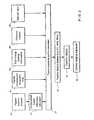
- FIG. 1illustrates a digital mammography system in which preferred embodiments disclosed herein can be implemented.
- FIG. 2is a flow chart illustrating processes of estimating and using tissue exposure control in a mammography system.
- FIG. 3illustrates a focused anti-scatter grid that can be used in the system of FIGS. 3 and 1 .
- FIG. 4illustrates an aspect of tomosynthesis in mammography.
- FIG. 5illustrates selection of a decreased size mammography image for storage and transmission.
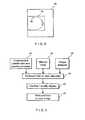
- FIG. 6illustrates processes involved in selecting a reduced size image for transmission and storage.
- a mammography systemcurrently available from the common assignee under the trade name Selenia except for the new features described herein, comprises a stand 10 supporting a C-arm 12 that can move up or down along stand 10 , to a selected height, driven by motor(s) controlled by a health professional operating the system.
- C-arm 12carries an x-ray tube at an upper end 12 a , a breast tray 12 b at a lower end.
- Tray 126covers a flat panel x-ray image receptor 12 c , spaced from the tray by a focused anti-scatter grid 12 d (which may be retractable so that it can be removed from the space between tray 12 b and receptor 12 c ).
- C-arm 12also carries a compression paddle 12 e that is between source 12 a and breast tray 12 b and is motorized to move away from tray 12 b so a patient's breast can be fitted between tray 12 b and paddle 12 e , and closer to tray 12 b so the patient's breast can be compressed and immobilized.
- the movement of paddle 12 eis motorized and controlled by the health professional. Different size paddles 12 e can be fitted to suit different breast sizes for best compression.
- the health professionalcan move paddle 12 along the width of tray 12 b to a position in which paddle 12 e matches the position of a breast that is not centered on tray 12 b , as in the Selenia system currently offered by the common assignee.
- the systemfurther includes other components, such as a control station 14 comprising interface devices such a keyboard 14 a and trackball 14 b , a display screen 14 c , and control and image processing facilities.
- a paddle position encoder 20measures the position of paddle 12 e relative to tray 12 b as the health professional positions, compresses and immobilizes the patient's breast for imaging.
- the thickness of the immobilized breastcan be measured or estimated in other ways instead.
- the final position of paddle 12 ecan be measured in some way, e.g. mechanically or optically or in some other way.
- the thickness of the immobilized breastmay be measured or estimated directly in any one of a number of ways.
- a calculator 22which can be implemented by suitably programming the processing unit 14 ( FIG. 1 ) calculates the thickness of the compressed and immobilized breast on the basis of the output of encoder 20 , or some other means for measuring breast thickness indicators, and provides information about breast thickness for a tissue exposure control calculator 24 , which again can be implement through such programming.
- calculator 24also relies on information about the x-ray density of the breast.
- informationcan come from one or more different sources.
- manual input 26e.g., keyboard 14 a ( FIG. 1 ), through which the health professional can input information characterizing the density of the breast.
- the systemcan present the health professional with three choices—fatty, normal, and dense—and the health professional can make the appropriate choice based on any one or more factors such as physical examination of the breast, information from previous views in the same examination or taken at a much earlier time, or other information about the patient.
- Another example of a source of information about breast densityis previous x-ray images (views) of the patient's breast or breasts.
- a previous viewcan be one taken at any earlier time, either in the same visit of the patient or at a previous visit.
- Information about densitycan be derived from the previous view(s) by the health professional, or it can be derived automatically—by measuring the overall density of a previous x-ray image and perhaps knowing the technic factors used to take it. If actually measured, the density information from the previous view(s) can be provided to calculator 24 manually or automatically, through a connection from the measuring device to calculator 24 .
- Another source of density informationis a dual energy arrangement 30 that pulses the immobilized breast with a low dose x-ray energy at each of two different energies, e.g. sequentially, and the measurements of x-rays with receptor 12 c ( FIG.
- X-ray tube 12 acan be used for such dual energy process, using two different x-ray filters to emit x-rays at the appropriate to different energies or energy bands.
- Yet another source of information about breast densitycan be an arrangement 32 that measures the force with which paddle 12 e compresses the breast and the time such force acts from the start of compression until the breast is immobilized for imaging, and supplies such force/time information to calculator 24 , manually automatically.
- Calculator 24can be implemented as look-up table that in effect has an entry for each of a number of combinations of breast thickness and breast density values.
- the initial values of the entriescan be estimated by actual tests, in essence a trial-and-error process, or in some other way.
- Calculator 24provides its output to technic factor display 34 , which can be display 14 c ( FIG. 1 ), at which the health professional can see the automatically estimated factors such as one or more of kV, mAs, filter, time, etc.
- An entry devicewhich can be keyboard 14 a , allows the health professional to confirm or modify the automatically estimated parameters, and control 38 (which can be a part of unit 14 of FIG. 1 ) uses the resulting final tissue exposure control technic factors for an imaging x-ray exposure.
- the examples disclosed in this patent specificationrefer to compressing and immobilizing the breast before determining technic factors and imaging.
- the breastneed not be compressed before imaging; the breast may be simply supported in some manner, such as by a breast tray, or may be suspended in some manner between an x-ray source and an image receptor.
- the breast thickness and density informationcan come from different sources, such as measurements or estimates of the thickness of the uncompressed breast, or an average of the thickness of the breast portion that will be imaged, of the thickness of the part that is of particular interest for imaging.
- the density informationmay come from the health professional, or from prior x-ray images of the breast, or from some other source.
- Elle same alternative of imaging the uncompressed breastapplies to the other two features discussed below—tomosynthesis and selecting a reduced field of view image for transmission and/or storage—where the alternative dispenses with compression but otherwise conforms to the description below.
- anti-scatter grid 12 dis focused to allow the passage of x-rays along paths 40 that emanate from the focal spot of x-ray source 12 a and to suppress (scattered) x-rays that travel along other paths. If such a grid changes its orientation relative to the x-ray source, it would undesirably suppress x-rays that it should be passing. Such change in orientation would result if x-ray tube 12 e moves in a direction transverse to the x-ray beam it emits while grid 12 d and detector 12 c remain stationary. In addition, such motion would reduce the field of view, so a portion of the breast projected on the receptor in one position of the moving source may fall outside the receptor outline at another position of the source.
- FIG. 4illustrates an arrangement that overcomes these deficiencies of a known proposal.
- x-ray tube 12 a and the combination of anti-scatter grid 12 d and receptor 12 crotate as a unit while a compressed and immobilized breast remains between them and in the path of x-rays emitted from tube 12 a and impinging on receptor 12 c .
- Anti-scatter grid 12 dremains focused on the focal spot of tube 12 a , and the effective field of view does not change with angular position of the source-receptor unit.
- source 12 a and grid 12 d and receptor 12 crotate as a unit, also together with compression paddle 12 c and breast tray 12 b , so a modification is needed to achieve the geometry of FIG. 4 .
- This modificationinvolves decoupling a means to compress and immobilize the breast from motion of tube 12 a , grid 12 d and receptor 12 c .
- thiscan be done by removing compression paddle 12 e and compressing and immobilizing the breast between compression paddles 44 that are appropriately positioned relative to the center of rotation of tube 12 a but do not rotate with tube 12 a , as illustrated in FIG. 4 .
- tube 12 a and receptor 12 ccan translate relative to the breast immobilized between paddles 44 .
- focused grid 12 dcan be decoupled from receptor 12 c and allowed to remain focused at tube 12 a , or a different grid can be used that is not focused or is less focused, and/or the motion of tube 12 a and/or receptor 12 c can be over a more limited path.
- Discrete x-ray images of the breastare taken at each of a number of different positions of tube 12 relative to the breast. The image data is used for tomosynthesis through the application of known image processing methods.
- An important advantage of the example of FIG. 4is that it allows imaging at relatively large angles between the extreme rotational or translational positions of x-ray tube 12 a as compared with known systems.
- Yet another feature of the mammography system disclosed hereis to transmit and store only a portion of the field of view.
- a relatively large field-of-view receptor 12 csuch as used in the Selenia system (24 ⁇ 29 cm)
- the image of the breastlies within a rectangle that is smaller than the field of view, as illustrated in FIG. 5 , where the image 46 of a breast is within a notional rectangular outline 48 (reduced field of view) that is much smaller than the field of view 50 of receptor 12 c .
- the area of field of view 50 that is outside the reduced filed of view area 48may contain little or no information about the breast.
- only the information within the reduced field of view 48may be used, and any information outside outline 48 can be discarded. If there is any significant information outside outline 48 , only that information can be attached to the information for the image portion inside outline 48 .
- One way to select the position and size of outline 48is to rely on the selection of the size and position of compression paddle 12 e that the health professional has made.
- the currently offered Selenia systemallows the health professional to select both the size of a paddle and, at least for some paddles, also the position of the paddle relative to receptor 12 c , so as to match the size and position on receptor 12 c of the breast being x-rayed.
- the size and position of paddle 12 ecan be automatically determined, and the result used to in effect crop the resulting breast image before transmitting and/or storing and/or formatting it for transmission or storage, for example according to DICOM standards.
- the size and position of the breast in the imagecan be found through image analysis, such as analysis involving edge detection, and the size and position of outline 48 can be found in this manner.
- the size and position of outline 48may be entered by the health professional, e.g., through keyboard 14 a , based on viewing the image displayed on monitor 14 c .
- a combination of said methodscan be used, e.g., an automatic determination based on one or both of image analysis and paddle selection, followed with a presentation of a recommended outline 48 displayed to the health professional together with the entire image, for confirmation or modification by the health professional.
- FIG. 6illustrates an arrangement for providing a reduced field of view image.
- a compression paddle size and position encoder incorporated in C-arm 12 or elsewhere in association with the means for mounting and moving paddle 12 eprovides information about the paddle 12 e that the health professional has selected, and about the position of the paddle's projection on receptor 12 c .
- a manual inputprovides information entered by the health professional, which can be similar to that provided by encoder 52 or can be information regarding which of several rectangles within the entire breast image encompasses the breast, or what arbitrary rectangle can encompass the breast on the image.
- An image analyzer 56provides information about the area in the overall image occupied by the breast.
- a calculator 58uses the information from one or more of units 52 , 54 and 57 to calculate the size and position of a reduced field of view that still encompasses the breast, and the calculation is displayed at 60 , e.g., as an outline 48 in an image such as illustrated in FIG. 5 , for the health professional to confirm or modify, e.g. through manual entries.
- the resultis a finalized reduced field of view image at 62 that can be used for further processing, for transmission, and/or storage. While a rectangular outline 48 has been discussed above, in fact outline 48 can have other suitable shapes.
Landscapes
- Health & Medical Sciences (AREA)
- Life Sciences & Earth Sciences (AREA)
- Medical Informatics (AREA)
- Engineering & Computer Science (AREA)
- Optics & Photonics (AREA)
- Biomedical Technology (AREA)
- Biophysics (AREA)
- High Energy & Nuclear Physics (AREA)
- Veterinary Medicine (AREA)
- Nuclear Medicine, Radiotherapy & Molecular Imaging (AREA)
- Public Health (AREA)
- Pathology (AREA)
- Radiology & Medical Imaging (AREA)
- Physics & Mathematics (AREA)
- Heart & Thoracic Surgery (AREA)
- Molecular Biology (AREA)
- Surgery (AREA)
- Animal Behavior & Ethology (AREA)
- General Health & Medical Sciences (AREA)
- Dentistry (AREA)
- Oral & Maxillofacial Surgery (AREA)
- Apparatus For Radiation Diagnosis (AREA)
Abstract
Description
Claims (12)
Priority Applications (3)
| Application Number | Priority Date | Filing Date | Title |
|---|---|---|---|
| US13/857,503US8831171B2 (en) | 2002-11-27 | 2013-04-05 | Full field mammography with tissue exposure control, tomosynthesis, and dynamic field of view processing |
| US14/479,931US20140376690A1 (en) | 2002-11-27 | 2014-09-08 | Full field mammography with tissue exposure control, tomosynthesis, and dynamic field of view processing |
| US15/875,095US10959694B2 (en) | 2002-11-27 | 2018-01-19 | Full field mammography with tissue exposure control, tomosynthesis, and dynamic field of view processing |
Applications Claiming Priority (6)
| Application Number | Priority Date | Filing Date | Title |
|---|---|---|---|
| US10/305,480US7123684B2 (en) | 2002-11-27 | 2002-11-27 | Full field mammography with tissue exposure control, tomosynthesis, and dynamic field of view processing |
| US11/582,061US7430272B2 (en) | 2002-11-27 | 2006-10-16 | Full field mammography with tissue exposure control, tomosynthesis, and dynamic field of view processing |
| US12/233,240US7760853B2 (en) | 2002-11-27 | 2008-09-18 | Full field mammography with tissue exposure control, tomosynthesis, and dynamic field of view processing |
| US12/699,613US7949091B2 (en) | 2002-11-27 | 2010-02-03 | Full field mammography with tissue exposure control, tomosynthesis, and dynamic field of view processing |
| US13/111,618US8416915B2 (en) | 2002-11-27 | 2011-05-19 | Full field mammography with tissue exposure control, tomosynthesis, and dynamic field of view processing |
| US13/857,503US8831171B2 (en) | 2002-11-27 | 2013-04-05 | Full field mammography with tissue exposure control, tomosynthesis, and dynamic field of view processing |
Related Parent Applications (1)
| Application Number | Title | Priority Date | Filing Date |
|---|---|---|---|
| US13/111,618ContinuationUS8416915B2 (en) | 2002-11-27 | 2011-05-19 | Full field mammography with tissue exposure control, tomosynthesis, and dynamic field of view processing |
Related Child Applications (1)
| Application Number | Title | Priority Date | Filing Date |
|---|---|---|---|
| US14/479,931ContinuationUS20140376690A1 (en) | 2002-11-27 | 2014-09-08 | Full field mammography with tissue exposure control, tomosynthesis, and dynamic field of view processing |
Publications (2)
| Publication Number | Publication Date |
|---|---|
| US20130223591A1 US20130223591A1 (en) | 2013-08-29 |
| US8831171B2true US8831171B2 (en) | 2014-09-09 |
Family
ID=32325431
Family Applications (8)
| Application Number | Title | Priority Date | Filing Date |
|---|---|---|---|
| US10/305,480Expired - LifetimeUS7123684B2 (en) | 2002-11-27 | 2002-11-27 | Full field mammography with tissue exposure control, tomosynthesis, and dynamic field of view processing |
| US11/582,061Expired - LifetimeUS7430272B2 (en) | 2002-11-27 | 2006-10-16 | Full field mammography with tissue exposure control, tomosynthesis, and dynamic field of view processing |
| US12/233,240Expired - LifetimeUS7760853B2 (en) | 2002-11-27 | 2008-09-18 | Full field mammography with tissue exposure control, tomosynthesis, and dynamic field of view processing |
| US12/699,613Expired - LifetimeUS7949091B2 (en) | 2002-11-27 | 2010-02-03 | Full field mammography with tissue exposure control, tomosynthesis, and dynamic field of view processing |
| US13/111,618Expired - Fee RelatedUS8416915B2 (en) | 2002-11-27 | 2011-05-19 | Full field mammography with tissue exposure control, tomosynthesis, and dynamic field of view processing |
| US13/857,503Expired - LifetimeUS8831171B2 (en) | 2002-11-27 | 2013-04-05 | Full field mammography with tissue exposure control, tomosynthesis, and dynamic field of view processing |
| US14/479,931AbandonedUS20140376690A1 (en) | 2002-11-27 | 2014-09-08 | Full field mammography with tissue exposure control, tomosynthesis, and dynamic field of view processing |
| US15/875,095Expired - Fee RelatedUS10959694B2 (en) | 2002-11-27 | 2018-01-19 | Full field mammography with tissue exposure control, tomosynthesis, and dynamic field of view processing |
Family Applications Before (5)
| Application Number | Title | Priority Date | Filing Date |
|---|---|---|---|
| US10/305,480Expired - LifetimeUS7123684B2 (en) | 2002-11-27 | 2002-11-27 | Full field mammography with tissue exposure control, tomosynthesis, and dynamic field of view processing |
| US11/582,061Expired - LifetimeUS7430272B2 (en) | 2002-11-27 | 2006-10-16 | Full field mammography with tissue exposure control, tomosynthesis, and dynamic field of view processing |
| US12/233,240Expired - LifetimeUS7760853B2 (en) | 2002-11-27 | 2008-09-18 | Full field mammography with tissue exposure control, tomosynthesis, and dynamic field of view processing |
| US12/699,613Expired - LifetimeUS7949091B2 (en) | 2002-11-27 | 2010-02-03 | Full field mammography with tissue exposure control, tomosynthesis, and dynamic field of view processing |
| US13/111,618Expired - Fee RelatedUS8416915B2 (en) | 2002-11-27 | 2011-05-19 | Full field mammography with tissue exposure control, tomosynthesis, and dynamic field of view processing |
Family Applications After (2)
| Application Number | Title | Priority Date | Filing Date |
|---|---|---|---|
| US14/479,931AbandonedUS20140376690A1 (en) | 2002-11-27 | 2014-09-08 | Full field mammography with tissue exposure control, tomosynthesis, and dynamic field of view processing |
| US15/875,095Expired - Fee RelatedUS10959694B2 (en) | 2002-11-27 | 2018-01-19 | Full field mammography with tissue exposure control, tomosynthesis, and dynamic field of view processing |
Country Status (7)
| Country | Link |
|---|---|
| US (8) | US7123684B2 (en) |
| EP (1) | EP1569556B1 (en) |
| JP (1) | JP4628793B2 (en) |
| CN (1) | CN1738573B (en) |
| AT (1) | ATE554707T1 (en) |
| AU (1) | AU2003291189A1 (en) |
| WO (1) | WO2004049949A1 (en) |
Cited By (19)
| Publication number | Priority date | Publication date | Assignee | Title |
|---|---|---|---|---|
| US9066706B2 (en) | 2004-11-26 | 2015-06-30 | Hologic, Inc. | Integrated multi-mode mammography/tomosynthesis x-ray system and method |
| US9460508B2 (en) | 2002-11-27 | 2016-10-04 | Hologic, Inc. | Image handling and display in X-ray mammography and tomosynthesis |
| US9498175B2 (en) | 2002-11-27 | 2016-11-22 | Hologic, Inc. | System and method for low dose tomosynthesis |
| US9851888B2 (en) | 2002-11-27 | 2017-12-26 | Hologic, Inc. | Image handling and display in X-ray mammography and tomosynthesis |
| US9901315B2 (en)* | 2013-03-15 | 2018-02-27 | Hologic, Inc. | X-ray scatter reducing device for use with 2D mammography and tomosynthesis |
| US9943280B2 (en) | 2016-03-07 | 2018-04-17 | General Electric Company | Breast tomosynthesis with flexible compression paddle |
| US10096106B2 (en) | 2016-11-10 | 2018-10-09 | General Electric Company | Combined medical imaging |
| US10638994B2 (en) | 2002-11-27 | 2020-05-05 | Hologic, Inc. | X-ray mammography with tomosynthesis |
| US10881359B2 (en) | 2017-08-22 | 2021-01-05 | Hologic, Inc. | Computed tomography system for imaging multiple anatomical targets |
| US10959694B2 (en) | 2002-11-27 | 2021-03-30 | Hologic, Inc. | Full field mammography with tissue exposure control, tomosynthesis, and dynamic field of view processing |
| US11076820B2 (en) | 2016-04-22 | 2021-08-03 | Hologic, Inc. | Tomosynthesis with shifting focal spot x-ray system using an addressable array |
| US11090017B2 (en) | 2018-09-13 | 2021-08-17 | Hologic, Inc. | Generating synthesized projection images for 3D breast tomosynthesis or multi-mode x-ray breast imaging |
| US11419569B2 (en) | 2017-08-16 | 2022-08-23 | Hologic, Inc. | Image quality compliance tool |
| US11471118B2 (en) | 2020-03-27 | 2022-10-18 | Hologic, Inc. | System and method for tracking x-ray tube focal spot position |
| US11510306B2 (en) | 2019-12-05 | 2022-11-22 | Hologic, Inc. | Systems and methods for improved x-ray tube life |
| US11783476B2 (en) | 2019-10-25 | 2023-10-10 | DeepHealth, Inc. | System and method for analyzing three-dimensional image data |
| US11786191B2 (en) | 2021-05-17 | 2023-10-17 | Hologic, Inc. | Contrast-enhanced tomosynthesis with a copper filter |
| US12367574B2 (en) | 2019-12-23 | 2025-07-22 | DeepHealth, Inc. | Systems and methods for analyzing two-dimensional and three-dimensional image data |
| US12414217B2 (en) | 2022-02-07 | 2025-09-09 | Hologic, Inc. | Systems and methods for adaptively controlling filament current in an X-ray tube |
Families Citing this family (146)
| Publication number | Priority date | Publication date | Assignee | Title |
|---|---|---|---|---|
| US8571289B2 (en) | 2002-11-27 | 2013-10-29 | Hologic, Inc. | System and method for generating a 2D image from a tomosynthesis data set |
| US7110490B2 (en)* | 2002-12-10 | 2006-09-19 | General Electric Company | Full field digital tomosynthesis method and apparatus |
| US7092482B2 (en)* | 2003-04-11 | 2006-08-15 | Fischer Imaging Corporation | Signal profiling for medical imaging systems |
| US8768026B2 (en)* | 2003-11-26 | 2014-07-01 | Hologic, Inc. | X-ray imaging with x-ray markers that provide adjunct information but preserve image quality |
| DE102004008735B4 (en)* | 2004-02-23 | 2015-07-23 | Siemens Aktiengesellschaft | Method and device for generating an X-ray image of the female breast |
| CN100355396C (en)* | 2004-07-30 | 2007-12-19 | 杭州吴越电子有限公司 | Mammary gland camera with air density compensation |
| DE102004053009A1 (en)* | 2004-10-29 | 2006-05-11 | Siemens Ag | Exposing object e.g. patient chest, illustrating method, involves arranging scattered radiation raster between exposing object and x-ray detector, and moving raster away from path of radiation of x-ray depending on thickness of object |
| DE102004052613B4 (en)* | 2004-10-29 | 2016-03-03 | Siemens Aktiengesellschaft | Mammography X-ray machine with a digital solid-state flat detector |
| US7920152B2 (en) | 2004-11-04 | 2011-04-05 | Dr Systems, Inc. | Systems and methods for viewing medical 3D imaging volumes |
| US7885440B2 (en) | 2004-11-04 | 2011-02-08 | Dr Systems, Inc. | Systems and methods for interleaving series of medical images |
| US7660488B2 (en) | 2004-11-04 | 2010-02-09 | Dr Systems, Inc. | Systems and methods for viewing medical images |
| US7787672B2 (en) | 2004-11-04 | 2010-08-31 | Dr Systems, Inc. | Systems and methods for matching, naming, and displaying medical images |
| US7662082B2 (en) | 2004-11-05 | 2010-02-16 | Theragenics Corporation | Expandable brachytherapy device |
| US7702142B2 (en) | 2004-11-15 | 2010-04-20 | Hologic, Inc. | Matching geometry generation and display of mammograms and tomosynthesis images |
| EP1858408A2 (en)* | 2005-03-16 | 2007-11-28 | Cornell Research Foundation Inc. | Method for expanding the domain of imaging software in a diagnostic work-up |
| US7245693B2 (en)* | 2005-06-02 | 2007-07-17 | Agilent Technologies, Inc. | X-ray inspection system having on-axis and off-axis sensors |
| US7245694B2 (en) | 2005-08-15 | 2007-07-17 | Hologic, Inc. | X-ray mammography/tomosynthesis of patient's breast |
| FR2890553B1 (en)* | 2005-09-13 | 2007-11-23 | Gen Electric | MIXED X-RAY DEVICE |
| JP4600942B2 (en)* | 2005-10-06 | 2010-12-22 | 富士フイルム株式会社 | Breast imaging device |
| JP4597936B2 (en)* | 2005-10-06 | 2010-12-15 | 富士フイルム株式会社 | Breast imaging device |
| WO2007043329A1 (en)* | 2005-10-12 | 2007-04-19 | Konica Minolta Medical & Graphic, Inc. | Radiographic imager |
| FI20085470A7 (en)* | 2005-10-17 | 2008-05-19 | J Morita Mfg Corp | Medical digital X-ray imaging device and medical digital X-ray sensor |
| EP1951119A2 (en)* | 2005-11-09 | 2008-08-06 | Dexela Limited | Methods and apparatus for obtaining low-dose imaging |
| US20070242868A1 (en)* | 2005-11-09 | 2007-10-18 | Dexela Limited | Methods and apparatus for displaying images |
| US7465268B2 (en) | 2005-11-18 | 2008-12-16 | Senorx, Inc. | Methods for asymmetrical irradiation of a body cavity |
| DE102006005068A1 (en)* | 2006-02-03 | 2007-08-09 | Siemens Ag | Positioning device for a mammography device |
| WO2007095330A2 (en) | 2006-02-15 | 2007-08-23 | Hologic Inc | Breast biopsy and needle localization using tomosynthesis systems |
| JP4891662B2 (en)* | 2006-06-08 | 2012-03-07 | 株式会社東芝 | Mammography equipment |
| US7292675B1 (en)* | 2006-07-19 | 2007-11-06 | General Electric Company | Automatic protocol assistance methods and apparatus |
| US20090080602A1 (en)* | 2006-08-03 | 2009-03-26 | Kenneth Brooks | Dedicated breast radiation imaging/therapy system |
| FR2905256B1 (en)* | 2006-09-05 | 2008-11-21 | Gen Electric | METHOD FOR OBTAINING A TOMOSYNTHESIS IMAGE |
| JP4874755B2 (en)* | 2006-09-29 | 2012-02-15 | 富士フイルム株式会社 | Radiation imaging equipment |
| JP4851296B2 (en)* | 2006-10-26 | 2012-01-11 | 富士フイルム株式会社 | Radiation tomographic image acquisition apparatus and radiation tomographic image acquisition method |
| US7953614B1 (en) | 2006-11-22 | 2011-05-31 | Dr Systems, Inc. | Smart placement rules |
| EP1925254A1 (en) | 2006-11-24 | 2008-05-28 | Ion Beam Applications S.A. | Method and device for quality management in a mammography apparatus |
| US20080219567A1 (en)* | 2007-03-07 | 2008-09-11 | General Electric Company | Tomosynthesis imaging data compression system and method |
| JP2008237631A (en)* | 2007-03-28 | 2008-10-09 | Fujifilm Corp | Radiation imaging device |
| US9597041B2 (en)* | 2007-03-30 | 2017-03-21 | General Electric Company | Sequential image acquisition with updating method and system |
| US8553967B2 (en)* | 2007-06-29 | 2013-10-08 | General Electric Company | System and method for a digital X-ray radiographic tomosynthesis user interface |
| US7630533B2 (en) | 2007-09-20 | 2009-12-08 | Hologic, Inc. | Breast tomosynthesis with display of highlighted suspected calcifications |
| US7929743B2 (en)* | 2007-10-02 | 2011-04-19 | Hologic, Inc. | Displaying breast tomosynthesis computer-aided detection results |
| FI120077B (en)* | 2007-11-14 | 2009-06-30 | Planmed Oy | Arrangement and procedure for digital mammography photography |
| JP5112097B2 (en)* | 2008-02-04 | 2013-01-09 | 株式会社東芝 | Breast X-ray diagnostic device |
| DE102008012394B4 (en)* | 2008-03-04 | 2010-03-25 | Siemens Aktiengesellschaft | Apparatus and method for generating digital x-ray images of a sample |
| US7792245B2 (en) | 2008-06-24 | 2010-09-07 | Hologic, Inc. | Breast tomosynthesis system with shifting face shield |
| US7991106B2 (en) | 2008-08-29 | 2011-08-02 | Hologic, Inc. | Multi-mode tomosynthesis/mammography gain calibration and image correction using gain map information from selected projection angles |
| US8380533B2 (en) | 2008-11-19 | 2013-02-19 | DR Systems Inc. | System and method of providing dynamic and customizable medical examination forms |
| KR101639374B1 (en)* | 2008-11-24 | 2016-07-13 | 홀로직, 인크. | Method and system for controlling x-ray focal spot characteristics for tomosynthesis and mammography imaging |
| US9248311B2 (en) | 2009-02-11 | 2016-02-02 | Hologic, Inc. | System and method for modifying a flexibility of a brachythereapy catheter |
| US9579524B2 (en) | 2009-02-11 | 2017-02-28 | Hologic, Inc. | Flexible multi-lumen brachytherapy device |
| US8170320B2 (en) | 2009-03-03 | 2012-05-01 | Hologic, Inc. | Mammography/tomosynthesis systems and methods automatically deriving breast characteristics from breast x-ray images and automatically adjusting image processing parameters accordingly |
| EP2408375B1 (en) | 2009-03-20 | 2017-12-06 | Orthoscan Incorporated | Moveable imaging apparatus |
| US8217357B2 (en) | 2009-04-13 | 2012-07-10 | Hologic, Inc. | Integrated breast X-ray and molecular imaging system |
| US10207126B2 (en) | 2009-05-11 | 2019-02-19 | Cytyc Corporation | Lumen visualization and identification system for multi-lumen balloon catheter |
| DK2462561T3 (en) | 2009-08-03 | 2018-12-10 | Volpara Health Tech Limited | A METHOD AND SYSTEM FOR ANALYZING Tissues FROM IMAGES |
| US8798353B2 (en)* | 2009-09-08 | 2014-08-05 | General Electric Company | Apparatus and method for two-view tomosynthesis imaging |
| US8331536B2 (en)* | 2009-09-18 | 2012-12-11 | General Electric Company | Apparatus for reducing scattered X-ray detection and method of same |
| JP5572040B2 (en)* | 2009-09-28 | 2014-08-13 | 富士フイルム株式会社 | Radiography equipment |
| US8712120B1 (en) | 2009-09-28 | 2014-04-29 | Dr Systems, Inc. | Rules-based approach to transferring and/or viewing medical images |
| ES2862525T3 (en) | 2009-10-08 | 2021-10-07 | Hologic Inc | Needle Breast Biopsy System and Method of Use |
| NL2005509C2 (en)* | 2010-02-19 | 2011-08-23 | Academisch Medisch Ct Bij De Universiteit Van Amsterdam | Mammography-apparatus. |
| JP5436301B2 (en)* | 2010-03-29 | 2014-03-05 | 富士フイルム株式会社 | Radiography apparatus and radiation imaging system |
| KR20110138803A (en) | 2010-06-22 | 2011-12-28 | 삼성전자주식회사 | Image Diagnosis Apparatus and Method Using V-RAB |
| KR101689866B1 (en)* | 2010-07-29 | 2016-12-27 | 삼성전자주식회사 | Method and apparatus of processing image and medical image system employing the same |
| CN101926651B (en)* | 2010-08-27 | 2016-06-08 | 深圳市尚荣医疗股份有限公司 | Half-view geometric mammary gland X-ray shooting device |
| DE102010035920A1 (en)* | 2010-08-31 | 2012-03-01 | Siemens Aktiengesellschaft | Method for displaying a predetermined volume section of an examination object by means of a tomosynthesis device and corresponding tomosynthesis device |
| US8744041B2 (en) | 2010-09-09 | 2014-06-03 | Hologic, Inc. | Methods and systems for dynamically modifying acquisition parameter during image acquisition |
| US9352172B2 (en) | 2010-09-30 | 2016-05-31 | Hologic, Inc. | Using a guide member to facilitate brachytherapy device swap |
| CA2813591C (en) | 2010-10-05 | 2020-09-22 | Hologic, Inc. | Upright x-ray breast imaging with a ct mode, multiple tomosynthesis modes, and a mammography mode |
| US20120133600A1 (en) | 2010-11-26 | 2012-05-31 | Hologic, Inc. | User interface for medical image review workstation |
| WO2012082799A1 (en) | 2010-12-13 | 2012-06-21 | Orthoscan, Inc. | Mobile fluoroscopic imaging system |
| US9168013B2 (en) | 2010-12-13 | 2015-10-27 | Koninklijke Philips N.V. | Breast density assessment |
| US9901320B2 (en) | 2010-12-14 | 2018-02-27 | Hologic, Inc. | System and method for fusing three dimensional image data from a plurality of different imaging systems for use in diagnostic imaging |
| US10342992B2 (en) | 2011-01-06 | 2019-07-09 | Hologic, Inc. | Orienting a brachytherapy applicator |
| DE102011002758A1 (en)* | 2011-01-17 | 2012-07-19 | Siemens Aktiengesellschaft | Mammography X-ray machine is provided with compression device with compression plate displaceable along axis, where compression device comprises operating element |
| DE102011003137A1 (en) | 2011-01-25 | 2012-07-26 | Siemens Aktiengesellschaft | Imaging method with an improved representation of a tissue area |
| ITBO20110086A1 (en)* | 2011-02-25 | 2012-08-26 | I M S Internaz Medicoscienti Fica S R L | EQUIPMENT FOR MAMMOGRAPHY AND / OR TOMOSYNTHESIS WITH DIFFUSED RADIATION REMOVAL DEVICE. |
| JP6057922B2 (en) | 2011-03-08 | 2017-01-11 | ホロジック, インコーポレイテッドHologic, Inc. | System and method for dual energy and / or contrast enhanced breast imaging for screening, diagnosis and biopsy |
| FR2975277B1 (en) | 2011-05-16 | 2014-08-08 | Gen Electric | METHOD FOR ACQUIRING MEDICAL IMAGES OF AN ORGAN AND MEDICAL IMAGING SYSTEM |
| WO2013005871A1 (en)* | 2011-07-01 | 2013-01-10 | 주식회사 휴먼레이 | Mammography detector having multiple sensors, and mammography device capable of 3d image acquisition |
| EP2729070B1 (en) | 2011-07-04 | 2018-08-08 | Koninklijke Philips N.V. | Apparatus adapting a tomosynthetic scan motion according to paddle position |
| US9075899B1 (en) | 2011-08-11 | 2015-07-07 | D.R. Systems, Inc. | Automated display settings for categories of items |
| US9782135B2 (en) | 2011-11-18 | 2017-10-10 | Hologic, Inc. | X-ray mammography and/or breast tomosynthesis using a compression paddle |
| US11259759B2 (en) | 2011-11-18 | 2022-03-01 | Hologic Inc. | X-ray mammography and/or breast tomosynthesis using a compression paddle |
| JP6157491B2 (en) | 2011-11-18 | 2017-07-05 | ホロジック, インコーポレイテッドHologic, Inc. | X-ray mammography and / or breast tomosynthesis using a compression paddle with an inflatable jacket to improve contrast and patient comfort |
| EP2782505B1 (en) | 2011-11-27 | 2020-04-22 | Hologic, Inc. | System and method for generating a 2d image using mammography and/or tomosynthesis image data |
| JP6240097B2 (en) | 2012-02-13 | 2017-11-29 | ホロジック インコーポレイティッド | How to navigate a tomosynthesis stack using composite image data |
| CN103445795B (en)* | 2012-06-05 | 2015-08-05 | 北京国药恒瑞美联信息技术有限公司 | A kind of mammary machine x-ray dose control method and system |
| CN103505232A (en)* | 2012-06-19 | 2014-01-15 | 深圳市蓝韵实业有限公司 | X-ray machine and exposure parameter adjustment method thereof |
| WO2014097026A1 (en)* | 2012-12-21 | 2014-06-26 | Koninklijke Philips N.V. | Breast thickness measurement in mammography |
| US9495604B1 (en) | 2013-01-09 | 2016-11-15 | D.R. Systems, Inc. | Intelligent management of computerized advanced processing |
| US10092358B2 (en) | 2013-03-15 | 2018-10-09 | Hologic, Inc. | Tomosynthesis-guided biopsy apparatus and method |
| CN105451657A (en) | 2013-03-15 | 2016-03-30 | 霍罗吉克公司 | System and method for navigating tomosynthesis stack including automatic focusing |
| CN113768529A (en) | 2013-04-26 | 2021-12-10 | 蒂莫西·R·斯坦戈 | X-ray breast imaging system and compression paddle for X-ray breast imaging system |
| CN104367331B (en)* | 2013-08-15 | 2017-02-15 | 深圳市蓝韵实业有限公司 | Full-digital automatic exposure method for digital mammary gland X-ray machine |
| JP6027687B2 (en)* | 2013-09-30 | 2016-11-16 | 富士フイルム株式会社 | Breast thickness measuring apparatus and breast thickness measuring method |
| CA2925907C (en) | 2013-10-09 | 2022-03-15 | Hologic, Inc. | X-ray breast tomosynthesis enhancing spatial resolution including in the thickness direction of a flattened breast |
| EP3060132B1 (en) | 2013-10-24 | 2019-12-04 | Hologic, Inc. | System and method for navigating x-ray guided breast biopsy |
| DE102013222386A1 (en) | 2013-11-05 | 2015-05-07 | Siemens Aktiengesellschaft | Method and CT System for Topogram Scanning |
| US9510793B2 (en)* | 2014-01-27 | 2016-12-06 | Epica International, Inc. | Radiological imaging device with advanced sensors |
| JP6506769B2 (en) | 2014-02-28 | 2019-04-24 | ホロジック, インコーポレイテッドHologic, Inc. | System and method for generating and displaying tomosynthesis image slabs |
| US9615803B2 (en) | 2014-06-16 | 2017-04-11 | General Electric Company | System and method for determining X-ray exposure parameters |
| US9610057B2 (en) | 2014-06-16 | 2017-04-04 | General Electric Company | System and method for determining X-ray exposure parameters |
| KR102326968B1 (en)* | 2014-08-29 | 2021-11-17 | (주)바텍이우홀딩스 | mammography system and method |
| US20170046483A1 (en) | 2015-04-30 | 2017-02-16 | D.R. Systems, Inc. | Database systems and interactive user interfaces for dynamic interaction with, and comparison of, digital medical image data |
| CN105726049B (en)* | 2016-01-14 | 2018-10-26 | 深圳安科高技术股份有限公司 | A kind of digital galactophore X-ray production apparatus and its automatic exposure image optimization method |
| KR101793100B1 (en)* | 2016-03-08 | 2017-11-03 | 주식회사 제타이미징 | X-ray examination apparatus |
| CN109313698B (en) | 2016-05-27 | 2022-08-30 | 霍罗吉克公司 | Simultaneous surface and internal tumor detection |
| US11147525B2 (en) | 2016-11-04 | 2021-10-19 | Hologic, Inc. | Medical imaging device and method of operating a medical imaging device |
| CN117838159A (en) | 2016-11-08 | 2024-04-09 | 豪洛捷公司 | Imaging using curved compression elements |
| EP3600047A1 (en) | 2017-03-30 | 2020-02-05 | Hologic, Inc. | System and method for hierarchical multi-level feature image synthesis and representation |
| EP3600052A1 (en) | 2017-03-30 | 2020-02-05 | Hologic, Inc. | System and method for targeted object enhancement to generate synthetic breast tissue images |
| CN110621233B (en) | 2017-03-30 | 2023-12-12 | 豪洛捷公司 | Method for processing breast tissue image data |
| WO2018236565A1 (en) | 2017-06-20 | 2018-12-27 | Hologic, Inc. | METHOD AND SYSTEM FOR MEDICAL IMAGING WITH DYNAMIC SELF-LEARNING |
| US11672493B2 (en) | 2017-08-11 | 2023-06-13 | Hologic, Inc. | Breast compression paddle with access corners |
| WO2019033022A1 (en) | 2017-08-11 | 2019-02-14 | Hologic, Inc. | Breast compression paddle having an inflatable jacket |
| US10993689B2 (en)* | 2017-08-31 | 2021-05-04 | General Electric Company | Method and system for motion assessment and correction in digital breast tomosynthesis |
| CN107582085B (en)* | 2017-09-14 | 2021-02-05 | 广州七喜医疗设备有限公司 | Intelligent digital X-ray exposure control device and method |
| CN108742664B (en)* | 2018-04-04 | 2022-04-12 | 深圳蓝韵医学影像有限公司 | Method, system, device and storage medium for calculating tissue density of mammary gland |
| US12121304B2 (en) | 2018-05-04 | 2024-10-22 | Hologic, Inc. | Introducer and localization wire visualization |
| EP3787520B1 (en) | 2018-05-04 | 2024-09-25 | Hologic, Inc. | Biopsy needle visualization |
| US11596368B2 (en)* | 2018-05-25 | 2023-03-07 | Hologic, Inc. | Breast compression paddles utilizing pivoting foam elements |
| CN109124667B (en)* | 2018-07-23 | 2022-09-06 | 中国科学院苏州生物医学工程技术研究所 | Adjusting device for cage type CT scanner |
| WO2020068851A1 (en) | 2018-09-24 | 2020-04-02 | Hologic, Inc. | Breast mapping and abnormality localization |
| WO2020068767A1 (en) | 2018-09-28 | 2020-04-02 | Hologic, Inc. | System and method for synthetic breast tissue image generation by high density element suppression |
| WO2020107019A1 (en) | 2018-11-25 | 2020-05-28 | Hologic, Inc. | Multimodality hanging protocols |
| JP7169430B2 (en)* | 2019-03-27 | 2022-11-10 | 富士フイルム株式会社 | Imaging control device, method and program |
| DE202020006044U1 (en) | 2019-03-29 | 2024-07-02 | Hologic Inc. | Report generation for cropped digital images |
| JP7105726B2 (en) | 2019-04-25 | 2022-07-25 | 富士フイルム株式会社 | Image processing device, method and program |
| US11139088B2 (en) | 2019-06-12 | 2021-10-05 | alephFS—Systems for Imaging | Grid for X-ray imaging |
| US11883206B2 (en) | 2019-07-29 | 2024-01-30 | Hologic, Inc. | Personalized breast imaging system |
| EP4439580A3 (en) | 2019-09-27 | 2024-12-25 | Hologic, Inc. | Ai system for predicting reading time and reading complexity for reviewing 2d/3d breast images |
| CN111028310B (en)* | 2019-12-31 | 2023-10-03 | 上海联影医疗科技股份有限公司 | Method, device, terminal and medium for determining scanning parameters of breast tomography |
| JP7742349B2 (en) | 2020-01-24 | 2025-09-19 | ホロジック, インコーポレイテッド | Horizontally displaceable foam breast compression paddles |
| EP4101386A4 (en) | 2020-02-04 | 2023-07-12 | FUJIFILM Corporation | IMAGE ADJUSTMENT DEVICE, METHOD AND PROGRAM |
| EP4119055B1 (en) | 2020-03-13 | 2024-10-30 | FUJIFILM Corporation | Image generation device and program, learning device and program, and image processing device and program |
| CN115297778B (en) | 2020-03-18 | 2025-08-08 | 富士胶片株式会社 | Image processing device, method, and recording medium |
| JP7446410B2 (en) | 2020-03-18 | 2024-03-08 | 富士フイルム株式会社 | Image processing device, method and program |
| KR20220158719A (en) | 2020-03-27 | 2022-12-01 | 홀로직, 인크. | Systems and methods for measuring deflection of a foam breast compression paddle |
| US11481038B2 (en) | 2020-03-27 | 2022-10-25 | Hologic, Inc. | Gesture recognition in controlling medical hardware or software |
| WO2022232137A1 (en)* | 2021-04-26 | 2022-11-03 | Hologic, Inc. | Systems and methods for measuring thickness of foam compressive elements |
| US12186119B2 (en) | 2021-10-05 | 2025-01-07 | Hologic, Inc. | Interactive model interface for image selection in medical imaging systems |
| US12254586B2 (en) | 2021-10-25 | 2025-03-18 | Hologic, Inc. | Auto-focus tool for multimodality image review |
| WO2023097279A1 (en) | 2021-11-29 | 2023-06-01 | Hologic, Inc. | Systems and methods for correlating objects of interest |
| KR102537577B1 (en)* | 2022-10-04 | 2023-05-26 | 이자성 | Veterinary x-ray imaging apparatus for an operator |
| CN118840300B (en)* | 2024-09-23 | 2024-11-22 | 华中科技大学同济医学院附属同济医院 | CT image quality optimization system based on image processing |
Citations (128)
| Publication number | Priority date | Publication date | Assignee | Title |
|---|---|---|---|---|
| US3502878A (en) | 1967-09-22 | 1970-03-24 | Us Health Education & Welfare | Automatic x-ray apparatus for limiting the field size of a projected x-ray beam in response to film size and to source-to-film distance |
| US3863073A (en) | 1973-04-26 | 1975-01-28 | Machlett Lab Inc | Automatic system for precise collimation of radiation |
| US3971950A (en) | 1975-04-14 | 1976-07-27 | Xerox Corporation | Independent compression and positioning device for use in mammography |
| US4160906A (en) | 1977-06-23 | 1979-07-10 | General Electric Company | Anatomically coordinated user dominated programmer for diagnostic x-ray apparatus |
| US4310766A (en) | 1978-09-06 | 1982-01-12 | Siemens Aktiengesellschaft | Motor driven x-ray grid and film-holder assembly |
| US4496557A (en) | 1981-08-27 | 1985-01-29 | Adir | Tricyclic ethers, their preparation and the pharmaceutical compositions containing them |
| US4559641A (en) | 1983-06-24 | 1985-12-17 | Thomson-Cgr | Retractable cassette holder for a radiological and radiographic examination apparatus |
| US4706269A (en) | 1985-03-11 | 1987-11-10 | Reina Leo J | Anti-scatter grid structure |
| US4744099A (en) | 1983-11-03 | 1988-05-10 | Siemens Aktiengesellschaft | X-ray diagnostic apparatus comprising radiation filters |
| US4773086A (en) | 1983-12-16 | 1988-09-20 | Yokogawa Medical Systems, Limited | Operator console for X-ray tomographs |
| US4773087A (en) | 1986-04-14 | 1988-09-20 | University Of Rochester | Quality of shadowgraphic x-ray images |
| US4819258A (en) | 1986-11-28 | 1989-04-04 | Bennett X-Ray Corp. | Auto-setting of KV in an x-ray machine after selection of technic factors |
| US4821727A (en) | 1986-10-30 | 1989-04-18 | Elscint Ltd. | Mammographic biopsy needle holder system |
| US4969174A (en) | 1989-09-06 | 1990-11-06 | General Electric Company | Scanning mammography system with reduced scatter radiation |
| US4989227A (en) | 1989-04-28 | 1991-01-29 | General Electric Cgr S.A. | Cassette carrier adaptable in size and position for mammography |
| US5018176A (en) | 1989-03-29 | 1991-05-21 | General Electric Cgr S.A. | Mammograph equipped with an integrated device for taking stereotaxic photographs and a method of utilization of said mammograph |
| US5029193A (en) | 1989-07-03 | 1991-07-02 | Siemens Aktiengesellschaft | X-ray diagnostic installation for mammography exposures |
| USRE33634E (en) | 1986-09-23 | 1991-07-09 | Method and structure for optimizing radiographic quality by controlling X-ray tube voltage, current focal spot size and exposure time | |
| US5051904A (en) | 1988-03-24 | 1991-09-24 | Olganix Corporation | Computerized dynamic tomography system |
| US5078142A (en) | 1989-11-21 | 1992-01-07 | Fischer Imaging Corporation | Precision mammographic needle biopsy system |
| US5163075A (en) | 1991-08-08 | 1992-11-10 | Eastman Kodak Company | Contrast enhancement of electrographic imaging |
| US5164976A (en) | 1989-09-06 | 1992-11-17 | General Electric Company | Scanning mammography system with improved skin line viewing |
| US5199056A (en) | 1989-11-28 | 1993-03-30 | Darrah Carol J | Mammography compression paddle |
| US5240011A (en) | 1991-11-27 | 1993-08-31 | Fischer Imaging Corporation | Motorized biopsy needle positioner |
| US5289520A (en) | 1991-11-27 | 1994-02-22 | Lorad Corporation | Stereotactic mammography imaging system with prone position examination table and CCD camera |
| US5359637A (en) | 1992-04-28 | 1994-10-25 | Wake Forest University | Self-calibrated tomosynthetic, radiographic-imaging system, method, and device |
| US5365562A (en) | 1993-09-20 | 1994-11-15 | Fischer Imaging Corporation | Digital imaging apparatus |
| US5415169A (en) | 1989-11-21 | 1995-05-16 | Fischer Imaging Corporation | Motorized mammographic biopsy apparatus |
| US5452367A (en) | 1993-11-29 | 1995-09-19 | Arch Development Corporation | Automated method and system for the segmentation of medical images |
| US5506877A (en) | 1994-11-23 | 1996-04-09 | The General Hospital Corporation | Mammography breast compression device and method |
| US5526394A (en) | 1993-11-26 | 1996-06-11 | Fischer Imaging Corporation | Digital scan mammography apparatus |
| US5539797A (en) | 1993-03-29 | 1996-07-23 | Ge Medical Systems Sa | Method and apparatus for digital stereotaxic mammography |
| US5553111A (en) | 1994-10-26 | 1996-09-03 | The General Hospital Corporation | Apparatus and method for improved tissue imaging |
| US5592562A (en) | 1994-01-19 | 1997-01-07 | International Business Machines Corporation | Inspection system for cross-sectional imaging |
| US5594769A (en) | 1991-11-27 | 1997-01-14 | Thermotrex Corporation | Method and apparatus for obtaining stereotactic mammographic guided needle breast biopsies |
| US5596200A (en) | 1992-10-14 | 1997-01-21 | Primex | Low dose mammography system |
| US5598454A (en) | 1994-04-26 | 1997-01-28 | Siemens Aktiengesellschaft | X-ray diagnostics installation |
| US5627869A (en) | 1995-11-22 | 1997-05-06 | Thermotrex Corporation | Mammography apparatus with proportional collimation |
| EP0775467A1 (en) | 1995-11-23 | 1997-05-28 | Planmed Oy | Method and system for controlling the functions of a mammography apparatus |
| US5657362A (en) | 1995-02-24 | 1997-08-12 | Arch Development Corporation | Automated method and system for computerized detection of masses and parenchymal distortions in medical images |
| US5668889A (en) | 1990-04-19 | 1997-09-16 | Fuji Photo Film Co., Ltd. | Apparatus for determining an image position, and method for adjusting read-out conditions and/or image processing conditions for a radiation image |
| US5769086A (en) | 1995-12-06 | 1998-06-23 | Biopsys Medical, Inc. | Control system and method for automated biopsy device |
| US5818898A (en) | 1995-11-07 | 1998-10-06 | Kabushiki Kaisha Toshiba | X-ray imaging apparatus using X-ray planar detector |
| US5828722A (en) | 1996-05-17 | 1998-10-27 | Sirona Dental Systems Gmbh & Co., Kg | X-ray diagnostic apparatus for tomosynthesis having a detector that detects positional relationships |
| US5872828A (en) | 1996-07-23 | 1999-02-16 | The General Hospital Corporation | Tomosynthesis system for breast imaging |
| US5878104A (en) | 1996-05-17 | 1999-03-02 | Sirona Dental Systems Gmbh & Co. Kg | Method for producing tomosynthesis exposures employing a reference object formed by a region of the examination subject |
| US5896437A (en) | 1996-05-17 | 1999-04-20 | Sirona Dental Systems Gmbh & Co. Kg | X-ray diagnostics apparatus for tomosynthesis having a reference object in fixed relationship to a radiation emitter |
| US5986662A (en) | 1996-10-16 | 1999-11-16 | Vital Images, Inc. | Advanced diagnostic viewer employing automated protocol selection for volume-rendered imaging |
| US5999836A (en) | 1995-06-06 | 1999-12-07 | Nelson; Robert S. | Enhanced high resolution breast imaging device and method utilizing non-ionizing radiation of narrow spectral bandwidth |
| US6005907A (en) | 1996-05-17 | 1999-12-21 | Sirona Dental Systems Gmbh & Co. Kg | Method and apparatus for producing tomosynthesis exposures employing a reference object composed of a number of sub-objects |
| EP0982001A1 (en) | 1998-08-25 | 2000-03-01 | General Electric Company | Protocol driven image reconstruction, display, and processing in a multislice imaging system |
| US6075879A (en) | 1993-09-29 | 2000-06-13 | R2 Technology, Inc. | Method and system for computer-aided lesion detection using information from multiple images |
| US6091841A (en) | 1997-09-04 | 2000-07-18 | Qualia Computing, Inc. | Method and system for segmenting desired regions in digital mammograms |
| US6137527A (en) | 1996-12-23 | 2000-10-24 | General Electric Company | System and method for prompt-radiology image screening service via satellite |
| US6149301A (en) | 1998-12-30 | 2000-11-21 | General Electric Company | X-ray target centering apparatus for radiographic imaging system |
| US6175117B1 (en) | 1998-01-23 | 2001-01-16 | Quanta Vision, Inc. | Tissue analysis apparatus |
| US6196715B1 (en) | 1959-04-28 | 2001-03-06 | Kabushiki Kaisha Toshiba | X-ray diagnostic system preferable to two dimensional x-ray detection |
| US6216540B1 (en) | 1995-06-06 | 2001-04-17 | Robert S. Nelson | High resolution device and method for imaging concealed objects within an obscuring medium |
| US6233473B1 (en) | 1999-02-16 | 2001-05-15 | Hologic, Inc. | Determining body composition using fan beam dual-energy x-ray absorptiometry |
| US6243441B1 (en) | 1999-07-13 | 2001-06-05 | Edge Medical Devices | Active matrix detector for X-ray imaging |
| US6256370B1 (en) | 2000-01-24 | 2001-07-03 | General Electric Company | Method and apparatus for performing tomosynthesis |
| US6272207B1 (en) | 1999-02-18 | 2001-08-07 | Creatv Microtech, Inc. | Method and apparatus for obtaining high-resolution digital X-ray and gamma ray images |
| US6289235B1 (en) | 1998-03-05 | 2001-09-11 | Wake Forest University | Method and system for creating three-dimensional images using tomosynthetic computed tomography |
| US6292530B1 (en) | 1999-04-29 | 2001-09-18 | General Electric Company | Method and apparatus for reconstructing image data acquired by a tomosynthesis x-ray imaging system |
| US20010038681A1 (en) | 2000-02-11 | 2001-11-08 | Brandeis University | Method and system for low-dose three-dimensional imaging of a scene |
| US6327336B1 (en) | 2000-06-05 | 2001-12-04 | Direct Radiography Corp. | Radiogram showing location of automatic exposure control sensor |
| US6341156B1 (en) | 1999-05-14 | 2002-01-22 | Siemens Aktiengesellschaft | X-ray diagnostic apparatus with relatively moved x-ray source and detector |
| US20020012450A1 (en) | 1998-01-09 | 2002-01-31 | Osamu Tsujii | Image processing apparatus and method |
| US6375352B1 (en) | 1999-10-01 | 2002-04-23 | General Electric Company | Apparatus and method for obtaining x-ray tomosynthesis data for mammography |
| US20020050986A1 (en) | 2000-08-11 | 2002-05-02 | Hitoshi Inoue | Image display apparatus and method, and storage medium |
| US20020075997A1 (en) | 2000-12-18 | 2002-06-20 | Unger Christopher David | Medical diagnostic method and apparatus to control dual energy exposure techniques based on image information |
| US6411836B1 (en) | 1999-12-30 | 2002-06-25 | General Electric Company | Method and apparatus for user preferences configuring in an image handling system |
| US6415015B2 (en) | 1999-12-28 | 2002-07-02 | Ge Medical Systems Sa | Method and system of compensation of thickness of an organ |
| US6442288B1 (en) | 1997-12-17 | 2002-08-27 | Siemens Aktiengesellschaft | Method for reconstructing a three-dimensional image of an object scanned in the context of a tomosynthesis, and apparatus for tomosynthesis |
| US6459925B1 (en) | 1998-11-25 | 2002-10-01 | Fischer Imaging Corporation | User interface system for mammographic imager |
| US20030018272A1 (en) | 2001-06-28 | 2003-01-23 | Treado Patrick J. | Method for Raman chemical imaging and characterization of calcification in tissue |
| WO2003020114A2 (en) | 2001-08-31 | 2003-03-13 | Analogic Corporation | Image positioning method and system for tomosynthesis in a digital x-ray radiography system |
| US20030072417A1 (en)* | 2001-10-12 | 2003-04-17 | Kaufhold John Patrick | Method and apparatus for calibrating an imaging system |
| US6556655B1 (en) | 1998-11-27 | 2003-04-29 | Ge Medical Systems Sa | Method for automatic detection of glandular tissue |
| US20030095624A1 (en) | 2001-11-21 | 2003-05-22 | Eberhard Jeffrey Wayne | Dose management system for mammographic tomosynthesis |
| US6597762B1 (en) | 2002-11-27 | 2003-07-22 | Ge Medical Systems Global Technology Co., Llc | Method and apparatus of lesion detection and validation based on multiple reviews of a CT image |
| US6611575B1 (en) | 2001-07-27 | 2003-08-26 | General Electric Company | Method and system for high resolution 3D visualization of mammography images |
| US6620111B2 (en) | 2001-04-20 | 2003-09-16 | Ethicon Endo-Surgery, Inc. | Surgical biopsy device having automatic rotation of the probe for taking multiple samples |
| US6626849B2 (en) | 2001-11-01 | 2003-09-30 | Ethicon Endo-Surgery, Inc. | MRI compatible surgical biopsy device |
| US6633674B1 (en) | 1999-11-24 | 2003-10-14 | General Electric Company | Picture archiving and communication system employing improved data compression |
| US20030194121A1 (en) | 2002-04-15 | 2003-10-16 | General Electric Company | Computer aided detection (CAD) for 3D digital mammography |
| US20030194051A1 (en) | 2002-04-15 | 2003-10-16 | General Electric | Tomosynthesis X-ray mammogram system and method with automatic drive system |
| US20030194050A1 (en) | 2002-04-15 | 2003-10-16 | General Electric Company | Multi modality X-ray and nuclear medicine mammography imaging system and method |
| US6638235B2 (en) | 2000-11-06 | 2003-10-28 | Suros Surgical Systems, Inc. | Biopsy apparatus |
| US6647092B2 (en) | 2002-01-18 | 2003-11-11 | General Electric Company | Radiation imaging system and method of collimation |
| US20030210254A1 (en) | 2002-05-13 | 2003-11-13 | Doan William D. | Method, system and computer product for displaying axial images |
| US20030215120A1 (en) | 2002-05-15 | 2003-11-20 | Renuka Uppaluri | Computer aided diagnosis of an image set |
| US20040066884A1 (en) | 2002-10-07 | 2004-04-08 | Hermann Claus Bernhard Erich | Continuous scan tomosynthesis system and method |
| US20040094167A1 (en) | 2000-03-17 | 2004-05-20 | Brady John Michael | Three-dimensional reconstructions of a breast from two x-ray mammographics |
| US20040101095A1 (en) | 2002-11-27 | 2004-05-27 | Hologic Inc. | Full field mammography with tissue exposure control, tomosynthesis, and dynamic field of view processing |
| US6748044B2 (en) | 2002-09-13 | 2004-06-08 | Ge Medical Systems Global Technology Company, Llc | Computer assisted analysis of tomographic mammography data |
| US20040109529A1 (en) | 2002-12-10 | 2004-06-10 | General Electric Company | Full field digital tomosynthesis method and apparatus |
| US20040171986A1 (en) | 1999-04-26 | 2004-09-02 | Scimed Life System, Inc. | Apparatus and methods for guiding a needle |
| US6813334B2 (en) | 2000-10-20 | 2004-11-02 | Koninklijke Philips Electronics N.V. | Tomosynthesis in a limited angular range |
| US20050063509A1 (en) | 2001-10-19 | 2005-03-24 | Defreitas Kenneth F | Mammography system and method employing offset compression paddles automatic collimation and retractable anti-scatter grid |
| US20050078797A1 (en) | 2002-03-01 | 2005-04-14 | Mats Danielsson | X-ray protection device |
| US6885724B2 (en) | 2003-08-22 | 2005-04-26 | Ge Medical Systems Global Technology Company, Llc | Radiographic tomosynthesis image acquisition utilizing asymmetric geometry |
| US20050105679A1 (en) | 2003-02-12 | 2005-05-19 | Tao Wu | Tomosynthesis imaging system and method |
| US20050113681A1 (en) | 2002-11-27 | 2005-05-26 | Defreitas Kenneth F. | X-ray mammography with tomosynthesis |
| US20050113715A1 (en) | 2000-11-06 | 2005-05-26 | Jeffrey Schwindt | Biopsy apparatus |
| US20050129172A1 (en) | 2003-11-17 | 2005-06-16 | Thomas Mertelmeier | X-ray diagnostic apparatus for mammography examinations |
| US20050135555A1 (en) | 2003-12-23 | 2005-06-23 | Claus Bernhard Erich H. | Method and system for simultaneously viewing rendered volumes |
| US20050135664A1 (en) | 2003-12-23 | 2005-06-23 | Kaufhold John P. | Methods and apparatus for reconstruction of volume data from projection data |
| US20050226375A1 (en) | 2004-03-31 | 2005-10-13 | Eberhard Jeffrey W | Enhanced X-ray imaging system and method |
| WO2005110230A1 (en) | 2004-05-14 | 2005-11-24 | Philips Intellectual Property & Standards Gmbh | System and method for diagnosing breast cancer |
| WO2005112767A1 (en) | 2004-05-21 | 2005-12-01 | Tissuomics Limited | Apparatus and method for penetrating radiation measurements |
| US6978040B2 (en) | 2001-12-19 | 2005-12-20 | Canon Kabushiki Kaisha | Optical recovery of radiographic geometry |
| US20060074288A1 (en) | 2004-10-04 | 2006-04-06 | Thomas Kelly | Estimating visceral fat by dual-energy x-ray absorptiometry |
| US20060098855A1 (en) | 2002-11-27 | 2006-05-11 | Gkanatsios Nikolaos A | Image handling and display in X-ray mammography and tomosynthesis |
| WO2006055830A2 (en) | 2004-11-15 | 2006-05-26 | Hologic, Inc. | Matching geometry generation and display of mammograms and tomosynthesis images |
| WO2006058160A2 (en) | 2004-11-26 | 2006-06-01 | Hologic, Inc. | Integrated multi-mode mammography/tomosynthesis x-ray system and method |
| US7110502B2 (en) | 2003-05-12 | 2006-09-19 | Canon Kabushiki Kaisha | Radiographic apparatus and method for switching a grid |
| US7127091B2 (en) | 2000-12-22 | 2006-10-24 | Koninklijke Philips Electronics, N.V. | Method and apparatus for visualizing a limited part of a 3D medical image-point-related data set, through basing a rendered image on an intermediate region between first and second clipping planes, and including spectroscopic viewing of such region |
| US20070036265A1 (en) | 2005-08-15 | 2007-02-15 | Zhenxue Jing | X-ray mammography/tomosynthesis of patient's breast |
| US20070223651A1 (en) | 2006-03-21 | 2007-09-27 | Wagenaar Douglas J | Dual modality mammography device |
| US7315607B2 (en) | 2005-09-02 | 2008-01-01 | Siemens Aktiengesellschaft | Mammograph system with a face shield |
| US20080019581A1 (en) | 2002-11-27 | 2008-01-24 | Gkanatsios Nikolaos A | Image Handling and display in X-ray mammography and tomosynthesis |
| US7323692B2 (en) | 2004-08-10 | 2008-01-29 | Research Foundation Of State University Of New York | Flat-panel detector with avalanche gain |
| US20080045833A1 (en) | 2006-02-15 | 2008-02-21 | Defreitas Kenneth F | Breast biopsy and needle localization using tomosynthesis systems |
| US20090080602A1 (en) | 2006-08-03 | 2009-03-26 | Kenneth Brooks | Dedicated breast radiation imaging/therapy system |
| US20090135997A1 (en) | 2006-03-27 | 2009-05-28 | Hologic, Inc. | Breast Compression For Digital Mammography, Tomosynthesis And Other Modalities |
| US20090268865A1 (en) | 2003-11-26 | 2009-10-29 | Baorui Ren | X-ray imaging with X-ray markers that provide adjunct information but preserve image quality |
| US7630533B2 (en) | 2007-09-20 | 2009-12-08 | Hologic, Inc. | Breast tomosynthesis with display of highlighted suspected calcifications |
Family Cites Families (148)
| Publication number | Priority date | Publication date | Assignee | Title |
|---|---|---|---|---|
| US2078142A (en)* | 1935-05-13 | 1937-04-20 | Vibra Lite Ltd | Illuminated display |
| US3365575A (en) | 1964-12-10 | 1968-01-23 | Charles & Stella Guttman Breas | Breast x-ray apparatus with means to accurately position the body of a patient |
| JPS5753531Y2 (en) | 1977-05-04 | 1982-11-19 | ||
| DE3037621A1 (en)* | 1980-10-04 | 1982-05-27 | Philips Patentverwaltung Gmbh, 2000 Hamburg | TRANSLUCTION ARRANGEMENT FOR TAKING LAYER IMAGES OF A THREE-DIMENSIONAL OBJECT |
| US4380086A (en)* | 1980-11-24 | 1983-04-12 | Picker Corporation | Radiation imaging system with cyclically shiftable grid assembly |
| DE3236081A1 (en)* | 1982-09-29 | 1984-03-29 | Siemens AG, 1000 Berlin und 8000 München | RECORDING DEVICE |
| JPH074354B2 (en) | 1984-10-29 | 1995-01-25 | 富士写真フイルム株式会社 | Radiation image information recording / reading device |
| US4662379A (en)* | 1984-12-20 | 1987-05-05 | Stanford University | Coronary artery imaging system using gated tomosynthesis |
| US4760589A (en)* | 1986-04-21 | 1988-07-26 | Siczek Aldona A | Grid cabinet and cassette tray for an X-ray examination apparatus |
| US4763343A (en)* | 1986-09-23 | 1988-08-09 | Yanaki Nicola E | Method and structure for optimizing radiographic quality by controlling X-ray tube voltage, current, focal spot size and exposure time |
| US4752948A (en)* | 1986-12-01 | 1988-06-21 | University Of Chicago | Mobile radiography alignment device |
| DK654488A (en) | 1988-11-23 | 1990-05-24 | Nordisk Roentgen Tech App | ROENTGENAPPARAT |
| US5212637A (en) | 1989-11-22 | 1993-05-18 | Stereometrix Corporation | Method of investigating mammograms for masses and calcifications, and apparatus for practicing such method |
| US5844965A (en) | 1989-11-24 | 1998-12-01 | Thomas Jefferson University | Method and apparatus for using film density measurements of a radiograph to monitor the reproducibility of X-ray exposure parameters of a mammography unit |
| US5864146A (en)* | 1996-11-13 | 1999-01-26 | University Of Massachusetts Medical Center | System for quantitative radiographic imaging |
| DE4124294C2 (en) | 1991-07-22 | 1997-03-20 | Siemens Ag | Method for operating an X-ray tube and use of the method |
| US5941832A (en) | 1991-09-27 | 1999-08-24 | Tumey; David M. | Method and apparatus for detection of cancerous and precancerous conditions in a breast |
| US5274690A (en) | 1992-01-06 | 1993-12-28 | Picker International, Inc. | Rotating housing and anode/stationary cathode x-ray tube with magnetic susceptor for holding the cathode stationary |
| US5256370B1 (en) | 1992-05-04 | 1996-09-03 | Indium Corp America | Lead-free alloy containing tin silver and indium |
| US5291539A (en)* | 1992-10-19 | 1994-03-01 | General Electric Company | Variable focussed X-ray grid |
| DE69425957T2 (en)* | 1993-01-27 | 2001-03-15 | Oleg Sokolov | CELLULAR GRID FOR X-RAY RAYS |
| US5983123A (en) | 1993-10-29 | 1999-11-09 | United States Surgical Corporation | Methods and apparatus for performing ultrasound and enhanced X-ray imaging |
| DE4434704C1 (en) | 1994-09-28 | 1995-06-29 | Siemens Ag | X=ray tube with annular vacuum housing |
| US6345194B1 (en) | 1995-06-06 | 2002-02-05 | Robert S. Nelson | Enhanced high resolution breast imaging device and method utilizing non-ionizing radiation of narrow spectral bandwidth |
| US5706327A (en) | 1996-02-09 | 1998-01-06 | Trex Medical Corporation | Method and apparatus for mammographic compression |
| EP0990254A1 (en) | 1996-02-12 | 2000-04-05 | The University of Akron | Multimedia detectors for medical imaging |
| JPH10305030A (en) | 1997-03-06 | 1998-11-17 | Canon Inc | Radiation imaging apparatus and driving method of radiation imaging apparatus |
| US5841829A (en) | 1997-05-13 | 1998-11-24 | Analogic Corporation | Optimal channel filter for CT system with wobbling focal spot |
| US6081577A (en) | 1998-07-24 | 2000-06-27 | Wake Forest University | Method and system for creating task-dependent three-dimensional images |
| CN1122501C (en)* | 1998-08-06 | 2003-10-01 | 深圳安科高技术股份有限公司 | Computerized tomographer for mammary gland |
| US6125167A (en) | 1998-11-25 | 2000-09-26 | Picker International, Inc. | Rotating anode x-ray tube with multiple simultaneously emitting focal spots |
| US6574629B1 (en) | 1998-12-23 | 2003-06-03 | Agfa Corporation | Picture archiving and communication system |
| JP2002538423A (en) | 1999-02-23 | 2002-11-12 | テラプロウブ リミテッド | Method and apparatus for terahertz imaging |
| US6338013B1 (en)* | 1999-03-19 | 2002-01-08 | Bryan John Ruffner | Multifunctional mobile appliance |
| US6256369B1 (en) | 1999-03-31 | 2001-07-03 | Analogic Corporation | Computerized tomography scanner with longitudinal flying focal spot |
| US6244507B1 (en)* | 1999-06-25 | 2001-06-12 | Canon Kabushiki Kaisha | Automatic grid parameter logging for digital radiography |
| US6542575B1 (en) | 1999-08-31 | 2003-04-01 | General Electric Company | Correction methods and apparatus for digital x-ray imaging |
| US6490476B1 (en) | 1999-10-14 | 2002-12-03 | Cti Pet Systems, Inc. | Combined PET and X-ray CT tomograph and method for using same |
| US6987831B2 (en) | 1999-11-18 | 2006-01-17 | University Of Rochester | Apparatus and method for cone beam volume computed tomography breast imaging |
| US6480565B1 (en) | 1999-11-18 | 2002-11-12 | University Of Rochester | Apparatus and method for cone beam volume computed tomography breast imaging |
| US6418189B1 (en) | 2000-01-24 | 2002-07-09 | Analogic Corporation | Explosive material detection apparatus and method using dual energy information of a scan |
| US7206462B1 (en) | 2000-03-17 | 2007-04-17 | The General Hospital Corporation | Method and system for the detection, comparison and volumetric quantification of pulmonary nodules on medical computed tomography scans |
| JP4163370B2 (en) | 2000-06-08 | 2008-10-08 | 富士フイルム株式会社 | Abnormal shadow candidate detection system |
| US6909792B1 (en) | 2000-06-23 | 2005-06-21 | Litton Systems, Inc. | Historical comparison of breast tissue by image processing |
| US7196519B2 (en) | 2000-07-28 | 2007-03-27 | Fonar Corporation | Stand-up vertical field MRI apparatus |
| US8565860B2 (en) | 2000-08-21 | 2013-10-22 | Biosensors International Group, Ltd. | Radioactive emission detector equipped with a position tracking system |
| FR2813973B1 (en) | 2000-09-08 | 2003-06-20 | Ge Med Sys Global Tech Co Llc | METHOD AND DEVICE FOR GENERATING THREE-DIMENSIONAL IMAGES AND APPARATUS FOR RADIOLOGY THEREOF |
| US6678350B2 (en) | 2000-09-29 | 2004-01-13 | Analogic Corporation | Method of and system for improving the signal to noise characteristics of images from a digital X-ray detector receiving bi-chromatic X-ray energy |
| US6553096B1 (en) | 2000-10-06 | 2003-04-22 | The University Of North Carolina Chapel Hill | X-ray generating mechanism using electron field emission cathode |
| US20040213378A1 (en) | 2003-04-24 | 2004-10-28 | The University Of North Carolina At Chapel Hill | Computed tomography system for imaging of human and small animal |
| US6925200B2 (en) | 2000-11-22 | 2005-08-02 | R2 Technology, Inc. | Graphical user interface for display of anatomical information |
| SE0004298D0 (en) | 2000-11-23 | 2000-11-23 | Siemens Elema Ab | Radiology Unit |
| US7597663B2 (en) | 2000-11-24 | 2009-10-06 | U-Systems, Inc. | Adjunctive ultrasound processing and display for breast cancer screening |
| US7556602B2 (en) | 2000-11-24 | 2009-07-07 | U-Systems, Inc. | Breast cancer screening with adjunctive ultrasound mammography |
| US20020090055A1 (en)* | 2000-11-27 | 2002-07-11 | Edge Medical Devices Ltd. | Digital X-ray bucky including grid storage |
| FR2818116B1 (en)* | 2000-12-19 | 2004-08-27 | Ge Med Sys Global Tech Co Llc | MAMMOGRAPHY APPARATUS |
| CA2437454A1 (en) | 2001-02-01 | 2002-08-22 | Creatv Microtech, Inc. | Anti-scatter grids and collimator designs, and their motion, fabrication and assembly |
| US6480572B2 (en) | 2001-03-09 | 2002-11-12 | Koninklijke Philips Electronics N.V. | Dual filament, electrostatically controlled focal spot for x-ray tubes |
| US6784433B2 (en) | 2001-07-16 | 2004-08-31 | Edge Medical Devices Ltd. | High resolution detector for X-ray imaging |
| JP2004536643A (en) | 2001-07-25 | 2004-12-09 | デンツプライ インターナショナル インコーポレーテッド | Real-time digital X-ray imaging device |
| US6674835B2 (en) | 2001-10-12 | 2004-01-06 | General Electric Co. | Methods and apparatus for estimating a material composition of an imaged object |
| US7609806B2 (en) | 2004-10-18 | 2009-10-27 | Hologic Inc. | Mammography system and method employing offset compression paddles, automatic collimations, and retractable anti-scatter grid |
| US20030097055A1 (en) | 2001-11-21 | 2003-05-22 | Philips Medical Systems(Cleveland), Inc. | Method of reviewing tomographic scans with a large number of images |
| US6895077B2 (en) | 2001-11-21 | 2005-05-17 | University Of Massachusetts Medical Center | System and method for x-ray fluoroscopic imaging |
| JP2005514273A (en) | 2001-12-21 | 2005-05-19 | ネクター セラピューティクス | Sealing pharmaceutical preparations in packaging |
| US6909790B2 (en) | 2002-02-15 | 2005-06-21 | Inventec Corporation | System and method of monitoring moving objects |
| US6574304B1 (en) | 2002-09-13 | 2003-06-03 | Ge Medical Systems Global Technology Company, Llc | Computer aided acquisition of medical images |
| US6970531B2 (en) | 2002-10-07 | 2005-11-29 | General Electric Company | Continuous scan RAD tomosynthesis system and method |
| US6825838B2 (en) | 2002-10-11 | 2004-11-30 | Sonocine, Inc. | 3D modeling system |
| US8565372B2 (en) | 2003-11-26 | 2013-10-22 | Hologic, Inc | System and method for low dose tomosynthesis |
| US10638994B2 (en) | 2002-11-27 | 2020-05-05 | Hologic, Inc. | X-ray mammography with tomosynthesis |
| US7760924B2 (en) | 2002-11-27 | 2010-07-20 | Hologic, Inc. | System and method for generating a 2D image from a tomosynthesis data set |
| US8571289B2 (en) | 2002-11-27 | 2013-10-29 | Hologic, Inc. | System and method for generating a 2D image from a tomosynthesis data set |
| DE10301071A1 (en) | 2003-01-14 | 2004-07-22 | Siemens Ag | Adjusting x-ray tube focal spot position involves measuring spot position signal, generating deflection signal depending on position signal, applying deflection signal to electron beam deflector |
| US20040146221A1 (en) | 2003-01-23 | 2004-07-29 | Siegel Scott H. | Radiography Image Management System |
| US6895076B2 (en) | 2003-06-03 | 2005-05-17 | Ge Medical Systems Global Technology Company, Llc | Methods and apparatus for multiple image acquisition on a digital detector |
| US6950492B2 (en) | 2003-06-25 | 2005-09-27 | Besson Guy M | Dynamic multi-spectral X-ray projection imaging |
| JP3942178B2 (en) | 2003-07-29 | 2007-07-11 | ジーイー・メディカル・システムズ・グローバル・テクノロジー・カンパニー・エルエルシー | X-ray CT system |
| JP4416736B2 (en) | 2003-08-01 | 2010-02-17 | 株式会社日立メディコ | Medical image diagnosis support apparatus and program |
| JP4739225B2 (en) | 2003-11-26 | 2011-08-03 | コーニンクレッカ フィリップス エレクトロニクス エヌ ヴィ | Workflow optimization for high-throughput imaging environments |
| SE526371C2 (en) | 2003-12-01 | 2005-08-30 | Xcounter Ab | Device and method for obtaining tomography, tomosynthesis and still image data for an object |
| US7244063B2 (en) | 2003-12-18 | 2007-07-17 | General Electric Company | Method and system for three dimensional tomosynthesis imaging |
| SE528366C2 (en) | 2004-02-13 | 2006-10-31 | Sectra Mamea Ab | Method and apparatus for X-ray imaging |
| JP4743472B2 (en) | 2004-06-30 | 2011-08-10 | 日立工機株式会社 | Tabletop cutting machine |
| WO2006005583A2 (en) | 2004-07-12 | 2006-01-19 | Geneprot Inc. | Secreted polypeptide species involved in multiple sclerosis |
| EP1623672A1 (en) | 2004-08-04 | 2006-02-08 | Siemens Aktiengesellschaft | X-ray apparatus, in particular for a device for x-ray mammography |
| DE102004051401A1 (en) | 2004-10-21 | 2006-05-24 | Siemens Ag | Method for diagnosis in three-dimensional imaging, in particular in mammography |
| DE102004051820A1 (en) | 2004-10-25 | 2006-05-04 | Siemens Ag | Tomography apparatus and method for a tomography apparatus for generating multiple energy images |
| US7539284B2 (en) | 2005-02-11 | 2009-05-26 | Besson Guy M | Method and system for dynamic low dose X-ray imaging |
| FR2882246B1 (en) | 2005-02-21 | 2007-05-18 | Gen Electric | MAMMOGRAPHIC APPARATUS PROVIDED WITH A SCREEN FOR PROTECTING THE HEAD OF A PATIENT |
| KR100907248B1 (en) | 2005-04-21 | 2009-07-10 | (주)안트로젠 | Volume replacement method of body by transplantation of differentiated young fat cells and biodegradable polymer |
| DE102005022899A1 (en) | 2005-05-18 | 2006-11-23 | Siemens Ag | Method and device for generating a digital tomosynthetic 3D X-ray image of an examination object |
| US7492858B2 (en) | 2005-05-20 | 2009-02-17 | Varian Medical Systems, Inc. | System and method for imaging and treatment of tumorous tissue in breasts using computed tomography and radiotherapy |
| EP1910961A2 (en) | 2005-07-26 | 2008-04-16 | Koninklijke Philips Electronics N.V. | Revolutionary series control for medical imaging archive manager |
| FR2890553B1 (en) | 2005-09-13 | 2007-11-23 | Gen Electric | MIXED X-RAY DEVICE |
| US8423123B2 (en) | 2005-09-30 | 2013-04-16 | Given Imaging Ltd. | System and method for in-vivo feature detection |
| EP1937149A1 (en) | 2005-10-19 | 2008-07-02 | The General Hospital Corporation | Imaging system and related techniques |
| US7180977B2 (en) | 2005-10-24 | 2007-02-20 | Xcounter Ab | Scanning-based detection of ionizing radiaion for tomosynthesis |
| US7302031B2 (en) | 2005-10-27 | 2007-11-27 | Sectra Mamea Ab | Method and arrangement relating to X-ray imaging |
| US7630531B2 (en) | 2006-01-31 | 2009-12-08 | Mevis Medical Solutions, Inc. | Enhanced navigational tools for comparing medical images |
| JP4769097B2 (en) | 2006-03-01 | 2011-09-07 | 富士フイルム株式会社 | Mammography device and breast compression plate used in the mammography device |
| EP2018120A2 (en) | 2006-05-05 | 2009-01-28 | Philips Intellectual Property & Standards GmbH | X-ray tube with oscillating anode |
| US7616731B2 (en) | 2006-08-30 | 2009-11-10 | General Electric Company | Acquisition and reconstruction of projection data using a stationary CT geometry |
| JP2008067933A (en) | 2006-09-14 | 2008-03-27 | Toshiba Corp | Digital mammography equipment |
| JP4874755B2 (en) | 2006-09-29 | 2012-02-15 | 富士フイルム株式会社 | Radiation imaging equipment |
| EP2074642B1 (en) | 2006-10-13 | 2011-01-19 | Philips Intellectual Property & Standards GmbH | X-ray emitting device and method of producing an electron beam to produce x-ray radiation in an x-ray emitting device |
| JP4851296B2 (en) | 2006-10-26 | 2012-01-11 | 富士フイルム株式会社 | Radiation tomographic image acquisition apparatus and radiation tomographic image acquisition method |
| US20100020938A1 (en) | 2006-12-12 | 2010-01-28 | Koninklijke Philips Electronics N.V. | Device and method for x-ray tube focal spot size and position control |
| JP5248031B2 (en) | 2007-04-23 | 2013-07-31 | ジーイー・メディカル・システムズ・グローバル・テクノロジー・カンパニー・エルエルシー | X-ray CT system |
| DE102008004473A1 (en) | 2008-01-15 | 2009-07-23 | Siemens Aktiengesellschaft | Method and device for generating a tomosynthetic 3D X-ray image |
| JP5558672B2 (en) | 2008-03-19 | 2014-07-23 | 株式会社東芝 | Image processing apparatus and X-ray computed tomography apparatus |
| US7832901B2 (en) | 2008-03-24 | 2010-11-16 | Cooper Technologies Company | Beam adjustment mechanism for an LED light fixture |
| CN101983034B (en) | 2008-03-31 | 2013-02-13 | 皇家飞利浦电子股份有限公司 | Fast tomosynthesis scanner apparatus and ct-based method based on rotational step-and-shoot image acquisition without focal spot motion during continuous tube movement for use in cone-beam volume ct mammography imaging |
| US20110178389A1 (en) | 2008-05-02 | 2011-07-21 | Eigen, Inc. | Fused image moldalities guidance |
| CN102088909B (en) | 2008-05-09 | 2014-11-26 | 皇家飞利浦电子股份有限公司 | X-ray system with efficient anode heat dissipation |
| US7792245B2 (en) | 2008-06-24 | 2010-09-07 | Hologic, Inc. | Breast tomosynthesis system with shifting face shield |
| US7991106B2 (en) | 2008-08-29 | 2011-08-02 | Hologic, Inc. | Multi-mode tomosynthesis/mammography gain calibration and image correction using gain map information from selected projection angles |
| DE102008050571A1 (en) | 2008-10-06 | 2010-04-15 | Siemens Aktiengesellschaft | Tomosynthesis apparatus and method for operating a tomosynthesis apparatus |
| US8515005B2 (en) | 2009-11-23 | 2013-08-20 | Hologic Inc. | Tomosynthesis with shifting focal spot and oscillating collimator blades |
| KR101639374B1 (en) | 2008-11-24 | 2016-07-13 | 홀로직, 인크. | Method and system for controlling x-ray focal spot characteristics for tomosynthesis and mammography imaging |
| US8782552B2 (en) | 2008-11-28 | 2014-07-15 | Sinan Batman | Active overlay system and method for accessing and manipulating imaging displays |
| EP2378974B1 (en) | 2008-12-17 | 2013-04-03 | Koninklijke Philips Electronics N.V. | X-ray examination apparatus and method |
| US7885384B2 (en) | 2009-01-26 | 2011-02-08 | General Electric Company | System and method to manage maintenance of a radiological imaging system |
| US8170320B2 (en) | 2009-03-03 | 2012-05-01 | Hologic, Inc. | Mammography/tomosynthesis systems and methods automatically deriving breast characteristics from breast x-ray images and automatically adjusting image processing parameters accordingly |
| DE102009021023A1 (en) | 2009-05-13 | 2010-11-18 | Siemens Aktiengesellschaft | Mammography procedure and mammography device |
| US8484728B2 (en) | 2009-06-03 | 2013-07-09 | Apple Inc. | Managing securely installed applications |
| US8451972B2 (en) | 2009-10-23 | 2013-05-28 | Arineta Ltd. | Methods, circuits, devices, apparatus, assemblies and systems for computer tomography |
| DE102010027871B4 (en) | 2010-04-16 | 2013-11-21 | Siemens Aktiengesellschaft | Ring cathode segment with nanostructure as electron emitter |
| WO2011138691A1 (en) | 2010-05-07 | 2011-11-10 | Koninklijke Philips Electronics N.V. | Motion compensation and patient feedback in medical imaging systems |
| EP2575620B1 (en) | 2010-06-02 | 2022-01-19 | Mayo Foundation For Medical Education And Research | Method and apparatus for dual-modality ultrasonic and nuclear emission mammography |
| US9498180B2 (en) | 2010-08-05 | 2016-11-22 | Hologic, Inc. | Detecting and quantifying patient motion during tomosynthesis scans |
| JP2012050519A (en) | 2010-08-31 | 2012-03-15 | Fujifilm Corp | Mammographic apparatus |
| CA2813591C (en) | 2010-10-05 | 2020-09-22 | Hologic, Inc. | Upright x-ray breast imaging with a ct mode, multiple tomosynthesis modes, and a mammography mode |
| JP6057922B2 (en) | 2011-03-08 | 2017-01-11 | ホロジック, インコーポレイテッドHologic, Inc. | System and method for dual energy and / or contrast enhanced breast imaging for screening, diagnosis and biopsy |
| US20120236987A1 (en) | 2011-03-18 | 2012-09-20 | David Ruimi | Multiple energy ct scanner |
| WO2013187970A2 (en) | 2012-05-14 | 2013-12-19 | The General Hospital Corporation | Method for coded-source phase contrast x-ray imaging |
| US8798230B2 (en) | 2012-11-19 | 2014-08-05 | Samsung Electronics Co., Ltd. | Radiation imaging apparatus, computed tomography apparatus, and radiation imaging method |
| KR20150001181A (en) | 2013-06-26 | 2015-01-06 | 삼성전자주식회사 | The X-ray generator and X-ray photographing apparatus including the same |
| CA2925907C (en) | 2013-10-09 | 2022-03-15 | Hologic, Inc. | X-ray breast tomosynthesis enhancing spatial resolution including in the thickness direction of a flattened breast |
| US10405813B2 (en) | 2015-02-04 | 2019-09-10 | Dental Imaging Technologies Corporation | Panoramic imaging using multi-spectral X-ray source |
| US20160331339A1 (en) | 2015-05-15 | 2016-11-17 | The Trustees Of Columbia University In The City Of New York | Systems And Methods For Early Detection And Monitoring Of Osteoarthritis |
| KR101728046B1 (en) | 2015-08-27 | 2017-04-18 | 삼성전자주식회사 | Tomography apparatus and method for reconstructing a tomography image thereof |
| WO2017185028A1 (en) | 2016-04-22 | 2017-10-26 | Hologic, Inc. | Tomosynthesis with shifting focal spot x-ray system using an addressable array |
| US10470733B2 (en) | 2016-05-09 | 2019-11-12 | Canon Medical Systems Corporation | X-ray CT device and medical information management device |
| CN109310877B (en) | 2016-06-23 | 2020-10-02 | 深圳市奥沃医学新技术发展有限公司 | Method for imaging by using ray source, shielding body, treatment head and treatment equipment |
| US10096106B2 (en) | 2016-11-10 | 2018-10-09 | General Electric Company | Combined medical imaging |
| EP4434465A3 (en) | 2017-03-31 | 2024-10-23 | Hologic, Inc. | Multiple modality body composition analysis |
| EP3449835B1 (en) | 2017-08-22 | 2023-01-11 | Hologic, Inc. | Computed tomography system and method for imaging multiple anatomical targets |
- 2002
- 2002-11-27USUS10/305,480patent/US7123684B2/ennot_activeExpired - Lifetime
- 2003
- 2003-11-26WOPCT/US2003/037763patent/WO2004049949A1/enactiveApplication Filing
- 2003-11-26ATAT03783784Tpatent/ATE554707T1/enactive
- 2003-11-26AUAU2003291189Apatent/AU2003291189A1/ennot_activeAbandoned
- 2003-11-26CNCN2003801089489Apatent/CN1738573B/ennot_activeExpired - Lifetime
- 2003-11-26JPJP2004557315Apatent/JP4628793B2/ennot_activeExpired - Lifetime
- 2003-11-26EPEP03783784Apatent/EP1569556B1/ennot_activeExpired - Lifetime
- 2006
- 2006-10-16USUS11/582,061patent/US7430272B2/ennot_activeExpired - Lifetime
- 2008
- 2008-09-18USUS12/233,240patent/US7760853B2/ennot_activeExpired - Lifetime
- 2010
- 2010-02-03USUS12/699,613patent/US7949091B2/ennot_activeExpired - Lifetime
- 2011
- 2011-05-19USUS13/111,618patent/US8416915B2/ennot_activeExpired - Fee Related
- 2013
- 2013-04-05USUS13/857,503patent/US8831171B2/ennot_activeExpired - Lifetime
- 2014
- 2014-09-08USUS14/479,931patent/US20140376690A1/ennot_activeAbandoned
- 2018
- 2018-01-19USUS15/875,095patent/US10959694B2/ennot_activeExpired - Fee Related
Patent Citations (169)
| Publication number | Priority date | Publication date | Assignee | Title |
|---|---|---|---|---|
| US6196715B1 (en) | 1959-04-28 | 2001-03-06 | Kabushiki Kaisha Toshiba | X-ray diagnostic system preferable to two dimensional x-ray detection |
| US3502878A (en) | 1967-09-22 | 1970-03-24 | Us Health Education & Welfare | Automatic x-ray apparatus for limiting the field size of a projected x-ray beam in response to film size and to source-to-film distance |
| US3863073A (en) | 1973-04-26 | 1975-01-28 | Machlett Lab Inc | Automatic system for precise collimation of radiation |
| US3971950A (en) | 1975-04-14 | 1976-07-27 | Xerox Corporation | Independent compression and positioning device for use in mammography |
| US4160906A (en) | 1977-06-23 | 1979-07-10 | General Electric Company | Anatomically coordinated user dominated programmer for diagnostic x-ray apparatus |
| US4310766A (en) | 1978-09-06 | 1982-01-12 | Siemens Aktiengesellschaft | Motor driven x-ray grid and film-holder assembly |
| US4496557A (en) | 1981-08-27 | 1985-01-29 | Adir | Tricyclic ethers, their preparation and the pharmaceutical compositions containing them |
| US4559641A (en) | 1983-06-24 | 1985-12-17 | Thomson-Cgr | Retractable cassette holder for a radiological and radiographic examination apparatus |
| US4744099A (en) | 1983-11-03 | 1988-05-10 | Siemens Aktiengesellschaft | X-ray diagnostic apparatus comprising radiation filters |
| US4773086A (en) | 1983-12-16 | 1988-09-20 | Yokogawa Medical Systems, Limited | Operator console for X-ray tomographs |
| US4706269A (en) | 1985-03-11 | 1987-11-10 | Reina Leo J | Anti-scatter grid structure |
| US4773087A (en) | 1986-04-14 | 1988-09-20 | University Of Rochester | Quality of shadowgraphic x-ray images |
| USRE33634E (en) | 1986-09-23 | 1991-07-09 | Method and structure for optimizing radiographic quality by controlling X-ray tube voltage, current focal spot size and exposure time | |
| US4821727A (en) | 1986-10-30 | 1989-04-18 | Elscint Ltd. | Mammographic biopsy needle holder system |
| US4819258A (en) | 1986-11-28 | 1989-04-04 | Bennett X-Ray Corp. | Auto-setting of KV in an x-ray machine after selection of technic factors |
| US5051904A (en) | 1988-03-24 | 1991-09-24 | Olganix Corporation | Computerized dynamic tomography system |
| US5018176A (en) | 1989-03-29 | 1991-05-21 | General Electric Cgr S.A. | Mammograph equipped with an integrated device for taking stereotaxic photographs and a method of utilization of said mammograph |
| US4989227A (en) | 1989-04-28 | 1991-01-29 | General Electric Cgr S.A. | Cassette carrier adaptable in size and position for mammography |
| US5029193A (en) | 1989-07-03 | 1991-07-02 | Siemens Aktiengesellschaft | X-ray diagnostic installation for mammography exposures |
| US5164976A (en) | 1989-09-06 | 1992-11-17 | General Electric Company | Scanning mammography system with improved skin line viewing |
| US4969174A (en) | 1989-09-06 | 1990-11-06 | General Electric Company | Scanning mammography system with reduced scatter radiation |
| US5415169A (en) | 1989-11-21 | 1995-05-16 | Fischer Imaging Corporation | Motorized mammographic biopsy apparatus |
| US5735264A (en) | 1989-11-21 | 1998-04-07 | Fischer Imaging Corporation | Motorized mammographic biopsy apparatus |
| US5078142A (en) | 1989-11-21 | 1992-01-07 | Fischer Imaging Corporation | Precision mammographic needle biopsy system |
| US5803912A (en) | 1989-11-21 | 1998-09-08 | Fischer Imaging Corporation | Positioning function mammographic biopsy function system with offset |
| US6022325A (en) | 1989-11-21 | 2000-02-08 | Fischer Imaging Corporation | Mammographic biopsy apparatus |
| US5199056A (en) | 1989-11-28 | 1993-03-30 | Darrah Carol J | Mammography compression paddle |
| US5668889A (en) | 1990-04-19 | 1997-09-16 | Fuji Photo Film Co., Ltd. | Apparatus for determining an image position, and method for adjusting read-out conditions and/or image processing conditions for a radiation image |
| US5163075A (en) | 1991-08-08 | 1992-11-10 | Eastman Kodak Company | Contrast enhancement of electrographic imaging |
| US5594769A (en) | 1991-11-27 | 1997-01-14 | Thermotrex Corporation | Method and apparatus for obtaining stereotactic mammographic guided needle breast biopsies |
| US5426685A (en) | 1991-11-27 | 1995-06-20 | Thermotrex Corporation | Stereotactic mammography system imaging |
| US5289520A (en) | 1991-11-27 | 1994-02-22 | Lorad Corporation | Stereotactic mammography imaging system with prone position examination table and CCD camera |
| US5240011A (en) | 1991-11-27 | 1993-08-31 | Fischer Imaging Corporation | Motorized biopsy needle positioner |
| US5609152A (en) | 1991-11-27 | 1997-03-11 | Thermotrex Corporation | Prone position stereotactic mammography needle biopsy apparatus and method for using the same |
| US5359637A (en) | 1992-04-28 | 1994-10-25 | Wake Forest University | Self-calibrated tomosynthetic, radiographic-imaging system, method, and device |
| US5596200A (en) | 1992-10-14 | 1997-01-21 | Primex | Low dose mammography system |
| US5539797A (en) | 1993-03-29 | 1996-07-23 | Ge Medical Systems Sa | Method and apparatus for digital stereotaxic mammography |
| US5365562A (en) | 1993-09-20 | 1994-11-15 | Fischer Imaging Corporation | Digital imaging apparatus |
| US6075879A (en) | 1993-09-29 | 2000-06-13 | R2 Technology, Inc. | Method and system for computer-aided lesion detection using information from multiple images |
| US5526394A (en) | 1993-11-26 | 1996-06-11 | Fischer Imaging Corporation | Digital scan mammography apparatus |
| US5452367A (en) | 1993-11-29 | 1995-09-19 | Arch Development Corporation | Automated method and system for the segmentation of medical images |
| US5592562A (en) | 1994-01-19 | 1997-01-07 | International Business Machines Corporation | Inspection system for cross-sectional imaging |
| US5719952A (en) | 1994-01-19 | 1998-02-17 | International Business Machines Corporation | Inspection system for cross-sectional imaging |
| US5598454A (en) | 1994-04-26 | 1997-01-28 | Siemens Aktiengesellschaft | X-ray diagnostics installation |
| US5553111A (en) | 1994-10-26 | 1996-09-03 | The General Hospital Corporation | Apparatus and method for improved tissue imaging |
| US5506877A (en) | 1994-11-23 | 1996-04-09 | The General Hospital Corporation | Mammography breast compression device and method |
| US5657362A (en) | 1995-02-24 | 1997-08-12 | Arch Development Corporation | Automated method and system for computerized detection of masses and parenchymal distortions in medical images |
| US5999836A (en) | 1995-06-06 | 1999-12-07 | Nelson; Robert S. | Enhanced high resolution breast imaging device and method utilizing non-ionizing radiation of narrow spectral bandwidth |
| US6216540B1 (en) | 1995-06-06 | 2001-04-17 | Robert S. Nelson | High resolution device and method for imaging concealed objects within an obscuring medium |
| US5818898A (en) | 1995-11-07 | 1998-10-06 | Kabushiki Kaisha Toshiba | X-ray imaging apparatus using X-ray planar detector |
| US5627869A (en) | 1995-11-22 | 1997-05-06 | Thermotrex Corporation | Mammography apparatus with proportional collimation |
| EP0775467A1 (en) | 1995-11-23 | 1997-05-28 | Planmed Oy | Method and system for controlling the functions of a mammography apparatus |
| US5769086A (en) | 1995-12-06 | 1998-06-23 | Biopsys Medical, Inc. | Control system and method for automated biopsy device |
| US5896437A (en) | 1996-05-17 | 1999-04-20 | Sirona Dental Systems Gmbh & Co. Kg | X-ray diagnostics apparatus for tomosynthesis having a reference object in fixed relationship to a radiation emitter |
| US5878104A (en) | 1996-05-17 | 1999-03-02 | Sirona Dental Systems Gmbh & Co. Kg | Method for producing tomosynthesis exposures employing a reference object formed by a region of the examination subject |
| US5828722A (en) | 1996-05-17 | 1998-10-27 | Sirona Dental Systems Gmbh & Co., Kg | X-ray diagnostic apparatus for tomosynthesis having a detector that detects positional relationships |
| US6005907A (en) | 1996-05-17 | 1999-12-21 | Sirona Dental Systems Gmbh & Co. Kg | Method and apparatus for producing tomosynthesis exposures employing a reference object composed of a number of sub-objects |
| US5872828A (en) | 1996-07-23 | 1999-02-16 | The General Hospital Corporation | Tomosynthesis system for breast imaging |
| US20030073895A1 (en) | 1996-10-15 | 2003-04-17 | Nields Morgan W. | User interface system for mammographic imager |
| US5986662A (en) | 1996-10-16 | 1999-11-16 | Vital Images, Inc. | Advanced diagnostic viewer employing automated protocol selection for volume-rendered imaging |
| US6219059B1 (en) | 1996-10-16 | 2001-04-17 | Vital Images, Inc. | Interactive control of voxel attributes using selectable characteristics |
| US6137527A (en) | 1996-12-23 | 2000-10-24 | General Electric Company | System and method for prompt-radiology image screening service via satellite |
| US6091841A (en) | 1997-09-04 | 2000-07-18 | Qualia Computing, Inc. | Method and system for segmenting desired regions in digital mammograms |
| US6442288B1 (en) | 1997-12-17 | 2002-08-27 | Siemens Aktiengesellschaft | Method for reconstructing a three-dimensional image of an object scanned in the context of a tomosynthesis, and apparatus for tomosynthesis |
| US20020012450A1 (en) | 1998-01-09 | 2002-01-31 | Osamu Tsujii | Image processing apparatus and method |
| US6175117B1 (en) | 1998-01-23 | 2001-01-16 | Quanta Vision, Inc. | Tissue analysis apparatus |
| US6289235B1 (en) | 1998-03-05 | 2001-09-11 | Wake Forest University | Method and system for creating three-dimensional images using tomosynthetic computed tomography |
| EP0982001A1 (en) | 1998-08-25 | 2000-03-01 | General Electric Company | Protocol driven image reconstruction, display, and processing in a multislice imaging system |
| US6141398A (en) | 1998-08-25 | 2000-10-31 | General Electric Company | Protocol driven image reconstruction, display, and processing in a multislice imaging system |
| US6459925B1 (en) | 1998-11-25 | 2002-10-01 | Fischer Imaging Corporation | User interface system for mammographic imager |
| US6556655B1 (en) | 1998-11-27 | 2003-04-29 | Ge Medical Systems Sa | Method for automatic detection of glandular tissue |
| US6149301A (en) | 1998-12-30 | 2000-11-21 | General Electric Company | X-ray target centering apparatus for radiographic imaging system |
| US6233473B1 (en) | 1999-02-16 | 2001-05-15 | Hologic, Inc. | Determining body composition using fan beam dual-energy x-ray absorptiometry |
| US6272207B1 (en) | 1999-02-18 | 2001-08-07 | Creatv Microtech, Inc. | Method and apparatus for obtaining high-resolution digital X-ray and gamma ray images |
| US20040171986A1 (en) | 1999-04-26 | 2004-09-02 | Scimed Life System, Inc. | Apparatus and methods for guiding a needle |
| US6292530B1 (en) | 1999-04-29 | 2001-09-18 | General Electric Company | Method and apparatus for reconstructing image data acquired by a tomosynthesis x-ray imaging system |
| US6341156B1 (en) | 1999-05-14 | 2002-01-22 | Siemens Aktiengesellschaft | X-ray diagnostic apparatus with relatively moved x-ray source and detector |
| US6243441B1 (en) | 1999-07-13 | 2001-06-05 | Edge Medical Devices | Active matrix detector for X-ray imaging |
| US6375352B1 (en) | 1999-10-01 | 2002-04-23 | General Electric Company | Apparatus and method for obtaining x-ray tomosynthesis data for mammography |
| US6633674B1 (en) | 1999-11-24 | 2003-10-14 | General Electric Company | Picture archiving and communication system employing improved data compression |
| US6912319B1 (en) | 1999-11-24 | 2005-06-28 | Ge Medical Systems Information Technologies, Inc. | Method and system for lossless wavelet decomposition, compression and decompression of data |
| US6415015B2 (en) | 1999-12-28 | 2002-07-02 | Ge Medical Systems Sa | Method and system of compensation of thickness of an organ |
| US6411836B1 (en) | 1999-12-30 | 2002-06-25 | General Electric Company | Method and apparatus for user preferences configuring in an image handling system |
| US6256370B1 (en) | 2000-01-24 | 2001-07-03 | General Electric Company | Method and apparatus for performing tomosynthesis |
| US6744848B2 (en) | 2000-02-11 | 2004-06-01 | Brandeis University | Method and system for low-dose three-dimensional imaging of a scene |
| US20010038681A1 (en) | 2000-02-11 | 2001-11-08 | Brandeis University | Method and system for low-dose three-dimensional imaging of a scene |
| US20040094167A1 (en) | 2000-03-17 | 2004-05-20 | Brady John Michael | Three-dimensional reconstructions of a breast from two x-ray mammographics |
| US6327336B1 (en) | 2000-06-05 | 2001-12-04 | Direct Radiography Corp. | Radiogram showing location of automatic exposure control sensor |
| US20020050986A1 (en) | 2000-08-11 | 2002-05-02 | Hitoshi Inoue | Image display apparatus and method, and storage medium |
| US6813334B2 (en) | 2000-10-20 | 2004-11-02 | Koninklijke Philips Electronics N.V. | Tomosynthesis in a limited angular range |
| US20060030784A1 (en) | 2000-11-06 | 2006-02-09 | Miller Michael E | Collection filter |
| US20060129062A1 (en) | 2000-11-06 | 2006-06-15 | Nicoson Zachary R | Fluid control element for biopsy apparatus |
| US20050113715A1 (en) | 2000-11-06 | 2005-05-26 | Jeffrey Schwindt | Biopsy apparatus |
| US20060155209A1 (en) | 2000-11-06 | 2006-07-13 | Miller Michael E | Selectively detachable outer cannula hub |
| US20050049521A1 (en) | 2000-11-06 | 2005-03-03 | Suros Surgical Systems, Inc. | Collection filter for biopsy apparatus |
| US20040267157A1 (en) | 2000-11-06 | 2004-12-30 | Miller Michael E | Biopsy apparatus |
| US6638235B2 (en) | 2000-11-06 | 2003-10-28 | Suros Surgical Systems, Inc. | Biopsy apparatus |
| US6758824B1 (en) | 2000-11-06 | 2004-07-06 | Suros Surgical Systems, Inc. | Biopsy apparatus |
| US6501819B2 (en) | 2000-12-18 | 2002-12-31 | Ge Medical Systems Global Technology Company, Llc | Medical diagnostic method and apparatus to control dual energy exposure techniques based on image information |
| US20020075997A1 (en) | 2000-12-18 | 2002-06-20 | Unger Christopher David | Medical diagnostic method and apparatus to control dual energy exposure techniques based on image information |
| US7127091B2 (en) | 2000-12-22 | 2006-10-24 | Koninklijke Philips Electronics, N.V. | Method and apparatus for visualizing a limited part of a 3D medical image-point-related data set, through basing a rendered image on an intermediate region between first and second clipping planes, and including spectroscopic viewing of such region |
| US6620111B2 (en) | 2001-04-20 | 2003-09-16 | Ethicon Endo-Surgery, Inc. | Surgical biopsy device having automatic rotation of the probe for taking multiple samples |
| US20030018272A1 (en) | 2001-06-28 | 2003-01-23 | Treado Patrick J. | Method for Raman chemical imaging and characterization of calcification in tissue |
| US6611575B1 (en) | 2001-07-27 | 2003-08-26 | General Electric Company | Method and system for high resolution 3D visualization of mammography images |
| WO2003020114A2 (en) | 2001-08-31 | 2003-03-13 | Analogic Corporation | Image positioning method and system for tomosynthesis in a digital x-ray radiography system |
| US20030072417A1 (en)* | 2001-10-12 | 2003-04-17 | Kaufhold John Patrick | Method and apparatus for calibrating an imaging system |
| US20050063509A1 (en) | 2001-10-19 | 2005-03-24 | Defreitas Kenneth F | Mammography system and method employing offset compression paddles automatic collimation and retractable anti-scatter grid |
| US7319735B2 (en) | 2001-10-19 | 2008-01-15 | Hologic, Inc. | Mammography system and method employing offset compression paddles, automatic collimation, and retractable anti-scatter grid |
| US7443949B2 (en) | 2001-10-19 | 2008-10-28 | Hologic, Inc. | Mammography system and method employing offset compression paddles, automatic collimation, and retractable anti-scatter grid |
| US20070076844A1 (en) | 2001-10-19 | 2007-04-05 | Defreitas Kenneth F | Mammography system and method employing offset compression paddles, automatic collimation, and retractable anti-scatter grid |
| US6626849B2 (en) | 2001-11-01 | 2003-09-30 | Ethicon Endo-Surgery, Inc. | MRI compatible surgical biopsy device |
| US6751285B2 (en) | 2001-11-21 | 2004-06-15 | General Electric Company | Dose management system for mammographic tomosynthesis |
| US20030095624A1 (en) | 2001-11-21 | 2003-05-22 | Eberhard Jeffrey Wayne | Dose management system for mammographic tomosynthesis |
| US6978040B2 (en) | 2001-12-19 | 2005-12-20 | Canon Kabushiki Kaisha | Optical recovery of radiographic geometry |
| US6647092B2 (en) | 2002-01-18 | 2003-11-11 | General Electric Company | Radiation imaging system and method of collimation |
| US20050078797A1 (en) | 2002-03-01 | 2005-04-14 | Mats Danielsson | X-ray protection device |
| US20030194051A1 (en) | 2002-04-15 | 2003-10-16 | General Electric | Tomosynthesis X-ray mammogram system and method with automatic drive system |
| US20030194121A1 (en) | 2002-04-15 | 2003-10-16 | General Electric Company | Computer aided detection (CAD) for 3D digital mammography |
| US6882700B2 (en) | 2002-04-15 | 2005-04-19 | General Electric Company | Tomosynthesis X-ray mammogram system and method with automatic drive system |
| US20030194050A1 (en) | 2002-04-15 | 2003-10-16 | General Electric Company | Multi modality X-ray and nuclear medicine mammography imaging system and method |
| US20030210254A1 (en) | 2002-05-13 | 2003-11-13 | Doan William D. | Method, system and computer product for displaying axial images |
| US20030215120A1 (en) | 2002-05-15 | 2003-11-20 | Renuka Uppaluri | Computer aided diagnosis of an image set |
| US6748044B2 (en) | 2002-09-13 | 2004-06-08 | Ge Medical Systems Global Technology Company, Llc | Computer assisted analysis of tomographic mammography data |
| US20040066884A1 (en) | 2002-10-07 | 2004-04-08 | Hermann Claus Bernhard Erich | Continuous scan tomosynthesis system and method |
| US6940943B2 (en) | 2002-10-07 | 2005-09-06 | General Electric Company | Continuous scan tomosynthesis system and method |
| US7123684B2 (en) | 2002-11-27 | 2006-10-17 | Hologic, Inc. | Full field mammography with tissue exposure control, tomosynthesis, and dynamic field of view processing |
| US20090010384A1 (en) | 2002-11-27 | 2009-01-08 | Hologic, Inc. | Full field mammography with tissue exposure control, tomosynthesis, and dynamic field of view processing |
| US20080019581A1 (en) | 2002-11-27 | 2008-01-24 | Gkanatsios Nikolaos A | Image Handling and display in X-ray mammography and tomosynthesis |
| US20040101095A1 (en) | 2002-11-27 | 2004-05-27 | Hologic Inc. | Full field mammography with tissue exposure control, tomosynthesis, and dynamic field of view processing |
| US7430272B2 (en) | 2002-11-27 | 2008-09-30 | Hologic, Inc. | Full field mammography with tissue exposure control, tomosynthesis, and dynamic field of view processing |
| US20070030949A1 (en) | 2002-11-27 | 2007-02-08 | Zhenxue Jing | Full field mammography with tissue exposure control, tomosynthesis, and dynamic field of view processing |
| US6597762B1 (en) | 2002-11-27 | 2003-07-22 | Ge Medical Systems Global Technology Co., Llc | Method and apparatus of lesion detection and validation based on multiple reviews of a CT image |
| US20050113681A1 (en) | 2002-11-27 | 2005-05-26 | Defreitas Kenneth F. | X-ray mammography with tomosynthesis |
| US8416915B2 (en)* | 2002-11-27 | 2013-04-09 | Hologic, Inc. | Full field mammography with tissue exposure control, tomosynthesis, and dynamic field of view processing |
| US20060098855A1 (en) | 2002-11-27 | 2006-05-11 | Gkanatsios Nikolaos A | Image handling and display in X-ray mammography and tomosynthesis |
| US7760853B2 (en)* | 2002-11-27 | 2010-07-20 | Hologic, Inc. | Full field mammography with tissue exposure control, tomosynthesis, and dynamic field of view processing |
| EP1428473A2 (en) | 2002-12-10 | 2004-06-16 | General Electric Company | Full field digital tomosynthesis method and apparatus |
| US20060291618A1 (en) | 2002-12-10 | 2006-12-28 | General Electric Company | Tomographic mammography method |
| US7110490B2 (en) | 2002-12-10 | 2006-09-19 | General Electric Company | Full field digital tomosynthesis method and apparatus |
| US20040109529A1 (en) | 2002-12-10 | 2004-06-10 | General Electric Company | Full field digital tomosynthesis method and apparatus |
| US20050105679A1 (en) | 2003-02-12 | 2005-05-19 | Tao Wu | Tomosynthesis imaging system and method |
| US7110502B2 (en) | 2003-05-12 | 2006-09-19 | Canon Kabushiki Kaisha | Radiographic apparatus and method for switching a grid |
| US6885724B2 (en) | 2003-08-22 | 2005-04-26 | Ge Medical Systems Global Technology Company, Llc | Radiographic tomosynthesis image acquisition utilizing asymmetric geometry |
| US20050129172A1 (en) | 2003-11-17 | 2005-06-16 | Thomas Mertelmeier | X-ray diagnostic apparatus for mammography examinations |
| US6999554B2 (en) | 2003-11-17 | 2006-02-14 | Siemens Aktiengesellschaft | X-ray diagnostic apparatus for mammography examinations |
| US20090268865A1 (en) | 2003-11-26 | 2009-10-29 | Baorui Ren | X-ray imaging with X-ray markers that provide adjunct information but preserve image quality |
| US20050135664A1 (en) | 2003-12-23 | 2005-06-23 | Kaufhold John P. | Methods and apparatus for reconstruction of volume data from projection data |
| US20050135555A1 (en) | 2003-12-23 | 2005-06-23 | Claus Bernhard Erich H. | Method and system for simultaneously viewing rendered volumes |
| US7142633B2 (en) | 2004-03-31 | 2006-11-28 | General Electric Company | Enhanced X-ray imaging system and method |
| US20050226375A1 (en) | 2004-03-31 | 2005-10-13 | Eberhard Jeffrey W | Enhanced X-ray imaging system and method |
| US20070225600A1 (en) | 2004-05-14 | 2007-09-27 | Koninklijke Philips Electronics, N.V. | System And Method For Diagnosing Breast Cancer |
| WO2005110230A1 (en) | 2004-05-14 | 2005-11-24 | Philips Intellectual Property & Standards Gmbh | System and method for diagnosing breast cancer |
| WO2005112767A1 (en) | 2004-05-21 | 2005-12-01 | Tissuomics Limited | Apparatus and method for penetrating radiation measurements |
| US7323692B2 (en) | 2004-08-10 | 2008-01-29 | Research Foundation Of State University Of New York | Flat-panel detector with avalanche gain |
| US20060074288A1 (en) | 2004-10-04 | 2006-04-06 | Thomas Kelly | Estimating visceral fat by dual-energy x-ray absorptiometry |
| WO2006055830A2 (en) | 2004-11-15 | 2006-05-26 | Hologic, Inc. | Matching geometry generation and display of mammograms and tomosynthesis images |
| US20080130979A1 (en) | 2004-11-15 | 2008-06-05 | Baorui Ren | Matching Geometry Generation and Display of Mammograms and Tomosynthesis Images |
| US20090003519A1 (en) | 2004-11-26 | 2009-01-01 | Kenneth Defreitas | Integrated Multi-Mode Mammography/Tomosynthesis X-Ray System And Method |
| WO2006058160A2 (en) | 2004-11-26 | 2006-06-01 | Hologic, Inc. | Integrated multi-mode mammography/tomosynthesis x-ray system and method |
| US7245694B2 (en) | 2005-08-15 | 2007-07-17 | Hologic, Inc. | X-ray mammography/tomosynthesis of patient's breast |
| US20070242800A1 (en) | 2005-08-15 | 2007-10-18 | Zhenxue Jing | X-ray mammography/tomosynthesis of patient's breast |
| US20070036265A1 (en) | 2005-08-15 | 2007-02-15 | Zhenxue Jing | X-ray mammography/tomosynthesis of patient's breast |
| US7315607B2 (en) | 2005-09-02 | 2008-01-01 | Siemens Aktiengesellschaft | Mammograph system with a face shield |
| US20080045833A1 (en) | 2006-02-15 | 2008-02-21 | Defreitas Kenneth F | Breast biopsy and needle localization using tomosynthesis systems |
| US20070223651A1 (en) | 2006-03-21 | 2007-09-27 | Wagenaar Douglas J | Dual modality mammography device |
| US20090135997A1 (en) | 2006-03-27 | 2009-05-28 | Hologic, Inc. | Breast Compression For Digital Mammography, Tomosynthesis And Other Modalities |
| US20090080602A1 (en) | 2006-08-03 | 2009-03-26 | Kenneth Brooks | Dedicated breast radiation imaging/therapy system |
| US20090080594A1 (en) | 2006-08-03 | 2009-03-26 | Kenneth Brooks | Dedicated breast radiation imaging/therapy system |
| US7630533B2 (en) | 2007-09-20 | 2009-12-08 | Hologic, Inc. | Breast tomosynthesis with display of highlighted suspected calcifications |
Non-Patent Citations (19)
| Title |
|---|
| "Essentials for life: Senographe Essential Full-Field Digital Mammography system", GE Health-care Brochure, MM-0132-05.06-EN-US, 2006. |
| "Filtered Back Projection," (NYGREN) published May 8, 2007; URL: http://web.archive.org/web/19991010131715/http://www.owlnet.rice.edu/~elec539/Projects97/cult/node2.html. |
| "Filtered Back Projection," (NYGREN) published May 8, 2007; URL: http://web.archive.org/web/19991010131715/http://www.owlnet.rice.edu/˜elec539/Projects97/cult/node2.html. |
| "Lorad Selenia" Document B-BI-SEO US/Intl (May 2006) copy right Hologic 2006. |
| Cole, Elodia, et al., "The Effects of Gray Scale Image Processing on Digital Mammography Interpretation Performance", Academic Radiology, vol. 12, No. 5, pp. 585-595, May 2005. |
| Digital Clinical Reports, Tomosynthesis, GE Brochure 98-5493, Nov. 1998. |
| Federica Pediconi et al., "Color-coded automated signal intensity-curve for detection and characterization of breast lesions: Preliminary evaluation of a new software for MR-based breast imaging", International Congress Series 1281 (2005) 1081-1086. |
| Heang-Ping Chan et al., "ROC study of the effect of stereoscopic imaging on assessment of breast lesions", Medical Physics, vol. 32, No. 4, Apr. 2005. |
| Kita et al., "Correspondence between different view breast X-rays using simulation of breast deformation", Proceedings 1998 IEE Computer Society Conference on Computer Vision and Pattern Recongnition, Santa Basbara, CA, Jun. 23-25, 1998, pp. 700-707. |
| Mammographic Accreditation Phantom, http://www.cirsinc.com/pdfs/015cp.pdf. |
| Senographe 700 & 800T (GE); 2-page download on Jun. 22, 2006 from www.gehealthcare.com/inen/rad/whe/products/mswh800t.html.; Figures 1-7 on 4 sheets re lateral shift compression paddle. |
| Smith, A., "Fundamentals of Breast Tomosynthesis", White Paper, Hologic Inc., WP-00007, Jun. 2008. |
| U.S. Appl. No. 12/396,978 of Andrew Paul Smith et al., filed Mar. 3, 2009. |
| U.S. Appl. No. 12/491,147 of Georgia Hitzke et al., filed Jun. 24, 2009. |
| U.S. Appl. No. 12/507,450 of Baorui Ren et al., filed Jul. 22, 2009. |
| U.S. Appl. No. 12/535,343 of Baorui Ren et al., filed Aug. 4, 2009. |
| U.S. Appl. No. 12/539,460 of Nikolaos A. Gkanatsioset al., filed Aug. 11, 2009. |
| U.S. Appl. No. 12/541,715 of Zhenxue Jing et al., filed Aug. 14, 2009. |
| U.S. Appl. No. 12/605,031 of Chris Ruth et al., filed Oct. 23, 2009. |
Cited By (38)
| Publication number | Priority date | Publication date | Assignee | Title |
|---|---|---|---|---|
| US9460508B2 (en) | 2002-11-27 | 2016-10-04 | Hologic, Inc. | Image handling and display in X-ray mammography and tomosynthesis |
| US9498175B2 (en) | 2002-11-27 | 2016-11-22 | Hologic, Inc. | System and method for low dose tomosynthesis |
| US11372534B2 (en) | 2002-11-27 | 2022-06-28 | Hologic, Inc. | Image handling and display in x-ray mammography and tomosynthesis |
| US9851888B2 (en) | 2002-11-27 | 2017-12-26 | Hologic, Inc. | Image handling and display in X-ray mammography and tomosynthesis |
| US10959694B2 (en) | 2002-11-27 | 2021-03-30 | Hologic, Inc. | Full field mammography with tissue exposure control, tomosynthesis, and dynamic field of view processing |
| US10719223B2 (en) | 2002-11-27 | 2020-07-21 | Hologic, Inc. | Image handling and display in X-ray mammography and tomosynthesis |
| US10108329B2 (en) | 2002-11-27 | 2018-10-23 | Hologic, Inc. | Image handling and display in x-ray mammography and tomosynthesis |
| US10638994B2 (en) | 2002-11-27 | 2020-05-05 | Hologic, Inc. | X-ray mammography with tomosynthesis |
| US10296199B2 (en) | 2002-11-27 | 2019-05-21 | Hologic, Inc. | Image handling and display in X-Ray mammography and tomosynthesis |
| US10452252B2 (en) | 2002-11-27 | 2019-10-22 | Hologic, Inc. | Image handling and display in X-ray mammography and tomosynthesis |
| US10413255B2 (en) | 2003-11-26 | 2019-09-17 | Hologic, Inc. | System and method for low dose tomosynthesis |
| US11096644B2 (en) | 2003-11-26 | 2021-08-24 | Hologic, Inc. | X-ray mammography with tomosynthesis |
| US10905385B2 (en) | 2004-11-26 | 2021-02-02 | Hologic, Inc. | Integrated multi-mode mammography/tomosynthesis x-ray system and method |
| US9066706B2 (en) | 2004-11-26 | 2015-06-30 | Hologic, Inc. | Integrated multi-mode mammography/tomosynthesis x-ray system and method |
| US10194875B2 (en) | 2004-11-26 | 2019-02-05 | Hologic, Inc. | Integrated multi-mode mammography/tomosynthesis X-ray system and method |
| US9549709B2 (en) | 2004-11-26 | 2017-01-24 | Hologic, Inc. | Integrated multi-mode mammography/tomosynthesis X-ray system and method |
| US11617548B2 (en) | 2004-11-26 | 2023-04-04 | Hologic, Inc. | Integrated multi-mode mammography/tomosynthesis x-ray system and method |
| US9901315B2 (en)* | 2013-03-15 | 2018-02-27 | Hologic, Inc. | X-ray scatter reducing device for use with 2D mammography and tomosynthesis |
| US12161496B2 (en) | 2013-03-15 | 2024-12-10 | Hologic, Inc. | X-ray scatter reducing device for use with 2D mammography and tomosynthesis |
| US11666296B2 (en) | 2013-03-15 | 2023-06-06 | Hologic, Inc. | X-ray scatter reducing device for use with 2D mammography and tomosynthesis |
| US10758195B2 (en)* | 2013-03-15 | 2020-09-01 | Hologic, Inc. | X-ray scatter reducing device for use with 2D mammography and tomosynthesis |
| US11266364B2 (en) | 2013-03-15 | 2022-03-08 | Hologic, Inc. | X-ray scatter reducing device for use with 2D mammography and tomosynthesis |
| US10335100B2 (en) | 2013-03-15 | 2019-07-02 | Hologic, Inc. | X-ray scatter reducing device for use with 2D mammography and tomosynthesis |
| US9943280B2 (en) | 2016-03-07 | 2018-04-17 | General Electric Company | Breast tomosynthesis with flexible compression paddle |
| US11076820B2 (en) | 2016-04-22 | 2021-08-03 | Hologic, Inc. | Tomosynthesis with shifting focal spot x-ray system using an addressable array |
| US10096106B2 (en) | 2016-11-10 | 2018-10-09 | General Electric Company | Combined medical imaging |
| US11419569B2 (en) | 2017-08-16 | 2022-08-23 | Hologic, Inc. | Image quality compliance tool |
| US11672500B2 (en) | 2017-08-16 | 2023-06-13 | Hologic, Inc. | Image quality compliance tool |
| US12011310B2 (en) | 2017-08-16 | 2024-06-18 | Hologic, Inc. | Image quality compliance tool |
| US12053319B2 (en) | 2017-08-16 | 2024-08-06 | Hologic, Inc. | Image quality compliance tool |
| US10881359B2 (en) | 2017-08-22 | 2021-01-05 | Hologic, Inc. | Computed tomography system for imaging multiple anatomical targets |
| US11090017B2 (en) | 2018-09-13 | 2021-08-17 | Hologic, Inc. | Generating synthesized projection images for 3D breast tomosynthesis or multi-mode x-ray breast imaging |
| US11783476B2 (en) | 2019-10-25 | 2023-10-10 | DeepHealth, Inc. | System and method for analyzing three-dimensional image data |
| US11510306B2 (en) | 2019-12-05 | 2022-11-22 | Hologic, Inc. | Systems and methods for improved x-ray tube life |
| US12367574B2 (en) | 2019-12-23 | 2025-07-22 | DeepHealth, Inc. | Systems and methods for analyzing two-dimensional and three-dimensional image data |
| US11471118B2 (en) | 2020-03-27 | 2022-10-18 | Hologic, Inc. | System and method for tracking x-ray tube focal spot position |
| US11786191B2 (en) | 2021-05-17 | 2023-10-17 | Hologic, Inc. | Contrast-enhanced tomosynthesis with a copper filter |
| US12414217B2 (en) | 2022-02-07 | 2025-09-09 | Hologic, Inc. | Systems and methods for adaptively controlling filament current in an X-ray tube |
Also Published As
| Publication number | Publication date |
|---|---|
| WO2004049949A1 (en) | 2004-06-17 |
| US7760853B2 (en) | 2010-07-20 |
| EP1569556A1 (en) | 2005-09-07 |
| AU2003291189A1 (en) | 2004-06-23 |
| US7949091B2 (en) | 2011-05-24 |
| US7123684B2 (en) | 2006-10-17 |
| EP1569556A4 (en) | 2006-05-03 |
| US20110216879A1 (en) | 2011-09-08 |
| US20100135456A1 (en) | 2010-06-03 |
| US20040101095A1 (en) | 2004-05-27 |
| JP4628793B2 (en) | 2011-02-09 |
| JP2006519625A (en) | 2006-08-31 |
| CN1738573B (en) | 2012-07-04 |
| US20130223591A1 (en) | 2013-08-29 |
| US7430272B2 (en) | 2008-09-30 |
| EP1569556B1 (en) | 2012-04-25 |
| US20070030949A1 (en) | 2007-02-08 |
| US20180177476A1 (en) | 2018-06-28 |
| US20090010384A1 (en) | 2009-01-08 |
| US20140376690A1 (en) | 2014-12-25 |
| CN1738573A (en) | 2006-02-22 |
| US10959694B2 (en) | 2021-03-30 |
| US8416915B2 (en) | 2013-04-09 |
| ATE554707T1 (en) | 2012-05-15 |
Similar Documents
| Publication | Publication Date | Title |
|---|---|---|
| US10959694B2 (en) | Full field mammography with tissue exposure control, tomosynthesis, and dynamic field of view processing | |
| US10602998B2 (en) | Methods and systems for dynamically modifying acquisition parameter during image acquisition | |
| JP5192372B2 (en) | X-ray CT system | |
| US20190000407A1 (en) | Variable distance imaging | |
| JP4159188B2 (en) | Tube current adjusting method and apparatus, and X-ray CT apparatus | |
| US20110002519A1 (en) | Mammography apparatus, image processing apparatus, and image processing method | |
| US20070153973A1 (en) | Bone mineral density assessment using mammography system | |
| JP2008259693A (en) | X-ray imaging system and program | |
| GB2408343A (en) | Image reconstruction with projected images aquired in a non-circular arc | |
| Yaffe et al. | Recommended specifications for new mammography equipment: report of the ACR-CDC Focus Group on mammography equipment. | |
| JP2021191388A (en) | Processing device, operation method of processing device, and operation program of processing device | |
| US12251257B2 (en) | System and method for analyzing breast support environment | |
| JP3862711B2 (en) | Radiography equipment | |
| AU2013101288A4 (en) | Integrated multi-mode mammography/tomosynthesis x-ray system and method |
Legal Events
| Date | Code | Title | Description |
|---|---|---|---|
| AS | Assignment | Owner name:HOLOGIC, INC., MASSACHUSETTS Free format text:ASSIGNMENT OF ASSIGNORS INTEREST;ASSIGNORS:JING, ZHENXUE;HITZKE, GEORGIA;KENNEDY, DONALD;AND OTHERS;REEL/FRAME:030365/0619 Effective date:20030617 | |
| AS | Assignment | Owner name:GOLDMAN SACHS BANK USA, NEW JERSEY Free format text:SECURITY AGREEMENT;ASSIGNORS:HOLOGIC, INC.;BIOLUCENT, LLC;CYTYC CORPORATION;AND OTHERS;REEL/FRAME:030755/0031 Effective date:20130708 | |
| STCF | Information on status: patent grant | Free format text:PATENTED CASE | |
| AS | Assignment | Owner name:CYTYC SURGICAL PRODUCTS, LIMITED PARTNERSHIP, MASSACHUSETTS Free format text:SECURITY INTEREST RELEASE R/F 030755 0031;ASSIGNOR:GOLDMAN SACHS BANK USA, AS COLLATERAL AGENT;REEL/FRAME:036127/0538 Effective date:20150529 Owner name:SUROS SURGICAL SYSTEMS, INC., MASSACHUSETTS Free format text:SECURITY INTEREST RELEASE R/F 030755 0031;ASSIGNOR:GOLDMAN SACHS BANK USA, AS COLLATERAL AGENT;REEL/FRAME:036127/0538 Effective date:20150529 Owner name:CYTYC SURGICAL PRODUCTS, LIMITED PARTNERSHIP, MASS Free format text:SECURITY INTEREST RELEASE R/F 030755 0031;ASSIGNOR:GOLDMAN SACHS BANK USA, AS COLLATERAL AGENT;REEL/FRAME:036127/0538 Effective date:20150529 Owner name:HOLOGIC, INC., MASSACHUSETTS Free format text:SECURITY INTEREST RELEASE R/F 030755 0031;ASSIGNOR:GOLDMAN SACHS BANK USA, AS COLLATERAL AGENT;REEL/FRAME:036127/0538 Effective date:20150529 Owner name:BIOLUCENT, LLC, MASSACHUSETTS Free format text:SECURITY INTEREST RELEASE R/F 030755 0031;ASSIGNOR:GOLDMAN SACHS BANK USA, AS COLLATERAL AGENT;REEL/FRAME:036127/0538 Effective date:20150529 Owner name:GEN-PROBE INCORPORATED, MASSACHUSETTS Free format text:SECURITY INTEREST RELEASE R/F 030755 0031;ASSIGNOR:GOLDMAN SACHS BANK USA, AS COLLATERAL AGENT;REEL/FRAME:036127/0538 Effective date:20150529 Owner name:CYTYC CORPORATION, MASSACHUSETTS Free format text:SECURITY INTEREST RELEASE R/F 030755 0031;ASSIGNOR:GOLDMAN SACHS BANK USA, AS COLLATERAL AGENT;REEL/FRAME:036127/0538 Effective date:20150529 Owner name:DIRECT RADIOGRAPHY CORP., MASSACHUSETTS Free format text:SECURITY INTEREST RELEASE R/F 030755 0031;ASSIGNOR:GOLDMAN SACHS BANK USA, AS COLLATERAL AGENT;REEL/FRAME:036127/0538 Effective date:20150529 Owner name:THIRD WAVE TECHNOLOGIES, INC., MASSACHUSETTS Free format text:SECURITY INTEREST RELEASE R/F 030755 0031;ASSIGNOR:GOLDMAN SACHS BANK USA, AS COLLATERAL AGENT;REEL/FRAME:036127/0538 Effective date:20150529 | |
| AS | Assignment | Owner name:BANK OF AMERICA, N.A., AS COLLATERAL AGENT, NORTH CAROLINA Free format text:SECURITY AGREEMENT;ASSIGNORS:HOLOGIC, INC.;BIOLUCENT, LLC;CYTYC CORPORATION;AND OTHERS;REEL/FRAME:036307/0199 Effective date:20150529 Owner name:BANK OF AMERICA, N.A., AS COLLATERAL AGENT, NORTH Free format text:SECURITY AGREEMENT;ASSIGNORS:HOLOGIC, INC.;BIOLUCENT, LLC;CYTYC CORPORATION;AND OTHERS;REEL/FRAME:036307/0199 Effective date:20150529 | |
| CC | Certificate of correction | ||
| MAFP | Maintenance fee payment | Free format text:PAYMENT OF MAINTENANCE FEE, 4TH YEAR, LARGE ENTITY (ORIGINAL EVENT CODE: M1551) Year of fee payment:4 | |
| MAFP | Maintenance fee payment | Free format text:PAYMENT OF MAINTENANCE FEE, 8TH YEAR, LARGE ENTITY (ORIGINAL EVENT CODE: M1552); ENTITY STATUS OF PATENT OWNER: LARGE ENTITY Year of fee payment:8 |