US8828028B2 - Suture device and method for closing a planar opening - Google Patents
Suture device and method for closing a planar openingDownload PDFInfo
- Publication number
- US8828028B2 US8828028B2US12/938,672US93867210AUS8828028B2US 8828028 B2US8828028 B2US 8828028B2US 93867210 AUS93867210 AUS 93867210AUS 8828028 B2US8828028 B2US 8828028B2
- Authority
- US
- United States
- Prior art keywords
- suture
- micro
- suturing device
- disposed
- imaging structure
- Prior art date
- Legal status (The legal status is an assumption and is not a legal conclusion. Google has not performed a legal analysis and makes no representation as to the accuracy of the status listed.)
- Active, expires
Links
- 238000000034methodMethods0.000titleclaimsdescription30
- 238000003384imaging methodMethods0.000claimsabstractdescription91
- 238000000060site-specific infrared dichroism spectroscopyMethods0.000claimsabstractdescription28
- 239000012781shape memory materialSubstances0.000claimsabstractdescription22
- 239000000463materialSubstances0.000claimsdescription7
- -1copper-zinc-aluminum-nickelChemical compound0.000claimsdescription6
- 239000012530fluidSubstances0.000claimsdescription5
- 229910045601alloyInorganic materials0.000claimsdescription4
- 239000000956alloySubstances0.000claimsdescription4
- 229920000431shape-memory polymerPolymers0.000claimsdescription4
- HZEWFHLRYVTOIW-UHFFFAOYSA-N[Ti].[Ni]Chemical compound[Ti].[Ni]HZEWFHLRYVTOIW-UHFFFAOYSA-N0.000claimsdescription3
- 239000000835fiberSubstances0.000claimsdescription3
- 229910001000nickel titaniumInorganic materials0.000claimsdescription3
- 238000012377drug deliveryMethods0.000claimsdescription2
- 238000003973irrigationMethods0.000claimsdescription2
- 230000002262irrigationEffects0.000claimsdescription2
- 230000005855radiationEffects0.000claimsdescription2
- 230000006870functionEffects0.000claims4
- 239000000853adhesiveSubstances0.000description6
- 230000001070adhesive effectEffects0.000description6
- 229910001285shape-memory alloyInorganic materials0.000description5
- 210000000078clawAnatomy0.000description4
- 238000002059diagnostic imagingMethods0.000description3
- 230000003287optical effectEffects0.000description3
- 229910000838Al alloyInorganic materials0.000description2
- 230000008859changeEffects0.000description2
- 150000002009diolsChemical class0.000description2
- 208000014674injuryDiseases0.000description2
- 238000012986modificationMethods0.000description2
- 230000004048modificationEffects0.000description2
- 230000035515penetrationEffects0.000description2
- 229920000642polymerPolymers0.000description2
- 230000004044responseEffects0.000description2
- 230000000452restraining effectEffects0.000description2
- 239000007787solidSubstances0.000description2
- 230000008733traumaEffects0.000description2
- 230000002792vascularEffects0.000description2
- 210000005166vasculatureAnatomy0.000description2
- 230000000007visual effectEffects0.000description2
- 229910000530Gallium indium arsenideInorganic materials0.000description1
- 208000008883Patent Foramen OvaleDiseases0.000description1
- XUIMIQQOPSSXEZ-UHFFFAOYSA-NSiliconChemical compound[Si]XUIMIQQOPSSXEZ-UHFFFAOYSA-N0.000description1
- FAPWRFPIFSIZLT-UHFFFAOYSA-MSodium chlorideChemical compound[Na+].[Cl-]FAPWRFPIFSIZLT-UHFFFAOYSA-M0.000description1
- 230000004075alterationEffects0.000description1
- 229910021417amorphous siliconInorganic materials0.000description1
- 238000004458analytical methodMethods0.000description1
- 210000003484anatomyAnatomy0.000description1
- 238000001574biopsyMethods0.000description1
- 210000001124body fluidAnatomy0.000description1
- 239000010839body fluidSubstances0.000description1
- 238000004891communicationMethods0.000description1
- 239000004020conductorSubstances0.000description1
- 238000010276constructionMethods0.000description1
- 230000008878couplingEffects0.000description1
- 238000010168coupling processMethods0.000description1
- 238000005859coupling reactionMethods0.000description1
- 238000013500data storageMethods0.000description1
- 230000007547defectEffects0.000description1
- 230000007812deficiencyEffects0.000description1
- 238000003745diagnosisMethods0.000description1
- 238000002405diagnostic procedureMethods0.000description1
- 201000010099diseaseDiseases0.000description1
- 208000037265diseases, disorders, signs and symptomsDiseases0.000description1
- 230000000694effectsEffects0.000description1
- 238000005516engineering processMethods0.000description1
- 238000002682general surgeryMethods0.000description1
- 238000010438heat treatmentMethods0.000description1
- 230000003116impacting effectEffects0.000description1
- 238000007689inspectionMethods0.000description1
- 230000013011matingEffects0.000description1
- 229910052751metalInorganic materials0.000description1
- 239000002184metalSubstances0.000description1
- 210000000056organAnatomy0.000description1
- 239000004065semiconductorSubstances0.000description1
- 229910052710siliconInorganic materials0.000description1
- 239000010703siliconSubstances0.000description1
- 239000000758substrateSubstances0.000description1
- 239000010409thin filmSubstances0.000description1
- 238000011179visual inspectionMethods0.000description1
- PAPBSGBWRJIAAV-UHFFFAOYSA-Nε-CaprolactoneChemical compoundO=C1CCCCCO1PAPBSGBWRJIAAV-UHFFFAOYSA-N0.000description1
Images


Classifications
- A—HUMAN NECESSITIES
- A61—MEDICAL OR VETERINARY SCIENCE; HYGIENE
- A61B—DIAGNOSIS; SURGERY; IDENTIFICATION
- A61B1/00—Instruments for performing medical examinations of the interior of cavities or tubes of the body by visual or photographical inspection, e.g. endoscopes; Illuminating arrangements therefor
- A61B1/313—Instruments for performing medical examinations of the interior of cavities or tubes of the body by visual or photographical inspection, e.g. endoscopes; Illuminating arrangements therefor for introducing through surgical openings, e.g. laparoscopes
- A61B1/3137—Instruments for performing medical examinations of the interior of cavities or tubes of the body by visual or photographical inspection, e.g. endoscopes; Illuminating arrangements therefor for introducing through surgical openings, e.g. laparoscopes for examination of the interior of blood vessels
- A—HUMAN NECESSITIES
- A61—MEDICAL OR VETERINARY SCIENCE; HYGIENE
- A61B—DIAGNOSIS; SURGERY; IDENTIFICATION
- A61B1/00—Instruments for performing medical examinations of the interior of cavities or tubes of the body by visual or photographical inspection, e.g. endoscopes; Illuminating arrangements therefor
- A61B1/00147—Holding or positioning arrangements
- A61B1/00154—Holding or positioning arrangements using guiding arrangements for insertion
- A—HUMAN NECESSITIES
- A61—MEDICAL OR VETERINARY SCIENCE; HYGIENE
- A61B—DIAGNOSIS; SURGERY; IDENTIFICATION
- A61B1/00—Instruments for performing medical examinations of the interior of cavities or tubes of the body by visual or photographical inspection, e.g. endoscopes; Illuminating arrangements therefor
- A61B1/005—Flexible endoscopes
- A61B1/0058—Flexible endoscopes using shape-memory elements
- A—HUMAN NECESSITIES
- A61—MEDICAL OR VETERINARY SCIENCE; HYGIENE
- A61B—DIAGNOSIS; SURGERY; IDENTIFICATION
- A61B17/00—Surgical instruments, devices or methods
- A61B17/04—Surgical instruments, devices or methods for suturing wounds; Holders or packages for needles or suture materials
- A61B17/0469—Suturing instruments for use in minimally invasive surgery, e.g. endoscopic surgery
- A—HUMAN NECESSITIES
- A61—MEDICAL OR VETERINARY SCIENCE; HYGIENE
- A61B—DIAGNOSIS; SURGERY; IDENTIFICATION
- A61B17/00—Surgical instruments, devices or methods
- A61B17/068—Surgical staplers, e.g. containing multiple staples or clamps
- A—HUMAN NECESSITIES
- A61—MEDICAL OR VETERINARY SCIENCE; HYGIENE
- A61B—DIAGNOSIS; SURGERY; IDENTIFICATION
- A61B17/00—Surgical instruments, devices or methods
- A61B17/00234—Surgical instruments, devices or methods for minimally invasive surgery
- A61B2017/00292—Surgical instruments, devices or methods for minimally invasive surgery mounted on or guided by flexible, e.g. catheter-like, means
- A61B2017/00296—Surgical instruments, devices or methods for minimally invasive surgery mounted on or guided by flexible, e.g. catheter-like, means mounted on an endoscope
- A—HUMAN NECESSITIES
- A61—MEDICAL OR VETERINARY SCIENCE; HYGIENE
- A61B—DIAGNOSIS; SURGERY; IDENTIFICATION
- A61B17/00—Surgical instruments, devices or methods
- A61B17/00234—Surgical instruments, devices or methods for minimally invasive surgery
- A61B2017/00292—Surgical instruments, devices or methods for minimally invasive surgery mounted on or guided by flexible, e.g. catheter-like, means
- A61B2017/003—Steerable
- A—HUMAN NECESSITIES
- A61—MEDICAL OR VETERINARY SCIENCE; HYGIENE
- A61B—DIAGNOSIS; SURGERY; IDENTIFICATION
- A61B17/00—Surgical instruments, devices or methods
- A61B17/0057—Implements for plugging an opening in the wall of a hollow or tubular organ, e.g. for sealing a vessel puncture or closing a cardiac septal defect
- A61B2017/00575—Implements for plugging an opening in the wall of a hollow or tubular organ, e.g. for sealing a vessel puncture or closing a cardiac septal defect for closure at remote site, e.g. closing atrial septum defects
- A61B2017/00623—Introducing or retrieving devices therefor
- A—HUMAN NECESSITIES
- A61—MEDICAL OR VETERINARY SCIENCE; HYGIENE
- A61B—DIAGNOSIS; SURGERY; IDENTIFICATION
- A61B17/00—Surgical instruments, devices or methods
- A61B17/0057—Implements for plugging an opening in the wall of a hollow or tubular organ, e.g. for sealing a vessel puncture or closing a cardiac septal defect
- A61B2017/00646—Type of implements
- A61B2017/00663—Type of implements the implement being a suture
- A—HUMAN NECESSITIES
- A61—MEDICAL OR VETERINARY SCIENCE; HYGIENE
- A61B—DIAGNOSIS; SURGERY; IDENTIFICATION
- A61B17/00—Surgical instruments, devices or methods
- A61B17/0057—Implements for plugging an opening in the wall of a hollow or tubular organ, e.g. for sealing a vessel puncture or closing a cardiac septal defect
- A61B2017/00646—Type of implements
- A61B2017/00668—Type of implements the implement being a tack or a staple
- A—HUMAN NECESSITIES
- A61—MEDICAL OR VETERINARY SCIENCE; HYGIENE
- A61B—DIAGNOSIS; SURGERY; IDENTIFICATION
- A61B17/00—Surgical instruments, devices or methods
- A61B2017/00831—Material properties
- A61B2017/00867—Material properties shape memory effect
- A—HUMAN NECESSITIES
- A61—MEDICAL OR VETERINARY SCIENCE; HYGIENE
- A61B—DIAGNOSIS; SURGERY; IDENTIFICATION
- A61B17/00—Surgical instruments, devices or methods
- A61B17/04—Surgical instruments, devices or methods for suturing wounds; Holders or packages for needles or suture materials
- A61B17/06—Needles ; Sutures; Needle-suture combinations; Holders or packages for needles or suture materials
- A61B17/06066—Needles, e.g. needle tip configurations
- A61B2017/06095—Needles, e.g. needle tip configurations pliable
- A—HUMAN NECESSITIES
- A61—MEDICAL OR VETERINARY SCIENCE; HYGIENE
- A61B—DIAGNOSIS; SURGERY; IDENTIFICATION
- A61B17/00—Surgical instruments, devices or methods
- A61B17/064—Surgical staples, i.e. penetrating the tissue
- A61B2017/0641—Surgical staples, i.e. penetrating the tissue having at least three legs as part of one single body
Definitions
- Minimally invasive diagnostic medical proceduresare used to assess the interior surfaces of an organ by inserting a tube into the body.
- the instruments utilizedmay have a rigid or flexible tube and provide an image for visual inspection and photography, but also enable taking biopsies and retrieval of foreign objects. Analysis of image data collected during the inspection and photography of the interior of the body cavity is a critical component of proper diagnosis of disease and other related conditions.
- Percutaneous catheterizationis a type of medical treatment that is generally less-invasive than directly accessing an internal body site for treatment, such as when using general surgery methods.
- catheterization techniquesa long tubular catheter is introduced into the body through a puncture site. It is then passed to that site, usually through passageways such as the vascular tree. Treatment or diagnostic procedures may then be accomplished using the catheter by manipulation of the portion of the catheter remaining outside the body.
- the present inventionseeks to overcome these by providing a micro-camera guided suturing device.
- the devicecomprises a suture having at least one lumen disposed therein.
- An imaging structureis disposed within the at least one lumen of the suture.
- the imaging structurecomprises a SSID optically coupled to a lens system.
- the microcamera guided suturing devicefurther comprises a conductive element detachably coupled to the suture.
- the suturecomprises a shape memory material.
- the shape memory materialcomprises a shape memory alloy such as copper-zinc-aluminum-nickel, copper-aluminum-nickel, or nickel-titanium alloys.
- the shape memory materialcomprises a shape memory polymer (e.g., a bioabsorbable polymer).
- a distal end of the lens systemis disposed near a distal end of the suture.
- the at least one lumenextends longitudinally through approximately the entire suture.
- the image plane of the imaging structureis approximately collinear with a distal end of the suture.
- a microcamera guided suturing devicecomprising a suture comprising a shape memory material biased in a first position and configured to assume a second position when subjected to a predetermined temperature paradigm.
- the devicefurther comprises a micro-camera comprising a SSID and a lens system optically coupled to the SSID.
- the micro-camerais detachably coupled to the suture.
- a distal end of the lens systemis disposed near a distal end of the suture.
- the devicefurther comprises a restraining device disposed about the suture maintaining the suture in a closed position.
- a method of placing suture within a patientcomprising providing a suture disposed on a distal end of a catheter, wherein the suture is a shape memory material having at least one lumen disposed therein.
- the methodfurther comprises advancing a distal end of the catheter into a portion of a patient.
- the methodfurther comprises viewing a portion of the interior of the patient with the imaging structure and emplacing the suture within a portion of the patient.
- a conductive elementis detachably coupled to the suture.
- the methodfurther comprises the step of conducting electrical energy to the suture through the conductive element.
- the methodfurther comprises the step of withdrawing the imaging structure from the suture.
- An additional embodiment of the present inventioncomprises a method of placing a suture within a patient comprising providing a suture disposed on a distal end of a catheter.
- the suturecomprises a shape memory material and an imaging structure detachably coupled to the suture.
- the imaging structuringcomprises a SSID optically coupled to a lens system.
- the methodfurther comprises advancing a distal end of the catheter into a portion of a patient and viewing a portion of the interior of the patient with the imaging structure.
- the methodfurther comprises emplacing the suture within a portion of the patient while viewing emplacement of the suture with the imaging structure.
- the methodfurther comprises detaching the imaging structure from the suture and withdrawing the catheter and imaging structure from the patient.
- the suturehas a conductive element detachably coupled to the suture.
- the methodfurther comprises the step of conducting electrical energy to the suture through the conductive element.
- FIG. 1is an exemplary view of a medical imaging system in accordance with an embodiment of the present invention
- FIG. 2is a side view of a micro-catheter in accordance with one embodiment of the present invention.
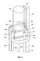
- FIG. 3is a perspective view of an imaging structure according to one embodiment of the present invention.
- FIG. 4is a perspective view of an imaging structure according to one embodiment of the present invention.
- FIG. 5is a side-view of a micro-camera guided suturing device according to one embodiment of the present invention.
- FIG. 6is a side-view of a micro-camera guided suturing device according to one embodiment of the present invention.
- FIG. 7is a side-view of a micro-camera guided suturing device according to one embodiment of the present invention.
- FIG. 8is a side-view of the micro-camera guided suturing device of FIG. 7 according to one embodiment of the present invention with the micro-camera portion of the device removed;
- FIG. 9is a side-view of the micro-camera guided suturing device according to one embodiment of the present invention.
- FIG. 10is a side-view of a micro-camera guided suturing device according to one embodiment of the present invention.
- FIG. 11is a side-view of a micro-camera guided suturing device according to one embodiment of the present invention.
- FIG. 12is a side-view of a micro-camera guided suturing device according to one embodiment of the present invention.
- FIG. 13is a side-view of a micro-camera guided suturing device according to one embodiment of the present invention.
- FIG. 14is a side-view of a micro-camera guided suturing device according to one embodiment of the present invention.
- FIG. 15is a front-view of a suture device according to one embodiment of the present invention.
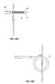
- FIG. 16Ais a side view of a suture device according to one embodiment of the present invention.
- FIG. 16Bis a side view of an emplaced suture according to one embodiment of the present invention.
- FIG. 17Ais a side view of a suture device according to one embodiment of the present invention.
- FIG. 17Bis a side view of an emplaced suture according to one embodiment of the present invention.
- An “SSID,” “solid state imaging device,” “SSID chip,” or “solid state imaging chip” in the exemplary embodimentsgenerally comprises an imaging array or pixel array for gathering image data, and can further comprise conductive pads electrically coupled to the imaging array, which facilitates electrical communication therebetween.
- the SSIDcan comprise a silicon or other semiconductor substrate (e.g., InGaAs) or amorphous silicon thin film transistors (TFT) having features typically manufactured therein.
- TFTamorphous silicon thin film transistors
- featurescan include the imaging array, the conductive pads, metal traces, circuitry, etc.
- Other integrated circuit componentscan also be present for desired applications. However, it is not required that all of these components be present, as long as there is a means of gathering visual or photon data, and a means of sending that data to provide a visual image or image reconstruction.
- an umbilicalcan include the collection of utilities that operate the SSID or the micro-camera as a whole.
- an umbilicalincludes a conductive line, such as electrical wire(s) or other conductors, for providing power, ground, clock signal, and output signal with respect to the SSID, though not all of these are strictly required.
- groundcan be provided by another means than through an electrical wire, e.g., to a camera housing such as micromachined tubing, etc.
- the umbilicalcan also include other utilities such as a light source, temperature sensors, force sensors, fluid irrigation or aspiration members, pressure sensors, fiber optics, microforceps, material retrieval tools, drug delivery devices, radiation emitting devices, laser diodes, electric cauterizers, and electric stimulators, for example.
- Other utilitieswill also be apparent to those skilled in the art and are thus comprehended by this disclosure.
- GRIN lensor “graduated refractive index lens” refers to a specialized lens that has a refractive index that is varied radially from a center optical axis to the outer diameter of the lens.
- such a lenscan be configured in a cylindrical shape, with the optical axis extending from a first flat end to a second flat end.
- a lens of this shapecan simulate the effects of a more traditionally shaped lens.
- FIG. 1illustrates a medical imaging system 10 comprising a micro-catheter 12 having an imaging device disposed at a distal tip 15 of the micro-catheter 12 .
- a processor 22such as an appropriately programmed computer, is provided to control the imaging system 10 and create an image of anatomy adjacent the distal tip portion 15 , within a patient (not shown), displayable on a monitor 24 , and storable in a data storage device 26 .
- An interface 28is provided which supplies power to the imaging device 14 and feeds a digital image signal to the processor based on a signal received from the imaging device via an electrical umbilical, including conductive wires through the micro-catheter 12 .
- a light sourcemay also be provided at the distal end of the micro-catheter 12 .
- the systemfurther includes a fitting 16 enabling an imaging fluid, such as a clear saline solution, to be dispensed to the distal tip portion of the micro-catheter 12 from a reservoir 18 through an elongated tubular member removably attached to the micro-catheter 12 or through a lumen of the microcatheter to displace body fluids as needed to provide a clearer image.
- Fluidsmay be pumped to the distal end of the micro-catheter for other reasons described herein.
- a pump 20is provided, and is manually actuated by a medical practitioner performing a medical imaging procedure, or can be automated and electronically controlled so as to dispense fluid on demand according to control signals from the practitioner, sensors, or according to software commands.
- an imaging device 30is disposed on a distal end of a micro-catheter 12 .
- Micromachined cuts 13are disposed non parallel to a longitudinal direction of the micro-catheter 12 to enable a user, such as a medical practitioner, to guide and steer the distal end of the micro-catheter 12 within a cavity of a patient.
- the micro-cathetermay incorporate structure and principles of operation from a catheter disclosed in U.S. Pat. No. 6,014,919 to Jacobsen et al., which is incorporated herein by reference.
- imaging device 30comprises at least two conductive wires 35 a , 35 b for conducting electronic image data to the data processor 22 and for securing an imaging structure 36 between the at least two conductive wires 35 a , 35 b .
- a plurality of conductive wires 35 a , 35 b , 35 c , 35 dmay be utilized.
- the at least two conductive wires 35 a , 35 bare oriented along a longitudinal axis of the imaging structure 36 and are disposed within alignment apertures 40 of a planar support member 45 .
- the planar support member 45comprises at least two alignment apertures 40 disposed on opposing sides of the planar support member 45 .
- the alignment apertures 40are configured to receive and align the at least two conductive wires 35 a , 35 b along the longitudinal axis of the imaging structure 36 .
- the imaging structure 36is at least partially secured between the at least two conductive wires 35 a , 35 b and is disposed adjacent a top surface of the planar support member 45 .
- the imaging structure 36comprises a GRIN lens 50 optically coupled to a SSID 55 and disposed adjacent the SSID 55 .
- the imaging structurefurther comprises an imaging array 60 disposed on a top surface of the SSID 55 .
- the GRIN lens 50is positioned directly on top of the imaging array 60 of the SSID 55 .
- the at least two conductive wires 35 a , 35 bare operatively coupled to the imaging structure 36 and are configured to align the imaging structure 36 there between.
- the conductive wires 35 a , 35 bare bonded to the imaging structure 36 at contact points 56 disposed on the periphery of a top surface of the SSID 55 .
- the conductive wires 35 a , 35 bare bonded to a side surface of the SSID 55 .
- the alignment apertures 40are oriented perpendicular to the top surface of the planar support member 45 .
- the alignment aperturesmay also be disposed in any orientation which is not non-parallel to the planar support member 45 as required to optimally align the imaging structure 36 as desired.
- the imaging structureis mounted and aligned such that the image plane of the imaging structure 36 is non parallel to a longitudinal axis of the micro-catheter 12 .
- a light sourcee.g., a fiber optic member, LED, etc.
- the imaging structure 30may incorporate structure and principles of operation from an imaging device disclosed in U.S. Pat. No. 7,166,537 to Jacobsen et al., which is incorporated herein by reference.
- the imaging device 30further comprises a lens support member 65 .
- the lens support member 65is bonded to a top surface of the SSID 55 .
- the lens support member 65is bonded to a side surface of the SSID 55 .
- the lens support member 65is bonded to a top surface of the planar support member 40 .
- the lens support member 65is oriented adjacent a side surface of the GRIN lens 50 to minimize movement of the GRIN lens 50 during operation and to ensure proper alignment of the GRIN lens 50 on the imaging array 60 during operation and/or construction of the device.
- a first sleeve member 70is disposed about the imaging structure 36 .
- An adhesiveis disposed within the first sleeve member 70 securing the components of the imaging structure 36 in place as well as securing the first sleeve member 70 to the imaging structure 36 .
- a second sleeve member 75is disposed about the first sleeve member 70 and secured with an adhesive.
- the second sleeve member 75comprises an opaque material to eliminate secondary light from impacting image quality.
- a micro-camera guided suturing device 80having a suture 81 comprising a shape memory material with at least one lumen 82 disposed therein.
- the suturing device 80further comprises an imaging structure 31 disposed within the at least one lumen 82 of the suture 81 , wherein the imaging structure 31 comprises a SSID 55 optically coupled to a lens system 50 .
- the micro-camera guided suturing device 80further comprises an outer catheter 85 which houses the suturing device 80 during advancement of the suturing device through portions of the body (e.g., the vascular tree).
- the imaging structure 31Due to the very small nature of the imaging structure 31 (e.g., less than 0.5 mm), it may advantageously be placed within the lumen 82 of the suture 81 .
- the lumen 82has an internal diameter of approximately 0.5 mm.
- the inner diameter of the lumen 82 of the suture 81may be smaller to accommodate a smaller imaging structure 81 as suits a particular application.
- the suture 81need not be made of a shape memory material as described herein to satisfy each and every application of the certain aspects of the present invention.
- the suture 81comprises a semi-rigid or rigid material which cannot undergo a significant change in shape without suffering plastic deformation.
- a conductive element(not shown) is detachably coupled to the suture 81 .
- the conductive elementacts to transfer energy to the suture to effectuate the shape change of the suture 81 due to the intrinsic properties of the shape memory material.
- the shape memory material of the suture 81comprises a shape memory alloy. Examples of shape memory alloy contemplated for use herein include, without limitation, copper-zinc-aluminum-nickel, copper-aluminum-nickel, or nickel-titanium alloys or any other suitable alloy which changes shape when subjected to varying predetermined temperatures.
- the shape memory material of the suture 81comprises a shape memory polymer such as a oligo(e-caprolactone)diol and crystallisable oligo(p-dioxamone)diol, or any other suitable polymer which assumes a predetermined shape when subject to a predetermined temperature.
- the shape memory materialis bio-absorbable in that it comprises materials which are broken down by the body over time.
- At least one lumen 82extends longitudinally through approximately the entire suture 81 .
- the at least one lumen 82may extend longitudinally through only a portion of the suture 81 as may be desirable for a particular application.
- an image plane of the imaging structure 31is approximately collinear with a distal end of the suture 81 .
- the distal end of the lens systemis disposed near a distal end of the suture to facilitate viewing of the body during emplacement of the suture. In this manner, a medical practitioner may view the area of the body where the suture 81 is being placed and may also view the body during emplacement of the suture to verify proper placement of the suture.
- improper placement of sutures within the bodymay be reduced thereby minimizing unnecessary trauma to a patient.
- a medial portion 91 of the suture 81is detachably coupled to the end portion of the suture 92 .
- the medial portion 91 of the suture 81may be coupled to the remaining portion of the suture by a threaded male member 95 and corresponding threaded female member 96 configured to receive the male member 95 .
- any system or device capable of detachably connecting the medial portion 91 of the suture 81 with the remaining portion of the suture 81is contemplated for use herewith. In this manner, the medial portion 91 of the suture 81 may be emplaced within the patient and the remaining components of the suturing device 80 may be removed from the patient.
- a medical practitionermay guide the suturing device 80 to a particular location within the body of a patient and emplace the suture 81 in a desired location.
- the suture 105may comprise a shape memory alloy which assumes a particular shape when subjected to a predetermined temperature.
- the suture 81may comprise a malleable, deformable material (e.g., an aluminum alloy) which bends in response to forces exerted by the steering movements of the micro-catheter 12 in which the suture 81 is placed.
- a medical practitionermay guide the micro-catheter to a desired location within the patient.
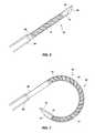
- the distal end of the suture 81may be placed through a portion of tissue near an area that requires suturing. Electrical current may be applied to the suture thereby deforming the suture 81 into a closed configuration (e.g., a loop shape). As the suture 81 deforms into a closed configuration, its distal end pierces a corresponding portion of tissue and closes the desired target area.
- a medical practitionermay view the target tissue being penetrated by the suture 81 in real time thereby ensuring proper placement of the suture 81 . It is important to note that the suture 81 may be preformed in a variety of configurations and may be designed to assume a variety of different shapes depending on a particular application.
- the suture 81may be biased in a hook configuration (not shown) and configured to close more tightly into a loop configuration as shown on FIG. 16B .
- the suture 81may be comprised of a non-shape memory material and be biased in a hook configuration in a rigid state or semi-rigid state as suits a particular application.
- a medical device employing embodiments of the present inventionmay comprise an imaging device disposed within a suture and may also comprise an imaging device at the distal end of the medical device so as to provide multiple views of the emplacement of the suture including an imaging device to provide a perspective view of suture emplacement as well as the imaging device within the suture to provide the “suture-view” of the emplacement.
- a suturing device 80may comprise a plurality of sutures disposed on a distal end of the device 80 configured such that any one of the plurality or all of the plurality of sutures may be placed without the need for advancement of a device through the vasculature of a patient multiple times. That is, in some instances there may be a need to emplace numerous sutures within a patient.
- This embodimentallows a single device to be placed within the vasculature of the patient once while providing the medical practitioner with the ability to place multiple sutures.
- suturing device 80may be used as a stand-alone device or may be used as one tool in connection with an endoscopic device capable of housing multiple tools.
- An exemplary endoscopic toolis illustrated in U.S. patent application Ser. No. 11/292,902 which is incorporated herein by reference.
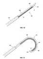
- a micro-camera guided suturing device 100comprising a suture 105 detachably coupled to a micro-catheter 12 is illustrated.
- the micro-catheter 12comprises an imaging structure 31 having an SSID 55 optically coupled to a lens system 50 .
- the micro-camera guided suturing device 100further comprises an outer catheter 110 which houses the suturing device 100 during advancement of the suturing device through portions of the body.
- a distal end 101 of the micro-catheter 12is shaped to approximate the distal end of a needle facilitating penetration into tissues of a patient.
- the suture 105 and micro-catheter 12are each shaped to approximate a cylinder which has been halved down a longitudinal axis of the cylinder. In this manner, when the suture 105 and micro-catheter 12 are joined together they form a whole cylinder.
- the suture 105 and micro-catheter 12each comprise a planar surface and an opposing half-circle surface such that when the two are joined together at their respective planar surfaces, the two form a single cylindrical body.
- the mating components of the suture 105 and micro-catheter 12facilitate easier penetration into tissues of a patient thereby reducing unnecessary trauma to the tissue and the patient.
- the suture 105may comprise a shape memory alloy which assumes a particular shape when subjected to a predetermined temperature.
- the suture 105may comprise a malleable, deformable material (e.g., an aluminum alloy) which bends in response to forces exerted by the steering movements of the micro-catheter 12 to which the suture 105 is attached.
- the suture 105is detachably coupled to a side portion of the micro-catheter 12 .
- the suture 105is detachably coupled to a side portion 102 of the micro-catheter 12 by a temperature sensitive adhesive whereby when the interface between the suture 105 and the micro-catheter 12 is subjected to a certain temperature, the adhesive becomes unstable and no longer secures the suture 105 to the micro-catheter 12 .
- the adhesivemay be designed to react to the temperature within the patient or may be designed to react to heating through an electrical element disposed about the micro-catheter. While use of an adhesive has been specifically described, it is contemplated herein that use of any means capable of detachably coupling and facilitating detachment of the micro-catheter 12 from the suture 105 may be used as is suitable for a particular application.
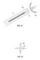
- a micro-camera guided suturing device 150comprising a suture 160 detachably coupled to a micro-catheter 12 is illustrated.
- the micro-catheter 12comprises an imaging structure 31 having an SSID 55 optically coupled to a lens system 50 .
- the micro-camera guided suturing device 150further comprises an outer catheter 165 which houses the suturing device 150 during advancement of the suturing device 150 through portions of the body.
- the suture 160is detachably coupled to a distal end of the imaging structure 31 .
- the suture 160comprises a four-pronged claw having a cavity 162 through a center of the claw.
- the cavity 162is configured to receive and detachably couple to a distal end of the imaging structure 31 .
- the cavity 162has a female threaded configuration adapted to receive and secure a male threaded member corresponding to the distal end of the imaging structure 31 .
- any suitable connection meansis contemplated for use herein.
- a four-pronged clawis illustrated herein, more or less prongs (e.g., 2 to 10) may be used as suits a particular application.
- the cavity 161 disposed within the clawmay be through the center, as described above, or it may be disposed in another portion of the suture 160 as suits a particular application.
- the prongs of the suture 160may be biased in an opened position and restrained in a closed position within outer catheter 165 .
- the suture 160may be biased by a restraining member (e.g., cord or binding) or it may be restrained by the walls of the outer catheter 165 .
- the suture 160may be positioned outside of the outer catheter 165 and opened to its biased position.
- the suture 160may comprise a shape memory material which assumes a predetermined shape based on specific temperature regimes.
- the suture 160is biased such that the prongs are perpendicular to a longitudinal axis of the outer catheter 165 . In this manner, the distal end of the imaging structure 31 may be positioned near the distal end of the outer catheter 165 thereby maximizing image clarity and by default the ability of the medical practitioner to more accurately advance the outer catheter 165 to a desired location within the patient.
- the suture 160may be advanced to a desired location within a patient that requires closing (e.g., a patent foramen ovale). While suture 160 is in an open configuration, the suture may be advanced such that the distal ends of the suture 160 puncture, or are placed against, the target tissue. In an aspect where the suture 160 comprises a shape memory material, an electrical current may be applied to the suture 160 causing the suture to assume a closed configuration. In this manner, the suture 160 may be placed in a target tissue while viewing the placement through or substantially proximate to the suture 160 .
- the suturemay be shaped to approximate the shape of a helix or coil having an imaging structure disposed in the center of the helix and near a proximal end of the helix.
- the helix suturemay be advanced and “threaded” or placed into the tissues of the patient.
- the imaging structureallows the medical practitioner to view through the suture itself while placing the suture within the patient.
- the particular shapes and configurations of sutures referenced hereinare not meant to limit the present invention in any way. Rather, any suture employing an imaging structure which is coupled to the suture is contemplated for use herein.
- a method of placing suture 81 within a patientcomprising providing a suture 81 disposed on a distal end of a catheter 12 , wherein the suture 81 comprises a shape memory material having at least one lumen 30 disposed therein and an imaging structure 31 disposed within the at least one lumen of the suture 81 .
- the imaging structure 31comprises a SSID 55 optically coupled to a lens system 50 .
- the methodfurther comprises advancing a distal end of the catheter 12 into a portion of a patient and viewing a portion of the interior of the patient with the imaging structure 31 .
- the methodfurther comprises emplacing the suture 81 within a portion of the patient.
- suture 81further comprises a conductive element detachably coupled to the suture 81 .
- the methodfurther comprises the step of conducting electrical energy to the suture 81 through the conductive element to deform the suture 81 into a predetermined configuration (e.g., a loop shape).
- the imaging structure 31may thereafter be withdrawn from the suture 81 and reused or discarded leaving the suture 81 within the patient.
- the methodfurther comprises viewing the target tissue through the distal end of the suture 81 as the target tissue is penetrated by the suture 81 .
- a medical practitionermay view the target tissue through, for example, the distal end of the suture 81 as it pierces the target tissue and closes the area which requires suturing.
- the methodallows the practitioner to minimize improper placement of sutures within a patient.
- a method of placing suture 105 within a patientcomprising providing a suture 105 disposed on a catheter 12 , wherein the suture 105 comprises a shape memory material and an imaging structure 31 detachably coupled to the suture.
- the imaging structure 31comprises a SSID 55 optically coupled to a lens system 50 .
- the methodfurther comprises advancing a distal end of the catheter 12 into a portion of a patient, viewing a portion of the interior of the patient with the imaging structure 31 , and emplacing the suture 105 within a portion of the patient while viewing emplacement of the suture 105 with the imaging structure 31 .
Landscapes
- Health & Medical Sciences (AREA)
- Life Sciences & Earth Sciences (AREA)
- Surgery (AREA)
- Nuclear Medicine, Radiotherapy & Molecular Imaging (AREA)
- Molecular Biology (AREA)
- Veterinary Medicine (AREA)
- Public Health (AREA)
- General Health & Medical Sciences (AREA)
- Animal Behavior & Ethology (AREA)
- Engineering & Computer Science (AREA)
- Biomedical Technology (AREA)
- Heart & Thoracic Surgery (AREA)
- Medical Informatics (AREA)
- Physics & Mathematics (AREA)
- Biophysics (AREA)
- Radiology & Medical Imaging (AREA)
- Pathology (AREA)
- Optics & Photonics (AREA)
- Endoscopes (AREA)
Abstract
Description
Claims (19)
Priority Applications (1)
| Application Number | Priority Date | Filing Date | Title |
|---|---|---|---|
| US12/938,672US8828028B2 (en) | 2009-11-03 | 2010-11-03 | Suture device and method for closing a planar opening |
Applications Claiming Priority (2)
| Application Number | Priority Date | Filing Date | Title |
|---|---|---|---|
| US25774609P | 2009-11-03 | 2009-11-03 | |
| US12/938,672US8828028B2 (en) | 2009-11-03 | 2010-11-03 | Suture device and method for closing a planar opening |
Publications (2)
| Publication Number | Publication Date |
|---|---|
| US20110270277A1 US20110270277A1 (en) | 2011-11-03 |
| US8828028B2true US8828028B2 (en) | 2014-09-09 |
Family
ID=44858856
Family Applications (1)
| Application Number | Title | Priority Date | Filing Date |
|---|---|---|---|
| US12/938,672Active2032-08-11US8828028B2 (en) | 2009-11-03 | 2010-11-03 | Suture device and method for closing a planar opening |
Country Status (1)
| Country | Link |
|---|---|
| US (1) | US8828028B2 (en) |
Cited By (1)
| Publication number | Priority date | Publication date | Assignee | Title |
|---|---|---|---|---|
| US20160287141A1 (en)* | 2014-03-02 | 2016-10-06 | V.T.M. (Virtual Tape Measure) Technologies Ltd. | Endoscopic measurement system and method |
Families Citing this family (2)
| Publication number | Priority date | Publication date | Assignee | Title |
|---|---|---|---|---|
| WO2010014792A2 (en) | 2008-07-30 | 2010-02-04 | Sterling Lc | Method and device for incremental wavelength variation to analyze tissue |
| CH705951B1 (en)* | 2011-12-23 | 2017-12-15 | Awaiba Consultadoria Desenvolvimento E Comércio De Componentes Microelectrónicos Unipessoal Lda | Optical sensor assembly and method of making and using same. |
Citations (384)
| Publication number | Priority date | Publication date | Assignee | Title |
|---|---|---|---|---|
| US3817635A (en) | 1967-08-08 | 1974-06-18 | Olumpus Co Ltd | Device for measuring the actual dimension of an object at the forward end portion of an endoscope |
| US3856000A (en) | 1972-06-19 | 1974-12-24 | Machido Seisakusho Kk | Endoscope |
| US3886933A (en) | 1973-10-10 | 1975-06-03 | Olympus Optical Co | Ureteral catheter device |
| US3918438A (en) | 1973-04-16 | 1975-11-11 | Olympus Optical Co | Endoscope capable of changing over the directions of visual field |
| US3971065A (en) | 1975-03-05 | 1976-07-20 | Eastman Kodak Company | Color imaging array |
| US4277168A (en) | 1978-04-20 | 1981-07-07 | Machida Endoscope Co., Ltd. | Endoscope with means for detecting rotation of a distal examining end |
| US4283115A (en) | 1978-06-28 | 1981-08-11 | Richard Wolf Gmbh | Beam splitters for endoscopes comprising a dual observation system |
| US4349456A (en) | 1976-04-22 | 1982-09-14 | Minnesota Mining And Manufacturing Company | Non-vitreous ceramic metal oxide microcapsules and process for making same |
| US4360275A (en) | 1980-08-11 | 1982-11-23 | Litton Systems Inc. | Device for measurement of optical scattering |
| US4403985A (en) | 1981-05-12 | 1983-09-13 | The United States Of America As Represented By The Department Of Health And Human Services | Jet controlled catheter |
| US4475902A (en) | 1981-03-24 | 1984-10-09 | Werner Schubert | Device for introducing medical instruments into a body |
| US4487206A (en) | 1982-10-13 | 1984-12-11 | Honeywell Inc. | Fiber optic pressure sensor with temperature compensation and reference |
| US4491865A (en) | 1982-09-29 | 1985-01-01 | Welch Allyn, Inc. | Image sensor assembly |
| US4515444A (en) | 1983-06-30 | 1985-05-07 | Dyonics, Inc. | Optical system |
| US4573450A (en) | 1983-11-11 | 1986-03-04 | Fuji Photo Optical Co., Ltd. | Endoscope |
| US4585349A (en) | 1983-09-12 | 1986-04-29 | Battelle Memorial Institute | Method of and apparatus for determining the position of a device relative to a reference |
| US4588294A (en) | 1984-06-27 | 1986-05-13 | Warner-Lambert Technologies, Inc. | Searching and measuring endoscope |
| US4589404A (en) | 1984-01-03 | 1986-05-20 | Medical Dynamics, Inc. | Laser endoscope |
| US4593313A (en) | 1983-09-05 | 1986-06-03 | Olympus Optical Co., Ltd. | Endoscope |
| US4594613A (en) | 1982-02-16 | 1986-06-10 | Canon Kabushiki Kaisha | Solid-state imaging device assembly |
| US4600831A (en) | 1982-12-07 | 1986-07-15 | The Secretary Of State For Defence In Her Britannic Majesty's Government Of The United Kingdom Of Great Britain And Northern Ireland | Apparatus to focus light on a surface based on color |
| US4604992A (en) | 1983-12-27 | 1986-08-12 | Olympus Optical Company, Ltd. | Endoscope system |
| US4620534A (en) | 1984-11-01 | 1986-11-04 | New Mexico State University Foundation | Apparatus for insertion of an intravaginal article |
| US4621284A (en) | 1984-06-09 | 1986-11-04 | Olympus Optical Co., Ltd. | Measuring endoscope |
| US4622954A (en) | 1984-05-15 | 1986-11-18 | Fuji Photo Optical Co., Ltd. | Endoscope having a plate-like image sensor for forming images |
| US4626079A (en) | 1984-04-13 | 1986-12-02 | Nippon Kogaku K.K. | Dark field illumination apparatus for epi-illumination system |
| US4641927A (en) | 1982-03-24 | 1987-02-10 | Dyonics, Inc. | Chromatic aberration corrected gradient index lens system |
| US4646724A (en) | 1982-10-15 | 1987-03-03 | Olympus Optical Co., Ltd. | Endoscopic photographing apparatus |
| US4672218A (en) | 1984-12-04 | 1987-06-09 | The Dow Chemical Company | Method for determining the onset of crystallization |
| US4706118A (en) | 1985-10-09 | 1987-11-10 | Olympus Optical Co., Ltd. | Control circuit for video endoscope |
| US4707134A (en) | 1984-12-04 | 1987-11-17 | The Dow Chemical Company | Fiber optic probe |
| US4723843A (en) | 1985-07-31 | 1988-02-09 | Richard Wolf Gmbh | Endoscope optical system |
| US4725721A (en) | 1984-08-17 | 1988-02-16 | Hitachi, Ltd. | Autofocusing control system |
| US4745471A (en) | 1986-05-13 | 1988-05-17 | Olympus Optical Co., Ltd. | Solid-state imaging apparatus and endoscope |
| US4745470A (en) | 1986-04-04 | 1988-05-17 | Olympus Optical Co., Ltd. | Endoscope using a chip carrier type solid state imaging device |
| US4783591A (en) | 1987-11-09 | 1988-11-08 | Honeywell Inc. | Color mark sensor |
| US4785815A (en) | 1985-10-23 | 1988-11-22 | Cordis Corporation | Apparatus for locating and ablating cardiac conduction pathways |
| US4790624A (en) | 1986-10-31 | 1988-12-13 | Identechs Corporation | Method and apparatus for spatially orienting movable members using shape memory effect alloy actuator |
| US4791479A (en) | 1986-06-04 | 1988-12-13 | Olympus Optical Co., Ltd. | Color-image sensing apparatus |
| US4802487A (en) | 1987-03-26 | 1989-02-07 | Washington Research Foundation | Endoscopically deliverable ultrasound imaging system |
| US4803562A (en) | 1986-06-20 | 1989-02-07 | Olympus Optical Co., Ltd. | Image sensing apparatus |
| US4832003A (en) | 1986-09-12 | 1989-05-23 | Olympus Optical Co., Ltd. | Electronic endoscope tip |
| US4843416A (en) | 1988-03-02 | 1989-06-27 | W. Haking Enterprises Limited | Autofocus camera system |
| US4846785A (en) | 1987-01-22 | 1989-07-11 | Robert Cassou | Instrument for artificial insemination, embryo transfer or sampling follicular liquids in mammals |
| US4859040A (en) | 1985-12-27 | 1989-08-22 | Canon Kabushiki Kaisha | Optical system having gradient-index lens and method for correcting aberrations |
| US4867137A (en) | 1987-03-19 | 1989-09-19 | Olympus Optical Co., Ltd. | Electronic endoscope |
| US4867174A (en) | 1987-11-18 | 1989-09-19 | Baxter Travenol Laboratories, Inc. | Guidewire for medical use |
| US4867138A (en) | 1987-05-13 | 1989-09-19 | Olympus Optical Co., Ltd. | Rigid electronic endoscope |
| US4880298A (en) | 1985-08-07 | 1989-11-14 | Olympus Optical Co., Ltd. | Microscope objective |
| US4895138A (en) | 1985-01-14 | 1990-01-23 | Olympus Optical Co., Ltd. | Endoscope with a detachable observation unit at its distal end |
| US4916534A (en) | 1987-04-28 | 1990-04-10 | Olympus Optical Co., Ltd. | Endoscope |
| US4926257A (en) | 1986-12-19 | 1990-05-15 | Olympus Optical Co., Ltd. | Stereoscopic electronic endoscope device |
| US4930880A (en) | 1988-10-11 | 1990-06-05 | Olympus Optical Co., Ltd. | Graded refractive index lens |
| US4932394A (en) | 1987-08-10 | 1990-06-12 | Kabushiki Kaisha Toshiba | Endoscope including scope terminal locking indicator |
| US4934340A (en) | 1989-06-08 | 1990-06-19 | Hemo Laser Corporation | Device for guiding medical catheters and scopes |
| US4941457A (en) | 1989-08-17 | 1990-07-17 | Olympus Optical Co., Ltd. | Endoscope using an optical guide twisted on the tip side to have the visual field direction and curvature axis coincide with each other |
| US4998807A (en) | 1988-08-23 | 1991-03-12 | Olympus Optical Co., Ltd. | Variable focal length lens system |
| US5006928A (en) | 1988-12-05 | 1991-04-09 | Fuji Photo Film Co., Ltd. | Image processing method in an electronic video endoscopy system |
| US5009483A (en) | 1989-04-12 | 1991-04-23 | Rockwell Iii Marshall A | Optical waveguide display system |
| US5021888A (en) | 1987-12-18 | 1991-06-04 | Kabushiki Kaisha Toshiba | Miniaturized solid state imaging device |
| US5022972A (en) | 1986-10-28 | 1991-06-11 | MTA Kutatasi Eszkozoket Kivitelezo Vallata | Process for measuring and determining zeta-potential in a laminarly flowing medium for practical purposes |
| US5032913A (en) | 1989-02-28 | 1991-07-16 | Olympus Optical Co., Ltd. | Electronic endoscope system equipped with color smear reducing means |
| US5040069A (en) | 1989-06-16 | 1991-08-13 | Fuji Photo Optical Co., Ltd. | Electronic endoscope with a mask bump bonded to an image pick-up device |
| US5061036A (en) | 1990-04-17 | 1991-10-29 | Photon Imaging Corp. | Color page scanner using fiber optic bundle and a photosensor array |
| US5093719A (en) | 1989-10-23 | 1992-03-03 | Manx Optical Corporation | Endoscopic gradient index optical systems |
| US5105269A (en) | 1986-11-29 | 1992-04-14 | Olympus Optical Co., Ltd. | Imaging apparatus and endoscope apparatus with selectable wavelength ranges |
| US5106387A (en) | 1985-03-22 | 1992-04-21 | Massachusetts Institute Of Technology | Method for spectroscopic diagnosis of tissue |
| US5109859A (en) | 1989-10-04 | 1992-05-05 | Beth Israel Hospital Association | Ultrasound guided laser angioplasty |
| US5113254A (en) | 1989-04-06 | 1992-05-12 | Olympus Optical Co., Ltd. | Electronic endoscope apparatus outputting ternary drive signal |
| US5111804A (en) | 1989-02-15 | 1992-05-12 | Kabushiki Kaisha Toshiba | Electronic endoscope |
| US5121213A (en) | 1987-02-25 | 1992-06-09 | Olympus Optical Co., Ltd. | Imaging system having a blurring optical element for minimizing moire phenomenon |
| US5126369A (en) | 1991-01-18 | 1992-06-30 | International Flavors & Fragrances Inc. | Use of lyrame® for repelling insects |
| US5126639A (en) | 1991-06-04 | 1992-06-30 | Zenith Electronics Corporation | Sequential scan system changes for multiple frequency range oscillator and control |
| US5130804A (en) | 1990-01-09 | 1992-07-14 | Konica Corporation | Compact recording apparatus with functional components mounted on a substrate |
| US5165063A (en) | 1987-06-26 | 1992-11-17 | Battelle-Institut E.V. | Device for measuring distances using an optical element of large chromatic aberration |
| US5166656A (en) | 1992-02-28 | 1992-11-24 | Avx Corporation | Thin film surface mount fuses |
| US5182672A (en) | 1990-07-17 | 1993-01-26 | Minolta Camera Co., Ltd. | Finder optical system |
| US5188093A (en) | 1991-02-04 | 1993-02-23 | Citation Medical Corporation | Portable arthroscope with periscope optics |
| US5190523A (en) | 1991-08-16 | 1993-03-02 | Idee International R & D Inc. | Disposable syringe and injector |
| US5191203A (en) | 1991-04-18 | 1993-03-02 | Mckinley Optics, Inc. | Stereo video endoscope objective lens system |
| US5198894A (en) | 1991-09-24 | 1993-03-30 | Hicks John W | Drape for endoscope |
| US5209219A (en) | 1991-03-18 | 1993-05-11 | Laser Medical Research Foundation | Endoscope adaptor |
| US5220198A (en) | 1990-08-27 | 1993-06-15 | Olympus Optical Co., Ltd. | Solid state imaging apparatus in which a solid state imaging device chip and substrate are face-bonded with each other |
| US5222477A (en) | 1991-09-30 | 1993-06-29 | Welch Allyn, Inc. | Endoscope or borescope stereo viewing system |
| US5228430A (en) | 1989-08-04 | 1993-07-20 | Kabushiki Kaisha Toshiba | Electronic endoscope apparatus including easy focusing distal end |
| US5258834A (en) | 1991-02-13 | 1993-11-02 | Olympus Optical Co., Ltd. | Electronic endoscope for producing a color image by extracting a plurality of field picture images in one field period without changing a horizontal clock rate |
| US5289434A (en) | 1992-09-18 | 1994-02-22 | Shell Oil Company | Retroreflector apparatus for remote seismic sensing |
| US5290555A (en) | 1989-09-14 | 1994-03-01 | Revlon Consumer Products Corporation | Cosmetic compositions with structural color |
| US5291010A (en) | 1990-10-04 | 1994-03-01 | Olympus Optical Co., Ltd. | Solid state imaging device having a chambered imaging chip corner |
| US5298741A (en) | 1993-01-13 | 1994-03-29 | Trustees Of Tufts College | Thin film fiber optic sensor array and apparatus for concurrent viewing and chemical sensing of a sample |
| US5304173A (en) | 1985-03-22 | 1994-04-19 | Massachusetts Institute Of Technology | Spectral diagonostic and treatment system |
| US5305098A (en) | 1991-04-11 | 1994-04-19 | Olympus Optical Co., Ltd. | Endoscope image processing system with means for discriminating between endoscope image area and character image area |
| US5361166A (en) | 1993-01-28 | 1994-11-01 | Gradient Lens Corporation | Negative abbe number radial gradient index relay and use of same |
| US5365268A (en) | 1991-04-26 | 1994-11-15 | Fuji Photo Optical Co., Ltd. | Circuit board of solid-state image sensor for electronic endoscope |
| US5377047A (en) | 1992-04-13 | 1994-12-27 | Linvatec Corporation | Disposable endoscope employing positive and negative gradient index of refraction optical materials |
| US5376960A (en) | 1991-09-10 | 1994-12-27 | Richard Wolf Gmbh | Video endoscope with solid-state imaging device |
| US5381784A (en) | 1992-09-30 | 1995-01-17 | Adair; Edwin L. | Stereoscopic endoscope |
| EP0639043A1 (en) | 1993-08-10 | 1995-02-15 | Siemens Nixdorf Informationssysteme AG | Process for manufacturing plated through-hole printed circuit boards having very small solder lands |
| US5396366A (en) | 1993-03-04 | 1995-03-07 | Sigma Dynamics Corporation | Endoscope apparatus |
| US5398685A (en) | 1992-01-10 | 1995-03-21 | Wilk; Peter J. | Endoscopic diagnostic system and associated method |
| US5402769A (en) | 1992-04-23 | 1995-04-04 | Olympus Optical Co., Ltd. | Endoscope apparatus which time-sequentially transmits sensor signals with image signals during a blanking period |
| US5408999A (en) | 1992-10-23 | 1995-04-25 | Optex Biomedical, Inc. | Fiber-optic probe for the measurement of fluid parameters |
| US5430475A (en) | 1990-06-29 | 1995-07-04 | Olympus Optical Co., Ltd. | Electronic endoscope apparatus having micro array on photoelectric conversion surface |
| US5434615A (en) | 1992-09-25 | 1995-07-18 | Fuji Photo Optical Co., Ltd. | Signal processing circuit adaptable to electronic endoscopes having different lengths |
| US5436655A (en) | 1991-08-09 | 1995-07-25 | Olympus Optical Co., Ltd. | Endoscope apparatus for three dimensional measurement for scanning spot light to execute three dimensional measurement |
| US5438975A (en) | 1993-03-24 | 1995-08-08 | Machida Endoscope Co., Ltd. | Distal tip of endoscope having spirally coiled control wires |
| US5440669A (en) | 1991-07-26 | 1995-08-08 | Accuwave Corporation | Photorefractive systems and methods |
| US5455455A (en) | 1992-09-14 | 1995-10-03 | Badehi; Peirre | Methods for producing packaged integrated circuit devices and packaged integrated circuit devices produced thereby |
| US5459570A (en) | 1991-04-29 | 1995-10-17 | Massachusetts Institute Of Technology | Method and apparatus for performing optical measurements |
| US5458612A (en) | 1994-01-06 | 1995-10-17 | Origin Medsystems, Inc. | Prostatic ablation method and apparatus for perineal approach |
| EP0681809A1 (en) | 1994-05-09 | 1995-11-15 | Welch Allyn, Inc. | Stereo imaging assembly for endoscopic probe |
| US5469841A (en) | 1992-10-29 | 1995-11-28 | Olympus Optical Co., Ltd. | Endoscope apparatus provided with liquid removing mechanism for the electric connector |
| US5512940A (en) | 1993-03-19 | 1996-04-30 | Olympus Optical Co., Ltd. | Image processing apparatus, endoscope image sensing and processing apparatus, and image processing method for performing different displays depending upon subject quantity |
| US5517997A (en) | 1994-09-15 | 1996-05-21 | Gabriel Medical, Inc. | Transillumination of body members for protection during body invasive procedures |
| US5531664A (en) | 1990-12-26 | 1996-07-02 | Olympus Optical Co., Ltd. | Bending actuator having a coil sheath with a fixed distal end and a free proximal end |
| US5547455A (en) | 1994-03-30 | 1996-08-20 | Medical Media Systems | Electronically steerable endoscope |
| US5594497A (en) | 1993-04-07 | 1997-01-14 | Ahern; John M. | Endoscope provided with a distally located color CCD |
| US5603687A (en) | 1992-10-28 | 1997-02-18 | Oktas General Partnership | Asymmetric stereo-optic endoscope |
| US5607435A (en)* | 1994-05-23 | 1997-03-04 | Memory Medical Systems, Inc. | Instrument for endoscopic-type procedures |
| US5621574A (en) | 1995-03-29 | 1997-04-15 | Nikon Corporation | Objective lens system utilizing axial gradient index (grin) lens elements |
| US5630788A (en) | 1994-08-12 | 1997-05-20 | Imagyn Medical, Inc. | Endoscope with curved end image guide |
| US5647368A (en) | 1996-02-28 | 1997-07-15 | Xillix Technologies Corp. | Imaging system for detecting diseased tissue using native fluorsecence in the gastrointestinal and respiratory tract |
| US5662621A (en) | 1995-07-06 | 1997-09-02 | Scimed Life Systems, Inc. | Guide catheter with shape memory retention |
| US5673083A (en) | 1989-03-17 | 1997-09-30 | Hitachi, Ltd. | Semiconductor device and video camera unit having the same and method for manufacturing the same |
| US5685311A (en) | 1994-10-20 | 1997-11-11 | Olympus Optical Company, Ltd. | Image display system |
| US5693043A (en) | 1985-03-22 | 1997-12-02 | Massachusetts Institute Of Technology | Catheter for laser angiosurgery |
| US5704892A (en) | 1992-09-01 | 1998-01-06 | Adair; Edwin L. | Endoscope with reusable core and disposable sheath with passageways |
| US5716759A (en) | 1993-09-02 | 1998-02-10 | Shellcase Ltd. | Method and apparatus for producing integrated circuit devices |
| US5716323A (en) | 1995-04-05 | 1998-02-10 | Karl Storz Imaging | Electrical isolation of endoscopic video camera |
| US5722403A (en) | 1996-10-28 | 1998-03-03 | Ep Technologies, Inc. | Systems and methods using a porous electrode for ablating and visualizing interior tissue regions |
| US5732150A (en) | 1995-09-19 | 1998-03-24 | Ihc Health Services, Inc. | Method and system for multiple wavelength microscopy image analysis |
| DE19742973A1 (en) | 1996-09-30 | 1998-04-16 | Fuji Photo Optical Co Ltd | Optical system for electronic endoscope |
| US5740808A (en) | 1996-10-28 | 1998-04-21 | Ep Technologies, Inc | Systems and methods for guilding diagnostic or therapeutic devices in interior tissue regions |
| EP0550995B1 (en) | 1991-12-18 | 1998-04-29 | Hamamatsu Photonics K.K. | Triple view imaging apparatus |
| US5751340A (en) | 1996-08-21 | 1998-05-12 | Karl Storz Gmbh & Co. | Method and apparatus for reducing the inherently dark grid pattern from the video display of images from fiber optic bundles |
| US5749827A (en) | 1995-03-07 | 1998-05-12 | Fuji Photo Optical Co., Ltd. | Objective optical member with air gap for endoscope imaging unit |
| US5752518A (en) | 1996-10-28 | 1998-05-19 | Ep Technologies, Inc. | Systems and methods for visualizing interior regions of the body |
| US5769792A (en) | 1991-07-03 | 1998-06-23 | Xillix Technologies Corp. | Endoscopic imaging system for diseased tissue |
| US5772597A (en) | 1992-09-14 | 1998-06-30 | Sextant Medical Corporation | Surgical tool end effector |
| US5776049A (en) | 1992-12-24 | 1998-07-07 | Olympus Optical Co., Ltd. | Stereo endoscope and stereo endoscope imaging apparatus |
| US5784098A (en) | 1995-08-28 | 1998-07-21 | Olympus Optical Co., Ltd. | Apparatus for measuring three-dimensional configurations |
| US5783829A (en) | 1995-11-06 | 1998-07-21 | The University Of Virginia | Energy and position sensitive radiation detectors |
| US5792984A (en) | 1996-07-01 | 1998-08-11 | Cts Corporation | Molded aluminum nitride packages |
| US5808665A (en) | 1992-01-21 | 1998-09-15 | Sri International | Endoscopic surgical instrument and method for use |
| US5818644A (en) | 1995-11-02 | 1998-10-06 | Olympus Optical Co., Ltd. | Gradient index optical element and method for making the same |
| US5827531A (en) | 1994-12-02 | 1998-10-27 | The United States Of America As Represented By The Administrator Of The National Aeronautics And Space Administration | Microcapsules and methods for making |
| US5840017A (en) | 1995-08-03 | 1998-11-24 | Asahi Kogaku Kogyo Kabushiki Kaisha | Endoscope system |
| US5846185A (en) | 1996-09-17 | 1998-12-08 | Carollo; Jerome T. | High resolution, wide field of view endoscopic viewing system |
| US5848969A (en) | 1996-10-28 | 1998-12-15 | Ep Technologies, Inc. | Systems and methods for visualizing interior tissue regions using expandable imaging structures |
| US5865729A (en) | 1997-10-10 | 1999-02-02 | Olympus America, Inc. | Apparatus for facilitating gynecological examinations and procedures |
| US5870229A (en) | 1995-08-04 | 1999-02-09 | Olympus Optical Co., Ltd. | Gradient index lens component and image pickup apparatus using the gradient index lens component |
| US5873816A (en) | 1994-11-02 | 1999-02-23 | Olympus Optical Co., Ltd. | Electronic endoscope having an insertional portion a part of which is a conductive armor |
| US5879285A (en) | 1995-09-28 | 1999-03-09 | Olympus Optical Co., Ltd. | Aligning means attaching a cable in an imaging apparatus |
| EP0482997B1 (en) | 1990-10-23 | 1999-04-21 | Sony Corporation | Lens barrel having reference shafts for movably supporting lenses |
| US5904651A (en) | 1996-10-28 | 1999-05-18 | Ep Technologies, Inc. | Systems and methods for visualizing tissue during diagnostic or therapeutic procedures |
| US5908445A (en) | 1996-10-28 | 1999-06-01 | Ep Technologies, Inc. | Systems for visualizing interior tissue regions including an actuator to move imaging element |
| US5913817A (en) | 1995-04-05 | 1999-06-22 | Karl Storz Imaging | Electrical isolation of endoscopic video camera |
| US5916155A (en) | 1996-07-30 | 1999-06-29 | Nellcor Puritan Bennett Incorporated | Fetal sensor with securing balloons remote from optics |
| US5929900A (en) | 1996-11-14 | 1999-07-27 | Fuji Photo Optical Co., Ltd. | Signal processor circuit for endoscope systems of all-pixels readout type |
| US5940126A (en) | 1994-10-25 | 1999-08-17 | Kabushiki Kaisha Toshiba | Multiple image video camera apparatus |
| US5947894A (en) | 1997-11-21 | 1999-09-07 | Endolap, Inc. | Disposable endoscope shield and method |
| US5951462A (en) | 1997-12-11 | 1999-09-14 | Fuji Photo Optical Co., Ltd. | Electronic endoscope system for displaying unconnected scope |
| US5957849A (en) | 1997-06-30 | 1999-09-28 | The Regents Of The University Of California | Endoluminal ultrasound-guided resectoscope |
| US5971915A (en) | 1997-06-13 | 1999-10-26 | Fuji Photo Optical Co., Ltd. | Stereoscopic endoscope |
| US5973779A (en) | 1996-03-29 | 1999-10-26 | Ansari; Rafat R. | Fiber-optic imaging probe |
| US5980663A (en) | 1995-05-15 | 1999-11-09 | Shellcase Ltd. | Bonding machine |
| US5989185A (en) | 1994-11-25 | 1999-11-23 | Olympus Optical Co., Ltd. | Endoscope apparatus |
| US5998878A (en) | 1995-07-13 | 1999-12-07 | Eastman Kodak Company | Image sensor assembly and packaging method |
| US5999327A (en) | 1995-09-12 | 1999-12-07 | Olympus Optical Co., Ltd. | Objective lens system |
| US6008123A (en) | 1997-11-04 | 1999-12-28 | Lucent Technologies Inc. | Method for using a hardmask to form an opening in a semiconductor substrate |
| US6014919A (en) | 1996-09-16 | 2000-01-18 | Precision Vascular Systems, Inc. | Method and apparatus for forming cuts in catheters, guidewires, and the like |
| US6022758A (en) | 1994-07-10 | 2000-02-08 | Shellcase Ltd. | Process for manufacturing solder leads on a semiconductor device package |
| US6040235A (en) | 1994-01-17 | 2000-03-21 | Shellcase Ltd. | Methods and apparatus for producing integrated circuit devices |
| US6059760A (en) | 1997-08-14 | 2000-05-09 | Medtronic, Inc. | Cannula having reverse flow tip |
| US6095970A (en) | 1997-02-19 | 2000-08-01 | Asahi Kogaku Kogyo Kabushiki Kaisha | Endoscope |
| US6118476A (en) | 1998-04-21 | 2000-09-12 | Moritex Corporation | CCD Microscope |
| US6117707A (en) | 1994-07-13 | 2000-09-12 | Shellcase Ltd. | Methods of producing integrated circuit devices |
| US6133637A (en) | 1997-01-24 | 2000-10-17 | Rohm Co., Ltd. | Semiconductor device having a plurality of semiconductor chips |
| US6134003A (en) | 1991-04-29 | 2000-10-17 | Massachusetts Institute Of Technology | Method and apparatus for performing optical measurements using a fiber optic imaging guidewire, catheter or endoscope |
| US6139489A (en) | 1999-10-05 | 2000-10-31 | Ethicon Endo-Surgery, Inc. | Surgical device with integrally mounted image sensor |
| US6139819A (en) | 1995-06-07 | 2000-10-31 | Imarx Pharmaceutical Corp. | Targeted contrast agents for diagnostic and therapeutic use |
| US6142930A (en) | 1997-01-13 | 2000-11-07 | Asahi Kogaku Kogyo Kabushiki Kaisha | Electronic endoscope having compact construction |
| US6161035A (en) | 1997-04-30 | 2000-12-12 | Asahi Kogaku Kogyo Kabushiki Kaisha | Fluorescence diagnostic apparatus |
| US6193908B1 (en) | 1997-02-24 | 2001-02-27 | Superior Micropowders Llc | Electroluminescent phosphor powders, methods for making phosphor powders and devices incorporating same |
| DE19859434C2 (en) | 1998-12-22 | 2001-03-08 | Bruker Optik Gmbh | IR spectroscopic endoscope with inflatable balloon |
| US6211955B1 (en) | 2000-01-24 | 2001-04-03 | Amnis Corporation | Imaging and analyzing parameters of small moving objects such as cells |
| US6224969B1 (en) | 1997-09-01 | 2001-05-01 | Stichting Voor De Technische Wetenschappen | Optical phantom suitable for stimulating the optical properties of biological material and a method of producing said phantom |
| EP1104182A1 (en) | 1999-11-27 | 2001-05-30 | STMicroelectronics Limited | Improved image sensor devices for Incorporation into endoscopes |
| US20010007051A1 (en) | 1999-12-03 | 2001-07-05 | Asahi Kogaku Kogyo Kabushiki Kaisha | Electronic endoscope |
| US20010007511A1 (en) | 2000-01-12 | 2001-07-12 | Itsuji Minami | Endoscope objective lens |
| US6262855B1 (en) | 1998-11-23 | 2001-07-17 | Seh America | Infrared laser beam viewing apparatus |
| US6271206B1 (en) | 1996-09-12 | 2001-08-07 | Valentis, Inc. | Sonic nebulized nucleic acid/cationic liposome complexes and methods for pulmonary gene delivery |
| US20010012053A1 (en) | 1995-05-24 | 2001-08-09 | Olympus Optical Co., Ltd. | Stereoscopic endoscope system and tv imaging system for endoscope |
| US6280960B1 (en) | 1997-06-13 | 2001-08-28 | Robert Carr | Optical detection and analysis of sub-micron particles |
| US6288172B1 (en) | 1995-06-26 | 2001-09-11 | 3M Innovative Properties Company | Light diffusing adhesive |
| US20010024848A1 (en) | 2000-03-22 | 2001-09-27 | Masao Nakamura | Solid-state imaging device and manufacturing method thereof |
| US6319745B1 (en) | 2000-05-31 | 2001-11-20 | International Business Machines Corporation | Formation of charge-coupled-device with image pick-up array |
| US6322498B1 (en) | 1996-10-04 | 2001-11-27 | University Of Florida | Imaging scope |
| US6327096B1 (en) | 1997-04-30 | 2001-12-04 | Olympus Optical Co., Ltd. | Set of lens system |
| US20010049509A1 (en) | 2000-02-29 | 2001-12-06 | Olympus Optical Co., Ltd. | Endoscopic treatment system |
| US20020007110A1 (en) | 1992-11-12 | 2002-01-17 | Ing. Klaus Irion | Endoscope, in particular, having stereo-lateral-view optics |
| US6352503B1 (en) | 1998-07-17 | 2002-03-05 | Olympus Optical Co., Ltd. | Endoscopic surgery apparatus |
| US20020034537A1 (en) | 2000-05-03 | 2002-03-21 | Brita Schulze | Cationic diagnostic, imaging and therapeutic agents associated with activated vascular sites |
| US6361491B1 (en) | 1998-12-15 | 2002-03-26 | Olympus Optical Co., Ltd. | Optical adaptor for highy precision endoscope |
| US6366726B1 (en) | 1995-11-20 | 2002-04-02 | Cirrex Corp. | Fiber optic probes for indwelling investigations |
| US20020039594A1 (en) | 1997-05-13 | 2002-04-04 | Evan C. Unger | Solid porous matrices and methods of making and using the same |
| EP1195130A2 (en) | 2000-10-06 | 2002-04-10 | Machida Endoscope Co., Ltd | Endoscope apparatus |
| US6375635B1 (en) | 1999-05-18 | 2002-04-23 | Hydrocision, Inc. | Fluid jet surgical instruments |
| US6384397B1 (en) | 2000-05-10 | 2002-05-07 | National Semiconductor Corporation | Low cost die sized module for imaging application having a lens housing assembly |
| US6384884B1 (en) | 1998-03-30 | 2002-05-07 | Kabushiki Kaisha Toshiba | Reflecting type liquid crystal display device |
| US6396116B1 (en) | 2000-02-25 | 2002-05-28 | Agilent Technologies, Inc. | Integrated circuit packaging for optical sensor devices |
| US6407768B1 (en) | 1999-06-23 | 2002-06-18 | Seiji Ishikawa | Video microscope |
| US20020080248A1 (en) | 1997-11-24 | 2002-06-27 | Adair Edwin L. | Reduced area imaging devices utilizing selected charge integration periods |
| US20020111534A1 (en) | 2000-07-24 | 2002-08-15 | Takayuki Suzuki | Endoscope and endoscopic instrument and method using same |
| US20020109774A1 (en) | 2001-01-16 | 2002-08-15 | Gavriel Meron | System and method for wide field imaging of body lumens |
| US6445939B1 (en) | 1999-08-09 | 2002-09-03 | Lightlab Imaging, Llc | Ultra-small optical probes, imaging optics, and methods for using same |
| US6456423B1 (en) | 1999-10-22 | 2002-09-24 | The Board Of Trustees Of The University Of Illinois | Silicon nanoparticle microcrystal nonlinear optical devices |
| US6471636B1 (en) | 1994-09-21 | 2002-10-29 | Asahi Kogaku Kogyo Kabushiki Kaisha | Fluorescence diagnosis endoscope system |
| US20020166949A1 (en) | 2001-04-19 | 2002-11-14 | Satoshi Machida | Photoelectic converter |
| US20020166946A1 (en) | 2001-03-12 | 2002-11-14 | Olympus Optical Co., Ltd. | Optical scanning probe device using low coherence light |
| US20020168776A1 (en) | 2001-05-09 | 2002-11-14 | Philip Cizdziel | Optical component based temperature measurement in analayte detection devices |
| US6485413B1 (en) | 1991-04-29 | 2002-11-26 | The General Hospital Corporation | Methods and apparatus for forward-directed optical scanning instruments |
| US20020188204A1 (en) | 2001-06-07 | 2002-12-12 | Mcnamara Edward I. | Fiber optic endoscopic gastrointestinal probe |
| US20020193660A1 (en) | 2001-06-19 | 2002-12-19 | Mallinckrodt Inc. | Balloon assisted endoscope for viewing a fetus during delivery |
| US6525866B1 (en) | 2002-01-16 | 2003-02-25 | Xerox Corporation | Electrophoretic displays, display fluids for use therein, and methods of displaying images |
| US6537205B1 (en) | 1999-10-14 | 2003-03-25 | Scimed Life Systems, Inc. | Endoscopic instrument system having reduced backlash control wire action |
| US20030071342A1 (en) | 2001-02-28 | 2003-04-17 | Fujitsu Limited | Semiconductor device and method for making the same |
| US6551302B1 (en) | 1997-09-24 | 2003-04-22 | Michael J. Rosinko | Steerable catheter with tip alignment and surface contact detector |
| US6552796B2 (en) | 2001-04-06 | 2003-04-22 | Lightlab Imaging, Llc | Apparatus and method for selective data collection and signal to noise ratio enhancement using optical coherence tomography |
| US6561972B2 (en) | 2000-03-29 | 2003-05-13 | Matsushita Electric Industrial Co., Ltd. | Video scope for simultaneously imaging a portion from multiple directions |
| US20030092995A1 (en) | 2001-11-13 | 2003-05-15 | Medtronic, Inc. | System and method of positioning implantable medical devices |
| US6570659B2 (en) | 2001-03-16 | 2003-05-27 | Lightlab Imaging, Llc | Broadband light source system and method and light source combiner |
| US6573950B1 (en) | 1999-01-21 | 2003-06-03 | Hitachi, Ltd. | Optical projection apparatus, transmission type screen, and projection type image display apparatus |
| US20030114732A1 (en) | 2001-12-18 | 2003-06-19 | Advanced Cardiovascular Systems, Inc. | Sheath for guiding imaging instruments |
| US6585717B1 (en) | 1999-06-15 | 2003-07-01 | Cryocath Technologies Inc. | Deflection structure |
| US6595913B2 (en) | 2000-09-07 | 2003-07-22 | Fuji Photo Optical Co., Ltd. | Cable structure in electronic endoscope |
| US6618614B1 (en) | 1995-01-03 | 2003-09-09 | Non-Invasive Technology, Inc. | Optical examination device, system and method |
| US6622367B1 (en) | 1998-02-03 | 2003-09-23 | Salient Interventional Systems, Inc. | Intravascular device and method of manufacture and use |
| US20030199761A1 (en) | 1986-02-28 | 2003-10-23 | Yock Paul G. | Method and apparatus for intravascular two-dimensional ultrasonography |
| US6643071B2 (en) | 2001-12-21 | 2003-11-04 | Lucent Technologies Inc. | Graded-index lens microscopes |
| US20030208211A1 (en)* | 2002-05-01 | 2003-11-06 | Juergen Kortenbach | Tissue fastening devices and related insertion tools and methods |
| US20030220574A1 (en) | 2002-03-18 | 2003-11-27 | Sarcos Investments Lc. | Miniaturized imaging device including utility aperture and SSID |
| US20030222325A1 (en) | 2002-03-18 | 2003-12-04 | Sarcos Investments Lc. | Miniaturized imaging device with integrated circuit connector system |
| US20040006274A1 (en) | 2000-10-16 | 2004-01-08 | Cole Giller | Method and apparatus for probe localization in brain matter |
| US20040015049A1 (en) | 2002-02-05 | 2004-01-22 | Kersten Zaar | Endoscope with sideview optics |
| US20040017961A1 (en) | 2002-07-25 | 2004-01-29 | Petersen Christopher L. | Scanning miniature optical probes with optical distortion correction and rotational control |
| US6695787B2 (en) | 2000-08-25 | 2004-02-24 | Neoseed Technology Llc. | Prostate visualization device and methods of use |
| CN1481753A (en) | 2003-04-11 | 2004-03-17 | 清华大学 | Two-way digital wireless endoscope system and its working method |
| US6710919B1 (en) | 1999-09-29 | 2004-03-23 | Scan Vision Screen Aps | Translucent screen comprising a lens system |
| US20040059204A1 (en) | 2000-11-08 | 2004-03-25 | Marshall Daniel R. | Swallowable data recorder capsule medical device |
| US6719686B2 (en) | 2000-08-30 | 2004-04-13 | Mallinckrodt, Inc. | Fetal probe having an optical imaging device |
| US6727313B2 (en) | 2001-01-17 | 2004-04-27 | 3M Innovative Properties Company | Polymeric compositions and articles with anisotropic light scattering and methods of making and using |
| US20040097804A1 (en) | 2002-11-18 | 2004-05-20 | Mediguide Ltd. | Method and system for mounting an MPS sensor on a catheter |
| US20040111031A1 (en) | 1999-07-22 | 2004-06-10 | Alfano Robert R. | Spectral polarizing tomographic dermatoscope |
| US20040115955A1 (en) | 2001-04-24 | 2004-06-17 | Kenichi Motoyama | Method of forming thick silica-based film |
| US6756437B1 (en) | 1999-09-08 | 2004-06-29 | Basf Aktiengesellschaft | Method for producing an aqueous dispersion of particles made up of polymerisate and fine inorganic solid material |
| US6761684B1 (en) | 2000-08-10 | 2004-07-13 | Linvatec Corporation | Endoscope tip protection system |
| US20040165858A1 (en) | 2002-07-08 | 2004-08-26 | Susana Curatolo | Oh and h resistant silicon material |
| US6785048B2 (en) | 2000-08-30 | 2004-08-31 | Matsushita Electric Industrial Co., Ltd. | Rear-projection screen and rear-projection image display |
| US20040181148A1 (en) | 2001-10-31 | 2004-09-16 | Olympus Corporation | Optical scanning observation apparatus |
| US20040222031A1 (en) | 2003-05-09 | 2004-11-11 | Norman Szalony | Independent rear suspension and support assembly |
| US20040225222A1 (en) | 2003-05-08 | 2004-11-11 | Haishan Zeng | Real-time contemporaneous multimodal imaging and spectroscopy uses thereof |
| US6826422B1 (en) | 1997-01-13 | 2004-11-30 | Medispectra, Inc. | Spectral volume microprobe arrays |
| US6827683B2 (en) | 2001-10-12 | 2004-12-07 | Olympus Corporation | Endoscope system and medical treatment method |
| US6834158B1 (en) | 2000-09-22 | 2004-12-21 | Advanced Micro Devices, Inc. | Pinhole defect repair by resist flow |
| US6833916B2 (en) | 1999-02-05 | 2004-12-21 | Axon Instruments, Inc. | High efficiency, large field scanning microscope |
| US20040260148A1 (en) | 2003-06-20 | 2004-12-23 | Schnitzer Mark J. | Multi-photon endoscopic imaging system |
| US20040257566A1 (en) | 2003-05-22 | 2004-12-23 | Chism William W. | Polarization modulation photoreflectance characterization of semiconductor quantum confined structures |
| US6842288B1 (en) | 2003-10-30 | 2005-01-11 | 3M Innovative Properties Company | Multilayer optical adhesives and articles |
| US6850659B2 (en) | 2001-06-04 | 2005-02-01 | Agility Communicatioins, Inc. | Grin lens based astigmatism correcting optical coupler |
| US20050054902A1 (en) | 2003-09-05 | 2005-03-10 | Mitsujiro Konno | Capsule endoscope |
| US20050065504A1 (en) | 1999-07-14 | 2005-03-24 | Gerald Melsky | Guided cardiac ablation catheters |
| US6881448B1 (en) | 1999-11-04 | 2005-04-19 | Dai Nippon Printing Co., Ltd. | Method for producing polymer-particle composites |
| US20050084229A1 (en) | 2003-10-20 | 2005-04-21 | Victor Babbitt | Light insertion and dispersion system |
| US20050099824A1 (en) | 2000-08-04 | 2005-05-12 | Color Kinetics, Inc. | Methods and systems for medical lighting |
| US6893432B2 (en) | 2001-03-23 | 2005-05-17 | Surgical Laser Technologies, Inc. | Light-dispersive probe |
| US6898458B2 (en) | 2000-12-19 | 2005-05-24 | Haishan Zeng | Methods and apparatus for fluorescence and reflectance imaging and spectroscopy and for contemporaneous measurements of electromagnetic radiation with multiple measuring devices |
| US6900913B2 (en) | 2001-01-23 | 2005-05-31 | Wen-Ching Chen | Image pickup module |
| US20050124875A1 (en) | 2003-10-01 | 2005-06-09 | Olympus Corporation | Vivo observation device |
| US20050152421A1 (en) | 2003-11-18 | 2005-07-14 | Sharp Kabushiki Kaisha | Semiconductor laser device and illumination apparatus |
| US20050154277A1 (en) | 2002-12-31 | 2005-07-14 | Jing Tang | Apparatus and methods of using built-in micro-spectroscopy micro-biosensors and specimen collection system for a wireless capsule in a biological body in vivo |
| US20050171521A1 (en) | 2003-10-30 | 2005-08-04 | Medical Cv, Inc. | Cardiac lesions with continuity testing |
| US20050174649A1 (en) | 2002-03-29 | 2005-08-11 | Kuniaki Okada | Micro-lens array substrate and production method therefor, and projection type liquid crystal display unit using those |
| US6930705B2 (en) | 2000-11-14 | 2005-08-16 | Pentax Corporation | Image search device |
| US20050187568A1 (en) | 2004-02-20 | 2005-08-25 | Klenk Alan R. | Devices and methods for closing a patent foramen ovale with a coil-shaped closure device |
| US6937268B2 (en) | 1999-09-01 | 2005-08-30 | Olympus Corporation | Endoscope apparatus |
| US6939348B2 (en) | 2003-03-27 | 2005-09-06 | Cierra, Inc. | Energy based devices and methods for treatment of patent foramen ovale |
| US6941041B2 (en) | 2001-07-10 | 2005-09-06 | Nippon Sheet Glass Co., Ltd. | Gradient index rod lens unit and microchemical system having the same |
| US20050197534A1 (en) | 2004-03-04 | 2005-09-08 | Scimed Life Systems, Inc. | Vision catheter system |
| US6944204B2 (en) | 2003-01-29 | 2005-09-13 | Lambda Solutions, Inc. | Laser-induced breakdown spectroscopy with second harmonic guide light |
| US6953432B2 (en) | 2003-05-20 | 2005-10-11 | Everest Vit, Inc. | Imager cover-glass mounting |
| US20050226636A1 (en) | 2002-03-08 | 2005-10-13 | Sharp Kabushiki Kaisha | Light source apparatus and optical communication module comprising it |
| US20050234345A1 (en) | 2004-03-23 | 2005-10-20 | California Institute Of Technology | Forward scanning imaging optical fiber probe |
| US20050231718A1 (en) | 2002-08-17 | 2005-10-20 | Goodall David M | Optical assembly and method for detection of light transmission |
| US6960165B2 (en) | 2001-05-16 | 2005-11-01 | Olympus Corporation | Endoscope with a single image pick-up element for fluorescent and normal-light images |
| US20050267340A1 (en) | 2004-03-29 | 2005-12-01 | Olympus Corporation | In-vivo information measurement apparatus |
| US20050264813A1 (en) | 2003-06-25 | 2005-12-01 | George Giakos | Multi-wavelength imaging system |
| JP2005334462A (en) | 2004-05-28 | 2005-12-08 | Olympus Corp | Stereoscopic vision endoscope system |
| US20050288555A1 (en) | 2004-06-28 | 2005-12-29 | Binmoeller Kenneth E | Methods and devices for illuminating, vievwing and monitoring a body cavity |
| US20060009682A1 (en) | 2004-07-06 | 2006-01-12 | Nobuyuki Nagasawa | In-vivo examination apparatus |
| US20060013593A1 (en) | 2004-07-15 | 2006-01-19 | Masakazu Yokoo | Light intensity modulation element, intensity-modulated-light generating device, laser exposure unit and photograph processing apparatus |
| US6990271B2 (en) | 2003-04-25 | 2006-01-24 | Corning Incorporated | Multi-bandwidth collimator |
| US20060017928A1 (en) | 2004-07-12 | 2006-01-26 | Utah State University | Spectral selection and image conveyance using micro filters and optical fibers |
| EP1626436A2 (en) | 2002-03-18 | 2006-02-15 | Sarcos Investment LC | Method of making a solid state imaging device |
| US20060051036A1 (en) | 2004-09-03 | 2006-03-09 | Treado Patrick J | Method and apparatus for fiberscope |
| US20060069312A1 (en) | 2004-09-30 | 2006-03-30 | Scimed Life Systems, Inc. | System for retaining optical clarity in a medical imaging system |
| US20060079835A1 (en) | 1997-02-10 | 2006-04-13 | Frassica James J | Rotate to advance catheterization system |
| US7030904B2 (en) | 1997-10-06 | 2006-04-18 | Micro-Medical Devices, Inc. | Reduced area imaging device incorporated within wireless endoscopic devices |
| EP1647569A1 (en) | 2004-10-12 | 2006-04-19 | Zimmer GmbH | PVA hydrogel |
| US7033317B2 (en) | 2003-06-05 | 2006-04-25 | Hydrocision, Inc. | Disposable endoscope and method of making a disposable endoscope |
| US20060106283A1 (en) | 2003-02-26 | 2006-05-18 | Wallace Jeffrey M | Methods and devices for endoscopic imaging |
| US7058294B2 (en) | 2003-04-08 | 2006-06-06 | Pentax Corporation | Passive autofocus system for a camera |
| US20060135921A1 (en) | 2003-04-04 | 2006-06-22 | Wiercinski Robert A | Porous particulate collagen sponges |
| US20060142700A1 (en) | 2003-06-20 | 2006-06-29 | Sobelman Owen S | Two-way slit valve |
| US20060146172A1 (en) | 2002-03-18 | 2006-07-06 | Jacobsen Stephen C | Miniaturized utility device having integrated optical capabilities |
| US7075576B2 (en) | 2000-10-13 | 2006-07-11 | Applied Scintillation Technologies, Ltd. | CCD camera having an anti-stokes phosphor bound thereto |
| US20060161048A1 (en) | 2004-09-10 | 2006-07-20 | Squicciarini John B | Flexible video scope extension and methods |
| US20060181774A1 (en) | 2005-02-16 | 2006-08-17 | Konica Minolta Opto, Inc. | Antireflection film, production method of the same, polarizing plate and display |
| US7098871B1 (en) | 1998-08-05 | 2006-08-29 | Microvision, Inc. | Optical scanning system with correction |
| US7102817B1 (en) | 2006-01-19 | 2006-09-05 | Anmo Electronics Corporation | Objective lens device for a microscope |
| US7108657B2 (en) | 2001-03-30 | 2006-09-19 | Karl Storz Gmbh & Co. Kg | Endoscopic visualization apparatus with different imaging systems |
| US20060253088A1 (en) | 2005-04-22 | 2006-11-09 | Mina Chow | Dual needle delivery system |
| US20060252994A1 (en) | 2005-05-06 | 2006-11-09 | Nitesh Ratnakar | Next Generation Colonoscope |
| US7153299B1 (en) | 2003-02-24 | 2006-12-26 | Maxwell Sensors Inc. | Optical apparatus for detecting and treating vulnerable plaque |
| US20070010709A1 (en) | 2005-07-08 | 2007-01-11 | Johannes Reinschke | Endoscopy capsule |
| US7167317B2 (en) | 2003-06-20 | 2007-01-23 | Samsung Electronics Co., Ltd. | Objective optical system employing grin lens |
| US7165552B2 (en) | 2003-03-27 | 2007-01-23 | Cierra, Inc. | Methods and apparatus for treatment of patent foramen ovale |
| US20070032796A1 (en) | 2005-07-07 | 2007-02-08 | Chao Chin-Chen | Patent foramen ovale closure device with steerable delivery system |
| US7186251B2 (en) | 2003-03-27 | 2007-03-06 | Cierra, Inc. | Energy based devices and methods for treatment of patent foramen ovale |
| US20070073321A1 (en) | 2005-09-28 | 2007-03-29 | Olympus Medical Systems Corp. | Method for suturing perforation |
| US20070083232A1 (en) | 2005-10-07 | 2007-04-12 | Innovasive, Inc. | Vascular closure device |
| US20070088276A1 (en) | 2003-04-08 | 2007-04-19 | Surgiquest, Inc. | Continuous gas flow trocar assembly |
| US7221388B2 (en) | 1999-02-04 | 2007-05-22 | Olympus Optical Co., Ltd. | Endoscope image sensing method and apparatus |
| US20070135803A1 (en) | 2005-09-14 | 2007-06-14 | Amir Belson | Methods and apparatus for performing transluminal and other procedures |
| US7234816B2 (en) | 2004-02-03 | 2007-06-26 | 3M Innovative Properties Company | Polarizing beam splitter assembly adhesive |
| US20070146887A1 (en) | 2003-12-26 | 2007-06-28 | Fuji Photo Film Co., Ltd. | Antireflection film, polarizing plate, method for producing them, liquid cryatal display element, liquid crystal display device, and image display device |
| US7247847B2 (en) | 2001-06-14 | 2007-07-24 | Ilika Technologies Limited | Mass spectrometers and methods of ion separation and detection |
| US20070208252A1 (en) | 2004-04-21 | 2007-09-06 | Acclarent, Inc. | Systems and methods for performing image guided procedures within the ear, nose, throat and paranasal sinuses |
| US20070233187A1 (en) | 2006-04-04 | 2007-10-04 | Medtronic Vascular, Inc. | Self-closing medical staple |
| US20070228300A1 (en) | 2006-03-31 | 2007-10-04 | Energetiq Technology, Inc. | Laser-Driven Light Source |
| US20070239066A1 (en) | 2006-01-25 | 2007-10-11 | Laham Roger J | Devices and methods for tissue transplant and regeneration |
| US20070255392A1 (en) | 2006-04-27 | 2007-11-01 | Phillips Plastics Corporation | Composite stent |
| US7304310B1 (en) | 2003-11-21 | 2007-12-04 | Kla-Tencor Technologies Corp. | Methods and systems for inspecting a specimen using light scattered in different wavelength ranges |
| US20070293727A1 (en) | 2004-04-21 | 2007-12-20 | Acclarent, Inc. | Endoscopic methods and devices for transnasal procedures |
| US20080045794A1 (en) | 2000-04-03 | 2008-02-21 | Amir Belson | Steerable segmented endoscope and method of insertion |
| US20080058601A1 (en) | 2004-01-19 | 2008-03-06 | Olympus Corporation | Endoscopic imaging apparatus and capsule-type endoscope |
| US20080071141A1 (en) | 2006-09-18 | 2008-03-20 | Abhisuek Gattani | Method and apparatus for measuring attributes of an anatomical feature during a medical procedure |
| US20080094326A1 (en) | 2004-12-24 | 2008-04-24 | Matsushita Electric Works, Ltd. | Liquid Crystal Display Device |
| US20080114309A1 (en) | 2006-11-13 | 2008-05-15 | Zuckerman Stephen D | Acoustic device for needle placement into a joint |
| US7393321B2 (en) | 2003-06-18 | 2008-07-01 | Olympus Corporation | Endoscope apparatus with solid-state pick-up |
| US20080160257A1 (en) | 2005-02-01 | 2008-07-03 | Nitto Denko Corporation | Antireflection Hard Coating Film, Optical Element and Image Display |
| US20080177141A1 (en) | 2007-01-24 | 2008-07-24 | Hsien-Ming Wu | Memory-type two-section endoscopic system |
| US20080183080A1 (en) | 2006-10-12 | 2008-07-31 | Innoscion, Llc | Image guided catheter having deployable balloons and pericardial access procedure |
| US20080188767A1 (en) | 2005-08-12 | 2008-08-07 | Olympus Corporation | Endoscopic therapeutic device, living body tissue analyzing and processing system, and sample-taking method for tissue analysis process |
| US20080227893A1 (en) | 2004-01-26 | 2008-09-18 | Jsr Corporation | Liquid Composition Containing Hollow Particle, Process For Producing The Same, And Optical Article |
| US20080267562A1 (en) | 2007-04-24 | 2008-10-30 | Feiling Wang | Delivering light via optical waveguide and multi-view optical probe head |
| EP1477104B1 (en) | 2003-05-16 | 2009-01-14 | Ethicon Endo-Surgery | Medical apparatus for use with an endoscope |
| US20090027765A1 (en) | 2007-07-27 | 2009-01-29 | Seiko Epson Corporation | Wavelength converting element, light source device, lighting device, monitoring device, and projector |
| US20090036764A1 (en) | 2007-05-18 | 2009-02-05 | Optiscan Biomedical Corporation | Fluid injection and safety system |
| US20090054791A1 (en) | 2007-07-17 | 2009-02-26 | Flusberg Benjamin A | Microendoscopy With Corrective Optics |
| US20090082626A1 (en) | 2006-05-31 | 2009-03-26 | Hironobu Ichimura | Endoscope and endoscope system |
| US7511891B2 (en) | 2006-09-28 | 2009-03-31 | Grintech Gmbh | Miniaturized optically imaging system with high lateral and axial resolution |
| US20090119808A1 (en) | 2005-05-12 | 2009-05-07 | The University Of Akron | Molecular imaging and nanophotonics imaging and detection principles and systems, and contrast agents, media makers and biomarkers, and mechanisms for such contrast agents |
| US20090137928A1 (en) | 2005-08-05 | 2009-05-28 | Senorx, Inc. | Biopsy device with fluid delivery to tissue specimens |
| US20090143645A1 (en) | 2007-10-12 | 2009-06-04 | Beth Israel Deaconess Medical Center | Catheter guided endotracheal intubation |
| US20090156899A1 (en) | 2007-08-29 | 2009-06-18 | Namiki Seimitsu Houseki Kabushiki Kaisha | Light-illuminating probe and fundus observing apparatus, fundus surgery apparatus, endoscope, and catheter using the light-illuminating probe |
| US20090155371A1 (en) | 2007-12-17 | 2009-06-18 | Sojka Milan F | Compositions Comprising Solid Particles Entrapped In Collapsed Polymeric Microspheres, And Methods Of Making The Same |
| US7554597B2 (en) | 1996-06-18 | 2009-06-30 | Sony Corporation | Optical image recording system, and associated processing system |
| US20090180197A1 (en) | 2008-01-11 | 2009-07-16 | Sterling Lc | Grin lens microscope system |
| US20090213894A1 (en) | 2008-02-27 | 2009-08-27 | Ipg Photonics Corporation | Portable laser head |
| US20090234325A1 (en) | 2008-03-17 | 2009-09-17 | Allan Rozenberg | Methods and devices for non-invasive cerebral and systemic cooling |
| US20090267270A1 (en) | 2005-09-20 | 2009-10-29 | Konica Minolta Opto, Inc. | Process for producing film with concavo-convex pattern |
| US20090287048A1 (en) | 2008-05-16 | 2009-11-19 | Sterling Lc | Method and apparatus for imaging within a living body |
| EP1488737B1 (en) | 2003-06-17 | 2010-04-07 | Moritex Corporation | Skin observing apparatus |
| US20100085567A1 (en) | 2007-04-27 | 2010-04-08 | Ed Dottery | Laser spectroscopy system |
| US20100106134A1 (en) | 2008-10-15 | 2010-04-29 | Med-El Elektromedizinische Geraete Gmbh | Inner Ear Drug Delivery Device and Method |
| US20100134872A1 (en) | 2006-11-21 | 2010-06-03 | Koninklijke Philips Electronics, N.V. | Switchable grating based on electrophoretic particle system |
| US20100171821A1 (en) | 2008-11-04 | 2010-07-08 | Jacobsen Stephen C | Method And Device For Wavelength Shifted Imaging |
| US20100248178A1 (en) | 2005-07-12 | 2010-09-30 | Sialo-Lite Ltd. | Device, system and method for dental treatment |
| US7835074B2 (en) | 2007-06-05 | 2010-11-16 | Sterling Lc | Mini-scope for multi-directional imaging |
| US7842046B1 (en)* | 2004-05-26 | 2010-11-30 | Granit Medical Innovations, Llc | Endoscopic sewing device and associated method |
| US7901870B1 (en) | 2004-05-12 | 2011-03-08 | Cirrex Systems Llc | Adjusting optical properties of optical thin films |
| US20110204265A1 (en) | 2006-03-31 | 2011-08-25 | Energetiq Technology, Inc. | Laser-Driven Light Source |
| EP1880656B1 (en) | 2005-05-11 | 2012-08-22 | Olympus Medical Systems Corp. | Distal end portion of endoscope |
- 2010
- 2010-11-03USUS12/938,672patent/US8828028B2/enactiveActive
Patent Citations (415)
| Publication number | Priority date | Publication date | Assignee | Title |
|---|---|---|---|---|
| US3817635A (en) | 1967-08-08 | 1974-06-18 | Olumpus Co Ltd | Device for measuring the actual dimension of an object at the forward end portion of an endoscope |
| US3856000A (en) | 1972-06-19 | 1974-12-24 | Machido Seisakusho Kk | Endoscope |
| US3918438A (en) | 1973-04-16 | 1975-11-11 | Olympus Optical Co | Endoscope capable of changing over the directions of visual field |
| US3886933A (en) | 1973-10-10 | 1975-06-03 | Olympus Optical Co | Ureteral catheter device |
| US3971065A (en) | 1975-03-05 | 1976-07-20 | Eastman Kodak Company | Color imaging array |
| US4349456A (en) | 1976-04-22 | 1982-09-14 | Minnesota Mining And Manufacturing Company | Non-vitreous ceramic metal oxide microcapsules and process for making same |
| US4277168A (en) | 1978-04-20 | 1981-07-07 | Machida Endoscope Co., Ltd. | Endoscope with means for detecting rotation of a distal examining end |
| US4283115A (en) | 1978-06-28 | 1981-08-11 | Richard Wolf Gmbh | Beam splitters for endoscopes comprising a dual observation system |
| US4360275A (en) | 1980-08-11 | 1982-11-23 | Litton Systems Inc. | Device for measurement of optical scattering |
| US4475902A (en) | 1981-03-24 | 1984-10-09 | Werner Schubert | Device for introducing medical instruments into a body |
| US4403985A (en) | 1981-05-12 | 1983-09-13 | The United States Of America As Represented By The Department Of Health And Human Services | Jet controlled catheter |
| US4594613A (en) | 1982-02-16 | 1986-06-10 | Canon Kabushiki Kaisha | Solid-state imaging device assembly |
| US4641927A (en) | 1982-03-24 | 1987-02-10 | Dyonics, Inc. | Chromatic aberration corrected gradient index lens system |
| US4491865A (en) | 1982-09-29 | 1985-01-01 | Welch Allyn, Inc. | Image sensor assembly |
| US4487206A (en) | 1982-10-13 | 1984-12-11 | Honeywell Inc. | Fiber optic pressure sensor with temperature compensation and reference |
| US4646724A (en) | 1982-10-15 | 1987-03-03 | Olympus Optical Co., Ltd. | Endoscopic photographing apparatus |
| US4600831A (en) | 1982-12-07 | 1986-07-15 | The Secretary Of State For Defence In Her Britannic Majesty's Government Of The United Kingdom Of Great Britain And Northern Ireland | Apparatus to focus light on a surface based on color |
| US4515444A (en) | 1983-06-30 | 1985-05-07 | Dyonics, Inc. | Optical system |
| US4593313A (en) | 1983-09-05 | 1986-06-03 | Olympus Optical Co., Ltd. | Endoscope |
| US4585349A (en) | 1983-09-12 | 1986-04-29 | Battelle Memorial Institute | Method of and apparatus for determining the position of a device relative to a reference |
| US4573450A (en) | 1983-11-11 | 1986-03-04 | Fuji Photo Optical Co., Ltd. | Endoscope |
| US4604992A (en) | 1983-12-27 | 1986-08-12 | Olympus Optical Company, Ltd. | Endoscope system |
| US4589404A (en) | 1984-01-03 | 1986-05-20 | Medical Dynamics, Inc. | Laser endoscope |
| US4626079A (en) | 1984-04-13 | 1986-12-02 | Nippon Kogaku K.K. | Dark field illumination apparatus for epi-illumination system |
| US4622954A (en) | 1984-05-15 | 1986-11-18 | Fuji Photo Optical Co., Ltd. | Endoscope having a plate-like image sensor for forming images |
| US4621284A (en) | 1984-06-09 | 1986-11-04 | Olympus Optical Co., Ltd. | Measuring endoscope |
| US4588294A (en) | 1984-06-27 | 1986-05-13 | Warner-Lambert Technologies, Inc. | Searching and measuring endoscope |
| US4725721A (en) | 1984-08-17 | 1988-02-16 | Hitachi, Ltd. | Autofocusing control system |
| US4620534A (en) | 1984-11-01 | 1986-11-04 | New Mexico State University Foundation | Apparatus for insertion of an intravaginal article |
| US4672218A (en) | 1984-12-04 | 1987-06-09 | The Dow Chemical Company | Method for determining the onset of crystallization |
| US4707134A (en) | 1984-12-04 | 1987-11-17 | The Dow Chemical Company | Fiber optic probe |
| US4895138A (en) | 1985-01-14 | 1990-01-23 | Olympus Optical Co., Ltd. | Endoscope with a detachable observation unit at its distal end |
| US5304173A (en) | 1985-03-22 | 1994-04-19 | Massachusetts Institute Of Technology | Spectral diagonostic and treatment system |
| US5318024A (en) | 1985-03-22 | 1994-06-07 | Massachusetts Institute Of Technology | Laser endoscope for spectroscopic imaging |
| US5693043A (en) | 1985-03-22 | 1997-12-02 | Massachusetts Institute Of Technology | Catheter for laser angiosurgery |
| US5106387A (en) | 1985-03-22 | 1992-04-21 | Massachusetts Institute Of Technology | Method for spectroscopic diagnosis of tissue |
| US4723843A (en) | 1985-07-31 | 1988-02-09 | Richard Wolf Gmbh | Endoscope optical system |
| US4880298A (en) | 1985-08-07 | 1989-11-14 | Olympus Optical Co., Ltd. | Microscope objective |
| US4706118A (en) | 1985-10-09 | 1987-11-10 | Olympus Optical Co., Ltd. | Control circuit for video endoscope |
| US4785815A (en) | 1985-10-23 | 1988-11-22 | Cordis Corporation | Apparatus for locating and ablating cardiac conduction pathways |
| US4859040A (en) | 1985-12-27 | 1989-08-22 | Canon Kabushiki Kaisha | Optical system having gradient-index lens and method for correcting aberrations |
| US20030199761A1 (en) | 1986-02-28 | 2003-10-23 | Yock Paul G. | Method and apparatus for intravascular two-dimensional ultrasonography |
| US4745470A (en) | 1986-04-04 | 1988-05-17 | Olympus Optical Co., Ltd. | Endoscope using a chip carrier type solid state imaging device |
| US4745471A (en) | 1986-05-13 | 1988-05-17 | Olympus Optical Co., Ltd. | Solid-state imaging apparatus and endoscope |
| US4791479A (en) | 1986-06-04 | 1988-12-13 | Olympus Optical Co., Ltd. | Color-image sensing apparatus |
| US4803562A (en) | 1986-06-20 | 1989-02-07 | Olympus Optical Co., Ltd. | Image sensing apparatus |
| US4832003A (en) | 1986-09-12 | 1989-05-23 | Olympus Optical Co., Ltd. | Electronic endoscope tip |
| US5022972A (en) | 1986-10-28 | 1991-06-11 | MTA Kutatasi Eszkozoket Kivitelezo Vallata | Process for measuring and determining zeta-potential in a laminarly flowing medium for practical purposes |
| US4790624A (en) | 1986-10-31 | 1988-12-13 | Identechs Corporation | Method and apparatus for spatially orienting movable members using shape memory effect alloy actuator |
| US5105269A (en) | 1986-11-29 | 1992-04-14 | Olympus Optical Co., Ltd. | Imaging apparatus and endoscope apparatus with selectable wavelength ranges |
| US4926257A (en) | 1986-12-19 | 1990-05-15 | Olympus Optical Co., Ltd. | Stereoscopic electronic endoscope device |
| US4846785A (en) | 1987-01-22 | 1989-07-11 | Robert Cassou | Instrument for artificial insemination, embryo transfer or sampling follicular liquids in mammals |
| US5121213A (en) | 1987-02-25 | 1992-06-09 | Olympus Optical Co., Ltd. | Imaging system having a blurring optical element for minimizing moire phenomenon |
| US5450243A (en) | 1987-02-25 | 1995-09-12 | Olympus Optical Co., Ltd. | Imaging system having a blurring optical element for minimizing moire phenomenon |
| US4867137A (en) | 1987-03-19 | 1989-09-19 | Olympus Optical Co., Ltd. | Electronic endoscope |
| US4802487A (en) | 1987-03-26 | 1989-02-07 | Washington Research Foundation | Endoscopically deliverable ultrasound imaging system |
| US4916534A (en) | 1987-04-28 | 1990-04-10 | Olympus Optical Co., Ltd. | Endoscope |
| US4867138A (en) | 1987-05-13 | 1989-09-19 | Olympus Optical Co., Ltd. | Rigid electronic endoscope |
| US5165063A (en) | 1987-06-26 | 1992-11-17 | Battelle-Institut E.V. | Device for measuring distances using an optical element of large chromatic aberration |
| US4932394A (en) | 1987-08-10 | 1990-06-12 | Kabushiki Kaisha Toshiba | Endoscope including scope terminal locking indicator |
| US4783591A (en) | 1987-11-09 | 1988-11-08 | Honeywell Inc. | Color mark sensor |
| US4867174A (en) | 1987-11-18 | 1989-09-19 | Baxter Travenol Laboratories, Inc. | Guidewire for medical use |
| US5021888A (en) | 1987-12-18 | 1991-06-04 | Kabushiki Kaisha Toshiba | Miniaturized solid state imaging device |
| US4843416A (en) | 1988-03-02 | 1989-06-27 | W. Haking Enterprises Limited | Autofocus camera system |
| US4998807A (en) | 1988-08-23 | 1991-03-12 | Olympus Optical Co., Ltd. | Variable focal length lens system |
| US4930880A (en) | 1988-10-11 | 1990-06-05 | Olympus Optical Co., Ltd. | Graded refractive index lens |
| US5006928A (en) | 1988-12-05 | 1991-04-09 | Fuji Photo Film Co., Ltd. | Image processing method in an electronic video endoscopy system |
| US5111804A (en) | 1989-02-15 | 1992-05-12 | Kabushiki Kaisha Toshiba | Electronic endoscope |
| US5032913A (en) | 1989-02-28 | 1991-07-16 | Olympus Optical Co., Ltd. | Electronic endoscope system equipped with color smear reducing means |
| US5673083A (en) | 1989-03-17 | 1997-09-30 | Hitachi, Ltd. | Semiconductor device and video camera unit having the same and method for manufacturing the same |
| US5113254A (en) | 1989-04-06 | 1992-05-12 | Olympus Optical Co., Ltd. | Electronic endoscope apparatus outputting ternary drive signal |
| US5009483A (en) | 1989-04-12 | 1991-04-23 | Rockwell Iii Marshall A | Optical waveguide display system |
| US4934340A (en) | 1989-06-08 | 1990-06-19 | Hemo Laser Corporation | Device for guiding medical catheters and scopes |
| US5040069A (en) | 1989-06-16 | 1991-08-13 | Fuji Photo Optical Co., Ltd. | Electronic endoscope with a mask bump bonded to an image pick-up device |
| US5228430A (en) | 1989-08-04 | 1993-07-20 | Kabushiki Kaisha Toshiba | Electronic endoscope apparatus including easy focusing distal end |
| US4941457A (en) | 1989-08-17 | 1990-07-17 | Olympus Optical Co., Ltd. | Endoscope using an optical guide twisted on the tip side to have the visual field direction and curvature axis coincide with each other |
| US5290555A (en) | 1989-09-14 | 1994-03-01 | Revlon Consumer Products Corporation | Cosmetic compositions with structural color |
| US5109859A (en) | 1989-10-04 | 1992-05-05 | Beth Israel Hospital Association | Ultrasound guided laser angioplasty |
| US5093719A (en) | 1989-10-23 | 1992-03-03 | Manx Optical Corporation | Endoscopic gradient index optical systems |
| US5130804A (en) | 1990-01-09 | 1992-07-14 | Konica Corporation | Compact recording apparatus with functional components mounted on a substrate |
| US5061036A (en) | 1990-04-17 | 1991-10-29 | Photon Imaging Corp. | Color page scanner using fiber optic bundle and a photosensor array |
| US5430475A (en) | 1990-06-29 | 1995-07-04 | Olympus Optical Co., Ltd. | Electronic endoscope apparatus having micro array on photoelectric conversion surface |
| US5182672A (en) | 1990-07-17 | 1993-01-26 | Minolta Camera Co., Ltd. | Finder optical system |
| US5220198A (en) | 1990-08-27 | 1993-06-15 | Olympus Optical Co., Ltd. | Solid state imaging apparatus in which a solid state imaging device chip and substrate are face-bonded with each other |
| US5291010A (en) | 1990-10-04 | 1994-03-01 | Olympus Optical Co., Ltd. | Solid state imaging device having a chambered imaging chip corner |
| EP0482997B1 (en) | 1990-10-23 | 1999-04-21 | Sony Corporation | Lens barrel having reference shafts for movably supporting lenses |
| US5531664A (en) | 1990-12-26 | 1996-07-02 | Olympus Optical Co., Ltd. | Bending actuator having a coil sheath with a fixed distal end and a free proximal end |
| US5126369A (en) | 1991-01-18 | 1992-06-30 | International Flavors & Fragrances Inc. | Use of lyrame® for repelling insects |
| US5188093A (en) | 1991-02-04 | 1993-02-23 | Citation Medical Corporation | Portable arthroscope with periscope optics |
| US5258834A (en) | 1991-02-13 | 1993-11-02 | Olympus Optical Co., Ltd. | Electronic endoscope for producing a color image by extracting a plurality of field picture images in one field period without changing a horizontal clock rate |
| US5209219A (en) | 1991-03-18 | 1993-05-11 | Laser Medical Research Foundation | Endoscope adaptor |
| US5305098A (en) | 1991-04-11 | 1994-04-19 | Olympus Optical Co., Ltd. | Endoscope image processing system with means for discriminating between endoscope image area and character image area |
| US5191203A (en) | 1991-04-18 | 1993-03-02 | Mckinley Optics, Inc. | Stereo video endoscope objective lens system |
| US5365268A (en) | 1991-04-26 | 1994-11-15 | Fuji Photo Optical Co., Ltd. | Circuit board of solid-state image sensor for electronic endoscope |
| US6134003A (en) | 1991-04-29 | 2000-10-17 | Massachusetts Institute Of Technology | Method and apparatus for performing optical measurements using a fiber optic imaging guidewire, catheter or endoscope |
| US5459570A (en) | 1991-04-29 | 1995-10-17 | Massachusetts Institute Of Technology | Method and apparatus for performing optical measurements |
| US6485413B1 (en) | 1991-04-29 | 2002-11-26 | The General Hospital Corporation | Methods and apparatus for forward-directed optical scanning instruments |
| US5126639A (en) | 1991-06-04 | 1992-06-30 | Zenith Electronics Corporation | Sequential scan system changes for multiple frequency range oscillator and control |
| US5769792A (en) | 1991-07-03 | 1998-06-23 | Xillix Technologies Corp. | Endoscopic imaging system for diseased tissue |
| US5440669A (en) | 1991-07-26 | 1995-08-08 | Accuwave Corporation | Photorefractive systems and methods |
| US5436655A (en) | 1991-08-09 | 1995-07-25 | Olympus Optical Co., Ltd. | Endoscope apparatus for three dimensional measurement for scanning spot light to execute three dimensional measurement |
| US5190523A (en) | 1991-08-16 | 1993-03-02 | Idee International R & D Inc. | Disposable syringe and injector |
| US5376960A (en) | 1991-09-10 | 1994-12-27 | Richard Wolf Gmbh | Video endoscope with solid-state imaging device |
| US5198894A (en) | 1991-09-24 | 1993-03-30 | Hicks John W | Drape for endoscope |
| US5222477A (en) | 1991-09-30 | 1993-06-29 | Welch Allyn, Inc. | Endoscope or borescope stereo viewing system |
| EP0550995B1 (en) | 1991-12-18 | 1998-04-29 | Hamamatsu Photonics K.K. | Triple view imaging apparatus |
| US5398685A (en) | 1992-01-10 | 1995-03-21 | Wilk; Peter J. | Endoscopic diagnostic system and associated method |
| US5808665A (en) | 1992-01-21 | 1998-09-15 | Sri International | Endoscopic surgical instrument and method for use |
| US5166656A (en) | 1992-02-28 | 1992-11-24 | Avx Corporation | Thin film surface mount fuses |
| US5377047A (en) | 1992-04-13 | 1994-12-27 | Linvatec Corporation | Disposable endoscope employing positive and negative gradient index of refraction optical materials |
| US5402769A (en) | 1992-04-23 | 1995-04-04 | Olympus Optical Co., Ltd. | Endoscope apparatus which time-sequentially transmits sensor signals with image signals during a blanking period |
| US5704892A (en) | 1992-09-01 | 1998-01-06 | Adair; Edwin L. | Endoscope with reusable core and disposable sheath with passageways |
| US5772597A (en) | 1992-09-14 | 1998-06-30 | Sextant Medical Corporation | Surgical tool end effector |
| US5455455A (en) | 1992-09-14 | 1995-10-03 | Badehi; Peirre | Methods for producing packaged integrated circuit devices and packaged integrated circuit devices produced thereby |
| US5807261A (en) | 1992-09-14 | 1998-09-15 | Sextant Medical Corporation | Noninvasive system for characterizing tissue in vivo |
| US5547906A (en) | 1992-09-14 | 1996-08-20 | Badehi; Pierre | Methods for producing integrated circuit devices |
| US5289434A (en) | 1992-09-18 | 1994-02-22 | Shell Oil Company | Retroreflector apparatus for remote seismic sensing |
| US5434615A (en) | 1992-09-25 | 1995-07-18 | Fuji Photo Optical Co., Ltd. | Signal processing circuit adaptable to electronic endoscopes having different lengths |
| US5494483A (en) | 1992-09-30 | 1996-02-27 | Adair; Edwin L. | Stereoscopic endoscope with miniaturized electronic imaging chip |
| US5381784A (en) | 1992-09-30 | 1995-01-17 | Adair; Edwin L. | Stereoscopic endoscope |
| US5408999A (en) | 1992-10-23 | 1995-04-25 | Optex Biomedical, Inc. | Fiber-optic probe for the measurement of fluid parameters |
| US5603687A (en) | 1992-10-28 | 1997-02-18 | Oktas General Partnership | Asymmetric stereo-optic endoscope |
| US5469841A (en) | 1992-10-29 | 1995-11-28 | Olympus Optical Co., Ltd. | Endoscope apparatus provided with liquid removing mechanism for the electric connector |
| US20020007110A1 (en) | 1992-11-12 | 2002-01-17 | Ing. Klaus Irion | Endoscope, in particular, having stereo-lateral-view optics |
| US5776049A (en) | 1992-12-24 | 1998-07-07 | Olympus Optical Co., Ltd. | Stereo endoscope and stereo endoscope imaging apparatus |
| US5298741A (en) | 1993-01-13 | 1994-03-29 | Trustees Of Tufts College | Thin film fiber optic sensor array and apparatus for concurrent viewing and chemical sensing of a sample |
| US5361166A (en) | 1993-01-28 | 1994-11-01 | Gradient Lens Corporation | Negative abbe number radial gradient index relay and use of same |
| US5396366A (en) | 1993-03-04 | 1995-03-07 | Sigma Dynamics Corporation | Endoscope apparatus |
| US5512940A (en) | 1993-03-19 | 1996-04-30 | Olympus Optical Co., Ltd. | Image processing apparatus, endoscope image sensing and processing apparatus, and image processing method for performing different displays depending upon subject quantity |
| US5438975A (en) | 1993-03-24 | 1995-08-08 | Machida Endoscope Co., Ltd. | Distal tip of endoscope having spirally coiled control wires |
| US5594497A (en) | 1993-04-07 | 1997-01-14 | Ahern; John M. | Endoscope provided with a distally located color CCD |
| EP0639043A1 (en) | 1993-08-10 | 1995-02-15 | Siemens Nixdorf Informationssysteme AG | Process for manufacturing plated through-hole printed circuit boards having very small solder lands |
| US5716759A (en) | 1993-09-02 | 1998-02-10 | Shellcase Ltd. | Method and apparatus for producing integrated circuit devices |
| US5458612A (en) | 1994-01-06 | 1995-10-17 | Origin Medsystems, Inc. | Prostatic ablation method and apparatus for perineal approach |
| US6040235A (en) | 1994-01-17 | 2000-03-21 | Shellcase Ltd. | Methods and apparatus for producing integrated circuit devices |
| US5547455A (en) | 1994-03-30 | 1996-08-20 | Medical Media Systems | Electronically steerable endoscope |
| US6261226B1 (en) | 1994-03-30 | 2001-07-17 | Medical Media Systems | Electronically Steerable Endoscope |
| US5800341A (en) | 1994-03-30 | 1998-09-01 | Medical Media Systems | Electronically steerable endoscope |
| EP0681809A1 (en) | 1994-05-09 | 1995-11-15 | Welch Allyn, Inc. | Stereo imaging assembly for endoscopic probe |
| US5607435A (en)* | 1994-05-23 | 1997-03-04 | Memory Medical Systems, Inc. | Instrument for endoscopic-type procedures |
| US6022758A (en) | 1994-07-10 | 2000-02-08 | Shellcase Ltd. | Process for manufacturing solder leads on a semiconductor device package |
| US6117707A (en) | 1994-07-13 | 2000-09-12 | Shellcase Ltd. | Methods of producing integrated circuit devices |
| US5630788A (en) | 1994-08-12 | 1997-05-20 | Imagyn Medical, Inc. | Endoscope with curved end image guide |
| US5517997A (en) | 1994-09-15 | 1996-05-21 | Gabriel Medical, Inc. | Transillumination of body members for protection during body invasive procedures |
| US6471636B1 (en) | 1994-09-21 | 2002-10-29 | Asahi Kogaku Kogyo Kabushiki Kaisha | Fluorescence diagnosis endoscope system |
| US5685311A (en) | 1994-10-20 | 1997-11-11 | Olympus Optical Company, Ltd. | Image display system |
| US5940126A (en) | 1994-10-25 | 1999-08-17 | Kabushiki Kaisha Toshiba | Multiple image video camera apparatus |
| US5873816A (en) | 1994-11-02 | 1999-02-23 | Olympus Optical Co., Ltd. | Electronic endoscope having an insertional portion a part of which is a conductive armor |
| US5989185A (en) | 1994-11-25 | 1999-11-23 | Olympus Optical Co., Ltd. | Endoscope apparatus |
| US6184923B1 (en) | 1994-11-25 | 2001-02-06 | Olympus Optical Co., Ltd. | Endoscope with an interchangeable distal end optical adapter |
| US5827531A (en) | 1994-12-02 | 1998-10-27 | The United States Of America As Represented By The Administrator Of The National Aeronautics And Space Administration | Microcapsules and methods for making |
| US6618614B1 (en) | 1995-01-03 | 2003-09-09 | Non-Invasive Technology, Inc. | Optical examination device, system and method |
| US5749827A (en) | 1995-03-07 | 1998-05-12 | Fuji Photo Optical Co., Ltd. | Objective optical member with air gap for endoscope imaging unit |
| US5621574A (en) | 1995-03-29 | 1997-04-15 | Nikon Corporation | Objective lens system utilizing axial gradient index (grin) lens elements |
| US5716323A (en) | 1995-04-05 | 1998-02-10 | Karl Storz Imaging | Electrical isolation of endoscopic video camera |
| US5913817A (en) | 1995-04-05 | 1999-06-22 | Karl Storz Imaging | Electrical isolation of endoscopic video camera |
| US5980663A (en) | 1995-05-15 | 1999-11-09 | Shellcase Ltd. | Bonding machine |
| US20010012053A1 (en) | 1995-05-24 | 2001-08-09 | Olympus Optical Co., Ltd. | Stereoscopic endoscope system and tv imaging system for endoscope |
| US6139819A (en) | 1995-06-07 | 2000-10-31 | Imarx Pharmaceutical Corp. | Targeted contrast agents for diagnostic and therapeutic use |
| US6288172B1 (en) | 1995-06-26 | 2001-09-11 | 3M Innovative Properties Company | Light diffusing adhesive |
| US5662621A (en) | 1995-07-06 | 1997-09-02 | Scimed Life Systems, Inc. | Guide catheter with shape memory retention |
| US5998878A (en) | 1995-07-13 | 1999-12-07 | Eastman Kodak Company | Image sensor assembly and packaging method |
| US5840017A (en) | 1995-08-03 | 1998-11-24 | Asahi Kogaku Kogyo Kabushiki Kaisha | Endoscope system |
| US5870229A (en) | 1995-08-04 | 1999-02-09 | Olympus Optical Co., Ltd. | Gradient index lens component and image pickup apparatus using the gradient index lens component |
| US5784098A (en) | 1995-08-28 | 1998-07-21 | Olympus Optical Co., Ltd. | Apparatus for measuring three-dimensional configurations |
| US5999327A (en) | 1995-09-12 | 1999-12-07 | Olympus Optical Co., Ltd. | Objective lens system |
| US5732150A (en) | 1995-09-19 | 1998-03-24 | Ihc Health Services, Inc. | Method and system for multiple wavelength microscopy image analysis |
| US5879285A (en) | 1995-09-28 | 1999-03-09 | Olympus Optical Co., Ltd. | Aligning means attaching a cable in an imaging apparatus |
| US5818644A (en) | 1995-11-02 | 1998-10-06 | Olympus Optical Co., Ltd. | Gradient index optical element and method for making the same |
| US5783829A (en) | 1995-11-06 | 1998-07-21 | The University Of Virginia | Energy and position sensitive radiation detectors |
| US6366726B1 (en) | 1995-11-20 | 2002-04-02 | Cirrex Corp. | Fiber optic probes for indwelling investigations |
| US5647368A (en) | 1996-02-28 | 1997-07-15 | Xillix Technologies Corp. | Imaging system for detecting diseased tissue using native fluorsecence in the gastrointestinal and respiratory tract |
| US5973779A (en) | 1996-03-29 | 1999-10-26 | Ansari; Rafat R. | Fiber-optic imaging probe |
| US7554597B2 (en) | 1996-06-18 | 2009-06-30 | Sony Corporation | Optical image recording system, and associated processing system |
| US5792984A (en) | 1996-07-01 | 1998-08-11 | Cts Corporation | Molded aluminum nitride packages |
| US5916155A (en) | 1996-07-30 | 1999-06-29 | Nellcor Puritan Bennett Incorporated | Fetal sensor with securing balloons remote from optics |
| US5751340A (en) | 1996-08-21 | 1998-05-12 | Karl Storz Gmbh & Co. | Method and apparatus for reducing the inherently dark grid pattern from the video display of images from fiber optic bundles |
| US6271206B1 (en) | 1996-09-12 | 2001-08-07 | Valentis, Inc. | Sonic nebulized nucleic acid/cationic liposome complexes and methods for pulmonary gene delivery |
| US6014919A (en) | 1996-09-16 | 2000-01-18 | Precision Vascular Systems, Inc. | Method and apparatus for forming cuts in catheters, guidewires, and the like |
| US5846185A (en) | 1996-09-17 | 1998-12-08 | Carollo; Jerome T. | High resolution, wide field of view endoscopic viewing system |
| US5827172A (en) | 1996-09-30 | 1998-10-27 | Fuji Photo Optical Co., Ltd. | Optical system for electronic endoscopes |
| DE19742973A1 (en) | 1996-09-30 | 1998-04-16 | Fuji Photo Optical Co Ltd | Optical system for electronic endoscope |
| US6322498B1 (en) | 1996-10-04 | 2001-11-27 | University Of Florida | Imaging scope |
| US5848969A (en) | 1996-10-28 | 1998-12-15 | Ep Technologies, Inc. | Systems and methods for visualizing interior tissue regions using expandable imaging structures |
| US6658279B2 (en) | 1996-10-28 | 2003-12-02 | Ep Technologies, Inc. | Ablation and imaging catheter |
| US6522913B2 (en) | 1996-10-28 | 2003-02-18 | Ep Technologies, Inc. | Systems and methods for visualizing tissue during diagnostic or therapeutic procedures |
| US5904651A (en) | 1996-10-28 | 1999-05-18 | Ep Technologies, Inc. | Systems and methods for visualizing tissue during diagnostic or therapeutic procedures |
| US5752518A (en) | 1996-10-28 | 1998-05-19 | Ep Technologies, Inc. | Systems and methods for visualizing interior regions of the body |
| US5908445A (en) | 1996-10-28 | 1999-06-01 | Ep Technologies, Inc. | Systems for visualizing interior tissue regions including an actuator to move imaging element |
| US5722403A (en) | 1996-10-28 | 1998-03-03 | Ep Technologies, Inc. | Systems and methods using a porous electrode for ablating and visualizing interior tissue regions |
| US5740808A (en) | 1996-10-28 | 1998-04-21 | Ep Technologies, Inc | Systems and methods for guilding diagnostic or therapeutic devices in interior tissue regions |
| US5929900A (en) | 1996-11-14 | 1999-07-27 | Fuji Photo Optical Co., Ltd. | Signal processor circuit for endoscope systems of all-pixels readout type |
| US6142930A (en) | 1997-01-13 | 2000-11-07 | Asahi Kogaku Kogyo Kabushiki Kaisha | Electronic endoscope having compact construction |
| US6826422B1 (en) | 1997-01-13 | 2004-11-30 | Medispectra, Inc. | Spectral volume microprobe arrays |
| US6133637A (en) | 1997-01-24 | 2000-10-17 | Rohm Co., Ltd. | Semiconductor device having a plurality of semiconductor chips |
| US20060079835A1 (en) | 1997-02-10 | 2006-04-13 | Frassica James J | Rotate to advance catheterization system |
| US6095970A (en) | 1997-02-19 | 2000-08-01 | Asahi Kogaku Kogyo Kabushiki Kaisha | Endoscope |
| US6193908B1 (en) | 1997-02-24 | 2001-02-27 | Superior Micropowders Llc | Electroluminescent phosphor powders, methods for making phosphor powders and devices incorporating same |
| US6327096B1 (en) | 1997-04-30 | 2001-12-04 | Olympus Optical Co., Ltd. | Set of lens system |
| US6161035A (en) | 1997-04-30 | 2000-12-12 | Asahi Kogaku Kogyo Kabushiki Kaisha | Fluorescence diagnostic apparatus |
| US20020039594A1 (en) | 1997-05-13 | 2002-04-04 | Evan C. Unger | Solid porous matrices and methods of making and using the same |
| US5971915A (en) | 1997-06-13 | 1999-10-26 | Fuji Photo Optical Co., Ltd. | Stereoscopic endoscope |
| US6280960B1 (en) | 1997-06-13 | 2001-08-28 | Robert Carr | Optical detection and analysis of sub-micron particles |
| US5957849A (en) | 1997-06-30 | 1999-09-28 | The Regents Of The University Of California | Endoluminal ultrasound-guided resectoscope |
| US6059760A (en) | 1997-08-14 | 2000-05-09 | Medtronic, Inc. | Cannula having reverse flow tip |
| US6224969B1 (en) | 1997-09-01 | 2001-05-01 | Stichting Voor De Technische Wetenschappen | Optical phantom suitable for stimulating the optical properties of biological material and a method of producing said phantom |
| US6551302B1 (en) | 1997-09-24 | 2003-04-22 | Michael J. Rosinko | Steerable catheter with tip alignment and surface contact detector |
| US7030904B2 (en) | 1997-10-06 | 2006-04-18 | Micro-Medical Devices, Inc. | Reduced area imaging device incorporated within wireless endoscopic devices |
| US5865729A (en) | 1997-10-10 | 1999-02-02 | Olympus America, Inc. | Apparatus for facilitating gynecological examinations and procedures |
| US6008123A (en) | 1997-11-04 | 1999-12-28 | Lucent Technologies Inc. | Method for using a hardmask to form an opening in a semiconductor substrate |
| US5947894A (en) | 1997-11-21 | 1999-09-07 | Endolap, Inc. | Disposable endoscope shield and method |
| US6982740B2 (en) | 1997-11-24 | 2006-01-03 | Micro-Medical Devices, Inc. | Reduced area imaging devices utilizing selected charge integration periods |
| US20020080248A1 (en) | 1997-11-24 | 2002-06-27 | Adair Edwin L. | Reduced area imaging devices utilizing selected charge integration periods |
| US5951462A (en) | 1997-12-11 | 1999-09-14 | Fuji Photo Optical Co., Ltd. | Electronic endoscope system for displaying unconnected scope |
| US6622367B1 (en) | 1998-02-03 | 2003-09-23 | Salient Interventional Systems, Inc. | Intravascular device and method of manufacture and use |
| US6384884B1 (en) | 1998-03-30 | 2002-05-07 | Kabushiki Kaisha Toshiba | Reflecting type liquid crystal display device |
| US6118476A (en) | 1998-04-21 | 2000-09-12 | Moritex Corporation | CCD Microscope |
| US6352503B1 (en) | 1998-07-17 | 2002-03-05 | Olympus Optical Co., Ltd. | Endoscopic surgery apparatus |
| US7098871B1 (en) | 1998-08-05 | 2006-08-29 | Microvision, Inc. | Optical scanning system with correction |
| US6262855B1 (en) | 1998-11-23 | 2001-07-17 | Seh America | Infrared laser beam viewing apparatus |
| US6361491B1 (en) | 1998-12-15 | 2002-03-26 | Olympus Optical Co., Ltd. | Optical adaptor for highy precision endoscope |
| DE19859434C2 (en) | 1998-12-22 | 2001-03-08 | Bruker Optik Gmbh | IR spectroscopic endoscope with inflatable balloon |
| US7081927B2 (en) | 1999-01-21 | 2006-07-25 | Hitachi, Ltd. | Optical projection apparatus, transmission type screen, and projection type image display apparatus |
| US6956624B2 (en) | 1999-01-21 | 2005-10-18 | Hitachi, Ltd. | Optical projection apparatus, transmission type screen, and projection type image display apparatus |
| US20030197812A1 (en) | 1999-01-21 | 2003-10-23 | Koji Hirata | Optical projection apparatus, transmission type screen, and projection type image display apparatus |
| US20030202127A1 (en) | 1999-01-21 | 2003-10-30 | Koji Hirata | Optical projection apparatus, transmission type screen, and projection type image display apparatus |
| US6573950B1 (en) | 1999-01-21 | 2003-06-03 | Hitachi, Ltd. | Optical projection apparatus, transmission type screen, and projection type image display apparatus |
| US6894729B2 (en) | 1999-01-21 | 2005-05-17 | Hitachi, Ltd. | Optical projection apparatus, transmission type screen, and projection type image display apparatus |
| US20050088576A1 (en) | 1999-01-21 | 2005-04-28 | Koji Hirata | Optical projection apparatus, transmission type screen, and projection type image display apparatus |
| US7221388B2 (en) | 1999-02-04 | 2007-05-22 | Olympus Optical Co., Ltd. | Endoscope image sensing method and apparatus |
| US6833916B2 (en) | 1999-02-05 | 2004-12-21 | Axon Instruments, Inc. | High efficiency, large field scanning microscope |
| US6375635B1 (en) | 1999-05-18 | 2002-04-23 | Hydrocision, Inc. | Fluid jet surgical instruments |
| US6585717B1 (en) | 1999-06-15 | 2003-07-01 | Cryocath Technologies Inc. | Deflection structure |
| US6407768B1 (en) | 1999-06-23 | 2002-06-18 | Seiji Ishikawa | Video microscope |
| US20050065504A1 (en) | 1999-07-14 | 2005-03-24 | Gerald Melsky | Guided cardiac ablation catheters |
| US20040111031A1 (en) | 1999-07-22 | 2004-06-10 | Alfano Robert R. | Spectral polarizing tomographic dermatoscope |
| US6445939B1 (en) | 1999-08-09 | 2002-09-03 | Lightlab Imaging, Llc | Ultra-small optical probes, imaging optics, and methods for using same |
| US6937268B2 (en) | 1999-09-01 | 2005-08-30 | Olympus Corporation | Endoscope apparatus |
| US6756437B1 (en) | 1999-09-08 | 2004-06-29 | Basf Aktiengesellschaft | Method for producing an aqueous dispersion of particles made up of polymerisate and fine inorganic solid material |
| US6710919B1 (en) | 1999-09-29 | 2004-03-23 | Scan Vision Screen Aps | Translucent screen comprising a lens system |
| US6139489A (en) | 1999-10-05 | 2000-10-31 | Ethicon Endo-Surgery, Inc. | Surgical device with integrally mounted image sensor |
| US6537205B1 (en) | 1999-10-14 | 2003-03-25 | Scimed Life Systems, Inc. | Endoscopic instrument system having reduced backlash control wire action |
| US6456423B1 (en) | 1999-10-22 | 2002-09-24 | The Board Of Trustees Of The University Of Illinois | Silicon nanoparticle microcrystal nonlinear optical devices |
| US6881448B1 (en) | 1999-11-04 | 2005-04-19 | Dai Nippon Printing Co., Ltd. | Method for producing polymer-particle composites |
| EP1104182A1 (en) | 1999-11-27 | 2001-05-30 | STMicroelectronics Limited | Improved image sensor devices for Incorporation into endoscopes |
| US6533722B2 (en) | 1999-12-03 | 2003-03-18 | Pentax Corporation | Electronic endoscope having reduced diameter |
| US20010007051A1 (en) | 1999-12-03 | 2001-07-05 | Asahi Kogaku Kogyo Kabushiki Kaisha | Electronic endoscope |
| US20010007511A1 (en) | 2000-01-12 | 2001-07-12 | Itsuji Minami | Endoscope objective lens |
| US6211955B1 (en) | 2000-01-24 | 2001-04-03 | Amnis Corporation | Imaging and analyzing parameters of small moving objects such as cells |
| US6396116B1 (en) | 2000-02-25 | 2002-05-28 | Agilent Technologies, Inc. | Integrated circuit packaging for optical sensor devices |
| US20010049509A1 (en) | 2000-02-29 | 2001-12-06 | Olympus Optical Co., Ltd. | Endoscopic treatment system |
| US20010024848A1 (en) | 2000-03-22 | 2001-09-27 | Masao Nakamura | Solid-state imaging device and manufacturing method thereof |
| US6561972B2 (en) | 2000-03-29 | 2003-05-13 | Matsushita Electric Industrial Co., Ltd. | Video scope for simultaneously imaging a portion from multiple directions |
| US20080045794A1 (en) | 2000-04-03 | 2008-02-21 | Amir Belson | Steerable segmented endoscope and method of insertion |
| US20020034537A1 (en) | 2000-05-03 | 2002-03-21 | Brita Schulze | Cationic diagnostic, imaging and therapeutic agents associated with activated vascular sites |
| US6384397B1 (en) | 2000-05-10 | 2002-05-07 | National Semiconductor Corporation | Low cost die sized module for imaging application having a lens housing assembly |
| US6319745B1 (en) | 2000-05-31 | 2001-11-20 | International Business Machines Corporation | Formation of charge-coupled-device with image pick-up array |
| US20020111534A1 (en) | 2000-07-24 | 2002-08-15 | Takayuki Suzuki | Endoscope and endoscopic instrument and method using same |
| US20050099824A1 (en) | 2000-08-04 | 2005-05-12 | Color Kinetics, Inc. | Methods and systems for medical lighting |
| US6761684B1 (en) | 2000-08-10 | 2004-07-13 | Linvatec Corporation | Endoscope tip protection system |
| US6695787B2 (en) | 2000-08-25 | 2004-02-24 | Neoseed Technology Llc. | Prostate visualization device and methods of use |
| US6785048B2 (en) | 2000-08-30 | 2004-08-31 | Matsushita Electric Industrial Co., Ltd. | Rear-projection screen and rear-projection image display |
| US6719686B2 (en) | 2000-08-30 | 2004-04-13 | Mallinckrodt, Inc. | Fetal probe having an optical imaging device |
| US6595913B2 (en) | 2000-09-07 | 2003-07-22 | Fuji Photo Optical Co., Ltd. | Cable structure in electronic endoscope |
| US6834158B1 (en) | 2000-09-22 | 2004-12-21 | Advanced Micro Devices, Inc. | Pinhole defect repair by resist flow |
| EP1195130A2 (en) | 2000-10-06 | 2002-04-10 | Machida Endoscope Co., Ltd | Endoscope apparatus |
| US7075576B2 (en) | 2000-10-13 | 2006-07-11 | Applied Scintillation Technologies, Ltd. | CCD camera having an anti-stokes phosphor bound thereto |
| US20040006274A1 (en) | 2000-10-16 | 2004-01-08 | Cole Giller | Method and apparatus for probe localization in brain matter |
| US20040059204A1 (en) | 2000-11-08 | 2004-03-25 | Marshall Daniel R. | Swallowable data recorder capsule medical device |
| US6930705B2 (en) | 2000-11-14 | 2005-08-16 | Pentax Corporation | Image search device |
| US6898458B2 (en) | 2000-12-19 | 2005-05-24 | Haishan Zeng | Methods and apparatus for fluorescence and reflectance imaging and spectroscopy and for contemporaneous measurements of electromagnetic radiation with multiple measuring devices |
| US20020109774A1 (en) | 2001-01-16 | 2002-08-15 | Gavriel Meron | System and method for wide field imaging of body lumens |
| US6727313B2 (en) | 2001-01-17 | 2004-04-27 | 3M Innovative Properties Company | Polymeric compositions and articles with anisotropic light scattering and methods of making and using |
| US6900913B2 (en) | 2001-01-23 | 2005-05-31 | Wen-Ching Chen | Image pickup module |
| US20030071342A1 (en) | 2001-02-28 | 2003-04-17 | Fujitsu Limited | Semiconductor device and method for making the same |
| US20020166946A1 (en) | 2001-03-12 | 2002-11-14 | Olympus Optical Co., Ltd. | Optical scanning probe device using low coherence light |
| US6570659B2 (en) | 2001-03-16 | 2003-05-27 | Lightlab Imaging, Llc | Broadband light source system and method and light source combiner |
| US6893432B2 (en) | 2001-03-23 | 2005-05-17 | Surgical Laser Technologies, Inc. | Light-dispersive probe |
| US7108657B2 (en) | 2001-03-30 | 2006-09-19 | Karl Storz Gmbh & Co. Kg | Endoscopic visualization apparatus with different imaging systems |
| US6552796B2 (en) | 2001-04-06 | 2003-04-22 | Lightlab Imaging, Llc | Apparatus and method for selective data collection and signal to noise ratio enhancement using optical coherence tomography |
| US20020166949A1 (en) | 2001-04-19 | 2002-11-14 | Satoshi Machida | Photoelectic converter |
| US20040115955A1 (en) | 2001-04-24 | 2004-06-17 | Kenichi Motoyama | Method of forming thick silica-based film |
| US20020168776A1 (en) | 2001-05-09 | 2002-11-14 | Philip Cizdziel | Optical component based temperature measurement in analayte detection devices |
| US6960165B2 (en) | 2001-05-16 | 2005-11-01 | Olympus Corporation | Endoscope with a single image pick-up element for fluorescent and normal-light images |
| US6850659B2 (en) | 2001-06-04 | 2005-02-01 | Agility Communicatioins, Inc. | Grin lens based astigmatism correcting optical coupler |
| US20020188204A1 (en) | 2001-06-07 | 2002-12-12 | Mcnamara Edward I. | Fiber optic endoscopic gastrointestinal probe |
| US6879851B2 (en) | 2001-06-07 | 2005-04-12 | Lightlab Imaging, Llc | Fiber optic endoscopic gastrointestinal probe |
| US7247847B2 (en) | 2001-06-14 | 2007-07-24 | Ilika Technologies Limited | Mass spectrometers and methods of ion separation and detection |
| US20020193660A1 (en) | 2001-06-19 | 2002-12-19 | Mallinckrodt Inc. | Balloon assisted endoscope for viewing a fetus during delivery |
| US6659941B2 (en) | 2001-06-19 | 2003-12-09 | Mallinckrodt, Inc. | Balloon assisted endoscope for viewing a fetus during delivery |
| US6941041B2 (en) | 2001-07-10 | 2005-09-06 | Nippon Sheet Glass Co., Ltd. | Gradient index rod lens unit and microchemical system having the same |
| US6827683B2 (en) | 2001-10-12 | 2004-12-07 | Olympus Corporation | Endoscope system and medical treatment method |
| US20040181148A1 (en) | 2001-10-31 | 2004-09-16 | Olympus Corporation | Optical scanning observation apparatus |
| US20030092995A1 (en) | 2001-11-13 | 2003-05-15 | Medtronic, Inc. | System and method of positioning implantable medical devices |
| US20030114732A1 (en) | 2001-12-18 | 2003-06-19 | Advanced Cardiovascular Systems, Inc. | Sheath for guiding imaging instruments |
| US6643071B2 (en) | 2001-12-21 | 2003-11-04 | Lucent Technologies Inc. | Graded-index lens microscopes |
| US6525866B1 (en) | 2002-01-16 | 2003-02-25 | Xerox Corporation | Electrophoretic displays, display fluids for use therein, and methods of displaying images |
| US20040015049A1 (en) | 2002-02-05 | 2004-01-22 | Kersten Zaar | Endoscope with sideview optics |
| US20050226636A1 (en) | 2002-03-08 | 2005-10-13 | Sharp Kabushiki Kaisha | Light source apparatus and optical communication module comprising it |
| US7787939B2 (en) | 2002-03-18 | 2010-08-31 | Sterling Lc | Miniaturized imaging device including utility aperture and SSID |
| US20030220574A1 (en) | 2002-03-18 | 2003-11-27 | Sarcos Investments Lc. | Miniaturized imaging device including utility aperture and SSID |
| US20030222325A1 (en) | 2002-03-18 | 2003-12-04 | Sarcos Investments Lc. | Miniaturized imaging device with integrated circuit connector system |
| EP1626436A2 (en) | 2002-03-18 | 2006-02-15 | Sarcos Investment LC | Method of making a solid state imaging device |
| US7166537B2 (en) | 2002-03-18 | 2007-01-23 | Sarcos Investments Lc | Miniaturized imaging device with integrated circuit connector system |
| US7591780B2 (en) | 2002-03-18 | 2009-09-22 | Sterling Lc | Miniaturized imaging device with integrated circuit connector system |
| US7629659B2 (en) | 2002-03-18 | 2009-12-08 | Sterling Lc | Miniaturized imaging device with integrated circuit connector system |
| US20050158899A1 (en) | 2002-03-18 | 2005-07-21 | Sarcos Investments Lc | Miniaturized imaging device with integrated circuit connector system |
| US20060146172A1 (en) | 2002-03-18 | 2006-07-06 | Jacobsen Stephen C | Miniaturized utility device having integrated optical capabilities |
| US20050174649A1 (en) | 2002-03-29 | 2005-08-11 | Kuniaki Okada | Micro-lens array substrate and production method therefor, and projection type liquid crystal display unit using those |
| US20030208211A1 (en)* | 2002-05-01 | 2003-11-06 | Juergen Kortenbach | Tissue fastening devices and related insertion tools and methods |
| US20040165858A1 (en) | 2002-07-08 | 2004-08-26 | Susana Curatolo | Oh and h resistant silicon material |
| US20040017961A1 (en) | 2002-07-25 | 2004-01-29 | Petersen Christopher L. | Scanning miniature optical probes with optical distortion correction and rotational control |
| US6891984B2 (en) | 2002-07-25 | 2005-05-10 | Lightlab Imaging, Llc | Scanning miniature optical probes with optical distortion correction and rotational control |
| US20050231718A1 (en) | 2002-08-17 | 2005-10-20 | Goodall David M | Optical assembly and method for detection of light transmission |
| US20040097804A1 (en) | 2002-11-18 | 2004-05-20 | Mediguide Ltd. | Method and system for mounting an MPS sensor on a catheter |
| US20050154277A1 (en) | 2002-12-31 | 2005-07-14 | Jing Tang | Apparatus and methods of using built-in micro-spectroscopy micro-biosensors and specimen collection system for a wireless capsule in a biological body in vivo |
| US6944204B2 (en) | 2003-01-29 | 2005-09-13 | Lambda Solutions, Inc. | Laser-induced breakdown spectroscopy with second harmonic guide light |
| US7153299B1 (en) | 2003-02-24 | 2006-12-26 | Maxwell Sensors Inc. | Optical apparatus for detecting and treating vulnerable plaque |
| US20060106283A1 (en) | 2003-02-26 | 2006-05-18 | Wallace Jeffrey M | Methods and devices for endoscopic imaging |
| US6939348B2 (en) | 2003-03-27 | 2005-09-06 | Cierra, Inc. | Energy based devices and methods for treatment of patent foramen ovale |
| US7165552B2 (en) | 2003-03-27 | 2007-01-23 | Cierra, Inc. | Methods and apparatus for treatment of patent foramen ovale |
| US7186251B2 (en) | 2003-03-27 | 2007-03-06 | Cierra, Inc. | Energy based devices and methods for treatment of patent foramen ovale |
| US20060135921A1 (en) | 2003-04-04 | 2006-06-22 | Wiercinski Robert A | Porous particulate collagen sponges |
| US7058294B2 (en) | 2003-04-08 | 2006-06-06 | Pentax Corporation | Passive autofocus system for a camera |
| US20070088276A1 (en) | 2003-04-08 | 2007-04-19 | Surgiquest, Inc. | Continuous gas flow trocar assembly |
| CN1481753A (en) | 2003-04-11 | 2004-03-17 | 清华大学 | Two-way digital wireless endoscope system and its working method |
| US6990271B2 (en) | 2003-04-25 | 2006-01-24 | Corning Incorporated | Multi-bandwidth collimator |
| US20040225222A1 (en) | 2003-05-08 | 2004-11-11 | Haishan Zeng | Real-time contemporaneous multimodal imaging and spectroscopy uses thereof |
| US20040222031A1 (en) | 2003-05-09 | 2004-11-11 | Norman Szalony | Independent rear suspension and support assembly |
| EP1477104B1 (en) | 2003-05-16 | 2009-01-14 | Ethicon Endo-Surgery | Medical apparatus for use with an endoscope |
| US6953432B2 (en) | 2003-05-20 | 2005-10-11 | Everest Vit, Inc. | Imager cover-glass mounting |
| US20040257566A1 (en) | 2003-05-22 | 2004-12-23 | Chism William W. | Polarization modulation photoreflectance characterization of semiconductor quantum confined structures |
| US7033317B2 (en) | 2003-06-05 | 2006-04-25 | Hydrocision, Inc. | Disposable endoscope and method of making a disposable endoscope |
| EP1488737B1 (en) | 2003-06-17 | 2010-04-07 | Moritex Corporation | Skin observing apparatus |
| US7393321B2 (en) | 2003-06-18 | 2008-07-01 | Olympus Corporation | Endoscope apparatus with solid-state pick-up |
| US20060142700A1 (en) | 2003-06-20 | 2006-06-29 | Sobelman Owen S | Two-way slit valve |
| US7167317B2 (en) | 2003-06-20 | 2007-01-23 | Samsung Electronics Co., Ltd. | Objective optical system employing grin lens |
| US20040260148A1 (en) | 2003-06-20 | 2004-12-23 | Schnitzer Mark J. | Multi-photon endoscopic imaging system |
| US7091500B2 (en) | 2003-06-20 | 2006-08-15 | Lucent Technologies Inc. | Multi-photon endoscopic imaging system |
| US7420675B2 (en) | 2003-06-25 | 2008-09-02 | The University Of Akron | Multi-wavelength imaging system |
| US20050264813A1 (en) | 2003-06-25 | 2005-12-01 | George Giakos | Multi-wavelength imaging system |
| US20050054902A1 (en) | 2003-09-05 | 2005-03-10 | Mitsujiro Konno | Capsule endoscope |
| US20050124875A1 (en) | 2003-10-01 | 2005-06-09 | Olympus Corporation | Vivo observation device |
| US20050084229A1 (en) | 2003-10-20 | 2005-04-21 | Victor Babbitt | Light insertion and dispersion system |
| US20050171521A1 (en) | 2003-10-30 | 2005-08-04 | Medical Cv, Inc. | Cardiac lesions with continuity testing |
| US6842288B1 (en) | 2003-10-30 | 2005-01-11 | 3M Innovative Properties Company | Multilayer optical adhesives and articles |
| US20050152421A1 (en) | 2003-11-18 | 2005-07-14 | Sharp Kabushiki Kaisha | Semiconductor laser device and illumination apparatus |
| US7304310B1 (en) | 2003-11-21 | 2007-12-04 | Kla-Tencor Technologies Corp. | Methods and systems for inspecting a specimen using light scattered in different wavelength ranges |
| US20070146887A1 (en) | 2003-12-26 | 2007-06-28 | Fuji Photo Film Co., Ltd. | Antireflection film, polarizing plate, method for producing them, liquid cryatal display element, liquid crystal display device, and image display device |
| US20080058601A1 (en) | 2004-01-19 | 2008-03-06 | Olympus Corporation | Endoscopic imaging apparatus and capsule-type endoscope |
| US20080227893A1 (en) | 2004-01-26 | 2008-09-18 | Jsr Corporation | Liquid Composition Containing Hollow Particle, Process For Producing The Same, And Optical Article |
| US7234816B2 (en) | 2004-02-03 | 2007-06-26 | 3M Innovative Properties Company | Polarizing beam splitter assembly adhesive |
| US20050187568A1 (en) | 2004-02-20 | 2005-08-25 | Klenk Alan R. | Devices and methods for closing a patent foramen ovale with a coil-shaped closure device |
| US20050197534A1 (en) | 2004-03-04 | 2005-09-08 | Scimed Life Systems, Inc. | Vision catheter system |
| US20050234345A1 (en) | 2004-03-23 | 2005-10-20 | California Institute Of Technology | Forward scanning imaging optical fiber probe |
| US20050267340A1 (en) | 2004-03-29 | 2005-12-01 | Olympus Corporation | In-vivo information measurement apparatus |
| US20070293727A1 (en) | 2004-04-21 | 2007-12-20 | Acclarent, Inc. | Endoscopic methods and devices for transnasal procedures |
| US20070208252A1 (en) | 2004-04-21 | 2007-09-06 | Acclarent, Inc. | Systems and methods for performing image guided procedures within the ear, nose, throat and paranasal sinuses |
| US7901870B1 (en) | 2004-05-12 | 2011-03-08 | Cirrex Systems Llc | Adjusting optical properties of optical thin films |
| US7842046B1 (en)* | 2004-05-26 | 2010-11-30 | Granit Medical Innovations, Llc | Endoscopic sewing device and associated method |
| JP2005334462A (en) | 2004-05-28 | 2005-12-08 | Olympus Corp | Stereoscopic vision endoscope system |
| US20050288555A1 (en) | 2004-06-28 | 2005-12-29 | Binmoeller Kenneth E | Methods and devices for illuminating, vievwing and monitoring a body cavity |
| US20060009682A1 (en) | 2004-07-06 | 2006-01-12 | Nobuyuki Nagasawa | In-vivo examination apparatus |
| US20060017928A1 (en) | 2004-07-12 | 2006-01-26 | Utah State University | Spectral selection and image conveyance using micro filters and optical fibers |
| US20060013593A1 (en) | 2004-07-15 | 2006-01-19 | Masakazu Yokoo | Light intensity modulation element, intensity-modulated-light generating device, laser exposure unit and photograph processing apparatus |
| US7218822B2 (en) | 2004-09-03 | 2007-05-15 | Chemimage Corporation | Method and apparatus for fiberscope |
| US20060051036A1 (en) | 2004-09-03 | 2006-03-09 | Treado Patrick J | Method and apparatus for fiberscope |
| US20060161048A1 (en) | 2004-09-10 | 2006-07-20 | Squicciarini John B | Flexible video scope extension and methods |
| US20060069312A1 (en) | 2004-09-30 | 2006-03-30 | Scimed Life Systems, Inc. | System for retaining optical clarity in a medical imaging system |
| EP1647569A1 (en) | 2004-10-12 | 2006-04-19 | Zimmer GmbH | PVA hydrogel |
| US20080094326A1 (en) | 2004-12-24 | 2008-04-24 | Matsushita Electric Works, Ltd. | Liquid Crystal Display Device |
| US20080160257A1 (en) | 2005-02-01 | 2008-07-03 | Nitto Denko Corporation | Antireflection Hard Coating Film, Optical Element and Image Display |
| US20060181774A1 (en) | 2005-02-16 | 2006-08-17 | Konica Minolta Opto, Inc. | Antireflection film, production method of the same, polarizing plate and display |
| US20060253088A1 (en) | 2005-04-22 | 2006-11-09 | Mina Chow | Dual needle delivery system |
| US20060252994A1 (en) | 2005-05-06 | 2006-11-09 | Nitesh Ratnakar | Next Generation Colonoscope |
| EP1880656B1 (en) | 2005-05-11 | 2012-08-22 | Olympus Medical Systems Corp. | Distal end portion of endoscope |
| US7823215B2 (en) | 2005-05-12 | 2010-10-26 | The University Of Akron | Molecular imaging and nanophotonics imaging and detection principles and systems, and contrast agents, media makers and biomarkers, and mechanisms for such contrast agents |
| US20090119808A1 (en) | 2005-05-12 | 2009-05-07 | The University Of Akron | Molecular imaging and nanophotonics imaging and detection principles and systems, and contrast agents, media makers and biomarkers, and mechanisms for such contrast agents |
| US20070032796A1 (en) | 2005-07-07 | 2007-02-08 | Chao Chin-Chen | Patent foramen ovale closure device with steerable delivery system |
| US20070010709A1 (en) | 2005-07-08 | 2007-01-11 | Johannes Reinschke | Endoscopy capsule |
| US20100248178A1 (en) | 2005-07-12 | 2010-09-30 | Sialo-Lite Ltd. | Device, system and method for dental treatment |
| US20090137928A1 (en) | 2005-08-05 | 2009-05-28 | Senorx, Inc. | Biopsy device with fluid delivery to tissue specimens |
| US20080188767A1 (en) | 2005-08-12 | 2008-08-07 | Olympus Corporation | Endoscopic therapeutic device, living body tissue analyzing and processing system, and sample-taking method for tissue analysis process |
| US20070135803A1 (en) | 2005-09-14 | 2007-06-14 | Amir Belson | Methods and apparatus for performing transluminal and other procedures |
| US20090267270A1 (en) | 2005-09-20 | 2009-10-29 | Konica Minolta Opto, Inc. | Process for producing film with concavo-convex pattern |
| US20070073321A1 (en) | 2005-09-28 | 2007-03-29 | Olympus Medical Systems Corp. | Method for suturing perforation |
| US20070083232A1 (en) | 2005-10-07 | 2007-04-12 | Innovasive, Inc. | Vascular closure device |
| US7102817B1 (en) | 2006-01-19 | 2006-09-05 | Anmo Electronics Corporation | Objective lens device for a microscope |
| US20070239066A1 (en) | 2006-01-25 | 2007-10-11 | Laham Roger J | Devices and methods for tissue transplant and regeneration |
| US20110204265A1 (en) | 2006-03-31 | 2011-08-25 | Energetiq Technology, Inc. | Laser-Driven Light Source |
| US20070228300A1 (en) | 2006-03-31 | 2007-10-04 | Energetiq Technology, Inc. | Laser-Driven Light Source |
| US20070233187A1 (en) | 2006-04-04 | 2007-10-04 | Medtronic Vascular, Inc. | Self-closing medical staple |
| US20070255392A1 (en) | 2006-04-27 | 2007-11-01 | Phillips Plastics Corporation | Composite stent |
| US20090082626A1 (en) | 2006-05-31 | 2009-03-26 | Hironobu Ichimura | Endoscope and endoscope system |
| US20080071141A1 (en) | 2006-09-18 | 2008-03-20 | Abhisuek Gattani | Method and apparatus for measuring attributes of an anatomical feature during a medical procedure |
| US7511891B2 (en) | 2006-09-28 | 2009-03-31 | Grintech Gmbh | Miniaturized optically imaging system with high lateral and axial resolution |
| US20080183080A1 (en) | 2006-10-12 | 2008-07-31 | Innoscion, Llc | Image guided catheter having deployable balloons and pericardial access procedure |
| US20080114309A1 (en) | 2006-11-13 | 2008-05-15 | Zuckerman Stephen D | Acoustic device for needle placement into a joint |
| US20100134872A1 (en) | 2006-11-21 | 2010-06-03 | Koninklijke Philips Electronics, N.V. | Switchable grating based on electrophoretic particle system |
| US20080177141A1 (en) | 2007-01-24 | 2008-07-24 | Hsien-Ming Wu | Memory-type two-section endoscopic system |
| US20080267562A1 (en) | 2007-04-24 | 2008-10-30 | Feiling Wang | Delivering light via optical waveguide and multi-view optical probe head |
| US20100085567A1 (en) | 2007-04-27 | 2010-04-08 | Ed Dottery | Laser spectroscopy system |
| US20090036764A1 (en) | 2007-05-18 | 2009-02-05 | Optiscan Biomedical Corporation | Fluid injection and safety system |
| US7835074B2 (en) | 2007-06-05 | 2010-11-16 | Sterling Lc | Mini-scope for multi-directional imaging |
| US20090054791A1 (en) | 2007-07-17 | 2009-02-26 | Flusberg Benjamin A | Microendoscopy With Corrective Optics |
| US20090027765A1 (en) | 2007-07-27 | 2009-01-29 | Seiko Epson Corporation | Wavelength converting element, light source device, lighting device, monitoring device, and projector |
| US20090156899A1 (en) | 2007-08-29 | 2009-06-18 | Namiki Seimitsu Houseki Kabushiki Kaisha | Light-illuminating probe and fundus observing apparatus, fundus surgery apparatus, endoscope, and catheter using the light-illuminating probe |
| US20090143645A1 (en) | 2007-10-12 | 2009-06-04 | Beth Israel Deaconess Medical Center | Catheter guided endotracheal intubation |
| US20090155371A1 (en) | 2007-12-17 | 2009-06-18 | Sojka Milan F | Compositions Comprising Solid Particles Entrapped In Collapsed Polymeric Microspheres, And Methods Of Making The Same |
| US20090180197A1 (en) | 2008-01-11 | 2009-07-16 | Sterling Lc | Grin lens microscope system |
| US20090213894A1 (en) | 2008-02-27 | 2009-08-27 | Ipg Photonics Corporation | Portable laser head |
| US20090234325A1 (en) | 2008-03-17 | 2009-09-17 | Allan Rozenberg | Methods and devices for non-invasive cerebral and systemic cooling |
| US20090287048A1 (en) | 2008-05-16 | 2009-11-19 | Sterling Lc | Method and apparatus for imaging within a living body |
| US20100106134A1 (en) | 2008-10-15 | 2010-04-29 | Med-El Elektromedizinische Geraete Gmbh | Inner Ear Drug Delivery Device and Method |
| US20100171821A1 (en) | 2008-11-04 | 2010-07-08 | Jacobsen Stephen C | Method And Device For Wavelength Shifted Imaging |
Non-Patent Citations (49)
| Title |
|---|
| Boppart, S.A. et al., "Forward-imaging instruments for optical coherence tomography." Optics Letters, Nov. 1, 1997, vol. 22, No. 21, pp. 1618-1620. |
| Boppart, S.A. et al., "Optical imaging technology in minimally invasive surgery," Surg. Endosc., 1999, vol. 13, pp. 718-722. |
| Frequency; Wikipedia, The Free Encyclopedia; http://en.wikipedia.org/wiki/Frequency; as accessed May 9, 2008; 4 pages. |
| Fujimoto, Jg et al., "High resolution in vivo intra-arterial imaging with optical coherence tomography," Heart, 1999, vol. 82, pp. 128-133. |
| Gaoping et al.; Research on the Measurement of Grin Lens Focused Spot Diameter and Resolution; Applied Optics; 1995; vol. 16, No. 6. |
| Gradient Index (GRIN) Lenses; Grin Tech; 2 pages; The Applicant believes the year of publication of this article is prior to the effective US filing date of this patent application. |
| Harder et al; Against the Migraine; Science News Online; http://www.sciencenews.org/articles/20050219/bob8.asp; Feb. 19, 2005; 11 pages. |
| Hirofumi Tsuchida et al., "Design of imaging lens systems that use low dispersive radial gradient-index rod," Jpn, J. Appl. Phys. vol. 37 No. 6B, Jun. 30, 1998, pp. 3633-3637. |
| http://news.thomasnet.com/fullstory/23462, "Near-IR Camera Utilizes CCD Array With Phosphor Coating"; Jun. 11, 2003; 5 pages. |
| Introduction to Gradient Index Optics; http://grintech.de/e-main-grin.htm; as accessed May 1, 2008; 7 pages. |
| Introduction to Gradient Index Optics; http://grintech.de/e—main—grin.htm; as accessed May 1, 2008; 7 pages. |
| J. Knittel et al., "Endoscope-compatible confocal microscope using a gradient index-lens system" Optics Communications, vol. 188, Issue 5-6, Feb. 2001, pp. 267-273. |
| Jacobsen, Stephen C., U.S. Appl. No. 10/391,489, filed Mar. 17, 2003. |
| Jacobsen, Stephen C., U.S. Appl. No. 10/391,490, filed Mar. 17, 2003. |
| Jacobsen, Stephen C., U.S. Appl. No. 10/391,513, filed Mar. 17, 2003. |
| Jacobsen, Stephen C., U.S. Appl. No. 11/292,902, filed Dec. 1, 2005. |
| Jacobsen, Stephen C., U.S. Appl. No. 11/810,702, filed Jun. 5, 2007. |
| Jacobsen, Stephen C., U.S. Appl. No. 12/008,486, filed Jan. 11, 2008. |
| Jacobsen, Stephen C., U.S. Appl. No. 12/079,741, filed Mar. 27, 2008. |
| Jacobsen, Stephen C., U.S. Appl. No. 12/152,730, filed May 16, 2008. |
| Jacobsen, Stephen C., U.S. Appl. No. 12/487,481, filed Jun. 18, 2009. |
| Jacobsen, Stephen C., U.S. Appl. No. 12/487,495, filed Jun. 18, 2009. |
| Jacobsen, Stephen C., U.S. Appl. No. 12/512,188, filed Jul. 30, 2009. |
| Jacobsen, Stephen C.; U.S. Appl. No. 12/611,776, filed Nov. 3, 2009. |
| Jacobsen, Stephen C.; U.S. Appl. No. 12/792,562, filed Jun. 2, 2010. |
| Jacobsen, Stephen C.; U.S. Appl. No. 12/896,731, filed Oct. 1, 2010. |
| Jacobsen, Stephen C.; U.S. Appl. No. 12/896,732, filed Oct. 1, 2010. |
| Jacobsen, Stephen C.; U.S. Appl. No. 12/896,737, filed Oct. 1, 2010. |
| Jacobsen, Stephen C.; U.S. Appl. No. 12/896,743, filed Oct. 1, 2010. |
| Jacobsen, Stephen C.; U.S. Appl. No. 12/946,442, filed Nov. 15, 2010. |
| Johansson et al.; "Generation of Turquoise Light by Sum Frequency Mixing of a Diode-Pumped Solid-State Laser and a Laser Diode in Periodically Poled KTP," Optics Express; Oct. 4, 2004; pp. 4935-4940; vol. 12, No. 12. |
| Literature from GRIN Tech, "In vivo medical confocal imaging and optical coherence tomography," www.grintech.de, Revision Jun. 2001, pp. 1-3. |
| Microcam, Minast Project 5.04, Nov. 11, 1999, http://www.imt.unine.ch/ESPLAB/www/projects/Microcam/, pp. 1-16. |
| Nguyen, Clark, "Communications Applications of Microelectromechanical Systems," Proceedings, Sensors Expo, May 19-21, 1998, San Jose, CA. pp. 447-455. |
| Obreja et al.; "Poly (vinyl-alcohol) Films for Microphotonics"; 2004, IEEE, pp. 1-4. |
| PCT Application PCT/US2010/051188; filed Oct. 1, 2010; Stephen C. Jacobsen; International Search Report mailed Jul. 13, 2011. |
| PCT Application PCT/US2010/051192; filed Oct. 1, 2010; Stephen C. Jacobsen; ISR mailed May 30, 2011. |
| PCT Application PCT/US2010/051198; filed Oct. 1, 2010; Stephen C. Jacobsen; ISR mailed Jun. 3, 2011. |
| PCT Application PCT/US2010/051200; filed Oct. 1, 2010; Stephen C. Jacobsen; ISR mailed Jun. 3, 2011. |
| Shape Memory Polymers-Biodegradable Sutures; http://www.azom.com/details.asp?ArticleID=1542; as accessed Nov. 6, 2007; 4 pages. |
| Shape Memory Polymers—Biodegradable Sutures; http://www.azom.com/details.asp?ArticleID=1542; as accessed Nov. 6, 2007; 4 pages. |
| Surgical Needles for Use With Sutures; Wikipedia, The Free Encyclopedia; as accessed Nov. 6, 2007; 6 pages. |
| Tearney, G.J. et al., "Scanning single-mode fiber optic catheter-endoscope for optical coherence tomography," Optics Letters, Apr. 1, 1996, vol. 21, No. 7, pp. 543-545. |
| U.S. Appl. No. 12/152,730, filed May 16, 2008; Stephen C. Jacobson; office action issued Sep. 16, 2011. |
| U.S. Appl. No. 12/487,481, filed Jun. 18, 2009; Stephen C. Jacobsen; office action dated Oct. 12, 2012. |
| U.S. Appl. No. 12/512,188, filed Jul. 30, 2009; Stephen C. Jacobsen; office action dated Nov. 19, 2012. |
| Xie et al; GRIN Lens Rod Based Probe for Endoscopic Spectral Domain Optical Coherence Tomography with Fast Dynamic Focus Tracking; Optics Express; Apr. 17, 2006; 9 pages; vol. 14, No. 8. |
| Xuting Technologies Co., Ltd.; http://www.xutingby.com/en/products/glinfo.htm; as accessed May 1, 2008; 5 pages. |
| Zeis, Michael et al., "Color Business Report," ISSN 1055-3339. Jul. 2002, p. 5. |
Cited By (2)
| Publication number | Priority date | Publication date | Assignee | Title |
|---|---|---|---|---|
| US20160287141A1 (en)* | 2014-03-02 | 2016-10-06 | V.T.M. (Virtual Tape Measure) Technologies Ltd. | Endoscopic measurement system and method |
| US9545220B2 (en)* | 2014-03-02 | 2017-01-17 | V.T.M (Virtual Tape Measure) Technologies Ltd. | Endoscopic measurement system and method |
Also Published As
| Publication number | Publication date |
|---|---|
| US20110270277A1 (en) | 2011-11-03 |
Similar Documents
| Publication | Publication Date | Title |
|---|---|---|
| US20220280022A1 (en) | Catheter control systems | |
| US9661996B2 (en) | Needle delivered imaging device | |
| CN210842941U (en) | Endoscope and endoscope system | |
| KR102636777B1 (en) | SUPERELASTIC MEDICAL INSTRUMENT | |
| US9144664B2 (en) | Method and apparatus for manipulating movement of a micro-catheter | |
| US20130331648A1 (en) | Method and Apparatus for Viewing a Body Cavity | |
| EP2556799B1 (en) | Tissue clamp production method and tissue clamp | |
| EP3094231B1 (en) | Fully integrated, disposable tissue visualization device | |
| EP1988812B1 (en) | Endoscope with an imaging catheter assembly and method of configuring an endoscope | |
| KR101849489B1 (en) | Ultrathin endoscope auxiliary system and method of use | |
| JP6100247B2 (en) | Sheath for medical instruments and medical device | |
| CN102137616A (en) | Transparent endoscopic lens to determine focal length | |
| US20070015964A1 (en) | Apparatus and Methods for Coronary Sinus Access | |
| US8614768B2 (en) | Miniaturized imaging device including GRIN lens optically coupled to SSID | |
| US11103131B2 (en) | Laparoscopic device and endoscopic system | |
| JP2008537693A (en) | Internal biopsy marking | |
| WO2010148361A1 (en) | Surgical device for minimal access surgery | |
| JP2002502276A (en) | Trajectory guide for surgical instruments | |
| WO2015119762A1 (en) | Cystoscopy system including a catheter endoscope and method of use | |
| WO2009140604A2 (en) | Method and apparatus for imaging within a living body | |
| US8828028B2 (en) | Suture device and method for closing a planar opening | |
| JP2022531836A (en) | Micro camera guide wire | |
| CN219090309U (en) | Guide wire, guide device and jejunum fistulization system | |
| CN213248969U (en) | Ultrasonic endoscope device | |
| CN212186677U (en) | Tip instrument for performing surgical operation and surgical robot |
Legal Events
| Date | Code | Title | Description |
|---|---|---|---|
| AS | Assignment | Owner name:STERLING L.C., UTAH Free format text:ASSIGNMENT OF ASSIGNORS INTEREST;ASSIGNORS:JACOBSEN, STEPHEN C.;SMITH, FRASER M.;SIGNING DATES FROM 20101111 TO 20110110;REEL/FRAME:025648/0973 | |
| AS | Assignment | Owner name:RAYTHEON COMPANY, MASSACHUSETTS Free format text:ASSIGNMENT OF ASSIGNORS INTEREST;ASSIGNOR:STERLING LC;REEL/FRAME:031381/0449 Effective date:20130920 | |
| STCF | Information on status: patent grant | Free format text:PATENTED CASE | |
| AS | Assignment | Owner name:SARCOS LC, UTAH Free format text:ASSIGNMENT OF ASSIGNORS INTEREST;ASSIGNOR:RAYTHEON COMPANY;REEL/FRAME:034622/0726 Effective date:20141114 | |
| MAFP | Maintenance fee payment | Free format text:PAYMENT OF MAINTENANCE FEE, 4TH YEAR, LARGE ENTITY (ORIGINAL EVENT CODE: M1551) Year of fee payment:4 | |
| MAFP | Maintenance fee payment | Free format text:PAYMENT OF MAINTENANCE FEE, 8TH YEAR, LARGE ENTITY (ORIGINAL EVENT CODE: M1552); ENTITY STATUS OF PATENT OWNER: LARGE ENTITY Year of fee payment:8 | |
| AS | Assignment | Owner name:SARCOS GROUP LC, UTAH Free format text:CHANGE OF NAME;ASSIGNOR:SARCOS LC;REEL/FRAME:068243/0961 Effective date:20150204 | |
| AS | Assignment | Owner name:REMEMDIA LC, UTAH Free format text:ASSIGNMENT OF ASSIGNORS INTEREST;ASSIGNOR:SARCOS GROUP LC;REEL/FRAME:068265/0890 Effective date:20240809 |