US8795328B2 - Medical device for modification of left atrial appendage and related systems and methods - Google Patents
Medical device for modification of left atrial appendage and related systems and methodsDownload PDFInfo
- Publication number
- US8795328B2 US8795328B2US12/684,783US68478310AUS8795328B2US 8795328 B2US8795328 B2US 8795328B2US 68478310 AUS68478310 AUS 68478310AUS 8795328 B2US8795328 B2US 8795328B2
- Authority
- US
- United States
- Prior art keywords
- medical device
- leg
- tissue growth
- discrete frame
- collapsing
- Prior art date
- Legal status (The legal status is an assumption and is not a legal conclusion. Google has not performed a legal analysis and makes no representation as to the accuracy of the status listed.)
- Active, expires
Links
- 238000000034methodMethods0.000titleclaimsabstractdescription28
- 210000005248left atrial appendageAnatomy0.000titleabstractdescription71
- 230000004048modificationEffects0.000titledescription10
- 238000012986modificationMethods0.000titledescription10
- 230000008467tissue growthEffects0.000claimsabstractdescription106
- 210000001008atrial appendageAnatomy0.000claimsdescription17
- 229910001000nickel titaniumInorganic materials0.000claimsdescription11
- 230000008878couplingEffects0.000claimsdescription8
- 238000010168coupling processMethods0.000claimsdescription8
- 238000005859coupling reactionMethods0.000claimsdescription8
- 229920000295expanded polytetrafluoroethylenePolymers0.000claimsdescription4
- 239000006261foam materialSubstances0.000claimsdescription4
- 230000012010growthEffects0.000claims2
- 238000004873anchoringMethods0.000description28
- 210000001519tissueAnatomy0.000description19
- 239000000463materialSubstances0.000description16
- 239000006260foamSubstances0.000description8
- 208000007536ThrombosisDiseases0.000description6
- 239000012530fluidSubstances0.000description5
- 210000002837heart atriumAnatomy0.000description5
- HLXZNVUGXRDIFK-UHFFFAOYSA-Nnickel titaniumChemical compound[Ti].[Ti].[Ti].[Ti].[Ti].[Ti].[Ti].[Ti].[Ti].[Ti].[Ti].[Ni].[Ni].[Ni].[Ni].[Ni].[Ni].[Ni].[Ni].[Ni].[Ni].[Ni].[Ni].[Ni].[Ni]HLXZNVUGXRDIFK-UHFFFAOYSA-N0.000description5
- 239000008280bloodSubstances0.000description4
- 210000004369bloodAnatomy0.000description4
- 238000003384imaging methodMethods0.000description4
- 239000007943implantSubstances0.000description4
- 230000000694effectsEffects0.000description3
- 230000006870functionEffects0.000description3
- 238000003698laser cuttingMethods0.000description3
- 230000007246mechanismEffects0.000description3
- 229920001343polytetrafluoroethylenePolymers0.000description3
- 239000004810polytetrafluoroethyleneSubstances0.000description3
- 206010003658Atrial FibrillationDiseases0.000description2
- 229920001247Reticulated foamPolymers0.000description2
- 239000000853adhesiveSubstances0.000description2
- 230000001070adhesive effectEffects0.000description2
- 210000003484anatomyAnatomy0.000description2
- 230000008901benefitEffects0.000description2
- 230000004217heart functionEffects0.000description2
- 210000005003heart tissueAnatomy0.000description2
- -1polyethylene terephthalatePolymers0.000description2
- 229920000139polyethylene terephthalatePolymers0.000description2
- 239000005020polyethylene terephthalateSubstances0.000description2
- 229920000642polymerPolymers0.000description2
- 238000003825pressingMethods0.000description2
- 229920004934Dacron®Polymers0.000description1
- 229920005830Polyurethane FoamPolymers0.000description1
- FAPWRFPIFSIZLT-UHFFFAOYSA-MSodium chlorideChemical compound[Na+].[Cl-]FAPWRFPIFSIZLT-UHFFFAOYSA-M0.000description1
- 208000006011StrokeDiseases0.000description1
- 206010003119arrhythmiaDiseases0.000description1
- 230000006793arrhythmiaEffects0.000description1
- 210000003157atrial septumAnatomy0.000description1
- 238000001311chemical methods and processMethods0.000description1
- 239000004744fabricSubstances0.000description1
- 239000000835fiberSubstances0.000description1
- 238000010438heat treatmentMethods0.000description1
- 230000000302ischemic effectEffects0.000description1
- 210000005246left atriumAnatomy0.000description1
- 230000013011matingEffects0.000description1
- 229910052751metalInorganic materials0.000description1
- 239000002184metalSubstances0.000description1
- 230000003387muscularEffects0.000description1
- 210000000056organAnatomy0.000description1
- 229920000728polyesterPolymers0.000description1
- 239000011496polyurethane foamSubstances0.000description1
- 230000008569processEffects0.000description1
- 238000005086pumpingMethods0.000description1
- 210000005245right atriumAnatomy0.000description1
- 229910001285shape-memory alloyInorganic materials0.000description1
- 239000011780sodium chlorideSubstances0.000description1
- 239000000126substanceSubstances0.000description1
- 238000001356surgical procedureMethods0.000description1
- 210000005243upper chamberAnatomy0.000description1
Images


Classifications
- A—HUMAN NECESSITIES
- A61—MEDICAL OR VETERINARY SCIENCE; HYGIENE
- A61B—DIAGNOSIS; SURGERY; IDENTIFICATION
- A61B17/00—Surgical instruments, devices or methods
- A61B17/12—Surgical instruments, devices or methods for ligaturing or otherwise compressing tubular parts of the body, e.g. blood vessels or umbilical cord
- A61B17/12022—Occluding by internal devices, e.g. balloons or releasable wires
- A61B17/12131—Occluding by internal devices, e.g. balloons or releasable wires characterised by the type of occluding device
- A61B17/1214—Coils or wires
- A61B17/1215—Coils or wires comprising additional materials, e.g. thrombogenic, having filaments, having fibers, being coated
- A—HUMAN NECESSITIES
- A61—MEDICAL OR VETERINARY SCIENCE; HYGIENE
- A61B—DIAGNOSIS; SURGERY; IDENTIFICATION
- A61B17/00—Surgical instruments, devices or methods
- A61B17/00234—Surgical instruments, devices or methods for minimally invasive surgery
- A—HUMAN NECESSITIES
- A61—MEDICAL OR VETERINARY SCIENCE; HYGIENE
- A61B—DIAGNOSIS; SURGERY; IDENTIFICATION
- A61B17/00—Surgical instruments, devices or methods
- A61B17/0057—Implements for plugging an opening in the wall of a hollow or tubular organ, e.g. for sealing a vessel puncture or closing a cardiac septal defect
- A—HUMAN NECESSITIES
- A61—MEDICAL OR VETERINARY SCIENCE; HYGIENE
- A61B—DIAGNOSIS; SURGERY; IDENTIFICATION
- A61B17/00—Surgical instruments, devices or methods
- A61B17/04—Surgical instruments, devices or methods for suturing wounds; Holders or packages for needles or suture materials
- A61B17/0401—Suture anchors, buttons or pledgets, i.e. means for attaching sutures to bone, cartilage or soft tissue; Instruments for applying or removing suture anchors
- A—HUMAN NECESSITIES
- A61—MEDICAL OR VETERINARY SCIENCE; HYGIENE
- A61B—DIAGNOSIS; SURGERY; IDENTIFICATION
- A61B17/00—Surgical instruments, devices or methods
- A61B17/04—Surgical instruments, devices or methods for suturing wounds; Holders or packages for needles or suture materials
- A61B17/06—Needles ; Sutures; Needle-suture combinations; Holders or packages for needles or suture materials
- A61B17/06166—Sutures
- A—HUMAN NECESSITIES
- A61—MEDICAL OR VETERINARY SCIENCE; HYGIENE
- A61B—DIAGNOSIS; SURGERY; IDENTIFICATION
- A61B17/00—Surgical instruments, devices or methods
- A61B17/12—Surgical instruments, devices or methods for ligaturing or otherwise compressing tubular parts of the body, e.g. blood vessels or umbilical cord
- A61B17/12022—Occluding by internal devices, e.g. balloons or releasable wires
- A61B17/12027—Type of occlusion
- A61B17/12031—Type of occlusion complete occlusion
- A—HUMAN NECESSITIES
- A61—MEDICAL OR VETERINARY SCIENCE; HYGIENE
- A61B—DIAGNOSIS; SURGERY; IDENTIFICATION
- A61B17/00—Surgical instruments, devices or methods
- A61B17/12—Surgical instruments, devices or methods for ligaturing or otherwise compressing tubular parts of the body, e.g. blood vessels or umbilical cord
- A61B17/12022—Occluding by internal devices, e.g. balloons or releasable wires
- A61B17/12027—Type of occlusion
- A61B17/1204—Type of occlusion temporary occlusion
- A—HUMAN NECESSITIES
- A61—MEDICAL OR VETERINARY SCIENCE; HYGIENE
- A61B—DIAGNOSIS; SURGERY; IDENTIFICATION
- A61B17/00—Surgical instruments, devices or methods
- A61B17/12—Surgical instruments, devices or methods for ligaturing or otherwise compressing tubular parts of the body, e.g. blood vessels or umbilical cord
- A61B17/12022—Occluding by internal devices, e.g. balloons or releasable wires
- A61B17/12099—Occluding by internal devices, e.g. balloons or releasable wires characterised by the location of the occluder
- A61B17/12122—Occluding by internal devices, e.g. balloons or releasable wires characterised by the location of the occluder within the heart
- A—HUMAN NECESSITIES
- A61—MEDICAL OR VETERINARY SCIENCE; HYGIENE
- A61B—DIAGNOSIS; SURGERY; IDENTIFICATION
- A61B17/00—Surgical instruments, devices or methods
- A61B17/12—Surgical instruments, devices or methods for ligaturing or otherwise compressing tubular parts of the body, e.g. blood vessels or umbilical cord
- A61B17/12022—Occluding by internal devices, e.g. balloons or releasable wires
- A61B17/12131—Occluding by internal devices, e.g. balloons or releasable wires characterised by the type of occluding device
- A61B17/12159—Solid plugs; being solid before insertion
- A—HUMAN NECESSITIES
- A61—MEDICAL OR VETERINARY SCIENCE; HYGIENE
- A61B—DIAGNOSIS; SURGERY; IDENTIFICATION
- A61B17/00—Surgical instruments, devices or methods
- A61B17/12—Surgical instruments, devices or methods for ligaturing or otherwise compressing tubular parts of the body, e.g. blood vessels or umbilical cord
- A61B17/12022—Occluding by internal devices, e.g. balloons or releasable wires
- A61B17/12131—Occluding by internal devices, e.g. balloons or releasable wires characterised by the type of occluding device
- A61B17/12168—Occluding by internal devices, e.g. balloons or releasable wires characterised by the type of occluding device having a mesh structure
- A61B17/12172—Occluding by internal devices, e.g. balloons or releasable wires characterised by the type of occluding device having a mesh structure having a pre-set deployed three-dimensional shape
- A—HUMAN NECESSITIES
- A61—MEDICAL OR VETERINARY SCIENCE; HYGIENE
- A61B—DIAGNOSIS; SURGERY; IDENTIFICATION
- A61B17/00—Surgical instruments, devices or methods
- A61B17/12—Surgical instruments, devices or methods for ligaturing or otherwise compressing tubular parts of the body, e.g. blood vessels or umbilical cord
- A61B17/122—Clamps or clips, e.g. for the umbilical cord
- A—HUMAN NECESSITIES
- A61—MEDICAL OR VETERINARY SCIENCE; HYGIENE
- A61B—DIAGNOSIS; SURGERY; IDENTIFICATION
- A61B17/00—Surgical instruments, devices or methods
- A61B17/04—Surgical instruments, devices or methods for suturing wounds; Holders or packages for needles or suture materials
- A61B17/0487—Suture clamps, clips or locks, e.g. for replacing suture knots; Instruments for applying or removing suture clamps, clips or locks
- A—HUMAN NECESSITIES
- A61—MEDICAL OR VETERINARY SCIENCE; HYGIENE
- A61B—DIAGNOSIS; SURGERY; IDENTIFICATION
- A61B17/00—Surgical instruments, devices or methods
- A61B17/00234—Surgical instruments, devices or methods for minimally invasive surgery
- A61B2017/00238—Type of minimally invasive operation
- A61B2017/00243—Type of minimally invasive operation cardiac
- A—HUMAN NECESSITIES
- A61—MEDICAL OR VETERINARY SCIENCE; HYGIENE
- A61B—DIAGNOSIS; SURGERY; IDENTIFICATION
- A61B17/00—Surgical instruments, devices or methods
- A61B2017/00526—Methods of manufacturing
- A—HUMAN NECESSITIES
- A61—MEDICAL OR VETERINARY SCIENCE; HYGIENE
- A61B—DIAGNOSIS; SURGERY; IDENTIFICATION
- A61B17/00—Surgical instruments, devices or methods
- A61B17/0057—Implements for plugging an opening in the wall of a hollow or tubular organ, e.g. for sealing a vessel puncture or closing a cardiac septal defect
- A61B2017/00575—Implements for plugging an opening in the wall of a hollow or tubular organ, e.g. for sealing a vessel puncture or closing a cardiac septal defect for closure at remote site, e.g. closing atrial septum defects
- A61B2017/00579—Barbed implements
- A—HUMAN NECESSITIES
- A61—MEDICAL OR VETERINARY SCIENCE; HYGIENE
- A61B—DIAGNOSIS; SURGERY; IDENTIFICATION
- A61B17/00—Surgical instruments, devices or methods
- A61B17/0057—Implements for plugging an opening in the wall of a hollow or tubular organ, e.g. for sealing a vessel puncture or closing a cardiac septal defect
- A61B2017/00575—Implements for plugging an opening in the wall of a hollow or tubular organ, e.g. for sealing a vessel puncture or closing a cardiac septal defect for closure at remote site, e.g. closing atrial septum defects
- A61B2017/00588—Rigid or stiff implements, e.g. made of several rigid parts linked by hinges
- A—HUMAN NECESSITIES
- A61—MEDICAL OR VETERINARY SCIENCE; HYGIENE
- A61B—DIAGNOSIS; SURGERY; IDENTIFICATION
- A61B17/00—Surgical instruments, devices or methods
- A61B17/0057—Implements for plugging an opening in the wall of a hollow or tubular organ, e.g. for sealing a vessel puncture or closing a cardiac septal defect
- A61B2017/00575—Implements for plugging an opening in the wall of a hollow or tubular organ, e.g. for sealing a vessel puncture or closing a cardiac septal defect for closure at remote site, e.g. closing atrial septum defects
- A61B2017/00592—Elastic or resilient implements
- A—HUMAN NECESSITIES
- A61—MEDICAL OR VETERINARY SCIENCE; HYGIENE
- A61B—DIAGNOSIS; SURGERY; IDENTIFICATION
- A61B17/00—Surgical instruments, devices or methods
- A61B17/0057—Implements for plugging an opening in the wall of a hollow or tubular organ, e.g. for sealing a vessel puncture or closing a cardiac septal defect
- A61B2017/00575—Implements for plugging an opening in the wall of a hollow or tubular organ, e.g. for sealing a vessel puncture or closing a cardiac septal defect for closure at remote site, e.g. closing atrial septum defects
- A61B2017/00597—Implements comprising a membrane
- A—HUMAN NECESSITIES
- A61—MEDICAL OR VETERINARY SCIENCE; HYGIENE
- A61B—DIAGNOSIS; SURGERY; IDENTIFICATION
- A61B17/00—Surgical instruments, devices or methods
- A61B17/0057—Implements for plugging an opening in the wall of a hollow or tubular organ, e.g. for sealing a vessel puncture or closing a cardiac septal defect
- A61B2017/00575—Implements for plugging an opening in the wall of a hollow or tubular organ, e.g. for sealing a vessel puncture or closing a cardiac septal defect for closure at remote site, e.g. closing atrial septum defects
- A61B2017/00601—Implements entirely comprised between the two sides of the opening
- A—HUMAN NECESSITIES
- A61—MEDICAL OR VETERINARY SCIENCE; HYGIENE
- A61B—DIAGNOSIS; SURGERY; IDENTIFICATION
- A61B17/00—Surgical instruments, devices or methods
- A61B17/0057—Implements for plugging an opening in the wall of a hollow or tubular organ, e.g. for sealing a vessel puncture or closing a cardiac septal defect
- A61B2017/00575—Implements for plugging an opening in the wall of a hollow or tubular organ, e.g. for sealing a vessel puncture or closing a cardiac septal defect for closure at remote site, e.g. closing atrial septum defects
- A61B2017/0061—Implements located only on one side of the opening
- A—HUMAN NECESSITIES
- A61—MEDICAL OR VETERINARY SCIENCE; HYGIENE
- A61B—DIAGNOSIS; SURGERY; IDENTIFICATION
- A61B17/00—Surgical instruments, devices or methods
- A61B17/0057—Implements for plugging an opening in the wall of a hollow or tubular organ, e.g. for sealing a vessel puncture or closing a cardiac septal defect
- A61B2017/00575—Implements for plugging an opening in the wall of a hollow or tubular organ, e.g. for sealing a vessel puncture or closing a cardiac septal defect for closure at remote site, e.g. closing atrial septum defects
- A61B2017/00632—Occluding a cavity, i.e. closing a blind opening
- A—HUMAN NECESSITIES
- A61—MEDICAL OR VETERINARY SCIENCE; HYGIENE
- A61B—DIAGNOSIS; SURGERY; IDENTIFICATION
- A61B17/00—Surgical instruments, devices or methods
- A61B2017/00831—Material properties
- A61B2017/00867—Material properties shape memory effect
- A—HUMAN NECESSITIES
- A61—MEDICAL OR VETERINARY SCIENCE; HYGIENE
- A61B—DIAGNOSIS; SURGERY; IDENTIFICATION
- A61B17/00—Surgical instruments, devices or methods
- A61B2017/00831—Material properties
- A61B2017/00884—Material properties enhancing wound closure
- A—HUMAN NECESSITIES
- A61—MEDICAL OR VETERINARY SCIENCE; HYGIENE
- A61B—DIAGNOSIS; SURGERY; IDENTIFICATION
- A61B17/00—Surgical instruments, devices or methods
- A61B17/04—Surgical instruments, devices or methods for suturing wounds; Holders or packages for needles or suture materials
- A61B17/0401—Suture anchors, buttons or pledgets, i.e. means for attaching sutures to bone, cartilage or soft tissue; Instruments for applying or removing suture anchors
- A61B2017/0409—Instruments for applying suture anchors
- A—HUMAN NECESSITIES
- A61—MEDICAL OR VETERINARY SCIENCE; HYGIENE
- A61B—DIAGNOSIS; SURGERY; IDENTIFICATION
- A61B17/00—Surgical instruments, devices or methods
- A61B17/04—Surgical instruments, devices or methods for suturing wounds; Holders or packages for needles or suture materials
- A61B17/0401—Suture anchors, buttons or pledgets, i.e. means for attaching sutures to bone, cartilage or soft tissue; Instruments for applying or removing suture anchors
- A61B2017/0412—Suture anchors, buttons or pledgets, i.e. means for attaching sutures to bone, cartilage or soft tissue; Instruments for applying or removing suture anchors having anchoring barbs or pins extending outwardly from suture anchor body
- A—HUMAN NECESSITIES
- A61—MEDICAL OR VETERINARY SCIENCE; HYGIENE
- A61B—DIAGNOSIS; SURGERY; IDENTIFICATION
- A61B17/00—Surgical instruments, devices or methods
- A61B17/04—Surgical instruments, devices or methods for suturing wounds; Holders or packages for needles or suture materials
- A61B17/0401—Suture anchors, buttons or pledgets, i.e. means for attaching sutures to bone, cartilage or soft tissue; Instruments for applying or removing suture anchors
- A61B2017/0414—Suture anchors, buttons or pledgets, i.e. means for attaching sutures to bone, cartilage or soft tissue; Instruments for applying or removing suture anchors having a suture-receiving opening, e.g. lateral opening
- A—HUMAN NECESSITIES
- A61—MEDICAL OR VETERINARY SCIENCE; HYGIENE
- A61B—DIAGNOSIS; SURGERY; IDENTIFICATION
- A61B17/00—Surgical instruments, devices or methods
- A61B17/04—Surgical instruments, devices or methods for suturing wounds; Holders or packages for needles or suture materials
- A61B17/0401—Suture anchors, buttons or pledgets, i.e. means for attaching sutures to bone, cartilage or soft tissue; Instruments for applying or removing suture anchors
- A61B2017/0427—Suture anchors, buttons or pledgets, i.e. means for attaching sutures to bone, cartilage or soft tissue; Instruments for applying or removing suture anchors having anchoring barbs or pins extending outwardly from the anchor body
- A61B2017/0437—Suture anchors, buttons or pledgets, i.e. means for attaching sutures to bone, cartilage or soft tissue; Instruments for applying or removing suture anchors having anchoring barbs or pins extending outwardly from the anchor body the barbs being resilient or spring-like
- A—HUMAN NECESSITIES
- A61—MEDICAL OR VETERINARY SCIENCE; HYGIENE
- A61B—DIAGNOSIS; SURGERY; IDENTIFICATION
- A61B17/00—Surgical instruments, devices or methods
- A61B17/04—Surgical instruments, devices or methods for suturing wounds; Holders or packages for needles or suture materials
- A61B17/0401—Suture anchors, buttons or pledgets, i.e. means for attaching sutures to bone, cartilage or soft tissue; Instruments for applying or removing suture anchors
- A61B2017/0446—Means for attaching and blocking the suture in the suture anchor
- A61B2017/0448—Additional elements on or within the anchor
- A—HUMAN NECESSITIES
- A61—MEDICAL OR VETERINARY SCIENCE; HYGIENE
- A61B—DIAGNOSIS; SURGERY; IDENTIFICATION
- A61B17/00—Surgical instruments, devices or methods
- A61B17/04—Surgical instruments, devices or methods for suturing wounds; Holders or packages for needles or suture materials
- A61B17/0401—Suture anchors, buttons or pledgets, i.e. means for attaching sutures to bone, cartilage or soft tissue; Instruments for applying or removing suture anchors
- A61B2017/0446—Means for attaching and blocking the suture in the suture anchor
- A61B2017/0459—Multiple holes in the anchor through which the suture extends and locking the suture when tension is applied
- A—HUMAN NECESSITIES
- A61—MEDICAL OR VETERINARY SCIENCE; HYGIENE
- A61B—DIAGNOSIS; SURGERY; IDENTIFICATION
- A61B17/00—Surgical instruments, devices or methods
- A61B17/04—Surgical instruments, devices or methods for suturing wounds; Holders or packages for needles or suture materials
- A61B17/0401—Suture anchors, buttons or pledgets, i.e. means for attaching sutures to bone, cartilage or soft tissue; Instruments for applying or removing suture anchors
- A61B2017/0464—Suture anchors, buttons or pledgets, i.e. means for attaching sutures to bone, cartilage or soft tissue; Instruments for applying or removing suture anchors for soft tissue
- A—HUMAN NECESSITIES
- A61—MEDICAL OR VETERINARY SCIENCE; HYGIENE
- A61B—DIAGNOSIS; SURGERY; IDENTIFICATION
- A61B17/00—Surgical instruments, devices or methods
- A61B17/12—Surgical instruments, devices or methods for ligaturing or otherwise compressing tubular parts of the body, e.g. blood vessels or umbilical cord
- A61B17/12022—Occluding by internal devices, e.g. balloons or releasable wires
- A61B2017/1205—Introduction devices
- Y—GENERAL TAGGING OF NEW TECHNOLOGICAL DEVELOPMENTS; GENERAL TAGGING OF CROSS-SECTIONAL TECHNOLOGIES SPANNING OVER SEVERAL SECTIONS OF THE IPC; TECHNICAL SUBJECTS COVERED BY FORMER USPC CROSS-REFERENCE ART COLLECTIONS [XRACs] AND DIGESTS
- Y10—TECHNICAL SUBJECTS COVERED BY FORMER USPC
- Y10T—TECHNICAL SUBJECTS COVERED BY FORMER US CLASSIFICATION
- Y10T29/00—Metal working
- Y10T29/49—Method of mechanical manufacture
- Y10T29/49826—Assembling or joining
Definitions
- the present inventionrelates generally to the modification of an atrial appendage and, more specifically, to devices, systems and methods for occluding or otherwise structurally altering such appendages.
- the atrial appendageis a feature of all human hearts.
- the upper chambers of the heart, the atriahave this appendage attached to each of them.
- the physiologic function of such appendagesis not completely understood, but they do act as a filling reservoir during the normal pumping of the heart.
- the appendagestypically protrude from the atria and cover an external portion of the atria.
- Atrial appendagesdiffer substantially from one to another in size, shape and specific location with respect to the atria.
- one atrial appendagemay be configured as a tapered protrusion while another atrial appendage may be configured as a re-entrant, sock-like hole.
- the inner surface of an appendageis conventionally trabeculated with cords of muscular cardiac tissue traversing its surface with one or more lobes.
- the atrial appendagesare inert while blood is being pumped through them during normal heart function. In other words, the appendages don't have a noticeable effect on blood pumped through them during normal heart function.
- bloodmay pool and thrombose inside of the appendages. Among other things, this can pose a stroke risk when it occurs in the left appendage since the thrombus may be pumped out of the heart and into the cranial circulation. Such can also lead to ischemic damage of other organs of the body.
- Atrial appendageshave sometimes been modified surgically to reduce the risk imposed by atrial fibrillation.
- devices which may be delivered percutaneously into the left atrial appendagehave been introduced. The basic function of these devices is to exclude the volume within the appendage with an implant which then allows blood within the appendage to safely thrombose and then to be gradually incorporated into cardiac tissue. This can leave a smooth, endothelialized surface where the appendage used to be.
- implant devices implanted percutaneouslyare clearly a less invasive means for addressing the problems associated with the left atrial appendage.
- implant devices that are currently usedtypically include structure that cannot meet such variability, resulting in inadequate devices for many left atrial appendages.
- implant devicesare substantially limited by the orientation by which they can successfully be deployed. Thus, successful placement and deployment of such devices becomes limited.
- percutaneous systems, methods and devicesthat, among other things, address one or more issues such as implant orientation and the variability in sizes of the left atrial appendage in order to provide high success in left atrial appendage modification.
- the present inventionincludes various embodiments of medical devices, systems and methods for modifying an atrial appendage.
- a medical deviceis provided for modifying an atrial appendage.
- the medical deviceincludes a plurality of discrete frame segments coupled with at least one ring member to form a frame structure.
- Each discrete frame segmentincludes an expanding leg, a collapsing leg and a hub extension.
- a tissue growth memberis coupled with the plurality of discrete frame segments to define a substantially convex surface and a substantially concave surface.
- the tissue growth memberincludes a porous foam material.
- the tissue growth membermay further comprise expanded polytetrafluoroethylene.
- the discrete frame segmentsare formed of a nickel-titanium alloy. The discrete frame segments may be formed such that each expanding leg is coplanar with its associated collapsing leg and its associated hub extension.
- Various other features and configurationsmay be associated with the medical device.
- a medical device systemin accordance with another embodiment of the present invention, includes a medical device having a plurality of discrete frame segments coupled with at least one ring member to form a frame structure. Each discrete frame segment includes an expanding leg, a collapsing leg, and a hub extension. A tissue growth member is coupled with the plurality of discrete frame segments to define a substantially convex surface and a substantially concave surface.
- the systemfurther includes a catheter and a pusher member configured to displace the medical device relative to the catheter.
- a method of forming a medical deviceincludes forming a plurality of discrete frame segments, wherein each discrete frame segment includes an expanding leg, a collapsing leg, and a hub extension.
- the hub extension of each of the plurality of discrete frame segmentsis coupled with at least one ring member and a tissue growth member is coupled with the plurality of discrete frame segments.
- FIG. 1is an exploded view of various components of a medical device system, according to an embodiment of the present invention
- FIG. 1Ais another embodiment of various components of a medical device system, according to the present invention.
- FIGS. 2A , 2 B and 2 Care cross-sectional views of a loading mechanism for loading a tissue growth member into a handle of the medical device system, according to one embodiment of the present invention
- FIGS. 3A through 3Dare cross-sectional views of respective steps utilizing the medical device system for modifying a left atrial appendage utilizing the medical device system, according to another embodiment of the present invention.
- FIGS. 4A through 4Care cross-sectional views of respective steps utilizing the medical device system for modifying a left atrial appendage having a plurality of appendage lobes, according to another embodiment of the present invention.
- FIG. 5is a perspective view of an anchoring member at a distal end of a tether, according to one embodiment of the present invention.
- FIG. 6is a perspective view of another embodiment of the anchoring member deployed from a distal end of a catheter, according to the present invention.
- FIGS. 7A through 7Eare various views of components that may be used in one or more embodiments of the present invention.
- FIGS. 8 and 9are perspective views of respective distal and proximal sides of an occluder, including a tissue growth member and a frame, that may be employed with the medical device system of FIGS. 1 and 1A , according to an embodiment of the present invention
- FIGS. 10 and 11is a perspective view and a simplified side view of the frame of FIGS. 7 and 8 , according to another embodiment of the present invention.
- FIGS. 12A and 12Bare perspective views of a clip in an open position and a closed position, respectively, formed in the frame depicted in FIGS. 10 and 11 , according to another embodiment of the present invention.
- FIGS. 13 and 14are simplified perspective views of a hub in an open and closed position, respectively, taken along a center line of the frame depicted in FIG. 10 , depicting a portion of a medical device system, according to another embodiment of the present invention
- FIGS. 15A and 15Bare side views of components that may be used in a frame for an occluder in accordance with various embodiments of the present invention.
- FIG. 16is a simplified perspective view of the frame, depicting a portion of a medical device system taken along a center line, according to another embodiment of the present invention.
- FIG. 17is a perspective view of a portion of the frame of FIG. 16 , according to the present invention.
- FIG. 18is a perspective view of the proximal side of an occluder in accordance with another embodiment of the present invention.
- FIG. 19is a partial cross-sectional side view of the occluder shown in FIG. 18 .
- a medical device system 10may be used to occlude or modify an opening or cavity such as, for example, a left atrial appendage (LAA).
- the medical device system 10may include a handle 12 with an actuator 14 and fluid port 16 .
- the fluid port 16may be used to flush out the catheter when in use as will be appreciated by those of ordinary skill in the art.
- the system 10includes a catheter 18 with a catheter lumen 20 extending longitudinally therethrough and attached to a distal end of the handle 12 .
- the catheter lumen 20may coincide and communicate with a handle lumen 22 as well as communicate with the fluid port 16 .
- the actuator 14may be configured to actuate or move the catheter 18 proximally and distally, relative to an associated tether 26 , to deploy and capture, respectively, an anchoring member 24 disposed at a distal end of the tether 26 .
- the tether 26may be configured to extend through and be positioned within the catheter lumen 20 .
- the tether 26may also extend through the handle lumen 22 and may extend out of a proximal end of the handle 12 .
- the medical device system 10may also include a capturing member 28 , a pusher member 30 and a loading member 32 (which, in one example, as shown, may be configured as a funnel structure or device) may be for loading a tissue growth member 40 into the handle 12 .
- a tissue growth member 40may be displaced through the handle 12 and over the tether 26 to a distal portion 34 of the catheter 18 for deployment during a desired procedure to modify an atrial appendage.
- the tissue growth member 40may exhibit various sizes and shapes.
- the tissue growth member 40may exhibit a shape similar to a cup, a disk, a cylinder, a coil configuration, or any other suitable shape or configuration, such as a spherical or semispherical geometry or the like.
- tissue growth membersmay also include a support structure 42 extending internally or externally (or both) of the tissue growth member 40 .
- the tissue growth member 40may be configured to be constrained and confined within the narrow configuration of a catheter 18 and, when released from the catheter, self expand to a larger configuration.
- the support structure 42may be configured to assist the tissue growth member 40 to expand to its intended larger configuration as well as configured to assist the tissue growth member to be predictably captured within the capturing member 28 and pushed distally through the handle 12 and catheter 18 .
- Such support structure 42may be formed, for example, from a shape-memory alloy, such as a nickel-titanium alloy (also referred to as Nitinol), from a polymeric material or any other suitable flexible material known in the art.
- the tissue growth member 40may be a self expanding porous member, such as a polymer based foam or a polyurethane foam.
- a self expanding porous membersuch as a polymer based foam or a polyurethane foam.
- Other materials with desired porositymay also be used, such as, for example, felt, fabric, a polyester fiber such as polyethylene terephthalate (PET, also known commercially as Dacron®), Nitinol braded wire, or Nitinol felt.
- PETpolyethylene terephthalate
- foammay be a reticulated foam, typically undergoing a chemical or heating process to open the pours within the foam as known in the art.
- the foammay also be a non-reticulated foam.
- the foammay also include graded density and graded porosity, as desired, and manipulated to expand in a desired geometry when the support structure 42 is moved to the expanded configuration.
- the tissue growth member 40is configured to induce tissue in-growth therethrough to, thereby, close the LAA opening.
- the tether 26may be formed from a metal or polymer based material or any other material suitable for maintaining access to the LAA with the anchor and to facilitate interconnection for one or more tissue growth members.
- FIG. 1Ais another embodiment of a medical device system 310 with an additional component as compared to the embodiment shown in FIG. 1 .
- the system 310includes an anchor catheter 319 having an anchor catheter handle 313 with an actuator 315 and fluid port 317 .
- the anchor catheter 319also includes an anchor 324 and tether 326 combination, similar to that previously described, disposed within the anchor catheter 319 .
- the medical device system 310includes a primary catheter 318 with a handle 312 and the anchor catheter 319 having the anchor catheter handle 313 .
- only one anchor catheter 319is depicted, there may be one or more anchor catheters in the system 330 , depending on the number of tethers needed to be anchored within a particular atrial appendage.
- the medical device system 310 of this embodimentalso may include a elements shown in FIG. 1 such as a capturing member 28 , loading member 32 and pusher member 30 to facilitate sliding the tissue growth member 40 in the handle 312 and through the primary catheter 318 of this embodiment.
- a elements shown in FIG. 1such as a capturing member 28 , loading member 32 and pusher member 30 to facilitate sliding the tissue growth member 40 in the handle 312 and through the primary catheter 318 of this embodiment.
- the anchor catheter 319includes an anchor 324 positioned at a distal portion 334 of the anchor catheter 319 and a tether 326 coupled to, and extending from, the anchor 324 .
- the tether 326may extend through the anchor catheter 319 and the anchor catheter handle 313 .
- Such anchor catheter 319is sized and configured to be advanced through the handle 312 and the primary catheter 318 (or rather through associated lumens of the handle 312 and the primary catheter 318 ) for deploying the anchor 324 within the LAA.
- the primary catheter 318is sized and configured to receive the anchor catheter 319 through the handle 312 to be advanced distally through the primary catheter 318 .
- the primary catheter 318may first be employed by advancing the primary catheter 318 through the right atrium, through the atrial septum wall via a septal puncture to enter the left atrium and navigated adjacent the LAA, utilizing standard catheterization techniques, as known to one of ordinary skill in the art.
- the anchor catheter 319may then be advanced in the primary catheter 318 and, further, advanced beyond the primary catheter 318 and within the LAA.
- the anchor 324may then be deployed via the actuator 315 and anchored within the LAA.
- the anchor catheter 319may then be withdrawn from the LAA and from the primary catheter 318 , leaving the tether 326 attached to the anchor 324 and extending through the primary catheter 318 while maintaining the primary catheter adjacent the LAA.
- the tissue growth member 40( FIG. 1 ) may then be positioned over the tether 326 and advanced in the handle 312 , similar to, for example, the embodiment disclosed in more detail below with respect to FIGS. 2A-2C .
- FIGS. 2A through 2Cillustrate one method for loading the tissue growth member 40 into the handle 12 utilizing the capturing member 28 , the loading member 32 and pusher member 30 .
- an opening 44 defined in the tissue growth member 40may be configured such that the tether 26 passes therethrough.
- the opening 44may be defined centrally within the tissue growth member 40 .
- the tether 26may also be positioned through respective central bores 52 , 54 , 56 defined in each of the loading member 32 , capturing member 28 and pusher member 30 .
- the pusher member 30may be displaced distally through the capturing member 28 and loading member 32 and the loading member 32 may be attached to the distal end of the capturing member 28 .
- the pusher member 30may include a grasping portion 58 configured to grasp the tissue growth member 40 .
- the grasping portion 58grabs or attaches to a portion of exposed support structure 42 (see FIG. 1 ) that, for example, extends through the tissue growth member 40 at a location adjacent the opening 44 of the tissue growth member 40 .
- the pusher member 30may be moved distally, as indicated by arrow 55 , to grab the tissue growth member 40 .
- the pusher member 30may then be displaced proximally, as indicated by arrow 57 , to pull the tissue growth member 40 against a surface 60 of the loading member 32 to assist the tissue growth member 40 to collapse or be compacted in a constricted and confined configuration and into the capturing member 28 .
- the loading member 32may be removed from the capturing member 28 , such as indicated by arrows 59 .
- the tissue growth member 40may then be moved distally, as indicated by arrow 61 , into the handle 12 (or, more specifically, the handle lumen 22 ) via the pusher member 30 which may continue to push the tissue growth member 40 distally to a distal portion of the catheter (not shown). It is also contemplated that once the tissue growth member 40 is contained within the capturing member 28 , the tissue growth member 40 may be moved to the distal portion of the catheter by other means. For example, the tissue growth member 40 may be displaced hydraulically such as by pushing saline through a fluid port to displace the tissue growth member distally. Further, it is contemplated that in another embodiment, the tissue growth member 40 may be loaded directly into the handle 12 . In another embodiment, the tissue growth member 40 may be loaded or pre-loaded into a separate catheter and advanced distally over the tether 26 , through the handle 12 and catheter 18 , similar to that disclosed with respect to the anchor catheter 319 (see FIG. 1A ).
- FIGS. 3A through 3Duse of a medical device system 10 for modifying a left atrial appendage 15 is shown according to an embodiment of the present invention.
- the catheter 18 of the medical device system 10is advanced to the left atrial appendage 15 of the heart. Such may be accomplished, for example, by advancing the catheter 18 through the septum wall of the heart via a trans-septal puncture. Imaging techniques, as known in the art, may be utilized for preferred positioning of the catheter 18 by advancing, for example, contrast through the catheter and into the left atrial appendage 15 .
- the catheter 18may be moved proximally, via the actuator 14 ( FIG.
- the anchoring member 24is sized and configured to self expand and lodge within the left atrial appendage 15 such as by pressing and engaging against and with the walls of the left atrial appendage 15 .
- the anchoring member 24is configured to readily engage with the trabeculated tissue deep within the LAA. The physician or operator may pull on the tether 26 , which is attached to the anchoring member 24 , to ensure that the anchoring member 24 is sufficiently lodged within the left atrial appendage.
- the anchoring member 24may be readily re-sheathed into the catheter 18 and another attempt may be made to position and lodge the anchoring member 24 within the left atrial appendage 15 .
- the tether 26extends from a proximal end of the anchoring member 24 and through the catheter 18 .
- the anchoring member 24 and tether 26 combinationallow the physician to maintain catheter access to the left atrial appendage.
- a tissue growth member 40is slid over the tether 26 through the catheter 18 and deployed in a desired position within the left atrial appendage 15 .
- the tissue growth member 40may be loaded over the tether 26 , as shown in FIGS. 2A through 2C , utilizing, for example, the catheter systems depicted in either FIG. 1 or 1 A, or any other suitable method for delivering the tissue growth member 40 , such as previously set forth.
- the physicianmay continue and utilize imaging techniques to determine if the tissue growth member 40 has sufficiently provided a surface that will substantially prevent thrombus from migrating from the left atrial appendage 15 .
- the physicianmay release one or more additional tissue growth members 40 , as depicted in FIG. 3D .
- a locking element 44may be slid over the tether 26 adjacent the proximal most tissue growth member 40 , after which the tether 26 may be cut or otherwise terminated proximally of such locking element 44 .
- the locking element 44may be a clamp slid up the tether or a knot formed in the tether or any other suitable fixture configured to ensure the tissue growth element 40 does not migrate from its deployed position.
- the medical device system of the present inventionmay include differently sized or shaped tissue growth members 40 so that a physician can utilize the size and shape necessary and best suited to create a surface that will substantially prevent thrombus from migrating from the left atrial appendage 15 .
- the physiciancan obtain imaging information while conducting the procedure and determine if proper occlusion of the LAA has been obtained and, if not, continue to determine and selectively choose appropriately sized additional tissue growth members to slide into the left atrial appendage to, thereby, occlude virtually any size or other variation that may be encountered when conducting such a procedure.
- any potential issues of device orientationare substantially eliminated as the tether provides a guide into the LAA 15 for placement of the tissue growth members 40 .
- FIGS. 4A through 4Canother method for employing the medical device system 10 of the present invention is provided wherein there are multiple lobes in the left atrial appendage 15 .
- a first tissue growth member 40 ais positioned in a first lobe 17 of the left atrial appendage 15 by being slid over a tether 26 the tether 26 being held in place with the anchoring member 24 in the first lobe 17 (such as described with respect to FIGS. 3A-3C above), and the first tissue growth member 40 a being prevented from migrating via the locking element 44 locked on the tether 26 at the proximal side of the first tissue growth member 40 a .
- Another catheter 18may then be advanced to deploy the anchoring member 24 in a second lobe 19 in the left atrial appendage 15 to anchor therein.
- a second tissue growth member 40 bmay then be selectively chosen and deployed in the second lobe 19 with the tether 26 maintaining access to preferred positioning within the left atrial appendage 15 via its attachment to the anchor member 24 that has been deployed within the second lobe 19 .
- positioning the tissue growth member 40 in the LAA 15may include the step of loading the tissue growth member 40 over the tether 26 after the step of lodging the anchoring member 24 in the LAA 15 with the tether 26 extending therefrom.
- deployment of individual tissue growth members 40 a and 40 bmay be sufficient for occlusion or modification of the LAA 15 . However, in other situations, deployment of additional tissue growth members may be desired or even required.
- a third tissue growth member 40 cmay be selectively chosen and deployed so as to be sized and configured to best fit within the remaining space and effectively provide a surface that will substantially prevent thrombus from migrating from the left atrial appendage.
- the tissue growth membersdue to the self expanding characteristics thereof, effectively lodge themselves within the left atrial appendage 15 , to ensure such tissue growth members do not migrate from the left atrial appendage, the locking element 44 can be slid over the tether 26 and clamped to the tether 26 adjacent to the proximal side of the third tissue growth member 40 c .
- a left atrial appendage 15 with multiple lobesmay be occluded to substantially prevent emboli from migrating from the left atrial appendage 15 and, over time, the tissue growth members will induce tissue in-growth therein to permanently create a tissue seal within the left atrial appendage 15 .
- the anchor catheter 319may be employed with the primary catheter 318 as set forth and described with respect to FIG. 1A .
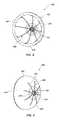
- FIG. 5depicts an anchoring member 24 interconnected to a distal end of the tether 26 according to one embodiment.
- Such an anchoring member 24is sized and configured to be collapsed in a constrained configuration at the distal portion of the catheter (see FIG. 1 ).
- the anchoring member 24may self expand to an expanded or deployed configuration, as shown.
- the anchoring member 24may include multiple legs 62 extending from a center portion 64 , the center portion being interconnected to the tether 26 . In the embodiment depicted in FIG. 5 , there are four legs 62 extending from the center portion 64 , however, in other embodiments there may be any suitable number of legs.
- Each leg 62may extend radially outward and include a looped portion 66 at the radial outermost end thereof.
- Such looped portion 66extends distally and then returns both radially inwardly and proximally such that a distal leg end 68 extends beyond a more proximal portion of the leg so as to act as an engagement nub to engage with the trabeculated tissue within the LAA.
- the legsare sized and configured to extend within the left atrial appendage and anchor within such tissue.
- the looped portion 66 and the outward extending legs 62may provide a spring effect to allow the physician to pull on the tether 26 without damaging the tissue when determining if the anchoring member 24 is sufficiently lodged within the left atrial appendage.
- anchoring member 24may be employed for the anchoring member 24 .
- anchoring structuresare disclosed in U.S. patent application Ser. No. 12/253,831 entitled MEDICAL DEVICE FOR MODIFICATION OF LEFT ATRIAL APPENDAGE AND RELATED SYSTEMS AND METHODS, filed on Oct. 17, 2008, the disclosure of which is incorporated by reference herein in its entirety.
- Such anchoring systems or structuresmay be incorporated into embodiments of the present invention in conjunction with an associated tether and tissue growth member.
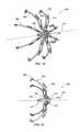
- FIG. 6depicts another embodiment of an anchoring member 25 which may be used in connection with the medical devices of the present invention.
- the anchoring member 25may include multiple j-shaped portions 72 extending radially outward to self expand from the distal portion of the catheter 18 .
- Each j-shaped portion 72includes a curved extension 74 and a distal coiled end 76 .
- the periphery of each coiled end 76may include one or more tapered nubs 78 . In this manner, the coiled ends 76 of the j-shaped configuration can self expand and nest within the left atrial appendage and substantially anchor therein.
- the spring-like quality of the curved extensions 74allows for substantial pull on the tether to determine proper anchoring while substantially limiting any damage to the tissue within the left atrial appendage. It should be noted that it is not required that there by three j-shaped portions as is shown in the drawings. Rather, there may be more or additional j-shaped portions than shown as may be desired.
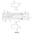
- FIGS. 7A-7Eanother embodiment of an anchor is shown that may be used in accordance with one or more embodiments of the present invention.
- FIG. 7Ashows a perspective view of the anchor 80
- FIG. 7Bshows a side view of the anchor 80
- FIG. 7Cshows a side view of the anchor 80 rotated approximately 90 degrees relative to that shown in FIG. 7B
- FIGS. 7D and 7Eshow side views of individual components used in forming the anchor 80 .
- the anchor 80may include multiple frame members 81 A and 81 B assembled together. While the frame members 81 A and 81 B may be substantially similar to one another, they are not necessarily identical to each other.
- each frame member 81 A and 81 Bmay include one or more anchor legs 82 (in the present depicted embodiment, each frame member includes two anchor legs) with various features.
- the anchor legs 82may include an arcuate distal end 83 having increased mass compared to the rest of the leg 82 , the arcuate distal end 83 curving radially inwardly.
- arcuate distal endsact as atraumatic tips to help prevent potential puncture of the walls of the LAA when deploying the anchor 80 .
- the inward curvature of the anchor legs distal endsare configured so that if the ends 83 are pushed against tissue within the LAA, the ends of the anchor legs 83 will roll radially inward.
- the anchor legs 82may also include tissue engaging features 84 that are configured to press against and engage the trabeculated tissue wall of the LAA.
- the engaging features 84may include, for example, proximally extending nubs, which may also be tapered.
- the engaging features 84(as well as various tissue engaging features of other anchors and structures described herein) are configured to be atraumatic.
- the engaging features 84may engage with the tissue of an LAA by nestling amongst the trabeculations along the tissue wall.
- the anchor legs 82may further include a flare 85 or projection that extends or deviates radially outwardly relative to the remaining path of the anchor legs 82 .
- the flare 85assists in loading the anchor 80 into a catheter or other delivery mechanism such that when the flare engages the periphery of a catheter lumen, it causes the anchor legs 82 to deflect radially inwardly a sufficient distance to avoid the interference of the engaging features 84 with the inner wall of the catheter's lumen.
- the anchor legs 82may exhibit different lengths than one another to further help facilitate placement of the anchor 80 within a catheter or other delivery mechanism.
- each anchor leg 82 of a give anchor 80may exhibit a different length than every other anchor leg.
- the frame members 81 A and 81 Balso include hub members 86 A and 86 B, respectively, that are cooperatively configured to effect mating or assembly of the frame members 81 A and 81 B to form the anchor 80 .
- the hub 86 A of one frame member 81 Amay include a slot 87 which may be accessed by displacing the free ends of two adjacent leg members 88 A and 88 B.
- the slot 87may be sized and configured to accept, and mate with, a body portion 89 of hub member 86 B from the other frame member 81 B.
- the body portion 89may have engagements surfaces 90 A and 90 B and be sized to fit snugly within the slot 87 of hub 86 A.
- Other slots 91 and 92 within the hub members 86 A and 86 Bmay be used in facilitating assembly of the anchor members 81 A and 81 B.
- the anchor members 81 A and 81 Bmay also include a plurality of through holes 93 A, 93 B and 93 C and/or slots 94 or notches. These through holes 93 A through 93 C may be used for coupling of the tether 26 to the assembled anchor 80 .
- the tether 26may pass through the various through holes 93 A- 93 C, while also wrapping around the assembled hub members 86 A and 86 B to couple the tether 26 with the anchor 80 and to help maintain assembly to the frame members 81 A and 81 B.
- the tethermay have a clip, a knot or otherwise be staked, as shown at 95 , to keep the tether 26 from becoming unattached from the anchor 80 .
- each frame member 81 A and 81 Bmay be formed as an integral, unitary and seamless component.
- the frame members 81 A and 81 Bmay be formed by laser cutting from a sheet of material such as a nickel-titanium alloy.
- the anchor legs 82 of a given frame member 81 A or 81 Bwould lie in a common plane.
- anchor 80as well as other anchors described herein, are configured to be deployed deep within an atrial appendage.
- the ability to vary the relative position of an anchor with an associated tissue growth membere.g., by varying the position of the two components along an associated tether) provides substantial flexibility in modifying an atrial appendage, particularly in light of the extreme variability from one atrial appendage to another.
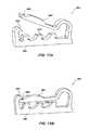
- an occluder member 350depicting perspective views of a distal side and a proximal side, respectively, of the occluder member 350 .
- the occluder member 350may be used in place of (or in some instances, in addition to) the tissue growth members 40 and associated support structure 42 described hereinabove.
- the presently considered embodiment of the occluder member 350may be employed with the medical device system depicted in FIG. 1 or FIG. 1A .
- the occluder member 350includes a tissue growth member 352 and a frame 354 .
- the tissue growth member 352may include a porous member configured to promote tissue in-growth therein.
- the tissue growth member 352may be a polymeric material, such as foam or other materials such as discussed above.
- the tissue growth member 352may exhibit a cup-like shape having an outer (or convex) surface 356 and an inner (or concave) surface 358 , the outer surface 356 including a distal surface portion 360 and a proximal surface portion 362 .
- the distal surface portion 360 of the tissue growth member 352is sized and configured to be in direct contact with tissue within the LAA (such as shown with respect to tissue growth members 40 a - 40 c in FIG. 4C ).
- the frame 354 or support structure of the occluder member 350is configured to assist in expanding the tissue growth member 352 and to assist in collapsing the tissue growth member 352 for delivery through an associated catheter or other medical device.
- Such frame 354may include an expander portion 366 , a collapser portion 368 and a hub portion 370 .
- the expander portion 366may extend from the hub portion 370 with multiple expanding legs 372 .
- the legs 372may extend along the inner surface 358 of the tissue growth member 352 .
- the collapser portion 368also may extend from the hub portion 370 with multiple collapsing legs 374 .
- the collapsing legs 374may extend along the proximal surface portion 362 of the tissue growth member 352 .
- the collapser portion 368 of the frame 354assists in collapsing the tissue growth member 352 (such as during a loading procedure) to a size wherein the occluder member 350 fits within the lumen of a catheter and may be displaced therethrough without damaging the tissue growth member 352 .
- the expander portion 366 of the frame 354is configured to self expand to assist in opening the tissue growth member 352 so that much (if not all) of the distal surface portion 360 of the tissue growth member 352 is in direct contact with the tissue of the LAA.
- FIG. 10shows a perspective view and FIG. 11 shows a side view of the frame 354 previously described with respect to FIGS. 8 and 9 .
- FIGS. 10 and 11do not depict the tissue growth member 352 for purposes of clarity. Additionally, FIG. 10 is shown in a simplified form (i.e., some frame components are not shown) for purposes of clarity.
- the frame 354may include multiple discrete frame segments 364 that may be assembled with the hub portion 370 to collectively provide the frame 354 .
- Each frame segment 364includes a hub extension 376 with an expanding leg 372 and a collapsing leg 374 extending from a proximal end 376 of the hub extension 376 .
- each frame segment 364is configured to be substantially flat. Otherwise said, the hub extension 376 , expanding leg 372 and collapsing leg 374 of a given frame segment 364 are substantially coplanar with respect to each other.
- the frame segments 364may each be laser cut or otherwise formed from a flat sheet of Nitinol, thereby, providing a substantially flat configuration to each of the frame segments 364 . In this manner, the frame 354 (when assembled from the plurality of frame segments 364 ) may be configured to collapse within a catheter as well as self expand when deployed from a catheter with the frame segments 364 being deflected and displaced in the process.
- Each frame segment 364may be positioned radially and substantially symmetrical with respect to each other about a longitudinal axis 375 that extends through the hub portion 370 .
- the frame segments 364may be coupled with one or more rings 378 having notches on a radial inner surface, a radial outer surface or both to correspond with notches formed within the hub extension 376 of the frame segment. Due to each frame segment 364 being discrete with respect to the other frame segments 364 , the expanding leg 372 and collapsing leg 374 may collapse or expand substantially independent from the other expanding and collapsing legs of the other frame segments 364 .
- each of the frame segments 364self expand, independent of each other, to facilitate the tissue growth member 352 to be in direct contact with the tissue of the LAA in a non-rigid and conformable manner. Further, the frame segments 364 each independently self expand so as to adapt to the varying anatomy that is encountered within the LAA.
- Each of the collapsing legs 374 and the expanding legs 372may include one or more clips 380 formed therewith.
- FIGS. 12A and 12Bare perspective enlarged views of the clips 380 in an open and closed position, respectively, in accordance with an embodiment of the present invention.
- Such clips 380may be formed in the proximal and/or distal portions of the legs for attaching the tissue growth member thereto (see FIGS. 8 and 9 ).
- the clips 380may include a leg base portion 382 , a cantilevered extension 384 with a free end 386 , and a pawl 388 that is configured to receive the free-end 386 of the cantilevered extension 384 .
- the clips 380may include nubs 389 extending from the leg base portion 382 to provide traction or additional engagement with the tissue growth member 352 .
- the clips 380may be integrally formed into the frame segments, such as by laser cutting.
- other means of fastening the tissue growth member 352 to the frame 354may be used (in lieu of, or in addition to the clips 380 ) including, for example, adhesives, sutures, or other mechanical structures or devices.
- FIGS. 13 and 14a simplified side view of portions of a medical device system 400 in an open position (also referred to as an expanded or deployed position) and a closed position (also referred to as a contracted position), according to one embodiment, is depicted.
- the medical device system 400may include the occluder member 350 , a tether filament 402 and a pusher member 404 .
- a tissue growth memberis not shown in FIGS. 13 and 14 , although one is contemplated and those of ordinary skill in the art will recognize its use an implementation in the following description. Additional reference is made during the following description to FIGS. 15A and 15B which show side views of frame segments 364 A and 364 B.
- the frame 354 of the occluder member 350may be formed of a plurality of frame segments 364 A and 364 B.
- the frame of the occluder member 350may include four of each type of frame segments 364 A and 364 B which alternate in their positions (i.e., each frame segment 364 A is adjacent to two frame segments 364 B and vice versa).
- the frame segments 364 A and 364 Bmay include expanding legs 374 , collapsing legs 372 and hub extensions 376 that have inner and outer notches 379 for engaging ring members during assembly of the frame.
- frame segments 364 A and 364 Bmay be formed, for example by laser cutting from a flat sheet of desired material such as a nickel-titanium alloy (e.g., Nitinol).
- a nickel-titanium alloye.g., Nitinol
- Such a configurationprovides for the expanding leg 374 , collapsing leg 372 and hub extension 376 to be coplanar.
- the hub portion 370may define a hole 406 extending centrally therethrough and may further include a threaded portion 408 that at least partially defines the hole 406 .
- the hub portion 370is defined via the assembled multiple hub extensions 376 radially oriented and positioned with the one or more rings 378 .
- the hub portion of the frame 354enables the occluder member 350 to slide over the tether filament 402 , such as previously depicted in the embodiments described in FIGS. 3A-3D and 4 A- 4 C.
- the pusher member 404includes a distal end 410 and a proximal end (not shown) with a lumen 412 extending longitudinally through at least a portion of the pusher member 404 .
- the pusher member 404includes a coupling member 414 at or proximate the distal end 410 of the pusher member 404 and a cutter 416 disposed within the lumen 412 , a distal end of the cutter 416 being proximal or adjacent to an outlet 422 defined in a wall of the pusher member 404 .
- the coupling member 414may include a threaded portion 418 and a non-threaded distal extension 420 , the extension 420 extending distal of the threaded portion 418 .
- the non-threaded distal extension 420engages the hub extensions 376 and places a gripper portion 424 of the hub portion 370 in an open position.
- the occluder member 350may slide or move over the tether filament 402 , through a catheter while in a collapsed position as well as once deployed from the catheter, with the tether filament 402 extending through at least the coupling portion 414 or a distal portion of the pusher member 404 and exiting from the pusher member 404 through the outlet 422 defined in the wall of the pusher member 404 .
- the pusher member 404may be un-threaded or removed from the occluder member 350 , thereby causing the gripper portion 424 of the hub portion 370 to engage or grip the tether filament 402 . That is, as the pusher member 404 is un-threaded, the distal extension 420 is moved proximally which causes the gripper portion 424 to move to the radially inward position (i.e., the radially closed position) to grip onto the tether filament 402 that is anchored distally and deep within a lobe of the LAA.
- the gripper portion 424may include bands 426 disposed around the gripper portion 424 to bias the gripper portion in the closed state and assist in more effectively gripping the tether filament 402 .
- the hub extensionsmay be configured to be biased towards the closed position even without the aid of other biasing elements. This configuration enables the hubs to work as a locking element to maintain the occluder member 350 in a desired position relative to the tether (and, thus, relative to an associated anchor).
- the pusher member 404can then be fully removed from the hub portion 370 of the occluder member 350 and, if the physician is satisfied with the position of the occluder member, the cutter element 416 can be moved distally to slice the tether filament 402 .
- another occluder membermay be loaded in a catheter and slid over the tether filament 402 to position within the LAA.
- the hub extensions 376may include a guide portion 430 that may be associated with the gripper portion 424 . Further, at the distal end of the guide portion 430 , there is a pawl 432 to latch a tether guide coil 434 .
- the tether guide coil 434extends distally and the tether filament 402 extends axially through the tether guide coil 434 .
- the tether guide coil 434extends a length sufficient to substantially prevent the tissue growth member (not shown) from contacting the tether filament 402 while the occluder 350 is in a collapsed position and being pushed distally within a catheter.
- the pusher member 404may include a coil (not shown) that is positioned proximal to the coupling member 414 and over the pusher member 404 such that the coil and the lumen 412 of the pusher member 404 have a common axis.
- the occluder 350 , the pusher member 404 and the tether filament 402may include radiopaque characteristics or markers so that the relevant portions of the medical device system 400 can be viewed with imaging techniques known in the art.
- FIG. 18shows a front perspective view while FIG. 19 shows a side, partial cross-sectional view of the occluder 350 .
- the occluder 350is similar to the embodiments show and described with respect to FIGS. 7 and 8 , but also includes an additional material layer 390 associated with the tissue growth member 352 .
- FIG. 19shows the additional material layer 390 in an “exploded” state for purposes of illustration.
- the additional material layer 390is, in actuality, contiguous with the underlying foam or other material forming the tissue growth member 352 , the additional material layer 390 being attached thereto by, for example, an adhesive.
- the additional material layer 390may include a polytetrafluoroethylene (PTFE) or expanded PTFE (ePTFE). Such a surface provides a smooth surface on the proximal side of the tissue growth member to tailor the tissue growth pattern once the occluder is deployed within an atrial appendage. It is noted the additional material layer 390 may be configured to allow a portion of the frame to be exposed on the proximal side (e.g., the hub portion) such as shown in FIG. 18 , or it may be configured to cover substantially all of the frame along the proximal side such as is shown in FIG. 19 .
- PTFEpolytetrafluoroethylene
- ePTFEexpanded PTFE
Landscapes
- Health & Medical Sciences (AREA)
- Surgery (AREA)
- Life Sciences & Earth Sciences (AREA)
- Heart & Thoracic Surgery (AREA)
- Molecular Biology (AREA)
- Veterinary Medicine (AREA)
- Engineering & Computer Science (AREA)
- Biomedical Technology (AREA)
- Public Health (AREA)
- Medical Informatics (AREA)
- Nuclear Medicine, Radiotherapy & Molecular Imaging (AREA)
- Animal Behavior & Ethology (AREA)
- General Health & Medical Sciences (AREA)
- Reproductive Health (AREA)
- Vascular Medicine (AREA)
- Cardiology (AREA)
- Rheumatology (AREA)
- Surgical Instruments (AREA)
- Prostheses (AREA)
Abstract
Description
Claims (26)
Priority Applications (3)
| Application Number | Priority Date | Filing Date | Title |
|---|---|---|---|
| US12/684,783US8795328B2 (en) | 2009-01-08 | 2010-01-08 | Medical device for modification of left atrial appendage and related systems and methods |
| US14/287,103US9750505B2 (en) | 2009-01-08 | 2014-05-26 | Medical device for modification of left atrial appendage and related systems and methods |
| US15/665,412US10695070B2 (en) | 2009-01-08 | 2017-07-31 | Medical device for modification of left atrial appendage and related systems and methods |
Applications Claiming Priority (4)
| Application Number | Priority Date | Filing Date | Title |
|---|---|---|---|
| US14336009P | 2009-01-08 | 2009-01-08 | |
| US16024709P | 2009-03-13 | 2009-03-13 | |
| US16431309P | 2009-03-27 | 2009-03-27 | |
| US12/684,783US8795328B2 (en) | 2009-01-08 | 2010-01-08 | Medical device for modification of left atrial appendage and related systems and methods |
Related Child Applications (1)
| Application Number | Title | Priority Date | Filing Date |
|---|---|---|---|
| US14/287,103ContinuationUS9750505B2 (en) | 2009-01-08 | 2014-05-26 | Medical device for modification of left atrial appendage and related systems and methods |
Publications (2)
| Publication Number | Publication Date |
|---|---|
| US20100228279A1 US20100228279A1 (en) | 2010-09-09 |
| US8795328B2true US8795328B2 (en) | 2014-08-05 |
Family
ID=42076907
Family Applications (7)
| Application Number | Title | Priority Date | Filing Date |
|---|---|---|---|
| US12/684,764Active2030-10-07US8690911B2 (en) | 2009-01-08 | 2010-01-08 | Medical device for modification of left atrial appendage and related systems and methods |
| US12/684,795Active2030-03-20US8840641B2 (en) | 2009-01-08 | 2010-01-08 | Medical device for modification of left atrial appendage and related systems and methods |
| US12/684,783Active2030-11-14US8795328B2 (en) | 2009-01-08 | 2010-01-08 | Medical device for modification of left atrial appendage and related systems and methods |
| US14/215,266ActiveUS9572584B2 (en) | 2009-01-08 | 2014-03-17 | Medical device for modification of left atrial appendage and related systems and methods |
| US14/287,103Active2031-10-07US9750505B2 (en) | 2009-01-08 | 2014-05-26 | Medical device for modification of left atrial appendage and related systems and methods |
| US15/430,973Active2030-05-31US10420564B2 (en) | 2009-01-08 | 2017-02-13 | Medical device for modification of left atrial appendage and related systems and methods |
| US15/665,412Active2030-11-26US10695070B2 (en) | 2009-01-08 | 2017-07-31 | Medical device for modification of left atrial appendage and related systems and methods |
Family Applications Before (2)
| Application Number | Title | Priority Date | Filing Date |
|---|---|---|---|
| US12/684,764Active2030-10-07US8690911B2 (en) | 2009-01-08 | 2010-01-08 | Medical device for modification of left atrial appendage and related systems and methods |
| US12/684,795Active2030-03-20US8840641B2 (en) | 2009-01-08 | 2010-01-08 | Medical device for modification of left atrial appendage and related systems and methods |
Family Applications After (4)
| Application Number | Title | Priority Date | Filing Date |
|---|---|---|---|
| US14/215,266ActiveUS9572584B2 (en) | 2009-01-08 | 2014-03-17 | Medical device for modification of left atrial appendage and related systems and methods |
| US14/287,103Active2031-10-07US9750505B2 (en) | 2009-01-08 | 2014-05-26 | Medical device for modification of left atrial appendage and related systems and methods |
| US15/430,973Active2030-05-31US10420564B2 (en) | 2009-01-08 | 2017-02-13 | Medical device for modification of left atrial appendage and related systems and methods |
| US15/665,412Active2030-11-26US10695070B2 (en) | 2009-01-08 | 2017-07-31 | Medical device for modification of left atrial appendage and related systems and methods |
Country Status (2)
| Country | Link |
|---|---|
| US (7) | US8690911B2 (en) |
| WO (3) | WO2010081039A1 (en) |
Cited By (23)
| Publication number | Priority date | Publication date | Assignee | Title |
|---|---|---|---|---|
| US9526891B2 (en) | 2015-04-24 | 2016-12-27 | Medtronic, Inc. | Intracardiac medical device |
| US9526522B2 (en) | 2013-09-27 | 2016-12-27 | Medtronic, Inc. | Interventional medical systems, tools, and assemblies |
| US9675798B2 (en) | 2014-08-26 | 2017-06-13 | Medtronic, Inc. | Interventional medical systems, devices, and components thereof |
| US10143823B2 (en) | 2016-04-29 | 2018-12-04 | Medtronic, Inc. | Interventional medical systems and improved assemblies thereof and associated methods of use |
| US10300286B2 (en) | 2013-09-27 | 2019-05-28 | Medtronic, Inc. | Tools and assemblies thereof for implantable medical devices |
| US10349948B2 (en) | 2014-03-31 | 2019-07-16 | Jitmed Sp. Z. O.O. | Left atrial appendage occlusion device |
| US10478620B2 (en) | 2014-08-26 | 2019-11-19 | Medtronic, Inc. | Interventional medical systems, devices, and methods of use |
| US10617425B2 (en) | 2014-03-10 | 2020-04-14 | Conformal Medical, Inc. | Devices and methods for excluding the left atrial appendage |
| US10722240B1 (en) | 2019-02-08 | 2020-07-28 | Conformal Medical, Inc. | Devices and methods for excluding the left atrial appendage |
| US10918392B2 (en) | 2018-01-26 | 2021-02-16 | Syntheon 2.0, LLC | Left atrial appendage clipping device and methods for clipping the LAA |
| US10925615B2 (en) | 2019-05-03 | 2021-02-23 | Syntheon 2.0, LLC | Recapturable left atrial appendage clipping device and methods for recapturing a left atrial appendage clip |
| US11369355B2 (en) | 2019-06-17 | 2022-06-28 | Coherex Medical, Inc. | Medical device and system for occluding a tissue opening and method thereof |
| US11399842B2 (en) | 2013-03-13 | 2022-08-02 | Conformal Medical, Inc. | Devices and methods for excluding the left atrial appendage |
| US11426172B2 (en) | 2016-10-27 | 2022-08-30 | Conformal Medical, Inc. | Devices and methods for excluding the left atrial appendage |
| US11517319B2 (en) | 2017-09-23 | 2022-12-06 | Universität Zürich | Medical occluder device |
| US11540837B2 (en) | 2009-06-17 | 2023-01-03 | Coherex Medical, Inc. | Medical device for modification of left atrial appendage and related systems and methods |
| US11717303B2 (en) | 2013-03-13 | 2023-08-08 | Conformal Medical, Inc. | Devices and methods for excluding the left atrial appendage |
| US11786256B2 (en) | 2016-10-27 | 2023-10-17 | Conformal Medical, Inc. | Devices and methods for excluding the left atrial appendage |
| US11812969B2 (en) | 2020-12-03 | 2023-11-14 | Coherex Medical, Inc. | Medical device and system for occluding a tissue opening and method thereof |
| US11918227B2 (en) | 2009-06-17 | 2024-03-05 | Coherex Medical, Inc. | Medical device for modification of left atrial appendage and related systems and methods |
| US11944315B2 (en) | 2019-09-26 | 2024-04-02 | Universität Zürich | Left atrial appendage occlusion devices |
| US12144508B2 (en) | 2019-02-08 | 2024-11-19 | Conformal Medical, Inc. | Devices and methods for excluding the left atrial appendage |
| US12402885B2 (en) | 2017-09-23 | 2025-09-02 | Universität Zürich | Medical occlusion device |
Families Citing this family (110)
| Publication number | Priority date | Publication date | Assignee | Title |
|---|---|---|---|---|
| US8845711B2 (en) | 2007-10-19 | 2014-09-30 | Coherex Medical, Inc. | Medical device for modification of left atrial appendage and related systems and methods |
| EP2015681B1 (en) | 2006-05-03 | 2018-03-28 | Datascope Corp. | Tissue closure device |
| US7828854B2 (en)* | 2006-10-31 | 2010-11-09 | Ethicon, Inc. | Implantable repair device |
| US20110257723A1 (en) | 2006-11-07 | 2011-10-20 | Dc Devices, Inc. | Devices and methods for coronary sinus pressure relief |
| US10413284B2 (en) | 2006-11-07 | 2019-09-17 | Corvia Medical, Inc. | Atrial pressure regulation with control, sensing, monitoring and therapy delivery |
| US9232997B2 (en) | 2006-11-07 | 2016-01-12 | Corvia Medical, Inc. | Devices and methods for retrievable intra-atrial implants |
| EP2097012A4 (en) | 2006-11-07 | 2012-08-15 | David Stephen Celermajer | Devices and methods for the treatment of heart failure |
| US8460372B2 (en) | 2006-11-07 | 2013-06-11 | Dc Devices, Inc. | Prosthesis for reducing intra-cardiac pressure having an embolic filter |
| WO2010081039A1 (en) | 2009-01-08 | 2010-07-15 | Coherex Medical, Inc. | Medical device for modification of left atrial appendage and related systems and methods |
| CA2765682C (en) | 2009-06-17 | 2018-07-24 | Coherex Medical, Inc. | Medical device for modification of left atrial appendage and related systems and methods |
| US10064628B2 (en) | 2009-06-17 | 2018-09-04 | Coherex Medical, Inc. | Medical device for modification of left atrial appendage and related systems and methods |
| US9351716B2 (en) | 2009-06-17 | 2016-05-31 | Coherex Medical, Inc. | Medical device and delivery system for modification of left atrial appendage and methods thereof |
| US9693781B2 (en) | 2009-06-17 | 2017-07-04 | Coherex Medical, Inc. | Medical device for modification of left atrial appendage and related systems and methods |
| US9757107B2 (en) | 2009-09-04 | 2017-09-12 | Corvia Medical, Inc. | Methods and devices for intra-atrial shunts having adjustable sizes |
| US8870950B2 (en) | 2009-12-08 | 2014-10-28 | Mitral Tech Ltd. | Rotation-based anchoring of an implant |
| WO2011094521A2 (en)* | 2010-01-29 | 2011-08-04 | Dc Devices, Inc. | Devices and methods for reducing venous pressure |
| EP2528646A4 (en) | 2010-01-29 | 2017-06-28 | DC Devices, Inc. | Devices and systems for treating heart failure |
| US20110224785A1 (en) | 2010-03-10 | 2011-09-15 | Hacohen Gil | Prosthetic mitral valve with tissue anchors |
| US9763657B2 (en) | 2010-07-21 | 2017-09-19 | Mitraltech Ltd. | Techniques for percutaneous mitral valve replacement and sealing |
| US11653910B2 (en) | 2010-07-21 | 2023-05-23 | Cardiovalve Ltd. | Helical anchor implantation |
| US9132009B2 (en) | 2010-07-21 | 2015-09-15 | Mitraltech Ltd. | Guide wires with commissural anchors to advance a prosthetic valve |
| US8992604B2 (en) | 2010-07-21 | 2015-03-31 | Mitraltech Ltd. | Techniques for percutaneous mitral valve replacement and sealing |
| US9186152B2 (en) | 2010-11-12 | 2015-11-17 | W. L. Gore & Associates, Inc. | Left atrial appendage occlusive devices |
| WO2012109557A2 (en) | 2011-02-10 | 2012-08-16 | Dc Devices, Inc. | Apparatus and methods to create and maintain an intra-atrial pressure relief opening |
| US12303119B2 (en) | 2011-02-10 | 2025-05-20 | Corvia Medical, Inc. | Apparatus and methods to create and maintain an intra-atrial pressure relief opening |
| US9744033B2 (en) | 2011-04-01 | 2017-08-29 | W.L. Gore & Associates, Inc. | Elastomeric leaflet for prosthetic heart valves |
| US10117765B2 (en) | 2011-06-14 | 2018-11-06 | W.L. Gore Associates, Inc | Apposition fiber for use in endoluminal deployment of expandable implants |
| WO2013021374A2 (en) | 2011-08-05 | 2013-02-14 | Mitraltech Ltd. | Techniques for percutaneous mitral valve replacement and sealing |
| US8852272B2 (en) | 2011-08-05 | 2014-10-07 | Mitraltech Ltd. | Techniques for percutaneous mitral valve replacement and sealing |
| EP2739214B1 (en)* | 2011-08-05 | 2018-10-10 | Cardiovalve Ltd | Percutaneous mitral valve replacement and sealing |
| US20140324164A1 (en) | 2011-08-05 | 2014-10-30 | Mitraltech Ltd. | Techniques for percutaneous mitral valve replacement and sealing |
| US9554806B2 (en) | 2011-09-16 | 2017-01-31 | W. L. Gore & Associates, Inc. | Occlusive devices |
| EP3305217A1 (en) | 2011-10-20 | 2018-04-11 | Teleflex Life Sciences Unlimited Company | Ligation clip |
| US9877858B2 (en) | 2011-11-14 | 2018-01-30 | W. L. Gore & Associates, Inc. | External steerable fiber for use in endoluminal deployment of expandable devices |
| US9782282B2 (en) | 2011-11-14 | 2017-10-10 | W. L. Gore & Associates, Inc. | External steerable fiber for use in endoluminal deployment of expandable devices |
| US8951223B2 (en) | 2011-12-22 | 2015-02-10 | Dc Devices, Inc. | Methods and devices for intra-atrial shunts having adjustable sizes |
| US9005155B2 (en) | 2012-02-03 | 2015-04-14 | Dc Devices, Inc. | Devices and methods for treating heart failure |
| US20130237908A1 (en)* | 2012-03-09 | 2013-09-12 | Boston Scientific Scimed, Inc. | Sponge-like left atrial occlusion device and related methods of use |
| US9375308B2 (en) | 2012-03-13 | 2016-06-28 | W. L. Gore & Associates, Inc. | External steerable fiber for use in endoluminal deployment of expandable devices |
| US10588611B2 (en) | 2012-04-19 | 2020-03-17 | Corvia Medical Inc. | Implant retention attachment and method of use |
| US9526623B2 (en) | 2012-05-30 | 2016-12-27 | Newvert Ltd. | Spinal disc annulus closure device |
| US9649480B2 (en) | 2012-07-06 | 2017-05-16 | Corvia Medical, Inc. | Devices and methods of treating or ameliorating diastolic heart failure through pulmonary valve intervention |
| WO2014018907A1 (en)* | 2012-07-26 | 2014-01-30 | University Of Louisville Research Foundation, Inc. | Atrial appendage closure device and related methods |
| US20150351906A1 (en) | 2013-01-24 | 2015-12-10 | Mitraltech Ltd. | Ventricularly-anchored prosthetic valves |
| US9775636B2 (en) | 2013-03-12 | 2017-10-03 | Corvia Medical, Inc. | Devices, systems, and methods for treating heart failure |
| CN204072201U (en)* | 2013-03-14 | 2015-01-07 | 卡迪欧凯尼迪克斯公司 | Assembling fixture and implant |
| US11911258B2 (en) | 2013-06-26 | 2024-02-27 | W. L. Gore & Associates, Inc. | Space filling devices |
| WO2015077356A1 (en) | 2013-11-19 | 2015-05-28 | Wheeler William K | Fastener applicator with interlock |
| US10675450B2 (en) | 2014-03-12 | 2020-06-09 | Corvia Medical, Inc. | Devices and methods for treating heart failure |
| AU2015249283B2 (en) | 2014-04-25 | 2019-07-18 | Flow Medtech, Llc | Left atrial appendage occlusion device |
| JP6799526B2 (en) | 2014-07-23 | 2020-12-16 | コルヴィア メディカル インコーポレイテッド | Equipment and methods for the treatment of heart failure |
| EP3174502B1 (en) | 2014-07-30 | 2022-04-06 | Cardiovalve Ltd | Apparatus for implantation of an articulatable prosthetic valve |
| CA2999169A1 (en) | 2014-09-19 | 2016-03-24 | Flow Medtech, Inc. | Left atrial appendage occlusion device delivery system |
| EP3028652A1 (en)* | 2014-12-03 | 2016-06-08 | Peter Osypka Stiftung | Closure device suitable for closing the atrial appendage |
| US9974651B2 (en) | 2015-02-05 | 2018-05-22 | Mitral Tech Ltd. | Prosthetic valve with axially-sliding frames |
| CN110141399B (en) | 2015-02-05 | 2021-07-27 | 卡迪尔维尔福股份有限公司 | Prosthetic valve with axial sliding frame |
| CA2986047C (en) | 2015-05-14 | 2020-11-10 | W. L. Gore & Associates, Inc. | Devices and methods for occlusion of an atrial appendage |
| US10531866B2 (en) | 2016-02-16 | 2020-01-14 | Cardiovalve Ltd. | Techniques for providing a replacement valve and transseptal communication |
| AU2016408266B2 (en)* | 2016-05-25 | 2021-02-18 | Coramaze Technologies Gmbh | Heart implant |
| US10058426B2 (en)* | 2016-07-20 | 2018-08-28 | Abbott Cardiovascular Systems Inc. | System for tricuspid valve repair |
| US20190231525A1 (en) | 2016-08-01 | 2019-08-01 | Mitraltech Ltd. | Minimally-invasive delivery systems |
| US10548609B2 (en) | 2016-08-03 | 2020-02-04 | Teleflex Medical Incorporated | Surgical ligation clip |
| US10201352B2 (en)* | 2016-08-08 | 2019-02-12 | Atricure, Inc. | Robotic Assisted clip applier |
| CA3031187A1 (en) | 2016-08-10 | 2018-02-15 | Cardiovalve Ltd. | Prosthetic valve with concentric frames |
| USD800908S1 (en) | 2016-08-10 | 2017-10-24 | Mitraltech Ltd. | Prosthetic valve element |
| US10548614B2 (en) | 2016-11-29 | 2020-02-04 | Evalve, Inc. | Tricuspid valve repair system |
| US10420565B2 (en) | 2016-11-29 | 2019-09-24 | Abbott Cardiovascular Systems Inc. | Cinch and post for tricuspid valve repair |
| US10952852B2 (en) | 2017-02-24 | 2021-03-23 | Abbott Cardiovascular Systems Inc. | Double basket assembly for valve repair |
| CA3055567C (en) | 2017-03-08 | 2021-11-23 | W. L. Gore & Associates, Inc. | Steering wire attach for angulation |
| WO2018175610A1 (en) | 2017-03-21 | 2018-09-27 | Teleflex Medical Incorporated | Surgical clip and clip applier |
| CN116784923A (en) | 2017-03-21 | 2023-09-22 | 泰利福医疗公司 | Clip applier with stabilizing member |
| JP6873267B2 (en) | 2017-03-21 | 2021-05-19 | テレフレックス メディカル インコーポレイテッド | Clip applier with stabilizer |
| JP7159189B2 (en) | 2017-03-21 | 2022-10-24 | テレフレックス メディカル インコーポレイテッド | Flexible stabilizing member for clip applier |
| US12023041B2 (en) | 2017-03-21 | 2024-07-02 | Teleflex Medical Incorporated | Clip applier |
| EP3614933A1 (en) | 2017-04-27 | 2020-03-04 | Boston Scientific Scimed, Inc. | Occlusive medical device with fabric retention barb |
| CA3068282C (en) | 2017-06-22 | 2022-06-28 | Teleflex Medical Incorporated | Surgical clip |
| US10537426B2 (en) | 2017-08-03 | 2020-01-21 | Cardiovalve Ltd. | Prosthetic heart valve |
| US11246704B2 (en) | 2017-08-03 | 2022-02-15 | Cardiovalve Ltd. | Prosthetic heart valve |
| US10575948B2 (en) | 2017-08-03 | 2020-03-03 | Cardiovalve Ltd. | Prosthetic heart valve |
| US11793633B2 (en) | 2017-08-03 | 2023-10-24 | Cardiovalve Ltd. | Prosthetic heart valve |
| US12064347B2 (en) | 2017-08-03 | 2024-08-20 | Cardiovalve Ltd. | Prosthetic heart valve |
| US10888421B2 (en) | 2017-09-19 | 2021-01-12 | Cardiovalve Ltd. | Prosthetic heart valve with pouch |
| CN107595347B (en)* | 2017-09-25 | 2019-11-26 | 潘湘斌 | A repeatable retractable left atrial appendage occluder assembly and its intervention method |
| US11173023B2 (en) | 2017-10-16 | 2021-11-16 | W. L. Gore & Associates, Inc. | Medical devices and anchors therefor |
| ES2981210T3 (en) | 2017-11-14 | 2024-10-07 | Teleflex Medical Inc | Surgical clip |
| GB201720803D0 (en) | 2017-12-13 | 2018-01-24 | Mitraltech Ltd | Prosthetic Valve and delivery tool therefor |
| CN108065970B (en)* | 2017-12-14 | 2019-06-25 | 谭雄进 | A kind of artificial cords fixation kit that can repeatedly adjust |
| JP6946454B2 (en)* | 2017-12-21 | 2021-10-06 | ザ テキサス エーアンドエム ユニバーシティ システムThe Texas A&M University System | Vascular prosthesis to prevent leaks during intravascular aneurysm repair |
| GB201800399D0 (en) | 2018-01-10 | 2018-02-21 | Mitraltech Ltd | Temperature-control during crimping of an implant |
| JP7348199B2 (en) | 2018-03-28 | 2023-09-20 | データスコープ コーポレイション | Device for atrial appendage exclusion |
| CN112714632B (en) | 2018-08-21 | 2024-08-30 | 波士顿科学医学有限公司 | Barbed protruding member for cardiovascular device |
| AU2018439076B2 (en) | 2018-08-31 | 2022-07-07 | W. L. Gore & Associates, Inc. | Apparatus, system, and method for steering an implantable medical device |
| JP7247330B2 (en) | 2018-09-26 | 2023-03-28 | テレフレックス メディカル インコーポレイテッド | Clip applier with stabilizing member |
| CN113873957B (en) | 2019-03-25 | 2025-06-24 | 拉米纳公司 | Devices and systems for treating the left atrial appendage |
| US11101101B2 (en) | 2019-05-15 | 2021-08-24 | Fei Company | Laser-based phase plate image contrast manipulation |
| US11534303B2 (en) | 2020-04-09 | 2022-12-27 | Evalve, Inc. | Devices and systems for accessing and repairing a heart valve |
| US11832828B2 (en) | 2019-05-24 | 2023-12-05 | Medtronic, Inc. | Left atrial appendage occlusion device |
| CN114126540A (en)* | 2019-07-17 | 2022-03-01 | 波士顿科学医学有限公司 | Continuously covered left atrial appendage implant |
| EP4563099A3 (en) | 2019-08-30 | 2025-08-06 | Boston Scientific Scimed, Inc. | Left atrial appendage implant with sealing disk |
| WO2021062170A1 (en) | 2019-09-26 | 2021-04-01 | Teleflex Medical Incorporated | Clip applier |
| EP4076216A1 (en) | 2019-12-19 | 2022-10-26 | Teleflex Medical Incorporated | Surgical clip |
| US12303116B2 (en) | 2020-03-24 | 2025-05-20 | Laminar, Inc. | Devices, systems, and methods for occluding cavities within the body |
| WO2021195085A1 (en) | 2020-03-24 | 2021-09-30 | Boston Scientific Scimed, Inc. | Medical system for treating a left atrial appendage |
| JP7627761B2 (en) | 2020-11-30 | 2025-02-06 | ボストン サイエンティフィック サイムド,インコーポレイテッド | Implantable passive mean pressure sensor |
| US12357459B2 (en) | 2020-12-03 | 2025-07-15 | Cardiovalve Ltd. | Transluminal delivery system |
| US12383201B2 (en) | 2021-02-03 | 2025-08-12 | Boston Scientific Scimed, Inc. | Medical system for treating a left atrial appendage |
| EP4326169A1 (en)* | 2021-04-20 | 2024-02-28 | Coherex Medical, Inc. | Occlusive material for medical device, system, and method thereof |
| EP4358872A1 (en) | 2021-06-22 | 2024-05-01 | Boston Scientific Scimed, Inc. | Left atrial appendage implant |
| CN117615720A (en) | 2021-07-08 | 2024-02-27 | 波士顿科学医学有限公司 | Left auricle closing device |
| WO2023038929A1 (en) | 2021-09-08 | 2023-03-16 | Boston Scientific Scimed, Inc. | Occlusive implant with multi-sharpness split tip soft tissue anchors |
Citations (114)
| Publication number | Priority date | Publication date | Assignee | Title |
|---|---|---|---|---|
| US3095877A (en) | 1961-05-01 | 1963-07-02 | Robert L Rowan | Surgical packing |
| US3874388A (en) | 1973-02-12 | 1975-04-01 | Ochsner Med Found Alton | Shunt defect closure system |
| US5192301A (en) | 1989-01-17 | 1993-03-09 | Nippon Zeon Co., Ltd. | Closing plug of a defect for medical use and a closing plug device utilizing it |
| US5284488A (en) | 1992-12-23 | 1994-02-08 | Sideris Eleftherios B | Adjustable devices for the occlusion of cardiac defects |
| US5334217A (en) | 1992-01-21 | 1994-08-02 | Regents Of The University Of Minnesota | Septal defect closure device |
| US5425733A (en) | 1992-02-19 | 1995-06-20 | Arthrex, Inc. | Interference screw with rounded back end and cannulated sheath for endosteal fixation of ligaments |
| US5425740A (en)* | 1994-05-17 | 1995-06-20 | Hutchinson, Jr.; William B. | Endoscopic hernia repair clip and method |
| US5433727A (en) | 1994-08-16 | 1995-07-18 | Sideris; Eleftherios B. | Centering buttoned device for the occlusion of large defects for occluding |
| US5861003A (en) | 1996-10-23 | 1999-01-19 | The Cleveland Clinic Foundation | Apparatus and method for occluding a defect or aperture within body surface |
| US5904703A (en) | 1996-05-08 | 1999-05-18 | Bard Connaught | Occluder device formed from an open cell foam material |
| US5910154A (en)* | 1997-05-08 | 1999-06-08 | Embol-X, Inc. | Percutaneous catheter and guidewire having filter and medical device deployment |
| US6152144A (en) | 1998-11-06 | 2000-11-28 | Appriva Medical, Inc. | Method and device for left atrial appendage occlusion |
| US6174322B1 (en) | 1997-08-08 | 2001-01-16 | Cardia, Inc. | Occlusion device for the closure of a physical anomaly such as a vascular aperture or an aperture in a septum |
| US6231561B1 (en) | 1999-09-20 | 2001-05-15 | Appriva Medical, Inc. | Method and apparatus for closing a body lumen |
| US6238403B1 (en) | 1999-10-04 | 2001-05-29 | Microvention, Inc. | Filamentous embolic device with expansible elements |
| US20010003161A1 (en) | 1996-11-04 | 2001-06-07 | Vardi Gil M. | Catheter with side sheath |
| US20020026217A1 (en) | 2000-04-26 | 2002-02-28 | Steven Baker | Apparatus and method for repair of perigraft flow |
| US20020026094A1 (en) | 1993-02-22 | 2002-02-28 | Roth Alex T. | Devices for less-invasive intracardiac interventions |
| US6355051B1 (en)* | 1999-03-04 | 2002-03-12 | Bioguide Consulting, Inc. | Guidewire filter device |
| US6371971B1 (en)* | 1999-11-15 | 2002-04-16 | Scimed Life Systems, Inc. | Guidewire filter and methods of use |
| US20020062130A1 (en) | 1999-11-18 | 2002-05-23 | Jugenheimer Kristen A. | Apparatus and method for compressing body tissue |
| US20020177855A1 (en) | 1999-10-04 | 2002-11-28 | Greene George R. | Method of manufacturing expansile filamentous embolization devices |
| US6488689B1 (en) | 1999-05-20 | 2002-12-03 | Aaron V. Kaplan | Methods and apparatus for transpericardial left atrial appendage closure |
| US20020183787A1 (en) | 2001-06-01 | 2002-12-05 | Velocimed, L.L.C. | Closure devices, related delivery methods and tools, and related methods of use |
| US20030014075A1 (en) | 2001-07-16 | 2003-01-16 | Microvention, Inc. | Methods, materials and apparatus for deterring or preventing endoleaks following endovascular graft implanation |
| US6530934B1 (en) | 2000-06-06 | 2003-03-11 | Sarcos Lc | Embolic device composed of a linear sequence of miniature beads |
| US20030055455A1 (en) | 2001-09-20 | 2003-03-20 | Scimed Life Systems, Inc. | Method and apparatus for treating septal defects |
| US20030057156A1 (en) | 2001-03-08 | 2003-03-27 | Dean Peterson | Atrial filter implants |
| US6551303B1 (en) | 1999-10-27 | 2003-04-22 | Atritech, Inc. | Barrier device for ostium of left atrial appendage |
| US6551341B2 (en)* | 2001-06-14 | 2003-04-22 | Advanced Cardiovascular Systems, Inc. | Devices configured from strain hardened Ni Ti tubing |
| US20030125790A1 (en) | 2001-12-27 | 2003-07-03 | Vitaly Fastovsky | Deployment device, system and method for medical implantation |
| US20030171739A1 (en) | 1998-09-04 | 2003-09-11 | Richard Murphy | Detachable aneurysm neck bridge |
| US20030181942A1 (en)* | 2002-01-25 | 2003-09-25 | Sutton Gregg S. | Atrial appendage blood filtration systems |
| US20030187474A1 (en)* | 1997-11-07 | 2003-10-02 | Martin Keegan | Embolic protection system |
| US20030195555A1 (en) | 1999-11-08 | 2003-10-16 | Ev3 Sunnyvale, Inc., A California Corporation | Implant retrieval system |
| EP1358850A2 (en) | 2002-04-25 | 2003-11-05 | Medtronic AVE Inc. | Aneurysm treatment system |
| US6650923B1 (en) | 2000-04-13 | 2003-11-18 | Ev3 Sunnyvale, Inc. | Method for accessing the left atrium of the heart by locating the fossa ovalis |
| US6652555B1 (en) | 1999-10-27 | 2003-11-25 | Atritech, Inc. | Barrier device for covering the ostium of left atrial appendage |
| US6652556B1 (en) | 1999-10-27 | 2003-11-25 | Atritech, Inc. | Filter apparatus for ostium of left atrial appendage |
| US20030220667A1 (en)* | 1998-11-06 | 2003-11-27 | Van Der Burg Erik J. | Method of containing embolic material in the left atrial appendage |
| US6666861B1 (en) | 2000-10-05 | 2003-12-23 | James R. Grabek | Atrial appendage remodeling device and method |
| US20040034366A1 (en) | 1999-11-08 | 2004-02-19 | Ev3 Sunnyvale, Inc., A California Corporation | Device for containing embolic material in the LAA having a plurality of tissue retention structures |
| US6706065B2 (en) | 2000-01-31 | 2004-03-16 | Ev3 Santa Rosa, Inc. | Endoluminal ventricular retention |
| WO2004045393A2 (en) | 2002-11-20 | 2004-06-03 | Fogarty, Thomas, J. | Devices and methods for treatment of vascular aneurysms |
| US6790229B1 (en) | 1999-05-25 | 2004-09-14 | Eric Berreklouw | Fixing device, in particular for fixing to vascular wall tissue |
| US20040215230A1 (en) | 2003-04-28 | 2004-10-28 | Frazier Andrew G. C. | Left atrial appendage occlusion device with active expansion |
| US20040260317A1 (en) | 2003-06-20 | 2004-12-23 | Elliot Bloom | Tensioning device, system, and method for treating mitral valve regurgitation |
| US20040267191A1 (en) | 2003-03-27 | 2004-12-30 | Cierra, Inc. | Methods and apparatus for treatment of patent foramen ovale |
| US20050033409A1 (en) | 2001-07-20 | 2005-02-10 | Burke Thomas H. | Aneurysm treatment device and method of use |
| US20050043759A1 (en) | 2003-07-14 | 2005-02-24 | Nmt Medical, Inc. | Tubular patent foramen ovale (PFO) closure device with catch system |
| US20050060017A1 (en) | 2003-09-15 | 2005-03-17 | Fischell Robert E. | Means and method for the treatment of cerebral aneurysms |
| US20050065589A1 (en) | 2003-07-25 | 2005-03-24 | Schneider Richard Lee | Method and anchor for medical implant placement, and method of anchor manufacture |
| US20050075665A1 (en) | 2003-09-19 | 2005-04-07 | St. Jude Medical, Inc. | Apparatus and methods for tissue gathering and securing |
| US20050090860A1 (en) | 2003-10-23 | 2005-04-28 | Paprocki Loran J. | Segmented plug for tissue tracts |
| US20050113861A1 (en) | 2003-11-25 | 2005-05-26 | Corcoran Michael P. | Left atrial appendage closure device |
| US20050192616A1 (en)* | 2004-02-02 | 2005-09-01 | Callister Jeffrey P. | Contraceptive with permeable and impermeable components |
| US20050192627A1 (en) | 2003-10-10 | 2005-09-01 | Whisenant Brian K. | Patent foramen ovale closure devices, delivery apparatus and related methods and systems |
| WO2005053547A3 (en) | 2003-11-28 | 2005-09-01 | Cook Inc | Vascular occlusion methods, systems and devices |
| US20050234540A1 (en) | 2004-03-12 | 2005-10-20 | Nmt Medical, Inc. | Dilatation systems and methods for left atrial appendage |
| US20050234543A1 (en) | 2004-03-30 | 2005-10-20 | Nmt Medical, Inc. | Plug for use in left atrial appendage |
| US20050256532A1 (en) | 2004-05-12 | 2005-11-17 | Asha Nayak | Cardiovascular defect patch device and method |
| US20050267524A1 (en) | 2004-04-09 | 2005-12-01 | Nmt Medical, Inc. | Split ends closure device |
| US6979344B2 (en) | 2000-09-18 | 2005-12-27 | Jones Donald K | Foam matrix embolization device |
| US20060009800A1 (en) | 2003-04-11 | 2006-01-12 | Velocimed Pfo, Inc. | Closure devices, related delivery methods, and related methods of use |
| US20060009798A1 (en)* | 2004-02-02 | 2006-01-12 | Ams Research Corporation | Methods and devices for occluding body lumens and/or enhancing tissue ingrowth |
| US20060052816A1 (en) | 2004-08-31 | 2006-03-09 | Cook Incorporated | Device for treating an aneurysm |
| US7011671B2 (en) | 2001-07-18 | 2006-03-14 | Atritech, Inc. | Cardiac implant device tether system and method |
| WO2006033641A1 (en) | 2004-12-22 | 2006-03-30 | Merlin Md Pte Ltd | A medical device |
| WO2006047748A2 (en) | 2004-10-26 | 2006-05-04 | Cordis Neurovascular, Inc. | Method of delivering embolic particles to an aneurysm |
| US7056294B2 (en) | 2000-04-13 | 2006-06-06 | Ev3 Sunnyvale, Inc | Method and apparatus for accessing the left atrial appendage |
| US20060122646A1 (en) | 2004-12-08 | 2006-06-08 | Cardia, Inc. | Daisy design for occlusion device |
| US20060149314A1 (en) | 2000-08-18 | 2006-07-06 | Atritech, Inc. | Expandable implant devices for filtering blood flow from atrial appendages |
| US20060149307A1 (en) | 1998-07-06 | 2006-07-06 | Boston Scientific Scimed, Inc. | Implant system and method for bulking tissue |
| US20060217761A1 (en) | 2005-03-24 | 2006-09-28 | Opolski Steven W | Curved arm intracardiac occluder |
| EP1266630B1 (en) | 2001-06-14 | 2006-12-06 | Cordis Neurovascular, Inc. | Aneurysm embolization device and deployment system |
| US20060276839A1 (en) | 1999-09-13 | 2006-12-07 | Rex Medical | Septal defect closure device |
| US7169164B2 (en) | 2000-09-21 | 2007-01-30 | Atritech, Inc. | Apparatus for implanting devices in atrial appendages |
| US20070083232A1 (en) | 2005-10-07 | 2007-04-12 | Innovasive, Inc. | Vascular closure device |
| US20070083230A1 (en) | 2005-10-07 | 2007-04-12 | Alex Javois | Left atrial appendage occlusion device |
| US20070088388A1 (en) | 2005-09-19 | 2007-04-19 | Opolski Steven W | Delivery device for implant with dual attachment sites |
| US20070112382A1 (en) | 2001-08-01 | 2007-05-17 | Microvena Corporation | Tissue opening occluder |
| WO2007054116A1 (en) | 2005-11-11 | 2007-05-18 | Occlutech Gmbh | Occlusion instrument for closing a cardiac auricle and a method for producing such an occlusion instrument |
| WO2005099365B1 (en) | 2004-04-08 | 2007-05-24 | Aga Medical Corp | Flange occlusion devices and methods |
| US20070123934A1 (en) | 2005-09-26 | 2007-05-31 | Whisenant Brian K | Delivery system for patent foramen ovale closure device |
| US20070129757A1 (en) | 2005-12-02 | 2007-06-07 | Cook Incorporated | Devices, systems, and methods for occluding a defect |
| US20070173885A1 (en)* | 2006-01-20 | 2007-07-26 | Angiodynamics, Inc. | Retrievable blood clot filter |
| US20070179583A1 (en) | 2003-07-25 | 2007-08-02 | Integrated Sensing Systems, Inc. | Delivery system, method, and anchor for medical implant placement |
| US20070179527A1 (en) | 2006-02-02 | 2007-08-02 | Boston Scientific Scimed, Inc. | Occlusion apparatus, system, and method |
| US20070191884A1 (en) | 2005-10-19 | 2007-08-16 | Pulsar Vascular, Inc. | Methods and systems for endovascularly clipping and repairing lumen and tissue defects |
| US20070198059A1 (en) | 2006-01-31 | 2007-08-23 | Patel Umesh H | Fistula grafts and related methods and systems for treating fistulae |
| US20070237720A1 (en) | 2006-04-06 | 2007-10-11 | Orlando Padilla | Embolic prosthesis for treatment of vascular aneurysm |
| US7293562B2 (en) | 2003-03-27 | 2007-11-13 | Cierra, Inc. | Energy based devices and methods for treatment of anatomic tissue defects |
| US20070276415A1 (en) | 2006-03-31 | 2007-11-29 | Nmt Medical, Inc. | Screw catch mechanism for PFO occluder and method of use |
| WO2007147145A2 (en) | 2006-06-15 | 2007-12-21 | Microvention, Inc. | Embolization device constructed from expansible polymer |
| US20080039929A1 (en) | 2006-08-09 | 2008-02-14 | Coherex Medical, Inc. | Devices for reducing the size of an internal tissue opening |
| US20080119891A1 (en) | 2006-08-09 | 2008-05-22 | Coherex Medical, Inc. | Methods, systems and devices for reducing the size of an internal tissue opening |
| DE102006056283A1 (en) | 2006-11-29 | 2008-06-05 | Biomagnetik Park Gmbh | Occlusion device for closing left heart auricle of patient, has cladding bag provided with fillable opening at its proximal end and partially made of thermoplastic polyurethane, where bag is partially filled with non-degradable plastic |
| US20080215086A1 (en) | 2006-09-29 | 2008-09-04 | Daniel Olsen | Single disc intraluminal fixation patent foramen ovale closure device |
| US20090025820A1 (en) | 2007-07-25 | 2009-01-29 | Aga Medical Corporation | Braided occlusion device having repeating expanded volume segments separated by articulation segments |
| US20090069840A1 (en) | 2007-09-07 | 2009-03-12 | Crusader Medical Llc | Percutaneous permanent retrievable vascular filter |
| US20090099596A1 (en)* | 2007-05-31 | 2009-04-16 | Rex Medical | Closure device for left atrial appendage |
| US20090112249A1 (en) | 2007-10-19 | 2009-04-30 | Coherex Medical, Inc. | Medical device for modification of left atrial appendage and related systems and methods |
| EP1741393B1 (en) | 2005-11-18 | 2009-05-13 | Cordis Corporation | Foam matrix encapsulated embolization device |
| US7608091B2 (en) | 1999-04-09 | 2009-10-27 | Evalve, Inc. | Methods and apparatus for cardiac valve repair |
| US20090318948A1 (en) | 2008-04-22 | 2009-12-24 | Coherex Medical, Inc. | Device, system and method for aneurysm embolization |
| WO2010081033A1 (en) | 2009-01-08 | 2010-07-15 | Coherex Medical, Inc. | Medical device for modification of left atrial appendage and related systems and methods |
| US20100234878A1 (en) | 2009-03-16 | 2010-09-16 | Cook Incorporated | Closure device with string retractable umbrella |
| US20100324588A1 (en) | 2009-06-17 | 2010-12-23 | Coherex Medical, Inc. | Medical device for modification of left atrial appendage and related systems and methods |
| US20110046658A1 (en) | 2008-05-01 | 2011-02-24 | Aneuclose Llc | Aneurysm occlusion device |
| US20110054515A1 (en) | 2009-08-25 | 2011-03-03 | John Bridgeman | Device and method for occluding the left atrial appendage |
| US20110208233A1 (en) | 2004-01-22 | 2011-08-25 | Mcguckin Jr James F | Device for preventing clot migration from left atrial appendage |
| US8142470B2 (en) | 2005-12-01 | 2012-03-27 | Atritech, Inc. | Method for accessing the left atrial appendage with a balloon-tipped transeptal sheath |
| US20120316584A1 (en) | 2009-06-17 | 2012-12-13 | Coherex Medical, Inc. | Medical Device and Delivery System for Modification of Left Atrial Appendage and Methods Thereof |
| US8740934B2 (en) | 2005-04-22 | 2014-06-03 | Rex Medical, L.P. | Closure device for left atrial appendage |
Family Cites Families (59)
| Publication number | Priority date | Publication date | Assignee | Title |
|---|---|---|---|---|
| DK169717B1 (en)* | 1989-12-22 | 1995-01-23 | Topsoe Haldor As | Crystalline aluminosilicate modified with metal sulfide, method of preparation and use thereof |
| ATE107150T1 (en)* | 1990-04-02 | 1994-07-15 | Kanji Inoue | DEVICE FOR CLOSING A SHUTTLE OPENING BY A NON-OPERATIONAL METHOD. |
| US5792165A (en) | 1993-07-21 | 1998-08-11 | Charles H. Klieman | Endoscopic instrument with detachable end effector |
| US5797886A (en) | 1994-02-18 | 1998-08-25 | Merit Medical Systems, Inc. | Catheter apparatus with means for subcutaneous delivery of anesthetic agent or other fluid medicament |
| GB2289510A (en) | 1994-05-10 | 1995-11-22 | Spembly Medical Ltd | Connector |
| GB2329866A (en)* | 1997-10-01 | 1999-04-07 | Goss Graphic Systems Limited | Printing unit |
| US5935112A (en) | 1997-10-15 | 1999-08-10 | Stevens; Brian W. | Hemostasis valve with catheter/guidewire seals |
| EP1039864B1 (en) | 1997-11-14 | 2006-12-27 | Boston Scientific Limited | Multi-sheath delivery catheter |
| US6626919B1 (en)* | 1997-12-29 | 2003-09-30 | Lee L. Swanstrom | Method and apparatus for attaching or locking an implant to an anatomic vessel or hollow organ wall |
| US6096027A (en) | 1998-09-30 | 2000-08-01 | Impra, Inc., A Subsidiary Of C.R. Bard, Inc. | Bag enclosed stent loading apparatus |
| US6398760B1 (en) | 1999-10-01 | 2002-06-04 | Baxter International, Inc. | Volumetric infusion pump with servo valve control |
| US8257428B2 (en) | 1999-08-09 | 2012-09-04 | Cardiokinetix, Inc. | System for improving cardiac function |
| US6551344B2 (en) | 2000-04-26 | 2003-04-22 | Ev3 Inc. | Septal defect occluder |
| US7147649B2 (en) | 2000-08-04 | 2006-12-12 | Duke University | Temporary vascular filters |
| FR2813518B1 (en) | 2000-09-04 | 2002-10-31 | Claude Mialhe | VASCULAR OCCLUSION DEVICE, APPARATUS AND METHOD OF USE |
| WO2002064012A2 (en) | 2000-11-07 | 2002-08-22 | Artemis Medical, Inc. | Target tissue localization assembly and method |
| US6585754B2 (en) | 2001-05-29 | 2003-07-01 | Scimed Life Systems, Inc. | Absorbable implantable vaso-occlusive member |
| US6554023B2 (en)* | 2001-06-13 | 2003-04-29 | Baxter International Inc. | Vacuum demand flow valve |
| US6493254B1 (en)* | 2001-06-28 | 2002-12-10 | Intel Corporation | Current leakage reduction for loaded bit-lines in on-chip memory structures |
| US20030023266A1 (en)* | 2001-07-19 | 2003-01-30 | Borillo Thomas E. | Individually customized atrial appendage implant device |
| US6719772B2 (en) | 2001-09-13 | 2004-04-13 | Terumo Medical Corporation | Retaining device for axially restraining movement between tubular elements of a medical device |
| WO2003028802A2 (en) | 2001-10-01 | 2003-04-10 | Am Discovery, Incorporated | Devices for treating atrial fibrilation |
| WO2003060852A2 (en)* | 2002-01-15 | 2003-07-24 | Michigan State University | Catalytic osmium-assisted oxidative cleavage of olefins |
| CN101919722A (en) | 2002-07-31 | 2010-12-22 | 微温森公司 | The vascular occluding device of three-part coaxial |
| US7478504B2 (en)* | 2002-08-07 | 2009-01-20 | Plascore, Inc. | Wall system, mounting plate and insert |
| US20040254594A1 (en)* | 2003-01-24 | 2004-12-16 | Arthur Alfaro | Cardiac defect occlusion device |
| US7744583B2 (en) | 2003-02-03 | 2010-06-29 | Boston Scientific Scimed | Systems and methods of de-endothelialization |
| US7658747B2 (en) | 2003-03-12 | 2010-02-09 | Nmt Medical, Inc. | Medical device for manipulation of a medical implant |
| US7122043B2 (en) | 2003-05-19 | 2006-10-17 | Stout Medical Group, L.P. | Tissue distention device and related methods for therapeutic intervention |
| US7735493B2 (en) | 2003-08-15 | 2010-06-15 | Atritech, Inc. | System and method for delivering a left atrial appendage containment device |
| US7419498B2 (en) | 2003-10-21 | 2008-09-02 | Nmt Medical, Inc. | Quick release knot attachment system |
| AU2004289362A1 (en) | 2003-11-10 | 2005-05-26 | Angiotech International Ag | Intravascular devices and fibrosis-inducing agents |
| EP1768604B1 (en) | 2003-12-04 | 2018-01-24 | Boston Scientific Scimed, Inc. | System for delivering a left atrial appendage containment device |
| US7806846B2 (en) | 2004-03-30 | 2010-10-05 | Nmt Medical, Inc. | Restoration of flow in LAA via tubular conduit |
| US7604658B2 (en) | 2004-05-04 | 2009-10-20 | Codman & Shurtleff, Inc. | Multiple lumen sensor attachment |
| CA2563426C (en) | 2004-05-05 | 2013-12-24 | Direct Flow Medical, Inc. | Unstented heart valve with formed in place support structure |
| US7842069B2 (en)* | 2004-05-07 | 2010-11-30 | Nmt Medical, Inc. | Inflatable occluder |
| AU2005262541B2 (en) | 2004-06-16 | 2011-04-21 | Cook Incorporated | Thoracic deployment device and stent graft |
| JP4289303B2 (en) | 2004-06-30 | 2009-07-01 | 株式会社デンソー | Intake control device for internal combustion engine |
| US8403890B2 (en) | 2004-11-29 | 2013-03-26 | C. R. Bard, Inc. | Reduced friction catheter introducer and method of manufacturing and using the same |
| US20060155323A1 (en) | 2005-01-07 | 2006-07-13 | Porter Stephen C | Intra-aneurysm devices |
| CN102525591B (en) | 2005-01-25 | 2014-12-10 | 泰科医疗集团有限合伙公司 | Structures for permanent occlusion of a hollow anatomical structure |
| JP5155146B2 (en) | 2005-03-15 | 2013-02-27 | サーモディクス,インコーポレイティド | Flexible polymer coating for insertable medical devices |
| US7862553B2 (en) | 2005-07-13 | 2011-01-04 | Microline Surgical, Inc. | Tip and shaft connection for medical device |
| KR100713155B1 (en)* | 2005-07-13 | 2007-05-02 | 삼성전자주식회사 | Radar system with single circularly polarized antenna |
| US8157818B2 (en) | 2005-08-01 | 2012-04-17 | Ension, Inc. | Integrated medical apparatus for non-traumatic grasping, manipulating and closure of tissue |
| US7972359B2 (en) | 2005-09-16 | 2011-07-05 | Atritech, Inc. | Intracardiac cage and method of delivering same |
| US20070135826A1 (en)* | 2005-12-01 | 2007-06-14 | Steve Zaver | Method and apparatus for delivering an implant without bias to a left atrial appendage |
| US8052715B2 (en) | 2005-12-01 | 2011-11-08 | Atritech, Inc. | Method and apparatus for recapturing an implant from the left atrial appendage |
| US20090105747A1 (en) | 2005-12-07 | 2009-04-23 | C.R. Bard, Inc. | Vena Cava Filter with Stent |
| US20070167981A1 (en) | 2005-12-22 | 2007-07-19 | Nmt Medical, Inc. | Catch members for occluder devices |
| US7766937B2 (en) | 2006-03-13 | 2010-08-03 | Mini-Lap Technologies, Inc. | Minimally invasive surgical assembly and methods |
| WO2008091612A2 (en) | 2007-01-23 | 2008-07-31 | Cvdevices, Llc. | Devices, systems, and methods for percutaneous trans-septal left atrial appendage occlusi0n |
| US9005242B2 (en) | 2007-04-05 | 2015-04-14 | W.L. Gore & Associates, Inc. | Septal closure device with centering mechanism |
| US8343029B2 (en) | 2007-10-24 | 2013-01-01 | Circulite, Inc. | Transseptal cannula, tip, delivery system, and method |
| US20090171386A1 (en) | 2007-12-28 | 2009-07-02 | Aga Medical Corporation | Percutaneous catheter directed intravascular occlusion devices |
| CA2711755A1 (en) | 2008-01-24 | 2009-07-30 | Boston Scientific Scimed, Inc. | Apparatus and method for loading and delivering a stent having improved handles to control relative catheter component movement |
| US9119607B2 (en) | 2008-03-07 | 2015-09-01 | Gore Enterprise Holdings, Inc. | Heart occlusion devices |
| US7717900B2 (en) | 2008-05-28 | 2010-05-18 | Angio Dynamics, Inc. | Locking clamp |
- 2010
- 2010-01-08WOPCT/US2010/020547patent/WO2010081039A1/enactiveApplication Filing
- 2010-01-08WOPCT/US2010/020549patent/WO2010081041A1/enactiveApplication Filing
- 2010-01-08USUS12/684,764patent/US8690911B2/enactiveActive
- 2010-01-08USUS12/684,795patent/US8840641B2/enactiveActive
- 2010-01-08WOPCT/US2010/020539patent/WO2010081033A1/enactiveApplication Filing
- 2010-01-08USUS12/684,783patent/US8795328B2/enactiveActive
- 2014
- 2014-03-17USUS14/215,266patent/US9572584B2/enactiveActive
- 2014-05-26USUS14/287,103patent/US9750505B2/enactiveActive
- 2017
- 2017-02-13USUS15/430,973patent/US10420564B2/enactiveActive
- 2017-07-31USUS15/665,412patent/US10695070B2/enactiveActive
Patent Citations (160)
| Publication number | Priority date | Publication date | Assignee | Title |
|---|---|---|---|---|
| US3095877A (en) | 1961-05-01 | 1963-07-02 | Robert L Rowan | Surgical packing |
| US3874388A (en) | 1973-02-12 | 1975-04-01 | Ochsner Med Found Alton | Shunt defect closure system |
| US5192301A (en) | 1989-01-17 | 1993-03-09 | Nippon Zeon Co., Ltd. | Closing plug of a defect for medical use and a closing plug device utilizing it |
| US5334217A (en) | 1992-01-21 | 1994-08-02 | Regents Of The University Of Minnesota | Septal defect closure device |
| US5425733A (en) | 1992-02-19 | 1995-06-20 | Arthrex, Inc. | Interference screw with rounded back end and cannulated sheath for endosteal fixation of ligaments |
| US5284488A (en) | 1992-12-23 | 1994-02-08 | Sideris Eleftherios B | Adjustable devices for the occlusion of cardiac defects |
| US20020026094A1 (en) | 1993-02-22 | 2002-02-28 | Roth Alex T. | Devices for less-invasive intracardiac interventions |
| US5425740A (en)* | 1994-05-17 | 1995-06-20 | Hutchinson, Jr.; William B. | Endoscopic hernia repair clip and method |
| US5433727A (en) | 1994-08-16 | 1995-07-18 | Sideris; Eleftherios B. | Centering buttoned device for the occlusion of large defects for occluding |
| US5904703A (en) | 1996-05-08 | 1999-05-18 | Bard Connaught | Occluder device formed from an open cell foam material |
| US5861003A (en) | 1996-10-23 | 1999-01-19 | The Cleveland Clinic Foundation | Apparatus and method for occluding a defect or aperture within body surface |
| US20010003161A1 (en) | 1996-11-04 | 2001-06-07 | Vardi Gil M. | Catheter with side sheath |
| US5910154A (en)* | 1997-05-08 | 1999-06-08 | Embol-X, Inc. | Percutaneous catheter and guidewire having filter and medical device deployment |
| US6174322B1 (en) | 1997-08-08 | 2001-01-16 | Cardia, Inc. | Occlusion device for the closure of a physical anomaly such as a vascular aperture or an aperture in a septum |
| US20030187474A1 (en)* | 1997-11-07 | 2003-10-02 | Martin Keegan | Embolic protection system |
| US20060149307A1 (en) | 1998-07-06 | 2006-07-06 | Boston Scientific Scimed, Inc. | Implant system and method for bulking tissue |
| US20030171739A1 (en) | 1998-09-04 | 2003-09-11 | Richard Murphy | Detachable aneurysm neck bridge |
| US20030199923A1 (en) | 1998-11-06 | 2003-10-23 | Ev3 Sunnyvale, Inc., A California Corporation | Adjustable left atrial appendage implant deployment system |
| US7152605B2 (en) | 1998-11-06 | 2006-12-26 | Ev3 Endovascular, Inc. | Adjustable left atrial appendage implant deployment system |
| US20050004652A1 (en) | 1998-11-06 | 2005-01-06 | Van Der Burg Eric J. | Method for left atrial appendage occlusion |
| US7128073B1 (en) | 1998-11-06 | 2006-10-31 | Ev3 Endovascular, Inc. | Method and device for left atrial appendage occlusion |
| US6152144A (en) | 1998-11-06 | 2000-11-28 | Appriva Medical, Inc. | Method and device for left atrial appendage occlusion |
| US20030220667A1 (en)* | 1998-11-06 | 2003-11-27 | Van Der Burg Erik J. | Method of containing embolic material in the left atrial appendage |
| US6355051B1 (en)* | 1999-03-04 | 2002-03-12 | Bioguide Consulting, Inc. | Guidewire filter device |
| US7608091B2 (en) | 1999-04-09 | 2009-10-27 | Evalve, Inc. | Methods and apparatus for cardiac valve repair |
| US7226458B2 (en) | 1999-05-20 | 2007-06-05 | Aaron V. Kaplan | Methods and apparatus for transpericardial left atrial appendage closure |
| US6488689B1 (en) | 1999-05-20 | 2002-12-03 | Aaron V. Kaplan | Methods and apparatus for transpericardial left atrial appendage closure |
| US6790229B1 (en) | 1999-05-25 | 2004-09-14 | Eric Berreklouw | Fixing device, in particular for fixing to vascular wall tissue |
| US20060276839A1 (en) | 1999-09-13 | 2006-12-07 | Rex Medical | Septal defect closure device |
| US6561969B2 (en) | 1999-09-20 | 2003-05-13 | Ev3 Inc. | Method of reducing the volume of the heart |
| US6328727B1 (en) | 1999-09-20 | 2001-12-11 | Appriva Medical, Inc. | Transluminal anastomosis method and apparatus |
| US6436088B2 (en) | 1999-09-20 | 2002-08-20 | Appriva Medical, Inc. | Method and apparatus for closing a subcutaneous tissue opening |
| US6231561B1 (en) | 1999-09-20 | 2001-05-15 | Appriva Medical, Inc. | Method and apparatus for closing a body lumen |
| US6419669B1 (en) | 1999-09-20 | 2002-07-16 | Appriva Medical, Inc. | Method and apparatus for patching a tissue opening |
| US7115110B2 (en) | 1999-09-20 | 2006-10-03 | Ev3 Sunnyvale, Inc. | Method and apparatus for closing a body lumen |
| US6458100B2 (en) | 1999-09-20 | 2002-10-01 | Appriva Medical, Inc. | Atrial septal defect closure catheter |
| US6641557B1 (en) | 1999-09-20 | 2003-11-04 | Ev3 Sunnyvale, Inc. | Method and apparatus for closing a body lumen |
| US6702825B2 (en) | 1999-09-20 | 2004-03-09 | Ev3 Sunnyvale, Inc. | Anastomosis catheter |
| US7025756B2 (en) | 1999-09-20 | 2006-04-11 | Ev 3 Sunnyvale, Inc. | Method of securing tissue |
| US6712804B2 (en) | 1999-09-20 | 2004-03-30 | Ev3 Sunnyvale, Inc. | Method of closing an opening in a wall of the heart |
| US6290674B1 (en) | 1999-09-20 | 2001-09-18 | Appriva Medical, Inc. | Method and apparatus for closing intracardiac septal defects |
| US6746472B2 (en) | 1999-09-20 | 2004-06-08 | Ev3 Sunnyvale, Inc. | Endoluminal anchor |
| US20020177855A1 (en) | 1999-10-04 | 2002-11-28 | Greene George R. | Method of manufacturing expansile filamentous embolization devices |
| US6238403B1 (en) | 1999-10-04 | 2001-05-29 | Microvention, Inc. | Filamentous embolic device with expansible elements |
| US20060149299A1 (en) | 1999-10-04 | 2006-07-06 | Microvention, Inc. | Method of manufacturing expansile filamentous embolization devices |
| US7842054B2 (en) | 1999-10-04 | 2010-11-30 | Microvention, Inc. | Method of manufacturing expansile filamentous embolization devices |
| US7014645B2 (en) | 1999-10-04 | 2006-03-21 | Microvention Inc. | Method of manufacturing expansile filamentous embolization devices |
| US6689150B1 (en) | 1999-10-27 | 2004-02-10 | Atritech, Inc. | Filter apparatus for ostium of left atrial appendage |
| US20030191526A1 (en) | 1999-10-27 | 2003-10-09 | Atritech, Inc. | Barrier device for ostium of left atrial appendage |
| US6652555B1 (en) | 1999-10-27 | 2003-11-25 | Atritech, Inc. | Barrier device for covering the ostium of left atrial appendage |
| US6652556B1 (en) | 1999-10-27 | 2003-11-25 | Atritech, Inc. | Filter apparatus for ostium of left atrial appendage |
| US6551303B1 (en) | 1999-10-27 | 2003-04-22 | Atritech, Inc. | Barrier device for ostium of left atrial appendage |
| US7727189B2 (en) | 1999-10-27 | 2010-06-01 | Atritech, Inc. | Filter apparatus for ostium of left atrial appendage |
| US20050049573A1 (en) | 1999-10-27 | 2005-03-03 | Atritech, Inc. | Barrier device for ostium of left atrial appendage |
| EP1523957A2 (en) | 1999-10-27 | 2005-04-20 | Atritech, Inc. | Filter apparatus for ostium of left atrial appendage |
| US20030120337A1 (en) | 1999-10-27 | 2003-06-26 | Atritech, Inc. | Barrier device for ostium of left atrial appendage |
| US6949113B2 (en) | 1999-10-27 | 2005-09-27 | Atritech, Inc. | Barrier device for ostium of left atrial appendage |
| US20040127935A1 (en) | 1999-10-27 | 2004-07-01 | Atritech, Inc. | Filter apparatus for ostium of left atrial appendage |
| US6730108B2 (en) | 1999-10-27 | 2004-05-04 | Atritech, Inc. | Barrier device for ostium of left atrial appendage |
| US20040122467A1 (en) | 1999-10-27 | 2004-06-24 | Atritech, Inc. | Filter apparatus for ostium of left atrial appendage |
| US20040034366A1 (en) | 1999-11-08 | 2004-02-19 | Ev3 Sunnyvale, Inc., A California Corporation | Device for containing embolic material in the LAA having a plurality of tissue retention structures |
| US7044134B2 (en) | 1999-11-08 | 2006-05-16 | Ev3 Sunnyvale, Inc | Method of implanting a device in the left atrial appendage |
| US20030195555A1 (en) | 1999-11-08 | 2003-10-16 | Ev3 Sunnyvale, Inc., A California Corporation | Implant retrieval system |
| US20060206148A1 (en) | 1999-11-08 | 2006-09-14 | Khairkhahan Alexander K | Method of implanting an adjustable occlusion device |
| US6994092B2 (en) | 1999-11-08 | 2006-02-07 | Ev3 Sunnyvale, Inc. | Device for containing embolic material in the LAA having a plurality of tissue retention structures |
| US20030204203A1 (en) | 1999-11-08 | 2003-10-30 | Ev3 Sunnyvale, Inc., A California Corporation | Adjustable left atrial appendage implant |
| US7192439B2 (en) | 1999-11-08 | 2007-03-20 | Ev3 Endovascular, Inc. | Method of removing an implanted device |
| US20030212432A1 (en) | 1999-11-08 | 2003-11-13 | Ev3 Sunnyvale, Inc., A California Corporation | Method of removing an implanted device |
| US6371971B1 (en)* | 1999-11-15 | 2002-04-16 | Scimed Life Systems, Inc. | Guidewire filter and methods of use |
| US20020062130A1 (en) | 1999-11-18 | 2002-05-23 | Jugenheimer Kristen A. | Apparatus and method for compressing body tissue |
| US6706065B2 (en) | 2000-01-31 | 2004-03-16 | Ev3 Santa Rosa, Inc. | Endoluminal ventricular retention |
| US7056294B2 (en) | 2000-04-13 | 2006-06-06 | Ev3 Sunnyvale, Inc | Method and apparatus for accessing the left atrial appendage |
| US6650923B1 (en) | 2000-04-13 | 2003-11-18 | Ev3 Sunnyvale, Inc. | Method for accessing the left atrium of the heart by locating the fossa ovalis |
| US20020026217A1 (en) | 2000-04-26 | 2002-02-28 | Steven Baker | Apparatus and method for repair of perigraft flow |
| US6530934B1 (en) | 2000-06-06 | 2003-03-11 | Sarcos Lc | Embolic device composed of a linear sequence of miniature beads |
| US20060149314A1 (en) | 2000-08-18 | 2006-07-06 | Atritech, Inc. | Expandable implant devices for filtering blood flow from atrial appendages |
| US6979344B2 (en) | 2000-09-18 | 2005-12-27 | Jones Donald K | Foam matrix embolization device |
| US7169164B2 (en) | 2000-09-21 | 2007-01-30 | Atritech, Inc. | Apparatus for implanting devices in atrial appendages |
| US6666861B1 (en) | 2000-10-05 | 2003-12-23 | James R. Grabek | Atrial appendage remodeling device and method |
| US20030057156A1 (en) | 2001-03-08 | 2003-03-27 | Dean Peterson | Atrial filter implants |
| US20060036282A1 (en) | 2001-06-01 | 2006-02-16 | Velocimed Pfo, Inc. | Closure devices, related delivery methods and tools, and related methods of use |
| US20020183787A1 (en) | 2001-06-01 | 2002-12-05 | Velocimed, L.L.C. | Closure devices, related delivery methods and tools, and related methods of use |
| US7717937B2 (en) | 2001-06-01 | 2010-05-18 | St. Jude Medical, Cardiology Division, Inc. | Closure devices, related delivery methods and tools, and related methods of use |
| US6551341B2 (en)* | 2001-06-14 | 2003-04-22 | Advanced Cardiovascular Systems, Inc. | Devices configured from strain hardened Ni Ti tubing |
| EP1266630B1 (en) | 2001-06-14 | 2006-12-06 | Cordis Neurovascular, Inc. | Aneurysm embolization device and deployment system |
| US20030014075A1 (en) | 2001-07-16 | 2003-01-16 | Microvention, Inc. | Methods, materials and apparatus for deterring or preventing endoleaks following endovascular graft implanation |
| US7011671B2 (en) | 2001-07-18 | 2006-03-14 | Atritech, Inc. | Cardiac implant device tether system and method |
| US20050033409A1 (en) | 2001-07-20 | 2005-02-10 | Burke Thomas H. | Aneurysm treatment device and method of use |
| US20070112382A1 (en) | 2001-08-01 | 2007-05-17 | Microvena Corporation | Tissue opening occluder |
| US20030055455A1 (en) | 2001-09-20 | 2003-03-20 | Scimed Life Systems, Inc. | Method and apparatus for treating septal defects |
| US20030125790A1 (en) | 2001-12-27 | 2003-07-03 | Vitaly Fastovsky | Deployment device, system and method for medical implantation |
| US20030181942A1 (en)* | 2002-01-25 | 2003-09-25 | Sutton Gregg S. | Atrial appendage blood filtration systems |
| EP1358850A2 (en) | 2002-04-25 | 2003-11-05 | Medtronic AVE Inc. | Aneurysm treatment system |
| WO2004045393A2 (en) | 2002-11-20 | 2004-06-03 | Fogarty, Thomas, J. | Devices and methods for treatment of vascular aneurysms |
| US20040267191A1 (en) | 2003-03-27 | 2004-12-30 | Cierra, Inc. | Methods and apparatus for treatment of patent foramen ovale |
| US7293562B2 (en) | 2003-03-27 | 2007-11-13 | Cierra, Inc. | Energy based devices and methods for treatment of anatomic tissue defects |
| US20060009800A1 (en) | 2003-04-11 | 2006-01-12 | Velocimed Pfo, Inc. | Closure devices, related delivery methods, and related methods of use |
| US7597704B2 (en) | 2003-04-28 | 2009-10-06 | Atritech, Inc. | Left atrial appendage occlusion device with active expansion |
| US20040215230A1 (en) | 2003-04-28 | 2004-10-28 | Frazier Andrew G. C. | Left atrial appendage occlusion device with active expansion |
| US20040260317A1 (en) | 2003-06-20 | 2004-12-23 | Elliot Bloom | Tensioning device, system, and method for treating mitral valve regurgitation |
| US20050043759A1 (en) | 2003-07-14 | 2005-02-24 | Nmt Medical, Inc. | Tubular patent foramen ovale (PFO) closure device with catch system |
| US20070179583A1 (en) | 2003-07-25 | 2007-08-02 | Integrated Sensing Systems, Inc. | Delivery system, method, and anchor for medical implant placement |
| US20050065589A1 (en) | 2003-07-25 | 2005-03-24 | Schneider Richard Lee | Method and anchor for medical implant placement, and method of anchor manufacture |
| US20050060017A1 (en) | 2003-09-15 | 2005-03-17 | Fischell Robert E. | Means and method for the treatment of cerebral aneurysms |
| US20050075665A1 (en) | 2003-09-19 | 2005-04-07 | St. Jude Medical, Inc. | Apparatus and methods for tissue gathering and securing |
| US20050192627A1 (en) | 2003-10-10 | 2005-09-01 | Whisenant Brian K. | Patent foramen ovale closure devices, delivery apparatus and related methods and systems |
| US20050090860A1 (en) | 2003-10-23 | 2005-04-28 | Paprocki Loran J. | Segmented plug for tissue tracts |
| US20050113861A1 (en) | 2003-11-25 | 2005-05-26 | Corcoran Michael P. | Left atrial appendage closure device |
| WO2005053547A3 (en) | 2003-11-28 | 2005-09-01 | Cook Inc | Vascular occlusion methods, systems and devices |
| US20110208233A1 (en) | 2004-01-22 | 2011-08-25 | Mcguckin Jr James F | Device for preventing clot migration from left atrial appendage |
| US20050192616A1 (en)* | 2004-02-02 | 2005-09-01 | Callister Jeffrey P. | Contraceptive with permeable and impermeable components |
| US20060009798A1 (en)* | 2004-02-02 | 2006-01-12 | Ams Research Corporation | Methods and devices for occluding body lumens and/or enhancing tissue ingrowth |
| US20050234540A1 (en) | 2004-03-12 | 2005-10-20 | Nmt Medical, Inc. | Dilatation systems and methods for left atrial appendage |
| US20050234543A1 (en) | 2004-03-30 | 2005-10-20 | Nmt Medical, Inc. | Plug for use in left atrial appendage |
| WO2005099365B1 (en) | 2004-04-08 | 2007-05-24 | Aga Medical Corp | Flange occlusion devices and methods |
| US20050267524A1 (en) | 2004-04-09 | 2005-12-01 | Nmt Medical, Inc. | Split ends closure device |
| US20050256532A1 (en) | 2004-05-12 | 2005-11-17 | Asha Nayak | Cardiovascular defect patch device and method |
| US20060052816A1 (en) | 2004-08-31 | 2006-03-09 | Cook Incorporated | Device for treating an aneurysm |
| US7780645B2 (en) | 2004-10-26 | 2010-08-24 | Codman & Shurtleff, Inc. | Method of delivering embolic particles to an aneurysm |
| WO2006047748A2 (en) | 2004-10-26 | 2006-05-04 | Cordis Neurovascular, Inc. | Method of delivering embolic particles to an aneurysm |
| US20060122646A1 (en) | 2004-12-08 | 2006-06-08 | Cardia, Inc. | Daisy design for occlusion device |
| WO2006033641A1 (en) | 2004-12-22 | 2006-03-30 | Merlin Md Pte Ltd | A medical device |
| US20060217761A1 (en) | 2005-03-24 | 2006-09-28 | Opolski Steven W | Curved arm intracardiac occluder |
| US8740934B2 (en) | 2005-04-22 | 2014-06-03 | Rex Medical, L.P. | Closure device for left atrial appendage |
| US20070088388A1 (en) | 2005-09-19 | 2007-04-19 | Opolski Steven W | Delivery device for implant with dual attachment sites |
| US20070123934A1 (en) | 2005-09-26 | 2007-05-31 | Whisenant Brian K | Delivery system for patent foramen ovale closure device |
| US20070083232A1 (en) | 2005-10-07 | 2007-04-12 | Innovasive, Inc. | Vascular closure device |
| US20070083230A1 (en) | 2005-10-07 | 2007-04-12 | Alex Javois | Left atrial appendage occlusion device |
| US20070191884A1 (en) | 2005-10-19 | 2007-08-16 | Pulsar Vascular, Inc. | Methods and systems for endovascularly clipping and repairing lumen and tissue defects |
| WO2007054116A1 (en) | 2005-11-11 | 2007-05-18 | Occlutech Gmbh | Occlusion instrument for closing a cardiac auricle and a method for producing such an occlusion instrument |
| EP1741393B1 (en) | 2005-11-18 | 2009-05-13 | Cordis Corporation | Foam matrix encapsulated embolization device |
| US8142470B2 (en) | 2005-12-01 | 2012-03-27 | Atritech, Inc. | Method for accessing the left atrial appendage with a balloon-tipped transeptal sheath |
| US20070129757A1 (en) | 2005-12-02 | 2007-06-07 | Cook Incorporated | Devices, systems, and methods for occluding a defect |
| US20070173885A1 (en)* | 2006-01-20 | 2007-07-26 | Angiodynamics, Inc. | Retrievable blood clot filter |
| US20070198059A1 (en) | 2006-01-31 | 2007-08-23 | Patel Umesh H | Fistula grafts and related methods and systems for treating fistulae |
| US20070179527A1 (en) | 2006-02-02 | 2007-08-02 | Boston Scientific Scimed, Inc. | Occlusion apparatus, system, and method |
| US20070276415A1 (en) | 2006-03-31 | 2007-11-29 | Nmt Medical, Inc. | Screw catch mechanism for PFO occluder and method of use |
| US20070237720A1 (en) | 2006-04-06 | 2007-10-11 | Orlando Padilla | Embolic prosthesis for treatment of vascular aneurysm |
| WO2007147145A2 (en) | 2006-06-15 | 2007-12-21 | Microvention, Inc. | Embolization device constructed from expansible polymer |
| US20080119891A1 (en) | 2006-08-09 | 2008-05-22 | Coherex Medical, Inc. | Methods, systems and devices for reducing the size of an internal tissue opening |
| US20080039929A1 (en) | 2006-08-09 | 2008-02-14 | Coherex Medical, Inc. | Devices for reducing the size of an internal tissue opening |
| US20080215086A1 (en) | 2006-09-29 | 2008-09-04 | Daniel Olsen | Single disc intraluminal fixation patent foramen ovale closure device |
| DE102006056283A1 (en) | 2006-11-29 | 2008-06-05 | Biomagnetik Park Gmbh | Occlusion device for closing left heart auricle of patient, has cladding bag provided with fillable opening at its proximal end and partially made of thermoplastic polyurethane, where bag is partially filled with non-degradable plastic |
| US20090099596A1 (en)* | 2007-05-31 | 2009-04-16 | Rex Medical | Closure device for left atrial appendage |
| US20090025820A1 (en) | 2007-07-25 | 2009-01-29 | Aga Medical Corporation | Braided occlusion device having repeating expanded volume segments separated by articulation segments |
| US20090069840A1 (en) | 2007-09-07 | 2009-03-12 | Crusader Medical Llc | Percutaneous permanent retrievable vascular filter |
| US20090112249A1 (en) | 2007-10-19 | 2009-04-30 | Coherex Medical, Inc. | Medical device for modification of left atrial appendage and related systems and methods |
| US20090318948A1 (en) | 2008-04-22 | 2009-12-24 | Coherex Medical, Inc. | Device, system and method for aneurysm embolization |
| US20110046658A1 (en) | 2008-05-01 | 2011-02-24 | Aneuclose Llc | Aneurysm occlusion device |
| US20110022079A1 (en) | 2009-01-08 | 2011-01-27 | Coherex Medical, Inc. | Medical device for modification of left atrial appendage and related systems and methods |
| WO2010081033A1 (en) | 2009-01-08 | 2010-07-15 | Coherex Medical, Inc. | Medical device for modification of left atrial appendage and related systems and methods |
| US20100228279A1 (en) | 2009-01-08 | 2010-09-09 | Coherex Medical, Inc. | Medical device for modification of left atrial appendage and related systems and methods |
| US20100234878A1 (en) | 2009-03-16 | 2010-09-16 | Cook Incorporated | Closure device with string retractable umbrella |
| US20100324588A1 (en) | 2009-06-17 | 2010-12-23 | Coherex Medical, Inc. | Medical device for modification of left atrial appendage and related systems and methods |
| US20100324587A1 (en) | 2009-06-17 | 2010-12-23 | Coherex Medical, Inc. | Medical device for modification of left atrial appendage and related systems and methods |
| US20100324586A1 (en) | 2009-06-17 | 2010-12-23 | Coherex Medical, Inc. | Medical device for modification of left atrial appendage and related systems and methods |
| US20100324585A1 (en) | 2009-06-17 | 2010-12-23 | Coherex Medical, Inc. | Medical device for modification of left atrial appendage and related systems and methods |
| US20120316584A1 (en) | 2009-06-17 | 2012-12-13 | Coherex Medical, Inc. | Medical Device and Delivery System for Modification of Left Atrial Appendage and Methods Thereof |
| WO2010148246A2 (en) | 2009-06-17 | 2010-12-23 | Coherex Medical, Inc. | Medical device for modification of left atrial appendage and related systems and methods |
| US20110054515A1 (en) | 2009-08-25 | 2011-03-03 | John Bridgeman | Device and method for occluding the left atrial appendage |
Non-Patent Citations (6)
| Title |
|---|
| International Search Report dated Apr. 26, 2010, for International Application No. PCT/US2010/020549 (7 pages). |
| International Search Report dated Jun. 15, 2009, for International Application No. PCT/US2008/080374 (7 pages). |
| International Search Report dated May 6, 2010, for International Application No. PCT/US2010/020539 (5 pages). |
| International Search Report dated May 7, 2010, for International Application No. PCT/US2010/020547 (4 pages). |
| U.S. Appl. No. 12/684,764, filed Jan. 8, 2010, entitled "Medical Device for Modfication of Left Atrial Appendage and Related Systems and Methods." |
| U.S. Appl. No. 12/684,795, filed Jan. 8, 2010, entitled "Medical Device for Modfication of Left Atrial Appendage and Related Systems and Methods." |
Cited By (42)
| Publication number | Priority date | Publication date | Assignee | Title |
|---|---|---|---|---|
| US12419643B2 (en) | 2009-06-17 | 2025-09-23 | Coherex Medical, Inc. | Medical device for modification of left atrial appendage and related systems and methods |
| US11540837B2 (en) | 2009-06-17 | 2023-01-03 | Coherex Medical, Inc. | Medical device for modification of left atrial appendage and related systems and methods |
| US11918227B2 (en) | 2009-06-17 | 2024-03-05 | Coherex Medical, Inc. | Medical device for modification of left atrial appendage and related systems and methods |
| US12213677B2 (en) | 2009-06-17 | 2025-02-04 | Coherex Medical, Inc. | Medical device for modification of left atrial appendage and related systems and methods |
| US11717303B2 (en) | 2013-03-13 | 2023-08-08 | Conformal Medical, Inc. | Devices and methods for excluding the left atrial appendage |
| US12082820B2 (en) | 2013-03-13 | 2024-09-10 | Conformal Medical, Inc. | Devices and methods for excluding the left atrial appendage |
| US11399842B2 (en) | 2013-03-13 | 2022-08-02 | Conformal Medical, Inc. | Devices and methods for excluding the left atrial appendage |
| US12376861B2 (en) | 2013-03-13 | 2025-08-05 | Conformal Medical, Inc. | Devices and methods for excluding the left atrial appendage |
| US9526522B2 (en) | 2013-09-27 | 2016-12-27 | Medtronic, Inc. | Interventional medical systems, tools, and assemblies |
| US10300286B2 (en) | 2013-09-27 | 2019-05-28 | Medtronic, Inc. | Tools and assemblies thereof for implantable medical devices |
| US12214206B2 (en) | 2013-09-27 | 2025-02-04 | Medtronic, Inc. | Tools and assemblies thereof for implantable medical devices |
| US11110278B2 (en) | 2013-09-27 | 2021-09-07 | Medtronic, Inc. | Tools and assemblies thereof for implantable medical devices |
| US10617425B2 (en) | 2014-03-10 | 2020-04-14 | Conformal Medical, Inc. | Devices and methods for excluding the left atrial appendage |
| US10349948B2 (en) | 2014-03-31 | 2019-07-16 | Jitmed Sp. Z. O.O. | Left atrial appendage occlusion device |
| US12194292B2 (en) | 2014-08-26 | 2025-01-14 | Medtronic, Inc. | Interventional medical device and method of use |
| US10478620B2 (en) | 2014-08-26 | 2019-11-19 | Medtronic, Inc. | Interventional medical systems, devices, and methods of use |
| US10376690B2 (en) | 2014-08-26 | 2019-08-13 | Medtronic, Inc. | Interventional medical systems, devices, and components thereof |
| US9675798B2 (en) | 2014-08-26 | 2017-06-13 | Medtronic, Inc. | Interventional medical systems, devices, and components thereof |
| US11684775B2 (en) | 2014-08-26 | 2023-06-27 | Medtronic, Inc. | Interventional medical device and method of use |
| US9526891B2 (en) | 2015-04-24 | 2016-12-27 | Medtronic, Inc. | Intracardiac medical device |
| US10143823B2 (en) | 2016-04-29 | 2018-12-04 | Medtronic, Inc. | Interventional medical systems and improved assemblies thereof and associated methods of use |
| US12167856B2 (en) | 2016-10-27 | 2024-12-17 | Conformal Medical, Inc. | Devices and methods for excluding the left atrial appendage |
| US11426172B2 (en) | 2016-10-27 | 2022-08-30 | Conformal Medical, Inc. | Devices and methods for excluding the left atrial appendage |
| US11786256B2 (en) | 2016-10-27 | 2023-10-17 | Conformal Medical, Inc. | Devices and methods for excluding the left atrial appendage |
| US12256938B2 (en) | 2016-10-27 | 2025-03-25 | Conformal Medical, Inc. | Devices and methods for excluding the left atrial appendage |
| US12402885B2 (en) | 2017-09-23 | 2025-09-02 | Universität Zürich | Medical occlusion device |
| US11517319B2 (en) | 2017-09-23 | 2022-12-06 | Universität Zürich | Medical occluder device |
| US11191547B2 (en) | 2018-01-26 | 2021-12-07 | Syntheon 2.0, LLC | Left atrial appendage clipping device and methods for clipping the LAA |
| US10918392B2 (en) | 2018-01-26 | 2021-02-16 | Syntheon 2.0, LLC | Left atrial appendage clipping device and methods for clipping the LAA |
| US11712249B2 (en) | 2018-01-26 | 2023-08-01 | Syntheon 2.0, LLC | Left atrial appendage clipping device and methods for clipping the LAA |
| US12268394B2 (en) | 2019-02-08 | 2025-04-08 | Conformal Medical, Inc. | Devices and methods for excluding the left atrial appendage |
| US12144508B2 (en) | 2019-02-08 | 2024-11-19 | Conformal Medical, Inc. | Devices and methods for excluding the left atrial appendage |
| US10722240B1 (en) | 2019-02-08 | 2020-07-28 | Conformal Medical, Inc. | Devices and methods for excluding the left atrial appendage |
| US11116510B2 (en) | 2019-02-08 | 2021-09-14 | Conformal Medical, Inc. | Devices and methods for excluding the left atrial appendage |
| US10925615B2 (en) | 2019-05-03 | 2021-02-23 | Syntheon 2.0, LLC | Recapturable left atrial appendage clipping device and methods for recapturing a left atrial appendage clip |
| US12171438B2 (en) | 2019-05-03 | 2024-12-24 | Medtronic, Inc. | Recapturable left atrial appendage clipping device and methods for recapturing a left atrial appendage clip |
| US12102309B2 (en) | 2019-06-17 | 2024-10-01 | Coherex Medical, Inc. | Medical device and system for occluding a tissue opening and method thereof |
| US11369355B2 (en) | 2019-06-17 | 2022-06-28 | Coherex Medical, Inc. | Medical device and system for occluding a tissue opening and method thereof |
| US11944315B2 (en) | 2019-09-26 | 2024-04-02 | Universität Zürich | Left atrial appendage occlusion devices |
| US12426887B2 (en) | 2019-09-26 | 2025-09-30 | Universität Zürich | Left atrial appendage occlusion devices |
| US12329388B2 (en) | 2020-12-03 | 2025-06-17 | Coherex Medical, Inc. | Medical device and system for occluding a tissue opening and method thereof |
| US11812969B2 (en) | 2020-12-03 | 2023-11-14 | Coherex Medical, Inc. | Medical device and system for occluding a tissue opening and method thereof |
Also Published As
| Publication number | Publication date |
|---|---|
| US9572584B2 (en) | 2017-02-21 |
| US10695070B2 (en) | 2020-06-30 |
| US20170325820A1 (en) | 2017-11-16 |
| US20110022079A1 (en) | 2011-01-27 |
| US20100228279A1 (en) | 2010-09-09 |
| WO2010081039A1 (en) | 2010-07-15 |
| US8690911B2 (en) | 2014-04-08 |
| US10420564B2 (en) | 2019-09-24 |
| WO2010081033A1 (en) | 2010-07-15 |
| US20140277117A1 (en) | 2014-09-18 |
| US9750505B2 (en) | 2017-09-05 |
| US20140343594A1 (en) | 2014-11-20 |
| US8840641B2 (en) | 2014-09-23 |
| US20170215888A1 (en) | 2017-08-03 |
| WO2010081041A1 (en) | 2010-07-15 |
| US20100228285A1 (en) | 2010-09-09 |
Similar Documents
| Publication | Publication Date | Title |
|---|---|---|
| US10695070B2 (en) | Medical device for modification of left atrial appendage and related systems and methods | |
| US11000289B2 (en) | Medical device for modification of left atrial appendage and related systems and methods | |
| US12102309B2 (en) | Medical device and system for occluding a tissue opening and method thereof | |
| US20130338686A1 (en) | Apparatus and methods for excluding the left atrial appendage |
Legal Events
| Date | Code | Title | Description |
|---|---|---|---|
| AS | Assignment | Owner name:COHEREX MEDICAL, INC., UTAH Free format text:ASSIGNMENT OF ASSIGNORS INTEREST;ASSIGNORS:MILES, SCOTT D.;EDMISTON, DARYL R.;DAVIS, CLARK C.;REEL/FRAME:024457/0622 Effective date:20100521 | |
| AS | Assignment | Owner name:JOHNSON & JOHNSON DEVELOPMENT CORPORATION, NEW JER Free format text:SECURITY AGREEMENT;ASSIGNOR:COHEREX MEDICAL, INC.;REEL/FRAME:031188/0546 Effective date:20120312 Owner name:JOHNSON & JOHNSON DEVELOPMENT CORPORATION, NEW JER Free format text:SECURITY AGREEMENT;ASSIGNOR:COHEREX MEDICAL, INC.;REEL/FRAME:031188/0580 Effective date:20130903 | |
| STCF | Information on status: patent grant | Free format text:PATENTED CASE | |
| AS | Assignment | Owner name:GENERAL ELECTRIC CAPITAL CORPORATION, MARYLAND Free format text:SECURITY INTEREST;ASSIGNOR:COHEREX MEDICAL, INC.;REEL/FRAME:036332/0904 Effective date:20150814 | |
| FEPP | Fee payment procedure | Free format text:PAT HOLDER NO LONGER CLAIMS SMALL ENTITY STATUS, ENTITY STATUS SET TO UNDISCOUNTED (ORIGINAL EVENT CODE: STOL); ENTITY STATUS OF PATENT OWNER: LARGE ENTITY | |
| AS | Assignment | Owner name:COHEREX MEDICAL, INC., UTAH Free format text:RELEASE BY SECURED PARTY;ASSIGNOR:HEALTHCARE FINANCIAL SOLUTIONS, LLC, SUCCESSOR IN INTEREST TO GENERAL ELECTRIC CAPITAL CORPORATION;REEL/FRAME:037294/0035 Effective date:20151119 | |
| MAFP | Maintenance fee payment | Free format text:PAYMENT OF MAINTENANCE FEE, 4TH YEAR, LARGE ENTITY (ORIGINAL EVENT CODE: M1551) Year of fee payment:4 | |
| MAFP | Maintenance fee payment | Free format text:PAYMENT OF MAINTENANCE FEE, 8TH YEAR, LARGE ENTITY (ORIGINAL EVENT CODE: M1552); ENTITY STATUS OF PATENT OWNER: LARGE ENTITY Year of fee payment:8 |