US8758428B2 - Coated implantable medical device - Google Patents
Coated implantable medical deviceDownload PDFInfo
- Publication number
- US8758428B2 US8758428B2US11/931,678US93167807AUS8758428B2US 8758428 B2US8758428 B2US 8758428B2US 93167807 AUS93167807 AUS 93167807AUS 8758428 B2US8758428 B2US 8758428B2
- Authority
- US
- United States
- Prior art keywords
- wells
- well
- stent
- arms
- bioactive material
- Prior art date
- Legal status (The legal status is an assumption and is not a legal conclusion. Google has not performed a legal analysis and makes no representation as to the accuracy of the status listed.)
- Expired - Fee Related, expires
Links
- 239000000463materialSubstances0.000claimsabstractdescription280
- 230000000975bioactive effectEffects0.000claimsabstractdescription196
- 239000003814drugSubstances0.000claimsdescription43
- 229940124597therapeutic agentDrugs0.000claimsdescription24
- 229910052751metalInorganic materials0.000claimsdescription20
- 239000002184metalSubstances0.000claimsdescription20
- 239000003018immunosuppressive agentSubstances0.000claimsdescription18
- 229940125721immunosuppressive agentDrugs0.000claimsdescription18
- PMATZTZNYRCHOR-CGLBZJNRSA-NCyclosporin AChemical groupCC[C@@H]1NC(=O)[C@H]([C@H](O)[C@H](C)C\C=C\C)N(C)C(=O)[C@H](C(C)C)N(C)C(=O)[C@H](CC(C)C)N(C)C(=O)[C@H](CC(C)C)N(C)C(=O)[C@@H](C)NC(=O)[C@H](C)NC(=O)[C@H](CC(C)C)N(C)C(=O)[C@H](C(C)C)NC(=O)[C@H](CC(C)C)N(C)C(=O)CN(C)C1=OPMATZTZNYRCHOR-CGLBZJNRSA-N0.000claimsdescription10
- 229930105110Cyclosporin ANatural products0.000claimsdescription10
- 108010036949CyclosporineProteins0.000claimsdescription10
- 229960001265ciclosporinDrugs0.000claimsdescription10
- 229930182912cyclosporinNatural products0.000claimsdescription10
- 230000001028anti-proliverative effectEffects0.000claimsdescription8
- 239000003112inhibitorSubstances0.000claimsdescription8
- 239000003102growth factorSubstances0.000claimsdescription7
- 238000007634remodelingMethods0.000claimsdescription4
- 239000003242anti bacterial agentSubstances0.000claimsdescription3
- 230000003110anti-inflammatory effectEffects0.000claimsdescription3
- 230000003115biocidal effectEffects0.000claimsdescription3
- 239000011149active materialSubstances0.000claims1
- 239000010410layerSubstances0.000abstractdescription194
- 229920000642polymerPolymers0.000abstractdescription58
- 239000011247coating layerSubstances0.000abstractdescription48
- 229920000052poly(p-xylylene)Polymers0.000abstractdescription46
- 230000002792vascularEffects0.000abstractdescription39
- 230000008021depositionEffects0.000abstractdescription22
- 239000000178monomerSubstances0.000abstractdescription12
- 238000013270controlled releaseMethods0.000abstractdescription11
- 239000002904solventSubstances0.000abstractdescription8
- 210000001072colonAnatomy0.000abstractdescription6
- 210000003238esophagusAnatomy0.000abstractdescription6
- 210000003437tracheaAnatomy0.000abstractdescription6
- 210000003445biliary tractAnatomy0.000abstractdescription5
- 239000003054catalystSubstances0.000abstractdescription5
- 210000001635urinary tractAnatomy0.000abstractdescription5
- 239000004952PolyamideSubstances0.000abstractdescription4
- 229920002647polyamidePolymers0.000abstractdescription4
- 238000009833condensationMethods0.000abstractdescription3
- 230000005494condensationEffects0.000abstractdescription3
- HTTJABKRGRZYRN-UHFFFAOYSA-NHeparinChemical compoundOC1C(NC(=O)C)C(O)OC(COS(O)(=O)=O)C1OC1C(OS(O)(=O)=O)C(O)C(OC2C(C(OS(O)(=O)=O)C(OC3C(C(O)C(O)C(O3)C(O)=O)OS(O)(=O)=O)C(CO)O2)NS(O)(=O)=O)C(C(O)=O)O1HTTJABKRGRZYRN-UHFFFAOYSA-N0.000description106
- 238000000034methodMethods0.000description46
- -1antiproliferativesSubstances0.000description37
- 239000003795chemical substances by applicationSubstances0.000description36
- 238000000151depositionMethods0.000description32
- 229960002897heparinDrugs0.000description32
- 229920000669heparinPolymers0.000description32
- 238000000576coating methodMethods0.000description25
- 239000011248coating agentSubstances0.000description23
- UREBDLICKHMUKA-CXSFZGCWSA-NdexamethasoneChemical compoundC1CC2=CC(=O)C=C[C@]2(C)[C@]2(F)[C@@H]1[C@@H]1C[C@@H](C)[C@@](C(=O)CO)(O)[C@@]1(C)C[C@@H]2OUREBDLICKHMUKA-CXSFZGCWSA-N0.000description21
- 238000002399angioplastyMethods0.000description18
- 229960003957dexamethasoneDrugs0.000description17
- 229940079593drugDrugs0.000description17
- 210000004204blood vesselAnatomy0.000description15
- 239000000203mixtureSubstances0.000description15
- 238000011282treatmentMethods0.000description13
- 239000003146anticoagulant agentSubstances0.000description12
- 208000037803restenosisDiseases0.000description12
- 238000007740vapor depositionMethods0.000description10
- KFZMGEQAYNKOFK-UHFFFAOYSA-NIsopropanolChemical compoundCC(C)OKFZMGEQAYNKOFK-UHFFFAOYSA-N0.000description9
- 206010028980NeoplasmDiseases0.000description9
- 210000001367arteryAnatomy0.000description9
- 239000008280bloodSubstances0.000description9
- 210000004369bloodAnatomy0.000description9
- 229910001220stainless steelInorganic materials0.000description9
- 239000010935stainless steelSubstances0.000description9
- 208000031481Pathologic ConstrictionDiseases0.000description8
- 239000003463adsorbentSubstances0.000description8
- 230000000702anti-platelet effectEffects0.000description8
- 229910000077silaneInorganic materials0.000description8
- 230000036262stenosisEffects0.000description8
- 208000037804stenosisDiseases0.000description8
- 210000001519tissueAnatomy0.000description8
- BLRPTPMANUNPDV-UHFFFAOYSA-NSilaneChemical compound[SiH4]BLRPTPMANUNPDV-UHFFFAOYSA-N0.000description7
- 201000010099diseaseDiseases0.000description7
- 208000037265diseases, disorders, signs and symptomsDiseases0.000description7
- 239000002245particleSubstances0.000description7
- 102000009123FibrinHuman genes0.000description6
- 108010073385FibrinProteins0.000description6
- BWGVNKXGVNDBDI-UHFFFAOYSA-NFibrin monomerChemical compoundCNC(=O)CNC(=O)CNBWGVNKXGVNDBDI-UHFFFAOYSA-N0.000description6
- 208000007536ThrombosisDiseases0.000description6
- 206010047163VasospasmDiseases0.000description6
- 239000002246antineoplastic agentSubstances0.000description6
- 239000012867bioactive agentSubstances0.000description6
- 229950003499fibrinDrugs0.000description6
- BASFCYQUMIYNBI-UHFFFAOYSA-NplatinumChemical compound[Pt]BASFCYQUMIYNBI-UHFFFAOYSA-N0.000description6
- 239000011148porous materialSubstances0.000description6
- 238000005507sprayingMethods0.000description6
- 238000002560therapeutic procedureMethods0.000description6
- 102100038737Centrosomal protein of 131 kDaHuman genes0.000description5
- 229920001577copolymerPolymers0.000description5
- 239000012530fluidSubstances0.000description5
- VNWKTOKETHGBQD-UHFFFAOYSA-NmethaneChemical compoundCVNWKTOKETHGBQD-UHFFFAOYSA-N0.000description5
- 238000009832plasma treatmentMethods0.000description5
- 239000000243solutionSubstances0.000description5
- IAZDPXIOMUYVGZ-UHFFFAOYSA-NDimethylsulphoxideChemical compoundCS(C)=OIAZDPXIOMUYVGZ-UHFFFAOYSA-N0.000description4
- MWUXSHHQAYIFBG-UHFFFAOYSA-NNitric oxideChemical compoundO=[N]MWUXSHHQAYIFBG-UHFFFAOYSA-N0.000description4
- 229920003171Poly (ethylene oxide)Polymers0.000description4
- 239000004642PolyimideSubstances0.000description4
- 108090000435Urokinase-type plasminogen activatorProteins0.000description4
- 102000003990Urokinase-type plasminogen activatorHuman genes0.000description4
- 230000001154acute effectEffects0.000description4
- 239000002260anti-inflammatory agentSubstances0.000description4
- 229920000249biocompatible polymerPolymers0.000description4
- 239000003124biologic agentSubstances0.000description4
- 230000015572biosynthetic processEffects0.000description4
- 230000015556catabolic processEffects0.000description4
- 238000006731degradation reactionMethods0.000description4
- 229960002344dexamethasone sodium phosphateDrugs0.000description4
- PLCQGRYPOISRTQ-FCJDYXGNSA-Ldexamethasone sodium phosphateChemical compound[Na+].[Na+].C1CC2=CC(=O)C=C[C@]2(C)[C@]2(F)[C@@H]1[C@@H]1C[C@@H](C)[C@@](C(=O)COP([O-])([O-])=O)(O)[C@@]1(C)C[C@@H]2OPLCQGRYPOISRTQ-FCJDYXGNSA-L0.000description4
- VYFYYTLLBUKUHU-UHFFFAOYSA-NdopamineChemical compoundNCCC1=CC=C(O)C(O)=C1VYFYYTLLBUKUHU-UHFFFAOYSA-N0.000description4
- 238000005530etchingMethods0.000description4
- 239000003527fibrinolytic agentSubstances0.000description4
- 238000000338in vitroMethods0.000description4
- 238000004519manufacturing processMethods0.000description4
- 229920001721polyimidePolymers0.000description4
- 229920001296polysiloxanePolymers0.000description4
- 239000002195soluble materialSubstances0.000description4
- 239000000126substanceSubstances0.000description4
- 230000001225therapeutic effectEffects0.000description4
- 229960000103thrombolytic agentDrugs0.000description4
- 230000001732thrombotic effectEffects0.000description4
- 229960005356urokinaseDrugs0.000description4
- 229940124549vasodilatorDrugs0.000description4
- 239000003071vasodilator agentSubstances0.000description4
- 208000037260Atherosclerotic PlaqueDiseases0.000description3
- OKTJSMMVPCPJKN-UHFFFAOYSA-NCarbonChemical compound[C]OKTJSMMVPCPJKN-UHFFFAOYSA-N0.000description3
- 102000004190EnzymesHuman genes0.000description3
- 108090000790EnzymesProteins0.000description3
- 108010035030Platelet Membrane Glycoprotein IIbProteins0.000description3
- 229920002732PolyanhydridePolymers0.000description3
- 101000712605Theromyzon tessulatum TherominProteins0.000description3
- 229940122388Thrombin inhibitorDrugs0.000description3
- 239000005557antagonistSubstances0.000description3
- 230000001407anti-thrombic effectEffects0.000description3
- 230000002785anti-thrombosisEffects0.000description3
- 239000003443antiviral agentSubstances0.000description3
- 210000000709aortaAnatomy0.000description3
- 230000008901benefitEffects0.000description3
- 229910052799carbonInorganic materials0.000description3
- 230000001413cellular effectEffects0.000description3
- 229920002678cellulosePolymers0.000description3
- 239000001913celluloseSubstances0.000description3
- 150000001875compoundsChemical class0.000description3
- 210000004351coronary vesselAnatomy0.000description3
- 229940127089cytotoxic agentDrugs0.000description3
- 230000001419dependent effectEffects0.000description3
- 230000000694effectsEffects0.000description3
- 229940088598enzymeDrugs0.000description3
- 230000006870functionEffects0.000description3
- PCHJSUWPFVWCPO-UHFFFAOYSA-NgoldChemical compound[Au]PCHJSUWPFVWCPO-UHFFFAOYSA-N0.000description3
- 229910052737goldInorganic materials0.000description3
- 239000010931goldSubstances0.000description3
- 239000007943implantSubstances0.000description3
- 230000001965increasing effectEffects0.000description3
- 230000007774longtermEffects0.000description3
- 230000000399orthopedic effectEffects0.000description3
- NRNFFDZCBYOZJY-UHFFFAOYSA-Np-quinodimethaneChemical groupC=C1C=CC(=C)C=C1NRNFFDZCBYOZJY-UHFFFAOYSA-N0.000description3
- 229910052697platinumInorganic materials0.000description3
- 238000006116polymerization reactionMethods0.000description3
- 229920002635polyurethanePolymers0.000description3
- 239000004814polyurethaneSubstances0.000description3
- 238000012545processingMethods0.000description3
- 230000009885systemic effectEffects0.000description3
- FQZYTYWMLGAPFJ-OQKDUQJOSA-Ntamoxifen citrateChemical compound[H+].[H+].[H+].[O-]C(=O)CC(O)(CC([O-])=O)C([O-])=O.C=1C=CC=CC=1C(/CC)=C(C=1C=CC(OCCN(C)C)=CC=1)/C1=CC=CC=C1FQZYTYWMLGAPFJ-OQKDUQJOSA-N0.000description3
- 229960003454tamoxifen citrateDrugs0.000description3
- 229910052715tantalumInorganic materials0.000description3
- GUVRBAGPIYLISA-UHFFFAOYSA-Ntantalum atomChemical compound[Ta]GUVRBAGPIYLISA-UHFFFAOYSA-N0.000description3
- RCINICONZNJXQF-XAZOAEDWSA-Ntaxol®Chemical compoundO([C@@H]1[C@@]2(CC(C(C)=C(C2(C)C)[C@H](C([C@]2(C)[C@@H](O)C[C@H]3OC[C@]3(C21)OC(C)=O)=O)OC(=O)C)OC(=O)[C@H](O)[C@@H](NC(=O)C=1C=CC=CC=1)C=1C=CC=CC=1)O)C(=O)C1=CC=CC=C1RCINICONZNJXQF-XAZOAEDWSA-N0.000description3
- VCKUSRYTPJJLNI-UHFFFAOYSA-NterazosinChemical compoundN=1C(N)=C2C=C(OC)C(OC)=CC2=NC=1N(CC1)CCN1C(=O)C1CCCO1VCKUSRYTPJJLNI-UHFFFAOYSA-N0.000description3
- BFKJFAAPBSQJPD-UHFFFAOYSA-NtetrafluoroetheneChemical groupFC(F)=C(F)FBFKJFAAPBSQJPD-UHFFFAOYSA-N0.000description3
- 239000003868thrombin inhibitorSubstances0.000description3
- 230000002537thrombolytic effectEffects0.000description3
- 231100000331toxicToxicity0.000description3
- 230000002588toxic effectEffects0.000description3
- WFKWXMTUELFFGS-UHFFFAOYSA-NtungstenChemical compound[W]WFKWXMTUELFFGS-UHFFFAOYSA-N0.000description3
- 229910052721tungstenInorganic materials0.000description3
- 239000010937tungstenSubstances0.000description3
- 239000012808vapor phaseSubstances0.000description3
- IAKHMKGGTNLKSZ-INIZCTEOSA-N(S)-colchicineChemical compoundC1([C@@H](NC(C)=O)CC2)=CC(=O)C(OC)=CC=C1C1=C2C=C(OC)C(OC)=C1OCIAKHMKGGTNLKSZ-INIZCTEOSA-N0.000description2
- OOLUVSIJOMLOCB-UHFFFAOYSA-N1633-22-3Chemical groupC1CC(C=C2)=CC=C2CCC2=CC=C1C=C2OOLUVSIJOMLOCB-UHFFFAOYSA-N0.000description2
- XFCMNSHQOZQILR-UHFFFAOYSA-N2-[2-(2-methylprop-2-enoyloxy)ethoxy]ethyl 2-methylprop-2-enoateChemical compoundCC(=C)C(=O)OCCOCCOC(=O)C(C)=CXFCMNSHQOZQILR-UHFFFAOYSA-N0.000description2
- KUDUQBURMYMBIJ-UHFFFAOYSA-N2-prop-2-enoyloxyethyl prop-2-enoateChemical compoundC=CC(=O)OCCOC(=O)C=CKUDUQBURMYMBIJ-UHFFFAOYSA-N0.000description2
- XDLMVUHYZWKMMD-UHFFFAOYSA-N3-trimethoxysilylpropyl 2-methylprop-2-enoateChemical compoundCO[Si](OC)(OC)CCCOC(=O)C(C)=CXDLMVUHYZWKMMD-UHFFFAOYSA-N0.000description2
- CIWBSHSKHKDKBQ-JLAZNSOCSA-NAscorbic acidChemical compoundOC[C@H](O)[C@H]1OC(=O)C(O)=C1OCIWBSHSKHKDKBQ-JLAZNSOCSA-N0.000description2
- 201000001320AtherosclerosisDiseases0.000description2
- 206010004446Benign prostatic hyperplasiaDiseases0.000description2
- SOGAXMICEFXMKE-UHFFFAOYSA-NButylmethacrylateChemical compoundCCCCOC(=O)C(C)=CSOGAXMICEFXMKE-UHFFFAOYSA-N0.000description2
- 229940127291Calcium channel antagonistDrugs0.000description2
- 102000008186CollagenHuman genes0.000description2
- 108010035532CollagenProteins0.000description2
- 108010061435EnalaprilProteins0.000description2
- LFQSCWFLJHTTHZ-UHFFFAOYSA-NEthanolChemical compoundCCOLFQSCWFLJHTTHZ-UHFFFAOYSA-N0.000description2
- 102000010834Extracellular Matrix ProteinsHuman genes0.000description2
- 108010037362Extracellular Matrix ProteinsProteins0.000description2
- 229940123457Free radical scavengerDrugs0.000description2
- 108010007267HirudinsProteins0.000description2
- 102000007625HirudinsHuman genes0.000description2
- 241000282412HomoSpecies0.000description2
- FBOZXECLQNJBKD-ZDUSSCGKSA-NL-methotrexateChemical compoundC=1N=C2N=C(N)N=C(N)C2=NC=1CN(C)C1=CC=C(C(=O)N[C@@H](CCC(O)=O)C(O)=O)C=C1FBOZXECLQNJBKD-ZDUSSCGKSA-N0.000description2
- CERQOIWHTDAKMF-UHFFFAOYSA-NMethacrylic acidChemical compoundCC(=C)C(O)=OCERQOIWHTDAKMF-UHFFFAOYSA-N0.000description2
- 229910002651NO3Inorganic materials0.000description2
- PXHVJJICTQNCMI-UHFFFAOYSA-NNickelChemical compound[Ni]PXHVJJICTQNCMI-UHFFFAOYSA-N0.000description2
- NHNBFGGVMKEFGY-UHFFFAOYSA-NNitrateChemical compound[O-][N+]([O-])=ONHNBFGGVMKEFGY-UHFFFAOYSA-N0.000description2
- 239000000020NitrocelluloseSubstances0.000description2
- URLKBWYHVLBVBO-UHFFFAOYSA-NPara-XyleneChemical groupCC1=CC=C(C)C=C1URLKBWYHVLBVBO-UHFFFAOYSA-N0.000description2
- 102000013566PlasminogenHuman genes0.000description2
- 108010051456PlasminogenProteins0.000description2
- 239000004698PolyethyleneSubstances0.000description2
- 229920000954PolyglycolidePolymers0.000description2
- 229920000331PolyhydroxybutyratePolymers0.000description2
- 229920001710PolyorthoesterPolymers0.000description2
- 239000004743PolypropyleneSubstances0.000description2
- 208000004403Prostatic HyperplasiaDiseases0.000description2
- BQCADISMDOOEFD-UHFFFAOYSA-NSilverChemical compound[Ag]BQCADISMDOOEFD-UHFFFAOYSA-N0.000description2
- 108010023197StreptokinaseProteins0.000description2
- 108090000190ThrombinProteins0.000description2
- RTAQQCXQSZGOHL-UHFFFAOYSA-NTitaniumChemical compound[Ti]RTAQQCXQSZGOHL-UHFFFAOYSA-N0.000description2
- 208000027418Wounds and injuryDiseases0.000description2
- FJWGYAHXMCUOOM-QHOUIDNNSA-N[(2s,3r,4s,5r,6r)-2-[(2r,3r,4s,5r,6s)-4,5-dinitrooxy-2-(nitrooxymethyl)-6-[(2r,3r,4s,5r,6s)-4,5,6-trinitrooxy-2-(nitrooxymethyl)oxan-3-yl]oxyoxan-3-yl]oxy-3,5-dinitrooxy-6-(nitrooxymethyl)oxan-4-yl] nitrateChemical compoundO([C@@H]1O[C@@H]([C@H]([C@H](O[N+]([O-])=O)[C@H]1O[N+]([O-])=O)O[C@H]1[C@@H]([C@@H](O[N+]([O-])=O)[C@H](O[N+]([O-])=O)[C@@H](CO[N+]([O-])=O)O1)O[N+]([O-])=O)CO[N+](=O)[O-])[C@@H]1[C@@H](CO[N+]([O-])=O)O[C@@H](O[N+]([O-])=O)[C@H](O[N+]([O-])=O)[C@H]1O[N+]([O-])=OFJWGYAHXMCUOOM-QHOUIDNNSA-N0.000description2
- FPVRUILUEYSIMD-RPRRAYFGSA-N[(8s,9r,10s,11s,13s,14s,16r,17r)-9-fluoro-11-hydroxy-17-(2-hydroxyacetyl)-10,13,16-trimethyl-3-oxo-6,7,8,11,12,14,15,16-octahydrocyclopenta[a]phenanthren-17-yl] acetateChemical compoundC1CC2=CC(=O)C=C[C@]2(C)[C@]2(F)[C@@H]1[C@@H]1C[C@@H](C)[C@@](C(=O)CO)(OC(C)=O)[C@@]1(C)C[C@@H]2OFPVRUILUEYSIMD-RPRRAYFGSA-N0.000description2
- 238000005299abrasionMethods0.000description2
- 239000002250absorbentSubstances0.000description2
- 230000002745absorbentEffects0.000description2
- 230000009471actionEffects0.000description2
- 239000000654additiveSubstances0.000description2
- 239000000853adhesiveSubstances0.000description2
- 230000001070adhesive effectEffects0.000description2
- 230000001093anti-cancerEffects0.000description2
- 230000002927anti-mitotic effectEffects0.000description2
- 230000002965anti-thrombogenic effectEffects0.000description2
- 230000010100anticoagulationEffects0.000description2
- 239000002220antihypertensive agentSubstances0.000description2
- 229940030600antihypertensive agentDrugs0.000description2
- 239000004599antimicrobialSubstances0.000description2
- 239000003963antioxidant agentSubstances0.000description2
- 235000006708antioxidantsNutrition0.000description2
- 229940127218antiplatelet drugDrugs0.000description2
- 229960004676antithrombotic agentDrugs0.000description2
- 239000007864aqueous solutionSubstances0.000description2
- 230000003143atherosclerotic effectEffects0.000description2
- 229920002988biodegradable polymerPolymers0.000description2
- 239000004621biodegradable polymerSubstances0.000description2
- 238000005422blastingMethods0.000description2
- 230000017531blood circulationEffects0.000description2
- 229960002802bromocriptineDrugs0.000description2
- OZVBMTJYIDMWIL-AYFBDAFISA-NbromocriptineChemical compoundC1=CC(C=2[C@H](N(C)C[C@@H](C=2)C(=O)N[C@]2(C(=O)N3[C@H](C(N4CCC[C@H]4[C@]3(O)O2)=O)CC(C)C)C(C)C)C2)=C3C2=C(Br)NC3=C1OZVBMTJYIDMWIL-AYFBDAFISA-N0.000description2
- 239000000480calcium channel blockerSubstances0.000description2
- 230000000747cardiac effectEffects0.000description2
- 210000001715carotid arteryAnatomy0.000description2
- 229920002301cellulose acetatePolymers0.000description2
- 230000001684chronic effectEffects0.000description2
- 238000004140cleaningMethods0.000description2
- 229920001436collagenPolymers0.000description2
- 238000010276constructionMethods0.000description2
- 238000005520cutting processMethods0.000description2
- 230000006378damageEffects0.000description2
- 229960003657dexamethasone acetateDrugs0.000description2
- 238000007598dipping methodMethods0.000description2
- 229960003638dopamineDrugs0.000description2
- 239000003136dopamine receptor stimulating agentSubstances0.000description2
- 238000005553drillingMethods0.000description2
- GBXSMTUPTTWBMN-XIRDDKMYSA-NenalaprilChemical compoundC([C@@H](C(=O)OCC)N[C@@H](C)C(=O)N1[C@@H](CCC1)C(O)=O)CC1=CC=CC=C1GBXSMTUPTTWBMN-XIRDDKMYSA-N0.000description2
- 229960000873enalaprilDrugs0.000description2
- 210000002744extracellular matrixAnatomy0.000description2
- 230000003176fibrotic effectEffects0.000description2
- DBEPLOCGEIEOCV-WSBQPABSSA-NfinasterideChemical compoundN([C@@H]1CC2)C(=O)C=C[C@]1(C)[C@@H]1[C@@H]2[C@@H]2CC[C@H](C(=O)NC(C)(C)C)[C@@]2(C)CC1DBEPLOCGEIEOCV-WSBQPABSSA-N0.000description2
- 229940125672glycoprotein IIb/IIIa inhibitorDrugs0.000description2
- 239000003163gonadal steroid hormoneSubstances0.000description2
- 238000010438heat treatmentMethods0.000description2
- WQPDUTSPKFMPDP-OUMQNGNKSA-NhirudinChemical compoundC([C@@H](C(=O)N[C@@H](CCC(O)=O)C(=O)N[C@@H](CCC(O)=O)C(=O)N[C@@H]([C@@H](C)CC)C(=O)N1[C@@H](CCC1)C(=O)N[C@@H](CCC(O)=O)C(=O)N[C@@H](CCC(O)=O)C(=O)N[C@@H](CC=1C=CC(OS(O)(=O)=O)=CC=1)C(=O)N[C@@H](CC(C)C)C(=O)N[C@@H](CCC(N)=O)C(O)=O)NC(=O)[C@H](CC(O)=O)NC(=O)CNC(=O)[C@H](CC(O)=O)NC(=O)[C@H](CC(N)=O)NC(=O)[C@H](CC=1NC=NC=1)NC(=O)[C@H](CO)NC(=O)[C@H](CCC(N)=O)NC(=O)[C@H]1N(CCC1)C(=O)[C@H](CCCCN)NC(=O)[C@H]1N(CCC1)C(=O)[C@@H](NC(=O)CNC(=O)[C@H](CCC(O)=O)NC(=O)CNC(=O)[C@@H](NC(=O)[C@@H](NC(=O)[C@H]1NC(=O)[C@H](CCC(N)=O)NC(=O)[C@H](CC(N)=O)NC(=O)[C@H](CCCCN)NC(=O)[C@H](CCC(O)=O)NC(=O)CNC(=O)[C@H](CC(O)=O)NC(=O)[C@H](CO)NC(=O)CNC(=O)[C@H](CC(C)C)NC(=O)[C@H]([C@@H](C)CC)NC(=O)[C@@H]2CSSC[C@@H](C(=O)N[C@@H](CCC(O)=O)C(=O)NCC(=O)N[C@@H](CO)C(=O)N[C@@H](CC(N)=O)C(=O)N[C@H](C(=O)N[C@H](C(NCC(=O)N[C@@H](CCC(N)=O)C(=O)NCC(=O)N[C@@H](CC(N)=O)C(=O)N[C@@H](CCCCN)C(=O)N2)=O)CSSC1)C(C)C)NC(=O)[C@H](CC(C)C)NC(=O)[C@H]1NC(=O)[C@H](CC(C)C)NC(=O)[C@H](CC(N)=O)NC(=O)[C@H](CCC(N)=O)NC(=O)CNC(=O)[C@H](CO)NC(=O)[C@H](CCC(O)=O)NC(=O)[C@H]([C@@H](C)O)NC(=O)[C@@H](NC(=O)[C@H](CC(O)=O)NC(=O)[C@@H](NC(=O)[C@H](CC=2C=CC(O)=CC=2)NC(=O)[C@@H](NC(=O)[C@@H](N)C(C)C)C(C)C)[C@@H](C)O)CSSC1)C(C)C)[C@@H](C)O)[C@@H](C)O)C1=CC=CC=C1WQPDUTSPKFMPDP-OUMQNGNKSA-N0.000description2
- 229940006607hirudinDrugs0.000description2
- 238000007654immersionMethods0.000description2
- 238000002513implantationMethods0.000description2
- 229910001026inconelInorganic materials0.000description2
- 208000014674injuryDiseases0.000description2
- 238000003780insertionMethods0.000description2
- 230000037431insertionEffects0.000description2
- 229910052741iridiumInorganic materials0.000description2
- GKOZUEZYRPOHIO-UHFFFAOYSA-Niridium atomChemical compound[Ir]GKOZUEZYRPOHIO-UHFFFAOYSA-N0.000description2
- 230000003902lesionEffects0.000description2
- 230000000670limiting effectEffects0.000description2
- 239000007788liquidSubstances0.000description2
- 230000007246mechanismEffects0.000description2
- 229960000485methotrexateDrugs0.000description2
- 238000002156mixingMethods0.000description2
- 208000010125myocardial infarctionDiseases0.000description2
- HLXZNVUGXRDIFK-UHFFFAOYSA-Nnickel titaniumChemical compound[Ti].[Ti].[Ti].[Ti].[Ti].[Ti].[Ti].[Ti].[Ti].[Ti].[Ti].[Ni].[Ni].[Ni].[Ni].[Ni].[Ni].[Ni].[Ni].[Ni].[Ni].[Ni].[Ni].[Ni].[Ni]HLXZNVUGXRDIFK-UHFFFAOYSA-N0.000description2
- 229910001000nickel titaniumInorganic materials0.000description2
- 229920001220nitrocellulosPolymers0.000description2
- 229940021182non-steroidal anti-inflammatory drugDrugs0.000description2
- 230000010412perfusionEffects0.000description2
- 229960001511pergolide mesylateDrugs0.000description2
- UWCVGPLTGZWHGS-ZORIOUSZSA-Npergolide mesylateChemical compoundCS(O)(=O)=O.C1=CC([C@H]2C[C@@H](CSC)CN([C@@H]2C2)CCC)=C3C2=CNC3=C1UWCVGPLTGZWHGS-ZORIOUSZSA-N0.000description2
- 238000002428photodynamic therapyMethods0.000description2
- 238000001020plasma etchingMethods0.000description2
- 231100000614poisonToxicity0.000description2
- 230000007096poisonous effectEffects0.000description2
- 239000005015poly(hydroxybutyrate)Substances0.000description2
- 229920000747poly(lactic acid)Polymers0.000description2
- 239000002745poly(ortho ester)Substances0.000description2
- 229920001610polycaprolactonePolymers0.000description2
- 239000004632polycaprolactoneSubstances0.000description2
- 239000004417polycarbonateSubstances0.000description2
- 229920000515polycarbonatePolymers0.000description2
- 229920000647polyepoxidePolymers0.000description2
- 229920000728polyesterPolymers0.000description2
- 229920000573polyethylenePolymers0.000description2
- 229920001223polyethylene glycolPolymers0.000description2
- 239000004626polylactic acidSubstances0.000description2
- 229920001155polypropylenePolymers0.000description2
- 229920001451polypropylene glycolPolymers0.000description2
- 239000003361porogenSubstances0.000description2
- 230000002265preventionEffects0.000description2
- 239000002987primer (paints)Substances0.000description2
- 239000000651prodrugSubstances0.000description2
- 229940002612prodrugDrugs0.000description2
- 229940072254proscarDrugs0.000description2
- 108090000623proteins and genesProteins0.000description2
- 102000004169proteins and genesHuman genes0.000description2
- 230000005855radiationEffects0.000description2
- 239000002516radical scavengerSubstances0.000description2
- 230000003439radiotherapeutic effectEffects0.000description2
- 239000003087receptor blocking agentSubstances0.000description2
- 108020003175receptorsProteins0.000description2
- 102000005962receptorsHuman genes0.000description2
- 230000002829reductive effectEffects0.000description2
- 238000009877renderingMethods0.000description2
- 230000004044responseEffects0.000description2
- 229910052709silverInorganic materials0.000description2
- 239000004332silverSubstances0.000description2
- 239000002356single layerSubstances0.000description2
- 239000007787solidSubstances0.000description2
- 150000003431steroidsChemical class0.000description2
- 239000002731stomach secretion inhibitorSubstances0.000description2
- 229960005202streptokinaseDrugs0.000description2
- 239000004094surface-active agentSubstances0.000description2
- 238000001356surgical procedureMethods0.000description2
- 238000002626targeted therapyMethods0.000description2
- 229960004072thrombinDrugs0.000description2
- 229910052719titaniumInorganic materials0.000description2
- 239000010936titaniumSubstances0.000description2
- UHUUYVZLXJHWDV-UHFFFAOYSA-Ntrimethyl(methylsilyloxy)silaneChemical compoundC[SiH2]O[Si](C)(C)CUHUUYVZLXJHWDV-UHFFFAOYSA-N0.000description2
- 125000000391vinyl groupChemical group[H]C([*])=C([H])[H]0.000description2
- 229920002554vinyl polymerPolymers0.000description2
- GVJHHUAWPYXKBD-IEOSBIPESA-Nα-tocopherolChemical compoundOC1=C(C)C(C)=C2O[C@@](CCC[C@H](C)CCC[C@H](C)CCCC(C)C)(C)CCC2=C1CGVJHHUAWPYXKBD-IEOSBIPESA-N0.000description2
- KIUKXJAPPMFGSW-DNGZLQJQSA-N(2S,3S,4S,5R,6R)-6-[(2S,3R,4R,5S,6R)-3-Acetamido-2-[(2S,3S,4R,5R,6R)-6-[(2R,3R,4R,5S,6R)-3-acetamido-2,5-dihydroxy-6-(hydroxymethyl)oxan-4-yl]oxy-2-carboxy-4,5-dihydroxyoxan-3-yl]oxy-5-hydroxy-6-(hydroxymethyl)oxan-4-yl]oxy-3,4,5-trihydroxyoxane-2-carboxylic acidChemical compoundCC(=O)N[C@H]1[C@H](O)O[C@H](CO)[C@@H](O)[C@@H]1O[C@H]1[C@H](O)[C@@H](O)[C@H](O[C@H]2[C@@H]([C@@H](O[C@H]3[C@@H]([C@@H](O)[C@H](O)[C@H](O3)C(O)=O)O)[C@H](O)[C@@H](CO)O2)NC(C)=O)[C@@H](C(O)=O)O1KIUKXJAPPMFGSW-DNGZLQJQSA-N0.000description1
- PUDHBTGHUJUUFI-SCTWWAJVSA-N(4r,7s,10s,13r,16s,19r)-10-(4-aminobutyl)-n-[(2s,3r)-1-amino-3-hydroxy-1-oxobutan-2-yl]-19-[[(2r)-2-amino-3-naphthalen-2-ylpropanoyl]amino]-16-[(4-hydroxyphenyl)methyl]-13-(1h-indol-3-ylmethyl)-6,9,12,15,18-pentaoxo-7-propan-2-yl-1,2-dithia-5,8,11,14,17-pChemical compoundC([C@H]1C(=O)N[C@H](CC=2C3=CC=CC=C3NC=2)C(=O)N[C@@H](CCCCN)C(=O)N[C@H](C(N[C@@H](CSSC[C@@H](C(=O)N1)NC(=O)[C@H](N)CC=1C=C2C=CC=CC2=CC=1)C(=O)N[C@@H]([C@@H](C)O)C(N)=O)=O)C(C)C)C1=CC=C(O)C=C1PUDHBTGHUJUUFI-SCTWWAJVSA-N0.000description1
- MYWOJODOMFBVCB-UHFFFAOYSA-N1,2,6-trimethylphenanthreneChemical compoundCC1=CC=C2C3=CC(C)=CC=C3C=CC2=C1CMYWOJODOMFBVCB-UHFFFAOYSA-N0.000description1
- DURPTKYDGMDSBL-UHFFFAOYSA-N1-butoxybutaneChemical compoundCCCCOCCCCDURPTKYDGMDSBL-UHFFFAOYSA-N0.000description1
- GOXQRTZXKQZDDN-UHFFFAOYSA-N2-Ethylhexyl acrylateChemical compoundCCCCC(CC)COC(=O)C=CGOXQRTZXKQZDDN-UHFFFAOYSA-N0.000description1
- GWZMWHWAWHPNHN-UHFFFAOYSA-N2-hydroxypropyl prop-2-enoateChemical compoundCC(O)COC(=O)C=CGWZMWHWAWHPNHN-UHFFFAOYSA-N0.000description1
- XAEPFBXVLMACNN-UHFFFAOYSA-N3-[10,15,20-tris(3-hydroxyphenyl)-21,24-dihydroporphyrin-5-yl]phenolChemical compoundOC1=CC=CC(C=2C=3C=CC(N=3)=C(C=3C=C(O)C=CC=3)C3=CC=C(N3)C(C=3C=C(O)C=CC=3)=C3C=CC(N3)=C(C=3C=C(O)C=CC=3)C=3C=CC=2N=3)=C1XAEPFBXVLMACNN-UHFFFAOYSA-N0.000description1
- ATVJXMYDOSMEPO-UHFFFAOYSA-N3-prop-2-enoxyprop-1-eneChemical compoundC=CCOCC=CATVJXMYDOSMEPO-UHFFFAOYSA-N0.000description1
- BVPWJMCABCPUQY-UHFFFAOYSA-N4-amino-5-chloro-2-methoxy-N-[1-(phenylmethyl)-4-piperidinyl]benzamideChemical compoundCOC1=CC(N)=C(Cl)C=C1C(=O)NC1CCN(CC=2C=CC=CC=2)CC1BVPWJMCABCPUQY-UHFFFAOYSA-N0.000description1
- DBCAQXHNJOFNGC-UHFFFAOYSA-N4-bromo-1,1,1-trifluorobutaneChemical compoundFC(F)(F)CCCBrDBCAQXHNJOFNGC-UHFFFAOYSA-N0.000description1
- SAPGBCWOQLHKKZ-UHFFFAOYSA-N6-(2-methylprop-2-enoyloxy)hexyl 2-methylprop-2-enoateChemical compoundCC(=C)C(=O)OCCCCCCOC(=O)C(C)=CSAPGBCWOQLHKKZ-UHFFFAOYSA-N0.000description1
- ZCYVEMRRCGMTRW-UHFFFAOYSA-N7553-56-2Chemical compound[I]ZCYVEMRRCGMTRW-UHFFFAOYSA-N0.000description1
- ZGXJTSGNIOSYLO-UHFFFAOYSA-N88755TAZ87Chemical compoundNCC(=O)CCC(O)=OZGXJTSGNIOSYLO-UHFFFAOYSA-N0.000description1
- NIXOWILDQLNWCW-UHFFFAOYSA-MAcrylateChemical compound[O-]C(=O)C=CNIXOWILDQLNWCW-UHFFFAOYSA-M0.000description1
- 229940122937Actin inhibitorDrugs0.000description1
- 102100022987AngiogeninHuman genes0.000description1
- 102000004411Antithrombin IIIHuman genes0.000description1
- 108090000935Antithrombin IIIProteins0.000description1
- 206010003162Arterial injuryDiseases0.000description1
- BSYNRYMUTXBXSQ-UHFFFAOYSA-NAspirinChemical compoundCC(=O)OC1=CC=CC=C1C(O)=OBSYNRYMUTXBXSQ-UHFFFAOYSA-N0.000description1
- 239000005528B01AC05 - TiclopidineSubstances0.000description1
- LCFVJGUPQDGYKZ-UHFFFAOYSA-NBisphenol A diglycidyl etherChemical compoundC=1C=C(OCC2OC2)C=CC=1C(C)(C)C(C=C1)=CC=C1OCC1CO1LCFVJGUPQDGYKZ-UHFFFAOYSA-N0.000description1
- 241000283690Bos taurusSpecies0.000description1
- 229920000049Carbon (fiber)Polymers0.000description1
- 229920002134Carboxymethyl cellulosePolymers0.000description1
- 229920000298CellophanePolymers0.000description1
- DQEFEBPAPFSJLV-UHFFFAOYSA-NCellulose propionateChemical compoundCCC(=O)OCC1OC(OC(=O)CC)C(OC(=O)CC)C(OC(=O)CC)C1OC1C(OC(=O)CC)C(OC(=O)CC)C(OC(=O)CC)C(COC(=O)CC)O1DQEFEBPAPFSJLV-UHFFFAOYSA-N0.000description1
- 229920001651CyanoacrylatePolymers0.000description1
- 102000053602DNAHuman genes0.000description1
- 108020004414DNAProteins0.000description1
- 108010015720Dopamine beta-HydroxylaseProteins0.000description1
- JOYRKODLDBILNP-UHFFFAOYSA-NEthyl urethaneChemical compoundCCOC(N)=OJOYRKODLDBILNP-UHFFFAOYSA-N0.000description1
- 108010049003FibrinogenProteins0.000description1
- 102000008946FibrinogenHuman genes0.000description1
- 108090000288GlycoproteinsProteins0.000description1
- 102000003886GlycoproteinsHuman genes0.000description1
- 229920002683GlycosaminoglycanPolymers0.000description1
- 229940122853Growth hormone antagonistDrugs0.000description1
- 208000032843HemorrhageDiseases0.000description1
- 239000004705High-molecular-weight polyethyleneSubstances0.000description1
- 108010090054Membrane GlycoproteinsProteins0.000description1
- 102000012750Membrane GlycoproteinsHuman genes0.000description1
- 229940122255Microtubule inhibitorDrugs0.000description1
- WHNWPMSKXPGLAX-UHFFFAOYSA-NN-Vinyl-2-pyrrolidoneChemical compoundC=CN1CCCC1=OWHNWPMSKXPGLAX-UHFFFAOYSA-N0.000description1
- UBQYURCVBFRUQT-UHFFFAOYSA-NN-benzoyl-Ferrioxamine BChemical compoundCC(=O)N(O)CCCCCNC(=O)CCC(=O)N(O)CCCCCNC(=O)CCC(=O)N(O)CCCCCNUBQYURCVBFRUQT-UHFFFAOYSA-N0.000description1
- 208000012902Nervous system diseaseDiseases0.000description1
- 208000009905NeurofibromatosesDiseases0.000description1
- 208000025966Neurological diseaseDiseases0.000description1
- 229920002292Nylon 6Polymers0.000description1
- 229920002302Nylon 6,6Polymers0.000description1
- 239000002033PVDF binderSubstances0.000description1
- 208000018737Parkinson diseaseDiseases0.000description1
- 108010001014Plasminogen ActivatorsProteins0.000description1
- 102000001938Plasminogen ActivatorsHuman genes0.000description1
- 241000158500Platanus racemosaSpecies0.000description1
- 108010010336Platelet Membrane GlycoproteinsProteins0.000description1
- 102000015795Platelet Membrane GlycoproteinsHuman genes0.000description1
- 229920001244Poly(D,L-lactide)Polymers0.000description1
- 239000004695Polyether sulfoneSubstances0.000description1
- 229920002367PolyisobutenePolymers0.000description1
- 239000004793PolystyreneSubstances0.000description1
- 229920001328Polyvinylidene chloridePolymers0.000description1
- 101900161471Pseudomonas aeruginosa Exotoxin AProteins0.000description1
- 208000010378Pulmonary EmbolismDiseases0.000description1
- 229920000297RayonPolymers0.000description1
- OZBDFBJXRJWNAV-UHFFFAOYSA-NRimantadine hydrochlorideChemical compoundCl.C1C(C2)CC3CC2CC1(C(N)C)C3OZBDFBJXRJWNAV-UHFFFAOYSA-N0.000description1
- 108010084592SaporinsProteins0.000description1
- 229920002472StarchPolymers0.000description1
- 208000006011StrokeDiseases0.000description1
- 102000019197Superoxide DismutaseHuman genes0.000description1
- 108010012715Superoxide dismutaseProteins0.000description1
- 208000001106Takayasu ArteritisDiseases0.000description1
- 239000004809TeflonSubstances0.000description1
- 229920006362Teflon®Polymers0.000description1
- 108090000373Tissue Plasminogen ActivatorProteins0.000description1
- 102000003978Tissue Plasminogen ActivatorHuman genes0.000description1
- 206010057469Vascular stenosisDiseases0.000description1
- QYKIQEUNHZKYBP-UHFFFAOYSA-NVinyl etherChemical classC=COC=CQYKIQEUNHZKYBP-UHFFFAOYSA-N0.000description1
- HCHKCACWOHOZIP-UHFFFAOYSA-NZincChemical compound[Zn]HCHKCACWOHOZIP-UHFFFAOYSA-N0.000description1
- 238000010521absorption reactionMethods0.000description1
- 229960001138acetylsalicylic acidDrugs0.000description1
- 229960004150aciclovirDrugs0.000description1
- MKUXAQIIEYXACX-UHFFFAOYSA-NaciclovirChemical compoundN1C(N)=NC(=O)C2=C1N(COCCO)C=N2MKUXAQIIEYXACX-UHFFFAOYSA-N0.000description1
- 150000001252acrylic acid derivativesChemical class0.000description1
- 125000005396acrylic acid ester groupChemical group0.000description1
- 229920006243acrylic copolymerPolymers0.000description1
- 229920000122acrylonitrile butadiene styrenePolymers0.000description1
- 229920001893acrylonitrile styrenePolymers0.000description1
- 231100000764actin inhibitorToxicity0.000description1
- 230000003213activating effectEffects0.000description1
- 230000004913activationEffects0.000description1
- 230000002411adverseEffects0.000description1
- 238000004220aggregationMethods0.000description1
- 230000002776aggregationEffects0.000description1
- 150000001336alkenesChemical class0.000description1
- 229920000180alkydPolymers0.000description1
- 229910045601alloyInorganic materials0.000description1
- 239000000956alloySubstances0.000description1
- 229940087168alpha tocopherolDrugs0.000description1
- 150000001408amidesChemical class0.000description1
- 229960002749aminolevulinic acidDrugs0.000description1
- 108010072788angiogeninProteins0.000description1
- 229940044094angiotensin-converting-enzyme inhibitorDrugs0.000description1
- 230000002942anti-growthEffects0.000description1
- 229940121363anti-inflammatory agentDrugs0.000description1
- 230000000340anti-metaboliteEffects0.000description1
- 230000000692anti-sense effectEffects0.000description1
- 230000000259anti-tumor effectEffects0.000description1
- 229940088710antibiotic agentDrugs0.000description1
- 229940127219anticoagulant drugDrugs0.000description1
- 229940100197antimetaboliteDrugs0.000description1
- 239000002256antimetaboliteSubstances0.000description1
- 239000003080antimitotic agentSubstances0.000description1
- 230000003078antioxidant effectEffects0.000description1
- 229960005348antithrombin iiiDrugs0.000description1
- KXNPVXPOPUZYGB-XYVMCAHJSA-NargatrobanChemical compoundOC(=O)[C@H]1C[C@H](C)CCN1C(=O)[C@H](CCCN=C(N)N)NS(=O)(=O)C1=CC=CC2=C1NC[C@H](C)C2KXNPVXPOPUZYGB-XYVMCAHJSA-N0.000description1
- 229960003856argatrobanDrugs0.000description1
- 235000010323ascorbic acidNutrition0.000description1
- 239000011668ascorbic acidSubstances0.000description1
- 229960005070ascorbic acidDrugs0.000description1
- 229910052788bariumInorganic materials0.000description1
- DSAJWYNOEDNPEQ-UHFFFAOYSA-Nbarium atomChemical compound[Ba]DSAJWYNOEDNPEQ-UHFFFAOYSA-N0.000description1
- OIRCOABEOLEUMC-GEJPAHFPSA-NbivalirudinChemical compoundC([C@@H](C(=O)N[C@@H](CCC(O)=O)C(=O)N[C@@H](CCC(O)=O)C(=O)N[C@@H]([C@@H](C)CC)C(=O)N1[C@@H](CCC1)C(=O)N[C@@H](CCC(O)=O)C(=O)N[C@@H](CCC(O)=O)C(=O)N[C@@H](CC=1C=CC(O)=CC=1)C(=O)N[C@@H](CC(C)C)C(O)=O)NC(=O)[C@H](CC(O)=O)NC(=O)CNC(=O)[C@H](CC(N)=O)NC(=O)CNC(=O)CNC(=O)CNC(=O)CNC(=O)[C@H]1N(CCC1)C(=O)[C@H](CCCNC(N)=N)NC(=O)[C@H]1N(CCC1)C(=O)[C@H](N)CC=1C=CC=CC=1)C1=CC=CC=C1OIRCOABEOLEUMC-GEJPAHFPSA-N0.000description1
- 229960001500bivalirudinDrugs0.000description1
- 108010055460bivalirudinProteins0.000description1
- 208000034158bleedingDiseases0.000description1
- 230000000740bleeding effectEffects0.000description1
- 238000009835boilingMethods0.000description1
- 210000000481breastAnatomy0.000description1
- 230000001680brushing effectEffects0.000description1
- CQEYYJKEWSMYFG-UHFFFAOYSA-Nbutyl acrylateChemical compoundCCCCOC(=O)C=CCQEYYJKEWSMYFG-UHFFFAOYSA-N0.000description1
- FAKRSMQSSFJEIM-RQJHMYQMSA-NcaptoprilChemical compoundSC[C@@H](C)C(=O)N1CCC[C@H]1C(O)=OFAKRSMQSSFJEIM-RQJHMYQMSA-N0.000description1
- 229960000830captoprilDrugs0.000description1
- 239000004917carbon fiberSubstances0.000description1
- 239000001768carboxy methyl celluloseSubstances0.000description1
- 235000010948carboxy methyl celluloseNutrition0.000description1
- 239000008112carboxymethyl-celluloseSubstances0.000description1
- 239000000969carrierSubstances0.000description1
- 210000004027cellAnatomy0.000description1
- 238000004113cell cultureMethods0.000description1
- 229920006217cellulose acetate butyratePolymers0.000description1
- 229920001727cellulose butyratePolymers0.000description1
- 229920003086cellulose etherPolymers0.000description1
- 229920006218cellulose propionatePolymers0.000description1
- OEUUFNIKLCFNLN-LLVKDONJSA-Nchembl432481Chemical compoundOC(=O)[C@@]1(C)CSC(C=2C(=CC(O)=CC=2)O)=N1OEUUFNIKLCFNLN-LLVKDONJSA-N0.000description1
- 238000007385chemical modificationMethods0.000description1
- 238000006243chemical reactionMethods0.000description1
- 239000007795chemical reaction productSubstances0.000description1
- 230000004087circulationEffects0.000description1
- 230000035602clottingEffects0.000description1
- 230000015271coagulationEffects0.000description1
- 238000005345coagulationMethods0.000description1
- 229960001338colchicineDrugs0.000description1
- 230000000112colonic effectEffects0.000description1
- 238000011254conventional chemotherapyMethods0.000description1
- 238000007887coronary angioplastyMethods0.000description1
- 238000005336crackingMethods0.000description1
- 229940088900crixivanDrugs0.000description1
- NLCKLZIHJQEMCU-UHFFFAOYSA-Ncyano prop-2-enoateChemical classC=CC(=O)OC#NNLCKLZIHJQEMCU-UHFFFAOYSA-N0.000description1
- JVHIPYJQMFNCEK-UHFFFAOYSA-NcytochalasinNatural productsN1C(=O)C2(C(C=CC(C)CC(C)CC=C3)OC(C)=O)C3C(O)C(=C)C(C)C2C1CC1=CC=CC=C1JVHIPYJQMFNCEK-UHFFFAOYSA-N0.000description1
- ZMAODHOXRBLOQO-UHFFFAOYSA-Ncytochalasin-ANatural productsN1C(=O)C23OC(=O)C=CC(=O)CCCC(C)CC=CC3C(O)C(=C)C(C)C2C1CC1=CC=CC=C1ZMAODHOXRBLOQO-UHFFFAOYSA-N0.000description1
- 231100000433cytotoxicToxicity0.000description1
- 230000001472cytotoxic effectEffects0.000description1
- 230000007423decreaseEffects0.000description1
- 230000003247decreasing effectEffects0.000description1
- 229960000958deferoxamineDrugs0.000description1
- 238000005137deposition processMethods0.000description1
- 239000000032diagnostic agentSubstances0.000description1
- 229940039227diagnostic agentDrugs0.000description1
- 230000010339dilationEffects0.000description1
- 150000002009diolsChemical class0.000description1
- 238000002224dissectionMethods0.000description1
- 238000010894electron beam technologyMethods0.000description1
- 238000004924electrostatic depositionMethods0.000description1
- 238000005516engineering processMethods0.000description1
- 230000002708enhancing effectEffects0.000description1
- 239000003822epoxy resinSubstances0.000description1
- 229940011871estrogenDrugs0.000description1
- 239000000262estrogenSubstances0.000description1
- 235000019441ethanolNutrition0.000description1
- LPUZTLKYAOOFDX-QXMHVHEDSA-Nethenyl (z)-octadec-9-enoateChemical compoundCCCCCCCC\C=C/CCCCCCCC(=O)OC=CLPUZTLKYAOOFDX-QXMHVHEDSA-N0.000description1
- STVZJERGLQHEKB-UHFFFAOYSA-Nethylene glycol dimethacrylateSubstancesCC(=C)C(=O)OCCOC(=O)C(C)=CSTVZJERGLQHEKB-UHFFFAOYSA-N0.000description1
- 229920006213ethylene-alphaolefin copolymerPolymers0.000description1
- 229920005680ethylene-methyl methacrylate copolymerPolymers0.000description1
- 229960004396famciclovirDrugs0.000description1
- GGXKWVWZWMLJEH-UHFFFAOYSA-NfamcyclovirChemical compoundN1=C(N)N=C2N(CCC(COC(=O)C)COC(C)=O)C=NC2=C1GGXKWVWZWMLJEH-UHFFFAOYSA-N0.000description1
- 229940012952fibrinogenDrugs0.000description1
- 230000003480fibrinolytic effectEffects0.000description1
- 210000002950fibroblastAnatomy0.000description1
- 238000002594fluoroscopyMethods0.000description1
- MKXKFYHWDHIYRV-UHFFFAOYSA-NflutamideChemical compoundCC(C)C(=O)NC1=CC=C([N+]([O-])=O)C(C(F)(F)F)=C1MKXKFYHWDHIYRV-UHFFFAOYSA-N0.000description1
- 229960002687ganciclovir sodiumDrugs0.000description1
- 238000001415gene therapyMethods0.000description1
- 230000002068genetic effectEffects0.000description1
- LNEPOXFFQSENCJ-UHFFFAOYSA-NhaloperidolChemical compoundC1CC(O)(C=2C=CC(Cl)=CC=2)CCN1CCCC(=O)C1=CC=C(F)C=C1LNEPOXFFQSENCJ-UHFFFAOYSA-N0.000description1
- 210000003709heart valveAnatomy0.000description1
- 229910001385heavy metalInorganic materials0.000description1
- 229960003569hematoporphyrinDrugs0.000description1
- 229920002674hyaluronanPolymers0.000description1
- 229960003160hyaluronic acidDrugs0.000description1
- 239000000017hydrogelSubstances0.000description1
- 230000002209hydrophobic effectEffects0.000description1
- 239000003230hygroscopic agentSubstances0.000description1
- 210000003090iliac arteryAnatomy0.000description1
- 230000006872improvementEffects0.000description1
- 238000010348incorporationMethods0.000description1
- CBVCZFGXHXORBI-PXQQMZJSSA-NindinavirChemical compoundC([C@H](N(CC1)C[C@@H](O)C[C@@H](CC=2C=CC=CC=2)C(=O)N[C@H]2C3=CC=CC=C3C[C@H]2O)C(=O)NC(C)(C)C)N1CC1=CC=CN=C1CBVCZFGXHXORBI-PXQQMZJSSA-N0.000description1
- 230000002401inhibitory effectEffects0.000description1
- 230000005764inhibitory processEffects0.000description1
- 238000007689inspectionMethods0.000description1
- 108010044426integrinsProteins0.000description1
- 102000006495integrinsHuman genes0.000description1
- 230000003993interactionEffects0.000description1
- 210000004347intestinal mucosaAnatomy0.000description1
- 238000001990intravenous administrationMethods0.000description1
- 239000011630iodineSubstances0.000description1
- 229910052740iodineInorganic materials0.000description1
- 238000010849ion bombardmentMethods0.000description1
- 230000007794irritationEffects0.000description1
- 208000028867ischemiaDiseases0.000description1
- 210000003292kidney cellAnatomy0.000description1
- 108010021336lanreotideProteins0.000description1
- 229960002437lanreotideDrugs0.000description1
- 238000000608laser ablationMethods0.000description1
- PBOSTUDLECTMNL-UHFFFAOYSA-Nlauryl acrylateChemical compoundCCCCCCCCCCCCOC(=O)C=CPBOSTUDLECTMNL-UHFFFAOYSA-N0.000description1
- CDOSHBSSFJOMGT-UHFFFAOYSA-NlinaloolChemical compoundCC(C)=CCCC(C)(O)C=CCDOSHBSSFJOMGT-UHFFFAOYSA-N0.000description1
- 150000002632lipidsChemical class0.000description1
- 239000002502liposomeSubstances0.000description1
- 238000011068loading methodMethods0.000description1
- 210000004072lungAnatomy0.000description1
- 238000003754machiningMethods0.000description1
- 230000003211malignant effectEffects0.000description1
- 238000013160medical therapyMethods0.000description1
- 230000002503metabolic effectEffects0.000description1
- CERQOIWHTDAKMF-UHFFFAOYSA-Mmethacrylate groupChemical groupC(C(=C)C)(=O)[O-]CERQOIWHTDAKMF-UHFFFAOYSA-M0.000description1
- 125000005397methacrylic acid ester groupChemical group0.000description1
- 231100000782microtubule inhibitorToxicity0.000description1
- 230000005012migrationEffects0.000description1
- 238000013508migrationMethods0.000description1
- 238000012986modificationMethods0.000description1
- 230000004048modificationEffects0.000description1
- 238000000465mouldingMethods0.000description1
- DNTMQTKDNSEIFO-UHFFFAOYSA-Nn-(hydroxymethyl)-2-methylprop-2-enamideChemical compoundCC(=C)C(=O)NCODNTMQTKDNSEIFO-UHFFFAOYSA-N0.000description1
- 201000004931neurofibromatosisDiseases0.000description1
- 229910052759nickelInorganic materials0.000description1
- 239000000041non-steroidal anti-inflammatory agentSubstances0.000description1
- 230000002474noradrenergic effectEffects0.000description1
- 229940072250norvirDrugs0.000description1
- 239000002773nucleotideSubstances0.000description1
- 125000003729nucleotide groupChemical group0.000description1
- 210000000056organAnatomy0.000description1
- 150000003891oxalate saltsChemical class0.000description1
- 230000036961partial effectEffects0.000description1
- 238000005192partitionMethods0.000description1
- 230000002093peripheral effectEffects0.000description1
- WTJKGGKOPKCXLL-RRHRGVEJSA-NphosphatidylcholineChemical compoundCCCCCCCCCCCCCCCC(=O)OC[C@H](COP([O-])(=O)OCC[N+](C)(C)C)OC(=O)CCCCCCCC=CCCCCCCCCWTJKGGKOPKCXLL-RRHRGVEJSA-N0.000description1
- 229920002120photoresistant polymerPolymers0.000description1
- IEQIEDJGQAUEQZ-UHFFFAOYSA-NphthalocyanineChemical compoundN1C(N=C2C3=CC=CC=C3C(N=C3C4=CC=CC=C4C(=N4)N3)=N2)=C(C=CC=C2)C2=C1N=C1C2=CC=CC=C2C4=N1IEQIEDJGQAUEQZ-UHFFFAOYSA-N0.000description1
- 229940012957plasminDrugs0.000description1
- 229940127126plasminogen activatorDrugs0.000description1
- 229920001308poly(aminoacid)Polymers0.000description1
- 229920001200poly(ethylene-vinyl acetate)Polymers0.000description1
- 229920006211poly(glycolic acid-co-trimethylene carbonate)Polymers0.000description1
- 229920001849poly(hydroxybutyrate-co-valerate)Polymers0.000description1
- 229920001606poly(lactic acid-co-glycolic acid)Polymers0.000description1
- 229920002463poly(p-dioxanone) polymerPolymers0.000description1
- 229920002627poly(phosphazenes)Polymers0.000description1
- 229920002432poly(vinyl methyl ether) polymerPolymers0.000description1
- 229920000058polyacrylatePolymers0.000description1
- 229920002239polyacrylonitrilePolymers0.000description1
- 229920001281polyalkylenePolymers0.000description1
- 239000000622polydioxanoneSubstances0.000description1
- 229920000570polyetherPolymers0.000description1
- 229920006393polyether sulfonePolymers0.000description1
- 239000004633polyglycolic acidSubstances0.000description1
- 239000013047polymeric layerSubstances0.000description1
- 230000000379polymerizing effectEffects0.000description1
- 229920000098polyolefinPolymers0.000description1
- 229920006324polyoxymethylenePolymers0.000description1
- 229920002223polystyrenePolymers0.000description1
- 229920001343polytetrafluoroethylenePolymers0.000description1
- 239000004810polytetrafluoroethyleneSubstances0.000description1
- 229920000166polytrimethylene carbonatePolymers0.000description1
- 229920002689polyvinyl acetatePolymers0.000description1
- 239000011118polyvinyl acetateSubstances0.000description1
- 229920006216polyvinyl aromaticPolymers0.000description1
- 239000004800polyvinyl chlorideSubstances0.000description1
- 229920000915polyvinyl chloridePolymers0.000description1
- 229920001290polyvinyl esterPolymers0.000description1
- 229920001289polyvinyl etherPolymers0.000description1
- 229920006215polyvinyl ketonePolymers0.000description1
- 239000005033polyvinylidene chlorideSubstances0.000description1
- 229920002981polyvinylidene fluoridePolymers0.000description1
- 229920006214polyvinylidene halidePolymers0.000description1
- 230000003389potentiating effectEffects0.000description1
- 239000000843powderSubstances0.000description1
- 238000002360preparation methodMethods0.000description1
- 230000008569processEffects0.000description1
- 108090000765processed proteins & peptidesProteins0.000description1
- 230000035755proliferationEffects0.000description1
- 230000002035prolonged effectEffects0.000description1
- SCUZVMOVTVSBLE-UHFFFAOYSA-Nprop-2-enenitrile;styreneChemical compoundC=CC#N.C=CC1=CC=CC=C1SCUZVMOVTVSBLE-UHFFFAOYSA-N0.000description1
- 210000002307prostateAnatomy0.000description1
- 238000011555rabbit modelMethods0.000description1
- 239000012857radioactive materialSubstances0.000description1
- 239000002964rayonSubstances0.000description1
- 210000002254renal arteryAnatomy0.000description1
- 230000008458response to injuryEffects0.000description1
- 150000004492retinoid derivativesChemical class0.000description1
- TUFFYSFVSYUHPA-UHFFFAOYSA-Mrhodamine 123Chemical compound[Cl-].COC(=O)C1=CC=CC=C1C1=C(C=CC(N)=C2)C2=[O+]C2=C1C=CC(N)=C2TUFFYSFVSYUHPA-UHFFFAOYSA-M0.000description1
- 229960004376rimantadine hydrochlorideDrugs0.000description1
- NCDNCNXCDXHOMX-XGKFQTDJSA-NritonavirChemical compoundN([C@@H](C(C)C)C(=O)N[C@H](C[C@H](O)[C@H](CC=1C=CC=CC=1)NC(=O)OCC=1SC=NC=1)CC=1C=CC=CC=1)C(=O)N(C)CC1=CSC(C(C)C)=N1NCDNCNXCDXHOMX-XGKFQTDJSA-N0.000description1
- 238000005096rolling processMethods0.000description1
- 150000004756silanesChemical class0.000description1
- 210000000813small intestineAnatomy0.000description1
- 210000000329smooth muscle myocyteAnatomy0.000description1
- JJICLMJFIKGAAU-UHFFFAOYSA-Msodium;2-amino-9-(1,3-dihydroxypropan-2-yloxymethyl)purin-6-olateChemical compound[Na+].NC1=NC([O-])=C2N=CN(COC(CO)CO)C2=N1JJICLMJFIKGAAU-UHFFFAOYSA-M0.000description1
- 238000001179sorption measurementMethods0.000description1
- 208000010110spontaneous platelet aggregationDiseases0.000description1
- 239000007921spraySubstances0.000description1
- 206010041823squamous cell carcinomaDiseases0.000description1
- 239000008107starchSubstances0.000description1
- 235000019698starchNutrition0.000description1
- 230000002966stenotic effectEffects0.000description1
- 210000003523substantia nigraAnatomy0.000description1
- 208000024891symptomDiseases0.000description1
- 238000007910systemic administrationMethods0.000description1
- 210000004876tela submucosaAnatomy0.000description1
- 230000000542thalamic effectEffects0.000description1
- 210000001103thalamusAnatomy0.000description1
- 230000008719thickeningEffects0.000description1
- PHWBOXQYWZNQIN-UHFFFAOYSA-NticlopidineChemical compoundClC1=CC=CC=C1CN1CC(C=CS2)=C2CC1PHWBOXQYWZNQIN-UHFFFAOYSA-N0.000description1
- 229960005001ticlopidineDrugs0.000description1
- 229960000187tissue plasminogen activatorDrugs0.000description1
- 229960000984tocofersolanDrugs0.000description1
- 231100000419toxicityToxicity0.000description1
- 230000001988toxicityEffects0.000description1
- 230000008733traumaEffects0.000description1
- 238000002604ultrasonographyMethods0.000description1
- 210000000626ureterAnatomy0.000description1
- 210000003708urethraAnatomy0.000description1
- 229940070710valerateDrugs0.000description1
- NQPDZGIKBAWPEJ-UHFFFAOYSA-Nvaleric acidChemical compoundCCCCC(O)=ONQPDZGIKBAWPEJ-UHFFFAOYSA-N0.000description1
- 238000001947vapour-phase growthMethods0.000description1
- 230000006444vascular growthEffects0.000description1
- 229920001567vinyl ester resinPolymers0.000description1
- 229960000834vinyl etherDrugs0.000description1
- 229910052725zincInorganic materials0.000description1
- 239000011701zincSubstances0.000description1
- 239000002076α-tocopherolSubstances0.000description1
- 235000004835α-tocopherolNutrition0.000description1
Images


Classifications
- A—HUMAN NECESSITIES
- A61—MEDICAL OR VETERINARY SCIENCE; HYGIENE
- A61K—PREPARATIONS FOR MEDICAL, DENTAL OR TOILETRY PURPOSES
- A61K51/00—Preparations containing radioactive substances for use in therapy or testing in vivo
- A61K51/12—Preparations containing radioactive substances for use in therapy or testing in vivo characterised by a special physical form, e.g. emulsion, microcapsules, liposomes, characterized by a special physical form, e.g. emulsions, dispersions, microcapsules
- A61K51/1282—Devices used in vivo and carrying the radioactive therapeutic or diagnostic agent, therapeutic or in vivo diagnostic kits, stents
- A—HUMAN NECESSITIES
- A61—MEDICAL OR VETERINARY SCIENCE; HYGIENE
- A61F—FILTERS IMPLANTABLE INTO BLOOD VESSELS; PROSTHESES; DEVICES PROVIDING PATENCY TO, OR PREVENTING COLLAPSING OF, TUBULAR STRUCTURES OF THE BODY, e.g. STENTS; ORTHOPAEDIC, NURSING OR CONTRACEPTIVE DEVICES; FOMENTATION; TREATMENT OR PROTECTION OF EYES OR EARS; BANDAGES, DRESSINGS OR ABSORBENT PADS; FIRST-AID KITS
- A61F2/00—Filters implantable into blood vessels; Prostheses, i.e. artificial substitutes or replacements for parts of the body; Appliances for connecting them with the body; Devices providing patency to, or preventing collapsing of, tubular structures of the body, e.g. stents
- A61F2/82—Devices providing patency to, or preventing collapsing of, tubular structures of the body, e.g. stents
- A61F2/86—Stents in a form characterised by the wire-like elements; Stents in the form characterised by a net-like or mesh-like structure
- A61F2/90—Stents in a form characterised by the wire-like elements; Stents in the form characterised by a net-like or mesh-like structure characterised by a net-like or mesh-like structure
- A61F2/91—Stents in a form characterised by the wire-like elements; Stents in the form characterised by a net-like or mesh-like structure characterised by a net-like or mesh-like structure made from perforated sheets or tubes, e.g. perforated by laser cuts or etched holes
- A—HUMAN NECESSITIES
- A61—MEDICAL OR VETERINARY SCIENCE; HYGIENE
- A61L—METHODS OR APPARATUS FOR STERILISING MATERIALS OR OBJECTS IN GENERAL; DISINFECTION, STERILISATION OR DEODORISATION OF AIR; CHEMICAL ASPECTS OF BANDAGES, DRESSINGS, ABSORBENT PADS OR SURGICAL ARTICLES; MATERIALS FOR BANDAGES, DRESSINGS, ABSORBENT PADS OR SURGICAL ARTICLES
- A61L27/00—Materials for grafts or prostheses or for coating grafts or prostheses
- A61L27/28—Materials for coating prostheses
- A61L27/30—Inorganic materials
- A61L27/306—Other specific inorganic materials not covered by A61L27/303 - A61L27/32
- A—HUMAN NECESSITIES
- A61—MEDICAL OR VETERINARY SCIENCE; HYGIENE
- A61L—METHODS OR APPARATUS FOR STERILISING MATERIALS OR OBJECTS IN GENERAL; DISINFECTION, STERILISATION OR DEODORISATION OF AIR; CHEMICAL ASPECTS OF BANDAGES, DRESSINGS, ABSORBENT PADS OR SURGICAL ARTICLES; MATERIALS FOR BANDAGES, DRESSINGS, ABSORBENT PADS OR SURGICAL ARTICLES
- A61L27/00—Materials for grafts or prostheses or for coating grafts or prostheses
- A61L27/50—Materials characterised by their function or physical properties, e.g. injectable or lubricating compositions, shape-memory materials, surface modified materials
- A61L27/54—Biologically active materials, e.g. therapeutic substances
- A—HUMAN NECESSITIES
- A61—MEDICAL OR VETERINARY SCIENCE; HYGIENE
- A61L—METHODS OR APPARATUS FOR STERILISING MATERIALS OR OBJECTS IN GENERAL; DISINFECTION, STERILISATION OR DEODORISATION OF AIR; CHEMICAL ASPECTS OF BANDAGES, DRESSINGS, ABSORBENT PADS OR SURGICAL ARTICLES; MATERIALS FOR BANDAGES, DRESSINGS, ABSORBENT PADS OR SURGICAL ARTICLES
- A61L29/00—Materials for catheters, medical tubing, cannulae, or endoscopes or for coating catheters
- A61L29/08—Materials for coatings
- A61L29/085—Macromolecular materials
- A—HUMAN NECESSITIES
- A61—MEDICAL OR VETERINARY SCIENCE; HYGIENE
- A61L—METHODS OR APPARATUS FOR STERILISING MATERIALS OR OBJECTS IN GENERAL; DISINFECTION, STERILISATION OR DEODORISATION OF AIR; CHEMICAL ASPECTS OF BANDAGES, DRESSINGS, ABSORBENT PADS OR SURGICAL ARTICLES; MATERIALS FOR BANDAGES, DRESSINGS, ABSORBENT PADS OR SURGICAL ARTICLES
- A61L29/00—Materials for catheters, medical tubing, cannulae, or endoscopes or for coating catheters
- A61L29/08—Materials for coatings
- A61L29/10—Inorganic materials
- A61L29/106—Inorganic materials other than carbon
- A—HUMAN NECESSITIES
- A61—MEDICAL OR VETERINARY SCIENCE; HYGIENE
- A61L—METHODS OR APPARATUS FOR STERILISING MATERIALS OR OBJECTS IN GENERAL; DISINFECTION, STERILISATION OR DEODORISATION OF AIR; CHEMICAL ASPECTS OF BANDAGES, DRESSINGS, ABSORBENT PADS OR SURGICAL ARTICLES; MATERIALS FOR BANDAGES, DRESSINGS, ABSORBENT PADS OR SURGICAL ARTICLES
- A61L29/00—Materials for catheters, medical tubing, cannulae, or endoscopes or for coating catheters
- A61L29/14—Materials characterised by their function or physical properties, e.g. lubricating compositions
- A61L29/16—Biologically active materials, e.g. therapeutic substances
- A—HUMAN NECESSITIES
- A61—MEDICAL OR VETERINARY SCIENCE; HYGIENE
- A61L—METHODS OR APPARATUS FOR STERILISING MATERIALS OR OBJECTS IN GENERAL; DISINFECTION, STERILISATION OR DEODORISATION OF AIR; CHEMICAL ASPECTS OF BANDAGES, DRESSINGS, ABSORBENT PADS OR SURGICAL ARTICLES; MATERIALS FOR BANDAGES, DRESSINGS, ABSORBENT PADS OR SURGICAL ARTICLES
- A61L31/00—Materials for other surgical articles, e.g. stents, stent-grafts, shunts, surgical drapes, guide wires, materials for adhesion prevention, occluding devices, surgical gloves, tissue fixation devices
- A61L31/08—Materials for coatings
- A61L31/082—Inorganic materials
- A61L31/088—Other specific inorganic materials not covered by A61L31/084 or A61L31/086
- A—HUMAN NECESSITIES
- A61—MEDICAL OR VETERINARY SCIENCE; HYGIENE
- A61L—METHODS OR APPARATUS FOR STERILISING MATERIALS OR OBJECTS IN GENERAL; DISINFECTION, STERILISATION OR DEODORISATION OF AIR; CHEMICAL ASPECTS OF BANDAGES, DRESSINGS, ABSORBENT PADS OR SURGICAL ARTICLES; MATERIALS FOR BANDAGES, DRESSINGS, ABSORBENT PADS OR SURGICAL ARTICLES
- A61L31/00—Materials for other surgical articles, e.g. stents, stent-grafts, shunts, surgical drapes, guide wires, materials for adhesion prevention, occluding devices, surgical gloves, tissue fixation devices
- A61L31/08—Materials for coatings
- A61L31/10—Macromolecular materials
- A—HUMAN NECESSITIES
- A61—MEDICAL OR VETERINARY SCIENCE; HYGIENE
- A61L—METHODS OR APPARATUS FOR STERILISING MATERIALS OR OBJECTS IN GENERAL; DISINFECTION, STERILISATION OR DEODORISATION OF AIR; CHEMICAL ASPECTS OF BANDAGES, DRESSINGS, ABSORBENT PADS OR SURGICAL ARTICLES; MATERIALS FOR BANDAGES, DRESSINGS, ABSORBENT PADS OR SURGICAL ARTICLES
- A61L31/00—Materials for other surgical articles, e.g. stents, stent-grafts, shunts, surgical drapes, guide wires, materials for adhesion prevention, occluding devices, surgical gloves, tissue fixation devices
- A61L31/14—Materials characterised by their function or physical properties, e.g. injectable or lubricating compositions, shape-memory materials, surface modified materials
- A61L31/16—Biologically active materials, e.g. therapeutic substances
- A—HUMAN NECESSITIES
- A61—MEDICAL OR VETERINARY SCIENCE; HYGIENE
- A61L—METHODS OR APPARATUS FOR STERILISING MATERIALS OR OBJECTS IN GENERAL; DISINFECTION, STERILISATION OR DEODORISATION OF AIR; CHEMICAL ASPECTS OF BANDAGES, DRESSINGS, ABSORBENT PADS OR SURGICAL ARTICLES; MATERIALS FOR BANDAGES, DRESSINGS, ABSORBENT PADS OR SURGICAL ARTICLES
- A61L33/00—Antithrombogenic treatment of surgical articles, e.g. sutures, catheters, prostheses, or of articles for the manipulation or conditioning of blood; Materials for such treatment
- A61L33/02—Use of inorganic materials
- A61L33/022—Metal or alloys
- A—HUMAN NECESSITIES
- A61—MEDICAL OR VETERINARY SCIENCE; HYGIENE
- A61F—FILTERS IMPLANTABLE INTO BLOOD VESSELS; PROSTHESES; DEVICES PROVIDING PATENCY TO, OR PREVENTING COLLAPSING OF, TUBULAR STRUCTURES OF THE BODY, e.g. STENTS; ORTHOPAEDIC, NURSING OR CONTRACEPTIVE DEVICES; FOMENTATION; TREATMENT OR PROTECTION OF EYES OR EARS; BANDAGES, DRESSINGS OR ABSORBENT PADS; FIRST-AID KITS
- A61F2/00—Filters implantable into blood vessels; Prostheses, i.e. artificial substitutes or replacements for parts of the body; Appliances for connecting them with the body; Devices providing patency to, or preventing collapsing of, tubular structures of the body, e.g. stents
- A61F2/02—Prostheses implantable into the body
- A61F2/24—Heart valves ; Vascular valves, e.g. venous valves; Heart implants, e.g. passive devices for improving the function of the native valve or the heart muscle; Transmyocardial revascularisation [TMR] devices; Valves implantable in the body
- A—HUMAN NECESSITIES
- A61—MEDICAL OR VETERINARY SCIENCE; HYGIENE
- A61F—FILTERS IMPLANTABLE INTO BLOOD VESSELS; PROSTHESES; DEVICES PROVIDING PATENCY TO, OR PREVENTING COLLAPSING OF, TUBULAR STRUCTURES OF THE BODY, e.g. STENTS; ORTHOPAEDIC, NURSING OR CONTRACEPTIVE DEVICES; FOMENTATION; TREATMENT OR PROTECTION OF EYES OR EARS; BANDAGES, DRESSINGS OR ABSORBENT PADS; FIRST-AID KITS
- A61F2/00—Filters implantable into blood vessels; Prostheses, i.e. artificial substitutes or replacements for parts of the body; Appliances for connecting them with the body; Devices providing patency to, or preventing collapsing of, tubular structures of the body, e.g. stents
- A61F2/82—Devices providing patency to, or preventing collapsing of, tubular structures of the body, e.g. stents
- A61F2/86—Stents in a form characterised by the wire-like elements; Stents in the form characterised by a net-like or mesh-like structure
- A61F2/88—Stents in a form characterised by the wire-like elements; Stents in the form characterised by a net-like or mesh-like structure the wire-like elements formed as helical or spiral coils
- A61F2/885—Stents in a form characterised by the wire-like elements; Stents in the form characterised by a net-like or mesh-like structure the wire-like elements formed as helical or spiral coils comprising a coil including a plurality of spiral or helical sections with alternate directions around a central axis
- A—HUMAN NECESSITIES
- A61—MEDICAL OR VETERINARY SCIENCE; HYGIENE
- A61F—FILTERS IMPLANTABLE INTO BLOOD VESSELS; PROSTHESES; DEVICES PROVIDING PATENCY TO, OR PREVENTING COLLAPSING OF, TUBULAR STRUCTURES OF THE BODY, e.g. STENTS; ORTHOPAEDIC, NURSING OR CONTRACEPTIVE DEVICES; FOMENTATION; TREATMENT OR PROTECTION OF EYES OR EARS; BANDAGES, DRESSINGS OR ABSORBENT PADS; FIRST-AID KITS
- A61F2210/00—Particular material properties of prostheses classified in groups A61F2/00 - A61F2/26 or A61F2/82 or A61F9/00 or A61F11/00 or subgroups thereof
- A61F2210/0076—Particular material properties of prostheses classified in groups A61F2/00 - A61F2/26 or A61F2/82 or A61F9/00 or A61F11/00 or subgroups thereof multilayered, e.g. laminated structures
- A—HUMAN NECESSITIES
- A61—MEDICAL OR VETERINARY SCIENCE; HYGIENE
- A61F—FILTERS IMPLANTABLE INTO BLOOD VESSELS; PROSTHESES; DEVICES PROVIDING PATENCY TO, OR PREVENTING COLLAPSING OF, TUBULAR STRUCTURES OF THE BODY, e.g. STENTS; ORTHOPAEDIC, NURSING OR CONTRACEPTIVE DEVICES; FOMENTATION; TREATMENT OR PROTECTION OF EYES OR EARS; BANDAGES, DRESSINGS OR ABSORBENT PADS; FIRST-AID KITS
- A61F2250/00—Special features of prostheses classified in groups A61F2/00 - A61F2/26 or A61F2/82 or A61F9/00 or A61F11/00 or subgroups thereof
- A61F2250/0058—Additional features; Implant or prostheses properties not otherwise provided for
- A61F2250/0067—Means for introducing or releasing pharmaceutical products into the body
- A61F2250/0068—Means for introducing or releasing pharmaceutical products into the body the pharmaceutical product being in a reservoir
- A—HUMAN NECESSITIES
- A61—MEDICAL OR VETERINARY SCIENCE; HYGIENE
- A61L—METHODS OR APPARATUS FOR STERILISING MATERIALS OR OBJECTS IN GENERAL; DISINFECTION, STERILISATION OR DEODORISATION OF AIR; CHEMICAL ASPECTS OF BANDAGES, DRESSINGS, ABSORBENT PADS OR SURGICAL ARTICLES; MATERIALS FOR BANDAGES, DRESSINGS, ABSORBENT PADS OR SURGICAL ARTICLES
- A61L2300/00—Biologically active materials used in bandages, wound dressings, absorbent pads or medical devices
- A61L2300/20—Biologically active materials used in bandages, wound dressings, absorbent pads or medical devices containing or releasing organic materials
- A61L2300/252—Polypeptides, proteins, e.g. glycoproteins, lipoproteins, cytokines
- A61L2300/256—Antibodies, e.g. immunoglobulins, vaccines
- A—HUMAN NECESSITIES
- A61—MEDICAL OR VETERINARY SCIENCE; HYGIENE
- A61L—METHODS OR APPARATUS FOR STERILISING MATERIALS OR OBJECTS IN GENERAL; DISINFECTION, STERILISATION OR DEODORISATION OF AIR; CHEMICAL ASPECTS OF BANDAGES, DRESSINGS, ABSORBENT PADS OR SURGICAL ARTICLES; MATERIALS FOR BANDAGES, DRESSINGS, ABSORBENT PADS OR SURGICAL ARTICLES
- A61L2300/00—Biologically active materials used in bandages, wound dressings, absorbent pads or medical devices
- A61L2300/40—Biologically active materials used in bandages, wound dressings, absorbent pads or medical devices characterised by a specific therapeutic activity or mode of action
- A61L2300/416—Anti-neoplastic or anti-proliferative or anti-restenosis or anti-angiogenic agents, e.g. paclitaxel, sirolimus
- A—HUMAN NECESSITIES
- A61—MEDICAL OR VETERINARY SCIENCE; HYGIENE
- A61L—METHODS OR APPARATUS FOR STERILISING MATERIALS OR OBJECTS IN GENERAL; DISINFECTION, STERILISATION OR DEODORISATION OF AIR; CHEMICAL ASPECTS OF BANDAGES, DRESSINGS, ABSORBENT PADS OR SURGICAL ARTICLES; MATERIALS FOR BANDAGES, DRESSINGS, ABSORBENT PADS OR SURGICAL ARTICLES
- A61L2300/00—Biologically active materials used in bandages, wound dressings, absorbent pads or medical devices
- A61L2300/60—Biologically active materials used in bandages, wound dressings, absorbent pads or medical devices characterised by a special physical form
- A61L2300/602—Type of release, e.g. controlled, sustained, slow
- A—HUMAN NECESSITIES
- A61—MEDICAL OR VETERINARY SCIENCE; HYGIENE
- A61L—METHODS OR APPARATUS FOR STERILISING MATERIALS OR OBJECTS IN GENERAL; DISINFECTION, STERILISATION OR DEODORISATION OF AIR; CHEMICAL ASPECTS OF BANDAGES, DRESSINGS, ABSORBENT PADS OR SURGICAL ARTICLES; MATERIALS FOR BANDAGES, DRESSINGS, ABSORBENT PADS OR SURGICAL ARTICLES
- A61L2300/00—Biologically active materials used in bandages, wound dressings, absorbent pads or medical devices
- A61L2300/60—Biologically active materials used in bandages, wound dressings, absorbent pads or medical devices characterised by a special physical form
- A61L2300/606—Coatings
- A61L2300/608—Coatings having two or more layers
- A—HUMAN NECESSITIES
- A61—MEDICAL OR VETERINARY SCIENCE; HYGIENE
- A61N—ELECTROTHERAPY; MAGNETOTHERAPY; RADIATION THERAPY; ULTRASOUND THERAPY
- A61N5/00—Radiation therapy
- A61N5/10—X-ray therapy; Gamma-ray therapy; Particle-irradiation therapy
- A61N5/1001—X-ray therapy; Gamma-ray therapy; Particle-irradiation therapy using radiation sources introduced into or applied onto the body; brachytherapy
- A61N5/1002—Intraluminal radiation therapy
Definitions
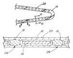
- the porous layer 20is composed of a polymer deposited on the bioactive material layer 18 , preferably by vapor deposition. Plasma deposition may also be useful for this purpose.
- the layer 20is one that is polymerized from a vapor which is free of any solvent, catalysts or similar polymerization promoters.
- the polymer in the porous layer 20is one which automatically polymerizes upon condensation from the vapor phase, without the action of any curative agent or activity such as heating, the application of visible or ultraviolet light, radiation, ultrasound, or the like.
- the polymer in the porous layer 20is polyimide, parylene or a parylene derivative.
- the connector 26is shown in FIG. 5 as a plurality of projections of the base material 14 securing a single porous layer 20 to the base material 14 .
- the connector 26may alternatively extend from the porous layer 20 , through the bioactive material layer 18 , and to the base material 14 . In either case, a single layer 18 of bioactive material, divided into several segments by the connector 26 , is posited between the porous layer 20 and the base material 14 .
- the connectorscan also function to partition the different bioactive agents into different regions of the device's surface.
- the present inventioncan be characterized as being directed to a stent comprising a generally thin walled cylinder, the cylinder having or containing a plurality of arms, the arms being expandable and having a generally uniform thickness; and at least one well 28 ′ formed in at least one of the arms, the well 28 ′ having a closed perimeter on all sides and an open top, and the well 28 ′ being smaller in all dimensions than the arm, the well 28 ′ containing a therapeutic agent applied therein and wherein the therapeutic agent is an immunosuppressive agent.
- the wall 28 ′can be generally rectangular or square in shape.
- the immunosuppressive agentis preferably cyclosporin.
Landscapes
- Health & Medical Sciences (AREA)
- Life Sciences & Earth Sciences (AREA)
- Chemical & Material Sciences (AREA)
- Animal Behavior & Ethology (AREA)
- General Health & Medical Sciences (AREA)
- Public Health (AREA)
- Veterinary Medicine (AREA)
- Epidemiology (AREA)
- Biomedical Technology (AREA)
- Engineering & Computer Science (AREA)
- Medicinal Chemistry (AREA)
- Heart & Thoracic Surgery (AREA)
- Inorganic Chemistry (AREA)
- Vascular Medicine (AREA)
- Surgery (AREA)
- Transplantation (AREA)
- Oral & Maxillofacial Surgery (AREA)
- Molecular Biology (AREA)
- Physics & Mathematics (AREA)
- Optics & Photonics (AREA)
- Dermatology (AREA)
- Cardiology (AREA)
- Hematology (AREA)
- Pharmacology & Pharmacy (AREA)
- Dispersion Chemistry (AREA)
- Materials For Medical Uses (AREA)
- Media Introduction/Drainage Providing Device (AREA)
- Prostheses (AREA)
- Chemical Kinetics & Catalysis (AREA)
- Polymers & Plastics (AREA)
- Organic Chemistry (AREA)
Abstract
Description


Claims (19)
Priority Applications (1)
| Application Number | Priority Date | Filing Date | Title |
|---|---|---|---|
| US11/931,678US8758428B2 (en) | 1995-06-07 | 2007-10-31 | Coated implantable medical device |
Applications Claiming Priority (6)
| Application Number | Priority Date | Filing Date | Title |
|---|---|---|---|
| US08/484,532US5609629A (en) | 1995-06-07 | 1995-06-07 | Coated implantable medical device |
| US08/645,646US6096070A (en) | 1995-06-07 | 1996-05-16 | Coated implantable medical device |
| US3845997P | 1997-02-20 | 1997-02-20 | |
| US09/027,054US6774278B1 (en) | 1995-06-07 | 1998-02-20 | Coated implantable medical device |
| US10/218,305US7550005B2 (en) | 1995-06-07 | 2002-08-14 | Coated implantable medical device |
| US11/931,678US8758428B2 (en) | 1995-06-07 | 2007-10-31 | Coated implantable medical device |
Related Parent Applications (1)
| Application Number | Title | Priority Date | Filing Date |
|---|---|---|---|
| US10/218,305ContinuationUS7550005B2 (en) | 1995-06-07 | 2002-08-14 | Coated implantable medical device |
Publications (2)
| Publication Number | Publication Date |
|---|---|
| US20080147166A1 US20080147166A1 (en) | 2008-06-19 |
| US8758428B2true US8758428B2 (en) | 2014-06-24 |
Family
ID=46281035
Family Applications (3)
| Application Number | Title | Priority Date | Filing Date |
|---|---|---|---|
| US10/218,305Expired - Fee RelatedUS7550005B2 (en) | 1995-06-07 | 2002-08-14 | Coated implantable medical device |
| US11/931,678Expired - Fee RelatedUS8758428B2 (en) | 1995-06-07 | 2007-10-31 | Coated implantable medical device |
| US12/490,162Expired - Fee RelatedUS8945206B2 (en) | 1995-06-07 | 2009-06-23 | Methods for making implantable medical devices |
Family Applications Before (1)
| Application Number | Title | Priority Date | Filing Date |
|---|---|---|---|
| US10/218,305Expired - Fee RelatedUS7550005B2 (en) | 1995-06-07 | 2002-08-14 | Coated implantable medical device |
Family Applications After (1)
| Application Number | Title | Priority Date | Filing Date |
|---|---|---|---|
| US12/490,162Expired - Fee RelatedUS8945206B2 (en) | 1995-06-07 | 2009-06-23 | Methods for making implantable medical devices |
Country Status (1)
| Country | Link |
|---|---|
| US (3) | US7550005B2 (en) |
Cited By (3)
| Publication number | Priority date | Publication date | Assignee | Title |
|---|---|---|---|---|
| WO2017044789A1 (en) | 2015-09-09 | 2017-03-16 | Micell Technologies, Inc. | Biopharma application of micell technology |
| US10758380B2 (en) | 2016-12-30 | 2020-09-01 | Bvw Holding Ag | Stents with improved fixation |
| US11690702B2 (en) | 2020-01-30 | 2023-07-04 | Rambam Medtech Ltd. | Urinary catheter prostheses |
Families Citing this family (273)
| Publication number | Priority date | Publication date | Assignee | Title |
|---|---|---|---|---|
| US7896914B2 (en)* | 1995-06-07 | 2011-03-01 | Cook Incorporated | Coated implantable medical device |
| US7550005B2 (en)* | 1995-06-07 | 2009-06-23 | Cook Incorporated | Coated implantable medical device |
| US7867275B2 (en)* | 1995-06-07 | 2011-01-11 | Cook Incorporated | Coated implantable medical device method |
| US6774278B1 (en)* | 1995-06-07 | 2004-08-10 | Cook Incorporated | Coated implantable medical device |
| US20070203520A1 (en)* | 1995-06-07 | 2007-08-30 | Dennis Griffin | Endovascular filter |
| US7846202B2 (en) | 1995-06-07 | 2010-12-07 | Cook Incorporated | Coated implantable medical device |
| US7611533B2 (en)* | 1995-06-07 | 2009-11-03 | Cook Incorporated | Coated implantable medical device |
| US20060030826A1 (en)* | 1996-06-04 | 2006-02-09 | Vance Products Incorporated,d/b/a Cook Urological Incorporated | Implantable medical device with anti-neoplastic drug |
| US20060025726A1 (en)* | 1996-06-04 | 2006-02-02 | Vance Products Incorporated, D/B/A Cook Urological Incorporated | Implantable medical device with pharmacologically active layer |
| US20060052757A1 (en)* | 1996-06-04 | 2006-03-09 | Vance Products Incorporated, D/B/A Cook Urological Incorporated | Implantable medical device with analgesic or anesthetic |
| US7070590B1 (en)* | 1996-07-02 | 2006-07-04 | Massachusetts Institute Of Technology | Microchip drug delivery devices |
| US6776792B1 (en) | 1997-04-24 | 2004-08-17 | Advanced Cardiovascular Systems Inc. | Coated endovascular stent |
| US7179289B2 (en) | 1998-03-30 | 2007-02-20 | Conor Medsystems, Inc. | Expandable medical device for delivery of beneficial agent |
| US7208010B2 (en) | 2000-10-16 | 2007-04-24 | Conor Medsystems, Inc. | Expandable medical device for delivery of beneficial agent |
| US7208011B2 (en) | 2001-08-20 | 2007-04-24 | Conor Medsystems, Inc. | Implantable medical device with drug filled holes |
| US20040254635A1 (en) | 1998-03-30 | 2004-12-16 | Shanley John F. | Expandable medical device for delivery of beneficial agent |
| US7713297B2 (en) | 1998-04-11 | 2010-05-11 | Boston Scientific Scimed, Inc. | Drug-releasing stent with ceramic-containing layer |
| US20030040790A1 (en)* | 1998-04-15 | 2003-02-27 | Furst Joseph G. | Stent coating |
| US20020099438A1 (en)* | 1998-04-15 | 2002-07-25 | Furst Joseph G. | Irradiated stent coating |
| US8070796B2 (en) | 1998-07-27 | 2011-12-06 | Icon Interventional Systems, Inc. | Thrombosis inhibiting graft |
| US7967855B2 (en)* | 1998-07-27 | 2011-06-28 | Icon Interventional Systems, Inc. | Coated medical device |
| US20070032853A1 (en)* | 2002-03-27 | 2007-02-08 | Hossainy Syed F | 40-O-(2-hydroxy)ethyl-rapamycin coated stent |
| US7807211B2 (en)* | 1999-09-03 | 2010-10-05 | Advanced Cardiovascular Systems, Inc. | Thermal treatment of an implantable medical device |
| AU770395B2 (en)* | 1999-11-17 | 2004-02-19 | Boston Scientific Limited | Microfabricated devices for the delivery of molecules into a carrier fluid |
| US20050238686A1 (en)* | 1999-12-23 | 2005-10-27 | Advanced Cardiovascular Systems, Inc. | Coating for implantable devices and a method of forming the same |
| US6953560B1 (en) | 2000-09-28 | 2005-10-11 | Advanced Cardiovascular Systems, Inc. | Barriers for polymer-coated implantable medical devices and methods for making the same |
| EP1498084B1 (en) | 2000-10-16 | 2014-06-18 | Innovational Holdings, LLC | Expandable medical device for delivery of beneficial agent |
| US6783793B1 (en) | 2000-10-26 | 2004-08-31 | Advanced Cardiovascular Systems, Inc. | Selective coating of medical devices |
| US7807210B1 (en) | 2000-10-31 | 2010-10-05 | Advanced Cardiovascular Systems, Inc. | Hemocompatible polymers on hydrophobic porous polymers |
| GB0100761D0 (en) | 2001-01-11 | 2001-02-21 | Biocompatibles Ltd | Drug delivery from stents |
| DE10115740A1 (en) | 2001-03-26 | 2002-10-02 | Ulrich Speck | Preparation for restenosis prophylaxis |
| US6780424B2 (en) | 2001-03-30 | 2004-08-24 | Charles David Claude | Controlled morphologies in polymer drug for release of drugs from polymer films |
| US7056967B2 (en)* | 2001-04-10 | 2006-06-06 | Ciba Specialty Chemicals Corporation | Stabilized medium and high voltage cable insulation composition |
| US6743462B1 (en)* | 2001-05-31 | 2004-06-01 | Advanced Cardiovascular Systems, Inc. | Apparatus and method for coating implantable devices |
| US6695920B1 (en)* | 2001-06-27 | 2004-02-24 | Advanced Cardiovascular Systems, Inc. | Mandrel for supporting a stent and a method of using the mandrel to coat a stent |
| WO2003002243A2 (en) | 2001-06-27 | 2003-01-09 | Remon Medical Technologies Ltd. | Method and device for electrochemical formation of therapeutic species in vivo |
| US8741378B1 (en) | 2001-06-27 | 2014-06-03 | Advanced Cardiovascular Systems, Inc. | Methods of coating an implantable device |
| US6565659B1 (en) | 2001-06-28 | 2003-05-20 | Advanced Cardiovascular Systems, Inc. | Stent mounting assembly and a method of using the same to coat a stent |
| US7682669B1 (en)* | 2001-07-30 | 2010-03-23 | Advanced Cardiovascular Systems, Inc. | Methods for covalently immobilizing anti-thrombogenic material into a coating on a medical device |
| US7056338B2 (en) | 2003-03-28 | 2006-06-06 | Conor Medsystems, Inc. | Therapeutic agent delivery device with controlled therapeutic agent release rates |
| US8303651B1 (en) | 2001-09-07 | 2012-11-06 | Advanced Cardiovascular Systems, Inc. | Polymeric coating for reducing the rate of release of a therapeutic substance from a stent |
| US6753071B1 (en)* | 2001-09-27 | 2004-06-22 | Advanced Cardiovascular Systems, Inc. | Rate-reducing membrane for release of an agent |
| US8740973B2 (en)* | 2001-10-26 | 2014-06-03 | Icon Medical Corp. | Polymer biodegradable medical device |
| US7033602B1 (en) | 2002-06-21 | 2006-04-25 | Advanced Cardiovascular Systems, Inc. | Polycationic peptide coatings and methods of coating implantable medical devices |
| US7056523B1 (en) | 2002-06-21 | 2006-06-06 | Advanced Cardiovascular Systems, Inc. | Implantable medical devices incorporating chemically conjugated polymers and oligomers of L-arginine |
| US7070798B1 (en) | 2002-06-21 | 2006-07-04 | Advanced Cardiovascular Systems, Inc. | Coatings for implantable medical devices incorporating chemically-bound polymers and oligomers of L-arginine |
| US6994867B1 (en) | 2002-06-21 | 2006-02-07 | Advanced Cardiovascular Systems, Inc. | Biocompatible carrier containing L-arginine |
| US7794743B2 (en)* | 2002-06-21 | 2010-09-14 | Advanced Cardiovascular Systems, Inc. | Polycationic peptide coatings and methods of making the same |
| US7217426B1 (en) | 2002-06-21 | 2007-05-15 | Advanced Cardiovascular Systems, Inc. | Coatings containing polycationic peptides for cardiovascular therapy |
| US8506617B1 (en) | 2002-06-21 | 2013-08-13 | Advanced Cardiovascular Systems, Inc. | Micronized peptide coated stent |
| US7011842B1 (en) | 2002-06-21 | 2006-03-14 | Advanced Cardiovascular Systems, Inc. | Polycationic peptide coatings and methods of making the same |
| DK1521603T3 (en) | 2002-07-12 | 2011-04-18 | Cook Inc | Coated medical device |
| US8016881B2 (en) | 2002-07-31 | 2011-09-13 | Icon Interventional Systems, Inc. | Sutures and surgical staples for anastamoses, wound closures, and surgical closures |
| DE10244847A1 (en) | 2002-09-20 | 2004-04-01 | Ulrich Prof. Dr. Speck | Medical device for drug delivery |
| US6896965B1 (en)* | 2002-11-12 | 2005-05-24 | Advanced Cardiovascular Systems, Inc. | Rate limiting barriers for implantable devices |
| US7776926B1 (en)* | 2002-12-11 | 2010-08-17 | Advanced Cardiovascular Systems, Inc. | Biocompatible coating for implantable medical devices |
| US7758880B2 (en) | 2002-12-11 | 2010-07-20 | Advanced Cardiovascular Systems, Inc. | Biocompatible polyacrylate compositions for medical applications |
| US7074276B1 (en)* | 2002-12-12 | 2006-07-11 | Advanced Cardiovascular Systems, Inc. | Clamp mandrel fixture and a method of using the same to minimize coating defects |
| US7758881B2 (en)* | 2004-06-30 | 2010-07-20 | Advanced Cardiovascular Systems, Inc. | Anti-proliferative and anti-inflammatory agent combination for treatment of vascular disorders with an implantable medical device |
| US8435550B2 (en)* | 2002-12-16 | 2013-05-07 | Abbot Cardiovascular Systems Inc. | Anti-proliferative and anti-inflammatory agent combination for treatment of vascular disorders with an implantable medical device |
| US20060002968A1 (en)* | 2004-06-30 | 2006-01-05 | Gordon Stewart | Anti-proliferative and anti-inflammatory agent combination for treatment of vascular disorders |
| US7094256B1 (en) | 2002-12-16 | 2006-08-22 | Advanced Cardiovascular Systems, Inc. | Coatings for implantable medical device containing polycationic peptides |
| US7063884B2 (en)* | 2003-02-26 | 2006-06-20 | Advanced Cardiovascular Systems, Inc. | Stent coating |
| AU2004226327A1 (en)* | 2003-03-28 | 2004-10-14 | Innovational Holdings, Llc | Implantable medical device with beneficial agent concentration gradient |
| US7279174B2 (en) | 2003-05-08 | 2007-10-09 | Advanced Cardiovascular Systems, Inc. | Stent coatings comprising hydrophilic additives |
| US7169179B2 (en) | 2003-06-05 | 2007-01-30 | Conor Medsystems, Inc. | Drug delivery device and method for bi-directional drug delivery |
| US20050118344A1 (en) | 2003-12-01 | 2005-06-02 | Pacetti Stephen D. | Temperature controlled crimping |
| US7785512B1 (en) | 2003-07-31 | 2010-08-31 | Advanced Cardiovascular Systems, Inc. | Method and system of controlled temperature mixing and molding of polymers with active agents for implantable medical devices |
| US7785653B2 (en) | 2003-09-22 | 2010-08-31 | Innovational Holdings Llc | Method and apparatus for loading a beneficial agent into an expandable medical device |
| US7198675B2 (en) | 2003-09-30 | 2007-04-03 | Advanced Cardiovascular Systems | Stent mandrel fixture and method for selectively coating surfaces of a stent |
| US7261946B2 (en) | 2003-11-14 | 2007-08-28 | Advanced Cardiovascular Systems, Inc. | Block copolymers of acrylates and methacrylates with fluoroalkenes |
| US9114198B2 (en)* | 2003-11-19 | 2015-08-25 | Advanced Cardiovascular Systems, Inc. | Biologically beneficial coatings for implantable devices containing fluorinated polymers and methods for fabricating the same |
| US8192752B2 (en)* | 2003-11-21 | 2012-06-05 | Advanced Cardiovascular Systems, Inc. | Coatings for implantable devices including biologically erodable polyesters and methods for fabricating the same |
| US7220816B2 (en)* | 2003-12-16 | 2007-05-22 | Advanced Cardiovascular Systems, Inc. | Biologically absorbable coatings for implantable devices based on poly(ester amides) and methods for fabricating the same |
| US7435788B2 (en)* | 2003-12-19 | 2008-10-14 | Advanced Cardiovascular Systems, Inc. | Biobeneficial polyamide/polyethylene glycol polymers for use with drug eluting stents |
| US7563324B1 (en) | 2003-12-29 | 2009-07-21 | Advanced Cardiovascular Systems Inc. | System and method for coating an implantable medical device |
| US7211108B2 (en)* | 2004-01-23 | 2007-05-01 | Icon Medical Corp. | Vascular grafts with amphiphilic block copolymer coatings |
| US7803178B2 (en)* | 2004-01-30 | 2010-09-28 | Trivascular, Inc. | Inflatable porous implants and methods for drug delivery |
| US8685431B2 (en)* | 2004-03-16 | 2014-04-01 | Advanced Cardiovascular Systems, Inc. | Biologically absorbable coatings for implantable devices based on copolymers having ester bonds and methods for fabricating the same |
| US8778014B1 (en) | 2004-03-31 | 2014-07-15 | Advanced Cardiovascular Systems, Inc. | Coatings for preventing balloon damage to polymer coated stents |
| US7553377B1 (en) | 2004-04-27 | 2009-06-30 | Advanced Cardiovascular Systems, Inc. | Apparatus and method for electrostatic coating of an abluminal stent surface |
| US8293890B2 (en)* | 2004-04-30 | 2012-10-23 | Advanced Cardiovascular Systems, Inc. | Hyaluronic acid based copolymers |
| US7820732B2 (en) | 2004-04-30 | 2010-10-26 | Advanced Cardiovascular Systems, Inc. | Methods for modulating thermal and mechanical properties of coatings on implantable devices |
| US9561309B2 (en)* | 2004-05-27 | 2017-02-07 | Advanced Cardiovascular Systems, Inc. | Antifouling heparin coatings |
| US7563780B1 (en) | 2004-06-18 | 2009-07-21 | Advanced Cardiovascular Systems, Inc. | Heparin prodrugs and drug delivery stents formed therefrom |
| WO2006002112A1 (en)* | 2004-06-18 | 2006-01-05 | Surmodics, Inc. | Devices, articles, coatings, and methods for controlled active agent release |
| US20050287287A1 (en)* | 2004-06-24 | 2005-12-29 | Parker Theodore L | Methods and systems for loading an implantable medical device with beneficial agent |
| US20050287184A1 (en) | 2004-06-29 | 2005-12-29 | Hossainy Syed F A | Drug-delivery stent formulations for restenosis and vulnerable plaque |
| USD516723S1 (en) | 2004-07-06 | 2006-03-07 | Conor Medsystems, Inc. | Stent wall structure |
| US8357391B2 (en)* | 2004-07-30 | 2013-01-22 | Advanced Cardiovascular Systems, Inc. | Coatings for implantable devices comprising poly (hydroxy-alkanoates) and diacid linkages |
| US7494665B1 (en)* | 2004-07-30 | 2009-02-24 | Advanced Cardiovascular Systems, Inc. | Polymers containing siloxane monomers |
| US7311980B1 (en) | 2004-08-02 | 2007-12-25 | Advanced Cardiovascular Systems, Inc. | Polyactive/polylactic acid coatings for an implantable device |
| US7648727B2 (en) | 2004-08-26 | 2010-01-19 | Advanced Cardiovascular Systems, Inc. | Methods for manufacturing a coated stent-balloon assembly |
| US7244443B2 (en)* | 2004-08-31 | 2007-07-17 | Advanced Cardiovascular Systems, Inc. | Polymers of fluorinated monomers and hydrophilic monomers |
| US8110211B2 (en)* | 2004-09-22 | 2012-02-07 | Advanced Cardiovascular Systems, Inc. | Medicated coatings for implantable medical devices including polyacrylates |
| US9011831B2 (en) | 2004-09-30 | 2015-04-21 | Advanced Cardiovascular Systems, Inc. | Methacrylate copolymers for medical devices |
| US7166680B2 (en) | 2004-10-06 | 2007-01-23 | Advanced Cardiovascular Systems, Inc. | Blends of poly(ester amide) polymers |
| US20060089485A1 (en)* | 2004-10-27 | 2006-04-27 | Desnoyer Jessica R | End-capped poly(ester amide) copolymers |
| US8603634B2 (en) | 2004-10-27 | 2013-12-10 | Abbott Cardiovascular Systems Inc. | End-capped poly(ester amide) copolymers |
| US7481835B1 (en) | 2004-10-29 | 2009-01-27 | Advanced Cardiovascular Systems, Inc. | Encapsulated covered stent |
| US20060095122A1 (en)* | 2004-10-29 | 2006-05-04 | Advanced Cardiovascular Systems, Inc. | Implantable devices comprising biologically absorbable star polymers and methods for fabricating the same |
| US7390497B2 (en)* | 2004-10-29 | 2008-06-24 | Advanced Cardiovascular Systems, Inc. | Poly(ester amide) filler blends for modulation of coating properties |
| US7214759B2 (en)* | 2004-11-24 | 2007-05-08 | Advanced Cardiovascular Systems, Inc. | Biologically absorbable coatings for implantable devices based on polyesters and methods for fabricating the same |
| US8609123B2 (en)* | 2004-11-29 | 2013-12-17 | Advanced Cardiovascular Systems, Inc. | Derivatized poly(ester amide) as a biobeneficial coating |
| US7892592B1 (en) | 2004-11-30 | 2011-02-22 | Advanced Cardiovascular Systems, Inc. | Coating abluminal surfaces of stents and other implantable medical devices |
| US20060115449A1 (en)* | 2004-11-30 | 2006-06-01 | Advanced Cardiovascular Systems, Inc. | Bioabsorbable, biobeneficial, tyrosine-based polymers for use in drug eluting stent coatings |
| US7632307B2 (en) | 2004-12-16 | 2009-12-15 | Advanced Cardiovascular Systems, Inc. | Abluminal, multilayer coating constructs for drug-delivery stents |
| US7604818B2 (en) | 2004-12-22 | 2009-10-20 | Advanced Cardiovascular Systems, Inc. | Polymers of fluorinated monomers and hydrocarbon monomers |
| US7419504B2 (en)* | 2004-12-27 | 2008-09-02 | Advanced Cardiovascular Systems, Inc. | Poly(ester amide) block copolymers |
| US8007775B2 (en)* | 2004-12-30 | 2011-08-30 | Advanced Cardiovascular Systems, Inc. | Polymers containing poly(hydroxyalkanoates) and agents for use with medical articles and methods of fabricating the same |
| US7202325B2 (en) | 2005-01-14 | 2007-04-10 | Advanced Cardiovascular Systems, Inc. | Poly(hydroxyalkanoate-co-ester amides) and agents for use with medical articles |
| KR100511618B1 (en)* | 2005-01-17 | 2005-08-31 | 이경범 | Multi-layer coating of drug release controllable coronary stent and method for manufacturing the same |
| US8202245B2 (en)* | 2005-01-26 | 2012-06-19 | Boston Scientific Scimed, Inc. | Medical devices and methods of making the same |
| US20060178697A1 (en)* | 2005-02-04 | 2006-08-10 | Carr-Brendel Victoria E | Vaso-occlusive devices including non-biodegradable biomaterials |
| US9452001B2 (en)* | 2005-02-22 | 2016-09-27 | Tecres S.P.A. | Disposable device for treatment of infections of human limbs |
| US20060264914A1 (en)* | 2005-03-03 | 2006-11-23 | Icon Medical Corp. | Metal alloys for medical devices |
| US9107899B2 (en) | 2005-03-03 | 2015-08-18 | Icon Medical Corporation | Metal alloys for medical devices |
| US20060201601A1 (en)* | 2005-03-03 | 2006-09-14 | Icon Interventional Systems, Inc. | Flexible markers |
| US20060200048A1 (en)* | 2005-03-03 | 2006-09-07 | Icon Medical Corp. | Removable sheath for device protection |
| WO2006096251A2 (en)* | 2005-03-03 | 2006-09-14 | Icon Medical Corp. | Improved metal alloys for medical device |
| WO2006110197A2 (en)* | 2005-03-03 | 2006-10-19 | Icon Medical Corp. | Polymer biodegradable medical device |
| US8323333B2 (en)* | 2005-03-03 | 2012-12-04 | Icon Medical Corp. | Fragile structure protective coating |
| US7540995B2 (en)* | 2005-03-03 | 2009-06-02 | Icon Medical Corp. | Process for forming an improved metal alloy stent |
| US9381279B2 (en) | 2005-03-24 | 2016-07-05 | Abbott Cardiovascular Systems Inc. | Implantable devices formed on non-fouling methacrylate or acrylate polymers |
| US7700659B2 (en)* | 2005-03-24 | 2010-04-20 | Advanced Cardiovascular Systems, Inc. | Implantable devices formed of non-fouling methacrylate or acrylate polymers |
| US7795467B1 (en) | 2005-04-26 | 2010-09-14 | Advanced Cardiovascular Systems, Inc. | Bioabsorbable, biobeneficial polyurethanes for use in medical devices |
| US8778375B2 (en) | 2005-04-29 | 2014-07-15 | Advanced Cardiovascular Systems, Inc. | Amorphous poly(D,L-lactide) coating |
| AU2006244393B2 (en)* | 2005-05-05 | 2012-06-21 | Cook Biotech Incorporated | Implantable materials and methods for inhibiting tissue adhesion formation |
| US7637941B1 (en) | 2005-05-11 | 2009-12-29 | Advanced Cardiovascular Systems, Inc. | Endothelial cell binding coatings for rapid encapsulation of bioerodable stents |
| US7622070B2 (en)* | 2005-06-20 | 2009-11-24 | Advanced Cardiovascular Systems, Inc. | Method of manufacturing an implantable polymeric medical device |
| US7823533B2 (en)* | 2005-06-30 | 2010-11-02 | Advanced Cardiovascular Systems, Inc. | Stent fixture and method for reducing coating defects |
| US8021676B2 (en) | 2005-07-08 | 2011-09-20 | Advanced Cardiovascular Systems, Inc. | Functionalized chemically inert polymers for coatings |
| US7785647B2 (en)* | 2005-07-25 | 2010-08-31 | Advanced Cardiovascular Systems, Inc. | Methods of providing antioxidants to a drug containing product |
| US7735449B1 (en) | 2005-07-28 | 2010-06-15 | Advanced Cardiovascular Systems, Inc. | Stent fixture having rounded support structures and method for use thereof |
| US20070128246A1 (en)* | 2005-12-06 | 2007-06-07 | Hossainy Syed F A | Solventless method for forming a coating |
| US20070135909A1 (en)* | 2005-12-08 | 2007-06-14 | Desnoyer Jessica R | Adhesion polymers to improve stent retention |
| US7591841B2 (en) | 2005-12-16 | 2009-09-22 | Advanced Cardiovascular Systems, Inc. | Implantable devices for accelerated healing |
| US7976891B1 (en) | 2005-12-16 | 2011-07-12 | Advanced Cardiovascular Systems, Inc. | Abluminal stent coating apparatus and method of using focused acoustic energy |
| US7867547B2 (en) | 2005-12-19 | 2011-01-11 | Advanced Cardiovascular Systems, Inc. | Selectively coating luminal surfaces of stents |
| US7638156B1 (en) | 2005-12-19 | 2009-12-29 | Advanced Cardiovascular Systems, Inc. | Apparatus and method for selectively coating a medical article |
| US20080286325A1 (en)* | 2006-01-05 | 2008-11-20 | Med Institute, Inc. | Cyclodextrin elution media for medical device coatings comprising a taxane therapeutic agent |
| US8840660B2 (en) | 2006-01-05 | 2014-09-23 | Boston Scientific Scimed, Inc. | Bioerodible endoprostheses and methods of making the same |
| US7919108B2 (en)* | 2006-03-10 | 2011-04-05 | Cook Incorporated | Taxane coatings for implantable medical devices |
| US8089029B2 (en) | 2006-02-01 | 2012-01-03 | Boston Scientific Scimed, Inc. | Bioabsorbable metal medical device and method of manufacture |
| US20070196428A1 (en)* | 2006-02-17 | 2007-08-23 | Thierry Glauser | Nitric oxide generating medical devices |
| US7601383B2 (en)* | 2006-02-28 | 2009-10-13 | Advanced Cardiovascular Systems, Inc. | Coating construct containing poly (vinyl alcohol) |
| US7713637B2 (en)* | 2006-03-03 | 2010-05-11 | Advanced Cardiovascular Systems, Inc. | Coating containing PEGylated hyaluronic acid and a PEGylated non-hyaluronic acid polymer |
| US7875284B2 (en)* | 2006-03-10 | 2011-01-25 | Cook Incorporated | Methods of manufacturing and modifying taxane coatings for implantable medical devices |
| US20070224235A1 (en)* | 2006-03-24 | 2007-09-27 | Barron Tenney | Medical devices having nanoporous coatings for controlled therapeutic agent delivery |
| US8187620B2 (en)* | 2006-03-27 | 2012-05-29 | Boston Scientific Scimed, Inc. | Medical devices comprising a porous metal oxide or metal material and a polymer coating for delivering therapeutic agents |
| US20070231363A1 (en)* | 2006-03-29 | 2007-10-04 | Yung-Ming Chen | Coatings formed from stimulus-sensitive material |
| US8048150B2 (en) | 2006-04-12 | 2011-11-01 | Boston Scientific Scimed, Inc. | Endoprosthesis having a fiber meshwork disposed thereon |
| US8303648B2 (en)* | 2006-04-25 | 2012-11-06 | Cook Medical Technologies Llc | Artificial venous valve containing therapeutic agent |
| US20070259101A1 (en)* | 2006-05-02 | 2007-11-08 | Kleiner Lothar W | Microporous coating on medical devices |
| US7985441B1 (en) | 2006-05-04 | 2011-07-26 | Yiwen Tang | Purification of polymers for coating applications |
| US8003156B2 (en) | 2006-05-04 | 2011-08-23 | Advanced Cardiovascular Systems, Inc. | Rotatable support elements for stents |
| US8304012B2 (en)* | 2006-05-04 | 2012-11-06 | Advanced Cardiovascular Systems, Inc. | Method for drying a stent |
| US20070275035A1 (en)* | 2006-05-24 | 2007-11-29 | Microchips, Inc. | Minimally Invasive Medical Implant Devices for Controlled Drug Delivery |
| US7775178B2 (en)* | 2006-05-26 | 2010-08-17 | Advanced Cardiovascular Systems, Inc. | Stent coating apparatus and method |
| US8568764B2 (en) | 2006-05-31 | 2013-10-29 | Advanced Cardiovascular Systems, Inc. | Methods of forming coating layers for medical devices utilizing flash vaporization |
| US9561351B2 (en) | 2006-05-31 | 2017-02-07 | Advanced Cardiovascular Systems, Inc. | Drug delivery spiral coil construct |
| US8703167B2 (en) | 2006-06-05 | 2014-04-22 | Advanced Cardiovascular Systems, Inc. | Coatings for implantable medical devices for controlled release of a hydrophilic drug and a hydrophobic drug |
| US20080124372A1 (en)* | 2006-06-06 | 2008-05-29 | Hossainy Syed F A | Morphology profiles for control of agent release rates from polymer matrices |
| US20070286882A1 (en)* | 2006-06-09 | 2007-12-13 | Yiwen Tang | Solvent systems for coating medical devices |
| US8778376B2 (en)* | 2006-06-09 | 2014-07-15 | Advanced Cardiovascular Systems, Inc. | Copolymer comprising elastin pentapeptide block and hydrophilic block, and medical device and method of treating |
| US8114150B2 (en) | 2006-06-14 | 2012-02-14 | Advanced Cardiovascular Systems, Inc. | RGD peptide attached to bioabsorbable stents |
| US20080095918A1 (en)* | 2006-06-14 | 2008-04-24 | Kleiner Lothar W | Coating construct with enhanced interfacial compatibility |
| US8603530B2 (en) | 2006-06-14 | 2013-12-10 | Abbott Cardiovascular Systems Inc. | Nanoshell therapy |
| US8048448B2 (en) | 2006-06-15 | 2011-11-01 | Abbott Cardiovascular Systems Inc. | Nanoshells for drug delivery |
| JP2009540926A (en)* | 2006-06-22 | 2009-11-26 | ウィルソン−クック・メディカル・インコーポレーテッド | Self-cleaning stent |
| US8017237B2 (en) | 2006-06-23 | 2011-09-13 | Abbott Cardiovascular Systems, Inc. | Nanoshells on polymers |
| US8815275B2 (en) | 2006-06-28 | 2014-08-26 | Boston Scientific Scimed, Inc. | Coatings for medical devices comprising a therapeutic agent and a metallic material |
| WO2008002778A2 (en) | 2006-06-29 | 2008-01-03 | Boston Scientific Limited | Medical devices with selective coating |
| US8956640B2 (en)* | 2006-06-29 | 2015-02-17 | Advanced Cardiovascular Systems, Inc. | Block copolymers including a methoxyethyl methacrylate midblock |
| US20080008736A1 (en)* | 2006-07-06 | 2008-01-10 | Thierry Glauser | Random copolymers of methacrylates and acrylates |
| US9028859B2 (en)* | 2006-07-07 | 2015-05-12 | Advanced Cardiovascular Systems, Inc. | Phase-separated block copolymer coatings for implantable medical devices |
| WO2008008291A2 (en)* | 2006-07-13 | 2008-01-17 | Icon Medical Corp. | Stent |
| WO2008006911A1 (en)* | 2006-07-14 | 2008-01-17 | Biocompatibles Uk Limited | Coated implant |
| EP2054537A2 (en) | 2006-08-02 | 2009-05-06 | Boston Scientific Scimed, Inc. | Endoprosthesis with three-dimensional disintegration control |
| US8703169B1 (en) | 2006-08-15 | 2014-04-22 | Abbott Cardiovascular Systems Inc. | Implantable device having a coating comprising carrageenan and a biostable polymer |
| US20080051911A1 (en)* | 2006-08-23 | 2008-02-28 | Wilson-Cook Medical Inc. | Stent with antimicrobial drainage lumen surface |
| EP2068757B1 (en)* | 2006-09-14 | 2011-05-11 | Boston Scientific Limited | Medical devices with drug-eluting coating |
| JP2010503489A (en) | 2006-09-15 | 2010-02-04 | ボストン サイエンティフィック リミテッド | Biodegradable endoprosthesis and method for producing the same |
| EP2959925B1 (en) | 2006-09-15 | 2018-08-29 | Boston Scientific Limited | Medical devices and methods of making the same |
| WO2008034066A1 (en) | 2006-09-15 | 2008-03-20 | Boston Scientific Limited | Bioerodible endoprostheses and methods of making the same |
| ES2357661T3 (en) | 2006-09-15 | 2011-04-28 | Boston Scientific Scimed, Inc. | BIOEROSIONABLE ENDOPROOTHESIS WITH BIOESTABLE INORGANIC LAYERS. |
| WO2008036548A2 (en) | 2006-09-18 | 2008-03-27 | Boston Scientific Limited | Endoprostheses |
| US20080075753A1 (en)* | 2006-09-25 | 2008-03-27 | Chappa Ralph A | Multi-layered coatings and methods for controlling elution of active agents |
| RU2009121671A (en)* | 2006-11-06 | 2010-12-20 | Биоринг Са (Ch) | DEVICE FOR TREATMENT OF URETHRIC STRUCTURE |
| US7981150B2 (en) | 2006-11-09 | 2011-07-19 | Boston Scientific Scimed, Inc. | Endoprosthesis with coatings |
| WO2008060973A1 (en)* | 2006-11-10 | 2008-05-22 | Vance Products Incorporated D/B/A Cook Urological Incorporated | Graft for hysterotomy closure |
| US7713541B1 (en)* | 2006-11-21 | 2010-05-11 | Abbott Cardiovascular Systems Inc. | Zwitterionic terpolymers, method of making and use on medical devices |
| JP5557373B2 (en) | 2006-11-21 | 2014-07-23 | アボット ラボラトリーズ | Use of terpolymers of tetrafluoroethylene, hexafluoropropylene, and vinylidene fluoride in drug-eluting coatings |
| US20080118541A1 (en)* | 2006-11-21 | 2008-05-22 | Abbott Laboratories | Use of a terpolymer of tetrafluoroethylene, hexafluoropropylene, and vinylidene fluoride in drug eluting coatings on medical devices |
| US8597673B2 (en)* | 2006-12-13 | 2013-12-03 | Advanced Cardiovascular Systems, Inc. | Coating of fast absorption or dissolution |
| US8017141B2 (en) | 2006-12-15 | 2011-09-13 | Advanced Cardiovascular Systems, Inc. | Coatings of acrylamide-based copolymers |
| ES2506144T3 (en) | 2006-12-28 | 2014-10-13 | Boston Scientific Limited | Bioerodible endoprosthesis and their manufacturing procedure |
| US8814930B2 (en) | 2007-01-19 | 2014-08-26 | Elixir Medical Corporation | Biodegradable endoprosthesis and methods for their fabrication |
| WO2008094548A2 (en)* | 2007-01-30 | 2008-08-07 | Boston Scientific Limited . | Local delivery of therapeutic agent to heart valves |
| US8932345B2 (en)* | 2007-02-07 | 2015-01-13 | Cook Medical Technologies Llc | Medical device coatings for releasing a therapeutic agent at multiple rates |
| US8431149B2 (en) | 2007-03-01 | 2013-04-30 | Boston Scientific Scimed, Inc. | Coated medical devices for abluminal drug delivery |
| US8070797B2 (en) | 2007-03-01 | 2011-12-06 | Boston Scientific Scimed, Inc. | Medical device with a porous surface for delivery of a therapeutic agent |
| US8067054B2 (en) | 2007-04-05 | 2011-11-29 | Boston Scientific Scimed, Inc. | Stents with ceramic drug reservoir layer and methods of making and using the same |
| EP2146673A1 (en)* | 2007-04-11 | 2010-01-27 | Boston Scientific Scimed, Inc. | Photoresist coating to apply a coating to select areas of a medical device |
| US20080286332A1 (en) | 2007-05-14 | 2008-11-20 | Pacetti Stephen D | Implantable medical devices with a topcoat layer of phosphoryl choline acrylate polymer for reduced thrombosis, and improved mechanical properties |
| US8147769B1 (en) | 2007-05-16 | 2012-04-03 | Abbott Cardiovascular Systems Inc. | Stent and delivery system with reduced chemical degradation |
| US7976915B2 (en) | 2007-05-23 | 2011-07-12 | Boston Scientific Scimed, Inc. | Endoprosthesis with select ceramic morphology |
| US9056155B1 (en) | 2007-05-29 | 2015-06-16 | Abbott Cardiovascular Systems Inc. | Coatings having an elastic primer layer |
| WO2008150375A1 (en) | 2007-06-01 | 2008-12-11 | Arrow International, Inc. | Combined fibrinolytic and antimicrobial catheter and uses thereof |
| US8048441B2 (en) | 2007-06-25 | 2011-11-01 | Abbott Cardiovascular Systems, Inc. | Nanobead releasing medical devices |
| US8109904B1 (en) | 2007-06-25 | 2012-02-07 | Abbott Cardiovascular Systems Inc. | Drug delivery medical devices |
| US7942926B2 (en)* | 2007-07-11 | 2011-05-17 | Boston Scientific Scimed, Inc. | Endoprosthesis coating |
| US8002823B2 (en) | 2007-07-11 | 2011-08-23 | Boston Scientific Scimed, Inc. | Endoprosthesis coating |
| US8205317B2 (en)* | 2007-07-16 | 2012-06-26 | Medtronic Vascular, Inc. | Method of manufacturing a controlled porosity stent |
| EP2187988B1 (en) | 2007-07-19 | 2013-08-21 | Boston Scientific Limited | Endoprosthesis having a non-fouling surface |
| US7931683B2 (en) | 2007-07-27 | 2011-04-26 | Boston Scientific Scimed, Inc. | Articles having ceramic coated surfaces |
| US8815273B2 (en) | 2007-07-27 | 2014-08-26 | Boston Scientific Scimed, Inc. | Drug eluting medical devices having porous layers |
| WO2009018340A2 (en) | 2007-07-31 | 2009-02-05 | Boston Scientific Scimed, Inc. | Medical device coating by laser cladding |
| JP2010535541A (en) | 2007-08-03 | 2010-11-25 | ボストン サイエンティフィック リミテッド | Coating for medical devices with large surface area |
| US20090041845A1 (en)* | 2007-08-08 | 2009-02-12 | Lothar Walter Kleiner | Implantable medical devices having thin absorbable coatings |
| CA2698096C (en)* | 2007-08-31 | 2013-01-22 | Wilson-Cook Medical Inc. | Medical implant having improved drug eluting features |
| US8052745B2 (en) | 2007-09-13 | 2011-11-08 | Boston Scientific Scimed, Inc. | Endoprosthesis |
| US8226701B2 (en) | 2007-09-26 | 2012-07-24 | Trivascular, Inc. | Stent and delivery system for deployment thereof |
| US8663309B2 (en) | 2007-09-26 | 2014-03-04 | Trivascular, Inc. | Asymmetric stent apparatus and method |
| US8066755B2 (en) | 2007-09-26 | 2011-11-29 | Trivascular, Inc. | System and method of pivoted stent deployment |
| US10159557B2 (en) | 2007-10-04 | 2018-12-25 | Trivascular, Inc. | Modular vascular graft for low profile percutaneous delivery |
| US8029554B2 (en) | 2007-11-02 | 2011-10-04 | Boston Scientific Scimed, Inc. | Stent with embedded material |
| US7938855B2 (en) | 2007-11-02 | 2011-05-10 | Boston Scientific Scimed, Inc. | Deformable underlayer for stent |
| US8216632B2 (en) | 2007-11-02 | 2012-07-10 | Boston Scientific Scimed, Inc. | Endoprosthesis coating |
| US20090118813A1 (en)* | 2007-11-02 | 2009-05-07 | Torsten Scheuermann | Nano-patterned implant surfaces |
| US8328861B2 (en) | 2007-11-16 | 2012-12-11 | Trivascular, Inc. | Delivery system and method for bifurcated graft |
| US8083789B2 (en) | 2007-11-16 | 2011-12-27 | Trivascular, Inc. | Securement assembly and method for expandable endovascular device |
| US8388678B2 (en)* | 2007-12-12 | 2013-03-05 | Boston Scientific Scimed, Inc. | Medical devices having porous component for controlled diffusion |
| US20090192580A1 (en)* | 2008-01-28 | 2009-07-30 | Shrojalkumar Desai | Medical electrical lead with biocompatible lead body coating |
| JP2011512215A (en)* | 2008-02-21 | 2011-04-21 | エグザカトゥ | Implantable medical device with a protective / holding layer for active ingredients or drugs, especially for water-soluble ones |
| FR2927812B1 (en)* | 2008-02-21 | 2011-09-23 | Hexacath | IMPLANTABLE MEDICAL DEVICE WITH DRUG AND PROTECTIVE OR RETENTIVE LAYER THEREOF |
| FR2927815B1 (en)* | 2008-02-21 | 2011-01-14 | Hexacath | MEDICAL DEVICE COMPRISING A NO PRECURSOR AGENT, SUCH AS L-ARGININE OR L-LYSINE, AND A PROTECTIVE AND / OR RETENTIVE LAYER THEREOF |
| US20100042206A1 (en)* | 2008-03-04 | 2010-02-18 | Icon Medical Corp. | Bioabsorbable coatings for medical devices |
| US8920491B2 (en) | 2008-04-22 | 2014-12-30 | Boston Scientific Scimed, Inc. | Medical devices having a coating of inorganic material |
| US8932346B2 (en) | 2008-04-24 | 2015-01-13 | Boston Scientific Scimed, Inc. | Medical devices having inorganic particle layers |
| US8114151B2 (en)* | 2008-05-08 | 2012-02-14 | Boston Scientific Scimed, Inc. | Stent with tabs and holes for drug delivery |
| US7998192B2 (en) | 2008-05-09 | 2011-08-16 | Boston Scientific Scimed, Inc. | Endoprostheses |
| US8236046B2 (en) | 2008-06-10 | 2012-08-07 | Boston Scientific Scimed, Inc. | Bioerodible endoprosthesis |
| US20090312832A1 (en)* | 2008-06-13 | 2009-12-17 | Cook Incorporated | Slip layer delivery catheter |
| EP2303350A2 (en) | 2008-06-18 | 2011-04-06 | Boston Scientific Scimed, Inc. | Endoprosthesis coating |
| US7985252B2 (en) | 2008-07-30 | 2011-07-26 | Boston Scientific Scimed, Inc. | Bioerodible endoprosthesis |
| US8382824B2 (en) | 2008-10-03 | 2013-02-26 | Boston Scientific Scimed, Inc. | Medical implant having NANO-crystal grains with barrier layers of metal nitrides or fluorides |
| US8231980B2 (en) | 2008-12-03 | 2012-07-31 | Boston Scientific Scimed, Inc. | Medical implants including iridium oxide |
| EP2403546A2 (en) | 2009-03-02 | 2012-01-11 | Boston Scientific Scimed, Inc. | Self-buffering medical implants |
| US8071156B2 (en) | 2009-03-04 | 2011-12-06 | Boston Scientific Scimed, Inc. | Endoprostheses |
| EP2413839B1 (en)* | 2009-04-02 | 2014-05-07 | Cook Medical Technologies LLC | System and method for maintaining patency of a stent using a magnet |
| US8287937B2 (en) | 2009-04-24 | 2012-10-16 | Boston Scientific Scimed, Inc. | Endoprosthese |
| US8016872B2 (en)* | 2009-12-22 | 2011-09-13 | Cook Medical Technologies Llc | Deployment and dilation with an expandable roll sock delivery system |
| US8398916B2 (en) | 2010-03-04 | 2013-03-19 | Icon Medical Corp. | Method for forming a tubular medical device |
| US8668732B2 (en) | 2010-03-23 | 2014-03-11 | Boston Scientific Scimed, Inc. | Surface treated bioerodible metal endoprostheses |
| US8685433B2 (en) | 2010-03-31 | 2014-04-01 | Abbott Cardiovascular Systems Inc. | Absorbable coating for implantable device |
| EP2399618A1 (en)* | 2010-06-22 | 2011-12-28 | Planton GmbH | Antimicrobial medical devices |
| PL217879B1 (en) | 2010-07-06 | 2014-08-29 | Univ Jagielloński | Multi-layer protective coating for protecting metal surfaces of implant materials and the application thereof |
| CA2827330A1 (en) | 2011-02-23 | 2012-08-30 | Ams Research Corporation | Pelvic implant and therapeutic agent system and method |
| JP2014530062A (en)* | 2011-10-06 | 2014-11-17 | パーデュー・リサーチ・ファウンデーションPurdue Research Foundation | System and stent for repairing defects in blood vessels and methods of use |
| US9987130B2 (en)* | 2011-12-13 | 2018-06-05 | Boston Scientific Scimed, Inc. | Decalcifying heart valve |
| US8992595B2 (en) | 2012-04-04 | 2015-03-31 | Trivascular, Inc. | Durable stent graft with tapered struts and stable delivery methods and devices |
| US9498363B2 (en) | 2012-04-06 | 2016-11-22 | Trivascular, Inc. | Delivery catheter for endovascular device |
| BR112016030273A8 (en) | 2014-06-24 | 2021-05-18 | Icon Medical Corp | medical device and method of forming said device |
| US9730819B2 (en) | 2014-08-15 | 2017-08-15 | Elixir Medical Corporation | Biodegradable endoprostheses and methods of their fabrication |
| US9480588B2 (en) | 2014-08-15 | 2016-11-01 | Elixir Medical Corporation | Biodegradable endoprostheses and methods of their fabrication |
| US9259339B1 (en)* | 2014-08-15 | 2016-02-16 | Elixir Medical Corporation | Biodegradable endoprostheses and methods of their fabrication |
| US9855156B2 (en)* | 2014-08-15 | 2018-01-02 | Elixir Medical Corporation | Biodegradable endoprostheses and methods of their fabrication |
| WO2016148648A1 (en)* | 2015-03-19 | 2016-09-22 | Nanyang Technological University | A stent assembly and method of preparing the stent assembly |
| US11766506B2 (en) | 2016-03-04 | 2023-09-26 | Mirus Llc | Stent device for spinal fusion |
| US11622872B2 (en) | 2016-05-16 | 2023-04-11 | Elixir Medical Corporation | Uncaging stent |
| JP6963566B2 (en) | 2016-05-16 | 2021-11-10 | エリクシアー メディカル コーポレイション | Uncaging stent |
| US9999899B2 (en) | 2016-11-01 | 2018-06-19 | International Business Machines Corporation | Controlled exposure of in-vivo sensors |
Citations (171)
| Publication number | Priority date | Publication date | Assignee | Title |
|---|---|---|---|---|
| US3908201A (en) | 1972-06-30 | 1975-09-30 | Ici Ltd | Prosthetics |
| US3952334A (en) | 1974-11-29 | 1976-04-27 | General Atomic Company | Biocompatible carbon prosthetic devices |
| US4101984A (en) | 1975-05-09 | 1978-07-25 | Macgregor David C | Cardiovascular prosthetic devices and implants with porous systems |
| US4300244A (en) | 1979-09-19 | 1981-11-17 | Carbomedics, Inc. | Cardiovascular grafts |
| US4305926A (en) | 1979-09-13 | 1981-12-15 | Johannes Everse | Immobilization of Streptokinase |
| US4343788A (en) | 1979-06-29 | 1982-08-10 | The Procter & Gamble Company | Antimicrobial polymer compositions |
| US4374669A (en)* | 1975-05-09 | 1983-02-22 | Mac Gregor David C | Cardiovascular prosthetic devices and implants with porous systems |
| US4476590A (en) | 1980-03-27 | 1984-10-16 | National Research Development Corporation | Antimicrobial surgical implants |
| US4479795A (en) | 1979-06-29 | 1984-10-30 | The Procter & Gamble Company | Antimicrobial polymer compositions |
| US4502159A (en) | 1982-08-12 | 1985-03-05 | Shiley Incorporated | Tubular prostheses prepared from pericardial tissue |
| US4666704A (en) | 1985-05-24 | 1987-05-19 | International Minerals & Chemical Corp. | Controlled release delivery system for macromolecules |
| US4677143A (en) | 1984-10-01 | 1987-06-30 | Baxter Travenol Laboratories, Inc. | Antimicrobial compositions |
| US4689046A (en) | 1985-03-11 | 1987-08-25 | Carbomedics, Inc. | Heart valve prosthesis |
| US4689386A (en) | 1984-11-07 | 1987-08-25 | Biocompatibles Ltd. | Biocompatible surfaces |
| US4739762A (en) | 1985-11-07 | 1988-04-26 | Expandable Grafts Partnership | Expandable intraluminal graft, and method and apparatus for implanting an expandable intraluminal graft |
| US4768507A (en) | 1986-02-24 | 1988-09-06 | Medinnovations, Inc. | Intravascular stent and percutaneous insertion catheter system for the dilation of an arterial stenosis and the prevention of arterial restenosis |
| US4769013A (en) | 1982-09-13 | 1988-09-06 | Hydromer, Inc. | Bio-effecting medical material and device |
| US4793825A (en) | 1984-09-11 | 1988-12-27 | The Secretary Of State For Defence In Her Britannic Majesty's Government Of The United Kingdom And Northern Ireland | Active silicon implant devices |
| US4872867A (en) | 1985-06-19 | 1989-10-10 | Ube Industries, Ltd. | Compositions having antithrombogenic properties and blood contact medical devices using the same |
| US4879135A (en) | 1984-07-23 | 1989-11-07 | University Of Medicine And Dentistry Of New Jersey | Drug bonded prosthesis and process for producing same |
| US4886062A (en) | 1987-10-19 | 1989-12-12 | Medtronic, Inc. | Intravascular radially expandable stent and method of implant |
| US4909799A (en) | 1987-09-18 | 1990-03-20 | Olav Thulesius | Methods for preventing thrombosis; and surgical implant having reduced platelet deposition characteristics |
| US4917686A (en) | 1985-12-16 | 1990-04-17 | Colorado Biomedical, Inc. | Antimicrobial device and method |
| US4925668A (en) | 1989-01-18 | 1990-05-15 | Becton, Dickinson And Company | Anti-infective and lubricious medical articles and method for their preparation |
| US4950256A (en) | 1988-04-07 | 1990-08-21 | Luther Medical Products, Inc. | Non-thrombogenic intravascular time release catheter |
| US4994047A (en) | 1988-05-06 | 1991-02-19 | Menlo Care, Inc. | Multi-layer cannula structure |
| US5004461A (en) | 1989-03-23 | 1991-04-02 | Wilson Joseph E | Methods for rendering plastics thromboresistant and product |
| US5019601A (en) | 1987-12-29 | 1991-05-28 | Cuno, Incorporated | Elastomeric composition containing therapeutic agents and articles manufactured therefrom |
| US5019393A (en) | 1988-08-03 | 1991-05-28 | New England Deaconess Hospital Corporation | Biocompatible substance with thromboresistance |
| US5019096A (en) | 1988-02-11 | 1991-05-28 | Trustees Of Columbia University In The City Of New York | Infection-resistant compositions, medical devices and surfaces and methods for preparing and using same |
| US5032113A (en) | 1989-04-13 | 1991-07-16 | Scimed Life Systems, Inc. | Innerless catheter |
| US5053048A (en) | 1988-09-22 | 1991-10-01 | Cordis Corporation | Thromboresistant coating |
| US5059166A (en) | 1989-12-11 | 1991-10-22 | Medical Innovative Technologies R & D Limited Partnership | Intra-arterial stent with the capability to inhibit intimal hyperplasia |
| US5067491A (en) | 1989-12-08 | 1991-11-26 | Becton, Dickinson And Company | Barrier coating on blood contacting devices |
| US5078736A (en) | 1990-05-04 | 1992-01-07 | Interventional Thermodynamics, Inc. | Method and apparatus for maintaining patency in the body passages |
| US5092841A (en) | 1990-05-17 | 1992-03-03 | Wayne State University | Method for treating an arterial wall injured during angioplasty |
| US5098977A (en) | 1987-09-23 | 1992-03-24 | Board Of Regents, The University Of Texas System | Methods and compositions for providing articles having improved biocompatability characteristics |
| US5102402A (en) | 1991-01-04 | 1992-04-07 | Medtronic, Inc. | Releasable coatings on balloon catheters |
| US5102417A (en) | 1985-11-07 | 1992-04-07 | Expandable Grafts Partnership | Expandable intraluminal graft, and method and apparatus for implanting an expandable intraluminal graft |
| US5108424A (en) | 1984-01-30 | 1992-04-28 | Meadox Medicals, Inc. | Collagen-impregnated dacron graft |
| US5112457A (en) | 1990-07-23 | 1992-05-12 | Case Western Reserve University | Process for producing hydroxylated plasma-polymerized films and the use of the films for enhancing the compatiblity of biomedical implants |
| US5135516A (en) | 1989-12-15 | 1992-08-04 | Boston Scientific Corporation | Lubricious antithrombogenic catheters, guidewires and coatings |
| US5163952A (en) | 1990-09-14 | 1992-11-17 | Michael Froix | Expandable polymeric stent with memory and delivery apparatus and method |
| US5165952A (en) | 1989-01-18 | 1992-11-24 | Becton, Dickinson And Company | Anti-infective and antithrombogenic medical articles and method for their preparation |
| US5171217A (en) | 1991-02-28 | 1992-12-15 | Indiana University Foundation | Method for delivery of smooth muscle cell inhibitors |
| US5180366A (en) | 1990-10-10 | 1993-01-19 | Woods W T | Apparatus and method for angioplasty and for preventing re-stenosis |
| US5182317A (en) | 1988-06-08 | 1993-01-26 | Cardiopulmonics, Inc. | Multifunctional thrombo-resistant coatings and methods of manufacture |
| US5197977A (en) | 1984-01-30 | 1993-03-30 | Meadox Medicals, Inc. | Drug delivery collagen-impregnated synthetic vascular graft |
| US5217493A (en) | 1992-03-11 | 1993-06-08 | Board Of Regents, The University Of Texas System | Antibacterial coated medical implants |
| US5222971A (en) | 1990-10-09 | 1993-06-29 | Scimed Life Systems, Inc. | Temporary stent and methods for use and manufacture |
| US5229172A (en) | 1993-01-19 | 1993-07-20 | Medtronic, Inc. | Modification of polymeric surface by graft polymerization |
| US5234456A (en) | 1990-02-08 | 1993-08-10 | Pfizer Hospital Products Group, Inc. | Hydrophilic stent |
| US5236447A (en) | 1990-06-29 | 1993-08-17 | Nissho Corporation | Artificial tubular organ |
| US5244654A (en) | 1990-11-08 | 1993-09-14 | Cordis Corporation | Radiofrequency plasma biocompatibility treatment of inside surfaces of medical tubing and the like |
| US5282823A (en) | 1992-03-19 | 1994-02-01 | Medtronic, Inc. | Intravascular radially expandable stent |
| US5283257A (en)* | 1992-07-10 | 1994-02-01 | The Board Of Trustees Of The Leland Stanford Junior University | Method of treating hyperproliferative vascular disease |
| US5288711A (en) | 1992-04-28 | 1994-02-22 | American Home Products Corporation | Method of treating hyperproliferative vascular disease |
| US5290271A (en) | 1990-05-14 | 1994-03-01 | Jernberg Gary R | Surgical implant and method for controlled release of chemotherapeutic agents |
| US5298255A (en) | 1988-10-28 | 1994-03-29 | Terumo Kabushiki Kaisha | Antithrombic medical material, artificial internal organ, and method for production of antithrombic medical material |
| US5304121A (en) | 1990-12-28 | 1994-04-19 | Boston Scientific Corporation | Drug delivery system making use of a hydrogel polymer coating |
| US5314688A (en) | 1991-11-21 | 1994-05-24 | Eli Lilly And Company | Local delivery of dipyridamole for the treatment of proliferative diseases |
| US5334015A (en) | 1992-03-26 | 1994-08-02 | Blechman Abraham M | Magnetic orthodontic appliance |
| US5342348A (en) | 1992-12-04 | 1994-08-30 | Kaplan Aaron V | Method and device for treating and enlarging body lumens |
| US5342621A (en) | 1992-09-15 | 1994-08-30 | Advanced Cardiovascular Systems, Inc. | Antithrombogenic surface |
| US5344444A (en) | 1991-07-03 | 1994-09-06 | Industrial Research B.V. | Expandable ring, cylinder or sleeve which can be made non-deformable |
| US5344411A (en) | 1991-02-27 | 1994-09-06 | Leonard Bloom | Method and device for inhibiting HIV, hepatitis B and other viruses and germs when using a catheter in a medical environment |
| US5345933A (en) | 1991-09-25 | 1994-09-13 | Siemens Aktiengesellschaft | Implantable medical device with a protected medication layer |
| US5348873A (en) | 1991-11-01 | 1994-09-20 | K.K. Vayu | Immobilization of an anti-thrombogenic substance with a photo-reactive azide and a photo-crosslinking material |
| US5356433A (en) | 1991-08-13 | 1994-10-18 | Cordis Corporation | Biocompatible metal surfaces |
| US5370681A (en)* | 1991-09-16 | 1994-12-06 | Atrium Medical Corporation | Polyumenal implantable organ |
| US5380299A (en) | 1993-08-30 | 1995-01-10 | Med Institute, Inc. | Thrombolytic treated intravascular medical device |
| US5383927A (en) | 1992-05-07 | 1995-01-24 | Intervascular Inc. | Non-thromogenic vascular prosthesis |
| US5383928A (en) | 1992-06-10 | 1995-01-24 | Emory University | Stent sheath for local drug delivery |
| US5419760A (en) | 1993-01-08 | 1995-05-30 | Pdt Systems, Inc. | Medicament dispensing stent for prevention of restenosis of a blood vessel |
| US5441527A (en) | 1992-02-20 | 1995-08-15 | Amei Technologies Inc. | Implantable bone growth stimulator and method of operation |
| US5443458A (en) | 1992-12-22 | 1995-08-22 | Advanced Cardiovascular Systems, Inc. | Multilayered biodegradable stent and method of manufacture |
| US5447724A (en) | 1990-05-17 | 1995-09-05 | Harbor Medical Devices, Inc. | Medical device polymer |
| US5449515A (en) | 1989-11-21 | 1995-09-12 | University Of Melbourne | Anti-inflammatory compositions and methods |
| US5449382A (en) | 1992-11-04 | 1995-09-12 | Dayton; Michael P. | Minimally invasive bioactivated endoprosthesis for vessel repair |
| US5454886A (en) | 1993-11-18 | 1995-10-03 | Westaim Technologies Inc. | Process of activating anti-microbial materials |
| US5455040A (en) | 1990-07-26 | 1995-10-03 | Case Western Reserve University | Anticoagulant plasma polymer-modified substrate |
| US5457113A (en) | 1993-10-15 | 1995-10-10 | Eli Lilly And Company | Methods for inhibiting vascular smooth muscle cell proliferation and restinosis |
| US5456663A (en) | 1984-05-25 | 1995-10-10 | Lemelson; Jerome H. | Drugs and methods for treating diseases |
| US5464650A (en) | 1993-04-26 | 1995-11-07 | Medtronic, Inc. | Intravascular stent and method |
| US5464450A (en) | 1991-10-04 | 1995-11-07 | Scimed Lifesystems Inc. | Biodegradable drug delivery vascular stent |
| US5474563A (en) | 1993-03-25 | 1995-12-12 | Myler; Richard | Cardiovascular stent and retrieval apparatus |
| US5500013A (en) | 1991-10-04 | 1996-03-19 | Scimed Life Systems, Inc. | Biodegradable drug delivery vascular stent |
| US5510330A (en) | 1994-03-25 | 1996-04-23 | Boehringer Mannheim Gmbh | Combinations of thrombolytically active proteins and non-heparin anticoagulants, and uses thereof. |
| US5512055A (en) | 1991-02-27 | 1996-04-30 | Leonard Bloom | Anti-infective and anti-inflammatory releasing systems for medical devices |
| US5522895A (en) | 1993-07-23 | 1996-06-04 | Rice University | Biodegradable bone templates |
| US5531716A (en) | 1993-09-29 | 1996-07-02 | Hercules Incorporated | Medical devices subject to triggered disintegration |
| US5534288A (en) | 1993-03-23 | 1996-07-09 | United States Surgical Corporation | Infection-resistant surgical devices and methods of making them |
| US5545208A (en) | 1990-02-28 | 1996-08-13 | Medtronic, Inc. | Intralumenal drug eluting prosthesis |
| US5554181A (en) | 1994-05-04 | 1996-09-10 | Regents Of The University Of Minnesota | Stent |
| US5567495A (en) | 1993-08-06 | 1996-10-22 | The Trustees Of Columbia University In The City Of New York | Infection resistant medical devices |
| US5578075A (en) | 1992-11-04 | 1996-11-26 | Michael Peck Dayton | Minimally invasive bioactivated endoprosthesis for vessel repair |
| US5591227A (en) | 1992-03-19 | 1997-01-07 | Medtronic, Inc. | Drug eluting stent |
| US5603722A (en) | 1995-06-06 | 1997-02-18 | Quanam Medical Corporation | Intravascular stent |
| US5605696A (en) | 1995-03-30 | 1997-02-25 | Advanced Cardiovascular Systems, Inc. | Drug loaded polymeric material and method of manufacture |
| US5607463A (en) | 1993-03-30 | 1997-03-04 | Medtronic, Inc. | Intravascular medical device |
| US5607475A (en) | 1995-08-22 | 1997-03-04 | Medtronic, Inc. | Biocompatible medical article and method |
| US5609629A (en) | 1995-06-07 | 1997-03-11 | Med Institute, Inc. | Coated implantable medical device |
| US5629008A (en) | 1992-06-02 | 1997-05-13 | C.R. Bard, Inc. | Method and device for long-term delivery of drugs |
| US5632771A (en)* | 1993-07-23 | 1997-05-27 | Cook Incorporated | Flexible stent having a pattern formed from a sheet of material |
| US5637113A (en) | 1994-12-13 | 1997-06-10 | Advanced Cardiovascular Systems, Inc. | Polymer film for wrapping a stent structure |
| US5649977A (en) | 1994-09-22 | 1997-07-22 | Advanced Cardiovascular Systems, Inc. | Metal reinforced polymer stent |
| US5667764A (en) | 1988-05-02 | 1997-09-16 | Zynaxis, Inc. | Compounds, compositions and methods for binding bio-affecting substances to surface membranes of bio-particles |
| US5674242A (en) | 1995-06-06 | 1997-10-07 | Quanam Medical Corporation | Endoprosthetic device with therapeutic compound |
| US5674192A (en) | 1990-12-28 | 1997-10-07 | Boston Scientific Corporation | Drug delivery |
| US5681846A (en) | 1995-03-17 | 1997-10-28 | Board Of Regents, The University Of Texas System | Extended stability formulations for paclitaxel |
| US5707385A (en)* | 1994-11-16 | 1998-01-13 | Advanced Cardiovascular Systems, Inc. | Drug loaded elastic membrane and method for delivery |
| US5716981A (en) | 1993-07-19 | 1998-02-10 | Angiogenesis Technologies, Inc. | Anti-angiogenic compositions and methods of use |
| US5733327A (en) | 1994-10-17 | 1998-03-31 | Igaki; Keiji | Stent for liberating drug |
| US5762638A (en) | 1991-02-27 | 1998-06-09 | Shikani; Alain H. | Anti-infective and anti-inflammatory releasing systems for medical devices |
| US5770198A (en) | 1988-05-18 | 1998-06-23 | The Research Foundation Of The State Of New York | Platelet-specific chimeric 7E3 immunoglobulin |
| US5772640A (en) | 1996-01-05 | 1998-06-30 | The Trustees Of Columbia University Of The City Of New York | Triclosan-containing medical devices |
| US5797898A (en)* | 1996-07-02 | 1998-08-25 | Massachusetts Institute Of Technology | Microchip drug delivery devices |
| US5800507A (en) | 1992-03-19 | 1998-09-01 | Medtronic, Inc. | Intraluminal stent |
| US5804366A (en) | 1995-02-09 | 1998-09-08 | Baxter International Inc. | Method and apparatus for sodding microvessel cells onto a synthetic vascular graft |
| US5820607A (en) | 1995-06-05 | 1998-10-13 | Board Of Regents, University Of Texas Systems | Multipurpose anti-microbial silastic sheath system for the prevention of device-related infections |
| US5824049A (en) | 1995-06-07 | 1998-10-20 | Med Institute, Inc. | Coated implantable medical device |
| US5853745A (en) | 1995-11-08 | 1998-12-29 | Baylor College Of Medicine | Medical implant having a durable, resilient and effective antimicrobial coating |
| EP0679373B1 (en) | 1994-04-28 | 1999-01-13 | Advanced Cardiovascular Systems, Inc. | Intravascular prosthesis with anti-thrombogenic coating |
| US5865814A (en) | 1995-06-07 | 1999-02-02 | Medtronic, Inc. | Blood contacting medical device and method |
| US5891108A (en)* | 1994-09-12 | 1999-04-06 | Cordis Corporation | Drug delivery stent |
| US5902283A (en) | 1995-04-24 | 1999-05-11 | Baylor College Of Medicine Board Of Regents | Antimicrobial impregnated catheters and other medical implants |
| US6015432A (en) | 1998-02-25 | 2000-01-18 | Cordis Corporation | Wire reinforced vascular prosthesis |
| US6071305A (en)* | 1996-11-25 | 2000-06-06 | Alza Corporation | Directional drug delivery stent and method of use |
| US6120536A (en) | 1995-04-19 | 2000-09-19 | Schneider (Usa) Inc. | Medical devices with long term non-thrombogenic coatings |
| US6146358A (en) | 1989-03-14 | 2000-11-14 | Cordis Corporation | Method and apparatus for delivery of therapeutic agent |
| US6159488A (en) | 1997-08-14 | 2000-12-12 | Agricultural Research Org. Ministry Of Agriculture (Gov.) | Intracoronary stents containing quinazolinone derivatives |
| US6171232B1 (en) | 1997-06-26 | 2001-01-09 | Cordis Corporation | Method for targeting in vivo nitric oxide release |
| US6240616B1 (en) | 1997-04-15 | 2001-06-05 | Advanced Cardiovascular Systems, Inc. | Method of manufacturing a medicated porous metal prosthesis |
| US6273913B1 (en) | 1997-04-18 | 2001-08-14 | Cordis Corporation | Modified stent useful for delivery of drugs along stent strut |
| US6273908B1 (en) | 1997-10-24 | 2001-08-14 | Robert Ndondo-Lay | Stents |
| US6299604B1 (en) | 1998-08-20 | 2001-10-09 | Cook Incorporated | Coated implantable medical device |
| US6306166B1 (en) | 1997-08-13 | 2001-10-23 | Scimed Life Systems, Inc. | Loading and release of water-insoluble drugs |
| US6335029B1 (en) | 1998-08-28 | 2002-01-01 | Scimed Life Systems, Inc. | Polymeric coatings for controlled delivery of active agents |
| US20020007215A1 (en)* | 2000-05-19 | 2002-01-17 | Robert Falotico | Drug/drug delivery systems for the prevention and treatment of vascular disease |
| US20020028243A1 (en) | 1998-09-25 | 2002-03-07 | Masters David B. | Protein matrix materials, devices and methods of making and using thereof |
| US20020038145A1 (en)* | 2000-06-05 | 2002-03-28 | Jang G. David | Intravascular stent with increasing coating retaining capacity |
| US6379382B1 (en) | 2000-03-13 | 2002-04-30 | Jun Yang | Stent having cover with drug delivery capability |
| US20020082680A1 (en)* | 2000-10-16 | 2002-06-27 | Shanley John F. | Expandable medical device for delivery of beneficial agent |
| US20030014102A1 (en)* | 2001-06-27 | 2003-01-16 | James Hong | Intravascular Stent |
| US20030028243A1 (en) | 1995-06-07 | 2003-02-06 | Cook Incorporated | Coated implantable medical device |
| US20030028244A1 (en) | 1995-06-07 | 2003-02-06 | Cook Incorporated | Coated implantable medical device |
| US20030036794A1 (en) | 1995-06-07 | 2003-02-20 | Cook Incorporated | Coated implantable medical device |
| US6599275B1 (en) | 1996-06-04 | 2003-07-29 | Cook Incorporated | Implantable medical device |
| US20030153983A1 (en) | 2002-02-08 | 2003-08-14 | Scimed Life Systems, Inc. | Implantable or insertable medical device resistant to microbial growth and biofilm formation |
| US20030181973A1 (en)* | 2002-03-20 | 2003-09-25 | Harvinder Sahota | Reduced restenosis drug containing stents |
| US6663662B2 (en) | 2000-12-28 | 2003-12-16 | Advanced Cardiovascular Systems, Inc. | Diffusion barrier layer for implantable devices |
| US20040068241A1 (en) | 1996-06-04 | 2004-04-08 | Fischer Frank J. | Implantable medical device |
| US20040073284A1 (en) | 2002-07-12 | 2004-04-15 | Cook Incorporated | Coated medical device |
| US20040117006A1 (en) | 2001-01-11 | 2004-06-17 | Lewis Andrew L. | Drug delivery from stents |
| US20050010275A1 (en) | 2002-10-11 | 2005-01-13 | Sahatjian Ronald A. | Implantable medical devices |
| US20050042295A1 (en) | 1993-07-19 | 2005-02-24 | Hunter William L. | Anti-angiogenic compositions and methods of use |
| US20050100580A1 (en) | 2003-10-14 | 2005-05-12 | Cook Incorporated | Hydrophilic coated medical device |
| US20050110214A1 (en)* | 2003-11-25 | 2005-05-26 | Shank Peter J. | Composite stent with inner and outer stent elements and method of using the same |
| US20050123605A1 (en) | 1993-07-19 | 2005-06-09 | Angiotech Pharmaceuticals, Inc. | Anti-angiogenic compositions and methods of use |
| US6918927B2 (en) | 2000-10-31 | 2005-07-19 | Cook Incorporated | Coated implantable medical device |
| US20060085057A1 (en)* | 2004-10-14 | 2006-04-20 | Cardiomind | Delivery guide member based stent anti-jumping technologies |
| US20060136046A1 (en)* | 2004-12-17 | 2006-06-22 | William A. Cook Australia Pty. Ltd. | Stented side branch graft |
| US7135039B2 (en) | 2000-03-06 | 2006-11-14 | Boston Scientific Scimed, Inc. | Intraluminar perforated radially expandable drug delivery prosthesis and a method for the production thereof |
| US20070020382A1 (en) | 2002-03-27 | 2007-01-25 | Advanced Cardiovascular Systems, Inc. | 40-O-(2-hydroxy)ethyl-rapamycin coated stent |
| US20070078513A1 (en) | 2002-09-18 | 2007-04-05 | Medtronic Vascular, Inc. | Controllable drug releasing gradient coatings for medical devices |
| US7326245B2 (en) | 2002-01-31 | 2008-02-05 | Boston Scientific Scimed, Inc. | Medical device for delivering biologically active material |
| US20080140186A1 (en) | 2003-02-21 | 2008-06-12 | Sorin Biomedica Cardio S.R.L. | Process for producing stents and corresponding stents |
| US20080183268A1 (en) | 1995-06-07 | 2008-07-31 | Cook Incorporated | Coated implantable medical device |
| US20090076595A1 (en) | 2007-09-14 | 2009-03-19 | Boston Scientific Scimed, Inc. | Medical devices having bioerodable layers for the release of therapeutic agents |
| US20110066228A1 (en) | 2001-11-13 | 2011-03-17 | Sorin Biomedica Cardio S.R.L. | Carrier and kit for intraluminal delivery of active principles or agents |
| US8070797B2 (en) | 2007-03-01 | 2011-12-06 | Boston Scientific Scimed, Inc. | Medical device with a porous surface for delivery of a therapeutic agent |
Family Cites Families (8)
| Publication number | Priority date | Publication date | Assignee | Title |
|---|---|---|---|---|
| US8800A (en)* | 1852-03-16 | Sofa-bedstead | ||
| CA2049973C (en) | 1990-02-28 | 2002-12-24 | Rodney G. Wolff | Intralumenal drug eluting prosthesis |
| US6515009B1 (en) | 1991-09-27 | 2003-02-04 | Neorx Corporation | Therapeutic inhibitor of vascular smooth muscle cells |
| WO1993006792A1 (en)* | 1991-10-04 | 1993-04-15 | Scimed Life Systems, Inc. | Biodegradable drug delivery vascular stent |
| US5629077A (en)* | 1994-06-27 | 1997-05-13 | Advanced Cardiovascular Systems, Inc. | Biodegradable mesh and film stent |
| US6491806B1 (en)* | 2000-04-27 | 2002-12-10 | Intel Corporation | Electroplating bath composition |
| DE10126542A1 (en)* | 2001-05-30 | 2003-01-02 | Winter & Ibe Olympus | Urological resectoscope with spring bridge |
| US8694569B2 (en)* | 2007-09-18 | 2014-04-08 | Pentomics, Inc. | High-speed system having compensation for distortion in a digital-to-analog converter |
- 2002
- 2002-08-14USUS10/218,305patent/US7550005B2/ennot_activeExpired - Fee Related
- 2007
- 2007-10-31USUS11/931,678patent/US8758428B2/ennot_activeExpired - Fee Related
- 2009
- 2009-06-23USUS12/490,162patent/US8945206B2/ennot_activeExpired - Fee Related
Patent Citations (205)
| Publication number | Priority date | Publication date | Assignee | Title |
|---|---|---|---|---|
| US3908201A (en) | 1972-06-30 | 1975-09-30 | Ici Ltd | Prosthetics |
| US3952334A (en) | 1974-11-29 | 1976-04-27 | General Atomic Company | Biocompatible carbon prosthetic devices |
| US4374669A (en)* | 1975-05-09 | 1983-02-22 | Mac Gregor David C | Cardiovascular prosthetic devices and implants with porous systems |
| US4101984A (en) | 1975-05-09 | 1978-07-25 | Macgregor David C | Cardiovascular prosthetic devices and implants with porous systems |
| US4479795A (en) | 1979-06-29 | 1984-10-30 | The Procter & Gamble Company | Antimicrobial polymer compositions |
| US4343788A (en) | 1979-06-29 | 1982-08-10 | The Procter & Gamble Company | Antimicrobial polymer compositions |
| US4305926A (en) | 1979-09-13 | 1981-12-15 | Johannes Everse | Immobilization of Streptokinase |
| US4300244A (en) | 1979-09-19 | 1981-11-17 | Carbomedics, Inc. | Cardiovascular grafts |
| US4476590A (en) | 1980-03-27 | 1984-10-16 | National Research Development Corporation | Antimicrobial surgical implants |
| US4502159A (en) | 1982-08-12 | 1985-03-05 | Shiley Incorporated | Tubular prostheses prepared from pericardial tissue |
| US4769013A (en) | 1982-09-13 | 1988-09-06 | Hydromer, Inc. | Bio-effecting medical material and device |
| US5108424A (en) | 1984-01-30 | 1992-04-28 | Meadox Medicals, Inc. | Collagen-impregnated dacron graft |
| US5197977A (en) | 1984-01-30 | 1993-03-30 | Meadox Medicals, Inc. | Drug delivery collagen-impregnated synthetic vascular graft |
| US5456663A (en) | 1984-05-25 | 1995-10-10 | Lemelson; Jerome H. | Drugs and methods for treating diseases |
| US4879135A (en) | 1984-07-23 | 1989-11-07 | University Of Medicine And Dentistry Of New Jersey | Drug bonded prosthesis and process for producing same |
| US4793825A (en) | 1984-09-11 | 1988-12-27 | The Secretary Of State For Defence In Her Britannic Majesty's Government Of The United Kingdom And Northern Ireland | Active silicon implant devices |
| US4677143A (en) | 1984-10-01 | 1987-06-30 | Baxter Travenol Laboratories, Inc. | Antimicrobial compositions |
| US4689386A (en) | 1984-11-07 | 1987-08-25 | Biocompatibles Ltd. | Biocompatible surfaces |
| US4689046A (en) | 1985-03-11 | 1987-08-25 | Carbomedics, Inc. | Heart valve prosthesis |
| US4666704A (en) | 1985-05-24 | 1987-05-19 | International Minerals & Chemical Corp. | Controlled release delivery system for macromolecules |
| US4872867A (en) | 1985-06-19 | 1989-10-10 | Ube Industries, Ltd. | Compositions having antithrombogenic properties and blood contact medical devices using the same |
| US5102417A (en) | 1985-11-07 | 1992-04-07 | Expandable Grafts Partnership | Expandable intraluminal graft, and method and apparatus for implanting an expandable intraluminal graft |
| US4739762B1 (en) | 1985-11-07 | 1998-10-27 | Expandable Grafts Partnership | Expandable intraluminal graft and method and apparatus for implanting an expandable intraluminal graft |
| US4739762A (en) | 1985-11-07 | 1988-04-26 | Expandable Grafts Partnership | Expandable intraluminal graft, and method and apparatus for implanting an expandable intraluminal graft |
| US4776337B1 (en) | 1985-11-07 | 2000-12-05 | Cordis Corp | Expandable intraluminal graft and method and apparatus for implanting an expandable intraluminal graft |
| US4776337A (en) | 1985-11-07 | 1988-10-11 | Expandable Grafts Partnership | Expandable intraluminal graft, and method and apparatus for implanting an expandable intraluminal graft |
| US4917686A (en) | 1985-12-16 | 1990-04-17 | Colorado Biomedical, Inc. | Antimicrobial device and method |
| US4768507A (en) | 1986-02-24 | 1988-09-06 | Medinnovations, Inc. | Intravascular stent and percutaneous insertion catheter system for the dilation of an arterial stenosis and the prevention of arterial restenosis |
| US4909799A (en) | 1987-09-18 | 1990-03-20 | Olav Thulesius | Methods for preventing thrombosis; and surgical implant having reduced platelet deposition characteristics |
| US5098977A (en) | 1987-09-23 | 1992-03-24 | Board Of Regents, The University Of Texas System | Methods and compositions for providing articles having improved biocompatability characteristics |
| US4886062A (en) | 1987-10-19 | 1989-12-12 | Medtronic, Inc. | Intravascular radially expandable stent and method of implant |
| US5019601A (en) | 1987-12-29 | 1991-05-28 | Cuno, Incorporated | Elastomeric composition containing therapeutic agents and articles manufactured therefrom |
| US5019096A (en) | 1988-02-11 | 1991-05-28 | Trustees Of Columbia University In The City Of New York | Infection-resistant compositions, medical devices and surfaces and methods for preparing and using same |
| US4950256A (en) | 1988-04-07 | 1990-08-21 | Luther Medical Products, Inc. | Non-thrombogenic intravascular time release catheter |
| US5667764A (en) | 1988-05-02 | 1997-09-16 | Zynaxis, Inc. | Compounds, compositions and methods for binding bio-affecting substances to surface membranes of bio-particles |
| US4994047A (en) | 1988-05-06 | 1991-02-19 | Menlo Care, Inc. | Multi-layer cannula structure |
| US5770198A (en) | 1988-05-18 | 1998-06-23 | The Research Foundation Of The State Of New York | Platelet-specific chimeric 7E3 immunoglobulin |
| US5182317A (en) | 1988-06-08 | 1993-01-26 | Cardiopulmonics, Inc. | Multifunctional thrombo-resistant coatings and methods of manufacture |
| US5019393A (en) | 1988-08-03 | 1991-05-28 | New England Deaconess Hospital Corporation | Biocompatible substance with thromboresistance |
| US5053048A (en) | 1988-09-22 | 1991-10-01 | Cordis Corporation | Thromboresistant coating |
| US5298255A (en) | 1988-10-28 | 1994-03-29 | Terumo Kabushiki Kaisha | Antithrombic medical material, artificial internal organ, and method for production of antithrombic medical material |
| US5165952A (en) | 1989-01-18 | 1992-11-24 | Becton, Dickinson And Company | Anti-infective and antithrombogenic medical articles and method for their preparation |
| US4925668A (en) | 1989-01-18 | 1990-05-15 | Becton, Dickinson And Company | Anti-infective and lubricious medical articles and method for their preparation |
| US6146358A (en) | 1989-03-14 | 2000-11-14 | Cordis Corporation | Method and apparatus for delivery of therapeutic agent |
| US5004461A (en) | 1989-03-23 | 1991-04-02 | Wilson Joseph E | Methods for rendering plastics thromboresistant and product |
| US5032113A (en) | 1989-04-13 | 1991-07-16 | Scimed Life Systems, Inc. | Innerless catheter |
| US5449515A (en) | 1989-11-21 | 1995-09-12 | University Of Melbourne | Anti-inflammatory compositions and methods |
| US5067491A (en) | 1989-12-08 | 1991-11-26 | Becton, Dickinson And Company | Barrier coating on blood contacting devices |
| US5059166A (en) | 1989-12-11 | 1991-10-22 | Medical Innovative Technologies R & D Limited Partnership | Intra-arterial stent with the capability to inhibit intimal hyperplasia |
| US5135516A (en) | 1989-12-15 | 1992-08-04 | Boston Scientific Corporation | Lubricious antithrombogenic catheters, guidewires and coatings |
| US5234456A (en) | 1990-02-08 | 1993-08-10 | Pfizer Hospital Products Group, Inc. | Hydrophilic stent |
| US5545208A (en) | 1990-02-28 | 1996-08-13 | Medtronic, Inc. | Intralumenal drug eluting prosthesis |
| US5078736A (en) | 1990-05-04 | 1992-01-07 | Interventional Thermodynamics, Inc. | Method and apparatus for maintaining patency in the body passages |
| US5290271A (en) | 1990-05-14 | 1994-03-01 | Jernberg Gary R | Surgical implant and method for controlled release of chemotherapeutic agents |
| US5447724A (en) | 1990-05-17 | 1995-09-05 | Harbor Medical Devices, Inc. | Medical device polymer |
| US5569463A (en) | 1990-05-17 | 1996-10-29 | Harbor Medical Devices, Inc. | Medical device polymer |
| US5092841A (en) | 1990-05-17 | 1992-03-03 | Wayne State University | Method for treating an arterial wall injured during angioplasty |
| US5236447A (en) | 1990-06-29 | 1993-08-17 | Nissho Corporation | Artificial tubular organ |
| US5112457A (en) | 1990-07-23 | 1992-05-12 | Case Western Reserve University | Process for producing hydroxylated plasma-polymerized films and the use of the films for enhancing the compatiblity of biomedical implants |
| US5455040A (en) | 1990-07-26 | 1995-10-03 | Case Western Reserve University | Anticoagulant plasma polymer-modified substrate |
| US5163952A (en) | 1990-09-14 | 1992-11-17 | Michael Froix | Expandable polymeric stent with memory and delivery apparatus and method |
| US5222971A (en) | 1990-10-09 | 1993-06-29 | Scimed Life Systems, Inc. | Temporary stent and methods for use and manufacture |
| US5180366A (en) | 1990-10-10 | 1993-01-19 | Woods W T | Apparatus and method for angioplasty and for preventing re-stenosis |
| US5244654A (en) | 1990-11-08 | 1993-09-14 | Cordis Corporation | Radiofrequency plasma biocompatibility treatment of inside surfaces of medical tubing and the like |
| US5304121A (en) | 1990-12-28 | 1994-04-19 | Boston Scientific Corporation | Drug delivery system making use of a hydrogel polymer coating |
| US5674192A (en) | 1990-12-28 | 1997-10-07 | Boston Scientific Corporation | Drug delivery |
| US5102402A (en) | 1991-01-04 | 1992-04-07 | Medtronic, Inc. | Releasable coatings on balloon catheters |
| US5762638A (en) | 1991-02-27 | 1998-06-09 | Shikani; Alain H. | Anti-infective and anti-inflammatory releasing systems for medical devices |
| US5344411A (en) | 1991-02-27 | 1994-09-06 | Leonard Bloom | Method and device for inhibiting HIV, hepatitis B and other viruses and germs when using a catheter in a medical environment |
| US5512055A (en) | 1991-02-27 | 1996-04-30 | Leonard Bloom | Anti-infective and anti-inflammatory releasing systems for medical devices |
| US5171217A (en) | 1991-02-28 | 1992-12-15 | Indiana University Foundation | Method for delivery of smooth muscle cell inhibitors |
| US5344444A (en) | 1991-07-03 | 1994-09-06 | Industrial Research B.V. | Expandable ring, cylinder or sleeve which can be made non-deformable |
| US5356433A (en) | 1991-08-13 | 1994-10-18 | Cordis Corporation | Biocompatible metal surfaces |
| US5370681A (en)* | 1991-09-16 | 1994-12-06 | Atrium Medical Corporation | Polyumenal implantable organ |
| US5345933A (en) | 1991-09-25 | 1994-09-13 | Siemens Aktiengesellschaft | Implantable medical device with a protected medication layer |
| US5500013A (en) | 1991-10-04 | 1996-03-19 | Scimed Life Systems, Inc. | Biodegradable drug delivery vascular stent |
| US5968092A (en)* | 1991-10-04 | 1999-10-19 | Boston Scientific Corporation | Method for making a biodegradable stent |
| US5464450A (en) | 1991-10-04 | 1995-11-07 | Scimed Lifesystems Inc. | Biodegradable drug delivery vascular stent |
| US5348873A (en) | 1991-11-01 | 1994-09-20 | K.K. Vayu | Immobilization of an anti-thrombogenic substance with a photo-reactive azide and a photo-crosslinking material |
| US5314688A (en) | 1991-11-21 | 1994-05-24 | Eli Lilly And Company | Local delivery of dipyridamole for the treatment of proliferative diseases |
| US5441527A (en) | 1992-02-20 | 1995-08-15 | Amei Technologies Inc. | Implantable bone growth stimulator and method of operation |
| US5217493A (en) | 1992-03-11 | 1993-06-08 | Board Of Regents, The University Of Texas System | Antibacterial coated medical implants |
| US5800507A (en) | 1992-03-19 | 1998-09-01 | Medtronic, Inc. | Intraluminal stent |
| US5591227A (en) | 1992-03-19 | 1997-01-07 | Medtronic, Inc. | Drug eluting stent |
| US5282823A (en) | 1992-03-19 | 1994-02-01 | Medtronic, Inc. | Intravascular radially expandable stent |
| US5697967A (en) | 1992-03-19 | 1997-12-16 | Medtronic, Inc. | Drug eluting stent |
| US5334015A (en) | 1992-03-26 | 1994-08-02 | Blechman Abraham M | Magnetic orthodontic appliance |
| US5288711A (en) | 1992-04-28 | 1994-02-22 | American Home Products Corporation | Method of treating hyperproliferative vascular disease |
| US5383927A (en) | 1992-05-07 | 1995-01-24 | Intervascular Inc. | Non-thromogenic vascular prosthesis |
| US5629008A (en) | 1992-06-02 | 1997-05-13 | C.R. Bard, Inc. | Method and device for long-term delivery of drugs |
| US5383928A (en) | 1992-06-10 | 1995-01-24 | Emory University | Stent sheath for local drug delivery |
| US5283257A (en)* | 1992-07-10 | 1994-02-01 | The Board Of Trustees Of The Leland Stanford Junior University | Method of treating hyperproliferative vascular disease |
| US5342621A (en) | 1992-09-15 | 1994-08-30 | Advanced Cardiovascular Systems, Inc. | Antithrombogenic surface |
| US5449382A (en) | 1992-11-04 | 1995-09-12 | Dayton; Michael P. | Minimally invasive bioactivated endoprosthesis for vessel repair |
| US5578075A (en) | 1992-11-04 | 1996-11-26 | Michael Peck Dayton | Minimally invasive bioactivated endoprosthesis for vessel repair |
| US5578075B1 (en) | 1992-11-04 | 2000-02-08 | Daynke Res Inc | Minimally invasive bioactivated endoprosthesis for vessel repair |
| US5342348A (en) | 1992-12-04 | 1994-08-30 | Kaplan Aaron V | Method and device for treating and enlarging body lumens |
| US5443458A (en) | 1992-12-22 | 1995-08-22 | Advanced Cardiovascular Systems, Inc. | Multilayered biodegradable stent and method of manufacture |
| US5419760A (en) | 1993-01-08 | 1995-05-30 | Pdt Systems, Inc. | Medicament dispensing stent for prevention of restenosis of a blood vessel |
| US5229172A (en) | 1993-01-19 | 1993-07-20 | Medtronic, Inc. | Modification of polymeric surface by graft polymerization |
| US5534288A (en) | 1993-03-23 | 1996-07-09 | United States Surgical Corporation | Infection-resistant surgical devices and methods of making them |
| US5474563A (en) | 1993-03-25 | 1995-12-12 | Myler; Richard | Cardiovascular stent and retrieval apparatus |
| US5607463A (en) | 1993-03-30 | 1997-03-04 | Medtronic, Inc. | Intravascular medical device |
| US5679400A (en) | 1993-04-26 | 1997-10-21 | Medtronic, Inc. | Intravascular stent and method |
| US5624411A (en) | 1993-04-26 | 1997-04-29 | Medtronic, Inc. | Intravascular stent and method |
| US5837008A (en) | 1993-04-26 | 1998-11-17 | Medtronic, Inc. | Intravascular stent and method |
| US5464650A (en) | 1993-04-26 | 1995-11-07 | Medtronic, Inc. | Intravascular stent and method |
| US20050123605A1 (en) | 1993-07-19 | 2005-06-09 | Angiotech Pharmaceuticals, Inc. | Anti-angiogenic compositions and methods of use |
| US20050042295A1 (en) | 1993-07-19 | 2005-02-24 | Hunter William L. | Anti-angiogenic compositions and methods of use |
| US5716981A (en) | 1993-07-19 | 1998-02-10 | Angiogenesis Technologies, Inc. | Anti-angiogenic compositions and methods of use |
| US5886026A (en) | 1993-07-19 | 1999-03-23 | Angiotech Pharmaceuticals Inc. | Anti-angiogenic compositions and methods of use |
| US5632771A (en)* | 1993-07-23 | 1997-05-27 | Cook Incorporated | Flexible stent having a pattern formed from a sheet of material |
| US5522895A (en) | 1993-07-23 | 1996-06-04 | Rice University | Biodegradable bone templates |
| US5567495A (en) | 1993-08-06 | 1996-10-22 | The Trustees Of Columbia University In The City Of New York | Infection resistant medical devices |
| US5380299A (en) | 1993-08-30 | 1995-01-10 | Med Institute, Inc. | Thrombolytic treated intravascular medical device |
| US5531716A (en) | 1993-09-29 | 1996-07-02 | Hercules Incorporated | Medical devices subject to triggered disintegration |
| US5457113A (en) | 1993-10-15 | 1995-10-10 | Eli Lilly And Company | Methods for inhibiting vascular smooth muscle cell proliferation and restinosis |
| US5454886A (en) | 1993-11-18 | 1995-10-03 | Westaim Technologies Inc. | Process of activating anti-microbial materials |
| US5510330A (en) | 1994-03-25 | 1996-04-23 | Boehringer Mannheim Gmbh | Combinations of thrombolytically active proteins and non-heparin anticoagulants, and uses thereof. |
| EP0679373B1 (en) | 1994-04-28 | 1999-01-13 | Advanced Cardiovascular Systems, Inc. | Intravascular prosthesis with anti-thrombogenic coating |
| US5554181A (en) | 1994-05-04 | 1996-09-10 | Regents Of The University Of Minnesota | Stent |
| US5891108A (en)* | 1994-09-12 | 1999-04-06 | Cordis Corporation | Drug delivery stent |
| US5649977A (en) | 1994-09-22 | 1997-07-22 | Advanced Cardiovascular Systems, Inc. | Metal reinforced polymer stent |
| US5733327A (en) | 1994-10-17 | 1998-03-31 | Igaki; Keiji | Stent for liberating drug |
| US5707385A (en)* | 1994-11-16 | 1998-01-13 | Advanced Cardiovascular Systems, Inc. | Drug loaded elastic membrane and method for delivery |
| US5637113A (en) | 1994-12-13 | 1997-06-10 | Advanced Cardiovascular Systems, Inc. | Polymer film for wrapping a stent structure |
| US5804366A (en) | 1995-02-09 | 1998-09-08 | Baxter International Inc. | Method and apparatus for sodding microvessel cells onto a synthetic vascular graft |
| US5681846A (en) | 1995-03-17 | 1997-10-28 | Board Of Regents, The University Of Texas System | Extended stability formulations for paclitaxel |
| US5605696A (en) | 1995-03-30 | 1997-02-25 | Advanced Cardiovascular Systems, Inc. | Drug loaded polymeric material and method of manufacture |
| US6120536A (en) | 1995-04-19 | 2000-09-19 | Schneider (Usa) Inc. | Medical devices with long term non-thrombogenic coatings |
| US5902283A (en) | 1995-04-24 | 1999-05-11 | Baylor College Of Medicine Board Of Regents | Antimicrobial impregnated catheters and other medical implants |
| US5820607A (en) | 1995-06-05 | 1998-10-13 | Board Of Regents, University Of Texas Systems | Multipurpose anti-microbial silastic sheath system for the prevention of device-related infections |
| US5674242A (en) | 1995-06-06 | 1997-10-07 | Quanam Medical Corporation | Endoprosthetic device with therapeutic compound |
| US5603722A (en) | 1995-06-06 | 1997-02-18 | Quanam Medical Corporation | Intravascular stent |
| US5609629A (en) | 1995-06-07 | 1997-03-11 | Med Institute, Inc. | Coated implantable medical device |
| US7862605B2 (en) | 1995-06-07 | 2011-01-04 | Med Institute, Inc. | Coated implantable medical device |
| EP0747069B1 (en) | 1995-06-07 | 2002-09-25 | Cook Incorporated | Implantable medical device |
| US5865814A (en) | 1995-06-07 | 1999-02-02 | Medtronic, Inc. | Blood contacting medical device and method |
| US20040243225A1 (en) | 1995-06-07 | 2004-12-02 | Ragheb Anthony O. | Coated implantable medical device |
| US6096070A (en) | 1995-06-07 | 2000-08-01 | Med Institute Inc. | Coated implantable medical device |
| US7550005B2 (en)* | 1995-06-07 | 2009-06-23 | Cook Incorporated | Coated implantable medical device |
| US5824049A (en) | 1995-06-07 | 1998-10-20 | Med Institute, Inc. | Coated implantable medical device |
| US6774278B1 (en) | 1995-06-07 | 2004-08-10 | Cook Incorporated | Coated implantable medical device |
| US5873904A (en) | 1995-06-07 | 1999-02-23 | Cook Incorporated | Silver implantable medical device |
| US20070168012A1 (en) | 1995-06-07 | 2007-07-19 | Med Institute, Inc. | Coated implantable medical device |
| US20080132992A1 (en) | 1995-06-07 | 2008-06-05 | Cook Incorporated | Coated implantable medical device |
| US20080183268A1 (en) | 1995-06-07 | 2008-07-31 | Cook Incorporated | Coated implantable medical device |
| US20030028243A1 (en) | 1995-06-07 | 2003-02-06 | Cook Incorporated | Coated implantable medical device |
| US20030036794A1 (en) | 1995-06-07 | 2003-02-20 | Cook Incorporated | Coated implantable medical device |
| US20030028244A1 (en) | 1995-06-07 | 2003-02-06 | Cook Incorporated | Coated implantable medical device |
| US7445628B2 (en) | 1995-06-07 | 2008-11-04 | Cook Incorporated | Method of treating a patient with a coated implantable medical device |
| US7799070B2 (en) | 1995-06-07 | 2010-09-21 | Cook Incorporated | Coated implantable medical device |
| US7611532B2 (en) | 1995-06-07 | 2009-11-03 | Cook Incorporated | Coated implantable medical device |
| US7611533B2 (en) | 1995-06-07 | 2009-11-03 | Cook Incorporated | Coated implantable medical device |
| US5607475A (en) | 1995-08-22 | 1997-03-04 | Medtronic, Inc. | Biocompatible medical article and method |
| US5853745A (en) | 1995-11-08 | 1998-12-29 | Baylor College Of Medicine | Medical implant having a durable, resilient and effective antimicrobial coating |
| US5772640A (en) | 1996-01-05 | 1998-06-30 | The Trustees Of Columbia University Of The City Of New York | Triclosan-containing medical devices |
| US6599275B1 (en) | 1996-06-04 | 2003-07-29 | Cook Incorporated | Implantable medical device |
| US20040068241A1 (en) | 1996-06-04 | 2004-04-08 | Fischer Frank J. | Implantable medical device |
| US5797898A (en)* | 1996-07-02 | 1998-08-25 | Massachusetts Institute Of Technology | Microchip drug delivery devices |
| US6071305A (en)* | 1996-11-25 | 2000-06-06 | Alza Corporation | Directional drug delivery stent and method of use |
| US6240616B1 (en) | 1997-04-15 | 2001-06-05 | Advanced Cardiovascular Systems, Inc. | Method of manufacturing a medicated porous metal prosthesis |
| US6585764B2 (en) | 1997-04-18 | 2003-07-01 | Cordis Corporation | Stent with therapeutically active dosage of rapamycin coated thereon |
| US6273913B1 (en) | 1997-04-18 | 2001-08-14 | Cordis Corporation | Modified stent useful for delivery of drugs along stent strut |
| US7223286B2 (en) | 1997-04-18 | 2007-05-29 | Cordis Corporation | Local delivery of rapamycin for treatment of proliferative sequelae associated with PTCA procedures, including delivery using a modified stent |
| US7217286B2 (en) | 1997-04-18 | 2007-05-15 | Cordis Corporation | Local delivery of rapamycin for treatment of proliferative sequelae associated with PTCA procedures, including delivery using a modified stent |
| US6171232B1 (en) | 1997-06-26 | 2001-01-09 | Cordis Corporation | Method for targeting in vivo nitric oxide release |
| US6306166B1 (en) | 1997-08-13 | 2001-10-23 | Scimed Life Systems, Inc. | Loading and release of water-insoluble drugs |
| US6159488A (en) | 1997-08-14 | 2000-12-12 | Agricultural Research Org. Ministry Of Agriculture (Gov.) | Intracoronary stents containing quinazolinone derivatives |
| US6273908B1 (en) | 1997-10-24 | 2001-08-14 | Robert Ndondo-Lay | Stents |
| US6015432A (en) | 1998-02-25 | 2000-01-18 | Cordis Corporation | Wire reinforced vascular prosthesis |
| US20090163995A1 (en) | 1998-03-30 | 2009-06-25 | Shanley John F | Expandable medical device for delivery of beneficial agent |
| US8206435B2 (en)* | 1998-03-30 | 2012-06-26 | Conor Medsystems, Inc. | Expandable medical device for delivery of beneficial agent |
| US6730064B2 (en) | 1998-08-20 | 2004-05-04 | Cook Incorporated | Coated implantable medical device |
| US6299604B1 (en) | 1998-08-20 | 2001-10-09 | Cook Incorporated | Coated implantable medical device |
| US6335029B1 (en) | 1998-08-28 | 2002-01-01 | Scimed Life Systems, Inc. | Polymeric coatings for controlled delivery of active agents |
| US20020028243A1 (en) | 1998-09-25 | 2002-03-07 | Masters David B. | Protein matrix materials, devices and methods of making and using thereof |
| US7135039B2 (en) | 2000-03-06 | 2006-11-14 | Boston Scientific Scimed, Inc. | Intraluminar perforated radially expandable drug delivery prosthesis and a method for the production thereof |
| US6379382B1 (en) | 2000-03-13 | 2002-04-30 | Jun Yang | Stent having cover with drug delivery capability |
| US20020007215A1 (en)* | 2000-05-19 | 2002-01-17 | Robert Falotico | Drug/drug delivery systems for the prevention and treatment of vascular disease |
| US20020038145A1 (en)* | 2000-06-05 | 2002-03-28 | Jang G. David | Intravascular stent with increasing coating retaining capacity |
| US7208010B2 (en)* | 2000-10-16 | 2007-04-24 | Conor Medsystems, Inc. | Expandable medical device for delivery of beneficial agent |
| US20020082680A1 (en)* | 2000-10-16 | 2002-06-27 | Shanley John F. | Expandable medical device for delivery of beneficial agent |
| US6918927B2 (en) | 2000-10-31 | 2005-07-19 | Cook Incorporated | Coated implantable medical device |
| US6663662B2 (en) | 2000-12-28 | 2003-12-16 | Advanced Cardiovascular Systems, Inc. | Diffusion barrier layer for implantable devices |
| US20040117006A1 (en) | 2001-01-11 | 2004-06-17 | Lewis Andrew L. | Drug delivery from stents |
| US7713538B2 (en) | 2001-01-11 | 2010-05-11 | Abbott Laboratories | Drug delivery from stents |
| US20030014102A1 (en)* | 2001-06-27 | 2003-01-16 | James Hong | Intravascular Stent |
| US20110066228A1 (en) | 2001-11-13 | 2011-03-17 | Sorin Biomedica Cardio S.R.L. | Carrier and kit for intraluminal delivery of active principles or agents |
| US7326245B2 (en) | 2002-01-31 | 2008-02-05 | Boston Scientific Scimed, Inc. | Medical device for delivering biologically active material |
| US20030153983A1 (en) | 2002-02-08 | 2003-08-14 | Scimed Life Systems, Inc. | Implantable or insertable medical device resistant to microbial growth and biofilm formation |
| US20030181973A1 (en)* | 2002-03-20 | 2003-09-25 | Harvinder Sahota | Reduced restenosis drug containing stents |
| US20070020382A1 (en) | 2002-03-27 | 2007-01-25 | Advanced Cardiovascular Systems, Inc. | 40-O-(2-hydroxy)ethyl-rapamycin coated stent |
| US8173199B2 (en)* | 2002-03-27 | 2012-05-08 | Advanced Cardiovascular Systems, Inc. | 40-O-(2-hydroxy)ethyl-rapamycin coated stent |
| US7731685B2 (en) | 2002-07-12 | 2010-06-08 | Cook Incorporated | Coated medical device |
| US20040073284A1 (en) | 2002-07-12 | 2004-04-15 | Cook Incorporated | Coated medical device |
| US20070078513A1 (en) | 2002-09-18 | 2007-04-05 | Medtronic Vascular, Inc. | Controllable drug releasing gradient coatings for medical devices |
| US20050010275A1 (en) | 2002-10-11 | 2005-01-13 | Sahatjian Ronald A. | Implantable medical devices |
| US20080140186A1 (en) | 2003-02-21 | 2008-06-12 | Sorin Biomedica Cardio S.R.L. | Process for producing stents and corresponding stents |
| US20050100580A1 (en) | 2003-10-14 | 2005-05-12 | Cook Incorporated | Hydrophilic coated medical device |
| US20050110214A1 (en)* | 2003-11-25 | 2005-05-26 | Shank Peter J. | Composite stent with inner and outer stent elements and method of using the same |
| US20060085057A1 (en)* | 2004-10-14 | 2006-04-20 | Cardiomind | Delivery guide member based stent anti-jumping technologies |
| US20060136046A1 (en)* | 2004-12-17 | 2006-06-22 | William A. Cook Australia Pty. Ltd. | Stented side branch graft |
| US8070797B2 (en) | 2007-03-01 | 2011-12-06 | Boston Scientific Scimed, Inc. | Medical device with a porous surface for delivery of a therapeutic agent |
| US20090076595A1 (en) | 2007-09-14 | 2009-03-19 | Boston Scientific Scimed, Inc. | Medical devices having bioerodable layers for the release of therapeutic agents |
Non-Patent Citations (3)
| Title |
|---|
| "Transmittal of New Application", U.S. Appl. No. 10/218,305, filed Aug. 14, 2002, entitled Coated Implantable Medical Graft. with Request for Declaration of Interference. |
| Hahn, Wallen W. et al., Glow Discharge Polymers as Coatings for Implanted Devices, Biomedical Sciences Instrumentation-vol. 17, pp. 109-113, (Dec. 15, 1981). |
| Nichols, M.F., et al., Electrical Insulation of Implantable Devices by Composite Polymer Coatings, Biomedical Sciences Instrumentation, vol. 23, pp. 57-62, (Feb. 1987). |
Cited By (3)
| Publication number | Priority date | Publication date | Assignee | Title |
|---|---|---|---|---|
| WO2017044789A1 (en) | 2015-09-09 | 2017-03-16 | Micell Technologies, Inc. | Biopharma application of micell technology |
| US10758380B2 (en) | 2016-12-30 | 2020-09-01 | Bvw Holding Ag | Stents with improved fixation |
| US11690702B2 (en) | 2020-01-30 | 2023-07-04 | Rambam Medtech Ltd. | Urinary catheter prostheses |
Also Published As
| Publication number | Publication date |
|---|---|
| US20080147166A1 (en) | 2008-06-19 |
| US20030028243A1 (en) | 2003-02-06 |
| US7550005B2 (en) | 2009-06-23 |
| US8945206B2 (en) | 2015-02-03 |
| US20090285975A1 (en) | 2009-11-19 |
Similar Documents
| Publication | Publication Date | Title |
|---|---|---|
| US8758428B2 (en) | Coated implantable medical device | |
| US7611532B2 (en) | Coated implantable medical device | |
| AU737252B2 (en) | Coated implantable medical device | |
| US5824049A (en) | Coated implantable medical device | |
| US6096070A (en) | Coated implantable medical device | |
| US6299604B1 (en) | Coated implantable medical device | |
| US6530951B1 (en) | Silver implantable medical device | |
| US7611533B2 (en) | Coated implantable medical device | |
| US7896914B2 (en) | Coated implantable medical device | |
| WO1998017331A1 (en) | Silver implantable medical device | |
| US20150150696A1 (en) | Coated implantable medical device |
Legal Events
| Date | Code | Title | Description |
|---|---|---|---|
| AS | Assignment | Owner name:MED INSTITUTE, INC., INDIANA Free format text:ASSIGNMENT OF ASSIGNORS INTEREST;ASSIGNORS:FEARNOT, NEAL E.;RAGHEB, ANTHONY O.;VOORHEES, WILLIAM D., III;AND OTHERS;REEL/FRAME:020670/0405;SIGNING DATES FROM 19960508 TO 19960515 Owner name:COOK INCORPORATED, INDIANA Free format text:ASSIGNMENT OF ASSIGNORS INTEREST;ASSIGNORS:FEARNOT, NEAL E.;RAGHEB, ANTHONY O.;VOORHEES, WILLIAM D., III;AND OTHERS;REEL/FRAME:020670/0405;SIGNING DATES FROM 19960508 TO 19960515 Owner name:MED INSTITUTE, INC., INDIANA Free format text:ASSIGNMENT OF ASSIGNORS INTEREST;ASSIGNORS:FEARNOT, NEAL E.;RAGHEB, ANTHONY O.;VOORHEES, WILLIAM D., III;AND OTHERS;SIGNING DATES FROM 19960508 TO 19960515;REEL/FRAME:020670/0405 Owner name:COOK INCORPORATED, INDIANA Free format text:ASSIGNMENT OF ASSIGNORS INTEREST;ASSIGNORS:FEARNOT, NEAL E.;RAGHEB, ANTHONY O.;VOORHEES, WILLIAM D., III;AND OTHERS;SIGNING DATES FROM 19960508 TO 19960515;REEL/FRAME:020670/0405 | |
| AS | Assignment | Owner name:COOK MEDICAL TECHNOLOGIES LLC, INDIANA Free format text:ASSIGNMENT OF ASSIGNORS INTEREST;ASSIGNOR:COOK INCORPORATED;REEL/FRAME:028788/0197 Effective date:20120730 Owner name:COOK MEDICAL TECHNOLOGIES LLC, INDIANA Free format text:ASSIGNMENT OF ASSIGNORS INTEREST;ASSIGNOR:MED INSTITUTE, INC.;REEL/FRAME:028788/0240 Effective date:20120727 | |
| FEPP | Fee payment procedure | Free format text:MAINTENANCE FEE REMINDER MAILED (ORIGINAL EVENT CODE: REM.) | |
| LAPS | Lapse for failure to pay maintenance fees | Free format text:PATENT EXPIRED FOR FAILURE TO PAY MAINTENANCE FEES (ORIGINAL EVENT CODE: EXP.) | |
| STCH | Information on status: patent discontinuation | Free format text:PATENT EXPIRED DUE TO NONPAYMENT OF MAINTENANCE FEES UNDER 37 CFR 1.362 | |
| FP | Expired due to failure to pay maintenance fee | Effective date:20180624 |