US8728103B2 - Linear clamps for anastomosis - Google Patents
Linear clamps for anastomosisDownload PDFInfo
- Publication number
- US8728103B2 US8728103B2US12/779,378US77937810AUS8728103B2US 8728103 B2US8728103 B2US 8728103B2US 77937810 AUS77937810 AUS 77937810AUS 8728103 B2US8728103 B2US 8728103B2
- Authority
- US
- United States
- Prior art keywords
- clamp members
- clamp
- bases
- proximal
- distal
- Prior art date
- Legal status (The legal status is an assumption and is not a legal conclusion. Google has not performed a legal analysis and makes no representation as to the accuracy of the status listed.)
- Active, expires
Links
- 230000003872anastomosisEffects0.000titleclaimsabstractdescription49
- 238000000034methodMethods0.000claimsabstractdescription30
- 239000000463materialSubstances0.000claimsdescription21
- 238000004891communicationMethods0.000claimsdescription3
- 210000001035gastrointestinal tractAnatomy0.000claims1
- 239000012781shape memory materialSubstances0.000claims1
- 210000001835visceraAnatomy0.000abstractdescription30
- 210000001630jejunumAnatomy0.000abstractdescription6
- 210000002784stomachAnatomy0.000abstractdescription4
- 238000001356surgical procedureMethods0.000description7
- 230000008901benefitEffects0.000description6
- 210000001124body fluidAnatomy0.000description4
- 230000002496gastric effectEffects0.000description4
- 229910052751metalInorganic materials0.000description4
- 239000002184metalSubstances0.000description4
- 230000017074necrotic cell deathEffects0.000description4
- 241001465754MetazoaSpecies0.000description3
- 230000006835compressionEffects0.000description3
- 238000007906compressionMethods0.000description3
- 229920003023plasticPolymers0.000description3
- 239000004033plasticSubstances0.000description3
- -1polyethylenePolymers0.000description3
- 230000015572biosynthetic processEffects0.000description2
- 238000011109contaminationMethods0.000description2
- 238000013461designMethods0.000description2
- 229920001971elastomerPolymers0.000description2
- 239000004744fabricSubstances0.000description2
- 210000003736gastrointestinal contentAnatomy0.000description2
- 238000003780insertionMethods0.000description2
- 230000037431insertionEffects0.000description2
- 238000002357laparoscopic surgeryMethods0.000description2
- 230000007246mechanismEffects0.000description2
- 229910001092metal group alloyInorganic materials0.000description2
- 229920000642polymerPolymers0.000description2
- 210000000813small intestineAnatomy0.000description2
- 235000000346sugarNutrition0.000description2
- 150000008163sugarsChemical class0.000description2
- 241000894006BacteriaSpecies0.000description1
- 239000004698PolyethyleneSubstances0.000description1
- 239000004743PolypropyleneSubstances0.000description1
- 210000000683abdominal cavityAnatomy0.000description1
- 230000009102absorptionEffects0.000description1
- 238000010521absorption reactionMethods0.000description1
- 210000003484anatomyAnatomy0.000description1
- 238000007681bariatric surgeryMethods0.000description1
- 230000004888barrier functionEffects0.000description1
- 210000000013bile ductAnatomy0.000description1
- 239000000560biocompatible materialSubstances0.000description1
- 230000015556catabolic processEffects0.000description1
- 238000010276constructionMethods0.000description1
- 229920001577copolymerPolymers0.000description1
- 230000008878couplingEffects0.000description1
- 238000010168coupling processMethods0.000description1
- 238000005859coupling reactionMethods0.000description1
- 230000034994deathEffects0.000description1
- 238000006731degradation reactionMethods0.000description1
- 238000004090dissolutionMethods0.000description1
- 210000001198duodenumAnatomy0.000description1
- 230000000694effectsEffects0.000description1
- 239000000806elastomerSubstances0.000description1
- 230000029142excretionEffects0.000description1
- 229920001519homopolymerPolymers0.000description1
- 238000002513implantationMethods0.000description1
- 208000015181infectious diseaseDiseases0.000description1
- 230000000302ischemic effectEffects0.000description1
- 210000002429large intestineAnatomy0.000description1
- 238000004519manufacturing processMethods0.000description1
- 238000000968medical method and processMethods0.000description1
- 150000002739metalsChemical class0.000description1
- 239000000203mixtureSubstances0.000description1
- 238000012986modificationMethods0.000description1
- 230000004048modificationEffects0.000description1
- 210000003281pleural cavityAnatomy0.000description1
- 229920000573polyethylenePolymers0.000description1
- 229920001155polypropylenePolymers0.000description1
- 229920001451polypropylene glycolPolymers0.000description1
- 239000005060rubberSubstances0.000description1
- 238000009958sewingMethods0.000description1
- 239000007787solidSubstances0.000description1
- 230000001225therapeutic effectEffects0.000description1
Images


Classifications
- A—HUMAN NECESSITIES
- A61—MEDICAL OR VETERINARY SCIENCE; HYGIENE
- A61B—DIAGNOSIS; SURGERY; IDENTIFICATION
- A61B17/00—Surgical instruments, devices or methods
- A61B17/11—Surgical instruments, devices or methods for performing anastomosis; Buttons for anastomosis
- A61B17/1114—Surgical instruments, devices or methods for performing anastomosis; Buttons for anastomosis of the digestive tract, e.g. bowels or oesophagus
- A—HUMAN NECESSITIES
- A61—MEDICAL OR VETERINARY SCIENCE; HYGIENE
- A61B—DIAGNOSIS; SURGERY; IDENTIFICATION
- A61B17/00—Surgical instruments, devices or methods
- A61B2017/00004—(bio)absorbable, (bio)resorbable or resorptive
- A—HUMAN NECESSITIES
- A61—MEDICAL OR VETERINARY SCIENCE; HYGIENE
- A61B—DIAGNOSIS; SURGERY; IDENTIFICATION
- A61B17/00—Surgical instruments, devices or methods
- A61B17/11—Surgical instruments, devices or methods for performing anastomosis; Buttons for anastomosis
- A61B2017/1139—Side-to-side connections, e.g. shunt or X-connections
Definitions
- the present embodimentsrelate generally to medical apparatuses for forming an anastomosis between two viscera, and more particularly relate to forming a side-to-side anastomosis such as a gastrojejunostomy.
- GIgastrointestinal
- anastomosisa channel between two viscera for the purpose of redirecting bodily fluids, i.e., an anastomosis.
- viscerasuch as the jejunum and the stomach (gastrojejunostomy), the bile duct and the duodenum, two sections of the small or large intestines, or various other combinations of viscera such as during bariatric surgery.
- fixatorssuch as sutures, staples, or some other fixation means. While fixators are being placed, the tissues of the respective viscera are held in proximity to one another using various means. In open surgery, this is usually accomplished with graspers, forceps, or other tissue holding instruments manipulated by clinicians. In laparoscopic surgery, similar instruments may be used, except the laparotic access limits the number of instruments to a few percutaneous “ports,” making the technical challenge of the procedure much greater.
- a MADmay consist of two magnet cores surrounded by metal rims. The two magnet cores are positioned in the two viscera between which the anastomosis is desired. Due to the magnetic attraction between the cores, the walls of the two adjacent viscera are compressed. The compression of the walls of the viscera results in ischemic necrosis to produce an anastomosis between the two viscera.
- MADsit is sometimes necessary to conduct a second procedure to insert a stent or other device to maintain the anastomosis that the MADs created. A second procedure requires additional costs, patient and physician time, and involves certain risks associated with any endoscopic procedure.
- an anastomosisis created over a several day period, rather than being created immediately at the time of the procedure.
- the present embodimentsprovide a medical apparatus, system, and method for rapidly forming an anastomosis between two viscera while reducing the technical challenge and minimizing the potential risk of prior techniques for forming anastomoses.
- the anastomosismay be formed with surety before the patient leaves the medical facility and eliminates the need for a follow-up procedure. Additional protection against breach of the mural boundary is provided and there is minimal risk of the anastomosis becoming separated or forming a leak while the patient is not in the medical facility.
- a medical system for approximating the tissues of two visceraincludes affixing to an elongate member a medical device that includes two bases with rotatable clamp members attached, and then inserting the medical device through the bodily walls of two viscera.
- the bases of the medical deviceare positioned opposite from each other to define an interior space, a longitudinal axis, and a lateral axis.
- the interior space between the two basesis sized to permit formation of the anastomosis therebetween and to maintain the anastomosis, while the rotatable clamp members compress the two viscera and maintain them in close proximity.
- the medical deviceis held to the elongate member and delivered via a first retainer, alternatively two retainers, or holds to the elongate member without a retainer by the nature of its design.
- the rotatable clamp memberseach have a proximal portion and a distal portion that rotate between a delivery state and a deployed state, where the portions are biased toward the deployed state.
- the proximal portionsrotate away from the distal portions to align longitudinally along the elongate member.
- the proximal portions and the distal portionsrotate laterally toward each other so that they are proximate to each other.
- a method for forming an anastomosis between two viscerais also provided in accordance with the teachings of the present embodiments.
- two stomasare created in two viscera, the stomas are brought into proximity with each other, and then the medical device with two bases and rotatable clamp members as described above is provided and is inserted into the stomas.
- the medical deviceis positioned within the two viscera such that the proximal portions and the distal portions of the clamp members compress the walls of the two viscera between them and hold the walls proximate to each other.
- the size of the anastomosismay be immediately enlarged by using a knife or other cutting device to excise additional tissue from the walls of the two viscera located within the interior space defined by the rotatable clamp members.
- the excising stepmay be performed endoscopically, and the cutting instrument may be introduced through a working channel of an endoscope.
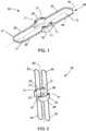
- FIG. 1is a perspective view of one preferred embodiment of a medical device in a deployed state for forming an anastomosis.
- FIG. 2is a perspective view of the medical device depicted in FIG. 1 in a delivery state.
- FIG. 3Ais a perspective view of a clamp member in a deployed state from the medical device depicted in FIG. 1 .
- FIG. 3Bis a perspective view of an alternative embodiment of a clamp member in a deployed state from the medical device depicted in FIG. 1 .
- FIG. 4is a top view of a first base and a second base of the medical device depicted in FIG. 1 .
- FIG. 5Ais a front view of a medical system for creating an anastomosis.
- FIG. 5Bis a front view, partly in cross-section, of an alternative embodiment of a medical system for creating an anastomosis.
- FIG. 5Cis a front view of another embodiment of a medical system for creating an anastomosis.
- FIG. 6is a front view of the medical system of FIG. 5A inserted into a cross-sectional view of tissue cut to reveal two viscera.
- FIG. 7is a front view similar to FIG. 6 with a medical device for forming an anastomosis deployed in two viscera.
- FIG. 8is a top view of the embodiment of FIG. 1 deployed in tissue.
- FIG. 9is a top view of the embodiment of FIG. 1 deployed in tissue where an additional incision has been made to enlarge the anastomosis.
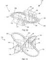
- FIG. 10is a perspective view of another preferred embodiment of a medical device in a deployed state for forming an anastomosis.
- FIG. 11is a perspective view of the medical device depicted in FIG. 10 in a delivery state.
- proximalrefers to a direction that is generally towards a physician during a medical procedure
- distalrefers to a direction that is generally away from the medical professional and/or towards a target site within a patient's anatomy during a medical procedure.
- a medical device 18for forming an anastomosis, such as during an endoscopic procedure.
- the medical device 18serves to clamp the tissue surrounding the anastomosis, hold it open, as well as facilitate enlargement of the anastomosis.
- the medical device 18generally includes a first base 20 and a second base 22 positioned opposite each other along a horizontal axis 29 .
- the bases 20 and 22are depicted as being semi-circular in shape to correspond to the outer surface of an endoscope, but other shapes are suitable as will be recognized by those skilled in the art.
- the bases 20 and 22each have two insert grooves 21 sized to securely receive a first clamp member 28 and a second clamp member 27 attached to the bases 20 and 22 by being pressed into the insert grooves 21 .
- the insert grooves 21are further depicted in FIG. 4 , and include a narrow throat T, preferably formed by one or more projections 21 a , so that the clamp members 28 and 27 may be “snap-fit” into the insert grooves 21 .
- the clamp membersmay also be more permanently attached to the bases 20 and 22 , such as by encapsulating them into the bases, or they may attach to the bases via hinges or other fastening devices known in the art.
- the base 20is positioned at a distance L opposite from base 22 to define an interior space 44 designed to allow the bases 20 and 22 to fit over an elongate member such as an endoscope.
- the distance Lpreferably ranges from about 7 mm to about 20 mm.
- the diameter D of the interior spacemay range from about 8 mm to about 23 mm.
- the dimensions and ranges used hereinare generally for endoscopic and gastrointestinal applications, but other sizes for other applications will be readily discernible to those skilled in the art.
- the basesmay be made out of metal, metal alloys, plastics, biocompatible materials, resorbable materials, degradable materials, or any other suitable materials.
- resorbablerefers to the ability of a material to be absorbed into a tissue and/or bodily fluid upon contact with the tissue and/or bodily fluid.
- a number of resorbable materialsare known in the art, and any suitable resorbable material may be used. Examples include resorbable homopolymers, copolymers, or blends of resorbable polymers.
- degradablerefers to the ability of a material to dissipate upon implantation within a body in a clinically reasonable amount of time, independent of the mechanisms by which dissipation can occur, such as dissolution, degradation, absorption and excretion, and the bases 20 and 22 need only degrade enough such that they disengage from the clamp members 28 and 27 .
- a number of degradable materialsare known in the art, and any suitable degradable material may be used. Examples include polyethylene, polypropylene and polyoxypropylene glycolic sugars, as well as polylactic sugars.
- the first clamp member 28has a proximal portion 24 , an intermediate portion 19 and a distal portion 26 .
- FIG. 3Adepicts the unattached first clamp member 28 in its natural state.
- Both the proximal and distal portions 24 and 26have a common shape and design such that they can mate as shown in FIG. 1 .
- the proximal and distal portions 24 and 26have a general U-shape, although they could be V-shaped, semi-rectangular in shape, or any other semi-annular shape.
- the clamp members 28 and 27thus have a shape that defines an interior space 71 that is in communication with the interior space 44 , and preferably contiguous with the interior space 44 .
- the proximal portion 24 and distal portion 26are rotatable at about the intermediate portion 19 where they attach to the grooves 21 in the bases 20 and 22 .
- the second clamp member 27has a proximal portion 23 and a distal portion 25 that are rotatable at about the intermediate portion 19 where they attach to the grooves 21 in the bases 20 and 22 .
- the clamp members 27 , 28may extend laterally away from the bases 20 , 22 a distance ranging from about 4 cm to about 12 cm.
- the clamp members 28 and 27are in a deployed state wherein the proximal portions 24 and 23 have rotated relative to the distal portions 26 and 25 to be about parallel such that the clamp members 28 and 27 project laterally along a lateral axis 40 (about which the bases 20 and 22 are also spaced apart).
- about parallelincludes deviations from parallel between the proximal portions 24 and 23 and the distal portions 26 and 25 , so long as tissue is capable of being clamped between the proximal portions 24 and 23 and the distal portions 26 and 25 .
- the deployed statemay also be described as where the proximal portions 24 and 23 and the distal portions 26 and 25 have rotated so that they are generally perpendicular to the intermediate portion 19 , as seen in FIG. 3A .
- the proximal portions 24 and 23have rotated towards the distal portions 26 and 25 to clamp tissue therebetween, as shown in FIG. 7 and described in further detail herein below.
- the clamp members 28 and 27 of the medical device 18are in the delivery state wherein the clamp members have rotated toward a longitudinal axis 42 so that the proximal portions 24 and 23 of the clamp members 28 and 27 are adjacent to each other, and the distal portions 26 and 25 of the clamp members 28 and 27 are adjacent to each other.
- the proximal portions 24 and 23 and the distal portions 26 and 25rotate so that clamp members 28 and 27 are about parallel to each other, aligned along the longitudinal axis 42 .
- the clamp membersare biased toward forming the deployed state as depicted in FIG. 3A .
- the clamp members 28 and 27are shown as being formed of a metal wire—preferably nitinol—having a round or rectangular (flat) cross-sectional shape, although other constructions may be employed.
- the clamp members 28 and 27may be comprised of other metals, metal alloys, plastics, or other materials that have suitable resiliency, whereby the clamp members can rotate according to the natural or imposed shape-memory characteristics of the clamp members.
- the clamp members 28 and 27may also be comprised of biocompatible or resorbable materials.
- clamp membersmay be attached to the bases by other fastening devices known in the art, or by hinges, springs or other rotatable couplings known in the art.
- hingescould be biased to rotate the clamp members to the deployed states when the clamp members are not restrained.
- clamp members 28 and 27are depicted as being two solid clamp members in FIGS. 1-3A , each may be made up of two portions, such as two wires, as shown in FIG. 3B .
- the proximal portion 24would be comprised of one wire attached to both bases 20 and 22
- the distal portion 26would be comprised of one wire attached to both bases 20 and 22
- each proximal portion 24 and each distal portion 26may be made up of two portions (e.g., wires) that have free ends adjacent to each other to form a near contiguous proximal portion 24 and a near contiguous distal portion 26 , wherein the clamp member is made up of four rotatable parts (not shown).
- the medical system 70includes a medical device for forming, creating, and maintaining an anastomosis, an elongate member for delivering the medical device, a retaining device for holding the medical device on the elongate member, and, optionally, an additional pusher device for ejecting the medical device off of the elongate member.
- the medical device 18is shown loaded on to an elongate member, in this case an endoscope 60 .
- the endoscope 60may be any type of scope known in the art, or may alternatively be any flexible elongate member suitable for being inserted into the body for therapeutic purposes.
- the device 18is slid over the endoscope 60 to its distal end 43 .
- the distal portions 26 and 25 of the clamp membersare held proximate to each other in the delivery state via a first retainer 31 .
- the first retainer 31is a suture 32 , threaded through an accessory channel 62 , which passes through loops or hooks 30 on each distal portion 26 and 25 .
- the suturealso may be wrapped around the distal portions 26 and 25 to hold them proximate to each other.
- a retaineralso holds the proximal portions 24 and 23 in the delivery state and preferably about parallel to each other along the endoscope 60 .
- a second retainer 33holds the proximal portions 24 and 23 in the delivery state, and preferably is an elastic band 34 .
- the second retainer 33may also be a multiband ligator and suture, a clip, a latching mechanism, or other devices known in the art for retaining objects in delivery states.
- one retainersuch as an overtube 66 as shown in FIG. 5B , may be used to keep both the distal portions and the proximal portions in place, rather than using the two retainers 31 and 33 .
- the medical device 18may be configured to retain itself on the endoscope 60 as depicted in FIG. 5C , thereby not needing a retainer.
- the proximal portions 24 and 23 and the distal portions 26 and 25 of the clamp members 28 and 27would be rotated longitudinally so that the proximal portions 24 and 23 and the distal portions 26 and 25 of the clamp members would all traverse the longitudinal axis 42 with one proximal portion passing through the interior space 71 of the other proximal portion, and one distal portion passing through the interior space 71 of the other distal portion.
- the medical device 18would then be fitted over the endoscope 60 , and the natural bias of the clamp members to return to the deployed state would cause the clamp members to exert force on the endoscope 60 to maintain the position of the medical device 18 on the endoscope 60 .
- the medical device 18has been inserted distally through a stoma 49 of a first bodily wall 46 (e.g., the stomach 45 ) and through a stoma 51 of a second bodily wall 48 (e.g., the small intestine, and typically, the jejunum 52 ) to rest within the interior 52 of the jejunum 50 .
- the suture 32has been retracted by the clinician or cut and retracted by the clinician, allowing the distal portions 26 and 25 of the clamp members 28 and 27 to rotate to their deployed states. Pulling back (i.e., retracting) the system 70 in the proximal direction causes the distal portions 26 and 25 to exert pressure on the interior surface of the second bodily wall 48 .
- the endoscope 60is further retracted until the bases are seen to be at least partially inside a stoma 49 ( FIG. 6 ) of the first bodily wall 46 , and the elastic band 34 is released, allowing the proximal portions 24 and 23 to rotate to their deployed states to press against and exert pressure on the proximal side of the first bodily wall 46 .
- the procedure for releasing the proximal portions 24 and 23varies depending on the retaining device used.
- the second retainer 33is the elastic band 34 as depicted in FIG. 6 , then the second retainer 33 can be removed simply by excising the elastic band 34 .
- the endoscope 60may alternatively be grooved or scored so that retraction of the endoscope 60 engages (e.g. via friction with the scoring) the elastic band 34 so that the elastic band 34 is rolled or moved proximally off of the proximal portions 24 and 23 , and preferably remains fixed to the endoscope 60 as it is retracted.
- a suturemay be run down the outside of the endoscope 60 and tied to the second retainer device 33 , so that the suture can be pulled to remove the second retainer device 33 . Still further, if the overtube 66 is used to cover the entire medical device 18 as depicted in FIG.
- the overtube 66is sized to receive the scope and apparatus in its lumen. Other retaining devices and means for removing them will be recognized by those skilled in the art.
- FIGS. 5-9A medical method for creating an anastomosis will now be described with reference to FIGS. 5-9 .
- stomasmust be created in the desired viscera, and the stomas must be brought within proximity of each other.
- One way to achieve this goalwould be to load the medical device 18 on an endoscope 60 as depicted in FIG. 5A , 5 B, or 5 C, and then advance the endoscope to the first viscera.
- a cutting device(not shown) could be advanced through a working lumen of the endoscope 60 and could be used to create the stoma 49 in the first viscera, for example in the stomach 45 .
- the endoscope 60could be further advanced to the second viscera, such as the jejunum 52 , and the cutting device could be used to create a second stoma 51 in the second viscera.
- the distal portions 26 and 25 of the medical device 18could be deployed as described above, and then the medical device 18 , endoscope 60 , and jejunum 52 could be retracted toward the stoma 49 in the first viscera. Once the medical device 18 is properly positioned as it is in FIG. 6 , the proximal portions 24 and 23 could be released and the anastomosis would be created.
- the stomasmay also be created and brought into proximity with one another prior to insertion of the medical device 18 .
- Laparoscopic surgery or open surgery and devices used in those types of surgeriesmay also be employed to create the stomas and to hold them in place proximal to each other to prepare for the insertion of the medical device 18 .
- the force exerted by the proximal portions 24 and 23 against the first bodily wall 46 and the force exerted by the distal portions 26 and 25 on the second bodily wall 48compress the two bodily walls and hold them proximate to each other.
- the first base 20 and the second base 22maintain the stoma 49 in the first bodily wall 46 and the stoma 51 in the second bodily wall 48 .
- the systemimmediately creates a substantially sized anastomosis.
- a knife or other cutting devicemay be used to excise tissue from the two bodily walls by cutting from the interior 44 laterally toward the apex of the first clamp member 28 to create a larger opening 72 as depicted in FIG. 9 . Additionally, an incision may be extended laterally from the interior 44 to the apex of the second clamp member 27 to form a second larger opening 72 , so that one large, continuous anastomosis is now formed.
- Removal of the medical device 18may be completed through natural means.
- the pressure exerted on the tissues 46 and 48will cause necrosis over a number of days, thereby forming an anastomosis that is slightly larger than 24 or 24 and 72 combined.
- the medical device 18will dislodge and will pass through the body naturally.
- the medical device 18may be made of degradable or resorbable materials so that it will be naturally broken down by the body.
- the bases 20 and 22may be made of a degradable or resorbable material so that they are broken down by the body naturally, and the clamp members 27 and 28 will then pass through the body naturally if they cannot be broken down.
- the area of compression of the tissues 46 and 48provides a barrier that guards against leakage of the GI contents or other bodily fluids depending on the viscera involved.
- the anastomosisis formed with surety before the patient leaves the medical facility, eliminating the need for a follow-up procedure.
- the clamp members 28 and 27also—maintain the size of the anastomosis, there is no need for a second procedure to insert a stent to maintain the opening.
- the medical device 118includes a first base 120 and a second base 122 , positioned opposite each other just as in medical device 18 .
- the bases 120 and 122each have two sockets 80 to receive and guide a first clamp member 128 and a second clamp member 127 .
- the clamp members 128 and 127are encapsulated by the bases 120 and 122 and are held in place by cylindrical tubes 82 that pass through insert apertures 84 in each intermediate portion 119 .
- the clamp membersmay also be attached to the bases 120 and 122 via hinges or other fastening devices known in the art, and changes may be made to the medical device 118 that are generally similar to those previously discussed for medical device 18 .
- the first clamp member 128has a proximal portion 124 , an intermediate portion 119 , and a distal portion 126
- the second clamp member 127has a proximal portion 123 , an intermediate portion 119 , and a distal portion 125
- FIG. 10depicts the clamp members 128 and 127 in their natural state, the deployed state.
- the proximal portions 124 and 123 and the distal portions 126 and 125are rotatable at about the intermediate portion 119 .
- the dimensions and use of the medical device 118are generally similar to the medical device 18 , and similar modifications to those previously discussed in the prior embodiment can be applied here.
- the clamp members 128 and 127 of the medical device 118are in the delivery state wherein the clamp members have rotated longitudinally away from the bases 120 and 122 so that the proximal portions 124 and 123 of the clamp members 128 and 127 are proximate to each other, and the distal portions 126 and 125 are proximate to each other.
- Sockets 80 in the bases 120 and 122guide the clamp members from the deployed state to the delivery state and prevent the clamp members 128 and 127 from aligning 180 degrees longitudinally as 28 and 27 of the medical device 18 are capable of doing as depicted in FIG. 2 .
- the sockets 80 and/or the size of the bases 120 and 122may be modified to allow the clamp members 128 and 127 to rotate 180 degrees longitudinally in the delivery state.
- the systems, devices, and methodsmay be used on any two layers of material (e.g., fabrics, cloth, polymers, elastomers, plastics, and rubber) that may or may not be associated with a human or animal body.
- the systems, devices, and methodscan find use in laboratory and industrial settings for approximating two or more layers of material that may or may not find application to the human or animal body, and likewise connecting holes or perforations in two or more layers of material that are not bodily tissue.
- Some examplesinclude sewing or stitching and related manufacturing, working with synthetic tissues, connecting or repairing polymeric sheets, animal studies, veterinary applications, and post-mortem activities.
Landscapes
- Health & Medical Sciences (AREA)
- Surgery (AREA)
- Life Sciences & Earth Sciences (AREA)
- Biomedical Technology (AREA)
- Nuclear Medicine, Radiotherapy & Molecular Imaging (AREA)
- Engineering & Computer Science (AREA)
- Physiology (AREA)
- Heart & Thoracic Surgery (AREA)
- Medical Informatics (AREA)
- Molecular Biology (AREA)
- Animal Behavior & Ethology (AREA)
- General Health & Medical Sciences (AREA)
- Public Health (AREA)
- Veterinary Medicine (AREA)
- Surgical Instruments (AREA)
Abstract
Description
Claims (23)
Priority Applications (1)
| Application Number | Priority Date | Filing Date | Title |
|---|---|---|---|
| US12/779,378US8728103B2 (en) | 2009-06-26 | 2010-05-13 | Linear clamps for anastomosis |
Applications Claiming Priority (2)
| Application Number | Priority Date | Filing Date | Title |
|---|---|---|---|
| US22084809P | 2009-06-26 | 2009-06-26 | |
| US12/779,378US8728103B2 (en) | 2009-06-26 | 2010-05-13 | Linear clamps for anastomosis |
Publications (2)
| Publication Number | Publication Date |
|---|---|
| US20100331866A1 US20100331866A1 (en) | 2010-12-30 |
| US8728103B2true US8728103B2 (en) | 2014-05-20 |
Family
ID=42352007
Family Applications (1)
| Application Number | Title | Priority Date | Filing Date |
|---|---|---|---|
| US12/779,378Active2031-05-19US8728103B2 (en) | 2009-06-26 | 2010-05-13 | Linear clamps for anastomosis |
Country Status (5)
| Country | Link |
|---|---|
| US (1) | US8728103B2 (en) |
| EP (1) | EP2445418B1 (en) |
| JP (1) | JP5674775B2 (en) |
| AU (1) | AU2010263224B2 (en) |
| WO (1) | WO2010151382A1 (en) |
Cited By (27)
| Publication number | Priority date | Publication date | Assignee | Title |
|---|---|---|---|---|
| US9364238B2 (en) | 2013-04-16 | 2016-06-14 | Ethicon Endo-Surgery, Inc. | Method and apparatus for joining hollow organ sections in anastomosis |
| US10828185B2 (en) | 2011-01-14 | 2020-11-10 | W. L. Gore & Associates, Inc. | Lattice |
| US11033272B2 (en) | 2013-04-16 | 2021-06-15 | Ethicon Endo-Surgery, Inc. | Methods for partial diversion of the intestinal tract |
| US11116621B2 (en) | 2012-11-13 | 2021-09-14 | W. L. Gore & Associates, Inc. | Elastic stent graft |
| US11229512B2 (en) | 2016-04-21 | 2022-01-25 | W. L. Gore & Associates, Inc. | Diametrically adjustable endoprostheses and associated systems and methods |
| US11439502B2 (en) | 2017-10-31 | 2022-09-13 | W. L. Gore & Associates, Inc. | Medical valve and leaflet promoting tissue ingrowth |
| US11471276B2 (en) | 2014-09-15 | 2022-10-18 | W. L. Gore & Associates, Inc. | Prosthetic heart valve with retention elements |
| US11497601B2 (en) | 2019-03-01 | 2022-11-15 | W. L. Gore & Associates, Inc. | Telescoping prosthetic valve with retention element |
| US11523919B2 (en) | 2011-01-14 | 2022-12-13 | W. L. Gore & Associates, Inc. | Stent |
| US11540731B2 (en) | 2018-12-21 | 2023-01-03 | W. L. Gore & Associates, Inc. | Medical treatment system using measurement data from multiple sensors |
| US11826248B2 (en) | 2012-12-19 | 2023-11-28 | Edwards Lifesciences Corporation | Vertical coaptation zone in a planar portion of prosthetic heart valve leaflet |
| US11857412B2 (en) | 2017-09-27 | 2024-01-02 | Edwards Lifesciences Corporation | Prosthetic valve with expandable frame and associated systems and methods |
| US11872122B2 (en) | 2012-12-19 | 2024-01-16 | Edwards Lifesciences Corporation | Methods for improved prosthetic heart valve with leaflet shelving |
| US11896481B2 (en) | 2012-12-19 | 2024-02-13 | Edwards Lifesciences Corporation | Truncated leaflet for prosthetic heart valves |
| US11911537B2 (en) | 2013-12-05 | 2024-02-27 | W. L. Gore & Associates, Inc. | Length extensible implantable device and methods for making such devices |
| US11950999B2 (en) | 2012-07-25 | 2024-04-09 | Edwards Lifesciences Corporation | Everting transcatheter valve and methods |
| US11986387B2 (en) | 2017-09-27 | 2024-05-21 | Edwards Lifesciences Corporation | Prosthetic valves with mechanically coupled leaflets |
| US12053381B2 (en) | 2018-07-18 | 2024-08-06 | W. L. Gore & Associates, Inc. | Implantable medical device deployment system |
| US12059344B2 (en) | 2017-09-12 | 2024-08-13 | Edwards Lifesciences Corporation | Leaflet frame attachment for prosthetic valves |
| US12064344B2 (en) | 2017-10-13 | 2024-08-20 | Edwards Lifesciences Corporation | Telescoping prosthetic valve and delivery system |
| US12115063B2 (en) | 2012-07-27 | 2024-10-15 | Edwards Lifesciences Corporation | Multi-frame prosthetic valve apparatus and methods |
| US12133795B2 (en) | 2012-12-19 | 2024-11-05 | Edwards Lifesciences Corporation | Geometric control of bending character in prosthetic heart valve leaflets |
| US12178699B2 (en) | 2012-12-19 | 2024-12-31 | Edwards Lifesciences Corporation | Multi-frame prosthetic heart valve |
| US12201520B2 (en) | 2017-10-31 | 2025-01-21 | Edwards Lifesciences Corporation | Prosthetic heart valve |
| US12279954B2 (en) | 2017-10-31 | 2025-04-22 | W. L. Gore & Associates, Inc. | Transcatheter deployment systems and associated methods |
| US12295835B2 (en) | 2012-12-19 | 2025-05-13 | Edwards Lifesciences Corporation | Prosthetic valves, frames and leaflets and methods thereof |
| US12357446B2 (en) | 2017-10-09 | 2025-07-15 | W. L. Gore & Associates, Inc. | Matched stent cover |
Families Citing this family (11)
| Publication number | Priority date | Publication date | Assignee | Title |
|---|---|---|---|---|
| MX369672B (en) | 2013-12-17 | 2019-11-15 | Standard Bariatrics Inc | Resection line guide for a medical procedure and method of using same. |
| AU2015241193B2 (en) | 2014-03-29 | 2020-01-02 | Standard Bariatrics, Inc. | End effectors surgical stapling devices, and methods of using same |
| WO2015153324A1 (en) | 2014-03-29 | 2015-10-08 | Standard Bariatrics, Inc. | End effectors, surgical stapling devices, and methods of using same |
| WO2016037158A1 (en) | 2014-09-05 | 2016-03-10 | Standard Bariatrics, Inc. | Sleeve gastrectomy calibration tube and method of using same |
| US10285837B1 (en) | 2015-09-16 | 2019-05-14 | Standard Bariatrics, Inc. | Systems and methods for measuring volume of potential sleeve in a sleeve gastrectomy |
| US10912562B2 (en) | 2017-08-14 | 2021-02-09 | Standard Bariatrics, Inc. | End effectors, surgical stapling devices, and methods of using same |
| BR112022008009A2 (en) | 2019-11-04 | 2022-07-12 | Standard Bariatrics Inc | SYSTEMS AND METHODS OF PERFORMING SURGERY USING LAPLACE'S LAW TENSION RETRACTION DURING SURGERY |
| US12274635B2 (en) | 2019-11-04 | 2025-04-15 | Standard Bariatrics, Inc. | Systems and methods of performing surgery using laplace's law tension retraction during surgery |
| CN115955943A (en) | 2020-06-30 | 2023-04-11 | 标准肥胖病研究公司 | Systems, devices, and methods for preventing or reducing insufflation loss during laparoscopic procedures |
| US20220257224A1 (en)* | 2021-02-12 | 2022-08-18 | St. Jude Medical, Cardiology Division, Inc. | Occluder medical device |
| AU2022242751B2 (en) | 2021-03-23 | 2024-05-02 | Standard Bariatrics, Inc. | Systems and methods for preventing tissue migration in surgical staplers |
Citations (215)
| Publication number | Priority date | Publication date | Assignee | Title |
|---|---|---|---|---|
| GB877903A (en) | 1958-02-17 | 1961-09-20 | Initial Plastics Ltd | Improvements in paper clips |
| US3299883A (en) | 1963-11-08 | 1967-01-24 | Engelhard Hanovia Inc | Gynecologic instrument |
| US3358676A (en) | 1962-11-30 | 1967-12-19 | Yeda Res & Dev | Magnetic propulsion of diagnostic or therapeutic elements through the body ducts of animal or human patients |
| US3674014A (en) | 1969-10-28 | 1972-07-04 | Astra Meditec Ab | Magnetically guidable catheter-tip and method |
| US3709214A (en) | 1971-10-27 | 1973-01-09 | J Robertson | Gas obturating method |
| US4022208A (en) | 1974-07-25 | 1977-05-10 | Valtchev Konstantin L | Gynecologic instrument |
| US4214587A (en) | 1979-02-12 | 1980-07-29 | Sakura Chester Y Jr | Anastomosis device and method |
| US4899744A (en) | 1988-12-15 | 1990-02-13 | Tatsuo Fujitsuka | Apparatus for anastomosing digestive tract |
| US5081997A (en) | 1989-03-09 | 1992-01-21 | Vance Products Incorporated | Echogenic devices, material and method |
| US5234447A (en) | 1990-08-28 | 1993-08-10 | Robert L. Kaster | Side-to-end vascular anastomotic staple apparatus |
| US5297536A (en) | 1992-08-25 | 1994-03-29 | Wilk Peter J | Method for use in intra-abdominal surgery |
| US5346501A (en) | 1993-02-05 | 1994-09-13 | Ethicon, Inc. | Laparoscopic absorbable anastomosic fastener and means for applying |
| US5391156A (en) | 1992-06-30 | 1995-02-21 | Ethicon, Inc. | Flexible encoscopic surgical port |
| US5429131A (en) | 1994-02-25 | 1995-07-04 | The Regents Of The University Of California | Magnetized electrode tip catheter |
| US5458131A (en) | 1992-08-25 | 1995-10-17 | Wilk; Peter J. | Method for use in intra-abdominal surgery |
| US5554183A (en) | 1994-01-19 | 1996-09-10 | Nazari; Stefano | Vascular prosthesis for the substitution or internal lining of blood vessels of medium or large diameter and device for its application |
| US5562688A (en) | 1994-03-25 | 1996-10-08 | Riza; Erol D. | Apparatus facilitating suturing in laparoscopic surgery |
| US5562683A (en) | 1993-07-12 | 1996-10-08 | Mitek Surgical Products, Inc. | Surgical repair kit and its method of use |
| US5571119A (en) | 1993-10-25 | 1996-11-05 | Children's Medical Center Corporation | Retractable suture needle with self-contained driver |
| US5571090A (en) | 1994-10-07 | 1996-11-05 | United States Surgical Corporation | Vascular suturing apparatus |
| US5573540A (en) | 1994-07-18 | 1996-11-12 | Yoon; Inbae | Apparatus and method for suturing an opening in anatomical tissue |
| US5573543A (en) | 1992-05-08 | 1996-11-12 | Ethicon, Inc. | Endoscopic surgical instrument and staples for applying purse string sutures |
| US5578044A (en) | 1992-09-04 | 1996-11-26 | Laurus Medical Corporation | Endoscopic suture system |
| US5582615A (en) | 1995-10-30 | 1996-12-10 | Pilling Weck, Incorporated | Handle for surgical clip applicator systems |
| US5584835A (en) | 1993-10-18 | 1996-12-17 | Greenfield; Jon B. | Soft tissue to bone fixation device and method |
| US5586986A (en) | 1993-07-14 | 1996-12-24 | United States Surgical Corporation | Instrument for closing trocar puncture wounds |
| US5593414A (en) | 1993-08-25 | 1997-01-14 | Apollo Camera, L.L.C. | Method of applying a surgical ligation clip |
| US5630824A (en) | 1994-06-01 | 1997-05-20 | Innovasive Devices, Inc. | Suture attachment device |
| US5643292A (en) | 1995-01-10 | 1997-07-01 | Applied Medical Resources Corporation | Percutaneous suturing device |
| US5643317A (en) | 1992-11-25 | 1997-07-01 | William Cook Europe S.A. | Closure prosthesis for transcatheter placement |
| US5645552A (en) | 1995-01-11 | 1997-07-08 | United States Surgical Corporation | Surgical apparatus for suturing body tissue |
| US5653717A (en) | 1995-08-28 | 1997-08-05 | Urohealth Systems, Inc. | Wound closure device |
| US5665067A (en) | 1994-02-28 | 1997-09-09 | Immuno Aktiengesellschaft | Apparatus for applying a multiple-component tissue adhesive |
| US5667527A (en) | 1993-03-02 | 1997-09-16 | Holobeam, Inc. | Staples |
| US5674231A (en) | 1995-10-20 | 1997-10-07 | United States Surgical Corporation | Apparatus and method for vascular hole closure |
| US5683402A (en) | 1989-07-31 | 1997-11-04 | Baxter International Inc. | Flexible suture guide and holder |
| US5690656A (en) | 1995-06-27 | 1997-11-25 | Cook Incorporated | Method and apparatus for creating abdominal visceral anastomoses |
| US5693060A (en) | 1992-11-17 | 1997-12-02 | Smith & Nephew, Inc. | Suture securing device and method |
| US5695525A (en) | 1992-05-20 | 1997-12-09 | C.R. Bard, Incorporated | Implantable prosthesis and method and apparatus for loading and delivering an implantable prosthesis |
| US5700273A (en) | 1995-07-14 | 1997-12-23 | C.R. Bard, Inc. | Wound closure apparatus and method |
| US5728113A (en) | 1994-10-07 | 1998-03-17 | United States Surgical Corporation | Endoscopic vascular suturing apparatus |
| US5728116A (en) | 1994-01-13 | 1998-03-17 | Ethicon, Inc. | Spiral surgical tack |
| US5741278A (en) | 1994-08-17 | 1998-04-21 | Tahoe Surgical Instruments | Endoscopic suture placement tool |
| US5779720A (en) | 1994-02-11 | 1998-07-14 | Createchnic Ag | One-piece surgical clip |
| US5788625A (en) | 1996-04-05 | 1998-08-04 | Depuy Orthopaedics, Inc. | Method of making reconstructive SIS structure for cartilaginous elements in situ |
| US5792153A (en) | 1994-03-23 | 1998-08-11 | University College London | Sewing device |
| US5810848A (en) | 1996-08-21 | 1998-09-22 | Hayhurst; John O. | Suturing system |
| US5824010A (en) | 1996-05-23 | 1998-10-20 | Mcdonald; Garth R. | Suture needle guide |
| US5836955A (en) | 1995-07-14 | 1998-11-17 | C.R. Bard, Inc. | Wound closure apparatus and method |
| US5860990A (en) | 1995-08-24 | 1999-01-19 | Nr Medical, Inc. | Method and apparatus for suturing |
| US5865791A (en) | 1995-06-07 | 1999-02-02 | E.P. Technologies Inc. | Atrial appendage stasis reduction procedure and devices |
| US5865836A (en) | 1996-09-20 | 1999-02-02 | United States Surgical Corporation | Needle-suture combination |
| US5868763A (en) | 1996-09-16 | 1999-02-09 | Guidant Corporation | Means and methods for performing an anastomosis |
| US5873530A (en) | 1997-09-26 | 1999-02-23 | Chizinsky; George | Liquid atomizing spray gun |
| US5891159A (en) | 1997-05-02 | 1999-04-06 | Cardiothoratic Systems, Inc. | Automatic purse string suture device |
| US5902228A (en) | 1996-10-11 | 1999-05-11 | Cornell Research Foundation, Inc. | Method and apparatus for support and tubularization of surgical grafts |
| US5908428A (en) | 1997-05-27 | 1999-06-01 | United States Surgical Corporation | Stitching devices for heart valve replacement surgery |
| US5919184A (en) | 1995-03-17 | 1999-07-06 | Tilton, Jr.; Eugene B. | Instrumentation for laparoscopic insertion and application of surgical sheet material |
| US5931844A (en) | 1998-03-31 | 1999-08-03 | Smith & Nephew, Inc. | Surgical drive tool |
| US5938668A (en) | 1994-10-07 | 1999-08-17 | United States Surgical | Surgical suturing apparatus |
| US5948000A (en) | 1996-10-03 | 1999-09-07 | United States Surgical Corporation | System for suture anchor placement |
| US5951531A (en) | 1993-04-20 | 1999-09-14 | Medchem Products, Inc. | Apparatus and method for applying a particulate hemostatic agent to living tissue |
| US5968078A (en) | 1995-08-25 | 1999-10-19 | Ultraortho, Inc. | Stabilizer for human joints |
| US5972002A (en) | 1998-06-02 | 1999-10-26 | Cabot Technology Corporation | Apparatus and method for surgical ligation |
| US5976159A (en) | 1995-02-24 | 1999-11-02 | Heartport, Inc. | Surgical clips and methods for tissue approximation |
| US5984949A (en) | 1997-10-06 | 1999-11-16 | Levin; John M. | Tissue hooks and tools for applying same |
| US6007515A (en) | 1997-04-14 | 1999-12-28 | Epstein; Gordon Howard | Controlled action, manually operable fluid application |
| US6015414A (en) | 1997-08-29 | 2000-01-18 | Stereotaxis, Inc. | Method and apparatus for magnetically controlling motion direction of a mechanically pushed catheter |
| US6021776A (en) | 1997-09-09 | 2000-02-08 | Intertex Research, Inc. | Disposable atomizer device with trigger valve system |
| US6030365A (en) | 1998-06-10 | 2000-02-29 | Laufer; Michael D. | Minimally invasive sterile surgical access device and method |
| US6059749A (en) | 1996-03-13 | 2000-05-09 | New York Blood Center | Fibrin sealant glue-gun with insertable compressed gas cartridge and luer-type reservoir connectors |
| US6077217A (en) | 1997-06-25 | 2000-06-20 | Ramus Medical Technologies, Inc. | System and method for assembling graft structures |
| US6086608A (en) | 1996-02-22 | 2000-07-11 | Smith & Nephew, Inc. | Suture collet |
| US6110183A (en) | 1998-12-22 | 2000-08-29 | Cook Incorporated | Suture anchor device |
| US6113612A (en) | 1998-11-06 | 2000-09-05 | St. Jude Medical Cardiovascular Group, Inc. | Medical anastomosis apparatus |
| US6149658A (en) | 1997-01-09 | 2000-11-21 | Coalescent Surgical, Inc. | Sutured staple surgical fasteners, instruments and methods for minimally invasive vascular and endoscopic surgery |
| US6152935A (en) | 1996-12-11 | 2000-11-28 | Ethicon, Inc. | Meniscal repair device having integral spring member |
| US6152937A (en) | 1998-11-06 | 2000-11-28 | St. Jude Medical Cardiovascular Group, Inc. | Medical graft connector and methods of making and installing same |
| US6156006A (en) | 1997-10-17 | 2000-12-05 | Circon Corporation | Medical instrument system for piercing through tissue |
| US6159223A (en) | 1999-01-26 | 2000-12-12 | Endoscopic Concepts, Inc. | Surgical clip applicator |
| US6171321B1 (en) | 1995-02-24 | 2001-01-09 | Heartport, Inc. | Devices and methods for performing a vascular anastomosis |
| US6183486B1 (en) | 1995-02-24 | 2001-02-06 | Heartport, Inc. | Device and method for minimizing heart displacements during a beating heart surgical procedure |
| EP1077047A2 (en) | 1999-08-19 | 2001-02-21 | Yugengaisha Pacs Optica Japan | Organ anastomosing apparatus |
| US6193732B1 (en) | 1999-01-08 | 2001-02-27 | Cardiothoracic System | Surgical clips and apparatus and method for clip placement |
| US6200329B1 (en) | 1998-08-31 | 2001-03-13 | Smith & Nephew, Inc. | Suture collet |
| US6251116B1 (en)* | 1999-07-28 | 2001-06-26 | Vasconnect, Inc. | Device for interconnecting vessels in a patient |
| US6290674B1 (en) | 1999-09-20 | 2001-09-18 | Appriva Medical, Inc. | Method and apparatus for closing intracardiac septal defects |
| US6293952B1 (en) | 1997-07-31 | 2001-09-25 | Circon Corporation | Medical instrument system for piercing through tissue |
| US6293961B2 (en) | 1998-12-30 | 2001-09-25 | Ethicon, Inc. | Suture locking device |
| US20010049497A1 (en) | 2000-03-24 | 2001-12-06 | Kalloo Anthony Nicolas | Methods and devices for diagnostic and therapeutic interventions in the peritoneal cavity |
| US6348059B1 (en) | 1992-10-19 | 2002-02-19 | Advanced Research & Technology Institute, Inc. | Apparatus and method for positive closure of an internal tissue membrane opening |
| US20020022851A1 (en) | 2000-08-17 | 2002-02-21 | Johns Hopkins University | Gastric reduction endoscopy |
| US6355050B1 (en) | 1992-12-10 | 2002-03-12 | Abbott Laboratories | Device and method for suturing tissue |
| US6368300B1 (en) | 1995-01-18 | 2002-04-09 | C. R. Bard, Inc. | Apparatus for applying a hemostatic agent onto a tissue |
| US6402765B1 (en) | 2000-06-12 | 2002-06-11 | Niti Alloys Technologies Ltd. | Surgical clip |
| US6423087B1 (en) | 1999-08-04 | 2002-07-23 | Olympus Optical Co., Ltd. | Internal organ walls joining instrument for an endoscope |
| US6428550B1 (en) | 1999-05-18 | 2002-08-06 | Cardica, Inc. | Sutureless closure and deployment system for connecting blood vessels |
| US6494889B1 (en) | 1999-09-01 | 2002-12-17 | Converge Medical, Inc. | Additional sutureless anastomosis embodiments |
| US6527753B2 (en) | 2000-02-29 | 2003-03-04 | Olympus Optical Co., Ltd. | Endoscopic treatment system |
| US6535764B2 (en) | 2001-05-01 | 2003-03-18 | Intrapace, Inc. | Gastric treatment and diagnosis device and method |
| US20030088256A1 (en)* | 2001-10-03 | 2003-05-08 | Conston Stanley R. | Devices and methods for interconnecting vessels |
| US6569173B1 (en) | 1999-12-14 | 2003-05-27 | Integrated Vascular Interventional Technologies, L.C. | Compression plate anastomosis apparatus |
| US20030216613A1 (en) | 2002-03-19 | 2003-11-20 | Anthony Kalloo | Anastomosis system |
| US20030225312A1 (en) | 2002-03-18 | 2003-12-04 | Anthony Kalloo | Endoscopic system for treating inside of body cavity |
| US6689062B1 (en) | 1999-11-23 | 2004-02-10 | Microaccess Medical Systems, Inc. | Method and apparatus for transesophageal cardiovascular procedures |
| US20040087985A1 (en) | 1999-03-19 | 2004-05-06 | Amir Loshakove | Graft and connector delivery |
| US6811555B1 (en)* | 1996-09-16 | 2004-11-02 | Origin Medsystems, Inc. | Method and apparatus for performing anastomosis with eversion of tissue edges and joining of exposed intima of the everted tissue |
| US20040225191A1 (en) | 2003-03-04 | 2004-11-11 | Olympus Corporation | Endoscopic treatment system and anastomotic method using this system |
| US20040249367A1 (en) | 2003-01-15 | 2004-12-09 | Usgi Medical Corp. | Endoluminal tool deployment system |
| EP1493391A1 (en) | 2002-04-10 | 2005-01-05 | Yugengaisha Pacs Optica Japan | Organ connecting device and method for using the device |
| US20050004584A1 (en) | 2000-09-25 | 2005-01-06 | Cohesion Technologies, Inc. | Resorbable anastomosis stents and plugs and their use in patients |
| US6918871B2 (en) | 2003-06-19 | 2005-07-19 | Ethicon Endo-Surgery, Inc. | Method for accessing cavity |
| US20050277981A1 (en) | 2004-06-09 | 2005-12-15 | Usgi Medical Inc. | Apparatus and methods for optimizing anchoring force |
| US20050277965A1 (en) | 2004-06-14 | 2005-12-15 | Rox Medical, Inc. | Devices for arterio-venous fistula creation |
| US20060025788A1 (en) | 2002-09-25 | 2006-02-02 | By-Pass, Inc. | Anastomotic leg arrangement |
| US20060036267A1 (en) | 2004-08-11 | 2006-02-16 | Usgi Medical Inc. | Methods and apparatus for performing malabsorptive bypass procedures within a patient's gastro-intestinal lumen |
| US20060100480A1 (en) | 2002-12-24 | 2006-05-11 | Usgi Medical Inc. | Apparatus and methods for achieving endoluminal access |
| US20060167482A1 (en) | 2003-04-04 | 2006-07-27 | Swain Christopher P | Device for transfixing and joining tissue |
| US20060200004A1 (en) | 2005-01-26 | 2006-09-07 | Wilk Patent, Llc | Intra-abdominal medical procedures and device |
| US20060212063A1 (en) | 2005-03-18 | 2006-09-21 | Wilk Patent, Llc | Surgical device and associated trans-organ surgical method |
| US20060211919A1 (en) | 2005-03-18 | 2006-09-21 | Wilk Patent. Llc | Intra-abdominal medical device and associated method |
| US20060229653A1 (en) | 2005-04-12 | 2006-10-12 | Wilk Patent, Llc | Intra-abdominal medical method and associated device |
| US20060241651A1 (en) | 2005-04-22 | 2006-10-26 | Wilk Patent, Llc | Surgical port device and associated method |
| US20060237022A1 (en) | 2005-04-26 | 2006-10-26 | Usgi Medical Inc. | Transgastric abdominal access |
| US20060241480A1 (en) | 2005-04-12 | 2006-10-26 | Wilk Patent, Llc | Endoscopic medical method and associated device |
| US20060241344A1 (en) | 2005-04-12 | 2006-10-26 | Wilk Patent, Llc | Intra-abdominal surgical method and associated apparatus |
| US20060241570A1 (en) | 2005-04-22 | 2006-10-26 | Wilk Patent, Llc | Intra-abdominal medical method |
| US20060237023A1 (en) | 2005-04-26 | 2006-10-26 | Usgi Medical Inc. | Transgastric tubal ligation |
| US20060241691A1 (en) | 2005-04-12 | 2006-10-26 | Wilk Patent, Llc | Medical treatment method and device utilizing magnetic elements |
| US7128708B2 (en) | 2002-06-13 | 2006-10-31 | Usgi Medical Inc. | Shape lockable apparatus and method for advancing an instrument through unsupported anatomy |
| US20060252997A1 (en) | 2005-04-22 | 2006-11-09 | Wilk Patent, Llc | Medical port device, kit and associated method |
| US20060253123A1 (en) | 2005-04-22 | 2006-11-09 | Wilk Patent, Llc | Port extraction method for trans-organ surgery |
| US20060258909A1 (en) | 2005-04-08 | 2006-11-16 | Usgi Medical, Inc. | Methods and apparatus for maintaining sterility during transluminal procedures |
| US20060264986A1 (en) | 2001-06-20 | 2006-11-23 | Park Medical, Llc | Anastomotic device |
| US20060287666A1 (en) | 2005-06-15 | 2006-12-21 | Usgi Medical Inc. | Apparatus and methods for endoluminal advancement |
| US20060293701A1 (en) | 2001-05-02 | 2006-12-28 | Medtronic, Inc. | Self-closing surgical clip for tissue |
| US20070004958A1 (en) | 2005-03-10 | 2007-01-04 | Olympus Medical Systems Corporation | Probing method and holding method for lumenal organ |
| US20070051380A1 (en) | 2005-08-12 | 2007-03-08 | Board Of Regents, The University Of Texas System | System, kit, and method of transgastric removal of visceral fat and other related methods |
| US20070100376A1 (en) | 2003-06-06 | 2007-05-03 | Olympus Corporation | Suturing instrument |
| US20070106313A1 (en) | 2002-09-12 | 2007-05-10 | Steve Golden | Anastomosis apparatus and methods |
| US20070112362A1 (en) | 2005-11-14 | 2007-05-17 | Olympus Medical Systems Corp. | Perforation suturing method |
| US20070123840A1 (en) | 2005-10-18 | 2007-05-31 | Usgi Medical, Inc. | Instrument assisted abdominal access |
| US7232448B2 (en) | 2004-06-17 | 2007-06-19 | Ethicon, Inc. - Usa | Minimally invasive stitching device |
| US20070163604A1 (en) | 2006-01-13 | 2007-07-19 | Olympus Medical Systems Corp. | Leak test method for medical procedure via natural orifice |
| US20070163596A1 (en) | 2006-01-13 | 2007-07-19 | Olympus Medical Systems Corp. | Endotracheal tube, device for use in medical procedure through natural opening and medical procedure through natural opening |
| US20070167675A1 (en) | 2006-01-13 | 2007-07-19 | Olympus Medical Systems Corp. | Overtube and medical procedure via natural orifice using the same |
| US20070167676A1 (en) | 2006-01-13 | 2007-07-19 | Olympus Medical Systems Corp. | Overtube and medical procedure via natural orifice using the same |
| US20070167967A1 (en) | 2006-01-13 | 2007-07-19 | Olympus Medical Systems Corp. | Medical procedure via natural orifice and puncture device |
| US20070163585A1 (en) | 2006-01-13 | 2007-07-19 | Olympus Medical Systems Corp. | Method for accessing abdominal cavity and medical procedure via natural orifice |
| US20070173867A1 (en) | 2004-08-27 | 2007-07-26 | Brenneman Rodney A | Device and method for establishing an artificial arterio-venous fistula |
| US20070173859A1 (en) | 2006-01-13 | 2007-07-26 | Olympus Medical Systems Corp. | Medical procedure via natural opening |
| US20070198000A1 (en) | 2006-02-21 | 2007-08-23 | Olympus Medical Systems Corp. | Overtube and operative procedure via bodily orifice |
| US20070197864A1 (en) | 2006-02-23 | 2007-08-23 | Olympus Medical Systems Corp. | Overtube and natural opening medical procedures using the same |
| US20070198033A1 (en) | 2003-11-26 | 2007-08-23 | Johns Hopkins University | Peroral Transgastric Endoscopic Techniques |
| US20070208360A1 (en) | 2004-02-13 | 2007-09-06 | Demarais Denise M | Methods and devices for reducing hollow organ volume |
| US20070213750A1 (en) | 2001-08-28 | 2007-09-13 | Ethicon, Inc. | Composite Staple for Completing an Anastomosis |
| US20070213702A1 (en) | 2006-03-08 | 2007-09-13 | Olympus Medical Systems Corp. | Medical procedure carried out via a natural opening |
| US20070213749A1 (en) | 2006-03-08 | 2007-09-13 | Olympus Medical Systems Corp. | Medical procedure performed inside abdominal cavity |
| US20070219411A1 (en) | 2006-01-13 | 2007-09-20 | Olympus Medical Systems Corp. | Overtube and endoscopic treatment system |
| US20070225734A1 (en) | 2006-03-22 | 2007-09-27 | Minos Medical | Systems and methods for less invasive resolution of maladies of tissue including the appendix, gall bladder, and hemorrhoids |
| US20070255100A1 (en) | 2006-01-06 | 2007-11-01 | Olympus Medical Systems Corporation | Medical method and medical system conducted percutaneous or using naturally occurring body orifice |
| US20070255165A1 (en) | 2006-01-13 | 2007-11-01 | Olympus Medical Systems Corporation | Natural orifice medical operation and endoscopic overtube |
| US20070255273A1 (en) | 2006-04-29 | 2007-11-01 | Board Of Regents, The University Of Texas System | Devices for use in Transluminal and Endoluminal Surgery |
| US20070255295A1 (en)* | 2006-04-27 | 2007-11-01 | Medtronic, Inc. | Sutureless implantable medical device fixation |
| US20070260214A1 (en) | 2006-01-13 | 2007-11-08 | Olympus Medical Systems Corp. | Medical procedure through natural body opening |
| US20070270629A1 (en) | 2006-05-19 | 2007-11-22 | Charles Filipi J | System and techniques for magnetic manipulation of internal organs during minimally invasive surgery |
| US20080015408A1 (en) | 1999-01-21 | 2008-01-17 | Coroneo, Inc. | Surgical apparatus and method for performing transabdominal cardiac surgery |
| US20080021277A1 (en) | 2006-07-20 | 2008-01-24 | David Stefanchik | Methods for stabilizing and positioning an endoscope and surgical procedures |
| US20080065103A1 (en) | 2006-06-13 | 2008-03-13 | Intuitive Surgical, Inc. | Surgical instrument control and actuation |
| US7351202B2 (en) | 2002-12-05 | 2008-04-01 | Ethicon Endo-Surgery, Inc. | Medical device with track and method of use |
| US20080097489A1 (en) | 1999-04-09 | 2008-04-24 | Evalve, Inc. | Fixation devices, systems and methods for engaging tissue |
| US20080114378A1 (en) | 2006-11-14 | 2008-05-15 | Olympus Medical Systems Corp. | Perforation suture clip and clip device |
| US20080119868A1 (en) | 2006-11-22 | 2008-05-22 | Minos Medical | Methods and Apparatus for Natural Orifice Vaginal Hysterectomy |
| US20080125804A1 (en) | 2006-11-15 | 2008-05-29 | Gostout Christopher J | Submucosal endoscopy with mucosal flap methods and kits |
| US20080154290A1 (en) | 2002-10-04 | 2008-06-26 | Steve Golden | Anastomosis apparatus and methods |
| US20080161641A1 (en) | 2006-12-28 | 2008-07-03 | Olympus Medical Systems Corp. | Procedure |
| US20080171907A1 (en) | 2007-01-12 | 2008-07-17 | Ethicon Endo-Surgery, Inc. | Magnetic Tissue Grasping |
| US20080183039A1 (en) | 2007-01-26 | 2008-07-31 | Ethicon Endo-Surgery, Inc. | Balloon Positioning System for Endoscopic Access |
| US20080200762A1 (en) | 2007-02-16 | 2008-08-21 | Stokes Michael J | Flexible endoscope shapelock |
| US20080208161A1 (en) | 2007-02-26 | 2008-08-28 | Olympus Medical Systems Corp. | Application of procedure through natural orifice |
| US20080228203A1 (en) | 2007-03-15 | 2008-09-18 | Minos Medical | System and method for translumenal closure in natural orifice surgery |
| US20080228029A1 (en) | 2003-02-11 | 2008-09-18 | Olympus Corporation | Over-tube, method of manufacturing over-tube, method of disposing over-tube, and method of treatment in abdominal cavity |
| US7431694B2 (en) | 2003-05-16 | 2008-10-07 | Ethicon Endo-Surgery, Inc. | Method of guiding medical devices |
| US20080249416A1 (en) | 2007-04-05 | 2008-10-09 | Olympus Medical Systems Corp. | Treatment instrument system |
| US20080275297A1 (en) | 2007-05-01 | 2008-11-06 | Ethicon Endo-Surgery, Inc. | Endoscopic guide device |
| US20080287963A1 (en) | 2005-12-30 | 2008-11-20 | Rogers Theodore W | Methods and apparatus to shape flexible entry guides for minimally invasive surgery |
| US20080312502A1 (en) | 2005-12-02 | 2008-12-18 | Christopher Paul Swain | System and Device for in Vivo Procedures |
| US20090023985A1 (en) | 2007-06-14 | 2009-01-22 | Usgi Medical, Inc. | Endoluminal instrument management system |
| US20090054728A1 (en) | 2007-08-21 | 2009-02-26 | Trusty Robert M | Manipulatable guide system and methods for natural orifice translumenal endoscopic surgery |
| US20090054761A1 (en) | 2007-08-22 | 2009-02-26 | Ethicon Endo-Surgery, Inc. | Medical system, method, and storage medium concerning a natural orifice transluminal medical procedure |
| DE19704211B4 (en) | 1997-02-05 | 2009-06-18 | Schulz, Rolf A., Dipl.-Kaufm. | Flat sheet clamp |
| US20090182195A1 (en) | 2008-01-11 | 2009-07-16 | Ethicon Endo-Surgery, Inc. | Endoscopic guide system |
| US20090221915A1 (en) | 2008-03-03 | 2009-09-03 | Ethicon Endo-Surgery, Inc. | Transluminal tissue markers |
| US7585308B2 (en) | 2005-03-30 | 2009-09-08 | Ethicon Endo-Surgery, Inc. | Handle system and method for use in anastomotic procedures |
| US20090227828A1 (en) | 2008-03-10 | 2009-09-10 | Ethicon Endo-Surgery, Inc. | Anastomotic device |
| US7591828B2 (en) | 2005-07-22 | 2009-09-22 | Ethicon Endo-Surgery, Inc. | Resposable anastomotic ring applier device |
| US20090254105A1 (en)* | 2008-04-04 | 2009-10-08 | Medtronic Vascular, Inc. | Anastomotic connectors |
| US7608086B2 (en) | 2003-09-30 | 2009-10-27 | Ethicon Endo-Surgery, Inc. | Anastomosis wire ring device |
| US20090276055A1 (en) | 2008-05-01 | 2009-11-05 | Ethicon Endo-Surgery, Inc. | Method for gastric volume reduction surgery |
| US20090281559A1 (en) | 2008-05-06 | 2009-11-12 | Ethicon Endo-Surgery, Inc. | Anastomosis patch |
| US7618427B2 (en) | 2003-12-29 | 2009-11-17 | Ethicon Endo-Surgery, Inc. | Device and method for intralumenal anastomosis |
| US7637919B2 (en) | 2002-01-30 | 2009-12-29 | Olympus Corporation | Anastomosis system for performing anastomosis in body |
| US20100010520A1 (en) | 2008-07-11 | 2010-01-14 | Olympus Medical Systems Corp. | Tissue fastener |
| US20100036399A1 (en) | 2008-08-05 | 2010-02-11 | Tyco Healthcare Group Lp | Magnetic compression anastomosis device |
| US7666197B2 (en) | 2002-06-19 | 2010-02-23 | Tyco Healthcare Group Lp | Method and apparatus for anastomosis |
| US20100056861A1 (en) | 2008-08-29 | 2010-03-04 | Ethicon Endo-Surgery, Inc. | Articulating end cap |
| US20100063521A1 (en) | 2003-04-16 | 2010-03-11 | Tyco Healthcare Group Lp | Method and apparatus for radical prostatectomy anastomosis including an anchor for engaging a body vessel and deployable sutures |
| US20100087842A1 (en) | 2002-04-16 | 2010-04-08 | Tyco Healthcare Group Lp | Methods and Apparatus for Anastomosis Including an Expandable Anchor |
| US20100099947A1 (en) | 2006-08-28 | 2010-04-22 | Olympus Medical Systems Corp. | Fistulectomy method of forming a fistula between a first duct and a second duct |
| US20100113872A1 (en) | 2007-09-20 | 2010-05-06 | Olympus Medical Systems Corp. | Medical apparatus and procedure of installing medical apparatus in patient |
| US20100114124A1 (en) | 2005-08-03 | 2010-05-06 | Brian Kelleher | Method and apparatus for partioning an organ within the body |
| US7713278B2 (en) | 2004-05-07 | 2010-05-11 | Ethicon Endo-Surgery, Inc. | Method and instrument for effecting anastomosis of respective tissues defining two body lumens |
| US20100160729A1 (en) | 2008-12-19 | 2010-06-24 | Smith Paul J | Systems and methods for directing instruments to varying positions at the distal end of a guide tube |
| US20100179510A1 (en) | 2009-01-12 | 2010-07-15 | Ethicon Endo-Surgery, Inc. | Apparatus for forming an anastomosis |
| US20100179540A1 (en) | 2006-10-06 | 2010-07-15 | Stanislaw Marczyk | Endoscopic Vessel Sealer and Divider Having a Flexible Articulating Shaft |
Family Cites Families (5)
| Publication number | Priority date | Publication date | Assignee | Title |
|---|---|---|---|---|
| US766197A (en)* | 1904-01-19 | 1904-08-02 | Louis M Picker | Display-case. |
| CA2244080A1 (en)* | 1996-02-02 | 1997-08-07 | Transvascular, Inc. | Methods and apparatus for blocking flow through blood vessels |
| US20050070935A1 (en)* | 2003-09-30 | 2005-03-31 | Ortiz Mark S. | Single lumen access deployable ring for intralumenal anastomosis |
| US7828814B2 (en)* | 2004-08-27 | 2010-11-09 | Rox Medical, Inc. | Device and method for establishing an artificial arterio-venous fistula |
| US20100106171A1 (en)* | 2005-05-06 | 2010-04-29 | Johns Hopkins University | Transcaval mesenteric venous anastomosis and access system |
- 2010
- 2010-05-13WOPCT/US2010/034690patent/WO2010151382A1/enactiveApplication Filing
- 2010-05-13JPJP2012517532Apatent/JP5674775B2/enactiveActive
- 2010-05-13EPEP10719705.5Apatent/EP2445418B1/enactiveActive
- 2010-05-13AUAU2010263224Apatent/AU2010263224B2/ennot_activeExpired - Fee Related
- 2010-05-13USUS12/779,378patent/US8728103B2/enactiveActive
Patent Citations (257)
| Publication number | Priority date | Publication date | Assignee | Title |
|---|---|---|---|---|
| GB877903A (en) | 1958-02-17 | 1961-09-20 | Initial Plastics Ltd | Improvements in paper clips |
| US3358676A (en) | 1962-11-30 | 1967-12-19 | Yeda Res & Dev | Magnetic propulsion of diagnostic or therapeutic elements through the body ducts of animal or human patients |
| US3299883A (en) | 1963-11-08 | 1967-01-24 | Engelhard Hanovia Inc | Gynecologic instrument |
| US3674014A (en) | 1969-10-28 | 1972-07-04 | Astra Meditec Ab | Magnetically guidable catheter-tip and method |
| US3709214A (en) | 1971-10-27 | 1973-01-09 | J Robertson | Gas obturating method |
| US4022208A (en) | 1974-07-25 | 1977-05-10 | Valtchev Konstantin L | Gynecologic instrument |
| US4214587A (en) | 1979-02-12 | 1980-07-29 | Sakura Chester Y Jr | Anastomosis device and method |
| US4899744A (en) | 1988-12-15 | 1990-02-13 | Tatsuo Fujitsuka | Apparatus for anastomosing digestive tract |
| US5081997A (en) | 1989-03-09 | 1992-01-21 | Vance Products Incorporated | Echogenic devices, material and method |
| US5683402A (en) | 1989-07-31 | 1997-11-04 | Baxter International Inc. | Flexible suture guide and holder |
| US5234447A (en) | 1990-08-28 | 1993-08-10 | Robert L. Kaster | Side-to-end vascular anastomotic staple apparatus |
| US5573543A (en) | 1992-05-08 | 1996-11-12 | Ethicon, Inc. | Endoscopic surgical instrument and staples for applying purse string sutures |
| US5695525A (en) | 1992-05-20 | 1997-12-09 | C.R. Bard, Incorporated | Implantable prosthesis and method and apparatus for loading and delivering an implantable prosthesis |
| US5391156A (en) | 1992-06-30 | 1995-02-21 | Ethicon, Inc. | Flexible encoscopic surgical port |
| US5297536A (en) | 1992-08-25 | 1994-03-29 | Wilk Peter J | Method for use in intra-abdominal surgery |
| US5458131A (en) | 1992-08-25 | 1995-10-17 | Wilk; Peter J. | Method for use in intra-abdominal surgery |
| US5578044A (en) | 1992-09-04 | 1996-11-26 | Laurus Medical Corporation | Endoscopic suture system |
| US6348059B1 (en) | 1992-10-19 | 2002-02-19 | Advanced Research & Technology Institute, Inc. | Apparatus and method for positive closure of an internal tissue membrane opening |
| US5693060A (en) | 1992-11-17 | 1997-12-02 | Smith & Nephew, Inc. | Suture securing device and method |
| US5947997A (en) | 1992-11-25 | 1999-09-07 | William Cook Europe A/S | Closure prothesis for transcatheter placement |
| US5643317A (en) | 1992-11-25 | 1997-07-01 | William Cook Europe S.A. | Closure prosthesis for transcatheter placement |
| US6355050B1 (en) | 1992-12-10 | 2002-03-12 | Abbott Laboratories | Device and method for suturing tissue |
| US5346501A (en) | 1993-02-05 | 1994-09-13 | Ethicon, Inc. | Laparoscopic absorbable anastomosic fastener and means for applying |
| US5667527A (en) | 1993-03-02 | 1997-09-16 | Holobeam, Inc. | Staples |
| US5951531A (en) | 1993-04-20 | 1999-09-14 | Medchem Products, Inc. | Apparatus and method for applying a particulate hemostatic agent to living tissue |
| US5562683A (en) | 1993-07-12 | 1996-10-08 | Mitek Surgical Products, Inc. | Surgical repair kit and its method of use |
| US5586986A (en) | 1993-07-14 | 1996-12-24 | United States Surgical Corporation | Instrument for closing trocar puncture wounds |
| US5593414A (en) | 1993-08-25 | 1997-01-14 | Apollo Camera, L.L.C. | Method of applying a surgical ligation clip |
| US5584835A (en) | 1993-10-18 | 1996-12-17 | Greenfield; Jon B. | Soft tissue to bone fixation device and method |
| US5571119A (en) | 1993-10-25 | 1996-11-05 | Children's Medical Center Corporation | Retractable suture needle with self-contained driver |
| US5728116A (en) | 1994-01-13 | 1998-03-17 | Ethicon, Inc. | Spiral surgical tack |
| US5554183A (en) | 1994-01-19 | 1996-09-10 | Nazari; Stefano | Vascular prosthesis for the substitution or internal lining of blood vessels of medium or large diameter and device for its application |
| US5779720A (en) | 1994-02-11 | 1998-07-14 | Createchnic Ag | One-piece surgical clip |
| US5429131A (en) | 1994-02-25 | 1995-07-04 | The Regents Of The University Of California | Magnetized electrode tip catheter |
| US5665067A (en) | 1994-02-28 | 1997-09-09 | Immuno Aktiengesellschaft | Apparatus for applying a multiple-component tissue adhesive |
| US5792153A (en) | 1994-03-23 | 1998-08-11 | University College London | Sewing device |
| US5562688A (en) | 1994-03-25 | 1996-10-08 | Riza; Erol D. | Apparatus facilitating suturing in laparoscopic surgery |
| US5630824A (en) | 1994-06-01 | 1997-05-20 | Innovasive Devices, Inc. | Suture attachment device |
| US5573540A (en) | 1994-07-18 | 1996-11-12 | Yoon; Inbae | Apparatus and method for suturing an opening in anatomical tissue |
| US5741278A (en) | 1994-08-17 | 1998-04-21 | Tahoe Surgical Instruments | Endoscopic suture placement tool |
| US5728113A (en) | 1994-10-07 | 1998-03-17 | United States Surgical Corporation | Endoscopic vascular suturing apparatus |
| US5746751A (en) | 1994-10-07 | 1998-05-05 | United States Surgical Corporation | Vascular suturing apparatus |
| US5938668A (en) | 1994-10-07 | 1999-08-17 | United States Surgical | Surgical suturing apparatus |
| US5571090A (en) | 1994-10-07 | 1996-11-05 | United States Surgical Corporation | Vascular suturing apparatus |
| US5643292A (en) | 1995-01-10 | 1997-07-01 | Applied Medical Resources Corporation | Percutaneous suturing device |
| US5645552A (en) | 1995-01-11 | 1997-07-08 | United States Surgical Corporation | Surgical apparatus for suturing body tissue |
| US6368300B1 (en) | 1995-01-18 | 2002-04-09 | C. R. Bard, Inc. | Apparatus for applying a hemostatic agent onto a tissue |
| US6171321B1 (en) | 1995-02-24 | 2001-01-09 | Heartport, Inc. | Devices and methods for performing a vascular anastomosis |
| US6183486B1 (en) | 1995-02-24 | 2001-02-06 | Heartport, Inc. | Device and method for minimizing heart displacements during a beating heart surgical procedure |
| US5976159A (en) | 1995-02-24 | 1999-11-02 | Heartport, Inc. | Surgical clips and methods for tissue approximation |
| US5919184A (en) | 1995-03-17 | 1999-07-06 | Tilton, Jr.; Eugene B. | Instrumentation for laparoscopic insertion and application of surgical sheet material |
| US5865791A (en) | 1995-06-07 | 1999-02-02 | E.P. Technologies Inc. | Atrial appendage stasis reduction procedure and devices |
| US5690656A (en) | 1995-06-27 | 1997-11-25 | Cook Incorporated | Method and apparatus for creating abdominal visceral anastomoses |
| US5846253A (en) | 1995-07-14 | 1998-12-08 | C. R. Bard, Inc. | Wound closure apparatus and method |
| US5836955A (en) | 1995-07-14 | 1998-11-17 | C.R. Bard, Inc. | Wound closure apparatus and method |
| US5836956A (en) | 1995-07-14 | 1998-11-17 | C.R. Bard, Inc. | Wound closure apparatus and method |
| US5700273A (en) | 1995-07-14 | 1997-12-23 | C.R. Bard, Inc. | Wound closure apparatus and method |
| US5860990A (en) | 1995-08-24 | 1999-01-19 | Nr Medical, Inc. | Method and apparatus for suturing |
| US5968078A (en) | 1995-08-25 | 1999-10-19 | Ultraortho, Inc. | Stabilizer for human joints |
| US5653717A (en) | 1995-08-28 | 1997-08-05 | Urohealth Systems, Inc. | Wound closure device |
| US5674231A (en) | 1995-10-20 | 1997-10-07 | United States Surgical Corporation | Apparatus and method for vascular hole closure |
| US5582615A (en) | 1995-10-30 | 1996-12-10 | Pilling Weck, Incorporated | Handle for surgical clip applicator systems |
| US6086608A (en) | 1996-02-22 | 2000-07-11 | Smith & Nephew, Inc. | Suture collet |
| US6059749A (en) | 1996-03-13 | 2000-05-09 | New York Blood Center | Fibrin sealant glue-gun with insertable compressed gas cartridge and luer-type reservoir connectors |
| US5788625A (en) | 1996-04-05 | 1998-08-04 | Depuy Orthopaedics, Inc. | Method of making reconstructive SIS structure for cartilaginous elements in situ |
| US5824010A (en) | 1996-05-23 | 1998-10-20 | Mcdonald; Garth R. | Suture needle guide |
| US5810848A (en) | 1996-08-21 | 1998-09-22 | Hayhurst; John O. | Suturing system |
| US6811555B1 (en)* | 1996-09-16 | 2004-11-02 | Origin Medsystems, Inc. | Method and apparatus for performing anastomosis with eversion of tissue edges and joining of exposed intima of the everted tissue |
| US5868763A (en) | 1996-09-16 | 1999-02-09 | Guidant Corporation | Means and methods for performing an anastomosis |
| US5865836A (en) | 1996-09-20 | 1999-02-02 | United States Surgical Corporation | Needle-suture combination |
| US5948000A (en) | 1996-10-03 | 1999-09-07 | United States Surgical Corporation | System for suture anchor placement |
| US5902228A (en) | 1996-10-11 | 1999-05-11 | Cornell Research Foundation, Inc. | Method and apparatus for support and tubularization of surgical grafts |
| US6152935A (en) | 1996-12-11 | 2000-11-28 | Ethicon, Inc. | Meniscal repair device having integral spring member |
| US6149658A (en) | 1997-01-09 | 2000-11-21 | Coalescent Surgical, Inc. | Sutured staple surgical fasteners, instruments and methods for minimally invasive vascular and endoscopic surgery |
| DE19704211B4 (en) | 1997-02-05 | 2009-06-18 | Schulz, Rolf A., Dipl.-Kaufm. | Flat sheet clamp |
| US6007515A (en) | 1997-04-14 | 1999-12-28 | Epstein; Gordon Howard | Controlled action, manually operable fluid application |
| US5891159A (en) | 1997-05-02 | 1999-04-06 | Cardiothoratic Systems, Inc. | Automatic purse string suture device |
| US5908428A (en) | 1997-05-27 | 1999-06-01 | United States Surgical Corporation | Stitching devices for heart valve replacement surgery |
| US6077217A (en) | 1997-06-25 | 2000-06-20 | Ramus Medical Technologies, Inc. | System and method for assembling graft structures |
| US6293952B1 (en) | 1997-07-31 | 2001-09-25 | Circon Corporation | Medical instrument system for piercing through tissue |
| US6015414A (en) | 1997-08-29 | 2000-01-18 | Stereotaxis, Inc. | Method and apparatus for magnetically controlling motion direction of a mechanically pushed catheter |
| US6021776A (en) | 1997-09-09 | 2000-02-08 | Intertex Research, Inc. | Disposable atomizer device with trigger valve system |
| US5873530A (en) | 1997-09-26 | 1999-02-23 | Chizinsky; George | Liquid atomizing spray gun |
| US5984949A (en) | 1997-10-06 | 1999-11-16 | Levin; John M. | Tissue hooks and tools for applying same |
| US6156006A (en) | 1997-10-17 | 2000-12-05 | Circon Corporation | Medical instrument system for piercing through tissue |
| US5931844A (en) | 1998-03-31 | 1999-08-03 | Smith & Nephew, Inc. | Surgical drive tool |
| US5972002A (en) | 1998-06-02 | 1999-10-26 | Cabot Technology Corporation | Apparatus and method for surgical ligation |
| US6030365A (en) | 1998-06-10 | 2000-02-29 | Laufer; Michael D. | Minimally invasive sterile surgical access device and method |
| US6200329B1 (en) | 1998-08-31 | 2001-03-13 | Smith & Nephew, Inc. | Suture collet |
| US6113612A (en) | 1998-11-06 | 2000-09-05 | St. Jude Medical Cardiovascular Group, Inc. | Medical anastomosis apparatus |
| US6152937A (en) | 1998-11-06 | 2000-11-28 | St. Jude Medical Cardiovascular Group, Inc. | Medical graft connector and methods of making and installing same |
| US6110183A (en) | 1998-12-22 | 2000-08-29 | Cook Incorporated | Suture anchor device |
| US6293961B2 (en) | 1998-12-30 | 2001-09-25 | Ethicon, Inc. | Suture locking device |
| US6319271B1 (en) | 1998-12-30 | 2001-11-20 | Depuy Orthopaedics, Inc. | Suture locking device |
| US6193732B1 (en) | 1999-01-08 | 2001-02-27 | Cardiothoracic System | Surgical clips and apparatus and method for clip placement |
| US20080015408A1 (en) | 1999-01-21 | 2008-01-17 | Coroneo, Inc. | Surgical apparatus and method for performing transabdominal cardiac surgery |
| US6159223A (en) | 1999-01-26 | 2000-12-12 | Endoscopic Concepts, Inc. | Surgical clip applicator |
| US20040092975A1 (en) | 1999-03-19 | 2004-05-13 | Amir Loshakove | Anastomotic connection system |
| US20040087985A1 (en) | 1999-03-19 | 2004-05-06 | Amir Loshakove | Graft and connector delivery |
| US20080097489A1 (en) | 1999-04-09 | 2008-04-24 | Evalve, Inc. | Fixation devices, systems and methods for engaging tissue |
| US6428550B1 (en) | 1999-05-18 | 2002-08-06 | Cardica, Inc. | Sutureless closure and deployment system for connecting blood vessels |
| US6251116B1 (en)* | 1999-07-28 | 2001-06-26 | Vasconnect, Inc. | Device for interconnecting vessels in a patient |
| US6423087B1 (en) | 1999-08-04 | 2002-07-23 | Olympus Optical Co., Ltd. | Internal organ walls joining instrument for an endoscope |
| EP1077047A2 (en) | 1999-08-19 | 2001-02-21 | Yugengaisha Pacs Optica Japan | Organ anastomosing apparatus |
| US6494889B1 (en) | 1999-09-01 | 2002-12-17 | Converge Medical, Inc. | Additional sutureless anastomosis embodiments |
| US6328727B1 (en) | 1999-09-20 | 2001-12-11 | Appriva Medical, Inc. | Transluminal anastomosis method and apparatus |
| US6290674B1 (en) | 1999-09-20 | 2001-09-18 | Appriva Medical, Inc. | Method and apparatus for closing intracardiac septal defects |
| US6419669B1 (en) | 1999-09-20 | 2002-07-16 | Appriva Medical, Inc. | Method and apparatus for patching a tissue opening |
| US6689062B1 (en) | 1999-11-23 | 2004-02-10 | Microaccess Medical Systems, Inc. | Method and apparatus for transesophageal cardiovascular procedures |
| US20040097801A1 (en) | 1999-11-23 | 2004-05-20 | Sameh Mesallum | Method and apparatus for performing transesphageal cardiovascular procedures |
| US6569173B1 (en) | 1999-12-14 | 2003-05-27 | Integrated Vascular Interventional Technologies, L.C. | Compression plate anastomosis apparatus |
| US6527753B2 (en) | 2000-02-29 | 2003-03-04 | Olympus Optical Co., Ltd. | Endoscopic treatment system |
| US20050107664A1 (en) | 2000-03-24 | 2005-05-19 | Kalloo Anthony N. | Methods and devices for diagnostic and therapeutic interventions in the peritoneal cavity |
| US20050101837A1 (en) | 2000-03-24 | 2005-05-12 | Johns Hopkins University | Methods and devices for diagnostic and therapeutic interventions in the peritoneal cavity |
| US20010049497A1 (en) | 2000-03-24 | 2001-12-06 | Kalloo Anthony Nicolas | Methods and devices for diagnostic and therapeutic interventions in the peritoneal cavity |
| US6402765B1 (en) | 2000-06-12 | 2002-06-11 | Niti Alloys Technologies Ltd. | Surgical clip |
| US6572629B2 (en) | 2000-08-17 | 2003-06-03 | Johns Hopkins University | Gastric reduction endoscopy |
| US20020022851A1 (en) | 2000-08-17 | 2002-02-21 | Johns Hopkins University | Gastric reduction endoscopy |
| US20050004584A1 (en) | 2000-09-25 | 2005-01-06 | Cohesion Technologies, Inc. | Resorbable anastomosis stents and plugs and their use in patients |
| US6535764B2 (en) | 2001-05-01 | 2003-03-18 | Intrapace, Inc. | Gastric treatment and diagnosis device and method |
| US20060293701A1 (en) | 2001-05-02 | 2006-12-28 | Medtronic, Inc. | Self-closing surgical clip for tissue |
| US20060264986A1 (en) | 2001-06-20 | 2006-11-23 | Park Medical, Llc | Anastomotic device |
| US20070213750A1 (en) | 2001-08-28 | 2007-09-13 | Ethicon, Inc. | Composite Staple for Completing an Anastomosis |
| US20030088256A1 (en)* | 2001-10-03 | 2003-05-08 | Conston Stanley R. | Devices and methods for interconnecting vessels |
| US7654951B2 (en) | 2002-01-30 | 2010-02-02 | Olympus Corporation | Anastomosis system for performing anastomosis in body |
| US7637919B2 (en) | 2002-01-30 | 2009-12-29 | Olympus Corporation | Anastomosis system for performing anastomosis in body |
| US20060025654A1 (en) | 2002-03-18 | 2006-02-02 | Olympus Corporation | Endoscopic system for treating inside of body cavity |
| US20030225312A1 (en) | 2002-03-18 | 2003-12-04 | Anthony Kalloo | Endoscopic system for treating inside of body cavity |
| US20030216613A1 (en) | 2002-03-19 | 2003-11-20 | Anthony Kalloo | Anastomosis system |
| EP1493391A1 (en) | 2002-04-10 | 2005-01-05 | Yugengaisha Pacs Optica Japan | Organ connecting device and method for using the device |
| US20100094319A1 (en) | 2002-04-16 | 2010-04-15 | Tyco Healthcare Group Lp | Method and apparatus for anastomosis including an expandable anchor |
| US20100087842A1 (en) | 2002-04-16 | 2010-04-08 | Tyco Healthcare Group Lp | Methods and Apparatus for Anastomosis Including an Expandable Anchor |
| US7128708B2 (en) | 2002-06-13 | 2006-10-31 | Usgi Medical Inc. | Shape lockable apparatus and method for advancing an instrument through unsupported anatomy |
| US7666197B2 (en) | 2002-06-19 | 2010-02-23 | Tyco Healthcare Group Lp | Method and apparatus for anastomosis |
| US20070106313A1 (en) | 2002-09-12 | 2007-05-10 | Steve Golden | Anastomosis apparatus and methods |
| US20060025788A1 (en) | 2002-09-25 | 2006-02-02 | By-Pass, Inc. | Anastomotic leg arrangement |
| US20080154290A1 (en) | 2002-10-04 | 2008-06-26 | Steve Golden | Anastomosis apparatus and methods |
| US7351202B2 (en) | 2002-12-05 | 2008-04-01 | Ethicon Endo-Surgery, Inc. | Medical device with track and method of use |
| US20060100480A1 (en) | 2002-12-24 | 2006-05-11 | Usgi Medical Inc. | Apparatus and methods for achieving endoluminal access |
| US20040249367A1 (en) | 2003-01-15 | 2004-12-09 | Usgi Medical Corp. | Endoluminal tool deployment system |
| US20060178560A1 (en) | 2003-01-15 | 2006-08-10 | Usgi Medical, Inc. | Endoluminal tool deployment system |
| US20080228029A1 (en) | 2003-02-11 | 2008-09-18 | Olympus Corporation | Over-tube, method of manufacturing over-tube, method of disposing over-tube, and method of treatment in abdominal cavity |
| US20040225191A1 (en) | 2003-03-04 | 2004-11-11 | Olympus Corporation | Endoscopic treatment system and anastomotic method using this system |
| US7273451B2 (en) | 2003-03-04 | 2007-09-25 | Olympus Corporation | Endoscopic treatment system and anastomotic method using this system |
| US20060167482A1 (en) | 2003-04-04 | 2006-07-27 | Swain Christopher P | Device for transfixing and joining tissue |
| US20100063521A1 (en) | 2003-04-16 | 2010-03-11 | Tyco Healthcare Group Lp | Method and apparatus for radical prostatectomy anastomosis including an anchor for engaging a body vessel and deployable sutures |
| US7431694B2 (en) | 2003-05-16 | 2008-10-07 | Ethicon Endo-Surgery, Inc. | Method of guiding medical devices |
| US20070100375A1 (en) | 2003-06-06 | 2007-05-03 | Olympus Corporation | Suturing instrument |
| US20070100376A1 (en) | 2003-06-06 | 2007-05-03 | Olympus Corporation | Suturing instrument |
| US6918871B2 (en) | 2003-06-19 | 2005-07-19 | Ethicon Endo-Surgery, Inc. | Method for accessing cavity |
| US7608086B2 (en) | 2003-09-30 | 2009-10-27 | Ethicon Endo-Surgery, Inc. | Anastomosis wire ring device |
| US20070198033A1 (en) | 2003-11-26 | 2007-08-23 | Johns Hopkins University | Peroral Transgastric Endoscopic Techniques |
| US7618427B2 (en) | 2003-12-29 | 2009-11-17 | Ethicon Endo-Surgery, Inc. | Device and method for intralumenal anastomosis |
| US20070208360A1 (en) | 2004-02-13 | 2007-09-06 | Demarais Denise M | Methods and devices for reducing hollow organ volume |
| US7713278B2 (en) | 2004-05-07 | 2010-05-11 | Ethicon Endo-Surgery, Inc. | Method and instrument for effecting anastomosis of respective tissues defining two body lumens |
| US20050277981A1 (en) | 2004-06-09 | 2005-12-15 | Usgi Medical Inc. | Apparatus and methods for optimizing anchoring force |
| US20050277965A1 (en) | 2004-06-14 | 2005-12-15 | Rox Medical, Inc. | Devices for arterio-venous fistula creation |
| US7232448B2 (en) | 2004-06-17 | 2007-06-19 | Ethicon, Inc. - Usa | Minimally invasive stitching device |
| US20060036267A1 (en) | 2004-08-11 | 2006-02-16 | Usgi Medical Inc. | Methods and apparatus for performing malabsorptive bypass procedures within a patient's gastro-intestinal lumen |
| US20070173867A1 (en) | 2004-08-27 | 2007-07-26 | Brenneman Rodney A | Device and method for establishing an artificial arterio-venous fistula |
| US20060200004A1 (en) | 2005-01-26 | 2006-09-07 | Wilk Patent, Llc | Intra-abdominal medical procedures and device |
| US20070004958A1 (en) | 2005-03-10 | 2007-01-04 | Olympus Medical Systems Corporation | Probing method and holding method for lumenal organ |
| US20060212063A1 (en) | 2005-03-18 | 2006-09-21 | Wilk Patent, Llc | Surgical device and associated trans-organ surgical method |
| US20060211919A1 (en) | 2005-03-18 | 2006-09-21 | Wilk Patent. Llc | Intra-abdominal medical device and associated method |
| US7585308B2 (en) | 2005-03-30 | 2009-09-08 | Ethicon Endo-Surgery, Inc. | Handle system and method for use in anastomotic procedures |
| US7708748B2 (en) | 2005-03-30 | 2010-05-04 | Ethicon Endo-Surgery, Inc. | Anastomosis device |
| US20060258909A1 (en) | 2005-04-08 | 2006-11-16 | Usgi Medical, Inc. | Methods and apparatus for maintaining sterility during transluminal procedures |
| US20060241344A1 (en) | 2005-04-12 | 2006-10-26 | Wilk Patent, Llc | Intra-abdominal surgical method and associated apparatus |
| US20060229653A1 (en) | 2005-04-12 | 2006-10-12 | Wilk Patent, Llc | Intra-abdominal medical method and associated device |
| US20060241691A1 (en) | 2005-04-12 | 2006-10-26 | Wilk Patent, Llc | Medical treatment method and device utilizing magnetic elements |
| US20060241480A1 (en) | 2005-04-12 | 2006-10-26 | Wilk Patent, Llc | Endoscopic medical method and associated device |
| US20060241570A1 (en) | 2005-04-22 | 2006-10-26 | Wilk Patent, Llc | Intra-abdominal medical method |
| US20060253123A1 (en) | 2005-04-22 | 2006-11-09 | Wilk Patent, Llc | Port extraction method for trans-organ surgery |
| US20060241651A1 (en) | 2005-04-22 | 2006-10-26 | Wilk Patent, Llc | Surgical port device and associated method |
| US20060252997A1 (en) | 2005-04-22 | 2006-11-09 | Wilk Patent, Llc | Medical port device, kit and associated method |
| US20060237022A1 (en) | 2005-04-26 | 2006-10-26 | Usgi Medical Inc. | Transgastric abdominal access |
| US20060237023A1 (en) | 2005-04-26 | 2006-10-26 | Usgi Medical Inc. | Transgastric tubal ligation |
| US20060287666A1 (en) | 2005-06-15 | 2006-12-21 | Usgi Medical Inc. | Apparatus and methods for endoluminal advancement |
| US7591828B2 (en) | 2005-07-22 | 2009-09-22 | Ethicon Endo-Surgery, Inc. | Resposable anastomotic ring applier device |
| US20100114124A1 (en) | 2005-08-03 | 2010-05-06 | Brian Kelleher | Method and apparatus for partioning an organ within the body |
| US20070051380A1 (en) | 2005-08-12 | 2007-03-08 | Board Of Regents, The University Of Texas System | System, kit, and method of transgastric removal of visceral fat and other related methods |
| US20070123840A1 (en) | 2005-10-18 | 2007-05-31 | Usgi Medical, Inc. | Instrument assisted abdominal access |
| US20070112362A1 (en) | 2005-11-14 | 2007-05-17 | Olympus Medical Systems Corp. | Perforation suturing method |
| US20080312502A1 (en) | 2005-12-02 | 2008-12-18 | Christopher Paul Swain | System and Device for in Vivo Procedures |
| US20080287963A1 (en) | 2005-12-30 | 2008-11-20 | Rogers Theodore W | Methods and apparatus to shape flexible entry guides for minimally invasive surgery |
| US20070255100A1 (en) | 2006-01-06 | 2007-11-01 | Olympus Medical Systems Corporation | Medical method and medical system conducted percutaneous or using naturally occurring body orifice |
| US20070167967A1 (en) | 2006-01-13 | 2007-07-19 | Olympus Medical Systems Corp. | Medical procedure via natural orifice and puncture device |
| US20070167676A1 (en) | 2006-01-13 | 2007-07-19 | Olympus Medical Systems Corp. | Overtube and medical procedure via natural orifice using the same |
| US20070260214A1 (en) | 2006-01-13 | 2007-11-08 | Olympus Medical Systems Corp. | Medical procedure through natural body opening |
| US20070163604A1 (en) | 2006-01-13 | 2007-07-19 | Olympus Medical Systems Corp. | Leak test method for medical procedure via natural orifice |
| US20070219411A1 (en) | 2006-01-13 | 2007-09-20 | Olympus Medical Systems Corp. | Overtube and endoscopic treatment system |
| US20070255165A1 (en) | 2006-01-13 | 2007-11-01 | Olympus Medical Systems Corporation | Natural orifice medical operation and endoscopic overtube |
| US20070163596A1 (en) | 2006-01-13 | 2007-07-19 | Olympus Medical Systems Corp. | Endotracheal tube, device for use in medical procedure through natural opening and medical procedure through natural opening |
| US20070173859A1 (en) | 2006-01-13 | 2007-07-26 | Olympus Medical Systems Corp. | Medical procedure via natural opening |
| US20070163585A1 (en) | 2006-01-13 | 2007-07-19 | Olympus Medical Systems Corp. | Method for accessing abdominal cavity and medical procedure via natural orifice |
| US20070167675A1 (en) | 2006-01-13 | 2007-07-19 | Olympus Medical Systems Corp. | Overtube and medical procedure via natural orifice using the same |
| US20070198000A1 (en) | 2006-02-21 | 2007-08-23 | Olympus Medical Systems Corp. | Overtube and operative procedure via bodily orifice |
| US20070197864A1 (en) | 2006-02-23 | 2007-08-23 | Olympus Medical Systems Corp. | Overtube and natural opening medical procedures using the same |
| US20070213749A1 (en) | 2006-03-08 | 2007-09-13 | Olympus Medical Systems Corp. | Medical procedure performed inside abdominal cavity |
| US20070213702A1 (en) | 2006-03-08 | 2007-09-13 | Olympus Medical Systems Corp. | Medical procedure carried out via a natural opening |
| US20070225734A1 (en) | 2006-03-22 | 2007-09-27 | Minos Medical | Systems and methods for less invasive resolution of maladies of tissue including the appendix, gall bladder, and hemorrhoids |
| US20070255295A1 (en)* | 2006-04-27 | 2007-11-01 | Medtronic, Inc. | Sutureless implantable medical device fixation |
| US20070255273A1 (en) | 2006-04-29 | 2007-11-01 | Board Of Regents, The University Of Texas System | Devices for use in Transluminal and Endoluminal Surgery |
| US7691103B2 (en) | 2006-04-29 | 2010-04-06 | Board Of Regents, The University Of Texas System | Devices for use in transluminal and endoluminal surgery |
| US20070270629A1 (en) | 2006-05-19 | 2007-11-22 | Charles Filipi J | System and techniques for magnetic manipulation of internal organs during minimally invasive surgery |
| US20080065102A1 (en) | 2006-06-13 | 2008-03-13 | Intuitive Surgical, Inc. | Surgical instrument with parallel motion mechanism |
| US20080065100A1 (en) | 2006-06-13 | 2008-03-13 | Intuitive Surgical, Inc. | Minimally invasive surgical instrument system |
| US20100198232A1 (en) | 2006-06-13 | 2010-08-05 | Intuitive Surgical Operations, Inc. | Minimally invasive surgical system |
| US7725214B2 (en) | 2006-06-13 | 2010-05-25 | Intuitive Surgical Operations, Inc. | Minimally invasive surgical system |
| US20080065103A1 (en) | 2006-06-13 | 2008-03-13 | Intuitive Surgical, Inc. | Surgical instrument control and actuation |
| US20080071290A1 (en) | 2006-06-13 | 2008-03-20 | Intuitive Surgical, Inc. | Minimally invasive surgery instrument assembly with reduced cross section |
| US20080065107A1 (en) | 2006-06-13 | 2008-03-13 | Intuitive Surgica, Inc. | Minimally invasive surgical system |
| US20080064931A1 (en) | 2006-06-13 | 2008-03-13 | Intuitive Surgical, Inc. | Minimally invasive surgical illumination |
| US20080065108A1 (en) | 2006-06-13 | 2008-03-13 | Intuitive Surgical, Inc. | Minimally invasive surgical system |
| US20080065098A1 (en) | 2006-06-13 | 2008-03-13 | Intuitive Surgical, Inc. | Minimally invasive surgical apparatus with independent imaging system |
| US20080065097A1 (en) | 2006-06-13 | 2008-03-13 | Intuitive Surgical, Inc. | Retrograde instrument |
| US20080065110A1 (en) | 2006-06-13 | 2008-03-13 | Intuitive Surgical Inc. | Retrograde instrument |
| US20080071291A1 (en) | 2006-06-13 | 2008-03-20 | Intuitive Surgical, Inc. | Minimally invasive surgical system |
| US20080071288A1 (en) | 2006-06-13 | 2008-03-20 | Intuitive Surgical, Inc. | Minimally invasive surgery guide tube |
| US20080065106A1 (en) | 2006-06-13 | 2008-03-13 | Intuitive Surgical, Inc. | Minimally invasive surgical apparatus with side exit instruments |
| US20080065104A1 (en) | 2006-06-13 | 2008-03-13 | Intuitive Surgical, Inc. | Minimally invasive surgical instrument advancement |
| US20080065105A1 (en) | 2006-06-13 | 2008-03-13 | Intuitive Surgical, Inc. | Minimally invasive surgical system |
| US20080065109A1 (en) | 2006-06-13 | 2008-03-13 | Intuitive Surgical, Inc. | Preventing instrument/tissue collisions |
| US20080064927A1 (en) | 2006-06-13 | 2008-03-13 | Intuitive Surgical, Inc. | Minimally invasrive surgery guide tube |
| US20080065099A1 (en) | 2006-06-13 | 2008-03-13 | Intuitive Surgical, Inc. | Side looking minimally invasive surgery instrument assembly |
| US20080071289A1 (en) | 2006-06-13 | 2008-03-20 | Intuitive Surgical, Inc. | Side looking minimally invasive surgery instrument assembly |
| US20080064921A1 (en) | 2006-06-13 | 2008-03-13 | Intuitive Surgical, Inc. | Guide tube control of minimally invasive surgical instruments |
| US20080065101A1 (en) | 2006-06-13 | 2008-03-13 | Intuitive Surgical, Inc. | Minimally invasive surgical apparatus with side exit instruments |
| US20080021277A1 (en) | 2006-07-20 | 2008-01-24 | David Stefanchik | Methods for stabilizing and positioning an endoscope and surgical procedures |
| US20100099947A1 (en) | 2006-08-28 | 2010-04-22 | Olympus Medical Systems Corp. | Fistulectomy method of forming a fistula between a first duct and a second duct |
| US20100179540A1 (en) | 2006-10-06 | 2010-07-15 | Stanislaw Marczyk | Endoscopic Vessel Sealer and Divider Having a Flexible Articulating Shaft |
| US20080114378A1 (en) | 2006-11-14 | 2008-05-15 | Olympus Medical Systems Corp. | Perforation suture clip and clip device |
| US20080125804A1 (en) | 2006-11-15 | 2008-05-29 | Gostout Christopher J | Submucosal endoscopy with mucosal flap methods and kits |
| US20080119868A1 (en) | 2006-11-22 | 2008-05-22 | Minos Medical | Methods and Apparatus for Natural Orifice Vaginal Hysterectomy |
| US20080161641A1 (en) | 2006-12-28 | 2008-07-03 | Olympus Medical Systems Corp. | Procedure |
| US20080171907A1 (en) | 2007-01-12 | 2008-07-17 | Ethicon Endo-Surgery, Inc. | Magnetic Tissue Grasping |
| US20080183039A1 (en) | 2007-01-26 | 2008-07-31 | Ethicon Endo-Surgery, Inc. | Balloon Positioning System for Endoscopic Access |
| US20080200762A1 (en) | 2007-02-16 | 2008-08-21 | Stokes Michael J | Flexible endoscope shapelock |
| US20080208161A1 (en) | 2007-02-26 | 2008-08-28 | Olympus Medical Systems Corp. | Application of procedure through natural orifice |
| US20080228203A1 (en) | 2007-03-15 | 2008-09-18 | Minos Medical | System and method for translumenal closure in natural orifice surgery |
| US20080249416A1 (en) | 2007-04-05 | 2008-10-09 | Olympus Medical Systems Corp. | Treatment instrument system |
| US20080275297A1 (en) | 2007-05-01 | 2008-11-06 | Ethicon Endo-Surgery, Inc. | Endoscopic guide device |
| US20090023985A1 (en) | 2007-06-14 | 2009-01-22 | Usgi Medical, Inc. | Endoluminal instrument management system |
| US20090054728A1 (en) | 2007-08-21 | 2009-02-26 | Trusty Robert M | Manipulatable guide system and methods for natural orifice translumenal endoscopic surgery |
| US20090054761A1 (en) | 2007-08-22 | 2009-02-26 | Ethicon Endo-Surgery, Inc. | Medical system, method, and storage medium concerning a natural orifice transluminal medical procedure |
| US20100113872A1 (en) | 2007-09-20 | 2010-05-06 | Olympus Medical Systems Corp. | Medical apparatus and procedure of installing medical apparatus in patient |
| US20090182195A1 (en) | 2008-01-11 | 2009-07-16 | Ethicon Endo-Surgery, Inc. | Endoscopic guide system |
| US20090221915A1 (en) | 2008-03-03 | 2009-09-03 | Ethicon Endo-Surgery, Inc. | Transluminal tissue markers |
| US20090227828A1 (en) | 2008-03-10 | 2009-09-10 | Ethicon Endo-Surgery, Inc. | Anastomotic device |
| US20090254105A1 (en)* | 2008-04-04 | 2009-10-08 | Medtronic Vascular, Inc. | Anastomotic connectors |
| US20090276055A1 (en) | 2008-05-01 | 2009-11-05 | Ethicon Endo-Surgery, Inc. | Method for gastric volume reduction surgery |
| US20090281559A1 (en) | 2008-05-06 | 2009-11-12 | Ethicon Endo-Surgery, Inc. | Anastomosis patch |
| US20100010520A1 (en) | 2008-07-11 | 2010-01-14 | Olympus Medical Systems Corp. | Tissue fastener |
| US20100036399A1 (en) | 2008-08-05 | 2010-02-11 | Tyco Healthcare Group Lp | Magnetic compression anastomosis device |
| US20100056861A1 (en) | 2008-08-29 | 2010-03-04 | Ethicon Endo-Surgery, Inc. | Articulating end cap |
| US20100160729A1 (en) | 2008-12-19 | 2010-06-24 | Smith Paul J | Systems and methods for directing instruments to varying positions at the distal end of a guide tube |
| US20100179510A1 (en) | 2009-01-12 | 2010-07-15 | Ethicon Endo-Surgery, Inc. | Apparatus for forming an anastomosis |
Non-Patent Citations (10)
| Title |
|---|
| Bories, et al., Transgastric endoscopic ultrasonography-guided biliary drainage: results of a pilot study, 2007, pp. 287-291, vol. 39. |
| Bories, et al., Transgastric endoscopic ultrasonography—guided biliary drainage: results of a pilot study, 2007, pp. 287-291, vol. 39. |
| Fritscher-Ravens, et al., Comparative study of NOTES alone vs. EUS-guided NOTES procedures, 2008, pp. 925-930, vol. 40. |
| Fritscher-Ravens, EUS-Experimental and Evolving Techniques, 2006, pp. S95-S99, vol. 38. |
| Fritscher-Ravens, EUS—Experimental and Evolving Techniques, 2006, pp. S95-S99, vol. 38. |
| Hagen, et al., Hybrid natural orifice transluminal endoscopic surgery (NOTES) for Roux-en-Y gastric bypass: an experimental surgical study in human cadavers, 2008, pp. 918-924, vol. 40. |
| International Search Report and Written Opinion of International Search Authority for PCT/US2010/053514 dated Dec. 6, 2010. |
| Specification Sheet-FUJINON Maximizing Productivity for Double Balloon Endoscopy System (3 pages). |
| Specification Sheet—FUJINON Maximizing Productivity for Double Balloon Endoscopy System (3 pages). |
| Will, et al., Treatment of biliary obstruction in selected patients by endoscopic ultrasonography (EUS)-guided transluminal biliary drainage, 2007, pp. 292-295, vol. 39. |
Cited By (36)
| Publication number | Priority date | Publication date | Assignee | Title |
|---|---|---|---|---|
| US12232987B2 (en) | 2011-01-14 | 2025-02-25 | W. L. Gore & Associates, Inc. | Stent |
| US10828185B2 (en) | 2011-01-14 | 2020-11-10 | W. L. Gore & Associates, Inc. | Lattice |
| US11523919B2 (en) | 2011-01-14 | 2022-12-13 | W. L. Gore & Associates, Inc. | Stent |
| US12156824B2 (en) | 2011-01-14 | 2024-12-03 | W. L. Gore & Associates, Inc. | Lattice |
| US11950999B2 (en) | 2012-07-25 | 2024-04-09 | Edwards Lifesciences Corporation | Everting transcatheter valve and methods |
| US12115063B2 (en) | 2012-07-27 | 2024-10-15 | Edwards Lifesciences Corporation | Multi-frame prosthetic valve apparatus and methods |
| US11116621B2 (en) | 2012-11-13 | 2021-09-14 | W. L. Gore & Associates, Inc. | Elastic stent graft |
| US11357611B2 (en) | 2012-11-13 | 2022-06-14 | W. L. Gore & Associates, Inc. | Elastic stent graft |
| US12178699B2 (en) | 2012-12-19 | 2024-12-31 | Edwards Lifesciences Corporation | Multi-frame prosthetic heart valve |
| US12295835B2 (en) | 2012-12-19 | 2025-05-13 | Edwards Lifesciences Corporation | Prosthetic valves, frames and leaflets and methods thereof |
| US12133795B2 (en) | 2012-12-19 | 2024-11-05 | Edwards Lifesciences Corporation | Geometric control of bending character in prosthetic heart valve leaflets |
| US11826248B2 (en) | 2012-12-19 | 2023-11-28 | Edwards Lifesciences Corporation | Vertical coaptation zone in a planar portion of prosthetic heart valve leaflet |
| US11872122B2 (en) | 2012-12-19 | 2024-01-16 | Edwards Lifesciences Corporation | Methods for improved prosthetic heart valve with leaflet shelving |
| US11896481B2 (en) | 2012-12-19 | 2024-02-13 | Edwards Lifesciences Corporation | Truncated leaflet for prosthetic heart valves |
| US11395659B2 (en) | 2013-04-16 | 2022-07-26 | Ethicon Endo-Surgery, Inc. | Method and apparatus for joining hollow organ sections in anastomosis |
| US9364238B2 (en) | 2013-04-16 | 2016-06-14 | Ethicon Endo-Surgery, Inc. | Method and apparatus for joining hollow organ sections in anastomosis |
| US11033272B2 (en) | 2013-04-16 | 2021-06-15 | Ethicon Endo-Surgery, Inc. | Methods for partial diversion of the intestinal tract |
| US10342544B2 (en) | 2013-04-16 | 2019-07-09 | Ethicon Endo-Surgery, Inc. | Method and apparatus for joining hollow organ sections in anastomosis |
| US11911537B2 (en) | 2013-12-05 | 2024-02-27 | W. L. Gore & Associates, Inc. | Length extensible implantable device and methods for making such devices |
| US11471276B2 (en) | 2014-09-15 | 2022-10-18 | W. L. Gore & Associates, Inc. | Prosthetic heart valve with retention elements |
| US11229512B2 (en) | 2016-04-21 | 2022-01-25 | W. L. Gore & Associates, Inc. | Diametrically adjustable endoprostheses and associated systems and methods |
| US12251300B2 (en) | 2016-04-21 | 2025-03-18 | W. L. Gore & Associates, Inc. | Diametrically adjustable endoprostheses and associated systems and methods |
| US12059344B2 (en) | 2017-09-12 | 2024-08-13 | Edwards Lifesciences Corporation | Leaflet frame attachment for prosthetic valves |
| US11986387B2 (en) | 2017-09-27 | 2024-05-21 | Edwards Lifesciences Corporation | Prosthetic valves with mechanically coupled leaflets |
| US11857412B2 (en) | 2017-09-27 | 2024-01-02 | Edwards Lifesciences Corporation | Prosthetic valve with expandable frame and associated systems and methods |
| US12357446B2 (en) | 2017-10-09 | 2025-07-15 | W. L. Gore & Associates, Inc. | Matched stent cover |
| US12064344B2 (en) | 2017-10-13 | 2024-08-20 | Edwards Lifesciences Corporation | Telescoping prosthetic valve and delivery system |
| US11439502B2 (en) | 2017-10-31 | 2022-09-13 | W. L. Gore & Associates, Inc. | Medical valve and leaflet promoting tissue ingrowth |
| US12201520B2 (en) | 2017-10-31 | 2025-01-21 | Edwards Lifesciences Corporation | Prosthetic heart valve |
| US12279954B2 (en) | 2017-10-31 | 2025-04-22 | W. L. Gore & Associates, Inc. | Transcatheter deployment systems and associated methods |
| US12053374B2 (en) | 2017-10-31 | 2024-08-06 | Edwards Lifesciences Corporation | Medical valve and leaflet promoting tissue ingrowth |
| US12053381B2 (en) | 2018-07-18 | 2024-08-06 | W. L. Gore & Associates, Inc. | Implantable medical device deployment system |
| US11540731B2 (en) | 2018-12-21 | 2023-01-03 | W. L. Gore & Associates, Inc. | Medical treatment system using measurement data from multiple sensors |
| US11911135B2 (en) | 2018-12-21 | 2024-02-27 | W. L. Gore & Associates, Inc. | Implantable medical device with adjustable blood flow |
| US11497601B2 (en) | 2019-03-01 | 2022-11-15 | W. L. Gore & Associates, Inc. | Telescoping prosthetic valve with retention element |
| US12090046B2 (en) | 2019-03-01 | 2024-09-17 | Edwards Lifesciences Corporation | Telescoping prosthetic valve with retention element |
Also Published As
| Publication number | Publication date |
|---|---|
| JP2012531255A (en) | 2012-12-10 |
| JP5674775B2 (en) | 2015-02-25 |
| EP2445418B1 (en) | 2015-03-18 |
| AU2010263224B2 (en) | 2014-02-06 |
| EP2445418A1 (en) | 2012-05-02 |
| US20100331866A1 (en) | 2010-12-30 |
| AU2010263224A1 (en) | 2012-02-02 |
| WO2010151382A1 (en) | 2010-12-29 |
Similar Documents
| Publication | Publication Date | Title |
|---|---|---|
| US8728103B2 (en) | Linear clamps for anastomosis | |
| US8545525B2 (en) | Planar clamps for anastomosis | |
| US9545256B2 (en) | Diverticulum inverting device | |
| US7261725B2 (en) | Endoscopic device with independently actuated legs | |
| JP5554339B2 (en) | Method and apparatus for applying multiple suture anchors | |
| US20180193050A1 (en) | Diverticulum inverting device | |
| US20110093009A1 (en) | Otomy closure device | |
| US11253258B2 (en) | Endoscopic closure device | |
| US20070233161A1 (en) | Devices and methods for placement of partitions within a hollow body organ | |
| US20080125796A1 (en) | Gastrotomy closure device | |
| US20090326561A1 (en) | Surgical suture arrangement | |
| CN103237505B (en) | Liner is anchored to applicator and the method for hollow organ | |
| WO2012091930A2 (en) | Methods for biliary diversion | |
| EP2308394A1 (en) | Spring jaw retraction device | |
| WO2006028898A2 (en) | Endoscopic device with independently actuated legs | |
| US8690902B2 (en) | Method of enlarging an anastomosis fistula into a larger anastomosis |
Legal Events
| Date | Code | Title | Description |
|---|---|---|---|
| AS | Assignment | Owner name:WILSON-COOK MEDICAL INC., NORTH CAROLINA Free format text:ASSIGNMENT OF ASSIGNORS INTEREST;ASSIGNORS:SURTI, VIHAR;MCLAWHORN, TYLER;DUCHARME, RICHARD W.;REEL/FRAME:024516/0570 Effective date:20100518 | |
| AS | Assignment | Owner name:WILSON-COOK MEDICAL INC., NORTH CAROLINA Free format text:CORRECTIVE ASSIGNMENT TO CORRECT THE SERIAL NO. FROM 12/799,378 TO 12/779,378 PREVIOUSLY RECORDED ON REEL 024516 FRAME 0570. ASSIGNOR(S) HEREBY CONFIRMS THE CORRECT SERIAL NO. IS 12/779,378;ASSIGNORS:SURTI, VIHAR;MCLAWHORN, TYLER;DUCHARME, RICHARD W.;REEL/FRAME:024830/0436 Effective date:20100518 | |
| AS | Assignment | Owner name:COOK MEDICAL TECHNOLOGIES LLC, INDIANA Free format text:ASSIGNMENT OF ASSIGNORS INTEREST;ASSIGNOR:WILSON-COOK MEDICAL INC.;REEL/FRAME:031725/0701 Effective date:20131202 | |
| STCF | Information on status: patent grant | Free format text:PATENTED CASE | |
| MAFP | Maintenance fee payment | Free format text:PAYMENT OF MAINTENANCE FEE, 4TH YEAR, LARGE ENTITY (ORIGINAL EVENT CODE: M1551) Year of fee payment:4 | |
| MAFP | Maintenance fee payment | Free format text:PAYMENT OF MAINTENANCE FEE, 8TH YEAR, LARGE ENTITY (ORIGINAL EVENT CODE: M1552); ENTITY STATUS OF PATENT OWNER: LARGE ENTITY Year of fee payment:8 | |
| AS | Assignment | Owner name:WILMINGTON TRUST, NATIONAL ASSOCIATION, AS COLLATERAL AGENT, DELAWARE Free format text:SECURITY INTEREST;ASSIGNOR:COOK MEDICAL TECHNOLOGIES LLC;REEL/FRAME:066700/0277 Effective date:20240227 |