US8672982B2 - Apparatus and methods for repairing craniomaxillofacial bones using customized bone plates - Google Patents
Apparatus and methods for repairing craniomaxillofacial bones using customized bone platesDownload PDFInfo
- Publication number
- US8672982B2 US8672982B2US13/772,947US201313772947AUS8672982B2US 8672982 B2US8672982 B2US 8672982B2US 201313772947 AUS201313772947 AUS 201313772947AUS 8672982 B2US8672982 B2US 8672982B2
- Authority
- US
- United States
- Prior art keywords
- bone
- conformable
- bone plate
- filling material
- delivery catheter
- Prior art date
- Legal status (The legal status is an assumption and is not a legal conclusion. Google has not performed a legal analysis and makes no representation as to the accuracy of the status listed.)
- Active
Links
Images


Classifications
- A—HUMAN NECESSITIES
- A61—MEDICAL OR VETERINARY SCIENCE; HYGIENE
- A61B—DIAGNOSIS; SURGERY; IDENTIFICATION
- A61B17/00—Surgical instruments, devices or methods
- A61B17/56—Surgical instruments or methods for treatment of bones or joints; Devices specially adapted therefor
- A61B17/58—Surgical instruments or methods for treatment of bones or joints; Devices specially adapted therefor for osteosynthesis, e.g. bone plates, screws or setting implements
- A61B17/68—Internal fixation devices, including fasteners and spinal fixators, even if a part thereof projects from the skin
- A61B17/80—Cortical plates, i.e. bone plates; Instruments for holding or positioning cortical plates, or for compressing bones attached to cortical plates
- A61B17/8061—Cortical plates, i.e. bone plates; Instruments for holding or positioning cortical plates, or for compressing bones attached to cortical plates specially adapted for particular bones
- A—HUMAN NECESSITIES
- A61—MEDICAL OR VETERINARY SCIENCE; HYGIENE
- A61B—DIAGNOSIS; SURGERY; IDENTIFICATION
- A61B17/00—Surgical instruments, devices or methods
- A61B17/56—Surgical instruments or methods for treatment of bones or joints; Devices specially adapted therefor
- A61B17/58—Surgical instruments or methods for treatment of bones or joints; Devices specially adapted therefor for osteosynthesis, e.g. bone plates, screws or setting implements
- A61B17/68—Internal fixation devices, including fasteners and spinal fixators, even if a part thereof projects from the skin
- A61B17/80—Cortical plates, i.e. bone plates; Instruments for holding or positioning cortical plates, or for compressing bones attached to cortical plates
- A61B17/8085—Cortical plates, i.e. bone plates; Instruments for holding or positioning cortical plates, or for compressing bones attached to cortical plates with pliable or malleable elements or having a mesh-like structure, e.g. small strips
Definitions
- the embodiments disclosed hereinrelate to medical devices for use in adjoining bone fragments, and more particularly to conformable bone plates for repair of craniomaxillofacial bony defects and methods of using conformable bone plates.
- the sectionscontain round screw holes at various points along their lengths for fastening the sections to bone.
- the metal plateis then bent into shape and secured to the fractured bone using a plurality of fasteners seated within the screw holes.
- known systems utilizing plates and fasteners for aiding the osteosynthesis of severed bone regionshave proven to be acceptable for certain applications, such systems are nevertheless susceptible to improvements that may enhance their performance.
- metalsare difficult to shape and are hampered by disadvantages such as infection and corrosion.
- Several resorbable plate and screw fixation systemsare now available for use in the maxillofacial skeleton. These systems allow initial stable fixation of bone segments during the bone-healing phase and then gradually are reabsorbed through physiologic processes.
- the platemust be contoured to lay passively against the underlying bone surfaces. Therefore, even though the plating systems themselves are manufactured with extremely precise tolerances, an element of imprecision remains for surgeons who repair facial fractures and do orthognathic surgery or reconstructive procedures repositioning the facial skeletal structures to improve esthetics or function.
- an osteotomy, fracture, or bone graftWhen an osteotomy, fracture, or bone graft is placed into appropriate position, the bone plate has to be manually bent to the contour of the anatomy. This manual manipulation creates a substantial element of imprecision even with the use of templates. Maladapted bone plates lead to inappropriate bone contour, irritation of the overlying soft tissues, abnormal anatomy or contour defects and either non-union, malunion or unaesthetic results.
- a bone plate for adjoining at least two bone fragmentsthat includes a flexible pad capable of expanding, the pad having a first surface and a second surface; and at least one aperture for receiving a fastener, the at least one aperture extending from the first surface to the second surface, wherein the bone plate is able to move from a first compact position to a second expanded position and is contoured to passively engage against the at least two bone fragments.
- a device for adjoining at least two bone fragmentsthat includes a delivery catheter having an elongated shaft with a proximal end, a distal end, and a longitudinal axis therebetween, wherein the delivery catheter has an inner void for passage of at least one filling material and an inner lumen for passage of light from a light source; a conformable bone plate releasably engaging the distal end of the delivery catheter, wherein the conformable bone plate has a first surface, a second surface, a proximal end, and a distal end and at least one aperture extending from the first surface to the second surface for receiving a fastener; and an adapter releasably engaging the proximal end of the delivery catheter for receiving light from the light source and a delivery system housing the at least one filling material.
- a method for adjoining at least two bone fragmentsincludes providing a device for adjoining at least two bone fragments, the device including a conformable bone plate engaged to a delivery catheter having an elongated shaft with a proximal end, a distal end, and a longitudinal axis therebetween, wherein the conformable bone plate has at least one aperture extending from a first surface of the bone plate to a second surface of the bone plate for receiving a fastener to affix the bone plate to the at least two bone fragments; positioning the conformable bone plate over an exterior surface spanning the at least two bone fragments; attaching a delivery system housing at least one filling material to the delivery catheter, wherein the elongated shaft of the delivery catheter has an inner void for passage of the at least one filling material to the conformable bone plate; inserting a light source into the delivery catheter, wherein the elongated shaft of the delivery catheter has an inner lumen for passage of the light source to the conformable bone plate; inf
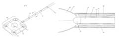
- FIG. 1A and FIG. 1Bshow perspective views of a device for repairing a craniomaxillofacial injury of the presently disclosed embodiments.
- FIG. 1Ashows a conformable bone plate of the device in a deflated state.
- FIG. 1Bshows a conformable bone plate of the device in an inflated state.
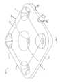
- FIG. 2shows an enlarged perspective view of the conformable bone plate of the device in an inflated state.
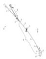
- FIG. 3A , FIG. 3B and FIG. 3Cshow close-up views of some of the main components of a device for repairing a craniomaxillofacial injury of the presently disclosed embodiments.
- FIG. 3Ashows a perspective view of a distal end of the device.
- FIG. 3Bshows a side cross-sectional view taken along line A-A in FIG. 3A of the device.
- FIG. 3Cshows a cross-sectional view of the device taken along line B-B in FIG. 3A .
- FIG. 4shows a perspective view of a light source for use with a device for repairing a craniomaxillofacial injury of the presently disclosed embodiments.
- FIG. 5A and FIG. 5Bshow close-up cross-sectional views of some of the main components, including a light pipe, of the device for repairing a craniomaxillofacial injury of the presently disclosed embodiments.
- FIG. 5Ashows a side cross-sectional view of the device taken along line A-A from FIG. 3A .
- FIG. 5Bshows a cross-sectional view of the device taken along line B-B from FIG. 3A .
- FIG. 6shows the placement of a conformable bone plate of the presently disclosed embodiments during repair of a craniomaxillofacial injury.
- the craniomaxillofacial bone plates of the presently disclosed embodimentsare made from a pliable, resilient, conformable, biocompatible, and strong material. At least one filling material is contained within the bone plates of the presently disclosed embodiments.
- a conformable bone plateis brought to the site of injury (an osteotomy, a fracture, a bone graft or other site of injury) in a deflated state. Once in place, the bone plate is expanded from a deflated state to an inflated state in situ by the addition of the at least one filling material.
- the addition of the at least one filling materialmay be precisely controlled such that the bone plate will resemble the contour of the site, on a patient-by-patient basis. Once the desired level of expansion is achieved, the at least one filling material is hardened within the bone plate using a light source. The hardened bone plate is then affixed to the site using fasteners.
- the customizable bone plate of the presently disclosed embodimentsprovides support and proper orientation of the bony defect resulting in the repair, healing, and strengthening of the defect.
- Filling materialsinclude, but are not limited to, bone reinforcing mixtures (such as bone cement mixtures, bone void fillers, epoxies, glues and similar adhesives), orthopedic wires, stainless-steel rods, metal pins, and other similar devices.
- the filling materialmay be a natural or synthetic material for strengthening, replacing, or reinforcing of bones or bone tissue.
- Bone reinforcing mixturesinclude glues, epoxies, adhesives, cements, hard tissue replacement polymers, biodegradable polymers such as PLA, PGA, and PLA-PGA copolymers, natural coral, hydroxyapatite, beta-tricalcium phosphate, and various other biomaterials known in the art for strengthening, replacing or reinforcing bones.
- bone reinforcing mixturesmay be incorporated into surrounding tissue or gradually replaced by original tissue.
- a device disclosed hereinmay be used for the repair of a craniomaxillofacial bony defect related to trauma, tumors, as well as for developmental and congenital craniomaxillofacial deformities.
- the main components of a device for repairing a craniomaxillofacial bony defectare shown generally in FIG. 1A and FIG. 1B in conjunction with FIG. 2 .
- the device 100includes a delivery catheter 110 having an elongated shaft with a proximal end 102 , a distal end 104 , and a longitudinal axis therebetween.
- the delivery catheter 110has a diameter of about 3 mm.
- the distal end 104 of the delivery catheter 110terminates in a releasable conformable bone plate 103 .
- the bone plate 103may move from a deflated state ( FIG. 1A ) to an inflated state ( FIG. 1B ) when at least one filling material is delivered to the bone plate 103 .
- the thickness of the deflated conformable bone plate 103is about 7 mm.
- An inflated conformable bone plate 103as shown in FIG. 1B , may have a size of about 3 mm by about 10 mm. Alternately, the inflated conformable bone plate 103 may have a size of about 25 mm by about 25 mm.
- the filling materialis delivered to the bone plate 103 via an inner void capable of allowing the filling material to pass through.
- the filling materialis a UV-activated glue.
- a stiffening member 105may surround the elongated shaft of the delivery catheter 110 and provides rigidity over the elongated shaft.
- a pusher or stabilizer 116is loaded proximal to the bone plate 103 .
- a slip sleeve 107may surround the stiffening member 105 .
- the slip sleeve 107surrounds the stiffening member 105 from a proximal end 123 of the bone plate 103 up until the pusher 116 .
- One or more radiopaque markers or bands 130may be placed at various locations along the bone plate 103 and/or the slip sleeve 107 for alignment of the device 100 during fluoroscopy or other forms of visualization.
- the one or more radiopaque bands 130using radiopaque materials such as barium sulfate, tantalum, or other materials known to increase radiopacity, allows a medical professional to view the device 100 using fluoroscopy techniques.
- the one or more radiopaque bands 130also provide visibility during inflation of the bone plate 103 to determine the precise positioning of the bone plate 103 and the device 100 during placement and inflation.
- the one or more radiopaque bands 130permit visualization of any voids that may be created by air that gets entrapped in the bone plate 103 .
- the one or more radiopaque bands 130permit visualization to preclude the bone plate 103 from misengaging or not meeting the bony defects due to improper inflation. It may be desirable to imbed radiopaque beads on the corners of the conformable bone plate 103 to assist in the visualization/location of the bone plate 103 .
- an adapter 115such as a Tuohy-Borst adapter, engages the proximal end 102 of the delivery catheter 110 .
- a light source that includes a light pipe 152may be introduced into one of the side-arms of the adapter 115 and passes within an inner lumen of the delivery catheter 110 up until the distal end 104 of the delivery catheter 110 .
- a delivery system 160 housing the filling materialmay be introduced into another side-arm of the adapter 115 , as shown in FIG. 1A .
- a Luer fittingmay engage the proximal end 102 of the delivery catheter 110 and a Luer fitting would exist on the light source such that the delivery catheter 110 and the light source would lock together.
- the delivery system 160is a syringe.
- the syringe 160has a control mechanism that regulates the flow of the filling material.
- the control mechanism of the syringe 160allows the filling material to flow into the delivery catheter 110 and the flow may be stopped if desired.
- the syringe 160makes direct contact to control the directional flow of the filling material, and the direction of flow of the filling material instantaneously changes within the delivery catheter 110 in response to a change in the direction of the syringe 160 .
- the syringe 160is opaque and does not allow light to penetrate within the syringe 160 . Having an opaque syringe 160 ensures that the filling material contained in the syringe 160 is not exposed to light and will not cure in the syringe 160 .
- the filling materialis of a liquid consistency, as measured in Centipoise (cP), the unit of dynamic viscosity, so the filling material may be infused from the syringe 160 into the delivery catheter 110 and into the bone plate 103 . Because the filling material has a liquid consistency and is viscous, the filling material may be delivered using low pressure delivery and high pressure delivery is not required, but may be used.
- a separation areais located at the junction between the proximal end 123 of the bone plate 103 and the delivery catheter 110 .
- the separation areamay also include an illumination band. When activated, the illumination band causes light to cure the filling material located in the bone plate 103 within the illumination band.
- the illumination bandextends around the delivery catheter 110 and has a stress concentrator.
- the stress concentratormay be a notch, groove, channel or similar structure that concentrates stress in the illumination band.
- the stress concentrator of the illumination bandmay be notched, scored, indented, pre-weakened or pre-stressed to direct separation of the bone plate 103 from the elongated shaft of the delivery catheter 110 under specific torsional load.
- the separation areaensures that there are no leaks of filling material from the elongated shaft of the delivery catheter 110 and/or the bone plate 103 .
- the separation areaseals the bone plate 103 and removes the elongated shaft of the delivery catheter 110 by making a break at a known or predetermined site (e.g., a separation area).
- the separation areamay be various lengths and up to about an inch long.
- Having a distinct switch to activate the illumination bandmay help to prevent inadvertent delivery of light from the light source to cure the filling material.
- the activation of the illumination bandseals the bone plate 103 and seals the end of the delivery catheter 110 , and ensures that there is a “hard seal” of the filling material at the illumination band allowing no filling material to leak from the bone plate 103 or the delivery catheter 110 .
- FIG. 2shows a close-up view of the bone plate 103 .
- the bone plate 103is a pad having, a first surface 133 , a second surface 143 , a right side surface 145 , a left side surface 142 , a front surface 141 , a back surface 144 , and an inner space therebetween.
- the first surface 133 and the second surface 143each have a first area.
- the left side surface 142 and the right side surface 145each have a second area.
- the front surface 141 and the back surface 144each have a third area. The first area is greater than the second area.
- a major longitudinal axisruns from the left side surface 142 to the right side surface 145
- a minor longitudinal axisruns from the first surface 133 to the second surface 143
- the padincludes an opening 147 positioned only through the right side surface 145 of the pad, wherein the opening 147 is sufficiently designed to engage the delivery catheter 110 .
- At least one pre-punched aperture 121extends from the first surface 133 to the second surface 143 , along the minor longitudinal axis, for attaching the bone plate 103 to an exterior surface of the bone fragments. In the embodiment depicted in FIG. 2 , there are five pre-punched apertures 121 for attaching the bone plate 103 to the bone fragments, one aperture at each corner and one aperture in the middle.
- apertures 121permits multiple connections to bones and ensures contouring and consistent engagement to bones.
- the apertures 121may be threaded or simply formed as non-threaded through holes.
- the apertures 121are adapted to receive a fastener for interconnecting the bone plate 103 with a severed bone region.
- the bone plate 103may have a baffle structure which reduces wave motion of the filling material in the bone plate 103 . Baffles would float within the bone plate 103 and may have serpentine, cone, coil or cylindrical shapes.
- the bone plate 103may have a pre-defined shape to fit over a specific bony defect site.
- the bone plate 103may be a pad that is round, flat, cylindrical, oval, rectangular or another shape, as long as the bone plate 103 bridges and supports fragments of fractured bone.
- the pre-defined shape of the bone plate 103is a generally square pad.
- the bone plate 103may be formed of a pliable, resilient, conformable, biocompatible, and strong material, including but not limited to urethane, polyethylene terephthalate (PET), nylon elastomer and other similar polymers.
- the bone plate 103is constructed out of a PET nylon aramet or other non-consumable materials.
- PETis a thermoplastic polymer resin of the polyester family that is used in synthetic fibers. Depending on its processing and thermal history, PET may exist both as an amorphous and as a semi-crystalline material. Semi-crystalline PET has good strength, ductility, stiffness and hardness. Amorphous PET has better ductility, but less stiffness and hardness. PET can be semi-rigid to rigid, depending on its thickness, and is very lightweight. PET is strong and impact-resistant, naturally colorless and transparent and has good resistance to mineral oils, solvents and acids.
- the bone plate 103has an outer surface 122 .
- the outer surface 122is resilient and puncture resistant.
- the outer surface 122 of the bone plate 103is substantially even and smooth.
- the outer surface 122 of the bone plate 103is not entirely smooth and may have some small bumps or convexity/concavity along the length.
- the outer surface 122 of the bone plate 103may have ribs, ridges, bumps or other shapes to help the bone plate 103 conform to the shape of a bony defect.
- the bone plate 103has a textured surface which provides one or more ridges that allow grabbing all portions of bony defects.
- sand blasted surfacing on the outer surface 122 of the bone plate 103improves the connection and adhesion between the outer surface 122 of the bone plate 103 and the bony defect.
- the surfacingsignificantly increases the amount of surface area that comes in contact with the bone resulting in a stronger grip.
- the outer surface 122 of the bone plate 103may be coated with materials such as drugs, bone glue, proteins, growth factors, or other coatings.
- materialssuch as drugs, bone glue, proteins, growth factors, or other coatings.
- An antibiotic drugmay be added to the outer surface 122 of the bone plate 103 to prevent or combat a possible infection.
- Proteins, such as, for example, the bone morphogenic protein or other growth factorshave been shown to induce the formation of cartilage and bone.
- a growth factormay be added to the outer surface 122 of the bone plate 103 to help induce the formation of new bone. Due to the lack of thermal egress of the filling material in the bone plate 103 , the effectiveness and stability of the coating is maintained.
- a water soluble glueis applied to the outer surface 122 of the bone plate 103 .
- the water soluble glue on the outer surface 122 of the bone plate 103becomes sticky or tacky and acts as a gripping member to increase the conformal bond of the bone plate 103 to the bone.
- the outer surface 122 of the bone plate 103grips the bone forming a mechanical bond as well as a chemical bond. These bonds prevent the potential for a bone slippage.
- the water soluble gluemay be cured by any light (e.g., UV not required).
- the bone plate 103 of the device 100typically does not have any valves.
- One benefit of having no valvesis that the bone plate 103 may be inflated or deflated as much as necessary to resemble the contour of the new anatomy of an osteotomy, fracture or bone graft site.
- Another benefit of the bone plate 103 having no valvesis the efficacy and safety of the device 100 . Since there is no communication passage of filling material to the body there cannot be any leakage of material because all the material is contained within the bone plate 103 . In an embodiment, a permanent seal is created between the bone plate 103 that is both hardened and affixed prior to the delivery catheter 110 being removed.
- a permanent sealis created between the bone plate 103 that is hardened, the deliver catheter 119 is removed, and the bone plate 103 is affixed to the bone.
- the bone plate 103may have valves, as all of the embodiments are not intended to be limited in this manner.
- FIG. 3A , FIG. 3B and FIG. 3Cshow close-up views of some of the main components of the device 100 .
- One or more radiopaque markers or bands 130may be placed at various locations along the slip sleeve 107 of the device 100 .
- radiopaque markers 130may also be placed at various locations along the bone plate 103 .
- the one or more radiopaque bands 130are placed at intervals of about 10 mm along the length of the slip sleeve 107 .
- the radiopaque markers 130are formed using radiopaque material such as barium sulfate, tantalum, or other materials known to increase radiopacity.
- the radiopaque markers 130provide visibility during inflation of the bone plate 103 to determine the precise positioning of the bone plate 103 and the delivery catheter during placement and inflation.
- the radiopaque markers 130permit visualization of voids created by air that may be entrapped in the bone plate 103 .
- the radiopaque markers 130permit visualization to preclude the bone plate 103 from misengaging or not meeting the bony defects due to improper inflation.
- the proximal end of the delivery cathetermay be attached to a delivery system that contains a filling mixture.
- FIG. 3Bshows a cross-sectional view taken along line A-A of FIG. 3A .
- an elongated shaft 101 of the delivery catheterterminates in the bone plate 103 having the outer surface 122 .
- a light pipe conduit 111for accepting a light source (not shown).
- a void 113 for passage of at least one filling materialis formed between an inner surface 124 of the delivery catheter and an outer surface 117 of the light pipe conduit 111 .
- a delivery system comprising the at least one filling materialmay be attached to a side arm of a Tuohy-Borst adapter that is engaged to a proximal end of the delivery catheter.
- the at least one filling materialpasses through the void 113 of the delivery catheter and enters the bone plate 103 .
- the infusion of the filling materialcauses the bone plate 103 to inflate to a desired state.
- the filling materialis infused through the void 113 in the delivery catheter to expand the bone plate 103 such that the bone plate 103 will resemble the contour of the new anatomy of an osteotomy, fracture or bone graft site.
- Orientation of the bones and bone plate 103may be done without any visualization of the process or using x-ray or a fluoroscope.
- a C arm imaging systemis used as part of a fluoroscope. The C arm imaging system may allow movement or manipulation of the fluoroscope to rotate around tissue while viewing.
- the bone plate 103may be composed of non ferromagnetic materials and, thus, is compatible with MRI.
- FIG. 3Cshows a cross-sectional view taken along line B-B of FIG. 3A .
- the outer slip sleevehas been removed.
- the stiffening member 105surrounds and provides rigidity to the elongated shaft 101 of the delivery catheter 110 .
- the light pipe conduit 111provides a space for a light source to pass through.
- the void 113is formed between the outer surface 117 of the light pipe conduit 111 and the inner surface 124 of the elongated shaft 101 . This void 113 provides a passageway for the at least one filling material.
- the outer surface 117 of the light pipe conduit 111allows for a separation between the light source and the filling material.
- FIG. 4in conjunction with FIGS. 1A and 1B , shows a light source 150 for use with the device 100 of the presently disclosed embodiments.
- the light source 150is used to harden the filling material that has been infused into the bone plate 103 through the delivery catheter 110 .
- the light source 150includes a light pipe 152 which terminates in an optical lens 154 . Energy emitted from the light pipe 152 is projected through the optical lens 154 and guided into the bone plate 103 of the device 100 .
- the optical lens 154may be convex, concave or planar.
- the optical lens 154is curved to converge or diverge the transmitted energy from the light pipe 152 .
- the optical lens 154is made out of a plastic material such as Acrylic (PMMA), Polycarbonate (PC), Polystyrene (PS), or other similar materials known to those in the art such as Cyclic Olefin Copolymer (COC), and Amorphous Polyolefin (Zeonex).
- the optical lens 154is made out of a glass material such as quartz.
- the light source 150is introduced into a side arm of the adapter 115 that engages the proximal end 102 of the delivery catheter 110 , as shown in FIG. 1A .
- the light source 150runs through the elongated shaft 101 of the delivery catheter 110 through the light pipe conduit and up into the proximal end 123 of the bone plate 103 A.
- the activation of the light source 150cures the filling material resulting in the affixing of the bone plate 103 in an expanded shape.
- a curemay refer to any chemical, physical, and/or mechanical transformation that allows a composition to progress from a form (e.g., flowable form) that allows it to be delivered through the void in the delivery catheter 110 , into a more permanent (e.g., cured) form for final use in vivo.
- curablemay refer to uncured composition, having the potential to be cured in vivo (as by catalysis or the application of a suitable energy source), as well as to a composition in the process of curing (e.g., a composition formed at the time of delivery by the concurrent mixing of a plurality of composition components).
- the filling materialis a light cure adhesive or ultraviolet (UV) adhesive.
- UV curingis a cure-on-demand process and that adhesives may be free of solvents and include environmentally friendly resins that cure in seconds upon exposure to long wave UV light or visible light.
- Different UV adhesivesuse photoinitiators sensitive to different ranges of UV and visible light. Being very energetic, UV light can break chemical bonds, making molecules unusually reactive or ionizing them, in general changing their mutual behavior. Visible light, for example, visible blue light, allows materials to be cured between substrates that block UV light but transmits visible light (e.g., plastics). Visible light penetrates through the adhesive to a greater depth.
- Additivesmay be used with the UV adhesive delivery system, including, but not limited to drugs (for example, antibiotics), proteins (for example, growth factors) or other natural or synthetic additives.
- the electromagnetic spectrumis the range of all possible electromagnetic radiation.
- the electromagnetic spectrum of an objectis the frequency range of electromagnetic radiation that the object emits, reflects, or transmits.
- the electromagnetic spectrumextends from just below the frequencies used for modern radio (at the long-wavelength end) to gamma radiation (at the short-wavelength end), covering wavelengths from thousands of kilometers down to fractions of the size of an atom.
- the UV adhesiveis a single-component, solvent-free adhesive that will not cure until a UV light engages the adhesive, and when that occurs, the adhesive will cure in seconds to form a complete bond with a shear strength.
- the filling materialexhibits a shrinkage upon cure of about 2 to about 3 percent.
- UV light wavelengthranges from about 1 nm to about 380 nm, and can be subdivided into the following categories: near UV (380-200 nm wavelength; abbreviated NUV), far or vacuum UV (200-10 nm; abbreviated FUV or VUV), and extreme UV (1-31 nm; abbreviated EUV or XUV).
- near UV380-200 nm wavelength
- FUVfar or vacuum UV
- EUVextreme UV
- visible lighthas a wavelength spectrum of between about 380 to about 780 nm.
- UV adhesivesmay be activated by UV light, visible light, x-rays, gamma rays, microwaves, radio waves, long waves or any light having a wavelength less than about 1 nm, between about 1 nm and about 380 nm, between about 380 nm and about 780 nm, or greater than 780 nm, as not all embodiments are intended to be limited in that respect.
- Using a UV light to cure the filling materialassists in holding broken bones in place, filling of the bone plate, and viewing under a C arm imaging system.
- the filling materialscure in such a way that is sufficient to conform to the contour of a bony defect. More specifically, the ability to inflate, set, adjust, orient bones, and the resulting union of the bone are available prior to hardening the filling material.
- the introduction of the UV lightstarts the photoinitiator and the UV adhesive hardens. Once the UV light is introduced, the adhesive inside the bone plate hardens and the adhesives inside are affixed in place.
- the bone platemay be inflated or deflated due to the viscosity of the adhesive.
- the adhesivemay be infused or removed from the bone plate due to the low viscosity of the adhesive.
- the viscosity of the filling materialhas a viscosity of about 1000 cP or less.
- the filling materialhas a viscosity ranging from about 650 cP to about 450 cP. Not all embodiments are intended to be limited in this respect and some embodiments may include filling materials having a viscosity exactly equal to or greater than 1000 cP.
- a contrast materialmay be added to the filling material without significantly increasing the viscosity. Contrast materials include, but are not limited to, barium sulfate, tantalum, or other contrast materials known in the art.
- epoxies known in the artare suitable for use as filling materials and vary in viscosity, cure times, and hardness (durometer or shore) when fully cured.
- a durometer of a materialindicates the hardness of the material, defined as the material's resistance to permanent indentation.
- a specific durometer UV adhesivemay be chosen.
- multiple UV adhesives having varying durometersmay be chosen for the repair of a bony defect and be within the scope and spirit of the presently disclosed embodiments.
- the durometer of a materialmay be altered to achieve either greater rigidity or a more malleable result.
- the mechanical properties of the epoxiesmay dictate using methods/measures that are typical for high-strength and high-impact materials including but not limited to, tensile strength and tensile modulus, tensile strength tests, ultimate modulus, Poisson's ratio, hardness measurements like Vickers and Charpy Impact which measures yield strength and toughness.
- the filling materialis cured by chemical activation or thermal activation.
- Chemical activationincludes but is not limited to water or other liquids.
- the filling materialis a drying adhesive which has a polymer dissolved in a solvent such that as the solvent evaporates, the adhesive hardens.
- the filling materialis a hot or thermoplastic adhesive such that as the adhesive cools, the adhesive hardens.
- Some filling materialsmay require or be enhanced by curing via any means, such as UV or visible light, heat, and/or addition or removal of a chemical or substance, may utilize any outside or internal processes to cure the material, or may not require curing.
- carbon nanotubesare added to the filling material to increase the strength of the material.
- Carbon nanotubesare an allotrope of carbon that take the form of cylindrical carbon molecules and have novel strength properties. Carbon nanotubes exhibit extraordinary strength.
- Nanotubesare members of the fullerene structural family, which also includes buckyballs. Whereas buckyballs are spherical in shape, a nanotube is cylindrical with at least one end typically capped with a hemisphere of the buckyball structure. Nanotubes are composed entirely of sp2 bonds, similar to those of graphite. This bonding structure, which is stronger than the sp3 bonds found in diamond, provides the molecules with their unique strength. Nanotubes naturally align themselves into “ropes” held together by Van der Waals forces. Single walled nanotubes or multi-walled nanotubes may be used to strengthen the filling materials.
- a central spacemay remain in the bone plate 103 which may be filled in order to provide extra strength and support to the fractured bones.
- An optical rod or similar devicemay be positioned in the central space and turned on or illuminated.
- An optical rod or similar devicecan be made of fiber, silica, quartz, sapphire or similar materials. The end of the optical rod may be cut and remain in the bone plate 103 to provide increased rigidity.
- FIG. 5A and FIG. 5Bshow cross-sectional views of the device 100 of FIG. 1A , showing the light source 150 of FIG. 4 passing through the light pipe conduit 111 of the delivery catheter 110 , through the opening 147 and into the inner space of the pad along the longitudinal axis of the pad.
- the light sourceincludes the light pipe 152 terminating in the optical lens 154 .
- the light sourceis used to harden the filling material that has been infused into the bone plate 103 of the device 100 .
- Energy from the light source 150is emitted from the light pipe 152 , projected through the optical lens 154 , and guided into the bone plate 103 of the device 100 .
- the optical lens 154may be convex, concave or planar.
- the optical lens 154is curved to converge or diverge the transmitted energy from the light pipe 152 .
- Craniofacial surgeryencompasses the reconstruction of a broad spectrum of facial deformities including, but not limited to, zygomatic fractures, maxillary fractures, nasoethmoid orbital fractures, internal orbital fractures, mandibular fractures, post-traumatic facial deformities, soft tissue deformities, and facial bone contouring.
- An implant for fracture fixationmust be strong, ductile, adaptable to the bone surface and biocompatible. Fixation system quality includes adequate strength and rigidity, avoiding adverse reactions, interference with bone healing, intracranial migration, visibility and palpability and implant removal operation.
- FIG. 6illustrates the device 100 of the presently disclosed embodiments in use during a procedure for repairing a craniomaxillofacial bony defect in a patient.
- the procedurebegins with placing the deflated bone plate 103 of the device 100 at the bony defect, where the bone plate 103 spans at least two bone fragments (A and B in FIG. 6 ).
- a delivery systemsuch as the syringe 160 filled with the filling material (not shown), is attached to the device 100 .
- the filling materialis then infused through the inner void in the elongated shaft of the delivery catheter 110 and enters the bone plate 103 of the device 100 , causing the bone plate 103 to move from a deflated state to an expanded state.
- the light source 152is attached to the device 100 .
- the light source 152is passed through the elongated shaft of the delivery catheter 110 through the light pipe conduit.
- the filling materialis a UV curable glue which requires a UV light source to cure the adhesive.
- the light source 152is then activated which causes the filling material to harden.
- the hardened bone plate 103may be affixed to the bony defect site, thus stabilizing the fracture.
- the light source 152is removed from the device 100 and the bone plate 103 is released from the delivery catheter 110 .
- the delivery catheter 110is cut to separate the bone plate 103 from the elongated shaft 101 .
- a deviceslides over the delivery catheter 110 and allows a right angle scissor to descend through the delivery catheter 110 and make a cut.
- the location of the cutmay be determined by using a fluoroscope or an x-ray. In an embodiment, the cut location is at the junction where the elongated shaft 101 meets the bone plate 103 .
- a separation areais located at the junction between the proximal end 123 of the bone plate 103 and the delivery catheter 110 .
- the separation areamay also include an illumination band. When activated, the illumination band causes light to cure the filling material located in the bone plate 103 within the illumination band.
- the illumination bandextends around the delivery catheter 110 and has a stress concentrator.
- the stress concentratormay be a notch, groove, channel or similar structure that concentrates stress in the illumination band.
- the stress concentrator of the illumination bandmay be notched, scored, indented, pre-weakened or pre-stressed to direct separation of the bone plate 103 from the elongated shaft of the delivery catheter 110 under specific torsional load.
- the separation areaensures that there are no leaks of filling material from the elongated shaft of the delivery catheter 110 and/or the bone plate 103 .
- the separation areaseals the bone plate 103 and removes the elongated shaft of the delivery catheter 110 by making a break at a known or predetermined site (e.g., a separation area).
- the separation areamay be various lengths and up to about an inch long.
- Having a distinct switch to activate the illumination bandmay help to prevent inadvertent delivery of light from the light source to cure the filling material.
- the activation of the illumination bandseals the bone plate 103 and seals the end of the delivery catheter 110 , and ensures that there is a “hard seal” of the filling material at the illumination band allowing no filling material to leak from the bone plate 103 or the delivery catheter 110 .
- the bone plate 103may be fastened to the bony defect before, after or during a repair procedure.
- Fasteners 163are disposed through the apertures 121 and engage the bone plate 103 to secure the bone plate 103 to one or more portions of bone.
- a method for adjoining at least two bone fragmentsincludes providing a device for adjoining at least two bone fragments, the device including a conformable bone plate engaged to a delivery catheter having an elongated shaft with a proximal end, a distal end, and a longitudinal axis therebetween, wherein the conformable bone plate has at least one aperture extending from a first surface of the bone plate to a second surface of the bone plate for receiving a fastener to affix the bone plate to the at least two bone fragments; positioning the conformable bone plate over an exterior surface spanning the at least two bone fragments; attaching a delivery system housing at least one filling material to the delivery catheter, wherein the elongated shaft of the delivery catheter has an inner void for passage of the at least one filling material to the conformable bone plate; inserting a light source into the delivery catheter, wherein the elongated shaft of the delivery catheter has an inner lumen for passage of the light source to the conformable bone plate; infusing the at least one filling material through the
- the bone plates and devices disclosed hereinhave been discussed in the repair of a craniomaxillofacial bony defect. Those skilled in the art will recognize that the bone plates and devices may be used in a variety of areas, such as bones of the vertebrae and any other fractured bone that required support on its exterior surface so as to hold the disassociated portions in alignment during healing.
Landscapes
- Health & Medical Sciences (AREA)
- Orthopedic Medicine & Surgery (AREA)
- Surgery (AREA)
- Life Sciences & Earth Sciences (AREA)
- Heart & Thoracic Surgery (AREA)
- Animal Behavior & Ethology (AREA)
- Engineering & Computer Science (AREA)
- Biomedical Technology (AREA)
- Neurology (AREA)
- Medical Informatics (AREA)
- Molecular Biology (AREA)
- Nuclear Medicine, Radiotherapy & Molecular Imaging (AREA)
- General Health & Medical Sciences (AREA)
- Public Health (AREA)
- Veterinary Medicine (AREA)
- Surgical Instruments (AREA)
- Dental Preparations (AREA)
- Prostheses (AREA)
Abstract
Description
This application is a continuation of U.S. application Ser. No. 11/964,370, filed on Dec. 26, 2007, which is incorporated herein by reference in its entirety.
The embodiments disclosed herein relate to medical devices for use in adjoining bone fragments, and more particularly to conformable bone plates for repair of craniomaxillofacial bony defects and methods of using conformable bone plates.
Maxillofacial and craniofacial injuries encompass any injury to the mouth, face and jaw. Common serious injury to the face occurs when bones are broken (a fracture). Fractures can involve the lower jaw, upper jaw, palate, cheekbones, eye sockets and combinations of these bones. The fracture needs to be held in the correct position while the bone is healing. In most cases this requires fixing the bones using metal or biodegradable plates and screws, known as internal fixation.
There are a variety of micro, mini and reconstruction plating systems. One of the most commonly used plating system developed to date is the Luhr system manufactured by Howmedica, Inc. Subsequently, based on the original concept by Luhr, several complete systems have been developed for use in all the various situations encountered in trauma and reconstructive surgery of the facial skeleton. Techniques and materials used for the internal fixation of the maxillofacial skeleton continue to evolve and improve. For example, metal plates have been used for the repair of craniomaxillofacial bone fractures. These metal plates are generally secured to the fractured bone portions with fasteners such as screws. The plates conventionally employed generally comprise small, generally flat, elongated sections of metal. The sections contain round screw holes at various points along their lengths for fastening the sections to bone. The metal plate is then bent into shape and secured to the fractured bone using a plurality of fasteners seated within the screw holes. While known systems utilizing plates and fasteners for aiding the osteosynthesis of severed bone regions have proven to be acceptable for certain applications, such systems are nevertheless susceptible to improvements that may enhance their performance. For example, metals are difficult to shape and are hampered by disadvantages such as infection and corrosion. Several resorbable plate and screw fixation systems are now available for use in the maxillofacial skeleton. These systems allow initial stable fixation of bone segments during the bone-healing phase and then gradually are reabsorbed through physiologic processes.
Regardless of the plate system used, the plate must be contoured to lay passively against the underlying bone surfaces. Therefore, even though the plating systems themselves are manufactured with extremely precise tolerances, an element of imprecision remains for surgeons who repair facial fractures and do orthognathic surgery or reconstructive procedures repositioning the facial skeletal structures to improve esthetics or function. When an osteotomy, fracture, or bone graft is placed into appropriate position, the bone plate has to be manually bent to the contour of the anatomy. This manual manipulation creates a substantial element of imprecision even with the use of templates. Maladapted bone plates lead to inappropriate bone contour, irritation of the overlying soft tissues, abnormal anatomy or contour defects and either non-union, malunion or unaesthetic results.
Thus, there is a need in the art for devices and methods for repairing maxillofacial and craniofacial bony defects that are easy to use, biocompatible, require minimal manipulation once in place, and are customizable on a patient-by-patient basis.
Conformable bone plates and methods for using the bone plates to adjoin bone fragments are disclosed herein. According to aspects illustrated herein, there is provided a bone plate for adjoining at least two bone fragments that includes a flexible pad capable of expanding, the pad having a first surface and a second surface; and at least one aperture for receiving a fastener, the at least one aperture extending from the first surface to the second surface, wherein the bone plate is able to move from a first compact position to a second expanded position and is contoured to passively engage against the at least two bone fragments.
According to aspects illustrated herein, there is provided a device for adjoining at least two bone fragments that includes a delivery catheter having an elongated shaft with a proximal end, a distal end, and a longitudinal axis therebetween, wherein the delivery catheter has an inner void for passage of at least one filling material and an inner lumen for passage of light from a light source; a conformable bone plate releasably engaging the distal end of the delivery catheter, wherein the conformable bone plate has a first surface, a second surface, a proximal end, and a distal end and at least one aperture extending from the first surface to the second surface for receiving a fastener; and an adapter releasably engaging the proximal end of the delivery catheter for receiving light from the light source and a delivery system housing the at least one filling material.
According to aspects illustrated herein, there is provided a method for adjoining at least two bone fragments that includes providing a device for adjoining at least two bone fragments, the device including a conformable bone plate engaged to a delivery catheter having an elongated shaft with a proximal end, a distal end, and a longitudinal axis therebetween, wherein the conformable bone plate has at least one aperture extending from a first surface of the bone plate to a second surface of the bone plate for receiving a fastener to affix the bone plate to the at least two bone fragments; positioning the conformable bone plate over an exterior surface spanning the at least two bone fragments; attaching a delivery system housing at least one filling material to the delivery catheter, wherein the elongated shaft of the delivery catheter has an inner void for passage of the at least one filling material to the conformable bone plate; inserting a light source into the delivery catheter, wherein the elongated shaft of the delivery catheter has an inner lumen for passage of the light source to the conformable bone plate; infusing the at least one filling material through the elongated shaft of the delivery catheter and into the conformable bone plate to expand the conformable bone plate; activating the light source to harden the at least one filling material in the expanded conformable bone plate; releasing the hardened conformable bone plate from the delivery catheter; and affixing the conformable bone plate to the exterior surfaces of the at least two bone fragments to adjoin the bone fragments.
The presently disclosed embodiments will be further explained with reference to the attached drawings, wherein like structures are referred to by like numerals throughout the several views. The drawings shown are not necessarily to scale, with emphasis instead generally being placed upon illustrating the principles of the presently disclosed embodiments.
While the above-identified drawings set forth presently disclosed embodiments, other embodiments are also contemplated, as noted in the discussion. This disclosure presents illustrative embodiments by way of representation and not limitation. Numerous other modifications and embodiments may be devised by those skilled in the art which fall within the scope and spirit of the principles of the presently disclosed embodiments.
Medical devices and methods for repairing maxillofacial and craniofacial injuries are disclosed herein. The craniomaxillofacial bone plates of the presently disclosed embodiments are made from a pliable, resilient, conformable, biocompatible, and strong material. At least one filling material is contained within the bone plates of the presently disclosed embodiments. During a procedure for repairing a craniomaxillofacial injury, a conformable bone plate is brought to the site of injury (an osteotomy, a fracture, a bone graft or other site of injury) in a deflated state. Once in place, the bone plate is expanded from a deflated state to an inflated state in situ by the addition of the at least one filling material. The addition of the at least one filling material may be precisely controlled such that the bone plate will resemble the contour of the site, on a patient-by-patient basis. Once the desired level of expansion is achieved, the at least one filling material is hardened within the bone plate using a light source. The hardened bone plate is then affixed to the site using fasteners. The customizable bone plate of the presently disclosed embodiments provides support and proper orientation of the bony defect resulting in the repair, healing, and strengthening of the defect.
Filling materials include, but are not limited to, bone reinforcing mixtures (such as bone cement mixtures, bone void fillers, epoxies, glues and similar adhesives), orthopedic wires, stainless-steel rods, metal pins, and other similar devices. The filling material may be a natural or synthetic material for strengthening, replacing, or reinforcing of bones or bone tissue. Bone reinforcing mixtures include glues, epoxies, adhesives, cements, hard tissue replacement polymers, biodegradable polymers such as PLA, PGA, and PLA-PGA copolymers, natural coral, hydroxyapatite, beta-tricalcium phosphate, and various other biomaterials known in the art for strengthening, replacing or reinforcing bones. As inert materials, bone reinforcing mixtures may be incorporated into surrounding tissue or gradually replaced by original tissue. Those skilled in the art will recognize that numerous bone reinforcing mixtures known in the art are within the spirit and scope of the presently disclosed embodiments.
A device disclosed herein may be used for the repair of a craniomaxillofacial bony defect related to trauma, tumors, as well as for developmental and congenital craniomaxillofacial deformities.
The main components of a device for repairing a craniomaxillofacial bony defect are shown generally inFIG. 1A andFIG. 1B in conjunction withFIG. 2 . Thedevice 100 includes adelivery catheter 110 having an elongated shaft with aproximal end 102, adistal end 104, and a longitudinal axis therebetween. In an embodiment, thedelivery catheter 110 has a diameter of about 3 mm. Thedistal end 104 of thedelivery catheter 110 terminates in a releasableconformable bone plate 103. Thebone plate 103 may move from a deflated state (FIG. 1A ) to an inflated state (FIG. 1B ) when at least one filling material is delivered to thebone plate 103. In an embodiment, the thickness of the deflatedconformable bone plate 103 is about 7 mm. An inflatedconformable bone plate 103, as shown inFIG. 1B , may have a size of about 3 mm by about 10 mm. Alternately, the inflatedconformable bone plate 103 may have a size of about 25 mm by about 25 mm. Those skilled in the art will recognize that variations within these ranges are possible and still within the scope and spirit of the presently disclosed embodiments. The filling material is delivered to thebone plate 103 via an inner void capable of allowing the filling material to pass through. In an embodiment, the filling material is a UV-activated glue.
A stiffeningmember 105 may surround the elongated shaft of thedelivery catheter 110 and provides rigidity over the elongated shaft. A pusher orstabilizer 116 is loaded proximal to thebone plate 103. Aslip sleeve 107 may surround the stiffeningmember 105. In an embodiment, theslip sleeve 107 surrounds the stiffeningmember 105 from aproximal end 123 of thebone plate 103 up until thepusher 116. One or more radiopaque markers orbands 130 may be placed at various locations along thebone plate 103 and/or theslip sleeve 107 for alignment of thedevice 100 during fluoroscopy or other forms of visualization. The one or moreradiopaque bands 130, using radiopaque materials such as barium sulfate, tantalum, or other materials known to increase radiopacity, allows a medical professional to view thedevice 100 using fluoroscopy techniques. The one or moreradiopaque bands 130 also provide visibility during inflation of thebone plate 103 to determine the precise positioning of thebone plate 103 and thedevice 100 during placement and inflation. The one or moreradiopaque bands 130 permit visualization of any voids that may be created by air that gets entrapped in thebone plate 103. The one or moreradiopaque bands 130 permit visualization to preclude thebone plate 103 from misengaging or not meeting the bony defects due to improper inflation. It may be desirable to imbed radiopaque beads on the corners of theconformable bone plate 103 to assist in the visualization/location of thebone plate 103.
In an embodiment, anadapter 115, such as a Tuohy-Borst adapter, engages theproximal end 102 of thedelivery catheter 110. A light source that includes alight pipe 152 may be introduced into one of the side-arms of theadapter 115 and passes within an inner lumen of thedelivery catheter 110 up until thedistal end 104 of thedelivery catheter 110. Adelivery system 160 housing the filling material may be introduced into another side-arm of theadapter 115, as shown inFIG. 1A . Alternately, a Luer fitting may engage theproximal end 102 of thedelivery catheter 110 and a Luer fitting would exist on the light source such that thedelivery catheter 110 and the light source would lock together.
Examples of delivery systems include, but are not limited to, caulking gun type systems, syringe systems, bag systems that contain the filling material where the delivery of the filling material is controlled using a tube clamp or any other restrictor valve. In the embodiments shown inFIG. 1A andFIG. 1B , thedelivery system 160 is a syringe. In an embodiment, thesyringe 160 has a control mechanism that regulates the flow of the filling material. The control mechanism of thesyringe 160 allows the filling material to flow into thedelivery catheter 110 and the flow may be stopped if desired. Thesyringe 160 makes direct contact to control the directional flow of the filling material, and the direction of flow of the filling material instantaneously changes within thedelivery catheter 110 in response to a change in the direction of thesyringe 160.
In an embodiment, thesyringe 160 is opaque and does not allow light to penetrate within thesyringe 160. Having anopaque syringe 160 ensures that the filling material contained in thesyringe 160 is not exposed to light and will not cure in thesyringe 160. The filling material is of a liquid consistency, as measured in Centipoise (cP), the unit of dynamic viscosity, so the filling material may be infused from thesyringe 160 into thedelivery catheter 110 and into thebone plate 103. Because the filling material has a liquid consistency and is viscous, the filling material may be delivered using low pressure delivery and high pressure delivery is not required, but may be used.
In an embodiment, a separation area is located at the junction between theproximal end 123 of thebone plate 103 and thedelivery catheter 110. The separation area may also include an illumination band. When activated, the illumination band causes light to cure the filling material located in thebone plate 103 within the illumination band. The illumination band extends around thedelivery catheter 110 and has a stress concentrator. The stress concentrator may be a notch, groove, channel or similar structure that concentrates stress in the illumination band. The stress concentrator of the illumination band may be notched, scored, indented, pre-weakened or pre-stressed to direct separation of thebone plate 103 from the elongated shaft of thedelivery catheter 110 under specific torsional load. The separation area ensures that there are no leaks of filling material from the elongated shaft of thedelivery catheter 110 and/or thebone plate 103. The separation area seals thebone plate 103 and removes the elongated shaft of thedelivery catheter 110 by making a break at a known or predetermined site (e.g., a separation area). The separation area may be various lengths and up to about an inch long. When torque (twisting) is applied to thedelivery catheter 110, the elongated shaft separates from thebone plate 103. The twisting creates a sufficient shear to break the residual filling material and create a clean separation of the plate/shaft interface. The illumination band may be connected to the light source and may be activated by a separate switch. Having a distinct switch to activate the illumination band may help to prevent inadvertent delivery of light from the light source to cure the filling material. The activation of the illumination band seals thebone plate 103 and seals the end of thedelivery catheter 110, and ensures that there is a “hard seal” of the filling material at the illumination band allowing no filling material to leak from thebone plate 103 or thedelivery catheter 110.
In an embodiment, thebone plate 103 may have a pre-defined shape to fit over a specific bony defect site. Thebone plate 103 may be a pad that is round, flat, cylindrical, oval, rectangular or another shape, as long as thebone plate 103 bridges and supports fragments of fractured bone. For example, as depicted in the embodiment ofFIG. 2 , the pre-defined shape of thebone plate 103 is a generally square pad.
Thebone plate 103 may be formed of a pliable, resilient, conformable, biocompatible, and strong material, including but not limited to urethane, polyethylene terephthalate (PET), nylon elastomer and other similar polymers. In an embodiment, thebone plate 103 is constructed out of a PET nylon aramet or other non-consumable materials. PET is a thermoplastic polymer resin of the polyester family that is used in synthetic fibers. Depending on its processing and thermal history, PET may exist both as an amorphous and as a semi-crystalline material. Semi-crystalline PET has good strength, ductility, stiffness and hardness. Amorphous PET has better ductility, but less stiffness and hardness. PET can be semi-rigid to rigid, depending on its thickness, and is very lightweight. PET is strong and impact-resistant, naturally colorless and transparent and has good resistance to mineral oils, solvents and acids.
Thebone plate 103 has anouter surface 122. Theouter surface 122 is resilient and puncture resistant. In an embodiment, theouter surface 122 of thebone plate 103 is substantially even and smooth. In an embodiment, theouter surface 122 of thebone plate 103 is not entirely smooth and may have some small bumps or convexity/concavity along the length. In an embodiment, theouter surface 122 of thebone plate 103 may have ribs, ridges, bumps or other shapes to help thebone plate 103 conform to the shape of a bony defect. In an embodiment, thebone plate 103 has a textured surface which provides one or more ridges that allow grabbing all portions of bony defects. In an embodiment, sand blasted surfacing on theouter surface 122 of thebone plate 103 improves the connection and adhesion between theouter surface 122 of thebone plate 103 and the bony defect. The surfacing significantly increases the amount of surface area that comes in contact with the bone resulting in a stronger grip.
Theouter surface 122 of thebone plate 103 may be coated with materials such as drugs, bone glue, proteins, growth factors, or other coatings. For example, after a surgical procedure an infection may develop in a patient, requiring the patient to undergo antibiotic treatment. An antibiotic drug may be added to theouter surface 122 of thebone plate 103 to prevent or combat a possible infection. Proteins, such as, for example, the bone morphogenic protein or other growth factors have been shown to induce the formation of cartilage and bone. A growth factor may be added to theouter surface 122 of thebone plate 103 to help induce the formation of new bone. Due to the lack of thermal egress of the filling material in thebone plate 103, the effectiveness and stability of the coating is maintained. In an embodiment, a water soluble glue is applied to theouter surface 122 of thebone plate 103. When thebone plate 103 is expanded and engages a moist bone, the water soluble glue on theouter surface 122 of thebone plate 103 becomes sticky or tacky and acts as a gripping member to increase the conformal bond of thebone plate 103 to the bone. Once thebone plate 103 is inflated, theouter surface 122 of thebone plate 103 grips the bone forming a mechanical bond as well as a chemical bond. These bonds prevent the potential for a bone slippage. The water soluble glue may be cured by any light (e.g., UV not required).
Thebone plate 103 of thedevice 100 typically does not have any valves. One benefit of having no valves is that thebone plate 103 may be inflated or deflated as much as necessary to resemble the contour of the new anatomy of an osteotomy, fracture or bone graft site. Another benefit of thebone plate 103 having no valves is the efficacy and safety of thedevice 100. Since there is no communication passage of filling material to the body there cannot be any leakage of material because all the material is contained within thebone plate 103. In an embodiment, a permanent seal is created between thebone plate 103 that is both hardened and affixed prior to thedelivery catheter 110 being removed. In an embodiment, a permanent seal is created between thebone plate 103 that is hardened, the deliver catheter119 is removed, and thebone plate 103 is affixed to the bone. Thebone plate 103 may have valves, as all of the embodiments are not intended to be limited in this manner.
Once the correct positioning of thebone plate 103 and delivery catheter are determined, the proximal end of the delivery catheter may be attached to a delivery system that contains a filling mixture.
Thelight source 150 is introduced into a side arm of theadapter 115 that engages theproximal end 102 of thedelivery catheter 110, as shown inFIG. 1A . Thelight source 150 runs through theelongated shaft 101 of thedelivery catheter 110 through the light pipe conduit and up into theproximal end 123 of the bone plate103A. The activation of thelight source 150 cures the filling material resulting in the affixing of thebone plate 103 in an expanded shape. A cure may refer to any chemical, physical, and/or mechanical transformation that allows a composition to progress from a form (e.g., flowable form) that allows it to be delivered through the void in thedelivery catheter 110, into a more permanent (e.g., cured) form for final use in vivo. For example, “curable” may refer to uncured composition, having the potential to be cured in vivo (as by catalysis or the application of a suitable energy source), as well as to a composition in the process of curing (e.g., a composition formed at the time of delivery by the concurrent mixing of a plurality of composition components).
In an embodiment, the filling material is a light cure adhesive or ultraviolet (UV) adhesive. A benefit of UV curing is that it is a cure-on-demand process and that adhesives may be free of solvents and include environmentally friendly resins that cure in seconds upon exposure to long wave UV light or visible light. Different UV adhesives use photoinitiators sensitive to different ranges of UV and visible light. Being very energetic, UV light can break chemical bonds, making molecules unusually reactive or ionizing them, in general changing their mutual behavior. Visible light, for example, visible blue light, allows materials to be cured between substrates that block UV light but transmits visible light (e.g., plastics). Visible light penetrates through the adhesive to a greater depth. Since the visible light penetrates through the adhesive, curing of the adhesive increases as a greater portion of the electromagnetic spectrum is available as useful energy. Additives may be used with the UV adhesive delivery system, including, but not limited to drugs (for example, antibiotics), proteins (for example, growth factors) or other natural or synthetic additives.
The electromagnetic spectrum is the range of all possible electromagnetic radiation. The electromagnetic spectrum of an object is the frequency range of electromagnetic radiation that the object emits, reflects, or transmits. The electromagnetic spectrum extends from just below the frequencies used for modern radio (at the long-wavelength end) to gamma radiation (at the short-wavelength end), covering wavelengths from thousands of kilometers down to fractions of the size of an atom. In an embodiment, the UV adhesive is a single-component, solvent-free adhesive that will not cure until a UV light engages the adhesive, and when that occurs, the adhesive will cure in seconds to form a complete bond with a shear strength. In an embodiment, the filling material exhibits a shrinkage upon cure of about 2 to about 3 percent.
UV light wavelength ranges from about 1 nm to about 380 nm, and can be subdivided into the following categories: near UV (380-200 nm wavelength; abbreviated NUV), far or vacuum UV (200-10 nm; abbreviated FUV or VUV), and extreme UV (1-31 nm; abbreviated EUV or XUV). Similarly, visible light has a wavelength spectrum of between about 380 to about 780 nm. Those skilled in the art will recognize that some UV adhesives may be activated by UV light, visible light, x-rays, gamma rays, microwaves, radio waves, long waves or any light having a wavelength less than about 1 nm, between about 1 nm and about 380 nm, between about 380 nm and about 780 nm, or greater than 780 nm, as not all embodiments are intended to be limited in that respect.
Using a UV light to cure the filling material assists in holding broken bones in place, filling of the bone plate, and viewing under a C arm imaging system. The filling materials cure in such a way that is sufficient to conform to the contour of a bony defect. More specifically, the ability to inflate, set, adjust, orient bones, and the resulting union of the bone are available prior to hardening the filling material. The introduction of the UV light starts the photoinitiator and the UV adhesive hardens. Once the UV light is introduced, the adhesive inside the bone plate hardens and the adhesives inside are affixed in place. Until the UV light is introduced, the bone placement is not disturbed or rushed as there is no hardening of the adhesives until the light is introduced, the bone plate may be inflated or deflated due to the viscosity of the adhesive. The adhesive may be infused or removed from the bone plate due to the low viscosity of the adhesive. In an embodiment, the viscosity of the filling material has a viscosity of about 1000 cP or less. In an embodiment, the filling material has a viscosity ranging from about 650 cP to about 450 cP. Not all embodiments are intended to be limited in this respect and some embodiments may include filling materials having a viscosity exactly equal to or greater than 1000 cP. In an embodiment, a contrast material may be added to the filling material without significantly increasing the viscosity. Contrast materials include, but are not limited to, barium sulfate, tantalum, or other contrast materials known in the art.
Several epoxies known in the art are suitable for use as filling materials and vary in viscosity, cure times, and hardness (durometer or shore) when fully cured. A durometer of a material indicates the hardness of the material, defined as the material's resistance to permanent indentation. Depending on the amount of resultant support that is necessary for a given bony defect, a specific durometer UV adhesive may be chosen. Alternately, multiple UV adhesives having varying durometers may be chosen for the repair of a bony defect and be within the scope and spirit of the presently disclosed embodiments. The durometer of a material may be altered to achieve either greater rigidity or a more malleable result. The mechanical properties of the epoxies may dictate using methods/measures that are typical for high-strength and high-impact materials including but not limited to, tensile strength and tensile modulus, tensile strength tests, ultimate modulus, Poisson's ratio, hardness measurements like Vickers and Charpy Impact which measures yield strength and toughness.
In an embodiment, the filling material is cured by chemical activation or thermal activation. Chemical activation includes but is not limited to water or other liquids. In an embodiment, the filling material is a drying adhesive which has a polymer dissolved in a solvent such that as the solvent evaporates, the adhesive hardens. In an embodiment, the filling material is a hot or thermoplastic adhesive such that as the adhesive cools, the adhesive hardens.
Some filling materials may require or be enhanced by curing via any means, such as UV or visible light, heat, and/or addition or removal of a chemical or substance, may utilize any outside or internal processes to cure the material, or may not require curing.
In an embodiment, carbon nanotubes (CNTs) are added to the filling material to increase the strength of the material. Carbon nanotubes are an allotrope of carbon that take the form of cylindrical carbon molecules and have novel strength properties. Carbon nanotubes exhibit extraordinary strength. Nanotubes are members of the fullerene structural family, which also includes buckyballs. Whereas buckyballs are spherical in shape, a nanotube is cylindrical with at least one end typically capped with a hemisphere of the buckyball structure. Nanotubes are composed entirely of sp2 bonds, similar to those of graphite. This bonding structure, which is stronger than the sp3 bonds found in diamond, provides the molecules with their unique strength. Nanotubes naturally align themselves into “ropes” held together by Van der Waals forces. Single walled nanotubes or multi-walled nanotubes may be used to strengthen the filling materials.
In an embodiment, a central space may remain in thebone plate 103 which may be filled in order to provide extra strength and support to the fractured bones. An optical rod or similar device may be positioned in the central space and turned on or illuminated. An optical rod or similar device can be made of fiber, silica, quartz, sapphire or similar materials. The end of the optical rod may be cut and remain in thebone plate 103 to provide increased rigidity.
Millions of people sustain trauma to the head and face resulting in complex fractures which, if not correctly diagnosed and treated, may cause permanent functional and cosmetic deformities. In acute trauma cases, the goal of reconstruction is a one-stage repair which has been made possible by the application of craniofacial techniques. Delayed treatment has been replaced by early or immediate surgical treatment and stabilization of small bone fragments augmented by bone grafts and miniplate fixation. These recent advances have allowed surgeons to approach and often reach the goal of restoring preinjury facial appearance and function while at the same time minimizing revisional surgery. Without treatment in a timely manner, many individuals will develop future problems, the severity and consequences of which can be much greater than if the injury had been immediately repaired. Craniofacial surgery encompasses the reconstruction of a broad spectrum of facial deformities including, but not limited to, zygomatic fractures, maxillary fractures, nasoethmoid orbital fractures, internal orbital fractures, mandibular fractures, post-traumatic facial deformities, soft tissue deformities, and facial bone contouring. An implant for fracture fixation must be strong, ductile, adaptable to the bone surface and biocompatible. Fixation system quality includes adequate strength and rigidity, avoiding adverse reactions, interference with bone healing, intracranial migration, visibility and palpability and implant removal operation.
In an embodiment, a separation area is located at the junction between theproximal end 123 of thebone plate 103 and thedelivery catheter 110. The separation area may also include an illumination band. When activated, the illumination band causes light to cure the filling material located in thebone plate 103 within the illumination band. The illumination band extends around thedelivery catheter 110 and has a stress concentrator. The stress concentrator may be a notch, groove, channel or similar structure that concentrates stress in the illumination band. The stress concentrator of the illumination band may be notched, scored, indented, pre-weakened or pre-stressed to direct separation of thebone plate 103 from the elongated shaft of thedelivery catheter 110 under specific torsional load. The separation area ensures that there are no leaks of filling material from the elongated shaft of thedelivery catheter 110 and/or thebone plate 103. The separation area seals thebone plate 103 and removes the elongated shaft of thedelivery catheter 110 by making a break at a known or predetermined site (e.g., a separation area). The separation area may be various lengths and up to about an inch long. When torque (twisting) is applied to thedelivery catheter 110, the elongated shaft separates from thebone plate 103. The twisting creates a sufficient shear to break the residual filling material and create a clean separation of the plate/shaft interface. The illumination band may be connected to the light source and may be activated by a separate switch. Having a distinct switch to activate the illumination band may help to prevent inadvertent delivery of light from the light source to cure the filling material. The activation of the illumination band seals thebone plate 103 and seals the end of thedelivery catheter 110, and ensures that there is a “hard seal” of the filling material at the illumination band allowing no filling material to leak from thebone plate 103 or thedelivery catheter 110.
Thebone plate 103 may be fastened to the bony defect before, after or during a repair procedure.Fasteners 163 are disposed through theapertures 121 and engage thebone plate 103 to secure thebone plate 103 to one or more portions of bone.
A method for adjoining at least two bone fragments includes providing a device for adjoining at least two bone fragments, the device including a conformable bone plate engaged to a delivery catheter having an elongated shaft with a proximal end, a distal end, and a longitudinal axis therebetween, wherein the conformable bone plate has at least one aperture extending from a first surface of the bone plate to a second surface of the bone plate for receiving a fastener to affix the bone plate to the at least two bone fragments; positioning the conformable bone plate over an exterior surface spanning the at least two bone fragments; attaching a delivery system housing at least one filling material to the delivery catheter, wherein the elongated shaft of the delivery catheter has an inner void for passage of the at least one filling material to the conformable bone plate; inserting a light source into the delivery catheter, wherein the elongated shaft of the delivery catheter has an inner lumen for passage of the light source to the conformable bone plate; infusing the at least one filling material through the elongated shaft of the delivery catheter and into the conformable bone plate to expand the conformable bone plate; activating the light source to harden the at least one filling material in the expanded conformable bone plate; releasing the hardened conformable bone plate from the delivery catheter; and affixing the conformable bone plate to the exterior surfaces of the at least two bone fragments to adjoin the bone fragments. In an embodiment, the at least two bone fragments are bones of the face. In an embodiment, the light source is removed from the delivery catheter once the at least one filling material has been hardened.
The bone plates and devices disclosed herein have been discussed in the repair of a craniomaxillofacial bony defect. Those skilled in the art will recognize that the bone plates and devices may be used in a variety of areas, such as bones of the vertebrae and any other fractured bone that required support on its exterior surface so as to hold the disassociated portions in alignment during healing.
All patents, patent applications, and published references cited herein are hereby incorporated by reference in their entirety. It will be appreciated that various of the above-disclosed and other features and functions, or alternatives thereof, may be desirably combined into many other different systems or applications. Various presently unforeseen or unanticipated alternatives, modifications, variations, or improvements therein may be subsequently made by those skilled in the art which are also intended to be encompassed by the following claims.
Claims (10)
1. A device for adjoining at least two bone fragments comprising:
a delivery catheter having an elongated shaft with a proximal end, a distal end, and a longitudinal axis therebetween;
a flexible expandable conformable bone pad releasably engaging to the distal end of the delivery catheter,
the flexible expandable conformable bone pad includes a first bone contacting surface and an opposing second outer surface, a left side surface and an opposing right side surface extending between the first bone contacting surface and the second outer surface, and a proximal surface and an opposing distal surface extending between the first bone contacting surface and the second outer surface defining an inner space in communication with an inner void of the delivery catheter for passage of at least one filling material into the inner space for expanding the flexible expandable conformable bone pad and in communication with an inner lumen of the delivery catheter for passage of an optical fiber into the inner space to transmit light from a light source into the inner space for hardening the at least one filling material in the inner space of the conformable bone pad such that the flexible expandable conformable bone pad is contoured to engage against the at least two bone fragments;
an opening at the proximal surface of the conformable bone pad to pass the at least one filling material and the optical fiber into the inner space of the conformable bone pad;
a major longitudinal axis running from the proximal surface to the distal surface;
a minor longitudinal axis running from the first bone contacting surface to the second outer surface and intersecting the major longitudinal axis at an intersection;
at least one centered through aperture extending from the second outer surface to the first bone contacting surface through a center of the conformable bone pad and through the intersection of the minor longitudinal axis and the major longitudinal axis of the conformable bone pad for receiving a fastener through the conformable bone pad; and
one or more peripheral through apertures extending from the second outer surface to the first bone contacting surface at the periphery of the conformable bone pad for receiving a fastener through the conformable bone pad.
2. The device ofclaim 1 wherein the adjoining of the at least two bone fragments occurs at an exterior surface of the bone fragments.
3. The device ofclaim 1 wherein the at least two bone fragments are craniomaxillofacial bones.
4. The device ofclaim 1 wherein at least one surface of the bone pad is textured.
5. The device ofclaim 1 wherein the conformable bone pad is constructed from a polymer material.
6. The device ofclaim 1 wherein the at least one filling material is a light curable glue.
7. The device ofclaim 1 the light source emits visible light for curing the at least one filling material.
8. The device ofclaim 1 wherein the optical fiber guides visible light from the light source into the conformable bone pad to harden the at least one filling material.
9. The device ofclaim 1 wherein the optical fiber passes into the inner space through the opening along the major longitudinal axis of the conformable bone pad.
10. The device ofclaim 1 wherein a fastener is received through the centered through aperture for securing the conformable bone plate to the bone fragments.
Priority Applications (2)
| Application Number | Priority Date | Filing Date | Title |
|---|---|---|---|
| US13/772,947US8672982B2 (en) | 2007-12-26 | 2013-02-21 | Apparatus and methods for repairing craniomaxillofacial bones using customized bone plates |
| US14/163,027US9005254B2 (en) | 2007-12-26 | 2014-01-24 | Methods for repairing craniomaxillofacial bones using customized bone plate |
Applications Claiming Priority (2)
| Application Number | Priority Date | Filing Date | Title |
|---|---|---|---|
| US11/964,370US8403968B2 (en) | 2007-12-26 | 2007-12-26 | Apparatus and methods for repairing craniomaxillofacial bones using customized bone plates |
| US13/772,947US8672982B2 (en) | 2007-12-26 | 2013-02-21 | Apparatus and methods for repairing craniomaxillofacial bones using customized bone plates |
Related Parent Applications (1)
| Application Number | Title | Priority Date | Filing Date |
|---|---|---|---|
| US11/964,370ContinuationUS8403968B2 (en) | 2007-12-26 | 2007-12-26 | Apparatus and methods for repairing craniomaxillofacial bones using customized bone plates |
Related Child Applications (1)
| Application Number | Title | Priority Date | Filing Date |
|---|---|---|---|
| US14/163,027ContinuationUS9005254B2 (en) | 2007-12-26 | 2014-01-24 | Methods for repairing craniomaxillofacial bones using customized bone plate |
Publications (2)
| Publication Number | Publication Date |
|---|---|
| US20130158607A1 US20130158607A1 (en) | 2013-06-20 |
| US8672982B2true US8672982B2 (en) | 2014-03-18 |
Family
ID=40801563
Family Applications (3)
| Application Number | Title | Priority Date | Filing Date |
|---|---|---|---|
| US11/964,370Active2029-12-12US8403968B2 (en) | 2007-12-26 | 2007-12-26 | Apparatus and methods for repairing craniomaxillofacial bones using customized bone plates |
| US13/772,947ActiveUS8672982B2 (en) | 2007-12-26 | 2013-02-21 | Apparatus and methods for repairing craniomaxillofacial bones using customized bone plates |
| US14/163,027ActiveUS9005254B2 (en) | 2007-12-26 | 2014-01-24 | Methods for repairing craniomaxillofacial bones using customized bone plate |
Family Applications Before (1)
| Application Number | Title | Priority Date | Filing Date |
|---|---|---|---|
| US11/964,370Active2029-12-12US8403968B2 (en) | 2007-12-26 | 2007-12-26 | Apparatus and methods for repairing craniomaxillofacial bones using customized bone plates |
Family Applications After (1)
| Application Number | Title | Priority Date | Filing Date |
|---|---|---|---|
| US14/163,027ActiveUS9005254B2 (en) | 2007-12-26 | 2014-01-24 | Methods for repairing craniomaxillofacial bones using customized bone plate |
Country Status (3)
| Country | Link |
|---|---|
| US (3) | US8403968B2 (en) |
| GB (1) | GB2467723B (en) |
| WO (1) | WO2009082688A1 (en) |
Cited By (16)
| Publication number | Priority date | Publication date | Assignee | Title |
|---|---|---|---|---|
| US20090112196A1 (en)* | 2007-10-31 | 2009-04-30 | Illuminoss Medical, Inc. | Light Source |
| US20100262188A1 (en)* | 2009-04-07 | 2010-10-14 | Illuminoss Medical, Inc. | Photodynamic Bone Stabilization Systems and Methods for Treating Spine Conditions |
| US8906031B2 (en) | 2006-11-10 | 2014-12-09 | Illuminoss Medical, Inc. | Systems and methods for internal bone fixation |
| US8906030B2 (en) | 2006-11-10 | 2014-12-09 | Illuminoss Medical, Inc. | Systems and methods for internal bone fixation |
| US8915966B2 (en) | 2009-08-19 | 2014-12-23 | Illuminoss Medical, Inc. | Devices and methods for bone alignment, stabilization and distraction |
| US8936644B2 (en) | 2011-07-19 | 2015-01-20 | Illuminoss Medical, Inc. | Systems and methods for joint stabilization |
| US8939977B2 (en) | 2012-07-10 | 2015-01-27 | Illuminoss Medical, Inc. | Systems and methods for separating bone fixation devices from introducer |
| US9005254B2 (en) | 2007-12-26 | 2015-04-14 | Illuminoss Medical, Inc. | Methods for repairing craniomaxillofacial bones using customized bone plate |
| US9144442B2 (en) | 2011-07-19 | 2015-09-29 | Illuminoss Medical, Inc. | Photodynamic articular joint implants and methods of use |
| US9179959B2 (en) | 2010-12-22 | 2015-11-10 | Illuminoss Medical, Inc. | Systems and methods for treating conditions and diseases of the spine |
| US9254156B2 (en) | 2006-04-26 | 2016-02-09 | Illuminoss Medical, Inc. | Apparatus for delivery of reinforcing materials to bone |
| US9687281B2 (en) | 2012-12-20 | 2017-06-27 | Illuminoss Medical, Inc. | Distal tip for bone fixation devices |
| US10226642B2 (en) | 2015-11-06 | 2019-03-12 | Illuminoss Medical, Inc. | Systems and methods for anti-microbial effect for bones |
| US11039865B2 (en) | 2018-03-02 | 2021-06-22 | Stryker European Operations Limited | Bone plates and associated screws |
| US11071572B2 (en) | 2018-06-27 | 2021-07-27 | Illuminoss Medical, Inc. | Systems and methods for bone stabilization and fixation |
| US11813368B2 (en) | 2021-08-27 | 2023-11-14 | Abl Medical Inc. | Anti-microbial blue light systems and methods |
Families Citing this family (14)
| Publication number | Priority date | Publication date | Assignee | Title |
|---|---|---|---|---|
| US7811290B2 (en) | 2006-04-26 | 2010-10-12 | Illuminoss Medical, Inc. | Apparatus and methods for reinforcing bone |
| GB0813659D0 (en) | 2008-07-25 | 2008-09-03 | Smith & Nephew | Fracture putty |
| US8210729B2 (en) | 2009-04-06 | 2012-07-03 | Illuminoss Medical, Inc. | Attachment system for light-conducting fibers |
| US8512338B2 (en)* | 2009-04-07 | 2013-08-20 | Illuminoss Medical, Inc. | Photodynamic bone stabilization systems and methods for reinforcing bone |
| BR112012011132A2 (en)* | 2009-11-10 | 2016-07-05 | Illuminoss Medical Inc | intramedullary implants having variable fixation placement |
| US8684965B2 (en) | 2010-06-21 | 2014-04-01 | Illuminoss Medical, Inc. | Photodynamic bone stabilization and drug delivery systems |
| EP2879600A4 (en)* | 2012-07-30 | 2016-03-30 | Sunnybrook Health Sciences Ct | OS STABILIZATION DEVICE AND METHOD FOR MANUFACTURING THE SAME |
| WO2016154549A1 (en)* | 2015-03-26 | 2016-09-29 | University Of Iowa Research Foundation | Cranioplasty plate |
| CA3026068A1 (en) | 2015-06-18 | 2016-12-22 | Nicholas TESTA | Eyelid supporters and related methods |
| JP6577675B2 (en)* | 2015-10-15 | 2019-09-18 | バイオメット マニュファクチャリング,リミティド ライアビリティ カンパニー | Patella fracture reduction plate |
| CA3024581A1 (en) | 2016-05-18 | 2017-11-23 | University Of Iowa Research Foundation | Cranioplasty plate assembly with pivotal struts |
| US11806057B2 (en)* | 2019-10-17 | 2023-11-07 | Scott D. Shoemaker | System and method for surgical implant positioning and fixation |
| WO2022204294A1 (en)* | 2021-03-23 | 2022-09-29 | RevBio, Inc. | Image guided delivery of compositions and related methods |
| US11643574B2 (en) | 2021-05-28 | 2023-05-09 | Cohesys Inc. | Adhesive devices and uses thereof |
Citations (269)
| Publication number | Priority date | Publication date | Assignee | Title |
|---|---|---|---|---|
| US4280233A (en) | 1979-02-15 | 1981-07-28 | Raab S | Bone connective prosthesis comprising a reinforcement element carrying a polymer layer having a varying modulus of elasticity |
| US4294251A (en) | 1978-10-17 | 1981-10-13 | Greenwald A Seth | Method of suction lavage |
| US4313434A (en) | 1980-10-17 | 1982-02-02 | David Segal | Fracture fixation |
| US4341691A (en) | 1980-02-20 | 1982-07-27 | Zimmer, Inc. | Low viscosity bone cement |
| US4369772A (en) | 1980-04-28 | 1983-01-25 | University Of Florida | Method for strengthening a fractured bone |
| US4414608A (en) | 1980-08-07 | 1983-11-08 | Olympus Optical Co., Ltd. | Endoscope with adapter |
| US4422719A (en) | 1981-05-07 | 1983-12-27 | Space-Lyte International, Inc. | Optical distribution system including light guide |
| US4433898A (en) | 1980-12-22 | 1984-02-28 | National Semiconductor Corporation | Fiber optic assembly for coupling an optical fiber and a light source |
| US4462394A (en) | 1982-05-03 | 1984-07-31 | Howmedica, Inc. | Intramedullary canal seal for cement pressurization |
| US4466435A (en) | 1981-09-04 | 1984-08-21 | Murray William M | Bone cement nozzle and method |
| US4562598A (en) | 1981-04-01 | 1986-01-07 | Mecron Medizinische Produkte Gmbh | Joint prosthesis |
| US4686973A (en) | 1984-10-12 | 1987-08-18 | Dow Corning Corporation | Method of making an intramedullary bone plug and bone plug made thereby |
| US4697584A (en) | 1984-10-12 | 1987-10-06 | Darrel W. Haynes | Device and method for plugging an intramedullary bone canal |
| US4735625A (en) | 1985-09-11 | 1988-04-05 | Richards Medical Company | Bone cement reinforcement and method |
| US4870953A (en) | 1987-11-13 | 1989-10-03 | Donmicheal T Anthony | Intravascular ultrasonic catheter/probe and method for treating intravascular blockage |
| US4888024A (en) | 1985-11-08 | 1989-12-19 | Powlan Roy Y | Prosthetic device and method of fixation within the medullary cavity of bones |
| US4904391A (en) | 1985-10-09 | 1990-02-27 | Freeman Richard B | Method and apparatus for removal of cells from bone marrow |
| US4961424A (en) | 1987-08-05 | 1990-10-09 | Olympus Optical Co., Ltd. | Ultrasonic treatment device |
| US4963151A (en) | 1988-12-28 | 1990-10-16 | Trustees Of The University Of Pennsylvania | Reinforced bone cement, method of production thereof and reinforcing fiber bundles therefor |
| US4969888A (en) | 1989-02-09 | 1990-11-13 | Arie Scholten | Surgical protocol for fixation of osteoporotic bone using inflatable device |
| US5030093A (en) | 1988-06-10 | 1991-07-09 | Aaron Teitelbaum | Method and apparatus for dental restorative material |
| US5049157A (en) | 1978-06-29 | 1991-09-17 | Osteo Ag | Reinforced bone cement |
| US5085660A (en) | 1990-11-19 | 1992-02-04 | Lin Kwan C | Innovative locking plate system |
| US5092899A (en) | 1988-03-21 | 1992-03-03 | Mark Forte | Prosthesis with flexible intramedullary stem |
| DE4028466A1 (en) | 1990-09-07 | 1992-03-12 | Gercke Hans Hermann | Catheter with light-conductor - accommodates conductor in auxiliary passage or alternatively extruded into it |
| NL9001858A (en) | 1990-08-23 | 1992-03-16 | Stamicarbon | Bone reconstruction, fracture repair or prosthetic mounting - provides high strength and stiffness bone repair using fibre-reinforced, resin-bonded material anchored within cavity in bone |
| US5102413A (en) | 1990-11-14 | 1992-04-07 | Poddar Satish B | Inflatable bone fixation device |
| US5112333A (en) | 1990-02-07 | 1992-05-12 | Fixel Irving E | Intramedullary nail |
| US5295733A (en) | 1991-04-29 | 1994-03-22 | Tamrock World Corporation, N.V. | Fiber optic remote control system for a continuous miner and method of use |
| US5295962A (en) | 1992-04-29 | 1994-03-22 | Cardiovascular Dynamics, Inc. | Drug delivery and dilatation catheter |
| US5303718A (en) | 1990-12-29 | 1994-04-19 | Milan Krajicek | Method and device for the osteosynthesis of bones |
| US5316550A (en) | 1986-04-07 | 1994-05-31 | Mark Forte | Prosthesis with flexible intramedullary stem |
| US5336699A (en) | 1992-02-20 | 1994-08-09 | Orthopaedic Research Institute | Bone cement having chemically joined reinforcing fillers |
| US5372598A (en) | 1987-05-14 | 1994-12-13 | Howmedica Gmbh | Small bone plate for cranial or facial fractures or the like |
| US5391144A (en) | 1990-02-02 | 1995-02-21 | Olympus Optical Co., Ltd. | Ultrasonic treatment apparatus |
| US5415654A (en) | 1993-10-05 | 1995-05-16 | S.L.T. Japan Co., Ltd. | Laser balloon catheter apparatus |
| US5423850A (en) | 1993-10-01 | 1995-06-13 | Berger; J. Lee | Balloon compressor for internal fixation of bone fractures |
| US5432876A (en) | 1992-10-19 | 1995-07-11 | Minnesota Mining And Manufacturing Company | Illumination devices and optical fibres for use therein |
| US5443468A (en) | 1994-02-04 | 1995-08-22 | Johnson; Lanny L. | Method for drilling a bore in bone using a compaction drill |
| US5462552A (en) | 1992-11-20 | 1995-10-31 | Kiester; P. Douglas | Bone cement removal and apparatus |
| US5480400A (en) | 1993-10-01 | 1996-01-02 | Berger; J. Lee | Method and device for internal fixation of bone fractures |
| US5538514A (en) | 1994-04-07 | 1996-07-23 | Zimmer, Inc. | Method for forming bone cement to an implant |
| US5548676A (en) | 1993-06-29 | 1996-08-20 | Savage, Jr.; John M. | Light pipe optical coupling between led and fiber optics cable |
| US5554111A (en) | 1995-03-16 | 1996-09-10 | Mayo Foundation For Medical Education & Research | Bone cleaning and drying system |
| US5556429A (en) | 1994-05-06 | 1996-09-17 | Advanced Bio Surfaces, Inc. | Joint resurfacing system |
| US5571204A (en) | 1994-07-14 | 1996-11-05 | Merck Patent Gesellschaft Mit Beschrankter Haftung | Femoral prosthesis |
| US5658963A (en) | 1995-02-02 | 1997-08-19 | Bisco, Inc. | One-component primer/bonding-resin systems |
| US5705181A (en) | 1994-10-11 | 1998-01-06 | Ethicon, Inc. | Method of making absorbable polymer blends of polylactides, polycaprolactone and polydioxanone |
| US5707374A (en) | 1995-06-09 | 1998-01-13 | Merck Patent Gesellschaft Mit Beschrankter Haftung | Apparatus for preparing the medullary cavity |
| US5713901A (en) | 1993-07-30 | 1998-02-03 | Tock; Gideon Raphael | Reticulated orthopaedic element to exploit the medullary canal of the long bones |
| US5824087A (en) | 1994-04-11 | 1998-10-20 | Aberdeen University And Plasma Biotal Limited | Bone regeneration |
| US5827289A (en) | 1994-01-26 | 1998-10-27 | Reiley; Mark A. | Inflatable device for use in surgical protocols relating to treatment of fractured or diseased bones |
| US5888220A (en) | 1994-05-06 | 1999-03-30 | Advanced Bio Surfaces, Inc. | Articulating joint repair |
| US5897557A (en) | 1998-03-13 | 1999-04-27 | Chin; Albert K. | Bone fracture reinforcement structure and method |
| US5908433A (en) | 1996-05-10 | 1999-06-01 | Stryker Corporation | Carpal tunnel knife |
| US5972015A (en) | 1997-08-15 | 1999-10-26 | Kyphon Inc. | Expandable, asymetric structures for deployment in interior body regions |
| US5980075A (en) | 1998-02-26 | 1999-11-09 | Sheaffer; Clifford G. | Fiber optics illumination system for plastic light fiber |
| US5980253A (en) | 1998-01-12 | 1999-11-09 | 3M Innovative Properties Company | Process for treating hard tissues |
| US5987199A (en) | 1996-01-19 | 1999-11-16 | Lumenyte International Corporation | Side lighting optical conduit |
| US5989230A (en) | 1996-01-11 | 1999-11-23 | Essex Technology, Inc. | Rotate to advance catheterization system |
| US6008264A (en) | 1997-04-30 | 1999-12-28 | Laser Med, Inc. | Method for curing polymeric materials, such as those used in dentistry, and for tailoring the post-cure properties of polymeric materials through the use of light source power modulation |
| US6019774A (en) | 1998-12-10 | 2000-02-01 | Kinetikos Medical Incorporated | Carpal tunnel release apparatus and method |
| US6019761A (en) | 1998-12-23 | 2000-02-01 | Gustilo; Ramon B. | Intramedullary nail and method of use |
| US6033411A (en) | 1997-10-14 | 2000-03-07 | Parallax Medical Inc. | Precision depth guided instruments for use in vertebroplasty |
| US6039762A (en) | 1995-06-07 | 2000-03-21 | Sdgi Holdings, Inc. | Reinforced bone graft substitutes |
| US6042380A (en) | 1997-11-25 | 2000-03-28 | Discotech Medical Technologies, Ltd. | Inflatable dental implant for receipt and support of a dental prosthesis |
| US6048346A (en) | 1997-08-13 | 2000-04-11 | Kyphon Inc. | Systems and methods for injecting flowable materials into bones |
| US6059789A (en) | 1998-06-22 | 2000-05-09 | Xomed Surgical Products, Inc. | Drill guide for creating a tunnel in bone for fixating soft tissue to the bone and kit and method for fixating soft tissue to bone |
| US6066154A (en) | 1994-01-26 | 2000-05-23 | Kyphon Inc. | Inflatable device for use in surgical protocol relating to fixation of bone |
| US6079868A (en) | 1997-12-18 | 2000-06-27 | Advanced Bio Surfaces, Inc. | Static mixer |
| US6103203A (en) | 1997-08-15 | 2000-08-15 | Ultradent Products, Inc. | System and method for controlling a light actuator to achieve partial polymerization |
| US6110176A (en) | 1997-07-01 | 2000-08-29 | Shapira; Ira L. | Methods for extracting bone marrow |
| US6121341A (en) | 1995-03-23 | 2000-09-19 | Board Of Regents, The University Of Texas System | Redox and photoinitiator systems for priming and improved adherence of gels to substrates |
| US6127597A (en) | 1997-03-07 | 2000-10-03 | Discotech N.V. | Systems for percutaneous bone and spinal stabilization, fixation and repair |
| US6140452A (en) | 1994-05-06 | 2000-10-31 | Advanced Bio Surfaces, Inc. | Biomaterial for in situ tissue repair |
| US6159236A (en) | 1999-01-28 | 2000-12-12 | Advanced Photodynamic Technologies, Inc. | Expandable treatment device for photodynamic therapy and method of using same |
| US6179852B1 (en) | 1999-02-01 | 2001-01-30 | Biomet, Inc. | Carpal tunnel device and method |
| US6195477B1 (en) | 1998-11-12 | 2001-02-27 | Delphi Technologies, Inc. | Hybrid fiber optic lighting distribution system |
| US6200134B1 (en) | 1998-01-20 | 2001-03-13 | Kerr Corporation | Apparatus and method for curing materials with radiation |
| US6217581B1 (en) | 1995-10-18 | 2001-04-17 | John Thomas Tolson | High pressure cement injection device for bone repair |
| US6223085B1 (en) | 1997-05-06 | 2001-04-24 | Urologix, Inc. | Device and method for preventing restenosis |
| US6224630B1 (en) | 1998-05-29 | 2001-05-01 | Advanced Bio Surfaces, Inc. | Implantable tissue repair device |
| US6241734B1 (en) | 1998-08-14 | 2001-06-05 | Kyphon, Inc. | Systems and methods for placing materials into bone |
| US6248131B1 (en) | 1994-05-06 | 2001-06-19 | Advanced Bio Surfaces, Inc. | Articulating joint repair |
| US6248110B1 (en) | 1994-01-26 | 2001-06-19 | Kyphon, Inc. | Systems and methods for treating fractured or diseased bone using expandable bodies |
| US6258089B1 (en) | 1998-05-19 | 2001-07-10 | Alphatec Manufacturing, Inc. | Anterior cervical plate and fixation system |
| US6261289B1 (en) | 1998-10-26 | 2001-07-17 | Mark Levy | Expandable orthopedic device |
| US6282013B1 (en) | 1997-04-30 | 2001-08-28 | Lasermed, Inc. | System for curing polymeric materials, such as those used in dentistry, and for tailoring the post-cure properties of polymeric materials through the use of light source power modulation |
| US6290382B1 (en) | 1998-08-17 | 2001-09-18 | Ppt Vision, Inc. | Fiber bundle combiner and led illumination system and method |
| US6299597B1 (en)* | 1993-09-16 | 2001-10-09 | Scimed Life Systems, Inc. | Percutaneous repair of cardiovascular anomalies and repair compositions |
| US6306177B1 (en) | 1994-05-06 | 2001-10-23 | Advanced Bio Surfaces, Inc. | Biomaterial system for in situ tissue repair |
| US6319255B1 (en) | 1996-12-18 | 2001-11-20 | Eska Implants Gmbh & Co. | Prophylactic implant against fracture of osteoporosis-affected bone segments |
| US6332894B1 (en) | 2000-03-07 | 2001-12-25 | Zimmer, Inc. | Polymer filled spinal fusion cage |
| US6336930B1 (en)* | 2000-03-07 | 2002-01-08 | Zimmer, Inc. | Polymer filled bone plate |
| US6336914B1 (en) | 2000-01-13 | 2002-01-08 | Gillespie, Iii Richard D. | Releasable interlock assembly having axial and rotational engagement |
| US6387098B1 (en) | 1999-10-21 | 2002-05-14 | Peter Alexander Cole | Intramedullary catheter nail apparatus and method |
| US6395007B1 (en) | 1999-03-16 | 2002-05-28 | American Osteomedix, Inc. | Apparatus and method for fixation of osteoporotic bone |
| US6416531B2 (en) | 1998-06-24 | 2002-07-09 | Light Sciences Corporation | Application of light at plural treatment sites within a tumor to increase the efficacy of light therapy |
| US6416737B1 (en) | 1998-11-19 | 2002-07-09 | Board Of Trustees Of The University Of Arkansas | Increasing bone strength with selected bisphosphonates |
| US6419483B1 (en) | 2000-03-01 | 2002-07-16 | 3M Innovative Properties Company | Method and apparatus for curling light-curable dental materials |
| US6425923B1 (en) | 2000-03-07 | 2002-07-30 | Zimmer, Inc. | Contourable polymer filled implant |
| US6440444B2 (en) | 1999-02-23 | 2002-08-27 | Osteotech, Inc. | Load bearing osteoimplant and method of repairing bone using the same |
| US6447514B1 (en) | 2000-03-07 | 2002-09-10 | Zimmer | Polymer filled hip fracture fixation device |
| US6458375B1 (en) | 1998-02-27 | 2002-10-01 | Musculoskeletal Transplant Foundation | Malleable paste with allograft bone reinforcement for filling bone defects |
| US20020156482A1 (en) | 1994-01-26 | 2002-10-24 | Kyphon Inc. | Expandable preformed structures for deployment in interior body regions |
| US20020161373A1 (en) | 1998-08-14 | 2002-10-31 | Kyphon Inc. | Methods and devices for treating fractured and/or diseased bone |
| US6478751B1 (en) | 2000-04-18 | 2002-11-12 | Allegiance Corporation | Bone marrow aspiration needle |
| US6485512B1 (en) | 2000-09-27 | 2002-11-26 | Advanced Cardiovascular Systems, Inc. | Two-stage light curable stent and delivery system |
| US6494883B1 (en) | 2000-05-26 | 2002-12-17 | Bret A. Ferree | Bone reinforcers |
| US20020198526A1 (en)* | 2000-06-23 | 2002-12-26 | Shaolian Samuel M. | Formed in place fixation system with thermal acceleration |
| US20030028210A1 (en) | 1999-11-04 | 2003-02-06 | Boyle Christopher T. | Balloon catheter having metal balloon and method of making same |
| US6524251B2 (en) | 1999-10-05 | 2003-02-25 | Omnisonics Medical Technologies, Inc. | Ultrasonic device for tissue ablation and sheath for use therewith |
| US6524313B1 (en) | 1999-10-14 | 2003-02-25 | Pega Medical | Intramedullary nail system |
| US6551337B1 (en) | 1999-10-05 | 2003-04-22 | Omnisonics Medical Technologies, Inc. | Ultrasonic medical device operating in a transverse mode |
| US6551321B1 (en) | 2000-06-23 | 2003-04-22 | Centerpulse Orthopedics Inc. | Flexible intramedullary nail |
| US20030083642A1 (en) | 2001-11-01 | 2003-05-01 | Boyd Lawrence M. | Devices and methods for the restoration of a spinal disc |
| US6565528B1 (en) | 1999-05-07 | 2003-05-20 | Scimed Life Systems, Inc. | Apparatus and method for delivering therapeutic and diagnostic agents |
| US20030105469A1 (en) | 2001-05-09 | 2003-06-05 | Regene Ex Ltd. | Bioresorbable inflatable devices, incision tool and methods for tissue expansion and tissue regeneration |
| US6579279B1 (en) | 1999-09-24 | 2003-06-17 | Omnisonics Medical Technologies, Inc. | Steerable catheter device |
| US6579277B1 (en) | 1999-09-24 | 2003-06-17 | Omnisonics Medical Technologies, Inc. | Variable stiffness medical device |
| US20030156431A1 (en) | 2001-12-31 | 2003-08-21 | Gozum John E. | Illumination device |
| US6620185B1 (en) | 2000-06-27 | 2003-09-16 | Smith & Nephew, Inc. | Surgical procedures and instruments |
| US6632235B2 (en) | 2001-04-19 | 2003-10-14 | Synthes (U.S.A.) | Inflatable device and method for reducing fractures in bone and in treating the spine |
| US20030199850A1 (en) | 2000-02-14 | 2003-10-23 | Saturnino Chavez | Orthogonal arterial catheter |
| US20030212426A1 (en) | 2002-05-08 | 2003-11-13 | Olson, Stanley W. | Tactical detachable anatomic containment device and therapeutic treatment system |
| US6648881B2 (en) | 1999-04-19 | 2003-11-18 | Cardiac Pacemakers, Inc. | Method for reducing arterial restenosis in the presence of an intravascular stent |
| US6652547B2 (en) | 1999-10-05 | 2003-11-25 | Omnisonics Medical Technologies, Inc. | Apparatus and method of removing occlusions using ultrasonic medical device operating in a transverse mode |
| US6652587B2 (en) | 2000-08-28 | 2003-11-25 | Advanced Bio Surfaces, Inc. | Method and system for mammalian joint resurfacing |
| US6660013B2 (en) | 1999-10-05 | 2003-12-09 | Omnisonics Medical Technologies, Inc. | Apparatus for removing plaque from blood vessels using ultrasonic energy |
| US20030229372A1 (en) | 1994-01-26 | 2003-12-11 | Kyphon Inc. | Inflatable device for use in surgical protocols relating to treatment of fractured or diseased bone |
| US20040006341A1 (en) | 2000-06-23 | 2004-01-08 | Shaolian Samuel M. | Curable media for implantable medical device |
| US20040024388A1 (en) | 1998-01-23 | 2004-02-05 | Altshuler Gregory B. | Methods and apparatus for light induced processing of biological tissues and of dental materials |
| US20040034434A1 (en) | 2002-06-13 | 2004-02-19 | Evans Douglas G. | Devices and methods for treating defects in the tissue of a living being |
| US6695781B2 (en) | 1999-10-05 | 2004-02-24 | Omnisonics Medical Technologies, Inc. | Ultrasonic medical device for tissue remodeling |
| US6696073B2 (en) | 1999-02-23 | 2004-02-24 | Osteotech, Inc. | Shaped load-bearing osteoimplant and methods of making same |
| US6695782B2 (en) | 1999-10-05 | 2004-02-24 | Omnisonics Medical Technologies, Inc. | Ultrasonic probe device with rapid attachment and detachment means |
| US20040059417A1 (en) | 2002-09-25 | 2004-03-25 | Medicinelodge, Inc. | Apparatus and method for the in-situ formation of a structural prosthesis |
| US20040059333A1 (en) | 1999-05-10 | 2004-03-25 | Allen Carl | Systems, methods, devices and device kits for fixation of bones and spinal vertebrae |
| US6716216B1 (en) | 1998-08-14 | 2004-04-06 | Kyphon Inc. | Systems and methods for treating vertebral bodies |
| US6719773B1 (en) | 1998-06-01 | 2004-04-13 | Kyphon Inc. | Expandable structures for deployment in interior body regions |
| US6730048B1 (en) | 2002-12-23 | 2004-05-04 | Omnisonics Medical Technologies, Inc. | Apparatus and method for ultrasonic medical device with improved visibility in imaging procedures |
| US6733451B2 (en) | 1999-10-05 | 2004-05-11 | Omnisonics Medical Technologies, Inc. | Apparatus and method for an ultrasonic probe used with a pharmacological agent |
| US20040092948A1 (en) | 2002-01-11 | 2004-05-13 | Kyphon Inc. | Inflatable device for use in surgical protocol relating to fixation of bone |
| US6740093B2 (en) | 2000-02-28 | 2004-05-25 | Stephen Hochschuler | Method and apparatus for treating a vertebral body |
| US20040117025A1 (en) | 2002-09-18 | 2004-06-17 | Reindel Eric S. | Wrist implant apparatus and method |
| US6755862B2 (en) | 2000-01-03 | 2004-06-29 | Orthoscope Ltd. | Intramedullary support strut |
| US20040133280A1 (en) | 2002-11-21 | 2004-07-08 | Trieu Hai H. | Systems and techniques for interbody spinal stabilization with expandable devices |
| US20040167625A1 (en) | 1999-01-27 | 2004-08-26 | Disc-O-Tech Orthopedic Technologies Inc. | Spacer filler |
| EP0709698B1 (en) | 1994-10-28 | 2004-10-20 | Kerr Corporation | Dental fiber optic light guide |
| US6818018B1 (en) | 1998-08-14 | 2004-11-16 | Incept Llc | In situ polymerizable hydrogels |
| US20040228142A1 (en) | 2003-03-13 | 2004-11-18 | Mitsuaki Takada | Photoirradiation device and fiber rod |
| US20040230309A1 (en) | 2003-02-14 | 2004-11-18 | Depuy Spine, Inc. | In-situ formed intervertebral fusion device and method |
| US20040247641A1 (en) | 2002-01-22 | 2004-12-09 | Felt Jeffrey C. | Interpositional arthroplasty system & method |
| US20050010231A1 (en) | 2003-06-20 | 2005-01-13 | Myers Thomas H. | Method and apparatus for strengthening the biomechanical properties of implants |
| US20050015140A1 (en) | 2003-07-14 | 2005-01-20 | Debeer Nicholas | Encapsulation device and methods of use |
| US20050015148A1 (en) | 2003-07-18 | 2005-01-20 | Jansen Lex P. | Biocompatible wires and methods of using same to fill bone void |
| US6852095B1 (en) | 1997-07-09 | 2005-02-08 | Charles D. Ray | Interbody device and method for treatment of osteoporotic vertebral collapse |
| US20050043808A1 (en) | 1994-05-06 | 2005-02-24 | Advanced Bio Surfaces, Inc. | Knee joint prosthesis |
| US20050043733A1 (en) | 2001-02-28 | 2005-02-24 | Lukas Eisermann | Woven orthopedic implants |
| US20050049691A1 (en) | 2003-09-02 | 2005-03-03 | Mericle Robert A. | Polymeric reconstrainable, repositionable, detachable, percutaneous endovascular stentgraft |
| US6866678B2 (en) | 2002-12-10 | 2005-03-15 | Interbational Technology Center | Phototherapeutic treatment methods and apparatus |
| US6885246B2 (en) | 2002-03-29 | 2005-04-26 | Renesas Technology Corp. | High frequency amplifier |
| US20050090901A1 (en) | 2001-12-05 | 2005-04-28 | Armin Studer | Intervertebral disk prosthesis or nucleus replacement prosthesis |
| US6887275B2 (en) | 2001-12-15 | 2005-05-03 | Ace Surgical Supply Co., Inc. | Maxillofacial anchoring system for alveolar and small bone skeletal distraction |
| US6899713B2 (en) | 2000-06-23 | 2005-05-31 | Vertelink Corporation | Formable orthopedic fixation system |
| US20050142315A1 (en) | 2003-12-24 | 2005-06-30 | Desimone Joseph M. | Liquid perfluoropolymers and medical applications incorporating same |
| US20050159749A1 (en) | 2004-01-16 | 2005-07-21 | Expanding Orthopedics, Inc. | Bone fracture treatment devices and methods of their use |
| US20050171604A1 (en) | 2004-01-20 | 2005-08-04 | Alexander Michalow | Unicondylar knee implant |
| US20050192671A1 (en) | 2002-05-23 | 2005-09-01 | Pioneer Laboratories, Inc. | Artificial disc device |
| US20050197711A1 (en) | 2004-03-03 | 2005-09-08 | Cachia Victor V. | Catheter deliverable foot implant and method of delivering the same |
| US20050228260A1 (en) | 2003-03-14 | 2005-10-13 | Phillip Burwell | Light generating device to intravascular use |
| US20050284485A9 (en) | 2002-09-06 | 2005-12-29 | Apneon, Inc. | Devices, systems, and methods to fixate tissue within the regions of body, such as the pharyngeal conduit |
| US20060009550A1 (en) | 2001-07-20 | 2006-01-12 | Messersmith Phillip B | Polymeric compositions and related methods of use |
| US20060015105A1 (en) | 2003-05-06 | 2006-01-19 | Christopher Warren | Proximal anchors for bone fixation system |
| US20060036253A1 (en) | 2004-04-20 | 2006-02-16 | Spineco, Inc. | Implant device |
| US7008433B2 (en) | 2001-02-15 | 2006-03-07 | Depuy Acromed, Inc. | Vertebroplasty injection device |
| US20060084985A1 (en) | 2004-10-20 | 2006-04-20 | The Board Of Trustees Of The Leland Stanford Junior University | Systems and methods for posterior dynamic stabilization of the spine |
| US20060100706A1 (en) | 2004-11-10 | 2006-05-11 | Shadduck John H | Stent systems and methods for spine treatment |
| US20060100547A1 (en) | 2004-10-27 | 2006-05-11 | Omnisonics Medical Technologies, Inc. | Apparatus and method for using an ultrasonic medical device to reinforce bone |
| US20060100635A1 (en) | 1994-01-26 | 2006-05-11 | Kyphon, Inc. | Inflatable device for use in surgical protocol relating to fixation of bone |
| US20060111726A1 (en) | 2002-07-11 | 2006-05-25 | Advanced Bio Surfaces, Inc. | Method and kit for interpositional arthroplasty |
| US7052498B2 (en) | 1998-10-26 | 2006-05-30 | Expanding Orthopedics, Inc. | Expandable orthopedic device |
| US20060122625A1 (en) | 2004-12-06 | 2006-06-08 | Csaba Truckai | Bone treatment systems and methods |
| US20060142747A1 (en) | 2002-12-11 | 2006-06-29 | Appling William M | Method of thermally treating blood vessels |
| US20060155296A1 (en) | 2005-01-07 | 2006-07-13 | Celonova Biosciences, Inc. | Three-dimensional implantable bone support |
| US20060173464A1 (en) | 2005-02-03 | 2006-08-03 | Ellman Alan G | Spinal fill for disk surgery |
| US20060183811A1 (en) | 2005-02-11 | 2006-08-17 | Noureddine Melikechi | Method for photo-curing polymerizable compositions with pulsed light |
| US20060184246A1 (en) | 2004-06-10 | 2006-08-17 | Zwirkoski Paul A | Non-soft tissue repair |
| US20060195165A1 (en) | 2005-01-25 | 2006-08-31 | Michael Gertner | Optical therapy devices, systems, kits and methods for providing therapy to a body cavity |
| US20060217747A1 (en) | 1999-05-28 | 2006-09-28 | Ferree Bret A | Methods and apparatus for treating disc herniation and preventing the extrusion of interbody bone graft |
| US20060229617A1 (en) | 2005-02-25 | 2006-10-12 | Orthomechanics Ltd. | Intramedullary devices and methods of deploying the same |
| US7124067B2 (en) | 2000-10-17 | 2006-10-17 | Maria-Grazia Ascenzi | System and method for modeling bone structure |
| US20060247787A1 (en) | 2003-06-27 | 2006-11-02 | Rydell Mark A | Method and system for toe arthroplasty |
| US20060253102A1 (en) | 2004-12-21 | 2006-11-09 | Nance Edward J | Non-expandable transluminal access sheath |
| US20060258981A1 (en) | 2005-04-27 | 2006-11-16 | Tracee Eidenschink | Balloon catheter with perfusion lumen |
| US20060264952A1 (en) | 2005-05-18 | 2006-11-23 | Nelson Charles L | Methods of Using Minimally Invasive Actuable Bone Fixation Devices |
| US20060265077A1 (en) | 2005-02-23 | 2006-11-23 | Zwirkoski Paul A | Spinal repair |
| US7141061B2 (en) | 2002-11-14 | 2006-11-28 | Synecor, Llc | Photocurable endoprosthesis system |
| US20060271061A1 (en) | 2001-07-25 | 2006-11-30 | Disc-O-Tech, Ltd. | Deformable tools and implants |
| US7144414B2 (en) | 2000-06-27 | 2006-12-05 | Smith & Nephew, Inc. | Surgical procedures and instruments |
| US20060276793A1 (en) | 2005-05-26 | 2006-12-07 | Amedica Corporation | Bone fixation plate with self-locking screws |
| US20060282169A1 (en) | 2004-12-17 | 2006-12-14 | Felt Jeffrey C | System and method for upper extremity joint arthroplasty |
| US20060287730A1 (en) | 2005-06-15 | 2006-12-21 | Jerome Segal | Mechanical apparatus and method for artificial disc replacement |
| US7153305B2 (en) | 2001-03-08 | 2006-12-26 | Spine Wave, Inc. | Method for treating tibial plateau compression fractures |
| US7169140B1 (en) | 1994-02-22 | 2007-01-30 | Boston Scientific Scimed, Inc. | Methods of using an intravascular balloon catheter in combination with an angioscope |
| US20070027547A1 (en) | 2003-06-27 | 2007-02-01 | Advanced Bio Surfaces, Inc. | System and method for ankle arthroplasty |
| US20070067032A1 (en) | 2003-06-27 | 2007-03-22 | Felt Jeffrey C | Meniscus preserving implant method and apparatus |
| US20070087031A1 (en) | 2005-10-19 | 2007-04-19 | A Enterprises, Inc. | Curable bone substitute |
| US7215863B1 (en) | 2006-04-27 | 2007-05-08 | International Business Machines Corporation | Light pipe optical coupling utilizing convex-shaped light pipe end |
| US20070118143A1 (en) | 1998-08-14 | 2007-05-24 | Kyphon Inc. | Systems and methods for reducing fractured bone using a fracture reduction cannula |
| US20070123876A1 (en) | 2005-10-31 | 2007-05-31 | Czartoski Timothy J | Multiple purpose nail, nail assembly and associated method |
| US20070123877A1 (en) | 2005-11-15 | 2007-05-31 | Aoi Medical, Inc. | Inflatable Device for Restoring Anatomy of Fractured Bone |
| US20070123878A1 (en) | 2005-10-21 | 2007-05-31 | Shaver Joseph A | Orthopedic rod with locking aperture |
| US20070161991A1 (en) | 2004-10-20 | 2007-07-12 | Moti Altarac | Systems and methods for posterior dynamic stabilization of the spine |
| US7258692B2 (en) | 2000-03-07 | 2007-08-21 | Zimmer, Inc. | Method and apparatus for reducing femoral fractures |
| US20070198023A1 (en) | 1997-08-13 | 2007-08-23 | Kyphon Inc. | Systems and methods for injecting flowable materials into bones |
| US20070255287A1 (en) | 2006-04-26 | 2007-11-01 | Illuminoss Medical, Inc. | Apparatus and methods for reinforcing bone |
| US20080015500A1 (en) | 1999-01-25 | 2008-01-17 | Atrium Medical Corporation | Application of a therapeutic substance to a tissue location using an expandable medical device |
| US20080021474A1 (en) | 2006-02-07 | 2008-01-24 | Bonutti Peter M | Methods and devices for intracorporeal bonding of implants with thermal energy |
| US20080039854A1 (en) | 2006-04-26 | 2008-02-14 | Illuminoss Medical, Inc. | Apparatus and methods for delivery of reinforcing materials to bone |
| US20080080205A1 (en) | 2006-10-02 | 2008-04-03 | Luminit Llc | Backlight Assembly With Thin Film Light Pipe |
| US20080103505A1 (en) | 2006-10-26 | 2008-05-01 | Hendrik Raoul Andre Fransen | Containment device for site-specific delivery of a therapeutic material and methods of use |
| US20080125784A1 (en) | 2006-11-10 | 2008-05-29 | Illuminoss Medical, Inc. | Systems and methods for internal bone fixation |
| US20080154368A1 (en) | 2006-12-21 | 2008-06-26 | Warsaw Orthopedic, Inc. | Curable orthopedic implant devices configured to harden after placement in vivo by application of a cure-initiating energy before insertion |
| US20080154373A1 (en) | 2006-12-21 | 2008-06-26 | Warsaw Orthopedic, Inc. | Curable orthopedic implant devices configured to be hardened after placement in vivo |
| US20080183122A1 (en) | 2007-01-25 | 2008-07-31 | Depuy Spine, Inc. | Syringe with energy delivery component and method of use |
| US20080188858A1 (en) | 2007-02-05 | 2008-08-07 | Robert Luzzi | Bone treatment systems and methods |
| US7419450B2 (en) | 2004-10-25 | 2008-09-02 | Hoya Corporation | Light source apparatus for electronic endoscope and electronic endoscope having the light source apparatus |
| US20080249529A1 (en) | 2007-03-15 | 2008-10-09 | Depuy Spine, Inc. | Dual incision disc repair device and method |
| US20080255560A1 (en) | 2004-05-21 | 2008-10-16 | Myers Surgical Solutions, Llc | Fracture Fixation and Site Stabilization System |
| US20080269750A1 (en) | 2007-04-24 | 2008-10-30 | Osteolign | Implantable Composite Apparatus and Method |
| US20080287951A1 (en) | 2007-03-22 | 2008-11-20 | Stoneburner James D | Segmented intramedullary structure |
| US20090018524A1 (en) | 2005-12-19 | 2009-01-15 | Stout Medical Group, L.P. | Expandable delivery device |
| US20090024166A1 (en) | 2004-08-03 | 2009-01-22 | Vertech Innovations, Llc. | Facet device and method |
| US20090048629A1 (en) | 2007-08-14 | 2009-02-19 | Illuminoss Medical, Inc. | Apparatus and methods for attaching soft tissue to bone |
| US20090054900A1 (en) | 2006-11-10 | 2009-02-26 | Illuminoss Medical, Inc. | Systems and Methods for Internal Bone Fixation |
| US20090093887A1 (en) | 2006-01-09 | 2009-04-09 | Silesco Pty Ltd. | Implantable Joint Prosthesis |
| US20090112196A1 (en) | 2007-10-31 | 2009-04-30 | Illuminoss Medical, Inc. | Light Source |
| US20090171358A1 (en) | 2007-12-28 | 2009-07-02 | Iiiuminoss Medical, Inc. | Internal Bone Fixation Sizing Device and Methods |
| US20090171265A1 (en) | 2005-06-23 | 2009-07-02 | Medtronic Vascular, Inc. | Catheter-Based, Dual Balloon Photopolymerization System |
| US20090177204A1 (en) | 2008-01-04 | 2009-07-09 | Illuminoss Medical, Inc. | Apparatus and Methods for Separating Internal Bone Fixation Device from Introducer |
| US20090182336A1 (en) | 2008-01-14 | 2009-07-16 | Brenzel Michael P | Apparatus and methods for fracture repair |
| US20090187192A1 (en) | 2008-01-18 | 2009-07-23 | Illuminoss Medical, Inc. | Internal Bone Fixation System with Integrated Mixer |
| US20090254064A1 (en) | 2008-04-08 | 2009-10-08 | Cook Incorporated | Weeping balloon catheter |
| US20090287309A1 (en) | 2007-01-30 | 2009-11-19 | Tornier Sas | Intra-articular joint replacement |
| US7628800B2 (en) | 2005-06-03 | 2009-12-08 | Warsaw Orthopedic, Inc. | Formed in place corpectomy device |
| US7632291B2 (en) | 2003-06-13 | 2009-12-15 | Trivascular2, Inc. | Inflatable implant |
| US7632277B2 (en) | 2004-03-29 | 2009-12-15 | Woll Bioorthopedics Llc | Orthopedic intramedullary fixation system |
| US7744555B2 (en) | 2004-02-06 | 2010-06-29 | Depuy Spine, Inc. | Implant having a photocatalytic unit |
| US7776075B2 (en) | 2006-01-31 | 2010-08-17 | Warsaw Orthopedic, Inc. | Expandable spinal rods and methods of use |
| US20100234958A1 (en) | 2007-11-19 | 2010-09-16 | Linares Medical Devices, Llc | Combination spacer insert and support for providing inter-cervical vertebral support |
| US20100241178A1 (en) | 2008-06-02 | 2010-09-23 | Loma Vista Medical, Inc. | Inflatable medical devices |
| US20100249942A1 (en) | 2009-03-27 | 2010-09-30 | Wright State University | Toe joint replacement models |
| US20100256641A1 (en) | 2007-12-26 | 2010-10-07 | Illuminoss Medical, Inc. | Apparatus and Methods for Repairing Craniomaxillofacial Bones Using Customized Bone Plates |
| US7811286B2 (en) | 2001-02-12 | 2010-10-12 | Robert J. Medoff | Implant device for applying compression across a fracture site |
| US20100262069A1 (en) | 2009-04-07 | 2010-10-14 | Illuminoss Medical, Inc. | Photodynamic Bone Stabilization Systems and Methods for Reinforcing Bone |
| US20100262188A1 (en) | 2009-04-07 | 2010-10-14 | Illuminoss Medical, Inc. | Photodynamic Bone Stabilization Systems and Methods for Treating Spine Conditions |
| US20100265733A1 (en) | 2009-04-06 | 2010-10-21 | Illuminoss Medical, Inc. | Attachment System for Light-Conducting Fibers |
| US7850711B1 (en) | 2005-06-22 | 2010-12-14 | Biomet Sports Medicine, Llc | Method and apparatus for securing soft tissue to bone |
| US20110029093A1 (en) | 2001-05-25 | 2011-02-03 | Ray Bojarski | Patient-adapted and improved articular implants, designs and related guide tools |
| US20110046746A1 (en) | 2009-08-19 | 2011-02-24 | Illuminoss Medical, Inc. | Devices and methods for bone alignment, stabilization and distraction |
| US20110110114A1 (en) | 2009-11-11 | 2011-05-12 | Papac Michael J | Structured illumination probe and method |
| US20110118740A1 (en) | 2009-11-10 | 2011-05-19 | Illuminoss Medical, Inc. | Intramedullary Implants Having Variable Fastener Placement |
| US20110160870A1 (en) | 2009-11-30 | 2011-06-30 | Adrian Baumgartner | Expandable implant |
| US20110166306A1 (en) | 2008-07-01 | 2011-07-07 | Stansbury Jeffrey W | Methods For Extensive Dark Curing Based on Visible-Light Initiated, Controlled Radical Polymerization |
| US20110313356A1 (en) | 2010-06-21 | 2011-12-22 | Illuminoss Medical, Inc. | Photodynamic bone stabilization and drug delivery systems |
| US20120165941A1 (en) | 2010-12-22 | 2012-06-28 | Illuminoss Medical, Inc. | Systems and methods for treating conditions and diseases of the spine |
| US20130023886A1 (en) | 2011-07-19 | 2013-01-24 | Illuminoss Medical, Inc. | Devices and Methods for Bone Restructure and Stabilization |
| US20130041472A1 (en) | 2011-07-19 | 2013-02-14 | Illuminoss Medical, Inc. | Systems and Methods for Joint Stabilization |
Family Cites Families (45)
| Publication number | Priority date | Publication date | Assignee | Title |
|---|---|---|---|---|
| US4892550A (en) | 1985-12-30 | 1990-01-09 | Huebsch Donald L | Endoprosthesis device and method |
| US5207669A (en) | 1989-05-26 | 1993-05-04 | C. R. Bard, Inc. | Optical fiber diffusion tip for uniform illumination |
| CH680564A5 (en) | 1989-12-07 | 1992-09-30 | Experimentelle Chirurgie Schwe | |
| US6423055B1 (en) | 1999-07-14 | 2002-07-23 | Cardiofocus, Inc. | Phototherapeutic wave guide apparatus |
| NL9500493A (en) | 1995-03-13 | 1996-10-01 | Cordis Europ | Catheter with light guide. |
| US6077265A (en) | 1995-04-21 | 2000-06-20 | Werding; Gerd | Nail for fixing the position and shape of broken long bones |
| US5879350A (en) | 1996-09-24 | 1999-03-09 | Sdgi Holdings, Inc. | Multi-axial bone screw assembly |
| US5913860A (en) | 1998-02-27 | 1999-06-22 | Synthes (Usa) | Surgical nail inserter |
| US6136011A (en) | 1998-07-14 | 2000-10-24 | Advanced Cardiovascular Systems, Inc. | Stent delivery system and method of use |
| US6336960B1 (en)* | 1999-09-28 | 2002-01-08 | Advanced Micro Devices, Inc. | System and method for purging air bubbles from filters |
| US6482234B1 (en) | 2000-04-26 | 2002-11-19 | Pearl Technology Holdings, Llc | Prosthetic spinal disc |
| US6692498B1 (en)* | 2000-11-27 | 2004-02-17 | Linvatec Corporation | Bioabsorbable, osteopromoting fixation plate |
| WO2002043628A1 (en) | 2000-12-01 | 2002-06-06 | Sabitzer Ronald J | Method and device for expanding a body cavity |
| US20060292206A1 (en) | 2001-11-26 | 2006-12-28 | Kim Steven W | Devices and methods for treatment of vascular aneurysms |
| US7857748B2 (en) | 2003-01-15 | 2010-12-28 | Syne Cor, Llc | Photocurable endoprosthesis methods of manufacture |
| JP4144468B2 (en) | 2003-07-25 | 2008-09-03 | 富士ゼロックス株式会社 | Multilayer polymer optical waveguide and method for manufacturing the same |
| US7740656B2 (en) | 2003-11-17 | 2010-06-22 | Medtronic, Inc. | Implantable heart valve prosthetic devices having intrinsically conductive polymers |
| JP2006017885A (en) | 2004-06-30 | 2006-01-19 | Fuji Xerox Co Ltd | Waveguide film type optical module, optical waveguide film and its manufacturing method |
| DE602005024575D1 (en)* | 2004-08-10 | 2010-12-16 | Yekimed Ag | BIOMIMETIC METHOD OF COATING SUBSTRATES |
| US8360970B2 (en) | 2005-06-22 | 2013-01-29 | Vycor Medical, Inc. | Surgical access instruments for use with spinal or orthopedic surgery |
| US7386203B2 (en) | 2005-06-30 | 2008-06-10 | Lawrence Livermore National Security, Llc | System for diffusing light from an optical fiber or light guide |
| US7985256B2 (en) | 2005-09-26 | 2011-07-26 | Coalign Innovations, Inc. | Selectively expanding spine cage, hydraulically controllable in three dimensions for enhanced spinal fusion |
| US7731710B2 (en) | 2005-10-31 | 2010-06-08 | Alcon, Inc. | Surgical wide-angle illuminator |
| EP1983917B1 (en) | 2006-01-27 | 2014-06-25 | Spinal Ventures, LLC | Low pressure delivery system for delivering a solid and liquid mixture into a target site for medical treatment |
| US20070239148A1 (en) | 2006-04-11 | 2007-10-11 | Synergetics, Inc. | Laser Probe Assembly with Laser Light Source Connector and Electronic Identification Connector |
| CN101431951B (en)* | 2006-04-26 | 2012-03-14 | 伊卢米诺斯医学有限公司 | Devices and methods for delivering reinforcing materials to bone |
| US8758407B2 (en) | 2006-12-21 | 2014-06-24 | Warsaw Orthopedic, Inc. | Methods for positioning a load-bearing orthopedic implant device in vivo |
| WO2008137428A2 (en) | 2007-04-30 | 2008-11-13 | Dfine, Inc. | Bone treatment systems and methods |
| US8273124B2 (en) | 2007-05-17 | 2012-09-25 | Depuy Spine, Inc. | Self-distracting cage |
| US20090118833A1 (en) | 2007-11-05 | 2009-05-07 | Zimmer Spine, Inc. | In-situ curable interspinous process spacer |
| EP2214580B1 (en) | 2007-11-16 | 2013-05-22 | Synthes GmbH | Porous containment device for stabilization of vertebral compression fractures |
| EP2285302A4 (en) | 2008-04-21 | 2013-05-01 | Jg Medical Products Llc | Light delivery structure for use in intramedullary transillumination apparatus and method for producing same |
| CN102046106B (en) | 2008-06-02 | 2012-12-26 | 斯恩蒂斯有限公司 | Inflatable interspinous spacer |
| GB0813659D0 (en) | 2008-07-25 | 2008-09-03 | Smith & Nephew | Fracture putty |
| BRPI0823138A2 (en) | 2008-10-31 | 2015-06-16 | Illuminoss Medical Inc | Systems and methods for internal bone fixation |
| US8226657B2 (en) | 2009-11-10 | 2012-07-24 | Carefusion 207, Inc. | Systems and methods for vertebral or other bone structure height restoration and stabilization |
| US8734458B2 (en) | 2009-12-07 | 2014-05-27 | Globus Medical, Inc. | Methods and apparatus for treating vertebral fractures |
| US8211121B1 (en) | 2010-03-06 | 2012-07-03 | Q-Tech Medical Incorporated | Methods and apparatus for image-guided extraluminal occlusion using clamping jaws |
| BR112013000994A2 (en) | 2010-08-10 | 2017-07-11 | Synthes Gmbh | expandable implant |
| WO2012051312A1 (en) | 2010-10-12 | 2012-04-19 | Extraortho, Inc. | Single lock external fixation clamp arrangement |
| US9554840B2 (en) | 2011-04-08 | 2017-01-31 | Kyphon SÀRL | Low cost low profile inflatable bone tamp |
| US8585770B2 (en) | 2011-07-12 | 2013-11-19 | Biomet Manufacturing, Llc | Implant sleeve for cement hip stems |
| CA2841962A1 (en) | 2011-07-19 | 2013-01-24 | Illuminoss Medical, Inc. | Devices and methods for bone restructure and stabilization |
| US8939977B2 (en) | 2012-07-10 | 2015-01-27 | Illuminoss Medical, Inc. | Systems and methods for separating bone fixation devices from introducer |
| US9687281B2 (en) | 2012-12-20 | 2017-06-27 | Illuminoss Medical, Inc. | Distal tip for bone fixation devices |
- 2007
- 2007-12-26USUS11/964,370patent/US8403968B2/enactiveActive
- 2008
- 2008-12-19GBGB201010615Apatent/GB2467723B/ennot_activeExpired - Fee Related
- 2008-12-19WOPCT/US2008/087630patent/WO2009082688A1/enactiveApplication Filing
- 2013
- 2013-02-21USUS13/772,947patent/US8672982B2/enactiveActive
- 2014
- 2014-01-24USUS14/163,027patent/US9005254B2/enactiveActive
Patent Citations (364)
| Publication number | Priority date | Publication date | Assignee | Title |
|---|---|---|---|---|
| US5049157A (en) | 1978-06-29 | 1991-09-17 | Osteo Ag | Reinforced bone cement |
| US4294251A (en) | 1978-10-17 | 1981-10-13 | Greenwald A Seth | Method of suction lavage |
| US4280233A (en) | 1979-02-15 | 1981-07-28 | Raab S | Bone connective prosthesis comprising a reinforcement element carrying a polymer layer having a varying modulus of elasticity |
| US4341691A (en) | 1980-02-20 | 1982-07-27 | Zimmer, Inc. | Low viscosity bone cement |
| US4369772A (en) | 1980-04-28 | 1983-01-25 | University Of Florida | Method for strengthening a fractured bone |
| US4414608A (en) | 1980-08-07 | 1983-11-08 | Olympus Optical Co., Ltd. | Endoscope with adapter |
| US4313434A (en) | 1980-10-17 | 1982-02-02 | David Segal | Fracture fixation |
| US4433898A (en) | 1980-12-22 | 1984-02-28 | National Semiconductor Corporation | Fiber optic assembly for coupling an optical fiber and a light source |
| US4562598A (en) | 1981-04-01 | 1986-01-07 | Mecron Medizinische Produkte Gmbh | Joint prosthesis |
| US4422719A (en) | 1981-05-07 | 1983-12-27 | Space-Lyte International, Inc. | Optical distribution system including light guide |
| US4466435A (en) | 1981-09-04 | 1984-08-21 | Murray William M | Bone cement nozzle and method |
| US4462394A (en) | 1982-05-03 | 1984-07-31 | Howmedica, Inc. | Intramedullary canal seal for cement pressurization |
| US4697584A (en) | 1984-10-12 | 1987-10-06 | Darrel W. Haynes | Device and method for plugging an intramedullary bone canal |
| US4686973A (en) | 1984-10-12 | 1987-08-18 | Dow Corning Corporation | Method of making an intramedullary bone plug and bone plug made thereby |
| US4735625A (en) | 1985-09-11 | 1988-04-05 | Richards Medical Company | Bone cement reinforcement and method |
| US4904391A (en) | 1985-10-09 | 1990-02-27 | Freeman Richard B | Method and apparatus for removal of cells from bone marrow |
| US4888024A (en) | 1985-11-08 | 1989-12-19 | Powlan Roy Y | Prosthetic device and method of fixation within the medullary cavity of bones |
| US5316550A (en) | 1986-04-07 | 1994-05-31 | Mark Forte | Prosthesis with flexible intramedullary stem |
| US5372598A (en) | 1987-05-14 | 1994-12-13 | Howmedica Gmbh | Small bone plate for cranial or facial fractures or the like |
| US4961424A (en) | 1987-08-05 | 1990-10-09 | Olympus Optical Co., Ltd. | Ultrasonic treatment device |
| US4870953A (en) | 1987-11-13 | 1989-10-03 | Donmicheal T Anthony | Intravascular ultrasonic catheter/probe and method for treating intravascular blockage |
| US5092899A (en) | 1988-03-21 | 1992-03-03 | Mark Forte | Prosthesis with flexible intramedullary stem |
| US5030093A (en) | 1988-06-10 | 1991-07-09 | Aaron Teitelbaum | Method and apparatus for dental restorative material |
| US4963151A (en) | 1988-12-28 | 1990-10-16 | Trustees Of The University Of Pennsylvania | Reinforced bone cement, method of production thereof and reinforcing fiber bundles therefor |
| US5108404A (en) | 1989-02-09 | 1992-04-28 | Arie Scholten | Surgical protocol for fixation of bone using inflatable device |
| US4969888A (en) | 1989-02-09 | 1990-11-13 | Arie Scholten | Surgical protocol for fixation of osteoporotic bone using inflatable device |
| US5391144A (en) | 1990-02-02 | 1995-02-21 | Olympus Optical Co., Ltd. | Ultrasonic treatment apparatus |
| US5112333A (en) | 1990-02-07 | 1992-05-12 | Fixel Irving E | Intramedullary nail |
| NL9001858A (en) | 1990-08-23 | 1992-03-16 | Stamicarbon | Bone reconstruction, fracture repair or prosthetic mounting - provides high strength and stiffness bone repair using fibre-reinforced, resin-bonded material anchored within cavity in bone |
| DE4028466A1 (en) | 1990-09-07 | 1992-03-12 | Gercke Hans Hermann | Catheter with light-conductor - accommodates conductor in auxiliary passage or alternatively extruded into it |
| US5102413A (en) | 1990-11-14 | 1992-04-07 | Poddar Satish B | Inflatable bone fixation device |
| US5085660A (en) | 1990-11-19 | 1992-02-04 | Lin Kwan C | Innovative locking plate system |
| US5303718A (en) | 1990-12-29 | 1994-04-19 | Milan Krajicek | Method and device for the osteosynthesis of bones |
| US5295733A (en) | 1991-04-29 | 1994-03-22 | Tamrock World Corporation, N.V. | Fiber optic remote control system for a continuous miner and method of use |
| US5336699A (en) | 1992-02-20 | 1994-08-09 | Orthopaedic Research Institute | Bone cement having chemically joined reinforcing fillers |
| US5295962A (en) | 1992-04-29 | 1994-03-22 | Cardiovascular Dynamics, Inc. | Drug delivery and dilatation catheter |
| US5432876A (en) | 1992-10-19 | 1995-07-11 | Minnesota Mining And Manufacturing Company | Illumination devices and optical fibres for use therein |
| US5432876C1 (en) | 1992-10-19 | 2002-05-21 | Minnesota Mining & Mfg | Illumination devices and optical fibres for use therein |
| US5462552A (en) | 1992-11-20 | 1995-10-31 | Kiester; P. Douglas | Bone cement removal and apparatus |
| US5548676A (en) | 1993-06-29 | 1996-08-20 | Savage, Jr.; John M. | Light pipe optical coupling between led and fiber optics cable |
| US5713901A (en) | 1993-07-30 | 1998-02-03 | Tock; Gideon Raphael | Reticulated orthopaedic element to exploit the medullary canal of the long bones |
| US6299597B1 (en)* | 1993-09-16 | 2001-10-09 | Scimed Life Systems, Inc. | Percutaneous repair of cardiovascular anomalies and repair compositions |
| US5480400A (en) | 1993-10-01 | 1996-01-02 | Berger; J. Lee | Method and device for internal fixation of bone fractures |
| US5658310A (en) | 1993-10-01 | 1997-08-19 | Berger; J. Lee | Balloon compressor for internal fixation of bone fractures |
| US5423850A (en) | 1993-10-01 | 1995-06-13 | Berger; J. Lee | Balloon compressor for internal fixation of bone fractures |
| US5415654A (en) | 1993-10-05 | 1995-05-16 | S.L.T. Japan Co., Ltd. | Laser balloon catheter apparatus |
| US20050119662A1 (en) | 1994-01-26 | 2005-06-02 | Kyphon Inc. | Systems and methods for treating fractured or diseased bone using expandable bodies |
| US20020156482A1 (en) | 1994-01-26 | 2002-10-24 | Kyphon Inc. | Expandable preformed structures for deployment in interior body regions |
| US6423083B2 (en) | 1994-01-26 | 2002-07-23 | Kyphon Inc. | Inflatable device for use in surgical protocol relating to fixation of bone |
| US20010044626A1 (en) | 1994-01-26 | 2001-11-22 | Mark A. Reiley | Inflatable device for use in surgical protocol relating to fixation of bone |
| US20040225296A1 (en) | 1994-01-26 | 2004-11-11 | Kyphon Inc. | Devices and methods using an expandable body with internal restraint for compressing cancellous bone |
| US20070225705A1 (en) | 1994-01-26 | 2007-09-27 | Kyphon Inc. | Methods and devices for treating fractured and/or diseased bone using an expandable bio-absorbable structure that remains within the bone |
| US20030229372A1 (en) | 1994-01-26 | 2003-12-11 | Kyphon Inc. | Inflatable device for use in surgical protocols relating to treatment of fractured or diseased bone |
| US7241303B2 (en) | 1994-01-26 | 2007-07-10 | Kyphon Inc. | Devices and methods using an expandable body with internal restraint for compressing cancellous bone |
| US5827289A (en) | 1994-01-26 | 1998-10-27 | Reiley; Mark A. | Inflatable device for use in surgical protocols relating to treatment of fractured or diseased bones |
| US6248110B1 (en) | 1994-01-26 | 2001-06-19 | Kyphon, Inc. | Systems and methods for treating fractured or diseased bone using expandable bodies |
| US6979341B2 (en) | 1994-01-26 | 2005-12-27 | Kyphon Inc. | Expandable preformed structures for deployment in interior body regions |
| US6899719B2 (en) | 1994-01-26 | 2005-05-31 | Kyphon Inc. | Systems and methods for treating fractured or diseased bone using expandable bodies |
| US6066154A (en) | 1994-01-26 | 2000-05-23 | Kyphon Inc. | Inflatable device for use in surgical protocol relating to fixation of bone |
| US20010011174A1 (en) | 1994-01-26 | 2001-08-02 | Kyphon Inc. | Inflatable device for use in surgical protocol relating to fixation of bone |
| US20060100635A1 (en) | 1994-01-26 | 2006-05-11 | Kyphon, Inc. | Inflatable device for use in surgical protocol relating to fixation of bone |
| US6981981B2 (en) | 1994-01-26 | 2006-01-03 | Kyphon Inc. | Inflatable device for use in surgical protocol relating to fixation of bone |
| US6235043B1 (en) | 1994-01-26 | 2001-05-22 | Kyphon, Inc. | Inflatable device for use in surgical protocol relating to fixation of bone |
| US5443468A (en) | 1994-02-04 | 1995-08-22 | Johnson; Lanny L. | Method for drilling a bore in bone using a compaction drill |
| US7169140B1 (en) | 1994-02-22 | 2007-01-30 | Boston Scientific Scimed, Inc. | Methods of using an intravascular balloon catheter in combination with an angioscope |
| US5538514A (en) | 1994-04-07 | 1996-07-23 | Zimmer, Inc. | Method for forming bone cement to an implant |
| US5824087A (en) | 1994-04-11 | 1998-10-20 | Aberdeen University And Plasma Biotal Limited | Bone regeneration |
| US5795353A (en) | 1994-05-06 | 1998-08-18 | Advanced Bio Surfaces, Inc. | Joint resurfacing system |
| US7001431B2 (en) | 1994-05-06 | 2006-02-21 | Disc Dynamics, Inc. | Intervertebral disc prosthesis |
| US6248131B1 (en) | 1994-05-06 | 2001-06-19 | Advanced Bio Surfaces, Inc. | Articulating joint repair |
| US6443988B2 (en) | 1994-05-06 | 2002-09-03 | Disc Dynamics, Inc. | Mold apparatus and kit for in situ tissue repair |
| US20060253200A1 (en) | 1994-05-06 | 2006-11-09 | Disc Dynamics, Inc. | Method of making an intervertebral disc prosthesis |
| US7077865B2 (en) | 1994-05-06 | 2006-07-18 | Disc Dynamics, Inc. | Method of making an intervertebral disc prosthesis |
| US5888220A (en) | 1994-05-06 | 1999-03-30 | Advanced Bio Surfaces, Inc. | Articulating joint repair |
| US6306177B1 (en) | 1994-05-06 | 2001-10-23 | Advanced Bio Surfaces, Inc. | Biomaterial system for in situ tissue repair |
| US7766965B2 (en) | 1994-05-06 | 2010-08-03 | Disc Dynamics, Inc. | Method of making an intervertebral disc prosthesis |
| US6140452A (en) | 1994-05-06 | 2000-10-31 | Advanced Bio Surfaces, Inc. | Biomaterial for in situ tissue repair |
| US5556429A (en) | 1994-05-06 | 1996-09-17 | Advanced Bio Surfaces, Inc. | Joint resurfacing system |
| US20050043808A1 (en) | 1994-05-06 | 2005-02-24 | Advanced Bio Surfaces, Inc. | Knee joint prosthesis |
| US5571204A (en) | 1994-07-14 | 1996-11-05 | Merck Patent Gesellschaft Mit Beschrankter Haftung | Femoral prosthesis |
| US5705181A (en) | 1994-10-11 | 1998-01-06 | Ethicon, Inc. | Method of making absorbable polymer blends of polylactides, polycaprolactone and polydioxanone |
| EP0709698B1 (en) | 1994-10-28 | 2004-10-20 | Kerr Corporation | Dental fiber optic light guide |
| US5658963A (en) | 1995-02-02 | 1997-08-19 | Bisco, Inc. | One-component primer/bonding-resin systems |
| US5554111A (en) | 1995-03-16 | 1996-09-10 | Mayo Foundation For Medical Education & Research | Bone cleaning and drying system |
| US6121341A (en) | 1995-03-23 | 2000-09-19 | Board Of Regents, The University Of Texas System | Redox and photoinitiator systems for priming and improved adherence of gels to substrates |
| US6039762A (en) | 1995-06-07 | 2000-03-21 | Sdgi Holdings, Inc. | Reinforced bone graft substitutes |
| US5707374A (en) | 1995-06-09 | 1998-01-13 | Merck Patent Gesellschaft Mit Beschrankter Haftung | Apparatus for preparing the medullary cavity |
| US6217581B1 (en) | 1995-10-18 | 2001-04-17 | John Thomas Tolson | High pressure cement injection device for bone repair |
| US5989230A (en) | 1996-01-11 | 1999-11-23 | Essex Technology, Inc. | Rotate to advance catheterization system |
| US5987199A (en) | 1996-01-19 | 1999-11-16 | Lumenyte International Corporation | Side lighting optical conduit |
| US5908433A (en) | 1996-05-10 | 1999-06-01 | Stryker Corporation | Carpal tunnel knife |
| US6319255B1 (en) | 1996-12-18 | 2001-11-20 | Eska Implants Gmbh & Co. | Prophylactic implant against fracture of osteoporosis-affected bone segments |
| US6127597A (en) | 1997-03-07 | 2000-10-03 | Discotech N.V. | Systems for percutaneous bone and spinal stabilization, fixation and repair |
| JP2001527437A (en) | 1997-03-07 | 2001-12-25 | ベイヤー、モルデキイ | System for percutaneous bone and spine stabilization, fixation and repair |
| US6008264A (en) | 1997-04-30 | 1999-12-28 | Laser Med, Inc. | Method for curing polymeric materials, such as those used in dentistry, and for tailoring the post-cure properties of polymeric materials through the use of light source power modulation |
| US6282013B1 (en) | 1997-04-30 | 2001-08-28 | Lasermed, Inc. | System for curing polymeric materials, such as those used in dentistry, and for tailoring the post-cure properties of polymeric materials through the use of light source power modulation |
| US6223085B1 (en) | 1997-05-06 | 2001-04-24 | Urologix, Inc. | Device and method for preventing restenosis |
| US6358252B1 (en) | 1997-07-01 | 2002-03-19 | Ira L. Shapira | Apparatus for extracting bone marrow |
| US6110176A (en) | 1997-07-01 | 2000-08-29 | Shapira; Ira L. | Methods for extracting bone marrow |
| US6852095B1 (en) | 1997-07-09 | 2005-02-08 | Charles D. Ray | Interbody device and method for treatment of osteoporotic vertebral collapse |
| US20070198023A1 (en) | 1997-08-13 | 2007-08-23 | Kyphon Inc. | Systems and methods for injecting flowable materials into bones |
| US6048346A (en) | 1997-08-13 | 2000-04-11 | Kyphon Inc. | Systems and methods for injecting flowable materials into bones |
| US5972015A (en) | 1997-08-15 | 1999-10-26 | Kyphon Inc. | Expandable, asymetric structures for deployment in interior body regions |
| US6280456B1 (en) | 1997-08-15 | 2001-08-28 | Kyphon Inc | Methods for treating bone |
| US6103203A (en) | 1997-08-15 | 2000-08-15 | Ultradent Products, Inc. | System and method for controlling a light actuator to achieve partial polymerization |
| US7156861B2 (en) | 1997-08-15 | 2007-01-02 | Kyphon Inc. | Expandable structures for deployment in interior body regions |
| US8034071B2 (en) | 1997-08-15 | 2011-10-11 | Kyphon Sarl | Systems and methods for forming a cavity in cancellous bone |
| US6623505B2 (en) | 1997-08-15 | 2003-09-23 | Kyphon Inc. | Expandable structures for deployment in interior body regions |
| US20100318087A1 (en) | 1997-08-15 | 2010-12-16 | Kyphon SÀRL | Systems and methods for forming a cavity in cancellous bone |
| US6033411A (en) | 1997-10-14 | 2000-03-07 | Parallax Medical Inc. | Precision depth guided instruments for use in vertebroplasty |
| US6042380A (en) | 1997-11-25 | 2000-03-28 | Discotech Medical Technologies, Ltd. | Inflatable dental implant for receipt and support of a dental prosthesis |
| US6079868A (en) | 1997-12-18 | 2000-06-27 | Advanced Bio Surfaces, Inc. | Static mixer |
| US5980253A (en) | 1998-01-12 | 1999-11-09 | 3M Innovative Properties Company | Process for treating hard tissues |
| US6200134B1 (en) | 1998-01-20 | 2001-03-13 | Kerr Corporation | Apparatus and method for curing materials with radiation |
| US20040024388A1 (en) | 1998-01-23 | 2004-02-05 | Altshuler Gregory B. | Methods and apparatus for light induced processing of biological tissues and of dental materials |
| US5980075A (en) | 1998-02-26 | 1999-11-09 | Sheaffer; Clifford G. | Fiber optics illumination system for plastic light fiber |
| US6458375B1 (en) | 1998-02-27 | 2002-10-01 | Musculoskeletal Transplant Foundation | Malleable paste with allograft bone reinforcement for filling bone defects |
| US5897557A (en) | 1998-03-13 | 1999-04-27 | Chin; Albert K. | Bone fracture reinforcement structure and method |
| US6258089B1 (en) | 1998-05-19 | 2001-07-10 | Alphatec Manufacturing, Inc. | Anterior cervical plate and fixation system |
| US6224630B1 (en) | 1998-05-29 | 2001-05-01 | Advanced Bio Surfaces, Inc. | Implantable tissue repair device |
| US20040167561A1 (en) | 1998-06-01 | 2004-08-26 | Kyphon Inc. | Expandable structures for deployment in interior body regions |
| US6719773B1 (en) | 1998-06-01 | 2004-04-13 | Kyphon Inc. | Expandable structures for deployment in interior body regions |
| US6059789A (en) | 1998-06-22 | 2000-05-09 | Xomed Surgical Products, Inc. | Drill guide for creating a tunnel in bone for fixating soft tissue to the bone and kit and method for fixating soft tissue to bone |
| US6416531B2 (en) | 1998-06-24 | 2002-07-09 | Light Sciences Corporation | Application of light at plural treatment sites within a tumor to increase the efficacy of light therapy |
| US20060276819A1 (en) | 1998-08-14 | 2006-12-07 | Osorio Reynaldo A | Methods and devices for treating fractured and/or diseased bone |
| US6716216B1 (en) | 1998-08-14 | 2004-04-06 | Kyphon Inc. | Systems and methods for treating vertebral bodies |
| US6241734B1 (en) | 1998-08-14 | 2001-06-05 | Kyphon, Inc. | Systems and methods for placing materials into bone |
| US6726691B2 (en) | 1998-08-14 | 2004-04-27 | Kyphon Inc. | Methods for treating fractured and/or diseased bone |
| US6818018B1 (en) | 1998-08-14 | 2004-11-16 | Incept Llc | In situ polymerizable hydrogels |
| US20020161373A1 (en) | 1998-08-14 | 2002-10-31 | Kyphon Inc. | Methods and devices for treating fractured and/or diseased bone |
| US20070118143A1 (en) | 1998-08-14 | 2007-05-24 | Kyphon Inc. | Systems and methods for reducing fractured bone using a fracture reduction cannula |
| US6290382B1 (en) | 1998-08-17 | 2001-09-18 | Ppt Vision, Inc. | Fiber bundle combiner and led illumination system and method |
| US6261289B1 (en) | 1998-10-26 | 2001-07-17 | Mark Levy | Expandable orthopedic device |
| US7052498B2 (en) | 1998-10-26 | 2006-05-30 | Expanding Orthopedics, Inc. | Expandable orthopedic device |
| US6195477B1 (en) | 1998-11-12 | 2001-02-27 | Delphi Technologies, Inc. | Hybrid fiber optic lighting distribution system |
| US6416737B1 (en) | 1998-11-19 | 2002-07-09 | Board Of Trustees Of The University Of Arkansas | Increasing bone strength with selected bisphosphonates |
| US6019774A (en) | 1998-12-10 | 2000-02-01 | Kinetikos Medical Incorporated | Carpal tunnel release apparatus and method |
| US6019761A (en) | 1998-12-23 | 2000-02-01 | Gustilo; Ramon B. | Intramedullary nail and method of use |
| US20080015500A1 (en) | 1999-01-25 | 2008-01-17 | Atrium Medical Corporation | Application of a therapeutic substance to a tissue location using an expandable medical device |
| US7947015B2 (en) | 1999-01-25 | 2011-05-24 | Atrium Medical Corporation | Application of a therapeutic substance to a tissue location using an expandable medical device |
| US20040167625A1 (en) | 1999-01-27 | 2004-08-26 | Disc-O-Tech Orthopedic Technologies Inc. | Spacer filler |
| US6159236A (en) | 1999-01-28 | 2000-12-12 | Advanced Photodynamic Technologies, Inc. | Expandable treatment device for photodynamic therapy and method of using same |
| US6179852B1 (en) | 1999-02-01 | 2001-01-30 | Biomet, Inc. | Carpal tunnel device and method |
| US6440444B2 (en) | 1999-02-23 | 2002-08-27 | Osteotech, Inc. | Load bearing osteoimplant and method of repairing bone using the same |
| US6696073B2 (en) | 1999-02-23 | 2004-02-24 | Osteotech, Inc. | Shaped load-bearing osteoimplant and methods of making same |
| US6887246B2 (en) | 1999-03-16 | 2005-05-03 | American Osteomedix, Inc. | Apparatus and method for fixation of osteoporotic bone |
| US6395007B1 (en) | 1999-03-16 | 2002-05-28 | American Osteomedix, Inc. | Apparatus and method for fixation of osteoporotic bone |
| US6648881B2 (en) | 1999-04-19 | 2003-11-18 | Cardiac Pacemakers, Inc. | Method for reducing arterial restenosis in the presence of an intravascular stent |
| US6565528B1 (en) | 1999-05-07 | 2003-05-20 | Scimed Life Systems, Inc. | Apparatus and method for delivering therapeutic and diagnostic agents |
| US20040059333A1 (en) | 1999-05-10 | 2004-03-25 | Allen Carl | Systems, methods, devices and device kits for fixation of bones and spinal vertebrae |
| US20060217747A1 (en) | 1999-05-28 | 2006-09-28 | Ferree Bret A | Methods and apparatus for treating disc herniation and preventing the extrusion of interbody bone graft |
| US6679873B2 (en) | 1999-09-24 | 2004-01-20 | Omnisonics Medical Technologies, Inc. | Method for using a steerable catheter device |
| US6579277B1 (en) | 1999-09-24 | 2003-06-17 | Omnisonics Medical Technologies, Inc. | Variable stiffness medical device |
| US6802835B2 (en) | 1999-09-24 | 2004-10-12 | Omnisonics Medical Technologies, Inc. | Apparatus and method for using a steerable catheter device |
| US6579279B1 (en) | 1999-09-24 | 2003-06-17 | Omnisonics Medical Technologies, Inc. | Steerable catheter device |
| US6660013B2 (en) | 1999-10-05 | 2003-12-09 | Omnisonics Medical Technologies, Inc. | Apparatus for removing plaque from blood vessels using ultrasonic energy |
| US6695781B2 (en) | 1999-10-05 | 2004-02-24 | Omnisonics Medical Technologies, Inc. | Ultrasonic medical device for tissue remodeling |
| US6733451B2 (en) | 1999-10-05 | 2004-05-11 | Omnisonics Medical Technologies, Inc. | Apparatus and method for an ultrasonic probe used with a pharmacological agent |
| US6695782B2 (en) | 1999-10-05 | 2004-02-24 | Omnisonics Medical Technologies, Inc. | Ultrasonic probe device with rapid attachment and detachment means |
| US6652547B2 (en) | 1999-10-05 | 2003-11-25 | Omnisonics Medical Technologies, Inc. | Apparatus and method of removing occlusions using ultrasonic medical device operating in a transverse mode |
| US6551337B1 (en) | 1999-10-05 | 2003-04-22 | Omnisonics Medical Technologies, Inc. | Ultrasonic medical device operating in a transverse mode |
| US6524251B2 (en) | 1999-10-05 | 2003-02-25 | Omnisonics Medical Technologies, Inc. | Ultrasonic device for tissue ablation and sheath for use therewith |
| US6524313B1 (en) | 1999-10-14 | 2003-02-25 | Pega Medical | Intramedullary nail system |
| US6387098B1 (en) | 1999-10-21 | 2002-05-14 | Peter Alexander Cole | Intramedullary catheter nail apparatus and method |
| US6783530B1 (en) | 1999-10-22 | 2004-08-31 | Expanding Orthopedics Inc. | Expandable orthopedic device |
| US20030028210A1 (en) | 1999-11-04 | 2003-02-06 | Boyle Christopher T. | Balloon catheter having metal balloon and method of making same |
| US6733513B2 (en) | 1999-11-04 | 2004-05-11 | Advanced Bioprosthetic Surfaces, Ltd. | Balloon catheter having metal balloon and method of making same |
| US6755862B2 (en) | 2000-01-03 | 2004-06-29 | Orthoscope Ltd. | Intramedullary support strut |
| US6336914B1 (en) | 2000-01-13 | 2002-01-08 | Gillespie, Iii Richard D. | Releasable interlock assembly having axial and rotational engagement |
| US20030199850A1 (en) | 2000-02-14 | 2003-10-23 | Saturnino Chavez | Orthogonal arterial catheter |
| US6740093B2 (en) | 2000-02-28 | 2004-05-25 | Stephen Hochschuler | Method and apparatus for treating a vertebral body |
| US6419483B1 (en) | 2000-03-01 | 2002-07-16 | 3M Innovative Properties Company | Method and apparatus for curling light-curable dental materials |
| US6447514B1 (en) | 2000-03-07 | 2002-09-10 | Zimmer | Polymer filled hip fracture fixation device |
| US6332894B1 (en) | 2000-03-07 | 2001-12-25 | Zimmer, Inc. | Polymer filled spinal fusion cage |
| US6336930B1 (en)* | 2000-03-07 | 2002-01-08 | Zimmer, Inc. | Polymer filled bone plate |
| US7258692B2 (en) | 2000-03-07 | 2007-08-21 | Zimmer, Inc. | Method and apparatus for reducing femoral fractures |
| US6425923B1 (en) | 2000-03-07 | 2002-07-30 | Zimmer, Inc. | Contourable polymer filled implant |
| US6478751B1 (en) | 2000-04-18 | 2002-11-12 | Allegiance Corporation | Bone marrow aspiration needle |
| US6494883B1 (en) | 2000-05-26 | 2002-12-17 | Bret A. Ferree | Bone reinforcers |
| US20020198526A1 (en)* | 2000-06-23 | 2002-12-26 | Shaolian Samuel M. | Formed in place fixation system with thermal acceleration |
| US20050234453A1 (en) | 2000-06-23 | 2005-10-20 | Shaolian Samuel M | Formed in place fixation system with thermal acceleration |
| US6551321B1 (en) | 2000-06-23 | 2003-04-22 | Centerpulse Orthopedics Inc. | Flexible intramedullary nail |
| US20050149022A1 (en) | 2000-06-23 | 2005-07-07 | Shaolian Samuel M. | Curable media for implantable medical device |
| US20050251140A1 (en) | 2000-06-23 | 2005-11-10 | Shaolian Samuel M | Formed in place fixation system with thermal acceleration |
| US6964667B2 (en) | 2000-06-23 | 2005-11-15 | Sdgi Holdings, Inc. | Formed in place fixation system with thermal acceleration |
| US6875212B2 (en) | 2000-06-23 | 2005-04-05 | Vertelink Corporation | Curable media for implantable medical device |
| US6899713B2 (en) | 2000-06-23 | 2005-05-31 | Vertelink Corporation | Formable orthopedic fixation system |
| US20040006341A1 (en) | 2000-06-23 | 2004-01-08 | Shaolian Samuel M. | Curable media for implantable medical device |
| US6620185B1 (en) | 2000-06-27 | 2003-09-16 | Smith & Nephew, Inc. | Surgical procedures and instruments |
| US7144414B2 (en) | 2000-06-27 | 2006-12-05 | Smith & Nephew, Inc. | Surgical procedures and instruments |
| US7320709B2 (en) | 2000-08-28 | 2008-01-22 | Advanced Bio Surfaces, Inc. | Method and system for mammalian joint resurfacing |
| US6652587B2 (en) | 2000-08-28 | 2003-11-25 | Advanced Bio Surfaces, Inc. | Method and system for mammalian joint resurfacing |
| US20080234820A1 (en) | 2000-08-28 | 2008-09-25 | Felt Jeffrey C | Method and system for mammalian joint resurfacing |
| US6869442B2 (en) | 2000-09-27 | 2005-03-22 | Advanced Cardiovascular Systems, Inc. | Two stage light curable stent and delivery system |
| US20030114914A1 (en) | 2000-09-27 | 2003-06-19 | Cheng E. Tina | Two stage light curable stent and delivery system |
| US6485512B1 (en) | 2000-09-27 | 2002-11-26 | Advanced Cardiovascular Systems, Inc. | Two-stage light curable stent and delivery system |
| US7124067B2 (en) | 2000-10-17 | 2006-10-17 | Maria-Grazia Ascenzi | System and method for modeling bone structure |
| US7811286B2 (en) | 2001-02-12 | 2010-10-12 | Robert J. Medoff | Implant device for applying compression across a fracture site |
| US7008433B2 (en) | 2001-02-15 | 2006-03-07 | Depuy Acromed, Inc. | Vertebroplasty injection device |
| US20050043733A1 (en) | 2001-02-28 | 2005-02-24 | Lukas Eisermann | Woven orthopedic implants |
| US7341601B2 (en) | 2001-02-28 | 2008-03-11 | Warsaw Orthopedic, Inc. | Woven orthopedic implants |
| US7153305B2 (en) | 2001-03-08 | 2006-12-26 | Spine Wave, Inc. | Method for treating tibial plateau compression fractures |
| US20040098015A1 (en) | 2001-04-19 | 2004-05-20 | Synthes (U.S.A.) | Inflatable device and method for reducing fractures in bone and in treating the spine |
| US7666205B2 (en) | 2001-04-19 | 2010-02-23 | Synthes Usa, Llc | Inflatable device and method for reducing fractures in bone and in treating the spine |
| JP2004526525A (en) | 2001-04-19 | 2004-09-02 | シンセス(ユーエスエイ) | Inflatable device and method for reducing fracture in bone and in spinal treatment |
| US6632235B2 (en) | 2001-04-19 | 2003-10-14 | Synthes (U.S.A.) | Inflatable device and method for reducing fractures in bone and in treating the spine |
| US20030105469A1 (en) | 2001-05-09 | 2003-06-05 | Regene Ex Ltd. | Bioresorbable inflatable devices, incision tool and methods for tissue expansion and tissue regeneration |
| US20110029093A1 (en) | 2001-05-25 | 2011-02-03 | Ray Bojarski | Patient-adapted and improved articular implants, designs and related guide tools |
| US20060009550A1 (en) | 2001-07-20 | 2006-01-12 | Messersmith Phillip B | Polymeric compositions and related methods of use |
| US20060271061A1 (en) | 2001-07-25 | 2006-11-30 | Disc-O-Tech, Ltd. | Deformable tools and implants |
| US20030083642A1 (en) | 2001-11-01 | 2003-05-01 | Boyd Lawrence M. | Devices and methods for the restoration of a spinal disc |
| US20050090901A1 (en) | 2001-12-05 | 2005-04-28 | Armin Studer | Intervertebral disk prosthesis or nucleus replacement prosthesis |
| JP2005511143A (en) | 2001-12-05 | 2005-04-28 | マシーズ メディツィナルテヒニク アクチエンゲゼルシャフト | Intervertebral disc prosthesis or nuclear replacement prosthesis |
| US6887275B2 (en) | 2001-12-15 | 2005-05-03 | Ace Surgical Supply Co., Inc. | Maxillofacial anchoring system for alveolar and small bone skeletal distraction |
| US20030156431A1 (en) | 2001-12-31 | 2003-08-21 | Gozum John E. | Illumination device |
| US7261720B2 (en) | 2002-01-11 | 2007-08-28 | Kyphon Inc. | Inflatable device for use in surgical protocol relating to fixation of bone |
| US20040092948A1 (en) | 2002-01-11 | 2004-05-13 | Kyphon Inc. | Inflatable device for use in surgical protocol relating to fixation of bone |
| US20040247641A1 (en) | 2002-01-22 | 2004-12-09 | Felt Jeffrey C. | Interpositional arthroplasty system & method |
| US6885246B2 (en) | 2002-03-29 | 2005-04-26 | Renesas Technology Corp. | High frequency amplifier |
| US20030212426A1 (en) | 2002-05-08 | 2003-11-13 | Olson, Stanley W. | Tactical detachable anatomic containment device and therapeutic treatment system |
| US20050192671A1 (en) | 2002-05-23 | 2005-09-01 | Pioneer Laboratories, Inc. | Artificial disc device |
| US7156880B2 (en) | 2002-06-13 | 2007-01-02 | Kensey Nash Corporation | Devices and methods for treating defects in the tissue of a living being |
| US20040034434A1 (en) | 2002-06-13 | 2004-02-19 | Evans Douglas G. | Devices and methods for treating defects in the tissue of a living being |
| US20060111726A1 (en) | 2002-07-11 | 2006-05-25 | Advanced Bio Surfaces, Inc. | Method and kit for interpositional arthroplasty |
| US20050284485A9 (en) | 2002-09-06 | 2005-12-29 | Apneon, Inc. | Devices, systems, and methods to fixate tissue within the regions of body, such as the pharyngeal conduit |
| US7360542B2 (en) | 2002-09-06 | 2008-04-22 | Apneon, Inc. | Devices, systems, and methods to fixate tissue within the regions of body, such as the pharyngeal conduit |
| US20040117025A1 (en) | 2002-09-18 | 2004-06-17 | Reindel Eric S. | Wrist implant apparatus and method |
| US20040059417A1 (en) | 2002-09-25 | 2004-03-25 | Medicinelodge, Inc. | Apparatus and method for the in-situ formation of a structural prosthesis |
| US6932843B2 (en) | 2002-09-25 | 2005-08-23 | Medicinelodge, Inc. | Apparatus and method for the in-situ formation of a structural prosthesis |
| US7141061B2 (en) | 2002-11-14 | 2006-11-28 | Synecor, Llc | Photocurable endoprosthesis system |
| US20040133280A1 (en) | 2002-11-21 | 2004-07-08 | Trieu Hai H. | Systems and techniques for interbody spinal stabilization with expandable devices |
| US6866678B2 (en) | 2002-12-10 | 2005-03-15 | Interbational Technology Center | Phototherapeutic treatment methods and apparatus |
| US20060142747A1 (en) | 2002-12-11 | 2006-06-29 | Appling William M | Method of thermally treating blood vessels |
| US8413664B2 (en) | 2002-12-11 | 2013-04-09 | Angiodynamics, Inc. | Method of thermally treating blood vessels |
| US6730048B1 (en) | 2002-12-23 | 2004-05-04 | Omnisonics Medical Technologies, Inc. | Apparatus and method for ultrasonic medical device with improved visibility in imaging procedures |
| US20040230309A1 (en) | 2003-02-14 | 2004-11-18 | Depuy Spine, Inc. | In-situ formed intervertebral fusion device and method |
| US20040228142A1 (en) | 2003-03-13 | 2004-11-18 | Mitsuaki Takada | Photoirradiation device and fiber rod |
| US20050228260A1 (en) | 2003-03-14 | 2005-10-13 | Phillip Burwell | Light generating device to intravascular use |
| US20060015105A1 (en) | 2003-05-06 | 2006-01-19 | Christopher Warren | Proximal anchors for bone fixation system |
| US7632291B2 (en) | 2003-06-13 | 2009-12-15 | Trivascular2, Inc. | Inflatable implant |
| US20050010231A1 (en) | 2003-06-20 | 2005-01-13 | Myers Thomas H. | Method and apparatus for strengthening the biomechanical properties of implants |
| US20070027547A1 (en) | 2003-06-27 | 2007-02-01 | Advanced Bio Surfaces, Inc. | System and method for ankle arthroplasty |
| US20060247787A1 (en) | 2003-06-27 | 2006-11-02 | Rydell Mark A | Method and system for toe arthroplasty |
| US20070067032A1 (en) | 2003-06-27 | 2007-03-22 | Felt Jeffrey C | Meniscus preserving implant method and apparatus |
| US20050015140A1 (en) | 2003-07-14 | 2005-01-20 | Debeer Nicholas | Encapsulation device and methods of use |
| US20050015148A1 (en) | 2003-07-18 | 2005-01-20 | Jansen Lex P. | Biocompatible wires and methods of using same to fill bone void |
| US20050049691A1 (en) | 2003-09-02 | 2005-03-03 | Mericle Robert A. | Polymeric reconstrainable, repositionable, detachable, percutaneous endovascular stentgraft |
| US20050142315A1 (en) | 2003-12-24 | 2005-06-30 | Desimone Joseph M. | Liquid perfluoropolymers and medical applications incorporating same |
| US20050159749A1 (en) | 2004-01-16 | 2005-07-21 | Expanding Orthopedics, Inc. | Bone fracture treatment devices and methods of their use |
| US20050171604A1 (en) | 2004-01-20 | 2005-08-04 | Alexander Michalow | Unicondylar knee implant |
| US7744555B2 (en) | 2004-02-06 | 2010-06-29 | Depuy Spine, Inc. | Implant having a photocatalytic unit |
| US20050197711A1 (en) | 2004-03-03 | 2005-09-08 | Cachia Victor V. | Catheter deliverable foot implant and method of delivering the same |
| US7632277B2 (en) | 2004-03-29 | 2009-12-15 | Woll Bioorthopedics Llc | Orthopedic intramedullary fixation system |
| US20060036253A1 (en) | 2004-04-20 | 2006-02-16 | Spineco, Inc. | Implant device |
| US20080255560A1 (en) | 2004-05-21 | 2008-10-16 | Myers Surgical Solutions, Llc | Fracture Fixation and Site Stabilization System |
| US20060184246A1 (en) | 2004-06-10 | 2006-08-17 | Zwirkoski Paul A | Non-soft tissue repair |
| US20090024166A1 (en) | 2004-08-03 | 2009-01-22 | Vertech Innovations, Llc. | Facet device and method |
| US8123807B2 (en) | 2004-10-20 | 2012-02-28 | Vertiflex, Inc. | Systems and methods for posterior dynamic stabilization of the spine |
| US20070161991A1 (en) | 2004-10-20 | 2007-07-12 | Moti Altarac | Systems and methods for posterior dynamic stabilization of the spine |
| US20060084985A1 (en) | 2004-10-20 | 2006-04-20 | The Board Of Trustees Of The Leland Stanford Junior University | Systems and methods for posterior dynamic stabilization of the spine |
| US7419450B2 (en) | 2004-10-25 | 2008-09-02 | Hoya Corporation | Light source apparatus for electronic endoscope and electronic endoscope having the light source apparatus |
| US20060100547A1 (en) | 2004-10-27 | 2006-05-11 | Omnisonics Medical Technologies, Inc. | Apparatus and method for using an ultrasonic medical device to reinforce bone |
| US20060100706A1 (en) | 2004-11-10 | 2006-05-11 | Shadduck John H | Stent systems and methods for spine treatment |
| US7722620B2 (en) | 2004-12-06 | 2010-05-25 | Dfine, Inc. | Bone treatment systems and methods |
| US20060122625A1 (en) | 2004-12-06 | 2006-06-08 | Csaba Truckai | Bone treatment systems and methods |
| US20060282169A1 (en) | 2004-12-17 | 2006-12-14 | Felt Jeffrey C | System and method for upper extremity joint arthroplasty |
| US20060253102A1 (en) | 2004-12-21 | 2006-11-09 | Nance Edward J | Non-expandable transluminal access sheath |
| US20060155296A1 (en) | 2005-01-07 | 2006-07-13 | Celonova Biosciences, Inc. | Three-dimensional implantable bone support |
| US20060195165A1 (en) | 2005-01-25 | 2006-08-31 | Michael Gertner | Optical therapy devices, systems, kits and methods for providing therapy to a body cavity |
| US7427295B2 (en) | 2005-02-03 | 2008-09-23 | Elli Quence, Llc | Spinal fill for disk surgery |
| JP2006212425A (en) | 2005-02-03 | 2006-08-17 | Alan G Ellman | Method and instrument of spinal fill for disk surgery |
| US20060173464A1 (en) | 2005-02-03 | 2006-08-03 | Ellman Alan G | Spinal fill for disk surgery |
| US7407616B2 (en) | 2005-02-11 | 2008-08-05 | New Photonics, Llc | Method for photo-curing polymerizable compositions with pulsed light |
| US20060183811A1 (en) | 2005-02-11 | 2006-08-17 | Noureddine Melikechi | Method for photo-curing polymerizable compositions with pulsed light |
| US20060265077A1 (en) | 2005-02-23 | 2006-11-23 | Zwirkoski Paul A | Spinal repair |
| US20060229617A1 (en) | 2005-02-25 | 2006-10-12 | Orthomechanics Ltd. | Intramedullary devices and methods of deploying the same |
| US20060258981A1 (en) | 2005-04-27 | 2006-11-16 | Tracee Eidenschink | Balloon catheter with perfusion lumen |
| US20060264952A1 (en) | 2005-05-18 | 2006-11-23 | Nelson Charles L | Methods of Using Minimally Invasive Actuable Bone Fixation Devices |
| US20060264950A1 (en) | 2005-05-18 | 2006-11-23 | Nelson Charles L | Minimally Invasive Actuable Bone Fixation Devices |
| US20060264951A1 (en) | 2005-05-18 | 2006-11-23 | Nelson Charles L | Minimally Invasive Actuable Bone Fixation Devices Having a Retractable Interdigitation Process |
| US20060276793A1 (en) | 2005-05-26 | 2006-12-07 | Amedica Corporation | Bone fixation plate with self-locking screws |
| US7628800B2 (en) | 2005-06-03 | 2009-12-08 | Warsaw Orthopedic, Inc. | Formed in place corpectomy device |
| US7547319B2 (en) | 2005-06-15 | 2009-06-16 | Ouroboros Medical | Mechanical apparatus and method for artificial disc replacement |
| US20060287730A1 (en) | 2005-06-15 | 2006-12-21 | Jerome Segal | Mechanical apparatus and method for artificial disc replacement |
| US7850711B1 (en) | 2005-06-22 | 2010-12-14 | Biomet Sports Medicine, Llc | Method and apparatus for securing soft tissue to bone |
| US20090171265A1 (en) | 2005-06-23 | 2009-07-02 | Medtronic Vascular, Inc. | Catheter-Based, Dual Balloon Photopolymerization System |
| US7912539B2 (en) | 2005-06-23 | 2011-03-22 | Medtronic Vascular, Inc. | Catheter-based, dual balloon photopolymerization system |
| US20070087031A1 (en) | 2005-10-19 | 2007-04-19 | A Enterprises, Inc. | Curable bone substitute |
| US20070123878A1 (en) | 2005-10-21 | 2007-05-31 | Shaver Joseph A | Orthopedic rod with locking aperture |
| US20070123876A1 (en) | 2005-10-31 | 2007-05-31 | Czartoski Timothy J | Multiple purpose nail, nail assembly and associated method |
| US20070123877A1 (en) | 2005-11-15 | 2007-05-31 | Aoi Medical, Inc. | Inflatable Device for Restoring Anatomy of Fractured Bone |
| US20090018524A1 (en) | 2005-12-19 | 2009-01-15 | Stout Medical Group, L.P. | Expandable delivery device |
| US20090093887A1 (en) | 2006-01-09 | 2009-04-09 | Silesco Pty Ltd. | Implantable Joint Prosthesis |
| US7776075B2 (en) | 2006-01-31 | 2010-08-17 | Warsaw Orthopedic, Inc. | Expandable spinal rods and methods of use |
| US20080021474A1 (en) | 2006-02-07 | 2008-01-24 | Bonutti Peter M | Methods and devices for intracorporeal bonding of implants with thermal energy |
| US20080039854A1 (en) | 2006-04-26 | 2008-02-14 | Illuminoss Medical, Inc. | Apparatus and methods for delivery of reinforcing materials to bone |
| US7811290B2 (en) | 2006-04-26 | 2010-10-12 | Illuminoss Medical, Inc. | Apparatus and methods for reinforcing bone |
| US7806900B2 (en) | 2006-04-26 | 2010-10-05 | Illuminoss Medical, Inc. | Apparatus and methods for delivery of reinforcing materials to bone |
| US20100331850A1 (en) | 2006-04-26 | 2010-12-30 | Illuminoss Medical, Inc. | Apparatus for delivery of reinforcing materials to bone |
| US8348956B2 (en) | 2006-04-26 | 2013-01-08 | Illuminoss Medical, Inc. | Apparatus and methods for reinforcing bone |
| US20110009871A1 (en) | 2006-04-26 | 2011-01-13 | Illuminoss Medical, Inc. | Apparatus and methods for reinforcing bone |
| US20070255287A1 (en) | 2006-04-26 | 2007-11-01 | Illuminoss Medical, Inc. | Apparatus and methods for reinforcing bone |
| US20120289968A1 (en) | 2006-04-26 | 2012-11-15 | Illuminoss Medical, Inc. | Apparatus for Delivery of Reinforcing Materials to Bone |
| US8246628B2 (en) | 2006-04-26 | 2012-08-21 | Illuminoss Medical, Inc. | Apparatus for delivery of reinforcing materials to bone |
| US7215863B1 (en) | 2006-04-27 | 2007-05-08 | International Business Machines Corporation | Light pipe optical coupling utilizing convex-shaped light pipe end |
| US20080080205A1 (en) | 2006-10-02 | 2008-04-03 | Luminit Llc | Backlight Assembly With Thin Film Light Pipe |
| US20080103505A1 (en) | 2006-10-26 | 2008-05-01 | Hendrik Raoul Andre Fransen | Containment device for site-specific delivery of a therapeutic material and methods of use |
| US20110098713A1 (en) | 2006-11-10 | 2011-04-28 | Illuminoss Medical, Inc. | Systems and Methods for Internal Bone Fixation |
| US20110004213A1 (en) | 2006-11-10 | 2011-01-06 | IlluminOss Medical , Inc. | Systems and methods for internal bone fixation |
| US20130184715A1 (en) | 2006-11-10 | 2013-07-18 | Illuminoss Medical, Inc. | Systems and Methods for Internal Bone Fixation |
| US20080125784A1 (en) | 2006-11-10 | 2008-05-29 | Illuminoss Medical, Inc. | Systems and methods for internal bone fixation |
| US20090054900A1 (en) | 2006-11-10 | 2009-02-26 | Illuminoss Medical, Inc. | Systems and Methods for Internal Bone Fixation |
| US7811284B2 (en) | 2006-11-10 | 2010-10-12 | Illuminoss Medical, Inc. | Systems and methods for internal bone fixation |
| US7879041B2 (en) | 2006-11-10 | 2011-02-01 | Illuminoss Medical, Inc. | Systems and methods for internal bone fixation |
| US20130066326A1 (en) | 2006-11-10 | 2013-03-14 | Illuminoss Medical, Inc. | Systems and methods for internal bone fixation |
| US8366711B2 (en) | 2006-11-10 | 2013-02-05 | Illuminoss Medical, Inc. | Systems and methods for internal bone fixation |
| US7771476B2 (en) | 2006-12-21 | 2010-08-10 | Warsaw Orthopedic Inc. | Curable orthopedic implant devices configured to harden after placement in vivo by application of a cure-initiating energy before insertion |
| US20080154373A1 (en) | 2006-12-21 | 2008-06-26 | Warsaw Orthopedic, Inc. | Curable orthopedic implant devices configured to be hardened after placement in vivo |
| US20080154368A1 (en) | 2006-12-21 | 2008-06-26 | Warsaw Orthopedic, Inc. | Curable orthopedic implant devices configured to harden after placement in vivo by application of a cure-initiating energy before insertion |
| US20080183122A1 (en) | 2007-01-25 | 2008-07-31 | Depuy Spine, Inc. | Syringe with energy delivery component and method of use |
| US20090287309A1 (en) | 2007-01-30 | 2009-11-19 | Tornier Sas | Intra-articular joint replacement |
| US20080188858A1 (en) | 2007-02-05 | 2008-08-07 | Robert Luzzi | Bone treatment systems and methods |
| US20080249529A1 (en) | 2007-03-15 | 2008-10-09 | Depuy Spine, Inc. | Dual incision disc repair device and method |
| US20090216232A1 (en) | 2007-03-22 | 2009-08-27 | Osteolign, Inc. | Modular segmented intramedullary system, apparatus and methods |
| US20080287951A1 (en) | 2007-03-22 | 2008-11-20 | Stoneburner James D | Segmented intramedullary structure |
| US20090228007A1 (en) | 2007-03-22 | 2009-09-10 | Osteolign, Inc. | Segmented intramedullary system and methods |
| US20080269750A1 (en) | 2007-04-24 | 2008-10-30 | Osteolign | Implantable Composite Apparatus and Method |
| US20090048629A1 (en) | 2007-08-14 | 2009-02-19 | Illuminoss Medical, Inc. | Apparatus and methods for attaching soft tissue to bone |
| US20130013010A1 (en) | 2007-10-31 | 2013-01-10 | Illuminoss Medical, Inc. | Light source |
| US20090112196A1 (en) | 2007-10-31 | 2009-04-30 | Illuminoss Medical, Inc. | Light Source |
| US20100234958A1 (en) | 2007-11-19 | 2010-09-16 | Linares Medical Devices, Llc | Combination spacer insert and support for providing inter-cervical vertebral support |
| US8403968B2 (en) | 2007-12-26 | 2013-03-26 | Illuminoss Medical, Inc. | Apparatus and methods for repairing craniomaxillofacial bones using customized bone plates |
| US20100256641A1 (en) | 2007-12-26 | 2010-10-07 | Illuminoss Medical, Inc. | Apparatus and Methods for Repairing Craniomaxillofacial Bones Using Customized Bone Plates |
| US20090171358A1 (en) | 2007-12-28 | 2009-07-02 | Iiiuminoss Medical, Inc. | Internal Bone Fixation Sizing Device and Methods |
| US20130013009A1 (en) | 2008-01-04 | 2013-01-10 | Illuminoss Medical, Inc. | Apparatus and Methods for Separating Internal Bone Fixation Device from Introducer |
| US20090177204A1 (en) | 2008-01-04 | 2009-07-09 | Illuminoss Medical, Inc. | Apparatus and Methods for Separating Internal Bone Fixation Device from Introducer |
| US20090182336A1 (en) | 2008-01-14 | 2009-07-16 | Brenzel Michael P | Apparatus and methods for fracture repair |
| US7842040B2 (en) | 2008-01-18 | 2010-11-30 | Illuminoss Medical, Inc. | Internal bone fixation system with integrated mixer |
| US20090187192A1 (en) | 2008-01-18 | 2009-07-23 | Illuminoss Medical, Inc. | Internal Bone Fixation System with Integrated Mixer |
| US20090254064A1 (en) | 2008-04-08 | 2009-10-08 | Cook Incorporated | Weeping balloon catheter |
| US20100241178A1 (en) | 2008-06-02 | 2010-09-23 | Loma Vista Medical, Inc. | Inflatable medical devices |
| US20110166306A1 (en) | 2008-07-01 | 2011-07-07 | Stansbury Jeffrey W | Methods For Extensive Dark Curing Based on Visible-Light Initiated, Controlled Radical Polymerization |
| US20100249942A1 (en) | 2009-03-27 | 2010-09-30 | Wright State University | Toe joint replacement models |
| US20100265733A1 (en) | 2009-04-06 | 2010-10-21 | Illuminoss Medical, Inc. | Attachment System for Light-Conducting Fibers |
| US20130003406A1 (en) | 2009-04-06 | 2013-01-03 | Illuminoss Medical, Inc. | Attachment System for Light-Conducting Fibers |
| US8210729B2 (en) | 2009-04-06 | 2012-07-03 | Illuminoss Medical, Inc. | Attachment system for light-conducting fibers |
| US8328402B2 (en) | 2009-04-06 | 2012-12-11 | Illuminoss Medical, Inc. | Attachment system for light-conducting fibers |
| US20120262939A1 (en) | 2009-04-06 | 2012-10-18 | Illuminoss Medical, Inc. | Attachment System for Light-Conducting Fibers |
| US20130023877A1 (en) | 2009-04-07 | 2013-01-24 | Illuminoss Medical, Inc. | Photodynamic bone stabilization systems and methods for reinforcing bone |
| US20100262188A1 (en) | 2009-04-07 | 2010-10-14 | Illuminoss Medical, Inc. | Photodynamic Bone Stabilization Systems and Methods for Treating Spine Conditions |
| US8512338B2 (en) | 2009-04-07 | 2013-08-20 | Illuminoss Medical, Inc. | Photodynamic bone stabilization systems and methods for reinforcing bone |
| US20130006304A1 (en) | 2009-04-07 | 2013-01-03 | Illuminoss Medical, Inc. | Photodynamic bone stabilization systems and methods for treating spine conditions |
| US20100262069A1 (en) | 2009-04-07 | 2010-10-14 | Illuminoss Medical, Inc. | Photodynamic Bone Stabilization Systems and Methods for Reinforcing Bone |
| US20130013008A1 (en) | 2009-08-19 | 2013-01-10 | Illuminoss Medical, Inc | Devices and Methods for Bone Alignment, Stabilization and Distraction |
| US20110046746A1 (en) | 2009-08-19 | 2011-02-24 | Illuminoss Medical, Inc. | Devices and methods for bone alignment, stabilization and distraction |
| US20110118740A1 (en) | 2009-11-10 | 2011-05-19 | Illuminoss Medical, Inc. | Intramedullary Implants Having Variable Fastener Placement |
| US20110110114A1 (en) | 2009-11-11 | 2011-05-12 | Papac Michael J | Structured illumination probe and method |
| US20110160870A1 (en) | 2009-11-30 | 2011-06-30 | Adrian Baumgartner | Expandable implant |
| US20110313356A1 (en) | 2010-06-21 | 2011-12-22 | Illuminoss Medical, Inc. | Photodynamic bone stabilization and drug delivery systems |
| US20120165941A1 (en) | 2010-12-22 | 2012-06-28 | Illuminoss Medical, Inc. | Systems and methods for treating conditions and diseases of the spine |
| US20130023876A1 (en) | 2011-07-19 | 2013-01-24 | Illuminoss Medical, Inc. | Combination Photodynamic Devices |
| US20130046390A1 (en) | 2011-07-19 | 2013-02-21 | Illuminoss Medical, Inc. | Photodynamic Articular Joint Implants and Methods of Use |
| US20130041472A1 (en) | 2011-07-19 | 2013-02-14 | Illuminoss Medical, Inc. | Systems and Methods for Joint Stabilization |
| US20130023886A1 (en) | 2011-07-19 | 2013-01-24 | Illuminoss Medical, Inc. | Devices and Methods for Bone Restructure and Stabilization |
Non-Patent Citations (63)
| Title |
|---|
| Extended European Search Report based on EP 07 75 6016 dated Jul. 30, 2012. |
| Extended European Search Report based on EP 07 75 6022 dated Jul. 30, 2012. |
| International Search Report and Written Opinion for PCT/US2012/061047 mailed Jan. 7, 2013. |
| Jovanovic et al., "Fixion Nails for Humeral Fractures, Injury", Int. J. Care Injured, vol. 35, Issue 11, pp. 1140-1142, Nov. 2004. |
| Maruyama et al., "Metacarpal Fracture Fixation with Absorbable Polyglycolide Rods and Stainless Steel K Wires: A Biomechanical Comparison", Journal of Biomedical Materials Research (Applied Biomaterials), vol. 33, Issue 1, pp. 9-12, Apr. 1996. |
| PCT International Search Report based on PCT/US07/10038 dated Aug. 27, 2008. |
| PCT International Search Report based on PCT/US07/10050 dated Apr. 17, 2008. |
| PCT International Search Report based on PCT/US07/20402 dated Apr. 1, 2008. |
| PCT International Search Report based on PCT/US08/81924 dated Feb. 9, 2009. |
| PCT International Search Report based on PCT/US08/81929 dated Jan. 12, 2009. |
| PCT International Search Report based on PCT/US08/87630 dated Feb. 24, 2009. |
| PCT International Search Report based on PCT/US10/30275 dated Aug. 11, 2010. |
| PCT International Search Report based on PCT/US10/46003 dated May 24, 2011. |
| PCT International Search Report based on PCT/US10/56219 dated Jan. 20, 2011. |
| PCT International Search Report based on PCT/US11/38389 dated Sep. 22, 2011. |
| PCT International Search Report based on PCT/US11/66871 dated May 1, 2012. |
| PCT International Search Report based on PCT/US12/47444 dated Oct. 18, 2012. |
| PCT International Search Report based on PCT/US12/47446 dated Oct. 15, 2012. |
| PCT International Search Report based on PCT/US12/47447 dated Oct. 2, 2012. |
| Supplemental European Search Report based on EP 08 87 7881 dated May 15, 2013. |
| USPTO Office Action in U.S. Appl. No. 11/789,906 mailed Apr. 29, 2009. |
| USPTO Office Action in U.S. Appl. No. 11/789,906 mailed Apr. 30, 2010. |
| USPTO Office Action in U.S. Appl. No. 11/789,906 mailed Mar. 11, 2010. |
| USPTO Office Action in U.S. Appl. No. 11/789,907 mailed May 11, 2010. |
| USPTO Office Action in U.S. Appl. No. 11/903,123 mailed Jul. 1, 2010. |
| USPTO Office Action in U.S. Appl. No. 11/964,370 mailed Apr. 28, 2011. |
| USPTO Office Action in U.S. Appl. No. 11/964,370 mailed Dec. 9, 2010. |
| USPTO Office Action in U.S. Appl. No. 11/964,370 mailed Jul. 6, 2012. |
| USPTO Office Action in U.S. Appl. No. 11/964,370 mailed Mar. 16, 2012. |
| USPTO Office Action in U.S. Appl. No. 11/964,370 mailed Sep. 23, 2011. |
| USPTO Office Action in U.S. Appl. No. 12/262,370 mailed Apr. 26, 2013. |
| USPTO Office Action in U.S. Appl. No. 12/262,370 mailed Dec. 14, 2012. |
| USPTO Office Action in U.S. Appl. No. 12/262,370 mailed May 29, 2012. |
| USPTO Office Action in U.S. Appl. No. 12/262,411 mailed Sep. 1, 2010. |
| USPTO Office Action in U.S. Appl. No. 12/755,784 mailed Aug. 1, 2012. |
| USPTO Office Action in U.S. Appl. No. 12/755,784 mailed Dec. 23, 2011. |
| USPTO Office Action in U.S. Appl. No. 12/755,784 mailed Mar. 13, 2013. |
| USPTO Office Action in U.S. Appl. No. 12/756,014 mailed Jan. 22, 2013. |
| USPTO Office Action in U.S. Appl. No. 12/756,014 mailed May 11, 2012. |
| USPTO Office Action in U.S. Appl. No. 12/756,014 mailed Oct. 25, 2012. |
| USPTO Office Action in U.S. Appl. No. 12/858,924 mailed Apr. 4, 2012. |
| USPTO Office Action in U.S. Appl. No. 12/858,924 mailed Aug. 2, 2012. |
| USPTO Office Action in U.S. Appl. No. 12/858,924 mailed Oct. 24, 2011. |
| USPTO Office Action in U.S. Appl. No. 12/859,680 mailed Jan. 17, 2013. |
| USPTO Office Action in U.S. Appl. No. 12/859,680 mailed Jul. 9, 2013. |
| USPTO Office Action in U.S. Appl. No. 12/859,680 mailed Nov. 9, 2012. |
| USPTO Office Action in U.S. Appl. No. 12/875,460 mailed Mar. 8, 2012. |
| USPTO Office Action in U.S. Appl. No. 12/886,288 mailed Aug. 15, 2012. |
| USPTO Office Action in U.S. Appl. No. 12/886,288 mailed Dec. 27, 2011. |
| USPTO Office Action in U.S. Appl. No. 12/886,288 mailed Jun. 26, 2012. |
| USPTO Office Action in U.S. Appl. No. 12/943,544 mailed Dec. 3, 2012. |
| USPTO Office Action in U.S. Appl. No. 12/943,544 mailed Jun. 8, 2012. |
| USPTO Office Action in U.S. Appl. No. 12/943,544 mailed Sep. 25, 2013. |
| USPTO Office Action in U.S. Appl. No. 13/088,916 mailed Jan. 23, 2013. |
| USPTO Office Action in U.S. Appl. No. 13/088,916 mailed May 13, 2013. |
| USPTO Office Action in U.S. Appl. No. 13/088,916 mailed Sep. 17, 2013. |
| USPTO Office Action in U.S. Appl. No. 13/561,249 mailed Apr. 23, 2013. |
| USPTO Office Action in U.S. Appl. No. 13/561,249 mailed Sep. 16, 2013. |
| USPTO Office Action in U.S. Appl. No. 13/616,416 mailed Mar. 25, 2013. |
| USPTO Office Action in U.S. Appl. No. 13/617,557 mailed Feb. 4, 2013. |
| USPTO Office Action in U.S. Appl. No. 13/617,557 mailed Oct. 9, 2013. |
| Waris et al., "Bioabsorbable Miniplating Versus Metallic Fixation for Metacarpal Fractures", Clinical Orthopaedics and Related Research, No. 410, pp. 310-319, May 2003. |
| Waris et al., "Self-Reinforced Bioabsorbable Versus Metallic Fixation Systems for Metacarpal and Phalangeal Fractures: A Biomechanical Study", The Journal of Hand Surgery, vol. 27A, No. 5, pp. 902-909, Sep. 2002. |
Cited By (44)
| Publication number | Priority date | Publication date | Assignee | Title |
|---|---|---|---|---|
| US11331132B2 (en) | 2006-04-26 | 2022-05-17 | Illuminoss Medical, Inc. | Apparatus for delivery of reinforcing materials to bone |
| US10456184B2 (en) | 2006-04-26 | 2019-10-29 | Illuminoss Medical, Inc. | Apparatus for delivery of reinforcing materials to bone |
| US9724147B2 (en) | 2006-04-26 | 2017-08-08 | Illuminoss Medical, Inc. | Apparatus for delivery of reinforcing materials to bone |
| US9254156B2 (en) | 2006-04-26 | 2016-02-09 | Illuminoss Medical, Inc. | Apparatus for delivery of reinforcing materials to bone |
| US12349946B2 (en) | 2006-11-10 | 2025-07-08 | Illuminoss Medical, Inc. | Systems and methods for internal bone fixation |
| US11793556B2 (en) | 2006-11-10 | 2023-10-24 | Illuminoss Medical, Inc. | Systems and methods for internal bone fixation |
| US11259847B2 (en) | 2006-11-10 | 2022-03-01 | Illuminoss Medical, Inc. | Systems and methods for internal bone fixation |
| US10543025B2 (en) | 2006-11-10 | 2020-01-28 | Illuminoss Medical, Inc. | Systems and methods for internal bone fixation |
| US9717542B2 (en) | 2006-11-10 | 2017-08-01 | Illuminoss Medical, Inc. | Systems and methods for internal bone fixation |
| US8906030B2 (en) | 2006-11-10 | 2014-12-09 | Illuminoss Medical, Inc. | Systems and methods for internal bone fixation |
| US8906031B2 (en) | 2006-11-10 | 2014-12-09 | Illuminoss Medical, Inc. | Systems and methods for internal bone fixation |
| US9433450B2 (en) | 2006-11-10 | 2016-09-06 | Illuminoss Medical, Inc. | Systems and methods for internal bone fixation |
| US20090112196A1 (en)* | 2007-10-31 | 2009-04-30 | Illuminoss Medical, Inc. | Light Source |
| US9427289B2 (en) | 2007-10-31 | 2016-08-30 | Illuminoss Medical, Inc. | Light source |
| US9005254B2 (en) | 2007-12-26 | 2015-04-14 | Illuminoss Medical, Inc. | Methods for repairing craniomaxillofacial bones using customized bone plate |
| US20100262188A1 (en)* | 2009-04-07 | 2010-10-14 | Illuminoss Medical, Inc. | Photodynamic Bone Stabilization Systems and Methods for Treating Spine Conditions |
| US8915966B2 (en) | 2009-08-19 | 2014-12-23 | Illuminoss Medical, Inc. | Devices and methods for bone alignment, stabilization and distraction |
| US9125706B2 (en) | 2009-08-19 | 2015-09-08 | Illuminoss Medical, Inc. | Devices and methods for bone alignment, stabilization and distraction |
| US10111689B2 (en) | 2010-12-22 | 2018-10-30 | Illuminoss Medical, Inc. | Systems and methods for treating conditions and diseases of the spine |
| US10772664B2 (en) | 2010-12-22 | 2020-09-15 | Illuminoss Medical, Inc. | Systems and methods for treating conditions and diseases of the spine |
| US9179959B2 (en) | 2010-12-22 | 2015-11-10 | Illuminoss Medical, Inc. | Systems and methods for treating conditions and diseases of the spine |
| US9855080B2 (en) | 2010-12-22 | 2018-01-02 | Illuminoss Medical, Inc. | Systems and methods for treating conditions and diseases of the spine |
| US11141207B2 (en) | 2011-07-19 | 2021-10-12 | Illuminoss Medical, Inc. | Photodynamic articular joint implants and methods of use |
| US9775661B2 (en) | 2011-07-19 | 2017-10-03 | Illuminoss Medical, Inc. | Devices and methods for bone restructure and stabilization |
| US11559343B2 (en) | 2011-07-19 | 2023-01-24 | Illuminoss Medical, Inc. | Photodynamic articular joint implants and methods of use |
| US9144442B2 (en) | 2011-07-19 | 2015-09-29 | Illuminoss Medical, Inc. | Photodynamic articular joint implants and methods of use |
| US8936644B2 (en) | 2011-07-19 | 2015-01-20 | Illuminoss Medical, Inc. | Systems and methods for joint stabilization |
| US9855145B2 (en) | 2011-07-19 | 2018-01-02 | IlluminsOss Medical, Inc. | Systems and methods for joint stabilization |
| US10292823B2 (en) | 2011-07-19 | 2019-05-21 | Illuminoss Medical, Inc. | Photodynamic articular joint implants and methods of use |
| US9254195B2 (en) | 2011-07-19 | 2016-02-09 | Illuminoss Medical, Inc. | Systems and methods for joint stabilization |
| US8939977B2 (en) | 2012-07-10 | 2015-01-27 | Illuminoss Medical, Inc. | Systems and methods for separating bone fixation devices from introducer |
| US10575882B2 (en) | 2012-12-20 | 2020-03-03 | Illuminoss Medical, Inc. | Distal tip for bone fixation devices |
| US9687281B2 (en) | 2012-12-20 | 2017-06-27 | Illuminoss Medical, Inc. | Distal tip for bone fixation devices |
| US12364521B2 (en) | 2012-12-20 | 2025-07-22 | Illuminoss Medical, Inc. | Distal tip for bone fixation devices |
| US11154724B2 (en) | 2015-11-06 | 2021-10-26 | Illuminoss Medical, Inc. | Systems and methods for anti-microbial effect for bones |
| US10226642B2 (en) | 2015-11-06 | 2019-03-12 | Illuminoss Medical, Inc. | Systems and methods for anti-microbial effect for bones |
| US11724124B2 (en) | 2015-11-06 | 2023-08-15 | Illuminoss Medical, Inc. | Systems and methods for anti-microbial effect for bones |
| US11039865B2 (en) | 2018-03-02 | 2021-06-22 | Stryker European Operations Limited | Bone plates and associated screws |
| US12144529B2 (en) | 2018-03-02 | 2024-11-19 | Stryker European Operations Limited | Bone plates and associated screws |
| US12161373B2 (en) | 2018-06-27 | 2024-12-10 | Illuminoss Medical, Inc. | Systems and methods for bone stabilization and fixation |
| US11419649B2 (en) | 2018-06-27 | 2022-08-23 | Illuminoss Medical, Inc. | Systems and methods for bone stabilization and fixation |
| US11071572B2 (en) | 2018-06-27 | 2021-07-27 | Illuminoss Medical, Inc. | Systems and methods for bone stabilization and fixation |
| US11813368B2 (en) | 2021-08-27 | 2023-11-14 | Abl Medical Inc. | Anti-microbial blue light systems and methods |
| US12397071B2 (en) | 2021-08-27 | 2025-08-26 | Abl Medical Inc. | Anti-microbial blue light systems and methods |
Also Published As
| Publication number | Publication date |
|---|---|
| GB2467723B (en) | 2012-08-08 |
| US8403968B2 (en) | 2013-03-26 |
| US20140135847A1 (en) | 2014-05-15 |
| GB2467723A (en) | 2010-08-11 |
| GB201010615D0 (en) | 2010-08-11 |
| US20130158607A1 (en) | 2013-06-20 |
| HK1146210A1 (en) | 2011-05-27 |
| US20100256641A1 (en) | 2010-10-07 |
| US9005254B2 (en) | 2015-04-14 |
| WO2009082688A1 (en) | 2009-07-02 |
Similar Documents
| Publication | Publication Date | Title |
|---|---|---|
| US9005254B2 (en) | Methods for repairing craniomaxillofacial bones using customized bone plate | |
| US12349946B2 (en) | Systems and methods for internal bone fixation | |
| US20220338913A1 (en) | Apparatus for Delivery of Reinforcing Materials to Bone | |
| EP2010075B1 (en) | Apparatus for delivery of reinforcing materials to a fractured long bone | |
| CN101534730A (en) | Systems and methods for internal bone fixation | |
| HK1134423A (en) | Systems and methods for internal bone fixation |
Legal Events
| Date | Code | Title | Description |
|---|---|---|---|
| AS | Assignment | Owner name:ILLUMINOSS MEDICAL, INC., RHODE ISLAND Free format text:ASSIGNMENT OF ASSIGNORS INTEREST;ASSIGNORS:RABINER, ROBERT A.;CRISCO, JOSEPH JOHN, III;SIGNING DATES FROM 20080108 TO 20080114;REEL/FRAME:030788/0424 | |
| STCF | Information on status: patent grant | Free format text:PATENTED CASE | |
| MAFP | Maintenance fee payment | Free format text:PAYMENT OF MAINTENANCE FEE, 4TH YR, SMALL ENTITY (ORIGINAL EVENT CODE: M2551) Year of fee payment:4 | |
| MAFP | Maintenance fee payment | Free format text:PAYMENT OF MAINTENANCE FEE, 8TH YR, SMALL ENTITY (ORIGINAL EVENT CODE: M2552); ENTITY STATUS OF PATENT OWNER: SMALL ENTITY Year of fee payment:8 | |
| MAFP | Maintenance fee payment | Free format text:PAYMENT OF MAINTENANCE FEE, 12TH YR, SMALL ENTITY (ORIGINAL EVENT CODE: M2553); ENTITY STATUS OF PATENT OWNER: SMALL ENTITY Year of fee payment:12 |