US8672840B2 - Surgical access system and related methods - Google Patents
Surgical access system and related methodsDownload PDFInfo
- Publication number
- US8672840B2 US8672840B2US13/466,398US201213466398AUS8672840B2US 8672840 B2US8672840 B2US 8672840B2US 201213466398 AUS201213466398 AUS 201213466398AUS 8672840 B2US8672840 B2US 8672840B2
- Authority
- US
- United States
- Prior art keywords
- retractor
- distraction
- blade
- lumbar spine
- corridor
- Prior art date
- Legal status (The legal status is an assumption and is not a legal conclusion. Google has not performed a legal analysis and makes no representation as to the accuracy of the status listed.)
- Expired - Lifetime, expires
Links
Images


Classifications
- A—HUMAN NECESSITIES
- A61—MEDICAL OR VETERINARY SCIENCE; HYGIENE
- A61B—DIAGNOSIS; SURGERY; IDENTIFICATION
- A61B17/00—Surgical instruments, devices or methods
- A61B17/02—Surgical instruments, devices or methods for holding wounds open, e.g. retractors; Tractors
- A—HUMAN NECESSITIES
- A61—MEDICAL OR VETERINARY SCIENCE; HYGIENE
- A61B—DIAGNOSIS; SURGERY; IDENTIFICATION
- A61B1/00—Instruments for performing medical examinations of the interior of cavities or tubes of the body by visual or photographical inspection, e.g. endoscopes; Illuminating arrangements therefor
- A61B1/32—Devices for opening or enlarging the visual field, e.g. of a tube of the body
- A—HUMAN NECESSITIES
- A61—MEDICAL OR VETERINARY SCIENCE; HYGIENE
- A61B—DIAGNOSIS; SURGERY; IDENTIFICATION
- A61B17/00—Surgical instruments, devices or methods
- A61B17/02—Surgical instruments, devices or methods for holding wounds open, e.g. retractors; Tractors
- A61B17/0206—Surgical instruments, devices or methods for holding wounds open, e.g. retractors; Tractors with antagonistic arms as supports for retractor elements
- A—HUMAN NECESSITIES
- A61—MEDICAL OR VETERINARY SCIENCE; HYGIENE
- A61B—DIAGNOSIS; SURGERY; IDENTIFICATION
- A61B17/00—Surgical instruments, devices or methods
- A61B17/02—Surgical instruments, devices or methods for holding wounds open, e.g. retractors; Tractors
- A61B17/0218—Surgical instruments, devices or methods for holding wounds open, e.g. retractors; Tractors for minimally invasive surgery
- A—HUMAN NECESSITIES
- A61—MEDICAL OR VETERINARY SCIENCE; HYGIENE
- A61B—DIAGNOSIS; SURGERY; IDENTIFICATION
- A61B17/00—Surgical instruments, devices or methods
- A61B17/02—Surgical instruments, devices or methods for holding wounds open, e.g. retractors; Tractors
- A61B17/025—Joint distractors
- A—HUMAN NECESSITIES
- A61—MEDICAL OR VETERINARY SCIENCE; HYGIENE
- A61B—DIAGNOSIS; SURGERY; IDENTIFICATION
- A61B17/00—Surgical instruments, devices or methods
- A61B17/02—Surgical instruments, devices or methods for holding wounds open, e.g. retractors; Tractors
- A61B17/0293—Surgical instruments, devices or methods for holding wounds open, e.g. retractors; Tractors with ring member to support retractor elements
- A—HUMAN NECESSITIES
- A61—MEDICAL OR VETERINARY SCIENCE; HYGIENE
- A61B—DIAGNOSIS; SURGERY; IDENTIFICATION
- A61B5/00—Measuring for diagnostic purposes; Identification of persons
- A61B5/24—Detecting, measuring or recording bioelectric or biomagnetic signals of the body or parts thereof
- A61B5/25—Bioelectric electrodes therefor
- A61B5/279—Bioelectric electrodes therefor specially adapted for particular uses
- A61B5/296—Bioelectric electrodes therefor specially adapted for particular uses for electromyography [EMG]
- A—HUMAN NECESSITIES
- A61—MEDICAL OR VETERINARY SCIENCE; HYGIENE
- A61B—DIAGNOSIS; SURGERY; IDENTIFICATION
- A61B5/00—Measuring for diagnostic purposes; Identification of persons
- A61B5/48—Other medical applications
- A61B5/4887—Locating particular structures in or on the body
- A61B5/4893—Nerves
- A—HUMAN NECESSITIES
- A61—MEDICAL OR VETERINARY SCIENCE; HYGIENE
- A61B—DIAGNOSIS; SURGERY; IDENTIFICATION
- A61B5/00—Measuring for diagnostic purposes; Identification of persons
- A61B5/74—Details of notification to user or communication with user or patient; User input means
- A61B5/7405—Details of notification to user or communication with user or patient; User input means using sound
- A—HUMAN NECESSITIES
- A61—MEDICAL OR VETERINARY SCIENCE; HYGIENE
- A61B—DIAGNOSIS; SURGERY; IDENTIFICATION
- A61B5/00—Measuring for diagnostic purposes; Identification of persons
- A61B5/74—Details of notification to user or communication with user or patient; User input means
- A61B5/742—Details of notification to user or communication with user or patient; User input means using visual displays
- A—HUMAN NECESSITIES
- A61—MEDICAL OR VETERINARY SCIENCE; HYGIENE
- A61B—DIAGNOSIS; SURGERY; IDENTIFICATION
- A61B17/00—Surgical instruments, devices or methods
- A61B2017/00017—Electrical control of surgical instruments
- A61B2017/00022—Sensing or detecting at the treatment site
- A61B2017/00026—Conductivity or impedance, e.g. of tissue
- A—HUMAN NECESSITIES
- A61—MEDICAL OR VETERINARY SCIENCE; HYGIENE
- A61B—DIAGNOSIS; SURGERY; IDENTIFICATION
- A61B17/00—Surgical instruments, devices or methods
- A61B2017/00017—Electrical control of surgical instruments
- A61B2017/00022—Sensing or detecting at the treatment site
- A61B2017/00039—Electric or electromagnetic phenomena other than conductivity, e.g. capacity, inductivity, Hall effect
- A—HUMAN NECESSITIES
- A61—MEDICAL OR VETERINARY SCIENCE; HYGIENE
- A61B—DIAGNOSIS; SURGERY; IDENTIFICATION
- A61B17/00—Surgical instruments, devices or methods
- A61B17/02—Surgical instruments, devices or methods for holding wounds open, e.g. retractors; Tractors
- A61B17/025—Joint distractors
- A61B2017/0256—Joint distractors for the spine
- A61B2017/0262—Joint distractors for the spine with a provision for protecting nerves
Definitions
- PCT/US02/35047entitled “System and Methods for Performing Percutaneous Pedicle Integrity Assessments,” filed on Oct. 30, 2002 (published as WO/03037170); and PCT App. Ser. No. PCT/US03/02056, entitled “System and Methods for Determining Nerve Direction to a Surgical Instrument,” filed Jan. 15, 2003 (published as WO/2004064634).
- the present inventionrelates generally to systems and methods for performing surgical procedures and, more particularly, for accessing a surgical target site in order to perform surgical procedures.
- Open surgical techniquesare generally undesirable in that they typically require large incisions and high amounts of tissue displacement to gain access to the surgical target site, which produces concomitantly high amounts of pain, lengthened hospitalization (increasing health care costs), and high morbidity in the patient population.
- Less-invasive surgical techniquesare gaining favor due to the fact that they involve accessing the surgical target site via incisions of substantially smaller size with greatly reduced tissue displacement requirements. This, in turn, reduces the pain, morbidity and cost associated with such procedures.
- the access systems developed to datefail in various respects to meet all the needs of the surgeon population.
- One drawback associated with prior art surgical access systemsrelates to the ease with which the operative corridor can be created, as well as maintained over time, depending upon the particular surgical target site. For example, when accessing surgical target sites located beneath or behind musculature or other relatively strong tissue (such as, by way of example only, the psoas muscle adjacent to the spine), it has been found that advancing an operative corridor-establishing instrument directly through such tissues can be challenging and/or lead to unwanted or undesirable effects (such as stressing or tearing the tissues). While certain efforts have been undertaken to reduce the trauma to tissue while creating an operative corridor, such as (by way of example only) the sequential dilation system of U.S. Pat. No.
- Posterior-access proceduresinvolve traversing a shorter distance within the patient to establish the operative corridor, albeit at the price of oftentimes having to reduce or cut away part of the posterior bony structures (i.e. lamina, facets, spinous process) in order to reach the target site (which typically comprises the disc space).
- Anterior-access proceduresare relatively simple for surgeons in that they do not involve reducing or cutting away bony structures to reach the surgical target site. However, they are nonetheless disadvantageous in that they require traversing through a much greater distance within the patient to establish the operative corridor, oftentimes requiring an additional surgeon to assist with moving the various internal organs out of the way to create the operative corridor.
- the present inventionis directed at eliminating, or at least minimizing the effects of, the above-identified drawbacks in the prior art.
- the present inventionaccomplishes this goal by providing a novel access system and related methods which involve: (1) distracting the tissue between the patient's skin and the surgical target site to create an area of distraction (otherwise referred to herein as a “distraction corridor”); (2) retracting the distraction corridor to establish and maintain an operative corridor; and/or (3) detecting the existence of (and optionally the distance and/or direction to) neural structures before, during and after the establishment of the operative corridor through (or near) any of a variety of tissues having such neural structures which, if contacted or impinged, may otherwise result in neural impairment for the patient.
- “distraction” or “distracting”is defined as the act of creating a corridor (extending to a location at or near the surgical target site) having a certain cross-sectional area and shape (“distraction corridor”)
- “retraction” or “retracting”is defined as the act of creating an operative corridor by increasing the cross-sectional area of the distraction corridor (and/or modifying its shape) with at least one retractor blade and thereafter maintaining that increased cross-sectional area and/or modified shape such that surgical instruments can be passed through operative corridor to the surgical target site.
- the access system of the present inventionis suitable for use in any number of additional surgical procedures, including those wherein tissue having significant neural structures must be passed through (or near) in order to establish an operative corridor.
- the present inventionprovides a surgical access system having an initial tissue distraction assembly and a pivot linkage assembly forming part of a secondary distraction assembly and a retraction assembly.
- the secondary distraction assemblyincludes first and second distraction arms forming part of the pivot linkage assembly, first and second speculum blades extending through receiving passageways formed within the first and second distraction arms, and a handle assembly forming part of the pivot linkage.
- the distraction armsmay be advanced over the initial distraction assembly such that the speculum blades are passed into the tissue to be secondarily distracted.
- the handle assemblymay be activated to perform the necessary distraction. That is, the handle assembly can be manipulated by a user to move the first and second distraction arms away from one another, which will at the same time move the distal ends of the speculum blades to create a full distraction corridor.
- a pair of retractors bladesmay be introduced into the distraction corridor and positioned to create an operative corridor to the surgical target site.
- retractor bladeis introduced first and positioned such that its distal end is generally located towards the posterior region of the spinal target site, which forms a useful barrier to prevent any exiting nerve roots 30 from entering the surgical target site, as well as to prevent any surgical instruments from passing outside the surgical target site and into contact with the exiting nerve roots 30 or other sensitive tissue.
- the retractor blademay thereafter be introduced and moved in a generally anterior direction away from the retractor blade, effectively creating the operative corridor.
- the retractor bladesmay be locked in relation to the pivot linkage assembly in any number of suitable fashions, including but not limited to the use of the nut-bolt assemblies well known in the art.
- optional locking membersmay be advanced through receiving passageways formed in one or more of the retractor blades such that a distal region of the locking member is brought into a press-fit, secure engagement between the adjacent vertebral bodies to thereby maintain the respective retractor blade in position.
- any of a variety of surgical instruments, devices, or implantsmay be passed through and/or manipulated at or near the surgical target site depending upon the given surgical procedure.
- any number of distraction assemblies and/or retraction assembliesmay be equipped to detect the presence of (and optionally the distance and/or direction to) neural structures during the steps tissue distraction and/or retraction.
- one or more stimulation electrodesare provided on the various components of the distraction assemblies and/or retraction assemblies, a stimulation source (e.g. voltage or current) is coupled to the stimulation electrodes, a stimulation signal is emitted from the stimulation electrodes as the various components are advanced towards the surgical target site, and the patient is monitored to determine if the stimulation signal causes muscles associated with nerves or neural structures within the tissue to innervate. If the nerves innervate, this indicates that neural structures may be in close proximity to the distraction and/or retraction assemblies.
- a stimulation sourcee.g. voltage or current
- This monitoringmay be accomplished via any number of suitable fashions, including but not limited to observing visual twitches in muscle groups associated with the neural structures likely to found in the tissue, as well as any number of monitoring systems.
- the access system of the present inventionmay advantageously be used to traverse tissue that would ordinarily be deemed unsafe or undesirable, thereby broadening the number of manners in which a given surgical target site may be accessed.
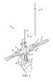
- FIG. 1is a perspective view of a surgical access system according to one aspect of the present invention
- FIG. 2is a perspective view of an initial tissue distraction assembly forming part of a surgical access system according to the present invention
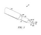
- FIGS. 3-4are exploded views detailing the distal portions of the initial tissue distraction assembly shown in FIG. 2 ;
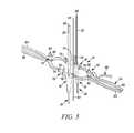
- FIG. 5is a perspective view of a pivot linkage assembly equipped with speculum blades according to the present invention.
- FIG. 6is a side view of the pivot linkage assembly shown in FIG. 5 ;
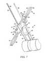
- FIGS. 7-8are perspective views showing the pivot linkage assembly of FIG. 5 in use
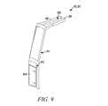
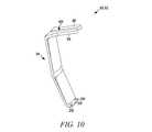
- FIGS. 9-10are perspective views of a retractor blade forming part of a surgical access system according to the present invention.
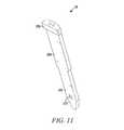
- FIG. 11is a perspective view of a locking member for use with the retractor blade of FIGS. 9-10 according to the present invention.
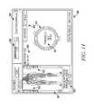
- FIG. 12is a perspective view of an exemplary nerve monitoring system capable of performing nerve monitoring before, during and after the creating of an operative corridor to a surgical target site using the surgical access system in accordance with the present invention
- FIG. 13is a block diagram of the nerve monitoring system shown in FIG. 12 ;
- FIGS. 14-15are screen displays illustrating exemplary features and information communicated to a user during the use of the nerve monitoring system of FIG. 12 ;
- FIGS. 16-33illustrate the various method steps (some optional) involved in accessing (by way of example only) a surgical target site in the spine according to the present invention.
- the present inventionis directed at a novel surgical access system and related methods which involve creating a distraction corridor to a surgical target site, thereafter retracting the distraction corridor to establish and maintain an operative corridor to the surgical target site, and optionally detecting the existence of (and optionally the distance and/or direction to) neural structures before, during and/or after the formation of the distraction and/or operative corridors.
- the steps of distraction followed by retractionare advantageous because they provide the ability to more easily position an operative corridor-establishing device through tissue that is strong, thick or otherwise challenging to traverse in order to access a surgical target site.
- the various distraction systems of the present inventionare advantageous in that they provide an improved manner of atraumatically establishing a distraction corridor prior to the use of the retraction systems of the present invention.
- the various retractor systems of the present inventionare advantageous in that they provide an operative corridor having improved cross-sectional area and shape (including customization thereof) relative to the prior art surgical access systems. Moreover, by optionally equipping the various distraction systems and/or retraction systems with one or more electrodes, an operative corridor may be established through (or near) any of a variety of tissues having such neural structures which, if contacted or impinged, may otherwise result in neural impairment for the patient.
- FIG. 1illustrates a surgical access system 10 according to one aspect of the present invention.
- the surgical access system 10includes an initial tissue distraction assembly 12 and a pivot linkage assembly 14 forming part of a secondary distraction assembly and a retraction assembly.
- the secondary distraction assemblyincludes first and second distraction arms 16 , 18 forming part of the pivot linkage assembly 14 , first and second speculum blades 20 , 22 extending through receiving passageways formed within the first and second distraction arms 16 , 18 , and a handle assembly 24 forming part of the pivot linkage 14 .
- the distraction arms 16 , 18may be advanced over the initial distraction assembly 12 such that the speculum blades 20 , 22 are passed into the tissue to be secondarily distracted.
- the handle assembly 24may be activated to perform the necessary distraction. That is, the handle assembly 24 can be manipulated by a user to move the first and second distraction arms 16 , 18 away from one another, which will at the same time move the distal ends of the speculum blades 20 , 22 to create a full distraction corridor.
- a pair of retractors blades 26 , 28may be introduced into the distraction corridor and positioned to create an operative corridor to the surgical target site.
- retractor blade 26is introduced first and positioned such that its distal end is generally located towards the posterior region of the spinal target site, which forms a useful barrier to prevent any exiting nerve roots 30 from entering the surgical target site, as well as to prevent any surgical instruments from passing outside the surgical target site and into contact with the exiting nerve roots 30 or other sensitive tissue.
- the retractor blade 28may thereafter be introduced and moved in a generally anterior direction away from the retractor blade 26 , effectively creating the operative corridor.
- the retractor blades 26 , 28may be locked in relation to the pivot linkage assembly 14 in any number of suitable fashions, including but not limited to the use of the nut-bolt assemblies 32 , 34 well known in the art.
- optional locking members 36may be advanced through receiving passageways formed in one or more of the retractor blades 26 , 28 such that a distal region of the locking member 36 is brought into a press-fit, secure engagement between the adjacent vertebral bodies to thereby maintain the respective retractor blade 26 , 28 in position.
- any of a variety of surgical instruments, devices, or implantsmay be passed through and/or manipulated at or near the surgical target site depending upon the given surgical procedure.
- FIG. 2illustrates the initial tissue distraction assembly 12 , which is designed to perform an initial distraction of tissue from the skin of the patient down to or near the surgical target site.
- the initial tissue distraction assembly 12may be constructed from any number of materials suitable for medical applications, including but not limited to plastics, metals, ceramics or any combination thereof. Depending on the construction, some or all of the tissue distraction assembly 12 may be disposable (i.e. single use) and/or reusable (i.e. multi-use).
- the initial tissue distraction assembly 12may include any number of components capable of performing the necessary initial distraction. By way of example, with combined reference to FIGS. 2-4 , this may be accomplished by providing the initial distraction assembly 12 as including a K-wire 44 and one or more dilators 46 , 48 .
- the K-wire 44is preferably constructed having generally narrow diameter (such as, by way of example only, 1.5 mm) and sufficient rigidity and strength such that it can pierce the skin of the patient and be advanced through the intervening tissue to reach the surgical target site.
- the K-wire 44also preferably includes indicia for determining the distance between a distal end 50 and the skin of the patient.
- the dilators 46 , 48are inner and outer dilating elements, respectively, capable of being sequentially introduced over the K-wire 44 for the purpose of further distracting the tissue previously distracted by the K-wire 44 .
- the inner dilator 46is preferably constructed having an inner diameter approximating the diameter of the K-wire 44 (such as, by way of example only, 1.5 mm), an outer diameter of increased dimension (such as, by way of example only, 6.5 mm), and indicia for determining the distance between a distal end 52 and the skin of the patient.
- the outer dilator 48is similarly preferably constructed having an inner diameter approximating the outer diameter of the inner dilator 46 (such as, by way of example only, 6.5 mm), an outer diameter of increased dimension (such as, by way of example only, 9 mm), and indicia for determining the distance between a distal end 54 and the skin of the patient.
- the respective lengths of the K-wire 44 and dilators 46 , 48may vary depending upon the given surgical target site (that is, the “depth” of the surgical target site within the patient). It will be similarly appreciated that the diameters and dimensions for these elements may also vary depending upon the particular surgical procedure. All such surgically appropriate variations (length, diameter, etc. . . . ) are contemplated as falling within the scope of the present invention. It is further contemplated and within the scope of the present invention that additional dilators of increasing diameters may be employed to sequentially dilate to the point where a bladed retractor or retraction assembly may be employed to thereafter create an operative corridor according to the present invention (without the need for secondary distraction as described below).
- the secondary tissue distractionis preferably performed using the pivot linkage assembly 14 in conjunction with the first and second distraction arms 16 , 18 and first and second speculum blades 20 , 22 .
- the speculum blades 20 , 22extend through receiving passageways 38 ( FIG. 6 ) formed within the first and second distraction arms 16 , 18 .
- the handle assembly 24includes first and second pivot arms 60 , 62 disposed on one end of the assembly, and third and fourth pivot arms 64 , 66 on the opposite end.
- First and second pivot arms 60 , 62are pivotably coupled via a rod 80 forming part of the locking assembly 32 (a locking nut 82 forms the remainder of the locking assembly 32 ).
- Second and third pivot arms 64 , 66are pivotably coupled via a rod 84 forming part of the locking assembly 34 (a locking nut 86 forms the remainder of the locking assembly 34 ).
- First and second linkage assemblies 70 , 72extend between the distal ends of the pivot arms 60 - 66 , each including a pair of linkages 74 , 76 pivotably coupled together via a rod 78 .
- a ratchet member 68may be used to maintain the first pivot arms 60 relative to the second pivot arm 62 as they are separated during use.
- the first and second distraction arms 16 , 18(being coupled to or integrally formed with the linkages 76 of first and second linkage assemblies 72 , 74 ) will similarly move away from one another.
- the speculum blades 20 , 22disposed within the passageways 38 ( FIG. 5 ), the relative movement of the pivot arms 16 , 18 will cause the speculum blades 20 , 22 to move apart and thus perform the desired secondary distraction.
- the pivot linkage assembly 14may be constructed from any number of materials suitable for medical applications, including but not limited to plastics, metals, ceramics or any combination thereof. Depending on the construction, some or all of the pivot linkage assembly 14 may be disposable (i.e. single use) and/or reusable (i.e. multi-use).
- the speculum blades 20 , 22are generally elongate in nature and include a pair of mating grooves 88 formed along the inwardly facing surfaces of the speculum blades 20 , 22 which, when mated together, form a lumen capable of passing over the K-wire 44 .
- the speculum blades 20 , 22are separable from distraction arms 16 , 18 such that the blades 20 , 22 can be introduced into the patient and thereafter engaged with the handle assembly 24 to effectuate the secondary distraction.
- this separable constructionallows the speculum blades 20 , 22 to be introduced down to the surgical target site by passing them through the outer dilator 48 and over with the K-wire 44 (the latter by virtue of the lumen formed by the pair of mating grooves 88 along the inwardly facing surfaces of the speculum blades 20 , 22 ). This is obviously only possible by first removing the inner dilator 46 from within the second dilator 48 while leaving the K-wire 44 in place.
- the speculum blades 20 , 22may be of generally non-separable or fixed construction with the pivot arms 16 , 18 of the handle assembly 24 .
- the retraction of the present inventionis performed by expanding and/or modifying the distraction corridor to establish and maintain an operative corridor to the surgical target site.
- the pivot linkage 14is configured to receive (and have coupled thereto) a pair of retractor blades 90 , 92 of the type shown in FIGS. 9-10 .
- the retractor blades 90 , 92include a main body element 94 extending downwardly and angularly away from a pair of mounting arms 96 , 98 .
- the mounting arms 96 , 98are spaced apart from one another so as to create a channel 100 dimensioned to receive the respective rods 80 , 84 of the locking assemblies 32 , 34 .
- the retractor blades 90 , 92may be locked in a desired position by tightening the respective nuts 82 , 86 of the locking assemblies 32 , 34 .
- one or more of the retractor blades 90 , 92may be equipped with a passageway 102 at or near the distal end of the main body 94 , such as by providing a generally planar member 104 along the generally curved distal region of the retractor blade 90 , 92 .
- This passageway 102is dimensioned to receive a locking member 36 of the type shown in FIG. 11 .
- the locking member 36includes a coupling region 106 for engagement with an introducer tool 112 ( FIG.
- the distal region 110also serves to prevent the ingress of unwanted or sensitive biological structures (e.g., nerve roots and/or vasculature) into the surgical target site, as well as prevent instruments from passing outside the surgical target site and contacting surrounding tissues or structures.
- unwanted or sensitive biological structurese.g., nerve roots and/or vasculature
- the retractor blades 90 , 92may also be optionally provided with at least one guard member 114 extending in a curved fashion (and/or, although not shown, in a generally straight fashion) from the distal end of the retractor blade 90 , 92 .
- the guard member 114may be provided, by way of example, for the purpose of preventing tissue (such as nerve roots in spinal surgery applications) from entering into the operative corridor during surgery and for preventing instruments from extending outside the operative corridor and/or the general vicinity of the surgical target site.
- the retractor blades 90 , 92may also be equipped with any number of different mechanisms for transporting or emitting light at or near the surgical target site to aid the surgeon's ability to visualize the surgical target site, instruments and/or implants during the given surgical procedure.
- one or more strands of fiber optic cablemay be coupled to the retractor blades 90 , 92 such that light may be delivered from a light source and selectively emitted into the operative corridor and/or the surgical target site.
- the retractor blades 90 , 92may be constructed of suitable material (such as clear polycarbonate) and configuration such that light may be transmitted generally distally through a light exit region formed along the entire inner periphery of the retractor blade 90 , 92 and located in the general vicinity as the distal opening of the passageway 102 .
- suitable materialsuch as clear polycarbonate
- Thismay be performed by providing the retractor blade 90 , 92 having light-transmission characteristics (such as with clear polycarbonate construction) and transmitting the light almost entirely within the walls of the retractor blade 90 , 92 (such as by frosting or otherwise rendering opaque portions of the exterior and/or interior and coupling the light source thereto such as via a port) until it exits a portion along the interior of the retractor blades 90 , 92 to shine at or near the surgical target site.
- light-transmission characteristicssuch as with clear polycarbonate construction
- each set of retractor blades 90 , 92may be provided, each having a different length to account for any number of possible surgical target sites.
- each set of retractor blades 90 , 92may be marked or color-coded to aid in indicating to the surgeon the particular length of the blade 90 , 92 or the depth of the surgical target site.
- the retractor blades 90 , 92 and the locking member 36may be constructed from any number of materials suitable for medical applications, including but not limited to plastics, metals, ceramics or any combination thereof. Depending on the construction, some or all of these devices may be disposable (i.e. single use) and/or reusable (i.e. multi-use).
- any number of suitable mounting unitsmay be employed to maintain the pivot linkage assembly 14 in a fixed and rigid fashion relative to the patient.
- thismay be accomplished by providing the mounting unit as a generally U-shaped mounting arm for lockable engagement with the pivot linkage assembly 14 , and a coupling mechanism (not shown) extending between the mounting arm and a rigid structure (such as the operating table) for maintaining the U-shaped mounting arm in a fixed and rigid position.
- any number of distraction components and/or retraction componentsmay be equipped to detect the presence of (and optionally the distance and/or direction to) neural structures during the steps tissue distraction and/or retraction. This is accomplished by employing the following steps: (1) one or more stimulation electrodes are provided on the various distraction and/or retraction components; (2) a stimulation source (e.g. voltage or current) is coupled to the stimulation electrodes; (3) a stimulation signal is emitted from the stimulation electrodes as the various components are advanced towards or maintained at or near the surgical target site; and (4) the patient is monitored to determine if the stimulation signal causes muscles associated with nerves or neural structures within the tissue to innervate. If the nerves innervate, this may indicate that neural structures may be in close proximity to the distraction and/or retraction components.
- a stimulation sourcee.g. voltage or current
- Neural monitoringmay be accomplished via any number of suitable fashions, including but not limited to observing visual twitches in muscle groups associated with the neural structures likely to found in the tissue, as well as any number of monitoring systems, including but not limited to any commercially available “traditional” electromyography (EMG) system (that is, typically operated by a neurophysiologist.
- EMGelectromyography
- Such monitoringmay also be carried out via the surgeon-driven EMG monitoring system shown and described in the following commonly owned and co-pending PCT Applications (collectively “NeuroVision PCT Applications”): PCT App. Ser. No. PCT/US02/22247, entitled “System and Methods for Determining Nerve Proximity, Direction, and Pathology During Surgery,” filed on Jul. 11, 2002; PCT App. Ser. No.
- PCT/US02/30617entitled “System and Methods for Performing Surgical Procedures and Assessments,” filed on Sep. 25, 2002; PCT App. Ser. No. PCT/US02/35047, entitled “System and Methods for Performing Percutaneous Pedicle Integrity Assessments,” filed on Oct. 30, 2002; and PCT App. Ser. No. PCT/US03/02056, entitled “System and Methods for Determining Nerve Direction to a Surgical Instrument,” filed Jan. 15, 2003.
- the entire contents of each of the above-enumerated NeuroVision PCT Applicationsis hereby expressly incorporated by reference into this disclosure as if set forth fully herein.
- the access system of the present inventionmay advantageously be used to traverse tissue that would ordinarily be deemed unsafe or undesirable, thereby broadening the number of manners in which a given surgical target site may be accessed.
- FIGS. 12-13illustrate, by way of example only, a monitoring system 120 of the type disclosed in the NeuroVision PCT Applications suitable for use with the surgical access system 10 of the present invention.
- the monitoring system 120includes a control unit 122 , a patient module 124 , and an EMG harness 126 and return electrode 128 coupled to the patient module 124 , and a cable 132 for establishing electrical communication between the patient module 124 and the surgical access system 10 ( FIG. 1 ).
- this electrical communicationcan be achieved by providing, by way of example only, a hand-held stimulation controller 152 capable of selectively providing a stimulation signal (due to the operation of manually operated buttons on the hand-held stimulation controller 152 ) to one or more connectors 156 a , 156 b , 156 c .
- the connectors 156 a , 156 b , 156 care suitable to establish electrical communication between the hand-held stimulation controller 152 and (by way of example only) the stimulation electrodes on the K-wire 44 , the dilators 46 , 46 , the speculum blades 20 , 22 , the retractor blades 90 , 92 , and/or the guard members 114 (collectively “surgical access instruments”).
- these surgical access instrumentsmust be connected to the connectors 156 a , 156 b and/or 156 c , at which point the user may selectively initiate a stimulation signal (preferably, a current signal) from the control unit 122 to a particular surgical access instruments. Stimulating the electrode(s) on these surgical access instruments before, during and/or after establishing operative corridor will cause nerves that come into close or relative proximity to the surgical access instruments to depolarize, producing a response in a myotome associated with the innervated nerve.
- a stimulation signalpreferably, a current signal
- the control unit 122includes a touch screen display 140 and a base 142 , which collectively contain the essential processing capabilities (software and/or hardware) for controlling the monitoring system 120 .
- the control unit 122may include an audio unit 118 that emits sounds according to a location of a surgical element with respect to a nerve.
- the patient module 124is connected to the control unit 122 via a data cable 144 , which establishes the electrical connections and communications (digital and/or analog) between the control unit 122 and patient module 124 .
- the main functions of the control unit 122include receiving user commands via the touch screen display 140 , activating stimulation electrodes on the surgical access instruments, processing signal data according to defined algorithms, displaying received parameters and processed data, and monitoring system status and report fault conditions.
- the touch screen display 140is preferably equipped with a graphical user interface (GUI) capable of communicating information to the user and receiving instructions from the user.
- GUIgraphical user interface
- the display 140 and/or base 142may contain patient module interface circuitry (hardware and/or software) that commands the stimulation sources, receives digitized signals and other information from the patient module 124 , processes the EMG responses to extract characteristic information for each muscle group, and displays the processed data to the operator via the display 140 .
- the monitoring system 120is capable of determining nerve direction relative to one or more of the K-wire 44 , dilation cannula 46 , 48 , speculum blades 20 , 22 , the retractor blades 90 , 92 , and/or the guard members 114 before, during and/or following the creation of an operative corridor to a surgical target site. Monitoring system 120 accomplishes this by having the control unit 122 and patient module 124 cooperate to send electrical stimulation signals to one or more of the stimulation electrodes provided on these instruments. Depending upon the location of the surgical access system 10 within a patient (and more particularly, to any neural structures), the stimulation signals may cause nerves adjacent to or in the general proximity of the surgical access system 10 to depolarize.
- the nerve direction feature of the system 120is based on assessing the evoked response of the various muscle myotomes monitored by the system 120 via the EMG harness 126 .
- the surgical access system 10is capable of detecting the presence of (and optionally the distant and/or direction to) such nerves. This provides the ability to actively negotiate around or past such nerves to safely and reproducibly form the operative corridor to a particular surgical target site, as well as monitor to ensure that no neural structures migrate into contact with the surgical access system 10 after the operative corridor has been established.
- the surgical access system 10may be particularly suited for establishing an operative corridor to an intervertebral target site in a postero-lateral, trans-psoas fashion so as to avoid the bony posterior elements of the spinal column.
- FIGS. 14-15are exemplary screen displays (to be shown on the display 140 ) illustrating one embodiment of the nerve direction feature of the monitoring system shown and described with reference to FIGS. 12-13 . These screen displays are intended to communicate a variety of information to the surgeon in an easy-to-interpret fashion.
- This informationmay include, but is not necessarily limited to, a display of the function 180 (in this case “DIRECTION”), a graphical representation of a patient 181 , the myotome levels being monitored 182 , the nerve or group associated with a displayed myotome 183 , the name of the instrument being used 184 (in this case, a dilator 46 , 48 ), the size of the instrument being used 185 , the stimulation threshold current 186 , a graphical representation of the instrument being used 187 (in this case, a cross-sectional view of a dilator 46 , 48 ) to provide a reference point from which to illustrate relative direction of the instrument to the nerve, the stimulation current being applied to the stimulation electrodes 188 , instructions for the user 189 (in this case, “ADVANCE” and/or “HOLD”), and (in FIG.
- a display of the function 180in this case “DIRECTION”
- a graphical representation of a patient 181the myotome levels being monitored 182
- an arrow 190 indicating the direction from the instrument to a nervemay be communicated in any number of suitable fashions, including but not limited to the use of visual indicia (such as alpha-numeric characters, light-emitting elements, and/or graphics) and audio communications (such as a speaker element).
- visual indiciasuch as alpha-numeric characters, light-emitting elements, and/or graphics
- audio communicationssuch as a speaker element.
- a dilating cannulasuch as at 184
- the present inventionis deemed to include providing similar information on the display 140 during the use of any or all of the various instruments forming the surgical access system 10 of the present invention, including the initial distraction assembly 12 (i.e. the K-wire 44 and dilators 46 , 48 ), the speculum blades 20 , 22 and/or the retractor blades 90 , 92 and/or the guard members 114 .
- the initial distraction assembly 12may be provided with one or more electrodes for use in providing the neural monitoring capabilities of the present invention.
- the K-wire 44may be equipped with a distal electrode 200 . This may be accomplished by constructing the K-wire 44 for a conductive material, providing outer layer of insulation 202 extending along the entire length with the exception of an exposure that defines the electrode 200 .
- the electrode 200has an angled configuration relative to the rest of the K-wire 44 (such as, by way of example only, in the range of between 15 and 75 degrees from the longitudinal axis of the K-wire 44 ). The angled nature of the electrode 200 is advantageous in that it aids in piercing tissue as the K-wire 44 is advanced towards the surgical target site.
- the angled nature of the distal electrode 200is also important in that it provides the ability to determine the location of nerves or neural structures relative to the K-wire 44 as it is advanced towards or resting at or near the surgical target site.
- This “directional” capabilityis achieved by the fact that the angled nature of the electrode 200 causes the electrical stimulation to be projected away from the distal portion of the K-wire 44 in a focused, or directed fashion.
- nerves or neural structureswhich are generally closer to the side of the K-wire 44 on which the electrode 200 is disposed will have a higher likelihood of firing or being innervated that nerves or neural structures on the opposite side as the electrode 200 .
- the direction to such nerves or neural structuresmay thus be determined by physically rotating the K-wire 44 at a particular point within the patient's tissue and monitoring to see if any neural stimulation occurs at a given point within the rotation.
- monitoringcan be performed via visual observation, a traditional EMG monitoring, as well as the nerve surveillance system disclosed in the above-referenced NeuroVision PCT Applications. If the signals appear more profound or significant at a given point within the rotation, the surgeon will be able tell where the corresponding nerves or neural structures are, by way of example only, by looking at reference information (such as the indicia) on the exposed part of the K-wire 44 (which reference point is preferably set forth in the exact same orientation as the electrode 200 ).
- reference informationsuch as the indicia
- Dilators 46 , 48may also be provided with angled electrodes 204 , 206 , respectively, for the purpose of determining the location of nerves or neural structures relative to the dilators 46 , 48 as they are advanced over the K-wire 44 towards or positioned at or near the surgical target site. Due to this similarity in function with the electrode 200 of the K-wire 44 , a repeat explanation is not deemed necessary.
- the dilators 46 , 48may be equipped with the electrodes 204 , 206 via any number of suitable methods, including but not limited to providing electrically conductive elements within the walls of the dilators 46 , 48 , such as by manufacturing the dilators 46 , 48 from plastic or similar material capable of injection molding or manufacturing the dilators 46 , 48 from aluminum (or similar metallic substance) and providing outer insulation layer with exposed regions (such as by anodizing the exterior of the aluminum dilator).
- additional neural monitoring equipmentmay be employed so as to further prevent inadvertent contact with neural structures.
- a confirmation probeproviding a stimulation signal
- the confirmation probemay thereafter be stimulated for the purpose of double-checking to ensure that no nerves or neural structures are disposed in the tissue near (or have migrated into the vicinity of) the distal end 54 of the outer dilator 48 before introducing the speculum blades 20 , 22 .
- the outer dilator 48may be removed following the introduction of the speculum blades 20 , 22 and the secondary distraction performed (by coupling the handle assembly 24 to the blades 20 , 22 and expanding) without fear of inadvertently causing the speculum blades 20 , 22 to contact nerves or neural structures.
- the secondary distraction of the present inventionmay be provided with one or more electrodes for use in providing the neural monitoring capabilities of the present invention.
- the retractor blades 90 , 92 of the present inventionmay also be provided with one or more electrodes for use in providing the neural monitoring capabilities of the present invention.
- the surgical access system 10 of the present inventionmay be sold or distributed to end users in any number of suitable kits or packages (sterile and/or non-sterile) containing some or all of the various components described herein.
- the pivot linkage assembly 14may be provided such that the pivot arms 16 , 18 and speculum blades 20 , 22 are disposable and the retractor blades 90 , 92 are re-usable.
- an initial kitmay include these materials, including a variety of sets of retractor blades 90 , 92 having varying (or “incremental”) lengths to account for surgical target sites of varying locations within the patient.
- the surgical access system 10 of the present inventionwill now be described, by way of example, with reference to the spinal application shown in FIGS. 16-33 . It will, of course, be appreciated that the surgical access system and related methods of the present invention may find applicability in any of a variety of surgical and/or medical applications such that the following description relative to the spine is not to be limiting of the overall scope of the present invention. More specifically, while described below employing the nerve monitoring features described above (otherwise referred to as “nerve surveillance”) during spinal surgery, it will be appreciated that such nerve surveillance will not be required in all situations, depending upon the particular surgical target site.
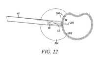
- FIGS. 16-22illustrate the method steps involved in using the initial tissue distraction assembly 12 of the present invention.
- the K-wire 44is first introduced along a given pathway towards the surgical target site (which, in this case, is an intervertebral disc level of the lumbar spine). Determining the preferred angle of incidence into the surgical target site (as well as the advancement or positioning of any required surgical instruments (such as the surgical access system of the present invention), devices and/or implants) may be facilitated through the use of surgical imaging systems (such as fluoroscopy) as well any number of stereotactic guide systems, including but not limited to those shown and described in co-pending and commonly owned U.S. patent application Ser. No. 09/888,223, filed Jun. 22, 2001 and entitled “Polar Coordinate Surgical Guideframe,” the entire contents of which is incorporated by reference as if set forth in its entirety herein.
- Nerve surveillanceis preferably conducted during this step (via electrode 200 ) to monitor for the existence of (and optionally the distance and direction to) nerves or neural structures in the tissue through which the K-wire 44 must pass to reach the surgical target site.
- it is generally desired to advance the K-wire 44such the distal electrode 200 is disposed a distance anterior to the exiting nerve root 300 (such as, by way of example, 10 mm) As shown in FIGS. 16-17 , it is preferred to advance the K-wire 44 to the annulus 302 of the disc before advancing the inner dilator 46 .
- the inner dilator 46may thereafter be advanced over the K-wire 44 until the distal end 52 abuts the annulus 302 of the disc. Nerve surveillance is also conducted during this step (via electrode 204 shown in FIGS. 3-4 ) to monitor for the existence of (and optionally the distance and direction to) nerves or neural structures in the tissue through which the inner dilator 46 must pass to reach the surgical target site.
- the K-wire 44may be advanced through the annulus 302 such that the electrode 200 is disposed within the interior (nucleus pulposis region) of the disc (such as, by way of example, an internal distance of 15 to 20 mm).
- the outer dilator 48is next advanced over the inner dilator 46 to further distract the tissue leading down to the surgical target site.
- nerve surveillanceis conducted during this step (via electrode 206 shown in FIGS. 3-4 ) to monitor for the existence of (and optionally the distance and direction to) nerves or neural structures in the tissue through which the outer dilator 48 must pass to reach the surgical target site.
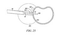
- the inner dilator 48is next removed, leaving the K-wire 44 and outer dilator 48 in position. This creates a space therebetween which, in one embodiment of the present invention, is dimensioned to receive the speculum blades 20 , 22 as shown in FIGS. 24-25 .
- the speculum blades 20 , 22must be disposed in an abutting relationship so as to form an inner lumen (via corresponding grooves 88 shown in FIG. 5 ) dimensioned to be slideably advancing over the stationary K-wire 44 .
- a confirmation probedown the outer dilator 48 to interrogate the tissue surrounding the surgical target site to ensure that no nerves or neural structures are present in (or have migrated into) this vicinity before the speculum blades 20 , 22 are advanced into the outer dilator 48 .
- the outer dilator 48may then be removed, leaving the speculum blades 20 , 22 in abutting relationship within the tissue previously distracted by the outer dilator 48 .
- the pivot linkage assembly 14may be advanced such that the pivot arms 16 , 18 slideably (or otherwise) pass over the speculum blades 20 , 22 .
- the pivot arms 16 , 18are dimensioned such that each distal end comes into general abutment with the exterior of the psoas muscle 304 .
- each distal endextends downward into the psoas 304 towards the surgical target site (which may be advantageous from the standpoint of adding rigidity to the distal portions of the speculum blades 20 , 22 for the purpose of facilitating the process of secondary tissue distraction).
- the handle assembly 24may be operate to distract tissue from the position shown in FIG. 28 to that shown in FIG. 29 .
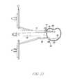
- the first retractor 90is then introduced into the distracted region, positioned adjacent to the posterior region of the disc space, and locked to the pivot linkage 14 via the locking assembly 32 .
- the locking member 36may be advanced via the tool 112 and engaged with the retractor blade 90 such that the middle region 108 resides at least partially within the passageway 102 and the distal region 110 extends into the disc space.
- the retractor blade 92may be introduced into the distracted region, positioned adjacent to the anterior region of the disc space, and locked to the pivot linkage 14 via the locking assembly 34 .
- another locking member 36may be engaged in the same fashion as with the retractor blade 90 , with the distal region 110 extending into the disc space.
- additional retractor blades 91 , 93may be coupled to the pivot linkage 14 to provide retraction in the caudal and cephalad directions, respectively.
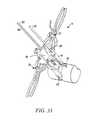
- FIG. 33The end result is shown in FIG. 33 , wherein an operative corridor has been created to the spinal target site (in this case, the disc space) defined by the retractor blades 90 , 92 (and optionally 91 , 93 ).
- the distal regions 110 of the locking each locking member 36advantageously extends into the disc space to prevent the ingress of tissue (e.g., neural, vasculature, etc. . . . ) into the surgical target site and/or operative corridor and the egress of instruments or implants out of the surgical target site and/or operative corridor.
- each retractor blade 90 , 92is equipped with a guard member 114 to prevent similar ingress and egress.
- Both guard members 114(as well as additional regions of the distal region 110 of the locking member 36 ) may be provided with electrodes 210 , 212 , respectively, capable of performing nerve surveillance to monitor for the existence of (and optionally the distance and direction to) nerves or neural structures in the tissue or region surrounding or adjacent to these components while disposed in the general vicinity of the surgical target site.
- the electrode 210 on the guard member 114 of the posterior retractor blade 90may be used to assess the status or health of the nerve root 300 , especially if the nerve root 300 is in close proximity to that guard member 114 .
- the present inventionaccomplishes the goal of providing a novel surgical access system and related methods which involve creating a distraction corridor to a surgical target site, thereafter retracting the distraction corridor to establish and maintain an operative corridor to the surgical target site, and optionally detecting the existence of (and optionally the distance and/or direction to) neural structures before, during and/or after the formation of the distraction and/or operative corridors.
- the steps of distraction followed by retractionare advantageous because they provide the ability to more easily position an operative corridor-establishing device through tissue that is strong, thick or otherwise challenging to traverse in order to access a surgical target site.
- the various distraction systems of the present inventionare advantageous in that they provide an improved manner of atraumatically establishing a distraction corridor prior to the use of the retraction systems of the present invention.
- the various retractor systems of the present inventionare advantageous in that they provide an operative corridor having improved cross-sectional area and shape (including customization thereof) relative to the prior art surgical access systems.
- an operative corridormay be established through (or near) any of a variety of tissues having such neural structures which, if contacted or impinged, may otherwise result in neural impairment for the patient.
- the surgical access system of the present inventioncan be used in any of a wide variety of surgical or medical applications, above and beyond the spinal applications discussed herein.
- any number of implants and/or instrumentsmay be introduced through the working cannula 50 , including but not limited to spinal fusion constructs (such as allograft implants, ceramic implants, cages, mesh, etc.), fixation devices (such as pedicle and/or facet screws and related tension bands or rod systems), and any number of motion-preserving devices (including but not limited to nucleus replacement and/or total disc replacement systems).
- the monitoring system 120may be implemented using any combination of computer programming software, firmware or hardware.
- the computer programming code (whether software or firmware) according to the applicationwill typically be stored in one or more machine readable storage mediums such as fixed (hard) drives, diskettes, optical disks, magnetic tape, semiconductor memories such as ROMs, PROMs, etc., thereby making an article of manufacture in accordance with the application.
- the article of manufacture containing the computer programming codemay be used by either executing the code directly from the storage device, by copying the code from the storage device into another storage device such as a hard disk, RAM, etc. or by transmitting the code on a network for remote execution.
- a hard disksuch as a hard disk, RAM, etc.
Landscapes
- Health & Medical Sciences (AREA)
- Life Sciences & Earth Sciences (AREA)
- Surgery (AREA)
- Veterinary Medicine (AREA)
- Animal Behavior & Ethology (AREA)
- Public Health (AREA)
- Engineering & Computer Science (AREA)
- Biomedical Technology (AREA)
- Heart & Thoracic Surgery (AREA)
- Medical Informatics (AREA)
- Molecular Biology (AREA)
- General Health & Medical Sciences (AREA)
- Nuclear Medicine, Radiotherapy & Molecular Imaging (AREA)
- Biophysics (AREA)
- Physics & Mathematics (AREA)
- Pathology (AREA)
- Neurology (AREA)
- Optics & Photonics (AREA)
- Radiology & Medical Imaging (AREA)
- Surgical Instruments (AREA)
- Measurement And Recording Of Electrical Phenomena And Electrical Characteristics Of The Living Body (AREA)
Abstract
Description
Claims (20)
Priority Applications (10)
| Application Number | Priority Date | Filing Date | Title |
|---|---|---|---|
| US13/466,398US8672840B2 (en) | 2002-06-26 | 2012-05-08 | Surgical access system and related methods |
| US13/757,035US8708899B2 (en) | 2002-06-26 | 2013-02-01 | Surgical access system and related methods |
| US13/865,598US8915846B2 (en) | 2002-06-26 | 2013-04-18 | Surgical access system and related methods |
| US14/263,797US9750490B2 (en) | 2002-06-26 | 2014-04-28 | Surgical access system and related methods |
| US14/297,369US10251633B2 (en) | 2002-06-26 | 2014-06-05 | Surgical access system and related methods |
| US14/297,438US9848861B2 (en) | 2002-06-26 | 2014-06-05 | Surgical access system and related methods |
| US15/445,854US9826968B2 (en) | 2002-06-26 | 2017-02-28 | Surgical access system and related methods |
| US15/448,395US9833227B2 (en) | 2002-06-26 | 2017-03-02 | Surgical access system and related methods |
| US16/293,223US10980524B2 (en) | 2002-06-26 | 2019-03-05 | Surgical access system and related methods |
| US17/176,059US20210177391A1 (en) | 2002-06-26 | 2021-02-15 | Surgical access system and related methods |
Applications Claiming Priority (14)
| Application Number | Priority Date | Filing Date | Title |
|---|---|---|---|
| US39221402P | 2002-06-26 | 2002-06-26 | |
| USPCT/US02/22247 | 2002-07-11 | ||
| PCT/US2002/022247WO2003005887A2 (en) | 2001-07-11 | 2002-07-11 | System and methods for determining nerve proximity, direction, and pathology during surgery |
| PCT/US2002/030617WO2003026482A2 (en) | 2001-09-25 | 2002-09-25 | System and methods for performing surgical procedures and assessments |
| USPCT/US02/30617 | 2002-09-25 | ||
| USPCT/US02/35047 | 2002-10-30 | ||
| PCT/US2002/035047WO2003037170A2 (en) | 2001-10-30 | 2002-10-30 | System and methods for performing percutaneous pedicle integrity assessments |
| PCT/US2003/002056WO2004064634A1 (en) | 2003-01-15 | 2003-01-15 | Systems and methods for determining direction to a nerve |
| USPCT/US03/02056 | 2003-01-15 | ||
| US10/608,362US7582058B1 (en) | 2002-06-26 | 2003-06-26 | Surgical access system and related methods |
| US12/428,081US7935051B2 (en) | 2002-06-26 | 2009-04-22 | Surgical access system and related methods |
| US12/635,418US8192356B2 (en) | 2002-06-26 | 2009-12-10 | Surgical access system and related methods |
| US12/649,604US8182423B2 (en) | 2002-06-26 | 2009-12-30 | Surgical access system and related methods |
| US13/466,398US8672840B2 (en) | 2002-06-26 | 2012-05-08 | Surgical access system and related methods |
Related Parent Applications (1)
| Application Number | Title | Priority Date | Filing Date |
|---|---|---|---|
| US12/649,604ContinuationUS8182423B2 (en) | 2002-06-26 | 2009-12-30 | Surgical access system and related methods |
Related Child Applications (1)
| Application Number | Title | Priority Date | Filing Date |
|---|---|---|---|
| US13/757,035ContinuationUS8708899B2 (en) | 2002-06-26 | 2013-02-01 | Surgical access system and related methods |
Publications (2)
| Publication Number | Publication Date |
|---|---|
| US20120238895A1 US20120238895A1 (en) | 2012-09-20 |
| US8672840B2true US8672840B2 (en) | 2014-03-18 |
Family
ID=41009160
Family Applications (21)
| Application Number | Title | Priority Date | Filing Date |
|---|---|---|---|
| US10/608,362Expired - LifetimeUS7582058B1 (en) | 2002-06-26 | 2003-06-26 | Surgical access system and related methods |
| US12/428,081Expired - LifetimeUS7935051B2 (en) | 2002-06-26 | 2009-04-22 | Surgical access system and related methods |
| US12/635,418Expired - Fee RelatedUS8192356B2 (en) | 2002-06-26 | 2009-12-10 | Surgical access system and related methods |
| US12/649,604Expired - LifetimeUS8182423B2 (en) | 2002-06-26 | 2009-12-30 | Surgical access system and related methods |
| US12/650,336Expired - Fee RelatedUS8187179B2 (en) | 2002-06-26 | 2009-12-30 | Surgical access system and related methods |
| US29/360,370ActiveUSD652519S1 (en) | 2002-06-26 | 2010-04-23 | Dilator |
| US29/360,368ActiveUSD652921S1 (en) | 2002-06-26 | 2010-04-23 | Dilator |
| US29/360,369ActiveUSD652922S1 (en) | 2002-06-26 | 2010-04-23 | Dilator |
| US29/411,162ActiveUSD666292S1 (en) | 2002-06-26 | 2012-01-17 | Dilator |
| US29/411,652ActiveUSD666294S1 (en) | 2002-06-26 | 2012-01-24 | Dilator |
| US29/411,651ActiveUSD666293S1 (en) | 2002-06-26 | 2012-01-24 | Dilator |
| US13/466,398Expired - LifetimeUS8672840B2 (en) | 2002-06-26 | 2012-05-08 | Surgical access system and related methods |
| US13/757,035Expired - LifetimeUS8708899B2 (en) | 2002-06-26 | 2013-02-01 | Surgical access system and related methods |
| US13/865,598Expired - LifetimeUS8915846B2 (en) | 2002-06-26 | 2013-04-18 | Surgical access system and related methods |
| US14/263,797Expired - LifetimeUS9750490B2 (en) | 2002-06-26 | 2014-04-28 | Surgical access system and related methods |
| US14/297,369Expired - LifetimeUS10251633B2 (en) | 2002-06-26 | 2014-06-05 | Surgical access system and related methods |
| US14/297,438Expired - LifetimeUS9848861B2 (en) | 2002-06-26 | 2014-06-05 | Surgical access system and related methods |
| US15/445,854Expired - LifetimeUS9826968B2 (en) | 2002-06-26 | 2017-02-28 | Surgical access system and related methods |
| US15/448,395Expired - LifetimeUS9833227B2 (en) | 2002-06-26 | 2017-03-02 | Surgical access system and related methods |
| US16/293,223Expired - LifetimeUS10980524B2 (en) | 2002-06-26 | 2019-03-05 | Surgical access system and related methods |
| US17/176,059AbandonedUS20210177391A1 (en) | 2002-06-26 | 2021-02-15 | Surgical access system and related methods |
Family Applications Before (11)
| Application Number | Title | Priority Date | Filing Date |
|---|---|---|---|
| US10/608,362Expired - LifetimeUS7582058B1 (en) | 2002-06-26 | 2003-06-26 | Surgical access system and related methods |
| US12/428,081Expired - LifetimeUS7935051B2 (en) | 2002-06-26 | 2009-04-22 | Surgical access system and related methods |
| US12/635,418Expired - Fee RelatedUS8192356B2 (en) | 2002-06-26 | 2009-12-10 | Surgical access system and related methods |
| US12/649,604Expired - LifetimeUS8182423B2 (en) | 2002-06-26 | 2009-12-30 | Surgical access system and related methods |
| US12/650,336Expired - Fee RelatedUS8187179B2 (en) | 2002-06-26 | 2009-12-30 | Surgical access system and related methods |
| US29/360,370ActiveUSD652519S1 (en) | 2002-06-26 | 2010-04-23 | Dilator |
| US29/360,368ActiveUSD652921S1 (en) | 2002-06-26 | 2010-04-23 | Dilator |
| US29/360,369ActiveUSD652922S1 (en) | 2002-06-26 | 2010-04-23 | Dilator |
| US29/411,162ActiveUSD666292S1 (en) | 2002-06-26 | 2012-01-17 | Dilator |
| US29/411,652ActiveUSD666294S1 (en) | 2002-06-26 | 2012-01-24 | Dilator |
| US29/411,651ActiveUSD666293S1 (en) | 2002-06-26 | 2012-01-24 | Dilator |
Family Applications After (9)
| Application Number | Title | Priority Date | Filing Date |
|---|---|---|---|
| US13/757,035Expired - LifetimeUS8708899B2 (en) | 2002-06-26 | 2013-02-01 | Surgical access system and related methods |
| US13/865,598Expired - LifetimeUS8915846B2 (en) | 2002-06-26 | 2013-04-18 | Surgical access system and related methods |
| US14/263,797Expired - LifetimeUS9750490B2 (en) | 2002-06-26 | 2014-04-28 | Surgical access system and related methods |
| US14/297,369Expired - LifetimeUS10251633B2 (en) | 2002-06-26 | 2014-06-05 | Surgical access system and related methods |
| US14/297,438Expired - LifetimeUS9848861B2 (en) | 2002-06-26 | 2014-06-05 | Surgical access system and related methods |
| US15/445,854Expired - LifetimeUS9826968B2 (en) | 2002-06-26 | 2017-02-28 | Surgical access system and related methods |
| US15/448,395Expired - LifetimeUS9833227B2 (en) | 2002-06-26 | 2017-03-02 | Surgical access system and related methods |
| US16/293,223Expired - LifetimeUS10980524B2 (en) | 2002-06-26 | 2019-03-05 | Surgical access system and related methods |
| US17/176,059AbandonedUS20210177391A1 (en) | 2002-06-26 | 2021-02-15 | Surgical access system and related methods |
Country Status (1)
| Country | Link |
|---|---|
| US (21) | US7582058B1 (en) |
Cited By (28)
| Publication number | Priority date | Publication date | Assignee | Title |
|---|---|---|---|---|
| US20120296433A1 (en)* | 2010-02-02 | 2012-11-22 | Azadeh Farin | Spine surgery device |
| US9533155B2 (en) | 2014-08-15 | 2017-01-03 | Axonics Modulation Technologies, Inc. | Methods for determining neurostimulation electrode configurations based on neural localization |
| US9555246B2 (en) | 2014-08-15 | 2017-01-31 | Axonics Modulation Technologies, Inc. | Electromyographic lead positioning and stimulation titration in a nerve stimulation system for treatment of overactive bladder |
| US9750490B2 (en) | 2002-06-26 | 2017-09-05 | Nuvasive, Inc. | Surgical access system and related methods |
| US9788822B2 (en) | 2003-09-25 | 2017-10-17 | Nuvasive, Inc. | Surgical access system and related methods |
| US9795371B2 (en) | 2003-01-16 | 2017-10-24 | Nuvasive, Inc. | Surgical access system and related methods |
| US9820729B2 (en) | 2002-10-08 | 2017-11-21 | Nuvasive, Inc. | Surgical access system and related methods |
| US9931077B2 (en) | 2001-07-11 | 2018-04-03 | Nuvasive, Inc. | System and methods for determining nerve proximity, direction and pathology during surgery |
| WO2018067794A1 (en) | 2016-10-05 | 2018-04-12 | Nuvasive, Inc. | Surgical navigation system and related methods |
| US9949840B1 (en) | 2011-04-01 | 2018-04-24 | William D. Smith | Systems and methods for performing spine surgery |
| US9949651B2 (en) | 2011-11-01 | 2018-04-24 | DePuy Synthes Products, Inc. | Intraoperative neurophysiological monitoring system |
| US10092762B2 (en) | 2014-08-15 | 2018-10-09 | Axonics Modulation Technologies, Inc. | Integrated electromyographic clinician programmer for use with an implantable neurostimulator |
| US10278686B2 (en) | 2010-09-20 | 2019-05-07 | DePuy Synthes Products, Inc. | Spinal access retractor |
| US10507120B2 (en) | 2001-09-25 | 2019-12-17 | Nuvasive, Inc. | Systems and methods for performing surgical procedures and assessments |
| US10653308B2 (en) | 2003-10-17 | 2020-05-19 | Nuvasive, Inc. | Surgical access system and related methods |
| US10687797B2 (en) | 2008-12-18 | 2020-06-23 | Howmedica Osteonics Corp. | Lateral access system for the lumbar spine |
| US11166709B2 (en) | 2016-08-23 | 2021-11-09 | Stryker European Operations Holdings Llc | Instrumentation and methods for the implantation of spinal implants |
| US11191532B2 (en) | 2018-03-30 | 2021-12-07 | Stryker European Operations Holdings Llc | Lateral access retractor and core insertion |
| USD956225S1 (en) | 2020-05-12 | 2022-06-28 | Innovasis, Inc. | Surgical retractor |
| USD956224S1 (en) | 2020-05-12 | 2022-06-28 | Innovasis, Inc. | Surgical retractor |
| USD956223S1 (en) | 2020-05-12 | 2022-06-28 | Innovasis, Inc. | Surgical retractor |
| US11432810B2 (en) | 2020-05-12 | 2022-09-06 | Innovasis, Inc. | Systems and methods for surgical retraction |
| US11439829B2 (en) | 2019-05-24 | 2022-09-13 | Axonics, Inc. | Clinician programmer methods and systems for maintaining target operating temperatures |
| WO2022203851A1 (en) | 2021-03-22 | 2022-09-29 | Nuvasive, Inc. | Multi-user surgical cart |
| US11564674B2 (en) | 2019-11-27 | 2023-01-31 | K2M, Inc. | Lateral access system and method of use |
| US11612440B2 (en) | 2019-09-05 | 2023-03-28 | Nuvasive, Inc. | Surgical instrument tracking devices and related methods |
| US11848090B2 (en) | 2019-05-24 | 2023-12-19 | Axonics, Inc. | Trainer for a neurostimulator programmer and associated methods of use with a neurostimulation system |
| US12343069B2 (en) | 2017-02-01 | 2025-07-01 | Avent, Inc. | EMG guidance for probe placement, nearby tissue preservation, and lesion confirmation |
Families Citing this family (276)
| Publication number | Priority date | Publication date | Assignee | Title |
|---|---|---|---|---|
| US7491205B1 (en) | 1988-06-13 | 2009-02-17 | Warsaw Orthopedic, Inc. | Instrumentation for the surgical correction of human thoracic and lumbar spinal disease from the lateral aspect of the spine |
| EP1146816B1 (en) | 1998-12-23 | 2005-10-12 | Nuvasive Inc. | Nerve surveillance cannulae systems |
| WO2001037728A1 (en) | 1999-11-24 | 2001-05-31 | Nuvasive, Inc. | Electromyography system |
| US8147421B2 (en) | 2003-01-15 | 2012-04-03 | Nuvasive, Inc. | System and methods for determining nerve direction to a surgical instrument |
| US6793678B2 (en) | 2002-06-27 | 2004-09-21 | Depuy Acromed, Inc. | Prosthetic intervertebral motion disc having dampening |
| AU2004212942A1 (en) | 2003-02-14 | 2004-09-02 | Depuy Spine, Inc. | In-situ formed intervertebral fusion device |
| US7819801B2 (en) | 2003-02-27 | 2010-10-26 | Nuvasive, Inc. | Surgical access system and related methods |
| US20040267367A1 (en) | 2003-06-30 | 2004-12-30 | Depuy Acromed, Inc | Intervertebral implant with conformable endplate |
| US9795367B1 (en) | 2003-10-17 | 2017-10-24 | Nuvasive, Inc. | Surgical access system and related methods |
| US8313430B1 (en) | 2006-01-11 | 2012-11-20 | Nuvasive, Inc. | Surgical access system and related methods |
| US7435219B2 (en)* | 2004-03-25 | 2008-10-14 | Depuy Spine, Inc. | Surgical retractor positioning device |
| CA2597944A1 (en)* | 2004-08-15 | 2006-02-23 | Kevin Seex | Distraction and retraction assemblies |
| WO2006042241A2 (en)* | 2004-10-08 | 2006-04-20 | Nuvasive, Inc. | Surgical access system and related methods |
| WO2006058221A2 (en) | 2004-11-24 | 2006-06-01 | Abdou Samy M | Devices and methods for inter-vertebral orthopedic device placement |
| US7643884B2 (en)* | 2005-01-31 | 2010-01-05 | Warsaw Orthopedic, Inc. | Electrically insulated surgical needle assembly |
| US7942826B1 (en)* | 2005-06-06 | 2011-05-17 | Nuvasive, Inc. | Insulated pedicle access system and related methods |
| US8328851B2 (en) | 2005-07-28 | 2012-12-11 | Nuvasive, Inc. | Total disc replacement system and related methods |
| US7846093B2 (en)* | 2005-09-26 | 2010-12-07 | K2M, Inc. | Minimally invasive retractor and methods of use |
| US8066730B2 (en)* | 2005-11-14 | 2011-11-29 | Scapa Flow, Llc | Medical dilator system or dilator device |
| US8480576B2 (en) | 2005-12-07 | 2013-07-09 | Faheem A. Sandhu | Access system for minimally invasive spinal surgery |
| US8034110B2 (en) | 2006-07-31 | 2011-10-11 | Depuy Spine, Inc. | Spinal fusion implant |
| US8142355B2 (en) | 2006-09-25 | 2012-03-27 | Spinal Elements, Inc. | Surgical tissue retractor |
| US20080103572A1 (en) | 2006-10-31 | 2008-05-01 | Medtronic, Inc. | Implantable medical lead with threaded fixation |
| USD631962S1 (en) | 2006-11-14 | 2011-02-01 | Scapa Flow, Llc | Medical dilator |
| WO2008070863A2 (en) | 2006-12-07 | 2008-06-12 | Interventional Spine, Inc. | Intervertebral implant |
| US20080183044A1 (en)* | 2007-01-26 | 2008-07-31 | Dennis Colleran | Flexible surgical retractor and method of use |
| US8062217B2 (en) | 2007-01-26 | 2011-11-22 | Theken Spine, Llc | Surgical retractor with removable blades and method of use |
| US8118737B2 (en)* | 2007-01-30 | 2012-02-21 | Mi4Spine, Llc | Retractor device for cervical spinal fusion |
| EP2114257B1 (en)* | 2007-02-09 | 2013-05-22 | Alphatec Spine, Inc. | Curvilinear spinal access device |
| WO2008124079A1 (en)* | 2007-04-03 | 2008-10-16 | Nuvasive, Inc. | Neurophysiologic monitoring system |
| US8075601B2 (en) | 2007-04-30 | 2011-12-13 | Warsaw Orthopedic, Inc. | Deformity correction using neural integrity monitoring |
| US8900307B2 (en) | 2007-06-26 | 2014-12-02 | DePuy Synthes Products, LLC | Highly lordosed fusion cage |
| BRPI0818608A2 (en) | 2007-10-05 | 2015-04-22 | Synthes Gmbh | Sequential directional dilatation system for dilating from a nerve of a patient's anatomy, and method for forming an access opening through a psoas muscle to a patient's spine using a dilatation system |
| US20090105788A1 (en)* | 2007-10-18 | 2009-04-23 | Innovative Surgical Solutions, Llc | Minimally invasive nerve monitoring device and method |
| US8942797B2 (en)* | 2007-10-18 | 2015-01-27 | Innovative Surgical Solutions, Llc | Neural monitoring system |
| US9084550B1 (en) | 2007-10-18 | 2015-07-21 | Innovative Surgical Solutions, Llc | Minimally invasive nerve monitoring device and method |
| US8343065B2 (en)* | 2007-10-18 | 2013-01-01 | Innovative Surgical Solutions, Llc | Neural event detection |
| US8343079B2 (en)* | 2007-10-18 | 2013-01-01 | Innovative Surgical Solutions, Llc | Neural monitoring sensor |
| US8348983B2 (en)* | 2007-11-13 | 2013-01-08 | Warsaw Orthopedic, Inc. | Surgical bone screw construction |
| EP2237748B1 (en) | 2008-01-17 | 2012-09-05 | Synthes GmbH | An expandable intervertebral implant |
| US8088163B1 (en) | 2008-02-06 | 2012-01-03 | Kleiner Jeffrey B | Tools and methods for spinal fusion |
| US8936641B2 (en) | 2008-04-05 | 2015-01-20 | DePuy Synthes Products, LLC | Expandable intervertebral implant |
| US20210378834A1 (en) | 2008-05-22 | 2021-12-09 | Spinal Surgical Strategies, Inc., A Nevada Corporation D/B/A Kleiner Device Labs | Spinal fusion cage system with inserter |
| US20100042149A1 (en)* | 2008-08-18 | 2010-02-18 | Chao Nam T | Pelvic obliquity correction instrument |
| US9610095B2 (en) | 2008-08-27 | 2017-04-04 | Spine View, Inc. | Retractor cannula system for accessing and visualizing spine and related methods |
| JP5602739B2 (en) | 2008-09-02 | 2014-10-08 | ジンテス ゲゼルシャフト ミット ベシュレンクテル ハフツング | Intervertebral implant having blades for connection to adjacent vertebral bodies |
| USD853560S1 (en) | 2008-10-09 | 2019-07-09 | Nuvasive, Inc. | Spinal implant insertion device |
| US8876851B1 (en) | 2008-10-15 | 2014-11-04 | Nuvasive, Inc. | Systems and methods for performing spinal fusion surgery |
| US20110144687A1 (en)* | 2009-12-10 | 2011-06-16 | Kleiner Jeffrey | Lateral Based Retractor System |
| US8366748B2 (en) | 2008-12-05 | 2013-02-05 | Kleiner Jeffrey | Apparatus and method of spinal implant and fusion |
| US8864654B2 (en) | 2010-04-20 | 2014-10-21 | Jeffrey B. Kleiner | Method and apparatus for performing retro peritoneal dissection |
| US9717403B2 (en) | 2008-12-05 | 2017-08-01 | Jeffrey B. Kleiner | Method and apparatus for performing retro peritoneal dissection |
| AU2009329873A1 (en) | 2008-12-26 | 2011-11-03 | Scott Spann | Minimally-invasive retroperitoneal lateral approach for spinal surgery |
| USD656610S1 (en) | 2009-02-06 | 2012-03-27 | Kleiner Jeffrey B | Spinal distraction instrument |
| US9247943B1 (en) | 2009-02-06 | 2016-02-02 | Kleiner Intellectual Property, Llc | Devices and methods for preparing an intervertebral workspace |
| US9526620B2 (en) | 2009-03-30 | 2016-12-27 | DePuy Synthes Products, Inc. | Zero profile spinal fusion cage |
| CN102448379B (en) | 2009-04-03 | 2014-11-19 | 米切尔·A·哈登布鲁克 | Surgical retractor system |
| US9351845B1 (en) | 2009-04-16 | 2016-05-31 | Nuvasive, Inc. | Method and apparatus for performing spine surgery |
| US10842642B2 (en) | 2009-04-16 | 2020-11-24 | Nuvasive, Inc. | Methods and apparatus of performing spine surgery |
| US8287597B1 (en) | 2009-04-16 | 2012-10-16 | Nuvasive, Inc. | Method and apparatus for performing spine surgery |
| US20110160585A1 (en)* | 2009-07-01 | 2011-06-30 | Medicinelodge, Inc. Dba Imds Co-Innovation | Ultrasound for navigation through psoas muscle |
| US9060877B2 (en) | 2009-09-18 | 2015-06-23 | Spinal Surgical Strategies, Llc | Fusion cage with combined biological delivery system |
| US9629729B2 (en) | 2009-09-18 | 2017-04-25 | Spinal Surgical Strategies, Llc | Biological delivery system with adaptable fusion cage interface |
| USD750249S1 (en) | 2014-10-20 | 2016-02-23 | Spinal Surgical Strategies, Llc | Expandable fusion cage |
| US10973656B2 (en) | 2009-09-18 | 2021-04-13 | Spinal Surgical Strategies, Inc. | Bone graft delivery system and method for using same |
| US9186193B2 (en) | 2009-09-18 | 2015-11-17 | Spinal Surgical Strategies, Llc | Fusion cage with combined biological delivery system |
| US20170238984A1 (en) | 2009-09-18 | 2017-08-24 | Spinal Surgical Strategies, Llc | Bone graft delivery device with positioning handle |
| USD723682S1 (en) | 2013-05-03 | 2015-03-03 | Spinal Surgical Strategies, Llc | Bone graft delivery tool |
| US9173694B2 (en) | 2009-09-18 | 2015-11-03 | Spinal Surgical Strategies, Llc | Fusion cage with combined biological delivery system |
| US10245159B1 (en) | 2009-09-18 | 2019-04-02 | Spinal Surgical Strategies, Llc | Bone graft delivery system and method for using same |
| US8685031B2 (en) | 2009-09-18 | 2014-04-01 | Spinal Surgical Strategies, Llc | Bone graft delivery system |
| US8906028B2 (en) | 2009-09-18 | 2014-12-09 | Spinal Surgical Strategies, Llc | Bone graft delivery device and method of using the same |
| DE112010004338B4 (en)* | 2009-11-10 | 2019-06-27 | Nuvasive, Inc. | DEVICE FOR IMPLEMENTING SPINE SURGERY |
| WO2011059498A1 (en) | 2009-11-11 | 2011-05-19 | Nuvasive, Inc. | Surgical access system and related methods |
| US20110137130A1 (en)* | 2009-12-03 | 2011-06-09 | John Thalgott | Posterior Lumbar Retractor System |
| US8764806B2 (en) | 2009-12-07 | 2014-07-01 | Samy Abdou | Devices and methods for minimally invasive spinal stabilization and instrumentation |
| US9168138B2 (en) | 2009-12-09 | 2015-10-27 | DePuy Synthes Products, Inc. | Aspirating implants and method of bony regeneration |
| US9393129B2 (en) | 2009-12-10 | 2016-07-19 | DePuy Synthes Products, Inc. | Bellows-like expandable interbody fusion cage |
| US8945003B2 (en)* | 2010-01-12 | 2015-02-03 | Tedan Surgical | Surgical retractor with curved rotating blades |
| US8636655B1 (en) | 2010-01-19 | 2014-01-28 | Ronald Childs | Tissue retraction system and related methods |
| US8376937B2 (en)* | 2010-01-28 | 2013-02-19 | Warsaw Orhtopedic, Inc. | Tissue monitoring surgical retractor system |
| EP2547292B1 (en) | 2010-03-16 | 2019-04-24 | Pinnacle Spine Group, LLC | Ntervertebral implants and graft delivery systems |
| US20110230785A1 (en)* | 2010-03-16 | 2011-09-22 | ProNerve, LLC | Somatosensory Evoked Potential (SSEP) Automated Alert System |
| US9265622B2 (en)* | 2010-03-22 | 2016-02-23 | Amendia, Inc. | Percutaneous arthrodesis method and system |
| US8728162B2 (en) | 2010-04-15 | 2014-05-20 | Osteomed, Llc | Direct lateral spine system instruments, implants and associated methods |
| US9907560B2 (en) | 2010-06-24 | 2018-03-06 | DePuy Synthes Products, Inc. | Flexible vertebral body shavers |
| US8979860B2 (en) | 2010-06-24 | 2015-03-17 | DePuy Synthes Products. LLC | Enhanced cage insertion device |
| US8623091B2 (en) | 2010-06-29 | 2014-01-07 | DePuy Synthes Products, LLC | Distractible intervertebral implant |
| US8617062B2 (en) | 2010-07-08 | 2013-12-31 | Warsaw Orthopedic, Inc. | Over dilation |
| CN109171845B (en) | 2010-08-23 | 2021-04-13 | 纽文思公司 | Retractor Assembly |
| US9392953B1 (en)* | 2010-09-17 | 2016-07-19 | Nuvasive, Inc. | Neurophysiologic monitoring |
| US9402732B2 (en) | 2010-10-11 | 2016-08-02 | DePuy Synthes Products, Inc. | Expandable interspinous process spacer implant |
| JP3165179U (en) | 2010-10-21 | 2011-01-06 | 株式会社湊川金属テストピース製作所 | Pedicle insertion instrument guide device |
| US9155503B2 (en)* | 2010-10-27 | 2015-10-13 | Cadwell Labs | Apparatus, system, and method for mapping the location of a nerve |
| USD665076S1 (en)* | 2010-11-15 | 2012-08-07 | Karl Storz Gmbh & Co. Kg | Changing rod |
| EP2651506B1 (en)* | 2010-12-13 | 2015-09-30 | Neural Pathways, LLC | Handheld emg stimulator device with adjustable shaft length |
| US9179843B2 (en) | 2011-04-21 | 2015-11-10 | Hassan Ghaderi MOGHADDAM | Method and system for optically evaluating proximity to the inferior alveolar nerve in situ |
| US8786233B2 (en) | 2011-04-27 | 2014-07-22 | Medtronic Xomed, Inc. | Electric ratchet for a powered screwdriver |
| US9307972B2 (en) | 2011-05-10 | 2016-04-12 | Nuvasive, Inc. | Method and apparatus for performing spinal fusion surgery |
| US20120310230A1 (en)* | 2011-06-01 | 2012-12-06 | Angiodynamics, Inc. | Coaxial dual function probe and method of use |
| CN103987326B (en) | 2011-08-19 | 2016-06-08 | 诺威适有限公司 | Surgical retractor system and method of use |
| US9113853B1 (en) | 2011-08-31 | 2015-08-25 | Nuvasive, Inc. | Systems and methods for performing spine surgery |
| US9707043B2 (en) | 2011-09-02 | 2017-07-18 | Stryker Corporation | Surgical instrument including housing, a cutting accessory that extends from the housing and actuators that establish the position of the cutting accessory relative to the housing |
| WO2013036705A1 (en)* | 2011-09-09 | 2013-03-14 | Spine Wave, Inc. | Apparatus for dilating bodily tissue and for monitoring neural activity in the dilated bodily tissue |
| US8845728B1 (en) | 2011-09-23 | 2014-09-30 | Samy Abdou | Spinal fixation devices and methods of use |
| US9132021B2 (en) | 2011-10-07 | 2015-09-15 | Pioneer Surgical Technology, Inc. | Intervertebral implant |
| US9622779B2 (en)* | 2011-10-27 | 2017-04-18 | DePuy Synthes Products, Inc. | Method and devices for a sub-splenius / supra-levator scapulae surgical access technique |
| US9198765B1 (en) | 2011-10-31 | 2015-12-01 | Nuvasive, Inc. | Expandable spinal fusion implants and related methods |
| US9808232B2 (en) | 2011-11-01 | 2017-11-07 | DePuy Synthes Products, Inc. | Dilation system |
| US9380932B1 (en) | 2011-11-02 | 2016-07-05 | Pinnacle Spine Group, Llc | Retractor devices for minimally invasive access to the spine |
| US8983593B2 (en) | 2011-11-10 | 2015-03-17 | Innovative Surgical Solutions, Llc | Method of assessing neural function |
| US9301711B2 (en) | 2011-11-10 | 2016-04-05 | Innovative Surgical Solutions, Llc | System and method for assessing neural health |
| US8795167B2 (en) | 2011-11-15 | 2014-08-05 | Baxano Surgical, Inc. | Spinal therapy lateral approach access instruments |
| US9463052B2 (en) | 2012-01-12 | 2016-10-11 | Integrity Implants Inc. | Access assembly for anterior and lateral spinal procedures |
| US20130184551A1 (en)* | 2012-01-13 | 2013-07-18 | Jude V. Paganelli | Neuromonitoring dilator |
| ES2656974T3 (en)* | 2012-01-19 | 2018-03-01 | Stryker European Holdings I, Llc | Cuff for suprarrotulian surgery |
| US9119120B2 (en) | 2012-01-23 | 2015-08-25 | Intel Corporation | Network assisted user association and offloading techniques for integrated multi-rat heterogeneous networks |
| US9066701B1 (en) | 2012-02-06 | 2015-06-30 | Nuvasive, Inc. | Systems and methods for performing neurophysiologic monitoring during spine surgery |
| US9655505B1 (en) | 2012-02-06 | 2017-05-23 | Nuvasive, Inc. | Systems and methods for performing neurophysiologic monitoring during spine surgery |
| US20130226240A1 (en) | 2012-02-22 | 2013-08-29 | Samy Abdou | Spinous process fixation devices and methods of use |
| US9060815B1 (en)* | 2012-03-08 | 2015-06-23 | Nuvasive, Inc. | Systems and methods for performing spine surgery |
| US9888859B1 (en) | 2013-03-14 | 2018-02-13 | Nuvasive, Inc. | Directional dilator for intraoperative monitoring |
| US8855822B2 (en) | 2012-03-23 | 2014-10-07 | Innovative Surgical Solutions, Llc | Robotic surgical system with mechanomyography feedback |
| US9265490B2 (en)* | 2012-04-16 | 2016-02-23 | DePuy Synthes Products, Inc. | Detachable dilator blade |
| EP2864244A2 (en) | 2012-06-21 | 2015-04-29 | Saudi Basic Industries Corporation | Process for producing a synthesis gas mixture |
| US9039630B2 (en) | 2012-08-22 | 2015-05-26 | Innovative Surgical Solutions, Llc | Method of detecting a sacral nerve |
| US9198767B2 (en) | 2012-08-28 | 2015-12-01 | Samy Abdou | Devices and methods for spinal stabilization and instrumentation |
| CA2882974C (en) | 2012-08-31 | 2018-10-23 | Alfred E. Mann Foundation For Scientific Research | Feedback controlled coil driver for inductive power transfer |
| US8892259B2 (en) | 2012-09-26 | 2014-11-18 | Innovative Surgical Solutions, LLC. | Robotic surgical system with mechanomyography feedback |
| US9480855B2 (en) | 2012-09-26 | 2016-11-01 | DePuy Synthes Products, Inc. | NIR/red light for lateral neuroprotection |
| EP2722021B1 (en) | 2012-10-20 | 2017-12-13 | K2M, Inc. | Lateral distractor |
| US9320617B2 (en) | 2012-10-22 | 2016-04-26 | Cogent Spine, LLC | Devices and methods for spinal stabilization and instrumentation |
| US9084591B2 (en) | 2012-10-23 | 2015-07-21 | Neurostructures, Inc. | Retractor |
| US11259737B2 (en) | 2012-11-06 | 2022-03-01 | Nuvasive, Inc. | Systems and methods for performing neurophysiologic monitoring during spine surgery |
| US11877860B2 (en) | 2012-11-06 | 2024-01-23 | Nuvasive, Inc. | Systems and methods for performing neurophysiologic monitoring during spine surgery |
| US9757067B1 (en) | 2012-11-09 | 2017-09-12 | Nuvasive, Inc. | Systems and methods for performing neurophysiologic monitoring during spine surgery |
| US9757072B1 (en) | 2013-02-11 | 2017-09-12 | Nuvasive, Inc. | Waveform marker placement algorithm for use in neurophysiologic monitoring |
| US9717601B2 (en) | 2013-02-28 | 2017-08-01 | DePuy Synthes Products, Inc. | Expandable intervertebral implant, system, kit and method |
| US9522070B2 (en) | 2013-03-07 | 2016-12-20 | Interventional Spine, Inc. | Intervertebral implant |
| US9408596B2 (en)* | 2013-03-11 | 2016-08-09 | Spinal Elements, Inc. | Method of using a surgical tissue retractor |
| WO2014159739A1 (en) | 2013-03-14 | 2014-10-02 | Pinnacle Spine Group, Llc | Interbody implants and graft delivery systems |
| US10098585B2 (en) | 2013-03-15 | 2018-10-16 | Cadwell Laboratories, Inc. | Neuromonitoring systems and methods |
| US9044614B2 (en) | 2013-03-15 | 2015-06-02 | Alfred E. Mann Foundation For Scientific Research | High voltage monitoring successive approximation analog to digital converter |
| CA2903843C (en) | 2013-03-15 | 2019-03-05 | Alfred E. Mann Foundation For Scientific Research | Current sensing multiple output current stimulators with fast turn on time |
| EP2991726B1 (en) | 2013-05-03 | 2018-07-25 | Alfred E. Mann Foundation for Scientific Research | Multi-branch stimulation electrode for subcutaneous field stimulation |
| JP6702856B2 (en) | 2013-05-03 | 2020-06-03 | アルフレッド イー. マン ファウンデーション フォー サイエンティフィック リサーチ | Reliable wire welding for embedded devices |
| AU2014262147B2 (en) | 2013-05-03 | 2018-11-08 | Alfred E. Mann Foundation For Scientific Research | Implant recharger handshaking system and method |
| US11229789B2 (en) | 2013-05-30 | 2022-01-25 | Neurostim Oab, Inc. | Neuro activator with controller |
| CA2913074C (en) | 2013-05-30 | 2023-09-12 | Graham H. Creasey | Topical neurological stimulation |
| US9780596B2 (en) | 2013-07-29 | 2017-10-03 | Alfred E. Mann Foundation For Scientific Research | Microprocessor controlled class E driver |
| AU2014296322B2 (en) | 2013-07-29 | 2020-01-16 | Alfred E. Mann Foundation For Scientific Research | High efficiency magnetic link for implantable devices |
| US10478096B2 (en) | 2013-08-13 | 2019-11-19 | Innovative Surgical Solutions. | Neural event detection |
| US10478097B2 (en) | 2013-08-13 | 2019-11-19 | Innovative Surgical Solutions | Neural event detection |
| US10376208B2 (en) | 2013-09-20 | 2019-08-13 | Innovative Surgical Solutions, Llc | Nerve mapping system |
| US10449002B2 (en) | 2013-09-20 | 2019-10-22 | Innovative Surgical Solutions, Llc | Method of mapping a nerve |
| US10376209B2 (en) | 2013-09-20 | 2019-08-13 | Innovative Surgical Solutions, Llc | Neural locating method |
| US9622684B2 (en) | 2013-09-20 | 2017-04-18 | Innovative Surgical Solutions, Llc | Neural locating system |
| EP3068316B1 (en) | 2013-11-13 | 2019-08-07 | Thixos LLC | Devices and kits for treatment of facet joints |
| WO2015134367A1 (en) | 2014-03-03 | 2015-09-11 | Alphatec Spine, Inc. | Soft tissue retractor |
| WO2015138317A1 (en) | 2014-03-10 | 2015-09-17 | Stryker Corporation | Limb positioning system |
| US9549723B2 (en) | 2014-03-19 | 2017-01-24 | Warsaw Othropedic, Inc. | Surgical instrumentation and method |
| US9724137B2 (en)* | 2014-03-19 | 2017-08-08 | Warsaw Orthopedic, Inc. | Surgical instrumentation and method |
| USD834188S1 (en) | 2014-06-02 | 2018-11-20 | Simplicity, Llc | Nasal dilator |
| USD802127S1 (en) | 2014-06-02 | 2017-11-07 | Simplicity, Llc | Nasal dilator |
| USD772408S1 (en) | 2014-07-09 | 2016-11-22 | Simplicity | Nasal dilator |
| US10258228B2 (en) | 2014-08-08 | 2019-04-16 | K2M, Inc. | Retraction devices, systems, and methods for minimally invasive spinal surgery |
| US9795370B2 (en) | 2014-08-13 | 2017-10-24 | Nuvasive, Inc. | Minimally disruptive retractor and associated methods for spinal surgery |
| AU2015301398B2 (en) | 2014-08-15 | 2020-05-21 | Axonics Modulation Technologies, Inc. | Implantable lead affixation structure for nerve stimulation to alleviate bladder dysfunction and other indications |
| US10682521B2 (en) | 2014-08-15 | 2020-06-16 | Axonics Modulation Technologies, Inc. | Attachment devices and associated methods of use with a nerve stimulation charging device |
| CA3208375A1 (en) | 2014-08-15 | 2016-02-18 | Axonics, Inc. | External pulse generator device and associated methods for trial nerve stimulation |
| USD735331S1 (en)* | 2014-08-27 | 2015-07-28 | Surgiquest, Inc. | Trocar for robotically assisted laparoscopic surgery |
| EP3190982B1 (en) | 2014-09-10 | 2020-06-24 | Spinal Elements, Inc. | Retractor |
| US10420480B1 (en) | 2014-09-16 | 2019-09-24 | Nuvasive, Inc. | Systems and methods for performing neurophysiologic monitoring |
| US10194896B2 (en) | 2014-11-03 | 2019-02-05 | Archer Medical, Inc. | Surgical retractor system and method |
| USD761957S1 (en) | 2014-11-11 | 2016-07-19 | Nuvasive, Inc. | Combined intradiscal insertion tool and intradiscal shim |
| CA2973192C (en) | 2015-01-09 | 2023-04-04 | Axonics Modulation Technologies, Inc. | Improved antenna and methods of use for an implantable nerve stimulator |
| CN107427675B (en) | 2015-01-09 | 2021-10-26 | 艾克索尼克斯股份有限公司 | Patient remote control and associated method for use with a neurostimulation system |
| JP6751718B2 (en) | 2015-01-09 | 2020-09-09 | アクソニクス モジュレーション テクノロジーズ インコーポレイテッド | Mounting devices and related methods for use with neurostimulation charging devices |
| WO2016131077A1 (en) | 2015-02-16 | 2016-08-25 | Eftekhar Behzad | Surgical rectractor blade with distal end formation for engaging anchor pin |
| US11077301B2 (en) | 2015-02-21 | 2021-08-03 | NeurostimOAB, Inc. | Topical nerve stimulator and sensor for bladder control |
| US11426290B2 (en) | 2015-03-06 | 2022-08-30 | DePuy Synthes Products, Inc. | Expandable intervertebral implant, system, kit and method |
| US9951904B2 (en) | 2015-03-24 | 2018-04-24 | Stryker Corporation | Rotatable seat clamps for rail clamp |
| USD790699S1 (en) | 2015-03-25 | 2017-06-27 | Medtronic Ps Medical, Inc. | Surgical tool |
| US10314610B2 (en) | 2015-03-25 | 2019-06-11 | Medtronic Ps Medical, Inc. | Slanted drive axis rotary surgical cutting tools and powered handpieces |
| USD800907S1 (en)* | 2015-03-25 | 2017-10-24 | Medtronic Ps Medical, Inc. | Surgical tool |
| USD782042S1 (en) | 2015-03-25 | 2017-03-21 | Medtronic Ps Medical, Inc. | Surgical tool |
| US10080579B2 (en) | 2015-03-25 | 2018-09-25 | Medtronic Ps Medical, Inc. | Pin drive rotary surgical cutting tools and powered handpieces |
| USD800906S1 (en)* | 2015-03-25 | 2017-10-24 | Medtronic Ps Medical, Inc. | Surgical tool |
| US10433793B1 (en) | 2015-03-27 | 2019-10-08 | Cadwell Laboratories, Inc. | Methods and systems for simultaneous review of brain activity and physical manifestations of users |
| CN107427676B (en)* | 2015-04-07 | 2021-11-05 | 阿尔法奥米茄神经技术有限公司 | Electrode with reduced Larsen Effect |
| US10966601B2 (en)* | 2015-05-27 | 2021-04-06 | Ob Tools Ltd. | Vaginal speculum with electromyographic sensors |
| AU2016291554B2 (en) | 2015-07-10 | 2021-01-07 | Axonics Modulation Technologies, Inc. | Implantable nerve stimulator having internal electronics without ASIC and methods of use |
| US10799226B2 (en) | 2015-07-15 | 2020-10-13 | Warsaw Orthopedic, Inc. | Surgical adaptor and method |
| US10213192B2 (en) | 2015-07-15 | 2019-02-26 | Warsaw Orthopedic, Inc. | Surgical instrument and method of use |
| US10219798B2 (en) | 2015-07-15 | 2019-03-05 | Warsaw Orthopedic, Inc. | Surgical instrument and method of use |
| USD827134S1 (en)* | 2015-08-13 | 2018-08-28 | Medical Pine Co., Ltd. | Sizer for cardiac valve ring and blood vessel diameter |
| US10058350B2 (en) | 2015-09-24 | 2018-08-28 | Integrity Implants, Inc. | Access assembly for anterior and lateral spinal procedures |
| US10857003B1 (en) | 2015-10-14 | 2020-12-08 | Samy Abdou | Devices and methods for vertebral stabilization |
| USD797290S1 (en) | 2015-10-19 | 2017-09-12 | Spinal Surgical Strategies, Llc | Bone graft delivery tool |
| US9517338B1 (en) | 2016-01-19 | 2016-12-13 | Axonics Modulation Technologies, Inc. | Multichannel clip device and methods of use |
| US10195423B2 (en) | 2016-01-19 | 2019-02-05 | Axonics Modulation Technologies, Inc. | Multichannel clip device and methods of use |
| JP6876363B2 (en) | 2016-01-29 | 2021-05-26 | アクソニクス モジュレーション テクノロジーズ インコーポレイテッド | Methods and systems for frequency adjustment that optimize the charging of implantable neurostimulators |
| USD800903S1 (en) | 2016-02-09 | 2017-10-24 | Medtronic Ps Medical, Inc. | Surgical tool |
| WO2017139784A1 (en) | 2016-02-12 | 2017-08-17 | Axonics Modulation Technologies, Inc. | External pulse generator device and associated methods for trial nerve stimulation |
| EP3426163A4 (en) | 2016-03-09 | 2019-12-11 | Spinal Elements Inc. | RETRACTOR |
| WO2017200555A1 (en)* | 2016-05-20 | 2017-11-23 | Choicespine, Lp | Access instruments to extend a surgical working channel |
| EP3474784A2 (en) | 2016-06-28 | 2019-05-01 | Eit Emerging Implant Technologies GmbH | Expandable and angularly adjustable intervertebral cages with articulating joint |
| US11510788B2 (en) | 2016-06-28 | 2022-11-29 | Eit Emerging Implant Technologies Gmbh | Expandable, angularly adjustable intervertebral cages |
| AU2017335994B2 (en)* | 2016-09-30 | 2019-11-21 | Boston Scientific Scimed, Inc. | Tissue retraction device |
| US10898175B2 (en) | 2016-10-04 | 2021-01-26 | Jgmg Bengochea, Llc | Retractor extension clip systems |
| US10321833B2 (en) | 2016-10-05 | 2019-06-18 | Innovative Surgical Solutions. | Neural locating method |
| US10973648B1 (en) | 2016-10-25 | 2021-04-13 | Samy Abdou | Devices and methods for vertebral bone realignment |
| US10744000B1 (en) | 2016-10-25 | 2020-08-18 | Samy Abdou | Devices and methods for vertebral bone realignment |
| CA3167707C (en) | 2016-11-03 | 2023-01-03 | Edge Surgical, Inc. | Surgical depth instrument having neuromonitoring capabilities |
| US11241297B2 (en) | 2016-12-12 | 2022-02-08 | Cadwell Laboratories, Inc. | System and method for high density electrode management |
| US10888433B2 (en) | 2016-12-14 | 2021-01-12 | DePuy Synthes Products, Inc. | Intervertebral implant inserter and related methods |
| US10786325B1 (en) | 2016-12-15 | 2020-09-29 | Pmt Corporation | Anchor bolt fixation system |
| US9935395B1 (en) | 2017-01-23 | 2018-04-03 | Cadwell Laboratories, Inc. | Mass connection plate for electrical connectors |
| USD832823S1 (en)* | 2017-04-05 | 2018-11-06 | Shenzhen Zijieyuanzi Technology Co., ltd. | Wireless earset |
| US10716553B2 (en) | 2017-04-19 | 2020-07-21 | Pantheon Spinal, Llc | Spine surgery retractor system and related methods |
| US10398563B2 (en) | 2017-05-08 | 2019-09-03 | Medos International Sarl | Expandable cage |
| US10792080B2 (en) | 2017-06-14 | 2020-10-06 | Edge Surgical, Inc. | Devices for minimally invasive procedures |
| US11344424B2 (en) | 2017-06-14 | 2022-05-31 | Medos International Sarl | Expandable intervertebral implant and related methods |
| US10940016B2 (en) | 2017-07-05 | 2021-03-09 | Medos International Sarl | Expandable intervertebral fusion cage |
| WO2019036048A2 (en) | 2017-08-17 | 2019-02-21 | Stryker European Holdings I, Llc | Lateral access bridges, shims and lighting including rod lighting |
| WO2019051260A1 (en) | 2017-09-08 | 2019-03-14 | Pioneer Surgical Technology, Inc. | Intervertebral implants, instruments, and methods |
| USD907771S1 (en) | 2017-10-09 | 2021-01-12 | Pioneer Surgical Technology, Inc. | Intervertebral implant |
| CN111601636A (en) | 2017-11-07 | 2020-08-28 | Oab神经电疗科技公司 | Non-invasive neural activator with adaptive circuit |
| WO2019165108A1 (en) | 2018-02-22 | 2019-08-29 | Axonics Modulation Technologies, Inc. | Neurostimulation leads for trial nerve stimulation and methods of use |
| US11992227B2 (en) | 2018-03-05 | 2024-05-28 | Edge Surgical, Inc. | Handheld devices for use in medical procedures |
| AU2019231188B2 (en) | 2018-03-05 | 2021-02-11 | Edge Surgical, Inc. | Handheld devices for use in medical procedures |
| WO2019181212A1 (en)* | 2018-03-23 | 2019-09-26 | ソニー・オリンパスメディカルソリューションズ株式会社 | Medical display control device and display control method |
| US11517239B2 (en) | 2018-04-05 | 2022-12-06 | Cadwell Laboratories, Inc. | Systems and methods for processing and displaying electromyographic signals |
| US11596337B2 (en) | 2018-04-24 | 2023-03-07 | Cadwell Laboratories, Inc | Methods and systems for operating an intraoperative neurophysiological monitoring system in conjunction with electrocautery procedures |
| US11253182B2 (en) | 2018-05-04 | 2022-02-22 | Cadwell Laboratories, Inc. | Apparatus and method for polyphasic multi-output constant-current and constant-voltage neurophysiological stimulation |
| US11992339B2 (en) | 2018-05-04 | 2024-05-28 | Cadwell Laboratories, Inc. | Systems and methods for dynamic neurophysiological stimulation |
| US10869616B2 (en) | 2018-06-01 | 2020-12-22 | DePuy Synthes Products, Inc. | Neural event detection |
| US10849634B2 (en) | 2018-06-20 | 2020-12-01 | Medtronic Xomed, Inc. | Coupling portion for rotary surgical cutting systems |
| US11443649B2 (en) | 2018-06-29 | 2022-09-13 | Cadwell Laboratories, Inc. | Neurophysiological monitoring training simulator |
| US11399819B2 (en)* | 2018-07-11 | 2022-08-02 | Lsi Solutions, Inc. | Percutaneous sub-xiphoid lifting device and methods thereof |
| US11185684B2 (en) | 2018-09-18 | 2021-11-30 | Cadwell Laboratories, Inc. | Minimally invasive two-dimensional grid electrode |
| US11179248B2 (en) | 2018-10-02 | 2021-11-23 | Samy Abdou | Devices and methods for spinal implantation |
| US10870002B2 (en) | 2018-10-12 | 2020-12-22 | DePuy Synthes Products, Inc. | Neuromuscular sensing device with multi-sensor array |
| US11413029B2 (en) | 2018-10-24 | 2022-08-16 | Stryker European Operations Holdings Llc | Anterior to psoas instrumentation |
| US11446156B2 (en) | 2018-10-25 | 2022-09-20 | Medos International Sarl | Expandable intervertebral implant, inserter instrument, and related methods |
| US11517245B2 (en) | 2018-10-30 | 2022-12-06 | Cadwell Laboratories, Inc. | Method and system for data synchronization |
| US11471087B2 (en) | 2018-11-09 | 2022-10-18 | Cadwell Laboratories, Inc. | Integrity verification system for testing high channel count neuromonitoring recording equipment |
| US11771464B2 (en) | 2018-11-13 | 2023-10-03 | Stryker European Operations Limited | Probes for surgical access system |
| US11317841B2 (en) | 2018-11-14 | 2022-05-03 | Cadwell Laboratories, Inc. | Method and system for electrode verification |
| US11529107B2 (en) | 2018-11-27 | 2022-12-20 | Cadwell Laboratories, Inc. | Methods for automatic generation of EEG montages |
| US11128076B2 (en) | 2019-01-21 | 2021-09-21 | Cadwell Laboratories, Inc. | Connector receptacle |
| US11266391B2 (en) | 2019-02-11 | 2022-03-08 | Warsaw Orthopedic, Inc. | Surgical retractor and method |
| US10959716B2 (en) | 2019-02-11 | 2021-03-30 | Warsaw Orthopedic, Inc. | Surgical retractor system and method |
| US12016543B2 (en) | 2019-02-12 | 2024-06-25 | Edward Rustamzadeh | Lateral retractor system for minimizing muscle damage in spinal surgery |
| US10631842B1 (en)* | 2019-02-12 | 2020-04-28 | Edward Rustamzadeh | Lateral retraction system for minimizing muscle damage in spinal surgery |
| US10925593B2 (en)* | 2019-02-12 | 2021-02-23 | Edward Rustamzadeh | Lateral retractor system for minimizing muscle damage in spinal surgery |
| US11246582B2 (en) | 2019-02-12 | 2022-02-15 | Edward Rustamzadeh | Dual-motion rotation and retraction system for minimizing muscle damage in spinal surgery |
| US10363023B1 (en)* | 2019-02-12 | 2019-07-30 | Edward Rustamzadeh | Lateral retractor system for minimizing muscle damage in spinal surgery |
| US10624623B1 (en) | 2019-02-12 | 2020-04-21 | Edward Rustamzadeh | Lateral retractor system for minimizing muscle damage in spinal surgery |
| US11642537B2 (en) | 2019-03-11 | 2023-05-09 | Axonics, Inc. | Charging device with off-center coil |
| US11602337B2 (en) | 2019-04-24 | 2023-03-14 | Nuvasive, Inc. | Transmission assembly |
| US11547395B2 (en) | 2019-06-18 | 2023-01-10 | Nuvasive, Inc. | Tissue retraction system |
| EP3986286B1 (en) | 2019-06-18 | 2024-10-16 | Nuvasive, Inc. | Tissue retraction system |
| WO2020264214A1 (en) | 2019-06-26 | 2020-12-30 | Neurostim Technologies Llc | Non-invasive nerve activator with adaptive circuit |
| AU2020315615A1 (en) | 2019-07-15 | 2022-02-17 | Stryker Corporation | Robotic hand-held surgical instrument systems and methods |
| US11399777B2 (en) | 2019-09-27 | 2022-08-02 | DePuy Synthes Products, Inc. | Intraoperative neural monitoring system and method |
| CN114728161A (en) | 2019-12-16 | 2022-07-08 | 神经科学技术有限责任公司 | Non-invasive neural activator with boosted charge delivery |
| US11426286B2 (en) | 2020-03-06 | 2022-08-30 | Eit Emerging Implant Technologies Gmbh | Expandable intervertebral implant |
| US12420103B1 (en) | 2020-08-20 | 2025-09-23 | Axonics, Inc. | Neurostimulation leads with reduced current leakage |
| US11850160B2 (en) | 2021-03-26 | 2023-12-26 | Medos International Sarl | Expandable lordotic intervertebral fusion cage |
| US11752009B2 (en) | 2021-04-06 | 2023-09-12 | Medos International Sarl | Expandable intervertebral fusion cage |
| USD1002842S1 (en) | 2021-06-29 | 2023-10-24 | Edward Rustamzadeh | Combination surgical spinal dilator and retractor system |
| US12090064B2 (en) | 2022-03-01 | 2024-09-17 | Medos International Sarl | Stabilization members for expandable intervertebral implants, and related systems and methods |
| US12390206B2 (en) | 2022-11-30 | 2025-08-19 | Thompson Surgical Instruments, Inc. | Compressor/distractor with tower traversal |
| US20240307049A1 (en)* | 2023-03-16 | 2024-09-19 | B Side Endeavors LLC | Systems and methods for tissue retraction |
| USD1095831S1 (en)* | 2023-04-07 | 2025-09-30 | Asahi Intecc Co., Ltd. | Medical dilator |
Citations (271)
| Publication number | Priority date | Publication date | Assignee | Title |
|---|---|---|---|---|
| US208227A (en) | 1878-09-24 | Improvement in vaginal speculums | ||
| US972983A (en) | 1909-05-17 | 1910-10-18 | Lester R Lantz | Dilator. |
| US1003232A (en) | 1910-10-15 | 1911-09-12 | Ferdinando Cerbo | Urethroscope. |
| US1044348A (en) | 1912-06-29 | 1912-11-12 | Ferdinando Cerbo | Urethroscope. |
| US1328624A (en) | 1917-08-13 | 1920-01-20 | Frank B Graham | Dilator |
| US1548184A (en) | 1923-04-11 | 1925-08-04 | Will J Cameron | Holder and control for pulp testers |
| US2594086A (en) | 1950-04-29 | 1952-04-22 | David P Smith | Table supported abdominal retractor |
| US2704064A (en) | 1952-09-10 | 1955-03-15 | Meditron Company | Neurosurgical stimulator |
| US2736002A (en) | 1956-02-21 | oriel | ||
| US2808826A (en) | 1956-01-19 | 1957-10-08 | Teca Corp | Electro-diagnostic apparatus and a circuit therefor |
| US3364929A (en) | 1964-12-21 | 1968-01-23 | Burroughs Wellcome Co | Method for administering muscle relaxant drug |
| US3664329A (en) | 1970-03-09 | 1972-05-23 | Concept | Nerve locator/stimulator |
| US3682162A (en) | 1968-12-13 | 1972-08-08 | Wellcome Found | Combined electrode and hypodermic syringe needle |
| US3785368A (en) | 1971-08-23 | 1974-01-15 | Carthy T Mc | Abnormal nerve pressure locus detector and method |
| US3803716A (en) | 1971-04-13 | 1974-04-16 | Micro Mega Sa | Handpieces for dental surgery and the like |
| US3830226A (en) | 1973-06-15 | 1974-08-20 | Concept | Variable output nerve locator |
| US3957036A (en) | 1975-02-03 | 1976-05-18 | Baylor College Of Medicine | Method and apparatus for recording activity in intact nerves |
| USD245789S (en) | 1975-11-05 | 1977-09-13 | Shea John J | Bone drill |
| US4099519A (en) | 1977-01-14 | 1978-07-11 | Warren Fred E | Diagnostic device |
| US4164214A (en) | 1977-07-25 | 1979-08-14 | The Regents Of The University Of California | Method and apparatus for measuring the sensitivity of teeth |
| US4207897A (en) | 1976-07-21 | 1980-06-17 | Spembly Limited | Cryosurgical probe |
| US4224949A (en) | 1977-11-17 | 1980-09-30 | Cornell Research Foundation, Inc. | Method and electrical resistance probe for detection of estrus in bovine |
| US4226228A (en) | 1978-11-02 | 1980-10-07 | Shin Hee J | Multiple joint retractor with light |
| US4226288A (en) | 1978-05-05 | 1980-10-07 | California Institute Of Technology | Side hole drilling in boreholes |
| US4235242A (en) | 1979-04-02 | 1980-11-25 | Med General, Inc. | Electronic circuit permitting simultaneous use of stimulating and monitoring equipment |
| US4285347A (en) | 1979-07-25 | 1981-08-25 | Cordis Corporation | Stabilized directional neural electrode lead |
| US4291705A (en) | 1979-09-10 | 1981-09-29 | The Regents Of The University Of California | Neuromuscular block monitor |
| US4449532A (en) | 1980-07-08 | 1984-05-22 | Karl Storz | Dilator to facilitate endoscope insertion into the body |
| US4461300A (en) | 1982-01-18 | 1984-07-24 | Sutter Biomedical, Inc. | Bone and tissue healing device including a special electrode assembly and method |
| US4512351A (en) | 1982-11-19 | 1985-04-23 | Cordis Corporation | Percutaneous lead introducing system and method |
| US4515168A (en) | 1983-07-22 | 1985-05-07 | Chester Martin H | Clamp-on nerve stimulator and locator |
| US4519403A (en) | 1983-04-29 | 1985-05-28 | Medtronic, Inc. | Balloon lead and inflator |
| US4545374A (en) | 1982-09-03 | 1985-10-08 | Jacobson Robert E | Method and instruments for performing a percutaneous lumbar diskectomy |
| US4561445A (en) | 1983-05-25 | 1985-12-31 | Joseph J. Berke | Elongated needle electrode and method of making same |
| US4562832A (en) | 1984-01-21 | 1986-01-07 | Wilder Joseph R | Medical instrument and light pipe illumination assembly |
| US4573448A (en) | 1983-10-05 | 1986-03-04 | Pilling Co. | Method for decompressing herniated intervertebral discs |
| US4592369A (en) | 1982-07-12 | 1986-06-03 | National Research Development Corp. | Method and apparatus for use in temporal analysis of waveforms |
| US4595018A (en) | 1983-06-10 | 1986-06-17 | Instrumentarium Corp. | Method of further developing the measuring of a neuro-muscular junction |
| US4595013A (en) | 1984-08-17 | 1986-06-17 | Neurologics, Inc. | Electrode harness |
| US4611597A (en) | 1982-11-03 | 1986-09-16 | Werner Kraus | Implantable device for the stimulation of bone growth |
| US4633889A (en) | 1984-12-12 | 1987-01-06 | Andrew Talalla | Stimulation of cauda-equina spinal nerves |
| US4658835A (en) | 1985-07-25 | 1987-04-21 | Cordis Corporation | Neural stimulating lead with fixation canopy formation |
| USD295445S (en) | 1985-09-19 | 1988-04-26 | Freeman Jerre M | Combined punctum plug dilator and inserter for treating dry eye or other ophthalmic ailments |
| US4744371A (en) | 1987-04-27 | 1988-05-17 | Cordis Leads, Inc. | Multi-conductor lead assembly for temporary use |
| US4753223A (en) | 1986-11-07 | 1988-06-28 | Bremer Paul W | System for controlling shape and direction of a catheter, cannula, electrode, endoscope or similar article |
| US4759377A (en) | 1986-11-26 | 1988-07-26 | Regents Of The University Of Minnesota | Apparatus and method for mechanical stimulation of nerves |
| US4784150A (en) | 1986-11-04 | 1988-11-15 | Research Corporation | Surgical retractor and blood flow monitor |
| US4807642A (en) | 1985-08-16 | 1989-02-28 | Brown David A | Electromyographic repetitive strain injury monitor |
| USD300561S (en) | 1985-12-04 | 1989-04-04 | Maurice Asa | Fluid dispenser tip |
| EP0334116A1 (en) | 1988-03-14 | 1989-09-27 | Canyon Medical Products | Device for sequential percutaneous dilation |
| US4892105A (en) | 1986-03-28 | 1990-01-09 | The Cleveland Clinic Foundation | Electrical stimulus probe |
| US4913134A (en) | 1987-07-24 | 1990-04-03 | Biotechnology, Inc. | Spinal fixation system |
| US4917704A (en) | 1987-07-09 | 1990-04-17 | Sulzer Brothers Limited | Intervertebral prosthesis |
| US4917274A (en) | 1983-09-27 | 1990-04-17 | Maurice Asa | Miniscule droplet dispenser tip |
| US4926865A (en) | 1987-10-01 | 1990-05-22 | Oman Paul S | Microcomputer-based nerve and muscle stimulator |
| US4950257A (en) | 1988-09-15 | 1990-08-21 | Mallinckrodt, Inc. | Catheter introducer with flexible tip |
| US4962766A (en) | 1989-07-19 | 1990-10-16 | Herzon Garrett D | Nerve locator and stimulator |
| US4964411A (en) | 1989-07-13 | 1990-10-23 | Empi, Inc. | Evoked EMG signal processing |
| US5007902A (en) | 1988-03-09 | 1991-04-16 | B. Braun Melsungen Ag | Catheter set for plexus anesthesia |
| US5015247A (en) | 1988-06-13 | 1991-05-14 | Michelson Gary K | Threaded spinal implant |
| US5045054A (en) | 1990-02-06 | 1991-09-03 | Advanced Osseous Technologies Inc. | Apparatus for implantation and extraction of osteal prostheses |
| US5052373A (en) | 1988-07-29 | 1991-10-01 | Michelson Gary K | Spinal retractor |
| US5058602A (en) | 1988-09-30 | 1991-10-22 | Brody Stanley R | Paraspinal electromyography scanning |
| US5081990A (en) | 1990-05-11 | 1992-01-21 | New York University | Catheter for spinal epidural injection of drugs and measurement of evoked potentials |
| US5092344A (en) | 1990-11-19 | 1992-03-03 | Lee Tzium Shou | Remote indicator for stimulator |
| US5127403A (en) | 1988-07-05 | 1992-07-07 | Cardiac Control Systems, Inc. | Pacemaker catheter utilizing bipolar electrodes spaced in accordance to the length of a heart depolarization signal |
| US5161533A (en) | 1991-09-19 | 1992-11-10 | Xomed-Treace Inc. | Break-apart needle electrode system for monitoring facial EMG |
| US5171279A (en) | 1992-03-17 | 1992-12-15 | Danek Medical | Method for subcutaneous suprafascial pedicular internal fixation |
| US5192327A (en) | 1991-03-22 | 1993-03-09 | Brantigan John W | Surgical prosthetic implant for vertebrae |
| US5196015A (en) | 1992-04-30 | 1993-03-23 | Neubardt Seth L | Procedure for spinal pedicle screw insertion |
| US5195541A (en) | 1991-10-18 | 1993-03-23 | Obenchain Theodore G | Method of performing laparoscopic lumbar discectomy |
| US5215100A (en) | 1991-04-29 | 1993-06-01 | Occupational Preventive Diagnostic, Inc. | Nerve condition monitoring system and electrode supporting structure |
| USRE34390E (en) | 1980-12-31 | 1993-09-28 | Nicolet Instrument Corporation | Apparatus and method for topographic display of multichannel EEG data |
| USD340521S (en) | 1990-04-09 | 1993-10-19 | Colgate-Palmolive Company | Periodontal instrument |
| US5255691A (en) | 1991-11-13 | 1993-10-26 | Medtronic, Inc. | Percutaneous epidural lead introducing system and method |
| US5282468A (en) | 1990-06-07 | 1994-02-01 | Medtronic, Inc. | Implantable neural electrode |
| US5284153A (en) | 1992-04-14 | 1994-02-08 | Brigham And Women's Hospital | Method for locating a nerve and for protecting nerves from injury during surgery |
| US5295994A (en) | 1991-11-15 | 1994-03-22 | Bonutti Peter M | Active cannulas |
| US5299563A (en) | 1992-07-31 | 1994-04-05 | Seton Joseph Z | Method of using a surgical retractor |
| US5312417A (en) | 1992-07-29 | 1994-05-17 | Wilk Peter J | Laparoscopic cannula assembly and associated method |
| US5313962A (en) | 1991-10-18 | 1994-05-24 | Obenchain Theodore G | Method of performing laparoscopic lumbar discectomy |
| US5313956A (en) | 1990-12-04 | 1994-05-24 | Dorsograf Ab | Apparatus for measuring the transport time of nerve signals |
| US5327902A (en) | 1993-05-14 | 1994-07-12 | Lemmen Roger D | Apparatus for use in nerve conduction studies |
| US5331975A (en) | 1990-03-02 | 1994-07-26 | Bonutti Peter M | Fluid operated retractors |
| US5333618A (en) | 1993-06-30 | 1994-08-02 | Gregory Lekhtman | Portable self-contained instrument for the measurement of nerve resistance of a patient |
| US5342384A (en) | 1992-08-13 | 1994-08-30 | Brigham & Women's Hospital | Surgical dilator |
| US5375067A (en) | 1992-12-11 | 1994-12-20 | Nicolet Instrument Corporation | Method and apparatus for adjustment of acquisition parameters in a data acquisition system such as a digital oscilloscope |
| US5375594A (en) | 1993-03-29 | 1994-12-27 | Cueva; Roberto A. | Removable medical electrode system |
| US5383876A (en) | 1992-11-13 | 1995-01-24 | American Cardiac Ablation Co., Inc. | Fluid cooled electrosurgical probe for cutting and cauterizing tissue |
| US5395317A (en) | 1991-10-30 | 1995-03-07 | Smith & Nephew Dyonics, Inc. | Unilateral biportal percutaneous surgical procedure |
| US5450845A (en) | 1993-01-11 | 1995-09-19 | Axelgaard; Jens | Medical electrode system |
| US5474057A (en) | 1993-02-22 | 1995-12-12 | Valleylab Inc. | Laparoscopic dissection tension retractor device and method |
| US5474558A (en) | 1992-04-30 | 1995-12-12 | Neubardt; Seth L. | Procedure and system for spinal pedicle screw insertion |
| US5480440A (en) | 1991-08-15 | 1996-01-02 | Smith & Nephew Richards, Inc. | Open surgical technique for vertebral fixation with subcutaneous fixators positioned between the skin and the lumbar fascia of a patient |
| US5482038A (en) | 1994-06-28 | 1996-01-09 | Cadwell Industries, Inc. | Needle electrode assembly |
| US5484437A (en) | 1988-06-13 | 1996-01-16 | Michelson; Gary K. | Apparatus and method of inserting spinal implants |
| US5487739A (en) | 1987-11-17 | 1996-01-30 | Brown University Research Foundation | Implantable therapy systems and methods |
| US5509893A (en) | 1991-06-06 | 1996-04-23 | Meditech International Pty Ltd. | Speculum |
| US5514153A (en) | 1990-03-02 | 1996-05-07 | General Surgical Innovations, Inc. | Method of dissecting tissue layers |
| US5540235A (en) | 1994-06-30 | 1996-07-30 | Wilson; John R. | Adaptor for neurophysiological monitoring with a personal computer |
| US5549656A (en) | 1993-08-16 | 1996-08-27 | Med Serve Group, Inc. | Combination neuromuscular stimulator and electromyograph system |
| US5560372A (en) | 1994-02-02 | 1996-10-01 | Cory; Philip C. | Non-invasive, peripheral nerve mapping device and method of use |
| US5566678A (en) | 1993-09-10 | 1996-10-22 | Cadwell Industries, Inc. | Digital EEG noise synthesizer |
| US5569290A (en) | 1995-01-30 | 1996-10-29 | Paul C. McAfee | Method of and apparatus for laparoscopic or endoscopic spinal surgery using an unsealed anteriorly inserted transparent trochar |
| US5571149A (en) | 1991-05-21 | 1996-11-05 | E.P., Inc. | Non-intrusive analgesic neuroaugmentive and iontophoretic delivery apparatus and management system |
| US5579781A (en) | 1994-10-13 | 1996-12-03 | Cooke; Thomas H. | Wireless transmitter for needle electrodes as used in electromyography |
| US5593429A (en) | 1994-06-28 | 1997-01-14 | Cadwell Industries, Inc. | Needle electrode with depth of penetration limiter |
| US5599279A (en) | 1994-03-16 | 1997-02-04 | Gus J. Slotman | Surgical instruments and method useful for endoscopic spinal procedures |
| US5630813A (en) | 1994-12-08 | 1997-05-20 | Kieturakis; Maciej J. | Electro-cauterizing dissector and method for facilitating breast implant procedure |
| US5667508A (en) | 1996-05-01 | 1997-09-16 | Fastenetix, Llc | Unitary locking cap for use with a pedicle screw |
| US5671752A (en) | 1995-03-31 | 1997-09-30 | Universite De Montreal/The Royal Insitution For The Advancement Of Learning (Mcgill University) | Diaphragm electromyography analysis method and system |
| US5681265A (en) | 1994-09-02 | 1997-10-28 | Yufu Seiki Co., Ltd. | Cylindrical anal retractor |
| US5688223A (en) | 1995-11-08 | 1997-11-18 | Minnesota Scientific, Inc. | Retractor support with adjustable retractor blades |
| US5707359A (en) | 1995-11-14 | 1998-01-13 | Bufalini; Bruno | Expanding trocar assembly |
| US5711307A (en) | 1995-04-13 | 1998-01-27 | Liberty Mutual Insurance Company | Method and apparatus for detecting myoelectric activity from the surface of the skin |
| US5728046A (en) | 1995-06-23 | 1998-03-17 | Aesculap Ag | Surgical retractor |
| US5759159A (en) | 1996-09-25 | 1998-06-02 | Ormco Corporation | Method and apparatus for apical detection with complex impedance measurement |
| US5762629A (en) | 1991-10-30 | 1998-06-09 | Smith & Nephew, Inc. | Oval cannula assembly and method of use |
| US5772661A (en) | 1988-06-13 | 1998-06-30 | Michelson; Gary Karlin | Methods and instrumentation for the surgical correction of human thoracic and lumbar spinal disease from the antero-lateral aspect of the spine |
| EP0567424B1 (en) | 1992-04-24 | 1998-07-01 | José Vicente Barbera Alacreu | Vertebral prosthesis for the substitution of a vertebra in malignant tumour surgery |
| US5776144A (en) | 1996-05-10 | 1998-07-07 | Implex Gmbh Spezialhorgerate | Device for positioning and fixing of therapeutic, surgical, or diagnostic instruments |
| US5775331A (en) | 1995-06-07 | 1998-07-07 | Uromed Corporation | Apparatus and method for locating a nerve |
| US5779642A (en) | 1996-01-16 | 1998-07-14 | Nightengale; Christopher | Interrogation device and method |
| US5785658A (en) | 1992-09-14 | 1998-07-28 | Sexant Medical Corporation | In vivo tissue analysis methods and apparatus |
| US5792044A (en) | 1996-03-22 | 1998-08-11 | Danek Medical, Inc. | Devices and methods for percutaneous surgery |
| US5797854A (en) | 1995-08-01 | 1998-08-25 | Hedgecock; James L. | Method and apparatus for testing and measuring current perception threshold and motor nerve junction performance |
| US5797909A (en) | 1988-06-13 | 1998-08-25 | Michelson; Gary Karlin | Apparatus for inserting spinal implants |
| US5814073A (en) | 1996-12-13 | 1998-09-29 | Bonutti; Peter M. | Method and apparatus for positioning a suture anchor |
| US5830151A (en) | 1995-04-10 | 1998-11-03 | Innovative Design Associates | Apparatus for locating and anesthetizing peripheral nerves a method therefor |
| US5851191A (en) | 1997-07-01 | 1998-12-22 | Neurometrix, Inc. | Apparatus and methods for assessment of neuromuscular function |
| US5853373A (en) | 1996-08-05 | 1998-12-29 | Becton, Dickinson And Company | Bi-level charge pulse apparatus to facilitate nerve location during peripheral nerve block procedures |
| US5862314A (en) | 1996-11-01 | 1999-01-19 | Micron Electronics, Inc. | System and method for remapping defective memory locations |
| US5860973A (en) | 1995-02-27 | 1999-01-19 | Michelson; Gary Karlin | Translateral spinal implant |
| US5872314A (en) | 1997-07-25 | 1999-02-16 | Clinton; Robert P. | Method and apparatus for measuring characteristics of meat |
| US5885210A (en) | 1998-09-21 | 1999-03-23 | Cox; Victor M. | Surgical retractor |
| US5888196A (en) | 1990-03-02 | 1999-03-30 | General Surgical Innovations, Inc. | Mechanically expandable arthroscopic retractors |
| US5891147A (en) | 1996-06-25 | 1999-04-06 | Sdgi Holdings, Inc. | Minimally invasive spinal surgical methods & instruments |
| DE29908259U1 (en) | 1999-05-07 | 1999-07-15 | Aesculap AG & Co. KG, 78532 Tuttlingen | Rotating surgical tool |
| US5928158A (en) | 1997-03-25 | 1999-07-27 | Aristides; Arellano | Medical instrument with nerve sensor |
| US5928139A (en) | 1998-04-24 | 1999-07-27 | Koros; Tibor B. | Retractor with adjustable length blades and light pipe guides |
| US5931777A (en) | 1998-03-11 | 1999-08-03 | Sava; Gerard A. | Tissue retractor and method for use |
| US5935131A (en) | 1990-06-28 | 1999-08-10 | Bonutti; Peter M. | Apparatus and method for tissue removal |
| US5938688A (en) | 1997-10-22 | 1999-08-17 | Cornell Research Foundation, Inc. | Deep brain stimulation method |
| US5944658A (en) | 1997-09-23 | 1999-08-31 | Koros; Tibor B. | Lumbar spinal fusion retractor and distractor system |
| US6004312A (en) | 1997-04-15 | 1999-12-21 | Paraspinal Diagnostic Corporation | Computerized EMG diagnostic system |
| US6004262A (en) | 1998-05-04 | 1999-12-21 | Ad-Tech Medical Instrument Corp. | Visually-positioned electrical monitoring apparatus |
| US6010520A (en) | 1998-05-01 | 2000-01-04 | Pattison; C. Phillip | Double tapered esophageal dilator |
| US6024696A (en) | 1998-04-24 | 2000-02-15 | Hoftman; Moshe | Side wall support speculum |
| US6024697A (en) | 1999-01-11 | 2000-02-15 | Pisarik; Paul | Multi-bladed speculum for dilating a body cavity |
| US6027456A (en) | 1998-07-10 | 2000-02-22 | Advanced Neuromodulation Systems, Inc. | Apparatus and method for positioning spinal cord stimulation leads |
| US6038477A (en) | 1998-12-23 | 2000-03-14 | Axon Engineering, Inc. | Multiple channel nerve stimulator with channel isolation |
| US6038469A (en) | 1994-10-07 | 2000-03-14 | Ortivus Ab | Myocardial ischemia and infarction analysis and monitoring method and apparatus |
| US6050992A (en) | 1997-05-19 | 2000-04-18 | Radiotherapeutics Corporation | Apparatus and method for treating tissue with multiple electrodes |
| EP1002500A1 (en) | 1998-11-17 | 2000-05-24 | B. Braun Melsungen Ag | Catheter set for plexus anaesthesia |
| US6074343A (en) | 1999-04-16 | 2000-06-13 | Nathanson; Michael | Surgical tissue retractor |
| US6083154A (en) | 1997-10-23 | 2000-07-04 | Sofamor S.N.C. | Surgical instrumentation and method for retracting and shifting tissues |
| US6095987A (en) | 1996-04-17 | 2000-08-01 | Imagyn Medical Techonologies California, Inc. | Apparatus and methods of bioelectrical impedance analysis of blood flow |
| US6104960A (en) | 1998-07-13 | 2000-08-15 | Medtronic, Inc. | System and method for providing medical electrical stimulation to a portion of the nervous system |
| US6104957A (en) | 1998-08-21 | 2000-08-15 | Alo; Kenneth M. | Epidural nerve root stimulation with lead placement method |
| US6120503A (en) | 1994-03-28 | 2000-09-19 | Michelson; Gary Karlin | Apparatus instrumentation, and method for spinal fixation |
| US6126660A (en) | 1998-07-29 | 2000-10-03 | Sofamor Danek Holdings, Inc. | Spinal compression and distraction devices and surgical methods |
| US6132387A (en) | 1997-07-01 | 2000-10-17 | Neurometrix, Inc. | Neuromuscular electrode |
| US6132386A (en) | 1997-07-01 | 2000-10-17 | Neurometrix, Inc. | Methods for the assessment of neuromuscular function by F-wave latency |
| US6135965A (en) | 1996-12-02 | 2000-10-24 | Board Of Regents, The University Of Texas System | Spectroscopic detection of cervical pre-cancer using radial basis function networks |
| US6139493A (en) | 1998-07-08 | 2000-10-31 | Koros; Tibor B. | Retractor with adjustable length blades and light pipe guides |
| US6146335A (en) | 1997-07-01 | 2000-11-14 | Neurometrix, Inc. | Apparatus for methods for the assessment of neuromuscular function of the lower extremity |
| US6152871A (en) | 1996-03-22 | 2000-11-28 | Sdgi Holdings, Inc. | Apparatus for percutaneous surgery |
| US6161047A (en) | 1998-04-30 | 2000-12-12 | Medtronic Inc. | Apparatus and method for expanding a stimulation lead body in situ |
| US6159179A (en) | 1999-03-12 | 2000-12-12 | Simonson; Robert E. | Cannula and sizing and insertion method |
| US6174311B1 (en) | 1998-10-28 | 2001-01-16 | Sdgi Holdings, Inc. | Interbody fusion grafts and instrumentation |
| US6181961B1 (en) | 1997-12-16 | 2001-01-30 | Richard L. Prass | Method and apparatus for an automatic setup of a multi-channel nerve integrity monitoring system |
| US6196969B1 (en) | 1999-05-21 | 2001-03-06 | Lab Engineering & Manufacturing, Inc. | Tissue retractor adapted for the attachment of an auxiliary element |
| US6206826B1 (en) | 1997-12-18 | 2001-03-27 | Sdgi Holdings, Inc. | Devices and methods for percutaneous surgery |
| US6217509B1 (en) | 1996-03-22 | 2001-04-17 | Sdgi Holdings, Inc. | Devices and methods for percutaneous surgery |
| US6224549B1 (en) | 1999-04-20 | 2001-05-01 | Nicolet Biomedical, Inc. | Medical signal monitoring and display |
| US6245082B1 (en) | 1997-02-13 | 2001-06-12 | Scimed Life Systems, Inc. | System for attaching a urethral sling to a suture |
| US6259945B1 (en) | 1999-04-30 | 2001-07-10 | Uromed Corporation | Method and device for locating a nerve |
| US6266558B1 (en) | 1998-12-01 | 2001-07-24 | Neurometrix, Inc. | Apparatus and method for nerve conduction measurements with automatic setting of stimulus intensity |
| US6264651B1 (en) | 1996-07-16 | 2001-07-24 | Arthrocare Corporation | Method for electrosurgical spine surgery |
| US6273905B1 (en) | 1999-03-23 | 2001-08-14 | Jackson Streeter | Method for treating spinal cord transection |
| US6292701B1 (en) | 1998-08-12 | 2001-09-18 | Medtronic Xomed, Inc. | Bipolar electrical stimulus probe with planar electrodes |
| FR2795624B1 (en) | 1999-07-01 | 2001-09-28 | Vanacker Gerard | METHOD FOR DRILLING THE VERTEBRAL PEDICLE, PARTICULARLY FOR THE PLACEMENT OF A PEDICULAR SCREW, AN INSTRUMENT FOR THE IMPLEMENTATION OF SUCH A PROCESS |
| US6306100B1 (en) | 1997-12-16 | 2001-10-23 | Richard L. Prass | Intraoperative neurophysiological monitoring system |
| US6308712B1 (en) | 2000-06-23 | 2001-10-30 | Fredrick C. Shaw | Immobilizing apparatus having a sterile insert |
| US6312392B1 (en) | 2000-04-06 | 2001-11-06 | Garrett D. Herzon | Bipolar handheld nerve locator and evaluator |
| US20010039949A1 (en) | 1999-05-04 | 2001-11-15 | Loubser Paul G. | Superglottic and peri-laryngeal apparatus for supraglottic airway insertion |
| US6334068B1 (en) | 1999-09-14 | 2001-12-25 | Medtronic Xomed, Inc. | Intraoperative neuroelectrophysiological monitor |
| US20010056280A1 (en) | 1992-01-07 | 2001-12-27 | Underwood Ronald A. | Systems and methods for electrosurgical spine surgery |
| US20020007129A1 (en) | 2000-06-08 | 2002-01-17 | Marino James F. | Nerve movement and status detection system and method |
| US20020010392A1 (en) | 1993-03-11 | 2002-01-24 | Desai Jawahar M. | Apparatus and method for cardiac ablation |
| US6348058B1 (en) | 1997-12-12 | 2002-02-19 | Surgical Navigation Technologies, Inc. | Image guided spinal surgery guide, system, and method for use thereof |
| US6360750B1 (en) | 1999-04-29 | 2002-03-26 | Medtronic, Inc. | Minimally invasive surgical techniques for implanting devices that deliver stimulant to the nervous system |
| US6371968B1 (en) | 1996-05-09 | 2002-04-16 | Olympus Optical Co., Ltd. | Cavity retaining tool for bone surgery, a cavity retaining tool for general surgery, an endoscopic surgery system involving the use of a cavity retaining tool, and a procedure for surgery |
| DE10048790A1 (en) | 2000-10-02 | 2002-04-25 | Aesculap Ag & Co Kg | Device for creating percutaneous access |
| US6395007B1 (en) | 1999-03-16 | 2002-05-28 | American Osteomedix, Inc. | Apparatus and method for fixation of osteoporotic bone |
| US20020072686A1 (en) | 2000-05-18 | 2002-06-13 | Nuvasive, Inc. | Tissue discrimination and applications in medical procedures |
| US20020077632A1 (en) | 2000-05-01 | 2002-06-20 | Tsou Paul M. | Method and apparatus for endoscopic spinal surgery |
| US6425901B1 (en) | 1995-12-07 | 2002-07-30 | Loma Linda University Medical Center | Vascular wound closure system |
| US6425887B1 (en) | 1998-12-09 | 2002-07-30 | Cook Incorporated | Multi-directional needle medical device |
| US20020123744A1 (en) | 2000-12-28 | 2002-09-05 | Michael Reynard | Phacophotolysis method and apparatus |
| US20020123780A1 (en) | 2000-09-26 | 2002-09-05 | Case Western Reserve University | Waveforms for selective stimulation of central nervous system neurons |
| US6451015B1 (en) | 1998-11-18 | 2002-09-17 | Sherwood Services Ag | Method and system for menu-driven two-dimensional display lesion generator |
| US6450952B1 (en) | 1998-04-23 | 2002-09-17 | Scimed Life Systems, Inc. | Medical body access device |
| US6466817B1 (en) | 1999-11-24 | 2002-10-15 | Nuvasive, Inc. | Nerve proximity and status detection system and method |
| US6468207B1 (en) | 2000-02-04 | 2002-10-22 | Lone Star Medical Products, Inc. | Deep tissue surgical retractor apparatus and method of retracting tissue |
| US6468205B1 (en) | 1996-03-20 | 2002-10-22 | General Surgical Innovations, Inc. | Method and apparatus for combined dissection and retraction |
| US20020161415A1 (en) | 2001-04-26 | 2002-10-31 | Ehud Cohen | Actuation and control of limbs through motor nerve stimulation |
| US20020193843A1 (en) | 2000-09-26 | 2002-12-19 | Hill Michael R.S. | Method and system for spinal cord stimulation prior to and during a medical procedure |
| US6500116B1 (en) | 1999-01-24 | 2002-12-31 | Genzyme Corporation | Surgical retractor having improved blades |
| US20030032966A1 (en) | 1999-10-20 | 2003-02-13 | Foley Kevin T. | Methods and instrumentation for distraction of a disc space |
| US6524320B2 (en) | 2001-05-15 | 2003-02-25 | Endius Incorporated | Cannula for receiving surgical instruments |
| US20030070682A1 (en) | 2001-10-11 | 2003-04-17 | Wilson Peter M. | Bronchial flow control devices and methods of use |
| US20030083688A1 (en) | 2001-10-30 | 2003-05-01 | Simonson Robert E. | Configured and sized cannula |
| US6564078B1 (en) | 1998-12-23 | 2003-05-13 | Nuvasive, Inc. | Nerve surveillance cannula systems |
| US20030105503A1 (en) | 2001-06-08 | 2003-06-05 | Nuvasive, Inc. | Relative nerve movement and status detection system and method |
| US6579244B2 (en) | 2001-10-24 | 2003-06-17 | Cutting Edge Surgical, Inc. | Intraosteal ultrasound during surgical implantation |
| US20030149341A1 (en) | 2002-02-06 | 2003-08-07 | Clifton Guy L. | Retractor and/or distractor for anterior cervical fusion |
| US6620157B1 (en) | 2000-12-28 | 2003-09-16 | Senorx, Inc. | High frequency power source |
| US6645194B2 (en) | 1997-01-09 | 2003-11-11 | Medtronic, Inc. | Flexible disc obturator for a cannula assembly |
| US20030225405A1 (en) | 2002-05-30 | 2003-12-04 | Millennium Medical Technologies, Inc. | Fixator with outrigger |
| US20030236544A1 (en) | 1991-05-29 | 2003-12-25 | Lunsford John P. | Method and inflatable chamber apparatus for separating layers of tissue |
| US6679833B2 (en) | 1996-03-22 | 2004-01-20 | Sdgi Holdings, Inc. | Devices and methods for percutaneous surgery |
| US6719692B2 (en) | 1999-05-07 | 2004-04-13 | Aesculap Ag & Co. Kg | Rotating surgical tool |
| US6770074B2 (en) | 1988-06-13 | 2004-08-03 | Gary Karlin Michelson | Apparatus for use in inserting spinal implants |
| US20040199084A1 (en) | 1999-11-24 | 2004-10-07 | Nuvasive, Inc. | Electromyography system |
| US6810281B2 (en) | 2000-12-21 | 2004-10-26 | Endovia Medical, Inc. | Medical mapping system |
| US20040225228A1 (en) | 2003-05-08 | 2004-11-11 | Ferree Bret A. | Neurophysiological apparatus and procedures |
| US6819956B2 (en) | 1998-08-05 | 2004-11-16 | Dilorenzo Daniel J. | Optimal method and apparatus for neural modulation for the treatment of neurological disease, particularly movement disorders |
| US6829508B2 (en) | 2001-10-19 | 2004-12-07 | Alfred E. Mann Foundation For Scientific Research | Electrically sensing and stimulating system for placement of a nerve stimulator or sensor |
| US20050004623A1 (en) | 2002-10-30 | 2005-01-06 | Patrick Miles | System and methods for performing percutaneous pedicle integrity assessments |
| US20050004593A1 (en) | 2001-10-30 | 2005-01-06 | Depuy Spine, Inc. | Non cannulated dilators |
| US6847849B2 (en) | 2000-11-15 | 2005-01-25 | Medtronic, Inc. | Minimally invasive apparatus for implanting a sacral stimulation lead |
| US20050033380A1 (en) | 2003-08-04 | 2005-02-10 | Philip Tanner | Method and device for stimulating the brain |
| US6855105B2 (en) | 2001-07-11 | 2005-02-15 | Jackson, Iii Avery M. | Endoscopic pedicle probe |
| US6871099B1 (en) | 2000-08-18 | 2005-03-22 | Advanced Bionics Corporation | Fully implantable microstimulator for spinal cord stimulation as a therapy for chronic pain |
| US6869398B2 (en) | 2003-01-06 | 2005-03-22 | Theodore G. Obenchain | Four-blade surgical speculum |
| US20050075578A1 (en) | 2001-09-25 | 2005-04-07 | James Gharib | System and methods for performing surgical procedures and assessments |
| WO2005030318A1 (en) | 2003-09-25 | 2005-04-07 | Nuvasive, Inc. | Surgical access system and related methods |
| US20050080320A1 (en) | 2003-08-14 | 2005-04-14 | Lee Andrew Max | Multiple-blade retractor |
| US6902569B2 (en) | 2000-08-17 | 2005-06-07 | Image-Guided Neurologics, Inc. | Trajectory guide with instrument immobilizer |
| US20050149035A1 (en) | 2003-10-17 | 2005-07-07 | Nuvasive, Inc. | Surgical access system and related methods |
| WO2005013805A3 (en) | 2003-08-05 | 2005-07-28 | Nuvasive Inc | Systemand methods for performing dynamic pedicle integrity assessments |
| US6926728B2 (en) | 2001-07-18 | 2005-08-09 | St. Francis Medical Technologies, Inc. | Curved dilator and method |
| US6929606B2 (en) | 2001-01-29 | 2005-08-16 | Depuy Spine, Inc. | Retractor and method for spinal pedicle screw placement |
| US20050182454A1 (en) | 2001-07-11 | 2005-08-18 | Nuvasive, Inc. | System and methods for determining nerve proximity, direction, and pathology during surgery |
| US20050192575A1 (en) | 2004-02-20 | 2005-09-01 | Pacheco Hector O. | Method of improving pedicle screw placement in spinal surgery |
| US6945933B2 (en) | 2002-06-26 | 2005-09-20 | Sdgi Holdings, Inc. | Instruments and methods for minimally invasive tissue retraction and surgery |
| US6951538B2 (en) | 2001-01-29 | 2005-10-04 | Depuy Spine, Inc. | Retractor and method for spinal pedicle screw placement |
| US20060052828A1 (en) | 2004-09-08 | 2006-03-09 | Kim Daniel H | Methods for stimulating a nerve root ganglion |
| US7047082B1 (en) | 1999-09-16 | 2006-05-16 | Micronet Medical, Inc. | Neurostimulating lead |
| US7079883B2 (en) | 1998-12-23 | 2006-07-18 | Nuvaslve, Inc. | Nerve surveillance cannulae systems |
| US7089059B1 (en) | 2000-11-03 | 2006-08-08 | Pless Benjamin D | Predicting susceptibility to neurological dysfunction based on measured neural electrophysiology |
| US20070016097A1 (en) | 2003-01-15 | 2007-01-18 | Nuvasive, Inc. | System and methods for determining nerve direction to a surgical instrument |
| US7198598B2 (en) | 1996-03-22 | 2007-04-03 | Warsaw Orthopedic, Inc. | Devices and methods for percutaneous surgery |
| WO2006066217A3 (en) | 2004-12-16 | 2007-04-19 | Alcoa Inc | Weight redistribution in freight trucks |
| US7226451B2 (en) | 2003-08-26 | 2007-06-05 | Shluzas Alan E | Minimally invasive access device and method |
| US7261688B2 (en) | 2002-04-05 | 2007-08-28 | Warsaw Orthopedic, Inc. | Devices and methods for percutaneous tissue retraction and surgery |
| WO2006042241A3 (en) | 2004-10-08 | 2007-10-04 | Nuvasive Inc | Surgical access system and related methods |
| US20080058606A1 (en) | 2002-10-08 | 2008-03-06 | Nuvasive, Inc. | Surgical access system and related methods |
| US20080097164A1 (en) | 2003-01-16 | 2008-04-24 | Nuvasive, Inc. | Surgical access system and related methods |
| US20080300465A1 (en) | 2005-09-08 | 2008-12-04 | Gregor Feigenwinter | Spine Retractor and Distractor Device |
| US7473222B2 (en) | 2002-06-26 | 2009-01-06 | Warsaw Orthopedic, Inc. | Instruments and methods for minimally invasive tissue retraction and surgery |
| US20090124860A1 (en) | 2003-02-27 | 2009-05-14 | Nuvasive, Inc. | Surgical access system and related methods |
| US7556601B2 (en) | 2002-08-02 | 2009-07-07 | Warsaw Orthopedic, Inc. | Systems and techniques for illuminating a surgical space |
| US7582058B1 (en) | 2002-06-26 | 2009-09-01 | Nuvasive, Inc. | Surgical access system and related methods |
| US7643884B2 (en) | 2005-01-31 | 2010-01-05 | Warsaw Orthopedic, Inc. | Electrically insulated surgical needle assembly |
| US7717959B2 (en) | 2002-03-30 | 2010-05-18 | Lytton William | Intervertebral device and method of use |
| US8021430B2 (en) | 1995-06-07 | 2011-09-20 | Warsaw Orthopedic, Inc. | Anatomic spinal implant having anatomic bearing surfaces |
| US8251997B2 (en) | 1988-06-13 | 2012-08-28 | Warsaw Orthopedic, Inc. | Method for inserting an artificial implant between two adjacent vertebrae along a coronal plane |
| US8303458B2 (en) | 2008-04-24 | 2012-11-06 | Smc Kabushiki Kaisha | Automatic speed reduction ratio switching apparatus |
| US8343224B2 (en) | 2010-03-16 | 2013-01-01 | Pinnacle Spine Group, Llc | Intervertebral implants and graft delivery systems and methods |
Family Cites Families (191)
| Publication number | Priority date | Publication date | Assignee | Title |
|---|---|---|---|---|
| US509226A (en) | 1893-11-21 | Surgical instrument | ||
| US1919120A (en)* | 1932-02-10 | 1933-07-18 | Bernard A O'connor | Surgical instrument |
| US2704061A (en) | 1953-10-27 | 1955-03-15 | Lloyd E Amspacker | Ring spreading device |
| US2840082A (en) | 1957-04-04 | 1958-06-24 | Joseph E Salvatore | Elastic band expanding device |
| US3740839A (en) | 1971-06-29 | 1973-06-26 | Raychem Corp | Cryogenic connection method and means |
| US3888004A (en)* | 1973-08-09 | 1975-06-10 | Donald Jackson Coleman | Ultrasonsically vibrated surgical cutting instrument |
| US4570640A (en)* | 1981-08-06 | 1986-02-18 | Barsa John E | Sensory monitoring apparatus and method |
| US4545373A (en) | 1983-05-04 | 1985-10-08 | Christoudias George C | Silastic tube relay rod |
| DE3509787C2 (en) | 1984-04-04 | 1987-03-19 | Aesculap-Werke Ag Vormals Jetter & Scheerer, 7200 Tuttlingen | Surgical instrument for spreading wound edges |
| US4817587A (en) | 1987-08-31 | 1989-04-04 | Janese Woodrow W | Ring para-spinal retractor |
| US4945896A (en) | 1989-01-24 | 1990-08-07 | Gade George F | Surgical retractor assembly having tissue viability sensor embedded therein |
| US5458638A (en) | 1989-07-06 | 1995-10-17 | Spine-Tech, Inc. | Non-threaded spinal implant |
| US5190561A (en) | 1991-01-23 | 1993-03-02 | Surgical Innovations, Inc. | Tissue and organ extractor |
| US5178133A (en) | 1991-03-26 | 1993-01-12 | Pena Louis T | Laparoscopic retractor and sheath |
| US5231974A (en) | 1991-05-31 | 1993-08-03 | Giglio Steven R | Self retaining retractor |
| US5329846A (en) | 1991-08-12 | 1994-07-19 | Bonutti Peter M | Tissue press and system |
| US5269797A (en) | 1991-09-12 | 1993-12-14 | Meditron Devices, Inc. | Cervical discectomy instruments |
| US5261918A (en) | 1992-04-27 | 1993-11-16 | Edward Weck Incorporated | Sheathed surgical instrument and applicator kit |
| DE9208414U1 (en) | 1992-06-24 | 1992-09-10 | Haindl, Hans, Dipl.-Ing. Dr.med., 3015 Wennigsen | Anaesthetic equipment |
| USD364501S (en)* | 1993-07-12 | 1995-11-28 | Gough Merrie B | Syringe traveling case |
| US5425772A (en) | 1993-09-20 | 1995-06-20 | Brantigan; John W. | Prosthetic implant for intervertebral spinal fusion |
| BE1007549A3 (en) | 1993-09-21 | 1995-08-01 | Beckers Louis Francois Charles | Implant. |
| JPH0793186A (en) | 1993-09-28 | 1995-04-07 | Hitachi Software Eng Co Ltd | Reusing method for existing test program |
| CN1156255C (en) | 1993-10-01 | 2004-07-07 | 美商-艾克罗米德公司 | Spinal implant |
| US5433739A (en) | 1993-11-02 | 1995-07-18 | Sluijter; Menno E. | Method and apparatus for heating an intervertebral disc for relief of back pain |
| US5893890A (en) | 1994-03-18 | 1999-04-13 | Perumala Corporation | Rotating, locking intervertebral disk stabilizer and applicator |
| US5653762A (en) | 1994-03-18 | 1997-08-05 | Pisharodi; Madhavan | Method of stabilizing adjacent vertebrae with rotating, lockable, middle-expanded intervertebral disk stabilizer |
| US6093207A (en) | 1994-03-18 | 2000-07-25 | Pisharodi; Madhavan | Middle expanded, removable intervertebral disk stabilizer disk |
| US5616130A (en) | 1994-06-20 | 1997-04-01 | Nima Enterprises, Inc. | Needleless injection site |
| US5503617A (en) | 1994-07-19 | 1996-04-02 | Jako; Geza J. | Retractor and method for direct access endoscopic surgery |
| US5445374A (en)* | 1994-08-25 | 1995-08-29 | Clark, Jr.; Herbert P. | Golf driving range, system and method for retrieving balls from target cup |
| US6033405A (en) | 1994-09-15 | 2000-03-07 | Surgical Dynamics, Inc. | Apparatus and method for implant insertion |
| US5562736A (en) | 1994-10-17 | 1996-10-08 | Raymedica, Inc. | Method for surgical implantation of a prosthetic spinal disc nucleus |
| US5766252A (en) | 1995-01-24 | 1998-06-16 | Osteonics Corp. | Interbody spinal prosthetic implant and method |
| EP0758874B1 (en) | 1995-03-08 | 2001-08-22 | Synthes AG, Chur | Intervertebral implant |
| US5782919A (en) | 1995-03-27 | 1998-07-21 | Sdgi Holdings, Inc. | Interbody fusion device and method for restoration of normal spinal anatomy |
| US6206922B1 (en) | 1995-03-27 | 2001-03-27 | Sdgi Holdings, Inc. | Methods and instruments for interbody fusion |
| US5814084A (en) | 1996-01-16 | 1998-09-29 | University Of Florida Tissue Bank, Inc. | Diaphysial cortical dowel |
| CA2198036C (en) | 1996-02-20 | 2000-12-05 | Charles S. Taylor | Access platform for internal mammary dissection |
| US5865845A (en) | 1996-03-05 | 1999-02-02 | Thalgott; John S. | Prosthetic intervertebral disc |
| US5800550A (en) | 1996-03-13 | 1998-09-01 | Sertich; Mario M. | Interbody fusion cage |
| CA2199462C (en) | 1996-03-14 | 2006-01-03 | Charles J. Winslow | Method and instrumentation for implant insertion |
| DE19622827B4 (en) | 1996-06-07 | 2009-04-23 | Ulrich, Heinrich | Implant for insertion between vertebrae as a placeholder |
| US5993474A (en) | 1996-06-11 | 1999-11-30 | Asahi Kogaku Kogyo Kabushiki Kaisha | Treatment accessory for endoscope |
| US5895426A (en) | 1996-09-06 | 1999-04-20 | Osteotech, Inc. | Fusion implant device and method of use |
| FR2753368B1 (en) | 1996-09-13 | 1999-01-08 | Chauvin Jean Luc | EXPANSIONAL OSTEOSYNTHESIS CAGE |
| US5851208A (en) | 1996-10-15 | 1998-12-22 | Linvatec Corporation | Rotatable surgical burr |
| US6063088A (en) | 1997-03-24 | 2000-05-16 | United States Surgical Corporation | Method and instrumentation for implant insertion |
| US5968098A (en) | 1996-10-22 | 1999-10-19 | Surgical Dynamics, Inc. | Apparatus for fusing adjacent bone structures |
| US5720751A (en) | 1996-11-27 | 1998-02-24 | Jackson; Roger P. | Tools for use in seating spinal rods in open ended implants |
| AU734856B2 (en) | 1996-11-27 | 2001-06-21 | Cook Medical Technologies Llc | Radio frequency dilator sheath |
| US5728159A (en) | 1997-01-02 | 1998-03-17 | Musculoskeletal Transplant Foundation | Serrated bone graft |
| US5817071A (en) | 1997-01-09 | 1998-10-06 | Medtronic, Inc. | Oval-shaped cardiac cannula |
| US6039761A (en) | 1997-02-12 | 2000-03-21 | Li Medical Technologies, Inc. | Intervertebral spacer and tool and method for emplacement thereof |
| US5897593A (en) | 1997-03-06 | 1999-04-27 | Sulzer Spine-Tech Inc. | Lordotic spinal implant |
| AU744371B2 (en) | 1997-04-25 | 2002-02-21 | Stryker European Holdings I, Llc | Two-part intersomatic implant |
| US6042582A (en) | 1997-05-20 | 2000-03-28 | Ray; Charles D. | Instrumentation and method for facilitating insertion of spinal implant |
| CN1179562C (en) | 1997-06-18 | 2004-12-08 | 松下电器产业株式会社 | Device for recovering and displaying information such as broadcast programs |
| US6080105A (en)* | 1997-06-26 | 2000-06-27 | Spears; Robert A. | Illuminated dental and surgical retractor and kit including plurality of blades and blades recharging base |
| US5976146A (en) | 1997-07-11 | 1999-11-02 | Olympus Optical Co., Ltd. | Surgical operation system and method of securing working space for surgical operation in body |
| US5910315A (en) | 1997-07-18 | 1999-06-08 | Stevenson; Sharon | Allograft tissue material for filling spinal fusion cages or related surgical spaces |
| US6241771B1 (en) | 1997-08-13 | 2001-06-05 | Cambridge Scientific, Inc. | Resorbable interbody spinal fusion devices |
| FR2767675B1 (en) | 1997-08-26 | 1999-12-03 | Materiel Orthopedique En Abreg | INTERSOMATIC IMPLANT AND ANCILLARY OF PREPARATION SUITABLE FOR ALLOWING ITS POSITION |
| US6004326A (en) | 1997-09-10 | 1999-12-21 | United States Surgical | Method and instrumentation for implant insertion |
| ATE247442T1 (en) | 1997-09-30 | 2003-09-15 | Ct Pulse Orthopedics Ltd | TUBULAR SUPPORT BODY FOR BRIDGING TWO VERTEBRATES |
| JP3429685B2 (en) | 1997-10-06 | 2003-07-22 | オリンパス光学工業株式会社 | Endoscope guide tube |
| US5908435A (en) | 1997-10-23 | 1999-06-01 | Samuels; Shaun L. W. | Expandable lumen device and method of use |
| EP1867293A2 (en) | 1997-10-27 | 2007-12-19 | St. Francis Medical Technologies, Inc. | Spine distraction implant |
| US6036638A (en) | 1997-11-04 | 2000-03-14 | Nwawka; Chudi C. | Vaginal sleeve |
| US5954769A (en) | 1997-12-05 | 1999-09-21 | Rosenlicht; Joel L. | Surgical drill positioning guide |
| US6217527B1 (en) | 1998-09-30 | 2001-04-17 | Lumend, Inc. | Methods and apparatus for crossing vascular occlusions |
| US6159215A (en) | 1997-12-19 | 2000-12-12 | Depuy Acromed, Inc. | Insertion instruments and method for delivering a vertebral body spacer |
| US6231546B1 (en) | 1998-01-13 | 2001-05-15 | Lumend, Inc. | Methods and apparatus for crossing total occlusions in blood vessels |
| US6986788B2 (en) | 1998-01-30 | 2006-01-17 | Synthes (U.S.A.) | Intervertebral allograft spacer |
| US6224631B1 (en) | 1998-03-20 | 2001-05-01 | Sulzer Spine-Tech Inc. | Intervertebral implant with reduced contact area and method |
| US6241729B1 (en) | 1998-04-09 | 2001-06-05 | Sdgi Holdings, Inc. | Method and instrumentation for posterior interbody fusion |
| US6008433A (en) | 1998-04-23 | 1999-12-28 | Stone; Kevin R. | Osteotomy wedge device, kit and methods for realignment of a varus angulated knee |
| US6241769B1 (en) | 1998-05-06 | 2001-06-05 | Cortek, Inc. | Implant for spinal fusion |
| JP2000037388A (en) | 1998-05-20 | 2000-02-08 | Osamu Yoshida | Organ storage bag and organ storage bag insert |
| US6251140B1 (en) | 1998-05-27 | 2001-06-26 | Nuvasive, Inc. | Interlocking spinal inserts |
| US6126689A (en) | 1998-06-15 | 2000-10-03 | Expanding Concepts, L.L.C. | Collapsible and expandable interbody fusion device |
| AU4716599A (en) | 1998-06-24 | 2000-01-10 | University Of South Florida | Lateral wall retractor vaginal speculum |
| CA2237877A1 (en) | 1998-06-26 | 1999-12-26 | Anthony Paolitto | Chest retractor for performing cardiac surgery |
| US6224545B1 (en) | 1998-07-24 | 2001-05-01 | Core Surgical, Inc. | Surgical retractor and method for use |
| DK1100417T3 (en) | 1998-08-03 | 2004-08-02 | Synthes Ag | Intervertebral allograft spacer |
| JP2000053126A (en)* | 1998-08-05 | 2000-02-22 | Nihon Tetra Pak Kk | Packaging container |
| US6187000B1 (en) | 1998-08-20 | 2001-02-13 | Endius Incorporated | Cannula for receiving surgical instruments |
| US6159211A (en) | 1998-10-22 | 2000-12-12 | Depuy Acromed, Inc. | Stackable cage system for corpectomy/vertebrectomy |
| FR2785518B3 (en) | 1998-11-10 | 2002-02-22 | Euros Sa | SPREADING DEVICE FOR PREVIOUSLY ACCESSING A PART OF THE RACHIS |
| US6200347B1 (en) | 1999-01-05 | 2001-03-13 | Lifenet | Composite bone graft, method of making and using same |
| US6547823B2 (en) | 1999-01-22 | 2003-04-15 | Osteotech, Inc. | Intervertebral implant |
| ATE464847T1 (en) | 1999-01-25 | 2010-05-15 | Warsaw Orthopedic Inc | INSTRUMENT FOR CREATION OF AN INTERVERBEL SPACE FOR ACCOMMODATION OF AN IMPLANT |
| DE19903762C1 (en) | 1999-01-30 | 2000-11-16 | Aesculap Ag & Co Kg | Surgical instrument for inserting intervertebral implants |
| US6648895B2 (en) | 2000-02-04 | 2003-11-18 | Sdgi Holdings, Inc. | Methods and instrumentation for vertebral interbody fusion |
| US6245108B1 (en) | 1999-02-25 | 2001-06-12 | Spineco | Spinal fusion implant |
| US6113638A (en) | 1999-02-26 | 2000-09-05 | Williams; Lytton A. | Method and apparatus for intervertebral implant anchorage |
| CA2363254C (en) | 1999-03-07 | 2009-05-05 | Discure Ltd. | Method and apparatus for computerized surgery |
| US6331157B2 (en) | 1999-04-15 | 2001-12-18 | Heartport, Inc. | Apparatus and methods for off-pump cardiac surgery |
| US6442814B1 (en) | 1999-04-23 | 2002-09-03 | Spinal Concepts, Inc. | Apparatus for manufacturing a bone dowel |
| US6491724B1 (en) | 1999-08-13 | 2002-12-10 | Bret Ferree | Spinal fusion cage with lordosis correction |
| US6277149B1 (en) | 1999-06-08 | 2001-08-21 | Osteotech, Inc. | Ramp-shaped intervertebral implant |
| FR2795945B1 (en) | 1999-07-09 | 2001-10-26 | Scient X | ANATOMICAL INTERSOMATIC IMPLANT AND GRIPPER FOR SUCH AN IMPLANT |
| US6454806B1 (en) | 1999-07-26 | 2002-09-24 | Advanced Prosthetic Technologies, Inc. | Spinal surgical prosthesis |
| US7824445B2 (en) | 1999-07-26 | 2010-11-02 | Ladislau Biro | Corpectomy vertebral body replacement implant system |
| IES990659A2 (en) | 1999-07-30 | 2001-02-21 | Gaya Ltd | A surgical device |
| US6830570B1 (en) | 1999-10-21 | 2004-12-14 | Sdgi Holdings, Inc. | Devices and techniques for a posterior lateral disc space approach |
| TW447286U (en) | 1999-12-10 | 2001-07-21 | Lin Jr Yi | Intervertebral restorer |
| US6319257B1 (en) | 1999-12-20 | 2001-11-20 | Kinamed, Inc. | Inserter assembly |
| DE19962547A1 (en) | 1999-12-23 | 2001-09-13 | Pag Parkhaus Ag | Multi-storey car park |
| US6447443B1 (en)* | 2001-01-13 | 2002-09-10 | Medtronic, Inc. | Method for organ positioning and stabilization |
| US6575979B1 (en) | 2000-02-16 | 2003-06-10 | Axiamed, Inc. | Method and apparatus for providing posterior or anterior trans-sacral access to spinal vertebrae |
| ATE390100T1 (en) | 2000-02-22 | 2008-04-15 | Warsaw Orthopedic Inc | SPINAL IMPLANT AND INTRODUCTION DEVICE |
| US7181289B2 (en) | 2000-03-20 | 2007-02-20 | Pflueger D Russell | Epidural nerve root access catheter and treatment methods |
| US6416465B2 (en) | 2000-04-14 | 2002-07-09 | Salvador A. Brau | Surgical retractor and related surgical approach to access the anterior lumbar region |
| US6478800B1 (en) | 2000-05-08 | 2002-11-12 | Depuy Acromed, Inc. | Medical installation tool |
| EP1284707A4 (en) | 2000-05-30 | 2003-06-25 | Paul S Lin | Implant for placement between cervical vertebrae |
| WO2001097680A2 (en) | 2000-06-22 | 2001-12-27 | Nuvasive, Inc. | Polar coordinate surgical guideframe |
| US7018416B2 (en) | 2000-07-06 | 2006-03-28 | Zimmer Spine, Inc. | Bone implants and methods |
| FR2811543B1 (en) | 2000-07-12 | 2003-07-04 | Spine Next Sa | INTERSOMATIC IMPLANT |
| US6626905B1 (en) | 2000-08-02 | 2003-09-30 | Sulzer Spine-Tech Inc. | Posterior oblique lumbar arthrodesis |
| USD472634S1 (en) | 2000-10-27 | 2003-04-01 | Lifenet | Bone implant |
| AU2002225831A1 (en) | 2000-11-03 | 2002-05-15 | Osteotech, Inc. | Spinal intervertebral implant and method of making |
| CA2429572A1 (en) | 2000-11-24 | 2002-07-18 | Ckm Diagnostics, Inc. | Nerve stimulator output control needle with depth determination capability and method of use |
| US6599240B2 (en) | 2000-12-20 | 2003-07-29 | Genzyme Corporation | Segmented arm assembly for use with a surgical retractor and instruments and methods related thereto |
| NO312274B1 (en) | 2001-01-11 | 2002-04-22 | Medinnova Sf | Device for locating fossa ovalis |
| US6468311B2 (en) | 2001-01-22 | 2002-10-22 | Sdgi Holdings, Inc. | Modular interbody fusion implant |
| US6595998B2 (en) | 2001-03-08 | 2003-07-22 | Spinewave, Inc. | Tissue distraction device |
| JP4181410B2 (en) | 2001-03-09 | 2008-11-12 | ボストン サイエンティフィック リミテッド | System and method for implanting an implant |
| US6989031B2 (en) | 2001-04-02 | 2006-01-24 | Sdgi Holdings, Inc. | Hemi-interbody spinal implant manufactured from a major long bone ring or a bone composite |
| US6512958B1 (en)* | 2001-04-26 | 2003-01-28 | Medtronic, Inc. | Percutaneous medical probe and flexible guide wire |
| US6440142B1 (en) | 2001-04-27 | 2002-08-27 | Third Millennium Engineering, Llc | Femoral ring loader |
| WO2003032801A2 (en) | 2001-10-18 | 2003-04-24 | Third Millennium Engineering Llc | Artificial intervertebral disc having a spider spring force restoring element |
| JP4340153B2 (en) | 2001-10-30 | 2009-10-07 | ヌバシブ, インコーポレイテッド | System and method for performing percutaneous stem integrity assessment |
| US6979353B2 (en) | 2001-12-03 | 2005-12-27 | Howmedica Osteonics Corp. | Apparatus for fusing adjacent bone structures |
| US6855167B2 (en) | 2001-12-05 | 2005-02-15 | Osteotech, Inc. | Spinal intervertebral implant, interconnections for such implant and processes for making |
| US6641613B2 (en) | 2002-01-30 | 2003-11-04 | Cortek, Inc. | Double dowel spinal fusion implant |
| US7611522B2 (en) | 2003-06-02 | 2009-11-03 | Nuvasive, Inc. | Gravity dependent pedicle screw tap hole guide and data processing device |
| AU2003231267A1 (en) | 2002-05-03 | 2004-02-16 | Kapp Surgical Instrument, Inc. | Surgical retractors and method of operation |
| USD472650S1 (en) | 2002-05-29 | 2003-04-01 | Ecmd, Inc. | Newel |
| US7083625B2 (en) | 2002-06-28 | 2006-08-01 | Sdgi Holdings, Inc. | Instruments and techniques for spinal disc space preparation |
| US6916294B2 (en)* | 2002-07-09 | 2005-07-12 | George Washington University | Brain retraction sensor |
| US7074226B2 (en) | 2002-09-19 | 2006-07-11 | Sdgi Holdings, Inc. | Oval dilator and retractor set and method |
| US7232463B2 (en) | 2002-10-23 | 2007-06-19 | U.S. Spinal Technologies, Llc | Intervertebral cage designs |
| EP2305812A3 (en)* | 2002-11-14 | 2012-06-06 | Dharmacon, Inc. | Fuctional and hyperfunctional sirna |
| AU2003295934B2 (en) | 2002-11-23 | 2009-02-26 | Frey, George A. | Distraction and retraction system for spinal surgery |
| US6761739B2 (en) | 2002-11-25 | 2004-07-13 | Musculoskeletal Transplant Foundation | Cortical and cancellous allograft spacer |
| US20050070949A1 (en)* | 2002-12-20 | 2005-03-31 | Bakos Gregory J. | Transparent dilator device and method of use |
| AU2003214884A1 (en) | 2003-01-15 | 2004-08-13 | Nuvasive, Inc. | Systems and methods for determining direction to a nerve |
| EP1585437B1 (en) | 2003-01-24 | 2014-01-01 | Applied Medical Resources Corporation | Internal tissue retractor |
| US7641659B2 (en) | 2003-03-13 | 2010-01-05 | Zimmer Spine, Inc. | Spinal access instrument |
| US7713304B2 (en) | 2003-07-31 | 2010-05-11 | Globus Medical, Inc. | Transforaminal prosthetic spinal disc replacement |
| JP2005092186A (en)* | 2003-08-08 | 2005-04-07 | Ricoh Co Ltd | Image forming apparatus |
| US7780692B2 (en)* | 2003-12-05 | 2010-08-24 | Onset Medical Corporation | Expandable percutaneous sheath |
| US7811292B2 (en) | 2004-03-02 | 2010-10-12 | Aesculap Implant Systems, Inc. | Surgical instrument for implants |
| US7918891B1 (en) | 2004-03-29 | 2011-04-05 | Nuvasive Inc. | Systems and methods for spinal fusion |
| USD503801S1 (en) | 2004-04-05 | 2005-04-05 | Roger P. Jackson | Interbody spacer for spinal implantation |
| US6942698B1 (en) | 2004-04-23 | 2005-09-13 | Roger P. Jackson | Spinal fusion interbody spacer |
| US7150714B2 (en) | 2004-06-14 | 2006-12-19 | Ebi, L.P. | Minimally invasive surgical spinal exposure system |
| EP1814472B1 (en) | 2004-09-08 | 2018-10-24 | NuVasive, Inc. | Systems for performing spinal fixation |
| KR100600788B1 (en)* | 2004-09-21 | 2006-07-19 | 삼성전자주식회사 | Volatile organic solvent concentration automatic control device and image forming device |
| USD530423S1 (en) | 2005-03-29 | 2006-10-17 | Nuvasive, Inc. | Intervertebral implant |
| US7491208B2 (en) | 2005-04-28 | 2009-02-17 | Warsaw Orthopedic, Inc. | Instrument and method for guiding surgical implants and instruments during surgery |
| US8740783B2 (en) | 2005-07-20 | 2014-06-03 | Nuvasive, Inc. | System and methods for performing neurophysiologic assessments with pressure monitoring |
| US7695475B2 (en) | 2005-08-26 | 2010-04-13 | Warsaw Orthopedic, Inc. | Instruments for minimally invasive stabilization of bony structures |
| WO2007035925A2 (en) | 2005-09-22 | 2007-03-29 | Nuvasive, Inc. | System and methods for performing pedicle integrity assessments of the thoracic spine |
| US7618459B2 (en) | 2005-09-26 | 2009-11-17 | Infinity Orthopedics Ltd. | Universal spinal disc implant system |
| JP5167494B2 (en) | 2005-10-10 | 2013-03-21 | ドナ・ジーン・カーヴァー | Artificial intervertebral disc replacement system and method |
| US8066730B2 (en)* | 2005-11-14 | 2011-11-29 | Scapa Flow, Llc | Medical dilator system or dilator device |
| JP4903815B2 (en) | 2006-01-23 | 2012-03-28 | アライドテレシスホールディングス株式会社 | Method and system for improving traffic distribution over a communications network |
| US20070208228A1 (en) | 2006-03-01 | 2007-09-06 | Nicholas Pavento | Surgical retractors and methods of minimally invasive surgery |
| WO2007121271A2 (en) | 2006-04-11 | 2007-10-25 | Synthes (U.S.A) | Minimally invasive fixation system |
| EP2023811B1 (en) | 2006-05-17 | 2018-08-08 | NuVasive, Inc. | Surgical trajectory monitoring system |
| US8162952B2 (en) | 2006-09-26 | 2012-04-24 | Ebi, Llc | Percutaneous instrument assembly |
| USD631962S1 (en)* | 2006-11-14 | 2011-02-01 | Scapa Flow, Llc | Medical dilator |
| US8636654B2 (en) | 2006-12-18 | 2014-01-28 | Warsaw Orthopedic, Inc. | Retractors facilitating imaging during surgery |
| US20080288077A1 (en) | 2006-12-28 | 2008-11-20 | Spinal Kinetics, Inc. | Prosthetic Disc Assembly Having Natural Biomechanical Movement |
| US8673005B1 (en) | 2007-03-07 | 2014-03-18 | Nuvasive, Inc. | System and methods for spinal fusion |
| US8202216B2 (en) | 2007-03-08 | 2012-06-19 | Warsaw Orthopedic, Inc. | Tissue retractor |
| US8057547B2 (en) | 2007-06-12 | 2011-11-15 | Kinetic Spine Technologies Inc. | Articulating intervertebral disc prosthesis |
| DE112008002851B4 (en) | 2007-10-24 | 2018-06-21 | Nuvasive, Inc. | Surgical pathway monitoring system and related procedures |
| USD597666S1 (en)* | 2007-12-11 | 2009-08-04 | Samuel George | Clip for a speculum |
| BRPI0822953A2 (en) | 2008-09-04 | 2015-06-23 | Synthes Gmbh | Adjustable intervertebral implant |
| US8128701B2 (en) | 2008-10-24 | 2012-03-06 | Ulrich Gmbh & Co. Kg | Implant for use between spiral vertebrae |
| US8292962B2 (en) | 2009-03-04 | 2012-10-23 | Warsaw Orthopedic, Inc. | Spinal nucleus replacement implants |
| US9387090B2 (en) | 2009-03-12 | 2016-07-12 | Nuvasive, Inc. | Vertebral body replacement |
| US20110046448A1 (en) | 2009-08-03 | 2011-02-24 | Anthony Paolitto | Cardiac tissue retractor with associated valve cusp depressor |
| DE112010004338B4 (en) | 2009-11-10 | 2019-06-27 | Nuvasive, Inc. | DEVICE FOR IMPLEMENTING SPINE SURGERY |
| WO2011059498A1 (en) | 2009-11-11 | 2011-05-19 | Nuvasive, Inc. | Surgical access system and related methods |
| CN109171845B (en) | 2010-08-23 | 2021-04-13 | 纽文思公司 | Retractor Assembly |
| WO2012103254A2 (en) | 2011-01-25 | 2012-08-02 | Nuvasive, Inc. | Spinal implants for rotationally adjusting vertebrae |
| CN103987326B (en) | 2011-08-19 | 2016-06-08 | 诺威适有限公司 | Surgical retractor system and method of use |
- 2003
- 2003-06-26USUS10/608,362patent/US7582058B1/ennot_activeExpired - Lifetime
- 2009
- 2009-04-22USUS12/428,081patent/US7935051B2/ennot_activeExpired - Lifetime
- 2009-12-10USUS12/635,418patent/US8192356B2/ennot_activeExpired - Fee Related
- 2009-12-30USUS12/649,604patent/US8182423B2/ennot_activeExpired - Lifetime
- 2009-12-30USUS12/650,336patent/US8187179B2/ennot_activeExpired - Fee Related
- 2010
- 2010-04-23USUS29/360,370patent/USD652519S1/enactiveActive
- 2010-04-23USUS29/360,368patent/USD652921S1/enactiveActive
- 2010-04-23USUS29/360,369patent/USD652922S1/enactiveActive
- 2012
- 2012-01-17USUS29/411,162patent/USD666292S1/enactiveActive
- 2012-01-24USUS29/411,652patent/USD666294S1/enactiveActive
- 2012-01-24USUS29/411,651patent/USD666293S1/enactiveActive
- 2012-05-08USUS13/466,398patent/US8672840B2/ennot_activeExpired - Lifetime
- 2013
- 2013-02-01USUS13/757,035patent/US8708899B2/ennot_activeExpired - Lifetime
- 2013-04-18USUS13/865,598patent/US8915846B2/ennot_activeExpired - Lifetime
- 2014
- 2014-04-28USUS14/263,797patent/US9750490B2/ennot_activeExpired - Lifetime
- 2014-06-05USUS14/297,369patent/US10251633B2/ennot_activeExpired - Lifetime
- 2014-06-05USUS14/297,438patent/US9848861B2/ennot_activeExpired - Lifetime
- 2017
- 2017-02-28USUS15/445,854patent/US9826968B2/ennot_activeExpired - Lifetime
- 2017-03-02USUS15/448,395patent/US9833227B2/ennot_activeExpired - Lifetime
- 2019
- 2019-03-05USUS16/293,223patent/US10980524B2/ennot_activeExpired - Lifetime
- 2021
- 2021-02-15USUS17/176,059patent/US20210177391A1/ennot_activeAbandoned
Patent Citations (325)
| Publication number | Priority date | Publication date | Assignee | Title |
|---|---|---|---|---|
| US2736002A (en) | 1956-02-21 | oriel | ||
| US208227A (en) | 1878-09-24 | Improvement in vaginal speculums | ||
| US972983A (en) | 1909-05-17 | 1910-10-18 | Lester R Lantz | Dilator. |
| US1003232A (en) | 1910-10-15 | 1911-09-12 | Ferdinando Cerbo | Urethroscope. |
| US1044348A (en) | 1912-06-29 | 1912-11-12 | Ferdinando Cerbo | Urethroscope. |
| US1328624A (en) | 1917-08-13 | 1920-01-20 | Frank B Graham | Dilator |
| US1548184A (en) | 1923-04-11 | 1925-08-04 | Will J Cameron | Holder and control for pulp testers |
| US2594086A (en) | 1950-04-29 | 1952-04-22 | David P Smith | Table supported abdominal retractor |
| US2704064A (en) | 1952-09-10 | 1955-03-15 | Meditron Company | Neurosurgical stimulator |
| US2808826A (en) | 1956-01-19 | 1957-10-08 | Teca Corp | Electro-diagnostic apparatus and a circuit therefor |
| US3364929A (en) | 1964-12-21 | 1968-01-23 | Burroughs Wellcome Co | Method for administering muscle relaxant drug |
| US3682162A (en) | 1968-12-13 | 1972-08-08 | Wellcome Found | Combined electrode and hypodermic syringe needle |
| US3664329A (en) | 1970-03-09 | 1972-05-23 | Concept | Nerve locator/stimulator |
| US3803716A (en) | 1971-04-13 | 1974-04-16 | Micro Mega Sa | Handpieces for dental surgery and the like |
| US3785368A (en) | 1971-08-23 | 1974-01-15 | Carthy T Mc | Abnormal nerve pressure locus detector and method |
| US3830226A (en) | 1973-06-15 | 1974-08-20 | Concept | Variable output nerve locator |
| US3957036A (en) | 1975-02-03 | 1976-05-18 | Baylor College Of Medicine | Method and apparatus for recording activity in intact nerves |
| USD245789S (en) | 1975-11-05 | 1977-09-13 | Shea John J | Bone drill |
| US4207897A (en) | 1976-07-21 | 1980-06-17 | Spembly Limited | Cryosurgical probe |
| US4099519A (en) | 1977-01-14 | 1978-07-11 | Warren Fred E | Diagnostic device |
| US4164214A (en) | 1977-07-25 | 1979-08-14 | The Regents Of The University Of California | Method and apparatus for measuring the sensitivity of teeth |
| US4224949A (en) | 1977-11-17 | 1980-09-30 | Cornell Research Foundation, Inc. | Method and electrical resistance probe for detection of estrus in bovine |
| US4226288A (en) | 1978-05-05 | 1980-10-07 | California Institute Of Technology | Side hole drilling in boreholes |
| US4226228A (en) | 1978-11-02 | 1980-10-07 | Shin Hee J | Multiple joint retractor with light |
| US4235242A (en) | 1979-04-02 | 1980-11-25 | Med General, Inc. | Electronic circuit permitting simultaneous use of stimulating and monitoring equipment |
| US4285347A (en) | 1979-07-25 | 1981-08-25 | Cordis Corporation | Stabilized directional neural electrode lead |
| US4291705A (en) | 1979-09-10 | 1981-09-29 | The Regents Of The University Of California | Neuromuscular block monitor |
| US4449532A (en) | 1980-07-08 | 1984-05-22 | Karl Storz | Dilator to facilitate endoscope insertion into the body |
| USRE34390E (en) | 1980-12-31 | 1993-09-28 | Nicolet Instrument Corporation | Apparatus and method for topographic display of multichannel EEG data |
| US4461300A (en) | 1982-01-18 | 1984-07-24 | Sutter Biomedical, Inc. | Bone and tissue healing device including a special electrode assembly and method |
| US4592369A (en) | 1982-07-12 | 1986-06-03 | National Research Development Corp. | Method and apparatus for use in temporal analysis of waveforms |
| US4545374A (en) | 1982-09-03 | 1985-10-08 | Jacobson Robert E | Method and instruments for performing a percutaneous lumbar diskectomy |
| US4611597A (en) | 1982-11-03 | 1986-09-16 | Werner Kraus | Implantable device for the stimulation of bone growth |
| US4512351A (en) | 1982-11-19 | 1985-04-23 | Cordis Corporation | Percutaneous lead introducing system and method |
| US4519403A (en) | 1983-04-29 | 1985-05-28 | Medtronic, Inc. | Balloon lead and inflator |
| US4561445A (en) | 1983-05-25 | 1985-12-31 | Joseph J. Berke | Elongated needle electrode and method of making same |
| US4595018A (en) | 1983-06-10 | 1986-06-17 | Instrumentarium Corp. | Method of further developing the measuring of a neuro-muscular junction |
| US4515168A (en) | 1983-07-22 | 1985-05-07 | Chester Martin H | Clamp-on nerve stimulator and locator |
| US4917274A (en) | 1983-09-27 | 1990-04-17 | Maurice Asa | Miniscule droplet dispenser tip |
| US4573448A (en) | 1983-10-05 | 1986-03-04 | Pilling Co. | Method for decompressing herniated intervertebral discs |
| US4562832A (en) | 1984-01-21 | 1986-01-07 | Wilder Joseph R | Medical instrument and light pipe illumination assembly |
| US4595013A (en) | 1984-08-17 | 1986-06-17 | Neurologics, Inc. | Electrode harness |
| US4633889A (en) | 1984-12-12 | 1987-01-06 | Andrew Talalla | Stimulation of cauda-equina spinal nerves |
| US4658835A (en) | 1985-07-25 | 1987-04-21 | Cordis Corporation | Neural stimulating lead with fixation canopy formation |
| US4807642A (en) | 1985-08-16 | 1989-02-28 | Brown David A | Electromyographic repetitive strain injury monitor |
| USD295445S (en) | 1985-09-19 | 1988-04-26 | Freeman Jerre M | Combined punctum plug dilator and inserter for treating dry eye or other ophthalmic ailments |
| USD300561S (en) | 1985-12-04 | 1989-04-04 | Maurice Asa | Fluid dispenser tip |
| US4892105A (en) | 1986-03-28 | 1990-01-09 | The Cleveland Clinic Foundation | Electrical stimulus probe |
| US4784150A (en) | 1986-11-04 | 1988-11-15 | Research Corporation | Surgical retractor and blood flow monitor |
| US4753223A (en) | 1986-11-07 | 1988-06-28 | Bremer Paul W | System for controlling shape and direction of a catheter, cannula, electrode, endoscope or similar article |
| US4759377A (en) | 1986-11-26 | 1988-07-26 | Regents Of The University Of Minnesota | Apparatus and method for mechanical stimulation of nerves |
| US4744371A (en) | 1987-04-27 | 1988-05-17 | Cordis Leads, Inc. | Multi-conductor lead assembly for temporary use |
| US4917704A (en) | 1987-07-09 | 1990-04-17 | Sulzer Brothers Limited | Intervertebral prosthesis |
| US4913134A (en) | 1987-07-24 | 1990-04-03 | Biotechnology, Inc. | Spinal fixation system |
| US4926865A (en) | 1987-10-01 | 1990-05-22 | Oman Paul S | Microcomputer-based nerve and muscle stimulator |
| US5487739A (en) | 1987-11-17 | 1996-01-30 | Brown University Research Foundation | Implantable therapy systems and methods |
| US5007902A (en) | 1988-03-09 | 1991-04-16 | B. Braun Melsungen Ag | Catheter set for plexus anesthesia |
| EP0334116A1 (en) | 1988-03-14 | 1989-09-27 | Canyon Medical Products | Device for sequential percutaneous dilation |
| US5484437A (en) | 1988-06-13 | 1996-01-16 | Michelson; Gary K. | Apparatus and method of inserting spinal implants |
| US8251997B2 (en) | 1988-06-13 | 2012-08-28 | Warsaw Orthopedic, Inc. | Method for inserting an artificial implant between two adjacent vertebrae along a coronal plane |
| US5797909A (en) | 1988-06-13 | 1998-08-25 | Michelson; Gary Karlin | Apparatus for inserting spinal implants |
| US5015247A (en) | 1988-06-13 | 1991-05-14 | Michelson Gary K | Threaded spinal implant |
| US6770074B2 (en) | 1988-06-13 | 2004-08-03 | Gary Karlin Michelson | Apparatus for use in inserting spinal implants |
| US5741253A (en) | 1988-06-13 | 1998-04-21 | Michelson; Gary Karlin | Method for inserting spinal implants |
| US5772661A (en) | 1988-06-13 | 1998-06-30 | Michelson; Gary Karlin | Methods and instrumentation for the surgical correction of human thoracic and lumbar spinal disease from the antero-lateral aspect of the spine |
| US5127403A (en) | 1988-07-05 | 1992-07-07 | Cardiac Control Systems, Inc. | Pacemaker catheter utilizing bipolar electrodes spaced in accordance to the length of a heart depolarization signal |
| US5052373A (en) | 1988-07-29 | 1991-10-01 | Michelson Gary K | Spinal retractor |
| US4950257A (en) | 1988-09-15 | 1990-08-21 | Mallinckrodt, Inc. | Catheter introducer with flexible tip |
| US5058602A (en) | 1988-09-30 | 1991-10-22 | Brody Stanley R | Paraspinal electromyography scanning |
| US4964411A (en) | 1989-07-13 | 1990-10-23 | Empi, Inc. | Evoked EMG signal processing |
| US4962766A (en) | 1989-07-19 | 1990-10-16 | Herzon Garrett D | Nerve locator and stimulator |
| US5045054A (en) | 1990-02-06 | 1991-09-03 | Advanced Osseous Technologies Inc. | Apparatus for implantation and extraction of osteal prostheses |
| US5514153A (en) | 1990-03-02 | 1996-05-07 | General Surgical Innovations, Inc. | Method of dissecting tissue layers |
| US5331975A (en) | 1990-03-02 | 1994-07-26 | Bonutti Peter M | Fluid operated retractors |
| US5888196A (en) | 1990-03-02 | 1999-03-30 | General Surgical Innovations, Inc. | Mechanically expandable arthroscopic retractors |
| USD340521S (en) | 1990-04-09 | 1993-10-19 | Colgate-Palmolive Company | Periodontal instrument |
| US5081990A (en) | 1990-05-11 | 1992-01-21 | New York University | Catheter for spinal epidural injection of drugs and measurement of evoked potentials |
| US5282468A (en) | 1990-06-07 | 1994-02-01 | Medtronic, Inc. | Implantable neural electrode |
| US5935131A (en) | 1990-06-28 | 1999-08-10 | Bonutti; Peter M. | Apparatus and method for tissue removal |
| US5092344A (en) | 1990-11-19 | 1992-03-03 | Lee Tzium Shou | Remote indicator for stimulator |
| US5313956A (en) | 1990-12-04 | 1994-05-24 | Dorsograf Ab | Apparatus for measuring the transport time of nerve signals |
| US5192327A (en) | 1991-03-22 | 1993-03-09 | Brantigan John W | Surgical prosthetic implant for vertebrae |
| US5215100A (en) | 1991-04-29 | 1993-06-01 | Occupational Preventive Diagnostic, Inc. | Nerve condition monitoring system and electrode supporting structure |
| US5571149A (en) | 1991-05-21 | 1996-11-05 | E.P., Inc. | Non-intrusive analgesic neuroaugmentive and iontophoretic delivery apparatus and management system |
| US20030236544A1 (en) | 1991-05-29 | 2003-12-25 | Lunsford John P. | Method and inflatable chamber apparatus for separating layers of tissue |
| US5509893A (en) | 1991-06-06 | 1996-04-23 | Meditech International Pty Ltd. | Speculum |
| US5480440A (en) | 1991-08-15 | 1996-01-02 | Smith & Nephew Richards, Inc. | Open surgical technique for vertebral fixation with subcutaneous fixators positioned between the skin and the lumbar fascia of a patient |
| US5161533A (en) | 1991-09-19 | 1992-11-10 | Xomed-Treace Inc. | Break-apart needle electrode system for monitoring facial EMG |
| US5195541A (en) | 1991-10-18 | 1993-03-23 | Obenchain Theodore G | Method of performing laparoscopic lumbar discectomy |
| US5313962A (en) | 1991-10-18 | 1994-05-24 | Obenchain Theodore G | Method of performing laparoscopic lumbar discectomy |
| US5762629A (en) | 1991-10-30 | 1998-06-09 | Smith & Nephew, Inc. | Oval cannula assembly and method of use |
| US5395317A (en) | 1991-10-30 | 1995-03-07 | Smith & Nephew Dyonics, Inc. | Unilateral biportal percutaneous surgical procedure |
| US5255691A (en) | 1991-11-13 | 1993-10-26 | Medtronic, Inc. | Percutaneous epidural lead introducing system and method |
| US5295994A (en) | 1991-11-15 | 1994-03-22 | Bonutti Peter M | Active cannulas |
| US20010056280A1 (en) | 1992-01-07 | 2001-12-27 | Underwood Ronald A. | Systems and methods for electrosurgical spine surgery |
| US5171279A (en) | 1992-03-17 | 1992-12-15 | Danek Medical | Method for subcutaneous suprafascial pedicular internal fixation |
| US5357983A (en) | 1992-03-17 | 1994-10-25 | Danek Medical, Inc. | Method for subcutaneous suprafascial pedicular internal fixation |
| US5284153A (en) | 1992-04-14 | 1994-02-08 | Brigham And Women's Hospital | Method for locating a nerve and for protecting nerves from injury during surgery |
| US5284154A (en) | 1992-04-14 | 1994-02-08 | Brigham And Women's Hospital | Apparatus for locating a nerve and for protecting nerves from injury during surgery |
| EP0567424B1 (en) | 1992-04-24 | 1998-07-01 | José Vicente Barbera Alacreu | Vertebral prosthesis for the substitution of a vertebra in malignant tumour surgery |
| US5474558A (en) | 1992-04-30 | 1995-12-12 | Neubardt; Seth L. | Procedure and system for spinal pedicle screw insertion |
| US5196015A (en) | 1992-04-30 | 1993-03-23 | Neubardt Seth L | Procedure for spinal pedicle screw insertion |
| US5312417A (en) | 1992-07-29 | 1994-05-17 | Wilk Peter J | Laparoscopic cannula assembly and associated method |
| US5299563A (en) | 1992-07-31 | 1994-04-05 | Seton Joseph Z | Method of using a surgical retractor |
| US5342384A (en) | 1992-08-13 | 1994-08-30 | Brigham & Women's Hospital | Surgical dilator |
| US5785658A (en) | 1992-09-14 | 1998-07-28 | Sexant Medical Corporation | In vivo tissue analysis methods and apparatus |
| US5383876A (en) | 1992-11-13 | 1995-01-24 | American Cardiac Ablation Co., Inc. | Fluid cooled electrosurgical probe for cutting and cauterizing tissue |
| US5375067A (en) | 1992-12-11 | 1994-12-20 | Nicolet Instrument Corporation | Method and apparatus for adjustment of acquisition parameters in a data acquisition system such as a digital oscilloscope |
| US5450845A (en) | 1993-01-11 | 1995-09-19 | Axelgaard; Jens | Medical electrode system |
| US5474057A (en) | 1993-02-22 | 1995-12-12 | Valleylab Inc. | Laparoscopic dissection tension retractor device and method |
| US20020010392A1 (en) | 1993-03-11 | 2002-01-24 | Desai Jawahar M. | Apparatus and method for cardiac ablation |
| US5375594A (en) | 1993-03-29 | 1994-12-27 | Cueva; Roberto A. | Removable medical electrode system |
| US5327902A (en) | 1993-05-14 | 1994-07-12 | Lemmen Roger D | Apparatus for use in nerve conduction studies |
| US5333618A (en) | 1993-06-30 | 1994-08-02 | Gregory Lekhtman | Portable self-contained instrument for the measurement of nerve resistance of a patient |
| US5549656A (en) | 1993-08-16 | 1996-08-27 | Med Serve Group, Inc. | Combination neuromuscular stimulator and electromyograph system |
| US5566678A (en) | 1993-09-10 | 1996-10-22 | Cadwell Industries, Inc. | Digital EEG noise synthesizer |
| US5566678B1 (en) | 1993-09-10 | 1999-11-30 | Cadwell Ind Inc | Digital eeg noise synthesizer |
| US5560372A (en) | 1994-02-02 | 1996-10-01 | Cory; Philip C. | Non-invasive, peripheral nerve mapping device and method of use |
| US5599279A (en) | 1994-03-16 | 1997-02-04 | Gus J. Slotman | Surgical instruments and method useful for endoscopic spinal procedures |
| US6120503A (en) | 1994-03-28 | 2000-09-19 | Michelson; Gary Karlin | Apparatus instrumentation, and method for spinal fixation |
| US5482038A (en) | 1994-06-28 | 1996-01-09 | Cadwell Industries, Inc. | Needle electrode assembly |
| US5593429A (en) | 1994-06-28 | 1997-01-14 | Cadwell Industries, Inc. | Needle electrode with depth of penetration limiter |
| US5540235A (en) | 1994-06-30 | 1996-07-30 | Wilson; John R. | Adaptor for neurophysiological monitoring with a personal computer |
| US5681265A (en) | 1994-09-02 | 1997-10-28 | Yufu Seiki Co., Ltd. | Cylindrical anal retractor |
| US6038469A (en) | 1994-10-07 | 2000-03-14 | Ortivus Ab | Myocardial ischemia and infarction analysis and monitoring method and apparatus |
| US5579781A (en) | 1994-10-13 | 1996-12-03 | Cooke; Thomas H. | Wireless transmitter for needle electrodes as used in electromyography |
| US5630813A (en) | 1994-12-08 | 1997-05-20 | Kieturakis; Maciej J. | Electro-cauterizing dissector and method for facilitating breast implant procedure |
| US5569290A (en) | 1995-01-30 | 1996-10-29 | Paul C. McAfee | Method of and apparatus for laparoscopic or endoscopic spinal surgery using an unsealed anteriorly inserted transparent trochar |
| US5860973A (en) | 1995-02-27 | 1999-01-19 | Michelson; Gary Karlin | Translateral spinal implant |
| US5671752A (en) | 1995-03-31 | 1997-09-30 | Universite De Montreal/The Royal Insitution For The Advancement Of Learning (Mcgill University) | Diaphragm electromyography analysis method and system |
| US5830151A (en) | 1995-04-10 | 1998-11-03 | Innovative Design Associates | Apparatus for locating and anesthetizing peripheral nerves a method therefor |
| US5711307A (en) | 1995-04-13 | 1998-01-27 | Liberty Mutual Insurance Company | Method and apparatus for detecting myoelectric activity from the surface of the skin |
| US5775331A (en) | 1995-06-07 | 1998-07-07 | Uromed Corporation | Apparatus and method for locating a nerve |
| US8021430B2 (en) | 1995-06-07 | 2011-09-20 | Warsaw Orthopedic, Inc. | Anatomic spinal implant having anatomic bearing surfaces |
| US5728046A (en) | 1995-06-23 | 1998-03-17 | Aesculap Ag | Surgical retractor |
| US5797854A (en) | 1995-08-01 | 1998-08-25 | Hedgecock; James L. | Method and apparatus for testing and measuring current perception threshold and motor nerve junction performance |
| US5688223A (en) | 1995-11-08 | 1997-11-18 | Minnesota Scientific, Inc. | Retractor support with adjustable retractor blades |
| US5707359A (en) | 1995-11-14 | 1998-01-13 | Bufalini; Bruno | Expanding trocar assembly |
| US6425901B1 (en) | 1995-12-07 | 2002-07-30 | Loma Linda University Medical Center | Vascular wound closure system |
| US5885219A (en) | 1996-01-16 | 1999-03-23 | Nightengale; Christopher | Interrogation device and method |
| US5779642A (en) | 1996-01-16 | 1998-07-14 | Nightengale; Christopher | Interrogation device and method |
| US6468205B1 (en) | 1996-03-20 | 2002-10-22 | General Surgical Innovations, Inc. | Method and apparatus for combined dissection and retraction |
| US6152871A (en) | 1996-03-22 | 2000-11-28 | Sdgi Holdings, Inc. | Apparatus for percutaneous surgery |
| US6007487A (en) | 1996-03-22 | 1999-12-28 | Sdgi Holdings, Inc. | Tissue retractor for use through a cannula |
| US6679833B2 (en) | 1996-03-22 | 2004-01-20 | Sdgi Holdings, Inc. | Devices and methods for percutaneous surgery |
| US6217509B1 (en) | 1996-03-22 | 2001-04-17 | Sdgi Holdings, Inc. | Devices and methods for percutaneous surgery |
| US7198598B2 (en) | 1996-03-22 | 2007-04-03 | Warsaw Orthopedic, Inc. | Devices and methods for percutaneous surgery |
| US6425859B1 (en) | 1996-03-22 | 2002-07-30 | Sdgi Holdings, Inc. | Cannula and a retractor for percutaneous surgery |
| US5902231A (en) | 1996-03-22 | 1999-05-11 | Sdgi Holdings, Inc. | Devices and methods for percutaneous surgery |
| US6520907B1 (en) | 1996-03-22 | 2003-02-18 | Sdgi Holdings, Inc. | Methods for accessing the spinal column |
| US5792044A (en) | 1996-03-22 | 1998-08-11 | Danek Medical, Inc. | Devices and methods for percutaneous surgery |
| US20030139648A1 (en) | 1996-03-22 | 2003-07-24 | Foley Kevin Thomas | Devices and methods for percutaneous surgery |
| US6095987A (en) | 1996-04-17 | 2000-08-01 | Imagyn Medical Techonologies California, Inc. | Apparatus and methods of bioelectrical impedance analysis of blood flow |
| US5667508A (en) | 1996-05-01 | 1997-09-16 | Fastenetix, Llc | Unitary locking cap for use with a pedicle screw |
| US6371968B1 (en) | 1996-05-09 | 2002-04-16 | Olympus Optical Co., Ltd. | Cavity retaining tool for bone surgery, a cavity retaining tool for general surgery, an endoscopic surgery system involving the use of a cavity retaining tool, and a procedure for surgery |
| US5776144A (en) | 1996-05-10 | 1998-07-07 | Implex Gmbh Spezialhorgerate | Device for positioning and fixing of therapeutic, surgical, or diagnostic instruments |
| US5891147A (en) | 1996-06-25 | 1999-04-06 | Sdgi Holdings, Inc. | Minimally invasive spinal surgical methods & instruments |
| US6264651B1 (en) | 1996-07-16 | 2001-07-24 | Arthrocare Corporation | Method for electrosurgical spine surgery |
| US5853373A (en) | 1996-08-05 | 1998-12-29 | Becton, Dickinson And Company | Bi-level charge pulse apparatus to facilitate nerve location during peripheral nerve block procedures |
| US6325764B1 (en) | 1996-08-05 | 2001-12-04 | Becton, Dickinson And Company | Bi-level charge pulse apparatus to facilitate nerve location during peripheral nerve block procedures |
| US5759159A (en) | 1996-09-25 | 1998-06-02 | Ormco Corporation | Method and apparatus for apical detection with complex impedance measurement |
| US5862314A (en) | 1996-11-01 | 1999-01-19 | Micron Electronics, Inc. | System and method for remapping defective memory locations |
| US6135965A (en) | 1996-12-02 | 2000-10-24 | Board Of Regents, The University Of Texas System | Spectroscopic detection of cervical pre-cancer using radial basis function networks |
| US5814073A (en) | 1996-12-13 | 1998-09-29 | Bonutti; Peter M. | Method and apparatus for positioning a suture anchor |
| US6645194B2 (en) | 1997-01-09 | 2003-11-11 | Medtronic, Inc. | Flexible disc obturator for a cannula assembly |
| US6245082B1 (en) | 1997-02-13 | 2001-06-12 | Scimed Life Systems, Inc. | System for attaching a urethral sling to a suture |
| US5928158A (en) | 1997-03-25 | 1999-07-27 | Aristides; Arellano | Medical instrument with nerve sensor |
| US6004312A (en) | 1997-04-15 | 1999-12-21 | Paraspinal Diagnostic Corporation | Computerized EMG diagnostic system |
| US6050992A (en) | 1997-05-19 | 2000-04-18 | Radiotherapeutics Corporation | Apparatus and method for treating tissue with multiple electrodes |
| US6132387A (en) | 1997-07-01 | 2000-10-17 | Neurometrix, Inc. | Neuromuscular electrode |
| US6132386A (en) | 1997-07-01 | 2000-10-17 | Neurometrix, Inc. | Methods for the assessment of neuromuscular function by F-wave latency |
| US5976094A (en) | 1997-07-01 | 1999-11-02 | Neurometrix, Inc. | Apparatus and methods for assessment of neuromuscular function |
| US6146335A (en) | 1997-07-01 | 2000-11-14 | Neurometrix, Inc. | Apparatus for methods for the assessment of neuromuscular function of the lower extremity |
| US5851191A (en) | 1997-07-01 | 1998-12-22 | Neurometrix, Inc. | Apparatus and methods for assessment of neuromuscular function |
| US5872314A (en) | 1997-07-25 | 1999-02-16 | Clinton; Robert P. | Method and apparatus for measuring characteristics of meat |
| US5944658A (en) | 1997-09-23 | 1999-08-31 | Koros; Tibor B. | Lumbar spinal fusion retractor and distractor system |
| US5938688A (en) | 1997-10-22 | 1999-08-17 | Cornell Research Foundation, Inc. | Deep brain stimulation method |
| US6083154A (en) | 1997-10-23 | 2000-07-04 | Sofamor S.N.C. | Surgical instrumentation and method for retracting and shifting tissues |
| US6348058B1 (en) | 1997-12-12 | 2002-02-19 | Surgical Navigation Technologies, Inc. | Image guided spinal surgery guide, system, and method for use thereof |
| US6306100B1 (en) | 1997-12-16 | 2001-10-23 | Richard L. Prass | Intraoperative neurophysiological monitoring system |
| US6181961B1 (en) | 1997-12-16 | 2001-01-30 | Richard L. Prass | Method and apparatus for an automatic setup of a multi-channel nerve integrity monitoring system |
| US6206826B1 (en) | 1997-12-18 | 2001-03-27 | Sdgi Holdings, Inc. | Devices and methods for percutaneous surgery |
| US5931777A (en) | 1998-03-11 | 1999-08-03 | Sava; Gerard A. | Tissue retractor and method for use |
| US6450952B1 (en) | 1998-04-23 | 2002-09-17 | Scimed Life Systems, Inc. | Medical body access device |
| US6024696A (en) | 1998-04-24 | 2000-02-15 | Hoftman; Moshe | Side wall support speculum |
| US5928139A (en) | 1998-04-24 | 1999-07-27 | Koros; Tibor B. | Retractor with adjustable length blades and light pipe guides |
| US6161047A (en) | 1998-04-30 | 2000-12-12 | Medtronic Inc. | Apparatus and method for expanding a stimulation lead body in situ |
| US6010520A (en) | 1998-05-01 | 2000-01-04 | Pattison; C. Phillip | Double tapered esophageal dilator |
| US6004262A (en) | 1998-05-04 | 1999-12-21 | Ad-Tech Medical Instrument Corp. | Visually-positioned electrical monitoring apparatus |
| US6139493A (en) | 1998-07-08 | 2000-10-31 | Koros; Tibor B. | Retractor with adjustable length blades and light pipe guides |
| US6027456A (en) | 1998-07-10 | 2000-02-22 | Advanced Neuromodulation Systems, Inc. | Apparatus and method for positioning spinal cord stimulation leads |
| EP0972538A3 (en) | 1998-07-13 | 2000-12-20 | Medtronic, Inc. | System for providing medical electrical stimulation to a portion of the nervous system |
| US6104960A (en) | 1998-07-13 | 2000-08-15 | Medtronic, Inc. | System and method for providing medical electrical stimulation to a portion of the nervous system |
| US6126660A (en) | 1998-07-29 | 2000-10-03 | Sofamor Danek Holdings, Inc. | Spinal compression and distraction devices and surgical methods |
| US6819956B2 (en) | 1998-08-05 | 2004-11-16 | Dilorenzo Daniel J. | Optimal method and apparatus for neural modulation for the treatment of neurological disease, particularly movement disorders |
| US6292701B1 (en) | 1998-08-12 | 2001-09-18 | Medtronic Xomed, Inc. | Bipolar electrical stimulus probe with planar electrodes |
| US6104957A (en) | 1998-08-21 | 2000-08-15 | Alo; Kenneth M. | Epidural nerve root stimulation with lead placement method |
| US5885210A (en) | 1998-09-21 | 1999-03-23 | Cox; Victor M. | Surgical retractor |
| US6174311B1 (en) | 1998-10-28 | 2001-01-16 | Sdgi Holdings, Inc. | Interbody fusion grafts and instrumentation |
| EP1002500A1 (en) | 1998-11-17 | 2000-05-24 | B. Braun Melsungen Ag | Catheter set for plexus anaesthesia |
| US6451015B1 (en) | 1998-11-18 | 2002-09-17 | Sherwood Services Ag | Method and system for menu-driven two-dimensional display lesion generator |
| US6266558B1 (en) | 1998-12-01 | 2001-07-24 | Neurometrix, Inc. | Apparatus and method for nerve conduction measurements with automatic setting of stimulus intensity |
| US6425887B1 (en) | 1998-12-09 | 2002-07-30 | Cook Incorporated | Multi-directional needle medical device |
| US6564078B1 (en) | 1998-12-23 | 2003-05-13 | Nuvasive, Inc. | Nerve surveillance cannula systems |
| US7079883B2 (en) | 1998-12-23 | 2006-07-18 | Nuvaslve, Inc. | Nerve surveillance cannulae systems |
| US6038477A (en) | 1998-12-23 | 2000-03-14 | Axon Engineering, Inc. | Multiple channel nerve stimulator with channel isolation |
| US7693562B2 (en) | 1998-12-23 | 2010-04-06 | Nuvasive, Inc. | Nerve surveillance cannulae systems |
| US6024697A (en) | 1999-01-11 | 2000-02-15 | Pisarik; Paul | Multi-bladed speculum for dilating a body cavity |
| US6500116B1 (en) | 1999-01-24 | 2002-12-31 | Genzyme Corporation | Surgical retractor having improved blades |
| US6159179A (en) | 1999-03-12 | 2000-12-12 | Simonson; Robert E. | Cannula and sizing and insertion method |
| US6395007B1 (en) | 1999-03-16 | 2002-05-28 | American Osteomedix, Inc. | Apparatus and method for fixation of osteoporotic bone |
| US6273905B1 (en) | 1999-03-23 | 2001-08-14 | Jackson Streeter | Method for treating spinal cord transection |
| US6074343A (en) | 1999-04-16 | 2000-06-13 | Nathanson; Michael | Surgical tissue retractor |
| US6224549B1 (en) | 1999-04-20 | 2001-05-01 | Nicolet Biomedical, Inc. | Medical signal monitoring and display |
| US6360750B1 (en) | 1999-04-29 | 2002-03-26 | Medtronic, Inc. | Minimally invasive surgical techniques for implanting devices that deliver stimulant to the nervous system |
| US6535759B1 (en) | 1999-04-30 | 2003-03-18 | Blue Torch Medical Technologies, Inc. | Method and device for locating and mapping nerves |
| US6259945B1 (en) | 1999-04-30 | 2001-07-10 | Uromed Corporation | Method and device for locating a nerve |
| US20010039949A1 (en) | 1999-05-04 | 2001-11-15 | Loubser Paul G. | Superglottic and peri-laryngeal apparatus for supraglottic airway insertion |
| DE29908259U1 (en) | 1999-05-07 | 1999-07-15 | Aesculap AG & Co. KG, 78532 Tuttlingen | Rotating surgical tool |
| US6719692B2 (en) | 1999-05-07 | 2004-04-13 | Aesculap Ag & Co. Kg | Rotating surgical tool |
| US6196969B1 (en) | 1999-05-21 | 2001-03-06 | Lab Engineering & Manufacturing, Inc. | Tissue retractor adapted for the attachment of an auxiliary element |
| US6796985B2 (en) | 1999-07-01 | 2004-09-28 | Spinevision S.A. | Method for drilling bone, in particular for setting a pedicle screw, equipment, instrument and control device for implementing said method |
| FR2795624B1 (en) | 1999-07-01 | 2001-09-28 | Vanacker Gerard | METHOD FOR DRILLING THE VERTEBRAL PEDICLE, PARTICULARLY FOR THE PLACEMENT OF A PEDICULAR SCREW, AN INSTRUMENT FOR THE IMPLEMENTATION OF SUCH A PROCESS |
| US6334068B1 (en) | 1999-09-14 | 2001-12-25 | Medtronic Xomed, Inc. | Intraoperative neuroelectrophysiological monitor |
| US7047082B1 (en) | 1999-09-16 | 2006-05-16 | Micronet Medical, Inc. | Neurostimulating lead |
| US20030032966A1 (en) | 1999-10-20 | 2003-02-13 | Foley Kevin T. | Methods and instrumentation for distraction of a disc space |
| US6466817B1 (en) | 1999-11-24 | 2002-10-15 | Nuvasive, Inc. | Nerve proximity and status detection system and method |
| US20040199084A1 (en) | 1999-11-24 | 2004-10-07 | Nuvasive, Inc. | Electromyography system |
| US7177677B2 (en) | 1999-11-24 | 2007-02-13 | Nuvasive, Inc. | Nerve proximity and status detection system and method |
| US20070293782A1 (en) | 1999-11-24 | 2007-12-20 | Nu Vasive, Inc. | Electromyography system |
| US20080064976A1 (en) | 1999-11-24 | 2008-03-13 | Nuvasive, Inc. | Electromyography system |
| US7470236B1 (en) | 1999-11-24 | 2008-12-30 | Nuvasive, Inc. | Electromyography system |
| US20080065178A1 (en) | 1999-11-24 | 2008-03-13 | Nuvasive, Inc. | Electromyography system |
| US20080064977A1 (en) | 1999-11-24 | 2008-03-13 | Nuvasive, Inc. | Electromyography system |
| US20080071191A1 (en) | 1999-11-24 | 2008-03-20 | Nuvasive, Inc. | Electromyography system |
| US6468207B1 (en) | 2000-02-04 | 2002-10-22 | Lone Star Medical Products, Inc. | Deep tissue surgical retractor apparatus and method of retracting tissue |
| US6312392B1 (en) | 2000-04-06 | 2001-11-06 | Garrett D. Herzon | Bipolar handheld nerve locator and evaluator |
| US20020077632A1 (en) | 2000-05-01 | 2002-06-20 | Tsou Paul M. | Method and apparatus for endoscopic spinal surgery |
| US6760616B2 (en) | 2000-05-18 | 2004-07-06 | Nu Vasive, Inc. | Tissue discrimination and applications in medical procedures |
| US20060224078A1 (en) | 2000-05-18 | 2006-10-05 | Nuvasive, Inc. | Tissue discrimination and applications in medical procedures |
| US7050848B2 (en) | 2000-05-18 | 2006-05-23 | Nuvasive, Inc. | Tissue discrimination and applications in medical procedures |
| US20020072686A1 (en) | 2000-05-18 | 2002-06-13 | Nuvasive, Inc. | Tissue discrimination and applications in medical procedures |
| US6500128B2 (en) | 2000-06-08 | 2002-12-31 | Nuvasive, Inc. | Nerve movement and status detection system and method |
| US20020007129A1 (en) | 2000-06-08 | 2002-01-17 | Marino James F. | Nerve movement and status detection system and method |
| US6308712B1 (en) | 2000-06-23 | 2001-10-30 | Fredrick C. Shaw | Immobilizing apparatus having a sterile insert |
| US6902569B2 (en) | 2000-08-17 | 2005-06-07 | Image-Guided Neurologics, Inc. | Trajectory guide with instrument immobilizer |
| US6871099B1 (en) | 2000-08-18 | 2005-03-22 | Advanced Bionics Corporation | Fully implantable microstimulator for spinal cord stimulation as a therapy for chronic pain |
| US20020123780A1 (en) | 2000-09-26 | 2002-09-05 | Case Western Reserve University | Waveforms for selective stimulation of central nervous system neurons |
| US20020193843A1 (en) | 2000-09-26 | 2002-12-19 | Hill Michael R.S. | Method and system for spinal cord stimulation prior to and during a medical procedure |
| DE10048790A1 (en) | 2000-10-02 | 2002-04-25 | Aesculap Ag & Co Kg | Device for creating percutaneous access |
| US7089059B1 (en) | 2000-11-03 | 2006-08-08 | Pless Benjamin D | Predicting susceptibility to neurological dysfunction based on measured neural electrophysiology |
| US6847849B2 (en) | 2000-11-15 | 2005-01-25 | Medtronic, Inc. | Minimally invasive apparatus for implanting a sacral stimulation lead |
| US6810281B2 (en) | 2000-12-21 | 2004-10-26 | Endovia Medical, Inc. | Medical mapping system |
| US6620157B1 (en) | 2000-12-28 | 2003-09-16 | Senorx, Inc. | High frequency power source |
| US20020123744A1 (en) | 2000-12-28 | 2002-09-05 | Michael Reynard | Phacophotolysis method and apparatus |
| US6929606B2 (en) | 2001-01-29 | 2005-08-16 | Depuy Spine, Inc. | Retractor and method for spinal pedicle screw placement |
| US6951538B2 (en) | 2001-01-29 | 2005-10-04 | Depuy Spine, Inc. | Retractor and method for spinal pedicle screw placement |
| US20020161415A1 (en) | 2001-04-26 | 2002-10-31 | Ehud Cohen | Actuation and control of limbs through motor nerve stimulation |
| US6524320B2 (en) | 2001-05-15 | 2003-02-25 | Endius Incorporated | Cannula for receiving surgical instruments |
| US20030105503A1 (en) | 2001-06-08 | 2003-06-05 | Nuvasive, Inc. | Relative nerve movement and status detection system and method |
| US20050182454A1 (en) | 2001-07-11 | 2005-08-18 | Nuvasive, Inc. | System and methods for determining nerve proximity, direction, and pathology during surgery |
| US6855105B2 (en) | 2001-07-11 | 2005-02-15 | Jackson, Iii Avery M. | Endoscopic pedicle probe |
| US6926728B2 (en) | 2001-07-18 | 2005-08-09 | St. Francis Medical Technologies, Inc. | Curved dilator and method |
| US20090204016A1 (en) | 2001-09-25 | 2009-08-13 | Nuvasive, Inc. | System And Methods For Performing Surgical Procedures and Assessments |
| US8005535B2 (en) | 2001-09-25 | 2011-08-23 | Nuvasive, Inc. | System and methods for performing surgical procedures and assessments |
| US8000782B2 (en) | 2001-09-25 | 2011-08-16 | Nuvasive, Inc. | System and methods for performing surgical procedures and assessments |
| US20090192403A1 (en) | 2001-09-25 | 2009-07-30 | Nuvasive, Inc. | System And Methods For Performing Surgical Procedures and Assessments |
| US7522953B2 (en) | 2001-09-25 | 2009-04-21 | Nuvasive, Inc. | System and methods for performing surgical procedures and assessments |
| US20050075578A1 (en) | 2001-09-25 | 2005-04-07 | James Gharib | System and methods for performing surgical procedures and assessments |
| US20030070682A1 (en) | 2001-10-11 | 2003-04-17 | Wilson Peter M. | Bronchial flow control devices and methods of use |
| US6829508B2 (en) | 2001-10-19 | 2004-12-07 | Alfred E. Mann Foundation For Scientific Research | Electrically sensing and stimulating system for placement of a nerve stimulator or sensor |
| US6579244B2 (en) | 2001-10-24 | 2003-06-17 | Cutting Edge Surgical, Inc. | Intraosteal ultrasound during surgical implantation |
| US6849047B2 (en) | 2001-10-24 | 2005-02-01 | Cutting Edge Surgical, Inc. | Intraosteal ultrasound during surgical implantation |
| US20030083688A1 (en) | 2001-10-30 | 2003-05-01 | Simonson Robert E. | Configured and sized cannula |
| US6916330B2 (en) | 2001-10-30 | 2005-07-12 | Depuy Spine, Inc. | Non cannulated dilators |
| US20050004593A1 (en) | 2001-10-30 | 2005-01-06 | Depuy Spine, Inc. | Non cannulated dilators |
| US20030149341A1 (en) | 2002-02-06 | 2003-08-07 | Clifton Guy L. | Retractor and/or distractor for anterior cervical fusion |
| US7717959B2 (en) | 2002-03-30 | 2010-05-18 | Lytton William | Intervertebral device and method of use |
| US7261688B2 (en) | 2002-04-05 | 2007-08-28 | Warsaw Orthopedic, Inc. | Devices and methods for percutaneous tissue retraction and surgery |
| US20030225405A1 (en) | 2002-05-30 | 2003-12-04 | Millennium Medical Technologies, Inc. | Fixator with outrigger |
| US6945933B2 (en) | 2002-06-26 | 2005-09-20 | Sdgi Holdings, Inc. | Instruments and methods for minimally invasive tissue retraction and surgery |
| US7582058B1 (en) | 2002-06-26 | 2009-09-01 | Nuvasive, Inc. | Surgical access system and related methods |
| US7473222B2 (en) | 2002-06-26 | 2009-01-06 | Warsaw Orthopedic, Inc. | Instruments and methods for minimally invasive tissue retraction and surgery |
| US7935051B2 (en) | 2002-06-26 | 2011-05-03 | Nuvasive, Inc. | Surgical access system and related methods |
| US8192356B2 (en)* | 2002-06-26 | 2012-06-05 | Nuvasive, Inc. | Surgical access system and related methods |
| US7556601B2 (en) | 2002-08-02 | 2009-07-07 | Warsaw Orthopedic, Inc. | Systems and techniques for illuminating a surgical space |
| US20100174146A1 (en) | 2002-10-08 | 2010-07-08 | Patrick Miles | Surgical access system and related methods |
| US20080058606A1 (en) | 2002-10-08 | 2008-03-06 | Nuvasive, Inc. | Surgical access system and related methods |
| US20120238822A1 (en) | 2002-10-08 | 2012-09-20 | Nuvasive, Inc. | Surgical access system and related methods |
| US20050004623A1 (en) | 2002-10-30 | 2005-01-06 | Patrick Miles | System and methods for performing percutaneous pedicle integrity assessments |
| US6869398B2 (en) | 2003-01-06 | 2005-03-22 | Theodore G. Obenchain | Four-blade surgical speculum |
| US20070016097A1 (en) | 2003-01-15 | 2007-01-18 | Nuvasive, Inc. | System and methods for determining nerve direction to a surgical instrument |
| US20100174148A1 (en) | 2003-01-16 | 2010-07-08 | Nuvasive, Inc. | Surgical access system and related methods |
| US8133173B2 (en) | 2003-01-16 | 2012-03-13 | Nuvasive, Inc. | Surgical access system and related methods |
| US8343046B2 (en) | 2003-01-16 | 2013-01-01 | Nuvasive, Inc. | Surgical access system and related methods |
| US20080097164A1 (en) | 2003-01-16 | 2008-04-24 | Nuvasive, Inc. | Surgical access system and related methods |
| US20100160738A1 (en) | 2003-01-16 | 2010-06-24 | Nuvasive, Inc. | Surgical access system and related methods |
| US7691057B2 (en) | 2003-01-16 | 2010-04-06 | Nuvasive, Inc. | Surgical access system and related methods |
| US7819801B2 (en) | 2003-02-27 | 2010-10-26 | Nuvasive, Inc. | Surgical access system and related methods |
| US20090124860A1 (en) | 2003-02-27 | 2009-05-14 | Nuvasive, Inc. | Surgical access system and related methods |
| US20100152603A1 (en) | 2003-02-27 | 2010-06-17 | Nuvasive, Inc. | Surgical access system and related methods |
| US20090138050A1 (en) | 2003-05-08 | 2009-05-28 | Nuvasive Inc. | Neurophysiological apparatus and procedures |
| US20040225228A1 (en) | 2003-05-08 | 2004-11-11 | Ferree Bret A. | Neurophysiological apparatus and procedures |
| US20050033380A1 (en) | 2003-08-04 | 2005-02-10 | Philip Tanner | Method and device for stimulating the brain |
| WO2005013805A3 (en) | 2003-08-05 | 2005-07-28 | Nuvasive Inc | Systemand methods for performing dynamic pedicle integrity assessments |
| US20060025703A1 (en) | 2003-08-05 | 2006-02-02 | Nuvasive, Inc. | System and methods for performing dynamic pedicle integrity assessments |
| US7481766B2 (en) | 2003-08-14 | 2009-01-27 | Synthes (U.S.A.) | Multiple-blade retractor |
| US20050080320A1 (en) | 2003-08-14 | 2005-04-14 | Lee Andrew Max | Multiple-blade retractor |
| US7226451B2 (en) | 2003-08-26 | 2007-06-05 | Shluzas Alan E | Minimally invasive access device and method |
| US20060069315A1 (en) | 2003-09-25 | 2006-03-30 | Patrick Miles | Surgical access system and related methods |
| WO2005030318A1 (en) | 2003-09-25 | 2005-04-07 | Nuvasive, Inc. | Surgical access system and related methods |
| US8388527B2 (en) | 2003-09-25 | 2013-03-05 | Nuvasive, Inc. | Surgical access system and related method |
| US7207949B2 (en) | 2003-09-25 | 2007-04-24 | Nuvasive, Inc. | Surgical access system and related methods |
| US20100069783A1 (en) | 2003-09-25 | 2010-03-18 | Nuvasive, Inc. | Surgical access system and related methods |
| US20070198062A1 (en) | 2003-09-25 | 2007-08-23 | Nuvasive, Inc. | Surgical access system and related methods |
| US20100130827A1 (en) | 2003-09-25 | 2010-05-27 | Nuvasive, Inc. | Surgical access system and related methods |
| US20050149035A1 (en) | 2003-10-17 | 2005-07-07 | Nuvasive, Inc. | Surgical access system and related methods |
| US20050192575A1 (en) | 2004-02-20 | 2005-09-01 | Pacheco Hector O. | Method of improving pedicle screw placement in spinal surgery |
| US20060052828A1 (en) | 2004-09-08 | 2006-03-09 | Kim Daniel H | Methods for stimulating a nerve root ganglion |
| WO2006042241A3 (en) | 2004-10-08 | 2007-10-04 | Nuvasive Inc | Surgical access system and related methods |
| WO2006066217A3 (en) | 2004-12-16 | 2007-04-19 | Alcoa Inc | Weight redistribution in freight trucks |
| US7643884B2 (en) | 2005-01-31 | 2010-01-05 | Warsaw Orthopedic, Inc. | Electrically insulated surgical needle assembly |
| US20080300465A1 (en) | 2005-09-08 | 2008-12-04 | Gregor Feigenwinter | Spine Retractor and Distractor Device |
| US8303458B2 (en) | 2008-04-24 | 2012-11-06 | Smc Kabushiki Kaisha | Automatic speed reduction ratio switching apparatus |
| US8343224B2 (en) | 2010-03-16 | 2013-01-01 | Pinnacle Spine Group, Llc | Intervertebral implants and graft delivery systems and methods |
Non-Patent Citations (170)
| Title |
|---|
| "Brackmann II EMG System," Medical Electronics, 1999, 4 pages. |
| "Electromyography System," International Search report from International Application No. PCT/US00/32329, Apr. 27, 2001, 9 pages. |
| "MetRx System MicroEndoscopic Discectomy: An Evolution in Minimally Invasive Spine Surgery," Sofamor Danek, 1999, 6 pages. |
| "Nerve Proximity and Status Detection System and Method," International Search Report from International Application No. PCT/US01/18606, Oct. 18, 2001, 6 pages. |
| "Neurovision SE Nerve Locator/Monitor", RLN Systems Inc. Operators Manual, 1999, 22 pages. |
| "NuVasive's spine surgery system cleared in the US," Pharm & Medical Industry Week, Dec. 10, 2001, 1 page. |
| "NuVasiveTM Receives Clearance to Market Two Key Elem Minimally Invasive Spine Surgery System," Nov. 27, 2001, 20 pages. |
| "Relative Nerve Movement and Status Detection System and Method," International Search Report from International Application No. PCT/US01/18579, Jan. 15, 2002, 6 pages. |
| "Sofamor Danek MED Microendoscopic Discectomy System Brochure" including Rapp "New endoscopic lumbar technique improves access preserves tissue" Reprinted with permission from: Orthopedics Today, 1998, 18(1): 2 pages. |
| "System and Method for Determining Nerve Proximity Direction and Pathology During Surgery," International Search Report from International Application No. PCT/US02/22247, Mar. 27, 2003, 4 pages. |
| "System and Methods for Determining Nerve Direction to a Surgical Instrument," International Search Report from International Application No. PCT/US03/02056, Aug. 12, 2003, 5 pages. |
| "Systems and Methods for Performing Percutaneous Pedicle Integrity Assessments," International Search Report from International Application No. PCT/US02/35047, Aug. 11, 2003, 5 pages. |
| "Systems and Methods for Performing Surgery Procedures and Assessments," International Search Report from International Application No. PCT/US02/30617, Jun. 5, 2003, 4 pages. |
| "The Brackmann II EMG Monitoring System," Medical Electronics Co. Operator's Manual Version 1.1, 1995, 50 pages. |
| "The Nicolet Viking IV," Nicolet Biomedical Products, 1999, 6 pages. |
| 510(K) No. K002677, approved by the FDA on Nov. 13, 2000 634 pages. |
| 510(K) No. K002677, approved by the FDA on Nov. 13, 2000, 634 pages. |
| 510(K) No. K013215, approved by the FDA on Oct. 16, 2001, 376 pages. |
| Acland's Video Atlas of Human Anatomy, Section 3.1.7: Paravertebral Muscles. Available online: http://aclandanatomy.com/abstract/4010463. Accessed Jul. 11, 2012. |
| Amendment in reply to Action of Feb. 7, 2011 and Notice of May 12, 2011, in U.S. Appl. No. 11/789,284, dated May 17, 2011, 16 pages. |
| Amendment in reply to Feb. 15, 2012 Office Action in U.S. Appl. No. 12/635,418, dated Mar. 16, 2012, 24 pages. |
| Anatomy of the Lumbar Spine in MED TM MicroEndoscopic Discectomy (1997 Ludann Grand Rapids MI), 14 pgs. |
| Anderson et al., "Pedicle screws with high electrical resistance: a potential source of error with stimulus-evoked EMG," Spine, Department of Orthopaedic Surgery University of Virginia, Jul. 15, 2002, 27(14): 1577-1581. |
| Axon 501(k) Notification: Epoch 2000 Neurological Workstation, Dec. 3, 1997, 464 pages. |
| Baulot et al., Adjuvant Anterior Spinal Fusion Via Thorascopy, Lyon Chirurgical, 1994, 90(5): 347-351 including English Translation and Certificate of Translation. |
| Bergey et al., "Endoscopic Lateral Transpsoas Approach to the Lumbar Spine," Spine, 2004, 29(15): 1681-1688. |
| Bose et al., "Neurophysiologic Monitoring of Spinal Nerve Root Function During Instrumented Posterior Lumber Spine Surgery," Spine, 2002, 27(13):1444-1450. |
| Brau, "Chapter 22: Anterior Retroperitoneal Muscle-Sparing approach to L2-S1 of the Lumbar Spine," Surgical Approaches to the Spine. Robert G. Watkins, MD. (ed) 2003. pp. 165-181. |
| Calancie et al., "Stimulus-Evoked EMG Monitoring During Transpedicular Lumbosacral Spine Instrumentation" Spine, 1994, 19(24): 2780-2786. |
| Clements et al., "Evoked and Spontaneous Electromyography to Evaluate Lumbosacral Pedicle Screw Placement," Spine, 1996, 21(5): 600-604. |
| Counterclaim Defendants' Corrected Amended Invalidity Contentions re U.S. Patent Nos. 8,000,782; 8,005,535; 8,016,767; 8,192,356; 8,187,334; 8,361,156, D652,922; D666,294 re Case No. 3:12-cv-02738-CAB(MDD), dated Aug. 19, 2013, 30 pages. |
| Counterclaim Defendants' Corrected Amended Invalidity Contentions re U.S. Patent Nos. 8,000,782; 8,005,535; 8,016,767; 8,192,356; 8,187,334; 8,361,156, D652,922; D666,294 re Case No. 3:12-cv02738-CAB(MDD), dated Aug. 19, 2013, 30 pages. |
| Crock, H.V. MD., "Anterior Lumbar Interbody Fusion," Clinical Orthopaedics and Related Research, Number One Hundred Sixty Five, 1982, pp. 157-163, 13 pages. |
| Danesh-Clough et al. ,"The Use of Evoked EMG in Detecting Misplaced Thoracolumbar Pedicle Screws," Spine, Orthopaedic Department Dunedin Hospital, Jun. 15, 2001, 26(12): 1313-1316. |
| Darden et al., "A Comparison of Impedance and Electromyogram Measurements in Detecting the Presence of Pedicle Wall Breakthrough," Spine, Charlotte Spine Center North Carolina, Jan. 15, 1998, 23(2): 256-262. |
| de Peretti et al., "New possibilities in L2-L5 lumbar arthrodesis using a lateral retroperitoneal approach assisted by laparoscopy: preliminary results," Eur Spine J, 1996, 5: 210-216. |
| Decision on Appeal in Inter Partes Reexamination Control No. 95/001,247, dated Mar. 18, 2013, 49 pages. |
| Decision on Appeal in Inter PartesReexamination Control U.S. Appl. No. No. 95/001,247, dated Mar. 18, 2013, 49 pages. |
| Declaration of Daniel Schwartz from IPR2014-0087, Oct. 21, 2013, 585 pages. |
| Declaration of Daniel Schwartz, from IPR2014-00073, Oct. 12, 2013, 1226 pages. |
| Declaration of Daniel Schwartz, from IPR2014-00074, Oct. 12, 2013, 565 pages. |
| Declaration of Daniel Schwartz, from IPR2014-00075, Oct. 12, 2013, 1107 pages. |
| Declaration of Daniel Schwartz, from IPR2014-00076, Oct. 12, 2013, 1247 pages. |
| Declaration of Daniel Schwartz, from IPR2014-0081, Oct. 21, 2013, 585 pages. |
| Declaration of Daniel Schwartz, Ph.D. from IPR2014-00034, Oct. 7, 2013, 1056 pages. |
| Declaration of Daniel Schwartz, Ph.D. from IPR2014-00035, Oct. 7, 2013, 661 pages. |
| Declaration of David Hacker from IPR2014-00034, Oct. 4, 2013, 64 pages MSD1016. |
| Declaration of David Hacker from IPR2014-00081, Oct. 10, 2013, 64 pages. |
| Declaration of David Hacker from IPR2014-00087, Oct. 10, 2013, 64 pages. |
| Declaration of David Hacker, from IPR2014-00073, Oct. 10, 2013, 64 pages. |
| Declaration of David Hacker, from IPR2014-00074, Oct. 10, 2013, 64 pages. |
| Declaration of David Hacker, from IPR2014-00075, Oct. 10, 2013, 64 pages. |
| Declaration of David Hacker, from IPR2014-00076, Oct. 10, 2013, 64 pages. |
| Declaration of Lee Grant, from IPR2014-00034, Oct. 7, 2013, 36 pages MSD1015. |
| Declaration of Lee Grant, from IPR2014-00073, Oct. 9, 2013, 36 pages MSD1014. |
| Declaration of Lee Grant, from IPR2014-00074, Oct. 9, 2013, 36 pages. |
| Declaration of Lee Grant, from IPR2014-00076, Oct. 9, 2013, 36 pages. |
| Declaration of Lee Grant, from IPR2014-0081, Oct. 10, 2013, 36 pages. |
| Declaration of Lee Grant, from IPR2014-0087, Oct. 9, 2013, 36 pages. |
| Declaration of Robert G. Watkins, from IPR2014-00073, Oct. 18, 2013, 1101 pages MSD1015. |
| Declaration of Robert G. Watkins, from IPR2014-00074, Oct. 18, 2013, 548 pages. |
| Declaration of Robert G. Watkins, from IPR2014-00075, Oct. 18, 2013, 674 pages. |
| Declaration of Robert G. Watkins, from IPR2014-00076, Oct. 18, 2013, 543 pages. |
| Defendant's Disclosure of Asserted Claims and Preliminary Infringement Contentions Regarding USP 7207949; 7470236 and 7582058, Aug. 31, 2009, 21 pages. |
| Dezawa et al., "Retroperitoneal Laparoscopic Lateral Approach to the Lumbar Spine: A New Approach, Technique, and Clinical Trial," Journal of Spinal Disorders, 2000, 13(2): 138-143. |
| Dirksmeier et al., "Microendoscopic and Open Laminotomy and Discectomy in Lumbar Disc Disease" Seminars in Spine Surgery, 1999, 11(2): 138-146. |
| Ebraheim et al., "Anatomic Relations Between the Lumbar Pedicle and the Adjacent Neural Structures," Spine, Department of Orthopaedic Surgery Medical College of Ohio, Oct. 15, 1997, 22(20): 2338-2341. |
| Foley and Smith, "Microendoscopic Discectomy," Techniques in Neurosurgery, 1997, 3(4):301-307. |
| Ford et al. "Electrical Characteristics of Peripheral Nerve Stimulators Implications for Nerve Localization," Regional Anesthesia, 1984, 9: 73-77. |
| Friedman, "Percutaneous discectomy: An alternative to chemonucleolysis," Neurosurgery, 1983, 13(5): 542-547. |
| Gardocki, "Tubular diskectomy minimizes collateral damage: A logical progression moves spine surgery forward," AAOS Now, 2009, 5 pages. |
| Glassman et al., "A Prospective Analysis of Intraoperative Electromyographic Monitoring of Pedicle Screw Placement With Computed Tomographic Scan Confirmation," Spine, 1995, 20(12): 1375-1379. |
| Greenblatt et al., "Needle Nerve Stimulator-Locator: Nerve Blocks with a New Instrument for Locating Nerves," Anesthesia & Analgesia, 1962, 41(5): 599-602. |
| Haig et al., "The Relation Among Spinal Geometry on MRI, Paraspinal Electromyographic Abnormalities, and Age in Persons Referred for Electrodiagnostic Testing of Low Back Symptoms," Spine, Department of Physical Medicine and Rehabilitation University of Michigan, Sep. 1, 2002, 27(17): 1918-1925. |
| Haig, "Point of view," Spine, 2002, 27(24): 2819. |
| Holland et al., "Higher Electrical Stimulus Intensities are Required to Activate Chronically Compressed Nerve Roots: Implications for Intraoperative Electromyographic Pedicle Screw Testing," Spine, Department of Neurology, Johns Hopkins University School of Medicine, Jan. 15, 1998, 23(2): 224-227. |
| Holland, "Intraoperative Electromyography During Thoracolumbar Spinal Surgery," Spine, 1998, 23(17): 1915-1922. |
| Hovorka et al., "Five years' experience of retroperitoneal lumbar and thoracolumbar surgery," Eur Spine J., 2000, 9(1): S30-S34. |
| Isley et al., "Recent Advances in Intraoperative Neuromonitoring of Spinal Cord Function: Pedicle Screw Stimulation Techniques," American Journal of Electroneurodagnostic Technology, Jun. 1997, 37(2): 93-126. |
| Japanese Patent Office JP Patent Application No. 2006-528306 Office Action with English Translation, Jun. 10, 2009, 4 pages. |
| Journee et al., "System for Intra-Operative Monitoring of the Cortical Integrity of the Pedicle During Pedicle Screw Placement in Low-Back Surgery: Design and Clinical Results," Sensory and Neuromuscular Diagnostic Instrumentation and Data Analysis I, 18th Annual International Conference on Engineering in Medicine and Biology Society, Amsterdam, 1996, pp. 144-145. |
| Kossman et al., "The use of a retractor system (SynFrame) for open, minimal invasive reconstruction of the anterior column of the thoracic and lumbar spine," Eur Spine J, 2001, 10: 396-402. |
| Kossmann et al., "Minimally Invasive Vertebral Replacement with Cages in Thoracic and Lumbar Spine," European Journal of Trauma, 2001, 27: 292-300. |
| Kossmann et al., "The use of a retractor system (SynFrame) for open, minimal invasive reconstruction of the anterior column of the thoracic and lumbar spine," Eur Spine J., 2001, 10: 396-402. |
| Larson and Maiman, "Surgery of the Lumbar Spine," Thieme Medical Publishers, Inc., 1999, pp. 305-319. |
| Lenke et al., "Triggered Electromyographic Threshold for Accuracy of Pedicle Screw Placement," Spine, 1995, 20(4): 1585-1591. |
| Leu et al., "Percutaneous Fusion of the Lumbar Spine," Spine, 1992, 6(3): 593-604. |
| Litwin et al., "Hand-assisted laparoscopic surgery (HALS) with the handport system," Annals of Surgery, 2000, 231(5): 715-723. |
| Maguire et al., "Evaluation of Intrapedicular Screw Position Using Intraoperative Evoked Electromyography," Spine, 1995, 20(9): 1068-1074. |
| Marina, "New Technology for Guided Navigation with Real Time Nerve Surveillance for Minimally Invasive Spine Discectomy & Arthrodesis," Spineline, 2000, p. 39. |
| Martin et al. "Initiation of Erection and Semen Release by Rectal Probe Electrostimulation (RPE)," The Journal of Urology, The Williams& Wilkins Co., 1983, 129: 637-642. |
| Mathews et al., "Laparoscopic Discectomy with Anterior Lumbar Interbody Fusion," Spine, 1995, 20(16): 1797-1802. |
| Mayer and Brock, "Percutaneous endoscopic discectomy: surgical technique and preliminary results compared to microsurgical discectomy," J. Neurosurg, 1993, 78: 216-225. |
| Mayer and Wiechert, "Microsurgical Anterior Approaches to the Lumbar Spine for Interbody Fusion and Total Disc Replacement," Neurosurgery, 2002, 51(2): 159-165. |
| Mayer H. M. (ed.) Minimally Invasive Spine Surgery: A Surgical Manual. 2000. 51 pages. |
| Mayer, "A New Microsurgical Technique for Minimally Invasive Anterior Lumbar Interbody Fusion," Spine, 1997, 22(6): 691-699. |
| Mayer, "The ALIF Concept," Eur Spine J., 2000, 9(1): S35-S43. |
| McAfee et al., "Minimally Invasive Anterior Retroperitoneal Approach to the Lumbar Spine: Emphasis on the Lateral BAK," Spine, 1998, 23(13): 1476-1484. |
| MedlinePlus, a Service of the U.S. National Library of Medicine and National Institutes of Health. Available online: http://www.nlm.nih.gov/medlineplus/. Accessed Jul. 11, 2012. |
| Medtronic Sofamor Danek "METRx System Surgical Technique," 2004, 22 pages. |
| Medtronic Sofamor Danek "METRx(TM) MicroDisectomy System," Medtronic Sofamor Danek USA, 2000, 21 pgs. |
| Medtronic Sofamor Danek "METRx™ MicroDisectomy System," Medtronic Sofamor Danek USA, 2000, 21 pgs. |
| Medtronic Sofamor Danek "UNION(TM)/ UNION-L(TM) Anterior & Lateral Impacted Fusion Devices: Clear choice of stabilization," Medtronic Sofamor Danek, 2000, 4 pages. |
| Medtronic Sofamor Danek "UNION(TM)/ UNION-L(TM) Anterior & Lateral Impacted Fusion Devices: Surgical Technique" Medtronic Sofamor Danek, 2001, 20 pages. |
| Medtronic Sofamor Danek "UNION™/ UNION-L™ Anterior & Lateral Impacted Fusion Devices: Clear choice of stabilization," Medtronic Sofamor Danek, 2000, 4 pages. |
| Medtronic Sofamor Danek "UNION™/ UNION-L™ Anterior & Lateral Impacted Fusion Devices: Surgical Technique" Medtronic Sofamor Danek, 2001, 20 pages. |
| Medtronic XOMED Surgical Products, Inc., NIM-Response Nerve Integrity Monitor Intraoperative EMG Monitor User's Guide, Revision B, 2000, 47 pages. |
| Merriam-Webster's Collegiate Dictionary, p. 65 (10th ed. 1998). |
| METRx Delivered Order Form, 1999, 13 pages. |
| Minahan et al., "The Effect of Neuromuscular Blockade on Pedicle Screw Stimulation Thresholds" Spine, Department of Neurology, Johns Hopkins University School of Medicine, Oct. 1, 2000, 25(19): 2526-2530. |
| Moed et al., "Evaluation of Intraoperative Nerve-Monitoring During Insertion of an Iliosacral Implant in an Animal Model, Journal of Bone and Joint Surgery," 1999, 81-A(11): 9. |
| Notice of Allowance in U.S. Appl. No. 11/789,284, dated Jul. 18, 2011, 8 pages. |
| NuVasive "INS-1 Screw Test," 2001, 10 pages. |
| NuVasive 510(k) Premarket Notification: Neurovision JJB System (Device Description), Aug. 20, 2007, 8 pages. |
| NuVasive 510(k) Premarket Notification: Spinal System (Summary), Apr. 12, 2004, 10 pages. |
| NuVasive 510(k) Summary NIM Monitor, Sep. 4, 1998, 4 pages. |
| NuVasive correspondence re 510(k) Premarket Notification INS-1 Intraoperative Nerve Surveillance System: Section IV Device Description, pp. 12-51 (prior to Sep. 25, 2003). |
| NuVasive letter re 510k Guided Arthroscopy System, Oct. 5, 1999, 6 pages. |
| NuVasive letter re 510k INS-1 Intraoperative Nerve Surveillance System, Nov. 13, 2000, 7 pages. |
| NuVasive letter re 510k Neuro Vision JJB System, Oct. 16, 2001, 5 pages. |
| NuVasive letter re: 510(k) for Neurovision JJB System (Summary), Sep. 25, 2001, 28 pages. |
| NuVasive letter re: 510(k) Premarket Notification: Guided Spinal Arthroscopy System (Device Description), Feb. 1, 1999, 40 pages. |
| NuVasive letter re: 510(k) Premarket Notification: Neurovision JJB System (Device Description), Jun. 24, 2005, 16 pages. |
| NuVasive letter re: Special 510(k) Premarket Notification: Neurovision JJB System (Device Description), Jul. 3, 2003, 18 pages. |
| NuVasive letter re: Special 510(k) Premarket Notification: Neurovision JJB System (Device Description), Mar. 1, 2004, 16 pages. |
| NuVasive letter re: Special 510(k) Premarket Notification: Neurovision JJB System (Device Description), May 26, 2005, 17 pages. |
| NuVasive letter re: Special 510(k) Premarket Notification: Neurovision JJB System (Device Description), Sep. 14, 2006, 17 pages. |
| NuVasive Triad(TM) Cortical Bone Allograft, 2000, 1 page (prior to Sep. 25, 2003). |
| NuVasive Triad(TM) Tri-Columnar Spinal EndoArthrodesis(TM) via Minimally Invasive Guidance, 2000, 1 page (prior to Sep. 25, 2003). |
| NuVasive Triad™ Cortical Bone Allograft, 2000, 1 page (prior to Sep. 25, 2003). |
| NuVasive Triad™ Tri-Columnar Spinal EndoArthrodesis™ via Minimally Invasive Guidance, 2000, 1 page (prior to Sep. 25, 2003). |
| NuVasive Vector(TM) Cannulae, 2000, 1 page. |
| NuVasive Vector™ Cannulae, 2000, 1 page. |
| NuVasive Vertebral Body Access System, 2000, 1 page. |
| NuVasive, Inc's Opening Claim Construction Brief Regarding U.S. Patent Nos. 8,000,782; 8 005 535. 8 016 767. 8 192 356. 8 187 334. 8 361 156. D652,922. And 5,676,146 C2, filed Sep. 3, 2013, in Warsaw Orthopedic, Inc. v. NuVasive, Inc., No. 3:12-cv-02738-CAB-MDD (S.D. Cal.)., 34 pages. |
| NuVasive, Inc's Opening Claim Construction Brief Regarding U.S. Patent Nos. 8,000,782; 8,005,535; 8,016,767; 8,192,356; 8,187,334; 8,361,156; D652,922; and 5,676,146 C2, filed Sep. 3, 2013, in Warsaw Orthopedic, Inc. v. NuVasive, Inc., No. 3:12-cv-02738-CAB-MDD (S.D. Cal.)., 34 pages. |
| Office action from U.S. Appl. No. 11/789,284, dated Feb. 7, 2011, 10 pages. |
| Petition for Inter Partes Review IPR2014-00034, filed Oct. 8, 2013, 65 pages. |
| Petition for Inter Partes Review IPR2014-00035, filed Oct. 8, 2013, 65 pages. |
| Petition for Inter Partes Review IPR2014-00073, filed Oct. 18, 2013, 65 pages. |
| Petition for Inter Partes Review IPR2014-00074, filed Oct. 18, 2013, 65 pages. |
| Petition for Inter Partes Review IPR2014-00075, filed Oct. 21, 2013, 66 pages. |
| Petition for Inter Partes Review IPR2014-00076, filed Oct. 21, 2013, 65 pages. |
| Petition for Inter Partes Review IPR2014-00081, filed Oct. 22, 2013, 64 pages. |
| Petition for Inter Partes Review IPR2014-00087, filed Oct. 22, 2013, 64 pages. |
| Pimenta et al., "Implante de protese de nucleo pulpost: analise inicial," Journal Brasileiro de Neurocirurgia, 2001, 12(2): 93-96. |
| Pimenta et al., "The Lateral Endoscopic Transpsoas Retroperitoneal Approach (Letra) for Implants in the Lumbar Spine," World Spine II-Second Interdisciplinary Congress on Spine Care, Aug. 2003, 2 pages. |
| Pimenta, "Initial Clinical Results of Direct Lateral, Minimally Invasive Access to the Lumbar Spine for Disc Nucleus Replacement Using a Novel Neurophysiological Monitoring System." The 9th IMAST, May 2002, 1 page. |
| Pither et al., "The Use of Peripheral Nerve Stimulators for Regional Anesthesia: Review of Experimental Characteristics Technique and Clinical Applications," Regional Anesthesia, 1985, 10:49-58. |
| Plaintiffs' Preliminary Invalidity Contentions re US Patents 7207949; 7470236 and 7582058, Sep. 18, 2009, 19 pages. |
| Plaintiffs' Preliminary Invalidity Contentions-Appendices, Sep. 18, 2009, 191 pages. |
| Plaintiffs' Supplemental Preliminary Invalidity Contentions re US Patents 7207949, 7470236, and 7582058, Sep. 29, 2009, 21 pages. |
| Plaintiffs' Supplemental Preliminary Invalidity Contentions-Appendices, Sep. 29, 2009, 294 pages. |
| Raj et al., "Infraclavicular Brachial Plexus Block-A New Approach" Anesthesia and Analgesia, 1973, (52)6: 897-904. |
| Raj et al., "The Use of Peripheral Nerve Stimulators for Regional Anesthesia," Clinical Issues in Regional Anesthesia, 1985, 1(4):1-6. |
| Raj et al., "Use of the Nerve Stimulator for Peripheral Blocks," Regional Anesthesia, Apr.-Jun. 1980, pp. 14-21. |
| Rao, et al. "Dynamic retraction of the psoas muscle to expose the lumbar spine using the retroperitoneal approach," J. Neurosurg Spine, 2006, 5: 468-470. |
| Raymond et al., "The Nerve Seeker: A System for Automated Nerve Localization," Regional Anesthesia, 1992, 17(3): 151-162. |
| Rose et al., "Persistently Electrified Pedicle Stimulation Instruments in Spinal Instrumentation: Techniques and Protocol Development," Spine, 1997, 22(3): 334-343. |
| Rosenthal et al., "Removal of a Protruded Thoracic Disc Using Microsurgical Endoscopy," Spine, 1994, 19(9): 1087-1091. |
| Schaffer and Kambin, "Percutaneous Posterolateral Lumbar Discectomy and Decompression with a 6.9-Millimeter Cannula," The Journal of Bone and Joint Surgery, 1991, 73A(6): 822-831. |
| Schick et al., "Microendoscopic lumbar discectomy versus open surgery: an intraoperative EMG study," Eur Spine J, 2002, 11: 20-26. |
| Shafik, "Cavernous Nerve Simulation through an Extrapelvic Subpubic Approach: Role in Penile Erection," Eur. Urol, 1994, 26: 98-102. |
| Smith and Foley "MetRx System MicroEndoscopic Discectomy: Surgical Technique" Medtronic Sofamor Danek, 2000, 24 pages. |
| Toleikis et al., "The Usefulness of Electrical Stimulation for Assessing Pedicle Screw Replacements," Journal of Spinal Disorder, 2000, 13(4): 283-289. |
| Traynelis, "Spinal Arthroplasty," Neurological Focus, 2002, 13(2): 12 pages. |
| U.S. Appl. No. 60/325,424, filed Sep. 25, 2001, 346 pages. |
| U.S. Appl. No. 60/392,214, filed Jun. 26, 2002, 97 pages. |
| Wolfla et al., "Retroperitoneal lateral lumbar interbody fusion with titanium threaded fusion cages," J. Neurosurg (Spine 1), 2002, 96: 50-55. |
| Zdeblick, Thomas A. (ed.). Anterior Approaches to the Spine. 1999. 43 pages. |
Cited By (56)
| Publication number | Priority date | Publication date | Assignee | Title |
|---|---|---|---|---|
| US9931077B2 (en) | 2001-07-11 | 2018-04-03 | Nuvasive, Inc. | System and methods for determining nerve proximity, direction and pathology during surgery |
| US10507120B2 (en) | 2001-09-25 | 2019-12-17 | Nuvasive, Inc. | Systems and methods for performing surgical procedures and assessments |
| US10251633B2 (en) | 2002-06-26 | 2019-04-09 | Nuvasive, Inc. | Surgical access system and related methods |
| US9826968B2 (en) | 2002-06-26 | 2017-11-28 | Nuvasive, Inc. | Surgical access system and related methods |
| US9848861B2 (en) | 2002-06-26 | 2017-12-26 | Nuvasive, Inc. | Surgical access system and related methods |
| US9833227B2 (en) | 2002-06-26 | 2017-12-05 | Nuvasive, Inc. | Surgical access system and related methods |
| US9750490B2 (en) | 2002-06-26 | 2017-09-05 | Nuvasive, Inc. | Surgical access system and related methods |
| US10695044B2 (en) | 2002-10-08 | 2020-06-30 | Nuvasive, Inc. | Surgical access system and related methods |
| US9820729B2 (en) | 2002-10-08 | 2017-11-21 | Nuvasive, Inc. | Surgical access system and related methods |
| US9795371B2 (en) | 2003-01-16 | 2017-10-24 | Nuvasive, Inc. | Surgical access system and related methods |
| US11219440B2 (en) | 2003-01-16 | 2022-01-11 | Nuvasive, Inc. | Surgical access system and related methods |
| US10357238B2 (en) | 2003-01-16 | 2019-07-23 | Nuvasive, Inc. | Surgical access system and related methods |
| US10357233B2 (en) | 2003-09-25 | 2019-07-23 | Nuvasive, Inc. | Surgical access system and related methods |
| US9788822B2 (en) | 2003-09-25 | 2017-10-17 | Nuvasive, Inc. | Surgical access system and related methods |
| US9974531B2 (en) | 2003-09-25 | 2018-05-22 | Nuvasive, Inc. | Surgical access system and related methods |
| US11064934B2 (en) | 2003-09-25 | 2021-07-20 | Nuvasive, Inc. | Surgical access system and related methods |
| US10653308B2 (en) | 2003-10-17 | 2020-05-19 | Nuvasive, Inc. | Surgical access system and related methods |
| US11925342B2 (en) | 2008-12-18 | 2024-03-12 | Howmedica Osteonics Corp. | Lateral access system for the lumbar spine |
| US10687797B2 (en) | 2008-12-18 | 2020-06-23 | Howmedica Osteonics Corp. | Lateral access system for the lumbar spine |
| US9545319B2 (en) | 2010-02-02 | 2017-01-17 | Azadeh Farin Wald, M.D., Inc., A Medical Corporation | Spine surgery device |
| US9078769B2 (en)* | 2010-02-02 | 2015-07-14 | Azadeh Farin | Spine surgery device |
| US20120296433A1 (en)* | 2010-02-02 | 2012-11-22 | Azadeh Farin | Spine surgery device |
| US11701235B2 (en) | 2010-02-02 | 2023-07-18 | Azadeh Farin Wald, M.D., Inc., A Medical Corporation | Spine surgery device |
| US11103227B2 (en) | 2010-09-20 | 2021-08-31 | DePuy Synthes Products, Inc. | Spinal access retractor |
| US10278686B2 (en) | 2010-09-20 | 2019-05-07 | DePuy Synthes Products, Inc. | Spinal access retractor |
| US12023016B2 (en) | 2010-09-20 | 2024-07-02 | DePuy Synthes Products, Inc. | Spine access retractor |
| US9949840B1 (en) | 2011-04-01 | 2018-04-24 | William D. Smith | Systems and methods for performing spine surgery |
| US9949651B2 (en) | 2011-11-01 | 2018-04-24 | DePuy Synthes Products, Inc. | Intraoperative neurophysiological monitoring system |
| US10980438B2 (en) | 2011-11-01 | 2021-04-20 | DePuy Synthes Products, LLC | Intraoperative neurophysiological monitoring system |
| US9855423B2 (en) | 2014-08-15 | 2018-01-02 | Axonics Modulation Technologies, Inc. | Systems and methods for neurostimulation electrode configurations based on neural localization |
| US9533155B2 (en) | 2014-08-15 | 2017-01-03 | Axonics Modulation Technologies, Inc. | Methods for determining neurostimulation electrode configurations based on neural localization |
| US10406369B2 (en) | 2014-08-15 | 2019-09-10 | Axonics Modulation Technologies, Inc. | Electromyographic lead positioning and stimulation titration in a nerve stimulation system for treatment of overactive bladder |
| US10092762B2 (en) | 2014-08-15 | 2018-10-09 | Axonics Modulation Technologies, Inc. | Integrated electromyographic clinician programmer for use with an implantable neurostimulator |
| US11497916B2 (en) | 2014-08-15 | 2022-11-15 | Axonics, Inc. | Electromyographic lead positioning and stimulation titration in a nerve stimulation system for treatment of overactive bladder |
| US11116985B2 (en) | 2014-08-15 | 2021-09-14 | Axonics, Inc. | Clinician programmer for use with an implantable neurostimulation lead |
| US10729903B2 (en) | 2014-08-15 | 2020-08-04 | Axonics Modulation Technologies, Inc. | Methods for determining neurostimulation electrode configurations based on neural localization |
| US9561372B2 (en) | 2014-08-15 | 2017-02-07 | Axonics Modulation Technologies, Inc. | Electromyographic lead positioning and stimulation titration in a nerve stimulation system for treatment of overactive bladder |
| US11730411B2 (en) | 2014-08-15 | 2023-08-22 | Axonics, Inc. | Methods for determining neurostimulation electrode configurations based on neural localization |
| US9555246B2 (en) | 2014-08-15 | 2017-01-31 | Axonics Modulation Technologies, Inc. | Electromyographic lead positioning and stimulation titration in a nerve stimulation system for treatment of overactive bladder |
| US11166709B2 (en) | 2016-08-23 | 2021-11-09 | Stryker European Operations Holdings Llc | Instrumentation and methods for the implantation of spinal implants |
| US12133643B2 (en) | 2016-08-23 | 2024-11-05 | Stryker European Operations Holdings Llc | Instrumentation and methods for the implantation of spinal implants |
| EP3375399A2 (en) | 2016-10-05 | 2018-09-19 | NuVasive, Inc. | Surgical navigation system and related methods |
| US11350995B2 (en) | 2016-10-05 | 2022-06-07 | Nuvasive, Inc. | Surgical navigation systems and methods |
| WO2018067794A1 (en) | 2016-10-05 | 2018-04-12 | Nuvasive, Inc. | Surgical navigation system and related methods |
| US12343069B2 (en) | 2017-02-01 | 2025-07-01 | Avent, Inc. | EMG guidance for probe placement, nearby tissue preservation, and lesion confirmation |
| US11191532B2 (en) | 2018-03-30 | 2021-12-07 | Stryker European Operations Holdings Llc | Lateral access retractor and core insertion |
| US11911016B2 (en) | 2018-03-30 | 2024-02-27 | Stryker European Operations Holdings Llc | Lateral access retractor and core insertion |
| US11439829B2 (en) | 2019-05-24 | 2022-09-13 | Axonics, Inc. | Clinician programmer methods and systems for maintaining target operating temperatures |
| US11848090B2 (en) | 2019-05-24 | 2023-12-19 | Axonics, Inc. | Trainer for a neurostimulator programmer and associated methods of use with a neurostimulation system |
| US11612440B2 (en) | 2019-09-05 | 2023-03-28 | Nuvasive, Inc. | Surgical instrument tracking devices and related methods |
| US11564674B2 (en) | 2019-11-27 | 2023-01-31 | K2M, Inc. | Lateral access system and method of use |
| US11432810B2 (en) | 2020-05-12 | 2022-09-06 | Innovasis, Inc. | Systems and methods for surgical retraction |
| USD956223S1 (en) | 2020-05-12 | 2022-06-28 | Innovasis, Inc. | Surgical retractor |
| USD956224S1 (en) | 2020-05-12 | 2022-06-28 | Innovasis, Inc. | Surgical retractor |
| USD956225S1 (en) | 2020-05-12 | 2022-06-28 | Innovasis, Inc. | Surgical retractor |
| WO2022203851A1 (en) | 2021-03-22 | 2022-09-29 | Nuvasive, Inc. | Multi-user surgical cart |
Also Published As
| Publication number | Publication date |
|---|---|
| US20090259108A1 (en) | 2009-10-15 |
| US9750490B2 (en) | 2017-09-05 |
| US20130150678A1 (en) | 2013-06-13 |
| US8187179B2 (en) | 2012-05-29 |
| US10251633B2 (en) | 2019-04-09 |
| US10980524B2 (en) | 2021-04-20 |
| US9826968B2 (en) | 2017-11-28 |
| US8915846B2 (en) | 2014-12-23 |
| US20210177391A1 (en) | 2021-06-17 |
| US20100105986A1 (en) | 2010-04-29 |
| US20140288374A1 (en) | 2014-09-25 |
| USD652519S1 (en) | 2012-01-17 |
| US7582058B1 (en) | 2009-09-01 |
| USD666294S1 (en) | 2012-08-28 |
| US20170172558A1 (en) | 2017-06-22 |
| US20140235950A1 (en) | 2014-08-21 |
| US9833227B2 (en) | 2017-12-05 |
| US9848861B2 (en) | 2017-12-26 |
| US8182423B2 (en) | 2012-05-22 |
| US20120238895A1 (en) | 2012-09-20 |
| US20190192130A1 (en) | 2019-06-27 |
| US20100094093A1 (en) | 2010-04-15 |
| US20130237765A1 (en) | 2013-09-12 |
| USD652921S1 (en) | 2012-01-24 |
| US20170164938A1 (en) | 2017-06-15 |
| US8192356B2 (en) | 2012-06-05 |
| US20140288375A1 (en) | 2014-09-25 |
| USD666293S1 (en) | 2012-08-28 |
| USD666292S1 (en) | 2012-08-28 |
| US7935051B2 (en) | 2011-05-03 |
| USD652922S1 (en) | 2012-01-24 |
| US20100113884A1 (en) | 2010-05-06 |
| US8708899B2 (en) | 2014-04-29 |
Similar Documents
| Publication | Publication Date | Title |
|---|---|---|
| US20210177391A1 (en) | Surgical access system and related methods | |
| US10695044B2 (en) | Surgical access system and related methods | |
| US20120179000A1 (en) | Surgical access system and related methods |
Legal Events
| Date | Code | Title | Description |
|---|---|---|---|
| AS | Assignment | Owner name:NUVASIVE, INC., CALIFORNIA Free format text:ASSIGNMENT OF ASSIGNORS INTEREST;ASSIGNORS:MILES, PATRICK;MARTINELLI, SCOT;FINLEY, ERIC;AND OTHERS;SIGNING DATES FROM 20040326 TO 20050428;REEL/FRAME:030789/0641 | |
| STCF | Information on status: patent grant | Free format text:PATENTED CASE | |
| AS | Assignment | Owner name:BANK OF AMERICA, N.A., AS ADMINISTRATIVE AGENT, CALIFORNIA Free format text:NOTICE OF GRANT OF SECURITY INTEREST IN PATENTS;ASSIGNORS:NUVASIVE, INC.;IMPULSE MONITORING, INC.;REEL/FRAME:040634/0404 Effective date:20160208 Owner name:BANK OF AMERICA, N.A., AS ADMINISTRATIVE AGENT, CA Free format text:NOTICE OF GRANT OF SECURITY INTEREST IN PATENTS;ASSIGNORS:NUVASIVE, INC.;IMPULSE MONITORING, INC.;REEL/FRAME:040634/0404 Effective date:20160208 | |
| FEPP | Fee payment procedure | Free format text:PAYOR NUMBER ASSIGNED (ORIGINAL EVENT CODE: ASPN); ENTITY STATUS OF PATENT OWNER: LARGE ENTITY | |
| AS | Assignment | Owner name:BANK OF AMERICA, N.A., AS ADMINISTRATIVE AGENT, TE Free format text:NOTICE OF GRANT OF SECURITY INTEREST IN PATENTS;ASSIGNORS:NUVASIVE, INC.;BIOTRONIC NATIONAL, LLC;NUVASIVE CLINICAL SERVICES MONITORING, INC.;AND OTHERS;REEL/FRAME:042490/0236 Effective date:20170425 Owner name:BANK OF AMERICA, N.A., AS ADMINISTRATIVE AGENT, TEXAS Free format text:NOTICE OF GRANT OF SECURITY INTEREST IN PATENTS;ASSIGNORS:NUVASIVE, INC.;BIOTRONIC NATIONAL, LLC;NUVASIVE CLINICAL SERVICES MONITORING, INC.;AND OTHERS;REEL/FRAME:042490/0236 Effective date:20170425 | |
| MAFP | Maintenance fee payment | Free format text:PAYMENT OF MAINTENANCE FEE, 4TH YEAR, LARGE ENTITY (ORIGINAL EVENT CODE: M1551) Year of fee payment:4 | |
| AS | Assignment | Owner name:BANK OF AMERICA, N.A., AS ADMINISTRATIVE AGENT, NORTH CAROLINA Free format text:SECURITY INTEREST;ASSIGNORS:NUVASIVE, INC.;NUVASIVE CLINICAL SERVICES MONITORING, INC.;NUVASIVE CLINICAL SERVICES, INC.;AND OTHERS;REEL/FRAME:052918/0595 Effective date:20200224 | |
| MAFP | Maintenance fee payment | Free format text:PAYMENT OF MAINTENANCE FEE, 8TH YEAR, LARGE ENTITY (ORIGINAL EVENT CODE: M1552); ENTITY STATUS OF PATENT OWNER: LARGE ENTITY Year of fee payment:8 |