US8672827B2 - Cardiac device and methods of use thereof - Google Patents
Cardiac device and methods of use thereofDownload PDFInfo
- Publication number
- US8672827B2 US8672827B2US12/691,587US69158710AUS8672827B2US 8672827 B2US8672827 B2US 8672827B2US 69158710 AUS69158710 AUS 69158710AUS 8672827 B2US8672827 B2US 8672827B2
- Authority
- US
- United States
- Prior art keywords
- ventricle
- ribs
- heart
- wall
- diastolic
- Prior art date
- Legal status (The legal status is an assumption and is not a legal conclusion. Google has not performed a legal analysis and makes no representation as to the accuracy of the status listed.)
- Expired - Fee Related, expires
Links
Images


Classifications
- A—HUMAN NECESSITIES
- A61—MEDICAL OR VETERINARY SCIENCE; HYGIENE
- A61N—ELECTROTHERAPY; MAGNETOTHERAPY; RADIATION THERAPY; ULTRASOUND THERAPY
- A61N1/00—Electrotherapy; Circuits therefor
- A61N1/02—Details
- A61N1/04—Electrodes
- A61N1/05—Electrodes for implantation or insertion into the body, e.g. heart electrode
- A—HUMAN NECESSITIES
- A61—MEDICAL OR VETERINARY SCIENCE; HYGIENE
- A61B—DIAGNOSIS; SURGERY; IDENTIFICATION
- A61B17/00—Surgical instruments, devices or methods
- A61B17/12—Surgical instruments, devices or methods for ligaturing or otherwise compressing tubular parts of the body, e.g. blood vessels or umbilical cord
- A61B17/12022—Occluding by internal devices, e.g. balloons or releasable wires
- A61B17/12099—Occluding by internal devices, e.g. balloons or releasable wires characterised by the location of the occluder
- A61B17/12122—Occluding by internal devices, e.g. balloons or releasable wires characterised by the location of the occluder within the heart
- A—HUMAN NECESSITIES
- A61—MEDICAL OR VETERINARY SCIENCE; HYGIENE
- A61B—DIAGNOSIS; SURGERY; IDENTIFICATION
- A61B17/00—Surgical instruments, devices or methods
- A61B17/12—Surgical instruments, devices or methods for ligaturing or otherwise compressing tubular parts of the body, e.g. blood vessels or umbilical cord
- A61B17/12022—Occluding by internal devices, e.g. balloons or releasable wires
- A61B17/12131—Occluding by internal devices, e.g. balloons or releasable wires characterised by the type of occluding device
- A61B17/12168—Occluding by internal devices, e.g. balloons or releasable wires characterised by the type of occluding device having a mesh structure
- A61B17/12172—Occluding by internal devices, e.g. balloons or releasable wires characterised by the type of occluding device having a mesh structure having a pre-set deployed three-dimensional shape
Definitions
- Heart failureis one of the most common causes of in-hospital mortality for patients with cardiac diseases. Heart failure is typified by the inability of the heart to pump enough blood to meet the body's metabolic requirements for oxygen and nutrients leading to discrepancies between myocardial oxygen supply and demand.
- the left ventricle's inability to generate sufficient cardiac outputi.e. HF
- left ventricular systolicemptying of left ventricular chamber
- its symptomsmay also arise as a result of diastolic (filling of left ventricular chamber) dysfunction (with or without the presence of systolic dysfunction).
- diastolic dysfunctionrefers to changes in ventricular diastolic properties that have an adverse effect on ventricular diastolic pressures and ventricular filling.
- LVleft ventricular
- Elastic recoil forcesare generated within healthy myocardium during systolic shortening.
- the magnitudes of elastic recoil forcesare inversely proportional to the volume of the LV, i.e., they increase as the LV volume decreases. Their contribution is important in early diastole because they allow rapid and enhanced early filling by assisting the expansion of the left ventricle.
- the left ventricular elastic recoil forcesmay be diminished or nonexistent, therefore ceasing to assist early ventricular filling and leading to an increase of the ventricular filling pressure.
- Intervention to alleviate the resultant symptoms of the physical changes described abovemay offer great benefit to patients with heart disease.
- Administration of vasodilators, diuretics, sodium channel blockers, and inotropic agentshave been used to reduce the number of acute events and slow the advance of disease, but cannot reverse the physical changes to the heart.
- Surgical interventioncan reduce the volume of the ventricle such that cardiac function is improved but carries high risk for the patient.
- Other less invasive modes of interventionoffer improved function while reducing risk for the patient during and after the procedure.
- the present inventionis directed to methods for the treatment of a patient's heart having, or one which is susceptible to, heart failure, in particular, a patient's heart exhibiting diastolic dysfunction.
- the diastolic dysfunctionmay be a result of one or more conditions, for example, reduced elastic recoil in the ventricular chamber, more specifically the left ventricle.
- Diastolic dysfunctionis established, for example, by measurements of various echocardiographic parameters such as decreased peak filling velocity and prolonged relaxation time, signs of increased filling pressure, and clinical symptoms of dyspnea and peripheral edema.
- a diastolic recoil devicewhich includes a membrane, a hub, preferably centrally located on the diastolic recoil device, and a radially expandable reinforcing frame formed of a plurality of ribs.
- ribsthere may be at least 3 and up to 20 ribs, depending on the application.
- An elastic, resilient framemay be used.
- the ribshave distal ends which may be pivotally mounted to the hub and biased outwardly or fixed to the hub, and free proximal ends which are configured to curve or flare away from a center line axis upon expansion of the partitioning device.
- Tissue penetrating proximal anchors of the free proximal endsare configured to penetrate the tissue lining at an angle 30-120 degrees to the centerline axis of the diastolic recoil device.
- the tissue penetrating proximal anchors of the ribsmay be provided with barbs, hooks, and the like which prevent undesired withdrawal of the tips from the heart wall.
- the diastolic recoil device and its componentsmay be made with various sizes and diameters.
- the unconstrained diameter (D, in FIG. 1 ) of the diastolic recoil devicemay be about 40 mm to about 100 mm, and the height of the device when expanded (H, in FIG.
- the diastolic recoil devicemay range from about 10 mm to about 60 mm, and when collapsed, the diastolic recoil device of any size will fit within a catheter of less than 12 mm for delivery.
- the unconstrained diameter of the diastolic recoil deviceis chosen to be oversized in relationship to the diameter of the ventricle that it is installed within.
- a single strandextends around essentially the entire periphery of the membrane so that the flexible periphery of the membrane between each pair of ribs is effectively sealed against the heart wall.
- the hubmay have a distally extending stem with a non-traumatic support component. The distally extending stem with non-traumatic support component together may extend a variable distance from the base of the hub.
- the stemmay extend from about 2 mm to 20 mm from the hub to space the central hub a selected distance from the wall of the ventricle where the diastolic recoil device is seated. In some embodiments, the stem distance can be varied while retaining the same diameter membrane, thus permitting variable partitioning of the volume of the chamber.
- the support componenthas a plurality of pods or feet, e.g., at least three, or any number desired to distribute the force of the diastolic recoil device about a region of the ventricular wall surface to minimize, and preferably avoid immediate or long term damage to the tissue of the heart wall, by partitioning necrotic tissue such as tissue of a myocardial infarct (MI), or supporting weakened cardiac wall, and the like.
- a diastolic recoil deviceadapted for percutaneous delivery to a ventricle of a heart of a patient comprising a plurality of radially expandable ribs connected at their distal ends to a central hub, is implanted in the ventricle of the patient wherein the radially expandable ribs are adapted to provide elastic support between opposing ventricular walls.
- a diastolic recoil deviceadapted for percutaneous delivery to a ventricle of a heart of a patient comprising a plurality of radially expandable ribs coupled at their distal ends to a central hub is implanted in the ventricle, wherein the ribs are adapted to augment ventricular wall movement during diastole.
- a diastolic recoil deviceadapted for percutaneous delivery to a ventricle of a heart of a patient comprising a plurality of radially expandable resilient ribs connected at their distal ends to a central hub and one or more anchor elements at each of the proximal ends of the ribs are adapted to secure the device to a selected area of a wall within the ventricle, wherein the ribs are adapted to support the wall and unload the cardiomyocytes to limit remodeling of the heart.
- a diastolic recoil deviceadapted for percutaneous delivery to a ventricle of a heart of a patient comprising a plurality of radially expandable ribs connected at their distal ends to a central hub is implanted in a patient, wherein the ribs are adapted to reduce diastolic pressure of a ventricle of the heart once deployed.
- a diastolic recoil deviceadapted for percutaneous delivery to a heart of a patient comprising a plurality of radially expandable ribs connected at their distal ends to a central hub; and a plurality of anchor elements attached to a plurality of said ribs at their proximal ends wherein the anchor elements are adapted to secure the apparatus to a wall of a ventricle of said heart; and, wherein once the device is implanted in a ventricle of a patient, the device is adapted to reduce a volume of the ventricle to improve the pressure-volume relationship of the ventricle.
- a diastolic recoil devicecomprising a resiliently deformable member and a plurality of anchors, is delivered percutaneously to and anchored within the interior of a ventricle of a patient's heart to span a region of said ventricle, wherein the resiliently deformable member deforms from a first shape to a second shape during systole and to return to the first shape during diastole to assist in expansion of the ventricle.
- a diastolic recoil devicecomprising a resiliently deformable member and a plurality of anchors, is delivered percutaneously to and anchored within the interior of a ventricle of a patient's heart to span a region of the ventricle, where the resiliently deformable member stores energy during systole and releases stored energy back to a wall of the ventricle in synchrony with a heart cycle.
- a diastolic recoil devicefurther comprises a delayed release spring having either a damped expansion mode or a triggered release such that the release of recoil forces back to the walls of the ventricular chamber can be selectively timed during diastole. This may aid individuals who require additional force to be applied back to ventricular walls during differing portions of diastole.
- a patientmay be treated who has no systolic dysfunction, but does have diastolic dysfunction.
- Devices and methodsare provided which utilize a diastolic recoil device having a frame and a hub which can provide force back to the walls of the ventricle.
- the devicedoes not have a membrane as partitioning a portion of the ventricle may not be necessary for these patients.
- the framemay need differing characteristics to perform, as these patients may require more force to be applied to potentially stiffened and thickened heart walls.
- the number of ribsmay be increased, the thickness of the ribs may be increased, the stiffness of the ribs may be increased, or the type of alloys or composite of which the frame is made may be different from other devices provided for in this invention.
- the devicemay be seated lower than the base of the papillary muscles in the ventricle.
- the unconstrained diameter of such a devicemay be at least about 25 mm to about 90 mm.
- methodswhich include partitioning a chamber (e.g., left and/or right ventricles) of a patient's heart, exhibiting diastolic dysfunction disorder, or one which exhibits the characteristics of diastolic dysfunction, into a functional portion and an excluded, nonfunctional portion by implanting a diastolic recoil device according to the present invention.
- a chambere.g., left and/or right ventricles
- Some embodiments of the inventionincludes the use of a diastolic recoil device having a partitioning membrane, preferably a reinforced partitioning membrane, with a pressure receiving surface, preferably concave, which defines in part the functional portion of the partitioned heart chamber when implanted or anchored within the patient's heart, in particular, within the ventricle.
- a patient suffering from a heart conditionis treated by advancing percutaneously a collapsed diastolic recoil device comprising a plurality of radially expandable ribs connected at their distal ends to a central hub and having an anchor element at the proximal end of each of the ribs; expanding the ribs in a ventricle of the heart; and, securing the device to a selected area of a wall of the ventricle with the anchor elements thereby providing elastic support between opposing ventricular walls.
- the ribsthus absorbing and releasing recoil forces back to the area of attachment reduce forces directed at the area of the heart in the newly created nonfunctional portion of the ventricle. This reduction eases pressure on a weakened area of a cardiac wall of the nonfunctional portion of the chamber.
- the storing and release of energy by the frameoccurs in synchrony with the action of the heart.
- This transfer of energymay decrease the ventricular pressure in diastole, increase the atrio-ventricular pressure gradient, increase filling, and thus improve ejection fraction
- Dyskinetic or aneurystic ventricular wallsresult in dyssynchronous behavior during the cardiac cycle, leading to inefficient pumping function.
- Installation of a device of the inventioncan remove those dyssynchronous contributions to heart rhythms, restoring overall synchrony in the cardiac cycle, and thus improve ejection fraction.
- a patient suffering from a heart conditionis treated by advancing percutaneously a collapsed diastolic recoil device comprising a plurality of radially expandable ribs connected at their distal ends to a central hub and having an anchor element at the proximal end of each of the ribs; expanding the ribs in a ventricle of the heart; and, securing the device to a selected area of a wall of the chamber with the anchor elements thereby augmenting a ventricular wall movement during diastole.
- Another embodiment of the methodtreats a patient suffering from a heart condition by advancing percutaneously a collapsed diastolic recoil device with a plurality of radially expandable resilient ribs connected at their distal ends to a central hub, and an anchor element at the proximal end of each of the ribs; expanding the ribs in a ventricle of the heart; and, securing the device to a selected area of a wall of the ventricle with the anchor elements wherein the ribs support the ventricular wall, unloading the myocardium, decreasing stress and thus benefiting mechanical function. More efficient function and decreased stress leads to decreased rates of dilation, and hence may limit remodeling of the heart.
- Still another method of the inventiontreats a heart of a patient by advancing percutaneously a collapsed diastolic recoil device comprising a plurality of radially expandable ribs connected at their distal ends to a central hub and having an anchor element at the proximal end of each of the ribs into a ventricle of the heart; expanding the ribs in the chamber of the heart; and, securing the device to a selected area of chamber wall with the anchor elements thereby reducing the diastolic pressure of the ventricle.
- a collapsed diastolic recoil devicecomprising a plurality of radially expandable ribs connected at their distal ends to a central hub and having an anchor element at the proximal end of each of the ribs; expanding the ribs in a ventricle of the heart; and, securing the device to a selected area of a wall of the ventricle with the anchor elements thereby reducing mitral valve regurgitation.
- Another embodiment of the inventionis a method of treating a patient suffering from a heart condition by advancing percutaneously to the interior of a ventricle of the patient's heart a diastolic recoil device comprising a resiliently deformable member and a plurality of anchors; securing the device to opposing wall sections of the ventricle with the anchors; deforming the deformable member as the opposing wall sections move toward each other during systole; and providing a recoil force from the deformable member to the wall sections during diastole.
- Yet another embodimentis a method of treating a patient suffering from a heart condition by advancing percutaneously to the interior of a ventricle of the patient's heart a diastolic recoil device comprising a resiliently deformable member and a plurality of anchors; securing the diastolic recoil device to opposing wall sections of the ventricle with the anchors; storing energy within the deformable member as the opposing wall sections move toward each other during systole; and releasing energy from the deformable member to the wall sections during diastole.
- use of the diastolic recoil device or the methods of treatmentresults in improvement in the ejection fraction of the ventricle.
- the ejection fraction increasemay be at least about 5% up to about 90%.
- use of the diastolic recoil device or the methods of treatmentresults in decreasing the left ventricle (LV) functional chamber by about 10% to 40%.
- use of the diastolic recoil device or the methods of treatmentresults in decreasing minimum LV pressure during diastole at least by about 5%.
- use of the diastolic recoil device or the methods of treatmentresults in decreasing end-diastolic pressure by at least about 5%.
- the diastolic recoil devicemay be installed according to the methods of the invention in about one hour.
- the implantation of the device according to the methods of the inventionrequires require periods of about 25 minutes under a fluoroscope to install the partitioning device.
- diastolic recoil devices and methodsmay be used in the left or right ventricle or other heart chambers.
- the left ventricle end systolic volume index (LVESVI) of the patientis decreased at least by about 5%.
- NT-Pro-Brain Natriuretic Peptideis a regulatory peptide which is produced in the ventricle, and is related to the level of stress in myocardium.
- NT-Pro-BNPis decreased post-implant by at least about 10%.
- implantation of a partitioning devicereverses the decline in ventricular function which may mitigate mitral valve regurgitation and/or decrease the stress on impaired valve leaflets sufficiently to alleviate regurgitation.
- Diastolic recoil device implantation according to this inventionmay therefore benefit patients with mitral valve regurgitation from any cause and decreases the regurgitant fraction by at least about 10%.
- FIG. 1is an elevational view of a partitioning device embodying features of the invention in an expanded configuration.
- FIG. 2is a plan view of the diastolic recoil device shown in FIG. 1 illustrating the upper surface of the device.
- FIG. 3is a bottom view of a diastolic recoil device.
- FIG. 4is a perspective view of one embodiment of a non-traumatic tip of the distally extending stem of a diastolic recoil device.
- FIG. 5is an elevational view of a diastolic recoil device embodying an alternative support component of the invention in an expanded configuration.
- FIG. 6is a partial elevational view of a diastolic recoil device embodying an alternative support component with curved bumper shaped feet.
- FIG. 7is a partial elevational view of a diastolic recoil device embodying an alternative support component with J-shaped feet.
- FIG. 8is a partial elevational view of a diastolic recoil device embodying an alternative support component with J-shaped feet.
- FIG. 9is a partial elevational view of a diastolic recoil device embodying an alternative support component with J-shaped feet.
- FIG. 10is a partial cross-sectional view of a lower section of a diastolic recoil device as shown in FIG. 2 taken along the lines 212 - 212 , showing details of connection of the ribs to the hub, the support component, and feet of a diastolic recoil device.
- FIG. 11is a detail cross sectional view of the hub of a diastolic recoil device as shown in FIG. 10 , taken along lines 1013 - 1013 .
- FIG. 12is a plan view of a diastolic recoil device incorporating a delayed or damped spring release mechanism attached to the pressure bearing side of the frame of the device.
- FIG. 13is a plan view of a diastolic recoil device which includes a frame and a hub but no membrane.
- FIG. 14is an elevational view of the device shown in FIG. 13 .
- FIG. 15Ais a partial elevational view of an alternate basal support for the device shown in FIGS. 13 and 14 .
- FIG. 15Bis a partial elevational view of an alternate basal support for device shown in FIGS. 13 and 14 .
- FIG. 16Ais a schematic view of a patient's heart exhibiting characteristics of heart failure or incipient CHF.
- FIG. 16Bis a schematic view of the patient's heart of FIG. 16A after treatment according to a method of the present invention using a round shaped diastolic recoil device.
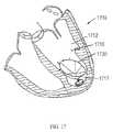
- FIG. 17is a schematic view of the patient's heart of FIG. 16A after treatment according to a method of the present invention using an elliptical shaped diastolic recoil device.
- FIG. 18Ais a drawing of the echocardiograph image of the patient's heart after treatment according to a method of the present invention using a diastolic recoil device at end-diastole, highlighting the effective diameter of the diastolic recoil device in the relaxed state.
- FIG. 18Bis a drawing of the echocardiograph image of the patient's heart after treatment according to a method of the present invention using a diastolic recoil device at end-systole, highlighting the effective diameter of the diastolic recoil device in the constrained state.
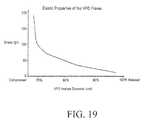
- FIG. 19is a diagrammatical illustration of the elastic characteristics of an embodiment of a diastolic recoil device implant.
- FIG. 20is a schematic representation of a heart with a ventricle having two distinct regions of myocardium with different contractile properties, Region 1 and Region 2 .
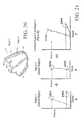
- FIGS. 21A-Care diagrammatical representations of the end-systolic pressure volume relationship (ESPVR) and end-diastolic pressure volume relationship (EDPVR) of the ventricle of FIG. 20 prior to installation of a partitioning device.
- ESPVRend-systolic pressure volume relationship
- EDPVRend-diastolic pressure volume relationship
- FIG. 22is a schematic representation of a heart with a ventricle having two distinct regions after installation of a diastolic recoil device.
- FIGS. 23A-Care diagrammatical representations of the ESPVR and EDPVR of the ventricle of FIG. 22 after treatment according to the present invention, and shows the comparison of the Stroke Volume, pre-implantation and post-implantation.
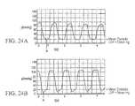
- FIG. 24Ais a diagrammatical illustration of the left ventricular pressure (LVP) in one dilated ventricle with diastolic dysfunction.
- LVPleft ventricular pressure
- FIG. 24Bis one diagrammatical illustration of the left ventricular pressure (LVP) of the ventricle of FIG. 4A after treatment according to the present invention.
- LVPleft ventricular pressure
- the present inventionis directed to devices and methods for the treatment of a patient's organ such as a heart.
- a patient's organsuch as a heart.
- the heartis susceptible to or experiencing diastolic dysfunction, mitral valve regurgitation or heart failure.
- Diastoleis the phase of cardiac cycle during which relaxation of the heart muscles occurs after ejecting blood into general circulation and is governed by active and passive properties of the myocardium, geometrical characteristics of the chamber and external forces.
- left ventricular diastolic fillingbegins with opening of the mitral valve as pressure in the ventricle falls below pressure in the atrium. As the ventricle begins to contract the pressure in the ventricle soon exceeds that of the atrium and the mitral valve closes, which marks the end of diastole.
- the ventricular pressure and volume at this pointare referred to as end-diastolic pressure (“EDP”) and end-diastolic volume (“EDV”), and the beginning of ventricular systole.
- the rate and amount of left ventricular diastolic fillingdepends upon the positive pressure upstream of the left ventricle provided by venous return and decreasing pressure provided within the left ventricle by expansion of the ventricle during diastole.
- a reduction in ventricular compliancei.e., increase in stiffness of ventricular heart wall
- less ventricular fillingi.e. decreased end-diastolic volume EDV
- a greater diastolic pressureresulting in a change in the ventricular diastolic pressure-volume characteristics.
- the left ventricular elastic recoil forcesmay be diminished, therefore leading to increase of the ventricular filling pressure.
- Diastolic dysfunctionmay also be caused by changes in the rate and degree of left ventricular relaxation, which as stated above, in part is an active process.
- Several factorscan affect left ventricular relaxation, including inotropic stimulation, fast heart rates, non-uniform heart activation and altered timing of all the forces that oppose ventricular ejection. Since calcium uptake by the sarcoplasmic reticulum is energy-dependent, any process that decreases the availability of high-energy phosphates, such as ischemia or hypoxia, also impairs myocardial relaxation.
- Diastolic dysfunctionis established, for example, by measurements of various echocardiographic parameters such as decreased peak filling velocity and prolonged relaxation time, signs of increased filling pressure and clinical symptoms of dyspnea and peripheral edema.
- the devices and methods hereincan be used to treat a patient's heart suffering from a diastolic dysfunction disorder or a condition exhibiting the characteristics of diastolic dysfunction.
- the devices and methods hereininvolve implanting within the ventricle a device whose shape elastically distorts during systole and recoils during diastole to augment the ventricle's natural recoil action.
- the devicealso partitions the patient's ventricle into a functional portion and an excluded, non-functional portion.
- the methodmay be used to treat a heart, in particular the left ventricle, which is exhibiting signs of diastolic dysfunction. Diastolic dysfunction may evidence itself by portions of the chamber becoming dilated, dyskinetic or akinetic, depending on the particular pathology inducing damage to the heart.
- FIG. 1illustrates a diastolic recoil device 130 which embodies features of the invention and which may be utilized in practicing the methods herein.
- the device 130includes hub 132 , preferably centrally located on the diastolic recoil device, and a radially expandable reinforcing frame 133 formed of a plurality of ribs 134 connected at their distal end to the hub.
- Alternative embodiments of the devices hereininclude at least three ribs.
- the ribsform an elastic frame and can be made of material such as, for example, Nitinol stainless steel, titanium alloys, NiTi alloy, other metal alloys, or plastic composites.
- the ribs/frameare made of a material which allows for compression of the free proximal ends towards the central axis during delivery and self expansion upon deployment (e.g. in the patient's heart).
- the ribs 134have distal ends 136 which may be pivotally mounted to the hub 132 and biased outwardly or fixed to the hub, and free proximal ends 137 which are configured to curve or flare away from a center line axis 138 at least upon expansion of the diastolic recoil device.
- Proximal ends 137 of ribs 134 in their expanded configurationangle outwardly from the hub at an angle ⁇ of about 20-90° away from a centerline axis 138 of the device.
- the free proximal ends 137curve outwardly so that the membrane when secured to the ribs of the expanded frame forms a trumpet-shaped concave pressure receiving surface.
- Proximal ends 137 of ribs 134can include anchors 150 configured to engage, and preferably penetrate into, the target tissue (e.g. endocardium of heart chamber to be partitioned, i.e. a ventricle). This enables the securing of a peripheral edge of the diastolic recoil device to the heart wall and fixation of the diastolic recoil device within the chamber so as to partition the chamber into two portions.
- Anchors 150are configured to penetrate the tissue lining at an angle ranging from 30-120 degrees to the centerline axis 138 of the partitioning device. Anchors 150 can include barbs, hooks and the like which prevent undesired withdrawal of device 130 from the target tissue.
- a membrane 131can be attached to the ribs 134 of the frame.
- Membrane 131can be made of a porous material, for example, expanded polytetrafluoroethylene (ePTFE, or GORE-TEX®, one commercially available product) or a non-porous material. When membrane 131 is porous, it facilitates tissue ingrowth after deployment in the non-functional portion of the heart chamber.
- Membrane 131can also be formed from other mesh materials including metals, alloys, or composites. In some cases Membrane 131 is formed from a biocompatible polymeric material such as nylon, polyethylene terephthalate (PET) or polyesters such as hytrel.
- the membrane 131has a first layer secured to the concave face of the frame formed by the ribs 134 , which creates a pressure receiving surface 135 .
- the pressure receiving surface 135is presented to the functional portion of the partitioned chamber.
- the membrane 131may have a second layer secured to the convex face of the frame formed by the ribs 134 , creating a non-pressure receiving surface 145 .
- the non-pressure receiving surface 145is presented to the non-functional portion of the partitioned chamber.
- the hub 132 shown in FIG. 1preferably has a distally extending stem 143 with a non-traumatic support component 144 .
- the distally extending stem 143 with non-traumatic support component 144 togethermay extend a variable distance from the base of the hub 132 , in order to space the device a selected distance from the wall of the chamber where the device is to be seated, thus permitting variable partitioning of the volume of the chamber.
- the stem 143 and support component 144 togethermay extend from about 3 mm to about 15 mm from the central hub 132 to isolate differing proportions of the chamber or to provide suitable fits for differing size hearts.
- Diastolic recoil deviceshave several distinct configurations.
- the unconstrained configurationis measured prior to any constriction or installation within a patient, and represents the largest diameter possible.
- the diameter (D) as shown in FIG. 1 of a device in its unconstrained configurationis at least 35 mm, up to about 100 mm, and its height (H) is at least 10 mm, to about 60 mm, as needed to fit within the heart of a patient as more fully discussed below.
- a diastolic recoil deviceWhen in its collapsed configuration, a diastolic recoil device has a diameter of less than 12 mm, such that it fits in a catheter for endovascular delivery.
- EDDEnd Diastole Diameter
- the diastolic recoil device implantPrior to the implantation procedure (as described further below), the diastolic recoil device implant is matched to the size of the left ventricle (e.g., the chamber into which it will be implanted) by comparing the left ventricle end-diastolic diameter at the level of the base of the papillary muscles (“landing zone” diameter) to the unconstrained diastolic recoil device diameter.
- the unconstrained diameter of the selected diastolic recoil deviceis oversized as compared to the diameter of the landing zone.
- Implantation of the oversized diastolic recoil deviceresults in storing compressive forces in the elastic NiTi frame of the device.
- the origin of compressive forcesis a bending deformation of the resilient frame ribs.
- the decrease of the unconstrained frame diameter to the landing zone diameteris associated with a radial tip displacement of each frame rib while the opposite end of the rib is fixed to the hub of the frame, therefore causing a flexing deformation of the ribs and a rebounding force attempting to return the frame to the unconstrained diameter.
- These outward recoil forcesare transmitted to the myocardium via proximal ends of the ribs implanted into the myocardium, thus applying pressure against the wall of the ventricle.
- the unconstrained diameter of the diastolic recoil deviceis selected to be oversized by at least about 10% up to about 60% over the diameter of the landing zone.
- the diastolic recoil deviceis elastic and its configuration changes from a small diameter at end-systole (ESD) to a larger diameter (EDD) at end-diastole.
- ESDend-systole
- EDDlarger diameter
- the compression of the diastolic recoil device from end-diastolic to end-systolic configurationcauses additional compressive forces to be stored in the elastic frame of the device and is preferably designed to be substantially equivalent at end systole to the elastic restoring forces that originate in the myocardium in a healthy heart.
- FIG. 2illustrates a top view of a diastolic recoil device 230 in its unconstrained configuration, as viewed from above the pressure receiving surface 235 .
- the diastolic recoil device 230 of FIG. 2has ribs 234 which are radially expandable and connected at their distal end to a central hub.
- the ribsare adapted to provide an elastic recoil force to a wall of a chamber of a heart (e.g. a left or right ventricle).
- the ribsstore energy during systole and release the stored energy back to the wall of the chamber of the heart in synchrony with the heart cycle.
- the device 230further comprises a membrane 231 coupled to the radially expandable ribs 234 .
- At least part of membrane 231is secured to a pressure receiving side of the frame 233 , creating the pressure receiving surface 235 . Radial expansion of the free proximal ends 237 unfurls the membrane 231 secured to the frame 233 so that the membrane presents the pressure receiving surface 235 which defines the functional and nonfunctional portions of the chamber.
- a peripheral edge 239 of the membrane 231may be serrated as shown in FIG. 2 . A serrated edge of peripheral edge 239 in this embodiment helps the membrane spread flat at the periphery.
- Anchors 250can include barbs, hooks and the like which prevent undesired withdrawal of device 130 from the wall of the chamber of heart after implantation of the device 230 .
- the ribs 234may be individually of variable length and the membrane 231 may be of variable shape suitable to practice the present invention.
- the membrane 231 and frame 233define a circular periphery and in other embodiments the membrane 231 and frame 233 define an eccentric or elliptical periphery.
- a strand 240extends around essentially the entire periphery of the membrane so that the flexible periphery of the membrane between each pair of ribs 234 is effectively sealed against the heart wall.
- the effectiveness of the sealcontributes to facile endothelialization of the pressure receiving surface of a porous membrane. Once endothelialized, the membrane supports regrowth of a new inner wall of the chamber.
- the expansive strand 240is formed from material which is stiffer than the flexible, unsupported material of the membrane to provide an outward expansive force or thrust to prevent formation of undesirable inwardly directed folds or wrinkles when the ribs of the diastolic recoil device are in a contracted configuration.
- a suitable strand 240is formed from materials such as polypropylene suture or super-elastic NiTi alloy wires. Such strands are typically about 0.005 to about 0.03 inch (about 0.13 to about 0.76 mm) in diameter to provide the requisite outward expansive force when placed in a circular position such as around the periphery of the membrane in less than completely expanded configuration. Ends 241 and 242 of the expansive strand 240 are shown extending away from the diastolic recoil device in FIG. 2 . The ends 241 and 242 may be left unattached or may be secured together, e.g. by a suitable adhesive, or to the membrane 231 itself.
- the outwardly biased strand 240ensures that there are no inwardly directed folds or wrinkles and that none are formed when the device is expanded for deployment within the heart chamber.
- the strand 240may be several strands of materials as above, rather than just one.
- FIG. 3is a bottom view of a device 330 herein.
- the nonpressure receiving surface 345 of the membrane 331which is secured to the ribs 334 (dotted lines) are illustrated in this view.
- Extending from the base of the frame 333are feet 355 which support the device within the non-functional portion of the chamber being partitioned against a wall therein.
- Feet 355extend radially and preferably are interconnected by lateral supports 346 which help distribute the force over an expanded area of the surface of the chamber.
- Feet 355 and lateral supports 346are made of resilient material which can support the device without causing trauma to the wall of the chamber at contact points. This minimizes or avoids immediate or long term damage to the tissue of the heart wall.
- the diastolic recoil devicecan be used to support weakened tissue of damaged heart wall such as necrotic tissue caused by myocardial infarction (MI) and the like.
- MImyocardial infarction
- FIG. 4is a side view of the support component of the device.
- the support component 444has a plurality of feet 455 , e.g., at least three or any variable number.
- the support component 444atraumatically contacts the wall of the ventricle within the nonfunctional portion of the partitioned ventricle, and distributes direct pressure on the wall to minimize stress on the cardiac wall in the nonfunctional portion of the partitioned ventricle through the feet 455 .
- Support component 444comprises a stem coupled to a non-traumatic base structure such as the plurality of feet 455 and connected on its other extremity to the stem 443 which extends distally from the non-pressure receiving side of the frame of the device.
- the support component 444can vary in length from about 3 mm to about 12 mm such that the non functional portion is sufficiently large in size/volume to partition necrotic tissue, such as tissue of a myocardial infarct (MI), a weakened cardiac wall, or the like.
- necrotic tissuesuch as tissue of a myocardial infarct (MI), a weakened cardiac wall, or the like.
- a web of materialmay extend between adjacent feet 445 to provide further support in addition to or in lieu of the supports 446 .
- FIG. 5illustrates a diastolic recoil device 530 comprising a frame 533 with ribs 534 .
- the membrane 531is attached to the frame 533 and the anchors 537 contact the wall of the chamber to secure the device within the chamber in order to partition it.
- Device 530has a nontraumatic support component 544 which has a simple rounded end which is connected to the stem 543 .
- the stem 543is connected to the central hub 532 which is connected to the frame 533 .
- FIG. 6illustrates an alternative support component 644 for the devices of the invention.
- Support component 644has a plurality of curved bumpers 645 which act as “feet” and contact the wall of the chamber atraumatically. There may be a variable number of curved bumpers to distribute the force that the support component will deliver to the wall of the chamber.
- FIG. 7illustrates an alternative support component 744 which has feet such as the plurality of J-bumpers 745 .
- FIG. 8illustrates a different embodiment of the support component 844 which has a plurality of J-shaped bumpers 845 .
- FIG. 9illustrates another embodiment of the support component 944 which has a soft, non-traumatic coil 945 which contacts the wall of the heart chamber, and distributes the force from a diastolic recoil device to a larger area of the wall of the heart, reducing strain on weakened or necrotic areas of the chamber.
- the distal ends 1036 of the ribs 1034are secured within the hub 1032 and, as shown in the detail of FIG. 11 , a transversely disposed connector bar 1047 is secured within the hub which is configured to secure the hub 1032 and thus the diastolic recoil device 1030 to a delivery system such as that described in co-pending applications referenced above.
- FIG. 10also illustrates the connection between connector hub 1032 , stem 1043 , support component 1044 , and feet 1045 .
- a device 1230further incorporating a delayed release spring 1260 as shown schematically in FIG. 12 , can be utilized to assist diastolic function.
- delayed response spring 1260is attached to restraint struts 1261 which in turn releasably contact the frame 1233 on the non-pressure bearing side 1245 of the membrane 1231 .
- the ventriclebegins an unassisted expansion while the device is partially secured from freely expanding.
- the delayed release mechanismis triggered.
- the restraint strutsare 1261 released from contact with the frame 1233 , and the stored energy fully released at that point in the cardiac cycle.
- the majority of the recoil energycan be given back to the ventricular wall at a select point during diastole, as required for a particular patient.
- Another embodiment of this aspect of the inventionmay have a spring means including only a damped releasing mechanism.
- the subsequent contraction of the ventricle during systolere-engages the delayed release spring mechanism or restores the damped spring to restore the contact between the restraint struts 1261 and the frame 1233 when the frame is in the compressed state for further cycles of delayed recoil assistance to the ventricle.
- FIG. 13may be utilized, which has a frame 1333 and central hub 1332 as previously described, but which has no membrane.
- the resilient frameprovides force back to the walls of the ventricle and improves the diastolic function of the heart.
- the framemay need to be different from the frames of other embodiments of this invention, i.e. frame 133 of FIG. 1 .
- the ventricles of this population of patientsmay require more force to be applied back to the ventricular walls, which may be thickened and stiffened relative to healthy ventricular walls. It may also be necessary to increase the number of ribs, the thickness of the material of the ribs, the relative stiffness of the ribs, and/or use different alloys or material compositions to form the frame in order to manufacture a device with appropriate resiliency/stiffness properties.
- the devicemay seat lower in the ventricular chamber, and may thus require devices with smaller diameters relative to those used for patients with ventricular dilation. The size matching then is made for the end-diastolic diameter of a landing zone at a level further below the base of the papillary muscles.
- the unconstrained diameter of devices according to this embodiment of the inventionmay therefore be at least 25 mm up to about 90 mm.
- the central hub 1432as shown in the side elevation view of a device depicted in FIG. 14 , may not have any distal extension and may ends as a flat disk.
- a distal extension of hub 1432may consist of a short rounded nub, or may connect to flexible basal supports which may stabilize the device in its seat in the apex of the ventricle.
- the basal supportsmay be configured in many ways. Two examples are given in FIGS. 15A and 15B respectively, shown as basal supports 1561 A and 1561 B.
- Implantation of the devices hereincan be accomplished endovascularly or intraoperatively in as little as one hour by a physician or appropriately trained personnel. Such implantation presents limited risk to the patient and requires the patient to be under a fluoroscope for a period of as little as 20 minutes.
- Implantation of the diastolic recoil device in the ischemic and enlarged ventriclemay bring back the ability of the ventricle to store elastic energy during systole and return this energy in the form of elastic recoil forces during diastole.
- this return of energy in the form of elastic recoilmay contribute to the improvement of the diastolic function, i.e., decrease of the filling pressure and increase in the magnitude of the early filling in patients with ischemic and/or dilated cardiomyopathy.
- the ejection fraction of the chamberis increased by at least about a 5% change.
- Diastolic recoil devices of the present inventionare delivered percutaneously or intraoperatively.
- a suitable delivery deviceis described in co-pending application Ser. No. 10/913,608, filed on Aug. 5, 2004, entitled “Ventricular Partitioning Device”, assigned to the assignee of the present invention, the full disclosure of which is incorporated herein by reference.
- diastolic recoil devicesmay be conveniently formed by the method described in above-referenced co-pending application Ser. No. 10/913,608 assigned to the assignee of the present invention and which is incorporated herein by reference in its entirety.
- FIG. 16Ais a schematic illustration of a patient's heart 1610 showing the right ventricle 1611 and the left ventricle 1612 with the mitral valve 1613 and aortic valve 1614 .
- a pericardium membrane 1615is shown surrounding the heart 1610 .
- FIG. 16Aillustrates a patient's heart with apical dilatation (round enlarged apex 1616 of the LV) which can be found in patients exhibiting characteristics of congestive heart failure.
- FIG. 16Billustrates the left ventricle 1612 of FIG.
- FIG. 17is a schematic view of the patient's heart of FIG. 16A after treatment according to a method of the present invention using an elliptical shaped diastolic recoil device 1730 .
- the device 1730is implanted into the left ventricle 1712 of the heart 1710 , creating a functional portion 1718 and nonfunctional portion 1717 .
- FIGS. 18A and 18Bare drawings of echocardiograph images of a patient's heart at end-diastole, and end-systole, respectively.
- the contours of the diastolic recoil device implanted in the left ventricleare visible as fine white lines in the base of the ventricle. Portions of the ribs and periphery can be seen in FIGS. 18A and 18B .
- the diameter of the elastic diastolic recoil deviceis at its maximal implanted diameter ( FIG. 18A ) at end-diastole, and at its minimal implanted diameter at end-systole ( FIG. 18B ).
- End-systolic diameters (ESD)can be in the range from about 25 mm to about 55 mm.
- End-diastolic diameters (EDD)can be in the range of about 45 mm to about 70 mm.
- the compression of the partitioning device from end-diastolic to end-systolic configurationcauses elastic recoil forces to be stored in the elastic frame of the device, and to be transmitted to the myocardium during ventricular filling in the outward direction thus enhancing outward motion of the ventricular walls.
- This storing and release of energy by the frameoccurs in synchrony with the action of the heart.
- This transfer of energymay decrease the ventricular pressure in diastole, increase the atrio-ventricular pressure gradient, increase filling, and thus improve ejection fraction
- Dyskinetic or aneurystic ventricular wallsresult in dyssynchronous behavior during the cardiac cycle, leading to inefficient pumping function.
- the partitioning deviceis substantially circular but another embodiment of the invention utilizes an elliptical shaped partitioning device as shown in FIG. 17 .
- Other configurations of the partitioning deviceare compatible with the construction as described above and with methods to partition a chamber of a heart as set forth here.
- the devices hereincan be used to treat a patient suffering from a heart condition.
- heart conditionscan include, for example, mitral valve regurgitation, myocardial infarction, or scar tissue or akinetic tissue in a heart chamber.
- a patientcan be screened for treatment by the a devices herein by any means known in the art including, but not limited to, measurements of echocardiographic parameters may be such as decreased peak filling velocity and prolonged relaxation time, signs of increased filling pressure, clinical symptoms of dyspnea and peripheral edema, as well as low ejection fraction and a distance a patient can walk in 6 minutes.
- the diastolic recoil device implantmay be matched to the size of the chamber where it is to be inserted (e.g. left ventricle) when the device is to be inserted into the left ventricle this can be accomplished by comparing the left ventricle end-diastolic diameter at the level of the papillary muscles base. This diameter is referred to hereinafter as the landing zone diameter. Measurement of landing zone diameter may be made by any method known in the art including; echocardiography, fluoroscopy, PET, MRI, contrast angiography, and the like, the landing zone diameter is the compared to the relaxed deployed/device diameter.
- the ventricleWhen a device is to be implanted in a ventricle, the ventricle may be dilated such that its end diastolic diameter is greater than 45 mm or even greater than 65 mm.
- the relaxed diameter of the selected diastolic recoil deviceis oversized as compared to the diameter of the landing zone.
- the relaxed diameter of the devicecan be oversized by at least about 10% and up to 60% over the landing zone diameter.
- the diastolic recoil device implantedthus decreases the LV volume by at least about 10% up to about 40%.
- the ratio of the nonfunctional portion to the functional portion, created by partitioning the ventricle by a method of the inventionis at least 1:10 or up to about 1:3.
- the diastolic recoil device frameis elastic and its diameter changes from a small diameter at end-systole to a larger diameter at end-diastole.
- the compression of the diastolic recoil device from end-diastolic to end-systolic configurationcauses additional compressive forces to be stored in the elastic frame of the device, thus enhancing the ejection fraction of the chamber by at least about 10%, or up to about 90%.
- the elastic characteristics of the diastolic recoil device implantmay be determined by a tensile/compression test, an example of which is diagrammatically shown in FIG. 19 .
- the diastolic recoil deviceis positioned inside a custom designed fixture which was connected to a force transducer.
- the fixturewas designed to create substantially equal compressive radial force (compatible and corresponding to physiological range of forces developed by normal myocardial fibers) on all ribs 34 (as described below) of the implant, thus determining the compression stress-diameter relationship for the frame 33 (as described below) of the device.
- FIG. 19shows an exemplary elastic property of the diastolic recoil device.
- the magnitude of the elastic recoil forces stored in the diastolic recoil device implantincreases as the diastolic recoil device diameter decreases under compression.
- the stiffness of the implantincreases in a non-linear fashion as the diameter of the implant decreases as it is compressed to less than 50% of the diameter of the fully relaxed implant.
- FIG. 20is a schematic representation of a heart with dilation and poor function in the left ventricle, having two distinct regions of myocardium surrounding the interior of the ventricle. Region 1 represents normal myocardium and region 2 represents dilated and dyskinetic or akinetic/myocardium.
- a simulation experimentis performed, using an elastance model, as described in J H. Artip; et al.; J. Thoracic and Cardiovascular Surg., 122(4), 775-782, 2001.
- FIGS. 21A-Crepresent a simulation carried out using a ventricle as in FIG. 20 without a partitioning device.
- the dashed lines labeled ESPVREnd Systolic Pressure Volume Relationship
- FIGS. 21A and Brepresent the maximal pressure that can be developed by that section of the ventricle at any given left ventricular volume.
- EDPVREnd Diastolic Pressure Volume Relationship
- the effectis modeled wherein the akinetic Region 2 of a ventricle with diastolic dysfunction (as shown in FIG. 22 ) of the LV is partitioned by a partitioning device of the invention.
- the ESPVR and EDPVR for the individual contributions from normal Region 1 , the diastolic recoil device, and akinetic Region 2are represented in FIGS. 23 A and B.
- the normal Region 1now exhibits a steeper slope to its ESPVR as the diastolic recoil device isolates Region 2 from Region 1 , reducing the overall volume and conferring greater resistance as systole proceeds.
- the solid line shown in FIG. 23Bshows similar information as that in FIG.
- FIG. 23Bis the ESPVR/EDPVR curve for the akinetic Region 2 in FIG. 22 .
- the new ESPVR and EDPVR for the ventricle as a wholeare shown in FIG. 23C , as solid lines.
- the corresponding ESPVR and EDPVR for the pre-implant ventricle from FIG. 21Care also reproduced in FIG. 23C as dashed lines for comparison. As can be seen in FIG. 23C
- the ESPVR and EDPVR curves of the post-implant ventricleare shifted leftwards as compared to the curves of the dilated pre-implant ventricle ( FIG. 23C “ESPVR Pre-Implant” and “EDPVR Pre-Implant”, dashed lines).
- the ESPVR curve for the partitioned ventricleis shifted more than the EDPVR curve for the partitioned ventricle. This results in increased pump function of the ventricle which can be demonstrated by examining the resultant the pressure-volume loops.
- SV Pre-Implant22
- SV Implant22
- the stroke volumeis represented by the width of these shaded volumes as filling proceeds along the EDVPR curves.
- the right-hand boundary of the stroke volumeis the pressure/volume line at end diastole, when isovolumetric contraction begins, and the left-hand boundary is the volume/pressure line representing isovolumetric relaxation during the heart cycle.
- the partitioned ventricleexhibits increased stroke volume (SV) compared to the dilated, pre-implant ventricle with akinetic Region 2 at comparable end-diastolic and aortic pressures (“SV Implant” vs. “SV Pre-Implant” in FIG. 23C ).
- FIGS. 24A and 24Bare diagrammatical illustrations of the recordings of the left ventricular pressure (LVP) in one dilated ventricle with diastolic dysfunction before and after implantation of a diastolic recoil device, respectively.
- LVPleft ventricular pressure
- FIG. 24Adiastolic dysfunction results in inefficient filling of the ventricle at relatively high mean diastolic pressure in the ventricle.
- the akinetic ventriclecan neither compress nor expand as effectively as a normal ventricular chamber.
- the resultant filling pressure at early diastoleis therefore higher than in a healthy heart and early filling is decreased.
- the deviceDuring installation of the diastolic recoil device, the device is anchored to functional portions of the ventricle wall, partitioning the akinetic (nonfunctional) portion of the chamber. This mode of attachment allows the elastic frame of the partitioning device to gain energy from the effectively contracting portion of the ventricular wall, by compressing the elastically resilient frame. As the ventricle relaxes and expands, the energy stored in the frame is released and imparts additional recoil force back to the ventricle wall, which aids in the process of filling the ventricular chamber. As can be seen from FIGS. 24A and 24B , implantation of the diastolic recoil device resulted in decreased minimum diastolic pressures.
- the minimum LV pressureis decreased by at least 50% (contrasted by points A and A′ in FIGS. 24A and 24B respectively) and mean diastolic pressure at least by 10%.
- the contribution of the elastic energy from the frame assisting expansion of the walls of the ventriclewas observed in early diastole, thereby augmenting filling and normalizing diastolic pressure.
- Decreased mean diastolic pressure of the partitioned ventricle compared to that of the pre-implant ventricleindicates improved diastolic function (mean diastolic LVP of ca. 14 mm Hg in FIG. 24B vs. “mean diastolic LVP of ca. 22 mm Hg in FIG. 24A ).
- a diastolic recoil device and methods of the inventionyields a decrease of minimum LV pressure during diastole by at least about 5% up to about 100%.
- the use of a diastolic recoil device by the methods of the inventionyields a decrease of end-diastolic pressure by at least about 5%, and up to about 35%.
- LVESVIleft ventricle end systolic volume index
- LVESVIindicates the size of the ventricle at end systole with values normalized to body size.
- the baseline value for a healthy individualis ⁇ 25 ml/m 2 .
- LVESVIhas significant predictive value for survival outcome, and may represent the most significant correlation used in diagnosis and treatment.
- a patientcan be first diagnosed as having heart disease by determining or detecting in that patient a LVESVI greater than 60 ml/m 2 . Such patient is thus treated by implanting one or more of the devices herein.
- the diastolic recoil deviceby partitioning the ventricle into functional and non-functional portions, causes an initial decrease in LVESVI upon installation.
- the implantation of the diastolic recoil devicemay also promote positive remodeling of the ventricle to further decrease ventricle volume as the supported cardiac muscle more effectively contracts and expands, thus decreasing LVESVI by at least 5%.
- LVEFLeft ventricle ejection fraction
- Implantation of the partitioning deviceincreases the LVEF by at least about 5% and up to about 90%.
- indices of ventricular functionmay also be used for diagnosis and for therapeutic follow-up.
- a number of biochemical markersmay be measured and used.
- One exampleis NT Pro-Brain Natriuretic Peptide, but many other biological molecules, for example, neurohormones, proteases, and proteins related to distressed or abnormal function may be measured to give quantification of the relative functionality of the ventricle prior and post-implant.
- NT-Pro-BNPNT-Pro-Brain Natriuretic Peptide
- a normal NT-BNP level for a healthy individualis generally in the range of 20-30 pg/ml, while in an individual with end stage heart failure, a level can be as high as 2000-3000 pg/ml, and in some instances there may be a correlation between BNP levels and LVEF.
- the use of NT-Pro-BNP levels as reliable markers for heart disease in a number of patient populationshas been proposed (J. L. Januzzi; Cleve. Clin. J.
- the present inventioncontemplates treating a patient by first determining the level of NT-Pro-BNP, and if the level of NT-Pro-BNP is greater than 170 pg/ml (third quartile) or 450 pg/ml (fourth quartile), delivering to such patient one or more of the devices herein. Implantation of a diastolic recoil device improves cardiac function, and decreases the level of NT-Pro-BNP observed post-implant by at least about 10%.
- Mitral valve regurgitationcan be observed in patients with diastolic dysfunction, and is coupled to poor outcome. Mitral valve regurgitation increases in magnitude as the ventricle increases in size due to pathological dilation. Intervention is often necessary as blood backflow into the atrium leads to accelerated progression of heart failure. Standard therapies include both prescribed medications (i.e. vasodilators like ACE inhibitors and nitrates, and diuretics) and surgical interventions to repair or replace mitral valves. However, these surgical interventions are invasive and may present high risk to the patient. Diastolic recoil device implantation can reverse the decline in ventricular function by decreasing the effective ventricular volume which may obliterate or attenuate the cause of the mitral valve regurgitation.
- prescribed medicationsi.e. vasodilators like ACE inhibitors and nitrates, and diuretics
- surgical interventionsare invasive and may present high risk to the patient.
- Diastolic recoil device implantationcan reverse the decline in ventricular function by decreasing the effective
- the severity of mitral valve regurgitationis categorized by measuring the regurgitant fraction by, for example, echocardiography.
- Color Doppler flow on a transthoracic echocardiogrammeasures the forward flow through the mitral valve during ventricular diastole and compares it to the outflow of blood through the aortic valve in ventricular systole, permitting the calculation of the regurgitant fraction.
- the present inventioncontemplates treating a patient by first determining the degree of mitral regurgitation as assessed by the regurgitant fraction and if the regurgitant fraction is at least 20%, delivering to such patient one or more of the devices herein. Diastolic recoil device implantation may therefore benefit patients with mitral valve regurgitation from any clinically relevant cause and decrease the regurgitant fraction by at least about 10%.
- diastolic recoil devicewhich is implanted in the left ventricle, it is understood by those skilled in the art that such reference is not limiting and similarly suitable diastolic recoil devices may be used in the right ventricle or other heart chambers.
- Symptomatic heart failure patients(New York Heart Association Classification levels II and III) diagnosed with ischemic cardiomyopathy post anterior infarction and systolic dysfunction were enrolled in a study implanting a diastolic recoil device similar to the one shown in FIG. 1 .
- Size selection of the specific devicewas based on echocardiography comparison with a mean landing zone diameter of 55.1 mm (mean diastolic value or largest value achieved during cardiac cycle). Either 75 mm (3/9 patients) or 85 mm (6/9 patients) diameter devices were installed in a 95.7 minute (mean value) procedure, requiring mean fluoroscope time of 25.5 minutes.
- Left ventricle end systolic volume index (LVESVI) in a healthy individualis usually around 25 ml/m 2 .
- the mean baseline value for the patient groupis notably higher, at 101.8 ml/m 2 .
- Significant reduction to 72.7 ml/m 2 ( ⁇ 25%) for the LVESVIis observed at 90 days post implant.
- the ventriclehas thus improved in function and was positively remodeled.
- LVEFLeft ventricle ejection fraction
- NT-Pro-brain Natriuretic peptide (NT-Pro-BNP) levels for a healthy individualare estimated to be in the range of 20-30 pg/ml.
- the baseline mean value of NT-Pro-BNP prior to implantationwas 566 pg/ml. This was significantly decreased by the 90 day timepoint to 363 pg/ml, an improvement of about 36%.
- Table 2represents data of overall functionality for the individual patients.
- the 6 minute walkis a simple test which measures the distance a patient is able to traverse during a 6 minutes timed period.
- the mean distance the patient cohort traveled prior to implantwas 328 m.
- Ninety days post implant, data available for 4 patientsshows significant improvement ( ⁇ 44%) to 471 m.
- the New York Heart Association (NYHA) Classification levels for the patients prior to implantationwere Class II/III for this group. At the 90 day timepoint, reassessment of the NYHA Classification was performed on the four patients with available data. Three of the four individuals could be reassigned to less severe disease classifications.
- NYHANew York Heart Association
Landscapes
- Health & Medical Sciences (AREA)
- Life Sciences & Earth Sciences (AREA)
- Surgery (AREA)
- Animal Behavior & Ethology (AREA)
- Veterinary Medicine (AREA)
- Public Health (AREA)
- Nuclear Medicine, Radiotherapy & Molecular Imaging (AREA)
- Engineering & Computer Science (AREA)
- Biomedical Technology (AREA)
- Heart & Thoracic Surgery (AREA)
- General Health & Medical Sciences (AREA)
- Molecular Biology (AREA)
- Medical Informatics (AREA)
- Vascular Medicine (AREA)
- Reproductive Health (AREA)
- Cardiology (AREA)
- Radiology & Medical Imaging (AREA)
- Prostheses (AREA)
- Surgical Instruments (AREA)
- External Artificial Organs (AREA)
Abstract
Description
| TABLE 1 | ||||
| Exploratory Endpoints | Baseline (n = 9) | |||
| All data as mean values | Before | 90 days (n = 4) | ||
| LVESVI (ml/m2) | 101.8 | 72.7 | ||
| LVEF (%) | 29.3 | 37.2 | ||
| Patients with MR | 5/9 | 1/4 | ||
| NT-Pro-BNP (pg/ml) | 566 | 393 | ||
| TABLE 2 | ||
| Exploratory Endpoints | Baseline (n = 9) | |
| Mean Values | Before | 90 days (n = 4) |
| 6 min walk (m) | 328 | 471 |
| Improvement in NYHA class | — | ¾ (75%) |
| MLHF | 22.9 | 12.7 |
Claims (47)
Priority Applications (3)
| Application Number | Priority Date | Filing Date | Title |
|---|---|---|---|
| US12/691,587US8672827B2 (en) | 1999-08-09 | 2010-01-21 | Cardiac device and methods of use thereof |
| US14/189,856US9694121B2 (en) | 1999-08-09 | 2014-02-25 | Systems and methods for improving cardiac function |
| US15/630,601US10251750B2 (en) | 1999-08-09 | 2017-06-22 | Systems and methods for improving cardiac function |
Applications Claiming Priority (5)
| Application Number | Priority Date | Filing Date | Title |
|---|---|---|---|
| US14789499P | 1999-08-09 | 1999-08-09 | |
| US63551100A | 2000-08-09 | 2000-08-09 | |
| US10/212,033US7303526B2 (en) | 1999-08-09 | 2002-08-01 | Device for improving cardiac function |
| US11/640,469US7674222B2 (en) | 1999-08-09 | 2006-12-14 | Cardiac device and methods of use thereof |
| US12/691,587US8672827B2 (en) | 1999-08-09 | 2010-01-21 | Cardiac device and methods of use thereof |
Related Parent Applications (1)
| Application Number | Title | Priority Date | Filing Date |
|---|---|---|---|
| US11/640,469ContinuationUS7674222B2 (en) | 1999-08-09 | 2006-12-14 | Cardiac device and methods of use thereof |
Related Child Applications (2)
| Application Number | Title | Priority Date | Filing Date |
|---|---|---|---|
| US63551100AContinuation-In-Part | 1999-08-09 | 2000-08-09 | |
| US14/189,856Continuation-In-PartUS9694121B2 (en) | 1999-08-09 | 2014-02-25 | Systems and methods for improving cardiac function |
Publications (2)
| Publication Number | Publication Date |
|---|---|
| US20100121132A1 US20100121132A1 (en) | 2010-05-13 |
| US8672827B2true US8672827B2 (en) | 2014-03-18 |
Family
ID=39537001
Family Applications (2)
| Application Number | Title | Priority Date | Filing Date |
|---|---|---|---|
| US11/640,469Expired - LifetimeUS7674222B2 (en) | 1999-08-09 | 2006-12-14 | Cardiac device and methods of use thereof |
| US12/691,587Expired - Fee RelatedUS8672827B2 (en) | 1999-08-09 | 2010-01-21 | Cardiac device and methods of use thereof |
Family Applications Before (1)
| Application Number | Title | Priority Date | Filing Date |
|---|---|---|---|
| US11/640,469Expired - LifetimeUS7674222B2 (en) | 1999-08-09 | 2006-12-14 | Cardiac device and methods of use thereof |
Country Status (6)
| Country | Link |
|---|---|
| US (2) | US7674222B2 (en) |
| EP (1) | EP2091588A4 (en) |
| JP (1) | JP5461192B2 (en) |
| AU (1) | AU2007333895A1 (en) |
| CA (1) | CA2671974A1 (en) |
| WO (1) | WO2008076853A2 (en) |
Cited By (10)
| Publication number | Priority date | Publication date | Assignee | Title |
|---|---|---|---|---|
| US20120239083A1 (en)* | 2005-09-16 | 2012-09-20 | Atritech, Inc. | Intracardiac cage and method of delivering same |
| US9039597B2 (en) | 2009-10-26 | 2015-05-26 | Cardiokinetix, Inc. | Ventricular volume reduction |
| US9592123B2 (en) | 2002-08-01 | 2017-03-14 | Cardiokinetix, Inc. | Therapeutic methods and devices following myocardial infarction |
| US9592399B2 (en) | 2013-06-20 | 2017-03-14 | Cardiac Pacemakers, Inc. | Deployable multi-electrode leadless electrostimulator |
| US9694121B2 (en) | 1999-08-09 | 2017-07-04 | Cardiokinetix, Inc. | Systems and methods for improving cardiac function |
| US9872767B2 (en) | 1999-08-09 | 2018-01-23 | Edwards Lifesciences Corporation | Retrievable cardiac devices |
| US10064696B2 (en) | 2000-08-09 | 2018-09-04 | Edwards Lifesciences Corporation | Devices and methods for delivering an endocardial device |
| US10307147B2 (en) | 1999-08-09 | 2019-06-04 | Edwards Lifesciences Corporation | System for improving cardiac function by sealing a partitioning membrane within a ventricle |
| US10751183B2 (en) | 2014-09-28 | 2020-08-25 | Edwards Lifesciences Corporation | Apparatuses for treating cardiac dysfunction |
| US10898330B2 (en) | 2017-03-28 | 2021-01-26 | Edwards Lifesciences Corporation | Positioning, deploying, and retrieving implantable devices |
Families Citing this family (138)
| Publication number | Priority date | Publication date | Assignee | Title |
|---|---|---|---|---|
| US7883539B2 (en) | 1997-01-02 | 2011-02-08 | Edwards Lifesciences Llc | Heart wall tension reduction apparatus and method |
| US7128073B1 (en) | 1998-11-06 | 2006-10-31 | Ev3 Endovascular, Inc. | Method and device for left atrial appendage occlusion |
| US8257428B2 (en)* | 1999-08-09 | 2012-09-04 | Cardiokinetix, Inc. | System for improving cardiac function |
| US7674222B2 (en)* | 1999-08-09 | 2010-03-09 | Cardiokinetix, Inc. | Cardiac device and methods of use thereof |
| US8388672B2 (en) | 1999-08-09 | 2013-03-05 | Cardiokinetix, Inc. | System for improving cardiac function by sealing a partitioning membrane within a ventricle |
| US8398537B2 (en) | 2005-06-10 | 2013-03-19 | Cardiokinetix, Inc. | Peripheral seal for a ventricular partitioning device |
| US7762943B2 (en)* | 2004-03-03 | 2010-07-27 | Cardiokinetix, Inc. | Inflatable ventricular partitioning device |
| US20060030881A1 (en) | 2004-08-05 | 2006-02-09 | Cardiokinetix, Inc. | Ventricular partitioning device |
| US9332993B2 (en) | 2004-08-05 | 2016-05-10 | Cardiokinetix, Inc. | Devices and methods for delivering an endocardial device |
| US9332992B2 (en) | 2004-08-05 | 2016-05-10 | Cardiokinetix, Inc. | Method for making a laminar ventricular partitioning device |
| US9078660B2 (en)* | 2000-08-09 | 2015-07-14 | Cardiokinetix, Inc. | Devices and methods for delivering an endocardial device |
| US7399271B2 (en)* | 2004-01-09 | 2008-07-15 | Cardiokinetix, Inc. | Ventricular partitioning device |
| US6723038B1 (en) | 2000-10-06 | 2004-04-20 | Myocor, Inc. | Methods and devices for improving mitral valve function |
| US6764510B2 (en) | 2002-01-09 | 2004-07-20 | Myocor, Inc. | Devices and methods for heart valve treatment |
| US7112219B2 (en) | 2002-11-12 | 2006-09-26 | Myocor, Inc. | Devices and methods for heart valve treatment |
| US8086315B2 (en) | 2004-02-12 | 2011-12-27 | Asap Medical, Inc. | Cardiac stimulation apparatus and method for the control of hypertension |
| US11511102B2 (en) | 2004-06-17 | 2022-11-29 | The Texas A&M University System | Cardiac compression device having passive and active chambers |
| US9510746B2 (en) | 2004-06-17 | 2016-12-06 | The Texas A&M University System | Deployment methods and mechanisms for minimally invasive implantation of heart contacting cardiac devices |
| US20060079736A1 (en) | 2004-10-13 | 2006-04-13 | Sing-Fatt Chin | Method and device for percutaneous left ventricular reconstruction |
| US8165674B2 (en) | 2005-03-02 | 2012-04-24 | Backbeat Medical, Inc. | Methods and apparatus to increase secretion of endogenous naturetic hormones |
| US20090082619A1 (en)* | 2005-06-09 | 2009-03-26 | De Marchena Eduardo | Method of treating cardiomyopathy |
| US7766816B2 (en) | 2005-06-09 | 2010-08-03 | Chf Technologies, Inc. | Method and apparatus for closing off a portion of a heart ventricle |
| US8506474B2 (en) | 2005-08-19 | 2013-08-13 | Bioventrix, Inc. | Method and device for treating dysfunctional cardiac tissue |
| EP1933756B1 (en) | 2005-08-19 | 2016-07-20 | CHF Technologies Inc. | Steerable lesion excluding heart implants for congestive heart failure |
| EP1986735A4 (en)* | 2006-02-06 | 2011-06-29 | Northwind Ventures | Systems and methods for volume reduction |
| US7749249B2 (en) | 2006-02-21 | 2010-07-06 | Kardium Inc. | Method and device for closing holes in tissue |
| US20070270688A1 (en) | 2006-05-19 | 2007-11-22 | Daniel Gelbart | Automatic atherectomy system |
| US8920411B2 (en) | 2006-06-28 | 2014-12-30 | Kardium Inc. | Apparatus and method for intra-cardiac mapping and ablation |
| US8449605B2 (en) | 2006-06-28 | 2013-05-28 | Kardium Inc. | Method for anchoring a mitral valve |
| US11389232B2 (en) | 2006-06-28 | 2022-07-19 | Kardium Inc. | Apparatus and method for intra-cardiac mapping and ablation |
| US9119633B2 (en) | 2006-06-28 | 2015-09-01 | Kardium Inc. | Apparatus and method for intra-cardiac mapping and ablation |
| US10028783B2 (en) | 2006-06-28 | 2018-07-24 | Kardium Inc. | Apparatus and method for intra-cardiac mapping and ablation |
| US7837610B2 (en)* | 2006-08-02 | 2010-11-23 | Kardium Inc. | System for improving diastolic dysfunction |
| US7869874B2 (en) | 2006-09-25 | 2011-01-11 | G&L Consulting, Llc | Methods and apparatus to stimulate heart atria |
| US9211115B2 (en) | 2006-09-28 | 2015-12-15 | Bioventrix, Inc. | Location, time, and/or pressure determining devices, systems, and methods for deployment of lesion-excluding heart implants for treatment of cardiac heart failure and other disease states |
| US8123668B2 (en) | 2006-09-28 | 2012-02-28 | Bioventrix (A Chf Technologies' Company) | Signal transmitting and lesion excluding heart implants for pacing defibrillating and/or sensing of heart beat |
| US8029556B2 (en)* | 2006-10-04 | 2011-10-04 | Edwards Lifesciences Corporation | Method and apparatus for reshaping a ventricle |
| US9192471B2 (en) | 2007-01-08 | 2015-11-24 | Millipede, Inc. | Device for translumenal reshaping of a mitral valve annulus |
| JP5581209B2 (en)* | 2007-07-18 | 2014-08-27 | ボストン サイエンティフィック サイムド,インコーポレイテッド | Endoscopic implant system |
| DE102007043830A1 (en) | 2007-09-13 | 2009-04-02 | Lozonschi, Lucian, Madison | Heart valve stent |
| US8491455B2 (en) | 2007-10-03 | 2013-07-23 | Bioventrix, Inc. | Treating dysfunctional cardiac tissue |
| US8906011B2 (en) | 2007-11-16 | 2014-12-09 | Kardium Inc. | Medical device for use in bodily lumens, for example an atrium |
| EP2082690B1 (en) | 2008-01-24 | 2012-06-20 | Kardium, Inc. | Medical device to assist diastolic function and prevent ventricular enlargement |
| US8489172B2 (en) | 2008-01-25 | 2013-07-16 | Kardium Inc. | Liposuction system |
| US20090287304A1 (en) | 2008-05-13 | 2009-11-19 | Kardium Inc. | Medical Device for Constricting Tissue or a Bodily Orifice, for example a mitral valve |
| US8340763B2 (en) | 2008-09-08 | 2012-12-25 | Backbeat Medical, Inc. | Methods and apparatus to stimulate heart atria |
| EP2344106B8 (en)* | 2008-10-10 | 2022-07-20 | MedicalTree Patent Ltd. | Heart help device |
| WO2010042018A1 (en)* | 2008-10-10 | 2010-04-15 | Milux Holding S.A. | Heart help device, system and method |
| EP2344070B1 (en)* | 2008-10-20 | 2017-08-09 | Corassist Cardiovascular Ltd. | Ventricular function assisting device and apparatus for implanting it |
| EP2381852A4 (en)* | 2009-01-21 | 2014-06-11 | Tendyne Medical Inc | Apical papillary muscle attachment for left ventricular reduction |
| US20100210899A1 (en)* | 2009-01-21 | 2010-08-19 | Tendyne Medical, Inc. | Method for percutaneous lateral access to the left ventricle for treatment of mitral insufficiency by papillary muscle alignment |
| US20100274227A1 (en)* | 2009-02-13 | 2010-10-28 | Alexander Khairkhahan | Delivery catheter handle cover |
| US20110015476A1 (en)* | 2009-03-04 | 2011-01-20 | Jeff Franco | Devices and Methods for Treating Cardiomyopathy |
| US9642957B2 (en) | 2009-07-22 | 2017-05-09 | The Texas A&M University System | Diastolic recoil method and device for treatment of cardiac pathologies |
| US10398556B2 (en)* | 2009-07-22 | 2019-09-03 | Corinnova Incorporated | Diastolic recoil method and device for treatment of cardiac pathologies |
| WO2011041571A2 (en) | 2009-10-01 | 2011-04-07 | Kardium Inc. | Medical device, kit and method for constricting tissue or a bodily orifice, for example, a mitral valve |
| EP3300695B1 (en) | 2009-12-08 | 2023-05-24 | Avalon Medical Ltd. | Device and system for transcatheter mitral valve replacement |
| US9050066B2 (en) | 2010-06-07 | 2015-06-09 | Kardium Inc. | Closing openings in anatomical tissue |
| IT1400503B1 (en)* | 2010-06-08 | 2013-06-11 | Parravicini | CARDIOVASCULAR DEVICE. |
| US20120053680A1 (en) | 2010-08-24 | 2012-03-01 | Bolling Steven F | Reconfiguring Heart Features |
| US8940002B2 (en) | 2010-09-30 | 2015-01-27 | Kardium Inc. | Tissue anchor system |
| US9452016B2 (en) | 2011-01-21 | 2016-09-27 | Kardium Inc. | Catheter system |
| CA2764494A1 (en) | 2011-01-21 | 2012-07-21 | Kardium Inc. | Enhanced medical device for use in bodily cavities, for example an atrium |
| US9480525B2 (en) | 2011-01-21 | 2016-11-01 | Kardium, Inc. | High-density electrode-based medical device system |
| US11259867B2 (en) | 2011-01-21 | 2022-03-01 | Kardium Inc. | High-density electrode-based medical device system |
| EP2672927A4 (en)* | 2011-02-10 | 2014-08-20 | Atrial Innovations Inc | EARLY APPENDIX OCCLUSION AND TREATMENT OF ARRHYTHMIA |
| US9072511B2 (en) | 2011-03-25 | 2015-07-07 | Kardium Inc. | Medical kit for constricting tissue or a bodily orifice, for example, a mitral valve |
| EP4289398A3 (en) | 2011-08-11 | 2024-03-13 | Tendyne Holdings, Inc. | Improvements for prosthetic valves and related inventions |
| EP2760328B1 (en) | 2011-09-30 | 2023-02-22 | Bioventrix, Inc. | Remote pericardial hemostasis for ventricular access and reconstruction or other organ therapies |
| EP2775933B1 (en)* | 2011-11-09 | 2016-12-28 | Easynotes Ltd. | Obstruction device |
| US9827092B2 (en) | 2011-12-16 | 2017-11-28 | Tendyne Holdings, Inc. | Tethers for prosthetic mitral valve |
| USD777926S1 (en) | 2012-01-20 | 2017-01-31 | Kardium Inc. | Intra-cardiac procedure device |
| USD777925S1 (en) | 2012-01-20 | 2017-01-31 | Kardium Inc. | Intra-cardiac procedure device |
| US9198592B2 (en) | 2012-05-21 | 2015-12-01 | Kardium Inc. | Systems and methods for activating transducers |
| US10827977B2 (en) | 2012-05-21 | 2020-11-10 | Kardium Inc. | Systems and methods for activating transducers |
| US9017321B2 (en) | 2012-05-21 | 2015-04-28 | Kardium, Inc. | Systems and methods for activating transducers |
| WO2014022124A1 (en) | 2012-07-28 | 2014-02-06 | Tendyne Holdings, Inc. | Improved multi-component designs for heart valve retrieval device, sealing structures and stent assembly |
| WO2014021905A1 (en) | 2012-07-30 | 2014-02-06 | Tendyne Holdings, Inc. | Improved delivery systems and methods for transcatheter prosthetic valves |
| US10849755B2 (en) | 2012-09-14 | 2020-12-01 | Boston Scientific Scimed, Inc. | Mitral valve inversion prostheses |
| US10543088B2 (en) | 2012-09-14 | 2020-01-28 | Boston Scientific Scimed, Inc. | Mitral valve inversion prostheses |
| US9008769B2 (en) | 2012-12-21 | 2015-04-14 | Backbeat Medical, Inc. | Methods and systems for lowering blood pressure through reduction of ventricle filling |
| US11224510B2 (en) | 2013-04-02 | 2022-01-18 | Tendyne Holdings, Inc. | Prosthetic heart valve and systems and methods for delivering the same |
| US10463489B2 (en) | 2013-04-02 | 2019-11-05 | Tendyne Holdings, Inc. | Prosthetic heart valve and systems and methods for delivering the same |
| US9486306B2 (en) | 2013-04-02 | 2016-11-08 | Tendyne Holdings, Inc. | Inflatable annular sealing device for prosthetic mitral valve |
| US10478293B2 (en) | 2013-04-04 | 2019-11-19 | Tendyne Holdings, Inc. | Retrieval and repositioning system for prosthetic heart valve |
| AU2014268717A1 (en) | 2013-05-24 | 2015-12-03 | Bioventrix, Inc. | Cardiac tissue penetrating devices, methods, and systems for treatment of congestive heart failure and other conditions |
| US9610159B2 (en) | 2013-05-30 | 2017-04-04 | Tendyne Holdings, Inc. | Structural members for prosthetic mitral valves |
| CN105658178B (en) | 2013-06-25 | 2018-05-08 | 坦迪尼控股股份有限公司 | Thrombus management and structural compliance features for prosthetic heart valves |
| AU2014296087B2 (en) | 2013-08-01 | 2019-08-01 | Tendyne Holdings, Inc. | Epicardial anchor devices and methods |
| US10575953B2 (en) | 2013-08-30 | 2020-03-03 | Bioventrix, Inc. | Heart anchor positioning devices, methods, and systems for treatment of congestive heart failure and other conditions |
| CA2922126A1 (en) | 2013-08-30 | 2015-03-05 | Bioventrix, Inc. | Cardiac tissue anchoring devices, methods, and systems for treatment of congestive heart failure and other conditions |
| WO2015058039A1 (en) | 2013-10-17 | 2015-04-23 | Robert Vidlund | Apparatus and methods for alignment and deployment of intracardiac devices |
| EP3062744B1 (en) | 2013-10-28 | 2020-01-22 | Tendyne Holdings, Inc. | Prosthetic heart valve and systems for delivering the same |
| US9526611B2 (en) | 2013-10-29 | 2016-12-27 | Tendyne Holdings, Inc. | Apparatus and methods for delivery of transcatheter prosthetic valves |
| WO2015120122A2 (en) | 2014-02-05 | 2015-08-13 | Robert Vidlund | Apparatus and methods for transfemoral delivery of prosthetic mitral valve |
| US9986993B2 (en) | 2014-02-11 | 2018-06-05 | Tendyne Holdings, Inc. | Adjustable tether and epicardial pad system for prosthetic heart valve |
| CN103800045A (en)* | 2014-02-24 | 2014-05-21 | 梅奇峰 | Medical cardiac tamponade object |
| JP6865037B2 (en) | 2014-03-10 | 2021-04-28 | テンダイン ホールディングス,インコーポレイテッド | Devices and methods for positioning the artificial mitral valve and monitoring the tether load of the artificial mitral valve |
| US9974647B2 (en)* | 2014-06-12 | 2018-05-22 | Caisson Interventional, LLC | Two stage anchor and mitral valve assembly |
| US9180005B1 (en) | 2014-07-17 | 2015-11-10 | Millipede, Inc. | Adjustable endolumenal mitral valve ring |
| US10722184B2 (en) | 2014-11-17 | 2020-07-28 | Kardium Inc. | Systems and methods for selecting, activating, or selecting and activating transducers |
| US10368936B2 (en) | 2014-11-17 | 2019-08-06 | Kardium Inc. | Systems and methods for selecting, activating, or selecting and activating transducers |
| AU2016205371B2 (en) | 2015-01-07 | 2019-10-10 | Tendyne Holdings, Inc. | Prosthetic mitral valves and apparatus and methods for delivery of same |
| US10383726B2 (en)* | 2015-01-13 | 2019-08-20 | George Kramer | Implantable transcatheter intracardiac devices and methods for treating incompetent atrioventricular valves |
| AU2016215197B2 (en) | 2015-02-05 | 2020-01-02 | Tendyne Holdings Inc. | Expandable epicardial pads and devices and methods for their delivery |
| US9848983B2 (en) | 2015-02-13 | 2017-12-26 | Millipede, Inc. | Valve replacement using rotational anchors |
| CA2983002C (en) | 2015-04-16 | 2023-07-04 | Tendyne Holdings, Inc. | Apparatus and methods for delivery, repositioning, and retrieval of transcatheter prosthetic valves |
| US9833551B2 (en) | 2015-04-29 | 2017-12-05 | The Texas A&M University System | Fully implantable direct cardiac and aortic compression device |
| WO2017011778A1 (en) | 2015-07-15 | 2017-01-19 | The Texas A&M University System | Self-expanding heart assist device |
| US10206779B2 (en) | 2015-09-10 | 2019-02-19 | Bioventrix, Inc. | Systems and methods for deploying a cardiac anchor |
| US10342982B2 (en)* | 2015-09-11 | 2019-07-09 | Backbeat Medical, Inc. | Methods and systems for treating cardiac malfunction |
| US10327894B2 (en) | 2015-09-18 | 2019-06-25 | Tendyne Holdings, Inc. | Methods for delivery of prosthetic mitral valves |
| US10335275B2 (en) | 2015-09-29 | 2019-07-02 | Millipede, Inc. | Methods for delivery of heart valve devices using intravascular ultrasound imaging |
| CN111329541B (en) | 2015-11-17 | 2023-09-19 | 波士顿科学国际有限公司 | Implantable devices and delivery systems for reshaping cardiac annulus |
| AU2016362474B2 (en) | 2015-12-03 | 2021-04-22 | Tendyne Holdings, Inc. | Frame features for prosthetic mitral valves |
| WO2017117109A1 (en) | 2015-12-28 | 2017-07-06 | Tendyne Holdings, Inc. | Atrial pocket closures for prosthetic heart valves |
| RU2738426C1 (en)* | 2015-12-29 | 2020-12-14 | Шэньчжэнь Кид Биомедикал Текнолоджи Ко.Лтд | Left atrial appendage occluder (versions) |
| WO2017117388A1 (en) | 2015-12-30 | 2017-07-06 | Caisson Interventional, LLC | Systems and methods for heart valve therapy |
| HK1257217A1 (en) | 2016-01-29 | 2019-10-18 | Bioventrix, Inc. | Percutaneous arterial access to position transmyocardial implant devices and methods |
| US10485658B2 (en) | 2016-04-22 | 2019-11-26 | Backbeat Medical, Inc. | Methods and systems for controlling blood pressure |
| WO2017189276A1 (en) | 2016-04-29 | 2017-11-02 | Medtronic Vascular Inc. | Prosthetic heart valve devices with tethered anchors and associated systems and methods |
| US10470877B2 (en) | 2016-05-03 | 2019-11-12 | Tendyne Holdings, Inc. | Apparatus and methods for anterior valve leaflet management |
| US11039921B2 (en) | 2016-06-13 | 2021-06-22 | Tendyne Holdings, Inc. | Sequential delivery of two-part prosthetic mitral valve |
| WO2017223485A1 (en) | 2016-06-23 | 2017-12-28 | The Texas A&M University System | Fully implantable direct myocardium assist device |
| JP6968113B2 (en) | 2016-06-30 | 2021-11-17 | テンダイン ホールディングス,インコーポレイテッド | Transapical delivery device for artificial heart valves |
| WO2018013515A1 (en) | 2016-07-12 | 2018-01-18 | Tendyne Holdings, Inc. | Apparatus and methods for trans-septal retrieval of prosthetic heart valves |
| EP3528866B1 (en)* | 2016-10-23 | 2021-09-08 | Thanikachalam, Mohan | A ventricular ejection device |
| US10548731B2 (en) | 2017-02-10 | 2020-02-04 | Boston Scientific Scimed, Inc. | Implantable device and delivery system for reshaping a heart valve annulus |
| CN208958224U (en)* | 2017-05-23 | 2019-06-11 | 杭州诺茂医疗科技有限公司 | A left atrial appendage occluder with staggered connection structure |
| CN111050702B (en) | 2017-07-13 | 2022-07-05 | 坦迪尼控股股份有限公司 | Prosthetic heart valve and devices and methods for delivering the prosthetic heart valve |
| AU2018323900A1 (en) | 2017-08-28 | 2020-02-27 | Tendyne Holdings, Inc. | Prosthetic heart valves with tether coupling features |
| EP3831343B1 (en) | 2019-12-05 | 2024-01-31 | Tendyne Holdings, Inc. | Braided anchor for mitral valve |
| US11648114B2 (en) | 2019-12-20 | 2023-05-16 | Tendyne Holdings, Inc. | Distally loaded sheath and loading funnel |
| IT202000001252A1 (en) | 2020-01-23 | 2021-07-23 | Eucardia S R L | CARDIOVASCULAR DEVICE AND KIT FOR THE REDUCTION OF A HEART CAVITY |
| US11951002B2 (en) | 2020-03-30 | 2024-04-09 | Tendyne Holdings, Inc. | Apparatus and methods for valve and tether fixation |
| CN111643145B (en)* | 2020-06-28 | 2021-07-20 | 哈尔滨工业大学 | A kind of left atrial appendage occluder and preparation method thereof |
| EP4199860A1 (en) | 2020-08-19 | 2023-06-28 | Tendyne Holdings, Inc. | Fully-transseptal apical pad with pulley for tensioning |
| CN114617676B (en)* | 2022-02-15 | 2024-06-11 | 源深(深圳)医疗器械有限责任公司 | Elastic spring type ventricular diastole auxiliary device and personalized customization method thereof |
Citations (208)
| Publication number | Priority date | Publication date | Assignee | Title |
|---|---|---|---|---|
| US3874388A (en) | 1973-02-12 | 1975-04-01 | Ochsner Med Found Alton | Shunt defect closure system |
| US4007743A (en) | 1975-10-20 | 1977-02-15 | American Hospital Supply Corporation | Opening mechanism for umbrella-like intravascular shunt defect closure device |
| US4425908A (en) | 1981-10-22 | 1984-01-17 | Beth Israel Hospital | Blood clot filter |
| US4453545A (en) | 1981-05-07 | 1984-06-12 | Hiroshi Inoue | Endotracheal tube with movable endobronchial blocker for one-lung anesthesia |
| US4536893A (en) | 1982-03-03 | 1985-08-27 | Roberto Parravicini | Implant device for substaining the activity of the myocardium |
| US4588404A (en) | 1979-01-22 | 1986-05-13 | Didier Lapeyre | Total cardiac prosthesis |
| US4619246A (en) | 1984-05-23 | 1986-10-28 | William Cook, Europe A/S | Collapsible filter basket |
| US4685446A (en) | 1984-02-21 | 1987-08-11 | Choy Daniel S J | Method for using a ventricular assist device |
| US4710192A (en) | 1985-12-30 | 1987-12-01 | Liotta Domingo S | Diaphragm and method for occlusion of the descending thoracic aorta |
| US4819751A (en) | 1987-10-16 | 1989-04-11 | Baxter Travenol Laboratories, Inc. | Valvuloplasty catheter and method |
| US4832055A (en) | 1988-07-08 | 1989-05-23 | Palestrant Aubrey M | Mechanically locking blood clot filter |
| US4917089A (en) | 1988-08-29 | 1990-04-17 | Sideris Eleftherios B | Buttoned device for the transvenous occlusion of intracardiac defects |
| US4983165A (en) | 1990-01-23 | 1991-01-08 | Loiterman David A | Guidance system for vascular catheter or the like |
| US5104399A (en) | 1986-12-10 | 1992-04-14 | Endovascular Technologies, Inc. | Artificial graft and implantation method |
| US5192301A (en) | 1989-01-17 | 1993-03-09 | Nippon Zeon Co., Ltd. | Closing plug of a defect for medical use and a closing plug device utilizing it |
| US5192314A (en) | 1991-12-12 | 1993-03-09 | Daskalakis Michael K | Synthetic intraventricular implants and method of inserting |
| US5375612A (en) | 1992-04-07 | 1994-12-27 | B. Braun Celsa | Possibly absorbable blood filter |
| US5385156A (en) | 1993-08-27 | 1995-01-31 | Rose Health Care Systems | Diagnostic and treatment method for cardiac rupture and apparatus for performing the same |
| US5389087A (en) | 1991-09-19 | 1995-02-14 | Baxter International Inc. | Fully exchangeable over-the-wire catheter with rip seam and gated side port |
| US5425744A (en) | 1991-11-05 | 1995-06-20 | C. R. Bard, Inc. | Occluder for repair of cardiac and vascular defects |
| US5433727A (en) | 1994-08-16 | 1995-07-18 | Sideris; Eleftherios B. | Centering buttoned device for the occlusion of large defects for occluding |
| US5451235A (en) | 1991-11-05 | 1995-09-19 | C.R. Bard, Inc. | Occluder and method for repair of cardiac and vascular defects |
| US5496277A (en) | 1990-04-12 | 1996-03-05 | Schneider (Usa) Inc. | Radially expandable body implantable device |
| US5527337A (en) | 1987-06-25 | 1996-06-18 | Duke University | Bioabsorbable stent and method of making the same |
| US5527338A (en) | 1992-09-02 | 1996-06-18 | Board Of Regents, The University Of Texas System | Intravascular device |
| US5549621A (en) | 1993-05-14 | 1996-08-27 | Byron C. Sutherland | Apparatus and method for performing vertical banded gastroplasty |
| US5551435A (en) | 1995-05-26 | 1996-09-03 | Sramek; Bohumir | Method and system for managing hemodynamic state and oxygen transport |
| JPH08257031A (en) | 1995-03-24 | 1996-10-08 | Toshio Saeki | Filter |
| US5578069A (en) | 1995-12-06 | 1996-11-26 | Vnetritex, Inc. | Electrode deployment mechanism and method using artificial muscle |
| US5634942A (en) | 1994-04-21 | 1997-06-03 | B. Braun Celsa | Assembly comprising a blood filter for temporary or definitive use and a device for implanting it |
| US5634936A (en) | 1995-02-06 | 1997-06-03 | Scimed Life Systems, Inc. | Device for closing a septal defect |
| US5647870A (en) | 1993-03-16 | 1997-07-15 | Ep Technologies, Inc. | Multiple electrode support structures |
| US5702343A (en) | 1996-10-02 | 1997-12-30 | Acorn Medical, Inc. | Cardiac reinforcement device |
| US5709707A (en) | 1995-10-30 | 1998-01-20 | Children's Medical Center Corporation | Self-centering umbrella-type septal closure device |
| WO1998003213A1 (en) | 1996-07-23 | 1998-01-29 | Heartport, Inc. | Minimally-invasive devices and methods for treatment of congestive heart failure |
| US5758664A (en) | 1995-06-07 | 1998-06-02 | W. L. Gore & Associates, Inc. | Method of maintaining a left ventricular assist device |
| US5791231A (en) | 1993-05-17 | 1998-08-11 | Endorobotics Corporation | Surgical robotic system and hydraulic actuator therefor |
| US5797960A (en) | 1993-02-22 | 1998-08-25 | Stevens; John H. | Method and apparatus for thoracoscopic intracardiac procedures |
| US5797849A (en) | 1995-03-28 | 1998-08-25 | Sonometrics Corporation | Method for carrying out a medical procedure using a three-dimensional tracking and imaging system |
| US5800457A (en) | 1997-03-05 | 1998-09-01 | Gelbfish; Gary A. | Intravascular filter and associated methodology |
| US5833698A (en) | 1996-07-23 | 1998-11-10 | United States Surgical Corporation | Anastomosis instrument and method |
| US5836968A (en) | 1996-07-17 | 1998-11-17 | Nitinol Medical Technologies, Inc. | Removable embolus blood clot filter |
| US5843170A (en) | 1994-09-02 | 1998-12-01 | Ahn; Sam Seunghae | Apparatus and method for performing aneurysm repair |
| US5860951A (en) | 1992-01-07 | 1999-01-19 | Arthrocare Corporation | Systems and methods for electrosurgical myocardial revascularization |
| US5861003A (en) | 1996-10-23 | 1999-01-19 | The Cleveland Clinic Foundation | Apparatus and method for occluding a defect or aperture within body surface |
| US5865730A (en) | 1997-10-07 | 1999-02-02 | Ethicon Endo-Surgery, Inc. | Tissue stabilization device for use during surgery having remotely actuated feet |
| US5865791A (en) | 1995-06-07 | 1999-02-02 | E.P. Technologies Inc. | Atrial appendage stasis reduction procedure and devices |
| US5871017A (en) | 1996-10-15 | 1999-02-16 | Mayer; Paul W. | Relative motion cancelling platform for surgery |
| US5876449A (en) | 1995-04-01 | 1999-03-02 | Variomed Ag | Stent for the transluminal implantation in hollow organs |
| US5876325A (en) | 1993-11-02 | 1999-03-02 | Olympus Optical Co., Ltd. | Surgical manipulation system |
| US5875782A (en) | 1996-11-14 | 1999-03-02 | Cardiothoracic Systems, Inc. | Methods and devices for minimally invasive coronary artery revascularization on a beating heart without cardiopulmonary bypass |
| US5879366A (en) | 1996-12-20 | 1999-03-09 | W.L. Gore & Associates, Inc. | Self-expanding defect closure device and method of making and using |
| US5882340A (en) | 1992-04-15 | 1999-03-16 | Yoon; Inbae | Penetrating instrument having an expandable anchoring portion for triggering protrusion of a safety member and/or retraction of a penetrating member |
| US5910150A (en) | 1996-12-02 | 1999-06-08 | Angiotrax, Inc. | Apparatus for performing surgery |
| US5916145A (en) | 1998-08-07 | 1999-06-29 | Scimed Life Systems, Inc. | Device and method of using a surgical assembly with mesh sheath |
| US5925076A (en) | 1995-05-19 | 1999-07-20 | Inoue; Kanji | Appliance to be implanted, method of collapsing the appliance to be implanted and method of using the appliance to be implanted |
| US5928260A (en) | 1997-07-10 | 1999-07-27 | Scimed Life Systems, Inc. | Removable occlusion system for aneurysm neck |
| US5961440A (en) | 1997-01-02 | 1999-10-05 | Myocor, Inc. | Heart wall tension reduction apparatus and method |
| US5961539A (en) | 1997-01-17 | 1999-10-05 | Segmed, Inc. | Method and apparatus for sizing, stabilizing and/or reducing the circumference of an anatomical structure |
| US6024756A (en) | 1996-03-22 | 2000-02-15 | Scimed Life Systems, Inc. | Method of reversibly closing a septal defect |
| US6024096A (en) | 1998-05-01 | 2000-02-15 | Correstore Inc | Anterior segment ventricular restoration apparatus and method |
| US6036720A (en) | 1997-12-15 | 2000-03-14 | Target Therapeutics, Inc. | Sheet metal aneurysm neck bridge |
| US6045497A (en) | 1997-01-02 | 2000-04-04 | Myocor, Inc. | Heart wall tension reduction apparatus and method |
| US6059715A (en) | 1997-01-02 | 2000-05-09 | Myocor, Inc. | Heart wall tension reduction apparatus |
| US6076013A (en) | 1999-01-14 | 2000-06-13 | Brennan; Edward F. | Apparatus and methods for treating congestive heart failure |
| US6077214A (en) | 1998-07-29 | 2000-06-20 | Myocor, Inc. | Stress reduction apparatus and method |
| US6093199A (en) | 1998-08-05 | 2000-07-25 | Endovascular Technologies, Inc. | Intra-luminal device for treatment of body cavities and lumens and method of use |
| WO2000042919A1 (en) | 1999-01-22 | 2000-07-27 | Buckberg Gerald D | Anterior and inferior segment cardiac restoration apparatus and method |
| US6095968A (en) | 1998-04-10 | 2000-08-01 | Cardio Technologies, Inc. | Reinforcement device |
| US6096347A (en) | 1996-11-05 | 2000-08-01 | Purdue Research Foundation | Myocardial graft constructs |
| US6099832A (en) | 1997-05-28 | 2000-08-08 | Genzyme Corporation | Transplants for myocardial scars |
| US6102887A (en) | 1998-08-11 | 2000-08-15 | Biocardia, Inc. | Catheter drug delivery system and method for use |
| WO2000050639A2 (en) | 1999-02-22 | 2000-08-31 | Variagenics, Inc. | Gene sequence variations with utility in determining the treatment of disease |
| US6142973A (en) | 1997-11-07 | 2000-11-07 | Ave Connaught | Balloon catheter for repairing bifurcated vessels |
| US6152144A (en) | 1998-11-06 | 2000-11-28 | Appriva Medical, Inc. | Method and device for left atrial appendage occlusion |
| US6156027A (en) | 1996-08-08 | 2000-12-05 | Medtronic, Inc. | Handle for catheter assembly with multifunction wire |
| US6155968A (en)* | 1998-07-23 | 2000-12-05 | Wilk; Peter J. | Method and device for improving cardiac function |
| US6161543A (en) | 1993-02-22 | 2000-12-19 | Epicor, Inc. | Methods of epicardial ablation for creating a lesion around the pulmonary veins |
| US6193731B1 (en) | 1998-10-27 | 2001-02-27 | Fziomed, Inc. | Laparoscopic insertion and deployment device |
| US6221092B1 (en) | 1998-03-30 | 2001-04-24 | Nissho Corporation | Closure device for transcatheter operations and catheter assembly therefor |
| US6230714B1 (en) | 1998-11-18 | 2001-05-15 | Acorn Cardiovascular, Inc. | Cardiac constraint with prior venus occlusion methods |
| US6231561B1 (en) | 1999-09-20 | 2001-05-15 | Appriva Medical, Inc. | Method and apparatus for closing a body lumen |
| US6258021B1 (en) | 1993-06-17 | 2001-07-10 | Peter J. Wilk | Intrapericardial assist method |
| US6267772B1 (en) | 1992-05-20 | 2001-07-31 | C. R. Bard, Inc. | Implantable prosthesis |
| US6296656B1 (en) | 1994-08-05 | 2001-10-02 | Origin Medsystems, Inc. | Surgical helical fastener with applicator |
| US6334864B1 (en) | 2000-05-17 | 2002-01-01 | Aga Medical Corp. | Alignment member for delivering a non-symmetric device with a predefined orientation |
| US6343605B1 (en) | 2000-08-08 | 2002-02-05 | Scimed Life Systems, Inc. | Percutaneous transluminal myocardial implantation device and method |
| US20020019580A1 (en) | 2000-03-10 | 2002-02-14 | Lilip Lau | Expandable cardiac harness for treating congestive heart failure |
| US6348068B1 (en) | 1999-07-23 | 2002-02-19 | Sulzer Carbomedics Inc. | Multi-filament valve stent for a cardisc valvular prosthesis |
| US20020026092A1 (en) | 1998-05-01 | 2002-02-28 | Buckberg Gerald D. | Ventricular restoration patch |
| US6355052B1 (en) | 1996-02-09 | 2002-03-12 | Pfm Produkte Fur Die Medizin Aktiengesellschaft | Device for closure of body defect openings |
| US20020032481A1 (en) | 2000-09-12 | 2002-03-14 | Shlomo Gabbay | Heart valve prosthesis and sutureless implantation of a heart valve prosthesis |
| US6360749B1 (en) | 1998-10-09 | 2002-03-26 | Swaminathan Jayaraman | Modification of properties and geometry of heart tissue to influence heart function |
| US6364896B1 (en) | 1998-11-24 | 2002-04-02 | Embol-X, Inc. | Compliant framework and methods of use |
| US20020055775A1 (en) | 1999-01-26 | 2002-05-09 | Alain F. Carpentier | Flexible heart valve |
| US20020055767A1 (en) | 2000-10-18 | 2002-05-09 | Forde Sean T. | Over-the-wire interlock attachment/detachment mechanism |
| US6387042B1 (en) | 1998-08-28 | 2002-05-14 | Juan Hernandez Herrero | Apparatus aiding physiologic systolic and diastolic dynamics of cardiac cavities |
| WO2002045710A1 (en) | 2000-12-07 | 2002-06-13 | Neuromolecular Inc. | Methods for treating neuropsychiatric disorders with nmda receptor antagonists |
| US6406420B1 (en) | 1997-01-02 | 2002-06-18 | Myocor, Inc. | Methods and devices for improving cardiac function in hearts |
| US20020111647A1 (en) | 1999-11-08 | 2002-08-15 | Khairkhahan Alexander K. | Adjustable left atrial appendage occlusion device |
| WO2002071977A2 (en) | 2001-03-08 | 2002-09-19 | Atritech, Inc. | Atrial filter implants |
| US20020133227A1 (en) | 2001-02-28 | 2002-09-19 | Gregory Murphy | Ventricular restoration patch apparatus and method of use |
| US20020161394A1 (en) | 1997-09-26 | 2002-10-31 | Macoviak John A. | Aortic filter catheter |
| US20020161392A1 (en) | 1998-04-27 | 2002-10-31 | Dubrul William R. | Particle-removing medical device and method |
| WO2002087481A1 (en) | 2001-04-27 | 2002-11-07 | Myomend, Inc. | Prevention of myocardial infarction induced ventricular expansion and remodeling |
| US20020169360A1 (en) | 1998-07-16 | 2002-11-14 | Cardiothoracic Systems, Inc., A California Corporation | Surgical procedures and devices for increasing cardiac output of the heart |
| US6482146B1 (en) | 2000-06-13 | 2002-11-19 | Acorn Cardiovascular, Inc. | Cardiac disease treatment and device |
| US6482228B1 (en) | 2000-11-14 | 2002-11-19 | Troy R. Norred | Percutaneous aortic valve replacement |
| US20020183604A1 (en) | 2000-05-22 | 2002-12-05 | Ashok Gowda | Apparatus for access to interstitial fluid, blood, or blood plasma components |
| US20020188170A1 (en) | 2001-04-27 | 2002-12-12 | Santamore William P. | Prevention of myocardial infarction induced ventricular expansion and remodeling |
| US6506204B2 (en) | 1996-01-24 | 2003-01-14 | Aga Medical Corporation | Method and apparatus for occluding aneurysms |
| US6508756B1 (en) | 1995-06-13 | 2003-01-21 | Abiomed, Inc. | Passive cardiac assistance device |
| US6511496B1 (en) | 2000-09-12 | 2003-01-28 | Advanced Cardiovascular Systems, Inc. | Embolic protection device for use in interventional procedures |
| US20030045896A1 (en) | 2001-02-28 | 2003-03-06 | Chase Medical, Lp | Method of using a ventricular restoration shaping apparatus |
| US20030050682A1 (en) | 1999-08-09 | 2003-03-13 | Sharkey Hugh R. | Device for improving cardiac function |
| US6537198B1 (en) | 2000-03-21 | 2003-03-25 | Myocor, Inc. | Splint assembly for improving cardiac function in hearts, and method for implanting the splint assembly |
| JP2003512129A (en) | 1999-10-27 | 2003-04-02 | アトリテック インコーポレイテッド | Barrier device for left atrial appendage stoma |
| JP2003512128A (en) | 1999-10-27 | 2003-04-02 | アトリテック インコーポレイテッド | Barrier device to cover the ostium of the left atrial appendage |
| US20030078671A1 (en) | 2001-04-27 | 2003-04-24 | Lesniak Jeanne M. | Prevention of myocardial infarction induced ventricular expansion and remodeling |
| WO2003043507A2 (en) | 2001-11-19 | 2003-05-30 | Swaminathan Jayaraman | Modification of properties and geometry of heart tissue to influence heart function |
| US6572643B1 (en) | 2000-07-19 | 2003-06-03 | Vascular Architects, Inc. | Endoprosthesis delivery catheter assembly and method |
| US20030105384A1 (en) | 1999-08-09 | 2003-06-05 | Sharkey Hugh R. | Method of improving cardiac function using a porous membrane |
| US6586414B2 (en) | 2000-03-28 | 2003-07-01 | Medicure International Inc. | Treatment of cerebrovascular disease |
| US6592608B2 (en) | 2001-12-07 | 2003-07-15 | Biopsy Sciences, Llc | Bioabsorbable sealant |
| US20030135230A1 (en) | 2002-01-17 | 2003-07-17 | Massey Joseph B. | Steerable dilatation system, dilator, and related methods for stepped dilatation |
| US20030149422A1 (en) | 2002-02-04 | 2003-08-07 | Charles Muller | Steerable catheter |
| US6613013B2 (en) | 1997-10-01 | 2003-09-02 | Boston Scientific Corporation | Guidewire compatible port and method for inserting same |
| WO2003073961A1 (en) | 2002-03-05 | 2003-09-12 | Salviac Limited | System with embolic filter and retracting snare |
| US6622730B2 (en) | 2001-03-30 | 2003-09-23 | Myocor, Inc. | Device for marking and aligning positions on the heart |
| US20030181942A1 (en) | 2002-01-25 | 2003-09-25 | Sutton Gregg S. | Atrial appendage blood filtration systems |
| US6645199B1 (en) | 1999-11-22 | 2003-11-11 | Scimed Life Systems, Inc. | Loop structures for supporting diagnostic and therapeutic elements contact with body tissue and expandable push devices for use with same |
| US20030220667A1 (en) | 1998-11-06 | 2003-11-27 | Van Der Burg Erik J. | Method of containing embolic material in the left atrial appendage |
| WO2003099320A1 (en) | 2002-05-24 | 2003-12-04 | Zensun (Shanghai) Sci-Tech.Ltd | Neuregulin based methods and compositions for treating viral myocarditis and dilated cardiomyopathy |
| WO2003103743A2 (en) | 2002-06-10 | 2003-12-18 | Leo Rubin | Medical device for intra-lumenal delivery of pharmaceutical agents |
| WO2003103538A1 (en) | 2002-06-07 | 2003-12-18 | Acorn Cardiovascular, Inc. | Cardiac support device |
| US20040002626A1 (en) | 2001-07-16 | 2004-01-01 | Yair Feld | In-vivo method and device for improving diastolic function of the left ventricle |
| US20040034366A1 (en) | 1999-11-08 | 2004-02-19 | Ev3 Sunnyvale, Inc., A California Corporation | Device for containing embolic material in the LAA having a plurality of tissue retention structures |
| US20040044361A1 (en) | 1998-11-06 | 2004-03-04 | Frazier Andrew G.C. | Detachable atrial appendage occlusion balloon |
| WO2004019866A2 (en) | 2002-08-28 | 2004-03-11 | Immunex Corporation | Compositions and methods for treating cardiovascular disease |
| US20040054394A1 (en) | 2002-09-17 | 2004-03-18 | Don Lee | Stent with combined distal protection device |
| US20040064014A1 (en) | 2001-05-31 | 2004-04-01 | Melvin David B. | Devices and methods for assisting natural heart function |
| US20040122090A1 (en) | 2001-12-07 | 2004-06-24 | Lipton Stuart A. | Methods for treating neuropsychiatric disorders with nmda receptor antagonists |
| US20040127935A1 (en) | 1999-10-27 | 2004-07-01 | Atritech, Inc. | Filter apparatus for ostium of left atrial appendage |
| US20040133062A1 (en) | 2002-10-11 | 2004-07-08 | Suresh Pai | Minimally invasive cardiac force transfer structures |
| WO2004066805A2 (en) | 2003-01-27 | 2004-08-12 | Corassist Cardiovascular Ltd. | In vivo device for improving diastolic ventricular function |
| US6776754B1 (en) | 2000-10-04 | 2004-08-17 | Wilk Patent Development Corporation | Method for closing off lower portion of heart ventricle |
| US20040172042A1 (en) | 1998-09-24 | 2004-09-02 | Scimed Life Systems, Inc. | Retrieval devices for vena cava filter |
| US20040186511A1 (en) | 2003-03-20 | 2004-09-23 | Adam Stephens | Control handle for intraluminal devices |
| US20040215230A1 (en) | 2003-04-28 | 2004-10-28 | Frazier Andrew G. C. | Left atrial appendage occlusion device with active expansion |
| WO2004100803A1 (en) | 2003-05-12 | 2004-11-25 | Cardiokinetix, Inc. | A system for improving cardiac function |
| US20040260331A1 (en) | 2003-06-20 | 2004-12-23 | D'aquanni Peter | Beta titanium embolic protection frame and guide wire |
| US20040260346A1 (en) | 2003-01-31 | 2004-12-23 | Overall William Ryan | Detection of apex motion for monitoring cardiac dysfunction |
| US20040267378A1 (en) | 2003-06-24 | 2004-12-30 | Gazi Bashir Mussa | Semi-stationary balloon in the gastric antrum provided with connecting an anchoring rod for inducing weight reduction in human beings |
| US20040267086A1 (en) | 2003-06-26 | 2004-12-30 | Anstadt Mark P. | Sensor-equipped and algorithm-controlled direct mechanical ventricular assist device |
| US20050007031A1 (en) | 2003-07-11 | 2005-01-13 | Hubbell Incorporated | Low voltage luminaire assembly |
| US20050015109A1 (en) | 2003-07-16 | 2005-01-20 | Samuel Lichtenstein | Methods and devices for altering blood flow through the left ventricle |
| WO2005007873A2 (en) | 2003-07-14 | 2005-01-27 | Nicholas Debeer | Encapsulation device and methods of use |
| US20050038470A1 (en) | 2003-08-15 | 2005-02-17 | Van Der Burg Erik J. | System and method for delivering a left atrial appendage containment device |
| US20050043708A1 (en) | 2002-01-31 | 2005-02-24 | Gleeson James B | Anastomosis device and method |
| US20050065548A1 (en) | 2003-09-23 | 2005-03-24 | Marino Joseph A. | Right retrieval mechanism |
| US20050085826A1 (en) | 2003-10-21 | 2005-04-21 | Scimed Life Systems, Inc. | Unfolding balloon catheter for proximal embolus protection |
| US6887192B1 (en) | 2000-09-08 | 2005-05-03 | Converge Medical, Inc. | Heart support to prevent ventricular remodeling |
| US20050096498A1 (en) | 2001-04-24 | 2005-05-05 | Houser Russell A. | Sizing and shaping device for treating congestive heart failure |
| WO2005041745A2 (en) | 2003-10-31 | 2005-05-12 | Corassist Cardiovascular Ltd. | In vivo device for improving diastolic ventricular function |
| US20050113861A1 (en) | 2003-11-25 | 2005-05-26 | Corcoran Michael P. | Left atrial appendage closure device |
| US20050124849A1 (en) | 2001-04-24 | 2005-06-09 | Barbut Denise R. | Partial aortic occlusion devices and methods for cerebral perfusion augmentation |
| US20050137690A1 (en) | 2003-12-23 | 2005-06-23 | Sadra Medical | Low profile heart valve and delivery system |
| US20050142180A1 (en) | 2003-10-20 | 2005-06-30 | Bisgaier Charles L. | Pharmaceutical formulations, methods, and dosing regimens for the treatment and prevention of acute coronary syndromes |
| US20050177180A1 (en) | 2001-11-28 | 2005-08-11 | Aptus Endosystems, Inc. | Devices, systems, and methods for supporting tissue and/or structures within a hollow body organ |
| US20050187620A1 (en) | 2003-11-14 | 2005-08-25 | Suresh Pai | Systems for heart treatment |
| US20050216052A1 (en) | 1994-07-08 | 2005-09-29 | Ev3 Inc. | Method of forming medical devices; intravascular occlusion devices |
| WO2005091860A2 (en) | 2004-03-05 | 2005-10-06 | Advanced Resuscitation, Llc. | Method and apparatus for direct mechanical ventricular actuation with favorable conditioning and minimal heart stress |
| US20050228434A1 (en) | 2004-03-19 | 2005-10-13 | Aga Medical Corporation | Multi-layer braided structures for occluding vascular defects |
| US20050277981A1 (en) | 2004-06-09 | 2005-12-15 | Usgi Medical Inc. | Apparatus and methods for optimizing anchoring force |
| US20050277983A1 (en) | 2004-06-09 | 2005-12-15 | Usgi Medical Inc. | System for optimizing anchoring force |
| US20050283218A1 (en) | 2004-06-22 | 2005-12-22 | Williams Michael S | Implantable chamber for biological induction or enhancement of muscle contraction |
| US20060025800A1 (en) | 2001-09-05 | 2006-02-02 | Mitta Suresh | Method and device for surgical ventricular repair |
| US20060030881A1 (en) | 2004-08-05 | 2006-02-09 | Cardiokinetix, Inc. | Ventricular partitioning device |
| US20060063970A1 (en) | 2002-09-06 | 2006-03-23 | Mardil, Inc. | Method and apparatus for external stabilization of the heart valves and myocardium |
| WO2006033107A2 (en) | 2004-09-22 | 2006-03-30 | Corassist Cardiovascular Ltd. | In vivo device for assisting and improving diastolic ventricular function |
| US20060069430A9 (en) | 2000-09-20 | 2006-03-30 | Ample Medical, Inc. | Devices, systems, and methods for retaining a native heart valve leaflet |
| US20060079736A1 (en) | 2004-10-13 | 2006-04-13 | Sing-Fatt Chin | Method and device for percutaneous left ventricular reconstruction |
| US20060116692A1 (en) | 2004-12-01 | 2006-06-01 | Scimed Life Systems, Inc. | Single operator medical device handles and related methods of use |
| US20060199995A1 (en) | 2005-03-02 | 2006-09-07 | Venkataramana Vijay | Percutaneous cardiac ventricular geometry restoration device and treatment for heart failure |
| US20060229491A1 (en) | 2002-08-01 | 2006-10-12 | Cardiokinetix, Inc. | Method for treating myocardial rupture |
| US20060259124A1 (en) | 2005-03-28 | 2006-11-16 | Terumo Kabushiki Kaisha | Stent delivery device |
| US7144363B2 (en) | 2001-10-16 | 2006-12-05 | Extensia Medical, Inc. | Systems for heart treatment |
| US20060276684A1 (en) | 2003-11-07 | 2006-12-07 | Giovanni Speziali | Device and method for treating congestive heart failure |
| US7172551B2 (en) | 2004-04-12 | 2007-02-06 | Scimed Life Systems, Inc. | Cyclical pressure coronary assist pump |
| US20070129753A1 (en) | 2005-12-01 | 2007-06-07 | Chris Quinn | Method and apparatus for recapturing an implant from the left atrial appendage |
| US20070135889A1 (en) | 2003-09-03 | 2007-06-14 | Bolton Medical, Inc. | Lumen repair device with capture structure |
| US20070162048A1 (en) | 2005-12-01 | 2007-07-12 | Chris Quinn | Method and apparatus for retrieving an embolized implant |
| US20080015717A1 (en) | 2006-03-03 | 2008-01-17 | Griffin Paul P Jr | Auxiliary power adapter having device controls |
| US7320665B2 (en) | 2005-03-02 | 2008-01-22 | Venkataramana Vijay | Cardiac Ventricular Geometry Restoration Device and Treatment for Heart Failure |
| US20080045778A1 (en) | 2006-08-02 | 2008-02-21 | Samuel Victor Lichtenstein | System for improving diastolic dysfunction |
| US7399271B2 (en) | 2004-01-09 | 2008-07-15 | Cardiokinetix, Inc. | Ventricular partitioning device |
| US20080293996A1 (en) | 2006-02-06 | 2008-11-27 | Evans Michael A | Systems and methods for volume reduction |
| US20080319254A1 (en) | 2002-08-01 | 2008-12-25 | Cardiokinetix, Inc. | Therapeutic methods and devices following myocardial infarction |
| US7485088B2 (en) | 2001-09-05 | 2009-02-03 | Chase Medical L.P. | Method and device for percutaneous surgical ventricular repair |
| US20090054723A1 (en) | 1999-08-09 | 2009-02-26 | Alexander Khairkhahan | Retrievable devices for improving cardiac function |
| US20090187063A1 (en) | 2004-03-03 | 2009-07-23 | Alexander Khairkhahan | Inflatable ventricular partitioning device |
| US7569062B1 (en) | 1998-07-15 | 2009-08-04 | St. Jude Medical, Inc. | Mitral and tricuspid valve repair |
| US7582051B2 (en) | 2005-06-10 | 2009-09-01 | Cardiokinetix, Inc. | Peripheral seal for a ventricular partitioning device |
| US20090254195A1 (en) | 1999-08-09 | 2009-10-08 | Alexander Khairkhahan | System for improving cardiac function by sealing a partitioning membrane within a ventricle |
| US20090287040A1 (en) | 2005-06-10 | 2009-11-19 | Alexander Khairkhahan | Peripheral seal for a ventricular partitioning device |
| US7674222B2 (en) | 1999-08-09 | 2010-03-09 | Cardiokinetix, Inc. | Cardiac device and methods of use thereof |
| US20100274227A1 (en) | 2009-02-13 | 2010-10-28 | Alexander Khairkhahan | Delivery catheter handle cover |
| US7862500B2 (en) | 2002-08-01 | 2011-01-04 | Cardiokinetix, Inc. | Multiple partitioning devices for heart treatment |
- 2006
- 2006-12-14USUS11/640,469patent/US7674222B2/ennot_activeExpired - Lifetime
- 2007
- 2007-12-13JPJP2009541604Apatent/JP5461192B2/ennot_activeExpired - Fee Related
- 2007-12-13WOPCT/US2007/087497patent/WO2008076853A2/enactiveApplication Filing
- 2007-12-13CACA002671974Apatent/CA2671974A1/ennot_activeAbandoned
- 2007-12-13EPEP07865668Apatent/EP2091588A4/ennot_activeCeased
- 2007-12-13AUAU2007333895Apatent/AU2007333895A1/ennot_activeAbandoned
- 2010
- 2010-01-21USUS12/691,587patent/US8672827B2/ennot_activeExpired - Fee Related
Patent Citations (258)
| Publication number | Priority date | Publication date | Assignee | Title |
|---|---|---|---|---|
| US3874388A (en) | 1973-02-12 | 1975-04-01 | Ochsner Med Found Alton | Shunt defect closure system |
| US4007743A (en) | 1975-10-20 | 1977-02-15 | American Hospital Supply Corporation | Opening mechanism for umbrella-like intravascular shunt defect closure device |
| US4588404A (en) | 1979-01-22 | 1986-05-13 | Didier Lapeyre | Total cardiac prosthesis |
| US4453545A (en) | 1981-05-07 | 1984-06-12 | Hiroshi Inoue | Endotracheal tube with movable endobronchial blocker for one-lung anesthesia |
| US4425908A (en) | 1981-10-22 | 1984-01-17 | Beth Israel Hospital | Blood clot filter |
| US4536893A (en) | 1982-03-03 | 1985-08-27 | Roberto Parravicini | Implant device for substaining the activity of the myocardium |
| US4685446A (en) | 1984-02-21 | 1987-08-11 | Choy Daniel S J | Method for using a ventricular assist device |
| US4619246A (en) | 1984-05-23 | 1986-10-28 | William Cook, Europe A/S | Collapsible filter basket |
| US4710192A (en) | 1985-12-30 | 1987-12-01 | Liotta Domingo S | Diaphragm and method for occlusion of the descending thoracic aorta |
| US5104399A (en) | 1986-12-10 | 1992-04-14 | Endovascular Technologies, Inc. | Artificial graft and implantation method |
| US5527337A (en) | 1987-06-25 | 1996-06-18 | Duke University | Bioabsorbable stent and method of making the same |
| US4819751A (en) | 1987-10-16 | 1989-04-11 | Baxter Travenol Laboratories, Inc. | Valvuloplasty catheter and method |
| US4832055A (en) | 1988-07-08 | 1989-05-23 | Palestrant Aubrey M | Mechanically locking blood clot filter |
| US4917089A (en) | 1988-08-29 | 1990-04-17 | Sideris Eleftherios B | Buttoned device for the transvenous occlusion of intracardiac defects |
| US5192301A (en) | 1989-01-17 | 1993-03-09 | Nippon Zeon Co., Ltd. | Closing plug of a defect for medical use and a closing plug device utilizing it |
| US4983165A (en) | 1990-01-23 | 1991-01-08 | Loiterman David A | Guidance system for vascular catheter or the like |
| US5496277A (en) | 1990-04-12 | 1996-03-05 | Schneider (Usa) Inc. | Radially expandable body implantable device |
| US5389087A (en) | 1991-09-19 | 1995-02-14 | Baxter International Inc. | Fully exchangeable over-the-wire catheter with rip seam and gated side port |
| US5425744A (en) | 1991-11-05 | 1995-06-20 | C. R. Bard, Inc. | Occluder for repair of cardiac and vascular defects |
| US5451235A (en) | 1991-11-05 | 1995-09-19 | C.R. Bard, Inc. | Occluder and method for repair of cardiac and vascular defects |
| US5192314A (en) | 1991-12-12 | 1993-03-09 | Daskalakis Michael K | Synthetic intraventricular implants and method of inserting |
| US5860951A (en) | 1992-01-07 | 1999-01-19 | Arthrocare Corporation | Systems and methods for electrosurgical myocardial revascularization |
| US5375612A (en) | 1992-04-07 | 1994-12-27 | B. Braun Celsa | Possibly absorbable blood filter |
| US5882340A (en) | 1992-04-15 | 1999-03-16 | Yoon; Inbae | Penetrating instrument having an expandable anchoring portion for triggering protrusion of a safety member and/or retraction of a penetrating member |
| US6267772B1 (en) | 1992-05-20 | 2001-07-31 | C. R. Bard, Inc. | Implantable prosthesis |
| US5527338A (en) | 1992-09-02 | 1996-06-18 | Board Of Regents, The University Of Texas System | Intravascular device |
| US5925062A (en) | 1992-09-02 | 1999-07-20 | Board Of Regents, The University Of Texas System | Intravascular device |
| US5924424A (en) | 1993-02-22 | 1999-07-20 | Heartport, Inc. | Method and apparatus for thoracoscopic intracardiac procedures |
| US6125852A (en) | 1993-02-22 | 2000-10-03 | Heartport, Inc. | Minimally-invasive devices and methods for treatment of congestive heart failure |
| US6161543A (en) | 1993-02-22 | 2000-12-19 | Epicor, Inc. | Methods of epicardial ablation for creating a lesion around the pulmonary veins |
| US5829447A (en) | 1993-02-22 | 1998-11-03 | Heartport, Inc. | Method and apparatus for thoracoscopic intracardiac procedures |
| US5797960A (en) | 1993-02-22 | 1998-08-25 | Stevens; John H. | Method and apparatus for thoracoscopic intracardiac procedures |
| US5647870A (en) | 1993-03-16 | 1997-07-15 | Ep Technologies, Inc. | Multiple electrode support structures |
| US5549621A (en) | 1993-05-14 | 1996-08-27 | Byron C. Sutherland | Apparatus and method for performing vertical banded gastroplasty |
| US5791231A (en) | 1993-05-17 | 1998-08-11 | Endorobotics Corporation | Surgical robotic system and hydraulic actuator therefor |
| US6258021B1 (en) | 1993-06-17 | 2001-07-10 | Peter J. Wilk | Intrapericardial assist method |
| US5385156A (en) | 1993-08-27 | 1995-01-31 | Rose Health Care Systems | Diagnostic and treatment method for cardiac rupture and apparatus for performing the same |
| US5876325A (en) | 1993-11-02 | 1999-03-02 | Olympus Optical Co., Ltd. | Surgical manipulation system |
| US5634942A (en) | 1994-04-21 | 1997-06-03 | B. Braun Celsa | Assembly comprising a blood filter for temporary or definitive use and a device for implanting it |
| US20050216052A1 (en) | 1994-07-08 | 2005-09-29 | Ev3 Inc. | Method of forming medical devices; intravascular occlusion devices |
| US6296656B1 (en) | 1994-08-05 | 2001-10-02 | Origin Medsystems, Inc. | Surgical helical fastener with applicator |
| US5433727A (en) | 1994-08-16 | 1995-07-18 | Sideris; Eleftherios B. | Centering buttoned device for the occlusion of large defects for occluding |
| US5843170A (en) | 1994-09-02 | 1998-12-01 | Ahn; Sam Seunghae | Apparatus and method for performing aneurysm repair |
| US5634936A (en) | 1995-02-06 | 1997-06-03 | Scimed Life Systems, Inc. | Device for closing a septal defect |
| JPH08257031A (en) | 1995-03-24 | 1996-10-08 | Toshio Saeki | Filter |
| US5797849A (en) | 1995-03-28 | 1998-08-25 | Sonometrics Corporation | Method for carrying out a medical procedure using a three-dimensional tracking and imaging system |
| US5876449A (en) | 1995-04-01 | 1999-03-02 | Variomed Ag | Stent for the transluminal implantation in hollow organs |
| US5925076A (en) | 1995-05-19 | 1999-07-20 | Inoue; Kanji | Appliance to be implanted, method of collapsing the appliance to be implanted and method of using the appliance to be implanted |
| WO1996037859A1 (en) | 1995-05-26 | 1996-11-28 | Bohumir Sramek | System for managing hemodynamic state |
| US5551435A (en) | 1995-05-26 | 1996-09-03 | Sramek; Bohumir | Method and system for managing hemodynamic state and oxygen transport |
| US5865791A (en) | 1995-06-07 | 1999-02-02 | E.P. Technologies Inc. | Atrial appendage stasis reduction procedure and devices |
| US6132438A (en) | 1995-06-07 | 2000-10-17 | Ep Technologies, Inc. | Devices for installing stasis reducing means in body tissue |
| US5758664A (en) | 1995-06-07 | 1998-06-02 | W. L. Gore & Associates, Inc. | Method of maintaining a left ventricular assist device |
| US5984917A (en) | 1995-06-07 | 1999-11-16 | Ep Technologies, Inc. | Device and method for remote insertion of a closed loop |
| US6508756B1 (en) | 1995-06-13 | 2003-01-21 | Abiomed, Inc. | Passive cardiac assistance device |
| US5709707A (en) | 1995-10-30 | 1998-01-20 | Children's Medical Center Corporation | Self-centering umbrella-type septal closure device |
| US5578069A (en) | 1995-12-06 | 1996-11-26 | Vnetritex, Inc. | Electrode deployment mechanism and method using artificial muscle |
| US6506204B2 (en) | 1996-01-24 | 2003-01-14 | Aga Medical Corporation | Method and apparatus for occluding aneurysms |
| US6355052B1 (en) | 1996-02-09 | 2002-03-12 | Pfm Produkte Fur Die Medizin Aktiengesellschaft | Device for closure of body defect openings |
| US6024756A (en) | 1996-03-22 | 2000-02-15 | Scimed Life Systems, Inc. | Method of reversibly closing a septal defect |
| US6312446B1 (en) | 1996-03-22 | 2001-11-06 | Scimed Life Systems, Inc. | Apparatus and method for closing a septal defect |
| US5836968A (en) | 1996-07-17 | 1998-11-17 | Nitinol Medical Technologies, Inc. | Removable embolus blood clot filter |
| US5833698A (en) | 1996-07-23 | 1998-11-10 | United States Surgical Corporation | Anastomosis instrument and method |
| WO1998003213A1 (en) | 1996-07-23 | 1998-01-29 | Heartport, Inc. | Minimally-invasive devices and methods for treatment of congestive heart failure |
| US6156027A (en) | 1996-08-08 | 2000-12-05 | Medtronic, Inc. | Handle for catheter assembly with multifunction wire |
| US6077218A (en) | 1996-10-02 | 2000-06-20 | Acorn Cardiovascular, Inc. | Cardiac reinforcement device |
| US5702343A (en) | 1996-10-02 | 1997-12-30 | Acorn Medical, Inc. | Cardiac reinforcement device |
| US20030149333A1 (en) | 1996-10-02 | 2003-08-07 | Acorn Cardiovascular, Inc. | Cardiac reinforcement device |
| US5871017A (en) | 1996-10-15 | 1999-02-16 | Mayer; Paul W. | Relative motion cancelling platform for surgery |
| US5861003A (en) | 1996-10-23 | 1999-01-19 | The Cleveland Clinic Foundation | Apparatus and method for occluding a defect or aperture within body surface |
| US6096347A (en) | 1996-11-05 | 2000-08-01 | Purdue Research Foundation | Myocardial graft constructs |
| US5875782A (en) | 1996-11-14 | 1999-03-02 | Cardiothoracic Systems, Inc. | Methods and devices for minimally invasive coronary artery revascularization on a beating heart without cardiopulmonary bypass |
| US5910150A (en) | 1996-12-02 | 1999-06-08 | Angiotrax, Inc. | Apparatus for performing surgery |
| US5879366A (en) | 1996-12-20 | 1999-03-09 | W.L. Gore & Associates, Inc. | Self-expanding defect closure device and method of making and using |
| US6045497A (en) | 1997-01-02 | 2000-04-04 | Myocor, Inc. | Heart wall tension reduction apparatus and method |
| US5961440A (en) | 1997-01-02 | 1999-10-05 | Myocor, Inc. | Heart wall tension reduction apparatus and method |
| US20020169359A1 (en) | 1997-01-02 | 2002-11-14 | Myocor, Inc. | Methods and devices for improving cardiac function in hearts |
| US6059715A (en) | 1997-01-02 | 2000-05-09 | Myocor, Inc. | Heart wall tension reduction apparatus |
| US6406420B1 (en) | 1997-01-02 | 2002-06-18 | Myocor, Inc. | Methods and devices for improving cardiac function in hearts |
| US5961539A (en) | 1997-01-17 | 1999-10-05 | Segmed, Inc. | Method and apparatus for sizing, stabilizing and/or reducing the circumference of an anatomical structure |
| US5800457A (en) | 1997-03-05 | 1998-09-01 | Gelbfish; Gary A. | Intravascular filter and associated methodology |
| US6099832A (en) | 1997-05-28 | 2000-08-08 | Genzyme Corporation | Transplants for myocardial scars |
| US5928260A (en) | 1997-07-10 | 1999-07-27 | Scimed Life Systems, Inc. | Removable occlusion system for aneurysm neck |
| US20020161394A1 (en) | 1997-09-26 | 2002-10-31 | Macoviak John A. | Aortic filter catheter |
| US6613013B2 (en) | 1997-10-01 | 2003-09-02 | Boston Scientific Corporation | Guidewire compatible port and method for inserting same |
| US5865730A (en) | 1997-10-07 | 1999-02-02 | Ethicon Endo-Surgery, Inc. | Tissue stabilization device for use during surgery having remotely actuated feet |
| US6142973A (en) | 1997-11-07 | 2000-11-07 | Ave Connaught | Balloon catheter for repairing bifurcated vessels |
| US6036720A (en) | 1997-12-15 | 2000-03-14 | Target Therapeutics, Inc. | Sheet metal aneurysm neck bridge |
| US6221092B1 (en) | 1998-03-30 | 2001-04-24 | Nissho Corporation | Closure device for transcatheter operations and catheter assembly therefor |
| US6095968A (en) | 1998-04-10 | 2000-08-01 | Cardio Technologies, Inc. | Reinforcement device |
| US20020161392A1 (en) | 1998-04-27 | 2002-10-31 | Dubrul William R. | Particle-removing medical device and method |
| US6024096A (en) | 1998-05-01 | 2000-02-15 | Correstore Inc | Anterior segment ventricular restoration apparatus and method |
| US6221104B1 (en) | 1998-05-01 | 2001-04-24 | Cor Restore, Inc. | Anterior and interior segment cardiac restoration apparatus and method |
| US6450171B1 (en) | 1998-05-01 | 2002-09-17 | Correstore, Inc. | Anterior and inferior segment ventricular restoration apparatus and method |
| US20020026092A1 (en) | 1998-05-01 | 2002-02-28 | Buckberg Gerald D. | Ventricular restoration patch |
| US7569062B1 (en) | 1998-07-15 | 2009-08-04 | St. Jude Medical, Inc. | Mitral and tricuspid valve repair |
| US20020169360A1 (en) | 1998-07-16 | 2002-11-14 | Cardiothoracic Systems, Inc., A California Corporation | Surgical procedures and devices for increasing cardiac output of the heart |
| US6155968A (en)* | 1998-07-23 | 2000-12-05 | Wilk; Peter J. | Method and device for improving cardiac function |
| US6077214A (en) | 1998-07-29 | 2000-06-20 | Myocor, Inc. | Stress reduction apparatus and method |
| US6093199A (en) | 1998-08-05 | 2000-07-25 | Endovascular Technologies, Inc. | Intra-luminal device for treatment of body cavities and lumens and method of use |
| US5916145A (en) | 1998-08-07 | 1999-06-29 | Scimed Life Systems, Inc. | Device and method of using a surgical assembly with mesh sheath |
| US6102887A (en) | 1998-08-11 | 2000-08-15 | Biocardia, Inc. | Catheter drug delivery system and method for use |
| US6387042B1 (en) | 1998-08-28 | 2002-05-14 | Juan Hernandez Herrero | Apparatus aiding physiologic systolic and diastolic dynamics of cardiac cavities |
| US20040172042A1 (en) | 1998-09-24 | 2004-09-02 | Scimed Life Systems, Inc. | Retrieval devices for vena cava filter |
| US6360749B1 (en) | 1998-10-09 | 2002-03-26 | Swaminathan Jayaraman | Modification of properties and geometry of heart tissue to influence heart function |
| US6685627B2 (en) | 1998-10-09 | 2004-02-03 | Swaminathan Jayaraman | Modification of properties and geometry of heart tissue to influence heart function |
| US6193731B1 (en) | 1998-10-27 | 2001-02-27 | Fziomed, Inc. | Laparoscopic insertion and deployment device |
| US20040044361A1 (en) | 1998-11-06 | 2004-03-04 | Frazier Andrew G.C. | Detachable atrial appendage occlusion balloon |
| US20030220667A1 (en) | 1998-11-06 | 2003-11-27 | Van Der Burg Erik J. | Method of containing embolic material in the left atrial appendage |
| US6152144A (en) | 1998-11-06 | 2000-11-28 | Appriva Medical, Inc. | Method and device for left atrial appendage occlusion |
| US6230714B1 (en) | 1998-11-18 | 2001-05-15 | Acorn Cardiovascular, Inc. | Cardiac constraint with prior venus occlusion methods |
| US6364896B1 (en) | 1998-11-24 | 2002-04-02 | Embol-X, Inc. | Compliant framework and methods of use |
| US6076013A (en) | 1999-01-14 | 2000-06-13 | Brennan; Edward F. | Apparatus and methods for treating congestive heart failure |
| WO2000042919A1 (en) | 1999-01-22 | 2000-07-27 | Buckberg Gerald D | Anterior and inferior segment cardiac restoration apparatus and method |
| US20020055775A1 (en) | 1999-01-26 | 2002-05-09 | Alain F. Carpentier | Flexible heart valve |
| WO2000050639A2 (en) | 1999-02-22 | 2000-08-31 | Variagenics, Inc. | Gene sequence variations with utility in determining the treatment of disease |
| US6348068B1 (en) | 1999-07-23 | 2002-02-19 | Sulzer Carbomedics Inc. | Multi-filament valve stent for a cardisc valvular prosthesis |
| US20090254195A1 (en) | 1999-08-09 | 2009-10-08 | Alexander Khairkhahan | System for improving cardiac function by sealing a partitioning membrane within a ventricle |
| US20130165735A1 (en) | 1999-08-09 | 2013-06-27 | Alexander Khairkhahan | System for improving cardiac function by sealing a partitioning membrane within a ventricle |
| US20060264980A1 (en) | 1999-08-09 | 2006-11-23 | Alexander Khairkhahan | System for improving cardiac function |
| US6852076B2 (en) | 1999-08-09 | 2005-02-08 | Cardiokinetix, Inc. | Method for improving cardiac function |
| US20100262168A1 (en) | 1999-08-09 | 2010-10-14 | Alexander Khairkhahan | Sealing and filling ventricular partitioning devices to improve cardiac function |
| US20070213815A1 (en) | 1999-08-09 | 2007-09-13 | Alexander Khairkhahan | System for improving cardiac function |
| US7674222B2 (en) | 1999-08-09 | 2010-03-09 | Cardiokinetix, Inc. | Cardiac device and methods of use thereof |
| US20120259356A1 (en) | 1999-08-09 | 2012-10-11 | Cardiokinetix, Inc. | Retrievable cardiac devices |
| US20070213578A1 (en) | 1999-08-09 | 2007-09-13 | Alexander Khairkhahan | System for improving cardiac function |
| US20100048987A1 (en) | 1999-08-09 | 2010-02-25 | Alexander Khairkhahan | Retrievable cardiac devices |
| US20030050685A1 (en) | 1999-08-09 | 2003-03-13 | Nikolic Serjan D. | Method for improving cardiac function |
| US20090054723A1 (en) | 1999-08-09 | 2009-02-26 | Alexander Khairkhahan | Retrievable devices for improving cardiac function |
| US20090062601A1 (en) | 1999-08-09 | 2009-03-05 | Alexander Khairkhahan | System for improving cardiac function |
| US20030109770A1 (en) | 1999-08-09 | 2003-06-12 | Sharkey Hugh R. | Device with a porous membrane for improving cardiac function |
| US20030105384A1 (en) | 1999-08-09 | 2003-06-05 | Sharkey Hugh R. | Method of improving cardiac function using a porous membrane |
| US20030050682A1 (en) | 1999-08-09 | 2003-03-13 | Sharkey Hugh R. | Device for improving cardiac function |
| US20010014800A1 (en) | 1999-09-20 | 2001-08-16 | Frazier Andrew G.C. | Method of reducing the volume of the heart |
| US6328727B1 (en) | 1999-09-20 | 2001-12-11 | Appriva Medical, Inc. | Transluminal anastomosis method and apparatus |
| US6419669B1 (en) | 1999-09-20 | 2002-07-16 | Appriva Medical, Inc. | Method and apparatus for patching a tissue opening |
| US6436088B2 (en) | 1999-09-20 | 2002-08-20 | Appriva Medical, Inc. | Method and apparatus for closing a subcutaneous tissue opening |
| US6290674B1 (en) | 1999-09-20 | 2001-09-18 | Appriva Medical, Inc. | Method and apparatus for closing intracardiac septal defects |
| US6231561B1 (en) | 1999-09-20 | 2001-05-15 | Appriva Medical, Inc. | Method and apparatus for closing a body lumen |
| US6652555B1 (en) | 1999-10-27 | 2003-11-25 | Atritech, Inc. | Barrier device for covering the ostium of left atrial appendage |
| US6551303B1 (en) | 1999-10-27 | 2003-04-22 | Atritech, Inc. | Barrier device for ostium of left atrial appendage |
| US6730108B2 (en) | 1999-10-27 | 2004-05-04 | Atritech, Inc. | Barrier device for ostium of left atrial appendage |
| US20030120337A1 (en) | 1999-10-27 | 2003-06-26 | Atritech, Inc. | Barrier device for ostium of left atrial appendage |
| JP2003512128A (en) | 1999-10-27 | 2003-04-02 | アトリテック インコーポレイテッド | Barrier device to cover the ostium of the left atrial appendage |
| JP2003512129A (en) | 1999-10-27 | 2003-04-02 | アトリテック インコーポレイテッド | Barrier device for left atrial appendage stoma |
| US20040127935A1 (en) | 1999-10-27 | 2004-07-01 | Atritech, Inc. | Filter apparatus for ostium of left atrial appendage |
| US20040034366A1 (en) | 1999-11-08 | 2004-02-19 | Ev3 Sunnyvale, Inc., A California Corporation | Device for containing embolic material in the LAA having a plurality of tissue retention structures |
| US20020111647A1 (en) | 1999-11-08 | 2002-08-15 | Khairkhahan Alexander K. | Adjustable left atrial appendage occlusion device |
| US6645199B1 (en) | 1999-11-22 | 2003-11-11 | Scimed Life Systems, Inc. | Loop structures for supporting diagnostic and therapeutic elements contact with body tissue and expandable push devices for use with same |
| US20020028981A1 (en) | 1999-12-23 | 2002-03-07 | Lilip Lau | Expandable cardiac harness for treating congestive heart failure |
| US20020019580A1 (en) | 2000-03-10 | 2002-02-14 | Lilip Lau | Expandable cardiac harness for treating congestive heart failure |
| US6537198B1 (en) | 2000-03-21 | 2003-03-25 | Myocor, Inc. | Splint assembly for improving cardiac function in hearts, and method for implanting the splint assembly |
| US6586414B2 (en) | 2000-03-28 | 2003-07-01 | Medicure International Inc. | Treatment of cerebrovascular disease |
| US6334864B1 (en) | 2000-05-17 | 2002-01-01 | Aga Medical Corp. | Alignment member for delivering a non-symmetric device with a predefined orientation |
| US20020183604A1 (en) | 2000-05-22 | 2002-12-05 | Ashok Gowda | Apparatus for access to interstitial fluid, blood, or blood plasma components |
| US6951534B2 (en) | 2000-06-13 | 2005-10-04 | Acorn Cardiovascular, Inc. | Cardiac support device |
| US6482146B1 (en) | 2000-06-13 | 2002-11-19 | Acorn Cardiovascular, Inc. | Cardiac disease treatment and device |
| US6572643B1 (en) | 2000-07-19 | 2003-06-03 | Vascular Architects, Inc. | Endoprosthesis delivery catheter assembly and method |
| US6343605B1 (en) | 2000-08-08 | 2002-02-05 | Scimed Life Systems, Inc. | Percutaneous transluminal myocardial implantation device and method |
| US6887192B1 (en) | 2000-09-08 | 2005-05-03 | Converge Medical, Inc. | Heart support to prevent ventricular remodeling |
| US6511496B1 (en) | 2000-09-12 | 2003-01-28 | Advanced Cardiovascular Systems, Inc. | Embolic protection device for use in interventional procedures |
| US20020032481A1 (en) | 2000-09-12 | 2002-03-14 | Shlomo Gabbay | Heart valve prosthesis and sutureless implantation of a heart valve prosthesis |
| US20060069430A9 (en) | 2000-09-20 | 2006-03-30 | Ample Medical, Inc. | Devices, systems, and methods for retaining a native heart valve leaflet |
| US6776754B1 (en) | 2000-10-04 | 2004-08-17 | Wilk Patent Development Corporation | Method for closing off lower portion of heart ventricle |
| US20020055767A1 (en) | 2000-10-18 | 2002-05-09 | Forde Sean T. | Over-the-wire interlock attachment/detachment mechanism |
| US6482228B1 (en) | 2000-11-14 | 2002-11-19 | Troy R. Norred | Percutaneous aortic valve replacement |
| WO2002045710A1 (en) | 2000-12-07 | 2002-06-13 | Neuromolecular Inc. | Methods for treating neuropsychiatric disorders with nmda receptor antagonists |
| US6994093B2 (en) | 2001-02-28 | 2006-02-07 | Chase Medical, L.P. | Ventricular restoration shaping apparatus and method of use |
| US6959711B2 (en) | 2001-02-28 | 2005-11-01 | Chase Medical, L.P. | Kit and method for use during ventricular restoration |
| US6681773B2 (en) | 2001-02-28 | 2004-01-27 | Chase Medical, Inc. | Kit and method for use during ventricular restoration |
| US20020133227A1 (en) | 2001-02-28 | 2002-09-19 | Gregory Murphy | Ventricular restoration patch apparatus and method of use |
| US20030045896A1 (en) | 2001-02-28 | 2003-03-06 | Chase Medical, Lp | Method of using a ventricular restoration shaping apparatus |
| US6702763B2 (en) | 2001-02-28 | 2004-03-09 | Chase Medical, L.P. | Sizing apparatus and method for use during ventricular restoration |
| WO2002071977A2 (en) | 2001-03-08 | 2002-09-19 | Atritech, Inc. | Atrial filter implants |
| US6622730B2 (en) | 2001-03-30 | 2003-09-23 | Myocor, Inc. | Device for marking and aligning positions on the heart |
| US20050096498A1 (en) | 2001-04-24 | 2005-05-05 | Houser Russell A. | Sizing and shaping device for treating congestive heart failure |
| US20050124849A1 (en) | 2001-04-24 | 2005-06-09 | Barbut Denise R. | Partial aortic occlusion devices and methods for cerebral perfusion augmentation |
| US20020188170A1 (en) | 2001-04-27 | 2002-12-12 | Santamore William P. | Prevention of myocardial infarction induced ventricular expansion and remodeling |
| WO2002087481A1 (en) | 2001-04-27 | 2002-11-07 | Myomend, Inc. | Prevention of myocardial infarction induced ventricular expansion and remodeling |
| US20030078671A1 (en) | 2001-04-27 | 2003-04-24 | Lesniak Jeanne M. | Prevention of myocardial infarction induced ventricular expansion and remodeling |
| US20040064014A1 (en) | 2001-05-31 | 2004-04-01 | Melvin David B. | Devices and methods for assisting natural heart function |
| US20040002626A1 (en) | 2001-07-16 | 2004-01-01 | Yair Feld | In-vivo method and device for improving diastolic function of the left ventricle |
| US7485088B2 (en) | 2001-09-05 | 2009-02-03 | Chase Medical L.P. | Method and device for percutaneous surgical ventricular repair |
| US20060025800A1 (en) | 2001-09-05 | 2006-02-02 | Mitta Suresh | Method and device for surgical ventricular repair |
| US7144363B2 (en) | 2001-10-16 | 2006-12-05 | Extensia Medical, Inc. | Systems for heart treatment |
| WO2003043507A2 (en) | 2001-11-19 | 2003-05-30 | Swaminathan Jayaraman | Modification of properties and geometry of heart tissue to influence heart function |
| US20050177180A1 (en) | 2001-11-28 | 2005-08-11 | Aptus Endosystems, Inc. | Devices, systems, and methods for supporting tissue and/or structures within a hollow body organ |
| US20040122090A1 (en) | 2001-12-07 | 2004-06-24 | Lipton Stuart A. | Methods for treating neuropsychiatric disorders with nmda receptor antagonists |
| US6592608B2 (en) | 2001-12-07 | 2003-07-15 | Biopsy Sciences, Llc | Bioabsorbable sealant |
| US20030135230A1 (en) | 2002-01-17 | 2003-07-17 | Massey Joseph B. | Steerable dilatation system, dilator, and related methods for stepped dilatation |
| US20030181942A1 (en) | 2002-01-25 | 2003-09-25 | Sutton Gregg S. | Atrial appendage blood filtration systems |
| US20050043708A1 (en) | 2002-01-31 | 2005-02-24 | Gleeson James B | Anastomosis device and method |
| US20030149422A1 (en) | 2002-02-04 | 2003-08-07 | Charles Muller | Steerable catheter |
| WO2003073961A1 (en) | 2002-03-05 | 2003-09-12 | Salviac Limited | System with embolic filter and retracting snare |
| WO2003090716A1 (en) | 2002-04-25 | 2003-11-06 | Myomend, Inc. | Prevention of myocardial infarction induced ventricular expansion and remodeling |
| WO2003099300A1 (en) | 2002-05-24 | 2003-12-04 | Zensun (Shanghai) Sci-Tech, Ltd. | Neuregulin based methods and compositions for treating cardiovascular diseases |
| WO2003099320A1 (en) | 2002-05-24 | 2003-12-04 | Zensun (Shanghai) Sci-Tech.Ltd | Neuregulin based methods and compositions for treating viral myocarditis and dilated cardiomyopathy |
| US20060019888A1 (en) | 2002-05-24 | 2006-01-26 | Zensun (Shanghai) Sci. & Tech. Ltd. | Neuregulin based methods and compositions for treating cardiovascular diseases |
| WO2003103538A1 (en) | 2002-06-07 | 2003-12-18 | Acorn Cardiovascular, Inc. | Cardiac support device |
| WO2003103743A2 (en) | 2002-06-10 | 2003-12-18 | Leo Rubin | Medical device for intra-lumenal delivery of pharmaceutical agents |
| US20080319254A1 (en) | 2002-08-01 | 2008-12-25 | Cardiokinetix, Inc. | Therapeutic methods and devices following myocardial infarction |
| US7862500B2 (en) | 2002-08-01 | 2011-01-04 | Cardiokinetix, Inc. | Multiple partitioning devices for heart treatment |
| US20060229491A1 (en) | 2002-08-01 | 2006-10-12 | Cardiokinetix, Inc. | Method for treating myocardial rupture |
| WO2004019866A2 (en) | 2002-08-28 | 2004-03-11 | Immunex Corporation | Compositions and methods for treating cardiovascular disease |
| US20040136992A1 (en) | 2002-08-28 | 2004-07-15 | Burton Paul B. J. | Compositions and method for treating cardiovascular disease |
| US20060063970A1 (en) | 2002-09-06 | 2006-03-23 | Mardil, Inc. | Method and apparatus for external stabilization of the heart valves and myocardium |
| US20040054394A1 (en) | 2002-09-17 | 2004-03-18 | Don Lee | Stent with combined distal protection device |
| US20040133062A1 (en) | 2002-10-11 | 2004-07-08 | Suresh Pai | Minimally invasive cardiac force transfer structures |
| WO2004066805A2 (en) | 2003-01-27 | 2004-08-12 | Corassist Cardiovascular Ltd. | In vivo device for improving diastolic ventricular function |
| US20040260346A1 (en) | 2003-01-31 | 2004-12-23 | Overall William Ryan | Detection of apex motion for monitoring cardiac dysfunction |
| US20040186511A1 (en) | 2003-03-20 | 2004-09-23 | Adam Stephens | Control handle for intraluminal devices |
| US20040215230A1 (en) | 2003-04-28 | 2004-10-28 | Frazier Andrew G. C. | Left atrial appendage occlusion device with active expansion |
| WO2004100803A1 (en) | 2003-05-12 | 2004-11-25 | Cardiokinetix, Inc. | A system for improving cardiac function |
| US20040260331A1 (en) | 2003-06-20 | 2004-12-23 | D'aquanni Peter | Beta titanium embolic protection frame and guide wire |
| US20040267378A1 (en) | 2003-06-24 | 2004-12-30 | Gazi Bashir Mussa | Semi-stationary balloon in the gastric antrum provided with connecting an anchoring rod for inducing weight reduction in human beings |
| US20040267086A1 (en) | 2003-06-26 | 2004-12-30 | Anstadt Mark P. | Sensor-equipped and algorithm-controlled direct mechanical ventricular assist device |
| US20050007031A1 (en) | 2003-07-11 | 2005-01-13 | Hubbell Incorporated | Low voltage luminaire assembly |
| WO2005007873A2 (en) | 2003-07-14 | 2005-01-27 | Nicholas Debeer | Encapsulation device and methods of use |
| US20050015109A1 (en) | 2003-07-16 | 2005-01-20 | Samuel Lichtenstein | Methods and devices for altering blood flow through the left ventricle |
| US20050038470A1 (en) | 2003-08-15 | 2005-02-17 | Van Der Burg Erik J. | System and method for delivering a left atrial appendage containment device |
| US20070135889A1 (en) | 2003-09-03 | 2007-06-14 | Bolton Medical, Inc. | Lumen repair device with capture structure |
| US20050065548A1 (en) | 2003-09-23 | 2005-03-24 | Marino Joseph A. | Right retrieval mechanism |
| US20050142180A1 (en) | 2003-10-20 | 2005-06-30 | Bisgaier Charles L. | Pharmaceutical formulations, methods, and dosing regimens for the treatment and prevention of acute coronary syndromes |
| US20050085826A1 (en) | 2003-10-21 | 2005-04-21 | Scimed Life Systems, Inc. | Unfolding balloon catheter for proximal embolus protection |
| WO2005041745A2 (en) | 2003-10-31 | 2005-05-12 | Corassist Cardiovascular Ltd. | In vivo device for improving diastolic ventricular function |
| US20060276684A1 (en) | 2003-11-07 | 2006-12-07 | Giovanni Speziali | Device and method for treating congestive heart failure |
| US20050187620A1 (en) | 2003-11-14 | 2005-08-25 | Suresh Pai | Systems for heart treatment |
| US20050113861A1 (en) | 2003-11-25 | 2005-05-26 | Corcoran Michael P. | Left atrial appendage closure device |
| US20050137690A1 (en) | 2003-12-23 | 2005-06-23 | Sadra Medical | Low profile heart valve and delivery system |
| US7399271B2 (en) | 2004-01-09 | 2008-07-15 | Cardiokinetix, Inc. | Ventricular partitioning device |
| US20080228205A1 (en) | 2004-01-09 | 2008-09-18 | Cardiokinetix, Inc. | Ventricular partitioning device |
| US7762943B2 (en) | 2004-03-03 | 2010-07-27 | Cardiokinetix, Inc. | Inflatable ventricular partitioning device |
| US20090187063A1 (en) | 2004-03-03 | 2009-07-23 | Alexander Khairkhahan | Inflatable ventricular partitioning device |
| WO2005091860A2 (en) | 2004-03-05 | 2005-10-06 | Advanced Resuscitation, Llc. | Method and apparatus for direct mechanical ventricular actuation with favorable conditioning and minimal heart stress |
| US20050228434A1 (en) | 2004-03-19 | 2005-10-13 | Aga Medical Corporation | Multi-layer braided structures for occluding vascular defects |
| WO2005102181A1 (en) | 2004-03-24 | 2005-11-03 | Aptus Endosystems, Inc. | Devices, systems, and methods for supporting tissue and/or structures within a hollow body organ |
| US7172551B2 (en) | 2004-04-12 | 2007-02-06 | Scimed Life Systems, Inc. | Cyclical pressure coronary assist pump |
| US20050277983A1 (en) | 2004-06-09 | 2005-12-15 | Usgi Medical Inc. | System for optimizing anchoring force |
| US20050277981A1 (en) | 2004-06-09 | 2005-12-15 | Usgi Medical Inc. | Apparatus and methods for optimizing anchoring force |
| US20050283218A1 (en) | 2004-06-22 | 2005-12-22 | Williams Michael S | Implantable chamber for biological induction or enhancement of muscle contraction |
| US20060030881A1 (en) | 2004-08-05 | 2006-02-09 | Cardiokinetix, Inc. | Ventricular partitioning device |
| US20080071298A1 (en) | 2004-08-05 | 2008-03-20 | Alexander Khairkhahan | Laminar ventricular partitioning device |
| WO2006033107A2 (en) | 2004-09-22 | 2006-03-30 | Corassist Cardiovascular Ltd. | In vivo device for assisting and improving diastolic ventricular function |
| US20060079736A1 (en) | 2004-10-13 | 2006-04-13 | Sing-Fatt Chin | Method and device for percutaneous left ventricular reconstruction |
| US20060116692A1 (en) | 2004-12-01 | 2006-06-01 | Scimed Life Systems, Inc. | Single operator medical device handles and related methods of use |
| US7320665B2 (en) | 2005-03-02 | 2008-01-22 | Venkataramana Vijay | Cardiac Ventricular Geometry Restoration Device and Treatment for Heart Failure |
| US20060199995A1 (en) | 2005-03-02 | 2006-09-07 | Venkataramana Vijay | Percutaneous cardiac ventricular geometry restoration device and treatment for heart failure |
| US20060259124A1 (en) | 2005-03-28 | 2006-11-16 | Terumo Kabushiki Kaisha | Stent delivery device |
| US20090287040A1 (en) | 2005-06-10 | 2009-11-19 | Alexander Khairkhahan | Peripheral seal for a ventricular partitioning device |
| US7582051B2 (en) | 2005-06-10 | 2009-09-01 | Cardiokinetix, Inc. | Peripheral seal for a ventricular partitioning device |
| US20070129753A1 (en) | 2005-12-01 | 2007-06-07 | Chris Quinn | Method and apparatus for recapturing an implant from the left atrial appendage |
| US20070162048A1 (en) | 2005-12-01 | 2007-07-12 | Chris Quinn | Method and apparatus for retrieving an embolized implant |
| US20080293996A1 (en) | 2006-02-06 | 2008-11-27 | Evans Michael A | Systems and methods for volume reduction |
| US20110178362A1 (en) | 2006-02-06 | 2011-07-21 | Evans Michael A | Systems and methods for volume reduction |
| US20130090677A1 (en) | 2006-02-06 | 2013-04-11 | Northwind Ventures | Systems and methods for volume reduction |
| US20080015717A1 (en) | 2006-03-03 | 2008-01-17 | Griffin Paul P Jr | Auxiliary power adapter having device controls |
| US20080045778A1 (en) | 2006-08-02 | 2008-02-21 | Samuel Victor Lichtenstein | System for improving diastolic dysfunction |
| US20100274227A1 (en) | 2009-02-13 | 2010-10-28 | Alexander Khairkhahan | Delivery catheter handle cover |
Non-Patent Citations (19)
| Title |
|---|
| AGA Medical Corporation. www.amplatzer.com/products. "The Muscular VSD Occluder" and "The Septal Occluder" device description. Accessed Apr. 3, 2002. |
| Alexander, Miles; U.S. Appl No. 13/827,927 entitled "Systems and methods for making a laminar ventricular partitioning device," filed Mar. 14, 2013. |
| Artrip et al.; Left ventricular volume reduction surgery for heart failure: A physiologic perspective; J Thorac Cardiovasc Surg; vol. 122; No. 4; pp. 775-782; 2001. |
| Boersma et al.; Early thrombolytic treatment in acute myocardial infarction: reappraisal of the golden hour; Lancet: vol. 348; pp. 771-775; 1996. |
| Boutillette et al.; U.S. Appl. No. 12/893,832 entitled "Devices and methods for delivering an endocardial device," filed Sep. 29, 2010. |
| Di Mattia, et al. Surgical treatment of left ventricular post-infarction aneurysm with endoventriculoplasty: late clinical and functioal results. European Journal of Cardio-thoracic Surgery. 1999; 15:413-418. |
| Dor, et al. Ventricular remodeling in coronary artery disease. Current Opinion in Cardiology. 1997; 12:533-537. |
| Dor, V. The treatment of refractory ischemic ventricular tachycardia by endoventricular patch plasty reconstruction of the left ventricle. Seminars in Thoracic and Cardiovascular Surgery. 1997; 9(2): 146-155. |
| Dor. Surgery for left ventricular aneurysm. Current Opinion in Cardiology. 1990; 5: 773-780. |
| Gore Medical. www.goremedical.com. "Helex Septal Occluder" product description. Accessed Apr. 3, 2002. |
| James et al.; Blood Volume and Brain Natriuretic Peptide in Congestive Heart Failure: A Pilot Study; American Heart Journal; vol. 150; issue 5, pp. 984.e1-984.e6 (abstract); Dec. 6, 2005. |
| Januzzi, James L.; Natriuretic peptide testing: A window into the diagnosis and prognosis of heart failure; Cleveland Clinic Journal of Medicine; vol. 73; No. 2; pp. 149-152 and 155-157; Feb. 2006. |
| Katsumata, et al. An objective appraisal of partial left ventriculectomy for heart failure. Journal of Congestive Heart Failure and Circulator Support. 1999; 1(2): 97-106. |
| Kawata, et al. Systolic and Diastolic Function after Patch Reconstruction of Left Ventricular Aneurysms. Ann. Thorac. Surg. 1995; 59:403-407. |
| Kermode et al.; U.S. Appl. No. 12/912,632 entitled "Ventrical volume reduction," filed Oct. 26, 2010. |
| Kermode et al.; U.S. Appl. No. 13/828,184 entitled "Devices and methods for delivering an endocardial device," filed Mar. 14, 2013. |
| Khairkhahan Alexander; U.S. Appl. No. 13/129,961 entitled "Devices and methods for delivering an endocardial device," filed Jul. 14, 2011. |
| Sharkey et al.; Left ventricular apex occluder. Description of a a ventricular partitioning device; EuroInterv.; 2(1); pp. 125-127; May 2006. |
| U.S. Food & Drug Administration; AneuRx Stent Graft System-Instructions for use; (pre-market approval); Sep. 29, 1999; downloaded Apr. 25, 2013 (http://www.accessdata.fda.gov/cdrh-docs/pdf/P990020c.pdf). |
Cited By (15)
| Publication number | Priority date | Publication date | Assignee | Title |
|---|---|---|---|---|
| US9694121B2 (en) | 1999-08-09 | 2017-07-04 | Cardiokinetix, Inc. | Systems and methods for improving cardiac function |
| US10307147B2 (en) | 1999-08-09 | 2019-06-04 | Edwards Lifesciences Corporation | System for improving cardiac function by sealing a partitioning membrane within a ventricle |
| US9872767B2 (en) | 1999-08-09 | 2018-01-23 | Edwards Lifesciences Corporation | Retrievable cardiac devices |
| US10064696B2 (en) | 2000-08-09 | 2018-09-04 | Edwards Lifesciences Corporation | Devices and methods for delivering an endocardial device |
| US9592123B2 (en) | 2002-08-01 | 2017-03-14 | Cardiokinetix, Inc. | Therapeutic methods and devices following myocardial infarction |
| US20120239083A1 (en)* | 2005-09-16 | 2012-09-20 | Atritech, Inc. | Intracardiac cage and method of delivering same |
| US10143458B2 (en) | 2005-09-16 | 2018-12-04 | Atritech, Inc. | Intracardiac cage and method of delivering same |
| US10028835B2 (en) | 2009-10-26 | 2018-07-24 | Edwards Lifesciences Corporation | Ventricular volume reduction |
| US9364327B2 (en) | 2009-10-26 | 2016-06-14 | Cardiokinetix, Inc. | Ventricular volume reduction |
| US9039597B2 (en) | 2009-10-26 | 2015-05-26 | Cardiokinetix, Inc. | Ventricular volume reduction |
| US9592399B2 (en) | 2013-06-20 | 2017-03-14 | Cardiac Pacemakers, Inc. | Deployable multi-electrode leadless electrostimulator |
| US10751183B2 (en) | 2014-09-28 | 2020-08-25 | Edwards Lifesciences Corporation | Apparatuses for treating cardiac dysfunction |
| US11690720B2 (en) | 2014-09-28 | 2023-07-04 | Edwards Lifesciences Corporation | Systems and methods for treating cardiac dysfunction |
| US12310853B2 (en) | 2014-09-28 | 2025-05-27 | Edwards Lifesciences Corporation | Systems and methods for treating cardiac dysfunction |
| US10898330B2 (en) | 2017-03-28 | 2021-01-26 | Edwards Lifesciences Corporation | Positioning, deploying, and retrieving implantable devices |
Also Published As
| Publication number | Publication date |
|---|---|
| CA2671974A1 (en) | 2008-06-26 |
| WO2008076853A2 (en) | 2008-06-26 |
| JP2010512855A (en) | 2010-04-30 |
| US7674222B2 (en) | 2010-03-09 |
| EP2091588A4 (en) | 2010-05-26 |
| WO2008076853A3 (en) | 2008-10-02 |
| US20100121132A1 (en) | 2010-05-13 |
| AU2007333895A1 (en) | 2008-06-26 |
| JP5461192B2 (en) | 2014-04-02 |
| US20070161846A1 (en) | 2007-07-12 |
| EP2091588A2 (en) | 2009-08-26 |
Similar Documents
| Publication | Publication Date | Title |
|---|---|---|
| US8672827B2 (en) | Cardiac device and methods of use thereof | |
| US10251750B2 (en) | Systems and methods for improving cardiac function | |
| US7862500B2 (en) | Multiple partitioning devices for heart treatment | |
| US11752321B2 (en) | Non-blood contacting mechanical device that improves heart function after injury | |
| EP1781186B1 (en) | Ventricular partitioning device | |
| CA2559320C (en) | Ventricular partitioning device | |
| US7582051B2 (en) | Peripheral seal for a ventricular partitioning device | |
| US7762943B2 (en) | Inflatable ventricular partitioning device | |
| JP4301501B2 (en) | In vivo method and apparatus for improving left ventricular diastolic function | |
| Kamohara et al. | Evaluation of a novel device for left atrial appendage exclusion: the second-generation atrial exclusion device | |
| US20080081942A1 (en) | Systems for heart treatment | |
| JP2006515530A (en) | In vivo device for improving ventricular diastolic function | |
| Bedeleanu et al. | Stanford type A aortic dissection in a hypertensive patient with atherosclerosis of aorta and aortitis |
Legal Events
| Date | Code | Title | Description |
|---|---|---|---|
| AS | Assignment | Owner name:SILICON VALLEY BANK,CALIFORNIA Free format text:SECURITY AGREEMENT;ASSIGNOR:CARDLOKINETIX, INC.;REEL/FRAME:024539/0631 Effective date:20100614 Owner name:SILICON VALLEY BANK, CALIFORNIA Free format text:SECURITY AGREEMENT;ASSIGNOR:CARDIOKINETIX, INC.;REEL/FRAME:024539/0631 Effective date:20100614 | |
| AS | Assignment | Owner name:CARDIOKINETIX, INC., CALIFORNIA Free format text:ASSIGNMENT OF ASSIGNORS INTEREST;ASSIGNORS:NIKOLIC, SERJAN;KHAIRKHAHAN, ALEXANDER;REEL/FRAME:025655/0621 Effective date:20070227 | |
| AS | Assignment | Owner name:CARDIOKINETIX, INC., CALIFORNIA Free format text:RELEASE;ASSIGNOR:SILICON VALLEY BANK;REEL/FRAME:027504/0897 Effective date:20111228 | |
| AS | Assignment | Owner name:SILICON VALLEY BANK, CALIFORNIA Free format text:SECURITY INTEREST;ASSIGNOR:CARDIOKINETIX, INC.;REEL/FRAME:027931/0845 Effective date:20120305 | |
| AS | Assignment | Owner name:CARDIOKINETIX, INC., CALIFORNIA Free format text:RELEASE;ASSIGNOR:SILICON VALLEY BANK;REEL/FRAME:029993/0546 Effective date:20130307 Owner name:CARDIOKINETIX, INC., CALIFORNIA Free format text:RELEASE BY SECURED PARTY;ASSIGNOR:SILICON VALLEY BANK;REEL/FRAME:029992/0487 Effective date:20130307 | |
| STCF | Information on status: patent grant | Free format text:PATENTED CASE | |
| FPAY | Fee payment | Year of fee payment:4 | |
| AS | Assignment | Owner name:CARDIOKINETIX (ASSIGNMENT FOR THE BENEFIT OF CREDITORS), LLC, CALIFORNIA Free format text:ASSIGNMENT OF ASSIGNORS INTEREST;ASSIGNOR:CARDIOKINETIX, INC.;REEL/FRAME:044599/0682 Effective date:20170626 Owner name:CARDIOKINETIX (ASSIGNMENT FOR THE BENEFIT OF CREDI Free format text:ASSIGNMENT OF ASSIGNORS INTEREST;ASSIGNOR:CARDIOKINETIX, INC.;REEL/FRAME:044599/0682 Effective date:20170626 | |
| AS | Assignment | Owner name:EDWARDS LIFESCIENCES CORPORATION, CALIFORNIA Free format text:ASSIGNMENT OF ASSIGNORS INTEREST;ASSIGNOR:CARDIOKINETIX (ASSIGNMENT FOR THE BENEFIT OF CREDITORS), LLC;REEL/FRAME:044348/0922 Effective date:20170921 | |
| FEPP | Fee payment procedure | Free format text:ENTITY STATUS SET TO UNDISCOUNTED (ORIGINAL EVENT CODE: BIG.) | |
| FEPP | Fee payment procedure | Free format text:MAINTENANCE FEE REMINDER MAILED (ORIGINAL EVENT CODE: REM.); ENTITY STATUS OF PATENT OWNER: LARGE ENTITY | |
| LAPS | Lapse for failure to pay maintenance fees | Free format text:PATENT EXPIRED FOR FAILURE TO PAY MAINTENANCE FEES (ORIGINAL EVENT CODE: EXP.); ENTITY STATUS OF PATENT OWNER: LARGE ENTITY | |
| STCH | Information on status: patent discontinuation | Free format text:PATENT EXPIRED DUE TO NONPAYMENT OF MAINTENANCE FEES UNDER 37 CFR 1.362 | |
| FP | Lapsed due to failure to pay maintenance fee | Effective date:20220318 |