US8591236B2 - Interactive medical training system and method - Google Patents
Interactive medical training system and methodDownload PDFInfo
- Publication number
- US8591236B2 US8591236B2US11/101,154US10115405AUS8591236B2US 8591236 B2US8591236 B2US 8591236B2US 10115405 AUS10115405 AUS 10115405AUS 8591236 B2US8591236 B2US 8591236B2
- Authority
- US
- United States
- Prior art keywords
- organ
- instrument
- user
- representation
- steps
- Prior art date
- Legal status (The legal status is an assumption and is not a legal conclusion. Google has not performed a legal analysis and makes no representation as to the accuracy of the status listed.)
- Active, expires
Links
- 238000012549trainingMethods0.000titleclaimsabstractdescription39
- 238000000034methodMethods0.000titleclaimsabstractdescription37
- 230000002452interceptive effectEffects0.000titleclaimsabstractdescription6
- 210000000056organAnatomy0.000claimsabstractdescription44
- 230000003993interactionEffects0.000claimsabstractdescription9
- 239000003550markerSubstances0.000claimsabstractdescription7
- 230000002596correlated effectEffects0.000claimsdescription2
- 230000000875corresponding effectEffects0.000claims2
- 238000012800visualizationMethods0.000description4
- 238000013459approachMethods0.000description2
- 238000004088simulationMethods0.000description2
- 230000000007visual effectEffects0.000description2
- 210000000683abdominal cavityAnatomy0.000description1
- 238000010521absorption reactionMethods0.000description1
- 230000000740bleeding effectEffects0.000description1
- 230000001276controlling effectEffects0.000description1
- 230000000694effectsEffects0.000description1
- 238000011156evaluationMethods0.000description1
- 239000011521glassSubstances0.000description1
- 231100001261hazardousToxicity0.000description1
- 238000005286illuminationMethods0.000description1
- 239000007788liquidSubstances0.000description1
- 238000012423maintenanceMethods0.000description1
- 239000000779smokeSubstances0.000description1
Images


Classifications
- G—PHYSICS
- G09—EDUCATION; CRYPTOGRAPHY; DISPLAY; ADVERTISING; SEALS
- G09B—EDUCATIONAL OR DEMONSTRATION APPLIANCES; APPLIANCES FOR TEACHING, OR COMMUNICATING WITH, THE BLIND, DEAF OR MUTE; MODELS; PLANETARIA; GLOBES; MAPS; DIAGRAMS
- G09B23/00—Models for scientific, medical, or mathematical purposes, e.g. full-sized devices for demonstration purposes
- G09B23/28—Models for scientific, medical, or mathematical purposes, e.g. full-sized devices for demonstration purposes for medicine
- G09B23/285—Models for scientific, medical, or mathematical purposes, e.g. full-sized devices for demonstration purposes for medicine for injections, endoscopy, bronchoscopy, sigmoidscopy, insertion of contraceptive devices or enemas
Definitions
- the inventionrelates generally to an interactive medical training system.
- U.S. Pat. No. 6,131,097shows a haptic/visual authoring tool comprising a disclosure of controlling an avatar interacting with a virtual object, generating responsive forces.
- U.S. Pat. No. 5,791,907discloses a method for training a user in a medical procedure utilizing an interactive computer system, said medical procedure having a plurality of steps. Following the answers of the user on specific questions of the system, the system provides an ongoing display history in connection with the correct answers and the errors.
- the present inventionrelates on the insight that a method for training a user in a medical procedure has to divide the training session into smaller parts, that the user should be able to start every of these parts repeatedly in an environment of a perfectly completed former part, that the method has to guide the user within the different steps, and that the method has to provide an assessment at the end of a completed training session clearly showing the results and especially possible weaknesses of the user in view of further training sessions.
- the result (i.e. the assessment) of a training sessionis available for a supervisor of the training session in connection with a stored session history to be able to evaluate the errors together with the user at any later moment.
- Such training sessionscan be conducted as endoscopic interventions and/or interventions using X-ray visualization.
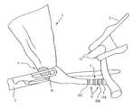
- FIG. 1shows a schematic view of a virtual organ, two instruments and several graphical identification markers
- FIG. 2shows a screen display showing different steps of the training session available in connection with the simulated treatment of the organ according to FIG. 1 ,
- FIG. 3shows a screen display showing different sub-steps of a step according to FIG. 2 .
- FIG. 4shows a schematic view of another virtual organ with further graphical identification markers.
- FIG. 1shows a schematic view of an organ 1 together with two instruments 2 and 3 as it is generated on a screen visible to the user in the training session.
- Said representations of organ 1 and instruments 2 and 3are virtual geometrical elements within a computer controlled virtual environment.
- the instruments 2 and 3are connected through a control arrangement to physical devices, namely handles of (fake) surgical instruments, which are manipulated by the user in said training session.
- the background of the image according to FIG. 1shows a virtual representation of the abdominal cavity (not shown).
- FIG. 2shows a screen display of different steps of the training session available in connection with the simulated treatment of the organ 1 according to FIG. 1 .
- this surgical interactionmay comprise:
- the trainingis separated into different training steps 51 to 53 .
- the different steps 51 to 53can be chosen independently from a menu as shown in FIG. 2 .
- the menucomprises a step 50 : information about the virtual patient, step 55 : tutored intervention of all steps 51 to 53 within one step, step 56 : complete “free” intervention of all steps 51 to 53 within one step, and step 57 : assessment of the training session.
- step 52“clipping of the organ” is the header of the menu wherein each step comprises preferably the following identical sub-steps, which can be chosen independently from the menu:
- the main advantage of this procedureis based on the insight that the user can be directed within each step and repeat the steps in which he needs the most training. This preparation together with the guidance through the below mentioned graphical codes gives a better training result. Additionally the result of the simulated intervention is stored and the evaluation is possible on parts of the intervention or the whole process.
- the graphical representationcomprises a direct correlation with the action of the instrument handled by the user. This can be shown within the embodiment of the intervention as follows.
- step 52 “clipping”the user has to move the instrument 2 towards the organ 1 and grip it in an area 4 (same procedure as in the step 51 “gripping”). Then he has to hold the organ 1 in a certain position and to apply three clips in the two areas 5 (in order to enable the cutting of the vessel in step 53 “cutting” between the clips in area 7 ).
- the area within which the instrument 2 has to be appliedis marked through a blue dot 14 (the area shaded in the drawing).
- the dotchanges its color, e.g. into yellow until the user has pulled the organ 1 into the corrected position, upon which occasion the dot changes its color a further time, e.g. into green. Since the user has to hold instrument 2 throughout the step in an extended period of time, he may deviate from the correct position, and the controller changes the color from green to red, when the position is not correct anymore. A less correct position may be marked yellow.
- Such graphical feedbackenables the user to find the correct position and the dot 14 becomes green again.
- the step 52 “clipping”asks the user to put three clips into predefined positions, which are marked 15 A, 15 B and 15 C.
- the necessity to apply the same number of clips to the second vessel in the backgroundis not treated within this simplified approach of the description.
- the ring 15 Ais blue.
- the uservirtually loads a clip 13 into instrument 3 and approaches the blue ring 15 A.
- the blue ring 15 Abecomes green.
- the ringmay become yellow or red.
- the color redis associated with a hazardous situation and yellow a situation which is not correct according to the teached procedure but not “dangerous” for the virtual patient.
- next ringhere 15 B or 15 C becomes blue and the process is repeated until all clips are set. Then all areas 14 , 15 A to 15 C which are shaded in the drawings and which should be green vanish as indication that the step is completed.
- step 53cutting to exchange instrument 3 towards scissors and to cut the vessel in area 7 , which is afterwards indicated through a (separated) green ring 7 .
- FIG. 4Although the intervention of opening surface tissues in an area is not part of the menus according to FIG. 2 or FIG. 3 , these further graphical indications are shown in connection with a further schematic drawing, FIG. 4 .
- FIG. 4shows a schematic view of another virtual organ 1 with further graphical identification markers 18 A, 18 B and 19 .
- Organ 1has a cylindrical form.
- a blue line 18 Ais appearing on the organ.
- This line 18 Ahas preferably a starting point and an end point which can be seen through enlarged dots.
- the relevant part of the line 18 Aturns green. If the user burns the area or enters the instrument too deep into the organ, then beside the effect of the virtual representation showing the results of this treatment (smoke, burned points, bleeding) this part of the line turns red.
- the line and the dotsturns into green and the next segment 18 B is lit up in blue colour. This advises the user where to continue his task until the related step is completed.
- Area 19shows a region in which the user has to open the surface within a larger area.
- the area to be openedis blue and all cut off pieces will loose there blue color and turn green.
- the whole areawill have green borders and then the marker areas will vanish.
- the concept of guidanceis to project a (two-dimensional) surface area information in the area which has to be treated through the user in training.
- Thismay be a larger spot, as spot 14 for a gripping action.
- Thismay be a ring, as ring 18 A for the clipping action.
- Thismay be a line or a segment of a line as lines 18 A and 18 B for marking or cutting purposes.
- thiscan be of any geometrical form as polygon 19 which marks the area which has to be treated.
- the correlation between the instrument and the graphical representationis immediate and gives a direct visual assessment in the training.
- FIGS. 1 and 4can be seen as typical for endoscopic interventions but can also show representations of organs under X-ray visualization.
- the simulationcan further demonstrate the use of contrast generating liquids etc. If a surgeon applies the method and uses equipment (e.g. polarizing glasses) to obtain different (3D) images for his two eyes, than he sees a virtual 3D image of the region under illumination, e.g. a 3D-image of an organ or a part of an organ.
- equipmente.g. polarizing glasses
- the methodtherefore also comprises the generation of a differently coloured volume part, e.g. a organ area part.
- a differently coloured volume parte.g. a organ area part.
- a different colour as yellow or red appearsif the instrument used grasps or penetrates another region of said organ area part, and wherein said area part becomes e.g. green, if the step is successfully terminated.
- every organ area partbeing a three-dimensional body, comprises a surrounding surface, which can receive, during the intervention, the same and/or a different colour than the organ area part concerned.
- a surface colorationwherein one surface lies behind an organ part in question. Said differences in depth can be shown through different colour shades or contrast/luminosity of the parts concerned.
Landscapes
- Engineering & Computer Science (AREA)
- General Physics & Mathematics (AREA)
- Health & Medical Sciences (AREA)
- Physics & Mathematics (AREA)
- Computational Mathematics (AREA)
- Mathematical Optimization (AREA)
- Medical Informatics (AREA)
- Medicinal Chemistry (AREA)
- Chemical & Material Sciences (AREA)
- Algebra (AREA)
- Radiology & Medical Imaging (AREA)
- Pulmonology (AREA)
- Mathematical Analysis (AREA)
- General Health & Medical Sciences (AREA)
- Mathematical Physics (AREA)
- Pure & Applied Mathematics (AREA)
- Business, Economics & Management (AREA)
- Educational Administration (AREA)
- Educational Technology (AREA)
- Theoretical Computer Science (AREA)
- Instructional Devices (AREA)
- Processing Or Creating Images (AREA)
Abstract
Description
- step51: gripping of the organ in the
area 4 withinstrument 2 and - step52: setting clips in the
area 5 with help of theinstrument 3, and. - step53: cutting a vessel in an
area 7.
- step51: gripping of the organ in the
- sub-step61: a prerecorded video representation of said
step - sub-step62: a prerecorded virtual reality video representation of the intervention to be performed within said
step - sub-step63: instructions for the intervention of said
step - sub-step64: a prerecorded virtual reality graphical (stills) or video representation of common errors within said
step - sub-step65: training session of the step in question with guidance,
- sub-step66: training session of the step in question without guidance.
- sub-step61: a prerecorded video representation of said
Claims (11)
Priority Applications (1)
| Application Number | Priority Date | Filing Date | Title |
|---|---|---|---|
| US11/101,154US8591236B2 (en) | 2002-10-07 | 2005-04-07 | Interactive medical training system and method |
Applications Claiming Priority (2)
| Application Number | Priority Date | Filing Date | Title |
|---|---|---|---|
| PCT/CH2002/000556WO2004032095A1 (en) | 2002-10-07 | 2002-10-07 | Interactive medical training system and method |
| US11/101,154US8591236B2 (en) | 2002-10-07 | 2005-04-07 | Interactive medical training system and method |
Related Parent Applications (1)
| Application Number | Title | Priority Date | Filing Date |
|---|---|---|---|
| PCT/CH2002/000556Continuation-In-PartWO2004032095A1 (en) | 2002-10-07 | 2002-10-07 | Interactive medical training system and method |
Publications (2)
| Publication Number | Publication Date |
|---|---|
| US20050221263A1 US20050221263A1 (en) | 2005-10-06 |
| US8591236B2true US8591236B2 (en) | 2013-11-26 |
Family
ID=35054767
Family Applications (1)
| Application Number | Title | Priority Date | Filing Date |
|---|---|---|---|
| US11/101,154Active2028-07-22US8591236B2 (en) | 2002-10-07 | 2005-04-07 | Interactive medical training system and method |
Country Status (1)
| Country | Link |
|---|---|
| US (1) | US8591236B2 (en) |
Cited By (4)
| Publication number | Priority date | Publication date | Assignee | Title |
|---|---|---|---|---|
| US9847044B1 (en) | 2011-01-03 | 2017-12-19 | Smith & Nephew Orthopaedics Ag | Surgical implement training process |
| CN110136559A (en)* | 2019-05-16 | 2019-08-16 | 解涛 | A simulated surgery device based on VR technology for simulating bleeding points with liquid |
| US10810907B2 (en) | 2016-12-19 | 2020-10-20 | National Board Of Medical Examiners | Medical training and performance assessment instruments, methods, and systems |
| KR20240043630A (en) | 2022-09-27 | 2024-04-03 | 연세대학교 산학협력단 | Medical simulation apparatus for training and evaluation of healthcare workers and method for controlling the same |
Families Citing this family (10)
| Publication number | Priority date | Publication date | Assignee | Title |
|---|---|---|---|---|
| CA2697781A1 (en)* | 2005-08-29 | 2007-03-08 | Go Virtual Medical Limited | Medical instruction system |
| WO2007121572A1 (en)* | 2006-04-21 | 2007-11-01 | Mcmaster University | Haptic enabled robotic training system and method |
| WO2008058039A1 (en)* | 2006-11-06 | 2008-05-15 | University Of Florida Research Foundation, Inc. | Devices and methods for utilizing mechanical surgical devices in a virtual environment |
| US20090061402A1 (en)* | 2007-08-29 | 2009-03-05 | Kiran Musunuru | Methods And Systems For Providing Interactive Educational Training |
| WO2009094621A2 (en) | 2008-01-25 | 2009-07-30 | University Of Florida Research Foundation, Inc. | Devices and methods for implementing endoscopic surgical procedures and instruments within a virtual environment |
| GB2511668A (en)* | 2012-04-12 | 2014-09-10 | Supercell Oy | System and method for controlling technical processes |
| EP2948936A1 (en)* | 2013-01-25 | 2015-12-02 | Rath, Matthias | Integrated multimedia tool, system and method to explore and study the virtual human body |
| CA3087094A1 (en) | 2017-12-28 | 2019-07-04 | Orbsurgical Ltd. | Microsurgery-specific haptic hand controller |
| CN114842708B (en)* | 2022-05-25 | 2024-06-14 | 北京大学口腔医学院 | Method for selecting training instrument and posture of color tooth Zhou Gua |
| WO2024205445A1 (en)* | 2023-03-31 | 2024-10-03 | Общество С Ограниченной Ответственностью "Мой Учитель" | Method for ai-based situational analysis in education |
Citations (10)
| Publication number | Priority date | Publication date | Assignee | Title |
|---|---|---|---|---|
| US4839822A (en)* | 1987-08-13 | 1989-06-13 | 501 Synthes (U.S.A.) | Computer system and method for suggesting treatments for physical trauma |
| US5546943A (en)* | 1994-12-09 | 1996-08-20 | Gould; Duncan K. | Stimulating a beneficial human response by using visualization of medical scan data to achieve psychoneuroimmunological virtual reality |
| US5740802A (en)* | 1993-04-20 | 1998-04-21 | General Electric Company | Computer graphic and live video system for enhancing visualization of body structures during surgery |
| US5766016A (en)* | 1994-11-14 | 1998-06-16 | Georgia Tech Research Corporation | Surgical simulator and method for simulating surgical procedure |
| US6132218A (en)* | 1998-11-13 | 2000-10-17 | Benja-Athon; Anuthep | Images for communication of medical information in computer |
| US6421560B1 (en)* | 1999-07-29 | 2002-07-16 | Yoo Tae Woo | Device for guiding spots for acupuncture and stimulation methods thereof |
| US6690397B1 (en)* | 2000-06-05 | 2004-02-10 | Advanced Neuromodulation Systems, Inc. | System for regional data association and presentation and method for the same |
| US6747672B1 (en)* | 1999-11-01 | 2004-06-08 | Medical Learning Company, Inc. | Virtual patient hot spots |
| US20040136578A1 (en)* | 2002-10-31 | 2004-07-15 | Sieracki Jeffrey M. | Body region indication |
| US7202851B2 (en)* | 2001-05-04 | 2007-04-10 | Immersion Medical Inc. | Haptic interface for palpation simulation |
- 2005
- 2005-04-07USUS11/101,154patent/US8591236B2/enactiveActive
Patent Citations (10)
| Publication number | Priority date | Publication date | Assignee | Title |
|---|---|---|---|---|
| US4839822A (en)* | 1987-08-13 | 1989-06-13 | 501 Synthes (U.S.A.) | Computer system and method for suggesting treatments for physical trauma |
| US5740802A (en)* | 1993-04-20 | 1998-04-21 | General Electric Company | Computer graphic and live video system for enhancing visualization of body structures during surgery |
| US5766016A (en)* | 1994-11-14 | 1998-06-16 | Georgia Tech Research Corporation | Surgical simulator and method for simulating surgical procedure |
| US5546943A (en)* | 1994-12-09 | 1996-08-20 | Gould; Duncan K. | Stimulating a beneficial human response by using visualization of medical scan data to achieve psychoneuroimmunological virtual reality |
| US6132218A (en)* | 1998-11-13 | 2000-10-17 | Benja-Athon; Anuthep | Images for communication of medical information in computer |
| US6421560B1 (en)* | 1999-07-29 | 2002-07-16 | Yoo Tae Woo | Device for guiding spots for acupuncture and stimulation methods thereof |
| US6747672B1 (en)* | 1999-11-01 | 2004-06-08 | Medical Learning Company, Inc. | Virtual patient hot spots |
| US6690397B1 (en)* | 2000-06-05 | 2004-02-10 | Advanced Neuromodulation Systems, Inc. | System for regional data association and presentation and method for the same |
| US7202851B2 (en)* | 2001-05-04 | 2007-04-10 | Immersion Medical Inc. | Haptic interface for palpation simulation |
| US20040136578A1 (en)* | 2002-10-31 | 2004-07-15 | Sieracki Jeffrey M. | Body region indication |
Cited By (6)
| Publication number | Priority date | Publication date | Assignee | Title |
|---|---|---|---|---|
| US9847044B1 (en) | 2011-01-03 | 2017-12-19 | Smith & Nephew Orthopaedics Ag | Surgical implement training process |
| US10810907B2 (en) | 2016-12-19 | 2020-10-20 | National Board Of Medical Examiners | Medical training and performance assessment instruments, methods, and systems |
| CN110136559A (en)* | 2019-05-16 | 2019-08-16 | 解涛 | A simulated surgery device based on VR technology for simulating bleeding points with liquid |
| CN110136559B (en)* | 2019-05-16 | 2020-12-01 | 解涛 | A simulated surgical device based on VR technology to simulate bleeding points with liquid |
| KR20240043630A (en) | 2022-09-27 | 2024-04-03 | 연세대학교 산학협력단 | Medical simulation apparatus for training and evaluation of healthcare workers and method for controlling the same |
| KR20250069516A (en) | 2022-09-27 | 2025-05-19 | 연세대학교 산학협력단 | Device for performing medical simulation for situational awareness training and method for controlling the same |
Also Published As
| Publication number | Publication date |
|---|---|
| US20050221263A1 (en) | 2005-10-06 |
Similar Documents
| Publication | Publication Date | Title |
|---|---|---|
| WO2004032095A1 (en) | Interactive medical training system and method | |
| US8591236B2 (en) | Interactive medical training system and method | |
| Hubens et al. | A performance study comparing manual and robotically assisted laparoscopic surgery using the da Vinci system | |
| US8834170B2 (en) | Devices and methods for utilizing mechanical surgical devices in a virtual environment | |
| Gallagher et al. | Objective psychomotor skills assessment of experienced, junior, and novice laparoscopists with virtual reality | |
| Abdi et al. | Control of a supernumerary robotic hand by foot: An experimental study in virtual reality | |
| US20110306986A1 (en) | Surgical robot system using augmented reality, and method for controlling same | |
| US10347155B2 (en) | Suturing training device and method | |
| CN110381873A (en) | Robotic surgical system, instrument and control | |
| Mori et al. | Significance of``hands-on training''in laparoscopic surgery | |
| KR20200048830A (en) | Cataract surgery Simulation System for education based on virtual reality | |
| Kunert et al. | Learning curves, potential and speed in training of laparoscopic skills: a randomised comparative study in a box trainer | |
| Gopher | Skill training in multimodal virtual environments | |
| Walliczek‐Dworschak et al. | Structured training on the da Vinci Skills Simulator leads to improvement in technical performance of robotic novices | |
| JP2021503102A (en) | Suture technique surgical training model | |
| Knafo et al. | Cognitive versus virtual reality simulation for evaluation of technical skills in neurosurgery | |
| EP1547053A1 (en) | Device and method for generating a virtual anatomic environment | |
| Jones et al. | Next-generation 3D videosystems may improve laparoscopic task performance | |
| Cassilly et al. | Optimizing motion scaling and magnification in robotic surgery | |
| CN114288519A (en) | Training display method | |
| Öbek et al. | Robotic versus conventional laparoscopic skill acquisition: implications for training | |
| de Almeida et al. | Impact of 3D laparoscopic surgical training on performance in standard 2D laparoscopic simulation: a randomised prospective study | |
| Haase et al. | Virtual reality and habitats for learning microsurgical skills | |
| Skinner et al. | Ambidexterity in laparoscopic surgical skills training | |
| Feng et al. | Surgical training and performance assessment using a motion tracking system |
Legal Events
| Date | Code | Title | Description |
|---|---|---|---|
| AS | Assignment | Owner name:XITACT S.A., SWITZERLAND Free format text:ASSIGNMENT OF ASSIGNORS INTEREST;ASSIGNORS:VECERINA, IVAN;ZOETHOUT, JURJEN;LAUNAY, MURIELLE;SIGNING DATES FROM 20050322 TO 20050404;REEL/FRAME:016089/0270 Owner name:XITACT S.A., SWITZERLAND Free format text:ASSIGNMENT OF ASSIGNORS INTEREST;ASSIGNORS:VECERINA, IVAN;ZOETHOUT, JURJEN;LAUNAY, MURIELLE;REEL/FRAME:016089/0270;SIGNING DATES FROM 20050322 TO 20050404 | |
| FEPP | Fee payment procedure | Free format text:PAYOR NUMBER ASSIGNED (ORIGINAL EVENT CODE: ASPN); ENTITY STATUS OF PATENT OWNER: SMALL ENTITY Free format text:PAYER NUMBER DE-ASSIGNED (ORIGINAL EVENT CODE: RMPN); ENTITY STATUS OF PATENT OWNER: SMALL ENTITY | |
| STCF | Information on status: patent grant | Free format text:PATENTED CASE | |
| AS | Assignment | Owner name:MENTICE SA, SWITZERLAND Free format text:CHANGE OF NAME;ASSIGNOR:XITACT S.A.;REEL/FRAME:034655/0117 Effective date:20140923 | |
| FPAY | Fee payment | Year of fee payment:4 | |
| AS | Assignment | Owner name:MENTICE AB, SWEDEN Free format text:ASSIGNMENT OF ASSIGNORS INTEREST;ASSIGNOR:MENTICE SA;REEL/FRAME:050397/0222 Effective date:20190625 | |
| MAFP | Maintenance fee payment | Free format text:PAYMENT OF MAINTENANCE FEE, 8TH YR, SMALL ENTITY (ORIGINAL EVENT CODE: M2552); ENTITY STATUS OF PATENT OWNER: SMALL ENTITY Year of fee payment:8 | |
| MAFP | Maintenance fee payment | Free format text:PAYMENT OF MAINTENANCE FEE, 12TH YR, SMALL ENTITY (ORIGINAL EVENT CODE: M2553); ENTITY STATUS OF PATENT OWNER: SMALL ENTITY Year of fee payment:12 |