US8565374B2 - Integrated multi-mode mammography/tomosynthesis x-ray system and method - Google Patents
Integrated multi-mode mammography/tomosynthesis x-ray system and methodDownload PDFInfo
- Publication number
- US8565374B2 US8565374B2US13/462,342US201213462342AUS8565374B2US 8565374 B2US8565374 B2US 8565374B2US 201213462342 AUS201213462342 AUS 201213462342AUS 8565374 B2US8565374 B2US 8565374B2
- Authority
- US
- United States
- Prior art keywords
- breast
- ray
- mode
- imaging
- images
- Prior art date
- Legal status (The legal status is an assumption and is not a legal conclusion. Google has not performed a legal analysis and makes no representation as to the accuracy of the status listed.)
- Active
Links
Images


Classifications
- A—HUMAN NECESSITIES
- A61—MEDICAL OR VETERINARY SCIENCE; HYGIENE
- A61B—DIAGNOSIS; SURGERY; IDENTIFICATION
- A61B6/00—Apparatus or devices for radiation diagnosis; Apparatus or devices for radiation diagnosis combined with radiation therapy equipment
- A61B6/10—Safety means specially adapted therefor
- A61B6/107—Protection against radiation, e.g. shielding
- A—HUMAN NECESSITIES
- A61—MEDICAL OR VETERINARY SCIENCE; HYGIENE
- A61B—DIAGNOSIS; SURGERY; IDENTIFICATION
- A61B6/00—Apparatus or devices for radiation diagnosis; Apparatus or devices for radiation diagnosis combined with radiation therapy equipment
- A61B6/02—Arrangements for diagnosis sequentially in different planes; Stereoscopic radiation diagnosis
- A61B6/025—Tomosynthesis
- A—HUMAN NECESSITIES
- A61—MEDICAL OR VETERINARY SCIENCE; HYGIENE
- A61B—DIAGNOSIS; SURGERY; IDENTIFICATION
- A61B6/00—Apparatus or devices for radiation diagnosis; Apparatus or devices for radiation diagnosis combined with radiation therapy equipment
- A61B6/44—Constructional features of apparatus for radiation diagnosis
- A61B6/4417—Constructional features of apparatus for radiation diagnosis related to combined acquisition of different diagnostic modalities
- A—HUMAN NECESSITIES
- A61—MEDICAL OR VETERINARY SCIENCE; HYGIENE
- A61B—DIAGNOSIS; SURGERY; IDENTIFICATION
- A61B6/00—Apparatus or devices for radiation diagnosis; Apparatus or devices for radiation diagnosis combined with radiation therapy equipment
- A61B6/44—Constructional features of apparatus for radiation diagnosis
- A61B6/4429—Constructional features of apparatus for radiation diagnosis related to the mounting of source units and detector units
- A61B6/4452—Constructional features of apparatus for radiation diagnosis related to the mounting of source units and detector units the source unit and the detector unit being able to move relative to each other
- A—HUMAN NECESSITIES
- A61—MEDICAL OR VETERINARY SCIENCE; HYGIENE
- A61B—DIAGNOSIS; SURGERY; IDENTIFICATION
- A61B6/00—Apparatus or devices for radiation diagnosis; Apparatus or devices for radiation diagnosis combined with radiation therapy equipment
- A61B6/44—Constructional features of apparatus for radiation diagnosis
- A61B6/4476—Constructional features of apparatus for radiation diagnosis related to motor-assisted motion of the source unit
- A—HUMAN NECESSITIES
- A61—MEDICAL OR VETERINARY SCIENCE; HYGIENE
- A61B—DIAGNOSIS; SURGERY; IDENTIFICATION
- A61B6/00—Apparatus or devices for radiation diagnosis; Apparatus or devices for radiation diagnosis combined with radiation therapy equipment
- A61B6/50—Apparatus or devices for radiation diagnosis; Apparatus or devices for radiation diagnosis combined with radiation therapy equipment specially adapted for specific body parts; specially adapted for specific clinical applications
- A61B6/502—Apparatus or devices for radiation diagnosis; Apparatus or devices for radiation diagnosis combined with radiation therapy equipment specially adapted for specific body parts; specially adapted for specific clinical applications for diagnosis of breast, i.e. mammography
- A—HUMAN NECESSITIES
- A61—MEDICAL OR VETERINARY SCIENCE; HYGIENE
- A61B—DIAGNOSIS; SURGERY; IDENTIFICATION
- A61B6/00—Apparatus or devices for radiation diagnosis; Apparatus or devices for radiation diagnosis combined with radiation therapy equipment
- A61B6/42—Arrangements for detecting radiation specially adapted for radiation diagnosis
- A61B6/4291—Arrangements for detecting radiation specially adapted for radiation diagnosis the detector being combined with a grid or grating
- A—HUMAN NECESSITIES
- A61—MEDICAL OR VETERINARY SCIENCE; HYGIENE
- A61B—DIAGNOSIS; SURGERY; IDENTIFICATION
- A61B6/00—Apparatus or devices for radiation diagnosis; Apparatus or devices for radiation diagnosis combined with radiation therapy equipment
- A61B6/48—Diagnostic techniques
- A61B6/482—Diagnostic techniques involving multiple energy imaging
- G—PHYSICS
- G06—COMPUTING OR CALCULATING; COUNTING
- G06T—IMAGE DATA PROCESSING OR GENERATION, IN GENERAL
- G06T2207/00—Indexing scheme for image analysis or image enhancement
- G06T2207/30—Subject of image; Context of image processing
- G06T2207/30004—Biomedical image processing
- G06T2207/30068—Mammography; Breast
Definitions
- This patent specificationpertains to x-ray mammography and, more specifically, to an integrated system for selectively carrying out x-ray mammography and/or tomosynthesis imaging and a method of using such a system.
- X-ray mammographyhas long been a screening modality for breast cancer and other lesions, and also has been relied on for diagnostic and other purposes.
- the breast imagewas recorded on x-ray film but more recently digital x-ray image receptors have come into use, as in the SeleniaTM mammography system available from Hologic Inc. of Bedford, Mass. and its division Lorad Corporation of Danbury, Conn.
- a cone-shaped or pyramid-shaped x-ray beampasses through the compressed breast and forms a two-dimensional projection image. Any one of a number of orientations can be used, such as cranial-caudal (CC) or MLO (mediolateral-oblique) orientation. More recently, breast x-ray tomosynthesis has been proposed.
- CCcranial-caudal
- MLOmediumolateral-oblique
- the technologytypically involves taking two-dimensional (2D) projection images of the immobilized breast at each of a number of angles of the x-ray beam relative to the breast and processing the resulting x-ray measurements to reconstruct images of breast slices that typically are in planes transverse to the x-ray beam axis, such as parallel to the image plane of a mammogram of the same breast.
- the range of anglesis substantially less than in computerized tomography, i.e. substantially less than 180°, e.g. ⁇ 15°.
- Tomosynthesis technologyis described in U.S. patent application Ser. No. 10/723,486 filed Nov. 26, 2003; a prototype of a unit with at least some of the described features was shown at the 2003 Radiological Society of North America meeting in Chicago, Ill.
- Mammography systemscan also be used in interventional procedures, such as biopsy, by adding a biopsy station (for example, the StereoLoc IITM Upright Stereotactic Breast Biopsy System, which is available from Hologic, Inc.).
- a biopsy stationfor example, the StereoLoc IITM Upright Stereotactic Breast Biopsy System, which is available from Hologic, Inc.
- the patents, applications, brochures, and article cited aboveare hereby incorporated by reference in this patent specification as though fully set forth herein.
- Mammogramsmay offer good visualization of microcalcifications, and can offer higher spatial resolution compared with tomosynthesis.
- Tomosynthesis imagesmay have different desirable characteristics—e.g., they may offer better visualization of structures that can be obscured by overlying or underlying tissue in a conventional mammogram.
- a single systemcarries out breast imaging in modes that include standard mammography, diagnostic mammography, dynamic imaging such as with a contrast agent and at different x-ray energies, tomosynthesis imaging, combined standard and tomosynthesis imaging during a single breast compression, needle localization, and stereotactic imaging with a biopsy station mounted to the system.
- a compression arm assembly for compressing and immobilizing the breast for x-ray imaging, an x-ray tube assembly, and an x-ray image receptorcan be angled relative to each other for different imaging protocols and modes. They can be independently rotated and synchronized as needed, or can be mechanically linked for appropriate synchronized rotation.
- a patient shieldcan be mounted to the compression arm assembly to provide a mechanical interlock against patient contact with the rotating x-ray tube assembly.
- a fully retractable anti-scatter gridcan be used that can cover the imaging area of the x-ray receptor in some modes but be retracted completely outside the imaging area for other modes.
- the exemplary systemfurther includes a breast compression paddle that is laterally movable, under manual control or when motorized and operating under software control.
- the compression paddlecan shift automatically depending on the view to be acquired.
- the paddlecan be centered on the x-ray receptor for a CC view, shifted to one lateral side of the receptor for an MLO view of one breast and to the other lateral side of the receptor for an MLO view of the other breast.
- the paddlecan be automatically recognized by the system when mounted so that the shifts can be adjusted to the type of paddle.
- the compression paddlecan be easily removable from a support that has a mechanism for laterally moving the paddle and for allowing the paddle to tilt for better conformance with the breast for selected image modes but locking the paddle against tilt for other modes.
- the paddleWith the movement mechanism in the support and not integral with the paddle, the paddle can be simple and inexpensive, and easy to mount to and remove from the support.
- a number of relatively inexpensive paddles of different sizes and shapescan be provided and conveniently interchanged to suit different procedures and patients.
- FIG. 1is a perspective view of a gantry and an acquisition workstation in accordance with an example of the disclosed system.
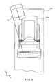
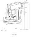
- FIG. 2is an enlarged view of a portion of the system of FIG. 1 , with a tube arm assembly in a rotated position.
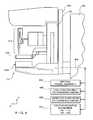
- FIG. 3is a front elevation of the apparatus of FIG. 2 .
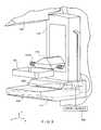
- FIG. 4is a side view of a gantry with a biopsy station and a spacer, with schematic illustration of other mechanisms.
- FIG. 5is an enlarged view of a portion of FIG. 1 .
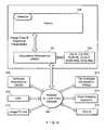
- FIG. 6is a block diagram of the disclosed system when connected to other systems.
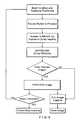
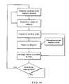
- FIG. 7is a flow chart illustrating a general work flow for the disclosed system.
- FIG. 8is a flow chart illustrating one of several examples of work flow for a standard mammography mode.
- FIG. 9is a flow chart illustrating one of several examples of work flow for an image detector subsystem in the standard mammography mode.
- FIG. 10is a perspective view of the structure of FIG. 4 .
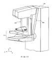
- FIG. 11is similar to FIG. 2 but shows a tube arm assembly angled differently.
- FIG. 12is a front elevation of the structure of FIG. 11 .
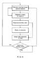
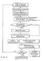
- FIG. 13is a flow chart illustrating one of several examples of work flow for a tomosynthesis mode.
- FIG. 14is a flow chart illustrating one of several examples of work flow for an image detector subsystem in the tomosynthesis mode.
- FIG. 15is a flow chart illustrating one of several examples of work flow for a combination mode.
- FIG. 16is a flow chart illustrating one of several examples of work flow for an image detector subsystem in the combination mode.
- FIG. 17is an enlarged side view of a structure for removably mounting a breast compression paddle.
- FIGS. 1-6illustrate a non-limiting example of a multi-mode mammography/tomosynthesis system comprising a gantry 100 and a data acquisition work-station 102 .
- Gantry 100includes a housing 104 supporting a tube arm assembly 106 rotatably mounted thereon to pivot about a horizontal axis 402 ( FIG. 4 ) and carrying an x-ray tube assembly 108 .
- X-ray tube assembly 108includes (1) an x-ray tube generating x-ray energy in a selected range, such as 20-50 kV, at mAs such as in the range 3-400 mAs, with focal spots such as a nominal size 0.3 mm large spot and nominal size 0.1 mm small spot (2) supports for multiple filters such as molybdenum, rhodium, aluminum, copper, and tin filters, and (3) an adjustable collimation assembly selectively collimating the x-ray beam from the focal spot in a range such as from 7 ⁇ 8 cm to 24 ⁇ 29 when measured at the image plane of an x-ray image receptor included in the system, at a maximum source-image distance such as 75 cm.
- a selected rangesuch as 20-50 kV
- focal spotssuch as a nominal size 0.3 mm large spot and nominal size 0.1 mm small spot
- supports for multiple filterssuch as molybdenum, rhodium, aluminum, copper, and tin filters
- an adjustable collimation assemblyselectively
- housing 104Also mounted on housing 104 , for rotation about the same axis 402 , is a compression arm assembly 110 that comprises a compression plate 122 and a receptor housing 114 having an upper surface 116 serving as a breast plate and enclosing a detector subsystem system 117 comprising a flat panel x-ray receptor 502 ( FIG. 5 ), a retractable anti-scatter grid 504 and a mechanism 506 for driving and retracting anti-scatter grid 504 .
- Housing 104also encloses the following components schematically illustrated in FIG.
- housing 104also encloses suitable motors and electrical and mechanical components and connections to implement the functions discussed here.
- a patient shield 200schematically illustrated in FIG.
- Work-station 102comprises components similar to those in the SeleniaTM mammography system, including a display screen (typically a flat panel display that may include touch-screen functionality), user interface devices such as a keyboard, possibly a touch-screen, and a mouse or trackball, and various switches and indicator lights and/or displays. Work-station 102 also includes computer facilities similar to those of the SeleniaTM system (but adapted through hardware, firmware and software differences) for controlling gantry 100 and for processing, storing and displaying data received from gantry 100 .
- a power generation facility for x-ray tube assembly 108may be included in housing 104 or in work-station 102 .
- a power source 118powers work-station 102 .
- Gantry 100 and work-station 102exchange data and controls over a schematically illustrated connection 120 .
- additional storage facilities 602can be connected to work-station 102 , such as one or more optical disc drives for storing information such as images and/or for providing information to work-station 102 such as previously obtained images and software, or a local printer (not shown).
- the disclosed systemcan be connected to a hospital or local area or other network 604 , and through the network to other systems such as a soft copy workstation 606 , a CAD (Computer Aided Detection) station 608 for computer-processing mammography and/or tomosynthesis images to identify likely abnormalities, an image printer 610 for printing images, a technologist workstation 612 , other imaging systems 614 such as other mammography systems or systems for other modalities for exchange of images and/or other information, and to a PACS (Picture Archiving) systems 616 for archiving images and other information and/or retrieving images and other information.
- CADComputer Aided Detection
- the illustrated systemhas several modes of operation.
- An example of typical workflow generally applicable for each modeis illustrated in FIG. 7 , and several examples of operational modes are discussed below. Of course, this is only one example and workflow steps may be arranged differently.
- the operatorcan perform x-ray exposure using manual setting of technic factors such as mA and mSec, or can use an automatic exposure control as known in the art to set the exposure time, kV and filter modes for an image, for example by using a short, low-x-ray dose pre-exposure.
- Work-station 102is set up to record the exposure technic information and associate it with the breast image for later review.
- tube arm assembly 106 and compression arm assembly 110are coupled and locked together by 410 in a relative position such as seen in FIG. 1 , such that an x-ray beam from x-ray tube assembly 108 illuminates x-ray receptor 502 when the patient's breast is compressed by compression device 112 .
- the systemoperates in a manner similar to said SeleniaTM system to take a mammogram.
- Vertical travel assembly 404 and tube arm rotation mechanism 406can make vertical adjustments to accommodate a patient, and can rotate tube arm assembly 106 and compression arm assembly 110 together as a unit about axis 402 for different image orientations such as for CC and for MLO images.
- tube arm assembly 106 and compression arm assembly 110can rotate between) ( ⁇ 195° and) (+150° about axis 402 .
- compression device 112includes a compression paddle 122 that can move laterally, in a direction along the chest wall of a patient, to adjust for different imaging orientations.
- the mechanism for supporting and moving compression paddle 122is different.
- anti-scatter grid 504is over x-ray receptor 502 in the standard mammography mode to reduce the effect of x-ray scatter.
- FIG. 8illustrates a typical workflow for an exposure in standard mammography mode
- FIG. 10illustrates an example of the operation of detector subsystem 117 in standard mammography. Of course, these are only examples; other workflow steps or orders of steps can be used instead.
- the patient's breastcan be spaced from upper surface 116 , for example by an x-ray translucent spacer gantry 1002 ( FIG. 10 ), with the system otherwise similar to FIG. 1 , for a magnification of up to 1.8, for example.
- tube arm assembly 106 and compression arm assembly 110are locked to each other and can move up or down and rotate about axis 402 for different image orientation.
- a different spacer 1002can be used for a different degree of magnification.
- differently shaped or dimensioned compression paddles 122can be used for different breast compression effects.
- the x-ray tube in x-ray tube assembly 108can be set to a smaller focal spot size to improve a diagnostic image.
- anti-scatter grid 504typically is retracted when magnification is used such that grid 504 is completely out of the image.
- the usercan elect not to use a spacer 1002 in diagnostic imaging, in which case anti-scatter grid 504 can be used over the entire image.
- a number of breast imagesare taken while the patient's breast remains compressed.
- an agentsuch as iodine is injected into the patient and after a suitable waiting time such as about one minute for a maximum uptake, two images breast are taken in rapid succession, for example one at an x-ray energy just above the K-edge of iodine and one at an energy just below the K-edge.
- a succession of breast imagescan be taken at a single x-ray energy band or bands just above and below the K-edge, or at another x-ray energy range, to track the uptake of agent over time.
- Still another dynamic imaging mode techniquecomprises injecting a contrast agent and taking a succession of images over a period such as 5-7 minutes, for example one image every minute, and processing the image data to generate for each pixel, or at least for each pixel of interest, a histogram of the change in the pixel value, to thereby use the manner in which pixel values change to differential abnormal tissue.
- work-station 102can store preset data that commands gantry 100 and work-station 102 to take a desired sequence of images for the dynamic mode technique selected by the operator, such that the command data sets the appropriate parameters such as x-ray energy, dose, timing of images, etc.
- processing to assess changes in pixel valuescan be done for a region of interest rather than over individual pixels, to produce information such as a measure of changes in the average pixel values in the region of interest.
- tube arm assembly 106 and compression arm assembly 110are decoupled by unit 410 such that compression arm assembly 110 stays in one position, compressing the patient's breast, while tube arm assembly 106 rotates about axis 402 , for example between the position illustrated in FIG. 2 to that illustrated in FIG. 11 , or ⁇ 15° relative to compression arm assembly 110 .
- Tomosynthesiscan be carried out for different image orientations, so that compression arm assembly 110 can be rotated about axis 402 (alone or together with assembly 106 ) for a desired image orientation and locked in place, and then tube arm assembly 106 can be rotated relative to that position of compression arm assembly 110 for tomosynthesis imaging over ⁇ 15° or some other desired angular range.
- 11 imagesare taken during an angular sweep of tube arm assembly 106 , one every approximately 3°.
- a different number of imagescan be taken, for example up to 21 during a single sweep.
- the x-ray tube in x-ray tube assembly 108continuously rotates and the x-ray tube is pulsed for each image, for example, for x-ray energy pulses each lasting approximately 100 mSec, although pulses of different duration can be selected.
- the rotational motioncan stop for taking each image, or continuous motion without pulsing can be used (and the timing of data measurements relied to define pixel values). As seen in FIGS.
- mechanism 506fully retracts anti-scatter grid 504 away from x-ray receptor 502 so grid 504 is out of the image.
- x-ray receptor 502rocks within receptor housing 114 . In this rocking motion, controlled by unit 408 ( FIG. 4 ), a line normal to the image face of x-ray receptor 502 may keep pointing to the focal spot of the x-ray tube in x-ray tube assembly 108 .
- the rotation of tube arm assembly 106 and rocking of x-ray receptor 502can be through different angles; for example, tube arm assembly 106 can rotate through 15° while x-ray receptor 502 rocks through 5°, i.e. the rocking angle can be an amount one-third that of assembly 108 .
- Synchronous rotation of tube arm assembly 106 and rocking of x-ray receptor 502can be achieved by controlling separate motors for each or, alternatively, through using a motor to drive tube arm assembly 106 and a mechanical coupling between the rotation of tube arm assembly 106 and rocking of x-ray receptor 502 .
- Image datacan be obtained and processed into tomosynthesis images for display and/or storage as described in the material incorporated by reference, for example in co-pending patent application Ser.
- FIG. 13illustrates a typical workflow for tomosynthesis mode operation
- FIG. 14illustrates an example of the operation of detector subsystem 117 in that mode. Again, these are only examples, and other steps or orders of steps can be used instead.
- a combination modeduring a single compression of the patient's breast the system takes a conventional mammogram and tomosynthesis images.
- tube arm assembly 106sweeps and x-ray receptor 502 rocks, each through an appropriate angle, and exposures are taken for tomosynthesis images, and (2) a standard mammogram is taken.
- the standard mammogramcan be taken at a 0° relative angle between tube arm assembly 106 and a normal to the imaging plane of x-ray receptor 502 , and can be taken before or after the tomosynthesis images are taken or between the taking of two successive tomosynthesis images.
- each tomosynthesis imageutilizes substantially lower x-ray dose than the standard mammogram.
- the total x-ray dosage for tomosynthesis imaging in one sweep of tube arm assembly 106can be approximately the same as that for a single standard mammogram, or up to approximately three times that dosage. The relationship between the two dosages can be user-selected.
- FIG. 15illustrates an example of workflow for the combination mode
- FIG. 16illustrates an example of the operation of detector subsystem 117 in that mode. Again, these are examples, and different steps or orders of steps can be used instead.
- a preferred approachmay be to take the standard mammogram first, then move arm 106 to one end of its rotational range for tomosynthesis and take the tomosynthesis images.
- the order in which the two types of images are takenmay be optimized such that the overall imaging time is minimized, and an order that achieves such minimization can be the preferred order.
- the exposure (tube current mA, tube voltage kVp, and exposure length msec) techniques for the standard mammogram and the tomosynthesis exposurescan be set manually, or by using automatic methods. If the standard mammogram is taken first, its exposure techniques can be used to set an optimal technique for the subsequent tomosynthesis images, and vice versa.
- the exposure techniquecan be modified dynamically, if the software senses that the signal reaching the image receptor is either too low or too high and adjust subsequent exposures as needed.
- X-ray receptor 502can remain in place for this procedure, or can be rocked through a selected angle, for example through an angle sufficient to maintain the same orientation of the imaging surface of receptor 502 relative to tube arm assembly 106 .
- a spacer 1002can be used for magnification.
- the two or more imagescan be used to identify the location of a lesion, so that needle biopsy can be used, for example with an upright needle biopsy station 412 ( FIG. 4 ) in a manner similar to that used with the commercially available SeleniaTM system and StereoLoc IITM.
- a compression paddle 122 appropriate for needle biopsytypically is used when taking the stereotactic images.
- some or all of the images taken in the tomosynthesis mode and/or in the combined modecan be used to identify the location of a lesion for biopsy, in which case a compression paddle 122 appropriate for the purpose typically is used when taking the images.
- x-ray imagescan be taken after a biopsy or other needle is inserted into the compressed breast.
- imagingsuch as in the stereotactic mode, the tomosynthesis mode, or the combined mode can be used.
- compression paddle 122is movable laterally, as generally described in U.S. Patent Application Publication No. 2005/0063509 A1, hereby incorporated by reference herein.
- compression paddle 122can pivot about an axis along the patient's chest wall to conform the breast shape in certain procedures, as discussed in said U.S. Pat. No. 5,706,327.
- compression paddle 122is mounted differently and moves in a different manner.
- compression paddle 122is removably mounted to a support 510 that moves up and down compression arm assembly 110 as needed for breast compression.
- a projection compression paddle 122 a of the paddleengages a projection 510 a of the support, and a projection 122 b of the paddle latches onto projection 510 b of the support.
- Projection 510 ais spring-loaded, such as by a spring schematically illustrates at 510 c to allow for pivoting compression paddle 122 about an axis where it latches onto 510 , as illustrated by arrow A, for better conformance with the compressed breast in some imaging protocols.
- Imaging protocolsmay require compression paddle 122 not to pivot, in which case projection 510 a is locked in place by a locking mechanism in 510 (not shown) to keep compression paddle 122 in place relative to support 510 .
- the locking mechanismcan be manually set to a lock position, and manually unlocked by the operator. Alternatively, the locking mechanism can be controlled through an operator input at gantry 100 or work-station 102 .
- a sensing mechanismcan be included to sense whether compression paddle 122 is locked against pivoting, to provide information that work-station 102 can use for setting imaging protocols such as for automated breast compression and automated exposure methods.
- Two knobs 510 dcan be manually rotated to move projection 510 b and thus compression paddle 122 laterally such that it compress a breast that is not centered laterally on upper surface 116 , for example for MLO imaging.
- Each knob 510 dcan operate a mechanism such as an endless screw rotating in a nut secured to projection 510 b .
- projection 510 b and thus compression paddle 122can be driven laterally by a motor, under control of operator switches or other interface at gantry 100 or at work-station 102 , or automatically positioned laterally under computer control.
- compression paddle 122is driven for lateral movement by components that are a part of support 510 .
- compression paddle 122can be simple structure, and can even be disposable, with a new one used for each patient or for only a few patients. This can simplify and reduce the cost of using the system, because an imaging facility usually stocks a number of different paddles for different purposes. If the lateral movement mechanism is integral with a compression paddle, the paddle assembly is considerably larger, heavier and more expensive.
- Compression paddle 122can include a bar code that is automatically read by a bar code reader in support 510 , to keep work-station 102 informed of the paddle currently mounted to support 510 , for use in automating imaging protocols.
- the bar code informationcan be checked to ensure through computer processing that the type of paddle that is currently mounted on support 510 matches the imaging that will be commanded, and the information from the sensor for whether compression paddle 122 is locked in non-tilting mode can be used to automatically make adjustments for compression height to ensure accurate automatic x-ray exposure operation. Further, the bar code information identifying the paddle can be used to automatically set collimation in x-ray tube assembly 108 so that the x-ray beam matches the size and shape of the currently installed compression paddle 122 .
Landscapes
- Health & Medical Sciences (AREA)
- Life Sciences & Earth Sciences (AREA)
- Medical Informatics (AREA)
- Engineering & Computer Science (AREA)
- Radiology & Medical Imaging (AREA)
- Molecular Biology (AREA)
- Biophysics (AREA)
- Nuclear Medicine, Radiotherapy & Molecular Imaging (AREA)
- Optics & Photonics (AREA)
- Pathology (AREA)
- Physics & Mathematics (AREA)
- Biomedical Technology (AREA)
- Heart & Thoracic Surgery (AREA)
- High Energy & Nuclear Physics (AREA)
- Surgery (AREA)
- Animal Behavior & Ethology (AREA)
- General Health & Medical Sciences (AREA)
- Public Health (AREA)
- Veterinary Medicine (AREA)
- Dentistry (AREA)
- Oral & Maxillofacial Surgery (AREA)
- Apparatus For Radiation Diagnosis (AREA)
Abstract
Description
Claims (24)
Priority Applications (6)
| Application Number | Priority Date | Filing Date | Title |
|---|---|---|---|
| US13/462,342US8565374B2 (en) | 2004-11-26 | 2012-05-02 | Integrated multi-mode mammography/tomosynthesis x-ray system and method |
| US14/058,385US9066706B2 (en) | 2004-11-26 | 2013-10-21 | Integrated multi-mode mammography/tomosynthesis x-ray system and method |
| US14/498,476US9549709B2 (en) | 2004-11-26 | 2014-09-26 | Integrated multi-mode mammography/tomosynthesis X-ray system and method |
| US15/411,502US10194875B2 (en) | 2004-11-26 | 2017-01-20 | Integrated multi-mode mammography/tomosynthesis X-ray system and method |
| US16/266,823US10905385B2 (en) | 2004-11-26 | 2019-02-04 | Integrated multi-mode mammography/tomosynthesis x-ray system and method |
| US17/137,032US11617548B2 (en) | 2004-11-26 | 2020-12-29 | Integrated multi-mode mammography/tomosynthesis x-ray system and method |
Applications Claiming Priority (5)
| Application Number | Priority Date | Filing Date | Title |
|---|---|---|---|
| US63129604P | 2004-11-26 | 2004-11-26 | |
| PCT/US2005/042613WO2006058160A2 (en) | 2004-11-26 | 2005-11-23 | Integrated multi-mode mammography/tomosynthesis x-ray system and method |
| US79160108A | 2008-02-22 | 2008-02-22 | |
| US12/954,971US8175219B2 (en) | 2004-11-26 | 2010-11-29 | Integrated multi-mode mammography/tomosynthesis X-ray system and method |
| US13/462,342US8565374B2 (en) | 2004-11-26 | 2012-05-02 | Integrated multi-mode mammography/tomosynthesis x-ray system and method |
Related Parent Applications (1)
| Application Number | Title | Priority Date | Filing Date |
|---|---|---|---|
| US12/954,971ContinuationUS8175219B2 (en) | 2004-11-26 | 2010-11-29 | Integrated multi-mode mammography/tomosynthesis X-ray system and method |
Related Child Applications (1)
| Application Number | Title | Priority Date | Filing Date |
|---|---|---|---|
| US14/058,385ContinuationUS9066706B2 (en) | 2004-11-26 | 2013-10-21 | Integrated multi-mode mammography/tomosynthesis x-ray system and method |
Publications (2)
| Publication Number | Publication Date |
|---|---|
| US20120219111A1 US20120219111A1 (en) | 2012-08-30 |
| US8565374B2true US8565374B2 (en) | 2013-10-22 |
Family
ID=36498527
Family Applications (8)
| Application Number | Title | Priority Date | Filing Date |
|---|---|---|---|
| US11/791,601ActiveUS7869563B2 (en) | 2004-11-26 | 2005-11-23 | Integrated multi-mode mammography/tomosynthesis x-ray system and method |
| US12/954,971ActiveUS8175219B2 (en) | 2004-11-26 | 2010-11-29 | Integrated multi-mode mammography/tomosynthesis X-ray system and method |
| US13/462,342ActiveUS8565374B2 (en) | 2004-11-26 | 2012-05-02 | Integrated multi-mode mammography/tomosynthesis x-ray system and method |
| US14/058,385ActiveUS9066706B2 (en) | 2004-11-26 | 2013-10-21 | Integrated multi-mode mammography/tomosynthesis x-ray system and method |
| US14/498,476Active2026-05-23US9549709B2 (en) | 2004-11-26 | 2014-09-26 | Integrated multi-mode mammography/tomosynthesis X-ray system and method |
| US15/411,502ActiveUS10194875B2 (en) | 2004-11-26 | 2017-01-20 | Integrated multi-mode mammography/tomosynthesis X-ray system and method |
| US16/266,823ActiveUS10905385B2 (en) | 2004-11-26 | 2019-02-04 | Integrated multi-mode mammography/tomosynthesis x-ray system and method |
| US17/137,032ActiveUS11617548B2 (en) | 2004-11-26 | 2020-12-29 | Integrated multi-mode mammography/tomosynthesis x-ray system and method |
Family Applications Before (2)
| Application Number | Title | Priority Date | Filing Date |
|---|---|---|---|
| US11/791,601ActiveUS7869563B2 (en) | 2004-11-26 | 2005-11-23 | Integrated multi-mode mammography/tomosynthesis x-ray system and method |
| US12/954,971ActiveUS8175219B2 (en) | 2004-11-26 | 2010-11-29 | Integrated multi-mode mammography/tomosynthesis X-ray system and method |
Family Applications After (5)
| Application Number | Title | Priority Date | Filing Date |
|---|---|---|---|
| US14/058,385ActiveUS9066706B2 (en) | 2004-11-26 | 2013-10-21 | Integrated multi-mode mammography/tomosynthesis x-ray system and method |
| US14/498,476Active2026-05-23US9549709B2 (en) | 2004-11-26 | 2014-09-26 | Integrated multi-mode mammography/tomosynthesis X-ray system and method |
| US15/411,502ActiveUS10194875B2 (en) | 2004-11-26 | 2017-01-20 | Integrated multi-mode mammography/tomosynthesis X-ray system and method |
| US16/266,823ActiveUS10905385B2 (en) | 2004-11-26 | 2019-02-04 | Integrated multi-mode mammography/tomosynthesis x-ray system and method |
| US17/137,032ActiveUS11617548B2 (en) | 2004-11-26 | 2020-12-29 | Integrated multi-mode mammography/tomosynthesis x-ray system and method |
Country Status (3)
| Country | Link |
|---|---|
| US (8) | US7869563B2 (en) |
| EP (2) | EP1816965B1 (en) |
| WO (1) | WO2006058160A2 (en) |
Cited By (35)
| Publication number | Priority date | Publication date | Assignee | Title |
|---|---|---|---|---|
| US9066706B2 (en) | 2004-11-26 | 2015-06-30 | Hologic, Inc. | Integrated multi-mode mammography/tomosynthesis x-ray system and method |
| US9180312B2 (en) | 2005-11-18 | 2015-11-10 | Hologic, Inc. | Brachytherapy device for asymmetrical irradiation of a body cavity |
| US9248311B2 (en) | 2009-02-11 | 2016-02-02 | Hologic, Inc. | System and method for modifying a flexibility of a brachythereapy catheter |
| US9460508B2 (en) | 2002-11-27 | 2016-10-04 | Hologic, Inc. | Image handling and display in X-ray mammography and tomosynthesis |
| US9498175B2 (en)* | 2002-11-27 | 2016-11-22 | Hologic, Inc. | System and method for low dose tomosynthesis |
| US9579524B2 (en) | 2009-02-11 | 2017-02-28 | Hologic, Inc. | Flexible multi-lumen brachytherapy device |
| US9623260B2 (en) | 2004-11-05 | 2017-04-18 | Theragenics Corporation | Expandable brachytherapy device |
| US9851888B2 (en) | 2002-11-27 | 2017-12-26 | Hologic, Inc. | Image handling and display in X-ray mammography and tomosynthesis |
| US9901315B2 (en)* | 2013-03-15 | 2018-02-27 | Hologic, Inc. | X-ray scatter reducing device for use with 2D mammography and tomosynthesis |
| WO2018085602A1 (en) | 2016-11-04 | 2018-05-11 | Hologic, Inc. | Medical imaging device and method of operating a medical imaging device |
| US10022557B2 (en) | 2010-09-30 | 2018-07-17 | Hologic, Inc. | Using a guided member to facilitate brachytherapy device swap |
| US10096106B2 (en) | 2016-11-10 | 2018-10-09 | General Electric Company | Combined medical imaging |
| US10157460B2 (en) | 2016-10-25 | 2018-12-18 | General Electric Company | Interpolated tomosynthesis projection images |
| US10207126B2 (en) | 2009-05-11 | 2019-02-19 | Cytyc Corporation | Lumen visualization and identification system for multi-lumen balloon catheter |
| US10342992B2 (en) | 2011-01-06 | 2019-07-09 | Hologic, Inc. | Orienting a brachytherapy applicator |
| US10463333B2 (en) | 2016-12-13 | 2019-11-05 | General Electric Company | Synthetic images for biopsy control |
| US10638994B2 (en) | 2002-11-27 | 2020-05-05 | Hologic, Inc. | X-ray mammography with tomosynthesis |
| US10646180B2 (en) | 2017-01-03 | 2020-05-12 | General Electric Company | System and method for breast imaging |
| US10881359B2 (en) | 2017-08-22 | 2021-01-05 | Hologic, Inc. | Computed tomography system for imaging multiple anatomical targets |
| US10959694B2 (en) | 2002-11-27 | 2021-03-30 | Hologic, Inc. | Full field mammography with tissue exposure control, tomosynthesis, and dynamic field of view processing |
| US11076820B2 (en) | 2016-04-22 | 2021-08-03 | Hologic, Inc. | Tomosynthesis with shifting focal spot x-ray system using an addressable array |
| US11090017B2 (en) | 2018-09-13 | 2021-08-17 | Hologic, Inc. | Generating synthesized projection images for 3D breast tomosynthesis or multi-mode x-ray breast imaging |
| US11419569B2 (en) | 2017-08-16 | 2022-08-23 | Hologic, Inc. | Image quality compliance tool |
| US11471118B2 (en) | 2020-03-27 | 2022-10-18 | Hologic, Inc. | System and method for tracking x-ray tube focal spot position |
| US11510306B2 (en) | 2019-12-05 | 2022-11-22 | Hologic, Inc. | Systems and methods for improved x-ray tube life |
| US11783476B2 (en) | 2019-10-25 | 2023-10-10 | DeepHealth, Inc. | System and method for analyzing three-dimensional image data |
| US11786191B2 (en) | 2021-05-17 | 2023-10-17 | Hologic, Inc. | Contrast-enhanced tomosynthesis with a copper filter |
| US12104997B2 (en) | 2015-09-04 | 2024-10-01 | Faxitron Bioptics, Llc | Multi-axis specimen imaging device with embedded orientation markers |
| US12121384B2 (en) | 2020-03-31 | 2024-10-22 | Hologic, Inc. | Systems and methods for x-ray imaging tissue specimens |
| US12161307B2 (en) | 2017-05-03 | 2024-12-10 | Hologic, Inc. | Devices and methods for reducing fluid in the imaging field of a tissue handling apparatus for improving biopsy system imaging quality |
| US12196771B2 (en) | 2010-11-24 | 2025-01-14 | Hologic, Inc. | System for improved tissue handling and in line analysis of the tissue |
| US12364443B2 (en) | 2016-11-04 | 2025-07-22 | Hologic, Inc. | Specimen radiography system comprising a cabinet and a specimen drawer positionable by a controller in the cabinet |
| US12367574B2 (en) | 2019-12-23 | 2025-07-22 | DeepHealth, Inc. | Systems and methods for analyzing two-dimensional and three-dimensional image data |
| US12396693B2 (en) | 2020-09-16 | 2025-08-26 | Hologic, Inc. | Systems and methods for confirming tissue specimens removed using contrast-enhanced x-ray imaging |
| US12414217B2 (en) | 2022-02-07 | 2025-09-09 | Hologic, Inc. | Systems and methods for adaptively controlling filament current in an X-ray tube |
Families Citing this family (97)
| Publication number | Priority date | Publication date | Assignee | Title |
|---|---|---|---|---|
| US8571289B2 (en) | 2002-11-27 | 2013-10-29 | Hologic, Inc. | System and method for generating a 2D image from a tomosynthesis data set |
| US8768026B2 (en) | 2003-11-26 | 2014-07-01 | Hologic, Inc. | X-ray imaging with x-ray markers that provide adjunct information but preserve image quality |
| US7702142B2 (en) | 2004-11-15 | 2010-04-20 | Hologic, Inc. | Matching geometry generation and display of mammograms and tomosynthesis images |
| WO2007095330A2 (en) | 2006-02-15 | 2007-08-23 | Hologic Inc | Breast biopsy and needle localization using tomosynthesis systems |
| US10682107B2 (en)* | 2007-01-31 | 2020-06-16 | Philips Digital Mammography Sweden Ab | Method and arrangement relating to x-ray imaging |
| US7630533B2 (en) | 2007-09-20 | 2009-12-08 | Hologic, Inc. | Breast tomosynthesis with display of highlighted suspected calcifications |
| DE102008004473A1 (en)* | 2008-01-15 | 2009-07-23 | Siemens Aktiengesellschaft | Method and device for generating a tomosynthetic 3D X-ray image |
| US7792245B2 (en)* | 2008-06-24 | 2010-09-07 | Hologic, Inc. | Breast tomosynthesis system with shifting face shield |
| US7991106B2 (en) | 2008-08-29 | 2011-08-02 | Hologic, Inc. | Multi-mode tomosynthesis/mammography gain calibration and image correction using gain map information from selected projection angles |
| AU2015224382B2 (en)* | 2008-09-04 | 2017-03-30 | Hologic, Inc. | Integrated multi-mode mammography/tomosynthesis x-ray system and method |
| CA2735935C (en)* | 2008-09-04 | 2017-07-25 | Hologic Inc. | Integrated multi-mode mammography/tomosynthesis x-ray system and method |
| US7801267B2 (en)* | 2008-10-23 | 2010-09-21 | General Electric Co. | Method and system for auto positioning compression mechanism in a mammography system |
| KR101639374B1 (en) | 2008-11-24 | 2016-07-13 | 홀로직, 인크. | Method and system for controlling x-ray focal spot characteristics for tomosynthesis and mammography imaging |
| FI123261B (en) | 2008-11-28 | 2013-01-15 | Planmed Oy | 3D mammography |
| US8170320B2 (en) | 2009-03-03 | 2012-05-01 | Hologic, Inc. | Mammography/tomosynthesis systems and methods automatically deriving breast characteristics from breast x-ray images and automatically adjusting image processing parameters accordingly |
| EP2408375B1 (en) | 2009-03-20 | 2017-12-06 | Orthoscan Incorporated | Moveable imaging apparatus |
| JP5373450B2 (en)* | 2009-03-31 | 2013-12-18 | 富士フイルム株式会社 | Biopsy device and method of operating biopsy device |
| JP5355271B2 (en)* | 2009-07-24 | 2013-11-27 | 富士フイルム株式会社 | Radiation imaging equipment |
| JP5572040B2 (en)* | 2009-09-28 | 2014-08-13 | 富士フイルム株式会社 | Radiography equipment |
| ES2862525T3 (en) | 2009-10-08 | 2021-10-07 | Hologic Inc | Needle Breast Biopsy System and Method of Use |
| US8744041B2 (en) | 2010-09-09 | 2014-06-03 | Hologic, Inc. | Methods and systems for dynamically modifying acquisition parameter during image acquisition |
| CA2813591C (en) | 2010-10-05 | 2020-09-22 | Hologic, Inc. | Upright x-ray breast imaging with a ct mode, multiple tomosynthesis modes, and a mammography mode |
| US20120133600A1 (en) | 2010-11-26 | 2012-05-31 | Hologic, Inc. | User interface for medical image review workstation |
| WO2012082799A1 (en) | 2010-12-13 | 2012-06-21 | Orthoscan, Inc. | Mobile fluoroscopic imaging system |
| US9901320B2 (en)* | 2010-12-14 | 2018-02-27 | Hologic, Inc. | System and method for fusing three dimensional image data from a plurality of different imaging systems for use in diagnostic imaging |
| FR2969918B1 (en)* | 2010-12-29 | 2013-12-13 | Gen Electric | METHOD AND DEVICE FOR IMPLEMENTING AN ANTI-DIFFUSING GRID |
| ITBO20110086A1 (en)* | 2011-02-25 | 2012-08-26 | I M S Internaz Medicoscienti Fica S R L | EQUIPMENT FOR MAMMOGRAPHY AND / OR TOMOSYNTHESIS WITH DIFFUSED RADIATION REMOVAL DEVICE. |
| JP6057922B2 (en)* | 2011-03-08 | 2017-01-11 | ホロジック, インコーポレイテッドHologic, Inc. | System and method for dual energy and / or contrast enhanced breast imaging for screening, diagnosis and biopsy |
| JP5355619B2 (en)* | 2011-04-27 | 2013-11-27 | 富士フイルム株式会社 | Radiation imaging equipment |
| WO2013005871A1 (en)* | 2011-07-01 | 2013-01-10 | 주식회사 휴먼레이 | Mammography detector having multiple sensors, and mammography device capable of 3d image acquisition |
| CN103648388B (en)* | 2011-07-04 | 2017-05-03 | 皇家飞利浦有限公司 | Phase contrast imaging apparatus |
| USD739534S1 (en)* | 2011-10-05 | 2015-09-22 | General Electric Company | Tomosynthesis device |
| US9782135B2 (en) | 2011-11-18 | 2017-10-10 | Hologic, Inc. | X-ray mammography and/or breast tomosynthesis using a compression paddle |
| US11259759B2 (en) | 2011-11-18 | 2022-03-01 | Hologic Inc. | X-ray mammography and/or breast tomosynthesis using a compression paddle |
| JP6157491B2 (en) | 2011-11-18 | 2017-07-05 | ホロジック, インコーポレイテッドHologic, Inc. | X-ray mammography and / or breast tomosynthesis using a compression paddle with an inflatable jacket to improve contrast and patient comfort |
| EP2782505B1 (en) | 2011-11-27 | 2020-04-22 | Hologic, Inc. | System and method for generating a 2d image using mammography and/or tomosynthesis image data |
| JP6240097B2 (en) | 2012-02-13 | 2017-11-29 | ホロジック インコーポレイティッド | How to navigate a tomosynthesis stack using composite image data |
| JP6016403B2 (en)* | 2012-03-27 | 2016-10-26 | キヤノン株式会社 | Image processing apparatus and image processing method |
| DE102012217301B4 (en)* | 2012-09-25 | 2021-10-14 | Bayer Pharma Aktiengesellschaft | Combination of contrast agent and mammography CT system with a specified energy range and method for generating tomographic mammography CT images using this combination |
| US9517038B2 (en) | 2012-10-12 | 2016-12-13 | University Of Virginia Patent Foundation | Apparatus and method for breast immobilization |
| KR101437273B1 (en)* | 2013-03-12 | 2014-09-03 | 제너럴 일렉트릭 캄파니 | Digital mammography apparatus |
| WO2014138995A1 (en)* | 2013-03-14 | 2014-09-18 | Sunnybrook Research Institute | System and method for low x-ray dose breast density evaluation |
| CN105451657A (en) | 2013-03-15 | 2016-03-30 | 霍罗吉克公司 | System and method for navigating tomosynthesis stack including automatic focusing |
| US10092358B2 (en) | 2013-03-15 | 2018-10-09 | Hologic, Inc. | Tomosynthesis-guided biopsy apparatus and method |
| CN113768529A (en) | 2013-04-26 | 2021-12-10 | 蒂莫西·R·斯坦戈 | X-ray breast imaging system and compression paddle for X-ray breast imaging system |
| US9417194B2 (en)* | 2013-08-16 | 2016-08-16 | General Electric Company | Assessment of focal spot characteristics |
| DE102013217961A1 (en)* | 2013-09-09 | 2015-03-12 | Siemens Aktiengesellschaft | Method and device for examining a tissue sample |
| CA2925907C (en) | 2013-10-09 | 2022-03-15 | Hologic, Inc. | X-ray breast tomosynthesis enhancing spatial resolution including in the thickness direction of a flattened breast |
| EP3060132B1 (en) | 2013-10-24 | 2019-12-04 | Hologic, Inc. | System and method for navigating x-ray guided breast biopsy |
| FI130432B (en) | 2013-11-29 | 2023-08-28 | Planmed Oy | Tomosynthesis calibration in connection with mammography |
| IN2014CH00688A (en)* | 2014-02-14 | 2015-08-21 | Panacea Medical Technologies Pvt Ltd | |
| KR101890380B1 (en)* | 2014-02-28 | 2018-09-28 | 스카니아 씨브이 악티에볼라그 | Device and method for impacting the amount of nitrogen oxides in exhaust gases from an internal combustion engine |
| JP6506769B2 (en) | 2014-02-28 | 2019-04-24 | ホロジック, インコーポレイテッドHologic, Inc. | System and method for generating and displaying tomosynthesis image slabs |
| AU2015343319B2 (en) | 2014-11-07 | 2020-07-23 | Hologic, Inc. | Pivoting paddle apparatus for mammography/tomosynthesis x-ray system |
| KR20160057626A (en)* | 2014-11-14 | 2016-05-24 | 삼성전자주식회사 | Mammography apparatus |
| JP6611428B2 (en)* | 2014-12-09 | 2019-11-27 | キヤノン株式会社 | Mammography system |
| JP6491471B2 (en)* | 2014-12-24 | 2019-03-27 | キヤノン株式会社 | Image processing apparatus, image processing method, and program |
| CN117838159A (en) | 2016-11-08 | 2024-04-09 | 豪洛捷公司 | Imaging using curved compression elements |
| USD831216S1 (en)* | 2016-11-25 | 2018-10-16 | Hologic, Inc. | Imaging system |
| CA3040736A1 (en)* | 2016-11-25 | 2018-05-31 | Hologic, Inc. | Controller for imaging apparatus |
| EP3600052A1 (en) | 2017-03-30 | 2020-02-05 | Hologic, Inc. | System and method for targeted object enhancement to generate synthetic breast tissue images |
| CN110621233B (en) | 2017-03-30 | 2023-12-12 | 豪洛捷公司 | Method for processing breast tissue image data |
| EP3600047A1 (en) | 2017-03-30 | 2020-02-05 | Hologic, Inc. | System and method for hierarchical multi-level feature image synthesis and representation |
| WO2018236565A1 (en) | 2017-06-20 | 2018-12-27 | Hologic, Inc. | METHOD AND SYSTEM FOR MEDICAL IMAGING WITH DYNAMIC SELF-LEARNING |
| US11672493B2 (en) | 2017-08-11 | 2023-06-13 | Hologic, Inc. | Breast compression paddle with access corners |
| WO2019033022A1 (en) | 2017-08-11 | 2019-02-14 | Hologic, Inc. | Breast compression paddle having an inflatable jacket |
| DE102018200108A1 (en)* | 2018-01-05 | 2019-07-11 | Siemens Healthcare Gmbh | Positioning of an examination object with respect to an X-ray device |
| JP6945491B2 (en)* | 2018-04-27 | 2021-10-06 | 富士フイルム株式会社 | Mammography equipment |
| EP3787520B1 (en) | 2018-05-04 | 2024-09-25 | Hologic, Inc. | Biopsy needle visualization |
| US12121304B2 (en) | 2018-05-04 | 2024-10-22 | Hologic, Inc. | Introducer and localization wire visualization |
| WO2019226873A2 (en) | 2018-05-25 | 2019-11-28 | Hologic, Inc. | Membrane-based breast compression systems |
| AU2019290182B2 (en)* | 2018-06-22 | 2025-01-30 | Hologic, Inc. | Multi-position ultrasound system |
| US12059282B2 (en) | 2018-09-17 | 2024-08-13 | Hologic, Inc. | Medical imaging system with contoured detector |
| WO2020068851A1 (en) | 2018-09-24 | 2020-04-02 | Hologic, Inc. | Breast mapping and abnormality localization |
| WO2020068767A1 (en) | 2018-09-28 | 2020-04-02 | Hologic, Inc. | System and method for synthetic breast tissue image generation by high density element suppression |
| WO2020107019A1 (en) | 2018-11-25 | 2020-05-28 | Hologic, Inc. | Multimodality hanging protocols |
| DE202020006044U1 (en) | 2019-03-29 | 2024-07-02 | Hologic Inc. | Report generation for cropped digital images |
| US11883206B2 (en) | 2019-07-29 | 2024-01-30 | Hologic, Inc. | Personalized breast imaging system |
| EP4439580A3 (en) | 2019-09-27 | 2024-12-25 | Hologic, Inc. | Ai system for predicting reading time and reading complexity for reviewing 2d/3d breast images |
| JP7742349B2 (en) | 2020-01-24 | 2025-09-19 | ホロジック, インコーポレイテッド | Horizontally displaceable foam breast compression paddles |
| EP4101386A4 (en) | 2020-02-04 | 2023-07-12 | FUJIFILM Corporation | IMAGE ADJUSTMENT DEVICE, METHOD AND PROGRAM |
| EP4119055B1 (en) | 2020-03-13 | 2024-10-30 | FUJIFILM Corporation | Image generation device and program, learning device and program, and image processing device and program |
| JP7446410B2 (en) | 2020-03-18 | 2024-03-08 | 富士フイルム株式会社 | Image processing device, method and program |
| CN115297778B (en) | 2020-03-18 | 2025-08-08 | 富士胶片株式会社 | Image processing device, method, and recording medium |
| US11481038B2 (en) | 2020-03-27 | 2022-10-25 | Hologic, Inc. | Gesture recognition in controlling medical hardware or software |
| US11300695B2 (en) | 2020-04-24 | 2022-04-12 | Ronald Nutt | Time-resolved positron emission tomography encoder system for producing event-by-event, real-time, high resolution, three-dimensional positron emission tomographic image without the necessity of performing image reconstruction |
| US11054534B1 (en) | 2020-04-24 | 2021-07-06 | Ronald Nutt | Time-resolved positron emission tomography encoder system for producing real-time, high resolution, three dimensional positron emission tomographic image without the necessity of performing image reconstruction |
| JP7510796B2 (en)* | 2020-06-22 | 2024-07-04 | キヤノンメディカルシステムズ株式会社 | Medical image diagnostic system and medical image diagnostic device control program |
| KR102611174B1 (en)* | 2021-06-29 | 2023-12-07 | 주식회사 디알텍 | Radiographic apparatus and radiographic method |
| US12186119B2 (en) | 2021-10-05 | 2025-01-07 | Hologic, Inc. | Interactive model interface for image selection in medical imaging systems |
| US12254586B2 (en) | 2021-10-25 | 2025-03-18 | Hologic, Inc. | Auto-focus tool for multimodality image review |
| CN114121334B (en)* | 2021-11-16 | 2025-04-22 | 湖州霍里思特智能科技有限公司 | A ray collimation adjustment device |
| WO2023097279A1 (en) | 2021-11-29 | 2023-06-01 | Hologic, Inc. | Systems and methods for correlating objects of interest |
| IT202200009080A1 (en)* | 2022-05-04 | 2023-11-04 | Ims Giotto S P A | MEDICAL ANALYSIS EQUIPMENT |
| US12350085B2 (en) | 2023-02-15 | 2025-07-08 | Ge Healthcare | Mammography imaging system with universal attachment structures |
| US12426842B2 (en) | 2023-03-17 | 2025-09-30 | GE Precision Healthcare LLC | Methods and systems for flexible paddle for use with a mammography system |
| US12275370B2 (en) | 2023-08-03 | 2025-04-15 | GM Global Technology Operations LLC | System and method for determining whether a seatbelt buckle extender is used in conjunction with a seatbelt |
Citations (137)
| Publication number | Priority date | Publication date | Assignee | Title |
|---|---|---|---|---|
| US3502878A (en) | 1967-09-22 | 1970-03-24 | Us Health Education & Welfare | Automatic x-ray apparatus for limiting the field size of a projected x-ray beam in response to film size and to source-to-film distance |
| US3863073A (en) | 1973-04-26 | 1975-01-28 | Machlett Lab Inc | Automatic system for precise collimation of radiation |
| US3971950A (en) | 1975-04-14 | 1976-07-27 | Xerox Corporation | Independent compression and positioning device for use in mammography |
| US4160906A (en) | 1977-06-23 | 1979-07-10 | General Electric Company | Anatomically coordinated user dominated programmer for diagnostic x-ray apparatus |
| US4310766A (en) | 1978-09-06 | 1982-01-12 | Siemens Aktiengesellschaft | Motor driven x-ray grid and film-holder assembly |
| US4496557A (en) | 1981-08-27 | 1985-01-29 | Adir | Tricyclic ethers, their preparation and the pharmaceutical compositions containing them |
| US4559641A (en) | 1983-06-24 | 1985-12-17 | Thomson-Cgr | Retractable cassette holder for a radiological and radiographic examination apparatus |
| US4706269A (en) | 1985-03-11 | 1987-11-10 | Reina Leo J | Anti-scatter grid structure |
| US4744099A (en) | 1983-11-03 | 1988-05-10 | Siemens Aktiengesellschaft | X-ray diagnostic apparatus comprising radiation filters |
| US4773087A (en) | 1986-04-14 | 1988-09-20 | University Of Rochester | Quality of shadowgraphic x-ray images |
| US4773086A (en) | 1983-12-16 | 1988-09-20 | Yokogawa Medical Systems, Limited | Operator console for X-ray tomographs |
| US4819258A (en) | 1986-11-28 | 1989-04-04 | Bennett X-Ray Corp. | Auto-setting of KV in an x-ray machine after selection of technic factors |
| US4821727A (en) | 1986-10-30 | 1989-04-18 | Elscint Ltd. | Mammographic biopsy needle holder system |
| US4969174A (en) | 1989-09-06 | 1990-11-06 | General Electric Company | Scanning mammography system with reduced scatter radiation |
| US4989227A (en) | 1989-04-28 | 1991-01-29 | General Electric Cgr S.A. | Cassette carrier adaptable in size and position for mammography |
| US5018176A (en) | 1989-03-29 | 1991-05-21 | General Electric Cgr S.A. | Mammograph equipped with an integrated device for taking stereotaxic photographs and a method of utilization of said mammograph |
| US5029193A (en) | 1989-07-03 | 1991-07-02 | Siemens Aktiengesellschaft | X-ray diagnostic installation for mammography exposures |
| USRE33634E (en) | 1986-09-23 | 1991-07-09 | Method and structure for optimizing radiographic quality by controlling X-ray tube voltage, current focal spot size and exposure time | |
| US5051904A (en) | 1988-03-24 | 1991-09-24 | Olganix Corporation | Computerized dynamic tomography system |
| US5078142A (en) | 1989-11-21 | 1992-01-07 | Fischer Imaging Corporation | Precision mammographic needle biopsy system |
| US5163075A (en) | 1991-08-08 | 1992-11-10 | Eastman Kodak Company | Contrast enhancement of electrographic imaging |
| US5164976A (en) | 1989-09-06 | 1992-11-17 | General Electric Company | Scanning mammography system with improved skin line viewing |
| US5199056A (en) | 1989-11-28 | 1993-03-30 | Darrah Carol J | Mammography compression paddle |
| US5240011A (en) | 1991-11-27 | 1993-08-31 | Fischer Imaging Corporation | Motorized biopsy needle positioner |
| US5289520A (en) | 1991-11-27 | 1994-02-22 | Lorad Corporation | Stereotactic mammography imaging system with prone position examination table and CCD camera |
| US5359637A (en) | 1992-04-28 | 1994-10-25 | Wake Forest University | Self-calibrated tomosynthetic, radiographic-imaging system, method, and device |
| US5365562A (en) | 1993-09-20 | 1994-11-15 | Fischer Imaging Corporation | Digital imaging apparatus |
| US5415169A (en) | 1989-11-21 | 1995-05-16 | Fischer Imaging Corporation | Motorized mammographic biopsy apparatus |
| US5452367A (en) | 1993-11-29 | 1995-09-19 | Arch Development Corporation | Automated method and system for the segmentation of medical images |
| US5506877A (en) | 1994-11-23 | 1996-04-09 | The General Hospital Corporation | Mammography breast compression device and method |
| US5526394A (en) | 1993-11-26 | 1996-06-11 | Fischer Imaging Corporation | Digital scan mammography apparatus |
| US5539797A (en) | 1993-03-29 | 1996-07-23 | Ge Medical Systems Sa | Method and apparatus for digital stereotaxic mammography |
| US5553111A (en) | 1994-10-26 | 1996-09-03 | The General Hospital Corporation | Apparatus and method for improved tissue imaging |
| US5592562A (en) | 1994-01-19 | 1997-01-07 | International Business Machines Corporation | Inspection system for cross-sectional imaging |
| US5594769A (en) | 1991-11-27 | 1997-01-14 | Thermotrex Corporation | Method and apparatus for obtaining stereotactic mammographic guided needle breast biopsies |
| US5596200A (en) | 1992-10-14 | 1997-01-21 | Primex | Low dose mammography system |
| US5598454A (en) | 1994-04-26 | 1997-01-28 | Siemens Aktiengesellschaft | X-ray diagnostics installation |
| US5627869A (en) | 1995-11-22 | 1997-05-06 | Thermotrex Corporation | Mammography apparatus with proportional collimation |
| EP0775467A1 (en) | 1995-11-23 | 1997-05-28 | Planmed Oy | Method and system for controlling the functions of a mammography apparatus |
| US5657362A (en) | 1995-02-24 | 1997-08-12 | Arch Development Corporation | Automated method and system for computerized detection of masses and parenchymal distortions in medical images |
| US5668889A (en) | 1990-04-19 | 1997-09-16 | Fuji Photo Film Co., Ltd. | Apparatus for determining an image position, and method for adjusting read-out conditions and/or image processing conditions for a radiation image |
| US5706327A (en) | 1996-02-09 | 1998-01-06 | Trex Medical Corporation | Method and apparatus for mammographic compression |
| US5769086A (en) | 1995-12-06 | 1998-06-23 | Biopsys Medical, Inc. | Control system and method for automated biopsy device |
| US5818898A (en) | 1995-11-07 | 1998-10-06 | Kabushiki Kaisha Toshiba | X-ray imaging apparatus using X-ray planar detector |
| US5828722A (en) | 1996-05-17 | 1998-10-27 | Sirona Dental Systems Gmbh & Co., Kg | X-ray diagnostic apparatus for tomosynthesis having a detector that detects positional relationships |
| US5872828A (en) | 1996-07-23 | 1999-02-16 | The General Hospital Corporation | Tomosynthesis system for breast imaging |
| US5878104A (en) | 1996-05-17 | 1999-03-02 | Sirona Dental Systems Gmbh & Co. Kg | Method for producing tomosynthesis exposures employing a reference object formed by a region of the examination subject |
| US5896437A (en) | 1996-05-17 | 1999-04-20 | Sirona Dental Systems Gmbh & Co. Kg | X-ray diagnostics apparatus for tomosynthesis having a reference object in fixed relationship to a radiation emitter |
| US5986662A (en) | 1996-10-16 | 1999-11-16 | Vital Images, Inc. | Advanced diagnostic viewer employing automated protocol selection for volume-rendered imaging |
| US6005907A (en) | 1996-05-17 | 1999-12-21 | Sirona Dental Systems Gmbh & Co. Kg | Method and apparatus for producing tomosynthesis exposures employing a reference object composed of a number of sub-objects |
| US6075879A (en) | 1993-09-29 | 2000-06-13 | R2 Technology, Inc. | Method and system for computer-aided lesion detection using information from multiple images |
| US6091841A (en) | 1997-09-04 | 2000-07-18 | Qualia Computing, Inc. | Method and system for segmenting desired regions in digital mammograms |
| US6137527A (en) | 1996-12-23 | 2000-10-24 | General Electric Company | System and method for prompt-radiology image screening service via satellite |
| US6141398A (en) | 1998-08-25 | 2000-10-31 | General Electric Company | Protocol driven image reconstruction, display, and processing in a multislice imaging system |
| US6149301A (en) | 1998-12-30 | 2000-11-21 | General Electric Company | X-ray target centering apparatus for radiographic imaging system |
| US6175117B1 (en) | 1998-01-23 | 2001-01-16 | Quanta Vision, Inc. | Tissue analysis apparatus |
| US6196715B1 (en) | 1959-04-28 | 2001-03-06 | Kabushiki Kaisha Toshiba | X-ray diagnostic system preferable to two dimensional x-ray detection |
| US6216540B1 (en) | 1995-06-06 | 2001-04-17 | Robert S. Nelson | High resolution device and method for imaging concealed objects within an obscuring medium |
| US6233473B1 (en) | 1999-02-16 | 2001-05-15 | Hologic, Inc. | Determining body composition using fan beam dual-energy x-ray absorptiometry |
| US6243441B1 (en) | 1999-07-13 | 2001-06-05 | Edge Medical Devices | Active matrix detector for X-ray imaging |
| US6256370B1 (en) | 2000-01-24 | 2001-07-03 | General Electric Company | Method and apparatus for performing tomosynthesis |
| US6272207B1 (en) | 1999-02-18 | 2001-08-07 | Creatv Microtech, Inc. | Method and apparatus for obtaining high-resolution digital X-ray and gamma ray images |
| US6289235B1 (en) | 1998-03-05 | 2001-09-11 | Wake Forest University | Method and system for creating three-dimensional images using tomosynthetic computed tomography |
| US6292530B1 (en) | 1999-04-29 | 2001-09-18 | General Electric Company | Method and apparatus for reconstructing image data acquired by a tomosynthesis x-ray imaging system |
| US20010038681A1 (en) | 2000-02-11 | 2001-11-08 | Brandeis University | Method and system for low-dose three-dimensional imaging of a scene |
| US6327336B1 (en) | 2000-06-05 | 2001-12-04 | Direct Radiography Corp. | Radiogram showing location of automatic exposure control sensor |
| US6341156B1 (en) | 1999-05-14 | 2002-01-22 | Siemens Aktiengesellschaft | X-ray diagnostic apparatus with relatively moved x-ray source and detector |
| US20020012450A1 (en) | 1998-01-09 | 2002-01-31 | Osamu Tsujii | Image processing apparatus and method |
| US6375352B1 (en) | 1999-10-01 | 2002-04-23 | General Electric Company | Apparatus and method for obtaining x-ray tomosynthesis data for mammography |
| US20020050986A1 (en) | 2000-08-11 | 2002-05-02 | Hitoshi Inoue | Image display apparatus and method, and storage medium |
| US20020075997A1 (en) | 2000-12-18 | 2002-06-20 | Unger Christopher David | Medical diagnostic method and apparatus to control dual energy exposure techniques based on image information |
| US6411836B1 (en) | 1999-12-30 | 2002-06-25 | General Electric Company | Method and apparatus for user preferences configuring in an image handling system |
| US6415015B2 (en) | 1999-12-28 | 2002-07-02 | Ge Medical Systems Sa | Method and system of compensation of thickness of an organ |
| US6442288B1 (en) | 1997-12-17 | 2002-08-27 | Siemens Aktiengesellschaft | Method for reconstructing a three-dimensional image of an object scanned in the context of a tomosynthesis, and apparatus for tomosynthesis |
| US6459925B1 (en) | 1998-11-25 | 2002-10-01 | Fischer Imaging Corporation | User interface system for mammographic imager |
| US20030018272A1 (en) | 2001-06-28 | 2003-01-23 | Treado Patrick J. | Method for Raman chemical imaging and characterization of calcification in tissue |
| WO2003020114A2 (en) | 2001-08-31 | 2003-03-13 | Analogic Corporation | Image positioning method and system for tomosynthesis in a digital x-ray radiography system |
| US6556655B1 (en) | 1998-11-27 | 2003-04-29 | Ge Medical Systems Sa | Method for automatic detection of glandular tissue |
| US20030095624A1 (en) | 2001-11-21 | 2003-05-22 | Eberhard Jeffrey Wayne | Dose management system for mammographic tomosynthesis |
| US6597762B1 (en) | 2002-11-27 | 2003-07-22 | Ge Medical Systems Global Technology Co., Llc | Method and apparatus of lesion detection and validation based on multiple reviews of a CT image |
| US6611575B1 (en) | 2001-07-27 | 2003-08-26 | General Electric Company | Method and system for high resolution 3D visualization of mammography images |
| US6620111B2 (en) | 2001-04-20 | 2003-09-16 | Ethicon Endo-Surgery, Inc. | Surgical biopsy device having automatic rotation of the probe for taking multiple samples |
| US6626849B2 (en) | 2001-11-01 | 2003-09-30 | Ethicon Endo-Surgery, Inc. | MRI compatible surgical biopsy device |
| US6633674B1 (en) | 1999-11-24 | 2003-10-14 | General Electric Company | Picture archiving and communication system employing improved data compression |
| US20030194121A1 (en) | 2002-04-15 | 2003-10-16 | General Electric Company | Computer aided detection (CAD) for 3D digital mammography |
| US20030194050A1 (en) | 2002-04-15 | 2003-10-16 | General Electric Company | Multi modality X-ray and nuclear medicine mammography imaging system and method |
| US20030194051A1 (en) | 2002-04-15 | 2003-10-16 | General Electric | Tomosynthesis X-ray mammogram system and method with automatic drive system |
| US6638235B2 (en) | 2000-11-06 | 2003-10-28 | Suros Surgical Systems, Inc. | Biopsy apparatus |
| US6647092B2 (en) | 2002-01-18 | 2003-11-11 | General Electric Company | Radiation imaging system and method of collimation |
| US20030210254A1 (en) | 2002-05-13 | 2003-11-13 | Doan William D. | Method, system and computer product for displaying axial images |
| US20030215120A1 (en) | 2002-05-15 | 2003-11-20 | Renuka Uppaluri | Computer aided diagnosis of an image set |
| US20040066884A1 (en) | 2002-10-07 | 2004-04-08 | Hermann Claus Bernhard Erich | Continuous scan tomosynthesis system and method |
| US20040094167A1 (en) | 2000-03-17 | 2004-05-20 | Brady John Michael | Three-dimensional reconstructions of a breast from two x-ray mammographics |
| US20040101095A1 (en) | 2002-11-27 | 2004-05-27 | Hologic Inc. | Full field mammography with tissue exposure control, tomosynthesis, and dynamic field of view processing |
| WO2004043535A2 (en) | 2002-11-12 | 2004-05-27 | University Of Rochester | Apparatus and method for cone beam volume computed tomography breast imaging |
| US6748044B2 (en) | 2002-09-13 | 2004-06-08 | Ge Medical Systems Global Technology Company, Llc | Computer assisted analysis of tomographic mammography data |
| US20040109529A1 (en)* | 2002-12-10 | 2004-06-10 | General Electric Company | Full field digital tomosynthesis method and apparatus |
| US20040171986A1 (en) | 1999-04-26 | 2004-09-02 | Scimed Life System, Inc. | Apparatus and methods for guiding a needle |
| US6813334B2 (en) | 2000-10-20 | 2004-11-02 | Koninklijke Philips Electronics N.V. | Tomosynthesis in a limited angular range |
| US20050063509A1 (en) | 2001-10-19 | 2005-03-24 | Defreitas Kenneth F | Mammography system and method employing offset compression paddles automatic collimation and retractable anti-scatter grid |
| US20050078797A1 (en) | 2002-03-01 | 2005-04-14 | Mats Danielsson | X-ray protection device |
| US6885724B2 (en) | 2003-08-22 | 2005-04-26 | Ge Medical Systems Global Technology Company, Llc | Radiographic tomosynthesis image acquisition utilizing asymmetric geometry |
| US20050105679A1 (en) | 2003-02-12 | 2005-05-19 | Tao Wu | Tomosynthesis imaging system and method |
| US20050113715A1 (en) | 2000-11-06 | 2005-05-26 | Jeffrey Schwindt | Biopsy apparatus |
| US20050113681A1 (en) | 2002-11-27 | 2005-05-26 | Defreitas Kenneth F. | X-ray mammography with tomosynthesis |
| US20050129172A1 (en) | 2003-11-17 | 2005-06-16 | Thomas Mertelmeier | X-ray diagnostic apparatus for mammography examinations |
| US20050135555A1 (en) | 2003-12-23 | 2005-06-23 | Claus Bernhard Erich H. | Method and system for simultaneously viewing rendered volumes |
| US20050135664A1 (en) | 2003-12-23 | 2005-06-23 | Kaufhold John P. | Methods and apparatus for reconstruction of volume data from projection data |
| US20050226375A1 (en) | 2004-03-31 | 2005-10-13 | Eberhard Jeffrey W | Enhanced X-ray imaging system and method |
| WO2005110230A1 (en) | 2004-05-14 | 2005-11-24 | Philips Intellectual Property & Standards Gmbh | System and method for diagnosing breast cancer |
| US6970531B2 (en) | 2002-10-07 | 2005-11-29 | General Electric Company | Continuous scan RAD tomosynthesis system and method |
| WO2005112767A1 (en) | 2004-05-21 | 2005-12-01 | Tissuomics Limited | Apparatus and method for penetrating radiation measurements |
| US6978040B2 (en) | 2001-12-19 | 2005-12-20 | Canon Kabushiki Kaisha | Optical recovery of radiographic geometry |
| US20060074288A1 (en) | 2004-10-04 | 2006-04-06 | Thomas Kelly | Estimating visceral fat by dual-energy x-ray absorptiometry |
| US20060098855A1 (en) | 2002-11-27 | 2006-05-11 | Gkanatsios Nikolaos A | Image handling and display in X-ray mammography and tomosynthesis |
| WO2006055830A2 (en) | 2004-11-15 | 2006-05-26 | Hologic, Inc. | Matching geometry generation and display of mammograms and tomosynthesis images |
| WO2006058160A2 (en)* | 2004-11-26 | 2006-06-01 | Hologic, Inc. | Integrated multi-mode mammography/tomosynthesis x-ray system and method |
| US7110502B2 (en) | 2003-05-12 | 2006-09-19 | Canon Kabushiki Kaisha | Radiographic apparatus and method for switching a grid |
| US7127091B2 (en) | 2000-12-22 | 2006-10-24 | Koninklijke Philips Electronics, N.V. | Method and apparatus for visualizing a limited part of a 3D medical image-point-related data set, through basing a rendered image on an intermediate region between first and second clipping planes, and including spectroscopic viewing of such region |
| US20070036265A1 (en) | 2005-08-15 | 2007-02-15 | Zhenxue Jing | X-ray mammography/tomosynthesis of patient's breast |
| US20070223651A1 (en) | 2006-03-21 | 2007-09-27 | Wagenaar Douglas J | Dual modality mammography device |
| US7315607B2 (en) | 2005-09-02 | 2008-01-01 | Siemens Aktiengesellschaft | Mammograph system with a face shield |
| US20080019581A1 (en) | 2002-11-27 | 2008-01-24 | Gkanatsios Nikolaos A | Image Handling and display in X-ray mammography and tomosynthesis |
| US7323692B2 (en) | 2004-08-10 | 2008-01-29 | Research Foundation Of State University Of New York | Flat-panel detector with avalanche gain |
| US20080045833A1 (en) | 2006-02-15 | 2008-02-21 | Defreitas Kenneth F | Breast biopsy and needle localization using tomosynthesis systems |
| US20080101537A1 (en)* | 2006-10-26 | 2008-05-01 | Fujifilm Corporation | Tomographic image obtainment apparatus and method |
| US20090080594A1 (en) | 2006-08-03 | 2009-03-26 | Kenneth Brooks | Dedicated breast radiation imaging/therapy system |
| US20090135997A1 (en) | 2006-03-27 | 2009-05-28 | Hologic, Inc. | Breast Compression For Digital Mammography, Tomosynthesis And Other Modalities |
| WO2009082001A1 (en) | 2007-12-26 | 2009-07-02 | Panasonic Electric Works Co., Ltd. | Electric power outlet and connector |
| US20090213987A1 (en)* | 2003-11-26 | 2009-08-27 | Jay Stein | System and Method for Low Dose Tomosynthesis |
| US7609806B2 (en) | 2004-10-18 | 2009-10-27 | Hologic Inc. | Mammography system and method employing offset compression paddles, automatic collimations, and retractable anti-scatter grid |
| US20090268865A1 (en) | 2003-11-26 | 2009-10-29 | Baorui Ren | X-ray imaging with X-ray markers that provide adjunct information but preserve image quality |
| US7630533B2 (en) | 2007-09-20 | 2009-12-08 | Hologic, Inc. | Breast tomosynthesis with display of highlighted suspected calcifications |
| US20100054400A1 (en) | 2008-08-29 | 2010-03-04 | Hologic, Inc. | Multi-mode tomosynthesis/mammography gain calibration and image correction using gain map information from selected projection angles |
| US7792245B2 (en) | 2008-06-24 | 2010-09-07 | Hologic, Inc. | Breast tomosynthesis system with shifting face shield |
| US20100226475A1 (en) | 2009-03-03 | 2010-09-09 | Hologic Inc. | Mammography/tomosynthesis systems and methods automatically deriving breast characteristics from breast x-ray images and automatically adjusting image processing parameters accordingly |
| US20100290585A1 (en)* | 2009-05-13 | 2010-11-18 | Eva Eliasson | Mammography method and mammography apparatus |
Family Cites Families (185)
| Publication number | Priority date | Publication date | Assignee | Title |
|---|---|---|---|---|
| US1019487A (en) | 1910-11-29 | 1912-03-05 | Daniel Kops | Apparel-corset. |
| US3365575A (en)* | 1964-12-10 | 1968-01-23 | Charles & Stella Guttman Breas | Breast x-ray apparatus with means to accurately position the body of a patient |
| JPS5753531Y2 (en) | 1977-05-04 | 1982-11-19 | ||
| US4212306A (en)* | 1978-05-18 | 1980-07-15 | Khalid Mahmud | Breast examination device and method |
| DE3037621A1 (en) | 1980-10-04 | 1982-05-27 | Philips Patentverwaltung Gmbh, 2000 Hamburg | TRANSLUCTION ARRANGEMENT FOR TAKING LAYER IMAGES OF A THREE-DIMENSIONAL OBJECT |
| US4380086A (en) | 1980-11-24 | 1983-04-12 | Picker Corporation | Radiation imaging system with cyclically shiftable grid assembly |
| DE3236081A1 (en) | 1982-09-29 | 1984-03-29 | Siemens AG, 1000 Berlin und 8000 München | RECORDING DEVICE |
| DE3340019A1 (en)* | 1983-11-04 | 1985-05-15 | Siemens AG, 1000 Berlin und 8000 München | COMPRESSION DEVICE FOR A X-RAY DIAGNOSTIC DEVICE |
| JPH074354B2 (en)* | 1984-10-29 | 1995-01-25 | 富士写真フイルム株式会社 | Radiation image information recording / reading device |
| US4662379A (en) | 1984-12-20 | 1987-05-05 | Stanford University | Coronary artery imaging system using gated tomosynthesis |
| US4760589A (en) | 1986-04-21 | 1988-07-26 | Siczek Aldona A | Grid cabinet and cassette tray for an X-ray examination apparatus |
| US4763343A (en) | 1986-09-23 | 1988-08-09 | Yanaki Nicola E | Method and structure for optimizing radiographic quality by controlling X-ray tube voltage, current, focal spot size and exposure time |
| US4752948A (en) | 1986-12-01 | 1988-06-21 | University Of Chicago | Mobile radiography alignment device |
| FR2628311A1 (en)* | 1988-03-08 | 1989-09-15 | Thomson Cgr | MAMMOGRAPHER |
| US4994021A (en) | 1988-11-15 | 1991-02-19 | Baxter International Inc. | Apparatus and method for collecting and freezing blood plasma |
| DK654488A (en)* | 1988-11-23 | 1990-05-24 | Nordisk Roentgen Tech App | ROENTGENAPPARAT |
| US5212637A (en) | 1989-11-22 | 1993-05-18 | Stereometrix Corporation | Method of investigating mammograms for masses and calcifications, and apparatus for practicing such method |
| US5844965A (en) | 1989-11-24 | 1998-12-01 | Thomas Jefferson University | Method and apparatus for using film density measurements of a radiograph to monitor the reproducibility of X-ray exposure parameters of a mammography unit |
| US5864146A (en) | 1996-11-13 | 1999-01-26 | University Of Massachusetts Medical Center | System for quantitative radiographic imaging |
| FR2668359B1 (en) | 1990-10-24 | 1998-02-20 | Gen Electric Cgr | MAMMOGRAPH PROVIDED WITH A PERFECTED NEEDLE HOLDER. |
| US5129911A (en) | 1991-03-11 | 1992-07-14 | Siczek Bernard W | Orbital aiming device |
| US5409497A (en) | 1991-03-11 | 1995-04-25 | Fischer Imaging Corporation | Orbital aiming device for mammo biopsy |
| DE4124294C2 (en) | 1991-07-22 | 1997-03-20 | Siemens Ag | Method for operating an X-ray tube and use of the method |
| US5941832A (en) | 1991-09-27 | 1999-08-24 | Tumey; David M. | Method and apparatus for detection of cancerous and precancerous conditions in a breast |
| US5274690A (en) | 1992-01-06 | 1993-12-28 | Picker International, Inc. | Rotating housing and anode/stationary cathode x-ray tube with magnetic susceptor for holding the cathode stationary |
| US5256370B1 (en) | 1992-05-04 | 1996-09-03 | Indium Corp America | Lead-free alloy containing tin silver and indium |
| US5386447A (en)* | 1992-09-23 | 1995-01-31 | Fischer Imaging Corporation | Mammographic screening and biopsy apparatus |
| US5291539A (en) | 1992-10-19 | 1994-03-01 | General Electric Company | Variable focussed X-ray grid |
| DE69425957T2 (en) | 1993-01-27 | 2001-03-15 | Oleg Sokolov | CELLULAR GRID FOR X-RAY RAYS |
| US5529797A (en)* | 1994-09-06 | 1996-06-25 | Mckee Foods Corporation | Method for continuously producing discrete wrapped baked dough products |
| DE4434704C1 (en) | 1994-09-28 | 1995-06-29 | Siemens Ag | X=ray tube with annular vacuum housing |
| US6345194B1 (en) | 1995-06-06 | 2002-02-05 | Robert S. Nelson | Enhanced high resolution breast imaging device and method utilizing non-ionizing radiation of narrow spectral bandwidth |
| US5999836A (en) | 1995-06-06 | 1999-12-07 | Nelson; Robert S. | Enhanced high resolution breast imaging device and method utilizing non-ionizing radiation of narrow spectral bandwidth |
| JPH10505286A (en) | 1995-06-20 | 1998-05-26 | シン ング、ワン | Articulated arm for medical procedures |
| US5693948A (en) | 1995-11-21 | 1997-12-02 | Loral Fairchild Corporation | Advanced CCD-based x-ray image sensor system |
| EP0990254A1 (en) | 1996-02-12 | 2000-04-05 | The University of Akron | Multimedia detectors for medical imaging |
| US5776062A (en) | 1996-10-15 | 1998-07-07 | Fischer Imaging Corporation | Enhanced breast imaging/biopsy system employing targeted ultrasound |
| US6293282B1 (en) | 1996-11-05 | 2001-09-25 | Jerome Lemelson | System and method for treating select tissue in living being |
| US5841829A (en) | 1997-05-13 | 1998-11-24 | Analogic Corporation | Optimal channel filter for CT system with wobbling focal spot |
| JP3288969B2 (en)* | 1998-04-07 | 2002-06-04 | 光洋精工株式会社 | Fastening structure |
| US6081577A (en) | 1998-07-24 | 2000-06-27 | Wake Forest University | Method and system for creating task-dependent three-dimensional images |
| US6101236A (en) | 1998-10-02 | 2000-08-08 | University Of Iowa Research Foundation | Iterative method and apparatus for x-ray computed tomographic fluoroscopy |
| US6125167A (en) | 1998-11-25 | 2000-09-26 | Picker International, Inc. | Rotating anode x-ray tube with multiple simultaneously emitting focal spots |
| US6574629B1 (en) | 1998-12-23 | 2003-06-03 | Agfa Corporation | Picture archiving and communication system |
| JP2002538423A (en) | 1999-02-23 | 2002-11-12 | テラプロウブ リミテッド | Method and apparatus for terahertz imaging |
| US6256369B1 (en) | 1999-03-31 | 2001-07-03 | Analogic Corporation | Computerized tomography scanner with longitudinal flying focal spot |
| US6244507B1 (en) | 1999-06-25 | 2001-06-12 | Canon Kabushiki Kaisha | Automatic grid parameter logging for digital radiography |
| US6542575B1 (en) | 1999-08-31 | 2003-04-01 | General Electric Company | Correction methods and apparatus for digital x-ray imaging |
| US6490476B1 (en) | 1999-10-14 | 2002-12-03 | Cti Pet Systems, Inc. | Combined PET and X-ray CT tomograph and method for using same |
| US6480565B1 (en) | 1999-11-18 | 2002-11-12 | University Of Rochester | Apparatus and method for cone beam volume computed tomography breast imaging |
| US6645520B2 (en) | 1999-12-16 | 2003-11-11 | Dermatrends, Inc. | Transdermal administration of nonsteroidal anti-inflammatory drugs using hydroxide-releasing agents as permeation enhancers |
| US6418189B1 (en) | 2000-01-24 | 2002-07-09 | Analogic Corporation | Explosive material detection apparatus and method using dual energy information of a scan |
| US7206462B1 (en) | 2000-03-17 | 2007-04-17 | The General Hospital Corporation | Method and system for the detection, comparison and volumetric quantification of pulmonary nodules on medical computed tomography scans |
| JP4163370B2 (en) | 2000-06-08 | 2008-10-08 | 富士フイルム株式会社 | Abnormal shadow candidate detection system |
| US6909792B1 (en) | 2000-06-23 | 2005-06-21 | Litton Systems, Inc. | Historical comparison of breast tissue by image processing |
| US7196519B2 (en) | 2000-07-28 | 2007-03-27 | Fonar Corporation | Stand-up vertical field MRI apparatus |
| US8565860B2 (en) | 2000-08-21 | 2013-10-22 | Biosensors International Group, Ltd. | Radioactive emission detector equipped with a position tracking system |
| FR2813973B1 (en) | 2000-09-08 | 2003-06-20 | Ge Med Sys Global Tech Co Llc | METHOD AND DEVICE FOR GENERATING THREE-DIMENSIONAL IMAGES AND APPARATUS FOR RADIOLOGY THEREOF |
| US6678350B2 (en) | 2000-09-29 | 2004-01-13 | Analogic Corporation | Method of and system for improving the signal to noise characteristics of images from a digital X-ray detector receiving bi-chromatic X-ray energy |
| US20040213378A1 (en) | 2003-04-24 | 2004-10-28 | The University Of North Carolina At Chapel Hill | Computed tomography system for imaging of human and small animal |
| US6553096B1 (en) | 2000-10-06 | 2003-04-22 | The University Of North Carolina Chapel Hill | X-ray generating mechanism using electron field emission cathode |
| US6925200B2 (en) | 2000-11-22 | 2005-08-02 | R2 Technology, Inc. | Graphical user interface for display of anatomical information |
| SE0004298D0 (en) | 2000-11-23 | 2000-11-23 | Siemens Elema Ab | Radiology Unit |
| US7597663B2 (en) | 2000-11-24 | 2009-10-06 | U-Systems, Inc. | Adjunctive ultrasound processing and display for breast cancer screening |
| US7556602B2 (en) | 2000-11-24 | 2009-07-07 | U-Systems, Inc. | Breast cancer screening with adjunctive ultrasound mammography |
| US7103205B2 (en) | 2000-11-24 | 2006-09-05 | U-Systems, Inc. | Breast cancer screening with ultrasound image overlays |
| US20020090055A1 (en) | 2000-11-27 | 2002-07-11 | Edge Medical Devices Ltd. | Digital X-ray bucky including grid storage |
| FR2818116B1 (en) | 2000-12-19 | 2004-08-27 | Ge Med Sys Global Tech Co Llc | MAMMOGRAPHY APPARATUS |
| CA2437454A1 (en) | 2001-02-01 | 2002-08-22 | Creatv Microtech, Inc. | Anti-scatter grids and collimator designs, and their motion, fabrication and assembly |
| US6486764B2 (en) | 2001-02-16 | 2002-11-26 | Delphi Technologies, Inc. | Rotary position sensor |
| US6480572B2 (en) | 2001-03-09 | 2002-11-12 | Koninklijke Philips Electronics N.V. | Dual filament, electrostatically controlled focal spot for x-ray tubes |
| JP2004536643A (en) | 2001-07-25 | 2004-12-09 | デンツプライ インターナショナル インコーポレーテッド | Real-time digital X-ray imaging device |
| US6632020B2 (en) | 2001-10-12 | 2003-10-14 | General Electric Company | Method and apparatus for calibrating an imaging system |
| US6674835B2 (en) | 2001-10-12 | 2004-01-06 | General Electric Co. | Methods and apparatus for estimating a material composition of an imaged object |
| US20030097055A1 (en) | 2001-11-21 | 2003-05-22 | Philips Medical Systems(Cleveland), Inc. | Method of reviewing tomographic scans with a large number of images |
| US6895077B2 (en) | 2001-11-21 | 2005-05-17 | University Of Massachusetts Medical Center | System and method for x-ray fluoroscopic imaging |
| JP2005514273A (en) | 2001-12-21 | 2005-05-19 | ネクター セラピューティクス | Sealing pharmaceutical preparations in packaging |
| US6909790B2 (en) | 2002-02-15 | 2005-06-21 | Inventec Corporation | System and method of monitoring moving objects |
| US6574304B1 (en) | 2002-09-13 | 2003-06-03 | Ge Medical Systems Global Technology Company, Llc | Computer aided acquisition of medical images |
| US6825838B2 (en) | 2002-10-11 | 2004-11-30 | Sonocine, Inc. | 3D modeling system |
| JP2004154409A (en)* | 2002-11-07 | 2004-06-03 | Fuji Photo Film Co Ltd | Picture imaging unit for breast |
| US8571289B2 (en) | 2002-11-27 | 2013-10-29 | Hologic, Inc. | System and method for generating a 2D image from a tomosynthesis data set |
| US10638994B2 (en) | 2002-11-27 | 2020-05-05 | Hologic, Inc. | X-ray mammography with tomosynthesis |
| US7760924B2 (en) | 2002-11-27 | 2010-07-20 | Hologic, Inc. | System and method for generating a 2D image from a tomosynthesis data set |
| DE10301071A1 (en) | 2003-01-14 | 2004-07-22 | Siemens Ag | Adjusting x-ray tube focal spot position involves measuring spot position signal, generating deflection signal depending on position signal, applying deflection signal to electron beam deflector |
| US20040146221A1 (en) | 2003-01-23 | 2004-07-29 | Siegel Scott H. | Radiography Image Management System |
| US6895076B2 (en) | 2003-06-03 | 2005-05-17 | Ge Medical Systems Global Technology Company, Llc | Methods and apparatus for multiple image acquisition on a digital detector |
| US6950492B2 (en) | 2003-06-25 | 2005-09-27 | Besson Guy M | Dynamic multi-spectral X-ray projection imaging |
| US7433507B2 (en) | 2003-07-03 | 2008-10-07 | Ge Medical Systems Global Technology Co. | Imaging chain for digital tomosynthesis on a flat panel detector |
| JP3942178B2 (en) | 2003-07-29 | 2007-07-11 | ジーイー・メディカル・システムズ・グローバル・テクノロジー・カンパニー・エルエルシー | X-ray CT system |
| JP4416736B2 (en) | 2003-08-01 | 2010-02-17 | 株式会社日立メディコ | Medical image diagnosis support apparatus and program |
| US20050089205A1 (en) | 2003-10-23 | 2005-04-28 | Ajay Kapur | Systems and methods for viewing an abnormality in different kinds of images |
| JP4739225B2 (en) | 2003-11-26 | 2011-08-03 | コーニンクレッカ フィリップス エレクトロニクス エヌ ヴィ | Workflow optimization for high-throughput imaging environments |
| SE526371C2 (en) | 2003-12-01 | 2005-08-30 | Xcounter Ab | Device and method for obtaining tomography, tomosynthesis and still image data for an object |
| US7244063B2 (en) | 2003-12-18 | 2007-07-17 | General Electric Company | Method and system for three dimensional tomosynthesis imaging |
| SE528366C2 (en) | 2004-02-13 | 2006-10-31 | Sectra Mamea Ab | Method and apparatus for X-ray imaging |
| US7699783B2 (en) | 2004-04-08 | 2010-04-20 | Techniscan, Inc. | Method for imaging and treating a breast |
| JP4743472B2 (en)* | 2004-06-30 | 2011-08-10 | 日立工機株式会社 | Tabletop cutting machine |
| EP1623672A1 (en) | 2004-08-04 | 2006-02-08 | Siemens Aktiengesellschaft | X-ray apparatus, in particular for a device for x-ray mammography |
| DE102004051401A1 (en) | 2004-10-21 | 2006-05-24 | Siemens Ag | Method for diagnosis in three-dimensional imaging, in particular in mammography |
| DE102004051820A1 (en) | 2004-10-25 | 2006-05-04 | Siemens Ag | Tomography apparatus and method for a tomography apparatus for generating multiple energy images |
| US7539284B2 (en) | 2005-02-11 | 2009-05-26 | Besson Guy M | Method and system for dynamic low dose X-ray imaging |
| FR2882246B1 (en) | 2005-02-21 | 2007-05-18 | Gen Electric | MAMMOGRAPHIC APPARATUS PROVIDED WITH A SCREEN FOR PROTECTING THE HEAD OF A PATIENT |
| KR100907248B1 (en) | 2005-04-21 | 2009-07-10 | (주)안트로젠 | Volume replacement method of body by transplantation of differentiated young fat cells and biodegradable polymer |
| EP1878239A2 (en) | 2005-04-28 | 2008-01-16 | Bruce Reiner | Method and apparatus for automated quality assurance in medical imaging |
| DE102005022899A1 (en) | 2005-05-18 | 2006-11-23 | Siemens Ag | Method and device for generating a digital tomosynthetic 3D X-ray image of an examination object |
| US7492858B2 (en) | 2005-05-20 | 2009-02-17 | Varian Medical Systems, Inc. | System and method for imaging and treatment of tumorous tissue in breasts using computed tomography and radiotherapy |
| EP1910961A2 (en) | 2005-07-26 | 2008-04-16 | Koninklijke Philips Electronics N.V. | Revolutionary series control for medical imaging archive manager |
| FR2890553B1 (en) | 2005-09-13 | 2007-11-23 | Gen Electric | MIXED X-RAY DEVICE |
| US8423123B2 (en) | 2005-09-30 | 2013-04-16 | Given Imaging Ltd. | System and method for in-vivo feature detection |
| EP1937149A1 (en) | 2005-10-19 | 2008-07-02 | The General Hospital Corporation | Imaging system and related techniques |
| US7180977B2 (en) | 2005-10-24 | 2007-02-20 | Xcounter Ab | Scanning-based detection of ionizing radiaion for tomosynthesis |
| US7302031B2 (en) | 2005-10-27 | 2007-11-27 | Sectra Mamea Ab | Method and arrangement relating to X-ray imaging |
| US7581399B2 (en) | 2006-01-05 | 2009-09-01 | United Technologies Corporation | Damped coil pin for attachment hanger hinge |
| US7630531B2 (en) | 2006-01-31 | 2009-12-08 | Mevis Medical Solutions, Inc. | Enhanced navigational tools for comparing medical images |
| JP4769097B2 (en) | 2006-03-01 | 2011-09-07 | 富士フイルム株式会社 | Mammography device and breast compression plate used in the mammography device |
| EP2018120A2 (en) | 2006-05-05 | 2009-01-28 | Philips Intellectual Property & Standards GmbH | X-ray tube with oscillating anode |
| CN100444800C (en) | 2006-07-25 | 2008-12-24 | 倪湘申 | X-ray puncture positioning device and method for microtrauma operation |
| US7616731B2 (en) | 2006-08-30 | 2009-11-10 | General Electric Company | Acquisition and reconstruction of projection data using a stationary CT geometry |
| JP2008067933A (en) | 2006-09-14 | 2008-03-27 | Toshiba Corp | Digital mammography equipment |
| JP4874755B2 (en) | 2006-09-29 | 2012-02-15 | 富士フイルム株式会社 | Radiation imaging equipment |
| EP2074642B1 (en) | 2006-10-13 | 2011-01-19 | Philips Intellectual Property & Standards GmbH | X-ray emitting device and method of producing an electron beam to produce x-ray radiation in an x-ray emitting device |
| US20080095259A1 (en) | 2006-10-23 | 2008-04-24 | Dyer Justin S | Pre-coding for multiple-input-multiple-output communications |
| US20100020938A1 (en) | 2006-12-12 | 2010-01-28 | Koninklijke Philips Electronics N.V. | Device and method for x-ray tube focal spot size and position control |
| US10682107B2 (en) | 2007-01-31 | 2020-06-16 | Philips Digital Mammography Sweden Ab | Method and arrangement relating to x-ray imaging |
| JP5248031B2 (en) | 2007-04-23 | 2013-07-31 | ジーイー・メディカル・システムズ・グローバル・テクノロジー・カンパニー・エルエルシー | X-ray CT system |
| WO2009026587A1 (en) | 2007-08-23 | 2009-02-26 | Fischer Medical Technologies, Inc. | Improved computed tomography breast imaging and biopsy system |
| US20090175408A1 (en)* | 2007-12-04 | 2009-07-09 | Goodsitt Mitchell M | Compression paddle and methods for using the same in various medical procedures |
| DE102008004473A1 (en) | 2008-01-15 | 2009-07-23 | Siemens Aktiengesellschaft | Method and device for generating a tomosynthetic 3D X-ray image |
| JP5558672B2 (en) | 2008-03-19 | 2014-07-23 | 株式会社東芝 | Image processing apparatus and X-ray computed tomography apparatus |
| US7832901B2 (en)* | 2008-03-24 | 2010-11-16 | Cooper Technologies Company | Beam adjustment mechanism for an LED light fixture |
| CN101983034B (en) | 2008-03-31 | 2013-02-13 | 皇家飞利浦电子股份有限公司 | Fast tomosynthesis scanner apparatus and ct-based method based on rotational step-and-shoot image acquisition without focal spot motion during continuous tube movement for use in cone-beam volume ct mammography imaging |
| FR2930132B1 (en) | 2008-04-17 | 2011-12-16 | Gen Electric | MAMMOGRAPHY SYSTEM AND METHOD FOR OPERATING IT. |
| US20110178389A1 (en) | 2008-05-02 | 2011-07-21 | Eigen, Inc. | Fused image moldalities guidance |
| CN102088909B (en) | 2008-05-09 | 2014-11-26 | 皇家飞利浦电子股份有限公司 | X-ray system with efficient anode heat dissipation |
| DE102008050571A1 (en) | 2008-10-06 | 2010-04-15 | Siemens Aktiengesellschaft | Tomosynthesis apparatus and method for operating a tomosynthesis apparatus |
| US8515005B2 (en) | 2009-11-23 | 2013-08-20 | Hologic Inc. | Tomosynthesis with shifting focal spot and oscillating collimator blades |
| KR101639374B1 (en) | 2008-11-24 | 2016-07-13 | 홀로직, 인크. | Method and system for controlling x-ray focal spot characteristics for tomosynthesis and mammography imaging |
| US8782552B2 (en) | 2008-11-28 | 2014-07-15 | Sinan Batman | Active overlay system and method for accessing and manipulating imaging displays |
| EP2378974B1 (en) | 2008-12-17 | 2013-04-03 | Koninklijke Philips Electronics N.V. | X-ray examination apparatus and method |
| US7885384B2 (en) | 2009-01-26 | 2011-02-08 | General Electric Company | System and method to manage maintenance of a radiological imaging system |
| JP5373450B2 (en) | 2009-03-31 | 2013-12-18 | 富士フイルム株式会社 | Biopsy device and method of operating biopsy device |
| US8484728B2 (en) | 2009-06-03 | 2013-07-09 | Apple Inc. | Managing securely installed applications |
| JP2011072667A (en) | 2009-09-30 | 2011-04-14 | Fujifilm Corp | X-ray image photographing apparatus, x-ray image processing method, and program |
| ES2862525T3 (en) | 2009-10-08 | 2021-10-07 | Hologic Inc | Needle Breast Biopsy System and Method of Use |
| US8451972B2 (en) | 2009-10-23 | 2013-05-28 | Arineta Ltd. | Methods, circuits, devices, apparatus, assemblies and systems for computer tomography |
| JP2013017491A (en) | 2009-11-10 | 2013-01-31 | Honda Hadronix Co Ltd | Signal generator for respiratory gating, tomography device, radiation simulator, and radiation therapy device |
| DE102010027871B4 (en) | 2010-04-16 | 2013-11-21 | Siemens Aktiengesellschaft | Ring cathode segment with nanostructure as electron emitter |
| WO2011138691A1 (en) | 2010-05-07 | 2011-11-10 | Koninklijke Philips Electronics N.V. | Motion compensation and patient feedback in medical imaging systems |
| JP2011250842A (en) | 2010-05-31 | 2011-12-15 | Toshiba Corp | Mammography apparatus |
| EP2575620B1 (en) | 2010-06-02 | 2022-01-19 | Mayo Foundation For Medical Education And Research | Method and apparatus for dual-modality ultrasonic and nuclear emission mammography |
| US9498180B2 (en) | 2010-08-05 | 2016-11-22 | Hologic, Inc. | Detecting and quantifying patient motion during tomosynthesis scans |
| JP2012050519A (en)* | 2010-08-31 | 2012-03-15 | Fujifilm Corp | Mammographic apparatus |
| CA2813591C (en) | 2010-10-05 | 2020-09-22 | Hologic, Inc. | Upright x-ray breast imaging with a ct mode, multiple tomosynthesis modes, and a mammography mode |
| JP6057922B2 (en) | 2011-03-08 | 2017-01-11 | ホロジック, インコーポレイテッドHologic, Inc. | System and method for dual energy and / or contrast enhanced breast imaging for screening, diagnosis and biopsy |
| US20120236987A1 (en) | 2011-03-18 | 2012-09-20 | David Ruimi | Multiple energy ct scanner |
| US9993223B2 (en) | 2011-11-21 | 2018-06-12 | Koninklijke Philips N.V. | Individual monitoring of compression for mammographic examinations |
| JP2013233415A (en) | 2012-04-11 | 2013-11-21 | Fujifilm Corp | Radiation image photographing apparatus, radiation image photographing program, and radiation image photographing method |
| WO2013187970A2 (en) | 2012-05-14 | 2013-12-19 | The General Hospital Corporation | Method for coded-source phase contrast x-ray imaging |
| EP2885806A4 (en) | 2012-08-16 | 2018-04-25 | Nanox Imaging Plc | Image capture device |
| US8798230B2 (en) | 2012-11-19 | 2014-08-05 | Samsung Electronics Co., Ltd. | Radiation imaging apparatus, computed tomography apparatus, and radiation imaging method |
| US8824752B1 (en) | 2013-03-15 | 2014-09-02 | Heartflow, Inc. | Methods and systems for assessing image quality in modeling of patient anatomic or blood flow characteristics |
| CN113768529A (en) | 2013-04-26 | 2021-12-10 | 蒂莫西·R·斯坦戈 | X-ray breast imaging system and compression paddle for X-ray breast imaging system |
| KR20150001181A (en) | 2013-06-26 | 2015-01-06 | 삼성전자주식회사 | The X-ray generator and X-ray photographing apparatus including the same |
| CA2925907C (en) | 2013-10-09 | 2022-03-15 | Hologic, Inc. | X-ray breast tomosynthesis enhancing spatial resolution including in the thickness direction of a flattened breast |
| CN105705096B (en) | 2013-10-30 | 2020-11-03 | 皇家飞利浦有限公司 | Method and apparatus for displaying medical images |
| US10610182B2 (en) | 2014-01-15 | 2020-04-07 | Alara Systems, Inc | Converting low-dose to higher dose 3D tomosynthesis images through machine-learning processes |
| US9526468B2 (en) | 2014-09-09 | 2016-12-27 | General Electric Company | Multiple frame acquisition for exposure control in X-ray medical imagers |
| EP3221847B1 (en) | 2014-11-20 | 2021-01-06 | Koninklijke Philips N.V. | Method for generation of synthetic mammograms from tomosynthesis data |
| GB2533632B (en) | 2014-12-24 | 2018-01-03 | Gen Electric | Method and system for obtaining low dose tomosynthesis and material decomposition images |
| KR102372214B1 (en) | 2015-01-19 | 2022-03-14 | 삼성전자주식회사 | Image processing apparatus, medical image apparatus and image processing method |
| US10405813B2 (en) | 2015-02-04 | 2019-09-10 | Dental Imaging Technologies Corporation | Panoramic imaging using multi-spectral X-ray source |
| US20160331339A1 (en) | 2015-05-15 | 2016-11-17 | The Trustees Of Columbia University In The City Of New York | Systems And Methods For Early Detection And Monitoring Of Osteoarthritis |
| US9984478B2 (en) | 2015-07-28 | 2018-05-29 | PME IP Pty Ltd | Apparatus and method for visualizing digital breast tomosynthesis and other volumetric images |
| KR101728046B1 (en) | 2015-08-27 | 2017-04-18 | 삼성전자주식회사 | Tomography apparatus and method for reconstructing a tomography image thereof |
| CN105286904B (en) | 2015-09-21 | 2020-06-23 | 上海惠影医疗科技有限公司 | A breast machine compression device |
| US10470733B2 (en) | 2016-05-09 | 2019-11-12 | Canon Medical Systems Corporation | X-ray CT device and medical information management device |
| CN109310877B (en) | 2016-06-23 | 2020-10-02 | 深圳市奥沃医学新技术发展有限公司 | Method for imaging by using ray source, shielding body, treatment head and treatment equipment |
| US10096106B2 (en) | 2016-11-10 | 2018-10-09 | General Electric Company | Combined medical imaging |
| WO2018170265A1 (en) | 2017-03-15 | 2018-09-20 | Hologic, Inc. | Techniques for patient positioning quality assurance prior to mammographic image acquisition |
| EP4434465A3 (en) | 2017-03-31 | 2024-10-23 | Hologic, Inc. | Multiple modality body composition analysis |
| EP4129188A1 (en) | 2017-08-16 | 2023-02-08 | Hologic, Inc. | Techniques for breast imaging patient motion artifact compensation |
| EP3449835B1 (en) | 2017-08-22 | 2023-01-11 | Hologic, Inc. | Computed tomography system and method for imaging multiple anatomical targets |
| JP7122886B2 (en) | 2018-06-25 | 2022-08-22 | 富士フイルム株式会社 | Imaging control device, method and program |
| US11786191B2 (en) | 2021-05-17 | 2023-10-17 | Hologic, Inc. | Contrast-enhanced tomosynthesis with a copper filter |
- 2005
- 2005-11-23EPEP05852126.1Apatent/EP1816965B1/enactiveActive
- 2005-11-23USUS11/791,601patent/US7869563B2/enactiveActive
- 2005-11-23WOPCT/US2005/042613patent/WO2006058160A2/enactiveApplication Filing
- 2005-11-23EPEP16176648.0Apatent/EP3106094B1/enactiveActive
- 2010
- 2010-11-29USUS12/954,971patent/US8175219B2/enactiveActive
- 2012
- 2012-05-02USUS13/462,342patent/US8565374B2/enactiveActive
- 2013
- 2013-10-21USUS14/058,385patent/US9066706B2/enactiveActive
- 2014
- 2014-09-26USUS14/498,476patent/US9549709B2/enactiveActive
- 2017
- 2017-01-20USUS15/411,502patent/US10194875B2/enactiveActive
- 2019
- 2019-02-04USUS16/266,823patent/US10905385B2/enactiveActive
- 2020
- 2020-12-29USUS17/137,032patent/US11617548B2/enactiveActive
Patent Citations (186)
| Publication number | Priority date | Publication date | Assignee | Title |
|---|---|---|---|---|
| US6196715B1 (en) | 1959-04-28 | 2001-03-06 | Kabushiki Kaisha Toshiba | X-ray diagnostic system preferable to two dimensional x-ray detection |
| US3502878A (en) | 1967-09-22 | 1970-03-24 | Us Health Education & Welfare | Automatic x-ray apparatus for limiting the field size of a projected x-ray beam in response to film size and to source-to-film distance |
| US3863073A (en) | 1973-04-26 | 1975-01-28 | Machlett Lab Inc | Automatic system for precise collimation of radiation |
| US3971950A (en) | 1975-04-14 | 1976-07-27 | Xerox Corporation | Independent compression and positioning device for use in mammography |
| US4160906A (en) | 1977-06-23 | 1979-07-10 | General Electric Company | Anatomically coordinated user dominated programmer for diagnostic x-ray apparatus |
| US4310766A (en) | 1978-09-06 | 1982-01-12 | Siemens Aktiengesellschaft | Motor driven x-ray grid and film-holder assembly |
| US4496557A (en) | 1981-08-27 | 1985-01-29 | Adir | Tricyclic ethers, their preparation and the pharmaceutical compositions containing them |
| US4559641A (en) | 1983-06-24 | 1985-12-17 | Thomson-Cgr | Retractable cassette holder for a radiological and radiographic examination apparatus |
| US4744099A (en) | 1983-11-03 | 1988-05-10 | Siemens Aktiengesellschaft | X-ray diagnostic apparatus comprising radiation filters |
| US4773086A (en) | 1983-12-16 | 1988-09-20 | Yokogawa Medical Systems, Limited | Operator console for X-ray tomographs |
| US4706269A (en) | 1985-03-11 | 1987-11-10 | Reina Leo J | Anti-scatter grid structure |
| US4773087A (en) | 1986-04-14 | 1988-09-20 | University Of Rochester | Quality of shadowgraphic x-ray images |
| USRE33634E (en) | 1986-09-23 | 1991-07-09 | Method and structure for optimizing radiographic quality by controlling X-ray tube voltage, current focal spot size and exposure time | |
| US4821727A (en) | 1986-10-30 | 1989-04-18 | Elscint Ltd. | Mammographic biopsy needle holder system |
| US4819258A (en) | 1986-11-28 | 1989-04-04 | Bennett X-Ray Corp. | Auto-setting of KV in an x-ray machine after selection of technic factors |
| US5051904A (en) | 1988-03-24 | 1991-09-24 | Olganix Corporation | Computerized dynamic tomography system |
| US5018176A (en) | 1989-03-29 | 1991-05-21 | General Electric Cgr S.A. | Mammograph equipped with an integrated device for taking stereotaxic photographs and a method of utilization of said mammograph |
| US4989227A (en) | 1989-04-28 | 1991-01-29 | General Electric Cgr S.A. | Cassette carrier adaptable in size and position for mammography |
| US5029193A (en) | 1989-07-03 | 1991-07-02 | Siemens Aktiengesellschaft | X-ray diagnostic installation for mammography exposures |
| US5164976A (en) | 1989-09-06 | 1992-11-17 | General Electric Company | Scanning mammography system with improved skin line viewing |
| US4969174A (en) | 1989-09-06 | 1990-11-06 | General Electric Company | Scanning mammography system with reduced scatter radiation |
| US5415169A (en) | 1989-11-21 | 1995-05-16 | Fischer Imaging Corporation | Motorized mammographic biopsy apparatus |
| US5735264A (en) | 1989-11-21 | 1998-04-07 | Fischer Imaging Corporation | Motorized mammographic biopsy apparatus |
| US5078142A (en) | 1989-11-21 | 1992-01-07 | Fischer Imaging Corporation | Precision mammographic needle biopsy system |
| US5803912A (en) | 1989-11-21 | 1998-09-08 | Fischer Imaging Corporation | Positioning function mammographic biopsy function system with offset |
| US6022325A (en) | 1989-11-21 | 2000-02-08 | Fischer Imaging Corporation | Mammographic biopsy apparatus |
| US5199056A (en) | 1989-11-28 | 1993-03-30 | Darrah Carol J | Mammography compression paddle |
| US5668889A (en) | 1990-04-19 | 1997-09-16 | Fuji Photo Film Co., Ltd. | Apparatus for determining an image position, and method for adjusting read-out conditions and/or image processing conditions for a radiation image |
| US5163075A (en) | 1991-08-08 | 1992-11-10 | Eastman Kodak Company | Contrast enhancement of electrographic imaging |
| US5594769A (en) | 1991-11-27 | 1997-01-14 | Thermotrex Corporation | Method and apparatus for obtaining stereotactic mammographic guided needle breast biopsies |
| US5426685A (en) | 1991-11-27 | 1995-06-20 | Thermotrex Corporation | Stereotactic mammography system imaging |
| US5289520A (en) | 1991-11-27 | 1994-02-22 | Lorad Corporation | Stereotactic mammography imaging system with prone position examination table and CCD camera |
| US5240011A (en) | 1991-11-27 | 1993-08-31 | Fischer Imaging Corporation | Motorized biopsy needle positioner |
| US5609152A (en) | 1991-11-27 | 1997-03-11 | Thermotrex Corporation | Prone position stereotactic mammography needle biopsy apparatus and method for using the same |
| US5359637A (en) | 1992-04-28 | 1994-10-25 | Wake Forest University | Self-calibrated tomosynthetic, radiographic-imaging system, method, and device |
| US5596200A (en) | 1992-10-14 | 1997-01-21 | Primex | Low dose mammography system |
| US5539797A (en) | 1993-03-29 | 1996-07-23 | Ge Medical Systems Sa | Method and apparatus for digital stereotaxic mammography |
| US5365562A (en) | 1993-09-20 | 1994-11-15 | Fischer Imaging Corporation | Digital imaging apparatus |
| US6075879A (en) | 1993-09-29 | 2000-06-13 | R2 Technology, Inc. | Method and system for computer-aided lesion detection using information from multiple images |
| US5526394A (en) | 1993-11-26 | 1996-06-11 | Fischer Imaging Corporation | Digital scan mammography apparatus |
| US5452367A (en) | 1993-11-29 | 1995-09-19 | Arch Development Corporation | Automated method and system for the segmentation of medical images |
| US5719952A (en) | 1994-01-19 | 1998-02-17 | International Business Machines Corporation | Inspection system for cross-sectional imaging |
| US5592562A (en) | 1994-01-19 | 1997-01-07 | International Business Machines Corporation | Inspection system for cross-sectional imaging |
| US5598454A (en) | 1994-04-26 | 1997-01-28 | Siemens Aktiengesellschaft | X-ray diagnostics installation |
| US5553111A (en) | 1994-10-26 | 1996-09-03 | The General Hospital Corporation | Apparatus and method for improved tissue imaging |
| US5506877A (en) | 1994-11-23 | 1996-04-09 | The General Hospital Corporation | Mammography breast compression device and method |
| US5657362A (en) | 1995-02-24 | 1997-08-12 | Arch Development Corporation | Automated method and system for computerized detection of masses and parenchymal distortions in medical images |
| US6216540B1 (en) | 1995-06-06 | 2001-04-17 | Robert S. Nelson | High resolution device and method for imaging concealed objects within an obscuring medium |
| US5818898A (en) | 1995-11-07 | 1998-10-06 | Kabushiki Kaisha Toshiba | X-ray imaging apparatus using X-ray planar detector |
| US5627869A (en) | 1995-11-22 | 1997-05-06 | Thermotrex Corporation | Mammography apparatus with proportional collimation |
| EP0775467A1 (en) | 1995-11-23 | 1997-05-28 | Planmed Oy | Method and system for controlling the functions of a mammography apparatus |
| US5769086A (en) | 1995-12-06 | 1998-06-23 | Biopsys Medical, Inc. | Control system and method for automated biopsy device |
| US5706327A (en) | 1996-02-09 | 1998-01-06 | Trex Medical Corporation | Method and apparatus for mammographic compression |
| US6005907A (en) | 1996-05-17 | 1999-12-21 | Sirona Dental Systems Gmbh & Co. Kg | Method and apparatus for producing tomosynthesis exposures employing a reference object composed of a number of sub-objects |
| US5828722A (en) | 1996-05-17 | 1998-10-27 | Sirona Dental Systems Gmbh & Co., Kg | X-ray diagnostic apparatus for tomosynthesis having a detector that detects positional relationships |
| US5896437A (en) | 1996-05-17 | 1999-04-20 | Sirona Dental Systems Gmbh & Co. Kg | X-ray diagnostics apparatus for tomosynthesis having a reference object in fixed relationship to a radiation emitter |
| US5878104A (en) | 1996-05-17 | 1999-03-02 | Sirona Dental Systems Gmbh & Co. Kg | Method for producing tomosynthesis exposures employing a reference object formed by a region of the examination subject |
| US5872828A (en) | 1996-07-23 | 1999-02-16 | The General Hospital Corporation | Tomosynthesis system for breast imaging |
| US20030073895A1 (en) | 1996-10-15 | 2003-04-17 | Nields Morgan W. | User interface system for mammographic imager |
| US5986662A (en) | 1996-10-16 | 1999-11-16 | Vital Images, Inc. | Advanced diagnostic viewer employing automated protocol selection for volume-rendered imaging |
| US6219059B1 (en) | 1996-10-16 | 2001-04-17 | Vital Images, Inc. | Interactive control of voxel attributes using selectable characteristics |
| US6137527A (en) | 1996-12-23 | 2000-10-24 | General Electric Company | System and method for prompt-radiology image screening service via satellite |
| US6091841A (en) | 1997-09-04 | 2000-07-18 | Qualia Computing, Inc. | Method and system for segmenting desired regions in digital mammograms |
| US6442288B1 (en) | 1997-12-17 | 2002-08-27 | Siemens Aktiengesellschaft | Method for reconstructing a three-dimensional image of an object scanned in the context of a tomosynthesis, and apparatus for tomosynthesis |
| US20020012450A1 (en) | 1998-01-09 | 2002-01-31 | Osamu Tsujii | Image processing apparatus and method |
| US6175117B1 (en) | 1998-01-23 | 2001-01-16 | Quanta Vision, Inc. | Tissue analysis apparatus |
| US6289235B1 (en) | 1998-03-05 | 2001-09-11 | Wake Forest University | Method and system for creating three-dimensional images using tomosynthetic computed tomography |
| US6141398A (en) | 1998-08-25 | 2000-10-31 | General Electric Company | Protocol driven image reconstruction, display, and processing in a multislice imaging system |
| US6459925B1 (en) | 1998-11-25 | 2002-10-01 | Fischer Imaging Corporation | User interface system for mammographic imager |
| US6556655B1 (en) | 1998-11-27 | 2003-04-29 | Ge Medical Systems Sa | Method for automatic detection of glandular tissue |
| US6149301A (en) | 1998-12-30 | 2000-11-21 | General Electric Company | X-ray target centering apparatus for radiographic imaging system |
| US6233473B1 (en) | 1999-02-16 | 2001-05-15 | Hologic, Inc. | Determining body composition using fan beam dual-energy x-ray absorptiometry |
| US6272207B1 (en) | 1999-02-18 | 2001-08-07 | Creatv Microtech, Inc. | Method and apparatus for obtaining high-resolution digital X-ray and gamma ray images |
| US20040171986A1 (en) | 1999-04-26 | 2004-09-02 | Scimed Life System, Inc. | Apparatus and methods for guiding a needle |
| US6292530B1 (en) | 1999-04-29 | 2001-09-18 | General Electric Company | Method and apparatus for reconstructing image data acquired by a tomosynthesis x-ray imaging system |
| US6341156B1 (en) | 1999-05-14 | 2002-01-22 | Siemens Aktiengesellschaft | X-ray diagnostic apparatus with relatively moved x-ray source and detector |
| US6243441B1 (en) | 1999-07-13 | 2001-06-05 | Edge Medical Devices | Active matrix detector for X-ray imaging |
| US6375352B1 (en) | 1999-10-01 | 2002-04-23 | General Electric Company | Apparatus and method for obtaining x-ray tomosynthesis data for mammography |
| US6987831B2 (en) | 1999-11-18 | 2006-01-17 | University Of Rochester | Apparatus and method for cone beam volume computed tomography breast imaging |
| US7697660B2 (en) | 1999-11-18 | 2010-04-13 | University Of Rochester | Apparatus and method for cone beam computed tomography breast imaging |
| US6912319B1 (en) | 1999-11-24 | 2005-06-28 | Ge Medical Systems Information Technologies, Inc. | Method and system for lossless wavelet decomposition, compression and decompression of data |
| US6633674B1 (en) | 1999-11-24 | 2003-10-14 | General Electric Company | Picture archiving and communication system employing improved data compression |
| US6415015B2 (en) | 1999-12-28 | 2002-07-02 | Ge Medical Systems Sa | Method and system of compensation of thickness of an organ |
| US6411836B1 (en) | 1999-12-30 | 2002-06-25 | General Electric Company | Method and apparatus for user preferences configuring in an image handling system |
| US6256370B1 (en) | 2000-01-24 | 2001-07-03 | General Electric Company | Method and apparatus for performing tomosynthesis |
| US20010038681A1 (en) | 2000-02-11 | 2001-11-08 | Brandeis University | Method and system for low-dose three-dimensional imaging of a scene |
| US6744848B2 (en) | 2000-02-11 | 2004-06-01 | Brandeis University | Method and system for low-dose three-dimensional imaging of a scene |
| US20040094167A1 (en) | 2000-03-17 | 2004-05-20 | Brady John Michael | Three-dimensional reconstructions of a breast from two x-ray mammographics |
| US6327336B1 (en) | 2000-06-05 | 2001-12-04 | Direct Radiography Corp. | Radiogram showing location of automatic exposure control sensor |
| US20020050986A1 (en) | 2000-08-11 | 2002-05-02 | Hitoshi Inoue | Image display apparatus and method, and storage medium |
| US6813334B2 (en) | 2000-10-20 | 2004-11-02 | Koninklijke Philips Electronics N.V. | Tomosynthesis in a limited angular range |
| US20050113715A1 (en) | 2000-11-06 | 2005-05-26 | Jeffrey Schwindt | Biopsy apparatus |
| US20050049521A1 (en) | 2000-11-06 | 2005-03-03 | Suros Surgical Systems, Inc. | Collection filter for biopsy apparatus |
| US20040267157A1 (en) | 2000-11-06 | 2004-12-30 | Miller Michael E | Biopsy apparatus |
| US6758824B1 (en) | 2000-11-06 | 2004-07-06 | Suros Surgical Systems, Inc. | Biopsy apparatus |
| US20060129062A1 (en) | 2000-11-06 | 2006-06-15 | Nicoson Zachary R | Fluid control element for biopsy apparatus |
| US6638235B2 (en) | 2000-11-06 | 2003-10-28 | Suros Surgical Systems, Inc. | Biopsy apparatus |
| US20060030784A1 (en) | 2000-11-06 | 2006-02-09 | Miller Michael E | Collection filter |
| US20060155209A1 (en) | 2000-11-06 | 2006-07-13 | Miller Michael E | Selectively detachable outer cannula hub |
| US6501819B2 (en) | 2000-12-18 | 2002-12-31 | Ge Medical Systems Global Technology Company, Llc | Medical diagnostic method and apparatus to control dual energy exposure techniques based on image information |
| US20020075997A1 (en) | 2000-12-18 | 2002-06-20 | Unger Christopher David | Medical diagnostic method and apparatus to control dual energy exposure techniques based on image information |
| US7127091B2 (en) | 2000-12-22 | 2006-10-24 | Koninklijke Philips Electronics, N.V. | Method and apparatus for visualizing a limited part of a 3D medical image-point-related data set, through basing a rendered image on an intermediate region between first and second clipping planes, and including spectroscopic viewing of such region |
| US6620111B2 (en) | 2001-04-20 | 2003-09-16 | Ethicon Endo-Surgery, Inc. | Surgical biopsy device having automatic rotation of the probe for taking multiple samples |
| US20030018272A1 (en) | 2001-06-28 | 2003-01-23 | Treado Patrick J. | Method for Raman chemical imaging and characterization of calcification in tissue |
| US6611575B1 (en) | 2001-07-27 | 2003-08-26 | General Electric Company | Method and system for high resolution 3D visualization of mammography images |
| WO2003020114A2 (en) | 2001-08-31 | 2003-03-13 | Analogic Corporation | Image positioning method and system for tomosynthesis in a digital x-ray radiography system |
| US20070076844A1 (en) | 2001-10-19 | 2007-04-05 | Defreitas Kenneth F | Mammography system and method employing offset compression paddles, automatic collimation, and retractable anti-scatter grid |
| US7319735B2 (en) | 2001-10-19 | 2008-01-15 | Hologic, Inc. | Mammography system and method employing offset compression paddles, automatic collimation, and retractable anti-scatter grid |
| US7443949B2 (en) | 2001-10-19 | 2008-10-28 | Hologic, Inc. | Mammography system and method employing offset compression paddles, automatic collimation, and retractable anti-scatter grid |
| US20050063509A1 (en) | 2001-10-19 | 2005-03-24 | Defreitas Kenneth F | Mammography system and method employing offset compression paddles automatic collimation and retractable anti-scatter grid |
| US20100150306A1 (en) | 2001-10-19 | 2010-06-17 | Hologic, Inc. | Mammography System And Method Employing Offset Compression Paddles, Automatic Collimation, And Retractable Anti-scatter Grid |
| US6626849B2 (en) | 2001-11-01 | 2003-09-30 | Ethicon Endo-Surgery, Inc. | MRI compatible surgical biopsy device |
| US20030095624A1 (en) | 2001-11-21 | 2003-05-22 | Eberhard Jeffrey Wayne | Dose management system for mammographic tomosynthesis |
| US6751285B2 (en) | 2001-11-21 | 2004-06-15 | General Electric Company | Dose management system for mammographic tomosynthesis |
| US6978040B2 (en) | 2001-12-19 | 2005-12-20 | Canon Kabushiki Kaisha | Optical recovery of radiographic geometry |
| US6647092B2 (en) | 2002-01-18 | 2003-11-11 | General Electric Company | Radiation imaging system and method of collimation |
| US20040066904A1 (en) | 2002-01-18 | 2004-04-08 | Eberhard Jeffrey Wayne | Radiation imaging system and method of collimation |
| US20050078797A1 (en) | 2002-03-01 | 2005-04-14 | Mats Danielsson | X-ray protection device |
| US20030194051A1 (en) | 2002-04-15 | 2003-10-16 | General Electric | Tomosynthesis X-ray mammogram system and method with automatic drive system |
| US6882700B2 (en) | 2002-04-15 | 2005-04-19 | General Electric Company | Tomosynthesis X-ray mammogram system and method with automatic drive system |
| US20030194050A1 (en) | 2002-04-15 | 2003-10-16 | General Electric Company | Multi modality X-ray and nuclear medicine mammography imaging system and method |
| US20030194121A1 (en) | 2002-04-15 | 2003-10-16 | General Electric Company | Computer aided detection (CAD) for 3D digital mammography |
| US20030210254A1 (en) | 2002-05-13 | 2003-11-13 | Doan William D. | Method, system and computer product for displaying axial images |
| US20030215120A1 (en) | 2002-05-15 | 2003-11-20 | Renuka Uppaluri | Computer aided diagnosis of an image set |
| US6748044B2 (en) | 2002-09-13 | 2004-06-08 | Ge Medical Systems Global Technology Company, Llc | Computer assisted analysis of tomographic mammography data |
| US6970531B2 (en) | 2002-10-07 | 2005-11-29 | General Electric Company | Continuous scan RAD tomosynthesis system and method |
| US20040066884A1 (en) | 2002-10-07 | 2004-04-08 | Hermann Claus Bernhard Erich | Continuous scan tomosynthesis system and method |
| US6940943B2 (en) | 2002-10-07 | 2005-09-06 | General Electric Company | Continuous scan tomosynthesis system and method |
| WO2004043535A2 (en) | 2002-11-12 | 2004-05-27 | University Of Rochester | Apparatus and method for cone beam volume computed tomography breast imaging |
| US7430272B2 (en) | 2002-11-27 | 2008-09-30 | Hologic, Inc. | Full field mammography with tissue exposure control, tomosynthesis, and dynamic field of view processing |
| US20090010384A1 (en) | 2002-11-27 | 2009-01-08 | Hologic, Inc. | Full field mammography with tissue exposure control, tomosynthesis, and dynamic field of view processing |
| US20040101095A1 (en) | 2002-11-27 | 2004-05-27 | Hologic Inc. | Full field mammography with tissue exposure control, tomosynthesis, and dynamic field of view processing |
| US6597762B1 (en) | 2002-11-27 | 2003-07-22 | Ge Medical Systems Global Technology Co., Llc | Method and apparatus of lesion detection and validation based on multiple reviews of a CT image |
| US20050113681A1 (en) | 2002-11-27 | 2005-05-26 | Defreitas Kenneth F. | X-ray mammography with tomosynthesis |
| US20070030949A1 (en) | 2002-11-27 | 2007-02-08 | Zhenxue Jing | Full field mammography with tissue exposure control, tomosynthesis, and dynamic field of view processing |
| US20080019581A1 (en) | 2002-11-27 | 2008-01-24 | Gkanatsios Nikolaos A | Image Handling and display in X-ray mammography and tomosynthesis |
| US20060098855A1 (en) | 2002-11-27 | 2006-05-11 | Gkanatsios Nikolaos A | Image handling and display in X-ray mammography and tomosynthesis |
| US7123684B2 (en) | 2002-11-27 | 2006-10-17 | Hologic, Inc. | Full field mammography with tissue exposure control, tomosynthesis, and dynamic field of view processing |
| US20090296882A1 (en) | 2002-11-27 | 2009-12-03 | Hologic, Inc. | Image Handling And Display In X-Ray Mammography And Tomosynthess |
| EP1428473A2 (en) | 2002-12-10 | 2004-06-16 | General Electric Company | Full field digital tomosynthesis method and apparatus |
| US7110490B2 (en) | 2002-12-10 | 2006-09-19 | General Electric Company | Full field digital tomosynthesis method and apparatus |
| US20040109529A1 (en)* | 2002-12-10 | 2004-06-10 | General Electric Company | Full field digital tomosynthesis method and apparatus |
| US20060291618A1 (en) | 2002-12-10 | 2006-12-28 | General Electric Company | Tomographic mammography method |
| US20050105679A1 (en) | 2003-02-12 | 2005-05-19 | Tao Wu | Tomosynthesis imaging system and method |
| US7110502B2 (en) | 2003-05-12 | 2006-09-19 | Canon Kabushiki Kaisha | Radiographic apparatus and method for switching a grid |
| US6885724B2 (en) | 2003-08-22 | 2005-04-26 | Ge Medical Systems Global Technology Company, Llc | Radiographic tomosynthesis image acquisition utilizing asymmetric geometry |
| US6999554B2 (en) | 2003-11-17 | 2006-02-14 | Siemens Aktiengesellschaft | X-ray diagnostic apparatus for mammography examinations |
| US20050129172A1 (en) | 2003-11-17 | 2005-06-16 | Thomas Mertelmeier | X-ray diagnostic apparatus for mammography examinations |
| US20090213987A1 (en)* | 2003-11-26 | 2009-08-27 | Jay Stein | System and Method for Low Dose Tomosynthesis |
| US20090268865A1 (en) | 2003-11-26 | 2009-10-29 | Baorui Ren | X-ray imaging with X-ray markers that provide adjunct information but preserve image quality |
| US20050135664A1 (en) | 2003-12-23 | 2005-06-23 | Kaufhold John P. | Methods and apparatus for reconstruction of volume data from projection data |
| US20050135555A1 (en) | 2003-12-23 | 2005-06-23 | Claus Bernhard Erich H. | Method and system for simultaneously viewing rendered volumes |
| US7142633B2 (en) | 2004-03-31 | 2006-11-28 | General Electric Company | Enhanced X-ray imaging system and method |
| US20050226375A1 (en) | 2004-03-31 | 2005-10-13 | Eberhard Jeffrey W | Enhanced X-ray imaging system and method |
| US20070225600A1 (en) | 2004-05-14 | 2007-09-27 | Koninklijke Philips Electronics, N.V. | System And Method For Diagnosing Breast Cancer |
| WO2005110230A1 (en) | 2004-05-14 | 2005-11-24 | Philips Intellectual Property & Standards Gmbh | System and method for diagnosing breast cancer |
| WO2005112767A1 (en) | 2004-05-21 | 2005-12-01 | Tissuomics Limited | Apparatus and method for penetrating radiation measurements |
| US7323692B2 (en) | 2004-08-10 | 2008-01-29 | Research Foundation Of State University Of New York | Flat-panel detector with avalanche gain |
| US20060074288A1 (en) | 2004-10-04 | 2006-04-06 | Thomas Kelly | Estimating visceral fat by dual-energy x-ray absorptiometry |
| US7609806B2 (en) | 2004-10-18 | 2009-10-27 | Hologic Inc. | Mammography system and method employing offset compression paddles, automatic collimations, and retractable anti-scatter grid |
| WO2006055830A2 (en) | 2004-11-15 | 2006-05-26 | Hologic, Inc. | Matching geometry generation and display of mammograms and tomosynthesis images |
| US20080130979A1 (en) | 2004-11-15 | 2008-06-05 | Baorui Ren | Matching Geometry Generation and Display of Mammograms and Tomosynthesis Images |
| US20100195882A1 (en) | 2004-11-15 | 2010-08-05 | Hologic, Inc. | Matching Geometry Generation And Display Of Mammograms And Tomosynthesis Images |
| US7869563B2 (en)* | 2004-11-26 | 2011-01-11 | Hologic, Inc. | Integrated multi-mode mammography/tomosynthesis x-ray system and method |
| US20090003519A1 (en) | 2004-11-26 | 2009-01-01 | Kenneth Defreitas | Integrated Multi-Mode Mammography/Tomosynthesis X-Ray System And Method |
| US20110069809A1 (en)* | 2004-11-26 | 2011-03-24 | Hologic, Inc. | Integrated multi-mode mammography/tomosynthesis x-ray system and method |
| WO2006058160A2 (en)* | 2004-11-26 | 2006-06-01 | Hologic, Inc. | Integrated multi-mode mammography/tomosynthesis x-ray system and method |
| US20070036265A1 (en) | 2005-08-15 | 2007-02-15 | Zhenxue Jing | X-ray mammography/tomosynthesis of patient's breast |
| EP1759637A2 (en) | 2005-08-15 | 2007-03-07 | Hologic, Inc. | X-ray mammography/tomosynthesis of patient's breast |
| US7245694B2 (en) | 2005-08-15 | 2007-07-17 | Hologic, Inc. | X-ray mammography/tomosynthesis of patient's breast |
| US20090304147A1 (en)* | 2005-08-15 | 2009-12-10 | Hologic, Inc. | X-ray mammography/tomosynthesis of patient's breast |
| US20070242800A1 (en) | 2005-08-15 | 2007-10-18 | Zhenxue Jing | X-ray mammography/tomosynthesis of patient's breast |
| US7315607B2 (en) | 2005-09-02 | 2008-01-01 | Siemens Aktiengesellschaft | Mammograph system with a face shield |
| US20080045833A1 (en) | 2006-02-15 | 2008-02-21 | Defreitas Kenneth F | Breast biopsy and needle localization using tomosynthesis systems |
| US20070223651A1 (en) | 2006-03-21 | 2007-09-27 | Wagenaar Douglas J | Dual modality mammography device |
| US20090135997A1 (en) | 2006-03-27 | 2009-05-28 | Hologic, Inc. | Breast Compression For Digital Mammography, Tomosynthesis And Other Modalities |
| US20090080602A1 (en) | 2006-08-03 | 2009-03-26 | Kenneth Brooks | Dedicated breast radiation imaging/therapy system |
| US20090080594A1 (en) | 2006-08-03 | 2009-03-26 | Kenneth Brooks | Dedicated breast radiation imaging/therapy system |
| US20080101537A1 (en)* | 2006-10-26 | 2008-05-01 | Fujifilm Corporation | Tomographic image obtainment apparatus and method |
| US20100086188A1 (en) | 2007-09-20 | 2010-04-08 | Hologic, Inc. | Breast Tomosynthesis With Display Of Highlighted Suspected Calcifications |
| US7630533B2 (en) | 2007-09-20 | 2009-12-08 | Hologic, Inc. | Breast tomosynthesis with display of highlighted suspected calcifications |
| WO2009082001A1 (en) | 2007-12-26 | 2009-07-02 | Panasonic Electric Works Co., Ltd. | Electric power outlet and connector |
| US7792245B2 (en) | 2008-06-24 | 2010-09-07 | Hologic, Inc. | Breast tomosynthesis system with shifting face shield |
| US20100054400A1 (en) | 2008-08-29 | 2010-03-04 | Hologic, Inc. | Multi-mode tomosynthesis/mammography gain calibration and image correction using gain map information from selected projection angles |
| US20100226475A1 (en) | 2009-03-03 | 2010-09-09 | Hologic Inc. | Mammography/tomosynthesis systems and methods automatically deriving breast characteristics from breast x-ray images and automatically adjusting image processing parameters accordingly |
| US20100290585A1 (en)* | 2009-05-13 | 2010-11-18 | Eva Eliasson | Mammography method and mammography apparatus |
Non-Patent Citations (16)
| Title |
|---|
| "Essentials for life: Senographe Essential Full-Field Digital Mammography system", GE Health-care Brochure, MM-0132-05.06-EN-US, 2006. |
| "Filtered Back Projection," (NYGREN) published May 8, 2007; URL:http://web.archive.org/web/19991010131715/http://www.owlnet.rice.edu/~e1ec539/Projects97/cult/node2.html. |
| "Filtered Back Projection," (NYGREN) published May 8, 2007; URL:http://web.archive.org/web/19991010131715/http://www.owlnet.rice.edu/˜e1ec539/Projects97/cult/node2.html. |
| "Lorad Selenia" Document B-BI-SEO US/Intl (May 2006) copy right Hologic 2006. |
| Apr. 23, 2010 International Search Report and Written Opinion in PCT/US2010/026062. |
| Aug. 12, 2009 International Search Report and Written Opinion in PCT/US2009/055981. |
| Cole, Elodia, et al., "The Effects of Gray Scale Image Processing on Digital Mammography Interpretation Performance", Academic Radiology, vol. 12, No. 5, pp. 585-595, May 2005. |
| Digital Clinical Reports, Tomosynthesis, GE Brochure 98-5493, Nov. 1998. |
| Federica Pediconi et al., "Color-coded automated signal intensity-curve for detection and characterization of breast lesions: Preliminary evaluation of a new software for MR-based breast imaging", International Congress Series 1281 (2005) 1081-1086. |
| Heang-Ping Chan et al., "ROC study of the effect of stereoscopic imaging on assessment of breast lesions", Medica Physics, vol. 32, No. 4, Apr. 2005. |
| Kita et al., "Correspondence between different view breast X-rays using simulation of breast deformation", Proceedings 1998 IEE Computer Society Conference on Computer Vision and Pattern Recongnition, Santa Barbara, CA, Jun. 23-25, 1998, pp. 700-707. |
| Mammographic Accreditation Phantom, http://www.cirsinc.com/pdfs/015cp.pdf. |
| Senographe 700 & 800T (GE); 2-page download on Jun. 22, 2006 from www.gehealthcare.com/inen/rad/whe/products/mswh800t.html.; Figures 1-7 on 4 sheets re lateral shift compression paddle. |
| Smith, A., "Fundamentals of Breast Tomosynthesis", White Paper, Hologic Inc., WP-00007, Jun. 2008. |
| Wheeler, F.W. et al. (2006), "Micro-Calcification Detection in Digital Tomosynthesis Mammography", Proceedings of SPIE, Conf-Physics of Semiconductor Devices-20011211 to 20011215-Delhi, SPIE, US, vol. 614420-614421. |
| Wu Tao et al. (2003), "Tomographic mammography using a limited number of low-does con-beam images", Medical Physics, AIP, Melville, NY, vol. 30, No. 3, pp. 365-380. |
Cited By (56)
| Publication number | Priority date | Publication date | Assignee | Title |
|---|---|---|---|---|
| US10719223B2 (en) | 2002-11-27 | 2020-07-21 | Hologic, Inc. | Image handling and display in X-ray mammography and tomosynthesis |
| US11372534B2 (en) | 2002-11-27 | 2022-06-28 | Hologic, Inc. | Image handling and display in x-ray mammography and tomosynthesis |
| US10959694B2 (en) | 2002-11-27 | 2021-03-30 | Hologic, Inc. | Full field mammography with tissue exposure control, tomosynthesis, and dynamic field of view processing |
| US9460508B2 (en) | 2002-11-27 | 2016-10-04 | Hologic, Inc. | Image handling and display in X-ray mammography and tomosynthesis |
| US9498175B2 (en)* | 2002-11-27 | 2016-11-22 | Hologic, Inc. | System and method for low dose tomosynthesis |
| US10108329B2 (en) | 2002-11-27 | 2018-10-23 | Hologic, Inc. | Image handling and display in x-ray mammography and tomosynthesis |
| US10638994B2 (en) | 2002-11-27 | 2020-05-05 | Hologic, Inc. | X-ray mammography with tomosynthesis |
| US9851888B2 (en) | 2002-11-27 | 2017-12-26 | Hologic, Inc. | Image handling and display in X-ray mammography and tomosynthesis |
| US10452252B2 (en) | 2002-11-27 | 2019-10-22 | Hologic, Inc. | Image handling and display in X-ray mammography and tomosynthesis |
| US10296199B2 (en) | 2002-11-27 | 2019-05-21 | Hologic, Inc. | Image handling and display in X-Ray mammography and tomosynthesis |
| US11096644B2 (en) | 2003-11-26 | 2021-08-24 | Hologic, Inc. | X-ray mammography with tomosynthesis |
| US10413255B2 (en) | 2003-11-26 | 2019-09-17 | Hologic, Inc. | System and method for low dose tomosynthesis |
| US9623260B2 (en) | 2004-11-05 | 2017-04-18 | Theragenics Corporation | Expandable brachytherapy device |
| US10194875B2 (en) | 2004-11-26 | 2019-02-05 | Hologic, Inc. | Integrated multi-mode mammography/tomosynthesis X-ray system and method |
| US9549709B2 (en) | 2004-11-26 | 2017-01-24 | Hologic, Inc. | Integrated multi-mode mammography/tomosynthesis X-ray system and method |
| US9066706B2 (en) | 2004-11-26 | 2015-06-30 | Hologic, Inc. | Integrated multi-mode mammography/tomosynthesis x-ray system and method |
| US10905385B2 (en) | 2004-11-26 | 2021-02-02 | Hologic, Inc. | Integrated multi-mode mammography/tomosynthesis x-ray system and method |
| US11617548B2 (en) | 2004-11-26 | 2023-04-04 | Hologic, Inc. | Integrated multi-mode mammography/tomosynthesis x-ray system and method |
| US9180312B2 (en) | 2005-11-18 | 2015-11-10 | Hologic, Inc. | Brachytherapy device for asymmetrical irradiation of a body cavity |
| US10413750B2 (en) | 2005-11-18 | 2019-09-17 | Hologic, Inc. | Brachytherapy device for facilitating asymmetrical irradiation of a body cavity |
| US9415239B2 (en) | 2005-11-18 | 2016-08-16 | Hologic, Inc. | Brachytherapy device for facilitating asymmetrical irradiation of a body cavity |
| US9248311B2 (en) | 2009-02-11 | 2016-02-02 | Hologic, Inc. | System and method for modifying a flexibility of a brachythereapy catheter |
| US9579524B2 (en) | 2009-02-11 | 2017-02-28 | Hologic, Inc. | Flexible multi-lumen brachytherapy device |
| US10207126B2 (en) | 2009-05-11 | 2019-02-19 | Cytyc Corporation | Lumen visualization and identification system for multi-lumen balloon catheter |
| US10022557B2 (en) | 2010-09-30 | 2018-07-17 | Hologic, Inc. | Using a guided member to facilitate brachytherapy device swap |
| US12196771B2 (en) | 2010-11-24 | 2025-01-14 | Hologic, Inc. | System for improved tissue handling and in line analysis of the tissue |
| US10342992B2 (en) | 2011-01-06 | 2019-07-09 | Hologic, Inc. | Orienting a brachytherapy applicator |
| US11666296B2 (en) | 2013-03-15 | 2023-06-06 | Hologic, Inc. | X-ray scatter reducing device for use with 2D mammography and tomosynthesis |
| US10758195B2 (en)* | 2013-03-15 | 2020-09-01 | Hologic, Inc. | X-ray scatter reducing device for use with 2D mammography and tomosynthesis |
| US9901315B2 (en)* | 2013-03-15 | 2018-02-27 | Hologic, Inc. | X-ray scatter reducing device for use with 2D mammography and tomosynthesis |
| US20190313989A1 (en)* | 2013-03-15 | 2019-10-17 | Hologic, Inc. | X-ray scatter reducing device for use with 2d mammography and tomosynthesis |
| US12161496B2 (en) | 2013-03-15 | 2024-12-10 | Hologic, Inc. | X-ray scatter reducing device for use with 2D mammography and tomosynthesis |
| US11266364B2 (en) | 2013-03-15 | 2022-03-08 | Hologic, Inc. | X-ray scatter reducing device for use with 2D mammography and tomosynthesis |
| US12104997B2 (en) | 2015-09-04 | 2024-10-01 | Faxitron Bioptics, Llc | Multi-axis specimen imaging device with embedded orientation markers |
| US11076820B2 (en) | 2016-04-22 | 2021-08-03 | Hologic, Inc. | Tomosynthesis with shifting focal spot x-ray system using an addressable array |
| US10157460B2 (en) | 2016-10-25 | 2018-12-18 | General Electric Company | Interpolated tomosynthesis projection images |
| US12364443B2 (en) | 2016-11-04 | 2025-07-22 | Hologic, Inc. | Specimen radiography system comprising a cabinet and a specimen drawer positionable by a controller in the cabinet |
| WO2018085602A1 (en) | 2016-11-04 | 2018-05-11 | Hologic, Inc. | Medical imaging device and method of operating a medical imaging device |
| US10096106B2 (en) | 2016-11-10 | 2018-10-09 | General Electric Company | Combined medical imaging |
| US10463333B2 (en) | 2016-12-13 | 2019-11-05 | General Electric Company | Synthetic images for biopsy control |
| US10646180B2 (en) | 2017-01-03 | 2020-05-12 | General Electric Company | System and method for breast imaging |
| US12161307B2 (en) | 2017-05-03 | 2024-12-10 | Hologic, Inc. | Devices and methods for reducing fluid in the imaging field of a tissue handling apparatus for improving biopsy system imaging quality |
| US11672500B2 (en) | 2017-08-16 | 2023-06-13 | Hologic, Inc. | Image quality compliance tool |
| US11419569B2 (en) | 2017-08-16 | 2022-08-23 | Hologic, Inc. | Image quality compliance tool |
| US12011310B2 (en) | 2017-08-16 | 2024-06-18 | Hologic, Inc. | Image quality compliance tool |
| US12053319B2 (en) | 2017-08-16 | 2024-08-06 | Hologic, Inc. | Image quality compliance tool |
| US10881359B2 (en) | 2017-08-22 | 2021-01-05 | Hologic, Inc. | Computed tomography system for imaging multiple anatomical targets |
| US11090017B2 (en) | 2018-09-13 | 2021-08-17 | Hologic, Inc. | Generating synthesized projection images for 3D breast tomosynthesis or multi-mode x-ray breast imaging |
| US11783476B2 (en) | 2019-10-25 | 2023-10-10 | DeepHealth, Inc. | System and method for analyzing three-dimensional image data |
| US11510306B2 (en) | 2019-12-05 | 2022-11-22 | Hologic, Inc. | Systems and methods for improved x-ray tube life |
| US12367574B2 (en) | 2019-12-23 | 2025-07-22 | DeepHealth, Inc. | Systems and methods for analyzing two-dimensional and three-dimensional image data |
| US11471118B2 (en) | 2020-03-27 | 2022-10-18 | Hologic, Inc. | System and method for tracking x-ray tube focal spot position |
| US12121384B2 (en) | 2020-03-31 | 2024-10-22 | Hologic, Inc. | Systems and methods for x-ray imaging tissue specimens |
| US12396693B2 (en) | 2020-09-16 | 2025-08-26 | Hologic, Inc. | Systems and methods for confirming tissue specimens removed using contrast-enhanced x-ray imaging |
| US11786191B2 (en) | 2021-05-17 | 2023-10-17 | Hologic, Inc. | Contrast-enhanced tomosynthesis with a copper filter |
| US12414217B2 (en) | 2022-02-07 | 2025-09-09 | Hologic, Inc. | Systems and methods for adaptively controlling filament current in an X-ray tube |
Also Published As
| Publication number | Publication date |
|---|---|
| US20110069809A1 (en) | 2011-03-24 |
| EP1816965A2 (en) | 2007-08-15 |
| US10194875B2 (en) | 2019-02-05 |
| US20120219111A1 (en) | 2012-08-30 |
| EP1816965A4 (en) | 2008-12-31 |
| WO2006058160A3 (en) | 2006-10-12 |
| EP1816965B1 (en) | 2016-06-29 |
| US9549709B2 (en) | 2017-01-24 |
| US20190200942A1 (en) | 2019-07-04 |
| US20090003519A1 (en) | 2009-01-01 |
| US11617548B2 (en) | 2023-04-04 |
| US20140044231A1 (en) | 2014-02-13 |
| EP3106094A3 (en) | 2017-01-04 |
| WO2006058160A2 (en) | 2006-06-01 |
| US9066706B2 (en) | 2015-06-30 |
| US20170128028A1 (en) | 2017-05-11 |
| US8175219B2 (en) | 2012-05-08 |
| US7869563B2 (en) | 2011-01-11 |
| US20150049859A1 (en) | 2015-02-19 |
| US20210128087A1 (en) | 2021-05-06 |
| EP3106094B1 (en) | 2021-09-08 |
| US10905385B2 (en) | 2021-02-02 |
| EP3106094A2 (en) | 2016-12-21 |
Similar Documents
| Publication | Publication Date | Title |
|---|---|---|
| US11617548B2 (en) | Integrated multi-mode mammography/tomosynthesis x-ray system and method | |
| EP2326248B1 (en) | Integrated multi-mode mammography/tomosynthesis x-ray system | |
| US10413255B2 (en) | System and method for low dose tomosynthesis | |
| AU2017204458B2 (en) | Integrated multi-mode mammography/tomosynthesis x-ray system and method | |
| AU2013101288A4 (en) | Integrated multi-mode mammography/tomosynthesis x-ray system and method |
Legal Events
| Date | Code | Title | Description |
|---|---|---|---|
| AS | Assignment | Owner name:HOLOGIC, INC., MASSACHUSETTS Free format text:ASSIGNMENT OF ASSIGNORS INTEREST;ASSIGNORS:DEFREITAS, KENNETH;FARBIZIO, TOM;REN, BAORI;AND OTHERS;SIGNING DATES FROM 20080121 TO 20080124;REEL/FRAME:028597/0618 | |
| AS | Assignment | Owner name:GOLDMAN SACHS BANK USA, NEW YORK Free format text:SECURITY AGREEMENT;ASSIGNORS:HOLOGIC, INC.;BIOLUCENT, LLC;CYTYC CORPORATION;AND OTHERS;REEL/FRAME:028810/0745 Effective date:20120801 | |
| STCF | Information on status: patent grant | Free format text:PATENTED CASE | |
| AS | Assignment | Owner name:CYTYC SURGICAL PRODUCTS, LIMITED PARTNERSHIP, MASSACHUSETTS Free format text:SECURITY INTEREST RELEASE REEL/FRAME 028810/0745;ASSIGNOR:GOLDMAN SACHS BANK USA, AS COLLATERAL AGENT;REEL/FRAME:035820/0239 Effective date:20150529 Owner name:CYTYC SURGICAL PRODUCTS, LIMITED PARTNERSHIP, MASS Free format text:SECURITY INTEREST RELEASE REEL/FRAME 028810/0745;ASSIGNOR:GOLDMAN SACHS BANK USA, AS COLLATERAL AGENT;REEL/FRAME:035820/0239 Effective date:20150529 Owner name:THIRD WAVE TECHNOLOGIES, INC., MASSACHUSETTS Free format text:SECURITY INTEREST RELEASE REEL/FRAME 028810/0745;ASSIGNOR:GOLDMAN SACHS BANK USA, AS COLLATERAL AGENT;REEL/FRAME:035820/0239 Effective date:20150529 Owner name:HOLOGIC, INC., MASSACHUSETTS Free format text:SECURITY INTEREST RELEASE REEL/FRAME 028810/0745;ASSIGNOR:GOLDMAN SACHS BANK USA, AS COLLATERAL AGENT;REEL/FRAME:035820/0239 Effective date:20150529 Owner name:SUROS SURGICAL SYSTEMS, INC., MASSACHUSETTS Free format text:SECURITY INTEREST RELEASE REEL/FRAME 028810/0745;ASSIGNOR:GOLDMAN SACHS BANK USA, AS COLLATERAL AGENT;REEL/FRAME:035820/0239 Effective date:20150529 Owner name:CYTYC CORPORATION, MASSACHUSETTS Free format text:SECURITY INTEREST RELEASE REEL/FRAME 028810/0745;ASSIGNOR:GOLDMAN SACHS BANK USA, AS COLLATERAL AGENT;REEL/FRAME:035820/0239 Effective date:20150529 Owner name:BIOLUCENT, LLC, MASSACHUSETTS Free format text:SECURITY INTEREST RELEASE REEL/FRAME 028810/0745;ASSIGNOR:GOLDMAN SACHS BANK USA, AS COLLATERAL AGENT;REEL/FRAME:035820/0239 Effective date:20150529 Owner name:GEN-PROBE INCORPORATED, MASSACHUSETTS Free format text:SECURITY INTEREST RELEASE REEL/FRAME 028810/0745;ASSIGNOR:GOLDMAN SACHS BANK USA, AS COLLATERAL AGENT;REEL/FRAME:035820/0239 Effective date:20150529 | |
| AS | Assignment | Owner name:BANK OF AMERICA, N.A., AS COLLATERAL AGENT, NORTH CAROLINA Free format text:SECURITY AGREEMENT;ASSIGNORS:HOLOGIC, INC.;BIOLUCENT, LLC;CYTYC CORPORATION;AND OTHERS;REEL/FRAME:036307/0199 Effective date:20150529 Owner name:BANK OF AMERICA, N.A., AS COLLATERAL AGENT, NORTH Free format text:SECURITY AGREEMENT;ASSIGNORS:HOLOGIC, INC.;BIOLUCENT, LLC;CYTYC CORPORATION;AND OTHERS;REEL/FRAME:036307/0199 Effective date:20150529 | |
| FPAY | Fee payment | Year of fee payment:4 | |
| CC | Certificate of correction | ||
| AS | Assignment | Owner name:CYTYC SURGICAL PRODUCTS, LIMITED PARTNERSHIP, MASSACHUSETTS Free format text:CORRECTIVE ASSIGNMENT TO CORRECT THE INCORRECT PATENT NO. 8081301 PREVIOUSLY RECORDED AT REEL: 035820 FRAME: 0239. ASSIGNOR(S) HEREBY CONFIRMS THE SECURITY INTEREST RELEASE;ASSIGNOR:GOLDMAN SACHS BANK USA, AS COLLATERAL AGENT;REEL/FRAME:044727/0529 Effective date:20150529 Owner name:GOLDMAN SACHS BANK USA, NEW YORK Free format text:CORRECTIVE ASSIGNMENT TO CORRECT THE INCORRECT PATENT NO. 8081301 PREVIOUSLY RECORDED AT REEL: 028810 FRAME: 0745. ASSIGNOR(S) HEREBY CONFIRMS THE SECURITY AGREEMENT;ASSIGNORS:HOLOGIC, INC.;BIOLUCENT, LLC;CYTYC CORPORATION;AND OTHERS;REEL/FRAME:044432/0565 Effective date:20120801 Owner name:GEN-PROBE INCORPORATED, MASSACHUSETTS Free format text:CORRECTIVE ASSIGNMENT TO CORRECT THE INCORRECT PATENT NO. 8081301 PREVIOUSLY RECORDED AT REEL: 035820 FRAME: 0239. ASSIGNOR(S) HEREBY CONFIRMS THE SECURITY INTEREST RELEASE;ASSIGNOR:GOLDMAN SACHS BANK USA, AS COLLATERAL AGENT;REEL/FRAME:044727/0529 Effective date:20150529 Owner name:THIRD WAVE TECHNOLOGIES, INC., MASSACHUSETTS Free format text:CORRECTIVE ASSIGNMENT TO CORRECT THE INCORRECT PATENT NO. 8081301 PREVIOUSLY RECORDED AT REEL: 035820 FRAME: 0239. ASSIGNOR(S) HEREBY CONFIRMS THE SECURITY INTEREST RELEASE;ASSIGNOR:GOLDMAN SACHS BANK USA, AS COLLATERAL AGENT;REEL/FRAME:044727/0529 Effective date:20150529 Owner name:SUROS SURGICAL SYSTEMS, INC., MASSACHUSETTS Free format text:CORRECTIVE ASSIGNMENT TO CORRECT THE INCORRECT PATENT NO. 8081301 PREVIOUSLY RECORDED AT REEL: 035820 FRAME: 0239. ASSIGNOR(S) HEREBY CONFIRMS THE SECURITY INTEREST RELEASE;ASSIGNOR:GOLDMAN SACHS BANK USA, AS COLLATERAL AGENT;REEL/FRAME:044727/0529 Effective date:20150529 Owner name:HOLOGIC, INC., MASSACHUSETTS Free format text:CORRECTIVE ASSIGNMENT TO CORRECT THE INCORRECT PATENT NO. 8081301 PREVIOUSLY RECORDED AT REEL: 035820 FRAME: 0239. ASSIGNOR(S) HEREBY CONFIRMS THE SECURITY INTEREST RELEASE;ASSIGNOR:GOLDMAN SACHS BANK USA, AS COLLATERAL AGENT;REEL/FRAME:044727/0529 Effective date:20150529 Owner name:BIOLUCENT, LLC, MASSACHUSETTS Free format text:CORRECTIVE ASSIGNMENT TO CORRECT THE INCORRECT PATENT NO. 8081301 PREVIOUSLY RECORDED AT REEL: 035820 FRAME: 0239. ASSIGNOR(S) HEREBY CONFIRMS THE SECURITY INTEREST RELEASE;ASSIGNOR:GOLDMAN SACHS BANK USA, AS COLLATERAL AGENT;REEL/FRAME:044727/0529 Effective date:20150529 Owner name:CYTYC SURGICAL PRODUCTS, LIMITED PARTNERSHIP, MASS Free format text:CORRECTIVE ASSIGNMENT TO CORRECT THE INCORRECT PATENT NO. 8081301 PREVIOUSLY RECORDED AT REEL: 035820 FRAME: 0239. ASSIGNOR(S) HEREBY CONFIRMS THE SECURITY INTEREST RELEASE;ASSIGNOR:GOLDMAN SACHS BANK USA, AS COLLATERAL AGENT;REEL/FRAME:044727/0529 Effective date:20150529 Owner name:CYTYC CORPORATION, MASSACHUSETTS Free format text:CORRECTIVE ASSIGNMENT TO CORRECT THE INCORRECT PATENT NO. 8081301 PREVIOUSLY RECORDED AT REEL: 035820 FRAME: 0239. ASSIGNOR(S) HEREBY CONFIRMS THE SECURITY INTEREST RELEASE;ASSIGNOR:GOLDMAN SACHS BANK USA, AS COLLATERAL AGENT;REEL/FRAME:044727/0529 Effective date:20150529 | |
| MAFP | Maintenance fee payment | Free format text:PAYMENT OF MAINTENANCE FEE, 8TH YEAR, LARGE ENTITY (ORIGINAL EVENT CODE: M1552); ENTITY STATUS OF PATENT OWNER: LARGE ENTITY Year of fee payment:8 | |
| MAFP | Maintenance fee payment | Free format text:PAYMENT OF MAINTENANCE FEE, 12TH YEAR, LARGE ENTITY (ORIGINAL EVENT CODE: M1553); ENTITY STATUS OF PATENT OWNER: LARGE ENTITY Year of fee payment:12 |