US8554307B2 - Image annotation in image-guided medical procedures - Google Patents
Image annotation in image-guided medical proceduresDownload PDFInfo
- Publication number
- US8554307B2 US8554307B2US13/014,596US201113014596AUS8554307B2US 8554307 B2US8554307 B2US 8554307B2US 201113014596 AUS201113014596 AUS 201113014596AUS 8554307 B2US8554307 B2US 8554307B2
- Authority
- US
- United States
- Prior art keywords
- medical device
- annotation
- orientation
- medical
- image
- Prior art date
- Legal status (The legal status is an assumption and is not a legal conclusion. Google has not performed a legal analysis and makes no representation as to the accuracy of the status listed.)
- Active, expires
Links
- 238000000034methodMethods0.000titleclaimsabstractdescription93
- 238000002604ultrasonographyMethods0.000claimsdescription45
- 238000009877renderingMethods0.000claimsdescription18
- 230000001052transient effectEffects0.000claims4
- 238000003384imaging methodMethods0.000description28
- 238000002679ablationMethods0.000description25
- 238000002591computed tomographyMethods0.000description19
- 230000008569processEffects0.000description11
- 230000003902lesionEffects0.000description9
- 230000003287optical effectEffects0.000description9
- 239000000523sampleSubstances0.000description9
- 238000007726management methodMethods0.000description8
- 206010028980NeoplasmDiseases0.000description7
- 238000002675image-guided surgeryMethods0.000description7
- 238000004891communicationMethods0.000description6
- 230000033001locomotionEffects0.000description6
- 238000012545processingMethods0.000description6
- 230000008901benefitEffects0.000description5
- 238000002595magnetic resonance imagingMethods0.000description5
- 230000008859changeEffects0.000description4
- 210000004204blood vesselAnatomy0.000description3
- 210000003128headAnatomy0.000description3
- 230000007246mechanismEffects0.000description3
- 238000003825pressingMethods0.000description3
- 238000001356surgical procedureMethods0.000description3
- 206010011732CystDiseases0.000description2
- 230000008878couplingEffects0.000description2
- 238000010168coupling processMethods0.000description2
- 238000005859coupling reactionMethods0.000description2
- 208000031513cystDiseases0.000description2
- 238000013499data modelMethods0.000description2
- 238000010586diagramMethods0.000description2
- 238000005516engineering processMethods0.000description2
- 238000005562fadingMethods0.000description2
- 230000006870functionEffects0.000description2
- 238000012986modificationMethods0.000description2
- 230000004048modificationEffects0.000description2
- 230000003068static effectEffects0.000description2
- 230000002123temporal effectEffects0.000description2
- 210000003462veinAnatomy0.000description2
- 241001313846CalypsoSpecies0.000description1
- 241000699666Mus <mouse, genus>Species0.000description1
- 241000699670Mus sp.Species0.000description1
- 230000009471actionEffects0.000description1
- 238000003491arrayMethods0.000description1
- 210000001367arteryAnatomy0.000description1
- 230000003190augmentative effectEffects0.000description1
- 239000005441auroraSubstances0.000description1
- 230000005540biological transmissionEffects0.000description1
- 210000000988bone and boneAnatomy0.000description1
- QBWCMBCROVPCKQ-UHFFFAOYSA-Nchlorous acidChemical compoundOCl=OQBWCMBCROVPCKQ-UHFFFAOYSA-N0.000description1
- 230000001419dependent effectEffects0.000description1
- 238000001514detection methodMethods0.000description1
- 238000011161developmentMethods0.000description1
- 230000018109developmental processEffects0.000description1
- 238000012377drug deliveryMethods0.000description1
- 230000009977dual effectEffects0.000description1
- 238000005538encapsulationMethods0.000description1
- 238000002594fluoroscopyMethods0.000description1
- 239000011521glassSubstances0.000description1
- 230000006872improvementEffects0.000description1
- 239000004973liquid crystal related substanceSubstances0.000description1
- 230000004807localizationEffects0.000description1
- 238000005259measurementMethods0.000description1
- 238000012014optical coherence tomographyMethods0.000description1
- 238000002600positron emission tomographyMethods0.000description1
- 238000002271resectionMethods0.000description1
- 230000002441reversible effectEffects0.000description1
- 238000012163sequencing techniqueMethods0.000description1
- 238000001228spectrumMethods0.000description1
- 210000001519tissueAnatomy0.000description1
- 238000012549trainingMethods0.000description1
- 230000009466transformationEffects0.000description1
- 238000000844transformationMethods0.000description1
- 238000002054transplantationMethods0.000description1
- 210000001835visceraAnatomy0.000description1
- 230000000007visual effectEffects0.000description1
- 238000012800visualizationMethods0.000description1
- 230000001755vocal effectEffects0.000description1
Images


Classifications
- A—HUMAN NECESSITIES
- A61—MEDICAL OR VETERINARY SCIENCE; HYGIENE
- A61B—DIAGNOSIS; SURGERY; IDENTIFICATION
- A61B34/00—Computer-aided surgery; Manipulators or robots specially adapted for use in surgery
- A61B34/20—Surgical navigation systems; Devices for tracking or guiding surgical instruments, e.g. for frameless stereotaxis
- A—HUMAN NECESSITIES
- A61—MEDICAL OR VETERINARY SCIENCE; HYGIENE
- A61B—DIAGNOSIS; SURGERY; IDENTIFICATION
- A61B18/00—Surgical instruments, devices or methods for transferring non-mechanical forms of energy to or from the body
- A61B18/04—Surgical instruments, devices or methods for transferring non-mechanical forms of energy to or from the body by heating
- A61B18/12—Surgical instruments, devices or methods for transferring non-mechanical forms of energy to or from the body by heating by passing a current through the tissue to be heated, e.g. high-frequency current
- A61B18/14—Probes or electrodes therefor
- A61B18/1477—Needle-like probes
- A—HUMAN NECESSITIES
- A61—MEDICAL OR VETERINARY SCIENCE; HYGIENE
- A61B—DIAGNOSIS; SURGERY; IDENTIFICATION
- A61B34/00—Computer-aided surgery; Manipulators or robots specially adapted for use in surgery
- A61B34/25—User interfaces for surgical systems
- A—HUMAN NECESSITIES
- A61—MEDICAL OR VETERINARY SCIENCE; HYGIENE
- A61B—DIAGNOSIS; SURGERY; IDENTIFICATION
- A61B5/00—Measuring for diagnostic purposes; Identification of persons
- A61B5/74—Details of notification to user or communication with user or patient; User input means
- A61B5/7475—User input or interface means, e.g. keyboard, pointing device, joystick
- A61B5/748—Selection of a region of interest, e.g. using a graphics tablet
- A—HUMAN NECESSITIES
- A61—MEDICAL OR VETERINARY SCIENCE; HYGIENE
- A61B—DIAGNOSIS; SURGERY; IDENTIFICATION
- A61B6/00—Apparatus or devices for radiation diagnosis; Apparatus or devices for radiation diagnosis combined with radiation therapy equipment
- A61B6/02—Arrangements for diagnosis sequentially in different planes; Stereoscopic radiation diagnosis
- A61B6/03—Computed tomography [CT]
- A61B6/032—Transmission computed tomography [CT]
- A—HUMAN NECESSITIES
- A61—MEDICAL OR VETERINARY SCIENCE; HYGIENE
- A61B—DIAGNOSIS; SURGERY; IDENTIFICATION
- A61B6/00—Apparatus or devices for radiation diagnosis; Apparatus or devices for radiation diagnosis combined with radiation therapy equipment
- A61B6/44—Constructional features of apparatus for radiation diagnosis
- A61B6/4494—Means for identifying the diagnostic device
- A—HUMAN NECESSITIES
- A61—MEDICAL OR VETERINARY SCIENCE; HYGIENE
- A61B—DIAGNOSIS; SURGERY; IDENTIFICATION
- A61B6/00—Apparatus or devices for radiation diagnosis; Apparatus or devices for radiation diagnosis combined with radiation therapy equipment
- A61B6/46—Arrangements for interfacing with the operator or the patient
- A61B6/461—Displaying means of special interest
- A61B6/466—Displaying means of special interest adapted to display 3D data
- A—HUMAN NECESSITIES
- A61—MEDICAL OR VETERINARY SCIENCE; HYGIENE
- A61B—DIAGNOSIS; SURGERY; IDENTIFICATION
- A61B6/00—Apparatus or devices for radiation diagnosis; Apparatus or devices for radiation diagnosis combined with radiation therapy equipment
- A61B6/46—Arrangements for interfacing with the operator or the patient
- A61B6/467—Arrangements for interfacing with the operator or the patient characterised by special input means
- A61B6/468—Arrangements for interfacing with the operator or the patient characterised by special input means allowing annotation or message recording
- A—HUMAN NECESSITIES
- A61—MEDICAL OR VETERINARY SCIENCE; HYGIENE
- A61B—DIAGNOSIS; SURGERY; IDENTIFICATION
- A61B6/00—Apparatus or devices for radiation diagnosis; Apparatus or devices for radiation diagnosis combined with radiation therapy equipment
- A61B6/52—Devices using data or image processing specially adapted for radiation diagnosis
- A61B6/5211—Devices using data or image processing specially adapted for radiation diagnosis involving processing of medical diagnostic data
- A61B6/5217—Devices using data or image processing specially adapted for radiation diagnosis involving processing of medical diagnostic data extracting a diagnostic or physiological parameter from medical diagnostic data
- A—HUMAN NECESSITIES
- A61—MEDICAL OR VETERINARY SCIENCE; HYGIENE
- A61B—DIAGNOSIS; SURGERY; IDENTIFICATION
- A61B6/00—Apparatus or devices for radiation diagnosis; Apparatus or devices for radiation diagnosis combined with radiation therapy equipment
- A61B6/52—Devices using data or image processing specially adapted for radiation diagnosis
- A61B6/5211—Devices using data or image processing specially adapted for radiation diagnosis involving processing of medical diagnostic data
- A61B6/5229—Devices using data or image processing specially adapted for radiation diagnosis involving processing of medical diagnostic data combining image data of a patient, e.g. combining a functional image with an anatomical image
- A61B6/5247—Devices using data or image processing specially adapted for radiation diagnosis involving processing of medical diagnostic data combining image data of a patient, e.g. combining a functional image with an anatomical image combining images from an ionising-radiation diagnostic technique and a non-ionising radiation diagnostic technique, e.g. X-ray and ultrasound
- A—HUMAN NECESSITIES
- A61—MEDICAL OR VETERINARY SCIENCE; HYGIENE
- A61B—DIAGNOSIS; SURGERY; IDENTIFICATION
- A61B8/00—Diagnosis using ultrasonic, sonic or infrasonic waves
- A61B8/12—Diagnosis using ultrasonic, sonic or infrasonic waves in body cavities or body tracts, e.g. by using catheters
- A—HUMAN NECESSITIES
- A61—MEDICAL OR VETERINARY SCIENCE; HYGIENE
- A61B—DIAGNOSIS; SURGERY; IDENTIFICATION
- A61B8/00—Diagnosis using ultrasonic, sonic or infrasonic waves
- A61B8/46—Ultrasonic, sonic or infrasonic diagnostic devices with special arrangements for interfacing with the operator or the patient
- A61B8/461—Displaying means of special interest
- A61B8/466—Displaying means of special interest adapted to display 3D data
- A—HUMAN NECESSITIES
- A61—MEDICAL OR VETERINARY SCIENCE; HYGIENE
- A61B—DIAGNOSIS; SURGERY; IDENTIFICATION
- A61B8/00—Diagnosis using ultrasonic, sonic or infrasonic waves
- A61B8/46—Ultrasonic, sonic or infrasonic diagnostic devices with special arrangements for interfacing with the operator or the patient
- A61B8/467—Ultrasonic, sonic or infrasonic diagnostic devices with special arrangements for interfacing with the operator or the patient characterised by special input means
- A61B8/468—Ultrasonic, sonic or infrasonic diagnostic devices with special arrangements for interfacing with the operator or the patient characterised by special input means allowing annotation or message recording
- A—HUMAN NECESSITIES
- A61—MEDICAL OR VETERINARY SCIENCE; HYGIENE
- A61B—DIAGNOSIS; SURGERY; IDENTIFICATION
- A61B8/00—Diagnosis using ultrasonic, sonic or infrasonic waves
- A61B8/48—Diagnostic techniques
- A61B8/483—Diagnostic techniques involving the acquisition of a 3D volume of data
- A—HUMAN NECESSITIES
- A61—MEDICAL OR VETERINARY SCIENCE; HYGIENE
- A61B—DIAGNOSIS; SURGERY; IDENTIFICATION
- A61B8/00—Diagnosis using ultrasonic, sonic or infrasonic waves
- A61B8/52—Devices using data or image processing specially adapted for diagnosis using ultrasonic, sonic or infrasonic waves
- A61B8/5215—Devices using data or image processing specially adapted for diagnosis using ultrasonic, sonic or infrasonic waves involving processing of medical diagnostic data
- A61B8/5223—Devices using data or image processing specially adapted for diagnosis using ultrasonic, sonic or infrasonic waves involving processing of medical diagnostic data for extracting a diagnostic or physiological parameter from medical diagnostic data
- A—HUMAN NECESSITIES
- A61—MEDICAL OR VETERINARY SCIENCE; HYGIENE
- A61B—DIAGNOSIS; SURGERY; IDENTIFICATION
- A61B90/00—Instruments, implements or accessories specially adapted for surgery or diagnosis and not covered by any of the groups A61B1/00 - A61B50/00, e.g. for luxation treatment or for protecting wound edges
- A61B90/36—Image-producing devices or illumination devices not otherwise provided for
- A61B90/37—Surgical systems with images on a monitor during operation
- G—PHYSICS
- G16—INFORMATION AND COMMUNICATION TECHNOLOGY [ICT] SPECIALLY ADAPTED FOR SPECIFIC APPLICATION FIELDS
- G16H—HEALTHCARE INFORMATICS, i.e. INFORMATION AND COMMUNICATION TECHNOLOGY [ICT] SPECIALLY ADAPTED FOR THE HANDLING OR PROCESSING OF MEDICAL OR HEALTHCARE DATA
- G16H50/00—ICT specially adapted for medical diagnosis, medical simulation or medical data mining; ICT specially adapted for detecting, monitoring or modelling epidemics or pandemics
- G16H50/30—ICT specially adapted for medical diagnosis, medical simulation or medical data mining; ICT specially adapted for detecting, monitoring or modelling epidemics or pandemics for calculating health indices; for individual health risk assessment
- A—HUMAN NECESSITIES
- A61—MEDICAL OR VETERINARY SCIENCE; HYGIENE
- A61B—DIAGNOSIS; SURGERY; IDENTIFICATION
- A61B17/00—Surgical instruments, devices or methods
- A61B2017/00017—Electrical control of surgical instruments
- A61B2017/00207—Electrical control of surgical instruments with hand gesture control or hand gesture recognition
- A—HUMAN NECESSITIES
- A61—MEDICAL OR VETERINARY SCIENCE; HYGIENE
- A61B—DIAGNOSIS; SURGERY; IDENTIFICATION
- A61B34/00—Computer-aided surgery; Manipulators or robots specially adapted for use in surgery
- A61B34/20—Surgical navigation systems; Devices for tracking or guiding surgical instruments, e.g. for frameless stereotaxis
- A61B2034/2046—Tracking techniques
- A61B2034/2048—Tracking techniques using an accelerometer or inertia sensor
- A—HUMAN NECESSITIES
- A61—MEDICAL OR VETERINARY SCIENCE; HYGIENE
- A61B—DIAGNOSIS; SURGERY; IDENTIFICATION
- A61B34/00—Computer-aided surgery; Manipulators or robots specially adapted for use in surgery
- A61B34/20—Surgical navigation systems; Devices for tracking or guiding surgical instruments, e.g. for frameless stereotaxis
- A61B2034/2046—Tracking techniques
- A61B2034/2051—Electromagnetic tracking systems
- A—HUMAN NECESSITIES
- A61—MEDICAL OR VETERINARY SCIENCE; HYGIENE
- A61B—DIAGNOSIS; SURGERY; IDENTIFICATION
- A61B34/00—Computer-aided surgery; Manipulators or robots specially adapted for use in surgery
- A61B34/20—Surgical navigation systems; Devices for tracking or guiding surgical instruments, e.g. for frameless stereotaxis
- A61B2034/2046—Tracking techniques
- A61B2034/2055—Optical tracking systems
- A—HUMAN NECESSITIES
- A61—MEDICAL OR VETERINARY SCIENCE; HYGIENE
- A61B—DIAGNOSIS; SURGERY; IDENTIFICATION
- A61B90/00—Instruments, implements or accessories specially adapted for surgery or diagnosis and not covered by any of the groups A61B1/00 - A61B50/00, e.g. for luxation treatment or for protecting wound edges
- A61B90/36—Image-producing devices or illumination devices not otherwise provided for
- A61B90/37—Surgical systems with images on a monitor during operation
- A61B2090/378—Surgical systems with images on a monitor during operation using ultrasound
Definitions
- the embodiments herein disclosedrelate to computer-assisted medical procedures and more specifically to image annotation in image-guided medical procedures.
- pose informationis determined for visualizable medical data and changing pose information is determined for a medical device over time.
- An annotation in 3D spacemay be generated based on the pose information over time for the medical device and the pose information for the visualizable medical data; and image guidance information may be generated based at least in part on the annotation in 3D space.
- a graphical rendering of the image guidance informationmay be displayed on one or more displays.
- a systemmay determine device type information for a first medical device; real-time emplacement information for the first medical device; and real-time emplacement information for a second medical device.
- the systemmay also determine the real-time relative emplacements of the first and second medical devices with the computer system and real-time prediction information for the first medical device.
- the image guidance systemmay then generate image guidance information based on the real-time relative emplacements of the first and second medical devices, the real-time prediction information for the first medical device, and data related to the second medical device.
- a graphical rendering of the image guidance informationmay be displayed on one or more displays.
- determining changing pose information for the medical device over timeinclude determining the changing pose information for the medical device over time relative to a 2D screen displaying the visualizable medical data; and/or generating the annotation in 3D space based on the pose information over time for the medical device and the pose information for the visualizable medical data may include determining the annotation in 3D space based at least in part on the 2D pose information.
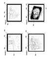
- FIGS. 1A-1Dillustrate four example interfaces for image annotation in image-guided medical procedures.
- FIGS. 2A and 2Billustrate example systems for image annotation in image-guided medical procedures.
- FIG. 3is a flow diagram that illustrates an example method for image annotation in image-guided medical procedures.
- FIG. 4illustrates a fifth example interface for image annotation in image-guided medical procedures.
- FIG. 5illustrates a sixth example interface for image annotation in image-guided medical procedures.
- FIGS. 6A-6Cillustrate three additional example interfaces for image annotation in image-guided medical procedures.
- FIGS. 7A-7Dillustrates a tenth example interface for image annotation in image-guided medical procedures.
- an operator, surgeon or other medical practitionermay annotate images during an image-guided medical procedure.
- the operatormay use medical devices that are typically present during the medical procedure to annotation the medical images.
- an operatorsuch as a surgeon or other medical practitioner, may use a first medical device 245 (e.g., an ablation needle) and a second medical device 255 (e.g., an ultrasound transducer) during a medical procedure and one or both of these medical devices 245 and 255 may be used for image annotation.
- a first medical device 245e.g., an ablation needle
- a second medical device 255e.g., an ultrasound transducer
- FIGS. 1A-1Dillustrate examples of image annotation in image-guided medical procedures.
- FIGS. 1A-1Dshow a representation on a computer screen 120 of an annotation being made with a medical device (represented on the display 120 as medical device 145 ).
- the medical devicemay be used to annotate an image 156 .
- FIG. 1Aillustrates the manipulation of a needle 145 pointing at an image 156 (e.g., an ultrasound image 156 ).
- the operatorcan make an annotation by moving the needle 145 through space in order to draw curve 171 on image 156 .
- Arrow 174may indicate the direction that the operator plans to or will draw in the future. In some embodiments, arrow 174 is not displayed.
- Indicator 180may represent the place on image 156 currently pointed to by needle 145 .
- Indicator 180may be any appropriate indicator such as an “X,” an arrow, a differently-colored area, etc.
- the operatorhas further moved needle 145 in order to complete the annotation 171 on image 156 .
- the indicator 180 of the intersection between the axis of the needle 145 and the image 156has now reached the lower-right quadrant of the image 156 .
- the image 156may be associated with a medical device, such as an ultrasound transducer (not pictured in FIGS. 1A-1D ).
- the image 156may be an ultrasound image 156 , or the image 156 may be a slice or image from other 3D visualizable medical data such as is described in Image Management in Image-Guided Medical Procedures, to Sharif Razzaque et al., filed concurrently herewith, which is incorporated by reference for all purposes.
- the annotation 171although it has been drawn on an image 156 , may actually be located in 3D space—defined by the placement of the image 156 and the annotation 171 .
- FIG. 1Cdepicts image 156 , associated with the ultrasound transducer turned or rotated about its vertical axis (axis not depicted in FIG. 1C ). Therefore, part of the annotation 171 is depicted in front of the image 156 , and part of the annotation 173 is behind the image 156 , thus illustrating the existence in 3D space of the annotation 171 / 173 .
- the location and display of annotations in 3D spacewill allow an operator to make an annotation for a feature (e.g., a tumor, cyst, or vein), and allow her to locate that feature again later.
- a featuree.g., a tumor, cyst, or vein
- FIG. 1Dillustrates that an operator may make a second annotation 172 on the image 156 .
- Part of the first annotation 171is in front of the image 156 and part 173 is behind.
- the operatorcan choose new locations within 3D space for annotations.
- the annotationsmay be for a blood vessel, tumor, or any other object or location of interest for the operator. There need not even be a particular object in the medical image that the operator is annotating.
- the operatormay, for example, sign her name or write a note. For example, an operator may circle or make marks near multiple tumors, trace a line such as annotation 171 along a vein or artery, etc.
- the operatormay make non-planar annotation (see, e.g., FIGS. 4 and 5 ).
- the operatormay be able to make a sphere or other non-planar annotation in order to annotate the volumetric aspects of a feature of interest. For example, the operator may draw the outline of a sphere around a tumor or cyst.
- a radiologist or other practitioneris not limited to marking tumors or other anatomical references on individual slices of CT scans. Instead, the radiologist may move in an intuitive manner through the CT scan. Further, various embodiments may decrease the time it takes to annotate an image, and/or to display those annotations, during a medical procedure, thereby reducing cost.
- annotationsBy allowing multiple annotations and by enabling the operator to place annotations in 3D space, various embodiments herein allow the operator to mark multiple objects of interest and view the location of those marks of interest at a later time.
- the annotationsmay be displayed using any display technique, such as those described in Image Management in Image-Guided Medical Procedures, to Sharif Razzaque et al., filed concurrently herewith and incorporated by reference above for all purposes.
- Imagesmay be annotated using embodiments herein during all a portion of a medical procedure.
- the image annotationwill only occur during an image annotation “session” (e.g. a period of time during which image annotation is performed, and before and after which, image annotation is not performed).
- An image annotation “session”may be initiated and/or terminated by the operator performing a key stroke, issuing a command (such as a verbal command), performing a gesture with a medical device or hand, pressing a button on the medical device, pressing a foot pedal, pressing a button on the medical device (e.g., a button on a Wacom pen), etc.
- a medical deviceis a broad term that encompasses but is not limited to a device, item, or part used in the medical procedure.
- a medical devicecould include an ablation needle, an ultrasound transducer, a cauterizer, a scalpel, a glove covering an operator's hand, the operator's hand or finger, etc.
- the medical device used for pose informationcould even be the operator's head, eyes, or gaze direction. Pose information for the medical device may be obtained using any system, device, method, or technique, such as those disclosed herein.
- FIG. 2Aillustrates a first exemplary system for image management in image guided surgery.
- FIG. 2Billustrates a second exemplary system for image management in image guided surgery.
- the embodiments illustrated by FIGS. 2A and 2Bare similar and use similar numbering. Where the two are different, those differences are noted.
- the differences between the two figuresmay include that, in FIG. 2A , two position sensing units 210 and 240 are shown, whereas in FIG. 2B , only a single position sensing unit 210 is shown.
- position sensing units 210 and 240may be tracking systems 210 and 240 and may track surgical instruments 245 and 255 and provide data to the image guidance unit 230 .
- the image guidance unit 230may process or combine the data and show image guidance data on display 220 .
- This image guidance datamay be used by a physician to guide a procedure and improve care.
- position sensing units 210 and 240may be combined and track all relevant surgical instruments 245 and 255 , as discussed in more detail below and exemplified in FIG. 2 B.
- Additional imaging units 250may be included and combined imaging data from the multiple imaging units 250 may be processed by image guidance unit 230 and shown on display unit 220 . Additionally, two or more surgical systems 249 may also be included.
- Imaging unit 250may be coupled to image guidance unit 230 .
- imaging unit 250may be coupled to a second display unit 251 .
- the second display unit 251may display imaging data from imaging unit 250 .
- the imaging data displayed on display unit 220 and displayed on second display unit 251may be, but are not necessarily, the same.
- the imaging unit 250is an ultrasound machine 250
- the movable imaging device 255is an ultrasound transducer 255 or ultrasound 255
- the second display unit 251is a display associated with the ultrasound machine 250 that displays the ultrasound images from the ultrasound machine 250 .
- a movable imaging unit 255may not be connected directly to an imaging unit 250 , but may instead be connected to image guidance unit 230 .
- the movable imaging unit 255may be useful for allowing a user to indicate what portions of a first set of imaging data should be displayed.
- the movable imaging unit 255may be an ultrasound transducer 255 or a tracked operative needle or other device 255 , for example, and may be used by a user to indicate what portions of imaging data, such as a pre-operative CT scan, to show on a display unit 220 as image 225 .
- system 200comprises a first position sensing unit 210 , a display unit 220 , and second position sensing unit 240 (if it is included) all coupled to image guidance unit 230 .
- first position sensing unit 210 , display unit 220 , and image guidance unit 230are all physically connected to stand 270 .
- Image guidance unit 230may be used to produce images 225 that are displayed on display unit 220 .
- the images 225 produced on display unit 220 by the image guidance unit 230may be determined based on ultrasound or other visual images from first surgical instrument 245 and second surgical instrument 255 .
- first surgical instrument 245is an ablation needle 245 and second surgical instrument 255 is an ultrasound probe 255
- images 225 produced on display 220may include the video or images from the ultrasound probe 255 combined with graphics, such as projected needle drive or projected ablation volume, determined based on the pose of ablation needle 245 .
- first surgical instrument 245is an ultrasound probe 245 and second surgical instrument 255 is a laparoscopic camera 255
- images 225 produced on display 220may include the video from the laparoscopic camera 255 combined with ultrasound data superimposed on the laparoscopic image.
- More surgical instrumentmay be added to the system.
- the systemmay include an ultrasound probe, ablation needle, laparoscopic camera, cauterizer, scalpel and/or any other surgical instrument or medical device.
- the systemmay also process and/or display previously collected data, such as preoperative CT scans, X-Rays, MRIs, laser scanned 3D surfaces etc.
- the term “pose” as used hereinis a broad term encompassing its plain and ordinary meaning and may refer to, without limitation, emplacement, position, orientation, the combination of position and orientation, or any other appropriate location information.
- the imaging data obtained from one or both of surgical instruments 245 and 255may include other modalities such as a CT scan, MRI, open-magnet MRI, optical coherence tomography, positron emission tomography (“PET”) scans, fluoroscopy, ultrasound, or other preoperative, or intraoperative 2D or 3D anatomical imaging data.
- surgical instruments 245 and 255may also be scalpels, implantable hardware, or any other device used in surgery. Any appropriate surgical system 249 or imaging unit 250 may be coupled to the corresponding medical instruments 245 and 255 .
- images 225 producedmay also be generated based on live, intraoperative, or real-time data obtained using second surgical instrument 255 , which is coupled to second imaging unit 250 .
- the term “real-time” as used hereinis a broad term and has its ordinary and customary meaning, including without limitation instantaneously or nearly instantaneously. The use of the term realtime may also mean that actions are performed or data is obtained with the intention to be used immediately, upon the next cycle of a system or control loop, or any other appropriate meaning.
- real-time datamay be data that is obtained at a frequency that would allow a surgeon to meaningfully interact with the data during surgery.
- real-time datamay be a medical image of a patient that is updated one time per second or multiple times per second.
- Second surgical instrument 255may be coupled to second position sensing unit 240 .
- Second position sensing unit 240may be part of imaging unit 250 or it may be separate. Second position sensing unit 240 may be used to determine the pose of second surgical instrument 255 .
- first and/or second position sensing units 210 and/or 240may be magnetic trackers and magnetic may be coils coupled to surgical instruments 245 and/or 255 .
- first and/or second position sensing units 210 and/or 240may be optical trackers and visually-detectable fiducials may be coupled to surgical instruments 245 and/or 255 .
- Images 225may be produced based on intraoperative or real-time data obtained using first surgical instrument 245 , which is coupled to first surgical system 249 .
- first surgical system 249is shown as coupled to image guidance unit 230 .
- the coupling between the first surgical system 249 and image guidance unit 230may not be present in all embodiments.
- the coupling between first surgical system 249 and image guidance unit 230may be included where information about first surgical instrument 245 available to first surgical system 249 is useful for the processing performed by image guidance unit 230 .
- first surgical instrument 245is an ablation needle 245 and first surgical system 249 is an ablation system 249 .
- first surgical system 249may not be coupled to image guidance unit 230 .
- Example embodiments including images and graphics that may be displayedare included below.
- first position sensing unit 210tracks the pose of first surgical device 245 .
- First position sensing unit 210may be an optical tracker 210 and first surgical device 245 may have optical fiducials attached thereto. The pose of optical fiducials may be detected by first position sensing unit 210 , and, therefrom, the pose of first surgical device 245 may be determined.
- a single position sensing unit 210may track both first medical device 245 and second medical device 255 .
- position sensing unit 210is a magnetic tracker and is mounted below a surgical table 280 . Such an arrangement may be useful when the tracking volume of the position sensing unit 210 is dependent on the location of the position sensing unit, as with many magnetic trackers. Magnetic tracking coils may be mounted in or on the medical devices 245 and 255 .
- first position sensing unit 210 and the second position sensing unit 240may be an Ascension Flock of Birds, Nest of Birds, driveBAY, medSAFE, trakSTAR, miniBIRD, MotionSTAR, pciBIRD, or Calypso 4D Localization System and tracking units attached to the first and/or second surgical or medical devices 245 and 255 may be magnetic tracking coils.
- the term “tracking unit,” as used herein,is a broad term encompassing its plain and ordinary meaning and includes without limitation all types of magnetic coils or other magnetic field sensing devices for use with magnetic trackers, fiducials or other optically detectable markers for use with optical trackers, such as those discussed above and below.
- Tracking unitscould also include optical position sensing devices such as the HiBall tracking system and the first and second position sensing units 210 and 240 may be part of a HiBall tracking systems.
- Tracking unitsmay also include a GPS device or signal emitting device that would allow for tracking of the position and, optionally, orientation of the tracking unit.
- a signal emitting devicemight include a radio-frequency identifier (RFID).
- RFIDradio-frequency identifier
- the first and/or second position sensing unit 210 and 240may take in the GPS coordinates of the tracking units or may, for example, triangulate the radio frequency signal being emitted by the RFID associated with tracking units.
- the tracking systemsmay also include one or more 3D mice.
- either or both of the first position sensing unit 210 and the second position sensing unit 240may be an Aurora® Electromagnetic Measurement System using sensor coils for tracking units attached to the first and/or second surgical devices 245 and 255 .
- either or both of the first position sensing unit 210 and the second position sensing unit 240may also be an optical 3D tracking system using fiducials.
- optical 3D tracking systemsmay include the NDI Polaris Spectra, Vicra, Certus, PhaseSpace IMPULSE, Vicon MX, InterSense IS-900, NaturalPoint OptiTrack, Polhemus FastTrak, IsoTrak, or Claron MicronTracker2.
- either or both of position sensing units 210 and 240may each be an inertial 3D tracking system comprising a compass, accelerometer, tilt sensor and/or gyro, such as the InterSense InertiaCube or the Wii controller.
- either or both of position sensing units 210 and 240may be attached to or affixed on the corresponding surgical device 245 and 255 .
- the position sensing units, 210 and 240may include sensing devices such as the HiBall tracking system, a GPS device, or signal emitting device that would allow for tracking of the position and, optionally, orientation of the tracking unit.
- a position sensing unit 210 or 240may be affixed to either or both of the surgical devices 245 and 255 .
- the surgical devices 245 or 255may be tracked by the position sensing units 210 or 240 .
- a world reference, such as the display 220may also be tracked by the position sensing unit 210 or 240 in order to determine the poses of the surgical devices 245 and 255 with respect to the world.
- Devices 245 and 255may also include or have coupled thereto one or more accelerometers, which may be used to estimate movement, position, and location of the devices.
- the display unit 220displays 3D images to a user, such as a physician.
- Stereoscopic 3D displaysseparate the imagery shown to each of the user's eyes. This can be accomplished by a stereoscopic display, a lenticular auto-stereoscopic display, or any other appropriate type of display.
- the display 220may be an alternating row or alternating column display.
- Example alternating row displaysinclude the Miracube G240S, as well as Zalman Trimon Monitors.
- Alternating column displaysinclude devices manufactured by Sharp, as well as many “auto-stereoscopic” displays (e.g., Philips).
- Display 220may also be a cathode ray tube.
- Cathode Ray Tube (CRT) based devicesmay use temporal sequencing, showing imagery for the left and right eye in temporal sequential alternation; this method may also be used by newer, projection-based devices, as well as by 120-Hz-switchable liquid crystal display (LCD) devices.
- CTRCathode Ray Tube
- a usermay wear a head mounted display in order to receive 3D images from the image guidance unit 230 .
- a separate displaysuch as the pictured display unit 220 , may be omitted.
- the 3D graphicsmay be produced using underlying data models, stored in the image guidance unit 230 and projected onto one or more 2D planes in order to create left and right eye images for a head mount, lenticular, or other 3D display.
- the underlying 3D modelmay be updated based on the relative poses of the various devices 245 and 255 , as determined by the position sensing unit(s), and/or based on new data associated with the devices 245 and 255 .
- the underlying data modelmay be updated to reflect the most recent ultrasound image.
- the first device 245is an ablation needle, then the underlying model may be updated to reflect any changes related to the needle, such as power or duration information.
- Any appropriate 3D graphics processingmay be used for rendering including processing based on OpenGL, Direct3D, Java 3D, etc. Whole, partial, or modified 3D graphics packages may also be used, such packages including 3DS Max, SolidWorks, Maya, Form Z, Cybermotion 3D, VTK, Slicer, or any others.
- various parts of the needed renderingmay occur on traditional or specialized graphics hardware.
- the renderingmay also occur on the general CPU, on programmable hardware, on a separate processor, be distributed over multiple processors, over multiple dedicated graphics cards, or using any other appropriate combination of hardware or technique.
- kitsmay be packaged and/or distributed as part of a kit.
- an ablation needle, tracking elements, 3D viewing glasses, and/or a portion of an ultrasound wandmay form a kit.
- Other embodimentsmay have different elements or combinations of elements grouped and/or packaged together. Kits may be sold or distributed separately from or with the other portions of the system.
- image guidance systemswhich may use, incorporate, support, or provide for the techniques, methods, processes, and systems described herein, such as the 3D computer-graphics-based assigned to InnerOptic Technologies, Inc. that provides for displaying guidance data from multiple sources, U.S. application Ser. No. 11/833,134, filed Aug. 2, 2007, the contents of which are incorporated by reference herein in their entirety for all purposes.
- the image guidancemay also be performed at least in part using the techniques described in U.S. patent application Ser. No. 11/828,826, filed Jul. 26, 2007, U.S. Pat. No. 7,728,868, U.S. patent application Ser. No. 12/299,899, U.S. patent application Ser. No. 12/483,099, U.S.
- FIG. 3depicts a method 300 for image annotation in image-guided medical procedures.
- pose information for an ultrasound transducer and its associated ultrasound imagemay be determined in block 310 .
- changing pose information for an ablation needlemay be determined in block 320 .
- the pose informationmay change as an operator moves the ablation needle and/or the ultrasound transducer.
- An annotationmay be generated in block 330 based on, for example, the intersection of an axis of the ablation needle and the ultrasound image plane.
- Image guidance informationmay be generated in block 340 based on the annotation in 3D space (and include, e.g., the annotation, the ultrasound image, a depiction of the ablation needle, and/or other imaging or guidance information).
- the image guidance informationmay be displayed.
- pose information for visualizable medical datais determined.
- “Visualizable medical data”is a broad term that encompasses its ordinary and customary meaning and includes, without limitation, any two-dimensional (2D) or 3D medical data that can be visualized.
- the visualizable medical datamay also be volumetric and can include, without limitation, one or more of a CT scan, an MRI, other 3D preoperative imaging data, other volume data, segmented internal organs, segmented blood vessels, annotations, tumors, etc.
- the visualizable medical datamay also include 2D medical data such as ultrasounds, X-rays, or segments or slices of 3D medical data.
- the visualizable medical datamay be associated with a medical device, such as an ultrasound probe, etc., and the medical device may be tracked in the medical scene.
- the pose information for the visualizable medical datamay be determined in block 310 from the pose of the associated medical device (that is tracked in the medical scene). For example, if the visualizable medical data is associated with an ultrasound probe and the ultrasound probe is tracked, then the pose of the visualizable medical data can be determined from the pose of the ultrasound probe. This can be the case even if the visualizable medical data is not generated by the medical device.
- the medical deviceis an ultrasound transducer and the visualizable medical data is a slice or image from a CT scan that is being navigated using the ultrasound transducer (see, for example, Image Management in Image-Guided Medical Procedures, to Sharif Razzaque et al., filed concurrently herewith, which is incorporated by reference above for all purposes) then the pose for that slice or image from the CT scan can still be determined based on the pose of the medical device.
- pose information for the medical devicemay be updated over time.
- Pose information for the underlying volumetric visualizable medical data setmay also be determined (e.g., relative to the medical scene).
- the pose information for the underlying volumetric visualizable medical datae.g., a CT scan or other volumetric data
- the pose information for the visualizable medical datamay initially be determined in order to register or approximately register the 3D visualizable medical data with the medical scene being visualized for the operator.
- Various techniques for registering the visualizable medical data with the medical scenemay be used, including matching features in 3D space with features in the visualizable medical data known to be in the medical scene, such as tumors, bones, blood vessels, etc.
- Manual registrationmay also be possible where an operator or other technician manipulates the pose of the visualizable medical data relative to the scene.
- changing pose informationis determined for a medical device.
- the medical device for which pose information is determined in block 320may be different from a medical device used for visualization of data in block 310 .
- pose information for the medical devicemay be determined using any system, device, method, or technique such as the tracking systems described herein.
- the medical deviceis an ablation needle, such as ablation needle 245 in FIGS. 2A and 2B
- determining the pose information for the ablation needle 245may include receiving tracking information from one or more position sensors sensing the position of ablation needle 245 .
- pose information for the medical devicemay be determined in other ways as well.
- a display 621may have a touchscreen that allows an operator to use her finger 645 as the medical device 645 to indicate the location of an annotation.
- the visualizable medical data 656may be shown on display 621 .
- an annotationmay appear on display 621 (not pictured in FIG. 6A ) or on a separate display of the image 656 , as depicted in FIG. 6C .
- FIG. 6Cwe see an annotation 671 that has been drawn by an operator up to point 680 .
- This annotation 671is positioned on image 656 and both are displayed on display 620 .
- An operatormay have dual displays 621 and 620 and be able to see both simultaneously.
- the medical device 645 used to point to an object on a screenmay also be a stylus, needle, or any other appropriate medical device 645 .
- the device used for inputmay not be a screen 621 , but may instead be a drawing tablet, or other input device (in which case image 656 may or may not be displayed on the device).
- a medical devicesuch as finger 645 in FIG. 6B
- An operatormay be able to point medical device 645 at the image 656 in order to define an annotation 671 up to a point 680 .
- Pointing with medical device 645 at display 622may define an intersection between medical device 645 and image 656 . That intersection may define the point 680 that is used to define or generate the annotation 671 .
- the medical device 645 used to point at the screenmay also be a remote (such as a Nintendo Wii controller), a surgical instrument, such as an ablation needle or scalpel, eye gaze or head direction, or any other appropriate medical device.
- the changing pose information for the medical device over timeis determined.
- changing pose informationis collected before proceeding to block 330 .
- the changing pose information for the medical deviceis collected iteratively and blocks 330 - 350 are performed as part of those iterations.
- pose information for the visualizable medical data, changing pose information for the medical device, and the other blocksare performed iteratively.
- pose information in block 310 for the visualizable medical data and changing pose information for the medical device in block 320are updated within the system as the updated pose information is received and this latest pose information is used in subsequent blocks 330 - 350 .
- annotationsare generated in 3D space based on the pose information received in blocks 310 and 320 . That is, the pose for the visualizable medical data (block 310 ) and the pose for the medical device (block 320 ) may be used to determine the annotations in 3D space (block 330 ).
- needle 145 and or image 156may have their poses change over time. The needle 145 and the image 156 may together define a point or mark 180 that changes over time as the poses of device 145 and image 156 change.
- point 180is defined by an axis extending out of the tip of needle 145 and its intersection with the plane of the image 156 , then as the needle 145 moves and/or the image 156 moves, the point 180 will change. As point 180 changes over time, it defines a curve, spline, segmented line, or other annotation that the operator is making. The annotations defined by these one or more movements is shown in FIG. 1B as annotation 171 .
- FIG. 4depicts a medical device 446 and an image 456 that together define an intersection point 480 .
- the curve 474shows a motion that an operator is going to make (curve 474 may or may not be displayed on display 420 ).
- an annotation 571is created by the movement of intersection point 580 .
- both device 546 and image 556have been moved so annotation 571 is not planar. Instead, annotation 571 is a three-dimensional surface.
- the movement of image 556is also illustrated by the outline 557 of the image's original placement at the start of the annotation (outline 557 may or may not be displayed on display 520 )—and by arrow 558 .
- the annotation determinedmay be a spline, a series of lines, a series of triangles, a point cloud, a series of voxels, or any other appropriate representation in 3D space.
- the generated annotationmay also be termed or thought of as “virtual ink.” In some embodiments, the annotation may be termed “virtual ink” when it corresponds to the drawing of ink on the image plane as the drawing instrument and image plane move.
- image guidance informationis generated based on the annotation.
- Generating image guidance information based on the annotation in block 330may include generating a 3D model or series of 3D models that represent the medical scene to be displayed to the operator. For example, as depicted in FIG. 1D , after a first annotation 171 / 173 is defined and a second annotation 172 is defined, generating image guidance data may include registering in the same 3D space, or determining transformations among, the various annotations 171 - 173 and the image 156 .
- determining the guidance information in block 340may include incorporating a planar slice of the CT data corresponding to the image 156 . Further, determining image guidance information may include numerous other techniques, methods, and systems, such as those described in Image Management in Image-Guided Medical Procedures, to Sharif Razzaque et al., filed concurrently herewith, which is incorporated by reference above for all purposes.
- a graphical rendering of the image guidance informationis displayed in block 350 .
- the display of graphical informationcan be monoscopic or stereoscopic. Further, multiple rendering techniques may be used. Edges or areas near the edge of a region of interest defined by the annotation, a medical device, or the image, may be displayed in a blurred or fading manner. Objects near objects of interest such as the image, the annotation, or the medical device may be displayed in sharper focus, may be displayed brighter, etc. In one embodiment, if an additional set of 3D visualizable medical data is displayed, a tunnel or cut-through that set of medical data may be made so that an image can be shown. Consider for example, FIG.
- medical device 145 and/or image 156may define a region of interest and items in that region of interest may be displayed distinctly from the rest of the data displayed on screen 120 . For example, if additional medical data is being displayed, then data from that additional medical display may be displayed within a region of interest around image 156 and/or medical device 145 .
- a medical devicemay define a region of interest 760 in which an ultrasound image 756 may be shown in focus.
- the CT scan 770may be displayed.
- a single slice of CT data 770may be displayed outside the region of interest 760 , or, as depicted in FIG. 7B , multiple slices of CT data 770 may be displayed outside the region of interest.
- the slices of CT datamay be rendered differently depending on the distance from the region of interest. For example, planes of CT scan data 770 may be rendered more transparently (less brightly, etc) the further each is from the plane containing the region of interest.
- the slices of CT datamay be the slices from the underlying CT data, or the slices may be generated to be, e.g., parallel or nearly parallel, with a plane associated with the region of interest 760 .
- FIG. 7Calso depicts that a tunnel may be cut through the rendered slices of the CT scan 770 in order to display the region of interest 760 without or with little overlap. This tunnel may be altered as the region of interest or CT scan data are moved to always allow the operator to view the region of interest.
- FIG. 7Ddepicts a semi-realistic rendering of a CT scan 770 around a region of interest 760 . Inside the region of interest 760 , an ultrasound image 756 is displayed. Also displayed on display 720 in FIG. 7D , is an outline of the medical device 755 .
- the data shown in the region of interestmay be any appropriate visualizable medical data, not limited to ultrasound or CT data.
- the data displayed outside of the region of interestmay be any visualizable medical data, and may even be from the same data set as the data shown in the region of interest.
- MRI datamay be shown in fading planes outside of the region of interest and in focus (and visualizable through a tunnel) inside the region of interest.
- annotationmay be displayed along with the rendering of the visualizable medical data inside and/or outside of the region of interest. In this manner, an operator may see the annotations in the context of the visualizable medical data.
- each point of the line segment, spline segment, point cloud, etc.may be made transparent and/or blurry based on its distance from the region of interest, and its rendering may be controlled using various graphic techniques, such as bit maps and pixel shaders, such as those discussed in Image Management in Image-Guided Medical Procedures, to Sharif Razzaque et al., filed concurrently herewith, which is incorporated by reference above for all purposes.
- process 300may be performed in a different order, may be augmented by other blocks or may have sub-blocks within the blocks shown. Further, the process 300 may be performed on a single computer or processor, on multiple computers or processors, on a single or multiple virtual machines, and/or in a distributed fashion on multiple processors, devices, machines, or virtual machines.
- the physicianmay manipulate a medical device, such as an ultrasound transducer, in order to navigate and view CT data preoperatively (or intraoperatively).
- the physicianmay be able to see the small lesions in the CT data.
- the physiciancan then annotate those lesions, perhaps by circling, creating a sphere around them, and/or drawing an arrow pointing to them, using annotation the techniques herein.
- the physicianmay be able to leverage the preoperative lesion annotation.
- the physicianmay use intraoperative ultrasound in order to spot the current location of the various lesions, guided at least in part by the annotation made in 3D space relative to the CT scan.
- the physicianhas utilized both the relative ease of discovery of lesions on the CT scan as well as the intraoperative accuracy of locating the lesions in the ultrasound. This can increase accuracy and reduce operative times and the problems and costs associated therewith.
- position sensing units 210 and 240 , display unit 220 , image guidance unit 230 , and/or any other module or unit of embodiments hereinmay each be separate computing devices, applications, or processes or may run as part of the same computing devices, applications, or processes—or one of more may be combined to run as part of one application or process—and/or each or one or more may be part of or run on a computing device.
- Computing devices or computer systemsmay include a bus or other communication mechanism for communicating information, and a processor coupled with the bus for processing information.
- a computer system or devicemay have a main memory, such as a random access memory or other dynamic storage device, coupled to the bus.
- the main memorymay be used to store instructions and temporary variables.
- the computer system or devicemay also include a read-only memory or other static storage device coupled to the bus for storing static information and instructions.
- the computer systems or devicesmay also be coupled to a display, such as a CRT, LCD monitor, LED array, e-paper, projector, or stereoscopic display.
- Input devicesmay also be coupled to the computer system or device. These input devices may include a mouse, a trackball, touchscreen, tablet, foot pedal, or cursor direction keys.
- Computer systems or devices described hereinmay include the image guidance unit 230 , first and second position sensing units 210 and 240 , and imaging unit 250 .
- Each computer system or computing devicemay be implemented using one or more physical computers, processors, embedded devices, field programmable gate arrays (FPGAs), or computer systems or portions thereof.
- the instructions executed by the computer system or computing devicemay also be read in from a computer-readable medium.
- the computer-readable mediummay be non-transitory, such as a CD, DVD, optical or magnetic disk, laserdisc, flash memory, or any other medium that is readable by the computer system or device.
- hardwired circuitrymay be used in place of or in combination with software instructions executed by the processor. Communication among modules, systems, devices, and elements may be over a direct or switched connections, and wired or wireless networks or connections, via directly connected wires, or any other appropriate communication mechanism.
- Transmission of informationmay be performed on the hardware layer using any appropriate system, device, or protocol, including those related to or utilizing Firewire, PCI, PCI express, CardBus, USB, CAN, SCSI, IDA, RS232, RS422, RS485, 802.11, etc.
- the communication among modules, systems, devices, and elementsmay include handshaking, notifications, coordination, encapsulation, encryption, headers, such as routing or error detecting headers, or any other appropriate communication protocol or attribute.
- Communicationmay also messages related to HTTP, HTTPS, FTP, TCP, IP, ebMS OASIS/ebXML, DICOM, DICOS, secure sockets, VPN, encrypted or unencrypted pipes, MIME, SMTP, MIME Multipart/Related Content-type, SQL, etc.
- Any appropriate 3D graphics processingmay be used for displaying or rendering, including processing based on OpenGL, Direct3D, Java 3D, etc.
- Whole, partial, or modified 3D graphics packagesmay also be used, such packages including 3DS Max, SolidWorks, Maya, Form Z, Cybermotion 3D, VTK, Slicer, Blender or any others.
- various parts of the needed renderingmay occur on traditional or specialized graphics hardware.
- the renderingmay also occur on the general CPU, on programmable hardware, on a separate processor, be distributed over multiple processors, over multiple dedicated graphics cards, or using any other appropriate combination of hardware or technique.
- All of the methods and processes described abovemay be embodied in, and fully automated via, software code modules executed by one or more general purpose computers or processors, such as those computer systems described above.
- the code modulesmay be stored in any type of computer-readable medium or other computer storage device. Some or all of the methods may alternatively be embodied in specialized computer hardware.
Landscapes
- Health & Medical Sciences (AREA)
- Life Sciences & Earth Sciences (AREA)
- Engineering & Computer Science (AREA)
- Medical Informatics (AREA)
- Surgery (AREA)
- Public Health (AREA)
- Biomedical Technology (AREA)
- General Health & Medical Sciences (AREA)
- Heart & Thoracic Surgery (AREA)
- Molecular Biology (AREA)
- Veterinary Medicine (AREA)
- Animal Behavior & Ethology (AREA)
- Nuclear Medicine, Radiotherapy & Molecular Imaging (AREA)
- Pathology (AREA)
- Physics & Mathematics (AREA)
- Biophysics (AREA)
- Radiology & Medical Imaging (AREA)
- Optics & Photonics (AREA)
- High Energy & Nuclear Physics (AREA)
- Computer Vision & Pattern Recognition (AREA)
- Human Computer Interaction (AREA)
- Physiology (AREA)
- Robotics (AREA)
- Computer Graphics (AREA)
- General Engineering & Computer Science (AREA)
- Plasma & Fusion (AREA)
- Theoretical Computer Science (AREA)
- Pulmonology (AREA)
- Otolaryngology (AREA)
- Gynecology & Obstetrics (AREA)
- Oral & Maxillofacial Surgery (AREA)
- Data Mining & Analysis (AREA)
- Databases & Information Systems (AREA)
- Epidemiology (AREA)
- Primary Health Care (AREA)
- Ultra Sonic Daignosis Equipment (AREA)
- Magnetic Resonance Imaging Apparatus (AREA)
- Apparatus For Radiation Diagnosis (AREA)
Abstract
Description
This application claims benefit to U.S. Provisional Patent Application No. 61/322,991 filed Apr. 12, 2010, and U.S. Provisional Patent Application No. 61/387,132, filed Sep. 28, 2010. Each of the provisional applications, 61/322,991 and 61/387,132 is incorporated by reference herein in its entirety for all purposes.
The embodiments herein disclosed relate to computer-assisted medical procedures and more specifically to image annotation in image-guided medical procedures.
The past few decades have seen incredible developments of technology and systems for computer-assisted, image-based, and image-guided surgery and other medical procedures. These advances in image-guided surgery are tied in part to technical and scientific improvements in imaging and three-dimensional (3D) computer graphics. For example, some of the early work of in this field in the late 1980's provided new 3D graphics rendering techniques, medical image shape detection, and head-mounted displays. These are some of the building blocks of later image-guided surgery systems developed in the mid-1990's and thereafter. Image-guided surgery makes use of imaging to aid a surgeon in performing more effective and more accurate surgeries.
Current image-guided surgery systems, however, do not provide adequate mechanisms to annotate images. The process of annotation is difficult and extremely time-consuming. Further, it would be difficult, disruptive, and time consuming for a surgeon or other operator to annotate an image during a medical procedure.
One or more of these problems and others are addressed by the systems, methods, devices, computer-readable media, techniques, and embodiments described herein. That is, some of the embodiments described herein may address one or more issues, while other embodiments may address different issues.
Presented herein are methods, systems, devices, and computer-readable media for image annotation in image-guided medical procedures. In some embodiments, pose information is determined for visualizable medical data and changing pose information is determined for a medical device over time. An annotation in 3D space may be generated based on the pose information over time for the medical device and the pose information for the visualizable medical data; and image guidance information may be generated based at least in part on the annotation in 3D space. A graphical rendering of the image guidance information may be displayed on one or more displays.
In some embodiments, a system may determine device type information for a first medical device; real-time emplacement information for the first medical device; and real-time emplacement information for a second medical device. The system may also determine the real-time relative emplacements of the first and second medical devices with the computer system and real-time prediction information for the first medical device. The image guidance system may then generate image guidance information based on the real-time relative emplacements of the first and second medical devices, the real-time prediction information for the first medical device, and data related to the second medical device. A graphical rendering of the image guidance information may be displayed on one or more displays. It is possible that determining changing pose information for the medical device over time include determining the changing pose information for the medical device over time relative to a 2D screen displaying the visualizable medical data; and/or generating the annotation in 3D space based on the pose information over time for the medical device and the pose information for the visualizable medical data may include determining the annotation in 3D space based at least in part on the 2D pose information.
Numerous other embodiments are described throughout herein. Although various embodiments are described herein, it is to be understood that not necessarily all objects, advantages, features or concepts need to be achieved in accordance with any particular embodiment. Thus, for example, those skilled in the art will recognize that the invention may be embodied or carried out in a manner that achieves or optimizes one advantage or group of advantages as taught or suggested herein without necessarily achieving other objects or advantages as may be taught or suggested herein.
All of these embodiments are intended to be within the scope of the invention herein disclosed. These and other embodiments will become readily apparent to those skilled in the art from the following detailed description having reference to the attached figures, the invention not being limited to any particular disclosed embodiment(s).
In some embodiments herein, an operator, surgeon or other medical practitioner may annotate images during an image-guided medical procedure. In some embodiments, the operator may use medical devices that are typically present during the medical procedure to annotation the medical images. As depicted inFIGS. 2A and 2B , and as described more below, an operator, such as a surgeon or other medical practitioner, may use a first medical device245 (e.g., an ablation needle) and a second medical device255 (e.g., an ultrasound transducer) during a medical procedure and one or both of thesemedical devices
Theimage 156 may be associated with a medical device, such as an ultrasound transducer (not pictured inFIGS. 1A-1D ). Theimage 156 may be anultrasound image 156, or theimage 156 may be a slice or image from other 3D visualizable medical data such as is described in Image Management in Image-Guided Medical Procedures, to Sharif Razzaque et al., filed concurrently herewith, which is incorporated by reference for all purposes.
Theannotation 171, although it has been drawn on animage 156, may actually be located in 3D space—defined by the placement of theimage 156 and theannotation 171.FIG. 1C depictsimage 156, associated with the ultrasound transducer turned or rotated about its vertical axis (axis not depicted inFIG. 1C ). Therefore, part of theannotation 171 is depicted in front of theimage 156, and part of theannotation 173 is behind theimage 156, thus illustrating the existence in 3D space of theannotation 171/173. The location and display of annotations in 3D space will allow an operator to make an annotation for a feature (e.g., a tumor, cyst, or vein), and allow her to locate that feature again later.
Using embodiments described herein, a radiologist or other practitioner is not limited to marking tumors or other anatomical references on individual slices of CT scans. Instead, the radiologist may move in an intuitive manner through the CT scan. Further, various embodiments may decrease the time it takes to annotate an image, and/or to display those annotations, during a medical procedure, thereby reducing cost.
By allowing multiple annotations and by enabling the operator to place annotations in 3D space, various embodiments herein allow the operator to mark multiple objects of interest and view the location of those marks of interest at a later time. The annotations may be displayed using any display technique, such as those described in Image Management in Image-Guided Medical Procedures, to Sharif Razzaque et al., filed concurrently herewith and incorporated by reference above for all purposes.
Images may be annotated using embodiments herein during all a portion of a medical procedure. In one embodiment, the image annotation will only occur during an image annotation “session” (e.g. a period of time during which image annotation is performed, and before and after which, image annotation is not performed). An image annotation “session” may be initiated and/or terminated by the operator performing a key stroke, issuing a command (such as a verbal command), performing a gesture with a medical device or hand, pressing a button on the medical device, pressing a foot pedal, pressing a button on the medical device (e.g., a button on a Wacom pen), etc.
As used herein, the term “medical device” is a broad term that encompasses but is not limited to a device, item, or part used in the medical procedure. For example, a medical device could include an ablation needle, an ultrasound transducer, a cauterizer, a scalpel, a glove covering an operator's hand, the operator's hand or finger, etc. The medical device used for pose information could even be the operator's head, eyes, or gaze direction. Pose information for the medical device may be obtained using any system, device, method, or technique, such as those disclosed herein.
In one embodiment,position sensing units systems surgical instruments image guidance unit 230. Theimage guidance unit 230 may process or combine the data and show image guidance data ondisplay 220. This image guidance data may be used by a physician to guide a procedure and improve care. There are numerous other possible embodiments ofsystem 200. For example, many of the depicted modules may be joined together to form a single module and may even be implemented in a single computer or machine. Further,position sensing units surgical instruments Additional imaging units 250 may be included and combined imaging data from themultiple imaging units 250 may be processed byimage guidance unit 230 and shown ondisplay unit 220. Additionally, two or moresurgical systems 249 may also be included.
Information about and from multiplesurgical systems 249 and/or attachedsurgical instruments 245 may be processed byimage guidance unit 230 and shown ondisplay 220. These and other possible embodiments are discussed in more detail below.Imaging unit 250 may be coupled to imageguidance unit 230. In one embodiment,imaging unit 250 may be coupled to asecond display unit 251. Thesecond display unit 251 may display imaging data fromimaging unit 250. The imaging data displayed ondisplay unit 220 and displayed onsecond display unit 251 may be, but are not necessarily, the same. In an embodiment, theimaging unit 250 is anultrasound machine 250, themovable imaging device 255 is anultrasound transducer 255 orultrasound 255, and thesecond display unit 251 is a display associated with theultrasound machine 250 that displays the ultrasound images from theultrasound machine 250. In one embodiment, amovable imaging unit 255 may not be connected directly to animaging unit 250, but may instead be connected to imageguidance unit 230. Themovable imaging unit 255 may be useful for allowing a user to indicate what portions of a first set of imaging data should be displayed. For example, themovable imaging unit 255 may be anultrasound transducer 255 or a tracked operative needle orother device 255, for example, and may be used by a user to indicate what portions of imaging data, such as a pre-operative CT scan, to show on adisplay unit 220 asimage 225. Further, in some embodiments, there could be a third set of pre-operative imaging data that could be displayed with the first set of imaging data.
In some embodiments,system 200 comprises a firstposition sensing unit 210, adisplay unit 220, and second position sensing unit240 (if it is included) all coupled to imageguidance unit 230. In one embodiment, firstposition sensing unit 210,display unit 220, andimage guidance unit 230 are all physically connected to stand270.Image guidance unit 230 may be used to produceimages 225 that are displayed ondisplay unit 220. Theimages 225 produced ondisplay unit 220 by theimage guidance unit 230 may be determined based on ultrasound or other visual images from firstsurgical instrument 245 and secondsurgical instrument 255. For example, if firstsurgical instrument 245 is anablation needle 245 and secondsurgical instrument 255 is anultrasound probe 255, thenimages 225 produced ondisplay 220 may include the video or images from theultrasound probe 255 combined with graphics, such as projected needle drive or projected ablation volume, determined based on the pose ofablation needle 245. If firstsurgical instrument 245 is anultrasound probe 245 and secondsurgical instrument 255 is alaparoscopic camera 255, thenimages 225 produced ondisplay 220 may include the video from thelaparoscopic camera 255 combined with ultrasound data superimposed on the laparoscopic image. More surgical instrument may be added to the system. For example, the system may include an ultrasound probe, ablation needle, laparoscopic camera, cauterizer, scalpel and/or any other surgical instrument or medical device. The system may also process and/or display previously collected data, such as preoperative CT scans, X-Rays, MRIs, laser scanned 3D surfaces etc.
The term “pose” as used herein is a broad term encompassing its plain and ordinary meaning and may refer to, without limitation, emplacement, position, orientation, the combination of position and orientation, or any other appropriate location information. In some embodiments, the imaging data obtained from one or both ofsurgical instruments surgical instruments surgical system 249 orimaging unit 250 may be coupled to the correspondingmedical instruments
As noted above,images 225 produced may also be generated based on live, intraoperative, or real-time data obtained using secondsurgical instrument 255, which is coupled tosecond imaging unit 250. The term “real-time” as used herein is a broad term and has its ordinary and customary meaning, including without limitation instantaneously or nearly instantaneously. The use of the term realtime may also mean that actions are performed or data is obtained with the intention to be used immediately, upon the next cycle of a system or control loop, or any other appropriate meaning. Additionally, as used herein, real-time data may be data that is obtained at a frequency that would allow a surgeon to meaningfully interact with the data during surgery. For example, in some embodiments, real-time data may be a medical image of a patient that is updated one time per second or multiple times per second.
Secondsurgical instrument 255 may be coupled to secondposition sensing unit 240. Secondposition sensing unit 240 may be part ofimaging unit 250 or it may be separate. Secondposition sensing unit 240 may be used to determine the pose of secondsurgical instrument 255. In some embodiments, first and/or secondposition sensing units 210 and/or240 may be magnetic trackers and magnetic may be coils coupled tosurgical instruments 245 and/or255. In some embodiments, first and/or secondposition sensing units 210 and/or240 may be optical trackers and visually-detectable fiducials may be coupled tosurgical instruments 245 and/or255.
In an embodiment, firstposition sensing unit 210 tracks the pose of firstsurgical device 245. Firstposition sensing unit 210 may be anoptical tracker 210 and firstsurgical device 245 may have optical fiducials attached thereto. The pose of optical fiducials may be detected by firstposition sensing unit 210, and, therefrom, the pose of firstsurgical device 245 may be determined.
In various embodiments, as depicted inFIG. 2B , a singleposition sensing unit 210 may track both firstmedical device 245 and secondmedical device 255. InFIG. 2B , in some embodiments,position sensing unit 210 is a magnetic tracker and is mounted below a surgical table280. Such an arrangement may be useful when the tracking volume of theposition sensing unit 210 is dependent on the location of the position sensing unit, as with many magnetic trackers. Magnetic tracking coils may be mounted in or on themedical devices
In some embodiments, either or both of the firstposition sensing unit 210 and the secondposition sensing unit 240 may be an Ascension Flock of Birds, Nest of Birds, driveBAY, medSAFE, trakSTAR, miniBIRD, MotionSTAR, pciBIRD, or Calypso 4D Localization System and tracking units attached to the first and/or second surgical ormedical devices position sensing units position sensing unit
In some embodiments, either or both of the firstposition sensing unit 210 and the secondposition sensing unit 240 may be an Aurora® Electromagnetic Measurement System using sensor coils for tracking units attached to the first and/or secondsurgical devices position sensing unit 210 and the secondposition sensing unit 240 may also be an optical 3D tracking system using fiducials. Such optical 3D tracking systems may include the NDI Polaris Spectra, Vicra, Certus, PhaseSpace IMPULSE, Vicon MX, InterSense IS-900, NaturalPoint OptiTrack, Polhemus FastTrak, IsoTrak, or Claron MicronTracker2. In some embodiments, either or both ofposition sensing units position sensing units surgical device position sensing unit surgical devices surgical devices position sensing units display 220 may also be tracked by theposition sensing unit surgical devices Devices
In an embodiment, thedisplay unit 220 displays 3D images to a user, such as a physician. Stereoscopic 3D displays separate the imagery shown to each of the user's eyes. This can be accomplished by a stereoscopic display, a lenticular auto-stereoscopic display, or any other appropriate type of display. Thedisplay 220 may be an alternating row or alternating column display. Example alternating row displays include the Miracube G240S, as well as Zalman Trimon Monitors. Alternating column displays include devices manufactured by Sharp, as well as many “auto-stereoscopic” displays (e.g., Philips).Display 220 may also be a cathode ray tube. Cathode Ray Tube (CRT) based devices, may use temporal sequencing, showing imagery for the left and right eye in temporal sequential alternation; this method may also be used by newer, projection-based devices, as well as by 120-Hz-switchable liquid crystal display (LCD) devices.
In one embodiment, a user may wear a head mounted display in order to receive 3D images from theimage guidance unit 230. In such embodiments, a separate display, such as the pictureddisplay unit 220, may be omitted. The 3D graphics may be produced using underlying data models, stored in theimage guidance unit 230 and projected onto one or more 2D planes in order to create left and right eye images for a head mount, lenticular, or other 3D display. The underlying 3D model may be updated based on the relative poses of thevarious devices devices ultrasound probe 255, then the underlying data model may be updated to reflect the most recent ultrasound image. If thefirst device 245 is an ablation needle, then the underlying model may be updated to reflect any changes related to the needle, such as power or duration information. Any appropriate 3D graphics processing may be used for rendering including processing based on OpenGL, Direct3D,Java 3D, etc. Whole, partial, or modified 3D graphics packages may also be used, such packages including 3DS Max, SolidWorks, Maya, Form Z,Cybermotion 3D, VTK, Slicer, or any others. In some embodiments, various parts of the needed rendering may occur on traditional or specialized graphics hardware. The rendering may also occur on the general CPU, on programmable hardware, on a separate processor, be distributed over multiple processors, over multiple dedicated graphics cards, or using any other appropriate combination of hardware or technique.
One or more modules, units, devices, or elements of various embodiments may be packaged and/or distributed as part of a kit. For example, in one embodiment, an ablation needle, tracking elements, 3D viewing glasses, and/or a portion of an ultrasound wand may form a kit. Other embodiments may have different elements or combinations of elements grouped and/or packaged together. Kits may be sold or distributed separately from or with the other portions of the system.
There are numerous other examples of image guidance systems which may use, incorporate, support, or provide for the techniques, methods, processes, and systems described herein, such as the 3D computer-graphics-based assigned to InnerOptic Technologies, Inc. that provides for displaying guidance data from multiple sources, U.S. application Ser. No. 11/833,134, filed Aug. 2, 2007, the contents of which are incorporated by reference herein in their entirety for all purposes. The image guidance may also be performed at least in part using the techniques described in U.S. patent application Ser. No. 11/828,826, filed Jul. 26, 2007, U.S. Pat. No. 7,728,868, U.S. patent application Ser. No. 12/299,899, U.S. patent application Ser. No. 12/483,099, U.S. patent application Ser. No. 12/893,123, U.S. patent application Ser. No. 12/842,261, and/or U.S. patent application Ser. No. 12/703,118, each of which is incorporated by reference herein in its entirety for all purposes.
Methods for Image Annotation in Image-Guided Medical Procedures
Inblock 310, pose information for visualizable medical data is determined. “Visualizable medical data” is a broad term that encompasses its ordinary and customary meaning and includes, without limitation, any two-dimensional (2D) or 3D medical data that can be visualized. The visualizable medical data may also be volumetric and can include, without limitation, one or more of a CT scan, an MRI, other 3D preoperative imaging data, other volume data, segmented internal organs, segmented blood vessels, annotations, tumors, etc. The visualizable medical data may also include 2D medical data such as ultrasounds, X-rays, or segments or slices of 3D medical data.
In some embodiments, the visualizable medical data may be associated with a medical device, such as an ultrasound probe, etc., and the medical device may be tracked in the medical scene. In such embodiments, the pose information for the visualizable medical data may be determined inblock 310 from the pose of the associated medical device (that is tracked in the medical scene). For example, if the visualizable medical data is associated with an ultrasound probe and the ultrasound probe is tracked, then the pose of the visualizable medical data can be determined from the pose of the ultrasound probe. This can be the case even if the visualizable medical data is not generated by the medical device. For example, if the medical device is an ultrasound transducer and the visualizable medical data is a slice or image from a CT scan that is being navigated using the ultrasound transducer (see, for example, Image Management in Image-Guided Medical Procedures, to Sharif Razzaque et al., filed concurrently herewith, which is incorporated by reference above for all purposes) then the pose for that slice or image from the CT scan can still be determined based on the pose of the medical device.
When navigating/visualizing CT or other volumetric data with a medical device such as an ultrasound transducer, pose information for the medical device may be updated over time. Pose information for the underlying volumetric visualizable medical data set may also be determined (e.g., relative to the medical scene). The pose information for the underlying volumetric visualizable medical data (e.g., a CT scan or other volumetric data) may be determined separately from the pose information of the medical device used to visualize the medical data. Further, in some embodiments, the pose information for the visualizable medical data may initially be determined in order to register or approximately register the 3D visualizable medical data with the medical scene being visualized for the operator. Various techniques for registering the visualizable medical data with the medical scene may be used, including matching features in 3D space with features in the visualizable medical data known to be in the medical scene, such as tumors, bones, blood vessels, etc. Manual registration may also be possible where an operator or other technician manipulates the pose of the visualizable medical data relative to the scene.
Inblock 320, changing pose information is determined for a medical device. The medical device for which pose information is determined inblock 320 may be different from a medical device used for visualization of data inblock 310.
Returning again to block320, pose information for the medical device may be determined using any system, device, method, or technique such as the tracking systems described herein. For example, if the medical device is an ablation needle, such asablation needle 245 inFIGS. 2A and 2B , then determining the pose information for theablation needle 245 may include receiving tracking information from one or more position sensors sensing the position ofablation needle 245.
As depicted inFIGS. 6A-6C , pose information for the medical device may be determined in other ways as well. For example, as shown inFIG. 6A , adisplay 621 may have a touchscreen that allows an operator to use herfinger 645 as themedical device 645 to indicate the location of an annotation. The visualizablemedical data 656 may be shown ondisplay 621. As the operator moves herfinger 645, an annotation may appear on display621 (not pictured inFIG. 6A ) or on a separate display of theimage 656, as depicted inFIG. 6C . InFIG. 6C , we see anannotation 671 that has been drawn by an operator up topoint 680. Thisannotation 671 is positioned onimage 656 and both are displayed ondisplay 620. An operator may havedual displays
Themedical device 645 used to point to an object on a screen may also be a stylus, needle, or any other appropriatemedical device 645. Further, in some embodiments, the device used for input may not be ascreen 621, but may instead be a drawing tablet, or other input device (in whichcase image 656 may or may not be displayed on the device).
In some embodiments, a medical device, such asfinger 645 inFIG. 6B , may be used to point at animage 656 displayed on adisplay 622. An operator may be able to pointmedical device 645 at theimage 656 in order to define anannotation 671 up to apoint 680. Pointing withmedical device 645 atdisplay 622 may define an intersection betweenmedical device 645 andimage 656. That intersection may define thepoint 680 that is used to define or generate theannotation 671. Themedical device 645 used to point at the screen may also be a remote (such as a Nintendo Wii controller), a surgical instrument, such as an ablation needle or scalpel, eye gaze or head direction, or any other appropriate medical device.
Returning again toFIG. 3 and block320, as pose information changes over time (e.g., because the medical device is being moved), such as described above with respect toFIGS. 1A-1D , the changing pose information for the medical device over time is determined. In one embodiment, changing pose information is collected before proceeding to block330. In another embodiment, the changing pose information for the medical device is collected iteratively and blocks330-350 are performed as part of those iterations. Further, in some embodiments, pose information for the visualizable medical data, changing pose information for the medical device, and the other blocks are performed iteratively. In yet other embodiments, pose information inblock 310 for the visualizable medical data and changing pose information for the medical device inblock 320 are updated within the system as the updated pose information is received and this latest pose information is used in subsequent blocks330-350.
Inblock 330, annotations are generated in 3D space based on the pose information received inblocks FIGS. 1A-1D ,needle 145 and orimage 156 may have their poses change over time. Theneedle 145 and theimage 156 may together define a point or mark180 that changes over time as the poses ofdevice 145 andimage 156 change. That is, ifpoint 180 is defined by an axis extending out of the tip ofneedle 145 and its intersection with the plane of theimage 156, then as theneedle 145 moves and/or theimage 156 moves, thepoint 180 will change. Aspoint 180 changes over time, it defines a curve, spline, segmented line, or other annotation that the operator is making. The annotations defined by these one or more movements is shown inFIG. 1B asannotation 171.
After an annotation has been created in 3D space inblock 330 then inblock 340 image guidance information is generated based on the annotation. Generating image guidance information based on the annotation inblock 330 may include generating a 3D model or series of 3D models that represent the medical scene to be displayed to the operator. For example, as depicted inFIG. 1D , after afirst annotation 171/173 is defined and asecond annotation 172 is defined, generating image guidance data may include registering in the same 3D space, or determining transformations among, the various annotations171-173 and theimage 156. If theimage 156 is, for example, a planar representation of 3D visualizable medical data, such as a CT scan, then determining the guidance information inblock 340 may include incorporating a planar slice of the CT data corresponding to theimage 156. Further, determining image guidance information may include numerous other techniques, methods, and systems, such as those described in Image Management in Image-Guided Medical Procedures, to Sharif Razzaque et al., filed concurrently herewith, which is incorporated by reference above for all purposes.
After image guidance information has been generated based on the annotation inblock 340, a graphical rendering of the image guidance information is displayed inblock 350. In some embodiments, the display of graphical information can be monoscopic or stereoscopic. Further, multiple rendering techniques may be used. Edges or areas near the edge of a region of interest defined by the annotation, a medical device, or the image, may be displayed in a blurred or fading manner. Objects near objects of interest such as the image, the annotation, or the medical device may be displayed in sharper focus, may be displayed brighter, etc. In one embodiment, if an additional set of 3D visualizable medical data is displayed, a tunnel or cut-through that set of medical data may be made so that an image can be shown. Consider for example,FIG. 6B . If another set of 3D data is being displayed on display622 (not depicted inFIG. 6B ), then the additional data may have a cut-through so thatimage 656 can be seen and theareas surrounding image 656 ondisplay 622 may show the additional visualizable medical data. Turning toFIG. 1B , it is possible thatmedical device 145 and/orimage 156 may define a region of interest and items in that region of interest may be displayed distinctly from the rest of the data displayed on screen120. For example, if additional medical data is being displayed, then data from that additional medical display may be displayed within a region of interest aroundimage 156 and/ormedical device 145.
Turning toFIG. 7A , in some embodiments, a medical device may define a region ofinterest 760 in which anultrasound image 756 may be shown in focus. Outside the region ofinterest 760, the CT scan770 may be displayed. As depicted inFIG. 7A , a single slice ofCT data 770 may be displayed outside the region ofinterest 760, or, as depicted inFIG. 7B , multiple slices ofCT data 770 may be displayed outside the region of interest. Further, as depicted inFIG. 7C , the slices of CT data may be rendered differently depending on the distance from the region of interest. For example, planes ofCT scan data 770 may be rendered more transparently (less brightly, etc) the further each is from the plane containing the region of interest. The slices of CT data may be the slices from the underlying CT data, or the slices may be generated to be, e.g., parallel or nearly parallel, with a plane associated with the region ofinterest 760.FIG. 7C also depicts that a tunnel may be cut through the rendered slices of the CT scan770 in order to display the region ofinterest 760 without or with little overlap. This tunnel may be altered as the region of interest or CT scan data are moved to always allow the operator to view the region of interest.FIG. 7D depicts a semi-realistic rendering of a CT scan770 around a region ofinterest 760. Inside the region ofinterest 760, anultrasound image 756 is displayed. Also displayed ondisplay 720 inFIG. 7D , is an outline of themedical device 755.
As noted extensively herein, the data shown in the region of interest may be any appropriate visualizable medical data, not limited to ultrasound or CT data. Further, the data displayed outside of the region of interest may be any visualizable medical data, and may even be from the same data set as the data shown in the region of interest. For example, MRI data may be shown in fading planes outside of the region of interest and in focus (and visualizable through a tunnel) inside the region of interest. Further, annotation may be displayed along with the rendering of the visualizable medical data inside and/or outside of the region of interest. In this manner, an operator may see the annotations in the context of the visualizable medical data.
In rendering the annotation, each point of the line segment, spline segment, point cloud, etc. may be made transparent and/or blurry based on its distance from the region of interest, and its rendering may be controlled using various graphic techniques, such as bit maps and pixel shaders, such as those discussed in Image Management in Image-Guided Medical Procedures, to Sharif Razzaque et al., filed concurrently herewith, which is incorporated by reference above for all purposes.
The blocks ofprocess 300 may be performed in a different order, may be augmented by other blocks or may have sub-blocks within the blocks shown. Further, theprocess 300 may be performed on a single computer or processor, on multiple computers or processors, on a single or multiple virtual machines, and/or in a distributed fashion on multiple processors, devices, machines, or virtual machines.
Consider an example ablation procedure. Lesions, which are often less than 3 cm in width, are typical targets of ablation. A physician may be able to see the lesions in a CT scan more clearly than she can in an ultrasound image. The physician may mark the lesions with annotations by navigating around the CT scan data using the techniques herein and various techniques in Image Management in Image-Guided Medical Procedures, to Sharif Razzaque et al., filed concurrently herewith, which is incorporated by reference above for all purposes.
That is, the physician may manipulate a medical device, such as an ultrasound transducer, in order to navigate and view CT data preoperatively (or intraoperatively). The physician may be able to see the small lesions in the CT data. The physician can then annotate those lesions, perhaps by circling, creating a sphere around them, and/or drawing an arrow pointing to them, using annotation the techniques herein.
Intraoperatively, the physician may be able to leverage the preoperative lesion annotation. The physician may use intraoperative ultrasound in order to spot the current location of the various lesions, guided at least in part by the annotation made in 3D space relative to the CT scan. By doing this, the physician has utilized both the relative ease of discovery of lesions on the CT scan as well as the intraoperative accuracy of locating the lesions in the ultrasound. This can increase accuracy and reduce operative times and the problems and costs associated therewith.
Although an example of an ablation is given, these techniques may be used with numerous other procedures, such as laparoscopic, endoscopic, arthroscopic, robotic and percutaneous procedures, resections, tissue transplantation, training, diagnostic, as well as drug delivery procedures, etc.
The processes, computer readable medium, and systems described herein may be performed on various types of hardware, such as computer systems or computing devices. In some embodiments,position sensing units display unit 220,image guidance unit 230, and/or any other module or unit of embodiments herein may each be separate computing devices, applications, or processes or may run as part of the same computing devices, applications, or processes—or one of more may be combined to run as part of one application or process—and/or each or one or more may be part of or run on a computing device. Computing devices or computer systems may include a bus or other communication mechanism for communicating information, and a processor coupled with the bus for processing information. A computer system or device may have a main memory, such as a random access memory or other dynamic storage device, coupled to the bus. The main memory may be used to store instructions and temporary variables. The computer system or device may also include a read-only memory or other static storage device coupled to the bus for storing static information and instructions. The computer systems or devices may also be coupled to a display, such as a CRT, LCD monitor, LED array, e-paper, projector, or stereoscopic display. Input devices may also be coupled to the computer system or device. These input devices may include a mouse, a trackball, touchscreen, tablet, foot pedal, or cursor direction keys. Computer systems or devices described herein may include theimage guidance unit 230, first and secondposition sensing units imaging unit 250.
Each computer system or computing device may be implemented using one or more physical computers, processors, embedded devices, field programmable gate arrays (FPGAs), or computer systems or portions thereof. The instructions executed by the computer system or computing device may also be read in from a computer-readable medium. The computer-readable medium may be non-transitory, such as a CD, DVD, optical or magnetic disk, laserdisc, flash memory, or any other medium that is readable by the computer system or device. In some embodiments, hardwired circuitry may be used in place of or in combination with software instructions executed by the processor. Communication among modules, systems, devices, and elements may be over a direct or switched connections, and wired or wireless networks or connections, via directly connected wires, or any other appropriate communication mechanism. Transmission of information may be performed on the hardware layer using any appropriate system, device, or protocol, including those related to or utilizing Firewire, PCI, PCI express, CardBus, USB, CAN, SCSI, IDA, RS232, RS422, RS485, 802.11, etc. The communication among modules, systems, devices, and elements may include handshaking, notifications, coordination, encapsulation, encryption, headers, such as routing or error detecting headers, or any other appropriate communication protocol or attribute. Communication may also messages related to HTTP, HTTPS, FTP, TCP, IP, ebMS OASIS/ebXML, DICOM, DICOS, secure sockets, VPN, encrypted or unencrypted pipes, MIME, SMTP, MIME Multipart/Related Content-type, SQL, etc.
Any appropriate 3D graphics processing may be used for displaying or rendering, including processing based on OpenGL, Direct3D,Java 3D, etc. Whole, partial, or modified 3D graphics packages may also be used, such packages including 3DS Max, SolidWorks, Maya, Form Z,Cybermotion 3D, VTK, Slicer, Blender or any others. In some embodiments, various parts of the needed rendering may occur on traditional or specialized graphics hardware. The rendering may also occur on the general CPU, on programmable hardware, on a separate processor, be distributed over multiple processors, over multiple dedicated graphics cards, or using any other appropriate combination of hardware or technique.
The features and attributes of the specific embodiments disclosed above may be combined in different ways to form additional embodiments, all of which fall within the scope of the present disclosure.
Conditional language used herein, such as, among others, “can,” “could,” “might,” “may,” “e.g.,” and the like, unless specifically stated otherwise, or otherwise understood within the context as used, is generally intended to convey that certain embodiments include, while other embodiments do not include, certain features, elements and/or states. Thus, such conditional language is not generally intended to imply that features, elements and/or states are in any way required for one or more embodiments or that one or more embodiments necessarily include logic for deciding, with or without author input or prompting, whether these features, elements and/or states are included or are to be performed in any particular embodiment.
Any process descriptions, elements, or blocks in the processes, methods, and flow diagrams described herein and/or depicted in the attached figures should be understood as potentially representing modules, segments, or portions of code which include one or more executable instructions for implementing specific logical functions or steps in the process. Alternate implementations are included within the scope of the embodiments described herein in which elements or functions may be deleted, executed out of order from that shown or discussed, including substantially concurrently or in reverse order, depending on the functionality involved, as would be understood by those skilled in the art.
All of the methods and processes described above may be embodied in, and fully automated via, software code modules executed by one or more general purpose computers or processors, such as those computer systems described above. The code modules may be stored in any type of computer-readable medium or other computer storage device. Some or all of the methods may alternatively be embodied in specialized computer hardware.
It should be emphasized that many variations and modifications may be made to the above-described embodiments, the elements of which are to be understood as being among other acceptable examples. All such modifications and variations are intended to be included herein within the scope of this disclosure and protected by the following claims.
Claims (21)
1. A method for image annotation in image guided medical procedures, comprising:
determining, with one or more computing devices, position and orientation of a first medical device based at least on data from a first 3D tracking unit coupled with the first medical device;
automatically selecting, using the one or more computing devices, a 2D plane within a 3D space based at least on the position and orientation of the first medical device;
selecting visualizable medical data in the 2D plane from a 3D medical data set of a patient based at least on the position and orientation of the first medical device;
displaying on one or more displays the 3D space and the visualizable medical data in the 2D plane;
determining, with the one or more computing devices, position and orientation of a second medical device over time based at least on data from a second 3D tracking unit coupled with the second medical device;
generating an annotation in the 3D space on the visualizable medical data in the 2D plane based at least on the position and orientation of the second medical device over time;
generating image guidance information, with the one or more computing devices, based at least on the annotation in the 3D space; and
displaying, on one or more displays, a graphical rendering of the image guidance information.
2. The method ofclaim 1 , wherein the method further comprises generating virtual ink at the intersection of an axis associated with the second medical device and the visualizable medical data in the 2D plane; and generating the annotation in the 3D space comprises generating the annotation in the 3D space based at least on the virtual ink.
3. The method ofclaim 1 , wherein generating the annotation in the 3D space comprises determining an intersection of an axis associated with the second medical device over time and the visualizable medical data in the 2D plane.
4. The method ofclaim 3 , wherein generating the annotation in the 3D space comprises generating a spline based on the intersection of the axis associated with the second medical device over time and the visualizable medical data in the 2D plane.
5. The method ofclaim 1 , further comprising determining position and orientation of the visualizable medical data in the 2D plane within the 3D space.
6. The method ofclaim 1 , wherein
determining the position and orientation of the second medical device over time, comprises determining the position and orientation of the second medical device over time relative to a 2D screen displaying the visualizable medical data in the 2D plane; and
generating the annotation in the 3D space comprises determining the annotation in the 3D space based at least in part on the position and orientation of the second medical over time relative to the 2D screen.
7. The method ofclaim 1 ,
wherein determining the position and orientation of the second medical device over time comprises determining the position and orientation of the second medical device over time relative to the one or more displays displaying the visualizable medical data in the 2D plane.
8. The method ofclaim 7 , further comprising:
determining an intersection of an axis associated with the second medical device and the visualizable medical data in the 2D plane displayed on the one or more displays,
wherein determining the annotation in the 3D space comprises determining the annotation in the 3D space based at least on the intersection of the axis associated with the second medical device and the visualizable medical data in the 2D plane displayed on the one or more displays.
9. The method ofclaim 1 , wherein determining position and orientation of the second medical device over time comprises receiving input from a touch screen and determining position and orientation based on input from the touch screen.
10. The method ofclaim 1 , wherein determining position and orientation of the second medical device over time comprises receiving input from a remote pointer and determining position and orientation based on input from the remote pointer.
11. A system for image annotation in image guided medical procedures, comprising one or more computing devices, said computing devices being configured to:
determine position and orientation of a first medical device based at least on data from a first 3D tracking unit coupled with the first medical device;
select a 2D plane of visualizable medical data from a 3D medical data set of a patient based at least on the position and orientation of the first medical device;
determine position and orientation over time for a second medical device based at least on data from a second 3D tracking unit coupled with the second medical device;
generate an annotation in 3D space on the visualizable medical data of the 2D plane of visualizable medical data based at least on the position and orientation over time for the second medical device;
generate image guidance information based at least on the annotation in 3D space; and
display, on one or more displays, a graphical rendering of the image guidance information.
12. The system ofclaim 11 , wherein generating the annotation in the 3D space comprises determining an intersection of an axis associated with the second medical device over time and the 2D plane of visualizable medical data and generating a spline based on the intersection of the axis associated with the second medical device over time and the 2D plane of visualizable medical data.
13. The system ofclaim 11 , wherein
determining position and orientation over time for the second medical device, comprises determining the position and orientation over time for the second medical device relative to a 2D screen displaying the 2D plane of visualizable medical data; and
generating the annotation in the 3D space comprises determining the annotation in the 3D space based at least in part on the position and orientation over time for the second medical device relative to the 2D screen.
14. The system ofclaim 11 , wherein the computing devices are further configured to:
display a projection of the 2D plane of visualizable medical data on a display, and
wherein determining position and orientation over time for the second medical device, comprises determining position and orientation over time of the second medical device relative to the displayed projection of the 2D plane of visualizable medical data.
15. The system ofclaim 14 , wherein the computing devices are further configured to:
determine an intersection of an axis associated with the second medical device and the displayed projection of the 2D plane of visualizable medical data,
wherein generating the annotation in the 3D space comprises generating the annotation in the 3D space based on the intersection of the axis associated with the second medical device and the displayed projection of the 2D plane of visualizable medical data.
16. A non-transient computer-readable medium comprising computer-executable instructions for image annotation in image guided medical procedures, said computer-executable instructions, when executing on one or more computing devices, cause the one or more computing devices to:
determine position and orientation of a first medical device;
select one or more planes of visualizable medical data of a patient based at least on the position and orientation of the first medical device;
determine position and orientation over time for a second medical device;
generate an annotation in the 3D space based on the position and orientation over time of the second medical device and the one or more planes of visualizable medical data;
generate image guidance information, with the one or more computing devices, based at least on the annotation in the 3D space; and
display, on one or more displays, a graphical rendering of the image guidance information.
17. The non-transient computer-readable medium ofclaim 16 , wherein determining the position and orientation of the first medical device comprises determining position and orientation over time for the first medical device.
18. The non-transient computer-readable medium ofclaim 17 , wherein generating the annotation in the 3D space comprises generating a non-planar annotation in the 3D space based on the position and orientation over time for the second medical device and the position and orientation over time for the first medical device.
19. The non-transient computer-readable medium ofclaim 16 , wherein generating the annotation in the 3D space comprises generating a spline based on the position and orientation of the first medical device and the position and orientation over time for the second medical device.
20. The method ofclaim 1 , wherein at least a portion of the second medical device is inserted into the patient.
21. The method ofclaim 1 , wherein the first medical device is an ultrasound wand.
Priority Applications (5)
| Application Number | Priority Date | Filing Date | Title |
|---|---|---|---|
| US13/014,596US8554307B2 (en) | 2010-04-12 | 2011-01-26 | Image annotation in image-guided medical procedures |
| US14/047,628US9107698B2 (en) | 2010-04-12 | 2013-10-07 | Image annotation in image-guided medical procedures |
| US14/823,914US20160166334A1 (en) | 2010-04-12 | 2015-08-11 | Image annotation in image-guided medical procedures |
| US16/359,381US11464578B2 (en) | 2009-02-17 | 2019-03-20 | Systems, methods, apparatuses, and computer-readable media for image management in image-guided medical procedures |
| US18/045,115US12419695B2 (en) | 2009-02-17 | 2022-10-07 | Systems, methods, apparatuses, and computer-readable media for image management in image-guided medical procedures |
Applications Claiming Priority (3)
| Application Number | Priority Date | Filing Date | Title |
|---|---|---|---|
| US32299110P | 2010-04-12 | 2010-04-12 | |
| US38713210P | 2010-09-28 | 2010-09-28 | |
| US13/014,596US8554307B2 (en) | 2010-04-12 | 2011-01-26 | Image annotation in image-guided medical procedures |
Related Parent Applications (1)
| Application Number | Title | Priority Date | Filing Date |
|---|---|---|---|
| US12/703,118Continuation-In-PartUS8690776B2 (en) | 2009-02-17 | 2010-02-09 | Systems, methods, apparatuses, and computer-readable media for image guided surgery |
Related Child Applications (2)
| Application Number | Title | Priority Date | Filing Date |
|---|---|---|---|
| US13/014,587ContinuationUS8641621B2 (en) | 2009-02-17 | 2011-01-26 | Systems, methods, apparatuses, and computer-readable media for image management in image-guided medical procedures |
| US14/047,628ContinuationUS9107698B2 (en) | 2009-02-17 | 2013-10-07 | Image annotation in image-guided medical procedures |
Publications (2)
| Publication Number | Publication Date |
|---|---|
| US20110251483A1 US20110251483A1 (en) | 2011-10-13 |
| US8554307B2true US8554307B2 (en) | 2013-10-08 |
Family
ID=44761428
Family Applications (3)
| Application Number | Title | Priority Date | Filing Date |
|---|---|---|---|
| US13/014,596Active2031-04-11US8554307B2 (en) | 2009-02-17 | 2011-01-26 | Image annotation in image-guided medical procedures |
| US14/047,628ActiveUS9107698B2 (en) | 2009-02-17 | 2013-10-07 | Image annotation in image-guided medical procedures |
| US14/823,914AbandonedUS20160166334A1 (en) | 2009-02-17 | 2015-08-11 | Image annotation in image-guided medical procedures |
Family Applications After (2)
| Application Number | Title | Priority Date | Filing Date |
|---|---|---|---|
| US14/047,628ActiveUS9107698B2 (en) | 2009-02-17 | 2013-10-07 | Image annotation in image-guided medical procedures |
| US14/823,914AbandonedUS20160166334A1 (en) | 2009-02-17 | 2015-08-11 | Image annotation in image-guided medical procedures |
Country Status (1)
| Country | Link |
|---|---|
| US (3) | US8554307B2 (en) |
Cited By (46)
| Publication number | Priority date | Publication date | Assignee | Title |
|---|---|---|---|---|
| US20120157887A1 (en)* | 2010-12-17 | 2012-06-21 | Richard Tyler Fanson | Method and system for aligning a prosthesis during surgery |
| US20120226150A1 (en)* | 2009-10-30 | 2012-09-06 | The Johns Hopkins University | Visual tracking and annotaton of clinically important anatomical landmarks for surgical interventions |
| US8641621B2 (en) | 2009-02-17 | 2014-02-04 | Inneroptic Technology, Inc. | Systems, methods, apparatuses, and computer-readable media for image management in image-guided medical procedures |
| US20150150440A1 (en)* | 2013-12-02 | 2015-06-04 | Welch Allyn, Inc. | Digital Colposcope System |
| US20150216541A1 (en)* | 2014-02-03 | 2015-08-06 | Arthrex, Inc. | Pointing device and drilling tool |
| US9107698B2 (en) | 2010-04-12 | 2015-08-18 | Inneroptic Technology, Inc. | Image annotation in image-guided medical procedures |
| WO2015148529A1 (en)* | 2014-03-24 | 2015-10-01 | University Of Houston | Interactive systems and methods for real-time laparoscopic navigation |
| USD748137S1 (en)* | 2013-08-20 | 2016-01-26 | Biosense Webster (Israel) Ltd. | Portion of a computer screen with an icon |
| US9247998B2 (en) | 2013-03-15 | 2016-02-02 | Intellijoint Surgical Inc. | System and method for intra-operative leg position measurement |
| US9265572B2 (en) | 2008-01-24 | 2016-02-23 | The University Of North Carolina At Chapel Hill | Methods, systems, and computer readable media for image guided ablation |
| US9314188B2 (en) | 2012-04-12 | 2016-04-19 | Intellijoint Surgical Inc. | Computer-assisted joint replacement surgery and navigation systems |
| US9510771B1 (en) | 2011-10-28 | 2016-12-06 | Nuvasive, Inc. | Systems and methods for performing spine surgery |
| US9659345B2 (en) | 2006-08-02 | 2017-05-23 | Inneroptic Technology, Inc. | System and method of providing real-time dynamic imagery of a medical procedure site using multiple modalities |
| US9675319B1 (en) | 2016-02-17 | 2017-06-13 | Inneroptic Technology, Inc. | Loupe display |
| US9848922B2 (en) | 2013-10-09 | 2017-12-26 | Nuvasive, Inc. | Systems and methods for performing spine surgery |
| US9901406B2 (en) | 2014-10-02 | 2018-02-27 | Inneroptic Technology, Inc. | Affected region display associated with a medical device |
| US9949700B2 (en) | 2015-07-22 | 2018-04-24 | Inneroptic Technology, Inc. | Medical device approaches |
| US9950194B2 (en) | 2014-09-09 | 2018-04-24 | Mevion Medical Systems, Inc. | Patient positioning system |
| US10055888B2 (en) | 2015-04-28 | 2018-08-21 | Microsoft Technology Licensing, Llc | Producing and consuming metadata within multi-dimensional data |
| EP3398552A1 (en)* | 2017-05-03 | 2018-11-07 | Covidien LP | Medical image viewer control from surgeon's camera |
| US10169917B2 (en) | 2015-08-20 | 2019-01-01 | Microsoft Technology Licensing, Llc | Augmented reality |
| US10188467B2 (en) | 2014-12-12 | 2019-01-29 | Inneroptic Technology, Inc. | Surgical guidance intersection display |
| US10235808B2 (en) | 2015-08-20 | 2019-03-19 | Microsoft Technology Licensing, Llc | Communication system |
| US10278778B2 (en) | 2016-10-27 | 2019-05-07 | Inneroptic Technology, Inc. | Medical device navigation using a virtual 3D space |
| US10314559B2 (en) | 2013-03-14 | 2019-06-11 | Inneroptic Technology, Inc. | Medical device guidance |
| US10440246B2 (en)* | 2014-11-19 | 2019-10-08 | Kiran K. Bhat | System for enabling remote annotation of media data captured using endoscopic instruments and the creation of targeted digital advertising in a documentation environment using diagnosis and procedure code entries |
| US10441435B2 (en) | 2008-12-02 | 2019-10-15 | Intellijoint Surgical Inc. | Method and system for aligning a prosthesis during surgery using active sensors |
| US20200008875A1 (en)* | 2017-03-21 | 2020-01-09 | Canon U.S.A., Inc. | Methods, apparatuses and storage mediums for ablation planning and performance |
| US10650537B2 (en) | 2014-08-22 | 2020-05-12 | B-K Medical Aps | Updating reference imaging data with update 2D and/or 3D imaging data |
| US10751128B2 (en) | 2017-11-22 | 2020-08-25 | Canon U.S.A., Inc. | Devices, systems, and methods for ablation-zone simulation and visualization |
| US10893842B2 (en) | 2018-02-08 | 2021-01-19 | Covidien Lp | System and method for pose estimation of an imaging device and for determining the location of a medical device with respect to a target |
| US10918441B2 (en) | 2017-11-22 | 2021-02-16 | Canon U.S.A., Inc. | Devices, systems, and methods for ablation-zone simulation and visualization |
| US11020144B2 (en) | 2015-07-21 | 2021-06-01 | 3Dintegrated Aps | Minimally invasive surgery system |
| US11033182B2 (en) | 2014-02-21 | 2021-06-15 | 3Dintegrated Aps | Set comprising a surgical instrument |
| US11039734B2 (en) | 2015-10-09 | 2021-06-22 | 3Dintegrated Aps | Real time correlated depiction system of surgical tool |
| US11113814B2 (en)* | 2018-02-08 | 2021-09-07 | Apn Health, Llc | Determining respiratory phase from fluoroscopic images |
| US11259879B2 (en) | 2017-08-01 | 2022-03-01 | Inneroptic Technology, Inc. | Selective transparency to assist medical device navigation |
| US11331120B2 (en) | 2015-07-21 | 2022-05-17 | 3Dintegrated Aps | Cannula assembly kit |
| US11375889B2 (en) | 2016-12-27 | 2022-07-05 | The Charlotte-Mecklenburg Hospital Authority | Devices, systems, and methods for image guided laparoscopic surgery |
| US11389962B2 (en)* | 2010-05-24 | 2022-07-19 | Teladoc Health, Inc. | Telepresence robot system that can be accessed by a cellular phone |
| US11464578B2 (en) | 2009-02-17 | 2022-10-11 | Inneroptic Technology, Inc. | Systems, methods, apparatuses, and computer-readable media for image management in image-guided medical procedures |
| US20220354691A1 (en)* | 2020-01-22 | 2022-11-10 | Beyeonics Surgical Ltd. | System and method for improved electronic assisted medical procedures |
| US20240233419A9 (en)* | 2022-10-20 | 2024-07-11 | Shanghai United Imaging Intelligence Co., Ltd. | Systems and methods for automatic data annotation |
| US12357393B2 (en) | 2014-06-17 | 2025-07-15 | Nuvasive, Inc. | Systems and methods for planning, performing, and assessing spinal correction during surgery |
| US12383349B2 (en) | 2022-07-26 | 2025-08-12 | Elucent Medical, Inc. | Systems and methods for wireless localization |
| US12444171B2 (en) | 2022-10-20 | 2025-10-14 | Shanghai United Imaging Intelligence Co., Ltd. | Systems and methods for annotating 3D data |
Families Citing this family (49)
| Publication number | Priority date | Publication date | Assignee | Title |
|---|---|---|---|---|
| US8340379B2 (en) | 2008-03-07 | 2012-12-25 | Inneroptic Technology, Inc. | Systems and methods for displaying guidance data based on updated deformable imaging data |
| US8690776B2 (en) | 2009-02-17 | 2014-04-08 | Inneroptic Technology, Inc. | Systems, methods, apparatuses, and computer-readable media for image guided surgery |
| US8670816B2 (en) | 2012-01-30 | 2014-03-11 | Inneroptic Technology, Inc. | Multiple medical device guidance |
| US20130225999A1 (en)* | 2012-02-29 | 2013-08-29 | Toshiba Medical Systems Corporation | Gesture commands user interface for ultrasound imaging systems |
| US9439623B2 (en) | 2012-05-22 | 2016-09-13 | Covidien Lp | Surgical planning system and navigation system |
| US8750568B2 (en) | 2012-05-22 | 2014-06-10 | Covidien Lp | System and method for conformal ablation planning |
| US9498182B2 (en) | 2012-05-22 | 2016-11-22 | Covidien Lp | Systems and methods for planning and navigation |
| US9439622B2 (en) | 2012-05-22 | 2016-09-13 | Covidien Lp | Surgical navigation system |
| US9439627B2 (en) | 2012-05-22 | 2016-09-13 | Covidien Lp | Planning system and navigation system for an ablation procedure |
| CN103565470B (en)* | 2012-08-07 | 2015-07-29 | 香港理工大学 | Ultrasonic image automatic annotation method and system based on three-dimensional virtual image |
| US20140160264A1 (en)* | 2012-12-10 | 2014-06-12 | The Johns Hopkins University | Augmented field of view imaging system |
| EP2943123B1 (en)* | 2013-01-08 | 2023-12-13 | Biocardia, Inc. | Target site selection, entry and update with automatic remote image annotation |
| DE102013001181A1 (en)* | 2013-01-24 | 2014-07-24 | Dräger Medical GmbH | Gas analysis, ventilation or anesthesia machine with operator guidance |
| US9167999B2 (en) | 2013-03-15 | 2015-10-27 | Restoration Robotics, Inc. | Systems and methods for planning hair transplantation |
| US9320593B2 (en) | 2013-03-15 | 2016-04-26 | Restoration Robotics, Inc. | Systems and methods for planning hair transplantation |
| WO2014145373A2 (en)* | 2013-03-15 | 2014-09-18 | Curexo Technology Corporation | Process for creating bone cavities for bone healing |
| KR20140121581A (en)* | 2013-04-08 | 2014-10-16 | 삼성전자주식회사 | Surgical robot system |
| US10779775B2 (en) | 2013-06-26 | 2020-09-22 | Corindus, Inc. | X-ray marker guided automated guide wire or working catheter advancement |
| US11229490B2 (en) | 2013-06-26 | 2022-01-25 | Corindus, Inc. | System and method for monitoring of guide catheter seating |
| WO2015066618A1 (en)* | 2013-11-01 | 2015-05-07 | The Florida International University Board Of Trustees | Context based algorithmic framework for identifying and classifying embedded images of follicle units |
| WO2015084837A1 (en)* | 2013-12-02 | 2015-06-11 | Immersive Touch, Inc. | Improvements for haptic augmented and virtual reality system for simulation of surgical procedures |
| FR3015222B1 (en)* | 2013-12-24 | 2019-11-22 | General Electric Company | METHOD OF PROCESSING MEDICAL IMAGES BY FOLLOWING REGARD |
| KR101545511B1 (en)* | 2014-01-20 | 2015-08-19 | 삼성전자주식회사 | Method and apparatus for reproducing medical image, and computer-readable recording medium |
| US10198840B2 (en)* | 2014-06-27 | 2019-02-05 | Koninklijke Philips N.V. | Silhouette display for visual assessment of calcified rib-cartilage joints |
| US9770216B2 (en)* | 2014-07-02 | 2017-09-26 | Covidien Lp | System and method for navigating within the lung |
| US10387021B2 (en) | 2014-07-31 | 2019-08-20 | Restoration Robotics, Inc. | Robotic hair transplantation system with touchscreen interface for controlling movement of tool |
| US20180085165A1 (en)* | 2014-08-31 | 2018-03-29 | Rahul Vaidya | Method and computer program product for preplanning orthopedic surgery |
| US9503681B1 (en)* | 2015-05-29 | 2016-11-22 | Purdue Research Foundation | Simulated transparent display with augmented reality for remote collaboration |
| US10413366B2 (en)* | 2016-03-16 | 2019-09-17 | Synaptive Medical (Bardbados) Inc. | Trajectory guidance alignment system and methods |
| CN105894056B (en)* | 2016-03-28 | 2019-03-26 | 南京铁道职业技术学院 | Vehicle identification system and method |
| EP3448232A4 (en)* | 2016-04-26 | 2019-12-18 | Ascend Hit Llc | MEDICAL IMAGE ANALYSIS AND REPORT SYSTEM AND METHODS |
| WO2017214172A1 (en) | 2016-06-06 | 2017-12-14 | Edda Technology, Inc. | Method and system for interactive laparoscopic ultrasound guided ablation planning and surgical procedure simulation |
| US10192032B2 (en) | 2016-11-09 | 2019-01-29 | General Electric Company | System and method for saving medical imaging data |
| TWI624243B (en)* | 2016-12-15 | 2018-05-21 | 神農資訊股份有限公司 | Surgical navigation system and instrument guiding method thereof |
| US20180235701A1 (en)* | 2017-02-21 | 2018-08-23 | General Electric Company | Systems and methods for intervention guidance using pre-operative planning with ultrasound |
| KR102366242B1 (en)* | 2017-12-07 | 2022-02-23 | 삼성전자주식회사 | Method for controlling depth of objects in mirror display system |
| US11484365B2 (en) | 2018-01-23 | 2022-11-01 | Inneroptic Technology, Inc. | Medical image guidance |
| WO2019160955A1 (en)* | 2018-02-13 | 2019-08-22 | SentiAR, Inc. | Augmented reality display sharing |
| EP3773309A4 (en) | 2018-03-26 | 2022-06-08 | Covidien LP | Telementoring control assemblies for robotic surgical systems |
| US11497563B2 (en) | 2018-06-15 | 2022-11-15 | Koninklijke Philips N.V. | Synchronized tracking of multiple interventional medical devices |
| CN112655029A (en)* | 2018-07-09 | 2021-04-13 | 渥太华医院研究所 | Virtual or augmented reality assisted 3D visualization and tagging system |
| CN112566581B (en) | 2018-08-10 | 2024-03-19 | 柯惠有限合伙公司 | System for ablation visualization |
| CN109493386B (en)* | 2018-11-26 | 2022-05-06 | 刘伟民 | Surgical instrument transmission device based on image recognition and control method |
| US20220068506A1 (en)* | 2020-08-27 | 2022-03-03 | Asensus Surgical Us, Inc. | Tele-collaboration during robotic surgical procedures |
| EP4000531A1 (en)* | 2020-11-11 | 2022-05-25 | Koninklijke Philips N.V. | Methods and systems for tracking a motion of a probe in an ultrasound system |
| US20220354462A1 (en)* | 2021-05-10 | 2022-11-10 | Excera, Inc. | Multiscale ultrasound tracking and display |
| BE1030488B1 (en)* | 2022-04-29 | 2023-12-04 | Rods&Cones Holding Bv | METHOD FOR LINKING ONE OR MULTIPLE DISPLAYS TO A VIDEO CONFERENCE |
| EP4586877A1 (en)* | 2022-09-12 | 2025-07-23 | Alcon Inc. | Automated image guidance for ophthalmic surgery |
| US20240127929A1 (en)* | 2022-10-17 | 2024-04-18 | Shanghai United Imaging Intelligence Co., Ltd. | System and method for reviewing annotated medical images |
Citations (264)
| Publication number | Priority date | Publication date | Assignee | Title |
|---|---|---|---|---|
| USRE30397E (en) | 1976-04-27 | 1980-09-09 | Three-dimensional ultrasonic imaging of animal soft tissue | |
| US4249539A (en) | 1979-02-09 | 1981-02-10 | Technicare Corporation | Ultrasound needle tip localization system |
| US4294544A (en) | 1979-08-03 | 1981-10-13 | Altschuler Bruce R | Topographic comparator |
| US4407294A (en) | 1982-01-07 | 1983-10-04 | Technicare Corporation | Ultrasound tissue probe localization system |
| US4567896A (en) | 1984-01-20 | 1986-02-04 | Elscint, Inc. | Method and apparatus for calibrating a biopsy attachment for ultrasonic imaging apparatus |
| US4671292A (en) | 1985-04-30 | 1987-06-09 | Dymax Corporation | Concentric biopsy probe |
| US4839836A (en) | 1986-03-11 | 1989-06-13 | U.S. Philips Corporation | Signal transient improvement arrangement |
| US4862873A (en) | 1987-05-27 | 1989-09-05 | Olympus Optical Co., Ltd. | Stereo endoscope |
| US4884219A (en) | 1987-01-21 | 1989-11-28 | W. Industries Limited | Method and apparatus for the perception of computer-generated imagery |
| US4899756A (en) | 1988-07-18 | 1990-02-13 | Sonek Jiri D | Articulated needle guide for ultrasound imaging and method of using same |
| US4911173A (en) | 1987-11-13 | 1990-03-27 | Diasonics, Inc. | Biopsy attachment for ultrasound probe |
| US4945305A (en) | 1986-10-09 | 1990-07-31 | Ascension Technology Corporation | Device for quantitatively measuring the relative position and orientation of two bodies in the presence of metals utilizing direct current magnetic fields |
| US5078140A (en) | 1986-05-08 | 1992-01-07 | Kwoh Yik S | Imaging device - aided robotic stereotaxis system |
| US5095910A (en) | 1990-04-18 | 1992-03-17 | Advanced Technology Laboratories, Inc. | Ultrasonic imaging of biopsy needle |
| US5109276A (en) | 1988-05-27 | 1992-04-28 | The University Of Connecticut | Multi-dimensional multi-spectral imaging system |
| US5193120A (en) | 1991-02-27 | 1993-03-09 | Mechanical Technology Incorporated | Machine vision three dimensional profiling system |
| US5249581A (en) | 1991-07-15 | 1993-10-05 | Horbal Mark T | Precision bone alignment |
| US5261404A (en) | 1991-07-08 | 1993-11-16 | Mick Peter R | Three-dimensional mammal anatomy imaging system and method |
| US5307153A (en) | 1990-06-19 | 1994-04-26 | Fujitsu Limited | Three-dimensional measuring apparatus |
| US5309913A (en) | 1992-11-30 | 1994-05-10 | The Cleveland Clinic Foundation | Frameless stereotaxy system |
| US5323002A (en) | 1992-03-25 | 1994-06-21 | Texas Instruments Incorporated | Spatial light modulator based optical calibration system |
| US5371543A (en) | 1993-03-03 | 1994-12-06 | Texas Instruments Incorporated | Monolithic color wheel |
| US5383454A (en) | 1990-10-19 | 1995-01-24 | St. Louis University | System for indicating the position of a surgical probe within a head on an image of the head |
| US5394875A (en) | 1993-10-21 | 1995-03-07 | Lewis; Judith T. | Automatic ultrasonic localization of targets implanted in a portion of the anatomy |
| US5411026A (en) | 1993-10-08 | 1995-05-02 | Nomos Corporation | Method and apparatus for lesion position verification |
| US5446798A (en) | 1989-06-20 | 1995-08-29 | Fujitsu Limited | Method and apparatus for measuring position and orientation of an object based on a sequence of projected points |
| US5447154A (en) | 1992-07-31 | 1995-09-05 | Universite Joseph Fourier | Method for determining the position of an organ |
| US5452024A (en) | 1993-11-01 | 1995-09-19 | Texas Instruments Incorporated | DMD display system |
| US5457493A (en) | 1993-09-15 | 1995-10-10 | Texas Instruments Incorporated | Digital micro-mirror based image simulation system |
| US5483961A (en) | 1993-03-19 | 1996-01-16 | Kelly; Patrick J. | Magnetic field digitizer for stereotactic surgery |
| US5488431A (en) | 1993-11-04 | 1996-01-30 | Texas Instruments Incorporated | Video data formatter for a multi-channel digital television system without overlap |
| US5489952A (en) | 1993-07-14 | 1996-02-06 | Texas Instruments Incorporated | Method and device for multi-format television |
| US5491510A (en) | 1993-12-03 | 1996-02-13 | Texas Instruments Incorporated | System and method for simultaneously viewing a scene and an obscured object |
| US5494039A (en) | 1993-07-16 | 1996-02-27 | Cryomedical Sciences, Inc. | Biopsy needle insertion guide and method of use in prostate cryosurgery |
| EP0427358B1 (en) | 1989-11-08 | 1996-03-27 | George S. Allen | Mechanical arm for and interactive image-guided surgical system |
| US5503152A (en) | 1994-09-28 | 1996-04-02 | Tetrad Corporation | Ultrasonic transducer assembly and method for three-dimensional imaging |
| US5517990A (en) | 1992-11-30 | 1996-05-21 | The Cleveland Clinic Foundation | Stereotaxy wand and tool guide |
| US5526051A (en) | 1993-10-27 | 1996-06-11 | Texas Instruments Incorporated | Digital television system |
| US5526812A (en) | 1993-06-21 | 1996-06-18 | General Electric Company | Display system for enhancing visualization of body structures during medical procedures |
| US5529070A (en) | 1990-11-22 | 1996-06-25 | Advanced Technology Laboratories, Inc. | Acquisition and display of ultrasonic images from sequentially oriented image planes |
| US5532997A (en) | 1990-06-06 | 1996-07-02 | Texas Instruments Incorporated | Optical tracking system |
| US5531227A (en) | 1994-01-28 | 1996-07-02 | Schneider Medical Technologies, Inc. | Imaging device and method |
| US5541723A (en) | 1993-06-21 | 1996-07-30 | Minolta Camera Kabushiki Kaisha | Distance measuring device |
| US5558091A (en) | 1993-10-06 | 1996-09-24 | Biosense, Inc. | Magnetic determination of position and orientation |
| US5579026A (en) | 1993-05-14 | 1996-11-26 | Olympus Optical Co., Ltd. | Image display apparatus of head mounted type |
| US5588948A (en) | 1993-02-17 | 1996-12-31 | Olympus Optical Co. Ltd. | Stereoscopic endoscope |
| US5608849A (en) | 1991-08-27 | 1997-03-04 | King, Jr.; Donald | Method of visual guidance for positioning images or data in three-dimensional space |
| US5611345A (en) | 1995-04-24 | 1997-03-18 | Hibbeln; John F. | Medical instrument with improved ultrasonic visibility |
| US5611353A (en) | 1993-06-21 | 1997-03-18 | Osteonics Corp. | Method and apparatus for locating functional structures of the lower leg during knee surgery |
| US5612753A (en) | 1995-01-27 | 1997-03-18 | Texas Instruments Incorporated | Full-color projection display system using two light modulators |
| US5625408A (en) | 1993-06-24 | 1997-04-29 | Canon Kabushiki Kaisha | Three-dimensional image recording/reconstructing method and apparatus therefor |
| US5630027A (en) | 1994-12-28 | 1997-05-13 | Texas Instruments Incorporated | Method and apparatus for compensating horizontal and vertical alignment errors in display systems |
| US5629794A (en) | 1995-05-31 | 1997-05-13 | Texas Instruments Incorporated | Spatial light modulator having an analog beam for steering light |
| US5647373A (en) | 1993-11-07 | 1997-07-15 | Ultra-Guide Ltd. | Articulated needle guide for ultrasound imaging and method of using same |
| US5660185A (en) | 1995-04-13 | 1997-08-26 | Neovision Corporation | Image-guided biopsy apparatus with enhanced imaging and methods |
| US5662111A (en) | 1991-01-28 | 1997-09-02 | Cosman; Eric R. | Process of stereotactic optical navigation |
| US5699444A (en) | 1995-03-31 | 1997-12-16 | Synthonics Incorporated | Methods and apparatus for using image data to determine camera location and orientation |
| US5701898A (en) | 1994-09-02 | 1997-12-30 | The United States Of America As Represented By The Department Of Health And Human Services | Method and system for Doppler ultrasound measurement of blood flow |
| US5726670A (en) | 1992-07-20 | 1998-03-10 | Olympus Optical Co., Ltd. | Display apparatus to be mounted on the head or face of an individual |
| US5728044A (en) | 1995-03-10 | 1998-03-17 | Shan; Yansong | Sensor device for spacial imaging of endoscopes |
| US5758650A (en) | 1996-09-30 | 1998-06-02 | Siemens Medical Systems, Inc. | Universal needle guide for ultrasonic transducers |
| US5766135A (en) | 1995-03-08 | 1998-06-16 | Terwilliger; Richard A. | Echogenic needle tip |
| US5784098A (en) | 1995-08-28 | 1998-07-21 | Olympus Optical Co., Ltd. | Apparatus for measuring three-dimensional configurations |
| US5793701A (en) | 1995-04-07 | 1998-08-11 | Acuson Corporation | Method and apparatus for coherent image formation |
| US5792147A (en) | 1994-03-17 | 1998-08-11 | Roke Manor Research Ltd. | Video-based systems for computer assisted surgery and localisation |
| US5797849A (en) | 1995-03-28 | 1998-08-25 | Sonometrics Corporation | Method for carrying out a medical procedure using a three-dimensional tracking and imaging system |
| US5807395A (en) | 1993-08-27 | 1998-09-15 | Medtronic, Inc. | Method and apparatus for RF ablation and hyperthermia |
| US5810008A (en) | 1996-12-03 | 1998-09-22 | Isg Technologies Inc. | Apparatus and method for visualizing ultrasonic images |
| US5817022A (en) | 1995-03-28 | 1998-10-06 | Sonometrics Corporation | System for displaying a 2-D ultrasound image within a 3-D viewing environment |
| US5820554A (en) | 1993-08-31 | 1998-10-13 | Medtronic, Inc. | Ultrasound biopsy needle |
| US5829444A (en) | 1994-09-15 | 1998-11-03 | Visualization Technology, Inc. | Position tracking and imaging system for use in medical applications |
| US5829439A (en) | 1995-06-28 | 1998-11-03 | Hitachi Medical Corporation | Needle-like ultrasonic probe for ultrasonic diagnosis apparatus, method of producing same, and ultrasonic diagnosis apparatus using same |
| US5870136A (en) | 1997-12-05 | 1999-02-09 | The University Of North Carolina At Chapel Hill | Dynamic generation of imperceptible structured light for tracking and acquisition of three dimensional scene geometry and surface characteristics in interactive three dimensional computer graphics applications |
| US5891034A (en) | 1990-10-19 | 1999-04-06 | St. Louis University | System for indicating the position of a surgical probe within a head on an image of the head |
| US5920395A (en) | 1993-04-22 | 1999-07-06 | Image Guided Technologies, Inc. | System for locating relative positions of objects in three dimensional space |
| US5961527A (en) | 1997-01-22 | 1999-10-05 | Barzell Whitmore Maroon Bells, Inc. | Omni-directional precision instrument platform |
| US6019724A (en) | 1995-02-22 | 2000-02-01 | Gronningsaeter; Aage | Method for ultrasound guidance during clinical procedures |
| US6048312A (en) | 1998-04-23 | 2000-04-11 | Ishrak; Syed Omar | Method and apparatus for three-dimensional ultrasound imaging of biopsy needle |
| US6064749A (en) | 1996-08-02 | 2000-05-16 | Hirota; Gentaro | Hybrid tracking for augmented reality using both camera motion detection and landmark tracking |
| US6095982A (en) | 1995-03-14 | 2000-08-01 | Board Of Regents, The University Of Texas System | Spectroscopic method and apparatus for optically detecting abnormal mammalian epithelial tissue |
| US6108130A (en) | 1999-09-10 | 2000-08-22 | Intel Corporation | Stereoscopic image sensor |
| US6122538A (en) | 1997-01-16 | 2000-09-19 | Acuson Corporation | Motion--Monitoring method and system for medical devices |
| US6122541A (en) | 1995-05-04 | 2000-09-19 | Radionics, Inc. | Head band for frameless stereotactic registration |
| US6167296A (en) | 1996-06-28 | 2000-12-26 | The Board Of Trustees Of The Leland Stanford Junior University | Method for volumetric image navigation |
| US6216029B1 (en) | 1995-07-16 | 2001-04-10 | Ultraguide Ltd. | Free-hand aiming of a needle guide |
| US6246898B1 (en) | 1995-03-28 | 2001-06-12 | Sonometrics Corporation | Method for carrying out a medical procedure using a three-dimensional tracking and imaging system |
| US6245017B1 (en) | 1998-10-30 | 2001-06-12 | Kabushiki Kaisha Toshiba | 3D ultrasonic diagnostic apparatus |
| US6248101B1 (en) | 1997-01-22 | 2001-06-19 | Barzell Whitmore Maroon Bells, Inc. | Omni-directional precision instrument platform |
| US6261234B1 (en) | 1998-05-07 | 2001-07-17 | Diasonics Ultrasound, Inc. | Method and apparatus for ultrasound imaging with biplane instrument guidance |
| US20010016804A1 (en) | 1996-09-04 | 2001-08-23 | Cunningham Richard L. | Surgical simulation interface device and method |
| US20010045979A1 (en) | 1995-03-29 | 2001-11-29 | Sanyo Electric Co., Ltd. | Methods for creating an image for a three-dimensional display, for calculating depth information, and for image processing using the depth information |
| US6341016B1 (en) | 1999-08-06 | 2002-01-22 | Michael Malione | Method and apparatus for measuring three-dimensional shape of object |
| US20020010384A1 (en) | 2000-03-30 | 2002-01-24 | Ramin Shahidi | Apparatus and method for calibrating an endoscope |
| US6348058B1 (en) | 1997-12-12 | 2002-02-19 | Surgical Navigation Technologies, Inc. | Image guided spinal surgery guide, system, and method for use thereof |
| US20020049375A1 (en) | 1999-05-18 | 2002-04-25 | Mediguide Ltd. | Method and apparatus for real time quantitative three-dimensional image reconstruction of a moving organ and intra-body navigation |
| US6379302B1 (en) | 1999-10-28 | 2002-04-30 | Surgical Navigation Technologies Inc. | Navigation information overlay onto ultrasound imagery |
| US6385475B1 (en) | 1997-03-11 | 2002-05-07 | Philippe Cinquin | Process and device for the preoperative determination of the positioning data of endoprosthetic parts |
| US20020077540A1 (en) | 2000-11-17 | 2002-06-20 | Kienzle Thomas C. | Enhanced graphic features for computer assisted surgery system |
| US20020077543A1 (en) | 2000-06-27 | 2002-06-20 | Robert Grzeszczuk | Method and apparatus for tracking a medical instrument based on image registration |
| US6442417B1 (en) | 1999-11-29 | 2002-08-27 | The Board Of Trustees Of The Leland Stanford Junior University | Method and apparatus for transforming view orientations in image-guided surgery |
| US6456868B2 (en) | 1999-03-30 | 2002-09-24 | Olympus Optical Co., Ltd. | Navigation apparatus and surgical operation image acquisition/display apparatus using the same |
| US20020135673A1 (en) | 2000-11-03 | 2002-09-26 | Favalora Gregg E. | Three-dimensional display systems |
| US20020138008A1 (en) | 2000-01-13 | 2002-09-26 | Kazuhiro Tsujita | Method and apparatus for displaying fluorescence images and method and apparatus for acquiring endoscope images |
| US20020140814A1 (en) | 2001-03-28 | 2002-10-03 | Koninkiijke Philips Electronics N.V. | Method for assisting an automated video tracking system in reaquiring a target |
| US6470207B1 (en) | 1999-03-23 | 2002-10-22 | Surgical Navigation Technologies, Inc. | Navigational guidance via computer-assisted fluoroscopic imaging |
| US6477400B1 (en) | 1998-08-20 | 2002-11-05 | Sofamor Danek Holdings, Inc. | Fluoroscopic image guided orthopaedic surgery system with intraoperative registration |
| US6478793B1 (en) | 1999-06-11 | 2002-11-12 | Sherwood Services Ag | Ablation treatment of bone metastases |
| US20020198451A1 (en) | 2001-02-27 | 2002-12-26 | Carson Christopher P. | Surgical navigation systems and processes for high tibial osteotomy |
| US6503195B1 (en) | 1999-05-24 | 2003-01-07 | University Of North Carolina At Chapel Hill | Methods and systems for real-time structured light depth extraction and endoscope using real-time structured light depth extraction |
| US6518939B1 (en) | 1996-11-08 | 2003-02-11 | Olympus Optical Co., Ltd. | Image observation apparatus |
| US6527443B1 (en) | 1999-04-20 | 2003-03-04 | Brainlab Ag | Process and apparatus for image guided treatment with an integration of X-ray detection and navigation system |
| US6545706B1 (en) | 1999-07-30 | 2003-04-08 | Electric Planet, Inc. | System, method and article of manufacture for tracking a head of a camera-generated image of a person |
| US6546279B1 (en) | 2001-10-12 | 2003-04-08 | University Of Florida | Computer controlled guidance of a biopsy needle |
| US6551325B2 (en) | 2000-09-26 | 2003-04-22 | Brainlab Ag | Device, system and method for determining the position of an incision block |
| US6570566B1 (en) | 1999-06-10 | 2003-05-27 | Sony Corporation | Image processing apparatus, image processing method, and program providing medium |
| US6587711B1 (en) | 1999-07-22 | 2003-07-01 | The Research Foundation Of Cuny | Spectral polarizing tomographic dermatoscope |
| US6594517B1 (en) | 1998-05-15 | 2003-07-15 | Robin Medical, Inc. | Method and apparatus for generating controlled torques on objects particularly objects inside a living body |
| US6597818B2 (en) | 1997-05-09 | 2003-07-22 | Sarnoff Corporation | Method and apparatus for performing geo-spatial registration of imagery |
| US6604404B2 (en) | 1997-12-31 | 2003-08-12 | Ultraguide Ltd. | Calibration method and apparatus for calibrating position sensors on scanning transducers |
| US20030163142A1 (en) | 1997-11-27 | 2003-08-28 | Yoav Paltieli | System and method for guiding the movements of a device to a target particularly for medical applications |
| US20030164172A1 (en) | 2000-06-09 | 2003-09-04 | Chumas Nicole Jane | Method and apparatus for guiding a surgical instrument |
| US6616610B2 (en) | 2000-11-16 | 2003-09-09 | Ge Medical Systems Kretztechnik Gmbh & Co. Ohg | Method for determination of the direction of introduction and for controlling the introduction path of biopsy needles |
| US6626832B1 (en) | 1999-04-15 | 2003-09-30 | Ultraguide Ltd. | Apparatus and method for detecting the bending of medical invasive tools in medical interventions |
| US20030231789A1 (en) | 2002-06-18 | 2003-12-18 | Scimed Life Systems, Inc. | Computer generated representation of the imaging pattern of an imaging device |
| US6689067B2 (en) | 2001-11-28 | 2004-02-10 | Siemens Corporate Research, Inc. | Method and apparatus for ultrasound guidance of needle biopsies |
| US20040034313A1 (en) | 2000-12-15 | 2004-02-19 | Aesculap Ag & Co. Kg | Method and device for determining the mechanical axis of a femur |
| US6695786B2 (en) | 2001-03-16 | 2004-02-24 | U-Systems, Inc. | Guide and position monitor for invasive medical instrument |
| US6711429B1 (en) | 1998-09-24 | 2004-03-23 | Super Dimension Ltd. | System and method for determining the location of a catheter during an intra-body medical procedure |
| US6725082B2 (en) | 1999-03-17 | 2004-04-20 | Synthes U.S.A. | System and method for ligament graft placement |
| US20040078036A1 (en) | 2002-10-21 | 2004-04-22 | Yaron Keidar | Real-time monitoring and mapping of ablation lesion formation in the heart |
| US6733458B1 (en) | 2001-09-25 | 2004-05-11 | Acuson Corporation | Diagnostic medical ultrasound systems and methods using image based freehand needle guidance |
| US20040095507A1 (en) | 2002-11-18 | 2004-05-20 | Medicapture, Inc. | Apparatus and method for capturing, processing and storing still images captured inline from an analog video stream and storing in a digital format on removable non-volatile memory |
| US6766184B2 (en) | 2000-03-28 | 2004-07-20 | Board Of Regents, The University Of Texas System | Methods and apparatus for diagnostic multispectral digital imaging |
| US6764449B2 (en) | 2001-12-31 | 2004-07-20 | Medison Co., Ltd. | Method and apparatus for enabling a biopsy needle to be observed |
| US6768496B2 (en) | 2000-03-30 | 2004-07-27 | Siemens Aktiengesellschaft | System and method for generating an image from an image dataset and a video image |
| US20040147920A1 (en) | 2002-10-21 | 2004-07-29 | Yaron Keidar | Prediction and assessment of ablation of cardiac tissue |
| US6775404B1 (en) | 1999-03-18 | 2004-08-10 | University Of Washington | Apparatus and method for interactive 3D registration of ultrasound and magnetic resonance images based on a magnetic position sensor |
| US6783524B2 (en) | 2001-04-19 | 2004-08-31 | Intuitive Surgical, Inc. | Robotic surgical tool with ultrasound cauterizing and cutting instrument |
| US20040215071A1 (en) | 2003-04-25 | 2004-10-28 | Frank Kevin J. | Method and apparatus for performing 2D to 3D registration |
| US20040243148A1 (en) | 2003-04-08 | 2004-12-02 | Wasielewski Ray C. | Use of micro- and miniature position sensing devices for use in TKA and THA |
| US20040238732A1 (en) | 2001-10-19 | 2004-12-02 | Andrei State | Methods and systems for dynamic virtual convergence and head mountable display |
| US20040243146A1 (en) | 2002-11-18 | 2004-12-02 | Chesbrough Richard M | Method and apparatus for supporting a medical device |
| US20040254454A1 (en)* | 2001-06-13 | 2004-12-16 | Kockro Ralf Alfons | Guide system and a probe therefor |
| JP2005058584A (en) | 2003-08-19 | 2005-03-10 | Toshiba Corp | Ultrasonic diagnostic equipment |
| US6873867B2 (en) | 2000-04-05 | 2005-03-29 | Brainlab Ag | Referencing or registering a patient or a patient body part in a medical navigation system by means of irradiation of light points |
| US6875179B2 (en) | 2002-06-17 | 2005-04-05 | Board Of Trustees Of The University Of Arkansas | Ultrasonic guided catheter deployment system |
| US20050085717A1 (en) | 2003-10-21 | 2005-04-21 | Ramin Shahidi | Systems and methods for intraoperative targetting |
| US20050085718A1 (en) | 2003-10-21 | 2005-04-21 | Ramin Shahidi | Systems and methods for intraoperative targetting |
| US20050090742A1 (en) | 2003-08-19 | 2005-04-28 | Yoshitaka Mine | Ultrasonic diagnostic apparatus |
| US6895268B1 (en) | 1999-06-28 | 2005-05-17 | Siemens Aktiengesellschaft | Medical workstation, imaging system, and method for mixing two images |
| US20050111733A1 (en) | 2003-11-26 | 2005-05-26 | Fors Steven L. | Automated digitized film slicing and registration tool |
| US20050159641A1 (en) | 2004-01-15 | 2005-07-21 | Pentax Corporation | Optical system for stereoscopic rigid endoscope |
| US20050182316A1 (en) | 2002-08-29 | 2005-08-18 | Burdette Everette C. | Method and system for localizing a medical tool |
| US6936048B2 (en) | 2003-01-16 | 2005-08-30 | Charlotte-Mecklenburg Hospital Authority | Echogenic needle for transvaginal ultrasound directed reduction of uterine fibroids and an associated method |
| US6947783B2 (en) | 2000-09-26 | 2005-09-20 | Brainlab Ag | System for the navigation-assisted positioning of elements |
| US20050222574A1 (en) | 2002-09-27 | 2005-10-06 | Aesculap Ag & Co. Kg | Method and apparatus for determining the position of the tibial exit point of the anterior cruciate ligament |
| US20050219552A1 (en) | 2002-06-07 | 2005-10-06 | Ackerman Jermy D | Methods and systems for laser based real-time structured light depth extraction |
| US20050251148A1 (en) | 2002-11-05 | 2005-11-10 | Aesculap Ag & Co. Kg | Method and device for determining the position of a knee-joint endoprosthesis |
| JP2005323669A (en) | 2004-05-12 | 2005-11-24 | Toshiba Corp | Ultrasonic diagnostic equipment for puncture therapy |
| US6978167B2 (en) | 2002-07-01 | 2005-12-20 | Claron Technology Inc. | Video pose tracking system and method |
| US20060004275A1 (en) | 2004-06-30 | 2006-01-05 | Vija A H | Systems and methods for localized image registration and fusion |
| US20060020204A1 (en)* | 2004-07-01 | 2006-01-26 | Bracco Imaging, S.P.A. | System and method for three-dimensional space management and visualization of ultrasound data ("SonoDEX") |
| US20060036162A1 (en) | 2004-02-02 | 2006-02-16 | Ramin Shahidi | Method and apparatus for guiding a medical instrument to a subsurface target site in a patient |
| US7008373B2 (en) | 2001-11-08 | 2006-03-07 | The Johns Hopkins University | System and method for robot targeting under fluoroscopy based on image servoing |
| US20060052792A1 (en) | 2003-02-26 | 2006-03-09 | Aesculap Ag & Co. Kg | Patella reference device |
| US20060100505A1 (en)* | 2004-10-26 | 2006-05-11 | Viswanathan Raju R | Surgical navigation using a three-dimensional user interface |
| US20060121624A1 (en) | 2004-03-03 | 2006-06-08 | Huang Lotien R | Methods and systems for fluid delivery |
| US7072707B2 (en) | 2001-06-27 | 2006-07-04 | Vanderbilt University | Method and apparatus for collecting and processing physical space data for use while performing image-guided surgery |
| US20060184040A1 (en) | 2004-12-09 | 2006-08-17 | Keller Kurtis P | Apparatus, system and method for optically analyzing a substrate |
| US20060193504A1 (en) | 2003-03-27 | 2006-08-31 | Koninklijke Philips Electronics N.V. | Guidance of invasive medical devices by three dimensional ultrasonic imaging |
| US7110013B2 (en) | 2000-03-15 | 2006-09-19 | Information Decision Technology | Augmented reality display integrated with self-contained breathing apparatus |
| US20060229594A1 (en) | 2000-01-19 | 2006-10-12 | Medtronic, Inc. | Method for guiding a medical device |
| US20060235290A1 (en) | 2005-04-04 | 2006-10-19 | Aesculap Ag & Co. Kg | Method and apparatus for positioning a cutting tool for orthopedic surgery using a localization system |
| US20060235538A1 (en) | 2005-04-13 | 2006-10-19 | Tornier | Surgical apparatus for implantation of a partial of total knee prosthesis |
| US20060241450A1 (en) | 2003-03-17 | 2006-10-26 | Biotelligent Inc. | Ultrasound guided tissue measurement system |
| US20060253030A1 (en) | 2005-04-26 | 2006-11-09 | Altmann Andres C | Registration of electro-anatomical map with pre-acquired image using ultrasound |
| US20060271056A1 (en) | 2005-05-10 | 2006-11-30 | Smith & Nephew, Inc. | System and method for modular navigated osteotome |
| US20060282023A1 (en) | 2002-12-03 | 2006-12-14 | Aesculap Ag & Co. Kg | Method of determining the position of the articular point of a joint |
| US20060293643A1 (en) | 2004-03-05 | 2006-12-28 | Wallace Daniel T | Robotic catheter system |
| US20070016035A1 (en) | 2005-05-17 | 2007-01-18 | Ge Medical Systems Global Technology Company, Llc | Ultrasonic diagnostic apparatus and ultrasonic image generating method |
| US20070032906A1 (en) | 2002-08-13 | 2007-02-08 | Sutherland Garnette R | Microsurgical robot system |
| US20070073155A1 (en) | 2005-09-02 | 2007-03-29 | Ultrasound Ventures, Llc | Ultrasound guidance system |
| US20070167701A1 (en) | 2005-12-26 | 2007-07-19 | Depuy Products, Inc. | Computer assisted orthopaedic surgery system with light source and associated method |
| US20070167699A1 (en) | 2005-12-20 | 2007-07-19 | Fabienne Lathuiliere | Methods and systems for segmentation and surface matching |
| US20070167801A1 (en) | 2005-12-02 | 2007-07-19 | Webler William E | Methods and apparatuses for image guided medical procedures |
| US20070167705A1 (en) | 2005-08-04 | 2007-07-19 | Chiang Alice M | Integrated ultrasound imaging system |
| US7248232B1 (en) | 1998-02-25 | 2007-07-24 | Semiconductor Energy Laboratory Co., Ltd. | Information processing device |
| US20070225553A1 (en) | 2003-10-21 | 2007-09-27 | The Board Of Trustees Of The Leland Stanford Junio | Systems and Methods for Intraoperative Targeting |
| US20070239281A1 (en) | 2006-01-10 | 2007-10-11 | Brainlab Ab | Femur head center localization |
| US20070244488A1 (en) | 2006-03-03 | 2007-10-18 | Robert Metzger | Tensor for use in surgical navigation |
| US20070255136A1 (en) | 2006-04-27 | 2007-11-01 | General Electric Company | Method and system for measuring flow through a heart valve |
| US20070270718A1 (en) | 2005-04-13 | 2007-11-22 | Tornier | Surgical apparatus for implantation of a partial or total knee prosthesis |
| US20080004516A1 (en) | 2006-06-30 | 2008-01-03 | Disilvestro Mark R | Registration pointer and method for registering a bone of a patient to a computer assisted orthopaedic surgery system |
| US20080004481A1 (en) | 2006-06-28 | 2008-01-03 | Jeffrey Bax | Apparatus and method for guiding insertion of a medical tool |
| US20080030578A1 (en) | 2006-08-02 | 2008-02-07 | Inneroptic Technology Inc. | System and method of providing real-time dynamic imagery of a medical procedure site using multiple modalities |
| US20080039723A1 (en) | 2006-05-18 | 2008-02-14 | Suri Jasjit S | System and method for 3-d biopsy |
| US20080051910A1 (en) | 2006-08-08 | 2008-02-28 | Aesculap Ag & Co. Kg | Method and apparatus for positioning a bone prosthesis using a localization system |
| US7351205B2 (en) | 2003-01-03 | 2008-04-01 | Civco Medical Instruments Co., Inc. | Shallow angle needle guide apparatus and method |
| US20080091106A1 (en) | 2006-10-17 | 2008-04-17 | Medison Co., Ltd. | Ultrasound system for fusing an ultrasound image and an external medical image |
| US20080114235A1 (en) | 2006-11-15 | 2008-05-15 | Wisconsin Alumni Research Foundation | System and method for simultaneous 3DPR device tracking and imaging under MR-guidance for therapeutic endovascular interventions |
| US7379769B2 (en) | 2003-09-30 | 2008-05-27 | Sunnybrook Health Sciences Center | Hybrid imaging method to monitor medical device delivery and patient support for use in the method |
| US7392076B2 (en) | 2003-11-04 | 2008-06-24 | Stryker Leibinger Gmbh & Co. Kg | System and method of registering image data to intra-operatively digitized landmarks |
| US20080161824A1 (en) | 2006-12-27 | 2008-07-03 | Howmedica Osteonics Corp. | System and method for performing femoral sizing through navigation |
| US7398116B2 (en) | 2003-08-11 | 2008-07-08 | Veran Medical Technologies, Inc. | Methods, apparatuses, and systems useful in conducting image guided interventions |
| US20080200794A1 (en) | 2007-02-19 | 2008-08-21 | Robert Teichman | Multi-configuration tracknig array and related method |
| US20080208081A1 (en) | 2005-05-02 | 2008-08-28 | Smith & Nephew, Inc. | System and Method For Determining Tibial Rotation |
| US20080208031A1 (en) | 2007-02-28 | 2008-08-28 | Wisconsin Alumni Research Foundation | Voltage standing wave suppression for MR-guided therapeutic interventions |
| US20080214932A1 (en) | 2005-06-15 | 2008-09-04 | Aesculap Ag & Co. Kg | Method and surgical navigation system for creating a recess to receive an acetabulum |
| US20080232679A1 (en) | 2005-08-17 | 2008-09-25 | Hahn Daniel V | Apparatus and Method for 3-Dimensional Scanning of an Object |
| US20080287805A1 (en) | 2007-05-16 | 2008-11-20 | General Electric Company | System and method to guide an instrument through an imaged subject |
| US7466303B2 (en) | 2004-02-10 | 2008-12-16 | Sunnybrook Health Sciences Center | Device and process for manipulating real and virtual objects in three-dimensional space |
| US20090024030A1 (en) | 2007-07-20 | 2009-01-22 | Martin Lachaine | Methods and systems for guiding the acquisition of ultrasound images |
| US7505809B2 (en) | 2003-01-13 | 2009-03-17 | Mediguide Ltd. | Method and system for registering a first image with a second image relative to the body of a patient |
| US20090118724A1 (en) | 2005-10-05 | 2009-05-07 | Roni Zvuloni | Method and Apparatus for Positioning a Medical Instrument |
| US20090137907A1 (en) | 2007-11-22 | 2009-05-28 | Kabushiki Kaisha Toshiba | Imaging diagnosis apparatus having needling navigation control system and a needling navigation controlling method |
| US20090226069A1 (en) | 2008-03-07 | 2009-09-10 | Inneroptic Technology, Inc. | Systems and methods for displaying guidance data based on updated deformable imaging data |
| US7588541B2 (en) | 2003-12-10 | 2009-09-15 | Sonosite, Inc. | Method and system for positioning a medical device at one or more angles relative to an imaging probe |
| US20090234369A1 (en) | 2006-06-19 | 2009-09-17 | Robarts Research Institute | Apparatus for guiding a medical tool |
| US20090312629A1 (en) | 2008-06-13 | 2009-12-17 | Inneroptic Technology Inc. | Correction of relative tracking errors based on a fiducial |
| US7662128B2 (en) | 2002-12-23 | 2010-02-16 | Salcudean Septimiu E | Steerable needle |
| US20100208963A1 (en) | 2006-11-27 | 2010-08-19 | Koninklijke Philips Electronics N. V. | System and method for fusing real-time ultrasound images with pre-acquired medical images |
| US20100268085A1 (en) | 2007-11-16 | 2010-10-21 | Koninklijke Philips Electronics N.V. | Interventional navigation using 3d contrast-enhanced ultrasound |
| US20100268072A1 (en) | 2007-11-15 | 2010-10-21 | Koninklijke Philips Electronics N.V. | Method and apparatus for positional tracking of therapeutic ultrasound transducer |
| US20100268067A1 (en) | 2009-02-17 | 2010-10-21 | Inneroptic Technology Inc. | Systems, methods, apparatuses, and computer-readable media for image guided surgery |
| US7833221B2 (en) | 2004-10-22 | 2010-11-16 | Ethicon Endo-Surgery, Inc. | System and method for treatment of tissue using the tissue as a fiducial |
| US7833168B2 (en) | 2003-08-13 | 2010-11-16 | Envisioneering Medical Technologies, Llc | Targeted biopsy delivery system |
| US20100305448A1 (en) | 2009-05-26 | 2010-12-02 | Anne Cecile Dagonneau | Apparatus and method for indicating ultrasound probe orientation and activation status |
| US7846103B2 (en) | 2004-09-17 | 2010-12-07 | Medical Equipment Diversified Services, Inc. | Probe guide for use with medical imaging systems |
| US20100312121A1 (en) | 2009-06-09 | 2010-12-09 | Zhonghui Guan | Apparatus for a needle director for an ultrasound transducer probe |
| US7876942B2 (en) | 2006-03-30 | 2011-01-25 | Activiews Ltd. | System and method for optical position measurement and guidance of a rigid or semi-flexible tool to a target |
| US20110043612A1 (en) | 2009-07-31 | 2011-02-24 | Inneroptic Technology Inc. | Dual-tube stereoscope |
| US20110046483A1 (en) | 2008-01-24 | 2011-02-24 | Henry Fuchs | Methods, systems, and computer readable media for image guided ablation |
| US20110057930A1 (en) | 2006-07-26 | 2011-03-10 | Inneroptic Technology Inc. | System and method of using high-speed, high-resolution depth extraction to provide three-dimensional imagery for endoscopy |
| US7920909B2 (en) | 2005-09-13 | 2011-04-05 | Veran Medical Technologies, Inc. | Apparatus and method for automatic image guided accuracy verification |
| US20110082351A1 (en) | 2009-10-07 | 2011-04-07 | Inneroptic Technology, Inc. | Representing measurement information during a medical procedure |
| US20110130641A1 (en) | 2009-12-01 | 2011-06-02 | Inneroptic Technology, Inc. | Imager focusing based on intraoperative data |
| US20110137156A1 (en) | 2009-02-17 | 2011-06-09 | Inneroptic Technology, Inc. | Systems, methods, apparatuses, and computer-readable media for image management in image-guided medical procedures |
| US7976469B2 (en) | 2007-06-04 | 2011-07-12 | Medtronic, Inc. | Percutaneous needle guide |
| US20110201976A1 (en) | 2005-06-01 | 2011-08-18 | Focus Surgery, Inc. | Laparoscopic hifu probe |
| US8023712B2 (en) | 2007-12-27 | 2011-09-20 | Olympus Medical Systems Corp. | Medical system and method for generating medical guide image |
| US20110237947A1 (en) | 2003-07-21 | 2011-09-29 | The Johns Hopkins University | Robotic 5-dimensional ultrasound |
| US8041413B2 (en) | 2006-10-02 | 2011-10-18 | Hansen Medical, Inc. | Systems and methods for three-dimensional ultrasound mapping |
| US8052636B2 (en) | 2004-03-05 | 2011-11-08 | Hansen Medical, Inc. | Robotic catheter system and methods |
| US20110282188A1 (en) | 2007-11-26 | 2011-11-17 | C.R. Bard, Inc. | Insertion guidance system for needles and medical components |
| US20110288412A1 (en) | 2006-04-20 | 2011-11-24 | Gynesonics, Inc. | Devices and methods for treatment of tissue |
| US8066644B2 (en) | 2007-05-17 | 2011-11-29 | Vanderbilt University | System, method and device for positioning a target located within soft tissue in a path of an instrument |
| US20110295108A1 (en) | 2007-11-26 | 2011-12-01 | C.R. Bard, Inc. | Apparatus for use with needle insertion guidance system |
| US8073528B2 (en) | 2007-09-30 | 2011-12-06 | Intuitive Surgical Operations, Inc. | Tool tracking systems, methods and computer products for image guided surgery |
| US20110301451A1 (en) | 2008-11-24 | 2011-12-08 | The University Of British Columbia | Apparatus And Method For Imaging A Medical Instrument |
| US8086298B2 (en) | 2008-09-29 | 2011-12-27 | Civco Medical Instruments Co., Inc. | EM tracking systems for use with ultrasound and other imaging modalities |
| US20120035473A1 (en) | 2003-03-10 | 2012-02-09 | Focus Surgery, Inc. | Laparoscopic hifu probe |
| US20120059260A1 (en) | 2009-04-28 | 2012-03-08 | Koninklijke Philips Electronics N.V. | Biopsy guide system with an ultrasound transducer and method of using same |
| US8137281B2 (en) | 2005-12-20 | 2012-03-20 | Shenzhen Mindray Bio-Medical Electronics Co., Ltd. | Structure for attaching needle guide to ultrasound probe |
| US20120071759A1 (en) | 2010-09-20 | 2012-03-22 | Soma Access Systems, Llc | Virtual image formation method for an ultrasound device |
| US20120078094A1 (en) | 2008-09-08 | 2012-03-29 | Olympus Medical Systems Corp. | Ultrasound-Guided Ablation Method and Ultrasound-Guided Ablation System |
| US8147408B2 (en) | 2005-08-31 | 2012-04-03 | Sonosite, Inc. | Medical device guide locator |
| US8152724B2 (en) | 2003-11-11 | 2012-04-10 | Soma Access Systems, Llc | Ultrasound guided probe device and method of using same |
| US20120143029A1 (en) | 2007-11-26 | 2012-06-07 | Bard Access Systems, Inc. | Systems and methods for guiding a medical instrument |
| US20120143055A1 (en) | 2010-12-01 | 2012-06-07 | General Electric Company | Method and system for ultrasound imaging |
| US20120165679A1 (en) | 2010-12-22 | 2012-06-28 | C. R. Bard. Inc. | Selectable Angle Needle Guide |
| US8216149B2 (en) | 2005-08-11 | 2012-07-10 | Kabushiki Kaisha Toshiba | Puncture adaptor, ultrasonic probe for puncture, ultrasonic diagnostic apparatus for puncture, method for detecting angle of puncture needle |
| US8221322B2 (en) | 2002-06-07 | 2012-07-17 | Verathon Inc. | Systems and methods to improve clarity in ultrasound images |
| US8228028B2 (en) | 2007-06-05 | 2012-07-24 | Ascension Technology Corporation | Systems and methods for compensating for large moving objects in magnetic-tracking environments |
| US20120259210A1 (en) | 2011-04-11 | 2012-10-11 | Harhen Edward P | Ultrasound guided positioning of cardiac replacement valves with 3d visualization |
Family Cites Families (94)
| Publication number | Priority date | Publication date | Assignee | Title |
|---|---|---|---|---|
| US3556079A (en) | 1967-05-16 | 1971-01-19 | Haruo Omizo | Method of puncturing a medical instrument under guidance of ultrasound |
| DE2443558B2 (en) | 1974-09-11 | 1979-01-04 | Siemens Ag, 1000 Berlin Und 8000 Muenchen | Device for puncturing internal organs and vessels |
| JPS56151027A (en) | 1980-04-24 | 1981-11-21 | Tokyo Shibaura Electric Co | Ultrasonic diagnosis apparatus |
| US4431006A (en) | 1982-01-07 | 1984-02-14 | Technicare Corporation | Passive ultrasound needle probe locator |
| US4583538A (en) | 1984-05-04 | 1986-04-22 | Onik Gary M | Method and apparatus for stereotaxic placement of probes in the body utilizing CT scanner localization |
| US4620546A (en) | 1984-06-30 | 1986-11-04 | Kabushiki Kaisha Toshiba | Ultrasound hyperthermia apparatus |
| JPS63290550A (en) | 1987-05-22 | 1988-11-28 | Hitachi Medical Corp | Ultrasonic tomographic apparatus for guiding puncture needle |
| US5251127A (en) | 1988-02-01 | 1993-10-05 | Faro Medical Technologies Inc. | Computer-aided surgery apparatus |
| US5078142A (en) | 1989-11-21 | 1992-01-07 | Fischer Imaging Corporation | Precision mammographic needle biopsy system |
| US5158088A (en) | 1990-11-14 | 1992-10-27 | Advanced Technology Laboratories, Inc. | Ultrasonic diagnostic systems for imaging medical instruments within the body |
| US5076279A (en) | 1990-07-17 | 1991-12-31 | Acuson Corporation | Needle guide for assembly upon an ultrasound imaging transducer |
| US5161536A (en) | 1991-03-22 | 1992-11-10 | Catheter Technology | Ultrasonic position indicating apparatus and methods |
| US5265610A (en) | 1991-09-03 | 1993-11-30 | General Electric Company | Multi-planar X-ray fluoroscopy system using radiofrequency fields |
| US5209235A (en) | 1991-09-13 | 1993-05-11 | Cardiovascular Imaging Systems, Inc. | Ultrasonic imaging catheter assembly and method for identification of the same |
| US5271400A (en) | 1992-04-01 | 1993-12-21 | General Electric Company | Tracking system to monitor the position and orientation of a device using magnetic resonance detection of a sample contained within the device |
| US5647361A (en) | 1992-09-28 | 1997-07-15 | Fonar Corporation | Magnetic resonance imaging method and apparatus for guiding invasive therapy |
| US5433198A (en) | 1993-03-11 | 1995-07-18 | Desai; Jawahar M. | Apparatus and method for cardiac ablation |
| GB9309861D0 (en) | 1993-05-13 | 1993-06-23 | Univ Hospital London Dev Corp | Ultrasonic blood volume flow rate meter |
| US5391199A (en) | 1993-07-20 | 1995-02-21 | Biosense, Inc. | Apparatus and method for treating cardiac arrhythmias |
| JPH07116164A (en) | 1993-10-25 | 1995-05-09 | Hitachi Medical Corp | Ultrasonic tomographic apparatus |
| US5433739A (en) | 1993-11-02 | 1995-07-18 | Sluijter; Menno E. | Method and apparatus for heating an intervertebral disc for relief of back pain |
| US6241725B1 (en) | 1993-12-15 | 2001-06-05 | Sherwood Services Ag | High frequency thermal ablation of cancerous tumors and functional targets with image data assistance |
| US6160666A (en) | 1994-02-07 | 2000-12-12 | I-O Display Systems Llc | Personal visual display system |
| NO943269D0 (en) | 1994-09-02 | 1994-09-02 | Vingmed Sound As | Method for analyzing and measuring ultrasound signals |
| AU1693095A (en) | 1994-08-19 | 1996-03-14 | Biosense, Inc. | Medical diagnosis, treatment and imaging systems |
| NO943214D0 (en) | 1994-08-30 | 1994-08-30 | Vingmed Sound As | Method of ultrasound imaging |
| NO943696D0 (en) | 1994-10-04 | 1994-10-04 | Vingmed Sound As | Method of ultrasound imaging |
| US5474073A (en) | 1994-11-22 | 1995-12-12 | Advanced Technology Laboratories, Inc. | Ultrasonic diagnostic scanning for three dimensional display |
| US5581271A (en) | 1994-12-05 | 1996-12-03 | Hughes Aircraft Company | Head mounted visual display |
| US5628327A (en) | 1994-12-15 | 1997-05-13 | Imarx Pharmaceutical Corp. | Apparatus for performing biopsies and the like |
| US5991085A (en) | 1995-04-21 | 1999-11-23 | I-O Display Systems Llc | Head-mounted personal visual display apparatus with image generator and holder |
| US5701900A (en) | 1995-05-01 | 1997-12-30 | Cedars-Sinai Medical Center | Ultrasonic transducer orientation sensing and display apparatus and method |
| US6575969B1 (en) | 1995-05-04 | 2003-06-10 | Sherwood Services Ag | Cool-tip radiofrequency thermosurgery electrode system for tumor ablation |
| US6181371B1 (en) | 1995-05-30 | 2001-01-30 | Francis J Maguire, Jr. | Apparatus for inducing attitudinal head movements for passive virtual reality |
| US6256529B1 (en) | 1995-07-26 | 2001-07-03 | Burdette Medical Systems, Inc. | Virtual reality 3D visualization for surgical procedures |
| NO301259B1 (en) | 1995-10-23 | 1997-10-06 | Cypromed As | Device for controlling dentures and other aids |
| NO954552D0 (en) | 1995-11-10 | 1995-11-10 | Nyfotek As | Surgical instrument |
| NO963175D0 (en) | 1996-07-30 | 1996-07-30 | Vingmed Sound As | Method of analysis and measurement |
| US6016439A (en) | 1996-10-15 | 2000-01-18 | Biosense, Inc. | Method and apparatus for synthetic viewpoint imaging |
| US6099471A (en) | 1997-10-07 | 2000-08-08 | General Electric Company | Method and apparatus for real-time calculation and display of strain in ultrasound imaging |
| WO1999017660A1 (en) | 1997-10-07 | 1999-04-15 | Ge Vingmed Ultrasound As | A method and apparatus for providing real-time calculation and display of strain in ultrasound imaging |
| CA2307877C (en) | 1997-10-30 | 2005-08-30 | The Microoptical Corporation | Eyeglass interface system |
| US6352507B1 (en) | 1999-08-23 | 2002-03-05 | G.E. Vingmed Ultrasound As | Method and apparatus for providing real-time calculation and display of tissue deformation in ultrasound imaging |
| US6493573B1 (en) | 1999-10-28 | 2002-12-10 | Winchester Development Associates | Method and system for navigating a catheter probe in the presence of field-influencing objects |
| US6350238B1 (en) | 1999-11-02 | 2002-02-26 | Ge Medical Systems Global Technology Company, Llc | Real-time display of ultrasound in slow motion |
| US6447450B1 (en) | 1999-11-02 | 2002-09-10 | Ge Medical Systems Global Technology Company, Llc | ECG gated ultrasonic image compounding |
| GB9928695D0 (en) | 1999-12-03 | 2000-02-02 | Sinvent As | Tool navigator |
| NO313399B1 (en) | 2000-09-14 | 2002-09-23 | Fast Search & Transfer Asa | Procedure for searching and analyzing information in computer networks |
| US20020105484A1 (en) | 2000-09-25 | 2002-08-08 | Nassir Navab | System and method for calibrating a monocular optical see-through head-mounted display system for augmented reality |
| US7245746B2 (en) | 2001-06-12 | 2007-07-17 | Ge Medical Systems Global Technology Company, Llc | Ultrasound color characteristic mapping |
| US6568132B1 (en)* | 2001-06-12 | 2003-05-27 | A. B. Walters | Diversion system and method |
| US6652462B2 (en) | 2001-06-12 | 2003-11-25 | Ge Medical Systems Global Technology Company, Llc. | Ultrasound display of movement parameter gradients |
| US6592522B2 (en) | 2001-06-12 | 2003-07-15 | Ge Medical Systems Global Technology Company, Llc | Ultrasound display of displacement |
| US6863655B2 (en) | 2001-06-12 | 2005-03-08 | Ge Medical Systems Global Technology Company, Llc | Ultrasound display of tissue, tracking and tagging |
| US6579240B2 (en) | 2001-06-12 | 2003-06-17 | Ge Medical Systems Global Technology Company, Llc | Ultrasound display of selected movement parameter values |
| US6471366B1 (en) | 2001-07-30 | 2002-10-29 | The United States Of America As Represented By The Secretary Of The Navy | Depth-compensated underwater light |
| US6537217B1 (en) | 2001-08-24 | 2003-03-25 | Ge Medical Systems Global Technology Company, Llc | Method and apparatus for improved spatial and temporal resolution in ultrasound imaging |
| EP1573495B1 (en) | 2002-11-04 | 2009-11-04 | Spectrum Dynamics LLC | Apparatus and methods for imaging and attenuation correction |
| AU2003287720A1 (en) | 2002-11-14 | 2004-06-15 | General Electric Medical Systems Global Technology Company, Llc | Interchangeable localizing devices for use with tracking systems |
| US20040116810A1 (en) | 2002-12-17 | 2004-06-17 | Bjorn Olstad | Ultrasound location of anatomical landmarks |
| US7660623B2 (en) | 2003-01-30 | 2010-02-09 | Medtronic Navigation, Inc. | Six degree of freedom alignment display for medical procedures |
| US20050010098A1 (en) | 2003-04-11 | 2005-01-13 | Sigmund Frigstad | Method and apparatus for knowledge based diagnostic imaging |
| US20040249281A1 (en) | 2003-06-09 | 2004-12-09 | Bjorn Olstad | Method and apparatus for extracting wall function information relative to ultrasound-located landmarks |
| US20040249282A1 (en) | 2003-06-09 | 2004-12-09 | Bjorn Olstad | System and method for extracting information based on ultrasound-located landmarks |
| US7313430B2 (en) | 2003-08-28 | 2007-12-25 | Medtronic Navigation, Inc. | Method and apparatus for performing stereotactic surgery |
| US7678052B2 (en) | 2004-04-13 | 2010-03-16 | General Electric Company | Method and apparatus for detecting anatomic structures |
| US7473250B2 (en) | 2004-05-21 | 2009-01-06 | Ethicon Endo-Surgery, Inc. | Ultrasound medical system and method |
| US20060010050A1 (en)* | 2004-06-29 | 2006-01-12 | Dowdell Fredrick E | Income tax preparation kiosk |
| US20060058675A1 (en) | 2004-08-31 | 2006-03-16 | General Electric Company | Three dimensional atrium-ventricle plane detection |
| US20060058610A1 (en) | 2004-08-31 | 2006-03-16 | General Electric Company | Increasing the efficiency of quantitation in stress echo |
| US20060058674A1 (en) | 2004-08-31 | 2006-03-16 | General Electric Company | Optimizing ultrasound acquisition based on ultrasound-located landmarks |
| US20060058609A1 (en) | 2004-08-31 | 2006-03-16 | General Electric Company | Extracting ultrasound summary information useful for inexperienced users of ultrasound |
| NO20052215L (en) | 2005-05-06 | 2006-11-07 | Fast Search & Transfer Asa | Procedure for determining contextual summary information of documents |
| US7889905B2 (en) | 2005-05-23 | 2011-02-15 | The Penn State Research Foundation | Fast 3D-2D image registration method with application to continuously guided endoscopy |
| JP4736678B2 (en) | 2005-09-29 | 2011-07-27 | トヨタ自動車株式会社 | Vehicle preferential system, electric vehicle and server |
| NO20054720L (en) | 2005-10-13 | 2007-04-16 | Fast Search & Transfer Asa | Information access with user-driven metadata feedback |
| NO327155B1 (en) | 2005-10-19 | 2009-05-04 | Fast Search & Transfer Asa | Procedure for displaying video data within result presentations in systems for accessing and searching for information |
| US20070213616A1 (en) | 2005-10-20 | 2007-09-13 | Thomas Anderson | Systems and methods for arteriotomy localization |
| US8675022B2 (en)* | 2006-05-16 | 2014-03-18 | Edda Technology, Inc. | Joy-stick like graphical user interface to adjust 3D cross sectional plane in 3D volume |
| US8790262B2 (en) | 2007-05-16 | 2014-07-29 | General Electric Company | Method for implementing an imaging and navigation system |
| EP2346398B1 (en) | 2008-10-23 | 2013-08-14 | Koninklijke Philips Electronics N.V. | Cardiac- and/or respiratory-gated image acquisition system for virtual anatomy enriched real-time 2d imaging in interventional radiofrequency ablation or pacemaker placement procedures |
| US8554307B2 (en) | 2010-04-12 | 2013-10-08 | Inneroptic Technology, Inc. | Image annotation in image-guided medical procedures |
| US8945511B2 (en) | 2009-06-25 | 2015-02-03 | Paul Weinberger | Sensitive methods for detecting the presence of cancer associated with the over-expression of galectin-3 using biomarkers derived from galectin-3 |
| KR101175398B1 (en) | 2009-08-18 | 2012-08-20 | 삼성메디슨 주식회사 | Ultrasound system and method for providing compound images |
| BR112012020082A2 (en) | 2010-02-10 | 2015-10-20 | Bayer Ip Gmbh | spiroheterocyclically substituted tetramic acid derivatives |
| US8834455B2 (en) | 2010-03-25 | 2014-09-16 | Sonowand As | Attachment device for attaching a rigid body to an arbitrary medical instrument |
| JP2013530028A (en) | 2010-05-04 | 2013-07-25 | パスファインダー セラピューティクス,インコーポレイテッド | System and method for abdominal surface matching using pseudo features |
| US8867812B2 (en) | 2011-04-18 | 2014-10-21 | Pathfinder Therapeutics, Inc. | Image segmentation of organs and anatomical structures |
| WO2013016251A2 (en) | 2011-07-28 | 2013-01-31 | Alouani Ali T | Image guided surgery trackers using multiple asynchronous sensors |
| WO2013044036A1 (en) | 2011-09-22 | 2013-03-28 | Kyle Robert Lynn | Ire guidance collar |
| WO2013044043A1 (en) | 2011-09-22 | 2013-03-28 | Kyle Robert Lynn | Ultrasound tracking adapter |
| US8566340B2 (en) | 2011-12-07 | 2013-10-22 | Microsoft Corporation | Provision of query suggestions independent of query logs |
| US8670816B2 (en) | 2012-01-30 | 2014-03-11 | Inneroptic Technology, Inc. | Multiple medical device guidance |
| US9782147B2 (en) | 2012-03-06 | 2017-10-10 | Analogic Corporation | Apparatus and methods for localization and relative positioning of a surgical instrument |
- 2011
- 2011-01-26USUS13/014,596patent/US8554307B2/enactiveActive
- 2013
- 2013-10-07USUS14/047,628patent/US9107698B2/enactiveActive
- 2015
- 2015-08-11USUS14/823,914patent/US20160166334A1/ennot_activeAbandoned
Patent Citations (308)
| Publication number | Priority date | Publication date | Assignee | Title |
|---|---|---|---|---|
| USRE30397E (en) | 1976-04-27 | 1980-09-09 | Three-dimensional ultrasonic imaging of animal soft tissue | |
| US4249539A (en) | 1979-02-09 | 1981-02-10 | Technicare Corporation | Ultrasound needle tip localization system |
| US4294544A (en) | 1979-08-03 | 1981-10-13 | Altschuler Bruce R | Topographic comparator |
| US4407294A (en) | 1982-01-07 | 1983-10-04 | Technicare Corporation | Ultrasound tissue probe localization system |
| US4567896A (en) | 1984-01-20 | 1986-02-04 | Elscint, Inc. | Method and apparatus for calibrating a biopsy attachment for ultrasonic imaging apparatus |
| US4671292A (en) | 1985-04-30 | 1987-06-09 | Dymax Corporation | Concentric biopsy probe |
| US4839836A (en) | 1986-03-11 | 1989-06-13 | U.S. Philips Corporation | Signal transient improvement arrangement |
| US5078140A (en) | 1986-05-08 | 1992-01-07 | Kwoh Yik S | Imaging device - aided robotic stereotaxis system |
| US4945305A (en) | 1986-10-09 | 1990-07-31 | Ascension Technology Corporation | Device for quantitatively measuring the relative position and orientation of two bodies in the presence of metals utilizing direct current magnetic fields |
| US4884219A (en) | 1987-01-21 | 1989-11-28 | W. Industries Limited | Method and apparatus for the perception of computer-generated imagery |
| US4862873A (en) | 1987-05-27 | 1989-09-05 | Olympus Optical Co., Ltd. | Stereo endoscope |
| US4911173A (en) | 1987-11-13 | 1990-03-27 | Diasonics, Inc. | Biopsy attachment for ultrasound probe |
| US5109276A (en) | 1988-05-27 | 1992-04-28 | The University Of Connecticut | Multi-dimensional multi-spectral imaging system |
| US4899756A (en) | 1988-07-18 | 1990-02-13 | Sonek Jiri D | Articulated needle guide for ultrasound imaging and method of using same |
| US5446798A (en) | 1989-06-20 | 1995-08-29 | Fujitsu Limited | Method and apparatus for measuring position and orientation of an object based on a sequence of projected points |
| EP0427358B1 (en) | 1989-11-08 | 1996-03-27 | George S. Allen | Mechanical arm for and interactive image-guided surgical system |
| US5095910A (en) | 1990-04-18 | 1992-03-17 | Advanced Technology Laboratories, Inc. | Ultrasonic imaging of biopsy needle |
| US5532997A (en) | 1990-06-06 | 1996-07-02 | Texas Instruments Incorporated | Optical tracking system |
| US5307153A (en) | 1990-06-19 | 1994-04-26 | Fujitsu Limited | Three-dimensional measuring apparatus |
| US5891034A (en) | 1990-10-19 | 1999-04-06 | St. Louis University | System for indicating the position of a surgical probe within a head on an image of the head |
| US5851183A (en) | 1990-10-19 | 1998-12-22 | St. Louis University | System for indicating the position of a surgical probe within a head on an image of the head |
| US5383454B1 (en) | 1990-10-19 | 1996-12-31 | Univ St Louis | System for indicating the position of a surgical probe within a head on an image of the head |
| US5383454A (en) | 1990-10-19 | 1995-01-24 | St. Louis University | System for indicating the position of a surgical probe within a head on an image of the head |
| US5529070A (en) | 1990-11-22 | 1996-06-25 | Advanced Technology Laboratories, Inc. | Acquisition and display of ultrasonic images from sequentially oriented image planes |
| US5662111A (en) | 1991-01-28 | 1997-09-02 | Cosman; Eric R. | Process of stereotactic optical navigation |
| US5193120A (en) | 1991-02-27 | 1993-03-09 | Mechanical Technology Incorporated | Machine vision three dimensional profiling system |
| US5261404A (en) | 1991-07-08 | 1993-11-16 | Mick Peter R | Three-dimensional mammal anatomy imaging system and method |
| US5249581A (en) | 1991-07-15 | 1993-10-05 | Horbal Mark T | Precision bone alignment |
| US5608849A (en) | 1991-08-27 | 1997-03-04 | King, Jr.; Donald | Method of visual guidance for positioning images or data in three-dimensional space |
| US5323002A (en) | 1992-03-25 | 1994-06-21 | Texas Instruments Incorporated | Spatial light modulator based optical calibration system |
| US5726670A (en) | 1992-07-20 | 1998-03-10 | Olympus Optical Co., Ltd. | Display apparatus to be mounted on the head or face of an individual |
| US5447154A (en) | 1992-07-31 | 1995-09-05 | Universite Joseph Fourier | Method for determining the position of an organ |
| US5309913A (en) | 1992-11-30 | 1994-05-10 | The Cleveland Clinic Foundation | Frameless stereotaxy system |
| US5517990A (en) | 1992-11-30 | 1996-05-21 | The Cleveland Clinic Foundation | Stereotaxy wand and tool guide |
| US5588948A (en) | 1993-02-17 | 1996-12-31 | Olympus Optical Co. Ltd. | Stereoscopic endoscope |
| US5371543A (en) | 1993-03-03 | 1994-12-06 | Texas Instruments Incorporated | Monolithic color wheel |
| US5483961A (en) | 1993-03-19 | 1996-01-16 | Kelly; Patrick J. | Magnetic field digitizer for stereotactic surgery |
| US5920395A (en) | 1993-04-22 | 1999-07-06 | Image Guided Technologies, Inc. | System for locating relative positions of objects in three dimensional space |
| US5579026A (en) | 1993-05-14 | 1996-11-26 | Olympus Optical Co., Ltd. | Image display apparatus of head mounted type |
| US5526812A (en) | 1993-06-21 | 1996-06-18 | General Electric Company | Display system for enhancing visualization of body structures during medical procedures |
| US5611353A (en) | 1993-06-21 | 1997-03-18 | Osteonics Corp. | Method and apparatus for locating functional structures of the lower leg during knee surgery |
| US5541723A (en) | 1993-06-21 | 1996-07-30 | Minolta Camera Kabushiki Kaisha | Distance measuring device |
| US5625408A (en) | 1993-06-24 | 1997-04-29 | Canon Kabushiki Kaisha | Three-dimensional image recording/reconstructing method and apparatus therefor |
| US5570135A (en) | 1993-07-14 | 1996-10-29 | Texas Instruments Incorporated | Method and device for multi-format television |
| US5489952A (en) | 1993-07-14 | 1996-02-06 | Texas Instruments Incorporated | Method and device for multi-format television |
| US5608468A (en) | 1993-07-14 | 1997-03-04 | Texas Instruments Incorporated | Method and device for multi-format television |
| US5494039A (en) | 1993-07-16 | 1996-02-27 | Cryomedical Sciences, Inc. | Biopsy needle insertion guide and method of use in prostate cryosurgery |
| US5807395A (en) | 1993-08-27 | 1998-09-15 | Medtronic, Inc. | Method and apparatus for RF ablation and hyperthermia |
| US5820554A (en) | 1993-08-31 | 1998-10-13 | Medtronic, Inc. | Ultrasound biopsy needle |
| US5457493A (en) | 1993-09-15 | 1995-10-10 | Texas Instruments Incorporated | Digital micro-mirror based image simulation system |
| US5558091A (en) | 1993-10-06 | 1996-09-24 | Biosense, Inc. | Magnetic determination of position and orientation |
| US5411026A (en) | 1993-10-08 | 1995-05-02 | Nomos Corporation | Method and apparatus for lesion position verification |
| US5394875A (en) | 1993-10-21 | 1995-03-07 | Lewis; Judith T. | Automatic ultrasonic localization of targets implanted in a portion of the anatomy |
| US5526051A (en) | 1993-10-27 | 1996-06-11 | Texas Instruments Incorporated | Digital television system |
| US5452024A (en) | 1993-11-01 | 1995-09-19 | Texas Instruments Incorporated | DMD display system |
| US5488431A (en) | 1993-11-04 | 1996-01-30 | Texas Instruments Incorporated | Video data formatter for a multi-channel digital television system without overlap |
| US5647373A (en) | 1993-11-07 | 1997-07-15 | Ultra-Guide Ltd. | Articulated needle guide for ultrasound imaging and method of using same |
| US5491510A (en) | 1993-12-03 | 1996-02-13 | Texas Instruments Incorporated | System and method for simultaneously viewing a scene and an obscured object |
| US5531227A (en) | 1994-01-28 | 1996-07-02 | Schneider Medical Technologies, Inc. | Imaging device and method |
| US5792147A (en) | 1994-03-17 | 1998-08-11 | Roke Manor Research Ltd. | Video-based systems for computer assisted surgery and localisation |
| US5701898A (en) | 1994-09-02 | 1997-12-30 | The United States Of America As Represented By The Department Of Health And Human Services | Method and system for Doppler ultrasound measurement of blood flow |
| US5829444A (en) | 1994-09-15 | 1998-11-03 | Visualization Technology, Inc. | Position tracking and imaging system for use in medical applications |
| US5503152A (en) | 1994-09-28 | 1996-04-02 | Tetrad Corporation | Ultrasonic transducer assembly and method for three-dimensional imaging |
| US5630027A (en) | 1994-12-28 | 1997-05-13 | Texas Instruments Incorporated | Method and apparatus for compensating horizontal and vertical alignment errors in display systems |
| US5612753A (en) | 1995-01-27 | 1997-03-18 | Texas Instruments Incorporated | Full-color projection display system using two light modulators |
| US6019724A (en) | 1995-02-22 | 2000-02-01 | Gronningsaeter; Aage | Method for ultrasound guidance during clinical procedures |
| US5766135A (en) | 1995-03-08 | 1998-06-16 | Terwilliger; Richard A. | Echogenic needle tip |
| US5728044A (en) | 1995-03-10 | 1998-03-17 | Shan; Yansong | Sensor device for spacial imaging of endoscopes |
| US6095982A (en) | 1995-03-14 | 2000-08-01 | Board Of Regents, The University Of Texas System | Spectroscopic method and apparatus for optically detecting abnormal mammalian epithelial tissue |
| US5817022A (en) | 1995-03-28 | 1998-10-06 | Sonometrics Corporation | System for displaying a 2-D ultrasound image within a 3-D viewing environment |
| US6246898B1 (en) | 1995-03-28 | 2001-06-12 | Sonometrics Corporation | Method for carrying out a medical procedure using a three-dimensional tracking and imaging system |
| US5797849A (en) | 1995-03-28 | 1998-08-25 | Sonometrics Corporation | Method for carrying out a medical procedure using a three-dimensional tracking and imaging system |
| US20010045979A1 (en) | 1995-03-29 | 2001-11-29 | Sanyo Electric Co., Ltd. | Methods for creating an image for a three-dimensional display, for calculating depth information, and for image processing using the depth information |
| US5699444A (en) | 1995-03-31 | 1997-12-16 | Synthonics Incorporated | Methods and apparatus for using image data to determine camera location and orientation |
| US5793701A (en) | 1995-04-07 | 1998-08-11 | Acuson Corporation | Method and apparatus for coherent image formation |
| US5660185A (en) | 1995-04-13 | 1997-08-26 | Neovision Corporation | Image-guided biopsy apparatus with enhanced imaging and methods |
| US5611345A (en) | 1995-04-24 | 1997-03-18 | Hibbeln; John F. | Medical instrument with improved ultrasonic visibility |
| US6122541A (en) | 1995-05-04 | 2000-09-19 | Radionics, Inc. | Head band for frameless stereotactic registration |
| US5629794A (en) | 1995-05-31 | 1997-05-13 | Texas Instruments Incorporated | Spatial light modulator having an analog beam for steering light |
| US5829439A (en) | 1995-06-28 | 1998-11-03 | Hitachi Medical Corporation | Needle-like ultrasonic probe for ultrasonic diagnosis apparatus, method of producing same, and ultrasonic diagnosis apparatus using same |
| US6216029B1 (en) | 1995-07-16 | 2001-04-10 | Ultraguide Ltd. | Free-hand aiming of a needle guide |
| US5784098A (en) | 1995-08-28 | 1998-07-21 | Olympus Optical Co., Ltd. | Apparatus for measuring three-dimensional configurations |
| US6167296A (en) | 1996-06-28 | 2000-12-26 | The Board Of Trustees Of The Leland Stanford Junior University | Method for volumetric image navigation |
| US6529758B2 (en) | 1996-06-28 | 2003-03-04 | The Board Of Trustees Of The Leland Stanford Junior University | Method and apparatus for volumetric image navigation |
| US20010007919A1 (en) | 1996-06-28 | 2001-07-12 | Ramin Shahidi | Method and apparatus for volumetric image navigation |
| US6591130B2 (en) | 1996-06-28 | 2003-07-08 | The Board Of Trustees Of The Leland Stanford Junior University | Method of image-enhanced endoscopy at a patient site |
| US6064749A (en) | 1996-08-02 | 2000-05-16 | Hirota; Gentaro | Hybrid tracking for augmented reality using both camera motion detection and landmark tracking |
| US20010016804A1 (en) | 1996-09-04 | 2001-08-23 | Cunningham Richard L. | Surgical simulation interface device and method |
| US5758650A (en) | 1996-09-30 | 1998-06-02 | Siemens Medical Systems, Inc. | Universal needle guide for ultrasonic transducers |
| US6518939B1 (en) | 1996-11-08 | 2003-02-11 | Olympus Optical Co., Ltd. | Image observation apparatus |
| US5810008A (en) | 1996-12-03 | 1998-09-22 | Isg Technologies Inc. | Apparatus and method for visualizing ultrasonic images |
| US6122538A (en) | 1997-01-16 | 2000-09-19 | Acuson Corporation | Motion--Monitoring method and system for medical devices |
| US6248101B1 (en) | 1997-01-22 | 2001-06-19 | Barzell Whitmore Maroon Bells, Inc. | Omni-directional precision instrument platform |
| US5961527A (en) | 1997-01-22 | 1999-10-05 | Barzell Whitmore Maroon Bells, Inc. | Omni-directional precision instrument platform |
| US20040181144A1 (en) | 1997-03-11 | 2004-09-16 | Aesculap Ag & Co. Kg | Process and device for the preoperative determination of the positioning data of endoprosthetic parts |
| US6385475B1 (en) | 1997-03-11 | 2002-05-07 | Philippe Cinquin | Process and device for the preoperative determination of the positioning data of endoprosthetic parts |
| US6915150B2 (en) | 1997-03-11 | 2005-07-05 | Aesculap Ag & Co. Kg | Process and device for the preoperative determination of the positioning data of endoprosthetic parts |
| US7033360B2 (en) | 1997-03-11 | 2006-04-25 | Aesculap Ag & Co. Kg | Process and device for the preoperative determination of the positioning data endoprosthetic parts |
| US6597818B2 (en) | 1997-05-09 | 2003-07-22 | Sarnoff Corporation | Method and apparatus for performing geo-spatial registration of imagery |
| US20030163142A1 (en) | 1997-11-27 | 2003-08-28 | Yoav Paltieli | System and method for guiding the movements of a device to a target particularly for medical applications |
| US5870136A (en) | 1997-12-05 | 1999-02-09 | The University Of North Carolina At Chapel Hill | Dynamic generation of imperceptible structured light for tracking and acquisition of three dimensional scene geometry and surface characteristics in interactive three dimensional computer graphics applications |
| US6348058B1 (en) | 1997-12-12 | 2002-02-19 | Surgical Navigation Technologies, Inc. | Image guided spinal surgery guide, system, and method for use thereof |
| US6604404B2 (en) | 1997-12-31 | 2003-08-12 | Ultraguide Ltd. | Calibration method and apparatus for calibrating position sensors on scanning transducers |
| US7248232B1 (en) | 1998-02-25 | 2007-07-24 | Semiconductor Energy Laboratory Co., Ltd. | Information processing device |
| US6048312A (en) | 1998-04-23 | 2000-04-11 | Ishrak; Syed Omar | Method and apparatus for three-dimensional ultrasound imaging of biopsy needle |
| US6261234B1 (en) | 1998-05-07 | 2001-07-17 | Diasonics Ultrasound, Inc. | Method and apparatus for ultrasound imaging with biplane instrument guidance |
| US6594517B1 (en) | 1998-05-15 | 2003-07-15 | Robin Medical, Inc. | Method and apparatus for generating controlled torques on objects particularly objects inside a living body |
| US6477400B1 (en) | 1998-08-20 | 2002-11-05 | Sofamor Danek Holdings, Inc. | Fluoroscopic image guided orthopaedic surgery system with intraoperative registration |
| US6711429B1 (en) | 1998-09-24 | 2004-03-23 | Super Dimension Ltd. | System and method for determining the location of a catheter during an intra-body medical procedure |
| US6245017B1 (en) | 1998-10-30 | 2001-06-12 | Kabushiki Kaisha Toshiba | 3D ultrasonic diagnostic apparatus |
| US6725082B2 (en) | 1999-03-17 | 2004-04-20 | Synthes U.S.A. | System and method for ligament graft placement |
| US6775404B1 (en) | 1999-03-18 | 2004-08-10 | University Of Washington | Apparatus and method for interactive 3D registration of ultrasound and magnetic resonance images based on a magnetic position sensor |
| US6470207B1 (en) | 1999-03-23 | 2002-10-22 | Surgical Navigation Technologies, Inc. | Navigational guidance via computer-assisted fluoroscopic imaging |
| US20030073901A1 (en) | 1999-03-23 | 2003-04-17 | Simon David A. | Navigational guidance via computer-assisted fluoroscopic imaging |
| US6456868B2 (en) | 1999-03-30 | 2002-09-24 | Olympus Optical Co., Ltd. | Navigation apparatus and surgical operation image acquisition/display apparatus using the same |
| US6626832B1 (en) | 1999-04-15 | 2003-09-30 | Ultraguide Ltd. | Apparatus and method for detecting the bending of medical invasive tools in medical interventions |
| US6527443B1 (en) | 1999-04-20 | 2003-03-04 | Brainlab Ag | Process and apparatus for image guided treatment with an integration of X-ray detection and navigation system |
| US20020049375A1 (en) | 1999-05-18 | 2002-04-25 | Mediguide Ltd. | Method and apparatus for real time quantitative three-dimensional image reconstruction of a moving organ and intra-body navigation |
| US6503195B1 (en) | 1999-05-24 | 2003-01-07 | University Of North Carolina At Chapel Hill | Methods and systems for real-time structured light depth extraction and endoscope using real-time structured light depth extraction |
| US6570566B1 (en) | 1999-06-10 | 2003-05-27 | Sony Corporation | Image processing apparatus, image processing method, and program providing medium |
| US7480533B2 (en) | 1999-06-11 | 2009-01-20 | Covidien Ag | Ablation treatment of bone metastases |
| US20050192564A1 (en) | 1999-06-11 | 2005-09-01 | Cosman Eric R. | Ablation treatment of bone metastases |
| US6478793B1 (en) | 1999-06-11 | 2002-11-12 | Sherwood Services Ag | Ablation treatment of bone metastases |
| US20030040743A1 (en) | 1999-06-11 | 2003-02-27 | Cosman Eric R. | Ablation treatment of bone metastases |
| US6881214B2 (en) | 1999-06-11 | 2005-04-19 | Sherwood Services Ag | Ablation treatment of bone metastases |
| US6895268B1 (en) | 1999-06-28 | 2005-05-17 | Siemens Aktiengesellschaft | Medical workstation, imaging system, and method for mixing two images |
| US6587711B1 (en) | 1999-07-22 | 2003-07-01 | The Research Foundation Of Cuny | Spectral polarizing tomographic dermatoscope |
| US6545706B1 (en) | 1999-07-30 | 2003-04-08 | Electric Planet, Inc. | System, method and article of manufacture for tracking a head of a camera-generated image of a person |
| US6341016B1 (en) | 1999-08-06 | 2002-01-22 | Michael Malione | Method and apparatus for measuring three-dimensional shape of object |
| US6108130A (en) | 1999-09-10 | 2000-08-22 | Intel Corporation | Stereoscopic image sensor |
| US20020156375A1 (en) | 1999-10-28 | 2002-10-24 | Paul Kessman | Navigation information overlay onto ultrasound imagery |
| US6968224B2 (en) | 1999-10-28 | 2005-11-22 | Surgical Navigation Technologies, Inc. | Method of detecting organ matter shift in a patient |
| US6669635B2 (en) | 1999-10-28 | 2003-12-30 | Surgical Navigation Technologies, Inc. | Navigation information overlay onto ultrasound imagery |
| US6379302B1 (en) | 1999-10-28 | 2002-04-30 | Surgical Navigation Technologies Inc. | Navigation information overlay onto ultrasound imagery |
| US6442417B1 (en) | 1999-11-29 | 2002-08-27 | The Board Of Trustees Of The Leland Stanford Junior University | Method and apparatus for transforming view orientations in image-guided surgery |
| US20020138008A1 (en) | 2000-01-13 | 2002-09-26 | Kazuhiro Tsujita | Method and apparatus for displaying fluorescence images and method and apparatus for acquiring endoscope images |
| US20060229594A1 (en) | 2000-01-19 | 2006-10-12 | Medtronic, Inc. | Method for guiding a medical device |
| US7110013B2 (en) | 2000-03-15 | 2006-09-19 | Information Decision Technology | Augmented reality display integrated with self-contained breathing apparatus |
| US6766184B2 (en) | 2000-03-28 | 2004-07-20 | Board Of Regents, The University Of Texas System | Methods and apparatus for diagnostic multispectral digital imaging |
| US6768496B2 (en) | 2000-03-30 | 2004-07-27 | Siemens Aktiengesellschaft | System and method for generating an image from an image dataset and a video image |
| US6511418B2 (en) | 2000-03-30 | 2003-01-28 | The Board Of Trustees Of The Leland Stanford Junior University | Apparatus and method for calibrating and endoscope |
| US20020010384A1 (en) | 2000-03-30 | 2002-01-24 | Ramin Shahidi | Apparatus and method for calibrating an endoscope |
| US6873867B2 (en) | 2000-04-05 | 2005-03-29 | Brainlab Ag | Referencing or registering a patient or a patient body part in a medical navigation system by means of irradiation of light points |
| US20030164172A1 (en) | 2000-06-09 | 2003-09-04 | Chumas Nicole Jane | Method and apparatus for guiding a surgical instrument |
| US6782287B2 (en) | 2000-06-27 | 2004-08-24 | The Board Of Trustees Of The Leland Stanford Junior University | Method and apparatus for tracking a medical instrument based on image registration |
| US20020077543A1 (en) | 2000-06-27 | 2002-06-20 | Robert Grzeszczuk | Method and apparatus for tracking a medical instrument based on image registration |
| US6947783B2 (en) | 2000-09-26 | 2005-09-20 | Brainlab Ag | System for the navigation-assisted positioning of elements |
| US6551325B2 (en) | 2000-09-26 | 2003-04-22 | Brainlab Ag | Device, system and method for determining the position of an incision block |
| US20020135673A1 (en) | 2000-11-03 | 2002-09-26 | Favalora Gregg E. | Three-dimensional display systems |
| US6616610B2 (en) | 2000-11-16 | 2003-09-09 | Ge Medical Systems Kretztechnik Gmbh & Co. Ohg | Method for determination of the direction of introduction and for controlling the introduction path of biopsy needles |
| US6917827B2 (en) | 2000-11-17 | 2005-07-12 | Ge Medical Systems Global Technology Company, Llc | Enhanced graphic features for computer assisted surgery system |
| US20020077540A1 (en) | 2000-11-17 | 2002-06-20 | Kienzle Thomas C. | Enhanced graphic features for computer assisted surgery system |
| US20040034313A1 (en) | 2000-12-15 | 2004-02-19 | Aesculap Ag & Co. Kg | Method and device for determining the mechanical axis of a femur |
| US7331932B2 (en) | 2000-12-15 | 2008-02-19 | Aesculap Ag & Co. Kg | Method and device for determining the mechanical axis of a femur |
| US20020198451A1 (en) | 2001-02-27 | 2002-12-26 | Carson Christopher P. | Surgical navigation systems and processes for high tibial osteotomy |
| US6923817B2 (en) | 2001-02-27 | 2005-08-02 | Smith & Nephew, Inc. | Total knee arthroplasty systems and processes |
| US6827723B2 (en) | 2001-02-27 | 2004-12-07 | Smith & Nephew, Inc. | Surgical navigation systems and processes for unicompartmental knee arthroplasty |
| US6695786B2 (en) | 2001-03-16 | 2004-02-24 | U-Systems, Inc. | Guide and position monitor for invasive medical instrument |
| US20020140814A1 (en) | 2001-03-28 | 2002-10-03 | Koninkiijke Philips Electronics N.V. | Method for assisting an automated video tracking system in reaquiring a target |
| US6783524B2 (en) | 2001-04-19 | 2004-08-31 | Intuitive Surgical, Inc. | Robotic surgical tool with ultrasound cauterizing and cutting instrument |
| US20040254454A1 (en)* | 2001-06-13 | 2004-12-16 | Kockro Ralf Alfons | Guide system and a probe therefor |
| US7072707B2 (en) | 2001-06-27 | 2006-07-04 | Vanderbilt University | Method and apparatus for collecting and processing physical space data for use while performing image-guided surgery |
| US6733458B1 (en) | 2001-09-25 | 2004-05-11 | Acuson Corporation | Diagnostic medical ultrasound systems and methods using image based freehand needle guidance |
| US6546279B1 (en) | 2001-10-12 | 2003-04-08 | University Of Florida | Computer controlled guidance of a biopsy needle |
| US20040238732A1 (en) | 2001-10-19 | 2004-12-02 | Andrei State | Methods and systems for dynamic virtual convergence and head mountable display |
| US20100045783A1 (en) | 2001-10-19 | 2010-02-25 | Andrei State | Methods and systems for dynamic virtual convergence and head mountable display using same |
| US7008373B2 (en) | 2001-11-08 | 2006-03-07 | The Johns Hopkins University | System and method for robot targeting under fluoroscopy based on image servoing |
| US6689067B2 (en) | 2001-11-28 | 2004-02-10 | Siemens Corporate Research, Inc. | Method and apparatus for ultrasound guidance of needle biopsies |
| US6764449B2 (en) | 2001-12-31 | 2004-07-20 | Medison Co., Ltd. | Method and apparatus for enabling a biopsy needle to be observed |
| US8221322B2 (en) | 2002-06-07 | 2012-07-17 | Verathon Inc. | Systems and methods to improve clarity in ultrasound images |
| US7385708B2 (en) | 2002-06-07 | 2008-06-10 | The University Of North Carolina At Chapel Hill | Methods and systems for laser based real-time structured light depth extraction |
| US20050219552A1 (en) | 2002-06-07 | 2005-10-06 | Ackerman Jermy D | Methods and systems for laser based real-time structured light depth extraction |
| US6875179B2 (en) | 2002-06-17 | 2005-04-05 | Board Of Trustees Of The University Of Arkansas | Ultrasonic guided catheter deployment system |
| US20030231789A1 (en) | 2002-06-18 | 2003-12-18 | Scimed Life Systems, Inc. | Computer generated representation of the imaging pattern of an imaging device |
| US6978167B2 (en) | 2002-07-01 | 2005-12-20 | Claron Technology Inc. | Video pose tracking system and method |
| US20070032906A1 (en) | 2002-08-13 | 2007-02-08 | Sutherland Garnette R | Microsurgical robot system |
| US20050182316A1 (en) | 2002-08-29 | 2005-08-18 | Burdette Everette C. | Method and system for localizing a medical tool |
| US20050222574A1 (en) | 2002-09-27 | 2005-10-06 | Aesculap Ag & Co. Kg | Method and apparatus for determining the position of the tibial exit point of the anterior cruciate ligament |
| US20040078036A1 (en) | 2002-10-21 | 2004-04-22 | Yaron Keidar | Real-time monitoring and mapping of ablation lesion formation in the heart |
| US20040147920A1 (en) | 2002-10-21 | 2004-07-29 | Yaron Keidar | Prediction and assessment of ablation of cardiac tissue |
| US20050251148A1 (en) | 2002-11-05 | 2005-11-10 | Aesculap Ag & Co. Kg | Method and device for determining the position of a knee-joint endoprosthesis |
| US20040243146A1 (en) | 2002-11-18 | 2004-12-02 | Chesbrough Richard M | Method and apparatus for supporting a medical device |
| US20040095507A1 (en) | 2002-11-18 | 2004-05-20 | Medicapture, Inc. | Apparatus and method for capturing, processing and storing still images captured inline from an analog video stream and storing in a digital format on removable non-volatile memory |
| US7209776B2 (en) | 2002-12-03 | 2007-04-24 | Aesculap Ag & Co. Kg | Method of determining the position of the articular point of a joint |
| US20060282023A1 (en) | 2002-12-03 | 2006-12-14 | Aesculap Ag & Co. Kg | Method of determining the position of the articular point of a joint |
| US7662128B2 (en) | 2002-12-23 | 2010-02-16 | Salcudean Septimiu E | Steerable needle |
| US7351205B2 (en) | 2003-01-03 | 2008-04-01 | Civco Medical Instruments Co., Inc. | Shallow angle needle guide apparatus and method |
| US7505809B2 (en) | 2003-01-13 | 2009-03-17 | Mediguide Ltd. | Method and system for registering a first image with a second image relative to the body of a patient |
| US6936048B2 (en) | 2003-01-16 | 2005-08-30 | Charlotte-Mecklenburg Hospital Authority | Echogenic needle for transvaginal ultrasound directed reduction of uterine fibroids and an associated method |
| US20060052792A1 (en) | 2003-02-26 | 2006-03-09 | Aesculap Ag & Co. Kg | Patella reference device |
| US20120035473A1 (en) | 2003-03-10 | 2012-02-09 | Focus Surgery, Inc. | Laparoscopic hifu probe |
| US20060241450A1 (en) | 2003-03-17 | 2006-10-26 | Biotelligent Inc. | Ultrasound guided tissue measurement system |
| US20060193504A1 (en) | 2003-03-27 | 2006-08-31 | Koninklijke Philips Electronics N.V. | Guidance of invasive medical devices by three dimensional ultrasonic imaging |
| US20040243148A1 (en) | 2003-04-08 | 2004-12-02 | Wasielewski Ray C. | Use of micro- and miniature position sensing devices for use in TKA and THA |
| US20040215071A1 (en) | 2003-04-25 | 2004-10-28 | Frank Kevin J. | Method and apparatus for performing 2D to 3D registration |
| US20110237947A1 (en) | 2003-07-21 | 2011-09-29 | The Johns Hopkins University | Robotic 5-dimensional ultrasound |
| US7398116B2 (en) | 2003-08-11 | 2008-07-08 | Veran Medical Technologies, Inc. | Methods, apparatuses, and systems useful in conducting image guided interventions |
| US7833168B2 (en) | 2003-08-13 | 2010-11-16 | Envisioneering Medical Technologies, Llc | Targeted biopsy delivery system |
| CN100381108C (en) | 2003-08-19 | 2008-04-16 | 株式会社东芝 | Ultrasonic diagnostic device |
| JP2005058584A (en) | 2003-08-19 | 2005-03-10 | Toshiba Corp | Ultrasonic diagnostic equipment |
| CN1636520A (en) | 2003-08-19 | 2005-07-13 | 株式会社东芝 | Ultrasonic diagnostic apparatus |
| US20050090742A1 (en) | 2003-08-19 | 2005-04-28 | Yoshitaka Mine | Ultrasonic diagnostic apparatus |
| US8050736B2 (en) | 2003-09-30 | 2011-11-01 | Hologic, Inc. | Hybrid imaging method to monitor medical device delivery and patient support for use in the method |
| US7379769B2 (en) | 2003-09-30 | 2008-05-27 | Sunnybrook Health Sciences Center | Hybrid imaging method to monitor medical device delivery and patient support for use in the method |
| US20070276234A1 (en) | 2003-10-21 | 2007-11-29 | The Board Of Trustees Of The Leland Stanford Junior University | Systems and Methods for Intraoperative Targeting |
| US20050085717A1 (en) | 2003-10-21 | 2005-04-21 | Ramin Shahidi | Systems and methods for intraoperative targetting |
| US20070225553A1 (en) | 2003-10-21 | 2007-09-27 | The Board Of Trustees Of The Leland Stanford Junio | Systems and Methods for Intraoperative Targeting |
| US20050085718A1 (en) | 2003-10-21 | 2005-04-21 | Ramin Shahidi | Systems and methods for intraoperative targetting |
| US7392076B2 (en) | 2003-11-04 | 2008-06-24 | Stryker Leibinger Gmbh & Co. Kg | System and method of registering image data to intra-operatively digitized landmarks |
| US8152724B2 (en) | 2003-11-11 | 2012-04-10 | Soma Access Systems, Llc | Ultrasound guided probe device and method of using same |
| US20050111733A1 (en) | 2003-11-26 | 2005-05-26 | Fors Steven L. | Automated digitized film slicing and registration tool |
| US7588541B2 (en) | 2003-12-10 | 2009-09-15 | Sonosite, Inc. | Method and system for positioning a medical device at one or more angles relative to an imaging probe |
| US20050159641A1 (en) | 2004-01-15 | 2005-07-21 | Pentax Corporation | Optical system for stereoscopic rigid endoscope |
| US20060036162A1 (en) | 2004-02-02 | 2006-02-16 | Ramin Shahidi | Method and apparatus for guiding a medical instrument to a subsurface target site in a patient |
| US7466303B2 (en) | 2004-02-10 | 2008-12-16 | Sunnybrook Health Sciences Center | Device and process for manipulating real and virtual objects in three-dimensional space |
| US20060121624A1 (en) | 2004-03-03 | 2006-06-08 | Huang Lotien R | Methods and systems for fluid delivery |
| US8052636B2 (en) | 2004-03-05 | 2011-11-08 | Hansen Medical, Inc. | Robotic catheter system and methods |
| US20060293643A1 (en) | 2004-03-05 | 2006-12-28 | Wallace Daniel T | Robotic catheter system |
| JP2005323669A (en) | 2004-05-12 | 2005-11-24 | Toshiba Corp | Ultrasonic diagnostic equipment for puncture therapy |
| US20060004275A1 (en) | 2004-06-30 | 2006-01-05 | Vija A H | Systems and methods for localized image registration and fusion |
| US20060020204A1 (en)* | 2004-07-01 | 2006-01-26 | Bracco Imaging, S.P.A. | System and method for three-dimensional space management and visualization of ultrasound data ("SonoDEX") |
| US7846103B2 (en) | 2004-09-17 | 2010-12-07 | Medical Equipment Diversified Services, Inc. | Probe guide for use with medical imaging systems |
| US7833221B2 (en) | 2004-10-22 | 2010-11-16 | Ethicon Endo-Surgery, Inc. | System and method for treatment of tissue using the tissue as a fiducial |
| US20060100505A1 (en)* | 2004-10-26 | 2006-05-11 | Viswanathan Raju R | Surgical navigation using a three-dimensional user interface |
| US20060184040A1 (en) | 2004-12-09 | 2006-08-17 | Keller Kurtis P | Apparatus, system and method for optically analyzing a substrate |
| US20060235290A1 (en) | 2005-04-04 | 2006-10-19 | Aesculap Ag & Co. Kg | Method and apparatus for positioning a cutting tool for orthopedic surgery using a localization system |
| US20060235538A1 (en) | 2005-04-13 | 2006-10-19 | Tornier | Surgical apparatus for implantation of a partial of total knee prosthesis |
| US20070270718A1 (en) | 2005-04-13 | 2007-11-22 | Tornier | Surgical apparatus for implantation of a partial or total knee prosthesis |
| US20060253030A1 (en) | 2005-04-26 | 2006-11-09 | Altmann Andres C | Registration of electro-anatomical map with pre-acquired image using ultrasound |
| US20060253032A1 (en) | 2005-04-26 | 2006-11-09 | Altmann Andres C | Display of catheter tip with beam direction for ultrasound system |
| US20080208081A1 (en) | 2005-05-02 | 2008-08-28 | Smith & Nephew, Inc. | System and Method For Determining Tibial Rotation |
| US20060271056A1 (en) | 2005-05-10 | 2006-11-30 | Smith & Nephew, Inc. | System and method for modular navigated osteotome |
| US20070016035A1 (en) | 2005-05-17 | 2007-01-18 | Ge Medical Systems Global Technology Company, Llc | Ultrasonic diagnostic apparatus and ultrasonic image generating method |
| US20110201976A1 (en) | 2005-06-01 | 2011-08-18 | Focus Surgery, Inc. | Laparoscopic hifu probe |
| US8038631B1 (en) | 2005-06-01 | 2011-10-18 | Sanghvi Narendra T | Laparoscopic HIFU probe |
| US20080214932A1 (en) | 2005-06-15 | 2008-09-04 | Aesculap Ag & Co. Kg | Method and surgical navigation system for creating a recess to receive an acetabulum |
| US20070167705A1 (en) | 2005-08-04 | 2007-07-19 | Chiang Alice M | Integrated ultrasound imaging system |
| US8216149B2 (en) | 2005-08-11 | 2012-07-10 | Kabushiki Kaisha Toshiba | Puncture adaptor, ultrasonic probe for puncture, ultrasonic diagnostic apparatus for puncture, method for detecting angle of puncture needle |
| US20080232679A1 (en) | 2005-08-17 | 2008-09-25 | Hahn Daniel V | Apparatus and Method for 3-Dimensional Scanning of an Object |
| US8147408B2 (en) | 2005-08-31 | 2012-04-03 | Sonosite, Inc. | Medical device guide locator |
| US20070078346A1 (en) | 2005-09-02 | 2007-04-05 | Ultrasound Ventures, Llc | Sterile shell for an ultrasonic probe and method of using same |
| US20070073155A1 (en) | 2005-09-02 | 2007-03-29 | Ultrasound Ventures, Llc | Ultrasound guidance system |
| US8257264B2 (en) | 2005-09-02 | 2012-09-04 | Ultrasound Ventures, Llc | Ultrasonic probe with a needle clip and method of using same |
| US7962193B2 (en) | 2005-09-13 | 2011-06-14 | Veran Medical Technologies, Inc. | Apparatus and method for image guided accuracy verification |
| US7920909B2 (en) | 2005-09-13 | 2011-04-05 | Veran Medical Technologies, Inc. | Apparatus and method for automatic image guided accuracy verification |
| US20090118724A1 (en) | 2005-10-05 | 2009-05-07 | Roni Zvuloni | Method and Apparatus for Positioning a Medical Instrument |
| JP2009517177A (en) | 2005-12-02 | 2009-04-30 | アボット・カーディオヴァスキュラー・システムズ・インコーポレーテッド | Method and apparatus for image guided medical procedures |
| EP1955284A2 (en) | 2005-12-02 | 2008-08-13 | Advanced Cardiovascular Systems, Inc. | Image-guidance in medical image processing |
| US20070167801A1 (en) | 2005-12-02 | 2007-07-19 | Webler William E | Methods and apparatuses for image guided medical procedures |
| US8137281B2 (en) | 2005-12-20 | 2012-03-20 | Shenzhen Mindray Bio-Medical Electronics Co., Ltd. | Structure for attaching needle guide to ultrasound probe |
| US20070167699A1 (en) | 2005-12-20 | 2007-07-19 | Fabienne Lathuiliere | Methods and systems for segmentation and surface matching |
| US20070167701A1 (en) | 2005-12-26 | 2007-07-19 | Depuy Products, Inc. | Computer assisted orthopaedic surgery system with light source and associated method |
| US20070239281A1 (en) | 2006-01-10 | 2007-10-11 | Brainlab Ab | Femur head center localization |
| US20070244488A1 (en) | 2006-03-03 | 2007-10-18 | Robert Metzger | Tensor for use in surgical navigation |
| US7876942B2 (en) | 2006-03-30 | 2011-01-25 | Activiews Ltd. | System and method for optical position measurement and guidance of a rigid or semi-flexible tool to a target |
| US20110288412A1 (en) | 2006-04-20 | 2011-11-24 | Gynesonics, Inc. | Devices and methods for treatment of tissue |
| US20070255136A1 (en) | 2006-04-27 | 2007-11-01 | General Electric Company | Method and system for measuring flow through a heart valve |
| US20080039723A1 (en) | 2006-05-18 | 2008-02-14 | Suri Jasjit S | System and method for 3-d biopsy |
| US20090234369A1 (en) | 2006-06-19 | 2009-09-17 | Robarts Research Institute | Apparatus for guiding a medical tool |
| US20080004481A1 (en) | 2006-06-28 | 2008-01-03 | Jeffrey Bax | Apparatus and method for guiding insertion of a medical tool |
| US20080004516A1 (en) | 2006-06-30 | 2008-01-03 | Disilvestro Mark R | Registration pointer and method for registering a bone of a patient to a computer assisted orthopaedic surgery system |
| US20110057930A1 (en) | 2006-07-26 | 2011-03-10 | Inneroptic Technology Inc. | System and method of using high-speed, high-resolution depth extraction to provide three-dimensional imagery for endoscopy |
| US8350902B2 (en) | 2006-08-02 | 2013-01-08 | Inneroptic Technology, Inc. | System and method of providing real-time dynamic imagery of a medical procedure site using multiple modalities |
| US20120108955A1 (en) | 2006-08-02 | 2012-05-03 | Inneroptic Technology Inc. | System and method of providing real-time dynamic imagery of a medical procedure site using multiple modalities |
| US20100198045A1 (en) | 2006-08-02 | 2010-08-05 | Inneroptic Technology Inc. | System and method of providing real-time dynamic imagery of a medical procedure site using multiple modalities |
| US7728868B2 (en) | 2006-08-02 | 2010-06-01 | Inneroptic Technology, Inc. | System and method of providing real-time dynamic imagery of a medical procedure site using multiple modalities |
| US20080030578A1 (en) | 2006-08-02 | 2008-02-07 | Inneroptic Technology Inc. | System and method of providing real-time dynamic imagery of a medical procedure site using multiple modalities |
| US20080051910A1 (en) | 2006-08-08 | 2008-02-28 | Aesculap Ag & Co. Kg | Method and apparatus for positioning a bone prosthesis using a localization system |
| US8041413B2 (en) | 2006-10-02 | 2011-10-18 | Hansen Medical, Inc. | Systems and methods for three-dimensional ultrasound mapping |
| US20080091106A1 (en) | 2006-10-17 | 2008-04-17 | Medison Co., Ltd. | Ultrasound system for fusing an ultrasound image and an external medical image |
| US20080114235A1 (en) | 2006-11-15 | 2008-05-15 | Wisconsin Alumni Research Foundation | System and method for simultaneous 3DPR device tracking and imaging under MR-guidance for therapeutic endovascular interventions |
| US20100208963A1 (en) | 2006-11-27 | 2010-08-19 | Koninklijke Philips Electronics N. V. | System and method for fusing real-time ultrasound images with pre-acquired medical images |
| US20080161824A1 (en) | 2006-12-27 | 2008-07-03 | Howmedica Osteonics Corp. | System and method for performing femoral sizing through navigation |
| US20080200794A1 (en) | 2007-02-19 | 2008-08-21 | Robert Teichman | Multi-configuration tracknig array and related method |
| US20080208031A1 (en) | 2007-02-28 | 2008-08-28 | Wisconsin Alumni Research Foundation | Voltage standing wave suppression for MR-guided therapeutic interventions |
| US20080287805A1 (en) | 2007-05-16 | 2008-11-20 | General Electric Company | System and method to guide an instrument through an imaged subject |
| US8066644B2 (en) | 2007-05-17 | 2011-11-29 | Vanderbilt University | System, method and device for positioning a target located within soft tissue in a path of an instrument |
| US7976469B2 (en) | 2007-06-04 | 2011-07-12 | Medtronic, Inc. | Percutaneous needle guide |
| US8228028B2 (en) | 2007-06-05 | 2012-07-24 | Ascension Technology Corporation | Systems and methods for compensating for large moving objects in magnetic-tracking environments |
| US20090024030A1 (en) | 2007-07-20 | 2009-01-22 | Martin Lachaine | Methods and systems for guiding the acquisition of ultrasound images |
| US8073528B2 (en) | 2007-09-30 | 2011-12-06 | Intuitive Surgical Operations, Inc. | Tool tracking systems, methods and computer products for image guided surgery |
| US20100268072A1 (en) | 2007-11-15 | 2010-10-21 | Koninklijke Philips Electronics N.V. | Method and apparatus for positional tracking of therapeutic ultrasound transducer |
| US20100268085A1 (en) | 2007-11-16 | 2010-10-21 | Koninklijke Philips Electronics N.V. | Interventional navigation using 3d contrast-enhanced ultrasound |
| US20090137907A1 (en) | 2007-11-22 | 2009-05-28 | Kabushiki Kaisha Toshiba | Imaging diagnosis apparatus having needling navigation control system and a needling navigation controlling method |
| US20120143029A1 (en) | 2007-11-26 | 2012-06-07 | Bard Access Systems, Inc. | Systems and methods for guiding a medical instrument |
| US20110295108A1 (en) | 2007-11-26 | 2011-12-01 | C.R. Bard, Inc. | Apparatus for use with needle insertion guidance system |
| US20110282188A1 (en) | 2007-11-26 | 2011-11-17 | C.R. Bard, Inc. | Insertion guidance system for needles and medical components |
| US8023712B2 (en) | 2007-12-27 | 2011-09-20 | Olympus Medical Systems Corp. | Medical system and method for generating medical guide image |
| US20110046483A1 (en) | 2008-01-24 | 2011-02-24 | Henry Fuchs | Methods, systems, and computer readable media for image guided ablation |
| US8340379B2 (en) | 2008-03-07 | 2012-12-25 | Inneroptic Technology, Inc. | Systems and methods for displaying guidance data based on updated deformable imaging data |
| US20090226069A1 (en) | 2008-03-07 | 2009-09-10 | Inneroptic Technology, Inc. | Systems and methods for displaying guidance data based on updated deformable imaging data |
| US20090312629A1 (en) | 2008-06-13 | 2009-12-17 | Inneroptic Technology Inc. | Correction of relative tracking errors based on a fiducial |
| US20120078094A1 (en) | 2008-09-08 | 2012-03-29 | Olympus Medical Systems Corp. | Ultrasound-Guided Ablation Method and Ultrasound-Guided Ablation System |
| US8086298B2 (en) | 2008-09-29 | 2011-12-27 | Civco Medical Instruments Co., Inc. | EM tracking systems for use with ultrasound and other imaging modalities |
| US20110301451A1 (en) | 2008-11-24 | 2011-12-08 | The University Of British Columbia | Apparatus And Method For Imaging A Medical Instrument |
| US20100268067A1 (en) | 2009-02-17 | 2010-10-21 | Inneroptic Technology Inc. | Systems, methods, apparatuses, and computer-readable media for image guided surgery |
| US20120101370A1 (en) | 2009-02-17 | 2012-04-26 | Inneroptic Technology Inc. | Systems, methods, apparatuses, and computer-readable media for image guided surgery |
| US20110137156A1 (en) | 2009-02-17 | 2011-06-09 | Inneroptic Technology, Inc. | Systems, methods, apparatuses, and computer-readable media for image management in image-guided medical procedures |
| US20120059260A1 (en) | 2009-04-28 | 2012-03-08 | Koninklijke Philips Electronics N.V. | Biopsy guide system with an ultrasound transducer and method of using same |
| US20100305448A1 (en) | 2009-05-26 | 2010-12-02 | Anne Cecile Dagonneau | Apparatus and method for indicating ultrasound probe orientation and activation status |
| US20100312121A1 (en) | 2009-06-09 | 2010-12-09 | Zhonghui Guan | Apparatus for a needle director for an ultrasound transducer probe |
| US20110043612A1 (en) | 2009-07-31 | 2011-02-24 | Inneroptic Technology Inc. | Dual-tube stereoscope |
| US20110082351A1 (en) | 2009-10-07 | 2011-04-07 | Inneroptic Technology, Inc. | Representing measurement information during a medical procedure |
| US20110130641A1 (en) | 2009-12-01 | 2011-06-02 | Inneroptic Technology, Inc. | Imager focusing based on intraoperative data |
| US20120071759A1 (en) | 2010-09-20 | 2012-03-22 | Soma Access Systems, Llc | Virtual image formation method for an ultrasound device |
| US20120143055A1 (en) | 2010-12-01 | 2012-06-07 | General Electric Company | Method and system for ultrasound imaging |
| US20120165679A1 (en) | 2010-12-22 | 2012-06-28 | C. R. Bard. Inc. | Selectable Angle Needle Guide |
| US20120259210A1 (en) | 2011-04-11 | 2012-10-11 | Harhen Edward P | Ultrasound guided positioning of cardiac replacement valves with 3d visualization |
Non-Patent Citations (106)
| Title |
|---|
| "3D Laparoscope Technology," http://www.inneroptic.com/tech-3DL.htm, copyright 2007 InnerOptic Technology, Inc. printed Sep. 19, 2007, 2 pages. |
| "Cancer Facts & Figures 2004," www.cancer.org/downloads/STT/CAFF-finalPWSecured.pdf, copyright 2004 American Cancer Society, Inc., printed Sep. 19, 2007, 60 pages. |
| "David Laserscanner (-Latest News (-Institute for Robotics and Process Control (-Te . . . ," http://www/rob.cs.tu-bs.de/en/news/david, printed Sep. 19, 2007, 1 page. |
| "laser scanned 3d model Final" video, still image of video attached, http://www.youtube.com/watch?v+DaLgIgmoUf8, copyright 2007 YouTube, LLC, printed Sep. 19, 2007, 2 pages. |
| "Olympus Endoscopic Ultrasound System," www.olympusamerica.com/msg-section/download-brochures/135-b-gfum130.pdf, printed Sep. 20, 2007, 20 pages. |
| "Point Grey Research Inc.-Imaging Products-Triclops SDK Samples," http://www.ptgrey.com/products/triclopsSDK/samples.asp, copyright 2007 Point Grey Research Inc., printed Sep. 19, 2007, 1 page. |
| "Robbins, Mike-Computer Vision Research-Stereo Depth Perception," http://www.compumike.com/vision/stereodepth. php, copyright 2007 Michael F. Robbins, printed Sep. 19, 2007, 3 pages. |
| "RUE: Registered Ultrasound-Endoscope," copyright 2007 InnerOptic Technology, Inc., 2 pages. |
| Advertisement, "Inspeck 3DC 3D Capturor," Inspeck 3DC 3D Capturor (www.inspeck.com), 1998. |
| Advertisement, "Virtual 3D High Speed Non-Contact Surface Perception," Virtual 3-D Technologies Corporation (www.virtual3dtech.com)., Dec. 21, 1998. |
| Advertisements, "Virtuoso," Visual Interface, Inc. (www.visint.com), Dec. 21, 1998. |
| Akka, "Automatic Software Control of Display Parameters for Stereoscopic Graphics Images," SPIE vol. 1669: Stereoscopic Displays and Applications III, pp. 31-38 (1992). |
| Ali et al., "Near Infrared Spectroscopy and Imaging to Probe Differences in Water Content in Normal and Cancer Human Prostate Tissues," Technology in Cancer Research & Treatment; Oct. 2004; 3(5):491-497; Adenine Press. |
| Andrei State et al., "Case Study: Observing a Volume Rendered Fetus within a Pregnant Patient," Proceedings of IEEE Visualization 1994, pp. 364-368, available from www.cs.unc.edu/~fuchs/publications/cs-ObservVolRendFetus94.pdf, printed Sep. 20, 2007, 5 pages. |
| Andrei State et al., "Case Study: Observing a Volume Rendered Fetus within a Pregnant Patient," Proceedings of IEEE Visualization 1994, pp. 364-368, available from www.cs.unc.edu/˜fuchs/publications/cs-ObservVolRendFetus94.pdf, printed Sep. 20, 2007, 5 pages. |
| Andrei State et al., "Simulation-Based Design and Rapid Prototyping of a Parallax-Free, Orthoscopic Video See-Through Head-Mounted Display," Proceedings of International Symposium on Mixed and Augmented Reality (ISMAR) 2005, available from www.cs.unc.edu/~andrei/pubs/2005-ISMAR-VSTHMD-design.pdf, printed Sep. 20, 2007, 4 pages. |
| Andrei State et al., "Simulation-Based Design and Rapid Prototyping of a Parallax-Free, Orthoscopic Video See-Through Head-Mounted Display," Proceedings of International Symposium on Mixed and Augmented Reality (ISMAR) 2005, available from www.cs.unc.edu/˜andrei/pubs/2005—ISMAR—VSTHMD—design.pdf, printed Sep. 20, 2007, 4 pages. |
| Andrei State et al., "Superior Augmented Reality Registration by Integrating Landmark Tracking and Magnetic Tracking," ACM SIGGRAPH Computer Graphics, Proceedings of SIGGRAPH 1996, pp. 429-438, available from www.cs.princeton.edu/courses/archive/fall01/cs597d/papers/state96.pdf, printed Sep. 20, 20007, 10 pages. |
| Andrei State et al., "Technologies for Augmented Reality Systems: Realizing Ultrasound-Guided Needle Biopsies," Computer Graphics, Proceedings of SIGGRAPH 1996, pp. 429-438, available from www.cs.princeton.edu/courses/archive/fall01/cs597d/papers/state96.pdf, printed Sep. 20, 2007. |
| Aylward et al., Analysis of the Parameter Space of a Metric for Registering 3D Vascular Images, in W. Niessen and M. Viergever (Eds.): MICCAI 2001, LNCS 2208, pp. 932-939, 2001. |
| Aylward et al., Registration and Analysis of Vascular Images, International Journal of Computer Vision 55(2/3), 123-138, 2003. |
| Azuma, "A Survey of Augmented Reality," Presence: Teleoperators and Virtual Environments 6, 4:1-48 (Aug. 1997). |
| Bajura, Michael et al., "Merging Virtual Objects with the Real World: Seeing Ultrasound Imagery within the Patient," Computer Graphics, Proceedings of SIGGRAPH 1992, vol. 26(2), pp. 203-210, available from www.cs.unc.edu/~fuchs/publications/MergVirtObjs92. |
| Bajura, Michael et al., "Merging Virtual Objects with the Real World: Seeing Ultrasound Imagery within the Patient," Computer Graphics, Proceedings of SIGGRAPH 1992, vol. 26(2), pp. 203-210, available from www.cs.unc.edu/˜fuchs/publications/MergVirtObjs92. |
| Benavides et al., "Multispectral digital colposcopy for in vivo detection of cervical cancer," Optics Express; May 19, 2003; 11(1 0) Optical Society of America; USA. |
| Beraldin, J.A. et al., "Optimized Position Sensors for Flying-Spot Active Triangulation Systems," Proceedings of the Fourth International Conference on a 3-D Digital Imaging and Modeling (3DIM), Banff, Alberta, Canada, Oct. 6-10, 2003, pp. 334-341, NRC 47083, copyright 2003 National Research Council of Canada, http:/iit-iti.nrc-cnrc.gc.ca/iit-publications-iti/docs/NRC-47083.pdf, printed Sep. 19, 2007, 9 pages. |
| Billinghurst, M. et al., Research Directions in Handheld AR; Int. J. of Virtual Reality 5(2),51-58 (2006). |
| Bishop, Azum R., G.; Improving Static and Dynamic Registration in an Optical See-Through HMO; Proceedings of SIGGRAPH '94, Computer Graphics, Annual Conference Series, 1994, 197-204 (1994). |
| Blais, F., "Review of 20 Years of Range Sensor Development," Journal of Electronic Imaging, 13(1): 231-240, Jan. 2004, NRC 46531, copyright 2004 National Research Council of Canada, http://iit-iti.nrc-cnrc.gc.ca/iit-publications-iti/docs/NRC-46531.pdf, printed Sep. 19, 2007, 14 pages. |
| Bouguet, Jean-Yves, "Camera Calibration Toolbox for Matlab," www.vision.caltech.edu/bouguetj/calib-doc, printed Sep. 20, 2007, 5 pages. |
| Buxton et al.; "Colposcopically directed punch biopsy: a potentially misleading investigation," British Journal of Obstetrics and Gynecology; Dec. 1991; 98:1273-1276. |
| Cancer Prevention & Early Detection Facts & Figures 2004; National Center for Tobacco-Free Kids; 2004; American Cancer Society; USA. |
| Cantor et al., "Cost-Effectiveness Analysis of Diagnosis and Management of Cervical Squamous Intraepithelial Lesions," Diagnostic Strategies for SILs; Feb. 1998; 91(2):270-277. |
| Catalano et al., "Multiphase helical CT findings after percutaneous ablation procedures for hepatocellular carcinoma." Abdom. Imaging, 25(6),2000, pp. 607-614. |
| Chiriboga et al., "Infrared Spectroscopy of Human Tissue. IV. Detection of Dysplastic and Neoplastic Changes of Human Cervical Tissue Via Infrared Microscopy," Cellular and Molecular Biology; 1998; 44(1): 219-229. |
| Crawford, David E. et al., "Computer Modeling of Prostate Biopsy: Tumor Size and Location-Not Clinical Significance-Determine Cancer Detection," Journal of Urology, Apr. 1998, vol. 159(4), pp. 1260-1264, 5 pages. |
| Deering, Michael "High Resolution Virtual Reality." Proceedings of SIGGRAPH '92, Computer Graphics, 26(2), 1992, pp. 195-202. |
| Depiero et al., "3-D Computer Vision Using Structured Light: Design, Calibration and Implementation Issues," The University of Tennessee, pp. 1-46, (1996). |
| Dodd, G.D. et al. "Minimally invasive treatment of malignant hepatic tumors: at the threshold of a major breakthrough." Radiographies 20(1),2000, pp. 9-27. |
| Drascic et al., "Perceptual Issues in Augmented Reality," SPIE vol. 2653: Stereoscopic Displays and Virtual Reality Systems III, pp. 123-134 (Feb. 1996). |
| Fahey et al., "Meta-analysis of Pap Test Accuracy; American Journal of Epidemiology," 1995 141(7):680-689; The John Hopkins University School of Hygiene and Public Health; USA. |
| Foxlin et al., An Inertial Head-Orientation Tracker with Automatic Drift Compensation for Use with HMD's, in Virtual Reality Software & Technology, Proceedings of the VRST Conference, pp. 159-173, Singapore, Aug. 23-26 1994. |
| Fronheiser et al., Real-Time 3D Color Doppler for Guidance of Vibrating Interventional Devices, IEEE Ultrasonics Symposium, pp. 149-152 (2004). |
| Fuchs, et al.: "Virtual Environments Technology To Aid Needle Biopsies of the Breast," Health Care in the Information Age, Ch. 6, pp. 60-61, Presented in San Diego, Jan. 17-20, 1996, published by IOS Press and Ohmsha Feb. 1996. |
| Fuchs, Henry et al., "Augmented Reality Visualization for Laparoscopic Surgery," Proceedings of Medical Image Computing and Computer-Assisted Intervention (MICCAI) 1998, pp. 934-943, available from www.cs.unc.edu/~fuchs/publications /AugRealVis-LaparoSurg9. |
| Fuchs, Henry et al., "Augmented Reality Visualization for Laparoscopic Surgery," Proceedings of Medical Image Computing and Computer-Assisted Intervention (MICCAI) 1998, pp. 934-943, available from www.cs.unc.edu/˜fuchs/publications /AugRealVis—LaparoSurg9. |
| Garrett, William F. et al., "Real-Time Incremental Visualization of Dynamic Ultrasound Volumes Using Parallel BSP Trees," Proceedings of IEEE Visualization 1996, pp. 235-240, available from www.cs.unc.edu/~andrei/pubs/1996-VIS-dualBSP-Mac.pdf, printed Sep. 20, 2007, 7 pages. |
| Garrett, William F. et al., "Real-Time Incremental Visualization of Dynamic Ultrasound Volumes Using Parallel BSP Trees," Proceedings of IEEE Visualization 1996, pp. 235-240, available from www.cs.unc.edu/˜andrei/pubs/1996—VIS—dualBSP—Mac.pdf, printed Sep. 20, 2007, 7 pages. |
| Georgakoudi et al., "Trimodal spectroscopy for the detection and characterization of cervical precancers in vivo," American Journal of Obstetrics and Gynecology; Mar. 2002; 186(3):374-382; USA. |
| Herline et al., Surface Registration for Use in Interactive, Image-Guided Liver Surgery, Computer Aided Surgery 5:11-17 (2000). |
| Holloway, R.; Registration Error Analysis for Augmented Reality; Presence: Teleoperators and Virtual Environments 6(4), 413-432 (1997). |
| Hornung et al., "Quantitative near-infrared spectroscopy of cervical dysplasia in vivo," Human Reproduction; 1999; 14(11):2908-2916; European Society of Human Reproduction and Embryology. |
| Howard et al., An Electronic Device for Needle Placement during Sonographically Guided Percutaneous Intervention, Radiology 2001; 218:905-911. |
| InnerAim Brochure; 3D Visualization Software for Simpler, Safer, more Precise Aiming, Published no earlier than Apr. 1, 2010. |
| International Search Report and Written Opinion received in corresponding PCT Application No. PCT/US2010/024378, mailed Oct. 13, 2010, 9 pages. |
| InVision System Brochure; A "GPS" for Real-Time 3D Needle Visualization & Guidance, Published no earlier than Mar. 1, 2008. |
| InVision User Manual; Professional Instructions for Use, Published no earlier than Dec. 1, 2008. |
| Jacobs, Marco C. et al., "Managing Latency in Complex Augmented Reality Systems," ACM SIGGRAPH Proceedings of the Symposium of Interactive 3D Graphics 1997, pp. 49-54, available from www.cs.unc.edu/~us/Latency//ManagingRelativeLatency.html, printed Sep. 20, 2007, 12 pages. |
| Jacobs, Marco C. et al., "Managing Latency in Complex Augmented Reality Systems," ACM SIGGRAPH Proceedings of the Symposium of Interactive 3D Graphics 1997, pp. 49-54, available from www.cs.unc.edu/˜us/Latency//ManagingRelativeLatency.html, printed Sep. 20, 2007, 12 pages. |
| Kanbara et al., "A Stereoscopic Video See-through Augmented Reality System Based on Real-time Vision-Based Registration," Nara Institute of Science and Technology, pp. 1-8 (2000). |
| Lass, Amir, "Assessment of Ovarian Reserve," Human Reproduction, 2004, vol. 19(3), pp. 467-469, available from http://humrep.oxfordjournals.orgcgi/reprint/19/3/467, printed Sep. 20, 2007, 3 pages. |
| Lee et al., "Modeling Real Objects Using Video See-Through Augmented Reality," Presence, 11(2):144-157 (Apr. 2002). |
| Leven et al., DaVinci Canvas: A Telerobotic Surgical System with Integrated, Robot-Assisted, Laparoscopic Ultrasound Capability, in J. Duncan and G. Gerig (Eds.): MICCAI 2005, LNCS 3749, pp. 811-818, 2005. |
| Levy et al., An Internet-Connected, Patient Specific, Deformable Brain Atlas Integrated into a Surgical Navigation System, Journal of Digital Imaging, vol. 10, No. 3. Suppl. 1 (August), 1997: pp. 231-237. |
| Livingston, Mark A. et al., "Magnetic Tracker Calibration for Improved Augmented Reality Registration," , Presence: Teleoperators and Virtual Environments, 1997, vol. 6(5), pp. 532-546, available from www.cs.unc.edu/~andrei/pubs/1997-Presence-calibr.pdf, printed Sep. 20, 2007, 14 pages. |
| Livingston, Mark A. et al., "Magnetic Tracker Calibration for Improved Augmented Reality Registration," , Presence: Teleoperators and Virtual Environments, 1997, vol. 6(5), pp. 532-546, available from www.cs.unc.edu/˜andrei/pubs/1997—Presence—calibr.pdf, printed Sep. 20, 2007, 14 pages. |
| Matsunaga et al., "The Effect of the Ratio Difference of Overlapped Areas of Stereoscopic Images on each Eye in a Teleoperalion," Stereoscopic Displays and Virtual Reality Systems VIII, Proceedings of SPIE, 3957:236-243 (2000). |
| Meehan, Michael et al., "Effect of Latency on Presence in Stressful Virtual Environment," Proceedings of IEEE Virtual Reality 2003, pp. 141-148, available from http://www.cs.unc.edu/~eve/pubs.html, printed Sep. 20, 2007, 9 pages. |
| Meehan, Michael et al., "Effect of Latency on Presence in Stressful Virtual Environment," Proceedings of IEEE Virtual Reality 2003, pp. 141-148, available from http://www.cs.unc.edu/˜eve/pubs.html, printed Sep. 20, 2007, 9 pages. |
| Milgram et al., "Adaptation Effects in Stereo due to Online Changes in Camera Configuration," SPIE vol. 1669-13, Stereoscopic Displays and Applications III, pp. 1-12 (1992). |
| Mitchell et al., "Colposcopy for the Diagnosis of Squamous Intraepithelial lesions: A metaanalysis," Obstetrics and Gynecology; Apr. 1998; 91(4):626-631. |
| Nakamoto et al., 3D Ultrasound System Using a Magneto-optic Hybrid Tracker for Augmented Reality Visualization in Laparoscopic Liver Surgery, in T. Dohi and R. Kikinis (Eds.): MICCAI 2002, LNCS 2489, pp. 148-155, 2002. |
| Nordstrom et al., "Identification of Cervical Intraepithelial Neoplasia (CIN) Using UV-Excited Fluorescence and Diffuse-Reflectance Tissue Spectroscopy," Lasers in Surgery and Medicine; 2001; 29; pp. 118-127; Wiley-Liss, Inc. |
| Ohbuchi et al., "An Incremental Volume Rendering Algorithm for Interactive 3D Ultrasound Imaging", UNC-CH Computer Science Technical Report TR91-003, (1991). |
| Ohbuchi et al., "Incremental Volume Reconstruction and Rendering for 3D Ultrasound Imaging," Visualization in Biomedical Computing, SPIE Proceedings, pp. 312-323, (Oct. 13, 1992). |
| Ohbuchi, "Incremental Acquisition and Visualization of 3D Ultrasound Images," Ph.D. Dissertation, UNC-CH Computer Science Technical Report TR95-023, (1993). |
| PCT, International Preliminary Report on Patentability, re PCT Application No. PCT/US07/75122, mailing date Mar. 3, 2009. |
| PCT, International Search Report and Written Opinion, re PCT Application No. PCT/US07/75122, mailing date Aug. 20, 2008. |
| PCT, The International Search Report of the International Searching Authority, mailed Mar. 3, 2011, for case PCT/US2010/043760. |
| PCT, The International Search Report of the International Searching Authority, mailed Sep. 9, 2009, for case PCT/US2009/032028. |
| PCT, The Written Opinion of the International Searching Authority, mailed Mar. 3, 2011, for case PCT/US2010/043760. |
| PCT. The Written Opinion of the International Searching Authority, mailed Sep. 9, 2009, for case PCT/US2009/032028. |
| Progue, Brian W. et al., "Analysis of acetic acid-induced whitening of high-grade squamous intraepitheliallesions," Journal of Biomedical Optics; Oct. 2001; 6(4):397-403. |
| Raij, A.B., et al., Comparing Interpersonal Interactions with a Virtual Human to Those with a Real Human; IEEE Transactions on Visualization and Computer Graphics 13(3), 443-457 (2007). |
| Raz et al, Real-Time Magnetic Resonance Imaging-Guided Focal Laser Therapy in Patients with Low-Risk Prostate Cancer, European Urology 58, pp. 173-177. Mar. 12, 2010. |
| Robinett et al., "A Computational Model for the Stereoscopic Optics of a Head-Mounted Display," SPIE vol. 1457, Stereoscopic Displays and Applications II, pp. 140-160 (1991). |
| Rolland et al., Towards Quantifying Depth and Size Perception in Virtual Environments, Presence: Teleoperators and Virtual Environments, Winter 1995, vol. 4, Issue 1, pp. 24-49. |
| Rosenthal, Michael et al., "Augmented Reality Guidance for Needle Biopsies: A Randomized, Controlled Trial in Phantoms," Proceedings of MICCAI 2001, eds. W. Niessen and M. Viergever, Lecture Notes in Computer Science, 2001, vol. 2208, pp. 240-248. |
| Rosenthal, Michael et al., "Augmented Reality Guidance for Needle Biopsies: An Initial Randomized, Controlled Trial in Phantoms," Proceedings of Medical Image Analysis, Sep. 2002, vol. 6(3), pp. 313-320, available from www.cs.unc.edu/~fuchs/publications/. |
| Rosenthal, Michael et al., "Augmented Reality Guidance for Needle Biopsies: An Initial Randomized, Controlled Trial in Phantoms," Proceedings of Medical Image Analysis, Sep. 2002, vol. 6(3), pp. 313-320, available from www.cs.unc.edu/˜fuchs/publications/. |
| Splechtna, Fuhrmann A. et al., Comprehensive calibration and registration procedures for augmented reality; Proc. Eurographics Workshop on Virtual Environments 2001,219-228 (2001). |
| State, Andrei "Exact Eye Contact with Virtual Humans." Proc. IEEE International Workshop on Human Computer Interaction 2007 (Rio de Janeiro, Brazil, Oct. 20, 2007), pp. 138-145. |
| State, Andrei et al., "Interactive Volume Visualization on a Heterogenous Message-Passing Multicomputer," Proceedings of 1995 Symposium on Interactive 3D Graphics, 1995, pp. 69-74, 208, available from www.cs.unc.edu/~andrei/pubs/1995-I3D-vol2-Mac.pdf, printed Sep. 20, 2007, 7 pages. |
| State, Andrei et al., "Interactive Volume Visualization on a Heterogenous Message-Passing Multicomputer," Proceedings of 1995 Symposium on Interactive 3D Graphics, 1995, pp. 69-74, 208, available from www.cs.unc.edu/˜andrei/pubs/1995—I3D—vol2—Mac.pdf, printed Sep. 20, 2007, 7 pages. |
| State, Andrei et al., "Stereo Imagery from the UNC Augmented Reality System for Breast Biopsy Guidance" Proc. Medicine Meets Virtual Reality (MMVR) 2003, Newport Beach, CA, Jan. 22-25, (2003). |
| Takagi et al., "Development of a Stereo Video See-through HMD for AR Systems," IEEE, pp. 68-77 (2000). |
| U.S. Appl. No. 11/828,826, filed Jul. 26, 2007, Kurtis P. Keller et al. |
| Ultraguide 1000 System, Ultraguide, www.ultraguideinc.com, 1998. |
| van Staveren et al., "Light Scattering in Intralipid-10% in the wavelength range of 400-1100 nm," Applied Optics; Nov. 1991; 30(31):4507-4514. |
| Viola et al., "Alignment by Maximization of Mutual Information," International Journal of Computer Vision, vol. 24, No. 2, pp. 1-29 (1997). |
| Viola, Paul A., Alignment by Maximization of Mutual Information, Ph.D. Dissertation, MIT-Artificial Intelligence Laboratory Technical Report No. 1548 (Jun. 1995). |
| Ware et al., "Dynamic Adjustment of Stereo Display Parameters," IEEE Transactions on Systems, Many and Cybernetics, 28(1):1-19 (1998). |
| Watson et al., "Using Texture Maps to Correct for Optical Distortion in Head-Mounted Displays," Proceedings of the Virtual Reality Annual Symposium '95, IEEE, pp. 1-7 (1995). |
| Welch, Hybrid Self-Tracker: An Inertial/Optical Hybrid Three-Dimensional Tracking System, University of North Carolina Chapel Hill Department of Computer Science, TR 95-048.(1995). |
| Yinghui Che, et al.,Real-Time Deformation Using Modal Analysis on Graphics Hardware, Graphite 2006, Kuala Lumpur, Malaysia, Nov. 29-Dec. 2, 2006. |
| Zitnick et al., "Multi-Base Stereo Using Surface Extraction," Visual Interface Inc., (Nov. 24, 1996). |
Cited By (92)
| Publication number | Priority date | Publication date | Assignee | Title |
|---|---|---|---|---|
| US10733700B2 (en) | 2006-08-02 | 2020-08-04 | Inneroptic Technology, Inc. | System and method of providing real-time dynamic imagery of a medical procedure site using multiple modalities |
| US9659345B2 (en) | 2006-08-02 | 2017-05-23 | Inneroptic Technology, Inc. | System and method of providing real-time dynamic imagery of a medical procedure site using multiple modalities |
| US10127629B2 (en) | 2006-08-02 | 2018-11-13 | Inneroptic Technology, Inc. | System and method of providing real-time dynamic imagery of a medical procedure site using multiple modalities |
| US11481868B2 (en) | 2006-08-02 | 2022-10-25 | Inneroptic Technology, Inc. | System and method of providing real-time dynamic imagery of a medical procedure she using multiple modalities |
| US9265572B2 (en) | 2008-01-24 | 2016-02-23 | The University Of North Carolina At Chapel Hill | Methods, systems, and computer readable media for image guided ablation |
| US10932921B2 (en) | 2008-12-02 | 2021-03-02 | Intellijoint Surgical Inc. | Method and system for aligning a prosthesis during surgery using active sensors |
| US20200008956A1 (en)* | 2008-12-02 | 2020-01-09 | Intellijoint Surgical Inc. | Method and system for aligning a prosthesis during surgery using active sensors |
| US10682242B2 (en)* | 2008-12-02 | 2020-06-16 | Intellijoint Surgical Inc. | Method and system for aligning a prosthesis during surgery using active sensors |
| US10441435B2 (en) | 2008-12-02 | 2019-10-15 | Intellijoint Surgical Inc. | Method and system for aligning a prosthesis during surgery using active sensors |
| US12419695B2 (en) | 2009-02-17 | 2025-09-23 | Inneroptic Technology, Inc. | Systems, methods, apparatuses, and computer-readable media for image management in image-guided medical procedures |
| US8641621B2 (en) | 2009-02-17 | 2014-02-04 | Inneroptic Technology, Inc. | Systems, methods, apparatuses, and computer-readable media for image management in image-guided medical procedures |
| US10398513B2 (en) | 2009-02-17 | 2019-09-03 | Inneroptic Technology, Inc. | Systems, methods, apparatuses, and computer-readable media for image management in image-guided medical procedures |
| US11464578B2 (en) | 2009-02-17 | 2022-10-11 | Inneroptic Technology, Inc. | Systems, methods, apparatuses, and computer-readable media for image management in image-guided medical procedures |
| US20120226150A1 (en)* | 2009-10-30 | 2012-09-06 | The Johns Hopkins University | Visual tracking and annotaton of clinically important anatomical landmarks for surgical interventions |
| US9814392B2 (en)* | 2009-10-30 | 2017-11-14 | The Johns Hopkins University | Visual tracking and annotaton of clinically important anatomical landmarks for surgical interventions |
| US9107698B2 (en) | 2010-04-12 | 2015-08-18 | Inneroptic Technology, Inc. | Image annotation in image-guided medical procedures |
| US11389962B2 (en)* | 2010-05-24 | 2022-07-19 | Teladoc Health, Inc. | Telepresence robot system that can be accessed by a cellular phone |
| US11865008B2 (en)* | 2010-12-17 | 2024-01-09 | Intellijoint Surgical Inc. | Method and system for determining a relative position of a tool |
| US20150313684A1 (en)* | 2010-12-17 | 2015-11-05 | Intellijoint Surgical Inc. | Method and system for aligning a prosthesis during surgery |
| US20130190887A1 (en)* | 2010-12-17 | 2013-07-25 | Avenir Medical Inc. | Method and system for aligning a prosthesis during surgery |
| US12076247B2 (en)* | 2010-12-17 | 2024-09-03 | Intellijoint Surgical Inc. | Method and system for aligning a prosthesis during surgery |
| US9138319B2 (en)* | 2010-12-17 | 2015-09-22 | Intellijoint Surgical Inc. | Method and system for aligning a prosthesis during surgery |
| US11229520B2 (en) | 2010-12-17 | 2022-01-25 | Intellijoint Surgical Inc. | Method and system for aligning a prosthesis during surgery |
| US20220175537A1 (en)* | 2010-12-17 | 2022-06-09 | Intellijoint Surgical Inc. | Method and system for aligning a prosthesis during surgery |
| US20120157887A1 (en)* | 2010-12-17 | 2012-06-21 | Richard Tyler Fanson | Method and system for aligning a prosthesis during surgery |
| US10117748B2 (en)* | 2010-12-17 | 2018-11-06 | Intellijoint Surgical Inc. | Method and system for aligning a prosthesis during surgery |
| US20230200997A1 (en)* | 2010-12-17 | 2023-06-29 | Intellijoint Surgical Inc. | Method and system for determining a relative position of a tool |
| USRE49094E1 (en) | 2011-10-28 | 2022-06-07 | Nuvasive, Inc. | Systems and methods for performing spine surgery |
| US9510771B1 (en) | 2011-10-28 | 2016-12-06 | Nuvasive, Inc. | Systems and methods for performing spine surgery |
| US9314188B2 (en) | 2012-04-12 | 2016-04-19 | Intellijoint Surgical Inc. | Computer-assisted joint replacement surgery and navigation systems |
| US10314559B2 (en) | 2013-03-14 | 2019-06-11 | Inneroptic Technology, Inc. | Medical device guidance |
| US9247998B2 (en) | 2013-03-15 | 2016-02-02 | Intellijoint Surgical Inc. | System and method for intra-operative leg position measurement |
| US11589930B2 (en) | 2013-03-15 | 2023-02-28 | Intellijoint Surgical Inc. | Systems and methods to compute a subluxation between two bones |
| US11826113B2 (en) | 2013-03-15 | 2023-11-28 | Intellijoint Surgical Inc. | Systems and methods to compute a subluxation between two bones |
| US10194996B2 (en) | 2013-03-15 | 2019-02-05 | Intellijoint Surgical Inc. | Systems and methods to compute a positional change between two bones |
| US11839436B2 (en) | 2013-03-15 | 2023-12-12 | Intellijoint Surgical Inc. | Methods and kit for a navigated procedure |
| US10881468B2 (en) | 2013-03-15 | 2021-01-05 | Intellijoint Surgical Inc. | Systems and methods to compute a subluxation between two bones |
| US9655749B2 (en) | 2013-03-15 | 2017-05-23 | Intelligent Surgical Inc. | Sterile optical sensor system having an adjustment mechanism |
| USD748137S1 (en)* | 2013-08-20 | 2016-01-26 | Biosense Webster (Israel) Ltd. | Portion of a computer screen with an icon |
| US9848922B2 (en) | 2013-10-09 | 2017-12-26 | Nuvasive, Inc. | Systems and methods for performing spine surgery |
| US10028649B2 (en)* | 2013-12-02 | 2018-07-24 | Welch Allyn, Inc. | Digital colposcope system |
| US20150150440A1 (en)* | 2013-12-02 | 2015-06-04 | Welch Allyn, Inc. | Digital Colposcope System |
| US10799105B2 (en) | 2013-12-02 | 2020-10-13 | Welch Allyn, Inc. | Digital colposcope system |
| US20150216541A1 (en)* | 2014-02-03 | 2015-08-06 | Arthrex, Inc. | Pointing device and drilling tool |
| US11033182B2 (en) | 2014-02-21 | 2021-06-15 | 3Dintegrated Aps | Set comprising a surgical instrument |
| US12075981B2 (en) | 2014-02-21 | 2024-09-03 | Cilag Gmbh International | Set comprising a surgical instrument |
| US10799146B2 (en) | 2014-03-24 | 2020-10-13 | University Of Houston System | Interactive systems and methods for real-time laparoscopic navigation |
| WO2015148529A1 (en)* | 2014-03-24 | 2015-10-01 | University Of Houston | Interactive systems and methods for real-time laparoscopic navigation |
| US12357393B2 (en) | 2014-06-17 | 2025-07-15 | Nuvasive, Inc. | Systems and methods for planning, performing, and assessing spinal correction during surgery |
| US10650537B2 (en) | 2014-08-22 | 2020-05-12 | B-K Medical Aps | Updating reference imaging data with update 2D and/or 3D imaging data |
| US9950194B2 (en) | 2014-09-09 | 2018-04-24 | Mevion Medical Systems, Inc. | Patient positioning system |
| US10820944B2 (en) | 2014-10-02 | 2020-11-03 | Inneroptic Technology, Inc. | Affected region display based on a variance parameter associated with a medical device |
| US11684429B2 (en) | 2014-10-02 | 2023-06-27 | Inneroptic Technology, Inc. | Affected region display associated with a medical device |
| US12262960B2 (en) | 2014-10-02 | 2025-04-01 | Inneroptic Technology, Inc. | Affected region display associated with a medical device |
| US9901406B2 (en) | 2014-10-02 | 2018-02-27 | Inneroptic Technology, Inc. | Affected region display associated with a medical device |
| US10440246B2 (en)* | 2014-11-19 | 2019-10-08 | Kiran K. Bhat | System for enabling remote annotation of media data captured using endoscopic instruments and the creation of targeted digital advertising in a documentation environment using diagnosis and procedure code entries |
| US11931117B2 (en) | 2014-12-12 | 2024-03-19 | Inneroptic Technology, Inc. | Surgical guidance intersection display |
| US10820946B2 (en) | 2014-12-12 | 2020-11-03 | Inneroptic Technology, Inc. | Surgical guidance intersection display |
| US11534245B2 (en) | 2014-12-12 | 2022-12-27 | Inneroptic Technology, Inc. | Surgical guidance intersection display |
| US10188467B2 (en) | 2014-12-12 | 2019-01-29 | Inneroptic Technology, Inc. | Surgical guidance intersection display |
| US10055888B2 (en) | 2015-04-28 | 2018-08-21 | Microsoft Technology Licensing, Llc | Producing and consuming metadata within multi-dimensional data |
| US11020144B2 (en) | 2015-07-21 | 2021-06-01 | 3Dintegrated Aps | Minimally invasive surgery system |
| US11331120B2 (en) | 2015-07-21 | 2022-05-17 | 3Dintegrated Aps | Cannula assembly kit |
| US11103200B2 (en) | 2015-07-22 | 2021-08-31 | Inneroptic Technology, Inc. | Medical device approaches |
| US9949700B2 (en) | 2015-07-22 | 2018-04-24 | Inneroptic Technology, Inc. | Medical device approaches |
| US10235808B2 (en) | 2015-08-20 | 2019-03-19 | Microsoft Technology Licensing, Llc | Communication system |
| US10169917B2 (en) | 2015-08-20 | 2019-01-01 | Microsoft Technology Licensing, Llc | Augmented reality |
| US11039734B2 (en) | 2015-10-09 | 2021-06-22 | 3Dintegrated Aps | Real time correlated depiction system of surgical tool |
| US11179136B2 (en) | 2016-02-17 | 2021-11-23 | Inneroptic Technology, Inc. | Loupe display |
| US10433814B2 (en) | 2016-02-17 | 2019-10-08 | Inneroptic Technology, Inc. | Loupe display |
| US9675319B1 (en) | 2016-02-17 | 2017-06-13 | Inneroptic Technology, Inc. | Loupe display |
| US10772686B2 (en) | 2016-10-27 | 2020-09-15 | Inneroptic Technology, Inc. | Medical device navigation using a virtual 3D space |
| US11369439B2 (en) | 2016-10-27 | 2022-06-28 | Inneroptic Technology, Inc. | Medical device navigation using a virtual 3D space |
| US10278778B2 (en) | 2016-10-27 | 2019-05-07 | Inneroptic Technology, Inc. | Medical device navigation using a virtual 3D space |
| US11375889B2 (en) | 2016-12-27 | 2022-07-05 | The Charlotte-Mecklenburg Hospital Authority | Devices, systems, and methods for image guided laparoscopic surgery |
| US12064183B2 (en)* | 2017-03-21 | 2024-08-20 | Canon U.S.A., Inc. | Methods, apparatuses and storage mediums for ablation planning and performance |
| US20200008875A1 (en)* | 2017-03-21 | 2020-01-09 | Canon U.S.A., Inc. | Methods, apparatuses and storage mediums for ablation planning and performance |
| EP3398552A1 (en)* | 2017-05-03 | 2018-11-07 | Covidien LP | Medical image viewer control from surgeon's camera |
| US11779192B2 (en) | 2017-05-03 | 2023-10-10 | Covidien Lp | Medical image viewer control from surgeon's camera |
| US11259879B2 (en) | 2017-08-01 | 2022-03-01 | Inneroptic Technology, Inc. | Selective transparency to assist medical device navigation |
| US10918441B2 (en) | 2017-11-22 | 2021-02-16 | Canon U.S.A., Inc. | Devices, systems, and methods for ablation-zone simulation and visualization |
| US10751128B2 (en) | 2017-11-22 | 2020-08-25 | Canon U.S.A., Inc. | Devices, systems, and methods for ablation-zone simulation and visualization |
| US11896414B2 (en) | 2018-02-08 | 2024-02-13 | Covidien Lp | System and method for pose estimation of an imaging device and for determining the location of a medical device with respect to a target |
| US11113814B2 (en)* | 2018-02-08 | 2021-09-07 | Apn Health, Llc | Determining respiratory phase from fluoroscopic images |
| US11364004B2 (en) | 2018-02-08 | 2022-06-21 | Covidien Lp | System and method for pose estimation of an imaging device and for determining the location of a medical device with respect to a target |
| US11712213B2 (en) | 2018-02-08 | 2023-08-01 | Covidien Lp | System and method for pose estimation of an imaging device and for determining the location of a medical device with respect to a target |
| US10893842B2 (en) | 2018-02-08 | 2021-01-19 | Covidien Lp | System and method for pose estimation of an imaging device and for determining the location of a medical device with respect to a target |
| US11931292B2 (en)* | 2020-01-22 | 2024-03-19 | Beyeonics Surgical Ltd. | System and method for improved electronic assisted medical procedures |
| US20220354691A1 (en)* | 2020-01-22 | 2022-11-10 | Beyeonics Surgical Ltd. | System and method for improved electronic assisted medical procedures |
| US12383349B2 (en) | 2022-07-26 | 2025-08-12 | Elucent Medical, Inc. | Systems and methods for wireless localization |
| US20240233419A9 (en)* | 2022-10-20 | 2024-07-11 | Shanghai United Imaging Intelligence Co., Ltd. | Systems and methods for automatic data annotation |
| US12444171B2 (en) | 2022-10-20 | 2025-10-14 | Shanghai United Imaging Intelligence Co., Ltd. | Systems and methods for annotating 3D data |
Also Published As
| Publication number | Publication date |
|---|---|
| US20160166334A1 (en) | 2016-06-16 |
| US20140094687A1 (en) | 2014-04-03 |
| US20110251483A1 (en) | 2011-10-13 |
| US9107698B2 (en) | 2015-08-18 |
Similar Documents
| Publication | Publication Date | Title |
|---|---|---|
| US8554307B2 (en) | Image annotation in image-guided medical procedures | |
| US12419695B2 (en) | Systems, methods, apparatuses, and computer-readable media for image management in image-guided medical procedures | |
| US20220192611A1 (en) | Medical device approaches | |
| US11931117B2 (en) | Surgical guidance intersection display | |
| US10398513B2 (en) | Systems, methods, apparatuses, and computer-readable media for image management in image-guided medical procedures | |
| US11484365B2 (en) | Medical image guidance | |
| US11259879B2 (en) | Selective transparency to assist medical device navigation | |
| US9282947B2 (en) | Imager focusing based on intraoperative data | |
| US20190167354A1 (en) | Systems, methods, apparatuses, and computer-readable media for image guided surgery | |
| US10314559B2 (en) | Medical device guidance | |
| EP3081184A1 (en) | System and method for fused image based navigation with late marker placement | |
| US20080123910A1 (en) | Method and system for providing accuracy evaluation of image guided surgery | |
| EP3009096A1 (en) | Method and system for displaying the position and orientation of a linear instrument navigated with respect to a 3D medical image | |
| US11389250B2 (en) | Position detection system by fiber Bragg grating based optical sensors in surgical fields |
Legal Events
| Date | Code | Title | Description |
|---|---|---|---|
| AS | Assignment | Owner name:INNEROPTIC TECHNOLOGY, INC, NORTH CAROLINA Free format text:ASSIGNMENT OF ASSIGNORS INTEREST;ASSIGNORS:RAZZAQUE, SHARIF;STATE, ANDREI;SIGNING DATES FROM 20110203 TO 20110221;REEL/FRAME:025842/0430 | |
| STCF | Information on status: patent grant | Free format text:PATENTED CASE | |
| FPAY | Fee payment | Year of fee payment:4 | |
| MAFP | Maintenance fee payment | Free format text:PAYMENT OF MAINTENANCE FEE, 8TH YR, SMALL ENTITY (ORIGINAL EVENT CODE: M2552); ENTITY STATUS OF PATENT OWNER: SMALL ENTITY Year of fee payment:8 | |
| MAFP | Maintenance fee payment | Free format text:PAYMENT OF MAINTENANCE FEE, 12TH YR, SMALL ENTITY (ORIGINAL EVENT CODE: M2553); ENTITY STATUS OF PATENT OWNER: SMALL ENTITY Year of fee payment:12 |