US8496970B2 - Conformable tissue repair implant capable of injection delivery - Google Patents
Conformable tissue repair implant capable of injection deliveryDownload PDFInfo
- Publication number
- US8496970B2 US8496970B2US13/406,230US201213406230AUS8496970B2US 8496970 B2US8496970 B2US 8496970B2US 201213406230 AUS201213406230 AUS 201213406230AUS 8496970 B2US8496970 B2US 8496970B2
- Authority
- US
- United States
- Prior art keywords
- tissue
- implant
- granules
- cells
- carrier matrix
- Prior art date
- Legal status (The legal status is an assumption and is not a legal conclusion. Google has not performed a legal analysis and makes no representation as to the accuracy of the status listed.)
- Expired - Fee Related
Links
Images


Classifications
- A—HUMAN NECESSITIES
- A61—MEDICAL OR VETERINARY SCIENCE; HYGIENE
- A61L—METHODS OR APPARATUS FOR STERILISING MATERIALS OR OBJECTS IN GENERAL; DISINFECTION, STERILISATION OR DEODORISATION OF AIR; CHEMICAL ASPECTS OF BANDAGES, DRESSINGS, ABSORBENT PADS OR SURGICAL ARTICLES; MATERIALS FOR BANDAGES, DRESSINGS, ABSORBENT PADS OR SURGICAL ARTICLES
- A61L27/00—Materials for grafts or prostheses or for coating grafts or prostheses
- A61L27/36—Materials for grafts or prostheses or for coating grafts or prostheses containing ingredients of undetermined constitution or reaction products thereof, e.g. transplant tissue, natural bone, extracellular matrix
- A61L27/3604—Materials for grafts or prostheses or for coating grafts or prostheses containing ingredients of undetermined constitution or reaction products thereof, e.g. transplant tissue, natural bone, extracellular matrix characterised by the human or animal origin of the biological material, e.g. hair, fascia, fish scales, silk, shellac, pericardium, pleura, renal tissue, amniotic membrane, parenchymal tissue, fetal tissue, muscle tissue, fat tissue, enamel
- A—HUMAN NECESSITIES
- A61—MEDICAL OR VETERINARY SCIENCE; HYGIENE
- A61L—METHODS OR APPARATUS FOR STERILISING MATERIALS OR OBJECTS IN GENERAL; DISINFECTION, STERILISATION OR DEODORISATION OF AIR; CHEMICAL ASPECTS OF BANDAGES, DRESSINGS, ABSORBENT PADS OR SURGICAL ARTICLES; MATERIALS FOR BANDAGES, DRESSINGS, ABSORBENT PADS OR SURGICAL ARTICLES
- A61L27/00—Materials for grafts or prostheses or for coating grafts or prostheses
- A61L27/36—Materials for grafts or prostheses or for coating grafts or prostheses containing ingredients of undetermined constitution or reaction products thereof, e.g. transplant tissue, natural bone, extracellular matrix
- A61L27/3683—Materials for grafts or prostheses or for coating grafts or prostheses containing ingredients of undetermined constitution or reaction products thereof, e.g. transplant tissue, natural bone, extracellular matrix subjected to a specific treatment prior to implantation, e.g. decellularising, demineralising, grinding, cellular disruption/non-collagenous protein removal, anti-calcification, crosslinking, supercritical fluid extraction, enzyme treatment
- A—HUMAN NECESSITIES
- A61—MEDICAL OR VETERINARY SCIENCE; HYGIENE
- A61L—METHODS OR APPARATUS FOR STERILISING MATERIALS OR OBJECTS IN GENERAL; DISINFECTION, STERILISATION OR DEODORISATION OF AIR; CHEMICAL ASPECTS OF BANDAGES, DRESSINGS, ABSORBENT PADS OR SURGICAL ARTICLES; MATERIALS FOR BANDAGES, DRESSINGS, ABSORBENT PADS OR SURGICAL ARTICLES
- A61L27/00—Materials for grafts or prostheses or for coating grafts or prostheses
- A61L27/36—Materials for grafts or prostheses or for coating grafts or prostheses containing ingredients of undetermined constitution or reaction products thereof, e.g. transplant tissue, natural bone, extracellular matrix
- A61L27/38—Materials for grafts or prostheses or for coating grafts or prostheses containing ingredients of undetermined constitution or reaction products thereof, e.g. transplant tissue, natural bone, extracellular matrix containing added animal cells
- A61L27/3839—Materials for grafts or prostheses or for coating grafts or prostheses containing ingredients of undetermined constitution or reaction products thereof, e.g. transplant tissue, natural bone, extracellular matrix containing added animal cells characterised by the site of application in the body
- A61L27/3843—Connective tissue
- A61L27/3852—Cartilage, e.g. meniscus
- A—HUMAN NECESSITIES
- A61—MEDICAL OR VETERINARY SCIENCE; HYGIENE
- A61L—METHODS OR APPARATUS FOR STERILISING MATERIALS OR OBJECTS IN GENERAL; DISINFECTION, STERILISATION OR DEODORISATION OF AIR; CHEMICAL ASPECTS OF BANDAGES, DRESSINGS, ABSORBENT PADS OR SURGICAL ARTICLES; MATERIALS FOR BANDAGES, DRESSINGS, ABSORBENT PADS OR SURGICAL ARTICLES
- A61L27/00—Materials for grafts or prostheses or for coating grafts or prostheses
- A61L27/50—Materials characterised by their function or physical properties, e.g. injectable or lubricating compositions, shape-memory materials, surface modified materials
- A61L27/58—Materials at least partially resorbable by the body
- A—HUMAN NECESSITIES
- A61—MEDICAL OR VETERINARY SCIENCE; HYGIENE
- A61P—SPECIFIC THERAPEUTIC ACTIVITY OF CHEMICAL COMPOUNDS OR MEDICINAL PREPARATIONS
- A61P17/00—Drugs for dermatological disorders
- A—HUMAN NECESSITIES
- A61—MEDICAL OR VETERINARY SCIENCE; HYGIENE
- A61P—SPECIFIC THERAPEUTIC ACTIVITY OF CHEMICAL COMPOUNDS OR MEDICINAL PREPARATIONS
- A61P19/00—Drugs for skeletal disorders
- A61P19/02—Drugs for skeletal disorders for joint disorders, e.g. arthritis, arthrosis
- A—HUMAN NECESSITIES
- A61—MEDICAL OR VETERINARY SCIENCE; HYGIENE
- A61P—SPECIFIC THERAPEUTIC ACTIVITY OF CHEMICAL COMPOUNDS OR MEDICINAL PREPARATIONS
- A61P19/00—Drugs for skeletal disorders
- A61P19/04—Drugs for skeletal disorders for non-specific disorders of the connective tissue
- A—HUMAN NECESSITIES
- A61—MEDICAL OR VETERINARY SCIENCE; HYGIENE
- A61P—SPECIFIC THERAPEUTIC ACTIVITY OF CHEMICAL COMPOUNDS OR MEDICINAL PREPARATIONS
- A61P19/00—Drugs for skeletal disorders
- A61P19/08—Drugs for skeletal disorders for bone diseases, e.g. rachitism, Paget's disease
- A—HUMAN NECESSITIES
- A61—MEDICAL OR VETERINARY SCIENCE; HYGIENE
- A61P—SPECIFIC THERAPEUTIC ACTIVITY OF CHEMICAL COMPOUNDS OR MEDICINAL PREPARATIONS
- A61P21/00—Drugs for disorders of the muscular or neuromuscular system
- A—HUMAN NECESSITIES
- A61—MEDICAL OR VETERINARY SCIENCE; HYGIENE
- A61P—SPECIFIC THERAPEUTIC ACTIVITY OF CHEMICAL COMPOUNDS OR MEDICINAL PREPARATIONS
- A61P29/00—Non-central analgesic, antipyretic or antiinflammatory agents, e.g. antirheumatic agents; Non-steroidal antiinflammatory drugs [NSAID]
- A—HUMAN NECESSITIES
- A61—MEDICAL OR VETERINARY SCIENCE; HYGIENE
- A61P—SPECIFIC THERAPEUTIC ACTIVITY OF CHEMICAL COMPOUNDS OR MEDICINAL PREPARATIONS
- A61P31/00—Antiinfectives, i.e. antibiotics, antiseptics, chemotherapeutics
- A—HUMAN NECESSITIES
- A61—MEDICAL OR VETERINARY SCIENCE; HYGIENE
- A61P—SPECIFIC THERAPEUTIC ACTIVITY OF CHEMICAL COMPOUNDS OR MEDICINAL PREPARATIONS
- A61P37/00—Drugs for immunological or allergic disorders
- A61P37/02—Immunomodulators
- A61P37/06—Immunosuppressants, e.g. drugs for graft rejection
- A—HUMAN NECESSITIES
- A61—MEDICAL OR VETERINARY SCIENCE; HYGIENE
- A61P—SPECIFIC THERAPEUTIC ACTIVITY OF CHEMICAL COMPOUNDS OR MEDICINAL PREPARATIONS
- A61P39/00—General protective or antinoxious agents
- A61P39/06—Free radical scavengers or antioxidants
- A—HUMAN NECESSITIES
- A61—MEDICAL OR VETERINARY SCIENCE; HYGIENE
- A61P—SPECIFIC THERAPEUTIC ACTIVITY OF CHEMICAL COMPOUNDS OR MEDICINAL PREPARATIONS
- A61P43/00—Drugs for specific purposes, not provided for in groups A61P1/00-A61P41/00
- A—HUMAN NECESSITIES
- A61—MEDICAL OR VETERINARY SCIENCE; HYGIENE
- A61P—SPECIFIC THERAPEUTIC ACTIVITY OF CHEMICAL COMPOUNDS OR MEDICINAL PREPARATIONS
- A61P7/00—Drugs for disorders of the blood or the extracellular fluid
- A61P7/04—Antihaemorrhagics; Procoagulants; Haemostatic agents; Antifibrinolytic agents
Definitions
- the present inventionrelates to methods and apparatus for the treatment of tissue injuries or defects. Specifically, the present invention relates to tissue repair and augmentation implants, and more particularly, to a conformable tissue repair and augmentation implant capable of injection and a method for its minimally invasive delivery.
- tissue engineering approaches to repairing tissue damage or injuryhave been used with increasing frequency. These methods typically involve replacing or reconstructing damaged or injured tissue with cells capable of new tissue growth.
- the cellsare usually incorporated into a delivery vehicle such as a surgical implant for placement at the tissue site, whereupon the healthy cells can grow into their surrounding environment.
- a delivery vehiclesuch as a surgical implant for placement at the tissue site, whereupon the healthy cells can grow into their surrounding environment.
- Various surgical implantsare known and have been used in surgical procedures to help achieve these benefits. For example, it is known to use various devices and techniques for creating implants having isolated cells loaded onto a delivery vehicle. Such cell-seeded implants are used in an in vitro method of making and/or repairing cartilage by growing cartilaginous structures that consist of chondrocytes seeded onto biodegradable, biocompatible fibrous polymeric matrices.
- Such methodsrequire the initial isolation of chondrocytes from cartilaginous tissue prior to the chondrocytes being seeded onto the polymeric matrices.
- Other techniques for repairing damaged tissueemploy implants having stem or progenitor cells that are used to produce the desired tissue.
- stem or progenitor cellssuch as the cells within fatty tissue, muscle, or bone marrow, to regenerate bone and/or cartilage in animal models.
- the stem cellsare removed from the animal and placed in an environment favorable to cartilage formation, thereby inducing the fatty tissue cells to proliferate and to create a different type of cell, such as cartilage cells.
- tissue engineering techniquesWhile the trend towards using tissue engineering approaches to tissue repair continues to gain popularity, mainly because of the long-term benefits provided to the patient, these current techniques are not without drawbacks.
- One disadvantage with current tissue engineering techniquesis that they can be time consuming.
- a typical processinvolves the harvest of cellular tissue in a first surgical procedure, which is then transported to a laboratory for cell culturing and amplification.
- the tissue sampleis treated with enzymes that will release the cells from the matrix, and the isolated cells will be grown for a period of 3 to 4 weeks using standard cell culture techniques. Once the cell population has reached a target number, the cells are sent back to the surgeon for implantation during a second surgical procedure.
- This manual labor-intense processis extremely costly and time consuming.
- the current model for tissue repairgenerally involves retrieving a cell sample from a patient, isolating the cells, culturing the cells for several weeks, and then implanting them in a defect, either with or without a scaffold.
- a scaffoldis used in order to facilitate newly developing cell growth.
- such scaffoldshave consisted mostly of two- or three-dimensional porous scaffolds that allow cell invasion and remodeling once the scaffold has been combined with living cells and has been delivered inside the patient. This model is limited in application because of the secondary surgery and high costs involved. More importantly, one limitation of using such scaffolds is that tissue defect geometry can often be unpredictable. Since the scaffold geometry is essentially limited to what has been manufactured, the scaffold carrier to be implanted rarely matches perfectly the site.
- the scaffoldIn order to achieve a desirable complementary fit with the defect or injury site, the scaffold often needs to be revised by trimming prior to or after implantation. This additional adjustment time adds onto the overall surgery time for the patient. For certain difficult to match or unusually shaped sites, even the step of trimming the scaffold does not ensure an ideal fit with the implantation site. Further, where relatively large tissue defects are involved, minimally invasive surgery may not be possible due to the limited size of the surgical access site. Therefore, delivery of large scaffolds may require an open procedure which poses more risks to the patient.
- Injectable gels and microcarrier beadshave also been used in the past as cell delivery vehicles. These systems have the advantage of sometimes being injectable and therefore require less invasive procedures for implantation. Typically, these carriers have been combined with isolated cells, which are sensitive to manipulation such as shear, or the presence of crosslinkers that are required to allow the carrier to be fixed or set in place. Hence, these systems have proven to be less than ideal due to the problems associated with cell viability once incorporated into these carrier systems. Accordingly, there continues to exist a need in this art for a method of delivering tissue repair implants through a minimally invasive procedure. Also desirable is a conformable tissue repair or augmentation implant that can adapt to the shape or geometry of the tissue site. The implant should be suitable for delivering viable tissue capable of effecting new cell growth. It is also desirable to provide a method for making such an implant, whereby the implant can be made in a quick and efficient manner for immediate use during surgery.
- This inventionrelates to a conformable tissue implant for use in treating injured or defective tissue, and a method for delivering such an implant in a minimally invasive procedure.
- the implantis configured to be introduced to the tissue site, where it can assume the shape or geometry of the tissue defect or injury site, thereby providing a close interface between the implant and the tissue site which enhances healing and promotes new cellular growth to occur.
- the biocompatible tissue implantcan be used for the repair, augmentation and/or regeneration of diseased or damaged tissue. Further, the tissue implant can be used for tissue bulking, cosmetic treatments, therapeutic treatments, tissue augmentation, as well as tissue repair.
- the tissue repair implant of the present inventioncomprises finely minced tissue fragments combined with a tissue carrier matrix formed of biocompatible, bioresorbable granules.
- the tissue fragmentscan be derived from a number of sources, including connective tissue such as cartilage, meniscus, tendon, ligament, dermis, bone, or combinations thereof.
- the tissue fragmentscan be autogenic tissue, allogeneic tissue, xenogeneic tissue, or combinations thereof.
- the tissue fragmentsserve as a cell source for new cellular growth, and have an effective amount of viable cells that can migrate out of the tissue fragment and populate the tissue carrier matrix once the implant is delivered to the patient.
- the granulesserve as a microcarrier to provide sufficient mechanical integrity for cellular integration with the surrounding environment during the tissue remodeling process.
- the finely minced tissue fragments and granulestogether form an injectable suspension that can be delivered by injection in a minimally invasive procedure.
- the plurality of biocompatible, bioresorbable granulesare resorbed to leave behind the new tissue at the implant site.
- the tissue carrier matrixfurther includes a binding agent that acts to gel together or facilitate cohesion of the tissue fragments and granules within the tissue carrier matrix.
- the binding agentenables the implant to take on a semi-solid or gel-like form.
- a curing agentcan additionally be provided with the tissue carrier matrix. This curing agent would act to crosslink the binding agent, thereby forming a solid implant within which are the tissue fragments and the bioresorbable, biocompatible granules.
- the implantis cured once it is delivered to the implantation site. In another aspect, the implant is cured prior to its delivery to the implantation site.
- the tissue carrier matrixcan also include a biological component or effector which enhances the effectiveness of the tissue fragments to new cellular growth.
- the inventionalso provides to a method of repairing a tissue defect or injury which involves the steps of providing a tissue repair implant in accordance with the present invention and delivering the tissue repair implant to a tissue defect or injury site.
- the step of deliveringincludes injecting the tissue repair implant into the tissue defect site.
- the tissue carrier matrixcan also include a curing agent, and the method of the present invention can further include the step of allowing the tissue repair implant to set at the tissue defect site.
- the tissue repair implantcan be allowed to set prior to delivering the tissue repair implant to the tissue defect or injury site.
- At least one tissue fragment associated with the tissue carrier matrixcomprises a type that is the same as the tissue to be treated. However, the tissue fragment can also comprise a type that is different from the tissue to be treated.
- the inventionalso provides a method of preparing a tissue repair implant in accordance with the present invention, which involves the steps of providing a tissue carrier matrix comprising a plurality of biocompatible, bioresorbable granules, introducing a fluid suspension containing at least one tissue fragment to the tissue carrier matrix, the tissue fragment having an effective amount of viable cells capable of migrating out of the tissue fragment and into the tissue carrier matrix, separating the at least one tissue fragment from the fluid suspension, and collecting the tissue carrier matrix with the at least one tissue fragment for implantation at a tissue site to be repaired.
- the tissue carrier matrixcan be provided with a binding agent which enables the implant to form a gel-like or semi-solid implant.
- a curing agentcan additionally be provided to enable the implant to set either before or after delivery to the implantation site.
- a biological component or an effectorcan also be added to the tissue carrier matrix to further enhance the effectiveness of the tissue fragments.
- the tissue implant of the present inventionaims to accomplish several tasks simultaneously in order to provide more efficient delivery of a tissue repair implant to a site of tissue injury or defect.
- the inventioncombines the utility of sieving or capturing a biological agent in a carrier, with the advantage of being able to immediately use the biological agent in an intraoperative procedure, in order to deliver a conformable tissue implant loaded with tissue fragments containing living cells to a tissue implantation site.
- Another advantage provided by the tissue repair implant of the present inventionis that there is no need to isolate cells, nor is there a need to grow tissue or attach cells to the carrier prior to delivering the implant to the implantation site.
- a carriercomprising bioabsorbable, biocompatible granules, the implant is able to combine sustained drug delivery capabilities and structural integrity provided by a scaffold support with the convenience of injection delivery.
- the tissue repair implantcan be used to treat a variety of injuries, such as injuries occurring within the musculoskeletal system (e.g., rotator cuff injuries, ACL ruptures, and meniscal tears), as well as injuries occurring in other connective tissues, such as skin and cartilage.
- injuries occurring within the musculoskeletal systeme.g., rotator cuff injuries, ACL ruptures, and meniscal tears
- other connective tissuessuch as skin and cartilage.
- such implantscan be used in other orthopaedic surgical procedures, such as hand and foot surgery, to repair tissues such as ligaments, nerves, and tendons.
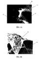
- FIG. 1Ais a photomicrograph showing a section of one embodiment of the tissue repair implant which comprises cartilage fragments, 158 ⁇ m PGA granules, and fibrin glue in accordance with the present invention.
- FIG. 1Bis a photomicrograph of a histological section of an implant similar to FIG. 1A , after 3 weeks in vitro.
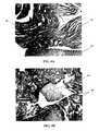
- FIG. 1Cis a photomicrograph of a histological section of an implant similar to FIG. 1A , after 6 weeks in vitro.
- FIG. 1Dis yet another photomicrograph of a histological section of the implant of FIG. 1C .
- FIG. 2Ais a photomicrograph of a histological section of another embodiment of the tissue repair implant comprising cartilage fragments, 286 ⁇ m PGA granules, and fibrin glue in accordance with the present invention.
- FIG. 2Bis a photomicrograph of a histological section of an implant similar to FIG. 2A , after 3 weeks in vitro.
- FIG. 2Cis a photomicrograph of a histological section of an implant similar to FIG. 2A , after 6 weeks in vitro.
- FIG. 2Dis yet another photomicrograph of a histological section of the implant of FIG. 2C .
- FIG. 3Ais a photomicrograph of a histological section at 100 ⁇ magnification of yet another embodiment of a tissue repair implant comprising cartilage fragments, 633 ⁇ m PLA granules and fibrin glue, after 3 weeks in vitro.
- FIG. 3Bis a photomicrograph of a histological section at 100 ⁇ magnification of a tissue repair implant similar to FIG. 3A , after 6 weeks in vitro.
- FIG. 4Ais a photomicrograph of a histological section at 100 ⁇ magnification of a tissue repair implant comprising cartilage fragments and fibrin glue, after 3 weeks in vitro.
- FIG. 4Bis a photomicrograph of a histological section at 100 ⁇ magnification of a tissue repair implant similar to FIG. 4A , after 6 weeks in vitro.
- the inventionrelates to a tissue repair implant that comprises finely minced tissue fragments combined with a tissue carrier matrix formed of a plurality of granules.
- the tissue fragmentsserve as a cell source for new cellular growth, and the tissue fragments have an effective amount of viable cells that can migrate out of the tissue fragment to populate the tissue carrier matrix once the implant is delivered to the patient.
- the granulesserve as a microcarrier to provide sufficient mechanical integrity for cellular integration with the surrounding environment during the tissue remodeling process.
- the finely minced tissue fragments and granulestogether form an injectable suspension that can be delivered by injection to a target site in a minimally invasive procedure.
- the implantis able to conform to any defect size, shape or geometry, and can assume a shape complementary to that of the implantation site.
- this feature of the inventionprovides an implant having a close interface with the tissue area to be repaired, thereby enhancing tissue remodeling and healing.
- the biocompatible tissue implants of the present inventionare used in the treatment of various types of tissue for various purposes.
- the implantscan be used for the remodeling, repair and/or regeneration of diseased or damaged tissue.
- tissue repair implantsthe implants are sometimes referred to herein as “tissue repair implants” and the methods of using the implants are sometimes characterized as tissue repair techniques, it is understood that the implants can be used for a variety of tissue treatments, including but not limited to tissue remodeling, tissue repair, tissue bulking, tissue augmentation, cosmetic treatments, therapeutic treatments, and for tissue sealing.
- the tissue repair implantincludes at least one sample of viable tissue that is associated with at least a portion of the tissue carrier matrix.
- viablerefers to a tissue sample having one or more viable cells.
- the tissue usedcan be obtained from a connective tissue such as cartilage tissue, meniscal tissue, ligament tissue, tendon tissue, skin tissue, bone tissue, muscle tissue, periosteal tissue, pericardial tissue, synovial tissue, nerve tissue, fat tissue, kidney tissue, bone marrow, liver tissue, bladder tissue, pancreas tissue, spleen tissue, intervertebral disc tissue, embryonic tissue, periodontal tissue, vascular tissue, blood and combinations thereof.
- connective tissuesuch as cartilage tissue, meniscal tissue, ligament tissue, tendon tissue, skin tissue, bone tissue, muscle tissue, periosteal tissue, pericardial tissue, synovial tissue, nerve tissue, fat tissue, kidney tissue, bone marrow, liver tissue, bladder tissue, pancreas tissue, spleen tissue, intervertebral disc tissue, embryonic tissue
- the tissueis free of bone tissue and is selected from the group consisting of fibrocartilage tissue containing chondrocytes, meniscal tissue, ligament tissue and tendon tissue.
- the tissue used to construct the tissue implantcan be autogenic tissue, allogeneic tissue, or xenogeneic tissue.
- healthy cartilage tissue, bone marrow tissue or aspiratesare suitable for use with tissue repair implants for repairing condylar surfaces.
- the tissue to be usedcan be of the same type or a different type than the tissue to be treated with the implant.
- the tissue used in the tissue repair implantcan be selected from the group consisting of meniscal tissue, cartilage tissue, skin, synovial tissue, periosteal tissue, pericardial tissue, fat tissue, bone marrow, blood, tendon tissue, ligament tissue, or combinations thereof.
- the tissuecan be obtained using any of a variety of conventional techniques, such as for example, by biopsy or other surgical removal.
- the tissue sampleis obtained under aseptic conditions. Once a sample of living tissue has been obtained, the sample can then be processed under sterile conditions to create a suspension having at least one minced, or finely divided, tissue particle.
- each tissue fragmentcan vary, for example, the tissue size can be in the range of about 0.1 to about 3 mm 3 , in the range of about 0.5 to about 1 mm 3 , in the range of about 1 to about 2 mm 3 , or in the range of about 2 to about 3 mm 3 , but preferably the tissue particle is less than about 1 mm 3 .
- the minced tissue fragmenthas at least one viable cell that can migrate from the tissue fragment into the tissue carrier matrix. More preferably, the tissue contains an effective amount of cells that can migrate from the tissue fragment and begin populating the tissue carrier matrix of granules after implantation.
- the minced tissue fragmentsmay be contacted with a matrix-digesting enzyme to facilitate cell migration out of the extracellular matrix surrounding the cells. The enzymes are used to increase the rate of cell migration out of the extracellular matrix and into the tissue carrier matrix.
- Suitable digesting enzymesthat can be used in the present invention include, but are not limited to, collagenase, metalloproteinase, chondroitinase, trypsin, elastase, hyaluronidase, peptidase, dispase, thermolysin and protease.
- the minced tissue particlescan be formed as a suspension in which the tissue particles are associated with a physiological buffering solution.
- physiological buffering solutionsinclude, but are not limited to, saline, phosphate buffer solution, Hank's balanced salts, Tris buffered saline, Hepes buffered saline and combinations thereof.
- the tissuecan be minced in any standard cell culture medium known to those skilled in the art, either in the presence or absence of serum.
- the minced tissue suspensionPrior to combining the minced tissue fragments with the granules of the tissue carrier matrix, the minced tissue suspension can be filtered and concentrated, such that only a small quantity of physiological buffering solution remains in the suspension to prevent the tissue particles from drying out.
- the minced tissue fragments in solutionare at concentration in the range of approximately 1 to about 100 mg/cm 2 , and more preferably in the range of about 1 to about 20 mg/cm 2 .
- tissue samples used in the present inventionare obtained from a donor (autogenic, allogeneic, or xenogeneic) using appropriate harvesting tools.
- the tissue samplescan be finely minced and divided into small particles either as the tissue is collected, or alternatively, the tissue sample can be minced after it is harvested and collected outside the body. In embodiments where the tissue sample is minced after it is harvested, the tissue samples can be weighed and then washed three times in phosphate buffered saline.
- tissuecan then be minced in the presence of a small quantity, such as, for example, about 1 ml, of a physiological buffering solution, such as, for example, phosphate buffered saline, or a matrix digesting enzyme, such as, for example, 0.2% collagenase in Hams F12.
- a physiological buffering solutionsuch as, for example, phosphate buffered saline, or a matrix digesting enzyme, such as, for example, 0.2% collagenase in Hams F12.
- the mincing actiondivides the tissue sample into particles or small pieces of approximately 1 mm 3 .
- Mincing the tissuecan be accomplished by a variety of methods. In one embodiment, the mincing is accomplished with two sterile scalpels using a parallel direction, and in another embodiment, the tissue can be minced by a processing tool that automatically divides the tissue into particles of a desired size.
- the minced tissuecan be separated from the physiological fluid and concentrated using any of a variety of methods known to those having ordinary skill in the art, such as for example, sieving, sedimenting or centrifuging with the bed of granules.
- the suspension of minced tissuepreferably retains a small quantity of fluid in the suspension to prevent the tissue from drying out.
- the minced tissue fragmentsare combined with a tissue carrier matrix formed of a plurality of granules.
- the granulesare formed from a bioresorbable or bioabsorbable material that has the ability to resorb in a timely fashion in the body.
- the biocompatible, bioresorbable granulesare resorbed to leave behind the new tissue at the implant site.
- the granulescan be formed from a variety of biocompatible, bioresorbable materials.
- the granulescan be formed from aliphatic polyesters, copoly(ether-esters), solid copolymers of fatty acid esters of glycerol and succinic acid, polyoxaesters, collagen, gelatin, albumin, hyaluronate, glucosaminoglycans, polyanhydrides, polyphosphazines, subintestinal mucosa, acellular tissues, and combinations thereof.
- the granulescan be porous and/or have surface features such as roughness or texture. Such features would further enhance the effectiveness of the granules to attach and combine with the minced tissue fragments as well as to the tissue implant site.
- Suitable aliphatic polyestersinclude homopolymers or copolymers of lactides, glycolides, ⁇ -caprolactone, p-dioxanone (1,4-dioxan-2-one), trimethylene carbonate (1,3-dioxan-2-one), and combinations thereof.
- lactidesglycolides, ⁇ -caprolactone, p-dioxanone (1,4-dioxan-2-one), trimethylene carbonate (1,3-dioxan-2-one), and combinations thereof.
- a copolymer of 35:65 ⁇ -caprolactone and glycolide(a relatively fast absorbing polymer) can be blended with 40:60 ⁇ -caprolactone and L-lactide copolymer (a relatively slow absorbing polymer) to form a suitable tissue carrier matrix.
- polyphosphazenesco-, ter- and higher order mixed monomer based polymers made from L-lactide, D,L-lactide, lactic acid, glycolide, glycolic acid, para-dioxanone, trimethylene carbonate and ⁇ -caprolactone such as are described by Allcock in The Encyclopedia of Polymer Science, Vol. 13, pages 31-41, Wiley Intersciences, John Wiley & Sons, 1988 and by Vandorpe, et al in the Handbook of Biodegradable Polymers, edited by Domb, et al., Hardwood Academic Press, pp. 161-182 (1997).
- glycolis understood to include polyglycolic acid.
- lactideis understood to include L-lactide, D-lactide, blends thereof, and lactic acid polymers and copolymers.
- Elastomeric copolymersare also particularly useful in the present invention.
- Suitable elastomeric polymersinclude those with an inherent viscosity in the range of about 1.2 dL/g to 4 dL/g, more preferably about 1.2 dL/g to 2 dL/g and most preferably about 1.4 dL/g to 2 dL/g as determined at 25° C. in a 0.1 gram per deciliter (g/dL) solution of polymer in hexafluoroisopropanol (HFIP).
- suitable elastomersexhibit a high percent elongation and a low modulus, while possessing good tensile strength and good recovery characteristics.
- the elastomerexhibits a percent elongation greater than about 200 percent and preferably greater than about 500 percent.
- suitable elastomersshould also have a tensile strength greater than about 500 psi, preferably greater than about 1,000 psi, and a tear strength of greater than about 50 lbs/inch, preferably greater than about 80 lbs/inch.
- Exemplary biocompatible elastomersthat can be used in the present invention include, but are not limited to, elastomeric copolymers of ⁇ -caprolactone and glycolide (including polyglycolic acid) with a mole ratio of ⁇ -caprolactone to glycolide of from about 35:65 to about 65:35, more preferably from 45:55 to 35:65; elastomeric copolymers of ⁇ -caprolactone and lactide (including L-lactide, D-lactide, blends thereof, and lactic acid polymers and copolymers) where the mole ratio of ⁇ -caprolactone to lactide is from about 35:65 to about 65:35 and more preferably from 45:55 to 30:70 or from about 95:5 to about 85:15; elastomeric copolymers of p-dioxanone (1,4-dioxan-2-one) and lactide (including L-lactide, D-lactide, blends thereof,
- the biocompatible, bioresorbable polymer or copolymer materialis milled to a powder and the particles that are produced serve as the granules. Once milled, the particles or granules can be sieved and sorted by size.
- An appropriate range of sizes for the granules of the present inventionare in the range of about 150 ⁇ m to about 600 ⁇ m in diameter.
- the granulescan have an average outer diameter in the range of about 150 to 600 ⁇ m, and preferably in the range of about 150 to 300 ⁇ m.
- a bed of these beads or granulescan be used to effectively sieve minced tissue fragments from a liquid suspension.
- the granules with the tissue fragmentsform a suspension that can be collected and loaded into an injection device for delivery to an injury or diseased tissue site.
- the granulesact as a carrier and also as a scaffold to support new tissue growth.
- Such a compositioncan conform to any defect geometry, enabling the implant to assume a shape complementary to that of the implantation site and provide enhanced healing.
- the tissue carrier matrixfurther includes a binding agent that acts to gel together or facilitate cohesion of the tissue fragments and granules, thereby creating a cohesive matrix.
- the binding agentenables the implant to take on a semi-solid or gel-like form which helps the suspension retain a given geometry while tissue remodeling occurs.
- the binding agentcould be a gel or biological or synthetic hydrogel so that the implant takes the form of an injectable gel.
- Suitable materials for the binding agentinclude shark cartilage, alginate, hyaluronic acid, collagen gel, fibrin glue, fibrin clot, poly(N-isopropylacrylamide), agarose, chitin, chitosan, cellulose, polysaccharides, poly(oxyalkylene), a copolymer of poly(ethylene oxide)-poly(propylene oxide), poly(vinyl alcohol), polyacrylate, platelet rich plasma (PRP) clot, platelet poor plasma (PPP) clot, Matrigel, blood clot, gelatin-resorcin-formalin adhesives, mussel-based adhesives, dihydroxyphenylalanine (DOPA) based adhesives, transglutaminase, poly(amino acid)-based adhesives, cellulose-based adhesives, polysaccharide-based adhesives, synthetic acrylate-based adhesives, liquid and semi-solid fatty acid esters of glycerol and succinic acid (MGSA
- a curing agentcan additionally be provided with the tissue carrier matrix to allow the injectable implant to set in place at the defect site.
- This curing agentwould act to crosslink the binding agent, thereby forming a solid implant within which are the tissue fragments and the bioresorbable, biocompatible granules.
- the implantis cured once it is delivered to the implantation site. It is contemplated, however, that the implant can be allowed to cure prior to implantation as well, if so desired.
- the curing agentshould be selected so as to effect crosslinking of the particular binding agent contained in the implant.
- Suitable curing agentsinclude, for example, proteases such as thrombin, calcium, divinyl sulfone (DVS), polyethylene glycol divinyl sulfone (VS-PEG-VS), hydroxyethyl methacrylate divinyl sulfone (HEMA-DIS-HEMA), formaldehyde, glutaraldehyde, aldehydes, isocyanates, alkyl and aryl halides, imidoesters, N-substituted maleimides, acylating compounds, carbodiimide, hydroxychloride, N-hydroxysuccinimide, light (e.g., blue light and UV light), pH, temperature, metal ions, and combinations thereof.
- proteasessuch as thrombin, calcium, divinyl sulfone (DVS), polyethylene glycol divinyl sulfone (VS-PEG-VS), hydroxyethyl methacrylate divinyl sulfone (HEMA-
- the tissue carrier matrixcan also include a biological component such as an effector which enhances the effectiveness of the tissue fragments and facilitates tissue repair and healing of the injured tissue.
- the granulescan be formulated to contain an effective molecule that would enhance the activity of the tissue fragments.
- the granulesfunction in multiple ways by sieving the tissue fragments, providing a support and carrier for injection delivery to the site, providing a structural support for tissue remodeling, and potentially delivering other enhancing drug therapeutics to the site.
- the biological componentcan be combined with the tissue carrier matrix in a variety of ways.
- the biological componentcan be contained inside the granules themselves.
- the granulescan be porous to allow the biological component to be contained inside the pores.
- the biological componentcan be contained in a slow-release coating covering the granules.
- the biological componentcan be incorporated into the granules by any suitable manner known in the art that allows the granules to administer the biological component to the minced tissue fragments, without affecting the effectiveness of the biological component.
- the biological componentcan be selected from among a variety of effectors that, when present at the site of injury, promotes healing and/or regeneration of the affected tissue.
- the effectorsmay also include compounds or agents that prevent infection (e.g., antimicrobial agents and antibiotics), compounds or agents that reduce inflammation (e.g., anti-inflammatory agents), compounds that prevent or minimize adhesion formation, such as oxidized regenerated cellulose (e.g., INTERCEED and Surgicel®, available from Ethicon, Inc.), hyaluronic acid, and compounds or agents that suppress the immune system (e.g., immunosuppressants).
- infectionse.g., antimicrobial agents and antibiotics
- compounds or agents that reduce inflammatione.g., anti-inflammatory agents
- compounds that prevent or minimize adhesion formationsuch as oxidized regenerated cellulose (e.g., INTERCEED and Surgicel®, available from Ethicon, Inc.), hyaluronic acid, and compounds or agents that suppress the immune system
- effectors suitable for use with the implant of the present inventioninclude antibiotics, antimicrobial agents, anti-inflammatory agents, heterologous or autologous growth factors, growth factor fragments, small-molecule wound healing stimulants, proteins (including xenogeneic cartilage and matrix proteins), peptides, antibodies, enzymes, platelets, glycoproteins, hormones, glycosaminoglycans, nucleic acids, analgesics, viruses, virus particles, and cell types. It is understood that one or more effectors of the same or different functionality may be incorporated within the implant.
- suitable effectorsinclude the multitude of heterologous or autologous growth factors known to promote healing and/or regeneration of injured or damaged tissue. These growth factors can be incorporated directly into the biocompatible scaffold, or alternatively, the biocompatible scaffold can include a source of growth factors, such as for example, platelets. Exemplary growth factors include, but are not limited to, TGF- ⁇ , bone morphogenic protein, cartilage-derived morphogenic protein, fibroblast growth factor, platelet-derived growth factor, vascular endothelial cell-derived growth factor (VEGF), epidermal growth factor, insulin-like growth factor, hepatocyte growth factor, and fragments thereof. Suitable effectors likewise include the agonists and antagonists of the agents noted above. The growth factor can also include combinations of the growth factors listed above.
- the growth factorcan be autologous growth factor that is supplied by platelets in the blood.
- the growth factor from plateletswill be an undefined cocktail of various growth factors. Platelets are normally found in the blood and play a role in hemostasis and wound healing. During clot formation, the platelets become activated and release growth factors such as PDGF, TGF- ⁇ , VEGF, and IGF. Platelets can be separated from blood using techniques such as centrifugation. When platelet rich plasma (PRP) is combined with an activator, a platelet clot is created.
- PRPplatelet rich plasma
- An activatorcan be, but is not limited to, thrombin, calcium, adenosine di-phosphate (ADP), collagen, epinephrine, arachidonic acid, prostaglandin E2, ristocetin, calcium, retinoids, ascorbate, antioxidants, and combinations thereof.
- ADPadenosine di-phosphate
- collagenepinephrine
- arachidonic acidepinephrine
- prostaglandin E2ristocetin
- calciumretinoids
- ascorbateantioxidants, and combinations thereof.
- the proteins that may be present within the implantinclude proteins that are secreted from a cell or other biological source, such as for example, a platelet, which is housed within the implant, as well as those that are present within the implant in an isolated form.
- the isolated form of a proteintypically is one that is about 55% or greater in purity, i.e., isolated from other cellular proteins, molecules, debris, etc. More preferably, the isolated protein is one that is at least 65% pure, and most preferably one that is at least about 75 to 95% pure. Notwithstanding the above, one of ordinary skill in the art will appreciate that proteins having a purity below about 55% are still considered to be within the scope of this invention.
- proteinembraces glycoproteins, lipoproteins, proteoglycans, peptides, and fragments thereof.
- proteins useful as effectorsinclude, but are not limited to, pleiotrophin, endothelin, tenascin, fibronectin, fibrinogen, vitronectin, V-CAM, I-CAM, N-CAM, selectin, cadherin, integrin, laminin, actin, myosin, collagen, microfilament, intermediate filament, antibody, elastin, fibrillin, tissue inhibitor of metalloproteinases (TIMPs), and fragments thereof.
- TRIPstissue inhibitor of metalloproteinases
- Glycosaminoglycansmay also serve as effectors according to the present invention.
- Exemplary glycosaminoglycans useful as effectorsinclude, but are not limited to, heparan sulfate, heparin, chondroitin sulfate, dermatan sulfate, keratan sulfate, hyaluronan (also known as hyaluronic acid), and combinations thereof.
- the tissue repair implant of the present inventioncan also have cells incorporated therein.
- Suitable cell types that can serve as effectors according to this inventioninclude, but are not limited to, osteocytes, osteoblasts, osteoclasts, fibroblasts, stem cells, pluripotent cells, chondrocyte progenitors, chondrocytes, endothelial cells, macrophages, leukocytes, adipocytes, monocytes, plasma cells, mast cells, umbilical cord cells, stromal cells, mesenchymal stem cells, epithelial cells, myoblasts, tenocytes, ligament fibroblasts, neurons, and bone marrow cells.
- Cellstypically have at their surface receptor molecules which are responsive to a cognate ligand (e.g., a stimulator).
- a stimulatoris a ligand which, when in contact with its cognate receptor, induces the cell possessing the receptor to produce a specific biological action.
- a cellmay produce significant levels of secondary messengers, like Ca +2 , which then will have subsequent effects upon cellular processes such as the phosphorylation of proteins, such as (keeping with our example) protein kinase C.
- the cellsecretes a cellular messenger usually in the form of a protein (including glycoproteins, proteoglycans, and lipoproteins).
- This cellular messengercan be an antibody (e.g., secreted from plasma cells), a hormone, (e.g., a paracrine, autocrine, or exocrine hormone), a cytokine, or natural or synthetic fragments thereof.
- the tissue implant of the present inventioncan also be used in gene therapy techniques in which nucleic acids, viruses, or virus particles deliver a gene of interest, which encodes at least one gene product of interest, to specific cells or cell types.
- the biological effectorcan be a nucleic acid (e.g., DNA, RNA, or an oligonucleotide), a virus, a virus particle, or a non-viral vector.
- the viruses and virus particlesmay be, or may be derived from, DNA or RNA viruses.
- the gene product of interestis preferably selected from the group consisting of proteins, polypeptides, interference ribonucleic acids (iRNA) and combinations thereof.
- the implantcan then be implanted into a particular site to elicit a type of biological response.
- the nucleic acid or viral agentcan then be taken up by the cells and any proteins that they encode can be produced locally by the cells.
- the nucleic acid or viral agentcan be taken up by the cells within the tissue fragment of the minced tissue suspension, or, in an alternative embodiment, the nucleic acid or viral agent can be taken up by the cells in the tissue surrounding the site of the injured tissue.
- the protein producedcan be a protein of the type noted above, or a similar protein that facilitates an enhanced capacity of the tissue to heal an injury or a disease, combat an infection, or reduce an inflammatory response.
- Nucleic acidscan also be used to block the expression of unwanted gene product that may impact negatively on a tissue repair process or other normal biological processes.
- DNA, RNA and viral agentsare often used to accomplish such an expression blocking function, which is also known as gene expression knock out.
- the identity of the biological componentmay be determined by a surgeon, based on principles of medical science and the applicable treatment objectives.
- the biocompatible, bioresorbable polymer or copolymer materialis milled to form a powder.
- the particles of the resultant powder, which serve as the granules of the implant,are then sorted by size.
- the sorted granulesare set aside, while a tissue sample containing viable cells is obtained.
- the tissue sampleis minced, producing a fluid suspension containing the minced tissue fragments.
- the fluid suspensionis then introduced to a bed of the granules of a selected range of sizes.
- the bed of granulesfunctions to sieve the fluid suspension, separating the fluid from the tissue fragments, resulting in a slurry or suspension containing both the granules and the tissue fragments.
- the slurry, which forms the tissue repair implantcan then be collected and inserted into a delivery device such as an injection device for immediate delivery to an injury or diseased tissue site.
- the tissue fragmentsserve as a cell source for new cellular growth, and have an effective amount of viable cells that can migrate out of the tissue fragment to populate the tissue carrier matrix once the implant is delivered to the patient.
- the granulesserve as a microcarrier to provide sufficient mechanical integrity for cellular integration with the surrounding environment during the tissue remodeling process. Because the implant is in a suspension form, the implant can conform to any defect geometry and assume a shape complementary to that of the implantation site. The close interface between the injected implant and the tissue site thus enhances tissue repair and healing.
- the tissue carrier matrixcan also include a binding agent that acts to gel together the tissue fragments and granules and thereby form a cohesive matrix.
- the binding agentcan be added to the matrix to enable the implant to take on a gel-like or semi-solid form which enhances retention of the implant at the tissue site while tissue remodeling is occurring.
- a curing agentcan additionally be provided with the tissue carrier matrix. This curing agent would act to crosslink (i.e., form covalent bonds with) the binding agent, thereby forming a solid implant within which are the tissue fragments and the bioresorbable, biocompatible granules.
- the curing agentis added to the implant after delivery to the implantation site so that the implant is set after delivery.
- the curing agentis added prior to delivery of the implant to the site, resulting in a cured implant being delivered.
- the tissue carrier matrixcan also include a biological component or effector which enhances the effectiveness of the tissue fragments to new cellular growth.
- the tissue carrier matrixcan be placed in a suitable container comprising the biological component prior to surgical placement at the tissue site. After an appropriate time and under suitable conditions, the granules can become impregnated with the biological component.
- an implant in which the tissue carrier matrix is devoid of any biological componentcan be infused with biological agent(s), or an implant in which the matrix includes at least one biological component can be augmented with a supplemental quantity of the biological component.
- Another exemplary method of incorporating a biological component within a surgically installed implantis by injection using an appropriately gauged syringe.
- the amount of the biological component included with the tissue repair implantwill vary depending on a variety of factors, including the size of the injury or defect site, the identity of the biological component, and the intended purpose of the tissue repair implant.
- One skilled in the artcan readily determine the appropriate quantity of biological component to include within an implant for a given application in order to facilitate and/or expedite the healing of tissue.
- the amount of biological componentwill, of course, vary depending upon the identity of the biological component and the given application.
- solidse.g., barium sulfate
- the solids that may be addedalso include those that will promote tissue regeneration or regrowth, as well as those that act as buffers, reinforcing materials or porosity modifiers.
- the tissue repair implantcan be used in the treatment of a tissue injury, such as injury to a ligament, tendon, nerve, skin, cartilage or meniscus. Repairing tissue injuries involves the steps of obtaining a sample of living tissue by any of the variety of techniques known to those skilled in the art, preferably by biopsy or other minimally invasive techniques. The sample of living tissue is then processed under sterile conditions to create at least one minced, finely divided tissue particle, combining the tissue fragment with a plurality of biocompatible, bioresorbable granules to form a suspension of tissue and granules, and injecting the suspension at a tissue injury site to deliver the tissue repair implant to the tissue injury. Additionally, the tissue repair implant can be allowed to set or cure into a shape complementary to the geometry of the defect site.
- the suspensioncan also be introduced into a mold and allowed to set prior to implantation at the defect site.
- the moldcan have a geometry and dimension matching that of the defect site. It is contemplated that a specialized surgical tool can be used to prepare the defect area so that the implantation site has a defined geometry. Once cured, the implant can be further trimmed and shaped as necessary before implantation.
- a patient diagnosed with a symptomatic articular cartilage defectis prepped for arthroscopic surgery.
- the surgeonthen harvests healthy cartilage tissue from a non-weight bearing area of the patient's joint using a harvesting instrument.
- the instrumentalso allows the surgeon to mince the cartilage tissue and collect the minced tissue fragments in a separate chamber of the instrument which is preloaded with the milled granules.
- the surgeoncan inject a binding agent such as a biological glue into the chamber to prepare a mixture containing the cartilage fragments, granules and binding agent.
- a curing agentcan also be introduced to the chamber at this time.
- the mixtureWhile the mixture is being prepared, careful debridement of the affected area can be performed to remove unhealthy tissue from the cartilage defect and prepare the area to receive the mixture.
- the formed mixturecan then be loaded into an injection device such as a specialized syringe to arthroscopically inject the mixture into the affected area.
- an injection devicesuch as a specialized syringe to arthroscopically inject the mixture into the affected area.
- the mixturecan be shaped or sculpted to fill the defect area and the surrounding tissue before the mixture has cured. Once cured, the arthroscopic ports can be sutured closed, and the patient can then begin a controlled rehabilitation program.
- the methods of repairing tissue injuries using the tissue implants according to the present inventioncan be conducted during a surgical procedure to repair the tissue injury.
- the steps of processing the tissue sample to create minced, finely divided tissue particles, depositing the tissue particles upon the scaffold to form a tissue repair implant, and/or incubating the tissue repair implant prior to implantationcan be conducted at another, sterile location prior to surgical placement of the implant relative to the site of injury.
- the primary objective of this studywas to examine the outgrowth of chondrocytes in a sample implant comprising bovine cartilage fragments, fibrin glue and polyglycolic acid (PGA) granules in vitro.
- a sample implantcomprising bovine cartilage fragments, fibrin glue and polyglycolic acid (PGA) granules in vitro.
- minced bovine cartilage and PGA granules of different sizeswere mixed with fibrin glue (TisseelTM) and injected into a mold to form bioadhesive plugs in accordance with Table 1 below.
- the resulting plugswere then cultured in cell culture or chondrogenic medium for 3 and 6 week periods, respectively. After incubation, the plugs were fixed, sectioned and stained with hematoxylin and eosin (H & E).
- FIG. 1Ashows a photomicrograph of a composite plug 10 with PGA granules 12 of approximately 158 ⁇ m diameter and cartilage pieces 14 in fibrin glue 16 , made in accordance with the first condition of Table 1.
- FIG. 1Bshows a histological section of a composite plug in accordance with the first condition of Table 1, H & E stained, after 3 weeks. Slight cell growth is seen. After 6 weeks, however, the PGA granules show some resorption while the fibrin glue also indicates absorption and/or degradation.
- FIGS. 1C and 1Dwhich are histological sections of a composite plug made in accordance with the first condition of Table 1, H & E stained, after 6 weeks, there appears to be chondrocyte outgrowth 18 around the cartilage fragments 14 .
- FIG. 2Ashows a photomicrograph of a composite plug 20 with PGA granules 22 of approximately 286 ⁇ m diameter and cartilage pieces 24 in fibrin glue 26 , made in accordance with the second condition of Table 1.
- FIG. 2Bshows a histological section of a composite plug in accordance with the second condition of Table 1, H & E stained, after 3 weeks. Similar to FIG. 1B , slight cell growth is seen. After 6 weeks, however, the PGA granules show some resorption while the fibrin glue also indicates absorption and/or degradation.
- FIGS. 2C and 2Dwhich are histological sections, H & E stained, after 6 weeks of a composite plug made in accordance with the second condition of Table 1, there appears to be chondrocyte outgrowth 28 around the cartilage fragments 24 .
- FIG. 3Ashows a histological section of a composite plug 30 made in accordance with the third condition of Table 1, containing cartilage fragments 34 and PLA granules 32 of approximately 633 ⁇ m diameter embedded in fibrin glue 36 , stained with H & E after 3 weeks, at 100 ⁇ magnification.
- FIG. 3Bshows a histological section of the composite plug 30 after 6 weeks. As shown, there is some outgrowth of chondrocytes 38 around the granules 32 and glue 36 after 6 weeks.
- FIGS. 4A and 4Bshow histological sections of a composite plug 40 made in accordance with the fourth condition of Table 1, containing cartilage fragments 44 embedded in fibrin glue 46 , stained with H & E after 3 weeks and 6 weeks, respectively, at 100 ⁇ magnification. At 6 weeks, FIG. 4B shows some cell outgrowth.
- bovine chondrocytes from minced cartilagewere able to migrate in and around the fibrin glue bioadhesive plug within 6 weeks. It further appears that the presence of the polyglycolic acid (PGA) granules embedded within the fibrin glue plugs facilitated the migration process. Finally, it appears that the smaller sized PGA granules (mean particle diameter 158 ⁇ m and 286 ⁇ m) were more effective in facilitating cell attachment and in inducing cellular migration to contribute to the overall tissue remodeling process.
- PGApolyglycolic acid
Landscapes
- Health & Medical Sciences (AREA)
- Life Sciences & Earth Sciences (AREA)
- Chemical & Material Sciences (AREA)
- Engineering & Computer Science (AREA)
- Animal Behavior & Ethology (AREA)
- Medicinal Chemistry (AREA)
- Veterinary Medicine (AREA)
- Public Health (AREA)
- General Health & Medical Sciences (AREA)
- Chemical Kinetics & Catalysis (AREA)
- Biomedical Technology (AREA)
- Dermatology (AREA)
- Pharmacology & Pharmacy (AREA)
- Organic Chemistry (AREA)
- Nuclear Medicine, Radiotherapy & Molecular Imaging (AREA)
- General Chemical & Material Sciences (AREA)
- Transplantation (AREA)
- Epidemiology (AREA)
- Oral & Maxillofacial Surgery (AREA)
- Bioinformatics & Cheminformatics (AREA)
- Botany (AREA)
- Zoology (AREA)
- Physical Education & Sports Medicine (AREA)
- Molecular Biology (AREA)
- Cell Biology (AREA)
- Immunology (AREA)
- Rheumatology (AREA)
- Orthopedic Medicine & Surgery (AREA)
- Vascular Medicine (AREA)
- Urology & Nephrology (AREA)
- Communicable Diseases (AREA)
- Hematology (AREA)
- Diabetes (AREA)
- Biochemistry (AREA)
- Neurology (AREA)
- Pain & Pain Management (AREA)
- Oncology (AREA)
- Toxicology (AREA)
- Materials For Medical Uses (AREA)
- Micro-Organisms Or Cultivation Processes Thereof (AREA)
Abstract
Description
The present invention is a continuation of U.S. patent application Ser. No. 12/980,544, filed on Dec. 29, 2010, entitled “Conformable Tissue Repair Implant Capable of Injection Delivery,” now U.S. Pat. No. 8,137,702, and a continuation of U.S. patent application Ser. No. 11/947,384, filed on Nov. 29, 2007, entitled “Conformable Tissue Repair Implant Capable of Injection Delivery,” now U.S. Pat. No. 7,875,296, and a continuation of U.S. patent application Ser. No. 10/723,982, filed on Nov. 26, 2003, entitled “Conformable Tissue Repair Implant Capable of Injection Delivery,” now U.S. Pat. No. 7,316,822. The entire contents of these applications are incorporated herein by reference.
Not applicable.
The present invention relates to methods and apparatus for the treatment of tissue injuries or defects. Specifically, the present invention relates to tissue repair and augmentation implants, and more particularly, to a conformable tissue repair and augmentation implant capable of injection and a method for its minimally invasive delivery.
Injuries to soft tissue, such as cartilage, skin, muscle, bone, tendon and ligament, where the tissue has been injured or traumatized frequently require surgical intervention to repair the damage and facilitate healing. Such surgical repairs can include suturing or otherwise repairing the damaged tissue with known medical devices, augmenting the damaged tissue with other tissue, using an implant, a graft or any combination of these techniques. Despite these conventional methods of tissue repair, there is a continuing need in this art for novel surgical techniques for the surgical treatment of damaged tissue (e.g., cartilage, meniscal cartilage, ligaments, tendons and skin) that can effect a more reliable tissue repair over the long term and can facilitate the healing of injured tissue.
Recently, tissue engineering approaches to repairing tissue damage or injury have been used with increasing frequency. These methods typically involve replacing or reconstructing damaged or injured tissue with cells capable of new tissue growth. The cells are usually incorporated into a delivery vehicle such as a surgical implant for placement at the tissue site, whereupon the healthy cells can grow into their surrounding environment. Various surgical implants are known and have been used in surgical procedures to help achieve these benefits. For example, it is known to use various devices and techniques for creating implants having isolated cells loaded onto a delivery vehicle. Such cell-seeded implants are used in an in vitro method of making and/or repairing cartilage by growing cartilaginous structures that consist of chondrocytes seeded onto biodegradable, biocompatible fibrous polymeric matrices. Such methods require the initial isolation of chondrocytes from cartilaginous tissue prior to the chondrocytes being seeded onto the polymeric matrices. Other techniques for repairing damaged tissue employ implants having stem or progenitor cells that are used to produce the desired tissue. For example, it is known to use stem or progenitor cells, such as the cells within fatty tissue, muscle, or bone marrow, to regenerate bone and/or cartilage in animal models. The stem cells are removed from the animal and placed in an environment favorable to cartilage formation, thereby inducing the fatty tissue cells to proliferate and to create a different type of cell, such as cartilage cells.
While the trend towards using tissue engineering approaches to tissue repair continues to gain popularity, mainly because of the long-term benefits provided to the patient, these current techniques are not without drawbacks. One disadvantage with current tissue engineering techniques is that they can be time consuming. A typical process involves the harvest of cellular tissue in a first surgical procedure, which is then transported to a laboratory for cell culturing and amplification. The tissue sample is treated with enzymes that will release the cells from the matrix, and the isolated cells will be grown for a period of 3 to 4 weeks using standard cell culture techniques. Once the cell population has reached a target number, the cells are sent back to the surgeon for implantation during a second surgical procedure. This manual labor-intense process is extremely costly and time consuming. Although the clinical data suggest long term benefits for the patient, the prohibitive cost of the procedure combined with the traumatic impact of two surgical procedures, has hampered adoption of this technique.
The current model for tissue repair generally involves retrieving a cell sample from a patient, isolating the cells, culturing the cells for several weeks, and then implanting them in a defect, either with or without a scaffold. Preferably, a scaffold is used in order to facilitate newly developing cell growth. In the past, such scaffolds have consisted mostly of two- or three-dimensional porous scaffolds that allow cell invasion and remodeling once the scaffold has been combined with living cells and has been delivered inside the patient. This model is limited in application because of the secondary surgery and high costs involved. More importantly, one limitation of using such scaffolds is that tissue defect geometry can often be unpredictable. Since the scaffold geometry is essentially limited to what has been manufactured, the scaffold carrier to be implanted rarely matches perfectly the site. In order to achieve a desirable complementary fit with the defect or injury site, the scaffold often needs to be revised by trimming prior to or after implantation. This additional adjustment time adds onto the overall surgery time for the patient. For certain difficult to match or unusually shaped sites, even the step of trimming the scaffold does not ensure an ideal fit with the implantation site. Further, where relatively large tissue defects are involved, minimally invasive surgery may not be possible due to the limited size of the surgical access site. Therefore, delivery of large scaffolds may require an open procedure which poses more risks to the patient.
Injectable gels and microcarrier beads have also been used in the past as cell delivery vehicles. These systems have the advantage of sometimes being injectable and therefore require less invasive procedures for implantation. Typically, these carriers have been combined with isolated cells, which are sensitive to manipulation such as shear, or the presence of crosslinkers that are required to allow the carrier to be fixed or set in place. Hence, these systems have proven to be less than ideal due to the problems associated with cell viability once incorporated into these carrier systems. Accordingly, there continues to exist a need in this art for a method of delivering tissue repair implants through a minimally invasive procedure. Also desirable is a conformable tissue repair or augmentation implant that can adapt to the shape or geometry of the tissue site. The implant should be suitable for delivering viable tissue capable of effecting new cell growth. It is also desirable to provide a method for making such an implant, whereby the implant can be made in a quick and efficient manner for immediate use during surgery.
This invention relates to a conformable tissue implant for use in treating injured or defective tissue, and a method for delivering such an implant in a minimally invasive procedure. The implant is configured to be introduced to the tissue site, where it can assume the shape or geometry of the tissue defect or injury site, thereby providing a close interface between the implant and the tissue site which enhances healing and promotes new cellular growth to occur. The biocompatible tissue implant can be used for the repair, augmentation and/or regeneration of diseased or damaged tissue. Further, the tissue implant can be used for tissue bulking, cosmetic treatments, therapeutic treatments, tissue augmentation, as well as tissue repair.
The tissue repair implant of the present invention comprises finely minced tissue fragments combined with a tissue carrier matrix formed of biocompatible, bioresorbable granules. The tissue fragments can be derived from a number of sources, including connective tissue such as cartilage, meniscus, tendon, ligament, dermis, bone, or combinations thereof. In addition, the tissue fragments can be autogenic tissue, allogeneic tissue, xenogeneic tissue, or combinations thereof. The tissue fragments serve as a cell source for new cellular growth, and have an effective amount of viable cells that can migrate out of the tissue fragment and populate the tissue carrier matrix once the implant is delivered to the patient. The granules serve as a microcarrier to provide sufficient mechanical integrity for cellular integration with the surrounding environment during the tissue remodeling process. In one aspect of the present invention, the finely minced tissue fragments and granules together form an injectable suspension that can be delivered by injection in a minimally invasive procedure. Over time, the plurality of biocompatible, bioresorbable granules are resorbed to leave behind the new tissue at the implant site.
In one embodiment of the present invention, the tissue carrier matrix further includes a binding agent that acts to gel together or facilitate cohesion of the tissue fragments and granules within the tissue carrier matrix. The binding agent enables the implant to take on a semi-solid or gel-like form. Where a solid or cured implant is desired, a curing agent can additionally be provided with the tissue carrier matrix. This curing agent would act to crosslink the binding agent, thereby forming a solid implant within which are the tissue fragments and the bioresorbable, biocompatible granules. In one aspect, the implant is cured once it is delivered to the implantation site. In another aspect, the implant is cured prior to its delivery to the implantation site. To further enhance the implant's regenerative or reconstructive abilities, the tissue carrier matrix can also include a biological component or effector which enhances the effectiveness of the tissue fragments to new cellular growth.
The invention also provides to a method of repairing a tissue defect or injury which involves the steps of providing a tissue repair implant in accordance with the present invention and delivering the tissue repair implant to a tissue defect or injury site. In one aspect, the step of delivering includes injecting the tissue repair implant into the tissue defect site. The tissue carrier matrix can also include a curing agent, and the method of the present invention can further include the step of allowing the tissue repair implant to set at the tissue defect site. Alternatively, the tissue repair implant can be allowed to set prior to delivering the tissue repair implant to the tissue defect or injury site. At least one tissue fragment associated with the tissue carrier matrix comprises a type that is the same as the tissue to be treated. However, the tissue fragment can also comprise a type that is different from the tissue to be treated.
The invention also provides a method of preparing a tissue repair implant in accordance with the present invention, which involves the steps of providing a tissue carrier matrix comprising a plurality of biocompatible, bioresorbable granules, introducing a fluid suspension containing at least one tissue fragment to the tissue carrier matrix, the tissue fragment having an effective amount of viable cells capable of migrating out of the tissue fragment and into the tissue carrier matrix, separating the at least one tissue fragment from the fluid suspension, and collecting the tissue carrier matrix with the at least one tissue fragment for implantation at a tissue site to be repaired. The tissue carrier matrix can be provided with a binding agent which enables the implant to form a gel-like or semi-solid implant. A curing agent can additionally be provided to enable the implant to set either before or after delivery to the implantation site. A biological component or an effector can also be added to the tissue carrier matrix to further enhance the effectiveness of the tissue fragments.
The tissue implant of the present invention aims to accomplish several tasks simultaneously in order to provide more efficient delivery of a tissue repair implant to a site of tissue injury or defect. The invention combines the utility of sieving or capturing a biological agent in a carrier, with the advantage of being able to immediately use the biological agent in an intraoperative procedure, in order to deliver a conformable tissue implant loaded with tissue fragments containing living cells to a tissue implantation site. Another advantage provided by the tissue repair implant of the present invention is that there is no need to isolate cells, nor is there a need to grow tissue or attach cells to the carrier prior to delivering the implant to the implantation site. Also, by using a carrier comprising bioabsorbable, biocompatible granules, the implant is able to combine sustained drug delivery capabilities and structural integrity provided by a scaffold support with the convenience of injection delivery.
In embodiments in which the implant is used for tissue repair, the tissue repair implant can be used to treat a variety of injuries, such as injuries occurring within the musculoskeletal system (e.g., rotator cuff injuries, ACL ruptures, and meniscal tears), as well as injuries occurring in other connective tissues, such as skin and cartilage. Furthermore, such implants can be used in other orthopaedic surgical procedures, such as hand and foot surgery, to repair tissues such as ligaments, nerves, and tendons.
In general, the invention relates to a tissue repair implant that comprises finely minced tissue fragments combined with a tissue carrier matrix formed of a plurality of granules. The tissue fragments serve as a cell source for new cellular growth, and the tissue fragments have an effective amount of viable cells that can migrate out of the tissue fragment to populate the tissue carrier matrix once the implant is delivered to the patient. The granules serve as a microcarrier to provide sufficient mechanical integrity for cellular integration with the surrounding environment during the tissue remodeling process. In one aspect of the present invention, the finely minced tissue fragments and granules together form an injectable suspension that can be delivered by injection to a target site in a minimally invasive procedure. By providing the implant in a suspension form, the implant is able to conform to any defect size, shape or geometry, and can assume a shape complementary to that of the implantation site. Ultimately, this feature of the invention provides an implant having a close interface with the tissue area to be repaired, thereby enhancing tissue remodeling and healing.
The biocompatible tissue implants of the present invention are used in the treatment of various types of tissue for various purposes. For example, the implants can be used for the remodeling, repair and/or regeneration of diseased or damaged tissue. Although the implants are sometimes referred to herein as “tissue repair implants” and the methods of using the implants are sometimes characterized as tissue repair techniques, it is understood that the implants can be used for a variety of tissue treatments, including but not limited to tissue remodeling, tissue repair, tissue bulking, tissue augmentation, cosmetic treatments, therapeutic treatments, and for tissue sealing.
The tissue repair implant includes at least one sample of viable tissue that is associated with at least a portion of the tissue carrier matrix. The term “viable,” as used herein, refers to a tissue sample having one or more viable cells. Virtually any type of tissue can be used to construct the tissue repair implants of the present invention. For example, the tissue used can be obtained from a connective tissue such as cartilage tissue, meniscal tissue, ligament tissue, tendon tissue, skin tissue, bone tissue, muscle tissue, periosteal tissue, pericardial tissue, synovial tissue, nerve tissue, fat tissue, kidney tissue, bone marrow, liver tissue, bladder tissue, pancreas tissue, spleen tissue, intervertebral disc tissue, embryonic tissue, periodontal tissue, vascular tissue, blood and combinations thereof. In one embodiment useful for cartilage repair, the tissue is free of bone tissue and is selected from the group consisting of fibrocartilage tissue containing chondrocytes, meniscal tissue, ligament tissue and tendon tissue. The tissue used to construct the tissue implant can be autogenic tissue, allogeneic tissue, or xenogeneic tissue. For example, healthy cartilage tissue, bone marrow tissue or aspirates are suitable for use with tissue repair implants for repairing condylar surfaces. It is also contemplated that the tissue to be used can be of the same type or a different type than the tissue to be treated with the implant.
In one embodiment useful for meniscal repair, the tissue used in the tissue repair implant can be selected from the group consisting of meniscal tissue, cartilage tissue, skin, synovial tissue, periosteal tissue, pericardial tissue, fat tissue, bone marrow, blood, tendon tissue, ligament tissue, or combinations thereof. The tissue can be obtained using any of a variety of conventional techniques, such as for example, by biopsy or other surgical removal. Preferably, the tissue sample is obtained under aseptic conditions. Once a sample of living tissue has been obtained, the sample can then be processed under sterile conditions to create a suspension having at least one minced, or finely divided, tissue particle. The particle size of each tissue fragment can vary, for example, the tissue size can be in the range of about 0.1 to about 3 mm3, in the range of about 0.5 to about 1 mm3, in the range of about 1 to about 2 mm3, or in the range of about 2 to about 3 mm3, but preferably the tissue particle is less than about 1 mm3.
Preferably, the minced tissue fragment has at least one viable cell that can migrate from the tissue fragment into the tissue carrier matrix. More preferably, the tissue contains an effective amount of cells that can migrate from the tissue fragment and begin populating the tissue carrier matrix of granules after implantation. In an optional embodiment, the minced tissue fragments may be contacted with a matrix-digesting enzyme to facilitate cell migration out of the extracellular matrix surrounding the cells. The enzymes are used to increase the rate of cell migration out of the extracellular matrix and into the tissue carrier matrix. Suitable digesting enzymes that can be used in the present invention include, but are not limited to, collagenase, metalloproteinase, chondroitinase, trypsin, elastase, hyaluronidase, peptidase, dispase, thermolysin and protease.
In one embodiment, the minced tissue particles can be formed as a suspension in which the tissue particles are associated with a physiological buffering solution. Suitable physiological buffering solutions include, but are not limited to, saline, phosphate buffer solution, Hank's balanced salts, Tris buffered saline, Hepes buffered saline and combinations thereof. In addition, the tissue can be minced in any standard cell culture medium known to those skilled in the art, either in the presence or absence of serum. Prior to combining the minced tissue fragments with the granules of the tissue carrier matrix, the minced tissue suspension can be filtered and concentrated, such that only a small quantity of physiological buffering solution remains in the suspension to prevent the tissue particles from drying out. Preferably, the minced tissue fragments in solution are at concentration in the range of approximately 1 to about 100 mg/cm2, and more preferably in the range of about 1 to about 20 mg/cm2.
The tissue samples used in the present invention are obtained from a donor (autogenic, allogeneic, or xenogeneic) using appropriate harvesting tools. The tissue samples can be finely minced and divided into small particles either as the tissue is collected, or alternatively, the tissue sample can be minced after it is harvested and collected outside the body. In embodiments where the tissue sample is minced after it is harvested, the tissue samples can be weighed and then washed three times in phosphate buffered saline. Approximately 300 to 500 mg of tissue can then be minced in the presence of a small quantity, such as, for example, about 1 ml, of a physiological buffering solution, such as, for example, phosphate buffered saline, or a matrix digesting enzyme, such as, for example, 0.2% collagenase in Hams F12. The mincing action divides the tissue sample into particles or small pieces of approximately 1 mm3. Mincing the tissue can be accomplished by a variety of methods. In one embodiment, the mincing is accomplished with two sterile scalpels using a parallel direction, and in another embodiment, the tissue can be minced by a processing tool that automatically divides the tissue into particles of a desired size. In another embodiment, the minced tissue can be separated from the physiological fluid and concentrated using any of a variety of methods known to those having ordinary skill in the art, such as for example, sieving, sedimenting or centrifuging with the bed of granules. In embodiments where the minced tissue is filtered and concentrated, the suspension of minced tissue preferably retains a small quantity of fluid in the suspension to prevent the tissue from drying out.
In the present invention, the minced tissue fragments are combined with a tissue carrier matrix formed of a plurality of granules. Preferably, the granules are formed from a bioresorbable or bioabsorbable material that has the ability to resorb in a timely fashion in the body. Over time, the biocompatible, bioresorbable granules are resorbed to leave behind the new tissue at the implant site. The granules can be formed from a variety of biocompatible, bioresorbable materials. For example, the granules can be formed from aliphatic polyesters, copoly(ether-esters), solid copolymers of fatty acid esters of glycerol and succinic acid, polyoxaesters, collagen, gelatin, albumin, hyaluronate, glucosaminoglycans, polyanhydrides, polyphosphazines, subintestinal mucosa, acellular tissues, and combinations thereof. In addition, the granules can be porous and/or have surface features such as roughness or texture. Such features would further enhance the effectiveness of the granules to attach and combine with the minced tissue fragments as well as to the tissue implant site.
Suitable aliphatic polyesters include homopolymers or copolymers of lactides, glycolides, ε-caprolactone, p-dioxanone (1,4-dioxan-2-one), trimethylene carbonate (1,3-dioxan-2-one), and combinations thereof. One skilled in the art will appreciate that the differences in the absorption time under in vivo conditions can be the basis for combining two different polymers to form the granules of the present invention. For example, a copolymer of 35:65 ε-caprolactone and glycolide (a relatively fast absorbing polymer) can be blended with 40:60 ε-caprolactone and L-lactide copolymer (a relatively slow absorbing polymer) to form a suitable tissue carrier matrix.
Other useful polymers include polyphosphazenes, co-, ter- and higher order mixed monomer based polymers made from L-lactide, D,L-lactide, lactic acid, glycolide, glycolic acid, para-dioxanone, trimethylene carbonate and εε-caprolactone such as are described by Allcock in The Encyclopedia of Polymer Science, Vol. 13, pages 31-41, Wiley Intersciences, John Wiley & Sons, 1988 and by Vandorpe, et al in the Handbook of Biodegradable Polymers, edited by Domb, et al., Hardwood Academic Press, pp. 161-182 (1997).
As used herein, the term “glycolide” is understood to include polyglycolic acid. Further, the term “lactide” is understood to include L-lactide, D-lactide, blends thereof, and lactic acid polymers and copolymers.
Elastomeric copolymers are also particularly useful in the present invention. Suitable elastomeric polymers include those with an inherent viscosity in the range of about 1.2 dL/g to 4 dL/g, more preferably about 1.2 dL/g to 2 dL/g and most preferably about 1.4 dL/g to 2 dL/g as determined at 25° C. in a 0.1 gram per deciliter (g/dL) solution of polymer in hexafluoroisopropanol (HFIP). Further, suitable elastomers exhibit a high percent elongation and a low modulus, while possessing good tensile strength and good recovery characteristics. In the preferred embodiments of this invention, the elastomer exhibits a percent elongation greater than about 200 percent and preferably greater than about 500 percent. In addition to these elongation and modulus properties, suitable elastomers should also have a tensile strength greater than about 500 psi, preferably greater than about 1,000 psi, and a tear strength of greater than about 50 lbs/inch, preferably greater than about 80 lbs/inch.
Exemplary biocompatible elastomers that can be used in the present invention include, but are not limited to, elastomeric copolymers of ε-caprolactone and glycolide (including polyglycolic acid) with a mole ratio of ε-caprolactone to glycolide of from about 35:65 to about 65:35, more preferably from 45:55 to 35:65; elastomeric copolymers of ε-caprolactone and lactide (including L-lactide, D-lactide, blends thereof, and lactic acid polymers and copolymers) where the mole ratio of ε-caprolactone to lactide is from about 35:65 to about 65:35 and more preferably from 45:55 to 30:70 or from about 95:5 to about 85:15; elastomeric copolymers of p-dioxanone (1,4-dioxan-2-one) and lactide (including L-lactide, D-lactide, blends thereof, and lactic acid polymers and copolymers) where the mole ratio of p-dioxanone to lactide is from about 40:60 to about 60:40; elastomeric copolymers of ε-caprolactone and p-dioxanone where the mole ratio of ε-caprolactone to p-dioxanone is from about from 30:70 to about 70:30; elastomeric copolymers of p-dioxanone and trimethylene carbonate where the mole ratio of p-dioxanone to trimethylene carbonate is from about 30:70 to about 70:30; elastomeric copolymers of trimethylene carbonate and glycolide (including polyglycolic acid) where the mole ratio of trimethylene carbonate to glycolide is from about 30:70 to about 70:30; elastomeric copolymers of trimethylene carbonate and lactide (including L-lactide, D-lactide, blends thereof, and lactic acid polymers and copolymers) where the mole ratio of trimethylene carbonate to lactide is from about 30:70 to about 70:30; and blends thereof. Examples of suitable biocompatible elastomers are described in U.S. Pat. No. 5,468,253.
To form the granules, the biocompatible, bioresorbable polymer or copolymer material is milled to a powder and the particles that are produced serve as the granules. Once milled, the particles or granules can be sieved and sorted by size. An appropriate range of sizes for the granules of the present invention are in the range of about 150 μm to about 600 μm in diameter. As explained in greater detail below, the granules can have an average outer diameter in the range of about 150 to 600 μm, and preferably in the range of about 150 to 300 μm. A bed of these beads or granules can be used to effectively sieve minced tissue fragments from a liquid suspension. The granules with the tissue fragments form a suspension that can be collected and loaded into an injection device for delivery to an injury or diseased tissue site. The granules act as a carrier and also as a scaffold to support new tissue growth. Such a composition can conform to any defect geometry, enabling the implant to assume a shape complementary to that of the implantation site and provide enhanced healing.
In another embodiment of the present invention, the tissue carrier matrix further includes a binding agent that acts to gel together or facilitate cohesion of the tissue fragments and granules, thereby creating a cohesive matrix. The binding agent enables the implant to take on a semi-solid or gel-like form which helps the suspension retain a given geometry while tissue remodeling occurs. For instance, the binding agent could be a gel or biological or synthetic hydrogel so that the implant takes the form of an injectable gel. Suitable materials for the binding agent include shark cartilage, alginate, hyaluronic acid, collagen gel, fibrin glue, fibrin clot, poly(N-isopropylacrylamide), agarose, chitin, chitosan, cellulose, polysaccharides, poly(oxyalkylene), a copolymer of poly(ethylene oxide)-poly(propylene oxide), poly(vinyl alcohol), polyacrylate, platelet rich plasma (PRP) clot, platelet poor plasma (PPP) clot, Matrigel, blood clot, gelatin-resorcin-formalin adhesives, mussel-based adhesives, dihydroxyphenylalanine (DOPA) based adhesives, transglutaminase, poly(amino acid)-based adhesives, cellulose-based adhesives, polysaccharide-based adhesives, synthetic acrylate-based adhesives, liquid and semi-solid fatty acid esters of glycerol and succinic acid (MGSA), MGSA/polyethylene glycol (MGSA/PEG) copolymers, polyvinylpyrolidone (PVP), PVP copolymers, gelatin, albumin, monoglycerides, diglycerides, triglycerides laminin, elastin, proteoglycans, and combinations thereof.
Where a solid or cured implant is desired, a curing agent can additionally be provided with the tissue carrier matrix to allow the injectable implant to set in place at the defect site. This curing agent would act to crosslink the binding agent, thereby forming a solid implant within which are the tissue fragments and the bioresorbable, biocompatible granules. In one aspect, the implant is cured once it is delivered to the implantation site. It is contemplated, however, that the implant can be allowed to cure prior to implantation as well, if so desired. The curing agent should be selected so as to effect crosslinking of the particular binding agent contained in the implant. Suitable curing agents include, for example, proteases such as thrombin, calcium, divinyl sulfone (DVS), polyethylene glycol divinyl sulfone (VS-PEG-VS), hydroxyethyl methacrylate divinyl sulfone (HEMA-DIS-HEMA), formaldehyde, glutaraldehyde, aldehydes, isocyanates, alkyl and aryl halides, imidoesters, N-substituted maleimides, acylating compounds, carbodiimide, hydroxychloride, N-hydroxysuccinimide, light (e.g., blue light and UV light), pH, temperature, metal ions, and combinations thereof. The present invention contemplates that, by using minced tissue fragments or particles rather than isolated cells, the natural environment of the tissue fragments will provide sufficient protection of the cells against the harsh reagents used for setting the implant.
To further enhance the implant's regenerative or reconstructive abilities, the tissue carrier matrix can also include a biological component such as an effector which enhances the effectiveness of the tissue fragments and facilitates tissue repair and healing of the injured tissue. For example, in yet another embodiment of the present invention, the granules can be formulated to contain an effective molecule that would enhance the activity of the tissue fragments. Thus, the granules function in multiple ways by sieving the tissue fragments, providing a support and carrier for injection delivery to the site, providing a structural support for tissue remodeling, and potentially delivering other enhancing drug therapeutics to the site.
The biological component can be combined with the tissue carrier matrix in a variety of ways. For example, the biological component can be contained inside the granules themselves. For instance, the granules can be porous to allow the biological component to be contained inside the pores. Alternatively, the biological component can be contained in a slow-release coating covering the granules. One skilled in the art will recognize that the biological component can be incorporated into the granules by any suitable manner known in the art that allows the granules to administer the biological component to the minced tissue fragments, without affecting the effectiveness of the biological component.
The biological component can be selected from among a variety of effectors that, when present at the site of injury, promotes healing and/or regeneration of the affected tissue. In addition to being compounds or agents that actually promote or expedite healing, the effectors may also include compounds or agents that prevent infection (e.g., antimicrobial agents and antibiotics), compounds or agents that reduce inflammation (e.g., anti-inflammatory agents), compounds that prevent or minimize adhesion formation, such as oxidized regenerated cellulose (e.g., INTERCEED and Surgicel®, available from Ethicon, Inc.), hyaluronic acid, and compounds or agents that suppress the immune system (e.g., immunosuppressants).
By way of example, other types of effectors suitable for use with the implant of the present invention include antibiotics, antimicrobial agents, anti-inflammatory agents, heterologous or autologous growth factors, growth factor fragments, small-molecule wound healing stimulants, proteins (including xenogeneic cartilage and matrix proteins), peptides, antibodies, enzymes, platelets, glycoproteins, hormones, glycosaminoglycans, nucleic acids, analgesics, viruses, virus particles, and cell types. It is understood that one or more effectors of the same or different functionality may be incorporated within the implant.
Examples of suitable effectors include the multitude of heterologous or autologous growth factors known to promote healing and/or regeneration of injured or damaged tissue. These growth factors can be incorporated directly into the biocompatible scaffold, or alternatively, the biocompatible scaffold can include a source of growth factors, such as for example, platelets. Exemplary growth factors include, but are not limited to, TGF-β, bone morphogenic protein, cartilage-derived morphogenic protein, fibroblast growth factor, platelet-derived growth factor, vascular endothelial cell-derived growth factor (VEGF), epidermal growth factor, insulin-like growth factor, hepatocyte growth factor, and fragments thereof. Suitable effectors likewise include the agonists and antagonists of the agents noted above. The growth factor can also include combinations of the growth factors listed above. In addition, the growth factor can be autologous growth factor that is supplied by platelets in the blood. In this case, the growth factor from platelets will be an undefined cocktail of various growth factors. Platelets are normally found in the blood and play a role in hemostasis and wound healing. During clot formation, the platelets become activated and release growth factors such as PDGF, TGF-β, VEGF, and IGF. Platelets can be separated from blood using techniques such as centrifugation. When platelet rich plasma (PRP) is combined with an activator, a platelet clot is created. An activator can be, but is not limited to, thrombin, calcium, adenosine di-phosphate (ADP), collagen, epinephrine, arachidonic acid, prostaglandin E2, ristocetin, calcium, retinoids, ascorbate, antioxidants, and combinations thereof.
The proteins that may be present within the implant include proteins that are secreted from a cell or other biological source, such as for example, a platelet, which is housed within the implant, as well as those that are present within the implant in an isolated form. The isolated form of a protein typically is one that is about 55% or greater in purity, i.e., isolated from other cellular proteins, molecules, debris, etc. More preferably, the isolated protein is one that is at least 65% pure, and most preferably one that is at least about 75 to 95% pure. Notwithstanding the above, one of ordinary skill in the art will appreciate that proteins having a purity below about 55% are still considered to be within the scope of this invention. As used herein, the term “protein” embraces glycoproteins, lipoproteins, proteoglycans, peptides, and fragments thereof. Examples of proteins useful as effectors include, but are not limited to, pleiotrophin, endothelin, tenascin, fibronectin, fibrinogen, vitronectin, V-CAM, I-CAM, N-CAM, selectin, cadherin, integrin, laminin, actin, myosin, collagen, microfilament, intermediate filament, antibody, elastin, fibrillin, tissue inhibitor of metalloproteinases (TIMPs), and fragments thereof.
Glycosaminoglycans, highly charged polysaccharides which play a role in cellular adhesion, may also serve as effectors according to the present invention. Exemplary glycosaminoglycans useful as effectors include, but are not limited to, heparan sulfate, heparin, chondroitin sulfate, dermatan sulfate, keratan sulfate, hyaluronan (also known as hyaluronic acid), and combinations thereof.
The tissue repair implant of the present invention can also have cells incorporated therein. Suitable cell types that can serve as effectors according to this invention include, but are not limited to, osteocytes, osteoblasts, osteoclasts, fibroblasts, stem cells, pluripotent cells, chondrocyte progenitors, chondrocytes, endothelial cells, macrophages, leukocytes, adipocytes, monocytes, plasma cells, mast cells, umbilical cord cells, stromal cells, mesenchymal stem cells, epithelial cells, myoblasts, tenocytes, ligament fibroblasts, neurons, and bone marrow cells. Cells typically have at their surface receptor molecules which are responsive to a cognate ligand (e.g., a stimulator). A stimulator is a ligand which, when in contact with its cognate receptor, induces the cell possessing the receptor to produce a specific biological action. For example, in response to a stimulator (or ligand) a cell may produce significant levels of secondary messengers, like Ca+2, which then will have subsequent effects upon cellular processes such as the phosphorylation of proteins, such as (keeping with our example) protein kinase C. In some instances, once a cell is stimulated with the proper stimulator, the cell secretes a cellular messenger usually in the form of a protein (including glycoproteins, proteoglycans, and lipoproteins). This cellular messenger can be an antibody (e.g., secreted from plasma cells), a hormone, (e.g., a paracrine, autocrine, or exocrine hormone), a cytokine, or natural or synthetic fragments thereof.
The tissue implant of the present invention can also be used in gene therapy techniques in which nucleic acids, viruses, or virus particles deliver a gene of interest, which encodes at least one gene product of interest, to specific cells or cell types. Accordingly, the biological effector can be a nucleic acid (e.g., DNA, RNA, or an oligonucleotide), a virus, a virus particle, or a non-viral vector. The viruses and virus particles may be, or may be derived from, DNA or RNA viruses. The gene product of interest is preferably selected from the group consisting of proteins, polypeptides, interference ribonucleic acids (iRNA) and combinations thereof.
Once the applicable nucleic acids and/or viral agents (i.e., viruses or viral particles) are incorporated into the tissue carrier matrix of the tissue repair implant, the implant can then be implanted into a particular site to elicit a type of biological response. The nucleic acid or viral agent can then be taken up by the cells and any proteins that they encode can be produced locally by the cells. In one embodiment, the nucleic acid or viral agent can be taken up by the cells within the tissue fragment of the minced tissue suspension, or, in an alternative embodiment, the nucleic acid or viral agent can be taken up by the cells in the tissue surrounding the site of the injured tissue. One of ordinary skill in the art will recognize that the protein produced can be a protein of the type noted above, or a similar protein that facilitates an enhanced capacity of the tissue to heal an injury or a disease, combat an infection, or reduce an inflammatory response. Nucleic acids can also be used to block the expression of unwanted gene product that may impact negatively on a tissue repair process or other normal biological processes. DNA, RNA and viral agents are often used to accomplish such an expression blocking function, which is also known as gene expression knock out.
One skilled in the art will appreciate that the identity of the biological component may be determined by a surgeon, based on principles of medical science and the applicable treatment objectives.
In an exemplary method of forming the tissue repair implant of the present invention, the biocompatible, bioresorbable polymer or copolymer material is milled to form a powder. The particles of the resultant powder, which serve as the granules of the implant, are then sorted by size. The sorted granules are set aside, while a tissue sample containing viable cells is obtained. The tissue sample is minced, producing a fluid suspension containing the minced tissue fragments. The fluid suspension is then introduced to a bed of the granules of a selected range of sizes. The bed of granules functions to sieve the fluid suspension, separating the fluid from the tissue fragments, resulting in a slurry or suspension containing both the granules and the tissue fragments. The slurry, which forms the tissue repair implant, can then be collected and inserted into a delivery device such as an injection device for immediate delivery to an injury or diseased tissue site. The tissue fragments serve as a cell source for new cellular growth, and have an effective amount of viable cells that can migrate out of the tissue fragment to populate the tissue carrier matrix once the implant is delivered to the patient. The granules serve as a microcarrier to provide sufficient mechanical integrity for cellular integration with the surrounding environment during the tissue remodeling process. Because the implant is in a suspension form, the implant can conform to any defect geometry and assume a shape complementary to that of the implantation site. The close interface between the injected implant and the tissue site thus enhances tissue repair and healing.
If desired, the tissue carrier matrix can also include a binding agent that acts to gel together the tissue fragments and granules and thereby form a cohesive matrix. The binding agent can be added to the matrix to enable the implant to take on a gel-like or semi-solid form which enhances retention of the implant at the tissue site while tissue remodeling is occurring. Where a solid or cured implant is desired, a curing agent can additionally be provided with the tissue carrier matrix. This curing agent would act to crosslink (i.e., form covalent bonds with) the binding agent, thereby forming a solid implant within which are the tissue fragments and the bioresorbable, biocompatible granules. In one aspect, the curing agent is added to the implant after delivery to the implantation site so that the implant is set after delivery. In another aspect, the curing agent is added prior to delivery of the implant to the site, resulting in a cured implant being delivered.
To further enhance the implant's regenerative or reconstructive abilities, the tissue carrier matrix can also include a biological component or effector which enhances the effectiveness of the tissue fragments to new cellular growth. In an exemplary method for incorporating the biological component, the tissue carrier matrix can be placed in a suitable container comprising the biological component prior to surgical placement at the tissue site. After an appropriate time and under suitable conditions, the granules can become impregnated with the biological component. Following surgical placement, an implant in which the tissue carrier matrix is devoid of any biological component can be infused with biological agent(s), or an implant in which the matrix includes at least one biological component can be augmented with a supplemental quantity of the biological component. Another exemplary method of incorporating a biological component within a surgically installed implant is by injection using an appropriately gauged syringe.
The amount of the biological component included with the tissue repair implant will vary depending on a variety of factors, including the size of the injury or defect site, the identity of the biological component, and the intended purpose of the tissue repair implant. One skilled in the art can readily determine the appropriate quantity of biological component to include within an implant for a given application in order to facilitate and/or expedite the healing of tissue. The amount of biological component will, of course, vary depending upon the identity of the biological component and the given application.
It is also possible to add solids (e.g., barium sulfate) that will render the tissue implants radio opaque. The solids that may be added also include those that will promote tissue regeneration or regrowth, as well as those that act as buffers, reinforcing materials or porosity modifiers.
The tissue repair implant can be used in the treatment of a tissue injury, such as injury to a ligament, tendon, nerve, skin, cartilage or meniscus. Repairing tissue injuries involves the steps of obtaining a sample of living tissue by any of the variety of techniques known to those skilled in the art, preferably by biopsy or other minimally invasive techniques. The sample of living tissue is then processed under sterile conditions to create at least one minced, finely divided tissue particle, combining the tissue fragment with a plurality of biocompatible, bioresorbable granules to form a suspension of tissue and granules, and injecting the suspension at a tissue injury site to deliver the tissue repair implant to the tissue injury. Additionally, the tissue repair implant can be allowed to set or cure into a shape complementary to the geometry of the defect site. In the alternative, it is contemplated that the suspension can also be introduced into a mold and allowed to set prior to implantation at the defect site. The mold can have a geometry and dimension matching that of the defect site. It is contemplated that a specialized surgical tool can be used to prepare the defect area so that the implantation site has a defined geometry. Once cured, the implant can be further trimmed and shaped as necessary before implantation.
In an exemplary method of repairing tissue using the implant of the present invention, a patient diagnosed with a symptomatic articular cartilage defect is prepped for arthroscopic surgery. The surgeon then harvests healthy cartilage tissue from a non-weight bearing area of the patient's joint using a harvesting instrument. Preferably, the instrument also allows the surgeon to mince the cartilage tissue and collect the minced tissue fragments in a separate chamber of the instrument which is preloaded with the milled granules. Next, the surgeon can inject a binding agent such as a biological glue into the chamber to prepare a mixture containing the cartilage fragments, granules and binding agent. A curing agent can also be introduced to the chamber at this time. While the mixture is being prepared, careful debridement of the affected area can be performed to remove unhealthy tissue from the cartilage defect and prepare the area to receive the mixture. The formed mixture can then be loaded into an injection device such as a specialized syringe to arthroscopically inject the mixture into the affected area. After injection, the mixture can be shaped or sculpted to fill the defect area and the surrounding tissue before the mixture has cured. Once cured, the arthroscopic ports can be sutured closed, and the patient can then begin a controlled rehabilitation program.
The methods of repairing tissue injuries using the tissue implants according to the present invention can be conducted during a surgical procedure to repair the tissue injury. Alternatively, the steps of processing the tissue sample to create minced, finely divided tissue particles, depositing the tissue particles upon the scaffold to form a tissue repair implant, and/or incubating the tissue repair implant prior to implantation can be conducted at another, sterile location prior to surgical placement of the implant relative to the site of injury.
The following example is illustrative of the principles and practice of this invention. Numerous additional embodiments within the scope and spirit of the invention will become apparent to those skilled in the art.
The primary objective of this study was to examine the outgrowth of chondrocytes in a sample implant comprising bovine cartilage fragments, fibrin glue and polyglycolic acid (PGA) granules in vitro. For the study, minced bovine cartilage and PGA granules of different sizes were mixed with fibrin glue (Tisseel™) and injected into a mold to form bioadhesive plugs in accordance with Table 1 below. The resulting plugs were then cultured in cell culture or chondrogenic medium for 3 and 6 week periods, respectively. After incubation, the plugs were fixed, sectioned and stained with hematoxylin and eosin (H & E).
| TABLE 1 |
| Experimental Conditions for Study of |
| Chondrocyte Outgrowth from Composite |
| Mold Containing Fibrin Glue, PLA Granules, |
| and Bovine Cartilage Fragments |
| Amount of | Amount | Amount | Incubation | |
| minced | of | of | period | |
| Conditions | cartilage | PGA | fibrin glue | 3 & 6 weeks |
| Composite plug | 100 mg | 31 mg | 400 μl | n = 1 each |
| with PGA (158 μm) | ||||
| granules | ||||
| Composite plug | 100 mg | 31 mg | 400 μl | n =1 each |
| with PGA (286 μm) | ||||
| granules | ||||
| Composite plug | 100 mg | 31 mg | 400 μl | n = 1 each |
| with PLA (633 μm) | ||||
| granules | ||||
| Fibrin glue plug | 100 mg | 0 mg | 400 μl | n = 1 each |
| only | ||||
Finally,FIGS. 4A and 4B show histological sections of acomposite plug 40 made in accordance with the fourth condition of Table 1, containing cartilage fragments44 embedded infibrin glue 46, stained with H & E after 3 weeks and 6 weeks, respectively, at 100× magnification. At 6 weeks,FIG. 4B shows some cell outgrowth.
Based on the results of the study, it can be concluded that bovine chondrocytes from minced cartilage were able to migrate in and around the fibrin glue bioadhesive plug within 6 weeks. It further appears that the presence of the polyglycolic acid (PGA) granules embedded within the fibrin glue plugs facilitated the migration process. Finally, it appears that the smaller sized PGA granules (mean particle diameter 158 μm and 286 μm) were more effective in facilitating cell attachment and in inducing cellular migration to contribute to the overall tissue remodeling process.
One skilled in the art will appreciate further features and advantages of the invention based on the above-described embodiments. Accordingly, the invention is not to be limited by what has been particularly shown and described, except as indicated by the appended claims. All publications and references cited herein are expressly incorporated herein by reference in their entirety.
Claims (18)
1. A tissue repair implant, comprising:
a tissue carrier matrix in the form of an injectable suspension, the tissue carrier matrix comprising a plurality of biocompatible, bioresorbable granules and at least one tissue fragment in association with the tissue carrier matrix, the at least one tissue fragment having an effective amount of viable cells that can migrate out of the tissue fragment and populate the tissue carrier matrix;
wherein the at least one tissue fragment has a particle size in the range of about 0.1 to about 2 mm3;
wherein each of the plurality of granules have an average outer diameter in a range of about 150 to about 300 μm; and
wherein the at least one tissue fragment is cohesive with the granules within said tissue carrier matrix.
2. The implant ofclaim 1 , wherein the plurality of granules is configured to sieve the at least one tissue fragment from a solution.
3. The implant ofclaim 1 , wherein the at least one tissue fragment has a particle size in the range of about 0.1 to about 2 mm3.
4. The implant ofclaim 1 , wherein the granules comprise at least one of aliphatic polyesters, copoly(ether-esters), solid copolymers of fatty acid esters of glycerol and succinic acid, polyoxaesters, collagen, gelatin, albumin, hyaluronate, glycosaminoglycans, polyanhydrides, polysaccharides, polyphosphazines, subintestinal mucosa, and acellular tissues.
5. The implant ofclaim 1 , wherein the at least one tissue fragment is obtained from a connective tissue type selected from the group consisting of cartilage, meniscus, tendon, ligament, dermis, bone, fat, synovial tissue, muscle tissue, and combinations thereof.
6. The implant ofclaim 1 , wherein the tissue carrier matrix comprises at least one of a proteoglycan, heparan sulfate, heparin, chondroitin sulfate, dermatan sulfate, keratan sulfate, and hyaluronan.
7. The implant ofclaim 1 , wherein the at least one tissue fragment comprises a tissue selected from the group consisting of autogeneic tissue, allogeneic tissue, xenogeneic tissue, and combinations thereof.
8. The implant ofclaim 1 , wherein the granules comprise an aliphatic polyester selected from the group consisting of homopolymers or copolymers of lactides, glycolides, 8-caprolactone, p-dioxanone (1,4-dioxan-2-one), trimethylene carbonate (1,3-dioxan-2-one), and combinations thereof.
9. The implant ofclaim 1 , wherein the tissue carrier matrix further comprises at least one biological component selected from the group consisting of antibiotics, antimicrobial agents, anti-inflammatory agents, growth factors, growth factor fragments, small-molecule wound healing stimulants, hormones, cytokines, proteins, peptides, antibodies, enzymes, isolated cells, glycosaminoglycans, immunosuppressants, nucleic acids, analgesics, platelets, an activator of platelets, viruses, virus particles, and combinations thereof.
10. The implant ofclaim 1 , wherein the at least one biological component is an isolated cell selected from the group consisting of osteocytes, fibroblasts, stem cells, pluripotent cells, chondrocyte progenitors, chondrocytes, osteoclasts, osteoblasts, endothelial cells, macrophages, adipocytes, monocytes, plasma cells, mast cells, umbilical cord cells, leukocytes, stromal cells, mesenchymal stem cells, epithelial cells, myoblasts, tenocytes, ligament fibroblasts, and bone marrow cells.
11. The implant ofclaim 1 , further including a binding agent in association with the tissue carrier matrix.
12. The implant ofclaim 1 , wherein the binding agent is selected from the group consisting of shark cartilage, alginate, hyaluronic acid, collagen gel, fibrin glue, fibrin clot, poly(N-isopropylacrylamide), agarose, chitin, chitosan, cellulose, polysaccharides, poly(oxyalkylene), a copolymer of poly(ethylene oxide)-poly(propylene oxide), poly(vinyl alcohol), polyacrylate, platelet rich plasma (PRP) clot, platelet poor plasma (PPP) clot, Matrigel, blood clot, gelatin-resorcin-formalin adhesives, mussel-based adhesives, dihydroxyphenylalanine (DOPA) based adhesives, transglutaminase, poly(amino acid)-based adhesives, cellulose-based adhesives, polysaccharide-based adhesives, synthetic acrylate-based adhesives, liquid and semi-solid fatty acid esters of glycerol and succinic acid (MGSA), MGSA/polyethylene glycol (MGSA/PEG) copolymers, polyvinylpyrolidone (PVP), PVP copolymers, gelatin, albumin, monoglycerides, diglycerides, triglycerides laminin, elastin, proteoglycans, and combinations thereof.
13. The implant ofclaim 8 , further including a curing agent that crosslinks the binding agent to enable the implant to set.
14. The implant ofclaim 1 , wherein the curing agent is selected from the group consisting of thrombin, calcium, divinyl sulfone (DVS), polyethylene glycol divinyl sulfone (VS-PEG-VS), hydroxyethyl methacrylate divinyl sulfone (HEMA-DIS-HEMA), formaldehyde, glutaraldehyde, aldehydes, isocyanates, alkyl and aryl halides, imidoesters, N-substituted maleimides, acylating compounds, carbodiimide, hydroxychloride, N-hydroxysuccinimide, light, pH, temperature, metal ions, and combinations thereof.
15. The implant ofclaim 1 , wherein the at least one biological component comprises platelets, and further includes an activator of platelets.
16. The implant ofclaim 1 , wherein the activator of platelets is selected from the group consisting of thrombin, calcium, adenosine di-phosphate (ADP), collagen, epinephrine, arachidonic acid, prostaglandin, ristocetin, retinoids, ascorbate, antioxidants, and combinations thereof.
17. A tissue repair implant, comprising a tissue carrier matrix comprising:
a plurality of biocompatible, bioresorbable granules;
at least one tissue fragment having an effective amount of viable cells that can migrate out of the tissue fragment and populate the tissue carrier matrix; and
a binding agent,
wherein the at least one tissue fragment, granules, and binding agent form a cohesive matrix that is in the form of an injectable suspension; and wherein each of the plurality of granules have an average outer diameter in a range of about 150 to about 300 μm.
18. The tissue repair implant ofclaim 16 , wherein the binding agent is a fibrin glue.
Priority Applications (1)
| Application Number | Priority Date | Filing Date | Title |
|---|---|---|---|
| US13/406,230US8496970B2 (en) | 2003-11-26 | 2012-02-27 | Conformable tissue repair implant capable of injection delivery |
Applications Claiming Priority (4)
| Application Number | Priority Date | Filing Date | Title |
|---|---|---|---|
| US10/723,982US7316822B2 (en) | 2003-11-26 | 2003-11-26 | Conformable tissue repair implant capable of injection delivery |
| US11/947,384US7875296B2 (en) | 2003-11-26 | 2007-11-29 | Conformable tissue repair implant capable of injection delivery |
| US12/980,544US8137702B2 (en) | 2003-11-26 | 2010-12-29 | Conformable tissue repair implant capable of injection delivery |
| US13/406,230US8496970B2 (en) | 2003-11-26 | 2012-02-27 | Conformable tissue repair implant capable of injection delivery |
Related Parent Applications (1)
| Application Number | Title | Priority Date | Filing Date |
|---|---|---|---|
| US12/980,544ContinuationUS8137702B2 (en) | 2003-11-26 | 2010-12-29 | Conformable tissue repair implant capable of injection delivery |
Publications (2)
| Publication Number | Publication Date |
|---|---|
| US20120156265A1 US20120156265A1 (en) | 2012-06-21 |
| US8496970B2true US8496970B2 (en) | 2013-07-30 |
Family
ID=34465719
Family Applications (4)
| Application Number | Title | Priority Date | Filing Date |
|---|---|---|---|
| US10/723,982Expired - LifetimeUS7316822B2 (en) | 2003-11-26 | 2003-11-26 | Conformable tissue repair implant capable of injection delivery |
| US11/947,384Expired - LifetimeUS7875296B2 (en) | 2003-11-26 | 2007-11-29 | Conformable tissue repair implant capable of injection delivery |
| US12/980,544Expired - LifetimeUS8137702B2 (en) | 2003-11-26 | 2010-12-29 | Conformable tissue repair implant capable of injection delivery |
| US13/406,230Expired - Fee RelatedUS8496970B2 (en) | 2003-11-26 | 2012-02-27 | Conformable tissue repair implant capable of injection delivery |
Family Applications Before (3)
| Application Number | Title | Priority Date | Filing Date |
|---|---|---|---|
| US10/723,982Expired - LifetimeUS7316822B2 (en) | 2003-11-26 | 2003-11-26 | Conformable tissue repair implant capable of injection delivery |
| US11/947,384Expired - LifetimeUS7875296B2 (en) | 2003-11-26 | 2007-11-29 | Conformable tissue repair implant capable of injection delivery |
| US12/980,544Expired - LifetimeUS8137702B2 (en) | 2003-11-26 | 2010-12-29 | Conformable tissue repair implant capable of injection delivery |
Country Status (6)
| Country | Link |
|---|---|
| US (4) | US7316822B2 (en) |
| EP (2) | EP2248540B1 (en) |
| JP (1) | JP4628756B2 (en) |
| AU (1) | AU2004226939B2 (en) |
| CA (1) | CA2487029C (en) |
| DE (1) | DE602004029542D1 (en) |
Cited By (21)
| Publication number | Priority date | Publication date | Assignee | Title |
|---|---|---|---|---|
| US20090232784A1 (en)* | 2005-03-10 | 2009-09-17 | Dale Feldman | Endothelial predecessor cell seeded wound healing scaffold |
| US8641775B2 (en) | 2003-12-05 | 2014-02-04 | Depuy Mitek, Llc | Viable tissue repair implants and methods of use |
| US8895045B2 (en) | 2003-03-07 | 2014-11-25 | Depuy Mitek, Llc | Method of preparation of bioabsorbable porous reinforced tissue implants and implants thereof |
| US8906110B2 (en) | 2007-01-24 | 2014-12-09 | Musculoskeletal Transplant Foundation | Two piece cancellous construct for cartilage repair |
| US9211362B2 (en) | 2003-06-30 | 2015-12-15 | Depuy Mitek, Llc | Scaffold for connective tissue repair |
| US9289452B2 (en) | 2013-03-07 | 2016-03-22 | Allosource | Consistent calcium content bone allograft systems and methods |
| US9352003B1 (en) | 2010-05-14 | 2016-05-31 | Musculoskeletal Transplant Foundation | Tissue-derived tissuegenic implants, and methods of fabricating and using same |
| US9364328B2 (en) | 2011-11-17 | 2016-06-14 | Allosource | Multi-piece machine graft systems and methods |
| US9511171B2 (en) | 2002-10-18 | 2016-12-06 | Depuy Mitek, Llc | Biocompatible scaffolds with tissue fragments |
| US9603710B2 (en) | 2013-03-15 | 2017-03-28 | Allosource | Methods of manufacturing perforated osteochondral allograft compositions |
| US9730796B2 (en) | 2014-05-16 | 2017-08-15 | Allosource | Composite bone constructs and methods |
| US9744266B2 (en) | 2011-12-19 | 2017-08-29 | Allosource | Flowable matrix compositions and methods |
| US9808558B2 (en) | 2008-11-20 | 2017-11-07 | Allosource | Allografts combined with tissue derived stem cells for bone healing |
| US10130736B1 (en) | 2010-05-14 | 2018-11-20 | Musculoskeletal Transplant Foundation | Tissue-derived tissuegenic implants, and methods of fabricating and using same |
| US10335281B2 (en) | 2013-02-22 | 2019-07-02 | Allosource | Cartilage mosaic compositions and methods |
| US10531957B2 (en) | 2015-05-21 | 2020-01-14 | Musculoskeletal Transplant Foundation | Modified demineralized cortical bone fibers |
| US10583220B2 (en) | 2003-08-11 | 2020-03-10 | DePuy Synthes Products, Inc. | Method and apparatus for resurfacing an articular surface |
| US11052175B2 (en) | 2015-08-19 | 2021-07-06 | Musculoskeletal Transplant Foundation | Cartilage-derived implants and methods of making and using same |
| US11229725B2 (en) | 2013-03-15 | 2022-01-25 | Allosource | Cell repopulated collagen matrix for soft tissue repair and regeneration |
| US11395865B2 (en) | 2004-02-09 | 2022-07-26 | DePuy Synthes Products, Inc. | Scaffolds with viable tissue |
| US11452796B2 (en) | 2017-06-30 | 2022-09-27 | Allosource | Cellular bone grafts, and methods of manufacture and use |
Families Citing this family (202)
| Publication number | Priority date | Publication date | Assignee | Title |
|---|---|---|---|---|
| US20020095157A1 (en) | 1999-07-23 | 2002-07-18 | Bowman Steven M. | Graft fixation device combination |
| US6179840B1 (en) | 1999-07-23 | 2001-01-30 | Ethicon, Inc. | Graft fixation device and method |
| CA2365376C (en) | 2000-12-21 | 2006-03-28 | Ethicon, Inc. | Use of reinforced foam implants with enhanced integrity for soft tissue repair and regeneration |
| US7824701B2 (en)* | 2002-10-18 | 2010-11-02 | Ethicon, Inc. | Biocompatible scaffold for ligament or tendon repair |
| FR2850282B1 (en) | 2003-01-27 | 2007-04-06 | Jerome Asius | INJECTABLE IMPLANT BASED ON CERAMIC FOR THE FILLING OF WRINKLES, CUTANEOUS DEPRESSIONS AND SCARS, AND ITS PREPARATION |
| US7794408B2 (en)* | 2003-03-28 | 2010-09-14 | Ethicon, Inc. | Tissue collection device and methods |
| US20050064042A1 (en)* | 2003-04-29 | 2005-03-24 | Musculoskeletal Transplant Foundation | Cartilage implant plug with fibrin glue and method for implantation |
| US20050222687A1 (en)* | 2004-04-02 | 2005-10-06 | Gordana Vunjak-Novakovic | Cartilage implant assembly and method for implantation |
| US7067123B2 (en) | 2003-04-29 | 2006-06-27 | Musculoskeletal Transplant Foundation | Glue for cartilage repair |
| US7901457B2 (en)* | 2003-05-16 | 2011-03-08 | Musculoskeletal Transplant Foundation | Cartilage allograft plug |
| US20090291112A1 (en)* | 2003-05-16 | 2009-11-26 | Truncale Katherine G | Allograft osteochondral plug combined with cartilage particle mixture |
| JP4673305B2 (en)* | 2003-08-11 | 2011-04-20 | ウィルソン−クック・メディカル・インコーポレーテッド | Surgical graft |
| US8034003B2 (en)* | 2003-09-11 | 2011-10-11 | Depuy Mitek, Inc. | Tissue extraction and collection device |
| US7611473B2 (en) | 2003-09-11 | 2009-11-03 | Ethicon, Inc. | Tissue extraction and maceration device |
| US7316822B2 (en) | 2003-11-26 | 2008-01-08 | Ethicon, Inc. | Conformable tissue repair implant capable of injection delivery |
| WO2005058207A1 (en) | 2003-12-11 | 2005-06-30 | Isto Technologies, Inc. | Particulate cartilage system |
| KR100531922B1 (en)* | 2003-12-23 | 2005-11-29 | 주식회사 셀론텍 | a composition for cartilage therapeutics and a using method thereof |
| US7988986B2 (en)* | 2003-12-30 | 2011-08-02 | Beisang Arthur A | Implant filling material and method |
| US8084513B2 (en)* | 2003-12-30 | 2011-12-27 | Beisang Arthur A | Implant filling material and method |
| CA2556902A1 (en)* | 2004-03-03 | 2005-10-06 | Schwartz Biomedical, Llc | Articular cartilage fixation device and method |
| US8221780B2 (en) | 2004-04-20 | 2012-07-17 | Depuy Mitek, Inc. | Nonwoven tissue scaffold |
| US8137686B2 (en)* | 2004-04-20 | 2012-03-20 | Depuy Mitek, Inc. | Nonwoven tissue scaffold |
| US8512730B2 (en) | 2004-07-12 | 2013-08-20 | Isto Technologies, Inc. | Methods of tissue repair and compositions therefor |
| US9012506B2 (en) | 2004-09-28 | 2015-04-21 | Atrium Medical Corporation | Cross-linked fatty acid-based biomaterials |
| US9000040B2 (en) | 2004-09-28 | 2015-04-07 | Atrium Medical Corporation | Cross-linked fatty acid-based biomaterials |
| US9801913B2 (en) | 2004-09-28 | 2017-10-31 | Atrium Medical Corporation | Barrier layer |
| US20080220044A1 (en)* | 2007-03-06 | 2008-09-11 | Semler Eric J | Cancellous construct with support ring for repair of osteochondral defects |
| DE102005034420A1 (en)* | 2004-12-23 | 2006-07-06 | Ossacur Ag | Gel-like material for filling bone and / or cartilage defects |
| KR100702250B1 (en)* | 2005-06-13 | 2007-04-03 | 세원셀론텍(주) | Semi-solid bone cell composition for fibrin mixed fracture fusion and preparation method thereof |
| KR100751690B1 (en)* | 2005-06-13 | 2007-08-23 | 세원셀론텍(주) | Bone-producing composition using a mixture of osteoblasts and biological matrix components and a method for producing the same |
| US7897167B2 (en) | 2005-06-21 | 2011-03-01 | Cook Incorporated | Implantable graft to close a fistula |
| US7815926B2 (en)* | 2005-07-11 | 2010-10-19 | Musculoskeletal Transplant Foundation | Implant for articular cartilage repair |
| KR100774089B1 (en)* | 2005-07-20 | 2007-11-06 | 세원셀론텍(주) | Implantation method of injectable chondrocyte therapy |
| WO2007025290A2 (en) | 2005-08-26 | 2007-03-01 | Isto Technologies, Inc. | Implants and methods for repair, replacement and treatment of joint disease |
| AU2006292224B2 (en) | 2005-09-19 | 2013-08-01 | Histogenics Corporation | Cell-support matrix and a method for preparation thereof |
| US9278161B2 (en) | 2005-09-28 | 2016-03-08 | Atrium Medical Corporation | Tissue-separating fatty acid adhesion barrier |
| US9427423B2 (en) | 2009-03-10 | 2016-08-30 | Atrium Medical Corporation | Fatty-acid based particles |
| EP1948259B1 (en) | 2005-10-26 | 2017-03-22 | Genesis Technologies Limited | Acellular bioabsorbable tissue regeneration matrices produced by incubating acellular blood products |
| CA2631520A1 (en)* | 2005-12-07 | 2007-06-14 | Isto Technologies, Inc. | Cartilage repair methods |
| FR2894127B1 (en)* | 2005-12-07 | 2008-08-22 | Alain Tornier | DEVICE FOR LATERAL STABILIZATION OF THE RACHIS |
| US8252333B2 (en) | 2006-01-26 | 2012-08-28 | Jorge Cueto-Garcia | Biodegradable, non-toxic biological adhesive for use in abdominal surgery |
| DE102006011211A1 (en)* | 2006-03-02 | 2007-09-06 | Ossacur Ag | Material for the treatment of bone and / or cartilage defects |
| US20090318875A1 (en)* | 2006-05-15 | 2009-12-24 | Mayo Foundation For Medical Education And Research | Devices and methods to treat nasal passages |
| US8580307B2 (en) | 2006-06-22 | 2013-11-12 | Ethicon, Inc. | High glass transition temperature absorbable microspheres |
| JP4821466B2 (en)* | 2006-07-03 | 2011-11-24 | 富士ゼロックス株式会社 | Droplet discharge head |
| US20080102097A1 (en)* | 2006-10-31 | 2008-05-01 | Zanella John M | Device and method for treating osteolysis using a drug depot to deliver an anti-inflammatory agent |
| KR101428122B1 (en) | 2006-12-15 | 2014-08-07 | 라이프본드 엘티디. | Gelatin-transglutaminase hemostatic dressings and sealants |
| US8163549B2 (en)* | 2006-12-20 | 2012-04-24 | Zimmer Orthobiologics, Inc. | Method of obtaining viable small tissue particles and use for tissue repair |
| US7776840B2 (en)* | 2007-02-21 | 2010-08-17 | Cutanea Life Sciences, Inc. | Methods of use of biomaterial and injectable implant containing biomaterial |
| GB2447019A (en) | 2007-02-27 | 2008-09-03 | Apatech Ltd | Bone-replacement material |
| US8435551B2 (en)* | 2007-03-06 | 2013-05-07 | Musculoskeletal Transplant Foundation | Cancellous construct with support ring for repair of osteochondral defects |
| PL2124831T3 (en) | 2007-03-15 | 2017-03-31 | Ortho-Space Ltd. | Prosthetic devices |
| ES2304321B1 (en)* | 2007-03-29 | 2009-09-11 | Mba Incorporado, S.A. | PROCEDURE FOR OBTAINING THREE-DIMENSIONAL STRUCTURES FOR TISSULAR ENGINEERING. |
| US20090012629A1 (en)* | 2007-04-12 | 2009-01-08 | Isto Technologies, Inc. | Compositions and methods for tissue repair |
| AU2008241379B9 (en)* | 2007-04-24 | 2014-08-28 | Orthocell Pty Ltd | Tenocyte containing bioscaffolds and treatment using the same |
| ATE487441T1 (en)* | 2007-06-01 | 2010-11-15 | Allergan Inc | DEVICE FOR GENERATING TENSILE-INDUCED GROWTH OF BIOLOGICAL TISSUE |
| US20080311219A1 (en)* | 2007-06-15 | 2008-12-18 | Anna Gosiewska | Tissue Fragment Compositions for the Treatment of Incontinence |
| ES2639183T3 (en) | 2007-09-19 | 2017-10-25 | The Charles Stark Draper Laboratory, Inc. | Microfluidic structures with circular cross section |
| WO2009042768A1 (en)* | 2007-09-25 | 2009-04-02 | University Of Pittsburgh-Of The Commonwealth System Of Higher Education | Triggerably dissolvable hollow fibers for controlled delivery |
| US8697044B2 (en) | 2007-10-09 | 2014-04-15 | Allergan, Inc. | Crossed-linked hyaluronic acid and collagen and uses thereof |
| WO2009065093A2 (en)* | 2007-11-17 | 2009-05-22 | University Of Medicine And Dentistry Of New Jersey | Use of stem cells for wound healing |
| EP2224884A2 (en)* | 2007-12-05 | 2010-09-08 | Musculoskeletal Transplant Foundation | Cancellous bone implant for cartilage repair |
| US20090181104A1 (en)* | 2007-12-14 | 2009-07-16 | Gino Rigotti | Breast reconstruction or augmentation using computer-modeled deposition of processed adipose tissue |
| EP2265220A1 (en)* | 2008-03-05 | 2010-12-29 | Musculoskeletal Transplant Foundation | Cancellous constructs, cartilage particles and combinations of cancellous constructs and cartilage particles |
| US20090234332A1 (en)* | 2008-03-17 | 2009-09-17 | The Charles Stark Draper Laboratory, Inc | Artificial microvascular device and methods for manufacturing and using the same |
| AU2009261559B2 (en)* | 2008-06-18 | 2014-01-16 | Lifebond Ltd | Improved cross-linked compositions |
| AU2009266859A1 (en)* | 2008-07-02 | 2010-01-07 | Allergan, Inc. | Compositions and methods for tissue filling and regeneration |
| ES2829971T3 (en) | 2008-09-02 | 2021-06-02 | Tautona Group Lp | Hyaluronic acid threads and / or derivatives thereof, methods to manufacture them and uses thereof |
| WO2010045487A1 (en)* | 2008-10-15 | 2010-04-22 | Palmetto Biomedical, Inc. | Device and method for delivery of therapeutic agents via internal implants |
| US9642658B2 (en)* | 2008-10-15 | 2017-05-09 | Orthoclip Llc | Device and method for delivery of therapeutic agents via internal implants |
| WO2010053918A1 (en)* | 2008-11-05 | 2010-05-14 | Hancock Jaffe Laboratories, Inc. | Composite containing collagen and elastin as a dermal expander and tissue filler |
| SI3187219T1 (en) | 2008-12-02 | 2020-09-30 | Allergan, Inc. | Injection device |
| US9308070B2 (en) | 2008-12-15 | 2016-04-12 | Allergan, Inc. | Pliable silk medical device |
| WO2010083051A2 (en)* | 2009-01-15 | 2010-07-22 | ProChon Biotech, Ltd. | Cartilage particle tissue mixtures optionally combined with a cancellous construct |
| US9393267B2 (en)* | 2009-02-19 | 2016-07-19 | University Of Southern California | Gel delivery system for tissue repair |
| EP2405827B1 (en)* | 2009-03-10 | 2020-05-13 | The Johns Hopkins University | Biological tissue connection and repair devices |
| US20100249924A1 (en) | 2009-03-27 | 2010-09-30 | Allergan, Inc. | Bioerodible matrix for tissue involvement |
| US8241298B2 (en) | 2009-03-27 | 2012-08-14 | Depuy Mitek, Inc. | Methods and devices for delivering and affixing tissue scaffolds |
| US8308814B2 (en) | 2009-03-27 | 2012-11-13 | Depuy Mitek, Inc. | Methods and devices for preparing and implanting tissue scaffolds |
| US20110111031A1 (en)* | 2009-04-20 | 2011-05-12 | Guang-Liang Jiang | Drug Delivery Platforms Comprising Silk Fibroin Hydrogels and Uses Thereof |
| US20110189292A1 (en)* | 2009-04-20 | 2011-08-04 | Allergan, Inc. | Dermal fillers comprising silk fibroin hydrogels and uses thereof |
| EP2421551B2 (en)* | 2009-04-20 | 2020-07-15 | Allergan, Inc. | Silk fibroin hydrogels and uses thereof |
| US20110052695A1 (en)* | 2009-04-20 | 2011-03-03 | Allergan, Inc. | Drug delivery platforms comprising silk fibroin hydrogels and uses thereof |
| DE102009021003A1 (en)* | 2009-05-12 | 2010-11-18 | Centrotherm Sitec Gmbh | Process and apparatus for providing liquid silicon |
| US20110038910A1 (en) | 2009-08-11 | 2011-02-17 | Atrium Medical Corporation | Anti-infective antimicrobial-containing biomaterials |
| US8377432B2 (en)* | 2009-09-02 | 2013-02-19 | Khay-Yong Saw | Method and composition for neochondrogenesis |
| WO2011044116A2 (en)* | 2009-10-05 | 2011-04-14 | The Charles Stark Draper Laboratory, Inc. | Three-dimensional microfluidic platforms and methods of use and manufacture thereof |
| US20110082563A1 (en)* | 2009-10-05 | 2011-04-07 | The Charles Stark Draper Laboratory, Inc. | Microscale multiple-fluid-stream bioreactor for cell culture |
| US8900181B2 (en) | 2009-12-18 | 2014-12-02 | Srgi Holdings, Llc | Skin treatment and drug delivery device |
| US9925140B2 (en) | 2009-12-22 | 2018-03-27 | National Cheng Kung University | Cell tissue gel containing collagen and hyaluronan |
| EP2515867B1 (en)* | 2009-12-22 | 2018-08-08 | National Cheng Kung University | Cell tissue gel containing collagen and hyaluronan |
| CA2782863A1 (en) | 2009-12-22 | 2011-06-30 | Lifebond Ltd | Modification of enzymatic crosslinkers for controlling properties of crosslinked matrices |
| US20110172180A1 (en) | 2010-01-13 | 2011-07-14 | Allergan Industrie. Sas | Heat stable hyaluronic acid compositions for dermatological use |
| US8337415B2 (en)* | 2010-02-22 | 2012-12-25 | Devicor Medical Products, Inc. | Tissue harvesting, mincing, and transport device |
| PL2550027T5 (en) | 2010-03-22 | 2019-07-31 | Allergan, Inc. | Polysaccharide and protein-polysaccharide cross-linked hydrogels for soft tissue augmentation |
| RU2012149102A (en)* | 2010-04-20 | 2014-05-27 | Интернэшнл Стем Селл Корпорейшн | PHYSIOLOGICAL METHODS FOR ISOLATING CELL POPULATIONS OF HIGH PURITY |
| US8646674B2 (en) | 2010-05-11 | 2014-02-11 | Ethicon Endo-Surgery, Inc. | Methods and apparatus for delivering tissue treatment compositions to stapled tissue |
| US8464925B2 (en) | 2010-05-11 | 2013-06-18 | Ethicon Endo-Surgery, Inc. | Methods and apparatus for delivering tissue treatment compositions to stapled tissue |
| US8974400B2 (en) | 2010-05-11 | 2015-03-10 | Ethicon Endo-Surgery, Inc. | Instrument for applying therapeutic cells, with distal portion for processing therapeutic cells |
| US8286899B2 (en) | 2010-05-11 | 2012-10-16 | Ethicon Endo-Surgery, Inc. | Tissue dicing and particle separation device |
| US8641641B2 (en) | 2010-05-11 | 2014-02-04 | Ethicon Endo-Surgery, Inc. | Instrument for applying therapeutic cells, with proximal portion for processing therapeutic cells |
| US8349255B2 (en) | 2010-05-11 | 2013-01-08 | Ethicon Endo-Surgery, Inc. | Tissue processing system and method |
| US9138210B2 (en) | 2010-05-11 | 2015-09-22 | Ethicon Endo-Surgery, Inc. | Fistula cleaning and repair device and method |
| US8535239B2 (en) | 2010-05-11 | 2013-09-17 | Ethicon Endo-Surgery, Inc. | Tissue harvesting device with manual dicing mechanism |
| US8714360B2 (en) | 2010-05-12 | 2014-05-06 | Ethicon Endo-Surgery, Inc. | Tissue processing device with ultrasonic tissue particle separator |
| US8986331B2 (en) | 2010-05-12 | 2015-03-24 | Ethicon Endo-Surgery, Inc. | Instrument for debriding fistula and applying therapeutic cells |
| US8468891B2 (en) | 2010-05-12 | 2013-06-25 | Ethicon Endo-Surgery, Inc. | Tissue processing device with ultrasonic measuring chamber |
| US20110282368A1 (en) | 2010-05-12 | 2011-11-17 | Swayze Jeffrey S | Fistula Repair Device with Extendable Barbs and Therapeutic Cell Delivery |
| US8858546B2 (en) | 2010-05-12 | 2014-10-14 | Ethicon Endo-Surgery, Inc. | Instrument for debriding fistula and applying therapeutic cells |
| US8568446B2 (en) | 2010-05-13 | 2013-10-29 | Ethicon Endo-Surgery, Inc. | Multi-chamber therapeutic cell applicator instrument |
| US8702644B2 (en) | 2010-05-13 | 2014-04-22 | Ethicon Endo-Surgery, Inc. | Instrument for debriding tissue and applying therapeutic cells |
| US8491497B2 (en) | 2010-05-13 | 2013-07-23 | Ethicon Endo-Surgery, Inc. | Method and apparatus for morcellating tissue |
| US8491526B2 (en) | 2010-05-13 | 2013-07-23 | Ethicon Endo-Surgery, Inc. | Therapeutic cell applicator instrument with modular tips |
| US8486155B2 (en) | 2010-05-13 | 2013-07-16 | Ethicon Endo-Surgery, Inc. | Fistula repair plug having multiple layers |
| US8883210B1 (en) | 2010-05-14 | 2014-11-11 | Musculoskeletal Transplant Foundation | Tissue-derived tissuegenic implants, and methods of fabricating and using same |
| USD644330S1 (en)* | 2010-06-14 | 2011-08-30 | Ethicon, Inc. | Tissue repair implant |
| US8808687B2 (en) | 2010-07-12 | 2014-08-19 | Mark Humayun | Biocompatible substrate for facilitating interconnections between stem cells and target tissues and methods for implanting same |
| WO2012009707A2 (en) | 2010-07-16 | 2012-01-19 | Atrium Medical Corporation | Composition and methods for altering the rate of hydrolysis of cured oil-based materials |
| EP2605762A1 (en) | 2010-08-19 | 2013-06-26 | Allergan, Inc. | Compositions comprising adipose tissue and a pge2 analogue and their use in the treatment of a soft tissue condition |
| US8889123B2 (en) | 2010-08-19 | 2014-11-18 | Allergan, Inc. | Compositions and soft tissue replacement methods |
| US8741281B2 (en) | 2010-08-19 | 2014-06-03 | Allergan, Inc. | Compositions and soft tissue replacement methods |
| US8900571B2 (en) | 2010-08-19 | 2014-12-02 | Allergan, Inc. | Compositions and soft tissue replacement methods |
| US8697057B2 (en) | 2010-08-19 | 2014-04-15 | Allergan, Inc. | Compositions and soft tissue replacement methods |
| US8894992B2 (en) | 2010-08-19 | 2014-11-25 | Allergan, Inc. | Compositions and soft tissue replacement methods |
| US8926963B2 (en) | 2010-08-19 | 2015-01-06 | Allergan, Inc. | Compositions and soft tissue replacement methods |
| US9005605B2 (en) | 2010-08-19 | 2015-04-14 | Allergan, Inc. | Compositions and soft tissue replacement methods |
| US8883139B2 (en) | 2010-08-19 | 2014-11-11 | Allergan Inc. | Compositions and soft tissue replacement methods |
| US9555171B2 (en) | 2010-09-30 | 2017-01-31 | Depuy Mitek, Llc | Methods and devices for collecting separate components of whole blood |
| US11000310B2 (en) | 2010-12-17 | 2021-05-11 | Srgi Holdings, Llc | Pixel array medical systems, devices and methods |
| US10695546B2 (en) | 2010-12-17 | 2020-06-30 | Srgi Holdings, Llc | Systems, devices and methods for fractional resection, fractional skin grafting, fractional scar reduction and fractional tattoo removal |
| US11278309B2 (en) | 2010-12-17 | 2022-03-22 | Srgi Holdings, Llc | Pixel array medical systems, devices and methods |
| US10967162B2 (en) | 2010-12-17 | 2021-04-06 | Srgi Holdings, Llc | Systems, devices and methods for fractional resection, fractional skin grafting, fractional scar reduction and fractional tattoo removal |
| US11103275B2 (en) | 2010-12-17 | 2021-08-31 | Srgi Holdings, Llc | Pixel array medical systems, devices and methods |
| US11109887B2 (en) | 2013-12-06 | 2021-09-07 | Srgi Holdings, Llc | Pixel array medical systems, devices and methods |
| US10856900B2 (en) | 2010-12-17 | 2020-12-08 | Srgi Holdings, Llc | Pixel array medical systems, devices and methods |
| US10736653B2 (en) | 2013-12-06 | 2020-08-11 | Srgi Holdings, Llc | Pixel array medical systems, devices and methods |
| US10702684B2 (en) | 2010-12-17 | 2020-07-07 | Srgi Holdings, Llc | Systems, devices and methods for fractional resection, fractional skin grafting, fractional scar reduction and fractional tattoo removal |
| US8440440B2 (en) | 2010-12-27 | 2013-05-14 | Intellicell Biosciences Inc. | Ultrasonic cavitation derived stromal or mesenchymal vascular extracts and cells derived therefrom obtained from adipose tissue and use thereof |
| US8877489B2 (en) | 2011-12-05 | 2014-11-04 | California Institute Of Technology | Ultrathin parylene-C semipermeable membranes for biomedical applications |
| WO2012149468A2 (en) | 2011-04-29 | 2012-11-01 | University Of Southern California | Instruments and methods for the implantation of cell-seeded substrates |
| US8834928B1 (en) | 2011-05-16 | 2014-09-16 | Musculoskeletal Transplant Foundation | Tissue-derived tissugenic implants, and methods of fabricating and using same |
| US9408797B2 (en) | 2011-06-03 | 2016-08-09 | Allergan, Inc. | Dermal filler compositions for fine line treatment |
| US20130096081A1 (en) | 2011-06-03 | 2013-04-18 | Allergan, Inc. | Dermal filler compositions |
| KR102154944B1 (en) | 2011-06-03 | 2020-09-11 | 알러간 인더스트리 에스에이에스 | Dermal filler compositions including antioxidants |
| US9393263B2 (en) | 2011-06-03 | 2016-07-19 | Allergan, Inc. | Dermal filler compositions including antioxidants |
| US9662422B2 (en) | 2011-09-06 | 2017-05-30 | Allergan, Inc. | Crosslinked hyaluronic acid-collagen gels for improving tissue graft viability and soft tissue augmentation |
| US20130244943A1 (en) | 2011-09-06 | 2013-09-19 | Allergan, Inc. | Hyaluronic acid-collagen matrices for dermal filling and volumizing applications |
| US9289307B2 (en) | 2011-10-18 | 2016-03-22 | Ortho-Space Ltd. | Prosthetic devices and methods for using same |
| US9248013B2 (en) | 2011-12-05 | 2016-02-02 | California Institute Of Technology | 3-Dimensional parylene scaffold cage |
| US9713519B2 (en)* | 2012-01-06 | 2017-07-25 | Atrium Medical Corporation | Implantable prosthesis |
| KR101347134B1 (en) | 2012-01-31 | 2014-01-16 | 한경대학교 산학협력단 | Device of Directional Property Localization and Control of Magnetic Field for Differentiation and/or Proliferation of Stem Cell |
| JP5186050B1 (en)* | 2012-02-24 | 2013-04-17 | 謙輔 山川 | Composition for promoting subcutaneous tissue and subcutaneous fat tissue increase |
| US9877822B2 (en) | 2012-04-24 | 2018-01-30 | Biostage, Inc. | Engineered tissue scaffolds and supports therefor |
| US9867880B2 (en) | 2012-06-13 | 2018-01-16 | Atrium Medical Corporation | Cured oil-hydrogel biomaterial compositions for controlled drug delivery |
| US10154881B2 (en)* | 2012-08-10 | 2018-12-18 | Arizona Board Of Regents For And On Behalf Of Arizona State University | Methods and compositions for tissue adhesives |
| WO2014028463A1 (en)* | 2012-08-14 | 2014-02-20 | Allergan, Inc. | Syringe for mixing and dispensing adipose tissue |
| WO2014078705A1 (en) | 2012-11-15 | 2014-05-22 | Allosource | Minced cartilage systems and methods |
| US10245306B2 (en) | 2012-11-16 | 2019-04-02 | Isto Technologies Ii, Llc | Flexible tissue matrix and methods for joint repair |
| US20140178343A1 (en) | 2012-12-21 | 2014-06-26 | Jian Q. Yao | Supports and methods for promoting integration of cartilage tissue explants |
| WO2014110300A1 (en) | 2013-01-09 | 2014-07-17 | Harvard Apparatus Regenerative Technology | Synthetic scaffolds |
| BR112015019395B1 (en) | 2013-02-12 | 2022-01-25 | Replicel Life Sciences Inc. | Use of isolated non-bulbar dermal sheath cells (nbds) for the preparation of a composition to treat or prevent injuries to an individual's tendon |
| US9867939B2 (en) | 2013-03-12 | 2018-01-16 | Allergan, Inc. | Adipose tissue combinations, devices, and uses thereof |
| US20140350516A1 (en) | 2013-05-23 | 2014-11-27 | Allergan, Inc. | Mechanical syringe accessory |
| US20140350518A1 (en) | 2013-05-23 | 2014-11-27 | Allergan, Inc. | Syringe extrusion accessory |
| US9078862B2 (en) | 2013-06-06 | 2015-07-14 | General Electric Company | Platelet activation using long electric field pulses |
| US9238808B2 (en) | 2013-06-06 | 2016-01-19 | General Electric Company | Modular adjustable pulse generator |
| WO2015017500A1 (en) | 2013-07-30 | 2015-02-05 | Musculoskeletal Transplant Foundation | Acellular soft tissue-derived matrices and methods for preparing same |
| US9248384B2 (en) | 2013-10-02 | 2016-02-02 | Allergan, Inc. | Fat processing system |
| ES2827049T3 (en) | 2013-10-02 | 2021-05-19 | Srgi Holdings Llc | Pixel Set Medical Devices |
| CA2926322C (en) | 2013-10-02 | 2022-10-18 | Srgi Holdings, Llc | Pixel array medical devices and methods |
| US11937846B2 (en) | 2013-12-06 | 2024-03-26 | Srgi Holdings Llc | Pixel array medical systems, devices and methods |
| US11229452B2 (en) | 2013-12-06 | 2022-01-25 | Srgi Holdings, Llc | Pixel array medical systems, devices and methods |
| US10500233B2 (en) | 2014-02-12 | 2019-12-10 | Replicel Life Sciences Inc. | Compositions and methods for treating bone, joints and cartilage |
| GB2523593A (en)* | 2014-02-28 | 2015-09-02 | Adrian Robert Ford | Method, design and preparation of a biological implant with enhanced biological activity via modification of the plastic deformation characteristics |
| US10029048B2 (en) | 2014-05-13 | 2018-07-24 | Allergan, Inc. | High force injection devices |
| EP3620184A1 (en) | 2014-09-30 | 2020-03-11 | Allergan Industrie, SAS | Stable hydrogel compositions including additives |
| US10226585B2 (en) | 2014-10-01 | 2019-03-12 | Allergan, Inc. | Devices for injection and dosing |
| US10179191B2 (en) | 2014-10-09 | 2019-01-15 | Isto Technologies Ii, Llc | Flexible tissue matrix and methods for joint repair |
| BR112017007645B1 (en) | 2014-10-14 | 2023-05-02 | Samuel Lynch | COMPOSITIONS TO TREAT WOUNDS |
| US10077420B2 (en) | 2014-12-02 | 2018-09-18 | Histogenics Corporation | Cell and tissue culture container |
| US9238090B1 (en) | 2014-12-24 | 2016-01-19 | Fettech, Llc | Tissue-based compositions |
| JP6805157B2 (en) | 2015-02-05 | 2020-12-23 | エスアールジーアイ ホールディングス エルエルシーSrgi Holdings Llc | Pixel Array Medical Systems, Devices and Methods |
| AU2016217792B2 (en) | 2015-02-13 | 2020-07-02 | Allergan Industrie, Sas | Implants for sculpting, augmenting or correcting facial features such as the chin |
| EP3268063A4 (en) | 2015-03-10 | 2018-10-31 | Allergan Pharmaceuticals Holdings (Ireland) Unlimited Company | Multiple needle injector |
| US10912864B2 (en) | 2015-07-24 | 2021-02-09 | Musculoskeletal Transplant Foundation | Acellular soft tissue-derived matrices and methods for preparing same |
| US11980389B2 (en) | 2015-08-31 | 2024-05-14 | Srgi Holdings Llc | Handed spiral slotted scalpet array |
| US11490952B2 (en) | 2015-08-31 | 2022-11-08 | Srgi Holdings, Llc | Pixel array medical devices and methods |
| US11751904B2 (en) | 2015-08-31 | 2023-09-12 | Srgi Holdings, Llc | Pixel array medical systems, devices and methods |
| WO2017046647A1 (en) | 2015-09-18 | 2017-03-23 | Ortho-Space Ltd. | Intramedullary fixated subacromial spacers |
| US11564706B2 (en) | 2019-10-28 | 2023-01-31 | Srgi Holdings, Llc | Pixel array medical systems, devices and methods |
| CN108778405B (en)* | 2016-02-26 | 2023-06-13 | 神经毫微股份公司 | Method for implanting cell aggregates and tissue fragments |
| CN109310827B (en) | 2016-04-08 | 2021-09-07 | 阿勒根公司 | Suction and Injection Devices |
| CA3023223A1 (en)* | 2016-05-03 | 2017-11-09 | Srgi Holdings, Llc | Pixel array medical systems, devices and methods |
| IT201600098385A1 (en)* | 2016-09-30 | 2018-03-30 | Antonio Sambusseti | PRODUCT FOR RECONSTRUCTION OF CARTILAGE |
| EP3573806A4 (en) | 2017-01-30 | 2019-12-11 | Ortho-Space Ltd. | Processing machine and methods for processing dip-molded articles |
| USD867582S1 (en) | 2017-03-24 | 2019-11-19 | Allergan, Inc. | Syringe device |
| WO2018219987A1 (en)* | 2017-05-30 | 2018-12-06 | Aqpha Ip B.V. | Resorbable biodegradable medical and cosmetic composition comprising poly(1,3-trimethylene carbonate) |
| EP3787702B1 (en) | 2018-05-03 | 2024-08-07 | CollPlant Ltd. | Dermal fillers and applications thereof |
| US11998654B2 (en) | 2018-07-12 | 2024-06-04 | Bard Shannon Limited | Securing implants and medical devices |
| CA3122024A1 (en) | 2018-12-03 | 2020-06-11 | Ross Ward | Indirect ultrasonic cavitation-derived perivascular cells and methods of use thereof |
| CN111671974A (en)* | 2020-04-26 | 2020-09-18 | 上海亚朋生物技术有限公司 | A kind of acellular dermal matrix tissue filler with water absorption-induced shape memory and preparation method thereof |
| DE102020124372A1 (en)* | 2020-09-18 | 2022-03-24 | Carl Zeiss Meditec Ag | Tissue adhesive for use in a treatment procedure in which an ophthalmic implant is implanted in a human or animal patient, and ophthalmic implantation system |
Citations (243)
| Publication number | Priority date | Publication date | Assignee | Title |
|---|---|---|---|---|
| GB1008193A (en) | 1961-03-01 | 1965-10-27 | Ethicon Inc | Improvements in or relating to surgical implants |
| US3272204A (en) | 1965-09-22 | 1966-09-13 | Ethicon Inc | Absorbable collagen prosthetic implant with non-absorbable reinforcing strands |
| US3739402A (en) | 1970-10-15 | 1973-06-19 | Cutter Lab | Bicuspid fascia lata valve |
| US3812017A (en) | 1972-07-26 | 1974-05-21 | Kennecott Copper Corp | Desulfurized char with phosphoric acid |
| US3857932A (en) | 1970-09-09 | 1974-12-31 | F Gould | Dry hydrophilic acrylate or methacrylate polymer prolonged release drug implants |
| US4045418A (en) | 1975-01-28 | 1977-08-30 | Gulf Oil Corporation | Copolymers of D,L-lactide and epsilon caprolactone |
| US4057537A (en) | 1975-01-28 | 1977-11-08 | Gulf Oil Corporation | Copolymers of L-(-)-lactide and epsilon caprolactone |
| US4105034A (en) | 1977-06-10 | 1978-08-08 | Ethicon, Inc. | Poly(alkylene oxalate) absorbable coating for sutures |
| US4130689A (en) | 1976-06-11 | 1978-12-19 | International Paper Company | Production of high strength hollow rayon fibers |
| US4130639A (en) | 1977-09-28 | 1978-12-19 | Ethicon, Inc. | Absorbable pharmaceutical compositions based on isomorphic copolyoxalates |
| US4140678A (en) | 1977-06-13 | 1979-02-20 | Ethicon, Inc. | Synthetic absorbable surgical devices of poly(alkylene oxalates) |
| US4141087A (en) | 1977-01-19 | 1979-02-27 | Ethicon, Inc. | Isomorphic copolyoxalates and sutures thereof |
| US4205399A (en) | 1977-06-13 | 1980-06-03 | Ethicon, Inc. | Synthetic absorbable surgical devices of poly(alkylene oxalates) |
| US4208511A (en) | 1977-01-19 | 1980-06-17 | Ethicon, Inc. | Isomorphic copolyoxalates and sutures thereof |
| US4344193A (en) | 1980-11-28 | 1982-08-17 | Kenny Charles H | Meniscus prosthesis |
| US4520821A (en) | 1982-04-30 | 1985-06-04 | The Regents Of The University Of California | Growing of long-term biological tissue correction structures in vivo |
| EP0145492A2 (en) | 1983-12-15 | 1985-06-19 | A.W.Showell (Surgicraft) Limited | Replacements for ligaments and tendons |
| US4553272A (en) | 1981-02-26 | 1985-11-19 | University Of Pittsburgh | Regeneration of living tissues by growth of isolated cells in porous implant and product thereof |
| US4597766A (en) | 1984-10-26 | 1986-07-01 | American Hospital Supply Corporation | Implantable bioprosthetic tendons and ligaments |
| US4609551A (en) | 1984-03-20 | 1986-09-02 | Arnold Caplan | Process of and material for stimulating growth of cartilage and bony tissue at anatomical sites |
| US4728329A (en) | 1985-05-03 | 1988-03-01 | Sulzer Brothers Ltd. | Prosthetic band |
| EP0274898A2 (en) | 1986-12-27 | 1988-07-20 | Ethicon, Inc. | Implant |
| EP0277678A1 (en) | 1987-01-19 | 1988-08-10 | Stichting Science Park Groningen | A graft suitable for treatment by reconstructive surgery and having tissue-specific porosity, and a process for making such graft |
| US4801299A (en) | 1983-06-10 | 1989-01-31 | University Patents, Inc. | Body implants of extracellular matrix and means and methods of making and using such implants |
| US4837285A (en) | 1984-03-27 | 1989-06-06 | Medimatrix | Collagen matrix beads for soft tissue repair |
| SU1535542A1 (en) | 1987-11-18 | 1990-01-15 | Всесоюзный Научно-Исследовательский Институт Глазных Болезней | Method of treating secondary graucoma |
| US4902508A (en) | 1988-07-11 | 1990-02-20 | Purdue Research Foundation | Tissue graft composition |
| US4917700A (en) | 1988-08-01 | 1990-04-17 | Zimmer, Inc. | Prosthetic ligament |
| US4946377A (en) | 1989-11-06 | 1990-08-07 | W. L. Gore & Associates, Inc. | Tissue repair device |
| US5007934A (en) | 1987-07-20 | 1991-04-16 | Regen Corporation | Prosthetic meniscus |
| US5041138A (en) | 1986-11-20 | 1991-08-20 | Massachusetts Institute Of Technology | Neomorphogenesis of cartilage in vivo from cell culture |
| US5053050A (en) | 1988-04-29 | 1991-10-01 | Samuel Itay | Compositions for repair of cartilage and bone |
| US5061281A (en) | 1985-12-17 | 1991-10-29 | Allied-Signal Inc. | Bioresorbable polymers and implantation devices thereof |
| US5078744A (en) | 1987-09-04 | 1992-01-07 | Bio-Products, Inc. | Method of using tendon/ligament substitutes composed of long, parallel, non-antigenic tendon/ligament fibers |
| EP0464163A1 (en) | 1989-12-14 | 1992-01-08 | Erba Carlo Spa | Use of supercritical fluids to obtain porous sponges of biodegradable polymers. |
| US5108989A (en) | 1990-04-04 | 1992-04-28 | Genentech, Inc. | Method of predisposing mammals to accelerated tissue repair |
| US5147400A (en) | 1989-05-10 | 1992-09-15 | United States Surgical Corporation | Connective tissue prosthesis |
| US5176708A (en) | 1990-03-12 | 1993-01-05 | Sulzer Brothers Limited | Prosthetic implant |
| US5206023A (en) | 1991-01-31 | 1993-04-27 | Robert F. Shaw | Method and compositions for the treatment and repair of defects or lesions in cartilage |
| FR2688690A1 (en) | 1992-03-19 | 1993-09-24 | Laboureau Jacques | ARTIFICIAL LIGAMENT. |
| US5258028A (en) | 1988-12-12 | 1993-11-02 | Ersek Robert A | Textured micro implants |
| US5263984A (en) | 1987-07-20 | 1993-11-23 | Regen Biologics, Inc. | Prosthetic ligaments |
| US5306311A (en) | 1987-07-20 | 1994-04-26 | Regen Corporation | Prosthetic articular cartilage |
| US5326357A (en) | 1992-03-18 | 1994-07-05 | Mount Sinai Hospital Corporation | Reconstituted cartridge tissue |
| US5366756A (en) | 1992-06-15 | 1994-11-22 | United States Surgical Corporation | Method for treating bioabsorbable implant material |
| US5425766A (en) | 1987-03-09 | 1995-06-20 | Astra Tech Aktiebolag | Resorbable prosthesis |
| US5443950A (en) | 1986-04-18 | 1995-08-22 | Advanced Tissue Sciences, Inc. | Three-dimensional cell and tissue culture system |
| US5445833A (en) | 1991-09-24 | 1995-08-29 | Purdue Research Foundation | Tendon or ligament graft for promoting autogenous tissue growth |
| US5455041A (en) | 1993-09-13 | 1995-10-03 | Research Foundation Of State University Of New York At Buffalo | Method for inducing periodontal tissue regeneration |
| US5464929A (en) | 1995-03-06 | 1995-11-07 | Ethicon, Inc. | Absorbable polyoxaesters |
| US5468253A (en) | 1993-01-21 | 1995-11-21 | Ethicon, Inc. | Elastomeric medical device |
| US5480827A (en) | 1991-07-19 | 1996-01-02 | Inoteb | Use of porous polycrystalline aragonite as a support material for in vitro culture of cells |
| US5487897A (en) | 1989-07-24 | 1996-01-30 | Atrix Laboratories, Inc. | Biodegradable implant precursor |
| US5514181A (en) | 1993-09-29 | 1996-05-07 | Johnson & Johnson Medical, Inc. | Absorbable structures for ligament and tendon repair |
| US5514378A (en) | 1993-02-01 | 1996-05-07 | Massachusetts Institute Of Technology | Biocompatible polymer membranes and methods of preparation of three dimensional membrane structures |
| US5571189A (en) | 1994-05-20 | 1996-11-05 | Kuslich; Stephen D. | Expandable fabric implant for stabilizing the spinal motion segment |
| US5577517A (en) | 1990-06-28 | 1996-11-26 | Bonutti; Peter M. | Method of grafting human tissue particles |
| US5589176A (en) | 1991-10-18 | 1996-12-31 | Seare, Jr.; William J. | Methods of making doubly porous device |
| US5595751A (en) | 1995-03-06 | 1997-01-21 | Ethicon, Inc. | Absorbable polyoxaesters containing amines and/or amido groups |
| US5597579A (en) | 1995-03-06 | 1997-01-28 | Ethicon, Inc. | Blends of absorbable polyoxaamides |
| US5607687A (en) | 1995-03-06 | 1997-03-04 | Ethicon, Inc. | Polymer blends containing absorbable polyoxaesters |
| US5612028A (en) | 1988-02-17 | 1997-03-18 | Genethics Limited | Method of regenerating or replacing cartilage tissue using amniotic cells |
| US5618552A (en) | 1995-03-06 | 1997-04-08 | Ethicon, Inc. | Absorbable polyoxaesters |
| US5620698A (en) | 1995-03-06 | 1997-04-15 | Ethicon, Inc. | Blends of absorbable polyoxaesters containing amines and/or amido groups |
| US5632745A (en) | 1995-02-07 | 1997-05-27 | R&D Biologicals, Inc. | Surgical implantation of cartilage repair unit |
| US5654135A (en) | 1990-10-02 | 1997-08-05 | Imedex, Societe Anonyme | Biomaterial based on collagen and its application |
| US5656492A (en) | 1993-02-12 | 1997-08-12 | Brigham And Women's Hospital, Inc. | Cell induction device |
| CA2247158A1 (en) | 1996-02-21 | 1997-08-28 | Advanced Tissue Sciences, Inc. | Method for making and/or repairing cartilage |
| US5677355A (en) | 1993-08-13 | 1997-10-14 | Smith & Nephew, Inc. | Continuous open-cell polymeric foams containing living cells |
| US5681353A (en) | 1987-07-20 | 1997-10-28 | Regen Biologics, Inc. | Meniscal augmentation device |
| US5698213A (en) | 1995-03-06 | 1997-12-16 | Ethicon, Inc. | Hydrogels of absorbable polyoxaesters |
| US5700583A (en) | 1995-03-06 | 1997-12-23 | Ethicon, Inc. | Hydrogels of absorbable polyoxaesters containing amines or amido groups |
| US5709854A (en) | 1993-04-30 | 1998-01-20 | Massachusetts Institute Of Technology | Tissue formation by injecting a cell-polymeric solution that gels in vivo |
| US5720969A (en) | 1993-04-27 | 1998-02-24 | Cytotherapeutics, Inc. | Membrane formed by an acrylonitrile-based polymer |
| US5723331A (en) | 1994-05-05 | 1998-03-03 | Genzyme Corporation | Methods and compositions for the repair of articular cartilage defects in mammals |
| US5736372A (en) | 1986-11-20 | 1998-04-07 | Massachusetts Institute Of Technology | Biodegradable synthetic polymeric fibrous matrix containing chondrocyte for in vivo production of a cartilaginous structure |
| US5755791A (en) | 1996-04-05 | 1998-05-26 | Purdue Research Foundation | Perforated submucosal tissue graft constructs |
| US5759190A (en) | 1996-08-30 | 1998-06-02 | Vts Holdings Limited | Method and kit for autologous transplantation |
| US5766631A (en) | 1993-09-21 | 1998-06-16 | Arnold; Peter Stuart | Wound implant materials |
| US5769899A (en) | 1994-08-12 | 1998-06-23 | Matrix Biotechnologies, Inc. | Cartilage repair unit |
| US5830493A (en) | 1994-09-30 | 1998-11-03 | Yamanouchi Pharmaceutical Co., Ltd. | Bone-forming graft |
| US5837235A (en) | 1994-07-08 | 1998-11-17 | Sulzer Medizinaltechnik Ag | Process for regenerating bone and cartilage |
| US5855608A (en) | 1994-05-13 | 1999-01-05 | Thm Biomedical, Inc. | Device and methods for in vivo culturing of diverse tissue cells |
| US5859150A (en) | 1995-03-06 | 1999-01-12 | Ethicon, Inc. | Prepolymers of absorbable polyoxaesters |
| US5891558A (en) | 1994-11-22 | 1999-04-06 | Tissue Engineering, Inc. | Biopolymer foams for use in tissue repair and reconstruction |
| US5902741A (en) | 1986-04-18 | 1999-05-11 | Advanced Tissue Sciences, Inc. | Three-dimensional cartilage cultures |
| US5904717A (en) | 1986-01-28 | 1999-05-18 | Thm Biomedical, Inc. | Method and device for reconstruction of articular cartilage |
| US5904716A (en) | 1995-04-26 | 1999-05-18 | Gendler; El | Method for reconstituting cartilage tissue using demineralized bone and product thereof |
| US5914121A (en) | 1997-02-12 | 1999-06-22 | The United States Of America As Represented By The Secretary Of The Department Of Health And Human Services | Formation of human bone in vivo using ceramic powder and human marrow stromal fibroblasts |
| US5922025A (en) | 1992-02-11 | 1999-07-13 | Bristol-Myers Squibb Company | Soft tissue augmentation material |
| DE19812195A1 (en) | 1998-03-19 | 1999-09-30 | Uwe Storch | Composition of matter for tissue formation |
| US5964805A (en) | 1997-02-12 | 1999-10-12 | Stone; Kevin R. | Method and paste for articular cartilage transplantation |
| US5980889A (en) | 1993-08-10 | 1999-11-09 | Gore Hybrid Technologies, Inc. | Cell encapsulating device containing a cell displacing core for maintaining cell viability |
| US5990194A (en) | 1988-10-03 | 1999-11-23 | Atrix Laboratories, Inc. | Biodegradable in-situ forming implants and methods of producing the same |
| US5990378A (en) | 1995-05-25 | 1999-11-23 | Bridport Gundry (Uk) Limited | Textile surgical implants |
| US5989269A (en) | 1996-08-30 | 1999-11-23 | Vts Holdings L.L.C. | Method, instruments and kit for autologous transplantation |
| US6001352A (en) | 1997-03-31 | 1999-12-14 | Osteobiologics, Inc. | Resurfacing cartilage defects with chondrocytes proliferated without differentiation using platelet-derived growth factor |
| US6001394A (en) | 1995-08-07 | 1999-12-14 | Centre National De La Recherche Scientifique (C.N.R.S.) | Biomaterial composition and method for preparing same |
| US6005161A (en) | 1986-01-28 | 1999-12-21 | Thm Biomedical, Inc. | Method and device for reconstruction of articular cartilage |
| US6027742A (en) | 1995-05-19 | 2000-02-22 | Etex Corporation | Bioresorbable ceramic composites |
| AU717552B2 (en) | 1996-08-16 | 2000-03-30 | Arthrex, Inc. | Method and apparatus for osteochondral autograft transplantation |
| US6054122A (en) | 1990-11-27 | 2000-04-25 | The American National Red Cross | Supplemented and unsupplemented tissue sealants, methods of their production and use |
| US6077989A (en) | 1996-05-28 | 2000-06-20 | Kandel; Rita | Resorbable implant biomaterial made of condensed calcium phosphate particles |
| US6080579A (en) | 1997-11-26 | 2000-06-27 | Charlotte-Mecklenburg Hospital Authority | Method for producing human intervertebral disc cells |
| US6096532A (en) | 1995-06-07 | 2000-08-01 | Aastrom Biosciences, Inc. | Processor apparatus for use in a system for maintaining and growing biological cells |
| US6103255A (en) | 1999-04-16 | 2000-08-15 | Rutgers, The State University | Porous polymer scaffolds for tissue engineering |
| EP1027897A1 (en) | 1999-02-10 | 2000-08-16 | Isotis B.V. | Cartillage tissue engineering |
| US6110209A (en) | 1997-08-07 | 2000-08-29 | Stone; Kevin R. | Method and paste for articular cartilage transplantation |
| US6110212A (en) | 1994-11-15 | 2000-08-29 | Kenton W. Gregory | Elastin and elastin-based materials |
| US6117166A (en) | 1997-10-27 | 2000-09-12 | Winston; Thomas R. | Apparatus and methods for grafting blood vessel tissue |
| US6120514A (en) | 1996-08-30 | 2000-09-19 | Vts Holdings, Llc | Method and kit for autologous transplantation |
| US6121042A (en) | 1995-04-27 | 2000-09-19 | Advanced Tissue Sciences, Inc. | Apparatus and method for simulating in vivo conditions while seeding and culturing three-dimensional tissue constructs |
| US6123727A (en) | 1995-05-01 | 2000-09-26 | Massachusetts Institute Of Technology | Tissue engineered tendons and ligaments |
| US6132463A (en) | 1995-05-19 | 2000-10-17 | Etex Corporation | Cell seeding of ceramic compositions |
| US6132468A (en) | 1998-09-10 | 2000-10-17 | Mansmann; Kevin A. | Arthroscopic replacement of cartilage using flexible inflatable envelopes |
| US6140039A (en) | 1986-04-18 | 2000-10-31 | Advanced Tissue Sciences, Inc. | Three-dimensional filamentous tissue having tendon or ligament function |
| US6143293A (en) | 1998-03-26 | 2000-11-07 | Carnegie Mellon | Assembled scaffolds for three dimensional cell culturing and tissue generation |
| US6156068A (en) | 1995-10-11 | 2000-12-05 | Osteobiologics, Inc. | Method of resurfacing a femoral condyle |
| US6165217A (en) | 1997-10-02 | 2000-12-26 | Gore Enterprise Holdings, Inc. | Self-cohering, continuous filament non-woven webs |
| US6176880B1 (en) | 1996-04-05 | 2001-01-23 | Depuy Orthopaedics, Inc. | Tissue graft construct for replacement of cartilaginous structures |
| US6179840B1 (en) | 1999-07-23 | 2001-01-30 | Ethicon, Inc. | Graft fixation device and method |
| US6179872B1 (en) | 1998-03-17 | 2001-01-30 | Tissue Engineering | Biopolymer matt for use in tissue repair and reconstruction |
| US6183737B1 (en) | 1997-10-30 | 2001-02-06 | The General Hospital Corporation | Bonding of cartilage pieces using isolated chondrocytes and a biological gel |
| US6187329B1 (en) | 1997-12-23 | 2001-02-13 | Board Of Regents Of The University Of Texas System | Variable permeability bone implants, methods for their preparation and use |
| US6187053B1 (en) | 1996-11-16 | 2001-02-13 | Will Minuth | Process for producing a natural implant |
| US6197325B1 (en) | 1990-11-27 | 2001-03-06 | The American National Red Cross | Supplemented and unsupplemented tissue sealants, methods of their production and use |
| US6197061B1 (en) | 1999-03-01 | 2001-03-06 | Koichi Masuda | In vitro production of transplantable cartilage tissue cohesive cartilage produced thereby, and method for the surgical repair of cartilage damage |
| US6200606B1 (en) | 1996-01-16 | 2001-03-13 | Depuy Orthopaedics, Inc. | Isolation of precursor cells from hematopoietic and nonhematopoietic tissues and their use in vivo bone and cartilage regeneration |
| US6214055B1 (en) | 1998-10-30 | 2001-04-10 | Mures Cardiovascular Research, Inc. | Method and kit for rapid preparation of autologous tissue medical devices |
| US6214045B1 (en) | 1997-10-10 | 2001-04-10 | John D. Corbitt, Jr. | Bioabsorbable breast implant |
| JP2001129073A (en) | 1999-11-02 | 2001-05-15 | Olympus Optical Co Ltd | Bone prosthesis material and tool for implanting bone prosthesis material |
| US6242247B1 (en) | 1996-06-04 | 2001-06-05 | Sulzer Orthopedics Ltd. | Method for making cartilage and implants |
| US6251673B1 (en) | 1996-09-10 | 2001-06-26 | Mediphore-Biotechnologie Ag | Method for manufacturing an implant consisting of a carrier material containing medically active agents |
| US20010014475A1 (en) | 1998-04-08 | 2001-08-16 | Frondoza Carmelita G. | Method for fabricating cell-containing implants |
| US20010016772A1 (en) | 1997-03-07 | 2001-08-23 | Lee David A. | Tissue implant |
| US20010016353A1 (en) | 1999-06-14 | 2001-08-23 | Janas Victor F. | Relic process for producing resorbable ceramic scaffolds |
| US6287340B1 (en) | 1999-05-14 | 2001-09-11 | Trustees Of Tufts College | Bioengineered anterior cruciate ligament |
| US6291240B1 (en) | 1998-01-29 | 2001-09-18 | Advanced Tissue Sciences, Inc. | Cells or tissues with increased protein factors and methods of making and using same |
| US6306424B1 (en) | 1999-06-30 | 2001-10-23 | Ethicon, Inc. | Foam composite for the repair or regeneration of tissue |
| US6306177B1 (en) | 1994-05-06 | 2001-10-23 | Advanced Bio Surfaces, Inc. | Biomaterial system for in situ tissue repair |
| US20010039453A1 (en) | 1997-08-13 | 2001-11-08 | Gresser Joseph D. | Resorbable interbody spinal fusion devices |
| US20010038848A1 (en) | 2000-02-18 | 2001-11-08 | Donda Russell S. | Implantable tissues infused with growth factors and other additives |
| US6316692B1 (en) | 1997-11-14 | 2001-11-13 | Cedars Sinai Medical Center | Transfection, storage and transfer of male germ cells for generation of transgenic species and genetic therapies |
| US6319712B1 (en) | 1998-01-30 | 2001-11-20 | Deutsche Institute Fur Textil-Und Faserforschung Stuttgart | Biohybrid articular surface replacement |
| US20010051834A1 (en) | 1999-03-24 | 2001-12-13 | Chondros, Inc. | Method for composite cell-based implants |
| US20010053839A1 (en) | 2000-06-19 | 2001-12-20 | Koken Co. Ltd. | Biomedical material and process for making same |
| US6333029B1 (en) | 1999-06-30 | 2001-12-25 | Ethicon, Inc. | Porous tissue scaffoldings for the repair of regeneration of tissue |
| EP1167517A1 (en) | 2000-06-22 | 2002-01-02 | IsoTis N.V. | Tissue engineering |
| US20020006428A1 (en) | 1999-02-10 | 2002-01-17 | Isotis N.V. | Cartilage tissue engineering |
| US20020009805A1 (en) | 1999-07-06 | 2002-01-24 | Ramot University Authority For Applied Research & Industrial Development Ltd. | Scaffold matrix and tissue maintaining systems |
| US20020009806A1 (en) | 1999-05-27 | 2002-01-24 | Hicks Wesley L. | In vitro cell culture device including cartilage and methods of using the same |
| US20020013627A1 (en) | 1998-10-05 | 2002-01-31 | Ed. Geistlich Soehne Ag Fur Chemische Industrie Switzerland | Method for promoting regeneration of surface cartilage in a damaged joint using multi-layer covering |
| EP1177800A1 (en) | 2000-08-04 | 2002-02-06 | Depuy Orthopaedics, Inc. | Reinforced small intestinal submucosa |
| US20020015719A1 (en) | 2000-04-28 | 2002-02-07 | Karin Kellner | Methods and reagents for tissue engineering of cartilage in vitro |
| US20020022883A1 (en) | 2000-06-13 | 2002-02-21 | Burg Karen J.L. | Tissue engineering composite |
| US20020022884A1 (en) | 2000-03-27 | 2002-02-21 | Mansmann Kevin A. | Meniscus-type implant with hydrogel surface reinforced by three-dimensional mesh |
| US20020028192A1 (en) | 1998-03-24 | 2002-03-07 | S. Dan Dimitrijevich | Non-contracting tissue equivalent |
| US6378527B1 (en) | 1998-04-08 | 2002-04-30 | Chondros, Inc. | Cell-culture and polymer constructs |
| US6378572B1 (en) | 2001-03-28 | 2002-04-30 | Siemens Corporate Research, Inc. | Image processing system for inspection of tablets in slab filler packaging machines |
| US20020099448A1 (en) | 1996-08-23 | 2002-07-25 | Michael C. Hiles | Multi-formed collagenous biomaterial medical device |
| US20020107570A1 (en) | 2000-12-08 | 2002-08-08 | Sybert Daryl R. | Biocompatible osteogenic band for repair of spinal disorders |
| RU2187261C1 (en) | 2001-05-21 | 2002-08-20 | Новокузнецкий государственный институт усовершенствования врачей | Method for treating nasal septal deformation in sportsmen |
| JP2002527402A (en) | 1998-10-14 | 2002-08-27 | オークエスト インコーポレイテッド | A method for inducing or enhancing cartilage formation with an extracellular matrix containing GDF-5. |
| US20020119177A1 (en) | 2000-12-21 | 2002-08-29 | Bowman Steven M. | Reinforced foam implants with enhanced integrity for soft tissue repair and regeneration |
| US20020120348A1 (en) | 2000-12-21 | 2002-08-29 | Melican Mora Carolynne | Reinforced tissue implants and methods of manufacture and use |
| US20020123750A1 (en) | 2001-02-28 | 2002-09-05 | Lukas Eisermann | Woven orthopedic implants |
| US20020127265A1 (en) | 2000-12-21 | 2002-09-12 | Bowman Steven M. | Use of reinforced foam implants with enhanced integrity for soft tissue repair and regeneration |
| US20020133229A1 (en) | 2000-03-24 | 2002-09-19 | Laurencin Cato T. | Ligament and tendon replacement constructs and methods for production and use thereof |
| US6464729B1 (en) | 1995-11-06 | 2002-10-15 | Mount Sinai Hospital Corporation | Reconstituted mineralized cartilage tissue |
| US20020150604A1 (en) | 2001-04-11 | 2002-10-17 | Chin-Feng Yi | Device and method for tissue engineering |
| US20020151975A1 (en) | 2001-04-17 | 2002-10-17 | Jack Farr | Methods and instruments for improved meniscus transplantation |
| JP2002535378A (en) | 1999-02-01 | 2002-10-22 | ジェネティックス・インスチチュート・インコーポレーテッド | Methods and compositions for healing and repair of articular cartilage |
| US20020173558A1 (en) | 1999-03-25 | 2002-11-21 | Metabolix, Inc. | Medical devices and applications of polyhydroxyalkanoate polymers |
| US6485723B1 (en) | 1995-02-10 | 2002-11-26 | Purdue Research Foundation | Enhanced submucosal tissue graft constructs |
| US20020177224A1 (en) | 2001-03-15 | 2002-11-28 | Henning Madry | Tissue engineering enhanced by the transfer of a growth factor gene |
| US20020176893A1 (en) | 2001-02-02 | 2002-11-28 | Wironen John F. | Compositions, implants, methods, and kits for closure of lumen openings, repair of ruptured tissue, and for bulking of tissue |
| US6489165B2 (en) | 1997-12-12 | 2002-12-03 | The Regents Of The University Of California | Chondrocyte-like cells useful for tissue engineering and methods |
| US20030004578A1 (en) | 2001-06-28 | 2003-01-02 | Ethicon, Inc. | Composite scaffold with post anchor for the repair and regeneration of tissue |
| US20030003153A1 (en) | 2001-05-08 | 2003-01-02 | Samuel Asculai | Reinforced matrices |
| US20030012805A1 (en) | 2001-07-04 | 2003-01-16 | Chen Guoping | Implant for cartilage tissue regeneration |
| US6511958B1 (en) | 1997-08-14 | 2003-01-28 | Sulzer Biologics, Inc. | Compositions for regeneration and repair of cartilage lesions |
| US20030023316A1 (en) | 2000-08-04 | 2003-01-30 | Brown Laura Jean | Hybrid biologic-synthetic bioabsorable scaffolds |
| US20030027332A1 (en) | 2001-07-16 | 2003-02-06 | Edwards Lifesciences Corporation | Tissue engineered heart valve |
| US20030026787A1 (en) | 1999-08-06 | 2003-02-06 | Fearnot Neal E. | Tubular graft construct |
| US20030033022A1 (en) | 2001-07-16 | 2003-02-13 | Plouhar Pamela Lynn | Cartilage repair and regeneration device and method |
| US6521430B1 (en) | 1997-11-06 | 2003-02-18 | Cellectricon Ab | Method for electro-permeabilization of individual cellular and organellar structures and use thereof |
| US20030036797A1 (en) | 2001-07-16 | 2003-02-20 | Prasanna Malaviya | Meniscus regeneration device and method |
| US20030036801A1 (en) | 2001-07-16 | 2003-02-20 | Schwartz Herbert E. | Cartilage repair apparatus and method |
| US6530956B1 (en) | 1998-09-10 | 2003-03-11 | Kevin A. Mansmann | Resorbable scaffolds to promote cartilage regeneration |
| US20030050709A1 (en) | 2001-02-23 | 2003-03-13 | Ulrich Noth | Trabecular bone-derived human mesenchymal stem cells |
| US6541024B1 (en) | 1996-04-19 | 2003-04-01 | Osiris Therapeutics, Inc. | Regeneration and augmentation of bone using mesenchymal stem cells |
| US20030064917A1 (en) | 1998-07-23 | 2003-04-03 | Crawford Susan E. | Methods and compositions for inhibiting angiogenesis |
| US6551355B1 (en) | 1998-08-14 | 2003-04-22 | Cambridge Scientific, Inc. | Tissue transplant coated with biocompatible biodegradable polymer |
| US20030078617A1 (en) | 2001-07-16 | 2003-04-24 | Schwartz Herbert E. | Unitary surgical device and method |
| US20030075822A1 (en) | 1997-05-30 | 2003-04-24 | Michael Slivka | Fiber-reinforced, porous, biodegradable implant device |
| US6569172B2 (en) | 1996-08-30 | 2003-05-27 | Verigen Transplantation Service International (Vtsi) | Method, instruments, and kit for autologous transplantation |
| US6592588B1 (en) | 1995-02-16 | 2003-07-15 | Arthrex, Inc. | Apparatus for osteochondral autograft transplantation |
| US20030147935A1 (en) | 2000-12-21 | 2003-08-07 | Ethicon, Inc. | Use of reinforced foam implants with enhanced integrity for soft tissue repair and regeneration |
| US6605294B2 (en) | 1998-08-14 | 2003-08-12 | Incept Llc | Methods of using in situ hydration of hydrogel articles for sealing or augmentation of tissue or vessels |
| EP1348451A1 (en) | 2002-03-29 | 2003-10-01 | Ethicon Inc. | Compositions and medical devices utilizing bioabsorbable liquid polymers |
| JP2003320008A (en) | 2002-04-30 | 2003-11-11 | Olympus Optical Co Ltd | Living tissue filling body and method of manufacturing the same |
| JP2004008437A (en) | 2002-06-06 | 2004-01-15 | Olympus Corp | Cultural bone |
| US20040024457A1 (en) | 2000-12-08 | 2004-02-05 | Boyce Todd M. | Implant for orthopedic applications |
| WO2004012782A1 (en) | 2002-07-30 | 2004-02-12 | Bionethos Holding | Method and device for culturing cells |
| US20040059416A1 (en) | 1999-06-22 | 2004-03-25 | Murray Martha M. | Biologic replacement for fibrin clot |
| EP1405649A1 (en) | 2002-09-27 | 2004-04-07 | Ethicon, Inc. | Composite scaffolds seeded with mammalian cells |
| EP1410811A1 (en) | 2002-10-18 | 2004-04-21 | Ethicon, Inc. | Biocompatible scaffolds with tissue fragments |
| US20040078077A1 (en) | 2002-10-18 | 2004-04-22 | Francois Binette | Biocompatible scaffold for ligament or tendon repair |
| US6727224B1 (en) | 1999-02-01 | 2004-04-27 | Genetics Institute, Llc. | Methods and compositions for healing and repair of articular cartilage |
| JP2004195103A (en) | 2002-12-20 | 2004-07-15 | Japan Tissue Engineering:Kk | Oral implant |
| US6773458B1 (en) | 1991-07-24 | 2004-08-10 | Baxter International Inc. | Angiogenic tissue implant systems and methods |
| US20040219182A1 (en) | 2003-04-29 | 2004-11-04 | Gomes Katherine A. | Novel glue for cartilage repair |
| US20040236424A1 (en) | 2001-05-25 | 2004-11-25 | Imaging Therapeutics, Inc. | Patient selectable joint arthroplasty devices and surgical tools facilitating increased accuracy, speed and simplicity in performing total and partial joint arthroplasty |
| US20040267362A1 (en) | 2003-06-30 | 2004-12-30 | Julia Hwang | Scaffold for connective tissue repair |
| US20050002915A1 (en) | 2003-01-28 | 2005-01-06 | Anthony Atala | Enhancement of angiogenesis to grafts using cells engineered to produce growth factors |
| EP1506790A1 (en) | 2003-08-11 | 2005-02-16 | DePuy Mitek, Inc. | Method and apparatus for resurfacing an articular surface |
| US20050048651A1 (en) | 1997-11-06 | 2005-03-03 | Frida Ryttsen | Method and apparatus for spatially confined electroporation |
| US6866681B2 (en) | 1996-11-20 | 2005-03-15 | Jacques-Phillippe Laboureau | Pre-adjusted prosthetic ligament and method of manufacture |
| US6886569B2 (en) | 1999-07-29 | 2005-05-03 | Ethicon, Inc. | Method for reconstructing a ligament |
| US20050113937A1 (en) | 2003-11-26 | 2005-05-26 | Francois Binette | Conformable tissue repair implant capable of injection delivery |
| EP1537839A1 (en) | 2003-12-02 | 2005-06-08 | Dr. h. c. Robert Mathys Foundation | Prosthetic device for cartilage repair |
| US20050125077A1 (en) | 2003-12-05 | 2005-06-09 | Harmon Alexander M. | Viable tissue repair implants and methods of use |
| US20050147645A1 (en) | 1998-07-24 | 2005-07-07 | Budny John A. | Composition and method for bone regeneration |
| US20050177249A1 (en) | 2004-02-09 | 2005-08-11 | Kladakis Stephanie M. | Scaffolds with viable tissue |
| EP0955024B1 (en) | 1998-05-04 | 2005-09-07 | Adamed SP. Z O.O. | Intravaginal set and method of treatment of prolapse of the urogenital organs |
| JP2005237476A (en) | 2004-02-24 | 2005-09-08 | Gunze Ltd | Neurotization tube |
| US20050232967A1 (en) | 2004-04-20 | 2005-10-20 | Kladakis Stephanie M | Nonwoven tissue scaffold |
| US20050234549A1 (en) | 2004-04-20 | 2005-10-20 | Kladakis Stephanie M | Meniscal repair scaffold |
| EP0562864B1 (en) | 1992-03-25 | 2005-12-28 | Ethicon, Inc. | Heteromorphic sponges containing active agents |
| US20060084930A1 (en) | 2004-10-20 | 2006-04-20 | Sridevi Dhanaraj | Reinforced absorbable multilayered fabric for use in medical devices |
| US20060204439A1 (en) | 2003-07-03 | 2006-09-14 | The Regents Of The University Of California | Methods for comparing relative flux rates of two or more biological molecules in vivo through a single protocol |
| US20060223177A1 (en) | 2003-06-27 | 2006-10-05 | Ethicon Inc. | Postpartum cells derived from umbilical cord tissue, and methods of making and using the same |
| US20060280768A1 (en) | 2005-06-13 | 2006-12-14 | Julia Hwang | Meniscal repair device and method |
| US20060293760A1 (en) | 2005-06-24 | 2006-12-28 | Dedeyne Patrick G | Soft tissue implants with improved interfaces |
| US20070031470A1 (en) | 2004-04-20 | 2007-02-08 | Depuy Mitek, Inc. | Nonwoven tissue scaffold |
| US20070036767A1 (en) | 2003-06-27 | 2007-02-15 | Ethicon, Incorporated | Postpartum cells derived from umbilical cord tissue, and methods of making and using the same |
| US7208177B2 (en) | 1995-02-22 | 2007-04-24 | Ed. Geistlich Soehne Ag Fuer Chemische Industrie | Resorbable extracellular matrix for reconstruction of cartilage |
| US20070250177A1 (en) | 2000-09-18 | 2007-10-25 | Organogenesis, Inc. | Methods for treating a patient using a bioengineered flat sheet graft protheses |
| US20080039955A1 (en) | 2001-01-30 | 2008-02-14 | Hunziker Ernst B | Compositions and methods for the treatment and repair of defects or lesions in articular cartilage using synovial-derived tissue or cells |
| US7368124B2 (en) | 2003-03-07 | 2008-05-06 | Depuy Mitek, Inc. | Method of preparation of bioabsorbable porous reinforced tissue implants and implants thereof |
| US20080226870A1 (en) | 2000-05-26 | 2008-09-18 | Sypeck David J | Multifunctional periodic cellular solids and the method of making thereof |
| US20110110958A1 (en) | 2007-09-11 | 2011-05-12 | University Of Florida Research Foundation | Compositions and methods for the treatment of neoplasia |
| US8197837B2 (en) | 2003-03-07 | 2012-06-12 | Depuy Mitek, Inc. | Method of preparation of bioabsorbable porous reinforced tissue implants and implants thereof |
Family Cites Families (32)
| Publication number | Priority date | Publication date | Assignee | Title |
|---|---|---|---|---|
| US224226A (en)* | 1880-02-03 | Frank rhind | ||
| US206200A (en)* | 1878-07-23 | Improvement in fountain-pen holders | ||
| US259260A (en)* | 1882-06-06 | Adolf baeyer | ||
| NL8402178A (en) | 1984-07-10 | 1986-02-03 | Rijksuniversiteit | ENT PIECE, SUITABLE FOR TREATMENT OF RECONSTRUCTIVE SURGERY OF DAMAGED DAMAGES. |
| JPH02143945A (en) | 1988-11-26 | 1990-06-01 | Seiko Epson Corp | magnetic head device |
| FR2667246A1 (en) | 1990-10-02 | 1992-04-03 | Imedex | BIOMATERIAL BASED ON COLLAGEN AND APPLICATIONS. |
| FR2679778B1 (en) | 1991-08-02 | 1995-07-07 | Coletica | USE OF CROLAGEN CROSSLINKED BY A CROSSLINKING AGENT FOR THE MANUFACTURE OF A SLOW RESORPTIVE, BIOCOMPATIBLE, SUTURABLE MEMBRANE, AS WELL AS SUCH A MEMBRANE. |
| IT1254170B (en) | 1991-12-18 | 1995-09-11 | Mini Ricerca Scient Tecnolog | COMPOSITE MEMBRANES FOR GUIDED REGENERATION OF FABRICS |
| JP3705842B2 (en)* | 1994-08-04 | 2005-10-12 | 株式会社ルネサステクノロジ | Semiconductor device |
| FR2724563A1 (en) | 1994-09-15 | 1996-03-22 | Coletica | USE OF COLLAGENIC MEMBRANES AS PERITONEAL REGENERATION PROSTHESES |
| US20030055502A1 (en) | 2001-05-25 | 2003-03-20 | Philipp Lang | Methods and compositions for articular resurfacing |
| JPH10234844A (en) | 1997-02-25 | 1998-09-08 | Gunze Ltd | Base material for regenerating cartilaginous tissue and regenerating method of cartilaginous tissue using the same |
| GB9708692D0 (en) | 1997-04-30 | 1997-06-18 | Jahoda Colin A B | Dermal sheath tissue and/or cells derived therefrom in wound healing |
| US6042592A (en) | 1997-08-04 | 2000-03-28 | Meadox Medicals, Inc. | Thin soft tissue support mesh |
| JP4070954B2 (en) | 1997-10-01 | 2008-04-02 | ボストン サイエンティフィック リミテッド | Pelvic floor remodeling |
| KR100269518B1 (en)* | 1997-12-29 | 2000-10-16 | 구본준 | Method for fabricating thin film transistor |
| JP2002502822A (en) | 1998-02-10 | 2002-01-29 | オレゴン ヘルス サイエンシーズ ユニバーシティー | Treatment of bone defects with osteoblast precursor cells |
| JP4480270B2 (en) | 1998-05-19 | 2010-06-16 | ジ・アメリカン・ナショナル・レッド・クロス | Hemostatic sandwich bandage |
| DE19841698A1 (en) | 1998-09-11 | 2000-03-16 | Curative Technologies Gmbh | Composition for accelerating healing of tissue damage in cartilage or wounds, comprises thrombocyte growth factor, fibrin or fibrinogen and polymer |
| GB9912240D0 (en) | 1999-05-27 | 1999-07-28 | Smith & Nephew | Implantable medical devices |
| DE19926083A1 (en) | 1999-06-08 | 2000-12-14 | Universitaetsklinikum Freiburg | Biological joint construct |
| US6881413B1 (en) | 1999-08-30 | 2005-04-19 | I.D.M. Immuno-Designed Molecules | Humanized biomaterials, a process for their preparation and their applications |
| WO2001034065A1 (en) | 1999-11-12 | 2001-05-17 | James Cho Hong Goh | Tissue-engineered ligament |
| EP1196390A2 (en)* | 2000-02-05 | 2002-04-17 | Vertex Pharmaceuticals Incorporated | Pyrazole compositions useful as inhibitors of erk |
| GB0011244D0 (en) | 2000-05-11 | 2000-06-28 | Smith & Nephew Inc | Tissue regrafting |
| SG149679A1 (en)* | 2000-06-29 | 2009-02-27 | Biosyntech Canada Inc | Composition and method for the repair and regeneration of cartilage and other tissues |
| CA2416481C (en) | 2000-07-19 | 2011-02-15 | Osteotech, Inc. | Osteoimplant and method of making same |
| AUPR289601A0 (en)* | 2001-02-05 | 2001-03-01 | Commonwealth Scientific And Industrial Research Organisation | Method of tissue repair |
| FI20010540A0 (en) | 2001-03-16 | 2001-03-16 | Yli Urpo Antti | Composite for the correction of defects in soft and hard tissue and the use of said composite |
| EP1425024A4 (en) | 2001-07-16 | 2007-04-18 | Depuy Products Inc | Porous extracellular matrix scaffold and method |
| US7153518B2 (en) | 2001-08-27 | 2006-12-26 | Regeneration Technologies, Inc. | Processed soft tissue for topical or internal application |
| JP2005509495A (en) | 2001-11-16 | 2005-04-14 | チルドレンズ メディカル センター コーポレーション | Enhancement of organ function |
- 2003
- 2003-11-26USUS10/723,982patent/US7316822B2/ennot_activeExpired - Lifetime
- 2004
- 2004-11-03AUAU2004226939Apatent/AU2004226939B2/ennot_activeCeased
- 2004-11-04CACA2487029Apatent/CA2487029C/ennot_activeExpired - Fee Related
- 2004-11-25EPEP10075307.8Apatent/EP2248540B1/ennot_activeExpired - Lifetime
- 2004-11-25EPEP04257327Apatent/EP1535633B1/ennot_activeExpired - Lifetime
- 2004-11-25DEDE602004029542Tpatent/DE602004029542D1/ennot_activeExpired - Lifetime
- 2004-11-25JPJP2004340887Apatent/JP4628756B2/ennot_activeExpired - Fee Related
- 2007
- 2007-11-29USUS11/947,384patent/US7875296B2/ennot_activeExpired - Lifetime
- 2010
- 2010-12-29USUS12/980,544patent/US8137702B2/ennot_activeExpired - Lifetime
- 2012
- 2012-02-27USUS13/406,230patent/US8496970B2/ennot_activeExpired - Fee Related
Patent Citations (304)
| Publication number | Priority date | Publication date | Assignee | Title |
|---|---|---|---|---|
| GB1008193A (en) | 1961-03-01 | 1965-10-27 | Ethicon Inc | Improvements in or relating to surgical implants |
| US3272204A (en) | 1965-09-22 | 1966-09-13 | Ethicon Inc | Absorbable collagen prosthetic implant with non-absorbable reinforcing strands |
| US3857932A (en) | 1970-09-09 | 1974-12-31 | F Gould | Dry hydrophilic acrylate or methacrylate polymer prolonged release drug implants |
| US3739402A (en) | 1970-10-15 | 1973-06-19 | Cutter Lab | Bicuspid fascia lata valve |
| US3812017A (en) | 1972-07-26 | 1974-05-21 | Kennecott Copper Corp | Desulfurized char with phosphoric acid |
| US4045418A (en) | 1975-01-28 | 1977-08-30 | Gulf Oil Corporation | Copolymers of D,L-lactide and epsilon caprolactone |
| US4057537A (en) | 1975-01-28 | 1977-11-08 | Gulf Oil Corporation | Copolymers of L-(-)-lactide and epsilon caprolactone |
| US4130689A (en) | 1976-06-11 | 1978-12-19 | International Paper Company | Production of high strength hollow rayon fibers |
| US4141087A (en) | 1977-01-19 | 1979-02-27 | Ethicon, Inc. | Isomorphic copolyoxalates and sutures thereof |
| US4208511A (en) | 1977-01-19 | 1980-06-17 | Ethicon, Inc. | Isomorphic copolyoxalates and sutures thereof |
| US4105034A (en) | 1977-06-10 | 1978-08-08 | Ethicon, Inc. | Poly(alkylene oxalate) absorbable coating for sutures |
| US4140678A (en) | 1977-06-13 | 1979-02-20 | Ethicon, Inc. | Synthetic absorbable surgical devices of poly(alkylene oxalates) |
| US4205399A (en) | 1977-06-13 | 1980-06-03 | Ethicon, Inc. | Synthetic absorbable surgical devices of poly(alkylene oxalates) |
| US4130639A (en) | 1977-09-28 | 1978-12-19 | Ethicon, Inc. | Absorbable pharmaceutical compositions based on isomorphic copolyoxalates |
| US4344193A (en) | 1980-11-28 | 1982-08-17 | Kenny Charles H | Meniscus prosthesis |
| US4553272A (en) | 1981-02-26 | 1985-11-19 | University Of Pittsburgh | Regeneration of living tissues by growth of isolated cells in porous implant and product thereof |
| US4520821A (en) | 1982-04-30 | 1985-06-04 | The Regents Of The University Of California | Growing of long-term biological tissue correction structures in vivo |
| US4801299A (en) | 1983-06-10 | 1989-01-31 | University Patents, Inc. | Body implants of extracellular matrix and means and methods of making and using such implants |
| EP0145492A2 (en) | 1983-12-15 | 1985-06-19 | A.W.Showell (Surgicraft) Limited | Replacements for ligaments and tendons |
| US4609551A (en) | 1984-03-20 | 1986-09-02 | Arnold Caplan | Process of and material for stimulating growth of cartilage and bony tissue at anatomical sites |
| US4837285A (en) | 1984-03-27 | 1989-06-06 | Medimatrix | Collagen matrix beads for soft tissue repair |
| US4597766A (en) | 1984-10-26 | 1986-07-01 | American Hospital Supply Corporation | Implantable bioprosthetic tendons and ligaments |
| US4728329A (en) | 1985-05-03 | 1988-03-01 | Sulzer Brothers Ltd. | Prosthetic band |
| US5061281A (en) | 1985-12-17 | 1991-10-29 | Allied-Signal Inc. | Bioresorbable polymers and implantation devices thereof |
| US6005161A (en) | 1986-01-28 | 1999-12-21 | Thm Biomedical, Inc. | Method and device for reconstruction of articular cartilage |
| US5904717A (en) | 1986-01-28 | 1999-05-18 | Thm Biomedical, Inc. | Method and device for reconstruction of articular cartilage |
| US6140039A (en) | 1986-04-18 | 2000-10-31 | Advanced Tissue Sciences, Inc. | Three-dimensional filamentous tissue having tendon or ligament function |
| US5902741A (en) | 1986-04-18 | 1999-05-11 | Advanced Tissue Sciences, Inc. | Three-dimensional cartilage cultures |
| US5443950A (en) | 1986-04-18 | 1995-08-22 | Advanced Tissue Sciences, Inc. | Three-dimensional cell and tissue culture system |
| US5736372A (en) | 1986-11-20 | 1998-04-07 | Massachusetts Institute Of Technology | Biodegradable synthetic polymeric fibrous matrix containing chondrocyte for in vivo production of a cartilaginous structure |
| US5041138A (en) | 1986-11-20 | 1991-08-20 | Massachusetts Institute Of Technology | Neomorphogenesis of cartilage in vivo from cell culture |
| EP0274898A2 (en) | 1986-12-27 | 1988-07-20 | Ethicon, Inc. | Implant |
| EP0277678A1 (en) | 1987-01-19 | 1988-08-10 | Stichting Science Park Groningen | A graft suitable for treatment by reconstructive surgery and having tissue-specific porosity, and a process for making such graft |
| US5425766A (en) | 1987-03-09 | 1995-06-20 | Astra Tech Aktiebolag | Resorbable prosthesis |
| US5306311A (en) | 1987-07-20 | 1994-04-26 | Regen Corporation | Prosthetic articular cartilage |
| US5735903A (en) | 1987-07-20 | 1998-04-07 | Li; Shu-Tung | Meniscal augmentation device |
| US5624463A (en) | 1987-07-20 | 1997-04-29 | Regen Biologics, Inc. | Prosthetic articular cartilage |
| US5681353A (en) | 1987-07-20 | 1997-10-28 | Regen Biologics, Inc. | Meniscal augmentation device |
| US6042610A (en) | 1987-07-20 | 2000-03-28 | Regen Biologics, Inc. | Meniscal augmentation device |
| US5007934A (en) | 1987-07-20 | 1991-04-16 | Regen Corporation | Prosthetic meniscus |
| US5263984A (en) | 1987-07-20 | 1993-11-23 | Regen Biologics, Inc. | Prosthetic ligaments |
| US5078744A (en) | 1987-09-04 | 1992-01-07 | Bio-Products, Inc. | Method of using tendon/ligament substitutes composed of long, parallel, non-antigenic tendon/ligament fibers |
| SU1535542A1 (en) | 1987-11-18 | 1990-01-15 | Всесоюзный Научно-Исследовательский Институт Глазных Болезней | Method of treating secondary graucoma |
| US5612028A (en) | 1988-02-17 | 1997-03-18 | Genethics Limited | Method of regenerating or replacing cartilage tissue using amniotic cells |
| US5053050A (en) | 1988-04-29 | 1991-10-01 | Samuel Itay | Compositions for repair of cartilage and bone |
| US4902508A (en) | 1988-07-11 | 1990-02-20 | Purdue Research Foundation | Tissue graft composition |
| US4917700A (en) | 1988-08-01 | 1990-04-17 | Zimmer, Inc. | Prosthetic ligament |
| US5990194A (en) | 1988-10-03 | 1999-11-23 | Atrix Laboratories, Inc. | Biodegradable in-situ forming implants and methods of producing the same |
| US5258028A (en) | 1988-12-12 | 1993-11-02 | Ersek Robert A | Textured micro implants |
| US5147400A (en) | 1989-05-10 | 1992-09-15 | United States Surgical Corporation | Connective tissue prosthesis |
| US5487897A (en) | 1989-07-24 | 1996-01-30 | Atrix Laboratories, Inc. | Biodegradable implant precursor |
| US4946377A (en) | 1989-11-06 | 1990-08-07 | W. L. Gore & Associates, Inc. | Tissue repair device |
| EP0464163A1 (en) | 1989-12-14 | 1992-01-08 | Erba Carlo Spa | Use of supercritical fluids to obtain porous sponges of biodegradable polymers. |
| US5176708A (en) | 1990-03-12 | 1993-01-05 | Sulzer Brothers Limited | Prosthetic implant |
| US5108989A (en) | 1990-04-04 | 1992-04-28 | Genentech, Inc. | Method of predisposing mammals to accelerated tissue repair |
| US20020091406A1 (en) | 1990-06-28 | 2002-07-11 | Bonutti Peter M. | Apparatus and method for tissue removal |
| US5577517A (en) | 1990-06-28 | 1996-11-26 | Bonutti; Peter M. | Method of grafting human tissue particles |
| US20020091403A1 (en) | 1990-06-28 | 2002-07-11 | Bonutti Peter M. | Cell harvesting method |
| US20020099401A1 (en) | 1990-06-28 | 2002-07-25 | Bonutti Petel M. | Apparatus and method for tissue removal |
| US20020082631A1 (en) | 1990-06-28 | 2002-06-27 | Bonutti Peter M. | Apparatus and method for tissue removal |
| US20020029055A1 (en) | 1990-06-28 | 2002-03-07 | Bonutti Peter M. | Apparatus and method for tissue removal |
| US5654135A (en) | 1990-10-02 | 1997-08-05 | Imedex, Societe Anonyme | Biomaterial based on collagen and its application |
| US6054122A (en) | 1990-11-27 | 2000-04-25 | The American National Red Cross | Supplemented and unsupplemented tissue sealants, methods of their production and use |
| US6197325B1 (en) | 1990-11-27 | 2001-03-06 | The American National Red Cross | Supplemented and unsupplemented tissue sealants, methods of their production and use |
| US5206023A (en) | 1991-01-31 | 1993-04-27 | Robert F. Shaw | Method and compositions for the treatment and repair of defects or lesions in cartilage |
| US5480827A (en) | 1991-07-19 | 1996-01-02 | Inoteb | Use of porous polycrystalline aragonite as a support material for in vitro culture of cells |
| US6773458B1 (en) | 1991-07-24 | 2004-08-10 | Baxter International Inc. | Angiogenic tissue implant systems and methods |
| US5445833A (en) | 1991-09-24 | 1995-08-29 | Purdue Research Foundation | Tendon or ligament graft for promoting autogenous tissue growth |
| US5589176A (en) | 1991-10-18 | 1996-12-31 | Seare, Jr.; William J. | Methods of making doubly porous device |
| US5922025A (en) | 1992-02-11 | 1999-07-13 | Bristol-Myers Squibb Company | Soft tissue augmentation material |
| US5326357A (en) | 1992-03-18 | 1994-07-05 | Mount Sinai Hospital Corporation | Reconstituted cartridge tissue |
| FR2688690A1 (en) | 1992-03-19 | 1993-09-24 | Laboureau Jacques | ARTIFICIAL LIGAMENT. |
| EP0562864B1 (en) | 1992-03-25 | 2005-12-28 | Ethicon, Inc. | Heteromorphic sponges containing active agents |
| US5366756A (en) | 1992-06-15 | 1994-11-22 | United States Surgical Corporation | Method for treating bioabsorbable implant material |
| US5697976A (en) | 1992-06-15 | 1997-12-16 | United States Surgical Corporation | Bioabsorbable implant material |
| US5468253A (en) | 1993-01-21 | 1995-11-21 | Ethicon, Inc. | Elastomeric medical device |
| US5514378A (en) | 1993-02-01 | 1996-05-07 | Massachusetts Institute Of Technology | Biocompatible polymer membranes and methods of preparation of three dimensional membrane structures |
| US5656492A (en) | 1993-02-12 | 1997-08-12 | Brigham And Women's Hospital, Inc. | Cell induction device |
| US5720969A (en) | 1993-04-27 | 1998-02-24 | Cytotherapeutics, Inc. | Membrane formed by an acrylonitrile-based polymer |
| US6180007B1 (en) | 1993-04-27 | 2001-01-30 | Neurotech, Sa | Membrane formed by an acrylonitrile-based polymer |
| US5709854A (en) | 1993-04-30 | 1998-01-20 | Massachusetts Institute Of Technology | Tissue formation by injecting a cell-polymeric solution that gels in vivo |
| US20010053353A1 (en) | 1993-04-30 | 2001-12-20 | Griffith Linda G | Injectable polysaccharide-cell compositions |
| US5980889A (en) | 1993-08-10 | 1999-11-09 | Gore Hybrid Technologies, Inc. | Cell encapsulating device containing a cell displacing core for maintaining cell viability |
| US5677355A (en) | 1993-08-13 | 1997-10-14 | Smith & Nephew, Inc. | Continuous open-cell polymeric foams containing living cells |
| US5455041A (en) | 1993-09-13 | 1995-10-03 | Research Foundation Of State University Of New York At Buffalo | Method for inducing periodontal tissue regeneration |
| US5766631A (en) | 1993-09-21 | 1998-06-16 | Arnold; Peter Stuart | Wound implant materials |
| US5514181A (en) | 1993-09-29 | 1996-05-07 | Johnson & Johnson Medical, Inc. | Absorbable structures for ligament and tendon repair |
| US5786217A (en) | 1994-05-05 | 1998-07-28 | Genzyme Corporation | Methods and compositions for the repair of articular cartilage defects in mammals |
| US5723331A (en) | 1994-05-05 | 1998-03-03 | Genzyme Corporation | Methods and compositions for the repair of articular cartilage defects in mammals |
| US6306177B1 (en) | 1994-05-06 | 2001-10-23 | Advanced Bio Surfaces, Inc. | Biomaterial system for in situ tissue repair |
| US5855608A (en) | 1994-05-13 | 1999-01-05 | Thm Biomedical, Inc. | Device and methods for in vivo culturing of diverse tissue cells |
| US5571189A (en) | 1994-05-20 | 1996-11-05 | Kuslich; Stephen D. | Expandable fabric implant for stabilizing the spinal motion segment |
| US5837235A (en) | 1994-07-08 | 1998-11-17 | Sulzer Medizinaltechnik Ag | Process for regenerating bone and cartilage |
| US5769899A (en) | 1994-08-12 | 1998-06-23 | Matrix Biotechnologies, Inc. | Cartilage repair unit |
| US5830493A (en) | 1994-09-30 | 1998-11-03 | Yamanouchi Pharmaceutical Co., Ltd. | Bone-forming graft |
| US6110212A (en) | 1994-11-15 | 2000-08-29 | Kenton W. Gregory | Elastin and elastin-based materials |
| US6153292A (en) | 1994-11-22 | 2000-11-28 | Tissue Engineering, Inc. | Biopolymer foams for use in tissue repair and reconstruction |
| US5891558A (en) | 1994-11-22 | 1999-04-06 | Tissue Engineering, Inc. | Biopolymer foams for use in tissue repair and reconstruction |
| US5632745A (en) | 1995-02-07 | 1997-05-27 | R&D Biologicals, Inc. | Surgical implantation of cartilage repair unit |
| US6485723B1 (en) | 1995-02-10 | 2002-11-26 | Purdue Research Foundation | Enhanced submucosal tissue graft constructs |
| US6592588B1 (en) | 1995-02-16 | 2003-07-15 | Arthrex, Inc. | Apparatus for osteochondral autograft transplantation |
| US7208177B2 (en) | 1995-02-22 | 2007-04-24 | Ed. Geistlich Soehne Ag Fuer Chemische Industrie | Resorbable extracellular matrix for reconstruction of cartilage |
| US5607687A (en) | 1995-03-06 | 1997-03-04 | Ethicon, Inc. | Polymer blends containing absorbable polyoxaesters |
| US5620698A (en) | 1995-03-06 | 1997-04-15 | Ethicon, Inc. | Blends of absorbable polyoxaesters containing amines and/or amido groups |
| US5618552A (en) | 1995-03-06 | 1997-04-08 | Ethicon, Inc. | Absorbable polyoxaesters |
| US5859150A (en) | 1995-03-06 | 1999-01-12 | Ethicon, Inc. | Prepolymers of absorbable polyoxaesters |
| US5700583A (en) | 1995-03-06 | 1997-12-23 | Ethicon, Inc. | Hydrogels of absorbable polyoxaesters containing amines or amido groups |
| US5645850A (en) | 1995-03-06 | 1997-07-08 | Ethicon, Inc. | Blending containing absorbable polyoxaamides |
| US5698213A (en) | 1995-03-06 | 1997-12-16 | Ethicon, Inc. | Hydrogels of absorbable polyoxaesters |
| US5464929A (en) | 1995-03-06 | 1995-11-07 | Ethicon, Inc. | Absorbable polyoxaesters |
| US5597579A (en) | 1995-03-06 | 1997-01-28 | Ethicon, Inc. | Blends of absorbable polyoxaamides |
| US5648088A (en) | 1995-03-06 | 1997-07-15 | Ethicon, Inc. | Blends of absorbable polyoxaesters containing amines and/or amide groups |
| US5595751A (en) | 1995-03-06 | 1997-01-21 | Ethicon, Inc. | Absorbable polyoxaesters containing amines and/or amido groups |
| US5904716A (en) | 1995-04-26 | 1999-05-18 | Gendler; El | Method for reconstituting cartilage tissue using demineralized bone and product thereof |
| US6121042A (en) | 1995-04-27 | 2000-09-19 | Advanced Tissue Sciences, Inc. | Apparatus and method for simulating in vivo conditions while seeding and culturing three-dimensional tissue constructs |
| US6840962B1 (en) | 1995-05-01 | 2005-01-11 | Massachusetts Institute Of Technology | Tissue engineered tendons and ligaments |
| US6123727A (en) | 1995-05-01 | 2000-09-26 | Massachusetts Institute Of Technology | Tissue engineered tendons and ligaments |
| US6277151B1 (en) | 1995-05-19 | 2001-08-21 | Etex Corporation | Cartilage growth from cell seeded ceramic compositions |
| US6331312B1 (en) | 1995-05-19 | 2001-12-18 | Etex Corporation | Bioresorbable ceramic composites |
| US6027742A (en) | 1995-05-19 | 2000-02-22 | Etex Corporation | Bioresorbable ceramic composites |
| US6132463A (en) | 1995-05-19 | 2000-10-17 | Etex Corporation | Cell seeding of ceramic compositions |
| US6139578A (en) | 1995-05-19 | 2000-10-31 | Etex Corporation | Preparation of cell seeded ceramic compositions |
| US5990378A (en) | 1995-05-25 | 1999-11-23 | Bridport Gundry (Uk) Limited | Textile surgical implants |
| US6096532A (en) | 1995-06-07 | 2000-08-01 | Aastrom Biosciences, Inc. | Processor apparatus for use in a system for maintaining and growing biological cells |
| US6001394A (en) | 1995-08-07 | 1999-12-14 | Centre National De La Recherche Scientifique (C.N.R.S.) | Biomaterial composition and method for preparing same |
| US6156068A (en) | 1995-10-11 | 2000-12-05 | Osteobiologics, Inc. | Method of resurfacing a femoral condyle |
| US6464729B1 (en) | 1995-11-06 | 2002-10-15 | Mount Sinai Hospital Corporation | Reconstituted mineralized cartilage tissue |
| US6200606B1 (en) | 1996-01-16 | 2001-03-13 | Depuy Orthopaedics, Inc. | Isolation of precursor cells from hematopoietic and nonhematopoietic tissues and their use in vivo bone and cartilage regeneration |
| CA2247158A1 (en) | 1996-02-21 | 1997-08-28 | Advanced Tissue Sciences, Inc. | Method for making and/or repairing cartilage |
| US5842477A (en) | 1996-02-21 | 1998-12-01 | Advanced Tissue Sciences, Inc. | Method for repairing cartilage |
| US6176880B1 (en) | 1996-04-05 | 2001-01-23 | Depuy Orthopaedics, Inc. | Tissue graft construct for replacement of cartilaginous structures |
| US5755791A (en) | 1996-04-05 | 1998-05-26 | Purdue Research Foundation | Perforated submucosal tissue graft constructs |
| US20010023373A1 (en) | 1996-04-05 | 2001-09-20 | Plouhar Pamela L. | Tissue graft construct for replacement of cartilaginous structures |
| US5968096A (en) | 1996-04-05 | 1999-10-19 | Purdue Research Foundation | Method of repairing perforated submucosal tissue graft constructs |
| US6541024B1 (en) | 1996-04-19 | 2003-04-01 | Osiris Therapeutics, Inc. | Regeneration and augmentation of bone using mesenchymal stem cells |
| US6077989A (en) | 1996-05-28 | 2000-06-20 | Kandel; Rita | Resorbable implant biomaterial made of condensed calcium phosphate particles |
| US6242247B1 (en) | 1996-06-04 | 2001-06-05 | Sulzer Orthopedics Ltd. | Method for making cartilage and implants |
| AU717552B2 (en) | 1996-08-16 | 2000-03-30 | Arthrex, Inc. | Method and apparatus for osteochondral autograft transplantation |
| US20020099448A1 (en) | 1996-08-23 | 2002-07-25 | Michael C. Hiles | Multi-formed collagenous biomaterial medical device |
| US6379367B1 (en) | 1996-08-30 | 2002-04-30 | Verigen Transplantation Service International (Vtsi) Ag | Method instruments and kit for autologous transplantation |
| US6569172B2 (en) | 1996-08-30 | 2003-05-27 | Verigen Transplantation Service International (Vtsi) | Method, instruments, and kit for autologous transplantation |
| US5989269A (en) | 1996-08-30 | 1999-11-23 | Vts Holdings L.L.C. | Method, instruments and kit for autologous transplantation |
| US5759190A (en) | 1996-08-30 | 1998-06-02 | Vts Holdings Limited | Method and kit for autologous transplantation |
| US6283980B1 (en) | 1996-08-30 | 2001-09-04 | Verigen Transplantation Services Internt'l | Method, instruments, and kit for autologous transplantation |
| US6120514A (en) | 1996-08-30 | 2000-09-19 | Vts Holdings, Llc | Method and kit for autologous transplantation |
| US6251673B1 (en) | 1996-09-10 | 2001-06-26 | Mediphore-Biotechnologie Ag | Method for manufacturing an implant consisting of a carrier material containing medically active agents |
| US6187053B1 (en) | 1996-11-16 | 2001-02-13 | Will Minuth | Process for producing a natural implant |
| US6866681B2 (en) | 1996-11-20 | 2005-03-15 | Jacques-Phillippe Laboureau | Pre-adjusted prosthetic ligament and method of manufacture |
| US5964805A (en) | 1997-02-12 | 1999-10-12 | Stone; Kevin R. | Method and paste for articular cartilage transplantation |
| US5914121A (en) | 1997-02-12 | 1999-06-22 | The United States Of America As Represented By The Secretary Of The Department Of Health And Human Services | Formation of human bone in vivo using ceramic powder and human marrow stromal fibroblasts |
| US20010016772A1 (en) | 1997-03-07 | 2001-08-23 | Lee David A. | Tissue implant |
| US6001352A (en) | 1997-03-31 | 1999-12-14 | Osteobiologics, Inc. | Resurfacing cartilage defects with chondrocytes proliferated without differentiation using platelet-derived growth factor |
| US20030075822A1 (en) | 1997-05-30 | 2003-04-24 | Michael Slivka | Fiber-reinforced, porous, biodegradable implant device |
| US6783712B2 (en) | 1997-05-30 | 2004-08-31 | Osteobiologics, Inc. | Fiber-reinforced, porous, biodegradable implant device |
| US6110209A (en) | 1997-08-07 | 2000-08-29 | Stone; Kevin R. | Method and paste for articular cartilage transplantation |
| US20010039453A1 (en) | 1997-08-13 | 2001-11-08 | Gresser Joseph D. | Resorbable interbody spinal fusion devices |
| US6511958B1 (en) | 1997-08-14 | 2003-01-28 | Sulzer Biologics, Inc. | Compositions for regeneration and repair of cartilage lesions |
| US6165217A (en) | 1997-10-02 | 2000-12-26 | Gore Enterprise Holdings, Inc. | Self-cohering, continuous filament non-woven webs |
| US6214045B1 (en) | 1997-10-10 | 2001-04-10 | John D. Corbitt, Jr. | Bioabsorbable breast implant |
| US6117166A (en) | 1997-10-27 | 2000-09-12 | Winston; Thomas R. | Apparatus and methods for grafting blood vessel tissue |
| US6183737B1 (en) | 1997-10-30 | 2001-02-06 | The General Hospital Corporation | Bonding of cartilage pieces using isolated chondrocytes and a biological gel |
| US7109034B2 (en) | 1997-11-06 | 2006-09-19 | Cellectricon Ab | Method for electro-permeabilization of individual cellular and organellar structures and use thereof |
| US6521430B1 (en) | 1997-11-06 | 2003-02-18 | Cellectricon Ab | Method for electro-permeabilization of individual cellular and organellar structures and use thereof |
| US7456012B2 (en) | 1997-11-06 | 2008-11-25 | Cellectricon Ab | Method and apparatus for spatially confined electroporation |
| US20050048651A1 (en) | 1997-11-06 | 2005-03-03 | Frida Ryttsen | Method and apparatus for spatially confined electroporation |
| US6316692B1 (en) | 1997-11-14 | 2001-11-13 | Cedars Sinai Medical Center | Transfection, storage and transfer of male germ cells for generation of transgenic species and genetic therapies |
| US20020083479A1 (en) | 1997-11-14 | 2002-06-27 | Robert Winston | In vitro transfection, storage and transfer of male germ cells for generation of transgenic species |
| US6080579A (en) | 1997-11-26 | 2000-06-27 | Charlotte-Mecklenburg Hospital Authority | Method for producing human intervertebral disc cells |
| US6489165B2 (en) | 1997-12-12 | 2002-12-03 | The Regents Of The University Of California | Chondrocyte-like cells useful for tissue engineering and methods |
| US6187329B1 (en) | 1997-12-23 | 2001-02-13 | Board Of Regents Of The University Of Texas System | Variable permeability bone implants, methods for their preparation and use |
| US6291240B1 (en) | 1998-01-29 | 2001-09-18 | Advanced Tissue Sciences, Inc. | Cells or tissues with increased protein factors and methods of making and using same |
| US6319712B1 (en) | 1998-01-30 | 2001-11-20 | Deutsche Institute Fur Textil-Und Faserforschung Stuttgart | Biohybrid articular surface replacement |
| US6179872B1 (en) | 1998-03-17 | 2001-01-30 | Tissue Engineering | Biopolymer matt for use in tissue repair and reconstruction |
| DE19812195A1 (en) | 1998-03-19 | 1999-09-30 | Uwe Storch | Composition of matter for tissue formation |
| US20020028192A1 (en) | 1998-03-24 | 2002-03-07 | S. Dan Dimitrijevich | Non-contracting tissue equivalent |
| US6143293A (en) | 1998-03-26 | 2000-11-07 | Carnegie Mellon | Assembled scaffolds for three dimensional cell culturing and tissue generation |
| US20010014475A1 (en) | 1998-04-08 | 2001-08-16 | Frondoza Carmelita G. | Method for fabricating cell-containing implants |
| US20020133235A1 (en) | 1998-04-08 | 2002-09-19 | Hungerford David S. | Cell-culture and polymer constructs |
| US6886568B2 (en) | 1998-04-08 | 2005-05-03 | The Johns Hopkins University | Method for fabricating cell-containing implants |
| US6378527B1 (en) | 1998-04-08 | 2002-04-30 | Chondros, Inc. | Cell-culture and polymer constructs |
| EP0955024B1 (en) | 1998-05-04 | 2005-09-07 | Adamed SP. Z O.O. | Intravaginal set and method of treatment of prolapse of the urogenital organs |
| US20030064917A1 (en) | 1998-07-23 | 2003-04-03 | Crawford Susan E. | Methods and compositions for inhibiting angiogenesis |
| US20050147645A1 (en) | 1998-07-24 | 2005-07-07 | Budny John A. | Composition and method for bone regeneration |
| US6605294B2 (en) | 1998-08-14 | 2003-08-12 | Incept Llc | Methods of using in situ hydration of hydrogel articles for sealing or augmentation of tissue or vessels |
| US6551355B1 (en) | 1998-08-14 | 2003-04-22 | Cambridge Scientific, Inc. | Tissue transplant coated with biocompatible biodegradable polymer |
| US6132468A (en) | 1998-09-10 | 2000-10-17 | Mansmann; Kevin A. | Arthroscopic replacement of cartilage using flexible inflatable envelopes |
| US6530956B1 (en) | 1998-09-10 | 2003-03-11 | Kevin A. Mansmann | Resorbable scaffolds to promote cartilage regeneration |
| US20020013627A1 (en) | 1998-10-05 | 2002-01-31 | Ed. Geistlich Soehne Ag Fur Chemische Industrie Switzerland | Method for promoting regeneration of surface cartilage in a damaged joint using multi-layer covering |
| JP2002527402A (en) | 1998-10-14 | 2002-08-27 | オークエスト インコーポレイテッド | A method for inducing or enhancing cartilage formation with an extracellular matrix containing GDF-5. |
| US6214055B1 (en) | 1998-10-30 | 2001-04-10 | Mures Cardiovascular Research, Inc. | Method and kit for rapid preparation of autologous tissue medical devices |
| JP2002535378A (en) | 1999-02-01 | 2002-10-22 | ジェネティックス・インスチチュート・インコーポレーテッド | Methods and compositions for healing and repair of articular cartilage |
| US6727224B1 (en) | 1999-02-01 | 2004-04-27 | Genetics Institute, Llc. | Methods and compositions for healing and repair of articular cartilage |
| US20020006428A1 (en) | 1999-02-10 | 2002-01-17 | Isotis N.V. | Cartilage tissue engineering |
| US20020009477A1 (en) | 1999-02-10 | 2002-01-24 | Isotis N.V. | Cartilage tissue engineering |
| EP1027897A1 (en) | 1999-02-10 | 2000-08-16 | Isotis B.V. | Cartillage tissue engineering |
| US6197061B1 (en) | 1999-03-01 | 2001-03-06 | Koichi Masuda | In vitro production of transplantable cartilage tissue cohesive cartilage produced thereby, and method for the surgical repair of cartilage damage |
| US20010051834A1 (en) | 1999-03-24 | 2001-12-13 | Chondros, Inc. | Method for composite cell-based implants |
| US20020173558A1 (en) | 1999-03-25 | 2002-11-21 | Metabolix, Inc. | Medical devices and applications of polyhydroxyalkanoate polymers |
| US6103255A (en) | 1999-04-16 | 2000-08-15 | Rutgers, The State University | Porous polymer scaffolds for tissue engineering |
| US6287340B1 (en) | 1999-05-14 | 2001-09-11 | Trustees Of Tufts College | Bioengineered anterior cruciate ligament |
| US20020062151A1 (en) | 1999-05-14 | 2002-05-23 | Gregory Altman | Bioengineered anterior cruciate ligament |
| US20020009806A1 (en) | 1999-05-27 | 2002-01-24 | Hicks Wesley L. | In vitro cell culture device including cartilage and methods of using the same |
| US20010016353A1 (en) | 1999-06-14 | 2001-08-23 | Janas Victor F. | Relic process for producing resorbable ceramic scaffolds |
| US20040059416A1 (en) | 1999-06-22 | 2004-03-25 | Murray Martha M. | Biologic replacement for fibrin clot |
| US6534084B1 (en) | 1999-06-30 | 2003-03-18 | Ethicon, Inc. | Porous tissue scaffoldings for the repair or regeneration of tissue |
| US20030077311A1 (en) | 1999-06-30 | 2003-04-24 | Vyakarnam Murty N. | Foam composite for the repair or regeneration of tissue |
| US6365149B2 (en) | 1999-06-30 | 2002-04-02 | Ethicon, Inc. | Porous tissue scaffoldings for the repair or regeneration of tissue |
| US6306424B1 (en) | 1999-06-30 | 2001-10-23 | Ethicon, Inc. | Foam composite for the repair or regeneration of tissue |
| EP1064958B1 (en) | 1999-06-30 | 2005-08-17 | Ethicon, Inc. | Porous tissue scaffoldings for the repair or regeneration of tissue |
| US6333029B1 (en) | 1999-06-30 | 2001-12-25 | Ethicon, Inc. | Porous tissue scaffoldings for the repair of regeneration of tissue |
| US20020009805A1 (en) | 1999-07-06 | 2002-01-24 | Ramot University Authority For Applied Research & Industrial Development Ltd. | Scaffold matrix and tissue maintaining systems |
| US6179840B1 (en) | 1999-07-23 | 2001-01-30 | Ethicon, Inc. | Graft fixation device and method |
| US6886569B2 (en) | 1999-07-29 | 2005-05-03 | Ethicon, Inc. | Method for reconstructing a ligament |
| US20030026787A1 (en) | 1999-08-06 | 2003-02-06 | Fearnot Neal E. | Tubular graft construct |
| JP2001129073A (en) | 1999-11-02 | 2001-05-15 | Olympus Optical Co Ltd | Bone prosthesis material and tool for implanting bone prosthesis material |
| US20010038848A1 (en) | 2000-02-18 | 2001-11-08 | Donda Russell S. | Implantable tissues infused with growth factors and other additives |
| US20020133229A1 (en) | 2000-03-24 | 2002-09-19 | Laurencin Cato T. | Ligament and tendon replacement constructs and methods for production and use thereof |
| US20020022884A1 (en) | 2000-03-27 | 2002-02-21 | Mansmann Kevin A. | Meniscus-type implant with hydrogel surface reinforced by three-dimensional mesh |
| US20020015719A1 (en) | 2000-04-28 | 2002-02-07 | Karin Kellner | Methods and reagents for tissue engineering of cartilage in vitro |
| US20080226870A1 (en) | 2000-05-26 | 2008-09-18 | Sypeck David J | Multifunctional periodic cellular solids and the method of making thereof |
| US20020022883A1 (en) | 2000-06-13 | 2002-02-21 | Burg Karen J.L. | Tissue engineering composite |
| US20010053839A1 (en) | 2000-06-19 | 2001-12-20 | Koken Co. Ltd. | Biomedical material and process for making same |
| EP1167517A1 (en) | 2000-06-22 | 2002-01-02 | IsoTis N.V. | Tissue engineering |
| US20030023316A1 (en) | 2000-08-04 | 2003-01-30 | Brown Laura Jean | Hybrid biologic-synthetic bioabsorable scaffolds |
| EP1177800A1 (en) | 2000-08-04 | 2002-02-06 | Depuy Orthopaedics, Inc. | Reinforced small intestinal submucosa |
| US20070250177A1 (en) | 2000-09-18 | 2007-10-25 | Organogenesis, Inc. | Methods for treating a patient using a bioengineered flat sheet graft protheses |
| US20020107570A1 (en) | 2000-12-08 | 2002-08-08 | Sybert Daryl R. | Biocompatible osteogenic band for repair of spinal disorders |
| US20040024457A1 (en) | 2000-12-08 | 2004-02-05 | Boyce Todd M. | Implant for orthopedic applications |
| US6599323B2 (en) | 2000-12-21 | 2003-07-29 | Ethicon, Inc. | Reinforced tissue implants and methods of manufacture and use |
| US20020127265A1 (en) | 2000-12-21 | 2002-09-12 | Bowman Steven M. | Use of reinforced foam implants with enhanced integrity for soft tissue repair and regeneration |
| US20030147935A1 (en) | 2000-12-21 | 2003-08-07 | Ethicon, Inc. | Use of reinforced foam implants with enhanced integrity for soft tissue repair and regeneration |
| US20060067967A1 (en) | 2000-12-21 | 2006-03-30 | Depuy Mitek, Inc. | Reinforced foam implants with enhanced integrity for soft tissue repair and regeneration |
| US6852330B2 (en) | 2000-12-21 | 2005-02-08 | Depuy Mitek, Inc. | Reinforced foam implants with enhanced integrity for soft tissue repair and regeneration |
| US20030193104A1 (en) | 2000-12-21 | 2003-10-16 | Melican Mora Carolynne | Reinforced tissue implants and methods of manufacture and use |
| US20020120348A1 (en) | 2000-12-21 | 2002-08-29 | Melican Mora Carolynne | Reinforced tissue implants and methods of manufacture and use |
| US6884428B2 (en) | 2000-12-21 | 2005-04-26 | Depuy Mitek, Inc. | Use of reinforced foam implants with enhanced integrity for soft tissue repair and regeneration |
| US20020119177A1 (en) | 2000-12-21 | 2002-08-29 | Bowman Steven M. | Reinforced foam implants with enhanced integrity for soft tissue repair and regeneration |
| US20080039955A1 (en) | 2001-01-30 | 2008-02-14 | Hunziker Ernst B | Compositions and methods for the treatment and repair of defects or lesions in articular cartilage using synovial-derived tissue or cells |
| US20020176893A1 (en) | 2001-02-02 | 2002-11-28 | Wironen John F. | Compositions, implants, methods, and kits for closure of lumen openings, repair of ruptured tissue, and for bulking of tissue |
| US20030050709A1 (en) | 2001-02-23 | 2003-03-13 | Ulrich Noth | Trabecular bone-derived human mesenchymal stem cells |
| US20020123750A1 (en) | 2001-02-28 | 2002-09-05 | Lukas Eisermann | Woven orthopedic implants |
| US20020177224A1 (en) | 2001-03-15 | 2002-11-28 | Henning Madry | Tissue engineering enhanced by the transfer of a growth factor gene |
| US6378572B1 (en) | 2001-03-28 | 2002-04-30 | Siemens Corporate Research, Inc. | Image processing system for inspection of tablets in slab filler packaging machines |
| US20020150604A1 (en) | 2001-04-11 | 2002-10-17 | Chin-Feng Yi | Device and method for tissue engineering |
| US20020151975A1 (en) | 2001-04-17 | 2002-10-17 | Jack Farr | Methods and instruments for improved meniscus transplantation |
| US20030003153A1 (en) | 2001-05-08 | 2003-01-02 | Samuel Asculai | Reinforced matrices |
| RU2187261C1 (en) | 2001-05-21 | 2002-08-20 | Новокузнецкий государственный институт усовершенствования врачей | Method for treating nasal septal deformation in sportsmen |
| US20040236424A1 (en) | 2001-05-25 | 2004-11-25 | Imaging Therapeutics, Inc. | Patient selectable joint arthroplasty devices and surgical tools facilitating increased accuracy, speed and simplicity in performing total and partial joint arthroplasty |
| US6626950B2 (en) | 2001-06-28 | 2003-09-30 | Ethicon, Inc. | Composite scaffold with post anchor for the repair and regeneration of tissue |
| US20030004578A1 (en) | 2001-06-28 | 2003-01-02 | Ethicon, Inc. | Composite scaffold with post anchor for the repair and regeneration of tissue |
| US20030012805A1 (en) | 2001-07-04 | 2003-01-16 | Chen Guoping | Implant for cartilage tissue regeneration |
| US20030078617A1 (en) | 2001-07-16 | 2003-04-24 | Schwartz Herbert E. | Unitary surgical device and method |
| US20030036801A1 (en) | 2001-07-16 | 2003-02-20 | Schwartz Herbert E. | Cartilage repair apparatus and method |
| US20030036797A1 (en) | 2001-07-16 | 2003-02-20 | Prasanna Malaviya | Meniscus regeneration device and method |
| US20030033021A1 (en) | 2001-07-16 | 2003-02-13 | Plouhar Pamela Lynn | Cartilage repair and regeneration scaffold and method |
| US20030027332A1 (en) | 2001-07-16 | 2003-02-06 | Edwards Lifesciences Corporation | Tissue engineered heart valve |
| US20030033022A1 (en) | 2001-07-16 | 2003-02-13 | Plouhar Pamela Lynn | Cartilage repair and regeneration device and method |
| EP1348451A1 (en) | 2002-03-29 | 2003-10-01 | Ethicon Inc. | Compositions and medical devices utilizing bioabsorbable liquid polymers |
| JP2003320008A (en) | 2002-04-30 | 2003-11-11 | Olympus Optical Co Ltd | Living tissue filling body and method of manufacturing the same |
| JP2004008437A (en) | 2002-06-06 | 2004-01-15 | Olympus Corp | Cultural bone |
| WO2004012782A1 (en) | 2002-07-30 | 2004-02-12 | Bionethos Holding | Method and device for culturing cells |
| EP1405649A1 (en) | 2002-09-27 | 2004-04-07 | Ethicon, Inc. | Composite scaffolds seeded with mammalian cells |
| EP1410811A1 (en) | 2002-10-18 | 2004-04-21 | Ethicon, Inc. | Biocompatible scaffolds with tissue fragments |
| US7824701B2 (en) | 2002-10-18 | 2010-11-02 | Ethicon, Inc. | Biocompatible scaffold for ligament or tendon repair |
| US20040078090A1 (en) | 2002-10-18 | 2004-04-22 | Francois Binette | Biocompatible scaffolds with tissue fragments |
| US20110009963A1 (en) | 2002-10-18 | 2011-01-13 | Depuy Mitek, Inc. | Biocompatible scaffold for ligament or tendon repair |
| US20040078077A1 (en) | 2002-10-18 | 2004-04-22 | Francois Binette | Biocompatible scaffold for ligament or tendon repair |
| US20110091517A1 (en) | 2002-10-18 | 2011-04-21 | Depuy Mitek, Inc. | Biocompatible scaffolds with tissue fragments |
| JP2004195103A (en) | 2002-12-20 | 2004-07-15 | Japan Tissue Engineering:Kk | Oral implant |
| US20050002915A1 (en) | 2003-01-28 | 2005-01-06 | Anthony Atala | Enhancement of angiogenesis to grafts using cells engineered to produce growth factors |
| US7368124B2 (en) | 2003-03-07 | 2008-05-06 | Depuy Mitek, Inc. | Method of preparation of bioabsorbable porous reinforced tissue implants and implants thereof |
| US8197837B2 (en) | 2003-03-07 | 2012-06-12 | Depuy Mitek, Inc. | Method of preparation of bioabsorbable porous reinforced tissue implants and implants thereof |
| US20040219182A1 (en) | 2003-04-29 | 2004-11-04 | Gomes Katherine A. | Novel glue for cartilage repair |
| US20070036767A1 (en) | 2003-06-27 | 2007-02-15 | Ethicon, Incorporated | Postpartum cells derived from umbilical cord tissue, and methods of making and using the same |
| US20060223177A1 (en) | 2003-06-27 | 2006-10-05 | Ethicon Inc. | Postpartum cells derived from umbilical cord tissue, and methods of making and using the same |
| US20040267362A1 (en) | 2003-06-30 | 2004-12-30 | Julia Hwang | Scaffold for connective tissue repair |
| US8226715B2 (en) | 2003-06-30 | 2012-07-24 | Depuy Mitek, Inc. | Scaffold for connective tissue repair |
| US20060204439A1 (en) | 2003-07-03 | 2006-09-14 | The Regents Of The University Of California | Methods for comparing relative flux rates of two or more biological molecules in vivo through a single protocol |
| US7262020B2 (en) | 2003-07-03 | 2007-08-28 | The Regents Of The University Of California | Methods for comparing relative flux rates of two or more biological molecules in vivo through a single protocol |
| EP1506790A1 (en) | 2003-08-11 | 2005-02-16 | DePuy Mitek, Inc. | Method and apparatus for resurfacing an articular surface |
| US20050038520A1 (en) | 2003-08-11 | 2005-02-17 | Francois Binette | Method and apparatus for resurfacing an articular surface |
| US8137702B2 (en) | 2003-11-26 | 2012-03-20 | Depuy Mitek, Inc. | Conformable tissue repair implant capable of injection delivery |
| US20110097381A1 (en) | 2003-11-26 | 2011-04-28 | Depuy Mitek, Inc. | Conformable tissue repair implant capable of injection delivery |
| US7316822B2 (en) | 2003-11-26 | 2008-01-08 | Ethicon, Inc. | Conformable tissue repair implant capable of injection delivery |
| US7875296B2 (en) | 2003-11-26 | 2011-01-25 | Depuy Mitek, Inc. | Conformable tissue repair implant capable of injection delivery |
| US20080071385A1 (en) | 2003-11-26 | 2008-03-20 | Depuy Mitek, Inc. | Conformable tissue repair implant capable of injection delivery |
| US20050113937A1 (en) | 2003-11-26 | 2005-05-26 | Francois Binette | Conformable tissue repair implant capable of injection delivery |
| EP1537839A1 (en) | 2003-12-02 | 2005-06-08 | Dr. h. c. Robert Mathys Foundation | Prosthetic device for cartilage repair |
| US20110177134A1 (en) | 2003-12-05 | 2011-07-21 | Depuy Mitek, Inc. | Viable Tissue Repair Implants and Methods of Use |
| US7901461B2 (en) | 2003-12-05 | 2011-03-08 | Ethicon, Inc. | Viable tissue repair implants and methods of use |
| US20050125077A1 (en) | 2003-12-05 | 2005-06-09 | Harmon Alexander M. | Viable tissue repair implants and methods of use |
| US20050177249A1 (en) | 2004-02-09 | 2005-08-11 | Kladakis Stephanie M. | Scaffolds with viable tissue |
| JP2005237476A (en) | 2004-02-24 | 2005-09-08 | Gunze Ltd | Neurotization tube |
| EP1604622A1 (en) | 2004-04-20 | 2005-12-14 | DePuy Mitek, Inc. | Nonwoven tissue scaffold for meniscal repair |
| US20050234549A1 (en) | 2004-04-20 | 2005-10-20 | Kladakis Stephanie M | Meniscal repair scaffold |
| US20050232967A1 (en) | 2004-04-20 | 2005-10-20 | Kladakis Stephanie M | Nonwoven tissue scaffold |
| US20070031470A1 (en) | 2004-04-20 | 2007-02-08 | Depuy Mitek, Inc. | Nonwoven tissue scaffold |
| US8137686B2 (en) | 2004-04-20 | 2012-03-20 | Depuy Mitek, Inc. | Nonwoven tissue scaffold |
| US20120165939A1 (en) | 2004-04-20 | 2012-06-28 | Depuy Mitek, Inc. | Nonwoven tissue scaffold |
| US8221780B2 (en) | 2004-04-20 | 2012-07-17 | Depuy Mitek, Inc. | Nonwoven tissue scaffold |
| US20060084930A1 (en) | 2004-10-20 | 2006-04-20 | Sridevi Dhanaraj | Reinforced absorbable multilayered fabric for use in medical devices |
| US20060280768A1 (en) | 2005-06-13 | 2006-12-14 | Julia Hwang | Meniscal repair device and method |
| US20060293760A1 (en) | 2005-06-24 | 2006-12-28 | Dedeyne Patrick G | Soft tissue implants with improved interfaces |
| US20110110958A1 (en) | 2007-09-11 | 2011-05-12 | University Of Florida Research Foundation | Compositions and methods for the treatment of neoplasia |
Non-Patent Citations (63)
| Title |
|---|
| "Allcock in the Encyclopedia of Polymer Science", vol. 13, pp. 31-41, Wiley Intersciences, John Wiley & Sons, 1988. |
| Albrecht et al., "Closure of Osteochondral Lesions Using Chondral Fragments and Fibrin Adhesive," Arch. Orthop. Trauma Surg. 101: 213-217 (1983). |
| Albrecht. F.H., "The Closure of Joint Cartilage Defects by Means of Cartilage Fragments and Fibrin Adhesive," Fortschr. Med. 101(37):1650-52 (1983). |
| Andreasen et al., Evaluation of different types of autotransplanted connective tissues as potential periodontal ligamant substitues: an experimental replantation study in monkeys, International Journal of Oral Surgery, Jun. 1981, vol. 10, Issue 3, pp. 189-201.(Full text). |
| Australian Search Report for AU application No. 2006200194, mailed Feb. 4, 2008. |
| Boland et. al., J. Macromol. Sci.-Pure Appl. Chem., 2001, A38(12), p. 1231-1243). |
| Bonisch, M., et al. "Septumredonstrucktion mit PDS-Folie" HNO 47: 1999 pp. 546-550. |
| Buschmann et al., J. Orthop. Res. 1992; 10:745-752. |
| Caterson EJ., et al. "Three-Dimensional Cartilage Formation by Bone Marrow-Derived Cells Seeded in Polylactide/Alginate Amalgam," J Biomed Mater Res. 57(3):394-403 (2001) (Abstract Only). |
| Chen G., Ushida T. and Tateishi T. "A hybrid network of synthetic polymer mesh and collagen sponge," Chem. Commun., 2000, 1505-1506. |
| De Groot, J.H. et al., "Meniscal tissue regeneration in porous 50/50 copoly(l-lactide/epsilon-caprolactone) implants" Biomaterials, vol. 18, No. 8, 1997, pp. 613-622. |
| De Groot, J.H. et al., "Use of porous polyurethanes for meniscal reconstruction and meniscal prostheses" Biomaterials, vol. 17, No. 2, 1996, pp. 163-173. |
| Defrere et al., "Teflon/polyurethane arthroplasty of the knee: the first 2 years preliminary clinical experience in a new concept of artificial resurfacing of full thickness cartilage legions of the knee," Acta Chir. Belg., 1992, vol. 92, No. 5, pp. 217-227. |
| Deuel, T. et al., "Growth Factors in Principles of Tissue Engineering," Second Edition, Academic Press pp. 129-141 (2000). |
| Dialog English language abstract for DE 19812195, published Sep. 30, 1999. |
| Eckersberger, M.D., Franz, "Circumferential tracheal replacement with costal cartilage", The Journal of Thoracic and Cardiovascular Surgery, 1987;94: pp. 175-180. |
| European Search Report for EP 08075114.2, mailed May 12, 2010. |
| European Search Report for EP 10075307 mailed Oct. 6, 2010. |
| European Search Report, for EP 03 25 6522, mailed Feb. 24, 2004. |
| European Search Report, for EP Application No. 07252617.1, mailed Nov. 2, 2007. |
| Examination file history of EP 01310810, priority date of Dec. 21, 2000. |
| Frenkel, S, Ph.D. and Paul E. Di Cesare, M.D., "Degradation and Repair of Articular Cartilage," Frontiers in Bioscience, 4th ed., pp. 671-685, pp. 1-32 (Oct. 15, 1999). |
| Gooch, K. et al., "Mechanical Forces and Growth Factors Utilized in Tissue Engineering" Frontier in Tissue Engineering, Pergamon Chapter II.3, pp. 61-82 (1998). |
| Grigolo, B., et al. "Transplantation of Chondrocytes Seeded on a Hyaluronan Derivative (hyaff-11) into Cartilage Defects in Rabbits," Biomaterials 22(17):2417-2424 (2001) (Abstract Only). |
| Heller: 'Handbook of Biodegradable Polymers', 1997, Hardwood Academic Press pp. 99-118. |
| Hutmacher DW., "Scaffold Design and Fabrication Technologies for Engineering Tissues-State of the Art and Future Prospectives", J Biomater Sci Polym Ed, 12(1):107-124 (2001) (Abstract Only). |
| Hutmacher DW., "Scaffolds in Tissue Engineering Bone and Cartilage", Biomaterials, 21(24):2529-2543 (2000) (Abstract Only). |
| Ibarra, C. M.D. et al. "Tissue-Engineered Meniscus-Cells and Matrix", Tissue Engineering in Orthopedic Surgery 31(3):411-418 (Jul. 2000). |
| Ikada, Yoshito, Handbook of Fiber Science and Technology, Edited by Menachem Lewin, Jack Preston, vol. III, Part B, Chapter 8, pp. 253, 289-295, Published by M. Dekker, 1983. |
| Japanese Office Action issued Apr. 24, 2012 for Application No. 2007-171032 (6 Pages). |
| Japanese Office Action issued Aug. 28, 2012 for Application No. 2004-233655 (6 Pages). |
| Japanese Office Action issued Feb. 26, 2013 for Application No. 2007-171032 (4 Pages). |
| Japanese Office Action, from JP 2004-191861, mailed Mar. 1, 2011. |
| Journal of Biomaterials Research, vol. 22, pp. 993-1009, 1988 by Cohn and Younes. |
| Kemnitzer and Kohn, in the Handbook of Biodegradable Polymers, edited by Domb, et. al., Hardwood Academic Press, pp. 251-272 (1997). |
| Koski, J. M.D. et al., "Meniscal Injury and Repair", Orthopedic Clinics of North American, 31(3):419-435 (Jul. 2000). |
| Koski, J. M.D. et al., "Tissue-Engineered Ligament-Cells, Matrix, and Growth Factors" Tissue Engineering in Orthopedic Surgery, 31(3):437-452 (Jul. 2000). |
| Kurashina, K. et al. "Osteogenesis in muscle with composite graft of hydroxyapatite and autogenous calvarial periosteum: a preliminary report" Biomaterials (1995) vol. 16, No. 2, pp. 119-123. |
| Matsuo, M.D., Kiyoshi et al., "Semiquantitative Correction of Posttraumatic Enophthalmos with Sliced Cartilage Grafts" Plastic and Reconstructive Surgery, vol. 83, No. 3, Postraumatic Enophthalmos, pp. 429-437 (1989). |
| Megumi, M.D., Yoshikazu, "Augmentation Rhinoplasty with Soft Tissue and Cartilage" Aesthetic Plastic Surgery, 1988, pp. 89-93. |
| Microcellular Foams via Phase Separation, J. Vac. Sci. Technolol., A.T. Young, vol. 4(3), May/Jun. 1986. |
| Murray, M., et al. "The Migration of Cells from the Ruptured Human Anterior Cruciate Ligament into Collagen-Glycosaminoglycan Regeneration Templates in Vitro," Biomaterials 22:2393-2402 (2001). |
| Noishiki Y., "A new trend in hybrid artificial organs" J. Artificial Organs, 1999, vol. 2: pp. 93-96. |
| Papadopulos, M.D., Angel, "Compound Implant to Project the Nasal Tip" Aesthetic Plastic Surgery, 1987, pp. 181-185. |
| Partial European Search Report, for EP 04 25 7515, mailed May 9, 2005. |
| Polymer Preprints (ACS Division of Polymer Chemistry), vol. 30(1), p. 498, 1989 by Cohn. |
| Powers, Dennis L. et al., "A cartilagenous graft as an adjunct to finger joint implant arthroplasty" Journal of Biomedical Materials Research, vol. 19, 1985 pp. 509-518. |
| Radice, M. "Hyaluronan-Based Biopolymers as delivery vehicles for Bone-Marrow-Derived Mesenchymal Progenitors", J Biomed Mater Res. 50(2):101-9 (2000) (Abstract Only). |
| Rohrbach, Jens Martin et al., "Biological Corneal Replacement-Alternative to Keratoplasty and Keratoprosthesis? A Pilot Study with Heterologous Hyaline Cartilage in the Rabbit Model", Klin Monatsbl Augenheilkd 207, 1995; pp. 191-196. |
| Rossi, et al., "Embryonic Purkinje Cells Grafted on the Surface of the Cerebellar Cortex Integrate in the Adult Unlesioned Cerebellum," EP J. Neuroscience 4:589-93 (1992). |
| Sampath, T. K., et al. "In Vitro Transformation of Mesenchymal Cells Derived From Embryonic Muscle Into Cartilage in Response to Extracellular Matrix Components of Bone," Proceedings of the National Academy of Science of the USA, 81(1): 3419-3423 (Jun. 1984). |
| Schreiber RE., et al. "A Method for Tissue Engineering of cartilage by Cell Seeding on Bioresorbable Scaffolds," Ann NY Acad Sci. 875:394-404 (1999) (Abstract Only). |
| Solov'ev et al., "Functional Activity of Hepatocytes in Liver Fragments in Vitro as a Function if Fragment Size and Duration of Culturing" Bull Exp Biol Med. Jun. 2000;129(6):595-7. |
| Spaans et al. "Solvent-free fabrication of micro-porous polyurethane amide and polyurethaneurea scaffolds for repair and replacement of the knee joint meniscus" Journal of Biomaterials, vol. 21, No. 23, 2000, pp. 2453-2460. |
| Stone, K. et al. "Meniscal Regeneration with Copolymeric Collagen Scaffolds," American Journal of Sports Medicine 20(2):104-111 (1992). |
| Takeuchi et al., The present situation and vision of joint transplantation. Journal of Clinical and Experimental Medicine. 1995; 164(10):748-9. Translation. |
| Tienen T. G. et al., "A porous polymer scaffold for meniscal lesion repair-A study in dogs" Biomaterials, vol. 24, No. 14, 2003, pp. 2541-2548. |
| Tozum et al., J Canadian Dental Assoc. Nov. 2003 69(10):664-664h. |
| Trenite, M.D., G.J. Nolst et al.., "Reimplantation of autologous septal cartilage in the growing nasal septum", Rhinology, 25, 1987, pp. 225-236. |
| van Susante JLC, et al. "Linkage of Chondroitin-Sulfate to Type I Collagen Scaffolds Stimulates the Bioactivity of Seeded Chondrocytes in Vitro", Biomaterials 22(17):2359-2369 (2001) (Abstract Only). |
| Vandorpe, et al in the Handbook of Biodegradable Polymers, edited by Domb, et al., Hardwood Academic Press, pp. 161-182 (1997). |
| www.bio-medicine.org/medicine-technology-1/New-Study-Shows-Cloning-From-Dried-Cells-Now-Possible-2988-1/, 2 pgs, printed Jan. 11, 2010. |
| www.btc-bti.com/applications/cryogenicstorage.htm, 6 pgs, printed Jan. 11, 2010. |
Cited By (35)
| Publication number | Priority date | Publication date | Assignee | Title |
|---|---|---|---|---|
| US10603408B2 (en) | 2002-10-18 | 2020-03-31 | DePuy Synthes Products, Inc. | Biocompatible scaffolds with tissue fragments |
| US9511171B2 (en) | 2002-10-18 | 2016-12-06 | Depuy Mitek, Llc | Biocompatible scaffolds with tissue fragments |
| US8895045B2 (en) | 2003-03-07 | 2014-11-25 | Depuy Mitek, Llc | Method of preparation of bioabsorbable porous reinforced tissue implants and implants thereof |
| US9211362B2 (en) | 2003-06-30 | 2015-12-15 | Depuy Mitek, Llc | Scaffold for connective tissue repair |
| US10583220B2 (en) | 2003-08-11 | 2020-03-10 | DePuy Synthes Products, Inc. | Method and apparatus for resurfacing an articular surface |
| US8641775B2 (en) | 2003-12-05 | 2014-02-04 | Depuy Mitek, Llc | Viable tissue repair implants and methods of use |
| US11395865B2 (en) | 2004-02-09 | 2022-07-26 | DePuy Synthes Products, Inc. | Scaffolds with viable tissue |
| US20090232784A1 (en)* | 2005-03-10 | 2009-09-17 | Dale Feldman | Endothelial predecessor cell seeded wound healing scaffold |
| US8906110B2 (en) | 2007-01-24 | 2014-12-09 | Musculoskeletal Transplant Foundation | Two piece cancellous construct for cartilage repair |
| US9814803B2 (en) | 2008-11-20 | 2017-11-14 | Allosource | Allografts combined with tissue derived stem cells for bone healing |
| US9808558B2 (en) | 2008-11-20 | 2017-11-07 | Allosource | Allografts combined with tissue derived stem cells for bone healing |
| US11305035B2 (en) | 2010-05-14 | 2022-04-19 | Musculoskeletal Transplant Foundatiaon | Tissue-derived tissuegenic implants, and methods of fabricating and using same |
| US9352003B1 (en) | 2010-05-14 | 2016-05-31 | Musculoskeletal Transplant Foundation | Tissue-derived tissuegenic implants, and methods of fabricating and using same |
| US10130736B1 (en) | 2010-05-14 | 2018-11-20 | Musculoskeletal Transplant Foundation | Tissue-derived tissuegenic implants, and methods of fabricating and using same |
| US9364328B2 (en) | 2011-11-17 | 2016-06-14 | Allosource | Multi-piece machine graft systems and methods |
| US9744266B2 (en) | 2011-12-19 | 2017-08-29 | Allosource | Flowable matrix compositions and methods |
| US9801975B2 (en) | 2011-12-19 | 2017-10-31 | Allosource | Flowable matrix compositions and methods |
| US9801976B2 (en) | 2011-12-19 | 2017-10-31 | Allosource | Flowable matrix compositions and methods |
| US11123193B2 (en) | 2013-02-22 | 2021-09-21 | Allosource | Cartilage mosaic compositions and methods |
| US12127943B2 (en) | 2013-02-22 | 2024-10-29 | Allosource | Cartilage mosaic compositions and methods |
| US10335281B2 (en) | 2013-02-22 | 2019-07-02 | Allosource | Cartilage mosaic compositions and methods |
| US9486556B2 (en) | 2013-03-07 | 2016-11-08 | Allosource | Consistent calcium content bone allograft systems and methods |
| US9289452B2 (en) | 2013-03-07 | 2016-03-22 | Allosource | Consistent calcium content bone allograft systems and methods |
| US9603710B2 (en) | 2013-03-15 | 2017-03-28 | Allosource | Methods of manufacturing perforated osteochondral allograft compositions |
| US11229725B2 (en) | 2013-03-15 | 2022-01-25 | Allosource | Cell repopulated collagen matrix for soft tissue repair and regeneration |
| US9730796B2 (en) | 2014-05-16 | 2017-08-15 | Allosource | Composite bone constructs and methods |
| US10617526B2 (en) | 2014-05-16 | 2020-04-14 | Allosource | Composite bone constructs and methods |
| US10531957B2 (en) | 2015-05-21 | 2020-01-14 | Musculoskeletal Transplant Foundation | Modified demineralized cortical bone fibers |
| US12295848B2 (en) | 2015-05-21 | 2025-05-13 | Musculoskeletal Transplant Foundation | Implants including modified demineralized cortical bone fibers and methods of making same |
| US11596517B2 (en) | 2015-05-21 | 2023-03-07 | Musculoskeletal Transplant Foundation | Modified demineralized cortical bone fibers |
| US11052175B2 (en) | 2015-08-19 | 2021-07-06 | Musculoskeletal Transplant Foundation | Cartilage-derived implants and methods of making and using same |
| US11938245B2 (en) | 2015-08-19 | 2024-03-26 | Musculoskeletal Transplant Foundation | Cartilage-derived implants and methods of making and using same |
| US11806443B2 (en) | 2015-08-19 | 2023-11-07 | Musculoskeletal Transplant Foundation | Cartilage-derived implants and methods of making and using same |
| US12251494B2 (en) | 2017-06-30 | 2025-03-18 | Allosource | Cellular bone grafts, and methods of manufacture and use |
| US11452796B2 (en) | 2017-06-30 | 2022-09-27 | Allosource | Cellular bone grafts, and methods of manufacture and use |
Also Published As
| Publication number | Publication date |
|---|---|
| EP1535633A1 (en) | 2005-06-01 |
| CA2487029A1 (en) | 2005-05-26 |
| US20110097381A1 (en) | 2011-04-28 |
| CA2487029C (en) | 2012-08-14 |
| JP4628756B2 (en) | 2011-02-09 |
| US7875296B2 (en) | 2011-01-25 |
| US20120156265A1 (en) | 2012-06-21 |
| AU2004226939B2 (en) | 2007-06-28 |
| US7316822B2 (en) | 2008-01-08 |
| US20050113937A1 (en) | 2005-05-26 |
| AU2004226939A1 (en) | 2005-06-09 |
| US8137702B2 (en) | 2012-03-20 |
| EP2248540A1 (en) | 2010-11-10 |
| DE602004029542D1 (en) | 2010-11-25 |
| JP2005152653A (en) | 2005-06-16 |
| EP2248540B1 (en) | 2019-02-27 |
| US20080071385A1 (en) | 2008-03-20 |
| EP1535633B1 (en) | 2010-10-13 |
Similar Documents
| Publication | Publication Date | Title |
|---|---|---|
| US8496970B2 (en) | Conformable tissue repair implant capable of injection delivery | |
| US20220331491A1 (en) | Scaffolds with viable tissue | |
| JP5345573B2 (en) | Biocompatible support scaffold with tissue fragments | |
| JP4623954B2 (en) | Biocompatible support skeletal device for ligament or tendon repair | |
| US7776100B2 (en) | Device for regeneration of articular cartilage and other tissue | |
| EP1537883B1 (en) | Implants comprising viable tissue for repairing a tissue injury or defect | |
| EP1416977B1 (en) | Device for regeneration of articular cartilage and other tissue | |
| US10583220B2 (en) | Method and apparatus for resurfacing an articular surface | |
| US20110300203A1 (en) | Cartilage regeneration without cell transplantation | |
| US20030045943A1 (en) | Device for regeneration of articular cartilage and other tissue |
Legal Events
| Date | Code | Title | Description |
|---|---|---|---|
| STCF | Information on status: patent grant | Free format text:PATENTED CASE | |
| FPAY | Fee payment | Year of fee payment:4 | |
| MAFP | Maintenance fee payment | Free format text:PAYMENT OF MAINTENANCE FEE, 8TH YEAR, LARGE ENTITY (ORIGINAL EVENT CODE: M1552); ENTITY STATUS OF PATENT OWNER: LARGE ENTITY Year of fee payment:8 | |
| FEPP | Fee payment procedure | Free format text:MAINTENANCE FEE REMINDER MAILED (ORIGINAL EVENT CODE: REM.); ENTITY STATUS OF PATENT OWNER: LARGE ENTITY | |
| LAPS | Lapse for failure to pay maintenance fees | Free format text:PATENT EXPIRED FOR FAILURE TO PAY MAINTENANCE FEES (ORIGINAL EVENT CODE: EXP.); ENTITY STATUS OF PATENT OWNER: LARGE ENTITY | |
| STCH | Information on status: patent discontinuation | Free format text:PATENT EXPIRED DUE TO NONPAYMENT OF MAINTENANCE FEES UNDER 37 CFR 1.362 | |
| FP | Lapsed due to failure to pay maintenance fee | Effective date:20250730 |