US8496006B2 - Methods and devices for passive residual lung volume reduction and functional lung volume expansion - Google Patents
Methods and devices for passive residual lung volume reduction and functional lung volume expansionDownload PDFInfo
- Publication number
- US8496006B2 US8496006B2US12/820,547US82054710AUS8496006B2US 8496006 B2US8496006 B2US 8496006B2US 82054710 AUS82054710 AUS 82054710AUS 8496006 B2US8496006 B2US 8496006B2
- Authority
- US
- United States
- Prior art keywords
- lung
- catheter
- lung compartment
- compartment
- collateral
- Prior art date
- Legal status (The legal status is an assumption and is not a legal conclusion. Google has not performed a legal analysis and makes no representation as to the accuracy of the status listed.)
- Active, expires
Links
Images


Classifications
- A—HUMAN NECESSITIES
- A61—MEDICAL OR VETERINARY SCIENCE; HYGIENE
- A61B—DIAGNOSIS; SURGERY; IDENTIFICATION
- A61B17/00—Surgical instruments, devices or methods
- A61B17/12—Surgical instruments, devices or methods for ligaturing or otherwise compressing tubular parts of the body, e.g. blood vessels or umbilical cord
- A61B17/12022—Occluding by internal devices, e.g. balloons or releasable wires
- A61B17/12099—Occluding by internal devices, e.g. balloons or releasable wires characterised by the location of the occluder
- A61B17/12104—Occluding by internal devices, e.g. balloons or releasable wires characterised by the location of the occluder in an air passage
- A—HUMAN NECESSITIES
- A61—MEDICAL OR VETERINARY SCIENCE; HYGIENE
- A61B—DIAGNOSIS; SURGERY; IDENTIFICATION
- A61B17/00—Surgical instruments, devices or methods
- A61B17/12—Surgical instruments, devices or methods for ligaturing or otherwise compressing tubular parts of the body, e.g. blood vessels or umbilical cord
- A61B17/12022—Occluding by internal devices, e.g. balloons or releasable wires
- A61B17/12027—Type of occlusion
- A61B17/1204—Type of occlusion temporary occlusion
- A—HUMAN NECESSITIES
- A61—MEDICAL OR VETERINARY SCIENCE; HYGIENE
- A61B—DIAGNOSIS; SURGERY; IDENTIFICATION
- A61B17/00—Surgical instruments, devices or methods
- A61B17/12—Surgical instruments, devices or methods for ligaturing or otherwise compressing tubular parts of the body, e.g. blood vessels or umbilical cord
- A61B17/12022—Occluding by internal devices, e.g. balloons or releasable wires
- A61B17/12131—Occluding by internal devices, e.g. balloons or releasable wires characterised by the type of occluding device
- A61B17/12136—Balloons
- A—HUMAN NECESSITIES
- A61—MEDICAL OR VETERINARY SCIENCE; HYGIENE
- A61B—DIAGNOSIS; SURGERY; IDENTIFICATION
- A61B5/00—Measuring for diagnostic purposes; Identification of persons
- A61B5/05—Detecting, measuring or recording for diagnosis by means of electric currents or magnetic fields; Measuring using microwaves or radio waves
- A61B5/055—Detecting, measuring or recording for diagnosis by means of electric currents or magnetic fields; Measuring using microwaves or radio waves involving electronic [EMR] or nuclear [NMR] magnetic resonance, e.g. magnetic resonance imaging
- A—HUMAN NECESSITIES
- A61—MEDICAL OR VETERINARY SCIENCE; HYGIENE
- A61B—DIAGNOSIS; SURGERY; IDENTIFICATION
- A61B5/00—Measuring for diagnostic purposes; Identification of persons
- A61B5/08—Measuring devices for evaluating the respiratory organs
- A—HUMAN NECESSITIES
- A61—MEDICAL OR VETERINARY SCIENCE; HYGIENE
- A61B—DIAGNOSIS; SURGERY; IDENTIFICATION
- A61B5/00—Measuring for diagnostic purposes; Identification of persons
- A61B5/08—Measuring devices for evaluating the respiratory organs
- A61B5/0813—Measurement of pulmonary parameters by tracers, e.g. radioactive tracers
- A—HUMAN NECESSITIES
- A61—MEDICAL OR VETERINARY SCIENCE; HYGIENE
- A61B—DIAGNOSIS; SURGERY; IDENTIFICATION
- A61B5/00—Measuring for diagnostic purposes; Identification of persons
- A61B5/08—Measuring devices for evaluating the respiratory organs
- A61B5/085—Measuring impedance of respiratory organs or lung elasticity
- A—HUMAN NECESSITIES
- A61—MEDICAL OR VETERINARY SCIENCE; HYGIENE
- A61B—DIAGNOSIS; SURGERY; IDENTIFICATION
- A61B5/00—Measuring for diagnostic purposes; Identification of persons
- A61B5/68—Arrangements of detecting, measuring or recording means, e.g. sensors, in relation to patient
- A61B5/6846—Arrangements of detecting, measuring or recording means, e.g. sensors, in relation to patient specially adapted to be brought in contact with an internal body part, i.e. invasive
- A61B5/6847—Arrangements of detecting, measuring or recording means, e.g. sensors, in relation to patient specially adapted to be brought in contact with an internal body part, i.e. invasive mounted on an invasive device
- A61B5/6852—Catheters
- A61B5/6853—Catheters with a balloon
- A—HUMAN NECESSITIES
- A61—MEDICAL OR VETERINARY SCIENCE; HYGIENE
- A61B—DIAGNOSIS; SURGERY; IDENTIFICATION
- A61B6/00—Apparatus or devices for radiation diagnosis; Apparatus or devices for radiation diagnosis combined with radiation therapy equipment
- A61B6/02—Arrangements for diagnosis sequentially in different planes; Stereoscopic radiation diagnosis
- A61B6/03—Computed tomography [CT]
- A61B6/032—Transmission computed tomography [CT]
- A—HUMAN NECESSITIES
- A61—MEDICAL OR VETERINARY SCIENCE; HYGIENE
- A61M—DEVICES FOR INTRODUCING MEDIA INTO, OR ONTO, THE BODY; DEVICES FOR TRANSDUCING BODY MEDIA OR FOR TAKING MEDIA FROM THE BODY; DEVICES FOR PRODUCING OR ENDING SLEEP OR STUPOR
- A61M16/00—Devices for influencing the respiratory system of patients by gas treatment, e.g. ventilators; Tracheal tubes
- A61M16/04—Tracheal tubes
- A61M16/0434—Cuffs
- A—HUMAN NECESSITIES
- A61—MEDICAL OR VETERINARY SCIENCE; HYGIENE
- A61M—DEVICES FOR INTRODUCING MEDIA INTO, OR ONTO, THE BODY; DEVICES FOR TRANSDUCING BODY MEDIA OR FOR TAKING MEDIA FROM THE BODY; DEVICES FOR PRODUCING OR ENDING SLEEP OR STUPOR
- A61M16/00—Devices for influencing the respiratory system of patients by gas treatment, e.g. ventilators; Tracheal tubes
- A61M16/20—Valves specially adapted to medical respiratory devices
- A61M16/208—Non-controlled one-way valves, e.g. exhalation, check, pop-off non-rebreathing valves
- A—HUMAN NECESSITIES
- A61—MEDICAL OR VETERINARY SCIENCE; HYGIENE
- A61B—DIAGNOSIS; SURGERY; IDENTIFICATION
- A61B17/00—Surgical instruments, devices or methods
- A61B2017/00017—Electrical control of surgical instruments
- A61B2017/00022—Sensing or detecting at the treatment site
- A—HUMAN NECESSITIES
- A61—MEDICAL OR VETERINARY SCIENCE; HYGIENE
- A61M—DEVICES FOR INTRODUCING MEDIA INTO, OR ONTO, THE BODY; DEVICES FOR TRANSDUCING BODY MEDIA OR FOR TAKING MEDIA FROM THE BODY; DEVICES FOR PRODUCING OR ENDING SLEEP OR STUPOR
- A61M16/00—Devices for influencing the respiratory system of patients by gas treatment, e.g. ventilators; Tracheal tubes
- A61M16/0003—Accessories therefor, e.g. sensors, vibrators, negative pressure
- A61M2016/0027—Accessories therefor, e.g. sensors, vibrators, negative pressure pressure meter
- A—HUMAN NECESSITIES
- A61—MEDICAL OR VETERINARY SCIENCE; HYGIENE
- A61M—DEVICES FOR INTRODUCING MEDIA INTO, OR ONTO, THE BODY; DEVICES FOR TRANSDUCING BODY MEDIA OR FOR TAKING MEDIA FROM THE BODY; DEVICES FOR PRODUCING OR ENDING SLEEP OR STUPOR
- A61M16/00—Devices for influencing the respiratory system of patients by gas treatment, e.g. ventilators; Tracheal tubes
- A61M16/0003—Accessories therefor, e.g. sensors, vibrators, negative pressure
- A61M2016/003—Accessories therefor, e.g. sensors, vibrators, negative pressure with a flowmeter
- A—HUMAN NECESSITIES
- A61—MEDICAL OR VETERINARY SCIENCE; HYGIENE
- A61M—DEVICES FOR INTRODUCING MEDIA INTO, OR ONTO, THE BODY; DEVICES FOR TRANSDUCING BODY MEDIA OR FOR TAKING MEDIA FROM THE BODY; DEVICES FOR PRODUCING OR ENDING SLEEP OR STUPOR
- A61M25/00—Catheters; Hollow probes
- A61M25/10—Balloon catheters
- A61M2025/1043—Balloon catheters with special features or adapted for special applications
- A61M2025/1052—Balloon catheters with special features or adapted for special applications for temporarily occluding a vessel for isolating a sector
- A—HUMAN NECESSITIES
- A61—MEDICAL OR VETERINARY SCIENCE; HYGIENE
- A61M—DEVICES FOR INTRODUCING MEDIA INTO, OR ONTO, THE BODY; DEVICES FOR TRANSDUCING BODY MEDIA OR FOR TAKING MEDIA FROM THE BODY; DEVICES FOR PRODUCING OR ENDING SLEEP OR STUPOR
- A61M2205/00—General characteristics of the apparatus
- A61M2205/33—Controlling, regulating or measuring
- A61M2205/3303—Using a biosensor
- A—HUMAN NECESSITIES
- A61—MEDICAL OR VETERINARY SCIENCE; HYGIENE
- A61M—DEVICES FOR INTRODUCING MEDIA INTO, OR ONTO, THE BODY; DEVICES FOR TRANSDUCING BODY MEDIA OR FOR TAKING MEDIA FROM THE BODY; DEVICES FOR PRODUCING OR ENDING SLEEP OR STUPOR
- A61M2205/00—General characteristics of the apparatus
- A61M2205/33—Controlling, regulating or measuring
- A61M2205/3331—Pressure; Flow
- A61M2205/3334—Measuring or controlling the flow rate
- A—HUMAN NECESSITIES
- A61—MEDICAL OR VETERINARY SCIENCE; HYGIENE
- A61M—DEVICES FOR INTRODUCING MEDIA INTO, OR ONTO, THE BODY; DEVICES FOR TRANSDUCING BODY MEDIA OR FOR TAKING MEDIA FROM THE BODY; DEVICES FOR PRODUCING OR ENDING SLEEP OR STUPOR
- A61M2205/00—General characteristics of the apparatus
- A61M2205/33—Controlling, regulating or measuring
- A61M2205/3331—Pressure; Flow
- A61M2205/3344—Measuring or controlling pressure at the body treatment site
- A—HUMAN NECESSITIES
- A61—MEDICAL OR VETERINARY SCIENCE; HYGIENE
- A61M—DEVICES FOR INTRODUCING MEDIA INTO, OR ONTO, THE BODY; DEVICES FOR TRANSDUCING BODY MEDIA OR FOR TAKING MEDIA FROM THE BODY; DEVICES FOR PRODUCING OR ENDING SLEEP OR STUPOR
- A61M2210/00—Anatomical parts of the body
- A61M2210/10—Trunk
- A61M2210/1025—Respiratory system
- A61M2210/1035—Bronchi
- A—HUMAN NECESSITIES
- A61—MEDICAL OR VETERINARY SCIENCE; HYGIENE
- A61M—DEVICES FOR INTRODUCING MEDIA INTO, OR ONTO, THE BODY; DEVICES FOR TRANSDUCING BODY MEDIA OR FOR TAKING MEDIA FROM THE BODY; DEVICES FOR PRODUCING OR ENDING SLEEP OR STUPOR
- A61M2230/00—Measuring parameters of the user
- A61M2230/40—Respiratory characteristics
- A61M2230/46—Resistance or compliance of the lungs
- A—HUMAN NECESSITIES
- A61—MEDICAL OR VETERINARY SCIENCE; HYGIENE
- A61M—DEVICES FOR INTRODUCING MEDIA INTO, OR ONTO, THE BODY; DEVICES FOR TRANSDUCING BODY MEDIA OR FOR TAKING MEDIA FROM THE BODY; DEVICES FOR PRODUCING OR ENDING SLEEP OR STUPOR
- A61M25/00—Catheters; Hollow probes
- A61M25/10—Balloon catheters
Definitions
- the present inventionrelates generally to medical methods and apparatus. More particularly, the present invention relates to methods and apparatus for endobronchial residual lung volume reduction by passive deflation of hyperinflated segments with functional lung volume expansion as a result.
- Chronic obstructive pulmonary diseaseis a significant medical problem affecting 16 million people or about 6% of the U.S. population. Specific diseases in this group include chronic bronchitis, asthmatic bronchitis, and emphysema. While a number of therapeutic interventions are used and have been proposed, none is completely effective, and chronic obstructive pulmonary disease remains the fourth most common cause of death in the United States. Thus, improved and alternative treatments and therapies would be of significant benefit.
- lung function in patients suffering from some forms of chronic obstructive pulmonary diseasecan be improved by reducing the effective lung volume, typically by resecting diseased portions of the lung.
- Resection of diseased portions of the lungsboth promotes expansion of the non-diseased regions of the lung and decreases the portion of inhaled air that goes into the lungs but is unable to transfer oxygen to the blood.
- Lung volume reductionis conventionally performed in open chest or thoracoscopic procedures where the lung is resected, typically using stapling devices having integral cutting blades.
- valvesWhile promising, the use of implantable, one-way valve structures is problematic in at least several respects.
- the valvesmust be implanted prior to assessing whether they are functioning properly. Thus, if the valve fails to either allow expiratory flow from or inhibit inspiratory flow into the diseased region, that failure will only be determined after the valve structure has been implanted, requiring surgical removal. Additionally, even if the valve structure functions properly, many patients have diseased lung segments with collateral flow from adjacent, healthy lung segments. In those patients, the lung volume reduction of the diseased region will be significantly impaired, even after successfully occluding inspiration through the main airway leading to the diseased region, since air will enter collaterally from the adjacent healthy lung region. When implanting one-way valve structures, the existence of such collateral flow will only be evident after the lung region fails to deflate over time, requiring further treatment.
- the methods and apparatuswill preferably allow for passive deflation of an isolated lung region without the need to implant a one-way valve structure in the lung.
- the methods and apparatuswill preferably be compatible with known protocols for occluding diseased lung segments and regions after deflation, such as placement of plugs and occluding members within the airways leading to such diseased segments and regions. Additionally, such methods and devices should be compatible with protocols for identifying and treating patients having diseased lung segments and regions which suffer from collateral flow with adjacent healthy lung regions. At least some of these objectives will be met by the inventions described hereinbelow.
- the present inventionprovides methods and apparatus for passively reducing the residual volume (the volume of air remaining after maximal exhalation) of a hyperinflated or otherwise diseased lung compartment or segment.
- passively reducingit is meant that air can be removed from the diseased lung region without the use of a vacuum aspiration to draw the air from the region.
- passive reductionwill rely on a non-implanted one-way flow structure, which permits air to be exhaled or exhausted from the lung region while preventing or inhibiting the inspiration of air back into the lung region.
- the methods of the present inventionwill not require the permanent implantation of valves or other structures prior to actually achieving the desired residual lung volume reduction, as with the one-way implantable valve structures of the prior art.
- the methods and apparatus of the present inventioncan be terminated and all apparatus removed should it appear for any reason that the desired residual lung volume reduction is not being achieved. Commonly, such failure can be the result of collateral flow into the diseased lung region from adjacent healthy lung region(s). In such cases, steps can be taken to limit or stop the collateral flow and allow resumption of the passive lung volume reduction protocols. In other cases, it might be desirable or necessary to employ open surgical, thoracoscopic, or other surgical procedures for lung resection.
- Patients who successfully achieve residual volume reduction of hyperinflated or other diseased lung regions in accordance with the principles of the present inventionwill typically have those regions sealed permanently to prevent reinflation.
- Such sealingcan be achieved by a variety of known techniques, including the application of radiofrequency or other energy for shrinking or sealing the walls of the airways feeding the lung region.
- synthetic or biological gluescould be used for achieving sealing of the airway walls.
- expandable plugswill be implanted in the airways leading to the deflated lung region to achieve the sealing.
- methods for reducing the residual volume of a hyperinflated lung compartmentcomprise sealingly engaging a distal end of a catheter in an airway feeding the lung compartment. Air is allowed to be expelled from the lung compartment through a passage in the catheter while the patient is exhaling, and air is blocked from re-entering the lung compartment through the catheter passage while the patient is inhaling. As the residual volume diminishes, the hyperinflated lung compartment reduces in size freeing up the previously occupied space in the thoracic cavity.
- TLCTotal Lung Capacity
- FVCFunctional Vital Capacity
- VCVital Capacity
- the hyperinflated lung compartmentwill usually be substantially free of collateral flow from adjacent lung compartments, and optionally the patient can be tested for the presence of such collateral flow, for example using techniques taught in copending, commonly assigned application Ser. Nos. 11/296,951, filed on Dec. 7, 2005; 11/550,660, filed on Oct. 18, 2006; and application Ser. No. 11/428,762, filed on Jul. 5, 2006, the full disclosures of which are incorporated herein by reference.
- treatment guidesare provided to determine a course of treatment for a lung compartment of a patient.
- the guidecomprises a plurality of hyperinflation values, each hyperinflation value representing a degree of hyperinflation of the lung compartment, and/or a plurality of compliance values, each compliance value representing a degree of compliance of the lung compartment, and a plurality of treatment options, wherein each treatment option is correlated to a hyperinflation value and/or a compliance value.
- the guidecomprises a computer program.
- the computer programincludes at least one mathematical computation to generate the plurality of hyperinflation values and/or the plurality of compliance values. The mathematical computation may utilize, for example, pressure and concentration of inert gas values.
- the methodincludes positioning an instrument within a lung passageway leading to the target lung compartment so that the target lung compartment is isolated, allowing the patient to inhale air, generating at least one measurement of at least one characteristic of the inhaled air within or exiting the target lung compartment with the use of the instrument, and determining a level of collateral ventilation into the target lung compartment based on the at least one measurement.
- the at least one characteristicincludes volumetric flow rate and pressure.
- Determining a level of collateral ventilationmay include calculating a value of collateral resistance.
- the methodmay further comprise determining a treatment plan based on the level of collateral ventilation.
- methodsfor evaluating a patient for treatment of a target lung compartment, the method comprising generating at least one measurement associated with the target lung compartment while the patient is breathing air, calculating a level of collateral ventilation into the target lung compartment based on the at least one measurement, and treating the patient based on the calculated level of collateral ventilation.
- Treating the patientmay comprise aspirating the target lung compartment.
- treating the patientmay comprise occluding a lung passageway feeding the target lung compartment.
- occludingcomprises positioning an occlusal stent within the lung passageway.
- Calculatingmay comprise calculating a value of collateral resistance based on the at least one measurement.
- additional treatment guidesare provided to determine a course of treatment for a lung compartment of a patient.
- the guidecomprises a plurality of collateral resistance values, each value representing degree of collateral ventilation of the lung compartment, and a plurality of treatment options, wherein each treatment option is correlated to a collateral resistance value.
- the guidecomprises a computer program.
- the computer programmay include at least one mathematical computation to generate the plurality of collateral resistance values.
- the mathematical computationmay utilize pressure and volumetric flow rate values.
- the guidealso includes a visual display showing a curve representing a relationship between the collateral resistance values and a combination of the pressure and volumetric flow rates.
- the methods of the present invention for reducing residual lung volumecan be performed in patients having collateral flow channels leading into the hyperinflated or other diseased lung compartment.
- the collateral flow channelsmay first be blocked, for example, by introducing glues, occlusive particles, hydrogels or other blocking substances, as taught for example in copending application Ser. No. 11/684,950, filed on Mar. 12, 2007, the full disclosure of which is incorporated herein by reference.
- those channelswill partially or fully collapse as the residual lung volume is reduced.
- the patientmay be treated as if the collateral flow channels did not exist.
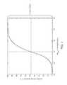
- the effectiveness of reduction in hyperinflationwill depend on the collateral resistance between the hyperinflated compartment and the neighboring compartments, as illustrated in FIG. 7 , where residual volume reduction is negligible when the resistance to collateral flow R coll is very small (significant collateral flow channels) and maximally effective when R coll is very high (no collateral flow channels).
- Absorption atelectasispromotes absorption of the remaining or residual gas in the compartment into the blood to further reduce the volume, either before or after permanent sealing of the lung volume compartment or segment.
- the present inventionprovides catheters for isolating and deflating hyperinflated and other diseased lung compartments.
- the cathetercomprises a catheter body, an expandable occluding member on the catheter body, and a one-way flow element associated with the catheter body.
- the catheter bodyusually has a distal end, a proximal end, and at least one lumen extending from a location at or near the distal end to a location at or near the proximal end. At least a distal portion of the catheter body is adapted to be advanced into and through the airways of a lung so that the distal end can reach an airway that feeds a target lung compartment or segment to be treated.
- the expandable occluding memberis disposed near the distal end of the catheter body and is adapted to be expanded in the airway that feeds the target lung compartment or segment so that said compartment or segment can be isolated, with access provided only through the lumen or catheter body when the occluding member is expanded.
- the one-way flow elementis adapted to be disposed within or in-line with the lumen of the catheter body in order to allow flow in a distal-to-proximal direction so that air will be expelled from the isolated lung compartment or segment as the patient exhales.
- the one-way flow elementinhibits or prevents flow through the lumen in a proximal-to-distal direction so that air cannot enter the isolated lung compartment or segment while the patient is inhaling.
- the catheter bodywill typically have a length in the range from 20 cm to 200 cm, preferably from 80 cm to 120 cm, and a diameter near the distal end in the range from 0.1 mm to 10 mm, preferably from 1 mm to 5 mm.
- the expandable occluding memberwill typically be an inflatable balloon or cuff, where the balloon or cuff has a width in the range from 1 mm to 30 mm, preferably from 5 mm to 20 mm, when inflated.
- the one-way flow elementis typically a conventional one-way flow valve, such as a duck-bill valve, a flap valve, or the like, which is disposed in the lumen of the catheter body, either near the distal end or at any other point within the lumen.
- the one-way flow elementcould be provided as a separate component, for example provided in a hub which is detachably mounted at the proximal end of the catheter body.
- a method for determining whether collateral ventilation of a hyperinflated lung compartment is presentmay involve: sealing a distal end of a catheter in an airway feeding the lung compartment; allowing air to be expelled from the lung compartment through a passage in the catheter while the patient is exhaling; blocking air from entering the lung compartment through the catheter passage while the patient is inhaling; comparing an image of the lung compartment with an earlier image of the lung compartment acquired before the sealing step; and determining whether collateral ventilation is present in the lung compartment, based on comparing the image and the earlier image.
- the compared imagesare CT scans, although in other embodiments alternative imaging modalities may be used, such as MRI, conventional radiographs and/or the like.
- the before and after imageswill be compared based on size, with a smaller size after catheter placement indicating a lack of significant collateral ventilation and little or no change in size indicating likely significant collateral ventilation.
- one embodimentmay involve advancing the catheter through a bronchoscope to position the catheter distal end in the airway before sealing.
- the methodmay also involve: detaching a hub from a proximal end of the catheter; removing the bronchoscope from the airway by sliding it proximally over the catheter, thus leaving the catheter in the airway; and acquiring the image of the lung compartment.
- the cathetermay be left in the airway for any suitable amount of time before acquiring the image—for example in one embodiment between about five minutes and about twenty-four hours.
- the methodmay further include treating the airway to permanently limit airflow into the lung compartment.
- FIG. 1is a perspective view of an isolation and deflation catheter constructed in accordance with the principles of the present invention.
- FIGS. 2-4illustrate alternative placements of one-way flow elements within a central lumen of the catheter of FIG. 1 .
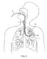
- FIG. 5illustrates the trans-tracheal endobronchial placement of the catheter of FIG. 1 in an airway leading to a diseased lung region in accordance with the principles of the present invention.
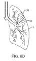
- FIGS. 6A-6Dillustrate use of the catheter as placed in FIG. 5 for isolating and reduction of the volume of the diseased lung region in accordance with the principles of the present invention.
- FIG. 7is a graph showing the relationship between collateral resistance R coll and residual volume reduction in an isolated lung compartment.
- an endobronchial lung volume reduction catheter 10constructed in accordance with the principles of the present invention includes an elongate catheter body 12 having a distal end 14 , a proximal end 16 , and an expandable occluding member 15 , such as an inflatable balloon, mounted near the distal end 14 .
- Catheter body 12also includes at least one lumen or central passage 18 extending generally from the distal end 14 to the proximal end 16 .
- Lumen 18has a distal opening 19 at or near the distal end 14 in order to permit air or other lung gases to enter the lumen 18 and flow in a distal-to-proximal direction out through the proximal end of the lumen 18 .
- a hub 20will be provided at the proximal end 16 , but the hub 20 is not a necessary component of the catheter 10 .
- the catheter 10is equipped to seal the area between the catheter body 12 and the bronchial wall such that only the lumen 18 is communicating with the airways distal to the seal.
- the seal, or isolation,is accomplished by the use of the occluding member 15 , such as an inflatable member, attached to (or near) the distal tip 14 of the catheter 10 .
- the isolated compartmentwill unsuccessfully attempt to draw air from the catheter lumen 18 during inspiration of normal respiration of the patient. Hence, during exhalation no air is returned to the catheter lumen.
- an additional amount of airis available to the isolated compartment during the inspiratory phase of each breath, namely the air traveling from the neighboring compartment(s) through the collateral channels, which enables volumetric expansion of the isolated compartment during inspiration, resulting during expiration in air movement away from the isolated compartment to atmosphere through the catheter lumen and the collateral channels.
- EMREndobronchial Volume Reduction
- the lung compartmentmay be analyzed for collateral ventilation prior to treatment to determine the likelihood of success of such treatment. Further, if undesired levels of collateral ventilation are measured, the collateral ventilation may be reduced to a desired level prior to treatment to ensure success of such treatment.
- the present inventionrelies on placement of a one-way flow element within or in-line with the lumen 18 so that flow from an isolated lung compartment or segment (as described hereinbelow) may occur in a distal-to-proximal direction but flow back into the lung compartment or segment is inhibited or blocked in the proximal-to-distal direction.
- a one-way flow element 22may be provided in the lumen 18 near the distal end 14 of the catheter body 12 , immediately proximal of the distal opening 19 .
- the same one-way flow element 22may be provided in the lumen 18 more proximally (either still near the distal end 14 or even more proximally in some embodiments).
- the one-way flow element 22may be a duck-bill valve, which opens as shown in broken line as the patient exhales to increase the pressure on the upstream or distal side of the valve 22 . As the patient inhales, the pressure on the upstream or distal side of the valve is reduced, drawing the valve leaflets closed as shown in solid line.
- the one-way flow element 22could be provided anywhere else in the lumen 18 , and two, three, four, or more such valve structures could be included in order to provide redundancy.
- the hub 20may be removable, or alternatively the catheter 10 may not include a hub. As will be explained further below, this may facilitate leaving the catheter 10 in a patient for diagnostic and/or treatment purposes. For example, if the catheter 10 is advanced into a patient through a bronchoscope, the hub 20 may be detached to allow the bronchoscope to be removed proximally over the catheter 10 , thus leaving the catheter body 12 with the one-way flow element 22 in the patient.
- a one-way valve structure 26 in the form of a flap valvecould be provided within the hub 20 .
- the hub 20could be removable or permanently fixed to the catheter body 12 .
- Other structures for providing in-line flow controlcould also be utilized.
- the catheter 10may be coupled with a one-way valve, a flow-measuring device or/and a pressure sensor, all of which are external to the body of the patient and are placed in series so as to communicate with the catheter's inside lumen 18 .
- the one-way valveprevents air from entering the target lung compartment from atmosphere but allows free air movement from the target lung compartment to atmosphere.
- the flow measuring device, the pressure sensor device and the one-way valvecan be placed anywhere along the length of the catheter lumen 18 .
- the seal provided by the catheter 10results, during expiration, in air movement away from the isolated lung compartment to atmosphere through the catheter lumen 18 and the collateral channels.
- airis expelled through the catheter lumen 18 during each exhalation and will register as positive airflow on the flow-measuring device.
- some airmay be expelled through the catheter lumen 18 during exhalation in the absence of collateral channels, however at a different rate, volume and trend than that in the presence of collateral channels.
- catheter 10Use of the endobronchial lung volume reduction catheter 10 to reduce the residual volume of a diseased region DR of a lung L is illustrated beginning in FIG. 5 .
- Catheter 10is introduced through the patient's mouth, down past the trachea T and into a lung L.
- the distal end 14 of the catheter 10is advanced to the main airway AW leading into the diseased region DR of the lung.
- Introduction and guidance of the catheter 10may be achieved in conventional manners, such as described in commonly-owned U.S. Pat. Nos. 6,287,290; 6,398,775; and 6,527,761, the full disclosures of which are incorporated herein by reference.
- the cathetermay be introduced through a flexible bronchoscope (not shown in FIG. 5 ).
- the expandable occluding element 15is expanded to occlude the airway.
- the expandable occluding elementmay be a balloon, cuff, or a braided balloon as described in copending applications 60/823,734, filed on Aug. 28, 2006, and 60/828,496 filed on Oct. 6, 2006, the full disclosures of which are incorporated herein by reference.
- the only path between the atmosphere and the diseased region DR of the lungis through the lumen 18 of the catheter 10 .
- the patient exhalesas shown in FIG.
- air from the diseased region DRflows outwardly through the lumen 18 and the one-way valve element 22 , causing a reduction in residual air within the region and a consequent reduction in volume. Air from the remainder of the lung also passes outward in the annular region around the catheter 10 in a normal manner.
- treating the patientmay comprise occluding the airway AW feeding the diseased region DR, by applying heat, radiofrequency energy, glues, or preferably by implanting an occluding element 30 , as shown in FIG. 6D .
- Implantation of the occluding elementmay be achieved by any of the techniques described in commonly-owned U.S. Pat. Nos. 6,287,290; and 6,527,761, the full disclosures of which have been previously incorporated herein by reference.
- treating the patientmay comprise aspirating the target lung compartment.
- volume reduction therapymay be performed by aspirating through the catheter and stent. The catheter is then removed and the volume reduction maintained.
- a catheter 10 as described hereinmay also be used to determine whether collateral ventilation is present in a lung.
- the '951 applicationdescribes a number of methods and devices for use in determining such collateral ventilation. Additionally or alternatively to those methods/devices, in one embodiment a catheter 10 (as described above) may be advanced through a bronchoscope and deployed as described in relation to FIGS. 5 and 6 A- 6 D of the present application.
- the catheter 10includes at least one one-way flow element 22 within the lumen 18 of the catheter body 12 .
- the hub 20 of the catheter 10may then be detached, and the bronchoscope may be removed proximally over the catheter body 12 , leaving the catheter body 12 in place in the patient.
- an imaging studysuch as a CT scan may be taken of the patient's lung to see if the residual volume of the diseased lung compartment has decreased.
- this CT scan or other imaging studywill be compared to a similar study taken before placement of the catheter 10 to determine if placement of the catheter has caused a reduction in residual volume in the lung compartment. If a reduction is noted, this may indicate that collateral ventilation is absent or minimal.
- This type of assessmentmay be used to help decide whether to treat a lung compartment further, such as with an implantable valve or blocking element.
- the hub 20 of the catheter 10may be left on, and the catheter 10 and bronchoscope may be left in the patient for a short time while an imaging study is performed.
Landscapes
- Health & Medical Sciences (AREA)
- Life Sciences & Earth Sciences (AREA)
- Engineering & Computer Science (AREA)
- Surgery (AREA)
- Public Health (AREA)
- Heart & Thoracic Surgery (AREA)
- Biomedical Technology (AREA)
- Veterinary Medicine (AREA)
- Animal Behavior & Ethology (AREA)
- General Health & Medical Sciences (AREA)
- Medical Informatics (AREA)
- Molecular Biology (AREA)
- Pulmonology (AREA)
- Nuclear Medicine, Radiotherapy & Molecular Imaging (AREA)
- Physics & Mathematics (AREA)
- Pathology (AREA)
- Biophysics (AREA)
- Reproductive Health (AREA)
- Vascular Medicine (AREA)
- Radiology & Medical Imaging (AREA)
- High Energy & Nuclear Physics (AREA)
- Physiology (AREA)
- Emergency Medicine (AREA)
- Anesthesiology (AREA)
- Hematology (AREA)
- Optics & Photonics (AREA)
- Theoretical Computer Science (AREA)
- Surgical Instruments (AREA)
- Measurement Of The Respiration, Hearing Ability, Form, And Blood Characteristics Of Living Organisms (AREA)
Abstract
Description
Claims (4)
Priority Applications (5)
| Application Number | Priority Date | Filing Date | Title |
|---|---|---|---|
| US12/820,547US8496006B2 (en) | 2005-01-20 | 2010-06-22 | Methods and devices for passive residual lung volume reduction and functional lung volume expansion |
| US13/938,025US9533116B2 (en) | 2005-01-20 | 2013-07-09 | Methods and devices for passive residual lung volume reduction and functional lung volume expansion |
| US15/358,483US10758239B2 (en) | 2005-01-20 | 2016-11-22 | Methods and devices for passive residual lung volume reduction and functional lung volume expansion |
| US16/930,194US11413045B2 (en) | 2005-01-20 | 2020-07-15 | Methods and devices for passive residual lung volume reduction and functional lung volume expansion |
| US17/867,990US11883029B2 (en) | 2005-01-20 | 2022-07-19 | Methods and devices for passive residual lung volume reduction and functional lung volume expansion |
Applications Claiming Priority (6)
| Application Number | Priority Date | Filing Date | Title |
|---|---|---|---|
| US64571105P | 2005-01-20 | 2005-01-20 | |
| US69694005P | 2005-07-05 | 2005-07-05 | |
| US69928905P | 2005-07-13 | 2005-07-13 | |
| US11/296,951US7883471B2 (en) | 2001-09-10 | 2005-12-07 | Minimally invasive determination of collateral ventilation in lungs |
| US11/685,008US20080228137A1 (en) | 2007-03-12 | 2007-03-12 | Methods and devices for passive residual lung volume reduction and functional lung volume expansion |
| US12/820,547US8496006B2 (en) | 2005-01-20 | 2010-06-22 | Methods and devices for passive residual lung volume reduction and functional lung volume expansion |
Related Parent Applications (1)
| Application Number | Title | Priority Date | Filing Date |
|---|---|---|---|
| US11/685,008Continuation-In-PartUS20080228137A1 (en) | 2005-01-20 | 2007-03-12 | Methods and devices for passive residual lung volume reduction and functional lung volume expansion |
Related Child Applications (1)
| Application Number | Title | Priority Date | Filing Date |
|---|---|---|---|
| US13/938,025ContinuationUS9533116B2 (en) | 2005-01-20 | 2013-07-09 | Methods and devices for passive residual lung volume reduction and functional lung volume expansion |
Publications (2)
| Publication Number | Publication Date |
|---|---|
| US20110152678A1 US20110152678A1 (en) | 2011-06-23 |
| US8496006B2true US8496006B2 (en) | 2013-07-30 |
Family
ID=44152051
Family Applications (4)
| Application Number | Title | Priority Date | Filing Date |
|---|---|---|---|
| US12/820,547Active2026-11-28US8496006B2 (en) | 2005-01-20 | 2010-06-22 | Methods and devices for passive residual lung volume reduction and functional lung volume expansion |
| US13/938,025Active2026-04-21US9533116B2 (en) | 2005-01-20 | 2013-07-09 | Methods and devices for passive residual lung volume reduction and functional lung volume expansion |
| US15/358,483Active2028-03-16US10758239B2 (en) | 2005-01-20 | 2016-11-22 | Methods and devices for passive residual lung volume reduction and functional lung volume expansion |
| US16/930,194Active2026-06-09US11413045B2 (en) | 2005-01-20 | 2020-07-15 | Methods and devices for passive residual lung volume reduction and functional lung volume expansion |
Family Applications After (3)
| Application Number | Title | Priority Date | Filing Date |
|---|---|---|---|
| US13/938,025Active2026-04-21US9533116B2 (en) | 2005-01-20 | 2013-07-09 | Methods and devices for passive residual lung volume reduction and functional lung volume expansion |
| US15/358,483Active2028-03-16US10758239B2 (en) | 2005-01-20 | 2016-11-22 | Methods and devices for passive residual lung volume reduction and functional lung volume expansion |
| US16/930,194Active2026-06-09US11413045B2 (en) | 2005-01-20 | 2020-07-15 | Methods and devices for passive residual lung volume reduction and functional lung volume expansion |
Country Status (1)
| Country | Link |
|---|---|
| US (4) | US8496006B2 (en) |
Cited By (3)
| Publication number | Priority date | Publication date | Assignee | Title |
|---|---|---|---|---|
| US20130296696A1 (en)* | 2005-01-20 | 2013-11-07 | Pulmonx Corporation | Methods and devices for passive residual lung volume reduction and functional lung volume expansion |
| US10314992B2 (en) | 2007-03-12 | 2019-06-11 | Pulmonx Corporation | Methods and devices for passive residual lung volume reduction and functional lung volume expansion |
| US20230000497A1 (en)* | 2005-01-20 | 2023-01-05 | Pulmonx Corporation | Methods and devices for passive residual lung volume reduction and functional lung volume expansion |
Families Citing this family (39)
| Publication number | Priority date | Publication date | Assignee | Title |
|---|---|---|---|---|
| US9198733B2 (en) | 2008-04-29 | 2015-12-01 | Virginia Tech Intellectual Properties, Inc. | Treatment planning for electroporation-based therapies |
| US10702326B2 (en) | 2011-07-15 | 2020-07-07 | Virginia Tech Intellectual Properties, Inc. | Device and method for electroporation based treatment of stenosis of a tubular body part |
| US10272178B2 (en) | 2008-04-29 | 2019-04-30 | Virginia Tech Intellectual Properties Inc. | Methods for blood-brain barrier disruption using electrical energy |
| US11272979B2 (en) | 2008-04-29 | 2022-03-15 | Virginia Tech Intellectual Properties, Inc. | System and method for estimating tissue heating of a target ablation zone for electrical-energy based therapies |
| US10117707B2 (en) | 2008-04-29 | 2018-11-06 | Virginia Tech Intellectual Properties, Inc. | System and method for estimating tissue heating of a target ablation zone for electrical-energy based therapies |
| US9598691B2 (en) | 2008-04-29 | 2017-03-21 | Virginia Tech Intellectual Properties, Inc. | Irreversible electroporation to create tissue scaffolds |
| US11254926B2 (en) | 2008-04-29 | 2022-02-22 | Virginia Tech Intellectual Properties, Inc. | Devices and methods for high frequency electroporation |
| US10238447B2 (en) | 2008-04-29 | 2019-03-26 | Virginia Tech Intellectual Properties, Inc. | System and method for ablating a tissue site by electroporation with real-time monitoring of treatment progress |
| US9283051B2 (en) | 2008-04-29 | 2016-03-15 | Virginia Tech Intellectual Properties, Inc. | System and method for estimating a treatment volume for administering electrical-energy based therapies |
| US10245098B2 (en) | 2008-04-29 | 2019-04-02 | Virginia Tech Intellectual Properties, Inc. | Acute blood-brain barrier disruption using electrical energy based therapy |
| US8992517B2 (en) | 2008-04-29 | 2015-03-31 | Virginia Tech Intellectual Properties Inc. | Irreversible electroporation to treat aberrant cell masses |
| US9867652B2 (en) | 2008-04-29 | 2018-01-16 | Virginia Tech Intellectual Properties, Inc. | Irreversible electroporation using tissue vasculature to treat aberrant cell masses or create tissue scaffolds |
| US8632534B2 (en) | 2009-04-03 | 2014-01-21 | Angiodynamics, Inc. | Irreversible electroporation (IRE) for congestive obstructive pulmonary disease (COPD) |
| US11382681B2 (en) | 2009-04-09 | 2022-07-12 | Virginia Tech Intellectual Properties, Inc. | Device and methods for delivery of high frequency electrical pulses for non-thermal ablation |
| US11638603B2 (en) | 2009-04-09 | 2023-05-02 | Virginia Tech Intellectual Properties, Inc. | Selective modulation of intracellular effects of cells using pulsed electric fields |
| WO2010138919A2 (en) | 2009-05-28 | 2010-12-02 | Angiodynamics, Inc. | System and method for synchronizing energy delivery to the cardiac rhythm |
| US9895189B2 (en) | 2009-06-19 | 2018-02-20 | Angiodynamics, Inc. | Methods of sterilization and treating infection using irreversible electroporation |
| US8425455B2 (en)* | 2010-03-30 | 2013-04-23 | Angiodynamics, Inc. | Bronchial catheter and method of use |
| EP2627274B1 (en) | 2010-10-13 | 2022-12-14 | AngioDynamics, Inc. | System for electrically ablating tissue of a patient |
| WO2012088149A2 (en) | 2010-12-20 | 2012-06-28 | Virginia Tech Intellectual Properties, Inc. | High-frequency electroporation for cancer therapy |
| US9078665B2 (en) | 2011-09-28 | 2015-07-14 | Angiodynamics, Inc. | Multiple treatment zone ablation probe |
| US9414881B2 (en) | 2012-02-08 | 2016-08-16 | Angiodynamics, Inc. | System and method for increasing a target zone for electrical ablation |
| WO2015175570A1 (en) | 2014-05-12 | 2015-11-19 | Virginia Tech Intellectual Properties, Inc. | Selective modulation of intracellular effects of cells using pulsed electric fields |
| US12114911B2 (en) | 2014-08-28 | 2024-10-15 | Angiodynamics, Inc. | System and method for ablating a tissue site by electroporation with real-time pulse monitoring |
| US10694972B2 (en) | 2014-12-15 | 2020-06-30 | Virginia Tech Intellectual Properties, Inc. | Devices, systems, and methods for real-time monitoring of electrophysical effects during tissue treatment |
| US20170329927A1 (en)* | 2016-05-11 | 2017-11-16 | InFluidS LLC | System and method for analyzing airway-pulmonary response using computational fluid dynamics to diagnose and monitoring potential health anomalies |
| US10448886B2 (en)* | 2016-08-17 | 2019-10-22 | Covidien Lp | Induced atelectasis and pulmonary consolidation systems and methods |
| US10905492B2 (en) | 2016-11-17 | 2021-02-02 | Angiodynamics, Inc. | Techniques for irreversible electroporation using a single-pole tine-style internal device communicating with an external surface electrode |
| CN108272538B (en)* | 2016-12-30 | 2020-06-12 | 先健科技(深圳)有限公司 | Elastic implant for lung volume reduction and lung volume reduction instrument |
| US11607537B2 (en) | 2017-12-05 | 2023-03-21 | Virginia Tech Intellectual Properties, Inc. | Method for treating neurological disorders, including tumors, with electroporation |
| US11925405B2 (en) | 2018-03-13 | 2024-03-12 | Virginia Tech Intellectual Properties, Inc. | Treatment planning system for immunotherapy enhancement via non-thermal ablation |
| US11311329B2 (en) | 2018-03-13 | 2022-04-26 | Virginia Tech Intellectual Properties, Inc. | Treatment planning for immunotherapy based treatments using non-thermal ablation techniques |
| US12390262B2 (en) | 2018-03-13 | 2025-08-19 | Virginia Tech Intellectual Properties, Inc. | Treatment planning system for immunotherapy enhancement via non-thermal ablation |
| DE102020112504A1 (en)* | 2019-05-17 | 2020-11-19 | Gyrus Acmi, Inc. D/B/A Olympus Surgical Technologies America | SYSTEM FOR EVALUATING COLLATERAL VENTILATION |
| US11950835B2 (en) | 2019-06-28 | 2024-04-09 | Virginia Tech Intellectual Properties, Inc. | Cycled pulsing to mitigate thermal damage for multi-electrode irreversible electroporation therapy |
| US12214189B2 (en) | 2019-07-24 | 2025-02-04 | Virginia Tech Intellectual Properties, Inc. | Fourier analysis spectroscopy for monitoring tissue impedance changes and treatment outcome during electroporation-based-therapies |
| AU2021304944A1 (en) | 2020-07-10 | 2023-02-16 | Pulmonx Corporation | Systems and methods for endobronchial diagnostics |
| EP4178434A4 (en) | 2020-07-10 | 2024-08-14 | Pulmonx Corporation | METHODS AND SYSTEMS FOR DETERMINING COLLATERAL VENTILATION |
| CN112220502B (en)* | 2020-10-14 | 2022-04-08 | 吉林大学 | A valve body device for assisting endoscopic specimen collection |
Citations (122)
| Publication number | Priority date | Publication date | Assignee | Title |
|---|---|---|---|---|
| US3322126A (en) | 1963-04-19 | 1967-05-30 | Willy Rusch Fa | Endotracheal catheter |
| US3498286A (en) | 1966-09-21 | 1970-03-03 | American Optical Corp | Catheters |
| US3669098A (en) | 1968-10-05 | 1972-06-13 | Olympus Optical Co | Endotracheal tube |
| US3677262A (en) | 1970-07-23 | 1972-07-18 | Henry J Zukowski | Surgical instrument illuminating endotracheal tube inserter |
| US3768504A (en) | 1972-06-19 | 1973-10-30 | S Rentsch | Check valve for use with a snorkel type breathing tube |
| US3776222A (en) | 1971-12-23 | 1973-12-04 | Lurosso A | Fiber optic entubator and method of entubation of the trachea through the nasopharynx |
| US3794026A (en) | 1970-07-29 | 1974-02-26 | H Jacobs | Ventilating apparatus embodying selective volume or pressure operation and catheter means for use therewith |
| US3866599A (en) | 1972-01-21 | 1975-02-18 | Univ Washington | Fiberoptic catheter |
| US3913568A (en) | 1973-01-22 | 1975-10-21 | American Optical Corp | Nasopharyngoscope |
| US4041936A (en) | 1975-04-23 | 1977-08-16 | Medical Engineering Corporation | Bronchoscopy tube |
| US4134407A (en) | 1977-03-25 | 1979-01-16 | Elam James O | External pressure-volume monitor for endotracheal cuff |
| US4147169A (en) | 1977-05-02 | 1979-04-03 | The Kendall Company | Balloon catheter with balloon retaining sleeves |
| US4327721A (en) | 1978-07-07 | 1982-05-04 | George Hanover | Endotracheal tube with topical agent delivery system and method of using the same |
| US4327720A (en) | 1979-01-22 | 1982-05-04 | Bronson Paul A | Esophageal-endotracheal airway |
| US4382442A (en)* | 1978-04-24 | 1983-05-10 | Jones James W | Thoracostomy pump-tube apparatus |
| US4453545A (en) | 1981-05-07 | 1984-06-12 | Hiroshi Inoue | Endotracheal tube with movable endobronchial blocker for one-lung anesthesia |
| US4468216A (en) | 1982-05-20 | 1984-08-28 | Rudolph Muto | Irrigation suction catheter |
| US4470407A (en) | 1982-03-11 | 1984-09-11 | Laserscope, Inc. | Endoscopic device |
| US4538607A (en) | 1984-02-06 | 1985-09-03 | Ab Fixfabriken | Tracheostomy valve |
| US4567882A (en) | 1982-12-06 | 1986-02-04 | Vanderbilt University | Method for locating the illuminated tip of an endotracheal tube |
| US4681093A (en) | 1982-12-13 | 1987-07-21 | Sumitomo Electric Industries, Ltd. | Endoscope |
| US4716896A (en) | 1986-08-01 | 1988-01-05 | Ackrad Laboratories | Bronchial catheter |
| US4742819A (en) | 1987-03-23 | 1988-05-10 | George Gordon P | Intubating scope with camera and screen |
| US4784133A (en) | 1987-01-28 | 1988-11-15 | Mackin Robert A | Working well balloon angioscope and method |
| US4796639A (en) | 1987-11-05 | 1989-01-10 | Medical Graphics Corporation | Pulmonary diagnostic system |
| US4819664A (en) | 1984-11-15 | 1989-04-11 | Stefano Nazari | Device for selective bronchial intubation and separate lung ventilation, particularly during anesthesia, intensive therapy and reanimation |
| US4846153A (en) | 1988-06-10 | 1989-07-11 | George Berci | Intubating video endoscope |
| US4850371A (en) | 1988-06-13 | 1989-07-25 | Broadhurst John H | Novel endotracheal tube and mass spectrometer |
| US4862874A (en) | 1987-06-10 | 1989-09-05 | Kellner Hans Joerg | Endoscope for removal of thrombi from pulmonary arterial vessels |
| US4896941A (en) | 1985-04-27 | 1990-01-30 | Doryokuro Kakunenryo Kaihatsu Jigyodan | Image-transmitting fiber |
| US4949716A (en) | 1988-10-31 | 1990-08-21 | Medical Devices, Inc. | Nasal intubation adjunct |
| US4955375A (en) | 1989-01-23 | 1990-09-11 | Ricardo Martinez | Endotracheal tube with channel for delivering drugs |
| US4958932A (en) | 1988-08-18 | 1990-09-25 | Mcdonnell Douglas Corporation | Optical measuring apparatus |
| US4961738A (en) | 1987-01-28 | 1990-10-09 | Mackin Robert A | Angioplasty catheter with illumination and visualization within angioplasty balloon |
| US4976710A (en) | 1987-01-28 | 1990-12-11 | Mackin Robert A | Working well balloon method |
| US5056529A (en) | 1990-04-03 | 1991-10-15 | Groot William J De | Apparatus and method for performing a transbroncheal biopsy |
| US5143062A (en) | 1990-10-26 | 1992-09-01 | Mallinckrodt Medical, Inc. | Endotracheal tube having irrigation means |
| US5146916A (en) | 1990-01-05 | 1992-09-15 | Catalani Angelo S | Endotracheal tube incorporating a drug-irrigation device |
| US5165420A (en) | 1990-12-21 | 1992-11-24 | Ballard Medical Products | Bronchoalveolar lavage catheter |
| US5181913A (en) | 1987-03-09 | 1993-01-26 | Prn Services, Inc. | Catheter with check valve and rolled sheath |
| US5285778A (en) | 1991-04-19 | 1994-02-15 | Mackin Robert A | Endotracheal tube wih fibers optic illumination and viewing and auxiliary tube |
| US5308325A (en) | 1991-01-28 | 1994-05-03 | Corpak, Inc. | Retention balloon for percutaneous catheter |
| US5309903A (en) | 1989-12-12 | 1994-05-10 | Burroughs Wellcome Co. | Method for administering surfactant to the lungs while concurrently providing one-lung ventilation |
| US5329940A (en)* | 1990-02-14 | 1994-07-19 | Adair Edwin Lloyd | Endotracheal tube intubation assist device |
| US5331947A (en) | 1992-05-01 | 1994-07-26 | Shturman Cardiology Systems, Inc. | Inflatable sheath for introduction of ultrasonic catheter through the lumen of a fiber optic endoscope |
| US5361753A (en) | 1992-07-07 | 1994-11-08 | Deutsche Aerospace Ag | Method of measuring and regulating the pressure in the sealing cuff of a tracheal tube and apparatus for implementing the method |
| US5447165A (en) | 1991-09-27 | 1995-09-05 | Gustafsson; Lars E. | Method for ascertaining prevailing lung condition and a device |
| US5477851A (en) | 1995-01-26 | 1995-12-26 | Callaghan; Eric B. | Laryngeal mask assembly and method for removing same |
| US5499625A (en) | 1994-01-27 | 1996-03-19 | The Kendall Company | Esophageal-tracheal double lumen airway |
| US5546935A (en) | 1993-03-09 | 1996-08-20 | Medamicus, Inc. | Endotracheal tube mounted pressure transducer |
| US5588424A (en) | 1995-06-28 | 1996-12-31 | The Cleveland Clinic Foundation | Bronchial blocker endotracheal apparatus |
| US5598840A (en) | 1995-03-17 | 1997-02-04 | Sorenson Critical Care, Inc. | Apparatus and method for ventilation and aspiration |
| US5624449A (en) | 1993-11-03 | 1997-04-29 | Target Therapeutics | Electrolytically severable joint for endovascular embolic devices |
| US5642730A (en) | 1994-06-17 | 1997-07-01 | Trudell Medical Limited | Catheter system for delivery of aerosolized medicine for use with pressurized propellant canister |
| US5645519A (en) | 1994-03-18 | 1997-07-08 | Jai S. Lee | Endoscopic instrument for controlled introduction of tubular members in the body and methods therefor |
| US5653231A (en) | 1995-11-28 | 1997-08-05 | Medcare Medical Group, Inc. | Tracheostomy length single use suction catheter |
| US5660175A (en) | 1995-08-21 | 1997-08-26 | Dayal; Bimal | Endotracheal device |
| EP0791340A1 (en) | 1996-02-22 | 1997-08-27 | Cordis Corporation | Temporary filter catheter |
| US5682880A (en) | 1996-07-26 | 1997-11-04 | Brain; Archibald Ian Jeremy | Laryngeal-mask airway with guide element, stiffener, and fiberoptic access |
| EP0815803A1 (en) | 1996-07-03 | 1998-01-07 | Cordis Europa N.V. | Catheter with temporary vena cava filter |
| US5707352A (en) | 1989-08-28 | 1998-01-13 | Alliance Pharmaceutical Corp. | Pulmonary delivery of therapeutic agent |
| US5752921A (en) | 1996-01-11 | 1998-05-19 | Korr Medical Technologies, Inc. | Method and apparatus for determining tracheal pressure |
| US5765557A (en) | 1995-03-17 | 1998-06-16 | Board Of Regents, The University Of Texas System | Method and apparatus for directing air flow within an intubated patient |
| US5795322A (en) | 1995-04-10 | 1998-08-18 | Cordis Corporation | Catheter with filter and thrombus-discharge device |
| US5800455A (en) | 1993-04-19 | 1998-09-01 | Target Therapeutics, Inc. | Detachable embolic coil assembly |
| US5893841A (en) | 1996-08-30 | 1999-04-13 | Delcath Systems, Inc. | Balloon catheter with occluded segment bypass |
| WO1999017827A2 (en) | 1997-10-03 | 1999-04-15 | Scimed Life Systems, Inc. | Braided angiography catheter having full length radiopacity and controlled flexibility |
| US5897528A (en) | 1998-04-30 | 1999-04-27 | Medtronic, Inc. | Filtered intracerebroventricular or intraspinal access port with direct cerebrospinal fluid access |
| WO1999020332A1 (en) | 1997-10-20 | 1999-04-29 | Christopher Kent L | System for monitoring and treating sleep disorders using a transtracheal catheter |
| US5915383A (en) | 1997-04-29 | 1999-06-29 | Smiths Industries Public Limited Company | Cuffed medico-surgical tubes |
| WO1999032040A1 (en) | 1997-12-19 | 1999-07-01 | Broncus Technologies, Inc. | Bronchial stenter |
| US5972026A (en) | 1997-04-07 | 1999-10-26 | Broncus Technologies, Inc. | Bronchial stenter having diametrically adjustable electrodes |
| WO2001002042A1 (en) | 1999-07-02 | 2001-01-11 | Pulmonx | Methods, systems, and kits for lung volume reduction |
| US6174307B1 (en) | 1996-03-29 | 2001-01-16 | Eclipse Surgical Technologies, Inc. | Viewing surgical scope for minimally invasive procedures |
| US6174323B1 (en) | 1998-06-05 | 2001-01-16 | Broncus Technologies, Inc. | Method and assembly for lung volume reduction |
| USRE37117E1 (en) | 1992-09-22 | 2001-03-27 | Target Therapeutics, Inc. | Detachable embolic coil assembly using interlocking clasps and method of use |
| US6258100B1 (en) | 1999-08-24 | 2001-07-10 | Spiration, Inc. | Method of reducing lung size |
| US20010051899A1 (en) | 2000-06-13 | 2001-12-13 | Takahiko Kawashima | Document managing apparatus for managing transaction slip data in electronic commerce |
| US20020049370A1 (en) | 1999-08-05 | 2002-04-25 | Laufer Michael D. | Devices for creating collateral channels in the lungs |
| US6398775B1 (en) | 1999-10-21 | 2002-06-04 | Pulmonx | Apparatus and method for isolated lung access |
| US6527761B1 (en) | 2000-10-27 | 2003-03-04 | Pulmonx, Inc. | Methods and devices for obstructing and aspirating lung tissue segments |
| WO2003022124A2 (en) | 2001-09-11 | 2003-03-20 | Spiration, Inc. | Removable lung reduction devices, systems, and methods |
| US20030051733A1 (en) | 2001-09-10 | 2003-03-20 | Pulmonx | Method and apparatus for endobronchial diagnosis |
| US6585639B1 (en) | 2000-10-27 | 2003-07-01 | Pulmonx | Sheath and method for reconfiguring lung viewing scope |
| US6609521B1 (en) | 2001-04-09 | 2003-08-26 | Regents Of The University Of Minnesota | Endotracheal tube |
| US20030171332A1 (en) | 2001-05-23 | 2003-09-11 | Abraham William M. | Treatment of respiratory conditions associated with bronchoconstriction with aerosolized hyaluronic acid |
| US20030228344A1 (en) | 2002-03-08 | 2003-12-11 | Fields Antony J. | Methods and devices for inducing collapse in lung regions fed by collateral pathways |
| US6679264B1 (en) | 2000-03-04 | 2004-01-20 | Emphasys Medical, Inc. | Methods and devices for use in performing pulmonary procedures |
| US6712812B2 (en) | 1999-08-05 | 2004-03-30 | Broncus Technologies, Inc. | Devices for creating collateral channels |
| US6722360B2 (en) | 2000-06-16 | 2004-04-20 | Rajiv Doshi | Methods and devices for improving breathing in patients with pulmonary disease |
| US6749606B2 (en) | 1999-08-05 | 2004-06-15 | Thomas Keast | Devices for creating collateral channels |
| US6792947B1 (en) | 2000-08-25 | 2004-09-21 | O-Two Systems International Inc. | Flow control valve for manual resuscitator devices |
| US20040243016A1 (en) | 2001-08-29 | 2004-12-02 | Sanderson Penelope Margaret | Method and means of physiological monitoring using sonification |
| US20050016530A1 (en) | 2003-07-09 | 2005-01-27 | Mccutcheon John | Treatment planning with implantable bronchial isolation devices |
| US20050022809A1 (en)* | 2003-04-25 | 2005-02-03 | Wondka Anthony David | Methods, systems and devices for desufflating a lung area |
| US6886558B2 (en) | 2002-08-28 | 2005-05-03 | Cordis Corporation | Collateral ventilation bypass trap system |
| US20050187561A1 (en) | 2004-02-25 | 2005-08-25 | Femasys, Inc. | Methods and devices for conduit occlusion |
| US6941950B2 (en) | 2001-10-11 | 2005-09-13 | Emphasys Medical, Inc. | Bronchial flow control devices and methods of use |
| US20050288684A1 (en) | 2004-06-16 | 2005-12-29 | Aronson Nathan A | Method of reducing collateral flow in a portion of a lung |
| US6997189B2 (en) | 1998-06-05 | 2006-02-14 | Broncus Technologies, Inc. | Method for lung volume reduction |
| US7011094B2 (en) | 2001-03-02 | 2006-03-14 | Emphasys Medical, Inc. | Bronchial flow control devices and methods of use |
| US7022088B2 (en) | 1999-08-05 | 2006-04-04 | Broncus Technologies, Inc. | Devices for applying energy to tissue |
| US20060102186A1 (en) | 2004-11-18 | 2006-05-18 | Mark Adler | Intra-bronchial apparatus for aspiration and insufflation of lung regions distal to placement or cross communication and deployment and placement system therefor |
| US20060122647A1 (en) | 2004-09-24 | 2006-06-08 | Callaghan David J | Occluder device double securement system for delivery/recovery of such occluder device |
| US20060129134A1 (en)* | 2004-11-29 | 2006-06-15 | Andrew Kerr | Dialysis catheter |
| US7086398B2 (en) | 2002-07-31 | 2006-08-08 | Cordis Corporation | Long term oxygen therapy system |
| EP1078601B1 (en) | 1999-08-24 | 2006-10-04 | Spiration, Inc. | Kit for lung volume reduction |
| US20060264772A1 (en) | 2001-09-10 | 2006-11-23 | Pulmonx | Minimally invasive determination of collateral ventilation in lungs |
| US20070005083A1 (en) | 1997-04-30 | 2007-01-04 | Sabaratham Sabanathan | Occlusion device |
| US20070096048A1 (en) | 2005-10-14 | 2007-05-03 | Claude Clerc | Bronchoscopic lung volume reduction valve |
| US20070142742A1 (en) | 2005-07-13 | 2007-06-21 | Pulmonx | Methods and systems for segmental lung diagnostics |
| US20070225747A1 (en) | 2006-03-08 | 2007-09-27 | Pulmonx | Methods and devices to induce controlled atelectasis and hypoxic pulmonary vasoconstriction |
| US7276077B2 (en) | 1997-09-16 | 2007-10-02 | Emphasys Medical, Inc. | Body fluid flow control device |
| US20080051719A1 (en) | 2006-08-28 | 2008-02-28 | Pulmonx | Functional assessment and treatment catheters and methods for their use in the lung |
| US20080228130A1 (en) | 2007-03-12 | 2008-09-18 | Pulmonx | Methods and systems for occluding collateral flow channels in the lung |
| US20080228137A1 (en) | 2007-03-12 | 2008-09-18 | Pulmonx | Methods and devices for passive residual lung volume reduction and functional lung volume expansion |
| US7449010B1 (en)* | 2002-07-15 | 2008-11-11 | Motoya Hayase | Material removal catheter and method |
| US7588033B2 (en) | 2003-06-18 | 2009-09-15 | Breathe Technologies, Inc. | Methods, systems and devices for improving ventilation in a lung area |
| US20100031964A1 (en) | 2008-08-11 | 2010-02-11 | Joseph William Turek | Flow control adapter for performing spirometry and pulmonary function testing |
| US20110011406A1 (en) | 2005-12-27 | 2011-01-20 | Hansa Medical Products, Inc. | Valved Fenestrated Tracheotomy Tube Having Outer and Inner Cannulae |
| US20110203594A1 (en) | 2001-08-23 | 2011-08-25 | Indian Ocean Medical Inc. | Disposable laryngeal mask airway device |
| US20110259339A1 (en) | 2006-09-11 | 2011-10-27 | Ric Investments, Llc | Ventilating apparatus and method enabling a patient to talk with or without a trachostomy tube check valve |
Family Cites Families (26)
| Publication number | Priority date | Publication date | Assignee | Title |
|---|---|---|---|---|
| US4852568A (en) | 1987-02-17 | 1989-08-01 | Kensey Nash Corporation | Method and apparatus for sealing an opening in tissue of a living being |
| US6013619A (en) | 1988-01-06 | 2000-01-11 | The Scripps Research Institute | Pulmonary surfactants and therapeutic uses, including pulmonary lavage |
| US5246012A (en) | 1990-12-21 | 1993-09-21 | Ballard Medical Products | Bronchoalveolar lavage catheter |
| US6346074B1 (en) | 1993-02-22 | 2002-02-12 | Heartport, Inc. | Devices for less invasive intracardiac interventions |
| WO1994024962A1 (en) | 1993-04-28 | 1994-11-10 | Focal, Inc. | Apparatus and methods for intraluminal photothermoforming |
| JP3838671B2 (en)* | 1993-10-25 | 2006-10-25 | アークレイ株式会社 | Breath collection device |
| GB9411215D0 (en) | 1994-06-04 | 1994-07-27 | Brain Archibald Ian Jeremy | A fibreoptic intubating laryngeal mask airway |
| DE69739259D1 (en)* | 1996-04-05 | 2009-03-26 | Kirin Brewery | Process for the preparation of a substance found in germinating seeds of grasses and containing proteins and insoluble fiber |
| US6273907B1 (en) | 1997-04-07 | 2001-08-14 | Broncus Technologies, Inc. | Bronchial stenter |
| US6283988B1 (en) | 1997-04-07 | 2001-09-04 | Broncus Technologies, Inc. | Bronchial stenter having expandable electrodes |
| US6411852B1 (en) | 1997-04-07 | 2002-06-25 | Broncus Technologies, Inc. | Modification of airways by application of energy |
| US6634363B1 (en) | 1997-04-07 | 2003-10-21 | Broncus Technologies, Inc. | Methods of treating lungs having reversible obstructive pulmonary disease |
| US6488673B1 (en) | 1997-04-07 | 2002-12-03 | Broncus Technologies, Inc. | Method of increasing gas exchange of a lung |
| US5957919A (en) | 1997-07-02 | 1999-09-28 | Laufer; Michael D. | Bleb reducer |
| US6152131A (en) | 1998-08-26 | 2000-11-28 | Instrumentarium Corp. | Method and apparatus for detecting an empty breathing gas compartment in a patient ventilator |
| US6749598B1 (en) | 1999-01-11 | 2004-06-15 | Flowmedica, Inc. | Apparatus and methods for treating congestive heart disease |
| US7780628B1 (en) | 1999-01-11 | 2010-08-24 | Angiodynamics, Inc. | Apparatus and methods for treating congestive heart disease |
| US7175644B2 (en)* | 2001-02-14 | 2007-02-13 | Broncus Technologies, Inc. | Devices and methods for maintaining collateral channels in tissue |
| US6610043B1 (en) | 1999-08-23 | 2003-08-26 | Bistech, Inc. | Tissue volume reduction |
| KR100481175B1 (en)* | 2002-08-08 | 2005-04-07 | 삼성전자주식회사 | Semiconductor memory device with shift redundancy circuits |
| US7681576B2 (en) | 2003-05-06 | 2010-03-23 | Mallinckrodt Inc. | Multiple cannula systems and methods |
| US7273052B2 (en) | 2003-12-11 | 2007-09-25 | Tvi Corporation | Pneumatic sealing system for protection masks |
| US20060162731A1 (en) | 2004-11-16 | 2006-07-27 | Pulmonx | Pulmonary occlusal stent delivery catheter, loading system and methods of use |
| US8496006B2 (en)* | 2005-01-20 | 2013-07-30 | Pulmonx Corporation | Methods and devices for passive residual lung volume reduction and functional lung volume expansion |
| EP1838217B1 (en) | 2005-01-20 | 2014-06-25 | Pulmonx | Minimally invasive determination of collateral ventilation in lungs |
| EP1866019B1 (en) | 2005-02-22 | 2017-10-25 | Cardiofocus, Inc. | Deflectable sheath catheters |
- 2010
- 2010-06-22USUS12/820,547patent/US8496006B2/enactiveActive
- 2013
- 2013-07-09USUS13/938,025patent/US9533116B2/enactiveActive
- 2016
- 2016-11-22USUS15/358,483patent/US10758239B2/enactiveActive
- 2020
- 2020-07-15USUS16/930,194patent/US11413045B2/enactiveActive
Patent Citations (139)
| Publication number | Priority date | Publication date | Assignee | Title |
|---|---|---|---|---|
| US3322126A (en) | 1963-04-19 | 1967-05-30 | Willy Rusch Fa | Endotracheal catheter |
| US3498286A (en) | 1966-09-21 | 1970-03-03 | American Optical Corp | Catheters |
| US3669098A (en) | 1968-10-05 | 1972-06-13 | Olympus Optical Co | Endotracheal tube |
| US3677262A (en) | 1970-07-23 | 1972-07-18 | Henry J Zukowski | Surgical instrument illuminating endotracheal tube inserter |
| US3794026A (en) | 1970-07-29 | 1974-02-26 | H Jacobs | Ventilating apparatus embodying selective volume or pressure operation and catheter means for use therewith |
| US3776222A (en) | 1971-12-23 | 1973-12-04 | Lurosso A | Fiber optic entubator and method of entubation of the trachea through the nasopharynx |
| US3866599A (en) | 1972-01-21 | 1975-02-18 | Univ Washington | Fiberoptic catheter |
| US3768504A (en) | 1972-06-19 | 1973-10-30 | S Rentsch | Check valve for use with a snorkel type breathing tube |
| US3913568A (en) | 1973-01-22 | 1975-10-21 | American Optical Corp | Nasopharyngoscope |
| US4041936A (en) | 1975-04-23 | 1977-08-16 | Medical Engineering Corporation | Bronchoscopy tube |
| US4134407A (en) | 1977-03-25 | 1979-01-16 | Elam James O | External pressure-volume monitor for endotracheal cuff |
| US4147169A (en) | 1977-05-02 | 1979-04-03 | The Kendall Company | Balloon catheter with balloon retaining sleeves |
| US4382442A (en)* | 1978-04-24 | 1983-05-10 | Jones James W | Thoracostomy pump-tube apparatus |
| US4327721A (en) | 1978-07-07 | 1982-05-04 | George Hanover | Endotracheal tube with topical agent delivery system and method of using the same |
| US4327720A (en) | 1979-01-22 | 1982-05-04 | Bronson Paul A | Esophageal-endotracheal airway |
| US4453545A (en) | 1981-05-07 | 1984-06-12 | Hiroshi Inoue | Endotracheal tube with movable endobronchial blocker for one-lung anesthesia |
| US4470407A (en) | 1982-03-11 | 1984-09-11 | Laserscope, Inc. | Endoscopic device |
| US4468216A (en) | 1982-05-20 | 1984-08-28 | Rudolph Muto | Irrigation suction catheter |
| US4567882A (en) | 1982-12-06 | 1986-02-04 | Vanderbilt University | Method for locating the illuminated tip of an endotracheal tube |
| US4681093A (en) | 1982-12-13 | 1987-07-21 | Sumitomo Electric Industries, Ltd. | Endoscope |
| US4538607A (en) | 1984-02-06 | 1985-09-03 | Ab Fixfabriken | Tracheostomy valve |
| US4819664A (en) | 1984-11-15 | 1989-04-11 | Stefano Nazari | Device for selective bronchial intubation and separate lung ventilation, particularly during anesthesia, intensive therapy and reanimation |
| US4896941A (en) | 1985-04-27 | 1990-01-30 | Doryokuro Kakunenryo Kaihatsu Jigyodan | Image-transmitting fiber |
| US4716896A (en) | 1986-08-01 | 1988-01-05 | Ackrad Laboratories | Bronchial catheter |
| US4784133A (en) | 1987-01-28 | 1988-11-15 | Mackin Robert A | Working well balloon angioscope and method |
| US4976710A (en) | 1987-01-28 | 1990-12-11 | Mackin Robert A | Working well balloon method |
| US4961738A (en) | 1987-01-28 | 1990-10-09 | Mackin Robert A | Angioplasty catheter with illumination and visualization within angioplasty balloon |
| US5181913A (en) | 1987-03-09 | 1993-01-26 | Prn Services, Inc. | Catheter with check valve and rolled sheath |
| US4742819A (en) | 1987-03-23 | 1988-05-10 | George Gordon P | Intubating scope with camera and screen |
| US4862874A (en) | 1987-06-10 | 1989-09-05 | Kellner Hans Joerg | Endoscope for removal of thrombi from pulmonary arterial vessels |
| US4796639A (en) | 1987-11-05 | 1989-01-10 | Medical Graphics Corporation | Pulmonary diagnostic system |
| US4846153A (en) | 1988-06-10 | 1989-07-11 | George Berci | Intubating video endoscope |
| US4850371A (en) | 1988-06-13 | 1989-07-25 | Broadhurst John H | Novel endotracheal tube and mass spectrometer |
| US4958932A (en) | 1988-08-18 | 1990-09-25 | Mcdonnell Douglas Corporation | Optical measuring apparatus |
| US4949716A (en) | 1988-10-31 | 1990-08-21 | Medical Devices, Inc. | Nasal intubation adjunct |
| US4955375A (en) | 1989-01-23 | 1990-09-11 | Ricardo Martinez | Endotracheal tube with channel for delivering drugs |
| US5707352A (en) | 1989-08-28 | 1998-01-13 | Alliance Pharmaceutical Corp. | Pulmonary delivery of therapeutic agent |
| US5309903A (en) | 1989-12-12 | 1994-05-10 | Burroughs Wellcome Co. | Method for administering surfactant to the lungs while concurrently providing one-lung ventilation |
| US5146916A (en) | 1990-01-05 | 1992-09-15 | Catalani Angelo S | Endotracheal tube incorporating a drug-irrigation device |
| US5329940A (en)* | 1990-02-14 | 1994-07-19 | Adair Edwin Lloyd | Endotracheal tube intubation assist device |
| US5056529A (en) | 1990-04-03 | 1991-10-15 | Groot William J De | Apparatus and method for performing a transbroncheal biopsy |
| US5143062A (en) | 1990-10-26 | 1992-09-01 | Mallinckrodt Medical, Inc. | Endotracheal tube having irrigation means |
| US5165420A (en) | 1990-12-21 | 1992-11-24 | Ballard Medical Products | Bronchoalveolar lavage catheter |
| US5308325A (en) | 1991-01-28 | 1994-05-03 | Corpak, Inc. | Retention balloon for percutaneous catheter |
| US5285778A (en) | 1991-04-19 | 1994-02-15 | Mackin Robert A | Endotracheal tube wih fibers optic illumination and viewing and auxiliary tube |
| US5447165A (en) | 1991-09-27 | 1995-09-05 | Gustafsson; Lars E. | Method for ascertaining prevailing lung condition and a device |
| US5331947A (en) | 1992-05-01 | 1994-07-26 | Shturman Cardiology Systems, Inc. | Inflatable sheath for introduction of ultrasonic catheter through the lumen of a fiber optic endoscope |
| US5361753A (en) | 1992-07-07 | 1994-11-08 | Deutsche Aerospace Ag | Method of measuring and regulating the pressure in the sealing cuff of a tracheal tube and apparatus for implementing the method |
| USRE37117E1 (en) | 1992-09-22 | 2001-03-27 | Target Therapeutics, Inc. | Detachable embolic coil assembly using interlocking clasps and method of use |
| US5546935A (en) | 1993-03-09 | 1996-08-20 | Medamicus, Inc. | Endotracheal tube mounted pressure transducer |
| US5800455A (en) | 1993-04-19 | 1998-09-01 | Target Therapeutics, Inc. | Detachable embolic coil assembly |
| US6099546A (en) | 1993-04-19 | 2000-08-08 | Target Therapeutics, Inc. | Detachable embolic coil assembly using interlocking hooks and slots |
| US5624449A (en) | 1993-11-03 | 1997-04-29 | Target Therapeutics | Electrolytically severable joint for endovascular embolic devices |
| US5499625A (en) | 1994-01-27 | 1996-03-19 | The Kendall Company | Esophageal-tracheal double lumen airway |
| US5645519A (en) | 1994-03-18 | 1997-07-08 | Jai S. Lee | Endoscopic instrument for controlled introduction of tubular members in the body and methods therefor |
| US5642730A (en) | 1994-06-17 | 1997-07-01 | Trudell Medical Limited | Catheter system for delivery of aerosolized medicine for use with pressurized propellant canister |
| US5477851A (en) | 1995-01-26 | 1995-12-26 | Callaghan; Eric B. | Laryngeal mask assembly and method for removing same |
| US5598840A (en) | 1995-03-17 | 1997-02-04 | Sorenson Critical Care, Inc. | Apparatus and method for ventilation and aspiration |
| US5765557A (en) | 1995-03-17 | 1998-06-16 | Board Of Regents, The University Of Texas System | Method and apparatus for directing air flow within an intubated patient |
| US5795322A (en) | 1995-04-10 | 1998-08-18 | Cordis Corporation | Catheter with filter and thrombus-discharge device |
| US5588424A (en) | 1995-06-28 | 1996-12-31 | The Cleveland Clinic Foundation | Bronchial blocker endotracheal apparatus |
| US5660175A (en) | 1995-08-21 | 1997-08-26 | Dayal; Bimal | Endotracheal device |
| US5653231A (en) | 1995-11-28 | 1997-08-05 | Medcare Medical Group, Inc. | Tracheostomy length single use suction catheter |
| US5752921A (en) | 1996-01-11 | 1998-05-19 | Korr Medical Technologies, Inc. | Method and apparatus for determining tracheal pressure |
| EP0791340A1 (en) | 1996-02-22 | 1997-08-27 | Cordis Corporation | Temporary filter catheter |
| US6174307B1 (en) | 1996-03-29 | 2001-01-16 | Eclipse Surgical Technologies, Inc. | Viewing surgical scope for minimally invasive procedures |
| EP0815803A1 (en) | 1996-07-03 | 1998-01-07 | Cordis Europa N.V. | Catheter with temporary vena cava filter |
| US5682880A (en) | 1996-07-26 | 1997-11-04 | Brain; Archibald Ian Jeremy | Laryngeal-mask airway with guide element, stiffener, and fiberoptic access |
| US5893841A (en) | 1996-08-30 | 1999-04-13 | Delcath Systems, Inc. | Balloon catheter with occluded segment bypass |
| US5972026A (en) | 1997-04-07 | 1999-10-26 | Broncus Technologies, Inc. | Bronchial stenter having diametrically adjustable electrodes |
| US6083255A (en) | 1997-04-07 | 2000-07-04 | Broncus Technologies, Inc. | Bronchial stenter |
| US5915383A (en) | 1997-04-29 | 1999-06-29 | Smiths Industries Public Limited Company | Cuffed medico-surgical tubes |
| US20070005083A1 (en) | 1997-04-30 | 2007-01-04 | Sabaratham Sabanathan | Occlusion device |
| US7276077B2 (en) | 1997-09-16 | 2007-10-02 | Emphasys Medical, Inc. | Body fluid flow control device |
| WO1999017827A2 (en) | 1997-10-03 | 1999-04-15 | Scimed Life Systems, Inc. | Braided angiography catheter having full length radiopacity and controlled flexibility |
| WO1999020332A1 (en) | 1997-10-20 | 1999-04-29 | Christopher Kent L | System for monitoring and treating sleep disorders using a transtracheal catheter |
| WO1999032040A1 (en) | 1997-12-19 | 1999-07-01 | Broncus Technologies, Inc. | Bronchial stenter |
| US5897528A (en) | 1998-04-30 | 1999-04-27 | Medtronic, Inc. | Filtered intracerebroventricular or intraspinal access port with direct cerebrospinal fluid access |
| US6174323B1 (en) | 1998-06-05 | 2001-01-16 | Broncus Technologies, Inc. | Method and assembly for lung volume reduction |
| US6997189B2 (en) | 1998-06-05 | 2006-02-14 | Broncus Technologies, Inc. | Method for lung volume reduction |
| US6878141B1 (en) | 1999-07-02 | 2005-04-12 | Pulmonx | Methods systems and kits for lung volume reduction |
| WO2001002042A1 (en) | 1999-07-02 | 2001-01-11 | Pulmonx | Methods, systems, and kits for lung volume reduction |
| US6287290B1 (en) | 1999-07-02 | 2001-09-11 | Pulmonx | Methods, systems, and kits for lung volume reduction |
| US6709401B2 (en) | 1999-07-02 | 2004-03-23 | Pulmonx | Methods, systems, and kits for lung volume reduction |
| US20020062120A1 (en) | 1999-07-02 | 2002-05-23 | Pulmonx | Methods, systems, and kits for lung volume reduction |
| US6749606B2 (en) | 1999-08-05 | 2004-06-15 | Thomas Keast | Devices for creating collateral channels |
| US20020049370A1 (en) | 1999-08-05 | 2002-04-25 | Laufer Michael D. | Devices for creating collateral channels in the lungs |
| US6692494B1 (en) | 1999-08-05 | 2004-02-17 | Broncus Technologies, Inc. | Methods and devices for creating collateral channels in the lungs |
| US7022088B2 (en) | 1999-08-05 | 2006-04-04 | Broncus Technologies, Inc. | Devices for applying energy to tissue |
| US6712812B2 (en) | 1999-08-05 | 2004-03-30 | Broncus Technologies, Inc. | Devices for creating collateral channels |
| US6629951B2 (en) | 1999-08-05 | 2003-10-07 | Broncus Technologies, Inc. | Devices for creating collateral in the lungs |
| EP1078601B1 (en) | 1999-08-24 | 2006-10-04 | Spiration, Inc. | Kit for lung volume reduction |
| US6293951B1 (en)* | 1999-08-24 | 2001-09-25 | Spiration, Inc. | Lung reduction device, system, and method |
| US6258100B1 (en) | 1999-08-24 | 2001-07-10 | Spiration, Inc. | Method of reducing lung size |
| US6398775B1 (en) | 1999-10-21 | 2002-06-04 | Pulmonx | Apparatus and method for isolated lung access |
| US6679264B1 (en) | 2000-03-04 | 2004-01-20 | Emphasys Medical, Inc. | Methods and devices for use in performing pulmonary procedures |
| JP2004504867A (en) | 2000-03-04 | 2004-02-19 | エンファシス・メディカル・インコーポレイテッド | Methods and devices for use in lung treatment |
| US20040016435A1 (en) | 2000-03-04 | 2004-01-29 | Deem Mark E. | Methods and devices for use in performing pulmonary procedures |
| US20010051899A1 (en) | 2000-06-13 | 2001-12-13 | Takahiko Kawashima | Document managing apparatus for managing transaction slip data in electronic commerce |
| US6722360B2 (en) | 2000-06-16 | 2004-04-20 | Rajiv Doshi | Methods and devices for improving breathing in patients with pulmonary disease |
| US6792947B1 (en) | 2000-08-25 | 2004-09-21 | O-Two Systems International Inc. | Flow control valve for manual resuscitator devices |
| US6585639B1 (en) | 2000-10-27 | 2003-07-01 | Pulmonx | Sheath and method for reconfiguring lung viewing scope |
| US6997918B2 (en) | 2000-10-27 | 2006-02-14 | Pulmonx | Methods and devices for obstructing and aspirating lung tissue segments |
| US20060095002A1 (en) | 2000-10-27 | 2006-05-04 | Pulmonx | Methods and devices for obstructing and aspirating lung tissue segments |
| US6527761B1 (en) | 2000-10-27 | 2003-03-04 | Pulmonx, Inc. | Methods and devices for obstructing and aspirating lung tissue segments |
| US7011094B2 (en) | 2001-03-02 | 2006-03-14 | Emphasys Medical, Inc. | Bronchial flow control devices and methods of use |
| US6609521B1 (en) | 2001-04-09 | 2003-08-26 | Regents Of The University Of Minnesota | Endotracheal tube |
| US20030171332A1 (en) | 2001-05-23 | 2003-09-11 | Abraham William M. | Treatment of respiratory conditions associated with bronchoconstriction with aerosolized hyaluronic acid |
| US20110203594A1 (en) | 2001-08-23 | 2011-08-25 | Indian Ocean Medical Inc. | Disposable laryngeal mask airway device |
| US20040243016A1 (en) | 2001-08-29 | 2004-12-02 | Sanderson Penelope Margaret | Method and means of physiological monitoring using sonification |
| US20060264772A1 (en) | 2001-09-10 | 2006-11-23 | Pulmonx | Minimally invasive determination of collateral ventilation in lungs |
| US20030051733A1 (en) | 2001-09-10 | 2003-03-20 | Pulmonx | Method and apparatus for endobronchial diagnosis |
| WO2003022221A3 (en) | 2001-09-10 | 2003-07-03 | Pulmonx | Method and apparatus for endobronchial diagnosis |
| WO2003022124A2 (en) | 2001-09-11 | 2003-03-20 | Spiration, Inc. | Removable lung reduction devices, systems, and methods |
| US6941950B2 (en) | 2001-10-11 | 2005-09-13 | Emphasys Medical, Inc. | Bronchial flow control devices and methods of use |
| US20060283462A1 (en) | 2002-03-08 | 2006-12-21 | Fields Antony J | Methods and devices for inducing collapse in lung regions fed by collateral pathways |
| US20030228344A1 (en) | 2002-03-08 | 2003-12-11 | Fields Antony J. | Methods and devices for inducing collapse in lung regions fed by collateral pathways |
| US7449010B1 (en)* | 2002-07-15 | 2008-11-11 | Motoya Hayase | Material removal catheter and method |
| US7086398B2 (en) | 2002-07-31 | 2006-08-08 | Cordis Corporation | Long term oxygen therapy system |
| US6886558B2 (en) | 2002-08-28 | 2005-05-03 | Cordis Corporation | Collateral ventilation bypass trap system |
| US20050022809A1 (en)* | 2003-04-25 | 2005-02-03 | Wondka Anthony David | Methods, systems and devices for desufflating a lung area |
| US20090260625A1 (en) | 2003-06-18 | 2009-10-22 | Breathe Technologies, Inc. | Methods, systems and devices for improving ventilation in a lung area |
| US7588033B2 (en) | 2003-06-18 | 2009-09-15 | Breathe Technologies, Inc. | Methods, systems and devices for improving ventilation in a lung area |
| US20050016530A1 (en) | 2003-07-09 | 2005-01-27 | Mccutcheon John | Treatment planning with implantable bronchial isolation devices |
| US20050187561A1 (en) | 2004-02-25 | 2005-08-25 | Femasys, Inc. | Methods and devices for conduit occlusion |
| US20050288684A1 (en) | 2004-06-16 | 2005-12-29 | Aronson Nathan A | Method of reducing collateral flow in a portion of a lung |
| US20060122647A1 (en) | 2004-09-24 | 2006-06-08 | Callaghan David J | Occluder device double securement system for delivery/recovery of such occluder device |
| US20060102186A1 (en) | 2004-11-18 | 2006-05-18 | Mark Adler | Intra-bronchial apparatus for aspiration and insufflation of lung regions distal to placement or cross communication and deployment and placement system therefor |
| US20060129134A1 (en)* | 2004-11-29 | 2006-06-15 | Andrew Kerr | Dialysis catheter |
| US20070142742A1 (en) | 2005-07-13 | 2007-06-21 | Pulmonx | Methods and systems for segmental lung diagnostics |
| US20070096048A1 (en) | 2005-10-14 | 2007-05-03 | Claude Clerc | Bronchoscopic lung volume reduction valve |
| US20110011406A1 (en) | 2005-12-27 | 2011-01-20 | Hansa Medical Products, Inc. | Valved Fenestrated Tracheotomy Tube Having Outer and Inner Cannulae |
| US20070225747A1 (en) | 2006-03-08 | 2007-09-27 | Pulmonx | Methods and devices to induce controlled atelectasis and hypoxic pulmonary vasoconstriction |
| US20080051719A1 (en) | 2006-08-28 | 2008-02-28 | Pulmonx | Functional assessment and treatment catheters and methods for their use in the lung |
| US20110259339A1 (en) | 2006-09-11 | 2011-10-27 | Ric Investments, Llc | Ventilating apparatus and method enabling a patient to talk with or without a trachostomy tube check valve |
| US20080228130A1 (en) | 2007-03-12 | 2008-09-18 | Pulmonx | Methods and systems for occluding collateral flow channels in the lung |
| US20080228137A1 (en) | 2007-03-12 | 2008-09-18 | Pulmonx | Methods and devices for passive residual lung volume reduction and functional lung volume expansion |
| US20090241964A1 (en) | 2007-03-12 | 2009-10-01 | Pulmonx, Inc. | Methods and devices for passive residual lung volume reduction and functional lung volume expansion |
| US20100031964A1 (en) | 2008-08-11 | 2010-02-11 | Joseph William Turek | Flow control adapter for performing spirometry and pulmonary function testing |
Non-Patent Citations (20)
| Title |
|---|
| Abandoned U.S. Appl. No. 11/428,762, filed Jul. 5, 2006; Inventor Nikolai Aljuri. |
| Becker et al., Lung Volumes Before and After Lung Volume Reduction Surgery, Am J Respir Crit Care Med, 1998; 157(5):1593-1599. |
| Criner et al., "Effect of Lung Volume Reduction Surgery on Diaphragm Strength," Am J Respir Crit Care Med, 1998; 157(5): 1578-1585. |
| Extended Search Report and Opinion of EP Application No. 06717427.6, mailed Nov. 16, 2009, 9 pages total. |
| Harada et al., "Re-expansion of Refractory Atelectasis Using a Bronchofiberscope with a Balloon Cuff," Chest, Dec. 1983; 84(6): 725-728; retrieved from the Internet: << http://chestjournal.chestpubs.org/content/84/6/725.full.pdf+html>>. |
| Harada et al., "Re-expansion of Refractory Atelectasis Using a Bronchofiberscope with a Balloon Cuff," Chest, Dec. 1983; 84(6): 725-728; retrieved from the Internet: >. |
| International Search Report and Written Opinion of PCT Application No. PCT/US08/56706, dated Aug. 26, 2008, 8 pages. |
| Kotloff et al., "Comparison of Short-term Functional Outcomes Following Unilateral and Bilateral Lung Volume Reduction Surgery," Chest, Apr. 1998; 113(4): 890-895; retrieved from the Internet: <<http://chestjournal.chestpubs.org/content/113/4/890.full.pdf+html>>. |
| Kotloff et al., "Comparison of Short-term Functional Outcomes Following Unilateral and Bilateral Lung Volume Reduction Surgery," Chest, Apr. 1998; 113(4): 890-895; retrieved from the Internet: >. |
| Morrell et al., "Collateral ventilation and gas exchange during airway occlusion in the normal human lung," Am Rev Respir Dis. Mar. 1993;147(3):535-539. |
| Office action dated Apr. 11, 2012 for U.S. Appl. No. 12/407,709. |
| Office action dated Apr. 15, 2010 for U.S. Appl. No. 11/685,008. |
| Office action dated Dec. 17, 2012 for U.S. Appl. No. 12/407,709. |
| Office action dated Nov. 24, 2009 for U.S. Appl. No. 11/685,008. |
| Ojo et al., "Lung Volume Reduction Surgery Alters Management of Pulmonary Nodules in Patients With Severe COPD," Chest, Dec. 1997; 112(6): 1494-1500; retrieved from the Internet: <<http://chestjournal.chestpubs.org/content/112/6/1494.full.pdf+html>>. |
| Ojo et al., "Lung Volume Reduction Surgery Alters Management of Pulmonary Nodules in Patients With Severe COPD," Chest, Dec. 1997; 112(6): 1494-1500; retrieved from the Internet: >. |
| Sclafani, "Clearing the Airways," AARC Times, Jan. 1999, pp. 69-72; retrieved from the Internet: http://www.aarc.org/members-area/aarc-times/01.99/01.99.069.pdf>>. |
| Snell, et al., "The potential for bronchoscopic lung volume reduction using bronchial prostheses," Chest, Sep. 2003, 124(3):1073-1080.* |
| U.S. Appl. No. 60/828,496, filed Oct. 6, 2006; first named inventor: Ajit Nair. |
| Woolcock et al., "Mechanical Factors Influencing Collateral Ventilation in Human, Dog, and Pig Lungs," J Appl Physiol. Jan. 1971; 30(1): 99-115. |
Cited By (8)
| Publication number | Priority date | Publication date | Assignee | Title |
|---|---|---|---|---|
| US20130296696A1 (en)* | 2005-01-20 | 2013-11-07 | Pulmonx Corporation | Methods and devices for passive residual lung volume reduction and functional lung volume expansion |
| US9533116B2 (en)* | 2005-01-20 | 2017-01-03 | Pulmonx Corporation | Methods and devices for passive residual lung volume reduction and functional lung volume expansion |
| US10758239B2 (en) | 2005-01-20 | 2020-09-01 | Pulmonx Corporation | Methods and devices for passive residual lung volume reduction and functional lung volume expansion |
| US11413045B2 (en)* | 2005-01-20 | 2022-08-16 | Pulmonx Corporation | Methods and devices for passive residual lung volume reduction and functional lung volume expansion |
| US20230000497A1 (en)* | 2005-01-20 | 2023-01-05 | Pulmonx Corporation | Methods and devices for passive residual lung volume reduction and functional lung volume expansion |
| US11883029B2 (en)* | 2005-01-20 | 2024-01-30 | Pulmonx Corporation | Methods and devices for passive residual lung volume reduction and functional lung volume expansion |
| US10314992B2 (en) | 2007-03-12 | 2019-06-11 | Pulmonx Corporation | Methods and devices for passive residual lung volume reduction and functional lung volume expansion |
| US12350428B2 (en) | 2007-03-12 | 2025-07-08 | Pulmonx Corporation | Methods and devices for passive residual lung volume reduction and functional lung volume expansion |
Also Published As
| Publication number | Publication date |
|---|---|
| US11413045B2 (en) | 2022-08-16 |
| US20130296696A1 (en) | 2013-11-07 |
| US10758239B2 (en) | 2020-09-01 |
| US9533116B2 (en) | 2017-01-03 |
| US20170071606A1 (en) | 2017-03-16 |
| US20110152678A1 (en) | 2011-06-23 |
| US20200405318A1 (en) | 2020-12-31 |
Similar Documents
| Publication | Publication Date | Title |
|---|---|---|
| US8496006B2 (en) | Methods and devices for passive residual lung volume reduction and functional lung volume expansion | |
| US12350428B2 (en) | Methods and devices for passive residual lung volume reduction and functional lung volume expansion | |
| US6609521B1 (en) | Endotracheal tube | |
| US5291882A (en) | Multi-lumen ITPV endotracheal tube | |
| US20200329996A1 (en) | Measuring lung function and lung disease progression at a lobar/segmental level | |
| US20170203066A1 (en) | Tracheostomy or endotracheal tube adapter for speech | |
| US20110295141A1 (en) | Methods and systems for endobronchial diagnostics | |
| US11883029B2 (en) | Methods and devices for passive residual lung volume reduction and functional lung volume expansion | |
| CN101970038B (en) | For evaluating the method and system of lung function and delivering therapeutic medicament | |
| US11911145B2 (en) | Methods and systems for determining collateral ventilation | |
| US20110301483A1 (en) | Local lung measurement and treatment | |
| EP4178433B1 (en) | Systems for endobronchial diagnostics | |
| EP2903514B1 (en) | Systems for pulmonary diagnosis |
Legal Events
| Date | Code | Title | Description |
|---|---|---|---|
| AS | Assignment | Owner name:PULMONX CORPORATION, CALIFORNIA Free format text:ASSIGNMENT OF ASSIGNORS INTEREST;ASSIGNORS:ALJURI, NIKOLAI;PERKINS, RODNEY C.;BEYHAN, NIYAZI;SIGNING DATES FROM 20100623 TO 20100714;REEL/FRAME:024711/0862 | |
| STCF | Information on status: patent grant | Free format text:PATENTED CASE | |
| FPAY | Fee payment | Year of fee payment:4 | |
| AS | Assignment | Owner name:OXFORD FINANCE LLC, AS COLLATERAL AGENT, VIRGINIA Free format text:SECURITY INTEREST;ASSIGNOR:PULMONX CORPORATION;REEL/FRAME:042466/0349 Effective date:20170515 | |
| AS | Assignment | Owner name:BOSTON SCIENTIFIC CORPORATION, MASSACHUSETTS Free format text:SECURITY INTEREST;ASSIGNOR:PULMONX CORPORATION;REEL/FRAME:043349/0725 Effective date:20170515 | |
| AS | Assignment | Owner name:CANADIAN IMPERIAL BANK OF COMMERCE, CANADA Free format text:SECURITY INTEREST;ASSIGNOR:PULMONX CORPORATION;REEL/FRAME:052916/0213 Effective date:20200220 | |
| AS | Assignment | Owner name:PULMONX CORPORATION, CALIFORNIA Free format text:RELEASE BY SECURED PARTY;ASSIGNOR:OXFORD FINANCE LLC, AS COLLATERAL AGENT;REEL/FRAME:053952/0044 Effective date:20200930 Owner name:PULMONX CORPORATION, CALIFORNIA Free format text:RELEASE BY SECURED PARTY;ASSIGNOR:BOSTON SCIENTIFIC CORPORATION;REEL/FRAME:053953/0548 Effective date:20200930 | |
| MAFP | Maintenance fee payment | Free format text:PAYMENT OF MAINTENANCE FEE, 8TH YR, SMALL ENTITY (ORIGINAL EVENT CODE: M2552); ENTITY STATUS OF PATENT OWNER: SMALL ENTITY Year of fee payment:8 | |
| MAFP | Maintenance fee payment | Free format text:PAYMENT OF MAINTENANCE FEE, 12TH YR, SMALL ENTITY (ORIGINAL EVENT CODE: M2553); ENTITY STATUS OF PATENT OWNER: SMALL ENTITY Year of fee payment:12 |