US8475476B2 - System and method for accessing a body cavity - Google Patents
System and method for accessing a body cavityDownload PDFInfo
- Publication number
- US8475476B2 US8475476B2US11/141,449US14144905AUS8475476B2US 8475476 B2US8475476 B2US 8475476B2US 14144905 AUS14144905 AUS 14144905AUS 8475476 B2US8475476 B2US 8475476B2
- Authority
- US
- United States
- Prior art keywords
- overtube
- suture
- lumen
- trocar
- obturator
- Prior art date
- Legal status (The legal status is an assumption and is not a legal conclusion. Google has not performed a legal analysis and makes no representation as to the accuracy of the status listed.)
- Active, expires
Links
Images


Classifications
- A—HUMAN NECESSITIES
- A61—MEDICAL OR VETERINARY SCIENCE; HYGIENE
- A61B—DIAGNOSIS; SURGERY; IDENTIFICATION
- A61B1/00—Instruments for performing medical examinations of the interior of cavities or tubes of the body by visual or photographical inspection, e.g. endoscopes; Illuminating arrangements therefor
- A61B1/005—Flexible endoscopes
- A61B1/01—Guiding arrangements therefore
- A—HUMAN NECESSITIES
- A61—MEDICAL OR VETERINARY SCIENCE; HYGIENE
- A61B—DIAGNOSIS; SURGERY; IDENTIFICATION
- A61B1/00—Instruments for performing medical examinations of the interior of cavities or tubes of the body by visual or photographical inspection, e.g. endoscopes; Illuminating arrangements therefor
- A61B1/00064—Constructional details of the endoscope body
- A61B1/00071—Insertion part of the endoscope body
- A61B1/0008—Insertion part of the endoscope body characterised by distal tip features
- A61B1/00087—Tools
- A—HUMAN NECESSITIES
- A61—MEDICAL OR VETERINARY SCIENCE; HYGIENE
- A61B—DIAGNOSIS; SURGERY; IDENTIFICATION
- A61B1/00—Instruments for performing medical examinations of the interior of cavities or tubes of the body by visual or photographical inspection, e.g. endoscopes; Illuminating arrangements therefor
- A61B1/00131—Accessories for endoscopes
- A61B1/00135—Oversleeves mounted on the endoscope prior to insertion
- A—HUMAN NECESSITIES
- A61—MEDICAL OR VETERINARY SCIENCE; HYGIENE
- A61B—DIAGNOSIS; SURGERY; IDENTIFICATION
- A61B1/00—Instruments for performing medical examinations of the interior of cavities or tubes of the body by visual or photographical inspection, e.g. endoscopes; Illuminating arrangements therefor
- A61B1/313—Instruments for performing medical examinations of the interior of cavities or tubes of the body by visual or photographical inspection, e.g. endoscopes; Illuminating arrangements therefor for introducing through surgical openings, e.g. laparoscopes
- A—HUMAN NECESSITIES
- A61—MEDICAL OR VETERINARY SCIENCE; HYGIENE
- A61B—DIAGNOSIS; SURGERY; IDENTIFICATION
- A61B17/00—Surgical instruments, devices or methods
- A61B17/34—Trocars; Puncturing needles
- A61B17/3417—Details of tips or shafts, e.g. grooves, expandable, bendable; Multiple coaxial sliding cannulas, e.g. for dilating
- A61B17/3421—Cannulas
- A—HUMAN NECESSITIES
- A61—MEDICAL OR VETERINARY SCIENCE; HYGIENE
- A61B—DIAGNOSIS; SURGERY; IDENTIFICATION
- A61B17/00—Surgical instruments, devices or methods
- A61B17/34—Trocars; Puncturing needles
- A61B17/3468—Trocars; Puncturing needles for implanting or removing devices, e.g. prostheses, implants, seeds, wires
- A—HUMAN NECESSITIES
- A61—MEDICAL OR VETERINARY SCIENCE; HYGIENE
- A61B—DIAGNOSIS; SURGERY; IDENTIFICATION
- A61B1/00—Instruments for performing medical examinations of the interior of cavities or tubes of the body by visual or photographical inspection, e.g. endoscopes; Illuminating arrangements therefor
- A61B1/273—Instruments for performing medical examinations of the interior of cavities or tubes of the body by visual or photographical inspection, e.g. endoscopes; Illuminating arrangements therefor for the upper alimentary canal, e.g. oesophagoscopes, gastroscopes
- A—HUMAN NECESSITIES
- A61—MEDICAL OR VETERINARY SCIENCE; HYGIENE
- A61B—DIAGNOSIS; SURGERY; IDENTIFICATION
- A61B17/00—Surgical instruments, devices or methods
- A61B17/00234—Surgical instruments, devices or methods for minimally invasive surgery
- A—HUMAN NECESSITIES
- A61—MEDICAL OR VETERINARY SCIENCE; HYGIENE
- A61B—DIAGNOSIS; SURGERY; IDENTIFICATION
- A61B17/00—Surgical instruments, devices or methods
- A61B17/34—Trocars; Puncturing needles
- A61B17/3494—Trocars; Puncturing needles with safety means for protection against accidental cutting or pricking, e.g. limiting insertion depth, pressure sensors
- A61B17/3496—Protecting sleeves or inner probes; Retractable tips
- A—HUMAN NECESSITIES
- A61—MEDICAL OR VETERINARY SCIENCE; HYGIENE
- A61B—DIAGNOSIS; SURGERY; IDENTIFICATION
- A61B17/00—Surgical instruments, devices or methods
- A61B17/34—Trocars; Puncturing needles
- A61B17/3498—Valves therefor, e.g. flapper valves, slide valves
- A—HUMAN NECESSITIES
- A61—MEDICAL OR VETERINARY SCIENCE; HYGIENE
- A61B—DIAGNOSIS; SURGERY; IDENTIFICATION
- A61B17/00—Surgical instruments, devices or methods
- A61B17/00234—Surgical instruments, devices or methods for minimally invasive surgery
- A61B2017/00238—Type of minimally invasive operation
- A61B2017/00278—Transorgan operations, e.g. transgastric
- A—HUMAN NECESSITIES
- A61—MEDICAL OR VETERINARY SCIENCE; HYGIENE
- A61B—DIAGNOSIS; SURGERY; IDENTIFICATION
- A61B17/00—Surgical instruments, devices or methods
- A61B17/0057—Implements for plugging an opening in the wall of a hollow or tubular organ, e.g. for sealing a vessel puncture or closing a cardiac septal defect
- A61B2017/00575—Implements for plugging an opening in the wall of a hollow or tubular organ, e.g. for sealing a vessel puncture or closing a cardiac septal defect for closure at remote site, e.g. closing atrial septum defects
- A—HUMAN NECESSITIES
- A61—MEDICAL OR VETERINARY SCIENCE; HYGIENE
- A61B—DIAGNOSIS; SURGERY; IDENTIFICATION
- A61B17/00—Surgical instruments, devices or methods
- A61B17/0057—Implements for plugging an opening in the wall of a hollow or tubular organ, e.g. for sealing a vessel puncture or closing a cardiac septal defect
- A61B2017/00637—Implements for plugging an opening in the wall of a hollow or tubular organ, e.g. for sealing a vessel puncture or closing a cardiac septal defect for sealing trocar wounds through abdominal wall
- A—HUMAN NECESSITIES
- A61—MEDICAL OR VETERINARY SCIENCE; HYGIENE
- A61B—DIAGNOSIS; SURGERY; IDENTIFICATION
- A61B17/00—Surgical instruments, devices or methods
- A61B17/04—Surgical instruments, devices or methods for suturing wounds; Holders or packages for needles or suture materials
- A61B17/0401—Suture anchors, buttons or pledgets, i.e. means for attaching sutures to bone, cartilage or soft tissue; Instruments for applying or removing suture anchors
- A61B2017/0409—Instruments for applying suture anchors
- A—HUMAN NECESSITIES
- A61—MEDICAL OR VETERINARY SCIENCE; HYGIENE
- A61B—DIAGNOSIS; SURGERY; IDENTIFICATION
- A61B17/00—Surgical instruments, devices or methods
- A61B17/04—Surgical instruments, devices or methods for suturing wounds; Holders or packages for needles or suture materials
- A61B17/0401—Suture anchors, buttons or pledgets, i.e. means for attaching sutures to bone, cartilage or soft tissue; Instruments for applying or removing suture anchors
- A61B2017/0417—T-fasteners
- A—HUMAN NECESSITIES
- A61—MEDICAL OR VETERINARY SCIENCE; HYGIENE
- A61B—DIAGNOSIS; SURGERY; IDENTIFICATION
- A61B17/00—Surgical instruments, devices or methods
- A61B17/04—Surgical instruments, devices or methods for suturing wounds; Holders or packages for needles or suture materials
- A61B17/0401—Suture anchors, buttons or pledgets, i.e. means for attaching sutures to bone, cartilage or soft tissue; Instruments for applying or removing suture anchors
- A61B2017/0464—Suture anchors, buttons or pledgets, i.e. means for attaching sutures to bone, cartilage or soft tissue; Instruments for applying or removing suture anchors for soft tissue
- A—HUMAN NECESSITIES
- A61—MEDICAL OR VETERINARY SCIENCE; HYGIENE
- A61B—DIAGNOSIS; SURGERY; IDENTIFICATION
- A61B17/00—Surgical instruments, devices or methods
- A61B17/04—Surgical instruments, devices or methods for suturing wounds; Holders or packages for needles or suture materials
- A61B17/0469—Suturing instruments for use in minimally invasive surgery, e.g. endoscopic surgery
- A61B2017/0472—Multiple-needled, e.g. double-needled, instruments
- A—HUMAN NECESSITIES
- A61—MEDICAL OR VETERINARY SCIENCE; HYGIENE
- A61B—DIAGNOSIS; SURGERY; IDENTIFICATION
- A61B17/00—Surgical instruments, devices or methods
- A61B17/34—Trocars; Puncturing needles
- A61B17/3417—Details of tips or shafts, e.g. grooves, expandable, bendable; Multiple coaxial sliding cannulas, e.g. for dilating
- A61B17/3421—Cannulas
- A61B2017/3445—Cannulas used as instrument channel for multiple instruments
- A61B2017/3447—Linked multiple cannulas
- A—HUMAN NECESSITIES
- A61—MEDICAL OR VETERINARY SCIENCE; HYGIENE
- A61B—DIAGNOSIS; SURGERY; IDENTIFICATION
- A61B17/00—Surgical instruments, devices or methods
- A61B17/34—Trocars; Puncturing needles
- A61B2017/348—Means for supporting the trocar against the body or retaining the trocar inside the body
- A61B2017/3492—Means for supporting the trocar against the body or retaining the trocar inside the body against the outside of the body
Definitions
- the inventiongenerally relates to methods and devices for accessing a body cavity of a patient.
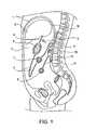
- the peritoneum 1includes a main cavity or greater sac 2 , which contains the small intestine 6 , transverse colon 7 , stomach 8 , liver 9 , bare area of liver 10 , pancreas 11 , duodenum 12 , aorta 13 , and mesentery 14 .
- laparoscopyAn alternative and somewhat less traumatic surgical technique used to access an internal cavity of a patient involves laparoscopy.
- a laparoscope and other surgical instrumentsare inserted through one or more small incisions of the anterior abdominal wall, e.g., a transumbilical incision.
- Patients undergoing laparoscopygenerally have shorter recovery times and less incision pain than those using traditional open surgery.
- the foregoing objectis obtained by providing access to a body cavity, such as the peritoneal cavity, via the stomach or rectum.
- a transmural access systemincludes an overtube that is flexible and has a plurality of lumens.
- One of the lumensis configured to receive an endoscope.
- Additional lumenscan be provided to pass an attachment mechanism, such as suture anchors, to the distal end of the overtube.
- the transmural access systemincludes a trocar and sheath for forming and maintaining an access portal in a body wall.
- the trocaris configured to pass through one of the lumens of the overtube.
- the trocar and sheathpreferably have an axial length between 50 cm and 100 cm.
- An alternative safety trocaris also disclosed.
- the safety trocarincludes at its distal end an obturator tip.
- the obturator tipis slidable between a first position and a second position. In the first position the distal tip is located distally to the trocar tube distal end, and in the second position, the distal tip is located proximal to the trocar tube distal end.
- a dilator tipis operably connected to the distal end of the trocar tube.
- the transmural access systemincludes a suture exchanger for use with the overtube.
- the suture exchangeris configured to catch or secure suture anchors and withdraw the suture ends proximally through the overtube.
- the suture exchangerincludes a shaft having a proximal end, a distal end, and a central portion therebetween. The central portion has an axial length between 30 centimeters and 120 centimeters and an outside diameter less than 3 centimeters.
- a hook or other toolis formed on the distal end of the shaft in order to catch or secure the sutures. The shaft can then be removed from the overtube, thereby withdrawing the distal ends of the sutures for knotting.
- a methodfor transmurally accessing a body cavity of a patient.
- the methodincludes providing a multi-lumen overtube having a proximal end, a distal end, a main lumen and at least one attachment lumen.
- An endoscopeis positioned to extend through at least a portion of the main lumen.
- At least one attachment mechanismis provided to extend through at least a portion of the at least one attachment lumen.
- the overtubecan then be advanced through a body orifice into and along at least a portion of a body passageway such that the distal end is adjacent a selected portal sight along a body wall (e.g., the stomach).
- the distal end of the overtubeis then secured to the body wall at the selected portal sight by advancing the at least one attachment mechanism through the at least one attachment lumen and into the body wall adjacent the selected portal sight.
- the endoscopecan then be withdrawn from the main lumen, and a flexible trocar advanced therethrough.
- the flexible trocaris used to form a portal at the selected portal sight.
- the endoscopecan be readvanced through the main lumen so that a distal end of the endoscope is disposed adjacent or through the portal. This allows viewing at least a portion of the body cavity.
- the portalcan be sutured by traditional techniques or with a suture anchor.
- FIG. 1is a partial, cross-sectional view of the peritoneum 1 that lines the wall the abdominal cavity and folds inwards to enclose the viscera;
- FIG. 2is cross-sectional view of a flexible, multi-lumen overtube according to an embodiment of the present invention
- FIG. 3is a sectional view of the overtube of FIG. 2 taken along line 3 - 3 ;
- FIG. 4is a view of a suture anchor according to an embodiment of the present invention.
- FIG. 5is a side-view of a needle introducer according to an embodiment of the present invention.
- FIG. 6is a cross-sectional view of needle introducers inserted into the overtube of FIG. 2 ;
- FIG. 7is a side-view of a pushing rod according to an embodiment of the present invention.
- FIG. 8is a side-view showing the suture anchor pre-loaded within the needle introducer and the pushing rod ready to be introduced into the needle introducer;
- FIG. 9is a side-view showing the pushing rod being advanced into the needle introducer and the suture anchor projecting through the needle introducer;
- FIG. 10is a side-view of a flexible trocar according to an embodiment of the present invention.
- FIG. 11is a side-view of a sheath according to an embodiment of the present invention.
- FIG. 12is a side-view of the flexible trocar of FIG. 9 positioned within the sheath of FIG. 11 ;
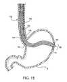
- FIG. 13is a partial, cross-sectional view showing the flexible, multi-lumen overtube of FIG. 2 positioned in the mouth and along the esophagus of a patient such that the overtube distal end is positioned against a selected portal sight of the stomach wall;
- FIG. 14is a partial, cross-sectional view illustrating the overtube distal end secured to the selected portal sight of the stomach wall with suture anchors and an endoscope positioned within the overtube;
- FIG. 15is a partial, cross-sectional view showing the overtube distal end secured to the selected portal sight of the stomach wall with suture anchors and a sheathed flexible trocar puncturing the selected portal sight such that the sheath and the flexible trocar extend through the stomach wall;
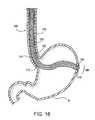
- FIG. 16is a view similar to FIG. 15 showing the flexible trocar withdrawn from the sheath, the sheath remaining extended through the stomach wall and having an endoscope disposed therein;
- FIG. 17is a perspective side view showing the overtube distal end partly separated from the stomach wall with suture anchors extending from the stomach wall into the attachment lumens of the overtube;
- FIG. 18is a perspective side view showing the overtube distal end, suture anchors extending from the stomach wall into the attachment lumens of the overtube, and an exemplary suture exchanger engaging the suture anchors;
- FIG. 19is a perspective side view showing the overtube distal end, suture anchors extending from the stomach wall into the attachment lumens of the overtube, and an exemplary suture exchanger retracting the suture anchors;
- FIG. 20is a cross-sectional side view showing the exemplary suture exchanger withdrawing the suture anchors through the central lumen of the overtube;
- FIG. 21is a perspective side view of a retroactor for use with the overtube
- FIG. 22is a cross-sectional view showing the overtube
- FIG. 23is a cross-sectional view showing the overtube
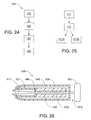
- FIG. 24is a flow-chart showing a method of using a suture exchanger
- FIG. 25is a flow-chart showing a method of ligating a puncture site
- FIG. 26is a cross-sectional side view showing the flexible, long safety trocar in accordance with an embodiment of the present invention.
- FIG. 27is a cross-sectional side view showing the obturator tip completely retracted within the dilator tip
- FIG. 28is an end view of the dilator tip showing the opening through which the obturator tip extends and retracts;
- FIG. 29is cross-sectional side view of the safety trocar without the obturator inserted
- FIG. 30is a cross-sectional side view showing the obturator for use with the safety trocar
- FIG. 31is a perspective side view showing an exemplary knife edge
- FIG. 32is a perspective side view showing an exemplary knife edge
- FIG. 33is a perspective side view showing an exemplary knife edge
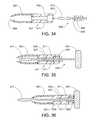
- FIG. 34is a cross-sectional side view of a trocar tube having proximal flanges and a spring-loaded obturator according to an embodiment of the present invention
- FIG. 35is a cross-sectional side view of the spring-loaded obturator within the trocar tube
- FIG. 36is a cross-sectional side view showing the spring-loaded obturator compressed by the flanged trocar tube with the obturator tip extending distally from the dilator tip according to an embodiment of the present invention
- FIG. 37is a cross-sectional side view showing a ratchet mechanism for extension and retraction of the obturator within the trocar tube according to an embodiment of the present invention.
- FIG. 38is a cross-sectional view of the safety trocar tube with the obturator withdrawn and the overlying outer protective sheath positioned within an overtube according to an embodiment of the present invention.
- FIG. 1an abdominal cavity of a patient. More specifically, FIG. 1 illustrates the peritoneum lining the walls of the abdominal cavity and folding inward to enclose the viscera.
- the peritoneumcomprises a main cavity, or greater sac, 2 and the omental bursa, or lesser sac, 3 , which are connected by the epiploic foramen 4 . Numerous peritoneal folds extend between the various organs to hold the viscera in position.
- a new paradigm to access the tissues and organs of a patientis to examine them with a flexible endoscope passed through a portal formed in a natural passageway of a patient.
- a flexible endoscopepassed through a portal formed in a natural passageway of a patient.
- a flexible endoscopeis passed into a patient's mouth through the esophagus, stomach, posterior stomach wall and into the lesser sac. This approach enables direct inspection of the pancreas for visual diagnosis, guided high-frequency ultra-sound with a probe, targeted biopsy, or direct access for therapy.
- peritoneal cavitymay also be accessed through other body passageways such as the colon by way of a non-limiting example.
- application of the principles of the invention to access other body cavities, such as the thoracic cavity by way of a non-limiting exampleis also within the ordinary skill in the art.
- the transluminal access systemincludes a flexible, multi-lumen overtube 110 , an attachment mechanism 115 , a flexible trocar 130 , and a suture exchanger 140 .
- the overtubeis used in combination with an endoscope to establish a passageway to a target portal site in the stomach.
- the attachment mechanismwhich includes one or more suture anchors, is used to attach the overtube to the stomach wall.
- the flexible trocaris passed through the overtube, and is used to puncture the stomach wall, thereby creating access to the peritoneal cavity.
- the suture exchangeris used to retract the suture anchors, as described in greater detail below.
- the flexible, multi-lumen overtube 110comprises a distal end 111 , a proximal end 112 , a main lumen 113 and at least one attachment lumen 114 . Any arrangement of the main lumen and the at least one attachment lumen is contemplated.
- FIGS. 2 and 3illustrate one embodiment of the flexible, multi-lumen overtube having a main lumen 113 with four attachment lumens 114 equally spaced about the main lumen 113 .
- the flexible multi-lumen overtube 110can have a single-piece construction as shown in the embodiment depicted in FIGS. 2 and 3 . Alternatively, several tubes may be bonded together to form the flexible, multi-lumen overtube 110 .
- the flexible, multi-lumen overtube 110can be made from any suitable material known in the art including, but not limited to, polyethylene ether ketone (PEEK), polytetrafluorethylene (PTFE), polyamide, polyurethane, polyethylene and nylon, including multi-layer or single layer structures and may also include reinforcement wires, braid wires, coils and or filaments.
- the flexible, multi-lumen overtube 110further comprises valve 199 over its proximal end 112 .
- Valve 199forms a seal between the flexible, multi-lumen overtube 110 and the endoscope or other device that is advanced therethrough to prevent the loss of any pressurized fluid that is introduced through the endoscope or other device, as will be explained in further detail below.
- valve 199is removable.
- One of ordinary skill in the artwould know how to assemble valve 199 over the proximal end 112 of multi-lumen overtube 110 .
- the main lumen 113is configured to receive and pass an endoscope 120 , as shown in FIG. 14 , a flexible trocar 130 , as shown in FIG. 15 , or an exchanger 151 , as shown in FIGS. 18-20 .
- the main lumen 113ranges in size from about 4 mm to about 9 mm. These sizes are provided for illustrative purposes only and are not intended to be construed as a limitation of the present invention. As one of ordinary skill in the art would appreciate, since the endoscope 120 and the flexible trocar 130 are advanced through the main lumen 113 , the size of the main lumen 113 is related to the size of either the endoscope 120 or the flexible trocar 130 , which ever is larger.
- the size of the trocaris related to the size of the medical instruments that are passed therethrough for the diagnosis and treatment of the tissues and organs within the body cavity.
- a flexible, multi-lumen overtube having a main lumen smaller than about 4 mm used with endoscopes or trocars having an outer diameter smaller than about 4 mm that may become available in the futureare contemplated as being within the scope of the claims of the present invention.
- the attachment lumen 114is configured to receive and pass an attachment mechanism 115 (see FIG. 4 ).
- the at least one attachment lumen 114 and the main lumen 113comprise a longitudinal slit (not shown) along its length so that the attachment mechanism 115 can be pulled from the at least one attachment lumen 114 into the main lumen 113 as explained more fully below.
- FIG. 4illustrates a non-limiting embodiment of an attachment mechanism comprising a suture anchor 115 .
- One exemplary suture anchor 115comprises a spring coil anchor 118 and a suture portion 119 , such as the Cope suture anchor manufactured by Cook, Incorporated, Bloomington, Ind.
- the suture exchanger 151generally comprises a shaft having proximal portion 153 and distal portion 155 .
- the suture exchangeris sized to fit through the main lumen of the overtube 110 .
- the suture exchangerwill generally have a length between 50 cm and 100 cm. However, a suture exchanger having a shorter length, for example for pediatric applications, could alternatively be used.
- the suture exchangercan be formed from any number of materials that can impart axial rotation from the proximal portion to the distal portion of the shaft, while maintaining the flexibility necessary to pass through the central passageway of the overtube.
- suture exchangercan be formed from stainless steel, a rigid plastic, or a superelastic allow such as nickel-titanium.
- proximal portion 153 of suture exchanger 151includes a handle 159 that allows a physician to easily grip and control the suture exchanger.
- the distal portion 153 of suture exchanger 151is adapted to catch, pull, loop, or ensnare the suture portion of the suture anchor(s) and then withdraw the suture portions through the main lumen, as described in greater detail below.
- the distal portion 155includes sickle-shaped hook 157 that is offset from the longitudinal axis A formed by the shaft.
- other alternatively-shaped distal portionswill become apparent to one of ordinary skill in the art in view of the present disclosure.
- the transmural access system of the present inventionis used to examine or treat tissues or organs within a body cavity, e.g. the peritoneal cavity, as follows.
- An endoscope 120is disposed within the main lumen 113 of the flexible, multi-lumen overtube 110 (see FIG. 14 ).
- the flexible, multi-lumen overtube and endoscopeare inserted within a body orifice of a patient. Any body orifice is contemplated including, but not limited to, the mouth or anus.
- the flexible, multi-lumen overtube and endoscopeare advanced through a body passageway and a sight for an access portal along a body wall is selected endoscopically.
- a sight for the gastric portal through the posterior stomach wallis selected as shown in FIG. 13 .
- a sight for an access portal through an intestinal wallis selected.
- the body cavityis insufflated to provide an unobstructed view of the tissues and organs therein, to protect the tissues and organs from damage as the access portal is formed and to provide a working space within the cavity.
- an insufflation needle(not shown) is deployed through the endoscope and punctures the stomach wall.
- a suitable gassuch as carbon dioxide, is injected through the endoscope to expand the peritoneal cavity.
- Valve 199 at the overtube proximal endprevents the gas from escaping about the endoscope.
- a wire guideis placed through the insufflation needle to maintain access to the puncture site.
- the insufflation needleis removed and the overtube distal end 111 is secured to the stomach wall.
- at least one suture anchor 115is passed through the at least one attachment lumen 114 and deployed into the selected portal site of the body wall 180 .
- FIGS. 4-8illustrate a non-limiting, exemplary method pre-loading and deploying the suture anchor 115 through the attachment lumen 114 of the flexible, multi-lumen overtube 110 .
- a suture anchor 115(shown in FIG. 4 ) is inserted into a needle introducer 160 (shown in FIG. 5 ).
- the needle introducer 160is passed through the attachment lumen 114 of the flexible, multi-lumen overtube 110 (shown in FIG. 6 ) and the suture needle 160 is inserted through the stomach wall at the selected access portal sight.
- a pusher rod 170FIG.
- the overtube distal end 111can be positioned firmly against the body wall by proximally pulling the suture portion 119 of the suture anchor 115 .
- the proximal end suture portion 119can be held by the physician or secured to the proximal end 112 of the flexible, multi-lumen overtube 110 .
- the endoscope 120since the endoscope 120 remains within the main lumen 113 of the flexible, multi-lumen overtube 110 , a physician can view the deployment of the suture anchor 115 through the body wall at the selected access portal sight 180 . This affords the physician the ability to attach the overtube distal end 111 against a body wall such that the main lumen 113 is precisely aligned with the selected access portal sight 180 .
- the endoscope 120is removed and a trocar 130 is advanced through the main lumen 113 of the flexible, multi-lumen overtube 110 and into the body wall to form the access portal to the body cavity as shown in FIG. 15 and as described above. If a wire guide has been placed and the trocar 130 comprises a wire guide lumen 137 , then the trocar 130 is advanced over the wire guide.
- the overtube distal end 111cannot shift as the physician forms the access portal through the body wall. This facilitates the formation of the access portal by providing a controlled puncture of the body wall and enables the physician to accurately locate the access portal at the selected portal sight 180 . Moreover, since the overtube distal end 111 is secured against the interior body wall at the selected portal sight 180 , the flexible, multi-lumen overtube 110 cannot extend or pass through the access portal. This eliminates any potential damage caused to tissues and organs within the body cavity due to over insertion of the flexible, multi-lumen overtube.
- the trocar 130is withdrawn.
- An endoscope or other instrumentationcan be inserted through the main lumen 113 of the flexible, multi-lumen overtube 110 for examination, diagnosis and/or treatment of the tissues or organs within the body cavity.
- the trocar 130comprises an outer sheath 133 (see FIG. 15 )
- the obturator portion of the trocar 130can be withdrawn leaving the sheath 133 positioned through the access portal.
- An endoscope or other instrumentationcan be inserted through the sheath 133 as shown in FIG. 16 .
- the remaining sheathforms a lumen from exterior of the patient to the body cavity.
- Sheath 133further provides the advantage of protecting the tissue surrounding the access portal from trauma as an endoscope or other instrumentation is advanced therethrough to perform procedures within the body cavity.
- the sheath 133also eliminates the need to dilate the access portal, which could damage the tissue surrounding the access portal to the body cavity.
- the access portalis closed by ligating the suture portions 119 of the suture anchors 115 .
- One method of ligating the suturesis by ensnaring ( FIG. 18 ) and withdrawing ( FIGS. 19-20 ) the sutures through the main lumen of the overtube by using suture exchanger 151 . More specifically, the sutures are unknotted or cut from the proximal end of overtube 110 . As illustrated in FIG. 17 , overtube 110 is then partly withdrawn from the patient, thereby exposing suture portion 119 at the distal end of overtube 110 . Hook 157 of suture exchanger 151 can then ensare ( FIG. 18 ) and withdraw ( FIGS. 19-20 ) suture portions 119 through main lumen 113 . After the sutures are extracted through the main lumen, the physician can use conventional techniques to close the trocar puncture.
- FIG. 24further illustrates this method of using suture exchanger 151 .
- the suture exchangeris passed through the main lumen of the overtube.
- the suturesare released from the proximal end of the overtube.
- the overtubeis partly retracted and the suture exchanger hook is used to ensnare and withdraw the suture portions through the main lumen.
- the physicianligates the sutures, thus closing the trocar puncture.
- the suture portions of the suture anchorsare pulled through a longitudinal slit 161 that extends along the length of attachment lumens 114 .
- Longitudinal slit 161extends from the attachment lumens into main lumen 113 , as illustrated in FIG. 22 , or alternatively, to the exterior of the overtube, as illustrated in FIG. 23 .
- a splittable wallcan be provided in the overtube.
- the suture portionsare dislocated from the attachment lumens into either the main lumen 113 or the exterior of overtube 110 . This allows the physician to tie the suture portions and ligate the trocar puncture site with the suture portions of the suture anchors. Ligation can be accomplished with conventional ligating techniques, or as described below in greater detail.
- the overtubeis entirely withdrawn from the patient before the trocar puncture is ligated.
- the sheathis removed.
- the proximal ends of the suture portionsare released from the overtube, thus detaching the overtube from the stomach wall.
- the overtubeis withdrawn from the patient, thereby leaving the proximal ends of the suture portions extending from the patient's mouth.
- the physicianuses and endoscope and a flexible endoscopic suturing device to approximate the tissue surrounding the puncture site.
- the physiciancan use a flexible Sew-Right® device, which is available from Wilson-Cook Medical, Winston-Salem, N.C.
- a flexible Sew-Right® devicewhich is available from Wilson-Cook Medical, Winston-Salem, N.C.
- the physiciancan endoscopically trim and ligate the suture portions with conventional ligating techniques.
- the flexible trocar 130comprises an obturator having a sharp, wedge tip 135 .
- the sharp, wedge tip 135comprises beveled edges.
- the flexible trocar 130includes a sheath 133 (see FIG. 11 ).
- the flexible trocarfurther includes a wire guide lumen 137 to accommodate a wire guide (not shown) that enables a guided puncture of the body wall.
- FIG. 26illustrates an alternative safety trocar, and in particular, safety trocar 500 .
- Safety trocar 500includes a trocar tube 502 and obturator 515 inserted in trocar tube 502 .
- Trocar tube 502comprises dilator tip 501 located at the distal end of trocar tube 502 and protective outer sheath 506 disposed along an outer surface of trocar tube 502 .
- Trocar tube 502 and dilator tip 501can be formed from any suitable polymers known in the art, such as biocompatible polymers.
- Trocar tube 502can also be provided with a PTFE coating.
- the coatingcan also comprise a hydrophilic polymer selected from the group comprising polyacrylate, copolymers comprising acrylic acid, polymethacrylate, polyacrylamide, poly(vinyl alcohol), poly(ethylene oxide), poly(ethylene imine), carboxymethylcellulose, methylcellulose, poly(acrylamide sulphonic acid), polyacrylonitrile, poly(vinyl pyrrolidoine), agar, dextran, dextrin, carrageenan, xanthan, and guar.
- the hydrophilic polymerscan also include ionizable groups such as acid groups, e.g., carboxylic, sulphonic or nitric groups.
- the hydrophilic polymersmay be cross-linked through a suitable cross-binding compound. The cross-binder actually-used depends on the polymer system: if the polymer system is polymerized as a free radical polymerization, a preferred cross-binder comprises 2 or 3 unsaturated double bonds.
- Obturator 515comprises an obturator tip 517 at the distal end of obturator 515 , a handle 503 at the proximal end of obturator 515 , and an elongated shaft 530 between the distal and proximal ends of obturator 515 .
- Obturator tip 517is biased toward a concealed position within dilator tip 501 , as shown in FIGS. 26-27 and as described in greater detail below.
- obturator tip 517is extended distally through dilator tip 501 (see FIG. 36 ). Since obturator tip 517 is biased toward a concealed position, immediately after the desired cut is made, obturator tip 517 automatically retracts within dilator tip 501 .
- FIG. 30illustrates a side view of obturator 515 having of a proximal handle 503 , a distal obturator tip 517 , and an elongated shaft 530 situated between the proximal and distal regions of obturator 515 .
- Elongated shaft 530can be manufactured from any relatively rigid, commercially available material, such as polymers or metals. Such materials should be axially strong enough to drive the obturator tip through tissue, while sufficiently flexible for endoscopic use. One such material is stainless steel, although many alternative materials will be apparent to one of ordinary skill in view of the present disclosure.
- handle 503is attached to the proximal end of elongated shaft 530 .
- Handle 503is configured to be easily grasped by a physician's hand and is preferably T-shaped. Handle 503 also provides leverage for tissue incision, puncturing, and cutting.
- FIG. 27illustrates a side view of dilator tip 501 , with obturator tip 517 retracted and completely encased within dilator tip 501 .
- Dilator tip 501shields obturator tip 517 , thereby preventing accidental or unintended puncture of tissue and internal organs while the obturator tip is not in use.
- Dilator tip 501can be provided as a rounded, bullet-shaped housing that surrounds the entire outside of obturator tip 517 when obturator tip 517 is not in an extended position for cutting tissue.
- the surface of dilator tip 501gradually tapers and ultimately converges at the distal end of dilator tip 501 where a longitudinal slot 504 is formed. Longitudinal slot 504 provides an orifice through which obturator tip 517 extends distally and can contact tissue.
- dilator tip 501 and trocar tube 502can be constructed as an integral piece.
- dilator tip 501can be a separately manufactured component, which is readily detached from trocar tube 502 .
- protective outer sheath 506is a separate component, axially slidable relative to trocar tube 502 .
- Sheath 506includes an axial passageway 546 and a lubricious exterior wall 545 .
- the axial passageway 546is adapted to receive trocar tube 502 .
- Surface wall 545provides a smooth contact surface with the outer surface of trocar tube 502 , thereby allowing trocar tube 502 to smoothly slide within axial passageway 546 of protective outer sheath 506 .
- FIG. 28shows an end-view of dilator tip 501 .
- Longitudinal slot 504allows the obturator tip 517 to extend distally from dilator tip 501 to penetrate and cut the desired tissue and thereafter retract proximally into dilator tip 501 when the required cutting has been performed.
- the longitudinal slot 504corresponds to the geometry of the obturator tip 517 .
- the surface of dilator tip 501is flush with trocar tube 502 outer surface so as to provide a smooth transition between trocar tube 502 and the tapered outer surface of dilator tip 501 .
- the smooth transitionensures minimal trauma upon insertion of dilator tip 501 and trocar tube 502 .
- the bullet-nose shape of dilator tip 501can be used to further dilate the tissue after obturator tip 517 has made an initial incision. Morever, dilator tip 501 is relatively wide, and thus provides a stable support or platform on which to cut tissue.
- FIG. 29shows trocar tube 502 without obturator 515 inserted.
- Trocar tube 502contains a lumen 507 through which obturator 515 is inserted and advanced therethrough until obturator tip 517 is completely encased within dilator tip 501 .
- Clearanceis preferably provided between the inside diameter of lumen 507 ( FIG. 29 ) and the outside diameter of elongated shaft 530 of obturator 515 ( FIG. 30 ). As a result, obturator 515 slides smoothly within trocar tube 502 during both extension and retraction of obturator tip 517 .
- Obturator tip 517can consist of a variety of cutting edges, as shown in FIGS. 31-33 .
- FIG. 31shows a double-edged obturator tip 517 .
- tip 517can be a flat, scalpel-like blade which allows for a clean incision, thereby minimizing tearing of surrounding tissue.
- obturator tip 517can also consist of a serrated knife edge. The serrated knife edge produces greater surface area contact with the tissue which reduces the force required to cut the tissue.
- Obturator tip 517can be formed from any malleable material that can be sharpened. Examples of such materials include, but are not limited to, stainless steel and ceramics. Many alternative materials will be apparent to one of ordinary skill in view of the present disclosure.
- FIG. 26shows a manual safety trocar 500 , in which obturator tip 517 extends distally through longitudinal slot 504 of dilator tip 501 when manual force is applied at proximal handle 503 .
- the force applied at proximal handle 503is transmitted to elongated shaft 530 and, ultimately, to obturator tip 517 .
- FIG. 34depicts an obturator 532 with the proximal region of elongated shaft 530 having coiled springs 505 .
- FIG. 34also shows corresponding trocar tube 531 , into which obturator 532 is inserted.
- Trocar tube 531contains flanges 511 located at the proximal end.
- FIG. 35shows obturator 532 fully inserted into lumen 507 of trocar tube 531 .
- FIG. 36shows flanges 511 at the proximal end of obturator 532 engaging coiled springs 505 . As obturator handle 503 is forced distally, flanges 511 compress coiled springs 505 .
- obturator tip 517extends distally through longitudinal slot 504 of dilator tip 501 and eventually punctures the desired tissue, as shown in FIG. 36 .
- the compressed spring coils 505urge obturator 532 back into the retracted proximal position when they are not being pushed by the user.
- Spring coils 505also provide stability to flexible obturator 532 such that a precise cut can be made.
- the safety trocarmay also include a ratchet mechanism 549 to extend and retract obturator tip 517 .
- FIG. 37shows a side view of obturator 533 with a ratchet mechanism 549 .
- the ratchet mechanism 549consists of a pivoting toothed cam 550 , which engages matching teeth 529 on the proximal end of elongated shaft 530 of obturator 533 .
- a spring 553 connected to the handle 503 of trocar tube 502allows for extension of obturator tip 517 in small incremental distances through longitudinal slot 504 of dilator tip 501 .
- ratchet-like operationpermits obturator 533 to incrementally extend to create the necessary tissue puncture while providing tactile feedback to the physician indicating that obturator 533 is being advanced through the tissue.
- the ratchet-mechanismalso prevents the sudden release of back pressure associated with puncturing tissue thereby eliminating accidental extension of obturator tip 517 through tissue and organs.
- Other embodiments for activating obturator tip 517 to extend and retractwill become apparent to one of ordinary skill in the art in view of the present disclosure.
- any of the above described safety trocarscan be used in the procedure for accessing a body cavity, described below.
- protective outer sheath 506is axially slidable relative to trocar tube 502 , after obturator tip 517 has created the desired access portal 180 to the body cavity, obturator 515 and trocar tube 502 can be withdrawn from protective outer sheath 506 , as shown in FIG. 38 .
- Protective outer sheath 506functions as an accessway to the body cavity. Because the tissue that lines stomach wall 8 is thick and tortuous, the trocar cut or puncture is typically not clean. The protective outer sheath 506 mitigates additional trauma incurred by passing the endoscope through the access port multiple times or from passing various accessories through the access port.
- safety trocar 500to create an access portal to a body cavity.
- one of ordinary skillwould know how to use the safety trocar 500 in a variety of other applications.
- a safety trocarcan be used in colonoscopic or thoracic applications.
Landscapes
- Health & Medical Sciences (AREA)
- Life Sciences & Earth Sciences (AREA)
- Surgery (AREA)
- Animal Behavior & Ethology (AREA)
- Public Health (AREA)
- Engineering & Computer Science (AREA)
- Biomedical Technology (AREA)
- Heart & Thoracic Surgery (AREA)
- Medical Informatics (AREA)
- Molecular Biology (AREA)
- Pathology (AREA)
- General Health & Medical Sciences (AREA)
- Nuclear Medicine, Radiotherapy & Molecular Imaging (AREA)
- Veterinary Medicine (AREA)
- Physics & Mathematics (AREA)
- Biophysics (AREA)
- Optics & Photonics (AREA)
- Radiology & Medical Imaging (AREA)
- Surgical Instruments (AREA)
- Endoscopes (AREA)
Abstract
Description
Claims (9)
Priority Applications (1)
| Application Number | Priority Date | Filing Date | Title |
|---|---|---|---|
| US11/141,449US8475476B2 (en) | 2004-06-01 | 2005-05-31 | System and method for accessing a body cavity |
Applications Claiming Priority (3)
| Application Number | Priority Date | Filing Date | Title |
|---|---|---|---|
| US57595804P | 2004-06-01 | 2004-06-01 | |
| US64879105P | 2005-02-01 | 2005-02-01 | |
| US11/141,449US8475476B2 (en) | 2004-06-01 | 2005-05-31 | System and method for accessing a body cavity |
Publications (2)
| Publication Number | Publication Date |
|---|---|
| US20060015006A1 US20060015006A1 (en) | 2006-01-19 |
| US8475476B2true US8475476B2 (en) | 2013-07-02 |
Family
ID=34941536
Family Applications (1)
| Application Number | Title | Priority Date | Filing Date |
|---|---|---|---|
| US11/141,449Active2026-09-02US8475476B2 (en) | 2004-06-01 | 2005-05-31 | System and method for accessing a body cavity |
Country Status (2)
| Country | Link |
|---|---|
| US (1) | US8475476B2 (en) |
| EP (1) | EP1602336B1 (en) |
Families Citing this family (89)
| Publication number | Priority date | Publication date | Assignee | Title |
|---|---|---|---|---|
| US6845776B2 (en) | 2001-08-27 | 2005-01-25 | Richard S. Stack | Satiation devices and methods |
| US6675809B2 (en) | 2001-08-27 | 2004-01-13 | Richard S. Stack | Satiation devices and methods |
| US7097665B2 (en)* | 2003-01-16 | 2006-08-29 | Synecor, Llc | Positioning tools and methods for implanting medical devices |
| CN101810521B (en) | 2001-08-27 | 2015-05-13 | 辛尼科有限责任公司 | Satiation devices and methods |
| US7146984B2 (en)* | 2002-04-08 | 2006-12-12 | Synecor, Llc | Method and apparatus for modifying the exit orifice of a satiation pouch |
| US20040143342A1 (en) | 2003-01-16 | 2004-07-22 | Stack Richard S. | Satiation pouches and methods of use |
| US20050247320A1 (en)* | 2003-10-10 | 2005-11-10 | Stack Richard S | Devices and methods for retaining a gastro-esophageal implant |
| US8206456B2 (en) | 2003-10-10 | 2012-06-26 | Barosense, Inc. | Restrictive and/or obstructive implant system for inducing weight loss |
| US8425539B2 (en) | 2004-04-12 | 2013-04-23 | Xlumena, Inc. | Luminal structure anchoring devices and methods |
| US12303105B2 (en) | 2004-04-12 | 2025-05-20 | Boston Scientific Scimed, Inc. | Luminal structure anchoring devices and methods |
| US9713465B1 (en)* | 2004-04-19 | 2017-07-25 | Granit Medical Innovation Llc | Surgical closure device and associated method |
| WO2005105003A1 (en) | 2004-04-26 | 2005-11-10 | Synecor, Llc | Restrictive and/or obstructive implant for inducing weight loss |
| JP5111112B2 (en) | 2004-12-08 | 2012-12-26 | エックスルミナ, インコーポレイテッド | Device for performing needle-guided therapy |
| US8784437B2 (en)* | 2005-06-09 | 2014-07-22 | Xlumena, Inc. | Methods and devices for endosonography-guided fundoplexy |
| US8777967B2 (en)* | 2005-06-09 | 2014-07-15 | Xlumena, Inc. | Methods and devices for anchoring to tissue |
| US20080190989A1 (en)* | 2005-10-03 | 2008-08-14 | Crews Samuel T | Endoscopic plication device and method |
| US9055942B2 (en) | 2005-10-03 | 2015-06-16 | Boston Scienctific Scimed, Inc. | Endoscopic plication devices and methods |
| JP2007185495A (en) | 2006-01-13 | 2007-07-26 | Olympus Medical Systems Corp | Overtube |
| US20070213702A1 (en)* | 2006-03-08 | 2007-09-13 | Olympus Medical Systems Corp. | Medical procedure carried out via a natural opening |
| US7785333B2 (en) | 2006-02-21 | 2010-08-31 | Olympus Medical Systems Corp. | Overtube and operative procedure via bodily orifice |
| US8241279B2 (en)* | 2006-02-23 | 2012-08-14 | Olympus Medical Systems Corp. | Overtube and natural opening medical procedures using the same |
| US8728121B2 (en) | 2006-01-13 | 2014-05-20 | Olympus Medical Systems Corp. | Puncture needle and medical procedure using puncture needle that is performed via natural orifice |
| US8721657B2 (en) | 2006-01-13 | 2014-05-13 | Olympus Medical Systems Corp. | Medical instrument |
| US20070167676A1 (en)* | 2006-01-13 | 2007-07-19 | Olympus Medical Systems Corp. | Overtube and medical procedure via natural orifice using the same |
| US7963912B2 (en)* | 2006-05-08 | 2011-06-21 | Ethicon Endo-Surgery, Inc. | Endoscopic translumenal surgical methods using a sheath |
| US20070260273A1 (en)* | 2006-05-08 | 2007-11-08 | Ethicon Endo-Surgery, Inc. | Endoscopic Translumenal Surgical Systems |
| US20070260121A1 (en)* | 2006-05-08 | 2007-11-08 | Ethicon Endo-Surgery, Inc. | Endoscopic Translumenal Surgical Systems |
| US20080097510A1 (en)* | 2006-09-01 | 2008-04-24 | Albrecht Thomas E | Method for inducing weight loss with a patient |
| JP5238702B2 (en) | 2006-09-02 | 2013-07-17 | シネコー・エルエルシー | Intestinal sleeve, installation system thereof, and method thereof |
| US20090125040A1 (en)* | 2006-09-13 | 2009-05-14 | Hambly Pablo R | Tissue acquisition devices and methods |
| US9314361B2 (en)* | 2006-09-15 | 2016-04-19 | Boston Scientific Scimed, Inc. | System and method for anchoring stomach implant |
| US20080264430A2 (en)* | 2006-11-21 | 2008-10-30 | Edmund Roschak | Methods and devices for accessing the heart |
| ATE514382T1 (en)* | 2006-11-30 | 2011-07-15 | Wilson Cook Medical Inc | FABRIC ANCHOR FOR SEAM CLOSURE OF PERFORATIONS |
| CA2691269C (en)* | 2007-05-12 | 2016-04-12 | Barosense, Inc. | Devices and methods for stomach partitioning |
| US20100191261A1 (en)* | 2007-05-22 | 2010-07-29 | Sally Carter | Suture management port |
| US9155532B2 (en)* | 2007-05-25 | 2015-10-13 | Cook Medical Technologies Llc | Medical devices, systems and methods for closing perforations |
| US8740937B2 (en)* | 2007-05-31 | 2014-06-03 | Cook Medical Technologies Llc | Suture lock |
| WO2009011881A1 (en)* | 2007-07-18 | 2009-01-22 | Barosense, Inc. | Overtube introducer for use in endoscopic bariatric surgery |
| JP5581209B2 (en) | 2007-07-18 | 2014-08-27 | ボストン サイエンティフィック サイムド,インコーポレイテッド | Endoscopic implant system |
| DE102007040358A1 (en)* | 2007-08-27 | 2009-03-05 | Technische Universität München | Trocar tube, trocar, obturator or rectoscope for transluminal endoscopic surgery over natural orifices |
| JP5226792B2 (en)* | 2007-09-25 | 2013-07-03 | クック メディカル テクノロジーズ エルエルシー | Medical instruments, devices and methods for using tissue anchors |
| AU2008310975B2 (en)* | 2007-10-09 | 2013-08-22 | Cook Medical Technologies Llc | Systems, devices and methods having an overtube for accessing a bodily opening |
| WO2009082596A1 (en)* | 2007-12-18 | 2009-07-02 | Wilson-Cook Medical, Inc. | Device and method for placement of tissue anchors |
| US20090171383A1 (en) | 2007-12-31 | 2009-07-02 | David Cole | Gastric space occupier systems and methods of use |
| EP2259733B1 (en)* | 2008-03-06 | 2014-07-23 | Cook Medical Technologies LLC | Medical systems for accessing an internal bodily opening |
| US8020741B2 (en) | 2008-03-18 | 2011-09-20 | Barosense, Inc. | Endoscopic stapling devices and methods |
| WO2009132111A1 (en)* | 2008-04-23 | 2009-10-29 | Wilson-Cook Medical Inc. | Tacking device |
| US20090281379A1 (en)* | 2008-05-12 | 2009-11-12 | Xlumena, Inc. | System and method for transluminal access |
| US8454632B2 (en) | 2008-05-12 | 2013-06-04 | Xlumena, Inc. | Tissue anchor for securing tissue layers |
| WO2009140594A2 (en)* | 2008-05-15 | 2009-11-19 | Wilson-Cook Medical, Inc. | Systems, devices and methods for accessing a bodily opening |
| WO2010022060A1 (en)* | 2008-08-19 | 2010-02-25 | Wilson-Cook Medical Inc. | Apparatus for removing lymph nodes or anchoring into tissue during a translumenal procedure |
| US8192461B2 (en)* | 2008-09-11 | 2012-06-05 | Cook Medical Technologies Llc | Methods for facilitating closure of a bodily opening using one or more tacking devices |
| WO2010042402A1 (en)* | 2008-10-06 | 2010-04-15 | Wilson-Cook Medical, Inc. | Endcap for safely deploying tissue anchors |
| US7934631B2 (en) | 2008-11-10 | 2011-05-03 | Barosense, Inc. | Multi-fire stapling systems and methods for delivering arrays of staples |
| US8377095B2 (en)* | 2008-12-05 | 2013-02-19 | Cook Medical Technologies, LLC | Tissue anchors for purse-string closure of perforations |
| CA2746213A1 (en)* | 2008-12-09 | 2010-07-08 | Wilson-Cook Medical Inc. | Apparatus and methods for controlled release of tacking devices |
| AU2009324819B2 (en) | 2008-12-09 | 2014-04-17 | Cook Medical Technologies Llc | Retractable tacking device |
| JP2012512715A (en)* | 2008-12-19 | 2012-06-07 | クック メディカル テクノロジーズ エルエルシー | A tacking device of varying thickness and method of delivery and deployment thereof |
| EP2389122B1 (en)* | 2008-12-19 | 2015-03-04 | Cook Medical Technologies LLC | Clip devices |
| US20100198019A1 (en)* | 2009-01-30 | 2010-08-05 | Tyco Healthcare Group Lp | Suture management apparatus for surgical portal apparatus including interlocking cap |
| US20100198018A1 (en)* | 2009-01-30 | 2010-08-05 | Tyco Healthcare Group Lp | Suture management system for surgical portal apparatus including internal tubes |
| US20100210912A1 (en)* | 2009-02-17 | 2010-08-19 | Tyco Healthcare Group Lp | Access port with suture management system including flapper with inserts |
| US20100211084A1 (en)* | 2009-02-18 | 2010-08-19 | Tyco Healthcare Group Lp | Suture management system for surgical portal apparatus including numbered clips |
| US20100249810A1 (en)* | 2009-03-24 | 2010-09-30 | Tyco Healthcare Group Lp | Suture management system for surgical portal apparatus including slotted ring |
| EP2413809B1 (en)* | 2009-04-03 | 2014-10-08 | Cook Medical Technologies LLC | Medical devices for rapid deployment and fixation of tissue anchors |
| CA2757554A1 (en) | 2009-04-03 | 2010-10-07 | Cook Medical Technologies Llc | Tissue anchors and medical devices for rapid deployment of tissue anchors |
| US20100268029A1 (en)* | 2009-04-21 | 2010-10-21 | Xlumena, Inc. | Methods and apparatus for advancing a device from one body lumen to another |
| US9364259B2 (en)* | 2009-04-21 | 2016-06-14 | Xlumena, Inc. | System and method for delivering expanding trocar through a sheath |
| US20100276469A1 (en)* | 2009-05-01 | 2010-11-04 | Barosense, Inc. | Plication tagging device and method |
| EP2429374B1 (en)* | 2009-05-01 | 2013-09-25 | Cook Medical Technologies LLC | Medical device for suturing perforations |
| US8961539B2 (en)* | 2009-05-04 | 2015-02-24 | Boston Scientific Scimed, Inc. | Endoscopic implant system and method |
| US8834361B2 (en)* | 2009-05-15 | 2014-09-16 | Cook Medical Technologies Llc | Systems, devices and methods for accessing a bodily opening |
| JP2012527970A (en)* | 2009-05-28 | 2012-11-12 | クック メディカル テクノロジーズ エルエルシー | Hail-fastening device and hail-fastening device deployment method |
| JP5535313B2 (en) | 2009-05-29 | 2014-07-02 | エックスルミナ, インコーポレイテッド | Device and method for deploying a stent across adjacent tissue layers |
| US20110022064A1 (en)* | 2009-07-27 | 2011-01-27 | Tyco Healthcare Group Lp | Access apparatus including hinged suture traps |
| EP2571427B1 (en) | 2010-05-21 | 2017-07-19 | Boston Scientific Scimed, Inc. | Tissue-acquisition and fastening devices |
| RU2463003C1 (en)* | 2011-04-01 | 2012-10-10 | Александр Цезаревич Буткевич | Method of preventing intestinal fistulas in staged treatment of pancreatonecrosis |
| US9113868B2 (en) | 2011-12-15 | 2015-08-25 | Ethicon Endo-Surgery, Inc. | Devices and methods for endoluminal plication |
| US9173657B2 (en) | 2011-12-15 | 2015-11-03 | Ethicon Endo-Surgery, Inc. | Devices and methods for endoluminal plication |
| US8992547B2 (en) | 2012-03-21 | 2015-03-31 | Ethicon Endo-Surgery, Inc. | Methods and devices for creating tissue plications |
| JP6360042B2 (en) | 2012-05-17 | 2018-07-18 | ボストン サイエンティフィック サイムド,インコーポレイテッドBoston Scientific Scimed,Inc. | Method and device for access across adjacent tissue layers |
| WO2014109775A1 (en)* | 2013-01-14 | 2014-07-17 | Empire Technology Development, Llc | Trans-urethral sling delivery device |
| ES2980140T3 (en) | 2013-02-21 | 2024-09-30 | Boston Scient Scimed Inc | Devices for forming an anastomosis |
| US9198647B2 (en) | 2013-02-25 | 2015-12-01 | Covidien Lp | Flexible access assembly |
| TWI554295B (en)* | 2014-02-27 | 2016-10-21 | 梓源生技有限公司 | Overtube and irrigation kit |
| JP6835731B2 (en) | 2014-11-13 | 2021-02-24 | パヴメド・インコーポレイテッドPAVMed Inc. | Intraosseous infusion port and usage |
| WO2016183503A1 (en)* | 2015-05-13 | 2016-11-17 | Atricure, Inc. | Access visualization systems |
| US11026673B2 (en) | 2018-05-10 | 2021-06-08 | Edwards Lifesciences Corporation | Corkscrew tissue anchor |
| CN114025822B (en)* | 2019-06-28 | 2023-12-15 | 南微医学科技股份有限公司 | Sheath device and endoscope assembly |
Citations (84)
| Publication number | Priority date | Publication date | Assignee | Title |
|---|---|---|---|---|
| US3809081A (en) | 1970-02-04 | 1974-05-07 | Deseret Pharma | Obturator |
| US4807593A (en) | 1987-05-08 | 1989-02-28 | Olympus Optical Co. Ltd. | Endoscope guide tube |
| US5025778A (en) | 1990-03-26 | 1991-06-25 | Opielab, Inc. | Endoscope with potential channels and method of using the same |
| US5059183A (en) | 1989-09-01 | 1991-10-22 | Neal Semrad | Stop guide wire and double ended obturator-catheter-sheath system and method of use of same |
| US5112308A (en) | 1990-10-03 | 1992-05-12 | Cook Incorporated | Medical device for and a method of endoscopic surgery |
| US5158543A (en) | 1990-10-30 | 1992-10-27 | Lazarus Harrison M | Laparoscopic surgical system and method |
| US5222508A (en) | 1992-10-09 | 1993-06-29 | Osvaldo Contarini | Method for suturing punctures of the human body |
| US5258003A (en) | 1992-06-01 | 1993-11-02 | Conmed Corporation | Method and apparatus for induction of pneumoperitoneum |
| US5304184A (en) | 1992-10-19 | 1994-04-19 | Indiana University Foundation | Apparatus and method for positive closure of an internal tissue membrane opening |
| US5320632A (en) | 1991-11-13 | 1994-06-14 | Harald Heidmueller | Surgical suturing apparatus |
| US5320610A (en) | 1991-12-16 | 1994-06-14 | Inbae Yoon | Automatic retractable trocar with safety shield and method of use |
| US5350385A (en) | 1993-04-28 | 1994-09-27 | Christy William J | Surgical stab wound closure device and method |
| US5364408A (en) | 1992-09-04 | 1994-11-15 | Laurus Medical Corporation | Endoscopic suture system |
| US5368601A (en) | 1992-04-30 | 1994-11-29 | Lasersurge, Inc. | Trocar wound closure device |
| US5374275A (en) | 1993-03-25 | 1994-12-20 | Synvasive Technology, Inc. | Surgical suturing device and method of use |
| US5391182A (en) | 1993-08-03 | 1995-02-21 | Origin Medsystems, Inc. | Apparatus and method for closing puncture wounds |
| US5403329A (en) | 1992-09-23 | 1995-04-04 | United States Surgical Corporation | Instrument for closing trocar puncture wounds |
| US5417699A (en) | 1992-12-10 | 1995-05-23 | Perclose Incorporated | Device and method for the percutaneous suturing of a vascular puncture site |
| US5431151A (en) | 1990-11-06 | 1995-07-11 | Partomed Medizintechnik Gmbh | Instrument for the penetration of body tissue |
| US5439469A (en) | 1993-11-05 | 1995-08-08 | Advanced Surgical, Inc. | Wound closure device |
| US5445142A (en) | 1994-03-15 | 1995-08-29 | Ethicon Endo-Surgery, Inc. | Surgical trocars having optical tips defining one or more viewing ports |
| US5462561A (en) | 1993-08-05 | 1995-10-31 | Voda; Jan K. | Suture device |
| US5470338A (en) | 1993-10-08 | 1995-11-28 | United States Surgical Corporation | Instrument for closing trocar puncture wounds |
| US5478353A (en) | 1987-05-14 | 1995-12-26 | Yoon; Inbae | Suture tie device system and method for suturing anatomical tissue proximate an opening |
| US5522833A (en) | 1994-08-29 | 1996-06-04 | Ethicon Endo-Surgery, Inc. | Retractable obturator for a trocar |
| US5554097A (en) | 1994-10-05 | 1996-09-10 | United States Surgical Corporation | Surgical instrumentation kit |
| US5562683A (en) | 1993-07-12 | 1996-10-08 | Mitek Surgical Products, Inc. | Surgical repair kit and its method of use |
| US5571114A (en) | 1994-07-13 | 1996-11-05 | Devanaboyina; Udaya-Sankar | Mechanism to advance or withdraw objects in lumens or cavities of mammals |
| US5573540A (en) | 1994-07-18 | 1996-11-12 | Yoon; Inbae | Apparatus and method for suturing an opening in anatomical tissue |
| US5586986A (en) | 1993-07-14 | 1996-12-24 | United States Surgical Corporation | Instrument for closing trocar puncture wounds |
| US5591190A (en) | 1992-01-06 | 1997-01-07 | Yoon; Inbae | Safety trocar penetrating instrument |
| US5591191A (en) | 1994-01-26 | 1997-01-07 | Kieturakis; Maciej J. | Surgical instrument and method for helically incising a pathway into the interior of the body |
| US5626614A (en)* | 1995-12-22 | 1997-05-06 | Applied Medical Resources Corporation | T-anchor suturing device and method for using same |
| US5630805A (en) | 1994-05-06 | 1997-05-20 | Ternamian; Artin M. | Method for accessing the peritoneal cavity |
| US5643292A (en) | 1995-01-10 | 1997-07-01 | Applied Medical Resources Corporation | Percutaneous suturing device |
| US5653717A (en) | 1995-08-28 | 1997-08-05 | Urohealth Systems, Inc. | Wound closure device |
| US5653718A (en) | 1994-05-16 | 1997-08-05 | Yoon; Inbae | Cannula anchoring system |
| US5662673A (en) | 1995-04-05 | 1997-09-02 | Kieturakis; Maciej J. | Surgical trocar and method for placing a trocar sleeve in a body wall |
| US5690663A (en) | 1994-08-25 | 1997-11-25 | Ethicon Endo-Surgery Inc. | Safety trocar |
| US5700273A (en) | 1995-07-14 | 1997-12-23 | C.R. Bard, Inc. | Wound closure apparatus and method |
| US5707355A (en) | 1995-11-15 | 1998-01-13 | Zimmon Science Corporation | Apparatus and method for the treatment of esophageal varices and mucosal neoplasms |
| US5743851A (en) | 1991-05-29 | 1998-04-28 | Origin Medsystems, Inc. | Retraction apparatus and methods for endoscopic surgery |
| US5772678A (en) | 1995-10-20 | 1998-06-30 | Inlet Medical, Inc. | Retractable disposable tip reusable trocar obturator |
| US5827319A (en) | 1996-05-20 | 1998-10-27 | Innerdyne, Inc. | Radially expandable access system having disposable and reusable components |
| US5836955A (en) | 1995-07-14 | 1998-11-17 | C.R. Bard, Inc. | Wound closure apparatus and method |
| US5842971A (en) | 1996-05-22 | 1998-12-01 | Yoon; Inbae | Optical endoscopic portals and methods of using the same to establish passages through cavity walls |
| US5860990A (en) | 1995-08-24 | 1999-01-19 | Nr Medical, Inc. | Method and apparatus for suturing |
| US5879332A (en) | 1997-03-26 | 1999-03-09 | Ethicon Endo-Surgery, Inc. | Trocar having protector with flexible end |
| US5882344A (en)* | 1995-10-18 | 1999-03-16 | Stouder, Jr.; Albert E. | Adjustable length cannula and trocar |
| US5882345A (en) | 1996-05-22 | 1999-03-16 | Yoon; Inbae | Expandable endoscopic portal |
| US5885217A (en) | 1995-01-20 | 1999-03-23 | Tyco Group S.A.R.L. | Catheter introducer |
| US5901424A (en) | 1997-05-29 | 1999-05-11 | Rector; Charles W. | Trocar button |
| US5906595A (en) | 1997-04-25 | 1999-05-25 | Ethicon Endo-Surgery, Inc. | Trocar having protector with flexible end and improved seal assembly |
| US5954732A (en)* | 1997-09-10 | 1999-09-21 | Hart; Charles C. | Suturing apparatus and method |
| US6030365A (en)* | 1998-06-10 | 2000-02-29 | Laufer; Michael D. | Minimally invasive sterile surgical access device and method |
| US6162236A (en) | 1994-07-11 | 2000-12-19 | Terumo Kabushiki Kaisha | Trocar needle and expandable trocar tube |
| US6203554B1 (en) | 1999-11-23 | 2001-03-20 | William Roberts | Apparatus, kit and methods for puncture site closure |
| US6210376B1 (en) | 1999-04-08 | 2001-04-03 | New York University | Cannulated delivery pin |
| US6210377B1 (en) | 1996-07-09 | 2001-04-03 | Asahi Kogaku Kogyo Kabushiki Kaisha | Treatment accessory for an endoscope |
| US6315733B1 (en) | 2000-01-14 | 2001-11-13 | Zimmon Science Corp. | Apparatus and method for continuous measurement of portal blood pressure |
| US6325812B1 (en) | 1993-03-05 | 2001-12-04 | Innerdyne, Inc. | Trocar system having expandable port |
| US20010049497A1 (en) | 2000-03-24 | 2001-12-06 | Kalloo Anthony Nicolas | Methods and devices for diagnostic and therapeutic interventions in the peritoneal cavity |
| US20020007153A1 (en) | 2000-05-25 | 2002-01-17 | Timothy Wells | Trocar assembly with cushioned activator |
| WO2002011605A2 (en)* | 2000-08-08 | 2002-02-14 | Tyco Healthcare Group Lp | Molded trocar latch |
| US6402770B1 (en) | 1998-06-01 | 2002-06-11 | Avatar Design & Development, Inc. | Method and apparatus for placing and maintaining a percutaneous tube into a body cavity |
| US6451041B1 (en) | 1996-02-29 | 2002-09-17 | Stephen P. Moenning | Apparatus for protecting a port site opening in the wall of a body cavity and reducing electrosurgical injuries |
| US20020151921A1 (en)* | 2000-09-01 | 2002-10-17 | Glenn Kanner | Advanced wound site management systems and methods |
| US20020188304A1 (en)* | 1999-04-05 | 2002-12-12 | Mollenauer Kenneth H. | Suture welding device |
| US20020198554A1 (en) | 2001-03-14 | 2002-12-26 | Whitman Michael P. | Trocar device |
| US6506182B2 (en) | 1996-06-12 | 2003-01-14 | Biolink Corporation | Method for subcutaneous access to the vascular system of a patient |
| US6527753B2 (en) | 2000-02-29 | 2003-03-04 | Olympus Optical Co., Ltd. | Endoscopic treatment system |
| US6535764B2 (en) | 2001-05-01 | 2003-03-18 | Intrapace, Inc. | Gastric treatment and diagnosis device and method |
| US20030065359A1 (en) | 2001-05-30 | 2003-04-03 | Gary Weller | Overtube apparatus for insertion into a body |
| US6551270B1 (en) | 2000-08-30 | 2003-04-22 | Snowden Pencer, Inc. | Dual lumen access port |
| US20030088212A1 (en) | 2001-07-17 | 2003-05-08 | Michael Tal | Tunneler-needle combination for tunneled catheter placement |
| US6572629B2 (en) | 2000-08-17 | 2003-06-03 | Johns Hopkins University | Gastric reduction endoscopy |
| US20030114917A1 (en)* | 2001-12-14 | 2003-06-19 | Holloway Ken A. | Layered stent-graft and methods of making the same |
| US20030167063A1 (en)* | 2002-03-01 | 2003-09-04 | Stephen Kerr | Laparoscopic port site fascial closure device |
| US6616678B2 (en) | 1997-10-01 | 2003-09-09 | Scimed Life Systems, Inc. | Dilation systems and related methods |
| US20030229296A1 (en) | 2002-03-18 | 2003-12-11 | Olympus Optical Co., Ltd. | Guide tube |
| US20040082969A1 (en)* | 2002-10-23 | 2004-04-29 | Stephen Kerr | Laparoscopic direct vision dissecting port |
| US6733479B1 (en) | 1999-07-30 | 2004-05-11 | Douglas E. Ott | Perforated trocar sleeve and method of use |
| US6743207B2 (en) | 2001-04-19 | 2004-06-01 | Scimed Life Systems, Inc. | Apparatus and method for the insertion of a medical device |
| US6849078B2 (en) | 1999-11-18 | 2005-02-01 | Ovesco Endoscopy, Gmbh | Apparatus and method for compressing body tissue |
Family Cites Families (7)
| Publication number | Priority date | Publication date | Assignee | Title |
|---|---|---|---|---|
| DE3504292C1 (en)* | 1985-02-08 | 1986-07-24 | Richard Wolf Gmbh, 7134 Knittlingen | Instrument for endoscopic interventions, especially for percutaneous gallstone removal or gallbladder surgery |
| US5183464A (en)* | 1991-05-17 | 1993-02-02 | Interventional Thermodynamics, Inc. | Radially expandable dilator |
| US5458131A (en)* | 1992-08-25 | 1995-10-17 | Wilk; Peter J. | Method for use in intra-abdominal surgery |
| AU4682093A (en)* | 1992-10-08 | 1994-05-09 | Abbott Laboratories | Laparoscopic jejunostomy instrumentation kit |
| US5776097A (en)* | 1996-12-19 | 1998-07-07 | University Of California At Los Angeles | Method and device for treating intracranial vascular aneurysms |
| US6033420A (en)* | 1998-09-02 | 2000-03-07 | Embol-X, Inc. | Trocar introducer system and methods of use |
| US7618430B2 (en)* | 2002-02-28 | 2009-11-17 | Biosense Webster, Inc. | Retractable dilator needle |
- 2005
- 2005-05-31USUS11/141,449patent/US8475476B2/enactiveActive
- 2005-06-01EPEP05253370Apatent/EP1602336B1/ennot_activeExpired - Lifetime
Patent Citations (96)
| Publication number | Priority date | Publication date | Assignee | Title |
|---|---|---|---|---|
| US3809081A (en) | 1970-02-04 | 1974-05-07 | Deseret Pharma | Obturator |
| US4807593A (en) | 1987-05-08 | 1989-02-28 | Olympus Optical Co. Ltd. | Endoscope guide tube |
| US5478353A (en) | 1987-05-14 | 1995-12-26 | Yoon; Inbae | Suture tie device system and method for suturing anatomical tissue proximate an opening |
| US5059183A (en) | 1989-09-01 | 1991-10-22 | Neal Semrad | Stop guide wire and double ended obturator-catheter-sheath system and method of use of same |
| US5025778A (en) | 1990-03-26 | 1991-06-25 | Opielab, Inc. | Endoscope with potential channels and method of using the same |
| US5112308A (en) | 1990-10-03 | 1992-05-12 | Cook Incorporated | Medical device for and a method of endoscopic surgery |
| US5158543A (en) | 1990-10-30 | 1992-10-27 | Lazarus Harrison M | Laparoscopic surgical system and method |
| US6007481A (en) | 1990-11-06 | 1999-12-28 | Riek; Siegfried | Instrument for the penetration of body tissue with cutting and viewing of body structure |
| US5431151A (en) | 1990-11-06 | 1995-07-11 | Partomed Medizintechnik Gmbh | Instrument for the penetration of body tissue |
| US5743851A (en) | 1991-05-29 | 1998-04-28 | Origin Medsystems, Inc. | Retraction apparatus and methods for endoscopic surgery |
| US5320632A (en) | 1991-11-13 | 1994-06-14 | Harald Heidmueller | Surgical suturing apparatus |
| US5320610A (en) | 1991-12-16 | 1994-06-14 | Inbae Yoon | Automatic retractable trocar with safety shield and method of use |
| US5688286A (en) | 1992-01-06 | 1997-11-18 | Yoon; Inbae | Safety trocar penetrating instrument with safety shield having resilient distal end |
| US5676683A (en) | 1992-01-06 | 1997-10-14 | Yoon; Inbae | Safety trocar penetrating instrument with safety shield having faceted distal end |
| US5676682A (en) | 1992-01-06 | 1997-10-14 | Yoon; Inbae | Safety trocar penetrating instrument with conical and/or threaded trocar and safety shield |
| US5676681A (en) | 1992-01-06 | 1997-10-14 | Yoon; Inbae | Safety trocar penetrating instrument with safety shield having resilient legs |
| US5591190A (en) | 1992-01-06 | 1997-01-07 | Yoon; Inbae | Safety trocar penetrating instrument |
| US5368601A (en) | 1992-04-30 | 1994-11-29 | Lasersurge, Inc. | Trocar wound closure device |
| US5258003A (en) | 1992-06-01 | 1993-11-02 | Conmed Corporation | Method and apparatus for induction of pneumoperitoneum |
| US5364408A (en) | 1992-09-04 | 1994-11-15 | Laurus Medical Corporation | Endoscopic suture system |
| US5403329A (en) | 1992-09-23 | 1995-04-04 | United States Surgical Corporation | Instrument for closing trocar puncture wounds |
| US5222508A (en) | 1992-10-09 | 1993-06-29 | Osvaldo Contarini | Method for suturing punctures of the human body |
| US5476469A (en) | 1992-10-19 | 1995-12-19 | Indiana University Foundation | Apparatus and method for positive closure of an internal tissue membrane opening |
| US5304184A (en) | 1992-10-19 | 1994-04-19 | Indiana University Foundation | Apparatus and method for positive closure of an internal tissue membrane opening |
| US5417699A (en) | 1992-12-10 | 1995-05-23 | Perclose Incorporated | Device and method for the percutaneous suturing of a vascular puncture site |
| US6325812B1 (en) | 1993-03-05 | 2001-12-04 | Innerdyne, Inc. | Trocar system having expandable port |
| US5374275A (en) | 1993-03-25 | 1994-12-20 | Synvasive Technology, Inc. | Surgical suturing device and method of use |
| US5350385A (en) | 1993-04-28 | 1994-09-27 | Christy William J | Surgical stab wound closure device and method |
| US5562683A (en) | 1993-07-12 | 1996-10-08 | Mitek Surgical Products, Inc. | Surgical repair kit and its method of use |
| US5586986A (en) | 1993-07-14 | 1996-12-24 | United States Surgical Corporation | Instrument for closing trocar puncture wounds |
| US5391182A (en) | 1993-08-03 | 1995-02-21 | Origin Medsystems, Inc. | Apparatus and method for closing puncture wounds |
| US5462561A (en) | 1993-08-05 | 1995-10-31 | Voda; Jan K. | Suture device |
| US5470338A (en) | 1993-10-08 | 1995-11-28 | United States Surgical Corporation | Instrument for closing trocar puncture wounds |
| US5439469A (en) | 1993-11-05 | 1995-08-08 | Advanced Surgical, Inc. | Wound closure device |
| US5591191A (en) | 1994-01-26 | 1997-01-07 | Kieturakis; Maciej J. | Surgical instrument and method for helically incising a pathway into the interior of the body |
| US5445142A (en) | 1994-03-15 | 1995-08-29 | Ethicon Endo-Surgery, Inc. | Surgical trocars having optical tips defining one or more viewing ports |
| US5630805A (en) | 1994-05-06 | 1997-05-20 | Ternamian; Artin M. | Method for accessing the peritoneal cavity |
| US5653718A (en) | 1994-05-16 | 1997-08-05 | Yoon; Inbae | Cannula anchoring system |
| US6162236A (en) | 1994-07-11 | 2000-12-19 | Terumo Kabushiki Kaisha | Trocar needle and expandable trocar tube |
| US5571114A (en) | 1994-07-13 | 1996-11-05 | Devanaboyina; Udaya-Sankar | Mechanism to advance or withdraw objects in lumens or cavities of mammals |
| US5573540A (en) | 1994-07-18 | 1996-11-12 | Yoon; Inbae | Apparatus and method for suturing an opening in anatomical tissue |
| US5690663A (en) | 1994-08-25 | 1997-11-25 | Ethicon Endo-Surgery Inc. | Safety trocar |
| US5522833A (en) | 1994-08-29 | 1996-06-04 | Ethicon Endo-Surgery, Inc. | Retractable obturator for a trocar |
| US5554097A (en) | 1994-10-05 | 1996-09-10 | United States Surgical Corporation | Surgical instrumentation kit |
| US5643292A (en) | 1995-01-10 | 1997-07-01 | Applied Medical Resources Corporation | Percutaneous suturing device |
| US5885217A (en) | 1995-01-20 | 1999-03-23 | Tyco Group S.A.R.L. | Catheter introducer |
| US5662673A (en) | 1995-04-05 | 1997-09-02 | Kieturakis; Maciej J. | Surgical trocar and method for placing a trocar sleeve in a body wall |
| US5700273A (en) | 1995-07-14 | 1997-12-23 | C.R. Bard, Inc. | Wound closure apparatus and method |
| US5836955A (en) | 1995-07-14 | 1998-11-17 | C.R. Bard, Inc. | Wound closure apparatus and method |
| US5836956A (en) | 1995-07-14 | 1998-11-17 | C.R. Bard, Inc. | Wound closure apparatus and method |
| US5846253A (en) | 1995-07-14 | 1998-12-08 | C. R. Bard, Inc. | Wound closure apparatus and method |
| US5860990A (en) | 1995-08-24 | 1999-01-19 | Nr Medical, Inc. | Method and apparatus for suturing |
| US5653717A (en) | 1995-08-28 | 1997-08-05 | Urohealth Systems, Inc. | Wound closure device |
| US5882344A (en)* | 1995-10-18 | 1999-03-16 | Stouder, Jr.; Albert E. | Adjustable length cannula and trocar |
| US5772678A (en) | 1995-10-20 | 1998-06-30 | Inlet Medical, Inc. | Retractable disposable tip reusable trocar obturator |
| US5707355A (en) | 1995-11-15 | 1998-01-13 | Zimmon Science Corporation | Apparatus and method for the treatment of esophageal varices and mucosal neoplasms |
| US5626614A (en)* | 1995-12-22 | 1997-05-06 | Applied Medical Resources Corporation | T-anchor suturing device and method for using same |
| US6451041B1 (en) | 1996-02-29 | 2002-09-17 | Stephen P. Moenning | Apparatus for protecting a port site opening in the wall of a body cavity and reducing electrosurgical injuries |
| US20020193806A1 (en) | 1996-02-29 | 2002-12-19 | Stephen P. Moenning | Apparatus and procedure for protecting a port site opening in the wall of a body cavity and reducing electrosurgical injuries |
| US5827319A (en) | 1996-05-20 | 1998-10-27 | Innerdyne, Inc. | Radially expandable access system having disposable and reusable components |
| US5842971A (en) | 1996-05-22 | 1998-12-01 | Yoon; Inbae | Optical endoscopic portals and methods of using the same to establish passages through cavity walls |
| US5882345A (en) | 1996-05-22 | 1999-03-16 | Yoon; Inbae | Expandable endoscopic portal |
| US6506182B2 (en) | 1996-06-12 | 2003-01-14 | Biolink Corporation | Method for subcutaneous access to the vascular system of a patient |
| US6210377B1 (en) | 1996-07-09 | 2001-04-03 | Asahi Kogaku Kogyo Kabushiki Kaisha | Treatment accessory for an endoscope |
| US5879332A (en) | 1997-03-26 | 1999-03-09 | Ethicon Endo-Surgery, Inc. | Trocar having protector with flexible end |
| US5997510A (en) | 1997-03-26 | 1999-12-07 | Ethicon Endo-Surgery, Inc. | Surgical trocar having obturator handle with flexible contact portion |
| US5947930A (en) | 1997-03-26 | 1999-09-07 | Ethicon Endo-Surgery, Inc. | Trocar having protector with sinusoidal member |
| US5906595A (en) | 1997-04-25 | 1999-05-25 | Ethicon Endo-Surgery, Inc. | Trocar having protector with flexible end and improved seal assembly |
| US5901424A (en) | 1997-05-29 | 1999-05-11 | Rector; Charles W. | Trocar button |
| US5954732A (en)* | 1997-09-10 | 1999-09-21 | Hart; Charles C. | Suturing apparatus and method |
| US6616678B2 (en) | 1997-10-01 | 2003-09-09 | Scimed Life Systems, Inc. | Dilation systems and related methods |
| US6402770B1 (en) | 1998-06-01 | 2002-06-11 | Avatar Design & Development, Inc. | Method and apparatus for placing and maintaining a percutaneous tube into a body cavity |
| US6030365A (en)* | 1998-06-10 | 2000-02-29 | Laufer; Michael D. | Minimally invasive sterile surgical access device and method |
| US20020188304A1 (en)* | 1999-04-05 | 2002-12-12 | Mollenauer Kenneth H. | Suture welding device |
| US6210376B1 (en) | 1999-04-08 | 2001-04-03 | New York University | Cannulated delivery pin |
| US6733479B1 (en) | 1999-07-30 | 2004-05-11 | Douglas E. Ott | Perforated trocar sleeve and method of use |
| US6849078B2 (en) | 1999-11-18 | 2005-02-01 | Ovesco Endoscopy, Gmbh | Apparatus and method for compressing body tissue |
| US6203554B1 (en) | 1999-11-23 | 2001-03-20 | William Roberts | Apparatus, kit and methods for puncture site closure |
| US6315733B1 (en) | 2000-01-14 | 2001-11-13 | Zimmon Science Corp. | Apparatus and method for continuous measurement of portal blood pressure |
| US6527753B2 (en) | 2000-02-29 | 2003-03-04 | Olympus Optical Co., Ltd. | Endoscopic treatment system |
| US20010049497A1 (en) | 2000-03-24 | 2001-12-06 | Kalloo Anthony Nicolas | Methods and devices for diagnostic and therapeutic interventions in the peritoneal cavity |
| US20020007153A1 (en) | 2000-05-25 | 2002-01-17 | Timothy Wells | Trocar assembly with cushioned activator |
| WO2002011605A2 (en)* | 2000-08-08 | 2002-02-14 | Tyco Healthcare Group Lp | Molded trocar latch |
| US6572629B2 (en) | 2000-08-17 | 2003-06-03 | Johns Hopkins University | Gastric reduction endoscopy |
| US6551270B1 (en) | 2000-08-30 | 2003-04-22 | Snowden Pencer, Inc. | Dual lumen access port |
| US20020151921A1 (en)* | 2000-09-01 | 2002-10-17 | Glenn Kanner | Advanced wound site management systems and methods |
| US20020198554A1 (en) | 2001-03-14 | 2002-12-26 | Whitman Michael P. | Trocar device |
| US6743207B2 (en) | 2001-04-19 | 2004-06-01 | Scimed Life Systems, Inc. | Apparatus and method for the insertion of a medical device |
| US6535764B2 (en) | 2001-05-01 | 2003-03-18 | Intrapace, Inc. | Gastric treatment and diagnosis device and method |
| US20030065359A1 (en) | 2001-05-30 | 2003-04-03 | Gary Weller | Overtube apparatus for insertion into a body |
| US20030088212A1 (en) | 2001-07-17 | 2003-05-08 | Michael Tal | Tunneler-needle combination for tunneled catheter placement |
| US20030114917A1 (en)* | 2001-12-14 | 2003-06-19 | Holloway Ken A. | Layered stent-graft and methods of making the same |
| US20030167063A1 (en)* | 2002-03-01 | 2003-09-04 | Stephen Kerr | Laparoscopic port site fascial closure device |
| US20050119670A1 (en)* | 2002-03-01 | 2005-06-02 | Stephen Kerr | Puncture site closure device |
| US20030229296A1 (en) | 2002-03-18 | 2003-12-11 | Olympus Optical Co., Ltd. | Guide tube |
| US20040082969A1 (en)* | 2002-10-23 | 2004-04-29 | Stephen Kerr | Laparoscopic direct vision dissecting port |
Non-Patent Citations (1)
| Title |
|---|
| Communication Pursuant to Article 94(3) EPC for EPO Application No. 05 253 370.0-2310 Mailed Jan. 21, 2009. |
Also Published As
| Publication number | Publication date |
|---|---|
| EP1602336A3 (en) | 2006-02-08 |
| EP1602336B1 (en) | 2012-04-25 |
| EP1602336A2 (en) | 2005-12-07 |
| US20060015006A1 (en) | 2006-01-19 |
Similar Documents
| Publication | Publication Date | Title |
|---|---|---|
| US8475476B2 (en) | System and method for accessing a body cavity | |
| US5997555A (en) | Device and method for suturing blood vessels | |
| US8092472B2 (en) | Methods and devices for endoscopic treatment of organs | |
| US5591180A (en) | Surgical suturing apparatus | |
| US6428549B1 (en) | Device and method for suturing blood vessels and the like | |
| US5980539A (en) | Device and method for suturing blood vessels and the like | |
| US5855585A (en) | Device and method for suturing blood vessels and the like | |
| US8361112B2 (en) | Surgical suture arrangement | |
| US6451031B1 (en) | Blood vessel suturing device with single guide-wire/needle receiving lumen | |
| CN113645909A (en) | Endoscopic tissue approximation system and method | |
| US20090149714A1 (en) | Surgical devices and methods | |
| US8317679B2 (en) | Endcap for safely deploying tissue anchors | |
| JP5224298B2 (en) | Lumen wall puncture overtube | |
| JP2007296350A (en) | Flexible endoscopic safety needle | |
| WO2011047229A1 (en) | Otomy closure device | |
| US10751035B2 (en) | Large bore closure device with inner and outer seals | |
| US20220000501A1 (en) | Passing tension member around tissue mass | |
| EP1915098B1 (en) | Gastric instrument sleeve with fixation means | |
| US20170325840A1 (en) | Percutaneous Gastrointestinal Access System and Method | |
| US8857441B2 (en) | Biological tissue transfer method and biological tissue treatment method | |
| WO2025019771A1 (en) | Devices, systems and methods for percutaneously accessing and securing internal organs or other target tissue within a patient | |
| WO2025160585A1 (en) | Devices, systems and methods for percutaneously accessing and securing internal organs or other target tissue within a patient | |
| US20080312495A1 (en) | Method of Performing Transgastric Abdominal Surgery | |
| HK1005648B (en) | Device for suturing blood vessels and the like |
Legal Events
| Date | Code | Title | Description |
|---|---|---|---|
| AS | Assignment | Owner name:WILSON-COOK MEDICAL INC., NORTH CAROLINA Free format text:ASSIGNMENT OF ASSIGNORS INTEREST;ASSIGNORS:LAURENCE, BERNARD H.;WALLER, DAVID F.;HARDIN, DAVID M.;SIGNING DATES FROM 20050830 TO 20050915;REEL/FRAME:017038/0130 Owner name:WILSON-COOK MEDICAL INC., NORTH CAROLINA Free format text:ASSIGNMENT OF ASSIGNORS INTEREST;ASSIGNORS:LAURENCE, BERNARD H.;WALLER, DAVID F.;HARDIN, DAVID M.;REEL/FRAME:017038/0130;SIGNING DATES FROM 20050830 TO 20050915 | |
| AS | Assignment | Owner name:COOK MEDICAL TECHNOLOGIES LLC, INDIANA Free format text:ASSIGNMENT OF ASSIGNORS INTEREST;ASSIGNOR:WILSON-COOK MEDICAL INC.;REEL/FRAME:030329/0138 Effective date:20130425 | |
| STCF | Information on status: patent grant | Free format text:PATENTED CASE | |
| FPAY | Fee payment | Year of fee payment:4 | |
| MAFP | Maintenance fee payment | Free format text:PAYMENT OF MAINTENANCE FEE, 8TH YEAR, LARGE ENTITY (ORIGINAL EVENT CODE: M1552); ENTITY STATUS OF PATENT OWNER: LARGE ENTITY Year of fee payment:8 | |
| AS | Assignment | Owner name:WILMINGTON TRUST, NATIONAL ASSOCIATION, AS COLLATERAL AGENT, DELAWARE Free format text:SECURITY INTEREST;ASSIGNOR:COOK MEDICAL TECHNOLOGIES LLC;REEL/FRAME:066700/0277 Effective date:20240227 | |
| MAFP | Maintenance fee payment | Free format text:PAYMENT OF MAINTENANCE FEE, 12TH YEAR, LARGE ENTITY (ORIGINAL EVENT CODE: M1553); ENTITY STATUS OF PATENT OWNER: LARGE ENTITY Year of fee payment:12 |